Next Issue
Volume 19, June
Previous Issue
Volume 19, April
 
 
ijms-logo

Journal Browser

Journal Browser

Int. J. Mol. Sci., Volume 19, Issue 5 (May 2018) – 281 articles

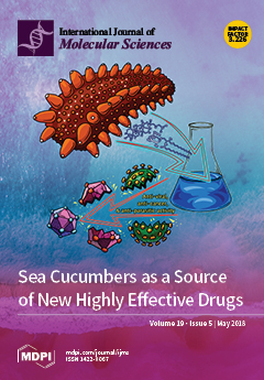
Cover Story (view full-size image): Marine species are a vast potential source of bioactive compounds that are not yet fully utilized. In Asian countries, holothurians that look similar to cucumbers are very popular as a valuable food. In traditional Chinese medicine, sea cucumbers are believed to have healing properties. Recently, sea cucumbers have received attention from researchers due to the novel data on their chemical content and the pharmacological effects of the bioactive ingredients isolated from these marine invertebrates. Some compounds from sea cucumbers possess specific properties that make them potential drug prototypes for the development of novel pharmaceuticals. View the paper here.
  • Issues are regarded as officially published after their release is announced to the table of contents alert mailing list.
  • You may sign up for e-mail alerts to receive table of contents of newly released issues.
  • PDF is the official format for papers published in both, html and pdf forms. To view the papers in pdf format, click on the "PDF Full-text" link, and use the free Adobe Reader to open them.
Order results
Result details
Section
Select all
Export citation of selected articles as:

Editorial

Jump to: Research, Review, Other

6 pages, 193 KiB  
Editorial
Plant–Microbe Interaction 2017—The Good, the Bad and the Diverse
by Jan Schirawski 1,* and Michael H. Perlin 2
1 Microbial Genetics, Institute of Applied Microbiology, RWTH Aachen University, Worringerweg 1, 52074 Aachen, Germany
2 Department of Biology, Program on Disease Resistance, University of Louisville, Louisville, KY 40292, USA
Int. J. Mol. Sci. 2018, 19(5), 1374; https://doi.org/10.3390/ijms19051374 - 5 May 2018
Cited by 108 | Viewed by 10603
Abstract
Of the many ways that plants interact with microbes, three aspects are highlighted in this issue: interactions where the plant benefits from the microbes, interactions where the plant suffers, and interactions where the plant serves as habitat for microbial communities. In this editorial, [...] Read more.
Of the many ways that plants interact with microbes, three aspects are highlighted in this issue: interactions where the plant benefits from the microbes, interactions where the plant suffers, and interactions where the plant serves as habitat for microbial communities. In this editorial, the fourteen articles published in the Special Issue Plant–Microbe Interaction 2017 are summarized and discussed as part of the global picture of the current understanding of plant-microbe interactions. Full article
(This article belongs to the Special Issue Plant Microbe Interaction 2017)
3 pages, 165 KiB  
Editorial
Human Chorionic Gonadotropin (hCG)—An Endocrine, Regulator of Gestation and Cancer
by Helene Heidegger and Udo Jeschke *
Department of Obsterics and Gynecology, University Hospital, LMU Munich, 81377 Munich, Germany
Int. J. Mol. Sci. 2018, 19(5), 1502; https://doi.org/10.3390/ijms19051502 - 17 May 2018
Cited by 12 | Viewed by 5192
Abstract
Human Chorionic Gonadotropin (hCG) is a heterodimeric glycoprotein composed of two subunits [...] Full article
(This article belongs to the Special Issue hCG—An Endocrine, Regulator of Gestation and Cancer)

Research

Jump to: Editorial, Review, Other

25 pages, 119885 KiB  
Article
Gastroprotective and Antioxidant Activity of Kalanchoe brasiliensis and Kalanchoe pinnata Leaf Juices against Indomethacin and Ethanol-Induced Gastric Lesions in Rats
by Edilane Rodrigues Dantas De Araújo 1, Gerlane Coelho Bernardo Guerra 2, Daline Fernandes de Souza Araújo 3, Aurigena Antunes De Araújo 2, Júlia Morais Fernandes 1, Raimundo Fernandes De Araújo Júnior 4, Valéria Costa Da Silva 2, Thaís Gomes De Carvalho 4, Leandro De Santis Ferreira 5 and Silvana Maria Zucolotto 1,*
1 Research Group on Bioactive Natural Products, Department of Pharmacy, Federal University of Rio Grande do Norte, 59012-570 Natal, Brazil
2 Department of Biophysics and Pharmacology, Biosciences Center, Federal University of Rio Grande do Norte, 59072-970 Natal, Brazil
3 Faculty of Health Sciences of Trairi, Federal University of Rio Grande do Norte, 59200-000 Santa Cruz, Brazil
4 Department of Morphology, Histology and Basic Pathology, Biosciences Center, Federal University of Rio Grande do Norte, 59072-970 Natal, Brazil
5 Department of Pharmacy, Federal University of Rio Grande do Norte, 59012-570 Natal, Brazil
Int. J. Mol. Sci. 2018, 19(5), 1265; https://doi.org/10.3390/ijms19051265 - 24 Apr 2018
Cited by 66 | Viewed by 8726
Abstract
Kalanchoe brasiliensis and Kalanchoe pinnata are used interchangeably in traditional medicine for treating peptic ulcers and inflammatory problems. In this context, this study aims to characterize the chemical constituents and evaluate the gastroprotective activity of the leaf juices of the two species in [...] Read more.
Kalanchoe brasiliensis and Kalanchoe pinnata are used interchangeably in traditional medicine for treating peptic ulcers and inflammatory problems. In this context, this study aims to characterize the chemical constituents and evaluate the gastroprotective activity of the leaf juices of the two species in acute gastric lesions models. Thin Layer Chromatography (TLC) and Ultra High Performance Liquid Chromatography coupled to Mass Spectrometer (UHPLC-MS) were performed for chemical characterization. Wistar rats were pre-treated orally with leaf juices (125, 250 and 500 mg/kg) or ranitidine (50 mg/kg). The peaks observed in the chromatogram of K. brasiliensis showed similar mass spectra to flavonoid glycosides derived from patuletin and eupafolin, while K. pinnata showed mass spectra similar to compounds derived from quercetin, patuletin, eupafolin and kaempferol. K. brasiliensis at all doses and K. pinnata at doses of 250 mg/kg and 500 mg/kg significantly reduced the lesions in the ethanol induction model. In the indomethacin induction model, both species showed significant results at doses of 250 and 500 mg/kg. Also, the pre-treatment with leaf juices increased the antioxidant defense system, glutathione (GSH), whereas malondialdehyde (MDA), myeloperoxidase (MPO), interleukin-1β (IL-1β) and tumor necrosis factor-α (TNF-α) levels were significantly decreased. Treatment with leaf juices led to the upregulation of zone occludes-1 (ZO-1) and the downregulation of inducible nitric oxide synthase (iNOS) and factor nuclear-κβ transcription (NF-κB-p65), while also showing a cytoprotective effect and maintaining mucus production. These findings show that the leaf juices of the two species showed gastroprotective effects on ethanol and gastric indomethacin injury which were a consequence of gastric inflammation suppression, antioxidant activity and the maintenance of cytoprotective defenses and mucosal structure architecture. Full article
(This article belongs to the Section Bioactives and Nutraceuticals)
Show Figures

Figure 1

13 pages, 1538 KiB  
Article
A Combined NMR-Computational Study of the Interaction between Influenza Virus Hemagglutinin and Sialic Derivatives from Human and Avian Receptors on the Surface of Transfected Cells
by Francesca Vasile 1,*, Maddalena Panigada 2, Antonio Siccardi 2, Donatella Potenza 1 and Guido Tiana 3
1 Department of Chemistry, University of Milano, Via Golgi 19, 20133 Milano, Italy
2 Molecular Immunology Unit, San Raffaele Research Institute, via Olgettina 58, 20132 Milano, Italy
3 Center for Complexity and Biosystems and Department of Physics, University of Milano and INFN, Via Celoria 16, 20133 Milano, Italy
Int. J. Mol. Sci. 2018, 19(5), 1267; https://doi.org/10.3390/ijms19051267 - 24 Apr 2018
Cited by 14 | Viewed by 5271
Abstract
The development of small-molecule inhibitors of influenza virus Hemagglutinin could be relevant to the opposition of the diffusion of new pandemic viruses. In this work, we made use of Nuclear Magnetic Resonance (NMR) spectroscopy to study the interaction between two derivatives of sialic [...] Read more.
The development of small-molecule inhibitors of influenza virus Hemagglutinin could be relevant to the opposition of the diffusion of new pandemic viruses. In this work, we made use of Nuclear Magnetic Resonance (NMR) spectroscopy to study the interaction between two derivatives of sialic acid, Neu5Ac-α-(2,6)-Gal-β-(1–4)-GlcNAc and Neu5Ac-α-(2,3)-Gal-β-(1–4)-GlcNAc, and hemagglutinin directly expressed on the surface of recombinant human cells. We analyzed the interaction of these trisaccharides with 293T cells transfected with the H5 and H1 variants of hemagglutinin, which thus retain their native trimeric conformation in such a realistic environment. By exploiting the magnetization transfer between the protein and the ligand, we obtained evidence of the binding event, and identified the epitope. We analyzed the conformational features of the glycans with an approach combining NMR spectroscopy and data-driven molecular dynamics simulations, thus obtaining useful information for an efficient drug design. Full article
(This article belongs to the Special Issue Molecular Recognition of Carbohydrates)
Show Figures

Graphical abstract

17 pages, 16346 KiB  
Article
The Commonly Used Bactericide Bismerthiazol Promotes Rice Defenses against Herbivores
by Pengyong Zhou, Xiaochang Mo, Wanwan Wang, Xia Chen and Yonggen Lou *
1 State Key Laboratory of Rice Biology & Ministry of Agriculture Key Lab of Molecular Biology of Crop Pathogens and Insects, Institute of Insect Sciences, Zhejiang University, Hangzhou 310058, China
These authors contributed equally to this work.
Int. J. Mol. Sci. 2018, 19(5), 1271; https://doi.org/10.3390/ijms19051271 - 24 Apr 2018
Cited by 17 | Viewed by 6452
Abstract
Chemical elicitors that enhance plant resistance to pathogens have been extensively studied, however, chemical elicitors that induce plant defenses against insect pests have received little attention. Here, we found that the exogenous application of a commonly used bactericide, bismerthiazol, on rice induced the [...] Read more.
Chemical elicitors that enhance plant resistance to pathogens have been extensively studied, however, chemical elicitors that induce plant defenses against insect pests have received little attention. Here, we found that the exogenous application of a commonly used bactericide, bismerthiazol, on rice induced the biosynthesis of constitutive and/or elicited jasmonic acid (JA), jasmonoyl-isoleucine conjugate (JA-Ile), ethylene and H2O2 but not salicylic acid. These activated signaling pathways altered the volatile profile of rice plants. White-backed planthopper (WBPH, Sogatella furcifera) nymphs and gravid females showed a preference for feeding and/or oviposition on control plants: survival rates were better and more eggs were laid than on bismerthiazol-treated plants. Moreover, bismerthiazol treatment also increased both the parasitism rate of WBPH eggs laid on plants in the field by Anagrus nilaparvatae, and also the resistance of rice to the brown planthopper (BPH) Nilaparvata lugens and the striped stem borer (SSB) Chilo suppressalis. These findings suggest that the bactericide bismerthiazol can induce the direct and/or indirect resistance of rice to multiple insect pests, and so can be used as a broad-spectrum chemical elicitor. Full article
(This article belongs to the Special Issue Plant Innate Immunity 2.0)
Show Figures

Graphical abstract

15 pages, 3352 KiB  
Article
Protection of Spleen Tissue of γ-ray Irradiated Mice against Immunosuppressive and Oxidative Effects of Radiation by Adenosine 5′-Monophosphate
by Cuilin Cheng 1,2,†, Juanjuan Yi 1,3,†, Rongchun Wang 1, Li Cheng 4, Zhenyu Wang 1,2,* and Weihong Lu 1,2,*
1 Food Science and Engineering, School of Chemical Engineering & Technology, Harbin Institute of Technology, Harbin 150090, China
2 Institute of Extreme Environmental Nutrition and Protection, Harbin Institute of Technology, Harbin 150090, China
3 School of Life Sciences, Zhengzhou University, Zhengzhou 450001, China
4 School of Life Science, Heilongjiang University, Harbin 150080, China
These authors contributed equally to this work.
Int. J. Mol. Sci. 2018, 19(5), 1273; https://doi.org/10.3390/ijms19051273 - 24 Apr 2018
Cited by 23 | Viewed by 4773
Abstract
The immune system is very sensitive to radiation. This study revealed that adenosine 5′-monophosphate (5′-AMP) increased the DNA contents of the spleen and the spleen index of irradiated mice. Moreover, the exogenous 5′-AMP could significantly repair the ultra-structure of the damaged spleen through [...] Read more.
The immune system is very sensitive to radiation. This study revealed that adenosine 5′-monophosphate (5′-AMP) increased the DNA contents of the spleen and the spleen index of irradiated mice. Moreover, the exogenous 5′-AMP could significantly repair the ultra-structure of the damaged spleen through transmission electron microscopy. When indicators of the mouse immune system were assessed, the flow cytometry and enzyme-linked immunosorbent assay (ELISA) revealed that the administration of exogenous 5′-AMP could reduce the apoptosis in the splenic cells. It could also regulate the transition of cells towards S phase, increase the proportion of CD4+ and CD8+ cellular subsets, and enhance the secretion of interleukin-2 (IL-2), IL-4, IL-10, and interferon-γ (IFN-γ). These effects were associated with a decrease in oxidative stress, as evidenced by changes in superoxide dismutase (SOD), glutathione peroxidase (GSH-Px), catalase (CAT), reduced glutathione (GSH), and malondialdehyde (MDA) levels of spleen tissues. These results suggested that exogenous 5′-AMP could repair the damaged spleen, increase the spleen index, and regulate the cell cycles and apoptosis. There was an increase in the production of various cytokines and play a protective role on the immune system of irradiated mice by dynamically adjusting the REDOX balance. Full article
(This article belongs to the Section Biochemistry)
Show Figures

Figure 1

18 pages, 4739 KiB  
Article
Effect of Amelogenin Coating of a Nano-Modified Titanium Surface on Bioactivity
by Chisato Terada 1, Satoshi Komasa 1,*, Tetsuji Kusumoto 2, Takayoshi Kawazoe 3 and Joji Okazaki 1
1 Department of Removable Prosthodontics and Occlusion, Osaka Dental University, 8-1, Kuzuhahanazono-cho, Hirakata-shi, Osaka 573-1121, Japan
2 Osaka Dental University Japan Faculty of Health Sciences, 1-4-4, Makino-honmachi, Hirakata-shi, Osaka 573-1144, Japan
3 Osaka Dental University, 8-1, Kuzuhahanazono-cho, Hirakata-shi, Osaka 573-1121, Japan
Int. J. Mol. Sci. 2018, 19(5), 1274; https://doi.org/10.3390/ijms19051274 - 24 Apr 2018
Cited by 19 | Viewed by 6657
Abstract
The interactions between implants and host tissues depend on several factors. In particular, a growing body of evidence has demonstrated that the surface texture of an implant influences the response of the surrounding cells. The purpose of this study is to develop new [...] Read more.
The interactions between implants and host tissues depend on several factors. In particular, a growing body of evidence has demonstrated that the surface texture of an implant influences the response of the surrounding cells. The purpose of this study is to develop new implant materials aiming at the regeneration of periodontal tissues as well as hard tissues by coating nano-modified titanium with amelogenin, which is one of the main proteins contained in Emdogain®. We confirmed by quartz crystal microbalance evaluation that amelogenin is easy to adsorb onto the nano-modified titanium surface as a coating. Scanning electron microscopy, scanning probe microscopy, X-ray photoelectron spectroscopy, and Fourier-transform infrared spectroscopy analyses confirmed that amelogenin coated the nano-modified titanium surface following alkali-treatment. In vitro evaluation using rat bone marrow and periodontal ligament cells revealed that the initial adhesion of both cell types and the induction of hard tissue differentiation such as cementum were improved by amelogenin coating. Additionally, the formation of new bone in implanted surrounding tissues was observed in in vivo evaluation using rat femurs. Together, these results suggest that this material may serve as a new implant material with the potential to play a major role in the advancement of clinical dentistry. Full article
(This article belongs to the Special Issue Bioactive Nanoparticles)
Show Figures

Figure 1

12 pages, 2842 KiB  
Article
HtrA1 Is Specifically Up-Regulated in Active Keloid Lesions and Stimulates Keloid Development
by Satoko Yamawaki 1, Motoko Naitoh 2,*, Hiroshi Kubota 3, Rino Aya 2, Yasuhiro Katayama 2, Toshihiro Ishiko 4, Taku Tamura 3, Katsuhiro Yoshikawa 5, Tatsuki Enoshiri 2, Mika Ikeda 6 and Shigehiko Suzuki 2
1 Department of Plastic and Reconstructive Surgery, Japanese Red Cross Fukui Hospital, 2-4-1, Tsukimi, Fukui-City, Fukui 918-8501, Japan
2 Department of Plastic and Reconstructive Surgery, Graduate School of Medicine, Kyoto University, 54 Kawahara-cho, Sakyo-ku, Kyoto 606-8507, Japan
3 Department of Life Science, Faculty of Engineering Science, Akita University, 1-1 Tegata Gakuenmachi, Akita 010-8502, Japan
4 Department of Plastic and Reconstructive Surgery, Japanese Red Cross Otsu Hospital, 1-1-35, Nagara, Otsu City, Shiga 520-8511, Japan
5 Department of Plastic and Reconstructive Surgery, Shiga Medical Center for Adults, 5-4-30, Moriyama, Moriyama City, Shiga 524-8524, Japan
6 Department of Plastic and Reconstructive Surgery, Kobe City Medical Center General Hospital, 2-1-1, Minatojima minami-machi, Cyuou-ku, Kobe City, Hyogo 650-0047, Japan
Int. J. Mol. Sci. 2018, 19(5), 1275; https://doi.org/10.3390/ijms19051275 - 24 Apr 2018
Cited by 9 | Viewed by 5494
Abstract
Keloids occur after failure of the wound healing process; inflammation persists, and various treatments are ineffective. Keloid pathogenesis is still unclear. We have previously analysed the gene expression profiles in keloid tissue and found that HtrA1 was markedly up-regulated in the keloid lesions. [...] Read more.
Keloids occur after failure of the wound healing process; inflammation persists, and various treatments are ineffective. Keloid pathogenesis is still unclear. We have previously analysed the gene expression profiles in keloid tissue and found that HtrA1 was markedly up-regulated in the keloid lesions. HtrA1 is a serine protease suggested to play a role in the pathogenesis of various diseases, including age-related macular degeneration and osteoarthritis, by modulating extracellular matrix or cell surface proteins. We analysed HtrA1 localization and its role in keloid pathogenesis. Thirty keloid patients and twelve unrelated patients were enrolled for in situ hybridization, immunohistochemical, western blot, and cell proliferation analyses. Fibroblast-like cells expressed more HtrA1 in active keloid lesions than in surrounding lesions. The proportion of HtrA1-positive cells in keloids was significantly higher than that in normal skin, and HtrA1 protein was up-regulated relative to normal skin. Silencing HtrA1 gene expression significantly suppressed cell proliferation. HtrA1 was highly expressed in keloid tissues, and the suppression of the HtrA1 gene inhibited the proliferation of keloid-derived fibroblasts. HtrA1 may promote keloid development by accelerating cell proliferation and remodelling keloid-specific extracellular matrix or cell surface molecules. HtrA1 is suggested to have an important role in keloid pathogenesis. Full article
(This article belongs to the Special Issue Recent Advances in Scar Biology)
Show Figures

Figure 1

14 pages, 2934 KiB  
Article
Organ-Specific MicroRNAs (MIR122, 137, and 206) Contribute to Tissue Characteristics and Carcinogenesis by Regulating Pyruvate Kinase M1/2 (PKM) Expression
by Kohei Taniguchi 1,2,*, Nobuhiko Sugito 3, Haruka Shinohara 3, Yuki Kuranaga 3, Yosuke Inomata 1, Kazumasa Komura 2, Kazuhisa Uchiyama 1 and Yukihiro Akao 3
1 Department of General and Gastroenterological Surgery, Osaka Medical College, 2-7 Daigaku-Machi, Takatsuki, Osaka 569-8686, Japan
2 Translational Research Program, Osaka Medical College, 2-7 Daigaku-Machi, Takatsuki, Osaka 569-8686, Japan
3 United Graduate School of Drug Discovery and Medical Information Sciences, Gifu University, 1-1 Yanagido, Gifu 501-1193, Japan
Int. J. Mol. Sci. 2018, 19(5), 1276; https://doi.org/10.3390/ijms19051276 - 24 Apr 2018
Cited by 26 | Viewed by 7140
Abstract
Pyruvate kinase is known as the glycolytic enzyme catalyzing the final step in glycolysis. In mammals, two different forms of it exist, i.e., pyruvate kinase M1/2 (PKM) and pyruvate kinase L/R (PKLR). Also, PKM has two isoforms, i.e., PKM1 [...] Read more.
Pyruvate kinase is known as the glycolytic enzyme catalyzing the final step in glycolysis. In mammals, two different forms of it exist, i.e., pyruvate kinase M1/2 (PKM) and pyruvate kinase L/R (PKLR). Also, PKM has two isoforms, i.e., PKM1 and PKM2. These genes have tissue-specific distribution. Namely, PKM1 is distributed in high-energy-demanding organs, such as brain and muscle. Also, PKM2 is distributed in various other organs, such as the colon. On the other hand, PKLR is distributed in liver and red blood cells (RBCs). Interestingly, PKM2 has been recognized as one of the essential genes for the cancer-specific energy metabolism termed the “Warburg effect”. However, the mechanism(s) underlying this fact have remained largely unclear. Recently, we found that some organ-specific microRNAs (miRNAs, MIR) regulate PKM isoform expression through direct targeting of polypyrimidine tract binding protein 1 (PTBP1), which is the splicer responsible for PKM2-dominant expression. In this study, we examined whether this machinery was conserved in the case of other PTBP1- and PKM-targeting miRNAs. We focused on the MIRs 122, 137, and 206, and investigated the expression profiles of each of these miRNAs in tissues from mouse and human organs. Also, we examined the regulatory mechanisms of PKM isoform expression by testing each of these miRNAs in human cancer cell lines. Presently, we found that brain-specific MIR137 and muscle-specific MIR206 predominantly induced PKM1 expression through direct targeting of PTBP1. Also, liver-specific MIR122 suppressed the expression of both PKM1 and PKM2, which action occurred through direct targeting of PKM to enable the expression of PKLR. Moreover, the expression levels of these miRNAs were downregulated in cancer cells that had originated from these tissues, resulting in PKM2 dominance. Our results suggest that the organ-specific distribution of miRNAs is one of the principal means by which miRNA establishes characteristics of a tissue and that dysregulation of these miRNAs results in cancer development through a change in the ratio of PKM isoform expression. Also, our results contribute to cancer diagnosis and will be useful for cancer-specific therapy for the Warburg effect in the near future. Full article
(This article belongs to the Collection Regulation by Non-coding RNAs)
Show Figures

Graphical abstract

11 pages, 1829 KiB  
Article
Comparative Analysis of Repetitive DNA between the Main Vectors of Chagas Disease: Triatoma infestans and Rhodnius prolixus
by Sebastián Pita 1, Pablo Mora 2, Jesús Vela 2, Teresa Palomeque 2, Antonio Sánchez 2, Francisco Panzera 1,* and Pedro Lorite 2,*
1 Evolutionary Genetic Section, Faculty of Science, University of the Republic, Iguá 4225, Montevideo 11400, Uruguay
2 Department of Experimental Biology, Genetics , University of Jaén, Paraje Las Lagunillas s/n., 23071 Jaén, Spain
Int. J. Mol. Sci. 2018, 19(5), 1277; https://doi.org/10.3390/ijms19051277 - 24 Apr 2018
Cited by 15 | Viewed by 5724
Abstract
Chagas disease or American trypanosomiasis affects six to seven million people worldwide, mostly in Latin America. This disease is transmitted by hematophagous insects known as “kissing bugs” (Hemiptera, Triatominae), with Triatoma infestans and Rhodnius prolixus being the two most important vector species. Despite [...] Read more.
Chagas disease or American trypanosomiasis affects six to seven million people worldwide, mostly in Latin America. This disease is transmitted by hematophagous insects known as “kissing bugs” (Hemiptera, Triatominae), with Triatoma infestans and Rhodnius prolixus being the two most important vector species. Despite the fact that both species present the same diploid chromosome number (2n = 22), they have remarkable differences in their total DNA content, chromosome structure and genome organization. Variations in the DNA genome size are expected to be due to differences in the amount of repetitive DNA sequences. The T. infestans genome-wide analysis revealed the existence of 42 satellite DNA families. BLAST searches of these sequences against the R. prolixus genome assembly revealed that only four of these satellite DNA families are shared between both species, suggesting a great differentiation between the Triatoma and Rhodnius genomes. Fluorescence in situ hybridization (FISH) location of these repetitive DNAs in both species showed that they are dispersed on the euchromatic regions of all autosomes and the X chromosome. Regarding the Y chromosome, these common satellite DNAs are absent in T. infestans but they are present in the R. prolixus Y chromosome. These results support a different origin and/or evolution in the Y chromosome of both species. Full article
(This article belongs to the Special Issue Molecular Entomology of Insects of Economic Importance)
Show Figures

Figure 1

15 pages, 1700 KiB  
Article
S1P4 Regulates Passive Systemic Anaphylaxis in Mice but Is Dispensable for Canonical IgE-Mediated Responses in Mast Cells
by Joseph M. Kulinski 1, Richard L. Proia 2, Elisabeth M. Larson 1, Dean D. Metcalfe 1 and Ana Olivera 1,*
1 Mast Cell Biology Section, Laboratory of Allergic Diseases, National Institute of Allergy and Infectious Diseases (NIAID), National Institutes of Health, Bethesda, MD 20892, USA
2 Genetics of Development and Disease Branch, National Institute of Diabetes and Digestive and Kidney Diseases (NIDDK), National Institutes of Health, Bethesda, MD 20892, USA
Int. J. Mol. Sci. 2018, 19(5), 1279; https://doi.org/10.3390/ijms19051279 - 25 Apr 2018
Cited by 15 | Viewed by 5434
Abstract
Mast cells are key players in the development of inflammatory allergic reactions. Cross-linking of the high-affinity receptor for IgE (FcεRI) on mast cells leads to the generation and secretion of the sphingolipid mediator, sphingosine-1-phosphate (S1P) which is able, in turn, to transactivate its [...] Read more.
Mast cells are key players in the development of inflammatory allergic reactions. Cross-linking of the high-affinity receptor for IgE (FcεRI) on mast cells leads to the generation and secretion of the sphingolipid mediator, sphingosine-1-phosphate (S1P) which is able, in turn, to transactivate its receptors on mast cells. Previous reports have identified the expression of two of the five receptors for S1P on mast cells, S1P1 and S1P2, with functions in FcεRI-mediated chemotaxis and degranulation, respectively. Here, we show that cultured mouse mast cells also express abundant message for S1P4. Genetic deletion of S1pr4 did not affect the differentiation of bone marrow progenitors into mast cells or the proliferation of mast cells in culture. A comprehensive characterization of IgE-mediated responses in S1P4-deficient bone marrow-derived and peritoneal mouse mast cells indicated that this receptor is dispensable for mast cell degranulation, cytokine/chemokine production and FcεRI-mediated chemotaxis in vitro. However, interleukin-33 (IL-33)-mediated enhancement of IgE-induced degranulation was reduced in S1P4-deficient peritoneal mast cells, revealing a potential negative regulatory role for S1P4 in an IL-33-rich environment. Surprisingly, genetic deletion of S1pr4 resulted in exacerbation of passive systemic anaphylaxis to IgE/anti-IgE in mice, a phenotype likely related to mast cell-extrinsic influences, such as the high circulating levels of IgE in these mice which increases FcεRI expression and consequently the extent of the response to FcεRI engagement. Thus, we provide evidence that S1P4 modulates anaphylaxis in an unexpected manner that does not involve regulation of mast cell responsiveness to IgE stimulation. Full article
(This article belongs to the Special Issue Sphingolipids: Signals and Disease)
Show Figures

Figure 1

12 pages, 11550 KiB  
Article
Ethylene Responsive Factor MeERF72 Negatively Regulates Sucrose synthase 1 Gene in Cassava
by Chen Liu 1,2,3,†, Xin Chen 2,3,†, Ping’an Ma 4, Shengkui Zhang 2,3,5, Changying Zeng 2,3, Xingyu Jiang 1,* and Wenquan Wang 2,3,*
1 Institute of Tropical Agriculture and Forestry, Hainan University, Haikou 570228, China
2 The Institute of Tropical Bioscience and Biotechnology, Chinese Academy of Tropical Agricultural Sciences, Haikou 571101, China
3 Key Laboratory of Biology and Genetic Resources of Tropical Crops, Ministry of Agriculture, Haikou 571101, China
4 College of Biological Engineering, Henan University of Technology, Zhengzhou 450001, China
5 College of Plant Science & Technology, Huazhong Agricultural University, Wuhan 430070, China
These authors contributed equally to this work.
Int. J. Mol. Sci. 2018, 19(5), 1281; https://doi.org/10.3390/ijms19051281 - 25 Apr 2018
Cited by 23 | Viewed by 4902
Abstract
Cassava, an important food and industrial crop globally, is characterized by its powerful starch accumulation in its storage root. However, the underlying molecular mechanism for this feature remains unclear. Sucrose synthase initializes the conversion of sucrose to starch, and, to a certain extent, [...] Read more.
Cassava, an important food and industrial crop globally, is characterized by its powerful starch accumulation in its storage root. However, the underlying molecular mechanism for this feature remains unclear. Sucrose synthase initializes the conversion of sucrose to starch, and, to a certain extent, its enzyme activity can represent sink strength. To understand the modulation of MeSus gene family, the relatively high expressed member in storage root, MeSus1, its promoter was used as bait to screen cassava storage root full-length cDNA library through a yeast one-hybrid system. An ethylene responsive factor cDNA, designated as MeERF72 according to its homolog in Arabidopsis, was screened out. The transcript level of MeERF72 was induced by ethylene, drought, and salt treatments and repressed by abscisic acid, Auxin, gibberellin, salicylic acid, and low and high temperatures. The MeERF72 protein has a conserved APETALA2 domain in its N-terminus and an activated domain of 30 amino acids in its C-terminus, can bind to MeSus1 promoter in vitro and in vivo, and represses the promoter activity of MeSus1. MeERF72 is a transcription factor that can negatively regulate the expression level of MeSus1 in cassava. Full article
(This article belongs to the Section Molecular Plant Sciences)
Show Figures

Figure 1

21 pages, 1852 KiB  
Article
Functional Analysis of the Arabidopsis thaliana CDPK-Related Kinase Family: AtCRK1 Regulates Responses to Continuous Light
by Abu Imran Baba 1,2,†, Gábor Rigó 1,3,*,†, Ferhan Ayaydin 1, Ateeq Ur Rehman 1, Norbert Andrási 1, Laura Zsigmond 1, Ildikó Valkai 1, János Urbancsok 4, Imre Vass 1, Taras Pasternak 5, Klaus Palme 5, László Szabados 1 and Ágnes Cséplő 1,*
1 Plant Biology Institute, Biological Research Centre, Hungarian Academy of Sciences, 6726 Szeged, Hungary
2 Doctoral School in Biology, Faculty of Science and Informatics, University of Szeged, 6720 Szeged, Hungary
3 Department of Plant Biology, University of Szeged, 6726 Szeged, Hungary
4 Department of Biology, Norwegian University of Science and Technology, Høgskoleringen 5, NO-7491 Trondheim, Norway
5 Faculty of Biologie II, Albert-Ludwigs Universität, Schänzlestr. 1, 79104 Freiburg, Germany
These authors contributed equally to this work.
Int. J. Mol. Sci. 2018, 19(5), 1282; https://doi.org/10.3390/ijms19051282 - 25 Apr 2018
Cited by 32 | Viewed by 9084
Abstract
The Calcium-Dependent Protein Kinase (CDPK)-Related Kinase family (CRKs) consists of eight members in Arabidopsis. Recently, AtCRK5 was shown to play a direct role in the regulation of root gravitropic response involving polar auxin transport (PAT). However, limited information is available about [...] Read more.
The Calcium-Dependent Protein Kinase (CDPK)-Related Kinase family (CRKs) consists of eight members in Arabidopsis. Recently, AtCRK5 was shown to play a direct role in the regulation of root gravitropic response involving polar auxin transport (PAT). However, limited information is available about the function of the other AtCRK genes. Here, we report a comparative analysis of the Arabidopsis CRK genes, including transcription regulation, intracellular localization, and biological function. AtCRK transcripts were detectable in all organs tested and a considerable variation in transcript levels was detected among them. Most AtCRK proteins localized at the plasma membrane as revealed by microscopic analysis of 35S::cCRK-GFP (Green Fluorescence Protein) expressing plants or protoplasts. Interestingly, 35S::cCRK1-GFP and 35S::cCRK7-GFP had a dual localization pattern which was associated with plasma membrane and endomembrane structures, as well. Analysis of T-DNA insertion mutants revealed that AtCRK genes are important for root growth and control of gravitropic responses in roots and hypocotyls. While Atcrk mutants were indistinguishable from wild type plants in short days, Atcrk1-1 mutant had serious growth defects under continuous illumination. Semi-dwarf phenotype of Atcrk1-1 was accompanied with chlorophyll depletion, disturbed photosynthesis, accumulation of singlet oxygen, and enhanced cell death in photosynthetic tissues. AtCRK1 is therefore important to maintain cellular homeostasis during continuous illumination. Full article
(This article belongs to the Special Issue Plasma-Membrane Transport)
Show Figures

Graphical abstract

14 pages, 1441 KiB  
Article
Dietary Supplementation with Oleum Cinnamomi Improves Intestinal Functions in Piglets
by Dan Yi 1, Qiuhong Fang 1, Yongqing Hou 1,*, Lei Wang 1, Haiwang Xu 1, Tao Wu 1, Joshua Gong 2 and Guoyao Wu 1,3
1 Hubei Collaborative Innovation Center for Animal Nutrition and Feed Safety, Hubei Key Laboratory of Animal Nutrition and Feed Science, Wuhan Polytechnic University, Wuhan 430023, China
2 Guelph Research and Development Centre, Agriculture and Agri-Food Canada, Guelph, ON N1G 5C9, Canada
3 Department of Animal Science, Texas A&M University, College Station, TX 77843, USA
Int. J. Mol. Sci. 2018, 19(5), 1284; https://doi.org/10.3390/ijms19051284 - 25 Apr 2018
Cited by 15 | Viewed by 5854
Abstract
The present study was to determine the efficacy of dietary supplementation with oleum cinnamomi (OCM) on growth performance and intestinal functions in piglets. Sixteen piglets (24-day-old) were randomly assigned to the control or OCM groups. Piglets in the control group were fed a [...] Read more.
The present study was to determine the efficacy of dietary supplementation with oleum cinnamomi (OCM) on growth performance and intestinal functions in piglets. Sixteen piglets (24-day-old) were randomly assigned to the control or OCM groups. Piglets in the control group were fed a basal diet, whereas piglets in the OCM group were fed the basal diet supplemented with 50 mg/kg OCM. On day 20 of the trial, blood samples and intestinal tissues were obtained from piglets. Compared with the control group, dietary OCM supplementation increased (p < 0.05) average daily feed intake, plasma insulin levels, villus width and villous surface area in the duodenum and jejunum, DNA levels and RNA/DNA ratios in the ileum, the abundance of Enterococcus genus and Lactobacillus genus in caecum digesta, mRNA levels for epithelial growth factor receptor (EGFR), Ras, extracellular signal-regulated kinase 1/2 (Erk1/2), b-cell lymphoma-extra large (Bcl-xL), villin, junctional adhesion molecule A (JAM-A), myxovirus resistance (MX) 1, MX2 and regenerating islet-derived protein 3 gamma (REG3G), and protein abundances of Ras and claudin-1, but decreased (p < 0.05) diarrhoea incidence; the abundances of Enterobacteriaceae family, Enterococcus genus, Lactobacillus genus, Bifidobacterium genus, and Clostrium coccoides in the colon digesta, and AMP-activated protein kinase (AMPK) mRNA levels and caspase-3 protein abundance in the jejunal mucosa of piglets. Taken together, these data indicate that dietary OCM supplementation modulates intestinal microbiota and improves intestinal function in weanling pigs. OCM is an effective feed additive and alternative to feed antibiotics for improving intestinal health in swine. Full article
(This article belongs to the Special Issue Nutrition and Gut Health)
Show Figures

Figure 1

17 pages, 4502 KiB  
Article
Complete Chloroplast Genome of Cercis chuniana (Fabaceae) with Structural and Genetic Comparison to Six Species in Caesalpinioideae
by Wanzhen Liu 1, Hanghui Kong 2,3, Juan Zhou 1, Peter W. Fritsch 4, Gang Hao 1,* and Wei Gong 1,*
1 College of Life Sciences, South China Agricultural University, Guangzhou 510614, China
2 Key Laboratory of Plant Resources Conservation and Sustainable Utilization, South China Botanical Garden, Chinese Academy of Sciences, Guangzhou 510650, China
3 Guangdong Provincial Key Laboratory of Applied Botany, South China Botanical Garden, Chinese Academy of Sciences, Guangzhou 510650, China
4 Botanical Research Institute of Texas, 1700 University Drive, Fort Worth, TX 76107, USA
Int. J. Mol. Sci. 2018, 19(5), 1286; https://doi.org/10.3390/ijms19051286 - 25 Apr 2018
Cited by 38 | Viewed by 5883
Abstract
The subfamily Caesalpinioideae of the Fabaceae has long been recognized as non-monophyletic due to its controversial phylogenetic relationships. Cercis chuniana, endemic to China, is a representative species of Cercis L. placed within Caesalpinioideae in the older sense. Here, we report the whole [...] Read more.
The subfamily Caesalpinioideae of the Fabaceae has long been recognized as non-monophyletic due to its controversial phylogenetic relationships. Cercis chuniana, endemic to China, is a representative species of Cercis L. placed within Caesalpinioideae in the older sense. Here, we report the whole chloroplast (cp) genome of C. chuniana and compare it to six other species from the Caesalpinioideae. Comparative analyses of gene synteny and simple sequence repeats (SSRs), as well as estimation of nucleotide diversity, the relative ratios of synonymous and nonsynonymous substitutions (dn/ds), and Kimura 2-parameter (K2P) interspecific genetic distances, were all conducted. The whole cp genome of C. chuniana was found to be 158,433 bp long with a total of 114 genes, 81 of which code for proteins. Nucleotide substitutions and length variation are present, particularly at the boundaries among large single copy (LSC), inverted repeat (IR) and small single copy (SSC) regions. Nucleotide diversity among all species was estimated to be 0.03, the average dn/ds ratio 0.3177, and the average K2P value 0.0372. Ninety-one SSRs were identified in C. chuniana, with the highest proportion in the LSC region. Ninety-seven species from the old Caesalpinioideae were selected for phylogenetic reconstruction, the analysis of which strongly supports the monophyly of Cercidoideae based on the new classification of the Fabaceae. Our study provides genomic information for further phylogenetic reconstruction and biogeographic inference of Cercis and other legume species. Full article
(This article belongs to the Special Issue Chloroplast)
Show Figures

Graphical abstract

11 pages, 2926 KiB  
Article
Effect of Silver Nitrate and Sodium Fluoride with Tri-Calcium Phosphate on Streptococcus mutans and Demineralised Dentine
by Ollie Yiru Yu, Irene Shuping Zhao, May Lei Mei, Edward Chin-Man Lo and Chun-Hung Chu *
Faculty of Dentistry, The University of Hong Kong, Hong Kong, China
Int. J. Mol. Sci. 2018, 19(5), 1288; https://doi.org/10.3390/ijms19051288 - 25 Apr 2018
Cited by 18 | Viewed by 5807
Abstract
This study investigated the effect of 25% silver nitrate (AgNO3) and 5% sodium fluoride (NaF) varnish with functionalized tri-calcium phosphate (fTCP) on a Streptococcus mutans (S. mutans) biofilm and dentine caries lesion. Demineralised dentine specimens were treated with 25% [...] Read more.
This study investigated the effect of 25% silver nitrate (AgNO3) and 5% sodium fluoride (NaF) varnish with functionalized tri-calcium phosphate (fTCP) on a Streptococcus mutans (S. mutans) biofilm and dentine caries lesion. Demineralised dentine specimens were treated with 25% AgNO3 and 5% NaF + fTCP (Group 1), 25% AgNO3 and 5% NaF (Group 2), 25% AgNO3 (Group 3), or water (Group 4). The specimens were subjected to a S. mutans biofilm challenge after treatment. The biofilm was then studied via scanning electron microscopy (SEM), confocal laser scanning microscopy (CLSM), and colony forming units (CFU). The specimens were assessed by micro-computed tomography, X-ray diffraction (XRD), and Fourier transform infrared spectroscopy (FTIR). SEM and CLSM revealed less biofilm in Groups 1 to 3. The log10 CFU of Groups 1 to 4 were 4.5 ± 0.7, 4.4 ± 0.9, 4.4 ± 0.9, and 6.7 ± 1.0, respectively (Groups 1, 2, 3 < 4, p < 0.001). The lesion depths of Groups 1 to 4 were 212.6 ± 20.1 µm, 280.8 ± 51.6 µm, 402.5 ± 61.7 µm, and 497.4 ± 67.2 µm, respectively (Groups 1 < 2 < 3 < 4, p < 0.001). XRD demonstrated silver chloride formation in Groups 1, 2, and 3. FTIR found the amide I: HPO42− values of the four groups were 0.22 ± 0.05, 0.25 ± 0.05, 0.41 ± 0.12, and 0.64 ± 0.14, respectively (Groups 1, 2 < 3 < 4; p < 0.001). In conclusion, this study revealed that AgNO3 and NaF + fTCP reduced the damage of dentine caries by cariogenic biofilm. Full article
(This article belongs to the Section Materials Science)
Show Figures

Figure 1

18 pages, 10322 KiB  
Article
Influence of Mechanical Unloading on Articular Chondrocyte Dedifferentiation
by Simon L. Wuest 1,2, Martina Caliò 1,2, Timon Wernas 1, Samuel Tanner 1, Christina Giger-Lange 1, Fabienne Wyss 1, Fabian Ille 1, Benjamin Gantenbein 2 and Marcel Egli 1,*
1 Lucerne University of Applied Sciences and Arts, School of Engineering and Architecture, Institute of Medical Engineering, Space Biology Group, CH-6052 Hergiswil, Switzerland
2 University of Bern, Institute for Surgical Technology and Biomechanics, Tissue and Organ Mechanobiology, CH-3014 Bern, Switzerland
Int. J. Mol. Sci. 2018, 19(5), 1289; https://doi.org/10.3390/ijms19051289 - 25 Apr 2018
Cited by 34 | Viewed by 6721
Abstract
Due to the limited self-repair capacity of articular cartilage, the surgical restoration of defective cartilage remains a major clinical challenge. The cell-based approach, which is known as autologous chondrocyte transplantation (ACT), has limited success, presumably because the chondrocytes acquire a fibroblast-like phenotype in [...] Read more.
Due to the limited self-repair capacity of articular cartilage, the surgical restoration of defective cartilage remains a major clinical challenge. The cell-based approach, which is known as autologous chondrocyte transplantation (ACT), has limited success, presumably because the chondrocytes acquire a fibroblast-like phenotype in monolayer culture. This unwanted dedifferentiation process is typically addressed by using three-dimensional scaffolds, pellet culture, and/or the application of exogenous factors. Alternative mechanical unloading approaches are suggested to be beneficial in preserving the chondrocyte phenotype. In this study, we examined if the random positioning machine (RPM) could be used to expand chondrocytes in vitro such that they maintain their phenotype. Bovine chondrocytes were exposed to (a) eight days in static monolayer culture; (b) two days in static monolayer culture, followed by six days of RPM exposure; and, (c) eight days of RPM exposure. Furthermore, the experiment was also conducted with the application of 20 mM gadolinium, which is a nonspecific ion-channel blocker. The results revealed that the chondrocyte phenotype is preserved when chondrocytes go into suspension and aggregate to cell clusters. Exposure to RPM rotation alone does not preserve the chondrocyte phenotype. Interestingly, the gene expression (mRNA) of the mechanosensitive ion channel TRPV4 decreased with progressing dedifferentiation. In contrast, the gene expression (mRNA) of the mechanosensitive ion channel TRPC1 was reduced around fivefold to 10-fold in all of the conditions. The application of gadolinium had only a minor influence on the results. This and previous studies suggest that the chondrocyte phenotype is preserved if cells maintain a round morphology and that the ion channel TRPV4 could play a key role in the dedifferentiation process. Full article
(This article belongs to the Section Biochemistry)
Show Figures

Graphical abstract

14 pages, 2104 KiB  
Article
Commensal Staphylococcus aureus Provokes Immunity to Protect against Skin Infection of Methicillin-Resistant Staphylococcus aureus
by John-Jackson Yang 1, Ting-Wei Chang 2, Yong Jiang 3, Hsin-Jou Kao 2, Bin-Hao Chiou 2, Ming-Shan Kao 2 and Chun-Ming Huang 1,2,4,*
1 Department of Life Sciences, National Central University, Taoyuan 32001, Taiwan
2 Department of Biomedical Sciences and Engineering, National Central University, Taoyuan 32001, Taiwan
3 Surface Bioadvances Inc., San Diego, CA 92121, USA
4 Department of Dermatology, University of California, San Diego. 3525 John Hopkins Court, Rm276, San Diego, CA 92121, USA
Int. J. Mol. Sci. 2018, 19(5), 1290; https://doi.org/10.3390/ijms19051290 - 25 Apr 2018
Cited by 26 | Viewed by 7699
Abstract
Unlike USA300, a strain of community-acquired methicillin-resistant Staphylococcus aureus (MRSA), commensal Staphylococcus aureus (S. aureus) bacteria isolated from human skin demonstrated the ability to mediate the glycerol fermentation to produce short-chain fatty acids (SCFAs). Quantitative proteomic analysis of enzymes involved in [...] Read more.
Unlike USA300, a strain of community-acquired methicillin-resistant Staphylococcus aureus (MRSA), commensal Staphylococcus aureus (S. aureus) bacteria isolated from human skin demonstrated the ability to mediate the glycerol fermentation to produce short-chain fatty acids (SCFAs). Quantitative proteomic analysis of enzymes involved in glycerol fermentation demonstrated that the expression levels of six enzymes, including glycerol-3-phosphate dehydrogenase (GPDH) and phosphoglycerate mutase (PGM), in commensal S. aureus are more than three-fold higher than those in USA300. Western blotting validated the low expression levels of GPDH in USA300, MRSA252 (a strain of hospital-acquired MRSA), and invasive methicillin-susceptible S. aureus (MSSA). In the presence of glycerol, commensal S. aureus effectively suppressed the growth of USA300 in vitro and in vivo. Active immunization of mice with lysates or recombinant α-hemolysin of commensal S. aureus or passive immunization with neutralizing sera provided immune protection against the skin infection of USA300. Our data illustrate for the first time that commensal S. aureus elicits both innate and adaptive immunity via glycerol fermentation and systemic antibody production, respectively, to fight off the skin infection of pathogenic MRSA. Full article
(This article belongs to the Special Issue Bacterial Protein Toxins: Enemies within or Unexpected Friends)
Show Figures

Graphical abstract

26 pages, 4020 KiB  
Article
Common microRNA–mRNA Interactions in Different Newcastle Disease Virus-Infected Chicken Embryonic Visceral Tissues
by Yan-Qing Jia 1, Xing-Long Wang 1, Xiang-Wei Wang 1, Chuan-Qi Yan 1, Chang-Jie Lv 1, Xiao-Qin Li 1, Zhi-Li Chu 1, Fathalrhman Eisa Addoma Adam 1,2, Sa Xiao 1, Shu-Xia Zhang 1 and Zeng-Qi Yang 1,*
1 College of Veterinary Medicine, Northwest A&F University, Yangling 712100, China
2 Department of Preventive Medicine and Public Health, Faculty of Veterinary Science, University of Nyala, P.O. Box, 155 Nyala, Sudan
Int. J. Mol. Sci. 2018, 19(5), 1291; https://doi.org/10.3390/ijms19051291 - 25 Apr 2018
Cited by 23 | Viewed by 5842
Abstract
To investigate the roles and explore the altered expression of microRNAs (miRNAs) and mRNAs in chicken embryos in response to Newcastle disease virus (NDV) infection, deep sequencing was performed. Then, a conjoint analysis of small RNA-seq and mRNA-seq was performed to screen interactional [...] Read more.
To investigate the roles and explore the altered expression of microRNAs (miRNAs) and mRNAs in chicken embryos in response to Newcastle disease virus (NDV) infection, deep sequencing was performed. Then, a conjoint analysis of small RNA-seq and mRNA-seq was performed to screen interactional miRNA–mRNA pairs during NDV infection. In total, 15 and 17 up- and downregulated miRNAs were identified that potentially targeted 4279 and 6080 mRNAs in NDV-infected chicken embryonic tissues, respectively; in addition, 595 upregulated and 480 downregulated mRNAs were identified. The conjoint analysis of the obtained data identified 1069 miRNA–mRNA pairs. Among these pairs, 130 pairs were related to immune or inflammatory responses. The relationship between gga-miR-203a and its target transglutaminase 2 (TGM2) was confirmed using a dual-luciferase reporter system and a real time quantitative polymerase chain reaction (RT-qPCR) assay. Overall, the discovery of miRNAs, mRNAs, and their potential pairing relationships, which may be involved in the regulation of NDV infection, will facilitate our understanding of the complex regulatory relationship between the host and the virus. Full article
Show Figures

Graphical abstract

16 pages, 10247 KiB  
Article
Hemiptera Mitochondrial Control Region: New Sights into the Structural Organization, Phylogenetic Utility, and Roles of Tandem Repetitions of the Noncoding Segment
by Kui Li 1,2 and Ai-Ping Liang 1,2,*
1 Key Laboratory of Zoological Systematics and Evolution, Institute of Zoology, Chinese Academy of Sciences, Beijing 100101, China
2 College of Life Sciences, University of Chinese Academy of Sciences, Beijing 100049, China
Int. J. Mol. Sci. 2018, 19(5), 1292; https://doi.org/10.3390/ijms19051292 - 26 Apr 2018
Cited by 31 | Viewed by 4288
Abstract
As a major noncoding fragment, the control region (CR) of mtDNA is responsible for the initiation of mitogenome transcription and replication. Several structural features of CR sequences have been reported in many insects. However, comprehensive analyses on the structural organization and phylogenetic utility, [...] Read more.
As a major noncoding fragment, the control region (CR) of mtDNA is responsible for the initiation of mitogenome transcription and replication. Several structural features of CR sequences have been reported in many insects. However, comprehensive analyses on the structural organization and phylogenetic utility, as well as the role of tandem replications (TRs) on length variation, high A+T content, and shift of base skew of CR sequences are poorly investigated in hemipteran insects. In this study, we conducted a series of comparative analyses, using 116 samples covering all 11 infraorders of the five currently recognized monophyletic groups in the Hemiptera. Several structural elements (mononucleotide stretches containing conserved sequence blocks (CSBs), TRs, and GA-rich region) were identified in the mitochondrial control region in hemipteran insects, without showing a consistent location. The presence and absence of certain specific structural elements in CR sequences show the various structural organizations of that segment among the five monophyletic groups, which indicates the diversification of the control region’s structural organization in Hemiptera. Among the many groups within Hemiptera, eight monophyletic groups and three consistent phylogenetic trees were recovered, using CSBs datasets by maximum likelihood and Bayesian methods, which suggests the possible utility of CR sequences for phylogenetic reconstruction in certain groups of Hemiptera. Statistical analyses showed that TRs may contribute to the length variation, high AT content, and the shift of base skewing of CR sequences toward high AT content in the Hemiptera. Our findings enrich the knowledge of structural organization, phylogenetic utility, and roles of tandem replication of hemipteran CR, and provide a possible framework for mitochondrial control region analyses in hemimetabolous insects. Full article
(This article belongs to the Section Biochemistry)
Show Figures

Figure 1

1 pages, 2879 KiB  
Article
Adropin Contributes to Anti-Atherosclerosis by Suppressing Monocyte-Endothelial Cell Adhesion and Smooth Muscle Cell Proliferation
by Kengo Sato 1,*,†, Tomoyuki Yamashita 1,†, Remina Shirai 1, Koichiro Shibata 1, Taisuke Okano 1, Maho Yamaguchi 1, Yusaku Mori 2, Tsutomu Hirano 2 and Takuya Watanabe 1
1 Laboratory of Cardiovascular Medicine, Tokyo University of Pharmacy and Life Sciences, 1432-1 Horinouchi, Hachioji-City, Tokyo 192-0392, Japan
2 Division of Diabetes, Metabolism, and Endocrinology, Department of Medicine, Showa University School of Medicine, 1-5-8 Hatanodai, Shinagawa-ku, Tokyo 142-8666, Japan
These authors contributed equally to this work.
Int. J. Mol. Sci. 2018, 19(5), 1293; https://doi.org/10.3390/ijms19051293 - 26 Apr 2018
Cited by 88 | Viewed by 9294
Abstract
Adropin, a peptide hormone expressed in liver and brain, is known to improve insulin resistance and endothelial dysfunction. Serum levels of adropin are negatively associated with the severity of coronary artery disease. However, it remains unknown whether adropin could modulate atherogenesis. We assessed [...] Read more.
Adropin, a peptide hormone expressed in liver and brain, is known to improve insulin resistance and endothelial dysfunction. Serum levels of adropin are negatively associated with the severity of coronary artery disease. However, it remains unknown whether adropin could modulate atherogenesis. We assessed the effects of adropin on inflammatory molecule expression and human THP1 monocyte adhesion in human umbilical vein endothelial cells (HUVECs), foam cell formation in THP1 monocyte-derived macrophages, and the migration and proliferation of human aortic smooth muscle cells (HASMCs) in vitro and atherogenesis in Apoe−/− mice in vivo. Adropin was expressed in THP1 monocytes, their derived macrophages, HASMCs, and HUVECs. Adropin suppressed tumor necrosis factor α-induced THP1 monocyte adhesion to HUVECs, which was associated with vascular cell adhesion molecule 1 and intercellular adhesion molecule 1 downregulation in HUVECs. Adropin shifted the phenotype to anti-inflammatory M2 rather than pro-inflammatory M1 via peroxisome proliferator-activated receptor γ upregulation during monocyte differentiation into macrophages. Adropin had no significant effects on oxidized low-density lipoprotein-induced foam cell formation in macrophages. In HASMCs, adropin suppressed the migration and proliferation without inducing apoptosis via ERK1/2 and Bax downregulation and phosphoinositide 3-kinase/Akt/Bcl2 upregulation. Chronic administration of adropin to Apoe−/− mice attenuated the development of atherosclerotic lesions in the aorta, with reduced the intra-plaque monocyte/macrophage infiltration and smooth muscle cell content. Thus, adropin could serve as a novel therapeutic target in atherosclerosis and related diseases. Full article
(This article belongs to the Special Issue Pathomechanisms of Atherosclerosis. Part I)
Show Figures

Graphical abstract

16 pages, 2492 KiB  
Article
Estrogen and/or Estrogen Receptor α Inhibits BNIP3-Induced Apoptosis and Autophagy in H9c2 Cardiomyoblast Cells
by Bih-Cheng Chen 1, Yi-Jiun Weng 2, Marthandam Asokan Shibu 2, Chien-Kuo Han 3, Yueh-Sheng Chen 4, Chia-Yao Shen 5, Yueh-Min Lin 6,7, Vijaya Padma Viswanadha 8, Hsin-Yueh Liang 9,10,† and Chih-Yang Huang 2,4,11,*,†
1 School of Post-Baccalaureate Chinese Medicine, China Medical University, Taichung 404, Taiwan
2 Graduate Institute of Basic Medical Science, China Medical University, Taichung 404, Taiwan
3 Department of Health and Nutrition Biotechnology, Asia University, Taichung 404, Taiwan
4 School of Chinese Medicine, China Medical University, Taichung 413, Taiwan
5 Department of Nursing, MeiHo University, Pingtung 912, Taiwan
6 Department of Pathology, Changhua Christian Hospital, Changhua 500, Taiwan
7 Department of Medical Technology, Jen-Teh Junior College of Medicine, Nursing and Management College, Taipei 11260, Taiwan
8 Department of Biotechnology, Bharathiar University, Coimbatore 641 046, India
9 Graduate Institute of Clinical Medical Science, China Medical University, Taichung 404, Taiwan
10 Division of Cardiology, China Medical University Hospital, Taichung 404, Taiwan
11 Department of Biological Science and Technology, Asia University, Taichung 404, Taiwan
These authors contributed equally to this work.
add Show full affiliation list remove Hide full affiliation list
Int. J. Mol. Sci. 2018, 19(5), 1298; https://doi.org/10.3390/ijms19051298 - 26 Apr 2018
Cited by 28 | Viewed by 5780
Abstract
The process of autophagy in heart cells maintains homeostasis during cellular stress such as hypoxia by removing aggregated proteins and damaged organelles and thereby protects the heart during the times of starvation and ischemia. However, autophagy can lead to substantial cell death under [...] Read more.
The process of autophagy in heart cells maintains homeostasis during cellular stress such as hypoxia by removing aggregated proteins and damaged organelles and thereby protects the heart during the times of starvation and ischemia. However, autophagy can lead to substantial cell death under certain circumstances. BCL2/adenovirus E1B 19 kDa protein-interacting protein 3 (BNIP3), a hypoxia-induced marker, has been shown to induce both autophagy and apoptosis. A BNIP3-docked organelle, e.g., mitochondria, also determines whether autophagy or apoptosis will take place. Estrogen (E2) and estrogen receptor (ER) alpha (ERα) have been shown to protect the heart against mitochondria-dependent apoptosis. The aim of the present study is to investigate the mechanisms by which ERα regulates BNIP3-induced apoptosis and autophagy, which is associated with hypoxic injury, in cardiomyoblast cells. An in vitro model to mimic hypoxic injury in the heart by engineering H9c2 cardiomyoblast cells to overexpress BNIP3 was established. Further, the effects of E2 and ERα in BNIP3-induced apoptosis and autophagy were determined in BNIP3 expressing H9c2 cells. Results from TUNEL assay and Immunoflourecense assay for LC3 puncta formation, respectively, revealed that ERα/E2 suppresses BNIP3-induced apoptosis and autophagy. The Western blot analysis showed ERα/E2 decreases the protein levels of caspase 3 (apoptotic marker), Atg5, and LC3-II (autophagic markers). Co-immunoprecipitation of BNIP3 and immunoblotting of Bcl-2 and Rheb showed that ERα reduced the interaction between BNIP3 and Bcl-2 or Rheb. The results confirm that ERα binds to BNIP3 causing a reduction in the levels of functional BNIP3 and thereby inhibits cellular apoptosis and autophagy. In addition, ERα attenuated the activity of the BNIP3 promoter by binding to SP-1 or NFκB sites. Full article
(This article belongs to the Section Molecular Pathology, Diagnostics, and Therapeutics)
Show Figures

Figure 1

14 pages, 5006 KiB  
Article
Characterization of RUNX1T1, an Adipogenesis Regulator in Ovine Preadipocyte Differentiation
by Kaiping Deng 1, Caifang Ren 1, Zifei Liu 1, Xiaoxiao Gao 1, Yixuan Fan 1, Guomin Zhang 1, Yanli Zhang 1, Ei-Samahy MA 1, Feng Wang 1,* and Peihua You 2
1 Institute of Sheep and Goat Science; Nanjing Agricultural University, Nanjing 210095, China
2 Portal Agri-Industries Co., Ltd., Xingdian Street, Pikou District, Nanjing 210095, China
Int. J. Mol. Sci. 2018, 19(5), 1300; https://doi.org/10.3390/ijms19051300 - 26 Apr 2018
Cited by 21 | Viewed by 4632
Abstract
Runt-related transcription factor 1 translocation partner 1 (RUNX1T1), a potential novel regulator of adipogenesis, exists in two splice variants: a long (RUNX1T1-L) and a short (RUNX1T1-S) isoform. However, there is no data showing the existence of RUNX1T1 in ovine subcutaneous fat at different [...] Read more.
Runt-related transcription factor 1 translocation partner 1 (RUNX1T1), a potential novel regulator of adipogenesis, exists in two splice variants: a long (RUNX1T1-L) and a short (RUNX1T1-S) isoform. However, there is no data showing the existence of RUNX1T1 in ovine subcutaneous fat at different stages of developmental and its role on ovine adipogenesis. Therefore, the objectives of this study were to evaluate the presence of RUNX1T1 in subcutaneous fat of five-day-old to 24-month-old sheep and to investigate the role of RUNX1T1 in ovine adipogenesis. In this study, we detected a 1829 bp cDNA fragment of RUNX1T1 which contains a 1815 bp coding sequence that encodes 602-amino acid and 14 bp of 5′ untranslated region, respectively. The amino acid sequence of RUNX1T1 has 31.18–94.21% homology with other species’ protein sequences. During fat development, the RUNX1T1 protein expression was higher in subcutaneous fat of 24-month-old Hu sheep. In addition, the expression of RUNX1T1-L mRNA decreased first, then subsequently increased during ovine preadipocyte differentiation. Knockdown of RUNX1T1-L in ovine preadipocytes promoted preadipocyte differentiation and lipid accumulation. Taken together, our data suggests that RUNX1T1 is an important functional molecule in adipogenesis. Moreover, it showed for the first time that RUNX1T1-L was negatively correlated with the ovine preadipocyte differentiation. Full article
(This article belongs to the Section Biochemistry)
Show Figures

Graphical abstract

21 pages, 4948 KiB  
Article
An Integrated Approach of Proteomics and Computational Genetic Modification Effectiveness Analysis to Uncover the Mechanisms of Flood Tolerance in Soybeans
by Xin Wang 1,2, Katsumi Sakata 3,* and Setsuko Komatsu 1,2,*
1 Graduate School of Life and Environmental Sciences, University of Tsukuba, Tsukuba 305-8572, Japan
2 National Institute of Crop Science, National Agriculture and Food Research Organization, Tsukuba 305-8518, Japan
3 Department of Life Science and Informatics, Maebashi Institute of Technology, Maebashi 371-0816, Japan
Int. J. Mol. Sci. 2018, 19(5), 1301; https://doi.org/10.3390/ijms19051301 - 26 Apr 2018
Cited by 21 | Viewed by 5201
Abstract
Flooding negatively affects the growth of soybeans. Recently, omic approaches have been used to study abiotic stress responses in plants. To explore flood-tolerant genes in soybeans, an integrated approach of proteomics and computational genetic modification effectiveness analysis was applied to the soybean ( [...] Read more.
Flooding negatively affects the growth of soybeans. Recently, omic approaches have been used to study abiotic stress responses in plants. To explore flood-tolerant genes in soybeans, an integrated approach of proteomics and computational genetic modification effectiveness analysis was applied to the soybean (Glycine max L. (Merrill)). Flood-tolerant mutant and abscisic acid (ABA)-treated soybean plants were used as the flood-tolerant materials. Among the primary metabolism, glycolysis, fermentation, and tricarboxylic acid cycle were markedly affected under flooding. Fifteen proteins, which were related to the affected processes, displayed similar protein profiles in the mutant and ABA-treated soybean plants. Protein levels of glyceraldehyde-3-phosphate dehydrogenase (GAPDH), aconitase 1, and 2-oxoglutarate dehydrogenase were higher in flood-tolerant materials than in wild-type soybean plants under flood conditions. These three proteins were positioned in each of the three enzyme groups revealed by our computational genetic modification effectiveness analysis, and the three proteins configured a candidate set of genes to promote flood tolerance. Additionally, transcript levels of GAPDH were similar in flood-tolerant materials and in unstressed plants. These results suggest that proteins related to energy metabolism might play an essential role to confer flood tolerance in soybeans. Full article
(This article belongs to the Section Molecular Plant Sciences)
Show Figures

Graphical abstract

14 pages, 2824 KiB  
Article
Baicalin Inhibits Haemophilus Parasuis-Induced High-Mobility Group Box 1 Release during Inflammation
by Shulin Fu 1,2,†, Huashan Liu 1,2,†, Xiao Chen 1,2,†, Yinsheng Qiu 1,2,*, Chun Ye 1,2, Yu Liu 1,2, Zhongyuan Wu 1,2, Ling Guo 1,2, Yongqing Hou 1,2 and Chien-An Andy Hu 1,3
1 Hubei Key Laboratory of Animal Nutrition and Feed Science, Wuhan Polytechnic University, Wuhan 430023, China
2 Hubei Collaborative Innovation Center for Animal Nutrition and Feed Safety, Wuhan 430023, China
3 Biochemistry and Molecular Biology, University of New Mexico School of Medicine, Albuquerque, NM 87131, USA
These authors contributed equally to the work.
Int. J. Mol. Sci. 2018, 19(5), 1307; https://doi.org/10.3390/ijms19051307 - 27 Apr 2018
Cited by 15 | Viewed by 4285
Abstract
Haemophilus parasuis (H. parasuis) can cause Glässer’s disease in pigs. However, the molecular mechanism of the inflammation response induced by H. parasuis remains unclear. The high-mobility group box 1 (HMGB1) protein is related to the pathogenesis of various infectious pathogens, but [...] Read more.
Haemophilus parasuis (H. parasuis) can cause Glässer’s disease in pigs. However, the molecular mechanism of the inflammation response induced by H. parasuis remains unclear. The high-mobility group box 1 (HMGB1) protein is related to the pathogenesis of various infectious pathogens, but little is known about whether H. parasuis can induce the release of HMGB1 in piglet peripheral blood monocytes. Baicalin displays important anti-inflammatory and anti-microbial activities. In the present study, we investigated whether H. parasuis can trigger the secretion of HMGB1 in piglet peripheral blood monocytes and the anti-inflammatory effect of baicalin on the production of HMGB1 in peripheral blood monocytes induced by H. parasuis during the inflammation response. In addition, host cell responses stimulated by H. parasuis were determined with RNA-Seq. The RNA-Seq results showed that H. parasuis infection provokes the expression of cytokines and the activation of numerous pathways. In addition, baicalin significantly reduced the release of HMGB1 in peripheral blood monocytes induced by H. parasuis. Taken together, our study showed that H. parasuis can induce the release of HMGB1 and baicalin can inhibit HMGB1 secretion in an H. parasuis-induced peripheral blood monocytes model, which may provide a new strategy for preventing the inflammatory disorders induced by H. parasuis. Full article
(This article belongs to the Section Biochemistry)
Show Figures

Graphical abstract

13 pages, 2601 KiB  
Article
Corosolic Acid Induces Non-Apoptotic Cell Death through Generation of Lipid Reactive Oxygen Species Production in Human Renal Carcinoma Caki Cells
by Seon Min Woo 1, Seung Un Seo 1, Kyoung-jin Min 1, Seung-Soon Im 2, Ju-Ock Nam 3, Jong-Soo Chang 4, Shin Kim 1, Jong-Wook Park 1 and Taeg Kyu Kwon 1,*
1 Department of Immunology, School of Medicine, Keimyung University, Daegu 42601, Korea
2 Physiology of Department, School of Medicine, Keimyung University, Daegu 42601, Korea
3 Department of Food Science and Biotechnology, Kyungpook National University, Daegu 41566, Korea
4 Department of Life Science, College of Science and Technology, Daejin University, Kyeonggido 11159, Korea
Int. J. Mol. Sci. 2018, 19(5), 1309; https://doi.org/10.3390/ijms19051309 - 27 Apr 2018
Cited by 53 | Viewed by 8043
Abstract
Corosolic acid is one of the pentacyclic triterpenoids isolated from Lagerstroemia speciose and has been reported to exhibit anti-cancer and anti-proliferative activities in various cancer cells. In the present study, we investigated the molecular mechanisms of corosolic acid in cancer cell death. Corosolic [...] Read more.
Corosolic acid is one of the pentacyclic triterpenoids isolated from Lagerstroemia speciose and has been reported to exhibit anti-cancer and anti-proliferative activities in various cancer cells. In the present study, we investigated the molecular mechanisms of corosolic acid in cancer cell death. Corosolic acid induces a decrease of cell viability and an increase of cell cytotoxicity in human renal carcinoma Caki cells. Corosolic acid-induced cell death is not inhibited by apoptosis inhibitor (z-VAD-fmk, a pan-caspase inhibitor), necroptosis inhibitor (necrostatin-1), or ferroptosis inhibitors (ferrostatin-1 and deferoxamine (DFO)). Furthermore, corosolic acid significantly induces reactive oxygen species (ROS) levels, but antioxidants (N-acetyl-l-cysteine (NAC) and trolox) do not inhibit corosolic acid-induced cell death. Interestingly, corosolic acid induces lipid oxidation, and α-tocopherol markedly prevents corosolic acid-induced lipid peroxidation and cell death. Anti-chemotherapeutic effects of α-tocopherol are dependent on inhibition of lipid oxidation rather than inhibition of ROS production. In addition, corosolic acid induces non-apoptotic cell death in other renal cancer (ACHN and A498), breast cancer (MDA-MB231), and hepatocellular carcinoma (SK-Hep1 and Huh7) cells, and α-tocopherol markedly inhibits corosolic acid-induced cell death. Therefore, our results suggest that corosolic acid induces non-apoptotic cell death in cancer cells through the increase of lipid peroxidation. Full article
(This article belongs to the Section Biochemistry)
Show Figures

Graphical abstract

12 pages, 1407 KiB  
Article
Advanced Glycation Endproducts Are Increased in the Animal Model of Multiple Sclerosis but Cannot Be Reduced by Pyridoxamine Treatment or Glyoxalase 1 Overexpression
by Suzan Wetzels 1,2, Kristiaan Wouters 2, Toshio Miyata 3, Jean L. J. M. Scheijen 2, Jerome J. A. Hendriks 1, Casper G. Schalkwijk 2,*,† and Tim Vanmierlo 1,4,†
1 Department of Immunology and Biochemistry, Biomedical Research Institute, Hasselt University, Martelarenlaan 42 3500 Hasselt, Belgium
2 Department of Internal Medicine, Cardiovascular Research Institute Maastricht, Maastricht University, 6229 ER Maastricht, The Netherlands
3 Division of Molecular Medicine and Therapy, Tohoku University Graduate School of Medicine, 980-8577 Sendai, Japan
4 Department of Psychiatry & Neuropsychology, School for Mental Health and Neuroscience, Maastricht University, 6229 ER Maastricht, The Netherlands
These authors contributed equally to this work.
Int. J. Mol. Sci. 2018, 19(5), 1311; https://doi.org/10.3390/ijms19051311 - 27 Apr 2018
Cited by 16 | Viewed by 5389
Abstract
Multiple sclerosis (MS) is a demyelinating autoimmune disease of the central nervous system (CNS). The immune response in MS patients leads to the infiltration of immune cells in the CNS and their subsequent activation. Immune cell activation induces a switch towards glycolysis. During [...] Read more.
Multiple sclerosis (MS) is a demyelinating autoimmune disease of the central nervous system (CNS). The immune response in MS patients leads to the infiltration of immune cells in the CNS and their subsequent activation. Immune cell activation induces a switch towards glycolysis. During glycolysis, the dicarbonyl product methylglyoxal (MGO) is produced. MGO is a glycating agent that can rapidly form advanced glycation endproducts (AGEs). In turn, AGEs are able to induce inflammatory responses. The glyoxalase system is the endogenous defense system of the body to reduce the burden of MGO thereby reducing AGE formation. This system consists of glyoxalase-1 and glyoxalase-2 which are able to detoxify MGO to D-lactate. We investigated whether AGE levels are induced in experimental autoimmune encephalitis (EAE), an inflammatory animal model of MS. Twenty seven days post EAE induction, MGO and AGE (Nε-(carboxymethyl)lysine (CML), Nε-(carboxyethyl)lysine (CEL), 5-hydro-5-methylimidazolone (MG-H1)) levels were significantly increased in the spinal cord of mice subjected to EAE. Yet, pyridoxamine treatment and glyoxalase-1 overexpression were unable to counteract AGE production during EAE and did not influence the clinical course of EAE. In conclusion, AGEs levels increase during EAE in the spinal cord, but AGE-modifying treatments do not inhibit EAE-induced AGE production and do not affect disease progression. Full article
(This article belongs to the Special Issue Glyoxalase System in Health and Disease 2017)
Show Figures

Graphical abstract

19 pages, 1198 KiB  
Article
Safety and Feasibility of Lin- Cells Administration to ALS Patients: A Novel View on Humoral Factors and miRNA Profiles
by Anna Sobuś 1,†, Bartłomiej Baumert 1,†, Zofia Litwińska 1, Monika Gołąb-Janowska 2, Jacek Stępniewski 3, Maciej Kotowski 1, Ewa Pius-Sadowska 1, Miłosz P. Kawa 1, Dorota Gródecka-Szwajkiewicz 1, Jarosław Peregud-Pogorzelski 4, Józef Dulak 3, Przemysław Nowacki 2 and Bogusław Machaliński 1,*
1 Department of General Pathology, Pomeranian Medical University, 70-111 Szczecin, Poland
2 Department of Neurology, Pomeranian Medical University, 71-252 Szczecin, Poland
3 Department of Medical Biotechnology, Faculty of Biochemistry, Biophysics and Biotechnology, 30-387 Krakow, Poland
4 Department of Paediatric Oncology, Pomeranian Medical University, 71-252 Szczecin, Poland
These authors equally contributed to the work.
Int. J. Mol. Sci. 2018, 19(5), 1312; https://doi.org/10.3390/ijms19051312 - 27 Apr 2018
Cited by 24 | Viewed by 5524
Abstract
Therapeutic options for amyotrophic lateral sclerosis (ALS) are still limited. Great hopes, however, are placed in growth factors that show neuroprotective abilities (e.g., nerve growth factor (NGF), brain-derived neurotrophic factor (BDNF), and vascular endothelial growth factor (VEGF)) and in the immune modulating features, [...] Read more.
Therapeutic options for amyotrophic lateral sclerosis (ALS) are still limited. Great hopes, however, are placed in growth factors that show neuroprotective abilities (e.g., nerve growth factor (NGF), brain-derived neurotrophic factor (BDNF), and vascular endothelial growth factor (VEGF)) and in the immune modulating features, in particular, the anti-inflammatory effects. In our study we aimed to investigate whether a bone marrow-derived lineage-negative (Lin-) cells population, after autologous application into cerebrospinal fluid (CSF), is able to produce noticeable concentrations of trophic factors and inflammatory-related proteins and thus influence the clinical course of ALS. To our knowledge, the evaluation of Lin- cells transplantation for ALS treatment has not been previously reported. Early hematopoietic Lin- cells were isolated from twelve ALS patients’ bone marrow, and later, the suspension of cells was administered into the subarachnoid space by lumbar puncture. Concentrations of selected proteins in the CSF and plasma were quantified by multiplex fluorescent bead-based immunoassays at different timepoints post-transplantation. We also chose microRNAs (miRNAs) related to muscle biology (miRNA-1, miRNA-133a, and miRNA-206) and angiogenesis and inflammation (miRNA-155 and miRNA-378) and tested, for the first time, their expression profiles in the CSF and plasma of ALS patients after Lin- cells transplantation. The injection of bone marrow cells resulted in decreased concentration of selected inflammatory proteins (C3) after Lin- cells injection, particularly in patients who had a better clinical outcome. Moreover, several analyzed miRNAs have changed expression levels in the CSF and plasma of ALS patients subsequent to Lin- cells administration. Interestingly, the expression of miR-206 increased in ALS patients, while miR-378 decreased both in the CSF and plasma one month after the cells’ injection. We propose that autologous lineage-negative early hematopoietic cells injected intrathecally may be a safe and feasible source of material for transplantations to the central nervous system (CNS) environment aimed at anti-inflammatory support provision for ALS adjuvant treatment strategies. Further research is needed to evaluate whether the observed effects could significantly influence the ALS progression. Full article
(This article belongs to the Special Issue Molecular Research on Neurodegenerative Diseases)
Show Figures

Figure 1

15 pages, 2306 KiB  
Article
Incorporation of Synthetic mRNA in Injectable Chitosan-Alginate Hybrid Hydrogels for Local and Sustained Expression of Exogenous Proteins in Cells
by Heidrun Steinle, Tudor-Mihai Ionescu, Selina Schenk, Sonia Golombek, Silju-John Kunnakattu, Melek Tutku Özbek, Christian Schlensak, Hans Peter Wendel and Meltem Avci-Adali *
Department of Thoracic and Cardiovascular Surgery, University Hospital Tuebingen, Calwerstraße 7/1, 72076 Tuebingen, Germany
Int. J. Mol. Sci. 2018, 19(5), 1313; https://doi.org/10.3390/ijms19051313 - 27 Apr 2018
Cited by 32 | Viewed by 6622
Abstract
The application of synthetic messenger RNA (mRNA) exhibits various advantages, such as expression of desired proteins in cells without genomic integration. In the field of tissue engineering, synthetic mRNAs could be also used to modulate the protein expression in implanted cells. Therefore, in [...] Read more.
The application of synthetic messenger RNA (mRNA) exhibits various advantages, such as expression of desired proteins in cells without genomic integration. In the field of tissue engineering, synthetic mRNAs could be also used to modulate the protein expression in implanted cells. Therefore, in this study, we incorporated synthetic humanized Gaussia luciferase (hGLuc) mRNA into alginate, chitosan, or chitosan-alginate hybrid hydrogels and analyzed the release of hGLuc mRNA from these hydrogels. After 3 weeks, 79% of the incorporated mRNA was released from alginate hydrogels, approximately 42% was released from chitosan hydrogels, and about 70% was released from chitosan-alginate hydrogels. Due to the injectability, chitosan-alginate hybrid hydrogels were selected for further investigation of the bioactivity of embedded hGLuc mRNA and the stability of these hydrogels was examined after the incorporation of synthetic mRNA by rheometric analysis. Therefore, HEK293 cells were incorporated into chitosan-alginate hydrogels containing mRNA transfection complexes and the luciferase activity in the supernatants was detected for up to 3 weeks. These results showed that the biodegradable chitosan-alginate hybrid hydrogels are promising delivery systems for sustained delivery of synthetic mRNAs into cells. Since chitosan-alginate hybrid hydrogels are injectable, the hydrogels can be simultaneously loaded with cells and the desired synthetic mRNA for exogenous protein synthesis and can be administered by minimally invasive local injection for tissue engineering applications. Full article
(This article belongs to the Section Biochemistry)
Show Figures

Graphical abstract

10 pages, 1775 KiB  
Article
Novel Ex Vivo Human Osteochondral Explant Model of Knee and Spine Osteoarthritis Enables Assessment of Inflammatory and Drug Treatment Responses
by Jeroen Geurts 1,2,*, Doria Jurić 2, Miriam Müller 2,3, Stefan Schären 1,† and Cordula Netzer 1,†
1 Department of Spine Surgery, University Hospital of Basel, 4031 Basel, Switzerland
2 Department of Biomedical Engineering, University Hospital of Basel, 4123 Allschwil, Switzerland
3 Institute for Chemistry and Bioanalytics, University of Applied Sciences and Art Northwestern Switzerland, 4132 Muttenz, Switzerland
These authors contributed equally to this work.
Int. J. Mol. Sci. 2018, 19(5), 1314; https://doi.org/10.3390/ijms19051314 - 28 Apr 2018
Cited by 38 | Viewed by 6494
Abstract
Osteoarthritis of the knee and spine is highly prevalent in modern society, yet a disease-modifying pharmacological treatment remains an unmet clinical need. A major challenge for drug development includes selection of appropriate preclinical models that accurately reflect clinical phenotypes of human disease. The [...] Read more.
Osteoarthritis of the knee and spine is highly prevalent in modern society, yet a disease-modifying pharmacological treatment remains an unmet clinical need. A major challenge for drug development includes selection of appropriate preclinical models that accurately reflect clinical phenotypes of human disease. The aim of this study was to establish an ex vivo explant model of human knee and spine osteoarthritis that enables assessment of osteochondral tissue responses to inflammation and drug treatment. Equal-sized osteochondral fragments from knee and facet joints (both n = 6) were subjected to explant culture for 7 days in the presence of a toll-like receptor 4 (TLR4) agonist and an inhibitor of transforming growth factor-beta (TGF-β) receptor type I signaling. Markers of inflammation, interleukin-6 (IL-6) and monocyte chemoattractant protein-1 (MCP-1), but not bone metabolism (pro-collagen-I) were significantly increased by treatment with TLR4 agonist. Targeting of TGF-β signaling resulted in a strong reduction of pro-collagen-I and significantly decreased IL-6 levels. MCP-1 secretion was increased, revealing a regulatory feedback mechanism between TGF-β and MCP-1 in joint tissues. These findings demonstrate proof-of-concept and feasibility of explant culture of human osteochondral specimens as a preclinical disease model, which might aid in definition and validation of disease-modifying drug targets. Full article
(This article belongs to the Special Issue Musculoskeletal Diseases Therapy)
Show Figures

Figure 1

15 pages, 9311 KiB  
Article
Metal Free Graphene Oxide (GO) Nanosheets and Pristine-Single Wall Carbon Nanotubes (p-SWCNTs) Biocompatibility Investigation: A Comparative Study in Different Human Cell Lines
by Federica Valentini 1,2,*, Emanuela Mari 3, Alessandra Zicari 3, Andrea Calcaterra 2, Maurizio Talamo 2, Maria Giovanna Scioli 4, Augusto Orlandi 4 and Stefania Mardente 3
1 Department of Sciences and Chemical Technologies, University of Rome Tor Vergata, via della Ricerca Scientifica 1, 00133 Rome, Italy
2 INUIT Foundation, University of Roma Tor Vergata, via dell’Archigginasio, 00133 Rome, Italy
3 Department of Experimental Medicine, University of Rome Sapienza, Viale Regina Elena 324, 00161 Rome, Italy
4 Department of Medicine, Pathological Anatomy, University of Rome Tor Vergata, Viale Oxford 81, 00133 Rome, Italy
Int. J. Mol. Sci. 2018, 19(5), 1316; https://doi.org/10.3390/ijms19051316 - 28 Apr 2018
Cited by 22 | Viewed by 5347
Abstract
The in vitro biocompatibility of Graphene Oxide (GO) nanosheets, which were obtained by the electrochemical exfoliation of graphite electrodes in an electrolytic bath containing salts, was compared with the pristine Single Wall Carbon Nanotubes (p-SWCNTs) under the same experimental conditions in different human [...] Read more.
The in vitro biocompatibility of Graphene Oxide (GO) nanosheets, which were obtained by the electrochemical exfoliation of graphite electrodes in an electrolytic bath containing salts, was compared with the pristine Single Wall Carbon Nanotubes (p-SWCNTs) under the same experimental conditions in different human cell lines. The cells were treated with different concentrations of GO and SWCNTs for up to 48 h. GO did not induce any significant morphological or functional modifications (demonstrating a high biocompatibility), while SWNCTs were toxic at any concentration used after a few hours of treatment. The cell viability or cytotoxicity were detected by the trypan blue assay and the lactate dehydrogenase LDH quantitative enzymatic test. The Confocal Laser Scanning Microscopy (CLSM) and transmission electron microscopy (TEM) analysis demonstrated the uptake and internalization of GO sheets into cells, which was localized mainly in the cytoplasm. Different results were observed in the same cell lines treated with p-SWCNTs. TEM and CLSM (Confocal Laser Scanning Microscopy) showed that the p-SWCNTs induced vacuolization in the cytoplasm, disruption of cellular architecture and damage to the nuclei. The most important result of this study is our finding of a higher GO biocompatibility compared to the p-SWCNTs in the same cell lines. This means that GO nanosheets, which are obtained by the electrochemical exfoliation of a graphite-based electrode (carried out in saline solutions or other physiological working media) could represent an eligible nanocarrier for drug delivery, gene transfection and molecular cell imaging tests. Full article
(This article belongs to the Special Issue Nanotoxicology and Nanosafety)
Show Figures

Graphical abstract

25 pages, 38744 KiB  
Article
Proteomic and Biochemical Changes during Senescence of Phalaenopsis ‘Red Dragon’ Petals
by Cong Chen, Lanting Zeng and Qingsheng Ye *
1 Guangdong Provincial Key Lab of Biotechnology for Plant Development, School of Life Sciences, South China Normal University, Guangzhou 510631, China
These authors contributed equally to this work.
Int. J. Mol. Sci. 2018, 19(5), 1317; https://doi.org/10.3390/ijms19051317 - 28 Apr 2018
Cited by 21 | Viewed by 5745
Abstract
Phalaenopsis flowers are some of the most popular ornamental flowers in the world. For most ornamental plants, petal longevity determines postharvest quality and garden performance. Therefore, it is important to have insight into the senescence mechanism of Phalaenopsis. In the present study, [...] Read more.
Phalaenopsis flowers are some of the most popular ornamental flowers in the world. For most ornamental plants, petal longevity determines postharvest quality and garden performance. Therefore, it is important to have insight into the senescence mechanism of Phalaenopsis. In the present study, a proteomic approach combined with ultrastructural observation and activity analysis of antioxidant enzymes was used to profile the molecular and biochemical changes during pollination-induced petal senescence in Phalaenopsis “Red Dragon”. Petals appeared to be visibly wilting at 24 h after pollination, accompanied by the mass degradation of macromolecules and organelles during senescence. In addition, 48 protein spots with significant differences in abundance were found by two-dimensional electrophoresis (2-DE) and subjected to matrix-assisted laser desorption/ionization time of flight mass spectrometry (MALDI-TOF/TOF-MS). There were 42 protein spots successfully identified and homologous to known functional protein species involved in key biological processes, including antioxidant pathways, stress response, protein metabolism, cell wall component metabolism, energy metabolism, cell structure, and signal transduction. The activity of all reactive oxygen species (ROS)-scavenging enzymes was increased, keeping the content of ROS at a low level at the early stage of senescence. These results suggest that two processes, a counteraction against increased levels of ROS and the degradation of cellular constituents for maintaining nutrient recycling, are activated during pollination-induced petal senescence in Phalaenopsis. The information provides a basis for understanding the mechanism regulating petal senescence and prolonging the florescence of Phalaenopsis. Full article
(This article belongs to the Special Issue Plant Proteomic Research 2.0)
Show Figures

Figure 1

10 pages, 2035 KiB  
Article
Exploring microRNA Biomarker for Amyotrophic Lateral Sclerosis
by Y.-h. Taguchi 1 and Hsiuying Wang 2,*
1 Department of Physics, Chuo University, Kasuga, Bunkyo-ku, Tokyo 112-855a, Japan
2 Institute of Statistics, National Chiao Tung University, Hsinchu 30010, Taiwan
Int. J. Mol. Sci. 2018, 19(5), 1318; https://doi.org/10.3390/ijms19051318 - 28 Apr 2018
Cited by 39 | Viewed by 5985
Abstract
Amyotrophic lateral sclerosis (ALS) is among the severe neuro degenerative diseases that lack widely available effective treatments. As the disease progresses, patients lose the control of voluntary muscles. Although the neuronal degeneration is the cause of this disease, the failure mechanism is still [...] Read more.
Amyotrophic lateral sclerosis (ALS) is among the severe neuro degenerative diseases that lack widely available effective treatments. As the disease progresses, patients lose the control of voluntary muscles. Although the neuronal degeneration is the cause of this disease, the failure mechanism is still unknown. In order to seek genetic mechanisms that initiate and progress ALS, the association of microRNA (miRNA) expression with this disease was considered. Serum miRNAs from healthy controls, sporadic ALS (sALS), familial ALS (fALS) and ALS mutation carriers were investigated. Principal component analysis (PCA)-based unsupervised feature extraction (FE) was applied to these serum miRNA profiles. As a result, we predict miRNAs that can discriminate patients from healthy controls with high accuracy. Thus, these miRNAs can be potential prognosis miRNA biomarkers for ALS. Full article
(This article belongs to the Section Biochemistry)
Show Figures

Graphical abstract

19 pages, 2889 KiB  
Article
Decellularized Diaphragmatic Muscle Drives a Constructive Angiogenic Response In Vivo
by Mario Enrique Alvarèz Fallas 1,2, Martina Piccoli 1, Chiara Franzin 1, Alberto Sgrò 2, Arben Dedja 3, Luca Urbani 4, Enrica Bertin 1, Caterina Trevisan 1,2, Piergiorgio Gamba 2, Alan J. Burns 4,5, Paolo De Coppi 4 and Michela Pozzobon 1,2,*
1 Stem Cells and Regenerative Medicine Lab, Fondazione Istituto di Ricerca Pediatrica Città della Speranza, Padova 35127 Italy
2 Department of Women and Children Health, University of Padova, Padova 35100, Italy
3 Department of Cardiac, Thoracic and Vascular Sciences, University of Padova, Padova 35100, Italy
4 Stem Cells & Regenerative Medicine Section, Developmental Biology & Cancer Programme, UCL Great Ormond Street Institute of Child Health, London WC1N 1EH, UK
5 Department of Clinical Genetics, Erasmus Medical Centre, Wytemaweg 80 3015 CN, Rotterdam, The Netherlands
Int. J. Mol. Sci. 2018, 19(5), 1319; https://doi.org/10.3390/ijms19051319 - 28 Apr 2018
Cited by 28 | Viewed by 6444
Abstract
Skeletal muscle tissue engineering (TE) aims to efficiently repair large congenital and acquired defects. Biological acellular scaffolds are considered a good tool for TE, as decellularization allows structural preservation of tissue extracellular matrix (ECM) and conservation of its unique cytokine reservoir and the [...] Read more.
Skeletal muscle tissue engineering (TE) aims to efficiently repair large congenital and acquired defects. Biological acellular scaffolds are considered a good tool for TE, as decellularization allows structural preservation of tissue extracellular matrix (ECM) and conservation of its unique cytokine reservoir and the ability to support angiogenesis, cell viability, and proliferation. This represents a major advantage compared to synthetic scaffolds, which can acquire these features only after modification and show limited biocompatibility. In this work, we describe the ability of a skeletal muscle acellular scaffold to promote vascularization both ex vivo and in vivo. Specifically, chicken chorioallantoic membrane assay and protein array confirmed the presence of pro-angiogenic molecules in the decellularized tissue such as HGF, VEGF, and SDF-1α. The acellular muscle was implanted in BL6/J mice both subcutaneously and ortotopically. In the first condition, the ECM-derived scaffold appeared vascularized 7 days post-implantation. When the decellularized diaphragm was ortotopically applied, newly formed blood vessels containing CD31+, αSMA+, and vWF+ cells were visible inside the scaffold. Systemic injection of Evans Blue proved function and perfusion of the new vessels, underlying a tissue-regenerative activation. On the contrary, the implantation of a synthetic matrix made of polytetrafluoroethylene used as control was only surrounded by vWF+ cells, with no cell migration inside the scaffold and clear foreign body reaction (giant cells were visible). The molecular profile and the analysis of macrophages confirmed the tendency of the synthetic scaffold to enhance inflammation instead of regeneration. In conclusion, we identified the angiogenic potential of a skeletal muscle-derived acellular scaffold and the pro-regenerative environment activated in vivo, showing clear evidence that the decellularized diaphragm is a suitable candidate for skeletal muscle tissue engineering and regeneration. Full article
(This article belongs to the Special Issue Extracellular Matrix in Development and Disease)
Show Figures

Graphical abstract

13 pages, 6872 KiB  
Article
Transcriptional Profiling of Host Cell Responses to Virulent Haemophilus parasuis: New Insights into Pathogenesis
by Shulin Fu 1,2,†, Jing Guo 1,2,†, Ruizhi Li 1,2,†, Yinsheng Qiu 1,2,*, Chun Ye 1,2, Yu Liu 1,2, Zhongyuan Wu 1,2, Ling Guo 1,2, Yongqing Hou 1,2 and Chien-An Andy Hu 1,3
1 Hubei Key Laboratory of Animal Nutrition and Feed Science, Wuhan Polytechnic University, Wuhan 430023, China
2 Hubei Collaborative Innovation Center for Animal Nutrition and Feed Safety, Wuhan 430023, China
3 Biochemistry and Molecular Biology, University of New Mexico School of Medicine, Albuquerque, NM 87131, USA
These authors contributed equally to the work.
Int. J. Mol. Sci. 2018, 19(5), 1320; https://doi.org/10.3390/ijms19051320 - 29 Apr 2018
Cited by 11 | Viewed by 4483
Abstract
Haemophilus parasuis is the causative agent of Glässer’s disease in pigs. H. parasuis can cause vascular damage, although the mechanism remains unclear. In this study, we investigated the host cell responses involved in the molecular pathway interactions in porcine aortic vascular endothelial cells [...] Read more.
Haemophilus parasuis is the causative agent of Glässer’s disease in pigs. H. parasuis can cause vascular damage, although the mechanism remains unclear. In this study, we investigated the host cell responses involved in the molecular pathway interactions in porcine aortic vascular endothelial cells (PAVECs) induced by H. parasuis using RNA-Seq. The transcriptome results showed that when PAVECs were infected with H. parasuis for 24 h, 281 differentially expressed genes (DEGs) were identified; of which, 236 were upregulated and 45 downregulated. The 281 DEGs were involved in 136 KEGG signaling pathways that were organismal systems, environmental information processing, metabolism, cellular processes, and genetic information processing. The main pathways were the Rap1, FoxO, and PI3K/Akt signaling pathways, and the overexpressed genes were determined and verified by quantitative reverse transcription polymerase chain reaction. In addition, 252 genes were clustered into biological processes, molecular processes, and cellular components. Our study provides new insights for understanding the interaction between bacterial and host cells, and analyzed, in detail, the possible mechanisms that lead to vascular damage induced by H. parasuis. This may lead to development of novel therapeutic targets to control H. parasuis infection. Full article
(This article belongs to the Section Biochemistry)
Show Figures

Graphical abstract

15 pages, 5054 KiB  
Article
Carnosol Increases Skeletal Muscle Cell Glucose Uptake via AMPK-Dependent GLUT4 Glucose Transporter Translocation
by Filip Vlavcheski 1, David Baron 1, Ioannis A. Vlachogiannis 1, Rebecca E. K. MacPherson 1 and Evangelia Tsiani 1,2,*
1 Department of Health Sciences, Brock University, St. Catharines, ON L2S 3A1, Canada
2 Centre for Bone and Muscle Health, Brock University, St. Catharines, ON L2S 3A1, Canada
Int. J. Mol. Sci. 2018, 19(5), 1321; https://doi.org/10.3390/ijms19051321 - 29 Apr 2018
Cited by 53 | Viewed by 6831
Abstract
Skeletal muscle is a major insulin-target tissue and plays an important role in glucose homeostasis. Insulin action in muscle activates the phosphatidylinositol-3 kinase (PI3K)/Akt signaling pathway causing the translocation of intracellularly stored GLUT4 glucose transporters to the plasma membrane and increased glucose uptake. [...] Read more.
Skeletal muscle is a major insulin-target tissue and plays an important role in glucose homeostasis. Insulin action in muscle activates the phosphatidylinositol-3 kinase (PI3K)/Akt signaling pathway causing the translocation of intracellularly stored GLUT4 glucose transporters to the plasma membrane and increased glucose uptake. Impaired insulin action in muscle results in insulin resistance and type 2 diabetes mellitus (T2DM). Activation of the energy sensor AMP-activated kinase (AMPK) increases muscle glucose uptake and the use of AMPK activators is viewed as an effective strategy to combat insulin resistance. Rosemary extract (RE) has been shown to stimulate muscle AMPK and glucose uptake, but the exact components responsible for these effects are unknown. In the current study, we investigated the effect of carnosol, a RE polyphenol, in L6 rat muscle cells. Carnosol stimulated glucose uptake in L6 myotubes in a dose- and time-dependent manner, did not affect Akt, increased AMPK phosphorylation and plasma membrane GLUT4 levels. The carnosol-stimulated glucose uptake and GLUT4 translocation was significantly reduced by the AMPK inhibitor compound C (CC). Our study is the first to show an AMPK-dependent increase in muscle glucose uptake by carnosol. Carnosol has potential as a glucose homeostasis regulating agent and deserves further study. Full article
(This article belongs to the Special Issue Bioactive Phenolics and Polyphenols 2018)
Show Figures

Figure 1

12 pages, 3878 KiB  
Article
Effect of Glutathione Bio-Molecule on Tooth Discoloration Associated with Silver Diammine Fluoride
by Mahmoud Sayed 1, Naoko Matsui 2,*, Noriko Hiraishi 3, Toru Nikaido 2, Michael F. Burrow 4 and Junji Tagami 2
1 Cariology and Operative Dentistry, Graduate School of Medical and Dental Sciences, Tokyo Medical and Dental University, 113-8510 Tokyo, Japan
2 Cariology and Operative Dentistry, Oral Restitution Department, Graduate School of Medical and Dental Sciences, Tokyo Medical and Dental University, 1-5-45, Yushima, Bunkyo-ku, 113-8549 Tokyo, Japan
3 Research Fellow of Japan Society for the Promotion of Science, Tokyo Medical and Dental University, 113-8510 Tokyo, Japan
4 Faculty of Dentistry, University of Hong Kong, Hong Kong, China
Int. J. Mol. Sci. 2018, 19(5), 1322; https://doi.org/10.3390/ijms19051322 - 29 Apr 2018
Cited by 39 | Viewed by 6993
Abstract
This study evaluated the effect of Glutathione (GSH) bio-molecule on the reduction of enamel and dentin discoloration after application of 38% silver diammine fluoride solution (SDF). One hundred and twenty bovine teeth specimens were used. The enamel and dentin specimens were divided into [...] Read more.
This study evaluated the effect of Glutathione (GSH) bio-molecule on the reduction of enamel and dentin discoloration after application of 38% silver diammine fluoride solution (SDF). One hundred and twenty bovine teeth specimens were used. The enamel and dentin specimens were divided into three groups: (1) SDF only (control); (2) SDF followed by application of a potassium iodide solution (KI); and (3) SDF mixed with 20% GSH. Half the specimens were exposed to light and the remainder kept in dark conditions (n = 10) Color changes were measured using a spectrophotometer at the following time intervals: before solution application (baseline) and immediately after application, then 3, 6, 24, 48, 72 h, and 7, 10 and 14 days. SEM/EDS analysis was performed on treated enamel and dentin. Statistical analysis was done using a repeated measures ANOVA test. The spectrophotometer results showed that the SDF group exhibited the greatest color changes under both light exposed and dark conditions, while SDF + GSH group was effective in decreasing the color changes in both light and dark conditions. The SDF + KI group showed an insignificant color changes over time. SEM/EDS analysis showed different patterns for the silver crystal formation in each group (SDF, SDF + GSH, and SDF + KI group). It was concluded GSH can effectively minimize color changes after application of SDF, especially on enamel and to a lesser extent on dentin. Full article
(This article belongs to the Section Materials Science)
Show Figures

Graphical abstract

16 pages, 3131 KiB  
Article
Zebrafish, a Novel Model System to Study Uremic Toxins: The Case for the Sulfur Amino Acid Lanthionine
by Alessandra F. Perna 1, Evgeniya Anishchenko 1,2, Carmela Vigorito 1,2, Miriam Zacchia 1, Francesco Trepiccione 1,3, Salvatore D’Aniello 4 and Diego Ingrosso 2,*
1 First Division of Nephrology, Department of Cardiotoracic & Respiratory Sciences, University of Campania “Luigi Vanvitelli”, School of Medicine, via Pansini 5, Bldg 17, 80131 Naples, Italy
2 Department of Precision Medicine, University of Campania “Luigi Vanvitelli”, School of Medicine, via Luigi de Crecchio 7, 80138 Naples, Italy
3 Biogem, Contrada Camporeale, 83031 Ariano Irpino AV, Italy
4 Biology and Evolution of Marine Organisms, Stazione Zoologica Anton Dohrn Napoli, Villa Comunale, 80121 Naples, Italy
Int. J. Mol. Sci. 2018, 19(5), 1323; https://doi.org/10.3390/ijms19051323 - 29 Apr 2018
Cited by 14 | Viewed by 5198
Abstract
The non-proteinogenic amino acid lanthionine is a byproduct of hydrogen sulfide biosynthesis: the third endogenous vasodilator gas, after nitric oxide and carbon monoxide. While hydrogen sulfide is decreased in uremic patients on hemodialysis, lanthionine is increased and has been proposed as a new [...] Read more.
The non-proteinogenic amino acid lanthionine is a byproduct of hydrogen sulfide biosynthesis: the third endogenous vasodilator gas, after nitric oxide and carbon monoxide. While hydrogen sulfide is decreased in uremic patients on hemodialysis, lanthionine is increased and has been proposed as a new uremic toxin, since it is able to impair hydrogen sulfide production in hepatoma cells. To characterize lanthionine as a uremic toxin, we explored its effects during the early development of the zebrafish (Danio rerio), a widely used model to study the organ and tissue alterations induced by xenobiotics. Lanthionine was employed at concentrations reproducing those previously detected in uremia. Light-induced visual motor response was also studied by means of the DanioVision system. Treatment of zebrafish embryos with lanthionine determined acute phenotypical alterations, on heart organogenesis (disproportion in cardiac chambers), increased heart beating, and arrhythmia. Lanthionine also induced locomotor alterations in zebrafish embryos. Some of these effects could be counteracted by glutathione. Lanthionine exerted acute effects on transsulfuration enzymes and the expression of genes involved in inflammation and metabolic regulation, and modified microRNA expression in a way comparable with some alterations detected in uremia. Lanthionine meets the criteria for classification as a uremic toxin. Zebrafish can be successfully used to explore uremic toxin effects. Full article
(This article belongs to the Special Issue Amino Acids Transport and Metabolism)
Show Figures

Figure 1

20 pages, 5819 KiB  
Article
Quantitative Changes in the Transcription of Phytohormone-Related Genes: Some Transcription Factors Are Major Causes of the Wheat Mutant dmc Not Tillering
by Ruishi He 1, Yongjing Ni 2, Junchang Li 1, Zhixin Jiao 1, Xinxin Zhu 1, Yumei Jiang 1, Qiaoyun Li 1 and Jishan Niu 1,*
1 National Centre of Engineering and Technological Research for Wheat/Key Laboratory of Physiological Ecology and Genetic Improvement of Food Crops in Henan Province, Henan Agricultural University, Zhengzhou 450046, Henan, China
2 Shangqiu Academy of Agricultural and Forestry Sciences, Shangqiu 476000, Henan, China
Int. J. Mol. Sci. 2018, 19(5), 1324; https://doi.org/10.3390/ijms19051324 - 29 Apr 2018
Cited by 29 | Viewed by 6232
Abstract
Tiller number is an important agronomic trait for grain yield of wheat (Triticum aestivum L.). A dwarf-monoculm wheat mutant (dmc) was obtained from cultivar Guomai 301 (wild type, WT). Here, we explored the molecular basis for the restrained tiller development [...] Read more.
Tiller number is an important agronomic trait for grain yield of wheat (Triticum aestivum L.). A dwarf-monoculm wheat mutant (dmc) was obtained from cultivar Guomai 301 (wild type, WT). Here, we explored the molecular basis for the restrained tiller development of the mutant dmc. Two bulked samples of the mutant dmc (T1, T2 and T3) and WT (T4, T5 and T6) with three biological replicates were comparatively analyzed at the transcriptional level by bulked RNA sequencing (RNA-Seq). In total, 68.8 Gb data and 463 million reads were generated, 80% of which were mapped to the wheat reference genome of Chinese Spring. A total of 4904 differentially expressed genes (DEGs) were identified between the mutant dmc and WT. DEGs and their related major biological functions were characterized based on GO (Gene Ontology) and KEGG (Kyoto Encyclopedia of Genes and Genomes) categories. These results were confirmed by quantitatively analyzing the expression profiles of twelve selected DEGs via real-time qRT-PCR. The down-regulated gene expressions related to phytohormone syntheses of auxin, zeatin, cytokinin and some transcription factor (TF) families of TALE, and WOX might be the major causes of the mutant dmc, not tillering. Our work provides a foundation for subsequent tiller development research in the future. Full article
Show Figures

Figure 1

15 pages, 14985 KiB  
Article
Fish Oil Ameliorates High-Fat Diet Induced Male Mouse Reproductive Dysfunction via Modifying the Rhythmic Expression of Testosterone Synthesis Related Genes
by Hualin Wang, Yazheng Cai, Yang Shao, Xifeng Zhang, Na Li, Hongyu Zhang and Zhiguo Liu *
1 School of Biology and Pharmaceutical Engineering, Wuhan Polytechnic University, Wuhan 30023, China
These authors contributed equally to this work.
Int. J. Mol. Sci. 2018, 19(5), 1325; https://doi.org/10.3390/ijms19051325 - 29 Apr 2018
Cited by 32 | Viewed by 5997
Abstract
The present study aims to investigate the protective effects of ω-3 polyunsaturated fatty acids (ω-3PUFAs) against high-fat diet induced male mouse reproductive dysfunction and to explore circadian regulation mechanisms. Male C57BL/6 mice were randomly divided into three groups and fed a normal chow [...] Read more.
The present study aims to investigate the protective effects of ω-3 polyunsaturated fatty acids (ω-3PUFAs) against high-fat diet induced male mouse reproductive dysfunction and to explore circadian regulation mechanisms. Male C57BL/6 mice were randomly divided into three groups and fed a normal chow diet (control group, CON), a high-fat diet (HFD group) or a HFD supplemented with fish oil (FO group) for 12 weeks. After 12 weeks of feeding, the body weight and the ratio of perinephric and epididymal fat weight to body weight were significantly higher in the HFD group compared with the CON group. The supplement of fish oil rich in ω-3PUFAs only slightly reduced the HFD-induced obesity but remarkably ameliorated HFD-induced dyslipidemia, sexual hormones disorder, testicle lesions and germ cell apoptosis. Fish oil supplementation restored the expression of steroid synthesis associated genes in HFD fed mouse and flattened the HFD-induced oscillations in circadian genes’ expression. Fish oil supplementation prevented HFD-induced male mouse reproductive dysfunction and modified the rhythmic expression of testosterone synthesis related genes. Full article
(This article belongs to the Section Bioactives and Nutraceuticals)
Show Figures

Figure 1

13 pages, 2281 KiB  
Article
Computational Characterization of Small Molecules Binding to the Human XPF Active Site and Virtual Screening to Identify Potential New DNA Repair Inhibitors Targeting the ERCC1-XPF Endonuclease
by Francesco Gentile 1,*, Khaled H. Barakat 2 and Jack A. Tuszynski 1,3,4
1 Department of Physics, University of Alberta, Edmonton, AB T6G 2E1, Canada
2 Faculty of Pharmacy and Pharmaceutical Sciences, University of Alberta, Edmonton, AB T6G 2H1, Canada
3 Department of Oncology, University of Alberta, Edmonton, AB T6G 1Z2, Canada
4 Department of Mechanical and Aerospace Engineering, Politecnico di Torino, 10129 Torino, Italy
Int. J. Mol. Sci. 2018, 19(5), 1328; https://doi.org/10.3390/ijms19051328 - 30 Apr 2018
Cited by 14 | Viewed by 5152
Abstract
The DNA excision repair protein ERCC-1-DNA repair endonuclease XPF (ERCC1-XPF) is a heterodimeric endonuclease essential for the nucleotide excision repair (NER) DNA repair pathway. Although its activity is required to maintain genome integrity in healthy cells, ERCC1-XPF can counteract the effect of DNA-damaging [...] Read more.
The DNA excision repair protein ERCC-1-DNA repair endonuclease XPF (ERCC1-XPF) is a heterodimeric endonuclease essential for the nucleotide excision repair (NER) DNA repair pathway. Although its activity is required to maintain genome integrity in healthy cells, ERCC1-XPF can counteract the effect of DNA-damaging therapies such as platinum-based chemotherapy in cancer cells. Therefore, a promising approach to enhance the effect of these therapies is to combine their use with small molecules, which can inhibit the repair mechanisms in cancer cells. Currently, there are no structures available for the catalytic site of the human ERCC1-XPF, which performs the metal-mediated cleavage of a DNA damaged strand at 5′. We adopted a homology modeling strategy to build a structural model of the human XPF nuclease domain which contained the active site and to extract dominant conformations of the domain using molecular dynamics simulations followed by clustering of the trajectory. We investigated the binding modes of known small molecule inhibitors targeting the active site to build a pharmacophore model. We then performed a virtual screening of the ZINC Is Not Commercial 15 (ZINC15) database to identify new ERCC1-XPF endonuclease inhibitors. Our work provides structural insights regarding the binding mode of small molecules targeting the ERCC1-XPF active site that can be used to rationally optimize such compounds. We also propose a set of new potential DNA repair inhibitors to be considered for combination cancer therapy strategies. Full article
Show Figures

Figure 1

18 pages, 3407 KiB  
Article
Comparison of the Hepatoprotective Effects of Four Endemic Cirsium Species Extracts from Taiwan on CCl4-Induced Acute Liver Damage in C57BL/6 Mice
by Zi-Wei Zhao 1, Jen-Chih Chang 2, Li-Wei Lin 3, Fan-Hsuan Tsai 3, Hung-Chi Chang 4,* and Chi-Rei Wu 1,*
1 Department of Chinese Pharmaceutical Sciences and Chinese Medicine Resources, College of Pharmacy, China Medical University, Taichung 40402, Taiwan
2 Taichung Armed Forces General Hospital, Taichung 404, Taiwan
3 The School of Chinese Medicines for Post-Baccalaureate, I-Shou University, Kaohsiung County 82445, Taiwan
4 Department of Golden-Ager Industry Management, College of Management, Chaoyang University of Technology, Taichung 41394, Taiwan
Int. J. Mol. Sci. 2018, 19(5), 1329; https://doi.org/10.3390/ijms19051329 - 30 Apr 2018
Cited by 23 | Viewed by 6845
Abstract
Species of Cirsium (Asteraceae family) have been used in folk hepatoprotective medicine in Taiwan. We collected four Cirsium species—including the aerial part of Cirsium arisanense (CAH), the aerial part of Cirsium kawakamii (CKH), the flower part of Cirsium japonicum DC. var. australe (CJF), [...] Read more.
Species of Cirsium (Asteraceae family) have been used in folk hepatoprotective medicine in Taiwan. We collected four Cirsium species—including the aerial part of Cirsium arisanense (CAH), the aerial part of Cirsium kawakamii (CKH), the flower part of Cirsium japonicum DC. var. australe (CJF), and Cirsii Herba (CH)—and then made extractions from them with 70% methanol. We compared the antioxidant contents and activities of these four Cirsium species extracts by a spectrophotometric method and high-performance liquid chromatography–photodiode array detector (HPLC-DAD). We further evaluated the hepatoprotective effects of these extracts on CCl4-induced acute liver damage in C57BL/6 mice. The present study found CAH possesses the highest antioxidant activity among the four Cirsium species, and these antioxidant activities are closely related to phenylpropanoid glycoside (PPG) contents. The extracts decreased serum ALT and AST levels elevated by injection with 0.2% CCl4. However, only CJF and CH decreased hepatic necrosis. Silibinin decreased serum alanine aminotransferase (ALT) and aspartate aminotransferase (AST) levels and hepatic necrosis caused by CCl4. CJF and CH restored the activities of hepatic antioxidant enzymes and decreased hepatic malondialdehyde (MDA) levels. CJF further restored the expression of hepatic antioxidant enzymes including Cu/Zn-superoxide dismutase (Cu/Zn-SOD), Mn-superoxide dismutase (Mn-SOD), and glutathione S-transferase (GST) proteins. HPLC chromatogram indicated that CKH, CJF, and CH contained silibinin diastereomers (α and β). Only CJF contained diosmetin. Hence, the hepatoprotective mechanism of CJF against CCl4-induced acute liver damage might be involved in restoring the activities and protein expression of the hepatic antioxidant defense system and inhibiting hepatic inflammation, and these hepatoprotective effects are related to the contents of silibinin diastereomers and diosmetin. Full article
Show Figures

Graphical abstract

11 pages, 1329 KiB  
Article
Nicotine Alters Estrogen Receptor-Beta-Regulated Inflammasome Activity and Exacerbates Ischemic Brain Damage in Female Rats
by Nathan D. D’Adesky 1, Juan Pablo De Rivero Vaccari 2, Pallab Bhattacharya 1, Marc Schatz 1, Miguel A. Perez-Pinzon 1, Helen M. Bramlett 2,3 and Ami P. Raval 1,*
1 Cerebral Vascular Disease Research Center, Department of Neurology and Neuroscience Program (D4-5), P.O. Box 016960, University of Miami School of Medicine, Miami, FL 33101, USA
2 Department of Neurological Surgery, The Miami Project to Cure Paralysis, University of Miami School of Medicine, Miami, FL 33136, USA
3 Bruce W. Carter Department of Veterans Affairs Medical Center, Miami, FL 33125, USA
Int. J. Mol. Sci. 2018, 19(5), 1330; https://doi.org/10.3390/ijms19051330 - 30 Apr 2018
Cited by 29 | Viewed by 6303
Abstract
Smoking is a preventable risk factor for stroke and smoking-derived nicotine exacerbates post-ischemic damage via inhibition of estrogen receptor beta (ER-β) signaling in the brain of female rats. ER-β regulates inflammasome activation in the brain. Therefore, we hypothesized that chronic nicotine exposure activates [...] Read more.
Smoking is a preventable risk factor for stroke and smoking-derived nicotine exacerbates post-ischemic damage via inhibition of estrogen receptor beta (ER-β) signaling in the brain of female rats. ER-β regulates inflammasome activation in the brain. Therefore, we hypothesized that chronic nicotine exposure activates the inflammasome in the brain, thus exacerbating ischemic brain damage in female rats. To test this hypothesis, adult female Sprague-Dawley rats (6–7 months old) were exposed to nicotine (4.5 mg/kg/day) or saline for 16 days. Subsequently, brain tissue was collected for immunoblot analysis. In addition, another set of rats underwent transient middle cerebral artery occlusion (tMCAO; 90 min) with or without nicotine exposure. One month after tMCAO, histopathological analysis revealed a significant increase in infarct volume in the nicotine-treated group (64.24 ± 7.3 mm3; mean ± SEM; n = 6) compared to the saline-treated group (37.12 ± 7.37 mm3; n = 7, p < 0.05). Immunoblot analysis indicated that nicotine increased cortical protein levels of caspase-1, apoptosis-associated speck-like protein containing a CARD (ASC) and pro-inflammatory cytokines interleukin (IL)-1β by 88% (p < 0.05), 48% (p < 0.05) and 149% (p < 0.05), respectively, when compared to the saline-treated group. Next, using an in vitro model of ischemia in organotypic slice cultures, we tested the hypothesis that inhibition of nicotine-induced inflammasome activation improves post-ischemic neuronal survival. Accordingly, slices were exposed to nicotine (100 ng/mL; 14–16 days) or saline, followed by treatment with the inflammasome inhibitor isoliquiritigenin (ILG; 24 h) prior to oxygen-glucose deprivation (OGD; 45 min). Quantification of neuronal death demonstrated that inflammasome inhibition significantly decreased nicotine-induced ischemic neuronal death. Overall, this study shows that chronic nicotine exposure exacerbates ischemic brain damage via activation of the inflammasome in the brain of female rats. Full article
(This article belongs to the Special Issue Molecular Pathways of Estrogen Receptor Action)
Show Figures

Graphical abstract

11 pages, 2409 KiB  
Article
Knockout of Pannexin-1 Induces Hearing Loss
by Jin Chen, Chun Liang, Liang Zong, Yan Zhu and Hong-Bo Zhao *
Department of Otolaryngology, University of Kentucky Medical Center, 800 Rose Street, Lexington, KY 40536, USA
Int. J. Mol. Sci. 2018, 19(5), 1332; https://doi.org/10.3390/ijms19051332 - 30 Apr 2018
Cited by 9 | Viewed by 5045
Abstract
Mutations of gap junction connexin genes induce a high incidence of nonsyndromic hearing loss. Pannexin genes also encode gap junctional proteins in vertebrates. Recent studies demonstrated that Pannexin-1 (Panx1) deficiency in mice and mutation in humans are also associated with hearing loss. So [...] Read more.
Mutations of gap junction connexin genes induce a high incidence of nonsyndromic hearing loss. Pannexin genes also encode gap junctional proteins in vertebrates. Recent studies demonstrated that Pannexin-1 (Panx1) deficiency in mice and mutation in humans are also associated with hearing loss. So far, several Panx1 knockout (KO) mouse lines were established. In general, these Panx1 KO mouse lines demonstrate consistent phenotypes in most aspects, including hearing loss. However, a recent study reported that a Panx1 KO mouse line, which was created by Genentech Inc., had no hearing loss as measured by the auditory brainstem response (ABR) threshold at low-frequency range (<24 kHz). Here, we used multiple auditory function tests and re-examined hearing function in the Genentech Panx1 (Gen-Panx1) KO mouse. We found that ABR thresholds in the Gen-Panx1 KO mouse were significantly increased, in particular, in the high-frequency region. Moreover, consistent with the increase in ABR threshold, distortion product otoacoustic emission (DPOAE) and cochlear microphonics (CM), which reflect active cochlear amplification and auditory receptor current, respectively, were significantly reduced. These data demonstrated that the Gen-Panx1 KO mouse has hearing loss and further confirmed that Panx1 deficiency can cause deafness. Full article
(This article belongs to the Special Issue Interplay of Connexins and Pannexins in Tissue Function and Disease)
Show Figures

Figure 1

15 pages, 2714 KiB  
Article
ScFvs as Allosteric Inhibitors of VEGFR-2: Novel Tools to Harness VEGF Signaling
by Kurt Ballmer-Hofer *, Caroline A.C. Hyde, Thomas Schleier and Dragana Avramovic *
Laboratory of Biomolecular Research, Paul Scherrer Institut, 5232 Villigen, Switzerland
Int. J. Mol. Sci. 2018, 19(5), 1334; https://doi.org/10.3390/ijms19051334 - 1 May 2018
Cited by 6 | Viewed by 5256
Abstract
Vascular Endothelial Growth Factor Receptor 2 (VEGFR-2) is the main mediator of angiogenic signaling in endothelial cells and a primary responder to VEGF. VEGF dependent VEGFR-2 activation regulates endothelial cell migration and proliferation, as well as vessel permeability. VEGF is presented as an [...] Read more.
Vascular Endothelial Growth Factor Receptor 2 (VEGFR-2) is the main mediator of angiogenic signaling in endothelial cells and a primary responder to VEGF. VEGF dependent VEGFR-2 activation regulates endothelial cell migration and proliferation, as well as vessel permeability. VEGF is presented as an antiparallel homodimer, and its binding to VEGFR-2 brings two receptors in close proximity. Downstream signaling is triggered by receptor dimerization, kinase activation, and receptor internalization. Our aim was to further investigate allosteric inhibition using binders targeting extracellular subdomains 4–7 of VEGFR-2 as an alternative to existing anti-angiogenic therapies, which rely on neutralizing VEGF or blocking of the ligand-binding site on the receptor. We applied phage display technology to produce single chain antibody fragments (scFvs) targeting VEGFR-2. Selected antibody fragments were characterized using biophysical and biological assays. We characterized several antibody fragments, which exert their inhibitory effect of VEGFR-2 independent of ligand binding. These reagents led to rapid clearance of VEGFR-2 from the cell surface without kinase activation, followed by an increase in intracellular receptor-positive vesicles, suggesting receptor internalization. Our highly specific VEGFR-2 binders thus represent novel tools for anti-angiogenic therapy and diagnostic applications. Full article
(This article belongs to the Special Issue Vascular Endothelial Growth Factor)
Show Figures

Figure 1

15 pages, 3990 KiB  
Article
Dietary α-Mangostin Provides Protective Effects against Acetaminophen-Induced Hepatotoxicity in Mice via Akt/mTOR-Mediated Inhibition of Autophagy and Apoptosis
by Xiao-tong Yan 1, Yin-shi Sun 1,2, Shen Ren 1, Li-chun Zhao 3, Wen-cong Liu 1, Chen Chen 4, Zi Wang 1 and Wei Li 1,*
1 College of Chinese Medicinal Materials, Jilin Agricultural University, Changchun 130118, China
2 Institute of Special Wild Economic Animals and Plant, CAAS, Changchun 132109, China
3 College of Pharmacy, Guangxi University of Chinese Medicine, Nanning 530011, China
4 School of Biomedical Sciences, University of Queensland, Brisbane 4072, Australia
Int. J. Mol. Sci. 2018, 19(5), 1335; https://doi.org/10.3390/ijms19051335 - 1 May 2018
Cited by 35 | Viewed by 7062
Abstract
Acetaminophen overdose-induced hepatotoxicity is the most common cause of acute liver failure in many countries. Previously, alpha-mangostin (α-MG) has been confirmed to exert protective effects on a variety of liver injuries, but the protective effect on acetaminophen-induced acute liver injury (ALI) remains largely [...] Read more.
Acetaminophen overdose-induced hepatotoxicity is the most common cause of acute liver failure in many countries. Previously, alpha-mangostin (α-MG) has been confirmed to exert protective effects on a variety of liver injuries, but the protective effect on acetaminophen-induced acute liver injury (ALI) remains largely unknown. This work investigated the regulatory effect and underlying cellular mechanisms of α-MG action to attenuate acetaminophen-induced hepatotoxicity in mice. The increased serum aminotransferase levels and glutathione (GSH) content and reduced malondialdehyde (MDA) demonstrated the protective effect of α-MG against acetaminophen-induced hepatotoxicity. In addition, α-MG pretreatment inhibited increases in tumor necrosis factor (TNF-α) and interleukin-1β (IL-1β) caused by exposure of mice to acetaminophen. In liver tissues, α-MG inhibited the protein expression of autophagy-related microtubule-associated protein light chain 3 (LC3) and BCL2/adenovirus E1B protein-interacting protein 3 (BNIP3). Western blotting analysis of liver tissues also proved evidence that α-MG partially inhibited the activation of apoptotic signaling pathways via increasing the expression of Bcl-2 and decreasing Bax and cleaved caspase 3 proteins. In addition, α-MG could in part downregulate the increase in p62 level and upregulate the decrease in p-mTOR, p-AKT and LC3 II /LC3 I ratio in autophagy signaling pathways in the mouse liver. Taken together, our findings proved novel perspectives that detoxification effect of α-MG on acetaminophen-induced ALI might be due to the alterations in Akt/mTOR pathway in the liver. Full article
(This article belongs to the Section Bioactives and Nutraceuticals)
Show Figures

Graphical abstract

16 pages, 5361 KiB  
Article
The Expression Pattern of PLIN2 in Differentiated Adipocytes from Qinchuan Cattle Analysis of Its Protein Structure and Interaction with CGI-58
by Peiwei Li 1, Yaning Wang 1, Le Zhang 1, Yue Ning 1 and Linsen Zan 1,2,*
1 College of Animal Science &Technology, Northwest A&F University, Yangling 712100, Shaanxi, China
2 National Beef Cattle Improvement Center, Yangling 712100, Shaanxi, China
Int. J. Mol. Sci. 2018, 19(5), 1336; https://doi.org/10.3390/ijms19051336 - 1 May 2018
Cited by 15 | Viewed by 6007
Abstract
PLIN2 (Perilipin-2) is a protein that can anchor on the membrane of lipid droplets (LDs), playing a vital role in the early formation of LDs and in the regulation of LD metabolism in many types of cells. However, little research has been conducted [...] Read more.
PLIN2 (Perilipin-2) is a protein that can anchor on the membrane of lipid droplets (LDs), playing a vital role in the early formation of LDs and in the regulation of LD metabolism in many types of cells. However, little research has been conducted in cattle adipocytes. In the present study, we found that the expression of PLIN2 mRNA peaks at Day 2 during cattle adipocyte differentiation (p < 0.01), but PLIN2 protein levels maintain high abundance until Day 4 and then decrease sharply. We first built an interaction model using PyMOL. The results of a pull-down assay indicated that bovine PLIN2 and CGI-58 (ABHD5, α/β hydrolase domain-containing protein 5) had an interaction relationship. Furthermore, Bimolecular Fluorescence Complementation-Flow Cytometry (BiFC-FC) was used to explore the function of the PLIN2-CGI-58 interaction. Interestingly, we found that different combined models had different levels of fluorescence intensity; specifically, PLIN2-VN173+CGI-58-VC155 expressed in bovine adipocytes exhibited the highest level of fluorescence intensity. Our findings elucidate the PLIN2 expression pattern in cattle adipocytes, the protein structure and the function of protein–protein interactions (PPI) as well as highlight the characteristics of bovine PLIN2 during the early formation and accumulation of lipid droplets. Full article
(This article belongs to the Section Biochemistry)
Show Figures

Figure 1

20 pages, 2431 KiB  
Article
Oxygen-Induced Retinopathy from Recurrent Intermittent Hypoxia Is Not Dependent on Resolution with Room Air or Oxygen, in Neonatal Rats
by Kay D. Beharry 1,2,3,*, Charles L. Cai 1, Jacqueline Skelton 1, Faisal Siddiqui 1, Christina D’Agrosa 1, Johanna Calo 1, Gloria B. Valencia 1 and Jacob V. Aranda 1,2,3
1 Department of Pediatrics, Division of Neonatal-Perinatal Medicine, State University of New York, Downstate Medical Center, Brooklyn, NY 11203, USA
2 Department of Ophthalmology; State University of New York, Downstate Medical Center, Brooklyn, NY 11203, USA
3 Departments of Pediatrics & Ophthalmology, State University of New York Eye Institute, New York, NY 10062, USA
Int. J. Mol. Sci. 2018, 19(5), 1337; https://doi.org/10.3390/ijms19051337 - 1 May 2018
Cited by 14 | Viewed by 4780
Abstract
Preterm infants often experience intermittent hypoxia (IH) with resolution in room air (RA) or hyperoxia (Hx) between events. Hypoxia is a major inducer of vascular endothelial growth factor, which plays a key role in normal and aberrant retinal angiogenesis. This study tested the [...] Read more.
Preterm infants often experience intermittent hypoxia (IH) with resolution in room air (RA) or hyperoxia (Hx) between events. Hypoxia is a major inducer of vascular endothelial growth factor, which plays a key role in normal and aberrant retinal angiogenesis. This study tested the hypothesis that neonatal IH which resolved with RA is less injurious to the immature retina than IH resolved by Hx between events. Newborn rats were exposed to: (1) Hx (50% O2) with brief hypoxia (12% O2); (2) RA with 12% O2; (3) Hx with RA; (4) Hx only; or (5) RA only, from P0 to P14. Pups were examined at P14 or placed in RA until P21. Retinal vascular and astrocyte integrity; retinal layer thickness; ocular and systemic biomarkers of angiogenesis; and somatic growth were determined at P14 and P21. All IH paradigms resulted in significant retinal vascular defects, disturbances in retinal astrocyte template, retinal thickening, and photoreceptor damage concurrent with elevations in angiogenesis biomarkers. These data suggest that the susceptibility of the immature retina to changes in oxygen render no differences in the outcomes between RA or O2 resolution. Interventions and initiatives to curtail O2 variations should remain a high priority to prevent severe retinopathy. Full article
(This article belongs to the Special Issue Vascular Endothelial Growth Factor)
Show Figures

Figure 1

20 pages, 5151 KiB  
Article
Genome-Wide Identification, Molecular Evolution, and Expression Profiling Analysis of Pectin Methylesterase Inhibitor Genes in Brassica campestris ssp. chinensis
by Tingting Liu 1,2,3, Hui Yu 1,2,3, Xingpeng Xiong 1,2,3, Xiaoyan Yue 1,2,3, Youjian Yu 4, Li Huang 1,2,3 and Jiashu Cao 1,2,3,*
1 Laboratory of Cell and Molecular Biology, Institute of Vegetable Science, Zhejiang University, Hangzhou 310058, China
2 Key Laboratory of Horticultural Plant Growth, Development and Quality Improvement, Ministry of Agriculture, Hangzhou 310058, China
3 Zhejiang Provincial Key Laboratory of Horticultural Plant Integrative Biology, Hangzhou 310058, China
4 Department of Horticulture, College of Agriculture and Food Science, Zhejiang A & F University, Lin’an 311300, China
Int. J. Mol. Sci. 2018, 19(5), 1338; https://doi.org/10.3390/ijms19051338 - 2 May 2018
Cited by 19 | Viewed by 5187
Abstract
Pectin methylesterase inhibitor genes (PMEIs) are a large multigene family and play crucial roles in cell wall modifications in plant growth and development. Here, a comprehensive analysis of the PMEI gene family in Brassica campestris, an important leaf vegetable, was [...] Read more.
Pectin methylesterase inhibitor genes (PMEIs) are a large multigene family and play crucial roles in cell wall modifications in plant growth and development. Here, a comprehensive analysis of the PMEI gene family in Brassica campestris, an important leaf vegetable, was performed. We identified 100 Brassica campestris PMEI genes (BcPMEIs), among which 96 BcPMEIs were unevenly distributed on 10 chromosomes and nine tandem arrays containing 20 BcPMEIs were found. We also detected 80 pairs of syntenic PMEI orthologs. These findings indicated that whole-genome triplication (WGT) and tandem duplication (TD) were the main mechanisms accounting for the current number of BcPMEIs. In evolution, BcPMEIs were retained preferentially and biasedly, consistent with the gene balance hypothesis and two-step theory, respectively. The molecular evolution analysis of BcPMEIs manifested that they evolved through purifying selection and the divergence time is in accordance with the WGT data of B. campestris. To obtain the functional information of BcPMEIs, the expression patterns in five tissues and the cis-elements distributed in promoter regions were investigated. This work can provide a better understanding of the molecular evolution and biological function of PMEIs in B. campestris. Full article
(This article belongs to the Section Molecular Plant Sciences)
Show Figures

Figure 1

13 pages, 13455 KiB  
Article
Somatotropic Axis Dysfunction in Non-Alcoholic Fatty Liver Disease: Beneficial Hepatic and Systemic Effects of Hormone Supplementation
by Daniel Cabrera 1,†, Claudio Cabello-Verrugio 2,†, Nancy Solís 1, Diego San Martín 1, Catalina Cofré 1, Margarita Pizarro 1, Juan Pablo Arab 1, Johanna Abrigo 2, Fabián Campos 2, Betzabé Irigoyen 2, Gonzalo Carrasco-Avino 3,4, Katiuska Bezares 5, Valentina Riquelme 6, Arnoldo Riquelme 1,7, Marco Arrese 1,8 and Francisco Barrera 1,*
1 Departament of Gastroenterology, School of Medicine, Pontificia Universidad Católica de Chile, Santiago 8320000, Chile
2 Faculty of Biological Sciences, Universidad Andrés Bello, Santiago 8320000, Chile
3 Departament of Pathotology, Clínica Las Condes, Santiago 8320000, Chile
4 Department of Pathology, Hospital Clínico Universidad de Chile, Santiago 8320000, Chile
5 Department of Pathology, Hospital Clínico San Juan de Dios, Santiago 8320000, Chile
6 Faculty of Arts, Pontificia Universidad Católica de Chile, Santiago 8320000, Chile
7 Department of Health Sciences, Faculty of Medicine, Pontificia Universidad Católica de Chile, Santiago 8320000, Chile
8 Centro de Envejecimiento y Regeneración (CARE), Pontificia Universidad Católica de Chile, Santiago 8320000, Chile
These authors contributed equally to this work.
Int. J. Mol. Sci. 2018, 19(5), 1339; https://doi.org/10.3390/ijms19051339 - 2 May 2018
Cited by 24 | Viewed by 5130
Abstract
Background: Somatotropic axis dysfunction associated with non-alcoholic fatty liver disease (NAFLD) has potential multisystemic detrimental effects. Here, we analysed the effects of growth hormone (GH) and insulin-like growth factor-1 (IGF-1) supplementation on liver histology, adipokine profile and muscle function in an NAFLD [...] Read more.
Background: Somatotropic axis dysfunction associated with non-alcoholic fatty liver disease (NAFLD) has potential multisystemic detrimental effects. Here, we analysed the effects of growth hormone (GH) and insulin-like growth factor-1 (IGF-1) supplementation on liver histology, adipokine profile and muscle function in an NAFLD model. Methods: C57BL/6 mice were fed with a high fat diet (HFD) for 12 weeks and were separated into three groups treated for 4 weeks with: (1) High fat diet (HFD) (n = 10); (2) HFD + GH 9 μg/g/d (n = 10); (3) HFD + IGF-1 0.02 µg/g/d (n = 9). A control group fed a chow diet was included (n = 6). Liver histology, liver triglycerides content, serum alanine aminotransferase (ALT) activity, adiponectin and leptin serum levels, in vivo muscle strength, tetanic force and muscle fibre cross-sectional area (CSA) were measured. Results: HFD + GH and HFD + IGF-1 groups showed significantly lower ALT activity compared to HFD (p < 0.01). Liver triglyceride content in HFD + GH was decreased compared to HFD (p < 0.01). Histologic steatosis score was increased in HFD and HFD + GH group (p < 0.01), whereas HFD + IGF-1 presented no difference compared to the chow group (p = 0.3). HFD + GH group presented lower serum leptin and adiponectin levels compared to HFD. GH and IGF-1 supplementation therapy reverted HFD-induced reduction in muscle strength and CSA (sarcopenia). Conclusions: GH and IGF-1 supplementation induced significant improvement in liver steatosis, aminotransferases and sarcopenia in a diet-induced NAFLD model. Full article
(This article belongs to the Special Issue IGFs in Health and Disease)
Show Figures

Figure 1

14 pages, 3218 KiB  
Article
The Aborted Microspores (AMS)-Like Gene Is Required for Anther and Microspore Development in Pepper (Capsicum annuum L.)
by Jinju Guo 1,†, Chen Liu 1,2,†, Peng Wang 1, Qing Cheng 1, Liang Sun 1, Wencai Yang 1 and Huolin Shen 1,*
1 Beijing Key Laboratory of Growth and Developmental Regulation for Protected Vegetable Crops, Department of Vegetable Science, College of Horticulture, China Agricultural University, Beijing 100193, China
2 Shandong Key Laboratory of Greenhouse Vegetable Biology, Institute of Vegetables and Flowers, Shandong Academy of Agricultural Sciences, Shandong Branch of National Vegetable Improvement Center, Jinan 250100, China
These authors contributed equally to this work.
Int. J. Mol. Sci. 2018, 19(5), 1341; https://doi.org/10.3390/ijms19051341 - 2 May 2018
Cited by 17 | Viewed by 4752
Abstract
Pepper (Capsicum annuum L.) is an economically important vegetable crop worldwide. Although many genes associated with anther and pollen development have been identified, little is known about the mechanism of pollen abortion in pepper. Here, we identified and isolated two putative aborted [...] Read more.
Pepper (Capsicum annuum L.) is an economically important vegetable crop worldwide. Although many genes associated with anther and pollen development have been identified, little is known about the mechanism of pollen abortion in pepper. Here, we identified and isolated two putative aborted microspore (AMS) isoforms from pepper flowers: CaAMS1 and CaAMS2. Sequence analysis showed that CaAMS2 was generated by retention of the fourth intron in CaAMS1 pre-mRNA. CaAMS1 encodes a putative protein with a basic helix-loop-helix (bHLH) domain belonging to the MYC subfamily of bHLH transcription factors, and it is localized to the nucleus. Truncated CaAMS2-1 and CaAMS2-2 are produced by alternative splicing. Quantitative real-time PCR analysis showed that CaAMS (referred to CaAMS1 and CaAMS2-2) was preferentially expressed in stamens and its expression level gradually decreases with flower development. RNA in situ hybridization analysis showed that CaAMS is strongly expressed in the tapetum at the tetrad and uninucleate stages. Downregulation of CaAMS led to partial shortened filaments, shriveled, indehiscent stamens and abortive pollens in pepper flowers. Several genes involved in pollen exine formation were downregulated in defective CaAMS-silenced anthers. Thus, CaAMS seems to play an important role in pepper tapetum and pollen development by regulating a complex genetic network. Full article
(This article belongs to the Section Molecular Plant Sciences)
Show Figures

Figure 1

18 pages, 3560 KiB  
Article
iTRAQ-Based Proteomics Analyses of Sterile/Fertile Anthers from a Thermo-Sensitive Cytoplasmic Male-Sterile Wheat with Aegilops kotschyi Cytoplasm
by Gaoming Zhang, Jiali Ye, Yulin Jia, Lingli Zhang and Xiyue Song *
College of Agronomy, Northwest A&F University, Yangling 712100, China
Int. J. Mol. Sci. 2018, 19(5), 1344; https://doi.org/10.3390/ijms19051344 - 2 May 2018
Cited by 15 | Viewed by 4951
Abstract
A “two-line hybrid system” was developed, previously based on thermo-sensitive cytoplasmic male sterility in Aegilops kotschyi (K-TCMS), which can be used in wheat breeding. The K-TCMS line exhibits complete male sterility and it can be used to produce hybrid wheat seeds during the [...] Read more.
A “two-line hybrid system” was developed, previously based on thermo-sensitive cytoplasmic male sterility in Aegilops kotschyi (K-TCMS), which can be used in wheat breeding. The K-TCMS line exhibits complete male sterility and it can be used to produce hybrid wheat seeds during the normal wheat-growing season; it propagates via self-pollination at high temperatures. Isobaric tags for relative and absolute quantification-based quantitative proteome and bioinformatics analyses of the TCMS line KTM3315A were conducted under different fertility conditions to understand the mechanisms of fertility conversion in the pollen development stages. In total, 4639 proteins were identified, the differentially abundant proteins that increased/decreased in plants with differences in fertility were mainly involved with energy metabolism, starch and sucrose metabolism, phenylpropanoid biosynthesis, protein synthesis, translation, folding, and degradation. Compared with the sterile condition, many of the proteins that related to energy and phenylpropanoid metabolism increased during the anther development stage. Thus, we suggest that energy and phenylpropanoid metabolism pathways are important for fertility conversion in K-TCMS wheat. These findings provide valuable insights into the proteins involved with anther and pollen development, thereby, helping to further understand the mechanism of TCMS in wheat. Full article
(This article belongs to the Section Molecular Plant Sciences)
Show Figures

Graphical abstract

18 pages, 2876 KiB  
Article
Screening of Natural Product Derivatives Identifies Two Structurally Related Flavonoids as Potent Quorum Sensing Inhibitors against Gram-Negative Bacteria
by Suvi Manner 1 and Adyary Fallarero 2,*
1 Pharmaceutical Sciences Laboratory, Faculty of Science and Engineering, Åbo Akademi University, Artillerigatan 6A, FI-20520 Turku, Finland
2 Pharmaceutical Design and Discovery (PharmDD), Pharmaceutical Biology, Division of Pharmaceutical Biosciences, Faculty of Pharmacy, University of Helsinki, Viikinkaari 5E, P.O. Box 56, FI-00014 Helsinki, Finland
Int. J. Mol. Sci. 2018, 19(5), 1346; https://doi.org/10.3390/ijms19051346 - 3 May 2018
Cited by 56 | Viewed by 7902
Abstract
Owing to the failure of conventional antibiotics in biofilm control, alternative approaches are urgently needed. Inhibition of quorum sensing (QS) represents an attractive target since it is involved in several processes essential for biofilm formation. In this study, a compound library of natural [...] Read more.
Owing to the failure of conventional antibiotics in biofilm control, alternative approaches are urgently needed. Inhibition of quorum sensing (QS) represents an attractive target since it is involved in several processes essential for biofilm formation. In this study, a compound library of natural product derivatives (n = 3040) was screened for anti-quorum sensing activity using Chromobacterium violaceum as reporter bacteria. Screening assays, based on QS-mediated violacein production and viability, were performed in parallel to identify non-bactericidal QS inhibitors (QSIs). Nine highly active QSIs were identified, while 328 compounds were classified as moderately actives and 2062 compounds as inactives. Re-testing of the highly actives at a lower concentration against C. violaceum, complemented by a literature search, led to the identification of two flavonoid derivatives as the most potent QSIs, and their impact on biofilm maturation in Escherichia coli and Pseudomonas aeruginosa was further investigated. Finally, effects of these leads on swimming and swarming motility of P. aeruginosa were quantified. The identified flavonoids affected all the studied QS-related functions at micromolar concentrations. These compounds can serve as starting points for further optimization and development of more potent QSIs as adjunctive agents used with antibiotics in the treatment of biofilms. Full article
(This article belongs to the Section Bioactives and Nutraceuticals)
Show Figures

Graphical abstract

12 pages, 1955 KiB  
Article
MCPIP3 as a Potential Metastasis Suppressor Gene in Human Colorectal Cancer
by Fat-Moon Suk 1,2, Chi-Ching Chang 2,3, Ren-Jye Lin 4, Shyr-Yi Lin 5,6, Ya-Ting Chen 4 and Yu-Chih Liang 4,7,8,*
1 Division of Gastroenterology, Department of Internal Medicine, Wan Fang Hospital, Taipei Medical University, Taipei 11696, Taiwan
2 Department of Internal Medicine, School of Medicine, College of Medicine, Taipei Medical University, Taipei 11031, Taiwan
3 Division of Rheumatology, Immunology and Allergy, Taipei Medical University Hospital, Taipei 11031, Taiwan
4 School of Medical Laboratory Science and Biotechnology, College of Medical Science and Technology, Taipei Medical University, Taipei 11031, Taiwan
5 Department of Primary Care Medicine, Taipei Medical University Hospital, Taipei 11031, Taiwan
6 Department of General Medicine, School of Medicine, College of Medicine, Taipei Medical University, Taipei 11031, Taiwan
7 Ph.D. Program in Medical Biotechnology, College of Medical Science and Technology, Taipei Medical University, Taipei 11031, Taiwan
8 Traditional Herbal Medicine Research Center, Taipei Medical University Hospital, Taipei 11031, Taiwan
Int. J. Mol. Sci. 2018, 19(5), 1350; https://doi.org/10.3390/ijms19051350 - 3 May 2018
Cited by 15 | Viewed by 3945
Abstract
Monocyte chemotactic protein induced protein 3 (MCPIP3) belongs to the Cys–Cys–Cys–His (CCCH)-zinc finger protein family and contains a highly conserved CCCH-zinc finger domain and a Nedd4-BP1 YacP nuclease (NYN) domain. Previous studies showed that MCPIP3 inhibits the expression of proinflammatory genes, such as [...] Read more.
Monocyte chemotactic protein induced protein 3 (MCPIP3) belongs to the Cys–Cys–Cys–His (CCCH)-zinc finger protein family and contains a highly conserved CCCH-zinc finger domain and a Nedd4-BP1 YacP nuclease (NYN) domain. Previous studies showed that MCPIP3 inhibits the expression of proinflammatory genes, such as vascular cell adhesion molecule (VCAM)-1, in human endothelial cells, but the roles and functions of MCPIP3 in cancer cells are still unknown. In human colorectal cancer specimens, we found that the messenger RNA expression of MCPIP3 was significantly downregulated in cancer tissues compared to adjacent normal tissues (18/25; average fold change of 8.18). Two cell models were used to demonstrate the anti-migration activity of MCPIP3. First, Tet-on T-REx-293/HA-MCPIP3 cells were used to examine whether MCPIP3 can change epithelial–mesenchymal transition (EMT)-related gene expressions. Second, we used two human colorectal cancer cell lines, SW620 and HCT116, to prove the role of MCPIP3 in regulating EMT-related gene expressions. We found that overexpression of MCPIP3 inhibited cell migration according to a wound-healing assay and Transwell invasion assay and vimentin expression, and increased E-cadherin expression in these two cell lines. These results suggest that MCPIP3 might play a negative role in cell migration of human colorectal cancer cells. Full article
(This article belongs to the Section Biochemistry)
Show Figures

Figure 1

15 pages, 17358 KiB  
Article
Therapeutic Potential of Sclareol in Experimental Models of Rheumatoid Arthritis
by Sen-Wei Tsai 1,2,†, Ming-Chia Hsieh 3,†, Shiming Li 4, Shih-Chao Lin 5,6, Shun-Ping Wang 7, Caitlin W. Lehman 5, Christopher Z. Lien 8 and Chi-Chien Lin 6,9,*
1 Department of Physical Medicine and Rehabilitation, Taichung Tzu Chi Hospital, Buddhist Tzu Chi Medical Foundation, Taichung 427, Taiwan
2 Department of Physical Medicine and Rehabilitation, School of Medicine, Tzu Chi University, Hualien 970, Taiwan
3 Division of Endocrinology and Metabolism, Department of Internal Medicine, Changhua Christian Hospital, Changhua 500, Taiwan
4 Hubei Key Laboratory of Processing and Application of Catalytic Materials, College of Chemical Engineering, Huanggang Normal University, Huanggang 438000, China
5 National Center for Biodefense and Infectious Diseases, School of Systems Biology, George Mason University, Manassas, VA 20110, USA
6 Institute of Biomedical Science, National Chung-Hsing University, Taichung 40227, Taiwan
7 Department of Orthopaedics, Taichung Veterans General Hospital, Taichung 40705, Taiwan
8 Biodefense Program, Schar School of Policy and Government, George Mason University, Fairfax, VA 20110, USA
9 Department of Medical Research, China Medical University Hospital, Taichung 40402, Taiwan
These authors contributed equally to this work.
Int. J. Mol. Sci. 2018, 19(5), 1351; https://doi.org/10.3390/ijms19051351 - 3 May 2018
Cited by 38 | Viewed by 7410
Abstract
Previous studies have shown that the natural diterpene compound, sclareol, potentially inhibits inflammation, but it has not yet been determined whether sclareol can alleviate inflammation associated with rheumatoid arthritis (RA). Here, we utilized human synovial cell line, SW982, and an experimental murine model [...] Read more.
Previous studies have shown that the natural diterpene compound, sclareol, potentially inhibits inflammation, but it has not yet been determined whether sclareol can alleviate inflammation associated with rheumatoid arthritis (RA). Here, we utilized human synovial cell line, SW982, and an experimental murine model of rheumatoid arthritis, collagen-induced arthritis (CIA), to evaluate the therapeutic effects of sclareol in RA. Arthritic DBA/1J mice were dosed with 5 and 10 mg/kg sclareol intraperitoneally every other day over 21 days. Arthritic severity was evaluated by levels of anti-collagen II (anti-CII) antibody, inflammatory cytokines, and histopathologic examination of knee joint tissues. Our results reveal that the serum anti-CII antibody, cytokines interleukin (IL)-1β, IL-6, tumor necrosis factor (TNF)-α, and IL-17, as well as Th17 and Th1 cell population in inguinal lymph nodes, were significantly lower in sclareol-treated mice compared to the control group. Also, the sclareol treatment groups showed reduced swelling in the paws and lower histological arthritic scores, indicating that sclareol potentially mitigates collagen-induced arthritis. Furthermore, IL-1β-stimulated SW982 cells secreted less inflammatory cytokines (TNF-α and IL-6), which is associated with the downregulation of p38-mitogen-activated protein kinase (MAPK), extracellular signal-regulated kinase (ERK), and NF-κB pathways. Overall, we demonstrate that sclareol could relieve arthritic severities by modulating excessive inflammation and our study merits the pharmaceutical development of sclareol as a therapeutic treatment for inflammation associated with RA. Full article
(This article belongs to the Special Issue Research of Pathogenesis and Novel Therapeutics in Arthritis)
Show Figures

Figure 1

13 pages, 2232 KiB  
Article
Thymoquinone Suppresses IRF-3-Mediated Expression of Type I Interferons via Suppression of TBK1
by Nur Aziz 1, Young-Jin Son 2,* and Jae Youl Cho 1,*
1 Department of Integrative Biotechnology and Biomedical Institute for Convergence (BICS), Sungkyunkwan University, Suwon 16419, Korea
2 Department of Pharmacy, Sunchon National University, Suncheon 57922, Korea
Int. J. Mol. Sci. 2018, 19(5), 1355; https://doi.org/10.3390/ijms19051355 - 3 May 2018
Cited by 50 | Viewed by 7279
Abstract
Interferon regulatory factor (IRF)-3 is known to have a critical role in viral and bacterial innate immune responses by regulating the production of type I interferon (IFN). Thymoquinone (TQ) is a compound derived from black cumin (Nigella sativa L.) and is known [...] Read more.
Interferon regulatory factor (IRF)-3 is known to have a critical role in viral and bacterial innate immune responses by regulating the production of type I interferon (IFN). Thymoquinone (TQ) is a compound derived from black cumin (Nigella sativa L.) and is known to regulate immune responses by affecting transcription factors associated with inflammation, including nuclear factor-κB (NF-κB) and activator protein-1 (AP-1). However, the role of TQ in the IRF-3 signaling pathway has not been elucidated. In this study, we explored the molecular mechanism of TQ-dependent regulation of enzymes in IRF-3 signaling pathways using the lipopolysaccharide (LPS)-stimulated murine macrophage-like RAW264.7 cell line. TQ decreased mRNA expression of the interferon genes IFN-α and IFN-β in these cells. This inhibition was due to its suppression of the transcriptional activation of IRF-3, as shown by inhibition of IRF-3 PRD (III-I) luciferase activity as well as the phosphorylation pattern of IRF-3 in the immunoblotting experiment. Moreover, TQ targeted the autophosphorylation of TANK-binding kinase 1 (TBK1), an upstream key enzyme responsible for IRF-3 activation. Taken together, these findings suggest that TQ can downregulate IRF-3 activation via inhibition of TBK1, which would subsequently decrease the production of type I IFN. TQ also regulated IRF-3, one of the inflammatory transcription factors, providing a novel insight into its anti-inflammatory activities. Full article
(This article belongs to the Section Biochemistry)
Show Figures

Graphical abstract

15 pages, 3451 KiB  
Article
Insights into the Origin of Distinct Medin Fibril Morphologies Induced by Incubation Conditions and Seeding
by Hannah A. Davies 1, Chiu Fan Lee 2, Leanne Miller 1,3, Lu-Ning Liu 1 and Jillian Madine 1,*
1 Institute of Integrative Biology, University of Liverpool, Liverpool L69 7ZB, UK
2 Department of Bioengineering, Imperial College London, London SW7 2AZ, UK
3 Department of Physics, University of Liverpool, Liverpool L69 7ZE, UK
Int. J. Mol. Sci. 2018, 19(5), 1357; https://doi.org/10.3390/ijms19051357 - 3 May 2018
Cited by 5 | Viewed by 4198
Abstract
Incubation conditions are an important factor to consider when studying protein aggregation in vitro. Here, we employed biophysical methods and atomic force microscopy to show that agitation dramatically alters the morphology of medin, an amyloid protein deposited in the aorta. Agitation reduces the [...] Read more.
Incubation conditions are an important factor to consider when studying protein aggregation in vitro. Here, we employed biophysical methods and atomic force microscopy to show that agitation dramatically alters the morphology of medin, an amyloid protein deposited in the aorta. Agitation reduces the lag time for fibrillation by ~18-fold, suggesting that the rate of fibril formation plays a key role in directing the protein packing arrangement within fibrils. Utilising preformed sonicated fibrils as seeds, we probed the role of seeding on medin fibrillation and revealed three distinct fibril morphologies, with biophysical modelling explaining the salient features of experimental observations. We showed that nucleation pathways to distinct fibril morphologies may be switched on and off depending on the properties of the seeding fibrils and growth conditions. These findings may impact on the development of amyloid-based biomaterials and enhance understanding of seeding as a pathological mechanism. Full article
(This article belongs to the Special Issue Atomic Force Microscopy for Biological Applications)
Show Figures

Graphical abstract

15 pages, 2106 KiB  
Article
Phosphatidylcholine Membrane Fusion Is pH-Dependent
by Sergey A. Akimov 1,2,*, Michael A. Polynkin 1, Irene Jiménez-Munguía 3, Konstantin V. Pavlov 4 and Oleg V. Batishchev 1,5
1 Laboratory of Bioelectrochemistry, A.N. Frumkin Institute of Physical Chemistry and Electrochemistry, Russian Academy of Sciences, 31/4 Leninskiy Prospekt, 119071 Moscow, Russia
2 Department of Theoretical Physics and Quantum Technologies, National University of Science and Technology “MISiS”, 4 Leninskiy Prospekt, 119049 Moscow, Russia
3 Department of Engineering of Technological Equipment, National University of Science and Technology “MISiS”, 4 Leninskiy Prospekt, 119049 Moscow, Russia
4 Laboratory of Electrophysiology, Federal Clinical Center of Physical-Chemical Medicine of FMBA, 1a Malaya Pirogovskaya Street, 119435 Moscow, Russia
5 Department of Physics of Living Systems, Moscow Institute of Physics and Technology (State University), 9 Institutskiy Lane, 141700 Dolgoprudniy Moscow Region, Russia
Int. J. Mol. Sci. 2018, 19(5), 1358; https://doi.org/10.3390/ijms19051358 - 3 May 2018
Cited by 27 | Viewed by 5508
Abstract
Membrane fusion mediates multiple vital processes in cell life. Specialized proteins mediate the fusion process, and a substantial part of their energy is used for topological rearrangement of the membrane lipid matrix. Therefore, the elastic parameters of lipid bilayers are of crucial importance [...] Read more.
Membrane fusion mediates multiple vital processes in cell life. Specialized proteins mediate the fusion process, and a substantial part of their energy is used for topological rearrangement of the membrane lipid matrix. Therefore, the elastic parameters of lipid bilayers are of crucial importance for fusion processes and for determination of the energy barriers that have to be crossed for the process to take place. In the case of fusion of enveloped viruses (e.g., influenza) with endosomal membrane, the interacting membranes are in an acidic environment, which can affect the membrane’s mechanical properties. This factor is often neglected in the analysis of virus-induced membrane fusion. In the present work, we demonstrate that even for membranes composed of zwitterionic lipids, changes of the environmental pH in the physiologically relevant range of 4.0 to 7.5 can affect the rate of the membrane fusion notably. Using a continual model, we demonstrated that the key factor defining the height of the energy barrier is the spontaneous curvature of the lipid monolayer. Changes of this parameter are likely to be caused by rearrangements of the polar part of lipid molecules in response to changes of the pH of the aqueous solution bathing the membrane. Full article
(This article belongs to the Special Issue Membrane Fusion)
Show Figures

Figure 1

12 pages, 2051 KiB  
Article
Acetylation Disfavors Tau Phase Separation
by Josephine C. Ferreon 1,*, Antrix Jain 2, Kyoung-Jae Choi 1, Phoebe S. Tsoi 1, Kevin R. MacKenzie 1,3, Sung Yun Jung 4 and Allan Chris Ferreon 1,*
1 Department of Pharmacology and Chemical Biology, Baylor College of Medicine, Houston, TX 77030, USA
2 Advanced Technology Cores, Baylor College of Medicine, Houston, TX 77030, USA
3 Department of Pathology and Immunology, Baylor College of Medicine, Houston, TX 77030, USA
4 Department of Biochemistry and Molecular Biology, Baylor College of Medicine, Houston, TX 77030, USA
Int. J. Mol. Sci. 2018, 19(5), 1360; https://doi.org/10.3390/ijms19051360 - 4 May 2018
Cited by 151 | Viewed by 11063
Abstract
Neuropathological aggregates of the intrinsically disordered microtubule-associated protein Tau are hallmarks of Alzheimer’s disease, with decades of research devoted to studying the protein’s aggregation properties both in vitro and in vivo. Recent demonstrations that Tau is capable of undergoing liquid-liquid phase separation (LLPS) [...] Read more.
Neuropathological aggregates of the intrinsically disordered microtubule-associated protein Tau are hallmarks of Alzheimer’s disease, with decades of research devoted to studying the protein’s aggregation properties both in vitro and in vivo. Recent demonstrations that Tau is capable of undergoing liquid-liquid phase separation (LLPS) reveal the possibility that protein-enriched phase separated compartments could serve as initiation sites for Tau aggregation, as shown for other amyloidogenic proteins, such as the Fused in Sarcoma protein (FUS) and TAR DNA-binding protein-43 (TDP-43). Although truncation, mutation, and hyperphosphorylation have been shown to enhance Tau LLPS and aggregation, the effect of hyperacetylation on Tau aggregation remains unclear. Here, we investigate how the acetylation of Tau affects its potential to undergo phase separation and aggregation. Our data show that the hyperacetylation of Tau by p300 histone acetyltransferase (HAT) disfavors LLPS, inhibits heparin-induced aggregation, and impedes access to LLPS-initiated microtubule assembly. We propose that Tau acetylation prevents the toxic effects of LLPS-dependent aggregation but, nevertheless, contributes to Tau loss-of-function pathology by inhibiting Tau LLPS-mediated microtubule assembly. Full article
Show Figures

Figure 1

11 pages, 2028 KiB  
Article
Antimalarial Activity of Orally Administered Curcumin Incorporated in Eudragit®-Containing Liposomes
by Elisabet Martí Coma-Cros 1,2,3,†, Arnau Biosca 1,2,3,†, Elena Lantero 1,2,3,†, Maria Letizia Manca 4, Carla Caddeo 4, Lucía Gutiérrez 1,2,3, Miriam Ramírez 1,2,3, Livia Neves Borgheti-Cardoso 1,2,3, Maria Manconi 4 and Xavier Fernàndez-Busquets 1,2,3,*
1 Nanomalaria Group, Institute for Bioengineering of Catalonia (IBEC), The Barcelona Institute of Science and Technology, Baldiri Reixac 10-12, ES-08028 Barcelona, Spain
2 Barcelona Institute for Global Health (ISGlobal, Hospital Clínic-Universitat de Barcelona), Rosselló 149-153, ES-08036 Barcelona, Spain
3 Nanoscience and Nanotechnology Institute (IN2UB), University of Barcelona, Martí i Franquès 1, ES-08028 Barcelona, Spain
4 Department of Scienze della Vita e dell’Ambiente, Sezione di Scienze del Farmaco, University of Cagliari, Via Ospedale 72, 09124 Cagliari, Italy
These authors contributed equally to this work.
Int. J. Mol. Sci. 2018, 19(5), 1361; https://doi.org/10.3390/ijms19051361 - 4 May 2018
Cited by 56 | Viewed by 8722
Abstract
Curcumin is an antimalarial compound easy to obtain and inexpensive, having shown little toxicity across a diverse population. However, the clinical use of this interesting polyphenol has been hampered by its poor oral absorption, extremely low aqueous solubility and rapid metabolism. In this [...] Read more.
Curcumin is an antimalarial compound easy to obtain and inexpensive, having shown little toxicity across a diverse population. However, the clinical use of this interesting polyphenol has been hampered by its poor oral absorption, extremely low aqueous solubility and rapid metabolism. In this study, we have used the anionic copolymer Eudragit® S100 to assemble liposomes incorporating curcumin and containing either hyaluronan (Eudragit-hyaluronan liposomes) or the water-soluble dextrin Nutriose® FM06 (Eudragit-nutriosomes). Upon oral administration of the rehydrated freeze-dried nanosystems administered at 25/75 mg curcumin·kg−1·day−1, only Eudragit-nutriosomes improved the in vivo antimalarial activity of curcumin in a dose-dependent manner, by enhancing the survival of all Plasmodium yoelii-infected mice up to 11/11 days, as compared to 6/7 days upon administration of an equal dose of the free compound. On the other hand, animals treated with curcumin incorporated in Eudragit-hyaluronan liposomes did not live longer than the controls, a result consistent with the lower stability of this formulation after reconstitution. Polymer-lipid nanovesicles hold promise for their development into systems for the oral delivery of curcumin-based antimalarial therapies. Full article
(This article belongs to the Special Issue Nanotechnology in Drug Delivery)
Show Figures

Figure 1

11 pages, 2665 KiB  
Article
The Novel miRNA N-72 Regulates EGF-Induced Migration of Human Amnion Mesenchymal Stem Cells by Targeting MMP2
by Ying Li, Dianbao Zhang, Meng Chen, Rui Wang, Tao Zhang, Feng Zhao, Xuewen Lin and Xining Pang *
Department of Stem Cells and Regenerative Medicine, Key Laboratory of Cell Biology, National Health Commission of China, and Key Laboratory of Medical Cell Biology, Ministry of Education of China, China Medical University, Shenyang 110122, China
Int. J. Mol. Sci. 2018, 19(5), 1363; https://doi.org/10.3390/ijms19051363 - 4 May 2018
Cited by 6 | Viewed by 4778
Abstract
Human amnion mesenchymal stem cells (hAMSCs) are promising sources of stem cells in regenerative medicine. The migration stimulated by cytokines is critical for mesenchymal stem cells (MSCs)-based cytotherapy, while the regulatory mechanisms of EGF (epidermal growth factor)-induced hAMSC migration are largely unclear. Here, [...] Read more.
Human amnion mesenchymal stem cells (hAMSCs) are promising sources of stem cells in regenerative medicine. The migration stimulated by cytokines is critical for mesenchymal stem cells (MSCs)-based cytotherapy, while the regulatory mechanisms of EGF (epidermal growth factor)-induced hAMSC migration are largely unclear. Here, a novel miRNA N-72 (GenBank accession number: MH269369) has been discovered, and its function on EGF-induced migration in hAMSCs was investigated. High-purity hAMSCs were isolated and cultured in vitro, which were characterized by flow cytometry and trilineage differentiation. The N-72 located on chromosome three was conserved, and pri-N-72 owned the ability to form a stem-loop secondary structure, which was predicated by bioinformatic programs. The expression of mature N-72 was verified in several human cells including hAMSC by real-time PCR. In EGF-stimulated hAMSC, N-72 showed a significant reduction in a PI3K and p38 MAPK-dependent manner, and N-72 mimics transfection-inhibited EGF-induced migration, which was verified by scratch assay and transwell assay. Further, the predicated target gene MMP2 was proved to be a direct target of N-72 via luciferase reporter assay, real-time PCR, and Western blotting. The results that MMP2 silencing repressed hAMSC migration suggested MMP2 as a functional downstream target of N-72. In summary, we have discovered the novel N-72, and it was crucial for EGF-induced migration by targeting MMP2 in hAMSCs. Full article
(This article belongs to the Collection Regulation by Non-coding RNAs)
Show Figures

Figure 1

14 pages, 13966 KiB  
Article
S-phase Synchronization Facilitates the Early Progression of Induced-Cardiomyocyte Reprogramming through Enhanced Cell-Cycle Exit
by Emre Bektik 1,2,3, Adrienne Dennis 1, Gary Pawlowski 1, Chen Zhou 1, Danielle Maleski 1, Satoru Takahashi 2,3, Kenneth R. Laurita 1, Isabelle Deschênes 1 and Ji-Dong Fu 1,*
1 Department of Medicine, Heart and Vascular Research Center, MetroHealth Campus, Case Western Reserve University, Cleveland, OH 44109, USA
2 Ph.D. Program in Human Biology, School of Integrative and Global Majors, University of Tsukuba, Tsukuba 305-8577, Japan
3 Department of Anatomy and Embryology, Faculty of Medicine, University of Tsukuba, Tsukuba 305-8577, Japan
Int. J. Mol. Sci. 2018, 19(5), 1364; https://doi.org/10.3390/ijms19051364 - 4 May 2018
Cited by 16 | Viewed by 5560
Abstract
Direct reprogramming of fibroblasts into induced cardiomyocytes (iCMs) holds a great promise for regenerative medicine and has been studied in several major directions. However, cell-cycle regulation, a fundamental biological process, has not been investigated during iCM-reprogramming. Here, our time-lapse imaging on iCMs, reprogrammed [...] Read more.
Direct reprogramming of fibroblasts into induced cardiomyocytes (iCMs) holds a great promise for regenerative medicine and has been studied in several major directions. However, cell-cycle regulation, a fundamental biological process, has not been investigated during iCM-reprogramming. Here, our time-lapse imaging on iCMs, reprogrammed by Gata4, Mef2c, and Tbx5 (GMT) monocistronic retroviruses, revealed that iCM-reprogramming was majorly initiated at late-G1- or S-phase and nearly half of GMT-reprogrammed iCMs divided soon after reprogramming. iCMs exited cell cycle along the process of reprogramming with decreased percentage of 5-ethynyl-20-deoxyuridine (EdU)+/α-myosin heavy chain (αMHC)-GFP+ cells. S-phase synchronization post-GMT-infection could enhance cell-cycle exit of reprogrammed iCMs and yield more GFPhigh iCMs, which achieved an advanced reprogramming with more expression of cardiac genes than GFPlow cells. However, S-phase synchronization did not enhance the reprogramming with a polycistronic-viral vector, in which cell-cycle exit had been accelerated. In conclusion, post-infection synchronization of S-phase facilitated the early progression of GMT-reprogramming through a mechanism of enhanced cell-cycle exit. Full article
(This article belongs to the Special Issue Cell Reprogramming)
Show Figures

Graphical abstract

20 pages, 15662 KiB  
Article
NAD(P)H Oxidase Activity in the Small Intestine Is Predominantly Found in Enterocytes, Not Professional Phagocytes
by Randall L. Lindquist 1, Jannike Bayat-Sarmadi 1, Ruth Leben 2, Raluca Niesner 2 and Anja E. Hauser 1,3,*
1 Deutsches Rheumaforschungszentrum Berlin, AG Immunodynamics, 10117 Berlin, Germany
2 Deutsches Rheumaforschungszentrum Berlin, AG Biophysical Analytics, 10117 Berlin, Germany
3 Department of Rheumatology and Clinical Immunology, Charité—Universitätsmedizin, 10117 Berlin, Germany
Int. J. Mol. Sci. 2018, 19(5), 1365; https://doi.org/10.3390/ijms19051365 - 4 May 2018
Cited by 13 | Viewed by 6492
Abstract
The balance between various cellular subsets of the innate and adaptive immune system and microbiota in the gastrointestinal tract is carefully regulated to maintain tolerance to the normal flora and dietary antigens, while protecting against pathogens. The intestinal epithelial cells and the network [...] Read more.
The balance between various cellular subsets of the innate and adaptive immune system and microbiota in the gastrointestinal tract is carefully regulated to maintain tolerance to the normal flora and dietary antigens, while protecting against pathogens. The intestinal epithelial cells and the network of dendritic cells and macrophages in the lamina propria are crucial lines of defense that regulate this balance. The complex relationship between the myeloid compartment (dendritic cells and macrophages) and lymphocyte compartment (T cells and innate lymphoid cells), as well as the impact of the epithelial cell layer have been studied in depth in recent years, revealing that the regulatory and effector functions of both innate and adaptive immune compartments exhibit more plasticity than had been previously appreciated. However, little is known about the metabolic activity of these cellular compartments, which is the basic function underlying all other additional tasks the cells perform. Here we perform intravital NAD(P)H fluorescence lifetime imaging in the small intestine of fluorescent reporter mice to monitor the NAD(P)H-dependent metabolism of epithelial and myeloid cells. The majority of myeloid cells which comprise the surveilling network in the lamina propria have a low metabolic activity and remain resting even upon stimulation. Only a few myeloid cells, typically localized at the tip of the villi, are metabolically active and are able to activate NADPH oxidases upon stimulation, leading to an oxidative burst. In contrast, the epithelial cells are metabolically highly active and, although not considered professional phagocytes, are also able to activate NADPH oxidases, leading to massive production of reactive oxygen species. Whereas the oxidative burst in myeloid cells is mainly catalyzed by the NOX2 isotype, in epithelial cells other isotypes of the NADPH oxidases family are involved, especially NOX4. They are constitutively expressed by the epithelial cells, but activated only on demand to ensure rapid defense against pathogens. This minimizes the potential for inadvertent damage from resting NOX activation, while maintaining the capacity to respond quickly if needed. Full article
(This article belongs to the Special Issue Signaling Pathway of Immune Cells and Immune Disorder)
Show Figures

Graphical abstract

15 pages, 1886 KiB  
Article
Co-Administration of Melatonin Effectively Enhances the Therapeutic Effects of Pioglitazone on Mesenchymal Stem Cells Undergoing Indoxyl Sulfate-Induced Senescence through Modulation of Cellular Prion Protein Expression
by Yong Seok Han 1, Sang Min Kim 1,2, Jun Hee Lee 3 and Sang Hun Lee 1,*
1 Soonchunhyang Medical Science Research Institute, Soonchunhyang University, Soonchunhyang University Seoul Hospital, Seoul 04401, Korea
2 Neuroregeneration and Stem Cell Programs, Institute for Cell Engineering, Department of Neurology, The Johns Hopkins University School of Medicine, Baltimore, MD 21287, USA
3 Department of Pharmacology and Toxicology, University of Alabama at Birmingham School of Medicine, Birmingham, AL 35294, USA
Int. J. Mol. Sci. 2018, 19(5), 1367; https://doi.org/10.3390/ijms19051367 - 4 May 2018
Cited by 18 | Viewed by 5044
Abstract
Background: Mesenchymal stem cells (MSCs) are a promising source for regenerative medicine. However, their therapeutic potential in patients with chronic kidney disease (CKD) is restricted by the presence of uremic toxins. To address this limitation, we explored the protective effect of melatonin and [...] Read more.
Background: Mesenchymal stem cells (MSCs) are a promising source for regenerative medicine. However, their therapeutic potential in patients with chronic kidney disease (CKD) is restricted by the presence of uremic toxins. To address this limitation, we explored the protective effect of melatonin and pioglitazone on MSCs undergoing senescence induced by the uremic toxin, indoxyl sulfate (IS). Methods: MSC senescence was induced by IS, and the therapeutic effects of melatonin and pioglitazone were identified. The expression of cellular prion protein (PrPC) was suppressed by transfection of MSCs with prion protein gene (PRNP) siRNA. Subsequently, these cells were used to study the protective effects of melatonin and pioglitazone against IS-induced senescence; Results: The IS-induced senescence of MSCs was significantly reduced by co-treatment with melatonin and pioglitazone compared to treatment with melatonin or pioglitazone alone. In the presence of IS, the reduced MSC proliferation was rescued by co-treatment with melatonin and pioglitazone. Melatonin and pioglitazone enhanced the expression of peroxisome proliferator-activated receptor-γ (PPAR-γ) in MSCs, which resulted in the augmentation of PrPC level. The inhibitory effect of the co-treatment with melatonin and pioglitazone on IS-induced senescence in MSCs was blocked by the knockdown of PRNP. In addition, the restorative effect of the co-treatment on the reduced MSC proliferation induced by IS was also blocked by the knockdown of PRNP. These findings indicate that co-treatment with melatonin and pioglitazone protected MSCs from uremic toxin-induced senescence through the regulation of the PPAR-γ-PrPC axis. Conclusions: Our study suggests that co-treatment of MSCs with melatonin and pioglitazone may represent a novel strategy for the development of MSC-based therapies for patients with CKD. Full article
(This article belongs to the Section Bioactives and Nutraceuticals)
Show Figures

Figure 1

24 pages, 9566 KiB  
Article
Meta-Analysis and Experimental Validation Identified FREM2 and SPRY1 as New Glioblastoma Marker Candidates
by Marko Vidak 1, Ivana Jovcevska 1, Neja Samec 1, Alja Zottel 1, Mirjana Liovic 1, Damjana Rozman 2, Saso Dzeroski 3, Peter Juvan 2,* and Radovan Komel 1,*
1 Faculty of Medicine, Medical Centre for Molecular Biology, University of Ljubljana, Ljubljana SI-1000, Slovenia
2 Faculty of Medicine, Centre for Functional Genomics and Bio-Chips, University of Ljubljana, Ljubljana SI-1000, Slovenia
3 Department of Knowledge Technologies, Jozef Stefan Institute, Ljubljana SI-1000, Slovenia
Int. J. Mol. Sci. 2018, 19(5), 1369; https://doi.org/10.3390/ijms19051369 - 4 May 2018
Cited by 13 | Viewed by 5787
Abstract
Glioblastoma (GB) is the most aggressive brain malignancy. Although some potential glioblastoma biomarkers have already been identified, there is a lack of cell membrane-bound biomarkers capable of distinguishing brain tissue from glioblastoma and/or glioblastoma stem cells (GSC), which are responsible for the rapid [...] Read more.
Glioblastoma (GB) is the most aggressive brain malignancy. Although some potential glioblastoma biomarkers have already been identified, there is a lack of cell membrane-bound biomarkers capable of distinguishing brain tissue from glioblastoma and/or glioblastoma stem cells (GSC), which are responsible for the rapid post-operative tumor reoccurrence. In order to find new GB/GSC marker candidates that would be cell surface proteins (CSP), we have performed meta-analysis of genome-scale mRNA expression data from three data repositories (GEO, ArrayExpress and GLIOMASdb). The search yielded ten appropriate datasets, and three (GSE4290/GDS1962, GSE23806/GDS3885, and GLIOMASdb) were used for selection of new GB/GSC marker candidates, while the other seven (GSE4412/GDS1975, GSE4412/GDS1976, E-GEOD-52009, E-GEOD-68848, E-GEOD-16011, E-GEOD-4536, and E-GEOD-74571) were used for bioinformatic validation. The selection identified four new CSP-encoding candidate genes—CD276, FREM2, SPRY1, and SLC47A1—and the bioinformatic validation confirmed these findings. A review of the literature revealed that CD276 is not a novel candidate, while SLC47A1 had lower validation test scores than the other new candidates and was therefore not considered for experimental validation. This validation revealed that the expression of FREM2—but not SPRY1—is higher in glioblastoma cell lines when compared to non-malignant astrocytes. In addition, FREM2 gene and protein expression levels are higher in GB stem-like cell lines than in conventional glioblastoma cell lines. FREM2 is thus proposed as a novel GB biomarker and a putative biomarker of glioblastoma stem cells. Both FREM2 and SPRY1 are expressed on the surface of the GB cells, while SPRY1 alone was found overexpressed in the cytosol of non-malignant astrocytes. Full article
(This article belongs to the Section Biochemistry)
Show Figures

Figure 1

18 pages, 3988 KiB  
Article
The Effect of Bornyl cis-4-Hydroxycinnamate on Melanoma Cell Apoptosis Is Associated with Mitochondrial Dysfunction and Endoplasmic Reticulum Stress
by Tzu-Yen Yang 1, Yu-Jen Wu 2,3, Chi-I Chang 4, Chien-Chih Chiu 5 and Mei-Li Wu 1,*
1 Department of Food Science, National Pingtung University of Science and Technology, Pingtung 91202, Taiwan
2 Department of Beauty Science, Meiho University, Pingtung 91202, Taiwan
3 Department of Biological Science and Technology, Meiho University, Pingtung 91202, Taiwan
4 Department of Biological Science and Technology, National Pingtung University of Science and Technology, Pingtung 91201, Taiwan
5 Department of Biotechnology, Kaohsiung Medical University, Kaohsiung 80708, Taiwan
Int. J. Mol. Sci. 2018, 19(5), 1370; https://doi.org/10.3390/ijms19051370 - 4 May 2018
Cited by 17 | Viewed by 5451
Abstract
Bornyl cis-4-hydroxycinnamate, an active compound isolated from Piper betle stems, was investigated in terms of its effects on A2058 and A375 melanoma cell proliferation and protein expression in this study. We used flow cytometric analysis to examine the early stages of apoptosis [...] Read more.
Bornyl cis-4-hydroxycinnamate, an active compound isolated from Piper betle stems, was investigated in terms of its effects on A2058 and A375 melanoma cell proliferation and protein expression in this study. We used flow cytometric analysis to examine the early stages of apoptosis induced by bornyl cis-4-hydroxycinnamate in the two melanoma cell lines and employed comparative proteomic analysis to investigate the effects of this compound on protein expression in A375 cells. Master maps generated by PDQuest software from two-dimensional electrophoresis (2-DE) analysis of A375 cells showed that the expression levels of 35 proteins were significantly altered, with 18 proteins upregulated and 17 downregulated. The proteomics study identified several proteins that are involved in mitochondrial dysfunction and endoplasmic reticulum stress (ER stress), in addition to apoptosis-associated proteins, including prohibitin, hypoxia-upregulated protein 1, stress 70 protein, 78 kDa glucose-regulated protein (GRP78), and protein deglycase DJ-1 (protein DJ-1) in melanoma cells exposed to bornyl cis-4-hydroxycinnamate. The treatment also resulted in a marked decline of the mitochondrial membrane potential, in cytochrome C release into the cytosol, in the activation of Bcl-2-associated X protein (Bax), Bcl-2-associated death promoter protein (Bad), caspase-3, and caspase-9, and in the decreased expression of p-Bad, B-cell lymphoma 2 (Bcl-2), Bcl-xl, and induced myeloid leukemia cell differentiation protein-1 (Mcl-1), indicating that apoptosis induced by bornyl cis-4-hydroxycinnamate was mediated by the mitochondria through the caspase-dependent pathway. Also, salubrinal (an eukaryotic initiation factor 2α inhibitor; eIF2α inhibitor) was able to protect the cells from bornyl cis-4-hydroxycinnamate-induced apoptosis. Bornyl cis-4-hydroxycinnamate-related cell death also implied that the protein kinase R-like endoplasmic reticulum kinase (PERK)–eIF2α–ATF4–CHOP signal pathways was activated upon bornyl cis-4-hydroxycinnamate treatment. Altogether, our results support the conclusion that bornyl cis-4-hydroxycinnamate-induced apoptosis in melanoma cells is associated with mechanisms correlated with the activation of caspase cascades, mitochondrial dysfunction, and endoplasmic reticulum stress, and indicate that this molecule has the potential to be developed as a chemotherapeutic agent for human melanoma. Full article
(This article belongs to the Section Biochemistry)
Show Figures

Graphical abstract

17 pages, 2179 KiB  
Article
A Biocontrol Strain of Bacillus subtilis WXCDD105 Used to Control Tomato Botrytis cinerea and Cladosporium fulvum Cooke and Promote the Growth of Seedlings
by Hui Wang 1, Yuying Shi 2,3, Doudou Wang 1, Zhongtong Yao 4, Yimei Wang 1, Jiayin Liu 5, Shumei Zhang 6 and Aoxue Wang 1,2,3,*
1 College of Life Sciences, Northeast Agricultural University, 150030 Harbin, China
2 College of Horticulture and Landscape Architecture, Northeast Agricultural University, 150030 Harbin, China
3 Key Laboratory of Biology and Genetic Improvement of Horticultural Crops (Northeast Region), Ministry of Agriculture, Northeast Agricultural University, 150030 Harbin, China
4 College of Agronomy, Northeast Agricultural University, 150030 Harbin, China
5 College of Science, Northeast Agricultural University, 150030 Harbin, China
6 Institute of Microbiology Heilongjiang Academy of Sciences, 150001 Harbin, China
Int. J. Mol. Sci. 2018, 19(5), 1371; https://doi.org/10.3390/ijms19051371 - 4 May 2018
Cited by 50 | Viewed by 9626
Abstract
In this study, a strain named WXCDD105, which has strong antagonistic effects on Botrytis cinerea and Cladosporium fulvum Cooke, was screened out from the rhizosphere of healthy tomato plants. The tomato plants had inhibition diameter zones of 5.00 mm during the dual culture [...] Read more.
In this study, a strain named WXCDD105, which has strong antagonistic effects on Botrytis cinerea and Cladosporium fulvum Cooke, was screened out from the rhizosphere of healthy tomato plants. The tomato plants had inhibition diameter zones of 5.00 mm during the dual culture for four days. Based on the morphological and physiological characteristics, the 16S rDNA sequence, and the gyrB gene sequence analysis, the strain WXCDD105 was identified as Bacillus subtilis suBap. subtilis. The results of the mycelial growth test showed that the sterile filtrate of the strain WXCDD105 could significantly inhibit mycelial growth of Botrytis cinerea and Cladosporium fulvum Cooke. The inhibition rates were 95.28 and 94.44%, respectively. The potting experiment showed that the strain WXCDD105 made effective the control of tomato gray mold and tomato leaf mold. The control efficiencies were 74.70 and 72.07%. The antagonistic test results showed that the strain WXCDD105 had different degrees of inhibition on 10 kinds of plant pathogenic fungi and the average inhibition rates were more than 80%. We also found that the strain WXCDD105 stimulated both the seed germination and seedling growth of tomatoes. Using the fermentation liquid of WXCDD105 (108 cfu·mL−1) to treat the seeds, the germination rate and radicle length were increased. Under the treatment of the fermentation liquid of the strain WXCDD105 (106 cfu·mL−1), nearly all physiological indexes of tomato seedlings were significantly higher than that of the control groups. This could not only keep the nutritional quality of tomato fruits but also prevent them from rotting. This study provided us with an excellent strain for biological control of tomato gray mold, tomato leaf mold, and tomato growth promotion. This also laid the technical foundation for its application. Full article
(This article belongs to the Special Issue Plant Innate Immunity 2.0)
Show Figures

Graphical abstract

17 pages, 4981 KiB  
Article
Thermosensitive Injectable Hydrogel for Simultaneous Intraperitoneal Delivery of Doxorubicin and Prevention of Peritoneal Adhesion
by Chih-Hao Chen 1,2, Chang-Yi Kuo 1, Shih-Hsien Chen 1, Shih-Hsuan Mao 2, Chih-Yen Chang 1, K. T. Shalumon 1 and Jyh-Ping Chen 1,2,3,4,*
1 Department of Chemical and Materials Engineering, Chang Gung University, Kwei-San, Taoyuan 33302, Taiwan
2 Department of Plastic and Reconstructive Surgery and Craniofacial Research Center, Chang Gung Memorial Hospital, Linkou, Chang Gung University School of Medicine, Kwei-San, Taoyuan 33305, Taiwan
3 Research Center for Food and Cosmetic Safety, Research Center for Chinese Herbal Medicine, College of Human Ecology, Chang Gung University of Science and Technology, Taoyuan 33302, Taiwan
4 Department of Materials Engineering, Ming Chi University of Technology, Tai-Shan, New Taipei City 24301, Taiwan
Int. J. Mol. Sci. 2018, 19(5), 1373; https://doi.org/10.3390/ijms19051373 - 4 May 2018
Cited by 52 | Viewed by 8252
Abstract
To improve intraperitoneal chemotherapy and to prevent postsurgical peritoneal adhesion, we aimed to develop a drug delivery strategy for controlled release of a chemotherapeutic drug from the intraperitoneally injected thermosensitive poly(N-isopropylacrylamide)-based hydrogel (HACPN), which is also endowed with peritoneal anti-adhesion properties. [...] Read more.
To improve intraperitoneal chemotherapy and to prevent postsurgical peritoneal adhesion, we aimed to develop a drug delivery strategy for controlled release of a chemotherapeutic drug from the intraperitoneally injected thermosensitive poly(N-isopropylacrylamide)-based hydrogel (HACPN), which is also endowed with peritoneal anti-adhesion properties. Anticancer drug doxorubicin (DOX) was loaded into the hydrogel (HACPN-DOX) to investigate the chemotherapeutic and adhesion barrier effects in vivo. A burst release followed by sustained release of DOX from HACPN-DOX was found due to gradual degradation of the hydrogel. Cell culture studies demonstrated the cytotoxicity of released DOX toward CT-26 mouse colon carcinoma cells in vitro. Using peritoneal carcinomatosis animal model in BALB/c mice with intraperitoneally injected CT-26 cells, animals treated with HACPN-DOX revealed the best antitumor efficacy judging from tumor weight and volume, survival rate, and bioluminescence signal intensity when compared with treatment with free DOX at the same drug dosage. HACPN (or HACPN-DOX) also significantly reduced the risk of postoperative peritoneal adhesion, which was generated by sidewall defect-cecum abrasion in tumor-bearing BALB/c mice, from gross and histology analyses. This study could create a paradigm to combine controlled drug release with barrier function in a single drug-loaded injectable hydrogel to enhance the intraperitoneal chemotherapeutic efficacy while simultaneously preventing postsurgical adhesion. Full article
(This article belongs to the Special Issue Smart Polymers for Biomedical Applications)
Show Figures

Graphical abstract

16 pages, 4514 KiB  
Article
Glioma-Associated Oncogene Homolog Inhibitors Have the Potential of Suppressing Cancer Stem Cells of Breast Cancer
by Kuo-Shyang Jeng 1, Chi-Juei Jeng 2, I-Shyan Sheen 3, Szu-Hua Wu 4, Ssu-Jung Lu 1, Chih-Hsuan Wang 1 and Chiung-Fang Chang 1,*
1 General Surgery Division and Department of Medical Research, Far Eastern Memorial Hospital, New Taipei City 220, Taiwan
2 Postgraduate Institute of Medicine, National Taiwan University, Taipei City 106, Taiwan
3 Department of Internal Medicine, Chang-Gung Memorial Hospital, Linkou Medical Center, Taoyuan City 333, Taiwan
4 Food Safety, Hygiene and Management University of Birmingham, Birmingham B152TT, UK
Int. J. Mol. Sci. 2018, 19(5), 1375; https://doi.org/10.3390/ijms19051375 - 5 May 2018
Cited by 14 | Viewed by 4696
Abstract
Overexpression of Sonic Hedgehog signaling (Shh) pathway molecules is associated with invasiveness and recurrence in breast carcinoma. Therefore, inhibition of the Shh pathway downstream molecule Glioma-associated Oncogene Homolog (Gli) was investigated for its ability to reduce progression and invasiveness of patient-derived breast cancer [...] Read more.
Overexpression of Sonic Hedgehog signaling (Shh) pathway molecules is associated with invasiveness and recurrence in breast carcinoma. Therefore, inhibition of the Shh pathway downstream molecule Glioma-associated Oncogene Homolog (Gli) was investigated for its ability to reduce progression and invasiveness of patient-derived breast cancer cells and cell lines. Human primary breast cancer T2 cells with high expression of Shh signaling pathway molecules were compared with breast cancer line MDA-MB-231 cells. The therapeutic effects of Gli inhibitors were examined in terms of the cell proliferation, apoptosis, cancer stem cells, cell migration and gene expression. Blockade of the Shh signaling pathway could reduce cell proliferation and migration only in MDA-MB-231 cells. Hh pathway inhibitor-1 (HPI-1) increased the percentages of late apoptotic cells in MDA-MB-231 cells and early apoptotic cells in T2 cells. It reduced Bcl2 expression for cell proliferation and increased Bim expression for apoptosis. In addition, Gli inhibitor HPI-1 decreased significantly the percentages of cancer stem cells in T2 cells. HPI-1 worked more effectively than GANT-58 against breast carcinoma cells. In conclusion, HPI-1 could inhibit cell proliferation, reduce cell invasion and decrease cancer stem cell population in breast cancer cells. To target Gli-1 could be a potential strategy to suppress breast cancer stem cells. Full article
(This article belongs to the Special Issue Alterations to Signalling Pathways in Cancer Cells 2018)
Show Figures

Graphical abstract

16 pages, 4589 KiB  
Article
Enhanced Homing Technique of Mesenchymal Stem Cells Using Iron Oxide Nanoparticles by Magnetic Attraction in Olfactory-Injured Mouse Models
by Wan Su Yun 1,†, Jin Sil Choi 2,3,†, Hyun Mi Ju 2,3, Min Hee Kim 2,3, Seong Jin Choi 4, Eun Seol Oh 1, Young Joon Seo 2,3,* and Jaehong Key 1,*
1 Department of Biomedical Engineering, Yonsei University, Wonju, Gangwon-do 26493, Korea
2 Laboratory of Smile Snail, Yonsei University Wonju College of Medicine, Wonju, Gangwon-do 26426, Korea
3 Department of Otorhinolaryngology, Yonsei University Wonju College of Medicine, Wonju, Gangwon-do 26426, Korea
4 Department of Obstetrics and Gynecology, Yonsei University Wonju College of Medicine, Wonju, Gangwon-do 26426, Korea
Contributed equally to this work.
Int. J. Mol. Sci. 2018, 19(5), 1376; https://doi.org/10.3390/ijms19051376 - 5 May 2018
Cited by 47 | Viewed by 6779
Abstract
Intranasal delivery of mesenchymal stem cells (MSCs) to the olfactory bulb is a promising approach for treating olfactory injury. Additionally, using the homing phenomenon of MSCs may be clinically applicable for developing therapeutic cell carriers. Herein, using superparamagnetic iron oxide nanoparticles (SPIONs) and [...] Read more.
Intranasal delivery of mesenchymal stem cells (MSCs) to the olfactory bulb is a promising approach for treating olfactory injury. Additionally, using the homing phenomenon of MSCs may be clinically applicable for developing therapeutic cell carriers. Herein, using superparamagnetic iron oxide nanoparticles (SPIONs) and a permanent magnet, we demonstrated an enhanced homing effect in an olfactory model. Superparamagnetic iron oxide nanoparticles with rhodamine B (IRBs) had a diameter of 5.22 ± 0.9 nm and ζ-potential of +15.2 ± 0.3 mV. IRB concentration of 15 µg/mL was injected with SPIONs into MSCs, as cell viability significantly decreased when 20 μg/mL was used (p ≤ 0.005) compared to in controls. The cells exhibited magnetic attraction in vitro. SPIONs also stimulated CXCR4 (C-X-C chemokine receptor type 4) expression and CXCR4-SDF-1 (Stromal cell-derived factor 1) signaling in MSCs. After injecting magnetized MSCs, these cells were detected in the damaged olfactory bulb one week after injury on one side, and there was a significant increase compared to when non-magnetized MSCs were injected. Our results suggest that SPIONs-labeled MSCs migrated to injured olfactory tissue through guidance with a permanent magnet, resulting in better homing effects of MSCs in vivo, and that iron oxide nanoparticles can be used for internalization, various biological applications, and regenerative studies. Full article
(This article belongs to the Special Issue Nano/Micro-Assisted Regenerative Medicine)
Show Figures

Figure 1

12 pages, 12371 KiB  
Article
cPKCγ-Modulated Sequential Reactivation of mTOR Inhibited Autophagic Flux in Neurons Exposed to Oxygen Glucose Deprivation/Reperfusion
by Rongrong Hua, Song Han, Nan Zhang, Qingqing Dai, Ting Liu and Junfa Li *
Department of Neurobiology and Center of Stroke, Beijing Institute for Brain Disorders, Capital Medical University, Beijing 100069, China
Int. J. Mol. Sci. 2018, 19(5), 1380; https://doi.org/10.3390/ijms19051380 - 6 May 2018
Cited by 24 | Viewed by 5385
Abstract
We have reported that conventional protein kinase Cγ (cPKCγ)-modulated neuron-specific autophagy improved the neurological outcome of mice following ischemic stroke through the Akt-mechanistic target of rapamycin (mTOR) pathway. However, its detailed molecular mechanism remains unclear. In this study, primary cortical neurons from postnatal [...] Read more.
We have reported that conventional protein kinase Cγ (cPKCγ)-modulated neuron-specific autophagy improved the neurological outcome of mice following ischemic stroke through the Akt-mechanistic target of rapamycin (mTOR) pathway. However, its detailed molecular mechanism remains unclear. In this study, primary cortical neurons from postnatal one-day-old C57BL/6J cPKCγ wild-type (cPKCγ+/+) and knockout (cPKCγ−/−) mice suffering oxygen glucose deprivation/reperfusion (OGD/R) were used to simulate ischemia/reperfusion injury in vitro. A block of autophagic flux was observed in cPKCγ+/+ neurons under OGD/R exposure, characterized by accumulation of p62. Immunofluorescent results showed a decrease in colocalization between LC3 and Atg14 or Stx17 in cPKCγ+/+ neurons when compared with cPKCγ−/− neurons after OGD/R. However, the colocalization between LC3 and Lamp2 was barely decreased, indicating the presence of autolysosomes. The larger lysotracker-positive structures were also significantly increased. These results suggest that cPKCγ-induced inhibition of autophagy occurred at the stages of autophagosome formation, Stx17 anchoring, and the degradation of autolysosomes in particular. In addition, cPKCγ-modulated phosphorylation of mTOR at Ser 2481 was dependent on the site of Ser 2448, which may have blocked autophagic flux. cPKCγ-modulated sequential reactivation of mTOR inhibited autophagic flux in neurons exposed to OGD/R, which may provide endogenous interventional strategies for stroke, especially ischemia/reperfusion injury. Full article
Show Figures

Graphical abstract

17 pages, 13156 KiB  
Article
Production of Antigen-Binding Fragment against O,O-Diethyl Organophosphorus Pesticides and Molecular Dynamics Simulations of Antibody Recognition
by Zi-Jian Chen 1, Xuan Zhang 1, Bing-Feng Wang 1, Mei-Fang Rao 1, Hong Wang 1, Hong-Tao Lei 1,*, Hui Liu 2, Yan Zhang 2, Yuan-Ming Sun 1 and Zhen-Lin Xu 1,*
1 Guangdong Provincial Key Laboratory of Food Quality and Safety, South China Agricultural University, Guangzhou 510640, China
2 Guangdong Institute of Product Quality Supervision and Inspection, Foshan 528300, China
Int. J. Mol. Sci. 2018, 19(5), 1381; https://doi.org/10.3390/ijms19051381 - 6 May 2018
Cited by 15 | Viewed by 5063
Abstract
Immunoassay for pesticides is an emerging analytical method since it is rapid, efficient, sensitive, and inexpensive. In this study, a recombinant antigen-binding fragment (Fab) against a broad set of O,O-diethyl organophosphorus pesticides (DOPs) was produced and characterized. The κ chain [...] Read more.
Immunoassay for pesticides is an emerging analytical method since it is rapid, efficient, sensitive, and inexpensive. In this study, a recombinant antigen-binding fragment (Fab) against a broad set of O,O-diethyl organophosphorus pesticides (DOPs) was produced and characterized. The κ chain and Fd fragment were amplified via PCR and inserted into the vector pComb3XSS and the soluble Fab on phagemid pComb3XSS was induced by isopropyl β-d-thiogalactoside in E. coli TOP 10F’. SDS-PAGE, Western blotting, and indirect competitive ELISA results indicated that Fab maintained the good characteristics of the parental mAb. To better understand antibody recognition, the three-dimensional (3D) model of Fab was built via homologous modeling and the interaction between Fab and DOPs was studied via molecular docking and dynamics simulations. The model clearly explained the interaction manner of Fab and DOPs, and showed that the Arg-L96 and Arg-H52 were mainly responsible for antibody binding. This work provided a foundation for further mutagenesis of Fab to improve its characteristics. Full article
(This article belongs to the Section Molecular Biophysics)
Show Figures

Graphical abstract

20 pages, 11429 KiB  
Article
Molecular Surface Remeshing with Local Region Refinement
by Dawar Khan 1,2, Dong-Ming Yan 1,2,*, Sheng Gui 2,3, Benzhuo Lu 2,3 and Xiaopeng Zhang 1,2
1 National Laboratory of Pattern Recognition, Institute of Automation, Chinese Academy of Sciences, Beijing 100190, China
2 University of Chinese Academy of Sciences, Beijing 100049, China
3 National Center for Mathematics and Interdisciplinary Sciences, State Key Laboratory of Scientific and Engineering Computing, Academy of Mathematics and Systems Science, Chinese Academy of Sciences, Beijing 100190, China
Int. J. Mol. Sci. 2018, 19(5), 1383; https://doi.org/10.3390/ijms19051383 - 6 May 2018
Cited by 9 | Viewed by 5144
Abstract
Molecular surface mesh generation is a prerequisite for using the boundary element method (BEM) and finite element method (FEM) in implicit-solvent modeling. Molecular surface meshes typically have small angles, redundant vertices, and low-quality elements. In the implicit-solvent modeling of biomolecular systems it is [...] Read more.
Molecular surface mesh generation is a prerequisite for using the boundary element method (BEM) and finite element method (FEM) in implicit-solvent modeling. Molecular surface meshes typically have small angles, redundant vertices, and low-quality elements. In the implicit-solvent modeling of biomolecular systems it is usually required to improve the mesh quality and eliminate low-quality elements. Existing methods often fail to efficiently remove low-quality elements, especially in complex molecular meshes. In this paper, we propose a mesh refinement method that smooths the meshes, eliminates invalid regions in a cut-and-fill strategy, and improves the minimal angle. We compared our method with four different state-of-the-art methods and found that our method showed a significant improvement over state-of-the-art methods in minimal angle, aspect ratio, and other meshing quality measurements. In addition, our method showed satisfactory results in terms of the ratio of regular vertices and the preservation of area and volume. Full article
(This article belongs to the Section Molecular Biophysics)
Show Figures

Graphical abstract

13 pages, 2654 KiB  
Article
The Disordered C-Terminus of Yeast Hsf1 Contains a Cryptic Low-Complexity Amyloidogenic Region
by Jordi Pujols 1,2, Jaime Santos 1,2, Irantzu Pallarès 1,2,* and Salvador Ventura 1,2,*
1 Institut de Biotecnologia i de Biomedicina, Universitat Autònoma de Barcelona, E-08193 Bellaterra (Barcelona), Spain
2 Departament de Bioquímica i Biologia Molecular, Universitat Autònoma de Barcelona, E-08193 Bellaterra (Barcelona), Spain
Int. J. Mol. Sci. 2018, 19(5), 1384; https://doi.org/10.3390/ijms19051384 - 6 May 2018
Cited by 8 | Viewed by 4992
Abstract
Response mechanisms to external stress rely on networks of proteins able to activate specific signaling pathways to ensure the maintenance of cell proteostasis. Many of the proteins mediating this kind of response contain intrinsically disordered regions, which lack a defined structure, but still [...] Read more.
Response mechanisms to external stress rely on networks of proteins able to activate specific signaling pathways to ensure the maintenance of cell proteostasis. Many of the proteins mediating this kind of response contain intrinsically disordered regions, which lack a defined structure, but still are able to interact with a wide range of clients that modulate the protein function. Some of these interactions are mediated by specific short sequences embedded in the longer disordered regions. Because the physicochemical properties that promote functional and abnormal interactions are similar, it has been shown that, in globular proteins, aggregation-prone and binding regions tend to overlap. It could be that the same principle applies for disordered protein regions. In this context, we show here that a predicted low-complexity interacting region in the disordered C-terminus of the stress response master regulator heat shock factor 1 (Hsf1) protein corresponds to a cryptic amyloid region able to self-assemble into fibrillary structures resembling those found in neurodegenerative disorders. Full article
(This article belongs to the Special Issue Protein Folding)
Show Figures

Graphical abstract

19 pages, 8696 KiB  
Article
Polysaccharides Extracted from Rhizoma Pleionis Have Antitumor Properties In Vitro and in an H22 Mouse Hepatoma Ascites Model In Vivo
by Yukun Fang, Anhong Ning, Sha Li, Shaozheng Zhou, Lei Liu, Thomson Patrick Joseph, Mintao Zhong, Jilong Jiao, Wei Zhang, Yonghui Shi, Meishan Zhang and Min Huang *
Department of Microbiology, Dalian Medical University, Dalian 116044, China
Int. J. Mol. Sci. 2018, 19(5), 1386; https://doi.org/10.3390/ijms19051386 - 7 May 2018
Cited by 21 | Viewed by 5451
Abstract
Malignant ascites is a highly severe and intractable complication of advanced or recurrent malignant tumors that is often immunotherapy-resistant. Rhizoma Pleionis is widely used in traditional medicine as an antimicrobial and anticancer agent, but its effectiveness in treating malignant ascites is unclear. In [...] Read more.
Malignant ascites is a highly severe and intractable complication of advanced or recurrent malignant tumors that is often immunotherapy-resistant. Rhizoma Pleionis is widely used in traditional medicine as an antimicrobial and anticancer agent, but its effectiveness in treating malignant ascites is unclear. In the current study, we investigated the effect of polysaccharides isolated from Rhizoma Pleionis (PRP) on murine hepatocarcinoma H22 cells in an ascites model. We have found that the main components of PRP, that presented a relative molecular weight of 383.57 kDa, were mannose and glucose. We also found that PRP reduced the occurrence of abdominal ascites and increased survival in our mouse model. An immune response in the ascites tumor model was observed by performing a lymphocytes proliferation experiment and an E-rosette test. The ratios of CD8+ cytotoxic T cells and NK cells in the spleen were examined by flow cytometry, and the mRNA expression of Foxp3+in CD4+CD25+ (T regulatory Tregs) was measured by RT-PCR (reverse transcription-polymerase chain reaction). The levels of the cytokines TNF-α (tumor necrosis factor), VEGF (vascular endothelial growth factor), IL-2 (interleukin), and IFN-γ (interferon) in the serum and ascites supernatants were measured by ELISA. The expression of Foxp3 and Stat3 in peritoneal cells in the mouse model was measured by immunocytochemistry. The results indicated that PRP increased H22 tumor cell apoptosis in vivo by activating and enhancing the immune response. Furthermore, the effects of PRP on the proliferation of H22 cells were assessed by the CCK8 assay, Hoechest 33258, and TUNEL staining in vitro. We found that PRP suppressed the proliferation of H22 tumor cells but had no effect on BRL (Big rat liver) -3A rat hepatoma normal cells in vitro. Next, we investigated the underlying immunological mechanism by which PRP inhibits malignant ascites. PRP induced tumor cell apoptosis by inhibiting the Jak1–Stat3 pathway and by activating Caspase-3 and Caspase-8 to increase the Bax/Bcl-2 ratio. Collectively, our results indicate that PRP exhibits significant antitumor properties in H22 cells in vivo and in vitro, indicating that PRP may be used as a new therapeutic drug for cancer treatment. Full article
(This article belongs to the Section Bioactives and Nutraceuticals)
Show Figures

Graphical abstract

18 pages, 2896 KiB  
Article
Protective Effect of Artemisia argyi and Its Flavonoid Constituents against Contrast-Induced Cytotoxicity by Iodixanol in LLC-PK1 Cells
by Dahae Lee 1,†, Chang-Eop Kim 2,†, Sa-Yoon Park 2, Kem Ok Kim 3, Nguyen Tuan Hiep 3, Dongho Lee 3, Hyuk-Jai Jang 4, Jae Wook Lee 5,* and Ki Sung Kang 2,*
1 School of Pharmacy, Sungkyunkwan University, Suwon 440-746, Korea
2 College of Korean Medicine, Gachon University, Seongnam 13120, Korea
3 Department of Biosystems and Biotechnology, College of Life Science and Biotechnology, Korea University, Seoul 02841, Korea
4 Department of Surgery, University of Ulsan College of Medicine, Seoul 05505, Korea
5 Natural Constituent Research Center, Korea Institute of Science and Technology, Gangnung 210-340, Korea
These authors contributed equally to this work.
Int. J. Mol. Sci. 2018, 19(5), 1387; https://doi.org/10.3390/ijms19051387 - 7 May 2018
Cited by 32 | Viewed by 5879
Abstract
Preventive effects and corresponding molecular mechanisms of mugwort (Artemisia argyi) extract and its flavonoid constituents on contrast-induced nephrotoxicity were explored in the present study. We treated cultured LLC-PK1 cells with iodixanol to induce contrast-induced nephrotoxicity, and found that A. argyi extracts [...] Read more.
Preventive effects and corresponding molecular mechanisms of mugwort (Artemisia argyi) extract and its flavonoid constituents on contrast-induced nephrotoxicity were explored in the present study. We treated cultured LLC-PK1 cells with iodixanol to induce contrast-induced nephrotoxicity, and found that A. argyi extracts ameliorated the reduction in cellular viability following iodixanol treatment. The anti-apoptotic effect of A. argyi extracts on contrast-induced nephrotoxicity was mediated by the inhibition of mitogen-activated protein kinase (MAPK) phosphorylation and the activation of caspases. The flavonoid compounds isolated from A. argyi improved the viability of iodixanol-treated cells against contrast-induced nephrotoxicity. Seven compounds (1, 2, 3, 15, 16, 18, and 19) from 19 flavonoids exerted a significant protective effect. Based on the in silico oral-bioavailability and drug-likeness assessment, which evaluate the drug potential of these compounds, compound 2 (artemetin) showed the highest oral bioavailability (49.55%) and drug-likeness (0.48) values. We further investigated the compound–target–disease network of compound 2, and proliferator-activated receptor gamma (PPAR-γ) emerged as a predicted key marker for the treatment of contrast-induced nephrotoxicity. Consequently, compound 2 was the preferred candidate, and its protective effect was mediated by inhibiting the contrast-induced inflammatory response through activation of PPAR-γ and inhibition of MAPK phosphorylation and activation of caspases. Full article
(This article belongs to the Special Issue Traditional Medicine – Unraveling Its Molecular Mechanism)
Show Figures

Graphical abstract

24 pages, 3839 KiB  
Article
Towards Resolving the Pro- and Anti-Tumor Effects of the Aryl Hydrocarbon Receptor
by Supraja Narasimhan 1, Elizabeth Stanford Zulick 2, Olga Novikov 3, Ashley J. Parks 4, Jennifer J. Schlezinger 5, Zhongyan Wang 5, Fabrice Laroche 6, Hui Feng 6, Francesca Mulas 7, Stefano Monti 8 and David H. Sherr 5,*
1 Department of Pathology and Laboratory Medicine, Boston University School of Medicine, 72 East Concord St., Boston, MA 02118, USA
2 Biological Sciences and Biotechnology Programs, Northeastern University, Boston, MA 02115, USA
3 Molecular and Translational Medicine Program, Department of Medicine, Boston University School of Medicine, 72 East Concord St., Boston, MA 02118, USA
4 Sage Therapeutics, 215 1rst St., Cambridge, MA 02142, USA
5 Department of Environmental Health, Boston University School of Public Health, 72 East Concord St., Boston, MA 02118, USA
6 Departments of Pharmacology and Medicine, Cancer Center, Boston University School of Medicine, 72 East Concord St., Boston, MA 02118, USA
7 Department of Pediatrics, University of California, San Diego, CA 92093, USA
8 Division of Computational Biomedicine, Department of Medicine, Boston University School of Medicine, 72 East Concord St., Boston, MA 02118, USA
Int. J. Mol. Sci. 2018, 19(5), 1388; https://doi.org/10.3390/ijms19051388 - 7 May 2018
Cited by 57 | Viewed by 6953
Abstract
We have postulated that the aryl hydrocarbon receptor (AHR) drives the later, more lethal stages of some cancers when chronically activated by endogenous ligands. However, other studies have suggested that, under some circumstances, the AHR can oppose tumor aggression. Resolving this apparent contradiction [...] Read more.
We have postulated that the aryl hydrocarbon receptor (AHR) drives the later, more lethal stages of some cancers when chronically activated by endogenous ligands. However, other studies have suggested that, under some circumstances, the AHR can oppose tumor aggression. Resolving this apparent contradiction is critical to the design of AHR-targeted cancer therapeutics. Molecular (siRNA, shRNA, AHR repressor, CRISPR-Cas9) and pharmacological (AHR inhibitors) approaches were used to confirm the hypothesis that AHR inhibition reduces human cancer cell invasion (irregular colony growth in 3D Matrigel cultures and Boyden chambers), migration (scratch wound assay) and metastasis (human cancer cell xenografts in zebrafish). Furthermore, these assays were used for a head-to-head comparison between AHR antagonists and agonists. AHR inhibition or knockdown/knockout consistently reduced human ER/PR/Her2 and inflammatory breast cancer cell invasion, migration, and metastasis. This was associated with a decrease in invasion-associated genes (e.g., Fibronectin, VCAM1, Thrombospondin, MMP1) and an increase in CDH1/E-cadherin, previously associated with decreased tumor aggression. Paradoxically, AHR agonists (2,3,7,8-tetrachlorodibenzo-p-dioxin and/or 3,3′-diindolylmethane) similarly inhibited irregular colony formation in Matrigel and blocked metastasis in vivo but accelerated migration. These data demonstrate the complexity of modulating AHR activity in cancer while suggesting that AHR inhibitors, and, under some circumstances, AHR agonists, may be useful as cancer therapeutics. Full article
(This article belongs to the Special Issue Aryl Hydrocarbon Receptor in Biology and Toxicology)
Show Figures

Figure 1

15 pages, 2274 KiB  
Article
Preanalytical Biases in the Measurement of Human Blood Sphingolipids
by Robert Brunkhorst 1,*, Waltraud Pfeilschifter 1, Sammy Patyna 2,3, Stefan Büttner 3, Timon Eckes 2, Sandra Trautmann 4, Dominique Thomas 4, Josef Pfeilschifter 2 and Alexander Koch 2
1 Department of Neurology, Goethe University Hospital Frankfurt, 60590 Frankfurt am Main, Germany
2 Department of General Pharmacology and Toxicology, Goethe University Hospital Frankfurt, 60590 Frankfurt am Main, Germany
3 Department of Nephrology, Goethe University Hospital Frankfurt, 60590 Frankfurt am Main, Germany
4 Department of Clinical Pharmacology, Goethe University Hospital Frankfurt, 60590 Frankfurt am Main, Germany
Int. J. Mol. Sci. 2018, 19(5), 1390; https://doi.org/10.3390/ijms19051390 - 7 May 2018
Cited by 19 | Viewed by 4667
Abstract
Dysregulation of blood sphingolipids is an emerging topic in clinical science. The objective of this study was to determine preanalytical biases that typically occur in clinical and translational studies and that influence measured blood sphingolipid levels. Therefore, we collected blood samples from four [...] Read more.
Dysregulation of blood sphingolipids is an emerging topic in clinical science. The objective of this study was to determine preanalytical biases that typically occur in clinical and translational studies and that influence measured blood sphingolipid levels. Therefore, we collected blood samples from four healthy male volunteers to investigate the effect of storage conditions (time, temperature, long-term storage, freeze–thaw cycles), blood drawing (venous or arterial sampling, prolonged venous compression), and sample preparation (centrifugation, freezing) on sphingolipid levels measured by LC-MS/MS. Our data show that sphingosine 1-phosphate (S1P) and sphinganine 1-phosphate (SA1P) were upregulated in whole blood samples in a time- and temperature-dependent manner. Increased centrifugation at higher speeds led to lower amounts of S1P and SA1P. All other preanalytical biases did not significantly alter the amounts of S1P and SA1P. Further, in almost all settings, we did not detect differences in (dihydro)ceramide levels. In summary, besides time-, temperature-, and centrifugation-dependent changes in S1P and SA1P levels, sphingolipids in blood remained stable under practically relevant preanalytical conditions. Full article
(This article belongs to the Section Biochemistry)
Show Figures

Figure 1

12 pages, 5794 KiB  
Article
New Insight into the Octamer of TYMS Stabilized by Intermolecular Cys43-Disulfide
by Dan Xie 1,†, Lulu Wang 2,†, Qi Xiao 1, Xiaoyan Wu 1, Lin Zhang 1, Qingkai Yang 1,* and Lina Wang 1,*
1 Institute of Cancer Stem Cell, Dalian Medical University, 9 Western Lvshun South Road, Dalian 116044, China
2 School of Life Science and Biotechnology, Dalian University of Technology, No. 2 Linggong Road, Dalian 116024, China
These authors contributed equally to this work.
Int. J. Mol. Sci. 2018, 19(5), 1393; https://doi.org/10.3390/ijms19051393 - 7 May 2018
Cited by 4 | Viewed by 4960
Abstract
Thymidylate synthase (TYMS) is an essential enzyme for the de novo synthesis of deoxythymidine monophosphate (dTMP) and has been a primary target for cancer chemotherapy. Although the physical structure of TYMS and the molecular mechanisms of TYMS catalyzing the conversion of deoxyuridine monophosphate [...] Read more.
Thymidylate synthase (TYMS) is an essential enzyme for the de novo synthesis of deoxythymidine monophosphate (dTMP) and has been a primary target for cancer chemotherapy. Although the physical structure of TYMS and the molecular mechanisms of TYMS catalyzing the conversion of deoxyuridine monophosphate (dUMP) to dTMP have been the subject of thorough studies, its oligomeric structure remains unclear. Here, we show that human TYMS not only exists in dimer form but also as an octamer by intermolecular Cys43-disulfide formation. We optimized the expression conditions of recombinant human TYMS using the Escherichia coli system. Using high-performance liquid chromatography–tandem mass spectrometry (HPLC–MS/MS), we have shown that purified TYMS has catalytic activity for producing dTMP. In the absence of reductant β-mercaptoethanol, SDS-PAGE and size exclusion chromatography (SEC) showed that the size of the TYMS protein is about 35 kDa, 70 kDa, and 280 kDa. When the Cys43 was mutated to Gly, the band of ~280 kDa and the peak of the octamer disappeared. Therefore, TYMS was determined to form an octamer, depending on the presence of Cys43-disulfide. By measuring steady-state parameters for the monomer, dimer, and octamer, we found the kcat of the octamer was increased slightly more than the monomer. On the basis of these findings, we suggest that the octamer in the active state might have a potential influence on the design of new drug targets. Full article
(This article belongs to the Section Biochemistry)
Show Figures

Figure 1

14 pages, 1787 KiB  
Article
Hypouricemic Effect of 2,5-Dihydroxyacetophenone, a Computational Screened Bioactive Compound from Ganoderma applanatum, on Hyperuricemic Mice
by Danling Liang 1,2,3, Tianqiao Yong 2,3,*, Shaodan Chen 2,3, Yizhen Xie 2,3, Diling Chen 2,3, Xinxin Zhou 1,*, Dan Li 1,2,3, Muxia Li 1,2,3, Lu Su 2,3 and Dan Zuo 4
1 School of Pharmaceutical Science, Guangzhou University of Chinese Medicine, Guangzhou 510006, China
2 Guangdong Yuewei Edible Fungi Technology Co., Guangzhou 510663, China
3 State Key Laboratory of Applied Microbiology Southern China, Guangdong Provincial Key Laboratory of Microbial Culture Collection and Application and Guangdong Open Laboratory of Applied Microbiology, Guangdong Institute of Microbiology, Guangzhou 510070, China
4 Guangzhou Institutes of Biomedicine and Health, Chinese Academy of Sciences, Guangzhou 510530, China
Int. J. Mol. Sci. 2018, 19(5), 1394; https://doi.org/10.3390/ijms19051394 - 7 May 2018
Cited by 28 | Viewed by 5628
Abstract
Searching novel hypouricemic agents of high efficacy and safety has attracted a great attention. Previously, we reported the hypouricemic effect of Ganoderma applanatum, but its bioactives, was not referred. Herein, we report the hypouricemic effect of 2,5-dihydroxyacetophenone (DHAP), a compound screened from [...] Read more.
Searching novel hypouricemic agents of high efficacy and safety has attracted a great attention. Previously, we reported the hypouricemic effect of Ganoderma applanatum, but its bioactives, was not referred. Herein, we report the hypouricemic effect of 2,5-dihydroxyacetophenone (DHAP), a compound screened from Ganoderma applanatum computationally. Serum parameters, such as uric acid (SUA), xanthine oxidase (XOD) activity, blood urea nitrogen (BUN), and creatinine were recorded. Real-time reverse transcription PCR (RT-PCR) and Western blot were exploited to assay RNA and protein expressions of organic anion transporter 1 (OAT1), glucose transporter 9 (GLUT9), uric acid transporter 1 (URAT1), and gastrointestinal concentrative nucleoside transporter 2 (CNT2). DHAP at 20, 40, and 80 mg/kg exerted excellent hypouricemic action on hyperuricemic mice, reducing SUA from hyperuricemic control (407 ± 31 μmol/L, p < 0.01) to 180 ± 29, 144 ± 13, and 139 ± 31 μmol/L, respectively. In contrast to the renal toxic allopurinol, DHAP showed some kidney-protective effects. Moreover, its suppression on XOD activity, in vivo and in vitro, suggested that XOD inhibition may be a mechanism for its hypouricemic effect. Given this, its binding mode to XOD was explored by molecular docking and revealed that three hydrogen bonds may play key roles in its binding and orientation. It upregulated OAT1 and downregulated GLUT9, URAT1, and CNT2 too. In summary, its hypouricemic effect may be mediated by regulation of XOD, OAT1, GLUT9, URAT1, and CNT2. Full article
(This article belongs to the Special Issue Traditional Medicine – Unraveling Its Molecular Mechanism)
Show Figures

Graphical abstract

21 pages, 10607 KiB  
Article
Administration of Oxygen Ultra-Fine Bubbles Improves Nerve Dysfunction in a Rat Sciatic Nerve Crush Injury Model
by Hozo Matsuoka 1, Kosuke Ebina 1,*, Hiroyuki Tanaka 1, Makoto Hirao 1, Toru Iwahashi 1, Takaaki Noguchi 2, Koji Suzuki 3, Shunsuke Nishimoto 3, Tsuyoshi Murase 1 and Hideki Yoshikawa 1
1 Department of Orthopaedic Surgery, Osaka University Graduate School of Medicine, 2-2 Yamadaoka, Suita 565-0871, Osaka, Japan
2 Department of Orthopaedic Surgery, National Hospital Organization, Osaka Minami Medical Center, 2-1 Kidohigashi, Kawachinagano 586-8521, Osaka, Japan
3 Department of Orthopaedic Surgery, Kansai Rosai Hospital, 3-1-69, Inabaso, Amagasaki 660-0064, Hyogo, Japan
Int. J. Mol. Sci. 2018, 19(5), 1395; https://doi.org/10.3390/ijms19051395 - 7 May 2018
Cited by 14 | Viewed by 9227
Abstract
Ultra-fine bubbles (<200 nm in diameter) have several unique properties and have been tested in various medical fields. The purpose of this study was to investigate the effects of oxygen ultra-fine bubbles (OUBs) on a sciatic nerve crush injury (SNC) model rats. Rats [...] Read more.
Ultra-fine bubbles (<200 nm in diameter) have several unique properties and have been tested in various medical fields. The purpose of this study was to investigate the effects of oxygen ultra-fine bubbles (OUBs) on a sciatic nerve crush injury (SNC) model rats. Rats were intraperitoneally injected with 1.5 mL saline, OUBs diluted in saline, or nitrogen ultra-fine bubbles (NUBs) diluted in saline three times per week for 4 weeks in four groups: (1) control, (sham operation + saline); (2) SNC, (crush + saline); (3) SNC+OUB, (crush + OUB-saline); (4) SNC+NUB, (crush + NUB-saline). The effects of the OUBs on dorsal root ganglion (DRG) neurons and Schwann cells (SCs) were examined by serial dilution of OUB medium in vitro. Sciatic functional index, paw withdrawal thresholds, nerve conduction velocity, and myelinated axons were significantly decreased in the SNC group compared to the control group; these parameters were significantly improved in the SNC+OUB group, although NUB treatment did not affect these parameters. In vitro, OUBs significantly promoted neurite outgrowth in DRG neurons by activating AKT signaling and SC proliferation by activating ERK1/2 and JNK/c-JUN signaling. OUBs may improve nerve dysfunction in SNC rats by promoting neurite outgrowth in DRG neurons and SC proliferation. Full article
(This article belongs to the Special Issue Neuroprotective Strategies 2022)
Show Figures

Graphical abstract

14 pages, 2812 KiB  
Article
Caffeic Acid Phenethyl Ester Induces N-myc Downstream Regulated Gene 1 to Inhibit Cell Proliferation and Invasion of Human Nasopharyngeal Cancer Cells
by Kun-Chun Chiang, Shih-Wei Yang, Kai-Ping Chang, Tsui-Hsia Feng, Kang-Shuo Chang, Ke-Hung Tsui, Yi-Syuan Shin, Chiu-Chun Chen, Mei Chao and Horng-Heng Juang *
Zebrafish Center, Department of General Surgery, Chang Gung Memorial Hospital, Keelung 204, Taiwan
Int. J. Mol. Sci. 2018, 19(5), 1397; https://doi.org/10.3390/ijms19051397 - 8 May 2018
Cited by 28 | Viewed by 5683
Abstract
Caffeic acid phenethyl ester (CAPE), a bioactive component extracted from propolis, is widely studied due to its anti-cancer effect. Nasopharyngeal carcinoma (NPC) is distinct from other head and neck carcinomas and has a high risk of distant metastases. N-myc downstream regulated gene [...] Read more.
Caffeic acid phenethyl ester (CAPE), a bioactive component extracted from propolis, is widely studied due to its anti-cancer effect. Nasopharyngeal carcinoma (NPC) is distinct from other head and neck carcinomas and has a high risk of distant metastases. N-myc downstream regulated gene 1 (NDRG1) is demonstrated as a tumor suppressor gene in several cancers. Our result showed that CAPE treatment could repress NPC cell growth, through induction of S phase cell cycle arrest, and invasion. CAPE treatment stimulated NDRG1 expression in NPC cells. NDRG1 knockdown increased NPC cell proliferation and invasion and rendered NPC cells less responsive to CAPE growth-inhibiting effect, indicating CAPE repressed NPC cell growth partly through NDRG1indcution. CAPE treatment increased phosphorylation of ERK, JNK, and p38 in a dose- and time-dependent manner. Pre-treatments by inhibitors of ERK (PD0325901), JNK (SP600125), or p38 (SB201290), respectively, all could partly inhibit the CAPE effect on NDRG1 induction in NPC cells. Further, STAT3 activity was also repressed by CAPE in NPC cells. In summary, CAPE attenuates NPC cell proliferation and invasion by upregulating NDRG1 expression via MAPK pathway and by inhibiting phosphorylation of STAT3. Considering the poor prognosis of NPC patients with metastasis, CAPE could be a promising agent against NPC. Full article
(This article belongs to the Section Biochemistry)
Show Figures

Graphical abstract

11 pages, 1403 KiB  
Article
High-Throughput Sequencing of microRNAs in Glucocorticoid Sensitive Paediatric Inflammatory Bowel Disease Patients
by Sara De Iudicibus 1,†, Marianna Lucafò 2,†, Nicola Vitulo 3, Stefano Martelossi 1, Rosanna Zimbello 4, Fabio De Pascale 4, Claudio Forcato 4, Samuele Naviglio 5, Alessia Di Silvestre 5, Marco Gerdol 6, Gabriele Stocco 6, Giorgio Valle 4, Alessandro Ventura 1,2, Matteo Bramuzzo 1,* and Giuliana Decorti 1,2
1 Institute for Maternal and Child Health- IRCCS “Burlo Garofolo”, 34127 Trieste, Italy
2 Department of Medicine, Surgery and Health Sciences, University of Trieste, 34127 Trieste, Italy
3 Department of Biotechnology, University of Verona, 37100 Verona, Italy
4 CRIBI Biotechnology Centre, University of Padua, 35100 Padua, Italy
5 PhD School in Science of Reproduction and Development, University of Trieste, 34127 Trieste, Italy
6 Department of Life Sciences, University of Trieste, 34127 Trieste, Italy
These authors contributed equally to this work.
Int. J. Mol. Sci. 2018, 19(5), 1399; https://doi.org/10.3390/ijms19051399 - 8 May 2018
Cited by 20 | Viewed by 4194
Abstract
The aim of this research was the identification of novel pharmacogenomic biomarkers for better understanding the complex gene regulation mechanisms underpinning glucocorticoid (GC) action in paediatric inflammatory bowel disease (IBD). This goal was achieved by evaluating high-throughput microRNA (miRNA) profiles during GC treatment, [...] Read more.
The aim of this research was the identification of novel pharmacogenomic biomarkers for better understanding the complex gene regulation mechanisms underpinning glucocorticoid (GC) action in paediatric inflammatory bowel disease (IBD). This goal was achieved by evaluating high-throughput microRNA (miRNA) profiles during GC treatment, integrated with the assessment of expression changes in GC receptor (GR) heterocomplex genes. Furthermore, we tested the hypothesis that differentially expressed miRNAs could be directly regulated by GCs through investigating the presence of GC responsive elements (GREs) in their gene promoters. Ten IBD paediatric patients responding to GCs were enrolled. Peripheral blood was obtained at diagnosis (T0) and after four weeks of steroid treatment (T4). MicroRNA profiles were analyzed using next generation sequencing, and selected significantly differentially expressed miRNAs were validated by quantitative reverse transcription-polymerase chain reaction. In detail, 18 miRNAs were differentially expressed from T0 to T4, 16 of which were upregulated and 2 of which were downregulated. Out of these, three miRNAs (miR-144, miR-142, and miR-96) could putatively recognize the 3’UTR of the GR gene and three miRNAs (miR-363, miR-96, miR-142) contained GREs sequences, thereby potentially enabling direct regulation by the GR. In conclusion, we identified miRNAs differently expressed during GC treatment and miRNAs which could be directly regulated by GCs in blood cells of young IBD patients. These results could represent a first step towards their translation as pharmacogenomic biomarkers. Full article
Show Figures

Graphical abstract

11 pages, 2953 KiB  
Article
The Annexin A1 Receptor FPR2 Regulates the Endosomal Export of Influenza Virus
by Fryad Rahman, Mohammad Chebbo, Noémie Courtin, Aurelien Fotso Fotso, Marie-Christine Alessi and Béatrice Riteau *
1 C2VN Aix Marseille Univ, INSERM, INRA, C2VN, 13005 Marseille, France
These authors contributed equally to this work.
Int. J. Mol. Sci. 2018, 19(5), 1400; https://doi.org/10.3390/ijms19051400 - 8 May 2018
Cited by 14 | Viewed by 6066
Abstract
The Formyl Peptide Receptor 2 (FPR2) is a novel promising target for the treatment of influenza. During viral infection, FPR2 is activated by annexinA1, which is present in the envelope of influenza viruses; this activation promotes virus replication. Here, we investigated whether blockage [...] Read more.
The Formyl Peptide Receptor 2 (FPR2) is a novel promising target for the treatment of influenza. During viral infection, FPR2 is activated by annexinA1, which is present in the envelope of influenza viruses; this activation promotes virus replication. Here, we investigated whether blockage of FPR2 would affect the genome trafficking of influenza virus. We found that, upon infection and cell treatment with the specific FPR2 antagonist WRW4 or the anti-FPR2 monoclonal antibody, FN-1D6-AI, influenza viruses were blocked into endosomes. This effect was independent on the strain and was observed for H1N1 and H3N2 viruses. In addition, blocking FPR2signaling in alveolar lung A549 epithelial cells with the monoclonal anti-FPR2 antibody significantly inhibited virus replication. Altogether, these results show that FPR2signaling interferes with the endosomal trafficking of influenza viruses and provides, for the first time, the proof of concept that monoclonal antibodies directed against FPR2 inhibit virus replication. Antibodies-based therapeutics have emerged as attractive reagents in infectious diseases. Thus, this study suggests that the use of anti-FPR2 antibodies against influenza hold great promise for the future. Full article
Show Figures

Graphical abstract

13 pages, 3472 KiB  
Article
Functional Analysis of the Promoter Region of Japanese Flounder (Paralichthys olivaceus) β-actin Gene: A Useful Tool for Gene Research in Marine Fish
by Bo Wang 1, Huizhen Wang 1, Chen Gao 1, Yuxiang Liu 1, Chaofan Jin 1, Minmin Sun 1, Quanqi Zhang 1,2 and Jie Qi 1,*
1 Key Laboratory of Marine Genetics and Breeding, Ministry of Education, Ocean University of China, Qingdao 266003, China
2 Laboratory for Marine Fisheries Science and Food Production Processes, Qingdao National Laboratory for Marine Science and Technology, Qingdao 266237, China
Int. J. Mol. Sci. 2018, 19(5), 1401; https://doi.org/10.3390/ijms19051401 - 8 May 2018
Cited by 13 | Viewed by 5594
Abstract
A newly isolated Japanese flounder (Paralichthys olivaceus) β-actin promoter and its derivative compact construct Poβ-actinΔ−1080/−801Δ−500/−201 have recently been demonstrated to promote ectopic gene expression in cell lines. Different Poβ-actin promoter deletion mutants were constructed and functionally characterized. Mutational analyses by dual-luciferase [...] Read more.
A newly isolated Japanese flounder (Paralichthys olivaceus) β-actin promoter and its derivative compact construct Poβ-actinΔ−1080/−801Δ−500/−201 have recently been demonstrated to promote ectopic gene expression in cell lines. Different Poβ-actin promoter deletion mutants were constructed and functionally characterized. Mutational analyses by dual-luciferase detected that three regulatory elements, including one enhancer (−1399/−1081) and two silencers (−1080/−801, −500/−201) in the first intron. The sequence located at −1399/−1081 was determined to significantly affect promoter activity. Additionally, the first exon (−1489/−1400) could also remarkably promote the β-actin promoter activity. In the following transduction application, we removed the two silencers and generated a compact reconstruct promoter/enhancer (Poβ-actinΔ−1080/−801Δ−500/−201), which exhibited relatively stronger promoter activity compared with Poβ-actin. Furthermore, the green fluorescent protein (GFP) transgenic stable flounder cell line was obtained by the reconstructed Poβ-actinΔ−1080/−801Δ−500/−201 promoter. Our study provided the potential application of Japanese flounder β-actin, particularly Poβ-actinΔ−1080/−801Δ−500/−201, in ectopic gene expression in the future. Full article
(This article belongs to the Section Biochemistry)
Show Figures

Figure 1

17 pages, 3084 KiB  
Article
CaWRKY40b in Pepper Acts as a Negative Regulator in Response to Ralstonia solanacearum by Directly Modulating Defense Genes Including CaWRKY40
by Muhammad Ifnan Khan 1,2,3,†, Yangwen Zhang 1,2,3,†, Zhiqin Liu 1,2,3,†, Jiong Hu 1,2,3, Cailing Liu 1,2,3, Sheng Yang 1,2,3, Ansar Hussain 1,2,3, Muhammad Furqan Ashraf 1,2,3, Ali Noman 1,2,3,4, Lei Shen 1,2,3, Xiaoqin Xia 1,2,3, Feng Yang 1,2,3, Deyi Guan 1,2,3 and Shuilin He 1,2,3,*
1 National Education Ministry, Key Laboratory of Plant Genetic Improvement and Comprehensive Utilization, Fujian Agriculture and Forestry University, Fuzhou 350002, China
2 College of Crop Science, Fujian Agriculture and Forestry University, Fuzhou 350002, China
3 Key Laboratory of Applied Genetics of Universities in Fujian Province, Fujian Agriculture and Forestry University, Fuzhou 350002, China
4 Department of Botany, Government College University, Faisalabad 38040, Pakistan
These authors contributed equally to this work.
Int. J. Mol. Sci. 2018, 19(5), 1403; https://doi.org/10.3390/ijms19051403 - 8 May 2018
Cited by 65 | Viewed by 6620
Abstract
WRKY transcription factors (TFs) have been implicated in plant growth, development, and in response to environmental cues; however, the function of the majority of pepper WRKY TFs remains unclear. In the present study, we functionally characterized CaWRKY40b, a homolog of AtWRKY40, [...] Read more.
WRKY transcription factors (TFs) have been implicated in plant growth, development, and in response to environmental cues; however, the function of the majority of pepper WRKY TFs remains unclear. In the present study, we functionally characterized CaWRKY40b, a homolog of AtWRKY40, in pepper immunity. Ralstonia solanacearum inoculation (RSI) in pepper plants resulted in downregulation of CaWRKY40b transcript, and green fluorescent protein (GFP)-tagged CaWRKY40b was localized to the nuclei when transiently overexpressed in the leaves of Nicotiana benthamiana. Virus-induced gene silencing (VIGS) of CaWRKY40b significantly decreased pepper’ susceptibility to RSI. Consistently, the transient over-expression of CaWRKY40b-SRDX (chimeric repressor version of CaWRKY40b) triggered cell death, as indicated by darker trypan blue and DAB staining. CaWRKY40b targets a number of immunity-associated genes, including CaWRKY40 JAR, RLK1, EIN3, FLS2, CNGIC8, CDPK13, and heat shock cognate protein 70 (HSC70), which were identified by ChIP-seq and confirmed using ChIP-real time PCR. Among these target genes, the negative regulator HSC70 was upregulated by transient overexpression of CaWRKY40b and downregulated by silencing of CaWRKY40b, whereas other positive regulators as well as two non-target genes, CaNPR1 and CaDEF1, were downregulated by the transient overexpression of CaWRKY40b and upregulated by CaWRKY40b silencing or transient overexpression of CaWRKY40b-SRDX. In addition, CaWRKY40b exhibited a positive feedback regulation at transcriptional level by directly targeting the promoter of itself. In conclusion, the findings of the present study suggest that CaWRKY40b acts as a negative regulator in pepper immunity against R. solanacearum by transcriptional modulation of a subset of immunity-associated genes; it also represses immunity in the absence of a pathogen, and derepresses immunity upon pathogen challenge. Full article
(This article belongs to the Special Issue Plant Innate Immunity 2.0)
Show Figures

Figure 1

14 pages, 7729 KiB  
Article
Human Macrophages Preferentially Infiltrate the Superficial Adipose Tissue
by Giuseppe Cappellano 1, Evi M. Morandi 1, Johannes Rainer 2, Philipp Grubwieser 1, Katharina Heinz 3, Dolores Wolfram 1, David Bernhard 4, Susanne Lobenwein 1, Gerhard Pierer 1 and Christian Ploner 1,*
1 Department of Plastic, Reconstructive and Aesthetic Surgery, Medical University of Innsbruck, Anichstrasse 35, 6020 Innsbruck, Austria
2 Institute for Biomedicine, Eurac Research, Affiliated Institute of the University of Lübeck, Viale Druso 1, 39100 Bolzano, Italy
3 Cardiac Surgery Research Laboratory, University Clinic for Cardiac Surgery, Medical University of Innsbruck, 6020 Innsbruck, Austria
4 Center for Medical Research, Medical Faculty, Johannes-Kepler-University Linz, 4020 Linz, Austria
Int. J. Mol. Sci. 2018, 19(5), 1404; https://doi.org/10.3390/ijms19051404 - 8 May 2018
Cited by 19 | Viewed by 6086
Abstract
Human abdominal subcutaneous adipose tissue consists of two individual layers—the superficial adipose tissue (SAT) and deep adipose tissue (DAT)—separated by the Scarpa’s fascia. The present study focuses on the analysis of morphological and immunological differences of primary adipocytes, adipose-derived stem cells (ASC), and [...] Read more.
Human abdominal subcutaneous adipose tissue consists of two individual layers—the superficial adipose tissue (SAT) and deep adipose tissue (DAT)—separated by the Scarpa’s fascia. The present study focuses on the analysis of morphological and immunological differences of primary adipocytes, adipose-derived stem cells (ASC), and tissue-infiltrating immune cells found in SAT and DAT. Adipocytes and stromal vascular fraction (SVF) cells were isolated from human SAT and DAT specimens and phenotypically characterized by in vitro assays. Ex vivo analysis of infiltrating immune cells was performed by flow cytometry. Primary adipocytes from SAT are larger in size but did not significantly differ in cytokine levels of LEPTIN, ADIPOQ, RBP4, CHEMERIN, DEFB1, VISFATIN, MCP1, or MSCF. ASC isolated from SAT proliferated faster and exhibited a higher differentiation potential than those isolated from DAT. Flow cytometry analysis indicated no specific differences in the relative numbers of ASC, epithelial progenitor cells (EPC), or CD3+ T-cells, but showed higher numbers of tissue-infiltrating macrophages in SAT compared to DAT. Our findings suggest that ASC isolated from SAT have a higher regenerative potential than DAT-ASC. Moreover, spatial proximity to skin microbiota might promote macrophage infiltration in SAT. Full article
(This article belongs to the Special Issue Adipose Stem Cells)
Show Figures

Figure 1

13 pages, 1376 KiB  
Article
Endogenous Purification of NR4A2 (Nurr1) Identified Poly(ADP-Ribose) Polymerase 1 as a Prime Coregulator in Human Adrenocortical H295R Cells
by Erika Noro 1, Atsushi Yokoyama 1,*, Makoto Kobayashi 1, Hiroki Shimada 1, Susumu Suzuki 1, Mari Hosokawa 1, Tomohiro Takehara 1, Rehana Parvin 1, Hiroki Shima 2, Kazuhiko Igarashi 2 and Akira Sugawara 1,*
1 Department of Molecular Endocrinology, Tohoku University Graduate School of Medicine, 2-1 Seiryo-machi, Aoba-ku, Sendai 980-8575, Japan
2 Department of Biochemistry, Tohoku University Graduate School of Medicine, 2-1 Seiryo-machi, Aoba-ku, Sendai 980-8575, Japan
Int. J. Mol. Sci. 2018, 19(5), 1406; https://doi.org/10.3390/ijms19051406 - 8 May 2018
Cited by 6 | Viewed by 6151
Abstract
Aldosterone is synthesized in zona glomerulosa of adrenal cortex in response to angiotensin II. This stimulation transcriptionally induces expression of a series of steroidogenic genes such as HSD3B and CYP11B2 via NR4A (nuclear receptor subfamily 4 group A) nuclear receptors and ATF (activating [...] Read more.
Aldosterone is synthesized in zona glomerulosa of adrenal cortex in response to angiotensin II. This stimulation transcriptionally induces expression of a series of steroidogenic genes such as HSD3B and CYP11B2 via NR4A (nuclear receptor subfamily 4 group A) nuclear receptors and ATF (activating transcription factor) family transcription factors. Nurr1 belongs to the NR4A family and is regarded as an orphan nuclear receptor. The physiological significance of Nurr1 in aldosterone production in adrenal cortex has been well studied. However, coregulators supporting the Nurr1 function still remain elusive. In this study, we performed RIME (rapid immunoprecipitation mass spectrometry of endogenous proteins), a recently developed endogenous coregulator purification method, in human adrenocortical H295R cells and identified PARP1 as one of the top Nurr1-interacting proteins. Nurr1-PARP1 interaction was verified by co-immunoprecipitation. In addition, both siRNA knockdown of PARP1 and treatment of AG14361, a specific PARP1 inhibitor suppressed the angiotensin II-mediated target gene induction in H295R cells. Furthermore, PARP1 inhibitor also suppressed the aldosterone secretion in response to the angiotensin II. Together, these results suggest PARP1 is a prime coregulator for Nurr1. Full article
(This article belongs to the Special Issue Molecular Biology of Nuclear Receptors)
Show Figures

Graphical abstract

11 pages, 2241 KiB  
Article
The Bacterial Toxin CNF1 Induces Activation and Maturation of Human Monocyte-Derived Dendritic Cells
by Laura Gall-Mas 1, Alessia Fabbri 2, Martin R. J. Namini 1, Michael Givskov 3, Carla Fiorentini 2 and Thorbjørn Krejsgaard 1,*
1 Department of Immunology and Microbiology, University of Copenhagen, Nørre Alle 14, 2200 Copenhagen, Denmark
2 Italian Center for Global Health, Istituto Superiore di Sanitá; Viale Regina Elena 299, 00161 Rome, Italy
3 Costerton Biofilm Center, Department of Immunology and Microbiology, University of Copenhagen, Nørre Alle 14, 2200 Copenhagen, Denmark
Int. J. Mol. Sci. 2018, 19(5), 1408; https://doi.org/10.3390/ijms19051408 - 8 May 2018
Cited by 9 | Viewed by 5326
Abstract
Cytotoxic necrotizing factor 1 (CNF1) is a bacterial protein toxin primarily expressed by pathogenic Escherichia coli strains, causing extraintestinal infections. The toxin is believed to enhance the invasiveness of E. coli by modulating the activity of Rho GTPases in host cells, but it [...] Read more.
Cytotoxic necrotizing factor 1 (CNF1) is a bacterial protein toxin primarily expressed by pathogenic Escherichia coli strains, causing extraintestinal infections. The toxin is believed to enhance the invasiveness of E. coli by modulating the activity of Rho GTPases in host cells, but it has interestingly also been shown to promote inflammation, stimulate host immunity and function as a potent immunoadjuvant. The mechanisms underlying the immunostimulatory properties of CNF1 are, however, poorly characterized, and little is known about the direct effects of the toxin on immune cells. Here, we show that CNF1 induces expression of maturation markers on human immature monocyte-derived dendritic cells (moDCs) without compromising cell viability. Consistent with the phenotypic maturation, CNF1 further triggered secretion of proinflammatory cytokines and increased the capacity of moDCs to stimulate proliferation of allogenic naïve CD4+ T cells. A catalytically inactive form of the toxin did not induce moDC maturation, indicating that the enzymatic activity of CNF1 triggers immature moDCs to undergo phenotypic and functional maturation. As the maturation of dendritic cells plays a central role in initiating inflammation and activating the adaptive immune response, the present findings shed new light on the immunostimulatory properties of CNF1 and may explain why the toxin functions as an immunoadjuvant. Full article
(This article belongs to the Special Issue Bacterial Protein Toxins: Enemies within or Unexpected Friends)
Show Figures

Graphical abstract

14 pages, 3141 KiB  
Article
Expression and Interaction Analysis among Saffron ALDHs and Crocetin Dialdehyde
by Lourdes Gómez-Gómez 1, Luis F. Pacios 2, Araceli Diaz-Perales 2, María Garrido-Arandia 2, Javier Argandoña 1, Ángela Rubio-Moraga 1 and Oussama Ahrazem 1,3,*
1 Instituto Botánico, Departamento de Ciencia y Tecnología Agroforestal y Genética, Facultad de Farmacia, Universidad de Castilla-La Mancha, Campus Universitario s/n, 02071 Albacete, Spain
2 Centro de Biotecnología y Genómica de Plantas (CBGP, UPM-INIA), Universidad Politécnica de Madrid (UPM)—Instituto Nacional de Investigación y Tecnología Agraria y Alimentaria (INIA), Campus de Montegancedo-UPM, 28223 Pozuelo de Alarcón, Madrid, Spain
3 Facultad de Ciencias Ambientales y Bioquímica Toledo, Campus Tecnológico de la Fábrica de Armas, Avda. Carlos III, s/n, E-45071 Toledo, Spain
Int. J. Mol. Sci. 2018, 19(5), 1409; https://doi.org/10.3390/ijms19051409 - 9 May 2018
Cited by 18 | Viewed by 5172
Abstract
In saffron, the cleavage of zeaxanthin by means of CCD2 generates crocetin dialdehyde, which is then converted by an unknown aldehyde dehydrogenase to crocetin. A proteome from saffron stigma was released recently and, based on the expression pattern and correlation analyses, five aldehyde [...] Read more.
In saffron, the cleavage of zeaxanthin by means of CCD2 generates crocetin dialdehyde, which is then converted by an unknown aldehyde dehydrogenase to crocetin. A proteome from saffron stigma was released recently and, based on the expression pattern and correlation analyses, five aldehyde dehydrogenases (ALDHs) were suggested as possible candidates to generate crocetin from crocetin dialdehydes. We selected four of the suggested ALDHs and analyzed their expression in different tissues, determined their activity over crocetin dialdehyde, and performed structure modeling and docking calculation to find their specificity. All the ALDHs were able to convert crocetin dialdehyde to crocetin, but two of them were stigma tissue-specific. Structure modeling and docking analyses revealed that, in all cases, there was a high coverage of residues in the models. All of them showed a very close conformation, indicated by the low root-mean-square deviation (RMSD) values of backbone atoms, which indicate a high similarity among them. However, low affinity between the enzymes and the crocetin dialdehyde were observed. Phylogenetic analysis and binding affinities calculations, including some ALDHs from Gardenia jasmonoides, Crocus sieberi, and Buddleja species that accumulate crocetin and Bixa orellana synthetizing the apocarotenoid bixin selected on their expression pattern matching with the accumulation of either crocins or bixin, pointed out that family 2 C4 members might be involved in the conversion of crocetin dialdehyde to crocetin with high specificity. Full article
(This article belongs to the Special Issue Molecular Transformations of Natural Products)
Show Figures

Figure 1

13 pages, 3296 KiB  
Article
Magnesium Deprivation Potentiates Human Mesenchymal Stem Cell Transcriptional Remodeling
by Azzurra Sargenti 1,†, Sara Castiglioni 2,†, Elena Olivi 3,4,†, Francesca Bianchi 4, Alessandra Cazzaniga 2, Giovanna Farruggia 1,4, Concettina Cappadone 1, Lucia Merolle 5, Emil Malucelli 1, Carlo Ventura 3,4,6, Jeanette A. M. Maier 2 and Stefano Iotti 1,4,*
1 Department of Pharmacy and Biotechnology, University of Bologna, 40127 Bologna, Italy
2 Department of Biomedical and Clinical Sciences ‘L. Sacco’, University of Milan, 20157 Milan, Italy
3 GUNA—ATTRE (Advanced Therapies and Tissue Regeneration), Innovation Accelerator at CNR, Via Gobetti 101, 40129 Bologna, Italy
4 National Institute of Biostructures and Biosystems (NIBB), 00136 Rome, Italy
5 Transfusion Medicine Unit, Azienda Usl di Reggio Emilia-IRCCS, 42123 Reggio Emilia, Italy
6 National Laboratory of Molecular Biology and Stem Cell Engineering—Eldor Lab, Innovation Accelerator at CNR, Via Gobetti 101, 40129 Bologna, Italy
These authors equally contributed to this work.
Int. J. Mol. Sci. 2018, 19(5), 1410; https://doi.org/10.3390/ijms19051410 - 9 May 2018
Cited by 27 | Viewed by 5038
Abstract
Magnesium plays a pivotal role in energy metabolism and in the control of cell growth. While magnesium deprivation clearly shapes the behavior of normal and neoplastic cells, little is known on the role of this element in cell differentiation. Here we show that [...] Read more.
Magnesium plays a pivotal role in energy metabolism and in the control of cell growth. While magnesium deprivation clearly shapes the behavior of normal and neoplastic cells, little is known on the role of this element in cell differentiation. Here we show that magnesium deficiency increases the transcription of multipotency markers and tissue-specific transcription factors in human adipose-derived mesenchymal stem cells exposed to a mixture of natural molecules, i.e., hyaluronic, butyric and retinoid acids, which tunes differentiation. We also demonstrate that magnesium deficiency accelerates the osteogenic differentiation of human bone marrow-derived mesenchymal stem cells. We argue that magnesium deprivation generates a stressful condition that modulates stem cell plasticity and differentiation potential. These studies indicate that it is possible to remodel transcription in mesenchymal stem cells by lowering extracellular magnesium without the need for genetic manipulation, thus offering new hints for regenerative medicine applications. Full article
(This article belongs to the Section Biochemistry)
Show Figures

Graphical abstract

11 pages, 2326 KiB  
Article
Mithramycin A Alleviates Osteoarthritic Cartilage Destruction by Inhibiting HIF-2α Expression
by Moon-Chang Choi 1,2 and Woo Hee Choi 1,*
1 School of Life Sciences, Gwangju Institute of Science and Technology, Gwangju 61005, Korea
2 Department of Biomedical Science, Chosun University, Gwangju 61452, Korea
Int. J. Mol. Sci. 2018, 19(5), 1411; https://doi.org/10.3390/ijms19051411 - 9 May 2018
Cited by 16 | Viewed by 4455
Abstract
Osteoarthritis (OA) is the most common and increasing joint disease worldwide. Current treatment for OA is limited to control of symptoms. The purpose of this study was to determine the effect of specificity protein 1 (SP1) inhibitor Mithramycin A (MitA) on chondrocyte catabolism [...] Read more.
Osteoarthritis (OA) is the most common and increasing joint disease worldwide. Current treatment for OA is limited to control of symptoms. The purpose of this study was to determine the effect of specificity protein 1 (SP1) inhibitor Mithramycin A (MitA) on chondrocyte catabolism and OA pathogenesis and to explore the underlying molecular mechanisms involving SP1 and other key factors that are critical for OA. Here, we show that MitA markedly inhibited expressions of matrix-degrading enzymes induced by pro-inflammatory cytokine interleukin-1β (IL-1β) in mouse primary chondrocytes. Intra-articular injection of MitA into mouse knee joint alleviated OA cartilage destruction induced by surgical destabilization of the medial meniscus (DMM). However, modulation of SP1 level in chondrocyte and mouse cartilage did not alter catabolic gene expression or cartilage integrity, respectively. Instead, MitA significantly impaired the expression of HIF-2α known to be critical for OA pathogenesis. Such reduction in expression of HIF-2α by MitA was caused by inhibition of NF-κB activation, at least in part. These results suggest that MitA can alleviate OA pathogenesis by suppressing NF-κB-HIF-2α pathway, thus providing insight into therapeutic strategy for OA. Full article
(This article belongs to the Section Molecular Pathology, Diagnostics, and Therapeutics)
Show Figures

Figure 1

25 pages, 4269 KiB  
Article
Suppression of External NADPH Dehydrogenase—NDB1 in Arabidopsis thaliana Confers Improved Tolerance to Ammonium Toxicity via Efficient Glutathione/Redox Metabolism
by Anna Podgórska 1,*, Monika Ostaszewska-Bugajska 1, Klaudia Borysiuk 1, Agata Tarnowska 1, Monika Jakubiak 1, Maria Burian 1, Allan G. Rasmusson 2 and Bożena Szal 1,*
1 Institute of Experimental Plant Biology and Biotechnology, Faculty of Biology, University of Warsaw, I. Miecznikowa 1, 02-096 Warsaw, Poland
2 Department of Biology, Lund University, Sölvegatan 35B, SE-223 62 Lund, Sweden
Int. J. Mol. Sci. 2018, 19(5), 1412; https://doi.org/10.3390/ijms19051412 - 9 May 2018
Cited by 22 | Viewed by 6791
Abstract
Environmental stresses, including ammonium (NH4+) nourishment, can damage key mitochondrial components through the production of surplus reactive oxygen species (ROS) in the mitochondrial electron transport chain. However, alternative electron pathways are significant for efficient reductant dissipation in mitochondria during ammonium [...] Read more.
Environmental stresses, including ammonium (NH4+) nourishment, can damage key mitochondrial components through the production of surplus reactive oxygen species (ROS) in the mitochondrial electron transport chain. However, alternative electron pathways are significant for efficient reductant dissipation in mitochondria during ammonium nutrition. The aim of this study was to define the role of external NADPH-dehydrogenase (NDB1) during oxidative metabolism of NH4+-fed plants. Most plant species grown with NH4+ as the sole nitrogen source experience a condition known as “ammonium toxicity syndrome”. Surprisingly, transgenic Arabidopsis thaliana plants suppressing NDB1 were more resistant to NH4+ treatment. The NDB1 knock-down line was characterized by milder oxidative stress symptoms in plant tissues when supplied with NH4+. Mitochondrial ROS accumulation, in particular, was attenuated in the NDB1 knock-down plants during NH4+ treatment. Enhanced antioxidant defense, primarily concerning the glutathione pool, may prevent ROS accumulation in NH4+-grown NDB1-suppressing plants. We found that induction of glutathione peroxidase-like enzymes and peroxiredoxins in the NDB1-surpressing line contributed to lower ammonium-toxicity stress. The major conclusion of this study was that NDB1 suppression in plants confers tolerance to changes in redox homeostasis that occur in response to prolonged ammonium nutrition, causing cross tolerance among plants. Full article
(This article belongs to the Special Issue Plant Mitochondria)
Show Figures

Graphical abstract

18 pages, 2419 KiB  
Article
Beta-Estradiol Regulates Voltage-Gated Calcium Channels and Estrogen Receptors in Telocytes from Human Myometrium
by Adela Banciu 1,2,†, Daniel Dumitru Banciu 1,†, Cosmin Catalin Mustaciosu 3,4, Mihai Radu 3, Dragos Cretoiu 5,6, Junjie Xiao 7, Sanda Maria Cretoiu 5, Nicolae Suciu 6,8 and Beatrice Mihaela Radu 1,9,*
1 Department of Anatomy, Animal Physiology and Biophysics, Faculty of Biology, University of Bucharest, Splaiul Independentei 91-95, 050095 Bucharest, Romania
2 Faculty of Medical Engineering, University Politehnica of Bucharest, Gheorge Polizu Street 1-7, 011061 Bucharest, Romania
3 Department of Life and Environmental Physics, Horia Hulubei National Institute of Physics and Nuclear Engineering, Reactorului 30, P.O. Box MG-6, 077125 Magurele, Romania
4 Faculty of Applied Chemistry and Materials Science, University Politehnica of Bucharest, 011061 Bucharest, Romania
5 Department of Cell and Molecular Biology and Histology, Carol Davila University of Medicine and Pharmacy, 050474 Bucharest, Romania
6 Alessandrescu-Rusescu National Institute of Mother and Child Health, Fetal Medicine Excellence Research Center, 020395 Bucharest, Romania
7 Cardiac Regeneration and Ageing Lab, Experimental Center of Life Sciences, School of Life Science, Shanghai University, Shanghai 200444, China
8 Department of Obstetrics and Gynecology, Polizu Clinical Hospital, 011062 Bucharest, Romania
9 Life, Environmental and Earth Sciences Division, Research Institute of the University of Bucharest (ICUB), 91-95 Splaiul Independenţei, 050095 Bucharest, Romania
These authors contributed equally to this work.
Int. J. Mol. Sci. 2018, 19(5), 1413; https://doi.org/10.3390/ijms19051413 - 9 May 2018
Cited by 31 | Viewed by 8039
Abstract
Voltage-gated calcium channels and estrogen receptors are essential players in uterine physiology, and their association with different calcium signaling pathways contributes to healthy and pathological conditions of the uterine myometrium. Among the properties of the various cell subtypes present in human uterine myometrium, [...] Read more.
Voltage-gated calcium channels and estrogen receptors are essential players in uterine physiology, and their association with different calcium signaling pathways contributes to healthy and pathological conditions of the uterine myometrium. Among the properties of the various cell subtypes present in human uterine myometrium, there is increasing evidence that calcium oscillations in telocytes (TCs) contribute to contractile activity and pregnancy. Our study aimed to evaluate the effects of beta-estradiol on voltage-gated calcium channels and estrogen receptors in TCs from human uterine myometrium and to understand their role in pregnancy. For this purpose, we employed patch-clamp recordings, ratiometric Fura-2-based calcium imaging analysis, and qRT-PCR techniques for the analysis of cultured human myometrial TCs derived from pregnant and non-pregnant uterine samples. In human myometrial TCs from both non-pregnant and pregnant uterus, we evidenced by qRT-PCR the presence of genes encoding for voltage-gated calcium channels (Cav3.1, Ca3.2, Cav3.3, Cav2.1), estrogen receptors (ESR1, ESR2, GPR30), and nuclear receptor coactivator 3 (NCOA3). Pregnancy significantly upregulated Cav3.1 and downregulated Cav3.2, Cav3.3, ESR1, ESR2, and NCOA3, compared to the non-pregnant condition. Beta-estradiol treatment (24 h, 10, 100, 1000 nM) downregulated Cav3.2, Cav3.3, Cav1.2, ESR1, ESR2, GRP30, and NCOA3 in TCs from human pregnant uterine myometrium. We also confirmed the functional expression of voltage-gated calcium channels by patch-clamp recordings and calcium imaging analysis of TCs from pregnant human myometrium by perfusing with BAY K8644, which induced calcium influx through these channels. Additionally, we demonstrated that beta-estradiol (1000 nM) antagonized the effect of BAY K8644 (2.5 or 5 µM) in the same preparations. In conclusion, we evidenced the presence of voltage-gated calcium channels and estrogen receptors in TCs from non-pregnant and pregnant human uterine myometrium and their gene expression regulation by beta-estradiol in pregnant conditions. Further exploration of the calcium signaling in TCs and its modulation by estrogen hormones will contribute to the understanding of labor and pregnancy mechanisms and to the development of effective strategies to reduce the risk of premature birth. Full article
(This article belongs to the Special Issue Calcium Signaling in Human Health and Diseases)
Show Figures

Graphical abstract

20 pages, 8684 KiB  
Article
The DNA Methylome and Association of Differentially Methylated Regions with Differential Gene Expression during Heat Stress in Brassica rapa
by Gaofeng Liu 1, Yudong Xia 2, Tongkun Liu 1, Shaojun Dai 3 and Xilin Hou 1,*
1 State Key Laboratory of Crop Genetics and Germplasm Enhancement, Key Laboratory of Biology and Germplasm Enhancement of Horticultural Crops in East China, Ministry of Agriculture, Nanjing Agricultural University, Nanjing 210095, China
2 Total Genomics Solution (TGS) Institute, Shenzhen 518000, China
3 Development Center of Plant Germplasm Resources, Shanghai Normal University, Shanghai 200234, China
Int. J. Mol. Sci. 2018, 19(5), 1414; https://doi.org/10.3390/ijms19051414 - 9 May 2018
Cited by 52 | Viewed by 6755
Abstract
Cytosine DNA methylation is a critical epigenetic mechanism in the silencing of transposable elements, imprinting and regulating gene expression. However, little is known about the potential role of mC in response to heat stress. To determine and explore the functions of the dynamic [...] Read more.
Cytosine DNA methylation is a critical epigenetic mechanism in the silencing of transposable elements, imprinting and regulating gene expression. However, little is known about the potential role of mC in response to heat stress. To determine and explore the functions of the dynamic DNA methylome during heat stress, we characterized single-base resolution methylome maps of Brassica rapa and assessed the dynamic changes of mC under heat stress using whole genome bisulfite sequencing. On average, the DNA methylation levels of CG, CHG and CHH are 39.3%, 15.38% and 5.24% in non-heading Chinese cabbage (NHCC), respectively. We found that the patterns of methylation are similar to other eudicot plants, but with higher CHH methylation levels. Further comparative analysis revealed varying patterns for three sequence contexts (mCG, mCHG and mCHH) under heat stress indicating context- and position-dependent methylation regulation. DNA methylation near the TSS and TES may be closely associated with methylation-dependent transcriptional silencing. Association analysis of differential methylation and differential gene expression revealed a different set of methDEGs involved at early and late stages under heat stress. The systemic characterization of the dynamic DNA methylome during heat stress will improve our understanding of the mechanism of epigenetic regulation under heat stress. Full article
(This article belongs to the Special Issue Temperature Stress and Responses in Plants)
Show Figures

Figure 1

26 pages, 15470 KiB  
Article
Role of Bone Morphogenetic Protein 7 (BMP7) in the Modulation of Corneal Stromal and Epithelial Cell Functions
by Bhavani S. Kowtharapu 1,*, Ruby Kala Prakasam 1, Radovan Murín 2, Dirk Koczan 3, Thomas Stahnke 1, Andreas Wree 4, Anselm G. M. Jünemann 1 and Oliver Stachs 1
1 Department of Ophthalmology, Rostock University Medical Center, 18057 Rostock, Germany
2 Department of Medical Biochemistry, Jessenius Faculty of Medicine in Martin, Comenius University in Bratislava, 03601 Martin, Slovakia
3 Institute for Immunology, Rostock University Medical Center, 18057 Rostock, Germany
4 Institute for Anatomy, Rostock University Medical Center, 18057 Rostock, Germany
Int. J. Mol. Sci. 2018, 19(5), 1415; https://doi.org/10.3390/ijms19051415 - 9 May 2018
Cited by 16 | Viewed by 6706
Abstract
In the cornea, healing of the wounded avascular surface is an intricate process comprising the involvement of epithelial, stromal and neuronal cell interactions. These interactions result to the release of various growth factors that play prominent roles during corneal wound healing response. Bone [...] Read more.
In the cornea, healing of the wounded avascular surface is an intricate process comprising the involvement of epithelial, stromal and neuronal cell interactions. These interactions result to the release of various growth factors that play prominent roles during corneal wound healing response. Bone morphogenetic proteins (BMPs) are unique multi-functional potent growth factors of the transforming growth factor-beta (TGF-β) superfamily. Treatment of corneal epithelial cells with substance P and nerve growth factor resulted to an increase in the expression of BMP7 mRNA. Since BMP7 is known to modulate the process of corneal wound healing, in this present study, we investigated the influence of exogenous rhBMP7 on human corneal epithelial cell and stromal cell (SFs) function. To obtain a high-fidelity expression profiling of activated biomarkers and pathways, transcriptome-wide gene-level expression profiling of epithelial cells in the presence of BMP7 was performed. Gene ontology analysis shows BMP7 stimulation activated TGF-β signaling and cell cycle pathways, whereas biological processes related to cell cycle, microtubule and intermediate filament cytoskeleton organization were significantly impacted in corneal epithelial cells. Scratch wound healing assay showed increased motility and migration of BMP7 treated epithelial cells. BMP7 stimulation studies show activation of MAPK cascade proteins in epithelial cells and SFs. Similarly, a difference in the expression of claudin, Zink finger E-box-binding homeobox 1 was observed along with phosphorylation levels of cofilin in epithelial cells. Stimulation of SFs with BMP7 activated them with increased expression of α-smooth muscle actin. In addition, an elevated phosphorylation of epidermal growth factor receptor following BMP7 stimulation was also observed both in corneal epithelial cells and SFs. Based on our transcriptome analysis data on epithelial cells and the results obtained in SFs, we conclude that BMP7 contributes to epithelial-to-mesenchymal transition-like responses and plays a role equivalent to TGF-β in the course of corneal wound healing. Full article
(This article belongs to the Special Issue New Innovations in Wound Healing and Repair)
Show Figures

Figure 1

23 pages, 4776 KiB  
Article
Glucosamine-Induced Autophagy through AMPK–mTOR Pathway Attenuates Lipofuscin-Like Autofluorescence in Human Retinal Pigment Epithelial Cells In Vitro
by Ching-Long Chen, Yi-Hao Chen, Chang-Min Liang, Ming-Cheng Tai, Da-Wen Lu and Jiann-Torng Chen *
Department of Ophthalmology, Tri-Service General Hospital, National Defense Medical Center, 325 Cheng-Kung Road, Section 2, Taipei 114, Taiwan
Int. J. Mol. Sci. 2018, 19(5), 1416; https://doi.org/10.3390/ijms19051416 - 9 May 2018
Cited by 31 | Viewed by 5923
Abstract
Age-related macular degeneration (AMD) is a vision-threatening age-associated disease. The retinal pigment epithelial (RPE) cells phagocytose and digest photoreceptor outer segment (POS). Incomplete digestion of POS leads to lipofuscin accumulation, which contributes to the pathology of the AMD. Autophagy could help reduce the [...] Read more.
Age-related macular degeneration (AMD) is a vision-threatening age-associated disease. The retinal pigment epithelial (RPE) cells phagocytose and digest photoreceptor outer segment (POS). Incomplete digestion of POS leads to lipofuscin accumulation, which contributes to the pathology of the AMD. Autophagy could help reduce the amount of lipofuscin accumulation. In the present study, we evaluated the effects of glucosamine (GlcN), a natural supplement, on the induction of autophagy and POS-derived lipofuscin-like autofluorescence (LLAF) in ARPE-19 cells in vitro, and investigated the potential molecular pathway involved. Our results revealed that GlcN had no effect on phagocytosis of POS at the lower doses. GlcN treatment induced autophagy in cells. GlcN decreased the LLAF in native POS-treated cells, whereas malondialdehyde or 4-hydroxynonenal-modified POS attenuated this effect. 3-Methyladenine inhibited GlcN-induced autophagy and attenuated the effect of GlcN on the decrease of the native POS-derived LLAF. Furthermore, GlcN induced the phosphorylation of AMP-activated protein kinase (AMPK) and inhibited the phosphorylation of mammalian target of rapamycin (mTOR), whereas Compound C inhibited these effects of GlcN. Altogether, these results suggest that GlcN decreased the native POS-derived LLAF through induction of autophagy, at least in part, by the AMPK–mTOR pathway. This mechanism has potential for the preventive treatment of lipofuscin-related retinal degeneration such as AMD. Full article
(This article belongs to the Section Molecular Pathology, Diagnostics, and Therapeutics)
Show Figures

Figure 1

14 pages, 2320 KiB  
Article
Turnover of Glycerolipid Metabolite Pool and Seed Viability
by Xiao-Long Hu 1,2,†, Xiao-Mei Yu 1,†, Hong-Ying Chen 1,3 and Wei-Qi Li 1,3,*
1 Key Laboratory for Plant Diversity and Biogeography of East Asia, Kunming Institute of Botany, Chinese Academy of Sciences, Kunming 650201, China
2 University of Chinese Academy of Sciences, Beijing 100039, China
3 The Germplasm Bank of Wild Species, Kunming Institute of Botany, Chinese Academy of Sciences, Kunming 650201, China
These authors contributed equally to this work.
Int. J. Mol. Sci. 2018, 19(5), 1417; https://doi.org/10.3390/ijms19051417 - 9 May 2018
Cited by 9 | Viewed by 3444
Abstract
Hydration–dehydration cycles can frequently cause stress to seeds, but can also be used to improve germination. However, the molecular basis of the stress caused is poorly understood. Herein, we examine the effects of hydration–dehydration cycles on seed viability and profile the membrane glycerolipid [...] Read more.
Hydration–dehydration cycles can frequently cause stress to seeds, but can also be used to improve germination. However, the molecular basis of the stress caused is poorly understood. Herein, we examine the effects of hydration–dehydration cycles on seed viability and profile the membrane glycerolipid molecular species. We find that seed viability was not affected during the first two cycles, but significantly decreased as further cycles were applied, until all viability was lost. The abundances of seven glycerolipid classes increased and decreased through hydration and dehydration, respectively, but the phosphatidic acid and diacylglycerol abundances changed in the opposite sense, while total glycerolipid contents remained constant. This suggests that during hydration–dehydration cycles, turnover of glycerolipid metabolite pools take place, while no significant lipid synthesis or degradation is involved. As further hydration–dehydration cycles occurred, lipid unsaturation increased, plastidic lipids decreased, and phosphatidylserine acyl chains lengthened. The latter two could be lethal for seeds. Our findings reveal a novel model of membrane lipid changes, and provide new insights into the responses of seeds to hydration–dehydration cycles. Full article
(This article belongs to the Section Molecular Plant Sciences)
Show Figures

Graphical abstract

17 pages, 2782 KiB  
Article
Sequencing, Characterization, and Comparative Analyses of the Plastome of Caragana rosea var. rosea
by Mei Jiang, Haimei Chen, Shuaibing He, Liqiang Wang, Amanda Juan Chen and Chang Liu *
Key Laboratory of Bioactive Substances and Resource Utilization of Chinese Herbal Medicine from Ministry of Education, Institute of Medicinal Plant Development, Chinese Academy of Medical Sciences, Peking Union Medical College, Beijing 100193, China
Int. J. Mol. Sci. 2018, 19(5), 1419; https://doi.org/10.3390/ijms19051419 - 9 May 2018
Cited by 36 | Viewed by 4958
Abstract
To exploit the drought-resistant Caragana species, we performed a comparative study of the plastomes from four species: Caragana rosea, C. microphylla, C. kozlowii, and C. Korshinskii. The complete plastome sequence of the C. rosea was obtained using the next [...] Read more.
To exploit the drought-resistant Caragana species, we performed a comparative study of the plastomes from four species: Caragana rosea, C. microphylla, C. kozlowii, and C. Korshinskii. The complete plastome sequence of the C. rosea was obtained using the next generation DNA sequencing technology. The genome is a circular structure of 133,122 bases and it lacks inverted repeat. It contains 111 unique genes, including 76 protein-coding, 30 tRNA, and four rRNA genes. Repeat analyses obtained 239, 244, 258, and 246 simple sequence repeats in C. rosea, C. microphylla, C. kozlowii, and C. korshinskii, respectively. Analyses of sequence divergence found two intergenic regions: trnI-CAU-ycf2 and trnN-GUU-ycf1, exhibiting a high degree of variations. Phylogenetic analyses showed that the four Caragana species belong to a monophyletic clade. Analyses of Ka/Ks ratios revealed that five genes: rpl16, rpl20, rps11, rps7, and ycf1 and several sites having undergone strong positive selection in the Caragana branch. The results lay the foundation for the development of molecular markers and the understanding of the evolutionary process for drought-resistant characteristics. Full article
(This article belongs to the Special Issue Chloroplast)
Show Figures

Figure 1

19 pages, 4188 KiB  
Article
Effects of Protocatechuic Acid (PCA) on Global Cerebral Ischemia-Induced Hippocampal Neuronal Death
by A Ra Kho 1, Bo Young Choi 1, Song Hee Lee 1, Dae Ki Hong 1, Sang Hwon Lee 1, Jeong Hyun Jeong 2, Kyoung-Ha Park 3,†, Hong Ki Song 4,†, Hui Chul Choi 4,† and Sang Won Suh 1,*
1 Department of Physiology, College of Medicine, Hallym University, Chuncheon 24252, Korea
2 Department of Medical Science, College of Medicine, Hallym University, Chuncheon 24252, Korea
3 Division of Cardiovascular Disease, Hallym University Medical Center, Anyang 14068, Korea
4 College of Medicine, Neurology, Hallym University, Chuncheon 24252, Korea
These authors contributed equally to this work.
Int. J. Mol. Sci. 2018, 19(5), 1420; https://doi.org/10.3390/ijms19051420 - 9 May 2018
Cited by 62 | Viewed by 6334
Abstract
Global cerebral ischemia (GCI) is one of the main causes of hippocampal neuronal death. Ischemic damage can be rescued by early blood reperfusion. However, under some circumstances reperfusion itself can trigger a cell death process that is initiated by the reintroduction of blood, [...] Read more.
Global cerebral ischemia (GCI) is one of the main causes of hippocampal neuronal death. Ischemic damage can be rescued by early blood reperfusion. However, under some circumstances reperfusion itself can trigger a cell death process that is initiated by the reintroduction of blood, followed by the production of superoxide, a blood–brain barrier (BBB) disruption and microglial activation. Protocatechuic acid (PCA) is a major metabolite of the antioxidant polyphenols, which have been discovered in green tea. PCA has been shown to have antioxidant effects on healthy cells and anti-proliferative effects on tumor cells. To test whether PCA can prevent ischemia-induced hippocampal neuronal death, rats were injected with PCA (30 mg/kg/day) per oral (p.o) for one week after global ischemia. To evaluate degenerating neurons, oxidative stress, microglial activation and BBB disruption, we performed Fluoro-Jade B (FJB), 4-hydroxynonenal (4HNE), CD11b, GFAP and IgG staining. In the present study, we found that PCA significantly decreased degenerating neuronal cell death, oxidative stress, microglial activation, astrocyte activation and BBB disruption compared with the vehicle-treated group after ischemia. In addition, an ischemia-induced reduction in glutathione (GSH) concentration in hippocampal neurons was recovered by PCA administration. Therefore, the administration of PCA may be further investigated as a promising tool for decreasing hippocampal neuronal death after global cerebral ischemia. Full article
(This article belongs to the Special Issue Molecular Pharmacology and Pathology of Strokes)
Show Figures

Graphical abstract

10 pages, 12438 KiB  
Communication
Microsatellite Instability Occurs Rarely in Patients with Cholangiocarcinoma: A Retrospective Study from a German Tertiary Care Hospital
by Ria Winkelmann 1,†, Markus Schneider 1,*,†, Sylvia Hartmann 1, Andreas A. Schnitzbauer 2, Stefan Zeuzem 3, Jan Peveling-Oberhag 3,4, Martin Leo Hansmann 1 and Dirk Walter 1,3
1 Senckenberg Institute of Pathology, Johann Wolfgang Goethe-University Hospital, Theodor-Stern-Kai 7, 60590 Frankfurt, Germany
2 Department of General and Visceral Surgery, Johann Wolfgang Goethe-University Hospital, Theodor-Stern-Kai 7, 60590 Frankfurt, Germany
3 Department of Internal Medicine I, Johann Wolfgang Goethe-University Hospital, Theodor-Stern-Kai 7, 60590 Frankfurt, Germany
4 Department for Gastroenterology, Hepatology and Endocrinology, Robert-Bosch Hospital, Auerbachstraße 110, 70376 Stuttgart, Germany
These authors contributed equally to this work.
Int. J. Mol. Sci. 2018, 19(5), 1421; https://doi.org/10.3390/ijms19051421 - 9 May 2018
Cited by 48 | Viewed by 4732
Abstract
Immune-modulating therapy is a promising therapy for patients with cholangiocarcinoma (CCA). Microsatellite instability (MSI) might be a favorable predictor for treatment response, but comprehensive data on the prevalence of MSI in CCA are missing. The aim of the current study was to determine [...] Read more.
Immune-modulating therapy is a promising therapy for patients with cholangiocarcinoma (CCA). Microsatellite instability (MSI) might be a favorable predictor for treatment response, but comprehensive data on the prevalence of MSI in CCA are missing. The aim of the current study was to determine the prevalence of MSI in a German tertiary care hospital. Formalin-fixed paraffin-embedded tissue samples, obtained in the study period from 2007 to 2015 from patients with CCA undergoing surgical resection with curative intention at Johann Wolfgang Goethe University hospital, were examined. All samples were investigated immunohistochemically for the presence of MSI (expression of MLH1, PMS2, MSH2, and MSH6) as well as by pentaplex polymerase chain reaction for five quasimonomorphic mononucleotide repeats (BAT-25, BAT-26, NR-21, NR-22, and NR-24). In total, 102 patients were included, presenting intrahepatic (n = 35, 34.3%), perihilar (n = 42, 41.2%), and distal CCA (n = 25, 24.5%). In the immunohistochemical analysis, no loss of expression of DNA repair enzymes was observed. In the PCR-based analysis, one out of 102 patients was found to be MSI-high and one out of 102 was found to be MSI-low. Thus, MSI seems to appear rarely in CCA in Germany. This should be considered when planning immune-modulating therapy trials for patients with CCA. Full article
Show Figures

Figure 1

22 pages, 1225 KiB  
Article
Safety Evaluations of Bifidobacterium bifidum BGN4 and Bifidobacterium longum BORI
by Min Jeong Kim 1,†, Seockmo Ku 2,†, Sun Young Kim 1, Hyun Ha Lee 1, Hui Jin 3, Sini Kang 3, Rui Li 3, Tony V. Johnston 2, Myeong Soo Park 4,* and Geun Eog Ji 1,3,*
1 Research Center, BIFIDO Co., Ltd., Hongcheon 25117, Korea
2 Fermentation Science Program, School of Agribusiness and Agriscience, College of Basic and Applied Sciences, Middle Tennessee State University, Murfreesboro, TN 37132, USA
3 Department of Food and Nutrition, College of Human Ecology, Seoul National University, Seoul 08826, Korea
4 Department of Hotel Culinary Arts, Yeonsung University, Anyang 14001, Korea
These authors contributed equally to this work.
Int. J. Mol. Sci. 2018, 19(5), 1422; https://doi.org/10.3390/ijms19051422 - 9 May 2018
Cited by 116 | Viewed by 13581
Abstract
Over the past decade, a variety of lactic acid bacteria have been commercially available to and steadily used by consumers. However, recent studies have shown that some lactic acid bacteria produce toxic substances and display properties of virulence. To establish safety guidelines for [...] Read more.
Over the past decade, a variety of lactic acid bacteria have been commercially available to and steadily used by consumers. However, recent studies have shown that some lactic acid bacteria produce toxic substances and display properties of virulence. To establish safety guidelines for lactic acid bacteria, the Food and Agriculture Organization of the United Nations (FAO)/World Health Organization (WHO) has suggested that lactic acid bacteria be characterized and proven safe for consumers’ health via multiple experiments (e.g., antibiotic resistance, metabolic activity, toxin production, hemolytic activity, infectivity in immune-compromised animal species, human side effects, and adverse-outcome analyses). Among the lactic acid bacteria, Bifidobacterium and Lactobacillus species are probiotic strains that are most commonly commercially produced and actively studied. Bifidobacterium bifidum BGN4 and Bifidobacterium longum BORI have been used in global functional food markets (e.g., China, Germany, Jordan, Korea, Lithuania, New Zealand, Poland, Singapore, Thailand, Turkey, and Vietnam) as nutraceutical ingredients for decades, without any adverse events. However, given that the safety of some newly screened probiotic species has recently been debated, it is crucial that the consumer safety of each commercially utilized strain be confirmed. Accordingly, this paper details a safety assessment of B. bifidum BGN4 and B. longum BORI via the assessment of ammonia production, hemolysis of blood cells, biogenic amine production, antimicrobial susceptibility pattern, antibiotic resistance gene transferability, PCR data on antibiotic resistance genes, mucin degradation, genome stability, and possession of virulence factors. These probiotic strains showed neither hemolytic activity nor mucin degradation activity, and they did not produce ammonia or biogenic amines (i.e., cadaverine, histamine or tyramine). B. bifidum BGN4 and B. longum BORI produced a small amount of putrescine, commonly found in living cells, at levels similar to or lower than that found in other foods (e.g., spinach, ketchup, green pea, sauerkraut, and sausage). B. bifidum BGN4 showed higher resistance to gentamicin than the European Food Safety Authority (EFSA) cut-off. However, this paper shows the gentamicin resistance of B. bifidum BGN4 was not transferred via conjugation with L. acidophilus ATCC 4356, the latter of which is highly susceptible to gentamicin. The entire genomic sequence of B. bifidum BGN4 has been published in GenBank (accession no.: CP001361.1), documenting the lack of retention of plasmids capable of transferring an antibiotic-resistant gene. Moreover, there was little genetic mutation between the first and 25th generations of B. bifidum BGN4. Tetracycline-resistant genes are prevalent among B. longum strains; B. longum BORI has a tet(W) gene on its chromosome DNA and has also shown resistance to tetracycline. However, this research shows that its tetracycline resistance was not transferred via conjugation with L. fermentum AGBG1, the latter of which is highly sensitive to tetracycline. These findings support the continuous use of B. bifidum BGN4 and B. longum BORI as probiotics, both of which have been reported as safe by several clinical studies, and have been used in food supplements for many years. Full article
(This article belongs to the Section Biochemistry)
Show Figures

Figure 1

21 pages, 4388 KiB  
Article
Cytokeratin 19 (KRT19) has a Role in the Reprogramming of Cancer Stem Cell-Like Cells to Less Aggressive and More Drug-Sensitive Cells
by Subbroto Kumar Saha, Kyeongseok Kim, Gwang-Mo Yang, Hye Yeon Choi and Ssang-Goo Cho *
Department of Stem Cell and Regenerative Biotechnology, Incurable Disease Animal Model & Stem Cell Institute (IDASI), Konkuk University, Seoul 05029, Korea
Int. J. Mol. Sci. 2018, 19(5), 1423; https://doi.org/10.3390/ijms19051423 - 9 May 2018
Cited by 48 | Viewed by 9540
Abstract
Cytokeratin 19 (KRT19) is a cytoplasmic intermediate filament protein, which is responsible for structural rigidity and multipurpose scaffolds. In several cancers, KRT19 is overexpressed and may play a crucial role in tumorigenic transformation. In our previous study, we revealed the role [...] Read more.
Cytokeratin 19 (KRT19) is a cytoplasmic intermediate filament protein, which is responsible for structural rigidity and multipurpose scaffolds. In several cancers, KRT19 is overexpressed and may play a crucial role in tumorigenic transformation. In our previous study, we revealed the role of KRT19 as signaling component which mediated Wnt/NOTCH crosstalk through NUMB transcription in breast cancer. Here, we investigated the function of KRT19 in cancer reprogramming and drug resistance in breast cancer cells. We found that expression of KRT19 was attenuated in several patients-derived breast cancer tissues and patients with a low expression of KRT19 were significantly correlated with poor prognosis in breast cancer patients. Consistently, highly aggressive and drug-resistant breast cancer patient-derived cancer stem cell-like cells (konkuk university-cancer stem cell-like cell (KU-CSLCs)) displayed higher expression of cancer stem cell (CSC) markers, including ALDH1, CXCR4, and CD133, but a much lower expression of KRT19 than that is seen in highly aggressive triple negative breast cancer MDA-MB231 cells. Moreover, we revealed that the knockdown of KRT19 in MDA-MB231 cells led to an enhancement of cancer properties, such as cell proliferation, sphere formation, migration, and drug resistance, while the overexpression of KRT19 in KU-CSLCs resulted in the significant attenuation of cancer properties. KRT19 regulated cancer stem cell reprogramming by modulating the expression of cancer stem cell markers (ALDH1, CXCR4, and CD133), as well as the phosphorylation of Src and GSK3β (Tyr216). Therefore, our data may imply that the modulation of KRT19 expression could be involved in cancer stem cell reprogramming and drug sensitivity, which might have clinical implications for cancer or cancer stem cell treatment. Full article
(This article belongs to the Special Issue Cell Reprogramming)
Show Figures

Graphical abstract

14 pages, 3789 KiB  
Article
Hierarchical ZIF-8 toward Immobilizing Burkholderia cepacia Lipase for Application in Biodiesel Preparation
by Miaad Adnan 1,2, Kai Li 1, Jianhua Wang 1, Li Xu 1 and Yunjun Yan 1,*
1 Key Laboratory of Molecular Biophysics of the Ministry of Education, College of Life Science and Technology, Huazhong University of Science and Technology, Wuhan 430074, China
2 Ministry of Science and Technology, Baghdad 10001, Iraq
Int. J. Mol. Sci. 2018, 19(5), 1424; https://doi.org/10.3390/ijms19051424 - 10 May 2018
Cited by 69 | Viewed by 8232
Abstract
A hierarchical mesoporous zeolitic imidazolate framework (ZIF-8) was processed based on cetyltrimethylammonium bromide (CTAB) as a morphological regulating agent and amino acid (l-histidine) as assisting template agent. Burkholderia cepacia lipase (BCL) was successfully immobilized by ZIF-8 as the carrier via an [...] Read more.
A hierarchical mesoporous zeolitic imidazolate framework (ZIF-8) was processed based on cetyltrimethylammonium bromide (CTAB) as a morphological regulating agent and amino acid (l-histidine) as assisting template agent. Burkholderia cepacia lipase (BCL) was successfully immobilized by ZIF-8 as the carrier via an adsorption method (BCL-ZIF-8). The immobilized lipase (BCL) showed utmost activity recovery up to 1279%, a 12-fold boost in its free counterpart. BCL-ZIF-8 was used as a biocatalyst in the transesterification reaction for the production of biodiesel with 93.4% yield. There was no significant lowering of conversion yield relative to original activity for BCL-ZIF-8 when continuously reused for eight cycles. This work provides a new outlook for biotechnological importance by immobilizing lipase on the hybrid catalyst (ZIF-8) and opens the door for its uses in the industrial field. Full article
(This article belongs to the Special Issue Immobilization of Microorganisms and Enzymes)
Show Figures

Figure 1

19 pages, 7028 KiB  
Article
CaWRKY22 Acts as a Positive Regulator in Pepper Response to Ralstonia Solanacearum by Constituting Networks with CaWRKY6, CaWRKY27, CaWRKY40, and CaWRKY58
by Ansar Hussain 1,2,3,†, Xia Li 1,2,3,†, Yahong Weng 1,2,3, Zhiqin Liu 1,2,3, Muhammad Furqan Ashraf 1,2,3, Ali Noman 1,2,3,4, Sheng Yang 1,2,3, Muhammad Ifnan 1,2,3, Shanshan Qiu 1,2,3, Yingjie Yang 1,2,3, Deyi Guan 1,2,3 and Shuilin He 1,2,3,*
1 Ministry of Education Key Laboratory of Plant Genetic Improvement and Comprehensive Utilization, Fujian Agriculture and Forestry University, Fuzhou 350002, China
2 College of Crop Science, Fujian Agriculture and Forestry University, Fuzhou 350002, China
3 Key Laboratory of Applied Genetics of Universities in Fujian Province, Fujian Agriculture and Forestry University, Fuzhou 350002, China
4 Department of Botany, Government College University, Faisalabad 38040, Pakistan
These authors contributed equally to the paper.
Int. J. Mol. Sci. 2018, 19(5), 1426; https://doi.org/10.3390/ijms19051426 - 10 May 2018
Cited by 74 | Viewed by 6461
Abstract
The WRKY web, which is comprised of a subset of WRKY transcription factors (TFs), plays a crucial role in the regulation of plant immunity, however, the mode of organization and operation of this network remains obscure, especially in non-model plants such as pepper [...] Read more.
The WRKY web, which is comprised of a subset of WRKY transcription factors (TFs), plays a crucial role in the regulation of plant immunity, however, the mode of organization and operation of this network remains obscure, especially in non-model plants such as pepper (Capsicum annuum). Herein, CaWRKY22, a member of a subgroup of IIe WRKY proteins from pepper, was functionally characterized in pepper immunity against Ralstonia Solanacearum. CaWRKY22 was found to target the nuclei, and its transcript level was significantly upregulated by Ralstonia Solanacearum inoculation (RSI) and exogenously applied salicylic acid (SA), Methyl jasmonate (MeJA), or ethephon (ETH). Loss-of-function CaWRKY22, caused by virus-induced gene silencing (VIGS), enhanced pepper’s susceptibility to RSI. In addition, the silencing of CaWRKY22 perturbed the hypersensitive response (HR)-like cell death elicited by RSI and downregulated defense-related genes including CaPO2, CaPR4, CaACC, CaBPR1, CaDEF1, CaHIR1, and CaWRKY40. CaWRKY22 was found to directly bind to the promoters of CaPR1, CaDEF1, and CaWRKY40 by chromatin immuno-precipitation (ChIP) analysis. Contrastingly, transient overexpression of CaWRKY22 in pepper leaves triggered significant HR-like cell death and upregulated the tested immunity associated maker genes. Moreover, the transient overexpression of CaWRKY22 upregulated the expression of CaWRKY6 and CaWRKY27 while it downregulated of the expression of CaWRKY58. Conversely, the transient overexpression of CaWRKY6, CaWRKY27, and CaWRKY40 upregulated the expression of CaWRKY22, while transient overexpression of CaWRKY58 downregulated the transcript levels of CaWRKY22. These data collectively recommend the role of CaWRKY22 as a positive regulator of pepper immunity against R. Solanacearum, which is regulated by signaling synergistically mediated by SA, jasmonic acid (JA), and ethylene (ET), integrating into WRKY networks with WRKY TFs including CaWRKY6, CaWRKY27, CaWRKY40, and CaWRKY58. Full article
(This article belongs to the Special Issue Plant Innate Immunity 2.0)
Show Figures

Figure 1

12 pages, 1331 KiB  
Article
Amino-Acid Network Clique Analysis of Protein Mutation Non-Additive Effects: A Case Study of Lysozyme
by Dengming Ming 1,*, Rui Chen 1 and He Huang 1,2,*
1 College of Biotechnology and Pharmaceutical Engineering, Nanjing Tech University, Biotech Building Room B1-404, 30 South Puzhu Road, Nanjing 211816, Jiangsu, China
2 College of Pharmaceutical Sciences, Nanjing Tech University, 30 Puzhu South Road, Nanjing 211816, Jiangsu, China
Int. J. Mol. Sci. 2018, 19(5), 1427; https://doi.org/10.3390/ijms19051427 - 10 May 2018
Cited by 11 | Viewed by 4200
Abstract
Optimizing amino-acid mutations in enzyme design has been a very challenging task in modern bio-industrial applications. It is well known that many successful designs often hinge on extensive correlations among mutations at different sites within the enzyme, however, the underpinning mechanism for these [...] Read more.
Optimizing amino-acid mutations in enzyme design has been a very challenging task in modern bio-industrial applications. It is well known that many successful designs often hinge on extensive correlations among mutations at different sites within the enzyme, however, the underpinning mechanism for these correlations is far from clear. Here, we present a topology-based model to quantitively characterize non-additive effects between mutations. The method is based on the molecular dynamic simulations and the amino-acid network clique analysis. It examines if the two mutation sites of a double-site mutation fall into to a 3-clique structure, and associates such topological property of mutational site spatial distribution with mutation additivity features. We analyzed 13 dual mutations of T4 phage lysozyme and found that the clique-based model successfully distinguishes highly correlated or non-additive double-site mutations from those additive ones whose component mutations have less correlation. We also applied the model to protein Eglin c whose structural topology is significantly different from that of T4 phage lysozyme, and found that the model can, to some extension, still identify non-additive mutations from additive ones. Our calculations showed that mutation non-additive effects may heavily depend on a structural topology relationship between mutation sites, which can be quantitatively determined using amino-acid network k-cliques. We also showed that double-site mutation correlations can be significantly altered by exerting a third mutation, indicating that more detailed physicochemical interactions should be considered along with the network clique-based model for better understanding of this elusive mutation-correlation principle. Full article
Show Figures

Figure 1

20 pages, 3817 KiB  
Article
Lipophilic Chemicals from Diesel Exhaust Particles Trigger Calcium Response in Human Endothelial Cells via Aryl Hydrocarbon Receptor Non-Genomic Signalling
by Bendik C. Brinchmann 1,2,*, Eric Le Ferrec 3, Normand Podechard 3, Dominique Lagadic-Gossmann 3, Kenji F. Shoji 3, Aubin Penna 3, Klara Kukowski 4, Alena Kubátová 4, Jørn A. Holme 1 and Johan Øvrevik 1,*
1 Department of Air Pollution and Noise, Division of Infection Control and Environmental Health, Norwegian Institute of Public Health, N-0403 Oslo, Norway
2 Division of Laboratory Medicine, Faculty of Medicine, University of Oslo, N-0315 Oslo, Norway
3 Inserm, EHESP, Irset (Institut de Recherche en Santé, Environnement et Travail), Univ. Rennes, UMR_S 1085, F-35000 Rennes, France
4 Department of Chemistry, University of North Dakota, Grand Forks, ND 58202, USA
Int. J. Mol. Sci. 2018, 19(5), 1429; https://doi.org/10.3390/ijms19051429 - 10 May 2018
Cited by 26 | Viewed by 5288
Abstract
Exposure to diesel exhaust particles (DEPs) affects endothelial function and may contribute to the development of atherosclerosis and vasomotor dysfunction. As intracellular calcium concentration [Ca2+]i is considered important in myoendothelial signalling, we explored the effects of extractable organic matter from [...] Read more.
Exposure to diesel exhaust particles (DEPs) affects endothelial function and may contribute to the development of atherosclerosis and vasomotor dysfunction. As intracellular calcium concentration [Ca2+]i is considered important in myoendothelial signalling, we explored the effects of extractable organic matter from DEPs (DEP-EOM) on [Ca2+]i and membrane microstructure in endothelial cells. DEP-EOM of increasing polarity was obtained by pressurized sequential extraction of DEPs with n-hexane (n-Hex-EOM), dichloromethane (DCM-EOM), methanol, and water. Chemical analysis revealed that the majority of organic matter was extracted by the n-Hex- and DCM-EOM, with polycyclic aromatic hydrocarbons primarily occurring in n-Hex-EOM. The concentration of calcium was measured in human microvascular endothelial cells (HMEC-1) using micro-spectrofluorometry. The lipophilic n-Hex-EOM and DCM-EOM, but not the more polar methanol- and water-soluble extracts, induced rapid [Ca2+]i increases in HMEC-1. n-Hex-EOM triggered [Ca2+]i increase from intracellular stores, followed by extracellular calcium influx consistent with store operated calcium entry (SOCE). By contrast, the less lipophilic DCM-EOM triggered [Ca2+]i increase via extracellular influx alone, resembling receptor operated calcium entry (ROCE). Both extracts increased [Ca2+]i via aryl hydrocarbon receptor (AhR) non-genomic signalling, verified by pharmacological inhibition and RNA-interference. Moreover, DCM-EOM appeared to induce an AhR-dependent reduction in the global plasma membrane order, as visualized by confocal fluorescence microscopy. DCM-EOM-triggered [Ca2+]i increase and membrane alterations were attenuated by the membrane stabilizing lipid cholesterol. In conclusion, lipophilic constituents of DEPs extracted by n-hexane and DCM seem to induce rapid AhR-dependent [Ca2+]i increase in HMEC-1 endothelial cells, possibly involving both ROCE and SOCE-mediated mechanisms. The semi-lipophilic fraction extracted by DCM also caused an AhR-dependent reduction in global membrane order, which appeared to be connected to the [Ca2+]i increase. Full article
(This article belongs to the Special Issue Novel Aspects of Toxicity Mechanisms of Dioxins and Related Compounds)
Show Figures

Graphical abstract

16 pages, 4320 KiB  
Article
A Functional Study Identifying Critical Residues Involving Metal Transport Activity and Selectivity in Natural Resistance-Associated Macrophage Protein 3 in Arabidopsis thaliana
by Jiyu Li, Lihua Wang, Lu Zheng, Yuerong Wang, Xi Chen * and Wei Zhang *
Department of Biochemistry & Molecular Biology, College of Life Sciences, Nanjing Agricultural University, Nanjing 210095, China
Int. J. Mol. Sci. 2018, 19(5), 1430; https://doi.org/10.3390/ijms19051430 - 10 May 2018
Cited by 26 | Viewed by 4406
Abstract
Arabidopsis thaliana natural resistance-associated macrophage protein 3 (AtNRAMP3) is involved in the transport of cadmium (Cd), iron (Fe), and manganese (Mn). Here, we present a structure-function analysis of AtNRAMP3 based on site-directed mutagenesis and metal toxicity growth assays involving yeast mutants, combined with [...] Read more.
Arabidopsis thaliana natural resistance-associated macrophage protein 3 (AtNRAMP3) is involved in the transport of cadmium (Cd), iron (Fe), and manganese (Mn). Here, we present a structure-function analysis of AtNRAMP3 based on site-directed mutagenesis and metal toxicity growth assays involving yeast mutants, combined with three-dimensional (3D) structure modeling based on the crystal structure of the Eremococcus coleocola NRAMP family transporter, EcoDMT. We demonstrated that two conservative sites, D72 and N75, are essential for the transport activity. The M248A mutation resulted in a decrease in Cd sensitivity, while maintaining Mn transport. The mutation involving G61 caused a significant impairment of Fe and Mn transport, thereby indicating the importance of the conserved residue for proper protein function. The mutation involving G171 disrupted Fe transport activity but not that of Mn and Cd, suggesting that G171 is essential to metal binding and selectivity. Two residues, E194 and R262, may play an important role in stabilizing outward-facing conformation, which is essential for transport activity. Deletion assays indicated that the N-terminus is necessary for the function of AtNRAMP3. The findings of the present study revealed the structure-function relationship of AtNRAMP3 and metal transport activity and selectivity, which may possibly be applied to other plant NRAMP proteins. Full article
(This article belongs to the Section Molecular Biophysics)
Show Figures

Figure 1

17 pages, 5496 KiB  
Article
Genome-Wide Identification of MicroRNAs in Response to Cadmium Stress in Oilseed Rape (Brassica napus L.) Using High-Throughput Sequencing
by Hongju Jian, Bo Yang, Aoxiang Zhang, Jinqi Ma, Yiran Ding, Zhiyou Chen, Jiana Li, Xinfu Xu and Liezhao Liu *
College of Agronomy and Biotechnology, Chongqing Engineering Research Center for Rapeseed, Academy of Agricultural Sciences, Southwest University, Beibei, Chongqing 400715, China
Int. J. Mol. Sci. 2018, 19(5), 1431; https://doi.org/10.3390/ijms19051431 - 10 May 2018
Cited by 36 | Viewed by 5572
Abstract
MicroRNAs (miRNAs) have important roles in regulating stress-response genes in plants. However, identification of miRNAs and the corresponding target genes that are induced in response to cadmium (Cd) stress in Brassica napus remains limited. In the current study, we sequenced three small-RNA libraries [...] Read more.
MicroRNAs (miRNAs) have important roles in regulating stress-response genes in plants. However, identification of miRNAs and the corresponding target genes that are induced in response to cadmium (Cd) stress in Brassica napus remains limited. In the current study, we sequenced three small-RNA libraries from B. napus after 0 days, 1 days, and 3 days of Cd treatment. In total, 44 known miRNAs (belonging to 27 families) and 103 novel miRNAs were identified. A comprehensive analysis of miRNA expression profiles found 39 differentially expressed miRNAs between control and Cd-treated plants; 13 differentially expressed miRNAs were confirmed by qRT-PCR. Characterization of the corresponding target genes indicated functions in processes including transcription factor regulation, biotic stress response, ion homeostasis, and secondary metabolism. Furthermore, we propose a hypothetical model of the Cd-response mechanism in B. napus. Combined with qRT-PCR confirmation, our data suggested that miRNAs were involved in the regulations of TFs, biotic stress defense, ion homeostasis and secondary metabolism synthesis to respond Cd stress in B. napus. Full article
(This article belongs to the Special Issue Ion Transporters and Abiotic Stress Tolerance in Plants)
Show Figures

Figure 1

16 pages, 6293 KiB  
Article
Biofilm Inhibition by Novel Natural Product- and Biocide-Containing Coatings Using High-Throughput Screening
by Maria Salta 1,*, Simon P. Dennington 2 and Julian A. Wharton 2
1 Faculty of Sciences, School of Biological Sciences, University of Portsmouth, Portsmouth PO1 2DY, UK
2 Faculty of Engineering and the Environment, National Centre for Advanced Tribology at Southampton (nCATS), University of Southampton, Highfield, Southampton SO17 1BJ, UK
Int. J. Mol. Sci. 2018, 19(5), 1434; https://doi.org/10.3390/ijms19051434 - 10 May 2018
Cited by 7 | Viewed by 6575
Abstract
The use of natural products (NPs) as possible alternative biocidal compounds for use in antifouling coatings has been the focus of research over the past decades. Despite the importance of this field, the efficacy of a given NP against biofilm (mainly bacteria and [...] Read more.
The use of natural products (NPs) as possible alternative biocidal compounds for use in antifouling coatings has been the focus of research over the past decades. Despite the importance of this field, the efficacy of a given NP against biofilm (mainly bacteria and diatoms) formation is tested with the NP being in solution, while almost no studies test the effect of an NP once incorporated into a coating system. The development of a novel bioassay to assess the activity of NP-containing and biocide-containing coatings against marine biofilm formation has been achieved using a high-throughput microplate reader and highly sensitive confocal laser scanning microscopy (CLSM), as well as nucleic acid staining. Juglone, an isolated NP that has previously shown efficacy against bacterial attachment, was incorporated into a simple coating matrix. Biofilm formation over 48 h was assessed and compared against coatings containing the NP and the commonly used booster biocide, cuprous oxide. Leaching of the NP from the coating was quantified at two time points, 24 h and 48 h, showing evidence of both juglone and cuprous oxide being released. Results from the microplate reader showed that the NP coatings exhibited antifouling efficacy, significantly inhibiting biofilm formation when compared to the control coatings, while NP coatings and the cuprous oxide coatings performed equally well. CLSM results and COMSTAT analysis on biofilm 3D morphology showed comparable results when the NP coatings were tested against the controls, with higher biofilm biovolume and maximum thickness being found on the controls. This new method proved to be repeatable and insightful and we believe it is applicable in antifouling and other numerous applications where interactions between biofilm formation and surfaces is of interest. Full article
(This article belongs to the Special Issue Biofilm Formation)
Show Figures

Graphical abstract

14 pages, 27692 KiB  
Communication
High Affinity Promotes Internalization of Engineered Antibodies Targeting FGFR1
by Łukasz Opaliński 1,*, Jakub Szymczyk 1, Martyna Szczepara 1, Marika Kucińska 1, Daniel Krowarsch 2, Małgorzata Zakrzewska 1 and Jacek Otlewski 1,*
1 Department of Protein Engineering, Faculty of Biotechnology, University of Wroclaw, Joliot-Curie 14a, 50-383 Wroclaw, Poland
2 Department of Protein Biotechnology, Faculty of Biotechnology, University of Wroclaw, Joliot-Curie 14a, 50-383 Wroclaw, Poland
Int. J. Mol. Sci. 2018, 19(5), 1435; https://doi.org/10.3390/ijms19051435 - 10 May 2018
Cited by 26 | Viewed by 8327
Abstract
Fibroblast growth factor receptor 1 (FGFR1) is a plasma membrane protein that transmits signals from the extracellular environment, regulating cell homeostasis and function. Dysregulation of FGFR1 leads to the development of human cancers and noncancerous diseases. Numerous tumors overproduce FGFR1, making this receptor [...] Read more.
Fibroblast growth factor receptor 1 (FGFR1) is a plasma membrane protein that transmits signals from the extracellular environment, regulating cell homeostasis and function. Dysregulation of FGFR1 leads to the development of human cancers and noncancerous diseases. Numerous tumors overproduce FGFR1, making this receptor a perspective target for cancer therapies. Antibody-drug conjugates (ADCs) are highly potent and selective anticancer agents. ADCs are composed of antibodies (targeting factors) fused to highly cytotoxic drugs (warheads). The efficiency of ADC strategy largely depends on the internalization of cytotoxic conjugate into cancer cells. Here, we have studied an interplay between affinity of anti-FGFR1 antibodies and efficiency of their cellular uptake. We have developed a unique set of engineered anti-FGFR1 antibodies that bind the same epitope in the extracellular part of FGFR1, but with different affinities. We have demonstrated that these antibodies are effectively taken up by cancer cells in the FGFR1-dependent manner. Interestingly, we have found that efficiency, defined as rate and level of antibody internalization, largely depends on the affinity of engineered antibodies towards FGFR1, as high affinity antibody displays fastest internalization kinetics. Our data may facilitate design of therapeutically relevant targeting molecules for selective treatment of FGFR1 overproducing cancers. Full article
(This article belongs to the Section Biochemistry)
Show Figures

Figure 1

16 pages, 2656 KiB  
Article
Application of 3D-QSAR, Pharmacophore, and Molecular Docking in the Molecular Design of Diarylpyrimidine Derivatives as HIV-1 Nonnucleoside Reverse Transcriptase Inhibitors
by Genyan Liu *, Wenjie Wang, Youlan Wan, Xiulian Ju and Shuangxi Gu *
Key Laboratory for Green Chemical Process of Ministry of Education, School of Chemical Engineering and Pharmacy, Wuhan Institute of Technology, Wuhan 430205, China
Int. J. Mol. Sci. 2018, 19(5), 1436; https://doi.org/10.3390/ijms19051436 - 11 May 2018
Cited by 32 | Viewed by 5870
Abstract
Diarylpyrimidines (DAPYs), acting as HIV-1 nonnucleoside reverse transcriptase inhibitors (NNRTIs), have been considered to be one of the most potent drug families in the fight against acquired immunodeficiency syndrome (AIDS). To better understand the structural requirements of HIV-1 NNRTIs, three-dimensional quantitative structure–activity relationship [...] Read more.
Diarylpyrimidines (DAPYs), acting as HIV-1 nonnucleoside reverse transcriptase inhibitors (NNRTIs), have been considered to be one of the most potent drug families in the fight against acquired immunodeficiency syndrome (AIDS). To better understand the structural requirements of HIV-1 NNRTIs, three-dimensional quantitative structure–activity relationship (3D-QSAR), pharmacophore, and molecular docking studies were performed on 52 DAPY analogues that were synthesized in our previous studies. The internal and external validation parameters indicated that the generated 3D-QSAR models, including comparative molecular field analysis (CoMFA, q 2 = 0.679, R 2 = 0.983, and r pred 2 = 0.884) and comparative molecular similarity indices analysis (CoMSIA, q 2 = 0.734, R 2 = 0.985, and r pred 2 = 0.891), exhibited good predictive abilities and significant statistical reliability. The docking results demonstrated that the phenyl ring at the C4-position of the pyrimidine ring was better than the cycloalkanes for the activity, as the phenyl group was able to participate in π–π stacking interactions with the aromatic residues of the binding site, whereas the cycloalkanes were not. The pharmacophore model and 3D-QSAR contour maps provided significant insights into the key structural features of DAPYs that were responsible for the activity. On the basis of the obtained information, a series of novel DAPY analogues of HIV-1 NNRTIs with potentially higher predicted activity was designed. This work might provide useful information for guiding the rational design of potential HIV-1 NNRTI DAPYs. Full article
(This article belongs to the Section Molecular Biophysics)
Show Figures

Graphical abstract

13 pages, 4472 KiB  
Article
Rho A Regulates Epidermal Growth Factor-Induced Human Osteosarcoma MG63 Cell Migration
by Jinyang Wang 1, Lei Zhang 2, Rongmei Qu 1, Lin Zhang 2,* and Wenhua Huang 1,*
1 Department of Anatomy, Guangdong Provincial Key Laboratory of Tissue Construction and Detection, Southern Medical University, Guangzhou 510515, China
2 Department of Histology and Embryology, Southern Medical University, Guangzhou 510515, China
Int. J. Mol. Sci. 2018, 19(5), 1437; https://doi.org/10.3390/ijms19051437 - 11 May 2018
Cited by 22 | Viewed by 4109
Abstract
Osteosarcoma, the most common primary bone tumor, occurs most frequently in children and adolescents and has a 5-year survival rate, which is unsatisfactory. As epidermal growth factor receptor (EGFR) positively correlates with TNM (tumor-node-metastasis) stage in osteosarcoma, EGFR may play an important role [...] Read more.
Osteosarcoma, the most common primary bone tumor, occurs most frequently in children and adolescents and has a 5-year survival rate, which is unsatisfactory. As epidermal growth factor receptor (EGFR) positively correlates with TNM (tumor-node-metastasis) stage in osteosarcoma, EGFR may play an important role in its progression. The purpose of this study was to explore potential mechanisms underlying this correlation. We found that EGF promotes MG63 cell migration and invasion as well as stress fiber formation via Rho A activation and that these effects can be reversed by inhibiting Rho A expression. In addition, molecules downstream of Rho A, including ROCK1, LIMK2, and Cofilin, are activated by EGF in MG63 cells, leading to actin stress fiber formation and cell migration. Moreover, inhibition of ROCK1, LIMK2, or Cofilin in MG63 cells using known inhibitors or short hairpin RNA (shRNA) prevents actin stress fiber formation and cell migration. Thus, we conclude that Rho A/ROCK1/LIMK2/Cofilin signaling mediates actin microfilament formation in MG63 cells upon EGFR activation. This novel pathway provides a promising target for preventing osteosarcoma progression and for treating this cancer. Full article
(This article belongs to the Section Biochemistry)
Show Figures

Figure 1

12 pages, 5527 KiB  
Article
Biochemical Basis of E. coli Topoisomerase I Relaxation Activity Reduction by Nonenzymatic Lysine Acetylation
by Qingxuan Zhou 1,2, Mario E. Gomez Hernandez 2, Francisco Fernandez-Lima 1,2 and Yuk-Ching Tse-Dinh 1,2,*
1 Biomolecular Sciences Institute, Florida International University, Miami, FL 33199, USA
2 Department of Chemistry and Biochemistry, Florida International University, Miami, FL 33199, USA
Int. J. Mol. Sci. 2018, 19(5), 1439; https://doi.org/10.3390/ijms19051439 - 11 May 2018
Cited by 11 | Viewed by 4541
Abstract
The relaxation activity of E. coli topoisomerase I is required for regulation of global and local DNA supercoiling. The in vivo topoisomerase I enzyme activity is sensitive to lysine acetylation–deacetylation and can affect DNA supercoiling and growth as a result. Nonenzymatic lysine acetylation [...] Read more.
The relaxation activity of E. coli topoisomerase I is required for regulation of global and local DNA supercoiling. The in vivo topoisomerase I enzyme activity is sensitive to lysine acetylation–deacetylation and can affect DNA supercoiling and growth as a result. Nonenzymatic lysine acetylation by acetyl phosphate has been shown to reduce the relaxation activity of E. coli topoisomerase I. In this work, the biochemical consequence of topoisomerase I modification by acetyl phosphate with enzymatic assays was studied. Results showed that noncovalent binding to DNA and DNA cleavage by the enzyme were reduced as a result of the acetylation, with greater effect on DNA cleavage. Four lysine acetylation sites were identified using bottom-up proteomics: Lys13, Lys45, Lys346, and Lys488. The Lys13 residue modified by acetyl phosphate has not been reported previously as a lysine acetylation site for E. coli topoisomerase I. We discuss the potential biochemical consequence of lysine acetylation at this strictly conserved lysine and other lysine residues on the enzyme based on available genetic and structural information. Full article
(This article belongs to the Special Issue DNA Topoisomerases)
Show Figures

Figure 1

17 pages, 1707 KiB  
Article
Comparative Analysis of Zearalenone Effects on Thyroid Receptor Alpha (TRα) and Beta (TRβ) Expression in Rat Primary Cerebellar Cell Cultures
by David Sandor Kiss 1, Eniko Ioja 2, Istvan Toth 1, Zoltan Barany 1, Gergely Jocsak 1, Tibor Bartha 1, Tamas L. Horvath 3 and Attila Zsarnovszky 2,3,*
1 Department of Physiology and Biochemistry, University of Veterinary Medicine, 1078 Budapest, Hungary
2 Department of Animal Physiology and Animal Health, Faculty of Agricultural and Environmental Sciences, Szent István University, Páter Károly u. 1, H-2100 Gödöllő, Hungary
3 Department of Comparative Medicine, Yale University School of Medicine, New Haven, CT 06520, USA
Int. J. Mol. Sci. 2018, 19(5), 1440; https://doi.org/10.3390/ijms19051440 - 11 May 2018
Cited by 11 | Viewed by 4503
Abstract
Thyroid receptors play an important role in postnatal brain development. Zearalenone (ZEN), a major mycotoxin of Fusarium fungi, is well known to cause serious health problems in animals and humans through various mechanisms, including the physiological pathways of thyroid hormone (TH). In the [...] Read more.
Thyroid receptors play an important role in postnatal brain development. Zearalenone (ZEN), a major mycotoxin of Fusarium fungi, is well known to cause serious health problems in animals and humans through various mechanisms, including the physiological pathways of thyroid hormone (TH). In the present study, we aimed to investigate the expression of thyroid receptors α (TRα) and β (TRβ) in primary cerebellar neurons in the presence or absence of glia and following ZEN treatment, using quantitative reverse transcription-polymerase chain reaction (qRT-PCR) and Western blot. Primary cerebellar granule cells were treated with low doses of ZEN (0.1 nM) in combination with physiologically relevant concentrations of l-thyroxine (T4), 3,3′,5-triiodo-l-thyronine (T3) and 17β-estradiol (E2). Expression levels of TRα and TRβ at mRNA and protein levels were slightly modified by ZEN administered alone; however, along with thyroid and steroid hormones, modelling the physiological conditions, expression levels of TRs varied highly depending on the given treatment. Gene expression levels were also highly modulated by the presence or absence of glial cells, with mostly contrasting effects. Our results demonstrate divergent transcriptional and translational mechanisms involved in the expression of TRs implied by ZEN and hormonal milieu, as well as culturing conditions. Full article
(This article belongs to the Special Issue Advances in the Research of Endocrine Disrupting Chemicals)
Show Figures

Graphical abstract

19 pages, 3289 KiB  
Article
Expression of AhDREB1, an AP2/ERF Transcription Factor Gene from Peanut, Is Affected by Histone Acetylation and Increases Abscisic Acid Sensitivity and Tolerance to Osmotic Stress in Arabidopsis
by Baihong Zhang 1, Liangchen Su 1,2, Bo Hu 1 and Ling Li 1,*
1 Guangdong Provincial Key Laboratory of Biotechnology for Plant Development, School of Life Sciences, South China Normal University, Guangzhou 510631, China
2 Department of Bioengineering, Zhuhai Campus of Zunyi Medical University, Zhuhai 519041, China
Int. J. Mol. Sci. 2018, 19(5), 1441; https://doi.org/10.3390/ijms19051441 - 11 May 2018
Cited by 51 | Viewed by 5663
Abstract
Drought stress negatively affects plant growth and development. An increasing number of reports have revealed the involvement of APETALA2/Ethylene Responsive Factor (AP2/ERF) transcription factors (TFs) in biotic and abiotic stress regulation in plants. However, research on these TFs in the peanut plant (Arachis [...] Read more.
Drought stress negatively affects plant growth and development. An increasing number of reports have revealed the involvement of APETALA2/Ethylene Responsive Factor (AP2/ERF) transcription factors (TFs) in biotic and abiotic stress regulation in plants. However, research on these TFs in the peanut plant (Arachis hypogaea) has been limited. Here, we isolated a full-length coding sequence (CDS) of the AP2/ERF family gene AhDREB1 from the peanut plant and showed that its expression was induced by Polyethylene Glycol (PEG) 6000 and exogenous abscisic acid (ABA) treatment. When overexpressed in Arabidopsis, AhDREB1 increased both ABA levels and ABA sensitivity, affected the ABA signaling pathway and increased the expression of downstream drought stress-related genes RD29A, P5CS1, P5CS2 and NCED1. These results demonstrate that AhDREB1 can improve tolerance to drought via the ABA-dependent pathway in Arabidopsis. In the peanut plant, the specific histone deacetylases (HDACs) inhibitor trichostatin A (TSA) promotes AhDREB1 transcription and the enrichment level of H3ac was increased in regions of the AhDREB1 gene during TSA and PEG treatment. In summary, histone acetylation can affect the expression of AhDREB1 under osmotic stress conditions, thereby improving plant drought resistance. Full article
(This article belongs to the Special Issue Ion Transporters and Abiotic Stress Tolerance in Plants)
Show Figures

Figure 1

13 pages, 1916 KiB  
Article
A Rationally Designed Hsp70 Variant Rescues the Aggregation-Associated Toxicity of Human IAPP in Cultured Pancreatic Islet β-Cells
by Marie Nicole Bongiovanni, Francesco Antonio Aprile *, Pietro Sormanni and Michele Vendruscolo *
Centre for Misfolding Diseases, Department of Chemistry, University of Cambridge, Cambridge CB2 1EW, UK
Int. J. Mol. Sci. 2018, 19(5), 1443; https://doi.org/10.3390/ijms19051443 - 12 May 2018
Cited by 14 | Viewed by 5669
Abstract
Molecular chaperones are key components of the protein homeostasis system against protein misfolding and aggregation. It has been recently shown that these molecules can be rationally modified to have an enhanced activity against specific amyloidogenic substrates. The resulting molecular chaperone variants can be [...] Read more.
Molecular chaperones are key components of the protein homeostasis system against protein misfolding and aggregation. It has been recently shown that these molecules can be rationally modified to have an enhanced activity against specific amyloidogenic substrates. The resulting molecular chaperone variants can be effective inhibitors of protein aggregation in vitro, thus suggesting that they may provide novel opportunities in biomedical and biotechnological applications. Before such opportunities can be exploited, however, their effects on cell viability should be better characterised. Here, we employ a rational design method to specifically enhance the activity of the 70-kDa heat shock protein (Hsp70) against the aggregation of the human islet amyloid polypeptide (hIAPP, also known as amylin). We then show that the Hsp70 variant that we designed (grafted heat shock protein 70 kDa-human islet amyloid polypeptide, GHsp70-hIAPP) is significantly more effective than the wild type in recovering the viability of cultured pancreatic islet β-cells RIN-m5F upon hIAPP aggregation. These results indicate that a full recovery of the toxic effects of hIAPP aggregates on cultured pancreatic cells can be achieved by increasing the specificity and activity of Hsp70 towards hIAPP, thus providing evidence that the strategy presented here provides a possible route for rationally tailoring molecular chaperones for enhancing their effects in a target-dependent manner. Full article
Show Figures

Graphical abstract

14 pages, 4355 KiB  
Article
Sphingosine-1-Phosphate Receptor 1 Is Involved in Non-Obese Diabetic Mouse Thymocyte Migration Disorders
by Julia P. Lemos 1,2, Salete Smaniotto 2,3, Carolina V. Messias 1,2, Otacilio C. Moreira 4, Vinicius Cotta-de-Almeida 1,2, Mireille Dardenne 5, Wilson Savino 1,2 and Daniella A. Mendes-da-Cruz 1,2,*
1 Laboratory on Thymus Research, Oswaldo Cruz Institute, Oswaldo Cruz Foundation, Rio de Janeiro 21040-900, Brazil
2 National Institute of Science and Technology on Neuroimmunomodulation, Oswaldo Cruz Institute, Oswaldo Cruz Foundation, Rio de Janeiro 21040-900, Brazil
3 Laboratory of Cell Biology, Institute of Biological and Health Sciences, Federal University of Alagoas, Maceió, Alagoas 57000-001, Brazil
4 Laboratory of Molecular Biology and Endemic Diseases, Oswaldo Cruz Institute, Oswaldo Cruz Foundation, Rio de Janeiro 21040-900, Brazil
5 French National Center for Scientific Research (CNRS), Mixed Research Unit (UMR) 8147, Paris Descartes University, 75006 Paris, France
Int. J. Mol. Sci. 2018, 19(5), 1446; https://doi.org/10.3390/ijms19051446 - 12 May 2018
Cited by 12 | Viewed by 4720
Abstract
NOD (non-obese diabetic) mice spontaneously develop type 1 diabetes following T cell-dependent destruction of pancreatic β cells. Several alterations are observed in the NOD thymus, including the presence of giant perivascular spaces (PVS) filled with single-positive (SP) CD4+ and CD8+ T [...] Read more.
NOD (non-obese diabetic) mice spontaneously develop type 1 diabetes following T cell-dependent destruction of pancreatic β cells. Several alterations are observed in the NOD thymus, including the presence of giant perivascular spaces (PVS) filled with single-positive (SP) CD4+ and CD8+ T cells that accumulate in the organ. These cells have a decreased expression of membrane CD49e (the α5 integrin chain of the fibronectin receptor VLA-5 (very late antigen-5). Herein, we observed lower sphingosine-1-phosphate receptor 1 (S1P1) expression in NOD mouse thymocytes when compared with controls, mainly in the mature SP CD4+CD62Lhi and CD8+CD62Lhi subpopulations bearing the CD49e phenotype. In contrast, differences in S1P1 expression were not observed in mature CD49e+ thymocytes. Functionally, NOD CD49e thymocytes had reduced S1P-driven migratory response, whereas CD49e+ cells were more responsive to S1P. We further noticed a decreased expression of the sphingosine-1-phosphate lyase (SGPL1) in NOD SP thymocytes, which can lead to a higher sphingosine-1-phosphate (S1P) expression around PVS and S1P1 internalization. In summary, our results indicate that the modulation of S1P1 expression and S1P/S1P1 interactions in NOD mouse thymocytes are part of the T-cell migratory disorder observed during the pathogenesis of type 1 diabetes. Full article
(This article belongs to the Special Issue Sphingolipids: Signals and Disease)
Show Figures

Graphical abstract

15 pages, 3132 KiB  
Article
mTOR Pathway in Papillary Thyroid Carcinoma: Different Contributions of mTORC1 and mTORC2 Complexes for Tumor Behavior and SLC5A5 mRNA Expression
by Catarina Tavares 1,2,3, Catarina Eloy 1,2,3, Miguel Melo 1,2,4,5, Adriana Gaspar da Rocha 1,2,6, Ana Pestana 1,2,3, Rui Batista 1,2,3, Luciana Bueno Ferreira 1,2,7, Elisabete Rios 1,2,3,8,9, Manuel Sobrinho Simões 1,2,3,8,9 and Paula Soares 1,2,3,8,*
1 Instituto de Investigação e Inovação em Saúde (i3S), Universidade do Porto, Porto 4099-002, Portugal
2 Institute of Molecular Pathology and Immunology of the University of Porto (IPATIMUP), Porto 4099-002, Portugal
3 Faculty of Medicine, Porto University, Porto 4099-002, Portugal
4 Department of Endocrinology, Diabetes and Metabolism, University and Hospital Center of Coimbra, 3000-075 Coimbra, Portugal
5 Faculty of Medicine, University of Coimbra, Coimbra 3004-504, Portugal
6 Public Health Unit, ACeS Baixo Mondego, Coimbra 3000-075, Portugal
7 Programa de Oncobiologia Celular e Molecular, Instituto Nacional de Câncer, Rio de Janeiro 20230-130, Brasil
8 Department of Pathology, Medical Faculty of the University of Porto, Porto 4099-002, Portugal
9 Department of Pathology, Hospital de S. João, Porto 4200-319, Portugal
Int. J. Mol. Sci. 2018, 19(5), 1448; https://doi.org/10.3390/ijms19051448 - 13 May 2018
Cited by 31 | Viewed by 4826
Abstract
The mammalian target of rapamycin (mTOR) pathway is overactivated in thyroid cancer (TC). We previously demonstrated that phospho-mTOR expression is associated with tumor aggressiveness, therapy resistance, and lower mRNA expression of SLC5A5 in papillary thyroid carcinoma (PTC), while phospho-S6 (mTORC1 effector) expression was [...] Read more.
The mammalian target of rapamycin (mTOR) pathway is overactivated in thyroid cancer (TC). We previously demonstrated that phospho-mTOR expression is associated with tumor aggressiveness, therapy resistance, and lower mRNA expression of SLC5A5 in papillary thyroid carcinoma (PTC), while phospho-S6 (mTORC1 effector) expression was associated with less aggressive clinicopathological features. The distinct behavior of the two markers led us to hypothesize that mTOR activation may be contributing to a preferential activation of the mTORC2 complex. To approach this question, we performed immunohistochemistry for phospho-AKT Ser473 (mTORC2 effector) in a series of 182 PTCs previously characterized for phospho-mTOR and phospho-S6 expression. We evaluated the impact of each mTOR complex on SLC5A5 mRNA expression by treating cell lines with RAD001 (mTORC1 blocker) and Torin2 (mTORC1 and mTORC2 blocker). Phospho-AKT Ser473 expression was positively correlated with phospho-mTOR expression. Nuclear expression of phospho-AKT Ser473 was significantly associated with the presence of distant metastases. Treatment of cell lines with RAD001 did not increase SLC5A5 mRNA levels, whereas Torin2 caused a ~6 fold increase in SLC5A5 mRNA expression in the TPC1 cell line. In PTC, phospho-mTOR activation may lead to the activation of the mTORC2 complex. Its downstream effector, phospho-AKT Ser473, may be implicated in distant metastization, therapy resistance, and downregulation of SLC5A5 mRNA expression. Full article
(This article belongs to the Special Issue mTOR in Human Diseases)
Show Figures

Figure 1

18 pages, 3085 KiB  
Article
Lipid Nanoparticles Decorated with TNF-Related Aptosis-Inducing Ligand (TRAIL) Are More Cytotoxic than Soluble Recombinant TRAIL in Sarcoma
by Ana Gallego-Lleyda 1,2, Diego De Miguel 1,3, Alberto Anel 1,2 and Luis Martinez-Lostao 2,4,5,6,*
1 Departamento de Bioquímica, Biología Molecular y Celular, Universidad de Zaragoza, 50009 Zaragoza, Spain
2 Instituto de Investigación Sanitaria de Aragón (ISS), 50009 Zaragoza, Spain
3 Cell Death, Cancer and Inflammation, University College of London, London WC1E 6BT, UK
4 Servicio de Inmunología, Hospital Clínico Universitario Lozano Blesa, 50009 Zaragoza, Spain
5 Departamento de Microbiología, Medicina Preventiva y Salud Pública, Universidad de Zaragoza, 50009 Zaragoza, Spain
6 Instituto de Nanociencia de Aragón, 50009 Zaragoza, Spain
Int. J. Mol. Sci. 2018, 19(5), 1449; https://doi.org/10.3390/ijms19051449 - 13 May 2018
Cited by 19 | Viewed by 4983
Abstract
Sarcomas are rare and heterogeneous cancers classically associated with a poor outcome. Sarcomas are 1% of the cancer but recent estimations indicate that sarcomas account for 2% of the estimated cancer-related deaths. Traditional treatment with surgery, radiotherapy, and chemotherapy has improved the outcome [...] Read more.
Sarcomas are rare and heterogeneous cancers classically associated with a poor outcome. Sarcomas are 1% of the cancer but recent estimations indicate that sarcomas account for 2% of the estimated cancer-related deaths. Traditional treatment with surgery, radiotherapy, and chemotherapy has improved the outcome for some types of sarcomas. However, novel therapeutic strategies to treat sarcomas are necessary. TNF-related apoptosis-inducing ligand (TRAIL) is a death ligand initially described as capable of inducing apoptosis on tumor cell while sparing normal cells. Only few clinical trials have used TRAIL-based treatments in sarcoma, but they show only low or moderate efficacy of TRAIL. Consequently, novel TRAIL formulations with an improved TRAIL bioactivity are necessary. Our group has developed a novel TRAIL formulation based on tethering this death ligand on a lipid nanoparticle surface (LUV-TRAIL) resembling the physiological secretion of TRAIL as a trasmembrane protein inserted into the membrane of exosomes. We have already demonstrated that LUV-TRAIL shows an improved cytotoxic activity when compared to soluble recombinant TRAIL both in hematological malignancies and epithelial-derived cancers. In the present study, we have tested LUV-TRAIL in several human sarcoma tumor cell lines with different sensitivity to soluble recombinant TRAIL, finding that LUV-TRAIL was more efficient than soluble recombinant TRAIL. Moreover, combined treatment of LUV-TRAIL with distinct drugs proved to be especially effective, sensitizing even more resistant cell lines to TRAIL. Full article
(This article belongs to the Special Issue Nanotechnology in Cancer Treatment)
Show Figures

Figure 1

16 pages, 3911 KiB  
Article
Involvement of MAFB and MAFF in Retinoid-Mediated Suppression of Hepatocellular Carcinoma Invasion
by Hiroyuki Tsuchiya 1,* and Seiya Oura 2
1 Graduate School of Medicine, Tottori University, Tottori 680-8550, Japan
2 Graduate School of Pharmaceutical Sciences, Osaka University, Osaka 565-0871, Japan
Int. J. Mol. Sci. 2018, 19(5), 1450; https://doi.org/10.3390/ijms19051450 - 13 May 2018
Cited by 13 | Viewed by 4810
Abstract
Retinoids exert antitumor effects through the retinoic acid receptor α (RARα). In the present study, we sought to identify the factors involved in the RARα-mediated transcriptional regulation of the tumor suppressor gene and the tissue factor pathway inhibitor 2 (TFPI2) in hepatocellular carcinoma [...] Read more.
Retinoids exert antitumor effects through the retinoic acid receptor α (RARα). In the present study, we sought to identify the factors involved in the RARα-mediated transcriptional regulation of the tumor suppressor gene and the tissue factor pathway inhibitor 2 (TFPI2) in hepatocellular carcinoma (HCC). All-trans-retinoic acid (ATRA) was used in the in vitro experiments. Cell invasiveness was measured using trans-well invasion assay. ATRA significantly increased TFPI2 expression through RARα in a human HCC cell line known as HuH7. TFPI2 was vital in the ATRA-mediated suppression of HuH7 cell invasion. The musculo-aponeurotic fibrosarcoma oncogene homolog B (MAFB) significantly enhanced the activation of the TFPI2 promoter via RARα while MAFF inhibited it. The knockdown of RARα or MAFB counteracted the ATRA-mediated suppression of HuH7 cell invasion while the knockdown of MAFF inhibited the invasion. TFPI2 expression in HCC tissues was significantly downregulated possibly due to the decreased expression of RARβ and MAFB. Patients with HCC expressing low MAFB and high MAFF levels showed the shortest disease-free survival time. These results suggest that MAFB and MAFF play critical roles in the antitumor effects of retinoids by regulating the expression of retinoid target genes such as TFPI2 and can be promising for developing therapies to combat HCC invasion. Full article
(This article belongs to the Special Issue Molecular Biology of Nuclear Receptors)
Show Figures

Graphical abstract

15 pages, 2194 KiB  
Article
SOX7 Target Genes and Their Contribution to Its Tumor Suppressive Function
by Yumeng Zhang 1,†, Daniel B. Stovall 2,3,†, Meimei Wan 3, Qiang Zhang 3, Jeff W. Chou 3, Dangdang Li 1 and Guangchao Sui 1,3,*
1 College of Life Science, Northeast Forestry University, Harbin 150040, Heilongjiang, China
2 Division of Math and Science, North Carolina Wesleyan College, Rocky Mount, NC 27804, USA
3 Department of Cancer Biology and Comprehensive Cancer Center, Wake Forest University School of Medicine, Winston-Salem, NC 27157, USA
These authors contributed equally to this work.
Int. J. Mol. Sci. 2018, 19(5), 1451; https://doi.org/10.3390/ijms19051451 - 14 May 2018
Cited by 24 | Viewed by 5053
Abstract
SOX7 is a transcription factor and acts as a tumor suppressor, but its target genes in cancers are poorly explored. We revealed SOX7-mediated gene expression profile in breast cancer cells using microarray chips and discovered multiple altered signaling pathways. When combinatorially analyzing the [...] Read more.
SOX7 is a transcription factor and acts as a tumor suppressor, but its target genes in cancers are poorly explored. We revealed SOX7-mediated gene expression profile in breast cancer cells using microarray chips and discovered multiple altered signaling pathways. When combinatorially analyzing the microarray data with a gene array dataset from 759 breast cancer patients, we identified four genes as potential targets of SOX7 and validated them by quantitative PCR and chromatin immunoprecipitation assays. Among these four genes, we determined that SOX7-activated SPRY1 and SLIT2, and SOX7-repressed TRIB3 and MTHFD2 could all differentially contribute to SOX7-mediated tumor suppression. Overall, we identified multiple cancer-related pathways mediated by SOX7 and for the first time revealed SOX7-regulated target genes in a cancer-relevant context. Full article
(This article belongs to the Section Biochemistry)
Show Figures

Figure 1

11 pages, 1532 KiB  
Article
PIEZO1 Channel Is a Potential Regulator of Synovial Sarcoma Cell-Viability
by Takahisa Suzuki, Yukiko Muraki, Noriyuki Hatano, Hiroka Suzuki and Katsuhiko Muraki *,†
1 Laboratory of Cellular Pharmacology, School of Pharmacy, Aichi-Gakuin University, 1-100 Kusumoto, Chikusa, Nagoya 464-8650, Japan
These authors contributed equally to this work.
Int. J. Mol. Sci. 2018, 19(5), 1452; https://doi.org/10.3390/ijms19051452 - 14 May 2018
Cited by 33 | Viewed by 6281
Abstract
Detection of mechanical stress is essential for diverse biological functions including touch, audition, and maintenance of vascular myogenic tone. PIEZO1, a mechano-sensing cation channel, is widely expressed in neuronal and non-neuronal cells and is expected to be involved in important biological functions. Here, [...] Read more.
Detection of mechanical stress is essential for diverse biological functions including touch, audition, and maintenance of vascular myogenic tone. PIEZO1, a mechano-sensing cation channel, is widely expressed in neuronal and non-neuronal cells and is expected to be involved in important biological functions. Here, we examined the possibility that PIEZO1 is involved in the regulation of synovial sarcoma cell-viability. Application of a PIEZO1 agonist Yoda1 effectively induced Ca2+ response and cation channel currents in PIEZO1-expressing HEK (HEK-Piezo1) cells and synovial sarcoma SW982 (SW982) cells. Mechanical stress, as well as Yoda1, induced the activity of an identical channel of conductance with 21.6 pS in HEK-Piezo1 cells. In contrast, Yoda1 up to 10 μM had no effects on membrane currents in HEK cells without transfecting PIEZO1. A knockdown of PIEZO1 with siRNA in SW982 cells abolished Yoda1-induced Ca2+ response and significantly reduced cell cell-viability. Because PIEZO1 is highly expressed in SW982 cells and its knockdown affects cell-viability, this gene is a potential target against synovial sarcoma. Full article
(This article belongs to the Special Issue Ion Channel and Ion-Related Signaling)
Show Figures

Graphical abstract

14 pages, 1809 KiB  
Article
Arthroprotective Effects of Cf-02 Sharing Structural Similarity with Quercetin
by Feng-Cheng Liu 1, Jeng-Wei Lu 2, Chiao-Yun Chien 1, Hsu-Shan Huang 3, Chia-Chung Lee 3, Shiu-Bii Lien 4, Leou-Chyr Lin 4, Liv Weichien Chen 1, Yi-Jung Ho 5, Min-Chung Shen 6, Ling-Jun Ho 7,8 and Jenn-Haung Lai 9,*
1 Rheumatology/Immunology and Allergy, Department of Medicine, Tri-Service General Hospital, National Defense Medical Center, No. 161, Section 6, Minquan East Road, Taipei 114, Taiwan
2 Department of Biological Sciences, National University of Singapore, 14 Science Drive 4, Singapore 117543, Singapore
3 Graduate Institute of Cancer Biology and Drug Discovery, College of Medical Science and Technology, Taipei Medical University, Taipei 110, Taiwan
4 Department of Orthopaedics, Tri-Service General Hospital, National Defense Medical Center, No. 161, Section 6, Minquan East Road, Taipei 114, Taiwan
5 School of Pharmacy, National Defense Medical Center, No. 161, Section 6, Minquan East Road, Taipei 114, Taiwan
6 Rheumatology/Immunology and Allergy, Department of Medicine, Armed Forces Taoyuan General Hospital; Taoyuan 325, Taiwan
7 Institute of Cellular and System Medicine, National Health Research Institute, No. 35, Keyan Road, Zhunan, Miaoli County 350, Taiwan
8 Graduate Institute of Microbiology and Immunology, National Defense Medical Center, No. 161, Section 6, Minquan East Road, Taipei 114, Taiwan
9 Division of Allergy, Immunology and Rheumatology, Department of Internal Medicine, Chang Gung Memorial Hospital, Chang Gung University, No. 5, Fusing St., Gueishan Township, Tao-Yuan County 333, Taiwan
Int. J. Mol. Sci. 2018, 19(5), 1453; https://doi.org/10.3390/ijms19051453 - 14 May 2018
Cited by 16 | Viewed by 4422
Abstract
In this study, we synthesized hundreds of analogues based on the structure of small-molecule inhibitors (SMIs) that were previously identified in our laboratory with the aim of identifying potent yet safe compounds for arthritis therapeutics. One of the analogues was shown to share [...] Read more.
In this study, we synthesized hundreds of analogues based on the structure of small-molecule inhibitors (SMIs) that were previously identified in our laboratory with the aim of identifying potent yet safe compounds for arthritis therapeutics. One of the analogues was shown to share structural similarity with quercetin, a potent anti-inflammatory flavonoid present in many different fruits and vegetables. We investigated the immunomodulatory effects of this compound, namely 6-(2,4-difluorophenyl)-3-(3-(trifluoromethyl)phenyl)-2H-benzo[e][1,3]oxazine-2,4(3H)-dione (Cf-02), in a side-by-side comparison with quercetin. Chondrocytes were isolated from pig joints or the joints of patients with osteoarthritis that had undergone total knee replacement surgery. Several measures were used to assess the immunomodulatory potency of these compounds in tumor necrosis factor (TNF-α)-stimulated chondrocytes. Characterization included the protein and mRNA levels of molecules associated with arthritis pathogenesis as well as the inducible nitric oxide synthase (iNOS)–nitric oxide (NO) system and matrix metalloproteinases (MMPs) in cultured chondrocytes and proteoglycan, and aggrecan degradation in cartilage explants. We also examined the activation of several important transcription factors, including nuclear factor-kappaB (NF-κB), interferon regulatory factor-1 (IRF-1), signal transducer and activator of transcription-3 (STAT-3), and activator protein-1 (AP-1). Our overall results indicate that the immunomodulatory potency of Cf-02 is fifty-fold more efficient than that of quercetin without any indication of cytotoxicity. When tested in vivo using the induced edema method, Cf-02 was shown to suppress inflammation and cartilage damage. The proposed method shows considerable promise for the identification of candidate disease-modifying immunomodulatory drugs and leads compounds for arthritis therapeutics. Full article
(This article belongs to the Special Issue Research of Pathogenesis and Novel Therapeutics in Arthritis)
Show Figures

Graphical abstract

16 pages, 3640 KiB  
Article
Osteogenic Differentiation Modulates the Cytokine, Chemokine, and Growth Factor Profile of ASCs and SHED
by Federico Mussano 1,*,†, Tullio Genova 1,2,†, Sara Petrillo 3, Ilaria Roato 4, Riccardo Ferracini 5 and Luca Munaron 2
1 CIR Dental School, Department of Surgical Sciences UNITO, via Nizza 230, 10126 Turin, Italy
2 Department of Life Sciences and Systems Biology, UNITO, via Accademia Albertina 13, 10123 Turin, Italy
3 Department of Molecular Biotechnology and Health Sciences, UNITO, Via Nizza 52, 10126 Turin, Italy
4 Center for Research and Medical Studies, A.O.U. Città della Salute e della Scienza, 10126 Turin, Italy
5 Department of Surgical Sciences (DISC), Orthopaedic Clinic-IRCCS A.O.U. San Martino, 16132 Genoa, Italy
These authors contributed equally to this work.
Int. J. Mol. Sci. 2018, 19(5), 1454; https://doi.org/10.3390/ijms19051454 - 14 May 2018
Cited by 38 | Viewed by 4404
Abstract
Great efforts have been made to improve bone regeneration techniques owing to a growing variety of sources of stem cells suitable for autologous transplants. Specifically, adipose-derived stem cells (ASCs) and stems cells from human exfoliated deciduous teeth (SHED) hold great potential for bone [...] Read more.
Great efforts have been made to improve bone regeneration techniques owing to a growing variety of sources of stem cells suitable for autologous transplants. Specifically, adipose-derived stem cells (ASCs) and stems cells from human exfoliated deciduous teeth (SHED) hold great potential for bone tissue engineering and cell therapy. After a preliminary characterization of the main biomolecules ASCs and SHED released in their conditioned media, cells were kept both in normal and osteo-inducing conditions. Conventional assays were performed to prove their osteogenic potential such as quantitative real-time polymerase chain reaction (qRT-PCR) (for RUNX-2, collagen type I, osteopontin and osteonectin), alkaline phosphatase activity, osteocalcin production, and von Kossa staining. Conditioned media were tested again after the osteogenic induction and compared to maintaining condition both at base line and after 14 days of culture. The osteogenic condition inhibited the release of all the biomolecules, with the exception, concerning SHED, of growth-regulated alpha protein precursor (GROα), and, to a lesser extent, interleukin (IL)-8. In conclusion, our data support that undifferentiated ASCs and SHED may be preferable to committed ones for general cell therapy approaches, due to their higher paracrine activity. Osteoinduction significantly affects the cytokine, chemokine, and growth factor profile in a differential way, as SHED kept a more pronounced pro-angiogenic signature than ASCs. Full article
(This article belongs to the Special Issue Biological Basis of Musculoskeletal Regeneration)
Show Figures

Graphical abstract

21 pages, 3339 KiB  
Article
Comparative Transcriptome Analysis of Waterlogging-Sensitive and Waterlogging-Tolerant Chrysanthemum morifolium Cultivars under Waterlogging Stress and Reoxygenation Conditions
by Nan Zhao, Chuanwei Li, Yajun Yan, Wen Cao, Aiping Song, Haibin Wang, Sumei Chen, Jiafu Jiang and Fadi Chen *
Key Laboratory of Landscape Agriculture, Ministry of Agriculture, College of Horticulture, Nanjing Agricultural University, Nanjing 210095, China
Int. J. Mol. Sci. 2018, 19(5), 1455; https://doi.org/10.3390/ijms19051455 - 14 May 2018
Cited by 62 | Viewed by 5972
Abstract
Waterlogging stress is among the most severe abiotic stressors in the breeding and the production of Chrysanthemum morifolium. However, the mechanism underlying the response to waterlogging and post-waterlogging reoxygenation in C. morifolium remains unknown. In this study, we compared the differences between the [...] Read more.
Waterlogging stress is among the most severe abiotic stressors in the breeding and the production of Chrysanthemum morifolium. However, the mechanism underlying the response to waterlogging and post-waterlogging reoxygenation in C. morifolium remains unknown. In this study, we compared the differences between the transcriptomes of two chrysanthemum cultivars, i.e., the waterlogging-tolerant cultivar “Nannongxuefeng” and the waterlogging-sensitive cultivar “Qinglu”, by performing RNA-seq to elucidate the possible mechanism of waterlogging and reoxygenation in C. morifolium. “Nannongxuefeng” had a higher ethylene production under the waterlogging and reoxygenation conditions. Furthermore, the expression of transcription factors and genes that are involved in the hormone response, N-end rule pathway and ROS signaling significantly differed between the two cultivars. “Nannongxuefeng” and “Qinglu” significantly differed in their response to waterlogging and reoxygenation, providing a deeper understanding of the mechanism underlying the response to waterlogging and guidance for the breeding of C. morifolium. Full article
(This article belongs to the Section Molecular Plant Sciences)
Show Figures

Figure 1

24 pages, 5977 KiB  
Article
Fatty Acid β-Oxidation Is Essential in Leptin-Mediated Oocytes Maturation of Yellow Catfish Pelteobagrus fulvidraco
by Yu-Feng Song 1,2, Xiao-Ying Tan 1,2,*, Ya-Xiong Pan 1,2, Li-Han Zhang 1,2 and Qi-Liang Chen 1,2
1 Key Laboratory of Freshwater Animal Breeding, Ministry of Agriculture of China, Fishery College, Huazhong Agricultural University, Wuhan 430070, China
2 Collaborative Innovation Center for Efficient and Health Production of Fisheries, Hunan University of Arts and Science, Changde 415000, China
Int. J. Mol. Sci. 2018, 19(5), 1457; https://doi.org/10.3390/ijms19051457 - 14 May 2018
Cited by 19 | Viewed by 5086
Abstract
Although several studies have been conducted to study leptin function, information is very scarce on the molecular mechanism of leptin in fatty acid β-oxidation and oocytes maturation in fish. In this study, we investigated the potential role of fatty acid β-oxidation in leptin-mediated [...] Read more.
Although several studies have been conducted to study leptin function, information is very scarce on the molecular mechanism of leptin in fatty acid β-oxidation and oocytes maturation in fish. In this study, we investigated the potential role of fatty acid β-oxidation in leptin-mediated oocytes maturation in Pelteobagrus fulvidraco. Exp. 1 investigated the transcriptomic profiles of ovary and the differential expression of genes involved in β-oxidation and oocytes maturation following rt-hLEP injection; rt-hLEP injection was associated with significant changes in the expression of genes, including twenty-five up-regulated genes (CPT1, Acsl, Acadl, Acadm, Hadhb, Echsl, Hsd17b4, Acca, PPARα, CYP8B1, ACOX1, ACBP, MAPK, RINGO, Cdc2, MEK1, IGF-1R, APC/C, Cdk2, GnRHR, STAG3, SMC1, FSHβ and C-Myc) and ten down-regulated gene (PPARγ, FATCD36, UBC, PDK1, Acads, Raf, Fizzy, C3H-4, Raf and PKC), involved in fatty acid β-oxidation and oocytes maturation. In Exp. 2, rt-hLEP and specific inhibitors AG490 (JAK-STAT inhibitor) were used to explore whether leptin induced oocytes maturation, and found that leptin incubation increased the diameters of oocytes and percentage of germinal vesicle breakdown (GVBD)-MII oocytes, up-regulated mRNA levels of genes involved in oocytes maturation and that leptin-induced oocyte maturation was related to activation of JAK-STAT pathway. In Exp. 3, primary oocytes of P. fulvidraco were treated with (R)-(+)-etomoxir (an inhibitor of β-oxidation) or l-carnitine (an enhancer of β-oxidation) for 48 h under rt-hLEP incubation. Exp. 3 indicated that the inhibition of fatty acid β-oxidation resulted in the down-regulation of gene expression involved in oocytes maturation, and repressed the leptin-induced up-regulation of these gene expression. Activation of fatty acid β-oxidation improved the maturation rate and mean diameter of oocytes, and up-regulated gene expression involved in oocytes maturation. Leptin is one of the main factors that links fatty acid β-oxidation with oocyte maturation; β-oxidation is essential for leptin-mediated oocyte maturation in fish. Full article
(This article belongs to the Special Issue Transcriptional Regulation in Lipid Metabolism)
Show Figures

Graphical abstract

13 pages, 2503 KiB  
Article
Integrative Bioinformatics and Functional Analyses of GEO, ENCODE, and TCGA Reveal FADD as a Direct Target of the Tumor Suppressor BRCA1
by Dinh-Duc Nguyen 1,2,†, Dong Gyu Lee 1,†, Sinae Kim 1, Keunsoo Kang 3, Je-keun Rhee 4 and Suhwan Chang 1,2,*
1 Department of Biomedical Sciences, University of Ulsan School of Medicine, Asan Medical Center, Seoul 05505, Korea
2 Department of Physiology, University of Ulsan School of Medicine, Asan Medical Center, Seoul 05505, Korea
3 Department of Microbiology, College of Natural Sciences, Dankook University, Cheonan 31116, Korea
4 Cancer Research Institute, Catholic University of Korea, Seoul 06591, Korea
These authors contributed equally to this work.
Int. J. Mol. Sci. 2018, 19(5), 1458; https://doi.org/10.3390/ijms19051458 - 14 May 2018
Cited by 8 | Viewed by 4466
Abstract
BRCA1 is a multifunctional tumor suppressor involved in several essential cellular processes. Although many of these functions are driven by or related to its transcriptional/epigenetic regulator activity, there has been no genome-wide study to reveal the transcriptional/epigenetic targets of BRCA1. Therefore, we conducted [...] Read more.
BRCA1 is a multifunctional tumor suppressor involved in several essential cellular processes. Although many of these functions are driven by or related to its transcriptional/epigenetic regulator activity, there has been no genome-wide study to reveal the transcriptional/epigenetic targets of BRCA1. Therefore, we conducted a comprehensive analysis of genomics/transcriptomics data to identify novel BRCA1 target genes. We first analyzed ENCODE data with BRCA1 chromatin immunoprecipitation (ChIP)-sequencing results and identified a set of genes with a promoter occupied by BRCA1. We collected 3085 loci with a BRCA1 ChIP signal from four cell lines and calculated the distance between the loci and the nearest gene transcription start site (TSS). Overall, 66.5% of the BRCA1-bound loci fell into a 2-kb region around the TSS, suggesting a role in transcriptional regulation. We selected 45 candidate genes based on gene expression correlation data, obtained from two GEO (Gene Expression Omnibus) datasets and TCGA data of human breast cancer, compared to BRCA1 expression levels. Among them, we further tested three genes (MEIS2, CKS1B and FADD) and verified FADD as a novel direct target of BRCA1 by ChIP, RT-PCR, and a luciferase reporter assay. Collectively, our data demonstrate genome-wide transcriptional regulation by BRCA1 and suggest target genes as biomarker candidates for BRCA1-associated breast cancer. Full article
(This article belongs to the Section Biochemistry)
Show Figures

Figure 1

14 pages, 3138 KiB  
Article
Genomic Regions Analysis of Seedling Root Traits and Their Regulation in Responses to Phosphorus Deficiency Tolerance in CSSL Population of Elite Super Hybrid Rice
by Galal Bakr Anis 1,2, Yingxin Zhang 1, Huimin Wang 3, Zihe Li 1, Weixun Wu 1, Lianping Sun 1, Aamir Riaz 1, Liyong Cao 1,* and Shihua Cheng 1,*
1 State Key Laboratory of Rice Biology, China National Rice Research Institute, Hangzhou 310006, Zhejiang, China
2 Rice Research and Training Center, Field Crops Research Institute, Agriculture Research Center, Kafr Elsheikh 33717, Egypt
3 Jiangxi Academy of Agricultural Sciences, Nanchang 210014, China
Int. J. Mol. Sci. 2018, 19(5), 1460; https://doi.org/10.3390/ijms19051460 - 14 May 2018
Cited by 8 | Viewed by 4557
Abstract
Phosphorus (P) is the essential macro-element supporting rice productivity. Quantitative trait loci (QTL) underlying related traits at the seedling stage under two different phosphorus levels was investigated in rice using a population of 76 Chromosomal Sequence Substitution Lines (CSSLs) derived from a cross [...] Read more.
Phosphorus (P) is the essential macro-element supporting rice productivity. Quantitative trait loci (QTL) underlying related traits at the seedling stage under two different phosphorus levels was investigated in rice using a population of 76 Chromosomal Sequence Substitution Lines (CSSLs) derived from a cross between the maintainer variety XieqingzaoB (P stress tolerant) and the restorer variety Zhonghui9308 (P stress sensitive); the parents of super hybrid rice Xieyou9308. A genetic linkage map with 120 DNA marker loci was constructed. At logarithmic odd (LOD) value of 2.0, a total of seven QTLs were detected for studied traits under two P levels and their relative ratio. The LOD values ranged from 2.00 to 3.32 and explaining 10.82% to 18.46% of phenotypic variation. Three QTLs were detected under low phosphorus (P), one under normal (P+) and three under their relative ratio (P/P+) on the rice chromosomes 3, 5, 6, 8 and 10. No significant QTLs were found for shoot dry weight (SDW) and total dry weight (TDW). The pleiotropic QTLs influencing root number (qRN5) and root dry weight (qRDW5) as novel QTLs under P level were detected near marker RM3638 on chromosome 5, which considered to directly contributing to phosphorus deficiency tolerance in rice. These QTLs need further analysis, including the fine mapping and cloning, which may use in molecular marker assisted breeding. Full article
(This article belongs to the Section Molecular Plant Sciences)
Show Figures

Graphical abstract

19 pages, 3460 KiB  
Article
Combined Transcriptomics, Proteomics and Bioinformatics Identify Drug Targets in Spinal Cord Injury
by Jure Tica 1, Elizabeth J. Bradbury 2 and Athanasios Didangelos 3,*
1 Imperial College London, Alexander Fleming Building, London SW7 2AZ, UK
2 King’s College London, Wolfson CARD, Institute of Psychiatry, Psychology & Neuroscience, London SE1 1UL, UK
3 Department of Infection, Immunity and Inflammation, University of Leicester, Leicester LE1 7RH, UK
Int. J. Mol. Sci. 2018, 19(5), 1461; https://doi.org/10.3390/ijms19051461 - 14 May 2018
Cited by 27 | Viewed by 6504
Abstract
Spinal cord injury (SCI) causes irreversible tissue damage and severe loss of neurological function. Currently, there are no approved treatments and very few therapeutic targets are under investigation. Here, we combined 4 high-throughput transcriptomics and proteomics datasets, 7 days and 8 weeks following [...] Read more.
Spinal cord injury (SCI) causes irreversible tissue damage and severe loss of neurological function. Currently, there are no approved treatments and very few therapeutic targets are under investigation. Here, we combined 4 high-throughput transcriptomics and proteomics datasets, 7 days and 8 weeks following clinically-relevant rat SCI to identify proteins with persistent differential expression post-injury. Out of thousands of differentially regulated entities our combined analysis identified 40 significantly upregulated versus 48 significantly downregulated molecules, which were persistently altered at the mRNA and protein level, 7 days and 8 weeks post-SCI. Bioinformatics analysis was then utilized to identify currently available drugs with activity against the filtered molecules and to isolate proteins with known or unknown function in SCI. Our findings revealed multiple overlooked therapeutic candidates with important bioactivity and established druggability but with unknown expression and function in SCI including the upregulated purine nucleoside phosphorylase (PNP), cathepsins A, H, Z (CTSA, CTSH, CTSZ) and proteasome protease PSMB10, as well as the downregulated ATP citrate lyase (ACLY), malic enzyme (ME1) and sodium-potassium ATPase (ATP1A3), amongst others. This work reveals previously unappreciated therapeutic candidates for SCI and available drugs, thus providing a valuable resource for further studies and potential repurposing of existing therapeutics for SCI. Full article
(This article belongs to the Special Issue Therapeutic Strategies to Spinal Cord Injury)
Show Figures

Figure 1

11 pages, 6610 KiB  
Article
The Long Non-Coding RNA MIR503HG Enhances Proliferation of Human ALK-Negative Anaplastic Large-Cell Lymphoma
by Po-Shuan Huang 1, I-Hsiao Chung 1, Yang-Hsiang Lin 1,2, Tzu-Kang Lin 3, Wei-Jan Chen 4 and Kwang-Huei Lin 1,2,5,*
1 Department of Biochemistry, College of Medicine, Chang Gung University, Taoyuan 333, Taiwan
2 Liver Research Center, Chang Gung Memorial Hospital, Linkou, Taoyuan 333, Taiwan
3 Neurosurgery, Fu Jen Catholic University Hospital and School of Medicine, Fu Jen Catholic University, New Taipei City 24250, Taiwan
4 Cardiovascular Division, Chang Gung Memorial Hospital, Chang Gung University College of Medicine, Taoyuan 333, Taiwan
5 Research Center for Chinese Herbal Medicine, College of Human Ecology, Chang Gung University of Science and Technology, Taoyuan 333, Taiwan
Int. J. Mol. Sci. 2018, 19(5), 1463; https://doi.org/10.3390/ijms19051463 - 14 May 2018
Cited by 32 | Viewed by 3908
Abstract
Anaplastic lymphoma kinase (ALK)-negative anaplastic large-cell lymphoma (ALCL) is a rare type of highly malignant, non-Hodgkin lymphoma. Currently, only a few gene rearrangements have been linked to ALK-negative ALCL progression. However, the specific molecular mechanisms underlying the growth of ALK-negative ALCL tumors remain [...] Read more.
Anaplastic lymphoma kinase (ALK)-negative anaplastic large-cell lymphoma (ALCL) is a rare type of highly malignant, non-Hodgkin lymphoma. Currently, only a few gene rearrangements have been linked to ALK-negative ALCL progression. However, the specific molecular mechanisms underlying the growth of ALK-negative ALCL tumors remain unclear. Here, we investigated aberrantly expressed, long non-coding RNAs (lncRNAs) in ALK-negative ALCL and assessed their potential biological function. MIR503HG (miR-503 host gene) was highly expressed in ALK-negative cell lines and was significantly upregulated in tumors in mice formed from ALK-negative ALCL cell lines. Depletion of MIR503HG suppressed tumor cell proliferation in vivo and in vitro; conversely, its overexpression enhanced tumor cell growth. MIR503HG-induced proliferation was mediated by the induction of microRNA-503 (miR-503) and suppression of Smurf2, resulting in stabilization of the tumor growth factor-β receptor (TGFBR) and enhanced tumor cell growth. Collectively, these findings support a potential role for MIR503HG in cancer cell proliferation through the miR-503/Smurf2/TGFBR axis and indicate that MIR503HG is a potential marker in ALK-negative ALCL. Full article
(This article belongs to the Section Molecular Pathology, Diagnostics, and Therapeutics)
Show Figures

Figure 1

12 pages, 2644 KiB  
Article
Inhibition of Proliferation in U937 Cells Treated by Blue Light Irradiation and Combined Blue Light Irradiation/Drug
by Jianjian Zhuang, Junsong Liu, Xuan Gao and Hongdong Li *
State Key Laboratory of Superhard Materials, Jilin University, Changchun 130012, China
Int. J. Mol. Sci. 2018, 19(5), 1464; https://doi.org/10.3390/ijms19051464 - 15 May 2018
Cited by 11 | Viewed by 3717
Abstract
The cell viability and apoptosis of tumor U937 cells treated by blue light (BL) irradiation have been examined. BL irradiation can specially inhibit the proliferation and promote the apoptosis of U937 cells, relating to the production of reactive oxygen species (ROS) and the [...] Read more.
The cell viability and apoptosis of tumor U937 cells treated by blue light (BL) irradiation have been examined. BL irradiation can specially inhibit the proliferation and promote the apoptosis of U937 cells, relating to the production of reactive oxygen species (ROS) and the decline of mitochondrial membrane potential (ΔΨm). The apoptosis is further associated with varying downregulated B-cell lymphoma-extra large (Bcl-XL) and B-cell lymphoma 2 (Bcl-2) genes, upregulated Bcl-2-associated X (Bax) gene, the activation of caspase-3 and caspase-9, and the cleavage of poly (ADP-ribose) polymerase (PARP) by the BL irradiation process. Moreover, BL irradiation induced proliferation inhibition is higher than that treated by a common chemotherapeutic drug of homoharringtonine (HHT). When we synergize BL irradiation with HHT (BL-HHT), a higher proliferation inhibition is obtained than that treated by BL irradiation or HHT alone. These results are helpful for establishing a low toxicity and high efficiency strategy of BL irradiation for clinical treatment of acute myeloid leukemia, not limited to U937 cells. Full article
(This article belongs to the Section Biochemistry)
Show Figures

Graphical abstract

14 pages, 2151 KiB  
Article
Function of the ERFL1a Transcription Factor in Wheat Responses to Water Deficiency
by Tian Gao 1,2,†, Ge-Zi Li 3,†, Chuan-Ren Wang 1, Jie Dong 2, Sha-Sha Yuan 2, Yong-Hua Wang 3 and Guo-Zhang Kang 1,3,*
1 The National Key Laboratory of Wheat and Maize Crop Science, Henan Agricultural University, Zhengzhou 450002, China
2 The Collaborative Center Innovation of Henan Food Crops, Henan Agricultural University, Zhengzhou 450002, China
3 The National Engineering Research Center for Wheat, Henan Agricultural University, Zhengzhou 450002, China
These authors contributed equally to this work.
Int. J. Mol. Sci. 2018, 19(5), 1465; https://doi.org/10.3390/ijms19051465 - 15 May 2018
Cited by 17 | Viewed by 4428
Abstract
The APETALA2/ethylene response factor (AP2/ERF) superfamily is involved in the responses of plants to biotic and abiotic stresses; however, the functions and mechanisms of some members of this family in plants are unclear. In our previous study, expression of TaERFL1a, a member [...] Read more.
The APETALA2/ethylene response factor (AP2/ERF) superfamily is involved in the responses of plants to biotic and abiotic stresses; however, the functions and mechanisms of some members of this family in plants are unclear. In our previous study, expression of TaERFL1a, a member of the AP2/ERF family, was remarkably induced in wheat seedlings suffering freezing stress. In this study, we show that its expression was rapidly upregulated in response to salt, cold, and water deficiency, suggesting roles in the responses to abiotic stresses. Further, transient barley stripe mosaic virus-induced gene silencing (BSMV-VIGS) resulted in significantly reduced tolerance to 20% PEG6000-stimulated water deficiency. Subcellular localization and transcriptional activation assays separately showed that TaERFL1a was targeted to the nucleus and possessed transcriptional activation activity. Yeast two-hybrid library screening identified six interacting proteins, and of these, the interactions between TaERFL1a and TaSGT1, and TaERFL1a and TaDAD2 proteins were further confirmed by yeast co-transformation and bimolecular fluorescent complementation (BiFC). Collectively, our results suggest that TaERFL1a is a stress-responsive transcription factor, which could be functionally related to proteins involved in the abiotic stress responses of plants. Full article
(This article belongs to the Section Molecular Plant Sciences)
Show Figures

Figure 1

14 pages, 2987 KiB  
Article
Cytoprotective Effect of Epigallocatechin Gallate (EGCG)-5′-O-α-Glucopyranoside, a Novel EGCG Derivative
by Sang Yun Han 1,†, Eunji Kim 1,†, Kyeonghwan Hwang 2,†, Zubair Ahmed Ratan 3, Hyunsik Hwang 1, Eun-Mi Kim 2, Doman Kim 4, Junseong Park 5,* and Jae Youl Cho 1,*
1 Department of Integrative Biotechnology and Biomedical Institute for Convergence (BICS), Sungkyunkwan University, Suwon 16419, Korea
2 Basic Research & Innovation Division, Amorepacific Corporation R&D Center, Yongin 17074, Korea
3 Department of Biomedical Engineering, Khulna University of Engineering and Technology, Khulna 9203, Bangladesh
4 Graduate School of International Agricultural Technology, Seoul National University, Pyeongchang 25354, Korea
5 Department of Engineering Chemistry, Chungbuk National University, Cheongju 28644, Korea
There authors contributed equally to this work.
Int. J. Mol. Sci. 2018, 19(5), 1466; https://doi.org/10.3390/ijms19051466 - 15 May 2018
Cited by 17 | Viewed by 5389
Abstract
Epigallocatechin gallate (EGCG) is a well-studied polyphenol with antioxidant effects. Since EGCG has low solubility and stability, many researchers have modified EGCG residues to ameliorate these problems. A novel EGCG derivative, EGCG-5′-O-α-glucopyranoside (EGCG-5′Glu), was synthesized, and its characteristics were investigated. EGCG-5′Glu [...] Read more.
Epigallocatechin gallate (EGCG) is a well-studied polyphenol with antioxidant effects. Since EGCG has low solubility and stability, many researchers have modified EGCG residues to ameliorate these problems. A novel EGCG derivative, EGCG-5′-O-α-glucopyranoside (EGCG-5′Glu), was synthesized, and its characteristics were investigated. EGCG-5′Glu showed antioxidant effects in cell and cell-free systems. Under SNP-derived radical exposure, EGCG-5′Glu decreased nitric oxide (NO) production, and recovered ROS-mediated cell viability. Moreover, EGCG-5′Glu regulated apoptotic pathways (caspases) and cell survival molecules (phosphoinositide 3-kinase (PI3K) and phosphoinositide-dependent kinase 1 (PDK1)). In another radical-induced condition, ultraviolet B (UVB) irradiation, EGCG-5′Glu protected cells from UVB and regulated the PI3K/PDK1/AKT pathway. Next, the proliferative effect of EGCG-5′Glu was examined. EGCG-5′Glu increased cell proliferation by modulating nuclear factor (NF)-κB activity. EGCG-5′Glu protects and repairs cells from external damage via its antioxidant effects. These results suggest that EGCG-5′Glu could be used as a cosmetics ingredient or dietary supplement. Full article
(This article belongs to the Section Bioactives and Nutraceuticals)
Show Figures

Figure 1

23 pages, 5039 KiB  
Article
Circulating MicroRNAs in Young Patients with Acute Coronary Syndrome
by Kind-Leng Tong 1, Ahmad Syadi Mahmood Zuhdi 2, Wan Azman Wan Ahmad 2, Paul M. Vanhoutte 3, Joao Pedro De Magalhaes 4, Mohd Rais Mustafa 1 and Pooi-Fong Wong 1,*
1 Department of Pharmacology, Faculty of Medicine, University of Malaya, 50603 Kuala Lumpur, Malaysia
2 Department of Medicine, Faculty of Medicine, University of Malaya, 50603 Kuala Lumpur, Malaysia
3 State Key Laboratory of Pharmaceutical Biotechnology, Department of Pharmacology and Pharmacy, Li Ka Shing Faculty of Medicine, The University of Hong Kong, Hong Kong, China
4 Integrative Genomics of Ageing Group, Institute of Ageing and Chronic Disease, University of Liverpool, Liverpool L7 8TX, UK
Int. J. Mol. Sci. 2018, 19(5), 1467; https://doi.org/10.3390/ijms19051467 - 15 May 2018
Cited by 37 | Viewed by 4315
Abstract
Circulating microRNAs (miRNAs) hold great potential as novel diagnostic markers for acute coronary syndrome (ACS). This study sought to identify plasma miRNAs that are differentially expressed in young ACS patients (mean age of 38.5 ± 4.3 years) and evaluate their diagnostic potentials. Small [...] Read more.
Circulating microRNAs (miRNAs) hold great potential as novel diagnostic markers for acute coronary syndrome (ACS). This study sought to identify plasma miRNAs that are differentially expressed in young ACS patients (mean age of 38.5 ± 4.3 years) and evaluate their diagnostic potentials. Small RNA sequencing (sRNA-seq) was used to profile plasma miRNAs. Discriminatory power of the miRNAs was determined using receiver operating characteristic (ROC) analysis. Thirteen up-regulated and 16 down-regulated miRNAs were identified in young ACS patients. Quantitative reverse transcription-polymerase chain reaction (qRT-PCR) validation showed miR-183-5p was significantly up-regulated (8-fold) in ACS patients with non-ST-segment elevated myocardial infarction (NSTEMI) whereas miR-134-5p, miR-15a-5p, and let-7i-5p were significantly down-regulated (5-fold, 7-fold and 3.5-fold, respectively) in patients with ST-segment elevated myocardial infarction (STEMI), compared to the healthy controls. MiR-183-5p had a high discriminatory power to differentiate NSTEMI patients from healthy controls (area under the curve (AUC) of ROC = 0.917). The discriminatory power for STEMI patients was highest with let-7i-5p (AUC = 0.833) followed by miR-134-5p and miR-15a-5p and this further improved (AUC = 0.935) with the three miRNAs combination. Plasma miR-183-5p, miR-134-5p, miR-15a-5p and let-7i-5p are deregulated in STEMI and NSTEMI and could be potentially used to discriminate the two ACS forms. Full article
(This article belongs to the Section Molecular Pathology, Diagnostics, and Therapeutics)
Show Figures

Figure 1

19 pages, 3063 KiB  
Article
Silver Nanoparticles: Two-Faced Neuronal Differentiation-Inducing Material in Neuroblastoma (SH-SY5Y) Cells
by Ahmed Abdal Dayem, Soo Bin Lee, Hye Yeon Choi and Ssang-Goo Cho *
Department of Stem Cell and Regenerative Biotechnology, Incurable Disease Animal Model & Stem Cell Institute (IDASI), Konkuk University, Seoul 05029, Korea
Int. J. Mol. Sci. 2018, 19(5), 1470; https://doi.org/10.3390/ijms19051470 - 15 May 2018
Cited by 16 | Viewed by 6269
Abstract
We have previously demonstrated the potential of biologically synthesized silver nanoparticles (AgNP) in the induction of neuronal differentiation of human neuroblastoma, SH-SY5Y cells; we aimed herein to unveil its molecular mechanism in comparison to the well-known neuronal differentiation-inducing agent, all-trans-retinoic acid (RA). AgNP-treated [...] Read more.
We have previously demonstrated the potential of biologically synthesized silver nanoparticles (AgNP) in the induction of neuronal differentiation of human neuroblastoma, SH-SY5Y cells; we aimed herein to unveil its molecular mechanism in comparison to the well-known neuronal differentiation-inducing agent, all-trans-retinoic acid (RA). AgNP-treated SH-SY5Y cells showed significantly higher reactive oxygen species (ROS) generation, stronger mitochondrial membrane depolarization, lower dual-specificity phosphatase expression, higher extracellular-signal-regulated kinase (ERK) phosphorylation, lower AKT phosphorylation, and lower expression of the genes encoding the antioxidant enzymes than RA-treated cells. Notably, pretreatment with N-acetyl-l-cysteine significantly abolished AgNP-induced neuronal differentiation, but not in that induced by RA. ERK inhibition, but not AKT inhibition, suppresses neurite growth that is induced by AgNP. Taken together, our results uncover the pivotal contribution of ROS in the AgNP-induced neuronal differentiation mechanism, which is different from that of RA. However, the negative consequence of AgNP-induced neurite growth may be high ROS generation and the downregulation of the expression of the genes encoding the antioxidant enzymes, which prompts the future consideration and an in-depth study of the application of AgNP-differentiated cells in neurodegenerative disease therapy. Full article
(This article belongs to the Special Issue Bioactive Nanoparticles)
Show Figures

Graphical abstract

17 pages, 4524 KiB  
Article
Combined Analysis of the Fruit Metabolome and Transcriptome Reveals Candidate Genes Involved in Flavonoid Biosynthesis in Actinidia arguta
by Yukuo Li, Jinbao Fang *, Xiujuan Qi *, Miaomiao Lin, Yunpeng Zhong, Leiming Sun and Wen Cui
Zhengzhou Fruit Research Institute, Chinese Academy of Agricultural Sciences, Zhengzhou 450009, China
Int. J. Mol. Sci. 2018, 19(5), 1471; https://doi.org/10.3390/ijms19051471 - 15 May 2018
Cited by 171 | Viewed by 8852
Abstract
To assess the interrelation between the change of metabolites and the change of fruit color, we performed a combined metabolome and transcriptome analysis of the flesh in two different Actinidia arguta cultivars: “HB” (“Hongbaoshixing”) and “YF” (“Yongfengyihao”) at two different fruit developmental stages: [...] Read more.
To assess the interrelation between the change of metabolites and the change of fruit color, we performed a combined metabolome and transcriptome analysis of the flesh in two different Actinidia arguta cultivars: “HB” (“Hongbaoshixing”) and “YF” (“Yongfengyihao”) at two different fruit developmental stages: 70d (days after full bloom) and 100d (days after full bloom). Metabolite and transcript profiling was obtained by ultra-performance liquid chromatography quadrupole time-of-flight tandem mass spectrometer and high-throughput RNA sequencing, respectively. The identification and quantification results of metabolites showed that a total of 28,837 metabolites had been obtained, of which 13,715 were annotated. In comparison of HB100 vs. HB70, 41 metabolites were identified as being flavonoids, 7 of which, with significant difference, were identified as bracteatin, luteolin, dihydromyricetin, cyanidin, pelargonidin, delphinidin and (−)-epigallocatechin. Association analysis between metabolome and transcriptome revealed that there were two metabolic pathways presenting significant differences during fruit development, one of which was flavonoid biosynthesis, in which 14 structural genes were selected to conduct expression analysis, as well as 5 transcription factor genes obtained by transcriptome analysis. RT-qPCR results and cluster analysis revealed that AaF3H, AaLDOX, AaUFGT, AaMYB, AabHLH, and AaHB2 showed the best possibility of being candidate genes. A regulatory network of flavonoid biosynthesis was established to illustrate differentially expressed candidate genes involved in accumulation of metabolites with significant differences, inducing red coloring during fruit development. Such a regulatory network linking genes and flavonoids revealed a system involved in the pigmentation of all-red-fleshed and all-green-fleshed A. arguta, suggesting this conjunct analysis approach is not only useful in understanding the relationship between genotype and phenotype, but is also a powerful tool for providing more valuable information for breeding. Full article
(This article belongs to the Section Biochemistry)
Show Figures

Graphical abstract

18 pages, 2651 KiB  
Article
Silencing Stem Cell Factor Gene in Fibroblasts to Regulate Paracrine Factor Productions and Enhance c-Kit Expression in Melanocytes on Melanogenesis
by Pin-Hui Li 1,†, Li-Heng Liu 2,†, Cheng-Chung Chang 3, Rong Gao 4, Chung-Hang Leung 5, Dik-Lung Ma 6 and Hui-Min David Wang 3,7,8,*
1 Department of Fragrance and Cosmetic Science, Kaohsiung Medical University, Kaohsiung City 807, Taiwan
2 Department of Life Science, National Chung Hsing University, Taichung City 402, Taiwan
3 Graduate Institute of Biomedical Engineering, National Chung Hsing University, Taichung City 402, Taiwan
4 Jiaxing Deqin Biotechnology Department, Yangtze Delta Region Institute of Tsinghua University, Jiaxing 314006, Zhejiang, China
5 State Key Laboratory of Quality Research in Chinese Medicine, Institute of Chinese Medical Sciences, University of Macau, Taipa, Macau 999078, China
6 Department of Chemistry, Hong Kong Baptist University, Kowloon Tong, Hong Kong 22100, China
7 Center for Stem Cell Research, Kaohsiung Medical University, Kaohsiung City 807, Taiwan
8 College of Oceanology and Food Science, Quanzhou Normal University, Quanzhou 362000, China
These authors contributed equally to this work.
Int. J. Mol. Sci. 2018, 19(5), 1475; https://doi.org/10.3390/ijms19051475 - 16 May 2018
Cited by 41 | Viewed by 6038
Abstract
Melanogenesis is a complex physiological mechanism involving various paracrine factors. Skin cells such as keratinocytes, fibroblasts, and melanocytes communicate with one another through secreted regulators, thereby regulating the melanocytes’ bio-functions. The stem cell factor (SCF) is a paracrine factor produced by fibroblasts, and [...] Read more.
Melanogenesis is a complex physiological mechanism involving various paracrine factors. Skin cells such as keratinocytes, fibroblasts, and melanocytes communicate with one another through secreted regulators, thereby regulating the melanocytes’ bio-functions. The stem cell factor (SCF) is a paracrine factor produced by fibroblasts, and its receptor, c-kit, is expressed on melanocytes. Binding of SCF to c-kit activates autophosphorylation and tyrosine kinase to switch on its signal transmission. SCF inhibition does not suppress fibroblast proliferation in MTT assay, and SCF silencing induced mRNA expressions of paracrine factor genes, HGF, NRG-1, and CRH in qPCR results. Following UVB stimulation, gene expressions of HGF, NRG, and CRH were higher than homeostasis; in particular, HGF exhibited the highest correlation with SCF variations. We detected fibroblasts regulated SCF in an autocrine-dependent manner, and the conditioned medium obtained from fibroblast culture was applied to treat melanocytes. Melanogenesis-related genes, tyrosinase and pmel17, were upregulated under conditioned mediums with SCF silencing and exposed to UVB treatments. Melanin quantities in the melanocytes had clearly increased in the pigment content assay. In conclusion, SCF silencing causes variations in both fibroblast paracrine factors and melanocyte melanogenesis, and the differences in gene expressions were observed following UVB exposure. Full article
(This article belongs to the Section Biochemistry)
Show Figures

Figure 1

9 pages, 4626 KiB  
Article
Lambertianic Acid Sensitizes Non-Small Cell Lung Cancers to TRAIL-Induced Apoptosis via Inhibition of XIAP/NF-κB and Activation of Caspases and Death Receptor 4
by Deok Soo Ahn, Hyo Jung Lee, Jisung Hwang, Hyukgyu Han, Bonglee Kim, BumSang Shim and Sung-Hoon Kim *
1 College of Korean Medicine, Kyung Hee University, Seoul 02447, Korea
These authors contributed equally to this work.
Int. J. Mol. Sci. 2018, 19(5), 1476; https://doi.org/10.3390/ijms19051476 - 16 May 2018
Cited by 26 | Viewed by 4642
Abstract
Lambertianic acid (LA) is a biologically active compound from the leaves of Pinus koraiensis. In the present study, apoptotic mechanisms of LA plus TNF-related apoptosis-inducing ligand (TRAIL) were elucidated in non-small cell lung cancer cells (NSCLCs). Cytotoxicity assay, flow cytometry, immunoprecipitation, and Western [...] Read more.
Lambertianic acid (LA) is a biologically active compound from the leaves of Pinus koraiensis. In the present study, apoptotic mechanisms of LA plus TNF-related apoptosis-inducing ligand (TRAIL) were elucidated in non-small cell lung cancer cells (NSCLCs). Cytotoxicity assay, flow cytometry, immunoprecipitation, and Western blotting were performed. Here, combined treatment of LA and TRAIL increased cytotoxicity, sub-G1 population, cleaved poly (ADP-ribose) polymerase (PARP), and caspase3/8/9 in A549 and H1299 cells compared to LA or TRAIL alone. Furthermore, combined treatment of LA and TRAIL significantly decreased antiapoptotic proteins such as B-cell lymphoma 2 (Bcl-2), Fas-like inhibitor protein (FLIP), and X-linked inhibitor of apoptosis protein (XIAP), and enhanced the activation of proapoptotic proteins Bid compared to LA or TRAIL alone. In addition, combined treatment of LA and TRAIL upregulated the expression of Death receptor 4 (DR4) and downregulated phosphorylation of nuclear factor κ-light-chain-enhancer of activated B cells (p-NF-κB), inhibitory protein of kB family (p-IκB), and FLIP in A549 and H1299 cells along with disrupted binding of XIAP with caspase3 or NF-κB. Overall, these findings suggest that lambertianic acid enhances TRAIL-induced apoptosis via inhibition of XIAP/NF-κB in TRAIL resistant NSCLCs. Full article
(This article belongs to the Special Issue Traditional Medicine – Unraveling Its Molecular Mechanism)
Show Figures

Graphical abstract

16 pages, 3559 KiB  
Article
Metformin Induces Apoptosis and Alters Cellular Responses to Oxidative Stress in Ht29 Colon Cancer Cells: Preliminary Findings
by Paola Sena 1, Stefano Mancini 2, Marta Benincasa 1, Francesco Mariani 2, Carla Palumbo 1 and Luca Roncucci 2,*
1 Department of Biomedical, Metabolic and Neurosciences, Section of Human Morphology, University of Modena and Reggio Emilia, Policlinico, Via Del Pozzo 71, I-41125 Modena, Italy
2 Department of Diagnostic and Clinical Medicine, and Public Health, University of Modena and Reggio Emilia, Policlinico, Via Del Pozzo 71, I-41125 Modena, Italy
Int. J. Mol. Sci. 2018, 19(5), 1478; https://doi.org/10.3390/ijms19051478 - 16 May 2018
Cited by 61 | Viewed by 6601
Abstract
Accumulating evidence suggests that metformin, used as an antidiabetic drug, possesses anti-cancer properties. Metformin reduced the incidence and growth of experimental tumors in vivo. In a randomized clinical trial among nondiabetic patients, metformin treatment significantly decreased the number of aberrant crypt foci compared [...] Read more.
Accumulating evidence suggests that metformin, used as an antidiabetic drug, possesses anti-cancer properties. Metformin reduced the incidence and growth of experimental tumors in vivo. In a randomized clinical trial among nondiabetic patients, metformin treatment significantly decreased the number of aberrant crypt foci compared to the untreated group with a follow-up of 1 month. In our study, HT29 cells were treated with graded concentrations of metformin, 10 mM/25 mM/50 mM for 24/48 h. We performed immunofluorescence experiments by means of confocal microscopy and western blot analysis to evaluate a panel of factors involved in apoptotic/autophagic processes and oxidative stress response. Moreover, HT29 cells treated with metformin were analyzed by a flow cytometry assay to detect the cell apoptotic rate. The results demonstrate that metformin exerts growth inhibitory effects on cultured HT29 cells by increasing both apoptosis and autophagy; moreover, it affects the survival of cultured cells inhibiting the transcriptional activation of Nuclear factor E2-related factor 2 (NRF-2) and nuclear factor-kappa B (NF-κB). The effects of metformin on HT29 cells were dose- and time-dependent. These results are very intriguing since metformin is emerging as a multi-faceted drug: It has a good safety profile and is associated with low cost and might be a promising candidate for the prevention or the treatment of colorectal cancer. Full article
(This article belongs to the Special Issue Metformin: Mechanism and Application)
Show Figures

Graphical abstract

20 pages, 4930 KiB  
Article
B-Myb Mediates Proliferation and Migration of Non-Small-Cell Lung Cancer via Suppressing IGFBP3
by Xiaoyan Fan 1,2,3, Yitao Wang 1,2, Tinghui Jiang 1,2, Wei Cai 1,2, Yuelei Jin 1,2,4, Yulong Niu 2, Huifang Zhu 1,2 and Youquan Bu 1,2,*
1 Department of Biochemistry and Molecular Biology, College of Basic Medical Sciences, ChongQing Medical University, Chongqing 400016, China
2 Molecular Medicine and Cancer Research Center, Chongqing Medical University, Chongqing 400016, China
3 Department of Pathology, College of Basic Medical Sciences, Jiamusi University, Jiamusi 154007, China
4 Department of Cell Biology, College of Basic Medical Sciences, Jiamusi University, Jiamusi 154007, China
Int. J. Mol. Sci. 2018, 19(5), 1479; https://doi.org/10.3390/ijms19051479 - 16 May 2018
Cited by 32 | Viewed by 5151
Abstract
B-Myb has been shown to play an important oncogenic role in several types of human cancers, including non-small-cell lung cancer (NSCLC). We previously found that B-Myb is aberrantly upregulated in NSCLC, and overexpression of B-Myb can significantly promote NSCLC cell growth and motility. [...] Read more.
B-Myb has been shown to play an important oncogenic role in several types of human cancers, including non-small-cell lung cancer (NSCLC). We previously found that B-Myb is aberrantly upregulated in NSCLC, and overexpression of B-Myb can significantly promote NSCLC cell growth and motility. In the present study, we have further investigated the therapeutic potential of B-Myb in NSCLC. Kaplan–Meier and Cox proportional hazards analysis indicated that high expression of B-Myb is significantly associated with poor prognosis in NSCLC patients. A loss-of-function study demonstrated that depletion of B-Myb resulted in significant inhibition of cell growth and delayed cell cycle progression in NSCLC cells. Notably, B-Myb depletion also decreased NSCLC cell migration and invasion ability as well as colony-forming ability. Moreover, an in vivo study demonstrated that B-Myb depletion caused significant inhibition of tumor growth in a NSCLC xenograft nude mouse model. A molecular mechanistic study by RNA-seq analysis revealed that B-Myb depletion led to deregulation of various downstream genes, including insulin-like growth factor binding protein 3 (IGFBP3). Overexpression of IGFBP3 suppressed the B-Myb-induced proliferation and migration, whereas knockdown of IGFBP3 significantly rescued the inhibited cell proliferation and motility caused by B-Myb siRNA (small interfering RNA). Expression and luciferase reporter assays revealed that B-Myb could directly suppress the expression of IGFBP3. Taken together, our results suggest that B-Myb functions as a tumor-promoting gene via suppressing IGFBP3 and could serve as a novel therapeutic target in NSCLC. Full article
(This article belongs to the Special Issue Cell Growth Regulation)
Show Figures

Graphical abstract

18 pages, 6578 KiB  
Article
Inhibition of Arachidonate 12/15-Lipoxygenase Improves α-Galactosidase Efficacy in iPSC-Derived Cardiomyocytes from Fabry Patients
by Yueh Chien 1,2, Shih-Jie Chou 1, Yuh-Lih Chang 1,3, Hsin-Bang Leu 4,5,6, Yi-Ping Yang 2, Ping-Hsing Tsai 2,4, Ying-Hsiu Lai 2, Kuan-Hsuan Chen 3,4, Wei-Chao Chang 7, Shih-Hsien Sung 4,6 and Wen-Chung Yu 4,6,*
1 Institute of Pharmacology, School of Medicine, National Yang-Ming University, Taipei 11217, Taiwan
2 Department of Medical Research, Taipei Veterans General Hospital, Taipei 11217, Taiwan
3 Department of Pharmacology, Taipei Veterans General Hospital, Taipei 11217, Taiwan
4 Institute of Clinical Medicine, School of Medicine, National Yang-Ming University, Taipei 11217, Taiwan
5 Heath Care and Management Center, Taipei Veterans General Hospital, Taipei 11217, Taiwan
6 Division of Cardiology, Department of Medicine, Taipei Veterans General Hospital, Taipei 11217, Taiwan
7 Center for Molecular Medicine, China Medical University Hospital, Taichung 40447, Taiwan
Int. J. Mol. Sci. 2018, 19(5), 1480; https://doi.org/10.3390/ijms19051480 - 16 May 2018
Cited by 12 | Viewed by 5424
Abstract
(1) Background: A high incidence of intervening sequence (IVS)4+919 G>A mutation with later-onset cardiac phenotype have been reported in a majority of Taiwan Fabry cohorts. Some evidence indicated that conventional biomarkers failed to predict the long-term progression and therapeutic outcome; (2) Methods: In [...] Read more.
(1) Background: A high incidence of intervening sequence (IVS)4+919 G>A mutation with later-onset cardiac phenotype have been reported in a majority of Taiwan Fabry cohorts. Some evidence indicated that conventional biomarkers failed to predict the long-term progression and therapeutic outcome; (2) Methods: In this study, we constructed an induced pluripotent stem cell (iPSC)-based platform from Fabry cardiomyopathy (FC) patients carrying IVS4+919 G>A mutation to screen for potential targets that may help the conventional treatment; (3) Results: The FC-patient-derived iPSC-differentiated cardiomyocytes (FC-iPSC-CMs) carried an expected IVS4+919 G>A genetic mutation and recapitulated several FC characteristics, including low α-galactosidase A enzyme activity and cellular hypertrophy. The proteomic analysis revealed that arachidonate 12/15-lipoxygenase (Alox12/15) was the most highly upregulated marker in FC-iPSC-CMs, and the metabolites of Alox12/15, 12(S)- and 15(S)-hydroxyeicosatetraenoic acid (HETE), were also elevated in the culture media. Late administration of Alox12/15 pharmacological inhibitor LOXBlock-1 combined with α-galactosidase, but not α-galactosidase alone, effectively reduced cardiomyocyte hypertrophy, the secretion of 12(S)- and 15(S)-HETE and the upregulation of fibrotic markers at the late phase of FC; (4) Conclusions: Our study demonstrates that cardiac Alox12/15 and circulating 12(S)-HETE/15(S)-HETE are involved in the pathogenesis of FC with IVS4+919 G>A mutation. Full article
(This article belongs to the Special Issue Disease Modeling Using Human Induced Pluripotent Stem Cells)
Show Figures

Graphical abstract

21 pages, 5812 KiB  
Article
Exploration of ABA Responsive miRNAs Reveals a New Hormone Signaling Crosstalk Pathway Regulating Root Growth of Populus euphratica
by Conglong Lian 1,2,3,4, Kun Yao 1,2,3,4, Hui Duan 1,2,3,4, Qing Li 1,2,3,4, Chao Liu 1,2,3,4, Weilun Yin 1,2,3,4 and Xinli Xia 1,2,3,4,*
1 Beijing Advanced Innovation Center for Tree Breeding by Molecular Design, Beijing Forestry University, Beijing 100083, China
2 National Engineering Laboratory for Tree Breeding, Beijing Forestry University, Beijing 100083, China
3 College of Biological Sciences and Technology, Beijing Forestry University, Beijing 100083, China
4 Key Laboratory of Genetics and Breeding in Forest Trees and Ornamental Plants, Beijing Forestry University, Beijing 100083, China
Int. J. Mol. Sci. 2018, 19(5), 1481; https://doi.org/10.3390/ijms19051481 - 16 May 2018
Cited by 24 | Viewed by 5597
Abstract
Abscisic acid (ABA) plays an important role in the regulation of plant adaptation, seed germination, and root development in plants. However, the mechanism of ABA regulation of root development is still poorly understood, especially through the miRNA-mediated pathway. Here, small RNA (sRNA)-seq [...] Read more.
Abscisic acid (ABA) plays an important role in the regulation of plant adaptation, seed germination, and root development in plants. However, the mechanism of ABA regulation of root development is still poorly understood, especially through the miRNA-mediated pathway. Here, small RNA (sRNA)-seq and degradome-seq were used to analyze the miRNAs’ responsive to ABA in the stems and roots of P. euphratica, a model tree species for abiotic stress-resistance research. In total, 255 unique mature sequences, containing 154 known miRNAs and 101 novel miRNAs were identified, among which 33 miRNAs and 54 miRNAs were responsive to ABA in the roots and stems, respectively. Furthermore, the analysis of these miRNAs and their targets revealed a new hormone signaling crosstalk model of ABA regulation of root growth through miRNA-mediated pathways, such as peu-miR-n68 mediation of the crosstalk between ABA and the brassinosteroid (BR) signaling pathway and peu-miR477b mediation of the crosstalk between ABA and Gibberellic acid (GA) signaling. Taken together, our genome-wide analysis of the miRNAs provides a new insight into the mechanism of ABA regulation of root growth in Populus. Full article
Show Figures

Graphical abstract

23 pages, 26115 KiB  
Article
Synthesis, Molecular Structure, Anticancer Activity, and QSAR Study of N-(aryl/heteroaryl)-4-(1H-pyrrol-1-yl)Benzenesulfonamide Derivatives
by Beata Żołnowska 1,*, Jarosław Sławiński 1,*, Zdzisław Brzozowski 1, Anna Kawiak 2,3, Mariusz Belka 4, Joanna Zielińska 4, Tomasz Bączek 4 and Jarosław Chojnacki 5
1 Department of Organic Chemistry, Medical University of Gdansk, Al. Gen. J. Hallera 107, 80-416 Gdansk, Poland
2 Department of Biotechnology, Intercollegiate Faculty of Biotechnology, University of Gdansk and Medical University of Gdansk, ul. Abrahama 58, 80-307 Gdansk, Poland
3 Laboratory of Human Physiology, Medical University of Gdansk, ul. Tuwima 15, 80-210 Gdansk, Poland
4 Department of Pharmaceutical Chemistry, Medical University of Gdansk, Al. Gen. J. Hallera 107, 80-416 Gdansk, Poland
5 Department of Inorganic Chemistry, Gdansk University of Technology, ul. Narutowicza 11/12, 80-233 Gdansk, Poland
Int. J. Mol. Sci. 2018, 19(5), 1482; https://doi.org/10.3390/ijms19051482 - 16 May 2018
Cited by 14 | Viewed by 5487
Abstract
A series of N-(aryl/heteroaryl)-4-(1H-pyrrol-1-yl)benzenesulfonamides were synthesized from 4-amino-N-(aryl/heteroaryl)benzenesulfonamides and 2,5-dimethoxytetrahydrofuran. All the synthesized compounds were evaluated for their anticancer activity on HeLa, HCT-116, and MCF-7 human tumor cell lines. Compound 28, bearing 8-quinolinyl moiety, exhibited the most [...] Read more.
A series of N-(aryl/heteroaryl)-4-(1H-pyrrol-1-yl)benzenesulfonamides were synthesized from 4-amino-N-(aryl/heteroaryl)benzenesulfonamides and 2,5-dimethoxytetrahydrofuran. All the synthesized compounds were evaluated for their anticancer activity on HeLa, HCT-116, and MCF-7 human tumor cell lines. Compound 28, bearing 8-quinolinyl moiety, exhibited the most potent anticancer activity against the HCT-116, MCF-7, and HeLa cell lines, with IC50 values of 3, 5, and 7 µM, respectively. The apoptotic potential of the most active compound (28) was analyzed through various assays: phosphatidylserine translocation, cell cycle distribution, and caspase activation. Compound 28 promoted cell cycle arrest in G2/M phase in cancer cells, induced caspase activity, and increased the population of apoptotic cells. Relationships between structure and biological activity were determined by the QSAR (quantitative structure activity relationships) method. Analysis of quantitative structure activity relationships allowed us to generate OPLS (Orthogonal Projections to Latent Structure) models with verified predictive ability that point out key molecular descriptors influencing benzenosulfonamide’s activity. Full article
(This article belongs to the Section Biochemistry)
Show Figures

Figure 1

15 pages, 2492 KiB  
Article
Lateral Membrane Heterogeneity Regulates Viral-Induced Membrane Fusion during HIV Entry
by Rodion J. Molotkovsky 1, Veronika V. Alexandrova 2, Timur R. Galimzyanov 1,3, Irene Jiménez-Munguía 4, Konstantin V. Pavlov 5, Oleg V. Batishchev 1,6 and Sergey A. Akimov 1,3,*
1 Laboratory of Bioelectrochemistry, A.N. Frumkin Institute of Physical Chemistry and Electrochemistry, Russian Academy of Sciences, 31/4 Leninskiy Prospekt, 119071 Moscow, Russia
2 Faculty of Physics, M.V. Lomonosov Moscow State University, 1-2 Leninskie Gory, 119991 Moscow, Russia
3 Department of Theoretical Physics and Quantum Technologies, National University of Science and Technology “MISiS”, 4 Leninskiy Prospekt, 119049 Moscow, Russia
4 Department of Engineering of Technological Equipment, National University of Science and Technology “MISiS”, 4 Leninskiy Prospekt, 119049 Moscow, Russia
5 Laboratory of Electrophysiology, Federal Clinical Center of Physical-Chemical Medicine of FMBA, 1a Malaya Pirogovskaya Street, 119435 Moscow, Russia
6 Department of Physics of Living Systems, Moscow Institute of Physics and Technology (State University), 9 Institutskiy Lane, Dolgoprudniy, 141700 Moscow Region, Russia
Int. J. Mol. Sci. 2018, 19(5), 1483; https://doi.org/10.3390/ijms19051483 - 16 May 2018
Cited by 25 | Viewed by 6009
Abstract
Sphingomyelin- and cholesterol- enriched membrane domains, commonly referred to as “rafts” play a crucial role in a large number of intra- and intercellular processes. Recent experiments suggest that not only the volumetric inhomogeneity of lipid distribution in rafts, but also the arrangement of [...] Read more.
Sphingomyelin- and cholesterol- enriched membrane domains, commonly referred to as “rafts” play a crucial role in a large number of intra- and intercellular processes. Recent experiments suggest that not only the volumetric inhomogeneity of lipid distribution in rafts, but also the arrangement of the 1D boundary between the raft and the surrounding membrane is important for the membrane-associated processes. The reason is that the boundary preferentially recruits different peptides, such as HIV (human immunodeficiency virus) fusion peptide. In the present work, we report a theoretical investigation of mechanisms of influence of the raft boundary arrangement upon virus-induced membrane fusion. We theoretically predict that the raft boundary can act as an attractor for viral fusion peptides, which preferentially distribute into the vicinity of the boundary, playing the role of ‘line active components’ of the membrane (‘linactants’). We have calculated the height of the fusion energy barrier and demonstrated that, in the case of fusion between HIV membrane and the target cell, presence of the raft boundary in the vicinity of the fusion site facilitates fusion. The results we obtained can be further generalized to be applicable to other enveloped viruses. Full article
(This article belongs to the Special Issue Membrane Fusion)
Show Figures

Figure 1

13 pages, 2759 KiB  
Article
Alpha-Mangostin Improves Insulin Secretion and Protects INS-1 Cells from Streptozotocin-Induced Damage
by Dahae Lee 1,†, Young-Mi Kim 2,†, Kiwon Jung 3,†, Young-Won Chin 2,* and Ki Sung Kang 4,*
1 School of Pharmacy, Sungkyunkwan University, Suwon 16419, Korea
2 College of Pharmacy and Integrated Research Institute for Drug Development, Dongguk University-Seoul, Gyeonggi 10326, Korea
3 Institute of Pharmaceutical Sciences, College of Pharmacy, CHA University, Sungnam 13844, Korea
4 College of Korean Medicine, Gachon University, Seongnam 13120, Korea
These authors contributed equally to this work.
Int. J. Mol. Sci. 2018, 19(5), 1484; https://doi.org/10.3390/ijms19051484 - 16 May 2018
Cited by 39 | Viewed by 4892
Abstract
Alpha (α)-mangostin, a yellow crystalline powder with a xanthone core structure, is isolated from mangosteen (Garcinia mangostana), which is a tropical fruit of great nutritional value. The aim of the present study was to investigate the anti-diabetic effects of α-mangostin and [...] Read more.
Alpha (α)-mangostin, a yellow crystalline powder with a xanthone core structure, is isolated from mangosteen (Garcinia mangostana), which is a tropical fruit of great nutritional value. The aim of the present study was to investigate the anti-diabetic effects of α-mangostin and to elucidate the molecular mechanisms underlying its effect on pancreatic beta (β)-cell dysfunction. To assess the effects of α-mangostin on insulin production, rat pancreatic INS-1 cells were treated with non-toxic doses of α-mangostin (1–10 μM) and its impact on insulin signaling was examined by Western blotting. In addition, the protective effect of α-mangostin against pancreatic β-cell apoptosis was verified by using the β-cell toxin streptozotocin (STZ). Our results showed that α-mangostin stimulated insulin secretion in INS-1 cells by activating insulin receptor (IR) and pancreatic and duodenal homeobox 1 (Pdx1) followed by phosphorylation of phospho-phosphatidylinositol-3 kinase (PI3K), Akt, and extracellular signal regulated kinase (ERK) signaling cascades, whereas it inhibited the phosphorylation of insulin receptor substrate (IRS-1) (Ser1101). Moreover, α-mangostin was found to restore the STZ-induced decrease in INS-1 cell viability in a dose-dependent manner. In addition, treatment of INS-1 cells with 50 μM STZ resulted in an increase in intracellular reactive oxygen species (ROS) levels, which was represented by the fluorescence intensity of 2′,7′-dichlorodihydrofluorescein diacetate (DCFH-DA). This oxidative stress was decreased by co-treatment with 5 μM α-mangostin. Similarly, marked increases in the phosphorylation of P38, c-Jun N-terminal kinase (JNK), and cleavage of caspase-3 by STZ were decreased significantly by co-treatment with 5 μM α-mangostin. These results suggest that α-mangostin is capable of improving insulin secretion in pancreatic β-cells and protecting cells from apoptotic damage. Full article
(This article belongs to the Section Molecular Pathology, Diagnostics, and Therapeutics)
Show Figures

Graphical abstract

18 pages, 1955 KiB  
Article
PIMT/NCOA6IP Deletion in the Mouse Heart Causes Delayed Cardiomyopathy Attributable to Perturbation in Energy Metabolism
by Yuzhi Jia 1,†, Ning Liu 1,†, Navin Viswakarma 2, Ruya Sun 1, Mathew J. Schipma 3, Meng Shang 4, Edward B. Thorp 1, Yashpal S. Kanwar 1, Bayar Thimmapaya 5,* and Janardan K. Reddy 1,*
1 Department of Pathology, Feinberg School of Medicine, Northwestern University, Chicago, IL 60611, USA
2 Department of Surgery, Division of Surgical Oncology, University of Illinois at Chicago, Chicago, IL 60612, USA
3 Next Generation Sequencing Core Facility, Feinberg School of Medicine, Northwestern University, Chicago, IL 60611, USA
4 Feinberg Cardiovascular Research Institute and Department of Medicine, Feinberg School of Medicine, Northwestern University, Chicago, IL 60611, USA
5 Department of Microbiology and Immunology, Feinberg School of Medicine, Northwestern University, Chicago, IL 60611, USA
These authors contributed equally to this work.
Int. J. Mol. Sci. 2018, 19(5), 1485; https://doi.org/10.3390/ijms19051485 - 16 May 2018
Cited by 9 | Viewed by 6745
Abstract
PIMT/NCOA6IP, a transcriptional coactivator PRIP/NCOA6 binding protein, enhances nuclear receptor transcriptional activity. Germline disruption of PIMT results in early embryonic lethality due to impairment of development around blastocyst and uterine implantation stages. We now generated mice with Cre-mediated cardiac-specific deletion of PIMT (csPIMT [...] Read more.
PIMT/NCOA6IP, a transcriptional coactivator PRIP/NCOA6 binding protein, enhances nuclear receptor transcriptional activity. Germline disruption of PIMT results in early embryonic lethality due to impairment of development around blastocyst and uterine implantation stages. We now generated mice with Cre-mediated cardiac-specific deletion of PIMT (csPIMT−/−) in adult mice. These mice manifest enlargement of heart, with nearly 100% mortality by 7.5 months of age due to dilated cardiomyopathy. Significant reductions in the expression of genes (i) pertaining to mitochondrial respiratory chain complexes I to IV; (ii) calcium cycling cardiac muscle contraction (Atp2a1, Atp2a2, Ryr2); and (iii) nuclear receptor PPAR- regulated genes involved in glucose and fatty acid energy metabolism were found in csPIMT−/− mouse heart. Elevated levels of Nppa and Nppb mRNAs were noted in csPIMT−/− heart indicative of myocardial damage. These hearts revealed increased reparative fibrosis associated with enhanced expression of Tgfβ2 and Ctgf. Furthermore, cardiac-specific deletion of PIMT in adult mice, using tamoxifen-inducible Cre-approach (TmcsPIMT−/−), results in the development of cardiomyopathy. Thus, cumulative evidence suggests that PIMT functions in cardiac energy metabolism by interacting with nuclear receptor coactivators and this property could be useful in the management of heart failure. Full article
(This article belongs to the Special Issue PPARs in Cellular and Whole Body Energy Metabolism)
Show Figures

Figure 1

17 pages, 4222 KiB  
Article
Effects of Sparassis crispa in Medical Therapeutics: A Systematic Review and Meta-Analysis of Randomized Controlled Trials
by Le Thi Nhu Ngoc 1, You-Kwan Oh 2, Young-Jong Lee 3,* and Young-Chul Lee 1,*
1 Department of BioNano Technology, Gachon University, 1342 Seongnam-Daero, Sujeong-Gu, Seongnam-Si, Gyeonggi-do 13120, Korea
2 School of Chemical and Biomolecular Engineering, Pusan National University, 2 Busandaehak-ro, Geumjeong-Gu, Busan 46241, Korea
3 Department of Herbology, College of Korean Medicine, Gachon University, 1342 Seongnam-Daero, Sujeong-Gu, Seongnam-Si, Gyeonggi-do 13120, Korea
Int. J. Mol. Sci. 2018, 19(5), 1487; https://doi.org/10.3390/ijms19051487 - 16 May 2018
Cited by 22 | Viewed by 7753
Abstract
In this study, we investigated the therapeutic potential and medical applications of Sparassis crispa (S. crispa) by conducting a systematic review of the existing literature and performing a meta-analysis. The original efficacy treatment of the mushroom extract is considered primarily and [...] Read more.
In this study, we investigated the therapeutic potential and medical applications of Sparassis crispa (S. crispa) by conducting a systematic review of the existing literature and performing a meta-analysis. The original efficacy treatment of the mushroom extract is considered primarily and searched in electronic databases. A total of 623 articles were assessed, 33 randomized controlled experiments were included after the manual screening, and some papers, review articles, or editorials that did not contain data were excluded. A comparative standard means difference (SMD) and a funnel plot between control and S. crispa groups were used as parameters to demonstrate the beneficial effects of S. crispa for diabetes and cancer treatment, as well as anti-inflammatory, anti-fungal and antioxidant activities. The meta-analysis was carried out using Review Manager 5.1 software. Although for therapeutic diabetes there was heterogeneity in the subgroup analysis (I2 = 91.9%), the overall results showed statistically significant SMDs in major symptoms that decreased serum insulin levels (SMD = 1.92, 95% CI (1.10, 2.75), I2 = 0%), wound rates (SMD = 3.55 (2.56, 4.54), I2 = 40%) and contributions to an increase in nutrient intake content (SMD = 0.32 (−0.15, 0.78), I2 = 0%). Simultaneously, the study confirmed the utility of S. crispa treatment in terms of not only anti-cancer activity (reduction of tumor activity and survival of cancer cells I2 = 42 and 34%, respectively) but also anti-inflammatory, anti-fungal and antioxidant activities (I2 = 50, 44, and 10%, respectively). Our findings suggest that S. crispa extracts are useful for prevention and treatment of human diseases and might be the best candidates for future medicines. Full article
(This article belongs to the Section Molecular Pathology, Diagnostics, and Therapeutics)
Show Figures

Graphical abstract

11 pages, 6062 KiB  
Article
A Rapid Screen for Host-Encoded miRNAs with Inhibitory Effects against Ebola Virus Using a Transcription- and Replication-Competent Virus-Like Particle System
by Zhongyi Wang, Jiaming Li, Yingying Fu, Zongzheng Zhao, Chunmao Zhang, Nan Li, Jingjing Li, Hongliang Cheng, Xiaojun Jin, Bing Lu, Zhendong Guo, Jun Qian and Linna Liu *
Academy of Military Medical Sciences, No. 27 Taiping Road, Beijing 100850, China
Int. J. Mol. Sci. 2018, 19(5), 1488; https://doi.org/10.3390/ijms19051488 - 16 May 2018
Cited by 10 | Viewed by 3994
Abstract
MicroRNAs (miRNAs) may become efficient antiviral agents against the Ebola virus (EBOV) targeting viral genomic RNAs or transcripts. We previously conducted a genome-wide search for differentially expressed miRNAs during viral replication and transcription. In this study, we established a rapid screen for miRNAs [...] Read more.
MicroRNAs (miRNAs) may become efficient antiviral agents against the Ebola virus (EBOV) targeting viral genomic RNAs or transcripts. We previously conducted a genome-wide search for differentially expressed miRNAs during viral replication and transcription. In this study, we established a rapid screen for miRNAs with inhibitory effects against EBOV using a tetracistronic transcription- and replication-competent virus-like particle (trVLP) system. This system uses a minigenome comprising an EBOV leader region, luciferase reporter, VP40, GP, VP24, EBOV trailer region, and three noncoding regions from the EBOV genome and can be used to model the life cycle of EBOV under biosafety level (BSL) 2 conditions. Informatic analysis was performed to select up-regulated miRNAs targeting the coding regions of the minigenome with the highest binding energy to perform inhibitory effect screening. Among these miRNAs, miR-150-3p had the most significant inhibitory effect. Reverse transcription polymerase chain reaction (RT-PCR), Western blot, and double fluorescence reporter experiments demonstrated that miR-150-3p inhibited the reproduction of trVLPs via the regulation of GP and VP40 expression by directly targeting the coding regions of GP and VP40. This novel, rapid, and convenient screening method will efficiently facilitate the exploration of miRNAs against EBOV under BSL-2 conditions. Full article
(This article belongs to the Section Molecular Pathology, Diagnostics, and Therapeutics)
Show Figures

Figure 1

18 pages, 8343 KiB  
Article
Tryptophan-Containing Dual Neuroprotective Peptides: Prolyl Endopeptidase Inhibition and Caenorhabditis elegans Protection from β-Amyloid Peptide Toxicity
by Paloma Manzanares 1,*, Roberto Martínez 2, Sandra Garrigues 1, Salvador Genovés 2, Daniel Ramón 2, Jose F. Marcos 1 and Patricia Martorell 2,*
1 Department of Biotechnology, Instituto de Agroquímica y Tecnología de Alimentos (IATA), Consejo Superior de Investigaciones Científicas (CSIC), 46980 Paterna, Valencia, Spain
2 Department of Food Biotechnology; Biópolis S.L.-Archer Daniels Midland, Parc Científic Universitat de València Edif. 2, 46980 Paterna, Valencia, Spain
Int. J. Mol. Sci. 2018, 19(5), 1491; https://doi.org/10.3390/ijms19051491 - 16 May 2018
Cited by 13 | Viewed by 5121
Abstract
Neuroprotective peptides represent an attractive pharmacological strategy for the prevention or treatment of age-related diseases, for which there are currently few effective therapies. Lactoferrin (LF)-derived peptides (PKHs) and a set of six rationally-designed tryptophan (W)-containing heptapeptides (PACEIs) were characterized as prolyl endopeptidase (PEP) [...] Read more.
Neuroprotective peptides represent an attractive pharmacological strategy for the prevention or treatment of age-related diseases, for which there are currently few effective therapies. Lactoferrin (LF)-derived peptides (PKHs) and a set of six rationally-designed tryptophan (W)-containing heptapeptides (PACEIs) were characterized as prolyl endopeptidase (PEP) inhibitors, and their effect on β-amyloid peptide (Aβ) toxicity in a Caenorhabditis elegans model of Alzheimer’s disease (AD) was evaluated. Two LF-derived sequences, PKH8 and PKH11, sharing a W at the C-terminal end, and the six PACEI heptapeptides (PACEI48L to PACEI53L) exhibited significant in vitro PEP inhibition. The inhibitory peptides PKH11 and PACEI50L also alleviated Aβ-induced paralysis in the in vivo C. elegans model of AD. Partial or total loss of the inhibitory effect on PEP was achieved by the substitution of W residues in PKH11 and PACEI50L and correlated with the loss of protection against Aβ toxicity, pointing out the relevance of W on the neuroprotective activity. Further experiments suggest that C. elegans protection might not be mediated by an antioxidant mechanism but rather by inhibition of Aβ oligomerization and thus, amyloid deposition. In conclusion, novel natural and rationally-designed W-containing peptides are suitable starting leads to design effective neuroprotective agents. Full article
(This article belongs to the Special Issue Peptides for Health Benefits)
Show Figures

Graphical abstract

22 pages, 6252 KiB  
Article
Design, Synthesis, and Biological Evaluation of Benzimidazole-Derived Biocompatible Copper(II) and Zinc(II) Complexes as Anticancer Chemotherapeutics
by Mohamed F. AlAjmi 1, Afzal Hussain 1,*, Md. Tabish Rehman 1, Azmat Ali Khan 2, Perwez Alam Shaikh 1 and Rais Ahmad Khan 3,*
1 Department of Pharmacognosy, College of Pharmacy, King Saud University, P.O. Box 2457, Riyadh 11451, KSA
2 Department of Pharmaceutical Chemistry, College of Pharmacy, King Saud University, P.O. Box 2457, Riyadh 11451, KSA
3 Department of Chemistry, College of Science, King Saud University, P.O. Box 2455, Riyadh 11451, KSA
Int. J. Mol. Sci. 2018, 19(5), 1492; https://doi.org/10.3390/ijms19051492 - 16 May 2018
Cited by 42 | Viewed by 5691
Abstract
Herein, we have synthesized and characterized a new benzimidazole-derived “BnI” ligand and its copper(II) complex, [Cu(BnI)2], 1, and zinc(II) complex, [Zn(BnI)2], 2, using elemental analysis and various spectroscopic techniques. Interaction of complexes 1 and 2 with the [...] Read more.
Herein, we have synthesized and characterized a new benzimidazole-derived “BnI” ligand and its copper(II) complex, [Cu(BnI)2], 1, and zinc(II) complex, [Zn(BnI)2], 2, using elemental analysis and various spectroscopic techniques. Interaction of complexes 1 and 2 with the biomolecules viz. HSA (human serum albumin) and DNA were studied using absorption titration, fluorescence techniques, and in silico molecular docking studies. The results exhibited the significant binding propensity of both complexes 1 and 2, but complex 1 showed more avid binding to HSA and DNA. Also, the nuclease activity of 1 and 2 was analyzed for pBR322 DNA, and the results obtained confirmed the potential of the complexes to cleave DNA. Moreover, the mechanistic pathway was studied in the presence of various radical scavengers, which revealed that ROS (reactive oxygen species) are responsible for the nuclease activity in complex 1, whereas in complex 2, the possibility of hydrolytic cleavage also exists. Furthermore, the cytotoxicity of the ligand and complexes 1 and 2 were studied on a panel of five different human cancer cells, namely: HepG2, SK-MEL-1, HT018, HeLa, and MDA-MB 231, and compared with the standard drug, cisplatin. The results are quite promising against MDA-MB 231 (breast cancer cell line of 1), with an IC50 value that is nearly the same as the standard drug. Apoptosis was induced by complex 1 on MDA-MB 231 cells predominantly as studied by flow cytometry (FACS). The adhesion and migration of cancer cells were also examined upon treatment of complexes 1 and 2. Furthermore, the in vivo chronic toxicity profile of complexes 1 and 2 was also studied on all of the major organs of the mice, and found them to be less toxic. Thus, the results warrant further investigations of complex 1. Full article
Show Figures

Graphical abstract

12 pages, 3072 KiB  
Article
Fine Mapping and Candidate Gene Prediction for White Immature Fruit Skin in Cucumber (Cucumis sativus L.)
by Hong-Yu Tang 1, Xu Dong 1, Jian-Ke Wang 2, Jun-Hui Xia 1, Fei Xie 1, Yu Zhang 1, Xuan Yao 3, Yue-Jin Xu 1 and Zheng-Jie Wan 1,*
1 Key Laboratory of Horticultural Plant Biology, Ministry of Education, College of Horticulture and Forestry, Huazhong Agricultural University, Wuhan 430070, China
2 Zhejiang Wuwangnong Academy of Seeds Science Co., Ltd., Hangzhou 310020, China
3 Key Laboratory of Horticultural Plant Biology, Ministry of Education, College of Plant Science and Technology, Huazhong Agricultural University, Wuhan 430070, China
Int. J. Mol. Sci. 2018, 19(5), 1493; https://doi.org/10.3390/ijms19051493 - 17 May 2018
Cited by 30 | Viewed by 6064
Abstract
In this study, a single recessive gene (designated w0) was identified to control the white immature fruit color. Genetic mapping with simple sequence repeats (SSR) markers located the w0 gene in the distal region of cucumber chromosome 3 (Chr.3). Fine [...] Read more.
In this study, a single recessive gene (designated w0) was identified to control the white immature fruit color. Genetic mapping with simple sequence repeats (SSR) markers located the w0 gene in the distal region of cucumber chromosome 3 (Chr.3). Fine mapping was then conducted using the method of draft genome scaffold-assisted chromosome walking with 7304 F2 individuals, which allowed for the assignment of the gene locus to a 100.3 kb genomic DNA region with two flanking markers, Q138 and Q193. Thirteen candidate genes were predicted in the 100.3 kb region. Quantitative real-time polymerase chain reaction (qRT-PCR) analysis showed that the expression of the Csa3G904140 gene, which encodes a two-component response regulator-like protein, was much higher in the immature fruit skin of the green parental line (Q1) than in the white parental line (H4). A coding sequence analysis suggested that a single-base insertion occurred at the ninth exon, resulting in a frameshift mutation in Csa3G904140 of H4, and the mutation was consistent with the phenotype in 17 green/white germplasms. Therefore, Csa3G904140 was taken as the likely candidate gene controlling the immature fruit color of cultivated cucumber. This study will contribute to the cloning of candidate genes and the development of white cucumber cultivars using marker-assisted breeding. Full article
(This article belongs to the Section Molecular Plant Sciences)
Show Figures

Figure 1

15 pages, 1318 KiB  
Article
Protein Carbamylation: A Marker Reflecting Increased Age-Related Cell Oxidation
by Julia Carracedo 1,†, Rafael Ramírez-Carracedo 2,†, Irene Martínez de Toda 1, Carmen Vida 1, Matilde Alique 3, Mónica De la Fuente 1 and Rafael Ramírez-Chamond 3,*
1 Department of Genetics, Physiology, and Microbiology, Faculty of Biology, Complutense University/Instituto de Investigación Sanitaria Hospital 12 de Octubre (imas12), 28040 Madrid, Spain
2 Cardiovascular Joint Research Unit, Francisco de Vitoria University/Hospital Ramon y Cajal Research Unit (IRYCIS), 28223 Madrid, Spain
3 Biology Systems Department, Physiology, Alcala University, Alcala de Henares, 28805 Madrid, Spain
These authors contributed equally to this work.
Int. J. Mol. Sci. 2018, 19(5), 1495; https://doi.org/10.3390/ijms19051495 - 17 May 2018
Cited by 36 | Viewed by 5884
Abstract
Carbamylation is a post-translational modification of proteins that may partake in the oxidative stress-associated cell damage, and its increment has been recently proposed as a “hallmark of aging”. The molecular mechanisms associated with aging are related to an increased release of free radicals. [...] Read more.
Carbamylation is a post-translational modification of proteins that may partake in the oxidative stress-associated cell damage, and its increment has been recently proposed as a “hallmark of aging”. The molecular mechanisms associated with aging are related to an increased release of free radicals. We have studied whether carbamylated proteins from the peripheral blood of healthy subjects are related to oxidative damage and aging, taking into account the gender and the immune profile of the subjects. The study was performed in healthy human volunteers. The detection of protein carbamylation and malondialdehyde (MDA) levels was evaluated using commercial kits. The immune profile was calculated using parameters of immune cell function. The results show that the individuals from the elderly group (60–79 years old) have increased carbamylated protein and MDA levels. When considered by gender, only men between 60 and 79 years old showed significantly increased carbamylated proteins and MDA levels. When those subjects were classified by their immune profile, the carbamylated protein levels were higher in those with an older immune profile. In conclusion, the carbamylation of proteins in peripheral blood is related to age-associated oxidative damage and to an aging functional immunological signature. Our results suggest that carbamylated proteins may play an important role at the cellular level in the aging process. Full article
(This article belongs to the Special Issue Immunosenescence and Related Processes)
Show Figures

Graphical abstract

14 pages, 1754 KiB  
Article
Tau Fibril Formation in Cultured Cells Compatible with a Mouse Model of Tauopathy
by Gen Matsumoto 1,*, Kazuki Matsumoto 1, Taeko Kimura 2, Tetsuya Suhara 2, Makoto Higuchi 2, Naruhiko Sahara 2,* and Nozomu Mori 1
1 Department of Anatomy and Neurobiology, Nagasaki University School of Medicine, 1-12-4 Sakamoto, Nagasaki 852-8523, Japan
2 Department of Functional Brain Imaging Research, National Institute of Radiological Sciences, National Institutes for Quantum and Radiological Science and technology, 4-9-1 Anagawa, Inage, Chiba 263-8555, Japan
Int. J. Mol. Sci. 2018, 19(5), 1497; https://doi.org/10.3390/ijms19051497 - 17 May 2018
Cited by 11 | Viewed by 7143
Abstract
Neurofibrillary tangles composed of hyperphosphorylated tau protein are primarily neuropathological features of a number of neurodegenerative diseases collectively termed tauopathy. To understand the mechanisms underlying the cause of tauopathy, precise cellular and animal models are required. Recent data suggest that the transient introduction [...] Read more.
Neurofibrillary tangles composed of hyperphosphorylated tau protein are primarily neuropathological features of a number of neurodegenerative diseases collectively termed tauopathy. To understand the mechanisms underlying the cause of tauopathy, precise cellular and animal models are required. Recent data suggest that the transient introduction of exogenous tau can accelerate the development of tauopathy in the brains of non-transgenic and transgenic mice expressing wild-type human tau. However, the transmission mechanism leading to tauopathy is not fully understood. In this study, we developed cultured-cell models of tauopathy representing a human tauopathy. Neuro2a (N2a) cells containing propagative tau filaments were generated by introducing purified tau fibrils. These cell lines expressed full-length (2N4R) human tau and the green fluorescent protein (GFP)-fused repeat domain of tau with P301L mutation. Immunocytochemistry and super-resolution microscopic imaging revealed that tau inclusions exhibited filamentous morphology and were composed of both full-length and repeat domain fragment tau. Live-cell imaging analysis revealed that filamentous tau inclusions are transmitted to daughter cells, resulting in yeast-prion-like propagation. By a standard method of tau preparation, both full-length tau and repeat domain fragments were recovered in sarkosyl insoluble fraction. Hyperphosphorylation of full-length tau was confirmed by the immunoreactivity of phospho-Tau antibodies and mobility shifts by sodium dodecyl sulfate-polyacrylamide gel electrophoresis (SDS-PAGE). These properties were similar to the biochemical features of P301L mutated human tau in a mouse model of tauopathy. In addition, filamentous tau aggregates in cells barely co-localized with ubiquitins, suggesting that most tau aggregates were excluded from protein degradation systems, and thus propagated to daughter cells. The present cellular model of tauopathy will provide an advantage for dissecting the mechanisms of tau aggregation and degradation and be a powerful tool for drug screening to prevent tauopathy. Full article
(This article belongs to the Special Issue Tau Function and Dysfunctional Tauopathies)
Show Figures

Figure 1

19 pages, 2944 KiB  
Article
Downregulation of the S1P Transporter Spinster Homology Protein 2 (Spns2) Exerts an Anti-Fibrotic and Anti-Inflammatory Effect in Human Renal Proximal Tubular Epithelial Cells
by Olivier Blanchard 1, Bisera Stepanovska 1, Manuel Starck 1, Martin Erhardt 1, Isolde Römer 2, Dagmar Meyer zu Heringdorf 2, Josef Pfeilschifter 2, Uwe Zangemeister-Wittke 1 and Andrea Huwiler 1,*
1 Institute of Pharmacology, University of Bern, Inselspital, INO-F, CH-3010 Bern, Switzerland
2 Institute of General Pharmacology and Toxicology, University Hospital Frankfurt am Main, Goethe-University, Theodor-Stern Kai 7, D-60590 Frankfurt am Main, Germany
Int. J. Mol. Sci. 2018, 19(5), 1498; https://doi.org/10.3390/ijms19051498 - 17 May 2018
Cited by 27 | Viewed by 6180
Abstract
Sphingosine kinase (SK) catalyses the formation of sphingosine 1-phosphate (S1P), which acts as a key regulator of inflammatory and fibrotic reactions, mainly via S1P receptor activation. Here, we show that in the human renal proximal tubular epithelial cell line HK2, the profibrotic mediator [...] Read more.
Sphingosine kinase (SK) catalyses the formation of sphingosine 1-phosphate (S1P), which acts as a key regulator of inflammatory and fibrotic reactions, mainly via S1P receptor activation. Here, we show that in the human renal proximal tubular epithelial cell line HK2, the profibrotic mediator transforming growth factor β (TGFβ) induces SK-1 mRNA and protein expression, and in parallel, it also upregulates the expression of the fibrotic markers connective tissue growth factor (CTGF) and fibronectin. Stable downregulation of SK-1 by RNAi resulted in the increased expression of CTGF, suggesting a suppressive effect of SK-1-derived intracellular S1P in the fibrotic process, which is lost when SK-1 is downregulated. In a further approach, the S1P transporter Spns2, which is known to export S1P and thereby reduces intracellular S1P levels, was stably downregulated in HK2 cells by RNAi. This treatment decreased TGFβ-induced CTGF and fibronectin expression, and it abolished the strong induction of the monocyte chemotactic protein 1 (MCP-1) by the pro-inflammatory cytokines tumor necrosis factor (TNF)α and interleukin (IL)-1β. Moreover, it enhanced the expression of aquaporin 1, which is an important water channel that is expressed in the proximal tubules, and reverted aquaporin 1 downregulation induced by IL-1β/TNFα. On the other hand, overexpression of a Spns2-GFP construct increased S1P secretion and it resulted in enhanced TGFβ-induced CTGF expression. In summary, our data demonstrate that in human renal proximal tubular epithelial cells, SK-1 downregulation accelerates an inflammatory and fibrotic reaction, whereas Spns2 downregulation has an opposite effect. We conclude that Spns2 represents a promising new target for the treatment of tubulointerstitial inflammation and fibrosis. Full article
(This article belongs to the Special Issue Sphingolipids: Signals and Disease)
Show Figures

Figure 1

22 pages, 4503 KiB  
Article
Ethyl Acetate Fraction from Persimmon (Diospyros kaki) Ameliorates Cerebral Neuronal Loss and Cognitive Deficit via the JNK/Akt Pathway in TMT-Induced Mice
by Jong Min Kim 1, Seon Kyeong Park 1, Jin Yong Kang 1, Su Bin Park 1, Seul Ki Yoo 1, Hye Ju Han 1, Chul-Woo Kim 2, Uk Lee 2, Sea-Hyun Kim 2 and Ho Jin Heo 1,*
1 Division of Applied Life Science (BK21 plus), Institute of Agriculture and Life Science, Gyeongsang National University, Jinju 52828, Korea
2 Division of Special Forest Products, National Institute of Forest Science, Suwon 16631, Korea
Int. J. Mol. Sci. 2018, 19(5), 1499; https://doi.org/10.3390/ijms19051499 - 17 May 2018
Cited by 30 | Viewed by 6736
Abstract
This study was conducted to assess the antioxidant capacity and protective effect of the ethyl acetate fraction from persimmon (Diospyros kaki) (EFDK) on H2O2-induced hippocampal HT22 cells and trimethyltin chloride (TMT)-induced Institute of Cancer Research (ICR) mice. [...] Read more.
This study was conducted to assess the antioxidant capacity and protective effect of the ethyl acetate fraction from persimmon (Diospyros kaki) (EFDK) on H2O2-induced hippocampal HT22 cells and trimethyltin chloride (TMT)-induced Institute of Cancer Research (ICR) mice. EFDK had high antioxidant activities and neuroprotective effects in HT22 cells. EFDK ameliorated behavioral and memory deficits in Y-maze, passive avoidance and Morris water maze tests. Also, EFDK restored the antioxidant system by regulating malondialdehyde (MDA), superoxide dismutase (SOD) and reduced gluthathione (GSH), and the cholinergic system by controlling the acetylcholine (ACh) level and acetylcholinesterase (AChE) activity and expression. EFDK enhanced mitochondrial function by regulating reactive oxygen species (ROS) production, mitochondrial membrane potential (MMP), and adenosine triphosphate (ATP). Ultimately, EFDK regulated the c-Jun N-terminal kinase (JNK)/protein kinase B (Akt) pathway and apoptotic pathway by suppressing the expression of tumor necrosis factor-alpha (TNF-α), phosphorylated insulin receptor substrate 1 (IRS-1pSer), phosphorylated JNK (p-JNK), phosphorylated tau (p-tau), phosphorylated nuclear factor kappa-light-chain-enhancer of activated B cells (p-NF-κB), Bcl-2-associated X protein (BAX) and cytosolic cytochrome c, and increasing the expression of phosphorylated Akt (p-Akt) and mitochondrial cytochrome c. This study suggested that EFDK had antioxidant activity and a neuroprotective effect, and ameliorated cognitive abnormalities in TMT-induced mice by regulating the JNK/Akt and apoptotic pathway. Full article
(This article belongs to the Section Bioactives and Nutraceuticals)
Show Figures

Graphical abstract

18 pages, 3260 KiB  
Article
The Effect of Human Mesenchymal Stem Cells Derived from Wharton’s Jelly in Spinal Cord Injury Treatment Is Dose-Dependent and Can Be Facilitated by Repeated Application
by Petr Krupa 1,2, Irena Vackova 2, Jiri Ruzicka 2, Kristyna Zaviskova 2,3, Jana Dubisova 2,3, Zuzana Koci 2,3, Karolina Turnovcova 2, Lucia Machova Urdzikova 2, Sarka Kubinova 2, Svatopluk Rehak 1 and Pavla Jendelova 2,3,*
1 Department of Neurosurgery, Charles University, Medical Faculty and University Hospital Hradec Králové, Sokolska 581, 50005 Hradec Kralove, Czech Republic
2 Institute of Experimental Medicine, Czech Academy of Sciences, Vídeňská 1083, 14220 Prague 4, Czech Republic
3 Department of Neuroscience, Charles University, Second Faculty of Medicine, 15006 Prague 5, Czech Republic
Int. J. Mol. Sci. 2018, 19(5), 1503; https://doi.org/10.3390/ijms19051503 - 17 May 2018
Cited by 54 | Viewed by 7031
Abstract
Human mesenchymal stem cells derived from Wharton’s jelly (WJ-MSCs) were used for the treatment of the ischemic-compression model of spinal cord injury in rats. To assess the effectivity of the treatment, different dosages (0.5 or 1.5 million cells) and repeated applications were compared. [...] Read more.
Human mesenchymal stem cells derived from Wharton’s jelly (WJ-MSCs) were used for the treatment of the ischemic-compression model of spinal cord injury in rats. To assess the effectivity of the treatment, different dosages (0.5 or 1.5 million cells) and repeated applications were compared. Cells or saline were applied intrathecally by lumbar puncture for one week only, or in three consecutive weeks after injury. Rats were assessed for locomotor skills (BBB, rotarod, flat beam) for 9 weeks. Spinal cord tissue was morphometrically analyzed for axonal sprouting, sparing of gray and white matter and astrogliosis. Endogenous gene expression (Gfap, Casp3, Irf5, Cd86, Mrc1, Cd163) was studied with quantitative Real-time polymerase chain reaction (qRT PCR). Significant recovery of functional outcome was observed in all of the treated groups except for the single application of the lowest number of cells. Histochemical analyses revealed a gradually increasing effect of grafted cells, resulting in a significant increase in the number of GAP43+ fibers, a higher amount of spared gray matter and reduced astrogliosis. mRNA expression of macrophage markers and apoptosis was downregulated after the repeated application of 1.5 million cells. We conclude that the effect of hWJ-MSCs on spinal cord regeneration is dose-dependent and potentiated by repeated application. Full article
(This article belongs to the Special Issue Therapeutic Strategies to Spinal Cord Injury)
Show Figures

Graphical abstract

17 pages, 5073 KiB  
Article
Doxycycline Impairs Mitochondrial Function and Protects Human Glioma Cells from Hypoxia-Induced Cell Death: Implications of Using Tet-Inducible Systems
by Anna-Luisa Luger 1,2,†, Benedikt Sauer 1,2,†, Nadja I. Lorenz 1,2, Anna L. Engel 1,2, Yannick Braun 2,3, Martin Voss 1,2, Patrick N. Harter 2,3, Joachim P. Steinbach 1,2 and Michael W. Ronellenfitsch 1,2,*
1 Dr. Senckenberg Institute of Neurooncology, University Hospital Frankfurt, Goethe University, Schleusenweg 2-16, 60528 Frankfurt am Main, Germany
2 German Cancer Research Center (DKFZ) Heidelberg, Germany and German Cancer Consortium (DKTK), Partner Site Frankfurt/Mainz, Theodor-Stern-Kai 7, 60590 Frankfurt am Main, Germany
3 Institute of Neurology (Edinger-Institute), University Hospital Frankfurt, Goethe University, Heinrich-Hoffmann Str. 7, 60528 Frankfurt am Main, Germany
These authors contributed equally to this work.
Int. J. Mol. Sci. 2018, 19(5), 1504; https://doi.org/10.3390/ijms19051504 - 17 May 2018
Cited by 33 | Viewed by 13178
Abstract
Inducible gene expression is an important tool in molecular biology research to study protein function. Most frequently, the antibiotic doxycycline is used for regulation of so-called tetracycline (Tet)-inducible systems. In contrast to stable gene overexpression, these systems allow investigation of acute and reversible [...] Read more.
Inducible gene expression is an important tool in molecular biology research to study protein function. Most frequently, the antibiotic doxycycline is used for regulation of so-called tetracycline (Tet)-inducible systems. In contrast to stable gene overexpression, these systems allow investigation of acute and reversible effects of cellular protein induction. Recent reports have already called for caution when using Tet-inducible systems as the employed antibiotics can disturb mitochondrial function and alter cellular metabolism by interfering with mitochondrial translation. Reprogramming of energy metabolism has lately been recognized as an important emerging hallmark of cancer and is a central focus of cancer research. Therefore, the scope of this study was to systematically analyze dose-dependent metabolic effects of doxycycline on a panel of glioma cell lines with concomitant monitoring of gene expression from Tet-inducible systems. We report that doxycycline doses commonly used with inducible expression systems (0.01–1 µg/mL) substantially alter cellular metabolism: Mitochondrial protein synthesis was inhibited accompanied by reduced oxygen and increased glucose consumption. Furthermore, doxycycline protected human glioma cells from hypoxia-induced cell death. An impairment of cell growth was only detectable with higher doxycycline doses (10 µg/mL). Our findings describe settings where doxycycline exerts effects on eukaryotic cellular metabolism, limiting the employment of Tet-inducible systems. Full article
(This article belongs to the Section Biochemistry)
Show Figures

Figure 1

15 pages, 3051 KiB  
Article
Melatonin Analogue Antiproliferative and Cytotoxic Effects on Human Prostate Cancer Cells
by Angela Calastretti 1,†, Giuliana Gatti 1,†, Valeria Lucini 2, Silvana Dugnani 2, Gianfranco Canti 1, Francesco Scaglione 2 and Annamaria Bevilacqua 1,*
1 Department of Medical Biotechnology and Translational Medicine, Università degli Studi di Milano, 20122 Milan, Italy
2 Department of Oncology and Hemato-oncology, Università degli Studi di Milano, 20122 Milan, Italy
These authors contributed equally to this work.
Int. J. Mol. Sci. 2018, 19(5), 1505; https://doi.org/10.3390/ijms19051505 - 18 May 2018
Cited by 22 | Viewed by 5599
Abstract
Melatonin has been indicated as a possible oncostatic agent in different types of cancer, its antiproliferative role being demonstrated in several in vitro and in vivo experimental models of tumors. Specifically, melatonin was proven to inhibit cell growth of both androgen-dependent and independent [...] Read more.
Melatonin has been indicated as a possible oncostatic agent in different types of cancer, its antiproliferative role being demonstrated in several in vitro and in vivo experimental models of tumors. Specifically, melatonin was proven to inhibit cell growth of both androgen-dependent and independent prostate cancer cells, through various mechanisms. A number of melatonin derivatives have been developed and tested for their role in the prevention and treatment of neoplastic diseases. We recently proved the in vitro and in vivo anticancer activity of UCM 1037, a newly-synthetized melatonin analogue, on melanoma and breast cancer cells. In this study we evaluated UCM 1037 effects on cell proliferation, cell cycle distribution, and cytotoxicity in LNCaP, PC3, DU145, and 22Rv1 prostate cancer cells. We demonstrated significant dose- and time-dependent UCM 1037 antiproliferative effects in androgen-sensitive LNCaP and 22Rv1 cells. Data from flow cytometric studies suggest that UCM 1037 is highly cytotoxic in androgen-sensitive prostate cancer cells, although no substantial increase in the apoptotic cell fraction has been observed. UCM 1037 cytotoxic effects were much less evident in androgen-insensitive PC3 and DU145 cells. Experiments performed to gain insights into the possible mechanism of action of the melatonin derivative revealed that UCM 1037 down-regulates androgen receptor levels and Akt activation in LNCaP and 22Rv1 cells. Full article
Show Figures

Figure 1

11 pages, 2374 KiB  
Communication
Reproduction of Characteristics of Extracellular Matrices in Specific Longitudinal Depth Zone Cartilage within Spherical Organoids in Response to Changes in Osmotic Pressure
by Eiichiro Takada and Shuichi Mizuno *
Department of Orthopedic Surgery, Brigham and Women’s Hospital, Harvard Medical School, Boston, MA 02115, USA
Int. J. Mol. Sci. 2018, 19(5), 1507; https://doi.org/10.3390/ijms19051507 - 18 May 2018
Cited by 14 | Viewed by 4572
Abstract
Articular cartilage is compressed with joint-loading and weight-bearing stresses, followed by a bulging of the tissue during times of off-loading. This loading and off-loading causes changes in water content, and thus alterations in osmotic pressure. Another unique characteristic of articular cartilage is that [...] Read more.
Articular cartilage is compressed with joint-loading and weight-bearing stresses, followed by a bulging of the tissue during times of off-loading. This loading and off-loading causes changes in water content, and thus alterations in osmotic pressure. Another unique characteristic of articular cartilage is that it has longitudinal depth: surface, middle, and deep zones. Since each zone is composed of unique components of highly negative extracellular matrices, each zone has a different level of osmotic pressure. It was unclear how changes in osmotic pressure affected chondrocyte matrix turnover in specific longitudinal zones. Therefore, we hypothesized that a change in extrinsic osmotic pressure would alter the production of extracellular matrices by zone-specific chondrocytes. We incubated spheroidal cartilage organoids, formed by specific longitudinal depth zone-derived chondrocytes, under different levels of osmotic pressure. We compared the gene expression and the immunohistology of the matrix proteins produced by the zone-specific chondrocytes. We found that high osmotic pressure significantly upregulated the transient expression of aggrecan and collagen type-II by all zone-derived chondrocytes (p < 0.05). At a high osmotic pressure, surface-zone chondrocytes significantly upregulated the expression of collagen type-I (p < 0.05), and middle- and deep-zone chondrocytes significantly upregulated matrix metalloproteinase-13 (p < 0.05). The spheroids, once exposed to high osmotic pressure, accumulated extracellular matrices with empty spaces. Our findings show that chondrocytes have zone-specific turnover of extracellular matrices in response to changes in osmotic pressure. Full article
(This article belongs to the Special Issue Biological Basis of Musculoskeletal Regeneration)
Show Figures

Figure 1

14 pages, 1716 KiB  
Article
GDF11 Modulates Ca2+-Dependent Smad2/3 Signaling to Prevent Cardiomyocyte Hypertrophy
by Javier Duran, Mayarling Francisca Troncoso, Daniel Lagos, Sebastian Ramos, Gabriel Marin and Manuel Estrada *
Programa de Fisiología y Biofísica, Instituto de Ciencias Biomédicas, Facultad de Medicina, Universidad de Chile, Santiago 8389100, Chile
Int. J. Mol. Sci. 2018, 19(5), 1508; https://doi.org/10.3390/ijms19051508 - 18 May 2018
Cited by 29 | Viewed by 6177
Abstract
Growth differentiation factor 11 (GDF11), a member of the transforming growth factor-β family, has been shown to act as a negative regulator in cardiac hypertrophy. Ca2+ signaling modulates cardiomyocyte growth; however, the role of Ca2+-dependent mechanisms in mediating the effects [...] Read more.
Growth differentiation factor 11 (GDF11), a member of the transforming growth factor-β family, has been shown to act as a negative regulator in cardiac hypertrophy. Ca2+ signaling modulates cardiomyocyte growth; however, the role of Ca2+-dependent mechanisms in mediating the effects of GDF11 remains elusive. Here, we found that GDF11 induced intracellular Ca2+ increases in neonatal rat cardiomyocytes and that this response was blocked by chelating the intracellular Ca2+ with BAPTA-AM or by pretreatment with inhibitors of the inositol 1,4,5-trisphosphate (IP3) pathway. Moreover, GDF11 increased the phosphorylation levels and luciferase activity of Smad2/3 in a concentration-dependent manner, and the inhibition of IP3-dependent Ca2+ release abolished GDF11-induced Smad2/3 activity. To assess whether GDF11 exerted antihypertrophic effects by modulating Ca2+ signaling, cardiomyocytes were exposed to hypertrophic agents (100 nM testosterone or 50 μM phenylephrine) for 24 h. Both treatments increased cardiomyocyte size and [3H]-leucine incorporation, and these responses were significantly blunted by pretreatment with GDF11 over 24 h. Moreover, downregulation of Smad2 and Smad3 with siRNA was accompanied by inhibition of the antihypertrophic effects of GDF11. These results suggest that GDF11 modulates Ca2+ signaling and the Smad2/3 pathway to prevent cardiomyocyte hypertrophy. Full article
(This article belongs to the Special Issue Calcium Binding Proteins)
Show Figures

Graphical abstract

9 pages, 3231 KiB  
Communication
Application of a Novel Anti-Adhesive Membrane, E8002, in a Rat Laminectomy Model
by Kiyoshi Kikuchi 1,2,3,4, Kentaro Setoyama 5,†, Takuto Terashi 6,†, Megumi Sumizono 6, Salunya Tancharoen 4, Shotaro Otsuka 6, Seiya Takada 6, Kazuki Nakanishi 6, Koki Ueda 6, Harutoshi Sakakima 6, Ko-ichi Kawahara 3,7, Ikuro Maruyama 3, Gohsuke Hattori 2, Motohiro Morioka 2, Eiichiro Tanaka 1,* and Hisaaki Uchikado 2,8,*
1 Division of Brain Science, Department of Physiology, Kurume University School of Medicine, 67 Asahi-machi, Kurume, Fukuoka 830-0011, Japan
2 Department of Neurosurgery, Kurume University School of Medicine, 67 Asahi-machi, Kurume 830-0011, Japan
3 Department of Systems Biology in Thromboregulation, Kagoshima University Graduate School of Medical and Dental Sciences, 8-35-1 Sakuragaoka, Kagoshima 890-8520, Japan
4 Department of Pharmacology, Faculty of Dentistry, Mahidol University, 6 Yothe Road, Rajthevee, Bangkok 10400, Thailand
5 Division of Laboratory Animal Science, Natural Science Center for Research and Education, Kagoshima University, 8-35-1 Sakuragaoka, Kagoshima 890-8520, Japan
6 Course of Physical Therapy, School of Health Sciences, Faculty of Medicine, Kagoshima University, 8-35-1 Sakuragaoka, Kagoshima 890-8544, Japan
7 Laboratory of Functional Foods, Department of Biomedical Engineering Osaka Institute of Technology, 5-16-1 Omiya, Asahi-ku, Osaka 535-8585, Japan
8 Uchikado Neuro-Spine Clinic, 1-2-3 Naka, Hakata-ku, Fukuoka 812-0893, Japan
These authors contributed equally to this work.
Int. J. Mol. Sci. 2018, 19(5), 1513; https://doi.org/10.3390/ijms19051513 - 18 May 2018
Cited by 6 | Viewed by 4405
Abstract
Neuropathic pain after spinal surgery, so-called failed back surgery syndrome, is a frequently observed common complication. One cause of the pain is scar tissue formation, observed as post-surgical epidural adhesions. These adhesions may compress surrounding spinal nerves, resulting in pain, even after successful [...] Read more.
Neuropathic pain after spinal surgery, so-called failed back surgery syndrome, is a frequently observed common complication. One cause of the pain is scar tissue formation, observed as post-surgical epidural adhesions. These adhesions may compress surrounding spinal nerves, resulting in pain, even after successful spinal surgery. E8002 is an anti-adhesive membrane. In Japan, a clinical trial of E8002 is currently ongoing in patients undergoing abdominal surgery. However, animal experiments have not been performed for E8002 in spinal surgery. We assessed the anti-adhesive effect of E8002 in a rat laminectomy model. The dura matter was covered with an E8002 membrane or left uncovered as a control. Neurological evaluations and histopathological findings were compared at six weeks postoperatively. Histopathological analyses were performed by hematoxylin–eosin and aldehyde fuchsin-Masson Goldner staining. Three assessment areas were selected at the middle and margins of the laminectomy sites, and the numbers of fibroblasts and inflammatory cells were counted. Blinded histopathological evaluation revealed that adhesions and scar formation were reduced in the E8002 group compared with the control group. The E8002 group had significantly lower numbers of fibroblasts and inflammatory cells than the control group. The present results indicate that E8002 can prevent epidural scar adhesions after laminectomy. Full article
(This article belongs to the Special Issue Therapeutic Strategies to Spinal Cord Injury)
Show Figures

Figure 1

17 pages, 11001 KiB  
Article
Jatrorrhizine Hydrochloride Suppresses Proliferation, Migration, and Secretion of Synoviocytes In Vitro and Ameliorates Rat Models of Rheumatoid Arthritis In Vivo
by Haiwen Qiu, Shengnan Sun, Xuemei Ma, Congcong Cui, Gang Chen, Zhenzhou Liu, Hui Li and Mei Liu *
Jiangsu Key Laboratory for Molecular and Medical Biotechnology and College of Life Sciences, Nanjing Normal University, Nanjing 210023, China
Int. J. Mol. Sci. 2018, 19(5), 1514; https://doi.org/10.3390/ijms19051514 - 18 May 2018
Cited by 35 | Viewed by 5184
Abstract
Jatrorrhizine hydrochloride (JH), an active component isolated from the traditional Chinese herb Coptis chinensis, has been reported to have antimicrobial, antitumor, antihypercholesterolemic, and neuroprotective activities. However, its antirheumatoid arthritis (RA) property remains unknown. In this study, a collagen-induced arthritis (CIA) rat model [...] Read more.
Jatrorrhizine hydrochloride (JH), an active component isolated from the traditional Chinese herb Coptis chinensis, has been reported to have antimicrobial, antitumor, antihypercholesterolemic, and neuroprotective activities. However, its antirheumatoid arthritis (RA) property remains unknown. In this study, a collagen-induced arthritis (CIA) rat model was used to evaluate the therapeutic effects of JH on RA by using arthritis score, radiological evaluation, and histopathological assessment. The in vitro effects of JH on proliferation, migration, and production of inflammatory mediators in RA-derived fibroblast-like synoviocyte MH7A cells were determined by the EdU incorporation assay, wound healing assay, real-time PCR, and ELISA, respectively. The in vivo studies showed that JH treatment significantly prevented the progression and development of RA in CIA rats through anti-inflammation and suppressing bone destruction. The in vitro studies revealed that JH could effectively attenuate the destructive phenotypes of MH7A cells, including inhibiting proliferation, migration, and production of inflammatory mediators. Further mechanistic analysis demonstrated that JH suppressed tumor necrosis factor alpha (TNFα)-stimulated activations of nuclear factor of kappaB (NF-κB) and mitogen-activated protein kinases (MAPKs) (ERK and p38) leading to the downregulation of proinflammatory cytokines, which might be beneficial to the antiproliferative and antimigratory activities of FLS cells. Collectively, our results demonstrated that JH has a great potential to be developed into a novel therapeutic agent for treating RA. Full article
(This article belongs to the Special Issue Musculoskeletal Diseases Therapy)
Show Figures

Figure 1

16 pages, 2417 KiB  
Article
Nonnative Energetic Frustrations in Protein Folding at Residual Level: A Simulation Study of Homologous Immunoglobulin-like β-Sandwich Proteins
by Yunxiang Sun 1,2, Feng Ding 2 and Dengming Ming 1,*
1 College of Biotechnology and Pharmaceutical Engineering, Nanjing Tech University, 30 Puzhu South Road, Nanjing 211816, China
2 Department of Physics and Astronomy, Clemson University, Clemson, SC 29634, USA
Int. J. Mol. Sci. 2018, 19(5), 1515; https://doi.org/10.3390/ijms19051515 - 18 May 2018
Cited by 1 | Viewed by 4499
Abstract
Nonnative interactions cause energetic frustrations in protein folding and were found to dominate key events in folding intermediates. However, systematically characterizing energetic frustrations that are caused by nonnative intra-residue interactions at residual resolution is still lacking. Recently, we studied the folding of a [...] Read more.
Nonnative interactions cause energetic frustrations in protein folding and were found to dominate key events in folding intermediates. However, systematically characterizing energetic frustrations that are caused by nonnative intra-residue interactions at residual resolution is still lacking. Recently, we studied the folding of a set of homologous all-α proteins and found that nonnative-contact-based energetic frustrations are highly correlated to topology of the protein native-contact network. Here, we studied the folding of nine homologous immunoglobulin-like (Ig-like) β-sandwich proteins, and examined nonnative-contact-based energetic frustrations Gō-like model. Our calculations showed that nonnative-interaction-based energetic frustrations in β-sandwich proteins are much more complicated than those in all- α proteins, and they exhibit highly heterogeneous effects on the folding of secondary structures. Further, the nonnative interactions introduced distinct correlations in the folding of different folding-patches of β-sandwich proteins. Taken together, a strong interplay might exist between nonnative-interaction energetic frustrations and the protein native-contact networks, which ensures that β-sandwich domains adopt a common folding mechanism. Full article
(This article belongs to the Special Issue Protein Structural Dynamics)
Show Figures

Figure 1

15 pages, 2818 KiB  
Article
Effect of Temperature Changes on Serum Protein Adsorption on Thermoresponsive Cell-Culture Surfaces Monitored by A Quartz Crystal Microbalance with Dissipation
by Jun Kobayashi 1,*, Yoshinori Arisaka 2, Nobuhiko Yui 2, Yoshikatsu Akiyama 1, Masayuki Yamato 1 and Teruo Okano 1,3
1 Institute of Advanced Biomedical Engineering and Science, Tokyo Women’s Medical University (TWIns), 8-1 Kawadacho, Shinjuku-ku, Tokyo 162-8666, Japan
2 Department of Organic Biomaterials, Institute of Biomaterials and Bioengineering, Tokyo Medical and Dental University, 2-3-10 Kanda-Surugadai, Chiyoda-ku, Tokyo 101-0062, Japan
3 Cell Sheet Tissue Engineering Center and Department of Pharmaceutics and Pharmaceutical Chemistry, University of Utah, 30 South 2000 East, Salt Lake City, UT 84112, USA
Int. J. Mol. Sci. 2018, 19(5), 1516; https://doi.org/10.3390/ijms19051516 - 18 May 2018
Cited by 22 | Viewed by 6882
Abstract
Thermoresponsive cell-culture polystyrene (PS) surfaces that are grafted with poly(N-isopropylacrylamide) (PIPAAm) facilitate the cultivation of cells at 37 °C and the detachment of cultured cells as a sheet with an underlying extracellular matrix (ECM) by reducing the temperature. However, the ECM [...] Read more.
Thermoresponsive cell-culture polystyrene (PS) surfaces that are grafted with poly(N-isopropylacrylamide) (PIPAAm) facilitate the cultivation of cells at 37 °C and the detachment of cultured cells as a sheet with an underlying extracellular matrix (ECM) by reducing the temperature. However, the ECM and cell detachment mechanisms are still unclear because the detachment of cells from thermoresponsive surfaces is governed by complex interactions among the cells/ECM/surface. To explore the dynamic behavior of serum protein adsorption/desorption, thermoresponsive surfaces that correspond to thermoresponsive tissue-culture PS dishes were formed on sensor chips for quartz crystal microbalance with dissipation (QCM-D) measurements. X-ray photoelectron spectroscopy (XPS) measurements and temperature-dependent frequency and dissipation shifts, Δf and ΔD, using QCM-D revealed that the thermoresponsive polymers were successfully grafted onto oxidized, thin PS films on the surfaces of the sensor chips. Increased amounts of adsorbed bovine serum albumin (BSA) and fibronectin (FN) were observed on the thermoresponsive polymer-grafted surfaces at 37 °C when compared with those at 20 °C because of enhanced hydrophobic interactions with the hydrophobic, thermoresponsive surface. While the calculated masses of adsorbed BSA and FN using QCM-D were 3–5 times more than those that were obtained from radiolabeling, the values were utilized for relative comparisons among the same substrate. More importantly, the thermoresponsive, dynamic behavior of serum protein adsorption/desorption was monitored using the QCM-D technique. Observations of this dynamic behavior revealed that the BSA and FN that were adsorbed at 37 °C remained on both surfaces after decreasing the temperature to 20 °C. Full article
(This article belongs to the Special Issue Smart Polymers for Biomedical Applications)
Show Figures

Figure 1

9 pages, 1337 KiB  
Article
Deleterious Effect of Advanced CKD on Glyoxalase System Activity not Limited to Diabetes Aetiology
by Lukáš Pácal 1,*, Katarína Chalásová 1, Anna Pleskačová 1,2, Jitka Řehořová 3, Josef Tomandl 2 and Kateřina Kaňková 1,4
1 Department of Pathophysiology, Faculty of Medicine, Masaryk University Brno, Kamenice 5, 625 00 Brno, Czech Republic
2 Department of Biochemistry, Faculty of Medicine, Masaryk University Brno, Kamenice 5, 625 00 Brno, Czech Republic
3 Department of Gastroenterology, University Hospital Brno, Jihlavská 20, 625 00 Brno, Czech Republic
4 Regional Centre for Applied Molecular Oncology, Masaryk Memorial Cancer Institute, Zluty kopec 7, 656 53 Brno, Czech Republic
Int. J. Mol. Sci. 2018, 19(5), 1517; https://doi.org/10.3390/ijms19051517 - 18 May 2018
Cited by 3 | Viewed by 4429
Abstract
Methylglyoxal production is increased in diabetes. Methylglyoxal is efficiently detoxified by enzyme glyoxalase 1 (GLO1). The aim was to study the effect of diabetic and CKD milieu on (a) GLO1 gene expression in peripheral blood mononuclear cells; (b) GLO1 protein levels in whole [...] Read more.
Methylglyoxal production is increased in diabetes. Methylglyoxal is efficiently detoxified by enzyme glyoxalase 1 (GLO1). The aim was to study the effect of diabetic and CKD milieu on (a) GLO1 gene expression in peripheral blood mononuclear cells; (b) GLO1 protein levels in whole blood; and (c) GLO1 activity in RBCs in vivo in diabetic vs. non-diabetic subjects with normal or slightly reduced vs. considerably reduced renal function (CKD1-2 vs. CKD3-4). A total of 83 subjects were included in the study. Gene expression was measured using real-time PCR, and protein levels were quantified using Western blotting. Erythrocyte GLO1 activity was measured spectrophotometrically. GLO1 gene expression was significantly higher in subjects with CKD1-2 compared to CKD3-4. GLO1 protein level was lower in diabetics than in non-diabetics. GLO1 activity in RBCs differed between the four groups being significantly higher in diabetics with CKD1-2 vs. healthy subjects and vs. nondiabeticsfig with CKD3-4. GLO1 activity was significantly higher in diabetics compared to nondiabetics. In conclusion, both diabetes and CKD affects the glyoxalase system. It appears that CKD in advanced stages has prevailing and suppressive effects compared to hyperglycaemia. CKD decreases GLO1 gene expression and protein levels (together with diabetes) without concomitant changes of GLO1 activity. Full article
(This article belongs to the Special Issue Glyoxalase System in Health and Disease 2017)
Show Figures

Figure 1

16 pages, 11427 KiB  
Article
Chlorogenic Acid Improves the Regorafenib Effects in Human Hepatocellular Carcinoma Cells
by Maria Grazia Refolo, Catia Lippolis, Nicola Carella, Aldo Cavallini, Caterina Messa * and Rosalba D’Alessandro *
Laboratory of Cellular and Molecular Biology, Department of Clinical Pathology, National Institute for Digestive Diseases, IRCCS “Saverio de Bellis”, Via Turi 27, 70013 Castellana Grotte, BA, Italy
Int. J. Mol. Sci. 2018, 19(5), 1518; https://doi.org/10.3390/ijms19051518 - 19 May 2018
Cited by 52 | Viewed by 5784
Abstract
Chlorogenic acid (CGA) is a polyphenol present in many human dietary foods. Several studies indicated a beneficial role of CGA in the prevention of cancer and an enhancement of chemotherapy when combined with CGA in the treatment of human hepatocarcinoma (HCC). Drug toxicity, [...] Read more.
Chlorogenic acid (CGA) is a polyphenol present in many human dietary foods. Several studies indicated a beneficial role of CGA in the prevention of cancer and an enhancement of chemotherapy when combined with CGA in the treatment of human hepatocarcinoma (HCC). Drug toxicity, resistance and subsequent disease progression represent a problem in HCC management, although treatment with the multikinase inhibitor Regorafenib improved overall survival. This study focused on the evaluation of the effects of combined treatment using both low Regorafenib concentrations and CGA as natural compound in HCC cells. The analysis of cell proliferation by Ki67 staining and cell cycle progression showed that CGA enhanced Regorafenib-mediated cell growth inhibition. Moreover, CGA potentiated the apoptotic effect of Regorafenib by the activation of the pro-apoptotic Annexin V, Bax and Caspase 3/7 and the inhibition of anti-apoptotic Bcl2 and Bcl-xL. Combined treatments were also effective in inhibiting cell motility. The mechanisms underlying the positive effects of combining CGA and Regorafenib were also addressed and an increased inhibition of MAPK (mitogen-activated protein kinase)and PI3K/Akt/mTORC (phosphatidylinositol-3-kinase (PI3K)/Akt and the mammalian target of rapamycin (mTOR) signaling was observed. Overall, these data demonstrated that co-treatment with Regorafenib and CGA enhanced Regorafenib action, reducing its cytotoxicity in HCC cells. In conclusion, this drug combination could be considered as a safe and more effective approach in HCC therapy. Full article
(This article belongs to the Section Biochemistry)
Show Figures

Figure 1

15 pages, 4251 KiB  
Article
Hyaluronic Acid Influence on Normal and Osteoarthritic Tissue-Engineered Cartilage
by Shabnam Hemmati-Sadeghi 1,2,3, Jochen Ringe 3, Tilo Dehne 3, Rainer Haag 2 and Michael Sittinger 3,*
1 Charité—Universitätsmedizin Berlin, Corporate Member of Freie Universität Berlin, Humboldt-Universität zu Berlin and Berlin Institute of Health, Berlin-Brandenburg School for Regenerative Therapies, 10117 Berlin, Germany
2 Institut für Chemie und Biochemie, Freie Universität Berlin, 14195 Berlin, Germany
3 Charité—Universitätsmedizin Berlin, Corporate Member of Freie Universität Berlin, Humboldt-Universität zu Berlin and Berlin Institute of Health, Tissue Engineering Laboratory, Berlin-Brandenburg Center for Regenerative Therapies & Department of Rheumatology and Clinical Immunology, 10117 Berlin, Germany
Int. J. Mol. Sci. 2018, 19(5), 1519; https://doi.org/10.3390/ijms19051519 - 19 May 2018
Cited by 14 | Viewed by 5395
Abstract
The aim of this study is to identify gene expression profiles associated with hyaluronic acid (HA) treatment of normal and osteoarthritis (OA)-like tissue-engineered cartilage. 3D cartilage micromasses were treated with tumour necrosis factor-α (TNF-α) (OA-inducer) and/or HA for 7 days. Viability was examined [...] Read more.
The aim of this study is to identify gene expression profiles associated with hyaluronic acid (HA) treatment of normal and osteoarthritis (OA)-like tissue-engineered cartilage. 3D cartilage micromasses were treated with tumour necrosis factor-α (TNF-α) (OA-inducer) and/or HA for 7 days. Viability was examined by PI/FDA staining. To document extracellular matrix (ECM) formation, glycosaminoglycans (GAG) were stained with Safranin-O and cartilage-specific type II collagen was detected immunohistochemically. Genome-wide gene expression was determined using microarray analysis. Normal and OA-like micromasses remained vital and showed a spherical morphology and homogenous cell distribution regardless of the treatment. There was no distinct difference in immunolabeling for type II collagen. Safranin-O staining demonstrated a typical depletion of GAG in TNF-α-treated micromasses (−73%), although the extent was limited in the presence of HA (−39%). The microarray data showed that HA can influence the cartilage metabolism via upregulation of TIMP3 in OA-like condition. The upregulation of VEGFA and ANKRD37 genes implies a supportive role of HA in cartilage maturation and survival. The results of this study validate the feasibility of the in vitro OA model for the investigation of HA. On the cellular level, no inhibiting or activating effect of HA was shown. Microarray data demonstrated a minor impact of HA on gene expression level. Full article
(This article belongs to the Special Issue Biological Basis of Musculoskeletal Regeneration)
Show Figures

Figure 1

13 pages, 2681 KiB  
Article
Serine Protease Inhibitor SERPINE2 Reversibly Modulates Murine Sperm Capacitation
by Sheng-Hsiang Li 1,2, Yuh-Ming Hwu 1,2,3,4, Chung-Hao Lu 3, Ming-Huei Lin 2,3, Ling-Yu Yeh 1 and Robert Kuo-Kuang Lee 1,3,5,*
1 Department of Medical Research, Mackay Memorial Hospital, Tamsui District, New Taipei City 251, Taiwan
2 Mackay Junior College of Medicine, Nursing, and Management, Beitou District, Taipei City 112, Taiwan
3 Department of Obstetrics and Gynecology, Mackay Memorial Hospital, Taipei City 104, Taiwan
4 Mackay Medical College, Sanzhi District, New Taipei City 252, Taiwan
5 Department of Obstetrics and Gynecology, Taipei Medical University, Taipei City 110, Taiwan
Int. J. Mol. Sci. 2018, 19(5), 1520; https://doi.org/10.3390/ijms19051520 - 19 May 2018
Cited by 22 | Viewed by 4718
Abstract
SERPINE2 (serpin peptidase inhibitor, clade E, member 2), predominantly expressed in the seminal vesicle, can inhibit murine sperm capacitation, suggesting its role as a sperm decapacitation factor (DF). A characteristic of DF is its ability to reverse the capacitation process. Here, we investigated [...] Read more.
SERPINE2 (serpin peptidase inhibitor, clade E, member 2), predominantly expressed in the seminal vesicle, can inhibit murine sperm capacitation, suggesting its role as a sperm decapacitation factor (DF). A characteristic of DF is its ability to reverse the capacitation process. Here, we investigated whether SERPINE2 can reversibly modulate sperm capacitation. Immunocytochemical staining revealed that SERPINE2 was bound onto both capacitated and uncapacitated sperm. It reversed the increase in BSA-induced sperm protein tyrosine phosphorylation levels. The effective dose and incubation time were found to be >0.1 mg/mL and >60 min, respectively. Calcium ion levels in the capacitated sperm were reduced to a level similar to that in uncapacitated sperm after 90 min of incubation with SERPINE2. In addition, the acrosome reaction of capacitated sperm was inhibited after 90 min of incubation with SERPINE2. Oviductal sperm was readily induced to undergo the acrosome reaction using the A23187 ionophore; however, the acrosome reaction was significantly reduced after incubation with SERPINE2 for 60 and 120 min. These findings suggested that SERPINE2 prevented as well as reversed sperm capacitation in vitro. It also prevented the acrosome reaction in in vivo-capacitated sperm isolated from the oviduct. Thus, SERPINE2 could reversibly modulate murine sperm capacitation. Full article
(This article belongs to the Section Biochemistry)
Show Figures

Figure 1

15 pages, 2824 KiB  
Article
Cell Propagation of Cholera Toxin CTA ADP-Ribosylating Factor by Exosome Mediated Transfer
by Cristiana Zanetti 1, Angelo Gallina 2, Alessia Fabbri 3, Sofia Parisi 3, Angela Palermo 3, Katia Fecchi 3, Zaira Boussadia 3, Maria Carollo 4, Mario Falchi 5, Luca Pasquini 4, Maria Luisa Fiani 3,* and Massimo Sargiacomo 3,*
1 Department of Oncology and Molecular Medicine, Istituto Superiore di Sanità, Viale Regina Elena 299, 00161 Rome, Italy
2 Department of Neurosciences, Istituto Superiore di Sanità, Viale Regina Elena 299, 00161 Rome, Italy
3 National Center for Global Health, Istituto Superiore di Sanità, Viale Regina Elena 299, 00161 Rome, Italy
4 Core Facilities–Cytometry Unit, Istituto Superiore di Sanità, Viale Regina Elena 299, 00161 Rome, Italy
5 National AIDS Center, Istituto Superiore di Sanità, Viale Regina Elena 299, 00161 Rome, Italy
Int. J. Mol. Sci. 2018, 19(5), 1521; https://doi.org/10.3390/ijms19051521 - 19 May 2018
Cited by 4 | Viewed by 5482
Abstract
In this study, we report how the cholera toxin (CT) A subunit (CTA), the enzyme moiety responsible for signaling alteration in host cells, enters the exosomal pathway, secretes extracellularly, transmits itself to a cell population. The first evidence for long-term transmission of CT’s [...] Read more.
In this study, we report how the cholera toxin (CT) A subunit (CTA), the enzyme moiety responsible for signaling alteration in host cells, enters the exosomal pathway, secretes extracellularly, transmits itself to a cell population. The first evidence for long-term transmission of CT’s toxic effect via extracellular vesicles was obtained in Chinese hamster ovary (CHO) cells. To follow the CT intracellular route towards exosome secretion, we used a novel strategy for generating metabolically-labeled fluorescent exosomes that can be counted by flow cytometry assay (FACS) and characterized. Our results clearly show the association of CT with exosomes, together with the heat shock protein 90 (HSP90) and Protein Disulfide Isomerase (PDI) molecules, proteins required for translocation of CTA across the ER membrane into the cytoplasm. Confocal microscopy showed direct internalization of CT containing fluorescent exo into CHO cells coupled with morphological changes in the recipient cells that are characteristic of CT action. Moreover, Me665 cells treated with CT-containing exosomes showed an increase in Adenosine 3’,5’-Cyclic Monophosphate (cAMP) level, reaching levels comparable to those seen in cells exposed directly to CT. Our results prompt the idea that CT can exploit an exosome-mediated cell communication pathway to extend its pathophysiological action beyond an initial host cell, into a multitude of cells. This finding could have implications for cholera disease pathogenesis and epidemiology. Full article
(This article belongs to the Special Issue Bacterial Protein Toxins: Enemies within or Unexpected Friends)
Show Figures

Figure 1

20 pages, 5317 KiB  
Article
Interplay between ER Ca2+ Binding Proteins, STIM1 and STIM2, Is Required for Store-Operated Ca2+ Entry
by Heather A. Nelson 1, Colin A. Leech 2, Richard F. Kopp 3 and Michael W. Roe 1,3,*
1 Department of Cell and Developmental Biology, SUNY Upstate Medical University, Syracuse, NY 13210, USA
2 Department of Surgery, SUNY Upstate Medical University, Syracuse, NY 13210, USA
3 Department of Medicine, SUNY Upstate Medical University, Syracuse, NY 13210, USA
Int. J. Mol. Sci. 2018, 19(5), 1522; https://doi.org/10.3390/ijms19051522 - 19 May 2018
Cited by 15 | Viewed by 6253
Abstract
Store-operated calcium entry (SOCE), a fundamentally important homeostatic and Ca2+ signaling pathway in many types of cells, is activated by the direct interaction of stromal interaction molecule 1 (STIM1), an endoplasmic reticulum (ER) Ca2+-binding protein, with Ca2+-selective Orai1 [...] Read more.
Store-operated calcium entry (SOCE), a fundamentally important homeostatic and Ca2+ signaling pathway in many types of cells, is activated by the direct interaction of stromal interaction molecule 1 (STIM1), an endoplasmic reticulum (ER) Ca2+-binding protein, with Ca2+-selective Orai1 channels localized in the plasma membrane. While much is known about the regulation of SOCE by STIM1, the role of stromal interaction molecule 2 (STIM2) in SOCE remains incompletely understood. Here, using clustered regularly interspaced short palindromic repeats -CRISPR associated protein 9 (CRISPR-Cas9) genomic editing and molecular imaging, we investigated the function of STIM2 in NIH 3T3 fibroblast and αT3 cell SOCE. We found that deletion of Stim2 expression reduced SOCE by more than 90% in NIH 3T3 cells. STIM1 expression levels were unaffected in the Stim2 null cells. However, quantitative confocal fluorescence imaging demonstrated that in the absence of Stim2 expression, STIM1 did not translocate or form punctae in plasma membrane-associated ER membrane (PAM) junctions following ER Ca2+ store depletion. Fluorescence resonance energy transfer (FRET) imaging of intact, living cells revealed that the formation of STIM1 and Orai1 complexes in PAM nanodomains was significantly reduced in the Stim2 knockout cells. Our findings indicate that STIM2 plays an essential role in regulating SOCE in NIH 3T3 and αT3 cells and suggests that dynamic interplay between STIM1 and STIM2 induced by ER Ca2+ store discharge is necessary for STIM1 translocation, its interaction with Orai1, and activation of SOCE. Full article
(This article belongs to the Special Issue Calcium Binding Proteins)
Show Figures

Graphical abstract

12 pages, 15945 KiB  
Article
Neuronal Dysfunction Associated with Cholesterol Deregulation
by Annalisa Marcuzzi 1,*, Claudia Loganes 2, Erica Valencic 2, Elisa Piscianz 1, Lorenzo Monasta 2, Sabrine Bilel 3, Roberta Bortul 1, Claudio Celeghini 4, Marina Zweyer 1 and Alberto Tommasini 2
1 Department of Medicine, Surgery and Health Sciences, University of Trieste, 34149 Trieste, Italy
2 Institute for Maternal and Child Health-IRCCS “Burlo Garofolo”, 34137 Trieste, Italy
3 Cluster in Biomedicine (CBM scrl), 34149 Trieste, Italy
4 Department of Life Sciences, University of Trieste, 34128 Trieste, Italy
Int. J. Mol. Sci. 2018, 19(5), 1523; https://doi.org/10.3390/ijms19051523 - 19 May 2018
Cited by 10 | Viewed by 5609
Abstract
Cholesterol metabolism is crucial for cells and, in particular, its biosynthesis in the central nervous system occurs in situ, and its deregulation involves morphological changes that cause functional variations and trigger programmed cell death. The pathogenesis of rare diseases, such as Mevalonate Kinase [...] Read more.
Cholesterol metabolism is crucial for cells and, in particular, its biosynthesis in the central nervous system occurs in situ, and its deregulation involves morphological changes that cause functional variations and trigger programmed cell death. The pathogenesis of rare diseases, such as Mevalonate Kinase Deficiency or Smith–Lemli–Opitz Syndrome, arises due to enzymatic defects in the cholesterol metabolic pathways, resulting in a shortage of downstream products. The most severe clinical manifestations of these diseases appear as neurological defects. Expanding the knowledge of this biological mechanism will be useful for identifying potential targets and preventing neuronal damage. Several studies have demonstrated that deregulation of the cholesterol pathway induces mitochondrial dysfunction as the result of respiratory chain damage. We set out to determine whether mitochondrial damage may be prevented by using protective mitochondria-targeted compounds, such as MitoQ, in a neuronal cell line treated with a statin to induce a biochemical block of the cholesterol pathway. Evidence from the literature suggests that mitochondria play a crucial role in the apoptotic mechanism secondary to blocking the cholesterol pathway. Our study shows that MitoQ, administered as a preventive agent, could counteract the cell damage induced by statins in the early stages, but its protective role fades over time. Full article
(This article belongs to the Special Issue Neuron Cell Death)
Show Figures

Graphical abstract

17 pages, 4794 KiB  
Article
Preferential Inhibition of Wnt/β-Catenin Signaling by Novel Benzimidazole Compounds in Triple-Negative Breast Cancer
by Abhishek Gangrade 1, Vibha Pathak 2, Corinne E. Augelli-Szafran 2, Han-Xun Wei 2, Patsy Oliver 3, Mark Suto 2 and Donald J. Buchsbaum 1,*
1 Department of Radiation Oncology, University of Alabama at Birmingham, Birmingham, AL 35294, USA
2 Department of Chemistry, Drug Discovery Division, Southern Research, Birmingham, AL 35205, USA
3 Division of Pulmonary, Allergy and Critical Care, University of Alabama at Birmingham, Birmingham, AL 35233, USA
Int. J. Mol. Sci. 2018, 19(5), 1524; https://doi.org/10.3390/ijms19051524 - 20 May 2018
Cited by 41 | Viewed by 8107
Abstract
Wnt/β-catenin signaling is upregulated in triple-negative breast cancer (TNBC) compared to other breast cancer subtypes and normal tissues. Current Wnt/β-catenin inhibitors, such as niclosamide, target the pathway nonspecifically and exhibit poor pharmacokinetics/pharmacodynamics in vivo. Niclosamide targets other pathways, including mTOR, STAT3 and Notch. [...] Read more.
Wnt/β-catenin signaling is upregulated in triple-negative breast cancer (TNBC) compared to other breast cancer subtypes and normal tissues. Current Wnt/β-catenin inhibitors, such as niclosamide, target the pathway nonspecifically and exhibit poor pharmacokinetics/pharmacodynamics in vivo. Niclosamide targets other pathways, including mTOR, STAT3 and Notch. Novel benzimidazoles have been developed to inhibit Wnt/β-catenin signaling with greater specificity. The compounds SRI33576 and SRI35889 were discovered to produce more cytotoxicity in TNBC cell lines than in noncancerous cells. The agents also downregulated Wnt/β-catenin signaling mediators LRP6, cyclin D1, survivin and nuclear active β-catenin. In addition, SRI33576 did not affect mTOR, STAT3 and Notch signaling in TNBC and noncancerous cells. SRI35889 inhibited mTOR signaling less in noncancerous than in cancerous cells, while not affecting STAT3 and Notch pathways. Compounds SRI32529, SRI35357 and SRI35361 were not selectively cytotoxic against TNBC cell lines compared to MCF10A cells. While SRI32529 inhibited Wnt/β-catenin signaling, the compound also mitigated mTOR, STAT3 and Notch signaling. SRI33576 and SRI35889 were identified as cytotoxic and selective inhibitors of Wnt/β-catenin signaling with therapeutic potential to treat TNBC in vivo. Full article
(This article belongs to the Special Issue Receptor-Targeted Cancer Therapy)
Show Figures

Figure 1

17 pages, 4679 KiB  
Article
Hyperoxia Disrupts Extracellular Signal-Regulated Kinases 1/2-Induced Angiogenesis in the Developing Lungs
by Renuka T. Menon 1, Amrit Kumar Shrestha 1, Roberto Barrios 2 and Binoy Shivanna 1,*
1 Section of Neonatology, Department of Pediatrics, Baylor College of Medicine, Houston, TX 77030, USA
2 Department of Pathology and Genomic Medicine, Houston Methodist Hospital, Houston, TX 77030, USA
Int. J. Mol. Sci. 2018, 19(5), 1525; https://doi.org/10.3390/ijms19051525 - 20 May 2018
Cited by 13 | Viewed by 5086
Abstract
Hyperoxia contributes to the pathogenesis of bronchopulmonary dysplasia (BPD), a chronic lung disease of infants that is characterized by interrupted alveologenesis. Disrupted angiogenesis inhibits alveologenesis, but the mechanisms of disrupted angiogenesis in the developing lungs are poorly understood. In pre-clinical BPD models, hyperoxia [...] Read more.
Hyperoxia contributes to the pathogenesis of bronchopulmonary dysplasia (BPD), a chronic lung disease of infants that is characterized by interrupted alveologenesis. Disrupted angiogenesis inhibits alveologenesis, but the mechanisms of disrupted angiogenesis in the developing lungs are poorly understood. In pre-clinical BPD models, hyperoxia increases the expression of extracellular signal-regulated kinases (ERK) 1/2; however, its effects on the lung endothelial ERK1/2 signaling are unclear. Further, whether ERK1/2 activation promotes lung angiogenesis in infants is unknown. Hence, we tested the following hypotheses: (1) hyperoxia exposure will increase lung endothelial ERK1/2 signaling in neonatal C57BL/6J (WT) mice and in fetal human pulmonary artery endothelial cells (HPAECs); (2) ERK1/2 inhibition will disrupt angiogenesis in vitro by repressing cell cycle progression. In mice, hyperoxia exposure transiently increased lung endothelial ERK1/2 activation at one week of life, before inhibiting it at two weeks of life. Interestingly, hyperoxia-mediated decrease in ERK1/2 activation in mice was associated with decreased angiogenesis and increased endothelial cell apoptosis. Hyperoxia also transiently activated ERK1/2 in HPAECs. ERK1/2 inhibition disrupted angiogenesis in vitro, and these effects were associated with altered levels of proteins that modulate cell cycle progression. Collectively, these findings support our hypotheses, emphasizing that the ERK1/2 pathway is a potential therapeutic target for BPD infants with decreased lung vascularization. Full article
Show Figures

Figure 1

14 pages, 3662 KiB  
Article
Pharmacological Basis for the Use of Evodiamine in Alzheimer’s Disease: Antioxidation and Antiapoptosis
by Yongfeng Zhang 1, Jiaqi Wang 1, Chunyue Wang 1, Zhiping Li 2, Xin Liu 1, Jun Zhang 3, Jiahui Lu 1 and Di Wang 1,*
1 School of Life Sciences, Jilin University, Changchun 130012, China
2 Department of Pharmacology, College of Basic Medical Sciences, Jilin University, Changchun 130006, China
3 Changchun Shengjinnuo Biological Pharmaceutical Co., Ltd., Changchun 130000, China
Int. J. Mol. Sci. 2018, 19(5), 1527; https://doi.org/10.3390/ijms19051527 - 21 May 2018
Cited by 47 | Viewed by 6329
Abstract
Evodiamine (Evo), a major alkaloid compound isolated from the dry unripened fruit of Evodia fructus, has a wide range of pharmacological activities. The present study sought to explore the neuroprotective effects of Evo in l-glutamate (l-Glu)-induced apoptosis of HT22 cells, [...] Read more.
Evodiamine (Evo), a major alkaloid compound isolated from the dry unripened fruit of Evodia fructus, has a wide range of pharmacological activities. The present study sought to explore the neuroprotective effects of Evo in l-glutamate (l-Glu)-induced apoptosis of HT22 cells, and in a d-galactose and aluminum trichloride-developed Alzheimer’s disease (AD) mouse model. Evo significantly enhanced cell viability, inhibited the accumulation of reactive oxygen species, ameliorated mitochondrial function, increased the B-cell lymphoma-2 protein content, and inhibited the high expression levels of Bax, Bad, and cleaved-caspase-3 and -8 in l-Glu-induced HT22 cells. Evo also enhanced the phosphorylation activities of protein kinase B and the mammalian target of rapamycin in the l-Glu-induced HT22 cells. In the AD mouse model, Evo reduced the aimless and chaotic movements, reduced the time spent in the central area in the open field test, and decreased the escape latency time in the Morris water maze test. Evo reduced the deposition of amyloid beta 42 (Aβ42) in the brain, and increased the serum level of Aβ42, but showed no significant effects on Aβ40. In addition, six weeks of Evo administration significantly suppressed oxidative stress by modulating the related enzyme levels. In the central cholinergic system of AD mice, Evo significantly increased the serum levels of acetylcholine and choline acetyltransferase and decreased the level of acetylcholinesterase in the serum, hypothalamus, and brain. Our results provide experimental evidence that Evo can serve as a neuroprotective candidate for the prevention and/or treatment of neurodegenerative diseases. Full article
(This article belongs to the Special Issue Neuron Cell Death)
Show Figures

Figure 1

15 pages, 3792 KiB  
Article
Protective Effects of 2-Amino-5,6-dihydro-4H-1,3-thiazine and Its Derivative against Radiation-Induced Hematopoietic and Intestinal Injury in Mice
by Yuanyuan Li, Shaofan Kong, Fujun Yang and Wenqing Xu *
Institute of Radiation Medicine, Chinese Academy of Medical Science and Peking Union Medical Collage, Tianjin Key Laboratory of Radiation Medicine and Molecular Nuclear Medicine, Tianjin 300192, China
Int. J. Mol. Sci. 2018, 19(5), 1530; https://doi.org/10.3390/ijms19051530 - 21 May 2018
Cited by 13 | Viewed by 4253
Abstract
Ionizing radiation (IR) acts as an external stimulating factor, when it acts on the body, it will activate NF- κ B and cause the up-regulation of inducible nitric oxide synthase (iNOS) and induce a large amount of nitric oxide (NO) production. NO and [...] Read more.
Ionizing radiation (IR) acts as an external stimulating factor, when it acts on the body, it will activate NF- κ B and cause the up-regulation of inducible nitric oxide synthase (iNOS) and induce a large amount of nitric oxide (NO) production. NO and other reactive nitrogen and oxygen species (RNS and ROS) can cause damage to biological molecules and affect their physiological functions. Our study investigated the protective role of 2-amino-5,6-dihydro-4H-1,3-thiazine hydrobromide (2-ADT) and 2-acetylamino-5,6-dihydro-4H-1,3-thiazine hydrobromide (2-AADT), two nitric oxide synthase inhibitors, against radiation-induced hematopoietic and intestinal injury in mice. Pretreatment with 2-ADT and 2-AADT improved the survival of mice exposed to a lethal dose of radiation, especially, the survival rate of the 2-ADT 20 mg/kg group was significantly higher than that of the vehicle group (p < 0.001). Our findings indicated that the radioprotective actions of 2-ADT and 2-AADT are achieved via accelerating hematopoietic system recovery, decreasing oxidative and nitrosative stress by enhancing the antioxidant defense system and reducing NO as well as peroxynitrite (ONOO ) content, and mitigating the radiation-induced DNA damage evaluated by comet assay. These results suggest that 2-ADT and 2-AADT may have great application potential in ameliorating the damages of radiotherapy. Full article
(This article belongs to the Section Biochemistry)
Show Figures

Figure 1

16 pages, 2335 KiB  
Article
A Functional Mutation in KIAA1462 Promoter Decreases Glucocorticoid Receptor Affinity and Affects Egg-Laying Performance in Yangzhou Geese
by Mengyuan Xia, Wei Wei, Zaohang Jiang, Dandan He, Zhen Li, Shigang Yu, Qiushi Wang, Honglin Liu and Jie Chen *
College of Animal Science and Technology, Nanjing Agricultural University, Nanjing 210095, China
Int. J. Mol. Sci. 2018, 19(5), 1531; https://doi.org/10.3390/ijms19051531 - 21 May 2018
Cited by 4 | Viewed by 4580
Abstract
The identification of genetic markers is valuable for improving the egg-laying performance in goose production. The single-nucleotide polymorphism (SNP) rs1714766362 in an intron of the goose KIAA1462 gene was found to be relevant to laying performance in our previous study. However, its function [...] Read more.
The identification of genetic markers is valuable for improving the egg-laying performance in goose production. The single-nucleotide polymorphism (SNP) rs1714766362 in an intron of the goose KIAA1462 gene was found to be relevant to laying performance in our previous study. However, its function remains unclear. In this study, the full-length coding sequence of KIAA1462 gene was firstly characterized in Yangzhou geese. Q-PCR (Quantitative Real Time Polymerase Chain Reaction) results showed that KIAA1462 was highly expressed in the liver, ovary, and mature F1 follicles. For SNP rs1714766362, geese with the AA genotype showed better laying performance than the TT ones and exhibited a higher KIAA1462 expression level in the ovary. Gain- and loss-of function experiments in granulosa cells revealed that KIAA1462 affected the expression of the apoptosis marker gene caspase-3. Considering that rs1714766362 locates in an intron area, we compared the KIAA1462 promoter regions of AA and TT individuals and identified the SNP c.-413C>G (Genbank ss2137504176), which was completely linked to SNP rs1714766362. According to the transcription factor prediction results, the glucocorticoid receptor (GR) would bind to the SNP site containing the C but not the G allele. In this study, we proved this hypothesis by an electrophoretic mobility shift assay (EMSA). In summary, we identified a novel mutation in the promoter of KIAA1462 gene which can modulate GR binding affinity and affect the laying performance of geese. Full article
Show Figures

Graphical abstract

16 pages, 1476 KiB  
Article
Metabolic Reprogramming by 3-Iodothyronamine (T1AM): A New Perspective to Reverse Obesity through Co-Regulation of Sirtuin 4 and 6 Expression
by Fariba M. Assadi-Porter 1,2,*, Hannah Reiland 1, Martina Sabatini 3, Leonardo Lorenzini 3, Vittoria Carnicelli 3, Micheal Rogowski 4, Ebru S. Selen Alpergin 1,5, Marco Tonelli 2, Sandra Ghelardoni 3, Alessandro Saba 3, Riccardo Zucchi 3 and Grazia Chiellini 3,6,*
1 Department of Integrative Biology, University of Wisconsin-Madison, Madison, WI 53706, USA
2 National Magnetic Resonance Facility at Madison, Madison, WI 53706, USA
3 Department of Surgical Pathology, Medicine, Molecular and Critical Area, University of Pisa, 56126 Pisa, Italy
4 School of Medicine, Division of Cardiovascular Disease, University of Alabama at Birmingham, Birmingham, AL 35233, USA
5 Department of Biological Chemistry, Johns Hopkins University, Baltimore, MD 21205, USA
6 Department of Biochemistry, University of Wisconsin-Madison, 433 Babcock Drive, Madison, WI 53706-1544, USA
Int. J. Mol. Sci. 2018, 19(5), 1535; https://doi.org/10.3390/ijms19051535 - 22 May 2018
Cited by 30 | Viewed by 6504
Abstract
Obesity is a complex disease associated with environmental and genetic factors. 3-Iodothyronamine (T1AM) has revealed great potential as an effective weight loss drug. We used metabolomics and associated transcriptional gene and protein expression analysis to investigate the tissue specific metabolic reprogramming effects of [...] Read more.
Obesity is a complex disease associated with environmental and genetic factors. 3-Iodothyronamine (T1AM) has revealed great potential as an effective weight loss drug. We used metabolomics and associated transcriptional gene and protein expression analysis to investigate the tissue specific metabolic reprogramming effects of subchronic T1AM treatment at two pharmacological daily doses (10 and 25 mg/kg) on targeted metabolic pathways. Multi-analytical results indicated that T1AM at 25 mg/kg can act as a novel master regulator of both glucose and lipid metabolism in mice through sirtuin-mediated pathways. In liver, we observed an increased gene and protein expression of Sirt6 (a master gene regulator of glucose) and Gck (glucose kinase) and a decreased expression of Sirt4 (a negative regulator of fatty acids oxidation (FAO)), whereas in white adipose tissue only Sirt6 was increased. Metabolomics analysis supported physiological changes at both doses with most increases in FAO, glycolysis indicators and the mitochondrial substrate, at the highest dose of T1AM. Together our results suggest that T1AM acts through sirtuin-mediated pathways to metabolically reprogram fatty acid and glucose metabolism possibly through small molecules signaling. Our novel mechanistic findings indicate that T1AM has a great potential as a drug for the treatment of obesity and possibly diabetes. Full article
Show Figures

Graphical abstract

15 pages, 2620 KiB  
Article
Kudzu Leaf Extract Suppresses the Production of Inducible Nitric Oxide Synthase, Cyclooxygenase-2, Tumor Necrosis Factor-Alpha, and Interleukin-6 via Inhibition of JNK, TBK1 and STAT1 in Inflammatory Macrophages
by Seok Hyun Eom 1, So-Jung Jin 1, Hee-Yeong Jeong 2, Youngju Song 3, You Jin Lim 1, Jong-In Kim 4, Youn-Hyung Lee 1 and Hee Kang 2,*
1 Department of Horticultural Biotechnology, College of Life Sciences, Kyung Hee University, Yongin 17104, Korea
2 Graduate School of East-West Medical Science, Kyung Hee University, Yongin 17104, Korea
3 Department of Biomedical Science and Technology, Graduate School, Kyung Hee University, Seoul 02447, Korea
4 Division of Acupuncture and Moxibustion Medicine, Kyung Hee Korean Medicine Hospital, Kyung Hee University, Seoul 02447, Korea
Int. J. Mol. Sci. 2018, 19(5), 1536; https://doi.org/10.3390/ijms19051536 - 22 May 2018
Cited by 17 | Viewed by 5773
Abstract
Kudzu (Pueraria montana var. lobata (Willd.) Sanjappa & Pradeep) is a perennial leguminous vine, and its root and flower have been used for herbal medicine in Asia for a long time. Most dietary flavonoids are reported to be concentrated in its root, [...] Read more.
Kudzu (Pueraria montana var. lobata (Willd.) Sanjappa & Pradeep) is a perennial leguminous vine, and its root and flower have been used for herbal medicine in Asia for a long time. Most dietary flavonoids are reported to be concentrated in its root, not in its aerial parts including leaves. In this study, we investigated whether kudzu leaf and its major constituent, robinin (kaempferol-3-O-robinoside-7-O-rhanmoside) possessed anti-inflammatory activity. To test this hypothesis, we used peritoneal macrophages isolated from BALB/c mice and stimulated the cells with lipopolysaccharide (LPS) or LPS plus interferon (IFN)-γ. Compared with kudzu root extract, its leaf extract was more potent in inhibiting the production of inducible nitric oxide synthase (iNOS), cyclooxygenase-2, tumor necrosis factor-α, and interleukin-6. Kudzu leaf extract decreased LPS-induced activation of c-Jun N-terminal kinase (JNK) and TANK-binding kinase 1(TBK1) with no effects on nuclear factor-κB and activator protein 1 transcriptional activity. Also, kudzu leaf extract inhibited LPS/IFN-γ-induced signal transducer and activator of transcription 1 (STAT1) activation partly via an altered level of STAT1 expression. Robinin, being present in 0.46% of dry weight of leaf extract, but almost undetected in the root, decreased iNOS protein involving modulation of JNK and STAT1 activation. However, robinin showed no impact on other inflammatory markers. Our data provide evidence that kudzu leaf is an excellent food source of as yet unknown anti-inflammatory constituents. Full article
(This article belongs to the Section Bioactives and Nutraceuticals)
Show Figures

Graphical abstract

20 pages, 2624 KiB  
Article
Role for Cystathionine γ Lyase (CSE) in an Ethanol (E)-Induced Lesion in Fetal Brain GSH Homeostasis
by Dhyanesh Patel 1, Marylatha Rathinam 1, Courtney Jarvis 2, Lenin Mahimainathan 1, George Henderson 1 and Madhusudhanan Narasimhan 1,*
1 Department of Pharmacology and Neuroscience, Texas Tech University Health Sciences Center, 3601 4th Street, Lubbock, TX 79430, USA
2 Department of Microbiology and Immunology, Texas Tech University Health Sciences Center, 3601 4th Street, Lubbock, TX 79430, USA
Int. J. Mol. Sci. 2018, 19(5), 1537; https://doi.org/10.3390/ijms19051537 - 22 May 2018
Cited by 7 | Viewed by 6268
Abstract
Earlier, we reported that gestational ethanol (E) can dysregulate neuron glutathione (GSH) homeostasis partially via impairing the EAAC1-mediated inward transport of Cysteine (Cys) and this can affect fetal brain development. In this study, we investigated if there is a role for the transulfuration [...] Read more.
Earlier, we reported that gestational ethanol (E) can dysregulate neuron glutathione (GSH) homeostasis partially via impairing the EAAC1-mediated inward transport of Cysteine (Cys) and this can affect fetal brain development. In this study, we investigated if there is a role for the transulfuration pathway (TSP), a critical bio-synthetic point to supply Cys in E-induced dysregulation of GSH homeostasis. These studies utilized an in utero E binge model where the pregnant Sprague–Dawley (SD) rat dams received five doses of E at 3.5 g/kg by gastric intubation beginning embryonic day (ED) 17 until ED19 separated by 12 h. The postnatal day 7 (PN7) alcohol model employed an oral dosing of 4 g/kg body weight split into 2 feedings at 2 h interval and an iso-caloric and iso-volumic equivalent maltose-dextrin milk solution served as controls. The in vitro model consisted of cerebral cortical neuron cultures from embryonic day (ED) 16–17 fetus from SD rats and differentiated neurons from ED18 rat cerebral cortical neuroblasts. E concentrations were 4 mg/mL. E induced an accumulation of cystathionine in primary cortical neurons (PCNs), 2nd trimester equivalent in utero binge, and 3rd trimester equivalent PN7 model suggesting that breakdown of cystathionine, a required process for Cys supply is impaired. This was associated with a significant reduction in cystathionine γ-lyase (CSE) protein expression in PCN (p < 0.05) and in fetal cerebral cortex in utero (53%, p < 0.05) without a change in the expression of cystathionine β-synthase (CBS). Concomitantly, E decreased Cse mRNA expression in PCNs (by 32% within 6 h of exposure, p < 0.05) and in fetal brain (33%, p < 0.05). In parallel, knock down of CSE in differentiated rat cortical neuroblasts exaggerated the E-induced ROS, GSH loss with a pronounced caspase-3 activation and cell death. These studies illustrate the importance of TSP in CSE-related maintenance of GSH and the downstream events via Cys synthesis in neurons and fetal brain. Full article
(This article belongs to the Special Issue Neuroprotective Strategies 2018)
Show Figures

Graphical abstract

16 pages, 3604 KiB  
Article
Dopamine Receptor Subtypes Differentially Regulate Autophagy
by Dongmei Wang 1,2, Xinmiao Ji 1, Juanjuan Liu 1, Zhiyuan Li 1,* and Xin Zhang 1,3,*
1 High Magnetic Field Laboratory, Key Laboratory of High Magnetic Field and Ion Beam Physical Biology, Hefei Institutes of Physical Science, Chinese Academy of Sciences, Hefei 230031, China
2 Science Island Branch of Graduate School, University of Science and Technology of China, Hefei 230031, China
3 Institute of Physical Science and Information Technology, Anhui University, Hefei 230601, China
Int. J. Mol. Sci. 2018, 19(5), 1540; https://doi.org/10.3390/ijms19051540 - 22 May 2018
Cited by 37 | Viewed by 7008
Abstract
Some dopamine receptor subtypes were reported to participate in autophagy regulation, but their exact functions and mechanisms are still unclear. Here we found that dopamine receptors D2 and D3 (D2-like family) are positive regulators of autophagy, while dopamine receptors D1 and D5 (D1-like [...] Read more.
Some dopamine receptor subtypes were reported to participate in autophagy regulation, but their exact functions and mechanisms are still unclear. Here we found that dopamine receptors D2 and D3 (D2-like family) are positive regulators of autophagy, while dopamine receptors D1 and D5 (D1-like family) are negative regulators. Furthermore, dopamine and ammonia, the two reported endogenous ligands of dopamine receptors, both can induce dopamine receptor internalization and degradation. In addition, we found that AKT (protein kinase B)-mTOR (mechanistic target of rapamycin) and AMPK (AMP-activated protein kinase) pathways are involved in DRD3 (dopamine receptor D3) regulated autophagy. Moreover, autophagy machinery perturbation inhibited DRD3 degradation and increased DRD3 oligomer. Therefore, our study investigated the functions and mechanisms of dopamine receptors in autophagy regulation, which not only provides insights into better understanding of some dopamine receptor-related neurodegeneration diseases, but also sheds light on their potential treatment in combination with autophagy or mTOR pathway modulations. Full article
(This article belongs to the Special Issue mTOR in Human Diseases)
Show Figures

Figure 1

13 pages, 5898 KiB  
Article
SCF/c-KIT Signaling Increased Mucin2 Production by Maintaining Atoh1 Expression in Mucinous Colorectal Adenocarcinoma
by Ping Shen 1,2, Shu Yang 1,2,3, Haimei Sun 1,2, Guilan Li 1, Bo Wu 1,2, Fengqing Ji 1, Tingyi Sun 1,2 and Deshan Zhou 1,2,3,*
1 Department of Histology and Embryology, School of Basic Medical Sciences, Capital Medical University, Beijing 100069, China
2 Beijing Key Laboratory of Cancer Invasion and Metastasis Research, Beijing 100069, China
3 Cancer Institute of Capital Medical University, Beijing 100069, China
Int. J. Mol. Sci. 2018, 19(5), 1541; https://doi.org/10.3390/ijms19051541 - 22 May 2018
Cited by 11 | Viewed by 6583
Abstract
Mucinous colorectal adenocarcinoma (MCA) patients often a show high risk of malignant potential and a poorer survival rate. Given that the pathological feature and oncobiological characteristics of MCA are correlated with its abundant extracellular mucin2 (MUC2), we paid interest toward investigating the key [...] Read more.
Mucinous colorectal adenocarcinoma (MCA) patients often a show high risk of malignant potential and a poorer survival rate. Given that the pathological feature and oncobiological characteristics of MCA are correlated with its abundant extracellular mucin2 (MUC2), we paid interest toward investigating the key factor that promotes MUC2 production exposure to highly-activated stem cell factor (SCF)/c-KIT signaling, which we believed to contribute to MCA formation. Long-term azoxymethane and dextran sodium sulfate treatment successfully induced MCA only in wild-type (WT) mice at week 37 and 43, while all c-kit loss-of-function mutant mice (Wadsm/m) developed non-MCA. Significantly, MUC2 and its key transcriptional factor Atonal homologue 1 (Atoh1) were remarkably expressed in MCA mice compared with non-MCA mice. Atoh1 was significantly elevated in colorectal cancer (CRC) cells stimulated by exogenous SCF or overexpressing c-KIT in vitro, while decreased by the blockage of SCF/c-KIT signaling with Imatinib. Furthermore, the maintained Atoh1 protein level was due to the inactive glycogen synthase kinase 3β (p-GSK3β) by virtue of the activated SCF/c-KIT-Protein Kinase B (AKT) signaling. Similar results were obtained from the ONCOMINE database and CRC patients. In conclusion, we suggested that SCF/c-KIT signaling promoted MUC2 production and MCA tumorigenesis by maintaining Atoh1 expression. Therefore, targeting the related key molecules might be beneficial for treating MCA patients. Full article
(This article belongs to the Section Molecular Pathology, Diagnostics, and Therapeutics)
Show Figures

Graphical abstract

18 pages, 5171 KiB  
Article
Protein Tyrosine Phosphatase 1B Inhibition and Glucose Uptake Potentials of Mulberrofuran G, Albanol B, and Kuwanon G from Root Bark of Morus alba L. in Insulin-Resistant HepG2 Cells: An In Vitro and In Silico Study
by Pradeep Paudel 1, Ting Yu 1, Su Hui Seong 1, Eun Bi Kuk 2, Hyun Ah Jung 2,* and Jae Sue Choi 1,*
1 Department of Food and Life Science, Pukyong National University, Busan 48513, Korea
2 Department of Food Science and Human Nutrition, Chonbuk National University, Jeonju 54896, Korea
Int. J. Mol. Sci. 2018, 19(5), 1542; https://doi.org/10.3390/ijms19051542 - 22 May 2018
Cited by 63 | Viewed by 8505
Abstract
Type II diabetes mellitus (T2DM) is the most common form of diabetes and has become a major health problem across the world. The root bark of Morus alba L. is widely used in Traditional Chinese Medicine for treatment and management of diabetes. The [...] Read more.
Type II diabetes mellitus (T2DM) is the most common form of diabetes and has become a major health problem across the world. The root bark of Morus alba L. is widely used in Traditional Chinese Medicine for treatment and management of diabetes. The aim of the present study was to evaluate the enzyme inhibitory potentials of three principle components, mulberrofuran G (1), albanol B (2), and kuwanon G (3) in M. alba root bark against diabetes, establish their enzyme kinetics, carry out a molecular docking simulation, and demonstrate the glucose uptake activity in insulin-resistant HepG2 cells. Compounds 13 showed potent mixed-type enzyme inhibition against protein tyrosine phosphatase 1B (PTP1B) and α-glucosidase. In particular, molecular docking simulations of 13 demonstrated negative binding energies in both enzymes. Moreover, 13 were non-toxic up to 5 µM concentration in HepG2 cells and enhanced glucose uptake significantly and decreased PTP1B expression in a dose-dependent manner in insulin-resistant HepG2 cells. Our overall results depict 13 from M. alba root bark as dual inhibitors of PTP1B and α-glucosidase enzymes, as well as insulin sensitizers. These active constituents in M. alba may potentially be utilized as an effective treatment for T2DM. Full article
(This article belongs to the Section Molecular Biophysics)
Show Figures

Figure 1

Review

Jump to: Editorial, Research, Other

14 pages, 2711 KiB  
Review
Molecular Analysis of Sensory Axon Branching Unraveled a cGMP-Dependent Signaling Cascade
by Alexandre Dumoulin 1,†, Gohar Ter-Avetisyan 1, Hannes Schmidt 2 and Fritz G. Rathjen 1,*
1 Max-Delbrück-Center, Robert-Rössle-Str. 10, 13092 Berlin, Germany
2 Interfaculty Institute of Biochemistry, University of Tübingen, Hoppe-Seyler-Str. 4, 72076 Tübingen, Germany
Current address: Institute of Molecular Life Sciences, University of Zürich, Winterthurerstrasse 190, CH-8057 Zürich, Switzerland.
Int. J. Mol. Sci. 2018, 19(5), 1266; https://doi.org/10.3390/ijms19051266 - 24 Apr 2018
Cited by 13 | Viewed by 5729
Abstract
Axonal branching is a key process in the establishment of circuit connectivity within the nervous system. Molecular-genetic studies have shown that a specific form of axonal branching—the bifurcation of sensory neurons at the transition zone between the peripheral and the central nervous system—is [...] Read more.
Axonal branching is a key process in the establishment of circuit connectivity within the nervous system. Molecular-genetic studies have shown that a specific form of axonal branching—the bifurcation of sensory neurons at the transition zone between the peripheral and the central nervous system—is regulated by a cyclic guanosine monophosphate (cGMP)-dependent signaling cascade which is composed of C-type natriuretic peptide (CNP), the receptor guanylyl cyclase Npr2, and cGMP-dependent protein kinase Iα (cGKIα). In the absence of any one of these components, neurons in dorsal root ganglia (DRG) and cranial sensory ganglia no longer bifurcate, and instead turn in either an ascending or a descending direction. In contrast, collateral axonal branch formation which represents a second type of axonal branch formation is not affected by inactivation of CNP, Npr2, or cGKI. Whereas axon bifurcation was lost in mouse mutants deficient for components of CNP-induced cGMP formation; the absence of the cGMP-degrading enzyme phosphodiesterase 2A had no effect on axon bifurcation. Adult mice that lack sensory axon bifurcation due to the conditional inactivation of Npr2-mediated cGMP signaling in DRG neurons demonstrated an altered shape of sensory axon terminal fields in the spinal cord, indicating that elaborate compensatory mechanisms reorganize neuronal circuits in the absence of bifurcation. On a functional level, these mice showed impaired heat sensation and nociception induced by chemical irritants, whereas responses to cold sensation, mechanical stimulation, and motor coordination are normal. These data point to a critical role of axon bifurcation for the processing of acute pain perception. Full article
(This article belongs to the Special Issue cGMP-Signalling in Cells: Molecular and Functional Features)
Show Figures

Graphical abstract

15 pages, 1551 KiB  
Review
Mecasermin in Insulin Receptor-Related Severe Insulin Resistance Syndromes: Case Report and Review of the Literature
by Michaela Plamper, Bettina Gohlke, Felix Schreiner and Joachim Woelfle *
Pediatric Endocrinology and Diabetology Division, Children’s Hospital, University of Bonn, Adenauerallee 119, 53113 Bonn, Germany
Int. J. Mol. Sci. 2018, 19(5), 1268; https://doi.org/10.3390/ijms19051268 - 24 Apr 2018
Cited by 19 | Viewed by 6730
Abstract
Mutations in the insulin receptor (INSR) gene underlie rare severe INSR-related insulin resistance syndromes (SIR), including insulin resistance type A, Rabson–Mendenhall syndrome and Donohue syndrome (DS), with DS representing the most severe form of insulin resistance. Treatment of these cases is challenging, with [...] Read more.
Mutations in the insulin receptor (INSR) gene underlie rare severe INSR-related insulin resistance syndromes (SIR), including insulin resistance type A, Rabson–Mendenhall syndrome and Donohue syndrome (DS), with DS representing the most severe form of insulin resistance. Treatment of these cases is challenging, with the majority of DS patients dying within the first two years of life. rhIGF-I (mecasermin) has been reported to improve metabolic control and increase lifespan in DS patients. A case report and literature review were completed. We present a case involving a male patient with DS, harbouring a homozygous mutation in the INSR gene (c.591delC). Initial rhIGF-I application via BID (twice daily) injection was unsatisfactory, but continuous subcutaneous rhIGF-I infusion via an insulin pump improved weight development and diabetes control (HbA1c decreased from 10 to 7.6%). However, our patient died at 22 months of age during the course of a respiratory infection in in Libya. Currently available data in the literature comprising more than 30 treated patients worldwide seem to support a trial of rhIGF-I in SIR. rhIGF-I represents a treatment option for challenging SIR cases, but careful consideration of the therapeutic benefits and the burden of the disease is warranted. Continuous application via pump might be advantageous compared to single injections. Full article
(This article belongs to the Special Issue IGFs in Health and Disease)
Show Figures

Figure 1

17 pages, 975 KiB  
Review
VEGF (Vascular Endothelial Growth Factor) and Fibrotic Lung Disease
by Shaney L. Barratt 1,*, Victoria A. Flower 2, John D. Pauling 2 and Ann B. Millar 1
1 Academic Respiratory Unit, School of Clinical Sciences, University of Bristol, Bristol BS10 5NB, UK
2 Department of Pharmacy and Pharmacology, University of Bath, Bath BA1 1RL, UK
Int. J. Mol. Sci. 2018, 19(5), 1269; https://doi.org/10.3390/ijms19051269 - 24 Apr 2018
Cited by 96 | Viewed by 10160
Abstract
Interstitial lung disease (ILD) encompasses a group of heterogeneous diseases characterised by varying degrees of aberrant inflammation and fibrosis of the lung parenchyma. This may occur in isolation, such as in idiopathic pulmonary fibrosis (IPF) or as part of a wider disease process [...] Read more.
Interstitial lung disease (ILD) encompasses a group of heterogeneous diseases characterised by varying degrees of aberrant inflammation and fibrosis of the lung parenchyma. This may occur in isolation, such as in idiopathic pulmonary fibrosis (IPF) or as part of a wider disease process affecting multiple organs, such as in systemic sclerosis. Anti-Vascular Endothelial Growth Factor (anti-VEGF) therapy is one component of an existing broad-spectrum therapeutic option in IPF (nintedanib) and may become part of the emerging therapeutic strategy for other ILDs in the future. This article describes our current understanding of VEGF biology in normal lung homeostasis and how changes in its bioavailability may contribute the pathogenesis of ILD. The complexity of VEGF biology is particularly highlighted with an emphasis on the potential non-vascular, non-angiogenic roles for VEGF in the lung, in both health and disease. Full article
(This article belongs to the Special Issue Vascular Endothelial Growth Factor)
Show Figures

Graphical abstract

23 pages, 1863 KiB  
Review
The Novel Roles of Connexin Channels and Tunneling Nanotubes in Cancer Pathogenesis
by Silvana Valdebenito 1,2, Emil Lou 3, John Baldoni 4, George Okafo 5 and Eliseo Eugenin 1,2,*
1 Public Health Research Institute (PHRI), Newark, NJ 07103, USA
2 Department of Microbiology, Biochemistry and Molecular Genetics, Rutgers New Jersey Medical School, Rutgers the State University of NJ, Newark, NJ 07103, USA
3 Department of Medicine, Division of Hematology, Oncology and Transplantation, University of Minnesota, Minneapolis, MN 55455, USA
4 GlaxoSmithKline, In-Silico Drug Discovery Unit, 1250 South Collegeville Road, Collegeville, PA 19426, USA
5 GlaxoSmithKline, In-Silico Drug Discovery Unit, Stevenage SG1 2NY, UK
Int. J. Mol. Sci. 2018, 19(5), 1270; https://doi.org/10.3390/ijms19051270 - 24 Apr 2018
Cited by 35 | Viewed by 7980
Abstract
Neoplastic growth and cellular differentiation are critical hallmarks of tumor development. It is well established that cell-to-cell communication between tumor cells and “normal” surrounding cells regulates tumor differentiation and proliferation, aggressiveness, and resistance to treatment. Nevertheless, the mechanisms that result in tumor growth [...] Read more.
Neoplastic growth and cellular differentiation are critical hallmarks of tumor development. It is well established that cell-to-cell communication between tumor cells and “normal” surrounding cells regulates tumor differentiation and proliferation, aggressiveness, and resistance to treatment. Nevertheless, the mechanisms that result in tumor growth and spread as well as the adaptation of healthy surrounding cells to the tumor environment are poorly understood. A major component of these communication systems is composed of connexin (Cx)-containing channels including gap junctions (GJs), tunneling nanotubes (TNTs), and hemichannels (HCs). There are hundreds of reports about the role of Cx-containing channels in the pathogenesis of cancer, and most of them demonstrate a downregulation of these proteins. Nonetheless, new data demonstrate that a localized communication via Cx-containing GJs, HCs, and TNTs plays a key role in tumor growth, differentiation, and resistance to therapies. Moreover, the type and downstream effects of signals communicated between the different populations of tumor cells are still unknown. However, new approaches such as artificial intelligence (AI) and machine learning (ML) could provide new insights into these signals communicated between connected cells. We propose that the identification and characterization of these new communication systems and their associated signaling could provide new targets to prevent or reduce the devastating consequences of cancer. Full article
Show Figures

Figure 1

12 pages, 1211 KiB  
Review
Contribution of Tumor Endothelial Cells in Cancer Progression
by Kyoko Hida 1,*, Nako Maishi 1, Dorcas A. Annan 1 and Yasuhiro Hida 2
1 Vascular Biology, Frontier Research Unit, Institute for Genetic Medicine, Hokkaido University, Sapporo 060-0815, Japan
2 Department of Cardiovascular and Thoracic Surgery, Hokkaido University Graduate School of Medicine, Sapporo 060-0815, Japan
Int. J. Mol. Sci. 2018, 19(5), 1272; https://doi.org/10.3390/ijms19051272 - 24 Apr 2018
Cited by 214 | Viewed by 11618
Abstract
Tumor progression depends on the process of angiogenesis, which is the formation of new blood vessels. These newly formed blood vessels supply oxygen and nutrients to the tumor, supporting its progression and providing a gateway for tumor metastasis. Tumor angiogenesis is regulated by [...] Read more.
Tumor progression depends on the process of angiogenesis, which is the formation of new blood vessels. These newly formed blood vessels supply oxygen and nutrients to the tumor, supporting its progression and providing a gateway for tumor metastasis. Tumor angiogenesis is regulated by the balance between angiogenic activators and inhibitors within the tumor microenvironment. Because the newly formed tumor blood vessels originate from preexisting normal vessels, tumor blood vessels, and tumor endothelial cells (TECs) have historically been considered to be the same as normal blood vessels and endothelial cells; however, evidence of TECs’ distinctive abnormal phenotypes has increased. In addition, it has been revealed that TECs constitute a heterogeneous population. Thus, TECs that line tumor blood vessels are important targets in cancer therapy. We have previously reported that TECs induce cancer metastasis. In this review, we describe recent studies on TEC abnormalities related to cancer progression to provide insight into new anticancer therapies. Full article
(This article belongs to the Special Issue Tumor Microenvironment)
Show Figures

Figure 1

32 pages, 5852 KiB  
Review
Insights into the Structure, Function, and Ligand Discovery of the Large Neutral Amino Acid Transporter 1, LAT1
by Natesh Singh and Gerhard F. Ecker *
Department of Pharmaceutical Chemistry, University of Vienna, Althanstrasse 14, 1090 Wien, Austria
Int. J. Mol. Sci. 2018, 19(5), 1278; https://doi.org/10.3390/ijms19051278 - 24 Apr 2018
Cited by 113 | Viewed by 15534
Abstract
The large neutral amino acid transporter 1 (LAT1, or SLC7A5) is a sodium- and pH-independent transporter, which supplies essential amino acids (e.g., leucine, phenylalanine) to cells. It plays an important role at the Blood–Brain Barrier (BBB) where it facilitates the transport of thyroid [...] Read more.
The large neutral amino acid transporter 1 (LAT1, or SLC7A5) is a sodium- and pH-independent transporter, which supplies essential amino acids (e.g., leucine, phenylalanine) to cells. It plays an important role at the Blood–Brain Barrier (BBB) where it facilitates the transport of thyroid hormones, pharmaceuticals (e.g., l-DOPA, gabapentin), and metabolites into the brain. Moreover, its expression is highly upregulated in various types of human cancer that are characterized by an intense demand for amino acids for growth and proliferation. Therefore, LAT1 is believed to be an important drug target for cancer treatment. With the crystallization of the arginine/agmatine antiporter (AdiC) from Escherichia Coli, numerous homology models of LAT1 have been built to elucidate the substrate binding site, ligand–transporter interaction, and structure–function relationship. The use of these models in combination with molecular docking and experimental testing has identified novel chemotypes of ligands of LAT1. Here, we highlight the structure, function, transport mechanism, and homology modeling of LAT1. Additionally, results from structure–function studies performed on LAT1 are addressed, which have enhanced our knowledge of the mechanism of substrate binding and translocation. This is followed by a discussion on ligand- and structure-based approaches, with an emphasis on elucidating the molecular basis of LAT1 inhibition. Finally, we provide an exhaustive summary of different LAT1 inhibitors that have been identified so far, including the recently discovered irreversible covalent inhibitors. Full article
(This article belongs to the Special Issue Amino Acids Transport and Metabolism)
Show Figures

Graphical abstract

14 pages, 678 KiB  
Review
The Costimulatory Pathways and T Regulatory Cells in Ischemia-Reperfusion Injury: A Strong Arm in the Inflammatory Response?
by Laura De Ramon 1, Jordi Guiteras 1, Roser Guiteras 1, Josep M. Cruzado 1,2, Josep M. Grinyó 1,2 and Juan Torras 1,2,*
1 Experimental and Translational Laboratory of Nephrology, Clinic Sciences Department, Universitat de Barcelona, Institut d’investigació biomédica de Bellvitge (IDIBELL), Hospitalet de Llobregat, 08907 Barcelona, Spain
2 Hospital Universitari de Bellvitge, Hospitalet de Llobregat, 08907 Barcelona, Spain
Int. J. Mol. Sci. 2018, 19(5), 1283; https://doi.org/10.3390/ijms19051283 - 25 Apr 2018
Cited by 6 | Viewed by 5265
Abstract
Costimulatory molecules have been identified as crucial regulators in the inflammatory response in various immunologic disease models. These molecules are classified into four different families depending on their structure. Here, we will focus on various ischemia studies that use costimulatory molecules as a [...] Read more.
Costimulatory molecules have been identified as crucial regulators in the inflammatory response in various immunologic disease models. These molecules are classified into four different families depending on their structure. Here, we will focus on various ischemia studies that use costimulatory molecules as a target to reduce the inherent inflammatory status. Furthermore, we will discuss the relevant role of T regulatory cells in these inflammatory mechanisms and the costimulatory pathways in which they are involved. Full article
Show Figures

Figure 1

14 pages, 1828 KiB  
Review
Adult Stem Cells Spheroids to Optimize Cell Colonization in Scaffolds for Cartilage and Bone Tissue Engineering
by Leandra Santos Baptista 1,2,3,4,*, Gabriela Soares Kronemberger 1,2,4, Isis Côrtes 1,2,3, Letícia Emiliano Charelli 1,2,3, Renata Akemi Morais Matsui 1,2,3, Thiago Nunes Palhares 5, Jerome Sohier 6, Alexandre Malta Rossi 5 and José Mauro Granjeiro 2,3,4,7
1 Nucleus of Multidisciplinary Research in Biology (Numpex-Bio), Federal University of Rio de Janeiro (UFRJ) Xerém, 25245-390 Duque de Caxias, Rio de Janeiro, Brazil
2 Laboratory of Tissue Bioengineering, National Institute of Metrology, Quality and Technology (Inmetro), 25250-020 Duque de Caxias, Rio de Janeiro, Brazil
3 Post-graduation Program in Biotechnology, National Institute of Metrology, Quality and Technology (Inmetro), 25250-020 Duque de Caxias, Rio de Janeiro, Brazil
4 Post-graduation Program of Translational Biomedicine (Biotrans), Unigranrio, Campus I, 25071-202 Duque de Caxias, Rio de Janeiro, Brazil
5 Brazilian Center for Physics Research, Xavier Sigaud 150, 22290-180 Urca, Rio de Janeiro, Brazil
6 Laboratory of tissue biology and therapeutic engineering—UMR 5305, CNRS, 69007 Lyon, France
7 Laboratory of Clinical Research in Odontology, Fluminense Federal University (UFF), 24020-140 Niterói, Brazil
Int. J. Mol. Sci. 2018, 19(5), 1285; https://doi.org/10.3390/ijms19051285 - 25 Apr 2018
Cited by 65 | Viewed by 9039
Abstract
Top-down tissue engineering aims to produce functional tissues using biomaterials as scaffolds, thus providing cues for cell proliferation and differentiation. Conversely, the bottom-up approach aims to precondition cells to form modular tissues units (building-blocks) represented by spheroids. In spheroid culture, adult stem cells [...] Read more.
Top-down tissue engineering aims to produce functional tissues using biomaterials as scaffolds, thus providing cues for cell proliferation and differentiation. Conversely, the bottom-up approach aims to precondition cells to form modular tissues units (building-blocks) represented by spheroids. In spheroid culture, adult stem cells are responsible for their extracellular matrix synthesis, re-creating structures at the tissue level. Spheroids from adult stem cells can be considered as organoids, since stem cells recapitulate differentiation pathways and also represent a promising approach for identifying new molecular targets (biomarkers) for diagnosis and therapy. Currently, spheroids can be used for scaffold-free (developmental engineering) or scaffold-based approaches. The scaffold promotes better spatial organization of individual spheroids and provides a defined geometry for their 3D assembly in larger and complex tissues. Furthermore, spheroids exhibit potent angiogenic and vasculogenic capacity and serve as efficient vascularization units in porous scaffolds for bone tissue engineering. An automated combinatorial approach that integrates spheroids into scaffolds is starting to be investigated for macro-scale tissue biofabrication. Full article
(This article belongs to the Special Issue Cell Colonization in Scaffolds)
Show Figures

Figure 1

15 pages, 641 KiB  
Review
Targeting Splicing in Prostate Cancer
by Effrosyni Antonopoulou and Michael Ladomery *
Faculty of Health and Applied Sciences, University of the West of England, Coldharbour Lane, Bristol BS16 1QY, UK
Int. J. Mol. Sci. 2018, 19(5), 1287; https://doi.org/10.3390/ijms19051287 - 25 Apr 2018
Cited by 19 | Viewed by 6100
Abstract
Over 95% of human genes are alternatively spliced, expressing splice isoforms that often exhibit antagonistic functions. We describe genes whose alternative splicing has been linked to prostate cancer; namely VEGFA, KLF6, BCL2L2, ERG, and AR. We discuss opportunities [...] Read more.
Over 95% of human genes are alternatively spliced, expressing splice isoforms that often exhibit antagonistic functions. We describe genes whose alternative splicing has been linked to prostate cancer; namely VEGFA, KLF6, BCL2L2, ERG, and AR. We discuss opportunities to develop novel therapies that target specific splice isoforms, or that target the machinery of splicing. Therapeutic approaches include the development of small molecule inhibitors of splice factor kinases, splice isoform specific siRNAs, and splice switching oligonucleotides. Full article
(This article belongs to the Special Issue Targeting Cancer through RNA Biology)
Show Figures

Figure 1

24 pages, 1319 KiB  
Review
TGF-β and the Tissue Microenvironment: Relevance in Fibrosis and Cancer
by Laia Caja 1,*, Francesco Dituri 2, Serena Mancarella 2, Daniel Caballero-Diaz 3,4, Aristidis Moustakas 1, Gianluigi Giannelli 2 and Isabel Fabregat 3,4,5,*
1 Science for Life Laboratory, Department of Medical Biochemistry and Microbiology, Biomedical Center, Uppsala University, Box 582, 75123 Uppsala, Sweden
2 National Institute of Gastroenterology, “S. de Bellis” Research Hospital, Castellana Grotte, 70013 Bari, Italy
3 TGF-β and Cancer Group, Oncobell Program, Bellvitge Biomedical Research Institute (IDIBELL), Gran Via de l’Hospitalet, 199, 08908 Barcelona, Spain
4 Oncology Program, CIBEREHD, National Biomedical Research Institute on Liver and Gastrointestinal Diseases, Instituto de Salud Carlos III, 28029 Madrid, Spain
5 Department of Physiological Sciences, Faculty of Medicine and Health Sciences, University of Barcelona, L’Hospitalet, 08907 Barcelona, Spain
Int. J. Mol. Sci. 2018, 19(5), 1294; https://doi.org/10.3390/ijms19051294 - 26 Apr 2018
Cited by 282 | Viewed by 16073
Abstract
Transforming growth factor-β (TGF-β) is a cytokine essential for the induction of the fibrotic response and for the activation of the cancer stroma. Strong evidence suggests that a strong cross-talk exists among TGF-β and the tissue extracellular matrix components. TGF-β is stored in [...] Read more.
Transforming growth factor-β (TGF-β) is a cytokine essential for the induction of the fibrotic response and for the activation of the cancer stroma. Strong evidence suggests that a strong cross-talk exists among TGF-β and the tissue extracellular matrix components. TGF-β is stored in the matrix as part of a large latent complex bound to the latent TGF-β binding protein (LTBP) and matrix binding of latent TGF-β complexes, which is required for an adequate TGF-β function. Once TGF-β is activated, it regulates extracellular matrix remodelling and promotes a fibroblast to myofibroblast transition, which is essential in fibrotic processes. This cytokine also acts on other cell types present in the fibrotic and tumour microenvironment, such as epithelial, endothelial cells or macrophages and it contributes to the cancer-associated fibroblast (CAF) phenotype. Furthermore, TGF-β exerts anti-tumour activity by inhibiting the host tumour immunosurveillance. Aim of this review is to update how TGF-β and the tissue microenvironment cooperate to promote the pleiotropic actions that regulate cell responses of different cell types, essential for the development of fibrosis and tumour progression. We discuss recent evidences suggesting the use of TGF-β chemical inhibitors as a new line of defence against fibrotic disorders or cancer. Full article
(This article belongs to the Special Issue TGF-beta Family in Fibrosis and Cancer)
Show Figures

Figure 1

21 pages, 2427 KiB  
Review
T Cell Calcium Signaling Regulation by the Co-Receptor CD5
by Claudia M. Tellez Freitas, Deborah K. Johnson and K. Scott Weber *
Department of Microbiology and Molecular Biology, Brigham Young University, Provo, UT 84604, USA
Int. J. Mol. Sci. 2018, 19(5), 1295; https://doi.org/10.3390/ijms19051295 - 26 Apr 2018
Cited by 24 | Viewed by 11672
Abstract
Calcium influx is critical for T cell effector function and fate. T cells are activated when T cell receptors (TCRs) engage peptides presented by antigen-presenting cells (APC), causing an increase of intracellular calcium (Ca2+) concentration. Co-receptors stabilize interactions between the TCR [...] Read more.
Calcium influx is critical for T cell effector function and fate. T cells are activated when T cell receptors (TCRs) engage peptides presented by antigen-presenting cells (APC), causing an increase of intracellular calcium (Ca2+) concentration. Co-receptors stabilize interactions between the TCR and its ligand, the peptide-major histocompatibility complex (pMHC), and enhance Ca2+ signaling and T cell activation. Conversely, some co-receptors can dampen Ca2+ signaling and inhibit T cell activation. Immune checkpoint therapies block inhibitory co-receptors, such as cytotoxic T-lymphocyte associated antigen 4 (CTLA-4) and programmed death 1 (PD-1), to increase T cell Ca2+ signaling and promote T cell survival. Similar to CTLA-4 and PD-1, the co-receptor CD5 has been known to act as a negative regulator of T cell activation and to alter Ca2+ signaling and T cell function. Though much is known about the role of CD5 in B cells, recent research has expanded our understanding of CD5 function in T cells. Here we review these recent findings and discuss how our improved understanding of CD5 Ca2+ signaling regulation could be useful for basic and clinical research. Full article
(This article belongs to the Special Issue Calcium Signaling in Human Health and Diseases)
Show Figures

Graphical abstract

36 pages, 1987 KiB  
Review
Connexins: Synthesis, Post-Translational Modifications, and Trafficking in Health and Disease
by Trond Aasen 1,*, Scott Johnstone 2,3,*, Laia Vidal-Brime 1, K. Sabrina Lynn 4 and Michael Koval 4,5,*
1 Translational Molecular Pathology, Vall d’Hebron Institute of Research (VHIR), Autonomous University of Barcelona, CIBERONC, 08035 Barcelona, Spain
2 Robert M. Berne Cardiovascular Research Center, University of Virginia School of Medicine, P.O. Box 801394, Charlottesville, VI 22908, USA
3 Institute of Cardiovascular and Medical Sciences, College of Medical, Veterinary and Life Sciences, University of Glasgow, Glasgow G12 8TT, UK
4 Division of Pulmonary, Allergy, Critical Care and Sleep Medicine, Department of Medicine, Emory University School of Medicine, Atlanta, GA 30322, USA
5 Department of Cell Biology, Emory University School of Medicine, Atlanta, GA 30322, USA
Int. J. Mol. Sci. 2018, 19(5), 1296; https://doi.org/10.3390/ijms19051296 - 26 Apr 2018
Cited by 105 | Viewed by 10721
Abstract
Connexins are tetraspan transmembrane proteins that form gap junctions and facilitate direct intercellular communication, a critical feature for the development, function, and homeostasis of tissues and organs. In addition, a growing number of gap junction-independent functions are being ascribed to these proteins. The [...] Read more.
Connexins are tetraspan transmembrane proteins that form gap junctions and facilitate direct intercellular communication, a critical feature for the development, function, and homeostasis of tissues and organs. In addition, a growing number of gap junction-independent functions are being ascribed to these proteins. The connexin gene family is under extensive regulation at the transcriptional and post-transcriptional level, and undergoes numerous modifications at the protein level, including phosphorylation, which ultimately affects their trafficking, stability, and function. Here, we summarize these key regulatory events, with emphasis on how these affect connexin multifunctionality in health and disease. Full article
(This article belongs to the Special Issue Interplay of Connexins and Pannexins in Tissue Function and Disease)
Show Figures

Figure 1

28 pages, 13412 KiB  
Review
In Silico and In Vitro Analyses of LncRNAs as Potential Regulators in the Transition from the Epithelioid to Sarcomatoid Histotype of Malignant Pleural Mesothelioma (MPM)
by Anand S. Singh 1,2, Richard Heery 1,2 and Steven G. Gray 1,3,4,5,*
1 Thoracic Oncology Research Group, Trinity Translational Medical Institute, St. James’s Hospital, Dublin D08 W9RT, Ireland
2 MSc in Translational Oncology Program, Trinity College Dublin, Dublin 2, Ireland
3 HOPE Directorate, St. James’s Hospital, Dublin 8, Ireland
4 Department of Clinical Medicine, Trinity College Dublin, Dublin 8, Ireland
5 Labmed Directorate, St. James’s Hospital, Dublin 8, Ireland
Int. J. Mol. Sci. 2018, 19(5), 1297; https://doi.org/10.3390/ijms19051297 - 26 Apr 2018
Cited by 17 | Viewed by 4672
Abstract
Malignant pleural mesothelioma (MPM) is a rare malignancy, with extremely poor survival rates. At present, treatment options are limited, with no second line chemotherapy for those who fail first line therapy. Extensive efforts are ongoing in a bid to characterise the underlying molecular [...] Read more.
Malignant pleural mesothelioma (MPM) is a rare malignancy, with extremely poor survival rates. At present, treatment options are limited, with no second line chemotherapy for those who fail first line therapy. Extensive efforts are ongoing in a bid to characterise the underlying molecular mechanisms of mesothelioma. Recent research has determined that between 70–90% of our genome is transcribed. As only 2% of our genome is protein coding, the roles of the remaining proportion of non-coding RNA in biological processes has many applications, including roles in carcinogenesis and epithelial–mesenchymal transition (EMT), a process thought to play important roles in MPM pathogenesis. Non-coding RNAs can be separated loosely into two subtypes, short non-coding RNAs (<200 nucleotides) or long (>200 nucleotides). A significant body of evidence has emerged for the roles of short non-coding RNAs in MPM. Less is known about the roles of long non-coding RNAs (lncRNAs) in this disease setting. LncRNAs have been shown to play diverse roles in EMT, and it has been suggested that EMT may play a role in the aggressiveness of MPM histological subsets. In this report, using both in vitro analyses on mesothelioma patient material and in silico analyses of existing RNA datasets, we posit that various lncRNAs may play important roles in EMT within MPM, and we review the current literature regarding these lncRNAs with respect to both EMT and MPM. Full article
(This article belongs to the Special Issue Mesothelioma Heterogeneity: Potential Mechanisms)
Show Figures

Figure 1

18 pages, 967 KiB  
Review
Molecular and Clinical Issues about the Risk of Venous Thromboembolism in Older Patients: A Focus on Parkinson’s Disease and Parkinsonism
by Claudio Tana 1, Fulvio Lauretani 1,2,3,*, Andrea Ticinesi 1,2, Beatrice Prati 1, Antonio Nouvenne 1 and Tiziana Meschi 1,2,3
1 Internal Medicine and Critical Subacute Care Unit, Medicine and Geriatric-Rehabilitation Department, University-Hospital of Parma, 43126 Parma, Italy
2 Department of Medicine and Surgery, University-Hospital of Parma, 43126 Parma, Italy
3 Cognitive and Motor Center, Medicine and Geriatric-Rehabilitation Department of Parma, University-Hospital of Parma, 43126 Parma, Italy
Int. J. Mol. Sci. 2018, 19(5), 1299; https://doi.org/10.3390/ijms19051299 - 26 Apr 2018
Cited by 18 | Viewed by 5856
Abstract
Venous thromboembolism (VTE) is a common and potentially life-threatening condition which includes both deep-vein thrombosis (DVT) and pulmonary embolism (PE). VTE has a significant clinical and epidemiological impact in the elderly, and its incidence increases to more than 1% per year in older [...] Read more.
Venous thromboembolism (VTE) is a common and potentially life-threatening condition which includes both deep-vein thrombosis (DVT) and pulmonary embolism (PE). VTE has a significant clinical and epidemiological impact in the elderly, and its incidence increases to more than 1% per year in older patients, suggesting the presence of specific age-related risk factors in this population. Immobilization seems to predominate as the main cause in patients admitted for medical acute illness in medicine wards, and there is evidence of a high risk in older patients with immobilization resulting from advanced forms of Parkinson’s disease (PD), regardless of the presence of an acute medical condition. In this review, we would to discuss the recent evidence on clinical, molecular and epidemiological features of VTE in older frail subjects focusing on patients with PD and parkinsonism. We also discuss some therapeutic issues about the risk prevention and we suggest a thorough comprehensive geriatric assessment that can represent an optimal strategy to identify and prevent the VTE risk in these patients. Full article
(This article belongs to the Special Issue Molecular Research on Chronic Venous Disease)
Show Figures

Figure 1

1 pages, 1250 KiB  
Review
Novel Targets for Treating Ischemia-Reperfusion Injury in the Liver
by Weili Yang, Ji Chen, Yuhong Meng, Zhenzhen Chen and Jichun Yang *
Department of Physiology and Pathophysiology, School of Basic Medical Sciences Key Laboratory of Molecular Cardiovascular Sciences of the Ministry of Education Center for Non-Coding RNA Medicine, Peking University Health Science Center, Beijing 100191, China
Int. J. Mol. Sci. 2018, 19(5), 1302; https://doi.org/10.3390/ijms19051302 - 26 Apr 2018
Cited by 68 | Viewed by 9893
Abstract
Liver ischemia-reperfusion injury (IRI) is a major complication of hemorrhagic shock, liver transplantation, and other liver surgeries. It is one of the leading causes for post-surgery hepatic dysfunction, always leading to morbidity and mortality. Several strategies, such as low-temperature reperfusion and ischemic preconditioning, [...] Read more.
Liver ischemia-reperfusion injury (IRI) is a major complication of hemorrhagic shock, liver transplantation, and other liver surgeries. It is one of the leading causes for post-surgery hepatic dysfunction, always leading to morbidity and mortality. Several strategies, such as low-temperature reperfusion and ischemic preconditioning, are useful for ameliorating liver IRI in animal models. However, these methods are difficult to perform in clinical surgeries. It has been reported that the activation of peroxisome proliferator activated receptor gamma (PPARγ) protects the liver against IRI, but with unidentified direct target gene(s) and unclear mechanism(s). Recently, FAM3A, a direct target gene of PPARγ, had been shown to mediate PPARγ’s protective effects in liver IRI. Moreover, noncoding RNAs, including LncRNAs and miRNAs, had also been reported to play important roles in the process of hepatic IRI. This review briefly discussed the roles and mechanisms of several classes of important molecules, including PPARγ, FAM3A, miRNAs, and LncRNAs, in liver IRI. In particular, oral administration of PPARγ agonists before liver surgery or liver transplantation to activate hepatic FAM3A pathways holds great promise for attenuating human liver IRI. Full article
Show Figures

Graphical abstract

12 pages, 654 KiB  
Review
Kruppel-Like Factor 15 Is Critical for the Development of Left Ventricular Hypertrophy
by Sheila K. Patel 1,*, Jay Ramchand 1,2, Vincenzo Crocitti 1 and Louise M. Burrell 1,2
1 Department of Medicine, Austin Health, University of Melbourne, Melbourne, VIC 3084, Australia
2 Department of Cardiology, Austin Health, Melbourne, VIC 3084, Australia
Int. J. Mol. Sci. 2018, 19(5), 1303; https://doi.org/10.3390/ijms19051303 - 27 Apr 2018
Cited by 13 | Viewed by 5736
Abstract
Left ventricular hypertrophy (LVH) is an independent risk factor for adverse cardiovascular events and is often present in patients with hypertension. Treatment to reduce blood pressure and regress LVH is key to improving health outcomes, but currently available drugs have only modest cardioprotective [...] Read more.
Left ventricular hypertrophy (LVH) is an independent risk factor for adverse cardiovascular events and is often present in patients with hypertension. Treatment to reduce blood pressure and regress LVH is key to improving health outcomes, but currently available drugs have only modest cardioprotective effects. Improved understanding of the molecular mechanisms involved in the development of LVH may lead to new therapeutic targets in the future. There is now compelling evidence that the transcription factor Kruppel-like factor 15 (KLF15) is an important negative regulator of cardiac hypertrophy in both experimental models and in man. Studies have reported that loss or suppression of KLF15 contributes to LVH, through lack of inhibition of pro-hypertrophic transcription factors and stimulation of trophic and fibrotic signaling pathways. This review provides a summary of the experimental and human studies that have investigated the role of KLF15 in the development of cardiac hypertrophy. It also discusses our recent paper that described the contribution of genetic variants in KLF15 to the development of LVH and heart failure in high-risk patients. Full article
(This article belongs to the Special Issue Role of Genomics in the Management of Hypertension)
Show Figures

Figure 1

47 pages, 16150 KiB  
Review
CDG Therapies: From Bench to Bedside
by Sandra Brasil 1,2, Carlota Pascoal 1,2,3, Rita Francisco 1,2,3, Dorinda Marques-da-Silva 1,2,3, Giuseppina Andreotti 4, Paula A. Videira 1,2,3, Eva Morava 2,5, Jaak Jaeken 2,6,* and Vanessa Dos Reis Ferreira 1,2,*
1 Portuguese Association for Congenital Disorders of Glycosylation (CDG), Departamento Ciências da Vida, Faculdade de Ciências e Tecnologia, Universidade NOVA de Lisboa, 2820-287 Lisboa, Portugal
2 Professionals and Patient Associations International Network (CDG & Allies—PPAIN), Departamento Ciências da Vida, Faculdade de Ciências e Tecnologia, Universidade NOVA de Lisboa, 2820-287 Lisboa, Portugal
3 Research Unit on Applied Molecular Biosciences (UCIBIO), Departamento Ciências da Vida, Faculdade de Ciências e Tecnologia, Universidade NOVA de Lisboa, 2829-516 Lisboa, Portugal
4 Istituto di Chimica Biomolecolare-Consiglio Nazionale delle Ricerche (CNR), 80078 Pozzuoli, Italy
5 Department of Clinical Genomics, Mayo Clinic, Rochester, MN 55905, USA
6 Center for Metabolic Diseases, Universitaire Ziekenhuizen (UZ) and Katholieke Universiteit (KU) Leuven, 3000 Leuven, Belgium
Int. J. Mol. Sci. 2018, 19(5), 1304; https://doi.org/10.3390/ijms19051304 - 27 Apr 2018
Cited by 81 | Viewed by 13911
Abstract
Congenital disorders of glycosylation (CDG) are a group of genetic disorders that affect protein and lipid glycosylation and glycosylphosphatidylinositol synthesis. More than 100 different disorders have been reported and the number is rapidly increasing. Since glycosylation is an essential post-translational process, patients present [...] Read more.
Congenital disorders of glycosylation (CDG) are a group of genetic disorders that affect protein and lipid glycosylation and glycosylphosphatidylinositol synthesis. More than 100 different disorders have been reported and the number is rapidly increasing. Since glycosylation is an essential post-translational process, patients present a large range of symptoms and variable phenotypes, from very mild to extremely severe. Only for few CDG, potentially curative therapies are being used, including dietary supplementation (e.g., galactose for PGM1-CDG, fucose for SLC35C1-CDG, Mn2+ for TMEM165-CDG or mannose for MPI-CDG) and organ transplantation (e.g., liver for MPI-CDG and heart for DOLK-CDG). However, for the majority of patients, only symptomatic and preventive treatments are in use. This constitutes a burden for patients, care-givers and ultimately the healthcare system. Innovative diagnostic approaches, in vitro and in vivo models and novel biomarkers have been developed that can lead to novel therapeutic avenues aiming to ameliorate the patients’ symptoms and lives. This review summarizes the advances in therapeutic approaches for CDG. Full article
(This article belongs to the Special Issue Rare Diseases: Molecular Mechanisms and Therapeutic Strategies)
Show Figures

Graphical abstract

20 pages, 6314 KiB  
Review
Intracellular Transport of Silver and Gold Nanoparticles and Biological Responses: An Update
by Elisa Panzarini 1,*, Stefania Mariano 1, Elisabetta Carata 1, Francesco Mura 2,3, Marco Rossi 2,3 and Luciana Dini 1,4
1 Department of Biological and Environmental Sciences and Technologies (DiSTeBA), University of Salento, 73100 Lecce, Italy
2 Department of Basic and Applied Science to Engineering, Sapienza University of Rome, 00161 Rome, Italy
3 Center for Nanotechnology Applied to Engineering of Sapienza (CNIS), Sapienza University of Rome, 00161 Rome, Italy
4 CNR-Nanotec, Institute of Nanotechnology, via Monteroni, 73100 Lecce, Italy
Int. J. Mol. Sci. 2018, 19(5), 1305; https://doi.org/10.3390/ijms19051305 - 27 Apr 2018
Cited by 119 | Viewed by 9365
Abstract
Medicine, food, and cosmetics represent the new promising applications for silver (Ag) and gold (Au) nanoparticles (NPs). AgNPs are most commonly used in food and cosmetics; conversely, the main applications of gold NPs (AuNPs) are in the medical field. Thus, in view of [...] Read more.
Medicine, food, and cosmetics represent the new promising applications for silver (Ag) and gold (Au) nanoparticles (NPs). AgNPs are most commonly used in food and cosmetics; conversely, the main applications of gold NPs (AuNPs) are in the medical field. Thus, in view of the risk of accidentally or non-intended uptake of NPs deriving from the use of cosmetics, drugs, and food, the study of NPs–cell interactions represents a key question that puzzles researchers in both the nanomedicine and nanotoxicology fields. The response of cells starts when the NPs bind to the cell surface or when they are internalized. The amount and modality of their uptake depend on many and diverse parameters, such as NPs and cell types. Here, we discuss the state of the art of the knowledge and the uncertainties regarding the biological consequences of AgNPs and AuNPs, focusing on NPs cell uptake, location, and translocation. Finally, a section will be dedicated to the most currently available methods for qualitative and quantitative analysis of intracellular transport of metal NPs. Full article
(This article belongs to the Special Issue Nanotoxicology and Nanosafety)
Show Figures

Figure 1

16 pages, 15018 KiB  
Review
Positive and Negative Regulation of Angiogenesis by Soluble Vascular Endothelial Growth Factor Receptor-1
by Cristina M. Failla 1,*, Miriam Carbo 2 and Veronica Morea 3
1 Istituto Dermopatico dell’Immacolata-IRCCS, 00167 Rome, Italy
2 Department of Biochemical Sciences “A. Rossi Fanelli”, Sapienza University, 00185 Rome, Italy
3 National Research Council of Italy (CNR), Department of Biochemical Sciences “A. Rossi Fanelli”, Institute of Molecular Biology and Pathology c/o, Sapienza University, 00185 Rome, Italy
Int. J. Mol. Sci. 2018, 19(5), 1306; https://doi.org/10.3390/ijms19051306 - 27 Apr 2018
Cited by 67 | Viewed by 9364
Abstract
Vascular endothelial growth factor receptor (VEGFR)-1 exists in different forms, derived from alternative splicing of the same gene. In addition to the transmembrane form, endothelial cells produce a soluble VEGFR-1 (sVEGFR-1) isoform, whereas non-endothelial cells produce both sVEGFR-1 and a different soluble molecule, [...] Read more.
Vascular endothelial growth factor receptor (VEGFR)-1 exists in different forms, derived from alternative splicing of the same gene. In addition to the transmembrane form, endothelial cells produce a soluble VEGFR-1 (sVEGFR-1) isoform, whereas non-endothelial cells produce both sVEGFR-1 and a different soluble molecule, known as soluble fms-like tyrosine kinase (sFlt)1-14. By binding members of the vascular endothelial growth factor (VEGF) family, the soluble forms reduce the amounts of VEGFs available for the interaction with their transmembrane receptors, thereby negatively regulating VEGFR-mediated signaling. In agreement with this activity, high levels of circulating sVEGFR-1 or sFlt1-14 are associated with different pathological conditions involving vascular dysfunction. Moreover, sVEGFR-1 and sFlt1-14 have an additional role in angiogenesis: they are deposited in the endothelial cell and pericyte extracellular matrix, and interact with cell membrane components. Interaction of sVEGFR-1 with α5β1 integrin on endothelial cell membranes regulates vessel growth, triggering a dynamic, pro-angiogenic phenotype. Interaction of sVEGFR-1/sFlt1-14 with cell membrane glycosphingolipids in lipid rafts controls kidney cell morphology and glomerular barrier functions. These cell–matrix contacts represent attractive novel targets for pharmacological intervention in addition to those addressing interactions between VEGFs and their receptors. Full article
(This article belongs to the Special Issue Vascular Endothelial Growth Factor)
Show Figures

Graphical abstract

24 pages, 840 KiB  
Review
Insulin-Like Growth Factor (IGF) System in Liver Diseases
by Agnieszka Adamek 1,* and Aldona Kasprzak 2
1 Department of Infectious Diseases, Hepatology and Acquired Immunodeficiencies, Poznan University of Medical Sciences, 3 Szwajcarska Str., 61-285 Poznań, Poland
2 Department of Histology and Embryology, Poznan University of Medical Sciences, 6 Swiecicki Str., 60-781 Poznań, Poland
Int. J. Mol. Sci. 2018, 19(5), 1308; https://doi.org/10.3390/ijms19051308 - 27 Apr 2018
Cited by 234 | Viewed by 19319
Abstract
Hepatocyte differentiation, proliferation, and apoptosis are affected by growth factors produced in liver. Insulin-like growth factor 1 and 2 (IGF1 and IGF2) act in response to growth hormone (GH). Other IGF family components include at least six binding proteins (IGFBP1 to 6), manifested [...] Read more.
Hepatocyte differentiation, proliferation, and apoptosis are affected by growth factors produced in liver. Insulin-like growth factor 1 and 2 (IGF1 and IGF2) act in response to growth hormone (GH). Other IGF family components include at least six binding proteins (IGFBP1 to 6), manifested by both IGFs develop due to interaction through the type 1 receptor (IGF1R). The data based on animal models and/or in vitro studies suggest the role of IGF system components in cellular aspects of hepatocarcinogenesis (cell cycle progression, uncontrolled proliferation, cell survival, migration, inhibition of apoptosis, protein synthesis and cell growth), and show that systemic IGF1 administration can reduce fibrosis and ameliorate general liver function. In epidemiologic and clinicopathological studies on chronic liver disease (CLD), lowered serum levels, decreased tissue expression of IGF1, elevated production of IGF1R and variable IGF2 expression has been noted, from the start of preneoplastic alterations up to the developed hepatocellular carcinoma (HCC) stage. These changes result in well-known clinical symptoms of IGF1 deficiency. This review summarized the current data of the complex role of IGF system components in the most common CLD (nonalcoholic fatty liver disease, cirrhosis, and hepatocellular carcinoma). Better recognition and understanding of this system can contribute to discovery of new and improved versions of current preventive and therapeutic actions in CLD. Full article
(This article belongs to the Special Issue IGFs in Health and Disease)
Show Figures

Figure 1

26 pages, 1838 KiB  
Review
Noncoding RNA:RNA Regulatory Networks in Cancer
by Jia Jia Chan 1 and Yvonne Tay 1,2,*
1 Cancer Science Institute of Singapore, Centre for Translational Medicine, National University of Singapore, Singapore 117599, Singapore
2 Department of Biochemistry, Yong Loo Lin School of Medicine, National University of Singapore, Singapore 117597, Singapore
Int. J. Mol. Sci. 2018, 19(5), 1310; https://doi.org/10.3390/ijms19051310 - 27 Apr 2018
Cited by 930 | Viewed by 23276
Abstract
Noncoding RNAs (ncRNAs) constitute the majority of the human transcribed genome. This largest class of RNA transcripts plays diverse roles in a multitude of cellular processes, and has been implicated in many pathological conditions, especially cancer. The different subclasses of ncRNAs include microRNAs, [...] Read more.
Noncoding RNAs (ncRNAs) constitute the majority of the human transcribed genome. This largest class of RNA transcripts plays diverse roles in a multitude of cellular processes, and has been implicated in many pathological conditions, especially cancer. The different subclasses of ncRNAs include microRNAs, a class of short ncRNAs; and a variety of long ncRNAs (lncRNAs), such as lincRNAs, antisense RNAs, pseudogenes, and circular RNAs. Many studies have demonstrated the involvement of these ncRNAs in competitive regulatory interactions, known as competing endogenous RNA (ceRNA) networks, whereby lncRNAs can act as microRNA decoys to modulate gene expression. These interactions are often interconnected, thus aberrant expression of any network component could derail the complex regulatory circuitry, culminating in cancer development and progression. Recent integrative analyses have provided evidence that new computational platforms and experimental approaches can be harnessed together to distinguish key ceRNA interactions in specific cancers, which could facilitate the identification of robust biomarkers and therapeutic targets, and hence, more effective cancer therapies and better patient outcome and survival. Full article
(This article belongs to the Special Issue The Role of MicroRNAs in Human Diseases)
Show Figures

Graphical abstract

16 pages, 2353 KiB  
Review
DNA Methyltransferases, DNA Methylation, and Age-Associated Cognitive Function
by Di Cui 1 and Xiangru Xu 1,2,*
1 Max Planck Institute for Biology of Ageing, 50931 Cologne, Germany
2 Department of Anesthesiology, Yale University School of Medicine, New Haven, CT 06520, USA
Int. J. Mol. Sci. 2018, 19(5), 1315; https://doi.org/10.3390/ijms19051315 - 28 Apr 2018
Cited by 115 | Viewed by 9871
Abstract
Ageing, a leading cause of the decline/deficits in human learning, memory, and cognitive abilities, is a major risk factor for age-associated neurodegenerative disorders such as Alzheimer’s disease. Emerging evidence suggests that epigenetics, an inheritable but reversible biochemical process, plays a crucial role in [...] Read more.
Ageing, a leading cause of the decline/deficits in human learning, memory, and cognitive abilities, is a major risk factor for age-associated neurodegenerative disorders such as Alzheimer’s disease. Emerging evidence suggests that epigenetics, an inheritable but reversible biochemical process, plays a crucial role in the pathogenesis of age-related neurological disorders. DNA methylation, the best-known epigenetic mark, has attracted most attention in this regard. DNA methyltransferases (DNMTs) are key enzymes in mediating the DNA methylation process, by which a methyl group is transferred, faithfully or anew, to genomic DNA sequences. Biologically, DNMTs are important for gene imprinting. Accumulating evidence suggests that DNMTs not only play critical roles, including gene imprinting and transcription regulation, in early development stages of the central nervous system (CNS), but also are indispensable in adult learning, memory, and cognition. Therefore, the impact of DNMTs and DNA methylation on age-associated cognitive functions and neurodegenerative diseases has emerged as a pivotal topic in the field. In this review, the effects of each DNMT on CNS development and healthy and pathological ageing are discussed. Full article
(This article belongs to the Special Issue DNA Methylation)
Show Figures

Figure 1

19 pages, 592 KiB  
Review
Animal Models of the Neuromuscular Junction, Vitally Informative for Understanding Function and the Molecular Mechanisms of Congenital Myasthenic Syndromes
by Richard G. Webster
Nuffield Department of Clinical Neurosciences, University of Oxford, Oxford OX3 9DS, UK
Int. J. Mol. Sci. 2018, 19(5), 1326; https://doi.org/10.3390/ijms19051326 - 29 Apr 2018
Cited by 23 | Viewed by 7163
Abstract
The neuromuscular junction is the point of contact between motor nerve and skeletal muscle, its vital role in muscle function is reliant on the precise location and function of many proteins. Congenital myasthenic syndromes (CMS) are a heterogeneous group of disorders of neuromuscular [...] Read more.
The neuromuscular junction is the point of contact between motor nerve and skeletal muscle, its vital role in muscle function is reliant on the precise location and function of many proteins. Congenital myasthenic syndromes (CMS) are a heterogeneous group of disorders of neuromuscular transmission with 30 or more implicated proteins. The use of animal models has been instrumental in determining the specific role of many CMS-related proteins. The mouse neuromuscular junction (NMJ) has been extensively studied in animal models of CMS due to its amenability for detailed electrophysiological and histological investigations and relative similarity to human NMJ. As well as their use to determine the precise molecular mechanisms of CMS variants, where an animal model accurately reflects the human phenotype they become useful tools for study of therapeutic interventions. Many of the animal models that have been important in deconvolving the complexities of neuromuscular transmission and revealing the molecular mechanisms of disease are highlighted. Full article
(This article belongs to the Special Issue The Neuromuscular Synapse in Health and Disease)
Show Figures

Figure 1

21 pages, 1943 KiB  
Review
NADPH Oxidases and Mitochondria in Vascular Senescence
by Gloria Salazar
Department of Nutrition, Food and Exercise Sciences and Center for Advancing Exercise and Nutrition Research on Aging (CAENRA), Florida State University, Tallahassee, FL 32306, USA
Int. J. Mol. Sci. 2018, 19(5), 1327; https://doi.org/10.3390/ijms19051327 - 29 Apr 2018
Cited by 108 | Viewed by 13955
Abstract
Aging is the major risk factor in the development of cardiovascular diseases (CVDs), including hypertension, atherosclerosis, and myocardial infarction. Oxidative stress caused by overproduction of reactive oxygen species (ROS) and/or by reduced expression of antioxidant enzymes is a major contributor to the progression [...] Read more.
Aging is the major risk factor in the development of cardiovascular diseases (CVDs), including hypertension, atherosclerosis, and myocardial infarction. Oxidative stress caused by overproduction of reactive oxygen species (ROS) and/or by reduced expression of antioxidant enzymes is a major contributor to the progression of vascular senescence, pathologic remodeling of the vascular wall, and disease. Both oxidative stress and inflammation promote the development of senescence, a process by which cells stop proliferating and become dysfunctional. This review focuses on the role of the mitochondria and the nicotinamide adenine dinucleotide phosphate (NADPH) oxidases Nox1 and Nox4 in vascular senescence, and their contribution to the development of atherosclerosis. Recent findings are reviewed, supporting a critical role of the mitochondrial regulator peroxisome proliferator-activated receptor gamma (PPARγ) coactivator-1α (PGC-1α), the inflammatory gene nuclear factor κB (NF-κB), zinc, the zinc transporters (ZnTs) ZnT3 and ZnT10, and angiotensin II (Ang II) in mitochondrial function, and their role in telomere stability, which provides new mechanistic insights into a previously proposed unified theory of aging. Full article
(This article belongs to the Special Issue Free Radicals and Oxidants in Pathogenesis)
Show Figures

Figure 1

18 pages, 1074 KiB  
Review
Towards Understanding Plant Calcium Signaling through Calmodulin-Like Proteins: A Biochemical and Structural Perspective
by Valentina La Verde, Paola Dominici and Alessandra Astegno *
Department of Biotechnology, University of Verona, Strada Le Grazie 15, 37134 Verona, Italy
Int. J. Mol. Sci. 2018, 19(5), 1331; https://doi.org/10.3390/ijms19051331 - 30 Apr 2018
Cited by 82 | Viewed by 8578
Abstract
Ca2+ ions play a key role in a wide variety of environmental responses and developmental processes in plants, and several protein families with Ca2+-binding domains have evolved to meet these needs, including calmodulin (CaM) and calmodulin-like proteins (CMLs). These proteins [...] Read more.
Ca2+ ions play a key role in a wide variety of environmental responses and developmental processes in plants, and several protein families with Ca2+-binding domains have evolved to meet these needs, including calmodulin (CaM) and calmodulin-like proteins (CMLs). These proteins have no catalytic activity, but rather act as sensor relays that regulate downstream targets. While CaM is well-studied, CMLs remain poorly characterized at both the structural and functional levels, even if they are the largest class of Ca2+ sensors in plants. The major structural theme in CMLs consists of EF-hands, and variations in these domains are predicted to significantly contribute to the functional versatility of CMLs. Herein, we focus on recent advances in understanding the features of CMLs from biochemical and structural points of view. The analysis of the metal binding and structural properties of CMLs can provide valuable insight into how such a vast array of CML proteins can coexist, with no apparent functional redundancy, and how these proteins contribute to cellular signaling while maintaining properties that are distinct from CaM and other Ca2+ sensors. An overview of the principal techniques used to study the biochemical properties of these interesting Ca2+ sensors is also presented. Full article
(This article belongs to the Special Issue Calcium Binding Proteins)
Show Figures

Figure 1

20 pages, 1376 KiB  
Review
The Vast Complexity of the Epigenetic Landscape during Neurodevelopment: An Open Frame to Understanding Brain Function
by Ariel Ernesto Cariaga-Martínez, Kilian Jesús Gutiérrez and Raúl Alelú-Paz *
Laboratory for Neuroscience of Mental Disorders Elena Pessino, Canis Majoris Foundation, Madrid Scientific Park, 28049 Madrid, Spain
Int. J. Mol. Sci. 2018, 19(5), 1333; https://doi.org/10.3390/ijms19051333 - 1 May 2018
Cited by 8 | Viewed by 4983
Abstract
Development is a well-defined stage-to-stage process that allows the coordination and maintenance of the structure and function of cells and their progenitors, in a complete organism embedded in an environment that, in turn, will shape cellular responses to external stimuli. Epigenetic mechanisms comprise [...] Read more.
Development is a well-defined stage-to-stage process that allows the coordination and maintenance of the structure and function of cells and their progenitors, in a complete organism embedded in an environment that, in turn, will shape cellular responses to external stimuli. Epigenetic mechanisms comprise a group of process that regulate genetic expression without changing the DNA sequence, and they contribute to the necessary plasticity of individuals to face a constantly changing medium. These mechanisms act in conjunction with genetic pools and their correct interactions will be crucial to zygote formation, embryo development, and brain tissue organization. In this work, we will summarize the main findings related to DNA methylation and histone modifications in embryonic stem cells and throughout early development phases. Furthermore, we will critically outline some key observations on how epigenetic mechanisms influence the rest of the developmental process and how long its footprint is extended from fecundation to adulthood. Full article
(This article belongs to the Special Issue Epigenetics of Neurodevelopmental Disorders)
Show Figures

Figure 1

13 pages, 529 KiB  
Review
Mechanisms of Intrinsic Tumor Resistance to Immunotherapy
by John Rieth 1 and Subbaya Subramanian 1,2,*
1 Department of Surgery, University of Minnesota Medical School, 11-212 Moos Tower, Mayo Mail Code 195, 420 Delaware Street SE, Minneapolis, MN 55455, USA
2 Masonic Cancer Center, University of Minnesota, Minneapolis, MN 55455, USA
Int. J. Mol. Sci. 2018, 19(5), 1340; https://doi.org/10.3390/ijms19051340 - 2 May 2018
Cited by 68 | Viewed by 9301
Abstract
An increased understanding of the interactions between the immune system and tumors has opened the door to immunotherapy for cancer patients. Despite some success with checkpoint inhibitors including ipilimumab, pembrolizumab, and nivolumab, most cancer patients remain unresponsive to such immunotherapy, likely due to [...] Read more.
An increased understanding of the interactions between the immune system and tumors has opened the door to immunotherapy for cancer patients. Despite some success with checkpoint inhibitors including ipilimumab, pembrolizumab, and nivolumab, most cancer patients remain unresponsive to such immunotherapy, likely due to intrinsic tumor resistance. The mechanisms most likely involve reducing the quantity and/or quality of antitumor lymphocytes, which ultimately are driven by any number of developments: tumor mutations and adaptations, reduced neoantigen generation or expression, indoleamine 2,3-dioxygenase (IDO) overexpression, loss of phosphatase and tensin homologue (PTEN) expression, and overexpression of the Wnt–β-catenin pathway. Current work in immunotherapy continues to identify various tumor resistance mechanisms; future work is needed to develop adjuvant treatments that target those mechanisms, in order to improve the efficacy of immunotherapy and to expand its scope. Full article
(This article belongs to the Special Issue Signaling Pathway of Immune Cells and Immune Disorder)
Show Figures

Graphical abstract

42 pages, 1905 KiB  
Review
Pharmacological Potential of Sea Cucumbers
by Yuri Khotimchenko 1,2
1 School of Biomedicine, Far Eastern Federal University, 8 ul. Sukhanova, Vladivostok 690950, Russia
2 National Scientific Center for Marine Biology, Far Eastern Branch of Russian Academy of Sciences, Vladivostok 690041, Russia
Int. J. Mol. Sci. 2018, 19(5), 1342; https://doi.org/10.3390/ijms19051342 - 2 May 2018
Cited by 156 | Viewed by 14230
Abstract
This review presents a detailed analysis of published research data focused on the pharmacological activity exerted by biologically active compounds isolated from sea cucumbers belonging to the class of Holothuroidea, phylum Echinodermata. The review contains descriptions of the structure, physico-chemical properties and pharmacological [...] Read more.
This review presents a detailed analysis of published research data focused on the pharmacological activity exerted by biologically active compounds isolated from sea cucumbers belonging to the class of Holothuroidea, phylum Echinodermata. The review contains descriptions of the structure, physico-chemical properties and pharmacological effects of these active substances. Particular attention is given to compounds with anticoagulant, antithrombotic, antioxidant, anticancer, anti-infectious, immune-stimulating and anti-ACE (angiotensin converting enzyme) activities as well as to the substances exerting a regulating influence on lipid and carbohydrate metabolism. All these compounds may be considered as prototypes for development of new pharmaceutical substances and medicines. Full article
(This article belongs to the Special Issue Traditional Medicine – Unraveling Its Molecular Mechanism)
Show Figures

Graphical abstract

13 pages, 1176 KiB  
Review
SOD1 in Amyotrophic Lateral Sclerosis: “Ambivalent” Behavior Connected to the Disease
by Orietta Pansarasa 1,†, Matteo Bordoni 1,2,†, Luca Diamanti 2,3, Daisy Sproviero 1, Stella Gagliardi 1 and Cristina Cereda 1,*
1 Genomic and Post-Genomic Center, IRCCS Mondino Foundation, Pavia 27100, Italy
2 Department of Brain and Behavioral Sciences, University of Pavia, Pavia 27100, Italy
3 General Neurology, IRCCS Mondino Foundation, Pavia 27100, Italy
These authors contributed equally to this work.
Int. J. Mol. Sci. 2018, 19(5), 1345; https://doi.org/10.3390/ijms19051345 - 3 May 2018
Cited by 121 | Viewed by 12347
Abstract
In 1993, Rosen and collaborators discovered that the gene encoding SOD1 has mutations in amyotrophic lateral sclerosis (ALS) patients; moreover, these mutations are found in the exon regions, suggesting that their toxic effects are the consequence of protein dysfunction with an increase of [...] Read more.
In 1993, Rosen and collaborators discovered that the gene encoding SOD1 has mutations in amyotrophic lateral sclerosis (ALS) patients; moreover, these mutations are found in the exon regions, suggesting that their toxic effects are the consequence of protein dysfunction with an increase of oxidative stress. While a clear genetic picture has been delineated, a more complex scenario has been ascribed to the SOD1 protein. On the one hand, some evidence sustains the hypothesis of an additionally toxic role for wild-type SOD1 (WT-SOD1) in the pathogenesis of sporadic ALS. On the other hand, our group identified a discrepancy among WT-SOD1 protein expression levels and mRNA in ALS sporadic patients, thus providing the hypothesis of a re-localization of the “missing” SOD1 in a different sub-cellular compartment, i.e., nucleus, or an aggregation/precipitation in the insoluble fraction. Moreover, our data also indicate an association between longer disease duration and higher amounts of soluble SOD1 within the nucleus, suggesting a possible defensive role of the protein in this compartment. Starting from this evidence, in this review we will attempt to resolve the “ambivalent” behavior of SOD1 in ALS disease and we will try to classify sporadic ALS patients according to a novel biological signature, i.e., SOD localization. Full article
(This article belongs to the Special Issue Free Radicals and Oxidants in Pathogenesis)
Show Figures

Graphical abstract

17 pages, 1168 KiB  
Review
Targeting the Polyadenylation Signal of Pre-mRNA: A New Gene Silencing Approach for Facioscapulohumeral Dystrophy
by Anne-Charlotte Marsollier 1,2, Romain Joubert 1, Virginie Mariot 1 and Julie Dumonceaux 1,*
1 NIHR Biomedical Research Centre, University College London, Great Ormond Street Institute of Child Health and Great Ormond Street Hospital NHS Trust, London WC1N 1EH, UK
2 Laboratoire Reproduction et Développement des plantes, Université de Lyon, ENS de Lyon, UCB Lyon 1, CNRS, INRA, F-69342 Lyon, France
Int. J. Mol. Sci. 2018, 19(5), 1347; https://doi.org/10.3390/ijms19051347 - 3 May 2018
Cited by 20 | Viewed by 10206
Abstract
Facioscapulohumeral dystrophy (FSHD) is characterized by the contraction of the D4Z4 array located in the sub-telomeric region of the chromosome 4, leading to the aberrant expression of the DUX4 transcription factor and the mis-regulation of hundreds of genes. Several therapeutic strategies have been [...] Read more.
Facioscapulohumeral dystrophy (FSHD) is characterized by the contraction of the D4Z4 array located in the sub-telomeric region of the chromosome 4, leading to the aberrant expression of the DUX4 transcription factor and the mis-regulation of hundreds of genes. Several therapeutic strategies have been proposed among which the possibility to target the polyadenylation signal to silence the causative gene of the disease. Indeed, defects in mRNA polyadenylation leads to an alteration of the transcription termination, a disruption of mRNA transport from the nucleus to the cytoplasm decreasing the mRNA stability and translation efficiency. This review discusses the polyadenylation mechanisms, why alternative polyadenylation impacts gene expression, and how targeting polyadenylation signal may be a potential therapeutic approach for FSHD. Full article
(This article belongs to the Special Issue Rare Diseases: Molecular Mechanisms and Therapeutic Strategies)
Show Figures

Figure 1

13 pages, 987 KiB  
Review
Annexin A1 and Autoimmunity: From Basic Science to Clinical Applications
by Maurizio Bruschi 1, Andrea Petretto 2, Augusto Vaglio 3, Laura Santucci 1, Giovanni Candiano 1 and Gian Marco Ghiggeri 4,*
1 Laboratory of Molecular Nephrology, Istituto Giannina Gaslini, Largo Gaslini n 5, 16147 Genoa, Italy
2 Core Facilities-Proteomics Laboratory, Istituto Giannina Gaslini, Largo Gaslini n 5, 16147 Genoa, Italy
3 Nephrology Unit, University Hospital, University of Parma, Viale Gramsci n 14, 43100 Parma, Italy
4 Division of Nephrology, Dialysis, and Transplantation, Scientific Institute for Research and Health Care (IRCCS), Istituto Giannina Gaslini, Largo Gaslini n 5, 16148 Genoa, Italy
Int. J. Mol. Sci. 2018, 19(5), 1348; https://doi.org/10.3390/ijms19051348 - 3 May 2018
Cited by 52 | Viewed by 6895
Abstract
Annexin A1 is a protein with multifunctional roles in innate and adaptive immunity mainly devoted to the regulation of inflammatory cells and the resolution of inflammation. Most of the data regarding Annexin A1 roles in immunity derive from cell studies and from mice [...] Read more.
Annexin A1 is a protein with multifunctional roles in innate and adaptive immunity mainly devoted to the regulation of inflammatory cells and the resolution of inflammation. Most of the data regarding Annexin A1 roles in immunity derive from cell studies and from mice models lacking Annexin A1 for genetic manipulation (Annexin A1−/−); only a few studies sought to define how Annexin A1 is involved in human diseases. High levels of anti-Annexin A1 autoantibodies have been reported in systemic lupus erythematosus (SLE), suggesting this protein is implicated in auto-immunity. Here, we reviewed the evidence available for an association of anti-Annexin A1 autoantibodies and SLE manifestations, in particular in those cases complicated by lupus nephritis. New studies show that serum levels of Annexin A1 are increased in patients presenting renal complications of SLE, but this increment does not correlate with circulating anti-Annexin A1 autoantibodies. On the other hand, high circulating Annexin A1 levels cannot explain per se the development of autoantibodies since post-translational modifications are necessary to make a protein immunogenic. A hypothesis is presented here and discussed regarding the possibility that Annexin A1 undergoes post-translational modifications as a part of neutrophil extracellular traps (NETs) that are produced in response to viral, bacterial, and/or inflammatory triggers. In particular, focus is on the process of citrullination of Annexin A1, which takes place within NETs and that mimics, to some extent, other autoimmune conditions, such as rheumatoid arthritis, that are characterized by the presence of anti-citrullinated peptides in circulation. The description of pathologic pathways leading to modification of Annexin A1 as a trigger of autoimmunity is a cognitive evolution, but requires more experimental data before becoming a solid concept for explaining autoimmunity in human beings. Full article
Show Figures

Figure 1

12 pages, 249 KiB  
Review
The Use and Safety of TNF Inhibitors during Pregnancy in Women with Psoriasis: A Review
by Cæcilie Bachdal Johansen 1,*, Espen Jimenez-Solem 1, Ann Haerskjold 2, Freja Lærke Sand 3 and Simon Francis Thomsen 2,4
1 Department of Clinical Pharmacology, Copenhagen University Hospital Bispebjerg, 2400 Copenhagen NV, Denmark
2 Department of Dermato-Venereology, Copenhagen University Hospital Bispebjerg, 2400 Copenhagen NV, Denmark
3 Danish Cancer Society Research Center, 2100 Copenhagen Ø, Denmark
4 Department of Biomedical Sciences, University of Copenhagen, 2200 Copenhagen NV, Denmark
Int. J. Mol. Sci. 2018, 19(5), 1349; https://doi.org/10.3390/ijms19051349 - 3 May 2018
Cited by 45 | Viewed by 6069
Abstract
Psoriasis is a chronic immune-mediated inflammatory disease affecting women of childbearing potential. Biologic agents, notably Tumor Necrosis Factor inhibitors (TNFi), are the only current non-contraindicated systemic treatment option during pregnancy. TNFi comprised of complete immunoglobulin G (IgG) antibodies antibodies (adalimumab, golimumab, and infliximab) [...] Read more.
Psoriasis is a chronic immune-mediated inflammatory disease affecting women of childbearing potential. Biologic agents, notably Tumor Necrosis Factor inhibitors (TNFi), are the only current non-contraindicated systemic treatment option during pregnancy. TNFi comprised of complete immunoglobulin G (IgG) antibodies antibodies (adalimumab, golimumab, and infliximab) actively cross the placenta from the second trimester and are detectable in the child up to one year postpartum. Data on safety of TNFi are conflicting; however a trend towards drug-specific harm has been reported, with increased risk of congenital malformations and preterm birth. TNFi exposure may alter the immune system of the infant towards hypersensitivity and reduced response to intracellular infections. Confounding by indication should be considered, as chronic inflammatory disease itself may pose a risk of adverse pregnancy outcomes. The quality of the current evidence is very low and no studies specifically address TNFi safety in women with psoriasis. Nonetheless, risks associated with TNFi treatment must be balanced against the as-yet uncertain risk of adverse outcomes in infants born to women with severe psoriasis. We searched PubMed using Medical Subject Headings (MeSH) terms and identified relevant studies and guidelines. Herein, we present the current knowledge of the use and safety of TNFi during pregnancy in women with psoriasis. Full article
(This article belongs to the Special Issue Tumor Necrosis Factor (TNF))
25 pages, 1410 KiB  
Review
Transport and Use of Bicarbonate in Plants: Current Knowledge and Challenges Ahead
by Charlotte Poschenrieder 1,*, José Antonio Fernández 2, Lourdes Rubio 2, Laura Pérez 1, Joana Terés 1 and Juan Barceló 1
1 Plant Physiology Lab., Bioscience Faculty, Universidad Autónoma de Barcelona, 08193 Barcelona, Spain
2 Department Biologia. Vegetal, Campus Teatinos, Universidad de Málaga, 29071 Málaga, Spain
Int. J. Mol. Sci. 2018, 19(5), 1352; https://doi.org/10.3390/ijms19051352 - 3 May 2018
Cited by 92 | Viewed by 10668
Abstract
Bicarbonate plays a fundamental role in the cell pH status in all organisms. In autotrophs, HCO3 may further contribute to carbon concentration mechanisms (CCM). This is especially relevant in the CO2-poor habitats of cyanobacteria, aquatic microalgae, and macrophytes. Photosynthesis [...] Read more.
Bicarbonate plays a fundamental role in the cell pH status in all organisms. In autotrophs, HCO3 may further contribute to carbon concentration mechanisms (CCM). This is especially relevant in the CO2-poor habitats of cyanobacteria, aquatic microalgae, and macrophytes. Photosynthesis of terrestrial plants can also benefit from CCM as evidenced by the evolution of C4 and Crassulacean Acid Metabolism (CAM). The presence of HCO3 in all organisms leads to more questions regarding the mechanisms of uptake and membrane transport in these different biological systems. This review aims to provide an overview of the transport and metabolic processes related to HCO3 in microalgae, macroalgae, seagrasses, and terrestrial plants. HCO3 transport in cyanobacteria and human cells is much better documented and is included for comparison. We further comment on the metabolic roles of HCO3 in plants by focusing on the diversity and functions of carbonic anhydrases and PEP carboxylases as well as on the signaling role of CO2/HCO3 in stomatal guard cells. Plant responses to excess soil HCO3 is briefly addressed. In conclusion, there are still considerable gaps in our knowledge of HCO3 uptake and transport in plants that hamper the development of breeding strategies for both more efficient CCM and better HCO3 tolerance in crop plants. Full article
(This article belongs to the Special Issue Plasma-Membrane Transport)
Show Figures

Graphical abstract

9 pages, 372 KiB  
Review
Is There a Role for the Non-Helicobacter pylori Bacteria in the Risk of Developing Gastric Cancer?
by Jackie Li and Guillermo I. Perez Perez *
Department of Medicine, New York University School of Medicine, New York, NY 10016, USA
Int. J. Mol. Sci. 2018, 19(5), 1353; https://doi.org/10.3390/ijms19051353 - 3 May 2018
Cited by 30 | Viewed by 6524
Abstract
Helicobacter pylori is the most abundant bacterium in the gastric epithelium, and its presence has been associated with the risk of developing gastric cancer. As of 15 years ago, no other bacteria were associated with gastric epithelial colonization; but thanks to new methodologies, [...] Read more.
Helicobacter pylori is the most abundant bacterium in the gastric epithelium, and its presence has been associated with the risk of developing gastric cancer. As of 15 years ago, no other bacteria were associated with gastric epithelial colonization; but thanks to new methodologies, many other non-H. pylori bacteria have been identified. It is possible that non-H. pylori may have a significant role in the development of gastric cancer. Here, we discuss the specific role of H. pylori as a potential trigger for events that may be conducive to gastric cancer, and consider whether or not the rest of the gastric microbiota represent an additional risk in the development of this disease. Full article
(This article belongs to the Special Issue Molecular Features Distinguishing Gastric Cancer Subtypes)
Show Figures

Figure 1

19 pages, 1461 KiB  
Review
Connexin Communication Compartments and Wound Repair in Epithelial Tissue
by Marc Chanson 1,†, Masakatsu Watanabe 2,†, Erin M. O’Shaughnessy 3, Alice Zoso 1 and Patricia E. Martin 3,*,†
1 Department of Pediatrics and Cell Physiology & Metabolism, Geneva University Hospitals and University of Geneva, 1211 Geneva, Switzerland
2 Graduate School of Frontier Biosciences, Osaka University, Osaka 565-0871, Japan
3 Department of Life Sciences, School of Health and Life Sciences, Glasgow Caledonian University, Glasgow G4 0BA, UK
These authors contributed equally to this work.
Int. J. Mol. Sci. 2018, 19(5), 1354; https://doi.org/10.3390/ijms19051354 - 3 May 2018
Cited by 25 | Viewed by 6331
Abstract
Epithelial tissues line the lumen of tracts and ducts connecting to the external environment. They are critical in forming an interface between the internal and external environment and, following assault from environmental factors and pathogens, they must rapidly repair to maintain cellular homeostasis. [...] Read more.
Epithelial tissues line the lumen of tracts and ducts connecting to the external environment. They are critical in forming an interface between the internal and external environment and, following assault from environmental factors and pathogens, they must rapidly repair to maintain cellular homeostasis. These tissue networks, that range from a single cell layer, such as in airway epithelium, to highly stratified and differentiated epithelial surfaces, such as the epidermis, are held together by a junctional nexus of proteins including adherens, tight and gap junctions, often forming unique and localised communication compartments activated for localised tissue repair. This review focuses on the dynamic changes that occur in connexins, the constituent proteins of the intercellular gap junction channel, during wound-healing processes and in localised inflammation, with an emphasis on the lung and skin. Current developments in targeting connexins as corrective therapies to improve wound closure and resolve localised inflammation are also discussed. Finally, we consider the emergence of the zebrafish as a concerted whole-animal model to study, visualise and track the events of wound repair and regeneration in real-time living model systems. Full article
(This article belongs to the Special Issue Interplay of Connexins and Pannexins in Tissue Function and Disease)
Show Figures

Graphical abstract

20 pages, 1593 KiB  
Review
Plant Perception and Short-Term Responses to Phytophagous Insects and Mites
by M. Estrella Santamaria 1,2,†, Ana Arnaiz 1,2,†, Pablo Gonzalez-Melendi 1,2, Manuel Martinez 1,2 and Isabel Diaz 1,2,*
1 Centro de Biotecnologia y Genomica de Plantas, Instituto Nacional de Investigacion y Tecnologia Agraria y Alimentaria (INIA), Campus Montegancedo, Universidad Politecnica de Madrid (UPM), Pozuelo de Alarcon, 28223 Madrid, Spain
2 Departamento de Biotecnologia-Biologia Vegetal, Escuela Tecnica Superior de Ingenieria Agronomica, Alimentaria y de Biosistemas, UPM, 28040 Madrid, Spain
These authors contributed equally to this work.
Int. J. Mol. Sci. 2018, 19(5), 1356; https://doi.org/10.3390/ijms19051356 - 3 May 2018
Cited by 51 | Viewed by 8646
Abstract
Plant–pest relationships involve complex processes encompassing a network of molecules, signals, and regulators for overcoming defenses they develop against each other. Phytophagous arthropods identify plants mainly as a source of food. In turn, plants develop a variety of strategies to avoid damage and [...] Read more.
Plant–pest relationships involve complex processes encompassing a network of molecules, signals, and regulators for overcoming defenses they develop against each other. Phytophagous arthropods identify plants mainly as a source of food. In turn, plants develop a variety of strategies to avoid damage and survive. The success of plant defenses depends on rapid and specific recognition of the phytophagous threat. Subsequently, plants trigger a cascade of short-term responses that eventually result in the production of a wide range of compounds with defense properties. This review deals with the main features involved in the interaction between plants and phytophagous insects and acari, focusing on early responses from the plant side. A general landscape of the diverse strategies employed by plants within the first hours after pest perception to block the capability of phytophagous insects to develop mechanisms of resistance is presented, with the potential of providing alternatives for pest control. Full article
(This article belongs to the Special Issue Plant Defense Genes Against Biotic Stresses)
Show Figures

Graphical abstract

25 pages, 937 KiB  
Review
Recent Advances in Prostate Cancer Treatment and Drug Discovery
by Ekaterina Nevedomskaya, Simon J. Baumgart and Bernard Haendler *
Therapeutic Research Groups, Research & Development, Pharmaceuticals, Bayer AG, Müllerstr. 178, 13353 Berlin, Germany
Int. J. Mol. Sci. 2018, 19(5), 1359; https://doi.org/10.3390/ijms19051359 - 4 May 2018
Cited by 215 | Viewed by 20410
Abstract
Novel drugs, drug sequences and combinations have improved the outcome of prostate cancer in recent years. The latest approvals include abiraterone acetate, enzalutamide and apalutamide which target androgen receptor (AR) signaling, radium-223 dichloride for reduction of bone metastases, sipuleucel-T immunotherapy and taxane-based chemotherapy. [...] Read more.
Novel drugs, drug sequences and combinations have improved the outcome of prostate cancer in recent years. The latest approvals include abiraterone acetate, enzalutamide and apalutamide which target androgen receptor (AR) signaling, radium-223 dichloride for reduction of bone metastases, sipuleucel-T immunotherapy and taxane-based chemotherapy. Adding abiraterone acetate to androgen deprivation therapy (ADT) in order to achieve complete androgen blockade has proven highly beneficial for treatment of locally advanced prostate cancer and metastatic hormone-sensitive prostate cancer (mHSPC). Also, ADT together with docetaxel treatment showed significant benefit in mHSPC. Ongoing clinical trials for different subgroups of prostate cancer patients include the evaluation of the second-generation AR antagonists enzalutamide, apalutamide and darolutamide, of inhibitors of the phosphatidylinositol-4,5-bisphosphate 3-kinase (PI3K) pathway, of inhibitors of DNA damage response, of targeted alpha therapy and of prostate-specific membrane antigen (PSMA) targeting approaches. Advanced clinical studies with immune checkpoint inhibitors have shown limited benefits in prostate cancer and more trials are needed to demonstrate efficacy. The identification of improved, personalized treatments will be much supported by the major progress recently made in the molecular characterization of early- and late-stage prostate cancer using “omics” technologies. This has already led to novel classifications of prostate tumors based on gene expression profiles and mutation status, and should greatly help in the choice of novel targeted therapies best tailored to the needs of patients. Full article
Show Figures

Figure 1

20 pages, 1083 KiB  
Review
The Janus Face of VEGF in Stroke
by Samuel J. Geiseler 1 and Cecilie Morland 1,2,*
1 Department of Pharmaceutical Biosciences, School of Pharmacy, University of Oslo, 0371 Oslo, Norway
2 Institute for Behavioral Sciences, Faculty of Health Sciences, OsloMet—Oslo Metropolitan University, 0166 Oslo, Norway
Int. J. Mol. Sci. 2018, 19(5), 1362; https://doi.org/10.3390/ijms19051362 - 4 May 2018
Cited by 144 | Viewed by 9425
Abstract
The family of vascular endothelial growth factors (VEGFs) are known for their regulation of vascularization. In the brain, VEGFs are important regulators of angiogenesis, neuroprotection and neurogenesis. Dysregulation of VEGFs is involved in a large number of neurodegenerative diseases and acute neurological insults, [...] Read more.
The family of vascular endothelial growth factors (VEGFs) are known for their regulation of vascularization. In the brain, VEGFs are important regulators of angiogenesis, neuroprotection and neurogenesis. Dysregulation of VEGFs is involved in a large number of neurodegenerative diseases and acute neurological insults, including stroke. Stroke is the main cause of acquired disabilities, and normally results from an occlusion of a cerebral artery or a hemorrhage, both leading to focal ischemia. Neurons in the ischemic core rapidly undergo necrosis. Cells in the penumbra are exposed to ischemia, but may be rescued if adequate perfusion is restored in time. The neuroprotective and angiogenic effects of VEGFs would theoretically make VEGFs ideal candidates for drug therapy in stroke. However, contradictory to what one might expect, endogenously upregulated levels of VEGF as well as the administration of exogenous VEGF is detrimental in acute stroke. This is probably due to VEGF-mediated blood–brain-barrier breakdown and vascular leakage, leading to edema and increased intracranial pressure as well as neuroinflammation. The key to understanding this Janus face of VEGF function in stroke may lie in the timing; the harmful effect of VEGFs on vessel integrity is transient, as both VEGF preconditioning and increased VEGF after the acute phase has a neuroprotective effect. The present review discusses the multifaceted action of VEGFs in stroke prevention and therapy. Full article
(This article belongs to the Special Issue Vascular Endothelial Growth Factor)
Show Figures

Figure 1

20 pages, 489 KiB  
Review
Emerging Roles of G Protein-Coupled Receptors in Hepatocellular Carcinoma
by Wen-Ting Peng 1,2,3, Wu-Yi Sun 1,2,3,*, Xin-Ran Li 1,2,3, Jia-Chang Sun 1,2,3, Jia-Jia Du 1,2,3 and Wei Wei 1,2,3
1 Institute of Clinical Pharmacology, Anhui Medical University, Hefei 230032, China
2 Key Laboratory of Antiinflammatory and Immune Medicine, Ministry of Education, Hefei 230032, China
3 Anhui Collaborative Innovation Center of Anti-Inflammatory and Immune Medicine, Hefei 230032, China
Int. J. Mol. Sci. 2018, 19(5), 1366; https://doi.org/10.3390/ijms19051366 - 4 May 2018
Cited by 39 | Viewed by 6495
Abstract
Among a great variety of cell surface receptors, the largest superfamily is G protein-coupled receptors (GPCRs), also known as seven-transmembrane domain receptors. GPCRs can modulate diverse signal-transduction pathways through G protein-dependent or independent pathways which involve β-arrestins, G protein receptor kinases (GRKs), ion [...] Read more.
Among a great variety of cell surface receptors, the largest superfamily is G protein-coupled receptors (GPCRs), also known as seven-transmembrane domain receptors. GPCRs can modulate diverse signal-transduction pathways through G protein-dependent or independent pathways which involve β-arrestins, G protein receptor kinases (GRKs), ion channels, or Src kinases under physiological and pathological conditions. Recent studies have revealed the crucial role of GPCRs in the tumorigenesis and the development of cancer metastasis. We will sum up the functions of GPCRs—particularly those coupled to chemokines, prostaglandin, lysophosphatidic acid, endothelin, catecholamine, and angiotensin—in the proliferation, invasion, metastasis, and angiogenesis of hepatoma cells and the development of hepatocellular carcinoma (HCC) in this review. We also highlight the potential avenues of GPCR-based therapeutics for HCC. Full article
(This article belongs to the Section Molecular Pathology, Diagnostics, and Therapeutics)
Show Figures

Graphical abstract

21 pages, 9637 KiB  
Review
Electron Microscopic Recording of the Power and Recovery Strokes of Individual Myosin Heads Coupled with ATP Hydrolysis: Facts and Implications
by Haruo Sugi 1,*, Shigeru Chaen 2 and Tsuyoshi Akimoto 1
1 Department of Physiology, School of Medicine, Teikyo University, Tokyo, Japan
2 Department of Integrated Sciences in Physics and Biology, College of Humanities and Science, Nihon University, Tokyo, Japan
Int. J. Mol. Sci. 2018, 19(5), 1368; https://doi.org/10.3390/ijms19051368 - 4 May 2018
Cited by 1 | Viewed by 8983
Abstract
The most straightforward way to get information on the performance of individual myosin heads producing muscle contraction may be to record their movement, coupled with ATP hydrolysis, electron-microscopically using the gas environmental chamber (EC). The EC enables us to visualize and record ATP-induced [...] Read more.
The most straightforward way to get information on the performance of individual myosin heads producing muscle contraction may be to record their movement, coupled with ATP hydrolysis, electron-microscopically using the gas environmental chamber (EC). The EC enables us to visualize and record ATP-induced myosin head movement in hydrated skeletal muscle myosin filaments. When actin filaments are absent, myosin heads fluctuate around a definite neutral position, so that their time-averaged mean position remains unchanged. On application of ATP, myosin heads are found to move away from, but not towards, the bare region, indicating that myosin heads perform a recovery stroke (average amplitude, 6 nm). After exhaustion of ATP, myosin heads return to their neutral position. In the actin–myosin filament mixture, myosin heads form rigor actin myosin linkages, and on application of ATP, they perform a power stroke by stretching adjacent elastic structures because of a limited amount of applied ATP ≤ 10 µM. The average amplitude of the power stroke is 3.3 nm and 2.5 nm at the distal and the proximal regions of the myosin head catalytic domain (CAD), respectively. The power stroke amplitude increases appreciably at low ionic strength, which is known to enhance Ca2+-activated force in muscle. In both the power and recovery strokes, myosin heads return to their neutral position after exhaustion of ATP. Full article
(This article belongs to the Special Issue The Actin-Myosin Interaction in Muscle)
Show Figures

Figure 1

29 pages, 2368 KiB  
Review
The Interaction of the Metallo-Glycopeptide Anti-Tumour Drug Bleomycin with DNA
by Vincent Murray *, Jon K. Chen and Long H. Chung
School of Biotechnology and Biomolecular Sciences, University of New South Wales, Sydney, NSW 2052, Australia
Int. J. Mol. Sci. 2018, 19(5), 1372; https://doi.org/10.3390/ijms19051372 - 4 May 2018
Cited by 56 | Viewed by 9119
Abstract
The cancer chemotherapeutic drug, bleomycin, is clinically used to treat several neoplasms including testicular and ovarian cancers. Bleomycin is a metallo-glycopeptide antibiotic that requires a transition metal ion, usually Fe(II), for activity. In this review, the properties of bleomycin are examined, especially the [...] Read more.
The cancer chemotherapeutic drug, bleomycin, is clinically used to treat several neoplasms including testicular and ovarian cancers. Bleomycin is a metallo-glycopeptide antibiotic that requires a transition metal ion, usually Fe(II), for activity. In this review, the properties of bleomycin are examined, especially the interaction of bleomycin with DNA. A Fe(II)-bleomycin complex is capable of DNA cleavage and this process is thought to be the major determinant for the cytotoxicity of bleomycin. The DNA sequence specificity of bleomycin cleavage is found to at 5′-GT* and 5′-GC* dinucleotides (where * indicates the cleaved nucleotide). Using next-generation DNA sequencing, over 200 million double-strand breaks were analysed, and an expanded bleomycin sequence specificity was found to be 5′-RTGT*AY (where R is G or A and Y is T or C) in cellular DNA and 5′-TGT*AT in purified DNA. The different environment of cellular DNA compared to purified DNA was proposed to be responsible for the difference. A number of bleomycin analogues have been examined and their interaction with DNA is also discussed. In particular, the production of bleomycin analogues via genetic manipulation of the modular non-ribosomal peptide synthetases and polyketide synthases in the bleomycin gene cluster is reviewed. The prospects for the synthesis of bleomycin analogues with increased effectiveness as cancer chemotherapeutic agents is also explored. Full article
Show Figures

Graphical abstract

14 pages, 1079 KiB  
Review
A Novel Action of Endocrine-Disrupting Chemicals on Wildlife; DDT and Its Derivatives Have Remained in the Environment
by Ayami Matsushima
Laboratory of Structure-Function Biochemistry, Department of Chemistry, Faculty of Science, Kyushu University, Fukuoka 819-0395, Japan
Int. J. Mol. Sci. 2018, 19(5), 1377; https://doi.org/10.3390/ijms19051377 - 5 May 2018
Cited by 25 | Viewed by 8456
Abstract
Huge numbers of chemicals are released uncontrolled into the environment and some of these chemicals induce unwanted biological effects, both on wildlife and humans. One class of these chemicals are endocrine-disrupting chemicals (EDCs), which are released even though EDCs can affect not only [...] Read more.
Huge numbers of chemicals are released uncontrolled into the environment and some of these chemicals induce unwanted biological effects, both on wildlife and humans. One class of these chemicals are endocrine-disrupting chemicals (EDCs), which are released even though EDCs can affect not only the functions of steroid hormones but also of various signaling molecules, including any ligand-mediated signal transduction pathways. Dichlorodiphenyltrichloroethane (DDT), a pesticide that is already banned, is one of the best-publicized EDCs and its metabolites have been considered to cause adverse effects on wildlife, even though the exact molecular mechanisms of the abnormalities it causes still remain obscure. Recently, an industrial raw material, bisphenol A (BPA), has attracted worldwide attention as an EDC because it induces developmental abnormalities even at low-dose exposures. DDT and BPA derivatives have structural similarities in their chemical features. In this short review, unclear points on the molecular mechanisms of adverse effects of DDT found on alligators are summarized from data in the literature, and recent experimental and molecular research on BPA derivatives is investigated to introduce novel perspectives on BPA derivatives. Especially, a recently developed BPA derivative, bisphenol C (BPC), is structurally similar to a DDT derivative called dichlorodiphenyldichloroethylene (DDE). Full article
(This article belongs to the Special Issue Advances in the Research of Endocrine Disrupting Chemicals)
Show Figures

Graphical abstract

12 pages, 1666 KiB  
Review
Polarised VEGFA Signalling at Vascular Blood–Neural Barriers
by Silvia Dragoni and Patric Turowski *
Institute of Ophthalmology, University College London, 11-43 Bath Street, London EC1V 9EL, UK
Int. J. Mol. Sci. 2018, 19(5), 1378; https://doi.org/10.3390/ijms19051378 - 5 May 2018
Cited by 23 | Viewed by 6907
Abstract
At blood–neural barriers, endothelial VEGFA signalling is highly polarised, with entirely different responses being triggered by luminal or abluminal stimulation. These recent findings were made in a field which is still in its mechanistic infancy. For a long time, endothelial polarity has intuitively [...] Read more.
At blood–neural barriers, endothelial VEGFA signalling is highly polarised, with entirely different responses being triggered by luminal or abluminal stimulation. These recent findings were made in a field which is still in its mechanistic infancy. For a long time, endothelial polarity has intuitively been presumed, and likened to that of epithelial cells, but rarely demonstrated. In the cerebral and the retinal microvasculature, the uneven distribution of VEGF receptors 1 and 2, with the former predominant on the luminal and the latter on the abluminal face of the endothelium, leads to a completely polarised signalling response to VEGFA. Luminal VEGFA activates VEGFR1 homodimers and AKT, leading to a cytoprotective response, whilst abluminal VEGFA induces vascular leakage via VEGFR2 homodimers and p38. Whilst these findings do not provide a complete picture of VEGFA signalling in the microvasculature—there are still unclear roles for heterodimeric receptor complexes as well as co-receptors—they provide essential insight into the adaptation of vascular systems to environmental cues that are naturally different, depending on whether they are present on the blood or tissue side. Importantly, sided responses are not only restricted to VEGFA, but exist for other important vasoactive agents. Full article
(This article belongs to the Special Issue Vascular Endothelial Growth Factor)
Show Figures

Graphical abstract

18 pages, 2778 KiB  
Review
Immune Ecosystem of Virus-Infected Host Tissues
by Mohamed Maarouf 1, Kul Raj Rai 1, Mohsan Ullah Goraya 2 and Ji-Long Chen 2,*
1 CAS Key Laboratory of Pathogenic Microbiology and Immunology, Institute of Microbiology, Chinese Academy of Sciences (CAS), Beijing 100101, China
2 Key Laboratory of Fujian-Taiwan Animal Pathogen Biology, College of Animal Sciences, Fujian Agriculture and Forestry University, Fuzhou 350002, China
Int. J. Mol. Sci. 2018, 19(5), 1379; https://doi.org/10.3390/ijms19051379 - 6 May 2018
Cited by 39 | Viewed by 7762
Abstract
Virus infected host cells serve as a central immune ecological niche during viral infection and replication and stimulate the host immune response via molecular signaling. The viral infection and multiplication process involves complex intracellular molecular interactions between viral components and the host factors. [...] Read more.
Virus infected host cells serve as a central immune ecological niche during viral infection and replication and stimulate the host immune response via molecular signaling. The viral infection and multiplication process involves complex intracellular molecular interactions between viral components and the host factors. Various types of host cells are also involved to modulate immune factors in delicate and dynamic equilibrium to maintain a balanced immune ecosystem in an infected host tissue. Antiviral host arsenals are equipped to combat or eliminate viral invasion. However, viruses have evolved with strategies to counter against antiviral immunity or hijack cellular machinery to survive inside host tissue for their multiplication. However, host immune systems have also evolved to neutralize the infection; which, in turn, either clears the virus from the infected host or causes immune-mediated host tissue injury. A complex relationship between viral pathogenesis and host antiviral defense could define the immune ecosystem of virus-infected host tissues. Understanding of the molecular mechanism underlying this ecosystem would uncover strategies to modulate host immune function for antiviral therapeutics. This review presents past and present updates of immune-ecological components of virus infected host tissue and explains how viruses subvert the host immune surveillances. Full article
(This article belongs to the Special Issue Host-Microbe Interaction 2018)
Show Figures

Figure 1

40 pages, 16063 KiB  
Review
Antimalarial Activity of Plant Metabolites
by Wen-Hui Pan 1, Xin-Ya Xu 1,2, Ni Shi 1, Siu Wai Tsang 1 and Hong-Jie Zhang 1,*
1 School of Chinese Medicine, Hong Kong Baptist University, 7 Baptist University Road, Kowloon Tong, Kowloon, Hong Kong SAR, China
2 CAS Key Laboratory of Tropical Marine Bio-resources and Ecology, Guangdong Provincial Key Laboratory of Applied Marine Biology, South China Sea Institute of Oceanology, Chinese Academy of Science, Guangzhou 510070, China
Int. J. Mol. Sci. 2018, 19(5), 1382; https://doi.org/10.3390/ijms19051382 - 6 May 2018
Cited by 86 | Viewed by 11825
Abstract
Malaria, as a major global health problem, continues to affect a large number of people each year, especially those in developing countries. Effective drug discovery is still one of the main efforts to control malaria. As natural products are still considered as a [...] Read more.
Malaria, as a major global health problem, continues to affect a large number of people each year, especially those in developing countries. Effective drug discovery is still one of the main efforts to control malaria. As natural products are still considered as a key source for discovery and development of therapeutic agents, we have evaluated more than 2000 plant extracts against Plasmodium falciparum. As a result, we discovered dozens of plant leads that displayed antimalarial activity. Our phytochemical study of some of these plant extracts led to the identification of several potent antimalarial compounds. The prior comprehensive review article entitled “Antimalarial activity of plant metabolites” by Schwikkard and Van Heerden (2002) reported structures of plant-derived compounds with antiplasmodial activity and covered literature up to the year 2000. As a continuation of this effort, the present review covers the antimalarial compounds isolated from plants, including marine plants, reported in the literature from 2001 to the end of 2017. During the span of the last 17 years, 175 antiplasmodial compounds were discovered from plants. These active compounds are organized in our review article according to their plant families. In addition, we also include ethnobotanical information of the antimalarial plants discussed. Full article
(This article belongs to the Special Issue Plant Natural Products for Human Health)
Show Figures

Graphical abstract

38 pages, 1964 KiB  
Review
Current Challenges in Plant Eco-Metabolomics
by Kristian Peters 1,*, Anja Worrich 2,3,4, Alexander Weinhold 2,3, Oliver Alka 5, Gerd Balcke 6, Claudia Birkemeyer 7, Helge Bruelheide 2,8, Onno W. Calf 9, Sophie Dietz 1, Kai Dührkop 10, Emmanuel Gaquerel 11, Uwe Heinig 12, Marlen Kücklich 13, Mirka Macel 9, Caroline Müller 14, Yvonne Poeschl 2,15, Georg Pohnert 16, Christian Ristok 2, Victor Manuel Rodríguez 17, Christoph Ruttkies 1, Meredith Schuman 18, Rabea Schweiger 14, Nir Shahaf 12, Christoph Steinbeck 16, Maria Tortosa 17, Hendrik Treutler 1, Nico Ueberschaar 16, Pablo Velasco 17, Brigitte M. Weiß 13, Anja Widdig 2,13,19, Steffen Neumann 1,2 and Nicole M. van Dam 2,3add Show full author list remove Hide full author list
1 Leibniz Institute of Plant Biochemistry, Stress and Developmental Biology, Weinberg 3, 06120 Halle (Saale), Germany
2 German Centre for Integrative Biodiversity Research (iDiv) Halle-Jena-Leipzig, Deutscher Platz 5e, 04103 Leipzig, Germany
3 Institute of Biodiversity, Friedrich Schiller University Jena, Dornburger-Str. 159, 07743 Jena, Germany
4 UFZ—Helmholtz-Centre for Environmental Research, Department Environmental Microbiology, Permoserstraße 15, 04318 Leipzig, Germany
5 Applied Bioinformatics Group, Center for Bioinformatics, University of Tübingen, Sand 14, 72076 Tübingen, Germany
6 Leibniz Institute of Plant Biochemistry, Cell and Metabolic Biology, Weinberg 3, 06120 Halle (Saale), Germany
7 Institute of Analytical Chemistry, University of Leipzig, Linnéstr. 3, 04103 Leipzig, Germany
8 Institute of Biology/Geobotany and Botanical Garden, Martin Luther University Halle-Wittenberg, Am Kirchtor 1, 06108 Halle (Saale), Germany
9 Molecular Interaction Ecology, Institute for Water and Wetland Research (IWWR), Radboud University, Heyendaalseweg 135, 6525 AJ Nijmegen, The Netherlands
10 Department of Bioinformatics, Friedrich Schiller University Jena, Ernst-Abbe-Platz 2, 07743 Jena, Germany
11 Centre for Organismal Studies, Heidelberg University, Im Neuenheimer Feld 360, 69120 Heidelberg, Germany
12 Weizmann Institute of Science, Faculty of Biochemistry, Department of Plant Sciences, 234 Herzl St., P.O. Box 26, Rehovot 7610001, Israel
13 Institute of Biology, University of Leipzig, Talstraße 33, 04109 Leipzig, Germany
14 Chemical Ecology, Bielefeld University, Universitätsstr. 25, 33615 Bielefeld, Germany
15 Institute of Informatics, Martin Luther University Halle-Wittenberg, Von-Seckendorff-Platz 1, 06120 Halle (Saale), Germany
16 Institute of Inorganic and Analytical Chemistry, Friedrich Schiller University Jena, Lessingstr. 8, 07743 Jena, Germany
17 Group of Genetics, Breeding and Biochemistry of Brassica, Misión Biológica de Galicia (CSIC), Apartado 28, 36080 Pontevedra, Spain
18 Department of Molecular Ecology, Max Planck Institute for Chemical Ecology, Hans-Knöll-Straße 8, 07745 Jena, Germany
19 Research Group of Primate Kin Selection, Max Planck Institute for Evolutionary Anthropology, Deutscher Platz 6, 04103 Leipzig, Germany
add Show full affiliation list remove Hide full affiliation list
Int. J. Mol. Sci. 2018, 19(5), 1385; https://doi.org/10.3390/ijms19051385 - 6 May 2018
Cited by 108 | Viewed by 16002
Abstract
The relatively new research discipline of Eco-Metabolomics is the application of metabolomics techniques to ecology with the aim to characterise biochemical interactions of organisms across different spatial and temporal scales. Metabolomics is an untargeted biochemical approach to measure many thousands of metabolites in [...] Read more.
The relatively new research discipline of Eco-Metabolomics is the application of metabolomics techniques to ecology with the aim to characterise biochemical interactions of organisms across different spatial and temporal scales. Metabolomics is an untargeted biochemical approach to measure many thousands of metabolites in different species, including plants and animals. Changes in metabolite concentrations can provide mechanistic evidence for biochemical processes that are relevant at ecological scales. These include physiological, phenotypic and morphological responses of plants and communities to environmental changes and also interactions with other organisms. Traditionally, research in biochemistry and ecology comes from two different directions and is performed at distinct spatiotemporal scales. Biochemical studies most often focus on intrinsic processes in individuals at physiological and cellular scales. Generally, they take a bottom-up approach scaling up cellular processes from spatiotemporally fine to coarser scales. Ecological studies usually focus on extrinsic processes acting upon organisms at population and community scales and typically study top-down and bottom-up processes in combination. Eco-Metabolomics is a transdisciplinary research discipline that links biochemistry and ecology and connects the distinct spatiotemporal scales. In this review, we focus on approaches to study chemical and biochemical interactions of plants at various ecological levels, mainly plant–organismal interactions, and discuss related examples from other domains. We present recent developments and highlight advancements in Eco-Metabolomics over the last decade from various angles. We further address the five key challenges: (1) complex experimental designs and large variation of metabolite profiles; (2) feature extraction; (3) metabolite identification; (4) statistical analyses; and (5) bioinformatics software tools and workflows. The presented solutions to these challenges will advance connecting the distinct spatiotemporal scales and bridging biochemistry and ecology. Full article
(This article belongs to the Special Issue Metabolomics in the Plant Sciences 2017)
Show Figures

Figure 1

15 pages, 834 KiB  
Review
Cancer Immunotherapy: A Focus on the Regulation of Immune Checkpoints
by Tao Shi 1, Yanyu Ma 1, Lingfeng Yu 1, Jiaxuan Jiang 1, Sunan Shen 1,2, Yayi Hou 1,2 and Tingting Wang 1,2,*
1 The State Key Laboratory of Pharmaceutical Biotechnology, Division of Immunology, Medical School, Nanjing University, Nanjing 210093, China
2 Jiangsu Key Laboratory of Molecular Medicine, Nanjing University, Nanjing 210093, China
Int. J. Mol. Sci. 2018, 19(5), 1389; https://doi.org/10.3390/ijms19051389 - 7 May 2018
Cited by 96 | Viewed by 10163
Abstract
In recent years, the role of cancer immunotherapy has become increasingly important compared to traditional cancer treatments, including surgery, chemotherapy and radiotherapy. Of note, the clinical successes of immune checkpoint blockade, such as PD-1 and CTLA-4, represent a landmark event in cancer immunotherapy [...] Read more.
In recent years, the role of cancer immunotherapy has become increasingly important compared to traditional cancer treatments, including surgery, chemotherapy and radiotherapy. Of note, the clinical successes of immune checkpoint blockade, such as PD-1 and CTLA-4, represent a landmark event in cancer immunotherapy development. Therefore, further exploration of how immune checkpoints are regulated in the tumor microenvironment will provide key insights into checkpoint blockade therapy. In this review, we discuss in details about the regulation of immune checkpoints mediated by immune cells, oncolytic viruses, epigenetics, and gut microbiota and mutual regulation by co-expressed checkpoints. Finally, predictions are made for future personalized cancer immunotherapy based on different checkpoint modulations. Full article
(This article belongs to the Section Biochemistry)
Show Figures

Graphical abstract

22 pages, 2777 KiB  
Review
The Spleen as an Optimal Site for Islet Transplantation and a Source of Mesenchymal Stem Cells
by Naoaki Sakata 1, Gumpei Yoshimatsu 2 and Shohta Kodama 1,2,3,*
1 Department of Regenerative Medicine and Transplantation, Faculty of Medicine, Fukuoka University, Fukuoka 814-0180, Japan
2 Center for Regenerative Medicine, Fukuoka University Hospital, Fukuoka 814-0180, Japan
3 Research Institute for Regenerative Medicine, Fukuoka University, 7-45-1 Nanakuma, Jonan-ku, Fukuoka 814-0180, Japan
Int. J. Mol. Sci. 2018, 19(5), 1391; https://doi.org/10.3390/ijms19051391 - 7 May 2018
Cited by 31 | Viewed by 8431
Abstract
This review demonstrates the unique potential of the spleen as an optimal site for islet transplantation and as a source of mesenchymal stem cells. Islet transplantation is a cellular replacement therapy used to treat severe diabetes mellitus; however, its clinical outcome is currently [...] Read more.
This review demonstrates the unique potential of the spleen as an optimal site for islet transplantation and as a source of mesenchymal stem cells. Islet transplantation is a cellular replacement therapy used to treat severe diabetes mellitus; however, its clinical outcome is currently unsatisfactory. Selection of the most appropriate transplantation site is a major factor affecting the clinical success of this therapy. The spleen has long been studied as a candidate site for islet transplantation. Its advantages include physiological insulin drainage and regulation of immunity, and it has recently also been shown to contribute to the regeneration of transplanted islets. However, the efficacy of transplantation in the spleen is lower than that of intraportal transplantation, which is the current representative method of clinical islet transplantation. Safer and more effective methods of islet transplantation need to be established to allow the spleen to be used for clinical transplantation. The spleen is also of interest as a mesenchymal stem cell reservoir. Splenic mesenchymal stem cells contribute to the repair of damaged tissue, and their infusion may thus be a promising therapy for autoimmune diseases, including type 1 diabetes mellitus and Sjogren’s syndrome. Full article
Show Figures

Figure 1

20 pages, 7304 KiB  
Review
Experimental Intrastriatal Applications of Botulinum Neurotoxin-A: A Review
by Alexander Hawlitschka * and Andreas Wree
Institute of Anatomy, Rostock University Medical Center, Gertrudenstraße 9, 18057 Rostock, Germany
Int. J. Mol. Sci. 2018, 19(5), 1392; https://doi.org/10.3390/ijms19051392 - 7 May 2018
Cited by 12 | Viewed by 4463
Abstract
Parkinson’s disease (PD) is one of the most frequent neurodegenerative disorders. Its main pathophysiological characteristic is the loss of dopaminergic neurons in the substantia nigra pars compacta followed by a lack of striatal dopaminergic input and a consequent disinhibition of tonically active cholinergic [...] Read more.
Parkinson’s disease (PD) is one of the most frequent neurodegenerative disorders. Its main pathophysiological characteristic is the loss of dopaminergic neurons in the substantia nigra pars compacta followed by a lack of striatal dopaminergic input and a consequent disinhibition of tonically active cholinergic interneurons. The resulting striatal hypercholinism causes major motor symptoms in PD. Anticholinergic pharmacotherapies have antiparkinsonian effects on motor symptoms, but, due to systemic actions, also numerous severe side effects occur on a regular basis. To circumvent these side effects, a local anticholinergic therapy acting exclusively in the striatum would be reasonable. Botulinum neurotoxin-A (BoNT-A) is synthesized by Clostridium botulinum and blocks the release of acetylcholine from the presynaptic bouton. For several decades, BoNT-A has been used successfully for medical and cosmetic purposes to induce controlled paralyses of single muscles. Our group and others investigated the experimental treatment of striatal hypercholinism by the direct injection of BoNT-A into the striatum of rats and mice as well as of hemiparkinsonian animal models. This review gives an overview of the most important results of the experimental intrastriatal BoNT-A application, with a focus on hemiparkinsonian rats. Full article
(This article belongs to the Special Issue Bacterial Protein Toxins: Enemies within or Unexpected Friends)
Show Figures

Figure 1

15 pages, 493 KiB  
Review
CREBH Regulates Systemic Glucose and Lipid Metabolism
by Yoshimi Nakagawa 1,2,* and Hitoshi Shimano 1,2,3,4,*
1 Department of Internal Medicine (Endocrinology and Metabolism), Faculty of Medicine, University of Tsukuba, Tsukuba, Ibaraki 305-8575, Japan
2 International Institute for Integrative Sleep Medicine (WPI-IIIS), University of Tsukuba, Tsukuba, Ibaraki 305-8575, Japan
3 Life Science Center, Tsukuba Advanced Research Alliance (TARA), University of Tsukuba, Tsukuba, Ibaraki 305-8577, Japan
4 Japan Agency for Medical Research and Development–Core Research for Evolutional Science and Technology (AMED-CREST), Chiyoda-ku, Tokyo 100-1004, Japan
Int. J. Mol. Sci. 2018, 19(5), 1396; https://doi.org/10.3390/ijms19051396 - 8 May 2018
Cited by 77 | Viewed by 9182
Abstract
The cyclic adenosine monophosphate (cAMP)-responsive element-binding protein H (CREBH, encoded by CREB3L3) is a membrane-bound transcriptional factor that primarily localizes in the liver and small intestine. CREBH governs triglyceride metabolism in the liver, which mediates the changes in gene expression governing fatty acid [...] Read more.
The cyclic adenosine monophosphate (cAMP)-responsive element-binding protein H (CREBH, encoded by CREB3L3) is a membrane-bound transcriptional factor that primarily localizes in the liver and small intestine. CREBH governs triglyceride metabolism in the liver, which mediates the changes in gene expression governing fatty acid oxidation, ketogenesis, and apolipoproteins related to lipoprotein lipase (LPL) activation. CREBH in the small intestine reduces cholesterol transporter gene Npc1l1 and suppresses cholesterol absorption from diet. A deficiency of CREBH in mice leads to severe hypertriglyceridemia, fatty liver, and atherosclerosis. CREBH, in synergy with peroxisome proliferator-activated receptor α (PPARα), has a crucial role in upregulating Fgf21 expression, which is implicated in metabolic homeostasis including glucose and lipid metabolism. CREBH binds to and functions as a co-activator for both PPARα and liver X receptor alpha (LXRα) in regulating gene expression of lipid metabolism. Therefore, CREBH has a crucial role in glucose and lipid metabolism in the liver and small intestine. Full article
(This article belongs to the Special Issue Transcriptional Regulation in Lipid Metabolism)
Show Figures

Figure 1

17 pages, 2681 KiB  
Review
On the Evolution of Specificity in Members of the Yeast Amino Acid Transporter Family as Parts of Specific Metabolic Pathways
by Christos Gournas *,†, Alexandros Athanasopoulos and Vicky Sophianopoulou *
1 Microbial Molecular Genetics Laboratory, Institute of Biosciences and Applications (IBE), National Centre for Scientific Research “Demokritos” (NCSRD), Patr. Grigoriou E & 27 Neapoleos St., 15341 Agia Paraskevi, Greece
Present address: Molecular Physiology of the Cell, Université Libre de Bruxelles (ULB), IBMM, 6041 Gosselies, Belgium.
Int. J. Mol. Sci. 2018, 19(5), 1398; https://doi.org/10.3390/ijms19051398 - 8 May 2018
Cited by 12 | Viewed by 4987
Abstract
In the recent years, molecular modeling and substrate docking, coupled with biochemical and genetic analyses have identified the substrate-binding residues of several amino acid transporters of the yeast amino acid transporter (YAT) family. These consist of (a) residues conserved across YATs that interact [...] Read more.
In the recent years, molecular modeling and substrate docking, coupled with biochemical and genetic analyses have identified the substrate-binding residues of several amino acid transporters of the yeast amino acid transporter (YAT) family. These consist of (a) residues conserved across YATs that interact with the invariable part of amino acid substrates and (b) variable residues that interact with the side chain of the amino acid substrate and thus define specificity. Secondary structure sequence alignments showed that the positions of these residues are conserved across YATs and could thus be used to predict the specificity of YATs. Here, we discuss the potential of combining molecular modeling and structural alignments with intra-species phylogenetic comparisons of transporters, in order to predict the function of uncharacterized members of the family. We additionally define some orphan branches which include transporters with potentially novel, and to be characterized specificities. In addition, we discuss the particular case of the highly specific l-proline transporter, PrnB, of Aspergillus nidulans, whose gene is part of a cluster of genes required for the utilization of proline as a carbon and/or nitrogen source. This clustering correlates with transcriptional regulation of these genes, potentially leading to the efficient coordination of the uptake of externally provided l-Pro via PrnB and its enzymatic degradation in the cell. Full article
(This article belongs to the Special Issue Amino Acids Transport and Metabolism)
Show Figures

Figure 1

13 pages, 5268 KiB  
Review
Unconventional Imaging Methods to Capture Transient Structures during Actomyosin Interaction
by Eisaku Katayama 1,* and Noriyuki Kodera 2,3
1 Waseda Research Institute for Science and Engineering, 3-4-1 Okubo, Shinjuku-ku, Tokyo 169-8555, Japan
2 WPI Nano Life Science Institute, Kanazawa University, Kakuma-machi, Kanazawa, Ishikawa 920-1192, Japan
3 Core Research for Evolutional Science and Technology (CREST), Japan Science and Technology Agency (JST), 4-1-8 Honcho, Kawaguchi, Saitama 332-0012, Japan
Int. J. Mol. Sci. 2018, 19(5), 1402; https://doi.org/10.3390/ijms19051402 - 8 May 2018
Cited by 3 | Viewed by 4722
Abstract
Half a century has passed since the cross-bridge structure was recognized as the molecular machine that generates muscle tension. Despite various approaches by a number of scientists, information on the structural changes in the myosin heads, particularly its transient configurations, remains scant even [...] Read more.
Half a century has passed since the cross-bridge structure was recognized as the molecular machine that generates muscle tension. Despite various approaches by a number of scientists, information on the structural changes in the myosin heads, particularly its transient configurations, remains scant even now, in part because of their small size and rapid stochastic movements during the power stroke. Though progress in cryo-electron microscopy is eagerly awaited as the ultimate means to elucidate structural details, the introduction of some unconventional methods that provide high-contrast raw images of the target protein assemblies is quite useful, if available, to break the current impasse. Quick-freeze deep–etch–replica electron microscopy coupled with dedicated image analysis procedures, and high-speed atomic-force microscopy are two such candidates. We have applied the former to visualize actin-associated myosin heads under in vitro motility assay conditions, and found that they take novel configurations similar to the SH1–SH2-crosslinked myosin that we characterized recently. By incorporating biochemical and biophysical results, we have revised the cross-bridge mechanism to involve the new conformer as an important main player. The latter “microscopy” is unique and advantageous enabling continuous observation of various protein assemblies as they function. Direct observation of myosin-V’s movement along actin filaments revealed several unexpected behaviors such as foot-stomping of the leading head and unwinding of the coiled-coil tail. The potential contribution of these methods with intermediate spatial resolution is discussed. Full article
(This article belongs to the Special Issue The Actin-Myosin Interaction in Muscle)
Show Figures

Graphical abstract

15 pages, 2576 KiB  
Review
Autophagic Regulation of p62 is Critical for Cancer Therapy
by Md. Ariful Islam 1, Mopa Alina Sooro 1 and Pinghu Zhang 2,*
1 Jiangsu Key Laboratory of New Drug Screening & Jiangsu Center for Pharmacodynamics Research and Evaluation, China Pharmaceutical University, Nanjing 210009, China
2 Institute of Translational Medicine & Jiangsu Key Laboratory of Integrated Traditional Chinese and Western Medicine for Prevention and Treatment of Senile Diseases, Medical College, Yangzhou University, Yangzhou 225001, China
Int. J. Mol. Sci. 2018, 19(5), 1405; https://doi.org/10.3390/ijms19051405 - 8 May 2018
Cited by 208 | Viewed by 19885
Abstract
Sequestosome1 (p62/SQSTM 1) is a multidomain protein that interacts with the autophagy machinery as a key adaptor of target cargo. It interacts with phagophores through the LC3-interacting (LIR) domain and with the ubiquitinated protein aggregates through the ubiquitin-associated domain (UBA) domain. It sequesters [...] Read more.
Sequestosome1 (p62/SQSTM 1) is a multidomain protein that interacts with the autophagy machinery as a key adaptor of target cargo. It interacts with phagophores through the LC3-interacting (LIR) domain and with the ubiquitinated protein aggregates through the ubiquitin-associated domain (UBA) domain. It sequesters the target cargo into inclusion bodies by its PB1 domain. This protein is further the central hub that interacts with several key signaling proteins. Emerging evidence implicates p62 in the induction of multiple cellular oncogenic transformations. Indeed, p62 upregulation and/or reduced degradation have been implicated in tumor formation, cancer promotion as well as in resistance to therapy. It has been established that the process of autophagy regulates the levels of p62. Autophagy-dependent apoptotic activity of p62 is recently being reported. It is evident that p62 plays a critical role in both autophagy and apoptosis. Therefore in this review we discuss the role of p62 in autophagy, apoptosis and cancer through its different domains and outline the importance of modulating cellular levels of p62 in cancer therapeutics. Full article
Show Figures

Graphical abstract

36 pages, 1012 KiB  
Review
From Structure to Phenotype: Impact of Collagen Alterations on Human Health
by Lavinia Arseni 1,†, Anita Lombardi 2,† and Donata Orioli 2,*
1 Department of Molecular Genetics, German Cancer Research Center (DKFZ), 69120 Heidelberg, Germany
2 Istituto di Genetica Molecolare, Consiglio Nazionale delle Ricerche, 27100 Pavia, Italy
These authors contributed equally to this work.
Int. J. Mol. Sci. 2018, 19(5), 1407; https://doi.org/10.3390/ijms19051407 - 8 May 2018
Cited by 142 | Viewed by 23621
Abstract
The extracellular matrix (ECM) is a highly dynamic and heterogeneous structure that plays multiple roles in living organisms. Its integrity and homeostasis are crucial for normal tissue development and organ physiology. Loss or alteration of ECM components turns towards a disease outcome. In [...] Read more.
The extracellular matrix (ECM) is a highly dynamic and heterogeneous structure that plays multiple roles in living organisms. Its integrity and homeostasis are crucial for normal tissue development and organ physiology. Loss or alteration of ECM components turns towards a disease outcome. In this review, we provide a general overview of ECM components with a special focus on collagens, the most abundant and diverse ECM molecules. We discuss the different functions of the ECM including its impact on cell proliferation, migration and differentiation by highlighting the relevance of the bidirectional cross-talk between the matrix and surrounding cells. By systematically reviewing all the hereditary disorders associated to altered collagen structure or resulting in excessive collagen degradation, we point to the functional relevance of the collagen and therefore of the ECM elements for human health. Moreover, the large overlapping spectrum of clinical features of the collagen-related disorders makes in some cases the patient clinical diagnosis very difficult. A better understanding of ECM complexity and molecular mechanisms regulating the expression and functions of the various ECM elements will be fundamental to fully recognize the different clinical entities. Full article
(This article belongs to the Special Issue Extracellular Matrix in Development and Disease)
Show Figures

Figure 1

24 pages, 3347 KiB  
Review
The Impact of Human Papilloma Viruses, Matrix Metallo-Proteinases and HIV Protease Inhibitors on the Onset and Progression of Uterine Cervix Epithelial Tumors: A Review of Preclinical and Clinical Studies
by Giovanni Barillari 1,*, Paolo Monini 2, Cecilia Sgadari 2 and Barbara Ensoli 2
1 Department of Clinical Sciences and Translational Medicine, University of Rome Tor Vergata, 1 via Montpellier, 00133 Rome, Italy
2 National HIV/AIDS Research Center, Istituto Superiore di Sanità, 299 viale Regina Elena, 00161 Rome, Italy
Int. J. Mol. Sci. 2018, 19(5), 1418; https://doi.org/10.3390/ijms19051418 - 9 May 2018
Cited by 20 | Viewed by 5686
Abstract
Infection of uterine cervix epithelial cells by the Human Papilloma Viruses (HPV) is associated with the development of dysplastic/hyperplastic lesions, termed cervical intraepithelial neoplasia (CIN). CIN lesions may regress, persist or progress to invasive cervical carcinoma (CC), a leading cause of death worldwide. [...] Read more.
Infection of uterine cervix epithelial cells by the Human Papilloma Viruses (HPV) is associated with the development of dysplastic/hyperplastic lesions, termed cervical intraepithelial neoplasia (CIN). CIN lesions may regress, persist or progress to invasive cervical carcinoma (CC), a leading cause of death worldwide. CIN is particularly frequent and aggressive in women infected by both HPV and the Human Immunodeficiency Virus (HIV), as compared to the general female population. In these individuals, however, therapeutic regimens employing HIV protease inhibitors (HIV-PI) have reduced CIN incidence and/or clinical progression, shedding light on the mechanism(s) of its development. This article reviews published work concerning: (i) the role of HPV proteins (including HPV-E5, E6 and E7) and of matrix-metalloproteinases (MMPs) in CIN evolution into invasive CC; and (ii) the effect of HIV-PI on events leading to CIN progression such as basement membrane and extracellular matrix invasion by HPV-positive CIN cells and the formation of new blood vessels. Results from the reviewed literature indicate that CIN clinical progression can be monitored by evaluating the expression of MMPs and HPV proteins and they suggest the use of HIV-PI or their derivatives for the block of CIN evolution into CC in both HIV-infected and uninfected women. Full article
(This article belongs to the Special Issue Human Polyomaviruses and Papillomaviruses)
Show Figures

Figure 1

28 pages, 6051 KiB  
Review
An aPPARent Functional Consequence in Skeletal Muscle Physiology via Peroxisome Proliferator-Activated Receptors
by Wendy Wen Ting Phua 1,2,3, Melissa Xin Yu Wong 1, Zehuan Liao 1 and Nguan Soon Tan 1,2,4,5,*
1 School of Biological Sciences, Nanyang Technological University 60 Nanyang Drive, Singapore 637551, Singapore
2 Lee Kong Chian School of Medicine, Nanyang Technological University, 50 Nanyang Avenue, Singapore 639798, Singapore
3 NTU Institute for Health Technologies, Interdisciplinary Graduate School, Nanyang Technological University, 50 Nanyang Drive, Singapore 637553, Singapore
4 Institute of Molecular and Cell Biology, A*STAR, 61 Biopolis Drive, Proteos, Singapore 138673, Singapore
5 KK Women’s and Children’s Hospital, 100 Bukit Timah Road, Singapore 229899, Singapore
Int. J. Mol. Sci. 2018, 19(5), 1425; https://doi.org/10.3390/ijms19051425 - 10 May 2018
Cited by 57 | Viewed by 11132
Abstract
Skeletal muscle comprises 30–40% of the total body mass and plays a central role in energy homeostasis in the body. The deregulation of energy homeostasis is a common underlying characteristic of metabolic syndrome. Over the past decades, peroxisome proliferator-activated receptors (PPARs) have been [...] Read more.
Skeletal muscle comprises 30–40% of the total body mass and plays a central role in energy homeostasis in the body. The deregulation of energy homeostasis is a common underlying characteristic of metabolic syndrome. Over the past decades, peroxisome proliferator-activated receptors (PPARs) have been shown to play critical regulatory roles in skeletal muscle. The three family members of PPAR have overlapping roles that contribute to the myriad of processes in skeletal muscle. This review aims to provide an overview of the functions of different PPAR members in energy homeostasis as well as during skeletal muscle metabolic disorders, with a particular focus on human and relevant mouse model studies. Full article
(This article belongs to the Special Issue PPARs in Cellular and Whole Body Energy Metabolism)
Show Figures

Figure 1

21 pages, 981 KiB  
Review
Protein–Protein Interactions with Connexin 43: Regulation and Function
by Paul L. Sorgen 1, Andrew J. Trease 1, Gaelle Spagnol 1, Mario Delmar 2 and Morten S. Nielsen 3,*
1 Department of Biochemistry and Molecular Biology, University of Nebraska Medical Center, Omaha, NE 68198, USA
2 Leon H Charney Division of Cardiology, NYU School of Medicine, New York, NY 10016, USA
3 Department of Biomedical Sciences, Faculty of Health and Medical Sciences, University of Copenhagen, DK-2200 Copenhagen, Denmark
Int. J. Mol. Sci. 2018, 19(5), 1428; https://doi.org/10.3390/ijms19051428 - 10 May 2018
Cited by 84 | Viewed by 7983
Abstract
Connexins are integral membrane building blocks that form gap junctions, enabling direct cytoplasmic exchange of ions and low-molecular-mass metabolites between adjacent cells. In the heart, gap junctions mediate the propagation of cardiac action potentials and the maintenance of a regular beating rhythm. A [...] Read more.
Connexins are integral membrane building blocks that form gap junctions, enabling direct cytoplasmic exchange of ions and low-molecular-mass metabolites between adjacent cells. In the heart, gap junctions mediate the propagation of cardiac action potentials and the maintenance of a regular beating rhythm. A number of connexin interacting proteins have been described and are known gap junction regulators either through direct effects (e.g., kinases) or the formation of larger multifunctional complexes (e.g., cytoskeleton scaffold proteins). Most connexin partners can be categorized as either proteins promoting coupling by stimulating forward trafficking and channel opening or inhibiting coupling by inducing channel closure, internalization, and degradation. While some interactions have only been implied through co-localization using immunohistochemistry, others have been confirmed by biophysical methods that allow detection of a direct interaction. Our understanding of these interactions is, by far, most well developed for connexin 43 (Cx43) and the scope of this review is to summarize our current knowledge of their functional and regulatory roles. The significance of these interactions is further exemplified by demonstrating their importance at the intercalated disc, a major hub for Cx43 regulation and Cx43 mediated effects. Full article
(This article belongs to the Special Issue Interplay of Connexins and Pannexins in Tissue Function and Disease)
Show Figures

Figure 1

13 pages, 1741 KiB  
Review
Calcium-Activated Cl Channel: Insights on the Molecular Identity in Epithelial Tissues
by Trey S. Rottgen 1,2, Andrew J. Nickerson 1,2 and Vazhaikkurichi M. Rajendran 1,2,*
1 Department of Physiology, Pharmacology, and Neuroscience, West Virginia University School of Medicine, Morgantown, WV 26506, USA
2 Department of Biochemistry and Molecular Pharmacology, West Virginia University School of Medicine, Morgantown, WV 26506, USA
Int. J. Mol. Sci. 2018, 19(5), 1432; https://doi.org/10.3390/ijms19051432 - 10 May 2018
Cited by 13 | Viewed by 4742
Abstract
Calcium-activated chloride secretion in epithelial tissues has been described for many years. However, the molecular identity of the channel responsible for the Ca2+-activated Cl secretion in epithelial tissues has remained a mystery. More recently, TMEM16A has been identified as a [...] Read more.
Calcium-activated chloride secretion in epithelial tissues has been described for many years. However, the molecular identity of the channel responsible for the Ca2+-activated Cl secretion in epithelial tissues has remained a mystery. More recently, TMEM16A has been identified as a new putative Ca2+-activated Cl channel (CaCC). The primary goal of this article will be to review the characterization of TMEM16A, as it relates to the physical structure of the channel, as well as important residues that confer voltage and Ca2+-sensitivity of the channel. This review will also discuss the role of TMEM16A in epithelial physiology and potential associated-pathophysiology. This will include discussion of developed knockout models that have provided much needed insight on the functional localization of TMEM16A in several epithelial tissues. Finally, this review will examine the implications of the identification of TMEM16A as it pertains to potential novel therapies in several pathologies. Full article
(This article belongs to the Special Issue Ion Channel and Ion-Related Signaling)
Show Figures

Figure 1

20 pages, 2707 KiB  
Review
Growth Hormone Receptor Mutations Related to Individual Dwarfism
by Shudai Lin 1,2,3,4,5, Congjun Li 4, Charles Li 5 and Xiquan Zhang 1,2,3,*
1 Department of Animal Genetics, Breeding and Reproduction, College of Animal Science, South China Agricultural University, Guangzhou 510642, Guangdong, China
2 Guangdong Provincial Key Lab of Agro-Animal Genomics and Molecular Breeding, South China Agricultural University, Guangzhou 510642, Guangdong, China
3 Key Lab of Chicken Genetics, Breeding and Reproduction, Ministry of Agriculture, South China Agricultural University, Guangzhou 510642, Guangdong, China
4 Animal Genomics and Improvement Laboratory, Agricultural Research Service, United States Department of Agriculture, Beltsville, MD 20705, USA
5 Animal Biosciences and Biotechnology Laboratory, Agricultural Research Service, United States Department of Agriculture, Beltsville, MD 20705, USA
Int. J. Mol. Sci. 2018, 19(5), 1433; https://doi.org/10.3390/ijms19051433 - 10 May 2018
Cited by 49 | Viewed by 9951
Abstract
Growth hormone (GH) promotes body growth by binding with two GH receptors (GHRs) at the cell surface. GHRs interact with Janus kinase, signal transducers, and transcription activators to stimulate metabolic effects and insulin‐like growth factor (IGF) synthesis. However, process dysfunctions in the GH–GHR–IGF-1 [...] Read more.
Growth hormone (GH) promotes body growth by binding with two GH receptors (GHRs) at the cell surface. GHRs interact with Janus kinase, signal transducers, and transcription activators to stimulate metabolic effects and insulin‐like growth factor (IGF) synthesis. However, process dysfunctions in the GH–GHR–IGF-1 axis cause animal dwarfism. If, during the GH process, GHR is not successfully recognized and/or bound, or GHR fails to transmit the GH signal to IGF-1, the GH dysfunction occurs. The goal of this review was to focus on the GHR mutations that lead to failures in the GH–GHR–IGF-1 signal transaction process in the dwarf phenotype. Until now, more than 90 GHR mutations relevant to human short stature (Laron syndrome and idiopathic short stature), including deletions, missense, nonsense, frameshift, and splice site mutations, and four GHR defects associated with chicken dwarfism, have been described. Among the 93 identified mutations of human GHR, 68 occur extracellularly, 13 occur in GHR introns, 10 occur intracellularly, and two occur in the transmembrane. These mutations interfere with the interaction between GH and GHRs, GHR dimerization, downstream signaling, and the expression of GHR. These mutations cause aberrant functioning in the GH-GHR-IGF-1 axis, resulting in defects in the number and diameter of muscle fibers as well as bone development. Full article
(This article belongs to the Special Issue Growth Hormone: Therapeutic Possibilities)
Show Figures

Figure 1

14 pages, 1339 KiB  
Review
Hydrogen Sulfide in Hypertension and Kidney Disease of Developmental Origins
by Chien-Ning Hsu 1 and You-Lin Tain 2,3,*
1 Department of Pharmacy, Kaohsiung Chang Gung Memorial Hospital, Kaohsiung 833, Taiwan
2 Departments of Pediatrics, Kaohsiung Chang Gung Memorial Hospital and Chang Gung University College of Medicine, Kaohsiung 833, Taiwan
3 Institute for Translational Research in Biomedicine, Kaohsiung Chang Gung Memorial Hospital and Chang Gung University College of Medicine, Kaohsiung 833, Taiwan
Int. J. Mol. Sci. 2018, 19(5), 1438; https://doi.org/10.3390/ijms19051438 - 11 May 2018
Cited by 36 | Viewed by 5531
Abstract
Adverse environments occurring during kidney development may produce long-term programming effects, namely renal programming, to create increased vulnerability to the development of later-life hypertension and kidney disease. Conversely, reprogramming is a strategy aimed at reversing the programming processes in early life, even before [...] Read more.
Adverse environments occurring during kidney development may produce long-term programming effects, namely renal programming, to create increased vulnerability to the development of later-life hypertension and kidney disease. Conversely, reprogramming is a strategy aimed at reversing the programming processes in early life, even before the onset of clinical symptoms, which may counter the rising epidemic of hypertension and kidney disease. Hydrogen sulfide (H2S), the third gasotransmitter, plays a key role in blood pressure regulation and renal physiology. This review will first present the role of H2S in the renal system and provide evidence for the links between H2S signaling and the underlying mechanisms of renal programming, including the renin–angiotensin system, oxidative stress, nutrient-sensing signals, sodium transporters, and epigenetic regulation. This will be followed by potential H2S treatment modalities that may serve as reprogramming strategies to prevent hypertension and kidney disease of developmental origins. These H2S treatment modalities include precursors for H2S synthesis, H2S donors, and natural plant-derived compounds. Despite emerging evidence from experimental studies in support of reprogramming strategies targeting the H2S signaling pathway to protect against hypertension and kidney disease of developmental origins, these results need further clinical translation. Full article
Show Figures

Figure 1

55 pages, 2664 KiB  
Review
A New Venue of TNF Targeting
by Sophie Steeland 1, Claude Libert 2 and Roosmarijn E. Vandenbroucke 1,*
1 Barriers in Inflammation, VIB Center for Inflammation Research, Ghent, Department of Biomedical Molecular Biology, Ghent University, 9052 Ghent, Belgium
2 Mouse Genetics in Inflammation, VIB Center for Inflammation Research, Ghent, Department of Biomedical Molecular Biology, Ghent University, 9052 Ghent, Belgium
Int. J. Mol. Sci. 2018, 19(5), 1442; https://doi.org/10.3390/ijms19051442 - 11 May 2018
Cited by 116 | Viewed by 13765
Abstract
The first Food and Drug Administration-(FDA)-approved drugs were small, chemically-manufactured and highly active molecules with possible off-target effects, followed by protein-based medicines such as antibodies. Conventional antibodies bind a specific protein and are becoming increasingly important in the therapeutic landscape. A very prominent [...] Read more.
The first Food and Drug Administration-(FDA)-approved drugs were small, chemically-manufactured and highly active molecules with possible off-target effects, followed by protein-based medicines such as antibodies. Conventional antibodies bind a specific protein and are becoming increasingly important in the therapeutic landscape. A very prominent class of biologicals are the anti-tumor necrosis factor (TNF) drugs that are applied in several inflammatory diseases that are characterized by dysregulated TNF levels. Marketing of TNF inhibitors revolutionized the treatment of diseases such as Crohn’s disease. However, these inhibitors also have undesired effects, some of them directly associated with the inherent nature of this drug class, whereas others are linked with their mechanism of action, being pan-TNF inhibition. The effects of TNF can diverge at the level of TNF format or receptor, and we discuss the consequences of this in sepsis, autoimmunity and neurodegeneration. Recently, researchers tried to design drugs with reduced side effects. These include molecules with more specificity targeting one specific TNF format or receptor, or that neutralize TNF in specific cells. Alternatively, TNF-directed biologicals without the typical antibody structure are manufactured. Here, we review the complications related to the use of conventional TNF inhibitors, together with the anti-TNF alternatives and the benefits of selective approaches in different diseases. Full article
(This article belongs to the Special Issue Tumor Necrosis Factor (TNF))
Show Figures

Graphical abstract

25 pages, 1417 KiB  
Review
Annexins—Coordinators of Cholesterol Homeostasis in Endocytic Pathways
by Carles Rentero 1,*, Patricia Blanco-Muñoz 1, Elsa Meneses-Salas 1, Thomas Grewal 2 and Carlos Enrich 1,3
1 Departament de Biomedicina, Unitat de Biologia Cel·lular, Facultat de Medicina i Ciències de la Salut, Universitat de Barcelona. 08036 Barcelona. Spain
2 School of Pharmacy, Faculty of Medicine and Health, University of Sydney, Sydney, NSW 2006, Australia
3 Centre de Recerca Biomèdica CELLEX, Institut d’Investigacions Biomèdiques August Pi i Sunyer (IDIBAPS), 08036 Barcelona, Spain
Int. J. Mol. Sci. 2018, 19(5), 1444; https://doi.org/10.3390/ijms19051444 - 12 May 2018
Cited by 46 | Viewed by 8914
Abstract
The spatiotemporal regulation of calcium (Ca2+) storage in late endosomes (LE) and lysosomes (Lys) is increasingly recognized to influence a variety of membrane trafficking events, including endocytosis, exocytosis, and autophagy. Alterations in Ca2+ homeostasis within the LE/Lys compartment are implicated [...] Read more.
The spatiotemporal regulation of calcium (Ca2+) storage in late endosomes (LE) and lysosomes (Lys) is increasingly recognized to influence a variety of membrane trafficking events, including endocytosis, exocytosis, and autophagy. Alterations in Ca2+ homeostasis within the LE/Lys compartment are implicated in human diseases, ranging from lysosomal storage diseases (LSDs) to neurodegeneration and cancer, and they correlate with changes in the membrane binding behaviour of Ca2+-binding proteins. This also includes Annexins (AnxA), which is a family of Ca2+-binding proteins participating in membrane traffic and tethering, microdomain organization, cytoskeleton interactions, Ca2+ signalling, and LE/Lys positioning. Although our knowledge regarding the way Annexins contribute to LE/Lys functions is still incomplete, recruitment of Annexins to LE/Lys is greatly influenced by the availability of Annexin bindings sites, including acidic phospholipids, such as phosphatidylserine (PS) and phosphatidic acid (PA), cholesterol, and phosphatidylinositol (4,5)-bisphosphate (PIP2). Moreover, the cytosolic portion of LE/Lys membrane proteins may also, directly or indirectly, determine the recruitment of Annexins to LE. Strikingly, within LE/Lys, AnxA1, A2, A6, and A8 differentially contribute to cholesterol transport along the endocytic route, in particular, cholesterol transfer between LE and other compartments, positioning Annexins at the centre of major pathways mediating cellular cholesterol homeostasis. Underlying mechanisms include the formation of membrane contact sites (MCS) and intraluminal vesicles (ILV), as well as the modulation of LE-cholesterol transporter activity. In this review, we will summarize the current understanding how Annexins contribute to influence LE/Lys membrane transport and associated functions. Full article
Show Figures

Graphical abstract

17 pages, 1394 KiB  
Review
The Rules and Functions of Nucleocytoplasmic Shuttling Proteins
by Xuekun Fu 1,2, Chao Liang 2,3,4,5, Fangfei Li 2,3,4,5, Luyao Wang 2,3,4,5, Xiaoqiu Wu 2,3,4,5, Aiping Lu 2,3,4,5, Guozhi Xiao 1,6,* and Ge Zhang 2,3,4,5,*
1 Department of Biology and Guangdong Provincial Key Laboratory of Cell Microenvironment and Disease Research, Southern University of Science and Technology, Shenzhen 518055, China
2 Law Sau Fai Institute for Advancing Translational Medicine in Bone and Joint Diseases, School of Chinese Medicine, Hong Kong Baptist University, Hong Kong, China
3 Institute of Integrated Bioinfomedicine and Translational Science, School of Chinese Medicine, Hong Kong Baptist University, Hong Kong, China
4 Institute of Precision Medicine and Innovative Drug Discovery, School of Chinese Medicine, Hong Kong Baptist University, Hong Kong, China
5 Shenzhen Lab of Combinatorial Compounds and Targeted Drug Delivery, HKBU Institute of Research and Continuing Education, Shenzhen 518057, China
6 Department of Orthopedic Surgery, Rush University Medical Center, Chicago, IL 60612, USA
Int. J. Mol. Sci. 2018, 19(5), 1445; https://doi.org/10.3390/ijms19051445 - 12 May 2018
Cited by 58 | Viewed by 10229
Abstract
Biological macromolecules are the basis of life activities. There is a separation of spatial dimension between DNA replication and RNA biogenesis, and protein synthesis, which is an interesting phenomenon. The former occurs in the cell nucleus, while the latter in the cytoplasm. The [...] Read more.
Biological macromolecules are the basis of life activities. There is a separation of spatial dimension between DNA replication and RNA biogenesis, and protein synthesis, which is an interesting phenomenon. The former occurs in the cell nucleus, while the latter in the cytoplasm. The separation requires protein to transport across the nuclear envelope to realize a variety of biological functions. Nucleocytoplasmic transport of protein including import to the nucleus and export to the cytoplasm is a complicated process that requires involvement and interaction of many proteins. In recent years, many studies have found that proteins constantly shuttle between the cytoplasm and the nucleus. These shuttling proteins play a crucial role as transport carriers and signal transduction regulators within cells. In this review, we describe the mechanism of nucleocytoplasmic transport of shuttling proteins and summarize some important diseases related shuttling proteins. Full article
(This article belongs to the Section Biochemistry)
Show Figures

Graphical abstract

17 pages, 1274 KiB  
Review
The Initiation of Th2 Immunity Towards Food Allergens
by Yosef Ellenbogen 1, Rodrigo Jiménez-Saiz 1, Paul Spill 1, Derek K. Chu 2, Susan Waserman 2 and Manel Jordana 1,*
1 McMaster Immunology Research Centre (MIRC), Department of Pathology & Molecular Medicine, McMaster University, MDCL 4013, 1280 Main St W, Hamilton, ON L8N 3Z5, Canada
2 Department of Medicine, McMaster University, Hamilton, ON L8N 3Z5, Canada
Int. J. Mol. Sci. 2018, 19(5), 1447; https://doi.org/10.3390/ijms19051447 - 12 May 2018
Cited by 45 | Viewed by 8404
Abstract
In contrast with Th1 immune responses against pathogenic viruses and bacteria, the incipient events that generate Th2 responses remain less understood. One difficulty in the identification of universal operating principles stems from the diversity of entities against which cellular and molecular Th2 responses [...] Read more.
In contrast with Th1 immune responses against pathogenic viruses and bacteria, the incipient events that generate Th2 responses remain less understood. One difficulty in the identification of universal operating principles stems from the diversity of entities against which cellular and molecular Th2 responses are produced. Such responses are launched against harmful macroscopic parasites and noxious substances, such as venoms, but also against largely innocuous allergens. This suggests that the established understanding about sense and recognition applied to Th1 responses may not be translatable to Th2 responses. This review will discuss processes and signals known to occur in Th2 responses, particularly in the context of food allergy. We propose that perturbations of homeostasis at barrier sites induced by external or internal subverters, which can activate or lower the threshold activation of the immune system, are the major requirement for allergic sensitization. Innate signals produced in the tissue under these conditions equip dendritic cells with a program that forms an adaptive Th2 response. Full article
(This article belongs to the Special Issue Molecular Research on Mucosal Immunity)
Show Figures

Figure 1

20 pages, 1371 KiB  
Review
Epigenetics of Subcellular Structure Functioning in the Origin of Risk or Resilience to Comorbidity of Neuropsychiatric and Cardiometabolic Disorders
by Carlos Manuel Zapata-Martín del Campo, Martín Martínez-Rosas and Verónica Guarner-Lans *
Department of Psychiatry, Instituto Nacional de Cardiología “Ignacio Chávez”, Ciudad de México 14080, Mexico
Int. J. Mol. Sci. 2018, 19(5), 1456; https://doi.org/10.3390/ijms19051456 - 14 May 2018
Cited by 9 | Viewed by 4910
Abstract
Mechanisms controlling mitochondrial function, protein folding in the endoplasmic reticulum (ER) and nuclear processes such as telomere length and DNA repair may be subject to epigenetic cues that relate the genomic expression and environmental exposures in early stages of life. They may also [...] Read more.
Mechanisms controlling mitochondrial function, protein folding in the endoplasmic reticulum (ER) and nuclear processes such as telomere length and DNA repair may be subject to epigenetic cues that relate the genomic expression and environmental exposures in early stages of life. They may also be involved in the comorbid appearance of cardiometabolic (CMD) and neuropsychiatric disorders (NPD) during adulthood. Mitochondrial function and protein folding in the endoplasmic reticulum are associated with oxidative stress and elevated intracellular calcium levels and may also underlie the vulnerability for comorbid CMD and NPD. Mitochondria provide key metabolites such as nicotinamide adenine dinucleotide (NAD+), ATP, α-ketoglutarate and acetyl coenzyme A that are required for many transcriptional and epigenetic processes. They are also a source of free radicals. On the other hand, epigenetic markers in nuclear DNA determine mitochondrial biogenesis. The ER is the subcellular organelle in which secretory proteins are folded. Many environmental factors stop the ability of cells to properly fold proteins and modify post-translationally secretory and transmembrane proteins leading to endoplasmic reticulum stress and oxidative stress. ER functioning may be epigenetically determined. Chronic ER stress is emerging as a key contributor to a growing list of human diseases, including CMD and NPD. Telomere loss causes chromosomal fusion, activation of the control of DNA damage-responses, unstable genome and altered stem cell function, which may underlie the comorbidity of CMD and NPD. The length of telomeres is related to oxidative stress and may be epigenetically programmed. Pathways involved in DNA repair may be epigenetically programmed and may contribute to diseases. In this paper, we describe subcellular mechanisms that are determined by epigenetic markers and their possible relation to the development of increased susceptibility to develop CMD and NPD. Full article
(This article belongs to the Special Issue Epigenetics of Neurodevelopmental Disorders)
Show Figures

Graphical abstract

60 pages, 798 KiB  
Review
Medicinal Plants Used in the Treatment of Human Immunodeficiency Virus
by Bahare Salehi 1,2, Nanjangud V. Anil Kumar 3, Bilge Şener 4, Mehdi Sharifi-Rad 5,*, Mehtap Kılıç 4, Gail B. Mahady 6, Sanja Vlaisavljevic 7, Marcello Iriti 8,*, Farzad Kobarfard 9,10, William N. Setzer 11,*, Seyed Abdulmajid Ayatollahi 9,12,13, Athar Ata 13 and Javad Sharifi-Rad 9,13,*
1 Medical Ethics and Law Research Center, Shahid Beheshti University of Medical Sciences, 88777539 Tehran, Iran
2 Student Research Committee, Shahid Beheshti University of Medical Sciences, 22439789 Tehran, Iran
3 Department of Chemistry, Manipal Institute of Technology, Manipal University, Manipal 576104, India
4 Department of Pharmacognosy, Gazi University, Faculty of Pharmacy, 06330 Ankara, Turkey
5 Department of Medical Parasitology, Zabol University of Medical Sciences, 61663-335 Zabol, Iran
6 PAHO/WHO Collaborating Centre for Traditional Medicine, College of Pharmacy, University of Illinois, 833 S. Wood St., Chicago, IL 60612, USA
7 Department of Chemistry, Biochemistry and Environmental Protection, Faculty of Sciences, University of Novi Sad, Trg Dositeja Obradovica 3, 21000 Novi Sad, Serbia
8 Department of Agricultural and Environmental Sciences, Milan State University, 20133 Milan, Italy
9 Phytochemistry Research Center, Shahid Beheshti University of Medical Sciences, 11369 Tehran, Iran
10 Department of Medicinal Chemistry, School of Pharmacy, Shahid Beheshti University of Medical Sciences, 11369 Tehran, Iran
11 Department of Chemistry, University of Alabama in Huntsville, Huntsville, AL 35899, USA
12 Department of Pharmacognosy, School of Pharmacy, Shahid Beheshti University of Medical Sciences, 11369 Tehran, Iran
13 Department of Chemistry, Richardson College for the Environmental Science Complex, The University of Winnipeg, Winnipeg, MB R3B 2G3, Canada
add Show full affiliation list remove Hide full affiliation list
Int. J. Mol. Sci. 2018, 19(5), 1459; https://doi.org/10.3390/ijms19051459 - 14 May 2018
Cited by 133 | Viewed by 21759
Abstract
Since the beginning of the epidemic, human immunodeficiency virus (HIV) has infected around 70 million people worldwide, most of whom reside is sub-Saharan Africa. There have been very promising developments in the treatment of HIV with anti-retroviral drug cocktails. However, drug resistance to [...] Read more.
Since the beginning of the epidemic, human immunodeficiency virus (HIV) has infected around 70 million people worldwide, most of whom reside is sub-Saharan Africa. There have been very promising developments in the treatment of HIV with anti-retroviral drug cocktails. However, drug resistance to anti-HIV drugs is emerging, and many people infected with HIV have adverse reactions or do not have ready access to currently available HIV chemotherapies. Thus, there is a need to discover new anti-HIV agents to supplement our current arsenal of anti-HIV drugs and to provide therapeutic options for populations with limited resources or access to currently efficacious chemotherapies. Plant-derived natural products continue to serve as a reservoir for the discovery of new medicines, including anti-HIV agents. This review presents a survey of plants that have shown anti-HIV activity, both in vitro and in vivo. Full article
(This article belongs to the Special Issue Natural Products against Viral Infections)
20 pages, 1231 KiB  
Review
Invasion-Related Factors as Potential Diagnostic and Therapeutic Targets in Oral Squamous Cell Carcinoma—A Review
by Samadarani B. S. M. Siriwardena 1, Takaaki Tsunematsu 2, Guangying Qi 3, Naozumi Ishimaru 4 and Yasusei Kudo 4,*
1 Department of Oral Pathology, Faculty of Dental Sciences, University of Peradeniya, Peradeniya 20400, Sri Lanka
2 Department of Pathology and Laboratory Medicine, Tokushima University Graduate School of Biomedical Sciences, Tokushima 770-8503, Japan
3 Department of Pathology and Physiopathology, Guilin Medical University, Guilin 541004, China
4 Department of Oral Molecular Pathology, Tokushima University Graduate School of Biomedical Sciences, Tokushima 770-8504, Japan
Int. J. Mol. Sci. 2018, 19(5), 1462; https://doi.org/10.3390/ijms19051462 - 14 May 2018
Cited by 47 | Viewed by 6283
Abstract
It is well recognized that the presence of cervical lymph node metastasis is the most important prognostic factor in oral squamous cell carcinoma (OSCC). In solid epithelial cancer, the first step during the process of metastasis is the invasion of cancer cells into [...] Read more.
It is well recognized that the presence of cervical lymph node metastasis is the most important prognostic factor in oral squamous cell carcinoma (OSCC). In solid epithelial cancer, the first step during the process of metastasis is the invasion of cancer cells into the underlying stroma, breaching the basement membrane (BM)—the natural barrier between epithelium and the underlying extracellular matrix (ECM). The ability to invade and metastasize is a key hallmark of cancer progression, and the most complicated and least understood. These topics continue to be very active fields of cancer research. A number of processes, factors, and signaling pathways are involved in regulating invasion and metastasis. However, appropriate clinical trials for anti-cancer drugs targeting the invasion of OSCC are incomplete. In this review, we summarize the recent progress on invasion-related factors and emerging molecular determinants which can be used as potential for diagnostic and therapeutic targets in OSCC. Full article
(This article belongs to the Special Issue Oral Cancer—Diagnosis and Therapeutics)
Show Figures

Graphical abstract

15 pages, 1959 KiB  
Review
Immunomodulatory Function of Myeloid-Derived Suppressor Cells during B Cell-Mediated Immune Responses
by Bilgenaz Özkan, Heejin Lim and Sung-Gyoo Park *
1 School of Life Sciences, Gwangju Institute of Science and Technology, Gwangju 61005, Korea
These authors contributed equally to this work.
Int. J. Mol. Sci. 2018, 19(5), 1468; https://doi.org/10.3390/ijms19051468 - 15 May 2018
Cited by 44 | Viewed by 8960
Abstract
Myeloid-derived suppressor cells (MDSCs) play roles in immune regulation during neoplastic and non-neoplastic inflammatory responses. This immune regulatory function is directed mainly toward T cells. However, MDSCs also regulate other cell populations, including B cells, during inflammatory responses. Indeed, B cells are essential [...] Read more.
Myeloid-derived suppressor cells (MDSCs) play roles in immune regulation during neoplastic and non-neoplastic inflammatory responses. This immune regulatory function is directed mainly toward T cells. However, MDSCs also regulate other cell populations, including B cells, during inflammatory responses. Indeed, B cells are essential for antibody-mediated immune responses. MDSCs regulate B cell immune responses directly via expression of effector molecules and indirectly by controlling other immune regulatory cells. B cell-mediated immune responses are a major component of the overall immune response; thus, MDSCs play a prominent role in their regulation. Here, we review the current knowledge about MDSC-mediated regulation of B cell responses. Full article
(This article belongs to the Special Issue B Cells and Immunological Tolerance)
Show Figures

Figure 1

15 pages, 1162 KiB  
Review
Spatiotemporal Labeling of Melanocytes in Mice
by Sarah Preston, Shweta Aras and M. Raza Zaidi *
Fels Institute for Cancer Research & Molecular Biology, Lewis Katz School of Medicine at Temple University, Philadelphia, PA 19140, USA
Int. J. Mol. Sci. 2018, 19(5), 1469; https://doi.org/10.3390/ijms19051469 - 15 May 2018
Cited by 5 | Viewed by 7623
Abstract
Melanocytes are pigment producing cells in the skin that give rise to cutaneous malignant melanoma, which is a highly aggressive and the deadliest form of skin cancer. Studying melanocytes in vivo is often difficult due to their small proportion in the skin and [...] Read more.
Melanocytes are pigment producing cells in the skin that give rise to cutaneous malignant melanoma, which is a highly aggressive and the deadliest form of skin cancer. Studying melanocytes in vivo is often difficult due to their small proportion in the skin and the lack of specific cell surface markers. Several genetically-engineered mouse models (GEMMs) have been created to specifically label the melanocyte compartment. These models give both spatial and temporal control over the expression of a cellular ‘beacon’ that has an added benefit of inducible expression that can be activated on demand. Two powerful models that are discussed in this review include the melanocyte-specific, tetracycline-inducible green fluorescent protein expression system (iDct-GFP), and the fluorescent ubiquitination-based cell cycle indicator (FUCCI) model that allows for the monitoring of the cell-cycle. These two systems are powerful tools in studying melanocyte and melanoma biology. We discuss their current uses and how they could be employed to help answer unresolved questions in the fields of melanocyte and melanoma biology. Full article
(This article belongs to the Special Issue Animal Models of Melanoma)
Show Figures

Graphical abstract

14 pages, 453 KiB  
Review
The Role of Peroxisome Proliferator-Activated Receptors and Their Transcriptional Coactivators Gene Variations in Human Trainability: A Systematic Review
by Miroslav Petr 1, Petr Stastny 1,*, Adam Zajac 2, James J. Tufano 1 and Agnieszka Maciejewska-Skrendo 3
1 Faculty of Physical Education and Sport, Charles University, 162 52 Prague, Czech Republic
2 Department of Theory and Practice of Sport, The Jerzy Kukuczka Academy of Physical Education in Katowice, 40-065 Katowice, Poland
3 Faulty of Physical Education, Gdansk University of Physical Education and Sport, 80-336 Gdansk, Poland
Int. J. Mol. Sci. 2018, 19(5), 1472; https://doi.org/10.3390/ijms19051472 - 15 May 2018
Cited by 41 | Viewed by 6501
Abstract
Background: The peroxisome proliferator-activated receptors (PPARA, PPARG, PPARD) and their transcriptional coactivators’ (PPARGC1A, PPARGC1B) gene polymorphisms have been associated with muscle morphology, oxygen uptake, power output and endurance performance. The purpose of this review is to [...] Read more.
Background: The peroxisome proliferator-activated receptors (PPARA, PPARG, PPARD) and their transcriptional coactivators’ (PPARGC1A, PPARGC1B) gene polymorphisms have been associated with muscle morphology, oxygen uptake, power output and endurance performance. The purpose of this review is to determine whether the PPARs and/or their coactivators’ polymorphisms can predict the training response to specific training stimuli. Methods: In accordance with the Preferred Reporting Items for Systematic Reviews and Meta Analyses, a literature review has been run for a combination of PPARs and physical activity key words. Results: All ten of the included studies were performed using aerobic training in general, sedentary or elderly populations from 21 to 75 years of age. The non-responders for aerobic training (VO2peak increase, slow muscle fiber increase and low-density lipoprotein decrease) are the carriers of PPARGC1A rs8192678 Ser/Ser. The negative responders for aerobic training (decrease in VO2peak) are carriers of the PPARD rs2267668 G allele. The negative responders for aerobic training (decreased glucose tolerance and insulin response) are subjects with the PPARG rs1801282 Pro/Pro genotype. The best responders to aerobic training are PPARGC1A rs8192678 Gly/Gly, PPARD rs1053049 TT, PPARD rs2267668 AA and PPARG rs1801282 Ala carriers. Conclusions: The human response for aerobic training is significantly influenced by PPARs’ gene polymorphism and their coactivators, where aerobic training can negatively influence glucose metabolism and VO2peak in some genetically-predisposed individuals. Full article
(This article belongs to the Special Issue PPARs in Cellular and Whole Body Energy Metabolism)
Show Figures

Figure 1

22 pages, 913 KiB  
Review
The Cholinergic Anti-Inflammatory Response and the Role of Macrophages in HIV-Induced Inflammation
by Manuel Delgado-Vélez 1 and José A. Lasalde-Dominicci 1,2,3,*
1 Molecular Sciences Research Center, University of Puerto Rico, San Juan 00926, Puerto Rico
2 Department of Biology, University of Puerto Rico, Río Piedras Campus, San Juan 00931, Puerto Rico
3 Department of Chemistry, University of Puerto Rico, Río Piedras Campus, San Juan 00931, Puerto Rico
Int. J. Mol. Sci. 2018, 19(5), 1473; https://doi.org/10.3390/ijms19051473 - 16 May 2018
Cited by 9 | Viewed by 6525
Abstract
Macrophages are phagocytic immune cells that protect the body from foreign invaders and actively support the immune response by releasing anti- and proinflammatory cytokines. A seminal finding revolutionized the way macrophages are seen. The expression of the neuronal alpha7 nicotinic acetylcholine receptor (α7-nAChR) [...] Read more.
Macrophages are phagocytic immune cells that protect the body from foreign invaders and actively support the immune response by releasing anti- and proinflammatory cytokines. A seminal finding revolutionized the way macrophages are seen. The expression of the neuronal alpha7 nicotinic acetylcholine receptor (α7-nAChR) in macrophages led to the establishment of the cholinergic anti-inflammatory response (CAR) in which the activation of this receptor inactivates macrophage production of proinflammatory cytokines. This novel neuroimmune response soon began to emerge as a potential target to counteract inflammation during illness and infection states. Human immunodeficiency virus (HIV)-infected individuals suffer from chronic inflammation that persists even under antiretroviral therapy. Despite the CAR’s importance, few studies involving macrophages have been performed in the HIV field. Evidence demonstrates that monocyte-derived macrophages (MDMs) recovered from HIV-infected individuals are upregulated for α7-nAChR. Moreover, in vitro studies demonstrate that addition of an HIV viral constituent, gp120IIIB, to uninfected MDMs also upregulates the α7-nAChR. Importantly, contrary to what was expected, activation of upregulated α7-nAChRs in macrophages does not reduce inflammation, suggesting a CAR disruption. Although it is reasonable to consider this receptor as a pharmacological target, additional studies are necessary since its activity seems to differ from that observed in neurons. Full article
(This article belongs to the Special Issue Macrophages in Inflammation)
Show Figures

Figure 1

22 pages, 1442 KiB  
Review
mTOR Signaling and Neural Stem Cells: The Tuberous Sclerosis Complex Model
by Alice Polchi 1, Alessandro Magini 1, Danila Di Meo 1,2,3, Brunella Tancini 1,* and Carla Emiliani 1
1 Department of Chemistry, Biology and Biotechnology, University of Perugia, Via del Giochetto, 06122 Perugia, Italy
2 Institute for Molecular Cell Biology, University of Münster, Schlossplatz 5, 48149 Münster, Germany
3 Cells-in-Motion Cluster of Excellence, University of Münster, 48149 Münster, Germany
Int. J. Mol. Sci. 2018, 19(5), 1474; https://doi.org/10.3390/ijms19051474 - 16 May 2018
Cited by 23 | Viewed by 7455
Abstract
The mechanistic target of rapamycin (mTOR), a serine-threonine kinase, plays a pivotal role in regulating cell growth and proliferation. Notably, a great deal of evidence indicates that mTOR signaling is also crucial in controlling proliferation and differentiation of several stem cell compartments. Consequently, [...] Read more.
The mechanistic target of rapamycin (mTOR), a serine-threonine kinase, plays a pivotal role in regulating cell growth and proliferation. Notably, a great deal of evidence indicates that mTOR signaling is also crucial in controlling proliferation and differentiation of several stem cell compartments. Consequently, dysregulation of the mTOR pathway is often associated with a variety of disease, such as cancer and metabolic and genetic disorders. For instance, hyperactivation of mTORC1 in neural stem cells (NSCs) is associated with the insurgence of neurological manifestation characterizing tuberous sclerosis complex (TSC). In this review, we survey the recent contributions of TSC physiopathology studies to understand the role of mTOR signaling in both neurogenesis and tumorigenesis and discuss how these new insights can contribute to developing new therapeutic strategies for neurological diseases and cancer. Full article
(This article belongs to the Special Issue Cell Growth Regulation)
Show Figures

Graphical abstract

16 pages, 3032 KiB  
Review
Inhibition of the CCL5/CCR5 Axis against the Progression of Gastric Cancer
by Donatella Aldinucci * and Naike Casagrande
Department of Molecular Oncology, CRO Aviano National Cancer Institute, via F. Gallini 2, I-33081 Aviano, Italy
Int. J. Mol. Sci. 2018, 19(5), 1477; https://doi.org/10.3390/ijms19051477 - 16 May 2018
Cited by 117 | Viewed by 14436
Abstract
Despite the progress made in molecular and clinical research, patients with advanced-stage gastric cancer (GC) have a bad prognosis and very low survival rates. Furthermore, it is challenging to find the complex molecular mechanisms that are involved in the development of GC, its [...] Read more.
Despite the progress made in molecular and clinical research, patients with advanced-stage gastric cancer (GC) have a bad prognosis and very low survival rates. Furthermore, it is challenging to find the complex molecular mechanisms that are involved in the development of GC, its progression, and its resistance to therapy. The interactions of chemokines, also known as chemotactic cytokines, with their receptors regulate immune and inflammatory responses. However, updated research demonstrates that cancer cells subvert the normal chemokine role, transforming them into fundamental constituents of the tumor microenvironment (TME) with tumor-promoting effects. C-C chemokine ligand 5 (CCL5) is a chemotactic cytokine, and its expression and secretion are regulated in T cells. C-C chemokine receptor type 5 (CCR5) is expressed in T cells, macrophages, other leukocytes, and certain types of cancer cells. The interaction between CCL5 and CCR5 plays an active role in recruiting leukocytes into target sites. This review summarizes recent information on the role of the CCL5 chemokine and its receptor CCR5 in GC cell proliferation, metastasis formation, and in the building of an immunosuppressive TME. Moreover, it highlights the development of new therapeutic strategies to inhibit the CCL5/CCR5 axis in different ways and their possible clinical relevance in the treatment of GC. Full article
(This article belongs to the Special Issue Molecular Features Distinguishing Gastric Cancer Subtypes)
Show Figures

Figure 1

18 pages, 1419 KiB  
Review
Modulating Chemosensitivity of Tumors to Platinum-Based Antitumor Drugs by Transcriptional Regulation of Copper Homeostasis
by Yu-Hsuan Lai 1,2, Chin Kuo 1, Macus Tien Kuo 3 and Helen H. W. Chen 1,4,*
1 Department of Radiation Oncology, National Cheng Kung University Hospital, College of Medicine, National Cheng Kung University, Tainan 70428, Taiwan
2 Institute of Clinical Medicine, College of Medicine, National Cheng Kung University, Tainan 70428, Taiwan
3 Department of Translational Molecular Pathology, The University of Texas MD Anderson Cancer Center, Houston, TX 77054, USA
4 Center of Applied Nanomedicine, National Cheng Kung University, Tainan 70101, Taiwan
Int. J. Mol. Sci. 2018, 19(5), 1486; https://doi.org/10.3390/ijms19051486 - 16 May 2018
Cited by 45 | Viewed by 5898
Abstract
Platinum (Pt)-based antitumor agents have been effective in treating many human malignancies. Drug importing, intracellular shuffling, and exporting—carried out by the high-affinity copper (Cu) transporter (hCtr1), Cu chaperone (Ato x1), and Cu exporters (ATP7A and ATP7B), respectively—cumulatively contribute to the chemosensitivity [...] Read more.
Platinum (Pt)-based antitumor agents have been effective in treating many human malignancies. Drug importing, intracellular shuffling, and exporting—carried out by the high-affinity copper (Cu) transporter (hCtr1), Cu chaperone (Ato x1), and Cu exporters (ATP7A and ATP7B), respectively—cumulatively contribute to the chemosensitivity of Pt drugs including cisplatin and carboplatin, but not oxaliplatin. This entire system can also handle Pt drugs via interactions between Pt and the thiol-containing amino acid residues in these proteins; the interactions are strongly influenced by cellular redox regulators such as glutathione. hCtr1 expression is induced by acute Cu deprivation, and the induction is regulated by the transcription factor specific protein 1 (Sp1) which by itself is also regulated by Cu concentration variations. Copper displaces zinc (Zn) coordination at the zinc finger (ZF) domains of Sp1 and inactivates its DNA binding, whereas Cu deprivation enhances Sp1-DNA interactions and increases Sp1 expression, which in turn upregulates hCtr1. Because of the shared transport system, chemosensitivity of Pt drugs can be modulated by targeting Cu transporters. A Cu-lowering agent (trientine) in combination with a Pt drug (carboplatin) has been used in clinical studies for overcoming Pt-resistance. Future research should aim at further developing effective Pt drug retention strategies for improving the treatment efficacy. Full article
Show Figures

Figure 1

15 pages, 1534 KiB  
Review
Why Two? On the Role of (A-)Symmetry in Negative Supercoiling of DNA by Gyrase
by Dagmar Klostermeier
Institute for Physical Chemistry, University of Muenster, Corrensstrasse 30, 48149 Muenster, Germany
Int. J. Mol. Sci. 2018, 19(5), 1489; https://doi.org/10.3390/ijms19051489 - 16 May 2018
Cited by 26 | Viewed by 6098
Abstract
Gyrase is a type IIA topoisomerase that catalyzes negative supercoiling of DNA. The enzyme consists of two GyrA and two GyrB subunits. It is believed to introduce negative supercoils into DNA by converting a positive DNA node into a negative node through strand [...] Read more.
Gyrase is a type IIA topoisomerase that catalyzes negative supercoiling of DNA. The enzyme consists of two GyrA and two GyrB subunits. It is believed to introduce negative supercoils into DNA by converting a positive DNA node into a negative node through strand passage: First, it cleaves both DNA strands of a double-stranded DNA, termed the G-segment, and then it passes a second segment of the same DNA molecule, termed the T-segment, through the gap created. As a two-fold symmetric enzyme, gyrase contains two copies of all elements that are key for the supercoiling reaction: The GyrB subunits provide two active sites for ATP binding and hydrolysis. The GyrA subunits contain two C-terminal domains (CTDs) for DNA binding and wrapping to stabilize the positive DNA node, and two catalytic tyrosines for DNA cleavage. While the presence of two catalytic tyrosines has been ascribed to the necessity of cleaving both strands of the G-segment to enable strand passage, the role of the two ATP hydrolysis events and of the two CTDs has been less clear. This review summarizes recent results on the role of these duplicate elements for individual steps of the supercoiling reaction, and discusses the implications for the mechanism of DNA supercoiling. Full article
(This article belongs to the Special Issue DNA Topoisomerases)
Show Figures

Figure 1

33 pages, 5324 KiB  
Review
At the Crossroads of Clinical and Preclinical Research for Muscular Dystrophy—Are We Closer to Effective Treatment for Patients?
by Kinga I. Gawlik
Department of Experimental Medical Science, Muscle Biology Unit, Lund University, Lund 221 84, Sweden
Int. J. Mol. Sci. 2018, 19(5), 1490; https://doi.org/10.3390/ijms19051490 - 16 May 2018
Cited by 17 | Viewed by 5978
Abstract
Among diseases affecting skeletal muscle, muscular dystrophy is one of the most devastating and complex disorders. The term ‘muscular dystrophy’ refers to a heterogeneous group of genetic diseases associated with a primary muscle defect that leads to progressive muscle wasting and consequent loss [...] Read more.
Among diseases affecting skeletal muscle, muscular dystrophy is one of the most devastating and complex disorders. The term ‘muscular dystrophy’ refers to a heterogeneous group of genetic diseases associated with a primary muscle defect that leads to progressive muscle wasting and consequent loss of muscle function. Muscular dystrophies are accompanied by numerous clinical complications and abnormalities in other tissues that cause extreme discomfort in everyday life. The fact that muscular dystrophy often takes its toll on babies and small children, and that many patients die at a young age, adds to the cruel character of the disease. Clinicians all over the world are facing the same problem: they have no therapy to offer except for symptom-relieving interventions. Patients, their families, but also clinicians, are in urgent need of an effective cure. Despite advances in genetics, increased understanding of molecular mechanisms underlying muscle disease, despite a sweeping range of successful preclinical strategies and relative progress of their implementation in the clinic, therapy for patients is currently out of reach. Only a greater comprehension of disease mechanisms, new preclinical studies, development of novel technologies, and tight collaboration between scientists and physicians can help improve clinical treatment. Fortunately, inventiveness in research is rapidly extending the limits and setting new standards for treatment design. This review provides a synopsis of muscular dystrophy and considers the steps of preclinical and clinical research that are taking the muscular dystrophy community towards the fundamental goal of combating the traumatic disease. Full article
(This article belongs to the Section Molecular Pathology, Diagnostics, and Therapeutics)
Show Figures

Figure 1

15 pages, 1972 KiB  
Review
The Making of Leukemia
by Inés González-Herrero 1,2,†, Guillermo Rodríguez-Hernández 1,2,†, Andrea Luengas-Martínez 1,2, Marta Isidro-Hernández 1,2, Rafael Jiménez 2,3, Maria Begoña García-Cenador 2,4, Francisco Javier García-Criado 2,4,*,†, Isidro Sánchez-García 1,2,*,† and Carolina Vicente-Dueñas 2,*,†
1 Experimental Therapeutics and Translational Oncology Program, Instituto de Biología Molecular y Celular del Cáncer, CSIC/Universidad de Salamanca, Campus M. de Unamuno s/n, 37007 Salamanca, Spain
2 Institute of Biomedical Research of Salamanca (IBSAL), 37007 Salamanca, Spain
3 Departamento de Fisiología y Farmacología, Universidad de Salamanca, Edificio Departamental, Campus M. de Unamuno s/n, 37007 Salamanca, Spain
4 Departamento de Cirugía, Universidad de Salamanca, 37007 Salamanca, Spain
These authors contributed equally to this work.
Int. J. Mol. Sci. 2018, 19(5), 1494; https://doi.org/10.3390/ijms19051494 - 17 May 2018
Cited by 13 | Viewed by 6986
Abstract
Due to the clonal nature of human leukemia evolution, all leukemic cells carry the same leukemia-initiating genetic lesions, independently of the intrinsic tumoral cellular heterogeneity. However, the latest findings have shown that the mode of action of oncogenes is not homogeneous throughout the [...] Read more.
Due to the clonal nature of human leukemia evolution, all leukemic cells carry the same leukemia-initiating genetic lesions, independently of the intrinsic tumoral cellular heterogeneity. However, the latest findings have shown that the mode of action of oncogenes is not homogeneous throughout the developmental history of leukemia. Studies on different types of hematopoietic tumors have shown that the contribution of oncogenes to leukemia is mainly mediated through the epigenetic reprogramming of the leukemia-initiating target cell. This driving of cancer by a malignant epigenetic stem cell rewiring is, however, not exclusive of the hematopoietic system, but rather represents a common tumoral mechanism that is also at work in epithelial tumors. Tumoral epigenetic reprogramming is therefore a new type of interaction between genes and their target cells, in which the action of the oncogene modifies the epigenome to prime leukemia development by establishing a new pathological tumoral cellular identity. This reprogramming may remain latent until it is triggered by either endogenous or environmental stimuli. This new view on the making of leukemia not only reveals a novel function for oncogenes, but also provides evidence for a previously unconsidered model of leukemogenesis, in which the programming of the leukemia cellular identity has already occurred at the level of stem cells, therefore showing a role for oncogenes in the timing of leukemia initiation. Full article
Show Figures

Graphical abstract

29 pages, 1881 KiB  
Review
Oxidative Stress in Preeclampsia and Placental Diseases
by Rajaa Aouache 1,2,3,4,†, Louise Biquard 1,2,3,4,†, Daniel Vaiman 1,2,3,4,* and Francisco Miralles 1,2,3,4
1 Institut National de la Santé Et de la Recherche Médicale, U1016, Institut Cochin, 75014 Paris, France
2 Centre National de la Recherche Scientifique, UMR8104, 75014 Paris, France
3 Département Développement, Génétique, Neurobiologie, Reproduction et Vieillissement, Université Paris Descartes, Sorbonne Paris Cité, 75014 Paris, France
4 Département Hospitalo-Universitaire Risques et Grossesse, PRES Sorbonne, 75014 Paris, France
These two authors contribute equally to this paper.
Int. J. Mol. Sci. 2018, 19(5), 1496; https://doi.org/10.3390/ijms19051496 - 17 May 2018
Cited by 451 | Viewed by 22630
Abstract
Preeclampsia is a persistent hypertensive gestational disease characterized by high blood pressure and proteinuria, which presents from the second trimester of pregnancy. At the cellular level, preeclampsia has largely been associated with the release of free radicals by the placenta. Placenta-borne oxidative and [...] Read more.
Preeclampsia is a persistent hypertensive gestational disease characterized by high blood pressure and proteinuria, which presents from the second trimester of pregnancy. At the cellular level, preeclampsia has largely been associated with the release of free radicals by the placenta. Placenta-borne oxidative and nitrosative stresses are even sometimes considered as the major molecular determinants of the maternal disease. In this review, we present the recent literature evaluating free radical production in both normal and pathological placentas (including preeclampsia and other major pregnancy diseases), in humans and animal models. We then assess the putative effects of these free radicals on the placenta and maternal endothelium. This analysis was conducted with regard to recent papers and possible therapeutic avenues. Full article
(This article belongs to the Special Issue Free Radicals and Oxidants in Pathogenesis)
Show Figures

Figure 1

11 pages, 224 KiB  
Review
Alpha-Mannosidosis: Therapeutic Strategies
by Maria Rachele Ceccarini 1, Michela Codini 1, Carmela Conte 1, Federica Patria 1, Samuela Cataldi 1, Matteo Bertelli 2, Elisabetta Albi 1 and Tommaso Beccari 1,*
1 Department of Pharmaceutical Sciences; University of Perugia, Via Fabretti 48, 06123 Perugia, Italy
2 MAGI Human Medical Genetics Institute; laboratory of genetic diagnosis of rare diseases, 38068 Rovereto, Italy
Int. J. Mol. Sci. 2018, 19(5), 1500; https://doi.org/10.3390/ijms19051500 - 17 May 2018
Cited by 54 | Viewed by 10219
Abstract
Alpha-mannosidosis (α-mannosidosis) is a rare lysosomal storage disorder with an autosomal recessive inheritance caused by mutations in the gene encoding for the lysosomal α-d-mannosidase. So far, 155 variants from 191 patients have been identified and in part characterized at the biochemical [...] Read more.
Alpha-mannosidosis (α-mannosidosis) is a rare lysosomal storage disorder with an autosomal recessive inheritance caused by mutations in the gene encoding for the lysosomal α-d-mannosidase. So far, 155 variants from 191 patients have been identified and in part characterized at the biochemical level. Similarly to other lysosomal storage diseases, there is no relationship between genotype and phenotype in alpha-mannosidosis. Enzyme replacement therapy is at the moment the most effective therapy for lysosomal storage disease, including alpha-mannosidosis. In this review, the genetic of alpha-mannosidosis has been described together with the results so far obtained by two different therapeutic strategies: bone marrow transplantation and enzyme replacement therapy. The primary indication to offer hematopoietic stem cell transplantation in patients affected by alpha-mannosidosis is preservation of neurocognitive function and prevention of early death. The results obtained from a Phase I–II study and a Phase III study provide evidence of the positive clinical effect of the recombinant enzyme on patients with alpha-mannosidosis. Full article
(This article belongs to the Special Issue Rare Diseases: Molecular Mechanisms and Therapeutic Strategies)
Show Figures

Graphical abstract

19 pages, 497 KiB  
Review
Overview of Cadmium Thyroid Disrupting Effects and Mechanisms
by Aleksandra Buha 1,*, Vesna Matovic 1, Biljana Antonijevic 1, Zorica Bulat 1, Marijana Curcic 1, Elisavet A. Renieri 2, Aristidis M. Tsatsakis 2, Amie Schweitzer 3 and David Wallace 3
1 Department of Toxicology “Akademik Danilo Soldatović”, University of Belgrade-Faculty of Pharmacy, 11000 Belgrade, Serbia
2 Laboratory of Toxicology, University of Crete, Medical School, 71003 Crete, Greece
3 School of Biomedical Science, Oklahoma State University Center for Health Sciences, Tulsa, OK 74107, USA
Int. J. Mol. Sci. 2018, 19(5), 1501; https://doi.org/10.3390/ijms19051501 - 17 May 2018
Cited by 175 | Viewed by 10185
Abstract
Humans are exposed to a significant number of chemicals that are suspected to produce disturbances in hormone homeostasis. Hence, in recent decades, there has been a growing interest in endocrine disruptive chemicals. One of the alleged thyroid disrupting substances is cadmium (Cd), a [...] Read more.
Humans are exposed to a significant number of chemicals that are suspected to produce disturbances in hormone homeostasis. Hence, in recent decades, there has been a growing interest in endocrine disruptive chemicals. One of the alleged thyroid disrupting substances is cadmium (Cd), a ubiquitous toxic metal shown to act as a thyroid disruptor and carcinogen in both animals and humans. Multiple PubMed searches with core keywords were performed to identify and evaluate appropriate studies which revealed literature suggesting evidence for the link between exposure to Cd and histological and metabolic changes in the thyroid gland. Furthermore, Cd influence on thyroid homeostasis at the peripheral level has also been hypothesized. Both in vivo and in vitro studies revealed that a Cd exposure at environmentally relevant concentrations results in biphasic Cd dose-thyroid response relationships. Development of thyroid tumors following exposure to Cd has been studied mainly using in vitro methodologies. In the thyroid, Cd has been shown to activate or stimulate the activity of various factors, leading to increased cell proliferation and a reduction in normal apoptotic activity. Evidence establishing the association between Cd and thyroid disruption remains ambiguous, with further studies needed to elucidate the issue and improve our understanding of Cd-mediated effects on the thyroid gland. Full article
(This article belongs to the Special Issue Advances in the Research of Endocrine Disrupting Chemicals)
Show Figures

Figure 1

26 pages, 341 KiB  
Review
Pharmacological Strategies for Manipulating Plant Ca2+ Signalling
by Kjell De Vriese 1,2, Alex Costa 3,4, Tom Beeckman 1,2 and Steffen Vanneste 1,2,5,*
1 Department of Plant Biotechnology and Bioinformatics, Ghent University, Technologiepark 927, 9052 Ghent, Belgium
2 VIB Center for Plant Systems Biology, VIB, Technologiepark 927, 9052 Ghent, Belgium
3 Department of Biosciences, University of Milan, 20133 Milan, Italy
4 Instititute of Biophysics, Consiglio Nazionale delle Ricerche, 20133 Milan, Italy
5 Lab of Plant Growth Analysis, Ghent University Global Campus, Songdomunhwa-Ro, 119, Yeonsu-gu, Incheon 21985, Korea
Int. J. Mol. Sci. 2018, 19(5), 1506; https://doi.org/10.3390/ijms19051506 - 18 May 2018
Cited by 32 | Viewed by 6525
Abstract
Calcium is one of the most pleiotropic second messengers in all living organisms. However, signalling specificity is encoded via spatio-temporally regulated signatures that act with surgical precision to elicit highly specific cellular responses. How this is brought about remains a big challenge in [...] Read more.
Calcium is one of the most pleiotropic second messengers in all living organisms. However, signalling specificity is encoded via spatio-temporally regulated signatures that act with surgical precision to elicit highly specific cellular responses. How this is brought about remains a big challenge in the plant field, in part due to a lack of specific tools to manipulate/interrogate the plant Ca2+ toolkit. In many cases, researchers resort to tools that were optimized in animal cells. However, the obviously large evolutionary distance between plants and animals implies that there is a good chance observed effects may not be specific to the intended plant target. Here, we provide an overview of pharmacological strategies that are commonly used to activate or inhibit plant Ca2+ signalling. We focus on highlighting modes of action where possible, and warn for potential pitfalls. Together, this review aims at guiding plant researchers through the Ca2+ pharmacology swamp. Full article
(This article belongs to the Special Issue Calcium Signals in Plant Cells)
15 pages, 255 KiB  
Review
Clinical and Molecular Aspects of Vitiligo Treatments
by Anuradha Bishnoi and Davinder Parsad *
Department of Dermatology, Venereology and Leprology, Post Graduate Institute of Medical Education and Research, Sector 12, Chandigarh 160012, India
Int. J. Mol. Sci. 2018, 19(5), 1509; https://doi.org/10.3390/ijms19051509 - 18 May 2018
Cited by 61 | Viewed by 10086
Abstract
Vitiligo is an asymptomatic but cosmetically disfiguring disorder that results in the formation of depigmented patches on skin and/or mucosae. Vitiligo can be segmental or non-segmental depending upon the morphology of the clinical involvement. It can also be classified as progressing or stable [...] Read more.
Vitiligo is an asymptomatic but cosmetically disfiguring disorder that results in the formation of depigmented patches on skin and/or mucosae. Vitiligo can be segmental or non-segmental depending upon the morphology of the clinical involvement. It can also be classified as progressing or stable based on the activity of the disease. Further, the extent of involvement can be limited (localized disease) or extensive (generalized disease). The treatment of vitiligo therefore depends on the clinical classification/characteristics of the disease and usually comprises of 2 strategies. The first involves arresting the progression of active disease (to provide stability) in order to limit the area involved by depigmentation. The second strategy aims at repigmentation of the depigmented area. It is also important to maintain the disease in a stable phase and to prevent relapse. Accordingly, a holistic treatment approach for vitiligo should be individualistic and should take care of all these considerations. In this review, we shall discuss the vitiligo treatments and their important clinical and molecular aspects. Full article
(This article belongs to the Special Issue Melanins and Melanogenesis: From Nature to Applications)
33 pages, 843 KiB  
Review
Copaifera of the Neotropics: A Review of the Phytochemistry and Pharmacology
by Rafaela Da Trindade 1, Joyce Kelly Da Silva 1,2 and William N. Setzer 3,4,*
1 Programa de Pós-Graduação em Biotecnologia, Universidade Federal do Pará, 66075-900 Belém, Brazil
2 Programa de Pós-Graduação em Química, Universidade Federal do Pará, 66075-900 Belém, Brazil
3 Department of Chemistry, University of Alabama in Huntsville, Huntsville, AL 35899, USA
4 Aromatic Plant Research Center, 615 St. George Square Court, Suite 300, Winston-Salem, NC 27103, USA
Int. J. Mol. Sci. 2018, 19(5), 1511; https://doi.org/10.3390/ijms19051511 - 18 May 2018
Cited by 103 | Viewed by 10641
Abstract
The oleoresin of Copaifera trees has been widely used as a traditional medicine in Neotropical regions for thousands of years and remains a popular treatment for a variety of ailments. The copaiba resins are generally composed of a volatile oil made up largely [...] Read more.
The oleoresin of Copaifera trees has been widely used as a traditional medicine in Neotropical regions for thousands of years and remains a popular treatment for a variety of ailments. The copaiba resins are generally composed of a volatile oil made up largely of sesquiterpene hydrocarbons, such as β-caryophyllene, α-copaene, β-elemene, α-humulene, and germacrene D. In addition, the oleoresin is also made up of several biologically active diterpene acids, including copalic acid, kaurenoic acid, alepterolic acid, and polyalthic acid. This review presents a summary of the ecology and distribution of Copaifera species, the traditional uses, the biological activities, and the phytochemistry of copaiba oleoresins. In addition, several biomolecular targets relevant to the bioactivities have been implicated by molecular docking methods. Full article
(This article belongs to the Special Issue Plant Natural Products for Human Health)
Show Figures

Graphical abstract

18 pages, 38691 KiB  
Review
Old and Young Actors Playing Novel Roles in the Drama of Multiple Myeloma Bone Marrow Microenvironment Dependent Drug Resistance
by Sabrina Manni 1,2,*,†, Marilena Carrino 1,2,†, Gianpietro Semenzato 1,2 and Francesco Piazza 1,2,*
1 Department of Medicine, Hematology Section, University of Padova, Via N.Giustiniani 2, 35128 Padova, Italy
2 Venetian Institute of Molecular Medicine, Via G.Orus 2, 35129 Padova, Italy
These authors equally contributed to this work.
Int. J. Mol. Sci. 2018, 19(5), 1512; https://doi.org/10.3390/ijms19051512 - 18 May 2018
Cited by 19 | Viewed by 8089
Abstract
Multiple myeloma (MM) is the second most frequent hematologic cancer. In addition to the deleterious effects of neoplastic plasma cell growth and spreading during the disease evolution, this tumor is characterized by the serious pathological consequences due to the massive secretion of monoclonal [...] Read more.
Multiple myeloma (MM) is the second most frequent hematologic cancer. In addition to the deleterious effects of neoplastic plasma cell growth and spreading during the disease evolution, this tumor is characterized by the serious pathological consequences due to the massive secretion of monoclonal immunoglobulins and by the derangement of bone physiology with progressive weakening of the skeleton. Despite significant progresses having been made in the last two decades in the therapeutic management of this plasma cell tumor, MM remains invariably lethal, due to its extremely complex genetic architecture and to the constant protection it receives from the tumor niche, which is represented by the bone marrow microenvironment. While it is predictable that the discovery of novel therapies against the first of these two pathobiological features will take a longer time, the identification of the cellular and molecular mechanisms underlying the pro-growth effects of the myeloma milieu is a task that could lead to the development of novel treatments in a shorter timeframe. In this regard, aside from known “old” determinants of the cross-talk between bone marrow and MM cells, “young” cellular and molecular factors are now emerging, taking the scene of this complex neoplastic setting. In this review we aimed at giving insights on the latest evidence of potentially-targetable modes that MM cells exploit to increase fitness and gain a survival advantage. The benefits coming from the derangements of stress-managing pathways, autophagy, transcriptional rewiring, and non-coding RNAs are examples of such methods that MM cells utilize to escape cell death, but that hopefully will offer novel targets for the ever-increasing anti-MM therapeutic armamentarium. Full article
(This article belongs to the Special Issue Novel Therapeutic Strategies in Multiple Myeloma)
Show Figures

Figure 1

13 pages, 881 KiB  
Review
Genomics of Fibromuscular Dysplasia
by Silvia Di Monaco 1,2, Adrien Georges 3, Jean-Philippe Lengelé 1,4, Miikka Vikkula 5 and Alexandre Persu 1,6,*
1 Division of Cardiology, Cliniques Universitaires Saint-Luc, Université Catholique de Louvain, 1200 Brussels, Belgium
2 Department of Medical Sciences, Internal Medicine and Hypertension Division, AOU Città della Salute e della Scienza, University of Turin, 10124 Turin, Italy
3 INSERM, UMR970 Paris Cardiovascular Research Center (PARCC), F-75015 Paris, France
4 Department of Nephrology, Grand Hôpital De Charleroi, 6060 Gilly, Belgium
5 Human Molecular Genetics, de Duve Institute, Université catholique de Louvain, 1200 Brussels, Belgium
6 Pole of Cardiovascular Research, Institut de Recherche Expérimentale et Clinique, Université catholique de Louvain, 1200 Brussels, Belgium
Int. J. Mol. Sci. 2018, 19(5), 1526; https://doi.org/10.3390/ijms19051526 - 21 May 2018
Cited by 31 | Viewed by 7898
Abstract
Fibromuscular Dysplasia (FMD) is “an idiopathic, segmental, non-atherosclerotic and non-inflammatory disease of the musculature of arterial walls, leading to stenosis of small and medium-sized arteries” (Persu, et al; 2014). FMD can lead to hypertension, arterial dissections, subarachnoid haemorrhage, stroke or mesenteric ischemia. The [...] Read more.
Fibromuscular Dysplasia (FMD) is “an idiopathic, segmental, non-atherosclerotic and non-inflammatory disease of the musculature of arterial walls, leading to stenosis of small and medium-sized arteries” (Persu, et al; 2014). FMD can lead to hypertension, arterial dissections, subarachnoid haemorrhage, stroke or mesenteric ischemia. The pathophysiology of the disease remains elusive. While familial cases are rare (<5%) in contemporary FMD registries, there is evidence in favour of the existence of multiple genetic factors involved in this vascular disease. Recent collaborative efforts allowed the identification of a first genetic locus associated with FMD. This intronic variant located in the phosphatase and actin regulator 1 gene (PHACTR1) may influence the transcription activity of the endothelin-1 gene (EDN1) located nearby on chromosome 6. Interestingly, the PHACTR1 locus has also been involved in vascular hypertrophy in normal subjects, carotid dissection, migraine and coronary artery disease. National and international registries of FMD patients, with deep and harmonised phenotypic and genetic characterisation, are expected to be instrumental to improve our understanding of the genetic basis and pathophysiology of this intriguing vascular disease. Full article
(This article belongs to the Special Issue Role of Genomics in the Management of Hypertension)
Show Figures

Figure 1

14 pages, 846 KiB  
Review
Melatonin: A Multifunctional Factor in Plants
by Jibiao Fan 1,†, Yan Xie 2,†, Zaichao Zhang 3 and Liang Chen 2,*
1 College of Animal Science and Technology, Yangzhou University, Yangzhou 225009, China
2 Key Laboratory of Plant Germplasm Enhancement and Specialty Agriculture, Wuhan Botanical Garden, Chinese Academy of Sciences, Wuhan 430074, China
3 Jiangsu Key Laboratory for the Chemistry of Low-Dimensional Materials, School of Chemistry and Chemical Engineering, Huaiyin Normal University, Huai’an 223300, China
These authors contributed equally to this work.
Int. J. Mol. Sci. 2018, 19(5), 1528; https://doi.org/10.3390/ijms19051528 - 21 May 2018
Cited by 197 | Viewed by 11424
Abstract
Melatonin (N-acetyl-5-methoxy-tryptamine) is a universal molecule that is present in animals and plants. It has been detected in different kinds of plants and organs in different levels. Melatonin in plants shares the same initial biosynthesis compound with auxin, and therefore functions [...] Read more.
Melatonin (N-acetyl-5-methoxy-tryptamine) is a universal molecule that is present in animals and plants. It has been detected in different kinds of plants and organs in different levels. Melatonin in plants shares the same initial biosynthesis compound with auxin, and therefore functions as indole-3-acetic acid like hormones. Moreover, melatonin is involved in regulating plant growth and development, protecting plants against biotic and abiotic stresses, such as salt, drought, cold, heat and heavy metal stresses. Melatonin improves the stress tolerance of plants via a direct pathway, which scavenges reactive oxygen species directly, and indirect pathways, such as increasing antioxidate enzymes activity, photosynthetic efficiency and metabolites content. In addition, melatonin plays a role in regulating gene expression, and hence affects performance of plants. In this review, the biosynthesis pathway, growth and development regulation, and the environment stress response of melatonin in plants are summarized and future research directions and priorities of melatonin in plants are speculated. Full article
Show Figures

Figure 1

16 pages, 666 KiB  
Review
The CD36-PPARγ Pathway in Metabolic Disorders
by Loïze Maréchal 1,2, Maximilien Laviolette 1,3, Amélie Rodrigue-Way 1,3, Baly Sow 1,3, Michèle Brochu 2, Véronique Caron 1 and André Tremblay 1,3,4,5,*
1 Research Center, CHU Sainte-Justine, Montréal, QC H3T 1C5, Canada
2 Department of Physiology, Faculty of Medicine, University of Montreal, Montréal, QC H3T 1J4, Canada
3 Department of Biochemistry and Molecular Medicine, Faculty of Medicine, University of Montreal, Montréal, QC H3T 1J4, Canada
4 Centre de Recherche en Reproduction et Fertilité, University of Montreal, Saint Hyacinthe, QC J2S 7C6, Canada
5 Department of Obstetrics & Gynecology, Faculty of Medicine, University of Montreal, Montréal, QC H3T 1C5, Canada
Int. J. Mol. Sci. 2018, 19(5), 1529; https://doi.org/10.3390/ijms19051529 - 21 May 2018
Cited by 132 | Viewed by 19662
Abstract
Uncovering the biological role of nuclear receptor peroxisome proliferator-activated receptors (PPARs) has greatly advanced our knowledge of the transcriptional control of glucose and energy metabolism. As such, pharmacological activation of PPARγ has emerged as an efficient approach for treating metabolic disorders with the [...] Read more.
Uncovering the biological role of nuclear receptor peroxisome proliferator-activated receptors (PPARs) has greatly advanced our knowledge of the transcriptional control of glucose and energy metabolism. As such, pharmacological activation of PPARγ has emerged as an efficient approach for treating metabolic disorders with the current use of thiazolidinediones to improve insulin resistance in diabetic patients. The recent identification of growth hormone releasing peptides (GHRP) as potent inducers of PPARγ through activation of the scavenger receptor CD36 has defined a novel alternative to regulate essential aspects of lipid and energy metabolism. Recent advances on the emerging role of CD36 and GHRP hexarelin in regulating PPARγ downstream actions with benefits on atherosclerosis, hepatic cholesterol biosynthesis and fat mitochondrial biogenesis are summarized here. The response of PPARγ coactivator PGC-1 is also discussed in these effects. The identification of the GHRP-CD36-PPARγ pathway in controlling various tissue metabolic functions provides an interesting option for metabolic disorders. Full article
(This article belongs to the Special Issue PPARs in Cellular and Whole Body Energy Metabolism)
Show Figures

Figure 1

21 pages, 3174 KiB  
Review
Fibroblasts in the Tumor Microenvironment: Shield or Spear?
by Twana Alkasalias 1,2,*, Lidia Moyano-Galceran 1, Marie Arsenian-Henriksson 1 and Kaisa Lehti 1,3,*
1 Department of Microbiology, Tumor and Cell Biology (MTC), Karolinska Institutet, Biomedicum, Solnavägen 9, SE-17177 Stockholm, Sweden
2 Department of Biology, College of Science, Salahaddin University, Irbil 44002, Kurdistan-Iraq
3 Research Programs Unit, Genome-Scale Biology and Medicum, University of Helsinki, and Helsinki University Hospital, P.O. Box 63, FI-00014 Helsinki, Finland
Int. J. Mol. Sci. 2018, 19(5), 1532; https://doi.org/10.3390/ijms19051532 - 21 May 2018
Cited by 206 | Viewed by 12957
Abstract
Tumorigenesis is a complex process involving dynamic interactions between malignant cells and their surrounding stroma, including both the cellular and acellular components. Within the stroma, fibroblasts represent not only a predominant cell type, but also a major source of the acellular tissue microenvironment [...] Read more.
Tumorigenesis is a complex process involving dynamic interactions between malignant cells and their surrounding stroma, including both the cellular and acellular components. Within the stroma, fibroblasts represent not only a predominant cell type, but also a major source of the acellular tissue microenvironment comprising the extracellular matrix (ECM) and soluble factors. Normal fibroblasts can exert diverse suppressive functions against cancer initiating and metastatic cells via direct cell-cell contact, paracrine signaling by soluble factors, and ECM integrity. The loss of such suppressive functions is an inherent step in tumor progression. A tumor cell-induced switch of normal fibroblasts into cancer-associated fibroblasts (CAFs), in turn, triggers a range of pro-tumorigenic signals accompanied by distraction of the normal tissue architecture, thus creating an optimal niche for cancer cells to grow extensively. To further support tumor progression and metastasis, CAFs secrete factors such as ECM remodeling enzymes that further modify the tumor microenvironment in combination with the altered adhesive forces and cell-cell interactions. These paradoxical tumor suppressive and promoting actions of fibroblasts are the focus of this review, highlighting the heterogenic molecular properties of both normal and cancer-associated fibroblasts, as well as their main mechanisms of action, including the emerging impact on immunomodulation and different therapy responses. Full article
(This article belongs to the Special Issue Tumor Microenvironment)
Show Figures

Figure 1

12 pages, 236 KiB  
Review
Impact of Bone Fracture on Ischemic Stroke Recovery
by Meng Wei, Haiyian Lyu, Kang Huo and Hua Su *
1 Center for Cerebrovascular Research, Department of Anesthesia and Perioperative Care, University of California, San Francisco, CA 94110, USA
These authors contributed equally to this work.
Int. J. Mol. Sci. 2018, 19(5), 1533; https://doi.org/10.3390/ijms19051533 - 22 May 2018
Cited by 13 | Viewed by 5662
Abstract
Stroke is one of the most devastating complications of bone fracture, occurring in up to 4% of patients after surgical repair for hip fracture. Bone fracture and ischemic stroke have many common risk factors. The impact of bone fracture on stroke recovery has [...] Read more.
Stroke is one of the most devastating complications of bone fracture, occurring in up to 4% of patients after surgical repair for hip fracture. Bone fracture and ischemic stroke have many common risk factors. The impact of bone fracture on stroke recovery has not drawn much attention in the research field. Bone fracture could occur in stroke patients at different times during the recovery phase, which steepens the trajectory of cognitive decline, greatly affects the quality of life, and causes a heavy burden on healthcare resources. In this paper, we reviewed the growing information on the pathophysiological mechanisms by which bone fracture may affect ischemic stroke recovery process. Full article
(This article belongs to the Special Issue Molecular Research on Neurodegenerative Diseases)
Show Figures

Graphical abstract

12 pages, 461 KiB  
Review
Skeletal Muscle MicroRNAs as Key Players in the Pathogenesis of Amyotrophic Lateral Sclerosis
by Lorena Di Pietro, Wanda Lattanzi and Camilla Bernardini *
Istituto di Anatomia Umana e Biologia Cellulare, Università Cattolica del Sacro Cuore, 00168 Rome, Italy
Int. J. Mol. Sci. 2018, 19(5), 1534; https://doi.org/10.3390/ijms19051534 - 22 May 2018
Cited by 24 | Viewed by 6979
Abstract
Amyotrophic lateral sclerosis (ALS) is a fatal neurodegenerative disorder, for which, to date, no effective treatment to ameliorate the clinical manifestations is available. The long-standing view of ALS as affecting only motor neurons has been challenged by the finding that the skeletal muscle [...] Read more.
Amyotrophic lateral sclerosis (ALS) is a fatal neurodegenerative disorder, for which, to date, no effective treatment to ameliorate the clinical manifestations is available. The long-standing view of ALS as affecting only motor neurons has been challenged by the finding that the skeletal muscle plays an active role in the disease pathogenesis and can be a valuable target for therapeutic strategies. In recent years, non-coding RNAs, including microRNAs, have emerged as important molecules that play key roles in several cellular mechanisms involved in the pathogenic mechanisms underlying various human conditions. In this review, we summarize how the expression of some microRNAs is dysregulated in the skeletal muscle of ALS mouse models and patients. Shedding light on the mechanisms underlying microRNAs dysregulation in the skeletal muscle could clarify some of the processes involved in the pathogenesis of ALS and especially identify new promising therapeutic targets in patients. Full article
(This article belongs to the Special Issue The Role of MicroRNAs in Human Diseases)
Show Figures

Figure 1

24 pages, 4843 KiB  
Review
Temperature Effects on Force and Actin–Myosin Interaction in Muscle: A Look Back on Some Experimental Findings
by K. W. Ranatunga
School of Physiology, Pharmacology & Neuroscience, University of Bristol, Bristol BS8 1TD, UK
Int. J. Mol. Sci. 2018, 19(5), 1538; https://doi.org/10.3390/ijms19051538 - 22 May 2018
Cited by 31 | Viewed by 10782
Abstract
Observations made in temperature studies on mammalian muscle during force development, shortening, and lengthening, are re-examined. The isometric force in active muscle goes up substantially on warming from less than 10 °C to temperatures closer to physiological (>30 °C), and the sigmoidal temperature [...] Read more.
Observations made in temperature studies on mammalian muscle during force development, shortening, and lengthening, are re-examined. The isometric force in active muscle goes up substantially on warming from less than 10 °C to temperatures closer to physiological (>30 °C), and the sigmoidal temperature dependence of this force has a half-maximum at ~10 °C. During steady shortening, when force is decreased to a steady level, the sigmoidal curve is more pronounced and shifted to higher temperatures, whereas, in lengthening muscle, the curve is shifted to lower temperatures, and there is a less marked increase with temperature. Even with a small rapid temperature-jump (T-jump), force in active muscle rises in a definitive way. The rate of tension rise is slower with adenosine diphosphate (ADP) and faster with increased phosphate. Analysis showed that a T-jump enhances an early, pre-phosphate release step in the acto-myosin (crossbridge) ATPase cycle, thus inducing a force-rise. The sigmoidal dependence of steady force on temperature is due to this endothermic nature of crossbridge force generation. During shortening, the force-generating step and the ATPase cycle are accelerated, whereas during lengthening, they are inhibited. The endothermic force generation is seen in different muscle types (fast, slow, and cardiac). The underlying mechanism may involve a structural change in attached myosin heads and/or their attachments on heat absorption. Full article
(This article belongs to the Special Issue The Actin-Myosin Interaction in Muscle)
Show Figures

Figure 1

32 pages, 311 KiB  
Review
Melatonin as a Therapy for Traumatic Brain Injury: A Review of Published Evidence
by Nicole Osier 1,2,*, Emily McGreevy 1,2, Lan Pham 3, Ava Puccio 3,4, Dianxu Ren 3, Yvette P. Conley 3,5, Sheila Alexander 3,6 and C. Edward Dixon 4,7,8
1 School of Nursing, University of Texas at Austin, Holistic Adult Health, Austin, TX 78712, USA
2 Department of Neurology, Dell Medical School, University of Texas at Austin, Austin, TX 78705, USA
3 School of Nursing, University of Pittsburgh, Pittsburgh, PA 15213, USA
4 Department of Neurological Surgery, University of Pittsburgh Medical Center, Pittsburgh, PA 15260, USA
5 Department of Human Genetics, University of Pittsburgh, Pittsburgh, PA 15213, USA
6 School of Medicine, University of Pittsburgh, Pittsburgh, PA 15261, USA
7 Safar Center for Resuscitation Research, University of Pittsburgh, Pittsburgh, PA 15213, USA
8 V.A. Pittsburgh Healthcare System, Pittsburgh, PA 15240, USA
Int. J. Mol. Sci. 2018, 19(5), 1539; https://doi.org/10.3390/ijms19051539 - 22 May 2018
Cited by 52 | Viewed by 8340
Abstract
Melatonin (MEL) is a hormone that is produced in the brain and is known to bind to MEL-specific receptors on neuronal membranes in several brain regions. MEL’s documented neuroprotective properties, low toxicity, and ability to cross the blood-brain-barrier have led to its evaluation [...] Read more.
Melatonin (MEL) is a hormone that is produced in the brain and is known to bind to MEL-specific receptors on neuronal membranes in several brain regions. MEL’s documented neuroprotective properties, low toxicity, and ability to cross the blood-brain-barrier have led to its evaluation for patients with traumatic brain injury (TBI), a condition for which there are currently no Food and Drug Administration (FDA)-approved therapies. The purpose of this manuscript is to summarize the evidence surrounding the use of melatonin after TBI, as well as identify existing gaps and future directions. To address this aim, a search of the literature was conducted using Pubmed, Google Scholar, and the Cochrane Database. In total, 239 unique articles were screened, and the 22 preclinical studies that met the a priori inclusion/exclusion criteria were summarized, including the study aims, sample (size, groups, species, strain, sex, age/weight), TBI model, therapeutic details (preparation, dose, route, duration), key findings, and conclusions. The evidence from these 22 studies was analyzed to draw comparisons across studies, identify remaining gaps, and suggest future directions. Taken together, the published evidence suggests that MEL has neuroprotective properties via a number of mechanisms with few toxic effects reported. Notably, available evidence is largely based on data from adult male rats and, to a lesser extent, mice. Few studies collected data beyond a few days of the initial injury, necessitating additional longer-term studies. Other future directions include diversification of samples to include female animals, pediatric and geriatric animals, and transgenic strains. Full article
18 pages, 943 KiB  
Review
Frizzled Receptors as Potential Therapeutic Targets in Human Cancers
by Chui-Mian Zeng 1, Zhe Chen 2 and Li Fu 1,*
1 Guangdong Key Laboratory for Genome Stability & Disease Prevention, Department of Pharmacology and Carson International Cancer Research Centre, Shenzhen University School of Medicine, Shenzhen 518060, China
2 Zhejiang Key Laboratory of Gastro-Intestinal Pathophysiology, Zhejiang Hospital of Traditional Chinese Medicine, Zhejiang Chinese Medical University, Hangzhou 310006, China
Int. J. Mol. Sci. 2018, 19(5), 1543; https://doi.org/10.3390/ijms19051543 - 22 May 2018
Cited by 87 | Viewed by 14035
Abstract
Frizzled receptors (FZDs) are a family of seven-span transmembrane receptors with hallmarks of G protein-coupled receptors (GPCRs) that serve as receptors for secreted Wingless-type (WNT) ligands in the WNT signaling pathway. Functionally, FZDs play crucial roles in regulating cell polarity, embryonic development, cell [...] Read more.
Frizzled receptors (FZDs) are a family of seven-span transmembrane receptors with hallmarks of G protein-coupled receptors (GPCRs) that serve as receptors for secreted Wingless-type (WNT) ligands in the WNT signaling pathway. Functionally, FZDs play crucial roles in regulating cell polarity, embryonic development, cell proliferation, formation of neural synapses, and many other processes in developing and adult organisms. In this review, we will introduce the basic structural features and review the biological function and mechanism of FZDs in the progression of human cancers, followed by an analysis of clinical relevance and therapeutic potential of FZDs. We will focus on the development of antibody-based and small molecule inhibitor-based therapeutic strategies by targeting FZDs for human cancers. Full article
(This article belongs to the Special Issue Cancer-Driver G Protein-Coupled Receptors as Therapeutic Targets)
Show Figures

Graphical abstract

16 pages, 1004 KiB  
Review
Role of mTOR Complexes in Neurogenesis
by Francesca LiCausi and Nathaniel W. Hartman *
Biology Program, School of Natural Sciences and Mathematics, Stockton University, Galloway, NJ 08205, USA
Int. J. Mol. Sci. 2018, 19(5), 1544; https://doi.org/10.3390/ijms19051544 - 22 May 2018
Cited by 118 | Viewed by 12868
Abstract
Dysregulation of neural stem cells (NSCs) is associated with several neurodevelopmental disorders, including epilepsy and autism spectrum disorder. The mammalian target of rapamycin (mTOR) integrates the intracellular signals to control cell growth, nutrient metabolism, and protein translation. mTOR regulates many functions in the [...] Read more.
Dysregulation of neural stem cells (NSCs) is associated with several neurodevelopmental disorders, including epilepsy and autism spectrum disorder. The mammalian target of rapamycin (mTOR) integrates the intracellular signals to control cell growth, nutrient metabolism, and protein translation. mTOR regulates many functions in the development of the brain, such as proliferation, differentiation, migration, and dendrite formation. In addition, mTOR is important in synaptic formation and plasticity. Abnormalities in mTOR activity is linked with severe deficits in nervous system development, including tumors, autism, and seizures. Dissecting the wide-ranging roles of mTOR activity during critical periods in development will greatly expand our understanding of neurogenesis. Full article
(This article belongs to the Special Issue Cell Growth Regulation)
Show Figures

Graphical abstract

21 pages, 1427 KiB  
Review
Alterations in Cellular Iron Metabolism Provide More Therapeutic Opportunities for Cancer
by Liangfu Zhou 1, Bin Zhao 1, Lixiu Zhang 1, Shenghang Wang 1, Dandan Dong 1, Huanhuan Lv 1,2,3 and Peng Shang 2,3,*
1 School of Life Science, Northwestern Polytechnical University, Xi’an 710072, China
2 Research & Development Institute in Shenzhen, Northwestern Polytechnical University, Shenzhen 518057, China
3 Key Laboratory for Space Bioscience and Biotechnology, Institute of Special Environmental Biophysics, Northwestern Polytechnical University, Xi’an 710072, China
Int. J. Mol. Sci. 2018, 19(5), 1545; https://doi.org/10.3390/ijms19051545 - 22 May 2018
Cited by 87 | Viewed by 9290
Abstract
Iron is an essential element for the growth and proliferation of cells. Cellular iron uptake, storage, utilization and export are tightly regulated to maintain iron homeostasis. However, cellular iron metabolism pathways are disturbed in most cancer cells. To maintain rapid growth and proliferation, [...] Read more.
Iron is an essential element for the growth and proliferation of cells. Cellular iron uptake, storage, utilization and export are tightly regulated to maintain iron homeostasis. However, cellular iron metabolism pathways are disturbed in most cancer cells. To maintain rapid growth and proliferation, cancer cells acquire large amounts of iron by altering expression of iron metabolism- related proteins. In this paper, normal cellular iron metabolism and the alterations of iron metabolic pathways in cancer cells were summarized. Therapeutic strategies based on targeting the altered iron metabolism were also discussed and disrupting redox homeostasis by intracellular high levels of iron provides new insight for cancer therapy. Altered iron metabolism constitutes a promising therapeutic target for cancer therapy. Full article
(This article belongs to the Section Molecular Pathology, Diagnostics, and Therapeutics)
Show Figures

Graphical abstract

Other

11 pages, 2166 KiB  
Case Report
Liver Transplantation and Gut Microbiota Profiling in a Child Colonized by a Multi-Drug Resistant Klebsiella pneumoniae: A New Approach to Move from Antibiotic to “Eubiotic” Control of Microbial Resistance
by Federica Del Chierico 1,†, Sabrina Cardile 2,†, Andrea Pietrobattista 2, Daniela Liccardo 2, Alessandra Russo 1, Manila Candusso 2, Maria Sole Basso 2, Chiara Grimaldi 3, Laura Pansani 4, Paola Bernaschi 4, Giuliano Torre 2 and Lorenza Putignani 1,5,*
1 Unit of Human Microbiome, Bambino Gesù Children’s Hospital, IRCCS, Viale San Paolo 15, 00146 Rome, Italy
2 Unit of Gastroenterology, Hepatology and Nutrition, Bambino Gesù Children’s Hospital, IRCCS, Piazza Sant’ Onofrio 4, 00165 Rome, Italy
3 Unit of Hepatobiliary Surgery and Abdominal Transplant, Bambino Gesù Children’s Hospital, IRCCS, Piazza Sant’Onofrio 4, 00165 Rome, Italy
4 Unit of Microbiology, Bambino Gesù Children’s Hospital, IRCCS, Piazza Sant’ Onofrio 4, 00165 Rome, Italy
5 Unit of Parasitology, Bambino Gesù Children’s Hospital, IRCCS, Piazza Sant’ Onofrio 4, 00165 Rome, Italy
These authors contributed equally to this work.
Int. J. Mol. Sci. 2018, 19(5), 1280; https://doi.org/10.3390/ijms19051280 - 25 Apr 2018
Cited by 7 | Viewed by 4277
Abstract
The increase of microorganisms multi-drug resistant (MDR) to antibiotics (ATBs) is becoming a global emergency, especially in frail subjects. In chronic liver disease (LD) with indications for liver transplantation (LT), MDR colonization can significantly affect the LT outcome. However, no clear guidelines for [...] Read more.
The increase of microorganisms multi-drug resistant (MDR) to antibiotics (ATBs) is becoming a global emergency, especially in frail subjects. In chronic liver disease (LD) with indications for liver transplantation (LT), MDR colonization can significantly affect the LT outcome. However, no clear guidelines for microbial management are available. A novel approach toward MDR-colonized patients undergoing LT was developed at our Center refraining from ATBs use during the transplant waiting list, and use of an intensive perioperative prophylaxis cycle. This study aimed to couple clinical evaluation with monitoring of gut microbiota in a pediatric LD patient colonized with MDR Klebsiella pneumoniae (KP) who underwent LT. No peri-transplant complications were reported, and a decontamination from the MDR bacteria occurred during follow-up. Significant changes in gut microbiota, especially during ATB treatment, were reported by microbiota profiling. Patterns of Klebsiella predominance and microbiota diversity revealed opposite temporal trends, with Klebsiella ecological microbiota niches linked to ATB-driven selection. Our infection control program appeared to control complications following LT in an MDR-KP-colonized patient. The perioperative ATB regimen, acting as LT prophylaxis, triggered MDR-KP overgrowth and gut dysbiosis, but buffered infectious processes. Mechanisms modulating the gut ecosystem should be taken into account in MDR colonization clinical management. Full article
(This article belongs to the Section Molecular Pathology, Diagnostics, and Therapeutics)
Show Figures

Graphical abstract

4 pages, 506 KiB  
Correction
Correction: Sarkar, D., et al. Multiple Isoforms of ANRIL in Melanoma Cells: Structural Complexity Suggests Variations in Processing. Int. J. Mol. Sci. 2017, 18, 1378
by Debina Sarkar 1,2, Ali Oghabian 3, Pasani K. Bodiyabadu 1,2, Wayne R. Joseph 1, Euphemia Y. Leung 1,2, Graeme J. Finlay 1,2,*, Bruce C. Baguley 1 and Marjan E. Askarian-Amiri 1,2,*
1 Auckland Cancer Society Research Centre, University of Auckland, Faculty of Medical and Health Sciences, University of Auckland, 85 Park Rd. Grafton, 1023 Auckland, New Zealand
2 Department of Molecular Medicine and Pathology, Faculty of Medical and Health Sciences, University of Auckland, 85 Park Rd. Grafton, 1023 Auckland, New Zealand
3 Institute of Biotechnology, P.O. Box 56 (Viikinkaari 5), University of Helsinki, FI-00014 Helsinki, Finland
Int. J. Mol. Sci. 2018, 19(5), 1343; https://doi.org/10.3390/ijms19051343 - 2 May 2018
Cited by 1 | Viewed by 3624
(This article belongs to the Collection Regulation by Non-coding RNAs)
Show Figures

Figure 1

19 pages, 2119 KiB  
Case Report
Identification of Novel Somatic TP53 Mutations in Patients with High-Grade Serous Ovarian Cancer (HGSOC) Using Next-Generation Sequencing (NGS)
by Marica Garziera 1,*, Erika Cecchin 1, Vincenzo Canzonieri 2, Roberto Sorio 3, Giorgio Giorda 4, Simona Scalone 3, Elena De Mattia 1, Rossana Roncato 1, Sara Gagno 1, Elena Poletto 5, Loredana Romanato 1, Franca Sartor 1, Jerry Polesel 6 and Giuseppe Toffoli 1
1 Experimental and Clinical Pharmacology Unit, CRO Aviano-National Cancer Institute, IRCCS, via F. Gallini 2, 33081 Aviano (PN), Italy
2 Pathology Unit, CRO Aviano-National Cancer Institute, IRCCS, via F. Gallini 2, 33081 Aviano (PN), Italy
3 Medical Oncology Unit C, CRO Aviano-National Cancer Institute, IRCCS, via F. Gallini 2, 33081 Aviano (PN), Italy
4 Gynecological Oncology Unit, CRO Aviano-National Cancer Institute, IRCCS, via F. Gallini 2, 33081 Aviano (PN), Italy
5 Medical Oncology Department, Azienda Sanitaria Universitaria Integrata di Udine, via Pozzuolo 330, 33100 Udine (UD), Italy
6 Unit of Cancer Epidemiology, CRO Aviano-National Cancer Institute, IRCCS, via F. Gallini 2, 33081 Aviano (PN), Italy
Int. J. Mol. Sci. 2018, 19(5), 1510; https://doi.org/10.3390/ijms19051510 - 18 May 2018
Cited by 14 | Viewed by 6283
Abstract
Somatic mutations in TP53 are a hallmark of high-grade serous ovarian cancer (HGSOC), although their prognostic and predictive value as markers is not well defined. Next-generation sequencing (NGS) can identify novel mutations with high sensitivity, that may be repurposed as potential druggable anti-cancer [...] Read more.
Somatic mutations in TP53 are a hallmark of high-grade serous ovarian cancer (HGSOC), although their prognostic and predictive value as markers is not well defined. Next-generation sequencing (NGS) can identify novel mutations with high sensitivity, that may be repurposed as potential druggable anti-cancer targets and aid in therapeutic decisions. Here, a commercial NGS cancer panel comprising 26 genes, including TP53, was used to identify new genetic markers of platinum resistance and patient prognosis in a retrospective set of patients diagnosed with epithelial ovarian cancer. Six novel TP53 somatic mutations in untreated tumors from six distinct patients diagnosed with HGSOC were identified: TP53 c.728_739delTGGGCGGCATGA (p.Met243_Met247del, in-frame insertion or deletion (INDEL); TP53 c.795_809delGGGACGGAACAGCTT (p.Gly266_Phe270del, in-frame INDEL); TP53 c.826_827GC>AT (p.Ala276Ile, missense); TP53 c.1022insT (p.Arg342Profs*5, frameshift INDEL); TP53 c.1180delT (p.Ter394Aspfs*28, frameshift INDEL); and TP53 c.573insT (p.Gln192Serfs*17, frameshift INDEL). Novel TP53 variants were validated by classical sequencing methods and their impact on protein expression in tumors explored by immunohistochemistry. Further insights into the potential functional effect of the mutations were obtained by different in silico approaches, bioinformatics tools, and structural modeling. This discovery of previously unreported TP53 somatic mutations provides an opportunity to translate NGS technology into personalized medicine and identify new potential targets for therapeutic applications. Full article
(This article belongs to the Section Molecular Pathology, Diagnostics, and Therapeutics)
Show Figures

Graphical abstract

Previous Issue
Next Issue
Back to TopTop