Next Issue
Volume 19, March
Previous Issue
Volume 19, January
 
 
ijms-logo

Journal Browser

Journal Browser

Int. J. Mol. Sci., Volume 19, Issue 2 (February 2018) – 327 articles

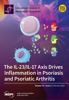
Cover Story (view full-size image): The IL-23/IL-17 axis mediates the initiation and persistence of inflammation during Psoriasis and Psoriatic Arthritis. IL-23 acts on numerous effector cells to drive the immune response in skin and joint disease either directly (IL-17-independent pathway) or indirectly by stimulating Th17 cells to release the inflammatory mediators IL-22 and IL-17 (IL-17-dependent pathway). View this paper
  • Issues are regarded as officially published after their release is announced to the table of contents alert mailing list.
  • You may sign up for e-mail alerts to receive table of contents of newly released issues.
  • PDF is the official format for papers published in both, html and pdf forms. To view the papers in pdf format, click on the "PDF Full-text" link, and use the free Adobe Reader to open them.
Order results
Result details
Section
Select all
Export citation of selected articles as:
19 pages, 3952 KiB  
Article
Crosstalk of PmCBFs and PmDAMs Based on the Changes of Phytohormones under Seasonal Cold Stress in the Stem of Prunus mume
by Kai Zhao 1, Yuzhen Zhou 1, Yushu Li 1, Xiaokang Zhuo 1, Sagheer Ahmad 1, Yu Han 1, Xue Yong 1 and Qixiang Zhang 1,2,*
1 Beijing Key Laboratory of Ornamental Plants Germplasm Innovation & Molecular Breeding, National Engineering Research Center for Floriculture, Beijing Laboratory of Urban and Rural Ecological Environment, Key Laboratory of Genetics and Breeding in Forest Trees and Ornamental Plants of Ministry of Education, School of Landscape Architecture, Beijing Forestry University, 100083 Beijing, China
2 Beijing Advanced Innovation Center for Tree Breeding by Molecular Design, Beijing Forestry University, 100083 Beijing, China
Int. J. Mol. Sci. 2018, 19(2), 15; https://doi.org/10.3390/ijms19020015 - 23 Jan 2018
Cited by 34 | Viewed by 5853
Abstract
Plants facing the seasonal variations always need a growth restraining mechanism when temperatures turn down. C-repeat binding factor (CBF) genes work essentially in the cold perception. Despite lots of researches on CBFs, the multiple crosstalk is still interesting on their [...] Read more.
Plants facing the seasonal variations always need a growth restraining mechanism when temperatures turn down. C-repeat binding factor (CBF) genes work essentially in the cold perception. Despite lots of researches on CBFs, the multiple crosstalk is still interesting on their interaction with hormones and dormancy-associated MADS (DAM) genes in the growth and dormancy control. Therefore, this study highlights roles of PmCBFs in cold-induced dormancy from different orgens. And a sense-response relationship between PmCBFs and PmDAMs is exhibited in this process, jointly regulated by six PmCBFs and PmDAM4–6. Meantime, GA3 and ABA showed negative and positive correlation with PmCBFs expression levels, respectively. We also find a high correlation between IAA and PmDAM1–3. Finally, we display the interaction mode of PmCBFs and PmDAMs, especially PmCBF1-PmDAM1. These results can disclose another view of molecular mechanism in plant growth between cold-response pathway and dormancy regulation together with genes and hormones. Full article
Show Figures

Figure 1

15 pages, 2416 KiB  
Article
Lasiodiplodia theobromae as a Producer of Biotechnologically Relevant Enzymes
by Carina Félix, Sofia Libório, Mariana Nunes, Rafael Félix, Ana S. Duarte, Artur Alves and Ana C. Esteves *
Department of Biology, CESAM—Centre for Environmental and Marine Studies, University of Aveiro, Campus Universitário de Santiago, 3810-193 Aveiro, Portugal
Int. J. Mol. Sci. 2018, 19(2), 29; https://doi.org/10.3390/ijms19020029 - 23 Jan 2018
Cited by 27 | Viewed by 6811
Abstract
Phytopathogenic fungi are known to produce several types of enzymes usually involved in plant cell wall degradation and pathogenesis. The increasing of global temperature may induce fungi, such as Lasiodiplodia theobromae (L. theobromae), to alter its behavior. Nonetheless, there is only [...] Read more.
Phytopathogenic fungi are known to produce several types of enzymes usually involved in plant cell wall degradation and pathogenesis. The increasing of global temperature may induce fungi, such as Lasiodiplodia theobromae (L. theobromae), to alter its behavior. Nonetheless, there is only limited information regarding the effect of temperature on L. theobromae production of enzymes. The need for new, thermostable enzymes, that are biotechnologically relevant, led us to investigate the effect of temperature on the production of several extracellular enzymatic activities by different L. theobromae strains. Fungi were grown at 25 °C, 30 °C and 37 °C and the enzymatic activities were detected by plate assays, quantified by spectrophotometric methods and characterized by zymography. The thermostability (25–80 °C) of the enzymes produced was also tested. Strains CAA019, CBS339.90, LA-SOL3, LA-SV1 and LA-MA-1 produced amylases, gelatinases, caseinases, cellulases, lipases, laccases, xylanases, pectinases and pectin liases. Temperature modulated the expression of the enzymes, and this effect was more visible when fungi were grown at 37 °C than at lower temperatures. Contrary to proteolytic and endoglucanolytic activities, whose highest activities were detected when fungi were grown at 30 °C, lipolytic activity was not detected at this growth temperature. Profiles of proteases and endoglucanases of fungi grown at different temperatures were characterized by zymography. Enzymes were shown to be more thermostable when fungi were grown at 30 °C. Proteases were active up to 50 °C and endoglucanases up to 70 °C. Lipases were the least stable, with activities detected up to 45 °C. The enzymatic profiles detected for L. theobromae strains tested showed to be temperature and strain-dependent, making this species a good target for biotechnological applications. Full article
(This article belongs to the Special Issue Microbial Enzymes)
Show Figures

Graphical abstract

12 pages, 2423 KiB  
Article
Peretinoin, an Acyclic Retinoid, Inhibits Hepatitis B Virus Replication by Suppressing Sphingosine Metabolic Pathway In Vitro
by Kazuhisa Murai 1,2,†, Takayoshi Shirasaki 1,2,†, Masao Honda 1,2,*, Ryogo Shimizu 1,2, Tetsuro Shimakami 1, Saki Nakasho 1,2, Natsumi Shirasaki 1, Hikari Okada 1, Yoshio Sakai 1, Taro Yamashita 1 and Shuichi Kaneko 1
1 Department of Gastroenterology, Kanazawa University Graduate School of Medical Science, Kanazawa 920-8641, Japan
2 Department of Advanced Medical Technology, Kanazawa University Graduate School of Health Medicine, Kanazawa 920-0942, Japan
These authors contributed equally to this work.
Int. J. Mol. Sci. 2018, 19(2), 108; https://doi.org/10.3390/ijms19020108 - 23 Jan 2018
Cited by 9 | Viewed by 6039
Abstract
Hepatocellular carcinoma (HCC) frequently develops from hepatitis C virus (HCV) and hepatitis B virus (HBV) infection. We previously reported that peretinoin, an acyclic retinoid, inhibits HCV replication. This study aimed to examine the influence of peretinoin on the HBV lifecycle. HBV-DNA and covalently [...] Read more.
Hepatocellular carcinoma (HCC) frequently develops from hepatitis C virus (HCV) and hepatitis B virus (HBV) infection. We previously reported that peretinoin, an acyclic retinoid, inhibits HCV replication. This study aimed to examine the influence of peretinoin on the HBV lifecycle. HBV-DNA and covalently closed circular DNA (cccDNA) were evaluated by a qPCR method in HepG2.2.15 cells. Peretinoin significantly reduced the levels of intracellular HBV-DNA, nuclear cccDNA, and HBV transcript at a concentration that did not induce cytotoxicity. Conversely, other retinoids, such as 9-cis, 13-cis retinoic acid (RA), and all-trans-retinoic acid (ATRA), had no effect or rather increased HBV replication. Mechanistically, although peretinoin increased the expression of HBV-related transcription factors, as observed for other retinoids, peretinoin enhanced the binding of histone deacetylase 1 (HDAC1) to cccDNA in the nucleus and negatively regulated HBV transcription. Moreover, peretinoin significantly inhibited the expression of SPHK1, a potential inhibitor of HDAC activity, and might be involved in hepatic inflammation, fibrosis, and HCC. SPHK1 overexpression in cells cancelled the inhibition of HBV replication induced by peretinoin. This indicates that peretinoin activates HDAC1 and thereby suppresses HBV replication by inhibiting the sphingosine metabolic pathway. Therefore, peretinoin may be a novel therapeutic agent for HBV replication and chemoprevention against HCC. Full article
(This article belongs to the Special Issue Inflammation and Cancer 2018)
Show Figures

Figure 1

11 pages, 2214 KiB  
Review
Melanins in Fossil Animals: Is It Possible to Infer Life History Traits from the Coloration of Extinct Species?
by Juan J. Negro 1,*, Clive Finlayson 2,3 and Ismael Galván 1
1 Department of Evolutionary Ecology, Doñana Biological Station—CSIC, 41092 Sevilla, Spain
2 The Gibraltar Museum, Gibraltar GX11 1AA, UK
3 Department of Anthropology, University of Toronto, Scarborough, ON M1C 1A4, Canada
Int. J. Mol. Sci. 2018, 19(2), 230; https://doi.org/10.3390/ijms19020230 - 23 Jan 2018
Cited by 15 | Viewed by 9340
Abstract
Paleo-colour scientists have recently made the transition from describing melanin-based colouration in fossil specimens to inferring life-history traits of the species involved. Two such cases correspond to counter-shaded dinosaurs: dark-coloured due to melanins dorsally, and light-coloured ventrally. We believe that colour reconstruction of [...] Read more.
Paleo-colour scientists have recently made the transition from describing melanin-based colouration in fossil specimens to inferring life-history traits of the species involved. Two such cases correspond to counter-shaded dinosaurs: dark-coloured due to melanins dorsally, and light-coloured ventrally. We believe that colour reconstruction of fossils based on the shape of preserved microstructures—the majority of paleo-colour studies involve melanin granules—is not without risks. In addition, animals with contrasting dorso-ventral colouration may be under different selection pressures beyond the need for camouflage, including, for instance, visual communication or ultraviolet (UV) protection. Melanin production is costly, and animals may invest less in areas of the integument where pigments are less needed. In addition, melanocytes exposed to UV radiation produce more melanin than unexposed melanocytes. Pigment economization may thus explain the colour pattern of some counter-shaded animals, including extinct species. Even in well-studied extant species, their diversity of hues and patterns is far from being understood; inferring colours and their functions in species only known from one or few specimens from the fossil record should be exerted with special prudence. Full article
(This article belongs to the Special Issue Melanins and Melanogenesis: From Nature to Applications)
Show Figures

Figure 1

21 pages, 5875 KiB  
Article
GC-MS Metabolomics to Evaluate the Composition of Plant Cuticular Waxes for Four Triticum aestivum Cultivars
by Florent D. Lavergne 1, Corey D. Broeckling 2, Darren M. Cockrell 3, Scott D. Haley 4, Frank B. Peairs 3, Courtney E. Jahn 3,* and Adam L. Heuberger 1,*
1 Department of Horticulture and Landscape Architecture, Colorado State University, Fort Collins, CO 80523, USA
2 Proteomics and Metabolomics Facility, Colorado State University, Fort Collins, CO 80523, USA
3 Department of Bioagricultural Sciences and Pest Management, Colorado State University, Fort Collins, CO 80523, USA
4 Department of Soil and Crop Sciences, Colorado State University, Fort Collins, CO 80523, USA
Int. J. Mol. Sci. 2018, 19(2), 249; https://doi.org/10.3390/ijms19020249 - 23 Jan 2018
Cited by 48 | Viewed by 8333
Abstract
Wheat (Triticum aestivum L.) is an important food crop, and biotic and abiotic stresses significantly impact grain yield. Wheat leaf and stem surface waxes are associated with traits of biological importance, including stress resistance. Past studies have characterized the composition of wheat [...] Read more.
Wheat (Triticum aestivum L.) is an important food crop, and biotic and abiotic stresses significantly impact grain yield. Wheat leaf and stem surface waxes are associated with traits of biological importance, including stress resistance. Past studies have characterized the composition of wheat cuticular waxes, however protocols can be relatively low-throughput and narrow in the range of metabolites detected. Here, gas chromatography-mass spectrometry (GC-MS) metabolomics methods were utilized to provide a comprehensive characterization of the chemical composition of cuticular waxes in wheat leaves and stems. Further, waxes from four wheat cultivars were assayed to evaluate the potential for GC-MS metabolomics to describe wax composition attributed to differences in wheat genotype. A total of 263 putative compounds were detected and included 58 wax compounds that can be classified (e.g., alkanes and fatty acids). Many of the detected wax metabolites have known associations to important biological functions. Principal component analysis and ANOVA were used to evaluate metabolite distribution, which was attributed to both tissue type (leaf, stem) and cultivar differences. Leaves contained more primary alcohols than stems such as 6-methylheptacosan-1-ol and octacosan-1-ol. The metabolite data were validated using scanning electron microscopy of epicuticular wax crystals which detected wax tubules and platelets. Conan was the only cultivar to display alcohol-associated platelet-shaped crystals on its abaxial leaf surface. Taken together, application of GC-MS metabolomics enabled the characterization of cuticular wax content in wheat tissues and provided relative quantitative comparisons among sample types, thus contributing to the understanding of wax composition associated with important phenotypic traits in a major crop. Full article
(This article belongs to the Special Issue Metabolomics in the Plant Sciences 2017)
Show Figures

Graphical abstract

19 pages, 8673 KiB  
Article
Solid Lipid Nanoparticles Carrying Temozolomide for Melanoma Treatment. Preliminary In Vitro and In Vivo Studies
by Nausicaa Clemente 1, Benedetta Ferrara 2, Casimiro Luca Gigliotti 1, Elena Boggio 1, Maria Teresa Capucchio 3, Elena Biasibetti 3, Davide Schiffer 4, Marta Mellai 4, Laura Annovazzi 4, Luigi Cangemi 2, Elisabetta Muntoni 2, Gianluca Miglio 2, Umberto Dianzani 1, Luigi Battaglia 2,* and Chiara Dianzani 2
1 Dipartimento di Scienze della Salute, Università del Piemonte Orientale, Via Solaroli, 17, 28100 Novara, Italy
2 Dipartimento di Scienza e Tecnologia del Farmaco, Università degli Studi di Torino, Via Pietro Giuria 9, 10124 Torino, Italy
3 Dipartimento di Scienze Veterinarie, Università degli Studi di Torino, Largo Paolo Braccini 2, 10095 Grugliasco (TO), Italy
4 Centro di Neuro Bio Oncologia, Policlinico di Monza, Via Pietro Micca 5, 13100 Vercelli, Italy
Int. J. Mol. Sci. 2018, 19(2), 255; https://doi.org/10.3390/ijms19020255 - 24 Jan 2018
Cited by 68 | Viewed by 8068
Abstract
Aim: To develop an innovative delivery system for temozolomide (TMZ) in solid lipid nanoparticles (SLN), which has been preliminarily investigated for the treatment of melanoma. Materials and Methods: SLN-TMZ was obtained through fatty acid coacervation. Its pharmacological effects were assessed and compared with [...] Read more.
Aim: To develop an innovative delivery system for temozolomide (TMZ) in solid lipid nanoparticles (SLN), which has been preliminarily investigated for the treatment of melanoma. Materials and Methods: SLN-TMZ was obtained through fatty acid coacervation. Its pharmacological effects were assessed and compared with free TMZ in in vitro and in vivo models of melanoma and glioblastoma. Results: Compared to the standard free TMZ, SLN-TMZ exerted larger effects, when cell proliferation of melanoma cells, and neoangiogeneis were evaluated. SLN-TMZ also inhibited growth and vascularization of B16-F10 melanoma in C57/BL6 mice, without apparent toxic effects. Conclusion: SLN could be a promising strategy for the delivery of TMZ, allowing an increased stability of the drug and thereby its employment in the treatment of aggressive malignacies. Full article
(This article belongs to the Special Issue Nanotechnology in Drug Delivery)
Show Figures

Graphical abstract

13 pages, 3592 KiB  
Article
Role of Disulfide Bonds in Activity and Stability of Tigerinin-1R
by Xiaolong Chen 1,2, Cuihua Hu 1,2, Yibing Huang 1,2 and Yuxin Chen 1,2,*
1 Key Laboratory for Molecular Enzymology and Engineering of the Ministry of Education, Jilin University, Changchun 130012, China
2 College of Life Sciences, Jilin University, Changchun 130012, China
Int. J. Mol. Sci. 2018, 19(2), 288; https://doi.org/10.3390/ijms19020288 - 23 Jan 2018
Cited by 1 | Viewed by 3701
Abstract
Tigerinin-1R (Arg–Val–Cys–Ser–Ala–Ile–Pro–Leu–Pro–Ile–Cys–His–NH2), a cationic 12-mer peptide containing a disulfide bond extracted from frog skin secretions, lacks antibacterial activity, but has the ability to stimulate insulin release both in vitro and in vivo. To study the structure–function relationships of tigerinin-1R, we designed [...] Read more.
Tigerinin-1R (Arg–Val–Cys–Ser–Ala–Ile–Pro–Leu–Pro–Ile–Cys–His–NH2), a cationic 12-mer peptide containing a disulfide bond extracted from frog skin secretions, lacks antibacterial activity, but has the ability to stimulate insulin release both in vitro and in vivo. To study the structure–function relationships of tigerinin-1R, we designed and synthesized five analogs, including tigerinin-cyclic, tigerinin-1R-L4, tigerinin-linear, [C3K]tigerinin-1R, and [C11K]tigerinin-1R. Tigerinin-1R promoted insulin secretion in a concentration-dependent manner in INS-1 cells without obvious cytotoxicity. At a concentration of 10−5 M, [C11K]tigerinin-1R exhibited the highest stimulation ability, suggesting that the positive charge at the C-terminus may contribute to the in vitro insulin-releasing activity of tigerinin-1R. Tigerinin-1R peptides stimulated insulin release in INS-1 cells through a universal mechanism that involves mobilization of intracellular calcium without disrupting the cell membrane. In vivo experiments showed that both tigerinin-1R and [C11K]tigerinin-1R improved glucose tolerance in overnight-fasted mice. Due to its structural stability, tigerinin-1R showed superior hypoglycemic activity to [C11K]tigerinin-1R, which suggested a critical role of the disulfide bonds. In addition, we also identified a protective effect of tigerinin-1R peptides in apoptosis induced by oxidative stress. These results further confirm the potential for the development of tigerinin-1R as an anti-diabetic therapeutic agent in clinical practice. Full article
(This article belongs to the Section Biochemistry)
Show Figures

Graphical abstract

16 pages, 1483 KiB  
Review
Signaling by Steroid Hormones in the 3D Nuclear Space
by François Le Dily 1,2 and Miguel Beato 1,2,*
1 Gene Regulation, Stem Cells and Cancer Program, Centre for Genomic Regulation (CRG), The Barcelona Institute of Science and Technology (BIST), Doctor Aiguader 88, 08003 Barcelona, Spain
2 Universitat Pompeu Fabra (UPF), 08003 Barcelona, Spain
Int. J. Mol. Sci. 2018, 19(2), 306; https://doi.org/10.3390/ijms19020306 - 23 Jan 2018
Cited by 46 | Viewed by 6263
Abstract
Initial studies showed that ligand-activated hormone receptors act by binding to the proximal promoters of individual target genes. Genome-wide studies have now revealed that regulation of transcription by steroid hormones mainly depends on binding of the receptors to distal regulatory elements. Those distal [...] Read more.
Initial studies showed that ligand-activated hormone receptors act by binding to the proximal promoters of individual target genes. Genome-wide studies have now revealed that regulation of transcription by steroid hormones mainly depends on binding of the receptors to distal regulatory elements. Those distal elements, either enhancers or silencers, act on the regulation of target genes by chromatin looping to the gene promoters. In the nucleus, this level of chromatin folding is integrated within dynamic higher orders of genome structures, which are organized in a non-random fashion. Terminally differentiated cells exhibit a tissue-specific three-dimensional (3D) organization of the genome that favors or restrains the activity of transcription factors and modulates the function of steroid hormone receptors, which are transiently activated upon hormone exposure. Conversely, integration of the hormones signal may require modifications of the 3D organization to allow appropriate transcriptional outcomes. In this review, we summarize the main levels of organization of the genome, review how they can modulate the response to steroids in a cell specific manner and discuss the role of receptors in shaping and rewiring the structure in response to hormone. Taking into account the dynamics of 3D genome organization will contribute to a better understanding of the pleiotropic effects of steroid hormones in normal and cancer cells. Full article
(This article belongs to the Special Issue Molecular Pathways of Estrogen Receptor Action)
Show Figures

Figure 1

16 pages, 3090 KiB  
Article
Selective Blockade of the Metabotropic Glutamate Receptor mGluR5 Protects Mouse Livers in In Vitro and Ex Vivo Models of Ischemia Reperfusion Injury
by Andrea Ferrigno 1,*, Clarissa Berardo 1, Laura Giuseppina Di Pasqua 1, Veronica Siciliano 1, Plinio Richelmi 1, Ferdinando Nicoletti 2,3 and Mariapia Vairetti 1
1 Department of Internal Medicine and Therapeutics, Cellular and Molecular Pharmacology and Toxicology Unit, University of Pavia, 27100 Pavia, Italy
2 Department of Physiology and Pharmacology, Sapienza University, 00185 Roma, Italy
3 I.R.C.C.S. Neuromed, 86077 Pozzilli, Italy
Int. J. Mol. Sci. 2018, 19(2), 314; https://doi.org/10.3390/ijms19020314 - 23 Jan 2018
Cited by 15 | Viewed by 6039
Abstract
2-Methyl-6-(phenylethynyl)pyridine (MPEP), a negative allosteric modulator of the metabotropic glutamate receptor (mGluR) 5, protects hepatocytes from ischemic injury. In astrocytes and microglia, MPEP depletes ATP. These findings seem to be self-contradictory, since ATP depletion is a fundamental stressor in ischemia. This study attempted [...] Read more.
2-Methyl-6-(phenylethynyl)pyridine (MPEP), a negative allosteric modulator of the metabotropic glutamate receptor (mGluR) 5, protects hepatocytes from ischemic injury. In astrocytes and microglia, MPEP depletes ATP. These findings seem to be self-contradictory, since ATP depletion is a fundamental stressor in ischemia. This study attempted to reconstruct the mechanism of MPEP-mediated ATP depletion and the consequences of ATP depletion on protection against ischemic injury. We compared the effects of MPEP and other mGluR5 negative modulators on ATP concentration when measured in rat hepatocytes and acellular solutions. We also evaluated the effects of mGluR5 blockade on viability in rat hepatocytes exposed to hypoxia. Furthermore, we studied the effects of MPEP treatment on mouse livers subjected to cold ischemia and warm ischemia reperfusion. We found that MPEP and 3-[(2-methyl-1,3-thiazol-4-yl)ethynyl]pyridine (MTEP) deplete ATP in hepatocytes and acellular solutions, unlike fenobam. This finding suggests that mGluR5s may not be involved, contrary to previous reports. MPEP, as well as MTEP and fenobam, improved hypoxic hepatocyte viability, suggesting that protection against ischemic injury is independent of ATP depletion. Significantly, MPEP protected mouse livers in two different ex vivo models of ischemia reperfusion injury, suggesting its possible protective deployment in the treatment of hepatic inflammatory conditions. Full article
Show Figures

Graphical abstract

19 pages, 5525 KiB  
Article
Identification of Proteins Involved in Carbohydrate Metabolism and Energy Metabolism Pathways and Their Regulation of Cytoplasmic Male Sterility in Wheat
by Xingxia Geng, Jiali Ye, Xuetong Yang, Sha Li, Lingli Zhang and Xiyue Song *
College of Agronomy, Northwest A&F University, Yangling 712100, Shaanxi, China
Int. J. Mol. Sci. 2018, 19(2), 324; https://doi.org/10.3390/ijms19020324 - 23 Jan 2018
Cited by 48 | Viewed by 7362
Abstract
Cytoplasmic male sterility (CMS) where no functional pollen is produced has important roles in wheat breeding. The anther is a unique organ for male gametogenesis and its abnormal development can cause male sterility. However, the mechanisms and regulatory networks related to plant male [...] Read more.
Cytoplasmic male sterility (CMS) where no functional pollen is produced has important roles in wheat breeding. The anther is a unique organ for male gametogenesis and its abnormal development can cause male sterility. However, the mechanisms and regulatory networks related to plant male sterility are poorly understood. In this study, we conducted comparative analyses using isobaric tags for relative and absolute quantification (iTRAQ) of the pollen proteins in a CMS line and its wheat maintainer. Differentially abundant proteins (DAPs) were analyzed based on Gene Ontology classifications, metabolic pathways and transcriptional regulation networks using Blast2GO. We identified 5570 proteins based on 23,277 peptides, which matched with 73,688 spectra, including proteins in key pathways such as glyceraldehyde-3-phosphate dehydrogenase, pyruvate kinase and 6-phosphofructokinase 1 in the glycolysis pathway, isocitrate dehydrogenase and citrate synthase in the tricarboxylic acid cycle and nicotinamide adenine dinucleotide (NADH)-dehydrogenase and adenosine-triphosphate (ATP) synthases in the oxidative phosphorylation pathway. These proteins may comprise a network that regulates male sterility in wheat. Quantitative real time polymerase chain reaction (qRT-PCR) analysis, ATP assays and total sugar assays validated the iTRAQ results. These DAPs could be associated with abnormal pollen grain formation and male sterility. Our findings provide insights into the molecular mechanism related to male sterility in wheat. Full article
(This article belongs to the Section Molecular Plant Sciences)
Show Figures

Graphical abstract

13 pages, 7502 KiB  
Article
Regulation of Akt/FoxO3a/Skp2 Axis Is Critically Involved in Berberine-Induced Cell Cycle Arrest in Hepatocellular Carcinoma Cells
by Fanni Li, Xiwen Dong, Peng Lin and Jianli Jiang *
State Key Laboratory of Cancer Biology, Cell Engineering Research Center & Department of Cell Biology, Fourth Military Medical University, 169 Changle West Road, Xi’an 710032, China
Int. J. Mol. Sci. 2018, 19(2), 327; https://doi.org/10.3390/ijms19020327 - 23 Jan 2018
Cited by 53 | Viewed by 7110
Abstract
The maintenance of ordinal cell cycle phases is a critical biological process in cancer genesis, which is a crucial target for anti-cancer drugs. As an important natural isoquinoline alkaloid from Chinese herbal medicine, Berberine (BBR) has been reported to possess anti-cancer potentiality to [...] Read more.
The maintenance of ordinal cell cycle phases is a critical biological process in cancer genesis, which is a crucial target for anti-cancer drugs. As an important natural isoquinoline alkaloid from Chinese herbal medicine, Berberine (BBR) has been reported to possess anti-cancer potentiality to induce cell cycle arrest in hepatocellular carcinoma cells (HCC). However, the underlying mechanism remains to be elucidated. In our present study, G0/G1 phase cell cycle arrest was observed in berberine-treated Huh-7 and HepG2 cells. Mechanically, we observed that BBR could deactivate the Akt pathway, which consequently suppressed the S-phase kinase-associated protein 2 (Skp2) expression and enhanced the expression and translocation of Forkhead box O3a (FoxO3a) into nucleus. The translocated FoxO3a on one hand could directly promote the transcription of cyclin-dependent kinase inhibitors (CDKIs) p21Cip1 and p27Kip1, on the other hand, it could repress Skp2 expression, both of which lead to up-regulation of p21Cip1 and p27Kip1, causing G0/G1 phase cell cycle arrest in HCC. In conclusion, BBR promotes the expression of CDKIs p21Cip1 and p27Kip1 via regulating the Akt/FoxO3a/Skp2 axis and further induces HCC G0/G1 phase cell cycle arrest. This research uncovered a new mechanism of an anti-cancer effect of BBR. Full article
(This article belongs to the Special Issue Cell Growth Regulation)
Show Figures

Figure 1

16 pages, 12559 KiB  
Article
Pharmacokinetic Study of Bioactive Flavonoids in the Traditional Japanese Medicine Keigairengyoto Exerting Antibacterial Effects against Staphylococcus aureus
by Takashi Matsumoto 1,*, Atsushi Kaneko 1, Junichi Koseki 1, Yosuke Matsubara 1, Setsuya Aiba 2 and Kenshi Yamasaki 2
1 Tsumura Kampo Research Laboratories, Kampo Research & Development Division, Tsumura & Co., Ibaraki 300-1192, Japan
2 Department of Dermatology, Tohoku University Graduate School of Medicine, Miyagi 980-8574, Japan
Int. J. Mol. Sci. 2018, 19(2), 328; https://doi.org/10.3390/ijms19020328 - 23 Jan 2018
Cited by 25 | Viewed by 6501
Abstract
Recent studies have demonstrated that flavonoid glucuronides can be deconjugated to the active form aglycone by β-glucuronidase-expressing macrophages. Keigairengyoto (KRT) is a flavonoid-rich traditional Japanese medicine reported to enhance bacterial clearance through immune modulation. Our aims are to examine the pharmacokinetics of KRT [...] Read more.
Recent studies have demonstrated that flavonoid glucuronides can be deconjugated to the active form aglycone by β-glucuronidase-expressing macrophages. Keigairengyoto (KRT) is a flavonoid-rich traditional Japanese medicine reported to enhance bacterial clearance through immune modulation. Our aims are to examine the pharmacokinetics of KRT flavonoids and to identify active flavonoids contributing to the adjuvant effects of KRT. KRT was evaluated at pharmacokinetic analysis to quantify absorbed flavonoids, and cutaneous infection assay induced in mice by inoculation of Staphylococcus aureus. Preventive or therapeutic KRT administration reduced the number of bacteria in the infection site as well as macroscopic and microscopic lesion scores with efficacies similar to antibiotics. Pharmacokinetic study revealed low plasma levels of flavonoid aglycones after KRT administration; however, plasma concentrations were enhanced markedly by β-glucuronidase treatment, with baicalein the most abundant (Cmax, 1.32 µg/mL). In random screening assays, flavonoids such as bacalein, genistein, and apigenin enhanced bacteria phagocytosis by macrophages. Glucuronide bacalin was converted to aglycone baicalein by incubation with living macrophages, macrophage lysate, or skin homogenate. Taken together, the adjuvant effect of KRT may be due to some blood-absorbed flavonoids which enhance macrophage functions in host defense. Flavonoid-rich KRT may be a beneficial treatment for infectious skin inflammation. Full article
(This article belongs to the Special Issue Bioactive Phenolics and Polyphenols 2018)
Show Figures

Graphical abstract

18 pages, 4276 KiB  
Article
Biotherapeutic Effect of Gingival Stem Cells Conditioned Medium in Bone Tissue Restoration
by Francesca Diomede 1, Agnese Gugliandolo 2, Domenico Scionti 2, Ilaria Merciaro 1, Marcos FXB Cavalcanti 3,4, Emanuela Mazzon 2,* and Oriana Trubiani 1
1 Department of Medical, Oral and Biotechnological Sciences, University “G. d’Annunzio” Chieti-Pescara, 66100 Chieti, Italy
2 IRCCS Centro Neurolesi “Bonino Pulejo”, 98124 Messina, Italy
3 Faculté de Médecine, UMR 7365 CNRS-Université de Lorraine, 9, Avenue de la Forêt de Haye, 54500 Vandoeuvre-lés-Nancy, France
4 Laser in Dentistry Program, Cruzeiro do Sul University (UNICSUL), Sao Paulo 08060-070, Brazil
Int. J. Mol. Sci. 2018, 19(2), 329; https://doi.org/10.3390/ijms19020329 - 23 Jan 2018
Cited by 75 | Viewed by 6681
Abstract
Bone tissue engineering is one of the main branches of regenerative medicine. In this field, the use of a scaffold, which supported bone development, in combination with mesenchymal stem cells (MSCs), has promised better outcomes for bone regeneration. In particular, human gingival mesenchymal [...] Read more.
Bone tissue engineering is one of the main branches of regenerative medicine. In this field, the use of a scaffold, which supported bone development, in combination with mesenchymal stem cells (MSCs), has promised better outcomes for bone regeneration. In particular, human gingival mesenchymal stem cells (hGMSCs) may present advantages compared to other MSCs, including the easier isolation. However, MSCs’ secretome has attracted much attention for its potential use in tissue regeneration, such as conditioned medium (CM) that contains different soluble factors proved to be useful for the regenerative purposes. In this study, we evaluated the osteogenic capacity of a poly-(lactide) (3D-PLA) scaffold enriched with hGMSCs and hGMSCs derived CM and its ability to regenerate bone defects in rat calvarias. 3D-PLA alone, 3D-PLA + CM or 3D-PLA + hGMSCs with/without CM were implanted in Wistar male rats subjected to calvarial defects. We observed that 3D-PLA scaffold enriched with hGMSCs and CM showed a better osteogenic capacity, being able to repair the calvarial defect as revealed in vivo by morphological evaluation. Moreover, transcriptomic analysis in vitro revealed the upregulation of genes involved in ossification and regulation of ossification in the 3D-PLA + CM + hGMSCs group. All of these results indicate the great osteogenic ability of 3D-PLA + CM + hGMSCs supporting its use in bone regenerative medicine, in particular in the repair of cranial bone defects. Especially, hGMSCs derived CM played a key role in the induction of the osteogenic process and in bone regeneration. Full article
(This article belongs to the Special Issue Cell Colonization in Scaffolds)
Show Figures

Figure 1

11 pages, 4181 KiB  
Article
Involvement of Bradykinin B2 Receptor in Pathological Vascularization in Oxygen-Induced Retinopathy in Mice and Rabbit Cornea
by Erika Terzuoli, Lucia Morbidelli, Ginevra Nannelli, Antonio Giachetti, Sandra Donnini * and Marina Ziche *
Department of Life Sciences, University of Siena, Via A. Moro 2, 53100 Siena, Italy
Int. J. Mol. Sci. 2018, 19(2), 330; https://doi.org/10.3390/ijms19020330 - 23 Jan 2018
Cited by 8 | Viewed by 6027
Abstract
The identification of components of the kallikrein–kinin system in the vitreous from patients with microvascular retinal diseases suggests that bradykinin (BK) signaling may contribute to pathogenesis of retinal vascular complications. BK receptor 2 (B2R) signaling has been implicated in both pro-inflammatory and pro-angiogenic [...] Read more.
The identification of components of the kallikrein–kinin system in the vitreous from patients with microvascular retinal diseases suggests that bradykinin (BK) signaling may contribute to pathogenesis of retinal vascular complications. BK receptor 2 (B2R) signaling has been implicated in both pro-inflammatory and pro-angiogenic effects promoted by BK. Here, we investigated the role of BK/B2R signaling in the retinal neovascularization in the oxygen-induced retinopathy (OIR) model. Blockade of B2R signaling by the antagonist fasitibant delayed retinal vascularization in mouse pups, indicating that the retinal endothelium is a target of the BK/B2R system. In the rabbit cornea assay, a model of pathological neoangiogenesis, the B2 agonist kallidin induced vessel sprouting and promoted cornea opacity, a sign of edema and tissue inflammation. In agreement with these results, in the OIR model, a blockade of B2R signaling significantly reduced retinal neovascularization, as determined by the area of retinal tufts, and, in the retinal vessel, it also reduced vascular endothelial growth factor and fibroblast growth factor-2 expression. All together, these findings show that B2R blockade reduces retinal neovascularization and inhibits the expression of proangiogenic and pro-inflammatory cytokines, suggesting that targeting B2R signaling may be an effective strategy for treating ischemic retinopathy. Full article
(This article belongs to the Special Issue Retinal Diseases: Bridging Basic and Clinical Research)
Show Figures

Graphical abstract

20 pages, 4121 KiB  
Article
The Differentiation of Rat Oligodendroglial Cells Is Highly Influenced by the Oxygen Tension: In Vitro Model Mimicking Physiologically Normoxic Conditions
by Justyna Janowska, Malgorzata Ziemka-Nalecz and Joanna Sypecka *
NeuroRepair Department, Mossakowski Medical Research Centre, Polish Academy of Sciences, 5 Pawinskiego Street, 02-106 Warsaw, Poland
Int. J. Mol. Sci. 2018, 19(2), 331; https://doi.org/10.3390/ijms19020331 - 24 Jan 2018
Cited by 14 | Viewed by 5212
Abstract
Oligodendrocyte progenitor cells (OPCs) constitute one of the main populations of dividing cells in the central nervous system (CNS). Physiologically, OPCs give rise to mature, myelinating oligodendrocytes and confer trophic support to their neighboring cells within the nervous tissue. OPCs are known to [...] Read more.
Oligodendrocyte progenitor cells (OPCs) constitute one of the main populations of dividing cells in the central nervous system (CNS). Physiologically, OPCs give rise to mature, myelinating oligodendrocytes and confer trophic support to their neighboring cells within the nervous tissue. OPCs are known to be extremely sensitive to the influence of exogenous clues which might affect their crucial biological processes, like survival, proliferation, differentiation, and the ability to generate a myelin membrane. Alterations in their differentiation influencing their final potential for myelinogenesis are usually the leading cause of CNS dys- and demyelination, contributing to the development of leukodystrophic disorders. The evaluation of the mechanisms that cause oligodendrocytes to malfunction requires detailed studies based on designed in vitro models. Since OPCs readily respond to changes in local homeostasis, it is crucial to establish restricted culture conditions to eliminate the potential stimuli that might influence oligodendrocyte biology. Additionally, the in vitro settings should mimic the physiological conditions to enable the obtained results to be translated to future preclinical studies. Therefore, the aim of our study was to investigate OPC differentiation in physiological normoxia (5% O2) and a restricted in vitro microenvironment. To evaluate the impact of the combined microenvironmental clues derived from other components of the nervous tissue, which are also influenced by the local oxygen concentration, the process of generating OPCs was additionally analyzed in organotypic hippocampal slices. The obtained results show that OPC differentiation, although significantly slowed down, proceeded correctly through its typical stages in the physiologically relevant conditions created in vitro. The established settings were also conducive to efficient cell proliferation, exerting also a neuroprotective effect by promoting the proliferation of neurons. In conclusion, the performed studies show how oxygen tension influences OPC proliferation, differentiation, and their ability to express myelin components, and should be taken into consideration while planning preclinical studies, e.g., to examine neurotoxic compounds or to test neuroprotective strategies. Full article
(This article belongs to the Special Issue Neuroprotective Strategies 2022)
Show Figures

Graphical abstract

13 pages, 8949 KiB  
Article
Visualization and Quantitative 3D Analysis of Intraocular Melanoma and Its Vascularization in a Hamster Eye
by Bartosz Leszczyński 1, Martyna Śniegocka 2, Andrzej Wróbel 1, Roman Pędrys 1, Małgorzata Szczygieł 2, Bożena Romanowska-Dixon 3, Krystyna Urbańska 2 and Martyna Elas 2,*
1 Marian Smoluchowski Institute of Physics, Jagiellonian University, Prof. Stanisława Łojasiewicza 11 Street, 30-348 Krakow, Poland
2 Faculty of Biochemistry, Biophysics and Biotechnology, Jagiellonian University, Gronostajowa 7 Street, 30-387 Krakow, Poland
3 Ophthalmology and Ocular Oncology Clinic, University Hospital, Kopernika 38 Street, 31-501 Krakow, Poland
Int. J. Mol. Sci. 2018, 19(2), 332; https://doi.org/10.3390/ijms19020332 - 24 Jan 2018
Cited by 7 | Viewed by 5109
Abstract
A tumor vasculature network undergoes intense growth and rebuilding during tumor growth. Traditionally, vascular networks are histologically examined using parameters such as vessel density determined from two-dimensional slices of the tumor. Two-dimensional probing of a complicated three-dimensional (3D) structure only provides partial information. [...] Read more.
A tumor vasculature network undergoes intense growth and rebuilding during tumor growth. Traditionally, vascular networks are histologically examined using parameters such as vessel density determined from two-dimensional slices of the tumor. Two-dimensional probing of a complicated three-dimensional (3D) structure only provides partial information. Therefore, we propose the use of microcomputed tomography (micro-CT) imaging to analyze the evolution of a tumor vasculature in an experimental ocular tumor model. A Bomirski Hamster Melanoma was implanted in the anterior chamber of a hamster eye. Ultrasound (US) imaging of the same tumor was performed in vivo, and the vascular results obtained using the two methods were compared. Normal ocular tissues, a tumor, and a tumor vascular structure were revealed with high accuracy using micro-CT. The vessels that grew within the tumor were chaotic, leaky, and contained many convoluted micro-vessels and embolizations. They comprised 20–38% of the tumor mass. The blood flow in the larger functional vessels was in the range from 10 to 25 mm/s, as determined by in vivo Doppler US. The micro-CT imaging of the hamster eyeball enabled both qualitative and quantitative 3D analyses of the globe at a histological level. Although the presented images were obtained ex vivo, micro-CT noninvasive imaging is being developed intensively, and high-resolution in vivo imaging is feasible. Full article
(This article belongs to the Special Issue Animal Models of Melanoma)
Show Figures

Graphical abstract

28 pages, 5965 KiB  
Review
Current Perspectives of Telomerase Structure and Function in Eukaryotes with Emerging Views on Telomerase in Human Parasites
by Abhishek Dey and Kausik Chakrabarti *
Department of Biological Sciences, The University of North Carolina at Charlotte, 9201 University City Boulevard, Charlotte, NC 28223, USA
Int. J. Mol. Sci. 2018, 19(2), 333; https://doi.org/10.3390/ijms19020333 - 24 Jan 2018
Cited by 24 | Viewed by 9100
Abstract
Replicative capacity of a cell is strongly correlated with telomere length regulation. Aberrant lengthening or reduction in the length of telomeres can lead to health anomalies, such as cancer or premature aging. Telomerase is a master regulator for maintaining replicative potential in most [...] Read more.
Replicative capacity of a cell is strongly correlated with telomere length regulation. Aberrant lengthening or reduction in the length of telomeres can lead to health anomalies, such as cancer or premature aging. Telomerase is a master regulator for maintaining replicative potential in most eukaryotic cells. It does so by controlling telomere length at chromosome ends. Akin to cancer cells, most single-cell eukaryotic pathogens are highly proliferative and require persistent telomerase activity to maintain constant length of telomere and propagation within their host. Although telomerase is key to unlimited cellular proliferation in both cases, not much was known about the role of telomerase in human parasites (malaria, Trypanosoma, etc.) until recently. Since telomerase regulation is mediated via its own structural components, interactions with catalytic reverse transcriptase and several factors that can recruit and assemble telomerase to telomeres in a cell cycle-dependent manner, we compare and discuss here recent findings in telomerase biology in cancer, aging and parasitic diseases to give a broader perspective of telomerase function in human diseases. Full article
(This article belongs to the Special Issue Role of Telomeres and Telomerase in Cancer and Aging)
Show Figures

Figure 1

17 pages, 15288 KiB  
Article
Unveiling a Selective Mechanism for the Inhibition of α-Synuclein Aggregation by β-Synuclein
by Andre Leitao 1,2, Akshay Bhumkar 1,2, Dominic J. B. Hunter 1,3, Yann Gambin 1,2,* and Emma Sierecki 1,2,*
1 European Molecular Biology Laboratory (EMBL), Australia Node in Single Molecule Science, Sydney NSW 2031, Australia
2 School of Medical Sciences, The University of New South Wales, Sydney NSW 2031, Australia
3 Institute for Molecular Bioscience, The University of Queensland, St Lucia QLD 4076, Australia
Int. J. Mol. Sci. 2018, 19(2), 334; https://doi.org/10.3390/ijms19020334 - 24 Jan 2018
Cited by 19 | Viewed by 6637
Abstract
α-Synuclein (αS) is an intrinsically disordered protein that is associated with Parkinson’s disease (PD) through its ability to self-assemble into oligomers and fibrils. Inhibition of this oligomerization cascade is an interesting approach to developing therapeutical strategies and β-synuclein (βS) has been described as [...] Read more.
α-Synuclein (αS) is an intrinsically disordered protein that is associated with Parkinson’s disease (PD) through its ability to self-assemble into oligomers and fibrils. Inhibition of this oligomerization cascade is an interesting approach to developing therapeutical strategies and β-synuclein (βS) has been described as a natural negative regulator of this process. However, the biological background and molecular mechanisms by which this inhibition occurs is unclear. Herein, we focused on assessing the effect of βS on the aggregation of five αS pathological mutants linked to early-onset PD (A30P, E46K, H50Q, G51D and A53T). By coupling single molecule fluorescence spectroscopy to a cell-free protein expression system, we validated the ability of βS to act as a chaperone of αS, effectively inhibiting its aggregation. Interestingly, we found that βS does so in a selective manner, i.e., is a more effective inhibitor for certain αS pathological mutants—A30P and G51D—as compared to E46K, H50Q and A53T. Moreover, two-color coincidence experiments proved that this discrepancy is due to a preferential incorporation of βS into smaller oligomers of αS. This was validated by showing that the chaperoning effect was lost when proteins were mixed after being expressed individually. This study highlights the potential of fluorescence spectroscopy to deconstruct αS aggregation cascade and its interplay with βS. Full article
(This article belongs to the Special Issue Protein Folding)
Show Figures

Graphical abstract

16 pages, 537 KiB  
Review
Lignins: Biosynthesis and Biological Functions in Plants
by Qingquan Liu 1,2,†, Le Luo 3,† and Luqing Zheng 1,*
1 College of Life Sciences, Nanjing Agricultural University, Nanjing 210095, China
2 Institute of Botany, Jiangsu Province and Chinese Academy of Sciences, Nanjing 210014, China
3 College of Resources and Environmental Sciences, Nanjing Agricultural University, Nanjing 210095, China
These authors contribute equally to this work.
Int. J. Mol. Sci. 2018, 19(2), 335; https://doi.org/10.3390/ijms19020335 - 24 Jan 2018
Cited by 1046 | Viewed by 35282
Abstract
Lignin is one of the main components of plant cell wall and it is a natural phenolic polymer with high molecular weight, complex composition and structure. Lignin biosynthesis extensively contributes to plant growth, tissue/organ development, lodging resistance and the responses to a variety [...] Read more.
Lignin is one of the main components of plant cell wall and it is a natural phenolic polymer with high molecular weight, complex composition and structure. Lignin biosynthesis extensively contributes to plant growth, tissue/organ development, lodging resistance and the responses to a variety of biotic and abiotic stresses. In the present review, we systematically introduce the biosynthesis of lignin and its regulation by genetic modification and summarize the main biological functions of lignin in plants and their applications. We hope this review will give an in-depth understanding of the important roles of lignin biosynthesis in various plants’ biological processes and provide a theoretical basis for the genetic improvement of lignin content and composition in energy plants and crops. Full article
(This article belongs to the Section Molecular Plant Sciences)
Show Figures

Graphical abstract

65 pages, 8532 KiB  
Review
Insights into the Molecular Mechanisms of Alzheimer’s and Parkinson’s Diseases with Molecular Simulations: Understanding the Roles of Artificial and Pathological Missense Mutations in Intrinsically Disordered Proteins Related to Pathology
by Orkid Coskuner-Weber 1,* and Vladimir N. Uversky 2,3,*
1 Türkisch-Deutsche Universität, Theoretical and Computational Biophysics Group, Molecular Biotechnology, Sahinkaya Caddesi, No. 86, Beykoz, Istanbul 34820, Turkey
2 Department of Molecular Medicine and USF Health Byrd Alzheimer’s Research Institute, Morsani College of Medicine, University of South Florida, Tampa, FL 33612, USA
3 Laboratory of New Methods in Biology, Institute for Biological Instrumentation, Russian Academy of Sciences, 142290 Pushchino, Moscow Region, Russia
Int. J. Mol. Sci. 2018, 19(2), 336; https://doi.org/10.3390/ijms19020336 - 24 Jan 2018
Cited by 54 | Viewed by 11260
Abstract
Amyloid-β and α-synuclein are intrinsically disordered proteins (IDPs), which are at the center of Alzheimer’s and Parkinson’s disease pathologies, respectively. These IDPs are extremely flexible and do not adopt stable structures. Furthermore, both amyloid-β and α-synuclein can form toxic oligomers, amyloid fibrils and [...] Read more.
Amyloid-β and α-synuclein are intrinsically disordered proteins (IDPs), which are at the center of Alzheimer’s and Parkinson’s disease pathologies, respectively. These IDPs are extremely flexible and do not adopt stable structures. Furthermore, both amyloid-β and α-synuclein can form toxic oligomers, amyloid fibrils and other type of aggregates in Alzheimer’s and Parkinson’s diseases. Experimentalists face challenges in investigating the structures and thermodynamic properties of these IDPs in their monomeric and oligomeric forms due to the rapid conformational changes, fast aggregation processes and strong solvent effects. Classical molecular dynamics simulations complement experiments and provide structural information at the atomic level with dynamics without facing the same experimental limitations. Artificial missense mutations are employed experimentally and computationally for providing insights into the structure-function relationships of amyloid-β and α-synuclein in relation to the pathologies of Alzheimer’s and Parkinson’s diseases. Furthermore, there are several natural genetic variations that play a role in the pathogenesis of familial cases of Alzheimer’s and Parkinson’s diseases, which are related to specific genetic defects inherited in dominant or recessive patterns. The present review summarizes the current understanding of monomeric and oligomeric forms of amyloid-β and α-synuclein, as well as the impacts of artificial and pathological missense mutations on the structural ensembles of these IDPs using molecular dynamics simulations. We also emphasize the recent investigations on residual secondary structure formation in dynamic conformational ensembles of amyloid-β and α-synuclein, such as β-structure linked to the oligomerization and fibrillation mechanisms related to the pathologies of Alzheimer’s and Parkinson’s diseases. This information represents an important foundation for the successful and efficient drug design studies. Full article
Show Figures

Graphical abstract

11 pages, 3101 KiB  
Article
An in Vitro Study on the Effect of Combined Treatment with Photodynamic and Chemical Therapies on Candida albicans
by Yi-Hsuan Hsieh 1, Jun-Hui Zhang 2, Wen-Ching Chuang 2, Kun-Hua Yu 2, Xian-Bin Huang 2, Yao-Chang Lee 3 and Cheng-I Lee 2,*
1 Department of Clinical Pathology, Buddhist Dalin Tzu Chi General Hospital, Chia-Yi 62247, Taiwan
2 Department of Biomedical Sciences, National Chung Cheng University, Min-Hsiung, Chia-Yi 62102, Taiwan
3 National Synchrotron Radiation Research Center, Hsinchu 30076, Taiwan
Int. J. Mol. Sci. 2018, 19(2), 337; https://doi.org/10.3390/ijms19020337 - 24 Jan 2018
Cited by 38 | Viewed by 5973
Abstract
Candida albicans is the most commonly encountered human fungal pathogen, and it is traditionally treated with antimicrobial chemical agents. The antimicrobial effect of these agents is largely weakened by drug resistance and biofilm-associated virulence. Enhancement of the antimicrobial activity of existing agents is [...] Read more.
Candida albicans is the most commonly encountered human fungal pathogen, and it is traditionally treated with antimicrobial chemical agents. The antimicrobial effect of these agents is largely weakened by drug resistance and biofilm-associated virulence. Enhancement of the antimicrobial activity of existing agents is needed for effective candidiasis treatment. Our aim was to develop a therapy that combined biofilm disruption with existing antimicrobial agents. Photodynamic therapy (PDT) utilizing curcumin and blue light was tested as an independent therapy and in combination with fluconazole treatment. Viability assays and morphology analysis were used to assess the effectiveness of C. albicans treatment. Results showed that fluconazole treatment decreased the viability of planktonic C. albicans, but the decrease was not as pronounced in adherent C. albicans because its biofilm form was markedly more resistant to the antimicrobiotic. PDT effectively eradicated C. albicans biofilms, and when combined with fluconazole, PDT significantly inhibited C. albicans to a greater extent. This study suggests that the addition of PDT to fluconazole to treat C. albicans infection enhances its effectiveness and can potentially be used clinically. Full article
(This article belongs to the Special Issue Laser Application in Life Sciences 2018)
Show Figures

Graphical abstract

10 pages, 238 KiB  
Article
The Prevalence of Helicobacter pylori in Estonian Bariatric Surgery Patients
by Natalja Šebunova 1, Jelena Štšepetova 1,2,*, Toomas Sillakivi 3 and Reet Mändar 1,2
1 Institute of Biomedicine and Translational Medicine, University of Tartu, Ravila Street 19, 50411 Tartu, Estonia
2 Competence Centre on Health Technologies, Tiigi Street 61B, 50410 Tartu, Estonia
3 Abdominal Surgery Department of Tartu University Hospital, Puusepa Street 8, 51014 Tartu, Estonia
Int. J. Mol. Sci. 2018, 19(2), 338; https://doi.org/10.3390/ijms19020338 - 24 Jan 2018
Cited by 12 | Viewed by 4179
Abstract
Helicobacter pylori (Hp) is one of the most important human pathogens that can cause duodenal and gastric ulcers, gastritis and stomach cancer. Hp infection is considered to be a cause of limiting access to bariatric surgery. The aim of this study [...] Read more.
Helicobacter pylori (Hp) is one of the most important human pathogens that can cause duodenal and gastric ulcers, gastritis and stomach cancer. Hp infection is considered to be a cause of limiting access to bariatric surgery. The aim of this study was to determine the prevalence of Hp in patients with obesity going into bariatric surgery and to reveal the relationship between Hp and clinical data. The study group was formed of 68 preoperative bariatric surgery patients (body mass index (BMI) 44.7 ± 4.8). Gastric biopsies (antrum and corpus) were used for histological and molecular (caqA and glmM genes) examinations. The PCR method revealed Hp infection in 64.7% of obese patients that is higher in comparison with histological analysis (55.9%). The prevalence of cagA and glmM genes in antrum mucosa was 45.6% and 47.0% while in the corpus it was 41.2% and 38.3%, respectively. The coincidence of both cagA and glmM virulence genes in the antrum and corpus mucosa was 33.8% and 22.1%, respectively. Either of the genes was found in 58.8% of antrum and 57.3% of corpus mucosa. Presence of caqA and glmM genes was in association with active and atrophic chronic gastritis. In conclusion, our study demonstrated that two thirds of morbidly obese patients undergoing bariatric surgery are infected with Hp and have a high prevalence of cagA and glmM virulence genes that points out the necessity for diagnostics and treatment of this infection before surgery. Full article
(This article belongs to the Special Issue Helicobacter pylori Research)
16 pages, 2341 KiB  
Article
Elucidating the Role of CD84 and AHR in Modulation of LPS-Induced Cytokines Production by Cruciferous Vegetable-Derived Compounds Indole-3-Carbinol and 3,3′-Diindolylmethane
by Thomas T. Y. Wang 1,*, Quynhchi Pham 1 and Young S. Kim 2,*
1 Diet, Genomics and Immunology Lab, Beltsville Human Nutrition Research Center, The Agricultural Research Service (ARS), United States Department of Agriculture (USDA), Beltsville, MD 20705, USA
2 Nutritional Science Research Group, Division of Cancer Prevention, National Cancer Institute, Rockville, MD 20850, USA
Int. J. Mol. Sci. 2018, 19(2), 339; https://doi.org/10.3390/ijms19020339 - 24 Jan 2018
Cited by 19 | Viewed by 6323
Abstract
Modulation of the immune system by cancer protective food bioactives has preventive and therapeutic importance in prostate cancer, but the mechanisms remain largely unclear. The current study tests the hypothesis that the diet-derived cancer protective compounds, indole-3-carbinol (I3C) and 3,3′-diindolylmethane (DIM), affect the [...] Read more.
Modulation of the immune system by cancer protective food bioactives has preventive and therapeutic importance in prostate cancer, but the mechanisms remain largely unclear. The current study tests the hypothesis that the diet-derived cancer protective compounds, indole-3-carbinol (I3C) and 3,3′-diindolylmethane (DIM), affect the tumor microenvironment by regulation of inflammatory responses in monocytes and macrophages. We also ask whether I3C and DIM act through the aryl hydrocarbon (AHR)-dependent pathway or the signaling lymphocyte activation molecule (SLAM) family protein CD84-mediated pathway. The effect of I3C and DIM was examined using the human THP-1 monocytic cell in its un-differentiated (monocyte) and differentiated (macrophage) state. We observed that I3C and DIM inhibited lipopolysaccharide (LPS) induction of IL-1β mRNA and protein in the monocyte form but not the macrophage form of THP-1. Interestingly, CD84 mRNA but not protein was inhibited by I3C and DIM. AHR siRNA knockdown experiments confirmed that the inhibitory effects of I3C and DIM on IL-1β as well as CD84 mRNA are regulated through AHR-mediated pathways. Additionally, the AHR ligand appeared to differentially regulate other LPS-induced cytokines expression. Hence, cross-talk between AHR and inflammation-mediated pathways, but not CD84-mediated pathways, in monocytes but not macrophages may contribute to the modulation of tumor environments by I3C and DIM in prostate cancer. Full article
(This article belongs to the Special Issue Natural Bioactives and Phytochemicals in Cancer Prevention)
Show Figures

Graphical abstract

16 pages, 622 KiB  
Review
Incorporation of Immune Checkpoint Blockade into Chimeric Antigen Receptor T Cells (CAR-Ts): Combination or Built-In CAR-T
by Dok Hyun Yoon 1,2,3,4,†, Mark J. Osborn 3,4,5,6,7,*,†, Jakub Tolar 3,4,5,6,7 and Chong Jai Kim 2
1 Department of Oncology, Asan Medical Center, University of Ulsan College of Medicine, Seoul 05505, Korea
2 Asan-Minnesota Institute for Innovating Transplantation, Asan Institute for Life Sciences, Asan Medical Center, University of Ulsan College of Medicine, Seoul 05505, Korea
3 Asan-Minnesota Institute for Innovating Transplantation, University of Minnesota, Minneapolis, MN 55455, USA
4 Department of Pediatrics, University of Minnesota Medical School, Division of Blood and Marrow Transplantation, Minneapolis, MN 55455, USA
5 Masonic Cancer Center, University of Minnesota, Minneapolis, MN 55455, USA
6 Stem Cell Institute, University of Minnesota, Minneapolis, MN 55455, USA
7 Center for Genome Engineering, University of Minnesota, Minneapolis, MN 55455, USA
These authors contributed equally to this work.
Int. J. Mol. Sci. 2018, 19(2), 340; https://doi.org/10.3390/ijms19020340 - 24 Jan 2018
Cited by 169 | Viewed by 14027
Abstract
Chimeric antigen receptor (CAR) T cell therapy represents the first U.S. Food and Drug Administration approved gene therapy and these engineered cells function with unprecedented efficacy in the treatment of refractory CD19 positive hematologic malignancies. CAR translation to solid tumors is also being [...] Read more.
Chimeric antigen receptor (CAR) T cell therapy represents the first U.S. Food and Drug Administration approved gene therapy and these engineered cells function with unprecedented efficacy in the treatment of refractory CD19 positive hematologic malignancies. CAR translation to solid tumors is also being actively investigated; however, efficacy to date has been variable due to tumor-evolved mechanisms that inhibit local immune cell activity. To bolster the potency of CAR-T cells, modulation of the immunosuppressive tumor microenvironment with immune-checkpoint blockade is a promising strategy. The impact of this approach on hematological malignancies is in its infancy, and in this review we discuss CAR-T cells and their synergy with immune-checkpoint blockade. Full article
(This article belongs to the Special Issue Chimeric Antigen Receptor (CAR) T Cell Therapy)
Show Figures

Figure 1

21 pages, 4488 KiB  
Article
Erinacine A-Enriched Hericium erinaceus Mycelium Produces Antidepressant-Like Effects through Modulating BDNF/PI3K/Akt/GSK-3β Signaling in Mice
by Chun-Hung Chiu 1, Charng-Cherng Chyau 1, Chin-Chu Chen 2,3,4,5,6, Li-Ya Lee 2, Wan-Ping Chen 2, Jia-Ling Liu 1, Wen-Hsin Lin 7 and Mei-Chin Mong 8,9,*
1 Research Institute of Biotechnology, HungKuang University, Taichung 43302, Taiwan
2 Bioengineering Center, Grape King Bio Ltd., Taoyuan City 32471, Taiwan
3 Department of Food Science, Nutrition, and Nutraceutical Biotechnology, Shih Chien University, Taipei 10462, Taiwan
4 Institute of Food Science and Technology, National Taiwan University, Taipei City 10617, Taiwan
5 Department of Bioscience Technology, Chung Yuan Christian University, Taoyuan City 32023, Taiwan
6 Institute of Biotechnology, National Changhua University of Education, Changhua County 50007, Taiwan
7 School of Pharmacy, China Medical University, Taichung 40402, Taiwan
8 Department of Food Nutrition and Health Biotechnology, Asia University, Taichung 41354, Taiwan
9 Department of Medical Research, China Medical University Hospital, China Medical University, Taichung 40402, Taiwan
Int. J. Mol. Sci. 2018, 19(2), 341; https://doi.org/10.3390/ijms19020341 - 24 Jan 2018
Cited by 93 | Viewed by 19076
Abstract
Antidepressant-like effects of ethanolic extract of Hericium erinaceus (HE) mycelium enriched in erinacine A on depressive mice challenged by repeated restraint stress (RS) were examined. HE at 100, 200 or 400 mg/kg body weight/day was orally given to mice for four weeks. After [...] Read more.
Antidepressant-like effects of ethanolic extract of Hericium erinaceus (HE) mycelium enriched in erinacine A on depressive mice challenged by repeated restraint stress (RS) were examined. HE at 100, 200 or 400 mg/kg body weight/day was orally given to mice for four weeks. After two weeks of HE administration, all mice except the control group went through with 14 days of RS protocol. Stressed mice exhibited various behavioral alterations, such as extending immobility time in the tail suspension test (TST) and forced swimming test (FST), and increasing the number of entries in open arm (POAE) and the time spent in the open arm (PTOA). Moreover, the levels of norepinephrine (NE), dopamine (DA) and serotonin (5-HT) were decreased in the stressed mice, while the levels of interleukin (IL)-6 and tumor necrosis factor (TNF)-α were increased. These changes were significantly inverted by the administration of HE, especially at the dose of 200 or 400 mg/kg body weight/day. Additionally, HE was shown to activate the BDNF/TrkB/PI3K/Akt/GSK-3β pathways and block the NF-κB signals in mice. Taken together, erinacine A-enriched HE mycelium could reverse the depressive-like behavior caused by RS and was accompanied by the modulation of monoamine neurotransmitters as well as pro-inflammatory cytokines, and regulation of BDNF pathways. Therefore, erinacine A-enriched HE mycelium could be an attractive agent for the treatment of depressive disorders. Full article
(This article belongs to the Section Bioactives and Nutraceuticals)
Show Figures

Figure 1

15 pages, 1573 KiB  
Article
Molecular Understanding of the Activation of CB1 and Blockade of TRPV1 Receptors: Implications for Novel Treatment Strategies in Osteoarthritis
by Jakub Mlost 1,2, Magdalena Kostrzewa 1, Natalia Malek 1 and Katarzyna Starowicz 1,2,*
1 Laboratory of Pain Pathophysiology, Department of Pain Pharmacology, Institute of Pharmacology Polish Academy of Sciences, 31-343 Krakow, Poland
2 Department of Neurochemistry, Institute of Pharmacology Polish Academy of Sciences, 31-343 Krakow, Poland
Int. J. Mol. Sci. 2018, 19(2), 342; https://doi.org/10.3390/ijms19020342 - 24 Jan 2018
Cited by 27 | Viewed by 7898
Abstract
Osteoarthritis (OA) is a joint disease in which cartilage degenerates as a result of mechanical and biochemical changes. The main OA symptom is chronic pain involving both peripheral and central mechanisms of nociceptive processing. Our previous studies have implicated the benefits of dual- [...] Read more.
Osteoarthritis (OA) is a joint disease in which cartilage degenerates as a result of mechanical and biochemical changes. The main OA symptom is chronic pain involving both peripheral and central mechanisms of nociceptive processing. Our previous studies have implicated the benefits of dual- over single-acting compounds interacting with the endocannabinoid system (ECS) in OA treatment. In the present study, we focused on the specific molecular alterations associated with pharmacological treatment. OA was induced in Wistar rats by intra-articular injection of 3 mg of monoiodoacetate (MIA). Single target compounds (URB597, an FAAH inhibitor, and SB366791, a TRPV1 antagonist) and a dual-acting compound OMDM198 (FAAH inhibitor/TRPV1 antagonist) were used in the present study. At day 21 post-MIA injection, rats were sacrificed 1 h after i.p. treatment, and changes in mRNA expression were evaluated in the lumbar spinal cord by RT-qPCR. Following MIA administration, we observed 2-4-fold increase in mRNA expression of targeted receptors (Cnr1, Cnr2, and Trpv1), endocannabinoid degradation enzymes (Faah, Ptgs2, and Alox12), and TRPV1 sensitizing kinases (Mapk3, Mapk14, Prkcg, and Prkaca). OMDM198 treatment reversed some of the MIA effects on the spinal cord towards intact levels (Alox12, Mapk14, and Prkcg). Apparent regulation of ECS and TRPV1 in response to pharmacological intervention is a strong justification for novel ECS-based multi-target drug treatment in OA. Full article
(This article belongs to the Special Issue Cannabinoid Signaling in Nervous System)
Show Figures

Graphical abstract

14 pages, 18258 KiB  
Article
Disrupting the Btk Pathway Suppresses COPD-Like Lung Alterations in Atherosclerosis Prone ApoE−/− Mice Following Regular Exposure to Cigarette Smoke
by Jon M. Florence 1, Agnieszka Krupa 1,2, Laela M. Booshehri 1, Adrian L. Gajewski 1,2 and Anna K. Kurdowska 1,*
1 Department of Cellular and Molecular Biology, University of Texas Health Science Center, Tyler, TX 75708, USA
2 Laboratory of Gastroimmunology, Department of Immunology and Infectious Biology, Faculty of Biology and Environmental Protection, University of Lodz, 90-237 Lodz, Poland
Int. J. Mol. Sci. 2018, 19(2), 343; https://doi.org/10.3390/ijms19020343 - 24 Jan 2018
Cited by 16 | Viewed by 5954
Abstract
Chronic obstructive pulmonary disease (COPD) is associated with severe chronic inflammation that promotes irreversible tissue destruction. Moreover, the most broadly accepted cause of COPD is exposure to cigarette smoke. There is no effective cure and significantly, the mechanism behind the development and progression [...] Read more.
Chronic obstructive pulmonary disease (COPD) is associated with severe chronic inflammation that promotes irreversible tissue destruction. Moreover, the most broadly accepted cause of COPD is exposure to cigarette smoke. There is no effective cure and significantly, the mechanism behind the development and progression of this disease remains unknown. Our laboratory has demonstrated that Bruton’s tyrosine kinase (Btk) is a critical regulator of pro-inflammatory processes in the lungs and that Btk controls expression of matrix metalloproteinase-9 (MMP-9) in the alveolar compartment. For this study apolipoprotein E null (ApoE−/−) mice were exposed to SHS to facilitate study in a COPD/atherosclerosis comorbidity model. We applied two types of treatments, animals received either a pharmacological inhibitor of Btk or MMP-9 specific siRNA to minimize MMP-9 expression in endothelial cells or neutrophils. We have shown that these treatments had a protective effect in the lung. We have noted a decrease in alveolar changes related to SHS induced inflammation in treated animals. In summary, we are presenting a novel concept in the field of COPD, i.e., that Btk may be a new drug target for this disease. Moreover, cell specific targeting of MMP-9 may also benefit patients affected by this disease. Full article
(This article belongs to the Special Issue Lung Diseases: Chronic Respiratory Infections)
Show Figures

Figure 1

15 pages, 5025 KiB  
Article
Lycopene Attenuates Tulathromycin and Diclofenac Sodium-Induced Cardiotoxicity in Mice
by Mohamed M. Abdel-Daim 1,*, Rasha Eltaysh 2, Azza Hassan 3 and Shaker A. Mousa 4
1 Pharmacology Department, Faculty of Veterinary Medicine, Suez Canal University, Ismailia 41522, Egypt
2 Department of Pharmacology, Faculty of Veterinary Medicine, Mansoura University, Mansoura 35516, Egypt
3 Pathology Department, Faculty of Veterinary Medicine, Cairo University, Giza 12122, Egypt
4 Pharmaceutical Research Institute, Albany College of Pharmacy and Health Sciences, New York, NY 12144, USA
Int. J. Mol. Sci. 2018, 19(2), 344; https://doi.org/10.3390/ijms19020344 - 24 Jan 2018
Cited by 61 | Viewed by 8232
Abstract
Recent experiments showed a potential cardiotoxic effect of the macrolide antibiotic (tulathromycin). This study was performed to investigate whether diclofenac sodium (DFS) potentiates the cardiotoxicity of tulathromycin and increases the cardioprotective effects of lycopene against DFS and tulathromycin. Seven groups (eight per group) [...] Read more.
Recent experiments showed a potential cardiotoxic effect of the macrolide antibiotic (tulathromycin). This study was performed to investigate whether diclofenac sodium (DFS) potentiates the cardiotoxicity of tulathromycin and increases the cardioprotective effects of lycopene against DFS and tulathromycin. Seven groups (eight per group) of adult Swiss albino mice received saline (control), tulathromycin (a single subcutaneous dose of 28 mg/kg/bw on day 14), DFS (a single oral dose of 100 mg/kg/bw on day 14), tulathromycin plus DFS, or lycopene (oral, 10 mg/kg/bw daily for 15 d) combined with tulathromycin, DFS, or both. Compared to the control group, the administration of tulathromycin or DFS (individually or in combination) caused significantly elevated (p < 0.05) serum levels of Creatine kinase-myocardial B fraction (CK-MB), lactate dehydrogenase, and cardiac-specific troponin-T and tissue levels of nitric oxide and malondialdehyde that were accompanied by significantly decreased tissue reduced glutathione content and glutathione peroxidase, superoxide dismutase, and catalase antioxidant enzyme activity. Upon histopathological and immunohistochemical examination, the mean pathology scores and the percentages of caspase-3-, Bax-, and CK-positive regions were significantly higher in the tulathromycin- and/or DFS-treated groups than in control mice. For all these parameters, the pathological changes were more significant in the tulathromycin–DFS combination group than in mice treated with either drug individually. Interestingly, co-administration of lycopene with tulathromycin and/or DFS significantly ameliorated the changes described above. In conclusion, DFS could potentiate the cardiotoxic effects of tulathromycin, whereas lycopene can serve as a cardioprotective agent against DFS and tulathromycin. Full article
(This article belongs to the Special Issue The Mechanism of Action of Food Components in Disease Prevention 2017)
Show Figures

Graphical abstract

9 pages, 1756 KiB  
Case Report
Late-Onset N-Acetylglutamate Synthase Deficiency: Report of a Paradigmatic Adult Case Presenting with Headaches and Review of the Literature
by Catia Cavicchi 1,†, Chiara Chilleri 1,†, Antonella Fioravanti 2, Lorenzo Ferri 1, Francesco Ripandelli 3, Cinzia Costa 3, Paolo Calabresi 3, Paolo Prontera 4, Francesca Pochiero 5, Elisabetta Pasquini 5, Silvia Funghini 6, Giancarlo La Marca 6,7, Maria Alice Donati 5 and Amelia Morrone 1,8,*
1 Molecular and Cell Biology Laboratory of Neurometabolic Diseases, Neuroscience Department, Meyer Children’s Hospital, 50139 Florence, Italy
2 Structural Biology Researcher Center, VIB, Vrije Universiteit Brussel, 1050 Brussels, Belgium
3 Neurology Unit, Santa Maria della Misericordia Hospital, 06123 Perugia, Italy
4 Medical Genetics Unit, Santa Maria della Misericordia Hospital, 06123 Perugia, Italy
5 Metabolic and Muscular Unit, Neuroscience Department, Meyer Children’s Hospital, 50139 Florence, Italy
6 Newborn Screening, Biochemistry and Pharmacology Laboratory, Neuroscience Department, Meyer Children’s Hospital, 50139 Florence, Italy
7 Department of Experimental and Clinical Biomedical Sciences, University of Florence, 50139 Florence, Italy
8 Department of Neurosciences, Psychology, Drug Research and Child Health (NEUROFARBA), University of Florence, 50139 Florence, Italy
These authors contributed equally to this work.
Int. J. Mol. Sci. 2018, 19(2), 345; https://doi.org/10.3390/ijms19020345 - 24 Jan 2018
Cited by 10 | Viewed by 6085
Abstract
N-acetylglutamate synthase deficiency (NAGSD) is an extremely rare urea cycle disorder (UCD) with few adult cases so far described. Diagnosis of late-onset presentations is difficult and delayed treatment may increase the risk of severe hyperammonemia. We describe a 52-year-old woman with recurrent [...] Read more.
N-acetylglutamate synthase deficiency (NAGSD) is an extremely rare urea cycle disorder (UCD) with few adult cases so far described. Diagnosis of late-onset presentations is difficult and delayed treatment may increase the risk of severe hyperammonemia. We describe a 52-year-old woman with recurrent headaches who experienced an acute onset of NAGSD. As very few papers focus on headaches in UCDs, we also report a literature review of types and pathophysiologic mechanisms of UCD-related headaches. In our case, headaches had been present since puberty (3–4 days a week) and were often accompanied by nausea, vomiting, or behavioural changes. Despite three previous episodes of altered consciousness, ammonia was measured for the first time at 52 years and levels were increased. Identification of the new homozygous c.344C>T (p.Ala115Val) NAGS variant allowed the definite diagnosis of NAGSD. Bioinformatic analysis suggested that an order/disorder alteration of the mutated form could affect the arginine-binding site, resulting in poor enzyme activation and late-onset presentation. After optimized treatment for NAGSD, ammonia and amino acid levels were constantly normal and prevented other headache bouts. The manuscript underlies that headache may be the presenting symptom of UCDs and provides clues for the rapid diagnosis and treatment of late-onset NAGSD. Full article
(This article belongs to the Section Molecular Pathology, Diagnostics, and Therapeutics)
Show Figures

Graphical abstract

15 pages, 9518 KiB  
Article
Deciphering the Functional Composition of Fusogenic Liposomes
by Rejhana Kolašinac, Christian Kleusch, Tobias Braun, Rudolf Merkel and Agnes Csiszár *
Forschungszentrum Jülich GmbH, Institute of Complex Systems (ICS-7), Biomechanics, 52425 Jülich, Germany
Int. J. Mol. Sci. 2018, 19(2), 346; https://doi.org/10.3390/ijms19020346 - 24 Jan 2018
Cited by 80 | Viewed by 11584
Abstract
Cationic liposomes are frequently used as carrier particles for nucleic acid delivery. The most popular formulation is the equimolar mixture of two components, a cationic lipid and a neutral phosphoethanolamine. Its uptake pathway has been described as endocytosis. The presence of an aromatic [...] Read more.
Cationic liposomes are frequently used as carrier particles for nucleic acid delivery. The most popular formulation is the equimolar mixture of two components, a cationic lipid and a neutral phosphoethanolamine. Its uptake pathway has been described as endocytosis. The presence of an aromatic molecule as a third component strongly influences the cellular uptake process and results in complete membrane fusion instead of endocytosis. Here, we systematically varied all three components of this lipid mixture and determined how efficiently the resulting particles fused with the plasma membrane of living mammalian cells. Our results show that an aromatic molecule and a cationic lipid component with conical molecular shape are essential for efficient fusion induction. While a neutral lipid is not mandatory, it can be used to control fusion efficiency and, in the most extreme case, to revert the uptake mechanism back to endocytosis. Full article
(This article belongs to the Special Issue Membrane Fusion)
Show Figures

Graphical abstract

29 pages, 2366 KiB  
Review
Regulation of Three Virulence Strategies of Mycobacterium tuberculosis: A Success Story
by Niels A. Zondervan 1, Jesse C. J. Van Dam 1, Peter J. Schaap 1, Vitor A. P. Martins dos Santos 1,2 and Maria Suarez-Diez 1,*
1 Laboratory of Systems and Synthetic Biology, Wageningen University & Research, Stippeneng 4, 6708 WE Wageningen, The Netherlands
2 LifeGlimmer GmbH, Markelstrasse 38, 12163 Berlin, Germany
Int. J. Mol. Sci. 2018, 19(2), 347; https://doi.org/10.3390/ijms19020347 - 24 Jan 2018
Cited by 36 | Viewed by 9718
Abstract
Tuberculosis remains one of the deadliest diseases. Emergence of drug-resistant and multidrug-resistant M. tuberculosis strains makes treating tuberculosis increasingly challenging. In order to develop novel intervention strategies, detailed understanding of the molecular mechanisms behind the success of this pathogen is required. Here, we [...] Read more.
Tuberculosis remains one of the deadliest diseases. Emergence of drug-resistant and multidrug-resistant M. tuberculosis strains makes treating tuberculosis increasingly challenging. In order to develop novel intervention strategies, detailed understanding of the molecular mechanisms behind the success of this pathogen is required. Here, we review recent literature to provide a systems level overview of the molecular and cellular components involved in divalent metal homeostasis and their role in regulating the three main virulence strategies of M. tuberculosis: immune modulation, dormancy and phagosomal rupture. We provide a visual and modular overview of these components and their regulation. Our analysis identified a single regulatory cascade for these three virulence strategies that respond to limited availability of divalent metals in the phagosome. Full article
(This article belongs to the Special Issue Molecular Mechanism of Infectious Disease)
Show Figures

Figure 1

13 pages, 2701 KiB  
Article
Cytoprotective Mechanisms in Fatty Liver Preservation against Cold Ischemia Injury: A Comparison between IGL-1 and HTK
by Arnau Panisello-Roselló 1, Eva Verde 2, Alexandre Lopez 3, Marta Flores 2, Emma Folch-Puy 1,4, Anabela Rolo 5, Carlos Palmeira 5, Georgina Hotter 1, Teresa Carbonell 2, René Adam 3 and Joan Roselló-Catafau 1,4,*
1 Experimental Hepatic Ischemia-Reperfusion Unit, Institut d’Investigacions Biomèdiques de Barcelona (IIBB), Spanish National Research Council (CSIC), 08036 Barcelona, Catalonia, Spain
2 Department of Cell Biology, Physiology and Immunology, Universitat de Barcelona, Barcelona, 08028 Catalonia, Spain
3 Centre Hépato-Biliaire, AP-PH, Hôpital Paul Brousse, 94800 Villejuif, France
4 Centro de Investigación Biomédica en Red de Enfermedades Hepáticas y Digestivas (CIBEREHD), Institut d’Investigacions Biomèdiques August Pi i Sunyer (IDIBAPS), 08036 Barcelona, Catalonia, Spain
5 Center for Neuroscience and Cell Biology, Universidade Coimbra, 3000-370 Coimbra, Portugal
Int. J. Mol. Sci. 2018, 19(2), 348; https://doi.org/10.3390/ijms19020348 - 24 Jan 2018
Cited by 19 | Viewed by 6280
Abstract
Institute Goeorges Lopez 1 (IGL-1) and Histidine-Tryptophan-Ketoglutarate (HTK) preservation solutions are regularly used in clinical for liver transplantation besides University of Wisconsin (UW) solution and Celsior. Several clinical trials and experimental works have been carried out comparing all the solutions, however the comparative [...] Read more.
Institute Goeorges Lopez 1 (IGL-1) and Histidine-Tryptophan-Ketoglutarate (HTK) preservation solutions are regularly used in clinical for liver transplantation besides University of Wisconsin (UW) solution and Celsior. Several clinical trials and experimental works have been carried out comparing all the solutions, however the comparative IGL-1 and HTK appraisals are poor; especially when they deal with the underlying protection mechanisms of the fatty liver graft during cold storage. Fatty livers from male obese Zücker rats were conserved for 24 h at 4 °C in IGL-1 or HTK preservation solutions. After organ recovery and rinsing of fatty liver grafts with Ringer Lactate solution, we measured the changes in mechanistic target of rapamycin (mTOR) signaling activation, liver autophagy markers (Beclin-1, Beclin-2, LC3B and ATG7) and apoptotic markers (caspase 3, caspase 9 and TUNEL). These determinations were correlated with the prevention of liver injury (aspartate and alanine aminostransferase (AST/ALT), histology) and mitochondrial damage (glutamate dehydrogenase (GLDH) and confocal microscopy findings). Liver grafts preserved in IGL-1 solution showed a marked reduction on p-TOR/mTOR ratio when compared to HTK. This was concomitant with significant increased cyto-protective autophagy and prevention of liver apoptosis, including inflammatory cytokines such as HMGB1. Together, our results revealed that IGL-1 preservation solution better protected fatty liver grafts against cold ischemia damage than HTK solution. IGL-1 protection was associated with a reduced liver damage, higher induced autophagy and decreased apoptosis. All these effects would contribute to limit the subsequent extension of reperfusion injury after graft revascularization in liver transplantation procedures. Full article
Show Figures

Graphical abstract

29 pages, 3662 KiB  
Article
Workers’ Exposure to Nano-Objects with Different Dimensionalities in R&D Laboratories: Measurement Strategy and Field Studies
by Fabio Boccuni 1,*, Riccardo Ferrante 1, Francesca Tombolini 1, Daniela Lega 2, Alessandra Antonini 2, Antonello Alvino 2, Pasqualantonio Pingue 3, Fabio Beltram 3, Lucia Sorba 3, Vincenzo Piazza 4,†, Mauro Gemmi 4, Andrea Porcari 5 and Sergio Iavicoli 1
1 Italian Workers’ Compensation Authority—Department of Occupational and Environmental Medicine, Epidemiology and Hygiene, Via Fontana Candida 1, I-00078 Rome, Italy
2 Italian Workers’ Compensation Authority—Department of Technologies, via del Torraccio di Torrenova 7, I-00133 Rome, Italy
3 Laboratorio NEST—Scuola Normale Superiore and Istituto Nanoscienze—CNR, Piazza San Silvestro 12, I-56127 Pisa, Italy
4 Laboratorio NEST—Scuola Normale Superiore and Italian Institute of Technology (IIT)—Center for Nanotechnology Innovation, Piazza San Silvestro 12, I-56127 Pisa, Italy
5 Italian Association for Industrial Research (AIRI), Viale Gorizia 25/c, I-00198 Rome, Italy
Current address: Sentec Ltd., 5 The Westbrook Centre, Milton Road, Cambridge CB4 1YG, UK.
Int. J. Mol. Sci. 2018, 19(2), 349; https://doi.org/10.3390/ijms19020349 - 24 Jan 2018
Cited by 23 | Viewed by 5445
Abstract
With the increasing interest in the potential benefits of nanotechnologies, concern is still growing that they may present emerging risks for workers. Various strategies have been developed to assess the exposure to nano-objects and their agglomerates and aggregates (NOAA) in the workplace, integrating [...] Read more.
With the increasing interest in the potential benefits of nanotechnologies, concern is still growing that they may present emerging risks for workers. Various strategies have been developed to assess the exposure to nano-objects and their agglomerates and aggregates (NOAA) in the workplace, integrating different aerosol measurement instruments and taking into account multiple parameters that may influence NOAA toxicity. The present study proposes a multi-metric approach for measuring and sampling NOAA in the workplace, applied to three case studies in laboratories each dedicated to materials with different shapes and dimensionalities: graphene, nanowires, and nanoparticles. The study is part of a larger project with the aim of improving risk management tools in nanomaterials research laboratories. The harmonized methodology proposed by the Organization for Economic Cooperation and Development (OECD) has been applied, including information gathering about materials and processes, measurements with easy-to-use and hand-held real-time devices, air sampling with personal samplers, and off-line analysis using scanning electron microscopy. Significant values beyond which an emission can be attributed to the NOAA production process were identified by comparison of the particle number concentration (PNC) time series and the corresponding background levels in the three laboratories. We explored the relations between background PNC and microclimatic parameters. Morphological and elemental analysis of sampled filters was done to identify possible emission sources of NOAA during the production processes: rare particles, spherical, with average diameter similar to the produced NOAA were identified in the nanoparticles laboratory, so further investigation is recommended to confirm the potential for worker exposure. In conclusion, the information obtained should provide a valuable basis for improving risk management strategies in the laboratory at work. Full article
(This article belongs to the Special Issue Nanotoxicology and Nanosafety)
Show Figures

Graphical abstract

18 pages, 2541 KiB  
Article
Comparative Digital Gene Expression Analysis of Tissue-Cultured Plantlets of Highly Resistant and Susceptible Banana Cultivars in Response to Fusarium oxysporum
by Yuqing Niu 1, Bei Hu 1, Xiaoquan Li 2, Houbin Chen 1, Tomáš Takáč 3, Jozef Šamaj 3 and Chunxiang Xu 1,*
1 College of Horticulture, South China Agricultural University, Guangzhou 510642, China
2 Institute of Biotechnology, Guangxi Academy of Agricultural Sciences, Nanning 530007, China
3 Centre of the Region Haná for Biotechnological and Agricultural Research, Department of Cell Biology, Faculty of Science, Palacký University, 783 01 Olomouc, Czech Republic
Int. J. Mol. Sci. 2018, 19(2), 350; https://doi.org/10.3390/ijms19020350 - 24 Jan 2018
Cited by 29 | Viewed by 7068
Abstract
Banana Fusarium wilt caused by Fusarium oxysporum f. sp. cubense (Foc) is one of the most destructive soil-borne diseases. In this study, young tissue-cultured plantlets of banana (Musa spp. AAA) cultivars differing in Foc susceptibility were used to reveal their [...] Read more.
Banana Fusarium wilt caused by Fusarium oxysporum f. sp. cubense (Foc) is one of the most destructive soil-borne diseases. In this study, young tissue-cultured plantlets of banana (Musa spp. AAA) cultivars differing in Foc susceptibility were used to reveal their differential responses to this pathogen using digital gene expression (DGE). Data were evaluated by various bioinformatic tools (Venn diagrams, gene ontology (GO) annotation and Kyoto encyclopedia of genes and genomes (KEGG) pathway analyses) and immunofluorescence labelling method to support the identification of gene candidates determining the resistance of banana against Foc. Interestingly, we have identified MaWRKY50 as an important gene involved in both constitutive and induced resistance. We also identified new genes involved in the resistance of banana to Foc, including several other transcription factors (TFs), pathogenesis-related (PR) genes and some genes related to the plant cell wall biosynthesis or degradation (e.g., pectinesterases, β-glucosidases, xyloglucan endotransglucosylase/hydrolase and endoglucanase). The resistant banana cultivar shows activation of PR-3 and PR-4 genes as well as formation of different constitutive cell barriers to restrict spreading of the pathogen. These data suggest new mechanisms of banana resistance to Foc. Full article
(This article belongs to the Section Molecular Plant Sciences)
Show Figures

Graphical abstract

21 pages, 1896 KiB  
Review
Nanofiltration and Tight Ultrafiltration Membranes for the Recovery of Polyphenols from Agro-Food By-Products
by Alfredo Cassano 1,*, Carmela Conidi 1, René Ruby-Figueroa 2 and Roberto Castro-Muñoz 3
1 Institute on Membrane Technology, ITM-CNR, c/o University of Calabria, via Pietro Bucci, 17/C, 87036 Rende (CS), Italy
2 Programa Institucional de Fomento a la Investigación, Desarrollo e Innovación, Universidad Tecnológica Metropolitana, Ignacio Valdivieso 2409, San Joaquín, Santiago 8940577, Chile
3 Department of Inorganic Technology, University of Chemistry and Technology Prague Technická 5, 166 28 Prague 6, Czech Republic
Int. J. Mol. Sci. 2018, 19(2), 351; https://doi.org/10.3390/ijms19020351 - 24 Jan 2018
Cited by 209 | Viewed by 13533
Abstract
Pressure-driven membrane-based technologies represent a valid approach to reduce the environmental pollution of several agro-food by-products. Recently, in relation to the major interest for natural compounds with biological activities, their use has been also addressed to the recovery, separation and fractionation of phenolic [...] Read more.
Pressure-driven membrane-based technologies represent a valid approach to reduce the environmental pollution of several agro-food by-products. Recently, in relation to the major interest for natural compounds with biological activities, their use has been also addressed to the recovery, separation and fractionation of phenolic compounds from such by-products. In particular, tight ultrafiltration (UF) and nanolfiltration (NF) membranes have been recognized for their capability to recover phenolic compounds from several types of agro-food by-products. The separation capability of these membranes, as well as their productivity, depends on multiple factors such as membrane material, molecular weight cut-off (MWCO) and operating conditions (e.g., pressure, temperature, feed flow rate, volume reduction factor, etc.). This paper aims at providing a critical overview of the influence of these parameters on the recovery of phenolic compounds from agro-food by-products by using tight UF and NF membranes. The literature data are analyzed and discussed in relation to separation processes, molecule properties, membrane characteristics and other phenomena occurring in the process. Current extraction methodologies of phenolic compounds from raw materials are also introduced in order to drive the implementation of integrated systems for the production of actractive phenolic formulations of potential interest as food antioxidants. Full article
(This article belongs to the Special Issue Bioactive Phenolics and Polyphenols 2018)
Show Figures

Graphical abstract

14 pages, 3936 KiB  
Article
Tauroursodeoxycholic Acid Protects against the Effects of P-Cresol-Induced Reactive Oxygen Species via the Expression of Cellular Prion Protein
by Seung Pil Yun 1, Yeo Min Yoon 2, Jun Hee Lee 3, Minjee Kook 1, Yong-Seok Han 2, Seo Kyung Jung 2 and Sang Hun Lee 2,*
1 Department of Neurology, Institute for Cell Engineering, Johns Hopkins University School of Medicine, Baltimore, MD 21205, USA
2 Institute of Soonchunhyang Medical Science Research, Soonchunhyang University Hospital Seoul, Soonchunhyang University, Seoul 04401, Korea
3 Department of Pharmacology and Toxicology, University of Alabama at Birmingham School of Medicine, Birmingham, AL 35294, USA
Int. J. Mol. Sci. 2018, 19(2), 352; https://doi.org/10.3390/ijms19020352 - 25 Jan 2018
Cited by 15 | Viewed by 4657
Abstract
Mesenchymal stem cells (MSCs) could be a promising solution in the treatment of various diseases including chronic kidney disease (CKD). However, endoplasmic reticulum (ER) stress induced by ischemia in the area of application limits the integration and survival of MSCs in patients. In [...] Read more.
Mesenchymal stem cells (MSCs) could be a promising solution in the treatment of various diseases including chronic kidney disease (CKD). However, endoplasmic reticulum (ER) stress induced by ischemia in the area of application limits the integration and survival of MSCs in patients. In our study, we generated ER stress-induced conditions in MSCs using P-cresol. As P-cresol is a toxic compound accumulated in the body of CKD patients and induces apoptosis and inflammation through reactive oxygen species (ROS), we observed ER stress-induced MSC apoptosis activated by oxidative stress, which in turn resulted from ROS generation. To overcome stress-induced apoptosis, we investigated the protective effects of tauroursodeoxycholic acid (TUDCA), a bile acid, on ER stress in MSCs. In ER stress, TUDCA treatment of MSCs reduced ER stress-associated protein activation, including GRP78, PERK, eIF2α, ATF4, IRE1α, and CHOP. Next, to explore the protective mechanism adopted by TUDCA, TUDCA-mediated cellular prion protein (PrPC) activation was assessed. We confirmed that PrPC expression significantly increased ROS, which was eliminated by superoxide dismutase and catalase in MSCs. These findings suggest that TUDCA protects from inflammation and apoptosis in ER stress via PrPC expression. Our study demonstrates that TUDCA protects MSCs against inflammation and apoptosis in ER stress by PrPC expression in response to P-cresol exposure. Full article
(This article belongs to the Section Biochemistry)
Show Figures

Figure 1

16 pages, 5175 KiB  
Article
Mitochondrial BK Channel Openers CGS7181 and CGS7184 Exhibit Cytotoxic Properties
by Bartłomiej Augustynek 1,2, Piotr Koprowski 1, Daria Rotko 1, Wolfram S. Kunz 3, Adam Szewczyk 1 and Bogusz Kulawiak 1,*
1 Laboratory of Intracellular Ion Channels, Nencki Institute of Experimental Biology, 3 Pasteur St., 02-093 Warsaw, Poland
2 Institute of Biochemistry and Molecular Medicine, University of Bern, Bühlstrasse 28, CH-3012 Bern, Switzerland
3 Department of Experimental Epileptology and Cognition Research and Department of Epileptology, University of Bonn, 25 Sigmund-Freud Strasse, D-53105 Bonn, Germany
Int. J. Mol. Sci. 2018, 19(2), 353; https://doi.org/10.3390/ijms19020353 - 25 Jan 2018
Cited by 18 | Viewed by 7134
Abstract
Potassium channel openers (KCOs) have been shown to play a role in cytoprotection through the activation of mitochondrial potassium channels. Recently, in several reports, a number of data has been described as off-target actions for KCOs. In the present study, we investigated the [...] Read more.
Potassium channel openers (KCOs) have been shown to play a role in cytoprotection through the activation of mitochondrial potassium channels. Recently, in several reports, a number of data has been described as off-target actions for KCOs. In the present study, we investigated the effects of BKCa channel openers CGS7181, CGS7184, NS1619, and NS004 in neuronal cells. For the purpose of this research, we used a rat brain, the mouse hippocampal HT22 cells, and the human astrocytoma U-87 MG cell line. We showed that CGS7184 activated the mitochondrial BKCa (mitoBKCa) channel in single-channel recordings performed on astrocytoma mitoplasts. Moreover, when applied to the rat brain homogenate or isolated rat brain mitochondria, CGS7184 increased the oxygen consumption rate, and can thus be considered a potentially cytoprotective agent. However, experiments on intact neuronal HT22 cells revealed that both CGS7181 and CGS7184 induced HT22 cell death in a concentration- and time-dependent manner. By contrast, we did not observe cell death when NS1619 or NS004 was applied. CGS7184 toxicity was not abolished by BKCa channel inhibitors, suggesting that the observed effects were independent of a BKCa-type channel activity. CGS7184 treatment resulted in an increase of cytoplasmic Ca2+ concentration that likely involved efflux from internal calcium stores and the activation of calpains (calcium-dependent proteases). The cytotoxic effect of the channel opener was partially reversed by a calpain inhibitor. Our data show that KCOs under study not only activate mitoBKCa channels from brain tissue, but also induce cell death when used in cellular models. Full article
(This article belongs to the Special Issue Ion Transporters and Channels in Physiology and Pathophysiology)
Show Figures

Graphical abstract

15 pages, 5820 KiB  
Article
The Effects of Varying Degree of MWCNT Carboxylation on Bioactivity in Various In Vivo and In Vitro Exposure Models
by Raymond F. Hamilton, Jr. 1, Zheqiong Wu 2, Somenath Mitra 2 and Andrij Holian 1,*
1 Department of Biomedical and Pharmaceutical Sciences, Center for Environmental Health Sciences, University of Montana, Missoula, MT 59812, USA
2 Department of Chemistry and Environmental Science, New Jersey Institute of Technology, Newark, NJ 07102, USA
Int. J. Mol. Sci. 2018, 19(2), 354; https://doi.org/10.3390/ijms19020354 - 25 Jan 2018
Cited by 22 | Viewed by 4559
Abstract
Functionalization has been shown to alter toxicity of multi-walled carbon nanotube (MWCNT) in several studies. This study varied the degree of functionalization (viz., amount of MWCNT surface carboxylation) to define the relationship between the extent of carboxylation and effects in a variety of [...] Read more.
Functionalization has been shown to alter toxicity of multi-walled carbon nanotube (MWCNT) in several studies. This study varied the degree of functionalization (viz., amount of MWCNT surface carboxylation) to define the relationship between the extent of carboxylation and effects in a variety of in vitro cell models and short-term ex vivo/in vivo particle exposures. Studies with vitamin D3 plus phorbol ester transformed THP-1 macrophages demonstrated that functionalization, regardless of amount, corresponded with profoundly decreased NLRP3 inflammasome activation. However, all MWCNT variants were slightly toxic in this model. Alternatively, studies with A549 epithelial cells showed some varied effects. For example, IL-33 and TNF-α release were related to varying amounts of functionalization. For in vivo particle exposures, autophagy of alveolar macrophages, measured using green fluorescent protein (GFP)- fused-LC3 transgenic mice, increased for all MWCNT tested three days after exposure, but, by Day 7, autophagy was clearly dependent on the amount of carboxylation. The instilled source MWCNT continued to produce cellular injury in alveolar macrophages over seven days. In contrast, the more functionalized MWCNT initially showed similar effects, but reduced over time. Dark-field imaging showed the more functionalized MWCNTs were distributed more uniformly throughout the lung and not isolated to macrophages. Taken together, the results indicated that in vitro and in vivo bioactivity of MWCNT decreased with increased carboxylation. Functionalization by carboxylation eliminated the bioactive potential of the MWCNT in the exposure models tested. The observation that maximally functionalized MWCNT distribute more freely throughout the lung with the absence of cellular damage, and extended deposition, may establish a practical use for these particles as a safer alternative for unmodified MWCNT. Full article
(This article belongs to the Special Issue Macrophages in Inflammation)
Show Figures

Figure 1

14 pages, 4234 KiB  
Article
The Anti-Stress Effect of Mentha arvensis in Immobilized Rats
by Weishun Tian 1, Md Rashedunnabi Akanda 1,2, Anowarul Islam 1, Hae-Dong Yang 3, Sang-Cheon Lee 4, Jeong-Ho Lee 5, Sang-Ki Kim 3, Yu-Jin Choi 4, So-Yeon Im 5 and Byung-Yong Park 1,*
1 College of Veterinary Medicine and Bio-Safety Research Institute, Chonbuk National University, Iksan 54596, Korea
2 Department of Pharmacology and Toxicology, Sylhet Agricultural University, Sylhet 3100, Bangladesh
3 Imsil Cheese Livestock Cooperative Association, 275 Galma-ri, Imsil-eup, Imsil-gun, Jeollabuk-do 55924, Korea
4 Imsil Cheese & Food Research Institute, 50 Doin 2-gil, Seongsu-myeon, Imsil-gun, Jeollabuk-do 55918, Korea
5 Sunchang Research Institute of Health and Longevity, 427-128 Indok-ro, Ingye-myeon, Sunchang-gun, Jeollabuk-do 56015, Korea
Int. J. Mol. Sci. 2018, 19(2), 355; https://doi.org/10.3390/ijms19020355 - 25 Jan 2018
Cited by 23 | Viewed by 7741
Abstract
Stress can lead to inflammation, accelerated aging, and some chronic diseases condition. Mentha arvensis (MA) is a traditional medicine having antioxidant and anti-inflammatory activities. The present study investigated the anti-stress role of MA and fermented MA (FMA) extract in immobilized rats. We studied [...] Read more.
Stress can lead to inflammation, accelerated aging, and some chronic diseases condition. Mentha arvensis (MA) is a traditional medicine having antioxidant and anti-inflammatory activities. The present study investigated the anti-stress role of MA and fermented MA (FMA) extract in immobilized rats. We studied the lipopolysaccharide (LPS)-induced inflammation in RAW 264.7 cells and rats were immobilized for 2 h per day for 14 days using a restraining cage. MA (100 mg/kg) and FMA (100 mg/kg) were orally administered to rats 1 h prior to immobilization. Using high-performance liquid chromatography (HPLC) analysis, we determined the rosmarinic acid content of MA and FMA. The generation of malondialdehyde (MDA) and nitric oxide (NO) in RAW 246.7 cells were suppressed by both MA and FMA. In rats, MA and FMA notably improved the body weight, daily food intake, and duodenum histology. MDA and NO level were gradually decreased by MA and FMA treatment. MA and FMA significantly controlled the stress-related hormones by decreasing corticosterone and β-endorphin and increasing serotonin level. Moreover, protein expression levels of mitogen activated protein kinases (MAPK) and cyclooxygenase-2 (COX-2) were markedly downregulated by MA and FMA. Taken together, MA and FMA could ameliorate immobilized-stress by reducing oxidative stress, regulating stress-related hormones, and MAPK/COX-2 signaling pathways in rats. Particularly, FMA has shown greater anti-stress activities than MA. Full article
(This article belongs to the Section Bioactives and Nutraceuticals)
Show Figures

Graphical abstract

14 pages, 622 KiB  
Review
Present Status and Future Challenges of New Therapeutic Targets in Preclinical Models of Stroke in Aged Animals with/without Comorbidities
by Aurel Popa-Wagner 1,2,*, Daniela-Gabriela Glavan 3,†, Andrei Olaru 4, Denissa-Greta Olaru 5, Otilia Margaritescu 6, Oana Tica 7, Roxana Surugiu 2,† and Raluca Elena Sandu 2,†
1 Griffith University School of Medicine, Gold Coast Campus, QLD, Queensland Eye Institute, Brisbane, QLD 4101, Australia
2 Department of Functional Sciences, Center of Clinical and Experimental Medicine, University of Medicine and Pharmacy of Craiova, 200349 Craiova, Romania
3 Psychiatry Clinic Hospital, University of Medicine and Pharmacy of Craiova, Petru Rares Street 2, 200349 Craiova, Romania
4 Department of Ophthalmology, University of Medicine and Pharmacy of Craiova, 200349 Craiova, Romania
5 Medlife Clinic, Department of Ophthalmology, 200349 Craiova, Romania
6 Department of Neurosurgery, University of Medicine and Pharmacy of Craiova, 200349 Craiova, Romania
7 Department of “Mother and Child”, University of Medicine and Pharmacy of Craiova, 200349 Craiova, Romania
These authors contributed equally to this work.
Int. J. Mol. Sci. 2018, 19(2), 356; https://doi.org/10.3390/ijms19020356 - 25 Jan 2018
Cited by 22 | Viewed by 4944
Abstract
The aging process, comorbidities, and age-associated diseases are closely dependent on each other. Cerebral ischemia impacts a wide range of systems in an age-dependent manner. However, the aging process has many facets which are influenced by the genetic background and epigenetic or environmental [...] Read more.
The aging process, comorbidities, and age-associated diseases are closely dependent on each other. Cerebral ischemia impacts a wide range of systems in an age-dependent manner. However, the aging process has many facets which are influenced by the genetic background and epigenetic or environmental factors, which can explain why some people age differently than others. Therefore, there is an urgent need to identify age-related changes in body functions or structures that increase the risk for stroke and which are associated with a poor outcome. Multimodal imaging, electrophysiology, cell biology, proteomics, and transcriptomics, offer a useful approach to link structural and functional changes in the aging brain, with or without comorbidities, to post-stroke rehabilitation. This can help us to improve our knowledge about senescence firstly, and in this context, aids in elucidating the pathophysiology of age-related diseases that allows us to develop therapeutic strategies or prevent diseases. These processes, including potential therapeutical interventions, need to be studied first in relevant preclinical models using aged animals, with and without comorbidities. Therefore, preclinical research on ischemic stroke should consider age as the most important risk factor for cerebral ischemia. Furthermore, the identification of effective therapeutic strategies, corroborated with successful translational studies, will have a dramatic impact on the lives of millions of people with cerebrovascular diseases. Full article
Show Figures

Figure 1

24 pages, 1009 KiB  
Article
Genotyping by Sequencing Highlights a Polygenic Resistance to Ralstonia pseudosolanacearum in Eggplant (Solanum melongena L.)
by Sylvia Salgon 1,2,3,*, Morgane Raynal 4, Sylvain Lebon 1, Jean-Michel Baptiste 1, Marie-Christine Daunay 5, Jacques Dintinger 1 and Cyril Jourda 1,*
1 Centre de Coopération Internationale en Recherche Agronomique pour le Développement (CIRAD), Unité Mixte de Recherche Peuplements Végétaux et Bio-agresseurs en Milieu Tropical (UMR PVBMT), F-97410 Saint-Pierre, France
2 Unité Mixte de Recherche Peuplements Végétaux et Bio-agresseurs en Milieu Tropical (UMR PVBMT), Université de la Réunion, F-97410 Saint-Pierre, France
3 Association Réunionnaise pour la Modernisation de l’Economie Fruitière Légumière et Horticole (ARMEFLHOR), F-97410 Saint-Pierre, France
4 NOVA GENETIC, F-49160 Longué-Jumelles, France
5 Institut National de la Recherche Agronomique (INRA), Unité de Recherche Génétique et Amélioration des Fruits et Légumes (UR GAFL), F-84143 Montfavet, France
Int. J. Mol. Sci. 2018, 19(2), 357; https://doi.org/10.3390/ijms19020357 - 25 Jan 2018
Cited by 33 | Viewed by 6031
Abstract
Eggplant cultivation is limited by numerous diseases, including the devastating bacterial wilt (BW) caused by the Ralstonia solanacearum species complex (RSSC). Within the RSSC, Ralstonia pseudosolanacearum (including phylotypes I and III) causes severe damage to all solanaceous crops, including eggplant. Therefore, the creation [...] Read more.
Eggplant cultivation is limited by numerous diseases, including the devastating bacterial wilt (BW) caused by the Ralstonia solanacearum species complex (RSSC). Within the RSSC, Ralstonia pseudosolanacearum (including phylotypes I and III) causes severe damage to all solanaceous crops, including eggplant. Therefore, the creation of cultivars resistant to R. pseudosolanacearum strains is a major goal for breeders. An intraspecific eggplant population, segregating for resistance, was created from the cross between the susceptible MM738 and the resistant EG203 lines. The population of 123 doubled haploid lines was challenged with two strains belonging to phylotypes I (PSS4) and III (R3598), which both bypass the published EBWR9 BW-resistance quantitative trait locus (QTL). Ten and three QTLs of resistance to PSS4 and to R3598, respectively, were detected and mapped. All were strongly influenced by environmental conditions. The most stable QTLs were found on chromosomes 3 and 6. Given their estimated physical position, these newly detected QTLs are putatively syntenic with BW-resistance QTLs in tomato. In particular, the QTLs’ position on chromosome 6 overlaps with that of the major broad-spectrum tomato resistance QTL Bwr-6. The present study is a first step towards understanding the complex polygenic system, which underlies the high level of BW resistance of the EG203 line. Full article
(This article belongs to the Special Issue Plant Defense Genes Against Biotic Stresses)
Show Figures

Graphical abstract

14 pages, 774 KiB  
Review
Innovative Clinical Perspectives for CIK Cells in Cancer Patients
by Martino Introna * and Fabio Correnti
USS Center of Cell Therapy “G. Lanzani”, USC Ematologia, ASST Papa Giovanni XXIII Bergamo, 24124 Bergamo, Italy
Int. J. Mol. Sci. 2018, 19(2), 358; https://doi.org/10.3390/ijms19020358 - 25 Jan 2018
Cited by 50 | Viewed by 8207
Abstract
Cytokine-induced killer (CIK) cells are T lymphocytes that have acquired, in vitro, following extensive manipulation by Interferon gamma (IFN-γ), OKT3 and Interleukin 2 (IL-2) addition, the expression of several Natural Killer (NK) cell-surface markers. CIK cells have a dual “nature”, due to the [...] Read more.
Cytokine-induced killer (CIK) cells are T lymphocytes that have acquired, in vitro, following extensive manipulation by Interferon gamma (IFN-γ), OKT3 and Interleukin 2 (IL-2) addition, the expression of several Natural Killer (NK) cell-surface markers. CIK cells have a dual “nature”, due to the presence of functional TCR as well as NK molecules, even if the antitumoral activity can be traced back only to the NK-like structures (DNAM-1, NKG2D, NKp30 and CD56). In addition to antineoplastic activity in vitro and in several in-vivo models, CIK cells show very limited, if any, GvHD toxicity as well as a strong intratumoral homing. For all such reasons, CIK cells have been proposed and tested in many clinical trials in cancer patients both in autologous and allogeneic combinations, up to haploidentical mismatching. Indeed, genetic modification of CIK cells as well as the possibility of combining them with specific monoclonal antibodies will further expand the possibility of their clinical utilization. Full article
(This article belongs to the Special Issue Natural Killer T (NKT) Cells)
Show Figures

Graphical abstract

14 pages, 3986 KiB  
Article
Aging Donor-Derived Human Mesenchymal Stem Cells Exhibit Reduced Reactive Oxygen Species Loads and Increased Differentiation Potential Following Serial Expansion on a PEG-PCL Copolymer Substrate
by Daniel A. Balikov 1, Spencer W. Crowder 2, Jung Bok Lee 1,3, Yunki Lee 1,4, Ung Hyun Ko 5, Mi-Lan Kang 3, Won Shik Kim 6, Jennifer H. Shin 5 and Hak-Joon Sung 3,4,*
1 Department of Biomedical Engineering, Vanderbilt University, Nashville, TN 37235, USA
2 Department of Materials and Department of Bioengineering, Imperial College London, London SW7 2AZ, UK
3 Severance Biomedical Science Institute, College of Medicine, Yonsei University, Seoul 03722, Korea
4 Department of Mechanical Engineering, Georgia Institute of Technology, Atlanta, GA 30332, USA
5 Department of Mechanical Engineering, Korea Advanced Institute of Science and Technology, Daejeon 34141, Korea
6 Department of Otorhinolaryngology, College of Medicine, Yonsei University, Seoul 03722, Korea
Int. J. Mol. Sci. 2018, 19(2), 359; https://doi.org/10.3390/ijms19020359 - 25 Jan 2018
Cited by 9 | Viewed by 5307
Abstract
Human mesenchymal stem cells (hMSCs) have been widely studied for therapeutic development in tissue engineering and regenerative medicine. They can be harvested from human donors via tissue biopsies, such as bone marrow aspiration, and cultured to reach clinically relevant cell numbers. However, an [...] Read more.
Human mesenchymal stem cells (hMSCs) have been widely studied for therapeutic development in tissue engineering and regenerative medicine. They can be harvested from human donors via tissue biopsies, such as bone marrow aspiration, and cultured to reach clinically relevant cell numbers. However, an unmet issue lies in the fact that the hMSC donors for regenerative therapies are more likely to be of advanced age. Their stem cells are not as potent compared to those of young donors, and continue to lose healthy, stemness-related activities when the hMSCs are serially passaged in tissue culture plates. Here, we have developed a cheap, scalable, and effective copolymer film to culture hMSCs obtained from aged human donors over several passages without loss of reactive oxygen species (ROS) handling or differentiation capacity. Assays of cell morphology, reactive oxygen species load, and differentiation potential demonstrate the effectiveness of copolymer culture on reduction in senescence-related activities of aging donor-derived hMSCs that could hinder the therapeutic potential of autologous stem cell therapies. Full article
(This article belongs to the Special Issue Novel Biomaterials for Tissue Engineering 2018)
Show Figures

Graphical abstract

19 pages, 997 KiB  
Review
Mesenchymal Stem Cells: Cell Fate Decision to Osteoblast or Adipocyte and Application in Osteoporosis Treatment
by Lifang Hu 1, Chong Yin 1, Fan Zhao 1, Arshad Ali 1, Jianhua Ma 1,2 and Airong Qian 1,*
1 Laboratory for Bone Metabolism, Key Laboratory for Space Biosciences and Biotechnology, School of Life Sciences, Northwestern Polytechnical University, Xi’an 710072, China
2 School of Public Health, Lanzhou University, Lanzhou 730000, China
Int. J. Mol. Sci. 2018, 19(2), 360; https://doi.org/10.3390/ijms19020360 - 25 Jan 2018
Cited by 353 | Viewed by 20826
Abstract
Osteoporosis is a progressive skeletal disease characterized by decreased bone mass and degraded bone microstructure, which leads to increased bone fragility and risks of bone fracture. Osteoporosis is generally age related and has become a major disease of the world. Uncovering the molecular [...] Read more.
Osteoporosis is a progressive skeletal disease characterized by decreased bone mass and degraded bone microstructure, which leads to increased bone fragility and risks of bone fracture. Osteoporosis is generally age related and has become a major disease of the world. Uncovering the molecular mechanisms underlying osteoporosis and developing effective prevention and therapy methods has great significance for human health. Mesenchymal stem cells (MSCs) are multipotent cells capable of differentiating into osteoblasts, adipocytes, or chondrocytes, and have become the favorite source of cell-based therapy. Evidence shows that during osteoporosis, a shift of the cell differentiation of MSCs to adipocytes rather than osteoblasts partly contributes to osteoporosis. Thus, uncovering the molecular mechanisms of the osteoblast or adipocyte differentiation of MSCs will provide more understanding of MSCs and perhaps new methods of osteoporosis treatment. The MSCs have been applied to both preclinical and clinical studies in osteoporosis treatment. Here, we review the recent advances in understanding the molecular mechanisms regulating osteoblast differentiation and adipocyte differentiation of MSCs and highlight the therapeutic application studies of MSCs in osteoporosis treatment. This will provide researchers with new insights into the development and treatment of osteoporosis. Full article
(This article belongs to the Section Biochemistry)
Show Figures

Graphical abstract

8 pages, 235 KiB  
Article
A Pilot Study of Stress System Activation in Children Enrolled in a Targeted Prevention Program: Implications for Personalization
by Bonnie Klimes-Dougan 1,*, David A. Klingbeil 2, Alaa Houri 3, Kathryn R. Cullen 4, Meredith Gunlicks-Stoessel 4 and Gerald August 5
1 Department of Psychology, University of Minnesota, N412 Elliot Hall, 75 E. River Road, Minneapolis, MN 55455, USA
2 Department of Educational Psychology, University of Wisconsin-Milwaukee, 2400 Hartford Ave., Milwaukee, WI 53211, USA
3 Department of Educational Psychology, University of Minnesota, 250 Educational Sciences Bldg, 56 East River Road, Minneapolis, MN 55455, USA
4 Department of Psychiatry, University of Minnesota, F282/2A West Building 2450, Riverside Avenue South, Minneapolis, MN 55454, USA
5 Department of Family Social Sciences, University of Minnesota, 290 McNeal Hall, 1985 Buford Avenue, St. Paul, MN 55108, USA
Int. J. Mol. Sci. 2018, 19(2), 361; https://doi.org/10.3390/ijms19020361 - 25 Jan 2018
Cited by 1 | Viewed by 3967
Abstract
Empirically validated interventions addressing childhood psychological problems are now readily available, but success likely depends in part on accurately identifying which children will benefit from which intervention. This pilot study examined the stress activation and response system, first as a way to differentiate [...] Read more.
Empirically validated interventions addressing childhood psychological problems are now readily available, but success likely depends in part on accurately identifying which children will benefit from which intervention. This pilot study examined the stress activation and response system, first as a way to differentiate high versus low-risk children, and second to explore indicators of the stress system associated with favorable intervention response. Method. Participants (N = 43, 58% male) were school-aged children who qualified for inclusion in the Early Risers “Skills for Success” Prevention Program based on their elevated levels of aggressive and/or socially withdrawn behavior and a normally developing comparison group. Compared to the normally developing group, children who were participants in the intervention exhibited a more blunted cortisol response to the stress paradigm. However, for the children in the intervention group, elevated cortisol levels at the start of the stress paradigm were concurrently associated with internalizing problems and predictive of improvement in internalizing problems over time. These findings provide preliminary evidence that hypothalamic pituitary adrenal (HPA) axis biological variables may be helpful tools for identifying children who would benefit from intervention and personalizing interventions. Full article
14 pages, 2769 KiB  
Article
Acidic Chitinase-Chitin Complex Is Dissociated in a Competitive Manner by Acetic Acid: Purification of Natural Enzyme for Supplementation Purposes
by Eri Tabata 1, Akinori Kashimura 1, Satoshi Wakita 1, Masayoshi Sakaguchi 1, Yasusato Sugahara 1, Yasutada Imamura 1, Hideaki Shimizu 2, Vaclav Matoska 3, Peter O. Bauer 3,4 and Fumitaka Oyama 1,*
1 Department of Chemistry and Life Science, Kogakuin University, Hachioji, Tokyo 192-0015, Japan
2 RIKEN Center for Life Science Technologies, Tsurumi, Yokohama 230-0045, Japan
3 Department of Clinical Biochemistry, Hematology and Immunology, Homolka Hospital, 142 20 Prague, Czech Republic
4 Bioinova Ltd., 142 20 Prague, Czech Republic
Int. J. Mol. Sci. 2018, 19(2), 362; https://doi.org/10.3390/ijms19020362 - 25 Jan 2018
Cited by 13 | Viewed by 7821
Abstract
Acidic chitinase (Chia) has been implicated in asthma, allergic inflammations, and food processing. We have purified Chia enzymes with striking acid stability and protease resistance from chicken and pig stomach tissues using a chitin column and 8 M urea (urea-Chia). Here, we report [...] Read more.
Acidic chitinase (Chia) has been implicated in asthma, allergic inflammations, and food processing. We have purified Chia enzymes with striking acid stability and protease resistance from chicken and pig stomach tissues using a chitin column and 8 M urea (urea-Chia). Here, we report that acetic acid is a suitable agent for native Chia purification from the stomach tissues using a chitin column (acetic acid-Chia). Chia protein can be eluted from a chitin column using 0.1 M acetic acid (pH 2.8), but not by using Gly-HCl (pH 2.5) or sodium acetate (pH 4.0 or 5.5). The melting temperatures of Chia are not affected substantially in the elution buffers, as assessed by differential scanning fluorimetry. Interestingly, acetic acid appears to be more effective for Chia-chitin dissociation than do other organic acids with similar structures. We propose a novel concept of this dissociation based on competitive interaction between chitin and acetic acid rather than on acid denaturation. Acetic acid-Chia also showed similar chitinolytic activity to urea-Chia, indicating that Chia is extremely stable against acid, proteases, and denaturing agents. Both acetic acid- and urea-Chia seem to have good potential for supplementation or compensatory purposes in agriculture or even biomedicine. Full article
(This article belongs to the Section Biochemistry)
Show Figures

Figure 1

12 pages, 7405 KiB  
Article
Ethanol Extract of Oldenlandia diffusa Herba Attenuates Scopolamine-Induced Cognitive Impairments in Mice via Activation of BDNF, P-CREB and Inhibition of Acetylcholinesterase
by Jung Eun Lee 1,†, Hyo-Sook Song 2,†, Moon Nyeo Park 1, Sung-Hoon Kim 1, Bum-Sang Shim 1,* and Bonglee Kim 1,2,*
1 Department of Pathology, College of Korean Medicine, Graduate School, Kyung Hee University, 1 Hoegi-dong, Dongdaemun-gu, Seoul 130-701, Korea
2 Department of Science in Korean Medicine, College of Korean Medicine, Graduate School, Kyung Hee University, 1 Hoegi-dong, Dongdaemun-gu, Seoul 130-701, Korea
These authors contributed equally to this work.
Int. J. Mol. Sci. 2018, 19(2), 363; https://doi.org/10.3390/ijms19020363 - 25 Jan 2018
Cited by 28 | Viewed by 7149 | Correction
Abstract
Though Oldenlandia diffusa Herba (ODH) has been known to exhibit anti-cancer and anti-inflammatory effects, its anti-amnestic effect has never been reported so far. The aim of this present study was to elucidate the anti-amnestic effect of ODH. ODH pretreatment significantly reduced escape latency [...] Read more.
Though Oldenlandia diffusa Herba (ODH) has been known to exhibit anti-cancer and anti-inflammatory effects, its anti-amnestic effect has never been reported so far. The aim of this present study was to elucidate the anti-amnestic effect of ODH. ODH pretreatment significantly reduced escape latency of scopolamine treated Institute of Cancer Research (ICR) mice compared to untreated control groups in a Morris water maze test. Similarly, the passive avoidance test showed that ODH treatment recovered the scopolamine induced amnesia in the ICR mouse model. Concentration of Ach in brains of ODH treated mice was increased compared to that of scopolamine treated mice. In addition, activity of acetylcholinesterase (AChE) was notably decreased by ODH. The protein expression of brain-derived neurotrophic factor (BDNF) and phospho-cAMP response element-binding protein (p-CREB) (Ser133) was increased in ODH pretreated group compared to control group. Consistently, immunohistochemistry (IHC) revealed the elevated expression of brain-derived neurotrophic factor (BDNF) and p-CREB in brains of ODH treated mice compared to the control group. Overall, these findings suggest that ODH has anti-amnestic potential via activation of BDNF and p-CREB and inhibition of AChE in mice with scopolamine induced amnesia. Full article
(This article belongs to the Special Issue Traditional Medicine – Unraveling Its Molecular Mechanism)
Show Figures

Graphical abstract

13 pages, 3223 KiB  
Article
Inadequate Dietary Phosphorus Levels Cause Skeletal Anomalies and Alter Osteocalcin Gene Expression in Zebrafish
by Juliana M. Costa 1,*, Maria M. P. Sartori 2, Nivaldo F. do Nascimento 3, Samir M. Kadri 4, Paulo E. M. Ribolla 5, Danillo Pinhal 1,* and Luiz E. Pezzato 4
1 Department of Genetics, Institute of Biosciences, Sao Paulo State University (UNESP), Botucatu, Sao Paulo 18618-970, Brazil
2 Department of Crop Science, College of Agricultural Sciences, Sao Paulo State University, Botucatu, Sao Paulo 18610-307, Brazil
3 Aquaculture Center, Sao Paulo State University (CAUNESP), Jaboticabal, Sao Paulo 14884-900, Brazil
4 College of Veterinary and Animal Science, Sao Paulo State University (UNESP), Botucatu, Sao Paulo 18618-970, Brazil
5 Botucatu Biotechnology Institute, Sao Paulo State University (UNESP), Botucatu, Sao Paulo 18618-970, Brazil
Int. J. Mol. Sci. 2018, 19(2), 364; https://doi.org/10.3390/ijms19020364 - 25 Jan 2018
Cited by 21 | Viewed by 6122
Abstract
Phosphorus (P) is an essential mineral for the development and maintenance of the vertebrate skeletal system. Modulation of P levels is believed to influence metabolism and the physiological responses of gene expression. In this study, we investigated the influence of dietary P on [...] Read more.
Phosphorus (P) is an essential mineral for the development and maintenance of the vertebrate skeletal system. Modulation of P levels is believed to influence metabolism and the physiological responses of gene expression. In this study, we investigated the influence of dietary P on skeletal deformities and osteocalcin gene expression in zebrafish (Danio rerio), and sought to determine appropriate levels in a diet. We analyzed a total of 450 zebrafish within 31 days of hatching. Animals were distributed in a completely randomized experimental design that consisted of five replications. After an eight-week experiment, fish were diaphanized to evaluate cranial and spinal bone deformities. Increases in dietary phosphorus were inversely proportional to the occurrence of partial spine fusions, the absence of spine fusions, absence of parallelism between spines, intervertebral spacing, vertebral compression, scoliosis, lordosis, ankylosis, fin caudal insertion, and craniofacial deformities. Additionally, osteocalcin expression was inversely correlated to P levels, suggesting a physiological recovery response for bone mineralization deficiency. Our data showed that dietary P concentration was a critical factor in the occurrence of zebrafish skeletal abnormalities. We concluded that 1.55% P in the diet significantly reduces the appearance of skeletal deformities and favors adequate bone mineralization through the adjustment of osteocalcin expression. Full article
(This article belongs to the Section Biochemistry)
Show Figures

Figure 1

15 pages, 2620 KiB  
Article
Size-Dependent Affinity of Glycine and Its Short Oligomers to Pyrite Surface: A Model for Prebiotic Accumulation of Amino Acid Oligomers on a Mineral Surface
by Rehana Afrin 1,*, Narangerel Ganbaatar 1,2, Masashi Aono 1,3, H. James Cleaves II 1, Taka-aki Yano 1,2 and Masahiko Hara 1,2
1 Chemical Evolution Lab Unit, Earth-Life Science Institute (ELSI), Tokyo Institute of Technology, 2-12-1-IE-1 Ookayama, Meguro-ku, Tokyo 152-8550, Japan
2 School of Materials and Chemical Technology, Tokyo Institute of Technology, 4259 Nagatsuta, Midori-ku, Yokohama 226-8502, Japan
3 Faculty of Environment and Information Studies, Keio University, 5322 Endo, Fujisawa-shi, Kanagawa 252-0882, Japan
Int. J. Mol. Sci. 2018, 19(2), 365; https://doi.org/10.3390/ijms19020365 - 25 Jan 2018
Cited by 3 | Viewed by 5226
Abstract
The interaction strength of progressively longer oligomers of glycine, (Gly), di-Gly, tri-Gly, and penta-Gly, with a natural pyrite surface was directly measured using the force mode of an atomic force microscope (AFM). In recent years, selective activation of abiotically formed amino acids on [...] Read more.
The interaction strength of progressively longer oligomers of glycine, (Gly), di-Gly, tri-Gly, and penta-Gly, with a natural pyrite surface was directly measured using the force mode of an atomic force microscope (AFM). In recent years, selective activation of abiotically formed amino acids on mineral surfaces, especially that of pyrite, has been proposed as an important step in many origins of life scenarios. To investigate such notions, we used AFM-based force measurements to probe possible non-covalent interactions between pyrite and amino acids, starting from the simplest amino acid, Gly. Although Gly itself interacted with the pyrite surface only weakly, progressively larger unbinding forces and binding frequencies were obtained using oligomers from di-Gly to penta-Gly. In addition to an expected increase of the configurational entropy and size-dependent van der Waals force, the increasing number of polar peptide bonds, among others, may be responsible for this observation. The effect of chain length was also investigated by performing similar experiments using l-lysine vs. poly-l-lysine (PLL), and l-glutamic acid vs. poly-l-glutamic acid. The results suggest that longer oligomers/polymers of amino acids can be preferentially adsorbed on pyrite surfaces. Full article
(This article belongs to the Special Issue Atomic Force Microscopy for Biological Applications)
Show Figures

Graphical abstract

15 pages, 2456 KiB  
Article
Mechanisms of Cardiovascular Protection Associated with Intermittent Hypobaric Hypoxia Exposure in a Rat Model: Role of Oxidative Stress
by Miguel Aguilar 1,†, Alejandro González-Candia 1,†, Jorge Rodríguez 1,2, Catalina Carrasco-Pozo 3,4, Daniel Cañas 5, Claudio García-Herrera 5, Emilio A. Herrera 1,6,* and Rodrigo L. Castillo 1,*
1 Programa de Fisiopatología, Instituto de Ciencias Biomédicas, Facultad de Medicina, Universidad de Chile, Santiago 7500922, Chile
2 Departamento de Kinesiología, Facultad de Medicina, Universidad de Chile, Santiago 8380453, Chile
3 Discovery Biology, Griffith Institute for Drug Discovery, Griffith University, Brisbane, QLD 4222, Australia
4 Departamento de Nutrición, Facultad de Medicina, Universidad de Chile, Santiago 8380453, Chile
5 Departamento de Ingeniería Mecánica, Facultad de Ingeniería, Universidad de Santiago de Chile, Santiago 9170125, Chile
6 International Center for Andean Studies, Universidad de Chile, Putre, Chile
These authors contributed equally to this work.
Int. J. Mol. Sci. 2018, 19(2), 366; https://doi.org/10.3390/ijms19020366 - 26 Jan 2018
Cited by 32 | Viewed by 7638
Abstract
More than 140 million people live and works (in a chronic or intermittent form) above 2500 m worldwide and 35 million live in the Andean Mountains. Furthermore, in Chile, it is estimated that 55,000 persons work in high altitude shifts, where stays at [...] Read more.
More than 140 million people live and works (in a chronic or intermittent form) above 2500 m worldwide and 35 million live in the Andean Mountains. Furthermore, in Chile, it is estimated that 55,000 persons work in high altitude shifts, where stays at lowlands and interspersed with working stays at highlands. Acute exposure to high altitude has been shown to induce oxidative stress in healthy human lowlanders, due to an increase in free radical formation and a decrease in antioxidant capacity. However, in animal models, intermittent hypoxia (IH) induce preconditioning, like responses and cardioprotection. Here, we aimed to describe in a rat model the responses on cardiac and vascular function to 4 cycles of intermittent hypobaric hypoxia (IHH). Twelve adult Wistar rats were randomly divided into two equal groups, a four-cycle of IHH, and a normobaric hypoxic control. Intermittent hypoxia was induced in a hypobaric chamber in four continuous cycles (1 cycle = 4 days hypoxia + 4 days normoxia), reaching a barometric pressure equivalent to 4600 m of altitude (428 Torr). At the end of the first and fourth cycle, cardiac structural, and functional variables were determined by echocardiography. Thereafter, ex vivo vascular function and biomechanical properties were determined in femoral arteries by wire myography. We further measured cardiac oxidative stress biomarkers (4-Hydroxy-nonenal, HNE; nytrotirosine, NT), reactive oxygen species (ROS) sources (NADPH and mitochondrial), and antioxidant enzymes activity (catalase, CAT; glutathione peroxidase, GPx, and superoxide dismutase, SOD). Our results show a higher ejection and shortening fraction of the left ventricle function by the end of the 4th cycle. Further, femoral vessels showed an improvement of vasodilator capacity and diminished stiffening. Cardiac tissue presented a higher expression of antioxidant enzymes and mitochondrial ROS formation in IHH, as compared with normobaric hypoxic controls. IHH exposure determines a preconditioning effect on the heart and femoral artery, both at structural and functional levels, associated with the induction of antioxidant defence mechanisms. However, mitochondrial ROS generation was increased in cardiac tissue. These findings suggest that initial states of IHH are beneficial for cardiovascular function and protection. Full article
(This article belongs to the Special Issue Adaptation to Chronic Hypoxia: The Last Word Has Not yet Been Said)
Show Figures

Graphical abstract

27 pages, 1645 KiB  
Review
Do Neuroendocrine Peptides and Their Receptors Qualify as Novel Therapeutic Targets in Osteoarthritis?
by Susanne Grässel * and Dominique Muschter
Department of Orthopedic Surgery, Exp. Orthopedics, ZMB/Biopark 1, University of Regensburg, 93053 Regensburg, Germany
Int. J. Mol. Sci. 2018, 19(2), 367; https://doi.org/10.3390/ijms19020367 - 26 Jan 2018
Cited by 32 | Viewed by 7788
Abstract
Joint tissues like synovium, articular cartilage, meniscus and subchondral bone, are targets for neuropeptides. Resident cells of these tissues express receptors for various neuroendocrine-derived peptides including proopiomelanocortin (POMC)-derived peptides, i.e., α-melanocyte-stimulating hormone (α-MSH), adrenocorticotropin (ACTH) and β-endorphin (β-ED), and sympathetic neuropeptides like vasoactive [...] Read more.
Joint tissues like synovium, articular cartilage, meniscus and subchondral bone, are targets for neuropeptides. Resident cells of these tissues express receptors for various neuroendocrine-derived peptides including proopiomelanocortin (POMC)-derived peptides, i.e., α-melanocyte-stimulating hormone (α-MSH), adrenocorticotropin (ACTH) and β-endorphin (β-ED), and sympathetic neuropeptides like vasoactive intestinal peptide (VIP) and neuropeptide y (NPY). Melanocortins attained particular attention due to their immunomodulatory and anti-inflammatory effects in several tissues and organs. In particular, α-MSH, ACTH and specific melanocortin-receptor (MCR) agonists appear to have promising anti-inflammatory actions demonstrated in animal models of experimentally induced arthritis and osteoarthritis (OA). Sympathetic neuropeptides have obtained increasing attention as they have crucial trophic effects that are critical for joint tissue and bone homeostasis. VIP and NPY are implicated in direct and indirect activation of several anabolic signaling pathways in bone and synovial cells. Additionally, pituitary adenylate cyclase-activating polypeptide (PACAP) proved to be chondroprotective and, thus, might be a novel target in OA. Taken together, it appears more and more likely that the anabolic effects of these neuroendocrine peptides or their respective receptor agonists/antagonists may be exploited for the treatment of patients with inflammatory and degenerative joint diseases in the future. Full article
(This article belongs to the Special Issue Musculoskeletal Diseases Therapy)
Show Figures

Figure 1

16 pages, 2093 KiB  
Review
Microtubule-Actin Crosslinking Factor 1 and Plakins as Therapeutic Drug Targets
by Quincy A. Quick
Department of Biological Sciences, Tennessee State University, 3500 John A. Merritt Blvd, Nashville, TN 37209, USA
Int. J. Mol. Sci. 2018, 19(2), 368; https://doi.org/10.3390/ijms19020368 - 26 Jan 2018
Cited by 12 | Viewed by 6264
Abstract
Plakins are a family of seven cytoskeletal cross-linker proteins (microtubule-actin crosslinking factor 1 (MACF), bullous pemphigoid antigen (BPAG1) desmoplakin, envoplakin, periplakin, plectin, epiplakin) that network the three major filaments that comprise the cytoskeleton. Plakins have been found to be involved in disorders and [...] Read more.
Plakins are a family of seven cytoskeletal cross-linker proteins (microtubule-actin crosslinking factor 1 (MACF), bullous pemphigoid antigen (BPAG1) desmoplakin, envoplakin, periplakin, plectin, epiplakin) that network the three major filaments that comprise the cytoskeleton. Plakins have been found to be involved in disorders and diseases of the skin, heart, nervous system, and cancer that are attributed to autoimmune responses and genetic alterations of these macromolecules. Despite their role and involvement across a spectrum of several diseases, there are no current drugs or pharmacological agents that specifically target the members of this protein family. On the contrary, microtubules have traditionally been targeted by microtubule inhibiting agents, used for the treatment of diseases such as cancer, in spite of the deleterious toxicities associated with their clinical utility. The Research Collaboratory for Structural Bioinformatics (RCSB) was used here to identify therapeutic drugs targeting the plakin proteins, particularly the spectraplakins MACF1 and BPAG1, which contain microtubule-binding domains. RCSB analysis revealed that plakin proteins had 329 ligands, of which more than 50% were MACF1 and BPAG1 ligands and 10 were documented, clinically or experimentally, to have several therapeutic applications as anticancer, anti-inflammatory, and antibiotic agents. Full article
(This article belongs to the Special Issue Microtubule-Targeting Agents)
Show Figures

Graphical abstract

19 pages, 4469 KiB  
Article
Skeleton-Controlled pDNA Delivery of Renewable Steroid-Based Cationic Lipids, the Endocytosis Pathway Analysis and Intracellular Localization
by Ruilong Sheng 1,2,3,*, Zhao Wang 1, Ting Luo 1, Amin Cao 1,*, Jingjing Sun 1 and Joseph M. Kinsella 2,*
1 Key Laboratory of Synthetic and Self-Assembly Chemistry for Organic Functional Molecules, Shanghai Institute of Organic Chemistry, Chinese Academy of Sciences, Lingling Road 345, Shanghai 200032, China
2 Department of Bioengineering, McGill University, 817 Sherbrook Street, Montréal, QC H3A0C3, Canada
3 CQM—Centro de Química da Madeira, Universidade da Madeira, Campus da Penteada, 9000-390 Funchal, Portugal
Int. J. Mol. Sci. 2018, 19(2), 369; https://doi.org/10.3390/ijms19020369 - 26 Jan 2018
Cited by 8 | Viewed by 4434
Abstract
Using renewable and biocompatible natural-based resources to construct functional biomaterials has attracted great attention in recent years. In this work, we successfully prepared a series of steroid-based cationic lipids by integrating various steroid skeletons/hydrophobes with (l-)-arginine headgroups via facile and efficient [...] Read more.
Using renewable and biocompatible natural-based resources to construct functional biomaterials has attracted great attention in recent years. In this work, we successfully prepared a series of steroid-based cationic lipids by integrating various steroid skeletons/hydrophobes with (l-)-arginine headgroups via facile and efficient synthetic approach. The plasmid DNA (pDNA) binding affinity of the steroid-based cationic lipids, average particle sizes, surface potentials, morphologies and stability of the steroid-based cationic lipids/pDNA lipoplexes were disclosed to depend largely on the steroid skeletons. Cellular evaluation results revealed that cytotoxicity and gene transfection efficiency of the steroid-based cationic lipids in H1299 and HeLa cells strongly relied on the steroid hydrophobes. Interestingly, the steroid lipids/pDNA lipoplexes inclined to enter H1299 cells mainly through caveolae and lipid-raft mediated endocytosis pathways, and an intracellular trafficking route of “lipid-raft-mediated endocytosis→lysosome→cell nucleic localization” was accordingly proposed. The study provided possible approach for developing high-performance steroid-based lipid gene carriers, in which the cytotoxicity, gene transfection capability, endocytosis pathways, and intracellular trafficking/localization manners could be tuned/controlled by introducing proper steroid skeletons/hydrophobes. Noteworthy, among the lipids, Cho-Arg showed remarkably high gene transfection efficacy, even under high serum concentration (50% fetal bovine serum), making it an efficient gene transfection agent for practical application. Full article
(This article belongs to the Special Issue Nucleic Acid Nanotechnology)
Show Figures

Graphical abstract

20 pages, 5760 KiB  
Article
Effect of Cyclic Dynamic Compressive Loading on Chondrocytes and Adipose-Derived Stem Cells Co-Cultured in Highly Elastic Cryogel Scaffolds
by Chih-Hao Chen 1,2, Chang-Yi Kuo 1,2 and Jyh-Ping Chen 1,2,3,4,*
1 Department of Chemical and Materials Engineering, Chang Gung University, Kwei-San, Taoyuan 33302, Taiwan
2 Department of Plastic and Reconstructive Surgery and Craniofacial Research Center, Chang Gung Memorial Hospital, Linkou, Chang Gung University School of Medicine, Kwei-San, Taoyuan 33305, Taiwan
3 Research Center for Chinese Herbal Medicine, Research Center for Food and Cosmetic Safety, College of Human Ecology, Chang Gung University of Science and Technology, Kwei-San, Taoyuan 33302, Taiwan
4 Department of Materials Engineering, Ming Chi University of Technology, Tai-Shan, New Taipei City 24301, Taiwan
Int. J. Mol. Sci. 2018, 19(2), 370; https://doi.org/10.3390/ijms19020370 - 26 Jan 2018
Cited by 26 | Viewed by 5742
Abstract
In this study, we first used gelatin/chondroitin-6-sulfate/hyaluronan/chitosan highly elastic cryogels, which showed total recovery from large strains during repeated compression cycles, as 3D scaffolds to study the effects of cyclic dynamic compressive loading on chondrocyte gene expression and extracellular matrix (ECM) production. Dynamic [...] Read more.
In this study, we first used gelatin/chondroitin-6-sulfate/hyaluronan/chitosan highly elastic cryogels, which showed total recovery from large strains during repeated compression cycles, as 3D scaffolds to study the effects of cyclic dynamic compressive loading on chondrocyte gene expression and extracellular matrix (ECM) production. Dynamic culture of porcine chondrocytes was studied at 1 Hz, 10% to 40% strain and 1 to 9 h/day stimulation duration, in a mechanical-driven multi-chamber bioreactor for 14 days. From the experimental results, we could identify the optimum dynamic culture condition (20% and 3 h/day) to enhance the chondrocytic phenotype of chondrocytes from the expression of marker (Col I, Col II, Col X, TNF-α, TGF-β1 and IGF-1) genes by quantitative real-time polymerase chain reactions (qRT-PCR) and production of ECM (GAGs and Col II) by biochemical analysis and immunofluorescence staining. With up-regulated growth factor (TGF-β1 and IGF-1) genes, co-culture of chondrocytes with porcine adipose-derived stem cells (ASCs) was employed to facilitate chondrogenic differentiation of ASCs during dynamic culture in cryogel scaffolds. By replacing half of the chondrocytes with ASCs during co-culture, we could obtain similar production of ECM (GAGs and Col II) and expression of Col II, but reduced expression of Col I, Col X and TNF-α. Subcutaneous implantation of cells/scaffold constructs in nude mice after mono-culture (chondrocytes or ASCs) or co-culture (chondrocytes + ASCs) and subject to static or dynamic culture condition in vitro for 14 days was tested for tissue-engineering applications. The constructs were retrieved 8 weeks post-implantation for histological analysis by Alcian blue, Safranin O and Col II immunohistochemical staining. The most abundant ectopic cartilage tissue was found for the chondrocytes and chondrocytes + ASCs groups using dynamic culture, which showed similar neo-cartilage formation capability with half of the chondrocytes replaced by ASCs for co-culture. This combined co-culture/dynamic culture strategy is expected to cut down the amount of donor chondrocytes needed for cartilage-tissue engineering. Full article
(This article belongs to the Section Materials Science)
Show Figures

Graphical abstract

10 pages, 6522 KiB  
Article
Frame-Insensitive Expression Cloning of Fluorescent Protein from Scolionema suvaense
by Yuki Horiuchi 1, Danai Laskaratou 2, Michel Sliwa 3, Cyril Ruckebusch 3, Kuniyuki Hatori 4, Hideaki Mizuno 2 and Jun-ichi Hotta 4,*
1 Department of Bioengineering, Graduate School of Science and Engineering, Yamagata University, 992-8510 Yonezawa, Japan
2 Biomolecular Network Dynamics, Biochemistry, Molecular and Structural Biology Section, KU Leuven, Celestijnenlaan 200g Box 2403, 3001 Leuven, Belgium
3 Laboratoire de Spectrochimie Infrarouge et Raman, Université de Lille, CNRS, UMR 8516, LASIR, F59 000 Lille, France
4 Department of Bio-System Engineering, Graduate School of Science and Engineering, Yamagata University, 992-8510 Yonezawa, Japan
Int. J. Mol. Sci. 2018, 19(2), 371; https://doi.org/10.3390/ijms19020371 - 26 Jan 2018
Cited by 2 | Viewed by 6211
Abstract
Expression cloning from cDNA is an important technique for acquiring genes encoding novel fluorescent proteins. However, the probability of in-frame cDNA insertion following the first start codon of the vector is normally only 1/3, which is a cause of low cloning efficiency. To [...] Read more.
Expression cloning from cDNA is an important technique for acquiring genes encoding novel fluorescent proteins. However, the probability of in-frame cDNA insertion following the first start codon of the vector is normally only 1/3, which is a cause of low cloning efficiency. To overcome this issue, we developed a new expression plasmid vector, pRSET-TriEX, in which transcriptional slippage was induced by introducing a DNA sequence of (dT)14 next to the first start codon of pRSET. The effectiveness of frame-insensitive cloning was validated by inserting the gene encoding eGFP with all three possible frames to the vector. After transformation with one of these plasmids, E. coli cells expressed eGFP with no significant difference in the expression level. The pRSET-TriEX vector was then used for expression cloning of a novel fluorescent protein from Scolionema suvaense. We screened 3658 E. coli colonies transformed with pRSET-TriEX containing Scolionema suvaense cDNA, and found one colony expressing a novel green fluorescent protein, ScSuFP. The highest score in protein sequence similarity was 42% with the chain c of multi-domain green fluorescent protein like protein “ember” from Anthoathecata sp. Variations in the N- and/or C-terminal sequence of ScSuFP compared to other fluorescent proteins indicate that the expression cloning, rather than the sequence similarity-based methods, was crucial for acquiring the gene encoding ScSuFP. The absorption maximum was at 498 nm, with an extinction efficiency of 1.17 × 10⁵ M−1·cm−1. The emission maximum was at 511 nm and the fluorescence quantum yield was determined to be 0.6. Pseudo-native gel electrophoresis showed that the protein forms obligatory homodimers. Full article
(This article belongs to the Special Issue Fluorescent Proteins)
Show Figures

Graphical abstract

17 pages, 4003 KiB  
Article
Poly-N-Acetyllactosamine Neo-Glycoproteins as Nanomolar Ligands of Human Galectin-3: Binding Kinetics and Modeling
by Ladislav Bumba 1,†, Dominic Laaf 2,†, Vojtěch Spiwok 3, Lothar Elling 2, Vladimír Křen 1 and Pavla Bojarová 1,*
1 Institute of Microbiology of the Czech Academy of Sciences, Vídeňská 1083, 14220 Prague, Czech Republic
2 Laboratory for Biomaterials, Institute for Biotechnology and Helmholtz-Institute for Biomedical Engineering, RWTH Aachen University, Pauwelsstrasse 20, 52074 Aachen, Germany
3 Department of Biochemistry and Microbiology, University of Chemistry and Technology Prague, Technická 3, 16628 Prague 6, Czech Republic
These authors contributed equally to this work.
Int. J. Mol. Sci. 2018, 19(2), 372; https://doi.org/10.3390/ijms19020372 - 26 Jan 2018
Cited by 51 | Viewed by 8442
Abstract
Galectin-3 (Gal-3) is recognized as a prognostic marker in several cancer types. Its involvement in tumor development and proliferation makes this lectin a promising target for early cancer diagnosis and anti-cancer therapies. Gal-3 recognizes poly-N-acetyllactosamine (LacNAc)-based carbohydrate motifs of glycoproteins and [...] Read more.
Galectin-3 (Gal-3) is recognized as a prognostic marker in several cancer types. Its involvement in tumor development and proliferation makes this lectin a promising target for early cancer diagnosis and anti-cancer therapies. Gal-3 recognizes poly-N-acetyllactosamine (LacNAc)-based carbohydrate motifs of glycoproteins and glycolipids with a high specificity for internal LacNAc epitopes. This study analyzes the mode and kinetics of binding of Gal-3 to a series of multivalent neo-glycoproteins presenting complex poly-LacNAc-based oligosaccharide ligands on a scaffold of bovine serum albumin. These neo-glycoproteins rank among the strongest Gal-3 ligands reported, with Kd reaching sub-nanomolar values as determined by surface plasmon resonance. Significant differences in the binding kinetics were observed within the ligand series, showing the tetrasaccharide capped with N,N′-diacetyllactosamine (LacdiNAc) as the strongest ligand of Gal-3 in this study. A molecular model of the Gal-3 carbohydrate recognition domain with docked oligosaccharide ligands is presented that shows the relations in the binding site at the molecular level. The neo-glycoproteins presented herein may be applied for selective recognition of Gal-3 both on the cell surface and in blood serum. Full article
(This article belongs to the Special Issue Galectins in Cancer and Translational Medicine)
Show Figures

Graphical abstract

14 pages, 1806 KiB  
Article
Transcriptome Analysis of Kiwifruit in Response to Pseudomonas syringae pv. actinidiae Infection
by Tao Wang, Gang Wang, Zhan-Hui Jia, De-Lin Pan, Ji-Yu Zhang * and Zhong-Ren Guo
Institute of Botany, Jiangsu Province and Chinese Academy of Sciences, Nanjing 210014, China
Int. J. Mol. Sci. 2018, 19(2), 373; https://doi.org/10.3390/ijms19020373 - 26 Jan 2018
Cited by 44 | Viewed by 7580
Abstract
Kiwifruit bacterial canker caused by Pseudomonas syringae pv. actinidiae (Psa) has brought about a severe threat to the kiwifruit industry worldwide since its first outbreak in 2008. Studies on other pathovars of P. syringae are revealing the pathogenesis of these pathogens, but little [...] Read more.
Kiwifruit bacterial canker caused by Pseudomonas syringae pv. actinidiae (Psa) has brought about a severe threat to the kiwifruit industry worldwide since its first outbreak in 2008. Studies on other pathovars of P. syringae are revealing the pathogenesis of these pathogens, but little about the mechanism of kiwifruit bacterial canker is known. In order to explore the species-specific interaction between Psa and kiwifruit, we analyzed the transcriptomic profile of kiwifruit infected by Psa. After 48 h, 8255 differentially expressed genes were identified, including those involved in metabolic process, secondary metabolites metabolism and plant response to stress. Genes related to biosynthesis of terpens were obviously regulated, indicating terpens may play roles in suppressing the growth of Psa. We identified 283 differentially expressed resistant genes, of which most U-box domain containing genes were obviously up regulated. Expression of genes involved in plant immunity was detected and some key genes showed differential expression. Our results suggest that Psa induced defense response of kiwifruit, including PAMP (pathogen/microbe-associated molecular patterns)-triggered immunity, effector-triggered immunity and hypersensitive response. Metabolic process was adjusted to adapt to these responses and production of secondary metabolites may be altered to suppress the growth of Psa. Full article
(This article belongs to the Special Issue Plant Microbe Interaction 2017)
Show Figures

Graphical abstract

16 pages, 2096 KiB  
Review
Acute Limb Ischemia—Much More Than Just a Lack of Oxygen
by Florian Simon 1,*, Alexander Oberhuber 1, Nikolaos Floros 1, Albert Busch 2, Markus Udo Wagenhäuser 1,3, Hubert Schelzig 1 and Mansur Duran 1
1 Department of Vascular and Endovascular Surgery, Heinrich-Heine-University Medical Center Düsseldorf, 40225 Düsseldorf, Germany
2 Department for Vascular and Endovascular Surgery, Klinikum Rechts der Isar Technical University, 81675 Munich, Germany
3 Division of Cardiovascular Medicine, Stanford University School of Medicine, Stanford, CA 94305, USA
Int. J. Mol. Sci. 2018, 19(2), 374; https://doi.org/10.3390/ijms19020374 - 26 Jan 2018
Cited by 51 | Viewed by 13544
Abstract
Acute ischemia of an extremity occurs in several stages, a lack of oxygen being the primary contributor of the event. Although underlying patho-mechanisms are similar, it is important to determine whether it is an acute or chronic event. Healthy tissue does not contain [...] Read more.
Acute ischemia of an extremity occurs in several stages, a lack of oxygen being the primary contributor of the event. Although underlying patho-mechanisms are similar, it is important to determine whether it is an acute or chronic event. Healthy tissue does not contain enlarged collaterals, which are formed in chronically malperfused tissue and can maintain a minimum supply despite occlusion. The underlying processes for enhanced collateral blood flow are sprouting vessels from pre-existing vessels (via angiogenesis) and a lumen extension of arterioles (via arteriogenesis). While disturbed flow patterns with associated local low shear stress upregulate angiogenesis promoting genes, elevated shear stress may trigger arteriogenesis due to increased blood volume. In case of an acute ischemia, especially during the reperfusion phase, fluid transfer occurs into the tissue while the vascular bed is simultaneously reduced and no longer reacts to vaso-relaxing factors such as nitric oxide. This process results in an exacerbative cycle, in which increased peripheral resistance leads to an additional lack of oxygen. This whole process is accompanied by an inundation of inflammatory cells, which amplify the inflammatory response by cytokine release. However, an extremity is an individual-specific composition of different tissues, so these processes may vary dramatically between patients. The image is more uniform when broken down to the single cell stage. Because each cell is dependent on energy produced from aerobic respiration, an event of acute hypoxia can be a life-threatening situation. Aerobic processes responsible for yielding adenosine triphosphate (ATP), such as the electron transport chain and oxidative phosphorylation in the mitochondria, suffer first, thus disrupting the integrity of cellular respiration. One consequence of this is irreparable damage of the cell membrane due to an imbalance of electrolytes. The eventual increase in net fluid influx associated with a decrease in intracellular pH is considered an end-stage event. Due to the lack of ATP, individual cell organelles can no longer sustain their activity, thus initiating the cascade pathways of apoptosis via the release of cytokines such as the BCL2 associated X protein (BAX). As ischemia may lead to direct necrosis, inflammatory processes are further aggravated. In the case of reperfusion, the flow of nascent oxygen will cause additional damage to the cell, further initiating apoptosis in additional surrounding cells. In particular, free oxygen radicals are formed, causing severe damage to cell membranes and desoxyribonucleic acid (DNA). However, the increased tissue stress caused by this process may be transient, as radical scavengers may attenuate the damage. Taking the above into final consideration, it is clearly elucidated that acute ischemia and subsequent reperfusion is a process that leads to acute tissue damage combined with end-organ loss of function, a condition that is difficult to counteract. Full article
Show Figures

Graphical abstract

21 pages, 1175 KiB  
Review
Growth Hormone (GH) and Gonadotropin-Releasing Hormone (GnRH) in the Central Nervous System: A Potential Neurological Combinatory Therapy?
by Carlos G. Martínez-Moreno 1,*, Denisse Calderón-Vallejo 2, Steve Harvey 3, Carlos Arámburo 1 and José Luis Quintanar 2,*
1 Departamento de Neurobiología Celular y Molecular, Instituto de Neurobiología, Campus Juriquilla, Universidad Nacional Autónoma de México, Boulevard Juriquilla 3001, Querétaro 76230, Mexico
2 Departamento de Fisiología y Farmacología, Centro de Ciencias Básicas, Universidad Autónoma de Aguascalientes, Av. Universidad 940, Ciudad Universitaria, Aguascalientes 20131, Mexico
3 Department of Physiology, University of Alberta, Edmonton, AB T6G 2H7, Canada
Int. J. Mol. Sci. 2018, 19(2), 375; https://doi.org/10.3390/ijms19020375 - 26 Jan 2018
Cited by 43 | Viewed by 9290
Abstract
This brief review of the neurological effects of growth hormone (GH) and gonadotropin-releasing hormone (GnRH) in the brain, particularly in the cerebral cortex, hypothalamus, hippocampus, cerebellum, spinal cord, neural retina, and brain tumors, summarizes recent information about their therapeutic potential as treatments for [...] Read more.
This brief review of the neurological effects of growth hormone (GH) and gonadotropin-releasing hormone (GnRH) in the brain, particularly in the cerebral cortex, hypothalamus, hippocampus, cerebellum, spinal cord, neural retina, and brain tumors, summarizes recent information about their therapeutic potential as treatments for different neuropathologies and neurodegenerative processes. The effect of GH and GnRH (by independent administration) has been associated with beneficial impacts in patients with brain trauma and spinal cord injuries. Both GH and GnRH have demonstrated potent neurotrophic, neuroprotective, and neuroregenerative action. Positive behavioral and cognitive effects are also associated with GH and GnRH administration. Increasing evidence suggests the possibility of a multifactorial therapy that includes both GH and GnRH. Full article
(This article belongs to the Special Issue Growth Hormone: Therapeutic Possibilities)
Show Figures

Graphical abstract

17 pages, 3594 KiB  
Review
The Effect of Triptolide in Rheumatoid Arthritis: From Basic Research towards Clinical Translation
by Danping Fan 1, Qingqing Guo 1,2, Jiawen Shen 1,3, Kang Zheng 1,2, Cheng Lu 1, Ge Zhang 2, Aiping Lu 2,4 and Xiaojuan He 1,2,*
1 Institute of Basic Research in Clinical Medicine, China Academy of Chinese Medical Sciences, Beijing 100700, China
2 Law Sau Fai Institute for Advancing Translational Medicine in Bone and Joint Diseases, School of Chinese Medicine, Hong Kong Baptist University, Kowloon Tong, Hong Kong
3 School of Life Sciences and Engineering, Southwest Jiaotong University, Chengdu 610031, China
4 School of Basic Medical Sciences, Shanghai University of Traditional Chinese Medicine, Shanghai 201203, China
Int. J. Mol. Sci. 2018, 19(2), 376; https://doi.org/10.3390/ijms19020376 - 26 Jan 2018
Cited by 109 | Viewed by 9788
Abstract
Triptolide (TP), a major extract of the herb Tripterygium wilfordii Hook F (TWHF), has been shown to exert potent pharmacological effects, especially an immunosuppressive effect in the treatment of rheumatoid arthritis (RA). However, its multiorgan toxicity prevents it from being widely used in [...] Read more.
Triptolide (TP), a major extract of the herb Tripterygium wilfordii Hook F (TWHF), has been shown to exert potent pharmacological effects, especially an immunosuppressive effect in the treatment of rheumatoid arthritis (RA). However, its multiorgan toxicity prevents it from being widely used in clinical practice. Recently, several attempts are being performed to reduce TP toxicity. In this review, recent progress in the use of TP for RA, including its pharmacological effects and toxicity, is summarized. Meanwhile, strategies relying on chemical structural modifications, innovative delivery systems, and drug combinations to alleviate the disadvantages of TP are also reviewed. Furthermore, we also discuss the challenges and perspectives in their clinical translation. Full article
(This article belongs to the Special Issue Research of Pathogenesis and Novel Therapeutics in Arthritis)
Show Figures

Graphical abstract

14 pages, 634 KiB  
Review
Autoimmunity and Gastric Cancer
by Nicola Bizzaro 1, Antonio Antico 2 and Danilo Villalta 3,*
1 Laboratorio di Patologia Clinica, Azienda Sanitaria Universitaria Integrata, 33100 Udine, Italy
2 Laboratorio Analisi ULSS 4, 36014 Santorso, Italy
3 Immunologia e Allergologia, Presidio Ospedaliero S. Maria degli Angeli, 33170 Pordenone, Italy
Int. J. Mol. Sci. 2018, 19(2), 377; https://doi.org/10.3390/ijms19020377 - 26 Jan 2018
Cited by 76 | Viewed by 12230
Abstract
Alterations in the immune response of patients with autoimmune diseases may predispose to malignancies, and a link between chronic autoimmune gastritis and gastric cancer has been reported in many studies. Intestinal metaplasia with dysplasia of the gastric corpus-fundus mucosa and hyperplasia of chromaffin [...] Read more.
Alterations in the immune response of patients with autoimmune diseases may predispose to malignancies, and a link between chronic autoimmune gastritis and gastric cancer has been reported in many studies. Intestinal metaplasia with dysplasia of the gastric corpus-fundus mucosa and hyperplasia of chromaffin cells, which are typical features of late-stage autoimmune gastritis, are considered precursor lesions. Autoimmune gastritis has been associated with the development of two types of gastric neoplasms: intestinal type and type I gastric carcinoid. Here, we review the association of autoimmune gastritis with gastric cancer and other autoimmune features present in gastric neoplasms. Full article
(This article belongs to the Special Issue Molecular Features Distinguishing Gastric Cancer Subtypes)
Show Figures

Figure 1

13 pages, 4064 KiB  
Article
Characterization and Oral Delivery of Proinsulin-Transferrin Fusion Protein Expressed Using ExpressTec
by Yu-Sheng Chen 1, Jennica L. Zaro 1,2, Deshui Zhang 3, Ning Huang 3, Andrew Simon 3 and Wei-Chiang Shen 1,*
1 Department of Pharmacology and Pharmaceutical Sciences, University of Southern California School of Pharmacy, Los Angeles, CA 90089, USA
2 Department of Pharmaceutical Sciences, West Coast University School of Pharmacy, Los Angeles, CA 90004, USA
3 Ventria Bioscience Inc., 2718 Industrial Drive, Junction City, KS 66441, USA
Int. J. Mol. Sci. 2018, 19(2), 378; https://doi.org/10.3390/ijms19020378 - 26 Jan 2018
Cited by 13 | Viewed by 5862
Abstract
Proinsulin-transferrin fusion protein (ProINS-Tf) has been designed and successfully expressed from the mammalian HEK293 cells (HEK-ProINS-Tf). It was found that HEK-ProINS-Tf could be converted into an activated form in the liver. Furthermore, HEK-ProINS-Tf was demonstrated as an extra-long acting insulin analogue with liver-specific [...] Read more.
Proinsulin-transferrin fusion protein (ProINS-Tf) has been designed and successfully expressed from the mammalian HEK293 cells (HEK-ProINS-Tf). It was found that HEK-ProINS-Tf could be converted into an activated form in the liver. Furthermore, HEK-ProINS-Tf was demonstrated as an extra-long acting insulin analogue with liver-specific insulin action in streptozotocin (STZ)-induced type 1 diabetic mice. However, due to the low production yield from transfected HEK293 cells, there are other interesting features, including the oral bioavailability, which have not been fully explored and characterized. To improve the protein production yield, an alternative protein expression system, ExpressTec using transgenic rice (Oryza sativa L.), was used. The intact and active rice-derived ProINS-Tf (ExpressTec-ProINS-Tf) was successfully expressed from the transgenic rice expression system. Our results suggested that, although the insulin-like bioactivity of ExpressTec-ProINS-Tf was slightly lower in vitro, its potency of in vivo blood glucose control was considerably stronger than that of HEK-ProINS-Tf. The oral delivery studies in type 1 diabetic mice demonstrated a prolonged control of blood glucose to near-normal levels after oral administration of ExpressTec-ProINS-Tf. Results in this report suggest that ExpressTec-ProINS-Tf is a promising insulin analog with advantages including low cost, prolonged and liver targeting effects, and most importantly, oral bioactivity. Full article
(This article belongs to the Special Issue Recombinant Proteins)
Show Figures

Figure 1

59 pages, 2066 KiB  
Review
Galectin-3: One Molecule for an Alphabet of Diseases, from A to Z
by Salvatore Sciacchitano 1,2,*, Luca Lavra 2, Alessandra Morgante 2, Alessandra Ulivieri 2, Fiorenza Magi 2, Gian Paolo De Francesco 3, Carlo Bellotti 4, Leila B. Salehi 2,5 and Alberto Ricci 1
1 Department of Clinical and Molecular Medicine, Sapienza University, Policlinico Umberto I, Viale Regina Elena 324, 00161 Rome, Italy
2 Laboratory of Biomedical Research, Niccolò Cusano University Foundation, Via Don Carlo Gnocchi 3, 00166 Rome, Italy
3 Department of Oncological Science, Breast Unit, St Andrea University Hospital, Via di Grottarossa, 1035/39, 00189 Rome, Italy
4 Operative Unit Surgery of Thyroid and Parathyroid, Sapienza University of Rome, S. Andrea Hospital, Via di Grottarossa, 1035/39, 00189 Rome, Italy
5 Department of Biopathology and Diagnostic Imaging, Tor Vergata University, Via Montpellier 1, 00133 Rome, Italy
Int. J. Mol. Sci. 2018, 19(2), 379; https://doi.org/10.3390/ijms19020379 - 26 Jan 2018
Cited by 307 | Viewed by 24797
Abstract
Galectin-3 (Gal-3) regulates basic cellular functions such as cell–cell and cell–matrix interactions, growth, proliferation, differentiation, and inflammation. It is not surprising, therefore, that this protein is involved in the pathogenesis of many relevant human diseases, including cancer, fibrosis, chronic inflammation and scarring affecting [...] Read more.
Galectin-3 (Gal-3) regulates basic cellular functions such as cell–cell and cell–matrix interactions, growth, proliferation, differentiation, and inflammation. It is not surprising, therefore, that this protein is involved in the pathogenesis of many relevant human diseases, including cancer, fibrosis, chronic inflammation and scarring affecting many different tissues. The papers published in the literature have progressively increased in number during the last decades, testifying the great interest given to this protein by numerous researchers involved in many different clinical contexts. Considering the crucial role exerted by Gal-3 in many different clinical conditions, Gal-3 is emerging as a new diagnostic, prognostic biomarker and as a new promising therapeutic target. The current review aims to extensively examine the studies published so far on the role of Gal-3 in all the clinical conditions and diseases, listed in alphabetical order, where it was analyzed. Full article
(This article belongs to the Special Issue Galectins in Cancer and Translational Medicine)
Show Figures

Graphical abstract

13 pages, 3002 KiB  
Article
Corylin Suppresses Hepatocellular Carcinoma Progression via the Inhibition of Epithelial-Mesenchymal Transition, Mediated by Long Noncoding RNA GAS5
by Chi-Yuan Chen 1,2, Chin-Chuan Chen 1,3, Tzong-Ming Shieh 4, Chuen Hsueh 1,5, Shu-Huei Wang 6, Yann-Lii Leu 3,7,8, Jang-Hau Lian 9 and Tong-Hong Wang 1,2,10,*
1 Tissue Bank, Chang Gung Memorial Hospital, Tao-Yuan 33305, Taiwan
2 Graduate Institute of Health Industry Technology and Research Center for Industry of Human Ecology, College of Human Ecology, Chang Gung University of Science and Technology, Tao-Yuan 33303, Taiwan
3 Graduate Institute of Natural Products, Chang Gung University, Tao-Yuan 33303, Taiwan
4 Department of Dental Hygiene, College of Health Care, China Medical University, Taichung 40402, Taiwan
5 Department of Anatomic Pathology, Chang Gung Memorial Hospital, Chang Gung University School of Medicine, Tao-Yuan 33305, Taiwan
6 Department of Anatomy and Cell Biology, College of Medicine, National Taiwan University, Taipei 10617, Taiwan
7 Chinese Herbal Medicine Research Team, Healthy Aging Research Center, Chang Gung University, Tao-Yuan 33303, Taiwan
8 Center for Traditional Chinese Medicine, Chang Gung Memorial Hospital, Tao-Yuan 33305, Taiwan
9 Genomic Medicine Core Laboratory, Chang Gung Memorial Hospital, Tao-Yuan 33305, Taiwan
10 Liver Research Center, Department of Hepato-Gastroenterology, Chang Gung Memorial Hospital, Tao-Yuan 33305, Taiwan
Int. J. Mol. Sci. 2018, 19(2), 380; https://doi.org/10.3390/ijms19020380 - 27 Jan 2018
Cited by 65 | Viewed by 6667
Abstract
Corylin is a flavonoid extracted from the nuts of Psoralea corylifolia L. (Fabaceae), which is a widely used anti-inflammatory and anticancer herb in China. Recent studies revealed antioxidant, anti-inflammatory, and bone differentiation–promoting effects of corylin. However, there are no studies examining the anticancer [...] Read more.
Corylin is a flavonoid extracted from the nuts of Psoralea corylifolia L. (Fabaceae), which is a widely used anti-inflammatory and anticancer herb in China. Recent studies revealed antioxidant, anti-inflammatory, and bone differentiation–promoting effects of corylin. However, there are no studies examining the anticancer activity of corylin. In this study, we used cells and animal models to examine the antitumor effects of corylin on hepatocellular carcinoma (HCC) and then studied its downstream regulatory mechanisms. The results showed that corylin significantly inhibited the proliferation, migration, and invasiveness of HCC cells and suppressed epithelial–mesenchymal transition. We found that the anti-HCC mechanism of corylin’s action lies in the upregulation of tumor suppressor long noncoding RNA growth arrest-specific transcript 5 (GAS5) and the activation of its downstream anticancer pathways. In animal experiments, we also found that corylin can significantly inhibit tumor growth without significant physiological toxicity. The above results suggest that corylin has anti-HCC effects and good potential as a clinical treatment. Full article
Show Figures

Graphical abstract

11 pages, 3693 KiB  
Review
How Do We Study the Dynamic Structure of Unstructured Proteins: A Case Study on Nopp140 as an Example of a Large, Intrinsically Disordered Protein
by Jung-Hyun Na 1, Won-Kyu Lee 2 and Yeon Gyu Yu 1,*
1 Department of Chemistry, Kookmin University 77, Jeongneung-Ro, Seongbuk-gu, Seoul 02707, Korea
2 New Drug Development Center, Osong Medical Innovation Foundation, Osong Sengmyung-Ro 123, Osong-eup, Heungdeok-gu, Cheongju-si, Chungbuk 28160, Korea
Int. J. Mol. Sci. 2018, 19(2), 381; https://doi.org/10.3390/ijms19020381 - 27 Jan 2018
Cited by 30 | Viewed by 6400
Abstract
Intrinsically disordered proteins (IDPs) represent approximately 30% of the human genome and play key roles in cell proliferation and cellular signaling by modulating the function of target proteins via protein–protein interactions. In addition, IDPs are involved in various human disorders, such as cancer, [...] Read more.
Intrinsically disordered proteins (IDPs) represent approximately 30% of the human genome and play key roles in cell proliferation and cellular signaling by modulating the function of target proteins via protein–protein interactions. In addition, IDPs are involved in various human disorders, such as cancer, neurodegenerative diseases, and amyloidosis. To understand the underlying molecular mechanism of IDPs, it is important to study their structural features during their interactions with target proteins. However, conventional biochemical and biophysical methods for analyzing proteins, such as X-ray crystallography, have difficulty in characterizing the features of IDPs because they lack an ordered three-dimensional structure. Here, we present biochemical and biophysical studies on nucleolar phosphoprotein 140 (Nopp140), which mostly consists of disordered regions, during its interaction with casein kinase 2 (CK2), which plays a central role in cell growth. Surface plasmon resonance and electron paramagnetic resonance studies were performed to characterize the interaction between Nopp140 and CK2. A single-molecule fluorescence resonance energy transfer study revealed conformational change in Nopp140 during its interaction with CK2. These studies on Nopp140 can provide a good model system for understanding the molecular function of IDPs. Full article
Show Figures

Figure 1

18 pages, 1644 KiB  
Article
Resistance to the Antiproliferative In Vitro Effect of PI3K-Akt-mTOR Inhibition in Primary Human Acute Myeloid Leukemia Cells Is Associated with Altered Cell Metabolism
by Ina Nepstad 1, Håkon Reikvam 2, Annette K. Brenner 1,2, Øystein Bruserud 1,2 and Kimberley J. Hatfield 1,3,*
1 Department of Clinical Science, University of Bergen, 5021Bergen, Norway
2 Department of Medicine, Haukeland University Hospital, 5021 Bergen, Norway
3 Department of Immunology and Transfusion Medicine, Haukeland University Hospital, 5021 Bergen, Norway
Int. J. Mol. Sci. 2018, 19(2), 382; https://doi.org/10.3390/ijms19020382 - 27 Jan 2018
Cited by 24 | Viewed by 5117
Abstract
Constitutive signaling through the phosphatidylinositol-3-kinase-Akt-mechanistic target of rapamycin (PI3K-Akt-mTOR) pathway is present in acute myeloid leukemia (AML) cells. However, AML is a heterogeneous disease, and we therefore investigated possible associations between cellular metabolism and sensitivity to PI3K-Akt-mTOR pathway inhibitors. We performed non-targeted metabolite [...] Read more.
Constitutive signaling through the phosphatidylinositol-3-kinase-Akt-mechanistic target of rapamycin (PI3K-Akt-mTOR) pathway is present in acute myeloid leukemia (AML) cells. However, AML is a heterogeneous disease, and we therefore investigated possible associations between cellular metabolism and sensitivity to PI3K-Akt-mTOR pathway inhibitors. We performed non-targeted metabolite profiling to compare the metabolome differences of primary human AML cells derived from patients susceptible or resistant to the in vitro antiproliferative effects of mTOR and PI3K inhibitors. In addition, the phosphorylation status of 18 proteins involved in PI3K-Akt-mTOR signaling and the effect of the cyclooxygenase inhibitor indomethacin on their phosphorylation status was investigated by flow cytometry. Strong antiproliferative effects by inhibitors were observed only for a subset of patients. We compared the metabolite profiles for responders and non-responders towards PI3K-mTOR inhibitors, and 627 metabolites could be detected. Of these metabolites, 128 were annotated and 15 of the annotated metabolites differed significantly between responders and non-responders, including metabolites involved in energy, amino acid, and lipid metabolism. To conclude, leukemia cells that are susceptible or resistant to PI3K-Akt-mTOR inhibitors differ in energy, amino acid, and arachidonic acid metabolism, and modulation of arachidonic acid metabolism alters the activation of mTOR and its downstream mediators. Full article
(This article belongs to the Special Issue mTOR in Human Diseases)
Show Figures

Graphical abstract

12 pages, 233 KiB  
Review
Human Microbiome Acquisition and Bioinformatic Challenges in Metagenomic Studies
by Valeria D’Argenio 1,2,3
1 CEINGE-Biotecnologie Avanzate, via G. Salvatore 486, 80145 Naples, Italy
2 Department of Molecular Medicine and Medical Biotechnologies, University of Naples Federico II, via Pansini 5, 80131 Naples, Italy
3 Task Force on Microbiome Studies, University of Naples Federico II, 80131 Naples, Italy
Int. J. Mol. Sci. 2018, 19(2), 383; https://doi.org/10.3390/ijms19020383 - 27 Jan 2018
Cited by 48 | Viewed by 9351
Abstract
The study of the human microbiome has become a very popular topic. Our microbial counterpart, in fact, appears to play an important role in human physiology and health maintenance. Accordingly, microbiome alterations have been reported in an increasing number of human diseases. Despite [...] Read more.
The study of the human microbiome has become a very popular topic. Our microbial counterpart, in fact, appears to play an important role in human physiology and health maintenance. Accordingly, microbiome alterations have been reported in an increasing number of human diseases. Despite the huge amount of data produced to date, less is known on how a microbial dysbiosis effectively contributes to a specific pathology. To fill in this gap, other approaches for microbiome study, more comprehensive than 16S rRNA gene sequencing, i.e., shotgun metagenomics and metatranscriptomics, are becoming more widely used. Methods standardization and the development of specific pipelines for data analysis are required to contribute to and increase our understanding of the human microbiome relationship with health and disease status. Full article
(This article belongs to the Special Issue Deciphering the Human Microbiota: Methods and Impact on Human Health)
Show Figures

Graphical abstract

19 pages, 7500 KiB  
Article
VEGF Triggers the Activation of Cofilin and the Arp2/3 Complex within the Growth Cone
by Matthias Schlau 1,†, Daniel Terheyden-Keighley 1,†, Verena Theis 1, Hans Georg Mannherz 2 and Carsten Theiss 1,*
1 Institute of Anatomy, Department of Cytology, Ruhr-University Bochum, Universitätsstraße 150, 44780 Bochum, Germany
2 Research Group Molecular Cardiology, University Hospital Bergmannsheil and St. Josef Hospital, c/o Clinical Pharmacology, Ruhr-University, 44780 Bochum, Germany
These authors contributed equally to this work.
Int. J. Mol. Sci. 2018, 19(2), 384; https://doi.org/10.3390/ijms19020384 - 27 Jan 2018
Cited by 13 | Viewed by 5262
Abstract
A crucial neuronal structure for the development and regeneration of neuronal networks is the axonal growth cone. Affected by different guidance cues, it grows in a predetermined direction to reach its final destination. One of those cues is the vascular endothelial growth factor [...] Read more.
A crucial neuronal structure for the development and regeneration of neuronal networks is the axonal growth cone. Affected by different guidance cues, it grows in a predetermined direction to reach its final destination. One of those cues is the vascular endothelial growth factor (VEGF), which was identified as a positive effector for growth cone movement. These positive effects are mainly mediated by a reorganization of the actin network. This study shows that VEGF triggers a tight colocalization of cofilin and the Arp2/3 complex to the actin cytoskeleton within chicken dorsal root ganglia (DRG). Live cell imaging after microinjection of GFP (green fluorescent protein)-cofilin and RFP (red fluorescent protein)-LifeAct revealed that both labeled proteins rapidly redistributed within growth cones, and showed a congruent distribution pattern after VEGF supplementation. Disruption of signaling upstream of cofilin via blocking LIM-kinase (LIMK) activity resulted in growth cones displaying regressive growth behavior. Microinjection of GFP-p16b (a subunit of the Arp2/3 complex) and RFP-LifeAct revealed that both proteins redistributed into lamellipodia of the growth cone within minutes after VEGF stimulation. Disruption of the signaling to the Arp2/3 complex in the presence of VEGF by inhibition of N-WASP (neuronal Wiskott–Aldrich–Scott protein) caused retraction of growth cones. Hence, cofilin and the Arp2/3 complex appear to be downstream effector proteins of VEGF signaling to the actin cytoskeleton of DRG growth cones. Our data suggest that VEGF simultaneously affects different pathways for signaling to the actin cytoskeleton, since activation of cofilin occurs via inhibition of LIMK, whereas activation of Arp2/3 is achieved by stimulation of N-WASP. Full article
Show Figures

Graphical abstract

22 pages, 2464 KiB  
Article
A Simple Method to Reduce both Lactic Acid and Ammonium Production in Industrial Animal Cell Culture
by Nathaniel W. Freund 1 and Matthew S. Croughan 2,*
1 Current address: Kite, a Gilead Company, Santa Monica, CA 90404, USA
2 Amgen Bioprocessing Center, Keck Graduate Institute, Claremont, CA 91711, USA
Int. J. Mol. Sci. 2018, 19(2), 385; https://doi.org/10.3390/ijms19020385 - 28 Jan 2018
Cited by 39 | Viewed by 13017
Abstract
Fed-batch animal cell culture is the most common method for commercial production of recombinant proteins. However, higher cell densities in these platforms are still limited due to factors such as excessive ammonium production, lactic acid production, nutrient limitation, and/or hyperosmotic stress related to [...] Read more.
Fed-batch animal cell culture is the most common method for commercial production of recombinant proteins. However, higher cell densities in these platforms are still limited due to factors such as excessive ammonium production, lactic acid production, nutrient limitation, and/or hyperosmotic stress related to nutrient feeds and base additions to control pH. To partly overcome these factors, we investigated a simple method to reduce both ammonium and lactic acid production—termed Lactate Supplementation and Adaptation (LSA) technology—through the use of CHO cells adapted to a lactate-supplemented medium. Using this simple method, we achieved a reduction of nearly 100% in lactic acid production with a simultaneous 50% reduction in ammonium production in batch shaker flasks cultures. In subsequent fed-batch bioreactor cultures, lactic acid production and base addition were both reduced eight-fold. Viable cell densities of 35 million cells per mL and integral viable cell days of 273 million cell-days per mL were achieved, both among the highest currently reported for a fed-batch animal cell culture. Investigating the benefits of LSA technology in animal cell culture is worthy of further consideration and may lead to process conditions more favorable for advanced industrial applications. Full article
(This article belongs to the Special Issue Recombinant Proteins)
Show Figures

Figure 1

15 pages, 3771 KiB  
Article
Structural Studies of Predicted Ligand Binding Sites and Molecular Docking Analysis of Slc2a4 as a Therapeutic Target for the Treatment of Cancer
by Raphael Taiwo Aruleba 1, Tayo Alex Adekiya 1, Babatunji Emmanuel Oyinloye 1,2,* and Abidemi Paul Kappo 1
1 Biotechnology and Structural Biochemistry (BSB) Group, Department of Biochemistry and Microbiology, University of Zululand, KwaDlangezwa 3886, South Africa
2 Department of Biochemistry, Afe Babalola University, PMB 5454, Ado-Ekiti 360001, Nigeria
Int. J. Mol. Sci. 2018, 19(2), 386; https://doi.org/10.3390/ijms19020386 - 28 Jan 2018
Cited by 44 | Viewed by 6260
Abstract
Presently, many studies have focused on exploring in silico approaches in the identification and development of alternative therapy for the treatment and management of cancer. Solute carrier family-2-member-4-gene (Slc2a4) which encodes glucose transporter 4 protein (GLUT4), has been identified as a [...] Read more.
Presently, many studies have focused on exploring in silico approaches in the identification and development of alternative therapy for the treatment and management of cancer. Solute carrier family-2-member-4-gene (Slc2a4) which encodes glucose transporter 4 protein (GLUT4), has been identified as a promising therapeutic target for cancer. Though Slc2a4 is known to play a major regulatory role in the pathophysiology of type 2 diabetes, emerging evidence suggests that successful pharmacological inhibition of this protein may lead to the development of a novel drug candidate for the treatment of cancer. In this study, Slc2a4 protein sequence was retrieved and analysed using in silico approaches, and we identified seven putative antimicrobial peptides (AMPs; RAB1-RAB7) as anti-cancer. The structures of the protein and AMPs were modelled using I-TASSER server, and the overall quality of the Slc2a4 model was validated using PROCHECK. Subsequently, the probable motifs and active site of the protein were forecasted. Also, the molecular interaction between the AMPs and Slc2a4 was ascertained using PatchDock. The result revealed that, all the AMPs are good Slc2a4 inhibitors with RAB1 having the highest binding affinity of 12,392 and binding energy of −39.13 kcal/mol. Hence, this study reveals that all the generated AMPs can serve as therapeutic drug in treating cancer by inhibiting Slc2a4 which is responsible for the production of energy for cancer cells during angiogenesis. This is the first report on AMPs as inhibitors of Slc2a4 for the treatment of cancer. Full article
(This article belongs to the Section Biochemistry)
Show Figures

Graphical abstract

21 pages, 3161 KiB  
Article
Multifunctional Tannic Acid/Silver Nanoparticle-Based Mucoadhesive Hydrogel for Improved Local Treatment of HSV Infection: In Vitro and In Vivo Studies
by Emilia Szymańska 1,*, Piotr Orłowski 2, Katarzyna Winnicka 1, Emilia Tomaszewska 3, Piotr Bąska 4, Grzegorz Celichowski 3, Jarosław Grobelny 3, Anna Basa 5 and Małgorzata Krzyżowska 2,6,*
1 Department of Pharmaceutical Technology, Medical University of Białystok, Mickiewicza 2c, 15-222 Białystok, Poland
2 Military Institute of Hygiene and Epidemiology, Kozielska 4, 01-163 Warsaw, Poland
3 Department of Materials Technology and Chemistry, Faculty of Chemistry, University of Łódź, Pomorska 163, 90-236 Łódź, Poland
4 Department of Preclinical Sciences, Faculty of Veterinary Medicine, Warsaw University of Life Sciences, Ciszewskiego 8, 02-786 Warsaw, Poland
5 Institute of Chemistry, University of Białystok, Ciołkowskiego 1K, 15-245 Białystok, Poland
6 Wrocław Research Centre EIT+, Stablowicka 147, 54-066 Wrocław, Poland
Int. J. Mol. Sci. 2018, 19(2), 387; https://doi.org/10.3390/ijms19020387 - 28 Jan 2018
Cited by 82 | Viewed by 7812
Abstract
Mucoadhesive gelling systems with tannic acid modified silver nanoparticles were developed for effective treatment of herpes virus infections. To increase nanoparticle residence time after local application, semi solid formulations designed from generally regarded as safe (GRAS) excipients were investigated for their rheological and [...] Read more.
Mucoadhesive gelling systems with tannic acid modified silver nanoparticles were developed for effective treatment of herpes virus infections. To increase nanoparticle residence time after local application, semi solid formulations designed from generally regarded as safe (GRAS) excipients were investigated for their rheological and mechanical properties followed with ex vivo mucoadhesive behavior to the porcine vaginal mucosa. Particular effort was made to evaluate the activity of nanoparticle-based hydrogels toward herpes simplex virus (HSV) type 1 and 2 infection in vitro in immortal human keratinocyte cell line and in vivo using murine model of HSV-2 genital infection. The effect of infectivity was determined by real time quantitative polymerase chain reaction, plaque assay, inactivation, attachment, penetration and cell-to-cell assessments. All analyzed nanoparticle-based hydrogels exhibited pseudoplastic and thixotropic properties. Viscosity and mechanical measurements of hydrogels were found to correlate with the mucoadhesive properties. The results confirmed the ability of nanoparticle-based hydrogels to affect viral attachment, impede penetration and cell-to-cell transmission, although profound differences in the activity evoked by tested preparations toward HSV-1 and HSV-2 were noted. In addition, these findings demonstrated the in vivo potential of tannic acid modified silver nanoparticle-based hydrogels for vaginal treatment of HSV-2 genital infection. Full article
(This article belongs to the Special Issue Nanomedicine/Molecular Medicine)
Show Figures

Graphical abstract

17 pages, 2059 KiB  
Article
Comparison of Human Dermal Fibroblasts and HaCat Cells Cultured in Medium with or without Serum via a Generic Tissue Engineering Research Platform
by Christopher Michael Gabbott and Tao Sun *
Centre for Biological Engineering, Department of Chemical Engineering, Loughborough University, Epinal Way, Loughborough LE11 3TU, UK
Int. J. Mol. Sci. 2018, 19(2), 388; https://doi.org/10.3390/ijms19020388 - 28 Jan 2018
Cited by 18 | Viewed by 7789
Abstract
A generic research platform with 2-dimensional (2D) cell culture technology, a 3-dimensional (3D) in vitro tissue model, and a scaled-down cell culture and imaging system in between, was utilized to address the problematic issues associated with the use of serum in skin tissue [...] Read more.
A generic research platform with 2-dimensional (2D) cell culture technology, a 3-dimensional (3D) in vitro tissue model, and a scaled-down cell culture and imaging system in between, was utilized to address the problematic issues associated with the use of serum in skin tissue engineering. Human dermal fibroblasts (HDFs) and immortalized keratinocytes (HaCat cells) mono- or co-cultured in serum or serum-free medium were compared and analyzed via the platform. It was demonstrated that serum depletion had significant influence on the attachment of HaCat cells onto tissue culture plastic (TCP), porous substrates and cellulosic scaffolds, which was further enhanced by the pre-seeded HDFs. The complex structures formed by the HDFs colonized within the porous substrates and scaffolds not only prevented the seeded HaCat cells from filtering through the open pores, but also acted as cellular substrates for HaCat cells to attach onto. When mono-cultured on TCP, both HDFs and HaCat cells were less proliferative in medium without serum than with serum. However, both cell types were successfully co-cultured in 2D using serum-free medium if the initial cell seeding density was higher than 80,000 cells/cm2 (with 1:1 ratio). Based on the results from 2D cultures, co-culture of both cell types on modular substrates with small open pores (125 μm) and cellulosic scaffolds with open pores of varying sizes (50–300 µm) were then conducted successfully in serum-free medium. This study demonstrated that the generic research platform had great potential for in-depth understanding of HDFs and HaCat cells cultivated in serum-free medium, which could inform the processes for manufacturing skin cells or tissues for clinical applications. Full article
(This article belongs to the Special Issue Cell Colonization in Scaffolds)
Show Figures

Figure 1

21 pages, 1187 KiB  
Review
Hypoxia-Inducible Factor and Its Role in the Management of Anemia in Chronic Kidney Disease
by Joshua M. Kaplan *, Neeraj Sharma and Sean Dikdan
Division of Nephrology and Hypertension, Rutgers-New Jersey Medical School, University Hospital, 185 South Orange Avenue, I512, Newark, NJ 07103, USA
Int. J. Mol. Sci. 2018, 19(2), 389; https://doi.org/10.3390/ijms19020389 - 29 Jan 2018
Cited by 80 | Viewed by 14272
Abstract
Hypoxia-inducible factor (HIF) plays a crucial role in the response to hypoxia at the cellular, tissue, and organism level. New agents under development to pharmacologically manipulate HIF may provide new and exciting possibilities in the treatment of anemia of chronic kidney disease (CKD) [...] Read more.
Hypoxia-inducible factor (HIF) plays a crucial role in the response to hypoxia at the cellular, tissue, and organism level. New agents under development to pharmacologically manipulate HIF may provide new and exciting possibilities in the treatment of anemia of chronic kidney disease (CKD) as well as in multiple other disease states involving ischemia–reperfusion injury. This article provides an overview of recent studies describing current standards of care for patients with anemia in CKD and associated clinical issues, and those supporting the clinical potential for targeting HIF stabilization with HIF prolyl-hydroxylase inhibitors (HIF-PHI) in these patients. Additionally, articles reporting the clinical potential for HIF-PHIs in ‘other’ putative therapeutic areas, the tissue and intracellular distribution of HIF- and prolyl-hydroxylase domain (PHD) isoforms, and HIF isoforms targeted by the different PHDs, were identified. There is increasing uncertainty regarding the optimal treatment for anemia of CKD with poorer outcomes associated with treatment to higher hemoglobin targets, and the increasing use of iron and consequent risk of iron imbalance. Attainment and maintenance of more physiologic erythropoietin levels associated with HIF stabilization may improve the management of patients resistant to treatment with erythropoiesis-stimulating agents and improve outcomes at higher hemoglobin targets. Full article
(This article belongs to the Section Molecular Pathology, Diagnostics, and Therapeutics)
Show Figures

Figure 1

18 pages, 1726 KiB  
Review
Unravelling Immunoglobulin G Fc N-Glycosylation: A Dynamic Marker Potentiating Predictive, Preventive and Personalised Medicine
by Alyce Russell 1, Eric Adua 1, Ivo Ugrina 2,3, Simon Laws 1,4,5 and Wei Wang 1,6,7,*
1 School of Medical and Health Sciences, Edith Cowan University, Joondalup 6027, Australia
2 Department of Twin Research & Genetic Epidemiology, King’s College London, London SE1 7EH, UK
3 Faculty of Pharmacy and Biochemistry, University of Zagreb, 10000 Zagreb, Croatia
4 School of Biomedical Sciences, Faculty of Health Sciences, Curtin Health Innovation Research Institute, Curtin University, Bentley 6102, Australia
5 Co-Operative Research Centre for Mental Health, Carlton South 3053, Australia
6 Key Municipal Laboratory of Clinical Epidemiology, Capital Medical University, Beijing 100054, China
7 Taishan Medical University, Taian Shi 271016, China
Int. J. Mol. Sci. 2018, 19(2), 390; https://doi.org/10.3390/ijms19020390 - 29 Jan 2018
Cited by 68 | Viewed by 10191
Abstract
Multiple factors influence immunoglobulin G glycosylation, which in turn affect the glycoproteins’ function on eliciting an anti-inflammatory or pro-inflammatory response. It is prudent to underscore these processes when considering the use of immunoglobulin G N-glycan moieties as an indication of disease presence, [...] Read more.
Multiple factors influence immunoglobulin G glycosylation, which in turn affect the glycoproteins’ function on eliciting an anti-inflammatory or pro-inflammatory response. It is prudent to underscore these processes when considering the use of immunoglobulin G N-glycan moieties as an indication of disease presence, progress, or response to therapeutics. It has been demonstrated that the altered expression of genes that encode enzymes involved in the biosynthesis of immunoglobulin G N-glycans, receptors, or complement factors may significantly modify immunoglobulin G effector response, which is important for regulating the immune system. The immunoglobulin G N-glycome is highly heterogenous; however, it is considered an interphenotype of disease (a link between genetic predisposition and environmental exposure) and so has the potential to be used as a dynamic biomarker from the perspective of predictive, preventive, and personalised medicine. Undoubtedly, a deeper understanding of how the multiple factors interact with each other to alter immunoglobulin G glycosylation is crucial. Herein we review the current literature on immunoglobulin G glycoprotein structure, immunoglobulin G Fc glycosylation, associated receptors, and complement factors, the downstream effector functions, and the factors associated with the heterogeneity of immunoglobulin G glycosylation. Full article
(This article belongs to the Section Biochemistry)
Show Figures

Graphical abstract

13 pages, 1352 KiB  
Article
S1P Signalling Differentially Affects Migration of Peritoneal B Cell Populations In Vitro and Influences the Production of Intestinal IgA In Vivo
by Annabel Kleinwort 1, Felix Lührs 1, Claus-Dieter Heidecke 1, Martin Lipp 2 and Tobias Schulze 1,2,*
1 Department of General Surgery, Visceral, Thoracic and Vascular Surgery, Surgery, Universitätsmedizin Greifswald, Greifswald 17475, Germany
2 Max-Delbrück-Centre for Molecular Medicine, Berlin 13125, Germany
Int. J. Mol. Sci. 2018, 19(2), 391; https://doi.org/10.3390/ijms19020391 - 29 Jan 2018
Cited by 25 | Viewed by 5438
Abstract
Introduction: Sphingosine-1-phosphate (S1P) regulates the migration of follicular B cells (B2 cells) and directs the positioning of Marginal zone B cells (MZ B cells) within the spleen. The function of S1P signalling in the third B cell lineage, B1 B cells, mainly present [...] Read more.
Introduction: Sphingosine-1-phosphate (S1P) regulates the migration of follicular B cells (B2 cells) and directs the positioning of Marginal zone B cells (MZ B cells) within the spleen. The function of S1P signalling in the third B cell lineage, B1 B cells, mainly present in the pleural and peritoneal cavity, has not yet been determined. Methods: S1P receptor expression was analysed in peritoneal B cells by real-time polymerase chain reaction (qPCR). The chemotactic response to S1P was studied in vitro. The role of S1P signalling was further explored in a s1p4−/− mouse strain. Results: Peritoneal B cells expressed considerable amounts of the S1P receptors 1 and 4 (S1P1 and S1P4, respectively). S1P1 showed differential expression between the distinct peritoneal B cell lineages. While B2 cells showed no chemotactic response to S1P, B1 B cells showed a migration response to S1P. s1p4−/− mice displayed significant alterations in the composition of peritoneal B cell populations, as well as a significant reduction of mucosal immunoglobulin A (IgA) in the gut. Discussion: S1P signalling influences peritoneal B1 B cell migration. S1P4 deficiency alters the composition of peritoneal B cell populations and reduces secretory IgA levels. These findings suggest that S1P signalling may be a target to modulate B cell function in inflammatory intestinal pathologies. Full article
(This article belongs to the Special Issue Sphingolipids: Signals and Disease)
Show Figures

Figure 1

20 pages, 21581 KiB  
Review
Dissecting the Structure–Activity Relationship of Galectin–Ligand Interactions
by Yi-Chen Chan 1,2,3,†, Hsien-Ya Lin 1,†, Zhijay Tu 1,†, Yen-Hsi Kuo 1, Shang-Te Danny Hsu 1,4 and Chun-Hung Lin 1,2,4,5,6,*
1 Institute of Biological Chemistry, Academia Sinica, No. 128, Academia Road Section 2, Nan-Kang, Taipei 11529, Taiwan
2 Taiwan International Graduate Program (TIGP), Sustainable Chemical Science and Technology (SCST), Academia Sinica, Taipei 11529, Taiwan
3 Department of Applied Chemistry, National Chiao Tung University, Hsinchu 300, Taiwan
4 Institute of Biochemical Sciences, National Taiwan University, Taipei 10617, Taiwan
5 Department of Chemistry, National Taiwan University, Taipei 10617, Taiwan
6 The Genomics Research Center, Academia Sinica, Taipei 11529, Taiwan
These authors have equal contribution to this work.
Int. J. Mol. Sci. 2018, 19(2), 392; https://doi.org/10.3390/ijms19020392 - 29 Jan 2018
Cited by 63 | Viewed by 10367
Abstract
Galectins are β-galactoside-binding proteins. As carbohydrate-binding proteins, they participate in intracellular trafficking, cell adhesion, and cell–cell signaling. Accumulating evidence indicates that they play a pivotal role in numerous physiological and pathological activities, such as the regulation on cancer progression, inflammation, immune response, and [...] Read more.
Galectins are β-galactoside-binding proteins. As carbohydrate-binding proteins, they participate in intracellular trafficking, cell adhesion, and cell–cell signaling. Accumulating evidence indicates that they play a pivotal role in numerous physiological and pathological activities, such as the regulation on cancer progression, inflammation, immune response, and bacterial and viral infections. Galectins have drawn much attention as targets for therapeutic interventions. Several molecules have been developed as galectin inhibitors. In particular, TD139, a thiodigalactoside derivative, is currently examined in clinical trials for the treatment of idiopathic pulmonary fibrosis. Herein, we provide an in-depth review on the development of galectin inhibitors, aiming at the dissection of the structure–activity relationship to demonstrate how inhibitors interact with galectin(s). We especially integrate the structural information established by X-ray crystallography with several biophysical methods to offer, not only in-depth understanding at the molecular level, but also insights to tackle the existing challenges. Full article
(This article belongs to the Special Issue Galectins in Cancer and Translational Medicine)
Show Figures

Figure 1

13 pages, 3760 KiB  
Article
Anti-Cancerous Effect of Inonotus taiwanensis Polysaccharide Extract on Human Acute Monocytic Leukemia Cells through ROS-Independent Intrinsic Mitochondrial Pathway
by Tsai-Ling Chao 1,2, Ting-Yin Wang 3, Chin-Huei Lee 4, Shuenn-Jiun Yiin 5, Chun-Te Ho 6, Sheng-Hua Wu 7, Huey-Ling You 1,2 and Chi-Liang Chern 2,*
1 Departments of Laboratory Medicine, Kaohsiung Chang Gung Memorial Hospital, Kaohsiung 833, Taiwan
2 Department of Medical Laboratory Science and Biotechnology, Fooyin University, Kaohsiung 831, Taiwan
3 Department of Laboratory Medicine, Yunlin Christian Hospital, Yunlin 648, Taiwan
4 Department of Biological Sciences, National Sun Yat-Sen University, Kaohsiung 804, Taiwan
5 Department of Nursing, Tajen University, Pintung 907, Taiwan
6 Graduate Institute of Medical Sciences, College of Medicine, Taipei Medical University, Taipei 110, Taiwan
7 Department of Biology, National Museum of Natural Science, Taichung 404, Taiwan
Int. J. Mol. Sci. 2018, 19(2), 393; https://doi.org/10.3390/ijms19020393 - 29 Jan 2018
Cited by 20 | Viewed by 4269
Abstract
Acute leukemia is one of the commonly diagnosed neoplasms and causes human death. However, the treatment for acute leukemia is not yet satisfactory. Studies have shown that mushroom-derived polysaccharides display low toxicity and have been used clinically for cancer therapy. Therefore, we set [...] Read more.
Acute leukemia is one of the commonly diagnosed neoplasms and causes human death. However, the treatment for acute leukemia is not yet satisfactory. Studies have shown that mushroom-derived polysaccharides display low toxicity and have been used clinically for cancer therapy. Therefore, we set out to evaluate the anti-cancerous efficacy of a water-soluble polysaccharide extract from Inonotus taiwanensis (WSPIS) on human acute monocytic leukemia THP-1 and U937 cell lines in vitro. Under our experimental conditions, WSPIS elicited dose-dependent growth retardation and induced apoptotic cell death. Further analysis showed that WSPIS-induced apoptosis was associated with a mitochondrial apoptotic pathway, such as the disruption of mitochondrial membrane potential (MMP), followed by the activation of caspase-9, caspase-3, and PARP (poly(ADP-ribose) polymerase) cleavage. However, a broad caspase inhibitor, Z-VAD.fmk, could not prevent WSPIS-induced apoptosis. These data imply that mechanism(s) other than caspase might be involved. Thus, the involvement of endonuclease G (endoG), a mediator arbitrating caspase-independent oligonucleosomal DNA fragmentation, was examined. Western blotting demonstrated that WSPIS could elicit nuclear translocation of endoG. MMP disruption after WSPIS treatment was accompanied by intracellular reactive oxygen species (ROS) generation. However, pretreatment with N-acetyl-l-cysteine (NAC) could not attenuate WSPIS-induced apoptosis. In addition, our data also show that WSPIS could inhibit autophagy. Activation of autophagy by rapamycin decreased WSPIS-induced apoptosis and cell death. Taken together, our findings suggest that cell cycle arrest, endonuclease G-mediated apoptosis, and autophagy inhibition contribute to the anti-cancerous effect of WSPIS on human acute monocytic leukemia cells. Full article
(This article belongs to the Section Bioactives and Nutraceuticals)
Show Figures

Graphical abstract

19 pages, 8260 KiB  
Review
Naturally Occurring Canine Melanoma as a Predictive Comparative Oncology Model for Human Mucosal and Other Triple Wild-Type Melanomas
by Belen Hernandez 1,2, Hibret A. Adissu 1, Bih-Rong Wei 1,3, Helen T. Michael 1,4, Glenn Merlino 1 and R. Mark Simpson 1,*
1 Laboratory of Cancer Biology and Genetics, Center for Cancer Research, National Cancer Institute, Bethesda, MD 20892, USA
2 Medical Research Scholars Program, Office of Clinical Research Training and Medical Education, Clinical Center, National Institutes of Health, Bethesda, MD 20892, USA
3 Leidos Biomedical Research, Inc., Frederick, MD 21704, USA
4 NIH Comparative Biomedical Scientist Training Program, Center for Cancer Research, National Cancer Institute, Bethesda, MD 20892, USA
Int. J. Mol. Sci. 2018, 19(2), 394; https://doi.org/10.3390/ijms19020394 - 30 Jan 2018
Cited by 95 | Viewed by 10660
Abstract
Melanoma remains mostly an untreatable fatal disease despite advances in decoding cancer genomics and developing new therapeutic modalities. Progress in patient care would benefit from additional predictive models germane for human disease mechanisms, tumor heterogeneity, and therapeutic responses. Toward this aim, this review [...] Read more.
Melanoma remains mostly an untreatable fatal disease despite advances in decoding cancer genomics and developing new therapeutic modalities. Progress in patient care would benefit from additional predictive models germane for human disease mechanisms, tumor heterogeneity, and therapeutic responses. Toward this aim, this review documents comparative aspects of human and naturally occurring canine melanomas. Clinical presentation, pathology, therapies, and genetic alterations are highlighted in the context of current basic and translational research in comparative oncology. Somewhat distinct from sun exposure-related human cutaneous melanomas, there is growing evidence that a variety of gene copy number alterations and protein structure/function mutations play roles in canine melanomas, in circumstances more analogous to human mucosal melanomas and to some extent other melanomas with murine sarcoma viral oncogene homolog B (BRAF), Neuroblastoma RAS Viral (V-Ras) Oncogene Homolog (NRAS), and neurofibromin 1 tumor suppressor NF1 triple wild-type genotype. Gaps in canine genome annotation, as well as an insufficient number and depth of sequences covered, remain considerable barriers to progress and should be collectively addressed. Preclinical approaches can be designed to include canine clinical trials addressing immune modulation as well as combined-targeted inhibition of Rat Sarcoma Superfamily/Mitogen-activated protein kinase (RAS/MAPK) and/or Phosphatidylinositol-3-Kinase/Protein Kinase B/Mammalian target of rapamycin (PI3K/AKT/mTOR) signal transduction, pathways frequently activated in both human and canine melanomas. Future investment should be aimed towards improving understanding of canine melanoma as a predictive preclinical surrogate for human melanoma and for mutually benefiting these uniquely co-dependent species. Full article
(This article belongs to the Special Issue Animal Models of Melanoma)
Show Figures

Graphical abstract

15 pages, 3041 KiB  
Review
The Molecular Mechanisms of Plant-Derived Compounds Targeting Brain Cancer
by Hueng-Chuen Fan 1,2,3, Ching-Shiang Chi 1,2, Yu-Kang Chang 2,3, Min-Che Tung 4, Shinn-Zong Lin 5,6 and Horng-Jyh Harn 5,7,*
1 Department of Pediatrics, Tung’s Taichung Metroharbor Hospital, Wuchi, Taichung 435, Taiwan
2 Department of Rehabilitation, Jen-Teh Junior College of Medicine, Nursing and Management, Miaoli 356, Taiwan
3 Department of Medical Research, Tung’s Taichung Metroharbor Hospital, Wuchi, Taichung 435, Taiwan
4 Department of Surgery, Tung’s Taichung Metroharbor Hospital, Wuchi, Taichung 435, Taiwan
5 Buddhist Tzu Chi Bioinnovation Center, Tzu Chi Foundation, Hualien 970, Taiwan
6 Department of Neurosurgery, Buddhist Tzu Chi General Hospital, Hualien 970, Taiwan
7 Department of Pathology, Buddhist Tzu Chi General Hospital and Tzu Chi University, Hualien 970, Taiwan
Int. J. Mol. Sci. 2018, 19(2), 395; https://doi.org/10.3390/ijms19020395 - 30 Jan 2018
Cited by 10 | Viewed by 6009
Abstract
Glioblastoma multiforme (GBM) is one of the most aggressive and malignant forms of brain tumors. Despite recent advances in operative and postoperative treatments, it is almost impossible to perform complete resection of these tumors owing to their invasive and diffuse nature. Several natural [...] Read more.
Glioblastoma multiforme (GBM) is one of the most aggressive and malignant forms of brain tumors. Despite recent advances in operative and postoperative treatments, it is almost impossible to perform complete resection of these tumors owing to their invasive and diffuse nature. Several natural plant-derived products, however, have been demonstrated to have promising therapeutic effects, such that they may serve as resources for anticancer drug discovery. The therapeutic effects of one such plant product, n-butylidenephthalide (BP), are wide-ranging in nature, including impacts on cancer cell apoptosis, cell cycle arrest, and cancer cell senescence. The compound also exhibits a relatively high level of penetration through the blood-brain barrier (BBB). Taken together, its actions have been shown to have anti-proliferative, anti-chemoresistance, anti-invasion, anti-migration, and anti-dissemination effects against GBM. In addition, a local drug delivery system for the subcutaneous and intracranial implantation of BP wafers that significantly reduce tumor size in xenograft models, as well as orthotopic and spontaneous brain tumors in animal models, has been developed. Isochaihulactone (ICL), another kind of plant product, possesses a broad spectrum of pharmacological activities, including impacts on cancer cell apoptosis and cell cycle arrest, as well as anti-proliferative and anti-chemoresistance effects. Furthermore, these actions have been specifically shown to have cancer-fighting effects on GBM. In short, the results of various studies reviewed herein have provided substantial evidence indicating that BP and ICH are promising novel anticancer compounds with good potential for clinical applications. Full article
(This article belongs to the Special Issue Natural Bioactives and Phytochemicals in Cancer Prevention)
Show Figures

Graphical abstract

18 pages, 13030 KiB  
Article
Comparative Transcriptome Analyses Uncover Key Candidate Genes Mediating Flight Capacity in Bactrocera dorsalis (Hendel) and Bactrocera correcta (Bezzi) (Diptera: Tephritidae)
by Shaokun Guo, Zihua Zhao, Lijun Liu, Zhihong Li * and Jie Shen *
Key Laboratory of Ministry of Agriculture for Monitoring and Green Management of Crop Pests, Department of Entomology, College of Plant Protection, China Agricultural University, Beijing 100193, China
Int. J. Mol. Sci. 2018, 19(2), 396; https://doi.org/10.3390/ijms19020396 - 30 Jan 2018
Cited by 19 | Viewed by 6275
Abstract
Flight capacity is important for invasive pests during entry, establishment and spreading. Both Bactrocera dorsalis Hendel and Bactrocera correcta Bezzi are invasive fruit flies but their flight capacities differ. Here, a tethered flight mill test demonstrated that B. dorsalis exhibits a greater flight [...] Read more.
Flight capacity is important for invasive pests during entry, establishment and spreading. Both Bactrocera dorsalis Hendel and Bactrocera correcta Bezzi are invasive fruit flies but their flight capacities differ. Here, a tethered flight mill test demonstrated that B. dorsalis exhibits a greater flight capacity than B. correcta. RNA-Seq was used to determine the transcriptomic differences associated with the flight capacity of two Bactrocera species. Transcriptome data showed that 6392 unigenes were differentially expressed between the two species in the larval stage, whereas in the adult stage, 4104 differentially expressed genes (DEGs) were identified in females, and 3445 DEGs were observed in males. The flight capacity appeared to be correlated with changes in the transcriptional levels of genes involved in wing formation, flight muscle structure, energy metabolism, and hormonal control. Using RNA interference (RNAi) to verify the function of one DEG, the epidermal growth factor receptor (EGFR), we confirmed the role of this gene in regulating wing development, and thereby flight capacity, in both species. This work reveals the flight mechanism of fruit flies and provides insight into fundamental transcriptomics for further studies on the flight performance of insects. Full article
(This article belongs to the Section Biochemistry)
Show Figures

Graphical abstract

13 pages, 581 KiB  
Review
Small Molecules in Rare Tumors: Emerging Role of MicroRNAs in GIST
by Juozas Kupcinskas 1,2
1 Institute for Digestive Research, Academy of Medicine, Lithuanian University of Health Sciences, Eiveniu str. 2, LT-50009 Kaunas, Lithuania
2 Department of Gastroenterology, Academy of Medicine, Lithuanian University of Health Sciences, Eiveniu str. 2, LT-50009 Kaunas, Lithuania
Int. J. Mol. Sci. 2018, 19(2), 397; https://doi.org/10.3390/ijms19020397 - 30 Jan 2018
Cited by 11 | Viewed by 3923
Abstract
Gastrointestinal stromal tumors (GISTs) are the most common mesenchymal tumors of gastrointestinal tract. GISTs have very different clinical phenotypes and underlying molecular characteristics that are not yet completely understood. microRNAs (miRNAs) have been shown to participate in carcinogenesis pathways through post-transcriptional regulation of [...] Read more.
Gastrointestinal stromal tumors (GISTs) are the most common mesenchymal tumors of gastrointestinal tract. GISTs have very different clinical phenotypes and underlying molecular characteristics that are not yet completely understood. microRNAs (miRNAs) have been shown to participate in carcinogenesis pathways through post-transcriptional regulation of gene expression in different tumors. Over the last years emerging evidence has highlighted the role of miRNAs in GISTs. This review provides an overview of original research papers that analyze miRNA deregulation patterns, functional role, diagnostic, therapeutic and prognostic implications in GIST as well as provides directions for further research in the field. Full article
(This article belongs to the Special Issue The Role of MicroRNAs in Human Diseases)
Show Figures

Figure 1

3 pages, 155 KiB  
Editorial
Molecular Research on Emerging Viruses: Evolution, Diagnostics, Pathogenesis, and Therapeutics
by Susanna K. P. Lau 1,2,3,4
1 State Key Laboratory of Emerging Infectious Diseases, Hong Kong, China
2 Department of Microbiology, Li Ka Shing Faculty of Medicine, The University of Hong Kong, Hong Kong, China
3 Carol Yu Centre for Infection, Li Ka Shing Faculty of Medicine, The University of Hong Kong, Hong Kong, China
4 Collaborative Innovation Center for Diagnosis and Treatment of Infectious Diseases, Li Ka Shing Faculty of Medicine, The University of Hong Kong, Hong Kong, China
Int. J. Mol. Sci. 2018, 19(2), 398; https://doi.org/10.3390/ijms19020398 - 30 Jan 2018
Cited by 1 | Viewed by 3517
Abstract
Viruses are increasingly recognized as emerging infectious disease agents in both humans and animals.[...] Full article
15 pages, 6277 KiB  
Article
Sex Hormone Receptors in Benign and Malignant Salivary Gland Tumors: Prognostic and Predictive Role
by Gabriella Aquino 1, Francesca Collina 1, Rocco Sabatino 1, Margherita Cerrone 1, Francesco Longo 2, Franco Ionna 2, Nunzia Simona Losito 1, Rossella De Cecio 1, Monica Cantile 1,*, Giuseppe Pannone 3 and Gerardo Botti 1
1 Pathology Unit, Istituto Nazionale per lo Studio e la Cura dei Tumori, “Fondazione G. Pascale”, IRCCS, Naples 80131, Italy
2 Head and Neck Surgery Unit, Istituto Nazionale per lo Studio e la Cura dei Tumori, “Fondazione G. Pascale”, IRCCS, Naples, 80131, Italy
3 Department of Clinical and Experimental Medicine, Pathological Anatomy Unit—University of Foggia, Foggia 71100, Italy
Int. J. Mol. Sci. 2018, 19(2), 399; https://doi.org/10.3390/ijms19020399 - 30 Jan 2018
Cited by 13 | Viewed by 4829
Abstract
The role of sex hormone receptors in human cancer development and progression has been well documented in numerous studies, as has the success of sex hormone antagonists in the biological therapy of many human tumors. In salivary gland tumors (SGTs), little and conflicting [...] Read more.
The role of sex hormone receptors in human cancer development and progression has been well documented in numerous studies, as has the success of sex hormone antagonists in the biological therapy of many human tumors. In salivary gland tumors (SGTs), little and conflicting information about the role of the estrogen receptor alpha (ERα), progesterone receptor (PgR) and androgen receptor (AR) has been described and in most cases the use of sex hormone antagonists is not contemplated in clinical practice. In this study, we analyzed a panel of sex hormone receptors that have not been widely investigated in SGTs—ERα, PgR, AR, but also ERβ and GPR30—to define their expression pattern and their prognostic and predictive value in a case series of 69 benign and malignant SGTs. We showed the aberrant expression of AR in mucoepidermoid and oncocytic carcinoma, a strong relation between cytoplasmic ERβ expression and tumor grade, and a strong correlation between nuclear GPR30 expression and disease-free survival (DFS) of SGT patients. Full article
(This article belongs to the Special Issue Sex Hormone Receptor Signals in Human Malignancies)
Show Figures

Figure 1

21 pages, 973 KiB  
Review
Altered Circadian Timing System-Mediated Non-Dipping Pattern of Blood Pressure and Associated Cardiovascular Disorders in Metabolic and Kidney Diseases
by Asadur Rahman 1, Arif Ul Hasan 1, Akira Nishiyama 2 and Hiroyuki Kobori 1,*
1 Department of Pharmacology, School of Medicine, International University of Health and Welfare, 4-2 Kozunomori, Narita, Chiba 286-8686, Japan
2 Department of Pharmacology, Faculty of Medicine, Kagawa University, Kagawa 761-0793, Japan
Int. J. Mol. Sci. 2018, 19(2), 400; https://doi.org/10.3390/ijms19020400 - 30 Jan 2018
Cited by 31 | Viewed by 9247
Abstract
The morning surge in blood pressure (BP) coincides with increased cardiovascular (CV) events. This strongly suggests that an altered circadian rhythm of BP plays a crucial role in the development of CV disease (CVD). A disrupted circadian rhythm of BP, such as the [...] Read more.
The morning surge in blood pressure (BP) coincides with increased cardiovascular (CV) events. This strongly suggests that an altered circadian rhythm of BP plays a crucial role in the development of CV disease (CVD). A disrupted circadian rhythm of BP, such as the non-dipping type of hypertension (i.e., absence of nocturnal BP decline), is frequently observed in metabolic disorders and chronic kidney disease (CKD). The circadian timing system, controlled by the central clock in the suprachiasmatic nucleus of the hypothalamus and/or by peripheral clocks in the heart, vasculature, and kidneys, modulates the 24 h oscillation of BP. However, little information is available regarding the molecular and cellular mechanisms of an altered circadian timing system-mediated disrupted dipping pattern of BP in metabolic disorders and CKD that can lead to the development of CV events. A more thorough understanding of this pathogenesis could provide novel therapeutic strategies for the management of CVD. This short review will address our and others’ recent findings on the molecular mechanisms that may affect the dipping pattern of BP in metabolic dysfunction and kidney disease and its association with CV disorders. Full article
(This article belongs to the Special Issue Molecular and Cellular Mechanisms of Circadian Rhythms)
Show Figures

Figure 1

14 pages, 2158 KiB  
Article
Discovery of Cryoprotective Activity in Human Genome-Derived Intrinsically Disordered Proteins
by Naoki Matsuo 1, Natsuko Goda 1, Kana Shimizu 2, Satoshi Fukuchi 3, Motonori Ota 4 and Hidekazu Hiroaki 1,5,*
1 Laboratory of Structural Molecular Pharmacology, Graduate School of Pharmaceutical Sciences, Nagoya University, Furo-cho, Chikusa-ku, Nagoya, Aichi 464-8601, Japan
2 Department of Computer Science and Communications Engineering, Waseda University, Okubo, Shinjuku-ku, Tokyo 169-8555, Japan
3 Faculty of Engineering, Maebashi Institute of Technology, Maebashi 371-0816, Japan
4 Graduate School of Informatics, Nagoya University, Furo-cho, Chikusa-ku, Nagoya, Aichi 464-8601, Japan
5 The Structural Biology Research Center and Division of Biological Science, Graduate School of Science, Nagoya University, Furo-cho, Chikusa-ku, Nagoya, Aichi 464-8601, Japan
Int. J. Mol. Sci. 2018, 19(2), 401; https://doi.org/10.3390/ijms19020401 - 30 Jan 2018
Cited by 11 | Viewed by 8831
Abstract
Intrinsically disordered proteins (IDPs) are an emerging phenomenon. They may have a high degree of flexibility in their polypeptide chains, which lack a stable 3D structure. Although several biological functions of IDPs have been proposed, their general function is not known. The only [...] Read more.
Intrinsically disordered proteins (IDPs) are an emerging phenomenon. They may have a high degree of flexibility in their polypeptide chains, which lack a stable 3D structure. Although several biological functions of IDPs have been proposed, their general function is not known. The only finding related to their function is the genetically conserved YSK2 motif present in plant dehydrins. These proteins were shown to be IDPs with the YSK2 motif serving as a core region for the dehydrins’ cryoprotective activity. Here we examined the cryoprotective activity of randomly selected IDPs toward the model enzyme lactate dehydrogenase (LDH). All five IDPs that were examined were in the range of 35–45 amino acid residues in length and were equally potent at a concentration of 50 μg/mL, whereas folded proteins, the PSD-95/Dlg/ZO-1 (PDZ) domain, and lysozymes had no potency. We further examined their cryoprotective activity toward glutathione S-transferase as an example of the other enzyme, and toward enhanced green fluorescent protein as a non-enzyme protein example. We further examined the lyophilization protective activity of the peptides toward LDH, which revealed that some IDPs showed a higher activity than that of bovine serum albumin (BSA). Based on these observations, we propose that cryoprotection is a general feature of IDPs. Our findings may become a clue to various industrial applications of IDPs in the future. Full article
Show Figures

Graphical abstract

16 pages, 11196 KiB  
Article
Differential Glycosylation and Modulation of Camel and Human HSP Isoforms in Response to Thermal and Hypoxic Stresses
by Abdullah Hoter 1,2, Mahdi Amiri 1,†, Abdelbary Prince 2, Hassan Amer 2, Mohamad Warda 2 and Hassan Y. Naim 1,*
1 Department of Physiological Chemistry, University of Veterinary Medicine Hannover, 30559 Hannover, Germany
2 Department of Biochemistry and Chemistry of Nutrition, Faculty of Veterinary Medicine, Cairo University, Giza 12211, Egypt
Current address: Department of Gastroenterology, Hepatology and Endocrinology, Hannover Medical School, 30625 Hannover, Germany.
Int. J. Mol. Sci. 2018, 19(2), 402; https://doi.org/10.3390/ijms19020402 - 30 Jan 2018
Cited by 14 | Viewed by 7177
Abstract
Increased expression of heat shock proteins (HSPs) following heat stress or other stress conditions is a common physiological response in almost all living organisms. Modification of cytosolic proteins including HSPs by O-GlcNAc has been shown to enhance their capabilities for counteracting lethal [...] Read more.
Increased expression of heat shock proteins (HSPs) following heat stress or other stress conditions is a common physiological response in almost all living organisms. Modification of cytosolic proteins including HSPs by O-GlcNAc has been shown to enhance their capabilities for counteracting lethal levels of cellular stress. Since HSPs are key players in stress resistance and protein homeostasis, we aimed to analyze their forms at the cellular and molecular level using camel and human HSPs as models for efficient and moderate thermotolerant mammals, respectively. In this study, we cloned the cDNA encoding two inducible HSP members, HSPA6 and CRYAB from both camel (Camelus dromedarius) and human in a Myc-tagged mammalian expression vector. Expression of these chaperones in COS-1 cells revealed protein bands of approximately 25-kDa for both camel and human CRYAB and 70-kDa for camel HSPA6 and its human homologue. While localization and trafficking of the camel and human HSPs revealed similar cytosolic localization, we could demonstrate altered glycan structure between camel and human HSPA6. Interestingly, the glycoform of camel HSPA6 was rapidly formed and stabilized under normal and stress culture conditions whereas human HSPA6 reacted differently under similar thermal and hypoxic stress conditions. Our data suggest that efficient glycosylation of camel HSPA6 is among the mechanisms that provide camelids with a superior capability for alleviating stressful environmental circumstances. Full article
(This article belongs to the Special Issue Protein Folding)
Show Figures

Graphical abstract

11 pages, 2142 KiB  
Communication
Nanobody Based Dual Specific CARs
by Stijn De Munter 1, Joline Ingels 1, Glenn Goetgeluk 1, Sarah Bonte 1, Melissa Pille 1, Karin Weening 1, Tessa Kerre 1, Hinrich Abken 2 and Bart Vandekerckhove 1,*
1 Department of Clinical Chemistry, Microbiology and Immunology, Ghent University, 9000 Ghent, Belgium
2 Center for Molecular Medicine Cologne (CMMC) and Departement of Internal Medicine, University of Cologne, 50923 Cologne, Germany
Int. J. Mol. Sci. 2018, 19(2), 403; https://doi.org/10.3390/ijms19020403 - 30 Jan 2018
Cited by 101 | Viewed by 11247
Abstract
Recent clinical trials have shown that adoptive chimeric antigen receptor (CAR) T cell therapy is a very potent and possibly curative option in the treatment of B cell leukemias and lymphomas. However, targeting a single antigen may not be sufficient, and relapse due [...] Read more.
Recent clinical trials have shown that adoptive chimeric antigen receptor (CAR) T cell therapy is a very potent and possibly curative option in the treatment of B cell leukemias and lymphomas. However, targeting a single antigen may not be sufficient, and relapse due to the emergence of antigen negative leukemic cells may occur. A potential strategy to counter the outgrowth of antigen escape variants is to broaden the specificity of the CAR by incorporation of multiple antigen recognition domains in tandem. As a proof of concept, we here describe a bispecific CAR in which the single chain variable fragment (scFv) is replaced by a tandem of two single-antibody domains or nanobodies (nanoCAR). High membrane nanoCAR expression levels are observed in retrovirally transduced T cells. NanoCARs specific for CD20 and HER2 induce T cell activation, cytokine production and tumor lysis upon incubation with transgenic Jurkat cells expressing either antigen or both antigens simultaneously. The use of nanobody technology allows for the production of compact CARs with dual specificity and predefined affinity. Full article
(This article belongs to the Special Issue Chimeric Antigen Receptor (CAR) T Cell Therapy)
Show Figures

Graphical abstract

17 pages, 3843 KiB  
Article
Different Achilles Tendon Pathologies Show Distinct Histological and Molecular Characteristics
by Franka Klatte-Schulz 1,2,*,†, Susann Minkwitz 1,2,†, Aysha Schmock 1, Nicole Bormann 1,2, Alper Kurtoglu 1, Serafeim Tsitsilonis 1, Sebastian Manegold 1,‡ and Britt Wildemann 1,2,‡
1 Julius Wolff Institute, Center for Musculoskeletal Surgery, Charité—Universitätsmedizin Berlin, Corporate Member of Freie Universität Berlin, Humboldt-Universität zu Berlin, and Berlin Institute of Health, 13353 Berlin, Germany
2 Berlin-Brandenburg Center for Regenerative Therapies, Charité—Universitätsmedizin Berlin, 13353 Berlin, Germany
These authors contributed equally to this work.
Int. J. Mol. Sci. 2018, 19(2), 404; https://doi.org/10.3390/ijms19020404 - 30 Jan 2018
Cited by 57 | Viewed by 9375
Abstract
Reasons for the development of chronic tendon pathologies are still under debate and more basic knowledge is needed about the different diseases. The aim of the present study was therefore to characterize different acute and chronic Achilles tendon disorders. Achilles tendon samples from [...] Read more.
Reasons for the development of chronic tendon pathologies are still under debate and more basic knowledge is needed about the different diseases. The aim of the present study was therefore to characterize different acute and chronic Achilles tendon disorders. Achilles tendon samples from patients with chronic tendinopathy (n = 7), chronic ruptures (n = 6), acute ruptures (n = 13), and intact tendons (n = 4) were analyzed. The histological score investigating pathological changes was significantly increased in tendinopathy and chronic ruptures compared to acute ruptures. Inflammatory infiltration was detected by immunohistochemistry in all tendon pathology groups, but was significantly lower in tendinopathy compared to chronic ruptures. Quantitative real-time PCR (qRT-PCR) analysis revealed significantly altered expression of genes related to collagens and matrix modeling/remodeling (matrix metalloproteinases, tissue inhibitors of metalloproteinases) in tendinopathy and chronic ruptures compared to intact tendons and/or acute ruptures. In all three tendon pathology groups markers of inflammation (interleukin (IL) , tumor necrosis factor α, IL6, IL10, IL33, soluble ST2, transforming growth factor β1, cyclooxygenase 2), inflammatory cells (cluster of differentaition (CD) 3, CD68, CD80, CD206), fat metabolism (fatty acid binding protein 4, peroxisome proliferator-activated receptor γ, CCAAT/enhancer-binding protein α, adiponectin), and innervation (protein gene product 9.5, growth associated protein 43, macrophage migration inhibitory factor) were detectable, but only in acute ruptures significantly regulated compared to intact tendons. The study gives an insight into structural and molecular changes of pathological processes in tendons and might be used to identify targets for future therapy of tendon pathologies. Full article
(This article belongs to the Special Issue Biological Basis of Musculoskeletal Regeneration)
Show Figures

Graphical abstract

15 pages, 665 KiB  
Review
Regulatory Role of MicroRNAs in Muscle Atrophy during Exercise Intervention
by Shufang Zhang 1,2 and Ning Chen 3,*
1 Graduate School, Wuhan Sports University, Wuhan 430079, China
2 College of Sports Science and Technology, Wuhan Sports University, Wuhan 430205, China
3 Tianjiu Research and Development Center for Exercise Nutrition and Foods, Hubei Key Laboratory of Sport Training and Monitoring, College of Health Science, Wuhan Sports University, Wuhan 430079, China
Int. J. Mol. Sci. 2018, 19(2), 405; https://doi.org/10.3390/ijms19020405 - 30 Jan 2018
Cited by 36 | Viewed by 10160
Abstract
Skeletal muscle comprising approximately 40% of body weight is highly important for locomotion and metabolic homeostasis. The growth and regeneration of skeletal muscle are highly organized processes; thus, it is not surprising to reveal certain complexity during these regulatory processes. Recently, a large [...] Read more.
Skeletal muscle comprising approximately 40% of body weight is highly important for locomotion and metabolic homeostasis. The growth and regeneration of skeletal muscle are highly organized processes; thus, it is not surprising to reveal certain complexity during these regulatory processes. Recently, a large number of evidence indicate that microRNAs can result in obvious impacts on growth, regeneration and metabolism of skeletal muscle. In this review, recent research achievements of microRNAs in regulating myogenesis, atrophy and aging during exercise intervention are discussed, which will provide the guidance for developing potential applications of microRNAs in health promotion and rehabilitation of sports injuries. Full article
(This article belongs to the Collection Regulation by Non-coding RNAs)
Show Figures

Graphical abstract

21 pages, 3617 KiB  
Article
Inhibition of GLO1 in Glioblastoma Multiforme Increases DNA-AGEs, Stimulates RAGE Expression, and Inhibits Brain Tumor Growth in Orthotopic Mouse Models
by Rahul Jandial 1, Josh Neman 1,2, Punnajit P. Lim 3, Daniel Tamae 3,4,5, Claudia M. Kowolik 3, Gerald E. Wuenschell 3, Sarah C. Shuck 3, Alexandra K. Ciminera 3,4, Luis R. De Jesus 3,6, Ching Ouyang 7, Mike Y. Chen 1 and John Termini 3,*
1 Division of Neurosurgery, City of Hope & Beckman Research Institute, 1500 East Duarte Road, Duarte, CA 91010, USA
2 Department of Neurosurgery, Keck School Medicine, University of Southern California, 1975 Zonal Ave., Los Angeles, CA 90033, USA
3 Department of Molecular Medicine, City of Hope & Beckman Research Institute, 1500 East Duarte Road, Duarte, CA 91010, USA
4 Irell and Manella Graduate School of Biological Sciences, City of Hope & Beckman Research Institute, 1500 East Duarte Road, Duarte, CA 91010, USA
5 Department of Chemistry and Biochemistry, California State University, Northridge, 18111 Nordhoff Street, Northridge, CA 91330, USA
6 Department of Chemistry, Texas A&M University, PO Box 30012, College Station, TX 77842, USA
7 Center for Informatics, City of Hope & Beckman Research Institute, 1500 East Duarte Road, Duarte, CA 91010, USA
Int. J. Mol. Sci. 2018, 19(2), 406; https://doi.org/10.3390/ijms19020406 - 30 Jan 2018
Cited by 27 | Viewed by 6589
Abstract
Cancers that exhibit the Warburg effect may elevate expression of glyoxylase 1 (GLO1) to detoxify the toxic glycolytic byproduct methylglyoxal (MG) and inhibit the formation of pro-apoptotic advanced glycation endproducts (AGEs). Inhibition of GLO1 in cancers that up-regulate glycolysis has been proposed as [...] Read more.
Cancers that exhibit the Warburg effect may elevate expression of glyoxylase 1 (GLO1) to detoxify the toxic glycolytic byproduct methylglyoxal (MG) and inhibit the formation of pro-apoptotic advanced glycation endproducts (AGEs). Inhibition of GLO1 in cancers that up-regulate glycolysis has been proposed as a therapeutic targeting strategy, but this approach has not been evaluated for glioblastoma multiforme (GBM), the most aggressive and difficult to treat malignancy of the brain. Elevated GLO1 expression in GBM was established in patient tumors and cell lines using bioinformatics tools and biochemical approaches. GLO1 inhibition in GBM cell lines and in an orthotopic xenograft GBM mouse model was examined using both small molecule and short hairpin RNA (shRNA) approaches. Inhibition of GLO1 with S-(p-bromobenzyl) glutathione dicyclopentyl ester (p-BrBzGSH(Cp)2) increased levels of the DNA-AGE N2-1-(carboxyethyl)-2′-deoxyguanosine (CEdG), a surrogate biomarker for nuclear MG exposure; substantially elevated expression of the immunoglobulin-like receptor for AGEs (RAGE); and induced apoptosis in GBM cell lines. Targeting GLO1 with shRNA similarly increased CEdG levels and RAGE expression, and was cytotoxic to glioma cells. Mice bearing orthotopic GBM xenografts treated systemically with p-BrBzGSH(Cp)2 exhibited tumor regression without significant off-target effects suggesting that GLO1 inhibition may have value in the therapeutic management of these drug-resistant tumors. Full article
(This article belongs to the Special Issue Glyoxalase System in Health and Disease 2017)
Show Figures

Graphical abstract

26 pages, 9445 KiB  
Review
Electrospinning of Chitosan-Based Solutions for Tissue Engineering and Regenerative Medicine
by Saad B. Qasim 1, Muhammad S. Zafar 2,3,*, Shariq Najeeb 4, Zohaib Khurshid 5, Altaf H. Shah 6, Shehriar Husain 7 and Ihtesham Ur Rehman 8
1 Department of Restorative and Prosthetic Dental Sciences, College of Dentistry, Dar Al Uloom University, P.O. Box 45142, Riyadh 11512, Saudi Arabia
2 Department of Restorative Dentistry, College of Dentistry, Taibah University, Al Madinah, Al Munawwarah 41311, Saudi Arabia
3 Department of Dental Materials, Islamic International Dental College, Riphah International University, Islamabad 44000, Pakistan
4 Restorative Dental Sciences, Al-Farabi Colleges, Riyadh 361724, Saudi Arabia
5 College of Dentistry, King Faisal University, P.O. Box 380, Al-Hofuf, Al-Ahsa 31982, Saudi Arabia
6 Department of Preventive Dental Sciences, College of Dentistry, Dar Al Uloom University, Riyadh 11512, Saudi Arabia
7 Department of Dental Materials, College of Dentistry, Jinnah Sindh Medical University, Karachi 75110, Pakistan
8 Materials Science and Engineering Department, Kroto Research Institute, University of Sheffield, Sheffield S3 7HQ, UK
Int. J. Mol. Sci. 2018, 19(2), 407; https://doi.org/10.3390/ijms19020407 - 30 Jan 2018
Cited by 272 | Viewed by 16709
Abstract
Electrospinning has been used for decades to generate nano-fibres via an electrically charged jet of polymer solution. This process is established on a spinning technique, using electrostatic forces to produce fine fibres from polymer solutions. Amongst, the electrospinning of available biopolymers (silk, cellulose, [...] Read more.
Electrospinning has been used for decades to generate nano-fibres via an electrically charged jet of polymer solution. This process is established on a spinning technique, using electrostatic forces to produce fine fibres from polymer solutions. Amongst, the electrospinning of available biopolymers (silk, cellulose, collagen, gelatine and hyaluronic acid), chitosan (CH) has shown a favourable outcome for tissue regeneration applications. The aim of the current review is to assess the current literature about electrospinning chitosan and its composite formulations for creating fibres in combination with other natural polymers to be employed in tissue engineering. In addition, various polymers blended with chitosan for electrospinning have been discussed in terms of their potential biomedical applications. The review shows that evidence exists in support of the favourable properties and biocompatibility of chitosan electrospun composite biomaterials for a range of applications. However, further research and in vivo studies are required to translate these materials from the laboratory to clinical applications. Full article
(This article belongs to the Special Issue Novel Biomaterials for Tissue Engineering 2018)
Show Figures

Figure 1

14 pages, 2125 KiB  
Article
The Design and Development of Potent Small Molecules as Anticancer Agents Targeting EGFR TK and Tubulin Polymerization
by Saleh Ihmaid 1,*,†, Hany E. A. Ahmed 1,2,*,† and Mohamed F. Zayed 1,3
1 Pharmacognosy and Pharmaceutical Chemistry Department, Pharmacy College, Taibah University, Al-Madinah Al-Munawarah 41477, Saudi Arabia
2 Pharmaceutical Organic Chemistry Department, Faculty of Pharmacy, Al-Azhar University, Cairo 11884, Egypt
3 Pharmaceutical Chemistry Department, Faculty of Pharmacy, Al-Azhar University, Cairo 11884, Egypt
These authors contributed equally to this work.
Int. J. Mol. Sci. 2018, 19(2), 408; https://doi.org/10.3390/ijms19020408 - 30 Jan 2018
Cited by 27 | Viewed by 4988
Abstract
Some novel anthranilate diamides derivatives 4ae, 6ac and 9ad were designed and synthesized to be evaluated for their in vitro anticancer activity. Structures of all newly synthesized compounds were confirmed by infra-red (IR), high-resolution mass (HR-MS) [...] Read more.
Some novel anthranilate diamides derivatives 4ae, 6ac and 9ad were designed and synthesized to be evaluated for their in vitro anticancer activity. Structures of all newly synthesized compounds were confirmed by infra-red (IR), high-resolution mass (HR-MS) spectra, 1H nuclear magnetic resonance (NMR) and 13C nuclear magnetic resonance (NMR) analyses. Cytotoxic screening was performed according to (3-(4,5-dimethylthiazole-2-yl)-2,5-diphenyltetrazolium bromide) tetrazolium (MTT) assay method using erlotinib as a reference drug against two different types of breast cancer cells. The molecular docking study was performed for representative compounds against two targets, epidermal growth factor receptor (EGFR) and tubulin in colchicine binding site to assess their binding affinities in order to rationalize their anticancer activity in a qualitative way. The data obtained from the molecular modeling was correlated with that obtained from the biological screening. These data showed considerable anticancer activity for these newly synthesized compounds. Biological data for most of the anthranilate diamide showed excellent activity with nanomolar or sub nanomolar half maximal inhibitory concentration (IC50) values against tumor cells. EGFR tyrosine kinase (TK) inhibition assay, tubulin inhibition assay and apoptosis analysis were performed for selected compounds to get more details about their mechanism of action. Extensive structure activity relationship (SAR) analyses were also carried out. Full article
(This article belongs to the Section Molecular Biophysics)
Show Figures

Graphical abstract

18 pages, 1649 KiB  
Article
Antioxidative and Anti-Melanogenic Activities of Bamboo Stems (Phyllostachys nigra variety henosis) via PKA/CREB-Mediated MITF Downregulation in B16F10 Melanoma Cells
by Moon-Hee Choi 1, Han-Gyo Jo 1, Ji Hye Yang 2, Sung Hwan Ki 2 and Hyun-Jae Shin 1,*
1 Department of Chemical Engineering, Graduate School of Chosun University, Gwangju 61452, Korea
2 College of Pharmacy, Chosun University, Gwangju 61452, Korea
Int. J. Mol. Sci. 2018, 19(2), 409; https://doi.org/10.3390/ijms19020409 - 30 Jan 2018
Cited by 62 | Viewed by 9022
Abstract
Phyllostachys nigra var. henosis, a domestic bamboo species, has been attracting much attention; its bioactive compounds (especially in the leaf) show antioxidant, anti-inflammatory, and anti-obesity activities. Little information is available on the antioxidative and anti-melanogenetic activities of the bioactive compounds in bamboo stems. [...] Read more.
Phyllostachys nigra var. henosis, a domestic bamboo species, has been attracting much attention; its bioactive compounds (especially in the leaf) show antioxidant, anti-inflammatory, and anti-obesity activities. Little information is available on the antioxidative and anti-melanogenetic activities of the bioactive compounds in bamboo stems. The anti-melanogenic and antioxidative activities of the EtOAc fraction (PN3) of a P. nigra stem extract were investigated in a cell-free system and in B16F10 melanoma cells. PN3 consisted of a mixture of flavonoids, such as catechin, chlorogenic acid, caffeic acid, and p-coumaric acid. The antioxidant activity (2,2-diphenyl-1-picrylhydrazyl (DPPH), 2,2′-azino-bis(3-ethylbenzothiazoline-6-sulfonic acid (ABTS)), and hydroxyl radical scavenging) was evaluated, as well as the inhibition of reactive oxygen species (ROS) produced by the Fenton reaction. PN3 showed in vitro tyrosinase inhibition activity with the half maximal inbihitory concentration (IC50) values of 240 μg/mL, and in vivo cytotoxic concentration ranges > 100 μg/mL. The protein expression levels and mRNA transcription levels of TYR, TRP-1, and MITF were decreased in a dose-dependent manner by the treatment with PN3. PN3 interfered with the phosphorylation of intracellular protein kinase A (PKA)/cAMP response element-binding protein (CREB), demonstrating potent anti-melanogenic effects. PN3 could inhibit PKA/CREB and the subsequent degradation of microphthalmia-associated transcription factor (MITF), resulting in the suppression of melanogenic enzymes and melanin production, probably because of the presence of flavonoid compounds. These properties make it a candidate as an additive to whitening cosmetics. Full article
(This article belongs to the Special Issue Melanins and Melanogenesis: From Nature to Applications)
Show Figures

Graphical abstract

13 pages, 729 KiB  
Review
The PUF Protein Family: Overview on PUF RNA Targets, Biological Functions, and Post Transcriptional Regulation
by Ming Wang, Laurent Ogé, Maria-Dolores Perez-Garcia, Latifa Hamama and Soulaiman Sakr *
IRHS, Agrocampus-Ouest, INRA, Université d’Angers, SFR 4207 QUASAV, F-49045 Angers, France
Int. J. Mol. Sci. 2018, 19(2), 410; https://doi.org/10.3390/ijms19020410 - 30 Jan 2018
Cited by 72 | Viewed by 9001
Abstract
Post-transcriptional regulation of gene expression plays a crucial role in many processes. In cells, it is mediated by diverse RNA-binding proteins. These proteins can influence mRNA stability, translation, and localization. The PUF protein family (Pumilio and FBF) is composed of RNA-binding proteins highly [...] Read more.
Post-transcriptional regulation of gene expression plays a crucial role in many processes. In cells, it is mediated by diverse RNA-binding proteins. These proteins can influence mRNA stability, translation, and localization. The PUF protein family (Pumilio and FBF) is composed of RNA-binding proteins highly conserved among most eukaryotic organisms. Previous investigations indicated that they could be involved in many processes by binding corresponding motifs in the 3′UTR or by interacting with other proteins. To date, most of the investigations on PUF proteins have been focused on Caenorhabditis elegans, Drosophila melanogaster, and Saccharomyces cerevisiae, while only a few have been conducted on Arabidopsis thaliana. The present article provides an overview of the PUF protein family. It addresses their RNA-binding motifs, biological functions, and post-transcriptional control mechanisms in Caenorhabditis elegans, Drosophila melanogaster, Saccharomyces cerevisiae, and Arabidopsis thaliana. These items of knowledge open onto new investigations into the relevance of PUF proteins in specific plant developmental processes. Full article
(This article belongs to the Section Molecular Biophysics)
Show Figures

Graphical abstract

13 pages, 1139 KiB  
Review
Natural Anti-Estrogen Receptor Alpha Antibodies Able to Induce Estrogenic Responses in Breast Cancer Cells: Hypotheses Concerning Their Mechanisms of Action and Emergence
by Guy Leclercq
Laboratoire de Cancérologie Mammaire, Institut J. Bordet, Centre des Tumeurs de l’Université Libre de Bruxelles, 1, rue Héger-Bordet, 1000 Brussels, Belgium
Int. J. Mol. Sci. 2018, 19(2), 411; https://doi.org/10.3390/ijms19020411 - 30 Jan 2018
Cited by 4 | Viewed by 11240
Abstract
The detection of human anti-estrogen receptor α antibodies (ERαABs) inducing estrogenic responses in MCF-7 mammary tumor cells suggests their implication in breast cancer emergence and/or evolution. A recent report revealing a correlation between the titer of such antibodies in sera from patients suffering [...] Read more.
The detection of human anti-estrogen receptor α antibodies (ERαABs) inducing estrogenic responses in MCF-7 mammary tumor cells suggests their implication in breast cancer emergence and/or evolution. A recent report revealing a correlation between the titer of such antibodies in sera from patients suffering from this disease and the percentage of proliferative cells in samples taken from their tumors supports this concept. Complementary evidence of the ability of ERαABs to interact with an epitope localized within the estradiol-binding core of ERα also argues in its favor. This epitope is indeed inserted in a regulatory platform implicated in ERα-initiated signal transduction pathways and transcriptions. According to some experimental observations, two auto-immune reactions may already be advocated to explain the emergence of ERαABs: one involving probably the idiotypic network to produce antibodies acting as estrogenic secretions and the other based on antibodies able to abrogate the action of a natural ERα inhibitor or to prevent the competitive inhibitory potency of released receptor degradation products able to entrap circulating estrogens and co-activators. All of this information, the aspect of which is mainly fundamental, may open new ways in the current tendency to combine immunological and endocrine approaches for the management of breast cancer. Full article
(This article belongs to the Special Issue Molecular Pathways of Estrogen Receptor Action)
Show Figures

Graphical abstract

30 pages, 18158 KiB  
Review
Substrate Recognition and Specificity of Chitin Deacetylases and Related Family 4 Carbohydrate Esterases
by Hugo Aragunde, Xevi Biarnés and Antoni Planas *
Laboratory of Biochemistry, Institut Químic de Sarrià, Universitat Ramon Llull, 08017 Barcelona, Spain
Int. J. Mol. Sci. 2018, 19(2), 412; https://doi.org/10.3390/ijms19020412 - 30 Jan 2018
Cited by 62 | Viewed by 8166
Abstract
Carbohydrate esterases family 4 (CE4 enzymes) includes chitin and peptidoglycan deacetylases, acetylxylan esterases, and poly-N-acetylglucosamine deacetylases that act on structural polysaccharides, altering their physicochemical properties, and participating in diverse biological functions. Chitin and peptidoglycan deacetylases are not only involved in cell [...] Read more.
Carbohydrate esterases family 4 (CE4 enzymes) includes chitin and peptidoglycan deacetylases, acetylxylan esterases, and poly-N-acetylglucosamine deacetylases that act on structural polysaccharides, altering their physicochemical properties, and participating in diverse biological functions. Chitin and peptidoglycan deacetylases are not only involved in cell wall morphogenesis and remodeling in fungi and bacteria, but they are also used by pathogenic microorganisms to evade host defense mechanisms. Likewise, biofilm formation in bacteria requires partial deacetylation of extracellular polysaccharides mediated by poly-N-acetylglucosamine deacetylases. Such biological functions make these enzymes attractive targets for drug design against pathogenic fungi and bacteria. On the other side, acetylxylan esterases deacetylate plant cell wall complex xylans to make them accessible to hydrolases, making them attractive biocatalysts for biomass utilization. CE4 family members are metal-dependent hydrolases. They are highly specific for their particular substrates, and show diverse modes of action, exhibiting either processive, multiple attack, or patterned deacetylation mechanisms. However, the determinants of substrate specificity remain poorly understood. Here, we review the current knowledge on the structure, activity, and specificity of CE4 enzymes, focusing on chitin deacetylases and related enzymes active on N-acetylglucosamine-containing oligo and polysaccharides. Full article
(This article belongs to the Special Issue Molecular Recognition of Carbohydrates)
Show Figures

Graphical abstract

11 pages, 2634 KiB  
Article
Comparison of Compressive Stress-Relaxation Behavior in Osteoarthritic (ICRS Graded) Human Articular Cartilage
by Rajesh Kumar 1,*, David M. Pierce 2, Vidar Isaksen 3, Catharina De Lange Davies 1, Jon O. Drogset 4 and Magnus B. Lilledahl 1
1 Department of Physics, Norwegian University of Science and Technology (NTNU), N-7491 Trondheim, Norway
2 Departments of Mechanical Engineering/Biomedical Engineering/Mathematics, University of Connecticut, Storrs, CT 06269, USA
3 Department of Clinical Pathology, University Hospital of Northern Norway, N-9038 Tromsø, Norway
4 Department of Orthopaedic Surgery, Trondheim University Hospital and Norwegian University of Science and Technology (NTNU), N-7491 Trondheim, Norway
Int. J. Mol. Sci. 2018, 19(2), 413; https://doi.org/10.3390/ijms19020413 - 31 Jan 2018
Cited by 24 | Viewed by 5841
Abstract
Osteoarthritis (OA) is a common joint disorder found mostly in elderly people. The role of mechanical behavior in the progression of OA is complex and remains unclear. The stress-relaxation behavior of human articular cartilage in clinically defined osteoarthritic stages may have importance in [...] Read more.
Osteoarthritis (OA) is a common joint disorder found mostly in elderly people. The role of mechanical behavior in the progression of OA is complex and remains unclear. The stress-relaxation behavior of human articular cartilage in clinically defined osteoarthritic stages may have importance in diagnosis and prognosis of OA. In this study we investigated differences in the biomechanical responses among human cartilage of ICRS grades I, II and III using polymer dynamics theory. We collected 24 explants of human articular cartilage (eight each of ICRS grade I, II and III) and acquired stress-relaxation data applying a continuous load on the articular surface of each cartilage explant for 1180 s. We observed a significant decrease in Young’s modulus, stress-relaxation time, and stretching exponent in advanced stages of OA (ICRS grade III). The stretch exponential model speculated that significant loss in hyaluronic acid polymer might be the reason for the loss of proteoglycan in advanced OA. This work encourages further biomechanical modelling of osteoarthritic cartilage utilizing these data as input parameters to enhance the fidelity of computational models aimed at revealing how mechanical behaviors play a role in pathogenesis of OA. Full article
(This article belongs to the Section Molecular Biophysics)
Show Figures

Figure 1

11 pages, 3588 KiB  
Article
Changes in the Distribution of Cocaine- and Amphetamine-Regulated Transcript-Containing Neural Structures in the Human Colon Affected by the Neoplastic Process
by Agnieszka Oponowicz 1,*, Anna Kozłowska 1, Sławomir Gonkowski 2, Janusz Godlewski 3 and Mariusz Majewski 1
1 Department of Human Physiology, School of Medicine, Collegium Medicum, University of Warmia and Mazury in Olsztyn, ul. Warszawska 30, 10-561 Olsztyn, Poland
2 Departement of Clinical Physiology, Faculty of Veterinary Medicine, University of Warmia and Mazury in Olsztyn, ul Oczapowskiego 13, 10-718 Olsztyn, Poland
3 Department of Human Histology and Embryology, School of Medicine, Collegium Medicum, University of Warmia and Mazury in Olsztyn, ul. Warszawska 30, 10-561 Olsztyn, Poland
Int. J. Mol. Sci. 2018, 19(2), 414; https://doi.org/10.3390/ijms19020414 - 31 Jan 2018
Cited by 10 | Viewed by 3635
Abstract
The present study analysed changes in the distribution pattern of cocaine- and amphetamine-regulated transcript (CART) in the enteric nervous system (ENS) of the human colon challenged by adenocarcinoma invasion, using the double-labelling immunofluorescence technique. In control specimens, CART immunoreactivity was found in neurons [...] Read more.
The present study analysed changes in the distribution pattern of cocaine- and amphetamine-regulated transcript (CART) in the enteric nervous system (ENS) of the human colon challenged by adenocarcinoma invasion, using the double-labelling immunofluorescence technique. In control specimens, CART immunoreactivity was found in neurons of all studied plexuses, representing 30.1 ± 4.1%, 12.9 ± 5.2%, and 4.1 ± 1.3% of all neurons forming the myenteric plexus (MP), outer submucous plexus (OSP), and inner submucous plexus (ISP), respectively. Tumour growth into the colon wall caused an increase in the relative frequency of CART-like immunoreactive (CART-LI) neurons in enteric plexuses located in the vicinity of the infiltrating neoplasm (to 36.1 ± 6.7%, 32.7 ± 7.3% and 12.1 ± 3.8% of all neurons in MP, OSP and ISP, respectively). The density of CART-LI nerves within particular layers of the intestinal wall did not differ between control and adenocarcinoma-affected areas of the human colon. This is the first detailed description of the CART distribution pattern within the ENS during the adenocarcinoma invasion of the human colon wall. The obtained results suggest that CART probably acts as a neuroprotective factor and may be involved in neuronal plasticity evoked by the progression of a neoplastic process. Full article
(This article belongs to the Section Molecular Pathology, Diagnostics, and Therapeutics)
Show Figures

Figure 1

17 pages, 2121 KiB  
Review
Glyoxalases in Urological Malignancies
by Cinzia Antognelli * and Vincenzo Nicola Talesa
Department of Experimental Medicine, University of Perugia, 06132 Perugia, Italy
Int. J. Mol. Sci. 2018, 19(2), 415; https://doi.org/10.3390/ijms19020415 - 31 Jan 2018
Cited by 52 | Viewed by 4625
Abstract
Urological cancers include a spectrum of malignancies affecting organs of the reproductive and/or urinary systems, such as prostate, kidney, bladder, and testis. Despite improved primary prevention, detection and treatment, urological cancers are still characterized by an increasing incidence and mortality worldwide. While advances [...] Read more.
Urological cancers include a spectrum of malignancies affecting organs of the reproductive and/or urinary systems, such as prostate, kidney, bladder, and testis. Despite improved primary prevention, detection and treatment, urological cancers are still characterized by an increasing incidence and mortality worldwide. While advances have been made towards understanding the molecular bases of these diseases, a complete understanding of the pathological mechanisms remains an unmet research goal that is essential for defining safer pharmacological therapies and prognostic factors, especially for the metastatic stage of these malignancies for which no effective therapies are currently being used. Glyoxalases, consisting of glyoxalase 1 (Glo1) and glyoxalase 2 (Glo2), are enzymes that catalyze the glutathione-dependent metabolism of cytotoxic methylglyoxal (MG), thus protecting against cellular damage and apoptosis. They are generally overexpressed in numerous cancers as a survival strategy by providing a safeguard through enhancement of MG detoxification. Increasing evidence suggests that glyoxalases, especially Glo1, play an important role in the initiation and progression of urological malignancies. In this review, we highlight the critical role of glyoxalases as regulators of tumorigenesis in the prostate through modulation of various critical signaling pathways, and provide an overview of the current knowledge on glyoxalases in bladder, kidney and testis cancers. We also discuss the promise and challenges for Glo1 inhibitors as future anti-prostate cancer (PCa) therapeutics and the potential of glyoxalases as biomarkers for PCa diagnosis. Full article
(This article belongs to the Special Issue Glyoxalase System in Health and Disease 2017)
Show Figures

Figure 1

14 pages, 6880 KiB  
Article
A Pathological Study of Acute Pulmonary Toxicity Induced by Inhaled Kanto Loam Powder
by Yoshimi Kobayashi 1, Akinori Shimada 2,*, Takehito Morita 1, Kenichiro Inoue 3 and Hirohisa Takano 4
1 Department of Veterinary Pathology, Tottori University, 4-101 Koyama Minami, Tottori-shi, Tottori 680-8553, Japan
2 Laboratory of Pathology, School of Life and Environmental Science, Azabu University, 1-17-71 Fuchinobe, Sagamihara-shi, Kanagawa 252-5201, Japan
3 School of Nursing, University of Shizuoka, Shizuoka-shi, Shizuoka 422-8526, Japan
4 Department of Environmental Engineering, Kyoto University Graduate School of Engineering, Kyoto-shi, Kyoto 615-8530, Japan
Int. J. Mol. Sci. 2018, 19(2), 416; https://doi.org/10.3390/ijms19020416 - 31 Jan 2018
Cited by 1 | Viewed by 4279
Abstract
The frequency and volume of Asian sand dust (ASD) (Kosa) are increasing in Japan, and it has been reported that ASD may cause adverse respiratory effects. The pulmonary toxicity of ASD has been previously analyzed in mice exposed to ASD particles by intratracheal [...] Read more.
The frequency and volume of Asian sand dust (ASD) (Kosa) are increasing in Japan, and it has been reported that ASD may cause adverse respiratory effects. The pulmonary toxicity of ASD has been previously analyzed in mice exposed to ASD particles by intratracheal instillation. To study the pulmonary toxicity induced by inhalation of ASD, ICR mice were exposed by inhalation to 50 or 200 mg/m3 Kanto loam powder, which resembles ASD in elemental composition and particle size, for 6 h a day over 1, 3, 6, 9, or 15 consecutive days. Histological examination revealed that Kanto loam powder induced acute inflammation in the whole lung at all the time points examined. The lesions were characterized by infiltration of neutrophils and macrophages. The intensity of the inflammatory changes in the lung and number of neutrophils in both histological lesions and bronchoalveolar lavage fluid (BALF) appeared to increase over time. Immunohistochemical staining showed interleukin (IL)-6- and tumor necrosis factor (TNF)-α-positive macrophages and a decrease in laminin positivity in the inflammatory lesions of the lung tissues. Electron microscopy revealed vacuolar degeneration in the alveolar epithelial cells close to the Kanto loam particles. The nitric oxide level in the BALF increased over time. These results suggest that inhaled Kanto loam powder may induce diffuse and acute pulmonary inflammation, which is associated with increased expression of inflammatory cytokines and oxidative stress. Full article
(This article belongs to the Section Molecular Toxicology)
Show Figures

Figure 1

20 pages, 499 KiB  
Review
Interplay between ROS and Antioxidants during Ischemia-Reperfusion Injuries in Cardiac and Skeletal Muscle
by Tingyang Zhou 1,2, Evan R. Prather 1, Davis E. Garrison 1 and Li Zuo 1,2,*
1 Radiologic Sciences and Respiratory Therapy Division, School of Health and Rehabilitation Sciences, The Ohio State University College of Medicine, The Ohio State University Wexner Medical Center, Columbus, OH 43210, USA
2 Interdisciplinary Biophysics Graduate Program, The Ohio State University, Columbus, OH 43210, USA
Int. J. Mol. Sci. 2018, 19(2), 417; https://doi.org/10.3390/ijms19020417 - 31 Jan 2018
Cited by 175 | Viewed by 10653
Abstract
Ischemia reperfusion (IR), present in myocardial infarction or extremity injuries, is a major clinical issue and leads to substantial tissue damage. Molecular mechanisms underlying IR injury in striated muscles involve the production of reactive oxygen species (ROS). Excessive ROS accumulation results in cellular [...] Read more.
Ischemia reperfusion (IR), present in myocardial infarction or extremity injuries, is a major clinical issue and leads to substantial tissue damage. Molecular mechanisms underlying IR injury in striated muscles involve the production of reactive oxygen species (ROS). Excessive ROS accumulation results in cellular oxidative stress, mitochondrial dysfunction, and initiation of cell death by activation of the mitochondrial permeability transition pore. Elevated ROS levels can also decrease myofibrillar Ca2+ sensitivity, thereby compromising muscle contractile function. Low levels of ROS can act as signaling molecules involved in the protective pathways of ischemic preconditioning (IPC). By scavenging ROS, antioxidant therapies aim to prevent IR injuries with positive treatment outcomes. Novel therapies such as postconditioning and pharmacological interventions that target IPC pathways hold great potential in attenuating IR injuries. Factors such as aging and diabetes could have a significant impact on the severity of IR injuries. The current paper aims to provide a comprehensive review on the multifaceted roles of ROS in IR injuries, with a focus on cardiac and skeletal muscle, as well as recent advancement in ROS-related therapies. Full article
Show Figures

Graphical abstract

11 pages, 2789 KiB  
Article
Synthesis of a Cleaved Form of Osteopontin by THP-1 Cells and Its Alteration by Phorbol 12-Myristate 13-Acetate and BCG Infection
by Gaowa Bai 1,†, Hirotoshi Motoda 1,†, Ryo Ozuru 2,†, Haorile Chagan-Yasutan 1,3, Toshio Hattori 1,* and Takashi Matsuba 2,*
1 Department of Health Science and Social Welfare, Kibi International University, 8 Igamachi, Takahashi 716-8508, Japan
2 Division of Bacteriology, Department of Microbiology and Immunology, Faculty of Medicine, Tottori University, Yonago, Tottori 683-8503, Japan
3 Mongolian Psychosomatic Medicine Department, International Mongolian Medicine Hospital of Inner Mongolia, Huhhot 010065, China
These authors contributed equally to this work.
Int. J. Mol. Sci. 2018, 19(2), 418; https://doi.org/10.3390/ijms19020418 - 31 Jan 2018
Cited by 11 | Viewed by 5327
Abstract
The protease-cleaved osteopontin (OPN) was proposed to enhance the migration of memory T cells to granulomas in tuberculosis. Various forms of OPN were identified in human monocytic THP-1 cells stimulated by phorbol 12-myristate 13-acetate (PMA). Antibodies O-17, 10A16 and 34E3, which recognize N-terminus, [...] Read more.
The protease-cleaved osteopontin (OPN) was proposed to enhance the migration of memory T cells to granulomas in tuberculosis. Various forms of OPN were identified in human monocytic THP-1 cells stimulated by phorbol 12-myristate 13-acetate (PMA). Antibodies O-17, 10A16 and 34E3, which recognize N-terminus, the C-half, and thrombin-cleaved site of OPN, respectively, all detected distinct bands on Western blots following PMA stimulation. Bands corresponding to 18 and 30 kD were detected by antibodies 34E3 and 10A16, indicating that OPN cleavage occurred by endogenous proteases in the PMA-stimulated THP-1 cells. In immune-fluorescence (IF) assay, 34E3 positive signals were detected in intracellular space of non-infected and bacillus Calmette-Guérin (BCG)-infected cells; however, 10A16 positive signals were confirmed in extracellular area in PMA-stimulated cells followed by BCG infection. Small amounts of full-length (FL) and thrombin-cleaved (Tr) OPN were detected by ELISA in the supernatants of non-PMA-stimulated cells, and increased levels of all forms, including undefined (Ud) OPN, in PMA-stimulated cells. ELISA showed a decrease in OPN synthesis during BCG infection. To our knowledge, this is the first report of OPN cleavage in THP-1 macrophages after PMA stimulation, and of enhanced cleavage induced by BCG infection. Full article
(This article belongs to the Special Issue Molecular Mechanism of Infectious Disease)
Show Figures

Figure 1

19 pages, 2139 KiB  
Article
HDAC Inhibition Improves the Sarcoendoplasmic Reticulum Ca2+-ATPase Activity in Cardiac Myocytes
by Viviana Meraviglia 1,†, Leonardo Bocchi 2,†, Roberta Sacchetto 3, Maria Cristina Florio 1, Benedetta M. Motta 1, Corrado Corti 1, Christian X. Weichenberger 1, Monia Savi 2, Yuri D’Elia 1, Marcelo D. Rosato-Siri 1, Silvia Suffredini 1, Chiara Piubelli 1, Giulio Pompilio 4,5, Peter P. Pramstaller 1, Francisco S. Domingues 1, Donatella Stilli 2,*,‡ and Alessandra Rossini 1,*,‡
1 Institute for Biomedicine, Eurac Research, 39100 Bolzano, Italy (affiliated institute of the University of Lübeck, 23562 Lübeck, Germany)
2 Department of Chemistry, Life Sciences and Environmental Sustainability, University of Parma, 43124 Parma, Italy
3 Department of Comparative Biomedicine and Food Science, University of Padova, 35020 Legnaro (Padova), Italy
4 Vascular Biology and Regenerative Medicine Unit, Centro Cardiologico Monzino, IRCCS, 20138 Milano, Italy
5 Dipartimento di Scienze Cliniche e di Comunità, Università degli Studi di Milano, 20122 Milano, Italy
These authors contributed equally to this work.
Int. J. Mol. Sci. 2018, 19(2), 419; https://doi.org/10.3390/ijms19020419 - 31 Jan 2018
Cited by 24 | Viewed by 5312
Abstract
SERCA2a is the Ca2+ ATPase playing the major contribution in cardiomyocyte (CM) calcium removal. Its activity can be regulated by both modulatory proteins and several post-translational modifications. The aim of the present work was to investigate whether the function of SERCA2 can [...] Read more.
SERCA2a is the Ca2+ ATPase playing the major contribution in cardiomyocyte (CM) calcium removal. Its activity can be regulated by both modulatory proteins and several post-translational modifications. The aim of the present work was to investigate whether the function of SERCA2 can be modulated by treating CMs with the histone deacetylase (HDAC) inhibitor suberanilohydroxamic acid (SAHA). The incubation with SAHA (2.5 µM, 90 min) of CMs isolated from rat adult hearts resulted in an increase of SERCA2 acetylation level and improved ATPase activity. This was associated with a significant improvement of calcium transient recovery time and cell contractility. Previous reports have identified K464 as an acetylation site in human SERCA2. Mutants were generated where K464 was substituted with glutamine (Q) or arginine (R), mimicking constitutive acetylation or deacetylation, respectively. The K464Q mutation ameliorated ATPase activity and calcium transient recovery time, thus indicating that constitutive K464 acetylation has a positive impact on human SERCA2a (hSERCA2a) function. In conclusion, SAHA induced deacetylation inhibition had a positive impact on CM calcium handling, that, at least in part, was due to improved SERCA2 activity. This observation can provide the basis for the development of novel pharmacological approaches to ameliorate SERCA2 efficiency. Full article
(This article belongs to the Special Issue Calcium Signaling in Human Health and Diseases)
Show Figures

Figure 1

31 pages, 1359 KiB  
Review
Expansion of Sphingosine Kinase and Sphingosine-1-Phosphate Receptor Function in Normal and Cancer Cells: From Membrane Restructuring to Mediation of Estrogen Signaling and Stem Cell Programming
by Olga A. Sukocheva
College of Nursing and Health Sciences, Flinders University of South Australia, Bedford Park, SA 5042, Australia
Int. J. Mol. Sci. 2018, 19(2), 420; https://doi.org/10.3390/ijms19020420 - 31 Jan 2018
Cited by 58 | Viewed by 7783
Abstract
Sphingolipids, sphingolipid metabolizing enzymes, and their receptors network are being recognized as part of the signaling mechanisms, which govern breast cancer cell growth, migration, and survival during chemotherapy treatment. Approximately 70% of breast cancers are estrogen receptor (ER) positive and, thus, rely on [...] Read more.
Sphingolipids, sphingolipid metabolizing enzymes, and their receptors network are being recognized as part of the signaling mechanisms, which govern breast cancer cell growth, migration, and survival during chemotherapy treatment. Approximately 70% of breast cancers are estrogen receptor (ER) positive and, thus, rely on estrogen signaling. Estrogen activates an intracellular network composed of many cytoplasmic and nuclear mediators. Some estrogen effects can be mediated by sphingolipids. Estrogen activates sphingosine kinase 1 (SphK1) and amplifies the intracellular concentration of sphingosine-1-phosphate (S1P) in breast cancer cells during stimulation of proliferation and survival. Specifically, Estrogen activates S1P receptors (S1PR) and induces growth factor receptor transactivation. SphK, S1P, and S1PR expression are causally associated with endocrine resistance and progression to advanced tumor stages in ER-positive breast cancers in vivo. Recently, the network of SphK/S1PR was shown to promote the development of ER-negative cancers and breast cancer stem cells, as well as stimulating angiogenesis. Novel findings confirm and broaden our knowledge about the cross-talk between sphingolipids and estrogen network in normal and malignant cells. Current S1PRs therapeutic inhibition was indicated as a promising chemotherapy approach in non-responsive and advanced malignancies. Considering that sphingolipid signaling has a prominent role in terminally differentiated cells, the impact should be considered when designing specific SphK/S1PR inhibitors. This study analyzes the dynamic of the transformation of sphingolipid axis during a transition from normal to pathological condition on the level of the whole organism. The sphingolipid-based mediation and facilitation of global effects of estrogen were critically accented as a bridging mechanism that should be explored in cancer prevention. Full article
(This article belongs to the Special Issue Cell Reprogramming)
Show Figures

Graphical abstract

17 pages, 2674 KiB  
Article
Implementation of Glycan Remodeling to Plant-Made Therapeutic Antibodies
by Lindsay D. Bennett 1, Qiang Yang 2, Brian R. Berquist 3, John P. Giddens 2, Zhongjie Ren 3, Vally Kommineni 3, Ryan P. Murray 4, Earl L. White 5, Barry R. Holtz 3, Lai-Xi Wang 2,* and Sylvain Marcel 3,*
1 Metropolitan Nashville Police Department Crime Lab, 400 Myatt Drive, Madison, TN 37115, USA
2 Department of Chemistry and Biochemistry, University of Maryland, 8051 Regents Drive, College Park, MD 20742, USA
3 iBio CDMO, 8800 Health Science Center Parkway, Bryan, TX 77807, USA
4 Lonza Houston, Inc., 8066 El Rio St., Houston, TX 77054, USA
5 MDx BioAnalytical Laboratory, Inc., 5890 Imperial loop, Suite 12, College Station, TX 77845, USA
Int. J. Mol. Sci. 2018, 19(2), 421; https://doi.org/10.3390/ijms19020421 - 31 Jan 2018
Cited by 12 | Viewed by 7216
Abstract
N-glycosylation profoundly affects the biological stability and function of therapeutic proteins, which explains the recent interest in glycoengineering technologies as methods to develop biobetter therapeutics. In current manufacturing processes, N-glycosylation is host-specific and remains difficult to control in a production environment [...] Read more.
N-glycosylation profoundly affects the biological stability and function of therapeutic proteins, which explains the recent interest in glycoengineering technologies as methods to develop biobetter therapeutics. In current manufacturing processes, N-glycosylation is host-specific and remains difficult to control in a production environment that changes with scale and production batches leading to glycosylation heterogeneity and inconsistency. On the other hand, in vitro chemoenzymatic glycan remodeling has been successful in producing homogeneous pre-defined protein glycoforms, but needs to be combined with a cost-effective and scalable production method. An efficient chemoenzymatic glycan remodeling technology using a plant expression system that combines in vivo deglycosylation with an in vitro chemoenzymatic glycosylation is described. Using the monoclonal antibody rituximab as a model therapeutic protein, a uniform Gal2GlcNAc2Man3GlcNAc2 (A2G2) glycoform without α-1,6-fucose, plant-specific α-1,3-fucose or β-1,2-xylose residues was produced. When compared with the innovator product Rituxan®, the plant-made remodeled afucosylated antibody showed similar binding affinity to the CD20 antigen but significantly enhanced cell cytotoxicity in vitro. Using a scalable plant expression system and reducing the in vitro deglycosylation burden creates the potential to eliminate glycan heterogeneity and provide affordable customization of therapeutics’ glycosylation for maximal and targeted biological activity. This feature can reduce cost and provide an affordable platform to manufacture biobetter antibodies. Full article
(This article belongs to the Special Issue Recombinant Proteins)
Show Figures

Graphical abstract

12 pages, 238 KiB  
Article
Cigarette Smoking Promotes Infection of Cervical Cells by High-Risk Human Papillomaviruses, but not Subsequent E7 Oncoprotein Expression
by Kimon Chatzistamatiou 1,*, Theodoros Moysiadis 2, Dimos Vryzas 3, Ekaterini Chatzaki 4, Andreas M. Kaufmann 5, Isabel Koch 6, Erwin Soutschek 6, Oliver Boecher 6, Athena Tsertanidou 7, Nikolaos Maglaveras 8, Pidder Jansen-Duerr 9 and Theodoros Agorastos 7
1 2nd Department of Obstetrics and Gynecology, Aristotle University of Thessaloniki, Hippokratio General Hospital, 54642 Thessaloniki, Greece
2 Institute of Applied Biosciences, Centre for Research & Technology-Hellas, 57001 Thessaloniki, Greece
3 Medical School, Democritus University of Thrace, 68100 Alexandroupolis, Greece
4 Laboratory of Pharmacology, Medical School, Democritus University of Thrace, 68100 Alexandroupolis, Greece
5 Department of Gynecology, Charité-Universitaetsmedizin Berlin, Campus Benjamin Franklin, 10117 Berlin, Germany
6 Mikrogen GmbH, 82061 Neuried, Germany
7 4th Department of Obstetrics and Gynecology, Aristotle University of Thessaloniki, Hippokratio General Hospital, 54642 Thessaloniki, Greece
8 Lab of Computing and Medical Informatics, Department of Medicine, Aristotle University of Thessaloniki, 54124 Thessaloniki, Greece
9 Research Institute for Biomedical Aging Research, University of Innsbruck, A-6020 Innsbruck, Austria
Int. J. Mol. Sci. 2018, 19(2), 422; https://doi.org/10.3390/ijms19020422 - 31 Jan 2018
Cited by 17 | Viewed by 4885
Abstract
Persistent cervical infection with high-risk human papillomaviruses (hrHPVs) is a necessary, but not sufficient, condition for the development of cervical cancer. Therefore, there are other co-factors facilitating the hrHPV carcinogenic process, one of which is smoking. To assess the effect of smoking on [...] Read more.
Persistent cervical infection with high-risk human papillomaviruses (hrHPVs) is a necessary, but not sufficient, condition for the development of cervical cancer. Therefore, there are other co-factors facilitating the hrHPV carcinogenic process, one of which is smoking. To assess the effect of smoking on high-risk (hr) HPV DNA positivity and on the expression of HPV E7 oncoprotein, as a surrogate of persistent hrHPV infection, we used data from women recruited for the PIPAVIR project, which examined the role of E7 protein detection in cervical cancer screening. Women were tested for hrHPV DNA, using Multiplex Genotyping (MPG), and E7 protein, using a novel sandwich ELISA method, and gave information on their smoking habits. Among 1473 women, hrHPV prevalence was 19.1%. The odds ratio (OR) for hrHPV positivity of smokers compared to non-smokers was 1.785 (95% confidence intervals (CI): 1.365–2.332, p < 0.001). The ORs for E7 positivity, concerning hrHPV positive women, ranged from 0.720 to 1.360 depending on the E7 detection assay used, but this was not statistically significant. Smoking increases the probability of hrHPV infection, and smoking intensity is positively associated to this increase. Smoking is not related to an increased probability of E7 protein positivity for hrHPV positive women. Full article
(This article belongs to the Special Issue Human Polyomaviruses and Papillomaviruses)
18 pages, 1540 KiB  
Review
Potential Role of Humoral IL-6 Cytokine in Mediating Pro-Inflammatory Endothelial Cell Response in Amyotrophic Lateral Sclerosis
by Svitlana Garbuzova-Davis 1,2,3,4,*, Jared Ehrhart 1, Paul R. Sanberg 1,2,4,5 and Cesario V. Borlongan 1,2
1 Center of Excellence for Aging & Brain Repair, Morsani College of Medicine, University of South Florida, 12901 Bruce B. Downs Blvd., MDC 78, Tampa, FL 33612, USA
2 Department of Neurosurgery and Brain Repair, Morsani College of Medicine, University of South Florida, Tampa, FL 33612, USA
3 Department of Molecular Pharmacology and Physiology, Morsani College of Medicine, University of South Florida, Tampa, FL 33612, USA
4 Department of Pathology and Cell Biology, Morsani College of Medicine, University of South Florida, Tampa, FL 33612, USA
5 Department of Psychiatry, Morsani College of Medicine, University of South Florida, Tampa, FL 33612, USA
Int. J. Mol. Sci. 2018, 19(2), 423; https://doi.org/10.3390/ijms19020423 - 31 Jan 2018
Cited by 34 | Viewed by 8846
Abstract
Amyotrophic lateral sclerosis (ALS) is a multifactorial disease with limited therapeutic options. Numerous intrinsic and extrinsic factors are involved in ALS motor neuron degeneration. One possible effector accelerating motor neuron death in ALS is damage to the blood-Central Nervous System barrier (B-CNS-B), mainly [...] Read more.
Amyotrophic lateral sclerosis (ALS) is a multifactorial disease with limited therapeutic options. Numerous intrinsic and extrinsic factors are involved in ALS motor neuron degeneration. One possible effector accelerating motor neuron death in ALS is damage to the blood-Central Nervous System barrier (B-CNS-B), mainly due to endothelial cell (EC) degeneration. Although mechanisms of EC damage in ALS are still unknown, vascular impairment may be initiated by various humoral inflammatory factors and other mediators. Systemic IL-6-mediated inflammation is a possible early extrinsic effector leading to the EC death causing central nervous system (CNS) barrier damage. In this review, we discuss the potential role of humoral factors in triggering EC alterations in ALS. A specific focus was on humoral IL-6 cytokine mediating EC inflammation via the trans-signaling pathway. Our preliminary in vitro studies demonstrated a proof of principle that short term exposure of human bone marrow endothelial cells to plasma from ALS patient leads to cell morphological changes, significantly upregulated IL-6R immunoexpression, and pro-inflammatory cell response. Our in-depth understanding of specific molecular mechanisms of this humoral cytokine in EC degeneration may facilitate an endothelial-IL-6-targeting therapy for restoring cell homeostasis and eventually reestablishing B-CNS-B integrity in ALS. Full article
(This article belongs to the Special Issue Molecular Research on Neurodegenerative Diseases)
Show Figures

Graphical abstract

18 pages, 997 KiB  
Review
Recent Advances in Treatment of Coronary Artery Disease: Role of Science and Technology
by Eswar Kandaswamy and Li Zuo *
Radiologic Sciences and Respiratory Therapy Division, School of Health and Rehabilitation Sciences, The Ohio State University College of Medicine, Columbus, OH 43210, USA
Int. J. Mol. Sci. 2018, 19(2), 424; https://doi.org/10.3390/ijms19020424 - 31 Jan 2018
Cited by 105 | Viewed by 21195
Abstract
Coronary artery disease (CAD) is one of the most common causes of death worldwide. In the last decade, significant advancements in CAD treatment have been made. The existing treatment is medical, surgical or a combination of both depending on the extent, severity and [...] Read more.
Coronary artery disease (CAD) is one of the most common causes of death worldwide. In the last decade, significant advancements in CAD treatment have been made. The existing treatment is medical, surgical or a combination of both depending on the extent, severity and clinical presentation of CAD. The collaboration between different science disciplines such as biotechnology and tissue engineering has led to the development of novel therapeutic strategies such as stem cells, nanotechnology, robotic surgery and other advancements (3-D printing and drugs). These treatment modalities show promising effects in managing CAD and associated conditions. Research on stem cells focuses on studying the potential for cardiac regeneration, while nanotechnology research investigates nano-drug delivery and percutaneous coronary interventions including stent modifications and coatings. This article aims to provide an update on the literature (in vitro, translational, animal and clinical) related to these novel strategies and to elucidate the rationale behind their potential treatment of CAD. Through the extensive and continued efforts of researchers and clinicians worldwide, these novel strategies hold the promise to be effective alternatives to existing treatment modalities. Full article
(This article belongs to the Special Issue Ischemic Heart Disease: From Bench to Bedside)
Show Figures

Figure 1

11 pages, 3802 KiB  
Article
Enhanced Thermostability of Glucose Oxidase through Computer-Aided Molecular Design
by Xiaoyan Ning 1,†, Yanli Zhang 1,†, Tiantian Yuan 1, Qingbin Li 1,2, Jian Tian 1, Weishi Guan 1, Bo Liu 1, Wei Zhang 1, Xinxin Xu 1,* and Yuhong Zhang 1,*
1 Biotechnology Research Institute, Chinese Academy of Agricultural Sciences, Beijing 100081, China
2 Center for Life Sciences, China Agricultural University, Beijing 100089, China
These authors contributed equally to this work.
Int. J. Mol. Sci. 2018, 19(2), 425; https://doi.org/10.3390/ijms19020425 - 31 Jan 2018
Cited by 33 | Viewed by 5604
Abstract
Glucose oxidase (GOD, EC.1.1.3.4) specifically catalyzes the reaction of β-d-glucose to gluconic acid and hydrogen peroxide in the presence of oxygen, which has become widely used in the food industry, gluconic acid production and the feed industry. However, the poor thermostability [...] Read more.
Glucose oxidase (GOD, EC.1.1.3.4) specifically catalyzes the reaction of β-d-glucose to gluconic acid and hydrogen peroxide in the presence of oxygen, which has become widely used in the food industry, gluconic acid production and the feed industry. However, the poor thermostability of the current commercial GOD is a key limiting factor preventing its widespread application. In the present study, amino acids closely related to the thermostability of glucose oxidase from Penicillium notatum were predicted with a computer-aided molecular simulation analysis, and mutant libraries were established following a saturation mutagenesis strategy. Two mutants with significantly improved thermostabilities, S100A and D408W, were subsequently obtained. Their protein denaturing temperatures were enhanced by about 4.4 °C and 1.2 °C, respectively, compared with the wild-type enzyme. Treated at 55 °C for 3 h, the residual activities of the mutants were greater than 72%, while that of the wild-type enzyme was only 20%. The half-lives of S100A and D408W were 5.13- and 4.41-fold greater, respectively, than that of the wild-type enzyme at the same temperature. This work provides novel and efficient approaches for enhancing the thermostability of GOD by reducing the protein free unfolding energy or increasing the interaction of amino acids with the coenzyme. Full article
(This article belongs to the Section Biochemistry)
Show Figures

Figure 1

17 pages, 1902 KiB  
Article
Preventive Effects of Resveratrol on Endocannabinoid System and Synaptic Protein Modifications in Rat Cerebral Cortex Challenged by Bilateral Common Carotid Artery Occlusion and Reperfusion
by Gianfranca Carta, Laura Poddighe, Maria Pina Serra, Marianna Boi, Tiziana Melis, Sara Lisai, Elisabetta Murru, Laura Muredda, Maria Collu, Sebastiano Banni and Marina Quartu *
1 Department of Biomedical Sciences, University of Cagliari, Cittadella Universitaria, 09042 Monserrato, Italy
These authors contributed equally to this work.
Int. J. Mol. Sci. 2018, 19(2), 426; https://doi.org/10.3390/ijms19020426 - 31 Jan 2018
Cited by 13 | Viewed by 6281
Abstract
This study aims to evaluate the putative roles of a single acute dose of resveratrol (RVT) in preventing cerebral oxidative stress induced by bilateral common carotid artery occlusion, followed by reperfusion (BCCAO/R) and to investigate RVT’s ability to preserve the neuronal structural integrity. [...] Read more.
This study aims to evaluate the putative roles of a single acute dose of resveratrol (RVT) in preventing cerebral oxidative stress induced by bilateral common carotid artery occlusion, followed by reperfusion (BCCAO/R) and to investigate RVT’s ability to preserve the neuronal structural integrity. Frontal and temporal-occipital cortices were examined in two groups of adult Wistar rats, sham-operated and submitted to BCCAO/R. In both groups, 6 h before surgery, half the rats were gavage-fed with a single dose of RVT (40 mg/per rat in 300 µL of sunflower oil as the vehicle), while the second half received the vehicle alone. In the frontal cortex, RVT pre-treatment prevented the BCCAO/R-induced increase of lipoperoxides, augmented concentrations of palmitoylethanolamide and docosahexaenoic acid, increased relative levels of the cannabinoid receptors type 1 (CB1) and 2 (CB2), and peroxisome-proliferator-activated-receptor (PPAR)-α proteins. Increased expression of CB1/CB2 receptors mirrored that of synaptophysin and post-synaptic density-95 protein. No BCCAO/R-induced changes occurred in the temporal-occipital cortex. Collectively, our results demonstrate that, in the frontal cortex, RVT pre-treatment prevents the BCCAO/R-induced oxidative stress and modulates the endocannabinoid and PPAR-α systems. The increased expression of synaptic structural proteins further suggests the possible efficacy of RVT as a dietary supplement to preserve the nervous tissue metabolism and control the physiological response to the hypoperfusion/reperfusion challenge. Full article
(This article belongs to the Special Issue The Beneficial Effects of Plant Oil on Human Health)
Show Figures

Graphical abstract

19 pages, 7060 KiB  
Article
Amorphous, Smart, and Bioinspired Polyphosphate Nano/Microparticles: A Biomaterial for Regeneration and Repair of Osteo-Articular Impairments In-Situ
by Werner E. G. Müller 1,*, Meik Neufurth 1, Shunfeng Wang 1, Maximilian Ackermann 2, Rafael Muñoz-Espí 3, Qingling Feng 4, Qiang Lu 1, Heinz C. Schröder 1 and Xiaohong Wang 1,*
1 ERC Advanced Investigator Grant Research Group at the Institute for Physiological Chemistry, University Medical Center of the Johannes Gutenberg University Mainz, Duesbergweg 6, 55128 Mainz, Germany
2 Institute of Functional and Clinical Anatomy, University Medical Center of the Johannes Gutenberg University Mainz, Johann Joachim Becher Weg 13, 55099 Mainz, Germany
3 Institute of Materials Science (ICMUV), Universitat de València, C/Catedràtic José Beltrán 2, Paterna, 46980 València, Spain
4 Key Laboratory of Advanced Materials of Ministry of Education of China, School of Materials Science and Engineering, Tsinghua University, Beijing 100084, China
Int. J. Mol. Sci. 2018, 19(2), 427; https://doi.org/10.3390/ijms19020427 - 31 Jan 2018
Cited by 24 | Viewed by 6705
Abstract
Using femur explants from mice as an in vitro model, we investigated the effect of the physiological polymer, inorganic polyphosphate (polyP), on differentiation of the cells of the bone marrow in their natural microenvironment into the osteogenic and chondrogenic lineages. In the form [...] Read more.
Using femur explants from mice as an in vitro model, we investigated the effect of the physiological polymer, inorganic polyphosphate (polyP), on differentiation of the cells of the bone marrow in their natural microenvironment into the osteogenic and chondrogenic lineages. In the form of amorphous Ca-polyP nano/microparticles, polyP retains its function to act as both an intra- and extracellular metabolic fuel and a stimulus eliciting morphogenetic signals. The method for synthesis of the nano/microparticles with the polyanionic polyP also allowed the fabrication of hybrid particles with the bisphosphonate zoledronic acid, a drug used in therapy of bone metastases in cancer patients. The results revealed that the amorphous Ca-polyP particles promote the growth/viability of mesenchymal stem cells, as well as the osteogenic and chondrogenic differentiation of the bone marrow cells in rat femur explants, as revealed by an upregulation of the expression of the transcription factors SOX9 (differentiation towards osteoblasts) and RUNX2 (chondrocyte differentiation). In parallel to this bone anabolic effect, incubation of the femur explants with these particles significantly reduced the expression of the gene encoding the osteoclast bone-catabolic enzyme, cathepsin-K, while the expression of the tartrate-resistant acid phosphatase remained unaffected. The gene expression data were supported by the finding of an increased mineralization of the cells in the femur explants in response to the Ca-polyP particles. Finally, we show that the hybrid particles of polyP complexed with zoledronic acid exhibit both the cytotoxic effect of the bisphosphonate and the morphogenetic and mineralization inducing activity of polyP. Our results suggest that the Ca-polyP nano/microparticles are not only a promising scaffold material for repairing long bone osteo-articular damages but can also be applied, as a hybrid with zoledronic acid, as a drug delivery system for treatment of bone metastases. The polyP particles are highlighted as genuine, smart, bioinspired nano/micro biomaterials. Full article
(This article belongs to the Special Issue Bioactive Nanoparticles)
Show Figures

Graphical abstract

14 pages, 15527 KiB  
Article
Protic Ionic Liquids for Lignin Extraction—A Lignin Characterization Study
by Ezinne C. Achinivu
Ionic Liquids & Electrolytes for Energy Technologies (ILEET) Laboratory, Department of Chemical & Biomolecular Engineering, North Carolina State University, Raleigh, NC 27695, USA
Int. J. Mol. Sci. 2018, 19(2), 428; https://doi.org/10.3390/ijms19020428 - 31 Jan 2018
Cited by 74 | Viewed by 8131
Abstract
Protic ionic liquids (PILs) have been established as effective solvents for the selective extraction and recovery of lignin from lignocellulosic biomass. In this study, we utilize extensive analytical techniques to characterize the PIL-extracted lignins to (1) expand on the physical/chemical structure, and to [...] Read more.
Protic ionic liquids (PILs) have been established as effective solvents for the selective extraction and recovery of lignin from lignocellulosic biomass. In this study, we utilize extensive analytical techniques to characterize the PIL-extracted lignins to (1) expand on the physical/chemical structure, and to (2) develop a better understanding of the mechanism behind the lignin dissolution process. The PIL-lignins were characterized using elemental and FT-IR analyses, alongside molecular weight distribution and chemical modeling via MM2. For the more ionic pyrrolidinium acetate ([Pyrr][Ac]), there is an increase in the fragmentation of lignin, resulting in lignin with a smaller average molecular weight and a more uniform dispersity. This lends better understanding to previous findings indicating that higher ionicity in a PIL leads to increased lignin extraction. Full article
(This article belongs to the Special Issue The Lignin Challenge: Exploring Innovative Applications)
Show Figures

Graphical abstract

13 pages, 6903 KiB  
Article
A Phenotyping Method of Giant Cells from Root-Knot Nematode Feeding Sites by Confocal Microscopy Highlights a Role for CHITINASE-LIKE 1 in Arabidopsis
by Javier Cabrera 1, Rocio Olmo 1, Virginia Ruiz-Ferrer 1, Isidro Abreu 2, Christian Hermans 3, Isabel Martinez-Argudo 4, Carmen Fenoll 1 and Carolina Escobar 1,*
1 Facultad de Ciencias Ambientales y Bioquímica, Universidad de Castilla-La Mancha, Área de Fisiología Vegetal, Avda, Carlos III, s/n, 45071 Toledo, Spain
2 Centro de Biotecnología y Genómica de Plantas (UPM-INIA), Pozuelo de Alarcón, 28223 Madrid, Spain
3 Laboratory of Plant Physiology and Molecular Genetics, Université libre de Bruxelles, Campus Plaine CP 242, Bd du Triomphe, 1050 Brussels, Belgium
4 Facultad de Ciencias Ambientales y Bioquímica, Universidad de Castilla-La Mancha, Área de Genética, Avda, Carlos III, s/n, 45071 Toledo, Spain
Int. J. Mol. Sci. 2018, 19(2), 429; https://doi.org/10.3390/ijms19020429 - 1 Feb 2018
Cited by 29 | Viewed by 7423
Abstract
Most effective nematicides for the control of root-knot nematodes are banned, which demands a better understanding of the plant-nematode interaction. Understanding how gene expression in the nematode-feeding sites relates to morphological features may assist a better characterization of the interaction. However, nematode-induced galls [...] Read more.
Most effective nematicides for the control of root-knot nematodes are banned, which demands a better understanding of the plant-nematode interaction. Understanding how gene expression in the nematode-feeding sites relates to morphological features may assist a better characterization of the interaction. However, nematode-induced galls resulting from cell-proliferation and hypertrophy hinders such observation, which would require tissue sectioning or clearing. We demonstrate that a method based on the green auto-fluorescence produced by glutaraldehyde and the tissue-clearing properties of benzyl-alcohol/benzyl-benzoate preserves the structure of the nematode-feeding sites and the plant-nematode interface with unprecedented resolution quality. This allowed us to obtain detailed measurements of the giant cells’ area in an Arabidopsis line overexpressing CHITINASE-LIKE-1 (CTL1) from optical sections by confocal microscopy, assigning a role for CTL1 and adding essential data to the scarce information of the role of gene repression in giant cells. Furthermore, subcellular structures and features of the nematodes body and tissues from thick organs formed after different biotic interactions, i.e., galls, syncytia, and nodules, were clearly distinguished without embedding or sectioning in different plant species (Arabidopsis, cucumber or Medicago). The combination of this method with molecular studies will be valuable for a better understanding of the plant-biotic interactions. Full article
(This article belongs to the Special Issue Plant Defense Genes Against Biotic Stresses)
Show Figures

Graphical abstract

11 pages, 242 KiB  
Review
Role of Galectins in Tumors and in Clinical Immunotherapy
by Feng-Cheng Chou 1, Heng-Yi Chen 2, Chih-Chi Kuo 3 and Huey-Kang Sytwu 1,2,*
1 Department and Graduate Institute of Microbiology and Immunology, National Defense Medical Center, Taipei 114, Taiwan
2 Graduate Institute of Life Sciences, National Defense Medical Center, Taipei 114, Taiwan
3 Teaching and Research Office, Tri-Service General Hospital Songshan Branch, National Defense Medical Center, Taipei 105, Taiwan
Int. J. Mol. Sci. 2018, 19(2), 430; https://doi.org/10.3390/ijms19020430 - 1 Feb 2018
Cited by 190 | Viewed by 12374
Abstract
Galectins are glycan-binding proteins that contain one or two carbohydrate domains and mediate multiple biological functions. By analyzing clinical tumor samples, the abnormal expression of galectins is known to be linked to the development, progression and metastasis of cancers. Galectins also have diverse [...] Read more.
Galectins are glycan-binding proteins that contain one or two carbohydrate domains and mediate multiple biological functions. By analyzing clinical tumor samples, the abnormal expression of galectins is known to be linked to the development, progression and metastasis of cancers. Galectins also have diverse functions on different immune cells that either promote inflammation or dampen T cell-mediated immune responses, depending on cognate receptors on target cells. Thus, tumor-derived galectins can have bifunctional effects on tumor and immune cells. This review focuses on the biological effects of galectin-1, galectin-3 and galectin-9 in various cancers and discusses anticancer therapies that target these molecules. Full article
(This article belongs to the Special Issue Galectins in Cancer and Translational Medicine)
Show Figures

Graphical abstract

22 pages, 542 KiB  
Review
Angiopoietin-Like Proteins in Angiogenesis, Inflammation and Cancer
by Carmine Carbone 1,*, Geny Piro 1,2, Valeria Merz 3, Francesca Simionato 1,3, Raffaela Santoro 1, Camilla Zecchetto 3, Giampaolo Tortora 2,3 and Davide Melisi 1,3,*
1 Digestive Molecular Clinical Oncology Research Unit, Department of Medicine, University of Verona, 37134 Verona, Italy
2 Laboratory of Oncology and Molecular Therapy, Department of Medicine, University of Verona, 37134 Verona, Italy
3 Medical Oncology Unit, Azienda Ospedaliera Universitaria Integrata, 37134 Verona, Italy
Int. J. Mol. Sci. 2018, 19(2), 431; https://doi.org/10.3390/ijms19020431 - 1 Feb 2018
Cited by 167 | Viewed by 10555
Abstract
Altered expression of secreted factors by tumor cells or cells of the tumor microenvironment is a key event in cancer development and progression. In the last decade, emerging evidences supported the autocrine and paracrine activity of the members of the Angiopoietin-like (ANGPTL) protein [...] Read more.
Altered expression of secreted factors by tumor cells or cells of the tumor microenvironment is a key event in cancer development and progression. In the last decade, emerging evidences supported the autocrine and paracrine activity of the members of the Angiopoietin-like (ANGPTL) protein family in angiogenesis, inflammation and in the regulation of different steps of carcinogenesis and metastasis development. Thus, ANGPTL proteins become attractive either as prognostic or predictive biomarkers, or as novel target for cancer treatment. Here, we outline the current knowledge about the functions of the ANGPTL proteins in angiogenesis, cancer progression and metastasis. Moreover, we discuss the most recent evidences sustaining their role as prognostic or predictive biomarkers for cancer therapy. Although the role of ANGPTL proteins in cancer has not been fully elucidated, increasing evidence suggest their key effects in the proliferative and invasive properties of cancer cells. Moreover, given the common overexpression of ANGPTL proteins in several aggressive solid tumors, and their role in tumor cells and cells of the tumor microenvironment, the field of research about ANGPTL proteins network may highlight new potential targets for the development of future therapeutic strategies. Full article
(This article belongs to the Section Molecular Pathology, Diagnostics, and Therapeutics)
Show Figures

Figure 1

18 pages, 11824 KiB  
Article
Analysis of Transcriptional Responses of the Inflorescence Meristems in Jatropha curcas Following Gibberellin Treatment
by Wen-Kai Hui 1, Yi Wang 2, Xiao-Yang Chen 1,2,*, Mohamed Zaky Zayed 2,3 and Guo-Jiang Wu 4,*
1 National Engineering Laboratory for Forest Tree Breeding, College of Biological Science and Technology, Beijing Forestry University, Beijing 100083, China
2 State Key Laboratory for Conservation and Utilization of Subtropical Agro-Bioresources, Guangdong Key Laboratory for Innovative Development and Utilization of Forest Plant Germplasm, College of Forestry and Landscape Architecture, South China Agricultural University, Guangzhou 510642, China
3 Forestry and Wood Technology Department, Faculty of Agriculture (EL-Shatby), Alexandria University, Alexandria 21527, Egypt
4 Key Laboratory of Plant Resources Conservation and Sustainable Utilization, South China Botanical Garden, Chinese Academy of Sciences, Guangzhou 510650, China
Int. J. Mol. Sci. 2018, 19(2), 432; https://doi.org/10.3390/ijms19020432 - 1 Feb 2018
Cited by 24 | Viewed by 5337
Abstract
Jatropha curcas L. seeds an oilseed plant with great potential for biodiesel production. However, low seed yield, which was limited by its lower female flowers, was a major drawback for its utilization. Our previous study found that the flower number and female-to-male ratio [...] Read more.
Jatropha curcas L. seeds an oilseed plant with great potential for biodiesel production. However, low seed yield, which was limited by its lower female flowers, was a major drawback for its utilization. Our previous study found that the flower number and female-to-male ratio were increased by gibberellin treatment. Here, we compared the transcriptomic profiles of inflorescence meristem at different time points after gibberellic acid A3 (GA3) treatment. The present study showed that 951 differentially expressed genes were obtained in response to gibberellin treatment, compared with control samples. The 6-h time point was an important phase in the response to exogenous gibberellin. Furthermore, the plant endogenous gibberellin, auxin, ethylene, abscisic acid, and brassinolide-signaling transduction pathways were repressed, whereas the genes associated with cytokinin and jasmonic acid signaling were upregulated for 24-h time point following GA3 treatment. In addition, the floral meristem determinacy genes (JcLFY, JcSOC1) and floral organ identity genes (JcAP3, JcPI, JcSEP1-3) were significantly upregulated, but their negative regulator (JcSVP) was downregulated after GA3 treatment. Moreover, the effects of phytohormone, which was induced by exogenous plant growth regulator, mainly acted on the female floral differentiation process. To the best of our knowledge, this data is the first comprehensive analysis of the underlying transcriptional response mechanism of floral differentiation following GA3 treatment in J. curcas, which helps in engineering high-yielding varieties of Jatropha. Full article
(This article belongs to the Section Molecular Plant Sciences)
Show Figures

Graphical abstract

14 pages, 6359 KiB  
Article
Variation on Molecular Structure, Crystallinity, and Optical Properties of Dentin Due to Nd:YAG Laser and Fluoride Aimed at Tooth Erosion Prevention
by Daísa L. Pereira 1,2, Anderson Z. Freitas 2, Luciano Bachmann 3, Carolina Benetti 4, Denise M. Zezell 5,* and Patricia A. Ana 6
1 Center for Engineering, Modeling and Applied Social Sciences, Universidade Federal do ABC, Sao Bernardo do Campo, SP 09606-045, Brazil
2 Center for Lasers and Applications, Instituto de Pesquisas Energéticas e Nucleares, IPEN-CNEN/SP, Sao Paulo, SP 05508-000, Brazil
3 Faculdade de Filosofia, Ciências e Letras de Ribeirão Preto, Universidade de Sao Paulo, Ribeirao Preto, SP 14040-900, Brazil
4 Center for Engineering, Modeling and Applied Social Sciences, Universidade Federal do ABC, Sao Bernardo do Campo, SP 09606-045, Brazil
5 Center for Lasers and Applications, Instituto de Pesquisas Energéticas e Nucleares, IPEN-CNEN/SP, Sao Paulo, SP 05508-000, Brazil
6 Center for Engineering, Modeling and Applied Social Sciences, Universidade Federal do ABC, Sao Bernardo do Campo, SP 09606-045, Brazil
Int. J. Mol. Sci. 2018, 19(2), 433; https://doi.org/10.3390/ijms19020433 - 1 Feb 2018
Cited by 27 | Viewed by 5436
Abstract
This in vitro study evaluated the compositional, crystalline, and morphological effects promoted by Nd:YAG laser on root dentin, and verified the effects of laser and topical acidulated phosphate fluoride application (APF-gel) on dentin erosion. 180 bovine dentin slabs were randomized into 4 groups [...] Read more.
This in vitro study evaluated the compositional, crystalline, and morphological effects promoted by Nd:YAG laser on root dentin, and verified the effects of laser and topical acidulated phosphate fluoride application (APF-gel) on dentin erosion. 180 bovine dentin slabs were randomized into 4 groups (n = 45): G1–untreated, G2–APF-gel (1.23% F, 4 min), G3–Nd:YAG (1064 nm, 84.9 J/cm2, 10 Hz), and G4–APF-gel application followed by Nd:YAG laser irradiation. The compositional, crystalline, and morphological effects promoted by treatments were investigated on five samples of each experimental group. The other samples were submitted to a 5-day, 10-day, or 15-day erosive and abrasive demineralization and remineralization cycling in order to create erosion lesions. The area and depth of lesions, as well as the optical attenuation coefficient, were assessed, and all data were statistically analysed (p < 0.05). Nd:YAG laser promoted the reduction of carbonate, the formation of tetracalcium phosphate, as well as the melting and recrystallization of the dentin surface. Laser significantly decreased the area and depth of erosion lesions and altered the optical attenuation coefficient when compared to untreated and APF-gel groups, but the association of APF-gel and laser did not promote an additional effect. Nd:YAG laser irradiation can be a promissory treatment to prevent dentin erosion and the abrasion process. Full article
(This article belongs to the Special Issue Laser Application in Life Sciences 2018)
Show Figures

Graphical abstract

12 pages, 2296 KiB  
Article
Photodynamic Inactivation Potentiates the Susceptibility of Antifungal Agents against the Planktonic and Biofilm Cells of Candida albicans
by Mu-Ching Huang, Mandy Shen, Yi-Jhen Huang, Hsiao-Chi Lin and Chin-Tin Chen *
1 Department of Biochemical Science and Technology, National Taiwan University, Taipei 106, Taiwan
These authors contributed equally to this work.
Int. J. Mol. Sci. 2018, 19(2), 434; https://doi.org/10.3390/ijms19020434 - 1 Feb 2018
Cited by 23 | Viewed by 4143
Abstract
Photodynamic inactivation (PDI) has been shown to be a potential treatment modality against Candida infection. However, limited light penetration might leave some cells alive and undergoing regrowth. In this study, we explored the possibility of combining PDI and antifungal agents to enhance the [...] Read more.
Photodynamic inactivation (PDI) has been shown to be a potential treatment modality against Candida infection. However, limited light penetration might leave some cells alive and undergoing regrowth. In this study, we explored the possibility of combining PDI and antifungal agents to enhance the therapeutic efficacy of Candida albicans and drug-resistant clinical isolates. We found that planktonic cells that had survived toluidine blue O (TBO)-mediated PDI were significantly susceptible to fluconazole within the first 2 h post PDI. Following PDI, the killing efficacy of antifungal agents relates to the PDI dose in wild-type and drug-resistant clinical isolates. However, only a 3-log reduction was found in the biofilm cells, suggesting limited therapeutic efficacy under the combined treatment of PDI and azole antifungal drugs. Using confocal microscopic analysis, we showed that TBO-mediated PDI could partially remove the extracellular polymeric substance (EPS) of biofilm. Finally, we showed that a combination of PDI with caspofungin could result in the complete killing of biofilms compared to those treated with caspofungin or PDI alone. These results clearly indicate that the combination of PDI and antifungal agents could be a promising treatment against C. albicans infections. Full article
(This article belongs to the Special Issue Laser Application in Life Sciences 2018)
Show Figures

Figure 1

27 pages, 6175 KiB  
Article
Improvement of the Chondrocyte-Specific Phenotype upon Equine Bone Marrow Mesenchymal Stem Cell Differentiation: Influence of Culture Time, Transforming Growth Factors and Type I Collagen siRNAs on the Differentiation Index
by Thomas Branly 1,†, Romain Contentin 1,†, Mélanie Desancé 1, Thibaud Jacquel 1, Lélia Bertoni 2, Sandrine Jacquet 2, Frédéric Mallein-Gerin 3, Jean-Marie Denoix 2, Fabrice Audigié 2, Magali Demoor 1 and Philippe Galéra 1,*
1 Normandie Univ, UNICAEN, BIOTARGEN, 14000 Caen, France
2 Center of Imaging and Research on Locomotor Affections in Equines, Ecole Vétérinaire d’Alfort, Université Paris-Est, 14430 Goustranville, France
3 Institute for Biology and Chemistry of Proteins, CNRS, UMR 5305 Laboratory of Tissue Biology and Therapeutic Engineering, Université Claude Bernard-Lyon 1, Université de Lyon, 69367 Lyon CEDEX 07, France
Those authors contributed equally to this work.
Int. J. Mol. Sci. 2018, 19(2), 435; https://doi.org/10.3390/ijms19020435 - 1 Feb 2018
Cited by 28 | Viewed by 6311
Abstract
Articular cartilage is a tissue characterized by its poor intrinsic capacity for self-repair. This tissue is frequently altered upon trauma or in osteoarthritis (OA), a degenerative disease that is currently incurable. Similar musculoskeletal disorders also affect horses and OA incurs considerable economic loss [...] Read more.
Articular cartilage is a tissue characterized by its poor intrinsic capacity for self-repair. This tissue is frequently altered upon trauma or in osteoarthritis (OA), a degenerative disease that is currently incurable. Similar musculoskeletal disorders also affect horses and OA incurs considerable economic loss for the equine sector. In the view to develop new therapies for humans and horses, significant progress in tissue engineering has led to the emergence of new generations of cartilage therapy. Matrix-associated autologous chondrocyte implantation is an advanced 3D cell-based therapy that holds promise for cartilage repair. This study aims to improve the autologous chondrocyte implantation technique by using equine mesenchymal stem cells (MSCs) from bone marrow differentiated into chondrocytes that can be implanted in the chondral lesion. The optimized protocol relies on culture under hypoxia within type I/III collagen sponges. Here, we explored three parameters that influence MSC differentiation: culture times, growth factors and RNA interference strategies. Our results suggest first that an increase in culture time from 14 to 28 or 42 days lead to a sharp increase in the expression of chondrocyte markers, notably type II collagen (especially the IIB isoform), along with a concomitant decrease in HtrA1 expression. Nevertheless, the expression of type I collagen also increased with longer culture times. Second, regarding the growth factor cocktail, TGF-β3 alone showed promising result but the previously tested association of BMP-2 and TGF-β1 better limits the expression of type I collagen. Third, RNA interference targeting Col1a2 as well as Col1a1 mRNA led to a more significant knockdown, compared with a conventional strategy targeting Col1a1 alone. This chondrogenic differentiation strategy showed a strong increase in the Col2a1:Col1a1 mRNA ratio in the chondrocytes derived from equine bone marrow MSCs, this ratio being considered as an index of the functionality of cartilage. These data provide evidence of a more stable chondrocyte phenotype when combining Col1a1 and Col1a2 siRNAs associated to a longer culture time in the presence of BMP-2 and TGF-β1, opening new opportunities for preclinical trials in the horse. In addition, because the horse is an excellent model for human articular cartilage disorders, the equine therapeutic approach developed here can also serve as a preclinical step for human medicine. Full article
(This article belongs to the Section Molecular Pathology, Diagnostics, and Therapeutics)
Show Figures

Figure 1

20 pages, 1590 KiB  
Review
When Immune Cells Turn Bad—Tumor-Associated Microglia/Macrophages in Glioma
by Saskia Roesch, Carmen Rapp, Steffen Dettling and Christel Herold-Mende *
Division of Experimental Neurosurgery, Department of Neurosurgery, University Hospital Heidelberg, INF400, 69120 Heidelberg, Germany
Int. J. Mol. Sci. 2018, 19(2), 436; https://doi.org/10.3390/ijms19020436 - 1 Feb 2018
Cited by 244 | Viewed by 15921
Abstract
As a substantial part of the brain tumor microenvironment (TME), glioma-associated microglia/macrophages (GAMs) have an emerging role in tumor progression and in controlling anti-tumor immune responses. We review challenges and improvements of cell models and highlight the contribution of this highly plastic cell [...] Read more.
As a substantial part of the brain tumor microenvironment (TME), glioma-associated microglia/macrophages (GAMs) have an emerging role in tumor progression and in controlling anti-tumor immune responses. We review challenges and improvements of cell models and highlight the contribution of this highly plastic cell population to an immunosuppressive TME, besides their well-known functional role regarding glioma cell invasion and angiogenesis. Finally, we summarize first therapeutic interventions to target GAMs and their effect on the immunobiology of gliomas, focusing on their interaction with T cells. Full article
(This article belongs to the Special Issue Glioma Cell Invasion)
Show Figures

Graphical abstract

12 pages, 2169 KiB  
Article
Adsorption Behavior of Surfactant on Lignite Surface: A Comparative Experimental and Molecular Dynamics Simulation Study
by Meng He, Wei Zhang, Xiaoqiang Cao, Xiaofang You and Lin Li *
College of Chemical and Environmental Engineering, Shandong University of Science and Technology, Qingdao 266590, China
Int. J. Mol. Sci. 2018, 19(2), 437; https://doi.org/10.3390/ijms19020437 - 1 Feb 2018
Cited by 46 | Viewed by 4938
Abstract
Experimental and computational simulation methods are used to investigate the adsorption behavior of the surfactant nonylphenol ethoxylate (NPEO10), which contains 10 ethylene oxide groups, on the lignite surface. The adsorption of NPEO10 on lignite follow a Langmuir-type isotherm. The thermodynamic parameters of the [...] Read more.
Experimental and computational simulation methods are used to investigate the adsorption behavior of the surfactant nonylphenol ethoxylate (NPEO10), which contains 10 ethylene oxide groups, on the lignite surface. The adsorption of NPEO10 on lignite follow a Langmuir-type isotherm. The thermodynamic parameters of the adsorption process show that the whole process is spontaneous. X-ray photoelectron spectroscopic (XPS) analysis indicates that a significant fraction of the oxygen-containing functional groups on the lignitic surface were covered by NPEO10. Molecular dynamics (MD) simulations show that the NPEO10 molecules were found to adsorb at the water-coal interface. Moreover, polar interactions are the main effect in the adsorption process. The density distributions of coal, NPEO10, and water molecules along the Z axis show that the remaining hydrophobic portions of the surfactant extend into the solution, creating a more hydrophobic coal surface that repels water molecules. The negative interaction energy calculated from the density profiles of the head and tail groups along the three spatial directions between the surfactant and the lignitic surface suggest that the adsorption process is spontaneous. The self-diffusion coefficients show that the presence of NPEO10 causes higher water mobility by improving the hydrophobicity of lignite. Full article
(This article belongs to the Section Molecular Biophysics)
Show Figures

Graphical abstract

10 pages, 3796 KiB  
Article
Rings and Bricks: Expression of Cohesin Components is Dynamic during Development and Adult Life
by Laura Rachele Bettini 1,2,†, Federica Graziola 1,3,†, Grazia Fazio 4, Paolo Grazioli 1, Valeria Scagliotti 3, Mariavittoria Pasquini 1, Giovanni Cazzaniga 4, Andrea Biondi 2,4, Lidia Larizza 5, Angelo Selicorni 6, Carles Gaston-Massuet 3 and Valentina Massa 1,*
1 Dipartimento di Scienze Della Salute, San Paolo Hospital Medical School, Università degli Studi di Milano, 20142 Milan, Italy
2 Clinica Pediatrica, Dipartimento di Medicina e Chirurgia, Università di Milano-Bicocca Ospedale San Gerardo/Fondazione MBBM, 20900 Monza, Italy
3 Centre for Endocrinology, William Harvey Research Institute, Barts and the London School of Medicine and Dentistry, Queen Mary University of London, London EC1M 6BQ, UK
4 Centro Ricerca M. Tettamanti, Clinica Pediatrica, Dipartimento di Medicina e Chirurgia, Università di Milano-Bicocca, Ospedale San Gerardo/Fondazione MBBM, 20900 Monza, Italy
5 Laboratory of Medical Cytogenetics and Molecular Genetics, IRCCS Istituto Auxologico Italiano, 20154 Milan, Italy
6 UOC Pediatria, ASST Lariana, 22077 Como, Italy
These authors contributed equally to this work.
Int. J. Mol. Sci. 2018, 19(2), 438; https://doi.org/10.3390/ijms19020438 - 1 Feb 2018
Cited by 4 | Viewed by 4401
Abstract
Cohesin complex components exert fundamental roles in animal cells, both canonical in cell cycle and non-canonical in gene expression regulation. Germline mutations in genes coding for cohesins result in developmental disorders named cohesinopaties, of which Cornelia de Lange syndrome (CdLS) is the best-known [...] Read more.
Cohesin complex components exert fundamental roles in animal cells, both canonical in cell cycle and non-canonical in gene expression regulation. Germline mutations in genes coding for cohesins result in developmental disorders named cohesinopaties, of which Cornelia de Lange syndrome (CdLS) is the best-known entity. However, a basic description of mammalian expression pattern of cohesins in a physiologic condition is still needed. Hence, we report a detailed analysis of expression in murine and human tissues of cohesin genes defective in CdLS. Using both quantitative and qualitative methods in fetal and adult tissues, cohesin genes were found to be ubiquitously and differentially expressed in human tissues. In particular, abundant expression was observed in hematopoietic and central nervous system organs. Findings of the present study indicate tissues which should be particularly sensitive to mutations, germline and/or somatic, in cohesin genes. Hence, this expression analysis in physiological conditions may represent a first core reference for cohesinopathies. Full article
(This article belongs to the Special Issue Rare Diseases: Molecular Mechanisms and Therapeutic Strategies)
Show Figures

Graphical abstract

19 pages, 524 KiB  
Review
The Zinc Sensing Receptor, ZnR/GPR39, in Health and Disease
by Michal Hershfinkel
Department of Physiology and Cell Biology and The Zlotowski Center for Neuroscience, Faculty of Health Sciences, POB 653, Ben-Gurion Ave. Ben-Gurion University of the Negev, Beer Sheva 84105, Israel
Int. J. Mol. Sci. 2018, 19(2), 439; https://doi.org/10.3390/ijms19020439 - 1 Feb 2018
Cited by 92 | Viewed by 10872
Abstract
A distinct G-protein coupled receptor that senses changes in extracellular Zn2+, ZnR/GPR39, was found in cells from tissues in which Zn2+ plays a physiological role. Most prominently, ZnR/GPR39 activity was described in prostate cancer, skin keratinocytes, and colon epithelial cells, [...] Read more.
A distinct G-protein coupled receptor that senses changes in extracellular Zn2+, ZnR/GPR39, was found in cells from tissues in which Zn2+ plays a physiological role. Most prominently, ZnR/GPR39 activity was described in prostate cancer, skin keratinocytes, and colon epithelial cells, where zinc is essential for cell growth, wound closure, and barrier formation. ZnR/GPR39 activity was also described in neurons that are postsynaptic to vesicular Zn2+ release. Activation of ZnR/GPR39 triggers Gαq-dependent signaling and subsequent cellular pathways associated with cell growth and survival. Furthermore, ZnR/GPR39 was shown to regulate the activity of ion transport mechanisms that are essential for the physiological function of epithelial and neuronal cells. Thus, ZnR/GPR39 provides a unique target for therapeutically modifying the actions of zinc in a specific and selective manner. Full article
(This article belongs to the Special Issue Zinc Signaling in Physiology and Pathogenesis)
Show Figures

Figure 1

10 pages, 840 KiB  
Review
The Janus Face of NKT Cell Function in Autoimmunity and Infectious Diseases
by Alessandra Torina 1, Giuliana Guggino 2, Marco Pio La Manna 3,4 and Guido Sireci 3,4,*
1 Experimental Zooprophylactic Institute of Sicily, Via Marinuzzi 3, 90100 Palermo, Italy
2 Biomedical Department of Internal and Specialized Medicine, Rheumatology Section, University of Palermo, Piazza delle Cliniche 2, 90100 Palermo, Italy
3 Department of Biopathology and Medical Biotechnology, Section of General Pathology, University of Palermo, Via del Vespro 129, 90100 Palermo, Italy
4 Central Laboratory Advanced Diagnostic and Biological Research, University Hospital, Via del Vespro 129, 90100 Palermo, Italy
Int. J. Mol. Sci. 2018, 19(2), 440; https://doi.org/10.3390/ijms19020440 - 1 Feb 2018
Cited by 38 | Viewed by 5116
Abstract
Natural killer T cells (NKT) are a subset of T lymphocytes bridging innate and adaptive immunity. These cells recognize self and microbial glycolipids bound to non-polymorphic and highly conserved CD1d molecules. Three NKT cell subsets, type I, II, and NKT-like expressing different antigen [...] Read more.
Natural killer T cells (NKT) are a subset of T lymphocytes bridging innate and adaptive immunity. These cells recognize self and microbial glycolipids bound to non-polymorphic and highly conserved CD1d molecules. Three NKT cell subsets, type I, II, and NKT-like expressing different antigen receptors (TCR) were described and TCR activation promotes intracellular events leading to specific functional activities. NKT can exhibit different functions depending on the secretion of soluble molecules and the interaction with other cell types. NKT cells act as regulatory cells in the defense against infections but, on the other hand, their effector functions can be involved in the pathogenesis of several inflammatory disorders due to their exposure to different microbial or self-antigens, respectively. A deep understanding of the biology and functions of type I, II, and NKT-like cells as well as their interplay with cell types acting in innate (neuthrophils, innate lymphoid cells, machrophages, and dendritic cells) and adaptive immunity (CD4+,CD8+, and double negative T cells) should be important to design potential immunotherapies for infectious and autoimmune diseases. Full article
(This article belongs to the Special Issue Natural Killer T (NKT) Cells)
Show Figures

Figure 1

24 pages, 4247 KiB  
Article
QTL Mapping of Fiber Quality and Yield-Related Traits in an Intra-Specific Upland Cotton Using Genotype by Sequencing (GBS)
by Latyr Diouf 1,2,†, Richard Odongo Magwanga 1,3,†, Wenfang Gong 1, Shoupu He 1, Zhaoe Pan 1, Yin Hua Jia 1, Joy Nyangasi Kirungu 1 and Xiongming Du 1,*
1 State Key Laboratory of Cotton Biology, Institute of Cotton Research, Chinese Academy of Agricultural Sciences, Anyang 455000, China
2 Senegalese River Valley Development Agency (SAED), Saint-Louis Bp74, Senegal
3 School of Physical and Biological Sciences (SPBS), Jaramogi Oginga Odinga University of Science and Technology (JOOUST), Main Campus, P.O. Box 210-40601, Bondo, Kenya
These authors contributed equally to this work.
Int. J. Mol. Sci. 2018, 19(2), 441; https://doi.org/10.3390/ijms19020441 - 1 Feb 2018
Cited by 49 | Viewed by 7793
Abstract
Fiber quality and yield improvement are crucial for cotton domestication and breeding. With the transformation in spinning techniques and multiplicity needs, the development of cotton fiber quality and yield is of great importance. A genetic map of 5178 Single Nucleotide Polymorphism (SNP) markers [...] Read more.
Fiber quality and yield improvement are crucial for cotton domestication and breeding. With the transformation in spinning techniques and multiplicity needs, the development of cotton fiber quality and yield is of great importance. A genetic map of 5178 Single Nucleotide Polymorphism (SNP) markers were generated using 277 F2:3 population, from an intra-specific cross between two upland cotton accessions, CCRI35 a high fiber quality as female and Nan Dan Ba Di Da Hua (NH), with good yield properties as male parent. The map spanned 4768.098 cM with an average distance of 0.92 cM. A total of 110 Quantitative Traits Loci (QTLs) were identified for 11 traits, but only 30 QTLs were consistent in at least two environments. The highest percentage of phenotypic variance explained by a single QTL was 15.45%. Two major cluster regions were found, cluster 1 (chromosome17-D03) and cluster 2 (chromosome26-D12). Five candidate genes were identified in the two QTL cluster regions. Based on GO functional annotation, all the genes were highly correlated with fiber development, with functions such as protein kinase and phosphorylation. The five genes were associated with various fiber traits as follows: Gh_D03G0889 linked to qFM-D03_cb, Gh_D12G0093, Gh_D12G0410, Gh_D12G0435 associated with qFS-D12_cb and Gh_D12G0969 linked to qFY-D12_cb. Further structural annotation and fine mapping is needed to determine the specific role played by the five identified genes in fiber quality and yield related pathway. Full article
(This article belongs to the Section Molecular Plant Sciences)
Show Figures

Graphical abstract

16 pages, 9637 KiB  
Article
Ablation of the Right Cardiac Vagus Nerve Reduces Acetylcholine Content without Changing the Inflammatory Response during Endotoxemia
by Konstanze Plaschke 1,*, Thuc Quyen Monica Do 1, Florian Uhle 1, Thorsten Brenner 1, Markus A. Weigand 1 and Jürgen Kopitz 2
1 Department of Anesthesiology, Heidelberg University Hospital, Im Neuenheimer Feld 110, D-69120 Heidelberg, Germany
2 Department of Pathology, Heidelberg University Hospital, Im Neuenheimer Feld 224, D-69120 Heidelberg, Germany
Int. J. Mol. Sci. 2018, 19(2), 442; https://doi.org/10.3390/ijms19020442 - 1 Feb 2018
Cited by 13 | Viewed by 5098
Abstract
Acetylcholine is the main transmitter of the parasympathetic vagus nerve. According to the cholinergic anti-inflammatory pathway (CAP) concept, acetylcholine has been shown to be important for signal transmission within the immune system and also for a variety of other functions throughout the organism. [...] Read more.
Acetylcholine is the main transmitter of the parasympathetic vagus nerve. According to the cholinergic anti-inflammatory pathway (CAP) concept, acetylcholine has been shown to be important for signal transmission within the immune system and also for a variety of other functions throughout the organism. The spleen is thought to play an important role in regulating the CAP. In contrast, the existence of a “non-neuronal cardiac cholinergic system” that influences cardiac innervation during inflammation has been hypothesized, with recent publications introducing the heart instead of the spleen as a possible interface between the immune and nervous systems. To prove this hypothesis, we investigated whether selectively disrupting vagal stimulation of the right ventricle plays an important role in rat CAP regulation during endotoxemia. We performed a selective resection of the right cardiac branch of the Nervus vagus (VGX) with a corresponding sham resection in vehicle-injected and endotoxemic rats. Rats were injected with lipopolysaccharide (LPS, 1 mg/kg body weight, intravenously) and observed for 4 h. Intraoperative blood gas analysis was performed, and hemodynamic parameters were assessed using a left ventricular pressure-volume catheter. Rat hearts and blood were collected, and the expression and concentration of proinflammatory cytokines using quantitative reverse transcription polymerase chain reaction and enzyme-linked immunosorbent assay were measured, respectively. Four hours after injection, LPS induced a marked deterioration in rat blood gas parameters such as pH value, potassium, base excess, glucose, and lactate. The mean arterial blood pressure and the end-diastolic volume had decreased significantly. Further, significant increases in blood cholinesterases and in proinflammatory (IL-1β, IL-6, TNF-α) cytokine concentration and gene expression were obtained. Right cardiac vagus nerve resection (VGX) led to a marked decrease in heart acetylcholine concentration and an increase in cardiac acetylcholinesterase activity. Without LPS, VGX changed rat hemodynamic parameters, including heart frequency, cardiac output, and end-diastolic volume. In contrast, VGX during endotoxemia did not significantly change the concentration and expression of proinflammatory cytokines in the heart. In conclusion we demonstrate that right cardiac vagal innervation regulates cardiac acetylcholine content but neither improves nor worsens systemic inflammation. Full article
(This article belongs to the Section Molecular Toxicology)
Show Figures

Figure 1

15 pages, 2229 KiB  
Article
Indole-3-Acetic Acid Biosynthesis Pathways in the Plant-Beneficial Bacterium Arthrobacter pascens ZZ21
by Mengsha Li 1, Rui Guo 1, Fei Yu 1, Xu Chen 1, Haiyan Zhao 2, Huixin Li 1,* and Jun Wu 1,*
1 Soil Ecology Lab, College of Resources and Environmental Science, Nanjing Agricultural University, Nanjing 210095, China
2 College of Resources and Environmental Science, Nanjing Agricultural University, Nanjing 210095, China
Int. J. Mol. Sci. 2018, 19(2), 443; https://doi.org/10.3390/ijms19020443 - 1 Feb 2018
Cited by 94 | Viewed by 11985
Abstract
Arthrobacter pascens ZZ21 is a plant-beneficial, fluoranthene-degrading bacterial strain found in the rhizosphere. The production of the phytohormone indole-3-aectic acid (IAA) by ZZ21 is thought to contribute to its ability to promote plant growth and remediate fluoranthene-contaminated soil. Using genome-wide analysis combined with [...] Read more.
Arthrobacter pascens ZZ21 is a plant-beneficial, fluoranthene-degrading bacterial strain found in the rhizosphere. The production of the phytohormone indole-3-aectic acid (IAA) by ZZ21 is thought to contribute to its ability to promote plant growth and remediate fluoranthene-contaminated soil. Using genome-wide analysis combined with metabolomic and high-performance liquid chromatography-mass spectrometry (HPLC-MS) analyses, we characterized the potential IAA biosynthesis pathways in A. pascens ZZ21. IAA production increased 4.5-fold in the presence of 200 mg·L−1 tryptophan in the culture medium. The transcript levels of prr and aldH, genes which were predicted to encode aldehyde dehydrogenases, were significantly upregulated in response to exogenous tryptophan. Additionally, metabolomic analysis identified the intermediates indole-3-acetamide (IAM), indole-3-pyruvic acid (IPyA), and the enzymatic reduction product of the latter, indole-3-lactic acid (ILA), among the metabolites of ZZ21, and subsequently also IAM, ILA, and indole-3-ethanol (TOL), which is the enzymatic reduction product of indole-3-acetaldehyde, by HPLC-MS. These results suggest that the tryptophan-dependent IAM and IPyA pathways function in ZZ21. Full article
(This article belongs to the Special Issue Auxin)
Show Figures

Figure 1

17 pages, 3729 KiB  
Review
Similarities and Differences between Silver Ions and Silver in Nanoforms as Antibacterial Agents
by Anna Kędziora 1,*, Mateusz Speruda 1, Eva Krzyżewska 2, Jacek Rybka 2, Anna Łukowiak 3 and Gabriela Bugla-Płoskońska 1,*
1 Department of Microbiology, Institute of Genetics and Microbiology, University of Wrocław, 51-148 Wrocław, Poland
2 Department of Immunology of Infectious Diseases, Hirszfeld Institute of Immunology and Experimental Therapy, Polish Academy of Sciences, 53-114 Wrocław, Poland
3 Institute of Low Temperature and Structure Research, Polish Academy of Sciences, 50-422 Wrocław, Poland
Int. J. Mol. Sci. 2018, 19(2), 444; https://doi.org/10.3390/ijms19020444 - 2 Feb 2018
Cited by 400 | Viewed by 14785
Abstract
Silver is considered as antibacterial agent with well-known mode of action and bacterial resistance against it is well described. The development of nanotechnology provided different methods for the modification of the chemical and physical structure of silver, which may increase its antibacterial potential. [...] Read more.
Silver is considered as antibacterial agent with well-known mode of action and bacterial resistance against it is well described. The development of nanotechnology provided different methods for the modification of the chemical and physical structure of silver, which may increase its antibacterial potential. The physico-chemical properties of silver nanoparticles and their interaction with living cells differs substantially from those of silver ions. Moreover, the variety of the forms and characteristics of various silver nanoparticles are also responsible for differences in their antibacterial mode of action and probably bacterial mechanism of resistance. The paper discusses in details the aforementioned aspects of silver activity. Full article
Show Figures

Figure 1

13 pages, 1879 KiB  
Review
Galectin-3: The Impact on the Clinical Management of Patients with Thyroid Nodules and Future Perspectives
by Armando Bartolazzi 1,*, Salvatore Sciacchitano 2,3 and Calogero D’Alessandria 4
1 Pathology Research Laboratory, Saint Andrea University Hospital, via di Grottarossa 1035, 00189 Rome, Italy
2 Department of Clinical and Molecular Medicine, Sapienza University, Policlinico Umberto I viale Regina Elena 324, 00161 Rome, Italy
3 Laboratory of Biomedical Research, Niccolò Cusano University Foundation, via Don Carlo Gnocchi 3, 00166 Rome, Italy
4 Nuklearmedizinische Klinik und Poliklinik, Klinikum rechts der Isar, Technische Universität München, Ismaninger Strasse 22, 81675 München, Germany
Int. J. Mol. Sci. 2018, 19(2), 445; https://doi.org/10.3390/ijms19020445 - 2 Feb 2018
Cited by 21 | Viewed by 6031
Abstract
Galectins (S-type lectins) are an evolutionarily-conserved family of lectin molecules, which can be expressed intracellularly and in the extracellular matrix, as well. Galectins bind β-galactose-containing glycoconjugates and are functionally active in converting glycan-related information into cell biological programs. Altered glycosylation notably occurring in [...] Read more.
Galectins (S-type lectins) are an evolutionarily-conserved family of lectin molecules, which can be expressed intracellularly and in the extracellular matrix, as well. Galectins bind β-galactose-containing glycoconjugates and are functionally active in converting glycan-related information into cell biological programs. Altered glycosylation notably occurring in cancer cells and expression of specific galectins provide, indeed, a fashionable mechanism of molecular interactions able to regulate several tumor relevant functions, among which are cell adhesion and migration, cell differentiation, gene transcription and RNA splicing, cell cycle and apoptosis. Furthermore, several galectin molecules also play a role in regulating the immune response. These functions are strongly dependent on the cell context, in which specific galectins and related glyco-ligands are expressed. Thyroid cancer likely represents the paradigmatic tumor model in which experimental studies on galectins’ glycobiology, in particular on galectin-3 expression and function, contributed greatly to the improvement of cancer diagnosis. The discovery of a restricted expression of galectin-3 in well-differentiated thyroid carcinomas (WDTC), compared to normal and benign thyroid conditions, contributed also to promoting preclinical studies aimed at exploring new strategies for imaging thyroid cancer in vivo based on galectin-3 immuno-targeting. Results derived from these recent experimental studies promise a further improvement of both thyroid cancer diagnosis and therapy in the near future. In this review, the biological role of galectin-3 expression in thyroid cancer, the validation and translation to a clinical setting of a galectin-3 test method for the preoperative characterization of thyroid nodules and a galectin-3-based immuno-positron emission tomography (immuno-PET) imaging of thyroid cancer in vivo are presented and discussed. Full article
(This article belongs to the Special Issue Galectins in Cancer and Translational Medicine)
Show Figures

Graphical abstract

22 pages, 5867 KiB  
Review
Temporomandibular Joint Regenerative Medicine
by Xavier Van Bellinghen 1,2,3,†, Ysia Idoux-Gillet 1,2,†, Marion Pugliano 1,2,†, Marion Strub 1,2,3, Fabien Bornert 1,2,3, Francois Clauss 1,2,3, Pascale Schwinté 1,2, Laetitia Keller 1,2, Nadia Benkirane-Jessel 1, Sabine Kuchler-Bopp 1, Jean Christophe Lutz 1,3,4 and Florence Fioretti 1,2,3,*
1 INSERM (French National Institute of Health and Medical Research), UMR 1260, Regenerative Nanomedicine (RNM), FMTS, 11 rue Humann, 67000 Strasbourg, France
2 Faculté de Chirurgie Dentaire, Université de Strasbourg, 8 rue Ste Elisabeth, 67000 Strasbourg, France
3 Médecine et Chirurgie Bucco-Dentaires & Chirurgie Maxillo-Facial, Hôpitaux Universitaires de Strasbourg (HUS), 1 place de l’Hôpital, 67000 Strasbourg, France
4 Faculté de Médecine, Université de Strasbourg, 11 rue Humann, 67000 Strasbourg, France
These authors contributed equally to this work.
Int. J. Mol. Sci. 2018, 19(2), 446; https://doi.org/10.3390/ijms19020446 - 2 Feb 2018
Cited by 57 | Viewed by 14645
Abstract
The temporomandibular joint (TMJ) is an articulation formed between the temporal bone and the mandibular condyle which is commonly affected. These affections are often so painful during fundamental oral activities that patients have lower quality of life. Limitations of therapeutics for severe TMJ [...] Read more.
The temporomandibular joint (TMJ) is an articulation formed between the temporal bone and the mandibular condyle which is commonly affected. These affections are often so painful during fundamental oral activities that patients have lower quality of life. Limitations of therapeutics for severe TMJ diseases have led to increased interest in regenerative strategies combining stem cells, implantable scaffolds and well-targeting bioactive molecules. To succeed in functional and structural regeneration of TMJ is very challenging. Innovative strategies and biomaterials are absolutely crucial because TMJ can be considered as one of the most difficult tissues to regenerate due to its limited healing capacity, its unique histological and structural properties and the necessity for long-term prevention of its ossified or fibrous adhesions. The ideal approach for TMJ regeneration is a unique scaffold functionalized with an osteochondral molecular gradient containing a single stem cell population able to undergo osteogenic and chondrogenic differentiation such as BMSCs, ADSCs or DPSCs. The key for this complex regeneration is the functionalization with active molecules such as IGF-1, TGF-β1 or bFGF. This regeneration can be optimized by nano/micro-assisted functionalization and by spatiotemporal drug delivery systems orchestrating the 3D formation of TMJ tissues. Full article
(This article belongs to the Special Issue Nano/Micro-Assisted Regenerative Medicine)
Show Figures

Figure 1

18 pages, 4151 KiB  
Article
Luteolin Inhibits Tumorigenesis and Induces Apoptosis of Non-Small Cell Lung Cancer Cells via Regulation of MicroRNA-34a-5p
by Ze-Qun Jiang, Mu-Han Li, Yue-Mu Qin, Hai-Ying Jiang, Xu Zhang and Mian-Hua Wu *
Jiangsu Collaborative Innovation Center of Traditional Chinese Medicine (TCM) Prevention and Treatment of Tumor, Nanjing University of Chinese Medicine, 138 Xianlin Road, Nanjing 210023, China
Int. J. Mol. Sci. 2018, 19(2), 447; https://doi.org/10.3390/ijms19020447 - 2 Feb 2018
Cited by 121 | Viewed by 7133
Abstract
Luteolin (LTL) exerts remarkable tumor suppressive activity on various types of cancers, including non-small cell lung cancer (NSCLC). However, it is not completely understood whether the mechanism of its action against NSCLC is related to microRNAs (miRNAs). In the present study, we investigated [...] Read more.
Luteolin (LTL) exerts remarkable tumor suppressive activity on various types of cancers, including non-small cell lung cancer (NSCLC). However, it is not completely understood whether the mechanism of its action against NSCLC is related to microRNAs (miRNAs). In the present study, we investigated the anti-tumor effects of LTL on NSCLC in vitro and in vivo. The results revealed that LTL could inhibit cell proliferation and induce apoptosis in both A549 and H460 cells. In a H460 xenograft tumor model of nude mice, LTL significantly suppressed tumor growth, inhibited cell proliferation, and induced apoptosis. miRNA microarray and quantitative PCR (qPCR) analysis indicated that miR-34a-5p was dramatically upregulated upon LTL treatment in tumor tissues. Furthermore, MDM4 was proved to be a direct target of miR-34a-5p by luciferase reporter gene assay. LTL treatment was associated with increased p53 and p21 protein expressions and decreased MDM4 protein expression in both NSCLC cells and tumor tissues. When miR-34a-5p was inhibited in vitro, the protein expressions of Bcl-2 and MDM4 were recovered, while that of p53, p21, and Bax were attenuated. Moreover, caspase-3 and caspase-9 activation induced by LHL treatment in vitro were also suppressed by miR-34a-5p inhibition. Overall, LTL could inhibit tumorigenesis and induce apoptosis of NSCLC cells by upregulation of miR-34a-5p via targeting MDM4. These findings provide novel insight into the molecular functions of LTL that suggest its potential as a therapeutic agent for human NSCLC. Full article
(This article belongs to the Special Issue Alterations to Signalling Pathways in Cancer Cells 2018)
Show Figures

Graphical abstract

10 pages, 1462 KiB  
Review
Apoptosis: A Target for Anticancer Therapy
by Claire M. Pfeffer and Amareshwar T. K. Singh *
Department of Biology, Division of Natural and Social Sciences, Carthage College, Kenosha, WI 53140, USA
Int. J. Mol. Sci. 2018, 19(2), 448; https://doi.org/10.3390/ijms19020448 - 2 Feb 2018
Cited by 1283 | Viewed by 31765
Abstract
Apoptosis, the cell’s natural mechanism for death, is a promising target for anticancer therapy. Both the intrinsic and extrinsic pathways use caspases to carry out apoptosis through the cleavage of hundreds of proteins. In cancer, the apoptotic pathway is typically inhibited through a [...] Read more.
Apoptosis, the cell’s natural mechanism for death, is a promising target for anticancer therapy. Both the intrinsic and extrinsic pathways use caspases to carry out apoptosis through the cleavage of hundreds of proteins. In cancer, the apoptotic pathway is typically inhibited through a wide variety of means including overexpression of antiapoptotic proteins and under-expression of proapoptotic proteins. Many of these changes cause intrinsic resistance to the most common anticancer therapy, chemotherapy. Promising new anticancer therapies are plant-derived compounds that exhibit anticancer activity through activating the apoptotic pathway. Full article
(This article belongs to the Collection Programmed Cell Death and Apoptosis)
Show Figures

Figure 1

33 pages, 2321 KiB  
Review
Beyond the Matrix: The Many Non-ECM Ligands for Integrins
by Bryce LaFoya 1, Jordan A. Munroe 2, Alison Miyamoto 3, Michael A. Detweiler 2, Jacob J. Crow 1, Tana Gazdik 2 and Allan R. Albig 1,2,*
1 Biomolecular Sciences PhD Program, Boise State University, Boise, ID 83725, USA
2 Department of Biological Sciences, Boise State University, Boise, ID 83725, USA
3 Department of Biological Science, California State University, Fullerton, CA 92831, USA
Int. J. Mol. Sci. 2018, 19(2), 449; https://doi.org/10.3390/ijms19020449 - 2 Feb 2018
Cited by 53 | Viewed by 15099
Abstract
The traditional view of integrins portrays these highly conserved cell surface receptors as mediators of cellular attachment to the extracellular matrix (ECM), and to a lesser degree, as coordinators of leukocyte adhesion to the endothelium. These canonical activities are indispensable; however, there is [...] Read more.
The traditional view of integrins portrays these highly conserved cell surface receptors as mediators of cellular attachment to the extracellular matrix (ECM), and to a lesser degree, as coordinators of leukocyte adhesion to the endothelium. These canonical activities are indispensable; however, there is also a wide variety of integrin functions mediated by non-ECM ligands that transcend the traditional roles of integrins. Some of these unorthodox roles involve cell-cell interactions and are engaged to support immune functions such as leukocyte transmigration, recognition of opsonization factors, and stimulation of neutrophil extracellular traps. Other cell-cell interactions mediated by integrins include hematopoietic stem cell and tumor cell homing to target tissues. Integrins also serve as cell-surface receptors for various growth factors, hormones, and small molecules. Interestingly, integrins have also been exploited by a wide variety of organisms including viruses and bacteria to support infectious activities such as cellular adhesion and/or cellular internalization. Additionally, the disruption of integrin function through the use of soluble integrin ligands is a common strategy adopted by several parasites in order to inhibit blood clotting during hematophagy, or by venomous snakes to kill prey. In this review, we strive to go beyond the matrix and summarize non-ECM ligands that interact with integrins in order to highlight these non-traditional functions of integrins. Full article
(This article belongs to the Special Issue Extracellular Matrix in Development and Disease)
Show Figures

Graphical abstract

33 pages, 4759 KiB  
Review
The Many Facets of Metzincins and Their Endogenous Inhibitors: Perspectives on Ovarian Cancer Progression
by Ruth M. Escalona 1,2,3, Emily Chan 1, George Kannourakis 3,4, Jock K. Findlay 1,2 and Nuzhat Ahmed 1,2,3,4,*
1 Department of Obstetrics and Gynaecology, University of Melbourne, Parkville, VIC 3052, Australia
2 The Hudson Institute of Medical Research, Clayton, VIC 3168, Australia
3 Fiona Elsey Cancer Research Institute, Ballarat, VIC 3353, Australia
4 Federation University Australia, Ballarat, VIC 3010, Australia
Int. J. Mol. Sci. 2018, 19(2), 450; https://doi.org/10.3390/ijms19020450 - 2 Feb 2018
Cited by 12 | Viewed by 6549
Abstract
Approximately sixty per cent of ovarian cancer patients die within the first five years of diagnosis due to recurrence associated with chemoresistance. The metzincin family of metalloproteinases is enzymes involved in matrix remodeling in response to normal physiological changes and diseased states. Recently, [...] Read more.
Approximately sixty per cent of ovarian cancer patients die within the first five years of diagnosis due to recurrence associated with chemoresistance. The metzincin family of metalloproteinases is enzymes involved in matrix remodeling in response to normal physiological changes and diseased states. Recently, there has been a mounting awareness of these proteinases and their endogenous inhibitors, the tissue inhibitors of metalloproteinases (TIMPs), as superb modulators of cellular communication and signaling regulating key biological processes in cancer progression. This review investigates the role of metzincins and their inhibitors in ovarian cancer. We propose that understanding the metzincins and TIMP biology in ovarian cancer may provide valuable insights in combating ovarian cancer progression and chemoresistance-mediated recurrence in patients. Full article
(This article belongs to the Special Issue Tumor Microenvironment)
Show Figures

Graphical abstract

13 pages, 738 KiB  
Review
Common Variable Immunodeficiency and Gastric Malignancies
by Patrizia Leone, Angelo Vacca, Franco Dammacco and Vito Racanelli *
Department of Biomedical Sciences and Human Oncology, Unit of Internal Medicine, University of Bari Medical School, 70124 Bari, Italy
Int. J. Mol. Sci. 2018, 19(2), 451; https://doi.org/10.3390/ijms19020451 - 2 Feb 2018
Cited by 37 | Viewed by 8875
Abstract
Common variable immunodeficiency (CVID) is an immunodeficiency disorder with a high incidence of gastrointestinal manifestations and an increased risk of gastric carcinoma and lymphoma. This review discusses the latest advancements into the immunological, clinical and diagnostic aspects of gastric malignancies in patients with [...] Read more.
Common variable immunodeficiency (CVID) is an immunodeficiency disorder with a high incidence of gastrointestinal manifestations and an increased risk of gastric carcinoma and lymphoma. This review discusses the latest advancements into the immunological, clinical and diagnostic aspects of gastric malignancies in patients with CVID. The exact molecular pathways underlying the relationships between CVID and gastric malignancies remain poorly understood. These include genetics, immune dysregulation and chronic infections by Helicobacter pylori. Further studies are needed to better stratify the risk for cancer in these patients, to elaborate surveillance programs aimed at preventing these complications, and to develop new and more effective therapeutic approaches. Full article
(This article belongs to the Special Issue Molecular Features Distinguishing Gastric Cancer Subtypes)
Show Figures

Figure 1

16 pages, 4511 KiB  
Article
The Application of Non-Invasive Apoptosis Detection Sensor (NIADS) on Histone Deacetylation Inhibitor (HDACi)-Induced Breast Cancer Cell Death
by Kai-Wen Hsu 1,2, Chien-Yu Huang 3,4, Ka-Wai Tam 3,4, Chun-Yu Lin 5,6, Li-Chi Huang 7, Ching-Ling Lin 7, Wen-Shyang Hsieh 8, Wei-Ming Chi 8, Yu-Jia Chang 3,9, Po-Li Wei 3,10,11,12,13, Shou-Tung Chen 14,* and Chia-Hwa Lee 8,15,16,17,*
1 Research Center for Tumor Medical Science, China Medical University, Taichung 40402, Taiwan
2 Graduate Institutes of New Drug Development, China Medical University, Taichung 40402, Taiwan
3 Department of Surgery, School of Medicine, College of Medicine, Taipei Medical University, Taipei 11031, Taiwan
4 Division of General Surgery, Department of Surgery, Shuang Ho Hospital, Taipei Medical University, New Taipei City 23561,Taiwan
5 Institute of Bioinformatics and Systems Biology, National Chiao Tung University, Hsinchu 30068, Taiwan
6 Bioinformatics Center, Institute for Chemical Research, Kyoto University, Kyoto 611-0011, Japan
7 Department of Endocrinology and metabolism, Cathay General Hospital, Taipei 10630, Taiwan
8 Department of Laboratory Medicine, Shuang Ho Hospital, Taipei Medical University, Taipei 23561, Taiwan
9 Graduate Institute of Clinical Medicine, College of Medicine, Taipei Medical University, Taipei 11031, Taiwan
10 Division of Colorectal Surgery, Department of Surgery, Wan Fang Hospital, Taipei Medical University, Taipei 11696, Taiwan
11 Division of Colorectal Surgery, Department of Surgery, Taipei Medical University Hospital, Taipei Medical University, Taipei 11031, Taiwan
12 Cancer Research Center and Translational Laboratory, Department of Medical Research, Taipei Medical University Hospital, Taipei Medical University, Taipei 11031, Taiwan
13 Graduate Institute of Cancer Biology and Drug Discovery, Taipei Medical University, Taipei 11031, Taiwan
14 Comprehensive Breast Cancer Center, Changhua Christian Hospital, Changhua 50006, Taiwan
15 School of Medical Laboratory Science and Biotechnology, College of Medical Science and Technology, Taipei Medical University, Taipei 11031, Taiwan
16 Ph.D. Program in Medicine Biotechnology, College of Medicine, Taipei Medical University, Taipei 11031, Taiwan
17 Comprehensive Cancer Center of Taipei Medical University, Taipei 11031, Taiwan
add Show full affiliation list remove Hide full affiliation list
Int. J. Mol. Sci. 2018, 19(2), 452; https://doi.org/10.3390/ijms19020452 - 2 Feb 2018
Cited by 27 | Viewed by 5653
Abstract
Breast cancer is the most common malignancy in women and the second leading cause of cancer death in women. Triple negative breast cancer (TNBC) subtype is a breast cancer subset without ER (estrogen receptor), PR (progesterone receptor) and HER2 (human epidermal growth factor [...] Read more.
Breast cancer is the most common malignancy in women and the second leading cause of cancer death in women. Triple negative breast cancer (TNBC) subtype is a breast cancer subset without ER (estrogen receptor), PR (progesterone receptor) and HER2 (human epidermal growth factor receptor 2) expression, limiting treatment options and presenting a poorer survival rate. Thus, we investigated whether histone deacetylation inhibitor (HDACi) could be used as potential anti-cancer therapy on breast cancer cells. In this study, we found TNBC and HER2-enriched breast cancers are extremely sensitive to Panobinostat, Belinostat of HDACi via experiments of cell viability assay, apoptotic marker identification and flow cytometry measurement. On the other hand, we developed a bioluminescence-based live cell non-invasive apoptosis detection sensor (NIADS) detection system to evaluate the quantitative and kinetic analyses of apoptotic cell death by HDAC treatment on breast cancer cells. In addition, the use of HDACi may also contribute a synergic anti-cancer effect with co-treatment of chemotherapeutic agent such as doxorubicin on TNBC cells (MDA-MB-231), but not in breast normal epithelia cells (MCF-10A), providing therapeutic benefits against breast tumor in the clinic. Full article
Show Figures

Graphical abstract

12 pages, 2337 KiB  
Article
Novel Bacterial Topoisomerase Inhibitors Exploit Asp83 and the Intrinsic Flexibility of the DNA Gyrase Binding Site
by Sebastian Franco-Ulloa 1,2,†, Giuseppina La Sala 2,†,‡, Gian Pietro Miscione 1,* and Marco De Vivo 2,3,*
1 COBO Computational Bio-Organic Chemistry Bogotá, Chemistry Department, Universidad de los Andes, Cra 1 No 18A-12, 111711 Bogotá, Colombia
2 Laboratory of Molecular Modeling and Drug Discovery, Istituto Italiano di Tecnologia, via Morego 30, 16163 Genova, Italy
3 IAS-5/INM-9 Computational Biomedicine Forschungszentrum Jülich Wilhelm-Johnen-Straße, 52428 Jülich, Germany
These authors contributed equally to this work.
Current Address: BiKi Technologies S.r.l., Via XX Settembre 33, 16121 Genoa, Italy.
Int. J. Mol. Sci. 2018, 19(2), 453; https://doi.org/10.3390/ijms19020453 - 3 Feb 2018
Cited by 19 | Viewed by 6584
Abstract
DNA gyrases are enzymes that control the topology of DNA in bacteria cells. This is a vital function for bacteria. For this reason, DNA gyrases are targeted by widely used antibiotics such as quinolones. Recently, structural and biochemical investigations identified a new class [...] Read more.
DNA gyrases are enzymes that control the topology of DNA in bacteria cells. This is a vital function for bacteria. For this reason, DNA gyrases are targeted by widely used antibiotics such as quinolones. Recently, structural and biochemical investigations identified a new class of DNA gyrase inhibitors called NBTIs (i.e., novel bacterial topoisomerase inhibitors). NBTIs are particularly promising because they are active against multi-drug resistant bacteria, an alarming clinical issue. Structural data recently demonstrated that these NBTIs bind tightly to a newly identified pocket at the dimer interface of the DNA–protein complex. In the present study, we used molecular dynamics (MD) simulations and docking calculations to shed new light on the binding of NBTIs to this site. Interestingly, our MD simulations demonstrate the intrinsic flexibility of this binding site, which allows the pocket to adapt its conformation and form optimal interactions with the ligand. In particular, we examined two ligands, AM8085 and AM8191, which induced a repositioning of a key aspartate (Asp83B), whose side chain can rotate within the binding site. The conformational rearrangement of Asp83B allows the formation of a newly identified H-bond interaction with an NH on the bound NBTI, which seems important for the binding of NBTIs having such functionality. We validated these findings through docking calculations using an extended set of cognate oxabicyclooctane-linked NBTIs derivatives (~150, in total), screened against multiple target conformations. The newly identified H-bond interaction significantly improves the docking enrichment. These insights could be helpful for future virtual screening campaigns against DNA gyrase. Full article
(This article belongs to the Special Issue DNA Topoisomerases)
Show Figures

Graphical abstract

15 pages, 2132 KiB  
Article
The Optimal PEG for Kidney Preservation: A Preclinical Porcine Study
by Sebastien Giraud 1,2,3, Raphael Thuillier 1,2,3,4, Ricardo Codas 1,5,6, Emily Manguy 1, Benoit Barrou 1,7,8, Alexandre Valagier 1, Alexis Puichaud 1, Lionel Badet 1,5,6, Emmanuelle Nicolas 3, Michel Eugene 1 and Thierry Hauet 1,2,3,4,9,10,*
1 Inserm, U1082, 86000 Poitiers, France
2 Faculté de Medecine et Pharmacie, Université de Poitiers, 86000 Poitiers, France
3 CHU de Poitiers, Service de Biochimie, 86000 Poitiers, France
4 FHU SUPORT, 86000 Poitiers, France
5 Faculté de Medecine, Université Claude Bernard Lyon 1, 69622 Villeurbanne, France
6 Réseau CENTAURE, 92160 Antony, France
7 Service d’Urologie et Transplantation, Groupe Hospitalier Pitié Salpetriere, AP-HP, 75013 Paris, France
8 Faculté de Medecine, Université Pierre et Marie Curie, Paris VI, 75005 Paris, France
9 Plate-forme MOPICT-IBiSA, INRA, Unité de Transplantation Expérimentale, GEPA, Domaine du Magneraud, 17700 Surgères, France
10 INSERM U1082, CHU de Poitiers, 2 rue de la Miletrie CS 90577, 86021 Poitiers, France
Int. J. Mol. Sci. 2018, 19(2), 454; https://doi.org/10.3390/ijms19020454 - 3 Feb 2018
Cited by 13 | Viewed by 4834
Abstract
University of Wisconsin (UW) solution is not optimal for preservation of marginal organs. Polyethylene glycol (PEG) could improve protection. Similarly formulated solutions containing either 15 or 20 g/L PEG 20 kDa or 5, 15 and 30 g/L PEG 35 kDa were tested in [...] Read more.
University of Wisconsin (UW) solution is not optimal for preservation of marginal organs. Polyethylene glycol (PEG) could improve protection. Similarly formulated solutions containing either 15 or 20 g/L PEG 20 kDa or 5, 15 and 30 g/L PEG 35 kDa were tested in vitro on kidney endothelial cells, ex vivo on preserved kidneys, and in vivo in a pig kidney autograft model. In vitro, all PEGs provided superior preservation than UW in terms of cell survival, adenosine triphosphate (ATP) production, and activation of survival pathways. Ex vivo, tissue injury was lower with PEG 20 kDa compared to UW or PEG 35 kDa. In vivo, function recovery was identical between UW and PEG 35 kDa groups, while PEG 20 kDa displayed swifter recovery. At three months, PEG 35 kDa 15 and 30 g/L animals had worse outcomes than UW, while 5 g/L PEG 35 kDa was similar. PEG 20 kDa was superior to both UW and PEG 35 kDa in terms of function and fibrosis development, with low activation of damage pathways. PEG 20 kDa at 15 g/L was superior to 20 g/L. While in vitro models did not discriminate between PEGs, in large animal models of transplantation we showed that PEG 20 kDa offers a higher level of protection than UW and that longer chains such as PEG 35 kDa must be used at low doses, such as found in Institut George Lopez (IGL1, 1g/L). Full article
Show Figures

Figure 1

14 pages, 1354 KiB  
Review
The Impact of Vitamin D in the Treatment of Essential Hypertension
by Christian Legarth 1, Daniela Grimm 1,*, Markus Wehland 2, Johann Bauer 3 and Marcus Krüger 2
1 Institute of Biomedicine, Pharmacology, Aarhus University, Wilhelm Meyers Allé 4, DK-8000 Aarhus C, Denmark
2 Clinic for Plastic, Aesthetic and Hand Surgery, Otto von Guericke University, Leipziger Str. 44, 39120 Magdeburg, Germany
3 Max Planck Institute for Biochemistry, Am Klopferspitz 18, 82152 Martinsried, Germany
Int. J. Mol. Sci. 2018, 19(2), 455; https://doi.org/10.3390/ijms19020455 - 3 Feb 2018
Cited by 78 | Viewed by 17221
Abstract
The aim of this review is to investigate, whether there is a possible link between vitamin D supplementation and the reduction of blood pressure in hypertensive patients. The renin-angiotensin-aldosterone system is known for being deeply involved in cardiovascular tonus and blood pressure regulation. [...] Read more.
The aim of this review is to investigate, whether there is a possible link between vitamin D supplementation and the reduction of blood pressure in hypertensive patients. The renin-angiotensin-aldosterone system is known for being deeply involved in cardiovascular tonus and blood pressure regulation. Hence, many of the pharmaceutical antihypertensive drugs inhibit this system. Interestingly, experimental studies in mice have indicated that vitamin D supplementation significantly lowers renin synthesis and blood pressure. It is conceivable that similar mechanisms may be found in the human organism. Regarding this, large-scale cross-sectional studies suggest the serum 25(OH)D-level to be inversely correlated to the prevalence of hypertension. However, randomized controlled trials (RCTs) have not found a clear association between vitamin D supplementation and improvements in hypertension. Nevertheless, the missing association of vitamin D and hypertension in clinical trials can be due to suboptimal study designs. There are hints that restoration of serum 25(OH)D levels during vitamin D therapy is essential to achieve possible beneficial cardiovascular effects. It is important to perform long-term trials with a short dose interval and a high bioavailability of supplementation. Taken together, more RCTs are required to further investigate if vitamin D can be beneficial for the reduction of blood pressure. Full article
(This article belongs to the Special Issue Vitamin D and Human Health)
Show Figures

Graphical abstract

13 pages, 898 KiB  
Review
Infectious Agents as Stimuli of Trained Innate Immunity
by Paulina Rusek 1, Mateusz Wala 2, Magdalena Druszczyńska 1 and Marek Fol 1,*
1 Department of Immunology and Infectious Biology, Faculty of Biology and Environmental Protection, University of Lodz, Banacha St. 12/16, 90-237 Lodz, Poland
2 Department of Plant Physiology and Biochemistry, Faculty of Biology and Environmental Protection, University of Lodz, Banacha St. 12/16, 90-237 Lodz, Poland
Int. J. Mol. Sci. 2018, 19(2), 456; https://doi.org/10.3390/ijms19020456 - 3 Feb 2018
Cited by 82 | Viewed by 14914
Abstract
The discoveries made over the past few years have modified the current immunological paradigm. It turns out that innate immunity cells can mount some kind of immunological memory, similar to that observed in the acquired immunity and corresponding to the defense mechanisms of [...] Read more.
The discoveries made over the past few years have modified the current immunological paradigm. It turns out that innate immunity cells can mount some kind of immunological memory, similar to that observed in the acquired immunity and corresponding to the defense mechanisms of lower organisms, which increases their resistance to reinfection. This phenomenon is termed trained innate immunity. It is based on epigenetic changes in innate immune cells (monocytes/macrophages, NK cells) after their stimulation with various infectious or non-infectious agents. Many infectious stimuli, including bacterial or fungal cells and their components (LPS, β-glucan, chitin) as well as viruses or even parasites are considered potent inducers of innate immune memory. Epigenetic cell reprogramming occurring at the heart of the phenomenon may provide a useful basis for designing novel prophylactic and therapeutic strategies to prevent and protect against multiple diseases. In this article, we present the current state of art on trained innate immunity occurring as a result of infectious agent induction. Additionally, we discuss the mechanisms of cell reprogramming and the implications for immune response stimulation/manipulation. Full article
(This article belongs to the Special Issue Molecular Mechanism of Infectious Disease)
Show Figures

Graphical abstract

13 pages, 3059 KiB  
Article
Scandoside Exerts Anti-Inflammatory Effect Via Suppressing NF-κB and MAPK Signaling Pathways in LPS-Induced RAW 264.7 Macrophages
by Jingyu He 1, Jiafeng Li 1, Han Liu 2, Zichao Yang 2, Fenghua Zhou 3, Ting Wei 1, Yaqian Dong 2, Hongjiao Xue 2, Lan Tang 2 and Menghua Liu 2,*
1 Bioengineering Research Centre, Guangzhou Institute of Advanced Technology, Chinese Academy of Sciences, Guangzhou 511458, China
2 Guangdong Provincial Key Laboratory of New Drug Screening, School of Pharmaceutical Sciences, Southern Medical University, Guangzhou 510515, China
3 School of Traditional Chinese Medicine, Southern Medical University, Guangzhou 510515, China
Int. J. Mol. Sci. 2018, 19(2), 457; https://doi.org/10.3390/ijms19020457 - 3 Feb 2018
Cited by 53 | Viewed by 7320
Abstract
The iridoids of Hedyotis diffusa Willd play an important role in the anti-inflammatory process, but the specific iridoid with anti-inflammatory effect and its mechanism has not be thoroughly studied. An iridoid compound named scandoside (SCA) was isolated from H. diffusa and its anti-inflammatory [...] Read more.
The iridoids of Hedyotis diffusa Willd play an important role in the anti-inflammatory process, but the specific iridoid with anti-inflammatory effect and its mechanism has not be thoroughly studied. An iridoid compound named scandoside (SCA) was isolated from H. diffusa and its anti-inflammatory effect was investigated in lipopolysaccharide (LPS)-induced RAW 264.7 macrophages. Its anti-inflammatory mechanism was confirmed by in intro experiments and molecular docking analyses. As results, SCA significantly decreased the productions of nitric oxide (NO), prostaglandin E2 (PGE2), tumor necrosis factor-α (TNF-α) and interleukin-6 (IL-6) and inhibited the levels of inducible nitric oxide synthase (iNOS), cyclooxygenase-2 (COX-2), TNF-α and IL-6 messenger RNA (mRNA) expression in LPS-induced RAW 264.7 macrophages. SCA treatment suppressed the phosphorylation of inhibitor of nuclear transcription factor kappa-B alpaha (IκB-α), p38, extracellular signal-regulated kinase (ERK) and c-Jun N-terminal kinase (JNK). The docking data suggested that SCA had great binding abilities to COX-2, iNOS and IκB. Taken together, the results indicated that the anti-inflammatory effect of SCA is due to inhibition of pro-inflammatory cytokines and mediators via suppressing the nuclear transcription factor kappa-B (NF-κB) and mitogen-activated protein kinase (MAPK) signaling pathways, which provided useful information for its application and development. Full article
Show Figures

Figure 1

25 pages, 2419 KiB  
Review
Molecular Pharmacology of Rosmarinic and Salvianolic Acids: Potential Seeds for Alzheimer’s and Vascular Dementia Drugs
by Solomon Habtemariam
Pharmacognosy Research Laboratories & Herbal Analysis Services, University of Greenwich, Central Avenue, Chatham-Maritime, Kent ME4 4TB, UK
Int. J. Mol. Sci. 2018, 19(2), 458; https://doi.org/10.3390/ijms19020458 - 3 Feb 2018
Cited by 100 | Viewed by 13304
Abstract
Both caffeic acid and 3,4-dihydroxyphenyllactic acid (danshensu) are synthesized through two distinct routs of the shikimic acid biosynthesis pathway. In many plants, especially the rosemary and sage family of Lamiaceae, these two compounds are joined through an ester linkage to form rosmarinic acid [...] Read more.
Both caffeic acid and 3,4-dihydroxyphenyllactic acid (danshensu) are synthesized through two distinct routs of the shikimic acid biosynthesis pathway. In many plants, especially the rosemary and sage family of Lamiaceae, these two compounds are joined through an ester linkage to form rosmarinic acid (RA). A further structural diversity of RA derivatives in some plants such as Salvia miltiorrhiza Bunge is a form of RA dimer, salvianolic acid-B (SA-B), that further give rise to diverse salvianolic acid derivatives. This review provides a comprehensive perspective on the chemistry and pharmacology of these compounds related to their potential therapeutic applications to dementia. The two common causes of dementia, Alzheimer’s disease (AD) and stroke, are employed to scrutinize the effects of these compounds in vitro and in animal models of dementia. Key pharmacological mechanisms beyond the common antioxidant and anti-inflammatory effects of polyphenols are highlighted with emphasis given to amyloid beta (Aβ) pathologies among others and neuronal regeneration from stem cells. Full article
(This article belongs to the Special Issue Molecular Research on Neurodegenerative Diseases)
Show Figures

Graphical abstract

28 pages, 1154 KiB  
Review
Epigenetics and MicroRNAs in Cancer
by Alice Ramassone 1,2,†, Sara Pagotto 1,2,†, Angelo Veronese 1,2,* and Rosa Visone 1,2,*
1 Ageing Research Center and Translational Medicine-CeSI-MeT, 66100 Chieti, Italy
2 Department of Medical, Oral and Biotechnological Sciences, G. d’Annunzio University Chieti-Pescara, 66100 Chieti, Italy
These authors contributed equally to this work.
Int. J. Mol. Sci. 2018, 19(2), 459; https://doi.org/10.3390/ijms19020459 - 3 Feb 2018
Cited by 135 | Viewed by 12781
Abstract
The ability to reprogram the transcriptional circuitry by remodeling the three-dimensional structure of the genome is exploited by cancer cells to promote tumorigenesis. This reprogramming occurs because of hereditable chromatin chemical modifications and the consequent formation of RNA-protein-DNA complexes that represent the principal [...] Read more.
The ability to reprogram the transcriptional circuitry by remodeling the three-dimensional structure of the genome is exploited by cancer cells to promote tumorigenesis. This reprogramming occurs because of hereditable chromatin chemical modifications and the consequent formation of RNA-protein-DNA complexes that represent the principal actors of the epigenetic phenomena. In this regard, the deregulation of a transcribed non-coding RNA may be both cause and consequence of a cancer-related epigenetic alteration. This review summarizes recent findings that implicate microRNAs in the aberrant epigenetic regulation of cancer cells. Full article
Show Figures

Graphical abstract

25 pages, 657 KiB  
Review
MicroRNAs as New Biomarkers for Diagnosis and Prognosis, and as Potential Therapeutic Targets in Acute Myeloid Leukemia
by Stefania Trino 1, Daniela Lamorte 1, Antonella Caivano 1, Ilaria Laurenzana 1, Daniela Tagliaferri 2, Geppino Falco 2,3, Luigi Del Vecchio 4,5, Pellegrino Musto 6,† and Luciana De Luca 1,*,†
1 Laboratory of Preclinical and Translational Research, IRCCS-Referral Cancer Center of Basilicata (CROB), 85028 Rionero in Vulture, Italy
2 Biogem Scarl, Istituto di Ricerche Genetiche ‘Gaetano Salvatore’, 83031 Ariano Irpino, Italy
3 Department of Biology, University of Naples Federico II, 80147 Naples, Italy
4 CEINGE Biotecnologie Avanzate s.c.a r.l., 80147 Naples, Italy
5 Department of Molecular Medicine and Medical Biotechnologies, University of Naples Federico II, 80138 Naples, Italy
6 Scientific Direction, IRCCS—Referral Cancer Center of Basilicata (CROB), 85028 Rionero in Vulture, Potenza, Italy
These authors contributed equally to this work.
Int. J. Mol. Sci. 2018, 19(2), 460; https://doi.org/10.3390/ijms19020460 - 3 Feb 2018
Cited by 74 | Viewed by 11470
Abstract
Acute myeloid leukemias (AML) are clonal disorders of hematopoietic progenitor cells which are characterized by relevant heterogeneity in terms of phenotypic, genotypic, and clinical features. Among the genetic aberrations that control disease development there are microRNAs (miRNAs). miRNAs are small non-coding RNAs that [...] Read more.
Acute myeloid leukemias (AML) are clonal disorders of hematopoietic progenitor cells which are characterized by relevant heterogeneity in terms of phenotypic, genotypic, and clinical features. Among the genetic aberrations that control disease development there are microRNAs (miRNAs). miRNAs are small non-coding RNAs that regulate, at post-transcriptional level, translation and stability of mRNAs. It is now established that deregulated miRNA expression is a prominent feature in AML. Functional studies have shown that miRNAs play an important role in AML pathogenesis and miRNA expression signatures are associated with chemotherapy response and clinical outcome. In this review we summarized miRNA signature in AML with different cytogenetic, molecular and clinical characteristics. Moreover, we reviewed the miRNA regulatory network in AML pathogenesis and we discussed the potential use of cellular and circulating miRNAs as biomarkers for diagnosis and prognosis and as therapeutic targets. Full article
Show Figures

Figure 1

19 pages, 3644 KiB  
Article
Evaluation of Polyphenol Anthocyanin-Enriched Extracts of Blackberry, Black Raspberry, Blueberry, Cranberry, Red Raspberry, and Strawberry for Free Radical Scavenging, Reactive Carbonyl Species Trapping, Anti-Glycation, Anti-β-Amyloid Aggregation, and Microglial Neuroprotective Effects
by Hang Ma 1,2,3,†, Shelby L. Johnson 2,3,†, Weixi Liu 4, Nicholas A. DaSilva 2,3, Susan Meschwitz 5, Joel A. Dain 4 and Navindra P. Seeram 2,3,*
1 School of Chemical and Environment Engineering, Wuyi University; International Healthcare Innovation Institute (Jiangmen), Jiangmen 529020, Guangdong, China
2 Bioactive Botanical Research Laboratory, Department of Biomedical and Pharmaceutical Sciences, College of Pharmacy, University of Rhode Island, Kingston, RI 02881, USA
3 George and Anne Ryan Institute for Neuroscience, University of Rhode Island, Kingston, RI 02881, USA
4 Department of Chemistry, University of Rhode Island, Kingston, RI 02881, USA
5 Department of Chemistry, Salve Regina University, Newport, RI 02840, USA
These authors contributed equally to this work.
Int. J. Mol. Sci. 2018, 19(2), 461; https://doi.org/10.3390/ijms19020461 - 3 Feb 2018
Cited by 152 | Viewed by 13798
Abstract
Glycation is associated with several neurodegenerative disorders, including Alzheimer’s disease (AD), where it potentiates the aggregation and toxicity of proteins such as β-amyloid (Aβ). Published studies support the anti-glycation and neuroprotective effects of several polyphenol-rich fruits, including berries, which are rich in anthocyanins. [...] Read more.
Glycation is associated with several neurodegenerative disorders, including Alzheimer’s disease (AD), where it potentiates the aggregation and toxicity of proteins such as β-amyloid (Aβ). Published studies support the anti-glycation and neuroprotective effects of several polyphenol-rich fruits, including berries, which are rich in anthocyanins. Herein, blackberry, black raspberry, blueberry, cranberry, red raspberry, and strawberry extracts were evaluated for: (1) total phenolic and anthocyanins contents, (2) free radical (DPPH) scavenging and reactive carbonyl species (methylglyoxal; MGO) trapping, (3) anti-glycation (using BSA-fructose and BSA-MGO models), (4) anti-Aβ aggregation (using thermal- and MGO-induced fibrillation models), and, (5) murine microglia (BV-2) neuroprotective properties. Berry crude extracts (CE) were fractionated to yield anthocyanins-free (ACF) and anthocyanins-enriched (ACE) extracts. The berry ACEs (at 100 μg/mL) showed superior free radical scavenging, reactive carbonyl species trapping, and anti-glycation effects compared to their respective ACFs. The berry ACEs (at 100 μg/mL) inhibited both thermal- and MGO-induced Aβ fibrillation. In addition, the berry ACEs (at 20 μg/mL) reduced H2O2-induced reactive oxygen species production, and lipopolysaccharide-induced nitric oxide species in BV-2 microglia as well as decreased H2O2-induced cytotoxicity and caspase-3/7 activity in BV-2 microglia. The free radical scavenging, reactive carbonyl trapping, anti-glycation, anti-Aβ fibrillation, and microglial neuroprotective effects of these berry extracts warrant further in vivo studies to evaluate their potential neuroprotective effects against AD. Full article
(This article belongs to the Special Issue Bioactive Phenolics and Polyphenols 2018)
Show Figures

Graphical abstract

16 pages, 4289 KiB  
Article
Influence of Graded Levels of l-Theanine Dietary Supplementation on Growth Performance, Carcass Traits, Meat Quality, Organs Histomorphometry, Blood Chemistry and Immune Response of Broiler Chickens
by Muhammad Saeed 1, Xu Yatao 1, Faiz-ul Hassan 2, Muhammad Asif Arain 3, Mohamed E. Abd El-Hack 4, Ahmed E. Noreldin 5 and Chao Sun 1,*
1 College of Animal Science and Technology, Northwest A&F University, Yangling 712100, China
2 Institute of Animal and Dairy Sciences, Faculty of Animal Husbandry, University of Agriculture Faisalabad, Faisalabad 38040, Pakistan
3 Faculty of Veterinary and Animal Sciences, Lasbela University of Agriculture, Water and Marine Sciences, Uthal 90150, Pakistan
4 Department of Poultry, Faculty of Agriculture, Zagazig University, Zagazig 44511, Egypt
5 Department of Histology and Cytology, Faculty of Veterinary Medicine, Damanhour University, Damanhour 22516, Egypt
Int. J. Mol. Sci. 2018, 19(2), 462; https://doi.org/10.3390/ijms19020462 - 3 Feb 2018
Cited by 77 | Viewed by 8812
Abstract
l-theanine is a water-soluble non-proteinous amino acid mainly found in green tea leaves. Despite the availability of abundant literature on green tea, studies on the use of l-theanine as a feed additive in animals, and especially broilers are limited. The objective [...] Read more.
l-theanine is a water-soluble non-proteinous amino acid mainly found in green tea leaves. Despite the availability of abundant literature on green tea, studies on the use of l-theanine as a feed additive in animals, and especially broilers are limited. The objective of this study was, therefore, to evaluate the effect of different dietary levels of l-theanine on meat quality, growth performance, immune response, and blood metabolites in broilers. A total of 400 day-old broiler chicks were randomly divided into four treatment groups using a completely randomized design; C-control, basal diet; 100LT-basal diet + 100 mg l-theanine/kg diet; 200LT-basal diet + 200 mg l-theanine/kg diet; and 300LT-basal diet + 300 mg l-theanine/kg diet. Results revealed that the intermediate level of l-theanine (200 mg/kg diet) showed better results in terms of body weight gain (BWG), feed consumed (FC), and feed conversion ratio (FCR) as compared with the other supplemented groups and the control. The live weight eviscerated weight and gizzard weight were higher in all l-theanine levels as compared to those of the control group. Increased weight (p ≤ 0.05) of spleen and bursa were found in group 200LT (200 mg l-theanine/kg diet). Concerning meat color parameters, values for yellowness (b*), and redness (a*) were greater in l-theanine-supplemented groups than the control. Supplementing broiler diet with l-theanine reduced (p = 0.02) total serum cholesterol contents while increased HDL. Further analysis revealed lower relative serum cytokines (IL-2 and INF-γ) and reduced mRNA expression of TNF-α and IL-6 in thymus, and IFN-γ and IL-2 in spleen in the treated group. Moreover, supplementation with 200 mg/kg of l-theanine improved antioxidant status in blood by increasing SOD, GSH-Px, and relative CAT levels. It is concluded that the optimum supplementation level of l-theanine is 200 mg/kg of diet because it resulted in improved performance parameters in broilers. However, higher levels of l-theanine (300 mg/kg diet) may have deleterious effects on performance and health of broiler chickens. Full article
(This article belongs to the Section Bioactives and Nutraceuticals)
Show Figures

Graphical abstract

20 pages, 3991 KiB  
Article
The Study of Anti-/Pro-Oxidant, Lipophilic, Microbial and Spectroscopic Properties of New Alkali Metal Salts of 5-O-Caffeoylquinic Acid
by Monika Kalinowska 1,*, Ewelina Bajko 2, Marzena Matejczyk 1, Piotr Kaczyński 3, Bożena Łozowicka 3 and Włodzimierz Lewandowski 1
1 Department of Chemistry, Biology and Biotechnology, Bialystok University of Technology, Wiejska 45E Str., 15-351 Bialystok, Poland
2 Faculty of Forestry, Bialystok University of Technology, Pilsudskiego 1A Str., 17-200 Hajnowka, Poland
3 Institute of Plant Protection—National Research Institute, Chelmonskiego 22 Str., 15-195 Bialystok, Poland
Int. J. Mol. Sci. 2018, 19(2), 463; https://doi.org/10.3390/ijms19020463 - 4 Feb 2018
Cited by 40 | Viewed by 5704
Abstract
Lithium, sodium, potassium, rubidium and caesium salts of 5-O-caffeoylquinic acid (chlorogenic acid, 5-CQA) were synthesized and described by FT-IR (infrared spectroscopy), FT-Raman (Raman spectroscopy), UV (UV absorption spectroscopy), 1H (400.15 MHz), 13C (100.63 MHz) NMR (nuclear magnetic resonance spectroscopy). [...] Read more.
Lithium, sodium, potassium, rubidium and caesium salts of 5-O-caffeoylquinic acid (chlorogenic acid, 5-CQA) were synthesized and described by FT-IR (infrared spectroscopy), FT-Raman (Raman spectroscopy), UV (UV absorption spectroscopy), 1H (400.15 MHz), 13C (100.63 MHz) NMR (nuclear magnetic resonance spectroscopy). The quantum–chemical calculations at the B3LYP/6-311++G** level were done in order to obtain the optimal structures, IR spectra, NBO (natural bond orbital) atomic charges, HOMO (highest occupied molecular orbital) and LUMO (lowest unoccupied molecular orbital) orbitals and chemical reactivity parameters for 5-CQA and Li, Na and K 5-CQAs (chlorogenates). The DPPH (α, α-diphenyl-β-picrylhydrazyl) and FRAP (ferric reducing antioxidant power) assays were used for the preliminary estimation of the antioxidant properties of alkali metal chlorogenates and chlorogenic acid. In the DPPH assay the EC50 parameter were equal to 7.39 μM for 5-CQA and was in the range of 4.50–5.89 μM for salts. The FRAP values for two different concentrations (5 and 2.5 μM) of the studied compounds were respectively 114.22 and 72.53 μM Fe2+ for 5-CQA, whereas for salts they were 106.92–141.13 and 78.93–132.00 μM Fe2+. The 5-CQA and its alkali metal salts possess higher antioxidant properties than commonly applied antioxidants (BHA, BHT, l-ascorbic acid). The pro-oxidant action of these compounds on trolox oxidation was studied in the range of their concentration 0.05–0.35 μM. The lipophilicity (logkw) of chlorogenates and chlorogenic acid was determined by RP-HPLC (reverse phase—high performance liquid chromatography) using five different columns (C8, PHE (phenyl), CN (cyano), C18, IAM (immobilized artificial membrane)). The compounds were screened for their in vitro antibacterial activity against E. coli, Bacillus sp., Staphylococcus sp., Streptococcus pyogenes and antifungal activity against Candida sp. The 5-CQA possessed lower antibacterial (minimal inhibitory concentration, MIC = 7.06 mM) and antifungal (MIC = 14.11 mM) properties than its alkali metal salts (MIC values: 6.46–2.63 mM and 12.91–5.27mM, respectively). The synthesized chlorogenates possessed better antioxidant, lipophilic, antimicrobial as well as lower pro-oxidant properties than the ligand alone. Moreover, a systematic change of the activity of alkali metal salts along the series Li→Cs suggests that there are correlations between the studied biological properties. The type of metal cation in the carboxylate group of chlorogenate is crucial for the activity of studied compounds. Full article
(This article belongs to the Section Bioactives and Nutraceuticals)
Show Figures

Graphical abstract

24 pages, 6493 KiB  
Article
Role of Corneal Stromal Cells on Epithelial Cell Function during Wound Healing
by Bhavani S. Kowtharapu 1,*, Radovan Murín 2, Anselm G. M. Jünemann 1 and Oliver Stachs 1
1 Department of Ophthalmology, Rostock University Medical Center, 18057 Rostock, Germany
2 Department of Medical Biochemistry, Jessenius Faculty of Medicine in Martin, Comenius University in Bratislava, Malá hora 4D, 03601 Martin, Slovakia
Int. J. Mol. Sci. 2018, 19(2), 464; https://doi.org/10.3390/ijms19020464 - 4 Feb 2018
Cited by 26 | Viewed by 8478
Abstract
Following injury, corneal stromal keratocytes transform into repair-phenotype of activated stromal fibroblasts (SFs) and participate in wound repair. Simultaneously, ongoing bi-directional communications between corneal stromal-epithelial cells also play a vital role in mediating the process of wound healing. Factors produced by stromal cells [...] Read more.
Following injury, corneal stromal keratocytes transform into repair-phenotype of activated stromal fibroblasts (SFs) and participate in wound repair. Simultaneously, ongoing bi-directional communications between corneal stromal-epithelial cells also play a vital role in mediating the process of wound healing. Factors produced by stromal cells are known to induce proliferation, differentiation, and motility of corneal epithelial cells, which are also subsequently the main processes that occur during wound healing. In this context, the present study aims to investigate the effect of SFs conditioned medium (SFCM) on corneal epithelial cell function along with substance P (SP). Antibody microarrays were employed to profile differentially expressed cell surface markers and cytokines in the presence of SFCM and SP. Antibody microarray data revealed enhanced expression of the ITGB1 in corneal epithelial cells following stimulation with SP whereas SFCM induced abundant expression of IL-8, ITGB1, PD1L1, PECA1, IL-15, BDNF, ICAM1, CD8A, CD44 and NTF4. All these proteins have either direct or indirect roles in epithelial cell growth, movement and adhesion related signaling cascades during tissue regeneration. We also observed activation of MAPK signaling pathway along with increased expression of focal adhesion kinase (FAK), paxillin, vimentin, β-catenin and vasodilator-stimulated phosphoprotein (VASP) phosphorylation. Additionally, epithelial-to-mesenchymal transition (EMT) regulating transcription factors Slug and ZEB1 expression were enhanced in the presence of SFCM. SP enriched the expression of integrin subunits α4, α5, αV, β1 and β3 whereas SFCM increased α4, α5, αV, β1 and β5 integrin subunits. We also observed increased expression of Serpin E1 following SP and SFCM treatment. Wound healing scratch assay revealed enhanced migration of epithelial cells following the addition of SFCM. Taken together, we conclude that SFCM-mediated sustained activation of ZEB1, Slug in combination with upregulated migration-associated integrins and ERK (Extracellular signal-regulated kinase)-FAK-paxillin axis, may lead to induce type 2 EMT-like changes during corneal epithelial wound healing. Full article
(This article belongs to the Special Issue New Innovations in Wound Healing and Repair)
Show Figures

Figure 1

13 pages, 3286 KiB  
Review
Emerging Roles of Tumor Necrosis Factor-Stimulated Gene-6 in the Pathophysiology and Treatment of Atherosclerosis
by Rena Watanabe 1, Yuki Sato 1, Nana Ozawa 1, Yui Takahashi 1, Shinji Koba 2 and Takuya Watanabe 1,*
1 Laboratory of Cardiovascular Medicine, Tokyo University of Pharmacy and Life Sciences, 1432-1 Horinouchi, Hachioji-City, Tokyo 192-0392, Japan
2 Division of Cardiology, Department of Medicine, Showa University School of Medicine, 1-5-8 Hatanodai, Shinagawa-ku, Tokyo 142-8666, Japan
Int. J. Mol. Sci. 2018, 19(2), 465; https://doi.org/10.3390/ijms19020465 - 5 Feb 2018
Cited by 20 | Viewed by 8070
Abstract
Tumor necrosis factor-stimulated gene-6 (TSG-6) is a 35-kDa glycoprotein that has been shown to exert anti-inflammatory effects in experimental models of arthritis, acute myocardial infarction, and acute cerebral infarction. Several lines of evidence have shed light on the pathophysiological roles of TSG-6 in [...] Read more.
Tumor necrosis factor-stimulated gene-6 (TSG-6) is a 35-kDa glycoprotein that has been shown to exert anti-inflammatory effects in experimental models of arthritis, acute myocardial infarction, and acute cerebral infarction. Several lines of evidence have shed light on the pathophysiological roles of TSG-6 in atherosclerosis. TSG-6 suppresses inflammatory responses of endothelial cells, neutrophils, and macrophages as well as macrophage foam cell formation and vascular smooth muscle cell (VSMC) migration and proliferation. Exogenous TSG-6 infusion and endogenous TSG-6 attenuation with a neutralizing antibody for four weeks retards and accelerates, respectively, the development of aortic atherosclerotic lesions in ApoE-deficient mice. TSG-6 also decreases the macrophage/VSMC ratio (a marker of plaque instability) and promotes collagen fibers in atheromatous plaques. In patients with coronary artery disease (CAD), plasma TSG-6 levels are increased and TSG-6 is abundantly expressed in the fibrous cap within coronary atheromatous plaques, indicating that TSG-6 increases to counteract the progression of atherosclerosis and stabilize the plaque. These findings indicate that endogenous TSG-6 enhancement and exogenous TSG-6 replacement treatments are expected to emerge as new lines of therapy against atherosclerosis and related CAD. Therefore, this review provides support for the clinical utility of TSG-6 in the diagnosis and treatment of atherosclerotic cardiovascular diseases. Full article
(This article belongs to the Special Issue Pathomechanisms of Atherosclerosis. Part I)
Show Figures

Graphical abstract

16 pages, 2519 KiB  
Article
BC-Box Motif-Mediated Neuronal Differentiation of Somatic Stem Cells
by Hiroshi Kanno 1,2,*, Yuqun Xu 3, Taykua Miyakawa 3, Atsuhiko Kubo 2, Tetsuhiro Higashida 2, Nahoko Baily Kobayashi 4, Tetsuhiko Yoshida 4 and Masaru Tanokura 3
1 Department of Neurosurgery, International University of Health and Welfare School of Medicine, Atami 413-0012, Japan
2 Department of Neurosurgery, Yokohama City University School of Medicine, Yokohama 236-0004, Japan
3 Department of Applied Biological Chemistry, Graduate School of Agricultural and Life Sciences, The University of Tokyo, Tokyo 113-8657, Japan
4 Keio Advanced Research Centers, Keio University, Tsukuba 300-2611, Japan
Int. J. Mol. Sci. 2018, 19(2), 466; https://doi.org/10.3390/ijms19020466 - 5 Feb 2018
Cited by 6 | Viewed by 4546
Abstract
Von Hippel-Lindau tumor suppressor protein (pVHL) functions to induce neuronal differentiation of neural stem/progenitor cells (NSCs) and skin-derived precursors (SKPs). Here we identified a neuronal differentiation domain (NDD) in pVHL. Neuronal differentiation of SKPs was induced by intracellular delivery of a peptide composed [...] Read more.
Von Hippel-Lindau tumor suppressor protein (pVHL) functions to induce neuronal differentiation of neural stem/progenitor cells (NSCs) and skin-derived precursors (SKPs). Here we identified a neuronal differentiation domain (NDD) in pVHL. Neuronal differentiation of SKPs was induced by intracellular delivery of a peptide composed of the amino-acid sequences encoded by the NDD. Neuronal differentiation mediated by the NDD was caused by the binding between it and elongin C followed by Janus kinase-2 (JAK2) ubiquitination of JAK2 and inhibition of the JAK2/the signal transducer and activator of transcription-3(STAT)3 pathway. The NDD in pVHL contained the BC-box motif ((A,P,S,T)LXXX (A,C) XXX(A,I,L,V)) corresponding to the binding site of elongin C. Therefore, we proposed that other BC-box proteins might also contain an NDD; and subsequently also identified in them an NDD containing the amino-acid sequence encoded by the BC-box motif in BC-box proteins. Furthermore, we showed that different NDD peptide-delivered cells differentiated into different kinds of neuron-like cells. That is, dopaminergic neuron-like cells, cholinergic neuron-like cells, GABAnergic neuron-like cells or rhodopsin-positive neuron-like cells were induced by different NDD peptides. These novel findings might contribute to the development of a new method for promoting neuronal differentiation and shed further light on the mechanism of neuronal differentiation of somatic stem cells. Full article
(This article belongs to the Section Biochemistry)
Show Figures

Graphical abstract

14 pages, 2239 KiB  
Article
Prediction of Effective Drug Combinations by an Improved Naïve Bayesian Algorithm
by Li-Yue Bai 1, Hao Dai 1, Qin Xu 1, Muhammad Junaid 1, Shao-Liang Peng 2,3, Xiaolei Zhu 4, Yi Xiong 1,* and Dong-Qing Wei 1,*
1 State Key Laboratory of Microbial Metabolism, Joint International Research Laboratory of Metabolic and Developmental Sciences, School of Life Sciences and Biotechnology, Shanghai Jiao Tong University, Shanghai 200240, China
2 College of Computer Science and Electronic Engineering & National Supercomputing Centre in Changsha, Hunan University, Changsha 410082, China
3 School of Computer Science, National University of Defense Technology, Changsha 410073, China
4 School of Life Sciences, Anhui University, Hefei 230601, China
Int. J. Mol. Sci. 2018, 19(2), 467; https://doi.org/10.3390/ijms19020467 - 5 Feb 2018
Cited by 26 | Viewed by 6916
Abstract
Drug combinatorial therapy is a promising strategy for combating complex diseases due to its fewer side effects, lower toxicity and better efficacy. However, it is not feasible to determine all the effective drug combinations in the vast space of possible combinations given the [...] Read more.
Drug combinatorial therapy is a promising strategy for combating complex diseases due to its fewer side effects, lower toxicity and better efficacy. However, it is not feasible to determine all the effective drug combinations in the vast space of possible combinations given the increasing number of approved drugs in the market, since the experimental methods for identification of effective drug combinations are both labor- and time-consuming. In this study, we conducted systematic analysis of various types of features to characterize pairs of drugs. These features included information about the targets of the drugs, the pathway in which the target protein of a drug was involved in, side effects of drugs, metabolic enzymes of the drugs, and drug transporters. The latter two features (metabolic enzymes and drug transporters) were related to the metabolism and transportation properties of drugs, which were not analyzed or used in previous studies. Then, we devised a novel improved naïve Bayesian algorithm to construct classification models to predict effective drug combinations by using the individual types of features mentioned above. Our results indicated that the performance of our proposed method was indeed better than the naïve Bayesian algorithm and other conventional classification algorithms such as support vector machine and K-nearest neighbor. Full article
(This article belongs to the Section Molecular Biophysics)
Show Figures

Figure 1

28 pages, 1574 KiB  
Review
Neutrophils: Beneficial and Harmful Cells in Septic Arthritis
by Daiane Boff 1,2, Helena Crijns 1,2, Mauro M. Teixeira 1, Flavio A. Amaral 1,† and Paul Proost 2,*,†
1 Imunofarmacologia, Department of Biochemistry and Immunology, Instituto de Ciências Biológicas, Universidade Federal de Minas Gerais, Belo Horizonte 31270-901, Brazil
2 Laboratory of Molecular Immunology, Department of Microbiology and Immunology, Rega Institute for Medical Research, KU Leuven, B-3000 Leuven, Belgium
These authors contributed equally to this work.
Int. J. Mol. Sci. 2018, 19(2), 468; https://doi.org/10.3390/ijms19020468 - 5 Feb 2018
Cited by 34 | Viewed by 26933
Abstract
Septic arthritis is an inflammatory joint disease that is induced by pathogens such as Staphylococcus aureus. Infection of the joint triggers an acute inflammatory response directed by inflammatory mediators including microbial danger signals and cytokines and is accompanied by an influx of [...] Read more.
Septic arthritis is an inflammatory joint disease that is induced by pathogens such as Staphylococcus aureus. Infection of the joint triggers an acute inflammatory response directed by inflammatory mediators including microbial danger signals and cytokines and is accompanied by an influx of leukocytes. The recruitment of these inflammatory cells depends on gradients of chemoattractants including formylated peptides from the infectious agent or dying cells, host-derived leukotrienes, complement proteins and chemokines. Neutrophils are of major importance and play a dual role in the pathogenesis of septic arthritis. On the one hand, these leukocytes are indispensable in the first-line defense to kill invading pathogens in the early stage of disease. However, on the other hand, neutrophils act as mediators of tissue destruction. Since the elimination of inflammatory neutrophils from the site of inflammation is a prerequisite for resolution of the acute inflammatory response, the prolonged stay of these leukocytes at the inflammatory site can lead to irreversible damage to the infected joint, which is known as an important complication in septic arthritis patients. Thus, timely reduction of the recruitment of inflammatory neutrophils to infected joints may be an efficient therapy to reduce tissue damage in septic arthritis. Full article
(This article belongs to the Special Issue Molecular Mechanism of Infectious Disease)
Show Figures

Figure 1

19 pages, 2803 KiB  
Article
Induced Salt Tolerance of Perennial Ryegrass by a Novel Bacterium Strain from the Rhizosphere of a Desert Shrub Haloxylon ammodendron
by Ao-Lei He, Shu-Qi Niu, Qi Zhao, Yong-Sheng Li, Jing-Yi Gou, Hui-Juan Gao, Sheng-Zhou Suo and Jin-Lin Zhang *
1 State Key Laboratory of Grassland Agro-Ecosystems, College of Pastoral Agriculture Science and Technology, Lanzhou University, Lanzhou 730000, China
These authors contributed equally to this work.
Int. J. Mol. Sci. 2018, 19(2), 469; https://doi.org/10.3390/ijms19020469 - 5 Feb 2018
Cited by 75 | Viewed by 7090
Abstract
Drought and soil salinity reduce agricultural output worldwide. Plant-growth-promoting rhizobacteria (PGPR) can enhance plant growth and augment plant tolerance to biotic and abiotic stresses. Haloxylon ammodendron, a C4 perennial succulent xerohalophyte shrub with excellent drought and salt tolerance, is naturally distributed in [...] Read more.
Drought and soil salinity reduce agricultural output worldwide. Plant-growth-promoting rhizobacteria (PGPR) can enhance plant growth and augment plant tolerance to biotic and abiotic stresses. Haloxylon ammodendron, a C4 perennial succulent xerohalophyte shrub with excellent drought and salt tolerance, is naturally distributed in the desert area of northwest China. In our previous work, a bacterium strain numbered as M30-35 was isolated from the rhizosphere of H. ammodendron in Tengger desert, Gansu province, northwest China. In current work, the effects of M30-35 inoculation on salt tolerance of perennial ryegrass were evaluated and its genome was sequenced to identify genes associated with plant growth promotion. Results showed that M30-35 significantly enhanced growth and salt tolerance of perennial ryegrass by increasing shoot fresh and dry weights, chlorophyll content, root volume, root activity, leaf catalase activity, soluble sugar and proline contents that contributed to reduced osmotic potential, tissue K+ content and K+/Na+ ratio, while decreasing malondialdehyde (MDA) content and relative electric conductivity (REC), especially under higher salinity. The genome of M30-35 contains 4421 protein encoding genes, 12 rRNA, 63 tRNA-encoding genes and four rRNA operons. M30-35 was initially classified as a new species in Pseudomonas and named as Pseudomonas sp. M30-35. Thirty-four genes showing homology to genes associated with PGPR traits and abiotic stress tolerance were identified in Pseudomonas sp. M30-35 genome, including 12 related to insoluble phosphorus solubilization, four to auxin biosynthesis, four to other process of growth promotion, seven to oxidative stress alleviation, four to salt and drought tolerance and three to cold and heat tolerance. Further study is needed to clarify the correlation between these genes from M30-35 and the salt stress alleviation of inoculated plants under salt stress. Overall, our research indicated that desert shrubs appear rich in PGPRs that can help important crops tolerate abiotic stress. Full article
(This article belongs to the Special Issue Plant Microbe Interaction 2017)
Show Figures

Figure 1

21 pages, 6544 KiB  
Review
Key Aspects of Nucleic Acid Library Design for in Vitro Selection
by Maria A. Vorobyeva 1,*, Anna S. Davydova 1, Pavel E. Vorobjev 1,2, Dmitrii V. Pyshnyi 1,2 and Alya G. Venyaminova 1
1 Institute of Chemical Biology and Fundamental Medicine, Siberian Division of Russian Academy of Sciences, Lavrentiev Ave., 8, 630090 Novosibirsk, Russia
2 Department of Natural Sciences, Novosibirsk State University, Pirogova St., 2, 630090 Novosibirsk, Russia
Int. J. Mol. Sci. 2018, 19(2), 470; https://doi.org/10.3390/ijms19020470 - 5 Feb 2018
Cited by 62 | Viewed by 7976
Abstract
Nucleic acid aptamers capable of selectively recognizing their target molecules have nowadays been established as powerful and tunable tools for biospecific applications, be it therapeutics, drug delivery systems or biosensors. It is now generally acknowledged that in vitro selection enables one to generate [...] Read more.
Nucleic acid aptamers capable of selectively recognizing their target molecules have nowadays been established as powerful and tunable tools for biospecific applications, be it therapeutics, drug delivery systems or biosensors. It is now generally acknowledged that in vitro selection enables one to generate aptamers to almost any target of interest. However, the success of selection and the affinity of the resulting aptamers depend to a large extent on the nature and design of an initial random nucleic acid library. In this review, we summarize and discuss the most important features of the design of nucleic acid libraries for in vitro selection such as the nature of the library (DNA, RNA or modified nucleotides), the length of a randomized region and the presence of fixed sequences. We also compare and contrast different randomization strategies and consider computer methods of library design and some other aspects. Full article
(This article belongs to the Special Issue Aptamers)
Show Figures

Figure 1

16 pages, 8565 KiB  
Article
Importance of a Laccase Gene (Lcc1) in the Development of Ganoderma tsugae
by Wensong Jin 1,2, Jiahuan Li 1,2, Hongchang Feng 1, Si You 1, Liaoyuan Zhang 1, Justice Norvienyeku 1, Kaihui Hu 1,2, Shujing Sun 1,2,* and Zonghua Wang 1,3,*
1 College of Life Sciences, Fujian Agriculture and Forestry University, Fuzhou 350002, China
2 Gutian Edible Fungi Research Institute, Fujian Agriculture and Forestry University, Gutian 352200, China
3 Institute of Occean Science, Minjiang University, Fuzhou 350002, China
Int. J. Mol. Sci. 2018, 19(2), 471; https://doi.org/10.3390/ijms19020471 - 6 Feb 2018
Cited by 23 | Viewed by 4696
Abstract
In this study, a novel laccase gene (Lcc1) from Ganoderma tsugae was isolated and its functions were characterized in detail. The results showed that Lcc1 has the highest expression activity during mycelium development and fruit body maturation based on the analysis [...] Read more.
In this study, a novel laccase gene (Lcc1) from Ganoderma tsugae was isolated and its functions were characterized in detail. The results showed that Lcc1 has the highest expression activity during mycelium development and fruit body maturation based on the analysis of Lcc1 RNA transcripts at different developmental stages of G. tsugae. To investigate the exact contribution of Lcc1 to mycelium and fruit body development in G. tsugae, Lcc1 transgenic strains were constructed by targeted gene replacement and over-expression approaches. The results showed that the lignin degradation rate in Lcc1 deletion mutant was much lower than the degradation efficiency of the wild-type (WT), over-expression and rescue strains. The lignin degradation activity of G. tsugae is dependent on Lcc1 and the deletion of Lcc1 exerted detrimental influences on the development of mycelium branch. Furthermore, the study uncovered that Lcc1 deletion mutants generated much shorter pale grey fruit bodies, suggesting that Lcc1 contributes directly to pigmentation and stipe elongation during fruit body development in G. tsugae. The information obtained in this study provides a novel and mechanistic insight into the specific role of Lcc1 during growth and development of G. tsugae. Full article
(This article belongs to the Section Molecular Plant Sciences)
Show Figures

Graphical abstract

12 pages, 3876 KiB  
Article
LncRNA HOTTIP-Mediated HOXA11 Expression Promotes Cell Growth, Migration and Inhibits Cell Apoptosis in Breast Cancer
by Yanqin Sun 1, Chao Zeng 1, Siyuan Gan 1, Hongmei Li 1, Ying Cheng 1, Dongjie Chen 2, Rujia Li 1 and Wei Zhu 1,*
1 Department of Pathology, Guangdong Medical University, Dongguan 523808, China
2 Department of Radiotherapy, State Key Laboratory of Oncology in South China, Cancer Center, Sun Yat-Sen University, Guangzhou 510060, China
Int. J. Mol. Sci. 2018, 19(2), 472; https://doi.org/10.3390/ijms19020472 - 6 Feb 2018
Cited by 50 | Viewed by 5022
Abstract
As the most common cause of cancer death in women, the pathogenesis of breast cancer still remains unclear. Here, we reported a long non-coding RNA (lncRNA), HOTTIP (HOXA transcript at the distal tip), that may play an important role in the pathogenesis of [...] Read more.
As the most common cause of cancer death in women, the pathogenesis of breast cancer still remains unclear. Here, we reported a long non-coding RNA (lncRNA), HOTTIP (HOXA transcript at the distal tip), that may play an important role in the pathogenesis of breast cancer. Using gain-and-loss-of experiments in vitro and in vivo, we observed the marked upregulation of HOTTIP/HOXA11 in the breast cancer cell line, MCF-7, and the downregulation of HOTTIP or HOXA11, which might inhibit cell proliferation and migration but promote cell apoptosis in breast cancer MCF-7 cells. In addition, by further rescue experiments with HOXA11 overexpression, we uncovered a novel potential regulatory mechanism between HOTTIP and one of its physical HOXA clusters, HOXA11. Hence, HOTTIP may mediate, at least partly, HOXA11 expression involved in cell growth, migration, and apoptosis of breast cancer MCF-7 cells. Full article
(This article belongs to the Section Molecular Pathology, Diagnostics, and Therapeutics)
Show Figures

Figure 1

15 pages, 622 KiB  
Review
miRNA as a New Regulatory Mechanism of Estrogen Vascular Action
by Daniel Pérez-Cremades 1,2, Ana Mompeón 1,2, Xavier Vidal-Gómez 1,2, Carlos Hermenegildo 1,2 and Susana Novella 1,2,*
1 Department of Physiology, Faculty of Medicine and Dentistry, University of Valencia, 46010 Valencia, Spain
2 INCLIVA Biomedical Research Institute, 46010 Valencia, Spain
Int. J. Mol. Sci. 2018, 19(2), 473; https://doi.org/10.3390/ijms19020473 - 6 Feb 2018
Cited by 45 | Viewed by 7666
Abstract
The beneficial effects of estrogen on the cardiovascular system have been reported extensively. In fact, the incidence of cardiovascular diseases in women is lower than in age-matched men during their fertile stage of life, a benefit that disappears after menopause. These sex-related differences [...] Read more.
The beneficial effects of estrogen on the cardiovascular system have been reported extensively. In fact, the incidence of cardiovascular diseases in women is lower than in age-matched men during their fertile stage of life, a benefit that disappears after menopause. These sex-related differences point to sexual hormones, mainly estrogen, as possible cardiovascular protective factors. The regulation of vascular function by estrogen is mainly related to the maintenance of normal endothelial function and is mediated by both direct and indirect gene transcription through the activity of specific estrogen receptors. Some of these mechanisms are known, but many remain to be elucidated. In recent years, microRNAs have been established as non-coding RNAs that regulate the expression of a high percentage of protein-coding genes in mammals and are related to the correct function of human physiology. Moreover, within the cardiovascular system, miRNAs have been related to physiological and pathological conditions. In this review, we address what is known about the role of estrogen-regulated miRNAs and their emerging involvement in vascular biology. Full article
(This article belongs to the Special Issue Molecular Pathways of Estrogen Receptor Action)
Show Figures

Graphical abstract

18 pages, 6016 KiB  
Article
Inhibiting Skp2 E3 Ligase Suppresses Bleomycin-Induced Pulmonary Fibrosis
by Masashi Mikamo 1,2,†, Kyoko Kitagawa 1,†, Satoshi Sakai 1, Chiharu Uchida 3, Tatsuya Ohhata 1, Koji Nishimoto 1,2, Hiroyuki Niida 1, Sayuri Suzuki 1,4, Keiichi I. Nakayama 5, Naoki Inui 2,6, Takafumi Suda 2 and Masatoshi Kitagawa 1,7,*
1 Department of Molecular Biology, Hamamatsu University School of Medicine, 1-20-1 Handayama, Higashi-ku, Hamamatsu, Shizuoka 431-3192, Japan
2 Second Division, Department of Internal Medicine, Hamamatsu University School of Medicine, 1-20-1 Handayama, Higashi-ku, Hamamatsu, Shizuoka 431-3192, Japan
3 Advanced Research Facilities & Services, Preeminent Medical Photonics Education & Research Center, Hamamatsu University School of Medicine, 1-20-1 Handayama, Higashi-ku, Hamamatsu, Shizuoka 431-3192, Japan
4 Center for Biomedical Research, The Queen’s Medical Center and University of Hawaii, Honolulu, HI 96813, USA
5 Department of Molecular and Cellular Biology, Medical Institute of Bioregulation, Kyushu University School of Medicine, 3-1-1 Maidashi, Higashi-ku, Fukuoka, Fukuoka 812-8582, Japan
6 Department of Clinical Pharmacology and Therapeutics, Hamamatsu University School of Medicine, 1-20-1 Handayama, Higashi-ku, Hamamatsu, Shizuoka 431-3192, Japan
7 Laboratory Animal Facilities & Services, Preeminent Medical Photonics Education & Research Center, Hamamatsu University School of Medicine, 1-20-1 Handayama, Higashi-ku, Hamamatsu, Shizuoka 431-3192, Japan
These authors contributed equally to this work.
Int. J. Mol. Sci. 2018, 19(2), 474; https://doi.org/10.3390/ijms19020474 - 6 Feb 2018
Cited by 20 | Viewed by 6111
Abstract
Idiopathic pulmonary fibrosis (IPF) is a progressive disease with poor prognosis and no curative therapies. SCF-Skp2 E3 ligase is a target for cancer therapy, but there have been no reports about Skp2 as a target for IPF. Here we demonstrate that Skp2 is [...] Read more.
Idiopathic pulmonary fibrosis (IPF) is a progressive disease with poor prognosis and no curative therapies. SCF-Skp2 E3 ligase is a target for cancer therapy, but there have been no reports about Skp2 as a target for IPF. Here we demonstrate that Skp2 is a promising therapeutic target for IPF. We examined whether disrupting Skp2 suppressed pulmonary fibrosis in a bleomycin (BLM)-induced mouse model and found that pulmonary fibrosis was significantly suppressed in Skp2-deficient mice compared with controls. The pulmonary accumulation of fibrotic markers such as collagen type 1 and fibronectin in BLM-infused mice was decreased in Skp2-deficient mice. Moreover, the number of bronchoalveolar lavage fluid cells accompanied with pulmonary fibrosis was significantly diminished. Levels of the Skp2 target p27 were significantly decreased by BLM-administration in wild-type mice, but recovered in Skp2−/− mice. In vimentin-positive mesenchymal fibroblasts, the decrease of p27-positive cells and increase of Ki67-positive cells by BLM-administration was suppressed by Skp2-deficency. As these results suggested that inhibiting Skp2 might be effective for BLM-induced pulmonary fibrosis, we next performed a treatment experiment using the Skp2 inhibitor SZL-P1-41. As expected, BLM-induced pulmonary fibrosis was significantly inhibited by SZL-P1-41. Moreover, p27 levels were increased by the SZL-P1-41 treatment, suggesting p27 may be an important Skp2 target for BLM-induced pulmonary fibrosis. Our study suggests that Skp2 is a potential molecular target for human pulmonary fibrosis including IPF. Full article
(This article belongs to the Special Issue Ubiquitin System)
Show Figures

Figure 1

12 pages, 18703 KiB  
Article
Alterations of Subchondral Bone Progenitor Cells in Human Knee and Hip Osteoarthritis Lead to a Bone Sclerosis Phenotype
by Daniel Bianco 1,†, Atanas Todorov 2,†, Tomislav Čengić 1,3, Geert Pagenstert 1, Stefan Schären 4, Cordula Netzer 4, Thomas Hügle 5 and Jeroen Geurts 1,4,6,*
1 Orthopaedic Department, University Hospital of Basel, 4031 Basel, Switzerland
2 Tissue Engineering, Department of Biomedicine, University Hospital of Basel, 4031 Basel, Switzerland
3 Department of Traumatology, University Hospital Centre Sestre Milosrdnice, 10000 Zagreb, Croatia
4 Department of Spine Surgery, University Hospital of Basel, 4031 Basel, Switzerland
5 Department of Rheumatology, University Hospital Lausanne (CHUV), 1005 Lausanne, Switzerland
6 Department of Biomedical Engineering, University Hospital of Basel, 4123 Allschwil, Switzerland
These authors contributed equally to this work.
Int. J. Mol. Sci. 2018, 19(2), 475; https://doi.org/10.3390/ijms19020475 - 6 Feb 2018
Cited by 25 | Viewed by 7909
Abstract
Subchondral bone tissue plays a key role in the initiation and progression of human and experimental osteoarthritis and has received considerable interest as a treatment target. Elevated bone turnover and remodeling leads to subchondral bone sclerosis that is characterized by an increase in [...] Read more.
Subchondral bone tissue plays a key role in the initiation and progression of human and experimental osteoarthritis and has received considerable interest as a treatment target. Elevated bone turnover and remodeling leads to subchondral bone sclerosis that is characterized by an increase in bone material that is less mineralized. The aim of this study was to investigate whether perturbations in subchondral bone-resident progenitor cells might play a role in aberrant bone formation in osteoarthritis. Colony formation assays indicated similar clonogenicity of progenitor cells from non-sclerotic and sclerotic subchondral trabecular bone tissues of osteoarthritic knee and hip joints compared with controls from iliac crest bone. However, the osteogenic potential at the clonal level was approximately two-fold higher in osteoarthritis than controls. An osteogenic differentiation assay indicated an efficient induction of alkaline phosphatase activity but blunted in vitro matrix mineralization irrespective of the presence of sclerosis. Micro-computed tomography and histology demonstrated the formation of de novo calcified tissues by osteoblast-like cells in an ectopic implantation model. The expression of bone sialoprotein, a marker for osteoblast maturation and mineralization, was significantly less in sclerotic progenitor cells. Perturbation of resident progenitor cell function is associated with subchondral bone sclerosis and may be a treatment target for osteoarthritis. Full article
(This article belongs to the Special Issue Musculoskeletal Diseases Therapy)
Show Figures

Graphical abstract

14 pages, 1113 KiB  
Review
Role of Zinc Homeostasis in the Pathogenesis of Diabetes and Obesity
by Ayako Fukunaka * and Yoshio Fujitani *
Laboratory of Developmental Biology & Metabolism, Institute for Molecular & Cellular Regulation, Gunma University, 3-39-15 Showa-machi, Maebashi, Gunma 371-8512, Japan
Int. J. Mol. Sci. 2018, 19(2), 476; https://doi.org/10.3390/ijms19020476 - 6 Feb 2018
Cited by 200 | Viewed by 20816
Abstract
Zinc deficiency is a risk factor for obesity and diabetes. However, until recently, the underlying molecular mechanisms remained unclear. The breakthrough discovery that the common polymorphism in zinc transporter SLC30A8/ZnT8 may increase susceptibility to type 2 diabetes provided novel insights into the [...] Read more.
Zinc deficiency is a risk factor for obesity and diabetes. However, until recently, the underlying molecular mechanisms remained unclear. The breakthrough discovery that the common polymorphism in zinc transporter SLC30A8/ZnT8 may increase susceptibility to type 2 diabetes provided novel insights into the role of zinc in diabetes. Our group and others showed that altered ZnT8 function may be involved in the pathogenesis of type 2 diabetes, indicating that the precise control of zinc homeostasis is crucial for maintaining health and preventing various diseases, including lifestyle-associated diseases. Recently, the role of the zinc transporter ZIP13 in the regulation of beige adipocyte biogenesis was clarified, which indicated zinc homeostasis regulation as a possible therapeutic target for obesity and metabolic syndrome. Here we review advances in the role of zinc homeostasis in the pathophysiology of diabetes, and propose that inadequate zinc distribution may affect the onset of diabetes and metabolic diseases by regulating various critical biological events. Full article
(This article belongs to the Special Issue Zinc Signaling in Physiology and Pathogenesis)
Show Figures

Graphical abstract

22 pages, 909 KiB  
Article
Changes in DNA Methylation from Age 18 to Pregnancy in Type 1, 2, and 17 T Helper and Regulatory T-Cells Pathway Genes
by Sabrina Iqbal 1, Gabrielle A. Lockett 2, John W. Holloway 2,3, S. Hasan Arshad 3,4, Hongmei Zhang 1, Akhilesh Kaushal 1, Sabarinath R. Tetali 1, Nandini Mukherjee 1 and Wilfried J. J. Karmaus 1,*
1 Division of Epidemiology, Biostatistics and Environmental Health, School of Public Health, University of Memphis, Memphis, 301 Robison Hall, 3825 DeSoto Avenue Memphis, TN 38152, USA
2 Human Development and Health, Faculty of Medicine, University of Southampton, Southampton SO17 1BJ, UK
3 Faculty of Medicine, University of Southampton, Southampton SO17 1BJ, UK
4 The David Hide Asthma and Allergy Research Centre, Newport PO30 5TG, UK
Int. J. Mol. Sci. 2018, 19(2), 477; https://doi.org/10.3390/ijms19020477 - 6 Feb 2018
Cited by 13 | Viewed by 5252
Abstract
To succeed, pregnancies need to initiate immune biases towards T helper 2 (Th2) responses, yet little is known about what establishes this bias. Using the Illumina 450 K platform, we explored changes in DNA methylation (DNAm) of Th1, Th2, Th17, and regulatory T [...] Read more.
To succeed, pregnancies need to initiate immune biases towards T helper 2 (Th2) responses, yet little is known about what establishes this bias. Using the Illumina 450 K platform, we explored changes in DNA methylation (DNAm) of Th1, Th2, Th17, and regulatory T cell pathway genes before and during pregnancy. Female participants were recruited at birth (1989), and followed through age 18 years and their pregnancy (2011–2015). Peripheral blood DNAm was measured in 245 girls at 18 years; from among these girls, the DNAm of 54 women was repeatedly measured in the first (weeks 8–21, n = 39) and second (weeks 22–38, n = 35) halves of pregnancy, respectively. M-values (logit-transformed β-values of DNAm) were analyzed: First, with repeated measurement models, cytosine–phosphate–guanine sites (CpGs) of pathway genes in pregnancy and at age 18 (nonpregnant) were compared for changes (p ≤ 0.05). Second, we tested how many of the 348 pathway-related CpGs changed compared to 10 randomly selected subsets of all other CpGs and compared to 10 randomly selected subsets of other CD4+-related CpGs (348 in each subset). Contrasted to the nonpregnant state, 27.7% of Th1-related CpGs changed in the first and 36.1% in the second half of pregnancy. Among the Th2 pathway CpGs, proportions of changes were 35.1% (first) and 33.8% (second half). The methylation changes suggest involvement of both Th1 and Th2 pathway CpGs in the immune bias during pregnancy. Changes in regulatory T cell and Th17 pathways need further exploration. Full article
(This article belongs to the Special Issue DNA Methylation)
Show Figures

Graphical abstract

18 pages, 854 KiB  
Review
Modulation of Telomerase Activity in Cancer Cells by Dietary Compounds: A Review
by Takahiro Eitsuka 1,*, Kiyotaka Nakagawa 1, Shunji Kato 1, Junya Ito 1, Yurika Otoki 1, Soo Takasu 1, Naoki Shimizu 1, Takumi Takahashi 1 and Teruo Miyazawa 2,3
1 Food & Biodynamic Chemistry Laboratory, Graduate School of Agricultural Science, Tohoku University, Sendai 980-0845, Japan
2 Food and Biotechnology Innovation Project, New Industry Creation Hatchery Center (NICHe), Tohoku University, Sendai 980-8579, Japan
3 Food and Health Science Research Unit, Graduate School of Agricultural Science, Tohoku University, Sendai 980-0845, Japan
Int. J. Mol. Sci. 2018, 19(2), 478; https://doi.org/10.3390/ijms19020478 - 6 Feb 2018
Cited by 38 | Viewed by 8452
Abstract
Telomerase is expressed in ~90% of human cancer cell lines and tumor specimens, whereas its enzymatic activity is not detectable in most human somatic cells, suggesting that telomerase represents a highly attractive target for selective cancer treatment. Accordingly, various classes of telomerase inhibitors [...] Read more.
Telomerase is expressed in ~90% of human cancer cell lines and tumor specimens, whereas its enzymatic activity is not detectable in most human somatic cells, suggesting that telomerase represents a highly attractive target for selective cancer treatment. Accordingly, various classes of telomerase inhibitors have been screened and developed in recent years. We and other researchers have successfully found that some dietary compounds can modulate telomerase activity in cancer cells. Telomerase inhibitors derived from food are subdivided into two groups: one group directly blocks the enzymatic activity of telomerase (e.g., catechin and sulfoquinovosyldiacylglycerol), and the other downregulates the expression of human telomerase reverse transcriptase (hTERT), the catalytic subunit of human telomerase, via signal transduction pathways (e.g., retinoic acid and tocotrienol). In contrast, a few dietary components, including genistein and glycated lipid, induce cellular telomerase activity in several types of cancer cells, suggesting that they may be involved in tumor progression. This review summarizes the current knowledge about the effects of dietary factors on telomerase regulation in cancer cells and discusses their molecular mechanisms of action. Full article
(This article belongs to the Special Issue Role of Telomeres and Telomerase in Cancer and Aging)
Show Figures

Graphical abstract

20 pages, 5326 KiB  
Article
The Role of Trio, a Rho Guanine Nucleotide Exchange Factor, in Glomerular Podocytes
by Mirela Maier 1, Cindy Baldwin 2, Lamine Aoudjit 2 and Tomoko Takano 1,2,*
1 Division of Experimental Medicine, McGill University, Montreal, QC H3A 0G4, Canada
2 Division of Nephrology, McGill University Health Centre, Montreal, QC H4A 3J1, Canada
Int. J. Mol. Sci. 2018, 19(2), 479; https://doi.org/10.3390/ijms19020479 - 6 Feb 2018
Cited by 13 | Viewed by 7004
Abstract
Nephrotic syndrome is a kidney disease featured by heavy proteinuria. It is caused by injury to the specialized epithelial cells called “podocytes” within the filtration unit of the kidney, glomerulus. Previous studies showed that hyperactivation of the RhoGTPase, Rac1, in podocytes causes podocyte [...] Read more.
Nephrotic syndrome is a kidney disease featured by heavy proteinuria. It is caused by injury to the specialized epithelial cells called “podocytes” within the filtration unit of the kidney, glomerulus. Previous studies showed that hyperactivation of the RhoGTPase, Rac1, in podocytes causes podocyte injury and glomerulosclerosis (accumulation of extracellular matrix in the glomerulus). However, the mechanism by which Rac1 is activated during podocyte injury is unknown. Trio is a guanine nucleotide exchange factor (GEF) known to activate Rac1. By RNA-sequencing, we found that Trio mRNA is abundantly expressed in cultured human podocytes. Trio mRNA was also significantly upregulated in humans with minimal change disease and focal segmental glomerulosclerosis, two representative causes of nephrotic syndrome. Reduced expression of Trio in cultured human podocytes decreased basal Rac1 activity, cell size, attachment to laminin, and motility. Furthermore, while the pro-fibrotic cytokine, transforming growth factor β1 increased Rac1 activity in control cells, it decreases Rac1 activity in cells with reduced Trio expression. This was likely due to simultaneous activation of the Rac1-GTPase activation protein, CdGAP. Thus, Trio is important in the basal functions of podocytes and may also contribute to glomerular pathology, such as sclerosis, via Rac1 activation. Full article
(This article belongs to the Special Issue Small GTPases)
Show Figures

Figure 1

13 pages, 2335 KiB  
Article
CircSMARCA5 Inhibits Migration of Glioblastoma Multiforme Cells by Regulating a Molecular Axis Involving Splicing Factors SRSF1/SRSF3/PTB
by Davide Barbagallo 1, Angela Caponnetto 1, Matilde Cirnigliaro 1, Duilia Brex 1, Cristina Barbagallo 1, Floriana D’Angeli 1, Antonio Morrone 2, Rosario Caltabiano 2, Giuseppe Maria Barbagallo 2,3, Marco Ragusa 1,3,†, Cinzia Di Pietro 1,†, Thomas Birkballe Hansen 4,5,*,† and Michele Purrello 1,3,*,†
1 Department of Biomedical and Biotechnological Sciences—Section of Biology and Genetics, University of Catania, 95123 Catania, Italy
2 Department of Medical, Surgical Sciences and Advanced Technologies and Biotechnological Sciences G.F. Ingrassia, University of Catania, 95123 Catania, Italy
3 Multidisciplinary Research Center on Brain Tumors Diagnosis and Therapy, University of Catania, 95123 Catania, Italy
4 Department of Molecular Biology and Genetics (MBG), Aarhus University, 8000 Aarhus C, Denmark
5 Interdisciplinary Nanoscience Center (iNANO), Aarhus University, 8000 Aarhus C, Denmark
These are Senior Authors.
Int. J. Mol. Sci. 2018, 19(2), 480; https://doi.org/10.3390/ijms19020480 - 6 Feb 2018
Cited by 164 | Viewed by 9339
Abstract
Circular RNAs (circRNAs) have recently emerged as a new class of RNAs, highly enriched in the brain and very stable within cells, exosomes and body fluids. To analyze their involvement in glioblastoma multiforme (GBM) pathogenesis, we assayed the expression of twelve circRNAs, physiologically [...] Read more.
Circular RNAs (circRNAs) have recently emerged as a new class of RNAs, highly enriched in the brain and very stable within cells, exosomes and body fluids. To analyze their involvement in glioblastoma multiforme (GBM) pathogenesis, we assayed the expression of twelve circRNAs, physiologically enriched in several regions of the brain, through real-time PCR in a cohort of fifty-six GBM patient biopsies and seven normal brain parenchymas. We focused on hsa_circ_0001445 (circSMARCA5): it was significantly downregulated in GBM biopsies as compared to normal brain tissues (p-value < 0.00001, student’s t-test), contrary to its linear isoform counterpart that did not show any differential expression (p-value = 0.694, student’s t-test). Analysis of a public dataset revealed a negative correlation between the expression of circSMARCA5 and glioma’s histological grade, suggesting its potential negative role in the progression to malignancy. Overexpressing circSMARCA5 in U87MG cells significantly decreased their migration, but not their proliferation rate. In silico scanning of circSMARCA5 sequence revealed an enrichment in binding motifs for several RNA binding proteins (RBPs), specifically involved in splicing. Among them, serine and arginine rich splicing factor 1 (SRSF1), a splicing factor known to be a positive controller of cell migration and known to be overexpressed in GBM, was predicted to bind circSMARCA5 by three different prediction tools. Direct interaction between circSMARCA5 and SRSF1 is supported by enhanced UV crosslinking and immunoprecipitation (eCLIP) data for SRSF1 in K562 cells from Encyclopedia of DNA Elements (ENCODE). Consistently, U87MG overexpressing circSMARCA5 showed an increased expression of serine and arginine rich splicing factor 3 (SRSF3) RNA isoform containing exon 4, normally skipped in a SRSF1-dependent manner, resulting in a non-productive non-sense mediated decay (NMD) substrate. Interestingly, SRSF3 is known to interplay with two other splicing factors, polypyrimidine tract binding protein 1 (PTBP1) and polypyrimidine tract binding protein 2 (PTBP2), that positively regulate glioma cells migration. Collectively, our data show circSMARCA5 as a promising druggable tumor suppressor in GBM and suggest that it may exert its function by tethering the RBP SRSF1. Full article
(This article belongs to the Special Issue Glioma Cell Invasion)
Show Figures

Graphical abstract

31 pages, 1845 KiB  
Review
The Impact of Aging on Cardio and Cerebrovascular Diseases
by Carmine Izzo 1,†, Albino Carrizzo 2,†, Antonia Alfano 3, Nicola Virtuoso 4, Mario Capunzo 1, Mariaconsiglia Calabrese 5, Eros De Simone 3, Sebastiano Sciarretta 2,6, Giacomo Frati 2,6, Marco Oliveti 1, Antonio Damato 2, Mariateresa Ambrosio 2, Francesco De Caro 1, Paolo Remondelli 1 and Carmine Vecchione 1,2,*
1 Departement of Medicine and Surgery, University of Salerno, 84081 Salerno, Italy
2 Vascular Physiopathology Unit, IRCCS Neuromed, 86077 Pozzilli, Italy
3 Heart Department, A.O.U. “San Giovanni di Dio e Ruggi d’Aragona”, 84131 Salerno, Italy
4 Department of Cardiovascular Medicine, A.O.U. Federico II, 80131 Naples, Italy
5 Rehabilitation Department, A.O.U. “San Giovanni di Dio e Ruggi d’Aragona”, 84131 Salerno, Italy
6 Department of Medico-Surgical Sciences and Biotechnologies, Sapienza University of Rome, Polo Pontino, 04100 Latina, Italy
These authors equally contributed to this work.
Int. J. Mol. Sci. 2018, 19(2), 481; https://doi.org/10.3390/ijms19020481 - 6 Feb 2018
Cited by 90 | Viewed by 12722
Abstract
A growing number of evidences report that aging represents the major risk factor for the development of cardio and cerebrovascular diseases. Understanding Aging from a genetic, biochemical and physiological point of view could be helpful to design a better medical approach and to [...] Read more.
A growing number of evidences report that aging represents the major risk factor for the development of cardio and cerebrovascular diseases. Understanding Aging from a genetic, biochemical and physiological point of view could be helpful to design a better medical approach and to elaborate the best therapeutic strategy to adopt, without neglecting all the risk factors associated with advanced age. Of course, the better way should always be understanding risk-to-benefit ratio, maintenance of independence and reduction of symptoms. Although improvements in treatment of cardiovascular diseases in the elderly population have increased the survival rate, several studies are needed to understand the best management option to improve therapeutic outcomes. The aim of this review is to give a 360° panorama on what goes on in the fragile ecosystem of elderly, why it happens and what we can do, right now, with the tools at our disposal to slow down aging, until new discoveries on aging, cardio and cerebrovascular diseases are at hand. Full article
(This article belongs to the Special Issue The Impact of Aging on Cardio and Cerebrovascular Diseases)
Show Figures

Figure 1

17 pages, 2449 KiB  
Review
Telomere Length Dynamics and the Evolution of Cancer Genome Architecture
by Kez Cleal, Kevin Norris and Duncan Baird *
Division of Cancer and Genetics, School of Medicine, UHW Main Building, Cardiff CF14 4XN, UK
Int. J. Mol. Sci. 2018, 19(2), 482; https://doi.org/10.3390/ijms19020482 - 6 Feb 2018
Cited by 52 | Viewed by 11939
Abstract
Telomeres are progressively eroded during repeated rounds of cell division due to the end replication problem but also undergo additional more substantial stochastic shortening events. In most cases, shortened telomeres induce a cell-cycle arrest or trigger apoptosis, although for those cells that bypass [...] Read more.
Telomeres are progressively eroded during repeated rounds of cell division due to the end replication problem but also undergo additional more substantial stochastic shortening events. In most cases, shortened telomeres induce a cell-cycle arrest or trigger apoptosis, although for those cells that bypass such signals during tumour progression, a critical length threshold is reached at which telomere dysfunction may ensue. Dysfunction of the telomere nucleoprotein complex can expose free chromosome ends to the DNA double-strand break (DSB) repair machinery, leading to telomere fusion with both telomeric and non-telomeric loci. The consequences of telomere fusions in promoting genome instability have long been appreciated through the breakage–fusion–bridge (BFB) cycle mechanism, although recent studies using high-throughput sequencing technologies have uncovered evidence of involvement in a wider spectrum of genomic rearrangements including chromothripsis. A critical step in cancer progression is the transition of a clone to immortality, through the stabilisation of the telomere repeat array. This can be achieved via the reactivation of telomerase, or the induction of the alternative lengthening of telomeres (ALT) pathway. Whilst telomere dysfunction may promote genome instability and tumour progression, by limiting the replicative potential of a cell and enforcing senescence, telomere shortening can act as a tumour suppressor mechanism. However, the burden of senescent cells has also been implicated as a driver of ageing and age-related pathology, and in the promotion of cancer through inflammatory signalling. Considering the critical role of telomere length in governing cancer biology, we review questions related to the prognostic value of studying the dynamics of telomere shortening and fusion, and discuss mechanisms and consequences of telomere-induced genome rearrangements. Full article
(This article belongs to the Special Issue Role of Telomeres and Telomerase in Cancer and Aging)
Show Figures

Figure 1

17 pages, 3400 KiB  
Article
Genome-Wide Analysis of the NF-YB Gene Family in Gossypium hirsutum L. and Characterization of the Role of GhDNF-YB22 in Embryogenesis
by Yanli Chen 1,2, Zhaoen Yang 1,3, Yanqing Xiao 1, Peng Wang 1, Ye Wang 1, Xiaoyang Ge 1, Chaojun Zhang 1, Xianlong Zhang 2 and Fuguang Li 1,*
1 State Key Laboratory of Cotton Biology, Institute of Cotton Research, Chinese Academy of Agricultural Sciences, Anyang 455000, China
2 National Key Laboratory of Crop Genetic Improvement, Huazhong Agricultural University, Wuhan 40070, China
3 Xinjiang Research Base, State Key Laboratory of Cotton Biology, Xinjiang Agricultural University, Urumqi 830052, China
Int. J. Mol. Sci. 2018, 19(2), 483; https://doi.org/10.3390/ijms19020483 - 6 Feb 2018
Cited by 18 | Viewed by 5887
Abstract
Members of the NF-YB transcription factor gene family play important roles in diverse processes related to plant growth and development, such as seed development, drought tolerance, and flowering time. However, the function of NF-YB genes in cotton remains unclear. A total of 23, [...] Read more.
Members of the NF-YB transcription factor gene family play important roles in diverse processes related to plant growth and development, such as seed development, drought tolerance, and flowering time. However, the function of NF-YB genes in cotton remains unclear. A total of 23, 24, and 50 NF-YB genes were identified in Gossypium arboreum (G. arboreum), Gossypium raimondii (G. raimondii), and G. hirsutum, respectively. A systematic phylogenetic analysis was carried out in G. arboretum, G. raimondii, G. hirsutum, Arabidopsis thaliana, cacao, rice and, sorghum, where the 150 NF-YB genes were divided into five groups (α–ε). Of these groups, α is the largest clade, and γ contains the LEC1 type NF-YB proteins. Syntenic analyses revealed that paralogues of NF-YB genes in G. hirsutum exhibited good collinearity. Owing to segmental duplication within the A sub-genome (At) and D sub-genome (Dt), there was an expanded set of NF-YB genes in G. hirsutum. Furthermore, we investigated the structures of exons, introns, and conserved motifs of NF-YB genes in upland cotton. Most of the NF-YB genes had only one exon, and the genes from the same clade exhibited a similar motif pattern. Expression data show that most NF-YB genes were expressed ubiquitously, and only a few genes were highly expressed in specific tissues, as confirmed by quantitative real-time PCR (qRT-PCR) analysis. The overexpression of GhDNF-YB22 gene, predominantly expressed in embryonic tissues, indicates that GhDNF-YB22 may affect embryogenesis in cotton. This study is the first comprehensive characterization of the GhNF-YB gene family in cotton, and showed that NF-YB genes could be divided into five clades. The duplication events that occurred over the course of evolution were the major impetus for NF-YB gene expansion in upland cotton. Collectively, this work provides insight into the evolution of NF-YB in cotton and further our knowledge of this commercially important species. Full article
(This article belongs to the Section Molecular Plant Sciences)
Show Figures

Figure 1

15 pages, 5905 KiB  
Article
Long Chain Omega-3 Polyunsaturated Fatty Acid Supplementation Protects Against Adriamycin and Cyclophosphamide Chemotherapy-Induced Bone Marrow Damage in Female Rats
by Chia-Ming Fan 1, Yu-Wen Su 1, Peter R. Howe 2,3 and Cory J. Xian 1,*
1 School of Pharmacy and Medical Sciences, and UniSA Cancer Research Institute, University of South Australia, Adelaide, SA 5001, Australia
2 Clinical Nutrition Research Centre, School of Biomedical Sciences and Pharmacy, University of Newcastle, Callaghan, NSW 2308, Australia
3 Institute for Resilient Regions, University of Southern Queensland, Springfield, QLD 4300, Australia
Int. J. Mol. Sci. 2018, 19(2), 484; https://doi.org/10.3390/ijms19020484 - 6 Feb 2018
Cited by 10 | Viewed by 5500
Abstract
Although bone marrow and bone toxicities have been reported in breast cancer survivors, preventative strategies are yet to be developed. Clinical studies suggest consumption of long chain omega-3 polyunsaturated fatty acids (LCn3PUFA) can attenuate age-related bone loss, and recent animal studies also revealed [...] Read more.
Although bone marrow and bone toxicities have been reported in breast cancer survivors, preventative strategies are yet to be developed. Clinical studies suggest consumption of long chain omega-3 polyunsaturated fatty acids (LCn3PUFA) can attenuate age-related bone loss, and recent animal studies also revealed benefits of LCn3PUFA in alleviating bone marrow and bone toxicities associated with methotrexate chemotherapy. Using a female rat model for one of the most commonly used anthracycline-containing breast cancer chemotherapy regimens (adriamycin + cyclophosphamide) (AC) chemotherapy, this study investigated potential effects of daily LCn3PUFA consumption in preserving bone marrow and bone microenvironment during chemotherapy. AC treatment for four cycles significantly reduced bone marrow cellularity and increased marrow adipocyte contents. It increased trabecular bone separation but no obvious changes in bone volume or bone cell densities. LCn3PUFA supplementation (375 mg/100 g/day) attenuated AC-induced bone marrow cell depletion and marrow adiposity. It also partially attenuated AC-induced increases in trabecular bone separation and the cell sizes and nuclear numbers of osteoclasts formed ex vivo from bone marrow cells isolated from AC-treated rats. This study suggests that LCn3PUFA supplementation may have beneficial effects in preventing bone marrow damage and partially protecting the bone during AC cancer chemotherapy. Full article
(This article belongs to the Special Issue Omega-3 Fatty Acids in Health and Disease: New Knowledge)
Show Figures

Figure 1

17 pages, 2421 KiB  
Review
Integrins in T Cell Physiology
by Alessandra Bertoni 1, Oscar Alabiso 2, Alessandra Silvia Galetto 3 and Gianluca Baldanzi 1,*
1 Department of Translational Medicine and Institute for Research and Cure of Autoimmune Diseases, University of Piemonte Orientale, 28100 Novara, Italy
2 Department of Translational Medicine, University of Eastern Piedmont, Novara—Italy and Oncology Division, University Hospital “Maggiore della Carità”, 28100 Novara, Italy
3 Department of Translational Medicine, University of Eastern Piedmont, Novara 28100—Italy and Palliative Care Division, A.S.L., 13100 Vercelli, Italy
Int. J. Mol. Sci. 2018, 19(2), 485; https://doi.org/10.3390/ijms19020485 - 6 Feb 2018
Cited by 66 | Viewed by 8531
Abstract
From the thymus to the peripheral lymph nodes, integrin-mediated interactions with neighbor cells and the extracellular matrix tune T cell behavior by organizing cytoskeletal remodeling and modulating receptor signaling. LFA-1 (αLβ2 integrin) and VLA-4 (α4β1 integrin) play a key role throughout the T [...] Read more.
From the thymus to the peripheral lymph nodes, integrin-mediated interactions with neighbor cells and the extracellular matrix tune T cell behavior by organizing cytoskeletal remodeling and modulating receptor signaling. LFA-1 (αLβ2 integrin) and VLA-4 (α4β1 integrin) play a key role throughout the T cell lifecycle from thymocyte differentiation to lymphocyte extravasation and finally play a fundamental role in organizing immune synapse, providing an essential costimulatory signal for the T cell receptor. Apart from tuning T cell signaling, integrins also contribute to homing to specific target organs as exemplified by the importance of α4β7 in maintaining the gut immune system. However, apart from those well-characterized examples, the physiological significance of the other integrin dimers expressed by T cells is far less understood. Thus, integrin-mediated cell-to-cell and cell-to-matrix interactions during the T cell lifespan still represent an open field of research. Full article
(This article belongs to the Special Issue Integrins and Human Pathologies)
Show Figures

Graphical abstract

16 pages, 4467 KiB  
Article
Effects of Interactions between ZnO Nanoparticles and Saccharides on Biological Responses
by Mi-Ran Go, Jin Yu, Song-Hwa Bae, Hyeon-Jin Kim and Soo-Jin Choi *
Major of Food Science & Technology, Department of Applied Food System, Seoul Women’s University, Seoul 01797, Korea
Int. J. Mol. Sci. 2018, 19(2), 486; https://doi.org/10.3390/ijms19020486 - 6 Feb 2018
Cited by 36 | Viewed by 5310
Abstract
Zinc oxide (ZnO) nanoparticles (NPs) are widely used as a Zn supplement, because Zn plays a role in many cellular and immune functions but public concern about their potentially undesirable effects on the human body is growing. When NPs are added in food [...] Read more.
Zinc oxide (ZnO) nanoparticles (NPs) are widely used as a Zn supplement, because Zn plays a role in many cellular and immune functions but public concern about their potentially undesirable effects on the human body is growing. When NPs are added in food matrices, interactions between NPs and food components occur, which can affect biological systems. In this study, interactions between ZnO NPs and saccharides were investigated by measuring changes in hydrodynamic radius, zeta potential and solubility and by quantifying amounts of adsorbed saccharides on NPs; acacia honey, sugar mixtures (containing equivalent amounts of fructose, glucose, sucrose and maltose) and monosaccharide solutions were used as model compounds. Biological responses of NPs dispersed in different saccharides were also evaluated in human intestinal cells and rats in terms of cytotoxicity, cellular uptake, intestinal transport and oral absorption. The results demonstrate that the hydrodynamic radii and zeta potentials of NPs were highly affected by saccharides. In addition, trace nutrients influenced NP/saccharide interactions and interactive effects between saccharides on the interactions were found. NPs in all saccharides increased inhibition of cell proliferation and enhanced cellular uptake. Oral absorption of NPs was highly enhanced by 5% glucose, which is in-line with intestinal transport result. These findings show that ZnO NPs interact with saccharides and these interactions affects biological responses. Full article
(This article belongs to the Special Issue Bioactive Nanoparticles)
Show Figures

Graphical abstract

15 pages, 4229 KiB  
Article
Peptide-Based Membrane Fusion Inhibitors Targeting HCoV-229E Spike Protein HR1 and HR2 Domains
by Shuai Xia 1,†, Wei Xu 1,†, Qian Wang 1, Cong Wang 1, Chen Hua 1, Weihua Li 2, Lu Lu 1 and Shibo Jiang 1,2,3,*
1 Key Laboratory of Medical Molecular Virology of MOE/MOH, School of Basic Medical Sciences & Shanghai Public Health Clinical Center, Fudan University, 130 Dong An Rd., Xuhui District, Shanghai 200032, China
2 Key Laboratory of Reproduction Regulation of National Population and Family Planning Commission, The Shanghai Institute of Planned Parenthood Research, Institute of Reproduction and Development, Fudan University, Shanghai 200032, China
3 Lindsley F. Kimball Research Institute, New York Blood Center, New York, NY 10065, USA
These authors contributed equally to this work.
Int. J. Mol. Sci. 2018, 19(2), 487; https://doi.org/10.3390/ijms19020487 - 6 Feb 2018
Cited by 57 | Viewed by 11472
Abstract
Human coronavirus 229E (HCoV-229E) infection in infants, elderly people, and immunocompromised patients can cause severe disease, thus calling for the development of effective and safe therapeutics to treat it. Here we reported the design, synthesis and characterization of two peptide-based membrane fusion inhibitors [...] Read more.
Human coronavirus 229E (HCoV-229E) infection in infants, elderly people, and immunocompromised patients can cause severe disease, thus calling for the development of effective and safe therapeutics to treat it. Here we reported the design, synthesis and characterization of two peptide-based membrane fusion inhibitors targeting HCoV-229E spike protein heptad repeat 1 (HR1) and heptad repeat 2 (HR2) domains, 229E-HR1P and 229E-HR2P, respectively. We found that 229E-HR1P and 229E-HR2P could interact to form a stable six-helix bundle and inhibit HCoV-229E spike protein-mediated cell-cell fusion with IC50 of 5.7 and 0.3 µM, respectively. 229E-HR2P effectively inhibited pseudotyped and live HCoV-229E infection with IC50 of 0.5 and 1.7 µM, respectively. In a mouse model, 229E-HR2P administered intranasally could widely distribute in the upper and lower respiratory tracts and maintain its fusion-inhibitory activity. Therefore, 229E-HR2P is a promising candidate for further development as an antiviral agent for the treatment and prevention of HCoV-229E infection. Full article
(This article belongs to the Special Issue Membrane Fusion)
Show Figures

Graphical abstract

16 pages, 5718 KiB  
Article
Transcriptomics Sequencing Provides Insights into Understanding the Mechanism of Grass Carp Reovirus Infection
by Geng Chen 1,2, Libo He 1, Lifei Luo 1,2, Rong Huang 1, Lanjie Liao 1, Yongming Li 1, Zuoyan Zhu 1 and Yaping Wang 1,2,*
1 State Key Laboratory of Freshwater Ecology and Biotechnology, Institute of Hydrobiology, Chinese Academy of Sciences, Wuhan 430072, China
2 University of Chinese Academy of Sciences, Beijing 101408, China
Int. J. Mol. Sci. 2018, 19(2), 488; https://doi.org/10.3390/ijms19020488 - 6 Feb 2018
Cited by 33 | Viewed by 5129
Abstract
Grass carp is an important aquaculture fish species in China that is affected by severe diseases, especially haemorrhagic disease caused by grass carp reovirus (GCRV). However, the mechanisms of GCRV invasion and infection remain to be elucidated. In the present study, Ctenopharyngodon idellus [...] Read more.
Grass carp is an important aquaculture fish species in China that is affected by severe diseases, especially haemorrhagic disease caused by grass carp reovirus (GCRV). However, the mechanisms of GCRV invasion and infection remain to be elucidated. In the present study, Ctenopharyngodon idellus kidney (CIK) cells were infected with GCRV, harvested at 0, 8, 24, and 72 h post infection, respectively, and then subjected to transcriptomics sequencing. Each sample yielded more than 6 Gb of clean data and 40 million clean reads. To better understand GCRV infection, the process was divided into three phases: the early (0–8 h post infection), middle (8–24 h post infection), and late (24–72 h) stages of infection. A total of 76 (35 up-regulated, 41 down-regulated), 553 (463 up-regulated, 90 down-regulated), and 284 (150 up-regulated, 134 down-regulated) differently expressed genes (DEGs) were identified during the early, middle, and late stages of infection, respectively. Gene Ontology (GO) and Kyoto Encyclopedia of Genes and Genomes (KEGG) enrichment analysis revealed that DEGs were mainly involved in carbohydrate biosynthesis, transport, and endocytosis in the early stage, phagocytosis and lysosome pathways were mainly enriched in the middle stage, and programmed cell death, apoptosis, and inflammation were largely associated with the late stage. These results suggest GCRV infection is a gradual process involving adsorption on the cell surface, followed by endocytosis into cells, transport by lysosomes, and eventually resulted in cell necrosis and/or apoptosis. Our findings provide insight into the mechanisms of grass carp reovirus infection. Full article
(This article belongs to the Section Biochemistry)
Show Figures

Graphical abstract

16 pages, 3706 KiB  
Article
Physicochemical Properties of the Mammalian Molecular Chaperone HSP60
by Ryuichi Ishida 1, Tomoya Okamoto 1, Fumihiro Motojima 2, Hiroshi Kubota 1, Hiroki Takahashi 1, Masako Tanabe 1, Toshihiko Oka 3, Akira Kitamura 4, Masataka Kinjo 4, Masasuke Yoshida 2, Michiro Otaka 5, Ewa Grave 1 and Hideaki Itoh 1,*
1 Department of Life Science, Graduate School and Faculty of Engineering Science, Akita University, Akita 010-8502, Japan
2 Department of Molecular Biosciences, Faculty of Life Sciences, Kyoto Sangyo University, Kamigamo, Kita-ku, Kyoto 803-8555, Japan
3 Department of Physics, Faculty of Science, Shizuoka University, 836 Ohya, Suruga, Shizuoka 422-8529, Japan
4 Laboratory of Molecular Cell Dynamics, Faculty of Advanced Life Science, Hokkaido University, N21W11, Kita-ku, Sapporo 001-0021, Japan
5 Department of Gastroenterology, Juntendo University School of Medicine, Bunkyo-Ku, Tokyo 113-8421, Japan
Int. J. Mol. Sci. 2018, 19(2), 489; https://doi.org/10.3390/ijms19020489 - 6 Feb 2018
Cited by 34 | Viewed by 8734
Abstract
The E. coli GroEL/GroES chaperonin complex acts as a folding cage by producing a bullet-like asymmetric complex, and GroEL exists as double rings regardless of the presence of adenosine triphosphate (ATP). Its mammalian chaperonin homolog, heat shock protein, HSP60, and co-chaperonin, HSP10, play [...] Read more.
The E. coli GroEL/GroES chaperonin complex acts as a folding cage by producing a bullet-like asymmetric complex, and GroEL exists as double rings regardless of the presence of adenosine triphosphate (ATP). Its mammalian chaperonin homolog, heat shock protein, HSP60, and co-chaperonin, HSP10, play an essential role in protein folding by capturing unfolded proteins in the HSP60/HSP10 complex. However, the structural transition in ATPase-dependent reaction cycle has remained unclear. We found nucleotide-dependent association and dissociation of the HSP60/HSP10 complex using various analytical techniques under near physiological conditions. Our results showed that HSP60 exist as a significant number of double-ring complexes (football- and bullet-type complexes) and a small number of single-ring complexes in the presence of ATP and HSP10. HSP10 binds to HSP60 in the presence of ATP, which increased the HSP60 double-ring formation. After ATP is hydrolyzed to Adenosine diphosphate (ADP), HSP60 released the HSP10 and the dissociation of the double-ring to single-rings occurred. These results indicated that HSP60/HSP10 undergoes an ATP-dependent transition between the single- and double-rings in their system that is highly distinctive from the GroEL/GroES system particularly in the manner of complex formation and the roles of ATP binding and hydrolysis in the reaction cycle. Full article
(This article belongs to the Special Issue Molecular Chaperones)
Show Figures

Graphical abstract

19 pages, 2230 KiB  
Review
Fundamental Molecules and Mechanisms for Forming and Maintaining Neuromuscular Synapses
by Steven J. Burden 1,*, Maartje G. Huijbers 2 and Leonor Remedio 1,3
1 Helen L. and Martin S. Kimmel Center for Biology and Medicine at the Skirball Institute of Biomolecular Medicine, New York University Medical School, 540 First Avenue, New York, NY 10011, USA
2 Departments of Neurology and Human Genetics, Leiden University Medical Center, 2333 ZA Leiden, The Netherlands
3 Zuckerman Mind Brain Behavior Institute, Columbia University, 3227 Broadway, New York, NY 10027, USA
Int. J. Mol. Sci. 2018, 19(2), 490; https://doi.org/10.3390/ijms19020490 - 6 Feb 2018
Cited by 74 | Viewed by 8148
Abstract
The neuromuscular synapse is a relatively large synapse with hundreds of active zones in presynaptic motor nerve terminals and more than ten million acetylcholine receptors (AChRs) in the postsynaptic membrane. The enrichment of proteins in presynaptic and postsynaptic membranes ensures a rapid, robust, [...] Read more.
The neuromuscular synapse is a relatively large synapse with hundreds of active zones in presynaptic motor nerve terminals and more than ten million acetylcholine receptors (AChRs) in the postsynaptic membrane. The enrichment of proteins in presynaptic and postsynaptic membranes ensures a rapid, robust, and reliable synaptic transmission. Over fifty years ago, classic studies of the neuromuscular synapse led to a comprehensive understanding of how a synapse looks and works, but these landmark studies did not reveal the molecular mechanisms responsible for building and maintaining a synapse. During the past two-dozen years, the critical molecular players, responsible for assembling the specialized postsynaptic membrane and regulating nerve terminal differentiation, have begun to be identified and their mechanism of action better understood. Here, we describe and discuss five of these key molecular players, paying heed to their discovery as well as describing their currently understood mechanisms of action. In addition, we discuss the important gaps that remain to better understand how these proteins act to control synaptic differentiation and maintenance. Full article
(This article belongs to the Special Issue The Neuromuscular Synapse in Health and Disease)
Show Figures

Figure 1

15 pages, 3927 KiB  
Article
In Vivo Evaluation of Cerebral Hemodynamics and Tissue Morphology in Rats during Changing Fraction of Inspired Oxygen Based on Spectrocolorimetric Imaging Technique
by Afrina Mustari 1, Takuya Kanie 1, Satoko Kawauchi 2, Shunichi Sato 2, Manabu Sato 3, Yasuaki Kokubo 4 and Izumi Nishidate 1,*
1 Graduate School of Bio-Applications & Systems Engineering, Tokyo University of Agriculture and Technology, Koganei 184-8588, Japan
2 Division of Bioinformation and Therapeutic Systems, National Defense Medical College Research Institute, Tokorozawa 359-8513, Japan
3 Graduate School of Science and Engineering, Yamagata University, Yonezawa 992-8510, Japan
4 Department of Neurosurgery, Faculty of Medicine, Yamagata University, Yamagata 990-9585, Japan
Int. J. Mol. Sci. 2018, 19(2), 491; https://doi.org/10.3390/ijms19020491 - 6 Feb 2018
Cited by 10 | Viewed by 4678
Abstract
During surgical treatment for cerebrovascular diseases, cortical hemodynamics are often controlled by bypass graft surgery, temporary occlusion of arteries, and surgical removal of veins. Since the brain is vulnerable to hypoxemia and ischemia, interruption of cerebral blood flow reduces the oxygen supply to [...] Read more.
During surgical treatment for cerebrovascular diseases, cortical hemodynamics are often controlled by bypass graft surgery, temporary occlusion of arteries, and surgical removal of veins. Since the brain is vulnerable to hypoxemia and ischemia, interruption of cerebral blood flow reduces the oxygen supply to tissues and induces irreversible damage to cells and tissues. Monitoring of cerebral hemodynamics and alteration of cellular structure during neurosurgery is thus crucial. Sequential recordings of red-green-blue (RGB) images of in vivo exposed rat brains were made during hyperoxia, normoxia, hypoxia, and anoxia. Monte Carlo simulation of light transport in brain tissue was used to specify relationships among RGB-values and oxygenated hemoglobin concentration (CHbO), deoxygenated hemoglobin concentration (CHbR), total hemoglobin concentration (CHbT), hemoglobin oxygen saturation (StO2), and scattering power b. Temporal courses of CHbO, CHbR, CHbT, and StO2 indicated physiological responses to reduced oxygen delivery to cerebral tissue. A rapid decrease in light scattering power b was observed after respiratory arrest, similar to the negative deflection of the extracellular direct current (DC) potential in so-called anoxic depolarization. These results suggest the potential of this method for evaluating pathophysiological conditions and loss of tissue viability. Full article
Show Figures

Figure 1

12 pages, 4251 KiB  
Review
Plant Cation-Chloride Cotransporters (CCC): Evolutionary Origins and Functional Insights
by Sam W. Henderson, Stefanie Wege and Matthew Gilliham *
1 ARC Centre of Excellence in Plant Energy Biology, School of Agriculture, Food and Wine, University of Adelaide, PMB1, Glen Osmond, SA 5064, Australia
Present address: CSIRO Agriculture and Food, Waite Campus, Locked Bag 2 Glen Osmond, South Australia 5064, Australia.
Int. J. Mol. Sci. 2018, 19(2), 492; https://doi.org/10.3390/ijms19020492 - 6 Feb 2018
Cited by 22 | Viewed by 5689
Abstract
Genomes of unicellular and multicellular green algae, mosses, grasses and dicots harbor genes encoding cation-chloride cotransporters (CCC). CCC proteins from the plant kingdom have been comparatively less well investigated than their animal counterparts, but proteins from both plants and animals have been shown [...] Read more.
Genomes of unicellular and multicellular green algae, mosses, grasses and dicots harbor genes encoding cation-chloride cotransporters (CCC). CCC proteins from the plant kingdom have been comparatively less well investigated than their animal counterparts, but proteins from both plants and animals have been shown to mediate ion fluxes, and are involved in regulation of osmotic processes. In this review, we show that CCC proteins from plants form two distinct phylogenetic clades (CCC1 and CCC2). Some lycophytes and bryophytes possess members from each clade, most land plants only have members of the CCC1 clade, and green algae possess only the CCC2 clade. It is currently unknown whether CCC1 and CCC2 proteins have similar or distinct functions, however they are both more closely related to animal KCC proteins compared to NKCCs. Existing heterologous expression systems that have been used to functionally characterize plant CCC proteins, namely yeast and Xenopus laevis oocytes, have limitations that are discussed. Studies from plants exposed to chemical inhibitors of animal CCC protein function are reviewed for their potential to discern CCC function in planta. Thus far, mutations in plant CCC genes have been evaluated only in two species of angiosperms, and such mutations cause a diverse array of phenotypes—seemingly more than could simply be explained by localized disruption of ion transport alone. We evaluate the putative roles of plant CCC proteins and suggest areas for future investigation. Full article
(This article belongs to the Special Issue Plasma-Membrane Transport)
Show Figures

Figure 1

15 pages, 24789 KiB  
Article
Protective Effects of Olive Leaf Extract on Acrolein-Exacerbated Myocardial Infarction via an Endoplasmic Reticulum Stress Pathway
by Yuyu Xu 1, Lixing Wu 1,2, Aochang Chen 1, Chaoqi Xu 1 and Qing Feng 1,*
1 Department of Nutrition and Food Hygiene, Nanjing Medical University, Nanjing 211166, China
2 Department of Cardiology, Jiangsu Province Hospital, The First Affiliated Hospital with Nanjing Medical University, Nanjing 210000, China
Int. J. Mol. Sci. 2018, 19(2), 493; https://doi.org/10.3390/ijms19020493 - 7 Feb 2018
Cited by 31 | Viewed by 5683
Abstract
Many studies reported that air pollution particulate matter (PM) exposure was associated with myocardial infarction (MI). Acrolein representing the unsaturated aldehydes, the main component of PM, derives from the incomplete combustion of wood, plastic, fossil fuels and the main constitute of cigarette smoking. [...] Read more.
Many studies reported that air pollution particulate matter (PM) exposure was associated with myocardial infarction (MI). Acrolein representing the unsaturated aldehydes, the main component of PM, derives from the incomplete combustion of wood, plastic, fossil fuels and the main constitute of cigarette smoking. However, the effect of acrolein on MI remains not that clear. In the current study, the effect of acrolein-exacerbated MI was investigated. In vivo, male Sprague–Dawley rats received olive leaf extract (OLE) followed by acrolein, then isoprenaline (ISO) was received by subcutaneous injection to induce MI. Results showed that the expression levels of GRP78 and CHOP, two major components of endoplasmic reticulum (ER) stress were higher in the combination of acrolein and ISO than those in ISO treatment. The apoptosis marker, Bax, was also higher while the anti-apoptosis indicator, Bcl2 expression was lower both at protein and mRNA levels in the combination group. Also, the acrolein-protein adducts and myocardial pathological damage increased in the combination of acrolein and ISO relative to the ISO treatment. Besides, cardiac parameters, ejection fraction (EF) and fractional shortening (FS) were reduced more significantly when acrolein was added than in ISO treatment. Interestingly, all the changes were able to be ameliorated by OLE. Since hydroxytyrosol (HT) and oleuropein (OP) were the main components in OLE, we next investigated the effect of HT and OP on cardiomyocyte H9c2 cell apoptosis induced by acrolein through ER stress and Bax pathway. Results showed that GRP78, CHOP and Bax expression were upregulated, while Bcl2 expression was downregulated both at the protein and mRNA levels, when the H9c2 cells were treated with acrolein. In addition, pretreatment with HT can reverse the expression of GRP78, CHOP, Bax and Bcl2 on the protein and mRNA levels, while there was no effect of OP on the expression of GRP78 and CHOP on the mRNA levels. Overall, all these results demonstrated that OLE and the main components (HT and OP) could prevent the negative effects of acrolein on myocardium and cardiomyocytes. Full article
Show Figures

Graphical abstract

13 pages, 5686 KiB  
Article
Consecutive Isoproterenol and Adenosine Treatment Confers Marked Protection against Reperfusion Injury in Adult but Not in Immature Heart: A Role for Glycogen
by Martin Lewis 1,*, Adrian Szobi 2, Dirki Balaska 3, Igor Khaliulin 1, Adriana Adameova 2, Elinor Griffiths 3, Clive H. Orchard 4 and M.-Saadeh Suleiman 1
1 Bristol Medical School, University of Bristol, Bristol BS8 1TH, UK
2 Department of Pharmacology and Toxicology, Faculty of Pharmacy, Comenius University in Bratislava, 814 99 Bratislava, Slovakia
3 School of Biochemistry, University of Bristol, Bristol BS8 1TH, UK
4 School of Physiology, Pharmacology & Neuroscience, University of Bristol, Bristol BS8 1TH, UK
Int. J. Mol. Sci. 2018, 19(2), 494; https://doi.org/10.3390/ijms19020494 - 7 Feb 2018
Cited by 4 | Viewed by 4397
Abstract
Consecutive treatment of adult rat heart with isoproterenol and adenosine (Iso/Aden), known to consecutively activate PKA/PKC signaling, is cardioprotective against ischemia and reperfusion (I/R). Whether this is cardioprotective in an immature heart is unknown. Langendorff–perfused hearts from adult and immature (60 and 14 [...] Read more.
Consecutive treatment of adult rat heart with isoproterenol and adenosine (Iso/Aden), known to consecutively activate PKA/PKC signaling, is cardioprotective against ischemia and reperfusion (I/R). Whether this is cardioprotective in an immature heart is unknown. Langendorff–perfused hearts from adult and immature (60 and 14 days old) male Wistar rats were exposed to 30 min ischemia and 120 min reperfusion, with or without prior perfusion with 5 nM Iso for 3 min followed by 30 μM Aden for 5 min. Changes in hemodynamics (developed pressure and coronary flow) and cardiac injury (Lactate Dehydrogenase (LDH) release and infarct size) were measured. Additional hearts were used to measure glycogen content. Iso induced a similar inotropic response in both age groups. Treatment with Iso/Aden resulted in a significant reduction in time to the onset of ischemic contracture in both age groups whilst time to peak contracture was significantly shorter only in immature hearts. Upon reperfusion, the intervention reduced cardiac injury and functional impairment in adults with no protection of immature heart. Immature hearts have significantly less glycogen content compared to adult. This work shows that Iso/Aden perfusion confers protection in an adult heart but not in an immature heart. It is likely that metabolic differences including glycogen content contribute to this difference. Full article
Show Figures

Figure 1

18 pages, 11300 KiB  
Article
3D Biomimetic Magnetic Structures for Static Magnetic Field Stimulation of Osteogenesis
by Irina Alexandra Paun 1,2, Roxana Cristina Popescu 3,4, Bogdan Stefanita Calin 1,2, Cosmin Catalin Mustaciosu 3,4, Maria Dinescu 5 and Catalin Romeo Luculescu 1,*
1 Center for Advanced Laser Technologies (CETAL), National Institute for Laser, Plasma and Radiation Physics, Magurele, RO-077125 Bucharest, Romania
2 Faculty of Applied Sciences, University Politehnica of Bucharest, RO-060042 Bucharest, Romania
3 Horia Hulubei National Institute for Physics and Nuclear Engineering IFIN-HH, Magurele, RO-077125 Bucharest, Romania
4 Faculty of Applied Chemistry and Materials Science, University Politehnica of Bucharest, RO-011061 Bucharest, Romania
5 National Institute for Laser, Plasma and Radiation Physics, Magurele, RO-077125 Bucharest, Romania
Int. J. Mol. Sci. 2018, 19(2), 495; https://doi.org/10.3390/ijms19020495 - 7 Feb 2018
Cited by 44 | Viewed by 7064
Abstract
We designed, fabricated and optimized 3D biomimetic magnetic structures that stimulate the osteogenesis in static magnetic fields. The structures were fabricated by direct laser writing via two-photon polymerization of IP-L780 photopolymer and were based on ellipsoidal, hexagonal units organized in a multilayered architecture. [...] Read more.
We designed, fabricated and optimized 3D biomimetic magnetic structures that stimulate the osteogenesis in static magnetic fields. The structures were fabricated by direct laser writing via two-photon polymerization of IP-L780 photopolymer and were based on ellipsoidal, hexagonal units organized in a multilayered architecture. The magnetic activity of the structures was assured by coating with a thin layer of collagen-chitosan-hydroxyapatite-magnetic nanoparticles composite. In vitro experiments using MG-63 osteoblast-like cells for 3D structures with gradients of pore size helped us to find an optimum pore size between 20–40 µm. Starting from optimized 3D structures, we evaluated both qualitatively and quantitatively the effects of static magnetic fields of up to 250 mT on cell proliferation and differentiation, by ALP (alkaline phosphatase) production, Alizarin Red and osteocalcin secretion measurements. We demonstrated that the synergic effect of 3D structure optimization and static magnetic stimulation enhances the bone regeneration by a factor greater than 2 as compared with the same structure in the absence of a magnetic field. Full article
(This article belongs to the Special Issue Novel Biomaterials for Tissue Engineering 2018)
Show Figures

Graphical abstract

17 pages, 3821 KiB  
Article
Histone Deacetylase Inhibitor Induced Radiation Sensitization Effects on Human Cancer Cells after Photon and Hadron Radiation Exposure
by Ariungerel Gerelchuluun 1, Junko Maeda 2, Eri Manabe 1, Colleen A. Brents 2, Takeji Sakae 1, Akira Fujimori 3, David J. Chen 4, Koji Tsuboi 1,* and Takamitsu A. Kato 2,*
1 Proton Medical Research Center, Faculty of Medicine, University of Tsukuba, Tsukuba, Ibaraki Prefecture 305-8575, Japan
2 Department of Environmental & Radiological Health Sciences, Colorado State University, Fort Collins, CO 80523, USA
3 Department of Basic Medical Sciences for Radiation Damages, National Institute of Radiological Sciences, National Institutes for Quantum and Radiological Science and Technology, Chiba, Chiba Prefecture 263-8555, Japan
4 Department of Radiation Oncology, University of Texas Southwestern Medical Center, Dallas, TX 75390, USA
Int. J. Mol. Sci. 2018, 19(2), 496; https://doi.org/10.3390/ijms19020496 - 7 Feb 2018
Cited by 31 | Viewed by 5571
Abstract
Suberoylanilide hydroxamic acid (SAHA) is a histone deacetylase inhibitor, which has been widely utilized throughout the cancer research field. SAHA-induced radiosensitization in normal human fibroblasts AG1522 and lung carcinoma cells A549 were evaluated with a combination of γ-rays, proton, and carbon ion exposure. [...] Read more.
Suberoylanilide hydroxamic acid (SAHA) is a histone deacetylase inhibitor, which has been widely utilized throughout the cancer research field. SAHA-induced radiosensitization in normal human fibroblasts AG1522 and lung carcinoma cells A549 were evaluated with a combination of γ-rays, proton, and carbon ion exposure. Growth delay was observed in both cell lines during SAHA treatment; 2 μM SAHA treatment decreased clonogenicity and induced cell cycle block in G1 phase but 0.2 μM SAHA treatment did not show either of them. Low LET (Linear Energy Transfer) irradiated A549 cells showed radiosensitization effects on cell killing in cycling and G1 phase with 0.2 or 2 μM SAHA pretreatment. In contrast, minimal sensitization was observed in normal human cells after low and high LET radiation exposure. The potentially lethal damage repair was not affected by SAHA treatment. SAHA treatment reduced the rate of γ-H2AX foci disappearance and suppressed RAD51 and RPA (Replication Protein A) focus formation. Suppression of DNA double strand break repair by SAHA did not result in the differences of SAHA-induced radiosensitization between human cancer cells and normal cells. In conclusion, our results suggest SAHA treatment will sensitize cancer cells to low and high LET radiation with minimum effects to normal cells. Full article
(This article belongs to the Special Issue Advances and Challenges in Biomolecular Radiation Research)
Show Figures

Graphical abstract

10 pages, 5806 KiB  
Article
A Novel Sugar Transporter from Dianthus spiculifolius, DsSWEET12, Affects Sugar Metabolism and Confers Osmotic and Oxidative Stress Tolerance in Arabidopsis
by Aimin Zhou 1,†, Hongping Ma 1,†, Shuang Feng 2, Shufang Gong 1 and Jingang Wang 1,*
1 College of Horticulture and Landscape Architecture, Northeast Agricultural University, Harbin 150030, China
2 Key Laboratory of Saline-Alkali Vegetation Ecology Restoration in Oil Field (SAVER), Ministry of Education, Alkali Soil Natural Environmental Science Center (ASNESC), Northeast Forestry University, Harbin 150040, China
These authors contributed equally to this work.
Int. J. Mol. Sci. 2018, 19(2), 497; https://doi.org/10.3390/ijms19020497 - 7 Feb 2018
Cited by 42 | Viewed by 6315
Abstract
Plant SWEETs (sugars will eventually be exported transporters) play a role in plant growth and plant response to biotic and abiotic stresses. In the present study, DsSWEET12 from Dianthus spiculifolius was identified and characterized. Real-time quantitative PCR analysis revealed that DsSWEET12 expression was [...] Read more.
Plant SWEETs (sugars will eventually be exported transporters) play a role in plant growth and plant response to biotic and abiotic stresses. In the present study, DsSWEET12 from Dianthus spiculifolius was identified and characterized. Real-time quantitative PCR analysis revealed that DsSWEET12 expression was induced by sucrose starvation, mannitol, and hydrogen peroxide. Colocalization experiment showed that the DsSWEET12-GFP fusion protein was localized to the plasma membrane, which was labeled with FM4-64 dye, in Arabidopsis and suspension cells of D. spiculifolius. Compared to wild type plants, transgenic Arabidopsis seedlings overexpressing DsSWEET12 have longer roots and have a greater fresh weight, which depends on sucrose content. Furthermore, a relative root length analysis showed that transgenic Arabidopsis showed higher tolerance to osmotic and oxidative stresses. Finally, a sugar content analysis showed that the sucrose content in transgenic Arabidopsis was less than that in the wild type, while fructose and glucose contents were higher than those in the wild type. Taken together, our results suggest that DsSWEET12 plays an important role in seedling growth and plant response to osmotic and oxidative stress in Arabidopsis by influencing sugar metabolism. Full article
(This article belongs to the Special Issue Plasma-Membrane Transport)
Show Figures

Graphical abstract

17 pages, 6385 KiB  
Article
Mass Spectrometry-Based Proteomic Profiling of Thrombotic Material Obtained by Endovascular Thrombectomy in Patients with Ischemic Stroke
by Roberto Muñoz 1,†, Enrique Santamaría 2,3,†, Idoya Rubio 1, Karina Ausín 3, Aiora Ostolaza 1, Alberto Labarga 4, Miren Roldán 5, Beatriz Zandio 1, Sergio Mayor 1, Rebeca Bermejo 6, Mónica Mendigaña 6, María Herrera 1, Nuria Aymerich 1, Jorge Olier 6, Jaime Gállego 1, Maite Mendioroz 1,5,*,‡ and Joaquín Fernández-Irigoyen 2,3,*,‡
1 Department of Neurology, Complejo Hospitalario de Navarra, Pamplona 31008, Spain
2 Clinical Neuroproteomics Laboratory, Navarrabiomed, Departamento de Salud, Universidad Pública de Navarra, IDISNA, Navarra Institute for Health Research, Pamplona 31008, Spain
3 Proteored-ISCIII, Proteomics Unit, Navarrabiomed, Departamento de Salud, Universidad Pública de Navarra, IDISNA, Navarra Institute for Health Research, Pamplona 31008, Spain
4 Bioinformatics Laboratory, Navarrabiomed—Departamento de Salud, Universidad Pública de Navarra, IDISNA, Navarra Institute for Health Research, Pamplona 31008, Spain
5 Neuroepigenetics Laboratory, Navarrabiomed—Departamento de Salud, Universidad Pública de Navarra, IDISNA, Navarra Institute for Health Research, Pamplona 31008, Spain
6 Department of Interventional Neuroradiology, Complejo Hospitalario de Navarra, Pamplona 31008, Spain
These authors contribute equally to this work.
These authors share senior authorship.
Int. J. Mol. Sci. 2018, 19(2), 498; https://doi.org/10.3390/ijms19020498 - 7 Feb 2018
Cited by 37 | Viewed by 6055
Abstract
Thrombotic material retrieved from acute ischemic stroke (AIS) patients represents a valuable source of biological information. In this study, we have developed a clinical proteomics workflow to characterize the protein cargo of thrombi derived from AIS patients. To analyze the thrombus proteome in [...] Read more.
Thrombotic material retrieved from acute ischemic stroke (AIS) patients represents a valuable source of biological information. In this study, we have developed a clinical proteomics workflow to characterize the protein cargo of thrombi derived from AIS patients. To analyze the thrombus proteome in a large-scale format, we developed a workflow that combines the isolation of thrombus by endovascular thrombectomy and peptide chromatographic fractionation coupled to mass-spectrometry. Using this workflow, we have characterized a specific proteomic expression profile derived from four AIS patients included in this study. Around 1600 protein species were unambiguously identified in the analyzed material. Functional bioinformatics analyses were performed, emphasizing a clustering of proteins with immunological functions as well as cardiopathy-related proteins with blood-cell dependent functions and peripheral vascular processes. In addition, we established a reference proteomic fingerprint of 341 proteins commonly detected in all patients. Protein interactome network of this subproteome revealed protein clusters involved in the interaction of fibronectin with 14-3-3 proteins, TGFβ signaling, and TCP complex network. Taken together, our data contributes to the repertoire of the human thrombus proteome, serving as a reference library to increase our knowledge about the molecular basis of thrombus derived from AIS patients, paving the way toward the establishment of a quantitative approach necessary to detect and characterize potential novel biomarkers in the stroke field. Full article
(This article belongs to the Special Issue Molecular Research on Neurodegenerative Diseases)
Show Figures

Graphical abstract

15 pages, 3465 KiB  
Article
Complex Epigenetic Regulation of Chemotherapy Resistance and Biology in Esophageal Squamous Cell Carcinoma via MicroRNAs
by Kirsten Lindner 1,†, Ann-Kathrin Eichelmann 2,*,†, Christiane Matuszcak 3, Damian James Hussey 4, Jörg Haier 5 and Richard Hummel 6,*
1 Department of Endocrine Surgery, Schoen Kliniken, Dehnhaide 120, 22081 Hamburg, Germany
2 Department of General and Visceral Surgery, University Hospital of Münster, Waldeyerstrasse 1, 48149 Münster, Germany
3 University Cancer Centre Hamburg (UCCH), University Hospital of Hamburg-Eppendorf, Martinistr. 52 (O24), 20246 Hamburg, Germany
4 Department of Surgery, Flinders Medical Centre, Flinders University Adelaide, Flinders Drive, Bedford Park, Adelaide, SA 5042, Australia
5 The Nordakademie, Van-der-Smissen Str. 9, 22767 Hamburg, Germany
6 Department of Surgery, University Hospital of Schleswig-Holstein—Campus Lübeck, Ratzeburger Allee 160, 23538 Lübeck, Germany
These authors contributed equally to this work.
Int. J. Mol. Sci. 2018, 19(2), 499; https://doi.org/10.3390/ijms19020499 - 7 Feb 2018
Cited by 26 | Viewed by 4834 | Correction
Abstract
Background: Resistance towards chemotherapy is a major obstacle in the treatment of esophageal squamous cell carcinoma (ESCC). We investigated the role of specific microRNAs in chemotherapy resistance and tumor biology. Methods: We selected three microRNAs from characteristic microRNA signatures of resistant ESCC (hsa-miR-125a-5p, [...] Read more.
Background: Resistance towards chemotherapy is a major obstacle in the treatment of esophageal squamous cell carcinoma (ESCC). We investigated the role of specific microRNAs in chemotherapy resistance and tumor biology. Methods: We selected three microRNAs from characteristic microRNA signatures of resistant ESCC (hsa-miR-125a-5p, hsa-miR-130a-3p, hsa-miR-1226-3p), and hsa-miR-148a-3p. Effects on chemotherapy, adhesion, migration, apoptosis and cell cycle were assessed in six ESCC cell lines. Target analyses were performed using Western blotting and luciferase techniques. Results: MiR-130a-3p sensitized cells towards cisplatin in 100% of cell lines, miR-148a-3p in 83%, miR-125a-5p in 67%, miR-1226-3p in 50% (p ≤ 0.04). MiR-130a-3p sensitized 83% of cell lines towards 5-FU, miR-148a-3p/miR-125a-5p/miR-1226-3p only 33% (p ≤ 0.015). Several resistance-relevant pathways seem to be targeted on various levels. Bcl-2 was confirmed as a direct target of miR-130a-3p and miR-148a-3p, and p53 as a target of miR-125a-5p. All microRNAs decreased migration and adhesion, except miR-130a-3p, and increased apoptosis. Simultaneous manipulation of two microRNAs exhibited additive sensitizing effects towards cisplatin in 50% (miR-125a-5p/miR-148a-3p), and 75% (miR-148a-3p/miR-130a-3p) of cell lines (p ≤ 0.006). Conclusion: Our data present strong evidence that specific microRNA signatures are responsible for drug resistance and aggressiveness of ESCC. Final functional readout of these complex processes appears to be more important than single microRNA-target interactions. Full article
Show Figures

Figure 1

12 pages, 1436 KiB  
Article
Interventional Left Atrial Appendage Closure Affects the Metabolism of Acylcarnitines
by Christian Fastner 1,†, Michael Behnes 1,*,†, Benjamin Sartorius 1, Annika Wenke 1, Siegfried Lang 1, Gökhan Yücel 1, Katherine Sattler 1, Jonas Rusnak 1, Ahmad Saleh 1, Christian Barth 1, Kambis Mashayekhi 2, Ursula Hoffmann 1, Martin Borggrefe 1 and Ibrahim Akin 1
1 First Department of Medicine, University Medical Center Mannheim, Faculty of Medicine Mannheim, University of Heidelberg, 68167 Mannheim, Germany
2 Division of Cardiology and Angiology II, University Heart Center Freiburg-Bad Krozingen, 79189 Bad Krozingen, Germany
Both authors contributed equally to this work.
Int. J. Mol. Sci. 2018, 19(2), 500; https://doi.org/10.3390/ijms19020500 - 7 Feb 2018
Cited by 8 | Viewed by 4051
Abstract
Background: Left atrial appendage closure (LAAC) represents the interventional alternative to oral anticoagulation for stroke prevention in atrial fibrillation (AF). The metabolism of acylcarnitines was shown to affect cardiovascular diseases. This study evaluates the influence of successful LAAC on the metabolism of acylcarnitines. [...] Read more.
Background: Left atrial appendage closure (LAAC) represents the interventional alternative to oral anticoagulation for stroke prevention in atrial fibrillation (AF). The metabolism of acylcarnitines was shown to affect cardiovascular diseases. This study evaluates the influence of successful LAAC on the metabolism of acylcarnitines. Methods: Patients undergoing successful LAAC were enrolled prospectively. Peripheral blood samples for metabolomics measurements were collected immediately before (i.e., index) and six months after LAAC (i.e., mid-term). A targeted metabolomics analysis based on electrospray ionization–liquid chromatography–mass spectrometry (ESI–LC–MS/MS) and MS/MS measurements was performed. Results: 44 patients with non-valvular AF (median CHA2DS2-VASc score 4, median HAS-BLED score 4) and successful LAAC were included. Significant changes in acylcarnitine levels were found in the total cohort, which were mainly attributed to patients with impaired left ventricular and renal function, elevated amino-terminal pro-brain natriuretic peptide (NT-proBNP) and diabetes mellitus. Adjusted multivariable regression models revealed significant changes of five metabolites over mid-term follow-up: C2, C14:1, C16, and C18:1 decreased significantly (each p < 0.05); short-chain C5 acylcarnitine plasma levels increased significantly (p < 0.05). Conclusion: This study demonstrates that successful LAAC affects the metabolism of acylcarnitines at mid-term follow-up. Clinical Trial Registration: ClinicalTrials.gov Identifier: NCT02985463. Full article
(This article belongs to the Section Biochemistry)
Show Figures

Figure 1

14 pages, 2680 KiB  
Article
MicroRNA-125a-5p Affects Adipocytes Proliferation, Differentiation and Fatty Acid Composition of Porcine Intramuscular Fat
by Jingjing Du 1,†, Yan Xu 1,†, Peiwen Zhang 1, Xue Zhao 1, Mailin Gan 1, Qiang Li 2, Jideng Ma 1, Guoqing Tang 1, Yanzhi Jiang 3, Jinyong Wang 4, Xuewei Li 1, Shunhua Zhang 1,* and Li Zhu 1,*
1 College of Animal Science and Technology, Sichuan Agricultural University, Chengdu 611130, China
2 Sichuan Province General Station of Animal Husbandry, Chengdu 611130, China
3 College of Life and Science, Sichuan Agricultural University, Chengdu 611130, China
4 Chongqing Academy of Animal Sciences, Chongqing 402460, China
These authors contributed equally to this work.
Int. J. Mol. Sci. 2018, 19(2), 501; https://doi.org/10.3390/ijms19020501 - 7 Feb 2018
Cited by 60 | Viewed by 5095
Abstract
Intramuscular fat (IMF) content and composition are considered crucial indicators of porcine meat quality. However, the molecular mechanism of porcine IMF development is still mostly unclear. Recently, new evidence suggested that microRNA (miRNAs) play important roles in porcine intramuscular adipogenesis. Previously, microRNA-125a-5p (miR-125a-5p) [...] Read more.
Intramuscular fat (IMF) content and composition are considered crucial indicators of porcine meat quality. However, the molecular mechanism of porcine IMF development is still mostly unclear. Recently, new evidence suggested that microRNA (miRNAs) play important roles in porcine intramuscular adipogenesis. Previously, microRNA-125a-5p (miR-125a-5p) was identified as an important regulator of adipogenesis. In the present study, we found that the expression of miR-125a-5p is dynamically regulated during porcine intramuscular preadipocytes differentiation and that its expression levels in different porcine muscle tissues were negatively involved with IMF content. To investigate the potential function role of miR-125a-5p in IMF development, porcine intramuscular preadipocytes were collected and transfected with miR-125a-5p mimics, inhibitors, or a negative control (NC), respectively. The results showed that overexpression of miR-125a-5p promoted proliferation and inhibited differentiation of porcine intramuscular preadipocytes while inhibition of miR-125a-5p had the opposite effects. Furthermore, a luciferase reporter assay demonstrated that porcine kruppel like factor 3 (KLF13) is a target gene of miR-125a-5p during porcine intramuscular preadipocytes differentiation. Interestingly, porcine ELOVL fatty acid elongase 6 (ELOVL6), a regulator of fatty acid composition, was also identified as a target gene of miR-125a-5p during porcine intramuscular adipogenesis. Further studies show that miR-125a-5p overexpression reduced total saturated fatty acids (SFA) content and monounsaturated fatty acids (MUFA)/SFA ratios while having no significant impact on polyunsaturated fatty acids (PUFA)/SFA and n-6/n-3 ratios. Taken together, our results identified that miR-125a-5p may be a novel regulator of porcine intramuscular adipogenesis and the fatty acid composition of porcine IMF. Full article
(This article belongs to the Section Biochemistry)
Show Figures

Figure 1

11 pages, 4835 KiB  
Review
Perspective Insight into Future Potential Fusion Gene Transcript Biomarker Candidates in Breast Cancer
by Ryong Nam Kim 1,*, Hyeong-Gon Moon 2,3, Wonshik Han 2,3 and Dong-Young Noh 2,3
1 Center for Medical Innovation, Biomedical Research Institute, Seoul National University Hospital, Seoul 03080, Korea
2 Department of Surgery, Seoul National University College of Medicine, Seoul 03080, Korea
3 Cancer Research Institute, Seoul National University, Seoul 03080, Korea
Int. J. Mol. Sci. 2018, 19(2), 502; https://doi.org/10.3390/ijms19020502 - 7 Feb 2018
Cited by 19 | Viewed by 5547
Abstract
Next generation sequencing has accelerated the discovery of a variety of new fusion gene types in clinical breast cancer samples by analyzing cancer genomes and transcriptomes. Although previous studies have focused on a few clinically validated oncogenic fusion genes as diagnostic and therapeutic [...] Read more.
Next generation sequencing has accelerated the discovery of a variety of new fusion gene types in clinical breast cancer samples by analyzing cancer genomes and transcriptomes. Although previous studies have focused on a few clinically validated oncogenic fusion genes as diagnostic and therapeutic targets in breast cancer, a perspective consideration has not been given thus far for a plethora of breast cancer fusion genes, which are being newly identified at an overwhelmingly increasing pace. In this perspective review, we discuss diverse fusion gene types recently identified in a variety of breast cancer subtypes, including breast clinical cancer samples in TCGA (The Cancer Genome Atlas) database. This perspective review will confer fresh and promising guidance onto breast cancer surgeons, clinical oncologists, and tumor biologists in determining research directions for seeking and developing novel fusion gene biomarkers for breast cancer diagnostics and therapeutic treatment in upcoming years. Full article
(This article belongs to the Section Molecular Pathology, Diagnostics, and Therapeutics)
Show Figures

Figure 1

19 pages, 267 KiB  
Review
Palladium Nanoparticles: Toxicological Effects and Potential Implications for Occupational Risk Assessment
by Veruscka Leso and Ivo Iavicoli *
Section of Occupational Medicine, Department of Public Health, University of Naples Federico II, Via S. Pansini 5, 80131 Naples, Italy
Int. J. Mol. Sci. 2018, 19(2), 503; https://doi.org/10.3390/ijms19020503 - 7 Feb 2018
Cited by 80 | Viewed by 5929
Abstract
The increasing technological applications of palladium nanoparticles (Pd-NPs) and their consequent enhancing release into the community and occupational environments, have raised public health concerns regarding possible adverse effects for exposed subjects, and particularly for workers chronically and highly exposed to these materials, whose [...] Read more.
The increasing technological applications of palladium nanoparticles (Pd-NPs) and their consequent enhancing release into the community and occupational environments, have raised public health concerns regarding possible adverse effects for exposed subjects, and particularly for workers chronically and highly exposed to these materials, whose toxico-kinetic and dynamic behavior remains to be fully understood. Therefore, this review aimed to critically analyze literature data to achieve a more comprehensive knowledge on the toxicological profile of Pd-NPs. Results from available studies demonstrated the potential for these chemicals to affect the ecosystem function, to exert cytotoxic and pro-inflammatory effects in vitro as well as to induce early alterations in different target organs in in vivo models. However, our revision pointed out the need for future studies aimed to clarify the role of the NP physico-chemical properties in determining their toxicological behavior, as well as the importance to carry out investigations focused on environmental and biological monitoring to verify and validate experimental biomarkers of exposure and early effect in real exposure contexts. Overall, this may be helpful to support the definition of suitable strategies for the assessment, communication and management of Pd-NP occupational risks to protect the health and safety of workers. Full article
(This article belongs to the Special Issue Nanotoxicology and Nanosafety)
Show Figures

Graphical abstract

12 pages, 866 KiB  
Review
Inflammatory Alteration of Human T Cells Exposed Continuously to Asbestos
by Naoko Kumagai-Takei 1, Shoko Yamamoto 1, Suni Lee 1, Megumi Maeda 2, Hidenori Masuzzaki 3, Nagisa Sada 1,4, Min Yu 1,5,6, Kei Yoshitome 1, Yasumitsu Nishimura 1 and Takemi Otsuki 2,*
1 Department of Hygiene, Kawasaki Medical School, 577 Matsushima, Kurashiki, Okayama 701-0192, Japan
2 Department of Biofunctional Chemistry, Division of Bioscience, Okayama University Graduate School of Natural Science and Technology, 1-1-1 Tsushima-Naka, Kita-Ku, Okayama 700-8530, Japan
3 Department of Life Science, Faculty of Life and Environmental Science, Prefectural University of Hiroshima, Shobara, Hiroshima 727-0023, Japan
4 Department of Biophysical Chemistry, Graduate School of Medicine, Dentistry and Pharmaceutical Sciences, Okayama University, Okayama 700-8530, Japan
5 Department of Occupational and Environmental Health Science, School of Public Health, Peking University, 38 Xueyuan Road, Beijing 100191, China
6 Department of Occupational Diseases, Zhejiang, Academy of Medical Sciences, 182 Tian Mu Shan Road, Zhejiang 310013, China
Int. J. Mol. Sci. 2018, 19(2), 504; https://doi.org/10.3390/ijms19020504 - 8 Feb 2018
Cited by 15 | Viewed by 5674
Abstract
Asbestos is a known carcinogen and exposure can lead to lung cancer and malignant mesothelioma. To examine the effects of asbestos fibers on human immune cells, the human T cell leukemia/lymphoma virus (HTLV)-1 immortalized human T cell line MT-2 was employed. Following continuous [...] Read more.
Asbestos is a known carcinogen and exposure can lead to lung cancer and malignant mesothelioma. To examine the effects of asbestos fibers on human immune cells, the human T cell leukemia/lymphoma virus (HTLV)-1 immortalized human T cell line MT-2 was employed. Following continuous exposure to asbestos fibers for more than eight months, MT-2 sublines showed acquisition of resistance to asbestos-induced apoptosis with decreased death signals and increased surviving signals. These sublines showed various characteristics that suggested a reduction in anti-tumor immunity. On the other hand, inflammatory changes such as expression of MMP7, CXCR5, CXCL13 and CD44 was found to be markedly higher in sublines continuously exposed to asbestos compared with original MT-2 cells. All of these molecules contribute to lung inflammation, T and B cell interactions and connections between mesothelial cells and T cells. Thus, further investigation focusing on these molecules may shed light on the role of chronic inflammation caused by asbestos exposure and the occurrence of malignant mesothelioma. Finally, regarding peripheral T cells from healthy donors (HD) and asbestos-exposed patients with pleural plaque (PP) or malignant pleural mesothelioma (MPM), following stimulation of CD4+ T cells, T cells from MPM patients showed reduced potential of interferon (IFN)-γ expression. Moreover, levels of interleukin (IL)-6, one of the most important cytokines in chronic inflammation, in cultured supernatants were higher in PP and MPM patients compared with HD. Overall, asbestos-induced chronic inflammation in the lung as well as the pleural cavity may facilitate the onset of asbestos-induced cancers due to alterations in the interactions among fibers, immune cells such as T and B cells and macrophages, and mesothelial and lung epithelial cells. Further investigations regarding chronic inflammation caused by asbestos fibers may assist in identifying molecular targets for preventive and therapeutic strategies related to the effects of asbestos exposure. Full article
(This article belongs to the Special Issue Macrophages in Inflammation)
Show Figures

Figure 1

12 pages, 8839 KiB  
Article
Dissection of the Mechanism for Compatible and Incompatible Graft Combinations of Citrus grandis (L.) Osbeck (‘Hongmian Miyou’)
by Wen He 1,2, Yan Wang 2, Qing Chen 2, Bo Sun 2, Hao-Ru Tang 2, Dong-Ming Pan 1,* and Xiao-Rong Wang 2,*
1 College of Horticulture, Fujian Agriculture and Forestry University, Fuzhou 350002, China
2 College of Horticulture, Sichuan Agricultural University, Chengdu 611130, China
Int. J. Mol. Sci. 2018, 19(2), 505; https://doi.org/10.3390/ijms19020505 - 8 Feb 2018
Cited by 44 | Viewed by 5155
Abstract
‘Hongmian miyou’ (Citrus grandis L. Osbeck) is mutated from ‘Guanxi miyou’, with a different spongy layer coloration. Trifoliate orange (Poncirus trifoliata) is widely used as rootstocks in ‘Guanxi miyou’ grafting, whereas ‘Hongmian miyou’ is incompatible with available trifoliate orange rootstocks. [...] Read more.
‘Hongmian miyou’ (Citrus grandis L. Osbeck) is mutated from ‘Guanxi miyou’, with a different spongy layer coloration. Trifoliate orange (Poncirus trifoliata) is widely used as rootstocks in ‘Guanxi miyou’ grafting, whereas ‘Hongmian miyou’ is incompatible with available trifoliate orange rootstocks. To explore the reasons for the etiolation of leaves of ‘Hongmian miyou’/trifoliate orange, anatomical differences among different graft unions, gene expression profiles, and auxin levels of scion were investigated in this study. A histological assay indicated that there was no significant difference in anatomical structure between the compatible and incompatible combinations. A total of 1950 significant differentially-expressed genes (DEGs) were identified and analyzed. The Kyoto Encyclopedia of Genes and Genomes (KEGG) pathway enrichment analysis revealed that genes involved in carbohydrate metabolism, energy metabolism, amino acid metabolism, and plant hormone signal transduction were significantly enriched. Moreover, the expression of nine genes in the auxin pathway were upregulated and three were downregulated in compatible combinations compared with those in the incompatible group. Further experiments verified that indole-3-acetic acid (IAA) content increases in the compatible graft combination, which suggests that IAA might promote graft compatibility. Full article
(This article belongs to the Section Molecular Plant Sciences)
Show Figures

Graphical abstract

15 pages, 4837 KiB  
Article
Tea Polysaccharide Prevents Colitis-Associated Carcinogenesis in Mice by Inhibiting the Proliferation and Invasion of Tumor Cells
by Li-Qiao Liu 1,2, Hai-Shan Li 1, Shao-Ping Nie 1,*, Ming-Yue Shen 1, Jie-Lun Hu 1 and Ming-Yong Xie 1,*
1 State Key Laboratory of Food Science and Technology, Nanchang University, Nanchang 330047, China
2 Basic Medical College, Nanchang University, Nanchang 330047, China
Int. J. Mol. Sci. 2018, 19(2), 506; https://doi.org/10.3390/ijms19020506 - 8 Feb 2018
Cited by 43 | Viewed by 6610
Abstract
The imbalance between cell proliferation and apoptosis can lead to tumor progression, causing oncogenic transformation, abnormal cell proliferation and cell apoptosis suppression. Tea polysaccharide (TPS) is the major bioactive component in green tea, it has showed antioxidant, antitumor and anti-inflammatory bioactivities. In this [...] Read more.
The imbalance between cell proliferation and apoptosis can lead to tumor progression, causing oncogenic transformation, abnormal cell proliferation and cell apoptosis suppression. Tea polysaccharide (TPS) is the major bioactive component in green tea, it has showed antioxidant, antitumor and anti-inflammatory bioactivities. In this study, the chemoprophylaxis effects of TPS on colitis-associated colon carcinogenesis, especially the cell apoptosis activation and inhibition effects on cell proliferation and invasion were analyzed. The azoxymethane/dextran sulfate sodium (AOM/DSS) was used to induce the colorectal carcinogenesis in mice. Results showed that the tumor incidence was reduced in TPS-treated AOM/DSS mice compared to AOM/DSS mice. TUNEL staining and Ki-67 immunohistochemistry staining showed that the TPS treatment increased significantly the cell apoptosis and decreased cell proliferation among AOM/DSS mice. Furthermore, TPS reduced the expression levels of the cell cycle protein cyclin D1, matrix metalloproteinase (MMP)-2, and MMP-9. In addition, in vitro studies showed that TPS, suppressed the proliferation and invasion of the mouse colon cancer cells. Overall, our findings demonstrated that TPS could be a potential agent in the treatment and/or prevention of colon tumor, which promoted the apoptosis and suppressed the proliferation and invasion of the mouse colon cancer cells via arresting cell cycle progression. Full article
Show Figures

Graphical abstract

20 pages, 2178 KiB  
Review
Extracellular Matrix Metalloproteinase Inducer EMMPRIN (CD147) in Cardiovascular Disease
by Saskia N. I. Von Ungern-Sternberg 1, Alma Zernecke 2,*,† and Peter Seizer 1,*,†
1 Medizinische Klinik III, Kardiologie und Kreislauferkrankungen, Eberhard Karls-Universität Tübingen, 72076 Tübingen, Germany
2 Institut für Experimentelle Biomedizin, Universitätsklinikum Würzburg, 97080 Würzburg, Germany
These authors contribute equally to this work.
Int. J. Mol. Sci. 2018, 19(2), 507; https://doi.org/10.3390/ijms19020507 - 8 Feb 2018
Cited by 52 | Viewed by 8387
Abstract
The receptor EMMPRIN is involved in the development and progression of cardiovascular diseases and in the pathogenesis of myocardial infarction. There are several binding partners of EMMPRIN mediating the effects of EMMPRIN in cardiovascular diseases. EMMPRIN interaction with most binding partners leads to [...] Read more.
The receptor EMMPRIN is involved in the development and progression of cardiovascular diseases and in the pathogenesis of myocardial infarction. There are several binding partners of EMMPRIN mediating the effects of EMMPRIN in cardiovascular diseases. EMMPRIN interaction with most binding partners leads to disease progression by mediating cytokine or chemokine release, the activation of platelets and monocytes, as well as the formation of monocyte-platelet aggregates (MPAs). EMMPRIN is also involved in atherosclerosis by mediating the infiltration of pro-inflammatory cells. There is also evidence that EMMPRIN controls energy metabolism of cells and that EMMPRIN binding partners modulate intracellular glycosylation and trafficking of EMMPRIN towards the cell membrane. In this review, we systematically discuss these multifaceted roles of EMMPRIN and its interaction partners, such as Cyclophilins, in cardiovascular disease. Full article
Show Figures

Figure 1

13 pages, 2290 KiB  
Article
Voluntary Physical Exercise Improves Subsequent Motor and Cognitive Impairments in a Rat Model of Parkinson’s Disease
by Shih-Chang Hsueh 1,2, Kai-Yun Chen 1,2, Jing-Huei Lai 2,3, Chung-Che Wu 1,2,3,4, Yu-Wen Yu 1,2, Yu Luo 5, Tsung-Hsun Hsieh 1,2,6 and Yung-Hsiao Chiang 1,2,3,4,*
1 The Ph.D. Program for Neural Regenerative Medicine, College of Medical Science and Technology, Taipei Medical University, Taipei 11031, Taiwan
2 Center for Neurotrauma and Neuroregeneration, Taipei Medical University, Taipei 11031, Taiwan
3 Department of Surgery, School of Medicine, College of Medicine, Taipei Medical University, Taipei 11031, Taiwan
4 Department of Neurosurgery, Taipei Medical University Hospital, Taipei 11031, Taiwan
5 Department of Neurosurgery, Case Western Reserve University School of Medicine, Cleveland, OH 44106, USA
6 Department of Physical Therapy and Graduate Institute of Rehabilitation Science, College of Medicine, Chang Gung University, Taoyuan 33302, Taiwan
Int. J. Mol. Sci. 2018, 19(2), 508; https://doi.org/10.3390/ijms19020508 - 8 Feb 2018
Cited by 43 | Viewed by 7650
Abstract
Background: Parkinson’s disease (PD) is typically characterized by impairment of motor function. Gait disturbances similar to those observed in patients with PD can be observed in animals after injection of neurotoxin 6-hydroxydopamine (6-OHDA) to induce unilateral nigrostriatal dopamine depletion. Exercise has been shown [...] Read more.
Background: Parkinson’s disease (PD) is typically characterized by impairment of motor function. Gait disturbances similar to those observed in patients with PD can be observed in animals after injection of neurotoxin 6-hydroxydopamine (6-OHDA) to induce unilateral nigrostriatal dopamine depletion. Exercise has been shown to be a promising non-pharmacological approach to reduce the risk of neurodegenerative disease. Methods: In this study, we investigated the long-term effects of voluntary running wheel exercise on gait phenotypes, depression, cognitive, rotational behaviors as well as histology in a 6-OHDA-lesioned rat model of PD. Results: We observed that, when compared with the non-exercise controls, five-week voluntary exercise alleviated and postponed the 6-OHDA-induced gait deficits, including a significantly improved walking speed, step/stride length, base of support and print length. In addition, we found that the non-motor functions, such as novel object recognition and forced swim test, were also ameliorated by voluntary exercise. However, the rotational behavior of the exercise group did not show significant differences when compared with the non-exercise group. Conclusions: We first analyzed the detailed spatiotemporal changes of gait pattern to investigate the potential benefits after long-term exercise in the rat model of PD, which could be useful for future objective assessment of locomotor function in PD or other neurological animal models. Furthermore, these results suggest that short-term voluntary exercise is sufficient to alleviate cognition deficits and depressive behavior in 6-OHDA lesioned rats and long-term treatment reduces the progression of motor symptoms and elevates tyrosine hydroxylase (TH), Brain-derived neurotrophic factor (BDNF), bone marrow tyrosine kinase in chromosome X (BMX) protein expression level without affecting dopaminergic (DA) neuron loss in this PD rat model. Full article
(This article belongs to the Special Issue Molecular Research on Neurodegenerative Diseases)
Show Figures

Graphical abstract

14 pages, 3626 KiB  
Article
Lipid Supplement in the Cultural Condition Facilitates the Porcine iPSC Derivation through cAMP/PKA/CREB Signal Pathway
by Wei Zhang 1,2,†, Hanning Wang 2,3,†, Shaopeng Zhang 2, Liang Zhong 2, Yanliang Wang 2, Yangli Pei 2, Jianyong Han 2,* and Suying Cao 1,*
1 The Animal Science and Technology College, Beijing University of Agriculture, Beijing 102206, China
2 State Key Laboratories for Agrobiotechnology, College of Biological Sciences, China Agricultural University, Beijing 100193, China
3 Beijing Advanced Innovation Center for Food Nutrition and Human Health, China Agricultural University, Beijing 100193, China
These authors contributed equally to this work.
Int. J. Mol. Sci. 2018, 19(2), 509; https://doi.org/10.3390/ijms19020509 - 8 Feb 2018
Cited by 17 | Viewed by 7322
Abstract
Large numbers of lipids exist in the porcine oocytes and early embryos and have the positive effects on their development, suggesting that the lipids may play an important role in pluripotency establishment and maintenance in pigs. However, the effects of lipids and their [...] Read more.
Large numbers of lipids exist in the porcine oocytes and early embryos and have the positive effects on their development, suggesting that the lipids may play an important role in pluripotency establishment and maintenance in pigs. However, the effects of lipids and their metabolites, such as fatty acids on reprogramming and the pluripotency gene expression of porcine-induced pluripotent stem cells (iPSCs), are unclear. Here, we generated the porcine iPSCs that resemble the mouse embryonic stem cells (ESCs) under lipid and fatty-acid-enriched cultural conditions (supplement of AlbuMAX). These porcine iPSCs show positive for the ESCs pluripotency markers and have the differentiation abilities to all three germ layers, and importantly, have the capability of aggregation into the inner cell mass (ICM) of porcine blastocysts. We further confirmed that lipid and fatty acid enriched condition can promote the cell proliferation and improve reprogramming efficiency by elevating cAMP levels. Interestingly, this lipids supplement promotes mesenchymal–epithelial transition (MET) through the cAMP/PKA/CREB signal pathway and upregulates the E-cadherin expression during porcine somatic cell reprogramming. The lipids supplement also makes a contribution to lipid droplets accumulation in the porcine iPSCs that resemble porcine preimplantation embryos. These findings may facilitate understanding of the lipid metabolism in porcine iPSCs and lay the foundation of bona fide porcine embryonic stem cell derivation. Full article
(This article belongs to the Special Issue Cell Reprogramming)
Show Figures

Graphical abstract

13 pages, 2414 KiB  
Article
Deficiency of Invariant Natural Killer T Cells Does Not Protect Against Obesity but Exacerbates Atherosclerosis in Ldlr−/− Mice
by Savitha Subramanian 1,*, Leela Goodspeed 1, Shari Wang 1, Yilei Ding 1, Kevin D. O’Brien 2, Godfrey S. Getz 3, Alan Chait 1 and Catherine A. Reardon 3
1 Diabetes Obesity Center for Excellence, Division of Metabolism, Endocrinology and Nutrition, University of Washington, 850 Republican Street Box 35805, Seattle, WA 98109, USA
2 Division of Cardiology, University of Washington, Seattle, WA 98195, USA
3 Department of Pathology, University of Chicago, Chicago, IL 60637, USA
Int. J. Mol. Sci. 2018, 19(2), 510; https://doi.org/10.3390/ijms19020510 - 8 Feb 2018
Cited by 16 | Viewed by 4513
Abstract
Obesity is a chronic inflammatory state characterized by altered levels of adipose tissue immune cell populations. Natural killer T (NKT) cells are CD1d restricted lymphocyte subsets that recognize lipid antigens whose level decreases in obese adipose tissue. However, studies in mice with deficiency [...] Read more.
Obesity is a chronic inflammatory state characterized by altered levels of adipose tissue immune cell populations. Natural killer T (NKT) cells are CD1d restricted lymphocyte subsets that recognize lipid antigens whose level decreases in obese adipose tissue. However, studies in mice with deficiency or increased levels of NKT cells have yielded contradictory results, so the exact role of these cells in obesity and adipose tissue inflammation is not yet established. We previously showed that Ldlr−/− mice with excess invariant NKT (iNKT) cells demonstrate significant weight gain, adiposity, metabolic abnormalities, and atherosclerosis. The current study evaluates the effects of NKT cell deficiency on obesity, associated metabolic changes, and atherosclerosis in Jα18−/−Ldlr−/− (lacking iNKT cells) and Cd1d−/−Ldlr−/− (lacking invariant and type II NKT cells) mice, and control mice were fed an obesogenic diet (high fat, sucrose, cholesterol) for 16 weeks. Contrary to expectations, Ja18−/−Ldlr−/− mice gained significantly more weight than Ldlr−/− or Cd1d−/−Ldlr−/− mice, developed hypertriglyceridemia, and had worsened adipose tissue inflammation. All the mice developed insulin resistance and hepatic triglyceride accumulation. Ja18−/−Ldlr−/− mice also had increased atherosclerotic lesion area. Our findings suggest that iNKT cells exacerbates the metabolic, inflammatory, and atherosclerotic features of diet-induced obesity. Further work is required to unravel the paradox of an apparently similar effect of iNKT cell surplus and depletion on obesity. Full article
(This article belongs to the Special Issue Natural Killer T (NKT) Cells)
Show Figures

Graphical abstract

18 pages, 824 KiB  
Article
A Novel Computational Method for Detecting DNA Methylation Sites with DNA Sequence Information and Physicochemical Properties
by Gaofeng Pan 1,2,†, Limin Jiang 1,2,†, Jijun Tang 1,2,3 and Fei Guo 1,2,*
1 School of Computer Science and Technology, Tianjin University, Tianjin 300350, China
2 Tianjin University Institute of Computational Biology, Tianjin University, Tianjin 300350, China
3 Department of Computer Science and Engineering, University of South Carolina, Columbia, SC 29208, USA
These authors contributed equally to this work.
Int. J. Mol. Sci. 2018, 19(2), 511; https://doi.org/10.3390/ijms19020511 - 8 Feb 2018
Cited by 41 | Viewed by 5810
Abstract
DNA methylation is an important biochemical process, and it has a close connection with many types of cancer. Research about DNA methylation can help us to understand the regulation mechanism and epigenetic reprogramming. Therefore, it becomes very important to recognize the methylation sites [...] Read more.
DNA methylation is an important biochemical process, and it has a close connection with many types of cancer. Research about DNA methylation can help us to understand the regulation mechanism and epigenetic reprogramming. Therefore, it becomes very important to recognize the methylation sites in the DNA sequence. In the past several decades, many computational methods—especially machine learning methods—have been developed since the high-throughout sequencing technology became widely used in research and industry. In order to accurately identify whether or not a nucleotide residue is methylated under the specific DNA sequence context, we propose a novel method that overcomes the shortcomings of previous methods for predicting methylation sites. We use k-gram, multivariate mutual information, discrete wavelet transform, and pseudo amino acid composition to extract features, and train a sparse Bayesian learning model to do DNA methylation prediction. Five criteria—area under the receiver operating characteristic curve (AUC), Matthew’s correlation coefficient (MCC), accuracy (ACC), sensitivity (SN), and specificity—are used to evaluate the prediction results of our method. On the benchmark dataset, we could reach 0.8632 on AUC, 0.8017 on ACC, 0.5558 on MCC, and 0.7268 on SN. Additionally, the best results on two scBS-seq profiled mouse embryonic stem cells datasets were 0.8896 and 0.9511 by AUC, respectively. When compared with other outstanding methods, our method surpassed them on the accuracy of prediction. The improvement of AUC by our method compared to other methods was at least 0.0399 . For the convenience of other researchers, our code has been uploaded to a file hosting service, and can be downloaded from: https://figshare.com/s/0697b692d802861282d3. Full article
(This article belongs to the Special Issue DNA Methylation)
Show Figures

Graphical abstract

14 pages, 8493 KiB  
Article
Roles of Two Sox9 Genes during Gonadal Development in Japanese Flounder: Sex Differentiation, Spermatogenesis and Gonadal Function Maintenance
by Xiaojing Li, Haiyang Yu, Yujue Wang, Xiaobing Liu, Yuezhong Liu, Jiangbo Qu and Xubo Wang *
Ministry of Education Key Laboratory of Marine Genetics and Breeding, College of Marine Life Sciences, Ocean University of China, Qingdao 266003, China
Int. J. Mol. Sci. 2018, 19(2), 512; https://doi.org/10.3390/ijms19020512 - 8 Feb 2018
Cited by 18 | Viewed by 5331
Abstract
The transcription factor sox9 has been implicated in cartilage formation and testis determination in mammals. Here, two duplicates of sox9 were found in Japanese flounder (Paralichthys olivaceus) named Posox9a and Posox9b, respectively. Phylogenetic and gene structure analyses revealed that Posox9a [...] Read more.
The transcription factor sox9 has been implicated in cartilage formation and testis determination in mammals. Here, two duplicates of sox9 were found in Japanese flounder (Paralichthys olivaceus) named Posox9a and Posox9b, respectively. Phylogenetic and gene structure analyses revealed that Posox9a and Posox9b were homologous to that of teleosts and tetrapods. Quantitative real-time polymerase chain reaction (qRT-PCR) showed that both Posox9a and Posox9b expressed higher in testis than in ovary of adult tissues. The in situ hybridization (ISH) analysis of gonads showed that Posox9a and Posox9b mRNA were both detected in oocytes, Sertoli cells and spermatocytes. During sex differentiation, the expression of Posox9a exhibited obvious sexual dimorphic expression from 60 days after hatch (dah) with higher expression in male preferred individuals than female preferred individuals and increased gradually from 30 to 100 dah. A similar pattern was detected in Posox9b expression. After injection of androgen (17α-methyltestosterone) of different concentrations, the expression level of Posox9b increased significantly, whereas Posox9a did not change obviously. These results indicated that the two sox9 genes of Japanese flounder had converse functions in sex differentiation, whereas their differences in 17α-methyltestosterone administration were obvious and worthwhile for exploring evolutionary and adaptive significance. This study provided a foundation for further exploration of the roles of Posox9 genes during the sex determination and differentiation, spermatogenesis and gonadal function maintenance of Japanese flounder. Full article
(This article belongs to the Section Biochemistry)
Show Figures

Graphical abstract

15 pages, 4262 KiB  
Article
Inhibition of Phosphodiesterase 4 by FCPR03 Alleviates Lipopolysaccharide-Induced Depressive-Like Behaviors in Mice: Involvement of p38 and JNK Signaling Pathways
by Hui Yu 1, Zhengqiang Zou 1,2,3, Xiaolin Zhang 1, Wanli Peng 1, Chen Chen 1, Yicheng Ye 1, Jiangping Xu 1,2,* and Haitao Wang 1,2
1 Department of Neuropharmacology and Drug Discovery, School of Pharmaceutical Sciences, Southern Medical University, Guangzhou 510515, China
2 Guangdong Provincial Key Laboratory of New Drug Screening, School of Pharmaceutical Sciences, Southern Medical University, Guangzhou 510515, China
3 Department of Pharmacy Intravenous Admixture Service, Ganzhou People’s Hospital, Ganzhou 341000, China
Int. J. Mol. Sci. 2018, 19(2), 513; https://doi.org/10.3390/ijms19020513 - 8 Feb 2018
Cited by 26 | Viewed by 5081
Abstract
Inflammatory responses induced by peripheral administration of lipopolysaccharide (LPS) triggers depressive-like behavioral syndrome in rodents. Inhibition of phosphodiesterase 4 (PDE4) produces a robust anti-inflammatory effect in inflammatory cells. Unfortunately, archetypal PDE4 inhibitors cause intolerable gastrointestinal side-effects, such as vomiting and nausea. N-isopropyl-3-(cyclopropylmethoxy)-4-difluoromethoxy [...] Read more.
Inflammatory responses induced by peripheral administration of lipopolysaccharide (LPS) triggers depressive-like behavioral syndrome in rodents. Inhibition of phosphodiesterase 4 (PDE4) produces a robust anti-inflammatory effect in inflammatory cells. Unfortunately, archetypal PDE4 inhibitors cause intolerable gastrointestinal side-effects, such as vomiting and nausea. N-isopropyl-3-(cyclopropylmethoxy)-4-difluoromethoxy benzamide (FCPR03) is a novel, selective PDE4 inhibitor with little, or no, emetic potency. Our previous studies show that FCPR03 is effective in attenuating neuroinflammation in mice treated with LPS. However, whether FCPR03 could exert antidepressant-like effect induced by LPS is largely unknown. In the present study, mice injected intraperitoneally (i.p.) with LPS was established as an in vivo animal model of depression. The antidepressant-like activities of FCPR03 were evaluated using a tail suspension test, forced swimming test, and sucrose preference test. We demonstrated that administration of FCPR03 (1 mg/kg) produced antidepressant-like effects in mice challenged by LPS, as evidenced by decreases in the duration of immobility in the forced swim and tail suspension tests, while no significant changes in locomotor activity were observed. FCPR03 also increased sucrose preference in mice treated with LPS. In addition, treatment with FCPR03 abolished the downregulation of brain-derived neurotrophic factor induced by LPS and decreased the level of corticosterone in plasma. Meanwhile, periphery immune challenge by LPS induced enhanced phosphorylation of p38-mitogen activated protein kinase (p38) and c-Jun N-terminal kinase (JNK) in both the cerebral cortex and hippocampus in mice. Interestingly, treatment with FCPR03 significantly blocked the role of LPS and reduced the levels of phosphorylated p38 and JNK. Collectively, these results indicate that FCPR03 shows antidepressant-like effects in mice challenged by LPS, and the p38/JNK signaling pathway is possibly involved in this process. Our findings suggest that FCPR03 is a potential compound for the prevention or treatment of depression. Full article
(This article belongs to the Section Molecular Pathology, Diagnostics, and Therapeutics)
Show Figures

Figure 1

15 pages, 1231 KiB  
Article
Intestinal Permeability and Cellular Antioxidant Activity of Phenolic Compounds from Mango (Mangifera indica cv. Ataulfo) Peels
by Ramón Pacheco-Ordaz 1, Marilena Antunes-Ricardo 2, Janet A. Gutiérrez-Uribe 2,3,* and Gustavo A. González-Aguilar 1,*
1 Centro de Investigación en Alimentación y Desarrollo, A.C., Carretera a la Victoria Km 0.6, La Victoria, Hermosillo 83000, Sonora, Mexico
2 Tecnologico de Monterrey, Centro de Biotecnologia-FEMSA., Av. Eugenio Garza Sada 2501 Sur, Monterrey C.P. 64849, Nuevo León, Mexico
3 Tecnologico de Monterrey, Department of Bioengineering and Science, Campus Puebla, Av. Atlixcáyotl 2301, Puebla C.P. 72453, Puebla, Mexico
Int. J. Mol. Sci. 2018, 19(2), 514; https://doi.org/10.3390/ijms19020514 - 8 Feb 2018
Cited by 54 | Viewed by 8256
Abstract
Mango (Mangifera indica cv. Ataulfo) peel contains bound phenolics that may be released by alkaline or acid hydrolysis and may be converted into less complex molecules. Free phenolics from mango cv. Ataulfo peel were obtained using a methanolic extraction, and their cellular [...] Read more.
Mango (Mangifera indica cv. Ataulfo) peel contains bound phenolics that may be released by alkaline or acid hydrolysis and may be converted into less complex molecules. Free phenolics from mango cv. Ataulfo peel were obtained using a methanolic extraction, and their cellular antioxidant activity (CAA) and permeability were compared to those obtained for bound phenolics released by alkaline or acid hydrolysis. Gallic acid was found as a simple phenolic acid after alkaline hydrolysis along with mangiferin isomers and quercetin as aglycone and glycosides. Only gallic acid, ethyl gallate, mangiferin, and quercetin were identified in the acid fraction. The acid and alkaline fractions showed the highest CAA (60.5% and 51.5%) when tested at 125 µg/mL. The value of the apparent permeability coefficient (Papp) across the Caco-2/HT-29 monolayer of gallic acid from the alkaline fraction was higher (2.61 × 10⁻⁶ cm/s) than in the other fractions and similar to that obtained when tested pure (2.48 × 10⁻⁶ cm/s). In conclusion, mango peels contain bound phenolic compounds that, after their release, have permeability similar to pure compounds and exert an important CAA. This finding can be applied in the development of nutraceuticals using this important by-product from the mango processing industry. Full article
(This article belongs to the Special Issue Bioactive Phenolics and Polyphenols 2018)
Show Figures

Graphical abstract

23 pages, 1998 KiB  
Review
Plant Responses to Pathogen Attack: Small RNAs in Focus
by Waqar Islam 1,†, Ali Noman 2,3,†, Muhammad Qasim 1 and Liande Wang 1,*
1 College of Plant Protection, Fujian Agriculture and Forestry University, Fuzhou 350002, China
2 Department of Botany, Government College University, Faisalabad 38040, Pakistan
3 College of Crop Sciences, Fujian Agriculture and Forestry University, Fuzhou 350002, China
These authors contributed equally to this work.
Int. J. Mol. Sci. 2018, 19(2), 515; https://doi.org/10.3390/ijms19020515 - 8 Feb 2018
Cited by 72 | Viewed by 12516
Abstract
Small RNAs (sRNA) are a significant group of gene expression regulators for multiple biological processes in eukaryotes. In plants, many sRNA silencing pathways produce extensive array of sRNAs with specialized roles. The evidence on record advocates for the functions of sRNAs during plant [...] Read more.
Small RNAs (sRNA) are a significant group of gene expression regulators for multiple biological processes in eukaryotes. In plants, many sRNA silencing pathways produce extensive array of sRNAs with specialized roles. The evidence on record advocates for the functions of sRNAs during plant microbe interactions. Host sRNAs are reckoned as mandatory elements of plant defense. sRNAs involved in plant defense processes via different pathways include both short interfering RNA (siRNA) and microRNA (miRNA) that actively regulate immunity in response to pathogenic attack via tackling pathogen-associated molecular patterns (PAMPs) and other effectors. In response to pathogen attack, plants protect themselves with the help of sRNA-dependent immune systems. That sRNA-mediated plant defense responses play a role during infections is an established fact. However, the regulations of several sRNAs still need extensive research. In this review, we discussed the topical advancements and findings relevant to pathogen attack and plant defense mediated by sRNAs. We attempted to point out diverse sRNAs as key defenders in plant systems. It is hoped that sRNAs would be exploited as a mainstream player to achieve food security by tackling different plant diseases. Full article
(This article belongs to the Collection Regulation by Non-coding RNAs)
Show Figures

Figure 1

20 pages, 11113 KiB  
Article
Neurotropin® Accelerates the Differentiation of Schwann Cells and Remyelination in a Rat Lysophosphatidylcholine-Induced Demyelination Model
by Hozo Matsuoka 1, Hiroyuki Tanaka 1,*, Junichi Sayanagi 1, Toru Iwahashi 1, Koji Suzuki 2, Shunsuke Nishimoto 2, Kiyoshi Okada 1,3, Tsuyoshi Murase 1 and Hideki Yoshikawa 1
1 Department of Orthopaedic Surgery, Osaka University Graduate School of Medicine, 2-2 Yamadaoka, Suita, Osaka 565-0871, Japan
2 Department of Orthopaedic Surgery, Kansai Rosai Hospital, 3-1-69 Inabaso, Amagasaki, Hyogo 660-0064, Japan
3 Medical Center for Translational and Clinical Research, Osaka University Hospital, 2-15 Yamadaoka, Suita, Osaka 565-0871, Japan
Int. J. Mol. Sci. 2018, 19(2), 516; https://doi.org/10.3390/ijms19020516 - 8 Feb 2018
Cited by 18 | Viewed by 9036
Abstract
Neurotropin® (NTP), a non-protein extract of inflamed rabbit skin inoculated with vaccinia virus, is clinically used for the treatment of neuropathic pain in Japan and China, although its effect on peripheral nerve regeneration remains to be elucidated. The purpose of this study [...] Read more.
Neurotropin® (NTP), a non-protein extract of inflamed rabbit skin inoculated with vaccinia virus, is clinically used for the treatment of neuropathic pain in Japan and China, although its effect on peripheral nerve regeneration remains to be elucidated. The purpose of this study was to investigate the effects of NTP on Schwann cells (SCs) in vitro and in vivo, which play an important role in peripheral nerve regeneration. In SCs, NTP upregulated protein kinase B (AKT) activity and Krox20 and downregulated extracellular signal-regulated kinase1/2 activity under both growth and differentiation conditions, enhanced the expression of myelin basic protein and protein zero under the differentiation condition. In a co-culture of dorsal root ganglion neurons and SCs, NTP accelerated myelination of SCs. To further investigate the influence of NTP on SCs in vivo, lysophosphatidylcholine was injected into the rat sciatic nerve, leading to the focal demyelination. After demyelination, NTP was administered systemically with an osmotic pump for one week. NTP improved the ratio of myelinated axons and motor, sensory, and electrophysiological function. These findings reveal novel effects of NTP on SCs differentiation in vitro and in vivo, and indicate NTP as a promising treatment option for peripheral nerve injuries and demyelinating diseases. Full article
(This article belongs to the Special Issue Peripheral Nerve Regeneration: From Bench to Bedside 2017)
Show Figures

Graphical abstract

12 pages, 3330 KiB  
Article
Hypoxia Enhances Differentiation of Adipose Tissue-Derived Stem Cells toward the Smooth Muscle Phenotype
by Fang Wang 1, Vladimir Zachar 1, Cristian Pablo Pennisi 1, Trine Fink 1, Yasuko Maeda 2 and Jeppe Emmersen 1,*
1 Laboratory for Stem Cell Research, Aalborg University, Fredrik Bajers Vej 3B, 9220 Aalborg, Denmark
2 Sir Alan Parks Physiology Unit, St. Mark’s Hospital, Northwick Park, Watford Road, Harrow HA1 3UJ, UK
Int. J. Mol. Sci. 2018, 19(2), 517; https://doi.org/10.3390/ijms19020517 - 8 Feb 2018
Cited by 17 | Viewed by 6522
Abstract
Smooth muscle differentiated adipose tissue-derived stem cells are a valuable resource for regeneration of gastrointestinal tissues, such as the gut and sphincters. Hypoxia has been shown to promote adipose tissue-derived stem cells proliferation and maintenance of pluripotency, but the influence of hypoxia on [...] Read more.
Smooth muscle differentiated adipose tissue-derived stem cells are a valuable resource for regeneration of gastrointestinal tissues, such as the gut and sphincters. Hypoxia has been shown to promote adipose tissue-derived stem cells proliferation and maintenance of pluripotency, but the influence of hypoxia on their smooth myogenic differentiation remains unexplored. This study investigated the phenotype and contractility of adipose-derived stem cells differentiated toward the smooth myogenic lineage under hypoxic conditions. Oxygen concentrations of 2%, 5%, 10%, and 20% were used during differentiation of adipose tissue-derived stem cells. Real time reverse transcription polymerase chain reaction and immunofluorescence staining were used to detect the expression of smooth muscle cells-specific markers, including early marker smooth muscle alpha actin, middle markers calponin, caldesmon, and late marker smooth muscle myosin heavy chain. The specific contractile properties of cells were verified with both a single cell contraction assay and a gel contraction assay. Five percent oxygen concentration significantly increased the expression levels of α-smooth muscle actin, calponin, and myosin heavy chain in adipose-derived stem cell cultures after 2 weeks of induction (p < 0.01). Cells differentiated in 5% oxygen conditions showed greater contraction effect (p < 0.01). Hypoxia influences differentiation of smooth muscle cells from adipose stem cells and 5% oxygen was the optimal condition to generate smooth muscle cells that contract from adipose stem cells. Full article
(This article belongs to the Special Issue Adipose Stem Cells)
Show Figures

Figure 1

11 pages, 953 KiB  
Opinion
Fine-Tuning of Gene Expression by tRNA-Derived Fragments during Abiotic Stress Signal Transduction
by Eun Joo Park and Tae-Houn Kim *
Department of Prepharm-Med/Health Functional Biomaterials, Duksung Women’s University, Seoul 01369, Korea
Int. J. Mol. Sci. 2018, 19(2), 518; https://doi.org/10.3390/ijms19020518 - 8 Feb 2018
Cited by 48 | Viewed by 6219
Abstract
When plants are subjected to unfavorable environmental conditions, overall gene expression in stressed cells is altered from a programmed pattern for normal development to an adaptive pattern for survival. Rapid changes in plant gene expression include production of stress responsive proteins for protection [...] Read more.
When plants are subjected to unfavorable environmental conditions, overall gene expression in stressed cells is altered from a programmed pattern for normal development to an adaptive pattern for survival. Rapid changes in plant gene expression include production of stress responsive proteins for protection as well as reduction of irrelevant proteins to minimize energy consumption during growth. In addition to the many established mechanisms known to modulate gene expression in eukaryotes, a novel strategy involving tRNA-derived fragments (tRFs) was recently reported to control gene expression. In animals, tRFs are shown to play a certain role in infected or cancer cells. However, tRFs are expected to function in the regulation of gene expression against abiotic stress conditions in plants. Moreover, the underlying mechanism linking up-regulation of tRFs under stress conditions with the stress tolerant response remains unknown. In this review, the biogenesis and putative function of diverse tRFs in abiotic stress signaling are discussed with a focus on tRFs as a transcriptional/post-transcriptional/translational regulator. Full article
(This article belongs to the Section Molecular Plant Sciences)
Show Figures

Graphical abstract

16 pages, 9228 KiB  
Article
Live Dynamics of 53BP1 Foci Following Simultaneous Induction of Clustered and Dispersed DNA Damage in U2OS Cells
by Alice Sollazzo 1, Beata Brzozowska 1,2, Lei Cheng 1, Lovisa Lundholm 1, Harry Scherthan 3 and Andrzej Wojcik 1,4,*
1 Centre for Radiation Protection Research, Department of Molecular Biosciences, The Wenner-Gren Institute, Stockholm University, 106 91 Stockholm, Sweden
2 Biomedical Physics Division, Institute of Experimental Physics, Faculty of Physics, University of Warsaw, 02-093 Warsaw, Poland
3 Bundeswehr Institute of Radiobiology, Affiliated to the University of Ulm, D-80937 Munich, Germany
4 Institute of Biology, Jan Kochanowski University, 25-406 Kielce, Poland
Int. J. Mol. Sci. 2018, 19(2), 519; https://doi.org/10.3390/ijms19020519 - 8 Feb 2018
Cited by 40 | Viewed by 6675
Abstract
Cells react differently to clustered and dispersed DNA double strand breaks (DSB). Little is known about the initial reaction to simultaneous induction of DSBs with different complexities. Here, we used live cell microscopy to analyse the behaviour of 53BP1-GFP (green fluorescence protein) foci [...] Read more.
Cells react differently to clustered and dispersed DNA double strand breaks (DSB). Little is known about the initial reaction to simultaneous induction of DSBs with different complexities. Here, we used live cell microscopy to analyse the behaviour of 53BP1-GFP (green fluorescence protein) foci formation at DSBs induced in U2OS cells by alpha particles, X-rays or mixed beams over a 75 min period post irradiation. X-ray-induced foci rapidly increased and declined over the observation interval. After an initial increase, mixed beam-induced foci remained at a constant level over the observation interval, similarly as alpha-induced foci. The average areas of radiation-induced foci were similar for mixed beams and X-rays, being significantly smaller than those induced by alpha particles. Pixel intensities were highest for mixed beam-induced foci and showed the lowest level of variability over time as compared to foci induced by alphas and X-rays alone. Finally, mixed beam-exposed foci showed the lowest level of mobility as compared to alpha and X-ray exposure. The results suggest paralysation of chromatin around foci containing clustered DNA damage. Full article
(This article belongs to the Special Issue Advances and Challenges in Biomolecular Radiation Research)
Show Figures

Figure 1

16 pages, 5059 KiB  
Article
De Novo Assembly and Characterization of the Xenocatantops brachycerus Transcriptome
by Le Zhao 1,2,†, Xinmei Zhang 1,†, Zhongying Qiu 1,3 and Yuan Huang 1,*
1 College of Life Sciences, Shaanxi Normal University, Xi’an 710062, Shaanxi, China
2 School of Biological Sciences and Engineering, Shaanxi University of Technology, Hanzhong 723001, Shaanxi, China
3 School of Basic Medical Sciences, Xi’an Medical University, Xi’an 710021, Shaanxi, China
These authors contributed equally to this work.
Int. J. Mol. Sci. 2018, 19(2), 520; https://doi.org/10.3390/ijms19020520 - 8 Feb 2018
Cited by 16 | Viewed by 4585
Abstract
Grasshoppers are common pests but also have high nutritional and commercial potential. Xenocatantops brachycerus Willemse (Orthoptera: Acrididae) is an economically important grasshopper species that is reared in China. Using the IlluminaHiSeqTM 4000 platform, three transcriptomes of the adult male, adult female, and [...] Read more.
Grasshoppers are common pests but also have high nutritional and commercial potential. Xenocatantops brachycerus Willemse (Orthoptera: Acrididae) is an economically important grasshopper species that is reared in China. Using the IlluminaHiSeqTM 4000 platform, three transcriptomes of the adult male, adult female, and nymph of X. brachycerus were sequenced. A total of 133,194,848 clean reads were obtained and de novo assembled into 43,187 unigenes with an average length of 964 bp (N50 of 1799 bp); of these, 24,717 (57.23%) unigenes matched known proteins. Based on these annotations, many putative transcripts related to X. brachycerus growth, development, environmental adaptability, and metabolism of nutritional components and bioactive components were identified. In addition, the expression profiles of all three transcriptome datasets were analyzed, and many differentially expressed genes were detected using RSEM and PossionDis. Unigenes. Unigenes with functions associated with growth and development exhibited higher transcript levels at the nymph stage, and unigenes associated with environmental adaptability showed increased transcription in the adults. These comprehensive X. brachycerus transcriptomic data will provide a useful molecular resource for gene prediction, molecular marker development, and studies on signaling pathways in this species and will serve as a reference for the efficient use of other grasshoppers. Full article
(This article belongs to the Section Molecular Biophysics)
Show Figures

Figure 1

27 pages, 370 KiB  
Review
Plant and Mammal Aquaporins: Same but Different
by Timothée Laloux, Bruna Junqueira, Laurie C. Maistriaux, Jahed Ahmed, Agnieszka Jurkiewicz and François Chaumont *
Institut des Sciences de la Vie, Université catholique de Louvain, Croix du Sud 4-L7.07.14, B-1348 Louvain-la Neuve, Belgium
Int. J. Mol. Sci. 2018, 19(2), 521; https://doi.org/10.3390/ijms19020521 - 8 Feb 2018
Cited by 61 | Viewed by 6823
Abstract
Aquaporins (AQPs) constitute an ancient and diverse protein family present in all living organisms, indicating a common ancient ancestor. However, during evolution, these organisms appear and evolve differently, leading to different cell organizations and physiological processes. Amongst the eukaryotes, an important distinction between [...] Read more.
Aquaporins (AQPs) constitute an ancient and diverse protein family present in all living organisms, indicating a common ancient ancestor. However, during evolution, these organisms appear and evolve differently, leading to different cell organizations and physiological processes. Amongst the eukaryotes, an important distinction between plants and animals is evident, the most conspicuous difference being that plants are sessile organisms facing ever-changing environmental conditions. In addition, plants are mostly autotrophic, being able to synthesize carbohydrates molecules from the carbon dioxide in the air during the process of photosynthesis, using sunlight as an energy source. It is therefore interesting to analyze how, in these different contexts specific to both kingdoms of life, AQP function and regulation evolved. This review aims at highlighting similarities and differences between plant and mammal AQPs. Emphasis is given to the comparison of isoform numbers, their substrate selectivity, the regulation of the subcellular localization, and the channel activity. Full article
(This article belongs to the Special Issue Aquaporins: Water Channels Essential for Living Organisms)
15 pages, 1974 KiB  
Article
miR-214-Dependent Increase of PHLPP2 Levels Mediates the Impairment of Insulin-Stimulated Akt Activation in Mouse Aortic Endothelial Cells Exposed to Methylglyoxal
by Cecilia Nigro, Paola Mirra, Immacolata Prevenzano, Alessia Leone, Francesca Fiory, Michele Longo, Serena Cabaro, Francesco Oriente, Francesco Beguinot and Claudia Miele *
1 URT Genomics of Diabetes-IEOS, CNR & Department of Translational Medicine, Federico II University of Naples, Via Pansini 5, 80131 Naples, Italy
These authors equally contributed to this study.
Int. J. Mol. Sci. 2018, 19(2), 522; https://doi.org/10.3390/ijms19020522 - 9 Feb 2018
Cited by 18 | Viewed by 5850
Abstract
Evidence has been provided linking microRNAs (miRNAs) and diabetic complications, by the regulation of molecular pathways, including insulin-signaling, involved in the pathophysiology of vascular dysfunction. Methylglyoxal (MGO) accumulates in diabetes and is associated with cardiovascular complications. This study aims to analyze the contribution [...] Read more.
Evidence has been provided linking microRNAs (miRNAs) and diabetic complications, by the regulation of molecular pathways, including insulin-signaling, involved in the pathophysiology of vascular dysfunction. Methylglyoxal (MGO) accumulates in diabetes and is associated with cardiovascular complications. This study aims to analyze the contribution of miRNAs in the MGO-induced damaging effect on insulin responsiveness in mouse aortic endothelial cells (MAECs). miRNA modulation was performed by transfection of specific miRNA mimics and inhibitors in MAECs, treated or not with MGO. miRNA-target protein levels were evaluated by Western blot. PH domain leucine-rich repeat protein phosphatase 2 (PHLPP2) regulation by miR-214 was tested by luciferase assays and by the use of a target protector specific for miR-214 on PHLPP2-3′UTR. This study reveals a 4-fold increase of PHLPP2 in MGO-treated MAECs. PHLPP2 levels inversely correlate with miR-214 modulation. Moreover, miR-214 overexpression is able to reduce PHLPP2 levels in MGO-treated MAECs. Interestingly, a direct regulation of PHLPP2 is proved to be dependent by miR-214. Finally, the inhibition of miR-214 impairs the insulin-dependent Akt activation, while its overexpression rescues the insulin effect on Akt activation in MGO-treated MAECs. In conclusion, this study shows that PHLPP2 is a target of miR-214 in MAECs, and identifies miR-214 downregulation as a contributing factor to MGO-induced endothelial insulin-resistance. Full article
(This article belongs to the Special Issue Glyoxalase System in Health and Disease 2017)
Show Figures

Graphical abstract

11 pages, 897 KiB  
Article
Aspergillus fumigatus Detection and Risk Factors in Patients with COPD–Bronchiectasis Overlap
by Stephanie Everaerts 1,2, Katrien Lagrou 3,4, Kristina Vermeersch 2, Lieven J. Dupont 1,2, Bart M. Vanaudenaerde 2 and Wim Janssens 1,2,*
1 Department of Respiratory Diseases, University Hospitals Leuven, Herestraat 49, B-3000 Leuven, Belgium
2 Department of Chronic Diseases, Metabolism & Aging, Laboratory of Respiratory Diseases, KU Leuven, Herestraat 49, B-3000 Leuven, Belgium
3 Department of Laboratory Medicine, University Hospitals Leuven, Herestraat 49, B-3000 Leuven, Belgium
4 Department of Microbiology and Immunology, KU Leuven, Herestraat 49, B-3000 Leuven, Belgium
Int. J. Mol. Sci. 2018, 19(2), 523; https://doi.org/10.3390/ijms19020523 - 9 Feb 2018
Cited by 27 | Viewed by 5436
Abstract
The role of Aspergillus fumigatus in the airways of chronic obstructive pulmonary disease (COPD) patients with bronchiectasis is currently unclear. We searched for a sensitive and noninvasive method for A. fumigatus detection in the sputum of COPD patients and addressed potential risk factors [...] Read more.
The role of Aspergillus fumigatus in the airways of chronic obstructive pulmonary disease (COPD) patients with bronchiectasis is currently unclear. We searched for a sensitive and noninvasive method for A. fumigatus detection in the sputum of COPD patients and addressed potential risk factors for its presence. Induced sputum samples of 18 COPD patients and 17 COPD patients with bronchiectasis were analyzed for the presence of A. fumigatus by culture, galactomannan detection, and PCR. Of the patients with COPD–bronchiectasis overlap, 23.5% had a positive culture for A. fumigatus versus 10.5% of COPD patients without bronchiectasis (p = 0.39). The median sputum galactomannan optical density index was significantly higher in patients with COPD and bronchiectasis compared with patients with COPD alone (p = 0.026) and ranged between the levels of healthy controls and A. fumigatus-colonized cystic fibrosis patients. Both the presence of bronchiectasis and the administration of systemic corticosteroids were associated with sputum galactomannan (p = 0.0028 and p = 0.0044, respectively) and showed significant interaction (p interaction = 0.022). PCR for Aspergillus was found to be a less sensitive method, but was critically dependent on the extraction technique. The higher sputum galactomannan levels suggest a more abundant presence of A. fumigatus in the airways of patients with COPD–bronchiectasis overlap compared with patients with COPD without bronchiectasis, particularly when systemic corticosteroids are administered. Full article
(This article belongs to the Special Issue Lung Diseases: Chronic Respiratory Infections)
Show Figures

Figure 1

13 pages, 820 KiB  
Article
Evaluation of an Internally Controlled Multiplex Tth Endonuclease Cleavage Loop-Mediated Isothermal Amplification (TEC-LAMP) Assay for the Detection of Bacterial Meningitis Pathogens
by Owen Higgins 1,*, Eoin Clancy 1, Martin Cormican 2, Teck Wee Boo 2, Robert Cunney 3 and Terry J. Smith 1
1 Molecular Diagnostics Research Group, School of Natural Sciences and National Centre for Biomedical Engineering Science, National University of Ireland, Galway, Ireland
2 School of Medicine, National University of Ireland, Galway and Galway University Hospital, Galway, Ireland
3 Irish Meningitis and Sepsis Reference Laboratory, Temple Street Children’s University Hospital, Temple Street, Dublin, Ireland
Int. J. Mol. Sci. 2018, 19(2), 524; https://doi.org/10.3390/ijms19020524 - 9 Feb 2018
Cited by 29 | Viewed by 8262
Abstract
Bacterial meningitis infection is a leading global health concern for which rapid and accurate diagnosis is essential to reduce associated morbidity and mortality. Loop-mediated isothermal amplification (LAMP) offers an effective low-cost diagnostic approach; however, multiplex LAMP is difficult to achieve, limiting its application. [...] Read more.
Bacterial meningitis infection is a leading global health concern for which rapid and accurate diagnosis is essential to reduce associated morbidity and mortality. Loop-mediated isothermal amplification (LAMP) offers an effective low-cost diagnostic approach; however, multiplex LAMP is difficult to achieve, limiting its application. We have developed novel real-time multiplex LAMP technology, TEC-LAMP, using Tth endonuclease IV and a unique LAMP primer/probe. This study evaluates the analytical specificity, limit of detection (LOD) and clinical application of an internally controlled multiplex TEC-LAMP assay for detection of leading bacterial meningitis pathogens: Streptococcus pneumoniae, Neisseria meningitidis and Haemophilus influenzae. Analytical specificities were established by testing 168 bacterial strains, and LODs were determined using Probit analysis. The TEC-LAMP assay was 100% specific, with LODs for S. pneumoniae, N. meningitidis and H. influenzae of 39.5, 17.3 and 25.9 genome copies per reaction, respectively. Clinical performance was evaluated by testing 65 archived PCR-positive samples. Compared to singleplex real-time PCR, the multiplex TEC-LAMP assay demonstrated diagnostic sensitivity and specificity of 92.3% and 100%, respectively. This is the first report of a single-tube internally controlled multiplex LAMP assay for bacterial meningitis pathogen detection, and the first report of Tth endonuclease IV incorporation into nucleic acid amplification diagnostic technology. Full article
(This article belongs to the Section Molecular Pathology, Diagnostics, and Therapeutics)
Show Figures

Figure 1

18 pages, 3657 KiB  
Article
The Complete Chloroplast Genome of Catha edulis: A Comparative Analysis of Genome Features with Related Species
by Cuihua Gu 1,2, Luke R. Tembrock 2, Shaoyu Zheng 1 and Zhiqiang Wu 3,*
1 School of Landscape and Architecture, Zhejiang Agriculture and Forestry University, Hangzhou 311300, China
2 Department of Biology, Colorado State University, Fort Collins, CO 80523, USA
3 Department of Ecology, Evolution, and Organismal Biology, Ames, IA 50011, USA
Int. J. Mol. Sci. 2018, 19(2), 525; https://doi.org/10.3390/ijms19020525 - 9 Feb 2018
Cited by 39 | Viewed by 5895
Abstract
Qat (Catha edulis, Celastraceae) is a woody evergreen species with great economic and cultural importance. It is cultivated for its stimulant alkaloids cathine and cathinone in East Africa and southwest Arabia. However, genome information, especially DNA sequence resources, for C. edulis [...] Read more.
Qat (Catha edulis, Celastraceae) is a woody evergreen species with great economic and cultural importance. It is cultivated for its stimulant alkaloids cathine and cathinone in East Africa and southwest Arabia. However, genome information, especially DNA sequence resources, for C. edulis are limited, hindering studies regarding interspecific and intraspecific relationships. Herein, the complete chloroplast (cp) genome of Catha edulis is reported. This genome is 157,960 bp in length with 37% GC content and is structurally arranged into two 26,577 bp inverted repeats and two single-copy areas. The size of the small single-copy and the large single-copy regions were 18,491 bp and 86,315 bp, respectively. The C. edulis cp genome consists of 129 coding genes including 37 transfer RNA (tRNA) genes, 8 ribosomal RNA (rRNA) genes, and 84 protein coding genes. For those genes, 112 are single copy genes and 17 genes are duplicated in two inverted regions with seven tRNAs, four rRNAs, and six protein coding genes. The phylogenetic relationships resolved from the cp genome of qat and 32 other species confirms the monophyly of Celastraceae. The cp genomes of C. edulis, Euonymus japonicus and seven Celastraceae species lack the rps16 intron, which indicates an intron loss took place among an ancestor of this family. The cp genome of C. edulis provides a highly valuable genetic resource for further phylogenomic research, barcoding and cp transformation in Celastraceae. Full article
(This article belongs to the Special Issue Chloroplast)
Show Figures

Figure 1

2 pages, 677 KiB  
Correction
Correction: Wen-I Liao, et al. Ac2-26, an Annexin A1 Peptide, Attenuates Ischemia-Reperfusion-Induced Acute Lung Injury. Int. J. Mol. Sci. 2017, 18, 1771
by Wen-I Liao 1,2, Shu-Yu Wu 3, Geng-Chin Wu 4, Hsin-Ping Pao 1, Shih-En Tang 5, Kun-Lun Huang 3,* and Shi-Jye Chu 5,*
1 The Graduate Institute of Medical Sciences, National Defense Medical Center, Taipei 114, Taiwan
2 Department of Emergency Medicine, Tri-Service General Hospital, National Defense Medical Center, Taipei 114, Taiwan
3 Institute of Aerospace and Undersea Medicine, National Defense Medical Center, Taipei 114, Taiwan
4 Department of Internal Medicine, Taoyuan Armed Forces General Hospital, Taoyuan 325, Taiwan
5 Department of Internal Medicine, Tri-Service General Hospital, National Defense Medical Center, Taipei 114, Taiwan
Int. J. Mol. Sci. 2018, 19(2), 526; https://doi.org/10.3390/ijms19020526 - 9 Feb 2018
Cited by 2 | Viewed by 3076
Abstract
The authors would like to make a correction to their published paper [1][...]
Full article
(This article belongs to the Section Molecular Pathology, Diagnostics, and Therapeutics)
17 pages, 20923 KiB  
Article
Genome-Wide Identification and Characterization of WD40 Protein Genes in the Silkworm, Bombyx mori
by Songzhen He, Xiaoling Tong, Minjin Han, Hai Hu and Fangyin Dai *
State Key Laboratory of Silkworm Genome Biology, Key Laboratory of Sericultural Biology and Genetic Breeding, Ministry of Agriculture, Southwest University, Chongqing 400715, China
Int. J. Mol. Sci. 2018, 19(2), 527; https://doi.org/10.3390/ijms19020527 - 9 Feb 2018
Cited by 18 | Viewed by 5790
Abstract
WD40 proteins are scaffolding molecules in protein-protein interactions and play crucial roles in fundamental biological processes. Genome-wide characterization of WD40 proteins in animals has been conducted solely in humans. We retrieved 172 WD40 protein genes in silkworm (BmWD40s) and identified these [...] Read more.
WD40 proteins are scaffolding molecules in protein-protein interactions and play crucial roles in fundamental biological processes. Genome-wide characterization of WD40 proteins in animals has been conducted solely in humans. We retrieved 172 WD40 protein genes in silkworm (BmWD40s) and identified these genes in 7 other insects, 9 vertebrates and 5 nematodes. Comparative analysis revealed that the WD40 protein gene family underwent lineage-specific expansions during animal evolution, but did not undergo significant expansion during insect evolution. The BmWD40s were categorized into five clusters and 12 classes according to the phylogenetic classification and their domain architectures, respectively. Sequence analyses indicated that tandem and segmental duplication played minor roles in producing the current number of BmWD40s, and domain recombination events of multi-domain BmWD40s might have occurred mainly after gene duplication events. Gene Ontology (GO) analysis revealed that a higher proportion of BmWD40s was involved in processes, such as binding, transcription-regulation and cellular component biogenesis, compared to all silkworm genes annotated in GO. Microarray-based analysis demonstrated that many BmWD40s had tissue-specific expression and exhibited high and/or sex-related expression during metamorphosis. These findings contribute to a better understanding of the evolution of the animal WD40 protein family and assist the study of the functions of BmWD40s. Full article
(This article belongs to the Special Issue Molecular Entomology of Insects of Economic Importance)
Show Figures

Figure 1

12 pages, 2364 KiB  
Article
Nano-Pore Size of Alumina Affects Osteoblastic Response
by Federico Mussano 1,*,†, Tullio Genova 1,2,†, Francesca Giulia Serra 3, Massimo Carossa 1, Luca Munaron 2,4 and Stefano Carossa 1
1 CIR Dental School, Department of Surgical Sciences, University of Turin, via Nizza 230, 10126 Turin, Italy
2 Department of Life Sciences and Systems Biology, UNITO, via Accademia Albertina 13, 10123 Turin, Italy
3 Department of Mechanical and Aerospatial Engineering (DIMEAS), Politecnico di Torino, Corso Duca degli Abruzzi 24, 10129 Turin, Italy
4 Centre for Nanostructured Interfaces and Surfaces (NIS), via Quarello 11/A, 10135 Turin, Italy
These authors contributed equally to this work.
Int. J. Mol. Sci. 2018, 19(2), 528; https://doi.org/10.3390/ijms19020528 - 9 Feb 2018
Cited by 28 | Viewed by 5313
Abstract
The rapid development and application of nanotechnology to biological interfaces has impacted the bone implant field, allowing researchers to finely modulate the interface between biomaterials and recipient tissues. In the present study, oxidative anodization was exploited to generate two alumina surfaces with different [...] Read more.
The rapid development and application of nanotechnology to biological interfaces has impacted the bone implant field, allowing researchers to finely modulate the interface between biomaterials and recipient tissues. In the present study, oxidative anodization was exploited to generate two alumina surfaces with different pore diameters. The former displayed surface pores in the mean range of 16–30 nm, while in the latter pores varied from to 65 to 89 nm. The samples were characterized by Field Emission Scanning Electron Microscopy (FESEM) and Energy Dispersive X-ray spectroscopy (EDX) analysis prior to being tested with pre-osteoblastic MC3T3-E1 cells. In vitro cell response was studied in terms of early cell adhesion, viability, and morphology, including focal adhesion quantification. Both the alumina samples promoted higher cell adhesion and viability than the control condition represented by the standard culture dish plastic. Osteogenic differentiation was assessed through alkaline phosphatase activity and extracellular calcium deposition, and it was found that of the two nano-surfaces, one was more efficient than the other. By comparing for the first time two nano-porous alumina surfaces with different pore diameters, our data supported the role of nano-topography in inducing cell response. Modulating a simple aspect of surface texture may become an attractive route for guiding bone healing and regeneration around implantable metals. Full article
Show Figures

Graphical abstract

13 pages, 4702 KiB  
Article
Bioreducible Polymer Micelles Based on Acid-Degradable Poly(ethylene glycol)-poly(amino ketal) Enhance the Stromal Cell-Derived Factor-1α Gene Transfection Efficacy and Therapeutic Angiogenesis of Human Adipose-Derived Stem Cells
by Tae-Jin Lee 1, Min Suk Shim 2, Taekyung Yu 3, Kyunghee Choi 4,5, Dong-Ik Kim 6, Soo-Hong Lee 7,* and Suk Ho Bhang 1,*
1 School of Chemical Engineering, Sungkyunkwan University, Suwon 16419, Korea
2 Division of Bioengineering, Incheon National University, Incheon 406-772, Korea
3 Department of Chemical Engineering, College of Engineering, Kyung Hee University, Youngin 17104, Korea
4 Department of Pathology and Immunology, Washington University School of Medicine, St. Louis, MO 63110, USA
5 Developmental, Regenerative, and Stem Cell Biology Program, Washington University School of Medicine, St. Louis, MO 63110, USA
6 Division of Vascular Surgery, Samsung Medical Center, Sungkyunkwan University School of Medicine, Seoul 06351, Korea
7 Department of Biomedical Science, CHA University, Seongnam 463-400, Korea
Int. J. Mol. Sci. 2018, 19(2), 529; https://doi.org/10.3390/ijms19020529 - 9 Feb 2018
Cited by 12 | Viewed by 4886
Abstract
Adipose-derived stem cells (ADSCs) have the potential to treat ischemic diseases. In general, ADSCs facilitate angiogenesis by secreting various pro-angiogenic growth factors. However, transplanted ADSCs have a low therapeutic efficacy in ischemic tissues due to their poor engraftment and low viability. Stromal cell-derived [...] Read more.
Adipose-derived stem cells (ADSCs) have the potential to treat ischemic diseases. In general, ADSCs facilitate angiogenesis by secreting various pro-angiogenic growth factors. However, transplanted ADSCs have a low therapeutic efficacy in ischemic tissues due to their poor engraftment and low viability. Stromal cell-derived factor-1α (SDF-1α) improves the survival rate of stem cells transplanted into ischemic regions. In this study, we developed acid-degradable poly(ethylene glycol)-poly(amino ketal) (PEG-PAK)-based micelles for efficient intracellular delivery of SDF-1α plasmid DNA. The SDF-1α gene was successfully delivered into human ADSCs (hADSCs) using PEG-PAK micelles. Transfection of SDF-1α increased SDF-1α, vascular endothelial growth factor, and basic fibroblast growth factor gene expression and decreased apoptotic activity in hADSCs cultured under hypoxic conditions in comparison with conventional gene transfection using polyethylenimine. SDF-1α-transfected hADSCs also showed significantly increased SDF-1α and VEGF expression together with reduced apoptotic activity at 4 weeks after transplantation into mouse ischemic hindlimbs. Consequently, these cells improved angiogenesis in ischemic hindlimb regions. These PEG-PAK micelles may lead to the development of a novel therapeutic modality for ischemic diseases based on an acid-degradable polymer specialized for gene delivery. Full article
(This article belongs to the Special Issue Nano/Micro-Assisted Regenerative Medicine)
Show Figures

Graphical abstract

27 pages, 912 KiB  
Review
Role of the IL-23/IL-17 Axis in Psoriasis and Psoriatic Arthritis: The Clinical Importance of Its Divergence in Skin and Joints
by Marie-Astrid Boutet 1, Alessandra Nerviani 1, Gabriele Gallo Afflitto 2 and Costantino Pitzalis 1,*
1 Centre for Experimental Medicine & Rheumatology, William Harvey Research Institute and Barts and The London School of Medicine and Dentistry, Queen Mary University of London, London EC1M 6BQ, UK
2 Unit of Allergology, Immunology & Rheumatology, Department of Medicine, Università campus Bio-Medico di Roma, 00128 Rome, Italy
Int. J. Mol. Sci. 2018, 19(2), 530; https://doi.org/10.3390/ijms19020530 - 9 Feb 2018
Cited by 173 | Viewed by 25510
Abstract
Psoriasis is a chronic systemic inflammatory disease causing erythematosus and scaly skin plaques; up to 30% of patients with psoriasis develop Psoriatic Arthritis (PsA), which is characterised by inflammation and progressive damage of the peripheral joints and/or the spine and/or the entheses. The [...] Read more.
Psoriasis is a chronic systemic inflammatory disease causing erythematosus and scaly skin plaques; up to 30% of patients with psoriasis develop Psoriatic Arthritis (PsA), which is characterised by inflammation and progressive damage of the peripheral joints and/or the spine and/or the entheses. The pathogenic mechanisms driving the skin disorder in psoriasis and the joint disease in PsA are sustained by the activation of inflammatory pathways that can be overlapping, but also, at least partially, distinct. Cytokines members of the IL-23/IL-17 family, critical in the development of autoimmunity, are abundantly expressed within the cutaneous lesions but also seem to be involved in chronic inflammation and damage of the synovium though, as it will be here discussed, not in all patients. In this review, we will focus on the state of the art of the molecular features of psoriatic skin and joints, focusing on the specific role of the IL-23/IL-17 pathway in each of these anatomical districts. We will then offer an overview of the approved and in-development biologics targeting this axis, emphasising how the availability of the “target” in the diseased tissues could provide a plausible explanation for the heterogeneous clinical efficacy of these drugs, thus opening future perspective of personalised therapies. Full article
(This article belongs to the Special Issue Inflammatory Skin Conditions 2017)
Show Figures

Graphical abstract

12 pages, 2918 KiB  
Article
Effect of Ball Mill Treatment on the Physicochemical Properties and Digestibility of Protein Extracts Generated from Scallops (Chlamys farreri)
by Di Wu, Chao Wu, Hui Chen, Zhenyu Wang, Cuiping Yu and Ming Du *
School of Food Science and Technology, National Engineering Research Center of Seafood, Dalian Polytechnic University, Dalian 116034, China
Int. J. Mol. Sci. 2018, 19(2), 531; https://doi.org/10.3390/ijms19020531 - 9 Feb 2018
Cited by 19 | Viewed by 5764
Abstract
The effects of ball mill treatment (0, 2, 4, 6, 8, and 10 min) on the physicochemical and digestible properties of scallops (Chlamys farreri) protein (CFP) were investigated. The CFP particle size decreased with increasing ball-milling time. The content of free [...] Read more.
The effects of ball mill treatment (0, 2, 4, 6, 8, and 10 min) on the physicochemical and digestible properties of scallops (Chlamys farreri) protein (CFP) were investigated. The CFP particle size decreased with increasing ball-milling time. The content of free sulfhydryl (SH) of CFP increased from 13.08 ± 0.25 μmol/g protein to 18.85 ± 0.24 μmol/g protein when the ball-milling time increased from 0 min to 10 min. A sharp increase of the surface hydrophobicity index (H0) from 48.53 ± 0.27 to 239.59 ± 0.37 was found when the ball-milling time increased from 0 min to 4 min. Furthermore, the foaming capacity increased from 46.08 ± 6.12% to 65.11 ± 1.05% with increasing ball-milling time from 0 min to 6 min, after which it reached a plateau. SDS-PAGE results showed that ball mill treatment did not change the primary structure of CFP. Digestible properties of BMCFP simulated gastrointestinal digestion as a function of ball mill treatment were analyzed by Tricine-SDS-PAGE and nitrogen recovery index. After 60 min of simulated human gastro digestion, nitrogen recovery index of CFP had a significant rise from 42.01 ± 0.31% to 58.78 ± 3.37% as the ball-milling time increased from 0 min to 6 min. Peptides from hydrolysates of Chlamys farreri protein (CFP) were identified by ultraperformance liquidchromatographysystem coupled to a Synapt Mass Quadrupole Time-of-Flight Mass Spectrometer (UPLC-Q-TOF-MS). After 2 h and 4 h of simulated human duodenal digestion, the number of peptides with 7–10 amino acids length increased apparently with the ball-milling time increased. This study presents an approach to investigating the effect of the ball-milling process on the physicochemical and digestible properties of CFP, which may provide valuable information on the application of CFP as an ingredient in food products. Full article
Show Figures

Graphical abstract

22 pages, 863 KiB  
Review
The Differential Effects of Eicosapentaenoic Acid and Docosahexaenoic Acid on Cardiometabolic Risk Factors: A Systematic Review
by Jacqueline K. Innes 1 and Philip C. Calder 1,2,*
1 Human Development and Health Academic Unit, Faculty of Medicine, University of Southampton, Southampton SO16 6YD, UK
2 National Institute for Health Research Southampton Biomedical Research Centre, University Hospital Southampton NHS Foundation Trust and University of Southampton, Southampton SO16 6YD, UK
Int. J. Mol. Sci. 2018, 19(2), 532; https://doi.org/10.3390/ijms19020532 - 9 Feb 2018
Cited by 164 | Viewed by 13164
Abstract
A large body of evidence supports the cardioprotective effects of the long-chain omega-3 polyunsaturated fatty acids (PUFAs), eicosapentaenoic acid (EPA), and docosahexaenoic acid (DHA). There is increasing interest in the independent effects of EPA and DHA in the modulation of cardiometabolic risk factors. [...] Read more.
A large body of evidence supports the cardioprotective effects of the long-chain omega-3 polyunsaturated fatty acids (PUFAs), eicosapentaenoic acid (EPA), and docosahexaenoic acid (DHA). There is increasing interest in the independent effects of EPA and DHA in the modulation of cardiometabolic risk factors. This systematic review aims to appraise the latest available evidence of the differential effects of EPA and DHA on such risk factors. A systematic literature review was conducted up to May 2017. Randomised controlled trials were included if they met strict eligibility criteria, including EPA or DHA > 2 g/day and purity ≥ 90%. Eighteen identified articles were included, corresponding to six unique studies involving 527 participants. Both EPA and DHA lowered triglyceride concentration, with DHA having a greater triglyceride-lowering effect. Whilst total cholesterol levels were largely unchanged by EPA and DHA, DHA increased high-density lipoprotein (HDL) cholesterol concentration, particularly HDL2, and increased low-density lipoprotein (LDL) cholesterol concentration and LDL particle size. Both EPA and DHA inhibited platelet activity, whilst DHA improved vascular function and lowered heart rate and blood pressure to a greater extent than EPA. The effects of EPA and DHA on inflammatory markers and glycaemic control were inconclusive; however both lowered oxidative stress. Thus, EPA and DHA appear to have differential effects on cardiometabolic risk factors, but these need to be confirmed by larger clinical studies. Full article
(This article belongs to the Special Issue Omega-3 Fatty Acids in Health and Disease: New Knowledge)
Show Figures

Figure 1

15 pages, 1226 KiB  
Article
High-Fat-Diet-Induced Obesity Produces Spontaneous Ventricular Arrhythmias and Increases the Activity of Ryanodine Receptors in Mice
by Gina Sánchez 1,†, Felipe Araneda 2,†, Juan Pedro Peña 3, José Pablo Finkelstein 2, Jaime A. Riquelme 4, Luis Montecinos 2, Genaro Barrientos 2, Paola Llanos 5, Zully Pedrozo 2,4, Matilde Said 6, Ricardo Bull 2 and Paulina Donoso 2,*
1 Programa de Fisiopatología, Instituto de Ciencias Biomédicas, Facultad de Medicina, Universidad de Chile, 8380453 Santiago, Chile
2 Programa de Fisiología y Biofísica, Instituto de Ciencias Biomédicas, Facultad de Medicina, Universidad de Chile, 8380453 Santiago, Chile
3 Escuela de Ciencias Veterinarias, Universidad de Viña del Mar, 2572007 Viña del Mar, Valparaíso, Chile
4 Advanced Center for Chronic Diseases, Facultad de Ciencias Químicas y Farmacéuticas, Universidad de Chile, 8380494 Santiago, Chile
5 Instituto de Investigación en Ciencias Odontológicas, Facultad de Odontología, Universidad de Chile, 8380492 Santiago, Chile
6 Centro de Investigaciones Cardiovasculares, CCT-CONICET La Plata, Facultad de Medicina, Universidad Nacional de La Plata, 1900 La Plata, Argentina
These authors contributed equally to this work.
Int. J. Mol. Sci. 2018, 19(2), 533; https://doi.org/10.3390/ijms19020533 - 10 Feb 2018
Cited by 30 | Viewed by 6111
Abstract
Ventricular arrhythmias are a common cause of sudden cardiac death, and their occurrence is higher in obese subjects. Abnormal gating of ryanodine receptors (RyR2), the calcium release channels of the sarcoplasmic reticulum, can produce ventricular arrhythmias. Since obesity promotes oxidative stress and RyR2 [...] Read more.
Ventricular arrhythmias are a common cause of sudden cardiac death, and their occurrence is higher in obese subjects. Abnormal gating of ryanodine receptors (RyR2), the calcium release channels of the sarcoplasmic reticulum, can produce ventricular arrhythmias. Since obesity promotes oxidative stress and RyR2 are redox-sensitive channels, we investigated whether the RyR2 activity was altered in obese mice. Mice fed a high fat diet (HFD) became obese after eight weeks and exhibited a significant increase in the occurrence of ventricular arrhythmias. Single RyR2 channels isolated from the hearts of obese mice were more active in planar bilayers than those isolated from the hearts of the control mice. At the molecular level, RyR2 channels from HFD-fed mice had substantially fewer free thiol residues, suggesting that redox modifications were responsible for the higher activity. Apocynin, provided in the drinking water, completely prevented the appearance of ventricular arrhythmias in HFD-fed mice, and normalized the activity and content of the free thiol residues of the protein. HFD increased the expression of NOX4, an isoform of NADPH oxidase, in the heart. Our results suggest that HFD increases the activity of RyR2 channels via a redox-dependent mechanism, favoring the appearance of ventricular arrhythmias. Full article
(This article belongs to the Special Issue Oxidative Stress in Cardiovascular Disease 2018)
Show Figures

Figure 1

19 pages, 12860 KiB  
Article
Site-Specific Cleavage by Topoisomerase 2: A Mark of the Core Centromere
by Walter E. Mills 1, Jennifer M. Spence 1, Tatsuo Fukagawa 2 and Christine J. Farr 1,*
1 Department of Genetics, University of Cambridge, Downing St, Cambridge CB2 3EH, UK
2 Laboratory of Chromosome Biology, Graduate School of Frontier Biosciences, Osaka University, Suita, Osaka 565-0871, Japan
Int. J. Mol. Sci. 2018, 19(2), 534; https://doi.org/10.3390/ijms19020534 - 10 Feb 2018
Cited by 9 | Viewed by 4737
Abstract
In addition to its roles in transcription and replication, topoisomerase 2 (topo 2) is crucial in shaping mitotic chromosomes and in ensuring the orderly separation of sister chromatids. As well as its recruitment throughout the length of the mitotic chromosome, topo 2 accumulates [...] Read more.
In addition to its roles in transcription and replication, topoisomerase 2 (topo 2) is crucial in shaping mitotic chromosomes and in ensuring the orderly separation of sister chromatids. As well as its recruitment throughout the length of the mitotic chromosome, topo 2 accumulates at the primary constriction. Here, following cohesin release, the enzymatic activity of topo 2 acts to remove residual sister catenations. Intriguingly, topo 2 does not bind and cleave all sites in the genome equally; one preferred site of cleavage is within the core centromere. Discrete topo 2-centromeric cleavage sites have been identified in α-satellite DNA arrays of active human centromeres and in the centromere regions of some protozoans. In this study, we show that topo 2 cleavage sites are also a feature of the centromere in Schizosaccharomyces pombe, the metazoan Drosophila melanogaster and in another vertebrate species, Gallus gallus (chicken). In vertebrates, we show that this site-specific cleavage is diminished by depletion of CENP-I, an essential constitutive centromere protein. The presence, within the core centromere of a wide range of eukaryotes, of precise sites hypersensitive to topo 2 cleavage suggests that these mark a fundamental and conserved aspect of this functional domain, such as a non-canonical secondary structure. Full article
(This article belongs to the Special Issue DNA Topoisomerases)
Show Figures

Figure 1

12 pages, 3258 KiB  
Article
Potential Antitumor Activity of 2-O-α-d-Glucopyranosyl-6-O-(2-Pentylheptanoyl)-l-Ascorbic Acid
by Kaori Miura 1, Misaki Haraguchi 1, Hideyuki Ito 2 and Akihiro Tai 1,*
1 Faculty of Life and Environmental Sciences, Prefectural University of Hiroshima, 5562 Nanatsuka-cho, Shobara, Hiroshima 727-0023, Japan
2 Faculty of Health and Welfare Science, Okayama Prefectural University, 111 Kuboki, Soja, Okayama 719-1197, Japan
Int. J. Mol. Sci. 2018, 19(2), 535; https://doi.org/10.3390/ijms19020535 - 10 Feb 2018
Cited by 9 | Viewed by 8858
Abstract
Intravenous administration of high-dose ascorbic acid (AA) has been reported as a treatment for cancer patients. However, cancer patients with renal failure cannot receive this therapy because high-dose AA infusion can have side effects. To solve this problem, we evaluated the antitumor activity [...] Read more.
Intravenous administration of high-dose ascorbic acid (AA) has been reported as a treatment for cancer patients. However, cancer patients with renal failure cannot receive this therapy because high-dose AA infusion can have side effects. To solve this problem, we evaluated the antitumor activity of a lipophilic stable AA derivative, 2-O-α-d-glucopyranosyl-6-O-(2-pentylheptanoyl)-l-ascorbic acid (6-bOcta-AA-2G). Intravenous administration of 6-bOcta-AA-2G suppressed tumor growth in colon-26 tumor-bearing mice more strongly than did AA, even at 1/10 of the molar amount of AA. Experiments on the biodistribution and clearance of 6-bOcta-AA-2G and its metabolites in tumor-bearing mice showed that 6-bOcta-AA-2G was hydrolyzed to 6-O-(2-propylpentanoyl)-l-ascorbic acid (6-bOcta-AA) slowly to yield AA, and the results suggested that this characteristic metabolic pattern is responsible for making the antitumor activity of 6-bOcta-AA-2G stronger than that of AA and that the active form of 6-bOcta-AA-2G showing antitumor activity is 6-bOcta-AA. In in vitro experiments, the oxidized form of 6-bOcta-AA as well as 6-bOcta-AA showed significant cytotoxicity, while the oxidized forms of ascorbic acid showed no cytotoxicity at all, suggesting that the antitumor activity mechanism of 6-bOcta-AA-2G is different from that of AA and that the antitumor activity is due to the reduced and oxidized form of 6-bOcta-AA. The findings suggest that 6-bOcta-AA-2G is a potent candidate as an alternative drug to intravenous high-dose AA. Full article
(This article belongs to the Section Bioactives and Nutraceuticals)
Show Figures

Graphical abstract

9 pages, 961 KiB  
Editorial
Special Protein Molecules Computational Identification
by Quan Zou * and Wenying He
School of Computer Science and Technology, Tianjin University, Tianjin 300354, China
Int. J. Mol. Sci. 2018, 19(2), 536; https://doi.org/10.3390/ijms19020536 - 10 Feb 2018
Cited by 5 | Viewed by 4680
Abstract
Computational identification of special protein molecules is a key issue in understanding protein function. It can guide molecular experiments and help to save costs. I assessed 18 papers published in the special issue of Int. J. Mol. Sci., and also discussed the [...] Read more.
Computational identification of special protein molecules is a key issue in understanding protein function. It can guide molecular experiments and help to save costs. I assessed 18 papers published in the special issue of Int. J. Mol. Sci., and also discussed the related works. The computational methods employed in this special issue focused on machine learning, network analysis, and molecular docking. New methods and new topics were also proposed. There were in addition several wet experiments, with proven results showing promise. I hope our special issue will help in protein molecules identification researches. Full article
(This article belongs to the Special Issue Special Protein Molecules Computational Identification)
Show Figures

Figure 1

33 pages, 9535 KiB  
Article
Chondrogenic Differentiation of Defined Equine Mesenchymal Stem Cells Derived from Umbilical Cord Blood for Use in Cartilage Repair Therapy
by Mélanie Desancé 1, Romain Contentin 1, Lélia Bertoni 2, Tangni Gomez-Leduc 1, Thomas Branly 1, Sandrine Jacquet 2, Jean-Marc Betsch 3, Agnès Batho 1,4, Florence Legendre 1, Fabrice Audigié 2, Philippe Galéra 1,* and Magali Demoor 1,*
1 Normandie University, UNICAEN, BIOTARGEN, 14000 Caen, France
2 Center of Imaging and Research on Locomotor Affections in Equines, Ecole Vétérinaire d’Alfort, Université Paris-Est, 14430 Goustranville, France
3 Clinique Vétérinaire Equine de Méheudin, Méheudin, 61150 Ecouché, France
4 EFS Caen, 14000 Caen, France
Int. J. Mol. Sci. 2018, 19(2), 537; https://doi.org/10.3390/ijms19020537 - 10 Feb 2018
Cited by 49 | Viewed by 7663
Abstract
Cartilage engineering is a new strategy for the treatment of cartilage damage due to osteoarthritis or trauma in humans. Racehorses are exposed to the same type of cartilage damage and the anatomical, cellular, and biochemical properties of their cartilage are comparable to those [...] Read more.
Cartilage engineering is a new strategy for the treatment of cartilage damage due to osteoarthritis or trauma in humans. Racehorses are exposed to the same type of cartilage damage and the anatomical, cellular, and biochemical properties of their cartilage are comparable to those of human cartilage, making the horse an excellent model for the development of cartilage engineering. Human mesenchymal stem cells (MSCs) differentiated into chondrocytes with chondrogenic factors in a biomaterial appears to be a promising therapeutic approach for direct implantation and cartilage repair. Here, we characterized equine umbilical cord blood-derived MSCs (eUCB-MSCs) and evaluated their potential for chondrocyte differentiation for use in cartilage repair therapy. Our results show that isolated eUCB-MSCs had high proliferative capacity and differentiated easily into osteoblasts and chondrocytes, but not into adipocytes. A three-dimensional (3D) culture approach with the chondrogenic factors BMP-2 and TGF-β1 potentiated chondrogenic differentiation with a significant increase in cartilage-specific markers at the mRNA level (Col2a1, Acan, Snorc) and the protein level (type II and IIB collagen) without an increase in hypertrophic chondrocyte markers (Col10a1 and Mmp13) in normoxia and in hypoxia. However, these chondrogenic factors caused an increase in type I collagen, which can be reduced using small interfering RNA targeting Col1a2. This study provides robust data on MSCs characterization and demonstrates that eUCB-MSCs have a great potential for cartilage tissue engineering. Full article
(This article belongs to the Section Materials Science)
Show Figures

Figure 1

16 pages, 2934 KiB  
Article
Comparative Membrane-Associated Proteomics of Three Different Immune Reactions in Potato
by Dharani Dhar Burra 1, Marit Lenman 1, Fredrik Levander 2, Svante Resjö 1 and Erik Andreasson 1,*
1 Department of Plant Protection Biology, Swedish University of Agricultural Sciences, 230 53 Alnarp, Sweden
2 Department of Immunotechnology, Lund University, 221 00 Lund, Sweden
Int. J. Mol. Sci. 2018, 19(2), 538; https://doi.org/10.3390/ijms19020538 - 10 Feb 2018
Cited by 15 | Viewed by 6861
Abstract
Plants have evolved different types of immune reactions but large-scale proteomics about these processes are lacking, especially in the case of agriculturally important crop pathosystems. We have established a system for investigating PAMP-triggered immunity (PTI) and two different effector-triggered immunity (ETI; triggered by [...] Read more.
Plants have evolved different types of immune reactions but large-scale proteomics about these processes are lacking, especially in the case of agriculturally important crop pathosystems. We have established a system for investigating PAMP-triggered immunity (PTI) and two different effector-triggered immunity (ETI; triggered by Avr2 or IpiO) responses in potato. The ETI responses are triggered by molecules from the agriculturally important Phytophthora infestans interaction. To perform large-scale membrane protein-based comparison of these responses, we established a method to extract proteins from subcellular compartments in leaves. In the membrane fractions that were subjected to quantitative proteomics analysis, we found that most proteins regulated during PTI were also regulated in the same way in ETI. Proteins related to photosynthesis had lower abundance, while proteins related to oxidative and biotic stress, as well as those related to general antimicrobial defense and cell wall degradation, were found to be higher in abundance. On the other hand, we identified a few proteins—for instance, an ABC transporter-like protein—that were only found in the PTI reaction. Furthermore, we also identified proteins that were regulated only in ETI interactions. These included proteins related to GTP binding and heterotrimeric G-protein signaling, as well as those related to phospholipase signaling. Full article
Show Figures

Figure 1

14 pages, 2900 KiB  
Article
Metabolic Effects of FecB Gene on Follicular Fluid and Ovarian Vein Serum in Sheep (Ovis aries)
by Xiaofei Guo 1,2,†, Xiangyu Wang 1,†, Ran Di 1, Qiuyue Liu 1, Wenping Hu 1, Xiaoyun He 1, Jiarui Yu 1, Xiaosheng Zhang 3, Jinlong Zhang 3, Katarzyna Broniowska 4, Wei Chen 5, Changxin Wu 2,* and Mingxing Chu 1,*
1 Key Laboratory of Animal Genetics and Breeding and Reproduction of Ministry of Agriculture, Institute of Animal Science, Chinese Academy of Agricultural Sciences, Beijing 100193, China
2 College of Animal Science and Technology, China Agricultural University, Beijing 100193, China
3 Tianjin Institute of Animal Sciences, Tianjin 300381, China
4 Metabolon, Inc., Morrisville, NC 27560, USA
5 Shanghai Applied Protein Technology Co., Ltd., Shanghai 200233, China
These authors contributed equally to this study.
Int. J. Mol. Sci. 2018, 19(2), 539; https://doi.org/10.3390/ijms19020539 - 11 Feb 2018
Cited by 41 | Viewed by 5475
Abstract
The FecB gene has been discovered as an important gene in sheep for its high relationship with the ovulation rate, but its regulatory mechanism remains unknown. In the present study, liquid chromatography-mass spectrometry (LC-MS) and gas chromatography-mass spectrometry (GC-MS) techniques were adopted to [...] Read more.
The FecB gene has been discovered as an important gene in sheep for its high relationship with the ovulation rate, but its regulatory mechanism remains unknown. In the present study, liquid chromatography-mass spectrometry (LC-MS) and gas chromatography-mass spectrometry (GC-MS) techniques were adopted to detect the metabolic effects of FecB gene in follicular fluid (FF) and ovarian vein serum (OVS) in Small Tail Han (STH) sheep. ANOVA and random forest statistical methods were employed for the identification of important metabolic pathways and biomarkers. Changes in amino acid metabolism, redox environment, and energy metabolism were observed in FF from the three FecB genotype STH ewes. Principal component analysis (PCA) and hierarchical clustering analysis (HCA) showed that metabolic effects of FecB gene are more pronounced in FF than in OVS. Therefore, the difference of the metabolic profile in FF is also affected by the FecB genotypes. In Spearman correlation analysis, key metabolites (e.g., glucose 6-phosphate, glucose 1-phosphate, aspartate, asparagine, glutathione oxidized (GSSG), cysteine-glutathione disulfide, γ-glutamylglutamine, and 2-hydrosybutyrate) in ovine FF samples showed a significant correlation with the ovulation rate. Our findings will help to explain the metabolic mechanism of high prolificacy ewes and benefit fertility identification. Full article
(This article belongs to the Special Issue Transcriptional Regulation in Lipid Metabolism)
Show Figures

Figure 1

10 pages, 559 KiB  
Review
The T2238C Human Atrial Natriuretic Peptide Molecular Variant and the Risk of Cardiovascular Diseases
by Speranza Rubattu 1,2,*, Sebastiano Sciarretta 2,3, Simona Marchitti 2, Franca Bianchi 2, Maurizio Forte 2 and Massimo Volpe 1,2
1 Department of Clinical and Molecular Medicine, School of Medicine and Psychology, Sapienza University of Rome, S. Andrea Hospital, via di Grottarossa 1035, 00189 Rome, Italy
2 Istituto di Ricovero e Cura a Carattere Scientifico (IRCCS) Neuromed, Località Camerelle, 86077 Pozzilli (Is), Italy
3 Department of Medical-Surgical Sciences and Biotechnologies, Sapienza University of Rome, 04100 Latina, Italy
Int. J. Mol. Sci. 2018, 19(2), 540; https://doi.org/10.3390/ijms19020540 - 11 Feb 2018
Cited by 15 | Viewed by 4592
Abstract
Atrial natriuretic peptide (ANP) is a cardiac hormone which plays important functions to maintain cardio-renal homeostasis. The peptide structure is highly conserved among species. However, a few gene variants are known to fall within the human ANP gene. The variant rs5065 (T2238C) exerts [...] Read more.
Atrial natriuretic peptide (ANP) is a cardiac hormone which plays important functions to maintain cardio-renal homeostasis. The peptide structure is highly conserved among species. However, a few gene variants are known to fall within the human ANP gene. The variant rs5065 (T2238C) exerts the most substantial effects. The T to C transition at the 2238 position of the gene (13–23% allele frequency in the general population) leads to the production of a 30-, instead of 28-, amino-acid-long α-carboxy-terminal peptide. In vitro, CC2238/αANP increases the levels of reactive oxygen species and causes endothelial damage, vascular smooth muscle cells contraction, and increased platelet aggregation. These effects are achieved through the deregulated activation of type C natriuretic peptide receptor, the consequent inhibition of adenylate cyclase activity, and the activation of Giα proteins. In vivo, endothelial dysfunction and increased platelet aggregation are present in human subjects carrying the C2238/αANP allele variant. Several studies documented an increased risk of stroke and of myocardial infarction in C2238/αANP carriers. Recently, an incomplete response to antiplatelet therapy in ischemic heart disease patients carrying the C2238/αANP variant and undergoing percutaneous coronary revascularization has been reported. In summary, the overall evidence supports the concept that T2238C/ANP is a cardiovascular genetic risk factor that needs to be taken into account in daily clinical practice. Full article
(This article belongs to the Special Issue Role of Genomics in the Management of Hypertension)
Show Figures

Graphical abstract

17 pages, 2528 KiB  
Article
Impact of Arginine to Cysteine Mutations in Collagen II on Protein Secretion and Cell Survival
by Salin A. Chakkalakal 1,†, Juliane Heilig 2,3,†, Ulrich Baumann 4, Mats Paulsson 2,3,5,6 and Frank Zaucke 2,3,7,*
1 Center for Research in FOP and Related Disorders, Perelman School of Medicine, University of Pennsylvania, Philadelphia, PA 19104, USA
2 Center for Biochemistry, Medical Faculty, University of Cologne, 50931 Cologne, Germany
3 Cologne Center for Musculoskeletal Biomechanics (CCMB), 50931 Cologne, Germany
4 Institute of Biochemistry, University of Cologne, 50931 Cologne, Germany
5 Cluster of Excellence Cellular Stress Responses in Aging-Associated Diseases (CECAD), University of Cologne, 50931 Cologne, Germany
6 Center for Molecular Medicine Cologne (CMMC), University of Cologne, 50931 Cologne, Germany
7 Dr. Rolf M. Schwiete Research Unit for Osteoarthritis, Orthopedic University Hospital Friedrichsheim, 60528 Frankfurt/Main, Germany
These authors contributed equally to this work.
Int. J. Mol. Sci. 2018, 19(2), 541; https://doi.org/10.3390/ijms19020541 - 11 Feb 2018
Cited by 21 | Viewed by 5504
Abstract
Inherited point mutations in collagen II in humans affecting mainly cartilage are broadly classified as chondrodysplasias. Most mutations occur in the glycine (Gly) of the Gly-X-Y repeats leading to destabilization of the triple helix. Arginine to cysteine substitutions that occur at either the [...] Read more.
Inherited point mutations in collagen II in humans affecting mainly cartilage are broadly classified as chondrodysplasias. Most mutations occur in the glycine (Gly) of the Gly-X-Y repeats leading to destabilization of the triple helix. Arginine to cysteine substitutions that occur at either the X or Y position within the Gly-X-Y cause different phenotypes like Stickler syndrome and congenital spondyloepiphyseal dysplasia (SEDC). We investigated the consequences of arginine to cysteine substitutions (X or Y position within the Gly-X-Y) towards the N and C terminus of the triple helix. Protein expression and its secretion trafficking were analyzed. Substitutions R75C, R134C and R704C did not alter the thermal stability with respect to wild type; R740C and R789C proteins displayed significantly reduced melting temperatures (Tm) affecting thermal stability. Additionally, R740C and R789C were susceptible to proteases; in cell culture, R789C protein was further cleaved by matrix metalloproteinases (MMPs) resulting in expression of only a truncated fragment affecting its secretion and intracellular retention. Retention of misfolded R740C and R789C proteins triggered an ER stress response leading to apoptosis of the expressing cells. Arginine to cysteine mutations towards the C-terminus of the triple helix had a deleterious effect, whereas mutations towards the N-terminus of the triple helix (R75C and R134C) and R704C had less impact. Full article
(This article belongs to the Special Issue Extracellular Matrix in Development and Disease)
Show Figures

Figure 1

12 pages, 2040 KiB  
Article
Analysis of Peptide Ligand Specificity of Different Insect Adipokinetic Hormone Receptors
by Elisabeth Marchal 1,†, Sam Schellens 1,†, Emilie Monjon 1, Evert Bruyninckx 1, Heather G. Marco 2, Gerd Gäde 2, Jozef Vanden Broeck 1 and Heleen Verlinden 1,*
1 Molecular Developmental Physiology and Signal Transduction, KU Leuven, Naamsestraat 59, P.O. Box 02465, B-3000 Leuven, Belgium
2 Department of Biological Sciences, University of Cape Town, Private Bag, Rondebosch ZA-7700, South Africa
These authors contributed equally to this work.
Int. J. Mol. Sci. 2018, 19(2), 542; https://doi.org/10.3390/ijms19020542 - 11 Feb 2018
Cited by 39 | Viewed by 6028
Abstract
Adipokinetic hormone (AKH) is a highly researched insect neuropeptide that induces the mobilization of carbohydrates and lipids from the fat body at times of high physical activity, such as flight and locomotion. As a naturally occurring ligand, AKH has undergone quite a number [...] Read more.
Adipokinetic hormone (AKH) is a highly researched insect neuropeptide that induces the mobilization of carbohydrates and lipids from the fat body at times of high physical activity, such as flight and locomotion. As a naturally occurring ligand, AKH has undergone quite a number of amino acid changes throughout evolution, and in some insect species multiple AKHs are present. AKH acts by binding to a rhodopsin-like G protein-coupled receptor, which is related to the vertebrate gonadotropin-releasing hormone receptors. In the current study, we have cloned AKH receptors (AKHRs) from seven different species, covering a wide phylogenetic range of insect orders: the fruit fly, Drosophila melanogaster, and the yellow fever mosquito, Aedes aegypti (Diptera); the red flour beetle, Tribolium castaneum, and the large pine weevil, Hylobius abietis (Coleoptera); the honeybee, Apis mellifera (Hymenoptera); the pea aphid, Acyrthosiphon pisum (Hemiptera); and the desert locust, Schistocerca gregaria (Orthoptera). The agonistic activity of different insect AKHs, including the respective endogenous AKHs, at these receptors was tested with a bioluminescence-based assay in Chinese hamster ovary cells. All receptors were activated by their endogenous ligand in the nanomolar range. Based on our data, we can refute the previously formulated hypothesis that a functional AKH signaling system is absent in the beneficial species, Apis mellifera. Furthermore, our data also suggest that some of the investigated AKH receptors, such as the mosquito AKHR, are more selective for the endogenous (conspecific) ligand, while others, such as the locust AKHR, are more promiscuous and can be activated by AKHs from many other insects. This information will be of high importance when further analyzing the potential use of AKHRs as targets for developing novel pest control agents. Full article
(This article belongs to the Collection G Protein-Coupled Receptor Signaling and Regulation)
Show Figures

Figure 1

14 pages, 3203 KiB  
Article
Determination of Proteinaceous Selenocysteine in Selenized Yeast
by Katarzyna Bierla, Ryszard Lobinski and Joanna Szpunar *
Institute of Analytical Sciences, IPREM, UMR 5254, CNRS-UPPA, Hélioparc, 2 Avenue Angot, 64053 Pau, France
Int. J. Mol. Sci. 2018, 19(2), 543; https://doi.org/10.3390/ijms19020543 - 11 Feb 2018
Cited by 17 | Viewed by 4507
Abstract
A method for the quantitation of proteinaceous selenocysteine (SeCys) in Se-rich yeast was developed. The method is based on the reduction of the Se-Se and S-Se bridges with dithiotretiol, derivatization with iodoacetamide (carbamidomethylation), followed by HPLC-ICP MS. The chromatographic conditions were optimized for [...] Read more.
A method for the quantitation of proteinaceous selenocysteine (SeCys) in Se-rich yeast was developed. The method is based on the reduction of the Se-Se and S-Se bridges with dithiotretiol, derivatization with iodoacetamide (carbamidomethylation), followed by HPLC-ICP MS. The chromatographic conditions were optimized for the total recovery of the proteinaceous selenocysteine, the minimum number of peaks in the chromatogram (reduction of derivatization products of other Se-species present) and the baseline separation. A typical chromatogram of a proteolytic digest of selenized yeast protein consisted of up to five peaks (including SeMet, carbamidomethylated (CAM)-SeCys, and Se(CAM)2) identified by retention time matching with available standards and electrospray MS. Inorganic selenium non-specifically attached to proteins and selenomethionine could be quantified (in the form of Se(CAM)2) along with SeCys. Selenocysteine, selenomethionine, inorganic selenium, and the water soluble-metabolite fraction accounted for the totality of selenium species in Se-rich yeast. Full article
Show Figures

Graphical abstract

13 pages, 1258 KiB  
Review
Bacterial Heterologous Expression System for Reconstitution of Chloroplast Inner Division Ring and Evaluation of Its Contributors
by Hiroki Irieda 1,* and Daisuke Shiomi 2,*
1 Academic Assembly, Institute of Agriculture, Shinshu University, Nagano 399-4598, Japan
2 Department of Life Science, College of Science, Rikkyo University, Tokyo 171-8501, Japan
Int. J. Mol. Sci. 2018, 19(2), 544; https://doi.org/10.3390/ijms19020544 - 11 Feb 2018
Cited by 4 | Viewed by 5287
Abstract
Plant chloroplasts originate from the symbiotic relationship between ancient free-living cyanobacteria and ancestral eukaryotic cells. Since the discovery of the bacterial derivative FtsZ gene—which encodes a tubulin homolog responsible for the formation of the chloroplast inner division ring (Z ring)—in the Arabidopsis genome [...] Read more.
Plant chloroplasts originate from the symbiotic relationship between ancient free-living cyanobacteria and ancestral eukaryotic cells. Since the discovery of the bacterial derivative FtsZ gene—which encodes a tubulin homolog responsible for the formation of the chloroplast inner division ring (Z ring)—in the Arabidopsis genome in 1995, many components of the chloroplast division machinery were successively identified. The knowledge of these components continues to expand; however, the mode of action of the chloroplast dividing system remains unknown (compared to bacterial cell division), owing to the complexities faced in in planta analyses. To date, yeast and bacterial heterologous expression systems have been developed for the reconstitution of Z ring-like structures formed by chloroplast FtsZ. In this review, we especially focus on recent progress of our bacterial system using the model bacterium Escherichia coli to dissect and understand the chloroplast division machinery—an evolutionary hybrid structure composed of both bacterial (inner) and host-derived (outer) components. Full article
(This article belongs to the Special Issue Chloroplast)
Show Figures

Figure 1

28 pages, 308 KiB  
Review
Alternative Splicing as a Target for Cancer Treatment
by Nancy Martinez-Montiel 1,2, Nora Hilda Rosas-Murrieta 3, Maricruz Anaya Ruiz 4, Eduardo Monjaraz-Guzman 5 and Rebeca Martinez-Contreras 1,*
1 Centro de Investigaciones en Ciencias Microbiológicas, Instituto de Ciencias, Benemérita Universidad Autónoma de Puebla, Puebla 72470, Mexico
2 Department of Microbiology and Infectious Diseases, Faculty of Medicine and Health Sciences, Université de Sherbrooke, Sherbrooke, QC J1E 4K8, Canada
3 Facultad de Ciencias Químicas, Benemérita Universidad Autónoma de Puebla, Puebla 72470, Mexico
4 Centro de Investigación Biomédica de Oriente (CIBIOR), Instituto Mexicano del Seguro Social (IMSS), Metepec, Puebla 74360, Mexico
5 Instituto de Fisiología, Benemérita Universidad Autónoma de Puebla, Puebla 72470, Mexico
Int. J. Mol. Sci. 2018, 19(2), 545; https://doi.org/10.3390/ijms19020545 - 11 Feb 2018
Cited by 91 | Viewed by 8410
Abstract
Alternative splicing is a key mechanism determinant for gene expression in metazoan. During alternative splicing, non-coding sequences are removed to generate different mature messenger RNAs due to a combination of sequence elements and cellular factors that contribute to splicing regulation. A different combination [...] Read more.
Alternative splicing is a key mechanism determinant for gene expression in metazoan. During alternative splicing, non-coding sequences are removed to generate different mature messenger RNAs due to a combination of sequence elements and cellular factors that contribute to splicing regulation. A different combination of splicing sites, exonic or intronic sequences, mutually exclusive exons or retained introns could be selected during alternative splicing to generate different mature mRNAs that could in turn produce distinct protein products. Alternative splicing is the main source of protein diversity responsible for 90% of human gene expression, and it has recently become a hallmark for cancer with a full potential as a prognostic and therapeutic tool. Currently, more than 15,000 alternative splicing events have been associated to different aspects of cancer biology, including cell proliferation and invasion, apoptosis resistance and susceptibility to different chemotherapeutic drugs. Here, we present well established and newly discovered splicing events that occur in different cancer-related genes, their modification by several approaches and the current status of key tools developed to target alternative splicing with diagnostic and therapeutic purposes. Full article
(This article belongs to the Special Issue Targeting Cancer through RNA Biology)
15 pages, 922 KiB  
Review
The Low-Renin Hypertension Phenotype: Genetics and the Role of the Mineralocorticoid Receptor
by Rene Baudrand 1 and Anand Vaidya 2,*
1 Department of Endocrinology, Endocrine Hypertension and Adrenal Disease Program, School of Medicine, Pontificia Universidad Catolica De Chile, Santiago 8330074, Chile
2 Department of Medicine, Division of Endocrinology Diabetes and Hypertension, Center for Adrenal Disorders, Brigham and Women’s Hospital, Harvard Medical School, Boston, MA 02115, USA
Int. J. Mol. Sci. 2018, 19(2), 546; https://doi.org/10.3390/ijms19020546 - 11 Feb 2018
Cited by 43 | Viewed by 17930
Abstract
A substantial proportion of patients with hypertension have a low or suppressed renin. This phenotype of low-renin hypertension (LRH) may be the manifestation of inherited genetic syndromes, acquired somatic mutations, or environmental exposures. Activation of the mineralocorticoid receptor is a common final mechanism [...] Read more.
A substantial proportion of patients with hypertension have a low or suppressed renin. This phenotype of low-renin hypertension (LRH) may be the manifestation of inherited genetic syndromes, acquired somatic mutations, or environmental exposures. Activation of the mineralocorticoid receptor is a common final mechanism for the development of LRH. Classically, the individual causes of LRH have been considered to be rare diseases; however, recent advances suggest that there are milder and “non-classical” variants of many LRH-inducing conditions. In this regard, our understanding of the underlying genetics and mechanisms accounting for LRH, and therefore, potentially the pathogenesis of a large subset of essential hypertension, is evolving. This review will discuss the potential causes of LRH, with a focus on implicated genetic mechanisms, the expanding recognition of non-classical variants of conditions that induce LRH, and the role of the mineralocorticoid receptor in determining this phenotype. Full article
(This article belongs to the Special Issue Role of Genomics in the Management of Hypertension)
Show Figures

Figure 1

16 pages, 6062 KiB  
Article
The Roles of Mitochondrion in Intergenomic Gene Transfer in Plants: A Source and a Pool
by Nan Zhao 1, Yumei Wang 2 and Jinping Hua 1,*
1 Laboratory of Cotton Genetics, Genomics and Breeding/Key Laboratory of Crop Heterosis and Utilization of Ministry of Education, College of Agronomy and Biotechnology , China Agricultural University, Beijing 100193, China
2 Institute of Cash Crops, Hubei Academy of Agricultural Sciences, Wuhan 430064, China
Int. J. Mol. Sci. 2018, 19(2), 547; https://doi.org/10.3390/ijms19020547 - 11 Feb 2018
Cited by 58 | Viewed by 5086
Abstract
Intergenomic gene transfer (IGT) is continuous in the evolutionary history of plants. In this field, most studies concentrate on a few related species. Here, we look at IGT from a broader evolutionary perspective, using 24 plants. We discover many IGT events by assessing [...] Read more.
Intergenomic gene transfer (IGT) is continuous in the evolutionary history of plants. In this field, most studies concentrate on a few related species. Here, we look at IGT from a broader evolutionary perspective, using 24 plants. We discover many IGT events by assessing the data from nuclear, mitochondrial and chloroplast genomes. Thus, we summarize the two roles of the mitochondrion: a source and a pool. That is, the mitochondrion gives massive sequences and integrates nuclear transposons and chloroplast tRNA genes. Though the directions are opposite, lots of likenesses emerge. First, mitochondrial gene transfer is pervasive in all 24 plants. Second, gene transfer is a single event of certain shared ancestors during evolutionary divergence. Third, sequence features of homologies vary for different purposes in the donor and recipient genomes. Finally, small repeats (or micro-homologies) contribute to gene transfer by mediating recombination in the recipient genome. Full article
(This article belongs to the Special Issue Plant Mitochondria)
Show Figures

Figure 1

20 pages, 3723 KiB  
Article
The Influence of Inflammation and Nerve Damage on the Neurochemical Characterization of Calcitonin Gene-Related Peptide—Like Immunoreactive (CGRP-LI) Neurons in the Enteric Nervous System of the Porcine Descending Colon
by Krystyna Makowska and Slawomir Gonkowski *
Department of Clinical Physiology, Faculty of Veterinary Medicine, University of Warmia and Mazury in Olsztyn, Oczapowski Str. 13, 10-718 Olsztyn, Poland
Int. J. Mol. Sci. 2018, 19(2), 548; https://doi.org/10.3390/ijms19020548 - 12 Feb 2018
Cited by 27 | Viewed by 5906
Abstract
The enteric nervous system (ENS), localized in the wall of the gastrointestinal tract, regulates the functions of the intestine using a wide range of neuronally-active substances. One of them is the calcitonin gene-related peptide (CGRP), whose participation in pathological states in the large [...] Read more.
The enteric nervous system (ENS), localized in the wall of the gastrointestinal tract, regulates the functions of the intestine using a wide range of neuronally-active substances. One of them is the calcitonin gene-related peptide (CGRP), whose participation in pathological states in the large intestine remains unclear. Therefore, the aim of this study was to investigate the influence of inflammation and nerve damage using a double immunofluorescence technique to neurochemically characterize CGRP-positive enteric nervous structures in the porcine descending colon. Both pathological factors caused an increase in the percentage of CGRP-positive enteric neurons, and these changes were the most visible in the myenteric plexus after nerve damage. Moreover, both pathological states change the degree of co-localization of CGRP with other neurochemical factors, including substance P, the neuronal isoform of nitric oxide synthase, galanin, cocaine- and amphetamine-regulated transcript peptide and vesicular acetylcholine transporter. The character and severity of these changes depended on the pathological factor and the type of enteric plexus. The obtained results show that CGRP-positive enteric neurons are varied in terms of neurochemical characterization and take part in adaptive processes in the descending colon during inflammation and after nerve damage. Full article
(This article belongs to the Special Issue Pain and Inflammation)
Show Figures

Graphical abstract

6 pages, 181 KiB  
Meeting Report
Report of the Signal Transduction Society Meeting 2017—Metabolism in Health and Disease
by Bastian Schirmer 1, Klaudia Giehl 2 and Katharina F. Kubatzky 3,*
1 Institut für Pharmakologie, Medizinische Hochschule Hannover, Carl-Neuberg-Str. 1, 30625 Hannover, Germany
2 Signaltransduktion Zellulärer Motilität, Innere Medizin V, Justus-Liebig-Universität Giessen, Aulweg 128, 35392 Giessen, Germany
3 Zentrum für Infektiologie, Medizinische Mikrobiologie und Hygiene, Universitätsklinikum Heidelberg, Im Neuenheimer Feld 324, 69120 Heidelberg, Germany
Int. J. Mol. Sci. 2018, 19(2), 549; https://doi.org/10.3390/ijms19020549 - 12 Feb 2018
Viewed by 3202
Abstract
The annual “Joint Meeting Signal Transduction—Receptors, Mediators and Genes” of the Signal Transduction Society (STS) aims to be an interdisciplinary forum for researchers who share a common interest in deciphering signal transduction pathways in normal or transformed cells, in health and disease, in [...] Read more.
The annual “Joint Meeting Signal Transduction—Receptors, Mediators and Genes” of the Signal Transduction Society (STS) aims to be an interdisciplinary forum for researchers who share a common interest in deciphering signal transduction pathways in normal or transformed cells, in health and disease, in humans and animal models, or in plants or bacteria. The special focus of the 21st annual Joint Meeting, which took place from 8–10 November 2017 in Weimar, was the topic “Metabolism in Health and Disease” and covered multiple aspects of this highly exciting and fast developing research field. Invited keynote speakers introduced the impact of metabolism on tumor immunology, immune cell signaling, and posttranslational modifications in three specific workshops to the audience. Various other aspects of signal transduction were intensively discussed in five additional workshops. Here, we give an overview of the various workshops and further aspects of the scientific program. Full article
(This article belongs to the Section Biochemistry)
13 pages, 5204 KiB  
Article
Expression of Pluripotency Genes in Chondrocyte-Like Cells Differentiated from Human Induced Pluripotent Stem Cells
by Ewelina Stelcer 1,2,3,*,†, Katarzyna Kulcenty 1,3,†, Marcin Rucinski 4, Karol Jopek 4, Tomasz Trzeciak 5, Magdalena Richter 5, Joanna P. Wroblewska 6 and Wiktoria M. Suchorska 1,3
1 Radiobiology Lab, Greater Poland Cancer Centre, Garbary 15th Street, 61-866 Poznan, Poland
2 The Postgraduate School of Molecular Medicine, Medical University of Warsaw, Zwirki i Wigury 61 Street, 02-091 Warsaw, Poland
3 Department of Electroradiology, Poznan University of Medical Sciences, Garbary 15th Street, 61-866 Poznan, Poland
4 Department of Histology and Embryology, Poznan University of Medical Sciences, Swiecickiego 6 Street, 60-781 Poznan, Poland
5 Department of Orthopedics and Traumatology, Poznan University of Medical Sciences, 18 czerwca 1956r Street, 61-545 Poznan, Poland
6 Department of Pathology, Poznan University of Medical Sciences, Greater Poland Cancer Centre, Garbary 15th Street, 61-866 Poznan, Poland
These authors contributed equally to this work.
Int. J. Mol. Sci. 2018, 19(2), 550; https://doi.org/10.3390/ijms19020550 - 12 Feb 2018
Cited by 5 | Viewed by 4130
Abstract
Human induced pluripotent stem cells (hiPSCs) constitute an important breakthrough in regenerative medicine, particularly in orthopedics, where more effective treatments are urgently needed. Despite the promise of hiPSCs only limited data on in vitro chondrogenic differentiation of hiPSCs are available. Therefore, we compared [...] Read more.
Human induced pluripotent stem cells (hiPSCs) constitute an important breakthrough in regenerative medicine, particularly in orthopedics, where more effective treatments are urgently needed. Despite the promise of hiPSCs only limited data on in vitro chondrogenic differentiation of hiPSCs are available. Therefore, we compared the gene expression profile of pluripotent genes in hiPSC-derived chondrocytes (ChiPS) to that of an hiPSC cell line created by our group (GPCCi001-A). The results are shown on heatmaps and plots and confirmed by Reverse Transcription Quantitative Polymerase Chain Reaction (RT-qPCR) analysis. Unlike the ChiPS, our GPCCi001-A cells maintained their pluripotency state during long-term culture, thus demonstrating that this cell line was comprised of stable, fully pluripotent hiPSCs. Moreover, these chondrocyte-like cells not only presented features that are characteristic of chondrocytes, but they also lost their pluripotency, which is an important advantage in favor of using this cell line in future clinical studies. Full article
(This article belongs to the Section Biochemistry)
Show Figures

Graphical abstract

10 pages, 3035 KiB  
Article
The Neuroprotective Effects of Cinnamic Aldehyde in an MPTP Mouse Model of Parkinson’s Disease
by Woom-Yee Bae 1,†, Jae-Sun Choi 2,† and Joo-Won Jeong 1,2,*
1 Department of Biomedical Science, Graduate School, Kyung Hee University, Seoul 02447, Korea
2 Department of Anatomy and Neurobiology, College of Medicine, Kyung Hee University, Seoul 02447, Korea
These authors equally contributed to this work.
Int. J. Mol. Sci. 2018, 19(2), 551; https://doi.org/10.3390/ijms19020551 - 12 Feb 2018
Cited by 42 | Viewed by 11671
Abstract
Cinnamic aldehyde (CA), a key flavor compound in cinnamon essential oil, has been identified as an anti-oxidant, anti-angiogenic, and anti-inflammatory material. Recently, the neuroprotective effects of CA have been reported in various neurodegenerative disorders, including Parkinson’s disease (PD). In neurons, autophagy is tightly [...] Read more.
Cinnamic aldehyde (CA), a key flavor compound in cinnamon essential oil, has been identified as an anti-oxidant, anti-angiogenic, and anti-inflammatory material. Recently, the neuroprotective effects of CA have been reported in various neurodegenerative disorders, including Parkinson’s disease (PD). In neurons, autophagy is tightly regulated, and consequently, the dysregulation of autophagy may induce neurodegenerative disorders. In the present study, we found that the selective dopaminergic neuronal death in the substantia nigra of 1-methyl-4-phenyl-1,2,3,6-tetrahydropyridine (MPTP) mouse models was prevented by CA. Stimulation of microtubule-associated protein light chain 3 (LC3) puncta mediated by MPTP treatment was decreased by CA. Moreover, down-regulated p62 in the substantia nigra of MPTP mice was increased by administration of CA. Finally, we showed that blockage of autophagy using autophagy inhibitors protected the 1-methyl-4-phenylpyridinium (MPP+)-mediated death of BE(2)-M17 cells. Together these results suggest that CA has a neuroprotective effect in a PD model and that inhibition of autophagy might be a promising therapeutic target for PD. Full article
(This article belongs to the Special Issue Traditional Medicine – Unraveling Its Molecular Mechanism)
Show Figures

Graphical abstract

16 pages, 1389 KiB  
Review
Biochemical Mechanism of Rhododendrol-Induced Leukoderma
by Shosuke Ito * and Kazumasa Wakamatsu
Department of Chemistry, Fujita Health University School of Health Sciences, 1-98 Dengakugakubo, Kutsukake-cho, Toyoake, Aichi 470-1192, Japan
Int. J. Mol. Sci. 2018, 19(2), 552; https://doi.org/10.3390/ijms19020552 - 12 Feb 2018
Cited by 31 | Viewed by 9414
Abstract
RS-4-(4-hydroxyphenyl)-2-butanol (rhododendrol (RD))—a skin-whitening ingredient—was reported to induce leukoderma in some consumers. We have examined the biochemical basis of the RD-induced leukoderma by elucidating the metabolic fate of RD in the course of tyrosinase-catalyzed oxidation. We found that the oxidation of racemic [...] Read more.
RS-4-(4-hydroxyphenyl)-2-butanol (rhododendrol (RD))—a skin-whitening ingredient—was reported to induce leukoderma in some consumers. We have examined the biochemical basis of the RD-induced leukoderma by elucidating the metabolic fate of RD in the course of tyrosinase-catalyzed oxidation. We found that the oxidation of racemic RD by mushroom tyrosinase rapidly produces RD-quinone, which gives rise to secondary quinone products. Subsequently, we confirmed that human tyrosinase is able to oxidize both enantiomers of RD. We then showed that B16 cells exposed to RD produce high levels of RD-pheomelanin and protein-SH adducts of RD-quinone. Our recent studies showed that RD-eumelanin—an oxidation product of RD—exhibits a potent pro-oxidant activity that is enhanced by ultraviolet-A radiation. In this review, we summarize our biochemical findings on the tyrosinase-dependent metabolism of RD and related studies by other research groups. The results suggest two major mechanisms of cytotoxicity to melanocytes. One is the cytotoxicity of RD-quinone through binding with sulfhydryl proteins that leads to the inactivation of sulfhydryl enzymes and protein denaturation that leads to endoplasmic reticulum stress. The other mechanism is the pro-oxidant activity of RD-derived melanins that leads to oxidative stress resulting from the depletion of antioxidants and the generation of reactive oxygen radicals. Full article
(This article belongs to the Special Issue Melanins and Melanogenesis: From Nature to Applications)
Show Figures

Figure 1

16 pages, 3464 KiB  
Article
Stabilization of Immobilized Lipases by Intense Intramolecular Cross-Linking of Their Surfaces by Using Aldehyde-Dextran Polymers
by Alejandro H. Orrego 1, Rohollah Ghobadi 1,2, Sonia Moreno-Perez 3, Adriana Jaime Mendoza 1,4, Gloria Fernandez-Lorente 1, Jose M. Guisan 1,* and Javier Rocha-Martin 1,*
1 Department of Biocatalysis, Institute of Catalysis and Petrochemistry, Consejo Superior de Investigaciones Científicas (CSIC), 28049 Madrid, Spain
2 Department of Chemistry, Kharazmi University, 1417466191 Tehran, Iran
3 Pharmacy and Biotechnology Department, School of Biomedical Sciences, Universidad Europea de Madrid, Villaviciosa de Odón, 28670 Madrid, Spain
4 Departamento de Química, Universidad de Guadalajara, Guadalajara 44430, Jalisco, Mexico
Int. J. Mol. Sci. 2018, 19(2), 553; https://doi.org/10.3390/ijms19020553 - 12 Feb 2018
Cited by 37 | Viewed by 6582
Abstract
Immobilized enzymes have a very large region that is not in contact with the support surface and this region could be the target of new stabilization strategies. The chemical amination of these regions plus further cross-linking with aldehyde-dextran polymers is proposed here as [...] Read more.
Immobilized enzymes have a very large region that is not in contact with the support surface and this region could be the target of new stabilization strategies. The chemical amination of these regions plus further cross-linking with aldehyde-dextran polymers is proposed here as a strategy to increase the stability of immobilized enzymes. Aldehyde-dextran is not able to react with single amino groups but it reacts very rapidly with polyaminated surfaces. Three lipases—from Thermomyces lanuginosus (TLL), Rhizomucor miehiei (RML), and Candida antarctica B (CALB)—were immobilized using interfacial adsorption on the hydrophobic octyl-Sepharose support, chemically aminated, and cross-linked. Catalytic activities remained higher than 70% with regard to unmodified conjugates. The increase in the amination degree of the lipases together with the increase in the density of aldehyde groups in the dextran-aldehyde polymer promoted a higher number of cross-links. The sodium dodecyl sulfate polyacrylamide gel electrophoresis (SDS-PAGE) analysis of those conjugates demonstrates the major role of the intramolecular cross-linking on the stabilization of the enzymes. The highest stabilization was achieved by the modified RML immobilized on octyl-Sepharose, which was 250-fold more stable than the unmodified conjugate. The TLL and the CALB were 40-fold and 4-fold more stable than the unmodified conjugate. Full article
(This article belongs to the Special Issue Immobilization of Microorganisms and Enzymes)
Show Figures

Graphical abstract

18 pages, 896 KiB  
Review
The Influences of Soybean Agglutinin and Functional Oligosaccharides on the Intestinal Tract of Monogastric Animals
by Li Pan 1, Mohammed Hamdy Farouk 1,2,*, Guixin Qin 1,*, Yuan Zhao 1 and Nan Bao 1
1 Key Laboratory of Animal Production, Product Quality and Security, Ministry of Education, Key Laboratory of Animal Nutrition and Feed Science, Jilin Province, College of Animal Science and Technology, Jilin Agricultural University, Changchun 130118, China
2 Animal Production Department, Faculty of Agriculture, Al-Azhar University, Nasr City, Cairo 11884, Egypt
Int. J. Mol. Sci. 2018, 19(2), 554; https://doi.org/10.3390/ijms19020554 - 12 Feb 2018
Cited by 66 | Viewed by 9297
Abstract
Soybean agglutinin (SBA) is a non-fiber carbohydrate-related protein and the main anti-nutritional factor that exists in soybean or soybean products. SBA possesses a specific binding affinity for N-glyphthalide-d-galactosamine or galactose and has a covalently linked oligosaccharide chain. SBA mediates negative [...] Read more.
Soybean agglutinin (SBA) is a non-fiber carbohydrate-related protein and the main anti-nutritional factor that exists in soybean or soybean products. SBA possesses a specific binding affinity for N-glyphthalide-d-galactosamine or galactose and has a covalently linked oligosaccharide chain. SBA mediates negative effects on animal intestinal health by influencing the intestinal structure, barrier function, mucosal immune system, and the balance of the intestinal flora. Functional oligosaccharides are non-digestible dietary oligosaccharides that are commonly applied as prebiotics since the biological effects of the functional oligosaccharides are to increase the host health by improving mucosal structure and function, protecting the integrity of the intestinal structure, modulating immunity, and balancing the gastrointestinal microbiota. The purpose of this review is to describe the structure and anti-nutritional functions of SBA, summarize the influence of SBA and functional oligosaccharides on the intestinal tract of monogastric animals, and emphasize the relationship between SBA and oligosaccharides. This review provides perspectives on applying functional oligosaccharides for alleviating the anti-nutritional effects of SBA on the intestinal tract. Full article
(This article belongs to the Special Issue Dietary Fibre: New Insights on Biochemistry and Health Benefits)
Show Figures

Graphical abstract

23 pages, 299 KiB  
Review
Impact of DNA and RNA Methylation on Radiobiology and Cancer Progression
by Hsiang-Cheng Chi 1, Chung-Ying Tsai 2, Ming-Ming Tsai 3,4 and Kwang-Huei Lin 5,6,7,*
1 Radiation Biology Research Center, Institute for Radiological Research, Chang Gung University/Chang Gung Memorial Hospital, Linkou, Taoyuan 333, Taiwan
2 Kidney Research Center and Department of Nephrology, Chang Gung Immunology Consortium, Chang Gung Memorial Hospital, Chang Gung University College of Medicine, Taoyuan 333, Taiwan
3 Department of Nursing, Chang-Gung University of Science and Technology, Taoyuan 333, Taiwan
4 Department of General Surgery, Chang Gung Memorial Hospital, Chiayi 613, Taiwan
5 Liver Research Center, Chang Gung Memorial Hospital, Linkou, Taoyuan 333, Taiwan
6 Department of Biochemistry, College of Medicine, Chang-Gung University, Taoyuan 333, Taiwan
7 Research Center for Chinese Herbal Medicine, College of Human Ecology, Chang Gung University of Science and Technology, Taoyuan 333, Taiwan
Int. J. Mol. Sci. 2018, 19(2), 555; https://doi.org/10.3390/ijms19020555 - 12 Feb 2018
Cited by 44 | Viewed by 6521
Abstract
Radiotherapy is a well-established regimen for nearly half the cancer patients worldwide. However, not all cancer patients respond to irradiation treatment, and radioresistance is highly associated with poor prognosis and risk of recurrence. Elucidation of the biological characteristics of radioresistance and development of [...] Read more.
Radiotherapy is a well-established regimen for nearly half the cancer patients worldwide. However, not all cancer patients respond to irradiation treatment, and radioresistance is highly associated with poor prognosis and risk of recurrence. Elucidation of the biological characteristics of radioresistance and development of effective prognostic markers to guide clinical decision making clearly remain an urgent medical requirement. In tumorigenic and radioresistant cancer cell populations, phenotypic switch is observed during the course of irradiation treatment, which is associated with both stable genetic and epigenetic changes. While the importance of epigenetic changes is widely accepted, the irradiation-triggered specific epigenetic alterations at the molecular level are incompletely defined. The present review provides a summary of current studies on the molecular functions of DNA and RNA m6A methylation, the key epigenetic mechanisms involved in regulating the expression of genetic information, in resistance to irradiation and cancer progression. We additionally discuss the effects of DNA methylation and RNA N6-methyladenosine (m6A) of specific genes in cancer progression, recurrence, and radioresistance. As epigenetic alterations could be reversed by drug treatment or inhibition of specific genes, they are also considered potential targets for anticancer therapy and/or radiotherapy sensitizers. The mechanisms of irradiation-induced alterations in DNA and RNA m6A methylation, and ways in which this understanding can be applied clinically, including utilization of methylation patterns as prognostic markers for cancer radiotherapy and their manipulation for anticancer therapy or use as radiotherapy sensitizers, have been further discussed. Full article
(This article belongs to the Section Biochemistry)
12 pages, 2266 KiB  
Article
Prostaglandin D2-Mediated DP2 and AKT Signal Regulate the Activation of Androgen Receptors in Human Dermal Papilla Cells
by Kwan Ho Jeong, Ji Hee Jung, Jung Eun Kim and Hoon Kang *
Department of Dermatology, St. Paul’s Hospital, College of Medicine, The Catholic University of Korea, Seoul 02259, Korea
Int. J. Mol. Sci. 2018, 19(2), 556; https://doi.org/10.3390/ijms19020556 - 12 Feb 2018
Cited by 24 | Viewed by 7664
Abstract
Prostaglandin D2 (PGD2) and prostaglandin D2 receptor 2 (DP2) is known to be an important factor in androgenetic alopecia (AGA). However, the effect of PGD2 in human dermal papilla cells (hDPCs) is not fully understood. The function of PGD2-induced expression of the androgen [...] Read more.
Prostaglandin D2 (PGD2) and prostaglandin D2 receptor 2 (DP2) is known to be an important factor in androgenetic alopecia (AGA). However, the effect of PGD2 in human dermal papilla cells (hDPCs) is not fully understood. The function of PGD2-induced expression of the androgen receptor (AR), DP2, and AKT (protein kinase B) signal were examined by using real time-PCR (qRT-PCR), western blot analysis, immunocytochemistry (ICC), and siRNA transfection system. PGD2 stimulated AR expression and AKT signaling through DP2. PGD2 stimulated AR related factors (transforming growth factor beta 1 (TGFβ1), Creb, lymphoid enhancer binding factor 1 (LEF1), and insulin-like growth factor 1, (IGF-1)) and AKT signaling (GSK3β and Creb) on the AR expression in hDPCs. However, these factors were down-regulated by DP2 antagonist (TM30089) and AKT inhibitor (LY294002) as well as DP2 knockdown in hDPCs decreased AR expression and AKT signaling. Finally, we confirmed that PGD2 stimulates the expression of AR related target genes, and that AKT and its downstream substrates are involved in AR expression on hDPCs. Taken together, our data suggest that PGD2 promotes AR and AKT signal via DP2 in hDPCs, thus, PGD2 and DP2 signal plays a critical role in AR expression. These findings support the additional explanation for the development of AGA involving PGD2-DP2 in hDPCs. Full article
(This article belongs to the Section Biochemistry)
Show Figures

Figure 1

16 pages, 582 KiB  
Article
Detection of HPV16 in Esophageal Cancer in a High-Incidence Region of Malawi
by Anja Lidwina Geßner 1,2, Angelika Borkowetz 3, Michael Baier 4, Angela Göhlert 5, Torsten J. Wilhelm 6, Alexander Thumbs 7, Eric Borgstein 7, Lars Jansen 2, Katrin Beer 2, Henning Mothes 1,*,† and Matthias Dürst 2,*,†
1 Department of General, Visceral and Vascular Surgery, Jena University Hospital—Friedrich-Schiller-University; 07747 Jena, Germany
2 Department of Gynecology, Jena University Hospital—Friedrich-Schiller-University, 07747 Jena, Germany
3 Department of Urology, Technische Universität Dresden; 01307 Dresden, Germany
4 Institute for Medical Microbiology, Jena University Hospital—Friedrich-Schiller-University, 07747 Jena, Germany
5 Institute for Pathology, Jena University Hospital—Friedrich-Schiller-University, 07743 Jena, Germany
6 Department of Surgery, University Medical Centre Mannheim, 68167 Mannheim, Germany
7 Department of Surgery, Queen Elizabeth Central Hospital—College of Medicine, Blantyre 3, Malawi
These authors contributed equally to this work.
Int. J. Mol. Sci. 2018, 19(2), 557; https://doi.org/10.3390/ijms19020557 - 12 Feb 2018
Cited by 15 | Viewed by 5364
Abstract
This study was designed to explore the role of human papillomavirus (HPV) in esophageal squamous cell carcinoma (ESCC). Fifty-five patients receiving diagnostic upper gastrointestinal endoscopy at Zomba Central Hospital or Queen Elizabeth Hospital in Blantyre (Malawi) in 2010, were included in our study. [...] Read more.
This study was designed to explore the role of human papillomavirus (HPV) in esophageal squamous cell carcinoma (ESCC). Fifty-five patients receiving diagnostic upper gastrointestinal endoscopy at Zomba Central Hospital or Queen Elizabeth Hospital in Blantyre (Malawi) in 2010, were included in our study. Formalin-fixed paraffin-embedded biopsies were collected for histopathological diagnosis. HPV DNA was detected using multiplex Quantitative PCR (qPCR) and in situ hybridization (ISH). p16INK4a staining served as a surrogate marker for HPV oncogene activity. Cell proliferation was determined by Ki-67 staining. Human immunodeficiency virus (HIV) status was evaluated by serology. Data on the consumption of alcohol and tobacco, and history of tuberculosis (TBC), oral thrush, and Herpes zoster, were obtained by questionnaire. Forty patients displayed ESCC, three displayed dysplastic epithelium, and 12 displayed normal epithelium. HPV16 was detected in six ESCC specimens and in one dysplastic lesion. Among HPV-positive patients, viral load varied from 0.001 to 2.5 copies per tumor cell. HPV DNA presence could not be confirmed by ISH. p16INK4a positivity correlated with the presence of HPV DNA (p = 0.03). Of particular note is that the Ki-67 proliferation index, in areas with diffuse nuclear or cytoplasmatic p16INK4a staining ≥50%, was significantly higher in HPV-positive tumors compared to the corresponding p16INK4a stained areas of HPV-negative tumors (p = 0.004). HPV infection in ESCC was not associated with the consumption of tobacco or alcohol, but there were significantly more patients drinking locally brewed alcohol among HPV-positive tumor patients compared to non-tumor patients (p = 0.02) and compared to HPV-negative tumor patients (p = 0.047). There was no association between HIV infection, history of TBC, Herpes zoster, oral thrush, or HPV infection, in ESCC patients. Our indirect evidence for viral oncogene activity is restricted to single tumor cell areas, indicative of the role of HPV16 in the development of ESCC. The inhomogeneous presence of the virus within the tumor is reminiscent of the “hit and run” mechanism discussed for β-HPV types, such as HPV38. Full article
(This article belongs to the Special Issue Human Polyomaviruses and Papillomaviruses)
Show Figures

Graphical abstract

26 pages, 1429 KiB  
Review
Recent Updates on Treatment of Ocular Microbial Infections by Stem Cell Therapy: A Review
by Seoh Wei Teh 1, Pooi Ling Mok 1,2,3,*, Munirah Abd Rashid 4, Mae-Lynn Catherine Bastion 4, Normala Ibrahim 5, Akon Higuchi 6, Kadarkarai Murugan 7, Rajan Mariappan 8 and Suresh Kumar Subbiah 2,9,*
1 Department of Biomedical Science, Universiti Putra Malaysia, 43400 UPM Serdang, Selangor, Malaysia
2 Genetics and Regenerative Medicine Research Centre, Universiti Putra Malaysia, 43400 UPM Serdang, Selangor, Malaysia
3 Department of Clinical Laboratory Sciences, College of Applied Medical Sciences, Aljouf University, 72442 Sakaka, Aljouf Province, Saudi Arabia
4 Department of Ophthalmology, Faculty of Medicine, UKM Medical Center, 56000 Cheras, Kuala Lumpur, Malaysia
5 Department of Psychiatry, Universiti Putra Malaysia, 43400 UPM Serdang, Selangor, Malaysia
6 Department of Chemical and Materials Engineering, National Central University, No. 300, Jhongda RD., Jhongli, 32001 Taoyuan, Taiwan
7 Department of Zoology, Thiruvalluvar University, Serkkadu, 632 115 Vellore, India
8 Biomaterials in Medicinal Chemistry Laboratory, Department of Natural Products Chemistry, School of Chemistry, Madurai Kamaraj University, Madurai, 625 021 Tamil Nadu, India
9 Department of Medical Microbiology and Parasitology, Universiti Putra Malaysia, 43400 UPM Serdang, Selangor, Malaysia
Int. J. Mol. Sci. 2018, 19(2), 558; https://doi.org/10.3390/ijms19020558 - 13 Feb 2018
Cited by 13 | Viewed by 7113
Abstract
Ocular microbial infection has emerged as a major public health crisis during the past two decades. A variety of causative agents can cause ocular microbial infections; which are characterized by persistent and destructive inflammation of the ocular tissue; progressive visual disturbance; and may [...] Read more.
Ocular microbial infection has emerged as a major public health crisis during the past two decades. A variety of causative agents can cause ocular microbial infections; which are characterized by persistent and destructive inflammation of the ocular tissue; progressive visual disturbance; and may result in loss of visual function in patients if early and effective treatments are not received. The conventional therapeutic approaches to treat vision impairment and blindness resulting from microbial infections involve antimicrobial therapy to eliminate the offending pathogens or in severe cases; by surgical methods and retinal prosthesis replacing of the infected area. In cases where there is concurrent inflammation, once infection is controlled, anti-inflammatory agents are indicated to reduce ocular damage from inflammation which ensues. Despite advances in medical research; progress in the control of ocular microbial infections remains slow. The varying level of ocular tissue recovery in individuals and the incomplete visual functional restoration indicate the chief limitations of current strategies. The development of a more extensive therapy is needed to help in healing to regain vision in patients. Stem cells are multipotent stromal cells that can give rise to a vast variety of cell types following proper differentiation protocol. Stem cell therapy shows promise in reducing inflammation and repairing tissue damage on the eye caused by microbial infections by its ability to modulate immune response and promote tissue regeneration. This article reviews a selected list of common infectious agents affecting the eye; which include fungi; viruses; parasites and bacteria with the aim of discussing the current antimicrobial treatments and the associated therapeutic challenges. We also provide recent updates of the advances in stem cells studies on sepsis therapy as a suggestion of optimum treatment regime for ocular microbial infections. Full article
(This article belongs to the Section Molecular Pathology, Diagnostics, and Therapeutics)
Show Figures

Graphical abstract

13 pages, 1158 KiB  
Review
New Insights into the Tumor Microenvironment Utilizing Protein Array Technology
by Wei Huang 1,2, Shuhong Luo 1,2,3, Rob Burgess 3, Yu-Hua Yi 1,2, Gordon F. Huang 3 and Ruo-Pan Huang 1,2,3,*
1 RayBiotech, Inc., Guangzhou, 79 Ruihe Road, Huangpu District, Guangzhou 510600, China
2 South China Biochip Research Center, 79 Ruihe Road, Huangpu District, Guangzhou 510600, China
3 RayBiotech, Inc., 3607 Parkway Lane, Norcross, GA 30092, USA
Int. J. Mol. Sci. 2018, 19(2), 559; https://doi.org/10.3390/ijms19020559 - 13 Feb 2018
Cited by 27 | Viewed by 7052
Abstract
The tumor microenvironment (TME) is a considerably heterogeneous niche, which is created by tumor cells, the surrounding tumor stroma, blood vessels, infiltrating immune cells, and a variety of associated stromal cells. Intercellular communication within this niche is driven by soluble proteins synthesized by [...] Read more.
The tumor microenvironment (TME) is a considerably heterogeneous niche, which is created by tumor cells, the surrounding tumor stroma, blood vessels, infiltrating immune cells, and a variety of associated stromal cells. Intercellular communication within this niche is driven by soluble proteins synthesized by local tumor and stromal cells and include chemokines, growth factors, interferons, interleukins, and angiogenic factors. The interaction of tumor cells with their microenvironment is essential for tumorigenesis, tumor progression, growth, and metastasis, and resistance to drug therapy. Protein arrays enable the parallel detection of hundreds of proteins in a small amount of biological sample. Recent data have demonstrated that the application of protein arrays may yield valuable information regarding the structure and functional mechanisms of the TME. In this review, we will discuss protein array technologies and their applications in TME analysis to discern pathways involved in promoting the tumorigenic phenotype. Full article
(This article belongs to the Special Issue Tumor Microenvironment)
Show Figures

Figure 1

18 pages, 4641 KiB  
Article
The Effect of N-Terminal Domain Removal towards the Biochemical and Structural Features of a Thermotolerant Lipase from an Antarctic Pseudomonas sp. Strain AMS3
by Wahhida Latip 1, Raja Noor Zaliha Raja Abd Rahman 2,*, Adam Thean Chor Leow 3, Fairolniza Mohd Shariff 2, Nor Hafizah Ahmad Kamarudin 1 and Mohd Shukuri Mohamad Ali 4
1 Enzyme and Microbial Technology Research Center, Universiti Putra Malaysia, Serdang 43400, Selangor, Malaysia
2 Enzyme and Microbial Technology Research Center, Department of Microbiology, Universiti Putra Malaysia, Serdang 43400, Selangor, Malaysia
3 Enzyme and Microbial Technology Research Center, Department of Cell and Molecular Biology, Universiti Putra Malaysia, Serdang 43400, Selangor, Malaysia
4 Enzyme and Microbial Technology Research Center, Department of Biochemistry, Universiti Putra Malaysia, Serdang 43400, Selangor, Malaysia
Int. J. Mol. Sci. 2018, 19(2), 560; https://doi.org/10.3390/ijms19020560 - 13 Feb 2018
Cited by 19 | Viewed by 4943
Abstract
Lipase plays an important role in industrial and biotechnological applications. Lipases have been subject to modification at the N and C terminals, allowing better understanding of lipase stability and the discovery of novel properties. A thermotolerant lipase has been isolated from Antarctic Pseudomonas [...] Read more.
Lipase plays an important role in industrial and biotechnological applications. Lipases have been subject to modification at the N and C terminals, allowing better understanding of lipase stability and the discovery of novel properties. A thermotolerant lipase has been isolated from Antarctic Pseudomonas sp. The purified Antarctic AMS3 lipase (native) was found to be stable across a broad range of temperatures and pH levels. The lipase has a partial Glutathione-S-transferase type C (GST-C) domain at the N-terminal not found in other lipases. To understand the influence of N-terminal GST-C domain on the biochemical and structural features of the native lipase, the deletion of the GST-C domain was carried out. The truncated protein was successfully expressed in E. coli BL21(DE3). The molecular weight of truncated AMS3 lipase was approximately ~45 kDa. The number of truncated AMS3 lipase purification folds was higher than native lipase. Various mono and divalent metal ions increased the activity of the AMS3 lipase. The truncated AMS3 lipase demonstrated a similarly broad temperature range, with the pH profile exhibiting higher activity under alkaline conditions. The purified lipase showed a substrate preference for a long carbon chain substrate. In addition, the enzyme activity in organic solvents was enhanced, especially for toluene, Dimethylsulfoxide (DMSO), chloroform and xylene. Molecular simulation revealed that the truncated lipase had increased structural compactness and rigidity as compared to native lipase. Removal of the N terminal GST-C generally improved the lipase biochemical characteristics. This enzyme may be utilized for industrial purposes. Full article
(This article belongs to the Special Issue Microbial Enzymes)
Show Figures

Graphical abstract

15 pages, 20418 KiB  
Article
The Effects of the WNT-Signaling Modulators BIO and PKF118-310 on the Chondrogenic Differentiation of Human Mesenchymal Stem Cells
by Xiaobin Huang, Leilei Zhong, Jan Hendriks, Janine N. Post and Marcel Karperien *
Developmental BioEngineering, MIRA Institute for Biomedical Technology and Technical Medicine, University of Twente, Enschede 7500 AE, The Netherlands
Int. J. Mol. Sci. 2018, 19(2), 561; https://doi.org/10.3390/ijms19020561 - 13 Feb 2018
Cited by 35 | Viewed by 7345
Abstract
Mesenchymal stem cells (MSCs) are multipotent cells, mainly from bone marrow, and an ideal source of cells in bone and cartilage tissue engineering. A study of the chondrogenic differentiation of MSCs is of particular interest for MSCs-based cartilage regeneration. In this study, we [...] Read more.
Mesenchymal stem cells (MSCs) are multipotent cells, mainly from bone marrow, and an ideal source of cells in bone and cartilage tissue engineering. A study of the chondrogenic differentiation of MSCs is of particular interest for MSCs-based cartilage regeneration. In this study, we aimed to optimize the conditions for the chrondogenic differentiation of MSCs by regulating WNT signaling using the small molecule WNT inhibitor PKF118-310 and activator BIO. Human mesenchymal stem cells (hMSCs) were isolated from bone marrow aspirates and cultured in hMSCs proliferation medium. Pellet culture was subsequently established for three-dimensional chondrogenic differentiation of 5 weeks. WNT signaling was increased by the small molecule glycogen synthase kinase-3 inhibitor 6-bromoindirubin-3-oxim (BIO) and decreased by the WNT inhibitor PKF118-310 (PKF). The effects of BIO and PKF on the chondrogenesis of hMSCs was examined by real-time PCR, histological methods, and ELISA. We found that activation of canonical WNT-signaling by BIO significantly downregulated the expression of cartilage-specific genes SOX9, COL2A1, and ACAN, and matrix metalloproteinase genes MMP1/3/9/13, but increased ADAMTS 4/5. Inhibition of WNT signaling by PKF increased the expression of SOX9, COL2A1, ACAN, and MMP9, but decreased MMP13 and ADAMTS4/5. In addition, a high level of WNT signaling induced the expression of hypertrophic markers COL10A1, ALPL, and RUNX2, the dedifferentiation marker COL1A1, and glycolysis genes GULT1 and PGK1. Deposition of glycosaminoglycan (GAG) and collagen type II in the pellet matrix was significantly lost in the BIO-treated group and increased in the PKF-treated group. The protein level of COL10A1 was also highly induced in the BIO group. Interestingly, BIO decreased the number of apoptotic cells while PKF significantly induced apoptosis during chondrogenesis. The natural WNT antagonist DKK1 and the protein level of MMP1 in the pellet culture medium were decreased after PKF treatment. All of these chondrogenic effects appeared to be mediated through the canonical WNT signaling pathway, since the target gene Axin2 and other WNT members, such as TCF4 and β-catenin, were upregulated by BIO and downregulated by PKF, respectively, and BIO induced nuclear translocation of β-catenin while PKF inhibited β-catenin translocation into the nucleus. We concluded that addition of BIO to a chondrogenic medium of hMSCs resulted in a loss of cartilage formation, while PKF induced chondrogenic differentiation and cartilage matrix deposition and inhibited hypertrophic differentiation. However, BIO promoted cell survival by inhibiting apoptosis while PKF induced cell apoptosis. This result indicates that either an overexpression or overinhibition of WNT signaling to some extent causes harmful effects on chondrogenic differentiation. Cartilage tissue engineering could benefit from the adjustment of the critical level of WNT signaling during chondrogenesis of hMSC. Full article
(This article belongs to the Section Biochemistry)
Show Figures

Graphical abstract

19 pages, 1685 KiB  
Review
The Crosstalk between Nrf2 and Inflammasomes
by Paulina Hennig 1, Martha Garstkiewicz 1, Serena Grossi 1, Michela Di Filippo 1, Lars E. French 1,2 and Hans-Dietmar Beer 1,2,*
1 Department of Dermatology, University Hospital of Zurich, Gloriastrasse 31, F30, CH-8091 Zurich, Switzerland
2 Faculty of Medicine, University of Zurich, CH-8091 Zurich, Switzerland
Int. J. Mol. Sci. 2018, 19(2), 562; https://doi.org/10.3390/ijms19020562 - 13 Feb 2018
Cited by 199 | Viewed by 11677
Abstract
The Nrf2 (nuclear factor E2-related factor or nuclear factor (erythroid-derived 2)-like 2) transcription factor is a key player in cytoprotection and activated in stress conditions caused by reactive oxygen species (ROS) or electrophiles. Inflammasomes represent central regulators of inflammation. Upon detection of various [...] Read more.
The Nrf2 (nuclear factor E2-related factor or nuclear factor (erythroid-derived 2)-like 2) transcription factor is a key player in cytoprotection and activated in stress conditions caused by reactive oxygen species (ROS) or electrophiles. Inflammasomes represent central regulators of inflammation. Upon detection of various stress factors, assembly of the inflamasome protein complex results in activation and secretion of proinflammatory cytokines. In addition, inflammasome activation causes pyroptosis, a lytic form of cell death, which supports inflammation. There is growing evidence of a crosstalk between the Nrf2 and inflammasome pathways at different levels. For example, Nrf2 activating compounds inhibit inflammasomes and consequently inflammation. This review summarizes what is known about the complex and predominantly antagonistic relationship of both stress-activated pathways. Full article
(This article belongs to the Special Issue Nrf2 in Redox Signaling: A Double Edged Sword)
Show Figures

Graphical abstract

13 pages, 3663 KiB  
Article
Regulation of Programmed Death Ligand 1 (PD-L1) Expression in Breast Cancer Cell Lines In Vitro and in Immunodeficient and Humanized Tumor Mice
by Eva-Maria Rom-Jurek 1,*, Nicole Kirchhammer 1,2, Peter Ugocsai 1, Olaf Ortmann 1, Anja K. Wege 1,† and Gero Brockhoff 1,†
1 Department of Gynecology and Obstetrics, University Medical Center Regensburg, Landshuter Straße 65, 93053 Regensburg, Germany
2 Department of Biomedicine, University Hospital Basel, Hebelstrasse, 204031 Basel, Switzerland
These authors share senior-authorship.
Int. J. Mol. Sci. 2018, 19(2), 563; https://doi.org/10.3390/ijms19020563 - 13 Feb 2018
Cited by 55 | Viewed by 12148
Abstract
Programmed death ligand 1 (PD-L1) expression is an efficient strategy of tumor cells to escape immunological eradiation. However, only little is known about the factors that affect the cellular expression levels. Here we assessed the PD-L1 expression on different breast cancer cell lines [...] Read more.
Programmed death ligand 1 (PD-L1) expression is an efficient strategy of tumor cells to escape immunological eradiation. However, only little is known about the factors that affect the cellular expression levels. Here we assessed the PD-L1 expression on different breast cancer cell lines under standard in vitro culture conditions and as a function of Epirubicin or Paclitaxel treatment. Moreover, we evaluated the expression in immunodeficient tumor mice as well as in humanized tumor mice (i.e., in the presence of a human immune system). We found highest PD-L1 levels in JIMT-1 and MDA-MB-231 cells. Epirubicin treatment caused a decrease and Paclitaxel treatment an increased PD-L1 expression in MDA-MB-231 cells. In addition, we identified nuclear PD-L1 in MDA-MB-231 cells. All in vivo transplanted breast cancer cell lines downregulated PD-L1 expression compared to their in vitro counterpart. Neither the gene copy number nor the presence of human immune system in humanized tumor mice had an effect on the PD-L1 content. We demonstrate that the degree of PD-L1 expression amongst breast cancer cell lines varies considerably. In addition, cytotoxic treatments and other extrinsic parameters differentially affect the expression. Hence, further investigations including in vivo evaluations are necessary to understand PD-L1 regulation for advanced breast cancer stratification. Full article
(This article belongs to the Section Molecular Pathology, Diagnostics, and Therapeutics)
Show Figures

Figure 1

19 pages, 665 KiB  
Review
Mitochondrial Dynamics in Basal and Stressful Conditions
by Naima Zemirli 1,2, Etienne Morel 1,2 and Diana Molino 1,2,*
1 Institut Necker-Enfants Malades (INEM), INSERM U1151-CNRS UMR 8253, Paris F-75014, France
2 Université Paris Descartes-Sorbonne Paris Cité, Paris F-75993, France
Int. J. Mol. Sci. 2018, 19(2), 564; https://doi.org/10.3390/ijms19020564 - 13 Feb 2018
Cited by 139 | Viewed by 11727
Abstract
The historical role of mitochondria resides in converting the energy released during the oxidation of macromolecules (carbohydrates, lipids and proteins) into adenosine tri-phosphate, a major form of chemically stored energy which sustains cell growth and homeostasis. Beyond this role in bioenergetics regulation, mitochondria [...] Read more.
The historical role of mitochondria resides in converting the energy released during the oxidation of macromolecules (carbohydrates, lipids and proteins) into adenosine tri-phosphate, a major form of chemically stored energy which sustains cell growth and homeostasis. Beyond this role in bioenergetics regulation, mitochondria play a role in several other cellular processes including lipid metabolism, cellular calcium homeostasis, autophagy and immune responses. Furthermore, mitochondria are highly dynamic organelles: as all other cellular endomembranes, they are continuously moving along cytoskeleton, and, most importantly, they constantly interact one with each other by membrane tethering, fusion and fission. This review aims to highlight the tight correlation between the morphodynamics of mitochondria and their biological function(s), in physiological as well as stress conditions, in particular nutrient deprivation, pathogen attack and some human diseases. Finally, we emphasize some crosstalk between the fusion/fission machinery and the autophagy pathway to ending on some speculative hypothesis to inspire future research in the field. Full article
(This article belongs to the Special Issue Membrane Fusion)
Show Figures

Graphical abstract

19 pages, 4156 KiB  
Article
Sjögren-Like Lacrimal Keratoconjunctivitis in Germ-Free Mice
by Changjun Wang 1,2, Mahira Zaheer 2, Fang Bian 2, Darin Quach 3, Alton G. Swennes 4, Robert A. Britton 3, Stephen C. Pflugfelder 2 and Cintia S. De Paiva 2,*
1 Eye Institute of Zhejiang University School of Medicine, Zhejiang Provincial Key Lab of Ophthalmology, Hangzhou 310009, China
2 Ocular Surface Center, Department of Ophthalmology, Cullen Eye Institute, Baylor College of Medicine, Houston, TX 77030, USA
3 Center for Metagenomics and Microbiome Research, Department of Molecular Virology and Microbiology, Baylor College of Medicine, Houston, TX 77030, USA
4 Center for Comparative Medicine and Department of Molecular Virology and Microbiology, Baylor College of Medicine, Houston, TX 77030, USA
Int. J. Mol. Sci. 2018, 19(2), 565; https://doi.org/10.3390/ijms19020565 - 13 Feb 2018
Cited by 84 | Viewed by 8622
Abstract
Commensal bacteria play an important role in the formation of the immune system but their role in the maintenance of immune homeostasis at the ocular surface and lacrimal gland remains poorly understood. This study investigated the eye and lacrimal gland phenotype in germ-free [...] Read more.
Commensal bacteria play an important role in the formation of the immune system but their role in the maintenance of immune homeostasis at the ocular surface and lacrimal gland remains poorly understood. This study investigated the eye and lacrimal gland phenotype in germ-free and conventional C57BL/6J mice. Our results showed that germ-free mice had significantly greater corneal barrier disruption, greater goblet cell loss, and greater total inflammatory cell and CD4+ T cell infiltration within the lacrimal gland compared to the conventionally housed group. A greater frequency of CD4+IFN-γ+ cells was observed in germ-free lacrimal glands. Females exhibited a more severe phenotype compared to males. Adoptive transfer of CD4+ T cells isolated from female germ-free mice into RAG1KO mice transferred Sjögren-like lacrimal keratoconjunctivitis. Fecal microbiota transplant from conventional mice reverted dry eye phenotype in germ-free mice and decreased CD4+IFN-γ+ cells to levels similar to conventional C57BL/6J mice. These findings indicate that germ-free mice have a spontaneous lacrimal keratoconjunctivitis similar to that observed in Sjögren syndrome patients and demonstrate that commensal bacteria function in maintaining immune homeostasis on the ocular surface. Thus, manipulation of intestinal commensal bacteria has the potential to become a novel therapeutic approach to treat Sjögren Syndrome. Full article
(This article belongs to the Special Issue Mechanisms of Disease in Sjögren Syndrome)
Show Figures

Figure 1

10 pages, 10209 KiB  
Article
ON the Nature of Ionic Liquid Gating of La2−xSrxCuO4
by Hasan Atesci 1, Wouter Gelling 1, Francesco Coneri 2, Hans Hilgenkamp 2 and Jan M. Van Ruitenbeek 1,*
1 Huygens-Kamerlingh Onnes Laboratorium, Universiteit Leiden, Postbus 9504, 2300 RA Leiden, The Netherlands
2 MESA+ Institute for Nanotechnology, University of Twente, P.O. Box 217, 7500 AE Enschede, The Netherlands
Int. J. Mol. Sci. 2018, 19(2), 566; https://doi.org/10.3390/ijms19020566 - 13 Feb 2018
Cited by 1 | Viewed by 5042
Abstract
Ionic liquids have recently been used as means of modulating the charge carrier properties of cuprates. The mechanism behind it, however, is still a matter of debate. In this paper we report experiments on ionic liquid gated ultrathin La2−xSrxCuO [...] Read more.
Ionic liquids have recently been used as means of modulating the charge carrier properties of cuprates. The mechanism behind it, however, is still a matter of debate. In this paper we report experiments on ionic liquid gated ultrathin La2−xSrxCuO4 films. Our results show that the electrostatic part of gating has limited influence in the conductance of the cuprate in the gate voltage range of 0 to 2 V. A non-electrostatic mechanism takes over for gate voltages below 2 V. This mechanism most likely changes the oxygen concentration of the film. The results presented are in line with previous X-ray based studies on ionic liquid gating induced oxygenation of the cuprate materials YBa2Cu3O7−x and La2−xSrxCuO4. Full article
(This article belongs to the Special Issue Ionic Liquids 2018 and Selected Papers from ILMAT IV)
Show Figures

Figure 1

15 pages, 16459 KiB  
Article
Molecular-Assisted Pollen Grain Analysis Reveals Spatiotemporal Origin of Long-Distance Migrants of a Noctuid Moth
by Hong Chang 1,2, Jianglong Guo 3, Xiaowei Fu 4, Yongqiang Liu 2, Kris A. G. Wyckhuys 2, Youming Hou 1,* and Kongming Wu 2,*
1 State Key Laboratory of Ecological Pest Control for Fujian and Taiwan Crops and Fujian Province Key Laboratory of Insect Ecology, Fujian Agriculture and Forestry University, Fuzhou 350002, China
2 State Key Laboratory for Biology of Plant Diseases and Insect Pests, Institute of Plant Protection, Chinese Academy of Agricultural Sciences, Beijing 100193, China
3 College of Plant Protection, Shenyang Agricultural University, Shenyang 110866, China
4 Department of Plant Protection, Henan Institute of Science and Technology, Xinxiang 453003, China
Int. J. Mol. Sci. 2018, 19(2), 567; https://doi.org/10.3390/ijms19020567 - 13 Feb 2018
Cited by 43 | Viewed by 7459
Abstract
Pollen grains are regularly used as markers to determine an insect’s movement patterns or host (plant) feeding behavior, yet conventional morphology-based pollen grain analysis (or palynology) encounters a number of important limitations. In the present study, we combine conventional analytical approaches with DNA [...] Read more.
Pollen grains are regularly used as markers to determine an insect’s movement patterns or host (plant) feeding behavior, yet conventional morphology-based pollen grain analysis (or palynology) encounters a number of important limitations. In the present study, we combine conventional analytical approaches with DNA meta-barcoding to identify pollen grains attached to migrating adults of the turnip moth, Agrotis segetum (Lepidoptera: Noctuidae) in Northeast China. More specifically, pollen grains were dislodged from 2566 A. segetum long-distance migrants captured on Beihuang Island (Bohai Sea) and identified to many (plant) species level. Pollen belonged to 26 families of plants, including Fagaceae, Oleaceae, Leguminosae, Asteraceae, Pinaceae and Rosaceae, including common species such as Citrus sinensis, Olea europaea, Ligustrum lucidum, Robinia pseudoacacia, Castanopsis echinocarpa, Melia azedarach and Castanea henryi. As the above plants are indigenous to southern climes, we deduce that A. segetum forage on plants in those locales prior to engaging in northward spring migration. Our work validates the use of DNA-assisted approaches in lepidopteran pollination ecology research and provides unique and valuable information on the adult feeding range and geographical origin of A. segetum. Our findings also enable targeted (area-wide) pest management interventions or guide the future isolation of volatile attractants. Full article
(This article belongs to the Special Issue Plant-Insect Interactions 2018)
Show Figures

Graphical abstract

10 pages, 2181 KiB  
Communication
AP-1/KIF13A Blocking Peptides Impair Melanosome Maturation and Melanin Synthesis
by Cécile Campagne 1,†, Léa Ripoll 1, Floriane Gilles-Marsens 1,‡, Graça Raposo 1,2 and Cédric Delevoye 1,2,*
1 Institut Curie, PSL Research University, CNRS, UMR144, Structure and Membrane Compartments, F-75005 Paris, France
2 Institut Curie, PSL Research University, CNRS, UMR144, Cell and Tissue Imaging Facility (PICT-IBiSA), F-75005 Paris, France
Present address: Technology Transfer and Industrial Partnerships Office, Institut Curie, F-75005 Paris, France.
Present address: Institut NeuroMyoGene (INMG), UCBL1, UMR 5310, INSERM U1217, Génétique et neurobiologie de C. Elegans, Faculté de Médecine et de Pharmacie, 8 Avenue Rockefeller, 69008 Lyon, France.
Int. J. Mol. Sci. 2018, 19(2), 568; https://doi.org/10.3390/ijms19020568 - 14 Feb 2018
Cited by 12 | Viewed by 9571
Abstract
Melanocytes are specialized cells that generate unique organelles called melanosomes in which melanin is synthesized and stored. Melanosome biogenesis and melanocyte pigmentation require the transport and delivery of melanin synthesizing enzymes, such as tyrosinase and related proteins (e.g., TYRP1), from endosomes to maturing [...] Read more.
Melanocytes are specialized cells that generate unique organelles called melanosomes in which melanin is synthesized and stored. Melanosome biogenesis and melanocyte pigmentation require the transport and delivery of melanin synthesizing enzymes, such as tyrosinase and related proteins (e.g., TYRP1), from endosomes to maturing melanosomes. Among the proteins controlling endosome-melanosome transport, AP-1 together with KIF13A coordinates the endosomal sorting and trafficking of TYRP1 to melanosomes. We identify here β1-adaptin AP-1 subunit-derived peptides of 5 amino acids that block the interaction of KIF13A with AP-1 in cells. Incubating these peptides with human MNT-1 cells or 3D-reconstructed pigmented epidermis decreases pigmentation by impacting the maturation of melanosomes in fully pigmented organelles. This study highlights that peptides targeting the intracellular trafficking of melanocytes are candidate molecules to tune pigmentation in health and disease. Full article
(This article belongs to the Special Issue Melanins and Melanogenesis: From Nature to Applications)
Show Figures

Graphical abstract

13 pages, 1965 KiB  
Article
Effects of TROL Presequence Mutagenesis on Its Import and Dual Localization in Chloroplasts
by Lea Vojta *, Andrea Čuletić and Hrvoje Fulgosi
1 Laboratory for Plant Molecular Biology and Biotechnology, Division of Molecular Biology, Ruđer Bošković Institute, Bijenička cesta 54, 10000 Zagreb, Croatia
Current address: Biologie der Pflanzenzelle, Botanisches Institut, Christian-Albrechts-Universität zu Kiel, Am Botanischen Garten 1-9, 24118 Kiel, Germany.
Int. J. Mol. Sci. 2018, 19(2), 569; https://doi.org/10.3390/ijms19020569 - 14 Feb 2018
Cited by 6 | Viewed by 4112
Abstract
Thylakoid rhodanase-like protein (TROL) is involved in the final step of photosynthetic electron transport from ferredoxin to ferredoxin: NADP+ oxidoreductase (FNR). TROL is located in two distinct chloroplast compartments—in the inner envelope of chloroplasts, in its precursor form; and in the thylakoid [...] Read more.
Thylakoid rhodanase-like protein (TROL) is involved in the final step of photosynthetic electron transport from ferredoxin to ferredoxin: NADP+ oxidoreductase (FNR). TROL is located in two distinct chloroplast compartments—in the inner envelope of chloroplasts, in its precursor form; and in the thylakoid membranes, in its fully processed form. Its role in the inner envelope, as well as the determinants for its differential localization, have not been resolved yet. In this work we created six N-terminal amino acid substitutions surrounding the predicted processing site in the presequence of TROL in order to obtain a construct whose import is affected or localization limited to a single intrachloroplastic site. By using in vitro transcription and translation and subsequent protein import methods, we found that a single amino acid exchange in the presequence, Ala67 to Ile67 interferes with processing in the stroma and directs the whole pool of in vitro translated TROL to the inner envelope of chloroplasts. This result opens up the possibility of studying the role of TROL in the chloroplast inner envelope as well as possible consequence/s of its absence from the thylakoids. Full article
(This article belongs to the Special Issue Chloroplast)
Show Figures

Graphical abstract

16 pages, 628 KiB  
Review
Alteration of Epigenetic Regulation by Long Noncoding RNAs in Cancer
by Mariangela Morlando *,† and Alessandro Fatica *,†
1 Department of Biology and Biotechnology “Charles Darwin”, Sapienza University of Rome, 00185 Rome, Italy
These authors contributed equally to this work.
Int. J. Mol. Sci. 2018, 19(2), 570; https://doi.org/10.3390/ijms19020570 - 14 Feb 2018
Cited by 153 | Viewed by 11648
Abstract
Long noncoding RNAs (lncRNAs) are important regulators of the epigenetic status of the human genome. Besides their participation to normal physiology, lncRNA expression and function have been already associated to many diseases, including cancer. By interacting with epigenetic regulators and by controlling chromatin [...] Read more.
Long noncoding RNAs (lncRNAs) are important regulators of the epigenetic status of the human genome. Besides their participation to normal physiology, lncRNA expression and function have been already associated to many diseases, including cancer. By interacting with epigenetic regulators and by controlling chromatin topology, their misregulation may result in an aberrant regulation of gene expression that may contribute to tumorigenesis. Here, we review the functional role and mechanisms of action of lncRNAs implicated in the aberrant epigenetic regulation that has characterized cancer development and progression. Full article
Show Figures

Figure 1

13 pages, 1350 KiB  
Article
Conformational Dynamics and Stability of U-Shaped and S-Shaped Amyloid β Assemblies
by Gianvito Grasso 1, Martina Rebella 2, Stefano Muscat 2, Umberto Morbiducci 2, Jack Tuszynski 2,3,*, Andrea Danani 1 and Marco A. Deriu 1,*
1 Istituto Dalle Molle di Studi sull’Intelligenza Artificiale (IDSIA), Scuola Universitaria Professionale della Svizzera Italiana (SUPSI), Università della Svizzera Italiana (USI), Centro Galleria 2, CH-6928 Manno, Switzerland
2 Department of Mechanical and Aerospace Engineering, Politecnico di Torino, Corso Duca degli Abruzzi 24, IT-10128 Torino, Italy
3 Department of Physics, University of Alberta, Edmonton, AB T6G 2R3, Canada
Int. J. Mol. Sci. 2018, 19(2), 571; https://doi.org/10.3390/ijms19020571 - 14 Feb 2018
Cited by 31 | Viewed by 5298
Abstract
Alzheimer’s disease is the most fatal neurodegenerative disorder characterized by the aggregation and deposition of Amyloid β (Aβ) oligomers in the brain of patients. Two principal variants of Aβ exist in humans: Aβ1–40 and Aβ1–42. The former is the most [...] Read more.
Alzheimer’s disease is the most fatal neurodegenerative disorder characterized by the aggregation and deposition of Amyloid β (Aβ) oligomers in the brain of patients. Two principal variants of Aβ exist in humans: Aβ1–40 and Aβ1–42. The former is the most abundant in the plaques, while the latter is the most toxic species and forms fibrils more rapidly. Interestingly, fibrils of Aβ1–40 peptides can only assume U-shaped conformations while Aβ1–42 can also arrange as S-shaped three-stranded chains, as recently discovered. As alterations in protein conformational arrangement correlate with cell toxicity and speed of disease progression, it is important to characterize, at molecular level, the conformational dynamics of amyloid fibrils. In this work, Replica Exchange Molecular Dynamics simulations were carried out to compare the conformational dynamics of U-shaped and S-shaped Aβ17–42 small fibrils. Our computational results provide support for the stability of the recently proposed S-shaped model due to the maximized interactions involving the C-terminal residues. On the other hand, the U-shaped motif is characterized by significant distortions resulting in a more disordered assembly. Outcomes of our work suggest that the molecular architecture of the protein aggregates might play a pivotal role in formation and conformational stability of the resulting fibrils. Full article
(This article belongs to the Special Issue Protein Structural Dynamics)
Show Figures

Graphical abstract

15 pages, 2324 KiB  
Article
Dexamethasone-Mediated Upregulation of Calreticulin Inhibits Primary Human Glioblastoma Dispersal Ex Vivo
by Mohan Nair 1, Juan Romero 1, Aria Mahtabfar 1, Ahmed M. Meleis 2, Ramsey A. Foty 1,* and Siobhan A. Corbett 1
1 Department of Surgery, Rutgers Robert Wood Johnson Medical School, One Robert Wood Johnson Place, New Brunswick, NJ 08901, USA
2 Department of Neurological Surgery, Rutgers New Jersey Medical School, 90 Bergen Street, Newark, NJ 07101, USA
Int. J. Mol. Sci. 2018, 19(2), 572; https://doi.org/10.3390/ijms19020572 - 14 Feb 2018
Cited by 6 | Viewed by 4063
Abstract
Dispersal of Glioblastoma (GBM) renders localized therapy ineffective and is a major cause of recurrence. Previous studies have demonstrated that Dexamethasone (Dex), a drug currently used to treat brain tumor–related edema, can also significantly reduce dispersal of human primary GBM cells from neurospheres. [...] Read more.
Dispersal of Glioblastoma (GBM) renders localized therapy ineffective and is a major cause of recurrence. Previous studies have demonstrated that Dexamethasone (Dex), a drug currently used to treat brain tumor–related edema, can also significantly reduce dispersal of human primary GBM cells from neurospheres. It does so by triggering α5 integrin activity, leading to restoration of fibronectin matrix assembly (FNMA), increased neurosphere cohesion, and reduction of neurosphere dispersal velocity (DV). How Dex specifically activates α5 integrin in these GBM lines is unknown. Several chaperone proteins are known to activate integrins, including calreticulin (CALR). We explore the role of CALR as a potential mediator of Dex-dependent induction of α5 integrin activity in primary human GBM cells. We use CALR knock-down and knock-in strategies to explore the effects on FNMA, aggregate compaction, and dispersal velocity in vitro, as well as dispersal ex vivo on extirpated mouse retina and brain slices. We show that Dex increases CALR expression and that siRNA knockdown suppresses Dex-mediated FNMA. Overexpression of CALR in GBM cells activates FNMA, increases compaction, and decreases DV in vitro and on explants of mouse retina and brain slices. Our results define a novel interaction between Dex, CALR, and FNMA as inhibitors of GBM dispersal. Full article
(This article belongs to the Special Issue Integrins and Human Pathologies)
Show Figures

Graphical abstract

19 pages, 2247 KiB  
Review
Protease-Activated Receptor 4 (PAR4): A Promising Target for Antiplatelet Therapy
by Gamariel Rwibasira Rudinga 1, Ghulam Jilany Khan 2 and Yi Kong 1,*
1 School of Life Science & Technology, China Pharmaceutical University, 24 Tong Jia Street, Nanjing 210009, China
2 Jiangsu Center for Pharmacodynamics Research, Evaluation and Drug Screening, China Pharmaceutical University, Nanjing 210009, China
Int. J. Mol. Sci. 2018, 19(2), 573; https://doi.org/10.3390/ijms19020573 - 14 Feb 2018
Cited by 35 | Viewed by 9496
Abstract
Cardiovascular diseases (CVDs) are currently among the leading causes of death worldwide. Platelet aggregation is a key cellular component of arterial thrombi and major cause of CVDs. Protease-activated receptors (PARs), including PAR1, PAR2, PAR3 and PAR4, fall within a subfamily of seven-transmembrane G-protein-coupled [...] Read more.
Cardiovascular diseases (CVDs) are currently among the leading causes of death worldwide. Platelet aggregation is a key cellular component of arterial thrombi and major cause of CVDs. Protease-activated receptors (PARs), including PAR1, PAR2, PAR3 and PAR4, fall within a subfamily of seven-transmembrane G-protein-coupled receptors (GPCR). Human platelets express PAR1 and PAR4, which contribute to the signaling transduction processes. In association with CVDs, PAR4 not only contributes to platelet activation but also is a modulator of cellular responses that serve as hallmarks of inflammation. Although several antiplatelet drugs are available on the market, they have many side effects that limit their use. Emerging evidence shows that PAR4 targeting is a safer strategy for preventing thrombosis and consequently may improve the overall cardiac safety profile. Our present review summarizes the PAR4 structural characteristics, activation mechanism, role in the pathophysiology of diseases and understanding the association of PAR4 targeting for improved cardiac protection. Conclusively, this review highlights the importance of PAR4 antagonists and its potential utility in different CVDs. Full article
(This article belongs to the Section Biochemistry)
Show Figures

Graphical abstract

12 pages, 467 KiB  
Article
Bioactive Films Containing Alginate-Pectin Composite Microbeads with Lactococcus lactis subsp. lactis: Physicochemical Characterization and Antilisterial Activity
by Mariam Bekhit 1, Elmira Arab-Tehrany 1, Cyril J.F. Kahn 1, Franck Cleymand 2, Solenne Fleutot 2, Stephane Desobry 1 and Laura Sánchez-González 1,*
1 Laboratoire d’Ingénierie des Biomolécules (LIBio), ENSAIA-Université de Lorraine, 2 Avenue de la Forêt de Haye, TSA 40602, 54518 Vandœuvre-lès-Nancy CEDEX, France
2 Institut Jean Lamour (UMR CNRS 7198), Université de Lorraine, Parc de Saurupt, 54011 Nancy CEDEX, France
Int. J. Mol. Sci. 2018, 19(2), 574; https://doi.org/10.3390/ijms19020574 - 14 Feb 2018
Cited by 26 | Viewed by 4651
Abstract
Novel bioactive films were developed from the incorporation of Lactococcus lactis into polysaccharide films. Two different biopolymers were tested: cellulose derivative (hydroxylpropylmethylcellulose (HPMC)) and corn starch. Lactic acid bacteria (LAB) free or previously encapsulated in alginate-pectin composite hydrogel microbeads were added directly to [...] Read more.
Novel bioactive films were developed from the incorporation of Lactococcus lactis into polysaccharide films. Two different biopolymers were tested: cellulose derivative (hydroxylpropylmethylcellulose (HPMC)) and corn starch. Lactic acid bacteria (LAB) free or previously encapsulated in alginate-pectin composite hydrogel microbeads were added directly to the film forming solution and films were obtained by casting. In order to study the impact of the incorporation of the protective culture into the biopolymer matrix, the water vapour permeability, oxygen permeability, optical and mechanical properties of the dry films were evaluated. Furthermore, the antimicrobial effect of bioactive films against Listeria monocytogenes was studied in synthetic medium. Results showed that the addition of LAB or alginate-pectin microbeads modified slightly films optical properties. In comparison with HPMC films, starch matrix proves to be more sensitive to the addition of bacterial cells or beads. Indeed, mechanical resistance of corn starch films was lower but barrier properties were improved, certainly related to the possible establishment of interactions between alginate-pectin beads and starch. HPMC and starch films containing encapsulated bioactive culture showed a complete inhibition of listerial growth during the first five days of storage at 5 °C and a reduction of 5 logs after 12 days. Full article
(This article belongs to the Special Issue Advanced Biomaterials for Food Edible Coatings)
Show Figures

Figure 1

20 pages, 2499 KiB  
Review
The Biased G-Protein-Coupled Receptor Agonism Bridges the Gap between the Insulin Receptor and the Metabolic Syndrome
by Iryna Liauchonak 1,†, Fady Dawoud 1,†, Yatin Riat 1,†, Bessi Qorri 2,†, Manpreet Sambi 2,†, Justin Jain 1, Regina-Veronicka Kalaydina 2, Nicole Mendonza 2, Komal Bajwa 1 and Myron R. Szewczuk 2,*
1 Postgraduate Medical Education, Graduate Diploma and Professional Master in Medical Sciences, School of Medicine, Queen’s University, Kingston, ON K7L 3L4, Canada
2 Department of Biomedical and Molecular Science, Queen’s University, Kingston, ON K7L 3N6, Canada
These authors contributed equally to this work.
Int. J. Mol. Sci. 2018, 19(2), 575; https://doi.org/10.3390/ijms19020575 - 17 Feb 2018
Cited by 12 | Viewed by 8762
Abstract
Insulin signaling, as mediated through the insulin receptor (IR), plays a critical role in metabolism. Aberrations in this signaling cascade lead to several pathologies, the majority of which are classified under the umbrella term “metabolic syndrome”. Although many of these pathologies are associated [...] Read more.
Insulin signaling, as mediated through the insulin receptor (IR), plays a critical role in metabolism. Aberrations in this signaling cascade lead to several pathologies, the majority of which are classified under the umbrella term “metabolic syndrome”. Although many of these pathologies are associated with insulin resistance, the exact mechanisms are not well understood. One area of current interest is the possibility of G-protein-coupled receptors (GPCRs) influencing or regulating IR signaling. This concept is particularly significant, because GPCRs have been shown to participate in cross-talk with the IR. More importantly, GPCR signaling has also been shown to preferentially regulate specific downstream signaling targets through GPCR agonist bias. A novel study recently demonstrated that this GPCR-biased agonism influences the activity of the IR without the presence of insulin. Although GPCR-IR cross-talk has previously been established, the notion that GPCRs can regulate the activation of the IR is particularly significant in relation to metabolic syndrome and other pathologies that develop as a result of alterations in IR signaling. As such, we aim to provide an overview of the physiological and pathophysiological roles of the IR within metabolic syndrome and its related pathologies, including cardiovascular health, gut microflora composition, gastrointestinal tract functioning, polycystic ovarian syndrome, pancreatic cancer, and neurodegenerative disorders. Furthermore, we propose that the GPCR-biased agonism may perhaps mediate some of the downstream signaling effects that further exacerbate these diseases for which the mechanisms are currently not well understood. Full article
(This article belongs to the Special Issue Insulin and Insulin Receptor in Diseases)
Show Figures

Graphical abstract

15 pages, 3622 KiB  
Article
Proteomic Differences in Feline Fibrosarcomas Grown Using Doxorubicin-Sensitive and -Resistant Cell Lines in the Chick Embryo Model
by Katarzyna Zabielska-Koczywąs 1,*, Katarzyna Michalak 2, Anna Wojtalewicz 1, Mateusz Winiarczyk 3, Łukasz Adaszek 2, Stanisław Winiarczyk 2 and Roman Lechowski 1
1 Department of Small Animal Diseases with Clinic, Faculty of Veterinary Medicine, Warsaw University of Life Sciences, Nowoursynowska 159c, 02-787 Warsaw, Poland
2 Department of Epizootiology and Clinic of Infectious Diseases, University of Life Sciences, Głęboka 30, 20-612 Lublin, Poland
3 Department of Vitreoretinal Surgery, Medical University of Lublin, Chmielna 1, 20-079 Lublin, Poland
Int. J. Mol. Sci. 2018, 19(2), 576; https://doi.org/10.3390/ijms19020576 - 14 Feb 2018
Cited by 7 | Viewed by 4794
Abstract
Proteomic analyses are rapid and powerful tools that are used to increase the understanding of cancer pathogenesis, discover cancer biomarkers and predictive markers, and select and monitor novel targets for cancer therapy. Feline injection-site sarcomas (FISS) are aggressive skin tumours with high recurrence [...] Read more.
Proteomic analyses are rapid and powerful tools that are used to increase the understanding of cancer pathogenesis, discover cancer biomarkers and predictive markers, and select and monitor novel targets for cancer therapy. Feline injection-site sarcomas (FISS) are aggressive skin tumours with high recurrence rates, despite treatment with surgery, radiotherapy, and chemotherapy. Doxorubicin is a drug of choice for soft tissue sarcomas, including FISS. However, multidrug resistance is one of the major causes of chemotherapy failure. The main aim of the present study was to identify proteins that differentiate doxorubicin-resistant from doxorubicin-sensitive FISS using two-dimensional gel electrophoresis (2DE), followed by matrix-assisted laser desorption ionisation time-of-flight mass spectrometry (MALDI-TOF MS) analysis. Using the three-dimensional (3D) preclinical in ovo model, which resembles features of spontaneous fibrosarcomas, three significantly (p ≤ 0.05) differentially expressed proteins were identified in tumours grown from doxorubicin-resistant fibrosarcoma cell lines (FFS1 and FFS3) in comparison to the doxorubicin-sensitive one (FFS5): Annexin A5 (ANXA5), Annexin A3 (ANXA3), and meiosis-specific nuclear structural protein 1 (MNS1). Moreover, nine other proteins were significantly differentially expressed in tumours grown from the high doxorubicin-resistant cell line (FFS1) in comparison to sensitive one (FFS5). This study may be the first proteomic fingerprinting of FISS reported, identifying potential candidates for specific predictive biomarkers and research targets for doxorubicin-resistant FISS. Full article
(This article belongs to the Special Issue Current Advances in Soft Tissue and Bone Sarcoma)
Show Figures

Figure 1

12 pages, 2480 KiB  
Article
Cytokeratin-8 in Anaplastic Thyroid Carcinoma: More Than a Simple Structural Cytoskeletal Protein
by Dehuang Guo 1,2, Qinqin Xu 3, Sarabjot Pabla 2, John Koomen 4, Paul Biddinger 5, Ashok Sharma 2, Simarjot Pabla 2, Rafal Pacholczyk 2, Chien-Chung Chang 6, Kevin Friedrich 7, Kamran Mohammed 1, Robert C. Smallridge 8,9, John A. Copland 9, Jin-Xiong She 2 and Paul M. Weinberger 3,*
1 Departments of Otolaryngology Head/Neck Surgery, Augusta University, Augusta, GA 30912, USA
2 Center for Biotechnology and Genomic Medicine, Augusta University, Augusta, GA 30912, USA
3 Departments of Otolaryngology, Molecular and Cellular Physiology, Feist-Weiller Cancer Center, LSU Health Shreveport, 1501 Kings Highway, Shreveport, LA 71103, USA xuqzju@gmail.com
4 Proteomics Core Facility, H. Lee Moffitt Cancer Center, Tampa, FL 33612, USA
5 Departments of Pathology, Augusta University, Augusta, GA 30912, USA
6 Institute of Molecular and Cellular Biology, Department of Life Science, National Tsing Hua University, Hsinchu 300, Taiwan
7 Medical College of Georgia, Augusta University, Augusta, GA 30912, USA
8 Division of Endocrinology and Metabolism, Mayo Clinic, Jacksonville, FL 32224, USA
9 Department of Cancer Biology, Mayo Clinic, Jacksonville, FL 32224, USA
Int. J. Mol. Sci. 2018, 19(2), 577; https://doi.org/10.3390/ijms19020577 - 14 Feb 2018
Cited by 14 | Viewed by 4991
Abstract
Anaplastic thyroid carcinoma (ATC) is almost universally fatal. Elevated keratin-8 (KRT8) protein expression is an established diagnostic cancer biomarker in several epithelial cancers (but not ATC). Several keratins, including KRT8, have been suggested to have a role in cell biology beyond that of [...] Read more.
Anaplastic thyroid carcinoma (ATC) is almost universally fatal. Elevated keratin-8 (KRT8) protein expression is an established diagnostic cancer biomarker in several epithelial cancers (but not ATC). Several keratins, including KRT8, have been suggested to have a role in cell biology beyond that of structural cytoskeletal proteins. Here, we provide evidence that KRT8 plays a direct role in the growth of ATCs. Genomic and transcriptomic analysis of >5000 patients demonstrates that KRT8 mutation and copy number amplification are frequently evident in epithelial-derived cancers. Carcinomas arising from diverse tissues exhibit KRT8 mRNA and protein overexpression when compared to normal tissue levels. Similarly, in a panel of patient-derived ATC cell lines and patient tumors, KRT8 expression shows a similar pattern. sh-RNA-mediated KRT8 knockdown in these cell lines increases apoptosis, whereas forced overexpression of KRT8 confers resistance to apoptosis under peroxide-induced cell stress conditions. We further show that KRT8 protein binds to annexin A2, a protein known to mediate apoptosis as well as the redox pathway. Full article
(This article belongs to the Section Biochemistry)
Show Figures

Figure 1

14 pages, 6597 KiB  
Article
Charged N-terminus of Influenza Fusion Peptide Facilitates Membrane Fusion
by Remigiusz Worch 1,*, Anita Dudek 2,3, Joanna Krupa 1, Anna Szymaniec 1,3 and Piotr Setny 2,*
1 Institute of Physics, Polish Academy of Sciences, Lotników 32/46 Avenue, 02-668 Warsaw, Poland
2 Centre of New Technologies, University of Warsaw, Banacha 2C Street, 02-097 Warsaw, Poland
3 Institute of Biochemistry and Biophysics Polish Academy of Sciences, Pawińskiego 5a, 02-106 Warsaw, Poland
Int. J. Mol. Sci. 2018, 19(2), 578; https://doi.org/10.3390/ijms19020578 - 14 Feb 2018
Cited by 10 | Viewed by 4460
Abstract
Cleavage of hemagglutinin precursor (HA0) by cellular proteases results in the formation of two subunits, HA1 and HA2. The N-terminal fragment of HA2, named a fusion peptide (HAfp), possess a charged, amine N-terminus. It has been shown that the N-terminus of HAfp stabilizes [...] Read more.
Cleavage of hemagglutinin precursor (HA0) by cellular proteases results in the formation of two subunits, HA1 and HA2. The N-terminal fragment of HA2, named a fusion peptide (HAfp), possess a charged, amine N-terminus. It has been shown that the N-terminus of HAfp stabilizes the structure of a helical hairpin observed for a 23-amino acid long peptide (HAfp1-23), whose larger activity than HAfp1-20 has been demonstrated recently. In this paper, we analyze the effect of N-terminal charge on peptide-mediated fusion efficiency and conformation changes at the membrane interface by comparison with the corresponding N-acetylated peptides of 20- and 23-amino acid lengths. We found that higher fusogenic activities of peptides with unmodified amino termini correlates with their ability to form helical hairpin structures oriented perpendicularly to the membrane plane. Molecular dynamics simulations showed that acetylated peptides adopt open and surface-bound conformation more often, which induced less disorder of the phospholipid chains, as compared to species with unmodified amino termini. Full article
(This article belongs to the Special Issue Membrane Fusion)
Show Figures

Figure 1

16 pages, 5270 KiB  
Article
Benzothiophenone Derivatives Targeting Mutant Forms of Estrogen Receptor-α in Hormone-Resistant Breast Cancers
by Kriti Singh, Ravi S. N. Munuganti, Nada Lallous, Kush Dalal, Ji Soo Yoon, Aishwariya Sharma, Takeshi Yamazaki, Artem Cherkasov and Paul S. Rennie *,‡
1 Vancouver Prostate Centre, University of British Columbia, 2660 Oak Street, Vancouver, BC V6H 3Z6, Canada
These authors contributed equally to this work.
Int. J. Mol. Sci. 2018, 19(2), 579; https://doi.org/10.3390/ijms19020579 - 15 Feb 2018
Cited by 12 | Viewed by 4598
Abstract
Estrogen receptor-α positive (ERα+) breast cancers represent 75% of all invasive breast cancer cases, while de novo or acquired resistance to ER-directed therapy is also on the rise. Numerous factors contribute to this phenomenon including the recently-reported ESR1 gene mutations such [...] Read more.
Estrogen receptor-α positive (ERα+) breast cancers represent 75% of all invasive breast cancer cases, while de novo or acquired resistance to ER-directed therapy is also on the rise. Numerous factors contribute to this phenomenon including the recently-reported ESR1 gene mutations such as Y537S, which amplifies co-activator interactions with ERα and promotes constitutive activation of ERα function. Herein, we propose that direct targeting of the activation function-2 (AF2) site on ERα represents a promising alternative therapeutic strategy to overcome mutation-driven resistance in breast cancer. A systematic computer-guided drug discovery approach was employed to develop a potent ERα inhibitor that was extensively evaluated by a series of experiments to confirm its AF2-specific activity. We demonstrate that the developed small-molecule inhibitor effectively prevents ERα-coactivator interactions and exhibits a strong anti-proliferative effect against tamoxifen-resistant cells, as well as downregulates ERα-dependent genes and effectively diminishes the receptor binding to chromatin. Notably, the identified lead compound successfully inhibits known constitutively-active, resistance-associated mutant forms of ERα observed in clinical settings. Overall, this study reports the development of a novel class of ERα AF2 inhibitors, which have the potential to effectively inhibit ERα activity by a unique mechanism and to circumvent the issue of mutation-driven resistance in breast cancer. Full article
Show Figures

Graphical abstract

28 pages, 5990 KiB  
Review
Glycosylation as a Main Regulator of Growth and Death Factor Receptors Signaling
by Inês Gomes Ferreira, Michela Pucci, Giulia Venturi, Nadia Malagolini, Mariella Chiricolo and Fabio Dall’Olio *
Department of Experimental, Diagnostic and Specialty Medicine (DIMES), General Pathology Building, University of Bologna, 40126 Bologna, Italy
Int. J. Mol. Sci. 2018, 19(2), 580; https://doi.org/10.3390/ijms19020580 - 15 Feb 2018
Cited by 102 | Viewed by 9264
Abstract
Glycosylation is a very frequent and functionally important post-translational protein modification that undergoes profound changes in cancer. Growth and death factor receptors and plasma membrane glycoproteins, which upon activation by extracellular ligands trigger a signal transduction cascade, are targets of several molecular anti-cancer [...] Read more.
Glycosylation is a very frequent and functionally important post-translational protein modification that undergoes profound changes in cancer. Growth and death factor receptors and plasma membrane glycoproteins, which upon activation by extracellular ligands trigger a signal transduction cascade, are targets of several molecular anti-cancer drugs. In this review, we provide a thorough picture of the mechanisms bywhich glycosylation affects the activity of growth and death factor receptors in normal and pathological conditions. Glycosylation affects receptor activity through three non-mutually exclusive basic mechanisms: (1) by directly regulating intracellular transport, ligand binding, oligomerization and signaling of receptors; (2) through the binding of receptor carbohydrate structures to galectins, forming a lattice thatregulates receptor turnover on the plasma membrane; and (3) by receptor interaction with gangliosides inside membrane microdomains. Some carbohydrate chains, for example core fucose and β1,6-branching, exert a stimulatory effect on all receptors, while other structures exert opposite effects on different receptors or in different cellular contexts. In light of the crucial role played by glycosylation in the regulation of receptor activity, the development of next-generation drugs targeting glyco-epitopes of growth factor receptors should be considered a therapeutically interesting goal. Full article
(This article belongs to the Special Issue Glycosylation and Glycoproteins 2017)
Show Figures

Figure 1

14 pages, 35994 KiB  
Article
Hypoxia Activates Src and Promotes Endocytosis Which Decreases MMP-2 Activity and Aggravates Renal Interstitial Fibrosis
by Zhengyuan Cheng, Lei Liu, Zhi Wang, Yingying Cai, Qing Xu and Pingsheng Chen *
Department of Pathology and Pathophysiology, Medical School, Southeast University, Dingjiaqiao 87, Gulou District, Nanjing 210009, China
Int. J. Mol. Sci. 2018, 19(2), 581; https://doi.org/10.3390/ijms19020581 - 15 Feb 2018
Cited by 28 | Viewed by 6394
Abstract
The aggravation of renal interstitial fibrosis in the advanced-stage of chronic kidney disease is related to decreased matrix metalloproteinase-2 (MMP-2) activity, which is induced by hypoxia in the kidney; however, the specific mechanism remains unclear. We previously demonstrated that inhibition of Caveolin-1, a [...] Read more.
The aggravation of renal interstitial fibrosis in the advanced-stage of chronic kidney disease is related to decreased matrix metalloproteinase-2 (MMP-2) activity, which is induced by hypoxia in the kidney; however, the specific mechanism remains unclear. We previously demonstrated that inhibition of Caveolin-1, a key gene involved in endocytosis, increased MMP-2 activity in hypoxic HK-2 cells. It has been reported that activated Src (phospho-Src Tyr416) is a key molecule in multiple fibrotic pathways. However, whether Src functions on the regulation of Caveolin-1 and MMP-2 activity in hypoxic HK-2 cells remains poorly understood. To explore the underlying mechanism, a rat model of renal interstitial fibrosis was established, then we observed obvious hypoxia in fibrotic kidney tissue and the protein levels of phospho-Src and Caveolin-1 increased, while MMP-2 activity decreased. Next, we treated HK-2 cells with the phospho-Src inhibitor PP1. Compared with normal cells grown in hypoxia, in cells treated with PP1, the protein levels of phospho-Src and Caveolin-1 decreased, as did the protein levels of the MMP-2-activity-regulated molecules RECK (reversion-inducing-cysteine-rich protein with kazal motifs) and TIMP-2 (tissue inhibitor of metalloproteinase-2), while the protein level of MT1-MMP (membrane type 1-matrix metalloproteinase) increased and MMP-2 activity was enhanced. Therefore, hypoxia promotes the phosphorylation of Src and phospho-Src can enhance the endocytosis of HK-2 cells, which leads to decreased MMP-2 activity and aggravates renal interstitial fibrosis. Full article
(This article belongs to the Section Molecular Pathology, Diagnostics, and Therapeutics)
Show Figures

Graphical abstract

12 pages, 3400 KiB  
Review
Lung Macrophage Phenotypes and Functional Responses: Role in the Pathogenesis of COPD
by Kei Yamasaki and Stephan F. van Eeden *
Centre for Heart Lung Innovation, Department of Medicine, University of British Columbia, Vancouver, BC V6Z1Y6, Canada
Int. J. Mol. Sci. 2018, 19(2), 582; https://doi.org/10.3390/ijms19020582 - 15 Feb 2018
Cited by 118 | Viewed by 14568
Abstract
Lung macrophages (LMs) are essential immune effector cells that are pivotal in both innate and adaptive immune responses to inhaled foreign matter. They either reside within the airways and lung tissues (from early life) or are derived from blood monocytes. Similar to macrophages [...] Read more.
Lung macrophages (LMs) are essential immune effector cells that are pivotal in both innate and adaptive immune responses to inhaled foreign matter. They either reside within the airways and lung tissues (from early life) or are derived from blood monocytes. Similar to macrophages in other organs and tissues, LMs have natural plasticity and can change phenotype and function depending largely on the microenvironment they reside in. Phenotype changes in lung tissue macrophages have been implicated in chronic inflammatory responses and disease progression of various chronic lung diseases, including Chronic Obstructive Pulmonary Disease (COPD). LMs have a wide variety of functional properties that include phagocytosis (inorganic particulate matter and organic particles, such as viruses/bacteria/fungi), the processing of phagocytosed material, and the production of signaling mediators. Functioning as janitors of the airways, they also play a key role in removing dead and dying cells, as well as cell debris (efferocytic functions). We herein review changes in LM phenotypes during chronic lung disease, focusing on COPD, as well as changes in their functional properties as a result of such shifts. Targeting molecular pathways involved in LM phenotypic shifts could potentially allow for future targeted therapeutic interventions in several diseases, such as COPD. Full article
(This article belongs to the Special Issue Macrophages in Inflammation)
Show Figures

Figure 1

12 pages, 2136 KiB  
Article
Molecular Cloning and Characterization of a Wild Eggplant Solanum aculeatissimum NBS-LRR Gene, Involved in Plant Resistance to Meloidogyne incognita
by Xiaohui Zhou, Jun Liu, Shengyou Bao, Yan Yang and Yong Zhuang *
Jiangsu Key Laboratory for Horticultural Crop Genetic Improvement, Institute of Vegetable Crops, Jiangsu Academy of Agricultural Sciences, Nanjing 210014, China
Int. J. Mol. Sci. 2018, 19(2), 583; https://doi.org/10.3390/ijms19020583 - 15 Feb 2018
Cited by 33 | Viewed by 5425
Abstract
Root-knot nematodes, Meloidogyne spp., cause considerable damage in eggplant production. Transferring of resistance genes from wild relatives would be valuable for the continued improvement of eggplant. Solanum aculeatissimum, a wild relative of eggplant possessing resistance to Meloidogyne incognita, is potentially useful [...] Read more.
Root-knot nematodes, Meloidogyne spp., cause considerable damage in eggplant production. Transferring of resistance genes from wild relatives would be valuable for the continued improvement of eggplant. Solanum aculeatissimum, a wild relative of eggplant possessing resistance to Meloidogyne incognita, is potentially useful for genetically enhancing eggplant. In the present study, we have isolated and characterized a nucleotide-binding site leucine-rich repeat (NBS-LRR) resistance gene, designated as SacMi. The full-length cDNA of the SacMi gene was obtained using the technique of rapid-amplification of cDNA ends (RACE). The open reading frame of the SacMi gene was 4014 bp and encoded a protein of 1338 amino acids. Sequence analysis indicated that SacMi belong to the non- Toll/Interleukin-1 receptor (TIR)-NBS-LRR type disease-resistance genes. Interestingly, quantitative RT-PCR showed that SacMi is expressed at low levels in uninfected roots, but was up-regulated by infection with M. incognita. To investigate the role of SacMi in S. aculeatissimum resistance against M. incognica, the tobacco rattle virus (TRV)-mediated virus-induced gene silencing (VIGS) system was used. Silencing of SacMi enhanced susceptibility of S. aculeatissimum plants to M. incognita, suggesting the possible involvement of SacMi in resistance against M. incognita infection. Full article
(This article belongs to the Section Bioactives and Nutraceuticals)
Show Figures

Figure 1

15 pages, 4350 KiB  
Article
Anti-Inflammatory and Gastroprotective Roles of Rabdosia inflexa through Downregulation of Pro-Inflammatory Cytokines and MAPK/NF-κB Signaling Pathways
by Md Rashedunnabi Akanda 1,2, In-Shik Kim 1, Dongchoon Ahn 1, Hyun-Jin Tae 1, Hyeon-Hwa Nam 3, Byung-Kil Choo 3, Kyunghwa Kim 4 and Byung-Yong Park 1,*
1 College of Veterinary Medicine and Bio-safety Research Institute, Chonbuk National University, Iksan 54596, Korea
2 Department of Pharmacology and Toxicology, Sylhet Agricultural University, Sylhet 3100, Bangladesh
3 Department of Crop Science and Biotechnology, Chonbuk National University, Jeonju 54896, Korea
4 Department of Cardiothoracic Surgery, Research Institute of Clinical Medicine, Chonbuk National University, Jeonju 54907, Korea
Int. J. Mol. Sci. 2018, 19(2), 584; https://doi.org/10.3390/ijms19020584 - 14 Feb 2018
Cited by 76 | Viewed by 7037 | Correction
Abstract
Globally, gastric ulcer is a vital health hazard for a human. Rabdosia inflexa (RI) has been used in traditional medicine for inflammatory diseases. The present study aimed to investigate the protective effect and related molecular mechanism of RI using lipopolysaccharide (LPS)-induced inflammation in [...] Read more.
Globally, gastric ulcer is a vital health hazard for a human. Rabdosia inflexa (RI) has been used in traditional medicine for inflammatory diseases. The present study aimed to investigate the protective effect and related molecular mechanism of RI using lipopolysaccharide (LPS)-induced inflammation in RAW 246.7 cells and HCl/EtOH-induced gastric ulcer in mice. We applied 3-(4,5-dimethyl-thiazol-2-yl)-2,5-diphenyltetrazolium bromide (MTT), nitric oxide (NO), reactive oxygen species (ROS), histopathology, malondialdehyde (MDA), quantitative real-time polymerase chain reaction (qPCR), immunohistochemistry (IHC), and Western blot analyses to evaluate the protective role of RI. Study revealed that RI effectively attenuated LPS-promoted NO and ROS production in RAW 246.7 cells. In addition, RI mitigated gastric oxidative stress by inhibiting lipid peroxidation, elevating NO, and decreasing gastric inflammation. RI significantly halted elevated gene expression of pro-inflammatory cytokines such as tumor necrosis factor-α (TNF-α), interleukin-1β (IL-1β), interleukin-6 (IL-6), inducible nitric oxide synthetase (iNOS), and cyclooxygenase-2 (COX-2) in gastric tissue. Likewise, RI markedly attenuated the mitogen-activated protein kinases (MAPKs) phosphorylation, COX-2 expression, phosphorylation and degradation of inhibitor kappa B (IκBα) and activation of nuclear factor kappa B (NF-κB). Thus, experimental findings suggested that the anti-inflammatory and gastroprotective activities of RI might contribute to regulating pro-inflammatory cytokines and MAPK/NF-κB signaling pathways. Full article
Show Figures

Figure 1

16 pages, 4543 KiB  
Article
Separation Options for Phosphorylated Osteopontin from Transgenic Microalgae Chlamydomonas reinhardtii
by Ayswarya Ravi 1, Shengchun Guo 1, Beth Rasala 2, Miller Tran 2, Stephen Mayfield 3 and Zivko L. Nikolov 1,*
1 Department of Biological and Agricultural Engineering, Texas A&M University, College Station, TX 77843, USA
2 Triton Algae Innovations, San Diego, CA 92121, USA
3 California Center of Algae Biotechnology, University of California San Diego, San Diego, CA 92093, USA
Int. J. Mol. Sci. 2018, 19(2), 585; https://doi.org/10.3390/ijms19020585 - 16 Feb 2018
Cited by 14 | Viewed by 6490
Abstract
Correct folding and post-translational modifications are vital for therapeutic proteins to elicit their biological functions. Osteopontin (OPN), a bone regenerative protein present in a range of mammalian cells, is an acidic phosphoprotein with multiple potential phosphorylation sites. In this study, the ability of [...] Read more.
Correct folding and post-translational modifications are vital for therapeutic proteins to elicit their biological functions. Osteopontin (OPN), a bone regenerative protein present in a range of mammalian cells, is an acidic phosphoprotein with multiple potential phosphorylation sites. In this study, the ability of unicellular microalgae, Chlamydomonas reinhardtii, to produce phosphorylated recombinant OPN in its chloroplast is investigated. This study further explores the impact of phosphorylation and expression from a “plant-like” algae on separation of OPN. Chromatography resins ceramic hydroxyapatite (CHT) and Gallium-immobilized metal affinity chromatography (Ga-IMAC) were assessed for their binding specificity to phosphoproteins. Non-phosphorylated recombinant OPN expressed in E. coli was used to compare the specificity of interaction of the resins to phosphorylated OPN. We observed that CHT binds OPN by multimodal interactions and was better able to distinguish phosphorylated proteins in the presence of 250 mM NaCl. Ga-IMAC interaction with OPN was not selective to phosphorylation, irrespective of salt, as the resin bound OPN from both algal and bacterial sources. Anion exchange chromatography proved an efficient capture method to partially separate major phosphorylated host cell protein impurities such as Rubisco from OPN. Full article
(This article belongs to the Special Issue Recombinant Proteins)
Show Figures

Figure 1

19 pages, 2947 KiB  
Article
Omega-3 PUFA Loaded in Resveratrol-Based Solid Lipid Nanoparticles: Physicochemical Properties and Antineoplastic Activities in Human Colorectal Cancer Cells In Vitro
by Simona Serini 1,†, Roberta Cassano 2,†, Paola Antonia Corsetto 3, Angela Maria Rizzo 3, Gabriella Calviello 1,* and Sonia Trombino 2
1 Institute of General Pathology, School of Medicine, Università Cattolica del Sacro Cuore, Largo F. Vito, 1, 00168 Roma, Italy
2 Department of Pharmacy, Health and Nutritional Sciences, Università della Calabria, Via Pietro Bucci, 87036 Arcavacata di Rende, Cosenza, Italy
3 Department of Pharmacological and Biomolecular Sciences, Università degli Studi di Milano, Via D. Trentacoste, 2, 20134 Milan, Italy
These authors contributed equally to this work.
Int. J. Mol. Sci. 2018, 19(2), 586; https://doi.org/10.3390/ijms19020586 - 16 Feb 2018
Cited by 105 | Viewed by 7521
Abstract
New strategies are being investigated to ameliorate the efficacy and reduce the toxicity of the drugs currently used in colorectal cancer (CRC), one of the most common malignancies in the Western world. Data have been accumulated demonstrating that the antineoplastic therapies with either [...] Read more.
New strategies are being investigated to ameliorate the efficacy and reduce the toxicity of the drugs currently used in colorectal cancer (CRC), one of the most common malignancies in the Western world. Data have been accumulated demonstrating that the antineoplastic therapies with either conventional or single-targeted drugs could take advantage from a combined treatment with omega-3 polyunsaturated fatty acids (omega-3 PUFA). These nutrients, shown to be safe at the dosage generally used in human trials, are able to modulate molecules involved in colon cancer cell growth and survival. They have also the potential to act against inflammation, which plays a critical role in CRC development, and to increase the anti-cancer immune response. In the present study, omega-3 PUFA were encapsulated in solid lipid nanoparticles (SLN) having a lipid matrix containing resveratrol esterified to stearic acid. Our aim was to increase the efficiency of the incorporation of these fatty acids into the cells and prevent their peroxidation and degradation. The Resveratrol-based SLN were characterized and investigated for their antioxidant activity. It was observed that the encapsulation of omega-3 PUFA into the SLN enhanced significantly their incorporation in human HT-29 CRC cells in vitro, and their growth inhibitory effects in these cancer cells, mainly by reducing cell proliferation. Full article
(This article belongs to the Special Issue Omega-3 Fatty Acids in Health and Disease: New Knowledge)
Show Figures

Graphical abstract

18 pages, 3080 KiB  
Article
Integrins Were Involved in Soybean Agglutinin Induced Cell Apoptosis in IPEC-J2
by Li Pan 1, Yuan Zhao 1, Mohammed Hamdy Farouk 1,2,*, Nan Bao 1, Tao Wang 1 and Guixin Qin 1,*
1 Key Laboratory of Animal Production, Product Quality and Security, Ministry of Education, Key Laboratory of Animal Nutrition and Feed Science, Jilin Province, College of Animal Science and Technology, Jilin Agricultural University, Changchun 130118, China
2 Department of Animal Production, Faculty of Agriculture, Al-Azhar University, Nasr City, Cairo 11884, Egypt
Int. J. Mol. Sci. 2018, 19(2), 587; https://doi.org/10.3390/ijms19020587 - 16 Feb 2018
Cited by 14 | Viewed by 4779
Abstract
Abstract: Soybean agglutinin (SBA), is a non-fiber carbohydrate related protein and a major anti-nutritional factor. Integrins, transmembrane glycoproteins, are involved in many biological processes. Although recent work suggested that integrins are involved in SBA-induced cell-cycle alterations, no comprehensive study has reported whether integrins [...] Read more.
Abstract: Soybean agglutinin (SBA), is a non-fiber carbohydrate related protein and a major anti-nutritional factor. Integrins, transmembrane glycoproteins, are involved in many biological processes. Although recent work suggested that integrins are involved in SBA-induced cell-cycle alterations, no comprehensive study has reported whether integrins are involved in SBA-induced cell apoptosis (SCA) in IPEC-J2. The relationship between SBA and integrins are still unclear. We aimed to elucidate the effects of SBA on IPEC-J2 cell proliferation and cell apoptosis; to study the roles of integrins in IPEC-J2 normal cell apoptosis (NCA) and SCA; and to illustrate the relationship and connection type between SBA and integrins. Thus, IPEC-J2 cells were treated with SBA at the levels of 0, 0.125, 0.25, 0.5, 1.0 or 2.0 mg/mL to determine cell proliferation and cell apoptosis. The cells were divided into control, SBA treated groups, integrin inhibitor groups, and SBA + integrin inhibitor groups to determine the integrin function in SCA. The results showed that SBA significantly (p < 0.05) lowered cell proliferation and induced cell apoptosis in IPEC-J2 (p < 0.05). Inhibition of any integrin type induced the cell apoptosis (p < 0.05) and these integrins were involved in SCA (p < 0.05). Even SBA had no physical connection with integrins, an association was detected between SBA and α-actinin-2 ACTN2 (integrin-binding protein). Additionally, SBA reduced the mRNA expression of integrins by down regulating the gene expression level of ACTN2. We concluded an evidence for the anti-nutritional mechanism of SBA by ACTN2 with integrins. Further trials are needed to prove whether ACTN2 is the only protein for connecting SBA with integrin. Full article
(This article belongs to the Section Biochemistry)
Show Figures

Graphical abstract

19 pages, 13767 KiB  
Review
New Insights into the Immune Molecular Regulation of the Pathogenesis of Acute Respiratory Distress Syndrome
by Chin-Yao Yang 1, Chien-Sheng Chen 2,3, Giou-Teng Yiang 2,3, Yeung-Leung Cheng 4,5, Su-Boon Yong 6,7,8, Meng-Yu Wu 2,3,* and Chia-Jung Li 9,*
1 Division of Chest Medicine, Kaohsiung Veterans General Hospital, Kaohsiung 813, Taiwan
2 Department of Emergency Medicine, Taipei Tzu Chi Hospital, Buddhist Tzu Chi Medical Foundation, New Taipei 231, Taiwan
3 Department of Emergency Medicine, School of Medicine, Tzu Chi University, Hualien 970, Taiwan
4 Division of Thoracic Surgery, Department of Surgery, Taipei Tzu Chi Hospital, Buddhist Tzu Chi Medical Foundation, New Taipei City 231, Taiwan
5 School of Surgery, Tzu Chi University, Hualien 970, Taiwan
6 Institute of Medicine, Chung Shan Medical University, Taichung 402, Taiwan
7 Division of Pediatric Allergy, Immunology and Rheumatology, Department of Pediatrics, Show Chwan Memorial Hospital, Changhua 500, Taiwan
8 Department of Nursing, Meiho University, Pingtung 912, Taiwan
9 Research Assistant Center, Show Chwan Memorial Hospital, Changhua 500, Taiwan
Int. J. Mol. Sci. 2018, 19(2), 588; https://doi.org/10.3390/ijms19020588 - 16 Feb 2018
Cited by 86 | Viewed by 11733
Abstract
Acute respiratory distress syndrome is an inflammatory disease characterized by dysfunction of pulmonary epithelial and capillary endothelial cells, infiltration of alveolar macrophages and neutrophils, cell apoptosis, necroptosis, NETosis, and fibrosis. Inflammatory responses have key effects on every phase of acute respiratory distress syndrome. [...] Read more.
Acute respiratory distress syndrome is an inflammatory disease characterized by dysfunction of pulmonary epithelial and capillary endothelial cells, infiltration of alveolar macrophages and neutrophils, cell apoptosis, necroptosis, NETosis, and fibrosis. Inflammatory responses have key effects on every phase of acute respiratory distress syndrome. The severe inflammatory cascades impaired the regulation of vascular endothelial barrier and vascular permeability. Therefore, understanding the relationship between the molecular regulation of immune cells and the pulmonary microenvironment is critical for disease management. This article reviews the current clinical and basic research on the pathogenesis of acute respiratory distress syndrome, including information on the microenvironment, vascular endothelial barrier and immune mechanisms, to offer a strong foundation for developing therapeutic interventions. Full article
(This article belongs to the Special Issue Signaling Pathway of Immune Cells and Immune Disorder)
Show Figures

Figure 1

11 pages, 1694 KiB  
Article
Peripheral B-Cell Subset Distribution in Primary Antiphospholipid Syndrome
by Lorena Álvarez-Rodríguez 1, Leyre Riancho-Zarrabeitia 2, Jaime Calvo-Alén 3, Marcos López-Hoyos 4,† and Víctor Martínez-Taboada 5,*,†
1 Transplantation and Autoimmunity Laboratory, Rheumatology Department, University Hospital Marqués de Valdecilla-IDIVAL, 39008 Santander, Spain
2 Rheumatology Department, Hospital Sierrallana, 39300 Torrelavega, Spain
3 Rheumatology Department, University Hospital Araba, 01004 Vitoria, Spain
4 Immunology Department, University Hospital Marqués de Valdecilla-IDIVAL, Faculty of Medicine, Cantabria University, 39008 Santander, Spain
5 Rheumatology Department, University Hospital Marqués de Valdecilla-IDIVAL, Faculty of Medicine, Cantabria University, 39008 Santander, Spain
These authors contributed equally to this work.
Int. J. Mol. Sci. 2018, 19(2), 589; https://doi.org/10.3390/ijms19020589 - 16 Feb 2018
Cited by 20 | Viewed by 5399
Abstract
Background: B-cell differentiation and B-cell tolerance checkpoints may be different in antiphospholipid syndrome (APS) from systemic lupus erythematosus (SLE) and can help to understand differences between them. Our aim was to define alterations of B-cell subsets in patients with primary APS (pAPS) and [...] Read more.
Background: B-cell differentiation and B-cell tolerance checkpoints may be different in antiphospholipid syndrome (APS) from systemic lupus erythematosus (SLE) and can help to understand differences between them. Our aim was to define alterations of B-cell subsets in patients with primary APS (pAPS) and to compare them with SLE patients and healthy controls (HC). Methods: Cross-sectional study including three study groups: 37 patients with pAPS, 11 SLE patients, and 21 age- and gender-matched HC. We determined the frequencies of different B-cell subsets in peripheral blood naïve and memory compartments. In addition, we measured serum B cell-activating factor (BAFF) levels and circulating pro-inflammatory cytokines, such as IL-6, by commercial ELISA and CBA, respectively. Results: Patients with pAPS showed a lower percentage of immature and naïve B cells than patients with SLE (p = 0.013 and p = 0.010, respectively) and a higher percentage of non-switched memory B cells than patients with SLE (p = 0.001). No differences either in the percentage of switched memory cells or plasma cells were found among the different groups. Serum BAFF levels were higher in SLE patients than in healthy controls and pAPS patients (p = 0.001 and p = 0.017, respectively). A significant increase in the serum BAFF levels was also observed in pAPS patients compared to HC (p = 0.047). Circulating IL-6 levels were higher in SLE and pAPS patients than HC (p = 0.036 and p = 0.048, respectively). A positive correlation was found between serum BAFF and IL-6 levels in patients with SLE but not in pAPS (p = 0.011). Conclusions: Our characterization of peripheral blood B-cell phenotypes in pAPS demonstrates different frequencies of circulating B cells at different stages of differentiation. These differences in the naïve B-cell repertoire could explain the higher number and variety of autoantibodies in SLE patients in comparison to pAPS patients, especially in those with obstetric complications. Full article
(This article belongs to the Special Issue B Cells and Immunological Tolerance)
Show Figures

Figure 1

17 pages, 4937 KiB  
Article
Multifaceted Mechanisms of Cisplatin Resistance in Long-Term Treated Urothelial Carcinoma Cell Lines
by Margaretha A. Skowron 1, Margarita Melnikova 2, Joep G. H. Van Roermund 3, Andrea Romano 4, Peter Albers 1, Jürgen Thomale 2, Wolfgang A. Schulz 1, Günter Niegisch 1 and Michèle J. Hoffmann 1,*
1 Department of Urology, Medical Faculty, Heinrich Heine University, 40225 Duesseldorf, Germany
2 Institute of Cell Biology (Cancer Research), University of Duisburg-Essen Medical School, 45122 Essen, Germany
3 Department of Urology, Maastricht University Medical Centre, 6202AZ Maastricht, The Netherlands
4 Department of Obstetrics and Gynaecology, GROW-School for Oncology & Developmental Biology, Maastricht University Medical Centre, 6229HX Maastricht, The Netherlands
Int. J. Mol. Sci. 2018, 19(2), 590; https://doi.org/10.3390/ijms19020590 - 16 Feb 2018
Cited by 36 | Viewed by 6028
Abstract
Therapeutic efficacy of cisplatin-based treatment of late stage urothelial carcinoma (UC) is limited by chemoresistance. To elucidate underlying mechanisms and to develop new approaches for overcoming resistance, we generated long-term cisplatin treated (LTT) UC cell lines, characterised their cisplatin response, and determined the [...] Read more.
Therapeutic efficacy of cisplatin-based treatment of late stage urothelial carcinoma (UC) is limited by chemoresistance. To elucidate underlying mechanisms and to develop new approaches for overcoming resistance, we generated long-term cisplatin treated (LTT) UC cell lines, characterised their cisplatin response, and determined the expression of molecules involved in cisplatin transport and detoxification, DNA repair, and apoptosis. Inhibitors of metallothioneins and Survivin were applied to investigate their ability to sensitise towards cisplatin. Cell growth, proliferation, and clonogenicity were examined after cisplatin treatment by MTT 3-(4,5-dimethylthiazol-2-yl)-2,5-diphenyltetrazolium bromide, EdU (5-ethynyl-2’-deoxyuridine) incorporation assay, and Giemsa staining, respectively. Cell cycle distribution and apoptosis were quantified by flow cytometry. mRNA and protein expressions were measured by real-time quantitative (qRT)-PCR, western blot, or immunofluorescence staining. LTTs recovered rapidly from cisplatin stress compared to parental cells. In LTTs, to various extents, cisplatin exporters and metallothioneins were induced, cisplatin adduct levels and DNA damage were decreased, whereas expression of DNA repair factors and specific anti-apoptotic factors was elevated. Pharmacological inhibition of Survivin, but not of metallothioneins, sensitised LTTs to cisplatin, in an additive manner. LTTs minimise cisplatin-induced DNA damage and evade apoptosis by increased expression of anti-apoptotic factors. The observed diversity among the four LTTs highlights the complexity of cisplatin resistance mechanisms even within one tumour entity, explaining heterogeneity in patient responses to chemotherapy. Full article
(This article belongs to the Special Issue Platinum-Based Anti-Tumor Drugs)
Show Figures

Graphical abstract

21 pages, 2446 KiB  
Review
Functional Association between Regulatory RNAs and the Annexins
by Katia Monastyrskaya
Urology Research Laboratory, Department for BioMedical Research, University of Bern, 3008 Bern, Switzerland
Int. J. Mol. Sci. 2018, 19(2), 591; https://doi.org/10.3390/ijms19020591 - 16 Feb 2018
Cited by 28 | Viewed by 5759
Abstract
Cells respond to pathophysiological states by activation of stress-induced signalling. Regulatory non-coding microRNAs (miRNAs) often form stable feed-forward loops which ensure prolongation of the signal, contributing to sustained activation. Members of the annexin protein family act as sensors for Ca2+, pH, [...] Read more.
Cells respond to pathophysiological states by activation of stress-induced signalling. Regulatory non-coding microRNAs (miRNAs) often form stable feed-forward loops which ensure prolongation of the signal, contributing to sustained activation. Members of the annexin protein family act as sensors for Ca2+, pH, and lipid second messengers, and regulate various signalling pathways. Recently, annexins were reported to participate in feedback loops, suppressing miRNA synthesis and attenuating stress-induced dysregulation of gene expression. They can directly or indirectly associate with RNAs, and are transferred between the cells in exosomes and shed microvesicles. The ability of annexins to recruit other proteins and miRNAs into exosomes implicates them in control of cell–cell interactions, affecting the adaptive responses and remodelling processes during disease. The studies summarized in this Review point to an emerging role of annexins in influencing the synthesis, localisation, and transfer of regulatory RNAs. Full article
Show Figures

Figure 1

15 pages, 3609 KiB  
Article
A Biochemical Approach to Detect Oxidative Stress in Infertile Women Undergoing Assisted Reproductive Technology Procedures
by Matteo Becatti 1, Rossella Fucci 1, Amanda Mannucci 1, Victoria Barygina 1, Marco Mugnaini 2, Luciana Criscuoli 1, Claudia Giachini 1, Francesco Bertocci 3, Rita Picone 3, Giacomo Emmi 4, Paolo Evangelisti 3, Francesca Rizzello 3, Cinzia Cozzi 3, Niccolò Taddei 1, Claudia Fiorillo 1,*,† and Maria Elisabetta Coccia 1,†
1 Department of Experimental and Clinical Biomedical Sciences “Mario Serio”, University of Florence, Viale Morgagni 50, 50134 Florence, Italy
2 Department of Information Engineering and Mathematics, University of Siena, 53100 Siena, Italy
3 Assisted Reproduction Center, Careggi University Hospital, 50134 Florence, Italy
4 Department of Experimental and Clinical Medicine, University of Florence, 50134 Florence, Italy
These authors contributed equally to this work.
Int. J. Mol. Sci. 2018, 19(2), 592; https://doi.org/10.3390/ijms19020592 - 16 Feb 2018
Cited by 48 | Viewed by 5444
Abstract
Oxidative stress plays a major role in critical biological processes in human reproduction. However, a reliable and biologically accurate indicator of this condition does not yet exist. On these bases, the aim of this study was to assess and compare the blood and [...] Read more.
Oxidative stress plays a major role in critical biological processes in human reproduction. However, a reliable and biologically accurate indicator of this condition does not yet exist. On these bases, the aim of this study was to assess and compare the blood and follicular fluid (FF) redox status of 45 infertile subjects (and 45 age-matched controls) undergoing in vitro fertilization (IVF), and explore possible relationships between the assessed redox parameters and IVF outcomes. Reactive Oxygen Species (ROS) production, assessed by flow cytometry analysis in blood leukocytes and granulosa cells, significantly increased (p < 0.05) in infertile patients. Also, oxidative stress markers—ThioBarbituric Acid-Reactive Substances (TBARS) as an index of lipid peroxidation, and Oxygen Radical Absorbance Capacity (ORAC) to account for total antioxidant capacity, both assayed by fluorometric procedures—in blood and FF were significantly (p < 0.001) modified in infertile patients compared to the control group. Moreover, a significant correlation between blood redox markers and FF redox markers was evident. An ORAC/TBARS ratio, defined as the redox index (RI), was obtained in the plasma and FF of the patients and controls. In the patients, the plasma RI was about 3.4-fold (p < 0.0001) lower than the control, and the FF RI was about six-fold (p < 0.0001) lower than the control. Interestingly, both the plasma RI and FF RI results were significantly correlated (p < 0.05) to the considered outcome parameters (metaphase II, fertilization rate, and ongoing pregnancies). Given the reported findings, a strict monitoring of redox parameters in assisted reproductive techniques and infertility management is recommended. Full article
(This article belongs to the Section Molecular Pathology, Diagnostics, and Therapeutics)
Show Figures

Figure 1

19 pages, 2870 KiB  
Article
Luminal lncRNAs Regulation by ERα-Controlled Enhancers in a Ligand-Independent Manner in Breast Cancer Cells
by Valentina Miano 1,2,†, Giulio Ferrero 1,2,3,†, Valentina Rosti 2,‡, Eleonora Manitta 2,§, Jamal Elhasnaoui 1,2, Giulia Basile 4 and Michele De Bortoli 1,2,*
1 Center for Molecular Systems Biology, University of Turin, Orbassano, 10043 Turin, Italy
2 Department of Clinical and Biological Sciences, University of Turin, Orbassano, 10043 Turin, Italy
3 Department of Computer Science, University of Turin, 10149 Turin, Italy
4 Italian Institute for Genomic Medicine (IIGM), 10126 Turin, Italy
These authors contribute equally to the work.
Current address: Istituto Nazionale di Genetica Molecolare INGM, 20122 Milan, Italy
§ Current address: Novo Nordisk Foundation, Center for Basic Metabolic Research, Faculty of Health and Medical Sciences, University of Copenhagen, DK-2200 Copenhagen, Denmark
Int. J. Mol. Sci. 2018, 19(2), 593; https://doi.org/10.3390/ijms19020593 - 16 Feb 2018
Cited by 14 | Viewed by 5821
Abstract
Estrogen receptor-α (ERα) is a ligand-inducible protein which mediates estrogenic hormones signaling and defines the luminal BC phenotype. Recently, we demonstrated that even in absence of ligands ERα (apoERα) binds chromatin sites where it regulates transcription of several protein-coding and lncRNA genes. Noteworthy, [...] Read more.
Estrogen receptor-α (ERα) is a ligand-inducible protein which mediates estrogenic hormones signaling and defines the luminal BC phenotype. Recently, we demonstrated that even in absence of ligands ERα (apoERα) binds chromatin sites where it regulates transcription of several protein-coding and lncRNA genes. Noteworthy, apoERα-regulated lncRNAs marginally overlap estrogen-induced transcripts, thus representing a new signature of luminal BC genes. By the analysis of H3K27ac enrichment in hormone-deprived MCF-7 cells, we defined a set of Super Enhancers (SEs) occupied by apoERα, including one mapped in proximity of the DSCAM-AS1 lncRNA gene. This represents a paradigm of apoERα activity since its expression is largely unaffected by estrogenic treatment, despite the fact that E2 increases ERα binding on DSCAM-AS1 promoter. We validated the enrichment of apoERα, p300, GATA3, FoxM1 and CTCF at both DSCAM-AS1 TSS and at its associated SE by ChIP-qPCR. Furthermore, by analyzing MCF-7 ChIA-PET data and by 3C assays, we confirmed long range chromatin interaction between the SE and the DSCAM-AS1 TSS. Interestingly, CTCF and p300 binding showed an enrichment in hormone-depleted medium and in the presence of ERα, elucidating the dynamics of the estrogen-independent regulation of DSCAM-AS1 expression. The analysis of this lncRNA provides a paradigm of transcriptional regulation of a luminal specific apoERα regulated lncRNA. Full article
Show Figures

Graphical abstract

16 pages, 710 KiB  
Review
Immunogenic Apoptosis as a Novel Tool for Anticancer Vaccine Development
by Barbara Montico 1,†, Annunziata Nigro 2,†, Vincenzo Casolaro 2 and Jessica Dal Col 2,*
1 Centro di Riferimento Oncologico, Department of Translational Research, Immunopathology and Cancer Biomarkers, 33081 Aviano (PN), Italy
2 Department of Medicine, Surgery and Dentistry ”Scuola Medica Salernitana”, University of Salerno, 84081 Baronissi (SA), Italy
These authors contributed equally to this work.
Int. J. Mol. Sci. 2018, 19(2), 594; https://doi.org/10.3390/ijms19020594 - 16 Feb 2018
Cited by 103 | Viewed by 8721
Abstract
Immunogenic apoptosis, or more appropriately called immunogenic cell death (ICD), is a recently described form of apoptosis induced by a specific set of chemotherapeutic drugs or by physical therapeutic modalities, such as ionizing irradiation and photodynamic therapy. The peculiar characteristic of ICD is [...] Read more.
Immunogenic apoptosis, or more appropriately called immunogenic cell death (ICD), is a recently described form of apoptosis induced by a specific set of chemotherapeutic drugs or by physical therapeutic modalities, such as ionizing irradiation and photodynamic therapy. The peculiar characteristic of ICD is the ability to favor recognition and elimination of dying tumor cells by phagocytes in association with the release of pro-inflammatory molecules (such as cytokines and high-mobility group box-1). While in vitro and animal models pointed to ICD as one of the molecular mechanisms mediating the clinical efficacy of some anticancer agents, it is hard to clearly demonstrate its contribution in cancer patients. Clinical evidence suggests that the induction of ICD alone is possibly not sufficient to fully subvert the immunosuppressive tumor microenvironment. However, interesting results from recent studies contemplate the exploitation of ICD for improving the immunogenicity of cancer cells to use them as an antigen cargo in the development of dendritic cell (DC) vaccines. Herein, we discuss the effects of danger signals expressed or released by cancer cells undergoing ICD on the maturation and activation of immature and mature DC, highlighting the potential added value of ICD in adoptive immunotherapy protocols. Full article
(This article belongs to the Collection Programmed Cell Death and Apoptosis)
Show Figures

Figure 1

27 pages, 1035 KiB  
Review
Secreted and Tissue miRNAs as Diagnosis Biomarkers of Malignant Pleural Mesothelioma
by Vanessa Martínez-Rivera 1, María Cristina Negrete-García 1, Federico Ávila-Moreno 2 and Blanca Ortiz-Quintero 1,*
1 Research Unit, Instituto Nacional de Enfermedades Respiratorias “Ismael Cosio Villegas”, Calzada de Tlalpan 4502, Colonia Sección XVI, 14080 Mexico City, Mexico
2 Unidad de Investigación en Biomedicina (UBIMED), Cancer Epigenomics and Lung Disease Laboratory 12, Facultad de Estudios Superiores (FES)-Iztacala, Universidad Nacional Autónoma de México, Avenida de los Barrios #1 Colonia los Reyes Iztacala, 54090 Mexico City, Mexico
Int. J. Mol. Sci. 2018, 19(2), 595; https://doi.org/10.3390/ijms19020595 - 17 Feb 2018
Cited by 26 | Viewed by 5585
Abstract
Malignant pleural mesothelioma (MPM) is a rare but aggressive tumor that originates in the pleura, is diagnosed in advanced stages and has a poor prognosis. Accurate diagnosis of MPM is often difficult and complex, and the gold standard diagnosis test is based on [...] Read more.
Malignant pleural mesothelioma (MPM) is a rare but aggressive tumor that originates in the pleura, is diagnosed in advanced stages and has a poor prognosis. Accurate diagnosis of MPM is often difficult and complex, and the gold standard diagnosis test is based on qualitative analysis of markers in pleural tissue by immunohistochemical staining. Therefore, it is necessary to develop quantitative and non-subjective alternative diagnostic tools. MicroRNAs are non-coding RNAs that regulate essential cellular mechanisms at the post-transcriptional level. Recent evidence indicates that miRNA expression in tissue and body fluids is aberrant in various tumors, revealing miRNAs as promising diagnostic biomarkers. This review summarizes evidence regarding secreted and tissue miRNAs as biomarkers of MPM and the biological characteristics associated with their potential diagnostic value. In addition to studies regarding miRNAs with potential diagnostic value for MPM, studies that aimed to identify the miRNAs involved in molecular mechanisms associated with MPM development are described with an emphasis on relevant aspects of the experimental designs that may influence the accuracy, consistency and real diagnostic value of currently reported data. Full article
(This article belongs to the Special Issue Mesothelioma Heterogeneity: Potential Mechanisms)
Show Figures

Graphical abstract

12 pages, 386 KiB  
Article
Sea Buckthorn Pomace Supplementation in the Diet of Growing Pigs—Effects on Fatty Acid Metabolism, HPA Activity and Immune Status
by Dirk Dannenberger 1,*, Margret Tuchscherer 1, Gerd Nürnberg 1, Marion Schmicke 2 and Ellen Kanitz 1
1 Leibniz Institute for Farm Animal Biology, Institutes of Muscle Biology and Growth, Behavioural Physiology, and Genetics and Biometry, 18196 Dummerstorf, Wilhelm-Stahl-Allee 2, Germany
2 University of Veterinary Medicine Hannover, Clinic for Cattle Endocrinology Laboratory, 30173 Hannover, Bischofsholer Damm 15, Germany
Int. J. Mol. Sci. 2018, 19(2), 596; https://doi.org/10.3390/ijms19020596 - 21 Feb 2018
Cited by 16 | Viewed by 5054
Abstract
There is evidence that sea buckthorn, as a source of n-3 polyunsaturated fatty acids (n-3 PUFA), possesses health-enhancing properties and may modulate neuroendocrine and immune functions. In the present study, we investigated the effect of sea buckthorn pomace (SBP) supplementation [...] Read more.
There is evidence that sea buckthorn, as a source of n-3 polyunsaturated fatty acids (n-3 PUFA), possesses health-enhancing properties and may modulate neuroendocrine and immune functions. In the present study, we investigated the effect of sea buckthorn pomace (SBP) supplementation in the diet of growing German Landrace pigs on fatty acids in the blood and hypothalamus, peripheral immune parameters and mRNA expression of corticotropin-releasing hormone (CRH), mineralocorticoid receptor (MR) and glucocorticoid receptor (GR) in the hypothalamus and spleen. Pigs were fed diets supplemented with 12% of dried SBP or 0% SBP (control group) over an intervention period of eight weeks. The fatty acid profiles in blood plasma were significantly affected by SBP supplementation only for C18:2n-6 and n-6/n-3 PUFA ratio compared with the control group. SBP supplementation did not significantly affect the fatty acid concentrations in the hypothalamus. Furthermore, there were no significant differences in mRNA expression of CRH, MR and GR in the hypothalamus or of GR mRNA expression in the spleen. Concerning the immune status, the plasma IgG levels tended to be higher in SBP pigs, whereas the leukocyte distribution, mitogen-stimulated lymphocyte proliferation, and serum IgM levels remained unchanged. In conclusion, the SBP supplementation of the diet only caused moderate effects on fatty acid metabolism, but no significant effects on hypothalamic–pituitary–adrenal (HPA) activity and immunity in growing pigs. It seems that a beneficial effect of dietary n-3 PUFA on health and welfare is more likely to be expected during stressful situations. Full article
(This article belongs to the Special Issue Omega-3 Fatty Acids in Health and Disease: New Knowledge)
Show Figures

Figure 1

25 pages, 5467 KiB  
Article
AOX1-Subfamily Gene Members in Olea europaea cv. “Galega Vulgar”—Gene Characterization and Expression of Transcripts during IBA-Induced in Vitro Adventitious Rooting
by Isabel Velada 1, Dariusz Grzebelus 2, Diana Lousa 3, Cláudio M. Soares 3, Elisete Santos Macedo 4, Augusto Peixe 1, Birgit Arnholdt-Schmitt 4,5,6,* and Hélia G. Cardoso 1,*
1 Departamento de Fitotecnia, ICAAM—Instituto de Ciências Agrárias e Ambientais Mediterrânicas, Universidade de Évora, Pólo da Mitra, Ap. 94, 7006-554 Évora, Portugal
2 Institute of Plant Biology and Biotechnology, Faculty of Biotechnology and Horticulture, University of Agriculture in Kraków, 31-120 Kraków, Poland
3 ITQB NOVA, Instituto de Tecnologia Química e Biológica António Xavier, Universidade Nova de Lisboa, Av. da República, 2780-157 Oeiras, Portugal
4 Functional Cell Reprogramming and Organism Plasticity (FunCrop), EU Marie Curie Chair, ICAAM, Universidade de Évora, 7006-554 Évora, Portugal
5 Functional Genomics and Bioinformatics, Department of Biochemistry and Molecular Biology, Federal University of Ceará, 60020-181Fortaleza, Brazil
6 Science and Technology Park Alentejo (PACT), 7005-841 Évora, Portugal
Int. J. Mol. Sci. 2018, 19(2), 597; https://doi.org/10.3390/ijms19020597 - 17 Feb 2018
Cited by 21 | Viewed by 4968
Abstract
Propagation of some Olea europaea L. cultivars is strongly limited due to recalcitrant behavior in adventitious root formation by semi-hardwood cuttings. One example is the cultivar ”Galega vulgar”. The formation of adventitious roots is considered a morphological response to stress. Alternative oxidase (AOX) [...] Read more.
Propagation of some Olea europaea L. cultivars is strongly limited due to recalcitrant behavior in adventitious root formation by semi-hardwood cuttings. One example is the cultivar ”Galega vulgar”. The formation of adventitious roots is considered a morphological response to stress. Alternative oxidase (AOX) is the terminal oxidase of the alternative pathway of the plant mitochondrial electron transport chain. This enzyme is well known to be induced in response to several biotic and abiotic stress situations. This work aimed to characterize the alternative oxidase 1 (AOX1)-subfamily in olive and to analyze the expression of transcripts during the indole-3-butyric acid (IBA)-induced in vitro adventitious rooting (AR) process. OeAOX1a (acc. no. MF410318) and OeAOX1d (acc. no. MF410319) were identified, as well as different transcript variants for both genes which resulted from alternative polyadenylation events. A correlation between transcript accumulation of both OeAOX1a and OeAOX1d transcripts and the three distinct phases (induction, initiation, and expression) of the AR process in olive was observed. Olive AOX1 genes seem to be associated with the induction and development of adventitious roots in IBA-treated explants. A better understanding of the molecular mechanisms underlying the stimulus needed for the induction of adventitious roots may help to develop more targeted and effective rooting induction protocols in order to improve the rooting ability of difficult-to-root cultivars. Full article
(This article belongs to the Special Issue Plant Mitochondria)
Show Figures

Figure 1

22 pages, 26951 KiB  
Article
The Cyanthin Diterpenoid and Sesterterpene Constituents of Hericium erinaceus Mycelium Ameliorate Alzheimer’s Disease-Related Pathologies in APP/PS1 Transgenic Mice
by Tsai-Teng Tzeng 1, Chien-Chih Chen 2, Chin-Chu Chen 3, Huey-Jen Tsay 4, Li-Ya Lee 3, Wan-Ping Chen 3, Chien-Chang Shen 5 and Young-Ji Shiao 1,5,*
1 Institute of Biopharmaceutical Science, National Yang-Ming University, Taipei 112, Taiwan
2 Department of Cosmetic Science, Chang Gung University of Science and Technology, Kweishan, Taoyuan 333, Taiwan
3 Biotechnology Center, Grape King Bio Ltd. Chung-Li, Taoyuan 320, Taiwan
4 Institute of Neuroscience, Brain Research Center, School of Life Science, National Yang-Ming University, Taipei 112, Taiwan
5 National Research Institute of Chinese Medicine, Ministry of Health and Welfare, Taipei 112, Taiwan
Int. J. Mol. Sci. 2018, 19(2), 598; https://doi.org/10.3390/ijms19020598 - 17 Feb 2018
Cited by 61 | Viewed by 9225
Abstract
Hericium erinaceus was used in traditional Chinese medicine for physiologically beneficial medicines. Recently, it has become a candidate in causing positive brain health-related activities. We previously reported that Hericium erinaceus mycelium ameliorates Alzheimer’s disease (AD)-related pathologies. To reveal the role of the cyanthin [...] Read more.
Hericium erinaceus was used in traditional Chinese medicine for physiologically beneficial medicines. Recently, it has become a candidate in causing positive brain health-related activities. We previously reported that Hericium erinaceus mycelium ameliorates Alzheimer’s disease (AD)-related pathologies. To reveal the role of the cyanthin diterpenoid and sesterterpene constituents on this effects, erinacine A and S were isolated and their effects on attenuating AD-related pathology in APPswe/PS1dE9 transgenic mice were investigated. A 30 day short-term administration of erinacine A and S were performed to explore the effect of each erinacine on AD-related pathology including amyloid β production and degradation, plaque formation, plaque growth, glial activation and neurogenesis deterioration. Our results indicated the benefit effects of both erinacine A and S in cerebrum of APPswe/PS1dE9 mice, including: (1) attenuating cerebral plaque loading by inhibiting plaque growth; (2) diminishing the activation of glial cells; (3) raising the level of insulin degrading enzyme; and (4) promoting hippocampal neurogenesis. Moreover, erinacine A reduced the level of insoluble amyloid β and C-terminal fragment of amyloid precursor protein which was not mediated by erinacine S. We further performed a long term administration of erinacine A and found that erinacine A recovered the impairment in the tasks including burrowing, nesting, and Morris water maze. Our data pointed out that although both erinacine A and S reduce AD pathology via reducing amyloid deposition and promoting neurogenesis, erinacine A can also inhibit amyloid β production and is worth to be further developed for AD therapeutic use. Full article
(This article belongs to the Special Issue Traditional Medicine – Unraveling Its Molecular Mechanism)
Show Figures

Figure 1

26 pages, 2086 KiB  
Review
The miRNA Mirage: How Close Are We to Finding a Non-Invasive Diagnostic Biomarker in Endometriosis? A Systematic Review
by Swati Agrawal 1, Thomas T. Tapmeier 1, Nilufer Rahmioglu 2, Shona Kirtley 3, Krina T. Zondervan 1,2 and Christian M. Becker 1,*
1 Endometriosis CaRe Centre, Nuffield Department of Women’s and Reproductive Health, University of Oxford, Oxford OX1 2JD, UK
2 Wellcome Trust Centre for Human Genetics, University of Oxford, Oxford OX1 2JD, UK
3 Centre for Statistics in Medicine, Nuffield Department of Orthopaedics, Rheumatology, and Musculoskeletal Sciences, University of Oxford, Oxford OX1 2JD, UK
Int. J. Mol. Sci. 2018, 19(2), 599; https://doi.org/10.3390/ijms19020599 - 17 Feb 2018
Cited by 102 | Viewed by 10045
Abstract
Background: Endometriosis is a common disorder of the reproductive age group, characterised by the presence of ectopic endometrial tissue. The disease not only causes enormous suffering to the affected women, but also brings a tremendous medical and economic burden to bear on society. [...] Read more.
Background: Endometriosis is a common disorder of the reproductive age group, characterised by the presence of ectopic endometrial tissue. The disease not only causes enormous suffering to the affected women, but also brings a tremendous medical and economic burden to bear on society. There is a long lag phase between the onset and diagnosis of the disease, mainly due to its non-specific symptoms and the lack of a non-invasive test. Endometriosis can only be diagnosed invasively by laparoscopy. A specific, non-invasive test to diagnose endometriosis is an unmet clinical need. The recent discovery of microRNAs (miRNAs) as modulators of gene expression, and their stability and specificity, make them an attractive candidate biomarker. Various studies on miRNAs in endometriosis have identified their cardinal role in the pathogenesis of the disease, and have proposed them as potential biomarkers in endometriosis. Rationale/Objectives: The aims of this review were to study the role of circulatory miRNAs in endometriosis, and bring to light whether circulatory miRNAs could be potential non-invasive biomarkers to diagnose the disease. Search methods: Three databases, PubMed, EMBASE, and BIOSIS were searched, using a combination of Mesh or Emtree headings and free-text terms, to identify literature relating to circulating miRNAs in endometriosis published from 1996 to 31 December 2017. Only peer-reviewed, full-text original research articles in English were included in the current review. The studies meeting the inclusion criteria were critically assessed and checked using the QUADAS-2 (Quality Assessment of Diagnostic Accuracy Studies) tool. The dysregulated miRNAs were assessed regarding the concordance between the various studies and their role in the disease. Outcomes: Nine studies were critically analysed, and 42 different miRNAs were found to be dysregulated in them, with only one common miRNA (miR-20a) differentially expressed in more than one study. miR-17-5p/20a, miR-200, miR-199a, miR-143, and miR-145 were explored for their pivotal role in the aetiopathogenesis of endometriosis. Wider implications: It is emerging that miRNAs play a central role in the pathogenesis of endometriosis and have the potential of being promising biomarkers. Circulating miRNAs as a non-invasive diagnostic tool may shorten the delay in the diagnosis of the disease, thus alleviating the suffering of women and reducing the burden on health care systems. However, despite numerous studies on circulating miRNAs in endometriosis, no single miRNA or any panel of them seems to meet the criteria of a diagnostic biomarker. The disagreement between the various studies upholds the demand of larger, well-controlled systematic validation studies with uniformity in the research approaches and involving diverse populations. Full article
(This article belongs to the Section Biochemistry)
Show Figures

Graphical abstract

12 pages, 3887 KiB  
Article
AWRK6, A Synthetic Cationic Peptide Derived from Antimicrobial Peptide Dybowskin-2CDYa, Inhibits Lipopolysaccharide-Induced Inflammatory Response
by Qiuyu Wang 1, Lili Jin 1, Huan Wang 1, Sijia Tai 1, Hongsheng Liu 2 and Dianbao Zhang 3,*
1 School of Life Science, Liaoning University, Shenyang 110036, China
2 Research Center for Computer Simulating and Information Processing of Bio-macromolecules of Liaoning Province, Liaoning University, Shenyang 110036, China
3 Department of Stem Cells and Regenerative Medicine, Key Laboratory of Cell Biology, Ministry of Public Health of China, and Key Laboratory of Medical Cell Biology, Ministry of Education of China, China Medical University, Shenyang 110122, China
Int. J. Mol. Sci. 2018, 19(2), 600; https://doi.org/10.3390/ijms19020600 - 17 Feb 2018
Cited by 21 | Viewed by 5054
Abstract
Lipopolysaccharides (LPS) are major outer membrane components of Gram-negative bacteria and produce strong inflammatory responses in animals. Most antibiotics have shown little clinical anti-endotoxin activity while some antimicrobial peptides have proved to be effective in blocking LPS. Here, the anti-LPS activity of the [...] Read more.
Lipopolysaccharides (LPS) are major outer membrane components of Gram-negative bacteria and produce strong inflammatory responses in animals. Most antibiotics have shown little clinical anti-endotoxin activity while some antimicrobial peptides have proved to be effective in blocking LPS. Here, the anti-LPS activity of the synthetic peptide AWRK6, which is derived from antimicrobial peptide dybowskin-2CDYa, has been investigated in vitro and in vivo. The positively charged α-helical AWRK6 was found to be effective in blocking the binding of LBP (LPS binding protein) with LPS in vitro using ELISA. In a murine endotoxemia model, AWRK6 offered satisfactory protection efficiency against endotoxemia death, and the serum levels of LPS, IL-1β, IL-6, and TNF-α were found to be attenuated using ELISA. Further, histopathological analysis suggested that AWRK6 could improve the healing of liver and lung injury in endotoxemia mice. The results of real-time PCR and Western blotting showed that AWRK6 significantly reversed LPS-induced TLR4 overexpression and IκB depression, as well as the enhanced IκB phosphorylation. Additionally, AWRK6 did not produce any significant toxicity in vivo and in vitro. In summary, AWRK6 showed efficacious protection from LPS challenges in vivo and in vitro, by blocking LPS binding to LBP, without obvious toxicity, providing a promising strategy against LPS-induced inflammatory responses. Full article
(This article belongs to the Special Issue Peptides for Health Benefits)
Show Figures

Graphical abstract

11 pages, 3511 KiB  
Article
Inhibitory Effect of Purpurogallin on Osteoclast Differentiation in Vitro through the Downregulation of c-Fos and NFATc1
by Kiryeong Kim 1, Tae Hoon Kim 2, Hye Jung Ihn 1, Jung Eun Kim 3, Je-Yong Choi 4, Hong-In Shin 1 and Eui Kyun Park 1,*
1 Department of Oral Pathology and Regenerative Medicine, School of Dentistry, IHBR, Kyungpook National University, Daegu 41940, Korea
2 Department of Food Science and Biotechnology, Daegu University, Gyeongsan 38453, Korea
3 Department of Molecular Medicine, School of Medicine, Kyungpook National University, Daegu 41944, Korea
4 Department of Biochemistry and Cell Biology, School of Medicine, Kyungpook National University, Daegu 41944, Korea
Int. J. Mol. Sci. 2018, 19(2), 601; https://doi.org/10.3390/ijms19020601 - 17 Feb 2018
Cited by 34 | Viewed by 6190
Abstract
Purpurogallin, a benzotropolone-containing natural compound, has been reported to exhibit numerous biological and pharmacological functions, such as antioxidant, anticancer, and anti-inflammatory effects. In this study, we enzymatically synthesized purpurogallin from pyrogallol and investigated its role in receptor activator of nuclear factor-κB ligand (RANKL)-induced [...] Read more.
Purpurogallin, a benzotropolone-containing natural compound, has been reported to exhibit numerous biological and pharmacological functions, such as antioxidant, anticancer, and anti-inflammatory effects. In this study, we enzymatically synthesized purpurogallin from pyrogallol and investigated its role in receptor activator of nuclear factor-κB ligand (RANKL)-induced osteoclastogenesis. Purpurogallin attenuated the formation of multinucleated tartrate-resistant acid phosphatase (TRAP)-positive osteoclasts from bone marrow macrophages (BMMs) without causing cytotoxicity, and suppressed upregulation of osteoclast-specific markers, including TRAP (Acp5), cathepsin K (Ctsk), and dendritic cell-specific transmembrane protein (Dcstamp). However, purpurogallin did not affect the bone resorbing function of mature osteoclasts evident by the resorption pit assay. Activation of mitogen-activated protein kinases, Akt and IkB pathways in RANK signaling were not altered by purpurogallin, whereas the expression of c-Fos and NFATc1, key transcriptional regulators in osteoclastogenesis, was dramatically inhibited by purpurogallin. Purpurogallin also significantly reduced the expression level of B lymphocyte-induced maturation protein-1 (Blimp1) gene (Prdm1). Further, downregulation of Blimp1 led to forced expression of anti-osteoclastogenic genes, including interferon regulatory factor-8 (Irf8) and B-cell lymphoma 6 (Bcl6) genes. Taken together, our data suggested that purpurogallin inhibits osteoclast differentiation via downregulation of c-Fos and NFATc1. Full article
(This article belongs to the Section Biochemistry)
Show Figures

Figure 1

12 pages, 3022 KiB  
Article
Exploring the History of Chloroplast Capture in Arabis Using Whole Chloroplast Genome Sequencing
by Akira Kawabe *, Hiroaki Nukii and Hazuka Y. Furihata
Faculty of Life Sciences, Kyoto Sangyo University, Kyoto, Kyoto 603-8555, Japan
Int. J. Mol. Sci. 2018, 19(2), 602; https://doi.org/10.3390/ijms19020602 - 18 Feb 2018
Cited by 38 | Viewed by 4983
Abstract
Chloroplast capture occurs when the chloroplast of one plant species is introgressed into another plant species. The phylogenies of nuclear and chloroplast markers from East Asian Arabis species are incongruent, which indicates hybrid origin and shows chloroplast capture. In the present study, the [...] Read more.
Chloroplast capture occurs when the chloroplast of one plant species is introgressed into another plant species. The phylogenies of nuclear and chloroplast markers from East Asian Arabis species are incongruent, which indicates hybrid origin and shows chloroplast capture. In the present study, the complete chloroplast genomes of A. hirsuta, A. nipponica, and A. flagellosa were sequenced in order to analyze their divergence and their relationships. The chloroplast genomes of A. nipponica and A. flagellosa were similar, which indicates chloroplast replacement. If hybridization causing chloroplast capture occurred once, divergence between recipient species would be lower than between donor species. However, the chloroplast genomes of species with possible hybrid origins, A. nipponica and A. stelleri, differ at similar levels to possible maternal donor species A. flagellosa, which suggests that multiple hybridization events have occurred in their respective histories. The mitochondrial genomes exhibited similar patterns, while A. nipponica and A. flagellosa were more similar to each other than to A. hirsuta. This suggests that the two organellar genomes were co-transferred during the hybridization history of the East Asian Arabis species. Full article
(This article belongs to the Special Issue Chloroplast)
Show Figures

Figure 1

14 pages, 2181 KiB  
Article
Overexpression of the Vitronectin V10 Subunit in Patients with Nonalcoholic Steatohepatitis: Implications for Noninvasive Diagnosis of NASH
by Maria Del Ben 1,†, Diletta Overi 2,†, Licia Polimeni 1, Guido Carpino 3, Giancarlo Labbadia 1, Francesco Baratta 1,2, Daniele Pastori 1,2, Valeria Noce 4, Eugenio Gaudio 2, Francesco Angelico 5 and Carmine Mancone 4,*
1 Department of Internal Medicine and Medical Specialties, Sapienza University of Rome, Viale del Policlinico 155, 00161 Rome, Italy
2 Department of Anatomical, Histological, Forensic Medicine and Orthopedics Sciences, Sapienza University of Rome, Via Borelli 50, 00161 Rome, Italy
3 Department of Movement, Human and Health Sciences, Division of Health Sciences, University of Rome “Foro Italico”, Piazza Lauro De Bosis 6, 00135 Rome, Italy
4 Department of Cellular Biotechnologies and Haematology, Sapienza University of Rome, Viale del Policlinico 155, 00161 Rome, Italy
5 Department of Public Health and Infectious Diseases, Sapienza University of Rome, Viale del Policlinico 155, 00161 Rome, Italy
These authors contributed equally to this work.
Int. J. Mol. Sci. 2018, 19(2), 603; https://doi.org/10.3390/ijms19020603 - 18 Feb 2018
Cited by 9 | Viewed by 4692
Abstract
Nonalcoholic steatohepatitis (NASH) is the critical stage of nonalcoholic fatty liver disease (NAFLD). The persistence of necroinflammatory lesions and fibrogenesis in NASH is the leading cause of liver cirrhosis and, ultimately, hepatocellular carcinoma. To date, the histological examination of liver biopsies, albeit invasive, [...] Read more.
Nonalcoholic steatohepatitis (NASH) is the critical stage of nonalcoholic fatty liver disease (NAFLD). The persistence of necroinflammatory lesions and fibrogenesis in NASH is the leading cause of liver cirrhosis and, ultimately, hepatocellular carcinoma. To date, the histological examination of liver biopsies, albeit invasive, remains the means to distinguish NASH from simple steatosis (NAFL). Therefore, a noninvasive diagnosis by serum biomarkers is eagerly needed. Here, by a proteomic approach, we analysed the soluble low-molecular-weight protein fragments flushed out from the liver tissue of NAFL and NASH patients. On the basis of the assumption that steatohepatitis leads to the remodelling of the liver extracellular matrix (ECM), NASH-specific fragments were in silico analysed for their involvement in the ECM molecular composition. The 10 kDa C-terminal fragment of the ECM protein vitronectin (VTN) was then selected as a promising circulating biomarker in discriminating NASH. The analysis of sera of patients provided these major findings: the circulating VTN fragment (i) is overexpressed in NASH patients and positively correlates with the NASH activity score (NAS); (ii) originates from the disulfide bond reduction between the V10 and the V65 subunits. In conclusion, V10 determination in the serum could represent a reliable tool for the noninvasive discrimination of NASH from simple steatosis. Full article
(This article belongs to the Special Issue Extracellular Matrix in Development and Disease)
Show Figures

Figure 1

16 pages, 2070 KiB  
Article
The Connexin Mimetic Peptide Gap27 and Cx43-Knockdown Reveal Differential Roles for Connexin43 in Wound Closure Events in Skin Model Systems
by Chrysovalantou Faniku 1, Erin O’Shaughnessy 1, Claire Lorraine 1, Scott R. Johnstone 1,2,3, Annette Graham 1, Sebastian Greenhough 1,† and Patricia E.M. Martin 1,*
1 Department of Life Sciences, School of Health and Life Sciences, Glasgow Caledonian University, Glasgow G4 0BA, UK
2 Robert M. Berne Cardiovascular Research Center, University of Virginia School of Medicine, P.O. Box 801394, Charlottesville, VA 22908, USA
3 Institute of Cardiovascular and Medical Sciences, College of Medical, Veterinary and Life Sciences, University of Glasgow, Glasgow G12 8TT, UK
Current Address: Cancer Research UK Beatson Institute, Garscube Estate, Switchback Road, Bearsden, Glasgow G61 1BD, UK
Int. J. Mol. Sci. 2018, 19(2), 604; https://doi.org/10.3390/ijms19020604 - 18 Feb 2018
Cited by 39 | Viewed by 5581
Abstract
In the epidermis, remodelling of Connexin43 is a key event in wound closure. However, controversy between the role of connexin channel and non-channel functions exist. We compared the impact of SiRNA targeted to Connexin43 and the connexin mimetic peptide Gap27 on scrape wound [...] Read more.
In the epidermis, remodelling of Connexin43 is a key event in wound closure. However, controversy between the role of connexin channel and non-channel functions exist. We compared the impact of SiRNA targeted to Connexin43 and the connexin mimetic peptide Gap27 on scrape wound closure rates and hemichannel signalling in adult keratinocytes (AK) and fibroblasts sourced from juvenile foreskin (JFF), human neonatal fibroblasts (HNDF) and adult dermal tissue (ADF). The impact of these agents, following 24 h exposure, on GJA1 (encoding Connexin43), Ki67 and TGF-β1 gene expression, and Connexin43 and pSmad3 protein expression levels, were examined by qPCR and Western Blot respectively. In all cell types Gap27 (100–100 μM) attenuated hemichannel activity. In AK and JFF cells, Gap27 (100 nM–100 μM) enhanced scrape wound closure rates by ~50% but did not influence movement in HNDF or ADF cells. In both JF and AK cells, exposure to Gap27 for 24 h reduced the level of Cx43 protein expression but did not affect the level in ADF and HNDF cells. Connexin43-SiRNA enhanced scrape wound closure in all the cell types under investigation. In HDNF and ADF, Connexin43-SiRNA enhanced cell proliferation rates, with enhanced proliferation also observed following exposure of HDNF to Gap27. By contrast, in JFF and AK cells no changes in proliferation occurred. In JFF cells, Connexin43-SiRNA enhanced TGF-β1 levels and in JFF and ADF cells both Connexin43-SiRNA and Gap27 enhanced pSmad3 protein expression levels. We conclude that Connexin43 signalling plays an important role in cell migration in keratinocytes and foreskin derived fibroblasts, however, different pathways are evoked and in dermal derived adult and neonatal fibroblasts, inhibition of Connexin43 signalling plays a more significant role in regulating cell proliferation than cell migration. Full article
(This article belongs to the Special Issue Interplay of Connexins and Pannexins in Tissue Function and Disease)
Show Figures

Figure 1

19 pages, 2639 KiB  
Article
Nanoluciferase Reporter Gene System Directed by Tandemly Repeated Pseudo-Palindromic NFAT-Response Elements Facilitates Analysis of Biological Endpoint Effects of Cellular Ca2+ Mobilization
by Wei Zhang, Terunao Takahara *, Takuya Achiha, Hideki Shibata and Masatoshi Maki *
Department of Applied Molecular Biosciences, Graduate School of Bioagricultural Sciences, Nagoya University, Furo-cho, Chikusa-ku, Nagoya 464-8601, Japan
Int. J. Mol. Sci. 2018, 19(2), 605; https://doi.org/10.3390/ijms19020605 - 18 Feb 2018
Cited by 7 | Viewed by 7385
Abstract
NFAT is a cytoplasm-localized hyper-phosphorylated transcription factor that is activated through dephosphorylation by calcineurin, a Ca2+/calmodulin-dependent phosphatase. A non-palindromic NFAT-response element (RE) found in the IL2 promoter region has been commonly used for a Ca2+-response reporter gene system, but [...] Read more.
NFAT is a cytoplasm-localized hyper-phosphorylated transcription factor that is activated through dephosphorylation by calcineurin, a Ca2+/calmodulin-dependent phosphatase. A non-palindromic NFAT-response element (RE) found in the IL2 promoter region has been commonly used for a Ca2+-response reporter gene system, but requirement of concomitant activation of AP-1 (Fos/Jun) often complicates the interpretation of obtained results. A new nanoluciferase (NanoLuc) reporter gene containing nine-tandem repeats of a pseudo-palindromic NFAT-RE located upstream of the IL8 promoter was designed to monitor Ca2+-induced transactivation activity of NFAT in human embryonic kidney (HEK) 293 cells by measuring luciferase activities of NanoLuc and co-expressed firefly luciferase for normalization. Ionomycin treatment enhanced the relative luciferase activity (RLA), which was suppressed by calcineurin inhibitors. HEK293 cells that stably express human STIM1 and Orai1, components of the store-operated calcium entry (SOCE) machinery, gave a much higher RLA by stimulation with thapsigargin, an inhibitor of sarcoplasmic/endoplamic reticulum Ca2+-ATPase (SERCA). HEK293 cells deficient in a penta-EF-hand Ca2+-binding protein ALG-2 showed a higher RLA value than the parental cells by stimulation with an acetylcholine receptor agonist carbachol. The novel reporter gene system is found to be useful for applications to cell signaling research to monitor biological endpoint effects of cellular Ca2+ mobilization. Full article
(This article belongs to the Special Issue Calcium Binding Proteins)
Show Figures

Graphical abstract

15 pages, 736 KiB  
Review
Telomere Length Maintenance in Cancer: At the Crossroad between Telomerase and Alternative Lengthening of Telomeres (ALT)
by Marco De Vitis, Francesco Berardinelli and Antonella Sgura *
Department of Science, Roma Tre University, 00146 Rome, Italy
Int. J. Mol. Sci. 2018, 19(2), 606; https://doi.org/10.3390/ijms19020606 - 18 Feb 2018
Cited by 138 | Viewed by 15896
Abstract
Eukaryotic cells undergo continuous telomere shortening as a consequence of multiple rounds of replications. During tumorigenesis, cells have to acquire telomere DNA maintenance mechanisms (TMMs) in order to counteract telomere shortening, to preserve telomeres from DNA damage repair systems and to avoid telomere-mediated [...] Read more.
Eukaryotic cells undergo continuous telomere shortening as a consequence of multiple rounds of replications. During tumorigenesis, cells have to acquire telomere DNA maintenance mechanisms (TMMs) in order to counteract telomere shortening, to preserve telomeres from DNA damage repair systems and to avoid telomere-mediated senescence and/or apoptosis. For this reason, telomere maintenance is an essential step in cancer progression. Most human tumors maintain their telomeres expressing telomerase, whereas a lower but significant proportion activates the alternative lengthening of telomeres (ALT) pathway. However, evidence about the coexistence of ALT and telomerase has been found both in vivo in the same cancer populations and in vitro in engineered cellular models, making the distinction between telomerase- and ALT-positive tumors elusive. Indeed, after the development of drugs able to target telomerase, the capability for some cancer cells to escape death, switching from telomerase to ALT, was highlighted. Unfortunately, to date, the mechanism underlying the possible switching or the coexistence of telomerase and ALT within the same cell or populations is not completely understood and different factors could be involved. In recent years, different studies have tried to shed light on the complex regulation network that controls the transition between the two TMMs, suggesting a role for embryonic cancer origin, epigenetic modifications, and specific genes activation—both in vivo and in vitro. In this review, we examine recent findings about the cancer-associated differential activation of the two known TMMs and the possible factors implicated in this process. Furthermore, some studies on cancers are also described that did not display any TMM. Full article
(This article belongs to the Special Issue Role of Telomeres and Telomerase in Cancer and Aging)
Show Figures

Figure 1

14 pages, 1513 KiB  
Article
Nanomechanical Phenotype of Melanoma Cells Depends Solely on the Amount of Endogenous Pigment in the Cells
by Michal Sarna *, Andrzej Zadlo, Barbara Czuba-Pelech and Krystyna Urbanska
Department of Biophysics, Faculty of Biochemistry, Biophysics and Biotechnology, Jagiellonian University, Gronostajowa 7, 30-387 Krakow, Poland
Int. J. Mol. Sci. 2018, 19(2), 607; https://doi.org/10.3390/ijms19020607 - 18 Feb 2018
Cited by 32 | Viewed by 5058
Abstract
Cancer cells have unique nanomechanical properties, i.e., they behave as if they were elastic. This property of cancer cells is believed to be one of the main reasons for their facilitated ability to spread and metastasize. Thus, the so-called nanomechanical phenotype of cancer [...] Read more.
Cancer cells have unique nanomechanical properties, i.e., they behave as if they were elastic. This property of cancer cells is believed to be one of the main reasons for their facilitated ability to spread and metastasize. Thus, the so-called nanomechanical phenotype of cancer cells is viewed as an important indicator of the cells’ metastatic behavior. One of the most highly metastatic cancer cells are melanoma cells, which have a very unusual property: they can synthesize the pigment melanin in large amounts, becoming heavily pigmented. So far, the role of melanin in melanoma remains unclear, particularly the impact of the pigment on metastatic behavior of melanoma cells. Importantly, until recently the potential mechanical role of melanin in melanoma metastasis was completely ignored. In this work, we examined melanoma cells isolated from hamster tumors containing endogenous melanin pigment. Applying an array of advanced microscopy and spectroscopy techniques, we determined that melanin is the dominating factor responsible for the mechanical properties of melanoma cells. Our results indicate that the nanomechanical phenotype of melanoma cells may be a reliable marker of the cells’ metastatic behavior and point to the important mechanical role of melanin in the process of metastasis of melanoma. Full article
(This article belongs to the Special Issue Melanins and Melanogenesis: From Nature to Applications)
Show Figures

Figure 1

13 pages, 1268 KiB  
Article
Presence of TERT Promoter Mutations is a Secondary Event and Associates with Elongated Telomere Length in Myxoid Liposarcomas
by Monica S. Ventura Ferreira 1, Martina Crysandt 1, Till Braunschweig 2, Edgar Jost 1, Barbara Voss 1, Anne-Sophie Bouillon 1, Ruth Knuechel 2, Tim H. Brümmendorf 1 and Fabian Beier 1,*
1 Department of Hematology, Oncology, Haemostaseology and Stem Cell Transplantation, RWTH Aachen University Medical Faculty, 52074 Aachen, Germany
2 Institute of Pathology, RWTH Aachen University Medical Faculty, 52074 Aachen, Germany
Int. J. Mol. Sci. 2018, 19(2), 608; https://doi.org/10.3390/ijms19020608 - 18 Feb 2018
Cited by 15 | Viewed by 5249
Abstract
The occurrence of TERT promoter mutations has been well described in soft tissue sarcomas (STS). However, the biological role of these mutations as well as their impact on telomere length in STS is still unclear. We analyzed 116 patient samples diagnosed with 22 [...] Read more.
The occurrence of TERT promoter mutations has been well described in soft tissue sarcomas (STS). However, the biological role of these mutations as well as their impact on telomere length in STS is still unclear. We analyzed 116 patient samples diagnosed with 22 distinct histological subtypes of bone and STS for the occurrence of TERT promoter mutations by Sanger sequencing. We observed TERT promoter mutations at an overall frequency of 9.5% distributed over 7 different sarcoma subtypes. Except for one chondrosarcoma case harboring a C250T mutation, all other mutations were detected at location C228T. By far the far highest frequency of TERT promoter mutations was found in myxoid liposarcoma (MLS) (4 out of 9 cases studied, i.e., 44%). Assessment of telomere length from tumor biopsies revealed that TERT promoter-mutated MLSs had significantly fewer shortened telomeres in comparison to TERT wildtype MLSs. Based on the frequency of TERT promoter mutations and the elongated telomere length in mutated compared to wildtype MLS, we hypothesize that occurrence of TERT promoter mutations has a pivotal role in the disease progression as a secondary genetic event at a time when tumor cells face the need for telomere elongation to allow further proliferation. Full article
(This article belongs to the Special Issue Role of Telomeres and Telomerase in Cancer and Aging)
Show Figures

Figure 1

14 pages, 2159 KiB  
Article
The Significance of MMP-1 in EGFR-TKI–Resistant Lung Adenocarcinoma: Potential for Therapeutic Targeting
by Ryoko Saito 1,*, Yasuhiro Miki 1, Naoya Ishida 1, Chihiro Inoue 1, Masayuki Kobayashi 1, Shuko Hata 2, Hisafumi Yamada-Okabe 3, Yoshinori Okada 4 and Hironobu Sasano 1
1 Department of Pathology, Tohoku University Graduate School of Medicine, Sendai 980-8575, Japan
2 Department of Pathology, Tohoku Medical and Pharmaceutical University School of Medicine, Sendai 981-8558, Japan
3 CHUGAI Pharmaceutical Co., Ltd., Gotemba 412-8513, Japan
4 Department of Thoracic Surgery, Tohoku University Hospital, Sendai 980-8574, Japan
Int. J. Mol. Sci. 2018, 19(2), 609; https://doi.org/10.3390/ijms19020609 - 18 Feb 2018
Cited by 24 | Viewed by 4603
Abstract
Epidermal growth factor receptor–tyrosine kinase inhibitor (EGFR-TKI) resistance is one of the most important problems in lung cancer therapy. Lung adenocarcinoma with EGFR-TKI resistance was reported to have higher abilities of invasion and migration than cancers sensitive to EGFR-TKI, but the function of [...] Read more.
Epidermal growth factor receptor–tyrosine kinase inhibitor (EGFR-TKI) resistance is one of the most important problems in lung cancer therapy. Lung adenocarcinoma with EGFR-TKI resistance was reported to have higher abilities of invasion and migration than cancers sensitive to EGFR-TKI, but the function of matrix metalloproteinases (MMPs) has not been explored in EGFR-TKI–resistant lung adenocarcinoma. This study aims to clarify the significance of MMP-1 in EGFR-TKI–resistant lung adenocarcinoma. From the results of in vitro studies of migration and invasion assays using EGFR-TKI–sensitive and –resistant cell lines and phosphorylation antibody arrays using EGF and rapamycin, we first demonstrate that overexpression of MMP-1, which might follow activation of a mammalian target of rapamycin (mTOR) pathway, plays an important role in the migration and invasion abilities of EGFR-TKI–resistant lung adenocarcinoma. Additionally, immunohistochemical studies using 89 cases of lung adenocarcinoma demonstrate that high expression of MMP-1 is significantly correlated with poor prognosis and factors such as smoking history and the subtype of invasive mucinous adenocarcinoma. These are consistent with the results of this in vitro study. To conclude, this study provides insights into the development of a possible alternative therapy manipulating MMP-1 and the mTOR signaling pathway in EGFR-TKI–resistant lung adenocarcinoma. Full article
(This article belongs to the Section Biochemistry)
Show Figures

Figure 1

14 pages, 3260 KiB  
Article
The Antioxidant Content and Protective Effect of Argan Oil and Syzygium aromaticum Essential Oil in Hydrogen Peroxide-Induced Biochemical and Histological Changes
by Meryem BAKOUR 1, Najoua SOULO 1, Nawal HAMMAS 2,3, Hinde EL FATEMI 2,3, Abderrazak ABOULGHAZI 1, Amal TAROQ 1, Abdelfattah ABDELLAOUI 1, Noori AL-WAILI 4 and Badiaa LYOUSSI 1,*
1 Laboratory of Physiology Pharmacology and Environmental Health, Department of Biology, Faculty of Sciences DharMehraz, University Sidi Mohamed Ben Abdellah, 30000 Fez, Morocco
2 Laboratory of Biomedical and Translational Research, Faculty of Medicine and Pharmacy, University Sidi Mohamed Ben Abdellah, 30000 Fez, Morocco
3 Department of Pathology, University Hospital Hassan II, 30000 Fez, Morocco
4 New York Medical Care for Nephrology, New York, NY 11418, USA
Int. J. Mol. Sci. 2018, 19(2), 610; https://doi.org/10.3390/ijms19020610 - 18 Feb 2018
Cited by 59 | Viewed by 8115
Abstract
Oxidative stress is an important etiology of chronic diseases and many studies have shown that natural products might alleviate oxidative stress-induced pathogenesis. The study aims to evaluate the effect of Argan oil and Syzygium aromaticum essential oil on hydrogen peroxide (H2O [...] Read more.
Oxidative stress is an important etiology of chronic diseases and many studies have shown that natural products might alleviate oxidative stress-induced pathogenesis. The study aims to evaluate the effect of Argan oil and Syzygium aromaticum essential oil on hydrogen peroxide (H2O2)-induced liver, brain and kidney tissue toxicity as well as biochemical changes in wistar rats. The antioxidant content of Argan oil and Syzygium aromaticum essential oil was studied with the use of gas chromatography. The animals received daily by gavage, for 21 days, either distilled water, Syzygium aromaticum essential oil, Argan oil, H2O2 alone, H2O2 and Syzygium aromaticum essential oil, or H2O2 and Argan oil. Blood samples were withdrawn on day 21 for the biochemical blood tests, and the kidney, liver and brain tissue samples were prepared for histopathology examination. The results showed that the content of antioxidant compounds in Syzygium aromaticum essential oil is higher than that found in Argan oil. H2O2 increased level of blood urea, liver enzymes, total cholesterol, Low Density Lipoprotein (LDL-C), Triglycerides (TG) and Very Low Density Lipoprotein (VLDL), and decreased the total protein, albumin and High Density Lipoprotein-cholesterol (HDL-C). There was no significant effect on blood electrolyte or serum creatinine. The histopathology examination demonstrated that H2O2 induces dilatation in the central vein, inflammation and binucleation in the liver, congestion and hemorrhage in the brain, and congestion in the kidney. The H2O2-induced histopathological and biochemical changes have been significantly alleviated by Syzygium aromaticum essential oil or Argan oil. It is concluded that the Argan oil and especially the mixture of Argan oil with Syzygium aromaticum essential oil can reduce the oxidative damage caused by H2O2, and this will pave the way to investigate the protective effects of these natural substances in the diseases attributed to the high oxidative stress. Full article
(This article belongs to the Special Issue The Beneficial Effects of Plant Oil on Human Health)
Show Figures

Figure 1

16 pages, 476 KiB  
Review
The Role of the Estrogen Pathway in the Tumor Microenvironment
by Natalie J Rothenberger 1, Ashwin Somasundaram 1,2 and Laura P. Stabile 3,4,*
1 Department of Medicine, Division of Hematology/Oncology, University of Pittsburgh, Pittsburgh, PA 15232, USA
2 Department of Immunology, University of Pittsburgh, Pittsburgh, PA 15213, USA
3 Department of Pharmacology & Chemical Biology, University of Pittsburgh, Pittsburgh, PA 15213, USA
4 UPMC Hillman Cancer Center, Pittsburgh, PA 15213, USA
Int. J. Mol. Sci. 2018, 19(2), 611; https://doi.org/10.3390/ijms19020611 - 19 Feb 2018
Cited by 152 | Viewed by 14456
Abstract
Estrogen receptors are broadly expressed in many cell types involved in the innate and adaptive immune responses, and differentially regulate the production of cytokines. While both genomic and non-genomic tumor cell promoting mechanisms of estrogen signaling are well characterized in multiple carcinomas including [...] Read more.
Estrogen receptors are broadly expressed in many cell types involved in the innate and adaptive immune responses, and differentially regulate the production of cytokines. While both genomic and non-genomic tumor cell promoting mechanisms of estrogen signaling are well characterized in multiple carcinomas including breast, ovarian, and lung, recent investigations have identified a potential immune regulatory role of estrogens in the tumor microenvironment. Tumor immune tolerance is a well-established mediator of oncogenesis, with increasing evidence indicating the importance of the immune response in tumor progression. Immune-based therapies such as antibodies that block checkpoint signals have emerged as exciting therapeutic approaches for cancer treatment, offering durable remissions and prolonged survival. However, only a subset of patients demonstrate clinical response to these agents, prompting efforts to elucidate additional immunosuppressive mechanisms within the tumor microenvironment. Evidence drawn from multiple cancer types, including carcinomas traditionally classified as non-immunogenic, implicate estrogen as a potential mediator of immunosuppression through modulation of protumor responses independent of direct activity on tumor cells. Herein, we review the interplay between estrogen and the tumor microenvironment and the clinical implications of endocrine therapy as a novel treatment strategy within immuno-oncology. Full article
(This article belongs to the Special Issue Molecular Pathways of Estrogen Receptor Action)
Show Figures

Figure 1

21 pages, 2646 KiB  
Article
Mathematical Modelling of Nitric Oxide/Cyclic GMP/Cyclic AMP Signalling in Platelets
by Rune Kleppe 1,*, Inge Jonassen 1, Stein Ove Døskeland 2 and Frode Selheim 2
1 Computational Biology Unit, Department of Informatics, University of Bergen, Thormøhlensgate 55, 5008 Bergen, Norway
2 Department of Biomedicine, Proteomics Unit at University of Bergen (PROBE), University of Bergen, Jonas Lies vei 91, 5020 Bergen, Norway
Int. J. Mol. Sci. 2018, 19(2), 612; https://doi.org/10.3390/ijms19020612 - 19 Feb 2018
Cited by 5 | Viewed by 6862
Abstract
Platelet activation contributes to normal haemostasis but also to pathologic conditions like stroke and cardiac infarction. Signalling by cGMP and cAMP inhibit platelet activation and are therefore attractive targets for thrombosis prevention. However, extensive cross-talk between the cGMP and cAMP signalling pathways in [...] Read more.
Platelet activation contributes to normal haemostasis but also to pathologic conditions like stroke and cardiac infarction. Signalling by cGMP and cAMP inhibit platelet activation and are therefore attractive targets for thrombosis prevention. However, extensive cross-talk between the cGMP and cAMP signalling pathways in multiple tissues complicates the selective targeting of their activities. We have used mathematical modelling based on experimental data from the literature to quantify the steady state behaviour of nitric oxide (NO)/cGMP/cAMP signalling in platelets. The analysis provides an assessment of NO-induced cGMP synthesis and PKG activation as well as cGMP-mediated cAMP and PKA activation though modulation of phosphodiesterase (PDE2 and 3) activities. Both one- and two-compartment models of platelet cyclic nucleotide signalling are presented. The models provide new insight for understanding how NO signalling to cGMP and indirectly cAMP, can inhibit platelet shape-change, the initial step of platelet activation. Only the two-compartment models could account for the experimental observation that NO-mediated PKA activation can occur when the bulk platelet cAMP level is unchanged. The models revealed also a potential for hierarchical interplay between the different platelet phosphodiesterases. Specifically, the models predict, unexpectedly, a strong effect of pharmacological inhibitors of cGMP-specific PDE5 on the cGMP/cAMP cross-talk. This may explain the successful use of weak PDE5-inhibitors, such as dipyridamole, in anti-platelet therapy. In conclusion, increased NO signalling or PDE5 inhibition are attractive ways of increasing cGMP-cAMP cross-talk selectively in platelets. Full article
(This article belongs to the Special Issue cGMP-Signalling in Cells: Molecular and Functional Features)
Show Figures

Figure 1

11 pages, 1862 KiB  
Article
Anti-Renal Fibrotic Effect of Exercise Training in Hypertension
by Chiu-Ching Huang 1,2,3, Yi-Yuan Lin 4, Ai-Lun Yang 5, Tang-Wei Kuo 6, Chia-Hua Kuo 5,6 and Shin-Da Lee 6,7,8,*
1 Kidney Institute and Division of Nephrology, Department of Internal Medicine, China Medical University Hospital, Taichung 40402, Taiwan
2 College of Medicine, China Medical University, Taichung 40402, Taiwan
3 Graduate Program of Translational Medicine, China Medical University, Taichung 40402, Taiwan
4 Graduate Institute of Clinical Medical Science, China Medical University, Taichung 40402, Taiwan
5 Department of Sports Sciences, University of Taipei, Taipei 11153, Taiwan
6 Department of Physical Therapy, Graduate Institute of Rehabilitation Science, China Medical University, Taichung, Taichung 40402, Taiwan
7 Department of Occupational Therapy, Asia University, Taichung 41354, Taiwan
8 School of Rehabilitation Science, Shanghai University of TCM, Shanghai 201203, China
Int. J. Mol. Sci. 2018, 19(2), 613; https://doi.org/10.3390/ijms19020613 - 20 Feb 2018
Cited by 27 | Viewed by 5805
Abstract
The purpose of this study was to evaluate the effects of exercise training on renal fibrosis in hypertensive rats. Masson’s trichrome staining and Western blotting were performed on the excised renal cortex from sixteen male spontaneously hypertensive rats (SHR), which were randomly divided [...] Read more.
The purpose of this study was to evaluate the effects of exercise training on renal fibrosis in hypertensive rats. Masson’s trichrome staining and Western blotting were performed on the excised renal cortex from sixteen male spontaneously hypertensive rats (SHR), which were randomly divided into either a sedentary hypertensive group (SHR) or exercise hypertensive group (SHR-EX, running on an exercise treadmill for 60 min/day, 5 sessions/week, for 12 weeks), and from eight male Wistar-Kyoto rats which served as a sedentary normotensive group (WKY). The systolic blood pressure (SBP) and renal fibrosis in hypertensive rats improved after exercise training. The inflammatory-related protein levels of interleukin-6 (IL-6) and cyclooxygenase-2 (COX-2), as well as the fibrotic-related protein levels of transforming growth factor-beta (TGF-β), phospho-Smad2/3 (p-Smad2/3), connective tissue growth factor (CTGF), matrix metalloproteinase-9 (MMP-9), and matrix metalloproteinase-2 (MMP-2) were decreased in the SHR-EX group when compared with the SHR group. Exercise training suppressed the hypertension-induced renal cortical inflammatory and fibrotic pathways in hypertensive rat models. These findings might indicate a new therapeutic effect for exercise training to prevent renal fibrosis in hypertensive nephropathy. Full article
(This article belongs to the Section Biochemistry)
Show Figures

Graphical abstract

20 pages, 902 KiB  
Review
Can Plant Defence Mechanisms Provide New Approaches for the Sustainable Control of the Two-Spotted Spider Mite Tetranychus urticae?
by Blas Agut 1,2, Victoria Pastor 1, Josep A. Jaques 2,* and Victor Flors 1,*
1 Departament de Ciències Agràries i del Medi Natural. Campus del Riu Sec, Metabolic Integration and Cell Signalling Group, Universitat Jaume I (UJI), E-12071-Castelló de la Plana, Spain
2 Departament de Ciències Agràries i del Medi Natural, Unitat Associada d’Entomologia IVIA-UJI, Universitat Jaume I (UJI), Campus del Riu Sec; E-12071-Castelló de la Plana, Spain
Int. J. Mol. Sci. 2018, 19(2), 614; https://doi.org/10.3390/ijms19020614 - 21 Feb 2018
Cited by 70 | Viewed by 13069
Abstract
Tetranychus urticae (T. urticae) Koch is a cosmopolitan, polyphagous mite which causes economic losses in both agricultural and ornamental plants. Some traits of T. urticae hamper its management, including a short life cycle, arrhenotokous parthenogenesis, its haplodiploid sex determination system, and [...] Read more.
Tetranychus urticae (T. urticae) Koch is a cosmopolitan, polyphagous mite which causes economic losses in both agricultural and ornamental plants. Some traits of T. urticae hamper its management, including a short life cycle, arrhenotokous parthenogenesis, its haplodiploid sex determination system, and its extraordinary ability to adapt to different hosts and environmental conditions. Currently, the use of chemical and biological control are the major control methods used against this mite. In recent years, some studies have focused on plant defence mechanisms against herbivores. Various families of plant compounds (such as flavonoids, glucosinolates, or acyl sugars) have been shown to behave as acaricides. Plants can be induced upon appropriate stimuli to increase their resistance against spider mites. This knowledge, together with the understanding of mechanisms by which T. urticae detoxifies and adapts to pesticides, may complement the control of this pest. Herein, we describe plant volatile compounds (VOCs) with repellent activity, and new findings about defence priming against spider mites, which interfere with the T. urticae performance. The use of VOCs and defence priming can be integrated into current management practices and reduce the damage caused by T. urticae in the field by implementing new, more sustainable crop management tools. Full article
(This article belongs to the Special Issue Plant Defense Genes Against Biotic Stresses)
Show Figures

Figure 1

15 pages, 795 KiB  
Review
Sarcoma Spheroids and Organoids—Promising Tools in the Era of Personalized Medicine
by Gianluca Colella 1, Flavio Fazioli 2, Michele Gallo 2, Annarosaria De Chiara 3, Gaetano Apice 4, Carlo Ruosi 1, Amelia Cimmino 5,† and Filomena De Nigris 6,*,†
1 Division of Orthopedic Surgery, Department of Human Health, Federico II University of Naples, 80133 Naples, Italy
2 Division of Musculoskeletal Oncology Surgery, National Cancer Institute, Pascale Foundation, 80131 Naples, Italy
3 Division of Pathology, National Cancer Institute, Pascale Foundation, 80131 Naples, Italy
4 Division of Medical Oncology, National Cancer Institute, Pascale Foundation, 80131 Naples, Italy
5 Institute of Genetics and Biophysics “A. Buzzati Traverso”, National Research Council (CNR), 80131 Naples, Italy
6 Department of Biochemistry Biophysics and General Pathology, University of Campania “Luigi Vanvitelli”, 80138 Naples, Italy
These authors contributed equally to this work.
Int. J. Mol. Sci. 2018, 19(2), 615; https://doi.org/10.3390/ijms19020615 - 21 Feb 2018
Cited by 61 | Viewed by 11708
Abstract
Cancer treatment is rapidly evolving toward personalized medicine, which takes into account the individual molecular and genetic variability of tumors. Sophisticated new in vitro disease models, such as three-dimensional cell cultures, may provide a tool for genetic, epigenetic, biomedical, and pharmacological research, and [...] Read more.
Cancer treatment is rapidly evolving toward personalized medicine, which takes into account the individual molecular and genetic variability of tumors. Sophisticated new in vitro disease models, such as three-dimensional cell cultures, may provide a tool for genetic, epigenetic, biomedical, and pharmacological research, and help determine the most promising individual treatment. Sarcomas, malignant neoplasms originating from mesenchymal cells, may have a multitude of genomic aberrations that give rise to more than 70 different histopathological subtypes. Their low incidence and high level of histopathological heterogeneity have greatly limited progress in their treatment, and trials of clinical sarcoma are less frequent than trials of other carcinomas. The main advantage of 3D cultures from tumor cells or biopsy is that they provide patient-specific models of solid tumors, and they overcome some limitations of traditional 2D monolayer cultures by reflecting cell heterogeneity, native histologic architectures, and cell–extracellular matrix interactions. Recent advances promise that these models can help bridge the gap between preclinical and clinical research by providing a relevant in vitro model of human cancer useful for drug testing and studying metastatic and dormancy mechanisms. However, additional improvements of 3D models are expected in the future, specifically the inclusion of tumor vasculature and the immune system, to enhance their full ability to capture the biological features of native tumors in high-throughput screening. Here, we summarize recent advances and future perspectives of spheroid and organoid in vitro models of rare sarcomas that can be used to investigate individual molecular biology and predict clinical responses. We also highlight how spheroid and organoid culture models could facilitate the personalization of sarcoma treatment, provide specific clinical scenarios, and discuss the relative strengths and limitations of these models. Full article
(This article belongs to the Special Issue Current Advances in Soft Tissue and Bone Sarcoma)
Show Figures

Graphical abstract

16 pages, 2361 KiB  
Review
Expression of Lectins in Heterologous Systems
by Dania Martínez-Alarcón 1, Alejandro Blanco-Labra 1 and Teresa García-Gasca 2,*
1 Departamento de Biotecnología y Bioquímica, Centro de Investigación y Estudios Avanzados del IPN, Km. 9.6 Libramiento Norte, Carretera Irapuato-León, Irapuato 36824, Guanajuato, Mexico
2 Facultad de Ciencias Naturales, Universidad Autónoma de Querétaro, Av. de las Ciencias s/n, Juriquilla, Santiago de Querétaro 76230, Querétaro, Mexico
Int. J. Mol. Sci. 2018, 19(2), 616; https://doi.org/10.3390/ijms19020616 - 21 Feb 2018
Cited by 24 | Viewed by 5644
Abstract
Lectins are proteins that have the ability to recognize and bind in a reversible and specific way to free carbohydrates or glycoconjugates of cell membranes. For these reasons, they have been extensively used in a wide range of industrial and pharmacological applications. Currently, [...] Read more.
Lectins are proteins that have the ability to recognize and bind in a reversible and specific way to free carbohydrates or glycoconjugates of cell membranes. For these reasons, they have been extensively used in a wide range of industrial and pharmacological applications. Currently, there is great interest in their production on a large scale. Unfortunately, conventional techniques do not provide the appropriate platform for this purpose and therefore, the heterologous production of lectins in different organisms has become the preferred method in many cases. Such systems have the advantage of providing better yields as well as more homogeneous and better-defined properties for the resultant products. However, an inappropriate choice of the expression system can cause important structural alterations that have repercussions on their biological activity since the specificity may lay in their post-translational processing, which depends largely on the producing organism. The present review aims to examine the most representative studies in the area, exposing the four most frequently used systems (bacteria, yeasts, plants and animal cells), with the intention of providing the necessary information to determine the strategy to follow in each case as well as their respective advantages and disadvantages. Full article
(This article belongs to the Special Issue Recombinant Proteins)
Show Figures

Figure 1

16 pages, 714 KiB  
Review
A Metabolomic Approach to Predict Breast Cancer Behavior and Chemotherapy Response
by Marcella Regina Cardoso 1, Juliana Carvalho Santos 1,*, Marcelo Lima Ribeiro 2, Maria Cecília Ramiro Talarico 1, Lais Rosa Viana 1 and Sophie Françoise Mauricette Derchain 1
1 Hospital da Mulher Prof. Dr. José Aristodemo Pinotti—Centro de Atenção Integral à Saúde da Mulher (CAISM), University of Campinas (UNICAMP), Campinas, São Paulo 13083-881, Brazil
2 Clinical Pharmacology and Gastroenterology Unit, São Francisco University, Bragança Paulista, São Paulo 13083-881, Brazil
Int. J. Mol. Sci. 2018, 19(2), 617; https://doi.org/10.3390/ijms19020617 - 21 Feb 2018
Cited by 38 | Viewed by 7359
Abstract
Although the classification of breast carcinomas into molecular or immunohistochemical subtypes has contributed to a better categorization of women into different therapeutic regimens, breast cancer nevertheless still progresses or recurs in a remarkable number of patients. Identifying women who would benefit from chemotherapy [...] Read more.
Although the classification of breast carcinomas into molecular or immunohistochemical subtypes has contributed to a better categorization of women into different therapeutic regimens, breast cancer nevertheless still progresses or recurs in a remarkable number of patients. Identifying women who would benefit from chemotherapy could potentially increase treatment effectiveness, which has important implications for long-term survival. Metabolomic analyses of fluids and tissues from cancer patients improve our knowledge of the reprogramming of metabolic pathways involved in resistance to chemotherapy. This review evaluates how recent metabolomic approaches have contributed to understanding the relationship between breast cancer and the acquisition of resistance. We focus on the advantages and challenges of cancer treatment and the use of new strategies in clinical care, which helps us comprehend drug resistance and predict responses to treatment. Full article
(This article belongs to the Special Issue Tumor Microenvironment)
Show Figures

Figure 1

14 pages, 4622 KiB  
Article
Preparation of Fish Skin Gelatin-Based Nanofibers Incorporating Cinnamaldehyde by Solution Blow Spinning
by Fei Liu 1,2, Furkan Türker Saricaoglu 3, Roberto J. Avena-Bustillos 4, David F. Bridges 4, Gary R. Takeoka 4, Vivian C. H. Wu 4, Bor-Sen Chiou 4, Delilah F. Wood 4, Tara H. McHugh 4,* and Fang Zhong 1,2,*
1 State Key Laboratory of Food Science and Technology, Jiangnan University, Wuxi 214122, China
2 School of Food Science and Technology, Jiangnan University, Wuxi 214122, China
3 Department of Food Engineering, Ondokuz Mayis University, Samsun 55139, Turkey
4 Western Regional Research Center, ARS, U.S. Department of Agriculture, Albany, CA 94710, USA
Int. J. Mol. Sci. 2018, 19(2), 618; https://doi.org/10.3390/ijms19020618 - 22 Feb 2018
Cited by 42 | Viewed by 6730
Abstract
Cinnamaldehyde, a natural preservative that can non-specifically deactivate foodborne pathogens, was successfully incorporated into fish skin gelatin (FSG) solutions and blow spun into uniform nanofibers. The effects of cinnamaldehyde ratios (5–30%, w/w FSG) on physicochemical properties of fiber-forming emulsions (FFEs) and [...] Read more.
Cinnamaldehyde, a natural preservative that can non-specifically deactivate foodborne pathogens, was successfully incorporated into fish skin gelatin (FSG) solutions and blow spun into uniform nanofibers. The effects of cinnamaldehyde ratios (5–30%, w/w FSG) on physicochemical properties of fiber-forming emulsions (FFEs) and their nanofibers were investigated. Higher ratios resulted in higher values in particle size and viscosity of FFEs, as well as higher values in diameter of nanofibers. Loss of cinnamaldehyde was observed during solution blow spinning (SBS) process and cinnamaldehyde was mainly located on the surface of resultant nanofibers. Nanofibers all showed antibacterial activity by direct diffusion and vapor release against Escherichia coli O157:H7, Salmonella typhimurium, and Listeria monocytogenes. Inhibition zones increased as cinnamaldehyde ratio increased. Nanofibers showed larger inhibition effects than films prepared by casting method when S. typhimurium was exposed to the released cinnamaldehyde vapor, although films had higher remaining cinnamaldehyde than nanofibers after preparation. Lower temperature was favorable for cinnamaldehyde retention, and nanofibers added with 10% cinnamaldehyde ratio showed the highest retention over eight-weeks of storage. Results suggest that FSG nanofibers can be prepared by SBS as carriers for antimicrobials. Full article
Show Figures

Graphical abstract

21 pages, 2561 KiB  
Article
Stroke-Like Episodes and Cerebellar Syndrome in Phosphomannomutase Deficiency (PMM2-CDG): Evidence for Hypoglycosylation-Driven Channelopathy
by Mercè Izquierdo-Serra 1,†, Antonio F. Martínez-Monseny 2,†, Laura López 3, Julia Carrillo-García 1, Albert Edo 1, Juan Darío Ortigoza-Escobar 4,5, Óscar García 6, Ramón Cancho-Candela 7, M Llanos Carrasco-Marina 8, Luis G. Gutiérrez-Solana 3, Daniel Cuadras 9, Jordi Muchart 4,5, Raquel Montero 4,5, Rafael Artuch 4,5, Celia Pérez-Cerdá 10, Belén Pérez 10, Belén Pérez-Dueñas 4,5, Alfons Macaya 11, José M. Fernández-Fernández 1,* and Mercedes Serrano 2,4,5,*
1 Laboratori de Fisiologia Molecular, Departament de Ciències Experimentals i de la Salut, Universitat Pompeu Fabra, 08003 Barcelona, Spain
2 Genetic Medicine and Rare Diseases Pediatric Institute, Hospital Sant Joan de Déu, 08002 Barcelona, Spain
3 Unit of Child Neurology, Department of Pediatrics, Hospital Infantil Universitario Niño Jesús de Madrid, 28009 Madrid, Spain
4 Neuropediatric, Radiology and Clinical Biochemistry Departments, Hospital Sant Joan de Déu, 08002 Barcelona, Spain
5 U-703 Centre for Biomedical Research on Rare Diseases (CIBER-ER), Instituto de Salud Carlos III, 08002 Barcelona, Spain
6 Pediatric Department, Hospital Virgen de la Salud, 45004 Toledo, Spain
7 Pediatric Neurology Unit, Pediatrics Department, Hospital Universitario Rio Hortega, 47012 Valladolid, Spain
8 Neuropediatric Department, Pediatric Service, Hospital Universitario Severo Ochoa, Leganés, 28009 Madrid, Spain
9 Statistics Department, Fundació Sant Joan de Déu, 08002 Barcelona, Spain
10 Centro de Diagnóstico de Enfermedades Moleculares (CEDEM), Universidad Autónoma de Madrid (UAM), U-746 Centre for Biomedical Research on Rare Diseases (CIBER-ER) Madrid, Instituto de Salud Carlos III, IdiPAZ, 28009 Madrid, Spain
11 Grup de Recerca en Neurologia Pediàtrica, Institut de Recerca Vall d’Hebron, Universitat Autònoma de Barcelona, Secció de Neurologia Pediàtrica, Hospital Universitari Vall d’Hebron, 08002 Barcelona, Spain
Both authors contributed equally to this work.
add Show full affiliation list remove Hide full affiliation list
Int. J. Mol. Sci. 2018, 19(2), 619; https://doi.org/10.3390/ijms19020619 - 22 Feb 2018
Cited by 46 | Viewed by 8280
Abstract
Stroke-like episodes (SLE) occur in phosphomannomutase deficiency (PMM2-CDG), and may complicate the course of channelopathies related to Familial Hemiplegic Migraine (FHM) caused by mutations in CACNA1A (encoding CaV2.1 channel). The underlying pathomechanisms are unknown. We analyze clinical variables to detect risk [...] Read more.
Stroke-like episodes (SLE) occur in phosphomannomutase deficiency (PMM2-CDG), and may complicate the course of channelopathies related to Familial Hemiplegic Migraine (FHM) caused by mutations in CACNA1A (encoding CaV2.1 channel). The underlying pathomechanisms are unknown. We analyze clinical variables to detect risk factors for SLE in a series of 43 PMM2-CDG patients. We explore the hypothesis of abnormal CaV2.1 function due to aberrant N-glycosylation as a potential novel pathomechanism of SLE and ataxia in PMM2-CDG by using whole-cell patch-clamp, N-glycosylation blockade and mutagenesis. Nine SLE were identified. Neuroimages showed no signs of stroke. Comparison of characteristics between SLE positive versus negative patients’ group showed no differences. Acute and chronic phenotypes of patients with PMM2-CDG or CACNA1A channelopathies show similarities. Hypoglycosylation of both CaV2.1 subunits (α1A and α) induced gain-of-function effects on channel gating that mirrored those reported for pathogenic CACNA1A mutations linked to FHM and ataxia. Unoccupied N-glycosylation site N283 at α1A contributes to a gain-of-function by lessening CaV2.1 inactivation. Hypoglycosylation of the α2δ subunit also participates in the gain-of-function effect by promoting voltage-dependent opening of the CaV2.1 channel. CaV2.1 hypoglycosylation may cause ataxia and SLEs in PMM2-CDG patients. Aberrant CaV2.1 N-glycosylation as a novel pathomechanism in PMM2-CDG opens new therapeutic possibilities. Full article
(This article belongs to the Special Issue Calcium Signaling in Human Health and Diseases)
Show Figures

Graphical abstract

12 pages, 1812 KiB  
Article
3,5,6,7,8,3′,4′-Heptamethoxyflavone, a Citrus Flavonoid, Inhibits Collagenase Activity and Induces Type I Procollagen Synthesis in HDFn Cells
by Hong-Il Kim, Yong-Un Jeong, Jong-Hyeon Kim and Young-Jin Park *
1 Department of Biomedical Chemistry, Research Institute for Biomedical & Health Science, College of Biomedical and Health Science, Konkuk University, 268 Chungwon-daero, Chungju-si 27478, Korea
These authors contributed equally to this work.
Int. J. Mol. Sci. 2018, 19(2), 620; https://doi.org/10.3390/ijms19020620 - 22 Feb 2018
Cited by 29 | Viewed by 6326
Abstract
Citrus fruits contain various types of flavonoids with powerful anti-aging and photoprotective effects on the skin, and have thus been attracting attention as potential, efficacious skincare agents. Here, we aimed to investigate the chemical composition of Citrus unshiu and its protective effects on [...] Read more.
Citrus fruits contain various types of flavonoids with powerful anti-aging and photoprotective effects on the skin, and have thus been attracting attention as potential, efficacious skincare agents. Here, we aimed to investigate the chemical composition of Citrus unshiu and its protective effects on photoaging. We isolated and identified a bioactive compound, 3,5,6,7,8,3′,4′-heptamethoxyflavone (HMF), from C. unshiu peels using ethanol extraction and hexane fractionation. HMF inhibited collagenase activity and increased type I procollagen content in UV-induced human dermal fibroblast neonatal (HDFn) cells. HMF also suppressed the expression of matrix metalloproteinases 1 (MMP-1) and induced the expression of type I procollagen protein in UV-induced HDFn cells. Additionally, HMF inhibited ultraviolet B (UVB)-induced phosphorylation of the mitogen-activated protein kinases (MAPK) cascade signaling components—ERK, JNK, and c-Jun—which are involved in the induction of MMP-1 expression. Furthermore, HMF affected the TGF-β/Smad signaling pathway, which is involved in the regulation of type I procollagen expression. In particular, HMF induced Smad3 protein expression and suppressed Smad7 protein expression in UV-induced HDFn cells in a dose-dependent manner. These findings suggest a role for Citrus unshiu in the preparation of skincare products in future. Full article
(This article belongs to the Special Issue Bioactive Phenolics and Polyphenols 2018)
Show Figures

Graphical abstract

14 pages, 251 KiB  
Review
Major Histocompatibility Complex and Hematopoietic Stem Cell Transplantation: Beyond the Classical HLA Polymorphism
by Alice Bertaina 1,2,* and Marco Andreani 3
1 Department of Pediatric Hematology and Oncology, IRCCS, Ospedale Bambino Gesu’, 00165 Rome, Italy
2 Division of Stem Cell Transplantation and Regenerative Medicine, Department of Pediatrics, School of Medicine, Stanford University, Stanford, CA 94305, USA
3 Laboratory of Immunogenetics and Transplant Biology, IME Foundation, Policlinic of the University of Tor Vergata, 00133 Rome, Italy
Int. J. Mol. Sci. 2018, 19(2), 621; https://doi.org/10.3390/ijms19020621 - 22 Feb 2018
Cited by 19 | Viewed by 6648
Abstract
Allogeneic hematopoietic stem cell transplantation (HSCT) represents a curative treatment for many patients with hematological malignant or non-malignant disorders. Evaluation of potential donors for HSCT includes a rigorous assessment of the human leukocyte antigens (HLA) match status of family members, and the identification [...] Read more.
Allogeneic hematopoietic stem cell transplantation (HSCT) represents a curative treatment for many patients with hematological malignant or non-malignant disorders. Evaluation of potential donors for HSCT includes a rigorous assessment of the human leukocyte antigens (HLA) match status of family members, and the identification of suitable unrelated donors. Genes encoding transplantation antigens are placed both within and outside the major histocompatibility complex (MHC). The human MHC is located on the short arm of chromosome 6 and contains a series of genes encoding two distinct types of highly polymorphic cell surface glycoproteins. Donors for HSCT are routinely selected based on the level of matching for HLA-A, -B, -C, -DRB1, and -DQB1 loci. However, disease relapse, graft-versus-host-disease, and infection remain significant risk factors of morbidity and mortality. In the same breath, in high-risk patients, graft-versus-leukemia effects inherent in HLA mismatching play a substantial immunological role to limit the recurrence of post-transplant disease. The definition of a suitable donor is ever changing, shaped not only by current typing technology, but also by the specific transplant procedure. Indeed, a more complete understanding of permissible HLA mismatches and the role of Killer Immunoglobulin-like receptors’ genes increases the availability of HLA-haploidentical and unrelated donors. Full article
(This article belongs to the Section Biochemistry)
16 pages, 4292 KiB  
Article
Belowground Interactions Impact the Soil Bacterial Community, Soil Fertility, and Crop Yield in Maize/Peanut Intercropping Systems
by Qisong Li 1,2,3, Jun Chen 2,3, Linkun Wu 2,3, Xiaomian Luo 1,2,3, Na Li 2,3, Yasir Arafat 2,3, Sheng Lin 1,2,3,* and Wenxiong Lin 1,2,3,*
1 College of crop Sciences, Fujian Agriculture and Forestry University, Fuzhou 350002, China
2 Fujian Provincial Key Laboratory of Agroecological Processing and Safety Monitoring, College of Life Sciences, Fujian Agriculture and Forestry University, Fuzhou 350002, China
3 Key Laboratory of Crop Ecology and Molecular Physiology (Fujian Agriculture and Forestry University), Fujian Province University, Fuzhou 35002, China
Int. J. Mol. Sci. 2018, 19(2), 622; https://doi.org/10.3390/ijms19020622 - 22 Feb 2018
Cited by 117 | Viewed by 9216
Abstract
Intercropping has been widely used to control disease and improve yield in agriculture. In this study, maize and peanut were used for non-separation intercropping (NS), semi-separation intercropping (SS) using a nylon net, and complete separation intercropping (CS) using a plastic sheet. In field [...] Read more.
Intercropping has been widely used to control disease and improve yield in agriculture. In this study, maize and peanut were used for non-separation intercropping (NS), semi-separation intercropping (SS) using a nylon net, and complete separation intercropping (CS) using a plastic sheet. In field experiments, two-year land equivalent ratios (LERs) showed yield advantages due to belowground interactions when using NS and SS patterns as compared to monoculture. In contrast, intercropping without belowground interactions (CS) showed a yield disadvantage. Meanwhile, in pot experiments, belowground interactions (found in NS and SS) improved levels of soil-available nutrients (nitrogen (N) and phosphorus (P)) and enzymes (urease and acid phosphomonoesterase) as compared to intercropping without belowground interactions (CS). Soil bacterial community assay showed that soil bacterial communities in the NS and SS crops clustered together and were considerably different from the CS crops. The diversity of bacterial communities was significantly improved in soils with NS and SS. The abundance of beneficial bacteria, which have the functions of P-solubilization, pathogen suppression, and N-cycling, was improved in maize and peanut soils due to belowground interactions through intercropping. Among these bacteria, numbers of Bacillus, Brevibacillus brevis, and Paenibacillus were mainly increased in the maize rhizosphere. Burkholderia, Pseudomonas, and Rhizobium were mainly increased in the peanut rhizosphere. In conclusion, using maize and peanut intercropping, belowground interactions increased the numbers of beneficial bacteria in the soil and improved the diversity of the bacterial community, which was conducive to improving soil nutrient (N and P) supply capacity and soil microecosystem stability. Full article
(This article belongs to the Special Issue Plant Microbe Interaction 2017)
Show Figures

Figure 1

25 pages, 2251 KiB  
Review
Articular Cartilage Aging-Potential Regenerative Capacities of Cell Manipulation and Stem Cell Therapy
by Magdalena Krajewska-Włodarczyk 1,2,3,*, Agnieszka Owczarczyk-Saczonek 4, Waldemar Placek 4, Adam Osowski 3 and Joanna Wojtkiewicz 3,5
1 Department of Rheumatology, Municipal Hospital in Olsztyn, 10-900 Olsztyn, Poland
2 Department of Internal Medicine, School of Medicine, Collegium Medicum, University of Warmia and Mazury, 10-900 Olsztyn, Poland
3 Department of Pathophysiology, School of Medicine, Collegium Medicum, University of Warmia and Mazury, 10-900 Olsztyn, Poland
4 Department of Dermatology, Sexually Transmitted Diseases and Clinical Immunology, School of Medicine, Collegium Medicum, University of Warmia and Mazury, 10-900 Olsztyn, Poland
5 Laboratory for Regenerative Medicine, School of Medicine, Collegium Medicum, University of Warmia and Mazury, 10-900 Olsztyn, Poland
Int. J. Mol. Sci. 2018, 19(2), 623; https://doi.org/10.3390/ijms19020623 - 22 Feb 2018
Cited by 21 | Viewed by 10543
Abstract
Changes in articular cartilage during the aging process are a stage of natural changes in the human body. Old age is the major risk factor for osteoarthritis but the disease does not have to be an inevitable consequence of aging. Chondrocytes are particularly [...] Read more.
Changes in articular cartilage during the aging process are a stage of natural changes in the human body. Old age is the major risk factor for osteoarthritis but the disease does not have to be an inevitable consequence of aging. Chondrocytes are particularly prone to developing age-related changes. Changes in articular cartilage that take place in the course of aging include the acquisition of the senescence-associated secretory phenotype by chondrocytes, a decrease in the sensitivity of chondrocytes to growth factors, a destructive effect of chronic production of reactive oxygen species and the accumulation of the glycation end products. All of these factors affect the mechanical properties of articular cartilage. A better understanding of the underlying mechanisms in the process of articular cartilage aging may help to create new therapies aimed at slowing or inhibiting age-related modifications of articular cartilage. This paper presents the causes and consequences of cellular aging of chondrocytes and the biological therapeutic outlook for the regeneration of age-related changes of articular cartilage. Full article
(This article belongs to the Special Issue Molecular and Cellular Mechanisms of Aging and Age-Related Disorders)
Show Figures

Figure 1

9 pages, 1202 KiB  
Review
Neurophysiological Assessment of Abnormalities of the Neuromuscular Junction in Children
by Matthew Pitt
Department of Clinical Neurophysiology, Great Ormond Street Hospital for Children National Health Service (NHS) Foundation Trust, London WC1N 3JH, UK
Int. J. Mol. Sci. 2018, 19(2), 624; https://doi.org/10.3390/ijms19020624 - 22 Feb 2018
Cited by 6 | Viewed by 5602
Abstract
The function of the neuromuscular junction in children is amenable to electrophysiological testing. Of the two tests available, repetitive nerve stimulation is uncomfortable and has a reduced sensitivity compared with single-fibre methodology. The latter is the method of choice, recording the variability in [...] Read more.
The function of the neuromuscular junction in children is amenable to electrophysiological testing. Of the two tests available, repetitive nerve stimulation is uncomfortable and has a reduced sensitivity compared with single-fibre methodology. The latter is the method of choice, recording the variability in neuromuscular transmission as a value called jitter. It can be performed by voluntary activation of the muscle being examined, which is not suitable in children, or by stimulation techniques. A modification of these techniques, called Stimulated Potential Analysis with Concentric needle Electrodes (SPACE), is well tolerated and can be performed while the child is awake. It has a high sensitivity (84%) for the diagnosis of neuromuscular transmission disorders, the majority of which are myasthenic syndromes, and a moderate specificity (70%). The latter can be improved by the exclusion of neurogenic causes and the determination of the degree of jitter abnormality. Minor jitter abnormalities, under 115% of the upper limit of normal, are usually caused by myopathies with an associated neuromuscular transmission disorder, whereas levels higher than this value are usually associated with one of the myasthenic conditions. Full article
(This article belongs to the Special Issue The Neuromuscular Synapse in Health and Disease)
Show Figures

Figure 1

18 pages, 6095 KiB  
Article
Altered Expression of Ganglioside Metabolizing Enzymes Results in GM3 Ganglioside Accumulation in Cerebellar Cells of a Mouse Model of Juvenile Neuronal Ceroid Lipofuscinosis
by Aleksandra Somogyi 1,†,‡, Anton Petcherski 1,†,§, Benedikt Beckert 2, Mylene Huebecker 3, David A. Priestman 3, Antje Banning 2, Susan L. Cotman 4, Frances M. Platt 3, Mika O. Ruonala 1,*,|| and Ritva Tikkanen 2,*
1 Center for Membrane Proteomics, Goethe University of Frankfurt, 60438 Frankfurt am Main, Germany
2 Institute of Biochemistry, Medical Faculty, University of Giessen, Friedrichstrasse 24, 35292 Giessen, Germany
3 Department of Pharmacology, University of Oxford, Mansfield Road, Oxford OX1 3QT, UK
4 Center for Genomic Medicine, Department of Neurology, Massachusetts General Hospital Research Institute, Harvard Medical School, 185 Cambridge Street, Boston, MA 02114, USA
Present Address: School of Biochemistry and Cell Biology, BioSciences Insitute, University College Cork, Cork T12YT20, Ireland.
§ Present Address: Division of Endocrinology, Department of Medicine, David Geffen School of Medicine, University of California, Los Angeles, 650 Charles E Young Drive S, Los Angeles, CA 90095, USA.
|| Present Address: Image Computing & Information Technologies, Kapersburgstrasse, 12 60437 Frankfurt, Germany.
These authors contributed equally to this work.
Int. J. Mol. Sci. 2018, 19(2), 625; https://doi.org/10.3390/ijms19020625 - 22 Feb 2018
Cited by 12 | Viewed by 8023
Abstract
Juvenile neuronal ceroid lipofuscinosis (JNCL) is caused by mutations in the CLN3 gene. Most JNCL patients exhibit a 1.02 kb genomic deletion removing exons 7 and 8 of this gene, which results in a truncated CLN3 protein carrying an aberrant C-terminus. A genetically [...] Read more.
Juvenile neuronal ceroid lipofuscinosis (JNCL) is caused by mutations in the CLN3 gene. Most JNCL patients exhibit a 1.02 kb genomic deletion removing exons 7 and 8 of this gene, which results in a truncated CLN3 protein carrying an aberrant C-terminus. A genetically accurate mouse model (Cln3Δex7/8 mice) for this deletion has been generated. Using cerebellar precursor cell lines generated from wildtype and Cln3Δex7/8 mice, we have here analyzed the consequences of the CLN3 deletion on levels of cellular gangliosides, particularly GM3, GM2, GM1a and GD1a. The levels of GM1a and GD1a were found to be significantly reduced by both biochemical and cytochemical methods. However, quantitative high-performance liquid chromatography analysis revealed a highly significant increase in GM3, suggesting a metabolic blockade in the conversion of GM3 to more complex gangliosides. Quantitative real-time PCR analysis revealed a significant reduction in the transcripts of the interconverting enzymes, especially of β-1,4-N-acetyl-galactosaminyl transferase 1 (GM2 synthase), which is the enzyme converting GM3 to GM2. Thus, our data suggest that the complex a-series gangliosides are reduced in Cln3Δex7/8 mouse cerebellar precursor cells due to impaired transcription of the genes responsible for their synthesis. Full article
(This article belongs to the Special Issue Molecular Features of Lysosomal Storage Disorders)
Show Figures

Figure 1

13 pages, 3022 KiB  
Article
Identification and Evaluation of Cytotoxicity of Peptide Liposome Incorporated Citron Extracts in an in Vitro System
by Xiaowei Zhang 1,†, Hee Jeong Yoon 1,†, Min Gyeong Kang 1, Gyeong Jin Kim 1, Sun Young Shin 2, Sang Hong Baek 2, Jung Gyu Lee 3, Jingjing Bai 3, Sang Yoon Lee 3, Mi Jung Choi 3, Kwonho Hong 4 and Hojae Bae 4,*
1 Department of Bioindustrial Technologies, College of Animal Bioscience and Technology, Konkuk University, Seoul 05029, Korea
2 Laboratory of Cardiovascular Regeneration, Division of Cardiology, Seoul St. Mary’s Hospital, The Catholic University of Korea School of Medicine, Seoul 02841, Korea
3 Department of Food Science and Biotechnology of Animal Resources, Sanghuh College of Life Sciences, Konkuk University, Seoul 02841, Korea
4 Department of Stem Cell and Regenerative Biotechnology, KU Convergence Science and Technology Institute, Konkuk University, Seoul 05029, Korea
These authors equally contributed to this work.
Int. J. Mol. Sci. 2018, 19(2), 626; https://doi.org/10.3390/ijms19020626 - 22 Feb 2018
Cited by 7 | Viewed by 9466
Abstract
Citrons have been widely used for medicinal purposes for a long time, but the application of citron in the food industry is still restricted. The extensive advantages of nanotechnology in the food industry have greatly broadened the application of foods. In this study, [...] Read more.
Citrons have been widely used for medicinal purposes for a long time, but the application of citron in the food industry is still restricted. The extensive advantages of nanotechnology in the food industry have greatly broadened the application of foods. In this study, by employing nanotechnology, we prepared citron-extract nanoparticle with an average size of 174.11 ± 3.89 nm, containing protein peptide and/or liposome. In order to evaluate the toxicity of nanoparticles and to ensure food safety, biological cytotoxicity at the cell and genomic levels was also identified to examine the toxicity of citron extracts by using an in vitro system. Our results demonstrated that the cytotoxicity of citronliposome was dependent on cell type in high concentrations (1 and 5 mg/mL), selectively against primary human cardiac progenitor cells (hCPCs), and human endothelial progenitor cells (hEPCs) in MTT and lactate dehydrogenase (LDH) assays. Interestingly, for the NIH-3T3 and H9C2 cell lines, cell cytotoxicity was observed with slight genotoxicity, especially from citronpeptide extract for both cell lines. Taken together, our study provides cytotoxicity data on nanoengineered citron extracts according to different cell type as is crucial for further applications. Full article
(This article belongs to the Special Issue Nano/Micro-Assisted Regenerative Medicine)
Show Figures

Graphical abstract

11 pages, 3133 KiB  
Article
“Nano-Ginseng” for Enhanced Cytotoxicity AGAINST Cancer Cells
by Lin Dai 1,*, Weiyan Zhu 1, Chuanling Si 1 and Jiandu Lei 2,*
1 Tianjin Key Laboratory of Pulp and Paper, College of Papermaking Science and Technology, Tianjin University of Science and Technology, Tianjin 300457, China
2 Beijing Key Laboratory of Lignocellulosic Chemistry, College of Materials Science and Technology, Beijing Forestry University, Beijing 100083, China
Int. J. Mol. Sci. 2018, 19(2), 627; https://doi.org/10.3390/ijms19020627 - 23 Feb 2018
Cited by 25 | Viewed by 5851
Abstract
Panax ginseng has high medicinal and health values. However, the various and complex components of ginseng may interact with each other, thus reducing and even reversing therapeutic effects. In this study, we designed and fabricated a novel “nano-ginseng” with definite ingredients, ginsenoside Rb1/protopanaxadiol [...] Read more.
Panax ginseng has high medicinal and health values. However, the various and complex components of ginseng may interact with each other, thus reducing and even reversing therapeutic effects. In this study, we designed and fabricated a novel “nano-ginseng” with definite ingredients, ginsenoside Rb1/protopanaxadiol nanoparticles (Rb1/PPD NPs), completely based on the protopanaxadiol-type extracts. The optimized nano-formulations demonstrated an appropriate size (~110 nm), high drug loading efficiency (~96.8%) and capacity (~27.9 wt %), long half-time in systemic circulation (nine-fold longer than free PPD), better antitumor effects in vitro and in vivo, higher accumulation at the tumor site and reduced damage to normal tissues. Importantly, this process of “nano-ginseng” production is a simple, scalable, green economy process. Full article
(This article belongs to the Special Issue Bioactive Nanoparticles)
Show Figures

Graphical abstract

15 pages, 4206 KiB  
Article
Epigallocatechin Gallate Reduces Ischemia/Reperfusion Injury in Isolated Perfused Rabbit Hearts
by Aida Salameh 1,*, Roxana Schuster 1, Ingo Dähnert 1, Johannes Seeger 2 and Stefan Dhein 3
1 Heart Centre Clinic for Paediatric Cardiology, University of Leipzig, 04289 Leipzig, Germany
2 Institute of Veterinary Anatomy, Histology and Embryology, University of Leipzig, 04103 Leipzig, Germany
3 Rudolf-Boehm-Institute for Pharmacology and Toxicology, University of Leipzig, 04107 Leipzig, Germany
Int. J. Mol. Sci. 2018, 19(2), 628; https://doi.org/10.3390/ijms19020628 - 23 Feb 2018
Cited by 18 | Viewed by 5545
Abstract
Cardioplegic arrest during heart operations is often used in cardiac surgery. During cardioplegia, the heart is subjected to a global ischemia/reperfusion-injury. (−)-epigallocatechin gallate (EGCG), one of the main ingredients of green tea, seems to be beneficial in various cardiac diseases. Therefore, the aim [...] Read more.
Cardioplegic arrest during heart operations is often used in cardiac surgery. During cardioplegia, the heart is subjected to a global ischemia/reperfusion-injury. (−)-epigallocatechin gallate (EGCG), one of the main ingredients of green tea, seems to be beneficial in various cardiac diseases. Therefore, the aim of our study was to evaluate EGCG in a rabbit model of cardioplegic arrest. Twenty four mature Chinchilla rabbits were examined. Rabbit hearts were isolated and perfused according to Langendorff. After induction of cardioplegia (without and with 20 µmol/L EGCG, n = 6 each) the hearts maintained arrested for 90-min. Thereafter, the hearts were re-perfused for 60 min. During the entire experiment hemodynamic and functional data were assessed. At the end of each experiment, left ventricular samples were processed for ATP measurements and for histological analysis. Directly after cessation of cardioplegia, all hearts showed the same decline in systolic and diastolic function. However, hearts of the EGCG-group showed a significantly faster and better hemodynamic recovery during reperfusion. In addition, tissue ATP-levels were significantly higher in the EGCG-treated hearts. Histological analysis revealed that markers of nitrosative and oxidative stress were significantly lower in the EGCG group. Thus, addition of EGCG significantly protected the cardiac muscle from ischemia/reperfusion injury. Full article
(This article belongs to the Special Issue The Mechanism of Action of Food Components in Disease Prevention 2017)
Show Figures

Graphical abstract

26 pages, 2217 KiB  
Review
Indispensable Role of Proteases in Plant Innate Immunity
by Anastasia V. Balakireva 1 and Andrey A. Zamyatnin, Jr. 1,2,*
1 Institute of Molecular Medicine, Sechenov First Moscow State Medical University, 8, Trubetskaya Str., Moscow 119991, Russia
2 Belozersky Institute of Physico-Chemical Biology, Lomonosov Moscow State University, Moscow 119992, Russia
Int. J. Mol. Sci. 2018, 19(2), 629; https://doi.org/10.3390/ijms19020629 - 23 Feb 2018
Cited by 67 | Viewed by 11686
Abstract
Plant defense is achieved mainly through the induction of microbe-associated molecular patterns (MAMP)-triggered immunity (MTI), effector-triggered immunity (ETI), systemic acquired resistance (SAR), induced systemic resistance (ISR), and RNA silencing. Plant immunity is a highly complex phenomenon with its own unique features that have [...] Read more.
Plant defense is achieved mainly through the induction of microbe-associated molecular patterns (MAMP)-triggered immunity (MTI), effector-triggered immunity (ETI), systemic acquired resistance (SAR), induced systemic resistance (ISR), and RNA silencing. Plant immunity is a highly complex phenomenon with its own unique features that have emerged as a result of the arms race between plants and pathogens. However, the regulation of these processes is the same for all living organisms, including plants, and is controlled by proteases. Different families of plant proteases are involved in every type of immunity: some of the proteases that are covered in this review participate in MTI, affecting stomatal closure and callose deposition. A large number of proteases act in the apoplast, contributing to ETI by managing extracellular defense. A vast majority of the endogenous proteases discussed in this review are associated with the programmed cell death (PCD) of the infected cells and exhibit caspase-like activities. The synthesis of signal molecules, such as salicylic acid, jasmonic acid, and ethylene, and their signaling pathways, are regulated by endogenous proteases that affect the induction of pathogenesis-related genes and SAR or ISR establishment. A number of proteases are associated with herbivore defense. In this review, we summarize the data concerning identified plant endogenous proteases, their effect on plant-pathogen interactions, their subcellular localization, and their functional properties, if available, and we attribute a role in the different types and stages of innate immunity for each of the proteases covered. Full article
(This article belongs to the Special Issue Plant Innate Immunity 2.0)
Show Figures

Figure 1

15 pages, 2406 KiB  
Article
Molecular Modeling Study for the Design of Novel Peroxisome Proliferator-Activated Receptor Gamma Agonists Using 3D-QSAR and Molecular Docking
by Yaning Jian 1,2, Yuyu He 1,2, Jingjing Yang 1,2, Wei Han 1,2, Xifeng Zhai 3, Ye Zhao 1,2 and Yang Li 1,2,*
1 Biomedicine Key Laboratory of Shaanxi Province, The College of Life Sciences, Northwest University, Xi’an 710069, China
2 Key Laboratory of Resource Biology and Biotechnology in Western China, Ministry of Education, Northwest University, Xi’an 710069, China
3 School of Pharmaceutical Sciences, Xi’an Medical University, Xi’an 710021, China
Int. J. Mol. Sci. 2018, 19(2), 630; https://doi.org/10.3390/ijms19020630 - 23 Feb 2018
Cited by 16 | Viewed by 5956
Abstract
Type 2 diabetes is becoming a global pandemic disease. As an important target for the generation and development of diabetes mellitus, peroxisome proliferator-activated receptor γ (PPARγ) has been widely studied. PPARγ agonists have been designed as potential anti-diabetic agents. The advanced development of [...] Read more.
Type 2 diabetes is becoming a global pandemic disease. As an important target for the generation and development of diabetes mellitus, peroxisome proliferator-activated receptor γ (PPARγ) has been widely studied. PPARγ agonists have been designed as potential anti-diabetic agents. The advanced development of PPARγ agonists represents a valuable research tool for diabetes therapy. To explore the structural requirements of PPARγ agonists, three-dimensional quantitative structure–activity relationship (3D-QSAR) and molecular docking studies were performed on a series of N-benzylbenzamide derivatives employing comparative molecular field analysis (CoMFA), comparative molecular similarity indices analysis (CoMSIA), and surflex-dock techniques. The generated models of CoMFA and CoMSIA exhibited a high cross-validation coefficient (q2) of 0.75 and 0.551, and a non-cross-validation coefficient (r2) of 0.958 and 0.912, respectively. The predictive ability of the models was validated using external validation with predictive factor (r2pred) of 0.722 and 0.682, respectively. These results indicate that the model has high statistical reliability and good predictive power. The probable binding modes of the best active compounds with PPARγ active site were analyzed, and the residues His323, Tyr473, Ser289 and Ser342 were found to have hydrogen bond interactions. Based on the analysis of molecular docking results, and the 3D contour maps generated from CoMFA and CoMSIA models, the key structural features of PPARγ agonists responsible for biological activity could be determined, and several new molecules, with potentially higher predicted activity, were designed thereafter. This work may provide valuable information in further optimization of N-benzylbenzamide derivatives as PPARγ agonists. Full article
(This article belongs to the Special Issue PPARs in Cellular and Whole Body Energy Metabolism)
Show Figures

Graphical abstract

16 pages, 2945 KiB  
Review
Temporospatial Analysis and New Players in the Immunology of Amyotrophic Lateral Sclerosis
by Abhirami K. Iyer 1,2, Kathryn J. Jones 1,2, Virginia M. Sanders 3 and Chandler L. Walker 1,2,4,*
1 Anatomy and Cell Biology Department, Indiana University School of Medicine, Indianapolis, IN 46202, USA
2 Richard L. Roudebush Veterans Affairs Medical Center, Indianapolis, IN 46202, USA
3 Department of Cancer Biology and Genetics, The Ohio State University Wexner Medical Center, Columbus, OH 43210, USA
4 Department of Biomedical and Applied Sciences, Indiana University School of Dentistry, Indianapolis, IN 46202, USA
Int. J. Mol. Sci. 2018, 19(2), 631; https://doi.org/10.3390/ijms19020631 - 23 Feb 2018
Cited by 11 | Viewed by 6904
Abstract
Amyotrophic lateral sclerosis (ALS) is a neurodegenerative disorder characterized by progressive loss of lower and upper motor neurons (MN) leading to muscle weakness, paralysis and eventually death. Although a highly varied etiology results in ALS, it broadly manifests itself as sporadic and familial [...] Read more.
Amyotrophic lateral sclerosis (ALS) is a neurodegenerative disorder characterized by progressive loss of lower and upper motor neurons (MN) leading to muscle weakness, paralysis and eventually death. Although a highly varied etiology results in ALS, it broadly manifests itself as sporadic and familial forms that have evident similarities in clinical symptoms and disease progression. There is a tremendous amount of knowledge on molecular mechanisms leading to loss of MNs and neuromuscular junctions (NMJ) as major determinants of disease onset, severity and progression in ALS. Specifically, two main opposing hypotheses, the dying forward and dying back phenomena, exist to account for NMJ denervation. The former hypothesis proposes that the earliest degeneration occurs at the central MNs and proceeds to the NMJ, whereas in the latter, the peripheral NMJ is the site of precipitating degeneration progressing backwards to the MN cell body. A large body of literature strongly indicates a role for the immune system in disease onset and progression via regulatory involvement at the level of both the central and peripheral nervous systems (CNS and PNS). In this review, we discuss the earliest reported immune responses with an emphasis on newly identified immune players in mutant superoxide dismutase 1 (mSOD1) transgenic mice, the gold standard mouse model for ALS. Full article
(This article belongs to the Special Issue Neuroprotective Strategies 2022)
Show Figures

Figure 1

20 pages, 1828 KiB  
Review
Alternative mRNA Splicing in the Pathogenesis of Obesity
by Chi-Ming Wong 1,2,*, Lu Xu 2 and Mabel Yin-Chun Yau 3
1 Department of Health Technology and Informatics, The Hong Kong Polytechnic University, Hong Kong, China
2 The State Key Laboratory of Pharmaceutical Biotechnology, Department of Medicine, Li Ka Shing Faculty of Medicine, The University of Hong Kong, Hong Kong, China
3 School of Medical and Health Sciences, Tung Wah College, Hong Kong, China
Int. J. Mol. Sci. 2018, 19(2), 632; https://doi.org/10.3390/ijms19020632 - 23 Feb 2018
Cited by 27 | Viewed by 9317
Abstract
Alternative mRNA splicing is an important mechanism in expansion of proteome diversity by production of multiple protein isoforms. However, emerging evidence indicates that only a limited number of annotated protein isoforms by alternative splicing are detected, and the coding sequence of alternative splice [...] Read more.
Alternative mRNA splicing is an important mechanism in expansion of proteome diversity by production of multiple protein isoforms. However, emerging evidence indicates that only a limited number of annotated protein isoforms by alternative splicing are detected, and the coding sequence of alternative splice variants usually is only slightly different from that of the canonical sequence. Nevertheless, mis-splicing is associated with a large array of human diseases. Previous reviews mainly focused on hereditary and somatic mutations in cis-acting RNA sequence elements and trans-acting splicing factors. The importance of environmental perturbations contributed to mis-splicing is not assessed. As significant changes in exon skipping and splicing factors expression levels are observed with diet-induced obesity, this review focuses on several well-known alternatively spliced metabolic factors and discusses recent advances in the regulation of the expressions of splice variants under the pathophysiological conditions of obesity. The potential of targeting the alternative mRNA mis-splicing for obesity-associated diseases therapies will also be discussed. Full article
(This article belongs to the Special Issue Pre-mRNA Splicing 2017)
Show Figures

Figure 1

17 pages, 852 KiB  
Review
On the Metal Cofactor in the Tyrosinase Family
by Francisco Solano
Department Biochemistry and Molecular Biology B and Immunology, School of Medicine and LAIB-IMIB, University of Murcia, 30100 Murcia, Spain
Int. J. Mol. Sci. 2018, 19(2), 633; https://doi.org/10.3390/ijms19020633 - 23 Feb 2018
Cited by 90 | Viewed by 9209
Abstract
The production of pigment in mammalian melanocytes requires the contribution of at least three melanogenic enzymes, tyrosinase and two other accessory enzymes called the tyrosinase-related proteins (Trp1 and Trp2), which regulate the type and amount of melanin. The last two proteins are paralogues [...] Read more.
The production of pigment in mammalian melanocytes requires the contribution of at least three melanogenic enzymes, tyrosinase and two other accessory enzymes called the tyrosinase-related proteins (Trp1 and Trp2), which regulate the type and amount of melanin. The last two proteins are paralogues to tyrosinase, and they appeared late in evolution by triplication of the tyrosinase gene. Tyrosinase is a copper-enzyme, and Trp2 is a zinc-enzyme. Trp1 has been more elusive, and the direct identification of its metal cofactor has never been achieved. However, due to its enzymatic activity and similarities with tyrosinase, it has been assumed as a copper-enzyme. Recently, recombinant human tyrosinase and Trp1 have been expressed in enough amounts to achieve for the first time their crystallization. Unexpectedly, it has been found that Trp1 contains a couple of Zn(II) at the active site. This review discusses data about the metal cofactor of tyrosinase and Trps. It points out differences in the studied models, and it proposes some possible points accounting for the apparent discrepancies currently appearing. Moreover, some proposals about the possible flexibility of the tyrosinase family to uptake copper or zinc are discussed. Full article
(This article belongs to the Special Issue Melanins and Melanogenesis: From Nature to Applications)
Show Figures

Graphical abstract

20 pages, 2722 KiB  
Article
Sinus Bradycardia in Carriers of the SCN5A-1795insD Mutation: Unraveling the Mechanism through Computer Simulations
by Ronald Wilders
Department of Medical Biology, Academic Medical Center, University of Amsterdam, 1105 AZ Amsterdam, The Netherlands
Int. J. Mol. Sci. 2018, 19(2), 634; https://doi.org/10.3390/ijms19020634 - 23 Feb 2018
Cited by 12 | Viewed by 5559
Abstract
The SCN5A gene encodes the pore-forming α-subunit of the ion channel that carries the cardiac fast sodium current (INa). The 1795insD mutation in SCN5A causes sinus bradycardia, with a mean heart rate of 70 beats/min in mutation carriers vs. 77 [...] Read more.
The SCN5A gene encodes the pore-forming α-subunit of the ion channel that carries the cardiac fast sodium current (INa). The 1795insD mutation in SCN5A causes sinus bradycardia, with a mean heart rate of 70 beats/min in mutation carriers vs. 77 beats/min in non-carriers from the same family (lowest heart rate 41 vs. 47 beats/min). To unravel the underlying mechanism, we incorporated the mutation-induced changes in INa into a recently developed comprehensive computational model of a single human sinoatrial node cell (Fabbri–Severi model). The 1795insD mutation reduced the beating rate of the model cell from 74 to 69 beats/min (from 49 to 43 beats/min in the simulated presence of 20 nmol/L acetylcholine). The mutation-induced persistent INa per se resulted in a substantial increase in beating rate. This gain-of-function effect was almost completely counteracted by the loss-of-function effect of the reduction in INa conductance. The further loss-of-function effect of the shifts in steady-state activation and inactivation resulted in an overall loss-of-function effect of the 1795insD mutation. We conclude that the experimentally identified mutation-induced changes in INa can explain the clinically observed sinus bradycardia. Furthermore, we conclude that the Fabbri–Severi model may prove a useful tool in understanding cardiac pacemaker activity in humans. Full article
(This article belongs to the Special Issue Ion Transporters and Channels in Physiology and Pathophysiology)
Show Figures

Graphical abstract

21 pages, 9573 KiB  
Article
The In Vitro Effects of Enzymatic Digested Gliadin on the Functionality of the Autophagy Process
by Federico Manai 1, Alberto Azzalin 1, Fabio Gabriele 1, Carolina Martinelli 1, Martina Morandi 1, Marco Biggiogera 1, Mauro Bozzola 2 and Sergio Comincini 1,*
1 Department of Biology and Biotechnology, University of Pavia, 27100 Pavia, Italy
2 Pediatrics and Adolescentology Unit, Department of Internal Medicine and Therapeutics, University of Pavia, Fondazione IRCCS San Matteo, 27100 Pavia, Italy
Int. J. Mol. Sci. 2018, 19(2), 635; https://doi.org/10.3390/ijms19020635 - 23 Feb 2018
Cited by 18 | Viewed by 5963
Abstract
Gliadin, the alcohol-soluble protein fraction of wheat, contains the factor toxic for celiac disease (CD), and its toxicity is not reduced by digestion with gastro-pancreatic enzymes. Importantly, it is proved that an innate immunity to gliadin plays a key role in the development [...] Read more.
Gliadin, the alcohol-soluble protein fraction of wheat, contains the factor toxic for celiac disease (CD), and its toxicity is not reduced by digestion with gastro-pancreatic enzymes. Importantly, it is proved that an innate immunity to gliadin plays a key role in the development of CD. The immune response induces epithelial stress and reprograms intraepithelial lymphocytes into natural killer (NK)-like cells, leading to enterocyte apoptosis and an increase in epithelium permeability. In this contribution, we have reported that in Caco-2 cells the administration of enzymatically digested gliadin (PT-gliadin) reduced significantly the expression of the autophagy-related marker LC3-II. Furthermore, electron and fluorescent microscope analysis suggested a compromised functionality of the autophagosome apparatus. The rescue of the dysregulated autophagy process, along with a reduction of PT-gliadin toxicity, was obtained with a starvation induction protocol and by 3-methyladenine administration, while rapamycin, a well-known autophagy inducer, did not produce a significant improvement in the clearance of extra- and intra-cellular fluorescent PT-gliadin amount. Altogether, our results highlighted the possible contribution of the autophagy process in the degradation and in the reduction of extra-cellular release of gliadin peptides and suggest novel molecular targets to counteract gliadin-induced toxicity in CD. Full article
Show Figures

Graphical abstract

19 pages, 3085 KiB  
Article
Phytochemical Analysis by HPLC–HRESI-MS and Anti-Inflammatory Activity of Tabernaemontana catharinensis
by José Ivan Marques 1,*, Jovelina Samara Ferreira Alves 1, Manoela Torres-Rêgo 2, Allanny Alves Furtado 2, Emerson Michell da Silva Siqueira 1, Eder Galinari 1, Daline Fernandes de Souza Araújo 3, Gerlane Coelho Bernardo Guerra 4, Eduardo Pereira de Azevedo 5, Matheus De Freitas Fernandes-Pedrosa 2 and Silvana Maria Zucolotto 1,*
1 Laboratory of Pharmacognosy (PNBio), Department of Pharmaceutical Sciences, Faculty of Pharmacy, Federal University of Rio Grande do Norte, Avenida General Gustavo Cordeiro de Farias, S/N, Petrópolis 59012-570, Natal, Brazil
2 Laboratory of Technology and Pharmaceutical Biotechnology (Tecbiofar), Department of Pharmaceutical Sciences, Faculty of Pharmacy, Federal University of Rio Grande do Norte, Avenida General Gustavo Cordeiro de Farias, S/N, Petrópolis 59012-570, Natal, Brazil
3 Faculty of Health Sciences of Trairí, Federal University of Rio Grande do Norte, Avenida Trairí, S/N, Centro 59200-000, Santa Cruz, Brazil
4 Department of Biophysics and Pharmacology, Bioscience Center, Campus Universitário, Federal University of Rio Grande do Norte, Avenida Senador Salgado Filho, 3000, Lagoa Nova 59072-970, Natal, Brazil
5 Graduate Program of Biotechnology, Laureate Universities, Universidade Potiguar (UnP), Avenida Senador Salgado Filho, 1610, Lagoa Nova 59056-000, Natal, Brazil
Int. J. Mol. Sci. 2018, 19(2), 636; https://doi.org/10.3390/ijms19020636 - 24 Feb 2018
Cited by 14 | Viewed by 6933
Abstract
Tabernaemontana catharinensis (Apocynaceae) has been popularly used by folk medicine because of its anti-inflammatory, analgesic, and antiophidic properties. This study aims to analyze the flavonoids composition of the hydroethanolic extract and of the ethyl acetate (EtOAc) and butanol (BuOH) fractions of T. catharinensis [...] Read more.
Tabernaemontana catharinensis (Apocynaceae) has been popularly used by folk medicine because of its anti-inflammatory, analgesic, and antiophidic properties. This study aims to analyze the flavonoids composition of the hydroethanolic extract and of the ethyl acetate (EtOAc) and butanol (BuOH) fractions of T. catharinensis leaves, as well as to evaluate their anti-inflammatory activity using in vivo models. The phytochemical profile, determined by High-Performance Liquid Chromatography–High-Resolution Electrospray Ionization-Mass Spectrometry (HPLC–HRESI-MS), showed the presence of flavonoids mainly having an isorhamnetin nucleus. The anti-inflammatory activity was evaluated in carrageenan-induced paw edema (pre- and post-treatment) with oral administration of a T. catharinensis hydroethanolic extract (50, 100, and 150 mg/kg) and of organic fractions (50 mg/kg). The extract and fractions showed antiedematogenic activity by decreasing myeloperoxidase (MPO) production. In the zymosan-air-pouch model, the extract and fractions inhibited leukocyte migration and significantly decreased the levels of various proteins, such as MPO, interleukin (IL)-1β, and tumor necrosis factor (TNF)-α. The cytotoxicity was evaluated by the 3-(4,5-dimethylthiazol-2-yl)-2,5-diphenyltetrazolium bromide (MTT) assay, which revealed no cytotoxicity of the extract and the fractions. These results suggest that the hydroethanolic extract and organic fractions of T. catharinensis leaves have sufficient anti-inflammatory activity to support the popular use of this plant in the treatment of inflammatory disorders. Full article
(This article belongs to the Section Bioactives and Nutraceuticals)
Show Figures

Graphical abstract

12 pages, 2811 KiB  
Article
Phosphate-Catalyzed Succinimide Formation from Asp Residues: A Computational Study of the Mechanism
by Ryota Kirikoshi, Noriyoshi Manabe and Ohgi Takahashi *
Faculty of Pharmaceutical Sciences, Tohoku Medical and Pharmaceutical University, 4-4-1 Komatsushima, Aoba-ku, Sendai 981-8558, Japan
Int. J. Mol. Sci. 2018, 19(2), 637; https://doi.org/10.3390/ijms19020637 - 24 Feb 2018
Cited by 18 | Viewed by 5556
Abstract
Aspartic acid (Asp) residues in proteins and peptides are prone to the non-enzymatic reactions that give biologically uncommon l-β-Asp, d-Asp, and d-β-Asp residues via the cyclic succinimide intermediate (aminosuccinyl residue, Suc). These abnormal Asp residues are known to have relevance [...] Read more.
Aspartic acid (Asp) residues in proteins and peptides are prone to the non-enzymatic reactions that give biologically uncommon l-β-Asp, d-Asp, and d-β-Asp residues via the cyclic succinimide intermediate (aminosuccinyl residue, Suc). These abnormal Asp residues are known to have relevance to aging and pathologies. Despite being non-enzymatic, the Suc formation is thought to require a catalyst under physiological conditions. In this study, we computationally investigated the mechanism of the Suc formation from Asp residues that were catalyzed by the dihydrogen phosphate ion, H2PO4. We used Ac–l-Asp–NHMe (Ac = acetyl, NHMe = methylamino) as a model compound. The H2PO4 ion (as a catalyst) and two explicit water molecules (as solvent molecules stabilizing the negative charge) were included in the calculations. All of the calculations were performed by density functional theory with the B3LYP functional. We revealed a phosphate-catalyzed two-step mechanism (cyclization–dehydration) of the Suc formation, where the first step is predicted to be rate-determining. In both steps, the reaction involved a proton relay mediated by the H2PO4 ion. The calculated activation barrier for this mechanism (100.3 kJ mol−1) is in reasonable agreement with an experimental activation energy (107 kJ mol−1) for the Suc formation from an Asp-containing peptide in a phosphate buffer, supporting the catalytic mechanism of the H2PO4 ion that is revealed in this study. Full article
(This article belongs to the Section Molecular Biophysics)
Show Figures

Figure 1

21 pages, 3002 KiB  
Article
Arabidopsis RETICULON-LIKE3 (RTNLB3) and RTNLB8 Participate in Agrobacterium-Mediated Plant Transformation
by Fan-Chen Huang 1,2, Bi-Ju Fu 1, Yin-Tzu Liu 1, Yao-Ren Chang 1, Shin-Fei Chi 1, Pei-Ru Chien 1, Si-Chi Huang 1 and Hau-Hsuan Hwang 1,3,4,*
1 Department of Life Sciences, National Chung Hsing University, Taichung 402, Taiwan
2 Ph.D. Program in Microbial Genomics, National Chung Hsing University and Academia Sinica, Taichung 402, Taiwan
3 Ph.D. Program in Microbial Genomics, National Chung Hsing University, Taichung 402, Taiwan
4 Agricultural Biotechnology Center, National Chung Hsing University, Taichung 402, Taiwan
Int. J. Mol. Sci. 2018, 19(2), 638; https://doi.org/10.3390/ijms19020638 - 24 Feb 2018
Cited by 13 | Viewed by 9148 | Correction
Abstract
Agrobacterium tumefaciens can genetically transform various eukaryotic cells because of the presence of a resident tumor-inducing (Ti) plasmid. During infection, a defined region of the Ti plasmid, transfer DNA (T-DNA), is transferred from bacteria into plant cells and causes plant cells to abnormally [...] Read more.
Agrobacterium tumefaciens can genetically transform various eukaryotic cells because of the presence of a resident tumor-inducing (Ti) plasmid. During infection, a defined region of the Ti plasmid, transfer DNA (T-DNA), is transferred from bacteria into plant cells and causes plant cells to abnormally synthesize auxin and cytokinin, which results in crown gall disease. T-DNA and several virulence (Vir) proteins are secreted through a type IV secretion system (T4SS) composed of T-pilus and a transmembrane protein complex. Three members of Arabidopsis reticulon-like B (RTNLB) proteins, RTNLB1, 2, and 4, interact with VirB2, the major component of T-pilus. Here, we have identified that other RTNLB proteins, RTNLB3 and 8, interact with VirB2 in vitro. Root-based A. tumefaciens transformation assays with Arabidopsis rtnlb3, or rtnlb5-10 single mutants showed that the rtnlb8 mutant was resistant to A. tumefaciens infection. In addition, rtnlb3 and rtnlb8 mutants showed reduced transient transformation efficiency in seedlings. RTNLB3- or 8 overexpression transgenic plants showed increased susceptibility to A. tumefaciens and Pseudomonas syringae infection. RTNLB1-4 and 8 transcript levels differed in roots, rosette leaves, cauline leaves, inflorescence, flowers, and siliques of wild-type plants. Taken together, RTNLB3 and 8 may participate in A. tumefaciens infection but may have different roles in plants. Full article
(This article belongs to the Special Issue Plant Innate Immunity 2.0)
Show Figures

Graphical abstract

12 pages, 677 KiB  
Review
Ten Prominent Host Proteases in Plant-Pathogen Interactions
by Emma L. Thomas and Renier A. L. Van der Hoorn *
The Plant Chemetics Laboratory, Department of Plant Sciences, University of Oxford, Oxford OX1 3RB, UK
Int. J. Mol. Sci. 2018, 19(2), 639; https://doi.org/10.3390/ijms19020639 - 24 Feb 2018
Cited by 54 | Viewed by 8369
Abstract
Proteases are enzymes integral to the plant immune system. Multiple aspects of defence are regulated by proteases, including the hypersensitive response, pathogen recognition, priming and peptide hormone release. These processes are regulated by unrelated proteases residing at different subcellular locations. In this review, [...] Read more.
Proteases are enzymes integral to the plant immune system. Multiple aspects of defence are regulated by proteases, including the hypersensitive response, pathogen recognition, priming and peptide hormone release. These processes are regulated by unrelated proteases residing at different subcellular locations. In this review, we discuss 10 prominent plant proteases contributing to the plant immune system, highlighting the diversity of roles they perform in plant defence. Full article
(This article belongs to the Special Issue Plant Defense Genes Against Biotic Stresses)
Show Figures

Graphical abstract

18 pages, 3383 KiB  
Article
Non-Native Conformational Isomers of the Catalytic Domain of PCSK9 Induce an Immune Response, Reduce Lipids and Increase LDL Receptor Levels
by Chuantao Jiang 1,†, Hersharan Nischal 1,†, Hua Sun 1,†, Li Li 2, Ying Cao 3, Peng Wei 3, Jui-Yoa Chang 1,‡ and Ba-Bie Teng 1,4,5,*
1 Research Center for Human Genetics at Brown Foundation Institute of Molecular Medicine, The University of Texas Health Science Center at Houston, 1825 Pressler St., Houston, TX 77030, USA
2 Research Center for Precision Biomedicine at Brown Foundation Institute of Molecular Medicine, The University of Texas Health Science Center at Houston, 1825 Pressler St., Houston, TX 77030, USA
3 Department of Biostatistics, The University of Texas MD Anderson Cancer Center, 1400 Pressler St., Houston, TX 77030, USA
4 Consortium on Aging, The University of Texas Health Science Center at Houston, Houston, TX 77030, USA
5 The University of Texas MD Anderson Cancer Center UTHealth Graduate School of Biomedical Sciences, Houston, TX 77225, USA
These authors contributed equally to this work.
This author retired.
Int. J. Mol. Sci. 2018, 19(2), 640; https://doi.org/10.3390/ijms19020640 - 24 Feb 2018
Cited by 2 | Viewed by 5013
Abstract
PCSK9 (Proprotein convertase subtilisin/kexin type 9) increases plasma cholesterol levels by promoting LDL receptor degradation. Current antibody inhibitors block the interaction between PCSK9 and LDL receptors, significantly decrease plasma cholesterol levels, and provide beneficial clinical outcomes. To reduce the action of PCSK9 in [...] Read more.
PCSK9 (Proprotein convertase subtilisin/kexin type 9) increases plasma cholesterol levels by promoting LDL receptor degradation. Current antibody inhibitors block the interaction between PCSK9 and LDL receptors, significantly decrease plasma cholesterol levels, and provide beneficial clinical outcomes. To reduce the action of PCSK9 in plasma, a novel strategy that will produce a panel of non-native, conformationally-altered isomers of PCSK9 (X-PCSK9) to develop active immunotherapy targeting of native PCSK9 and inhibiting/blocking the interaction of PCSK9 with LDL receptor, thus decreasing plasma cholesterol levels is proposed. The authors used the scrambled disulfide bond technique to generate conformationally-altered isomers of the catalytic domain of mouse PCSK9. The focus was on the immune response of four X-isomers and their effects on plasma cholesterol and triglyceride levels in both C57BL/6J and Apoe−/− mice. The authors showed that the four immunogens produced significant immunogenicity against native PCSK9 to day 120 after immunization of C57BL/6J and Apoe−/− mice. This resulted in significantly decreased plasma cholesterol levels in C57BL/6J mice, and to a lesser degree in Apoe−/− mice. The X-PCSK9-B1 treated mice had increased LDL receptor mRNA and protein levels at day 120 after treatment. Thus, this study provides a new, potentially promising approach that uses long-term immunotherapy for a treatment of hypercholesterolemia. Full article
(This article belongs to the Special Issue Cholesterol and Lipoprotein Metabolism)
Show Figures

Figure 1

21 pages, 776 KiB  
Review
Plant Mitochondrial Inner Membrane Protein Insertion
by Renuka Kolli 1, Jürgen Soll 1,2 and Chris Carrie 1,*
1 Department of Biology I, Botany, Ludwig-Maximilians-Universität München, Großhaderner Strasse 2-4, D-82152 Planegg-Martinsried, Germany
2 Munich Center for Integrated Protein Science, CiPSM, Ludwig-Maximilians-Universität München, Feodor-Lynen-Strasse 25, D-81377 Munich, Germany
Int. J. Mol. Sci. 2018, 19(2), 641; https://doi.org/10.3390/ijms19020641 - 24 Feb 2018
Cited by 20 | Viewed by 9502
Abstract
During the biogenesis of the mitochondrial inner membrane, most nuclear-encoded inner membrane proteins are laterally released into the membrane by the TIM23 and the TIM22 machinery during their import into mitochondria. A subset of nuclear-encoded mitochondrial inner membrane proteins and all the mitochondrial-encoded [...] Read more.
During the biogenesis of the mitochondrial inner membrane, most nuclear-encoded inner membrane proteins are laterally released into the membrane by the TIM23 and the TIM22 machinery during their import into mitochondria. A subset of nuclear-encoded mitochondrial inner membrane proteins and all the mitochondrial-encoded inner membrane proteins use the Oxa machinery—which is evolutionarily conserved from the endosymbiotic bacterial ancestor of mitochondria—for membrane insertion. Compared to the mitochondria from other eukaryotes, plant mitochondria have several unique features, such as a larger genome and a branched electron transport pathway, and are also involved in additional cellular functions such as photorespiration and stress perception. This review focuses on the unique aspects of plant mitochondrial inner membrane protein insertion machinery, which differs from that in yeast and humans, and includes a case study on the biogenesis of Cox2 in yeast, humans, two plant species, and an algal species to highlight lineage-specific similarities and differences. Interestingly, unlike mitochondria of other eukaryotes but similar to bacteria and chloroplasts, plant mitochondria appear to use the Tat machinery for membrane insertion of the Rieske Fe/S protein. Full article
(This article belongs to the Special Issue Plant Mitochondria)
Show Figures

Figure 1

17 pages, 3337 KiB  
Article
Refining the Results of a Classical SELEX Experiment by Expanding the Sequence Data Set of an Aptamer Pool Selected for Protein A
by Regina Stoltenburg 1,* and Beate Strehlitz 2
1 UFZ—Helmholtz Centre for Environmental Research, Department of Soil Ecology, 06120 Halle, Germany
2 UFZ—Helmholtz Centre for Environmental Research, Department of Environmental and Biotechnology Centre, 04318 Leipzig, Germany
Int. J. Mol. Sci. 2018, 19(2), 642; https://doi.org/10.3390/ijms19020642 - 24 Feb 2018
Cited by 21 | Viewed by 7250
Abstract
New, as yet undiscovered aptamers for Protein A were identified by applying next generation sequencing (NGS) to a previously selected aptamer pool. This pool was obtained in a classical SELEX (Systematic Evolution of Ligands by EXponential enrichment) experiment using the FluMag-SELEX procedure followed [...] Read more.
New, as yet undiscovered aptamers for Protein A were identified by applying next generation sequencing (NGS) to a previously selected aptamer pool. This pool was obtained in a classical SELEX (Systematic Evolution of Ligands by EXponential enrichment) experiment using the FluMag-SELEX procedure followed by cloning and Sanger sequencing. PA#2/8 was identified as the only Protein A-binding aptamer from the Sanger sequence pool, and was shown to be able to bind intact cells of Staphylococcus aureus. In this study, we show the extension of the SELEX results by re-sequencing of the same aptamer pool using a medium throughput NGS approach and data analysis. Both data pools were compared. They confirm the selection of a highly complex and heterogeneous oligonucleotide pool and show consistently a high content of orphans as well as a similar relative frequency of certain sequence groups. But in contrast to the Sanger data pool, the NGS pool was clearly dominated by one sequence group containing the known Protein A-binding aptamer PA#2/8 as the most frequent sequence in this group. In addition, we found two new sequence groups in the NGS pool represented by PA-C10 and PA-C8, respectively, which also have high specificity for Protein A. Comparative affinity studies reveal differences between the aptamers and confirm that PA#2/8 remains the most potent sequence within the selected aptamer pool reaching affinities in the low nanomolar range of KD = 20 ± 1 nM. Full article
(This article belongs to the Section Biochemistry)
Show Figures

Graphical abstract

Previous Issue
Next Issue
Back to TopTop