Next Issue
Volume 19, January
Previous Issue
Volume 18, November
 
 
ijms-logo

Journal Browser

Journal Browser

Int. J. Mol. Sci., Volume 18, Issue 12 (December 2017) – 280 articles

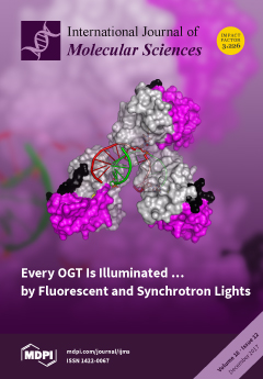
Cover Story (view full-size image): The unprecedented crystal structure of Mycobacterium tuberculosis O6-alkylguanine-DNA alkyltransferase (OGT) in complex, with a modified double-stranded DNA (dsDNA) reveals molecular details of the cooperative DNA binding mechanism of this suicidal enzyme. A peculiar supramolecular assembly can be observed in the OGT–dsDNA crystal lattice in which three different protein units are oriented onto the same DNA molecule. In particular, one protein unit binds a modified guanine base, while both additional protein monomers flip out a deoxyadenosine residue. View this paper
  • Issues are regarded as officially published after their release is announced to the table of contents alert mailing list.
  • You may sign up for e-mail alerts to receive table of contents of newly released issues.
  • PDF is the official format for papers published in both, html and pdf forms. To view the papers in pdf format, click on the "PDF Full-text" link, and use the free Adobe Reader to open them.
Order results
Result details
Section
Select all
Export citation of selected articles as:

Editorial

Jump to: Research, Review, Other

4 pages, 203 KiB  
Editorial
Acute Myeloid Leukaemia: New Targets and Therapies
by Geoffrey Brown 1,2,* and Ewa Marcinkowska 3
1 Institute of Clinical Science, College of Medical and Dental Sciences, University of Birmingham, Edgbaston, Birmingham B15 2TT, UK
2 Institute of Immunology and Immunotherapy, College of Medical and Dental Sciences, University of Birmingham, Edgbaston, Birmingham B15 2TT, UK
3 Laboratory of Protein Biochemistry, Faculty of Biotechnology, University of Wroclaw, Joliot-Curie 14a, 50-383 Wroclaw, Poland
Int. J. Mol. Sci. 2017, 18(12), 2577; https://doi.org/10.3390/ijms18122577 - 30 Nov 2017
Cited by 3 | Viewed by 3747
Abstract
The most common acute hematological malignancy in adults is acute myeloid leukaemia (AML), accounting for more than 80% of cases in patients over 60 years of age [...] Full article
(This article belongs to the Special Issue The Biology and Treatment of Myeloid Leukaemias)
5 pages, 158 KiB  
Editorial
An Updated View of Translocator Protein (TSPO)
by Nunzio Denora 1,* and Giovanni Natile 2,*
1 Department of Pharmacy-Drug Sciences, University of Bari “Aldo Moro”, 70125 Bari, Italy
2 Department of Chemistry, University of Bari “Aldo Moro”, 70125 Bari, Italy
Int. J. Mol. Sci. 2017, 18(12), 2640; https://doi.org/10.3390/ijms18122640 - 6 Dec 2017
Cited by 29 | Viewed by 4670
Abstract
Decades of study on the role of mitochondria in living cells have evidenced the importance of the 18 kDa mitochondrial translocator protein (TSPO), first discovered in the 1977 as an alternative binding site for the benzodiazepine diazepam in the kidneys. This protein participates [...] Read more.
Decades of study on the role of mitochondria in living cells have evidenced the importance of the 18 kDa mitochondrial translocator protein (TSPO), first discovered in the 1977 as an alternative binding site for the benzodiazepine diazepam in the kidneys. This protein participates in a variety of cellular functions, including cholesterol transport, steroid hormone synthesis, mitochondrial respiration, permeability transition pore opening, apoptosis, and cell proliferation. Thus, TSPO has become an extremely attractive subcellular target for the early detection of disease states that involve the overexpression of this protein and the selective mitochondrial drug delivery. This special issue was programmed with the aim of summarizing the latest findings about the role of TSPO in eukaryotic cells and as a potential subcellular target of diagnostics or therapeutics. A total of 9 papers have been accepted for publication in this issue, in particular, 2 reviews and 7 primary data manuscripts, overall describing the main advances in this field. Full article
(This article belongs to the Special Issue Translocator Protein (TSPO))
6 pages, 188 KiB  
Editorial
Tumor Microenvironment and Metabolism
by Li V. Yang 1,2
1 Department of Internal Medicine, Brody School of Medicine, East Carolina University, Greenville, NC 27834, USA
2 Department of Anatomy and Cell Biology, Brody School of Medicine, East Carolina University, Greenville, NC 27834, USA
Int. J. Mol. Sci. 2017, 18(12), 2729; https://doi.org/10.3390/ijms18122729 - 16 Dec 2017
Cited by 44 | Viewed by 6632
Abstract
The tumor microenvironment has profound effects on cancer development, progression, and therapeutic response. [...]
Full article
(This article belongs to the Special Issue Tumor Microenvironment and Metabolism)

Research

Jump to: Editorial, Review, Other

17 pages, 2849 KiB  
Article
Secondary Metabolic Profiles of Two Cultivars of Piper nigrum (Black Pepper) Resulting from Infection by Fusarium solani f. sp. piperis
by Shirlley F. M. Da Luz 1, Lydia F. Yamaguchi 2, Massuo J. Kato 2, Oriel F. De Lemos 3, Luciana P. Xavier 1, José Guilherme S. Maia 4, Alessandra De R. Ramos 5, William N. Setzer 6 and Joyce Kelly do R. Da Silva 1,*
1 Programa de Pós-Graduação em Biotecnologia, Universidade Federal do Pará, Belém 66075-900, Brazil
2 Laboratório de Química de Produtos Naturais, Instituto de Química, Universidade de São Paulo, São Paulo 05508-000, Brazil
3 Centro de Pesquisa Agroflorestal da Amazônia Oriental, Empresa Brasileira de Pesquisa Agropecuária, Belém 66095-903, Brazil
4 Programa de Pós-Graduação em Recursos Naturais da Amazônia, Universidade Federal do Oeste do Pará, Santarém 68035-110, Brazil
5 Instituto de Estudos em Saúde e Biológicas, Universidade Federal do Sul e Sudeste do Pará, Marabá 68501-970, Brazil
6 Department of Chemistry, University of Alabama in Huntsville, Huntsville, AL 35899, USA
Int. J. Mol. Sci. 2017, 18(12), 2434; https://doi.org/10.3390/ijms18122434 - 7 Dec 2017
Cited by 20 | Viewed by 6254
Abstract
Bragantina and Cingapura are the main black pepper (Piper nigrum L.) cultivars and the Pará state is the largest producer in Brazil with about 90% of national production, representing the third largest production in the world. The infection of Fusarium solani f. [...] Read more.
Bragantina and Cingapura are the main black pepper (Piper nigrum L.) cultivars and the Pará state is the largest producer in Brazil with about 90% of national production, representing the third largest production in the world. The infection of Fusarium solani f. sp. piperis, the causal agent of Fusarium disease in black pepper, was monitored on the cultivars Bragantina (susceptible) and Cingapura (tolerant), during 45 days’ post infection (dpi). Gas Chromatography-Mass spectrometry (GC-MS) analysis of the volatile concentrates of both cultivars showed that the Bragantina responded with the production of higher contents of α-bisabolol at 21 dpi and a decrease of elemol, mostly at 30 dpi; while Cingapura displayed an decrease of δ-elemene production, except at 15 dpi. The phenolic content determined by the Folin Ciocalteu method showed an increase in the leaves of plants inoculated at 7 dpi (Bragantina) and 7–15 dpi (Cingapura); in the roots, the infection caused a phenolic content decrease in Bragantina cultivar at 45 dpi and an increase in the Cingapura cultivar at 15, 30 and 45 dpi. High Performance Liquid Chromatography-Mass spectrometry (HPLC-MS) analysis of the root extracts showed a qualitative variation of alkamides during infection. The results indicated that there is a possible relationship between secondary metabolites and tolerance against phytopathogens. Full article
(This article belongs to the Section Biochemistry)
Show Figures

Graphical abstract

12 pages, 2085 KiB  
Article
Identifying the Epitope Regions of Therapeutic Antibodies Based on Structure Descriptors
by Jingxuan Qiu 1,2,†, Tianyi Qiu 3,†, Yin Huang 1 and Zhiwei Cao 1,*
1 School of Life Sciences and Technology, Tongji University, Shanghai 200092, China
2 School of Medical Instrument and Food Engineering, University of Shanghai for Science and Technology, Shanghai 200093, China
3 The Institute of Biomedical Sciences, Fudan University, Shanghai 200433, China
These authors contributed equally to this work.
Int. J. Mol. Sci. 2017, 18(12), 2457; https://doi.org/10.3390/ijms18122457 - 24 Nov 2017
Cited by 5 | Viewed by 4866
Abstract
Therapeutic antibodies are widely used for disease detection and specific treatments. However, as an exogenous protein, these antibodies can be detected by the human immune system and elicit a response that can lead to serious illnesses. Therapeutic antibodies can be engineered through antibody [...] Read more.
Therapeutic antibodies are widely used for disease detection and specific treatments. However, as an exogenous protein, these antibodies can be detected by the human immune system and elicit a response that can lead to serious illnesses. Therapeutic antibodies can be engineered through antibody humanization, which aims to maintain the specificity and biological function of the original antibodies, and reduce immunogenicity. However, the antibody drug effect is synchronously reduced as more exogenous parts are replaced by human antibodies. Hence, a major challenge in this area is to precisely detect the epitope regions in immunogenic antibodies and guide point mutations of exogenous antibodies to balance both humanization level and drug effect. In this article, the latest dataset of immunoglobulin complexes was collected from protein data bank (PDB) to discover the spatial features of immunogenic antibody. Furthermore, a series of structure descriptors were generated to characterize and distinguish epitope residues from non-immunogenic regions. Finally, a computational model was established based on structure descriptors, and results indicated that this model has the potential to precisely predict the epitope regions of therapeutic antibodies. With rapid accumulation of immunoglobulin complexes, this methodology could be used to improve and guide future antibody humanization and potential clinical applications. Full article
Show Figures

Graphical abstract

15 pages, 1748 KiB  
Article
Comparative Proteomics Analyses of Pollination Response in Endangered Orchid Species Dendrobium Chrysanthum
by Wei Wang 1,†, Hongyang Yu 1,2,†, Tinghai Li 1, Lexing Li 1, Guoqiang Zhang 3, Zhongjian Liu 3, Tengbo Huang 1 and Yongxia Zhang 1,*
1 Guangdong Provincial Key Laboratory for Plant Epigenetics, College of Life Sciences and Oceanography, Shenzhen University, Shenzhen 518060, China
2 Key Laboratory of Optoelectronic Devices and Systems of Ministry of Education and Guangdong Province, College of Optoelectronic Engineering, Shenzhen University, Shenzhen 518060, China
3 Shenzhen Key Laboratory for Orchid Conservation and Utilization, the National Orchid Conservation Center of China and the Orchid Conservation & Research Center of Shenzhen, Shenzhen 518114, China
These two authors contributed equally to this work.
Int. J. Mol. Sci. 2017, 18(12), 2496; https://doi.org/10.3390/ijms18122496 - 23 Nov 2017
Cited by 10 | Viewed by 4686
Abstract
Pollination is a crucial stage in plant reproductive process. The self-compatibility (SC) and self-incompatibility (SI) mechanisms determined the plant genetic diversity and species survival. D. chrysanthum is a highly valued ornamental and traditional herbal orchid in Asia but has been declared endangered. The [...] Read more.
Pollination is a crucial stage in plant reproductive process. The self-compatibility (SC) and self-incompatibility (SI) mechanisms determined the plant genetic diversity and species survival. D. chrysanthum is a highly valued ornamental and traditional herbal orchid in Asia but has been declared endangered. The sexual reproduction in D. chrysanthum relies on the compatibility of pollination. To provide a better understanding of the mechanism of pollination, the differentially expressed proteins (DEP) between the self-pollination (SP) and cross-pollination (CP) pistil of D. chrysanthum were investigated using proteomic approaches—two-dimensional electrophoresis (2-DE) coupled with tandem mass spectrometry technique. A total of 54 DEP spots were identified in the two-dimensional electrophoresis (2-DE) maps between the SP and CP. Gene ontology analysis revealed an array of proteins belonging to following different functional categories: metabolic process (8.94%), response to stimulus (5.69%), biosynthetic process (4.07%), protein folding (3.25%) and transport (3.25%). Identification of these DEPs at the early response stage of pollination will hopefully provide new insights in the mechanism of pollination response and help for the conservation of the orchid species. Full article
(This article belongs to the Special Issue Selected Papers from the 6th National Plant Protein Research Congress)
Show Figures

Figure 1

11 pages, 2964 KiB  
Article
Ionic Liquid-Mediated Homogeneous Esterification of Cinnamic Anhydride to Xylans
by Guihua Yang 1, Huifang Zhou 1, Jiachuan Chen 1,*, Gaojin Lyu 1,*, Yuanyuan Xia 1 and Lucian A. Lucia 1,2
1 Key Lab of Pulp & Paper Science and Technology of the Ministry of Education, Qilu University of Technology, Jinan 250353, China
2 Departments of Chemistry, Wood & Paper Science (Forest Biomaterials), North Carolina State University, Raleigh, NC 27695-8005, USA
Int. J. Mol. Sci. 2017, 18(12), 2502; https://doi.org/10.3390/ijms18122502 - 23 Nov 2017
Cited by 7 | Viewed by 4980
Abstract
A new functional biopolymer was synthesized through an ionic liquid-mediated homogeneous grafting of cinnamic anhydride to xylans. The ionic liquid used was 1-allyl-3-methylimidazolium chloride (AMIMCl) ionic liquid. Xylans with degrees of substitution (DS) between 0.11 and 0.57 were accessible in a completely homogeneous [...] Read more.
A new functional biopolymer was synthesized through an ionic liquid-mediated homogeneous grafting of cinnamic anhydride to xylans. The ionic liquid used was 1-allyl-3-methylimidazolium chloride (AMIMCl) ionic liquid. Xylans with degrees of substitution (DS) between 0.11 and 0.57 were accessible in a completely homogeneous system by changing catalysts (NaOH, KOH and LiOH), time, reaction temperature, and cinnamic anhydride/xylan molar ratio. The chemical structure and the thermal stability of the derivatives were characterized by Fourier transform infrared spectroscopy (FT-IR), 13C-NMR spectroscopy, and thermogravimetry. The thermal stability of the derivatives was reduced compared with the original xylan. Possible applications of the cinnamic anhydride-acylated xylan derivatives include wet-end papermaking, organic–inorganic composite films, and hydrogels. Full article
(This article belongs to the Special Issue Ionic Liquids 2018 and Selected Papers from ILMAT IV)
Show Figures

Graphical abstract

27 pages, 3626 KiB  
Article
Dissection of Resistance Genes to Pseudomonas syringae pv. phaseolicola in UI3 Common Bean Cultivar
by Ana M. González, Luís Godoy and Marta Santalla *
Grupo de Biología de Agrosistemas (BAS, www.bas-group.es), Misión Biológica de Galicia-CSIC, P.O. Box 28, 36080 Pontevedra, Spain
Int. J. Mol. Sci. 2017, 18(12), 2503; https://doi.org/10.3390/ijms18122503 - 23 Nov 2017
Cited by 17 | Viewed by 4878
Abstract
Few quantitative trait loci have been mapped for resistance to Pseudomonas syringae pv. phaseolicola in common bean. Two F2 populations were developed from the host differential UI3 cultivar. The objective of this study was to further characterize the resistance to races 1, [...] Read more.
Few quantitative trait loci have been mapped for resistance to Pseudomonas syringae pv. phaseolicola in common bean. Two F2 populations were developed from the host differential UI3 cultivar. The objective of this study was to further characterize the resistance to races 1, 5, 7 and 9 of Psp included in UI3. Using a QTL mapping approach, 16 and 11 main-effect QTLs for pod and primary leaf resistance were located on LG10, explaining up to 90% and 26% of the phenotypic variation, respectively. The homologous genomic region corresponding to primary leaf resistance QTLs detected tested positive for the presence of resistance-associated gene cluster encoding nucleotide-binding and leucine-rich repeat (NL), Natural Resistance Associated Macrophage (NRAMP) and Pentatricopeptide Repeat family (PPR) proteins. It is worth noting that the main effect QTLs for resistance in pod were located inside a 3.5 Mb genomic region that included the Phvul.010G021200 gene, which encodes a protein that has the highest sequence similarity to the RIN4 gene of Arabidopsis, and can be considered an important candidate gene for the organ-specific QTLs identified here. These results support that resistance to Psp from UI3 might result from the immune response activated by combinations of R proteins, and suggest the guard model as an important mechanism in pod resistance to halo blight. The candidate genes identified here warrant functional studies that will help in characterizing the actual defense gene(s) in UI3 genotype. Full article
(This article belongs to the Section Molecular Plant Sciences)
Show Figures

Graphical abstract

16 pages, 11077 KiB  
Article
Effects of Commonly Used Pesticides in China on the Mitochondria and Ubiquitin-Proteasome System in Parkinson’s Disease
by Tingting Chen 1, Jieqiong Tan 1, Zhengqing Wan 1, Yongyi Zou 1, Henok Kessete Afewerky 1,2,3, Zhuohua Zhang 1 and Tongmei Zhang 1,2,3,*
1 State Key Laboratory of Medical Genetics, Xiangya Medical School, Central South University, Changsha 410078, China
2 Department of Physiology, School of Basic Medicine, Tongji Medical College, Huazhong University of Science and Technology, Wuhan 430030, China
3 The Institute of Brain Research, Huazhong University of Science and Technology, Wuhan 430030, China
Int. J. Mol. Sci. 2017, 18(12), 2507; https://doi.org/10.3390/ijms18122507 - 23 Nov 2017
Cited by 55 | Viewed by 8111
Abstract
Evidence continues to accumulate that pesticides are the leading candidates of environmental toxins that may contribute to the pathogenesis of Parkinson’s disease. The mechanisms, however, remain largely unclear. According to epidemiological studies, we selected nine representative pesticides (paraquat, rotenone, chlorpyrifos, pendimethalin, endosulfan, fenpyroximate, [...] Read more.
Evidence continues to accumulate that pesticides are the leading candidates of environmental toxins that may contribute to the pathogenesis of Parkinson’s disease. The mechanisms, however, remain largely unclear. According to epidemiological studies, we selected nine representative pesticides (paraquat, rotenone, chlorpyrifos, pendimethalin, endosulfan, fenpyroximate, tebufenpyrad, trichlorphon and carbaryl) which are commonly used in China and detected the effects of the pesticides on mitochondria and ubiquitin-proteasome system (UPS) function. Our results reveal that all the nine studied pesticides induce morphological changes of mitochondria at low concentrations. Paraquat, rotenone, chlorpyrifos, pendimethalin, endosulfan, fenpyroximate and tebufenpyrad induced mitochondria fragmentation. Furthermore, some of them (paraquat, rotenone, chlorpyrifos, fenpyroximate and tebufenpyrad) caused a significant dose-dependent decrease of intracellular ATP. Interestingly, these pesticides which induce mitochondria dysfunction also inhibit 26S and 20S proteasome activity. However, two out of the nine pesticides, namely trichlorphon and carbaryl, were found not to cause mitochondrial fragmentation or functional damage, nor inhibit the activity of the proteasome, which provides significant guidance for selection of pesticides in China. Moreover, our results demonstrate a potential link between inhibition of mitochondria and the UPS, and pesticide-induced Parkinsonism. Full article
(This article belongs to the Section Molecular Toxicology)
Show Figures

Graphical abstract

13 pages, 6280 KiB  
Article
Administration of Protocatechuic Acid Reduces Traumatic Brain Injury-Induced Neuronal Death
by Sang Hwon Lee, Bo Young Choi, Song Hee Lee, A. Ra Kho, Jeong Hyun Jeong, Dae Ki Hong and Sang Won Suh *
1 Department of Physiology, College of Medicine, Hallym University, Chuncheon 24252, Korea
These authors contributed equally to this work.
Int. J. Mol. Sci. 2017, 18(12), 2510; https://doi.org/10.3390/ijms18122510 - 23 Nov 2017
Cited by 29 | Viewed by 6471
Abstract
Protocatechuic acid (PCA) was first purified from green tea and has shown numerous biological activities, including anti-apoptotic, anti-inflammatory, and anti-atherosclerotic effects. The effect of PCA on traumatic brain injury (TBI)-induced neuronal death has not previously been evaluated. TBI is defined as damage to [...] Read more.
Protocatechuic acid (PCA) was first purified from green tea and has shown numerous biological activities, including anti-apoptotic, anti-inflammatory, and anti-atherosclerotic effects. The effect of PCA on traumatic brain injury (TBI)-induced neuronal death has not previously been evaluated. TBI is defined as damage to the brain resulting from external mechanical force, such as rapid acceleration or deceleration, impact, blast waves, or penetration by a projectile. TBI causes neuronal death in the hippocampus and cerebral cortex. The present study aimed to evaluate the therapeutic potential of PCA on TBI-induced neuronal death. Here, TBI was induced by a controlled cortical impact model using rats. PCA (30 mg/kg) was injected into the intraperitoneal (ip) space immediately after TBI. Neuronal death was evaluated with Fluoro Jade-B (FJB) staining at 24 h after TBI. Oxidative injury was detected by 4-hydroxy-2-nonenal (4HNE), glutathione (GSH) concentration was analyzed by glutathione adduct with N-ethylmaleimide (GS-NEM) staining at 24 h after TBI, and microglial activation in the hippocampus was detected by CD11b immunohistochemistry at one week after TBI. We found that the proportion of degenerating neurons, oxidative injury, GSH depletion, and microglia activation in the hippocampus and cortex were all reduced by PCA treatment following TBI. Therefore, our study suggests that PCA may have therapeutic potential in preventing TBI-induced neuronal death. Full article
(This article belongs to the Special Issue Neuroprotective Strategies 2017)
Show Figures

Figure 1

21 pages, 2925 KiB  
Article
The Evaluation of Pro-Cognitive and Antiamnestic Properties of Berberine and Magnoflorine Isolated from Barberry Species by Centrifugal Partition Chromatography (CPC), in Relation to QSAR Modelling
by Wirginia Kukula-Koch 1,*, Marta Kruk-Słomka 2, Katarzyna Stępnik 3, Radosław Szalak 4 and Grażyna Biała 2
1 Chair and Department of Pharmacognosy with Medicinal Plant Unit, Medical University in Lublin, 20-093 Lublin, Poland
2 Department of Pharmacology and Pharmacodynamics, Medical University of Lublin, 20-093 Lublin, Poland
3 Faculty of Chemistry, Chair of Physical Chemistry, Department of Planar Chromatography, Maria Curie-Skłodowska University, 20-031 Lublin, Poland
4 Department of Animal Anatomy and Histology, Faculty of Veterinary Medicine, University of Life Science, 20-950 Lublin, Poland
Int. J. Mol. Sci. 2017, 18(12), 2511; https://doi.org/10.3390/ijms18122511 - 24 Nov 2017
Cited by 25 | Viewed by 5964
Abstract
Civilization diseases associated with memory disorders are important health problems occurring due to a prolonged life span. The manuscript shows the results of an in vivo study targeting the emergence of two drug candidates with anti-amnestic properties. The preceding quantitative structure–activity relationship (QSAR) [...] Read more.
Civilization diseases associated with memory disorders are important health problems occurring due to a prolonged life span. The manuscript shows the results of an in vivo study targeting the emergence of two drug candidates with anti-amnestic properties. The preceding quantitative structure–activity relationship (QSAR) studies provided information on the ability of berberine and magnoflorine to cross the blood–brain barrier (BBB). In the light of these findings, both compounds were purified from crude plant extracts of barberries: berberine—from Berberis siberica using a method published earlier, and magnoflorine—from Berberis cretica by centrifugal partition chromatography (solvent system: ethyl acetate:butanol:water-0.6:1.5:3 v/v/v). Both the compounds were evaluated for their memory enhancing and scopolamine inhibitory properties in an in vivo passive avoidance (PA) test on mice towards short-term and long-term memory. Cognition enhancing properties were observed at the following doses: 5 mg/kg (i.p.) for berberine and 20 mg/kg (i.p.) for magnoflorine. In addition, both the tested isoquinolines with the co-administered scopolamine were found to block long-term but not short-term memory impairment. No influence on the locomotor activity was observed for the tested doses. The results confirmed a marked central activity of magnoflorine and showed the necessity to lower the dosage of berberine. Optimized purification conditions have been elaborated for magnoflorine. Full article
Show Figures

Graphical abstract

17 pages, 1385 KiB  
Article
Screening In Vitro Targets Related to Diabetes in Herbal Extracts from Peru: Identification of Active Compounds in Hypericum laricifolium Juss. by Offline High-Performance Liquid Chromatography
by Yanymee N. Guillen Quispe 1,†, Seung Hwan Hwang 1,†, Zhiqiang Wang 1,2, Guanglei Zuo 1 and Soon Sung Lim 1,3,4,*
1 Department of Food Science and Nutrition, Hallym University, 1 Hallymdeahak-gil, Chuncheon 24252, Korea
2 College of Public Health, Hebei University, Baoding 071002, China
3 Institute of Natural Medicine, Hallym University, 1 Hallymdeahak-gil, Chuncheon 24252, Korea
4 Institute of Korean Nutrition, Hallym University, 1 Hallymdeahak-gil, Chuncheon 24252, Korea
These authors contributed equally to this work.
Int. J. Mol. Sci. 2017, 18(12), 2512; https://doi.org/10.3390/ijms18122512 - 24 Nov 2017
Cited by 21 | Viewed by 4999
Abstract
This study investigates in vitro targets related to diabetes in 30 herbal extracts from Peru, for the first time, using α-glucosidase, aldose reductase (AR) inhibitory assays and 2,2-diphenyl-1-picrylhydrazyl (DPPH) and 2,2′-azino-bis(3-ethylbenzothiazoline-6-sulfonic acid) (ABTS) scavenging assays. Among the 30 herbal extracts, Hypericum laricifolium Juss. [...] Read more.
This study investigates in vitro targets related to diabetes in 30 herbal extracts from Peru, for the first time, using α-glucosidase, aldose reductase (AR) inhibitory assays and 2,2-diphenyl-1-picrylhydrazyl (DPPH) and 2,2′-azino-bis(3-ethylbenzothiazoline-6-sulfonic acid) (ABTS) scavenging assays. Among the 30 herbal extracts, Hypericum laricifolium Juss. (HL) was the herb which showed more than 50% inhibition in all assays, presenting 97.2 ± 2.0%, 56.9 ± 5.6%, 81.9 ± 2.5%, and 58.8 ± 4.6% inhibition for the α-glucosidase, AR, DPPH, and ABTS assays, respectively. Finally, six bioactive compounds, namely, protocatechuic acid, chlorogenic acid, caffeic acid, kaempferol 3-O-glucuronide, quercetin, and kaempferol were identified in HL by offline high-performance liquid chromatography (HPLC). Quercetin exhibited the strongest inhibition in all enzyme assays and the strongest antioxidant activity. The results suggest that HL shows great potential for the complementary treatment of diabetes and its complications. Full article
(This article belongs to the Section Bioactives and Nutraceuticals)
Show Figures

Graphical abstract

15 pages, 1581 KiB  
Article
Characterization of a Sea Buckthorn Extract and Its Effect on Free and Encapsulated Lactobacillus casei
by Oana Lelia Pop 1, Francisc Vasile Dulf 2, Lucian Cuibus 1, Marta Castro-Giráldez 3, Pedro J. Fito 3, Dan Cristian Vodnar 1, Cristina Coman 1, Carmen Socaciu 1 and Ramona Suharoschi 1,*
1 Faculty of Food Science and Technology, University of Agricultural Sciences and Veterinary Medicine, Calea Mănăștur 3-5, 400372 Cluj-Napoca, Romania
2 Department of Environmental and Plant Protection, University of Agricultural Sciences and Veterinary Medicine, Calea Mănăștur 3-5, 400372 Cluj-Napoca, Romania
3 Instituto Universitario de Ingeniería de Alimentos para el Desarrollo, Universidad Politécnica de Valencia, Camino de Vera s/n, 46022 Valencia, Spain
Int. J. Mol. Sci. 2017, 18(12), 2513; https://doi.org/10.3390/ijms18122513 - 24 Nov 2017
Cited by 24 | Viewed by 5929
Abstract
Probiotics are bacteria that can provide health benefits to consumers and are suitable to be added to a variety of foods. In this research, viability of immobilized Lactobacillus casei in alginate with or without sea buckthorn lipid extract were studied during heat treatment [...] Read more.
Probiotics are bacteria that can provide health benefits to consumers and are suitable to be added to a variety of foods. In this research, viability of immobilized Lactobacillus casei in alginate with or without sea buckthorn lipid extract were studied during heat treatment and with an in vitro gastrointestinal model. The characterization of the lipid extract was also done using the UV-Vis spectrometry (UV-Vis), high-performance liquid chromatography photodiode array detection method (HPLC-PDA), gas chromatography coupled with mass spectrometry (GS-MS) and Cryo scanning electron microscopy (Cryo-SEM). During heat treatment, the entrapped probiotic cells proved high viability (>6 CFU log/g), even at temperatures above 50 °C. The rich in monounsaturated fatty acids sea buckthorn fraction improved the in vitro digestion passage regarding the probiotic viability. The survival of the probiotic cells was 15% higher after 2 h in the acidic medium of the simulated gastric fluid in the sample where L. casei was encapsulated with the sea buckthorn extract compared with the samples where no extract was added. Thus, this approach may be effective for the future development of probiotic-supplemented foods as foods with health welfare for the consumers. Full article
(This article belongs to the Section Bioactives and Nutraceuticals)
Show Figures

Graphical abstract

14 pages, 1807 KiB  
Article
The Silencing of Carotenoid β-Hydroxylases by RNA Interference in Different Maize Genetic Backgrounds Increases the β-Carotene Content of the Endosperm
by Judit Berman 1, Uxue Zorrilla-López 1, Gerhard Sandmann 2, Teresa Capell 1, Paul Christou 1,3 and Changfu Zhu 1,4,*
1 Department of Plant Production and Forestry Science, University of Lleida-Agrotecnio Center, Av. Alcalde Rovira Roure, 191, 25198 Lleida, Spain
2 Biosynthesis Group, Molecular Biosciences, Johann Wolfgang Goethe Universität, 60054 Frankfurt, Germany
3 ICREA, Catalan Institute for Research and Advanced Studies, Passeig Lluís Companys 23, 08010 Barcelona, Spain
4 School of Life Sciences, Changchun Normal University, Changchun 130032, China
Int. J. Mol. Sci. 2017, 18(12), 2515; https://doi.org/10.3390/ijms18122515 - 24 Nov 2017
Cited by 29 | Viewed by 5105
Abstract
Maize (Zea mays L.) is a staple food in many parts of Africa, but the endosperm generally contains low levels of the pro-vitamin A carotenoid β-carotene, leading to vitamin A deficiency disease in populations relying on cereal-based diets. However, maize endosperm does [...] Read more.
Maize (Zea mays L.) is a staple food in many parts of Africa, but the endosperm generally contains low levels of the pro-vitamin A carotenoid β-carotene, leading to vitamin A deficiency disease in populations relying on cereal-based diets. However, maize endosperm does accumulate high levels of other carotenoids, including zeaxanthin, which is derived from β-carotene via two hydroxylation reactions. Blocking these reactions could therefore improve the endosperm β-carotene content. Accordingly, we used RNA interference (RNAi) to silence the endogenous ZmBCH1 and ZmBCH2 genes, which encode two non-heme di-iron carotenoid β-hydroxylases. The genes were silenced in a range of maize genetic backgrounds by introgressing the RNAi cassette, allowing us to determine the impact of ZmBCH1/ZmBCH2 silencing in diverse hybrids. The β-carotene content of the endosperm increased substantially in all hybrids in which ZmBCH2 was silenced, regardless of whether or not ZmBCH1 was silenced simultaneously. However, the β-carotene content did not change significantly in C17 hybrids (M7 × C17 and M13 × C17) compared to C17 alone, because ZmBCH2 is already expressed at negligible levels in the C17 parent. Our data indicate that ZmBCH2 is primarily responsible for the conversion of β-carotene to zeaxanthin in maize endosperm. Full article
(This article belongs to the Special Issue Molecular Transformations of Natural Products)
Show Figures

Graphical abstract

18 pages, 3198 KiB  
Article
Ascorbic Acid Attenuates Senescence of Human Osteoarthritic Osteoblasts
by Maximilian G. Burger 1,2,†, Amir Steinitz 1,3,†, Jeroen Geurts 4, Benjamin E. Pippenger 4, Dirk J. Schaefer 2, Ivan Martin 1, Andrea Barbero 1,* and Karoliina Pelttari 1
1 Department of Biomedicine, University of Basel, University Hospital of Basel, 4031 Basel, Switzerland
2 Department of Plastic, Reconstructive, Aesthetic and Hand Surgery, University Hospital of Basel, University of Basel, 4031 Basel, Switzerland
3 Departments for Orthopedic Surgery and Traumatology, University Hospital of Basel, 4031 Basel, Switzerland
4 Departments Spine Surgery and Biomedical Engineering, University Hospital of Basel, University of Basel, 4031 Basel, Switzerland
These authors contributed equally to this work.
Int. J. Mol. Sci. 2017, 18(12), 2517; https://doi.org/10.3390/ijms18122517 - 24 Nov 2017
Cited by 19 | Viewed by 6008
Abstract
The accumulation of senescent cells is implicated in the pathology of several age-related diseases. While the clearance of senescent cells has been suggested as a therapeutic target for patients with osteoarthritis (OA), cellular senescence of bone-resident osteoblasts (OB) remains poorly explored. Since oxidative [...] Read more.
The accumulation of senescent cells is implicated in the pathology of several age-related diseases. While the clearance of senescent cells has been suggested as a therapeutic target for patients with osteoarthritis (OA), cellular senescence of bone-resident osteoblasts (OB) remains poorly explored. Since oxidative stress is a well-known inducer of cellular senescence, we here investigated the effect of antioxidant supplementation on the isolation efficiency, expansion, differentiation potential, and transcriptomic profile of OB from osteoarthritic subchondral bone. Bone chips were harvested from sclerotic and non-sclerotic regions of the subchondral bone of human OA joints. The application of 0.1 mM ascorbic acid-2-phosphate (AA) significantly increased the number of outgrowing cells and their proliferation capacity. This enhanced proliferative capacity showed a negative correlation with the amount of senescent cells and was accompanied by decreased expression of reactive oxygen species (ROS) in cultured OB. Expanded cells continued to express differentiated OB markers independently of AA supplementation and demonstrated no changes in their capacity to osteogenically differentiate. Transcriptomic analyses revealed that apoptotic, cell cycle–proliferation, and catabolic pathways were the main pathways affected in the presence of AA during OB expansion. Supplementation with AA can thus help to expand subchondral bone OB in vitro while maintaining their special cellular characteristics. The clearance of such senescent OB could be envisioned as a potential therapeutic target for the treatment of OA. Full article
Show Figures

Figure 1

18 pages, 2682 KiB  
Article
Controlling the Molecular Weight of Lignosulfonates by an Alkaline Oxidative Treatment at Moderate Temperatures and Atmospheric Pressure: A Size-Exclusion and Reverse-Phase Chromatography Study
by Chamseddine Guizani * and Dominique Lachenal
French National Centre for Scientific Research (CNRS), University Grenoble Alpes, Grenoble INP, LGP2, F-38000 Grenoble, France
Int. J. Mol. Sci. 2017, 18(12), 2520; https://doi.org/10.3390/ijms18122520 - 24 Nov 2017
Cited by 10 | Viewed by 5071
Abstract
The molecular weights of lignosulfonates (LSs) are modified by a rather simple process involving an alkaline oxidative treatment at moderate temperatures (70–90 °C) and atmospheric pressure. Starting from LSs with an average molecular weight of 90,000 Da, and using such a treatment, one [...] Read more.
The molecular weights of lignosulfonates (LSs) are modified by a rather simple process involving an alkaline oxidative treatment at moderate temperatures (70–90 °C) and atmospheric pressure. Starting from LSs with an average molecular weight of 90,000 Da, and using such a treatment, one can prepare controlled molecular weight LSs in the range of 30,000 to 3500 Da based on the average mass molecular weight. The LS depolymerisation was monitored via reverse-phase and size-exclusion chromatography. It has been shown that the combination of O2, H2O2 and Cu as a catalyst in alkaline conditions at 80 °C induces a high LS depolymerisation. The depolymerisation was systemically accompanied by a vanillin production, the yields of which reached 1.4 wt % (weight percentage on LS raw basis) in such conditions. Also, the average molecular weight and vanillin concentration were correlated and depended linearly on the temperature and reaction duration. Full article
(This article belongs to the Special Issue The Lignin Challenge: Exploring Innovative Applications)
Show Figures

Figure 1

12 pages, 2506 KiB  
Article
Altered Leukocyte Sphingolipid Pathway in Breast Cancer
by Larissa P. Maia 1, Paula S. Santos 1, Patrícia T. Alves 1, Cláudia M. Rodrigues 1, Thaíse G. Araújo 1, Yara Cristina P. Maia 1, Alinne Tatiane F. Câmara 1, Donizeti W. Santos 2 and Luiz Ricardo Goulart 1,3,*
1 Laboratory of Nanobiotechnology, Institute of Genetics and Biochemistry, Federal University of Uberlandia, Umuarama, Uberlandia, MG 38400-902, Brazil
2 Obstetric Division, Internal Medicine, University Hospital, Federal University of Uberlandia, Umuarama, Uberlandia, MG 38405-320, Brazil
3 Department of Medical Microbiology and Immunology, School of Medicine, University of California Davis, Davis, CA 95616, USA
Int. J. Mol. Sci. 2017, 18(12), 2521; https://doi.org/10.3390/ijms18122521 - 24 Nov 2017
Cited by 9 | Viewed by 4982
Abstract
Sphingolipid metabolism pathway is essential in membrane homeostasis, and its dysfunction has been associated with favorable tumor microenvironment, disease progression, and chemotherapy resistance. Its major components have key functions on survival and proliferation, with opposing effects. We have profiled the components of the [...] Read more.
Sphingolipid metabolism pathway is essential in membrane homeostasis, and its dysfunction has been associated with favorable tumor microenvironment, disease progression, and chemotherapy resistance. Its major components have key functions on survival and proliferation, with opposing effects. We have profiled the components of the sphingolipid pathway on leukocytes of breast cancer (BC) patients undergoing chemotherapy treatment and without, including the five sphingosine 1-phosphate (S1P) receptors, the major functional genes, and cytokines, in order to better understand the S1P signaling in the immune cells of these patients. To the best of our knowledge, this is the first characterization of the sphingolipid pathway in whole blood of BC patients. Skewed gene profiles favoring high SPHK1 expression toward S1P production during BC development was observed, which was reversed by chemotherapy treatment, and reached similar levels to those found in healthy donors. Such levels were also correlated with high levels of TNF-α. Our data revealed an important role of the sphingolipid pathway in immune cells in BC with skewed signaling of S1P receptors, which favored cancer development even under chemotherapy, and may probably be a trigger of cancer resistance. Thus, these molecules must be considered as a target pathway for combined BC therapeutics. Full article
(This article belongs to the Special Issue Sphingolipids: Signals and Disease)
Show Figures

Graphical abstract

18 pages, 9573 KiB  
Article
Impact of Antibiotics on the Proliferation and Differentiation of Human Adipose-Derived Mesenchymal Stem Cells
by Aleksandra Skubis 1,*, Joanna Gola 1, Bartosz Sikora 1, Jolanta Hybiak 2, Monika Paul-Samojedny 3, Urszula Mazurek 1 and Marek J. Łos 4,5,6,*
1 Department of Molecular Biology, School of Pharmacy with the Division of Laboratory Medicine in Sosnowiec, Medical University of Silesia in Katowice, 40-055 Katowice, Poland
2 Department of Pathology, Pomeranian Medical University, 70-204 Szczecin, Poland
3 Department of Medical Genetics, School of Pharmacy with the Division of Laboratory Medicine in Sosnowiec, Medical University of Silesia in Katowice, 40-055 Katowice, Poland
4 Małopolska Centre of Biotechnology, Jagiellonian University, Gronostajowa 7A str., 30-387 Krakow, Poland
5 LinkoCare Life Sciences AB, 583 30 Linköping, Sweden
6 Centre de Biophysique Moléculaire, UPR4301 CNRS CS80054, Rue Charles Sadron, 45071 Orleans CEDEX 2, France
Int. J. Mol. Sci. 2017, 18(12), 2522; https://doi.org/10.3390/ijms18122522 - 24 Nov 2017
Cited by 51 | Viewed by 9778
Abstract
Adipose tissue is a promising source of mesenchymal stem cells. Their potential to differentiate and regenerate other types of tissues may be affected by several factors. This may be due to in vitro cell-culture conditions, especially the supplementation with antibiotics. The aim of [...] Read more.
Adipose tissue is a promising source of mesenchymal stem cells. Their potential to differentiate and regenerate other types of tissues may be affected by several factors. This may be due to in vitro cell-culture conditions, especially the supplementation with antibiotics. The aim of our study was to evaluate the effects of a penicillin-streptomycin mixture (PS), amphotericin B (AmB), a complex of AmB with copper (II) ions (AmB-Cu2+) and various combinations of these antibiotics on the proliferation and differentiation of adipose-derived stem cells in vitro. Normal human adipose-derived stem cells (ADSC, Lonza) were routinely maintained in a Dulbecco’s Modified Eagle Medium (DMEM) that was either supplemented with selected antibiotics or without antibiotics. The ADSC that were used for the experiment were at the second passage. The effect of antibiotics on proliferation was analyzed using the 3-[4,5-dimethylthiazol-2-yl]-2,5-diphenyltetrazolium bromide (MTT) and sulforhodamine-B (SRB) tests. Differentiation was evaluated based on Alizarin Red staining, Oil Red O staining and determination of the expression of ADSC, osteoblast and adipocyte markers by real-time RT-qPCR. The obtained results indicate that the influence of antibiotics on adipose-derived stem cells depends on the duration of exposure and on the combination of applied compounds. We show that antibiotics alter the proliferation of cells and also promote natural osteogenesis, and adipogenesis, and that this effect is also noticeable in stimulated osteogenesis. Full article
(This article belongs to the Section Biochemistry)
Show Figures

Figure 1

15 pages, 3949 KiB  
Article
Whole-Genome Re-Alignment Facilitates Development of Specific Molecular Markers for Races 1 and 4 of Xanthomonas campestris pv. campestris, the Cause of Black Rot Disease in Brassica oleracea
by Mehede Hassan Rubel 1, Arif Hasan Khan Robin 1, Sathishkumar Natarajan 1, Joana G. Vicente 2, Hoy-Taek Kim 1, Jong-In Park 1,* and Ill-Sup Nou 1,*
1 Department of Horticulture, Sunchon National University, 255, Jungang-ro, Suncheon 57922, Korea
2 School of Life Sciences, University of Warwick, Wellesbourne Campus, Warwick CV35 9EF, UK
Int. J. Mol. Sci. 2017, 18(12), 2523; https://doi.org/10.3390/ijms18122523 - 24 Nov 2017
Cited by 22 | Viewed by 6841
Abstract
Black rot, caused by Xanthomonas campestris pv. campestris (Xcc), is a seed borne disease of Brassicaceae. Eleven pathogenic races have been identified based on the phenotype interaction pattern of differential brassica cultivars inoculated with different strains. Race 1 and 4 [...] Read more.
Black rot, caused by Xanthomonas campestris pv. campestris (Xcc), is a seed borne disease of Brassicaceae. Eleven pathogenic races have been identified based on the phenotype interaction pattern of differential brassica cultivars inoculated with different strains. Race 1 and 4 are the two most frequent races found in Brassica oleracea crops. In this study, a PCR molecular diagnostic tool was developed for the identification of Xcc races 1 and 4 of this pathogen. Whole genomic sequences of races 1, 3, 4 and 9 and sequences of three other Xanthomonas pathovars/species (X. campestris pv. incanae (Xci), X. campestris pv. raphani (Xcr) and X. euvesicatoria (Xev) were aligned to identify variable regions among races. To develop specific markers for races 1 and 4, primers were developed from a region where sequences were dissimilar in other races. Sequence-characterized amplified regions (SCAR) and insertion or deletion of bases (InDel) were used to develop each specific set of primers. The specificity of the selected primers was confirmed by PCR tests using genomic DNA of seven different Xcc races, two strains of X. campestris pathovars and other species of bacteria. Bacterial samples of the races 1 and 4 isolates were collected from artificially inoculated cabbage leaves to conduct bio-PCR. Bio-PCR successfully detected the two Xcc isolates. By using our race-specific markers, a potential race 1 strain from the existing Korean Xcc collection was identified. The Xcc race 1 and 4-specific markers developed in this study are novel and can potentially be used for rapid detection of Xcc races through PCR. Full article
(This article belongs to the Section Molecular Plant Sciences)
Show Figures

Graphical abstract

17 pages, 4834 KiB  
Article
Synthetic Secoisolariciresinol Diglucoside (LGM2605) Protects Human Lung in an Ex Vivo Model of Proton Radiation Damage
by Anastasia Velalopoulou 1,†, Shampa Chatterjee 2,†, Ralph A. Pietrofesa 1, Cynthia Koziol-White 1, Reynold A. Panettieri 1, Liyong Lin 3, Stephen Tuttle 3, Abigail Berman 3, Constantinos Koumenis 3 and Melpo Christofidou-Solomidou 1,*
1 Division of Pulmonary, Allergy, and Critical Care, Department of Medicine, University of Pennsylvania Perelman School of Medicine, 3450 Hamilton Walk, Stemmler Hall, Office Suite 227, Philadelphia, PA 19104, USA
2 Department of Physiology, University of Pennsylvania Perelman School of Medicine, Philadelphia, PA 19104, USA
3 Department of Radiation Oncology, University of Pennsylvania Perelman School of Medicine, Philadelphia, PA 19104, USA
These authors contributed equally to this work.
Int. J. Mol. Sci. 2017, 18(12), 2525; https://doi.org/10.3390/ijms18122525 - 25 Nov 2017
Cited by 18 | Viewed by 5403
Abstract
Radiation therapy for the treatment of thoracic malignancies has improved significantly by directing of the proton beam in higher doses on the targeted tumor while normal tissues around the tumor receive much lower doses. Nevertheless, exposure of normal tissues to protons is known [...] Read more.
Radiation therapy for the treatment of thoracic malignancies has improved significantly by directing of the proton beam in higher doses on the targeted tumor while normal tissues around the tumor receive much lower doses. Nevertheless, exposure of normal tissues to protons is known to pose a substantial risk in long-term survivors, as confirmed by our work in space-relevant exposures of murine lungs to proton radiation. Thus, radioprotective strategies are being sought. We established that LGM2605 is a potent protector from radiation-induced lung toxicity and aimed in the current study to extend the initial findings of space-relevant, proton radiation-associated late lung damage in mice by looking at acute changes in human lung. We used an ex vivo model of organ culture where tissue slices of donor living human lung were kept in culture and exposed to proton radiation. We exposed donor human lung precision-cut lung sections (huPCLS), pretreated with LGM2605, to 4 Gy proton radiation and evaluated them 30 min and 24 h later for gene expression changes relevant to inflammation, oxidative stress, and cell cycle arrest, and determined radiation-induced senescence, inflammation, and oxidative tissue damage. We identified an LGM2605-mediated reduction of proton radiation-induced cellular senescence and associated cell cycle changes, an associated proinflammatory phenotype, and associated oxidative tissue damage. This is a first report on the effects of proton radiation and of the radioprotective properties of LGM2605 on human lung. Full article
(This article belongs to the Special Issue Oxidative Stress and Space Biology: An Organ-Based Approach)
Show Figures

Graphical abstract

13 pages, 1990 KiB  
Article
Genetic Polymorphism of miR-196a-2 is Associated with Bone Mineral Density (BMD)
by Irma Karabegović 1, Silvana Maas 1, Carolina Medina-Gomez 1,2, Maša Zrimšek 2, Sjur Reppe 3,4, Kaare M. Gautvik 4,5, André G. Uitterlinden 1,2, Fernando Rivadeneira 1,2 and Mohsen Ghanbari 1,6,*
1 Department of Epidemiology, Erasmus University Medical Center, ’s-Gravendijkwal 230, 3015 CE Rotterdam, the Netherlands
2 Department of Internal Medicine, Erasmus University Medical Center, ’s-Gravendijkwal 230, 3015 CE Rotterdam, the Netherlands
3 Department of Medical Biochemistry, Oslo University Hospital, Ullevaal, 0450 Oslo, Norway
4 Unger-Vetlesen Institute, Oslo Diakonale Hospital, 0456 Oslo, Norway
5 Department of Molecular Medicine, University of Oslo, 0372 Oslo, Norway
6 Department of Genetics, School of Medicine, Mashhad University of Medical Sciences, 91388-13944 Mashhad, Iran
Int. J. Mol. Sci. 2017, 18(12), 2529; https://doi.org/10.3390/ijms18122529 - 25 Nov 2017
Cited by 16 | Viewed by 7996
Abstract
MicroRNAs (miRNAs) are small non-coding RNA molecules that post-transcriptionally regulate the translation of messenger RNAs. Given the crucial role of miRNAs in gene expression, genetic variants within miRNA-related sequences may affect miRNA function and contribute to disease risk. Osteoporosis is characterized by reduced [...] Read more.
MicroRNAs (miRNAs) are small non-coding RNA molecules that post-transcriptionally regulate the translation of messenger RNAs. Given the crucial role of miRNAs in gene expression, genetic variants within miRNA-related sequences may affect miRNA function and contribute to disease risk. Osteoporosis is characterized by reduced bone mass, and bone mineral density (BMD) is a major diagnostic proxy to assess osteoporosis risk. Here, we aimed to identify miRNAs that are involved in BMD using data from recent genome-wide association studies (GWAS) on femoral neck, lumbar spine and forearm BMD. Of 242 miRNA-variants available in the GWAS data, we found rs11614913:C > T in the precursor miR-196a-2 to be significantly associated with femoral neck-BMD (p-value = 9.9 × 10⁻⁷, β = −0.038) and lumbar spine-BMD (p-value = 3.2 × 10⁻¹¹, β = −0.061). Furthermore, our sensitivity analyses using the Rotterdam study data showed a sex-specific association of rs11614913 with BMD only in women. Subsequently, we highlighted a number of miR-196a-2 target genes, expressed in bone and associated with BMD, that may mediate the miRNA function in BMD. Collectively, our results suggest that miR-196a-2 may contribute to variations in BMD level. Further biological investigations will give more insights into the mechanisms by which miR-196a-2 control expression of BMD-related genes. Full article
(This article belongs to the Special Issue Human Single Nucleotide Polymorphisms and Disease Diagnostics)
Show Figures

Figure 1

17 pages, 3788 KiB  
Article
BCL11A mRNA Targeting by miR-210: A Possible Network Regulating γ-Globin Gene Expression
by Jessica Gasparello 1,2, Enrica Fabbri 1, Nicoletta Bianchi 1, Giulia Breveglieri 1,2, Cristina Zuccato 1, Monica Borgatti 1, Roberto Gambari 1,2,* and Alessia Finotti 1,2,*
1 Department of Life Sciences and Biotechnology, Ferrara University, 44121 Ferrara, Italy
2 Laboratory for the Development of Pharmacological and Pharmacogenomic Therapy of Thalassaemia, Biotechnology Center, Ferrara University, 44121 Ferrara, Italy
Int. J. Mol. Sci. 2017, 18(12), 2530; https://doi.org/10.3390/ijms18122530 - 26 Nov 2017
Cited by 42 | Viewed by 7284
Abstract
The involvement of microRNAs in the control of repressors of human γ-globin gene transcription has been firmly demonstrated, as described for the miR-486-3p mediated down-regulation of BCL11A. On the other hand, we have reported that miR-210 is involved in erythroid differentiation and, possibly, [...] Read more.
The involvement of microRNAs in the control of repressors of human γ-globin gene transcription has been firmly demonstrated, as described for the miR-486-3p mediated down-regulation of BCL11A. On the other hand, we have reported that miR-210 is involved in erythroid differentiation and, possibly, in γ-globin gene up-regulation. In the present study, we have identified the coding sequence of BCL11A as a possible target of miR-210. The following results sustain this hypothesis: (a) interactions between miR-210 and the miR-210 BCL11A site were demonstrated by SPR-based biomolecular interaction analysis (BIA); (b) the miR-210 site of BCL11A is conserved through molecular evolution; (c) forced expression of miR-210 leads to decrease of BCL11A-XL and increase of γ-globin mRNA content in erythroid cells, including erythroid precursors isolated from β-thalassemia patients. Our study suggests that the coding mRNA sequence of BCL11A can be targeted by miR-210. In addition to the theoretical point of view, these data are of interest from the applied point of view, supporting a novel strategy to inhibit BCL11A by mimicking miR-210 functions, accordingly with the concept supported by several papers and patent applications that inhibition of BCL11A is an efficient strategy for fetal hemoglobin induction in the treatment of β-thalassemia. Full article
(This article belongs to the Collection Regulation by Non-coding RNAs)
Show Figures

Figure 1

18 pages, 5620 KiB  
Article
Role of Vitamin D in Maintaining Renal Epithelial Barrier Function in Uremic Conditions
by Milos Mihajlovic 1, Michele Fedecostante 1, Miriam J. Oost 1, Sonja K. P. Steenhuis 1, Eef G. W. M. Lentjes 2, Inge Maitimu-Smeele 2, Manoe J. Janssen 1, Luuk B. Hilbrands 3 and Rosalinde Masereeuw 1,*
1 Division of Pharmacology, Utrecht Institute for Pharmaceutical Sciences, Utrecht University, 3584 CG Utrecht, The Netherlands
2 Department of Clinical Chemistry and Haematology, University Medical Centre Utrecht, 3584 CX Utrecht, The Netherlands
3 Department of Nephrology, Radboud University Medical Center, 6525 GA Nijmegen, The Netherlands
Int. J. Mol. Sci. 2017, 18(12), 2531; https://doi.org/10.3390/ijms18122531 - 26 Nov 2017
Cited by 25 | Viewed by 6988
Abstract
As current kidney replacement therapies are not efficient enough for end-stage renal disease (ESRD) treatment, a bioartificial kidney (BAK) device, based on conditionally immortalized human proximal tubule epithelial cells (ciPTEC), could represent an attractive solution. The active transport activity of such a system [...] Read more.
As current kidney replacement therapies are not efficient enough for end-stage renal disease (ESRD) treatment, a bioartificial kidney (BAK) device, based on conditionally immortalized human proximal tubule epithelial cells (ciPTEC), could represent an attractive solution. The active transport activity of such a system was recently demonstrated. In addition, endocrine functions of the cells, such as vitamin D activation, are relevant. The organic anion transporter 1 (OAT-1) overexpressing ciPTEC line presented 1α-hydroxylase (CYP27B1), 24-hydroxylase (CYP24A1) and vitamin D receptor (VDR), responsible for vitamin D activation, degradation and function, respectively. The ability to produce and secrete 1α,25-dihydroxy-vitamin D3, was shown after incubation with the precursor, 25-hydroxy-vitamin D3. The beneficial effect of vitamin D on cell function and behavior in uremic conditions was studied in the presence of an anionic uremic toxins mixture. Vitamin D could restore cell viability, and inflammatory and oxidative status, as shown by cell metabolic activity, interleukin-6 (IL-6) levels and reactive oxygen species (ROS) production, respectively. Finally, vitamin D restored transepithelial barrier function, as evidenced by decreased inulin-FITC leakage in biofunctionalized hollow fiber membranes (HFM) carrying ciPTEC-OAT1. In conclusion, the protective effects of vitamin D in uremic conditions and proven ciPTEC-OAT1 endocrine function encourage the use of these cells for BAK application. Full article
Show Figures

Graphical abstract

11 pages, 7598 KiB  
Article
Differential Expression of Nitric Oxide Synthase Isoforms nNOS and iNOS in Patients with Non-Segmental Generalized Vitiligo
by Mario Vaccaro 1,*,†, Natasha Irrera 1,*,†, Giuseppina Cutroneo 1, Giuseppina Rizzo 2, Federico Vaccaro 2, Giuseppe P. Anastasi 2, Francesco Borgia 1, Serafinella P. Cannavò 1, Domenica Altavilla 2 and Francesco Squadrito 1
1 Department of Clinical and Experimental Medicine, University of Messina, Via Consolare Valeria, 98124 Messina, Italy
2 Department of Biomedical Sciences and Morpho-Functional Images, University of Messina, I-98125 Messina, Italy
These authors contributed equally to this work.
Int. J. Mol. Sci. 2017, 18(12), 2533; https://doi.org/10.3390/ijms18122533 - 26 Nov 2017
Cited by 28 | Viewed by 6434
Abstract
Nitric oxide (NO) is involved in several biological processes, but its role in human melanogenesis is still not well understood. Exposure to UVA and UVB induces nitric oxide production in keratinocytes and melanocytes through the activation of constitutive nitric oxide synthase, increasing tyrosinase [...] Read more.
Nitric oxide (NO) is involved in several biological processes, but its role in human melanogenesis is still not well understood. Exposure to UVA and UVB induces nitric oxide production in keratinocytes and melanocytes through the activation of constitutive nitric oxide synthase, increasing tyrosinase activity and melanin synthesis, whereas inducible nitric oxide synthase over expression might be involved in hypopigmentary disorders. The aim of this study was to evaluate whether inducible nitric oxide synthase and neuronal nitric oxide synthase expression were modified in vitiligo skin compared to healthy controls. Skin biopsies were obtained from inflammatory/lesional and white/lesional skin in 12 patients with active, non-segmental vitiligo; site-matched biopsies of normal skin from eight patients were used as controls. Nitric oxide synthase isoforms expression was evaluated by confocal laser scanning microscopy and Western Blot analysis. Inducible nitric oxide synthase expression was significantly increased in inflammatory/lesional skin compared to healthy skin; melanocytes showed a moderate neuronal nitric oxide synthase expression in white/lesional skin, demonstrating that metabolic function still goes on. The obtained data demonstrated that vitiligo lesions were characterized by modifications of nitric oxide synthase isoforms, thus confirming the hypothesis that nitric oxide imbalance is involved in vitiligo and supporting the idea that nitric oxide synthase inhibitors might be used as a possible therapeutic approach for the management of vitiligo. Full article
(This article belongs to the Special Issue Inflammaging and Oxidative Stress in Aging and Age-Related Disorders)
Show Figures

Figure 1

12 pages, 768 KiB  
Article
Dietary Supplementation with Lactobacillus casei Alleviates Lipopolysaccharide-Induced Liver Injury in a Porcine Model
by Di Zhao 1,†, Tao Wu 1,†, Dan Yi 1, Lei Wang 1, Peng Li 1, Junmei Zhang 1, Yongqing Hou 1,* and Guoyao Wu 1,2
1 Hubei Collaborative Innovation Center for Animal Nutrition and Feed Safety, Hubei Key Laboratory of Animal Nutrition and Feed Science, Wuhan Polytechnic University, Wuhan 430023, China
2 Department of Animal Science, Texas A & M University, College Station, TX 77843, USA
These authors contributed equally to this work and should be considered co-first authors.
Int. J. Mol. Sci. 2017, 18(12), 2535; https://doi.org/10.3390/ijms18122535 - 26 Nov 2017
Cited by 28 | Viewed by 4929
Abstract
This study aims to determine whether Lactobacillus casei (L. casei) could relieve liver injury in piglets challenged with lipopolysaccharide (LPS). Piglets were randomly allocated into one of the three groups: control, LPS, and L. casei. The control and LPS groups [...] Read more.
This study aims to determine whether Lactobacillus casei (L. casei) could relieve liver injury in piglets challenged with lipopolysaccharide (LPS). Piglets were randomly allocated into one of the three groups: control, LPS, and L. casei. The control and LPS groups were fed a corn- and soybean meal-based diet, whereas the L. casei group was fed the basal diet supplemented with 6 × 10⁶ cfu/g L. casei. On Day 31 of the trial, piglets in the LPS and L. casei groups received intraperitoneal administration of LPS (100 µg/kg body weight), while the control group received the same volume of saline. Blood and liver samples were collected for analysis. Results showed that L. casei supplementation decreased the feed/gain ratio (p = 0.027) and diarrhea incidence (p < 0.001), and attenuated LPS-induced liver histomorphological abnormalities. Compared with the control group, LPS challenge dramatically increased glutamyl transpeptidase activity (p = 0.001) in plasma as well as the concentrations of Interleukin 6 (IL-6) (p = 0.048), Tumor necrosis factor-alpha (TNF-α) (p = 0.041), and Malondialdehyde (MDA) (p = 0.001) in the liver, while decreasing the hepatic SOD activity. LPS also increased (p < 0.05) the mRNA levels for IL-6, IL-8, TNF-α, Toll-like receptors 4 (TLR4), Nuclear factor κB (NF-κB) and Heat shock protein 70 (HSP70) in the liver. The adverse effects of LPS challenge were ameliorated by L. casei supplementation. In conclusion, dietary L. casei alleviates LPS-induced liver injury via reducing pro-inflammatory cytokines and increasing anti-oxidative capacity. Full article
(This article belongs to the Section Molecular Pathology, Diagnostics, and Therapeutics)
Show Figures

Figure 1

11 pages, 2366 KiB  
Communication
Could −79 °C Spray-Type Cryotherapy Be an Effective Monotherapy for the Treatment of Keloid?
by Tae Hwan Park 1,*,†, Hyeon-Ju Cho 2,†, Jang Won Lee 1, Chan Woo Kim 1, Yosep Chong 3, Choong Hyun Chang 4 and Kyung-Soon Park 2,*
1 Department of Plastic and Reconstructive Surgery, CHA Bundang Medical Center, CHA University, Seongnam 13496, Korea
2 Department of Biomedical Science, College of Life Science, CHA University, Seongnam 13488, Korea
3 Department of Hospital Pathology, College of Medicine, The Catholic University of Korea, Seoul 06591, Korea
4 Department of Plastic and Reconstructive Surgery, Kangbuk Samsung Hospital, Sungkyunkwan University School of Medicine, Seoul 03181, Korea
These authors contributed equally to this work.
Int. J. Mol. Sci. 2017, 18(12), 2536; https://doi.org/10.3390/ijms18122536 - 26 Nov 2017
Cited by 11 | Viewed by 7581
Abstract
Cryotherapy has been regarded as an effective modality for the treatment of keloids, and the spray-type device is one of the novel cryotherapeutic units. However, the biological mechanisms and therapeutic effects of this technique are incompletely studied. We evaluated the clinical efficacy of [...] Read more.
Cryotherapy has been regarded as an effective modality for the treatment of keloids, and the spray-type device is one of the novel cryotherapeutic units. However, the biological mechanisms and therapeutic effects of this technique are incompletely studied. We evaluated the clinical efficacy of our cryotherapy protocol with molecular and pathologic evidence for the treatment of keloids. We evenly split each of ten keloid lesions into a non-treated (C−) and treated (C+) area; the C+ area was subjected to two freeze-thaw cycles of spray-type cryotherapy using −79 °C spray-type CryoPen™. This treatment was repeated after an interval of two weeks. The proliferation and migration abilities of the fibroblasts isolated from the dermis under the cryotherapy-treated or untreated keloid tissues (at least 5 mm deep) were compared and pathologic findings of the full layer were evaluated. Molecular analysis revealed that the number of dermal fibroblasts was significantly higher in C+ group as compared with C− group. The dermal fibroblasts from C+ group showed more than two-fold increase in the migration ability as compared with the fibroblasts from C− group. The expression of matrix metallopeptidase 9 was increased by more than two-fold and a significant increase in transforming growth factor beta 1 expression and Smad2/3 phosphorylation level was observed in C+ group. C+ group showed more extensive lymphoplasmacytic infiltration with thicker fibrosis and occasional “proliferating core collagen” as compared with C− group. Thus, −79 °C spray-type cryotherapy is ineffective as a monotherapy and should be used in combination with intralesional corticosteroids or botulinum toxin A for favourable outcomes in the treatment of thick keloids. Full article
(This article belongs to the Special Issue Recent Advances in Scar Biology)
Show Figures

Graphical abstract

18 pages, 3119 KiB  
Article
Protective Effects of Liquiritigenin against Citrinin-Triggered, Oxidative-Stress-Mediated Apoptosis and Disruption of Embryonic Development in Mouse Blastocysts
by Chien-Hsun Huang 1 and Wen-Hsiung Chan 2,3,*
1 Department of Obstetrics and Gynecology, Taoyuan General Hospital, Ministry of Health & Welfare, Taoyuan City 33004, Taiwan
2 Department of Bioscience Technology and Center for Nanotechnology, Chung Yuan Christian University, Chung Li District, Taoyuan City 32023, Taiwan
3 Department of Medical Research, China Medical University Hospital, China Medical University, Taichung 40402, Taiwan
Int. J. Mol. Sci. 2017, 18(12), 2538; https://doi.org/10.3390/ijms18122538 - 27 Nov 2017
Cited by 18 | Viewed by 4281
Abstract
The mycotoxin citrinin (CTN), a natural contaminant in foodstuffs and animal feeds, exerts cytotoxic and genotoxic effects on various mammalian cells and embryos. A previous investigation by our group revealed potentially hazardous effects of CTN on mouse oocyte maturation and pre- and post-implantation [...] Read more.
The mycotoxin citrinin (CTN), a natural contaminant in foodstuffs and animal feeds, exerts cytotoxic and genotoxic effects on various mammalian cells and embryos. A previous investigation by our group revealed potentially hazardous effects of CTN on mouse oocyte maturation and pre- and post-implantation embryo development via the induction of apoptosis. The present study showed that CTN induces apoptosis and inhibits cell proliferation in the inner cell mass of mouse blastocysts. Notably, we observed for the first time that both these effects are suppressed by liquiritigenin (LQ). LQ is a type of flavonoid isolated from Glycyrrhiza radix with several biochemical and pharmacological activities, including antioxidant and anti-inflammatory properties. The preincubation of blastocysts with LQ clearly prevented CTN-induced disruption of pre- and post-implantation embryonic development and fetal weight loss, both in vitro and in vivo. CTN-induced damage processes directly promoted reactive oxygen species (ROS) generation, loss of mitochondrial membrane potential (MMP) and activation of caspase-9 and caspase-3, which were effectively blocked by LQ. Moreover, in an animal model, intravenous injection of dams with CTN (3 mg/kg/day) triggered apoptosis of blastocysts, disruption of embryonic development from the zygote to the blastocyst stage and a decrease in fetal weight. Pre-injection with LQ (5 mg/kg/day) effectively reduced apoptosis and impaired the cytotoxic effects of CTN on development. Our in vivo findings further confirm that CTN exposure via injection has the potential to impair pre- and post-implantation development, leading to apoptosis and the suppression of sequent embryonic development, which can be effectively prevented by LQ. Full article
(This article belongs to the Section Biochemistry)
Show Figures

Figure 1

13 pages, 4190 KiB  
Article
Ketamine, a Clinically Used Anesthetic, Inhibits Vascular Smooth Muscle Cell Proliferation via PP2A-Activated PI3K/Akt/ERK Inhibition
by Yi Chang 1,2,3,†, Jiun-Yi Li 3,4,†, Thanasekaran Jayakumar 3, Shou-Huang Hung 3, Wei-Cheng Lee 3, Manjunath Manubolu 5, Joen-Rong Sheu 3,* and Ming-Jen Hsu 3,*
1 Department of Anesthesiology, Shin Kong Wu Ho-Su Memorial Hospital, No. 95, Wenchang Rd., Taipei 111, Taiwan
2 School of Medicine, Fu-Jen Catholic University, No. 510, Zhongzheng Rd., Xinzhuang Dist, New Taipei City 242, Taiwan
3 Department of Pharmacology and Graduate Institute of Medical Sciences, College of Medicine, Taipei Medical University, No. 250, Wu-Hsing St., Taipei 110, Taiwan
4 Department of Cardiovascular Surgery, Mackay Memorial Hospital, and Mackay Medical College, Taipei 104, Taiwan
5 Department of Evolution, Ecology and Organismal Biology, Ohio State University, 1314 Kinnear Rd, Columbus, OH 43212, USA
These authors contributed equally to this work.
Int. J. Mol. Sci. 2017, 18(12), 2545; https://doi.org/10.3390/ijms18122545 - 27 Nov 2017
Cited by 12 | Viewed by 5988
Abstract
Abnormal proliferation of vascular smooth muscle cells (VSMCs) gives rise to major pathological processes involved in the development of cardiovascular diseases. The use of anti-proliferative agents for VSMCs offers potential for the treatment of vascular disorders. Intravenous anesthetics are firmly established to have [...] Read more.
Abnormal proliferation of vascular smooth muscle cells (VSMCs) gives rise to major pathological processes involved in the development of cardiovascular diseases. The use of anti-proliferative agents for VSMCs offers potential for the treatment of vascular disorders. Intravenous anesthetics are firmly established to have direct effects on VSMCs, resulting in modulation of blood pressure. Ketamine has been used for many years in the intensive care unit (ICU) for sedation, and has recently been considered for adjunctive therapy. In the present study, we investigated the effects of ketamine on platelet-derived growth factor BB (PDGF-BB)-induced VSMC proliferation and the associated mechanism. Ketamine concentration-dependently inhibited PDGF-BB-induced VSMC proliferation without cytotoxicity, and phosphatidylinositol 3-kinase (PI3K) and extracellular signal-regulated protein kinase (ERK) inhibitors, LY294002 and PD98059, respectively, have similar inhibitory effects. Ketamine was shown to attenuate PI3K, Akt, and ERK1/2 phosphorylation induced by PDGF-BB. Okadaic acid, a selective protein phosphatase 2A (PP2A) inhibitor, significantly reversed ketamine-mediated PDGF-BB-induced PI3K, Akt, and ERK1/2 phosphorylation; a transfected protein phosphatse 2a (pp2a) siRNA reversed Akt and ERK1/2 phosphorylation; and 3-O-Methyl-sphingomyeline (3-OME), an inhibitor of sphingomyelinase, also significantly reversed ERK1/2 phosphorylation. Moreover, ketamine alone significantly inhibited tyrosine phosphorylation and demethylation of PP2A in a concentration-dependent manner. In addition, the pp2a siRNA potently reversed the ketamine-activated catalytic subunit (PP2A-C) of PP2A. These results provide evidence of an anti-proliferating effect of ketamine in VSMCs, showing activation of PP2A blocks PI3K, Akt, and ERK phosphorylation that subsequently inhibits the proliferation of VSMCs. Thus, ketamine may be considered a potential effective therapeutic agent for reducing atherosclerotic process by blocking the proliferation of VSMCs. Full article
Show Figures

Figure 1

16 pages, 3703 KiB  
Article
RNA Chaperone Function of a Universal Stress Protein in Arabidopsis Confers Enhanced Cold Stress Tolerance in Plants
by Sarah Mae Boyles Melencion, Yong Hun Chi, Thuy Thi Pham, Seol Ki Paeng, Seong Dong Wi, Changyu Lee, Seoung Woo Ryu, Sung Sun Koo and Sang Yeol Lee *
1 Division of Applied Life Science (BK21+ Program), PMBBRC, Gyeongsang National University, Jinju 52828, Korea
These authors contributed equally to this work.
Int. J. Mol. Sci. 2017, 18(12), 2546; https://doi.org/10.3390/ijms18122546 - 27 Nov 2017
Cited by 52 | Viewed by 7150
Abstract
The physiological function of Arabidopsis thaliana universal stress protein (AtUSP) in plant has remained unclear. Thus, we report here the functional role of the Arabidopsis universal stress protein, AtUSP (At3g53990). To determine how AtUSP affects physiological responses towards cold stress, AtUSP overexpression (AtUSP [...] Read more.
The physiological function of Arabidopsis thaliana universal stress protein (AtUSP) in plant has remained unclear. Thus, we report here the functional role of the Arabidopsis universal stress protein, AtUSP (At3g53990). To determine how AtUSP affects physiological responses towards cold stress, AtUSP overexpression (AtUSP OE) and T-DNA insertion knock-out (atusp, SALK_146059) mutant lines were used. The results indicated that AtUSP OE enhanced plant tolerance to cold stress, whereas atusp did not. AtUSP is localized in the nucleus and cytoplasm, and cold stress significantly affects RNA metabolism such as by misfolding and secondary structure changes of RNA. Therefore, we investigated the relationship of AtUSP with RNA metabolism. We found that AtUSP can bind nucleic acids, including single- and double-stranded DNA and luciferase mRNA. AtUSP also displayed strong nucleic acid-melting activity. We expressed AtUSP in RL211 Escherichia coli, which contains a hairpin-loop RNA structure upstream of chloramphenicol acetyltransferase (CAT), and observed that AtUSP exhibited anti-termination activity that enabled CAT gene expression. AtUSP expression in the cold-sensitive Escherichia coli (E. coli) mutant BX04 complemented the cold sensitivity of the mutant cells. As these properties are typical characteristics of RNA chaperones, we conclude that AtUSP functions as a RNA chaperone under cold-shock conditions. Thus, the enhanced tolerance of AtUSP OE lines to cold stress is mediated by the RNA chaperone function of AtUSP. Full article
(This article belongs to the Special Issue Molecular Chaperones)
Show Figures

Graphical abstract

17 pages, 2909 KiB  
Article
Nodule-Enriched GRETCHEN HAGEN 3 Enzymes Have Distinct Substrate Specificities and Are Important for Proper Soybean Nodule Development
by Suresh Damodaran 1, Corey S. Westfall 2, Brian A. Kisely 1, Joseph M. Jez 2,* and Senthil Subramanian 1,3,*
1 Department of Agronomy, Horticulture and Plant Science, South Dakota State University, Brookings, SD 57007, USA
2 Department of Biology, Washington University in St. Louis, St. Louis, MO 63130, USA
3 Department of Biology and Microbiology, South Dakota State University, Brookings, SD 57007, USA
Int. J. Mol. Sci. 2017, 18(12), 2547; https://doi.org/10.3390/ijms18122547 - 28 Nov 2017
Cited by 14 | Viewed by 6629
Abstract
Legume root nodules develop as a result of a symbiotic relationship between the plant and nitrogen-fixing rhizobia bacteria in soil. Auxin activity is detected in different cell types at different stages of nodule development; as well as an enhanced sensitivity to auxin inhibits, [...] Read more.
Legume root nodules develop as a result of a symbiotic relationship between the plant and nitrogen-fixing rhizobia bacteria in soil. Auxin activity is detected in different cell types at different stages of nodule development; as well as an enhanced sensitivity to auxin inhibits, which could affect nodule development. While some transport and signaling mechanisms that achieve precise spatiotemporal auxin output are known, the role of auxin metabolism during nodule development is unclear. Using a soybean root lateral organ transcriptome data set, we identified distinct nodule enrichment of three genes encoding auxin-deactivating GRETCHEN HAGEN 3 (GH3) indole-3-acetic acid (IAA) amido transferase enzymes: GmGH3-11/12, GmGH3-14 and GmGH3-15. In vitro enzymatic assays showed that each of these GH3 proteins preferred IAA and aspartate as acyl and amino acid substrates, respectively. GmGH3-15 showed a broad substrate preference, especially with different forms of auxin. Promoter:GUS expression analysis indicated that GmGH3-14 acts primarily in the root epidermis and the nodule primordium where as GmGH3-15 might act in the vasculature. Silencing the expression of these GH3 genes in soybean composite plants led to altered nodule numbers, maturity, and size. Our results indicate that these GH3s are needed for proper nodule maturation in soybean, but the precise mechanism by which they regulate nodule development remains to be explained. Full article
(This article belongs to the Special Issue Auxin)
Show Figures

Graphical abstract

18 pages, 2373 KiB  
Article
Human Skin Permeation Studies with PPARγ Agonist to Improve Its Permeability and Efficacy in Inflammatory Processes
by Marcelle Silva-Abreu 1,2, Lupe Carolina Espinoza 1,3, María José Rodríguez-Lagunas 4,5, María-José Fábrega 4,6, Marta Espina 1,2, María Luisa García 1,2 and Ana Cristina Calpena 1,2,*
1 Department of Pharmacy, Pharmaceutical Technology and Physical Chemistry, Faculty of Pharmacy and Food Sciences, University of Barcelona, 08028 Barcelona, Spain
2 Institute of Nanoscience and Nanotechnology (IN2UB), University of Barcelona, 08028 Barcelona, Spain
3 Departamento de Química y Ciencias Exactas, Universidad Técnica Particular de Loja, Loja 1101608, Ecuador
4 Department of Biochemistry and Physiology, Faculty of Pharmacy and Food Sciences, University of Barcelona, 08028 Barcelona, Spain
5 Institut de Recerca en Nutrició i Seguretat Alimentària (INSA), Universitat de Barcelona (UB), 08028 Barcelona, Spain
6 Institute of Biomedicine, University of Barcelona, 08028 Barcelona, Spain
Int. J. Mol. Sci. 2017, 18(12), 2548; https://doi.org/10.3390/ijms18122548 - 28 Nov 2017
Cited by 24 | Viewed by 5602
Abstract
Rosacea is the most common inflammatory skin disease. It is characterized by erythema, inflammatory papules and pustules, visible blood vessels, and telangiectasia. The current treatment has limitations and unsatisfactory results. Pioglitazone (PGZ) is an agonist of peroxisome proliferator-activated receptors (PPARs), a nuclear receptor [...] Read more.
Rosacea is the most common inflammatory skin disease. It is characterized by erythema, inflammatory papules and pustules, visible blood vessels, and telangiectasia. The current treatment has limitations and unsatisfactory results. Pioglitazone (PGZ) is an agonist of peroxisome proliferator-activated receptors (PPARs), a nuclear receptor that regulates important cellular functions, including inflammatory responses. The purpose of this study was to evaluate the permeation of PGZ with a selection of penetration enhancers and to analyze its effectiveness for treating rosacea. The high-performance liquid chromatography (HPLC) method was validated for the quantitative determination of PGZ. Ex vivo permeation experiments were realized in Franz diffusion cells using human skin, in which PGZ with different penetration enhancers were assayed. The results showed that the limonene was the most effective penetration enhancer that promotes the permeation of PGZ through the skin. The cytotoxicity studies and the Draize test detected cell viability and the absence of skin irritation, respectively. The determination of the skin color using a skin colorimetric probe and the results of histopathological studies confirmed the ability of PGZ-limonene to reduce erythema and vasodilation. This study suggests new pharmacological indications of PGZ and its possible application in the treatment of skin diseases, namely rosacea. Full article
(This article belongs to the Special Issue Inflammatory Skin Conditions 2017)
Show Figures

Graphical abstract

23 pages, 3826 KiB  
Article
Graphene Oxide–Silver Nanoparticles Nanocomposite Stimulates Differentiation in Human Neuroblastoma Cancer Cells (SH-SY5Y)
by Sangiliyandi Gurunathan * and Jin-Hoi Kim *
Department of Stem Cell and Regenerative Biotechnology, Konkuk University, Seoul 05029, Korea
Int. J. Mol. Sci. 2017, 18(12), 2549; https://doi.org/10.3390/ijms18122549 - 28 Nov 2017
Cited by 43 | Viewed by 8096
Abstract
Recently, graphene and graphene related nanocomposite receive much attention due to high surface-to-volume ratio, and unique physiochemical and biological properties. The combination of metallic nanoparticles with graphene-based materials offers a promising method to fabricate novel graphene–silver hybrid nanomaterials with unique functions in biomedical [...] Read more.
Recently, graphene and graphene related nanocomposite receive much attention due to high surface-to-volume ratio, and unique physiochemical and biological properties. The combination of metallic nanoparticles with graphene-based materials offers a promising method to fabricate novel graphene–silver hybrid nanomaterials with unique functions in biomedical nanotechnology, and nanomedicine. Therefore, this study was designed to prepare graphene oxide (GO) silver nanoparticles (AgNPs) nanocomposite (GO-AgNPs) containing two different nanomaterials in single platform with distinctive properties using luciferin as reducing agents. In addition, we investigated the effect of GO-AgNPs on differentiation in SH-SY5Y cells. The synthesized GO-AgNPs were characterized by ultraviolet-visible absorption spectroscopy (UV-vis), X-ray diffraction (XRD), scanning electron microscopy (SEM), transmission electron microscopy (TEM) and Raman spectroscopy. The differentiation was confirmed by series of cellular and biochemical assays. The AgNPs were distributed uniformly on the surface of graphene oxide with an average size of 25 nm. As prepared GO-AgNPOs induces differentiation by increasing the expression of neuronal differentiation markers and decreasing the expression of stem cell markers. The results indicated that the redox biology involved the expression of various signaling molecules, which play an important role in differentiation. This study suggests that GO-AgNP nanocomposite could stimulate differentiation of SH-SY5Y cells. Furthermore, understanding the mechanisms of differentiation of neuroblastoma cells could provide new strategies for cancer and stem cell therapies. Therefore, these studies suggest that GO-AgNPs could target specific chemotherapy-resistant cells within a tumor. Full article
(This article belongs to the Section Materials Science)
Show Figures

Figure 1

15 pages, 3812 KiB  
Article
Aberrant Lipid Metabolism in Hepatocellular Carcinoma Revealed by Liver Lipidomics
by Zhao Li 1, Ming Guan 2,3, Yu Lin 2, Xiao Cui 1, Yangyang Zhang 2, Zhenwen Zhao 2,3,* and Jiye Zhu 1
1 Department of Hepatobiliary Surgery, Peking University People’s Hospital, Beijing 100044, China
2 Beijing National Laboratory for Molecular Sciences, Key Laboratory of Analytical Chemistry for Living Biosystems, Institute of Chemistry Chinese Academy of Sciences, Beijing Mass Spectrum Center, Beijing 100190, China
3 Graduate School, University of Chinese Academy of Sciences, Beijing 100049, China
Int. J. Mol. Sci. 2017, 18(12), 2550; https://doi.org/10.3390/ijms18122550 - 28 Nov 2017
Cited by 76 | Viewed by 6996
Abstract
Background: The aim of this study was to characterize the disorder of lipid metabolism in hepatocellular carcinoma (HCC). HCC is a worldwide disease. The research into the disorder of lipid metabolism in HCC is very limited. Study of lipid metabolism in liver cancer [...] Read more.
Background: The aim of this study was to characterize the disorder of lipid metabolism in hepatocellular carcinoma (HCC). HCC is a worldwide disease. The research into the disorder of lipid metabolism in HCC is very limited. Study of lipid metabolism in liver cancer tissue may have the potential to provide new insight into HCC mechanisms. Methods: A lipidomics study of HCC based on Ultra high performance liquid chromatography-electronic spray ionization-QTOF mass spectrometer (UPLC-ESI-QTOF MS) and Matrix assisted laser desorption ionization-fourier transform ion cyclotron resonance mass spectrometer (MALDI-FTICR MS) was performed. Results: Triacylglycerols (TAGs) with the number of double bond (DB) > 2 (except 56:5 and 56:4 TAG) were significantly down-regulated; conversely, others (except 52:2 TAG) were greatly up-regulated in HCC tissues. Moreover, the more serious the disease was, the higher the saturated TAG concentration and the lower the polyunsaturated TAG concentration were in HCC tissues. The phosphatidylcholine (PC), phosphatidylethanolamine (PE) and phosphatidylinositol (PI) were altered in a certain way. Sphingomyelin (SM) was up-regulated and ceramide (Cer) were down-regulated in HCC tissues. Conclusions: To our knowledge, this is the first such report showing a unique trend of TAG, PC, PE and PI. The use of polyunsaturated fatty acids, like eicosapentanoic and docosahexanoic acid, as supplementation, proposed for the treatment of Non-alcoholic steatohepatitis (NASH), may also be effective for the treatment of HCC. Full article
(This article belongs to the Section Molecular Pathology, Diagnostics, and Therapeutics)
Show Figures

Graphical abstract

14 pages, 3628 KiB  
Article
Insights into Insulin Fibril Assembly at Physiological and Acidic pH and Related Amyloid Intrinsic Fluorescence
by Clara Iannuzzi 1,*, Margherita Borriello 1, Marianna Portaccio 2, Gaetano Irace 1 and Ivana Sirangelo 1
1 Department of Biochemistry, Biophysics and General Pathology, Università degli Studi della Campania “Luigi Vanvitelli”, 80138 Naples, Italy
2 Department of Experimental Medicine, Università degli Studi della Campania “Luigi Vanvitelli”, 80138 Naples, Italy
Int. J. Mol. Sci. 2017, 18(12), 2551; https://doi.org/10.3390/ijms18122551 - 28 Nov 2017
Cited by 68 | Viewed by 7322
Abstract
Human insulin is a widely used model protein for the study of amyloid formation as both associated to insulin injection amyloidosis in type II diabetes and highly prone to form amyloid fibrils in vitro. In this study, we aim to gain new structural [...] Read more.
Human insulin is a widely used model protein for the study of amyloid formation as both associated to insulin injection amyloidosis in type II diabetes and highly prone to form amyloid fibrils in vitro. In this study, we aim to gain new structural insights into insulin fibril formation under two different aggregating conditions at neutral and acidic pH, using a combination of fluorescence, circular dichroism, Fourier-transform infrared spectroscopy, and transmission electron miscroscopy. We reveal that fibrils formed at neutral pH are morphologically different from those obtained at lower pH. Moreover, differences in FTIR spectra were also detected. In addition, only insulin fibrils formed at neutral pH showed the characteristic blue-green fluorescence generally associated to amyloid fibrils. So far, the molecular origin of this fluorescence phenomenon has not been clarified and different hypotheses have been proposed. In this respect, our data provide experimental evidence that allow identifying the molecular origin of such intrinsic property. Full article
(This article belongs to the Special Issue Protein Folding)
Show Figures

Graphical abstract

13 pages, 5095 KiB  
Article
Analysis of MicroRNA Expression in Newborns with Differential Birth Weight Using Newborn Screening Cards
by Patricia Rodil-Garcia, Elvira Del Carmen Arellanes-Licea, Angélica Montoya-Contreras and Luis A. Salazar-Olivo *
1 Molecular Biology Division, Instituto Potosino de Investigación Científica y Tecnológica, Camino a la Presa San José 2055, San Luis Potosí 78216, SLP, México
Present address: Academic Division, Universidad Tecnológica de Corregidora, Corregidora 76900, Querétaro, México.
Int. J. Mol. Sci. 2017, 18(12), 2552; https://doi.org/10.3390/ijms18122552 - 28 Nov 2017
Cited by 22 | Viewed by 4966
Abstract
Birth weight is an early predictor for metabolic diseases and microRNAs (miRNAs) are proposed as fetal programming participants. To evaluate the use of dried blood spots (DBS) on newborn screening cards (NSC) as a source of analyzable miRNAs, we optimized a commercial protocol [...] Read more.
Birth weight is an early predictor for metabolic diseases and microRNAs (miRNAs) are proposed as fetal programming participants. To evaluate the use of dried blood spots (DBS) on newborn screening cards (NSC) as a source of analyzable miRNAs, we optimized a commercial protocol to recover total miRNA from normal birth weight (NBW, n = 17–20), low birth weight (LBW, n = 17–20) and high birth weight (macrosomia, n = 17–20) newborns and analyzed the relative expression of selected miRNAs by stem-loop RT-qPCR. The possible role of miRNAs on the fetal programming of metabolic diseases was explored by bioinformatic tools. The optimized extraction of RNA resulted in a 1.2-fold enrichment of miRNAs respect to the commercial kit. miR-33b and miR-375 were overexpressed in macrosomia 9.8-fold (p < 0.001) and 1.7-fold, (p < 0.05), respectively and miR-454-3p was overexpressed in both LBW and macrosomia (19.7-fold, p < 0.001 and 10.8-fold, p < 0.001, respectively), as compared to NBW. Potential target genes for these miRNAs are associated to cyclic-guanosine monophosphate (cGMP)-dependent protein kinase (PKG), mitogen-activated protein kinase (MAPK), type 2 diabetes, transforming growth factor-β (TGF-β)and Forkhead box O protein (FoxO) pathways. In summary, we improved a protocol for analyzing miRNAs from NSC and provide the first evidence that birth weight modifies the expression of miRNAs associated to adult metabolic dysfunctions. Our work suggests archived NSC are an invaluable resource in the search for fetal programming biomarkers. Full article
(This article belongs to the Section Biochemistry)
Show Figures

Graphical abstract

18 pages, 4194 KiB  
Article
Bioinspired versus Enzymatic Oxidation of Some Homologous Thionine Dyes in the Presence of Immobilized Metalloporphyrin Catalysts and Ligninolytic Enzymes
by Gianmarco Cocco, Andrea Cocco, Francesca Sollai, Enrico Sanjust * and Paolo Zucca
Department of Biomedical Sciences, Biochemistry Unit, University of Cagliari, 09042 Monserrato (CA), Italy
Int. J. Mol. Sci. 2017, 18(12), 2553; https://doi.org/10.3390/ijms18122553 - 28 Nov 2017
Cited by 6 | Viewed by 4946
Abstract
Thionines are recalcitrant and polluting textile dyes presenting various degrees of N-methylation. In this paper, a complete series of homologous thionines was used as the substrates for oxidation in the presence of a bioinspired commercial iron-porphyrin immobilized on to imidazole- and pyridine-functionalized [...] Read more.
Thionines are recalcitrant and polluting textile dyes presenting various degrees of N-methylation. In this paper, a complete series of homologous thionines was used as the substrates for oxidation in the presence of a bioinspired commercial iron-porphyrin immobilized on to imidazole- and pyridine-functionalized fumed silica, to emulate the active site of ligninolytic peroxidases. The obtained catalytic adducts showed a remarkable ability to catalyze thionine dye oxidation in the presence of different oxidants such as potassium monopersulfate and hydrogen peroxide. Different oxidation patterns were obtained and mechanistically discussed, in comparison with those observed in the presence of some ligninolytic oxidizing enzymes. Full article
(This article belongs to the Section Biochemistry)
Show Figures

Graphical abstract

15 pages, 3154 KiB  
Article
Antarctic Krill Oil Diet Protects against Lipopolysaccharide-Induced Oxidative Stress, Neuroinflammation and Cognitive Impairment
by Ji Yeon Choi, Jun Sung Jang, Dong Ju Son, Hyung-Sik Im, Ji Yeong Kim, Joung Eun Park, Won Rak Choi, Sang-Bae Han and Jin Tae Hong *
College of Pharmacy and Medical Research Center, Chungbuk National University, 194-31 Osongsaemgmyeong 1-ro, Osong-eup, Heungdeok-gu, Cheongju 28160, Chungbuk, Korea
Int. J. Mol. Sci. 2017, 18(12), 2554; https://doi.org/10.3390/ijms18122554 - 28 Nov 2017
Cited by 48 | Viewed by 8672
Abstract
Oxidative stress and neuroinflammation are implicated in the development and pathogenesis of Alzheimer’s disease (AD). Here, we investigated the anti-inflammatory and antioxidative effects of krill oil. Oil from Euphausia superba (Antarctic krill), an Antarctic marine species, is rich in eicosapentaenoic acid (EPA) and [...] Read more.
Oxidative stress and neuroinflammation are implicated in the development and pathogenesis of Alzheimer’s disease (AD). Here, we investigated the anti-inflammatory and antioxidative effects of krill oil. Oil from Euphausia superba (Antarctic krill), an Antarctic marine species, is rich in eicosapentaenoic acid (EPA) and docosahexaenoic acid (DHA). We examined whether krill oil diet (80 mg/kg/day for one month) prevents amyloidogenesis and cognitive impairment induced by intraperitoneal lipopolysaccharide (LPS) (250 µg/kg, seven times daily) injections in AD mice model and found that krill oil treatment inhibited the LPS-induced memory loss. We also found that krill oil treatment inhibited the LPS-induced expression of inducible nitric oxide synthase (iNOS) and cyclooxygenase-2 (COX-2) and decreased reactive oxygen species (ROS) and malondialdehyde levels. Krill oil also suppresses IκB degradation as well as p50 and p65 translocation into the nuclei of LPS-injected mice brain cells. In association with the inhibitory effect on neuroinflammation and oxidative stress, krill oil suppressed amyloid beta (1–42) peptide generation by the down-regulating APP and BACE1 expression in vivo. We found that eicosapentaenoic acid (EPA) and docosahexaenoic acid (DHA) (50 and 100 µM) dose-dependently decreased LPS-induced nitric oxide and ROS generation, and COX-2 and iNOS expression as well as nuclear factor-κB activity in cultured microglial BV-2 cells. These results suggest that krill oil ameliorated impairment via anti-inflammatory, antioxidative, and anti-amyloidogenic mechanisms. Full article
(This article belongs to the Special Issue Neurological Injuries’ Monitoring, Tracking and Treatment 2017)
Show Figures

Figure 1

11 pages, 3524 KiB  
Article
Pharmacokinetics of Chlorin e6-Cobalt Bis(Dicarbollide) Conjugate in Balb/c Mice with Engrafted Carcinoma
by Arthur B. Volovetsky 1,*, Vladimir S. Sukhov 1, Irina V. Balalaeva 1, Varvara V. Dudenkova 1,2, Natalia Yu. Shilyagina 1, Аlexey V. Feofanov 3,4, Anastasija V. Efremenko 3,4, Mikhail A. Grin 5, Andrey F. Mironov 5, Igor B. Sivaev 6, Vladimir I. Bregadze 6 and Anna V. Maslennikova 1,2
1 Department of Biophysics, Lobachevsky State University of Nizhny Novgorod, 23 Gagarina Av., 603950 Nizhny Novgorod, Russia
2 Department of Oncology, Nizhny Novgorod State Medical Academy, 10/1 Minin and Pozharsky Sq., 603005 Nizhny Novgorod, Russia
3 Shemyakin and Ovchinnikov Institute of Bioorganic Chemistry, Russian Academy of Sciences, 16/10 Miklukho-Maklaya Str., 117997 Moscow, Russia
4 Biological Faculty, Lomonosov Moscow State University, Vorobyevi Gori 1, 119992 Moscow, Russia
5 Institute of Fine Chemical Technology, Moscow Technological University, 86 Vernadskii Av., 119571 Moscow, Russia
6 Nesmeyanov Institute of Organoelement Compounds, Russian Academy of Sciences, 28 Vavilov Str., 119991 Moscow, Russia
Int. J. Mol. Sci. 2017, 18(12), 2556; https://doi.org/10.3390/ijms18122556 - 28 Nov 2017
Cited by 12 | Viewed by 4699
Abstract
The necessary precondition for efficient boron neutron capture therapy (BNCT) is control over the content of isotope 10B in the tumor and normal tissues. In the case of boron-containing porphyrins, the fluorescent part of molecule can be used for quantitative assessment of [...] Read more.
The necessary precondition for efficient boron neutron capture therapy (BNCT) is control over the content of isotope 10B in the tumor and normal tissues. In the case of boron-containing porphyrins, the fluorescent part of molecule can be used for quantitative assessment of the boron content. Study Objective: We performed a study of the biodistribution of the chlorin e6-Cobalt bis(dicarbollide) conjugate in carcinoma-bearing Balb/c mice using ex vivo fluorescence imaging, and developed a mathematical model describing boron accumulation and release based on the obtained experimental data. Materials and Methods: The study was performed on Balb/c tumor-bearing mice (CT-26 tumor model). A solution of the chlorin e6-Cobalt bis(dicarbollide) conjugate (CCDC) was injected into the blood at a dose of 10 mg/kg of the animal’s weight. Analysis of the fluorescence signal intensity was performed at several time points by spectrofluorimetry in blood and by laser scanning microscopy in muscle, liver, and tumor tissues. The boron content in the same samples was determined by mass spectroscopy with inductively coupled plasma. Results: Analysis of a linear approximation between the fluorescence intensity and boron content in the tissues demonstrated a satisfactory value of approximation reliability with a Spearman’s rank correlation coefficient of r = 0.938, p < 0.01. The dynamics of the boron concentration change in various organs, calculated on the basis of the fluorescence intensity, enabled the development of a model describing the accumulation of the studied compound and its distribution in tissues. The obtained results reveal a high level of correspondence between the model and experimental data. Full article
(This article belongs to the Section Molecular Pathology, Diagnostics, and Therapeutics)
Show Figures

Graphical abstract

12 pages, 1066 KiB  
Article
Effect of Dietary Acidolysis-Oxidized Konjac Glucomannan Supplementation on Serum Immune Parameters and Intestinal Immune-Related Gene Expression of Schizothorax prenanti
by Mingrui Chen 1,†, Shuyao Wang 1,†, Xue Liang 1, Donghui Ma 1, Li He 1 and Yaowen Liu 1,2,*
1 College of Food Science, Sichuan Agricultural University, Yaan 625014, China
2 School of Materials Science and Engineering, Southwest Jiaotong University, Chengdu 610031, China
These authors contributed equally to the work.
Int. J. Mol. Sci. 2017, 18(12), 2558; https://doi.org/10.3390/ijms18122558 - 28 Nov 2017
Cited by 12 | Viewed by 4307
Abstract
The present study was conducted to investigate the effects of dietary acidolysis-oxidized konjac glucomannan (A-OKGM) (0%, 0.4%, 0.8%, and 1.6%) supplementation on the immunity and expression of immune-related genes in Schizothorax prenanti. After feeding for eight weeks, the serum and guts were [...] Read more.
The present study was conducted to investigate the effects of dietary acidolysis-oxidized konjac glucomannan (A-OKGM) (0%, 0.4%, 0.8%, and 1.6%) supplementation on the immunity and expression of immune-related genes in Schizothorax prenanti. After feeding for eight weeks, the serum and guts were used for measurement of biochemical parameters, and immune-related gene expression in the gut were also analyzed by real-time quantitative polymerase chain reaction (RT-qPCR). C-reactive protein and IgM levels were significantly higher in the A-OKGM fed groups than in the control group, regardless of the dosage. The 0.4% and 1.6% A-OKGM groups showed significant up-regulation of tumor necrosis factor α (TNFα) in the anterior gut. The 0.8% and 1.6% A-OKGM groups also showed significantly enhanced TNFα expression in the mid- and distal guts. Interleukin-1β (IL-1β) expression in the anterior gut of fish fed with 0.4% and 1.6% A-OKGM diets was significantly enhanced. The 0.8% and 1.6% A-OKGM diets resulted in significantly increased the expression of IL-1β in the distal gut. Similarly, the interleukin-6 (IL-6) messenger RNA (mRNA) levels in the 0.4% and 1.6% diet groups were significantly higher in the anterior gut. The 0.8% and 1.6% A-OKGM diet groups showed significant induction of IL-6 gene expression in the distal gut. A-OKGM modified from KGM can act as an immunostimulant to enhance the immunity of S. prenanti. Full article
(This article belongs to the Section Biochemistry)
Show Figures

Figure 1

15 pages, 3336 KiB  
Article
PPARγ Modulates Long Chain Fatty Acid Processing in the Intestinal Epithelium
by Kalina Duszka 1,2,3, Matej Oresic 4, Cedric Le May 5, Jürgen König 3,6 and Walter Wahli 1,2,7,*
1 Lee Kong Chian School of Medicine, Nanyang Technological University, 11 Mandalay Road, Singapore 308232, Singapore
2 Center for Integrative Genomics, University of Lausanne, Génopode, CH-1015 Lausanne, Switzerland
3 Department of Nutritional Sciences, University of Vienna, Althanstrasse 14, 1090 Vienna,Austria
4 Turku Centre for Biotechnology, University of Turku and Åbo Akademi University,Tykisokatu 6, 20520 Turku, Finland
5 Institut du Thorax, INSERM, CNRS, UNIV Nantes, 44007 Nantes, France
6 Vienna Metabolomics Center (VIME), University of Vienna, Althanstrasse 14, 1090 Vienna, Austria
7 ToxAlim, Research Center in Food Toxicology, National Institute for Agricultural Research (INRA),180 Chemin de Tournefeuille, 31300 Toulouse, France
Int. J. Mol. Sci. 2017, 18(12), 2559; https://doi.org/10.3390/ijms18122559 - 28 Nov 2017
Cited by 45 | Viewed by 7095
Abstract
Nuclear receptor PPARγ affects lipid metabolism in several tissues, but its role in intestinal lipid metabolism has not been explored. As alterations have been observed in the plasma lipid profile of ad libitum fed intestinal epithelium-specific PPARγ knockout mice (iePPARγKO), we submitted these [...] Read more.
Nuclear receptor PPARγ affects lipid metabolism in several tissues, but its role in intestinal lipid metabolism has not been explored. As alterations have been observed in the plasma lipid profile of ad libitum fed intestinal epithelium-specific PPARγ knockout mice (iePPARγKO), we submitted these mice to lipid gavage challenges. Within hours after gavage with long chain unsaturated fatty acid (FA)-rich canola oil, the iePPARγKO mice had higher plasma free FA levels and lower gastric inhibitory polypeptide levels than their wild-type (WT) littermates, and altered expression of incretin genes and lipid metabolism-associated genes in the intestinal epithelium. Gavage with the medium chain saturated FA-rich coconut oil did not result in differences between the two genotypes. Furthermore, the iePPARγKO mice did not exhibit defective lipid uptake and stomach emptying; however, their intestinal transit was more rapid than in WT mice. When fed a canola oil-rich diet for 4.5 months, iePPARγKO mice had higher body lean mass than the WT mice. We conclude that intestinal epithelium PPARγ is activated preferentially by long chain unsaturated FAs compared to medium chain saturated FAs. Furthermore, we hypothesize that the iePPARγKO phenotype originates from altered lipid metabolism and release in epithelial cells, as well as changes in intestinal motility. Full article
(This article belongs to the Special Issue PPARs in Cellular and Whole Body Energy Metabolism)
Show Figures

Graphical abstract

10 pages, 2011 KiB  
Article
Estrogen Metabolism-Associated CYP2D6 and IL6-174G/C Polymorphisms in Schistosoma haematobium Infection
by Rita Cardoso 1, Pedro C. Lacerda 2, Paulo P. Costa 2,3, Ana Machado 3,4, André Carvalho 5, Adriano Bordalo 3,4, Ruben Fernandes 6,7, Raquel Soares 7,8, Joachim Richter 9, Helena Alves 1,10 and Monica C. Botelho 1,7,*
1 Department of Health Promotion and Chronic Diseases, National Institute of Health Dr. Ricardo Jorge (INSA), Rua Alexandre Herculano 321, 4000-055 Porto, Portugal
2 Department of Human Genetics, National Institute of Health Dr. Ricardo Jorge (INSA), Rua Alexandre Herculano 321, 4000-055 Porto, Portugal
3 Instituto de Ciências Biomédicas Abel Salazar (ICBAS/UP), Universidade do Porto, Rua Jorge Viterbo Ferreira 228, P 4050-313 Porto, Portugal
4 Centro Interdisciplinar de Investigação Marinha e Ambiental (CIIMAR/CIMAR), Universidade do Porto, Av. General Norton de Matos s/n, 4450-208 Matosinhos, Portugal
5 Division of Endocrinology, Diabetes and Metabolism, Santo Antonio Hospital—Centro Hospitalar do Porto (CHP), Largo do Prof. Abel Salazar, 4099-001 Porto, Portugal
6 Escola Superior de Saúde, Instituto Politécnico do Porto, Rua Dr. António Bernardino de Almeida, 400, 4200-079 Porto, Portugal
7 Unit of Metabolism, Nutrition and Endocrinology, Instituto de Investigação e Inovação da Universidade do Porto (i3S), Rua Alfredo Allen, 4200-135 Porto, Portugal
8 Departamento de Biomedicina, Unidade de Bioquímica, Faculdade de Medicina, Universidade do Porto, Al. Prof. Hernâni Monteiro, 4200-319 Porto, Portugal
9 Institute of Tropical Medicine and International Health, Charité—Universitätsmedizin Berlin, Augustenburger Platz 1, 13353 Berlin, Germany
10 Fundação Professor Ernesto Morais, Rua de Monsanto 512, 4250-288 Porto, Portugal
Int. J. Mol. Sci. 2017, 18(12), 2560; https://doi.org/10.3390/ijms18122560 - 28 Nov 2017
Cited by 8 | Viewed by 5629
Abstract
Schistosoma haematobium is a human blood fluke causing a chronic infection called urogenital schistosomiasis. Squamous cell carcinoma of the urinary bladder (SCC) constitutes chronic sequelae of this infection, and S. haematobium infection is accounted as a risk factor for this type of cancer. [...] Read more.
Schistosoma haematobium is a human blood fluke causing a chronic infection called urogenital schistosomiasis. Squamous cell carcinoma of the urinary bladder (SCC) constitutes chronic sequelae of this infection, and S. haematobium infection is accounted as a risk factor for this type of cancer. This infection is considered a neglected tropical disease and is endemic in numerous countries in Africa and the Middle East. Schistosome eggs produce catechol-estrogens. These estrogenic molecules are metabolized to active quinones that induce modifications in DNA. The cytochrome P450 (CYP) enzymes are a superfamily of mono-oxygenases involved in estrogen biosynthesis and metabolism, the generation of DNA damaging procarcinogens, and the response to anti-estrogen therapies. IL6 Interleukin-6 (IL-6) is a pleiotropic cytokine expressed in various tissues. This cytokine is largely expressed in the female urogenital tract as well as reproductive organs. Very high or very low levels of IL-6 are associated with estrogen metabolism imbalance. In the present study, we investigated the polymorphic variants in the CYP2D6 gene and the C-174G promoter polymorphism of the IL-6 gene on S. haematobium-infected children patients from Guine Bissau. CYP2D6 inactivated alleles (28.5%) and IL6G-174C (13.3%) variants were frequent in S. haematobium-infected patients when compared to previously studied healthy populations (4.5% and 0.05%, respectively). Here we discuss our recent findings on these polymorphisms and whether they can be predictive markers of schistosome infection and/or represent potential biomarkers for urogenital schistosomiasis associated bladder cancer and infertility. Full article
(This article belongs to the Special Issue Molecular Pathways of Estrogen Receptor Action)
Show Figures

Figure 1

14 pages, 15878 KiB  
Article
Bioaccumulation and Toxicity of Carbon Nanoparticles Suspension Injection in Intravenously Exposed Mice
by Ping Xie 1, Sheng-Tao Yang 2,*, Tiantian He 3, Shengnan Yang 2 and Xiao-Hai Tang 3,*
1 State Key Laboratory of Oral Diseases, West China College of Stomatology, Sichuan University, Chengdu 610041, China
2 College of Chemistry & Environment Protection Engineering, Southwest Minzu University, Chengdu 610041, China
3 Chongqing Lummy Pharmaceutical Co., Ltd., Chongqing 401123, China
Int. J. Mol. Sci. 2017, 18(12), 2562; https://doi.org/10.3390/ijms18122562 - 29 Nov 2017
Cited by 48 | Viewed by 5889
Abstract
Carbon nanoparticles suspension injection (CNSI) has been widely used in tumor drainage lymph node mapping, and its new applications in drug delivery, photothermal therapy, and so on have been extensively investigated. To develop new clinical applications, the toxicity of CNSI after intravenous exposure [...] Read more.
Carbon nanoparticles suspension injection (CNSI) has been widely used in tumor drainage lymph node mapping, and its new applications in drug delivery, photothermal therapy, and so on have been extensively investigated. To develop new clinical applications, the toxicity of CNSI after intravenous exposure should be thoroughly investigated to ensure its safe use. Herein, we studied the bioaccumulation of CNSI in reticuloendothelial system (RES) organs and the corresponding toxicity to mice. After the intravenous injection of CNSI, no abnormal behavior of mice was observed during the 28-day observation period. The body weight increases were similar among the exposed groups and the control group. The parameters of hematology and serum biochemistry remained nearly unchanged, with very few of them showing significant changes. The low toxicity of CNSI was also reflected by the unchanged histopathological characteristics of these organs. The injection of CNSI did not induce higher apoptosis levels either. The slight oxidative stress was observed in RES organs at high dosages at day 7 post-exposure. The implication to the clinical applications and toxicological evaluations of carbon nanomaterials is discussed. Full article
(This article belongs to the Special Issue Bioactive Nanoparticles)
Show Figures

Graphical abstract

9 pages, 2059 KiB  
Article
Buprofezin Is Metabolized by CYP353D1v2, a Cytochrome P450 Associated with Imidacloprid Resistance in Laodelphax striatellus
by Mohammed Esmail Abdalla Elzaki 1,2,*, Mohammad Asaduzzaman Miah 1 and Zhaojun Han 1,*
1 The Key Laboratory of Monitoring and Management of Plant Diseases and Insects, Department of Entomology, College of Plant Protection, Ministry of Agriculture, Nanjing Agricultural University, Nanjing 210095, China
2 College of Crop Science, Fujian Agriculture and Forestry University, Fuzhou 350002, China
Int. J. Mol. Sci. 2017, 18(12), 2564; https://doi.org/10.3390/ijms18122564 - 29 Nov 2017
Cited by 13 | Viewed by 6718
Abstract
CYP353D1v2 is a cytochrome P450 related to imidacloprid resistance in Laodelphax striatellus. This work was conducted to examine the ability of CYP353D1v2 to metabolize other insecticides. Carbon monoxide difference spectra analysis indicates that CYP353D1v2 was successfully expressed in insect cell Sf9. The [...] Read more.
CYP353D1v2 is a cytochrome P450 related to imidacloprid resistance in Laodelphax striatellus. This work was conducted to examine the ability of CYP353D1v2 to metabolize other insecticides. Carbon monoxide difference spectra analysis indicates that CYP353D1v2 was successfully expressed in insect cell Sf9. The catalytic activity of CYP353D1v2 relating to degrading buprofezin, chlorpyrifos, and deltamethrin was tested by measuring substrate depletion and analyzing the formation of metabolites. The results showed the nicotinamide–adenine dinucleotide phosphate (NADPH)-dependent depletion of buprofezin (eluting at 8.7 min) and parallel formation of an unknown metabolite (eluting 9.5 min). However, CYP353D1v2 is unable to metabolize deltamethrin and chlorpyrifos. The recombinant CYP353D1v2 protein efficiently catalyzed the model substrate p-nitroanisole with a maximum velocity of 9.24 nmol/min/mg of protein and a Michaelis constant of Km = 6.21 µM. In addition, imidacloprid was metabolized in vitro by the recombinant CYP353D1v2 microsomes (catalytic constant Kcat) 0.064 pmol/min/pmol P450, Km = 6.41 µM. The mass spectrum of UPLC-MS analysis shows that the metabolite was a product of buprofezin, which was buprofezin sulfone. This result provided direct evidence that L. striatellus cytochrome P450 CYP353D1v2 is capable of metabolizing imidacloprid and buprofezin. Full article
(This article belongs to the Special Issue Cytochromes P450: Drug Metabolism and Bioactivation)
Show Figures

Graphical abstract

15 pages, 2373 KiB  
Article
Lipopolysaccharide Modifies Glycerol Permeability and Metabolism in 3T3-L1 Adipocytes
by Jeanne Durendale Chiadak 1,†, Patrizia Gena 2,†, Françoise Gregoire 1, Nargis Bolaky 1, Valérie Delforge 1, Jason Perret 1, Giuseppe Calamita 2 and Christine Delporte 1,*
1 Laboratory of Pathophysiological and Nutritional Biochemistry, Université Libre de Bruxelles, 1070 Brussels, Belgium
2 Department of Biosciences, Biotechnologies and Biopharmaceutics, University of Bari “Aldo Moro”, 70125 Bari, Italy
These authors equally contributed to this work.
Int. J. Mol. Sci. 2017, 18(12), 2566; https://doi.org/10.3390/ijms18122566 - 29 Nov 2017
Cited by 17 | Viewed by 6567
Abstract
Aquaglyceroporins—aquaporin membrane channels (AQP) that conduct glycerol and other small neutral solutes in addition to water—play major roles in obesity. In adipocytes, aquaglyceroporins mediate glycerol uptake and release across the plasma membrane, which are two key steps for triacylglycerols (TAGs) synthesis (lipogenesis) and [...] Read more.
Aquaglyceroporins—aquaporin membrane channels (AQP) that conduct glycerol and other small neutral solutes in addition to water—play major roles in obesity. In adipocytes, aquaglyceroporins mediate glycerol uptake and release across the plasma membrane, which are two key steps for triacylglycerols (TAGs) synthesis (lipogenesis) and hydrolysis (lipolysis). The aim of this study was to assess both glycerol permeability and metabolism in undifferentiated 3T3-L1 cells (UDCs) as well as in untreated (CTL-DCs) versus lipopolysaccharide (LPS-DCs)-treated differentiated 3T3-L1 adipocytes. Glycerol release, TAGs content and whole membrane glycerol permeability were significantly increased in DCs as compared to UDCs. Moreover, in DCs, LPS treatment significantly increased TAGs content and decreased glycerol permeability. In addition, a significant reduction in whole membrane glycerol permeability was observed in LPS-DCs as compared to CTL-DCs. The relative contributions of AQP3, AQP7 and AQP9 (facilitated diffusion), as well as that of the phospholipid bilayer (simple diffusion), to the whole membrane glycerol permeability, were estimated biophysically in UDCs, CTL-DCs and LPS-DCs, using selective AQP inhibitors. Further studies will be required to determine if modifications in either subcellular localization and/or activity of aquaglyceroporins could account for the data herein. Nevertheless, our findings provide novel insights in understanding the LPS-induced adipocyte hypertrophy that accompanies obesity. Full article
(This article belongs to the Special Issue Aquaporins: Water Channels Essential for Living Organisms)
Show Figures

Graphical abstract

14 pages, 4830 KiB  
Article
Immunohistochemical Characterization of Connexin43 Expression in a Mouse Model of Diabetic Retinopathy and in Human Donor Retinas
by Odunayo O. Mugisho 1,3, Colin R. Green 2, Jie Zhang 2, Nicolette Binz 4, Monica L. Acosta 3, Elizabeth Rakoczy 4 and Ilva D. Rupenthal 1,*
1 Buchanan Ocular Therapeutics Unit, Department of Ophthalmology, New Zealand National Eye Centre, University of Auckland, Auckland 1142, New Zealand
2 Department of Ophthalmology, New Zealand National Eye Centre, University of Auckland, Auckland 1142, New Zealand
3 School of Optometry and Vision Science, New Zealand National Eye Centre, University of Auckland, Auckland 1142, New Zealand
4 Centre for Ophthalmology and Visual Science, University of Western Australia, Perth 6009, Western Australia, Australia
Int. J. Mol. Sci. 2017, 18(12), 2567; https://doi.org/10.3390/ijms18122567 - 29 Nov 2017
Cited by 28 | Viewed by 5939
Abstract
Diabetic retinopathy (DR) develops due to hyperglycemia and inflammation-induced vascular disruptions in the retina with connexin43 expression patterns in the disease still debated. Here, the effects of hyperglycemia and inflammation on connexin43 expression in vitro in a mouse model of DR and in [...] Read more.
Diabetic retinopathy (DR) develops due to hyperglycemia and inflammation-induced vascular disruptions in the retina with connexin43 expression patterns in the disease still debated. Here, the effects of hyperglycemia and inflammation on connexin43 expression in vitro in a mouse model of DR and in human donor tissues were evaluated. Primary human retinal microvascular endothelial cells (hRMECs) were exposed to high glucose (HG; 25 mM) or pro-inflammatory cytokines IL-1β and TNF-α (10 ng/mL each) or both before assessing connexin43 expression. Additionally, connexin43, glial fibrillary acidic protein (GFAP), and plasmalemma vesicular associated protein (PLVAP) were labeled in wild-type (C57BL/6), Akita (diabetic), and Akimba (DR) mouse retinas. Finally, connexin43 and GFAP expression in donor retinas with confirmed DR was compared to age-matched controls. Co-application of HG and cytokines increased connexin43 expression in hRMECs in line with results seen in mice, with no significant difference in connexin43 or GFAP expression in Akita but higher expression in Akimba compared to wild-type mice. On PLVAP-positive vessels, connexin43 was higher in Akimba but unchanged in Akita compared to wild-type mice. Connexin43 expression appeared higher in donor retinas with confirmed DR compared to age-matched controls, similar to the distribution seen in Akimba mice and correlating with the in vitro results. Although connexin43 expression seems reduced in diabetes, hyperglycemia and inflammation present in the pathology of DR seem to increase connexin43 expression, suggesting a causal role of connexin43 channels in the disease progression. Full article
Show Figures

Graphical abstract

12 pages, 2941 KiB  
Article
Volasertib Enhances Sensitivity to TRAIL in Renal Carcinoma Caki Cells through Downregulation of c-FLIP Expression
by Mi-Yeon Jeon, Kyoung-jin Min, Seon Min Woo, Seung Un Seo, Shin Kim, Jong-Wook Park and Taeg Kyu Kwon *
Department of Immunology, School of Mediine, Keimyung University, 2800 Dalgubeoldaero, Dalseo-Gu, Daegu 704-701, Korea
Int. J. Mol. Sci. 2017, 18(12), 2568; https://doi.org/10.3390/ijms18122568 - 29 Nov 2017
Cited by 8 | Viewed by 3956
Abstract
Polo-like kinase 1 (PLK1) plays major roles in cell cycle control and DNA damage response. Therefore, PLK1 has been investigated as a target for cancer therapy. Volasertib is the second-in class dihydropteridinone derivate that is a specific PLK1 inhibitor. In this study, we [...] Read more.
Polo-like kinase 1 (PLK1) plays major roles in cell cycle control and DNA damage response. Therefore, PLK1 has been investigated as a target for cancer therapy. Volasertib is the second-in class dihydropteridinone derivate that is a specific PLK1 inhibitor. In this study, we examined that combining PLK1 inhibitor with tumor necrosis factor-related apoptosis-inducing ligand (TRAIL) would have an additive and synergistic effect on induction of apoptosis in cancer cells. We found that volasertib alone and TRAIL alone had no effect on apoptosis, but the combined treatment of volasertib and TRAIL markedly induced apoptosis in Caki (renal carcinoma), A498 (renal carcinoma) and A549 (lung carcinoma) cells, but not in normal cells (human skin fibroblast cells and mesangial cells). Combined treatment induced accumulation of sub-G1 phase, DNA fragmentation, cleavage of poly (ADP-ribose) polymerase (PARP) and activation of caspase 3 activity in Caki cells. Interestingly, combined treatment induced downregulation of cellular-FLICE-inhibitory protein (c-FLIP) expression and ectopic expression of c-FLIP markedly blocked combined treatment-induced apoptosis. Therefore, this study demonstrates that volasertib may sensitize TRAIL-induced apoptosis in Caki cells via downregulation of c-FLIP. Full article
(This article belongs to the Section Biochemistry)
Show Figures

Graphical abstract

14 pages, 10738 KiB  
Article
A New Bone Substitute Developed from 3D-Prints of Polylactide (PLA) Loaded with Collagen I: An In Vitro Study
by Ulrike Ritz 1,*, Rebekka Gerke 1, Hermann Götz 2, Stefan Stein 3 and Pol Maria Rommens 1
1 Department of Orthopaedics and Traumatology, BiomaTiCS, University Medical Center, Johannes Gutenberg University, 55131 Mainz, Germany
2 Platform for Biomaterial Research, University Medical Center, BiomaTiCS, Johannes Gutenberg University, 55131 Mainz, Germany
3 Georg-Speyer-Haus—Institute for Tumor Biology and Experimental Therapy, 60659 Frankfurt, Germany
Int. J. Mol. Sci. 2017, 18(12), 2569; https://doi.org/10.3390/ijms18122569 - 29 Nov 2017
Cited by 56 | Viewed by 8587
Abstract
Although a lot of research has been performed, large segmental bone defects caused by trauma, infection, bone tumors or revision surgeries still represent big challenges for trauma surgeons. New and innovative bone substitutes are needed. Three-dimensional (3D) printing is a novel procedure to [...] Read more.
Although a lot of research has been performed, large segmental bone defects caused by trauma, infection, bone tumors or revision surgeries still represent big challenges for trauma surgeons. New and innovative bone substitutes are needed. Three-dimensional (3D) printing is a novel procedure to create 3D porous scaffolds that can be used for bone tissue engineering. In the present study, solid discs as well as porous cage-like 3D prints made of polylactide (PLA) are coated or filled with collagen, respectively, and tested for biocompatibility and endotoxin contamination. Microscopic analyses as well as proliferation assays were performed using various cell types on PLA discs. Stromal-derived factor (SDF-1) release from cages filled with collagen was analyzed and the effect on endothelial cells tested. This study confirms the biocompatibility of PLA and demonstrates an endotoxin contamination clearly below the FDA (Food and Drug Administration) limit. Cells of various cell types (osteoblasts, osteoblast-like cells, fibroblasts and endothelial cells) grow, spread and proliferate on PLA-printed discs. PLA cages loaded with SDF-1 collagen display a steady SDF-1 release, support cell growth of endothelial cells and induce neo-vessel formation. These results demonstrate the potential for PLA scaffolds printed with an inexpensive desktop printer in medical applications, for example, in bone tissue engineering. Full article
(This article belongs to the Special Issue Novel Biomaterials for Tissue Engineering 2018)
Show Figures

Graphical abstract

19 pages, 1801 KiB  
Article
Sulfur-Mediated-Alleviation of Aluminum-Toxicity in Citrus grandis Seedlings
by Peng Guo 1, Qiang Li 1, Yi-Ping Qi 2, Lin-Tong Yang 1, Xin Ye 1, Huan-Huan Chen 1 and Li-Song Chen 1,3,4,*
1 Institute of Plant Nutritional Physiology and Molecular Biology, College of Resources and Environment, Fujian Agriculture and Forestry University (FAFU), Fuzhou 350002, China
2 Institute of Materia Medica, Fujian Academy of Medical Sciences, Fuzhou 350002, China
3 Fujian Provincial Key Laboratory of Soil Environmental Health and Regulation, College of Resources and Environment, FAFU, Fuzhou 350002, China
4 The Higher Education Key Laboratory of Fujian Province for Soil Ecosystem Health and Regulation, College of Resources and Environment, FAFU, Fuzhou 350002, China
Int. J. Mol. Sci. 2017, 18(12), 2570; https://doi.org/10.3390/ijms18122570 - 3 Dec 2017
Cited by 51 | Viewed by 5695
Abstract
Limited data are available on the sulfur (S)-mediated-alleviation of aluminum (Al)-toxicity in higher plants. Citrus grandis seedlings were irrigated for 18 weeks with 0.5 mM MgSO4 or 0.5 mM MgSO4 + 0.5 mM Na2SO4, and 0 (−Al) [...] Read more.
Limited data are available on the sulfur (S)-mediated-alleviation of aluminum (Al)-toxicity in higher plants. Citrus grandis seedlings were irrigated for 18 weeks with 0.5 mM MgSO4 or 0.5 mM MgSO4 + 0.5 mM Na2SO4, and 0 (−Al) or 1 mM AlCl3·6H2O (+Al, Al-toxicity). Under Al-toxicity, S decreased the level of Al in leaves; increased the relative water content (RWC) of roots and leaves, the contents of phosphorus (P), calcium (Ca) and magnesium (Mg) per plant, the dry weights (DW) of roots and shoots, the ratios of root DW/shoot DW, and the Al-induced secretion of citrate from root; and alleviated the Al-induced inhibition of photosynthesis via mitigating the Al-induced decrease of electron transport capacity resulting from the impaired photosynthetic electron transport chain. In addition to decreasing the Al-stimulated H2O2 production, the S-induced upregulation of both S metabolism-related enzymes and antioxidant enzymes also contributed to the S-mediated-alleviation of oxidative damage in Al-treated roots and leaves. Decreased transport of Al from roots to shoots and relatively little accumulation of Al in leaves, and increased leaf and root RWC and P, Ca, and Mg contents per plant might also play a role in the S-mediated-alleviation of Al-toxicity. Full article
(This article belongs to the Special Issue Metabolomics in the Plant Sciences 2017)
Show Figures

Figure 1

14 pages, 2273 KiB  
Article
Structural Masquerade of Plesiomonas shigelloides Strain CNCTC 78/89 O-Antigen—High-Resolution Magic Angle Spinning NMR Reveals the Modified d-galactan I of Klebsiella pneumoniae
by Karolina Ucieklak, Sabina Koj, Damian Pawelczyk and Tomasz Niedziela *
Hirszfeld Institute of Immunology and Experimental Therapy, Polish Academy of Sciences, 53-114 Wroclaw, Poland
Int. J. Mol. Sci. 2017, 18(12), 2572; https://doi.org/10.3390/ijms18122572 - 29 Nov 2017
Cited by 5 | Viewed by 4011
Abstract
The high-resolution magic angle spinning nuclear magnetic resonance spectroscopy (HR-MAS NMR) analysis of Plesiomonas shigelloides 78/89 lipopolysaccharide directly on bacteria revealed the characteristic structural features of the O-acetylated polysaccharide in the NMR spectra. The O-antigen profiles were unique, yet the pattern [...] Read more.
The high-resolution magic angle spinning nuclear magnetic resonance spectroscopy (HR-MAS NMR) analysis of Plesiomonas shigelloides 78/89 lipopolysaccharide directly on bacteria revealed the characteristic structural features of the O-acetylated polysaccharide in the NMR spectra. The O-antigen profiles were unique, yet the pattern of signals in the, spectra along with their 1H,13C chemical shift values, resembled these of d-galactan I of Klebsiella pneumoniae. The isolated O-specific polysaccharide (O-PS) of P. shigelloides strain CNCTC 78/89 was investigated by 1H and 13C NMR spectroscopy, mass spectrometry and chemical methods. The analyses demonstrated that the P. shigelloides 78/89 O-PS is composed of →3)-α-d-Galp-(1→3)-β-d-Galf2OAc-(1→ disaccharide repeating units. The O-acetylation was incomplete and resulted in a microheterogeneity of the O-antigen. This O-acetylation generates additional antigenic determinants within the O-antigen, forms a new chemotype, and contributes to the epitopes recognized by the O-serotype specific antibodies. The serological cross-reactivities further confirmed the inter-specific structural similarity of these O-antigens. Full article
(This article belongs to the Special Issue Lipopolysaccharides (LPSs))
Show Figures

Graphical abstract

20 pages, 3910 KiB  
Article
Identification and Characterization of Hyphantria cunea Aminopeptidase N as a Binding Protein of Bacillus thuringiensis Cry1Ab35 Toxin
by Yakun Zhang 1,†, Dan Zhao 1,†, Xiaoping Yan 1, Wei Guo 1,2,*, Yajun Bao 1, Wei Wang 1 and Xiaoyun Wang 3
1 College of Plant Protection, Hebei Agricultural University, Baoding 071000, China
2 Plant Science and Technology College, Beijing University of Agriculture, Beijing 102206, China
3 College of Agriculture, Northeast Agricultural University, Harbin 150038, China
These authors contributed equally to this work.
Int. J. Mol. Sci. 2017, 18(12), 2575; https://doi.org/10.3390/ijms18122575 - 30 Nov 2017
Cited by 16 | Viewed by 5098
Abstract
The fall webworm, Hyphantria cunea (Drury) is a major invasive pest in China. Aminopeptidase N (APN) isoforms in lepidopteran larvae midguts are known for their involvement in the mode of action of insecticidal crystal (Cry) proteins from Bacillus thuringiensis. In the present [...] Read more.
The fall webworm, Hyphantria cunea (Drury) is a major invasive pest in China. Aminopeptidase N (APN) isoforms in lepidopteran larvae midguts are known for their involvement in the mode of action of insecticidal crystal (Cry) proteins from Bacillus thuringiensis. In the present work, we identified a putative Cry1Ab toxin-binding protein, an APN isoform designated HcAPN3, in the midgut of H. cunea by ligand blot and mass spectrometry. HcAPN3 was highly expressed throughout all larval developmental stages and was abundant in the midgut and hindgut tissues. HcAPN3 was down-regulated at 6 h, then was up-regulated significantly at 12 h and 24 h after Cry1Ab toxin treatment. We expressed HcAPN3 in insect cells and detected its interaction with Cry1Ab toxin by ligand blot assays. Furthermore, RNA interference (RNAi) against HcAPN3 using oral delivery and injection of double-stranded RNA (dsRNA) resulted in a 61–66% decrease in transcript level. Down-regulating of the expression of HcAPN3 was closely associated with reduced susceptibility of H. cunea to Cry1Ab. In addition, the HcAPN3E fragment peptide expressed in Escherichia coli enhanced Cry1Ab toxicity against H. cunea larvae. This work represents the first evidence to suggest that an APN in H. cunea is a putative binding protein involved in Cry1Ab susceptibility. Full article
(This article belongs to the Special Issue Molecular Entomology of Insects of Economic Importance)
Show Figures

Figure 1

29 pages, 2761 KiB  
Article
Effect of Low Temperature Cultivation on the Phytochemical Profile and Bioactivity of Arctic Plants: A Case of Dracocephalum palmatum
by Daniil N. Olennikov 1,2,*, Nadezhda K. Chirikova 2, Nina I. Kashchenko 1, Tat’yana G. Gornostai 3, Inessa Yu. Selyutina 4 and Ifrat N. Zilfikarov 5
1 Institute of General and Experimental Biology, Siberian Division, Russian Academy of Science, Sakh’yanovoy Street 6, 670047 Ulan-Ude, Russia
2 Department of Biochemistry and Biotechnology, North-Eastern Federal University, 58 Belinsky Street, 677027 Yakutsk, Russia
3 Siberian Institute of Plant Physiology and Biochemistry, Siberian Division, Russian Academy of Science, Lermontova Street 132, 664033 Irkutsk, Russia
4 Central Siberian Botanical Garden, Siberian Division, Russian Academy of Science, Zolotodolinskaya Street 1, 630090 Novosibirsk, Russia
5 All-Russian Institute of Medical and Aromatic Plants, Greena Street 7/1, 117216 Moscow, Russia
Int. J. Mol. Sci. 2017, 18(12), 2579; https://doi.org/10.3390/ijms18122579 - 30 Nov 2017
Cited by 68 | Viewed by 6026
Abstract
The influence of climatic factors, e.g., low temperature, on the phytochemical composition and bioactivity of the arctic plant Dracocephalum palmatum Steph. ax Willd. (palmate dragonhead), a traditional food and medical herb of Northern Siberia, was investigated. D. palmatum seedlings were grown in a [...] Read more.
The influence of climatic factors, e.g., low temperature, on the phytochemical composition and bioactivity of the arctic plant Dracocephalum palmatum Steph. ax Willd. (palmate dragonhead), a traditional food and medical herb of Northern Siberia, was investigated. D. palmatum seedlings were grown in a greenhouse experiment at normal (20 °C, NT) and low (1 °C, LT) temperature levels and five groups of components that were lipophilic and hydrophilic in nature were characterized. The analyses indicated that D. palmatum under NT demonstrates high content of photosynthetic pigments, specific fatty acid (FA) profile with domination of saturated FA (53.3%) and the essential oil with trans-pinocamphone as a main component (37.9%). Phenolic compounds were identified using a combination of high performance liquid chromatography with diode array detection and electrospray ionization mass-spectrometric detection (HPLC-DAD-ESI-MS) techniques, as well as free carbohydrates and water soluble polysaccharides. For the first time, it was established that the cold acclimation of D. palmatum seedlings resulted in various changes in physiological and biochemical parameters such as membrane permeability, photosynthetic potential, membrane fluidity, leaf surface secretory function, reactive oxygen species–antioxidant balance, osmoregulator content and cell wall polymers. In brief, results showed that the adaptive strategy of D. palmatum under LT was realized on the accumulation of membrane or surface components with more fluid properties (unsaturated FA and essential oils), antioxidants (phenolic compounds and enzymes), osmoprotectants (free sugars) and cell wall components (polysaccharides). In addition, the occurrence of unusual flavonoids including two new isomeric malonyl esters of eriodictyol-7-O-glucoside was found in LT samples. Data thus obtained allow improving our understanding of ecophysiological mechanisms of cold adaptation of arctic plants. Full article
(This article belongs to the Special Issue Metabolomics in the Plant Sciences 2017)
Show Figures

Graphical abstract

14 pages, 1415 KiB  
Article
Elevated Systemic IL-6 Levels in Patients with Aneurysmal Subarachnoid Hemorrhage Is an Unspecific Marker for Post-SAH Complications
by Shafqat Rasul Chaudhry 1,2, Birgit Stoffel-Wagner 3, Thomas Mehari Kinfe 1, Erdem Güresir 1, Hartmut Vatter 1, Dirk Dietrich 1, Alf Lamprecht 2 and Sajjad Muhammad 1,*
1 Department of Neurosurgery, University Hospital Bonn, D-53127 Bonn, Germany
2 Department of Pharmaceutics, University of Bonn, D-53121 Bonn, Germany
3 Department of Clinical Chemistry and Clinical Pharmacology, University Hospital Bonn, D-53127 Bonn, Germany
Int. J. Mol. Sci. 2017, 18(12), 2580; https://doi.org/10.3390/ijms18122580 - 1 Dec 2017
Cited by 84 | Viewed by 5885
Abstract
Background: Aneurysmal subarachnoid hemorrhage (aSAH) is still a fatal and morbid disease, although bleeding aneurysms can be secured in almost all cases. Occurrence of post-SAH complications including cerebral vasospasm, delayed cerebral ischemia, hydrocephalus, epilepsy, and infections are the main determinants of clinical outcome. [...] Read more.
Background: Aneurysmal subarachnoid hemorrhage (aSAH) is still a fatal and morbid disease, although bleeding aneurysms can be secured in almost all cases. Occurrence of post-SAH complications including cerebral vasospasm, delayed cerebral ischemia, hydrocephalus, epilepsy, and infections are the main determinants of clinical outcome. Hence, it is important to search for early predictors for specific post-SAH complications to treat these complications properly. Both cellular and molecular (cytokines) inflammation play a key role after aSAH during the phase of occurrence of post-SAH complications. Interleukin-6 (IL-6) is a well-known cytokine that has been extensively analyzed in cerebrospinal fluid (CSF) of patients after aSAH, but detailed studies exploring the role of systemic IL-6 in aSAH associated complications and its impact on early clinical outcome prediction are lacking. The current study aims to analyze the systemic IL-6 levels over two weeks after bleeding and its role in post-SAH complications. Methods: We recruited 80 aSAH patients prospectively who underwent peripheral venous blood withdrawal in serum gel tubes. The blood was centrifuged to harvest the serum, which was immediately frozen at −80 °C until analysis. Serum IL-6 levels were quantified using Immulite immunoassay system. Patient records including age, gender, post-SAH complications, aneurysm treatment, and clinical outcome (modified Rankin scale and Glasgow outcome scale) were retrieved to allow different subgroup analysis. Results: Serum IL-6 levels were significantly raised after aSAH compared to healthy controls over the first two weeks after hemorrhage. Serum IL-6 levels were found to be significantly elevated in aSAH patients presenting with higher Hunt and Hess grades, increasing age, and both intraventricular and intracerebral hemorrhage. Interestingly, serum IL-6 was also significantly raised in aSAH patients who developed seizures, cerebral vasospasm (CVS), and chronic hydrocephalus. IL-6 levels were sensitive to the development of infections and showed an increase in patients who developed pneumoniae. Intriguingly, we found a delayed increase in serum IL-6 in patients developing cerebral infarction. Finally, IL-6 levels were significantly higher in patients presenting with poor clinical outcome in comparison to good clinical outcome at discharge from hospital. Conclusion: Serum IL-6 levels were elevated early after aSAH and remained high over the two weeks after initial bleeding. Serum IL-6 was elevated in different aSAH associated complications, acting as a non-specific marker for post-SAH complications and an important biomarker for clinical outcome at discharge. Full article
(This article belongs to the Special Issue The Interleukins in Health and Disease)
Show Figures

Graphical abstract

12 pages, 4456 KiB  
Article
The E3 Ubiquitin Ligase RNF7 Negatively Regulates CARD14/CARMA2sh Signaling
by Gianluca Telesio 1, Ivan Scudiero 1, Maddalena Pizzulo 1, Pellegrino Mazzone 1, Tiziana Zotti 2, Serena Voccola 3, Immacolata Polvere 3, Pasquale Vito 1,2,3,* and Romania Stilo 3
1 Biogem Consortium, Via Camporeale, 83031 Ariano Irpino (AV), Italy
2 Genus Biotech, Università degli Studi del Sannio, Strada Statale Appia, 82010 Apollosa (BN), Italy
3 Dipartimento di Scienze e Tecnologie, Università degli Studi del Sannio, Via Port’Arsa 11, 82100 Benevento, Italy
Int. J. Mol. Sci. 2017, 18(12), 2581; https://doi.org/10.3390/ijms18122581 - 1 Dec 2017
Cited by 13 | Viewed by 5682
Abstract
The three CARD-containing MAGUK (CARMA) proteins function as scaffolding molecules that regulate activation of the pro-inflammatory transcription factor NF-κB. Recently, mutations in CARMA2 have been linked to psoriasis susceptibility due to their acquired altered capacity to activate NF-κB. By means of two-hybrid screening [...] Read more.
The three CARD-containing MAGUK (CARMA) proteins function as scaffolding molecules that regulate activation of the pro-inflammatory transcription factor NF-κB. Recently, mutations in CARMA2 have been linked to psoriasis susceptibility due to their acquired altered capacity to activate NF-κB. By means of two-hybrid screening with yeast, we identified RING finger protein 7 (RNF7) as an interactor of CARMA2. We present evidence that RNF7 functions as a negative regulator of the NF-κB-activating capacity of CARMA2. Mechanistically, RNF7 influences CARMA2 signaling by regulating the ubiquitination state of MALT1 and the NF-κB-regulatory molecule NEMO. Interestingly, CARMA2short (CARMA2sh) mutants associated with psoriasis susceptibility escape the negative control exerted by RNF7. In conclusion, our findings identify a new mechanism through which the ability of CARMA2 to activate NF-κB is regulated, which could have significant implications for our understanding of why mutations of this protein trigger human psoriasis. Full article
(This article belongs to the Special Issue Ubiquitin System)
Show Figures

Figure 1

13 pages, 1250 KiB  
Article
Tunisian Milk Thistle: An Investigation of the Chemical Composition and the Characterization of Its Cold-Pressed Seed Oils
by Wiem Meddeb 1,2,3,*, Leila Rezig 4, Manef Abderrabba 1, Gérard Lizard 3 and Mondher Mejri 1
1 Laboratory of Materials, Molecules and Applications (LMMA), Preparatory Institute of Scientific and Technical Studies (IPEST), University of Carthage, La Marsa, 2070 Tunis, Tunisia
2 Faculty of Science of Bizerte, Zarzouna, 7021 Bizerte, Tunisia
3 Biochemistry of the Peroxisome, Inflammation and Lipid Metabolism’ EA 7270, Université de Bourgogne Franche-Comté, Inserm, 21000 Dijon, France
4 Food Conservation and Valorization Laboratory, High Institute of Food Industries, 58 Avenue Alain Savary, El Khadra City, 1003 Tunis , Tunisia
Int. J. Mol. Sci. 2017, 18(12), 2582; https://doi.org/10.3390/ijms18122582 - 2 Dec 2017
Cited by 49 | Viewed by 7303
Abstract
In this study, milk thistle seeds growing in different areas in Tunisia were cold pressed and the extracted oils were examined for their chemical and antioxidant properties. The major fatty acids were linoleic acid (C18:2) (57.0%, 60.0%, and 60.3% for the milk thistle [...] Read more.
In this study, milk thistle seeds growing in different areas in Tunisia were cold pressed and the extracted oils were examined for their chemical and antioxidant properties. The major fatty acids were linoleic acid (C18:2) (57.0%, 60.0%, and 60.3% for the milk thistle seed oils native to Bizerte, Zaghouan and Sousse, respectively) and oleic acid (C18:1) (15.5%, 21.5%, and 22.4% for the milk thistle seed oils originating from Bizerte, Zaghouan and Sousse, respectively). High performance liquid chromatography (HPLC) analysis showed the richness of the milk thistle seed oils (MTSO) in α-tocopherol. The highest content was recorded for that of the region of Zaghouan (286.22 mg/kg). The total phenolic contents (TPC) of Zaghouan, Bizerte, and Sousse were 1.59, 8.12, and 4.73 Gallic Acid Equivalent (GAE) mg/g, respectively. Three phenolic acids were also identified (vanillic, p-coumaric, and silybine), with a predominance of the vanillic acid. The highest value was recorded for the Zaghouan milk thistle seed oil (83 mg/100 g). Differences in outcomes between regions may be due to climatic differences in areas. Zaghouan’s cold-pressed milk thistle seed oil had a better quality than those of Bizerte and Sousse, and can be considered as a valuable source for new multi-purpose products or by-products for industrial, cosmetic, and pharmaceutical utilization. Full article
(This article belongs to the Special Issue The Beneficial Effects of Plant Oil on Human Health)
Show Figures

Graphical abstract

10 pages, 3179 KiB  
Article
In Vivo Imaging of Prostate Cancer Tumors and Metastasis Using Non-Specific Fluorescent Nanoparticles in Mice
by Coralie Genevois 1, Arnaud Hocquelet 1, Claire Mazzocco 1, Emilie Rustique 2, Franck Couillaud 1,* and Nicolas Grenier 1,*
1 Imagerie Moléculaire et Thérapies Innovantes en Oncologie, IMOTION, EA 7435, Bordeaux University, F33076 Bordeaux, France
2 CEA Grenoble, LETI-DTBS, MINATEC Campus, F38054 Grenoble, France
Int. J. Mol. Sci. 2017, 18(12), 2584; https://doi.org/10.3390/ijms18122584 - 1 Dec 2017
Cited by 12 | Viewed by 9561
Abstract
With the growing interest in the use of nanoparticles (NPs) in nanomedicine, there is a crucial need for imaging and targeted therapies to determine NP distribution in the body after systemic administration, and to achieve strong accumulation in tumors with low background in [...] Read more.
With the growing interest in the use of nanoparticles (NPs) in nanomedicine, there is a crucial need for imaging and targeted therapies to determine NP distribution in the body after systemic administration, and to achieve strong accumulation in tumors with low background in other tissues. Accumulation of NPs in tumors results from different mechanisms, and appears extremely heterogeneous in mice models and rather limited in humans. Developing new tumor models in mice, with their low spontaneous NP accumulation, is thus necessary for screening imaging probes and for testing new targeting strategies. In the present work, accumulation of LipImageTM 815, a non-specific nanosized fluorescent imaging agent, was compared in subcutaneous, orthotopic and metastatic tumors of RM1 cells (murine prostate cancer cell line) by in vivo and ex vivo fluorescence imaging techniques. LipImageTM 815 mainly accumulated in liver at 24 h but also in orthotopic tumors. Limited accumulation occurred in subcutaneous tumors, and very low fluorescence was detected in metastasis. Altogether, these different tumor models in mice offered a wide range of NP accumulation levels, and a panel of in vivo models that may be useful to further challenge NP targeting properties. Full article
(This article belongs to the Special Issue Chemical and Molecular Approach to Tumor Metastases)
Show Figures

Graphical abstract

20 pages, 28489 KiB  
Article
Subcellular Interactions during Vascular Morphogenesis in 3D Cocultures between Endothelial Cells and Fibroblasts
by Sabine Kaessmeyer 1,*, Julia Sehl 1, Maneenooch Khiao In 1, Roswitha Merle 2, Ken Richardson 3 and Johanna Plendl 1
1 Department of Veterinary Medicine, Institute of Veterinary Anatomy, Freie Universitaet Berlin, Koserstraße 20, 14195 Berlin, Germany
2 Department of Veterinary Medicine, Institute of Veterinary Epidemiology and Biostatistics, Freie Universitaet Berlin, Koenigsweg 67, 14163 Berlin, Germany
3 College of Veterinary Medicine, School of Veterinary and Life Sciences, Murdoch University, Murdoch, WA 6150, Australia
Int. J. Mol. Sci. 2017, 18(12), 2590; https://doi.org/10.3390/ijms18122590 - 1 Dec 2017
Cited by 9 | Viewed by 4979
Abstract
Background: Increasing the complexity of in vitro systems to mimic three-dimensional tissues and the cellular interactions within them will increase the reliability of data that were previously collected with in vitro systems. In vivo vascularization is based on complex and clearly defined cell–matrix [...] Read more.
Background: Increasing the complexity of in vitro systems to mimic three-dimensional tissues and the cellular interactions within them will increase the reliability of data that were previously collected with in vitro systems. In vivo vascularization is based on complex and clearly defined cell–matrix and cell–cell interactions, where the extracellular matrix (ECM) seems to play a very important role. The aim of this study was to monitor and visualize the subcellular and molecular interactions between endothelial cells (ECs), fibroblasts, and their surrounding microenvironment during vascular morphogenesis in a three-dimensional coculture model. Methods: Quantitative and qualitative analyses during the generation of a coculture tissue construct consisting of endothelial cells and fibroblasts were done using transmission electron microscopy and immunohistochemistry. Results: Dynamic interactions were found in cocultures between ECs, between fibroblasts (FBs), between ECs and FBs, and between the cells and the ECM. Microvesicles were involved in intercellular information transfer. FBs took an active and physical part in the angiogenesis process. The ECM deposited by the cells triggered endothelial angiogenic activity. Capillary-like tubular structures developed and matured. Moreover, some ECM assembled into a basement membrane (BM) having three different layers equivalent to those seen in vivo. Finally, the three-dimensional in vitro construct mirrored the topography of histological tissue sections. Conclusion: Our results visualize the importance of the physical contact between all cellular and acellular components of the cocultures. Full article
(This article belongs to the Special Issue Cell-cell Interactions in Blood Vessels)
Show Figures

Figure 1

17 pages, 31050 KiB  
Article
Impaired Osteogenesis of Disease-Specific Induced Pluripotent Stem Cells Derived from a CFC Syndrome Patient
by Jung-Yun Choi 1, Kyu-Min Han 2, Dongkyu Kim 2, Beom-Hee Lee 3, Han-Wook Yoo 3, Jin-Ho Choi 3 and Yong-Mahn Han 1,2,*
1 Graduate School of Medical Science and Engineering, Korea Advanced Institute of Science and Technology (KAIST), Daejeon 34141, Korea
2 Department of Biological Sciences, KAIST, Daejeon 34141, Korea
3 Department of Pediatrics, Asan Medical Center Children’s hospital, University of Ulsan College of Medicine, Seoul 05505, Korea
Int. J. Mol. Sci. 2017, 18(12), 2591; https://doi.org/10.3390/ijms18122591 - 1 Dec 2017
Cited by 8 | Viewed by 6105
Abstract
Cardiofaciocutaneous (CFC) syndrome is a rare genetic disorder caused by mutations in the extracellular signal-regulated kinase (ERK) signaling. However, little is known about how aberrant ERK signaling is associated with the defective bone development manifested in most CFC syndrome patients. In this study, [...] Read more.
Cardiofaciocutaneous (CFC) syndrome is a rare genetic disorder caused by mutations in the extracellular signal-regulated kinase (ERK) signaling. However, little is known about how aberrant ERK signaling is associated with the defective bone development manifested in most CFC syndrome patients. In this study, induced pluripotent stem cells (iPSCs) were generated from dermal fibroblasts of a CFC syndrome patient having rapidly accelerated fibrosarcoma kinase B (BRAF) gain-of-function mutation. CFC-iPSCs were differentiated into mesenchymal stem cells (CFC-MSCs) and further induced to osteoblasts in vitro. The osteogenic defects of CFC-MSCs were revealed by alkaline phosphatase activity assay, mineralization assay, quantitative real-time polymerase chain reaction (qRT-PCR), and western blotting. Osteogenesis of CFC-MSCs was attenuated compared to wild-type (WT)-MSCs. In addition to activated ERK signaling, increased p-SMAD2 and decreased p-SMAD1 were observed in CFC-MSCs during osteogenesis. The defective osteogenesis of CFC-MSCs was rescued by inhibition of ERK signaling and SMAD2 signaling or activation of SMAD1 signaling. Importantly, activation of ERK signaling and SMAD2 signaling or inhibition of SMAD1 signaling recapitulated the impaired osteogenesis in WT-MSCs. Our findings indicate that SMAD2 signaling and SMAD1 signaling as well as ERK signaling are responsible for defective early bone development in CFC syndrome, providing a novel insight on the pathological mechanism and therapeutic targets. Full article
(This article belongs to the Section Molecular Pathology, Diagnostics, and Therapeutics)
Show Figures

Graphical abstract

12 pages, 10362 KiB  
Article
Developing a Novel Parameter Estimation Method for Agent-Based Model in Immune System Simulation under the Framework of History Matching: A Case Study on Influenza A Virus Infection
by Tingting Li 1,*,†, Zhengguo Cheng 2,† and Le Zhang 2,3,*
1 College of Mathematics and Statistics, Southwest University, Chongqing 400715, China
2 College of Computer and Information Science, Southwest University, Chongqing 400715, China
3 College of Computer Science, Sichuan University, Chengdu 610065, China
These authors contributed equally to this work.
Int. J. Mol. Sci. 2017, 18(12), 2592; https://doi.org/10.3390/ijms18122592 - 1 Dec 2017
Cited by 16 | Viewed by 4857
Abstract
Since they can provide a natural and flexible description of nonlinear dynamic behavior of complex system, Agent-based models (ABM) have been commonly used for immune system simulation. However, it is crucial for ABM to obtain an appropriate estimation for the key parameters of [...] Read more.
Since they can provide a natural and flexible description of nonlinear dynamic behavior of complex system, Agent-based models (ABM) have been commonly used for immune system simulation. However, it is crucial for ABM to obtain an appropriate estimation for the key parameters of the model by incorporating experimental data. In this paper, a systematic procedure for immune system simulation by integrating the ABM and regression method under the framework of history matching is developed. A novel parameter estimation method by incorporating the experiment data for the simulator ABM during the procedure is proposed. First, we employ ABM as simulator to simulate the immune system. Then, the dimension-reduced type generalized additive model (GAM) is employed to train a statistical regression model by using the input and output data of ABM and play a role as an emulator during history matching. Next, we reduce the input space of parameters by introducing an implausible measure to discard the implausible input values. At last, the estimation of model parameters is obtained using the particle swarm optimization algorithm (PSO) by fitting the experiment data among the non-implausible input values. The real Influeza A Virus (IAV) data set is employed to demonstrate the performance of our proposed method, and the results show that the proposed method not only has good fitting and predicting accuracy, but it also owns favorable computational efficiency. Full article
Show Figures

Figure 1

16 pages, 2188 KiB  
Article
Whole Body and CNS Biodistribution of rhHNS in Cynomolgus Monkeys after Intrathecal Lumbar Administration: Treatment Implications for Patients with MPS IIIA
by Jou-Ku Chung 1,†, Luying Pan 1,*, Kathleen Palmieri 1, Amir S. Youssef 2 and Thomas G. McCauley 1,†
1 Nonclinical Development, Research and Discovery, Shire, 300 Shire Way, Lexington, MA 02421, USA
2 KinderPharm/PKPD Bioscience Inc., 100 Arrandale Blvd #101, Exton, PA 19341, USA
Jou-Ku Chung and Thomas G. McCauley were employees of Shire at the time of these studies.
Int. J. Mol. Sci. 2017, 18(12), 2594; https://doi.org/10.3390/ijms18122594 - 1 Dec 2017
Cited by 6 | Viewed by 4414
Abstract
Mucopolysaccharidosis III type A (MPS IIIA; Sanfilippo syndrome), a genetic lysosomal disorder causing a deficiency of heparan N-sulfatase (HNS), leads to progressive cognitive decline from an early age. An effective enzyme replacement therapy (ERT) for MPS IIIA requires central nervous system (CNS) biodistribution. [...] Read more.
Mucopolysaccharidosis III type A (MPS IIIA; Sanfilippo syndrome), a genetic lysosomal disorder causing a deficiency of heparan N-sulfatase (HNS), leads to progressive cognitive decline from an early age. An effective enzyme replacement therapy (ERT) for MPS IIIA requires central nervous system (CNS) biodistribution. Recombinant human heparan N-sulfatase (rhHNS), an investigatory ERT for MPS IIIA, has been formulated for intrathecal (IT) administration since intravenous (IV) administration cannot cross the blood brain barrier (BBB) in sufficient amounts to have a therapeutic effect. In this study, systemic and CNS distribution of rhHNS in cynomolgus monkeys following IV and IT administration was evaluated by quantitation of rhHNS in serum, cerebral spinal fluid (CSF) and various tissues, and positron emission tomography (PET) imaging of live animals. Following IV administration, rhHNS levels were low to non-detectable in the CSF, and systemic clearance was rapid (≤2 h). With IT administration, rhHNS was observable in CNS tissues in ≤1 h, with varying Tmax (1–24 h). Appreciable systemic distribution was observed up to 7 days. This provides evidence that in this animal model, intrathecal administration of rhHNS delivers the replacement enzyme to therapeutically relevant tissues for the treatment of Sanfilippo Syndrome type A. Penetration into grey matter and cortex was 3–4 times greater than concentrations in white matter and deeper parenchymal regions, suggesting some limitations of this ERT strategy. Full article
(This article belongs to the Section Molecular Pathology, Diagnostics, and Therapeutics)
Show Figures

Graphical abstract

16 pages, 1778 KiB  
Article
Ethanol (E) Impairs Fetal Brain GSH Homeostasis by Inhibiting Excitatory Amino-Acid Carrier 1 (EAAC1)-Mediated Cysteine Transport
by Dhyanesh Patel, Lenin Mahimainathan *, Madhusudhanan Narasimhan, Marylatha Rathinam and George Henderson
Department of Pharmacology and Neuroscience, Texas Tech University Health Sciences Center, 3601 4th Street, Lubbock, TX 79430, USA
Int. J. Mol. Sci. 2017, 18(12), 2596; https://doi.org/10.3390/ijms18122596 - 5 Dec 2017
Cited by 4 | Viewed by 4133
Abstract
Central among the fetotoxic responses to in utero ethanol (E) exposure is redox-shift related glutathione (GSH) loss and apoptosis. Previously, we reported that despite an E-generated Nrf2 upregulation, fetal neurons still succumb. In this study, we investigate if the compromised GSH results from [...] Read more.
Central among the fetotoxic responses to in utero ethanol (E) exposure is redox-shift related glutathione (GSH) loss and apoptosis. Previously, we reported that despite an E-generated Nrf2 upregulation, fetal neurons still succumb. In this study, we investigate if the compromised GSH results from an impaired inward transport of cysteine (Cys), a precursor of GSH in association with dysregulated excitatory amino acid carrier1 (EAAC1), a cysteine transporter. In utero binge model involves administration of isocaloric dextrose or 20% E (3.5 g/kg)/ by gavage at 12 h intervals to pregnant Sprague Dawley (SD) rats, starting gestation day (gd) 17 with a final dose on gd19, 2 h prior to sacrifice. Primary cerebral cortical neurons (PCNs) from embryonic day 16–17 fetal SD rats were the in vitro model. E reduced both PCN and cerebral cortical GSH and Cys up to 50% and the abridged GSH could be blocked by administration of N-acetylcysteine. E reduced EAAC1 protein expression in utero and in PCNs (p < 0.05). This was accompanied by a 60–70% decrease in neuron surface expression of EAAC1 along with significant reductions of EAAC1/Slc1a1 mRNA (p < 0.05). In PCNs, EAAC1 knockdown significantly decreased GSH but not oxidized glutathione (GSSG) illustrating that while not the sole provider of Cys, EAAC1 plays an important role in neuron GSH homeostasis. These studies strongly support the concept that in both E exposed intact fetal brain and cultured PCNs a mechanism underlying E impairment of GSH homeostasis is reduction of import of external Cys which is mediated by perturbations of EAAC1 expression/function. Full article
(This article belongs to the Section Biochemistry)
Show Figures

Graphical abstract

20 pages, 4065 KiB  
Article
Switching between Successful and Dead-End Intermediates in Membrane Fusion
by Rodion J. Molotkovsky 1,†, Timur R. Galimzyanov 1,2,†, Irene Jiménez-Munguía 3, Konstantin V. Pavlov 4, Oleg V. Batishchev 1,5 and Sergey A. Akimov 1,2,*
1 Laboratory of Bioelectrochemistry, A.N. Frumkin Institute of Physical Chemistry and Electrochemistry, Russian Academy of Sciences, 31/4 Leninskiy Prospekt, 119071 Moscow, Russia
2 Department of Theoretical Physics and Quantum Technologies, National University of Science and Technology “MISiS”, 4 Leninskiy Prospekt, 119049 Moscow, Russia
3 Department of Engineering of Technological Equipment, National University of Science and Technology “MISiS”, 4 Leninskiy Prospekt, 119049 Moscow, Russia
4 Laboratory of Electrophysiology, Federal Clinical Center of Physical-Chemical Medicine of FMBA, 1a Malaya Pirogovskaya Street, 119435 Moscow, Russia
5 Department of Physics of Living Systems, Moscow Institute of Physics and Technology (State University), 9 Institutskiy Lane, 141700 Dolgoprudniy Moscow Region, Russia
These authors contributed equally to the work.
Int. J. Mol. Sci. 2017, 18(12), 2598; https://doi.org/10.3390/ijms18122598 - 2 Dec 2017
Cited by 21 | Viewed by 5525
Abstract
Fusion of cellular membranes during normal biological processes, including proliferation, or synaptic transmission, is mediated and controlled by sophisticated protein machinery ensuring the preservation of the vital barrier function of the membrane throughout the process. Fusion of virus particles with host cell membranes [...] Read more.
Fusion of cellular membranes during normal biological processes, including proliferation, or synaptic transmission, is mediated and controlled by sophisticated protein machinery ensuring the preservation of the vital barrier function of the membrane throughout the process. Fusion of virus particles with host cell membranes is more sparingly arranged and often mediated by a single fusion protein, and the virus can afford to be less discriminative towards the possible different outcomes of fusion attempts. Formation of leaky intermediates was recently observed in some fusion processes, and an alternative trajectory of the process involving formation of π-shaped structures was suggested. In this study, we apply the methods of elasticity theory and Lagrangian formalism augmented by phenomenological and molecular geometry constraints and boundary conditions to investigate the traits of this trajectory and the drivers behind the choice of one of the possible scenarios depending on the properties of the system. The alternative pathway proved to be a dead end, and, depending on the parameters of the participating membranes and fusion proteins, the system can either reversibly enter the corresponding “leaky” configuration or be trapped in it. A parametric study in the biologically relevant range of variables emphasized the fusion protein properties crucial for the choice of the fusion scenario. Full article
(This article belongs to the Special Issue Membrane Fusion)
Show Figures

Graphical abstract

14 pages, 3112 KiB  
Article
Genome-Wide Profiling of Small RNAs and Degradome Revealed Conserved Regulations of miRNAs on Auxin-Responsive Genes during Fruit Enlargement in Peaches
by Mengya Shi 1,2,†, Xiao Hu 1,†, Yu Wei 3,†, Xu Hou 4, Xue Yuan 4,†, Jun Liu 3,* and Yueping Liu 4,5,*
1 College of Plant Science and Technology, Beijing University of Agriculture, Beijing 102206, China
2 National Agro-Tech Extension and Service Center, Beijing 100125, China
3 National Key Facility for Crop Resources and Genetic Improvement, Institute of Crop Science, Chinese Academy of Agricultural Sciences, Beijing 100081, China
4 College of Biological Science and Engineering, Beijing University of Agriculture, Beijing 102206, China
5 Beijing Collaborative Innovation Center for Eco-environmental Improvement with Forestry and Fruit Trees, Beijing University of Agriculture, Beijing 102206, China
These authors contributed equally to this work.
Int. J. Mol. Sci. 2017, 18(12), 2599; https://doi.org/10.3390/ijms18122599 - 13 Dec 2017
Cited by 34 | Viewed by 4616
Abstract
Auxin has long been known as a critical phytohormone that regulates fruit development in plants. However, due to the lack of an enlarged ovary wall in the model plants Arabidopsis and rice, the molecular regulatory mechanisms of fruit division and enlargement remain unclear. [...] Read more.
Auxin has long been known as a critical phytohormone that regulates fruit development in plants. However, due to the lack of an enlarged ovary wall in the model plants Arabidopsis and rice, the molecular regulatory mechanisms of fruit division and enlargement remain unclear. In this study, we performed small RNA sequencing and degradome sequencing analyses to systematically explore post-transcriptional regulation in the mesocarp at the hard core stage following treatment of the peach (Prunus persica L.) fruit with the synthetic auxin α-naphthylacetic acid (NAA). Our analyses identified 24 evolutionarily conserved miRNA genes as well as 16 predicted genes. Experimental verification showed that the expression levels of miR398 and miR408b were significantly upregulated after NAA treatment, whereas those of miR156, miR160, miR166, miR167, miR390, miR393, miR482, miR535 and miR2118 were significantly downregulated. Degradome sequencing coupled with miRNA target prediction analyses detected 119 significant cleavage sites on several mRNA targets, including SQUAMOSA promoter binding protein–like (SPL), ARF, (NAM, ATAF1/2 and CUC2) NAC, Arabidopsis thaliana homeobox protein (ATHB), the homeodomain-leucine zipper transcription factor revoluta(REV), (teosinte-like1, cycloidea and proliferating cell factor1) TCP and auxin signaling F-box protein (AFB) family genes. Our systematic profiling of miRNAs and the degradome in peach fruit suggests the existence of a post-transcriptional regulation network of miRNAs that target auxin pathway genes in fruit development. Full article
(This article belongs to the Section Molecular Plant Sciences)
Show Figures

Graphical abstract

17 pages, 5935 KiB  
Article
Glycoprotein 90K Promotes E-Cadherin Degradation in a Cell Density-Dependent Manner via Dissociation of E-Cadherin–p120-Catenin Complex
by So-Yeon Park 1,†, Somy Yoon 2,†, Eun Gene Sun 2,†, Rui Zhou 1, Jeong A. Bae 2, Young-Woo Seo 3, Jung-Il Chae 4, Man-Jeong Paik 1, Hyung-Ho Ha 1, Hangun Kim 1,* and Kyung Keun Kim 2,*
1 College of Pharmacy, Sunchon National University, 255 Jungang-ro, Sunchon, Jeonnam 57922, Korea
2 Medical Research Center for Gene Regulation, Brain Korea 21 Project, Chonnam National University Medical School, 160 Baekseo-ro, Dong-gu, Gwangju 61469, Korea
3 Korea Basic Science Institute, Gwangju Center, 77 Yongbong-ro, Buk-gu, Gwangju 61186, Korea
4 Department of Dental Pharmacology, School of Dentistry and Institute of Oral Bioscience, BK21 Plus, Chonbuk National University, 567 Baekje-daero, Jeonju, Jeonbuk 54896, Korea
These authors contributed equally to this work.
Int. J. Mol. Sci. 2017, 18(12), 2601; https://doi.org/10.3390/ijms18122601 - 2 Dec 2017
Cited by 13 | Viewed by 5777
Abstract
Glycoprotein 90K (also known as LGALS3BP or Mac-2BP) is a tumor-associated protein, and high 90K levels are associated with poor prognosis in some cancers. To clarify the role of 90K as an indicator for poor prognosis and metastasis in epithelial cancers, the present [...] Read more.
Glycoprotein 90K (also known as LGALS3BP or Mac-2BP) is a tumor-associated protein, and high 90K levels are associated with poor prognosis in some cancers. To clarify the role of 90K as an indicator for poor prognosis and metastasis in epithelial cancers, the present study investigated the effect of 90K on an adherens junctional protein, E-cadherin, which is frequently absent or downregulated in human epithelial cancers. Treatment of certain cancer cells with 90K significantly reduced E-cadherin levels in a cell-population-dependent manner, and these cells showed decreases in cell adhesion and increases in invasive cell motility. Mechanistically, 90K-induced E-cadherin downregulation occurred via ubiquitination-mediated proteasomal degradation. 90K interacted with the E-cadherin–p120-catenin complex and induced its dissociation, altering the phosphorylation status of p120-catenin, whereas it did not associate with β-catenin. In subconfluent cells, 90K decreased membrane-localized p120-catenin and the membrane fraction of the p120-catenin. Particularly, 90K-induced E-cadherin downregulation was diminished in p120-catenin knocked-down cells. Taken together, 90K upregulation promotes the dissociation of the E-cadherin–p120-catenin complex, leading to E-cadherin proteasomal degradation, and thereby destabilizing adherens junctions in less confluent tumor cells. Our results provide a potential mechanism to explain the poor prognosis of cancer patients with high serum 90K levels. Full article
(This article belongs to the Special Issue Glycan–Receptor Interaction 2018)
Show Figures

Figure 1

19 pages, 3720 KiB  
Article
Rapid Non-Enzymatic Glycation of the Insulin Receptor under Hyperglycemic Conditions Inhibits Insulin Binding In Vitro: Implications for Insulin Resistance
by Tyler Rhinesmith, Thomas Turkette and Robert Root-Bernstein *
Department of Physiology, Michigan State University, 567 Wilson Road, Room 2201, East Lansing, MI 48824, USA
Int. J. Mol. Sci. 2017, 18(12), 2602; https://doi.org/10.3390/ijms18122602 - 2 Dec 2017
Cited by 10 | Viewed by 5533
Abstract
The causes of insulin resistance are not well-understood in either type 1 or type 2 diabetes. Insulin (INS) is known to undergo rapid non-enzymatic covalent conjugation to glucose or other sugars (glycation). Because the insulin receptor (IR) has INS-like regions associated with both [...] Read more.
The causes of insulin resistance are not well-understood in either type 1 or type 2 diabetes. Insulin (INS) is known to undergo rapid non-enzymatic covalent conjugation to glucose or other sugars (glycation). Because the insulin receptor (IR) has INS-like regions associated with both glucose and INS binding, we hypothesize that hyperglycemic conditions may rapidly glycate the IR, chronically interfering with INS binding. IR peptides were synthesized spanning IR- associated INS-binding regions. Glycation rates of peptides under hyperglycemic conditions were followed over six days using matrix assisted laser desorption/ionization-time of flight (MALDI-TOF) mass spectrometry. INS conjugated to horse-radish peroxidase was used to determine INS binding to IR peptides in glycated and non-glycated forms. Several IR peptides were glycated up to 14% within days of exposure to 20–60 mM glucose. Rates of IR-peptide glycation were comparable to those of insulin. Glycation of four IR peptides significantly inhibits INS binding to them. Glycation of intact IR also decreases INS binding by about a third, although it was not possible to confirm the glycation sites on the intact IR. Glycation of the IR may therefore provide a mechanism by which INS resistance develops in diabetes. Demonstration of glycation of intact IR in vivo is needed. Full article
(This article belongs to the Special Issue Insulin and Insulin Receptor in Diseases)
Show Figures

Figure 1

15 pages, 3190 KiB  
Article
Quinacrine Inhibits ICAM-1 Transcription by Blocking DNA Binding of the NF-κB Subunit p65 and Sensitizes Human Lung Adenocarcinoma A549 Cells to TNF-α and the Fas Ligand
by Misuzu Harada 1, Kyoko Morimoto 1, Tetsuya Kondo 1, Reiko Hiramatsu 1, Yuji Okina 1, Ryo Muko 1, Iyo Matsuda 1 and Takao Kataoka 1,2,*
1 Department of Applied Biology, Kyoto Institute of Technology, Matsugasaki, Sakyo-ku, Kyoto 606-8585, Japan
2 The Center for Advanced Insect Research Promotion (CAIRP), Kyoto Institute of Technology, Matsugasaki, Sakyo-ku, Kyoto 606-8585, Japan
Int. J. Mol. Sci. 2017, 18(12), 2603; https://doi.org/10.3390/ijms18122603 - 2 Dec 2017
Cited by 23 | Viewed by 5484
Abstract
Quinacrine has been used for therapeutic drugs in some clinical settings. In the present study, we demonstrated that quinacrine decreased the expression of intercellular adhesion molecule-1 (ICAM-1) induced by tumor necrosis factor (TNF)-α and interleukin-1 (IL-1) α in human lung adenocarcinoma A549 cells. [...] Read more.
Quinacrine has been used for therapeutic drugs in some clinical settings. In the present study, we demonstrated that quinacrine decreased the expression of intercellular adhesion molecule-1 (ICAM-1) induced by tumor necrosis factor (TNF)-α and interleukin-1 (IL-1) α in human lung adenocarcinoma A549 cells. Quinacrine inhibited ICAM-1 mRNA expression and nuclear factor κB (NF-κB)-responsive luciferase reporter activity following a treatment with TNF-α and IL-1α. In the NF-κB signaling pathway, quinacrine did not markedly affect the TNF-α-induced degradation of the inhibitor of NF-κB or the TNF-α-induced phosphorylation of the NF-κB subunit, p65, at Ser-536 and its subsequent translocation to the nucleus. In contrast, a chromatin immunoprecipitation assay showed that quinacrine prevented the binding of p65 to the ICAM-1 promoter following TNF-α stimulation. Moreover, TNF-α and the Fas ligand effectively reduced the viability of A549 cells in the presence of quinacrine only. Quinacrine down-regulated the constitutive and TNF-α-induced expression of c-FLIP and Mcl-1 in A549 cells. These results revealed that quinacrine inhibits ICAM-1 transcription by blocking the DNA binding of p65 and sensitizes A549 cells to TNF-α and the Fas ligand. Full article
(This article belongs to the Special Issue NF-κB and Cancer)
Show Figures

Figure 1

22 pages, 12520 KiB  
Article
Preparation of A Spaceflight: Apoptosis Search in Sutured Wound Healing Models
by Stefan Riwaldt 1,2,†, Monica Monici 3,†, Asbjørn Graver Petersen 1, Uffe Birk Jensen 4,5, Katja Evert 6, Desiré Pantalone 7, Kirsten Utpatel 6, Matthias Evert 6, Markus Wehland 2, Marcus Krüger 2, Sascha Kopp 2, Sofie Frandsen 1, Thomas Corydon 1,8, Jayashree Sahana 1, Johann Bauer 9, Ronald Lützenberg 2, Manfred Infanger 2 and Daniela Grimm 1,2,*
1 Department of Biomedicine, Aarhus University, 8000 Aarhus C, Denmark
2 University Clinic for Plastic, Aesthetic and Hand Surgery, Otto-von-Guericke-University, 39120 Magdeburg, Germany
3 ASA Campus Joint Laboratory, ASA Research Division, Department. of Experimental and Clinical Biomedical Sciences, University of Florence, 50121 Florence, Italy
4 Department of Clinical Genetics, Aarhus University Hospital, 8000 Aarhus C, Denmark
5 Department of Clinical Medicine, Aarhus University, 8000 Aarhus C, Denmark
6 Institute for Pathology, University of Regensburg, 95053 Regensburg, Germany
7 Department of Critical Medicine and Surgery, University of Florence, 50134 Florence, Italy
8 Department of Ophthalmology, Aarhus University Hospital, 8000 Aarhus C, Denmark
9 Max-Planck-Institute for Biochemistry Martinsried, 82152 Planegg, Germany
The authors contributed equally to this work.
Int. J. Mol. Sci. 2017, 18(12), 2604; https://doi.org/10.3390/ijms18122604 - 3 Dec 2017
Cited by 17 | Viewed by 8714
Abstract
To prepare the ESA (European Space Agency) spaceflight project “Wound healing and Sutures in Unloading Conditions”, we studied mechanisms of apoptosis in wound healing models based on ex vivo skin tissue cultures, kept for 10 days alive in serum-free DMEM/F12 medium supplemented with [...] Read more.
To prepare the ESA (European Space Agency) spaceflight project “Wound healing and Sutures in Unloading Conditions”, we studied mechanisms of apoptosis in wound healing models based on ex vivo skin tissue cultures, kept for 10 days alive in serum-free DMEM/F12 medium supplemented with bovine serum albumin, hydrocortisone, insulin, ascorbic acid and antibiotics at 32 °C. The overall goal is to test: (i) the viability of tissue specimens; (ii) the gene expression of activators and inhibitors of apoptosis and extracellular matrix components in wound and suture models; and (iii) to design analytical protocols for future tissue specimens after post-spaceflight download. Hematoxylin-Eosin and Elastica-van-Gieson staining showed a normal skin histology with no signs of necrosis in controls and showed a normal wound suture. TdT-mediated dUTP-biotin nick end labeling for detecting DNA fragmentation revealed no significant apoptosis. No activation of caspase-3 protein was detectable. FASL, FADD, CASP3, CASP8, CASP10, BAX, BCL2, CYC1, APAF1, LAMA3 and SPP1 mRNAs were not altered in epidermis and dermis samples with and without a wound compared to 0 day samples (specimens investigated directly post-surgery). BIRC5, CASP9, and FN1 mRNAs were downregulated in epidermis/dermis samples with and/or without a wound compared to 0 day samples. BIRC2, BIRC3 were upregulated in 10 day wound samples compared to 0 day samples in epidermis/dermis. RELA/FAS mRNAs were elevated in 10 day wound and no wound samples compared to 0 day samples in dermis. In conclusion, we demonstrate that it is possible to maintain live skin tissue cultures for 10 days. The viability analysis showed no significant signs of cell death in wound and suture models. The gene expression analysis demonstrated the interplay of activators and inhibitors of apoptosis and extracellular matrix components, thereby describing important features in ex vivo sutured wound healing models. Collectively, the performed methods defining analytical protocols proved to be applicable for post-flight analyzes of tissue specimens after sample return. Full article
(This article belongs to the Collection Programmed Cell Death and Apoptosis)
Show Figures

Graphical abstract

13 pages, 4129 KiB  
Article
Glycosaminoglycan-Mediated Downstream Signaling of CXCL8 Binding to Endothelial Cells
by Rupert Derler 1,2, Bernd Gesslbauer 2, Corinna Weber 2, Elisabeth Strutzmann 2, Ingrid Miller 3 and Andreas Kungl 1,2,*
1 Antagonis Biotherapeutics GmbH, Strasserhofweg 77a, 8045 Graz, Austria
2 Department of Pharmaceutical Chemistry, Institute of Pharmaceutical Sciences, University of Graz, Schubertstrasse 1, 8010 Graz, Austria
3 Institute for Medical Biochemistry, University of Veterinary Medicine, Veterinärplatz 1, 1210 Vienna, Austria
Int. J. Mol. Sci. 2017, 18(12), 2605; https://doi.org/10.3390/ijms18122605 - 4 Dec 2017
Cited by 21 | Viewed by 5184
Abstract
The recruitment of leukocytes, mediated by endothelium bound chemokine gradients, is a vital process in inflammation. The highly negatively charged, unbranched polysaccharide family of glycosaminoglycans (GAGs), such as heparan sulfate and chondroitin sulfate mediate chemokine immobilization. Specifically the binding of CXCL8 (interleukin 8) [...] Read more.
The recruitment of leukocytes, mediated by endothelium bound chemokine gradients, is a vital process in inflammation. The highly negatively charged, unbranched polysaccharide family of glycosaminoglycans (GAGs), such as heparan sulfate and chondroitin sulfate mediate chemokine immobilization. Specifically the binding of CXCL8 (interleukin 8) to GAGs on endothelial cell surfaces is known to regulate neutrophil recruitment. Currently, it is not clear if binding of CXCL8 to GAGs leads to endothelial downstream signaling in addition to the typical CXCR1/CXCR2 (C-X-C motif chemokine receptor 1 and 2)-mediated signaling which activates neutrophils. Here we have investigated the changes in protein expression of human microvascular endothelial cells induced by CXCL8. Tumor necrosis factor alpha (TNFα) stimulation was used to mimic an inflammatory state which allowed us to identify syndecan-4 (SDC4) as the potential proteoglycan co-receptor of CXCL8 by gene array, real-time PCR and flow cytometry experiments. Enzymatic GAG depolymerization via heparinase III and chondroitinase ABC was used to emulate the effect of glycocalyx remodeling on CXCL8-induced endothelial downstream signaling. Proteomic analyses showed changes in the expression pattern of a number of endothelial proteins such as Zyxin and Caldesmon involved in cytoskeletal organization, cell adhesion and cell mobility. These results demonstrate for the first time a potential role of GAG-mediated endothelial downstream signaling in addition to the well-known CXCL8-CXCR1/CXCR2 signaling pathways in neutrophils. Full article
(This article belongs to the Special Issue Molecular Recognition of Carbohydrates)
Show Figures

Figure 1

13 pages, 1031 KiB  
Article
Plasma Sphingolipids in Acute Pancreatitis
by Tomasz Konończuk 1, Bartłomiej Łukaszuk 2, Małgorzata Żendzian-Piotrowska 1, Andrzej Dąbrowski 3, Michalina Krzyżak 1, Lucyna Ostrowska 4 and Krzysztof Kurek 3,*
1 Department of Hygiene, Epidemiology and Ergonomics, Medical University of Bialystok, Mickiewicza 2c Street, 15-022 Bialystok, Poland
2 Department of Physiology, Medical University of Bialystok, Mickiewicza 2c Street, 15-222 Bialystok, Poland
3 Department of Gastroenterology and Internal Medicine, Medical University of Bialystok, Skłodowskiej MC 24a Street, 15-276 Bialystok, Poland
4 Department of Clinical Nutrition, Medical University of Białystok, Mieszka I 4b Street, 15-054 Bialystok, Poland
Int. J. Mol. Sci. 2017, 18(12), 2606; https://doi.org/10.3390/ijms18122606 - 4 Dec 2017
Cited by 22 | Viewed by 4403
Abstract
Acute pancreatitis (AP) is a prevalent gastrointestinal disorder associated with systemic inflammatory response syndrome and, in the case of severe AP, a mortality rate ranging from 36% to 50%. Standard clinical treatment of AP includes intensive hydration, analgesia, and management of complications. Unfortunately, [...] Read more.
Acute pancreatitis (AP) is a prevalent gastrointestinal disorder associated with systemic inflammatory response syndrome and, in the case of severe AP, a mortality rate ranging from 36% to 50%. Standard clinical treatment of AP includes intensive hydration, analgesia, and management of complications. Unfortunately, the direct treatment of AP at the level of its molecular pathomechanism has not yet been established. Recent studies indicate that the sphingolipid signaling pathway may be one of the important factors contributing to the development of inflammation in pancreatic diseases. In the current study, we sought to investigate this promising route. We examined the plasma sphingolipid profile of 44 patients with acute pancreatitis, dividing them into three groups: mild, moderate and severe AP. Samples were collected from these groups at days 1, 3 and 7 following their hospital admission. We demonstrated significant changes in blood plasma sphingolipids in relation to the time course of AP. We also found an inhibition of de novo ceramide synthesis in mild and moderate AP. However, the most important and novel finding was a significant elevation in sphingosine-1-phosphate (S1P) (a downstream metabolite of ceramide) in mild AP, as well as a dramatic reduction in the lipid molecule content in the early stage (days 1 and 3) of severe AP. This strongly indicates that plasma S1P could serve as a prognostic marker of AP severity. Full article
(This article belongs to the Special Issue Sphingolipids: Signals and Disease)
Show Figures

Graphical abstract

19 pages, 24905 KiB  
Article
Efficient Generation of Somatic Cell Nuclear Transfer-Competent Porcine Cells with Mutated Alleles at Multiple Target Loci by Using CRISPR/Cas9 Combined with Targeted Toxin-Based Selection System
by Masahiro Sato 1,*, Kazuchika Miyoshi 2, Shingo Nakamura 3, Masato Ohtsuka 4,5, Takayuki Sakurai 6, Satoshi Watanabe 7, Hiroaki Kawaguchi 8 and Akihide Tanimoto 9
1 Section of Gene Expression Regulation, Frontier Science Research Center, Kagoshima University, Kagoshima 890-8544, Japan
2 Laboratory of Animal Reproduction, Faculty of Agriculture, Kagoshima University, Kagoshima 890-0065, Japan
3 Division of Biomedical Engineering, National Defense Medical College Research Institute, Saitama 359-8513, Japan
4 Division of Basic Medical Science and Molecular Medicine, School of Medicine, Tokai University, Kanagawa 259-1193, Japan
5 The Institute of Medical Sciences, Tokai University, Kanagawa 259-1193, Japan
6 Basic Research Division for Next-Generation Disease Models and Fundamental Technology, Research Center for Next Generation Medicine, Shinshu University, Nagano 390-8621, Japan
7 Animal Genome Research Unit, Division of Animal Science, National Institute of Agrobiological Sciences, Ibaraki 305-8602, Japan
8 Department of Hygiene and Health Promotion Medicine, Graduate School of Medical and Dental Sciences, Kagoshima University, Kagoshima 890-0065, Japan
9 Department of Pathology, Graduate School of Medical and Dental Sciences, Kagoshima University, Kagoshima 890-0065, Japan
Int. J. Mol. Sci. 2017, 18(12), 2610; https://doi.org/10.3390/ijms18122610 - 4 Dec 2017
Cited by 6 | Viewed by 5272
Abstract
The recent advancement in genome editing such a CRISPR/Cas9 system has enabled isolation of cells with knocked multiple alleles through a one-step transfection. Somatic cell nuclear transfer (SCNT) has been frequently employed as one of the efficient tools for the production of genetically [...] Read more.
The recent advancement in genome editing such a CRISPR/Cas9 system has enabled isolation of cells with knocked multiple alleles through a one-step transfection. Somatic cell nuclear transfer (SCNT) has been frequently employed as one of the efficient tools for the production of genetically modified (GM) animals. To use GM cells as SCNT donor, efficient isolation of transfectants with mutations at multiple target loci is often required. The methods for the isolation of such GM cells largely rely on the use of drug selection-based approach using selectable genes; however, it is often difficult to isolate cells with mutations at multiple target loci. In this study, we used a novel approach for the efficient isolation of porcine cells with at least two target loci mutations by one-step introduction of CRISPR/Cas9-related components. A single guide (sg) RNA targeted to GGTA1 gene, involved in the synthesis of cell-surface α-Gal epitope (known as xenogenic antigen), is always a prerequisite. When the transfected cells were reacted with toxin-labeled BS-I-B4 isolectin for 2 h at 37 °C to eliminate α-Gal epitope-expressing cells, the surviving clones lacked α-Gal epitope expression and were highly expected to exhibit induced mutations at another target loci. Analysis of these α-Gal epitope-negative surviving cells demonstrated a 100% occurrence of genome editing at target loci. SCNT using these cells as donors resulted in the production of cloned blastocysts with the genotype similar to that of the donor cells used. Thus, this novel system will be useful for SCNT-mediated acquisition of GM cloned piglets, in which multiple target loci may be mutated. Full article
(This article belongs to the Special Issue Genome Editing 2018)
Show Figures

Graphical abstract

23 pages, 1074 KiB  
Article
The Contribution of Oxidative Stress and Inflamm-Aging in Human and Equine Asthma
by Michela Bullone 1 and Jean-Pierre Lavoie 2,*
1 Department of Clinical and Biological Sciences, University of Turin, AUO San Luigi Gonzaga, Regione Gonzole 10, 10043 Orbassano, Italy
2 Department of Clinical Sciences, Faculty of Veterinary Medicine, Université de Montréal, 3200 Rue Sicotte, St-Hyacinthe, QC J2S 2M2, Canada
Int. J. Mol. Sci. 2017, 18(12), 2612; https://doi.org/10.3390/ijms18122612 - 5 Dec 2017
Cited by 60 | Viewed by 12725
Abstract
Aging is associated with a dysregulation of the immune system, leading to a general pro-inflammatory state of the organism, a process that has been named inflamm-aging. Oxidative stress has an important role in aging and in the regulation of immune responses, probably playing [...] Read more.
Aging is associated with a dysregulation of the immune system, leading to a general pro-inflammatory state of the organism, a process that has been named inflamm-aging. Oxidative stress has an important role in aging and in the regulation of immune responses, probably playing a role in the development of age-related diseases. The respiratory system function physiologically declines with the advancement of age. In elderly asthmatic patients, this may contribute to disease expression. In this review, we will focus on age-related changes affecting the immune system and in respiratory structure and function that could contribute to asthma occurrence, and/or clinical presentation in the elderly. Also, naturally occurring equine asthma will be discussed as a possible model for studying the importance of oxidative stress and immun-aging/inflamm-aging in humans. Full article
(This article belongs to the Special Issue Inflammaging and Oxidative Stress in Aging and Age-Related Disorders)
Show Figures

Graphical abstract

11 pages, 4909 KiB  
Article
Bioconverted Orostachys japonicas Extracts Suppress Angiogenic Activity of Ms-1 Endothelial Cells
by Seul Gi Lee 1, Jin Soo Kim 1, Han-Saem Lee 2, Yu-Mi Lim 2, Jai-Hyun So 2, Dongyup Hahn 3, Yu Shin Ha 4,* and Ju-Ock Nam 1,*
1 Department of Food Science and Biotechnology, Kyungpook National University, Daegu 41566, Korea
2 National Development Institute of Korean Medicine, 94, Hwarang-ro, Gyeongsan, Gyeongsangbuk-do 712-260, Korea
3 Institute of Agricultural Science and Technology, Kyungpook National University, Daegu 41404, Korea
4 Department of Bio Industrial Machinery Engineering, Kyungpook National University, Daegu 41566, Korea
Int. J. Mol. Sci. 2017, 18(12), 2615; https://doi.org/10.3390/ijms18122615 - 5 Dec 2017
Cited by 13 | Viewed by 3938
Abstract
Orostachys japonicus A. Berger (), known as Wa-song in Korea, has been reported to exert various biological effects, such as anti-tumor, anti-oxidant, and anti-febrile effects. However, the anti-angiogenic effects of O. japonicus extracts remain to be investigated. In the present study, we demonstrated [...] Read more.
Orostachys japonicus A. Berger (), known as Wa-song in Korea, has been reported to exert various biological effects, such as anti-tumor, anti-oxidant, and anti-febrile effects. However, the anti-angiogenic effects of O. japonicus extracts remain to be investigated. In the present study, we demonstrated the anti-angiogenic effects of bioconverted O. japonicus extract (BOE) in Ms-1 mouse endothelial cells and compared them with the bioactivities of O. japonicus extract (OE). BOE, but not OE, were found to exert anti-angiogenic effects, including inhibition of cell migration, cell adhesion, tube formation of Ms-1 cells, and blood vessel formation of matrigel plug assay in vivo. Furthermore, protein levels of phosphorylated Src kinase were lower in BOE-treated cells than in OE-treated cells. Treatment with OE or BOE did not influence cell viability during the experimental period. Bioconverted extract of O. japonicus have anti-angiogenic effects in vitro and vivo, but non-bioconverted extract do not. We suggest that these observed anti-angiogenic effects are caused by the changes in the composition of bioactive compounds in the extracts as a result of biological conversion. Full article
(This article belongs to the Section Bioactives and Nutraceuticals)
Show Figures

Graphical abstract

16 pages, 3924 KiB  
Article
New Therapeutic Agent against Arterial Thrombosis: An Iridium(III)-Derived Organometallic Compound
by Chih-Wei Hsia 1,†, Marappan Velusamy 1,2,†, Jeng-Ting Tsao 1,3,†,‡, Chih-Hsuan Hsia 1, Duen-Suey Chou 1, Thanasekaran Jayakumar 1, Lin-Wen Lee 4, Jiun-Yi Li 5 and Joen-Rong Sheu 1,*
1 Department of Pharmacology and Graduate Institute of Medical Sciences, College of Medicine, Taipei Medical University, 250Wu-Hsing Street, Taipei 110, Taiwan
2 Department of Chemistry, North Eastern Hill University, Shillong 793022, India
3 Division of Allergy and Immunology, Department of Internal Medicine, Cathay General Hospital, Taipei 106, Taiwan
4 Department of Microbiology and Immunology, Taipei Medical University, Taipei 110, Taiwan
5 Department of Cardiovascular Surgery, Mackay Memorial Hospital, and Mackay Medical College, Taipei 104, Taiwan
These authors contributed equally to this work.
Current address: Division of General Internal Medicine, Koo Foundation Sun Yat-Sen Cancer Center, Taipei 112, Taiwan.
Int. J. Mol. Sci. 2017, 18(12), 2616; https://doi.org/10.3390/ijms18122616 - 5 Dec 2017
Cited by 6 | Viewed by 4219
Abstract
Platelet activation plays a major role in cardio and cerebrovascular diseases, and cancer progression. Disruption of platelet activation represents an attractive therapeutic target for reducing the bidirectional cross talk between platelets and tumor cells. Platinum (Pt) compounds have been used for treating cancer. [...] Read more.
Platelet activation plays a major role in cardio and cerebrovascular diseases, and cancer progression. Disruption of platelet activation represents an attractive therapeutic target for reducing the bidirectional cross talk between platelets and tumor cells. Platinum (Pt) compounds have been used for treating cancer. Hence, replacing Pt with iridium (Ir) is considered a potential alternative. We recently developed an Ir(III)-derived complex, [Ir(Cp*)1-(2-pyridyl)-3-(2-hydroxyphenyl)imidazo[1,5-a]pyridine Cl]BF4 (Ir-11), which exhibited strong antiplatelet activity; hence, we assessed the therapeutic potential of Ir-11 against arterial thrombosis. In collagen-activated platelets, Ir-11 inhibited platelet aggregation, adenosine triphosphate (ATP) release, intracellular Ca2+ mobilization, P-selectin expression, and OH· formation, as well as the phosphorylation of phospholipase Cγ2 (PLCγ2), protein kinase C (PKC), mitogen-activated protein kinases (MAPKs), and Akt. Neither the adenylate cyclase inhibitor nor the guanylate cyclase inhibitor reversed the Ir-11-mediated antiplatelet effects. In experimental mice, Ir-11 prolonged the bleeding time and reduced mortality associated with acute pulmonary thromboembolism. Ir-11 plays a crucial role by inhibiting platelet activation through the inhibition of the PLCγ2–PKC cascade, and the subsequent suppression of Akt and MAPK activation, ultimately inhibiting platelet aggregation. Therefore, Ir-11 can be considered a new therapeutic agent against either arterial thrombosis or the bidirectional cross talk between platelets and tumor cells. Full article
(This article belongs to the Special Issue Molecular Pharmacology and Pathology of Strokes)
Show Figures

Graphical abstract

14 pages, 4768 KiB  
Article
l-Glutamine Attenuates Apoptosis Induced by Endoplasmic Reticulum Stress by Activating the IRE1α-XBP1 Axis in IPEC-J2: A Novel Mechanism of l-Glutamine in Promoting Intestinal Health
by Qian Jiang 1,2, Jiashun Chen 1,3, Shaojuan Liu 1,2, Gang Liu 1,3,*, Kang Yao 1,3,4,* and Yulong Yin 1,3,4
1 Key Laboratory of Agroecological Processes in Subtropical Region, Institute of Subtropical Agriculture, Chinese Academy of Sciences, National Engineering Laboratory for Pollution Control and Waste Utilization in Livestock and Poultry Production, Changsha 410125, China
2 University of the Chinese Academy of Sciences, Beijing 10008, China
3 College of Animal Science and Technology, Hunan Agricultural University, Changsha 410125, China
4 Hunan Co-Innovation Center of Animal Production Safety, CICAPS, Changsha 410128, China
Int. J. Mol. Sci. 2017, 18(12), 2617; https://doi.org/10.3390/ijms18122617 - 5 Dec 2017
Cited by 31 | Viewed by 5785
Abstract
Intestinal absorption and barrier malfunctions are associated with endoplasmic reticulum stress (ERS) in the intestine. We induced ERS by exposing the intestinal porcine epithelial cell line J2 (IPEC-J2) to tunicamycin (TUNI) to explore the potential of l-glutamine to reduce ERS-induced apoptosis. Our [...] Read more.
Intestinal absorption and barrier malfunctions are associated with endoplasmic reticulum stress (ERS) in the intestine. We induced ERS by exposing the intestinal porcine epithelial cell line J2 (IPEC-J2) to tunicamycin (TUNI) to explore the potential of l-glutamine to reduce ERS-induced apoptosis. Our experiments demonstrated that exposing cells to TUNI results in spontaneous ERS and encourages the upregulation of glucose-regulated protein 78 (GRP78). Prolonged TUNI-induced ERS was found to increase apoptosis mediated by C/enhancer binding protein homologous protein (CHOP), accompanied by GRP78 downregulation. Treatment with l-glutamine was found to promote cell proliferation within the growth medium but to have little effect in basic Dulbecco’s modified Eagle medium. Finally, in the milieu of TUNI-induced ERS, l-glutamine was found to maintain a high level of GRP78, alleviate CHOP-mediated apoptosis and activate the inositol requiring enzyme 1α (IRE1α)-X-box binding protein 1 (XBP1) axis. A specific inhibitor of the IRE1α-XBP1 axis reversed the protective effect of l-glutamine by blocking the expression of IRE1α/XBP1s. We propose that the functional effect of l-glutamine on intestinal health may be partly due to its modulation of ERS and CHOP-mediated apoptosis. Full article
(This article belongs to the Special Issue Amino Acids Transport and Metabolism)
Show Figures

Graphical abstract

16 pages, 4551 KiB  
Article
Selection on the Major Color Gene Melanocortin-1-Receptor Shaped the Evolution of the Melanocortin System Genes
by Linda Dib 1,2, Luis M. San-Jose 1, Anne-Lyse Ducrest 1, Nicolas Salamin 1,3,4,* and Alexandre Roulin 1,4,*
1 Department of Ecology and Evolution, Biophore, University of Lausanne, 1015 Lausanne, Switzerland
2 Laboratoire de Recherche en Neuroimagerie, Centre Hospitalier Universitaire Vaudois, 1015 Lausanne, Switzerland
3 Swiss Institute of Bioinformatics, Quartier Sorge, 1015 Lausanne, Switzerland
4 Department of Computational Biology, University of Lausanne, Rue du Bugnon 27, 1011 Lausanne, Switzerland
Int. J. Mol. Sci. 2017, 18(12), 2618; https://doi.org/10.3390/ijms18122618 - 5 Dec 2017
Cited by 24 | Viewed by 7032
Abstract
Modular genetic systems and networks have complex evolutionary histories shaped by selection acting on single genes as well as on their integrated function within the network. However, uncovering molecular coevolution requires the detection of coevolving sites in sequences. Detailed knowledge of the functions [...] Read more.
Modular genetic systems and networks have complex evolutionary histories shaped by selection acting on single genes as well as on their integrated function within the network. However, uncovering molecular coevolution requires the detection of coevolving sites in sequences. Detailed knowledge of the functions of each gene in the system is also necessary to identify the selective agents driving coevolution. Using recently developed computational tools, we investigated the effect of positive selection on the coevolution of ten major genes in the melanocortin system, responsible for multiple physiological functions and human diseases. Substitutions driven by positive selection at the melanocortin-1-receptor (MC1R) induced more coevolutionary changes on the system than positive selection on other genes in the system. Contrarily, selection on the highly pleiotropic POMC gene, which orchestrates the activation of the different melanocortin receptors, had the lowest coevolutionary influence. MC1R and possibly its main function, melanin pigmentation, seems to have influenced the evolution of the melanocortin system more than functions regulated by MC2-5Rs such as energy homeostasis, glucocorticoid-dependent stress and anti-inflammatory responses. Although replication in other regulatory systems is needed, this suggests that single functional aspects of a genetic network or system can be of higher importance than others in shaping coevolution among the genes that integrate it. Full article
(This article belongs to the Special Issue Melanins and Melanogenesis: From Nature to Applications)
Show Figures

Graphical abstract

16 pages, 9710 KiB  
Article
Kisspeptin-10 Induces β-Casein Synthesis via GPR54 and Its Downstream Signaling Pathways in Bovine Mammary Epithelial Cells
by Jianhua Sun, Juxiong Liu, Bingxu Huang, Xingchi Kan, Guangxin Chen, Wei Wang and Shoupeng Fu *
College of Animal Science and Veterinary Medicine, Jilin University, Changchun 130062, China
Int. J. Mol. Sci. 2017, 18(12), 2621; https://doi.org/10.3390/ijms18122621 - 5 Dec 2017
Cited by 15 | Viewed by 4811
Abstract
Kisspeptins (Kps) play a key role in the regulation of GnRH axis and as an anti-metastasis agent by binding with GPR54. Recently, we observed that the expression of GPR54 was higher in the lactating mammary tissues of dairy cows with high-quality milk [...] Read more.
Kisspeptins (Kps) play a key role in the regulation of GnRH axis and as an anti-metastasis agent by binding with GPR54. Recently, we observed that the expression of GPR54 was higher in the lactating mammary tissues of dairy cows with high-quality milk (0.81 ± 0.13 kg/day of milk protein yield; 1.07 ± 0.18 kg/day of milk fat yield) than in those with low-quality milk (0.51 ± 0.14 kg/day of milk protein yield; 0.67 ± 0.22 kg/day of milk fat yield). We hypothesized that Kp-10 might regulate the milk protein, β-casein (CSN2) synthesis via GPR54 and its downstream signaling. First, we isolated the bovine mammary epithelial cells (bMECs) from lactating Holstein dairy cows, and treated them with different concentrations of Kp-10. Compared with the control cells, the synthesis of CSN2 is significantly increased at a concentration of 100 nM of Kp-10. In addition, the increased effect of CSN2 synthesis was blocked when the cells were pre-treated with the selective inhibitor of GPR54 Peptide-234 (P-234). Mechanistic study revealed that Kp-10 activated ERK1/2, AKT, mTOR and STAT5 in bMECs. Moreover, inhibiting ERK1/2, AKT, mTOR and STAT5 with U0126, MK2206, Rapamycin and AG490 could block the effects of Kp-10. Together, these results demonstrate that Kp-10 facilitates the synthesis of CSN2 via GPR54 and its downstream signaling pathways mTOR, ERK1/2, STAT5 and AKT. Full article
(This article belongs to the Section Biochemistry)
Show Figures

Graphical abstract

23 pages, 5217 KiB  
Article
High-Density Linkage Map Construction and Mapping of Salt-Tolerant QTLs at Seedling Stage in Upland Cotton Using Genotyping by Sequencing (GBS)
by Latyr Diouf 1,2, Zhaoe Pan 1, Shou-Pu He 1, Wen-Fang Gong 1, Yin Hua Jia 1, Richard Odongo Magwanga 1,3, Kimbembe Romesh Eric Romy 4, Harun Or Rashid 1, Joy Nyangasi Kirungu 1 and Xiongming Du 1,*
1 State Key Laboratory of Cotton Biology/Institute of Cotton Research, Chinese Academy of Agricultural Sciences, Anyang 455000, China
2 Senegalese River Valley Development Agency (SAED), Saint-Louis Bp74, Senegal
3 School of Physical and Biological Sciences (SPBS), Jaramogi Oginga Odinga University of Science and Technology (JOOUST), Main Campus, P.O. Box 210-40601 Bondo, Kenya
4 Chinese National Rice Research Institute (CNRRI), Chinese Academy of Agricultural Sciences, Hangzhou 311400, China
Int. J. Mol. Sci. 2017, 18(12), 2622; https://doi.org/10.3390/ijms18122622 - 5 Dec 2017
Cited by 40 | Viewed by 10044
Abstract
Over 6% of agricultural land is affected by salinity. It is becoming obligatory to use saline soils, so growing salt-tolerant plants is a priority. To gain an understanding of the genetic basis of upland cotton tolerance to salinity at seedling stage, an intra-specific [...] Read more.
Over 6% of agricultural land is affected by salinity. It is becoming obligatory to use saline soils, so growing salt-tolerant plants is a priority. To gain an understanding of the genetic basis of upland cotton tolerance to salinity at seedling stage, an intra-specific cross was developed from CCRI35, tolerant to salinity, as female with Nan Dan (NH), sensitive to salinity, as the male. A genetic map of 5178 SNP markers was developed from 277 F2:3 populations. The map spanned 4768.098 cM, with an average distance of 0.92 cM. A total of 66 QTLs for 10 traits related to salinity were detected in three environments (0, 110, and 150 mM salt treatment). Only 14 QTLs were consistent, accounting for 2.72% to 9.87% of phenotypic variation. Parental contributions were found to be in the ratio of 3:1, 10 QTLs from the sensitive and four QTLs from the resistant parent. Five QTLs were located in At and nine QTLs in the Dt sub-genome. Moreover, eight clusters were identified, in which 12 putative key genes were found to be related to salinity. The GBS-SNPs-based genetic map developed is the first high-density genetic map that has the potential to provide deeper insights into upland cotton salinity tolerance. The 12 key genes found in this study could be used for QTL fine mapping and cloning for further studies. Full article
(This article belongs to the Section Molecular Plant Sciences)
Show Figures

Graphical abstract

12 pages, 1814 KiB  
Article
A RNA-Seq Analysis of the Response of Photosynthetic System to Low Nitrogen Supply in Maize Leaf
by Xiaohuan Mu, Qinwu Chen, Fanjun Chen, Lixing Yuan and Guohua Mi *
Department of Plant Nutrition, China Agricultural University, Beijing 100193, China
Int. J. Mol. Sci. 2017, 18(12), 2624; https://doi.org/10.3390/ijms18122624 - 5 Dec 2017
Cited by 58 | Viewed by 6471
Abstract
Nitrogen is a major limiting factor for crop productivity. The relationship between photosynthesis and nitrogen nutrition has been widely studied. However, the molecular response of leaf photosynthesis to low nitrogen supply in crops is less clear. In this study, RNA sequencing technology (RNA-Seq) [...] Read more.
Nitrogen is a major limiting factor for crop productivity. The relationship between photosynthesis and nitrogen nutrition has been widely studied. However, the molecular response of leaf photosynthesis to low nitrogen supply in crops is less clear. In this study, RNA sequencing technology (RNA-Seq) was used to investigate the gene expressions related to photosynthesis in maize in response to low nitrogen supply. It was found that low nitrogen supply down-regulated the expression of genes involved in photosystem I (PSI) and photosystem II (PSII). Thus, low nitrogen supply down-regulated the expression of genes related to the antenna system, reduced light absorption, light transport, and electron transport. Correspondingly, the parameters related to chlorophyll fluorescence were very sensitive to nitrogen deficiency. Under low nitrogen supply, leaf chlorophyll content, actual quantum yield of PSII photochemistry, photochemical quenching, and electron transport rate, were reduced. However, the thermal diffusion and chlorophyll fluorescence were increased. RNA-Seq was used to analyze the genes involved in the response of leaf photosynthesis to low nitrogen supply in maize. These results highlight the possibility of utilizing chlorophyll fluorescence parameters, and the related genes, as indicators for plant nitrogen nutrition. This could lead to the development of new tools to make precise nitrogen fertilizer recommendations and select nitrogen-efficient genotypes. Full article
(This article belongs to the Special Issue Photosynthesis)
Show Figures

Graphical abstract

12 pages, 2690 KiB  
Article
Immortalization of Porcine 11β-Hydroxysteroid Dehydrogenase Type 1-Transgenic Liver Cells Using SV40 Large T Antigen
by Hee Young Kang 1,3, Young-Kwon Choi 1, Yeon Ik Jeong 2, Kyung-Chul Choi 1, Sang-Hwan Hyun 1, Woo-Suk Hwang 2 and Eui-Bae Jeung 1,*
1 College of Veterinary Medicine, Chungbuk National University, 1 Chungdae-ro, Seowon-gu, Cheongju, Chungbuk 28644, Korea
2 Sooam Biotech Research Foundation, 64 Kyunginro, Guro-gu, Seoul 08359, Korea
3 Immunotherapy Convergence Research Center, Korea Research Institute of Bioscience and Biotechnology (KRIBB), 125 Gwahak-ro, Yuseong-gu, Daejeon 34141, Korea
Int. J. Mol. Sci. 2017, 18(12), 2625; https://doi.org/10.3390/ijms18122625 - 5 Dec 2017
Cited by 6 | Viewed by 5145
Abstract
Cortisol is a steroid hormone essential to the maintenance of homeostasis that is released in response to stress and low blood glucose concentration. Cortisol is converted from cortisone by 11β-hydroxysteroid dehydrogenase type 1 (HSD11B1). It has been reported that too much cortisol or [...] Read more.
Cortisol is a steroid hormone essential to the maintenance of homeostasis that is released in response to stress and low blood glucose concentration. Cortisol is converted from cortisone by 11β-hydroxysteroid dehydrogenase type 1 (HSD11B1). It has been reported that too much cortisol or overexpression of HSD11B1 induces obesity and the insulin resistance that accompanies metabolic syndrome in rodent adipose tissue. In our previous study, HSD11B1-transgenic (TG) fibroblasts were established, and a porcine model was generated by SCNT using those fibroblasts. Hepatocytes overexpressing HSD11B1 were obtained from livers of this porcine model and cultured in vitro. However, the primary hepatocytes were found to have a short life span or low proliferation rate. To overcome these problems, the SV40 large T antigen was transduced into primary HSD11B1-TG hepatocytes, and those cells were immortalized. Immortalized HSD11B1-TG hepatocytes showed restored morphology, more rapid proliferation rate, and more expression of HSD11B1 than primary hepatocytes. As well, these cells kept the hepatic characteristics such as gluconeogenic response to cortisone and increased expression of hepatic makers. The immortalized HSD11B1-TG hepatocytes may be useful for studying traits and potential therapeutic drugs for treatment of metabolic disorders induced by overexpression of HSD11B1. Full article
(This article belongs to the Section Biochemistry)
Show Figures

Graphical abstract

10 pages, 1881 KiB  
Article
Duloxetine Protects against Oxaliplatin-Induced Neuropathic Pain and Spinal Neuron Hyperexcitability in Rodents
by Woojin Kim 1,2,†, Yeongu Chung 2,3,†, Seunghwan Choi 2, Byung-Il Min 2,4 and Sun Kwang Kim 1,2,*
1 Department of Physiology, College of Korean Medicine, Kyung Hee University, Seoul 02447, Korea
2 Department of East-West Medicine, Graduate School, Kyung Hee University, Seoul 02447, Korea
3 Department of Neurosurgery, College of Medicine, Kyung Hee University, Kyung Hee University Hospital, Seoul 02447, Korea
4 Yeongju Municipal Hospital, Yeongju-si 36051, Korea
These authors contributed equally to this work.
Int. J. Mol. Sci. 2017, 18(12), 2626; https://doi.org/10.3390/ijms18122626 - 5 Dec 2017
Cited by 41 | Viewed by 7096
Abstract
Oxaliplatin is a widely used chemotherapy agent, but induces serious peripheral neuropathy. Duloxetine is a dual reuptake inhibitor of serotonin and norepinephrine, and is shown to be effective against pain. However, whether and how duloxetine can attenuate oxaliplatin-induced allodynia in rodents is not [...] Read more.
Oxaliplatin is a widely used chemotherapy agent, but induces serious peripheral neuropathy. Duloxetine is a dual reuptake inhibitor of serotonin and norepinephrine, and is shown to be effective against pain. However, whether and how duloxetine can attenuate oxaliplatin-induced allodynia in rodents is not clearly understood. A single injection of oxaliplatin (6 mg/kg, intraperitoneal; i.p.) induced a cold and mechanical allodynia, which was assessed by acetone and von Frey filament tests, respectively. When significant allodynic signs were observed, three different doses of duloxetine (10, 30, and 60 mg/kg, i.p.) were injected. Administration of 30 and 60 mg/kg of duloxetine significantly reduced the allodynia, whereas 10 mg/kg did not. By using an in vivo extracellular recording method, we further confirmed that 30 mg/kg of duloxetine could significantly inhibit the hyperexcitability of spinal wide dynamic range (WDR) cells. The anti-allodynic effect of duloxetine was completely blocked by an intrathecal injection of phentolamine (non-selective α-adrenergic receptor antagonist, 20 μg), or prazosin (α1-adrenergic receptor antagonists, 10 μg); however, idazoxan (α2-adrenergic receptor antagonist, 10 μg) did not block it. In conclusion, we suggest that duloxetine may have an effective protective action against oxaliplatin-induced neuropathic pain and spinal hyperexcitability, which is mediated by spinal α1-adrenergic receptors. Full article
(This article belongs to the Special Issue Neuroprotective Strategies 2017)
Show Figures

Figure 1

22 pages, 7731 KiB  
Article
Glucocorticoids Improve Myogenic Differentiation In Vitro by Suppressing the Synthesis of Versican, a Transitional Matrix Protein Overexpressed in Dystrophic Skeletal Muscles
by Natasha McRae 1, Leonard Forgan 1, Bryony McNeill 1, Alex Addinsall 1, Daniel McCulloch 2, Chris Van der Poel 3 and Nicole Stupka 1,*
1 School of Medicine, Deakin University, Waurn Ponds, VIC 3216, Australia
2 Faculty of Law, The University of Queensland, Brisbane, QLD 4072, Australia
3 Department of Physiology, Anatomy and Microbiology, La Trobe University, Bundoora, VIC 3086, Australia
Int. J. Mol. Sci. 2017, 18(12), 2629; https://doi.org/10.3390/ijms18122629 - 6 Dec 2017
Cited by 20 | Viewed by 7465
Abstract
In Duchenne muscular dystrophy (DMD), a dysregulated extracellular matrix (ECM) directly exacerbates pathology. Glucocorticoids are beneficial therapeutics in DMD, and have pleiotropic effects on the composition and processing of ECM proteins in other biological contexts. The synthesis and remodelling of a transitional versican-rich [...] Read more.
In Duchenne muscular dystrophy (DMD), a dysregulated extracellular matrix (ECM) directly exacerbates pathology. Glucocorticoids are beneficial therapeutics in DMD, and have pleiotropic effects on the composition and processing of ECM proteins in other biological contexts. The synthesis and remodelling of a transitional versican-rich matrix is necessary for myogenesis; whether glucocorticoids modulate this transitional matrix is not known. Here, versican expression and processing were examined in hindlimb and diaphragm muscles from mdx dystrophin-deficient mice and C57BL/10 wild type mice. V0/V1 versican (Vcan) mRNA transcripts and protein levels were upregulated in dystrophic compared to wild type muscles, especially in the more severely affected mdx diaphragm. Processed versican (versikine) was detected in wild type and dystrophic muscles, and immunoreactivity was highly associated with newly regenerated myofibres. Glucocorticoids enhanced C2C12 myoblast fusion by modulating the expression of genes regulating transitional matrix synthesis and processing. Specifically, Tgfβ1, Vcan and hyaluronan synthase-2 (Has2) mRNA transcripts were decreased by 50% and Adamts1 mRNA transcripts were increased three-fold by glucocorticoid treatment. The addition of exogenous versican impaired myoblast fusion, whilst glucocorticoids alleviated this inhibition in fusion. In dystrophic mdx muscles, versican upregulation correlated with pathology. We propose that versican is a novel and relevant target gene in DMD, given its suppression by glucocorticoids and that in excess it impairs myoblast fusion, a process key for muscle regeneration. Full article
(This article belongs to the Special Issue Extracellular Matrix in Development and Disease)
Show Figures

Graphical abstract

11 pages, 3230 KiB  
Article
Characterization of the Asiatic Acid Glucosyltransferase, UGT73AH1, Involved in Asiaticoside Biosynthesis in Centella asiatica (L.) Urban
by Ok Tae Kim 1,*, Mei Lan Jin 1, Dae Young Lee 1 and Reinhard Jetter 2,3
1 Department of Herbal Research, National Institute of Horticultural and Herbal Science, RDA, Eumseong 27709, Korea
2 Department of Botany, University of British Columbia, 6270 University Blvd., Vancouver, BC V6T 1Z4, Canada
3 Department of Chemistry, University of British Columbia, 2036 Main Mall, Vancouver, BC V6T 1Z1, Canada
Int. J. Mol. Sci. 2017, 18(12), 2630; https://doi.org/10.3390/ijms18122630 - 6 Dec 2017
Cited by 51 | Viewed by 9782
Abstract
Centella asiatica (L.) Urban contains two ursane-type triterpene saponins, asiaticoside and madecassoside, as major secondary metabolites. In order to select candidate genes encoding UDP-glucosyltransferases (UGTs) involved in asiaticoside biosynthesis, we performed transcriptomic analysis of leaves elicited by methyl jasmonate (MeJA). Among the unigenes, [...] Read more.
Centella asiatica (L.) Urban contains two ursane-type triterpene saponins, asiaticoside and madecassoside, as major secondary metabolites. In order to select candidate genes encoding UDP-glucosyltransferases (UGTs) involved in asiaticoside biosynthesis, we performed transcriptomic analysis of leaves elicited by methyl jasmonate (MeJA). Among the unigenes, 120 isotigs and 13 singletons of unique sequences were annotated as UGTs, including 37 putative full-length cDNAs, and 15 of the putative UGT genes were named according to the UGT committee nomenclature protocols. One of them, UGT73AH1, was characterized by heterologous expression in Escherichia coli BL21 (DE3) cells. After induction with IPTG, a total protein extract was assayed with UDP-glucose and asiatic acid. UPLC-QTOF/MS analysis showed that UGT73AH1 catalyzes the glycosylation of asiatic acid to its monoglucoside. It remains unclear whether glycosylation occurs on the triterpene C-2α, C-3β, C-23, or C-28 position. However, it is very likely that UGT73AH1 glucosylates the C-28 position, because only C-28 bears a glucose moiety in the final pathway product of asiatic acid, while C-2α, C-3β, and C-23 remain un-conjugated. Full article
(This article belongs to the Special Issue Molecular Transformations of Natural Products)
Show Figures

Figure 1

13 pages, 2587 KiB  
Article
Aberrant N-Glycosylation Profile of Serum Immunoglobulins is a Diagnostic Biomarker of Urothelial Carcinomas
by Toshikazu Tanaka 1, Tohru Yoneyama 2,*, Daisuke Noro 1, Kengo Imanishi 1, Yuta Kojima 1, Shingo Hatakeyama 1, Yuki Tobisawa 1, Kazuyuki Mori 1, Hayato Yamamoto 1, Atsushi Imai 1, Takahiro Yoneyama 1, Yasuhiro Hashimoto 2, Takuya Koie 1, Masakazu Tanaka 3, Shin-Ichiro Nishimura 3, Shizuka Kurauchi 4, Ippei Takahashi 4 and Chikara Ohyama 1,2
1 Department of Urology, Hirosaki University Graduate School of Medicine, Hirosaki 036-8562, Japan
2 Department of Advanced Transplant and Regenerative Medicine, Hirosaki University Graduate School of Medicine, Hirosaki 036-8562, Japan
3 Graduate School of Life Science, Frontier Research Centre for Advanced Material and Life Science, Hokkaido University, Sapporo 060-0810, Japan
4 Department of Social Medicine, Hirosaki University Graduate School of Medicine, Hirosaki 036-8562, Japan
Int. J. Mol. Sci. 2017, 18(12), 2632; https://doi.org/10.3390/ijms18122632 - 6 Dec 2017
Cited by 34 | Viewed by 5142
Abstract
The aim of this study to determine whether the aberrant N-glycosylated serum immunoglobulins (Igs) can be applied as a diagnostic marker of urothelial carcinoma (UC). Between 2009 and 2016, we randomly obtained serum available from 237 UC and also 96 prostate cancer [...] Read more.
The aim of this study to determine whether the aberrant N-glycosylated serum immunoglobulins (Igs) can be applied as a diagnostic marker of urothelial carcinoma (UC). Between 2009 and 2016, we randomly obtained serum available from 237 UC and also 96 prostate cancer as other cancer controls from our serum bank and also obtained—from 339 healthy volunteers (HV)—controls obtained from community-dwelling volunteers in Iwaki Health Promotion Project. A total of 32 types of N-glycan levels on Igs were determined by high-throughput N-glycomics and analyzed by multivariable discriminant analysis. We found five UC-associated aberrant N-glycans changes on Igs and also found that asialo-bisecting GlcNAc type N-glycan on Igs were significantly accumulated in UC patients. The diagnostic N-glycan Score (dNGScore) established by combination of five N-glycans on Igs discriminated UC patients from HV and prostate cancer (PC) patients with 92.8% sensitivity and 97.2% specificity. The area under the curve (AUC) for of the dNGScore was 0.969 for UC detection that was much superior to that of urine cytology (AUC, 0.707) and hematuria (AUC, 0.892). Furthermore, dNGScore can detect hematuria and urine cytology negative patients. The dNGscore based on aberrant N-glycosylation signatures of Igs were found to be promising diagnostic biomarkers of UCs. Full article
Show Figures

Graphical abstract

19 pages, 5457 KiB  
Article
Frequency-Dependent Multi-Well Cardiotoxicity Screening Enabled by Optogenetic Stimulation
by Susanne Rehnelt 1, Daniela Malan 1, Krisztina Juhasz 2,3, Benjamin Wolters 4, Leo Doerr 2, Matthias Beckler 2, Ralf Kettenhofen 4, Heribert Bohlen 4, Tobias Bruegmann 1,5,* and Philipp Sasse 1,*
1 Institute of Physiology I, Medical Faculty, University of Bonn, 53127 Bonn, Germany
2 Nanion Technologies GmbH, 80636 Munich, Germany
3 Present address: Institute for Nanoelectronics, Department of Electrical Engineering and Information Technology, Technische Universität München, 80339 Munich, Germany
4 Part of the Ncardia Group, Axiogenesis AG, 50829 Cologne, Germany
5 Research Training Group 1873, University of Bonn, 53127 Bonn, Germany
Int. J. Mol. Sci. 2017, 18(12), 2634; https://doi.org/10.3390/ijms18122634 - 6 Dec 2017
Cited by 25 | Viewed by 7011
Abstract
Side effects on cardiac ion channels causing lethal arrhythmias are one major reason for drug withdrawals from the market. Field potential (FP) recording from cardiomyocytes, is a well-suited tool to assess such cardiotoxic effects of drug candidates in preclinical drug development, but it [...] Read more.
Side effects on cardiac ion channels causing lethal arrhythmias are one major reason for drug withdrawals from the market. Field potential (FP) recording from cardiomyocytes, is a well-suited tool to assess such cardiotoxic effects of drug candidates in preclinical drug development, but it is currently limited to the spontaneous beating of the cardiomyocytes and manual analysis. Herein, we present a novel optogenetic cardiotoxicity screening system suited for the parallel automated frequency-dependent analysis of drug effects on FP recorded from human pluripotent stem cell-derived cardiomyocytes. For the expression of the light-sensitive cation channel Channelrhodopsin-2, we optimised protocols using virus transduction or transient mRNA transfection. Optical stimulation was performed with a new light-emitting diode lid for a 96-well FP recording system. This enabled reliable pacing at physiologically relevant heart rates and robust recording of FP. Thereby we detected rate-dependent effects of drugs on Na+, Ca2+ and K+ channel function indicated by FP prolongation, FP shortening and the slowing of the FP downstroke component, as well as generation of afterdepolarisations. Taken together, we present a scalable approach for preclinical frequency-dependent screening of drug effects on cardiac electrophysiology. Importantly, we show that the recording and analysis can be fully automated and the technology is readily available using commercial products. Full article
(This article belongs to the Special Issue Optogenetic Approaches in Neuroscience)
Show Figures

Graphical abstract

10 pages, 2924 KiB  
Article
Diphlorethohydroxycarmalol from Ishige okamurae Suppresses Osteoclast Differentiation by Downregulating the NF-κB Signaling Pathway
by Hye Jung Ihn 1, Ju Ang Kim 1, Hye Sung Cho 1, Hong-In Shin 1, Gi-Young Kim 2, Yung Hyun Choi 3, You-Jin Jeon 2 and Eui Kyun Park 1,*
1 Department of Oral Pathology and Regenerative Medicine, School of Dentistry, Institute for Hard Tissue and Biotooth Regeneration, Kyungpook National University, Daegu 41940, Korea
2 Department of Marine Life Science, Jeju National University, Jeju 63243, Korea
3 Department of Biochemistry, College of Oriental Medicine, Dong-Eui University, Busan 47227, Korea
Int. J. Mol. Sci. 2017, 18(12), 2635; https://doi.org/10.3390/ijms18122635 - 6 Dec 2017
Cited by 17 | Viewed by 6342
Abstract
Marine algae possess a variety of beneficial effects on human health. In this study, we investigated whether diphlorethohydroxycarmalol (DPHC), isolated from Ishige okamurae, a brown alga, suppresses receptor activator of nuclear factor-κB ligand (RANKL)-induced osteoclast differentiation. DPHC significantly suppressed RANKL-induced osteoclast differentiation [...] Read more.
Marine algae possess a variety of beneficial effects on human health. In this study, we investigated whether diphlorethohydroxycarmalol (DPHC), isolated from Ishige okamurae, a brown alga, suppresses receptor activator of nuclear factor-κB ligand (RANKL)-induced osteoclast differentiation. DPHC significantly suppressed RANKL-induced osteoclast differentiation and macrophage-colony stimulating factor (M-CSF) expression in a dose-dependent manner. In addition, it significantly inhibited actin ring formation, the expression of osteoclast marker genes, such as tartrate-resistant acid phosphatase (TRAP), nuclear factor of activated T-cells cytoplasmic 1 (Nfatc1), cathepsin K (Ctsk), and dendritic cell-specific transmembrane protein (Dcstamp), and osteoclast-induced bone resorption. Analysis of the RANKL-mediated signaling pathway showed that the phosphorylation of both IκB and p65 was specifically inhibited by DPHC. These results suggest that DPHC substantially suppresses osteoclastogenesis by downregulating the RANK-NF-κB signaling pathway. Thus, it holds significant potential for the treatment of skeletal diseases associated with an enhanced osteoclast activity. Full article
(This article belongs to the Section Biochemistry)
Show Figures

Graphical abstract

15 pages, 1543 KiB  
Article
Pharmacokinetics, Pharmacodynamics, Tolerability, and Food Effect of Cenerimod, a Selective S1P1 Receptor Modulator in Healthy Subjects
by Pierre-Eric Juif 1,*, Daniela Baldoni 1, Maribel Reyes 1, Darren Wilbraham 2, Salvatore Febbraro 3, Andrea Vaclavkova 4, Matthias Hoch 1 and Jasper Dingemanse 1
1 Department of Clinical Pharmacology, Idorsia Pharmaceuticals Ltd., Allschwil 4123, Switzerland
2 Quintiles Drug Research Unit, London SE1 1YR, UK
3 Simbec Research Ltd., Merthyr Tydfil CF48 4DR, UK
4 Department of Global Drug Safety, Actelion Pharmaceuticals Ltd., Allschwil 4123, Switzerland
Int. J. Mol. Sci. 2017, 18(12), 2636; https://doi.org/10.3390/ijms18122636 - 6 Dec 2017
Cited by 15 | Viewed by 7521
Abstract
The pharmacokinetics, pharmacodynamics, tolerability, and food effect of cenerimod, a potent sphingosine-1-phosphate subtype 1 receptor modulator, were investigated in three sub-studies. Two double-blind, placebo-controlled, randomised studies in healthy male subjects were performed. Cenerimod was administered either as single dose (1, 3, 10 or [...] Read more.
The pharmacokinetics, pharmacodynamics, tolerability, and food effect of cenerimod, a potent sphingosine-1-phosphate subtype 1 receptor modulator, were investigated in three sub-studies. Two double-blind, placebo-controlled, randomised studies in healthy male subjects were performed. Cenerimod was administered either as single dose (1, 3, 10 or 25 mg; Study 1) or once daily for 35 days (0.5, 1, 2 or 4 mg; Study 2). A two-period cross-over, open-label study was performed to assess the food effect (1 mg, Study 3). The pharmacokinetic profile of cenerimod was characterised by a tmax of 5.0–6.2 h. Terminal half-life after single and multiple doses ranged from 170 to 199 h and 283 to 539 h, respectively. Food had no relevant effect on the pharmacokinetics of cenerimod. A dose-dependent decrease in lymphocyte count was observed after initiation of cenerimod and reached a plateau (maximum change from baseline: −64%) after 20–23 days of treatment. Lymphocyte counts returned to baseline values at end-of-study examination. One serious adverse event of circulatory collapse (25 mg dose group, maximum tolerated dose: 10 mg) and adverse events of mild-to-moderate intensity were reported. Treatment initiation was associated with transient decreases in heart rate and blood pressure at doses >1 and ≥10 mg, respectively. Full article
(This article belongs to the Special Issue Sphingolipids: Signals and Disease)
Show Figures

Figure 1

14 pages, 3525 KiB  
Article
Lipopolysaccharide-Induced Nitric Oxide, Prostaglandin E2, and Cytokine Production of Mouse and Human Macrophages Are Suppressed by Pheophytin-b
by Chun-Yu Lin 1,2,3, Wen-Hung Wang 1,3, Shin-Huei Chen 2, Yu-Wei Chang 2, Ling-Chien Hung 1,3, Chung-Yi Chen 4 and Yen-Hsu Chen 1,2,3,5,*
1 Division of Infectious Diseases, Department of Internal Medicine, Kaohsiung Medical University Hospital, Kaohsiung 807, Taiwan
2 Sepsis Research Center, Graduate Institute of Medicine, College of Medicine, Kaohsiung Medical University, Kaohsiung 807, Taiwan
3 Research Center for Environmental Medicine, Kaohsiung Medical University, Kaohsiung 807, Taiwan
4 School of Medical and Health Sciences, Fooyin University, Kaohsiung 831, Taiwan
5 Department of Biological Science and Technology, College of Biological Science and Technology, National Chiao Tung University, Hsinchu 300, Taiwan
Int. J. Mol. Sci. 2017, 18(12), 2637; https://doi.org/10.3390/ijms18122637 - 6 Dec 2017
Cited by 50 | Viewed by 12486
Abstract
Sepsis is an overwhelming systemic response to infection that frequently results in tissue damage, organ failure, and even death. Nitric oxide (NO), prostaglandin E2 (PGE2), and cytokine overproduction are thought to be associated with the immunostimulatory cascade in sepsis. In the present study, [...] Read more.
Sepsis is an overwhelming systemic response to infection that frequently results in tissue damage, organ failure, and even death. Nitric oxide (NO), prostaglandin E2 (PGE2), and cytokine overproduction are thought to be associated with the immunostimulatory cascade in sepsis. In the present study, we analyzed the anti-inflammatory efficacy of the pheophytin-b on both RAW 264.7 murine macrophage and purified human CD14+ monocytes stimulated with lipopolysaccharide (LPS) and elucidated the mechanisms by analyzing the cell signaling pathways known to be activated in sepsis. Pheophytin-b suppressed the overexpression of NO, PGE2, and cytokines in LPS-stimulated macrophages without inducing cytotoxicity. It also reduced NOS2 and COX-2 mRNA and protein levels. The inhibitory effects on NO, PGE2, and cytokine overproduction arose from the suppression of STAT-1 and PI3K/Akt pathways; no changes in NF-κB, MAPK, and AP-1 signaling were detected. Thus, pheophytin-b may represent a potential candidate to beneficially modulate the inflammatory response in sepsis. Full article
Show Figures

Graphical abstract

16 pages, 1770 KiB  
Article
In Vitro Effect of 3D Plates Used for Surgical Treatment of Condylar Fractures on Prostaglandin E2 (PGE2) and Thromboxane B2 (TXB2) Concentration in THP-1 Macrophages
by Maciej Sikora 1,2, Marta Goschorska 2,*, Irena Baranowska-Bosiacka 2 and Dariusz Chlubek 2
1 Department of Maxillofacial Surgery, Hospital of the Ministry of Interior, Kielce, Wojska Polskiego 51, 25-375 Kielce, Poland
2 Department of Biochemistry and Medical Chemistry, Pomeranian Medical University, Powst. Wlkp. 72; 70-111 Szczecin, Poland
Int. J. Mol. Sci. 2017, 18(12), 2638; https://doi.org/10.3390/ijms18122638 - 8 Dec 2017
Cited by 7 | Viewed by 3744
Abstract
Recent studies have shown promising results concerning the effectiveness of 3D plates in terms of stabilization of condylar fractures. Despite the use of new techniques and new materials, we can still observe certain side effects, including the immune reaction of the body, which [...] Read more.
Recent studies have shown promising results concerning the effectiveness of 3D plates in terms of stabilization of condylar fractures. Despite the use of new techniques and new materials, we can still observe certain side effects, including the immune reaction of the body, which may lead to the excessive inflammation. The aim of this paper was to determine how the production of prostaglandin E2 (PGE2) and thromboxane B2 (TXB2) in THP-1 monocytes/macrophages is influenced by the titanium 3D plates and dedicated screws. The experiments were conducted on THP-1 monocytic cell line and macrophages derived from a THP-1cells. The concentrations of PGE2 and TXB2 released were measured by using immunoassay kit. Verification of plate-induced activation of THP-1 monocytes and macrophages and initiation of inflammatory reaction was conducted by flow cytometry. Despite some differences in the content of the implant devices our results showed that these plates did not statistically significantly increase the production of these prostanoids. Osteosynthesis of condylar fractures using 3D titanium mini-plates seems to be a good alternative to traditional plates due to their lack of stimulating the cyclooxygenase-dependent production of prostanoids; limiting the development of inflammatory reactions. Full article
(This article belongs to the Section Biochemistry)
Show Figures

Figure 1

14 pages, 3477 KiB  
Article
Expression Profiling of Autophagy Genes BxATG1 and BxATG8 under Biotic and Abiotic Stresses in Pine Wood Nematode Bursaphelenchus xylophilus
by Fan Wu 1,2,†, Li-Na Deng 1,2,3,†, Xiao-Qin Wu 1,2,*, Hong-Bin Liu 1,2 and Jian-Ren Ye 1,2
1 Co-Innovation Center for Sustainable Forestry in Southern China, College of Forestry, Nanjing Forestry University, Nanjing 210037, China
2 Jiangsu Key Laboratory for Prevention and Management of Invasive Species, Nanjing Forestry University, Nanjing 210037, China
3 College of Marine and Biological Engineering, Yancheng Institute of Technology, Yancheng 224003, China
These authors contributed equally to this work.
Int. J. Mol. Sci. 2017, 18(12), 2639; https://doi.org/10.3390/ijms18122639 - 6 Dec 2017
Cited by 5 | Viewed by 3904
Abstract
The pine wood nematode (PWN), Bursaphelenchus xylophilus, is the pathogen of pine wilt disease (PWD) and causes huge economic losses in pine forests and shows a remarkable ability to survive under unfavorable and changing environmental conditions. This ability may be related to [...] Read more.
The pine wood nematode (PWN), Bursaphelenchus xylophilus, is the pathogen of pine wilt disease (PWD) and causes huge economic losses in pine forests and shows a remarkable ability to survive under unfavorable and changing environmental conditions. This ability may be related to autophagy, which is still poorly understood in B. xylophilus. Our previous studies showed that autophagy exists in PWN. Therefore, we tested the effects of autophagy inducer rapamycin on PWN and the results revealed that the feeding rate and reproduction were significantly promoted on fungal mats. The gene expression patterns of BxATG1 and BxATG8 under the different stress were determined by quantitative reverse transcription PCR (qRT-PCR). We tested the effects of RNA interference on BxATG1 and BxATG8 in PWN during different periods of infection in Pinus thunbergii. The results revealed that BxATG1 and BxATG8 may play roles in allowing PWN to adapt to changing environmental conditions and the virulence of PWN was influenced by the silence of autophagy-related genes BxATG1 and BxATG8. These results provided fundamental information on the relationship between autophagy and PWN, and on better understanding of gene function of BxATG1 and BxATG8 in PWN. Full article
(This article belongs to the Section Biochemistry)
Show Figures

Figure 1

13 pages, 1667 KiB  
Article
Aqueous Extract of Pomegranate Alone or in Combination with Citalopram Produces Antidepressant-Like Effects in an Animal Model of Menopause: Participation of Estrogen Receptors
by Brenda Valdés-Sustaita 1, Carolina López-Rubalcava 1, María Eva González-Trujano 2, Cristina García-Viguera 3 and Erika Estrada-Camarena 4,*
1 Departamento de Farmacobiología, Centro de Investigación y Estudios Avanzados (CINVESTAV), Mexico City C.P.14330, Mexico
2 Laboratorio de Neurofarmacología de Productos Naturales, Dirección de Investigaciones en Neurociencias, Instituto Nacional de Psiquiatría “Ramón de la Fuente,” Mexico City C.P.14370, Mexico
3 Department of Food Science and Technology, CEBAS-CSIC, 30100 Murcia, Spain
4 Laboratorio de Neuropsicofarmacología, Dirección de Investigaciones en Neurociencias, Instituto Nacional de Psiquiatría “Ramón de la Fuente,” Mexico City C.P.14370, Mexico
Int. J. Mol. Sci. 2017, 18(12), 2643; https://doi.org/10.3390/ijms18122643 - 19 Dec 2017
Cited by 18 | Viewed by 6116
Abstract
It has been reported that the aqueous extract of pomegranate (AE-PG) has polyphenols with estrogenic-like activities. The present work determines if AE-PG alone or in combination with the selective serotonin reuptake inhibitor, citalopram, has antidepressant-like effects. It was also analyzed the participation of [...] Read more.
It has been reported that the aqueous extract of pomegranate (AE-PG) has polyphenols with estrogenic-like activities. The present work determines if AE-PG alone or in combination with the selective serotonin reuptake inhibitor, citalopram, has antidepressant-like effects. It was also analyzed the participation of estrogen receptors (ER). AE-PG (0.1, 1.0, 10, or 100 mg/kg) was evaluated in ovariectomized female Wistar rats subjected to the forced swimming test. The effects induced by AE-PG were compared with those of citalopram (2.5, 5.0, 10, and 20.0 mg/kg) and 17β-estradiol (E2; 2.5 5.0, and 10 μg/rat). Likewise, the combination of suboptimal doses of AE-PG (0.1 mg/kg) plus citalopram (2.5 mg/kg) was evaluated. To determine if ER participates in the antidepressant-like action of pomegranate, the estrogen antagonist tamoxifen (15 mg/kg) was administered with AE-PG (1 mg/kg). AE-PG produced antidepressant-like actions with a similar behavioral profile induced by citalopram and E2. Suboptimal doses of citalopram plus AE-PG produced antidepressant-like effects. Tamoxifen was able to block AE-PG’s antidepressant-like actions. These results confirm the participation of ER in AE-PG’s antidepressant-like effects. Furthermore, the additive effects observed with the combined treatment of AE-PG plus citalopram could be advantageous in the treatment of depressive disorders, such as menopause. Full article
Show Figures

Graphical abstract

16 pages, 2766 KiB  
Article
Anti-Diabetic Effect of Organo-Chalcogen (Sulfur and Selenium) Zinc Complexes with Hydroxy-Pyrone Derivatives on Leptin-Deficient Type 2 Diabetes Model ob/ob Mice
by Takayuki Nishiguchi 1, Yutaka Yoshikawa 2,* and Hiroyuki Yasui 1,*
1 Department of Analytical and Bioinorganic Chemistry, Division of Analytical and Physical Sciences, Kyoto Pharmaceutical University, 5 Nakauchi-cho, Misasagi, Yamashina-ku, Kyoto 607-8414, Japan
2 Department of Health, Sports, and Nutrition, Faculty of Health and Welfare, Kobe Women’s University, 4-7-2 Minatojima-nakamachi, Chuo-ku, Kobe 650-0046, Japan
Int. J. Mol. Sci. 2017, 18(12), 2647; https://doi.org/10.3390/ijms18122647 - 7 Dec 2017
Cited by 18 | Viewed by 6153
Abstract
Since the discovery of the anti-diabetic effects of zinc (Zn) complex, we synthesized several Zn complexes and evaluated their effects using the KKAy type 2 diabetes mouse model. Recently, we demonstrated that organo-chalcogen (sulfur and selenium) Zn complexes elicit strong anti-diabetic effects. [...] Read more.
Since the discovery of the anti-diabetic effects of zinc (Zn) complex, we synthesized several Zn complexes and evaluated their effects using the KKAy type 2 diabetes mouse model. Recently, we demonstrated that organo-chalcogen (sulfur and selenium) Zn complexes elicit strong anti-diabetic effects. In this study, we treated leptin-deficient ob/ob mice with organo-chalcogen Zn complexes, and evaluated the resulting anti-diabetic effects in a mouse model of diabetes arising from pathogenic mechanisms different from those in KKAy mice. C57BL/6J ob/ob mice orally received either bis(3-hydroxy-2-methyl-4(H)-pyran-4-thiono)Zn, [Zn(hmpt)2] or bis(3-hydroxy-2-methyl-4(H)-pyran-4-seleno)Zn, [Zn(hmps)2], daily for 28 days. Both Zn complexes elicited potent blood glucose-lowering effects and improved HbA1c values. Moreover, glucose intolerance improved as evidenced by the oral glucose tolerance test, and fasting plasma insulin levels decreased in both types of Zn complex-treated mice. Zn concentrations in the liver and pancreas of [Zn(hmpt)2]-treated mice and in the pancreas of [Zn(hmps)2]-treated mice were increased, respectively. The results suggest that the present Zn complexes mainly exerted an anti-diabetic effect in the liver or pancreas. This study is the first to demonstrate that potent Zn complexes elicit anti-diabetic effects in not only KKAy but also ob/ob mice via a normalizing effect on insulin secretion and fasting blood glucose levels. Full article
Show Figures

Graphical abstract

12 pages, 1707 KiB  
Article
CMG2 Expression Is an Independent Prognostic Factor for Soft Tissue Sarcoma Patients
by Thomas Greither 1,*, Alice Wedler 1, Swetlana Rot 2, Jacqueline Keßler 3, Astrid Kehlen 4, Hans-Jürgen Holzhausen 5, Matthias Bache 3, Peter Würl 6, Helge Taubert 7 and Matthias Kappler 2
1 Center for Reproductive Medicine and Andrology, Martin Luther University, 06120 Halle (Saale), Germany
2 Department of Oral and Maxillofacial Plastic Surgery, Martin Luther University Halle-Wittenberg, 06120 Halle (Saale), Germany
3 Department of Radiotherapy, Martin Luther University Halle-Wittenberg, 06120 Halle (Saale), Germany
4 Institute of Medical Microbiology, Martin Luther University Halle-Wittenberg, 06120 Halle (Saale), Germany
5 Institute of Pathology, Martin Luther University Halle-Wittenberg, 06120 Halle (Saale), Germany
6 Department of General and Visceral Surgery, Hospital Dessau, 06847 Dessau-Roßlau, Germany
7 Clinic of Urology, FA University Hospital Erlangen-Nuremberg, 91054 Erlangen, Germany
Int. J. Mol. Sci. 2017, 18(12), 2648; https://doi.org/10.3390/ijms18122648 - 7 Dec 2017
Cited by 11 | Viewed by 8710
Abstract
The capillary morphogenesis gene 2 (CMG2), also known as the anthrax toxin receptor 2 (ANTXR2), is a transmembrane protein putatively involved in extracellular matrix (ECM) adhesion and tissue remodeling. CMG2 promotes endothelial cell proliferation and exhibits angiogenic properties. Its downregulation is associated with [...] Read more.
The capillary morphogenesis gene 2 (CMG2), also known as the anthrax toxin receptor 2 (ANTXR2), is a transmembrane protein putatively involved in extracellular matrix (ECM) adhesion and tissue remodeling. CMG2 promotes endothelial cell proliferation and exhibits angiogenic properties. Its downregulation is associated with a worsened survival of breast carcinoma patients. Aim of this study was to analyze the CMG2 mRNA and protein expression in soft tissue sarcoma and their association with patient outcome. CMG2 mRNA was measured in 121 tumor samples of soft tissue sarcoma patients using quantitative real-time PCR. CMG2 protein was evaluated in 52 tumor samples by ELISA. CMG2 mRNA was significantly correlated with the corresponding CMG2 protein expression (rs = 0.31; p = 0.027). CMG2 mRNA expression was associated with the mRNA expressions of several ECM and tissue remodeling enzymes, among them CD26 and components of the uPA system. Low CMG2 mRNA expression was correlated with a worsened patients’ disease-specific survival in Kaplan-Meier analyses (mean patient survival was 25 vs. 96 months; p = 0.013), especially in high-stage tumors. A decreased CMG2 expression is a negative prognostic factor for soft tissue sarcoma patients. CMG2 may be an interesting candidate gene for the further exploration of soft tissue sarcoma genesis and progression. Full article
(This article belongs to the Special Issue Current Advances in Soft Tissue and Bone Sarcoma)
Show Figures

Graphical abstract

16 pages, 2640 KiB  
Article
Influenza A Virus M2 Protein: Roles from Ingress to Egress
by Rashid Manzoor 1,*, Manabu Igarashi 1,2 and Ayato Takada 1,2,3
1 Division of Global Epidemiology, Research Center for Zoonosis Control, Hokkaido University, Sapporo 001-0020, Japan
2 Global Station for Zoonosis Control, Global Institution for Collaborative Research and Education, Hokkaido University, Sapporo 001-0020, Japan
3 School of Veterinary Medicine, The University of Zambia, Lusaka 10101, Zambia
Int. J. Mol. Sci. 2017, 18(12), 2649; https://doi.org/10.3390/ijms18122649 - 7 Dec 2017
Cited by 61 | Viewed by 18024
Abstract
Influenza A virus (IAV) matrix protein 2 (M2) is among the smallest bona fide, hence extensively studied, ion channel proteins. The M2 ion channel activity is not only essential for virus replication, but also involved in modulation of cellular homeostasis in a [...] Read more.
Influenza A virus (IAV) matrix protein 2 (M2) is among the smallest bona fide, hence extensively studied, ion channel proteins. The M2 ion channel activity is not only essential for virus replication, but also involved in modulation of cellular homeostasis in a variety of ways. It is also the target for ion channel inhibitors, i.e., anti-influenza drugs. Thus far, several studies have been conducted to elucidate its biophysical characteristics, structure-function relationships of the ion channel, and the M2-host interactome. In this review, we discuss M2 protein synthesis and assembly into an ion channel, its roles in IAV replication, and the pathophysiological impact on the host cell. Full article
Show Figures

Graphical abstract

13 pages, 3865 KiB  
Article
Synergistic Effects of Bacillus amyloliquefaciens (GB03) and Water Retaining Agent on Drought Tolerance of Perennial Ryegrass
by An-Yu Su 1,†, Shu-Qi Niu 1,†, Yuan-Zheng Liu 1,†, Ao-Lei He 1, Qi Zhao 1, Paul W. Paré 2, Meng-Fei Li 3, Qing-Qing Han 1, Sardar Ali Khan 1 and Jin-Lin Zhang 1,3,*
1 State Key Laboratory of Grassland Agro-ecosystems, College of Pastoral Agriculture Science and Technology, Lanzhou University, Lanzhou 730020, China
2 Department of Chemistry and Biochemistry, Texas Tech University, Lubbock, TX 79409, USA
3 College of Life Science and Technology, Gansu Agricultural University, Lanzhou 730070, China
These authors contributed equally to this work.
Int. J. Mol. Sci. 2017, 18(12), 2651; https://doi.org/10.3390/ijms18122651 - 11 Dec 2017
Cited by 44 | Viewed by 8244
Abstract
Water retaining agent (WRA) is widely used for soil erosion control and agricultural water saving. Here, we evaluated the effects of the combination of beneficial soil bacterium Bacillus amyloliquefaciens strain GB03 and WRA (the compound is super absorbent hydrogels) on drought tolerance of [...] Read more.
Water retaining agent (WRA) is widely used for soil erosion control and agricultural water saving. Here, we evaluated the effects of the combination of beneficial soil bacterium Bacillus amyloliquefaciens strain GB03 and WRA (the compound is super absorbent hydrogels) on drought tolerance of perennial ryegrass (Lolium perenne L.). Seedlings were subjected to natural drought for maximum 20 days by stopping watering and then rewatered for seven days. Plant survival rate, biomass, photosynthesis, water status and leaf cell membrane integrity were measured. The results showed that under severe drought stress (20-day natural drought), compared to control, GB03, WRA and GB03+WRA all significantly improved shoot fresh weight, dry weight, relative water content (RWC) and chlorophyll content and decreased leaf relative electric conductivity (REC) and leaf malondialdehyde (MDA) content; GB03+WRA significantly enhanced chlorophyll content compared to control and other two treatments. Seven days after rewatering, GB03, WRA and GB03+WRA all significantly enhanced plant survival rate, biomass, RWC and maintained chlorophyll content compared to control; GB03+WRA significantly enhanced plant survival rate, biomass and chlorophyll content compared to control and other two treatments. The results established that GB03 together with water retaining agent promotes ryegrass growth under drought conditions by improving survival rate and maintaining chlorophyll content. Full article
(This article belongs to the Special Issue Plant Microbe Interaction 2017)
Show Figures

Figure 1

11 pages, 629 KiB  
Article
Joint Effects of PON1 Polymorphisms and Vegetable Intake on Ischemic Stroke: A Family-Based Case Control Study
by Juan Juan 1, Xia Jiang 2, Xun Tang 1, Yiqun Wu 1, Kexin Sun 1, Xiao Xiang 1, Yaohua Tian 1, Tao Wu 1, Qi Sun 3,4, Peter Kraft 2,5 and Yonghua Hu 1,*
1 Department of Epidemiology and Biostatistics, School of Public Health, Peking University, Beijing 100191, China
2 Program in Genetic Epidemiology and Statistical Genetics, Harvard T.H. Chan School of Public Health, Harvard University, Boston, MA 02115, USA
3 Department of Nutrition, Harvard T.H. Chan School of Public Health, Harvard University, Boston, MA 02115, USA
4 Channing Division of Network Medicine, Department of Medicine, Brigham and Women’s Hospital and Harvard Medical School, Harvard University, Boston, MA 02115, USA
5 Department of Epidemiology, Harvard T.H. Chan School of Public Health, Harvard University, Boston, MA 02115, USA
Int. J. Mol. Sci. 2017, 18(12), 2652; https://doi.org/10.3390/ijms18122652 - 7 Dec 2017
Cited by 13 | Viewed by 4545
Abstract
Paraoxonase 1 gene (PON1) polymorphisms and dietary vegetable and fruit intake are both established determinants of ischemic stroke (IS). However, little is known about whether these factors jointly influence the risk of IS. We analyzed the main effects of PON1, [...] Read more.
Paraoxonase 1 gene (PON1) polymorphisms and dietary vegetable and fruit intake are both established determinants of ischemic stroke (IS). However, little is known about whether these factors jointly influence the risk of IS. We analyzed the main effects of PON1, as well as the interactions between PON1 and dietary vegetable or fruit intake with the risk of total IS and its subtypes in a family-based case-control study conducted among 2158 Chinese participants (1007 IS cases and 1151 IS-free controls) from 918 families. Conditional logistic regression models, with each family as a stratum, were used to examine the association between rs662 and IS. Gene-diet interactions were tested by including a cross-product term of dietary vegetable or fruit intake by rs662_G allele count in the models. Each copy of the PON1 rs662_G allele was associated with 28% higher risk of total IS (p = 0.008) and 32% higher risk of large artery atherosclerosis subtype (LAA) (p = 0.01). We observed an interaction between rs662 and vegetable intake for both total IS (p = 0.006) and LAA (p = 0.02) after adjustment for covariates. Individuals who carry the rs662_A allele may benefit to a greater extent from intake of vegetables and thus be more effectively protected from ischemic stroke, whereas carriers of the G allele may still remain at greater risk for ischemic stroke due to their genetic backgrounds even when they consume a high level of vegetables. More studies are needed to replicate our findings among other populations. Full article
(This article belongs to the Section Molecular Pathology, Diagnostics, and Therapeutics)
Show Figures

Figure 1

10 pages, 2459 KiB  
Communication
RSV Infection in Human Macrophages Promotes CXCL10/IP-10 Expression during Bacterial Co-Infection
by Daniela Machado 1,2, Jonathan Hoffmann 1, Marie Moroso 1, Manuel Rosa-Calatrava 2, Hubert Endtz 1, Olivier Terrier 2,*,† and Glaucia Paranhos-Baccalà 1,†,‡
1 Laboratoire des Pathogènes Emergents, Fondation Mérieux, Centre International de Recherche en Infectiologie (CIRI), INSERM U1111, CNRS UMR5308, ENS Lyon, Université Claude Bernard Lyon 1, Université de Lyon, 69007 Lyon, France
2 Virologie et Pathologie Humaine—VirPath Team, Centre International de Recherche en Infectiologie (CIRI), INSERM U1111, CNRS UMR5308, ENS Lyon, Université Claude Bernard Lyon 1, Université de Lyon, 69008 Lyon, France
These authors contributed equally to this work.
Current Address: Center of Excellence for Tropical Infectious Diseases, Medical Diagnostic Discovery Department (MD3), bioMérieux, 20000-000 Rio de Janeiro, Brazil.
Int. J. Mol. Sci. 2017, 18(12), 2654; https://doi.org/10.3390/ijms18122654 - 7 Dec 2017
Cited by 11 | Viewed by 5461
Abstract
Respiratory syncytial virus (RSV), a major etiologic agent of acute lower respiratory infection constitutes the most important cause of death in young children worldwide. Viral/bacterial mixed infections are related to severity of respiratory inflammatory diseases, but the underlying mechanisms remain poorly understood. We [...] Read more.
Respiratory syncytial virus (RSV), a major etiologic agent of acute lower respiratory infection constitutes the most important cause of death in young children worldwide. Viral/bacterial mixed infections are related to severity of respiratory inflammatory diseases, but the underlying mechanisms remain poorly understood. We have previously investigated the intracellular mechanisms that mediate the immune response in the context of influenza virus/Streptococcus pneumoniae (Sp) co-infection using a model of human monocyte-derived macrophages (MDMs). Here, we set up and characterized a similar model of MDMs to investigate different scenarios of RSV infection and co-infection with Sp. Our results suggest that Sp contributes to a faster and possibly higher level of CXCL10/IP-10 expression induced by RSV infection in human MDMs. Full article
(This article belongs to the Special Issue Molecular Mechanism of Infectious Disease)
Show Figures

Graphical abstract

18 pages, 9787 KiB  
Article
Dihydrocoumarin, an HDAC Inhibitor, Increases DNA Damage Sensitivity by Inhibiting Rad52
by Chin-Chuan Chen 1,2,3, Ju-Sui Huang 1, Tong-Hong Wang 2,4,5, Chen-Hsin Kuo 1, Chia-Jen Wang 6, Shu-Huei Wang 7,* and Yann-Lii Leu 1,3,8,*
1 Graduate Institute of Natural Products, Chang Gung University, Taoyuan 333, Taiwan
2 Tissue Bank, Chang Gung Memorial Hospital, Taoyuan 333, Taiwan
3 Chinese Herbal Medicine Research Team, Healthy Aging Research Center, Chang Gung University, Taoyuan 333, Taiwan
4 Graduate Institute of Health Industry Technology, Research Center for Industry of Human Ecology, College of Human Ecology, Chang Gung University of Science and Technology, Taoyuan 333, Taiwan
5 Liver Research Center, Chang Gung Memorial Hospital, Linko 333, Taiwan
6 Institute of Stem Cell and Translational Cancer Research, Chang Gung Memorial Hospital, Taoyuan 333, Taiwan
7 Department of Anatomy and Cell Biology, College of Medicine, National Taiwan University, Taipei 100, Taiwan
8 Center for Traditional Chinese Medicine, Chang Gung Memorial Hospital, Taoyuan 333, Taiwan
Int. J. Mol. Sci. 2017, 18(12), 2655; https://doi.org/10.3390/ijms18122655 - 7 Dec 2017
Cited by 18 | Viewed by 5250
Abstract
Effective DNA repair enables cancer cells to survive DNA damage induced by chemotherapeutic or radiotherapeutic treatments. Therefore, inhibiting DNA repair pathways is a promising therapeutic strategy for increasing the efficacy of such treatments. In this study, we found that dihydrocoumarin (DHC), a flavoring [...] Read more.
Effective DNA repair enables cancer cells to survive DNA damage induced by chemotherapeutic or radiotherapeutic treatments. Therefore, inhibiting DNA repair pathways is a promising therapeutic strategy for increasing the efficacy of such treatments. In this study, we found that dihydrocoumarin (DHC), a flavoring agent, causes deficiencies in double-stand break (DSB) repair and prolonged DNA damage checkpoint recovery in yeast. Following DNA damage, Rad52 recombinase was revealed to be inhibited by DHC, which results in deficiencies in DSB repair and prolonged DNA damage checkpoint recovery. The deletion of RPD3, a class I histone deacetylase (HDAC), was found to mimic DHC-induced suppression of Rad52 expression, suggesting that the HDAC inhibitor activity of DHC is critical to DSB repair and DNA damage sensitivity. Overall, our findings delineate the regulatory mechanisms of DHC in DSB repair and suggest that it might potentially be used as an inhibitor of the DNA repair pathway in human cells. Full article
(This article belongs to the Section Molecular Pathology, Diagnostics, and Therapeutics)
Show Figures

Graphical abstract

11 pages, 1992 KiB  
Article
Icariin Regulates Cellular Functions and Gene Expression of Osteoarthritis Patient-Derived Human Fibroblast-Like Synoviocytes
by Lianhong Pan 1, Yonghui Zhang 1, Na Chen 2,* and Li Yang 3
1 Department of Basic Medicine, Chongqing Three Gorges Medical College, Chongqing 404000, China
2 Digital Medicine Institute, Biomedical Engineering College, Third Military Medical University, Chongqing 400038, China
3 National Innovation and Attracting Talents “111” Base, Key Laboratory of Biorheological Science and Technology, Ministry of Education, College of Bioengineering, Chongqing University, Chongqing 400030, China
Int. J. Mol. Sci. 2017, 18(12), 2656; https://doi.org/10.3390/ijms18122656 - 8 Dec 2017
Cited by 42 | Viewed by 4460
Abstract
Synovial inflammation plays an important role in the pathogenesis and progress of osteoarthritis (OA). There is an urgent need to find safe and effective drugs that can reduce the inflammation and regulate the pathogenesis of cytokines of the OA disease. Here, we investigated [...] Read more.
Synovial inflammation plays an important role in the pathogenesis and progress of osteoarthritis (OA). There is an urgent need to find safe and effective drugs that can reduce the inflammation and regulate the pathogenesis of cytokines of the OA disease. Here, we investigated the effect of icariin, the major pharmacological active component of herb Epimedium on human osteoarthritis fibroblast-like synoviocytes (OA–FLSs). The OA–FLSs were isolated from patients with osteoarthritis and cultured in vitro with different concentrations of icariin. Then, cell viability, proliferation, and migration were investigated; MMP14, GRP78, and IL-1β gene expression levels were detected via qRT-PCR. Icariin showed low cytotoxicity to OA–FLSs at a concentration of under 10 μM and decreased the proliferation of the cells at concentrations of 1 and 10 μM. Icariin inhibited cell migration with concentrations ranging from 0.1 to 1 μM. Also, the expression of three cytokines for the pathogenesis of OA which include IL-1β, MMP14 and GRP78 was decreased by the various concentrations of icariin. These preliminary results imply that icariin might be an effective compound for the treatment of OA disease. Full article
Show Figures

Figure 1

23 pages, 19985 KiB  
Article
Systematic Analysis of Transcriptomic Profile of Renal Cell Carcinoma under Long-Term Hypoxia Using Next-Generation Sequencing and Bioinformatics
by Szu-Chia Chen 1,2,3,4, Feng-Wei Chen 1, Ya-Ling Hsu 5 and Po-Lin Kuo 1,6,*
1 Graduate Institute of Clinical Medicine, College of Medicine, Kaohsiung Medical University, Kaohsiung 807, Taiwan
2 Division of Nephrology, Department of Internal Medicine, Kaohsiung Medical University Hospital, Kaohsiung Medical University, Kaohsiung 807, Taiwan
3 Department of Internal Medicine, Kaohsiung Municipal Hsiao-Kang Hospital, Kaohsiung Medical University, Kaohsiung 812, Taiwan
4 Faculty of Medicine, College of Medicine, Kaohsiung Medical University, Kaohsiung 807, Taiwan
5 Graduate Institute of Medicine, College of Medicine, Kaohsiung Medical University, Kaohsiung 807, Taiwan
6 Institute of Medical Science and Technology, National Sun Yat-Sen University, Kaohsiung 804, Taiwan
Int. J. Mol. Sci. 2017, 18(12), 2657; https://doi.org/10.3390/ijms18122657 - 7 Dec 2017
Cited by 20 | Viewed by 5065
Abstract
Patients with clear cell renal cell carcinoma (ccRCC) are often diagnosed with both von Hippel-Lindau (VHL) mutations and the constitutive activation of hypoxia-inducible factor-dependent signaling. In this study, we investigated the effects of long-term hypoxia in 786-O, a VHL-defective renal cell carcinoma cell [...] Read more.
Patients with clear cell renal cell carcinoma (ccRCC) are often diagnosed with both von Hippel-Lindau (VHL) mutations and the constitutive activation of hypoxia-inducible factor-dependent signaling. In this study, we investigated the effects of long-term hypoxia in 786-O, a VHL-defective renal cell carcinoma cell line, to identify potential genes and microRNAs associated with tumor malignancy. The transcriptomic profiles of 786-O under normoxia, short-term hypoxia and long-term hypoxia were analyzed using next-generation sequencing. The results showed that long-term hypoxia promoted the ability of colony formation and transwell migration compared to normoxia. In addition, the differentially expressed genes induced by long-term hypoxia were involved in various biological processes including cell proliferation, the tumor necrosis factor signaling pathway, basal cell carcinoma and cancer pathways. The upregulated (L1CAM and FBN1) and downregulated (AUTS2, MAPT, AGT and USH1C) genes in 786-O under long-term hypoxia were also observed in clinical ccRCC samples along with malignant grade. The expressions of these genes were significantly correlated with survival outcomes in patients with renal cancer. We also found that long-term hypoxia in 786-O resulted in decreased expressions of hsa-mir-100 and hsa-mir-378 and this effect was also observed in samples of metastatic ccRCC compared to samples of non-metastatic ccRCC. These findings may provide a new direction for the study of potential molecular mechanisms associated with the progression of ccRCC. Full article
(This article belongs to the Section Biochemistry)
Show Figures

Graphical abstract

12 pages, 984 KiB  
Article
Tuning the Functional Properties of Bitter Vetch (Vicia ervilia) Protein Films Grafted with Spermidine
by Raffaele Porta 1, Prospero Di Pierro 1,*, Valentina Roviello 2 and Mohammed Sabbah 1,3
1 Department of Chemical Sciences, University of Naples “Federico II”, 80126 Naples, Italy
2 CeSMA, University of Naples “Federico II”, 80126 Naples, Italy
3 Department of Nutrition and Food Technology, An-Najah National University, P.O. Box 7 Nablus, Palestine
Int. J. Mol. Sci. 2017, 18(12), 2658; https://doi.org/10.3390/ijms18122658 - 8 Dec 2017
Cited by 21 | Viewed by 4260
Abstract
Bitter vetch protein films containing positively charged spermidine, alone or with low amounts of glycerol, showed high tensile strength that progressively decreased by increasing the plasticizer concentration. Accordingly, lower film elongation at break and higher Young’s module values were detected in the presence [...] Read more.
Bitter vetch protein films containing positively charged spermidine, alone or with low amounts of glycerol, showed high tensile strength that progressively decreased by increasing the plasticizer concentration. Accordingly, lower film elongation at break and higher Young’s module values were detected in the presence of the polyamine without or with small amounts of glycerol. These data suggest that spermidine not only acts as a plasticizer itself by ionically interacting with proteins, but that it also facilitates glycerol-dependent reduction of the intermolecular forces along the protein chains, consequently improving the film flexibility and extensibility. Thus, spermidine may be considered not only as a primary, but also as a secondary plasticizer because of its ability to enhance glycerol plasticizing performance. Such double behavior of the polyamine was confirmed by the film permeability tests, since spermidine increased the barrier properties to gases and water vapor, while glycerol emphasized this effect at low concentrations but led to its marked reversal at high concentrations. Film microscopic images also substantiated these findings, showing more compact, cohesive, and homogeneous matrices in all spermidine-containing films. Full article
(This article belongs to the Special Issue Advanced Biomaterials for Food Edible Coatings)
Show Figures

Graphical abstract

15 pages, 1167 KiB  
Article
Could Oxidative Stress Regulate the Expression of MicroRNA-146a and MicroRNA-34a in Human Osteoarthritic Chondrocyte Cultures?
by Sara Cheleschi 1, Anna De Palma 1,2, Nicola Antonio Pascarelli 1, Nicola Giordano 3, Mauro Galeazzi 1, Sara Tenti 4 and Antonella Fioravanti 1,*
1 Rheumatology Unit, Azienda Ospedaliera Universitaria Senese, Policlinico Le Scotte, Viale Bracci 1, 53100 Siena, Italy
2 Department of Medical Biotechnologies, University of Siena, Policlinico Le Scotte, Viale Bracci 1, 53100 Siena, Italy
3 Department of Medicine, Surgery and Neurosciences, Scleroderma Unit, University of Siena, Policlinico Le Scotte, Viale Bracci 1, 53100 Siena, Italy
4 Department of Medicine, Surgery and Neuroscience, Rheumatology Unit, University of Siena, Policlinico Le Scotte, Viale Bracci 1, 53100 Siena, Italy
Int. J. Mol. Sci. 2017, 18(12), 2660; https://doi.org/10.3390/ijms18122660 - 8 Dec 2017
Cited by 56 | Viewed by 7721
Abstract
Oxidative stress and the overproduction of reactive oxygen species (ROS) play an important role in the pathogenesis of osteoarthritis (OA). Accumulating evidence has demonstrated the involvement of microRNAs (miRNAs) dysregulation in disease development and progression. In this study, we evaluated the effect of [...] Read more.
Oxidative stress and the overproduction of reactive oxygen species (ROS) play an important role in the pathogenesis of osteoarthritis (OA). Accumulating evidence has demonstrated the involvement of microRNAs (miRNAs) dysregulation in disease development and progression. In this study, we evaluated the effect of oxidative stress on miR-146a and miR-34a expression levels in human OA chondrocytes cultures stimulated by H2O2. Mitochondrial ROS production and cell apoptosis were detected by flow cytometry. The antioxidant enzymes SOD-2, CAT, GPx, the transcriptional factor NRF2 and the selected miRNAs were analyzed by qRT-PCR. The H2O2-induced oxidative stress was confirmed by a significant increase in superoxide anion production and of the apoptotic ratio. Furthermore, H2O2 significantly up-regulated the expression levels of SOD-2, CAT, GPx and NRF2, and modulated miR-146a and miR-34a gene expression. The same analyses were carried out after pre-treatment with taurine, a known antioxidant substance, which, in our experience, counteracted the H2O2-induced effect. In conclusion, the induction of oxidative stress affected cell apoptosis and the expression of the enzymes involved in the oxidant/antioxidant balance. Moreover, we demonstrated for the first time the modification of miR-146a and miR-34a in OA chondrocytes subjected to H2O2 stimulus and we confirmed the antioxidant effect of taurine. Full article
(This article belongs to the Section Biochemistry)
Show Figures

Graphical abstract

14 pages, 9336 KiB  
Article
Primary Culture of Undifferentiated Pleomorphic Sarcoma: Molecular Characterization and Response to Anticancer Agents
by Alessandro De Vita 1,*, Federica Recine 1, Laura Mercatali 1, Giacomo Miserocchi 1, Chiara Spadazzi 1, Chiara Liverani 1, Alberto Bongiovanni 1, Federica Pieri 2, Roberto Casadei 3, Nada Riva 1, Valentina Fausti 1, Dino Amadori 1 and Toni Ibrahim 1
1 Osteoncology and Rare Tumors Center, Istituto Scientifico Romagnolo per lo Studio e la Cura dei Tumori (IRST) IRCCS, Via P. Maroncelli 40, 47014 Meldola, Italy
2 Pathology Unit, Morgagni-Pierantoni Hospital, Via Carlo Forlanini 34, 47121 Forlì, Italy
3 Department of Orthopedics, Istituto Ortopedico Rizzoli, University of Bologna, Via Pupilli 1, 40136 Bologna, Italy
Int. J. Mol. Sci. 2017, 18(12), 2662; https://doi.org/10.3390/ijms18122662 - 8 Dec 2017
Cited by 32 | Viewed by 5978
Abstract
Undifferentiated pleomorphic sarcoma (UPS) is an aggressive mesenchymal neoplasm with no specific line of differentiation. Eribulin, a novel synthetic microtubule inhibitor, has shown anticancer activity in several tumors, including soft tissue sarcomas (STS). We investigated the molecular biology of UPS, and the mechanisms [...] Read more.
Undifferentiated pleomorphic sarcoma (UPS) is an aggressive mesenchymal neoplasm with no specific line of differentiation. Eribulin, a novel synthetic microtubule inhibitor, has shown anticancer activity in several tumors, including soft tissue sarcomas (STS). We investigated the molecular biology of UPS, and the mechanisms of action of this innovative microtubule-depolymerizing drug. A primary culture from a patient with UPS was established and characterized in terms of gene expression. The activity of eribulin was also compared with that of other drugs currently used for STS treatment, including trabectedin. Finally, Western blot analysis was performed to better elucidate the activity of eribulin. Our results showed an upregulation of epithelial mesenchymal transition-related genes, and a downregulation of epithelial markers. Furthermore, genes involved in chemoresistance were upregulated. Pharmacological analysis confirmed limited sensitivity to chemotherapy. Interestingly, eribulin exhibited a similar activity to that of standard treatments. Molecular analysis revealed the expression of cell cycle arrest-related and pro-apoptotic-related proteins. These findings are suggestive of aggressive behavior in UPS. Furthermore, the identification of chemoresistance-related genes could facilitate the development of innovative drugs to improve patient outcome. Overall, the results from the present study furnish a rationale for elucidating the role of eribulin for the treatment of UPS. Full article
(This article belongs to the Special Issue Current Advances in Soft Tissue and Bone Sarcoma)
Show Figures

Graphical abstract

11 pages, 1144 KiB  
Article
Activation of PPARα by Oral Clofibrate Increases Renal Fatty Acid Oxidation in Developing Pigs
by Yonghui He, Imad Khan, Xiumei Bai, Jack Odle and Lin Xi *
Laboratory of Developmental Nutrition, Department of Animal Sciences, North Carolina State University, Raleigh, NC 27695, USA
Int. J. Mol. Sci. 2017, 18(12), 2663; https://doi.org/10.3390/ijms18122663 - 8 Dec 2017
Cited by 9 | Viewed by 4610
Abstract
The objective of this study was to evaluate the effects of peroxisome proliferator-activated receptor α (PPARα) activation by clofibrate on both mitochondrial and peroxisomal fatty acid oxidation in the developing kidney. Ten newborn pigs from 5 litters were randomly assigned to two groups [...] Read more.
The objective of this study was to evaluate the effects of peroxisome proliferator-activated receptor α (PPARα) activation by clofibrate on both mitochondrial and peroxisomal fatty acid oxidation in the developing kidney. Ten newborn pigs from 5 litters were randomly assigned to two groups and fed either 5 mL of a control vehicle (2% Tween 80) or a vehicle containing clofibrate (75 mg/kg body weight, treatment). The pigs received oral gavage daily for three days. In vitro fatty acid oxidation was then measured in kidneys with and without mitochondria inhibitors (antimycin A and rotenone) using [1-14C]-labeled oleic acid (C18:1) and erucic acid (C22:1) as substrates. Clofibrate significantly stimulated C18:1 and C22:1 oxidation in mitochondria (p < 0.001) but not in peroxisomes. In addition, the oxidation rate of C18:1 was greater in mitochondria than peroxisomes, while the oxidation of C22:1 was higher in peroxisomes than mitochondria (p < 0.001). Consistent with the increase in fatty acid oxidation, the mRNA abundance and enzyme activity of carnitine palmitoyltransferase I (CPT I) in mitochondria were increased. Although mRNA of mitochondrial 3-hydroxy-3-methylglutaryl-coenzyme A synthase (mHMGCS) was increased, the β-hydroxybutyrate concentration measured in kidneys did not increase in pigs treated with clofibrate. These findings indicate that PPARα activation stimulates renal fatty acid oxidation but not ketogenesis. Full article
(This article belongs to the Special Issue PPARs in Cellular and Whole Body Energy Metabolism)
Show Figures

Graphical abstract

8 pages, 1122 KiB  
Article
Elevation of Serum APE1/Ref-1 in Experimental Murine Myocarditis
by Seon-Ah Jin 1,†, Byung-Kwan Lim 2,†, Hee Jung Seo 1, Sun Kyeong Kim 1, Kye Taek Ahn 1, Byeong Hwa Jeon 3 and Jin-Ok Jeong 1,*
1 Division of Cardiology, Department of Internal Medicine, Chungnam National University Hospital, Chungnam National University School of Medicine, 282 Munhwa-ro, Jung-gu, Daejeon 35015, Korea
2 Department of Biomedical Science, Jungwon University, Goesan-gun 28024, Korea
3 Department of Physiology, Chungnam National University Hospital, Chungnam National University School of Medicine, 266 Munhwa-ro, Jung-gu, Daejeon 35015, Korea
These authors contributed equally to this work.
Int. J. Mol. Sci. 2017, 18(12), 2664; https://doi.org/10.3390/ijms18122664 - 8 Dec 2017
Cited by 16 | Viewed by 4676
Abstract
Myocarditis is an inflammatory disease of the myocardium that causes cardiogenic shock and death. However, endomyocardial biopsy that is, the gold standard for a diagnosis is limited. Apurinic/apyrimidinic endonuclease 1/redox effector factor-1 (APE1/Ref-1) is a multifunctional protein, which is involved in DNA-based excision [...] Read more.
Myocarditis is an inflammatory disease of the myocardium that causes cardiogenic shock and death. However, endomyocardial biopsy that is, the gold standard for a diagnosis is limited. Apurinic/apyrimidinic endonuclease 1/redox effector factor-1 (APE1/Ref-1) is a multifunctional protein, which is involved in DNA-based excision repair pathway, and in redox signaling, its changes are observed in various cardiovascular diseases including hypertension and coronary artery disease. We analyzed serum APE1/Ref-1 in experimental murine myocarditis. To induce myocarditis, coxsackievirus B3 was injected intraperitoneally to BALB/c mice. The serum APE1/Ref-1, N-terminal pro-B-type natriuretic peptide (NT-proBNP) and troponin I were measured. The histology and virus titers measurements were performed. The troponin I and inflammation were significantly elevated at day 3, peaked to day 7 and decreased at day 10. The NT-proBNP and virus titers were significantly peaked at day 3, and dropped at day 7 and 10. The serum APE1/Ref-1 was gradually raised and its elevation is still maintained until a later time, namely day 10. Also, its level was positively correlated with myocardial inflammation, reflecting severity of myocardial injury. We suggest that serum APE1/Ref-1 can be used to assess for myocardial injury in viral myocarditis without endomyocardial biopsy. Full article
(This article belongs to the Special Issue Oxidative Stress in Cardiovascular Disease 2018)
Show Figures

Figure 1

16 pages, 4935 KiB  
Article
Regulation of Aquaporin Functional Properties Mediated by the Antioxidant Effects of Natural Compounds
by Giorgia Pellavio 1, Marta Rui 2, Laura Caliogna 3, Emanuela Martino 4, Giulia Gastaldi 1, Simona Collina 2 and Umberto Laforenza 1,*
1 Department of Molecular Medicine, Human Physiology Unit, University of Pavia, I-27100 Pavia, Italy
2 Department of Drug Sciences, Medicinal Chemistry and Pharmaceutical Technology Section, University of Pavia, I-27100 Pavia, Italy
3 Operative Unit of Orthopaedics and Traumatology, Fondazione IRCCS Policlinico San Matteo, I-27100 Pavia, Italy
4 Department of Earth and Environmental Sciences, University of Pavia, I-27100 Pavia, Italy
Int. J. Mol. Sci. 2017, 18(12), 2665; https://doi.org/10.3390/ijms18122665 - 8 Dec 2017
Cited by 42 | Viewed by 5608
Abstract
Some aquaporins (AQPs) have been recently demonstrated to facilitate the diffusion of hydrogen peroxide (H2O2) from the producing cells to the extracellular fluid, and their reactive oxygen species scavenging properties have been defined. Nevertheless, the identification of different AQPs [...] Read more.
Some aquaporins (AQPs) have been recently demonstrated to facilitate the diffusion of hydrogen peroxide (H2O2) from the producing cells to the extracellular fluid, and their reactive oxygen species scavenging properties have been defined. Nevertheless, the identification of different AQPs acting as peroxiporins, their functional role in eustress and distress, and the identification of antioxidant compounds able to regulate AQP gating, remain unsolved. This study aims to investigate, in HeLa cells: (1) the expression of different AQPs; (2) the evaluation of naringenin, quercetin, (R)-aloesaponol III 8-methyl ether, marrubiin, and curcumin antioxidant profiles, via α,α-diphenyl-β-picrylhydrazyl assay; (3) the effect of the compounds on the water permeability in the presence and in the absence of oxidative stress; and (4) the effect of pre- and post-treatment with the compounds on the H2O2 content in heat-stressed cells. Results showed that HeLa cells expressed AQP1, 3, 8, and 11 proteins. The oxidative stress reduced the water transport, and both pre- and post-treatment with the natural compounds recovering the water permeability, with the exception of curcumin. Moreover, the pre- and post-treatment with all the compounds reduced the H2O2 content of heat-stressed cells. This study confirms that oxidative stress reduced water AQP-mediated permeability, reversed by some chemical antioxidant compounds. Moreover, curcumin was shown to regulate AQP gating. This suggests a novel mechanism to regulate cell signaling and survival during stress, and to manipulate key signaling pathways in cancer and degenerative diseases. Full article
(This article belongs to the Special Issue Aquaporins: Water Channels Essential for Living Organisms)
Show Figures

Graphical abstract

9 pages, 1915 KiB  
Article
Azithromycin and Chloramphenicol Diminish Neutrophil Extracellular Traps (NETs) Release
by Weronika Bystrzycka 1,2,3, Aneta Manda-Handzlik 1,3,*, Sandra Sieczkowska 2, Aneta Moskalik 2, Urszula Demkow 1 and Olga Ciepiela 1,*
1 Department of Laboratory Diagnostics and Clinical Immunology of Developmental Age, Medical University of Warsaw, 02-091 Warsaw, Poland
2 Student’s Scientific Group at Department of Laboratory Diagnostics and Clinical Immunology of Developmental Age, Medical University of Warsaw, 02-091 Warsaw, Poland
3 Postgraduate School of Molecular Medicine, Medical University of Warsaw, 02-091 Warsaw, Poland
Int. J. Mol. Sci. 2017, 18(12), 2666; https://doi.org/10.3390/ijms18122666 - 8 Dec 2017
Cited by 77 | Viewed by 7184
Abstract
Neutrophils are one of the first cells to arrive at the site of infection, where they apply several strategies to kill pathogens: degranulation, respiratory burst, phagocytosis, and release of neutrophil extracellular traps (NETs). Antibiotics have an immunomodulating effect, and they can influence the [...] Read more.
Neutrophils are one of the first cells to arrive at the site of infection, where they apply several strategies to kill pathogens: degranulation, respiratory burst, phagocytosis, and release of neutrophil extracellular traps (NETs). Antibiotics have an immunomodulating effect, and they can influence the properties of numerous immune cells, including neutrophils. The aim of this study was to investigate the effects of azithromycin and chloramphenicol on degranulation, apoptosis, respiratory burst, and the release of NETs by neutrophils. Neutrophils were isolated from healthy donors by density-gradient centrifugation method and incubated for 1 h with the studied antibiotics at different concentrations (0.5, 10 and 50 μg/mL—azithromycin and 10 and 50 μg/mL—chloramphenicol). Next, NET release was induced by a 3 h incubation with 100 nM phorbol 12-myristate 13-acetate (PMA). Amount of extracellular DNA was quantified by fluorometry, and NETs were visualized by immunofluorescent microscopy. Degranulation, apoptosis and respiratory burst were assessed by flow cytometry. We found that pretreatment of neutrophils with azithromycin and chloramphenicol decreases the release of NETs. Moreover, azithromycin showed a concentration-dependent effect on respiratory burst in neutrophils. Chloramphenicol did not affect degranulation, apoptosis nor respiratory burst. It can be concluded that antibiotics modulate the ability of neutrophils to release NETs influencing human innate immunity. Full article
Show Figures

Graphical abstract

14 pages, 3228 KiB  
Article
Lysine-Less Variants of Spinal Muscular Atrophy SMN and SMNΔ7 Proteins Are Degraded by the Proteasome Pathway
by Raúl Sánchez-Lanzas and José G. Castaño *
Departamento de Bioquímica, Instituto de Investigaciones Biomédicas “Alberto Sols” (UAM-CSIC), Centro de Investigación Biomédica en Red sobre Enfermedades Neurodegenerativas (CIBERNED), Facultad de Medicina de la Universidad Autónoma de Madrid (UAM), 28029 Madrid, Spain
Int. J. Mol. Sci. 2017, 18(12), 2667; https://doi.org/10.3390/ijms18122667 - 8 Dec 2017
Cited by 4 | Viewed by 4583
Abstract
Spinal muscular atrophy is due to mutations affecting the SMN1 gene coding for the full-length protein (survival motor neuron; SMN) and the SMN2 gene that preferentially generates an exon 7-deleted protein (SMNΔ7) by alternative splicing. To study SMN and SMNΔ7 degradation in the [...] Read more.
Spinal muscular atrophy is due to mutations affecting the SMN1 gene coding for the full-length protein (survival motor neuron; SMN) and the SMN2 gene that preferentially generates an exon 7-deleted protein (SMNΔ7) by alternative splicing. To study SMN and SMNΔ7 degradation in the cell, we have used tagged versions at the N- (Flag) or C-terminus (V5) of both proteins. Transfection of those constructs into HeLa cells and treatment with cycloheximide showed that those protein constructs were degraded. Proteasomal degradation usually requires prior lysine ubiquitylation. Surprisingly, lysine-less variants of both proteins tagged either at N- (Flag) or C-terminus (V5) were also degraded. The degradation of the endogenous SMN protein, and the protein constructs mentioned above, was mediated by the proteasome, as it was blocked by lactacystin, a specific and irreversible proteasomal inhibitor. The results obtained allowed us to conclude that SMN and SMNΔ7 proteasomal degradation did not absolutely require internal ubiquitylation nor N-terminal ubiquitylation (prevented by N-terminal tagging). While the above conclusions are firmly supported by the experimental data presented, we discuss and justify the need of deep proteomic techniques for the study of SMN complex components (orphan and bound) turn-over to understand the physiological relevant mechanisms of degradation of SMN and SMNΔ7 in the cell. Full article
(This article belongs to the Special Issue Ubiquitin System)
Show Figures

Figure 1

19 pages, 4132 KiB  
Article
The Discrepant and Similar Responses of Genome-Wide Transcriptional Profiles between Drought and Cold Stresses in Cassava
by Changying Zeng, Zehong Ding, Fang Zhou, Yufei Zhou, Ruiju Yang, Zi Yang, Wenquan Wang and Ming Peng *
1 Institute of Tropical Bioscience and Biotechnology, Chinese Academy of Tropical Agricultural Sciences, Haikou 571101, China
These authors contributed equally in this work.
Int. J. Mol. Sci. 2017, 18(12), 2668; https://doi.org/10.3390/ijms18122668 - 12 Dec 2017
Cited by 17 | Viewed by 4321
Abstract
Background: Cassava, an important tropical crop, has remarkable drought tolerance, but is very sensitive to cold. The growth, development, and root productivity of cassava are all adversely affected under cold and drought. Methods: To profile the transcriptional response to cold and [...] Read more.
Background: Cassava, an important tropical crop, has remarkable drought tolerance, but is very sensitive to cold. The growth, development, and root productivity of cassava are all adversely affected under cold and drought. Methods: To profile the transcriptional response to cold and drought stresses, cassava seedlings were respectively subjected to 0, 6, 24, and 48 h of cold stress and 0, 4, 6, and 10 days of drought stress. Their folded leaves, fully extended leaves, and roots were respectively investigated using RNA-seq. Results: Many genes specifically and commonly responsive to cold and drought were revealed: genes related to basic cellular metabolism, tetrapyrrole synthesis, and brassinosteroid metabolism exclusively responded to cold; genes related to abiotic stress and ethylene metabolism exclusively responded to drought; and genes related to cell wall, photosynthesis, and carbohydrate metabolism, DNA synthesis/chromatic structure, abscisic acid and salicylic acid metabolism, and calcium signaling commonly responded to both cold and drought. Discussion: Combined with cold- and/or drought-responsive transcription factors, the regulatory networks responding to cold and drought in cassava were constructed. All these findings will improve our understanding of the specific and common responses to cold and drought in cassava, and shed light on genetic improvement of cold and drought tolerance in cassava. Full article
(This article belongs to the Section Molecular Plant Sciences)
Show Figures

Figure 1

16 pages, 2536 KiB  
Article
Evidence of Altered Glycosylation of Serum Proteins Prior to Pancreatic Cancer Diagnosis
by Shibu Krishnan 1, Harry J. Whitwell 2, Joy Cuenco 2, Aleksandra Gentry-Maharaj 2, Usha Menon 2, Stephen P. Pereira 3, Marco Gaspari 1 and John F. Timms 2,*
1 Research Center for Advanced Biochemistry and Molecular Biology, Department of Experimental and Clinical Medicine, University of Catanzaro ‘Magna Graecia’, 88100 Catanzaro, Italy
2 Institute for Women’s Health, University College London, Gower Street, London WC1E 6BT, UK
3 Institute for Liver and Digestive Health, Royal Free Hospital Campus, University College London, London NW3 2QG, UK
Int. J. Mol. Sci. 2017, 18(12), 2670; https://doi.org/10.3390/ijms18122670 - 9 Dec 2017
Cited by 28 | Viewed by 6084
Abstract
Biomarkers for the early detection of pancreatic cancer are urgently needed. The aim of this pilot study was to evaluate changes in serum N-glycoproteins and their glycosylation status prior to clinical presentation of pancreatic cancer that may be potential biomarkers. Prediagnosis serum [...] Read more.
Biomarkers for the early detection of pancreatic cancer are urgently needed. The aim of this pilot study was to evaluate changes in serum N-glycoproteins and their glycosylation status prior to clinical presentation of pancreatic cancer that may be potential biomarkers. Prediagnosis serum samples pooled according to five time-to-diagnosis groups and a non-cancer control pool were digested with trypsin, labelled with mass tags, and subjected to titanium dioxide capture, deglycosylation, and 2D-LC-MS/MS profiling. Unbound peptides were profiled in parallel. Across the sample groups, 703 proteins were quantified and 426 putative sites of N-glycosylation were identified with evidence of several novel sites. Altered proteins with biomarker potential were predominantly abundant inflammatory response, coagulation, and immune-related proteins. Whilst glycopeptide profiles largely paralleled those of their parent proteins, there was evidence of altered N-glycosylation site occupancy or sialic acid content prior to diagnosis for some proteins, most notably of immunoglobulin gamma chains. α-1-Antitrypsin was tested as a biomarker, but found not to complement carbohydrate antigen 19-9 (CA19-9) in early detection of cancer. In conclusion, we provide preliminary evidence of altered glycosylation of several serum proteins prior to pancreatic cancer diagnosis, warranting further investigation of these proteins as early biomarkers. These changes may be largely driven by inflammatory processes that occur in response to tumour formation and progression. Full article
(This article belongs to the Section Molecular Pathology, Diagnostics, and Therapeutics)
Show Figures

Graphical abstract

9 pages, 2645 KiB  
Communication
Galectin-1 Inhibitor OTX008 Induces Tumor Vessel Normalization and Tumor Growth Inhibition in Human Head and Neck Squamous Cell Carcinoma Models
by Nathan A. Koonce 1,2, Robert J. Griffin 1,* and Ruud P. M. Dings 1,*
1 Department of Radiation Oncology, University of Arkansas for Medical Sciences, Little Rock, AR 72205, USA
2 National Center for Toxicological Research, Food and Drug Administration, Jefferson, AR 72079, USA
Int. J. Mol. Sci. 2017, 18(12), 2671; https://doi.org/10.3390/ijms18122671 - 9 Dec 2017
Cited by 48 | Viewed by 6063
Abstract
Galectin-1 is a hypoxia-regulated protein and a prognostic marker in head and neck squamous cell carcinomas (HNSCC). Here we assessed the ability of non-peptidic galectin-1 inhibitor OTX008 to improve tumor oxygenation levels via tumor vessel normalization as well as tumor growth inhibition in [...] Read more.
Galectin-1 is a hypoxia-regulated protein and a prognostic marker in head and neck squamous cell carcinomas (HNSCC). Here we assessed the ability of non-peptidic galectin-1 inhibitor OTX008 to improve tumor oxygenation levels via tumor vessel normalization as well as tumor growth inhibition in two human HNSCC tumor models, the human laryngeal squamous carcinoma SQ20B and the human epithelial type 2 HEp-2. Tumor-bearing mice were treated with OTX008, Anginex, or Avastin and oxygen levels were determined by fiber-optics and molecular marker pimonidazole binding. Immuno-fluorescence was used to determine vessel normalization status. Continued OTX008 treatment caused a transient reoxygenation in SQ20B tumors peaking on day 14, while a steady increase in tumor oxygenation was observed over 21 days in the HEp-2 model. A >50% decrease in immunohistochemical staining for tumor hypoxia verified the oxygenation data measured using a partial pressure of oxygen (pO2) probe. Additionally, OTX008 induced tumor vessel normalization as tumor pericyte coverage increased by approximately 40% without inducing any toxicity. Moreover, OTX008 inhibited tumor growth as effectively as Anginex and Avastin, except in the HEp-2 model where Avastin was found to suspend tumor growth. Galectin-1 inhibitor OTX008 transiently increased overall tumor oxygenation via vessel normalization to various degrees in both HNSCC models. These findings suggest that targeting galectin-1—e.g., by OTX008—may be an effective approach to treat cancer patients as stand-alone therapy or in combination with other standards of care. Full article
(This article belongs to the Special Issue Galectins in Cancer and Translational Medicine)
Show Figures

Graphical abstract

14 pages, 2486 KiB  
Article
Expression Profile of Genes Regulating Steroid Biosynthesis and Metabolism in Human Ovarian Granulosa Cells—A Primary Culture Approach
by Wiesława Kranc 1, Maciej Brązert 2, Katarzyna Ożegowska 2, Mariusz J. Nawrocki 1, Joanna Budna 3, Piotr Celichowski 3, Marta Dyszkiewicz-Konwińska 1,4, Maurycy Jankowski 1, Michal Jeseta 5, Leszek Pawelczyk 2, Małgorzata Bruska 1, Michał Nowicki 3, Maciej Zabel 3,6 and Bartosz Kempisty 1,3,5,*
1 Department of Anatomy, Poznan University of Medical Sciences, 60-781 Poznan, Poland
2 Division of Infertility and Reproductive Endocrinology, Department of Gynecology, Obstetrics and Gynecological Oncology, Poznan University of Medical Sciences, 60-101 Poznan, Poland
3 Department of Histology and Embryology, Poznan University of Medical Sciences, 60-781 Poznan, Poland
4 Department of Biomaterials and Experimental Dentistry, Poznan University of Medical Sciences, 60-812 Poznan, Poland
5 Department of Obstetrics and Gynecology, University Hospital and Masaryk University, 625 00 Brno, Czech Republic
6 Department of Histology and Embryology, Wroclaw Medical University, 50-368 Wroclaw, Poland
Int. J. Mol. Sci. 2017, 18(12), 2673; https://doi.org/10.3390/ijms18122673 - 9 Dec 2017
Cited by 27 | Viewed by 5678
Abstract
Because of the deep involvement of granulosa cells in the processes surrounding the cycles of menstruation and reproduction, there is a great need for a deeper understanding of the ways in which they function during the various stages of those cycles. One of [...] Read more.
Because of the deep involvement of granulosa cells in the processes surrounding the cycles of menstruation and reproduction, there is a great need for a deeper understanding of the ways in which they function during the various stages of those cycles. One of the main ways in which the granulosa cells influence the numerous sex associated processes is hormonal interaction. Expression of steroid sex hormones influences a range of both primary and secondary sexual characteristics, as well as regulate the processes of oogenesis, folliculogenesis, ovulation, and pregnancy. Understanding of the exact molecular mechanisms underlying those processes could not only provide us with deep insight into the regulation of the reproductive cycle, but also create new clinical advantages in detection and treatment of various diseases associated with sex hormone abnormalities. We have used the microarray approach validated by RT-qPCR, to analyze the patterns of gene expression in primary cultures of human granulosa cells at days 1, 7, 15, and 30 of said cultures. We have especially focused on genes belonging to ontology groups associated with steroid biosynthesis and metabolism, namely “Regulation of steroid biosynthesis process” and “Regulation of steroid metabolic process”. Eleven genes have been chosen, as they exhibited major change under a culture condition. Out of those, ten genes, namely STAR, SCAP, POR, SREBF1, GFI1, SEC14L2, STARD4, INSIG1, DHCR7, and IL1B, belong to both groups. Patterns of expression of those genes were analyzed, along with brief description of their functions. That analysis helped us achieve a better understanding of the exact molecular processes underlying steroid biosynthesis and metabolism in human granulosa cells. Full article
(This article belongs to the Special Issue Molecular Pathways of Estrogen Receptor Action)
Show Figures

Figure 1

15 pages, 8709 KiB  
Article
Mast Cells Interact with Endothelial Cells to Accelerate In Vitro Angiogenesis
by Devandir Antonio De Souza Junior, Vivian Marino Mazucato, Ana Carolina Santana, Constance Oliver and Maria Celia Jamur *
Department of Cell and Molecular Biology and Pathogenic Bioagents, Ribeirão Preto Medical School, University of São Paulo, 14.049-900 Ribeirão Preto, SP, Brazil
Int. J. Mol. Sci. 2017, 18(12), 2674; https://doi.org/10.3390/ijms18122674 - 13 Dec 2017
Cited by 20 | Viewed by 5935
Abstract
Angiogenesis is a complex process that involves interactions between endothelial cells and various other cell types as well as the tissue microenvironment. Several previous studies have demonstrated that mast cells accumulate at angiogenic sites. In spite of the evidence suggesting a relationship between [...] Read more.
Angiogenesis is a complex process that involves interactions between endothelial cells and various other cell types as well as the tissue microenvironment. Several previous studies have demonstrated that mast cells accumulate at angiogenic sites. In spite of the evidence suggesting a relationship between mast cells and angiogenesis, the association of mast cells and endothelial cells remains poorly understood. The present study aims to investigate the relationship between mast cells and endothelial cells during in vitro angiogenesis. When endothelial cells were co-cultured with mast cells, angiogenesis was stimulated. Furthermore, there was direct intercellular communication via gap junctions between the two cell types. In addition, the presence of mast cells stimulated endothelial cells to release angiogenic factors. Moreover, conditioned medium from the co-cultures also stimulated in vitro angiogenesis. The results from this investigation demonstrate that mast cells have both direct and indirect proangiogenic effects and provide new insights into the role of mast cells in angiogenesis. Full article
Show Figures

Graphical abstract

15 pages, 3754 KiB  
Article
Synthesis and Characterization of Nanofunctionalized Gelatin Methacrylate Hydrogels
by Kamel Rahali 1,†, Ghazi Ben Messaoud 1,†, Cyril J.F. Kahn 1, Laura Sanchez-Gonzalez 1, Mouna Kaci 1, Franck Cleymand 2, Solenne Fleutot 2, Michel Linder 1, Stéphane Desobry 1 and Elmira Arab-Tehrany 1,*
1 Laboratoire d’Ingénierie des Biomolécules (LIBio), Université de Lorraine, 2 Avenue de la Forêt de Haye–BP 20163, 54505 Vandoeuvre-lès-Nancy, France
2 Institut Jean Lamour (UMR CNRS 7198), Université de Lorraine, Parc de Saurupt, CS 50840, 54011 Nancy CEDEX, France
These authors contributed equally to this work.
Int. J. Mol. Sci. 2017, 18(12), 2675; https://doi.org/10.3390/ijms18122675 - 10 Dec 2017
Cited by 95 | Viewed by 11072
Abstract
Given the importance of the extracellular medium during tissue formation, it was wise to develop an artificial structure that mimics the extracellular matrix while having improved physico-chemical properties. That is why the choice was focused on gelatin methacryloyl (GelMA), an inexpensive biocompatible hydrogel. [...] Read more.
Given the importance of the extracellular medium during tissue formation, it was wise to develop an artificial structure that mimics the extracellular matrix while having improved physico-chemical properties. That is why the choice was focused on gelatin methacryloyl (GelMA), an inexpensive biocompatible hydrogel. Physicochemical and mechanical properties were improved by the incorporation of nanoparticles developed from two innovative fabrication processes: High shear fluid and low frequencies/high frequencies ultrasounds. Both rapeseed nanoliposomes and nanodroplets were successfully incorporated in the GelMA networks during the photo polymerization process. The impact on polymer microstructure was investigated by Fourier-transform infrared spectroscopy (FTIR), scanning electron microscopy (SEM), and enzymatic degradation investigations. Mechanical stability and viscoelastic tests were conducted to demonstrate the beneficial effect of the functionalization on GelMA hydrogels. Adding nanoparticles to GelMA improved the surface properties (porosity), tuned swelling, and degradability properties. In addition, we observed that nanoemulsion didn’t change significantly the mechanical properties to shear and compression solicitations, whereas nanoliposome addition decreased Young’s modulus under compression solicitations. Thus, these ways of functionalization allow controlling the design of the material by choosing the type of nanoparticle (nanoliposome or nanoemulsion) in function of the application. Full article
(This article belongs to the Special Issue Novel Biomaterials for Tissue Engineering 2018)
Show Figures

Figure 1

12 pages, 2858 KiB  
Article
miR-34a Regulates Sperm Motility in Zebrafish
by Wenjie Guo 1, Binyue Xie 1, Shuting Xiong 1, Xufang Liang 1, Jian-Fang Gui 1,2 and Jie Mei 1,*
1 College of Fisheries, Key Laboratory of Freshwater Animal Breeding, Ministry of Agriculture, Huazhong Agricultural University, Wuhan 430070, China
2 State Key Laboratory of Freshwater Ecology and Biotechnology, Institute of Hydrobiology, Chinese Academy of Sciences, University of the Chinese Academy of Sciences, Wuhan 430072, China
Int. J. Mol. Sci. 2017, 18(12), 2676; https://doi.org/10.3390/ijms18122676 - 10 Dec 2017
Cited by 37 | Viewed by 6089
Abstract
Increasing attention has been focused on the role of microRNAs in post-transcription regulation during spermatogenesis. Recently, the miR-34 family has been shown to be involved in the spermatogenesis, but the clear function of the miR-34 family in spermatogenesis is still obscure. Here we [...] Read more.
Increasing attention has been focused on the role of microRNAs in post-transcription regulation during spermatogenesis. Recently, the miR-34 family has been shown to be involved in the spermatogenesis, but the clear function of the miR-34 family in spermatogenesis is still obscure. Here we analyzed the function of miR-34a, a member of the miR-34 family, during spermatogenesis using miR-34a knockout zebrafish generated by the clustered regularly interspaced short palindromic repeats/associated protein 9 (CRISPR/Cas9) system. miR-34a knockout zebrafish showed no obvious defects on testis morphology and sperm quantity. However, we found a significant increase in progressive sperm motility that is one of the pivotal factors influencing in vitro fertilization rates, in the knockout zebrafish. Moreover, breeding experiments showed that, when miR-34a-knockout male zebrafish mated with the wide-type females, they had a higher fertilization rate than did the wide-type males. Glycogen synthase kinase-3a (gsk3a), a potential sperm motility regulatory gene was predicted to be targeted by miR-34a, which was further supported by luciferase reporter assays, since a significant decrease of luciferase activity was detected upon ectopic overexpression of miR-34a. Our findings suggest that miR-34a downregulates gsk3a by targeting its 3′ untranslated region, and miR-34a/gsk3a interaction modulates sperm motility in zebrafish. This study will help in understanding in the role of the miR-34 family during spermatogenesis and will set paths for further studies. Full article
(This article belongs to the Section Biochemistry)
Show Figures

Figure 1

12 pages, 719 KiB  
Article
Population-Specific Associations of Deleterious Rare Variants in Coding Region of P2RY1–P2RY12 Purinergic Receptor Genes in Large-Vessel Ischemic Stroke Patients
by Piotr K. Janicki 1, Ceren Eyileten 2, Victor Ruiz-Velasco 3, Khaled Anwar Sedeek 3, Justyna Pordzik 2, Anna Czlonkowska 2,4, Iwona Kurkowska-Jastrzebska 4, Shigekazu Sugino 1, Yuka Imamura-Kawasawa 5, Dagmara Mirowska-Guzel 2 and Marek Postula 1,2,*
1 Perioperative Genomics Laboratory, Penn State College of Medicine, Hershey, PA 17033, USA
2 Department of Experimental and Clinical Pharmacology, Medical University of Warsaw, Center for Preclinical Research and Technology CEPT, 02-097 Warsaw, Poland
3 Department of Anesthesiology and Perioperative Medicine, Penn State College of Medicine, Hershey, PA 17033, USA
4 2nd Department of Neurology, Institute of Psychiatry and Neurology, 02-957 Warsaw, Poland
5 Genome Sciences Facility, Penn State College of Medicine, Hershey, PA 17033, USA
Int. J. Mol. Sci. 2017, 18(12), 2678; https://doi.org/10.3390/ijms18122678 - 11 Dec 2017
Cited by 11 | Viewed by 5493
Abstract
The contribution of low-frequency and damaging genetic variants associated with platelet function to ischemic stroke (IS) susceptibility remains unknown. We employed a deep re-sequencing approach in Polish patients in order to investigate the contribution of rare variants (minor allele frequency, MAF < 1%) [...] Read more.
The contribution of low-frequency and damaging genetic variants associated with platelet function to ischemic stroke (IS) susceptibility remains unknown. We employed a deep re-sequencing approach in Polish patients in order to investigate the contribution of rare variants (minor allele frequency, MAF < 1%) to the IS genetic susceptibility in this population. The genes selected for re-sequencing consisted of 26 genes coding for proteins associated with the surface membrane of platelets. Targeted pooled re-sequencing (Illumina HiSeq 2500) was performed on genomic DNA of 500 cases (patients with history of clinically proven diagnosis of large-vessel IS) and 500 controls. After quality control and prioritization based on allele frequency and damaging probability, follow-up individual genotyping of deleterious rare variants was performed in patients from the original cohort. Gene-based analyses identified an association between IS and 6 rare functional and damaging variants in the purinergic genes (P2RY1 and P2RY12 locus). The predicted properties of the most damaging rare variants in P2RY1 and P2RY12 were confirmed by using mouse fibroblast cell cultures transfected with plasmid constructs containing cDNA of mutated variants (FLIPR on FlexStation3). This study identified a putative role for rare variants in P2RY1 and P2RY12 genes involved in platelet reactivity on large-vessel IS susceptibility in a Polish population. Full article
(This article belongs to the Special Issue Molecular Pharmacology and Pathology of Strokes)
Show Figures

Graphical abstract

15 pages, 2284 KiB  
Article
A Split-Luciferase Reporter Recognizing GFP and mCherry Tags to Facilitate Studies of Protein–Protein Interactions
by Mehdi Moustaqil 1,2,†, Akshay Bhumkar 1,2,†, Laura Gonzalez 1,2, Lisa Raoul 2,‡, Dominic J. B. Hunter 1, Pascal Carrive 2, Emma Sierecki 1,2,* and Yann Gambin 1,2,*
1 European Molecular Biology Laboratory Australia (EMBL Australia) Node in Single Molecule Science, Sydney NSW 2031, Australia
2 School of Medical Sciences, The University of New South Wales, Sydney NSW 2031, Australia
These authors contributed equally.
Current address: Ecole Normale Superieure de Rennes, 35170 Bruz, France.
Int. J. Mol. Sci. 2017, 18(12), 2681; https://doi.org/10.3390/ijms18122681 - 11 Dec 2017
Cited by 8 | Viewed by 11111
Abstract
The use of fluorescently-tagged proteins in microscopy has become routine, and anti-GFP (Green fluorescent protein) affinity matrices are increasingly used in proteomics protocols. However, some protein–protein interactions assays, such as protein complementation assays (PCA), require recloning of each protein as a fusion with [...] Read more.
The use of fluorescently-tagged proteins in microscopy has become routine, and anti-GFP (Green fluorescent protein) affinity matrices are increasingly used in proteomics protocols. However, some protein–protein interactions assays, such as protein complementation assays (PCA), require recloning of each protein as a fusion with the different parts of the complementation system. Here we describe a generic system where the complementation is separated from the proteins and can be directly used with fluorescently-tagged proteins. By using nanobodies and performing tests in cell-free expression systems, we accelerated the development of multiple reporters, detecting heterodimers and homodimers or oligomers tagged with GFP or mCherry. We demonstrate that the system can detect interactions at a broad range of concentrations, from low nanomolar up to micromolar. Full article
(This article belongs to the Special Issue Proteins and Protein-Ligand Interactions)
Show Figures

Graphical abstract

12 pages, 4213 KiB  
Article
Study on the Correlation between Gene Expression and Enzyme Activity of Seven Key Enzymes and Ginsenoside Content in Ginseng in Over Time in Ji’an, China
by Juxin Yin 1,2, Daihui Zhang 3, Jianjian Zhuang 1, Yi Huang 1, Ying Mu 2 and Shaowu Lv 1,*
1 Key Laboratory for Molecular Enzymology and Engineering of the Ministry of Education, College of Life Science, Jilin University, Changchun 130000, China
2 Research Center for Analytical Instrumentation, Institute of Cyber-Systems and Control, State Key Laboratory of Industrial Control Technology, Zhejiang University, Hangzhou 310000, China
3 Jilin Entry Exit Inspection and Quarantine Bureau, Changchun 130000, China
Int. J. Mol. Sci. 2017, 18(12), 2682; https://doi.org/10.3390/ijms18122682 - 11 Dec 2017
Cited by 24 | Viewed by 4969
Abstract
Panax ginseng is a traditional medicine. Fresh ginseng is one of the most important industries related to ginseng development, and fresh ginseng of varying ages has different medicinal properties. Previous research has not systematically reported the correlation between changes in key enzyme activity [...] Read more.
Panax ginseng is a traditional medicine. Fresh ginseng is one of the most important industries related to ginseng development, and fresh ginseng of varying ages has different medicinal properties. Previous research has not systematically reported the correlation between changes in key enzyme activity with changes in ginsenoside content in fresh ginseng over time. In this study, for the first time, we use ginseng samples of varying ages in Ji’an and systematically reported the changes in the activity of seven key enzymes (HMGR, FPS, SS, SE, DS, CYP450, and GT). We investigated the content of ginsenoside and gene expression of these key enzymes. Ginsenoside content was measured using HPLC. HPLC, GC-MS, and LC-MS were combined to measure the enzyme activity of the key enzymes. Quantitative PCR was used in the investigation of gene expression. By analyzing the correlation between the enzyme activity and the transcription level of the key enzymes with ginsenoside content, we found that DS and GT enzyme activities are significantly correlated with the ginsenoside content in different ages of ginseng. Our findings might provide a new strategy to discriminate between ginseng of different years. Meanwhile, this research provides important information for the in-depth study of ginsenoside biosynthesis. Full article
(This article belongs to the Section Molecular Plant Sciences)
Show Figures

Graphical abstract

13 pages, 2646 KiB  
Article
Differential Effects of sEH Inhibitors on the Proliferation and Migration of Vascular Smooth Muscle Cells
by Hyo Seon Kim 1, Sang Kyum Kim 2 and Keon Wook Kang 1,*
1 College of Pharmacy and Research Institute of Pharmaceutical Sciences, Seoul National University, Seoul 08826, Korea
2 College of Pharmacy, Chungnam National University, Daejeon 34134, Korea
Int. J. Mol. Sci. 2017, 18(12), 2683; https://doi.org/10.3390/ijms18122683 - 11 Dec 2017
Cited by 13 | Viewed by 5086
Abstract
Epoxyeicosatrienoic acid (EET) is a cardioprotective metabolite of arachidonic acid. It is known that soluble epoxide hydrolase (sEH) is involved in the metabolic degradation of EET. The abnormal proliferation and migration of vascular smooth muscle cells (VSMCs) play important roles in the pathogenesis [...] Read more.
Epoxyeicosatrienoic acid (EET) is a cardioprotective metabolite of arachidonic acid. It is known that soluble epoxide hydrolase (sEH) is involved in the metabolic degradation of EET. The abnormal proliferation and migration of vascular smooth muscle cells (VSMCs) play important roles in the pathogenesis of atherosclerosis and restenosis. Thus, the present study investigated the effects of the sEH inhibitor 12-(((tricyclo(3.3.1.13,7)dec-1-ylamino)carbonyl)amino)-dodecanoic acid (AUDA) on platelet-derived growth factor (PDGF)-induced proliferation and migration in rat VSMCs. AUDA significantly inhibited PDGF-induced rat VSMC proliferation, which coincided with Pin1 suppression and heme oxygenase-1 (HO-1) upregulation. However, exogenous 8,9-EET, 11,12-EET, and 14,15-EET treatments did not alter Pin1 or HO-1 levels and had little effect on the proliferation of rat VSMCs. On the other hand, AUDA enhanced the PDGF-stimulated cell migration of rat VSMCs. Furthermore, AUDA-induced activation of cyclooxygenase-2 (COX-2) and subsequent thromboxane A2 (TXA2) production were required for the enhanced migration. Additionally, EETs increased COX-2 expression but inhibited the migration of rat VSMCs. In conclusion, the present study showed that AUDA exerted differential effects on the proliferation and migration of PDGF-stimulated rat VSMCs and that these results may not depend on EET stabilization. Full article
(This article belongs to the Special Issue Cell-cell Interactions in Blood Vessels)
Show Figures

Graphical abstract

19 pages, 4212 KiB  
Article
Significant Down-Regulation of “Biological Adhesion” Genes in Porcine Oocytes after IVM
by Joanna Budna 1, Piotr Celichowski 1, Artur Bryja 2, Marta Dyszkiewicz-Konwińska 2,3, Michal Jeseta 4, Dorota Bukowska 5, Paweł Antosik 5, Klaus Peter Brüssow 5, Małgorzata Bruska 2, Michał Nowicki 1, Maciej Zabel 1,6 and Bartosz Kempisty 1,2,4,*
1 Department of Histology and Embryology, Poznan University of Medical Sciences, 60-781 Poznan, Poland
2 Department of Anatomy, Poznan University of Medical Sciences, 60-781 Poznan, Poland
3 Department of Biomaterials and Experimental Dentistry, Poznan University of Medical Sciences, 60-812 Poznan, Poland
4 Department of Obstetrics and Gynecology, University Hospital and Masaryk University, 602 00 Brno, Czech Republic
5 Veterinary Center, Nicolaus Copernicus University in Toruń, 87-100 Torun, Poland
6 Department of Histology and Embryology, Wroclaw Medical University, 50-368 Wroclaw, Poland
Int. J. Mol. Sci. 2017, 18(12), 2685; https://doi.org/10.3390/ijms18122685 - 11 Dec 2017
Cited by 12 | Viewed by 4027
Abstract
Proper maturation of the mammalian oocyte is a compound processes determining successful monospermic fertilization, however the number of fully mature porcine oocytes is still unsatisfactory. Since oocytes’ maturation and fertilization involve cellular adhesion and membranous contact, the aim was to investigate cell adhesion [...] Read more.
Proper maturation of the mammalian oocyte is a compound processes determining successful monospermic fertilization, however the number of fully mature porcine oocytes is still unsatisfactory. Since oocytes’ maturation and fertilization involve cellular adhesion and membranous contact, the aim was to investigate cell adhesion ontology group in porcine oocytes. The oocytes were collected from ovaries of 45 pubertal crossbred Landrace gilts and subjected to two BCB tests. After the first test, only granulosa cell-free BCB+ oocytes were directly exposed to microarray assays and RT-qPCR (“before IVM” group), or first in vitro matured and then if classified as BCB+ passed to molecular analyses (“after IVM” group). As a result, we have discovered substantial down-regulation of genes involved in adhesion processes, such as: organization of actin cytoskeleton, migration, proliferation, differentiation, apoptosis, survival or angiogenesis in porcine oocytes after IVM, compared to oocytes analyzed before IVM. In conclusion, we found that biological adhesion may be recognized as the process involved in porcine oocytes’ successful IVM. Down-regulation of genes included in this ontology group in immature oocytes after IVM points to their unique function in oocyte’s achievement of fully mature stages. Thus, results indicated new molecular markers involved in porcine oocyte IVM, displaying essential roles in biological adhesion processes. Full article
(This article belongs to the Section Biochemistry)
Show Figures

Figure 1

19 pages, 8585 KiB  
Article
Simvastatin Inhibits Cell Proliferation and Migration in Human Anaplastic Thyroid Cancer
by Mei-Chieh Chen 1,2, Yuan-Chin Tsai 3, Jen-Ho Tseng 4, Jr-Jiun Liou 2, Steve Horng 3, Heng-Ching Wen 2, Yu-Ching Fan 3, Wen-Bin Zhong 2,5,* and Sung-Po Hsu 2,5,*
1 Department of Microbiology and Immunology, School of Medicine, College of Medicine, Taipei Medical University, Taipei 110, Taiwan
2 Graduate Institute of Medical Sciences, College of Medicine, Taipei Medical University, Taipei 110, Taiwan
3 Graduate Institute of Cancer Biology and Drug Discovery, College of Medical Science and Technology, Taipei Medical University, Taipei 110, Taiwan
4 Department of Neurosurgery, Taipei City Hospital, Renai Branch, Taipei 106, Taiwan
5 Department of Physiology, School of Medicine, College of Medicine, Taipei Medical University, Taipei 110, Taiwan
Int. J. Mol. Sci. 2017, 18(12), 2690; https://doi.org/10.3390/ijms18122690 - 13 Dec 2017
Cited by 29 | Viewed by 5226
Abstract
Malignant human anaplastic thyroid cancer (ATC) is pertinacious to conventional therapies. The present study investigated the anti-cancer activity of simvastatin and its underlying regulatory mechanism in cultured ATC cells. Simvastatin (0–20 μM) concentration-dependently reduced cell viability and relative colony formation. Depletions of mevalonate [...] Read more.
Malignant human anaplastic thyroid cancer (ATC) is pertinacious to conventional therapies. The present study investigated the anti-cancer activity of simvastatin and its underlying regulatory mechanism in cultured ATC cells. Simvastatin (0–20 μM) concentration-dependently reduced cell viability and relative colony formation. Depletions of mevalonate (MEV) and geranylgeranyl pyrophosphate (GGpp) by simvastatin induced G1 arrest and increased apoptotic cell populations at the sub-G1 phase. Adding MEV and GGpp prevented the simvastatin-inhibited cell proliferation. Immunoblotting analysis illustrated that simvastatin diminished the activation of RhoA and Rac1 protein, and this effect was prevented by pre-treatment with MEV and GGpp. Simvastatin increased the levels of p21cip and p27kip proteins and reduced the levels of hyperphosphorylated-Rb, E2F1 and CCND1 proteins. Adding GGpp abolished the simvastatin-increased levels of p27kip protein, and the GGpp-caused effect was abolished by Skp2 inhibition. Introduction of Cyr61 siRNA into ATC cells prevented the epidermal growth factor (EGF)-enhanced cell migration. The EGF-induced increases of Cyr61 protein expression and cell migration were prevented by simvastatin. Taken together, these results suggest that simvastatin induced ATC proliferation inhibition through the deactivation of RhoA/Rac1 protein and overexpression of p21cip and p27kip, and migration inhibition through the abrogation of Cyr61 protein expression. Full article
(This article belongs to the Special Issue Cell and Molecular Biology of Thyroid Disorders)
Show Figures

Graphical abstract

14 pages, 1299 KiB  
Article
Analysis and Prediction of Exon Skipping Events from RNA-Seq with Sequence Information Using Rotation Forest
by Xiuquan Du 1,2,3,*, Changlin Hu 3, Yu Yao 3, Shiwei Sun 3 and Yanping Zhang 1,2,3
1 Key Laboratory of Intelligent Computing and Signal Processing of Ministry of Education, Anhui University, Hefei 230601, China
2 Center of Information Support & Assurance Technology, Anhui University, Hefei 230601, China
3 School of Computer Science and Technology, Anhui University, Hefei 230601, China
Int. J. Mol. Sci. 2017, 18(12), 2691; https://doi.org/10.3390/ijms18122691 - 12 Dec 2017
Cited by 2 | Viewed by 5434
Abstract
In bioinformatics, exon skipping (ES) event prediction is an essential part of alternative splicing (AS) event analysis. Although many methods have been developed to predict ES events, a solution has yet to be found. In this study, given the limitations of machine learning [...] Read more.
In bioinformatics, exon skipping (ES) event prediction is an essential part of alternative splicing (AS) event analysis. Although many methods have been developed to predict ES events, a solution has yet to be found. In this study, given the limitations of machine learning algorithms with RNA-Seq data or genome sequences, a new feature, called RS (RNA-seq and sequence) features, was constructed. These features include RNA-Seq features derived from the RNA-Seq data and sequence features derived from genome sequences. We propose a novel Rotation Forest classifier to predict ES events with the RS features (RotaF-RSES). To validate the efficacy of RotaF-RSES, a dataset from two human tissues was used, and RotaF-RSES achieved an accuracy of 98.4%, a specificity of 99.2%, a sensitivity of 94.1%, and an area under the curve (AUC) of 98.6%. When compared to the other available methods, the results indicate that RotaF-RSES is efficient and can predict ES events with RS features. Full article
(This article belongs to the Section Biochemistry)
Show Figures

Figure 1

18 pages, 5148 KiB  
Article
Exploring the Caste-Specific Multi-Layer Defense Mechanism of Formosan Subterranean Termites, Coptotermes formosanus Shiraki
by Abid Hussain 1,2, Shuo-Yang Wen 1,* and Ming-Yi Tian 1
1 Department of Entomology, College of Natural Resources and Environment, South China Agricultural University, Guangzhou 510640, China
2 Laboratory of Bio-control and Molecular Biology, Department of Arid Land Agriculture, College of Agricultural and Food Sciences, King Faisal University, 31982 Hofuf, Al-Ahsa, Saudi Arabia
Int. J. Mol. Sci. 2017, 18(12), 2694; https://doi.org/10.3390/ijms18122694 - 12 Dec 2017
Cited by 19 | Viewed by 4211
Abstract
The survival and foraging of Coptotermes formosanus Shiraki in a microbe-rich environment reflect the adaptation of an extraordinary, sophisticated defense mechanism by the nest-mates. We aimed to explore the host pathogen interaction by studying caste-specific volatile chemistry and genes encoding the antioxidant defense [...] Read more.
The survival and foraging of Coptotermes formosanus Shiraki in a microbe-rich environment reflect the adaptation of an extraordinary, sophisticated defense mechanism by the nest-mates. We aimed to explore the host pathogen interaction by studying caste-specific volatile chemistry and genes encoding the antioxidant defense of winged imagoes, nymphs, soldiers and workers of Formosan subterranean termites. Qualitative analyses of C. formosanus Shiraki performed by HS-SPME/GC-MS showed considerable variations in the chemical composition of volatile organic compounds (VOCs) and their proportions among all the castes. Winged imagoes produced the most important compounds such as naphthalene and n-hexanoic acid. The antifungal activity of these compounds along with nonanal, n-pentadecane, n-tetradecane, n-heptadecane and methyl octanoate against the conidial suspensions of Metarhizium anisopliae and Beauveria bassiana isolates enable us to suggest that the failure of natural fungal infection in the nest is due to the antiseptic environment of the nest, which is mainly controlled by the VOCs of nest-mates. In addition, conidial germination of M. anisopliae and B. bassiana isolates evaluated on the cuticle of each caste showed significant variations among isolates and different castes. Our results showed that the conidia of M. anisopliae 02049 exhibited the highest germination on the cuticle of all the inoculated castes. Moreover, we recorded the lowest germination of the conidia of B. bassiana 200436. Caste-specific germination variations enabled us to report for the first time that the cuticle of winged imagoes was found to be the most resistant cuticle. The analysis of the transcriptome of C. formosanus Shiraki revealed the identification of 17 genes directly involved in antioxidant defense. Expression patterns of the identified antioxidant genes by quantitative real-time PCR (qPCR) revealed the significantly highest upregulation of CAT, GST, PRXSL, Cu/Zn-SOD2, TXN1, TXN2, TXNL1, TXNL2, TXNL4A and TPx genes among winged imagoes upon infection with the most virulent isolate, M. anisopliae 02049. Furthermore, soldiers showed the least expression of genes encoding antioxidant defense. Our findings indicated that the volatile chemistry of nest-mates and genes encoding antioxidant defense greatly contribute to the survival and foraging of Formosan subterranean termites in a microbe-rich habitat. Full article
(This article belongs to the Special Issue Host-Microbe Interaction 2018)
Show Figures

Figure 1

19 pages, 2298 KiB  
Article
The Venom of the Spine-Bellied Sea Snake (Hydrophis curtus): Proteome, Toxin Diversity and Intraspecific Variation
by Vanessa Neale 1,2,*, Javier Sotillo 2, Jamie E. Seymour 2 and David Wilson 2,*
1 College of Public Health, Medical and Veterinary Sciences, James Cook University, McGregor Road, Smithfield, Cairns 4878, Australia
2 Australian Institute of Tropical Health and Medicine (AITHM) and Centre for Biodiscovery and Molecular Development of Therapeutics (CBMDT), James Cook University, McGregor Road, Smithfield, Cairns 4878, Australia
Int. J. Mol. Sci. 2017, 18(12), 2695; https://doi.org/10.3390/ijms18122695 - 12 Dec 2017
Cited by 19 | Viewed by 5654
Abstract
The spine-bellied sea snake (Hydrophis curtus) is known to cause human deaths, yet its venom composition has not yet been proteomically characterised. An in-depth proteomic analysis was performed on H. curtus venom from two different seasons, January and June, corresponding to [...] Read more.
The spine-bellied sea snake (Hydrophis curtus) is known to cause human deaths, yet its venom composition has not yet been proteomically characterised. An in-depth proteomic analysis was performed on H. curtus venom from two different seasons, January and June, corresponding to adults and subadults, respectively. Venoms from adult and subadult H. curtus individuals were compared using reversed-phase high-performance liquid chromatography (RP-HPLC), matrix-assisted laser desorption ionisation-time of flight (MALDI-TOF) mass spectrometry and liquid chromatography electrospray ionisation mass spectrometry (LC-ESI-MS) to detect intraspecific variation, and the molecular weight data obtained with ESI-MS were used to assess toxin diversity. RP-HPLC and LC-ESI-MS/MS were used to characterise the venom proteome and estimate the relative abundances of protein families present. The most abundant protein family in January and June venoms is phospholipase A2 (PLA2: January 66.7%; June 54.5%), followed by three-finger toxins (3FTx: January 30.4%; June 40.4%) and a minor component of cysteine-rich secretory proteins (CRISP: January 2.5%; June 5%). Trace amounts of snake venom metalloproteinases (SVMP), C-type lectins and housekeeping and regulatory proteins were also found. Although the complexity of the venom is low by number of families present, each family contained a more diverse set of isoforms than previously reported, a finding that may have implications for the development of next-generation sea snake antivenoms. Intraspecific variability was shown to be minor with one obvious exception of a 14,157-Da protein that was present in some January (adult) venoms, but not at all in June (subadult) venoms. There is also a greater abundance of short-chain neurotoxins in June (subadult) venom compared with January (adult) venom. These differences potentially indicate the presence of seasonal, ontogenetic or sexual variation in H. curtus venom. Full article
Show Figures

Figure 1

11 pages, 1280 KiB  
Article
Clodronate as a Therapeutic Strategy against Osteoarthritis
by Maria Teresa Valenti 1,*, Monica Mottes 2, Alessandro Biotti 1, Massimiliano Perduca 3, Arianna Pisani 3, Michele Bovi 3, Michela Deiana 1,2, Samuele Cheri 1,2 and Luca Dalle Carbonare 1
1 Internal Medicine, Section D, Department of Medicine, University of Verona, 37134 Verona, Italy
2 Department of Neurosciences, Biomedicine and Movement Sciences, University of Verona, 37134 Verona, Italy
3 Biocrystallography Lab, Department of Biotechnology, University of Verona, 37134 Verona, Italy
Int. J. Mol. Sci. 2017, 18(12), 2696; https://doi.org/10.3390/ijms18122696 - 13 Dec 2017
Cited by 22 | Viewed by 6044
Abstract
Osteoarthritis (OA), the most prevalent musculoskeletal pathology, is mainly characterized by the progressive degradation of articular cartilage due to an imbalance between anabolic and catabolic processes. Consequently, OA has been associated with defects in the chondrocitic differentiation of progenitor stem cells (PSCs). In [...] Read more.
Osteoarthritis (OA), the most prevalent musculoskeletal pathology, is mainly characterized by the progressive degradation of articular cartilage due to an imbalance between anabolic and catabolic processes. Consequently, OA has been associated with defects in the chondrocitic differentiation of progenitor stem cells (PSCs). In addition, SOX9 is the transcription factor responsible for PSCs chondrogenic commitment. To evaluate the effects of the non-amino bisphosphonate clodronate in OA patients we investigated SOX9 gene expression in circulating progenitor cells (CPCs) and in an in vitro OA model. We evaluated pain intensity, mental and physical performance in OA patients, as well as serum biomarkers related to bone metabolism. In addition, in order to improve therapeutic strategies, we assayed nanoparticle-embedded clodronate (NPs-clo) in an in vitro model of chondrogenic differentiation. Our data showed upregulation of SOX9 gene expression upon treatment, suggesting an increase in chondrocytic commitment. Clodronate also reduced osteoarticular pain and improved mental and physical performance in patients. Furthermore, NPs-clo stimulated SOX9 expression more efficaciously than clodronate alone. Clodronate may therefore be considered a good therapeutic tool against OA; its formulation in nanoparticles may represent a promising challenge to counteract cartilage degeneration. Full article
(This article belongs to the Special Issue Research of Pathogenesis and Novel Therapeutics in Arthritis)
Show Figures

Graphical abstract

15 pages, 2209 KiB  
Article
hASC and DFAT, Multipotent Stem Cells for Regenerative Medicine: A Comparison of Their Potential Differentiation In Vitro
by Marco Saler 1,*, Laura Caliogna 2, Laura Botta 3, Francesco Benazzo 2,4,5, Federica Riva 5,6 and Giulia Gastaldi 5,7
1 Department Clinical Surgical, Diagnostic and Pediatric Sciences, Plastic and Reconstructive Surgery Unit, University of Pavia, 27100 Pavia, Italy
2 Orthopedics and Traumatology Clinic, IRCCS Policlinico San Matteo Foundation, 27100 Pavia, Italy
3 Department of Biology and Biotechnology “Lazzaro Spallanzani”, University of Pavia, 27100 Pavia, Italy
4 Department of Clinical Surgical, Diagnostic and Pediatric Sciences, Locomotor System Diseases Unit, University of Pavia, 27100 Pavia, Italy
5 Centre for Health Technologies, University of Pavia, 27100 Pavia, Italy
6 Department of Public Health, Experimental and Forensic Medicine, Histology and Embryology Unit, University of Pavia, 27100 Pavia, Italy
7 Department of Molecular Medicine, Human Physiology Unit, University of Pavia, 27100 Pavia, Italy
Int. J. Mol. Sci. 2017, 18(12), 2699; https://doi.org/10.3390/ijms18122699 - 13 Dec 2017
Cited by 32 | Viewed by 5994
Abstract
Adipose tissue comprises both adipose and non-adipose cells such as mesenchymal stem cells. These cells show a surface antigenic profile similar to that of bone-marrow-derived MSC. The cells derived from the dedifferentiation of mature adipocytes (DFAT) are another cell population with characteristics of [...] Read more.
Adipose tissue comprises both adipose and non-adipose cells such as mesenchymal stem cells. These cells show a surface antigenic profile similar to that of bone-marrow-derived MSC. The cells derived from the dedifferentiation of mature adipocytes (DFAT) are another cell population with characteristics of stemness. The aim of this study is to provide evidence of the stemness, proliferation, and differentiation of human adipose stem cells (hASC) and DFAT obtained from human subcutaneous AT and evaluate their potential use in regenerative medicine. Cell populations were studied by histochemical and molecular biology techniques. Both hASC and DFAT were positive for MSC markers. Their proliferative capacity was similar and both populations were able to differentiate into osteogenic, chondrogenic, and adipogenic lineages. DFAT were able to accumulate lipids and their lipoprotein lipase and adiponectin gene expression were high. Alkaline phosphatase and RUNX2 gene expression were greater in hASC than in DFAT at 14 days but became similar after three weeks. Both cell populations were able to differentiate into chondrocytes, showing positive staining with Alcian Blue and gene expression of SOX9 and ACAN. In conclusion, both hASC and DFAT populations derived from AT have a high differentiation capacity and thus may have applications in regenerative medicine. Full article
(This article belongs to the Special Issue Adipose Stem Cells)
Show Figures

Graphical abstract

13 pages, 13062 KiB  
Article
Effect of Guar Gum with Sorbitol Coating on the Properties and Oil Absorption of French Fries
by Bo Jia 1,2, Daming Fan 2, Jinwei Li 2,*, Zhenhua Duan 1,* and Liuping Fan 1,2
1 Institute of Food Research, Hezhou University, Hezhou 542899, China
2 School of Food Science and Technology, Jiangnan University, Wuxi 214122, China
Int. J. Mol. Sci. 2017, 18(12), 2700; https://doi.org/10.3390/ijms18122700 - 13 Dec 2017
Cited by 36 | Viewed by 7939
Abstract
This paper investigated the effects of guar gum with sorbitol coating on the oil absorption of French fries by combined dye oil methods, confocal laser scanning microscopy (CLSM) and scanning electron microscopy (SEM). The results showed that pretreatment of blanching with calcium ions [...] Read more.
This paper investigated the effects of guar gum with sorbitol coating on the oil absorption of French fries by combined dye oil methods, confocal laser scanning microscopy (CLSM) and scanning electron microscopy (SEM). The results showed that pretreatment of blanching with calcium ions and coating with guar gum and sorbitol could significantly reduce the structural oil (STO) and penetrated surface oil (PSO) of French fries and have no negative effects on its texture and also effectively control the final moisture content (p < 0.05). Compared with control or samples coated with guar gum (blanching with or without calcium ions), the total oil (TO) of French fries with guar gum and sorbitol reduced by 50.8%, 33.1% and 30.6%, respectively. CLSM photographs confirmed that STO significantly reduced after coating with guar gum and sorbitol, followed by PSO. In the process of frying, the coatings of guar gum or guar gum with sorbitol could effectively prevent oil from infiltrating the potato tissue, which can be seen in the SEM photographs. The barrier properties of French fries were enhanced by coating guar gum, and sorbitol was added to avoid pores and cracks. Blanching with calcium ion can significantly reduce the final moisture content of coating French fries. Full article
(This article belongs to the Special Issue Advanced Biomaterials for Food Edible Coatings)
Show Figures

Graphical abstract

15 pages, 1951 KiB  
Article
Hyaluronan Production by Renomedullary Interstitial Cells: Influence of Endothelin, Angiotensin II and Vasopressin
by Sara Stridh 1,2, Fredrik Palm 1, Tomoko Takahashi 3, Mayumi Ikegami-Kawai 3, Malou Friederich-Persson 1 and Peter Hansell 1,*
1 Department of Medical Cell Biology, Uppsala University, Biomedical Center, SE-75123 Uppsala, Sweden
2 Department of Health Sciences, Red Cross University College, SE-14152 Stockholm, Sweden
3 Faculty of Pharmaceutical Sciences, Hoshi University, Tokyo 142-8501, Japan
Int. J. Mol. Sci. 2017, 18(12), 2701; https://doi.org/10.3390/ijms18122701 - 13 Dec 2017
Cited by 7 | Viewed by 4294
Abstract
The content of hyaluronan (HA) in the interstitium of the renal medulla changes in relation to body hydration status. We investigated if hormones of central importance for body fluid homeostasis affect HA production by renomedullary interstitial cells in culture (RMICs). Simultaneous treatment with [...] Read more.
The content of hyaluronan (HA) in the interstitium of the renal medulla changes in relation to body hydration status. We investigated if hormones of central importance for body fluid homeostasis affect HA production by renomedullary interstitial cells in culture (RMICs). Simultaneous treatment with vasopressin and angiotensin II (Ang II) reduced HA by 69%. No change occurred in the mRNA expressions of hyaluronan synthase 2 (HAS2) or hyaluronidases (Hyals), while Hyal activity in the supernatant increased by 67% and CD44 expression reduced by 42%. The autocoid endothelin (ET-1) at low concentrations (10−10 and 10−8 M) increased HA 3-fold. On the contrary, at a high concentration (10−6 M) ET-1 reduced HA by 47%. The ET-A receptor antagonist BQ123 not only reversed the reducing effect of high ET-1 on HA, but elevated it to the same level as low concentration ET-1, suggesting separate regulating roles for ET-A and ET-B receptors. This was corroborated by the addition of ET-B receptor antagonist BQ788 to low concentration ET-1, which abolished the HA increase. HAS2 and Hyal2 mRNA did not alter, while Hyal1 mRNA was increased at all ET-1 concentrations tested. Hyal activity was elevated the most by high ET-1 concentration, and blockade of ET-A receptors by BQ123 prevented about 30% of this response. The present study demonstrates an important regulatory influence of hormones involved in body fluid balance on HA handling by RMICs, thereby supporting the concept of a dynamic involvement of interstitial HA in renal fluid handling. Full article
(This article belongs to the Special Issue Extracellular Matrix in Development and Disease)
Show Figures

Graphical abstract

11 pages, 987 KiB  
Article
Phylodynamic and Genetic Diversity of Canine Parvovirus Type 2c in Taiwan
by Yung-Cheng Lin 1,2, Shu-Yun Chiang 3, Hung-Yi Wu 4, Jih-Hui Lin 5, Ming-Tang Chiou 3,6,*, Hsin-Fu Liu 1,7,8,* and Chao-Nan Lin 3,6,*
1 Department of Medical Research, Mackay Memorial Hospital, Taipei 10449, Taiwan
2 Department of Nursing, Shu-Zen Junior College of Medicine and Management, Kaohsiung 82144, Taiwan
3 Department of Veterinary Medicine, College of Veterinary Medicine, National Pingtung University of Science and Technology, Pingtung 91201, Taiwan
4 Graduate Institute of Veterinary Pathobiology, College of Veterinary Medicine, National Chung-Hsing University, Taichung 40227, Taiwan
5 Center of Diagnostics and Vaccine Development, Centers for Disease Control, Taipei 11561, Taiwan
6 Animal Disease Diagnostic Center, College of Veterinary Medicine, National Pingtung University of Science and Technology, Pingtung 91201, Taiwan
7 Department of Bioscience and Biotechnology, National Taiwan Ocean University, Keelung 20224, Taiwan
8 Department of Nursing, National Taipei University of Nursing and Health Sciences, Taipei 11219, Taiwan
Int. J. Mol. Sci. 2017, 18(12), 2703; https://doi.org/10.3390/ijms18122703 - 13 Dec 2017
Cited by 24 | Viewed by 5512
Abstract
Canine parvovirus type 2c (CPV-2c) emerged in 2000 and is known for causing a more severe disease than other CPV-2 variants in puppies. In 2015, the emerging CPV-2c variant was isolated in Taiwan and it subsequently became the predominant variant. To trace the [...] Read more.
Canine parvovirus type 2c (CPV-2c) emerged in 2000 and is known for causing a more severe disease than other CPV-2 variants in puppies. In 2015, the emerging CPV-2c variant was isolated in Taiwan and it subsequently became the predominant variant. To trace the evolution of Taiwanese CPV-2c, we compared complete VP2 genes of CPV-2c from Taiwan and sequences obtained from GenBank. The evolutionary rate of CPV-2c was estimated to be 4.586 × 10⁻⁴ substitutions per site per year (95% highest posterior density (HPD) was 3.284–6.076 × 10⁻⁴). The time to the most recent common ancestor (TMRCA) dated to 1990 (95% HPD: 1984–1996) and 2011 (95% HPD: 2010–2013) for the CPV-2c variant and Taiwanese isolates, respectively. The CPV-2c variant isolated from Taiwan was clustered with CPV-2c from China. This phylogenetic clade began to branch off in approximately 2010 (95% HPD was 3.823–6.497). Notably, two unique mutations of Taiwanese CPV-2c were found, Q383R and P410L. In summary, this is the first report on the genome evolution of CPV-2c in Taiwan, revealing that this CPV-2c variant shares a common evolutionary origin with strains from China. The demographic history inferred by the Bayesian skyline plot showed that the effective population of CPV-2c increased until 2006 and then slowly declined until 2011. Full article
(This article belongs to the Special Issue Virus Comparative Genomics)
Show Figures

Figure 1

19 pages, 1765 KiB  
Article
In Silico Screening and In Vitro Activity Measurement of Javamide Analogues as Potential p38 MAPK Inhibitors
by Jae B. Park
Diet, Genomics, and Immunology Laboratory, Bldg. 307C, Rm. 131, BHNRC, ARS, USDA, Beltsville, MD 20705, USA
Int. J. Mol. Sci. 2017, 18(12), 2704; https://doi.org/10.3390/ijms18122704 - 13 Dec 2017
Cited by 9 | Viewed by 4046
Abstract
p38 Mitogen-activated protein kinase (p38 MAPK) is a protein kinase critically involved in the progress of inflammation/stress-associated diseases. Our data suggested that javamide analogues may contain strong anti-inflammation activities, but there is little information about their effects on p38 MAPK. Therefore, in this [...] Read more.
p38 Mitogen-activated protein kinase (p38 MAPK) is a protein kinase critically involved in the progress of inflammation/stress-associated diseases. Our data suggested that javamide analogues may contain strong anti-inflammation activities, but there is little information about their effects on p38 MAPK. Therefore, in this paper, the effects of thirty javamide analogues on p38 MAPK were investigated using in silico screening and in vitro p38 MAPK assay methods. The javamide analogues were synthesized and their chemical structures were confirmed using nuclear magnetic resonance (NMR) spectroscopic methods. Then, the javamide analogues were screened using an in silico modeling program. The screened analogues demonstrated a wide range of binding energy (ΔE; −20 to −39) and several analogues with ΔE; −34 to −39 showed strong binding affinity to p38 MAPK. In vitro p38 MAPK assay, the kinase was significantly inhibited by the analogues with great binding energy (ΔE; −34 to −39) and in silico scores (Avg. score; −27.5 to −29.3). Furthermore, the comparative analysis of both assays showed a positive correlation between the in silico scores and p38 MAPK inhibition. In fact, the javamide analogues with top five in silico scores (Avg. score; −27.5 to −29.3) were found to inhibit p38 MAPK by 27–31% (p < 0.05) better than those with less scores (ΔE < −27.0). Especially, javamide-II-O-ethyl ester with relatively high in silico score (Avg. score; −29.2) inhibited p38 MAPK (IC50 = 9.9 μM) a little better than its methyl ester with best in silico score (Avg. score; −29.3). To support the ability to inhibit p38 MAPK, the treatment of javamide-II-ethyl and -methyl esters could suppress the production of IL-8 and MCP-1 protein significantly by 22–73% (p < 0.05) in the differentiated THP-1 cells, and the inhibition was slightly stronger by the ethyl ester than the methyl ester. Altogether, this study suggests that javamide-II-O-ethyl ester may be a most potent p38 MAPK inhibitor among the tested compounds and the combining in silico and in vitro assay approach may be a useful and efficient solution as a functional screening approach in searching new lead compounds for targeted molecules. Full article
(This article belongs to the Special Issue Molecular Transformations of Natural Products)
Show Figures

Figure 1

14 pages, 1990 KiB  
Article
Differential Expression of VvLOXA Diversifies C6 Volatile Profiles in Some Vitis vinifera Table Grape Cultivars
by Xu Qian 1,†, Lei Sun 2,†, Xiao-Qing Xu 1, Bao-Qing Zhu 1,* and Hai-Ying Xu 2,*
1 Department of Food Science and Engineering, College of Biological Sciences and Technology, Beijing Forest University, Beijing 100083, China
2 Beijing Academy of Forestry and Pomology Sciences, Beijing 100093, China
These authors contributed equally to this work.
Int. J. Mol. Sci. 2017, 18(12), 2705; https://doi.org/10.3390/ijms18122705 - 20 Dec 2017
Cited by 31 | Viewed by 6076
Abstract
C6 volatiles are synthesized through lipoxygenase-hydroperoxide lyase (LOX-HPL) pathway and these volatiles play important roles in the aromatic quality of grape berries. This study investigated the evolution of both C6 volatiles and the key genes in the LOX-HPL pathway in different table grape [...] Read more.
C6 volatiles are synthesized through lipoxygenase-hydroperoxide lyase (LOX-HPL) pathway and these volatiles play important roles in the aromatic quality of grape berries. This study investigated the evolution of both C6 volatiles and the key genes in the LOX-HPL pathway in different table grape cultivars during the berry development period, and further assessed the correlation between the accumulation of C6 volatiles and the expression of these genes in these cultivars. Results showed that hexanal, (E)-2-hexenal, (E)-2-hexen-1-ol and (Z)-3-hexen-1-ol were found to be the dominant C6 volatiles in these ripened grape cultivars under two consecutive vintages, and their flavor notes were incorporated in the overall aroma of these cultivars. The cultivar “Xiangfei” showed the most abundant level of C6 aldehydes and C6 acid, whereas the cultivar “Tamina” and “Moldova” possessed the highest C6 alcohol content. The “Muscat of Alexandria” cultivar was found to contain the highest level of C6 esters. C6 volatiles were grouped into three evolutionary patterns in these cultivars during berry development, and their evolution was consistent with the evolution of the LOX-HPL pathway genes’ expression. Pearson’s correlation analysis indicated that the LOX-HPL-pathway-related genes were correlated to the accumulation of C6 volatiles in these cultivars, and VvLOXA appeared to be an important gene that regulated the synthesis of all C6 volatiles. Full article
(This article belongs to the Section Molecular Plant Sciences)
Show Figures

Graphical abstract

10 pages, 253 KiB  
Article
Cut-Off Values of Hematologic Parameters to Predict the Number of Alpha Genes Deleted in Subjects with Deletional Alpha Thalassemia
by Diego Velasco-Rodríguez 1,*, Carlos Blas 1, Juan-Manuel Alonso-Domínguez 1, Gala Vega 1, Carlos Soto 1, Aránzazu García-Raso 1 and Pilar Llamas-Sillero 2
1 Servicio de Hematología, Hospital Universitario, Fundación Jiménez Díaz, IIS-FJD, Universidad Autónoma de Madrid, 28029 Madrid, Spain
2 Servicio de Hematología, Hospitales Quirón Públicos, IIS-FJD, Universidad Autónoma de Madrid, 28029 Madrid, Spain
Int. J. Mol. Sci. 2017, 18(12), 2707; https://doi.org/10.3390/ijms18122707 - 13 Dec 2017
Cited by 18 | Viewed by 4682
Abstract
Most α-thalassemia cases are caused by deletions of the structural α-globin genes. The degree of microcytosis and hypochromia has been correlated with the number of affected α-globin genes, suggesting a promising role of hematologic parameters as predictive diagnostic tools. However, cut-off points for [...] Read more.
Most α-thalassemia cases are caused by deletions of the structural α-globin genes. The degree of microcytosis and hypochromia has been correlated with the number of affected α-globin genes, suggesting a promising role of hematologic parameters as predictive diagnostic tools. However, cut-off points for these parameters to discriminate between the different subtypes of α-thalassemia are yet to be clearly defined. Six hematologic parameters (RBC, Hb, MCV, MCH, MCHC and RDW) were evaluated in 129 cases of deletional α-thalassemia (56 heterozygous α+ thalassemia, 36 homozygous α+ thalassemia, 29 heterozygous α0 thalassemia and 8 cases of Hb H disease). A good correlation between the number of deleted alpha genes and MCV (r = −0.672, p < 0.001), MCH (r = −0.788, p < 0.001) and RDW (r = 0.633, p < 0.001) was observed. The presence of an α0 allele should be discarded in individuals with microcytosis without iron deficiency and normal values of Hb A2 and Hb F with MCH < 23.40 pg. Furthermore, MCH < 21.90 pg and/or MCV < 70.80 fL are strongly suggestive of the presence of one α0 allele. Finally, an accurate presumptive diagnosis of Hb H disease can be made if both RDW ≥ 20% and MCH < 19 pg are seen. Full article
(This article belongs to the Special Issue Thalassemia in 2017)
13 pages, 4061 KiB  
Article
High-Throughput RNA-Seq Data Analysis of the Single Nucleotide Polymorphisms (SNPs) and Zygomorphic Flower Development in Pea (Pisum sativum L.)
by Keyuan Jiao 1,†, Xin Li 2,*,†, Wuxiu Guo 1, Shihao Su 1 and Da Luo 1
1 Guangdong Key Laboratory of Plant Resources, School of Life Sciences, Sun Yat-sen University, Guangzhou 510275, China
2 College of Life Sciences, Laboratory Center of Life Sciences, Nanjing Agricultural University, Nanjing 210014, China
These authors have contributed equally to this work.
Int. J. Mol. Sci. 2017, 18(12), 2710; https://doi.org/10.3390/ijms18122710 - 20 Dec 2017
Cited by 9 | Viewed by 5885
Abstract
Pea (Pisum sativum L.) is a model plant that has been used in classical genetics and organ development studies. However, its large and complex genome has hindered research investigations in pea. Here, we generated transcriptomes from different tissues or organs of three [...] Read more.
Pea (Pisum sativum L.) is a model plant that has been used in classical genetics and organ development studies. However, its large and complex genome has hindered research investigations in pea. Here, we generated transcriptomes from different tissues or organs of three pea accessions using next-generation sequencing to assess single nucleotide polymorphisms (SNPs), and further investigated petal differentially expressed genes to elucidate the mechanisms regulating floral zygomorphy. Eighteen samples were sequenced, which yielded a total of 617 million clean reads, and de novo assembly resulted in 87,137 unigenes. A total of 9044 high-quality SNPs were obtained among the three accessions, and a consensus map was constructed. We further discovered several dorsoventral asymmetrically expressed genes that were confirmed by qRT-PCR among different petals, including previously reported three CYC-like proliferating cell factor (TCP) genes. One MADS-box gene was highly expressed in dorsal petals, and several MYB factors were predominantly expressed among dorsal, lateral, and/or ventral petals, together with a ventrally expressed TCP gene. In sum, our comprehensive database complements the existing resources for comparative genetic mapping and facilitates future investigations in legume zygomorphic flower development. Full article
(This article belongs to the Section Molecular Plant Sciences)
Show Figures

Graphical abstract

15 pages, 579 KiB  
Article
Fibrinogen as a Pleiotropic Protein Causing Human Diseases: The Mutational Burden of Aα, Bβ, and γ Chains
by Elvezia Maria Paraboschi 1, Stefano Duga 1,2 and Rosanna Asselta 1,2,*
1 Department of Biomedical Sciences, Humanitas University, Via Rita Levi Montalcini 4, 20090 Pieve Emanuele, Milan, Italy
2 Humanitas Clinical and Research Center, Via Manzoni 56, 20089 Rozzano, Milan, Italy
Int. J. Mol. Sci. 2017, 18(12), 2711; https://doi.org/10.3390/ijms18122711 - 14 Dec 2017
Cited by 49 | Viewed by 6770
Abstract
Fibrinogen is a highly pleiotropic protein that is involved in the final step of the coagulation cascade, wound healing, inflammation, and angiogenesis. Heterozygous mutations in Aα, Bβ, or γ fibrinogen-chain genes (FGA, FGB, FGG) have been described as being [...] Read more.
Fibrinogen is a highly pleiotropic protein that is involved in the final step of the coagulation cascade, wound healing, inflammation, and angiogenesis. Heterozygous mutations in Aα, Bβ, or γ fibrinogen-chain genes (FGA, FGB, FGG) have been described as being responsible for fibrinogen deficiencies (hypofibrinogenemia, hypo-dysfibrinogenemia, dysfibrinogenemia) and for more rare conditions, such as fibrinogen storage disease and hereditary renal amyloidosis. Instead, biallelic mutations have been associated with afibrinogenemia/severe hypofibrinogenemia, i.e., the severest forms of fibrinogen deficiency, affecting approximately 1–2 cases per million people. However, the “true” prevalence for these conditions on a global scale is currently not available. Here, we defined the mutational burden of the FGA, FGB, and FGG genes, and estimated the prevalence of inherited fibrinogen disorders through a systematic analysis of exome/genome data from ~140,000 individuals belonging to the genome Aggregation Database. Our analysis showed that the world-wide prevalence for recessively-inherited fibrinogen deficiencies could be 10-fold higher than that reported so far (prevalence rates vary from 1 in 106 in East Asians to 24.5 in 106 in non-Finnish Europeans). The global prevalence for autosomal-dominant fibrinogen disorders was estimated to be ~11 in 1000 individuals, with heterozygous carriers present at a frequency varying from 3 every 1000 individuals in Finns, to 1–2 every 100 individuals among non-Finnish Europeans and Africans/African Americans. Our analysis also allowed for the identification of recurrent (i.e., FGG-p.Ala108Gly, FGG-Thr47Ile) or ethnic-specific mutations (e.g., FGB-p.Gly103Arg in Admixed Americans, FGG-p.Ser245Phe in Africans/African Americans). Full article
(This article belongs to the Special Issue Genetic Basis of Fibrinogen Disorders)
Show Figures

Graphical abstract

17 pages, 5919 KiB  
Article
Physicochemical and Antimicrobial Characterization of Beeswax–Starch Food-Grade Nanoemulsions Incorporating Natural Antimicrobials
by Teresita Arredondo-Ochoa 1, Blanca E. García-Almendárez 1, Monserrat Escamilla-García 1, Olga Martín-Belloso 2, Giovanna Rossi-Márquez 3, Luis Medina-Torres 4 and Carlos Regalado-González 1,*
1 DIPA, PROPAC, Facultad de Química, Universidad Autónoma de Querétaro. C.U., Cerro de las Campanas s/n, Col. Las Campanas, Querétaro 76010, Qro., Mexico
2 Department of Food Technology, University of Lleida–Agrotecnio Center, Avda. Alcalde Rovira Roure, 191, E-25198 Lleida, Spain
3 Instituto Tecnológico “José Mario Molina Pasquel y Henríquez”–Unidad Académica Lagos de Moreno, Libramiento Tecnológico No. 5000, Col. Portugalejo de los Romanes, C.P., Lagos de Moreno 47480, Jalisco, Mexico
4 Facultad de Química, Área de Reología de Materiales Complejos, Universidad Nacional Autónoma de México, Circuito Interior s/n, CDMX 04510, Mexico
Int. J. Mol. Sci. 2017, 18(12), 2712; https://doi.org/10.3390/ijms18122712 - 15 Dec 2017
Cited by 20 | Viewed by 6769
Abstract
Nanoemulsions are feasible delivery systems of lipophilic compounds, showing potential as edible coatings with enhanced functional properties. The aim of this work was to study the effect of emulsifier type (stearic acid (SA), Tween 80 (T80) or Tween 80/Span 60 (T80/S60)) and emulsification [...] Read more.
Nanoemulsions are feasible delivery systems of lipophilic compounds, showing potential as edible coatings with enhanced functional properties. The aim of this work was to study the effect of emulsifier type (stearic acid (SA), Tween 80 (T80) or Tween 80/Span 60 (T80/S60)) and emulsification process (homogenization, ultrasound or microfluidization) on nanoemulsion formation based on oxidized corn starch, beeswax (BW) and natural antimicrobials (lauric arginate and natamycin). The response variables were physicochemical properties, rheological behavior, wettability and antimicrobial activity of BW–starch nanoemulsions (BW–SN). The BW–SN emulsified using T80 and microfluidized showed the lowest droplet size (77.6 ± 6.2 nm), a polydispersion index of 0.4 ± 0.0 and whiteness index (WI) of 31.8 ± 0.8. This BW–SN exhibited a more negative ζ-potential: −36 ± 4 mV, and Newtonian flow behavior, indicating great stability. BW–SN antimicrobial activity was not affected by microfluidization nor the presence of T80, showing inhibition of the deteriorative fungi R. stolonifer, C. gloeosporioides and B. cinerea, and the pathogenic bacterium S. Saintpaul. In addition, regardless of emulsifier type and emulsification process, BW–SN applied on the tomato surface exhibited low contact angles (38.5° to 48.6°), resulting in efficient wettability (−7.0 mN/m to −8.9 mN/m). These nanoemulsions may be useful to produce edible coatings to preserve fresh-produce quality and safety. Full article
(This article belongs to the Special Issue Advanced Biomaterials for Food Edible Coatings)
Show Figures

Graphical abstract

13 pages, 4231 KiB  
Article
Mussel-Inspired Fabrication of Konjac Glucomannan/Poly (Lactic Acid) Cryogels with Enhanced Thermal and Mechanical Properties
by Lin Wang, Yi Yuan, Ruo-Jun Mu, Jingni Gong, Yongsheng Ni, Xin Hong, Jie Pang * and Chunhua Wu *
College of Food Science, Fujian Agriculture and Forestry University, Fuzhou 350002, China
Int. J. Mol. Sci. 2017, 18(12), 2714; https://doi.org/10.3390/ijms18122714 - 16 Dec 2017
Cited by 23 | Viewed by 6238
Abstract
Three-dimensional nanofibers cryogels (NFCs) with both thermally-tolerant and mechanically-robust properties have potential for wide application in biomedical or food areas; however, creating such NFCs has proven to be extremely challenging. In this study, konjac glucomannan (KGM)/poly (lactic acid) (PLA)-based novel NFCs were prepared [...] Read more.
Three-dimensional nanofibers cryogels (NFCs) with both thermally-tolerant and mechanically-robust properties have potential for wide application in biomedical or food areas; however, creating such NFCs has proven to be extremely challenging. In this study, konjac glucomannan (KGM)/poly (lactic acid) (PLA)-based novel NFCs were prepared by the incorporation of the mussel-inspired protein polydopamine (PDA) via a facile and environmentally-friendly electrospinning and freeze-shaping technique. The obtained KGM/PLA/PDA (KPP) NFCs were characterized by field emission scanning electron microscopy (FE-SEM), Fourier transform infrared spectroscopy (FTIR), X-ray diffraction (XRD), thermogravimetric analysis (TGA), differential scanning calorimetry (DSC) and compressive and tensile test. The results showed that the hierarchical cellular structure and physicochemical properties of KPP NFCs were dependent on the incorporation of PDA content. Moreover, the strong intermolecular hydrogen bond interactions among KGM, PLA and PDA also gave KPP NFCs high thermostability and mechanically-robust properties. Thus, this study developed a simple approach to fabricate multifunctional NFCs with significant potential for biomedical or food application. Full article
Show Figures

Graphical abstract

9 pages, 1670 KiB  
Article
Femtograms of Interferon-γ Suffice to Modulate the Behavior of Jurkat Cells: A New Light in Immunomodulation
by Sara Castiglioni 1,*, Vincenzo Miranda 2, Alessandra Cazzaniga 1, Marilena Campanella 2, Michele Nichelatti 3, Marco Andena 1 and Jeanette A. M. Maier 1
1 Dipartimento di Scienze Biomediche e Cliniche L. Sacco, Università di Milano, I-20157 Milan, Italy
2 Clinical Research Unit, GUNA S.p.a., Via Palmanova, 71, 20132 Milan, Italy
3 Service of Biostatistics Hematology Department Niguarda Ca’ Granda Hospital, 20162 Milan, Italy
Int. J. Mol. Sci. 2017, 18(12), 2715; https://doi.org/10.3390/ijms18122715 - 15 Dec 2017
Cited by 13 | Viewed by 5255
Abstract
Since interferon-γ (IFN-γ) tunes both innate and adaptive immune systems, it was expected to enter clinical practice as an immunomodulatory drug. However, the use of IFN-γ has been limited by its dose-dependent side effects. Low-dose medicine, which is emerging as a novel strategy [...] Read more.
Since interferon-γ (IFN-γ) tunes both innate and adaptive immune systems, it was expected to enter clinical practice as an immunomodulatory drug. However, the use of IFN-γ has been limited by its dose-dependent side effects. Low-dose medicine, which is emerging as a novel strategy to treat diseases, might circumvent this restriction. Several clinical studies have proved the efficacy of therapies with a low dose of cytokines subjected to kinetic activation, while no in vitro data are available. To fill this gap, we investigated whether low concentrations, in the femtogram range, of kinetically activated IFN-γ modulate the behavior of Jurkat cells, a widely used experimental model that has importantly contributed to the present knowledge about T cell signaling. In parallel, IFN-γ in the nanogram range was used and shown to activate Signal transducer and activator of transcription (STAT)-1 and then to induce suppressor of cytokine signaling-1 (SOCS-1), which inhibits downstream signaling. When added together, femtograms of IFN-γ interfere with the transduction cascade activated by nanograms of IFN-γ by prolonging the activation of STAT-1 through the downregulation of SOCS-1. We conclude that femtograms of IFN-γ exert an immunomodulatory action in Jurkat cells. Full article
(This article belongs to the Section Biochemistry)
Show Figures

Graphical abstract

11 pages, 2753 KiB  
Article
Identification of Novel Human NK Cell Progenitor Subsets
by Priyanka Sathe 1,2, Swee Heng Milon Pang 1,2, Rebecca Delconte 1,2, Ngaire Elwood 3,4 and Nicholas D. Huntington 1,2,*
1 The Walter and Eliza Hall Institute of Medical Research, 1G Royal Parade, Parkville, VIC 3052, Australia
2 Department of Medical Biology, University of Melbourne, Parkville, VIC 3050, Australia
3 Cord Blood Research, Murdoch Children’s Research Institute, Melbourne, VIC 3052, Australia
4 Department of Paediatrics, University of Melbourne, Melbourne, VIC 3010, Australia
Int. J. Mol. Sci. 2017, 18(12), 2716; https://doi.org/10.3390/ijms18122716 - 15 Dec 2017
Cited by 4 | Viewed by 4643
Abstract
Understanding the pathways and regulation of human haematopoiesis, in particular, lymphopoiesis, is vital to manipulation of these processes for therapeutic purposes. However, although haematopoiesis has been extensively characterised in mice, translation of these findings to human biology remains rudimentary. Here, we describe the [...] Read more.
Understanding the pathways and regulation of human haematopoiesis, in particular, lymphopoiesis, is vital to manipulation of these processes for therapeutic purposes. However, although haematopoiesis has been extensively characterised in mice, translation of these findings to human biology remains rudimentary. Here, we describe the isolation of three progenitor subsets from human foetal bone marrow that represent differential stages of commitment to the natural killer (NK) cell lineage based on IL-15 responsiveness. We identify CD7 as a marker of IL-15 responsive progenitors in human bone marrow and find that this expression is maintained throughout commitment and maturation. Within the CD7+ fraction, we focussed on the lineage potential of three subsets based on CD127 and CD117 expression and observed restricted lymphoid and biased NK cell potential amongst subsets. We further demonstrate the presence of subsets similar in both phenotype and function in umbilical cord blood and the bone marrow of humanised mice, validating these as appropriate sources of progenitors for the investigation of human haematopoiesis. Overall, we describe several stages in the process of lymphopoiesis that will form the basis of investigating the regulators of this process in humans. Full article
(This article belongs to the Special Issue Natural Killer (NK) Cells)
Show Figures

Figure 1

13 pages, 3706 KiB  
Article
Fibrinogen Gamma Chain Mutations Provoke Fibrinogen and Apolipoprotein B Plasma Deficiency and Liver Storage
by Francesco Callea 1,*, Isabella Giovannoni 1, Sinan Sari 2, Esendagli Guldal 3, Buket Dalgic 2, Gulen Akyol 3, Tsuyoshi Sogo 4, Abdulrahman Al-Hussaini 5, Giuseppe Maggiore 6, Andrea Bartuli 7, Renata Boldrini 1, Paola Francalanci 1 and Emanuele Bellacchio 8
1 Department Pathology and Molecular Histopathology, Bambino Gesù Children’s Hospital, IRCCS, 00165 Rome, Italy
2 Department Pediatric Gastroenterology, Gazi University Ankara, 06560 Ankara, Turkey
3 Department Pathology, Gazi University Ankara, 06560 Ankara, Turkey
4 Department of Pediatric Hepatology and Gastroenterology, Saiseikai Yokohama City Tobu Hospital 3-6-1, Shimosueyoshi, Tsurumi Ward, Yokohama City, Kanagawa, Japan
5 Division of Pediatric Gastroenterology, Children's Specialized Hospital, King Fahad Medical City, College of Medicine, Alfaisal University Riyadh 11525, Saudi Arabia
6 Section of Pediatrics, Department of Medical Sciences, University of Ferrara, University Hospital Arcispedale Sant’Anna, 44100 Ferrara, Italy
7 Rare Disease and Medical Genetics, Bambino Gesù Children’s Hospital, IRCCS, 00165 Rome, Italy
8 Genetics and Rare Diseases, Research Division, Bambino Gesù Children’s Hospital, IRCCS, 00165 Rome, Italy
Int. J. Mol. Sci. 2017, 18(12), 2717; https://doi.org/10.3390/ijms18122717 - 15 Dec 2017
Cited by 20 | Viewed by 5621
Abstract
p.R375W (Fibrinogen Aguadilla) is one out of seven identified mutations (Brescia, Aguadilla, Angers, Al du Pont, Pisa, Beograd, and Ankara) causing hepatic storage of the mutant fibrinogen γ. The Aguadilla mutation has been reported in children from the Caribbean, Europe, Japan, Saudi Arabia, [...] Read more.
p.R375W (Fibrinogen Aguadilla) is one out of seven identified mutations (Brescia, Aguadilla, Angers, Al du Pont, Pisa, Beograd, and Ankara) causing hepatic storage of the mutant fibrinogen γ. The Aguadilla mutation has been reported in children from the Caribbean, Europe, Japan, Saudi Arabia, Turkey, and China. All reported children presented with a variable degree of histologically proven chronic liver disease and low plasma fibrinogen levels. In addition, one Japanese and one Turkish child had concomitant hypo-APOB-lipoproteinemia of unknown origin. We report here on an additional child from Turkey with hypofibrinogenemia due to the Aguadilla mutation, massive hepatic storage of the mutant protein, and severe hypo-APOB-lipoproteinemia. The liver biopsy of the patient was studied by light microscopy, electron microscopy (EM), and immunohistochemistry. The investigation included the DNA sequencing of the three fibrinogen and APOB–lipoprotein regulatory genes and the analysis of the encoded protein structures. Six additional Fibrinogen Storage Disease (FSD) patients with either the Aguadilla, Ankara, or Brescia mutations were investigated with the same methodology. A molecular analysis revealed the fibrinogen gamma p.R375W mutation (Aguadilla) but no changes in the APOB and MTTP genes. APOB and MTTP genes showed no abnormalities in the other study cases. Light microscopy and EM studies of liver tissue samples from the child led to the demonstration of the simultaneous accumulation of both fibrinogen and APOB in the same inclusions. Interestingly enough, APOB-containing lipid droplets were entrapped within the fibrinogen inclusions in the hepatocytic Endoplasmic Reticulum (ER). Similar histological, immunohistochemical, EM, and molecular genetics findings were found in the other six FSD cases associated with the Aguadilla, as well as with the Ankara and Brescia mutations. The simultaneous retention of fibrinogen and APOB-lipoproteins in FSD can be detected in routinely stained histological sections. The analysis of protein structures unraveled the pathomorphogenesis of this unexpected phenomenon. Fibrinogen gamma chain mutations provoke conformational changes in the region of the globular domain involved in the “end-to-end” interaction, thus impairing the D-dimer formation. Each monomeric fibrinogen gamma chain is left with an abnormal exposure of hydrophobic patches that become available for interactions with APOB and lipids, causing their intracellular retention and impairment of export as a secondary unavoidable phenomenon. Full article
(This article belongs to the Special Issue Genetic Basis of Fibrinogen Disorders)
Show Figures

Figure 1

16 pages, 1139 KiB  
Article
Protein Subcellular Localization with Gaussian Kernel Discriminant Analysis and Its Kernel Parameter Selection
by Shunfang Wang 1,*,†, Bing Nie 1,†, Kun Yue 1,*, Yu Fei 2,*, Wenjia Li 1 and Dongshu Xu 1
1 Department of Computer Science and Engineering, School of Information Science and Engineering, Yunnan University, Kunming 650504, China
2 School of Statistics and Mathematics, Yunnan University of Finance and Economics, Kunming 650221, China
These authors contributed equally to this work.
Int. J. Mol. Sci. 2017, 18(12), 2718; https://doi.org/10.3390/ijms18122718 - 15 Dec 2017
Cited by 9 | Viewed by 3531
Abstract
Kernel discriminant analysis (KDA) is a dimension reduction and classification algorithm based on nonlinear kernel trick, which can be novelly used to treat high-dimensional and complex biological data before undergoing classification processes such as protein subcellular localization. Kernel parameters make a great impact [...] Read more.
Kernel discriminant analysis (KDA) is a dimension reduction and classification algorithm based on nonlinear kernel trick, which can be novelly used to treat high-dimensional and complex biological data before undergoing classification processes such as protein subcellular localization. Kernel parameters make a great impact on the performance of the KDA model. Specifically, for KDA with the popular Gaussian kernel, to select the scale parameter is still a challenging problem. Thus, this paper introduces the KDA method and proposes a new method for Gaussian kernel parameter selection depending on the fact that the differences between reconstruction errors of edge normal samples and those of interior normal samples should be maximized for certain suitable kernel parameters. Experiments with various standard data sets of protein subcellular localization show that the overall accuracy of protein classification prediction with KDA is much higher than that without KDA. Meanwhile, the kernel parameter of KDA has a great impact on the efficiency, and the proposed method can produce an optimum parameter, which makes the new algorithm not only perform as effectively as the traditional ones, but also reduce the computational time and thus improve efficiency. Full article
(This article belongs to the Special Issue Special Protein Molecules Computational Identification)
Show Figures

Graphical abstract

16 pages, 3408 KiB  
Article
Identification and Expression Profiling of the Auxin Response Factors in Capsicum annuum L. under Abiotic Stress and Hormone Treatments
by Chenliang Yu 1,*, Yihua Zhan 2, Xuping Feng 3, Zong-An Huang 4 and Chendong Sun 2
1 Vegetable Research Institute, Zhejiang Academy of Agricultural Sciences, Hangzhou 310021, China
2 State Key Laboratory of Plant Physiology and Biochemistry, College of Life Sciences, Zhejiang University, Hangzhou 310058, China
3 Key Laboratory of Spectroscopy, Ministry of Agriculture, College of Biosystems Engineering and Food Science, Zhejiang University, Hangzhou 310058, China
4 Institute of Vegetable Sciences, Wenzhou Academy of Agricultural Sciences, Wenzhou 325014, China
Int. J. Mol. Sci. 2017, 18(12), 2719; https://doi.org/10.3390/ijms18122719 - 15 Dec 2017
Cited by 26 | Viewed by 5569
Abstract
Auxin response factors (ARFs) play important roles in regulating plant growth and development and response to environmental stress. An exhaustive analysis of the CaARF family was performed using the latest publicly available genome for pepper (Capsicum annuum L.). In total, 22 non-redundant [...] Read more.
Auxin response factors (ARFs) play important roles in regulating plant growth and development and response to environmental stress. An exhaustive analysis of the CaARF family was performed using the latest publicly available genome for pepper (Capsicum annuum L.). In total, 22 non-redundant CaARF gene family members in six classes were analyzed, including chromosome locations, gene structures, conserved motifs of proteins, phylogenetic relationships and Subcellular localization. Phylogenetic analysis of the ARFs from pepper (Capsicum annuum L.), tomato (Solanum lycopersicum L.), Arabidopsis and rice (Oryza sativa L.) revealed both similarity and divergence between the four ARF families, and aided in predicting biological functions of the CaARFs. Furthermore, expression profiling of CaARFs was obtained in various organs and tissues using quantitative real-time RT-PCR (qRT-PCR). Expression analysis of these genes was also conducted with various hormones and abiotic treatments using qRT-PCR. Most CaARF genes were regulated by exogenous hormone treatments at the transcriptional level, and many CaARF genes were altered by abiotic stress. Systematic analysis of CaARF genes is imperative to elucidate the roles of CaARF family members in mediating auxin signaling in the adaptation of pepper to a challenging environment. Full article
(This article belongs to the Special Issue Auxin)
Show Figures

Figure 1

11 pages, 1738 KiB  
Article
Ophthalmic Formulation Containing Nilvadipine Nanoparticles Prevents Retinal Dysfunction in Rats Injected with Streptozotocin
by Saori Deguchi 1, Hiroko Otake 1, Yosuke Nakazawa 2, Noriko Hiramatsu 3, Naoki Yamamoto 3 and Noriaki Nagai 1,*
1 Faculty of Pharmacy, Kindai University, 3-4-1 Kowakae, Higashi-Osaka, Osaka 577-8502, Japan
2 Faculty of Pharmacy, Keio University, 1-5-30 Shibakoen, Minato-ku, Tokyo 105-8512, Japan
3 Laboratory of Molecularbiology and Histochemistry, Fujita Health University Institute of Joint Research, 1-98 Dengakugakubo, Kutsukake, Toyoake 470-1192, Aichi, Japan
Int. J. Mol. Sci. 2017, 18(12), 2720; https://doi.org/10.3390/ijms18122720 - 15 Dec 2017
Cited by 21 | Viewed by 4288
Abstract
Retinopathy leads to irreparable vision loss via capillary closure and areas of nonperfusion. However, the current instillation systems do not allow a sufficient amount of drug required to treat retinopathy to reach the posterior segment (retina); therefore, a new formulation targeting the posterior [...] Read more.
Retinopathy leads to irreparable vision loss via capillary closure and areas of nonperfusion. However, the current instillation systems do not allow a sufficient amount of drug required to treat retinopathy to reach the posterior segment (retina); therefore, a new formulation targeting the posterior segment is expected as therapy for retinopathy. We prepared ophthalmic formulations containing nilvadipine nanoparticles (NILnano), and demonstrated whether the instillation of NILnano can prevent retinal dysfunction in rats injected with excessive streptozotocin (STZ rats) in this study. NILnano (mean particle size, 77 nm) was prepared by wet bead mill treatment, with the inclusion of various additives (2-hydroxypropyl-β-cyclodextrin, benzalkonium chloride, d-mannitol, and methylcellulose). Retinal dysfunction was observable two weeks after rats received intraperitoneal injections of streptozotocin (100 mg/kg × 2, consecutive days, STZ rat). Changes in retinal function were evaluated by electroretinogram (ERG) and immunological methods. The retinal thickness, measured as the distance between the ganglion cell layer and the distal border of the outer nuclear layer, increased two weeks after the injection of streptozotocin, resulting in decreases in the levels of a-waves, b-waves, and oscillatory potential amplitudes in ERG of rats. The instillation of NILnano allowed the topical supplement of nilvadipine into the retina, and repeated instillation of NILnano (2 times/day) attenuated the retinal disorders led by the excessive streptozotocin. In conclusion, we found that retinal dysfunction in rats injected with streptozotocin can be prevented by the NILnano instillation. These results are useful in further studies aimed at the therapeutic treatment of retinopathy. Full article
Show Figures

Graphical abstract

22 pages, 5712 KiB  
Article
Sphingosine-1-Phosphate Receptor 1, Expressed in Myeloid Cells, Slows Diet-Induced Atherosclerosis and Protects against Macrophage Apoptosis in Ldlr KO Mice
by Leticia Gonzalez, Alexander S. Qian, Usama Tahir, Pei Yu and Bernardo L. Trigatti *
Department of Biochemistry and Biomedical Sciences, and Thrombosis and Atherosclerosis Research Institute, McMaster University, Hamilton, ON L8L 2X2, Canada
Int. J. Mol. Sci. 2017, 18(12), 2721; https://doi.org/10.3390/ijms18122721 - 15 Dec 2017
Cited by 26 | Viewed by 5426
Abstract
We generated myeloid specific sphingosine-1-phosphate receptor 1 (S1pr1) deficient mice by crossing mice that had myeloid specific expression of Cre recombinase (lyzMCre) with mice having the S1pr1 gene flanked by loxP recombination sites. We transplanted bone marrow from [...] Read more.
We generated myeloid specific sphingosine-1-phosphate receptor 1 (S1pr1) deficient mice by crossing mice that had myeloid specific expression of Cre recombinase (lyzMCre) with mice having the S1pr1 gene flanked by loxP recombination sites. We transplanted bone marrow from these mice and control lyzMCre mice with intact macrophage S1pr1 gene expression into low-density lipoprotein (LDL) receptor gene (Ldlr) deficient mice. The resulting chimeras were fed a high fat atherogenic diet for nine or twelve weeks and evaluated for atherosclerosis development in the aortic sinus. Selective S1pr1 deficiency in bone marrow-derived myeloid cells resulted in accelerated development of atherosclerosis, necrotic core formation and the appearance of apoptotic cells within atherosclerotic plaques of Ldlr knockout mice in response to a high fat diet. Examination of macrophages in culture revealed that the sphingosine-1-phosphate receptor 1 selective agonist, SEW2871 or high density lipoprotein (HDL), protected macrophages against apoptosis induced by endoplasmic reticulum (ER) stress or oxidized LDL, through activation of phosphatidylinositol-3-kinase/Akt signaling. Targeted S1pr1-deletion prevented Akt activation and protection against apoptosis by either SEW2871 or HDL. Our data suggests that sphingosine-1-phosphate receptor 1 in macrophages plays an important role in protecting them against apoptosis in vitro and in atherosclerotic plaques in vivo, and delays diet induced atherosclerosis development in Ldlr deficient mice. Full article
(This article belongs to the Special Issue Sphingolipids: Signals and Disease)
Show Figures

Graphical abstract

20 pages, 3946 KiB  
Article
Proteomic Analysis Reveals Coordinated Regulation of Anthocyanin Biosynthesis through Signal Transduction and Sugar Metabolism in Black Rice Leaf
by Linghua Chen 1,2, Yining Huang 3,4, Ming Xu 4, Zuxin Cheng 4 and Jingui Zheng 1,4,*
1 College of Life Sciences, Fujian Agriculture and Forestry University, Fuzhou 350002, China
2 Jinshan College of Fujian Agriculture and Forestry University, Fuzhou 350002, China
3 Department of Food and Biology Engineering, Zhangzhou Institute of Technology, Zhangzhou 363000, China
4 College of Crop Science, Fujian Agriculture and Forestry University, Fuzhou 350002, China
Int. J. Mol. Sci. 2017, 18(12), 2722; https://doi.org/10.3390/ijms18122722 - 15 Dec 2017
Cited by 13 | Viewed by 5882
Abstract
Black rice (Oryza sativa L.) is considered to be a healthy food due to its high content of anthocyanins in the pericarp. The synthetic pathway of anthocyanins in black rice grains has been identified, however, the proteomic profile of leaves during grain [...] Read more.
Black rice (Oryza sativa L.) is considered to be a healthy food due to its high content of anthocyanins in the pericarp. The synthetic pathway of anthocyanins in black rice grains has been identified, however, the proteomic profile of leaves during grain development is still unclear. Here, isobaric Tags Relative and Absolute Quantification (iTRAQ) MS/MS was carried out to identify statistically significant changes of leaf proteome in the black rice during grain development. Throughout three sequential developmental stages, a total of 3562 proteins were detected and 24 functional proteins were differentially expressed 3–10 days after flowering (DAF). The detected proteins are known to be involved in various biological processes and most of these proteins were related to gene expression regulatory (33.3%), signal transduction (16.7%) and developmental regulation and hormone-like proteins (12.5%). The coordinated changes were consistent with changes in regulatory proteins playing a leading role in leaves during black rice grain development. This indicated that signal transduction between leaves and grains may have an important role in anthocyanin biosynthesis and accumulation during grain development of black rice. In addition, four identified up-regulated proteins associated with starch metabolism suggested that the remobilization of nutrients for starch synthesis plays a potential role in anthocyanin biosynthesis of grain. The mRNA transcription for eight selected proteins was validated with quantitative real-time PCR. Our results explored the proteomics of the coordination between leaf and grain in anthocyanins biosynthesis of grain, which might be regulated by signal transduction and sugar metabolism in black rice leaf. Full article
(This article belongs to the Section Molecular Plant Sciences)
Show Figures

Graphical abstract

8 pages, 591 KiB  
Article
Serum Levels of S100b and NSE Proteins in Patients with Non-Transfusion-Dependent Thalassemia as Biomarkers of Brain Ischemia and Cerebral Vasculopathy
by Aikaterini Kanavaki 1,*, Konstantinos Spengos 2, Maria Moraki 1, Polyxeni Delaporta 1, Catherine Kariyannis 3, Ioannis Papassotiriou 3 and Antonis Kattamis 1
1 First Department of Pediatrics, National and Kapodistrian University of Athens, 11527 Athens, Greece
2 First Department of Neurology, “Eginition” Hospital, National and Kapodistrian University of Athens, 11528 Athens, Greece
3 Department of Clinical Biochemistry, “Aghia Sophia” Children’s Hospital, Goudi, 11527 Athens, Greece
Int. J. Mol. Sci. 2017, 18(12), 2724; https://doi.org/10.3390/ijms18122724 - 15 Dec 2017
Cited by 28 | Viewed by 3766
Abstract
Patients with non-transfusion-dependent thalassemia (NTDT) are at risk of developing brain ischemia. Transcranial Doppler (TCD) has been established as a useful screening tool of cerebrovascular disease in patients with sickle cell disease. Proteins neuron specific enolase (NSE) and S100B are biomarkers that reflect [...] Read more.
Patients with non-transfusion-dependent thalassemia (NTDT) are at risk of developing brain ischemia. Transcranial Doppler (TCD) has been established as a useful screening tool of cerebrovascular disease in patients with sickle cell disease. Proteins neuron specific enolase (NSE) and S100B are biomarkers that reflect CNS injury. The purpose of this study is to evaluate cerebral vessel vasculopathy and brain damage in NTDT patients using non-invasive methods as TCD and measurement serum levels of NSE and S100B. We included in our study 30 patients with NTDT, aged between 8 and 62 years old (mean: 29.4, median: 32) who presented in our Unit for regular follow-up. We performed in all patients a non-imaging TCD examination and have measured serum S100, NSE and lactate dehydrogenase (LDH) levels. We investigated the possible correlation between TCD results and S100B, NSE and LDH levels as well as between NSE-LDH and S100B-LDH levels by regression analysis. We found a statistically significant relationship for both NSE, S100B with LDH. We also found a statistically significant relationship for S100B and time-averaged mean velocity (TAMV)/peak velocity of left middle cerebral artery (MCA), NSE and pulsatility index (PI)/resistive index (RI) of the left posterior cerebral artery (PCA). TCD results correlated with biomarkers for brain ischemia. This finding enhances the role of TCD as a screening tool for brain ischemia in patients with NTDT. Full article
(This article belongs to the Special Issue Thalassemia in 2017)
Show Figures

Figure 1

17 pages, 2603 KiB  
Article
Bisphenol A Causes Liver Damage and Selectively Alters the Neurochemical Coding of Intrahepatic Parasympathetic Nerves in Juvenile Porcine Models under Physiological Conditions
by Michael Thoene 1,*, Liliana Rytel 2, Ewa Dzika 1, Andrzej Włodarczyk 3, Ewa Kruminis-Kaszkiel 4, Ptaszyński Konrad 5 and Joanna Wojtkiewicz 4,6
1 Department of Medical Biology, Faculty of Health Sciences, University of Warmia and Mazury, 10-561 Olsztyn, Poland
2 Department of Internal Medicine and Clinic, Faculty of Veterinary Medicine, University of Warmia and Mazury, 10-718 Olsztyn, Poland
3 Department of Public Health, Epidemiology and Microbiology, School of Medicine, University of Warmia and Mazury, 10-718 Olsztyn, Poland
4 Department of Pathophysiology, School of Medicine, University of Warmia and Mazury, 10-718 Olsztyn, Poland
5 Department of Pathomorphology, School of Medicine, University of Warmia and Mazury, 10-718 Olsztyn, Poland
6 Laboratory of Regenerative Medicine, University of Warmia and Mazury in Olsztyn, 10-900 Olsztyn, Poland
Int. J. Mol. Sci. 2017, 18(12), 2726; https://doi.org/10.3390/ijms18122726 - 15 Dec 2017
Cited by 41 | Viewed by 5130
Abstract
Bisphenol A (BPA) is an extremely common polymer that is used in typical everyday products throughout the world, especially in food and beverage containers. Within the last ten years, it has been found that the BPA monomer tends to leach into foodstuffs, and [...] Read more.
Bisphenol A (BPA) is an extremely common polymer that is used in typical everyday products throughout the world, especially in food and beverage containers. Within the last ten years, it has been found that the BPA monomer tends to leach into foodstuffs, and nanogram concentrations of it may cause a variety of deleterious health effects. These health problems are very evident in developing children and in young adults. The aim of this study was to expose developing pigs to dietary BPA at both legally acceptable and ten-fold higher levels. Livers that had been exposed to BPA showed vacuolar degeneration, sinusoidal dilatation, vascular congestion and glycogen depletion that increased with exposure levels. Furthermore, the livers of these models were then examined for irregularities and double-labeled immunofluorescence was used to check the innervated hepatic samples for varying neuronal expression of selected neuronal markers in the parasympathetic nervous system (PSNS). It was found that both the PSNS and all of the neuronal markers showed increased expression, with some of them being significant even at recommended safe exposure levels. The implications are quite serious since these effects have been observed at recommended safe levels with expression increasing in-line with exposure levels. The increased neuronal markers studied here have been previously correlated with behavioral/psychological disorders of children and young adults, as well as with childhood obesity and diabetes. However, further research must be performed in order to develop a mechanism for the above-mentioned correlations. Full article
(This article belongs to the Section Molecular Toxicology)
Show Figures

Figure 1

11 pages, 2139 KiB  
Article
Usefulness of Amino Acid Profiling in Ovarian Cancer Screening with Special Emphasis on Their Role in Cancerogenesis
by Szymon Plewa 1, Agnieszka Horała 2, Paweł Dereziński 1, Agnieszka Klupczynska 1, Ewa Nowak-Markwitz 2, Jan Matysiak 1 and Zenon J. Kokot 1,*
1 Department of Inorganic and Analytical Chemistry, Poznan University of Medical Sciences, 6 Grunwaldzka Street, 60-780 Poznan, Poland
2 Gynecologic Oncology Department, Poznan University of Medical Sciences, 33 Polna Street, 60-535 Poznan, Poland
Int. J. Mol. Sci. 2017, 18(12), 2727; https://doi.org/10.3390/ijms18122727 - 16 Dec 2017
Cited by 46 | Viewed by 5927
Abstract
The aim of this study was to quantitate 42 serum-free amino acids, propose the biochemical explanation of their role in tumor development, and identify new ovarian cancer (OC) biomarkers for potential use in OC screening. The additional value of this work is the [...] Read more.
The aim of this study was to quantitate 42 serum-free amino acids, propose the biochemical explanation of their role in tumor development, and identify new ovarian cancer (OC) biomarkers for potential use in OC screening. The additional value of this work is the schematic presentation of the interrelationship between metabolites which were identified as significant for OC development and progression. The liquid chromatography-tandem mass spectrometry technique using highly-selective multiple reaction monitoring mode and labeled internal standards for each analyzed compound was applied. Performed statistical analyses showed that amino acids are potentially useful as OC biomarkers, especially as variables in multi-marker models. For the distinguishing metabolites the following metabolic pathways involved in cancer growth and development were proposed: histidine metabolism; tryptophan metabolism; arginine biosynthesis; arginine and proline metabolism; and alanine, aspartate and glutamine metabolism. The presented research identifies histidine and citrulline as potential new OC biomarkers. Furthermore, it provides evidence that amino acids are involved in metabolic pathways related to tumor growth and play an important role in cancerogenesis. Full article
(This article belongs to the Special Issue Ovarian Cancer: Pathogenesis, Diagnosis, and Treatment)
Show Figures

Figure 1

12 pages, 420 KiB  
Article
Identification of Differentially Expressed miRNAs in Colorado Potato Beetles (Leptinotarsa decemlineata (Say)) Exposed to Imidacloprid
by Mathieu D. Morin 1, Pierre J. Lyons 2, Nicolas Crapoulet 2, Sébastien Boquel 3 and Pier Jr Morin 1,*
1 Department of Chemistry and Biochemistry, University of Moncton, 18 Antonine-Maillet Avenue, Moncton, NB E1A 3E9, Canada
2 Atlantic Cancer Research Institute, Pavillon Hôtel-Dieu 35 Providence Street, Moncton, NB E1C 8X3, Canada
3 Fredericton Research and Development Centre, Agriculture and Agri-Food Canada, 850 Lincoln Road, Fredericton, NB E3B 4Z7, Canada
Int. J. Mol. Sci. 2017, 18(12), 2728; https://doi.org/10.3390/ijms18122728 - 16 Dec 2017
Cited by 22 | Viewed by 4657
Abstract
The Colorado potato beetle (Leptinotarsa decemlineata (Say)) is a significant pest of potato plants that has been controlled for more than two decades by neonicotinoid imidacloprid. L. decemlineata can develop resistance to this agent even though the molecular mechanisms underlying this resistance [...] Read more.
The Colorado potato beetle (Leptinotarsa decemlineata (Say)) is a significant pest of potato plants that has been controlled for more than two decades by neonicotinoid imidacloprid. L. decemlineata can develop resistance to this agent even though the molecular mechanisms underlying this resistance are not well characterized. MicroRNAs (miRNAs) are short ribonucleic acids that have been linked to response to various insecticides in several insect models. Unfortunately, the information is lacking regarding differentially expressed miRNAs following imidacloprid treatment in L. decemlineata. In this study, next-generation sequencing and quantitative real-time polymerase chain reaction (qRT-PCR) were used to identify modulated miRNAs in imidacloprid-treated versus untreated L. decemlineata. This approach identified 33 differentially expressed miRNAs between the two experimental conditions. Of interest, miR-282 and miR-989, miRNAs previously shown to be modulated by imidacloprid in other insects, and miR-100, a miRNA associated with regulation of cytochrome P450 expression, were significantly modulated in imidacloprid-treated beetles. Overall, this work presents the first report of a miRNA signature associated with imidacloprid exposure in L. decemlineata using a high-throughput approach. It also reveals interesting miRNA candidates that potentially underly imidacloprid response in this insect pest. Full article
(This article belongs to the Special Issue Molecular Entomology of Insects of Economic Importance)
Show Figures

Figure 1

28 pages, 10927 KiB  
Article
Insights into the Structural Requirements of Potent Brassinosteroids as Vegetable Growth Promoters Using Second-Internode Elongation as Biological Activity: CoMFA and CoMSIA Studies
by Karoll Ferrer-Pertuz 1,2, Luis Espinoza 1,* and Jaime Mella 2,3,*
1 Departamento de Química, Universidad Técnica Federico Santa María, Av. España No. 1680, Valparaíso 2340000, Chile
2 Instituto de Química y Bioquímica, Facultad de Ciencias, Universidad de Valparaíso, Casilla 5030, Avda. Gran Bretaña 1111, Playa Ancha, Valparaíso 2360102, Chile
3 Centro de Investigación Farmacopea Chilena (CIFAR), Universidad de Valparaíso, Casilla 5030, Avda. Gran Bretaña 1111, Playa Ancha, Valparaíso 2360102, Chile
Int. J. Mol. Sci. 2017, 18(12), 2734; https://doi.org/10.3390/ijms18122734 - 17 Dec 2017
Cited by 8 | Viewed by 3859
Abstract
In the present study, we have employed the ligand-based drug design technique, 3D-QSAR, through a comparative molecular field analysis (CoMFA) and a comparative molecular similarity indices analysis (CoMSIA) to determine the key factors for the plant growth promoting activity of brassinosteroids reported in [...] Read more.
In the present study, we have employed the ligand-based drug design technique, 3D-QSAR, through a comparative molecular field analysis (CoMFA) and a comparative molecular similarity indices analysis (CoMSIA) to determine the key factors for the plant growth promoting activity of brassinosteroids reported in literature, using the bean second-internode bioassay measured on two groups of compounds with different molar concentrations. This is the first 3D-QSAR study using the second internode elongation as biological activity. These results provide useful ideas for the design of new molecules, which could be explored in the future to identify novel vegetable growth promoters with similar or greater biological activity than natural brassinosteroids. The reliability of this study was supported by the robust statistical parameters obtained from CoMFA (Model A, r2pred = 0.751; Model B, r2pred = 0.770) and CoMSIA (Model A, r2pred = 0.946; Model B, r2pred = 0.923) analysis. Full article
(This article belongs to the Section Molecular Plant Sciences)
Show Figures

Figure 1

15 pages, 2807 KiB  
Article
Biophysical Analysis of Lipopolysaccharide Formulations for an Understanding of the Low Endotoxin Recovery (LER) Phenomenon
by Wilmar Correa 1, Klaus Brandenburg 1,2,*, Ulrich Zähringer 1, Kishore Ravuri 3, Tarik Khan 3 and Friedrich Von Wintzingerode 4
1 Forschungszentrum Borstel, Leibniz-Zentrum für Medizin und Biowissenschaften, Parkallee 1-40, D-23845 Borstel, Germany
2 Brandenburg Antiinfektiva GmbH, Parkallee 10b, D-23845 Borstel, Germany
3 Pharmaceutical Development & Supplies, F. Hoffmann-La Roche Ltd., 4070 Basel, Switzerland
4 Roche Diagnostics GmbH, Nonnenwald 2, Roche Diagnostics GmbH, 82377 Penzberg, Germany
Int. J. Mol. Sci. 2017, 18(12), 2737; https://doi.org/10.3390/ijms18122737 - 16 Dec 2017
Cited by 20 | Viewed by 6214
Abstract
Lipopolysaccharides (LPS, endotoxin) are complex and indispensable components of the outer membrane of most Gram-negative bacteria. They represent stimuli for many biological effects with pathophysiological character. Recombinant therapeutic proteins that are manufactured using biotechnological processes are prone to LPS contaminations due to their [...] Read more.
Lipopolysaccharides (LPS, endotoxin) are complex and indispensable components of the outer membrane of most Gram-negative bacteria. They represent stimuli for many biological effects with pathophysiological character. Recombinant therapeutic proteins that are manufactured using biotechnological processes are prone to LPS contaminations due to their ubiquitous occurrence. The maximum endotoxin load of recombinant therapeutic proteins must be below the pyrogenic threshold. Certain matrices that are commonly used for recombinant therapeutic proteins show a phenomenon called “Low Endotoxin Recovery (LER)”. LER is defined as the loss of detectable endotoxin activity over time using compendial Limulus amebocyte lysate (LAL) assays when undiluted products are spiked with known amount of endotoxin standards. Because LER poses potential risks that endotoxin contaminations in products may be underestimated or undetected by the LAL assay, the United States (U.S.) Food and Drug Administration’s (FDA’s) Center for Drug Evaluation and Research (CDER) has recently started requesting that companies conduct endotoxin spike/hold recovery studies to determine whether a given biological product causes LER. Here, we have performed an analysis of different LPS preparations with relevant detergents studying their acyl chain phase transition, their aggregate structures, their size distributions, and binding affinity with a particular anti-endotoxin peptide, and correlating it with the respective data in the macrophage activation test. In this way, we have worked out biophysical parameters that are important for an understanding of LER. Full article
(This article belongs to the Special Issue Lipopolysaccharides (LPSs))
Show Figures

Figure 1

13 pages, 3627 KiB  
Article
Impact of UVR Exposure Pattern on Squamous Cell Carcinoma-A Dose–Delivery and Dose–Response Study in Pigmented Hairless Mice
by Catharina M. Lerche *, Katrine Togsverd-Bo, Peter A. Philipsen and Hans Christian Wulf
Department of Dermatology, Bispebjerg Hospital, University of Copenhagen, Bispebjerg Bakke 23, DK-2400 Copenhagen, Denmark
Int. J. Mol. Sci. 2017, 18(12), 2738; https://doi.org/10.3390/ijms18122738 - 16 Dec 2017
Cited by 11 | Viewed by 4877
Abstract
Cumulative lifetime ultraviolet radiation (UVR) is an important factor in the development of squamous cell carcinoma. This study examines the impact of UVR exposure pattern on tumor development. Hairless C3.Cg/TifBomTac immunocompetent pigmented mice (n = 351) were irradiated with 12 standard erythema [...] Read more.
Cumulative lifetime ultraviolet radiation (UVR) is an important factor in the development of squamous cell carcinoma. This study examines the impact of UVR exposure pattern on tumor development. Hairless C3.Cg/TifBomTac immunocompetent pigmented mice (n = 351) were irradiated with 12 standard erythema doses (SED)/week, given as 2 SED ×6, 3 SED ×4, 4 SED ×3, or 6 SED ×2 (dose–delivery study) or 0, 0.6, 1.2, 2, 3 or 4 SED ×3/week (dose–response study). All mice were irradiated until development of 3 tumors of 4 mm each. Pigmentation was measured once monthly. In the dose–delivery study, the median time until tumor development was independent of dose fractions. In the dose–response study, higher UVR doses resulted in faster tumor appearance. When the weekly UVR dose was decreased from 12 to 6 SED, the cumulative UVR dose needed for tumor development was reduced by 40%. In conclusion, delivery schedules of a fixed weekly UVR dose did not affect tumor development. When using different weekly UVR doses, longer time to tumor development was observed using lower UVR doses. Lower weekly UVR doses however resulted in lower cumulative UVR doses to induce tumors in hairless pigmented mice. Full article
(This article belongs to the Collection Radiation Toxicity in Cells)
Show Figures

Graphical abstract

12 pages, 1225 KiB  
Article
Impact of Unsaturated Fatty Acids on Cytokine-Driven Endothelial Cell Dysfunction
by Simon Trommer, Anja Leimert, Michael Bucher and Julia Schumann *
Clinic for Anesthesiology and Surgical Intensive Care, University Hospital Halle (Saale), 06120 Halle (Saale), Germany
Int. J. Mol. Sci. 2017, 18(12), 2739; https://doi.org/10.3390/ijms18122739 - 16 Dec 2017
Cited by 11 | Viewed by 4150
Abstract
Polyunsaturated fatty acids (PUFA) are reported to exert prophylactic and acute therapeutic effects in diseases linked to endothelial dysfunction. In the present study, the consequences of a PUFA enrichment of endothelial cells (cell line TIME) on cell viability, expression of the cytokines interleukin-6 [...] Read more.
Polyunsaturated fatty acids (PUFA) are reported to exert prophylactic and acute therapeutic effects in diseases linked to endothelial dysfunction. In the present study, the consequences of a PUFA enrichment of endothelial cells (cell line TIME) on cell viability, expression of the cytokines interleukin-6 (IL-6), interleukin-8 (IL-8), granulocyte-macrophage colony-stimulating factor (GM-CSF), and monocyte chemoattractant protein 1 (MCP-1), synthesis of the adhesion molecules intercellular adhesion molecule 1 (ICAM-1) and vascular adhesion molecule 1 (VCAM-1), and production of the coagulation factors plasminogen activator inhibitor-1 (PAI-1), von Willebrand factor (vWF), and tissue factor (TF) was analyzed in parallel. PUFA of both the n3 and the n6 family were investigated in a physiologically relevant concentration of 15 µM, and experiments were performed in both the presence and the absence of the pro-inflammatory cytokines interleukin-1β (IL-1β), tumor necrosis factor-α (TNF-α), and interferon-γ (IFN-γ). Supplementation of the culture medium with particular fatty acids was found to have a promoting effect on cellular production of the cytokines IL-6, IL-8, GM-CSF, and MCP-1. Further on, PUFA treatment in the absence of a stimulant diminished the percentage of endothelial cells positive for ICAM-1, and adversely affected the stimulation-induced upregulation of VCAM-1. Cell viability and production of coagulation factors were not or only marginally affected by supplemented fatty acids. Altogether, the data indicate that PUFA of either family are only partially able to counterbalance the destructive consequences of an endothelial dysfunction. Full article
(This article belongs to the Special Issue Vascular Endothelial Cells)
Show Figures

Figure 1

12 pages, 965 KiB  
Article
Loss of miR-107, miR-181c and miR-29a-3p Promote Activation of Notch2 Signaling in Pediatric High-Grade Gliomas (pHGGs)
by Giuseppina Catanzaro 1,†, Claudia Sabato 2,3,†, Michele Russo 2, Alessandro Rosa 3,4, Luana Abballe 1, Zein Mersini Besharat 1, Agnese Po 2, Evelina Miele 5, Diana Bellavia 2, Martina Chiacchiarini 2,3, Marco Gessi 6, Giovanna Peruzzi 3, Maddalena Napolitano 2, Manila Antonelli 7, Angela Mastronuzzi 5, Felice Giangaspero 7,8, Franco Locatelli 5,9, Isabella Screpanti 2,3,10, Alessandra Vacca 1,† and Elisabetta Ferretti 1,8,*,†
1 Department of Experimental Medicine, Sapienza University, Viale Regina Elena, 291, 00161 Rome, Italy
2 Department of Molecular Medicine, Sapienza University, 00161 Rome, Italy
3 Center for Life NanoScience@Sapienza, Istituto Italiano di Tecnologia, 00161 Rome, Italy
4 Department of Biology and Biotechnology “Charles Darwin”, Sapienza University of Rome, Piazzale Aldo Moro 5, 00185 Rome, Italy
5 Department of Hematology/Oncology and Stem Cell Transplantation, Bambino Gesù Children’s Hospital, Istituto di Ricovero e Cura a Carattere Scientifico, 00165 Rome, Italy
6 Department of Histopathology, Fondazione Policlinico Universitario “A. Gemelli”, Università Cattolica Sacro cuore, Largo A. Gemelli 8, 00168 Rome, Italy
7 Department of Radiological, Oncological and Pathological Science, Sapienza University, 00161 Rome, Italy
8 Istituto di Ricovero e Cura a Carattere Scientifico Neuromed, Pozzilli, 86077 Isernia, Italy
9 Department of Pediatrics, University of Pavia, 27100 Pavia, Italy
10 Institute Pasteur-Foundation Cenci Bolognetti, Sapienza University, 00161 Rome, Italy
These authors contributed equally to the manuscript.
add Show full affiliation list remove Hide full affiliation list
Int. J. Mol. Sci. 2017, 18(12), 2742; https://doi.org/10.3390/ijms18122742 - 17 Dec 2017
Cited by 22 | Viewed by 4733
Abstract
The mechanisms by which microRNAs control pediatric high-grade gliomas (pHGGs) have yet to be fully elucidated. Our studies of patient-derived pHGG tissues and of the pHGG cell line KNS42 revealed down-regulation in these tumors of three microRNAs, specifically miR-107, miR-181c, and miR-29a-3p. This [...] Read more.
The mechanisms by which microRNAs control pediatric high-grade gliomas (pHGGs) have yet to be fully elucidated. Our studies of patient-derived pHGG tissues and of the pHGG cell line KNS42 revealed down-regulation in these tumors of three microRNAs, specifically miR-107, miR-181c, and miR-29a-3p. This down-regulation increases the proliferation of KNS42 cells by de-repressing expression of the Notch2 receptor (Notch2), a validated target of miR-107 and miR-181c and a putative target of miR-29a-3p. Inhibition (either pharmacologic or genetic) of Notch2 or re-expression of the implicated microRNAs (all three combined but also individually) significantly reduced KNS42 cell proliferation. These findings suggest that Notch2 pathway activation plays a critical role in pHGGs growth and reveal a direct epigenetic mechanism that controls Notch2 expression, which could potentially be targeted by novel forms of therapy for these childhood tumors characterized by high-morbidity and high-mortality. Full article
(This article belongs to the Special Issue Glioma Cell Invasion)
Show Figures

Graphical abstract

14 pages, 3234 KiB  
Article
Tussilagone Inhibits the Inflammatory Response and Improves Survival in CLP-Induced Septic Mice
by Yun Kyu Kim 1,†, Myeong Gu Yeo 2,†, Bo Kang Oh 3, Ha Yeong Kim 3, Hun Ji Yang 3, Seung-Sik Cho 4, Minchan Gil 1,* and Kyung Jin Lee 3,*
1 Nano-Bio Resources Center, Department of Cosmetic Sciences, Sookmyung Women’s University, Seoul 04310, Korea
2 Department of Integrative Medical Sciences, Nambu University, Gwangju 506-706, Korea
3 Department of Convergence Medicine, Asan Institute for Life Sciences, University of Ulsan College of Medicine, Asan Medical Center, 88 Olympic-ro 43-gil, Songpa-gu, Seoul 05505, Korea
4 College of Pharmacy and Natural Medicine Research Institute, Mokpo National University, Muan, Jeonnam 58554, Korea
These authors contributed equally to this work.
Int. J. Mol. Sci. 2017, 18(12), 2744; https://doi.org/10.3390/ijms18122744 - 18 Dec 2017
Cited by 23 | Viewed by 5858 | Correction
Abstract
Tussilagone, extracted from Tussilago farfara is an oriental medicine used for asthma and bronchitis. We investigated its mechanism of action, its inhibitory effects on lipopolysaccharide-induced inflammation in macrophages, and its impact on viability in a cecal ligation and puncture (CLP)-induced mouse model of [...] Read more.
Tussilagone, extracted from Tussilago farfara is an oriental medicine used for asthma and bronchitis. We investigated its mechanism of action, its inhibitory effects on lipopolysaccharide-induced inflammation in macrophages, and its impact on viability in a cecal ligation and puncture (CLP)-induced mouse model of sepsis. Tussilagone suppressed the expression of the inflammatory mediators, nitric oxide and prostaglandin E2, and the inflammatory cytokines, tumor necrosis factor-alpha (TNF-α) and high-mobility group box 1 (HMGB1), in lipopolysaccharide-stimulated RAW 264.7 cells and peritoneal macrophages. Tussilagone also reduced the activation of the mitogen-activated protein kinases and nuclear factor kappa-light-chain-enhancer of activated B cells (NF-κB) involved in the activation of various inflammatory mediators in activated macrophages. Moreover, tussilagone administration (1 mg/kg and 10 mg/kg) produced decreased mortality and lung injury in CLP-activated septic mice. Augmented expression of cyclooxygenase (COX)-2 and TNF-α in pulmonary alveolar macrophages of septic mice were attenuated by tussilagone administration. Tussilagone also suppressed the induction of nitric oxide, prostaglandin E2, TNF-α and HMGB1 in the serum of the septic mice. Overall, tussilagone exhibited protective effects against inflammation and polymicrobial sepsis by suppressing inflammatory mediators possibly via the inhibition of NF-κB activation and the MAP kinase pathway. These results suggest the possible use of tussilagone for developing novel therapeutic modalities for sepsis and other inflammatory diseases. Full article
(This article belongs to the Special Issue Traditional Medicine – Unraveling Its Molecular Mechanism)
Show Figures

Graphical abstract

17 pages, 3740 KiB  
Article
Astragalus membranaceus-Polysaccharides Ameliorates Obesity, Hepatic Steatosis, Neuroinflammation and Cognition Impairment without Affecting Amyloid Deposition in Metabolically Stressed APPswe/PS1dE9 Mice
by Yung-Cheng Huang 1,2,†, Huey-Jen Tsay 3,†, Mei-Kuang Lu 4, Chien-Hung Lin 3, Chih-Wen Yeh 3, Hui-Kang Liu 4,5,* and Young-Ji Shiao 4,6,*
1 Department of Physical Medicine and Rehabilitation, Cheng Hsin General Hospital, Taipei 11220, Taiwan
2 Program in Molecular Medicine, School of Life Sciences, National Yang-Ming University, Taipei 11221, Taiwan
3 Institute of Neuroscience, Brain Research Center, School of Life Science, National Yang-Ming University, Taipei 11221, Taiwan
4 National Research Institute of Chinese Medicine, Ministry of Health and Welfare, Taipei 11221, Taiwan
5 Program in Clinical Drug Development of Chinese Herbal Medicine, Taipei Medical University, Taipei 11031, Taiwan
6 Institute of Biopharmaceutical Science, National Yang-Ming University, Taipei 11221, Taiwan
These authors contributed equally to this work.
Int. J. Mol. Sci. 2017, 18(12), 2746; https://doi.org/10.3390/ijms18122746 - 18 Dec 2017
Cited by 108 | Viewed by 7074
Abstract
Astragalus membranaceus is commonly used in traditional Chinese medicine for strengthening the host defense system. Astragalus membranaceus-polysaccharides is an effective component with various important bioactivities, such as immunomodulation, antioxidant, anti-diabetes, anti-inflammation and neuroprotection. In the present study, we determine the effects of [...] Read more.
Astragalus membranaceus is commonly used in traditional Chinese medicine for strengthening the host defense system. Astragalus membranaceus-polysaccharides is an effective component with various important bioactivities, such as immunomodulation, antioxidant, anti-diabetes, anti-inflammation and neuroprotection. In the present study, we determine the effects of Astragalus membranaceus-polysaccharides on metabolically stressed transgenic mice in order to develop this macromolecules for treatment of sporadic Alzheimer’s disease, a neurodegenerative disease with metabolic risk factors. Transgenic mice, at 10 weeks old prior to the appearance of senile plaques, were treated in combination of administrating high-fat diet and injecting low-dose streptozotocin to create the metabolically stressed mice model. Astragalus membranaceus-polysaccharides was administrated starting at 14 weeks for 7 weeks. We found that Astragalus membranaceus-polysaccharides reduced metabolic stress-induced increase of body weight, insulin and insulin and leptin level, insulin resistance, and hepatic triglyceride. Astragalus membranaceus-polysaccharides also ameliorated metabolic stress-exacerbated oral glucose intolerance, although the fasting blood glucose was only temporally reduced. In brain, metabolic stress-elicited astrogliosis and microglia activation in the vicinity of plaques was also diminished by Astragalus membranaceus-polysaccharides administration. The plaque deposition, however, was not significantly affected by Astragalus membranaceus-polysaccharides administration. These findings suggest that Astragalus membranaceus-polysaccharides may be used to ameliorate metabolic stress-induced diabesity and the subsequent neuroinflammation, which improved the behavior performance in metabolically stressed transgenic mice. Full article
(This article belongs to the Section Molecular Pathology, Diagnostics, and Therapeutics)
Show Figures

Figure 1

12 pages, 2893 KiB  
Article
A Molecular Interpretation on the Different Penetration Enhancement Effect of Borneol and Menthol towards 5-Fluorouracil
by Ran Wang 1,2,†, Zhimin Wu 1,†, Shufang Yang 1,2, Shujuan Guo 1, Xingxing Dai 1,2, Yanjiang Qiao 1,2,* and Xinyuan Shi 1,2,*
1 Beijing University of Chinese Medicine, No. 11 of North 3rd Ring East Road, Chaoyang District, Beijing 100029, China
2 Key Laboratory of TCM-Information Engineering of State Administration of TCM, No. 11 of North 3rd Ring East Road, Chaoyang District, Beijing 100029, China
These authors contributed equally to this study.
Int. J. Mol. Sci. 2017, 18(12), 2747; https://doi.org/10.3390/ijms18122747 - 18 Dec 2017
Cited by 21 | Viewed by 4732
Abstract
Borneol and menthol are terpenes that are widely used as penetration enhancers in transdermal drug delivery. To explore their penetration-enhancement effects on hydrophilic drugs, 5-fluorouracil (5-FU) was selected as a model drug. An approach that combined in vitro permeation studies and coarse-grained molecular [...] Read more.
Borneol and menthol are terpenes that are widely used as penetration enhancers in transdermal drug delivery. To explore their penetration-enhancement effects on hydrophilic drugs, 5-fluorouracil (5-FU) was selected as a model drug. An approach that combined in vitro permeation studies and coarse-grained molecular dynamics was used to investigate their penetration-enhancement effect on 5-FU. The results showed that although both borneol and menthol imparted penetration-enhancement effects on 5-FU, these differed in terms of their mechanism, which may account for the observed variations in penetration-enhancement effects. The main mechanism of action of menthol involves the disruption of the stratum corneum (SC) bilayer, whereas borneol involves multiple mechanisms, including the disruption of the SC bilayer, increasing the diffusion coefficient of 5-FU, and inducing the formation of transient pores. The findings of the present study improve our understanding of the molecular mechanism that is underlying 5-FU penetration-enhancement by borneol and menthol, which may be utilized in future investigations and applications. Full article
(This article belongs to the Section Molecular Biophysics)
Show Figures

Graphical abstract

13 pages, 2393 KiB  
Article
Canadine from Corydalis turtschaninovii Stimulates Myoblast Differentiation and Protects against Myotube Atrophy
by Hyejin Lee 1, Sang-Jin Lee 1, Gyu-Un Bae 1, Nam-In Baek 2 and Jae-Ha Ryu 1,*
1 Research Center for Cell Fate Control and College of Pharmacy, Sookmyung Women’s University, 100 Chungparo 47-Gil, Yongsan-Gu, Seoul 04310, Korea
2 The Graduate School of Biotechnology, Kyung Hee University, Yongin, Gyeonggi 17104, Korea
Int. J. Mol. Sci. 2017, 18(12), 2748; https://doi.org/10.3390/ijms18122748 - 18 Dec 2017
Cited by 20 | Viewed by 5431
Abstract
Cachexia and sarcopenia are the main causes of muscle atrophy. These result in a reduction in the muscle fiber area, myo-protein content, and muscle strength, with various molecular modulators being involved. Although several reports have proposed potential therapeutic agents, no effective treatments have [...] Read more.
Cachexia and sarcopenia are the main causes of muscle atrophy. These result in a reduction in the muscle fiber area, myo-protein content, and muscle strength, with various molecular modulators being involved. Although several reports have proposed potential therapeutic agents, no effective treatments have been found for muscle atrophy. We searched for myogenic modulators from medicinal plants to treat muscle diseases. We isolated six alkaloids from Corydalis turtschaninovii and evaluated their myogenic potential by using the MyoD reporter gene assay in C2C12 cells. Among the tested compounds, canadine showed the strongest transactivation of MyoD and increased MHC expression during myogenesis. The activation of p38 MAP kinase and Akt are major mechanisms that contribute to the myogenesis by canadine. Canadine increased the number of multinucleated and cylinder-shaped myotubes during myogenesis of C2C12 myoblasts. To determine the preventive effect of canadine in cancer-induced muscle wasting, differentiated C2C12 myotubes were treated with conditioned media from CT26 colon carcinoma culture (CT26 CM) in the presence of canadine. Canadine ameliorated the muscle protein degradation caused by CT26-CM by down-regulating the muscle specific-E3 ligases, MAFbx/atrogin-1 and MuRF1. In this study, we found that canadine from C. turtschaninovii stimulates myogenesis and also inhibits muscle protein degradation. Therefore, we suggest canadine as a protective agent against muscle atrophy. Full article
(This article belongs to the Section Bioactives and Nutraceuticals)
Show Figures

Graphical abstract

13 pages, 2297 KiB  
Article
Low Levels of IgG Recognizing the α-1-Antitrypsin Peptide and Its Association with Taiwanese Women with Primary Sjögren’s Syndrome
by Yu-Sheng Chang 1,2, Chih-Hong Pan 3,4, Che-Chang Chang 5, Kai-Leun Tsai 1, Han-Wen Chou 6, Jin-Hua Chen 7,8, Sheng-Hong Lin 1, Yi-Ying Lu 6, Chih-Chun Tai 9, Yi-Fang Lin 9 and Ching-Yu Lin 6,10,11,*
1 Division of Allergy, Immunology, and Rheumatology, Department of Internal Medicine, Shuang Ho Hospital, Taipei Medical University, New Taipei City 23561, Taiwan
2 Department of Internal Medicine, School of Medicine, College of Medicine, Taipei Medical University, Taipei 11031, Taiwan
3 Institute of Labor, Occupational Safety and Health, Ministry of Labor, New Taipei City 23561, Taiwan
4 School of Public Health, National Defense Medical Center, Taipei 11031, Taiwan
5 Graduate Institute of Translational Medicine, College of Medical Science and Technology, Taipei Medical University, Taipei 11031, Taiwan
6 School of Medical Laboratory Science and Biotechnology, College of Medical Science and Technology, Taipei Medical University, Taipei 11031, Taiwan
7 Graduate Institute of Data Science, College of Management, Taipei Medical University, Taipei 11031, Taiwan
8 Research Center of Biostatistics, College of Management, Taipei Medical University, Taipei 11031, Taiwan
9 Department of Laboratory Medicine, Taipei Medical University-Shuang-Ho Hospital, Taipei Medical University, New Taipei City 23561, Taiwan
10 Department of Biotechnology and Animal Science, National Ilan University, Ilan 26047, Taiwan
11 Ph.D. Program in Medical Biotechnology, College of Medical Science and Technology, Taipei Medical University, Taipei 11031, Taiwan
add Show full affiliation list remove Hide full affiliation list
Int. J. Mol. Sci. 2017, 18(12), 2750; https://doi.org/10.3390/ijms18122750 - 18 Dec 2017
Cited by 3 | Viewed by 4369
Abstract
The aim of this study was to examine oxidative stress and low level of α-1-antitrypsin (A1AT) in primary Sjögren’s syndrome (pSS), and evaluate the associated autoreactivity against unmodified and their 4-hydroxy-2-nonenal (HNE)-modified peptides with pSS. Two differentially expressed proteins, α-1-acid glycoprotein 1 (A1AG1) [...] Read more.
The aim of this study was to examine oxidative stress and low level of α-1-antitrypsin (A1AT) in primary Sjögren’s syndrome (pSS), and evaluate the associated autoreactivity against unmodified and their 4-hydroxy-2-nonenal (HNE)-modified peptides with pSS. Two differentially expressed proteins, α-1-acid glycoprotein 1 (A1AG1) and A1AT, exhibited 2-fold differences, and their HNE modifications were identified by depleted-albumin and immunoglobulin G (IgG) serum protein, in-solution digestion, in-gel digestion, and nano-liquid chromatography–tandem mass spectrometry (nano-LC-MS/MS) from pSS patients and age-matched healthy controls (HCs). Furthermore, levels of proteins, confirmation of HNE modifications, HNE-protein adducts and autoreactivity against unmodified and their HNE-modified peptides were further validated. Levels of the HNE-protein adduct and A1AG1 were significantly higher in pSS patients than HCs, but levels of A1AT were significantly lower in pSS patients compared to HCs. Only the HNE modification of A1AT was confirmed. Our study suggests that elevated HNE-protein adduct, oxidative stress, level (odds ratio (OR) 4.877, p = 0.003), lowered A1AT level (OR 3.910, p = 0.010) and a decreased level of anti-A1AT50–63 IgG (OR 3.360, p = 0.010) showed an increased risk in pSS patients compared to HCs, respectively. Full article
Show Figures

Figure 1

13 pages, 248 KiB  
Article
Coinfection with Epstein–Barr Virus (EBV), Human Papilloma Virus (HPV) and Polyoma BK Virus (BKPyV) in Laryngeal, Oropharyngeal and Oral Cavity Cancer
by Bartłomiej Drop 1, Małgorzata Strycharz-Dudziak 2,*, Ewa Kliszczewska 3 and Małgorzata Polz-Dacewicz 3
1 Department of Information Technology and Medical Statistics, Medical University of Lublin, 20-059 Lublin, Poland
2 Chair and Department of Conservative Dentistry with Endodontics, Medical University of Lublin, 20-059 Lublin, Poland
3 Department of Virology, Medical University of Lublin, 20-059 Lublin, Poland
Int. J. Mol. Sci. 2017, 18(12), 2752; https://doi.org/10.3390/ijms18122752 - 19 Dec 2017
Cited by 59 | Viewed by 6803
Abstract
Most research providing evidence for the role of oncogenic viruses in head and neck squamous cell carcinoma (SCC) development is focused on one type of virus without analyzing possible interactions between two or more types of viruses. The aim of this study was [...] Read more.
Most research providing evidence for the role of oncogenic viruses in head and neck squamous cell carcinoma (SCC) development is focused on one type of virus without analyzing possible interactions between two or more types of viruses. The aim of this study was to analyse the prevalence of co-infection with human papillomavirus (HPV), Epstein–Barr virus (EBV) and polyoma BK virus (BKPyV) in oral, oropharyngeal and laryngeal squamous cell carcinomas in Polish patients. The correlations between viral infection, SCC, demographic parameters, evidence of metastases and grading were also investigated. Fresh-frozen tumour tissue samples were collected from 146 patients with laryngeal, oropharyngeal and oral cancer. After DNA extraction, the DNA of the studied viruses was detected using polymerase chain rection (PCR) assay. Males (87.7%) with a history of smoking (70.6%) and alcohol abuse (59.6%) prevailed in the studied group. Histological type G2 was recognized in 64.4% cases. The patients were most frequently diagnosed with T2 stage (36.3%) and with N1 stage (45.8%). Infection with at least two viruses was detected in 56.2% of patients. In this group, co-infection with HPV/EBV was identified in 34.1% of cases, EBV/BKV in 23.2%, HPV/BKV in 22.0%, and HPV/EBV/BKV in 20.7%. No difference of multiple infection in different locations of cancer was observed. The prevalence of poorly differentiated tumours (G3) was more frequent in co-infection with all three viruses than EBV or BKV alone. A significant correlation was observed between tumour dimensions (T) and lymph-node involvement (N) in co-infected patients compared to single infection. Further studies are necessary to clarify whether co-infection plays an important role in the initiation and/or progression of oncogenic transformation of oral, oropharyngeal and laryngeal epithelial cells. Full article
(This article belongs to the Special Issue Human Polyomaviruses and Papillomaviruses)
Show Figures

Graphical abstract

20 pages, 5647 KiB  
Article
Sulfuretin Attenuates MPP+-Induced Neurotoxicity through Akt/GSK3β and ERK Signaling Pathways
by Ramesh Pariyar 1,2, Ramakanta Lamichhane 3, Hyun Ju Jung 3, Sung Yeon Kim 1 and Jungwon Seo 1,2,*
1 Institute of Pharmaceutical Research and Development, College of Pharmacy, Wonkwang University, Iksan 570-749, Korea
2 Hanbang Body-Fluid Research Center, Wonkwang University, Iksan 570-749, Korea
3 Deptartment of Oriental Pharmacy, & Wonkwang-Oriental Medicines Research Institute, College of Pharmacy, Wonkwang University, Iksan 570-749, Korea
Int. J. Mol. Sci. 2017, 18(12), 2753; https://doi.org/10.3390/ijms18122753 - 19 Dec 2017
Cited by 26 | Viewed by 9820
Abstract
Parkinson’s disease (PD) is the second most common neurodegenerative disease. It is caused by the death of dopaminergic neurons in the substantia nigra pars compacta. Oxidative stress and mitochondrial dysfunction contribute to the loss of dopaminergic neurons in PD. Sulfuretin is a potent [...] Read more.
Parkinson’s disease (PD) is the second most common neurodegenerative disease. It is caused by the death of dopaminergic neurons in the substantia nigra pars compacta. Oxidative stress and mitochondrial dysfunction contribute to the loss of dopaminergic neurons in PD. Sulfuretin is a potent antioxidant that is reported to be beneficial in the treatment of neurodegenerative diseases. In this study, we examined the protective effect of sulfuretin against 1-methyl-4-phenyl pyridinium (MPP+)-induced cell model of PD in SH-SY5Y cells and the underlying molecular mechanisms. Sulfuretin significantly decreased MPP+-induced apoptotic cell death, accompanied by a reduction in caspase 3 activity and polyADP-ribose polymerase (PARP) cleavage. Furthermore, it attenuated MPP+-induced production of intracellular reactive oxygen species (ROS) and disruption of mitochondrial membrane potential (MMP). Consistently, sulfuretin decreased p53 expression and the Bax/Bcl-2 ratio. Moreover, sulfuretin significantly increased the phosphorylation of Akt, GSK3β, and ERK. Pharmacological inhibitors of PI3K/Akt and ERK abolished the cytoprotective effects of sulfuretin against MPP+. An inhibitor of GSK3β mimicked sulfuretin-induced protection against MPP+. Taken together, these results suggest that sulfuretin significantly attenuates MPP+-induced neurotoxicity through Akt/GSK3β and ERK signaling pathways in SH-SY5Y cells. Our findings suggest that sulfuretin might be one of the potential candidates for the treatment of PD. Full article
(This article belongs to the Special Issue Neuroprotective Strategies 2017)
Show Figures

Graphical abstract

15 pages, 2530 KiB  
Article
Flavonoids of Kudzu Root Fermented by Eurtotium cristatum Protected Rat Pheochromocytoma Line 12 (PC12) Cells against H2O2-Induced Apoptosis
by Bo Zhang 1,2, Wen Li 3 and Mingsheng Dong 1,*
1 College of Food Science and Technology, Nanjing Agricultural University, Nanjing 210095, China
2 College of Food Science and Technology, Bohai University, Jinzhou 121013, China
3 Jiangsu Key Construction Laboratory of Food Resource Development and Quality Safe, Xuzhou Institute Technology, Xuzhou 221008, China
Int. J. Mol. Sci. 2017, 18(12), 2754; https://doi.org/10.3390/ijms18122754 - 19 Dec 2017
Cited by 30 | Viewed by 5059
Abstract
Novel bioactive components have greatly attracted attention as they demonstrate health benefits. Reversed-phase high performance liquid chromatography (RP-HPLC) showed that isoflavonoid compounds of kudzu root (Pueraria lobata) fermented by Eurtotium cristatum and extracted using de-ionized water were higher active compared with [...] Read more.
Novel bioactive components have greatly attracted attention as they demonstrate health benefits. Reversed-phase high performance liquid chromatography (RP-HPLC) showed that isoflavonoid compounds of kudzu root (Pueraria lobata) fermented by Eurtotium cristatum and extracted using de-ionized water were higher active compared with non-fermented. A model of H2O2-inducd cell damage was built using rat pheochromocytoma line 12 (PC12) cell to observe the protective effect of non-fermented kudzu root (Pueraria lobata) (NFK) and fermented kudzu root (Pueraria lobata) (FK). Cell viability and apoptosis were analyzed through inverted microscopy and flow cytometry. The level of lactate dehydrogenase, catalase activity, superoxide dismutase, glutathione, and reactive oxygen species (ROS) were evaluated. Results showed that NFK and FK could significantly protect PC12 cell against damage caused by H2O2-induced oxidative stress. The intracellular antioxidant system was increased, protected the cell membrane inhibit H2O2-induced apoptosis by scavenging of ROS. Moreover, NFK and FK regulated the cell cycle to prevent cell apoptosis. Isoflavonoid from the kudzu root especially fermented kudzu root with E. cristatum are potentially therapeutic drugs against diseases induced by oxidative damage. Full article
(This article belongs to the Section Biochemistry)
Show Figures

Graphical abstract

14 pages, 6622 KiB  
Article
Biopersistence of NiO and TiO2 Nanoparticles Following Intratracheal Instillation and Inhalation
by Takako Oyabu 1,*, Toshihiko Myojo 1, Byeong-Woo Lee 1, Takami Okada 1, Hiroto Izumi 1, Yukiko Yoshiura 1, Taisuke Tomonaga 1, Yun-Shan Li 1, Kazuaki Kawai 1, Manabu Shimada 2, Masaru Kubo 2, Kazuhiro Yamamoto 3, Kenji Kawaguchi 3, Takeshi Sasaki 3 and Yasuo Morimoto 1
1 Institute of Industrial Ecological Sciences, University of Occupational and Environmental Health, 1-1 Iseigaoka, Yahata-nishi-ku, Kitakyushu, Fukuoka 807-8555, Japan
2 Department of Chemical Engineering, Hiroshima University, Higashi-Hiroshima 739-8528, Japan
3 National Institute of Advanced Industrial Science and Technology (AIST), 1-1-1 Higashi, Tsukuba, Ibaraki 305-8565, Japan
Int. J. Mol. Sci. 2017, 18(12), 2757; https://doi.org/10.3390/ijms18122757 - 19 Dec 2017
Cited by 30 | Viewed by 4877
Abstract
The hazards of various types of nanoparticles with high functionality have not been fully assessed. We investigated the usefulness of biopersistence as a hazard indicator of nanoparticles by performing inhalation and intratracheal instillation studies and comparing the biopersistence of two nanoparticles with different [...] Read more.
The hazards of various types of nanoparticles with high functionality have not been fully assessed. We investigated the usefulness of biopersistence as a hazard indicator of nanoparticles by performing inhalation and intratracheal instillation studies and comparing the biopersistence of two nanoparticles with different toxicities: NiO and TiO2 nanoparticles with high and low toxicity among nanoparticles, respectively. In the 4-week inhalation studies, the average exposure concentrations were 0.32 and 1.65 mg/m3 for NiO, and 0.50 and 1.84 mg/m3 for TiO2. In the instillation studies, 0.2 and 1.0 mg of NiO nanoparticles and 0.2, 0.36, and 1.0 mg of TiO2 were dispersed in 0.4 mL water and instilled to rats. After the exposure, the lung burden in each of five rats was determined by Inductively Coupled Plasma-Atomic Emission Spectrometer (ICP-AES) from 3 days to 3 months for inhalation studies and to 6 months for instillation studies. In both the inhalation and instillation studies, NiO nanoparticles persisted for longer in the lung compared with TiO2 nanoparticles, and the calculated biological half times (BHTs) of the NiO nanoparticles was longer than that of the TiO2 nanoparticles. Biopersistence also correlated with histopathological changes, inflammatory response, and other biomarkers in bronchoalveolar lavage fluid (BALF) after the exposure to nanoparticles. These results suggested that the biopersistence is a good indicator of the hazards of nanoparticles. Full article
Show Figures

Figure 1

16 pages, 4307 KiB  
Article
Microsomal Prostaglandin E Synthase-1 Facilitates an Intercellular Interaction between CD4+ T Cells through IL-1β Autocrine Function in Experimental Autoimmune Encephalomyelitis
by Takako Takemiya 1,*, Chisen Takeuchi 2 and Marumi Kawakami 1
1 Medical Research Institute, Tokyo Women’s Medical University, Tokyo 162-8666, Japan
2 Department of Neurology, Tokyo Metropolitan Kita Medical and Rehabilitation Center for the Disabled, Tokyo 114-0033, Japan
Int. J. Mol. Sci. 2017, 18(12), 2758; https://doi.org/10.3390/ijms18122758 - 19 Dec 2017
Cited by 7 | Viewed by 6502
Abstract
Microsomal prostaglandin synthetase-1 (mPGES-1) is an inducible terminal enzyme that produces prostaglandin E2 (PGE2). In our previous study, we investigated the role of mPGES-1 in the inflammation and demyelination observed in experimental autoimmune encephalomyelitis (EAE), an animal model of multiple [...] Read more.
Microsomal prostaglandin synthetase-1 (mPGES-1) is an inducible terminal enzyme that produces prostaglandin E2 (PGE2). In our previous study, we investigated the role of mPGES-1 in the inflammation and demyelination observed in experimental autoimmune encephalomyelitis (EAE), an animal model of multiple sclerosis, using mPGES-1-deficient (mPGES-1−/−) and wild-type (wt) mice. We found that mPGES-1 facilitated inflammation, demyelination, and paralysis and was induced in vascular endothelial cells and macrophages and microglia around inflammatory foci. Here, we investigated the role of interleukin-1β (IL-1β) in the intercellular mechanism stimulated by mPGES-1 in EAE spinal cords in the presence of inflammation. We found that the area invaded by CD4-positive (CD4+) T cells was extensive, and that PGE2 receptors EP1–4 were more induced in activated CD4+ T cells of wt mice than in those of mPGES-1−/− mice. Moreover, IL-1β and IL-1 receptor 1 (IL-1r1) were produced by 65% and 48% of CD4+ T cells in wt mice and by 44% and 27% of CD4+ T cells in mPGES-1−/− mice. Furthermore, interleukin-17 (IL-17) was released from the activated CD4+ T cells. Therefore, mPGES-1 stimulates an intercellular interaction between CD4+ T cells by upregulating the autocrine function of IL-1β in activated CD4+ T cells, which release IL-17 to facilitate axonal and myelin damage in EAE mice. Full article
(This article belongs to the Special Issue Advances in Multiple Sclerosis 2017)
Show Figures

Graphical abstract

40 pages, 3838 KiB  
Article
Functional Analysis of Human Hub Proteins and Their Interactors Involved in the Intrinsic Disorder-Enriched Interactions
by Gang Hu 1,†, Zhonghua Wu 1,†, Vladimir N. Uversky 2,3,* and Lukasz Kurgan 4,*
1 School of Mathematical Sciences and LPMC, Nankai University, 300071 Tianjin, China
2 Department of Molecular Medicine and Byrd Alzheimer’s Research Institute, Morsani College of Medicine, University of South Florida, Tampa, FL 33612, USA
3 Laboratory of New Methods in Biology, Institute for Biological Instrumentation of the Russian Academy of Sciences, Pushchino, Moscow region 142290, Russia
4 Department of Computer Science, Virginia Commonwealth University, Richmond, VA 23284, USA
These authors contributed equally to this work.
Int. J. Mol. Sci. 2017, 18(12), 2761; https://doi.org/10.3390/ijms18122761 - 19 Dec 2017
Cited by 95 | Viewed by 7386
Abstract
Some of the intrinsically disordered proteins and protein regions are promiscuous interactors that are involved in one-to-many and many-to-one binding. Several studies have analyzed enrichment of intrinsic disorder among the promiscuous hub proteins. We extended these works by providing a detailed functional characterization [...] Read more.
Some of the intrinsically disordered proteins and protein regions are promiscuous interactors that are involved in one-to-many and many-to-one binding. Several studies have analyzed enrichment of intrinsic disorder among the promiscuous hub proteins. We extended these works by providing a detailed functional characterization of the disorder-enriched hub protein-protein interactions (PPIs), including both hubs and their interactors, and by analyzing their enrichment among disease-associated proteins. We focused on the human interactome, given its high degree of completeness and relevance to the analysis of the disease-linked proteins. We quantified and investigated numerous functional and structural characteristics of the disorder-enriched hub PPIs, including protein binding, structural stability, evolutionary conservation, several categories of functional sites, and presence of over twenty types of posttranslational modifications (PTMs). We showed that the disorder-enriched hub PPIs have a significantly enlarged number of disordered protein binding regions and long intrinsically disordered regions. They also include high numbers of targeting, catalytic, and many types of PTM sites. We empirically demonstrated that these hub PPIs are significantly enriched among 11 out of 18 considered classes of human diseases that are associated with at least 100 human proteins. Finally, we also illustrated how over a dozen specific human hubs utilize intrinsic disorder for their promiscuous PPIs. Full article
Show Figures

Figure 1

17 pages, 8205 KiB  
Article
Potent Inhibition of miR-34b on Migration and Invasion in Metastatic Prostate Cancer Cells by Regulating the TGF-β Pathway
by Li-li Fang 1,2,3,†, Bao-fei Sun 1,3,†, Li-rong Huang 1,2, Hai-bo Yuan 1,2, Shuo Zhang 1,3, Jing Chen 1,3, Zi-jiang Yu 1,3,* and Heng Luo 1,2,*
1 State Key Laboratory of Functions and Applications of Medicinal Plants, Guizhou Medical University, Guiyang 550014, China
2 Key Laboratory of Chemistry for Natural Products of Guizhou Province and Chinese Academy of Sciences, Guiyang 550002, China
3 Department of Anatomy, Guizhou Medical University, Guiyang 550000, China
Both authors contributed equally to this work.
Int. J. Mol. Sci. 2017, 18(12), 2762; https://doi.org/10.3390/ijms18122762 - 19 Dec 2017
Cited by 46 | Viewed by 4953
Abstract
The importance of miRNAs in the progression of prostate cancer (PCa) has further been supported by the finding that miRNAs have been identified as potential oncogenes or tumor suppressors in PCa. Indeed, in eukaryotes, miRNAs have been found to regulate and control gene [...] Read more.
The importance of miRNAs in the progression of prostate cancer (PCa) has further been supported by the finding that miRNAs have been identified as potential oncogenes or tumor suppressors in PCa. Indeed, in eukaryotes, miRNAs have been found to regulate and control gene expression by degrading mRNA at the post-transcriptional level. In this study, we investigated the expression of miR-34 family members, miR-34b and miR-34c, in different PCa cell lines, and discussed the molecular mechanism of miR-34b in the invasion and migration of PCa cells in vitro. The difference analyses of the transcriptome between the DU145 and PC3 cell lines demonstrated that both miR-34b and -34c target critical pathways that are involved in metabolism, such as proliferation, and migration, and invasion. The molecular expression of miR-34b/c were lower in PC3 cells. Moreover, over-expression of miR-34b/c in PC3 cells caused profound phenotypic changes, including decreased cell proliferation, migration and invasion. Moreover, the players that regulate expression levels of transforming growth factor-β (TGF-β), TGF-β receptor 1 (TGF-βR1), and p53 or phosphorylation levels of mothers against decapentaplegic 3 (SMAD3) in the TGF-β/Smad3 signaling pathway have yet to be elucidated, and will provide novel tools for diagnosis and treatment of metastatic PCa. Full article
(This article belongs to the Section Biochemistry)
Show Figures

Graphical abstract

18 pages, 3186 KiB  
Article
A Genome-Wide Association Study and Complex Network Identify Four Core Hub Genes in Bipolar Disorder
by Zengyan Xie, Xianyan Yang, Xiaoya Deng, Mingyue Ma and Kunxian Shu *
Institute of Bioinformatics, Chongqing University of Posts and Telecommunications, Chongqing 400065, China
Int. J. Mol. Sci. 2017, 18(12), 2763; https://doi.org/10.3390/ijms18122763 - 19 Dec 2017
Cited by 11 | Viewed by 6507
Abstract
Bipolar disorder is a common and severe mental illness with unsolved pathophysiology. A genome-wide association study (GWAS) has been used to find a number of risk genes, but it is difficult for a GWAS to find genes indirectly associated with a disease. To [...] Read more.
Bipolar disorder is a common and severe mental illness with unsolved pathophysiology. A genome-wide association study (GWAS) has been used to find a number of risk genes, but it is difficult for a GWAS to find genes indirectly associated with a disease. To find core hub genes, we introduce a network analysis after the GWAS was conducted. Six thousand four hundred fifty eight single nucleotide polymorphisms (SNPs) with p < 0.01 were sifted out from Wellcome Trust Case Control Consortium (WTCCC) dataset and mapped to 2045 genes, which are then compared with the protein–protein network. One hundred twelve genes with a degree >17 were chosen as hub genes from which five significant modules and four core hub genes (FBXL13, WDFY2, bFGF, and MTHFD1L) were found. These core hub genes have not been reported to be directly associated with BD but may function by interacting with genes directly related to BD. Our method engenders new thoughts on finding genes indirectly associated with, but important for, complex diseases. Full article
Show Figures

Graphical abstract

21 pages, 2834 KiB  
Article
Vitamin D and Its Analogues Decrease Amyloid-β (Aβ) Formation and Increase Aβ-Degradation
by Marcus O. W. Grimm 1,2,3,*,†, Andrea Thiel 1,†, Anna A. Lauer 1, Jakob Winkler 1, Johannes Lehmann 1,4, Liesa Regner 1, Christopher Nelke 1, Daniel Janitschke 1, Céline Benoist 1, Olga Streidenberger 1, Hannah Stötzel 1, Kristina Endres 5, Christian Herr 6, Christoph Beisswenger 6, Heike S. Grimm 1, Robert Bals 6, Frank Lammert 4 and Tobias Hartmann 1,2,3
1 Experimental Neurology, Saarland University, Kirrberger Str. 1, 66421 Homburg/Saar, Germany
2 Neurodegeneration and Neurobiology, Saarland University, Kirrberger Str. 1, 66421 Homburg/Saar, Germany
3 Deutsches Institut für DemenzPrävention (DIDP), Saarland University, Kirrberger Str. 1, 66421 Homburg/Saar, Germany
4 Department of Internal Medicine II–Gastroenterology, Saarland University Hospital, Saarland University, Kirrberger Str. 100, 66421 Homburg/Saar, Germany
5 Department of Psychiatry and Psychotherapy, Clinical Research Group, University Medical Centre Johannes Gutenberg, University of Mainz, Untere Zahlbacher Str. 8, 55131 Mainz, Germany
6 Department of Internal Medicine V–Pulmonology, Allergology, Respiratory Intensive Care Medicine, Saarland University Hospital, Kirrberger Str. 1, 66421 Homburg/Saar, Germany
These authors contributed equally to this work.
Int. J. Mol. Sci. 2017, 18(12), 2764; https://doi.org/10.3390/ijms18122764 - 19 Dec 2017
Cited by 80 | Viewed by 8724
Abstract
Alzheimer’s disease (AD) is characterized by extracellular plaques in the brain, mainly consisting of amyloid-β (Aβ), as derived from sequential cleavage of the amyloid precursor protein. Epidemiological studies suggest a tight link between hypovitaminosis of the secosteroid vitamin D and AD. Besides decreased [...] Read more.
Alzheimer’s disease (AD) is characterized by extracellular plaques in the brain, mainly consisting of amyloid-β (Aβ), as derived from sequential cleavage of the amyloid precursor protein. Epidemiological studies suggest a tight link between hypovitaminosis of the secosteroid vitamin D and AD. Besides decreased vitamin D level in AD patients, an effect of vitamin D on Aβ-homeostasis is discussed. However, the exact underlying mechanisms remain to be elucidated and nothing is known about the potential effect of vitamin D analogues. Here we systematically investigate the effect of vitamin D and therapeutically used analogues (maxacalcitol, calcipotriol, alfacalcidol, paricalcitol, doxercalciferol) on AD-relevant mechanisms. D2 and D3 analogues decreased Aβ-production and increased Aβ-degradation in neuroblastoma cells or vitamin D deficient mouse brains. Effects were mediated by affecting the Aβ-producing enzymes BACE1 and γ-secretase. A reduced secretase activity was accompanied by a decreased BACE1 protein level and nicastrin expression, an essential component of the γ-secretase. Vitamin D and analogues decreased β-secretase activity, not only in mouse brains with mild vitamin D hypovitaminosis, but also in non-deficient mouse brains. Our results further strengthen the link between AD and vitamin D, suggesting that supplementation of vitamin D or vitamin D analogues might have beneficial effects in AD prevention. Full article
Show Figures

Graphical abstract

15 pages, 1234 KiB  
Article
Overexpression and Down-Regulation of Barley Lipoxygenase LOX2.2 Affects Jasmonate-Regulated Genes and Aphid Fecundity
by Aleksandra Losvik 1,†, Lisa Beste 1,†, Robert Glinwood 2, Emelie Ivarson 3, Jennifer Stephens 4, Li-Hua Zhu 3 and Lisbeth Jonsson 1,*
1 Department of Ecology, Environment and Plant Sciences, Stockholm University, 10691 Stockholm, Sweden
2 Department of Crop Production Ecology, Swedish University of Agricultural Sciences, 75007 Uppsala, Sweden
3 Department of Plant Breeding, Swedish University of Agricultural Sciences, 23053 Alnarp, Sweden
4 Cell and Molecular Science, James Hutton Institute, Invergowrie, Dundee DD2 5DA, UK
These authors contributed equally to this work.
Int. J. Mol. Sci. 2017, 18(12), 2765; https://doi.org/10.3390/ijms18122765 - 19 Dec 2017
Cited by 33 | Viewed by 5871
Abstract
Aphids are pests on many crops and depend on plant phloem sap as their food source. In an attempt to find factors improving plant resistance against aphids, we studied the effects of overexpression and down-regulation of the lipoxygenase gene LOX2.2 in barley ( [...] Read more.
Aphids are pests on many crops and depend on plant phloem sap as their food source. In an attempt to find factors improving plant resistance against aphids, we studied the effects of overexpression and down-regulation of the lipoxygenase gene LOX2.2 in barley (Hordeum vulgare L.) on the performance of two aphid species. A specialist, bird cherry-oat aphid (Rhopalosiphum padi L.) and a generalist, green peach aphid (Myzus persicae Sulzer) were studied. LOX2.2 overexpressing lines showed up-regulation of some other jasmonic acid (JA)-regulated genes, and antisense lines showed down-regulation of such genes. Overexpression or suppression of LOX2.2 did not affect aphid settling or the life span on the plants, but in short term fecundity tests, overexpressing plants supported lower aphid numbers and antisense plants higher aphid numbers. The amounts and composition of released volatile organic compounds did not differ between control and LOX2.2 overexpressing lines. Up-regulation of genes was similar for both aphid species. The results suggest that LOX2.2 plays a role in the activation of JA-mediated responses and indicates the involvement of LOX2.2 in basic defense responses. Full article
(This article belongs to the Special Issue Plant Defense Genes Against Biotic Stresses)
Show Figures

Graphical abstract

12 pages, 11119 KiB  
Article
Effects and Mechanisms of Total Flavonoids from Blumea balsamifera (L.) DC. on Skin Wound in Rats
by Yuxin Pang 1,2,3,4,†, Yan Zhang 1,2,†, Luqi Huang 1,2,*, Luofeng Xu 3,4, Kai Wang 3,4, Dan Wang 3,4, Lingliang Guan 3,4, Yingbo Zhang 3,4, Fulai Yu 3,4, Zhenxia Chen 3,4 and Xiaoli Xie 3,4
1 National Resource Center for Chinese Materia Medica, China Academy of Chinese Medical Sciences, Beijing 100700, China
2 Center for Post-Doctoral Research, China Academy of Chinese Medical Sciences, Beijing 100700, China
3 Tropical Crops Genetic Resources Institute, Chinese Academy of Tropical Agricultural Sciences, Danzhou 571737, China
4 Hainan Provincial Engineering Research Center for Blumea Balsamifera, Danzhou 571737, China
These authors contributed equally to this work.
Int. J. Mol. Sci. 2017, 18(12), 2766; https://doi.org/10.3390/ijms18122766 - 19 Dec 2017
Cited by 43 | Viewed by 8414
Abstract
Chinese herbal medicine (CHM) evolved through thousands of years of practice and was popular not only among the Chinese population, but also most countries in the world. Blumea balsamifera (L.) DC. as a traditional treatment for wound healing in Li Nationality Medicine has [...] Read more.
Chinese herbal medicine (CHM) evolved through thousands of years of practice and was popular not only among the Chinese population, but also most countries in the world. Blumea balsamifera (L.) DC. as a traditional treatment for wound healing in Li Nationality Medicine has a long history of nearly 2000 years. This study was to evaluate the effects of total flavonoids from Blumea balsamifera (L.) DC. on skin excisional wound on the back of Sprague-Dawley rats, reveal its chemical constitution, and postulate its action mechanism. The rats were divided into five groups and the model groups were treated with 30% glycerol, the positive control groups with Jing Wan Hong (JWH) ointment, and three treatment groups with high dose (2.52 g·kg−1), medium dose (1.26 g·kg−1), and low dose (0.63 g·kg−1) of total flavonoids from B. balsamifera. During 10 consecutive days of treatment, the therapeutic effects of rates were evaluated. On day 1, day 3, day 5, day 7, and day 10 after treatment, skin samples were taken from all the rats for further study. Significant increases of granulation tissue, fibroblast, and capillary vessel proliferation were observed at day 7 in the high dose and positive control groups, compared with the model group, with the method of 4% paraformaldehyde for histopathological examination and immunofluorescence staining. To reveal the action mechanisms of total flavonoids on wound healing, the levels of CD68, vascular endothelial growth factor (VEGF), transforming growth factor-β1 (TGF-β1), and hydroxyproline were measured at different days. Results showed that total flavonoids had significant effects on rat skin excisional wound healing compared with controls, especially high dose ones (p < 0.05). Furthermore, the total flavonoid extract was investigated phytochemically, and twenty-seven compounds were identified from the total flavonoid sample by ultra-high-performance liquid chromatography coupled with quadrupole time-of-flight mass spectrometry/diode array detector (UPLC-Q-TOF-MS/DAD), including 16 flavonoid aglucons, five flavonoid glycosides (main peaks in chromatogram), five chlorogenic acid analogs, and 1 coumarin. Reports show that flavonoid glycoside possesses therapeutic effects of curing wounds by inducing neovascularization, and chlorogenic acid also has anti-inflammatory and wound healing activities; we postulated that all the ingredients in total flavonoids sample maybe exert a synergetic effect on wound curing. Accompanied with detection of four growth factors, the upregulation of these key growth factors may be the mechanism of therapeutic activities of total flavonoids. The present study confirmed undoubtedly that flavonoids were the main active constituents that contribute to excisional wound healing, and suggested its action mechanism of improving expression levels of growth factors at different healing phases. Full article
(This article belongs to the Special Issue Novel Biomaterials for Tissue Engineering 2018)
Show Figures

Figure 1

24 pages, 5021 KiB  
Article
Comparative Analysis of Fruit Ripening-Related miRNAs and Their Targets in Blueberry Using Small RNA and Degradome Sequencing
by Yanming Hou 1,†, Lulu Zhai 1,†, Xuyan Li 1,†, Yu Xue 2, Jingjing Wang 1, Pengjie Yang 1, Chunmei Cao 1, Hongxue Li 1, Yuhai Cui 3 and Shaomin Bian 1,*
1 College of Plant Science, Jilin University, Changchun 130062, China
2 College of Life Sciences, Jilin University, Changchun 130012, China
3 Agriculture and Agri-Food Canada, London Research and Development Centre, London, ON N5V 4T3, Canada
These authors contributed equally to the work.
Int. J. Mol. Sci. 2017, 18(12), 2767; https://doi.org/10.3390/ijms18122767 - 19 Dec 2017
Cited by 37 | Viewed by 5691
Abstract
MicroRNAs (miRNAs) play vital roles in the regulation of fruit development and ripening. Blueberry is an important small berry fruit crop with economical and nutritional value. However, nothing is known about the miRNAs and their targets involved in blueberry fruit ripening. In this [...] Read more.
MicroRNAs (miRNAs) play vital roles in the regulation of fruit development and ripening. Blueberry is an important small berry fruit crop with economical and nutritional value. However, nothing is known about the miRNAs and their targets involved in blueberry fruit ripening. In this study, using high-throughput sequencing of small RNAs, 84 known miRNAs belonging to 28 families and 16 novel miRNAs were identified in white fruit (WF) and blue fruit (BF) libraries, which represent fruit ripening onset and in progress, respectively. Among them, 41 miRNAs were shown to be differentially expressed during fruit maturation, and 16 miRNAs representing 16 families were further chosen to validate the sRNA sequencing data by stem-loop qRT-PCR. Meanwhile, 178 targets were identified for 41 known and 7 novel miRNAs in WF and BF libraries using degradome sequencing, and targets of miR160 were validated using RLM-RACE (RNA Ligase-Mediated (RLM)-Rapid Amplification of cDNA Ends) approach. Moreover, the expression patterns of 6 miRNAs and their targets were examined during fruit development and ripening. Finally, integrative analysis of miRNAs and their targets revealed a complex miRNA-mRNA regulatory network involving a wide variety of biological processes. The findings will facilitate future investigations of the miRNA-mediated mechanisms that regulate fruit development and ripening in blueberry. Full article
(This article belongs to the Section Molecular Plant Sciences)
Show Figures

Graphical abstract

21 pages, 5597 KiB  
Article
Genome-Wide Identification, Evolutionary and Expression Analyses of the GALACTINOL SYNTHASE Gene Family in Rapeseed and Tobacco
by Yonghai Fan 1,2,†, Mengna Yu 1,2,†, Miao Liu 1,2, Rui Zhang 1,2, Wei Sun 1,2, Mingchao Qian 1,2, Huichun Duan 1,2, Wei Chang 1,2, Jinqi Ma 1,2, Cunmin Qu 1,2, Kai Zhang 1,*, Bo Lei 3,4,* and Kun Lu 1,2,*
1 College of Agronomy and Biotechnology, Southwest University, Chongqing 400715, China
2 Academy of Agricultural Sciences, Southwest University, Chongqing 400715, China
3 Key Laboratory of Molecular Genetics, China National Tobacco Corporation, Guizhou Academy of Tobacco Science, Guiyang 550081, China
4 Upland Flue-Cured Tobacco Quality and Ecology Key Laboratory of China Tobacco, Guizhou Academy of Tobacco Science, Guiyang 550081, China
These authors contributed equally to this work.
Int. J. Mol. Sci. 2017, 18(12), 2768; https://doi.org/10.3390/ijms18122768 - 20 Dec 2017
Cited by 31 | Viewed by 5948
Abstract
Galactinol synthase (GolS) is a key enzyme in raffinose family oligosaccharide (RFO) biosynthesis. The finding that GolS accumulates in plants exposed to abiotic stresses indicates RFOs function in environmental adaptation. However, the evolutionary relationships and biological functions of GolS family in rapeseed ( [...] Read more.
Galactinol synthase (GolS) is a key enzyme in raffinose family oligosaccharide (RFO) biosynthesis. The finding that GolS accumulates in plants exposed to abiotic stresses indicates RFOs function in environmental adaptation. However, the evolutionary relationships and biological functions of GolS family in rapeseed (Brassica napus) and tobacco (Nicotiana tabacum) remain unclear. In this study, we identified 20 BnGolS and 9 NtGolS genes. Subcellular localization predictions showed that most of the proteins are localized to the cytoplasm. Phylogenetic analysis identified a lost event of an ancient GolS copy in the Solanaceae and an ancient duplication event leading to evolution of GolS4/7 in the Brassicaceae. The three-dimensional structures of two GolS proteins were conserved, with an important DxD motif for binding to UDP-galactose (uridine diphosphate-galactose) and inositol. Expression profile analysis indicated that BnGolS and NtGolS genes were expressed in most tissues and highly expressed in one or two specific tissues. Hormone treatments strongly induced the expression of most BnGolS genes and homologous genes in the same subfamilies exhibited divergent-induced expression. Our study provides a comprehensive evolutionary analysis of GolS genes among the Brassicaceae and Solanaceae as well as an insight into the biological function of GolS genes in hormone response in plants. Full article
(This article belongs to the Section Biochemistry)
Show Figures

Figure 1

13 pages, 3978 KiB  
Article
Lym-1 Chimeric Antigen Receptor T Cells Exhibit Potent Anti-Tumor Effects against B-Cell Lymphoma
by Long Zheng 1, Peisheng Hu 1, Brandon Wolfe 1,2, Caryn Gonsalves 1, Luqing Ren 1, Leslie A. Khawli 1,*, Harvey R. Kaslow 2 and Alan L. Epstein 1,*
1 Department of Pathology, Keck School of Medicine, University of Southern California, Los Angeles, CA 90033, USA
2 Department of Physiology and Neuroscience, Keck School of Medicine, University of Southern California, Los Angeles, CA 90033, USA
Int. J. Mol. Sci. 2017, 18(12), 2773; https://doi.org/10.3390/ijms18122773 - 20 Dec 2017
Cited by 5 | Viewed by 9424
Abstract
T cells expressing chimeric antigen receptors (CARs) recognizing CD19 epitopes have produced remarkable anti-tumor effects in patients with B-cell malignancies. However, cancer cells lacking recognized epitopes can emerge, leading to relapse and death. Thus, CAR T cells targeting different epitopes on different antigens [...] Read more.
T cells expressing chimeric antigen receptors (CARs) recognizing CD19 epitopes have produced remarkable anti-tumor effects in patients with B-cell malignancies. However, cancer cells lacking recognized epitopes can emerge, leading to relapse and death. Thus, CAR T cells targeting different epitopes on different antigens could improve immunotherapy. The Lym-1 antibody targets a conformational epitope of Human Leukocyte Antigen-antigen D Related (HLA-DR) on the surface of human B-cell lymphomas. Lym-1 CAR T cells were thus generated for evaluation of cytotoxic activity towards lymphoma cells in vitro and in vivo. Human T cells from healthy donors were transduced to express a Lym-1 CAR, and assessed for epitope-driven function in culture and towards Raji xenografts in NOD-scidIL2Rgammanull (NSG) mice. Lym-1 CAR T cells exhibited epitope-driven activation and lytic function against human B-cell lymphoma cell lines in culture and mediated complete regression of Raji/Luciferase-Green fluorescent protein (Raji/Luc-GFP) in NSG mice with similar or better reactivity than CD19 CAR T cells. Lym-1 CAR transduction of T cells is a promising immunotherapy for patients with Lym-1 epitope positive B-cell malignancies. Full article
(This article belongs to the Special Issue Chimeric Antigen Receptor (CAR) T Cell Therapy)
Show Figures

Graphical abstract

14 pages, 6165 KiB  
Article
Evaluation of the Expression of Amine Oxidase Proteins in Breast Cancer
by Woo Young Sun 1, Junjeong Choi 2, Yoon Jin Cha 3 and Ja Seung Koo 3,*
1 Department of Surgery, Daejeon St. Mary’s Hospital, College of Medicine, The Cathololic University of Korea, Seoul 06591, Korea
2 College of Pharmacy, Yonsei Institute of Pharmaceutical Sciences, Yonsei University, Incheon 21988, Korea
3 Department of Pathology, Yonsei University College of Medicine, Seoul 03722, Korea
Int. J. Mol. Sci. 2017, 18(12), 2775; https://doi.org/10.3390/ijms18122775 - 20 Dec 2017
Cited by 34 | Viewed by 7136
Abstract
We aimed to evaluate the expression of amine oxidase proteins in breast cancer and their clinical implications. We performed immunohistochemical staining of amine oxidase proteins (LOX, lysyl oxidase, AOC3, amine oxidase, MAOA, monoamine oxidase A, MAOB, monoamine oxidase B). Based on their hormone [...] Read more.
We aimed to evaluate the expression of amine oxidase proteins in breast cancer and their clinical implications. We performed immunohistochemical staining of amine oxidase proteins (LOX, lysyl oxidase, AOC3, amine oxidase, MAOA, monoamine oxidase A, MAOB, monoamine oxidase B). Based on their hormone receptors, such as estrogen receptor (ER) and progesterone receptor (PR), human epidermal growth factor receptor 2 (HER-2), and Ki-67 immunohistochemical staining, breast cancer was divided into four molecular subtypes: luminal A, luminal B, HER-2 type, and triple-negative breast cancer (TNBC). Luminal A was observed in 380 cases (49.4%), luminal B in 224 (29.1%), HER-2 type in 68 (8.8%), and TNBC in 98 (12.7%). Stromal AOC3, MAO-A, and MAO-B expression varied according to molecular subtypes. Stromal AOC3 expression was high in luminal B and HER-2 type and MAO-A expression was high in luminal A and luminal B (p < 0.001). MAO-B expression was higher in TNBC than in other subtypes (p = 0.020). LOX positivity was associated with high histological grade (p < 0.001) and high Ki-67 labeling index (LI) (p = 0.009), and stromal AOC3 positivity was associated with high histological grade (p = 0.001), high Ki-67 LI (p < 0.001), and HER-2 positivity (p = 0.002). MAO-A positivity was related to low histological grade (p < 0.001), ER positivity, PR positivity (p < 0.001), and low Ki-67 LI (p < 0.001). In univariate analysis, MAO-A positivity was related to short disease-free survival in HER-2 type (p = 0.013), AOC3 negativity was related to short disease-free survival and overall survival in ER-positive breast cancer, PR-positive breast cancer, HER-2-negative breast cancer, and lymph node metastasis. In conclusion, the expression of amine oxidase proteins varies depending on the molecular subtype of breast cancer. Stromal AOC3 expression was high in luminal B and HER-2 type, and MAO-A expression was high in luminal A and luminal B. Full article
Show Figures

Graphical abstract

13 pages, 2793 KiB  
Article
The Role of PAR2 in TGF-β1-Induced ERK Activation and Cell Motility
by Hendrik Ungefroren 1,2,*, David Witte 1, Christian Fiedler 1, Thomas Gädeken 1, Roland Kaufmann 3, Hendrik Lehnert 1, Frank Gieseler 1 and Bernhard H. Rauch 4
1 First Department of Medicine, UKSH, Campus Lübeck, 23538 Lübeck, Germany
2 Department of General and Thoracic Surgery, UKSH, Campus Kiel, 24105 Kiel, Germany
3 Department of General, Visceral and Vascular Surgery, Jena University Hospital, 07747 Jena, Germany
4 Institute of Pharmacology, Department of General Pharmacology, University Medicine Greifswald, 17487 Greifswald, Germany
Int. J. Mol. Sci. 2017, 18(12), 2776; https://doi.org/10.3390/ijms18122776 - 20 Dec 2017
Cited by 22 | Viewed by 6627
Abstract
Background: Recently, the expression of proteinase-activated receptor 2 (PAR2) has been shown to be essential for activin receptor-like kinase 5 (ALK5)/SMAD-mediated signaling and cell migration by transforming growth factor (TGF)-β1. However, it is not known whether activation of non-SMAD TGF-β signaling (e.g., RAS–RAF–MEK–extracellular [...] Read more.
Background: Recently, the expression of proteinase-activated receptor 2 (PAR2) has been shown to be essential for activin receptor-like kinase 5 (ALK5)/SMAD-mediated signaling and cell migration by transforming growth factor (TGF)-β1. However, it is not known whether activation of non-SMAD TGF-β signaling (e.g., RAS–RAF–MEK–extracellular signal-regulated kinase (ERK) signaling) is required for cell migration and whether it is also dependent on PAR2. Methods: RNA interference was used to deplete cells of PAR2, followed by xCELLigence technology to measure cell migration, phospho-immunoblotting to assess ERK1/2 activation, and co-immunoprecipitation to detect a PAR2–ALK5 physical interaction. Results: Inhibition of ERK signaling with the MEK inhibitor U0126 blunted the ability of TGF-β1 to induce migration in pancreatic cancer Panc1 cells. ERK activation in response to PAR2 agonistic peptide (PAR2–AP) was strong and rapid, while it was moderate and delayed in response to TGF-β1. Basal and TGF-β1-dependent ERK, but not SMAD activation, was blocked by U0126 in Panc1 and other cell types indicating that ERK activation is downstream or independent of SMAD signaling. Moreover, cellular depletion of PAR2 in HaCaT cells strongly inhibited TGF-β1-induced ERK activation, while the biased PAR2 agonist GB88 at 10 and 100 µM potentiated TGF-β1-dependent ERK activation and cell migration. Finally, we provide evidence for a physical interaction between PAR2 and ALK5. Our data show that both PAR2–AP- and TGF-β1-induced cell migration depend on ERK activation, that PAR2 expression is crucial for TGF-β1-induced ERK activation, and that the functional cooperation of PAR2 and TGF-β1 involves a physical interaction between PAR2 and ALK5. Full article
(This article belongs to the Special Issue TGF-beta Family in Fibrosis and Cancer)
Show Figures

Graphical abstract

16 pages, 2158 KiB  
Article
Lipopolysaccharide-Induced Acute Kidney Injury Is Dependent on an IL-18 Receptor Signaling Pathway
by Yuji Nozaki *, Shoichi Hino, Jinhai Ri, Kenji Sakai, Yasuaki Nagare, Mai Kawanishi, Kaoru Niki, Masanori Funauchi and Itaru Matsumura
1 Department of Hematology and Rheumatology, Kindai University Faculty of Medicine, Osaka-sayama, Osaka 589-8511, Japan
These authors contributed equally to this work.
Int. J. Mol. Sci. 2017, 18(12), 2777; https://doi.org/10.3390/ijms18122777 - 20 Dec 2017
Cited by 23 | Viewed by 6954
Abstract
The proinflammatory cytokine interleukin (IL)-18 is an important mediator of the organ failure induced by endotoxemia. IL-18 (known as an interferon-gamma (IFN-γ) inducing factor), and other inflammatory cytokines have important roles in lipopolysaccharide (LPS)-induced acute kidney injury (AKI). We investigated the effect of [...] Read more.
The proinflammatory cytokine interleukin (IL)-18 is an important mediator of the organ failure induced by endotoxemia. IL-18 (known as an interferon-gamma (IFN-γ) inducing factor), and other inflammatory cytokines have important roles in lipopolysaccharide (LPS)-induced acute kidney injury (AKI). We investigated the effect of inflammatory cytokines and Toll-like receptor 4 (TLR4) expression, an event that is accompanied by an influx of monocytes, including CD4+ T cells and antigen-presenting cells (APCs) in IL-18Rα knockout (KO) mice and wild-type (WT) mice after LPS injection. In the acute advanced phase, the IL-18Rα KO mice showed a higher survival rate and a suppressed increase of blood urea nitrogen, increased levels of proinflammatory cytokines such as IFN-γ and IL-18, the infiltration of CD4+ T cells and the expression of kidney injury molecule-1 as an AKI marker. In that phase, the renal mRNA expression of the M1 macrophage phenotype and C-C chemokine receptor type 7 as the maturation marker of dendritic cells (DCs) was also significantly decreased in the IL-18Rα KO mice, although there were small numbers of F4/80+ cells and DCs in the kidney. Conversely, there were no significant differences in the expressions of mRNA and protein TLR4 after LPS injection between the WT and IL-18Rα KO groups. Our results demonstrated that the IL-18Rα-mediated signaling pathway plays critical roles in CD4+ T cells and APCs and responded more quickly to IFN-γ and IL-18 than TLR4 stimulation in the pathogenesis of LPS-induced AKI. Full article
(This article belongs to the Special Issue Signaling Pathway of Immune Cells and Immune Disorder)
Show Figures

Figure 1

16 pages, 7626 KiB  
Article
Anomalous Behavior of Hyaluronan Crosslinking Due to the Presence of Excess Phospholipids in the Articular Cartilage System of Osteoarthritis
by Piotr Bełdowski 1, Piotr Weber 2, Tomasz Andrysiak 3,*, Wayne K. Augé II 4, Damian Ledziński 3, Tristan De Leon 5 and Adam Gadomski 1
1 Institute of Mathematics and Physics, UTP University of Science and Technology, PL 85796 Bydgoszcz, Poland
2 Atomic and Optical Physics Division, Department of Atomic, Molecular and Optical Physics, Gdańsk University of Technology, PL 80233 Gdańsk, Poland
3 Faculty of Telecommunications, Computer Science and Technology, UTP University of Science and Technology, PL 85796 Bydgoszcz, Poland
4 Department of Research and Development, NuOrtho Surgical, Inc., Boston, MA 02723, USA
5 College of Mathematics, Natural Sciences and Technology, Delaware State University, Dover, DE 19901, USA
Int. J. Mol. Sci. 2017, 18(12), 2779; https://doi.org/10.3390/ijms18122779 - 20 Dec 2017
Cited by 17 | Viewed by 4558
Abstract
Lubrication of articular cartilage is a complex multiscale phenomenon in synovial joint organ systems. In these systems, synovial fluid properties result from synergistic interactions between a variety of molecular constituent. Two molecular classes in particular are of importance in understanding lubrication mechanisms: hyaluronic [...] Read more.
Lubrication of articular cartilage is a complex multiscale phenomenon in synovial joint organ systems. In these systems, synovial fluid properties result from synergistic interactions between a variety of molecular constituent. Two molecular classes in particular are of importance in understanding lubrication mechanisms: hyaluronic acid and phospholipids. The purpose of this study is to evaluate interactions between hyaluronic acid and phospholipids at various functionality levels during normal and pathological synovial fluid conditions. Molecular dynamic simulations of hyaluronic acid and phospholipids complexes were performed with the concentration of hyaluronic acid set at a constant value for two organizational forms, extended (normal) and coiled (pathologic). The results demonstrated that phospholipids affect the crosslinking mechanisms of hyaluronic acid significantly and the influence is higher during pathological conditions. During normal conditions, hyaluronic acid and phospholipid interactions seem to have no competing mechanism to that of the interaction between hyaluronic acid to hyaluronic acid. On the other hand, the structures formed under pathologic conditions were highly affected by phospholipid concentration. Full article
(This article belongs to the Section Molecular Biophysics)
Show Figures

Figure 1

10 pages, 1047 KiB  
Article
Evaluation of Biosynthesis, Accumulation and Antioxidant Activityof Vitamin E in Sweet Corn (Zea mays L.) during Kernel Development
by Lihua Xie 1, Yongtao Yu 2,3, Jihua Mao 2,3, Haiying Liu 1, Jian Guang Hu 2,3, Tong Li 4, Xinbo Guo 1,* and Rui Hai Liu 4,*
1 School of Food Science and Engineering, South China University of Technology, Guangzhou510641, China
2 Crop Research Institute, Guangdong Academy of Agricultural Sciences, Guangzhou 510640, China
3 Key Laboratory of Crops Genetics Improvement of Guangdong Province, Guangzhou 510640, China
4 Department of Food Science, Stocking Hall, Cornell University, Ithaca, New York, NY 14853, USA
Int. J. Mol. Sci. 2017, 18(12), 2780; https://doi.org/10.3390/ijms18122780 - 20 Dec 2017
Cited by 29 | Viewed by 5556
Abstract
Sweet corn kernels were used in this research to study the dynamics of vitamin E, by evaluatingthe expression levels of genes involved in vitamin E synthesis, the accumulation of vitamin E, and the antioxidant activity during the different stage of kernel development. Results [...] Read more.
Sweet corn kernels were used in this research to study the dynamics of vitamin E, by evaluatingthe expression levels of genes involved in vitamin E synthesis, the accumulation of vitamin E, and the antioxidant activity during the different stage of kernel development. Results showed that expression levels of ZmHPT and ZmTC genes increased, whereas ZmTMT gene dramatically decreased during kernel development. The contents of all the types of vitamin E in sweet corn had a significant upward increase during kernel development, and reached the highest level at 30 days after pollination (DAP). Amongst the eight isomers of vitamin E, the content of γ-tocotrienol was the highest, and increased by 14.9 folds, followed by α-tocopherolwith an increase of 22 folds, and thecontents of isomers γ-tocopherol, α-tocotrienol, δ-tocopherol,δ-tocotrienol, and β-tocopherol were also followed during kernel development. The antioxidant activity of sweet corn during kernel development was increased, and was up to 101.8 ± 22.3 μmol of α-tocopherol equivlent/100 g in fresh weight (FW) at 30 DAP. There was a positive correlation between vitamin E contents and antioxidant activity in sweet corn during the kernel development, and a negative correlation between the expressions of ZmTMT gene and vitamin E contents. These results revealed the relations amongst the content of vitamin E isomers and the gene expression, vitamin E accumulation, and antioxidant activity. The study can provide a harvesting strategy for vitamin E bio-fortification in sweet corn. Full article
(This article belongs to the Special Issue Molecular Transformations of Natural Products)
Show Figures

Graphical abstract

15 pages, 2082 KiB  
Article
Leptin Stimulates Prolactin mRNA Expression in the Goldfish Pituitary through a Combination of the PI3K/Akt/mTOR, MKK3/6/p38MAPK and MEK1/2/ERK1/2 Signalling Pathways
by Aifen Yan 1, Yanfeng Chen 1, Shuang Chen 2, Shuisheng Li 3, Yong Zhang 3, Jirong Jia 3, Hui Yu 1, Lian Liu 1, Fang Liu 1, Chaoqun Hu 4, Dongsheng Tang 1,* and Ting Chen 4,*
1 Foshan University, Foshan 528000, China
2 The Beijing Genomics Institute (BGI), Shenzhen 518083, China
3 State Key Laboratory of Biocontrol, Institute of Aquatic Economic Animals, and the Guangdong Province Key Laboratory for Aquatic Economic Animals, School of Life Sciences, Sun Yat-Sen University, Guangzhou 510275, China
4 CAS Key Laboratory of Tropical Marine Bio-resources and Ecology (LMB), Guangdong Provincial Key Laboratory of Applied Marine Biology (LAMB), South China Sea Institute of Oceanology, Chinese Academy of Sciences, Guangzhou 510301, China
Int. J. Mol. Sci. 2017, 18(12), 2781; https://doi.org/10.3390/ijms18122781 - 20 Dec 2017
Cited by 15 | Viewed by 7205
Abstract
Leptin actions at the pituitary level have been extensively investigated in mammalian species, but remain insufficiently characterized in lower vertebrates, especially in teleost fish. Prolactin (PRL) is a pituitary hormone of central importance to osmoregulation in fish. Using goldfish as a model, we [...] Read more.
Leptin actions at the pituitary level have been extensively investigated in mammalian species, but remain insufficiently characterized in lower vertebrates, especially in teleost fish. Prolactin (PRL) is a pituitary hormone of central importance to osmoregulation in fish. Using goldfish as a model, we examined the global and brain-pituitary distribution of a leptin receptor (lepR) and examined the relationship between expression of lepR and major pituitary hormones in different pituitary regions. The effects of recombinant goldfish leptin-AI and leptin-AII on PRL mRNA expression in the pituitary were further analysed, and the mechanisms underlying signal transduction for leptin-induced PRL expression were determined by pharmacological approaches. Our results showed that goldfish lepR is abundantly expressed in the brain-pituitary regions, with highly overlapping PRL transcripts within the pituitary. Recombinant goldfish leptin-AI and leptin-AII proteins could stimulate PRL mRNA expression in dose- and time-dependent manners in the goldfish pituitary, by both intraperitoneal injection and primary cell incubation approaches. Moreover, the PI3K/Akt/mTOR, MKK3/6/p38MAPK, and MEK1/2/ERK1/2—but not JAK2/STAT 1, 3 and 5 cascades—were involved in leptin-induced PRL mRNA expression in the goldfish pituitary. Full article
(This article belongs to the Section Biochemistry)
Show Figures

Graphical abstract

Review

Jump to: Editorial, Research, Other

40 pages, 6000 KiB  
Review
Analgesic-Like Activity of Essential Oil Constituents: An Update
by Rita De Cássia da Silveira e Sá 1, Tamires Cardoso Lima 2, Flávio Rogério Da Nóbrega 3, Anna Emmanuela Medeiros De Brito 3 and Damião Pergentino De Sousa 3,*
1 Departamento de Fisiologia e Patologia, Universidade Federal da Paraíba, CP 5009, João Pessoa-PB 58051-970, Brazil
2 Departamento de Farmácia, Universidade Federal de Sergipe, São Cristóvão-SE 49100-000, Brazil
3 Departamento de Ciências Farmacêuticas, Universidade Federal da Paraíba, João Pessoa-PB 58051-970, Brazil
Int. J. Mol. Sci. 2017, 18(12), 2392; https://doi.org/10.3390/ijms18122392 - 9 Dec 2017
Cited by 66 | Viewed by 11053
Abstract
The constituents of essential oils are widely found in foods and aromatic plants giving characteristic odor and flavor. However, pharmacological studies evidence its therapeutic potential for the treatment of several diseases and promising use as compounds with analgesic-like action. Considering that pain affects [...] Read more.
The constituents of essential oils are widely found in foods and aromatic plants giving characteristic odor and flavor. However, pharmacological studies evidence its therapeutic potential for the treatment of several diseases and promising use as compounds with analgesic-like action. Considering that pain affects a significant part of the world population and the need for the development of new analgesics, this review reports on the current studies of essential oils’ chemical constituents with analgesic-like activity, including a description of their mechanisms of action and chemical aspects. Full article
(This article belongs to the Special Issue Effective Mechanisms of Plant Bioactive Essential Fats and Oils)
21 pages, 273 KiB  
Review
Role of Vitamin E in the Treatment of Alzheimer’s Disease: Evidence from Animal Models
by Agnese Gugliandolo, Placido Bramanti and Emanuela Mazzon *
IRCCS Centro Neurolesi “Bonino-Pulejo”, Via Provinciale Palermo, Contrada Casazza, 98124 Messina, Italy
Int. J. Mol. Sci. 2017, 18(12), 2504; https://doi.org/10.3390/ijms18122504 - 23 Nov 2017
Cited by 132 | Viewed by 9625
Abstract
Alzheimer’s disease (AD) is a neurodegenerative disorder representing the major cause of dementia. It is characterized by memory loss, and cognitive and behavioral decline. In particular, the hallmarks of the pathology are amyloid-β (Aβ) plaques and neurofibrillary tangles (NFTs), formed by aggregated hyperphosphorylated [...] Read more.
Alzheimer’s disease (AD) is a neurodegenerative disorder representing the major cause of dementia. It is characterized by memory loss, and cognitive and behavioral decline. In particular, the hallmarks of the pathology are amyloid-β (Aβ) plaques and neurofibrillary tangles (NFTs), formed by aggregated hyperphosphorylated tau protein. Oxidative stress plays a main role in AD, and it is involved in initiation and progression of AD. It is well known that Aβ induced oxidative stress, promoting reactive oxygen species (ROS) production and consequently lipid peroxidation, protein oxidation, tau hyperphosphorylation, results in toxic effects on synapses and neurons. In turn, oxidative stress can increase Aβ production. For these reasons, the administration of an antioxidant therapy in AD patients was suggested. The term vitamin E includes different fat-soluble compounds, divided into tocopherols and tocotrienols, that possess antioxidant action. α-Tocopherol is the most studied, but some studies suggested that tocotrienols may have different health promoting capacities. In this review, we focused our attention on the effects of vitamin E supplementation in AD animal models and AD patients or older population. Experimental models showed that vitamin E supplementation, by decreasing oxidative stress, may be a good strategy to improve cognitive and memory deficits. Furthermore, the combination of vitamin E with other antioxidant or anti-inflammatory compounds may increase its efficacy. However, even if some trials have evidenced some benefits, the effects of vitamin E in AD patients are still under debate. Full article
(This article belongs to the Section Molecular Pathology, Diagnostics, and Therapeutics)
13 pages, 1286 KiB  
Review
Long Non-Coding RNAs in Hepatitis B Virus-Related Hepatocellular Carcinoma: Regulation, Functions, and Underlying Mechanisms
by Lipeng Qiu 1,*, Tao Wang 1, Xiuquan Xu 2, Yihang Wu 3, Qi Tang 1 and Keping Chen 1,*
1 Institute of Life Sciences, Jiangsu University, Zhenjiang 212013, China
2 School of Pharmacy, Jiangsu University, Zhenjiang 212013, China
3 Department of Pharmacy, College of Life Sciences, China Jiliang University, Hangzhou 310018, China
Int. J. Mol. Sci. 2017, 18(12), 2505; https://doi.org/10.3390/ijms18122505 - 23 Nov 2017
Cited by 34 | Viewed by 7299
Abstract
Hepatocellular carcinoma (HCC) is the fifth most common cancer and the third leading cause of cancer death in the world. Hepatitis B virus (HBV) and its X gene-encoded protein (HBx) play important roles in the progression of HCC. Although long non-coding RNAs (lncRNAs) [...] Read more.
Hepatocellular carcinoma (HCC) is the fifth most common cancer and the third leading cause of cancer death in the world. Hepatitis B virus (HBV) and its X gene-encoded protein (HBx) play important roles in the progression of HCC. Although long non-coding RNAs (lncRNAs) cannot encode proteins, growing evidence indicates that they play essential roles in HCC progression, and contribute to cell proliferation, invasion and metastasis, autophagy, and apoptosis by targeting a large number of pivotal protein-coding genes, miRNAs, and signaling pathways. In this review, we briefly outline recent findings of differentially expressed lncRNAs in HBV-related HCC, with particular focus on several key lncRNAs, and discuss their regulation by HBV/HBx, their functions, and their underlying molecular mechanisms in the progression of HCC. Full article
(This article belongs to the Collection Regulation by Non-coding RNAs)
Show Figures

Graphical abstract

13 pages, 212 KiB  
Review
Zinc Signal in Brain Diseases
by Stuart D. Portbury and Paul A. Adlard *
The Florey Institute of Neuroscience and Mental Health, The University of Melbourne, Melbourne, Victoria 3052, Australia
Int. J. Mol. Sci. 2017, 18(12), 2506; https://doi.org/10.3390/ijms18122506 - 23 Nov 2017
Cited by 138 | Viewed by 10183
Abstract
The divalent cation zinc is an integral requirement for optimal cellular processes, whereby it contributes to the function of over 300 enzymes, regulates intracellular signal transduction, and contributes to efficient synaptic transmission in the central nervous system. Given the critical role of zinc [...] Read more.
The divalent cation zinc is an integral requirement for optimal cellular processes, whereby it contributes to the function of over 300 enzymes, regulates intracellular signal transduction, and contributes to efficient synaptic transmission in the central nervous system. Given the critical role of zinc in a breadth of cellular processes, its cellular distribution and local tissue level concentrations remain tightly regulated via a series of proteins, primarily including zinc transporter and zinc import proteins. A loss of function of these regulatory pathways, or dietary alterations that result in a change in zinc homeostasis in the brain, can all lead to a myriad of pathological conditions with both acute and chronic effects on function. This review aims to highlight the role of zinc signaling in the central nervous system, where it may precipitate or potentiate diverse issues such as age-related cognitive decline, depression, Alzheimer’s disease or negative outcomes following brain injury. Full article
(This article belongs to the Special Issue Zinc Signaling in Physiology and Pathogenesis)
Show Figures

Graphical abstract

10 pages, 767 KiB  
Review
Microtubule Depolymerization by Kinase Inhibitors: Unexpected Findings of Dual Inhibitors
by Kenji Tanabe
Medical Research Institute, Tokyo Women’s Medical University, Tokyo 162-8666, Japan
Int. J. Mol. Sci. 2017, 18(12), 2508; https://doi.org/10.3390/ijms18122508 - 23 Nov 2017
Cited by 29 | Viewed by 5794
Abstract
Microtubule-targeting agents are widely used as clinical drugs in the treatment of cancer. However, some kinase inhibitors can also disrupt microtubule organization by directly binding to tubulin. These unexpected effects may result in a plethora of harmful events and/or a misinterpretation of the [...] Read more.
Microtubule-targeting agents are widely used as clinical drugs in the treatment of cancer. However, some kinase inhibitors can also disrupt microtubule organization by directly binding to tubulin. These unexpected effects may result in a plethora of harmful events and/or a misinterpretation of the experimental results. Thus, further studies are needed to understand these dual inhibitors. In this review, I discuss the roles of dual inhibitors of kinase activity and microtubule function as well as describe the properties underlining their dual roles. Since both kinase and microtubule inhibitors cause cell toxicity and cell cycle arrest, it is difficult to determine which inhibitor is responsible for each phenotype. A discrimination of cell cycle arrest at G0/G1 or G2/M and/or image analyses of cellular phenotype may eventually lead to new insights on drug duality. Because of the indispensable roles of microtubules in mitosis and vesicle transport, I propose a simple and easy method to identify microtubule depolymerizing compounds. Full article
(This article belongs to the Special Issue Microtubule-Targeting Agents)
Show Figures

Graphical abstract

7 pages, 203 KiB  
Review
Coxiella burnetii Lipopolysaccharide: What Do We Know?
by Prasad Abnave 1, Xavier Muracciole 2 and Eric Ghigo 3,*
1 Department of Zoology, University of Oxford, Tinbergen Building, South Parks Road, Oxford OX1 3PS, UK
2 Department of Radiotherapy Oncology, CHU de la Timone, Assistance Publique-Hopitaux Marseille, 13385 Marseille, France
3 Unité de Recherche sur les Maladies Infectieuses et Tropicales Emergentes (URMITE), Institut Hospitalier Universitaire Méditerranée-Infection, 19-21 Bd Jean Moulin, CEDEX 05, 13385 Marseille, France
Int. J. Mol. Sci. 2017, 18(12), 2509; https://doi.org/10.3390/ijms18122509 - 23 Nov 2017
Cited by 27 | Viewed by 7071
Abstract
A small gram-negative bacterium, Coxiella burnetii (C. burnetii), is responsible for a zoonosis called Q fever. C. burnetii is an intracellular bacterium that can survive inside microbicidal cells like monocytes and macrophages by hijacking several functions of the immune system. Among [...] Read more.
A small gram-negative bacterium, Coxiella burnetii (C. burnetii), is responsible for a zoonosis called Q fever. C. burnetii is an intracellular bacterium that can survive inside microbicidal cells like monocytes and macrophages by hijacking several functions of the immune system. Among several virulence factors, the lipopolysaccharide (LPS) of C. burnetii is one of the major factors involved in this immune hijacking because of its atypical composition and structure. Thus, the aim of this mini-review is to summarize the repressive effects of C. burnetii LPS on the antibacterial immunity of cells. Full article
(This article belongs to the Special Issue Lipopolysaccharides (LPSs))
17 pages, 1606 KiB  
Review
Models in the Research Process of Psoriasis
by Katarzyna Bocheńska 1,†, Elwira Smolińska 1,2,†, Marta Moskot 1,3, Joanna Jakóbkiewicz-Banecka 1 and Magdalena Gabig-Cimińska 1,3,*
1 Department of Medical Biology and Genetics, University of Gdańsk, Wita Stwosza 59, 80-308 Gdańsk, Poland
2 Department of Physiology, Medical University of Gdańsk, Dębinki 1, 80-211 Gdańsk, Poland
3 Institute of Biochemistry and Biophysics, Polish Academy of Sciences, Laboratory of Molecular Biology, Kładki 24, 80-822 Gdańsk, Poland
These authors contributed equally to this work.
Int. J. Mol. Sci. 2017, 18(12), 2514; https://doi.org/10.3390/ijms18122514 - 24 Nov 2017
Cited by 114 | Viewed by 15710
Abstract
Psoriasis is an ancient, universal chronic skin disease with a significant geographical variability, with the lowest incidence rate at the equator, increasing towards the poles. Insights into the mechanisms responsible for psoriasis have generated an increasing number of druggable targets and molecular drugs. [...] Read more.
Psoriasis is an ancient, universal chronic skin disease with a significant geographical variability, with the lowest incidence rate at the equator, increasing towards the poles. Insights into the mechanisms responsible for psoriasis have generated an increasing number of druggable targets and molecular drugs. The development of relevant in vitro and in vivo models of psoriasis is now a priority and an important step towards its cure. In this review, we summarize the current cellular and animal systems suited to the study of psoriasis. We discuss the strengths and limitations of the various models and the lessons learned. We conclude that, so far, there is no one model that can meet all of the research needs. Therefore, the choice model system will depend on the questions being addressed. Full article
(This article belongs to the Special Issue Psoriasis)
Show Figures

Graphical abstract

22 pages, 3391 KiB  
Review
Aptamer Bioinformatics
by Andrew B. Kinghorn, Lewis A. Fraser, Shaolin Liang, Simon Chi-Chin Shiu and Julian A. Tanner *
School of Biomedical Sciences, Li Ka Shing Faculty of Medicine, The University of Hong Kong, Pokfulam, Hong Kong SAR China
Int. J. Mol. Sci. 2017, 18(12), 2516; https://doi.org/10.3390/ijms18122516 - 24 Nov 2017
Cited by 135 | Viewed by 18063
Abstract
Aptamers are short nucleic acid sequences capable of specific, high-affinity molecular binding. They are isolated via SELEX (Systematic Evolution of Ligands by Exponential Enrichment), an evolutionary process that involves iterative rounds of selection and amplification before sequencing and aptamer characterization. As aptamers are [...] Read more.
Aptamers are short nucleic acid sequences capable of specific, high-affinity molecular binding. They are isolated via SELEX (Systematic Evolution of Ligands by Exponential Enrichment), an evolutionary process that involves iterative rounds of selection and amplification before sequencing and aptamer characterization. As aptamers are genetic in nature, bioinformatic approaches have been used to improve both aptamers and their selection. This review will discuss the advancements made in several enclaves of aptamer bioinformatics, including simulation of aptamer selection, fragment-based aptamer design, patterning of libraries, identification of lead aptamers from high-throughput sequencing (HTS) data and in silico aptamer optimization. Full article
(This article belongs to the Special Issue Aptamers)
Show Figures

Graphical abstract

17 pages, 1284 KiB  
Review
Molecular Mechanisms of GPCR Signaling: A Structural Perspective
by Vsevolod V. Gurevich * and Eugenia V. Gurevich
Department of Pharmacology, Vanderbilt University, Nashville, TN 37232, USA
Int. J. Mol. Sci. 2017, 18(12), 2519; https://doi.org/10.3390/ijms18122519 - 24 Nov 2017
Cited by 63 | Viewed by 15344
Abstract
G protein-coupled receptors (GPCRs) are cell surface receptors that respond to a wide variety of stimuli, from light, odorants, hormones, and neurotransmitters to proteins and extracellular calcium. GPCRs represent the largest family of signaling proteins targeted by many clinically used drugs. Recent studies [...] Read more.
G protein-coupled receptors (GPCRs) are cell surface receptors that respond to a wide variety of stimuli, from light, odorants, hormones, and neurotransmitters to proteins and extracellular calcium. GPCRs represent the largest family of signaling proteins targeted by many clinically used drugs. Recent studies shed light on the conformational changes that accompany GPCR activation and the structural state of the receptor necessary for the interactions with the three classes of proteins that preferentially bind active GPCRs, G proteins, G protein-coupled receptor kinases (GRKs), and arrestins. Importantly, structural and biophysical studies also revealed activation-related conformational changes in these three types of signal transducers. Here, we summarize what is already known and point out questions that still need to be answered. Clear understanding of the structural basis of signaling by GPCRs and their interaction partners would pave the way to designing signaling-biased proteins with scientific and therapeutic potential. Full article
(This article belongs to the Collection G Protein-Coupled Receptor Signaling and Regulation)
Show Figures

Figure 1

12 pages, 1127 KiB  
Review
The POZ/BTB and AT-Hook Containing Zinc Finger 1 (PATZ1) Transcription Regulator: Physiological Functions and Disease Involvement
by Monica Fedele *, Elvira Crescenzi and Laura Cerchia
CNR—Institute of Experimental Endocrinology and Oncology (IEOS), 80131 Naples, Italy
Int. J. Mol. Sci. 2017, 18(12), 2524; https://doi.org/10.3390/ijms18122524 - 24 Nov 2017
Cited by 28 | Viewed by 8048
Abstract
PATZ1 is a zinc finger protein, belonging to the POZ domain Krüppel-like zinc finger (POK) family of architectural transcription factors, first discovered in 2000 by three independent groups. Since that time accumulating evidences have shown its involvement in a variety of biological processes [...] Read more.
PATZ1 is a zinc finger protein, belonging to the POZ domain Krüppel-like zinc finger (POK) family of architectural transcription factors, first discovered in 2000 by three independent groups. Since that time accumulating evidences have shown its involvement in a variety of biological processes (i.e., embryogenesis, stemness, apoptosis, senescence, proliferation, T-lymphocyte differentiation) and human diseases. Here we summarize these studies with a focus on the PATZ1 emerging and controversial role in cancer, where it acts as either a tumor suppressor or an oncogene. Finally, we give some insight on clinical perspectives using PATZ1 as a prognostic marker and therapeutic target. Full article
(This article belongs to the Section Biochemistry)
Show Figures

Figure 1

9 pages, 189 KiB  
Review
The Genetic Basis of Psoriasis
by Francesca Capon
Department of Medical and Molecular Genetics, School of Basic and Biomedical Sciences, King’s College London, London SE1 9RT, UK
Int. J. Mol. Sci. 2017, 18(12), 2526; https://doi.org/10.3390/ijms18122526 - 25 Nov 2017
Cited by 174 | Viewed by 15754
Abstract
Psoriasis is widely regarded as a multifactorial condition which is caused by the interaction between inherited susceptibility alleles and environmental triggers. In the last decade, technological advances have enabled substantial progress in the understanding of disease genetics. Genome-wide association studies have identified more [...] Read more.
Psoriasis is widely regarded as a multifactorial condition which is caused by the interaction between inherited susceptibility alleles and environmental triggers. In the last decade, technological advances have enabled substantial progress in the understanding of disease genetics. Genome-wide association studies have identified more than 60 disease susceptibility regions, highlighting the pathogenic involvement of genes related to Th17 cell activation. This pathway has now been targeted by a new generation of biologics that have shown great efficacy in clinical trials. At the same time, the study of rare variants of psoriasis has identified interleukin (IL)-36 cytokines as important amplifiers of Th17 signaling and promising targets for therapeutic intervention. Here, we review these exciting discoveries, which highlight the translational potential of genetic studies. Full article
(This article belongs to the Special Issue Psoriasis)
Show Figures

Graphical abstract

10 pages, 819 KiB  
Review
Role of MicroRNAs in TGF-β Signaling Pathway-Mediated Pulmonary Fibrosis
by Hara Kang
Division of Life Sciences, College of Life Sciences and Bioengineering, Incheon National University, Incheon 406-772, Korea
Int. J. Mol. Sci. 2017, 18(12), 2527; https://doi.org/10.3390/ijms18122527 - 25 Nov 2017
Cited by 90 | Viewed by 10782
Abstract
Pulmonary fibrosis is the most common form of interstitial lung disease. The transforming growth factor-β (TGF-β) signaling pathway is extensively involved in the development of pulmonary fibrosis by inducing cell differentiation, migration, invasion, or hyperplastic changes. Accumulating evidence indicates that microRNAs (miRNAs) are [...] Read more.
Pulmonary fibrosis is the most common form of interstitial lung disease. The transforming growth factor-β (TGF-β) signaling pathway is extensively involved in the development of pulmonary fibrosis by inducing cell differentiation, migration, invasion, or hyperplastic changes. Accumulating evidence indicates that microRNAs (miRNAs) are dysregulated during the initiation of pulmonary fibrosis. miRNAs are small noncoding RNAs functioning as negative regulators of gene expression at the post-transcriptional level. A number of miRNAs have been reported to regulate the TGF-β signaling pathway and consequently affect the process of pulmonary fibrosis. A better understanding of the pro-fibrotic role of the TGF-β signaling pathway and relevant miRNA regulation will shed light on biomedical research of pulmonary fibrosis. This review summarizes the current knowledge of miRNAs regulating the TGF-β signaling pathway with relevance to pulmonary fibrosis. Full article
(This article belongs to the Special Issue TGF-beta Family in Fibrosis and Cancer)
Show Figures

Figure 1

24 pages, 1307 KiB  
Review
Translational Aspects of Sphingolipid Metabolism in Renal Disorders
by Alaa Abou Daher 1, Tatiana El Jalkh 1, Assaad A. Eid 1, Alessia Fornoni 2, Brian Marples 3 and Youssef H. Zeidan 1,4,*
1 Department of Anatomy, Cell Biology and Physiology, Faculty of Medicine, American University of Beirut, Beirut 1107 2020, Lebanon
2 Department of Medicine, Peggy and Harold Katz Family Drug Discovery Center, University of Miami, Miami, FL 33136, USA
3 Department of Radiation Oncology, Miller School of Medicine/Sylvester Cancer Center, University of Miami, Miami, FL 33136, USA
4 Department of Radiation Oncology, American University of Beirut Medical Center, Beirut 1107 2020, Lebanon
Int. J. Mol. Sci. 2017, 18(12), 2528; https://doi.org/10.3390/ijms18122528 - 25 Nov 2017
Cited by 27 | Viewed by 8347
Abstract
Sphingolipids, long thought to be passive components of biological membranes with merely a structural role, have proved throughout the past decade to be major players in the pathogenesis of many human diseases. The study and characterization of several genetic disorders like Fabry’s and [...] Read more.
Sphingolipids, long thought to be passive components of biological membranes with merely a structural role, have proved throughout the past decade to be major players in the pathogenesis of many human diseases. The study and characterization of several genetic disorders like Fabry’s and Tay Sachs, where sphingolipid metabolism is disrupted, leading to a systemic array of clinical symptoms, have indeed helped elucidate and appreciate the importance of sphingolipids and their metabolites as active signaling molecules. In addition to being involved in dynamic cellular processes like apoptosis, senescence and differentiation, sphingolipids are implicated in critical physiological functions such as immune responses and pathophysiological conditions like inflammation and insulin resistance. Interestingly, the kidneys are among the most sensitive organ systems to sphingolipid alterations, rendering these molecules and the enzymes involved in their metabolism, promising therapeutic targets for numerous nephropathic complications that stand behind podocyte injury and renal failure. Full article
(This article belongs to the Special Issue Sphingolipids: Signals and Disease)
Show Figures

Figure 1

15 pages, 3802 KiB  
Review
Paraneoplastic Pemphigus: Insight into the Autoimmune Pathogenesis, Clinical Features and Therapy
by Giovanni Paolino 1,2,*, Dario Didona 3, Giuseppe Magliulo 4, Giannicola Iannella 4, Biagio Didona 3, Santo Raffaele Mercuri 1, Elisa Moliterni 2, Michele Donati 5, Andrea Ciofalo 4, Guido Granata 6, Patricia Ranuzzi 7, Vincenzo Falasca 4 and Stefano Calvieri 2
1 Unit of Dermatology and Cosmetology, IRCCS University Vita-Salute San Raffaele, 20132 Milan, Italy
2 Dipartimento di Medicina Interna e Specialità Mediche, Dermatologia, Sapienza Università di Roma, Piazzale Aldo Moro, 5, 00185 Roma, Italy
3 Prima Divisione di Dermatologia, Istituto Dermopatico dell’Immacolata-IRCCS, Via dei Monti di Creta, 104, 00167 Roma, Italy
4 Dipartimento di Organi di Senso, Sapienza Università di Roma, Piazzale Aldo Moro, 5, 00185 Roma, Italy
5 Dipartimento di Anatomia Patologica, Università Campus-Biomedico, 00128 Roma, Italy
6 Dipartimento di Immunologia Clinica, Sapienza Università di Roma, Piazzale Aldo Moro, 5, 00185 Roma, Italy
7 Dipartimento di Farmacologia, Sapienza Università di Roma, Piazzale Aldo Moro, 5, 00185 Roma, Italy
Int. J. Mol. Sci. 2017, 18(12), 2532; https://doi.org/10.3390/ijms18122532 - 26 Nov 2017
Cited by 108 | Viewed by 19236
Abstract
Paraneoplastic pemphigus is a rare autoimmune skin disease that is always associated with a neoplasm. Usually, oral, skin, and mucosal lesions are the earliest manifestations shown by paraneoplastic pemphigus patients. The pathogenesis of paraneoplastic pemphigus is not yet completely understood, although some immunological [...] Read more.
Paraneoplastic pemphigus is a rare autoimmune skin disease that is always associated with a neoplasm. Usually, oral, skin, and mucosal lesions are the earliest manifestations shown by paraneoplastic pemphigus patients. The pathogenesis of paraneoplastic pemphigus is not yet completely understood, although some immunological aspects have been recently clarified. Because of its rarity, several diagnostic criteria have been proposed. Besides, several diagnostic procedures have been used for the diagnosis, including indirect immunofluorescence, direct immunofluorescence, and ELISA. We reviewed the most recent literature, searching on PubMed “paraneoplastic pemphigus”. We included also papers in French, German, and Spanish. We found 613 papers for “paraneoplastic pemphigus”. Among them, 169 were review papers. Because of its varying clinical features, paraneoplastic pemphigus still represents a challenge for clinicians. Furthermore, diagnosis and management of paraneoplastic pemphigus requires close collaboration between physicians, including dermatologist, oncologist, and otorhinolaryngologist. Full article
(This article belongs to the Special Issue Inflammatory Skin Conditions 2017)
Show Figures

Figure 1

20 pages, 1272 KiB  
Review
TGF-β1 in Vascular Wall Pathology: Unraveling Chronic Venous Insufficiency Pathophysiology
by Pedro Serralheiro 1,2,*, Andreia Soares 1, Carlos M. Costa Almeida 3 and Ignacio Verde 2
1 Norfolk and Norwich University Hospital, Colney Ln, Norwich NR47UY, UK
2 Faculty of Health Sciences, CICS-UBI—Health Sciences Research Centre, University of Beira Interior, Av. Infante D. Henrique, 6201-506 Covilhã, Portugal
3 Department of General Surgery (C), Coimbra University Hospital Centre, Portugal; Faculty of Medicine, University of Coimbra, Praceta Prof. Mota Pinto, 3000-075 Coimbra, Portugal
Int. J. Mol. Sci. 2017, 18(12), 2534; https://doi.org/10.3390/ijms18122534 - 26 Nov 2017
Cited by 42 | Viewed by 9022
Abstract
Chronic venous insufficiency and varicose veins occur commonly in affluent countries and are a socioeconomic burden. However, there remains a relative lack of knowledge about venous pathophysiology. Various theories have been suggested, yet the molecular sequence of events is poorly understood. Transforming growth [...] Read more.
Chronic venous insufficiency and varicose veins occur commonly in affluent countries and are a socioeconomic burden. However, there remains a relative lack of knowledge about venous pathophysiology. Various theories have been suggested, yet the molecular sequence of events is poorly understood. Transforming growth factor-beta one (TGF-β1) is a highly complex polypeptide with multifunctional properties that has an active role during embryonic development, in adult organ physiology and in the pathophysiology of major diseases, including cancer and various autoimmune, fibrotic and cardiovascular diseases. Therefore, an emphasis on understanding its signaling pathways (and possible disruptions) will be an essential requirement for a better comprehension and management of specific diseases. This review aims at shedding more light on venous pathophysiology by describing the TGF-β1 structure, function, activation and signaling, and providing an overview of how this growth factor and disturbances in its signaling pathway may contribute to specific pathological processes concerning the vessel wall which, in turn, may have a role in chronic venous insufficiency. Full article
(This article belongs to the Special Issue Molecular Research on Chronic Venous Disease)
Show Figures

Figure 1

27 pages, 2130 KiB  
Review
Thermodynamic Aspects and Reprogramming Cellular Energy Metabolism during the Fibrosis Process
by Alexandre Vallée 1,*, Yves Lecarpentier 2 and Jean-Noël Vallée 1,3
1 Laboratory of Mathematics and Applications (LMA), DACTIM, UMR CNRS 7348, CHU de Poitiers and University of Poitiers, 86021 Poitiers, France
2 Centre de Recherche Clinique, Grand Hôpital de l’Est Francilien (GHEF), 77100 Meaux, France
3 CHU Amiens Picardie, University of Picardie Jules Verne (UPJV), 80025 Amiens, France
Int. J. Mol. Sci. 2017, 18(12), 2537; https://doi.org/10.3390/ijms18122537 - 27 Nov 2017
Cited by 54 | Viewed by 9888
Abstract
Fibrosis is characterized by fibroblast proliferation and fibroblast differentiation into myofibroblasts, which generate a relaxation-free contraction mechanism associated with excessive collagen synthesis in the extracellular matrix, which promotes irreversible tissue retraction evolving towards fibrosis. From a thermodynamic point of view, the mechanisms leading [...] Read more.
Fibrosis is characterized by fibroblast proliferation and fibroblast differentiation into myofibroblasts, which generate a relaxation-free contraction mechanism associated with excessive collagen synthesis in the extracellular matrix, which promotes irreversible tissue retraction evolving towards fibrosis. From a thermodynamic point of view, the mechanisms leading to fibrosis are irreversible processes that can occur through changing the entropy production rate. The thermodynamic behaviors of metabolic enzymes involved in fibrosis are modified by the dysregulation of both transforming growth factor β (TGF-β) signaling and the canonical WNT/β-catenin pathway, leading to aerobic glycolysis, called the Warburg effect. Molecular signaling pathways leading to fibrosis are considered dissipative structures that exchange energy or matter with their environment far from the thermodynamic equilibrium. The myofibroblastic cells arise from exergonic processes by switching the core metabolism from oxidative phosphorylation to glycolysis, which generates energy and reprograms cellular energy metabolism to induce the process of myofibroblast differentiation. Circadian rhythms are far-from-equilibrium thermodynamic processes. They directly participate in regulating the TGF-β and WNT/β-catenin pathways involved in energetic dysregulation and enabling fibrosis. The present review focusses on the thermodynamic implications of the reprogramming of cellular energy metabolism, leading to fibroblast differentiation into myofibroblasts through the positive interplay between TGF-β and WNT/β-catenin pathways underlying in fibrosis. Full article
(This article belongs to the Section Biochemistry)
Show Figures

Figure 1

12 pages, 1603 KiB  
Review
Prostaglandin E2 in the Regulation of Water Transport in Renal Collecting Ducts
by Yuyuan Li 1, Yuanyi Wei 1, Feng Zheng 1, Youfei Guan 1,2,3,* and Xiaoyan Zhang 1,3,*
1 Advanced Institute for Medical Sciences, Dalian Medical University, Dalian 116044, China
2 Department of Physiology and Pathophysiology, School of Basic Medical Sciences, Dalian Medical University, Dalian 116044, China
3 AstraZeneca–Shenzhen University Joint Institute of Nephrology, Shenzhen University Health Science Center, Shenzhen 518060, China
Int. J. Mol. Sci. 2017, 18(12), 2539; https://doi.org/10.3390/ijms18122539 - 27 Nov 2017
Cited by 35 | Viewed by 13014
Abstract
The kidney plays a central role in the regulation of the body water balance. The process of targeting the water channel aquaporin-2 (AQP2) on the apical plasma membrane of the collecting duct (CD) principal cells is mainly regulated by the antidiuretic peptide hormone [...] Read more.
The kidney plays a central role in the regulation of the body water balance. The process of targeting the water channel aquaporin-2 (AQP2) on the apical plasma membrane of the collecting duct (CD) principal cells is mainly regulated by the antidiuretic peptide hormone arginine vasopressin (AVP), which is responsible for the maintenance of water homeostasis. Recently, much attention has been focused on the local factors modulating renal water reabsorption by AQP2 in the collecting ducts, especially prostaglandin E2 (PGE2). PGE2 is a lipid mediator involved in a variety of physiological and pathophysiological processes in the kidney. The biological function of PGE2 is mainly mediated by four G-protein-coupled receptors, namely EP1-4, which couple to drive separate intracellular signaling pathways. Increasing evidence demonstrates that PGE2 is essential for renal water transport regulation via multiple mechanisms. Each EP receptor plays a unique role in regulating water reabsorption in renal collecting ducts. This brief review highlights the role of PGE2 in the regulation of water reabsorption and discusses the involvement of each EP receptor subtype in renal collecting duct. A better understanding of the role of PGE2 in renal water transport process may improve disease management strategies for water balance disorders, including nephrogenic diabetes insipidus. Full article
(This article belongs to the Special Issue Aquaporins: Water Channels Essential for Living Organisms)
Show Figures

Graphical abstract

11 pages, 1232 KiB  
Review
The Roles of microRNAs in Regulating the Expression of PD-1/PD-L1 Immune Checkpoint
by Qingshui Wang 1,2, Wei Lin 3, Xiaoqiong Tang 1, Suhuan Li 1, Libin Guo 2, Yao Lin 1,* and Hang Fai Kwok 2,*
1 Provincial University Key Laboratory of Cellular Stress Response and Metabolic Regulation, College of Life Sciences, Fujian Normal University, Fuzhou 350117, China
2 Faculty of Health Sciences, University of Macau, Avenida de Universidade, Taipa, Macau SAR, China
3 Academy of Integrative Medicine, Fujian University of Traditional Chinese Medicine, Fuzhou 350112, Fujian, China
Int. J. Mol. Sci. 2017, 18(12), 2540; https://doi.org/10.3390/ijms18122540 - 27 Nov 2017
Cited by 107 | Viewed by 9429
Abstract
Engagement of programmed death-ligand 1 (PD-L1) with its receptor programmed death 1 (PD-1) on T cells has been speculated to play a major role in suppressing the immune system, which helps tumor cells evade anti-tumor immunity. With the development of whole genome sequencing [...] Read more.
Engagement of programmed death-ligand 1 (PD-L1) with its receptor programmed death 1 (PD-1) on T cells has been speculated to play a major role in suppressing the immune system, which helps tumor cells evade anti-tumor immunity. With the development of whole genome sequencing technologies, microRNAs have gained more attention as an important new layer of molecular regulation. Recent studies have revealed that altered expression of microRNAs play a pivotal role in immune checkpoint and various cellular processes in cancer. In this review, we focused on the latest progress about microRNAs research which involves the regulation of PD-1/PD-L1 immune checkpoint. Full article
(This article belongs to the Special Issue Targeting Cancer through RNA Biology)
Show Figures

Figure 1

21 pages, 1397 KiB  
Review
Regulation of the Tumor-Suppressor BECLIN 1 by Distinct Ubiquitination Cascades
by Fahd Boutouja 1, Rebecca Brinkmeier 1, Thomas Mastalski 1, Fouzi El Magraoui 2 and Harald W. Platta 1,*
1 Biochemie Intrazellulärer Transportprozesse, Ruhr-Universität Bochum, 44801 Bochum, Germany
2 Biomedizinische Forschung, Leibniz-Institut für Analytische Wissenschaften-ISAS-e.V. 44139 Dortmund, Germany
Int. J. Mol. Sci. 2017, 18(12), 2541; https://doi.org/10.3390/ijms18122541 - 27 Nov 2017
Cited by 44 | Viewed by 8806
Abstract
Autophagy contributes to cellular homeostasis through the degradation of various intracellular targets such as proteins, organelles and microbes. This relates autophagy to various diseases such as infections, neurodegenerative diseases and cancer. A central component of the autophagy machinery is the class III phosphatidylinositol [...] Read more.
Autophagy contributes to cellular homeostasis through the degradation of various intracellular targets such as proteins, organelles and microbes. This relates autophagy to various diseases such as infections, neurodegenerative diseases and cancer. A central component of the autophagy machinery is the class III phosphatidylinositol 3-kinase (PI3K-III) complex, which generates the signaling lipid phosphatidylinositol 3-phosphate (PtdIns3P). The catalytic subunit of this complex is the lipid-kinase VPS34, which associates with the membrane-targeting factor VPS15 as well as the multivalent adaptor protein BECLIN 1. A growing list of regulatory proteins binds to BECLIN 1 and modulates the activity of the PI3K-III complex. Here we discuss the regulation of BECLIN 1 by several different types of ubiquitination, resulting in distinct polyubiquitin chain linkages catalyzed by a set of E3 ligases. This contribution is part of the Special Issue “Ubiquitin System”. Full article
(This article belongs to the Special Issue Ubiquitin System)
Show Figures

Figure 1

22 pages, 4307 KiB  
Review
Ionic Substitutions in Non-Apatitic Calcium Phosphates
by Aleksandra Laskus and Joanna Kolmas *
Department of Inorganic and Analytical Chemistry, Faculty of Pharmacy with Laboratory Medicine Division, Medical University of Warsaw, ul. Banacha 1, 02-097 Warsaw, Poland
Int. J. Mol. Sci. 2017, 18(12), 2542; https://doi.org/10.3390/ijms18122542 - 27 Nov 2017
Cited by 77 | Viewed by 9604
Abstract
Calcium phosphate materials (CaPs) are similar to inorganic part of human mineralized tissues (i.e., bone, enamel, and dentin). Owing to their high biocompatibility, CaPs, mainly hydroxyapatite (HA), have been investigated for their use in various medical applications. One of the most widely used [...] Read more.
Calcium phosphate materials (CaPs) are similar to inorganic part of human mineralized tissues (i.e., bone, enamel, and dentin). Owing to their high biocompatibility, CaPs, mainly hydroxyapatite (HA), have been investigated for their use in various medical applications. One of the most widely used ways to improve the biological and physicochemical properties of HA is ionic substitution with trace ions. Recent developments in bioceramics have already demonstrated that introducing foreign ions is also possible in other CaPs, such as tricalcium phosphates (amorphous as well as α and β crystalline forms) and brushite. The purpose of this paper is to review recent achievements in the field of non-apatitic CaPs substituted with various ions. Particular attention will be focused on tricalcium phosphates (TCP) and “additives” such as magnesium, zinc, strontium, and silicate ions, all of which have been widely investigated thanks to their important biological role. This review also highlights some of the potential biomedical applications of non-apatitic substituted CaPs. Full article
(This article belongs to the Special Issue Novel Biomaterials for Tissue Engineering 2018)
Show Figures

Graphical abstract

31 pages, 9488 KiB  
Review
Protein Kinase Targets in Breast Cancer
by Marilina García-Aranda 1 and Maximino Redondo 1,2,*
1 Biochemistry Department, Hospital Costa del Sol, REDISSEC, Carretera de Cádiz km, 187, 29600 Marbella, Málaga, Spain
2 Biochemistry Department, Facultad de Medicina de la Universidad de Málaga, Bulevar Louis Pasteur 32, 29010 Málaga, Spain
Int. J. Mol. Sci. 2017, 18(12), 2543; https://doi.org/10.3390/ijms18122543 - 27 Nov 2017
Cited by 58 | Viewed by 9421
Abstract
With 1.67 million new cases and 522,000 deaths in the year 2012, breast cancer is the most common type of diagnosed malignancy and the second leading cause of cancer death in women around the world. Despite the success of screening programs and the [...] Read more.
With 1.67 million new cases and 522,000 deaths in the year 2012, breast cancer is the most common type of diagnosed malignancy and the second leading cause of cancer death in women around the world. Despite the success of screening programs and the development of adjuvant therapies, a significant percentage of breast cancer patients will suffer a metastatic disease that, to this day, remains incurable and justifies the research of new therapies to improve their life expectancy. Among the new therapies that have been developed in recent years, the emergence of targeted therapies has been a milestone in the fight against cancer. Over the past decade, many studies have shown a causal role of protein kinase dysregulations or mutations in different human diseases, including cancer. Along these lines, cancer research has demonstrated a key role of many protein kinases during human tumorigenesis and cancer progression, turning these molecules into valid candidates for new targeted therapies. The subsequent discovery and introduction in 2001 of the kinase inhibitor imatinib, as a targeted treatment for chronic myelogenous leukemia, revolutionized cancer genetic pathways research, and lead to the development of multiple small-molecule kinase inhibitors against various malignancies, including breast cancer. In this review, we analyze studies published to date about novel small-molecule kinase inhibitors and evaluate if they would be useful to develop new treatment strategies for breast cancer patients. Full article
(This article belongs to the Special Issue Kinase Signal Transduction 2017)
Show Figures

Graphical abstract

35 pages, 6748 KiB  
Review
Obstacles to Brain Tumor Therapy: Key ABC Transporters
by Juwina Wijaya, Yu Fukuda and John D. Schuetz *
1 Department of Pharmaceutical Sciences, St. Jude Children’s Research Hospital, 262 Danny Thomas Place, Memphis, TN 38105-2794, USA
These authors contributed equally to this work.
Int. J. Mol. Sci. 2017, 18(12), 2544; https://doi.org/10.3390/ijms18122544 - 27 Nov 2017
Cited by 68 | Viewed by 9801
Abstract
The delivery of cancer chemotherapy to treat brain tumors remains a challenge, in part, because of the inherent biological barrier, the blood–brain barrier. While its presence and role as a protector of the normal brain parenchyma has been acknowledged for decades, it is [...] Read more.
The delivery of cancer chemotherapy to treat brain tumors remains a challenge, in part, because of the inherent biological barrier, the blood–brain barrier. While its presence and role as a protector of the normal brain parenchyma has been acknowledged for decades, it is only recently that the important transporter components, expressed in the tightly knit capillary endothelial cells, have been deciphered. These transporters are ATP-binding cassette (ABC) transporters and, so far, the major clinically important ones that functionally contribute to the blood–brain barrier are ABCG2 and ABCB1. A further limitation to cancer therapy of brain tumors or brain metastases is the blood–tumor barrier, where tumors erect a barrier of transporters that further impede drug entry. The expression and regulation of these two transporters at these barriers, as well as tumor derived alteration in expression and/or mutation, are likely obstacles to effective therapy. Full article
(This article belongs to the Special Issue Physiological and Pathological Roles of ABC Transporters)
Show Figures

Figure 1

25 pages, 868 KiB  
Review
Recent Advances in Momordica charantia: Functional Components and Biological Activities
by Shuo Jia, Mingyue Shen, Fan Zhang and Jianhua Xie *
State Key Laboratory of Food Science and Technology, Nanchang University, Nanchang 330047, China
Int. J. Mol. Sci. 2017, 18(12), 2555; https://doi.org/10.3390/ijms18122555 - 28 Nov 2017
Cited by 290 | Viewed by 33600
Abstract
Momordica charantia L. (M. charantia), a member of the Cucurbitaceae family, is widely distributed in tropical and subtropical regions of the world. It has been used in folk medicine for the treatment of diabetes mellitus, and its fruit has been used [...] Read more.
Momordica charantia L. (M. charantia), a member of the Cucurbitaceae family, is widely distributed in tropical and subtropical regions of the world. It has been used in folk medicine for the treatment of diabetes mellitus, and its fruit has been used as a vegetable for thousands of years. Phytochemicals including proteins, polysaccharides, flavonoids, triterpenes, saponins, ascorbic acid and steroids have been found in this plant. Various biological activities of M. charantia have been reported, such as antihyperglycemic, antibacterial, antiviral, antitumor, immunomodulation, antioxidant, antidiabetic, anthelmintic, antimutagenic, antiulcer, antilipolytic, antifertility, hepatoprotective, anticancer and anti-inflammatory activities. However, both in vitro and in vivo studies have also demonstrated that M. charantia may also exert toxic or adverse effects under different conditions. This review addresses the chemical constituents of M. charantia and discusses their pharmacological activities as well as their adverse effects, aimed at providing a comprehensive overview of the phytochemistry and biological activities of M. charantia. Full article
Show Figures

Figure 1

32 pages, 2378 KiB  
Review
Probing Protein Glycation by Chromatography and Mass Spectrometry: Analysis of Glycation Adducts
by Alena Soboleva 1,2,†, Maria Vikhnina 1,2,†, Tatiana Grishina 1 and Andrej Frolov 1,2,*
1 Department of Biochemistry, St. Petersburg State University, 199034 Saint Petersburg, Russia
2 Department of Bioorganic Chemistry, Leibniz Institute of Plant Biochemistry, 06120 Halle (Saale), Germany
These authors contributed equally to the work.
Int. J. Mol. Sci. 2017, 18(12), 2557; https://doi.org/10.3390/ijms18122557 - 28 Nov 2017
Cited by 58 | Viewed by 9930
Abstract
Glycation is a non-enzymatic post-translational modification of proteins, formed by the reaction of reducing sugars and α-dicarbonyl products of their degradation with amino and guanidino groups of proteins. Resulted early glycation products are readily involved in further transformation, yielding a heterogeneous group of [...] Read more.
Glycation is a non-enzymatic post-translational modification of proteins, formed by the reaction of reducing sugars and α-dicarbonyl products of their degradation with amino and guanidino groups of proteins. Resulted early glycation products are readily involved in further transformation, yielding a heterogeneous group of advanced glycation end products (AGEs). Their formation is associated with ageing, metabolic diseases, and thermal processing of foods. Therefore, individual glycation adducts are often considered as the markers of related pathologies and food quality. In this context, their quantification in biological and food matrices is required for diagnostics and establishment of food preparation technologies. For this, exhaustive protein hydrolysis with subsequent amino acid analysis is the strategy of choice. Thereby, multi-step enzymatic digestion procedures ensure good recoveries for the most of AGEs, whereas tandem mass spectrometry (MS/MS) in the multiple reaction monitoring (MRM) mode with stable isotope dilution or standard addition represents “a gold standard” for their quantification. Although the spectrum of quantitatively assessed AGE structures is continuously increases, application of untargeted profiling techniques for identification of new products is desired, especially for in vivo characterization of anti-glycative systems. Thereby, due to a high glycative potential of plant metabolites, more attention needs to be paid on plant-derived AGEs. Full article
(This article belongs to the Special Issue Glyoxalase System in Health and Disease 2017)
Show Figures

Graphical abstract

17 pages, 1746 KiB  
Review
Adjuvant Biological Therapies in Chronic Leg Ulcers
by Natalia Burgos-Alonso 1,2, Igone Lobato 3, Igone Hernández 3, Kepa San Sebastian 1, Begoña Rodríguez 4, Gontzal Grandes 1 and Isabel Andia 5,*
1 Primary Care Research Unit of Bizkaia, BioCruces Health Research Institute, 48014 Bilbao, Spain
2 Preventive Medicine and Public Health Department, Faculty of Medicine and Odontology, Universidad del País Vasco/Euskal Herriko Unibertsitatea UPV/EHU, University of the Basque Country, 48940 Lejona, Spain
3 Enkarterrri-Ezkerraldea-Cruces Health Region, Basque Health Service (Osakidetza), 48903 Barakaldo, Spain
4 Bilbao-Basurto Health Region, Basque Health Service (Osakidetza), 48014 Bilbao, Spain
5 Regenerative Medicine Laboratory, BioCruces Health Research Institute, Cruces University Hospital, 48903 Barakaldo, Spain
Int. J. Mol. Sci. 2017, 18(12), 2561; https://doi.org/10.3390/ijms18122561 - 28 Nov 2017
Cited by 19 | Viewed by 5814
Abstract
Current biological treatments for non-healing wounds aim to address the common deviations in healing mechanisms, mainly inflammation, inadequate angiogenesis and reduced synthesis of extracellular matrix. In this context, regenerative medicine strategies, i.e., platelet rich plasmas and mesenchymal stromal cell products, may form part [...] Read more.
Current biological treatments for non-healing wounds aim to address the common deviations in healing mechanisms, mainly inflammation, inadequate angiogenesis and reduced synthesis of extracellular matrix. In this context, regenerative medicine strategies, i.e., platelet rich plasmas and mesenchymal stromal cell products, may form part of adjuvant interventions in an integral patient management. We synthesized the clinical experience on ulcer management using these two categories of biological adjuvants. The results of ten controlled trials that are included in this systematic review favor the use of mesenchymal stromal cell based-adjuvants for impaired wound healing, but the number and quality of studies is moderate-low and are complicated by the diversity of biological products. Regarding platelet-derived products, 18 controlled studies investigated their efficacy in chronic wounds in the lower limb, but the heterogeneity of products and protocols hinders clinically meaningful quantitative synthesis. Most patients were diabetic, emphasizing an unmet medical need in this condition. Overall, there is not sufficient evidence to inform routine care, and further clinical research is necessary to realize the full potential of adjuvant regenerative medicine strategies in the management of chronic leg ulcers. Full article
(This article belongs to the Special Issue Inflammatory Skin Conditions 2017)
Show Figures

Graphical abstract

23 pages, 1178 KiB  
Review
Cellular and Oxidative Mechanisms Associated with Interleukin-6 Signaling in the Vasculature
by Sean P. Didion
Departments of Pharmacology and Neurology, The University of Mississippi Medical Center, Arthur C. Guyton Laboratory Research Building, Jackson, MS 39216, USA
Int. J. Mol. Sci. 2017, 18(12), 2563; https://doi.org/10.3390/ijms18122563 - 29 Nov 2017
Cited by 175 | Viewed by 12800
Abstract
Reactive oxygen species, particularly superoxide, promote endothelial dysfunction and alterations in vascular structure. It is increasingly recognized that inflammatory cytokines, such as interleukin-6 (IL-6), contribute to endothelial dysfunction and vascular hypertrophy and fibrosis. IL-6 is increased in a number of cardiovascular diseases, including [...] Read more.
Reactive oxygen species, particularly superoxide, promote endothelial dysfunction and alterations in vascular structure. It is increasingly recognized that inflammatory cytokines, such as interleukin-6 (IL-6), contribute to endothelial dysfunction and vascular hypertrophy and fibrosis. IL-6 is increased in a number of cardiovascular diseases, including hypertension. IL-6 is also associated with a higher incidence of future cardiovascular events and all-cause mortality. Both immune and vascular cells produce IL-6 in response to a number of stimuli, such as angiotensin II. The vasculature is responsive to IL-6 produced from vascular and non-vascular sources via classical IL-6 signaling involving a membrane-bound IL-6 receptor (IL-6R) and membrane-bound gp130 via Jak/STAT as well as SHP2-dependent signaling pathways. IL-6 signaling is unique because it can also occur via a soluble IL-6 receptor (sIL-6R) which allows for IL-6 signaling in tissues that do not normally express IL-6R through a process referred to as IL-6 trans-signaling. IL-6 signaling mediates a vast array of effects in the vascular wall, including endothelial activation, vascular permeability, immune cell recruitment, endothelial dysfunction, as well as vascular hypertrophy and fibrosis. Many of the effects of IL-6 on vascular function and structure are representative of loss or reductions in nitric oxide (NO) bioavailability. IL-6 has direct effects on endothelial nitric oxide synthase activity and expression as well as increasing vascular superoxide, which rapidly inactivates NO thereby limiting NO bioavailability. The goal of this review is to highlight both the cellular and oxidative mechanisms associated with IL-6-signaling in the vascular wall in general, in hypertension, and in response to angiotensin II. Full article
(This article belongs to the Special Issue Oxidative Stress in Vascular Diseases)
Show Figures

Figure 1

13 pages, 565 KiB  
Review
Applications of Alternative Nucleases in the Age of CRISPR/Cas9
by Tuhin K. Guha and David R. Edgell *
Department of Biochemistry, Schulich School of Medicine and Dentistry, University of Western Ontario, London, ON N6A 5C1, Canada
Int. J. Mol. Sci. 2017, 18(12), 2565; https://doi.org/10.3390/ijms18122565 - 29 Nov 2017
Cited by 30 | Viewed by 8050
Abstract
Breakthroughs in the development of programmable site-specific nucleases, including zinc-finger nucleases (ZFNs), transcription activator-like effector nucleases (TALENs), meganucleases (MNs), and most recently, the clustered regularly interspaced short palindromic repeats (CRISPR) associated proteins (including Cas9) have greatly enabled and accelerated genome editing. By targeting [...] Read more.
Breakthroughs in the development of programmable site-specific nucleases, including zinc-finger nucleases (ZFNs), transcription activator-like effector nucleases (TALENs), meganucleases (MNs), and most recently, the clustered regularly interspaced short palindromic repeats (CRISPR) associated proteins (including Cas9) have greatly enabled and accelerated genome editing. By targeting double-strand breaks to user-defined locations, the rates of DNA repair events are greatly enhanced relative to un-catalyzed events at the same sites. However, the underlying biology of each genome-editing nuclease influences the targeting potential, the spectrum of off-target cleavages, the ease-of-use, and the types of recombination events at targeted double-strand breaks. No single genome-editing nuclease is optimized for all possible applications. Here, we focus on the diversity of nuclease domains available for genome editing, highlighting biochemical properties and the potential applications that are best suited to each domain. Full article
(This article belongs to the Special Issue Genome Editing 2018)
Show Figures

Figure 1

42 pages, 3801 KiB  
Review
Essential Oils from Neotropical Piper Species and Their Biological Activities
by Joyce Kelly Da Silva 1,*, Rafaela Da Trindade 1, Nayara Sabrina Alves 1, Pablo Luís Figueiredo 2, José Guilherme S. Maia 2 and William N. Setzer 3,4
1 Programa de Pós-Graduação em Biotecnologia, Universidade Federal do Pará, Belém 66075-900, Brazil
2 Programa de Pós-Graduação em Química, Universidade Federal do Pará, Belém 66075-900, Brazil
3 Department of Chemistry, University of Alabama in Huntsville, Huntsville, AL 35899, USA
4 Aromatic Plant Research Center, 615 St. George Square Court, Suite 300, Winston-Salem, NC 27103, USA
Int. J. Mol. Sci. 2017, 18(12), 2571; https://doi.org/10.3390/ijms18122571 - 14 Dec 2017
Cited by 75 | Viewed by 21147
Abstract
The Piper genus is the most representative of the Piperaceae reaching around 2000 species distributed in the pantropical region. In the Neotropics, its species are represented by herbs, shrubs, and lianas, which are used in traditional medicine to prepare teas and infusions. Its [...] Read more.
The Piper genus is the most representative of the Piperaceae reaching around 2000 species distributed in the pantropical region. In the Neotropics, its species are represented by herbs, shrubs, and lianas, which are used in traditional medicine to prepare teas and infusions. Its essential oils (EOs) present high yield and are chemically constituted by complex mixtures or the predominance of main volatile constituents. The chemical composition of Piper EOs displays interspecific or intraspecific variations, according to the site of collection or seasonality. The main volatile compounds identified in Piper EOs are monoterpenes hydrocarbons, oxygenated monoterpenoids, sesquiterpene hydrocarbons, oxygenated sesquiterpenoids and large amounts of phenylpropanoids. In this review, we are reporting the biological potential of Piper EOs from the Neotropical region. There are many reports of Piper EOs as antimicrobial agents (fungi and bacteria), antiprotozoal (Leishmania spp., Plasmodium spp., and Trypanosoma spp.), acetylcholinesterase inhibitor, antinociceptive, anti-inflammatory and cytotoxic activity against different tumor cells lines (breast, leukemia, melanoma, gastric, among others). These studies can contribute to the rational and economic exploration of Piper species, once they have been identified as potent natural and alternative sources to treat human diseases. Full article
(This article belongs to the Special Issue The Beneficial Effects of Plant Oil on Human Health)
Show Figures

Graphical abstract

32 pages, 379 KiB  
Review
Telomeres, Aging and Exercise: Guilty by Association?
by Warrick Chilton 1,*, Brendan O’Brien 1 and Fadi Charchar 1,2,3,*
1 Faculty of Health Sciences and Faculty of Science and Technology, Federation University Australia, Ballarat, VIC 3350, Australia
2 Department of Physiology, University of Melbourne, Melbourne, VIC 3010, Australia
3 Department of Cardiovascular Sciences, University of Leicester, Leicester LE1 7RH, UK
Int. J. Mol. Sci. 2017, 18(12), 2573; https://doi.org/10.3390/ijms18122573 - 29 Nov 2017
Cited by 32 | Viewed by 10373
Abstract
Telomeres are repetitive tandem DNA sequences that cap chromosomal ends protecting genomic DNA from enzymatic degradation. Telomeres progressively shorten with cellular replication and are therefore assumed to correlate with biological and chronological age. An expanding body of evidence suggests (i) a predictable inverse [...] Read more.
Telomeres are repetitive tandem DNA sequences that cap chromosomal ends protecting genomic DNA from enzymatic degradation. Telomeres progressively shorten with cellular replication and are therefore assumed to correlate with biological and chronological age. An expanding body of evidence suggests (i) a predictable inverse association between telomere length, aging and age-related diseases and (ii) a positive association between physical activity and telomere length. Both hypotheses have garnered tremendous research attention and broad consensus; however, the evidence for each proposition is inconsistent and equivocal at best. Telomere length does not meet the basic criteria for an aging biomarker and at least 50% of key studies fail to find associations with physical activity. In this review, we address the evidence in support and refutation of the putative associations between telomere length, aging and physical activity. We finish with a brief review of plausible mechanisms and potential future research directions. Full article
(This article belongs to the Special Issue Role of Telomeres and Telomerase in Cancer and Aging)
22 pages, 1610 KiB  
Review
Drug Resistance Driven by Cancer Stem Cells and Their Niche
by Marta Prieto-Vila, Ryou-u Takahashi, Wataru Usuba, Isaku Kohama and Takahiro Ochiya *
Division of Molecular and Cellular Medicine, National Cancer Center Research Institute, Tokyo 104-0045, Japan
Int. J. Mol. Sci. 2017, 18(12), 2574; https://doi.org/10.3390/ijms18122574 - 1 Dec 2017
Cited by 414 | Viewed by 13380
Abstract
Drug resistance represents one of the greatest challenges in cancer treatment. Cancer stem cells (CSCs), a subset of cells within the tumor with the potential for self-renewal, differentiation and tumorigenicity, are thought to be the major cause of cancer therapy failure due to [...] Read more.
Drug resistance represents one of the greatest challenges in cancer treatment. Cancer stem cells (CSCs), a subset of cells within the tumor with the potential for self-renewal, differentiation and tumorigenicity, are thought to be the major cause of cancer therapy failure due to their considerable chemo- and radioresistance, resulting in tumor recurrence and eventually metastasis. CSCs are situated in a specialized microenvironment termed the niche, mainly composed of fibroblasts and endothelial, mesenchymal and immune cells, which also play pivotal roles in drug resistance. These neighboring cells promote the molecular signaling pathways required for CSC maintenance and survival and also trigger endogenous drug resistance in CSCs. In addition, tumor niche components such as the extracellular matrix also physically shelter CSCs from therapeutic agents. Interestingly, CSCs contribute directly to the niche in a bilateral feedback loop manner. Here, we review the recent advances in the study of CSCs, the niche and especially their collective contribution to resistance, since increasingly studies suggest that this interaction should be considered as a target for therapeutic strategies. Full article
(This article belongs to the Special Issue Cancer Stem Cells)
Show Figures

Graphical abstract

17 pages, 1270 KiB  
Review
Aptamers and Glioblastoma: Their Potential Use for Imaging and Therapeutic Applications
by Emma M. Hays, Wei Duan and Sarah Shigdar *
Centre for Molecular and Medical Research, School of Medicine, Deakin University, 75 Pigdons Road, Waurn Ponds, Victoria 3216, Australia
Int. J. Mol. Sci. 2017, 18(12), 2576; https://doi.org/10.3390/ijms18122576 - 30 Nov 2017
Cited by 33 | Viewed by 6381
Abstract
Glioblastoma is a highly aggressive primary brain tumour, renowned for its infiltrative growth and varied genetic profiles. The current treatment options are insufficient, and their off-target effects greatly reduce patient quality of life. The major challenge in improving glioblastoma diagnosis and treatment involves [...] Read more.
Glioblastoma is a highly aggressive primary brain tumour, renowned for its infiltrative growth and varied genetic profiles. The current treatment options are insufficient, and their off-target effects greatly reduce patient quality of life. The major challenge in improving glioblastoma diagnosis and treatment involves the development of a targeted imaging and drug delivery platform, capable of circumventing the blood brain barrier and specifically targeting glioblastoma tumours. The unique properties of aptamers demonstrate their capability of bridging the gap to the development of successful diagnosis and treatment options, where antibodies have previously failed. Aptamers possess many characteristics that make them an ideal novel imaging and therapeutic agent for the treatment of glioblastoma and other brain malignancies, and are likely to provide patients with a better standard of care and improved quality of life. Their target sensitivity, selective nature, ease of modification and low immunogenicity make them an ideal drug-delivery platform. This review article summarises the aptamers previously generated against glioblastoma cells or its identified biomarkers, and their potential application in diagnosis and therapeutic targeting of glioblastoma tumours. Full article
(This article belongs to the Special Issue Aptamers)
Show Figures

Figure 1

17 pages, 707 KiB  
Review
Long Non-Coding RNAs in Metabolic Organs and Energy Homeostasis
by Maude Giroud 1,2,3 and Marcel Scheideler 1,2,3,*
1 Institute for Diabetes and Cancer (IDC), Helmholtz Zentrum München, German Research Center for Environmental Health, 85764 Neuherberg, Germany
2 Joint Heidelberg-IDC Translational Diabetes Program, Heidelberg University Hospital, 69120 Heidelberg, Germany
3 Germany Center for Diabetes Research (DZD), 85764 Neuherberg, Germany
Int. J. Mol. Sci. 2017, 18(12), 2578; https://doi.org/10.3390/ijms18122578 - 30 Nov 2017
Cited by 59 | Viewed by 9607
Abstract
Single cell organisms can surprisingly exceed the number of human protein-coding genes, which are thus not at the origin of the complexity of an organism. In contrast, the relative amount of non-protein-coding sequences increases consistently with organismal complexity. Moreover, the mammalian transcriptome predominantly [...] Read more.
Single cell organisms can surprisingly exceed the number of human protein-coding genes, which are thus not at the origin of the complexity of an organism. In contrast, the relative amount of non-protein-coding sequences increases consistently with organismal complexity. Moreover, the mammalian transcriptome predominantly comprises non-(protein)-coding RNAs (ncRNA), of which the long ncRNAs (lncRNAs) constitute the most abundant part. lncRNAs are highly species- and tissue-specific with very versatile modes of action in accordance with their binding to a large spectrum of molecules and their diverse localization. lncRNAs are transcriptional regulators adding an additional regulatory layer in biological processes and pathophysiological conditions. Here, we review lncRNAs affecting metabolic organs with a focus on the liver, pancreas, skeletal muscle, cardiac muscle, brain, and adipose organ. In addition, we will discuss the impact of lncRNAs on metabolic diseases such as obesity and diabetes. In contrast to the substantial number of lncRNA loci in the human genome, the functionally characterized lncRNAs are just the tip of the iceberg. So far, our knowledge concerning lncRNAs in energy homeostasis is still in its infancy, meaning that the rest of the iceberg is a treasure chest yet to be discovered. Full article
(This article belongs to the Collection Regulation by Non-coding RNAs)
Show Figures

Graphical abstract

17 pages, 257 KiB  
Review
Thyroid Disrupting Chemicals
by Valeria Calsolaro 1,2, Giuseppe Pasqualetti 1, Filippo Niccolai 1, Nadia Caraccio 1 and Fabio Monzani 1,*
1 Department of Clinical & Experimental Medicine, University of Pisa, Pisa 56125, Italy
2 Neurology Imaging Unit, Imperial College, London W12 0NN, UK
Int. J. Mol. Sci. 2017, 18(12), 2583; https://doi.org/10.3390/ijms18122583 - 1 Dec 2017
Cited by 116 | Viewed by 9207
Abstract
Endocrine disruptor compounds are exogenous agents able to interfere with a gland function, exerting their action across different functional passages, from the synthesis to the metabolism and binding to receptors of the hormone produced. Several issues, such as different levels and time of [...] Read more.
Endocrine disruptor compounds are exogenous agents able to interfere with a gland function, exerting their action across different functional passages, from the synthesis to the metabolism and binding to receptors of the hormone produced. Several issues, such as different levels and time of exposure and different action across different ages as well as gender, make the study of endocrine disruptors still a challenge. The thyroid is very sensitive to the action of disruptors, and considering the importance of a correct thyroid function for physical and cognitive functioning, addressing this topic should be considered a priority. In this review, we examined the most recent studies, many of them concentrating on maternal and child exposure, conducted to assess the impact of industrial chemicals which showed an influence on thyroid function. So far, the number of studies conducted on that topic is not sufficient to provide solid conclusions and lead to homogeneous guidelines. The lack of uniformity is certainly due to differences in areas and populations examined, the different conditions of exposures and the remarkable inter-subject variability. Nonetheless, the European Commission for Health and Food Safety is implementing recommendations to ensure that substances identified as endocrine disruptors will be withdrawn from the market. Full article
(This article belongs to the Special Issue Advances in the Research of Endocrine Disrupting Chemicals)
Show Figures

Graphical abstract

14 pages, 1718 KiB  
Review
Auxin Information Processing; Partners and Interactions beyond the Usual Suspects
by Thea Van den Berg and Kirsten H. Ten Tusscher *
Theoretical Biology, Department of Biology, Utrecht University, 3584 CH Utrecht, The Netherlands
Int. J. Mol. Sci. 2017, 18(12), 2585; https://doi.org/10.3390/ijms18122585 - 1 Dec 2017
Cited by 7 | Viewed by 5204
Abstract
Auxin plays a major role in a variety of processes involved in plant developmental patterning and its adaptation to environmental conditions. Therefore, an important question is how specificity in auxin signalling is achieved, that is, how a single signalling molecule can carry so [...] Read more.
Auxin plays a major role in a variety of processes involved in plant developmental patterning and its adaptation to environmental conditions. Therefore, an important question is how specificity in auxin signalling is achieved, that is, how a single signalling molecule can carry so many different types of information. In recent years, many studies on auxin specificity have been published, unravelling increasingly more details on differential auxin sensitivity, expression domains and downstream partners of the auxin receptors (transport inhibitor response 1 (TIR1) and other auxin signaling F-box proteins (AFB)), transcriptional repressors that are degraded in response to auxin (AUX/IAA) and downstream auxin response factors (ARF) that together constitute the plant’s major auxin response pathways. These data are critical to explain how, in the same cells, different auxin levels may trigger different responses, as well as how in different spatial or temporal contexts similar auxin signals converge to different responses. However, these insights do not yet answer more complex questions regarding auxin specificity. As an example, they leave open the question of how similar sized auxin changes at similar locations result in different responses depending on the duration and spatial extent of the fluctuation in auxin levels. Similarly, it leaves unanswered how, in the case of certain tropisms, small differences in signal strength at both sides of a plant organ are converted into an instructive auxin asymmetry that enables a robust tropic response. Finally, it does not explain how, in certain cases, substantially different auxin levels become translated into similar cellular responses, while in other cases similar auxin levels, even when combined with similar auxin response machinery, may trigger different responses. In this review, we illustrate how considering the regulatory networks and contexts in which auxin signalling takes place helps answer these types of fundamental questions. Full article
(This article belongs to the Special Issue Auxin)
Show Figures

Figure 1

19 pages, 781 KiB  
Review
Alternative Oral Agents in Prophylaxis and Therapy of Uterine Fibroids—An Up-to-Date Review
by Michał Ciebiera 1,*, Krzysztof Łukaszuk 2,3, Błażej Męczekalski 4, Magdalena Ciebiera 5, Cezary Wojtyła 1, Aneta Słabuszewska-Jóźwiak 1 and Grzegorz Jakiel 1
1 Department of Obstetrics and Gynecology, The Centre of Postgraduate Medical Education, 00-416 Warsaw, Poland
2 Department of Obstetrics and Gynecological Nursing, Faculty of Health Sciences, Medical University of Gdansk, 80-210 Gdansk, Poland
3 INVICTA Fertility and Reproductive Center, 80-172 Gdansk, Poland
4 Department of Gynecological Endocrinology, Poznan University of Medical Sciences, 60-513 Poznan, Poland
5 Students’ Scientific Association at the I Department of Obstetrics and Gynecology, Medical University of Warsaw, 02-015 Warsaw, Poland
Int. J. Mol. Sci. 2017, 18(12), 2586; https://doi.org/10.3390/ijms18122586 - 1 Dec 2017
Cited by 37 | Viewed by 7739
Abstract
Uterine fibroids (UFs) are the most common tumors of the female genital tract. The effect of UFs on the quality of life and the overall cost of treatment are significant issues worldwide. Tumor size and location are the two specific factors which influence [...] Read more.
Uterine fibroids (UFs) are the most common tumors of the female genital tract. The effect of UFs on the quality of life and the overall cost of treatment are significant issues worldwide. Tumor size and location are the two specific factors which influence the occurrence of symptoms, the need for, and method of, treatment (some tumors require surgery while some can be treated with selected drugs). Primary prevention and treatment of early UF disease are worthy goals that might have a great impact on health care systems. Several treatments and prophylactic methods can be used in this endeavor. This publication presents current data about lesser-known substances which may have a beneficial effect on the treatment or prophylaxis of UFs and can be administered orally, serving as an alternative to (or complement of) surgery or selective progesterone receptor modulators (SPRMs). Early prevention and treatment of UFs in women from high-risk groups should be our priority. Innovative forms of UF management are under intensive investigation and may be promising options in the near future. Many of them evaluated vitamin D, paricalcitol, epigallocatechin gallate (EGCG), elagolix, aromatase inhibitors (AIs), and cabergoline and deemed them to be safe and effective. The next step in such projects should be properly constructed randomized control trials (RCTs), carried out by successive phases. Full article
(This article belongs to the Section Molecular Pathology, Diagnostics, and Therapeutics)
Show Figures

Graphical abstract

29 pages, 899 KiB  
Review
Control of Endogenous Auxin Levels in Plant Root Development
by Damilola Olatunji 1, Danny Geelen 1 and Inge Verstraeten 1,2,*
1 Department of Plant Production, Faculty of Bioscience Engineering, Ghent University, Coupure Links 653, 9000 Gent, Belgium
2 Institute of Science and Technology Austria, Am Campus 1, 3400 Klosterneuburg, Austria
Int. J. Mol. Sci. 2017, 18(12), 2587; https://doi.org/10.3390/ijms18122587 - 1 Dec 2017
Cited by 149 | Viewed by 15712
Abstract
In this review, we summarize the different biosynthesis-related pathways that contribute to the regulation of endogenous auxin in plants. We demonstrate that all known genes involved in auxin biosynthesis also have a role in root formation, from the initiation of a root meristem [...] Read more.
In this review, we summarize the different biosynthesis-related pathways that contribute to the regulation of endogenous auxin in plants. We demonstrate that all known genes involved in auxin biosynthesis also have a role in root formation, from the initiation of a root meristem during embryogenesis to the generation of a functional root system with a primary root, secondary lateral root branches and adventitious roots. Furthermore, the versatile adaptation of root development in response to environmental challenges is mediated by both local and distant control of auxin biosynthesis. In conclusion, auxin homeostasis mediated by spatial and temporal regulation of auxin biosynthesis plays a central role in determining root architecture. Full article
(This article belongs to the Special Issue Auxin)
Show Figures

Graphical abstract

15 pages, 3447 KiB  
Review
Matrix-Assisted Laser Desorption/Ionisation Mass Spectrometry Imaging in the Study of Gastric Cancer: A Mini Review
by Andrew Smith, Isabella Piga, Manuel Galli, Martina Stella, Vanna Denti, Marina Del Puppo and Fulvio Magni *
1 Department of Medicine and Surgery, University of Milano-Bicocca, Clinical Proteomics and Metabolomics Unit, 20854 Vedano al Lambro, Italy
These authors contributed equally to this work.
Int. J. Mol. Sci. 2017, 18(12), 2588; https://doi.org/10.3390/ijms18122588 - 1 Dec 2017
Cited by 29 | Viewed by 10579
Abstract
Gastric cancer (GC) is one of the leading causes of cancer-related deaths worldwide and the disease outcome commonly depends upon the tumour stage at the time of diagnosis. However, this cancer can often be asymptomatic during the early stages and remain undetected until [...] Read more.
Gastric cancer (GC) is one of the leading causes of cancer-related deaths worldwide and the disease outcome commonly depends upon the tumour stage at the time of diagnosis. However, this cancer can often be asymptomatic during the early stages and remain undetected until the later stages of tumour development, having a significant impact on patient prognosis. However, our comprehension of the mechanisms underlying the development of gastric malignancies is still lacking. For these reasons, the search for new diagnostic and prognostic markers for gastric cancer is an ongoing pursuit. Modern mass spectrometry imaging (MSI) techniques, in particular matrix-assisted laser desorption/ionisation (MALDI), have emerged as a plausible tool in clinical pathology as a whole. More specifically, MALDI-MSI is being increasingly employed in the study of gastric cancer and has already elucidated some important disease checkpoints that may help us to better understand the molecular mechanisms underpinning this aggressive cancer. Here we report the state of the art of MALDI-MSI approaches, ranging from sample preparation to statistical analysis, and provide a complete review of the key findings that have been reported in the literature thus far. Full article
(This article belongs to the Special Issue Molecular Features Distinguishing Gastric Cancer Subtypes)
Show Figures

Figure 1

36 pages, 679 KiB  
Review
The Role of Resveratrol in Cancer Therapy
by Jeong-Hyeon Ko 1, Gautam Sethi 2,3,4,*, Jae-Young Um 1, Muthu K Shanmugam 4, Frank Arfuso 5, Alan Prem Kumar 4, Anupam Bishayee 6 and Kwang Seok Ahn 1,*
1 College of Korean Medicine, Kyung Hee University, 24 Kyungheedae-ro, Dongdaemun-gu, Seoul 02447, Korea
2 Department for Management of Science and Technology Development, Ton Duc Thang University, Ho Chi Minh City 700000, Vietnam
3 Faculty of Pharmacy, Ton Duc Thang University, Ho Chi Minh City 700000, Vietnam
4 Department of Pharmacology, Yong Loo Lin School of Medicine, National University of Singapore, Singapore 117600, Singapore
5 Stem Cell and Cancer Biology Laboratory, School of Biomedical Sciences, Curtin Health Innovation Research Institute, Curtin University, Perth WA 6009, Australia
6 Department of Pharmaceutical Sciences, College of Pharmacy, Larkin University, Miami, FL 33169, USA
Int. J. Mol. Sci. 2017, 18(12), 2589; https://doi.org/10.3390/ijms18122589 - 1 Dec 2017
Cited by 660 | Viewed by 29323
Abstract
Abstract: Natural product compounds have recently attracted significant attention from the scientific community for their potent effects against inflammation-driven diseases, including cancer. A significant amount of research, including preclinical, clinical, and epidemiological studies, has indicated that dietary consumption of polyphenols, found at [...] Read more.
Abstract: Natural product compounds have recently attracted significant attention from the scientific community for their potent effects against inflammation-driven diseases, including cancer. A significant amount of research, including preclinical, clinical, and epidemiological studies, has indicated that dietary consumption of polyphenols, found at high levels in cereals, pulses, vegetables, and fruits, may prevent the evolution of an array of diseases, including cancer. Cancer development is a carefully orchestrated progression where normal cells acquires mutations in their genetic makeup, which cause the cells to continuously grow, colonize, and metastasize to other organs such as the liver, lungs, colon, and brain. Compounds that modulate these oncogenic processes can be considered as potential anti-cancer agents that may ultimately make it to clinical application. Resveratrol, a natural stilbene and a non-flavonoid polyphenol, is a phytoestrogen that possesses anti-oxidant, anti-inflammatory, cardioprotective, and anti-cancer properties. It has been reported that resveratrol can reverse multidrug resistance in cancer cells, and, when used in combination with clinically used drugs, it can sensitize cancer cells to standard chemotherapeutic agents. Several novel analogs of resveratrol have been developed with improved anti-cancer activity, bioavailability, and pharmacokinetic profile. The current focus of this review is resveratrol’s in vivo and in vitro effects in a variety of cancers, and intracellular molecular targets modulated by this polyphenol. This is also accompanied by a comprehensive update of the various clinical trials that have demonstrated it to be a promising therapeutic and chemopreventive agent. Full article
(This article belongs to the Special Issue Chemically-Induced DNA Damage, Mutagenesis, and Cancer)
Show Figures

Figure 1

9 pages, 221 KiB  
Review
Aquaporins during Pregnancy: Their Function and Significance
by Eszter Ducza, Adrienn Csányi and Róbert Gáspár *
Department of Pharmacodynamics and Biopharmacy, Faculty of Pharmacy, University of Szeged, H-6720 Szeged, Hungary
Int. J. Mol. Sci. 2017, 18(12), 2593; https://doi.org/10.3390/ijms18122593 - 1 Dec 2017
Cited by 34 | Viewed by 4403
Abstract
Water is the major component of cells and tissues, and the movement of water across the cell membrane is a fundamental property of life. Until the discovery of the first water channel, aquaporin, it was long assumed that the transport of water was [...] Read more.
Water is the major component of cells and tissues, and the movement of water across the cell membrane is a fundamental property of life. Until the discovery of the first water channel, aquaporin, it was long assumed that the transport of water was due to simple diffusion through the lipid bilayer membrane that encloses cells. Aquaporin (AQP) molecules were first discovered in the human uterus in 1994, and since then several studies have investigated these channels in the female reproductive system. The expressions of AQPs have been proven in the reproductive system. Their levels are altered during the implantation process, both in the uterus and the fetal cells, and participate in the control of the flow of amniotic fluid. They seem to be very important for the normal placental functions. AQPs are present during parturition, participating in the control of pregnant myometrial contractions and cervical ripening. However, most of the physiological and regulatory roles of AQPs are not clarified in the reproductive tract. Furthermore, no satisfactory knowledge is available about their sensitivities to different drugs. AQP-selective ligands may contribute to the development of new drug candidates and the therapy of several reproductive disorders. Full article
(This article belongs to the Special Issue Aquaporins: Water Channels Essential for Living Organisms)
Show Figures

Graphical abstract

12 pages, 595 KiB  
Review
Emerging Roles of Mitochondrial Ribosomal Proteins in Plant Development
by Pedro Robles and Víctor Quesada *
Instituto de Bioingeniería, Universidad Miguel Hernández, Campus de Elche, 03202 Elche, Spain
Int. J. Mol. Sci. 2017, 18(12), 2595; https://doi.org/10.3390/ijms18122595 - 2 Dec 2017
Cited by 37 | Viewed by 9128
Abstract
Mitochondria are the powerhouse of eukaryotic cells because they are responsible for energy production through the aerobic respiration required for growth and development. These organelles harbour their own genomes and translational apparatus: mitochondrial ribosomes or mitoribosomes. Deficient mitochondrial translation would impair the activity [...] Read more.
Mitochondria are the powerhouse of eukaryotic cells because they are responsible for energy production through the aerobic respiration required for growth and development. These organelles harbour their own genomes and translational apparatus: mitochondrial ribosomes or mitoribosomes. Deficient mitochondrial translation would impair the activity of this organelle, and is expected to severely perturb different biological processes of eukaryotic organisms. In plants, mitoribosomes consist of three rRNA molecules, encoded by the mitochondrial genome, and an undefined set of ribosomal proteins (mitoRPs), encoded by nuclear and organelle genomes. A detailed functional and structural characterisation of the mitochondrial translation apparatus in plants is currently lacking. In some plant species, presence of small gene families of mitoRPs whose members have functionally diverged has led to the proposal of the heterogeneity of the mitoribosomes. This hypothesis supports a dynamic composition of the mitoribosomes. Information on the effects of the impaired function of mitoRPs on plant development is extremely scarce. Nonetheless, several works have recently reported the phenotypic and molecular characterisation of plant mutants affected in mitoRPs that exhibit alterations in specific development aspects, such as embryogenesis, leaf morphogenesis or the formation of reproductive tissues. Some of these results would be in line with the ribosomal filter hypothesis, which proposes that ribosomes, besides being the machinery responsible for performing translation, are also able to regulate gene expression. This review describes the phenotypic effects on plant development displayed by the mutants characterised to date that are defective in genes which encode mitoRPs. The elucidation of plant mitoRPs functions will provide a better understanding of the mechanisms that control organelle gene expression and their contribution to plant growth and morphogenesis. Full article
(This article belongs to the Special Issue Plant Mitochondria)
Show Figures

Graphical abstract

11 pages, 987 KiB  
Review
Function of microRNAs in the Osteogenic Differentiation and Therapeutic Application of Adipose-Derived Stem Cells (ASCs)
by Walter M. Hodges 1, Frederick O’Brien III 2, Sadanand Fulzele 1 and Mark W. Hamrick 1,*
1 Department of Cellular Biology & Anatomy, Medical College of Georgia, Augusta University, Augusta, GA 30912, USA
2 Dwight D. Eisenhower Army Medical Center, Fort Gordon, Augusta, GA 30912, USA
Int. J. Mol. Sci. 2017, 18(12), 2597; https://doi.org/10.3390/ijms18122597 - 2 Dec 2017
Cited by 34 | Viewed by 6673
Abstract
Traumatic wounds with segmental bone defects represent substantial reconstructive challenges. Autologous bone grafting is considered the gold standard for surgical treatment in many cases, but donor site morbidity and associated post-operative complications remain a concern. Advances in regenerative techniques utilizing mesenchymal stem cell [...] Read more.
Traumatic wounds with segmental bone defects represent substantial reconstructive challenges. Autologous bone grafting is considered the gold standard for surgical treatment in many cases, but donor site morbidity and associated post-operative complications remain a concern. Advances in regenerative techniques utilizing mesenchymal stem cell populations from bone and adipose tissue have opened the door to improving bone repair in the limbs, spine, and craniofacial skeleton. The widespread availability, ease of extraction, and lack of immunogenicity have made adipose-derived stem cells (ASCs) particularly attractive as a stem cell source for regenerative strategies. Recently it has been shown that small, non-coding miRNAs are involved in the osteogenic differentiation of ASCs. Specifically, microRNAs such as miR-17, miR-23a, and miR-31 are expressed during the osteogenic differentiation of ASCs, and appear to play a role in inhibiting various steps in bone morphogenetic protein-2 (BMP2) mediated osteogenesis. Importantly, a number of microRNAs including miR-17 and miR-31 that act to attenuate the osteogenic differentiation of ASCs are themselves stimulated by transforming growth factor β-1 (TGFβ-1). In addition, transforming growth factor β-1 is also known to suppress the expression of microRNAs involved in myogenic differentiation. These data suggest that preconditioning strategies to reduce TGFβ-1 activity in ASCs may improve the therapeutic potential of ASCs for musculoskeletal application. Moreover, these findings support the isolation of ASCs from subcutaneous fat depots that tend to have low endogenous levels of TGFβ-1 expression. Full article
(This article belongs to the Special Issue Adipose Stem Cells)
Show Figures

Graphical abstract

20 pages, 2653 KiB  
Review
Diffuse Axonal Injury and Oxidative Stress: A Comprehensive Review
by Alessandro Frati 1,2, Daniela Cerretani 3, Anna Ida Fiaschi 3, Paola Frati 1,4, Vittorio Gatto 4, Raffaele La Russa 1,4, Alessandro Pesce 2, Enrica Pinchi 4, Alessandro Santurro 4, Flavia Fraschetti 2 and Vittorio Fineschi 1,4,*
1 Istituto di Ricovero e Cura a Carattere Scientifico (IRCCS) Neuromed, Via Atinense 18, 86077 Pozzilli, Italy
2 Department of Neurosciences, Mental Health, and Sensory Organs, Sant’Andrea Hospital, Sapienza University of Rome, Via di Grottarossa 1035, 00189 Rome, Italy
3 Department of Medicine, Surgery and Neuroscience, University of Siena, Viale Mario Bracci 16, 53100 Siena, Italy
4 Department of Anatomical, Histological, Forensic and Orthopaedic Sciences, Sapienza University of Rome, Viale Regina Elena 336, 00185 Rome, Italy
Int. J. Mol. Sci. 2017, 18(12), 2600; https://doi.org/10.3390/ijms18122600 - 2 Dec 2017
Cited by 128 | Viewed by 17299
Abstract
Traumatic brain injury (TBI) is one of the world’s leading causes of morbidity and mortality among young individuals. TBI applies powerful rotational and translational forces to the brain parenchyma, which results in a traumatic diffuse axonal injury (DAI) responsible for brain swelling and [...] Read more.
Traumatic brain injury (TBI) is one of the world’s leading causes of morbidity and mortality among young individuals. TBI applies powerful rotational and translational forces to the brain parenchyma, which results in a traumatic diffuse axonal injury (DAI) responsible for brain swelling and neuronal death. Following TBI, axonal degeneration has been identified as a progressive process that starts with disrupted axonal transport causing axonal swelling, followed by secondary axonal disconnection and Wallerian degeneration. These modifications in the axonal cytoskeleton interrupt the axoplasmic transport mechanisms, causing the gradual gathering of transport products so as to generate axonal swellings and modifications in neuronal homeostasis. Oxidative stress with consequent impairment of endogenous antioxidant defense mechanisms plays a significant role in the secondary events leading to neuronal death. Studies support the role of an altered axonal calcium homeostasis as a mechanism in the secondary damage of axon, and suggest that calcium channel blocker can alleviate the secondary damage, as well as other mechanisms implied in the secondary injury, and could be targeted as a candidate for therapeutic approaches. Reactive oxygen species (ROS)-mediated axonal degeneration is mainly caused by extracellular Ca2+. Increases in the defense mechanisms through the use of exogenous antioxidants may be neuroprotective, particularly if they are given within the neuroprotective time window. A promising potential therapeutic target for DAI is to directly address mitochondria-related injury or to modulate energetic axonal energy failure. Full article
(This article belongs to the Special Issue Neuroprotective Strategies 2017)
Show Figures

Graphical abstract

24 pages, 814 KiB  
Review
Determination of the Relative Efficacy of Eicosapentaenoic Acid and Docosahexaenoic Acid for Anti-Cancer Effects in Human Breast Cancer Models
by Laura VanderSluis 1, Vera C. Mazurak 1, Sambasivarao Damaraju 2 and Catherine J. Field 1,*
1 Department of Agricultural, Food and Nutritional Science, Faculty of Agricultural, Life and Environmental Sciences, Li Ka Shing Center for Health Research Innovation, University of Alberta, Edmonton, AB T6G 2E1, Canada
2 Department of Laboratory Medicine and Pathology, Faculty of Medicine and Dentistry, University of Alberta, Edmonton, AB T6G 2R3, Canada
Int. J. Mol. Sci. 2017, 18(12), 2607; https://doi.org/10.3390/ijms18122607 - 4 Dec 2017
Cited by 30 | Viewed by 6458
Abstract
Epidemiological studies have associated high fish oil consumption with decreased risk of breast cancer (BC). n-3 long chain polyunsaturated fatty acids (n-3 LCPUFA), eicosapentaenoic acid (EPA) and docosahexaenoic acid (DHA) found in fish and fish oils exert anti-cancer effects. However, few studies have [...] Read more.
Epidemiological studies have associated high fish oil consumption with decreased risk of breast cancer (BC). n-3 long chain polyunsaturated fatty acids (n-3 LCPUFA), eicosapentaenoic acid (EPA) and docosahexaenoic acid (DHA) found in fish and fish oils exert anti-cancer effects. However, few studies have examined the relative efficacy of EPA and DHA alone and in mixtures on BC subtypes. This was the objective of the present review, as this research is a necessity for the translation of findings to human health and disease. The literature suggests that DHA has a greater anti-cancer effect in triple negative BC (TNBC). In estrogen positive (ER+) BC, DHA has a greater effect on cell viability, while both fatty acids have similar effects on apoptosis and proliferation. These effects are associated with preferential uptake of DHA into TNBC lipid rafts and EPA in ER+ BC. EPA:DHA mixtures have anti-cancer activity; however, the ratio of EPA:DHA does not predict the relative incorporation of these two fatty acids into membrane lipids as EPA appears to be preferentially incorporated. In summary, DHA and EPA should be considered separately in the context of BC prevention. The elucidation of optimal EPA:DHA ratios will be important for designing targeted n-3 LCPUFA treatments. Full article
(This article belongs to the Special Issue Omega-3 Fatty Acids in Health and Disease: New Knowledge)
Show Figures

Graphical abstract

25 pages, 1572 KiB  
Review
The Role of Omega-3 Fatty Acids in Developmental Psychopathology: A Systematic Review on Early Psychosis, Autism, and ADHD
by Carlo Agostoni 1,2,*, Maria Nobile 3, Valentina Ciappolino 4, Giuseppe Delvecchio 5, Alessandra Tesei 3, Stefano Turolo 6, Alessandro Crippa 3, Alessandra Mazzocchi 1, Carlo A. Altamura 4 and Paolo Brambilla 4,7
1 Pediatric Intermediate Care Unit, Department of Clinical Sciences and Community Health, Fondazione IRCCS Ospedale Cà Granda-Ospedale Maggiore Policlinico, University of Milan, 20122 Milan, Italy
2 SIGENP (Italian Society of Pediatric Gastroenterology, Hepatology, and Nutrition), via Libero Temolo 4 (Torre U8), 20126 Milan, Italy
3 Child Psychopathology Unit, Scientific Institute, IRCCS Eugenio Medea, via Don Luigi Monza 20, Bosisio Parini, 23842 Lecco, Italy
4 Department of Neurosciences and Mental Health, Fondazione IRCCS Ospedale Cà Granda-Ospedale Maggiore Policlinico, University of Milan, 20122 Milan, Italy
5 Department of Pathophysiology and Transplantation, Fondazione IRCCS Ospedale Cà Granda-Ospedale Maggiore Policlinico, University of Milan, 20122 Milan, Italy
6 Pediatric Nephrology & Dialysis, Milano Fondazione IRCCS Cà Grande Ospedale Maggiore Policlinico, University of Milan, 20122 Milan, Italy
7 Department of Psychiatry and Behavioural Neurosciences, University of Texas at Houston, Houston, 77021 TX, USA
Int. J. Mol. Sci. 2017, 18(12), 2608; https://doi.org/10.3390/ijms18122608 - 4 Dec 2017
Cited by 97 | Viewed by 22065
Abstract
In this systematic review, we will consider and debate studies that have explored the effects of ω-3 polyunsaturated fatty acids (PUFAs) in three major, and somehow related, developmental psychiatric disorders: Autism, Attention Deficit and Hyperactivity disorder and Psychosis. The impact of ω-3 PUFAs [...] Read more.
In this systematic review, we will consider and debate studies that have explored the effects of ω-3 polyunsaturated fatty acids (PUFAs) in three major, and somehow related, developmental psychiatric disorders: Autism, Attention Deficit and Hyperactivity disorder and Psychosis. The impact of ω-3 PUFAs on clinical symptoms and, if possible, brain trajectory in children and adolescents suffering from these illnesses will be reviewed and discussed, considering the biological plausibility of the effects of omega-3 fatty acids, together with their potential perspectives in the field. Heterogeneity in study designs will be discussed in the light of differences in results and interpretation of studies carried out so far. Full article
(This article belongs to the Special Issue Omega-3 Fatty Acids in Health and Disease: New Knowledge)
Show Figures

Figure 1

12 pages, 2120 KiB  
Review
Molecular and Genetic Determinants of Glioma Cell Invasion
by Kenta Masui 1,*,†, Yoichiro Kato 1,†, Tatsuo Sawada 1, Paul S. Mischel 2 and Noriyuki Shibata 1
1 Department of Pathology, Tokyo Women’s Medical University, Tokyo 162-8666, Japan
2 Ludwig Institute for Cancer Research, University of California San Diego, La Jolla, CA 92093, USA
These authors contributed equally to the work.
Int. J. Mol. Sci. 2017, 18(12), 2609; https://doi.org/10.3390/ijms18122609 - 4 Dec 2017
Cited by 28 | Viewed by 6647
Abstract
A diffusely invasive nature is a major obstacle in treating a malignant brain tumor, “diffuse glioma”, which prevents neurooncologists from surgically removing the tumor cells even in combination with chemotherapy and radiation. Recently updated classification of diffuse gliomas based on distinct genetic and [...] Read more.
A diffusely invasive nature is a major obstacle in treating a malignant brain tumor, “diffuse glioma”, which prevents neurooncologists from surgically removing the tumor cells even in combination with chemotherapy and radiation. Recently updated classification of diffuse gliomas based on distinct genetic and epigenetic features has culminated in a multilayered diagnostic approach to combine histologic phenotypes and molecular genotypes in an integrated diagnosis. However, it is still a work in progress to decipher how the genetic aberrations contribute to the aggressive nature of gliomas including their highly invasive capacity. Here we depict a set of recent discoveries involving molecular genetic determinants of the infiltrating nature of glioma cells, especially focusing on genetic mutations in receptor tyrosine kinase pathways and metabolic reprogramming downstream of common cancer mutations. The specific biology of glioma cell invasion provides an opportunity to explore the genotype-phenotype correlation in cancer and develop novel glioma-specific therapeutic strategies for this devastating disease. Full article
(This article belongs to the Special Issue Glioma Cell Invasion)
Show Figures

Graphical abstract

20 pages, 1163 KiB  
Review
Around and beyond 53BP1 Nuclear Bodies
by Anne Fernandez-Vidal, Julien Vignard *,† and Gladys Mirey *
1 Toxalim (Research Centre in Food Toxicology), Université de Toulouse, INRA, ENVT, INP-Purpan, UPS, 31027 Toulouse, France
These authors contributed equally to this work.
Int. J. Mol. Sci. 2017, 18(12), 2611; https://doi.org/10.3390/ijms18122611 - 5 Dec 2017
Cited by 26 | Viewed by 10469
Abstract
Within the nucleus, sub-nuclear domains define territories where specific functions occur. Nuclear bodies (NBs) are dynamic structures that concentrate nuclear factors and that can be observed microscopically. Recently, NBs containing the p53 binding protein 1 (53BP1), a key component of the DNA damage [...] Read more.
Within the nucleus, sub-nuclear domains define territories where specific functions occur. Nuclear bodies (NBs) are dynamic structures that concentrate nuclear factors and that can be observed microscopically. Recently, NBs containing the p53 binding protein 1 (53BP1), a key component of the DNA damage response, were defined. Interestingly, 53BP1 NBs are visualized during G1 phase, in daughter cells, while DNA damage was generated in mother cells and not properly processed. Unlike most NBs involved in transcriptional processes, replication has proven to be key for 53BP1 NBs, with replication stress leading to the formation of these large chromatin domains in daughter cells. In this review, we expose the composition and organization of 53BP1 NBs and focus on recent findings regarding their regulation and dynamics. We then concentrate on the importance of the replication stress, examine the relation of 53BP1 NBs with DNA damage and discuss their dysfunction. Full article
(This article belongs to the Special Issue DNA Injury and Repair Systems)
Show Figures

Graphical abstract

18 pages, 3379 KiB  
Review
Every OGT Is Illuminated … by Fluorescent and Synchrotron Lights
by Riccardo Miggiano 1, Anna Valenti 2, Franca Rossi 1, Menico Rizzi 1, Giuseppe Perugino 2,* and Maria Ciaramella 2,*
1 DSF-Dipartimento di Scienze del Farmaco, University of Piemonte Orientale, Via Bovio 6, 28100 Novara, Italy
2 Institute of Biosciences and BioResources, National Research Council of Italy, Via Pietro Castellino 111, 80131 Naples, Italy
Int. J. Mol. Sci. 2017, 18(12), 2613; https://doi.org/10.3390/ijms18122613 - 5 Dec 2017
Cited by 14 | Viewed by 5131
Abstract
O6-DNA-alkyl-guanine-DNA-alkyl-transferases (OGTs) are evolutionarily conserved, unique proteins that repair alkylation lesions in DNA in a single step reaction. Alkylating agents are environmental pollutants as well as by-products of cellular reactions, but are also very effective chemotherapeutic drugs. OGTs are major players [...] Read more.
O6-DNA-alkyl-guanine-DNA-alkyl-transferases (OGTs) are evolutionarily conserved, unique proteins that repair alkylation lesions in DNA in a single step reaction. Alkylating agents are environmental pollutants as well as by-products of cellular reactions, but are also very effective chemotherapeutic drugs. OGTs are major players in counteracting the effects of such agents, thus their action in turn affects genome integrity, survival of organisms under challenging conditions and response to chemotherapy. Numerous studies on OGTs from eukaryotes, bacteria and archaea have been reported, highlighting amazing features that make OGTs unique proteins in their reaction mechanism as well as post-reaction fate. This review reports recent functional and structural data on two prokaryotic OGTs, from the pathogenic bacterium Mycobacterium tuberculosis and the hyperthermophilic archaeon Sulfolobus solfataricus, respectively. These studies provided insight in the role of OGTs in the biology of these microorganisms, but also important hints useful to understand the general properties of this class of proteins. Full article
(This article belongs to the Special Issue Chemically-Induced DNA Damage, Mutagenesis, and Cancer)
Show Figures

Graphical abstract

31 pages, 1164 KiB  
Review
The FKBP51 Glucocorticoid Receptor Co-Chaperone: Regulation, Function, and Implications in Health and Disease
by Gabriel R. Fries 1,*, Nils C. Gassen 2 and Theo Rein 2,*
1 Translational Psychiatry Program, Department of Psychiatry and Behavioral Sciences, McGovern Medical School, The University of Texas Health Science Center at Houston, Houston, TX 77054, USA
2 Department of Translational Science in Psychiatry, Max Planck Institute of Psychiatry, 80804 Munich, Germany
Int. J. Mol. Sci. 2017, 18(12), 2614; https://doi.org/10.3390/ijms18122614 - 5 Dec 2017
Cited by 121 | Viewed by 10750
Abstract
Among the chaperones and co-chaperones regulating the glucocorticoid receptor (GR), FK506 binding protein (FKBP) 51 is the most intensely investigated across different disciplines. This review provides an update on the role of the different co-chaperones of Hsp70 and Hsp90 in the regulation of [...] Read more.
Among the chaperones and co-chaperones regulating the glucocorticoid receptor (GR), FK506 binding protein (FKBP) 51 is the most intensely investigated across different disciplines. This review provides an update on the role of the different co-chaperones of Hsp70 and Hsp90 in the regulation of GR function. The development leading to the focus on FKBP51 is outlined. Further, a survey of the vast literature on the mechanism and function of FKBP51 is provided. This includes its structure and biochemical function, its regulation on different levels—transcription, post-transcription, and post-translation—and its function in signaling pathways. The evidence portraying FKBP51 as a scaffolding protein organizing protein complexes rather than a chaperone contributing to the folding of individual proteins is collated. Finally, FKBP51’s involvement in physiology and disease is outlined, and the promising efforts in developing drugs targeting FKBP51 are discussed. Full article
(This article belongs to the Section Biochemistry)
Show Figures

Figure 1

23 pages, 6816 KiB  
Review
The Imbalance between n-6/n-3 Polyunsaturated Fatty Acids and Inflammatory Bowel Disease: A Comprehensive Review and Future Therapeutic Perspectives
by Eleonora Scaioli, Elisa Liverani and Andrea Belluzzi *
Department of Medical and Surgical Sciences, St. Orsola-Malpighi Hospital, University of Bologna, 40138 Bologna, Italy
Int. J. Mol. Sci. 2017, 18(12), 2619; https://doi.org/10.3390/ijms18122619 - 5 Dec 2017
Cited by 141 | Viewed by 13834
Abstract
Eating habits have changed dramatically over the years, leading to an imbalance in the ratio of n-6/n-3 polyunsaturated fatty acids (PUFAs) in favour of n-6 PUFAs, particularly in the Western diet. Meanwhile, the incidence of inflammatory bowel disease (IBD) is increasing worldwide. Recent [...] Read more.
Eating habits have changed dramatically over the years, leading to an imbalance in the ratio of n-6/n-3 polyunsaturated fatty acids (PUFAs) in favour of n-6 PUFAs, particularly in the Western diet. Meanwhile, the incidence of inflammatory bowel disease (IBD) is increasing worldwide. Recent epidemiological data indicate the potential beneficial effect of n-3 PUFAs in ulcerative colitis (UC) prevention, whereas consumption of a higher ratio of n-6 PUFAs versus n-3 PUFAs has been associated with an increased UC incidence. The long-chain dietary n-3 PUFAs are the major components of n-3 fish oil and have been shown to have anti-inflammatory properties in several chronic inflammatory disorders, being involved in the regulation of immunological and inflammatory responses. Despite experimental evidence implying biological plausibility, clinical data are still controversial, especially in Crohn’s disease. Clinical trials of fish-oil derivatives in IBD have produced mixed results, showing beneficial effects, but failing to demonstrate a clear protective effect in preventing clinical relapse. Such data are insufficient to make a recommendation for the use of n-3 PUFAs in clinical practice. Here, we present the findings of a comprehensive literature search on the role of n-3 PUFAs in IBD development and treatment, and highlight new therapeutic perspectives. Full article
(This article belongs to the Special Issue Omega-3 Fatty Acids in Health and Disease: New Knowledge)
Show Figures

Figure 1

28 pages, 2652 KiB  
Review
A Looking-Glass of Non-Coding RNAs in Oral Cancer
by Alexandra Iulia Irimie 1, Cornelia Braicu 2, Laura Sonea 3, Alina Andreea Zimta 3, Roxana Cojocneanu-Petric 2, Konstantin Tonchev 4,5, Nikolay Mehterov 6, Diana Diudea 1, Smaranda Buduru 7,* and Ioana Berindan-Neagoe 2,3,8
1 Department of Prosthetic dentistry and Dental materials, Division Dental Propaedeutics, Aesthetic, “Iuliu Hatieganu” University of Medicine and Pharmacy, Cluj-Napoca, 23 Marinescu Street, 40015 Cluj-Napoca, Romania
2 Research Center for Functional Genomics and Translational Medicine, “Iuliu Hatieganu” University of Medicine and Pharmacy, 23 Marinescu Street, 40015 Cluj-Napoca, Romania
3 MEDFUTURE-Research Center for Advanced Medicine, University of Medicine and Pharmacy Iuliu-Hatieganu, 23 Marinescu Street, 40015 Cluj-Napoca, Romania
4 Department of Maxillofacial Surgery, Medical University, 3 Hristo Botev Blvd, 4002 Plovdiv, Bulgaria
5 Clinic of Maxillofacial Surgery, University Hospital “St. George”, 66 Peshtersko Shosse Blvd, 4002 Plovdiv, Bulgaria
6 Department of Medical Biology, Medical University Plovdiv, 15-А Vasil Aprilov Bul, 4002 Plovdiv, Bulgaria
7 Prosthetics and Dental materials, Faculty of Dental Medicine, “Iuliu Hatieganu” University of Medicine and Pharmacy, Cluj-Napoca, 32 Clinicilor Street, 400006 Cluj-Napoca, Romania
8 Department of Functional Genomics and Experimental Pathology, The Oncology Institute “Prof. Dr. Ion Chiricuta”, Republicii 34th street, 400015 Cluj-Napoca, Romania
Int. J. Mol. Sci. 2017, 18(12), 2620; https://doi.org/10.3390/ijms18122620 - 5 Dec 2017
Cited by 56 | Viewed by 6739
Abstract
Oral cancer is a multifactorial pathology and is characterized by the lack of efficient treatment and accurate diagnostic tools. This is mainly due the late diagnosis; therefore, reliable biomarkers for the timely detection of the disease and patient stratification are required. Non-coding RNAs [...] Read more.
Oral cancer is a multifactorial pathology and is characterized by the lack of efficient treatment and accurate diagnostic tools. This is mainly due the late diagnosis; therefore, reliable biomarkers for the timely detection of the disease and patient stratification are required. Non-coding RNAs (ncRNAs) are key elements in the physiological and pathological processes of various cancers, which is also reflected in oral cancer development and progression. A better understanding of their role could give a more thorough perspective on the future treatment options for this cancer type. This review offers a glimpse into the ncRNA involvement in oral cancer, which can help the medical community tap into the world of ncRNAs and lay the ground for more powerful diagnostic, prognostic and treatment tools for oral cancer that will ultimately help build a brighter future for these patients. Full article
(This article belongs to the Section Biochemistry)
Show Figures

Graphical abstract

20 pages, 1797 KiB  
Review
Predictive and Experimental Approaches for Elucidating Protein–Protein Interactions and Quaternary Structures
by John Oliver Nealon, Limcy Seby Philomina and Liam James McGuffin *
School of Biological Sciences, University of Reading, Reading RG6 6AS, UK
Int. J. Mol. Sci. 2017, 18(12), 2623; https://doi.org/10.3390/ijms18122623 - 5 Dec 2017
Cited by 17 | Viewed by 7803
Abstract
The elucidation of protein–protein interactions is vital for determining the function and action of quaternary protein structures. Here, we discuss the difficulty and importance of establishing protein quaternary structure and review in vitro and in silico methods for doing so. Determining the interacting [...] Read more.
The elucidation of protein–protein interactions is vital for determining the function and action of quaternary protein structures. Here, we discuss the difficulty and importance of establishing protein quaternary structure and review in vitro and in silico methods for doing so. Determining the interacting partner proteins of predicted protein structures is very time-consuming when using in vitro methods, this can be somewhat alleviated by use of predictive methods. However, developing reliably accurate predictive tools has proved to be difficult. We review the current state of the art in predictive protein interaction software and discuss the problem of scoring and therefore ranking predictions. Current community-based predictive exercises are discussed in relation to the growth of protein interaction prediction as an area within these exercises. We suggest a fusion of experimental and predictive methods that make use of sparse experimental data to determine higher resolution predicted protein interactions as being necessary to drive forward development. Full article
(This article belongs to the Special Issue Proteins and Protein-Ligand Interactions)
Show Figures

Graphical abstract

12 pages, 471 KiB  
Review
Immunotherapy for Prostate Cancer: Where We Are Headed
by Giuseppe Schepisi 1,*, Alberto Farolfi 1, Vincenza Conteduca 1, Filippo Martignano 2, Delia De Lisi 3, Giorgia Ravaglia 4, Lorena Rossi 1, Cecilia Menna 1, Salvatore Roberto Bellia 5, Domenico Barone 6, Roberta Gunelli 7 and Ugo De Giorgi 1
1 Department of Medical Oncology, Istituto Scientifico Romagnolo per lo Studio e la Cura dei Tumori (IRST) IRCCS, Via P. Maroncelli 40, 47014 Meldola, Italy
2 Biosciences Laboratory, Istituto Scientifico Romagnolo per lo Studio e la Cura dei Tumori (IRST) IRCCS, Via P. Maroncelli 40, 47014 Meldola, Italy
3 Medical Oncology Department, Campus Bio-Medico University, Via Alvaro del Portillo 200, 00128 Rome, Italy
4 Unit of Biostatistics and Clinical Trials, Istituto Scientifico Romagnolo per lo Studio e la Cura dei Tumori (IRST) IRCCS, Via P. Maroncelli 40, 47014 Meldola, Italy
5 Radiotherapy Unit, Istituto Scientifico Romagnolo per lo Studio e la Cura dei Tumori (IRST) IRCCS, Via P. Maroncelli 40, 47014 Meldola, Italy
6 Radiology Unit, Istituto Scientifico Romagnolo per lo Studio e la Cura dei Tumori (IRST) IRCCS, Via P. Maroncelli 40, 47014 Meldola, Italy
7 Urology Unit, Forlì Hospital, Romagna Local Health Service, 47100 Forlì, Italy
Int. J. Mol. Sci. 2017, 18(12), 2627; https://doi.org/10.3390/ijms18122627 - 5 Dec 2017
Cited by 43 | Viewed by 7199
Abstract
Prostate cancer is one of the most common malignant neoplasms in men worldwide, and is the fifth cause of cancer-related death. In recent years, a new generation of therapies have been approved for the management of metastatic disease. Moreover, the development of new [...] Read more.
Prostate cancer is one of the most common malignant neoplasms in men worldwide, and is the fifth cause of cancer-related death. In recent years, a new generation of therapies have been approved for the management of metastatic disease. Moreover, the development of new immunotherapeutic drugs has become a novel frontier for the treatment of several tumor types; to date, numerous studies have investigated their potential activity, including in prostate cancer. In this article, we discuss the role of emerging immunotherapeutic drugs in prostate cancer patients. Full article
(This article belongs to the Special Issue Targeting Immune Checkpoints and Immunotherapy)
Show Figures

Graphical abstract

19 pages, 2912 KiB  
Review
Adverse Intrauterine Environment and Cardiac miRNA Expression
by Mitchell C. Lock 1, Kimberley J. Botting 1, Ross L. Tellam 1,2, Doug Brooks 3 and Janna L. Morrison 1,*
1 Early Origins of Adult Health Research Group; School of Pharmacy & Medical Sciences, Sansom Institute for Health Research, University of South Australia, Adelaide, SA 5001, Australia
2 CSIRO Agriculture, 306 Carmody Rd, St. Lucia, QLD 4067, Australia
3 Mechanisms in Cell Biology and Disease Research Group School of Pharmacy & Medical Sciences, Sansom Institute for Health Research, University of South Australia, Adelaide, SA 5001, Australia
Int. J. Mol. Sci. 2017, 18(12), 2628; https://doi.org/10.3390/ijms18122628 - 6 Dec 2017
Cited by 25 | Viewed by 9336
Abstract
Placental insufficiency, high altitude pregnancies, maternal obesity/diabetes, maternal undernutrition and stress can result in a poor setting for growth of the developing fetus. These adverse intrauterine environments result in physiological changes to the developing heart that impact how the heart will function in [...] Read more.
Placental insufficiency, high altitude pregnancies, maternal obesity/diabetes, maternal undernutrition and stress can result in a poor setting for growth of the developing fetus. These adverse intrauterine environments result in physiological changes to the developing heart that impact how the heart will function in postnatal life. The intrauterine environment plays a key role in the complex interplay between genes and the epigenetic mechanisms that regulate their expression. In this review we describe how an adverse intrauterine environment can influence the expression of miRNAs (a sub-set of non-coding RNAs) and how these changes may impact heart development. Potential consequences of altered miRNA expression in the fetal heart include; Hypoxia inducible factor (HIF) activation, dysregulation of angiogenesis, mitochondrial abnormalities and altered glucose and fatty acid transport/metabolism. It is important to understand how miRNAs are altered in these adverse environments to identify key pathways that can be targeted using miRNA mimics or inhibitors to condition an improved developmental response. Full article
(This article belongs to the Special Issue Adaptation to Chronic Hypoxia: The Last Word Has Not yet Been Said)
Show Figures

Figure 1

14 pages, 262 KiB  
Review
Similarity and Differences in Inflammation-Related Characteristics of the Peripheral Immune System of Patients with Parkinson’s and Alzheimer’s Diseases
by Anna A. Boyko, Natalya I. Troyanova, Elena I. Kovalenko and Alexander M. Sapozhnikov *
Laboratory of Cell Interactions, Department of Immunology, Shemyakin–Ovchinnikov Institute of Bioorganic Chemistry, Moscow 117997, Russia
Int. J. Mol. Sci. 2017, 18(12), 2633; https://doi.org/10.3390/ijms18122633 - 6 Dec 2017
Cited by 42 | Viewed by 9189
Abstract
Parkinson’s disease (PD) and Alzheimer’s disease (AD) are the most common age-related neurodegenerative disorders. Both diseases are characterized by chronic inflammation in the brain—neuroinflammation. The first signs of PD and AD are most often manifested in old age, in which the immune system [...] Read more.
Parkinson’s disease (PD) and Alzheimer’s disease (AD) are the most common age-related neurodegenerative disorders. Both diseases are characterized by chronic inflammation in the brain—neuroinflammation. The first signs of PD and AD are most often manifested in old age, in which the immune system is usually characterized by chronic inflammation, so-called “inflammaging” In recent years, there is growing evidence that pathogenesis of these diseases is connected with both regional and peripheral immune processes. Currently, the association of clinical signs of PD and AD with different characteristics of patient immune status is actively being researched. In this mini-review we compare the association of PD and AD alterations of a number of immune system parameters connected with the process of inflammation. Full article
(This article belongs to the Special Issue Inflammaging and Oxidative Stress in Aging and Age-Related Disorders)
Show Figures

Graphical abstract

10 pages, 3356 KiB  
Review
Charomers—Interleukin-6 Receptor Specific Aptamers for Cellular Internalization and Targeted Drug Delivery
by Ulrich Hahn
Chemistry Department, Institute for Biochemistry and Molecular Biology, MIN-Faculty, Universität Hamburg, Martin-Luther-King-Platz 6, D-20146 Hamburg, Germany
Int. J. Mol. Sci. 2017, 18(12), 2641; https://doi.org/10.3390/ijms18122641 - 6 Dec 2017
Cited by 15 | Viewed by 6765
Abstract
Interleukin-6 (IL-6) is a key player in inflammation and the main factor for the induction of acute phase protein biosynthesis. Further to its central role in many aspects of the immune system, IL-6 regulates a variety of homeostatic processes. To interfere with IL-6 [...] Read more.
Interleukin-6 (IL-6) is a key player in inflammation and the main factor for the induction of acute phase protein biosynthesis. Further to its central role in many aspects of the immune system, IL-6 regulates a variety of homeostatic processes. To interfere with IL-6 dependent diseases, such as various autoimmune diseases or certain cancers like multiple myeloma or hepatocellular carcinoma associated with chronic inflammation, it might be a sensible strategy to target human IL-6 receptor (hIL-6R) presenting cells with aptamers. We therefore have selected and characterized different DNA and RNA aptamers specifically binding IL-6R. These IL-6R aptamers, however, do not interfere with the IL-6 signaling pathway but are internalized with the receptor and thus can serve as vehicles for the delivery of different cargo molecules like therapeutics. We succeeded in the construction of a chlorin e6 derivatized aptamer to be delivered for targeted photodynamic therapy (PDT). Furthermore, we were able to synthesize an aptamer intrinsically comprising the cytostatic 5-Fluoro-2′-deoxy-uridine for targeted chemotherapy. The α6β4 integrin specific DNA aptamer IDA, also selected in our laboratory is internalized, too. All these aptamers can serve as vehicles for targeted drug delivery into cells. We call them charomers—in memory of Charon, the ferryman in Greek mythology, who ferried the deceased into the underworld. Full article
(This article belongs to the Special Issue Aptamers)
Show Figures

Figure 1

13 pages, 490 KiB  
Review
Newly Emerging Immune Checkpoints: Promises for Future Cancer Therapy
by Robert J. Torphy, Richard D. Schulick and Yuwen Zhu *
Department of Surgery, University of Colorado Anschutz Medical Campus, Aurora, CO 80045, USA
Int. J. Mol. Sci. 2017, 18(12), 2642; https://doi.org/10.3390/ijms18122642 - 6 Dec 2017
Cited by 86 | Viewed by 11067
Abstract
Cancer immunotherapy has been a great breakthrough, with immune checkpoint inhibitors leading the way. Despite the clinical effectiveness of certain immune checkpoint inhibitors, the overall response rate remains low, and the effectiveness of immunotherapies for many tumors has been disappointing. There is substantial [...] Read more.
Cancer immunotherapy has been a great breakthrough, with immune checkpoint inhibitors leading the way. Despite the clinical effectiveness of certain immune checkpoint inhibitors, the overall response rate remains low, and the effectiveness of immunotherapies for many tumors has been disappointing. There is substantial interest in looking for additional immune checkpoint molecules that may act as therapeutic targets for cancer. Recent advances during the last decade have identified several novel immune checkpoint targets, including lymphocyte activation gene-3 (LAG-3), B and T lymphocyte attenuator (BTLA), programmed death-1 homolog (PD-1H), T-cell immunoglobulin and immunoreceptor tyrosine-based inhibitory motif domain (TIM-3)/carcinoembryonic antigen cell adhesion molecule 1 (CEACAM1), and the poliovirus receptor (PVR)-like receptors. The investigations into these molecules have generated promising results in preclinical studies. Herein, we will summarize our current progress and understanding of these newly-characterized immune checkpoints and their potential application in cancer immunotherapy. Full article
(This article belongs to the Special Issue Targeting Immune Checkpoints and Immunotherapy)
Show Figures

Figure 1

20 pages, 3631 KiB  
Review
Poly-γ-glutamic Acid Synthesis, Gene Regulation, Phylogenetic Relationships, and Role in Fermentation
by Yi-Huang Hsueh 1,*, Kai-Yao Huang 2,3, Sikhumbuzo Charles Kunene 1 and Tzong-Yi Lee 2
1 Graduate School of Biotechnology and Bioengineering, Yuan Ze University, Taoyuan city 32003, Taiwan
2 Department of Computer Science and Engineering, Yuan Ze University, Taoyuan city 32003, Taiwan
3 Department of Medical Research, Hsinchu Mackay Memorial Hospital, Hsinchu city 300, Taiwan
Int. J. Mol. Sci. 2017, 18(12), 2644; https://doi.org/10.3390/ijms18122644 - 7 Dec 2017
Cited by 66 | Viewed by 11537
Abstract
Poly-γ-glutamic acid (γ-PGA) is a biodegradable biopolymer produced by several bacteria, including Bacillus subtilis and other Bacillus species; it has good biocompatibility, is non-toxic, and has various potential biological applications in the food, pharmaceutical, cosmetic, and other industries. In this review, we have [...] Read more.
Poly-γ-glutamic acid (γ-PGA) is a biodegradable biopolymer produced by several bacteria, including Bacillus subtilis and other Bacillus species; it has good biocompatibility, is non-toxic, and has various potential biological applications in the food, pharmaceutical, cosmetic, and other industries. In this review, we have described the mechanisms of γ-PGA synthesis and gene regulation, its role in fermentation, and the phylogenetic relationships among various pgsBCAE, a biosynthesis gene cluster of γ-PGA, and pgdS, a degradation gene of γ-PGA. We also discuss potential applications of γ-PGA and highlight the established genetic recombinant bacterial strains that produce high levels of γ-PGA, which can be useful for large-scale γ-PGA production. Full article
(This article belongs to the Section Bioactives and Nutraceuticals)
Show Figures

Graphical abstract

18 pages, 697 KiB  
Review
Impact of Omega-3 Fatty Acids on the Gut Microbiota
by Lara Costantini, Romina Molinari, Barbara Farinon and Nicolò Merendino *
1 Department of Ecological and Biological Sciences (DEB), Tuscia University, Largo dell’Università snc, 01100 Viterbo, Italy
These authors contributed equally to this work.
Int. J. Mol. Sci. 2017, 18(12), 2645; https://doi.org/10.3390/ijms18122645 - 7 Dec 2017
Cited by 566 | Viewed by 48583
Abstract
Long-term dietary habits play a crucial role in creating a host-specific gut microbiota community in humans. Despite the many publications about the effects of carbohydrates (prebiotic fibers), the impact of dietary fats, such as omega-3 polyunsaturated fatty acids (PUFAs), on the gut microbiota [...] Read more.
Long-term dietary habits play a crucial role in creating a host-specific gut microbiota community in humans. Despite the many publications about the effects of carbohydrates (prebiotic fibers), the impact of dietary fats, such as omega-3 polyunsaturated fatty acids (PUFAs), on the gut microbiota is less well defined. The few studies completed in adults showed some common changes in the gut microbiota after omega-3 PUFA supplementation. In particular, a decrease in Faecalibacterium, often associated with an increase in the Bacteroidetes and butyrate-producing bacteria belonging to the Lachnospiraceae family, has been observed. Coincidentally, a dysbiosis of these taxa is found in patients with inflammatory bowel disease. Omega-3 PUFAs can exert a positive action by reverting the microbiota composition in these diseases, and increase the production of anti-inflammatory compounds, like short-chain fatty acids. In addition, accumulating evidence in animal model studies indicates that the interplay between gut microbiota, omega-3 fatty acids, and immunity helps to maintain the intestinal wall integrity and interacts with host immune cells. Finally, human and animal studies have highlighted the ability of omega-3 PUFAs to influence the gut–brain axis, acting through gut microbiota composition. From these findings, the importance of the omega-3 connection to the microbiota emerges, encouraging further studies. Full article
(This article belongs to the Special Issue Omega-3 Fatty Acids in Health and Disease: New Knowledge)
Show Figures

Figure 1

12 pages, 246 KiB  
Review
The Immunogenetics of Psoriasis and Implications for Drug Repositioning
by Xuan Xu and Hong-Yu Zhang *
Hubei Key Laboratory of Agricultural Bioinformatics, College of Informatics, Huazhong Agricultural University, Wuhan 430070, China
Int. J. Mol. Sci. 2017, 18(12), 2650; https://doi.org/10.3390/ijms18122650 - 8 Dec 2017
Cited by 22 | Viewed by 5529
Abstract
Psoriasis is a genetically-regulated, T lymphocyte-mediated autoimmune skin disease that causes systemic damage, seriously affecting patient quality of life and survival. Psoriasis treatments, which aim to control the disease’s development, are greatly limited because its etiology and pathogenesis have not yet been fully [...] Read more.
Psoriasis is a genetically-regulated, T lymphocyte-mediated autoimmune skin disease that causes systemic damage, seriously affecting patient quality of life and survival. Psoriasis treatments, which aim to control the disease’s development, are greatly limited because its etiology and pathogenesis have not yet been fully elucidated. A large number of studies have demonstrated that immunogenetic elements are the most important factors responsible for psoriasis susceptibility. This paper delineates the immunogenetic mechanisms of psoriasis and provides useful information with regards to performing drug repositioning for the treatment of psoriasis. Full article
(This article belongs to the Special Issue Psoriasis)
12 pages, 202 KiB  
Review
The Role of Vitamin D in Non-Scarring Alopecia
by Agnieszka Gerkowicz *, Katarzyna Chyl-Surdacka, Dorota Krasowska and Grażyna Chodorowska
Chair and Department of Dermatology, Venereology and Paediatric Dermatology, Medical University of Lublin, Radziwiłłowska 13, 20-080 Lublin, Poland
Int. J. Mol. Sci. 2017, 18(12), 2653; https://doi.org/10.3390/ijms18122653 - 7 Dec 2017
Cited by 40 | Viewed by 18312
Abstract
Non-scarring hair loss is a common problem that affects both male and female patients. Since any disturbances in the hair follicle cycle may lead to hair shedding, or alopecia, it is not surprising that the possible role of vitamin D in alopecia was [...] Read more.
Non-scarring hair loss is a common problem that affects both male and female patients. Since any disturbances in the hair follicle cycle may lead to hair shedding, or alopecia, it is not surprising that the possible role of vitamin D in alopecia was investigated in many studies. Vitamin D has been shown to have many important functions. A growing body of evidence shows that vitamin D and its receptor are responsible for maintaining not only calcium homeostasis but also skin homeostasis. Moreover, vitamin D could also regulate cutaneous innate and adaptive immunity. This paper presents a review of current literature considering the role of vitamin D in alopecia areata, telogen effluvium, and female pattern hair loss. The majority of studies revealed decreased serum 25-hydroxyvitamin D levels in patients with different types of non-scarring alopecia, which could suggest its potential role in the pathogenesis of hair loss. According to the authors, vitamin D supplementation could be a therapeutic option for patients with alopecia areata, female pattern hair loss, or telogen effluvium. However, further studies on a larger group of patients are required. Full article
11 pages, 757 KiB  
Review
LncRNA Structural Characteristics in Epigenetic Regulation
by Chenguang Wang 1,2, Lianzong Wang 1,2, Yu Ding 1,2, Xiaoyan Lu 1,2, Guosi Zhang 1,2, Jiaxin Yang 1,2, Hewei Zheng 1,2, Hong Wang 1,2,*, Yongshuai Jiang 1,2,* and Liangde Xu 1,2,*
1 College of Bioinformatics Science and Technology, Harbin Medical University, Harbin 150081, China
2 Training Center for Students Innovation and Entrepreneurship Education, Harbin Medical University, Harbin 150081, China
Int. J. Mol. Sci. 2017, 18(12), 2659; https://doi.org/10.3390/ijms18122659 - 8 Dec 2017
Cited by 137 | Viewed by 17950
Abstract
The rapid development of new generation sequencing technology has deepened the understanding of genomes and functional products. RNA-sequencing studies in mammals show that approximately 85% of the DNA sequences have RNA products, for which the length greater than 200 nucleotides (nt) is called [...] Read more.
The rapid development of new generation sequencing technology has deepened the understanding of genomes and functional products. RNA-sequencing studies in mammals show that approximately 85% of the DNA sequences have RNA products, for which the length greater than 200 nucleotides (nt) is called long non-coding RNAs (lncRNA). LncRNAs now have been shown to play important epigenetic regulatory roles in key molecular processes, such as gene expression, genetic imprinting, histone modification, chromatin dynamics, and other activities by forming specific structures and interacting with all kinds of molecules. This paper mainly discusses the correlation between the structure and function of lncRNAs with the recent progress in epigenetic regulation, which is important to the understanding of the mechanism of lncRNAs in physiological and pathological processes. Full article
Show Figures

Graphical abstract

23 pages, 1206 KiB  
Review
Arachidonic Acid Metabolite as a Novel Therapeutic Target in Breast Cancer Metastasis
by Thaiz F. Borin *, Kartik Angara, Mohammad H. Rashid, Bhagelu R. Achyut and Ali S. Arbab
Tumor Angiogenesis Laboratory, Georgia Cancer Center, Department of Biochemistry and Molecular Biology, Augusta University, Augusta, GA 30912, USA
Int. J. Mol. Sci. 2017, 18(12), 2661; https://doi.org/10.3390/ijms18122661 - 8 Dec 2017
Cited by 78 | Viewed by 12386
Abstract
Metastatic breast cancer (BC) (also referred to as stage IV) spreads beyond the breast to the bones, lungs, liver, or brain and is a major contributor to the deaths of cancer patients. Interestingly, metastasis is a result of stroma-coordinated hallmarks such as invasion [...] Read more.
Metastatic breast cancer (BC) (also referred to as stage IV) spreads beyond the breast to the bones, lungs, liver, or brain and is a major contributor to the deaths of cancer patients. Interestingly, metastasis is a result of stroma-coordinated hallmarks such as invasion and migration of the tumor cells from the primary niche, regrowth of the invading tumor cells in the distant organs, proliferation, vascularization, and immune suppression. Targeted therapies, when used as monotherapies or combination therapies, have shown limited success in decreasing the established metastatic growth and improving survival. Thus, novel therapeutic targets are warranted to improve the metastasis outcomes. We have been actively investigating the cytochrome P450 4 (CYP4) family of enzymes that can biosynthesize 20-hydroxyeicosatetraenoic acid (20-HETE), an important signaling eicosanoid involved in the regulation of vascular tone and angiogenesis. We have shown that 20-HETE can activate several intracellular protein kinases, pro-inflammatory mediators, and chemokines in cancer. This review article is focused on understanding the role of the arachidonic acid metabolic pathway in BC metastasis with an emphasis on 20-HETE as a novel therapeutic target to decrease BC metastasis. We have discussed all the significant investigational mechanisms and put forward studies showing how 20-HETE can promote angiogenesis and metastasis, and how its inhibition could affect the metastatic niches. Potential adjuvant therapies targeting the tumor microenvironment showing anti-tumor properties against BC and its lung metastasis are discussed at the end. This review will highlight the importance of exploring tumor-inherent and stromal-inherent metabolic pathways in the development of novel therapeutics for treating BC metastasis. Full article
(This article belongs to the Special Issue Chemical and Molecular Approach to Tumor Metastases)
Show Figures

Figure 1

19 pages, 2521 KiB  
Review
Improving Cerebral Blood Flow after Arterial Recanalization: A Novel Therapeutic Strategy in Stroke
by Mohamad El Amki * and Susanne Wegener
Department of Neurology, University Hospital Zurich and University of Zurich, 8091 Zürich, Switzerland
Int. J. Mol. Sci. 2017, 18(12), 2669; https://doi.org/10.3390/ijms18122669 - 9 Dec 2017
Cited by 70 | Viewed by 17470
Abstract
Ischemic stroke is caused by a disruption in blood supply to a region of the brain. It induces dysfunction of brain cells and networks, resulting in sudden neurological deficits. The cause of stroke is vascular, but the consequences are neurological. Decades of research [...] Read more.
Ischemic stroke is caused by a disruption in blood supply to a region of the brain. It induces dysfunction of brain cells and networks, resulting in sudden neurological deficits. The cause of stroke is vascular, but the consequences are neurological. Decades of research have focused on finding new strategies to reduce the neural damage after cerebral ischemia. However, despite the incredibly huge investment, all strategies targeting neuroprotection have failed to demonstrate clinical efficacy. Today, treatment for stroke consists of dealing with the cause, attempting to remove the occluding blood clot and recanalize the vessel. However, clinical evidence suggests that the beneficial effect of post-stroke recanalization may be hampered by the occurrence of microvascular reperfusion failure. In short: recanalization is not synonymous with reperfusion. Today, clinicians are confronted with several challenges in acute stroke therapy, even after successful recanalization: (1) induce reperfusion, (2) avoid hemorrhagic transformation (HT), and (3) avoid early or late vascular reocclusion. All these parameters impact the restoration of cerebral blood flow after stroke. Recent advances in understanding the molecular consequences of recanalization and reperfusion may lead to innovative therapeutic strategies for improving reperfusion after stroke. In this review, we will highlight the importance of restoring normal cerebral blood flow after stroke and outline molecular mechanisms involved in blood flow regulation. Full article
(This article belongs to the Special Issue Cerebral Blood Flow and Metabolism)
Show Figures

Graphical abstract

11 pages, 845 KiB  
Review
Of Energy and Entropy: The Ineluctable Impact of Aging in Old Age Dementia
by Virginia Boccardi, Chiara Comanducci, Marta Baroni and Patrizia Mecocci *
Institute of Gerontology and Geriatrics, Department of Medicine, University of Perugia, 06132 Perugia, Italy
Int. J. Mol. Sci. 2017, 18(12), 2672; https://doi.org/10.3390/ijms18122672 - 9 Dec 2017
Cited by 32 | Viewed by 6104
Abstract
Alzheimer’s disease (AD) represents the most common form of dementia among older age subjects, and despite decades of studies, the underlying mechanisms remain unresolved. The definition of AD has changed over the past 100 years, and while early-onset AD is commonly related to [...] Read more.
Alzheimer’s disease (AD) represents the most common form of dementia among older age subjects, and despite decades of studies, the underlying mechanisms remain unresolved. The definition of AD has changed over the past 100 years, and while early-onset AD is commonly related to genetic mutations, late-onset AD is more likely due to a gradual accumulation of age-related modifications. “Normal brain aging” and AD may represent different pathways of successful or failed capability to adapt brain structures and cerebral functions. Cellular senescence and age-related changes (ARCs) affecting the brain may be considered as biologic manifestations of increasing entropy, a measure of disorder. Late-onset AD may be regarded as the final effect of a reduced energy production, due to exhausted mitochondria, and an increased entropy in the brain. This unique trajectory enables a bioenergetics-centered strategy targeting disease-stage specific profile of brain metabolism for disease prevention and treatment. Full article
(This article belongs to the Special Issue Inflammaging and Oxidative Stress in Aging and Age-Related Disorders)
Show Figures

Figure 1

45 pages, 3695 KiB  
Review
Maillard Proteomics: Opening New Pages
by Alena Soboleva 1,2, Rico Schmidt 3, Maria Vikhnina 1,2, Tatiana Grishina 1 and Andrej Frolov 1,2,*
1 Department of Biochemistry, St. Petersburg State University, Saint Petersburg 199034, Russia
2 Department of Bioorganic Chemistry, Leibniz Institute of Plant Biochemistry, 06120 Halle, Germany
3 Department of Pharmaceutical Chemistry and Bioanalytics, Institute of Pharmacy, Martin-Luther Universität Halle-Wittenberg, 06108 Halle, Germany
Int. J. Mol. Sci. 2017, 18(12), 2677; https://doi.org/10.3390/ijms18122677 - 12 Dec 2017
Cited by 41 | Viewed by 9179
Abstract
Protein glycation is a ubiquitous non-enzymatic post-translational modification, formed by reaction of protein amino and guanidino groups with carbonyl compounds, presumably reducing sugars and α-dicarbonyls. Resulting advanced glycation end products (AGEs) represent a highly heterogeneous group of compounds, deleterious in mammals due to [...] Read more.
Protein glycation is a ubiquitous non-enzymatic post-translational modification, formed by reaction of protein amino and guanidino groups with carbonyl compounds, presumably reducing sugars and α-dicarbonyls. Resulting advanced glycation end products (AGEs) represent a highly heterogeneous group of compounds, deleterious in mammals due to their pro-inflammatory effect, and impact in pathogenesis of diabetes mellitus, Alzheimer’s disease and ageing. The body of information on the mechanisms and pathways of AGE formation, acquired during the last decades, clearly indicates a certain site-specificity of glycation. It makes characterization of individual glycation sites a critical pre-requisite for understanding in vivo mechanisms of AGE formation and developing adequate nutritional and therapeutic approaches to reduce it in humans. In this context, proteomics is the methodology of choice to address site-specific molecular changes related to protein glycation. Therefore, here we summarize the methods of Maillard proteomics, specifically focusing on the techniques providing comprehensive structural and quantitative characterization of glycated proteome. Further, we address the novel break-through areas, recently established in the field of Maillard research, i.e., in vitro models based on synthetic peptides, site-based diagnostics of metabolism-related diseases (e.g., diabetes mellitus), proteomics of anti-glycative defense, and dynamics of plant glycated proteome during ageing and response to environmental stress. Full article
(This article belongs to the Section Biochemistry)
Show Figures

Graphical abstract

17 pages, 257 KiB  
Review
An Oldie but Goodie: Lithium in the Treatment of Bipolar Disorder through Neuroprotective and Neurotrophic Mechanisms
by Eunsoo Won and Yong-Ku Kim *
Department of Psychiatry, College of Medicine, Korea University, Seoul 02841, Korea
Int. J. Mol. Sci. 2017, 18(12), 2679; https://doi.org/10.3390/ijms18122679 - 11 Dec 2017
Cited by 170 | Viewed by 18881
Abstract
Lithium has been used for the treatment of bipolar disorder (BD) for the last sixty or more years, and recent studies with more reliable designs and updated guidelines have recommended lithium to be the treatment of choice for acute manic, mixed and depressive [...] Read more.
Lithium has been used for the treatment of bipolar disorder (BD) for the last sixty or more years, and recent studies with more reliable designs and updated guidelines have recommended lithium to be the treatment of choice for acute manic, mixed and depressive episodes of BD, along with long-term prophylaxis. Lithium’s specific mechanism of action in mood regulation is progressively being clarified, such as the direct inhibition on glycogen synthase kinase 3β, and its various effects on neurotrophic factors, neurotransmitters, oxidative metabolism, apoptosis, second messenger systems, and biological systems are also being revealed. Furthermore, lithium has been proposed to exert its treatment effects through mechanisms associated with neuronal plasticity. In this review, we have overviewed the clinical aspects of lithium use for BD, and have focused on the neuroprotective and neurotrophic effects of lithium. Full article
11 pages, 1645 KiB  
Review
The Importance of the Circadian Clock in Regulating Plant Metabolism
by Jin A Kim 1,*, Hyun-Soon Kim 2,*, Seo-Hwa Choi 1, Ji-Young Jang 2, Mi-Jeong Jeong 1 and Soo In Lee 1
1 National Academy of Agricultural Science, Rural Development Administration, 370, Nongsaengmyeong-ro, Wansan-gu, Jeonju-si, Jeollabuk-do 560-500, Korea
2 Plant System Engineering Research Center, Korea Research Institute of Bioscience and Biotechnology, 125 Gwahak-ro, Yuseong-gu, Daejeon 34141, Korea
Int. J. Mol. Sci. 2017, 18(12), 2680; https://doi.org/10.3390/ijms18122680 - 11 Dec 2017
Cited by 87 | Viewed by 12398
Abstract
Carbohydrates are the primary energy source for plant development. Plants synthesize sucrose in source organs and transport them to sink organs during plant growth. This metabolism is sensitive to environmental changes in light quantity, quality, and photoperiod. In the daytime, the synthesis of [...] Read more.
Carbohydrates are the primary energy source for plant development. Plants synthesize sucrose in source organs and transport them to sink organs during plant growth. This metabolism is sensitive to environmental changes in light quantity, quality, and photoperiod. In the daytime, the synthesis of sucrose and starch accumulates, and starch is degraded at nighttime. The circadian clock genes provide plants with information on the daily environmental changes and directly control many developmental processes, which are related to the path of primary metabolites throughout the life cycle. The circadian clock mechanism and processes of metabolism controlled by the circadian rhythm were studied in the model plant Arabidopsis and in the crops potato and rice. However, the translation of molecular mechanisms obtained from studies of model plants to crop plants is still difficult. Crop plants have specific organs such as edible seed and tuber that increase the size or accumulate valuable metabolites by harvestable metabolic components. Human consumers are interested in the regulation and promotion of these agriculturally significant crops. Circadian clock manipulation may suggest various strategies for the increased productivity of food crops through using environmental signal or overcoming environmental stress. Full article
(This article belongs to the Section Molecular Plant Sciences)
Show Figures

Graphical abstract

26 pages, 1828 KiB  
Review
Molecular Mechanisms and Management of a Cutaneous Inflammatory Disorder: Psoriasis
by Yu Ri Woo 1, Dae Ho Cho 2 and Hyun Jeong Park 1,*
1 Department of Dermatology, Yeouido St. Mary’s Hospital, College of Medicine, The Catholic University of Korea, Seoul 07345, Korea
2 Department of Life Science, Sookmyung Women’s University, Seoul 04310, Korea
Int. J. Mol. Sci. 2017, 18(12), 2684; https://doi.org/10.3390/ijms18122684 - 11 Dec 2017
Cited by 93 | Viewed by 11641
Abstract
Psoriasis is a complex chronic inflammatory cutaneous disorder. To date, robust molecular mechanisms of psoriasis have been reported. Among diverse aberrant immunopathogenetic mechanisms, the current model emphasizes the role of Th1 and the IL-23/Th17 axis, skin-resident immune cells and major signal transduction pathways [...] Read more.
Psoriasis is a complex chronic inflammatory cutaneous disorder. To date, robust molecular mechanisms of psoriasis have been reported. Among diverse aberrant immunopathogenetic mechanisms, the current model emphasizes the role of Th1 and the IL-23/Th17 axis, skin-resident immune cells and major signal transduction pathways involved in psoriasis. The multiple genetic risk loci for psoriasis have been rapidly revealed with the advent of a novel technology. Moreover, identifying epigenetic modifications could bridge the gap between genetic and environmental risk factors in psoriasis. This review will provide a better understanding of the pathogenesis of psoriasis by unraveling the complicated interplay among immunological abnormalities, genetic risk foci, epigenetic modification and environmental factors of psoriasis. With advances in molecular biology, diverse new targets are under investigation to manage psoriasis. The recent advances in treatment modalities for psoriasis based on targeted molecules are also discussed. Full article
(This article belongs to the Special Issue Inflammatory Skin Conditions 2017)
Show Figures

Figure 1

12 pages, 813 KiB  
Review
Genetic Dissection of Leaf Senescence in Rice
by Yujia Leng 1,2, Guoyou Ye 2,* and Dali Zeng 1,*
1 State Key Lab for Rice Biology, China National Rice Research Institute, Hangzhou 310006, China
2 CAAS-IRRI Joint Laboratory for Genomics-assisted Germplasm Enhancement, Agricultural Genomics Institute in Shenzhen, Chinese Academy of Agricultural Sciences, Shenzhen 518120, China
Int. J. Mol. Sci. 2017, 18(12), 2686; https://doi.org/10.3390/ijms18122686 - 11 Dec 2017
Cited by 42 | Viewed by 7440
Abstract
Leaf senescence, the final stage of leaf development, is a complex and highly regulated process that involves a series of coordinated actions at the cellular, tissue, organ, and organism levels under the control of a highly regulated genetic program. In the last decade, [...] Read more.
Leaf senescence, the final stage of leaf development, is a complex and highly regulated process that involves a series of coordinated actions at the cellular, tissue, organ, and organism levels under the control of a highly regulated genetic program. In the last decade, the use of mutants with different levels of leaf senescence phenotypes has led to the cloning and functional characterizations of a few genes, which has greatly improved the understanding of genetic mechanisms underlying leaf senescence. In this review, we summarize the recent achievements in the genetic mechanisms in rice leaf senescence. Full article
(This article belongs to the Special Issue Molecular Mechanisms in Plant Senescence)
Show Figures

Graphical abstract

18 pages, 5467 KiB  
Review
Predicting the Functional Impact of CDH1 Missense Mutations in Hereditary Diffuse Gastric Cancer
by Soraia Melo 1,2,3, Joana Figueiredo 1,2, Maria Sofia Fernandes 1,2,4, Margarida Gonçalves 1,5, Eurico Morais-de-Sá 1,5, João Miguel Sanches 4 and Raquel Seruca 1,2,3,*
1 Instituto de Investigação e Inovação em Saúde (i3S), University of Porto, 4200-135 Porto, Portugal
2 Institute of Molecular Pathology and Immunology, University of Porto (IPATIMUP), 4200-135 Porto, Portugal
3 Medical Faculty, University of Porto, 4200-135 Porto, Portugal
4 Institute for Systems and Robotics (ISR), Instituto Superior Técnico (IST), 1049-001 Lisboa, Portugal
5 Instituto de Biologia Molecular e Celular (IBMC), Universidade do Porto, 4200-135 Porto, Portugal
Int. J. Mol. Sci. 2017, 18(12), 2687; https://doi.org/10.3390/ijms18122687 - 12 Dec 2017
Cited by 42 | Viewed by 8461
Abstract
The role of E-cadherin in Hereditary Diffuse Gastric Cancer (HDGC) is unequivocal. Germline alterations in its encoding gene (CDH1) are causative of HDGC and occur in about 40% of patients. Importantly, while in most cases CDH1 alterations result in the complete [...] Read more.
The role of E-cadherin in Hereditary Diffuse Gastric Cancer (HDGC) is unequivocal. Germline alterations in its encoding gene (CDH1) are causative of HDGC and occur in about 40% of patients. Importantly, while in most cases CDH1 alterations result in the complete loss of E-cadherin associated with a well-established clinical impact, in about 20% of cases the mutations are of the missense type. The latter are of particular concern in terms of genetic counselling and clinical management, as the effect of the sequence variants in E-cadherin function is not predictable. If a deleterious variant is identified, prophylactic surgery could be recommended. Therefore, over the last few years, intensive research has focused on evaluating the functional consequences of CDH1 missense variants and in assessing E-cadherin pathogenicity. In that context, our group has contributed to better characterize CDH1 germline missense variants and is now considered a worldwide reference centre. In this review, we highlight the state of the art methodologies to categorize CDH1 variants, as neutral or deleterious. This information is subsequently integrated with clinical data for genetic counseling and management of CDH1 variant carriers. Full article
(This article belongs to the Special Issue Molecular Features Distinguishing Gastric Cancer Subtypes)
Show Figures

Figure 1

14 pages, 2035 KiB  
Review
The Consequences of Being in an Infectious Biofilm: Microenvironmental Conditions Governing Antibiotic Tolerance
by Majken Sønderholm 1, Thomas Bjarnsholt 1,2, Maria Alhede 1, Mette Kolpen 1,2, Peter Ø. Jensen 1,2, Michael Kühl 3,4 and Kasper N. Kragh 1,*
1 Costerton Biofilm Centre, Department of Immunology and Microbiology, University of Copenhagen, DK-2200 Copenhagen, Denmark
2 Department of Clinical Microbiology, Copenhagen University Hospital, DK-2100 Copenhagen, Denmark
3 Marine Biology Section, Department of Biology, University of Copenhagen, DK-3000 Elsinore, Denmark
4 Climate Change Cluster, University of Technology Sydney, Ultimo NSW 2007, Australia
Int. J. Mol. Sci. 2017, 18(12), 2688; https://doi.org/10.3390/ijms18122688 - 12 Dec 2017
Cited by 69 | Viewed by 6987
Abstract
The main driver behind biofilm research is the desire to understand the mechanisms governing the antibiotic tolerance of biofilm-growing bacteria found in chronic bacterial infections. Rather than genetic traits, several physical and chemical traits of the biofilm have been shown to be attributable [...] Read more.
The main driver behind biofilm research is the desire to understand the mechanisms governing the antibiotic tolerance of biofilm-growing bacteria found in chronic bacterial infections. Rather than genetic traits, several physical and chemical traits of the biofilm have been shown to be attributable to antibiotic tolerance. During infection, bacteria in biofilms exhibit slow growth and a low metabolic state due to O2 limitation imposed by intense O2 consumption of polymorphonuclear leukocytes or metabolically active bacteria in the biofilm periphery. Due to variable O2 availability throughout the infection, pathogen growth can involve aerobic, microaerobic and anaerobic metabolism. This has serious implications for the antibiotic treatment of infections (e.g., in chronic wounds or in the chronic lung infection of cystic fibrosis patients), as antibiotics are usually optimized for aerobic, fast-growing bacteria. This review summarizes knowledge about the links between the microenvironment of biofilms in chronic infections and their tolerance against antibiotics. Full article
(This article belongs to the Special Issue Biofilm Formation)
Show Figures

Graphical abstract

20 pages, 1146 KiB  
Review
Protective Effects of ω-3 PUFA in Anthracycline-Induced Cardiotoxicity: A Critical Review
by Simona Serini 1,†, Renata Ottes Vasconcelos 2,†, Renata Nascimento Gomes 2 and Gabriella Calviello 1,*
1 Istituto di Patologia Generale, Università Cattolica del Sacro Cuore, Largo Francesco Vito, 1-00168 Roma, Italy
2 Center for Translational Research in Oncology (Laboratory of Medical Investigation #24), Department of Radiology and Oncology, School of Medicine of São Paulo University, Cancer Institute of São Paulo, São Paulo 01246-000, Brazil
These authors contributed equally to this work.
Int. J. Mol. Sci. 2017, 18(12), 2689; https://doi.org/10.3390/ijms18122689 - 12 Dec 2017
Cited by 21 | Viewed by 6393
Abstract
It has been demonstrated that ω-3 polyunsaturated fatty acids (ω-3 PUFA) may exert a beneficial role as adjuvants in the prevention and treatment of many disorders, including cardiovascular diseases and cancer. Particularly, several in vitro and in vivo preclinical studies have shown the [...] Read more.
It has been demonstrated that ω-3 polyunsaturated fatty acids (ω-3 PUFA) may exert a beneficial role as adjuvants in the prevention and treatment of many disorders, including cardiovascular diseases and cancer. Particularly, several in vitro and in vivo preclinical studies have shown the antitumor activity of ω-3 PUFA in different kinds of cancers, and several human studies have shown that ω-3 PUFA are able to decrease the risk of a series of cardiovascular diseases. Several mechanisms have been proposed to explain their pleiotropic beneficial effects. ω-3 PUFA have also been shown to prevent harmful side-effects (including cardiotoxicity and heart failure) induced by conventional and innovative anti-cancer drugs in both animals and patients. The available literature regarding the possible protective effects of ω-3 PUFA against anthracycline-induced cardiotoxicity, as well as the mechanisms involved, will be critically discussed herein. The study will analyze the critical role of different levels of ω-3 PUFA intake in determining the results of the combinatory studies with anthracyclines. Suggestions for future research will also be considered. Full article
(This article belongs to the Special Issue Omega-3 Fatty Acids in Health and Disease: New Knowledge)
Show Figures

Figure 1

17 pages, 1794 KiB  
Review
The Role of Aquaporins in Ocular Lens Homeostasis
by Kevin L. Schey 1,*, Rosica S. Petrova 2, Romell B. Gletten 1 and Paul J. Donaldson 2,3
1 Department of Biochemistry, Vanderbilt University, Nashville, TN 37240, USA
2 Department of Physiology, School of Medical Sciences, New Zealand National Eye Centre, University of Auckland, Auckland 1023, New Zealand
3 School of Optometry and Vison Sciences, New Zealand National Eye Centre, University of Auckland, Auckland 1023, New Zealand
Int. J. Mol. Sci. 2017, 18(12), 2693; https://doi.org/10.3390/ijms18122693 - 12 Dec 2017
Cited by 46 | Viewed by 6915
Abstract
Abstract: Aquaporins (AQPs), by playing essential roles in the maintenance of ocular lens homeostasis, contribute to the establishment and maintenance of the overall optical properties of the lens over many decades of life. Three aquaporins, AQP0, AQP1 and AQP5, each with distinctly [...] Read more.
Abstract: Aquaporins (AQPs), by playing essential roles in the maintenance of ocular lens homeostasis, contribute to the establishment and maintenance of the overall optical properties of the lens over many decades of life. Three aquaporins, AQP0, AQP1 and AQP5, each with distinctly different functional properties, are abundantly and differentially expressed in the different regions of the ocular lens. Furthermore, the diversity of AQP functionality is increased in the absence of protein turnover by age-related modifications to lens AQPs that are proposed to alter AQP function in the different regions of the lens. These regional differences in AQP functionality are proposed to contribute to the generation and directionality of the lens internal microcirculation; a system of circulating ionic and fluid fluxes that delivers nutrients to and removes wastes from the lens faster than could be achieved by passive diffusion alone. In this review, we present how regional differences in lens AQP isoforms potentially contribute to this microcirculation system by highlighting current areas of investigation and emphasizing areas where future work is required. Full article
(This article belongs to the Special Issue Aquaporins: Water Channels Essential for Living Organisms)
Show Figures

Graphical abstract

32 pages, 4563 KiB  
Review
Advances in Proteomic Techniques for Cytokine Analysis: Focus on Melanoma Research
by Helena Kupcova Skalnikova 1,*, Jana Cizkova 1,2, Jakub Cervenka 1,3 and Petr Vodicka 1
1 Laboratory of Applied Proteome Analyses, Institute of Animal Physiology and Genetics, Czech Academy of Sciences, Rumburska 89, 27721 Libechov, Czech Republic
2 Department of Veterinary Sciences, Faculty of Agrobiology, Food and Natural Resources, Czech University of Life Sciences, Kamycka 129, 16500 Prague, Czech Republic
3 Department of Cell Biology, Faculty of Science, Charles University, Vinicna 7, 12843 Prague 4, Czech Republic
Int. J. Mol. Sci. 2017, 18(12), 2697; https://doi.org/10.3390/ijms18122697 - 13 Dec 2017
Cited by 71 | Viewed by 13232
Abstract
Melanoma is a skin cancer with permanently increasing incidence and resistance to therapies in advanced stages. Reports of spontaneous regression and tumour infiltration with T-lymphocytes makes melanoma candidate for immunotherapies. Cytokines are key factors regulating immune response and intercellular communication in tumour microenvironment. [...] Read more.
Melanoma is a skin cancer with permanently increasing incidence and resistance to therapies in advanced stages. Reports of spontaneous regression and tumour infiltration with T-lymphocytes makes melanoma candidate for immunotherapies. Cytokines are key factors regulating immune response and intercellular communication in tumour microenvironment. Cytokines may be used in therapy of melanoma to modulate immune response. Cytokines also possess diagnostic and prognostic potential and cytokine production may reflect effects of immunotherapies. The purpose of this review is to give an overview of recent advances in proteomic techniques for the detection and quantification of cytokines in melanoma research. Approaches covered span from mass spectrometry to immunoassays for single molecule detection (ELISA, western blot), multiplex assays (chemiluminescent, bead-based (Luminex) and planar antibody arrays), ultrasensitive techniques (Singulex, Simoa, immuno-PCR, proximity ligation/extension assay, immunomagnetic reduction assay), to analyses of single cells producing cytokines (ELISpot, flow cytometry, mass cytometry and emerging techniques for single cell secretomics). Although this review is focused mainly on cancer and particularly melanoma, the discussed techniques are in general applicable to broad research field of biology and medicine, including stem cells, development, aging, immunology and intercellular communication. Full article
Show Figures

Graphical abstract

30 pages, 1011 KiB  
Review
microRNAs in Parkinson’s Disease: From Pathogenesis to Novel Diagnostic and Therapeutic Approaches
by Loredana Leggio 1,†, Silvia Vivarelli 1,†, Francesca L’Episcopo 2, Cataldo Tirolo 2, Salvo Caniglia 2, Nunzio Testa 2, Bianca Marchetti 1,2,* and Nunzio Iraci 1,*
1 Department of Biomedical and Biotechnological Sciences (BIOMETEC), University of Catania, Torre Biologica, Via S. Sofia 97, 95125 Catania, Italy
2 Neuropharmacology Section, OASI Institute for Research and Care on Mental Retardation and Brain Aging (IRCCS), 94018 Troina, Italy
These authors contributed equally to this work.
Int. J. Mol. Sci. 2017, 18(12), 2698; https://doi.org/10.3390/ijms18122698 - 13 Dec 2017
Cited by 188 | Viewed by 12829
Abstract
Parkinson’s disease (PD) is the most prevalent central nervous system (CNS) movement disorder and the second most common neurodegenerative disease overall. PD is characterized by the progressive loss of dopaminergic (DAergic) neurons in the substantia nigra pars compacta (SNpc) within the midbrain, accumulation [...] Read more.
Parkinson’s disease (PD) is the most prevalent central nervous system (CNS) movement disorder and the second most common neurodegenerative disease overall. PD is characterized by the progressive loss of dopaminergic (DAergic) neurons in the substantia nigra pars compacta (SNpc) within the midbrain, accumulation of alpha-synuclein (α-SYN) in Lewy bodies and neurites and excessive neuroinflammation. The neurodegenerative processes typically begin decades before the appearance of clinical symptoms. Therefore, the diagnosis is achievable only when the majority of the relevant DAergic neurons have already died and for that reason available treatments are only palliative at best. The causes and mechanism(s) of this devastating disease are ill-defined but complex interactions between genetic susceptibility and environmental factors are considered major contributors to the etiology of PD. In addition to the role of classical gene mutations in PD, the importance of regulatory elements modulating gene expression has been increasingly recognized. One example is the critical role played by microRNAs (miRNAs) in the development and homeostasis of distinct populations of neurons within the CNS and, in particular, in the context of PD. Recent reports demonstrate how distinct miRNAs are involved in the regulation of PD genes, whereas profiling approaches are unveiling variations in the abundance of certain miRNAs possibly relevant either to the onset or to the progression of the disease. In this review, we provide an overview of the miRNAs recently found to be implicated in PD etiology, with particular focus on their potential relevance as PD biomarkers, as well as their possible use in PD targeted therapy. Full article
(This article belongs to the Collection Regulation by Non-coding RNAs)
Show Figures

Graphical abstract

13 pages, 820 KiB  
Review
The Toxicity of Nanoparticles Depends on Multiple Molecular and Physicochemical Mechanisms
by Yue-Wern Huang 1,*, Melissa Cambre 1 and Han-Jung Lee 2
1 Department of Biological Sciences, Missouri University of Science and Technology, Rolla, 143 Schrenk Hall, 1870 Miner Circle, Rolla, MO 65409, USA
2 Department of Natural Resources and Environmental Studies, National Dong Hwa University, Hualien 97401, Taiwan
Int. J. Mol. Sci. 2017, 18(12), 2702; https://doi.org/10.3390/ijms18122702 - 13 Dec 2017
Cited by 341 | Viewed by 10971
Abstract
Nanotechnology is an emerging discipline that studies matters at the nanoscale level. Eventually, the goal is to manipulate matters at the atomic level to serve mankind. One growing area in nanotechnology is biomedical applications, which involve disease management and the discovery of basic [...] Read more.
Nanotechnology is an emerging discipline that studies matters at the nanoscale level. Eventually, the goal is to manipulate matters at the atomic level to serve mankind. One growing area in nanotechnology is biomedical applications, which involve disease management and the discovery of basic biological principles. In this review, we discuss characteristics of nanomaterials, with an emphasis on transition metal oxide nanoparticles that influence cytotoxicity. Identification of those properties may lead to the design of more efficient and safer nanosized products for various industrial purposes and provide guidance for assessment of human and environmental health risk. We then investigate biochemical and molecular mechanisms of cytotoxicity that include oxidative stress-induced cellular events and alteration of the pathways pertaining to intracellular calcium homeostasis. All the stresses lead to cell injuries and death. Furthermore, as exposure to nanoparticles results in deregulation of the cell cycle (i.e., interfering with cell proliferation), the change in cell number is a function of cell killing and the suppression of cell proliferation. Collectively, the review article provides insights into the complexity of nanotoxicology. Full article
(This article belongs to the Special Issue Nanotoxicology and Nanosafety)
Show Figures

Graphical abstract

22 pages, 279 KiB  
Review
Molecular Markers for Interspecies Transmission of Avian Influenza Viruses in Mammalian Hosts
by Khristine Kaith S. Lloren 1,2,†, Taehyung Lee 1,†, Jin Jung Kwon 1 and Min-Suk Song 1,*
1 College of Medicine and Medical Research Institute, Chungbuk National University, Chungdae-ro 1, Seowon-Ku, Cheongju 28644, Korea
2 College of Veterinary Medicine and Agricultural Sciences, De La Salle Araneta University, 303 Victoneta Avenue, Potrero Malabon City 1475, Philippines
These authors contributed equally to this work.
Int. J. Mol. Sci. 2017, 18(12), 2706; https://doi.org/10.3390/ijms18122706 - 13 Dec 2017
Cited by 42 | Viewed by 10171
Abstract
In the last decade, a wide range of avian influenza viruses (AIVs) have infected various mammalian hosts and continuously threaten both human and animal health. It is a result of overcoming the inter-species barrier which is mostly associated with gene reassortment and accumulation [...] Read more.
In the last decade, a wide range of avian influenza viruses (AIVs) have infected various mammalian hosts and continuously threaten both human and animal health. It is a result of overcoming the inter-species barrier which is mostly associated with gene reassortment and accumulation of mutations in their gene segments. Several recent studies have shed insights into the phenotypic and genetic changes that are involved in the interspecies transmission of AIVs. These studies have a major focus on transmission from avian to mammalian species due to the high zoonotic potential of the viruses. As more mammalian species have been infected with these viruses, there is higher risk of genetic evolution of these viruses that may lead to the next human pandemic which represents and raises public health concern. Thus, understanding the mechanism of interspecies transmission and molecular determinants through which the emerging AIVs can acquire the ability to transmit to humans and other mammals is an important key in evaluating the potential risk caused by AIVs among humans. Here, we summarize previous and recent studies on molecular markers that are specifically involved in the transmission of avian-derived influenza viruses to various mammalian hosts including humans, pigs, horses, dogs, and marine mammals. Full article
Show Figures

Graphical abstract

21 pages, 1192 KiB  
Review
Recent Advances in the Role of SLC39A/ZIP Zinc Transporters In Vivo
by Teruhisa Takagishi 1, Takafumi Hara 1 and Toshiyuki Fukada 1,2,3,*
1 Faculty of Pharmaceutical Sciences, Tokushima Bunri University, Tokushima 770-8514, Japan
2 Division of Pathology, Department of Oral Diagnostic Sciences, School of Dentistry, Showa University, Tokyo 142-8555, Japan
3 RIKEN Center for Integrative Medical Sciences, Yokohama, Kanagawa 230-0042, Japan
Int. J. Mol. Sci. 2017, 18(12), 2708; https://doi.org/10.3390/ijms18122708 - 13 Dec 2017
Cited by 77 | Viewed by 10880
Abstract
Zinc (Zn), which is an essential trace element, is involved in numerous mammalian physiological events; therefore, either a deficiency or excess of Zn impairs cellular machineries and influences physiological events, such as systemic growth, bone homeostasis, skin formation, immune responses, endocrine function, and [...] Read more.
Zinc (Zn), which is an essential trace element, is involved in numerous mammalian physiological events; therefore, either a deficiency or excess of Zn impairs cellular machineries and influences physiological events, such as systemic growth, bone homeostasis, skin formation, immune responses, endocrine function, and neuronal function. Zn transporters are thought to mainly contribute to Zn homeostasis within cells and in the whole body. Recent genetic, cellular, and molecular studies of Zn transporters highlight the dynamic role of Zn as a signaling mediator linking several cellular events and signaling pathways. Dysfunction in Zn transporters causes various diseases. This review aims to provide an update of Zn transporters and Zn signaling studies and discusses the remaining questions and future directions by focusing on recent progress in determining the roles of SLC39A/ZIP family members in vivo. Full article
(This article belongs to the Special Issue Zinc Signaling in Physiology and Pathogenesis)
Show Figures

Graphical abstract

26 pages, 1846 KiB  
Review
Heat Shock Proteins in Vascular Diabetic Complications: Review and Future Perspective
by Stefania Bellini 1,†, Federica Barutta 1,†, Raffaella Mastrocola 2, Luigi Imperatore 1, Graziella Bruno 1 and Gabriella Gruden 1,*
1 Laboratory of Diabetic Nephropathy, Department of Medical Sciences, University of Turin, Corso Dogliotti 14, 10126 Turin, Italy
2 Department of Clinical and Biological Sciences, University of Turin, Corso Raffaello 30, 10125 Turin, Italy
These authors contribute equally to this work.
Int. J. Mol. Sci. 2017, 18(12), 2709; https://doi.org/10.3390/ijms18122709 - 14 Dec 2017
Cited by 56 | Viewed by 8111
Abstract
Heat shock proteins (HSPs) are a large family of proteins highly conserved throughout evolution because of their unique cytoprotective properties. Besides assisting protein refolding and regulating proteostasis under stressful conditions, HSPs also play an important role in protecting cells from oxidative stress, inflammation, [...] Read more.
Heat shock proteins (HSPs) are a large family of proteins highly conserved throughout evolution because of their unique cytoprotective properties. Besides assisting protein refolding and regulating proteostasis under stressful conditions, HSPs also play an important role in protecting cells from oxidative stress, inflammation, and apoptosis. Therefore, HSPs are crucial in counteracting the deleterious effects of hyperglycemia in target organs of diabetes vascular complications. Changes in HSP expression have been demonstrated in diabetic complications and functionally related to hyperglycemia-induced cell injury. Moreover, associations between diabetic complications and altered circulating levels of both HSPs and anti-HSPs have been shown in clinical studies. HSPs thus represent an exciting therapeutic opportunity and might also be valuable as clinical biomarkers. However, this field of research is still in its infancy and further studies in both experimental diabetes and humans are required to gain a full understanding of HSP relevance. In this review, we summarize current knowledge and discuss future perspective. Full article
(This article belongs to the Special Issue Molecular Chaperones)
Show Figures

Figure 1

27 pages, 1587 KiB  
Review
G Protein-Coupled Receptors at the Crossroad between Physiologic and Pathologic Angiogenesis: Old Paradigms and Emerging Concepts
by Ernestina M. De Francesco 1,2, Federica Sotgia 3, Robert B. Clarke 2, Michael P. Lisanti 3 and Marcello Maggiolini 1,*
1 Department of Pharmacy, Health and Nutrition Sciences, University of Calabria via Savinio, 87036 Rende, Italy
2 Breast Cancer Now Research Unit, Division of Cancer Sciences, Manchester Cancer Research Centre, University of Manchester, Wilmslow Road, Manchester M20 4GJ, UK
3 Translational Medicine, School of Environment and Life Sciences, Biomedical Research Centre, University of Salford, Greater Manchester M5 4WT, UK
Int. J. Mol. Sci. 2017, 18(12), 2713; https://doi.org/10.3390/ijms18122713 - 14 Dec 2017
Cited by 35 | Viewed by 16432
Abstract
G protein-coupled receptors (GPCRs) have been implicated in transmitting signals across the extra- and intra-cellular compartments, thus allowing environmental stimuli to elicit critical biological responses. As GPCRs can be activated by an extensive range of factors including hormones, neurotransmitters, phospholipids and other stimuli, [...] Read more.
G protein-coupled receptors (GPCRs) have been implicated in transmitting signals across the extra- and intra-cellular compartments, thus allowing environmental stimuli to elicit critical biological responses. As GPCRs can be activated by an extensive range of factors including hormones, neurotransmitters, phospholipids and other stimuli, their involvement in a plethora of physiological functions is not surprising. Aberrant GPCR signaling has been regarded as a major contributor to diverse pathologic conditions, such as inflammatory, cardiovascular and neoplastic diseases. In this regard, solid tumors have been demonstrated to activate an angiogenic program that relies on GPCR action to support cancer growth and metastatic dissemination. Therefore, the manipulation of aberrant GPCR signaling could represent a promising target in anticancer therapy. Here, we highlight the GPCR-mediated angiogenic function focusing on the molecular mechanisms and transduction effectors driving the patho-physiological vasculogenesis. Specifically, we describe evidence for the role of heptahelic receptors and associated G proteins in promoting angiogenic responses in pathologic conditions, especially tumor angiogenesis and progression. Likewise, we discuss opportunities to manipulate aberrant GPCR-mediated angiogenic signaling for therapeutic benefit using innovative GPCR-targeted and patient-tailored pharmacological strategies. Full article
(This article belongs to the Collection G Protein-Coupled Receptor Signaling and Regulation)
Show Figures

Figure 1

14 pages, 1416 KiB  
Review
Impact of Methods on the Measurement of mRNA Turnover
by Takeo Wada and Attila Becskei *
Biozentrum, University of Basel, Klingelbergstrasse 50/70, 4056 Basel, Switzerland
Int. J. Mol. Sci. 2017, 18(12), 2723; https://doi.org/10.3390/ijms18122723 - 15 Dec 2017
Cited by 47 | Viewed by 9901
Abstract
The turnover of the RNA molecules is determined by the rates of transcription and RNA degradation. Several methods have been developed to study RNA turnover since the beginnings of molecular biology. Here we summarize the main methods to measure RNA half-life: transcription inhibition, [...] Read more.
The turnover of the RNA molecules is determined by the rates of transcription and RNA degradation. Several methods have been developed to study RNA turnover since the beginnings of molecular biology. Here we summarize the main methods to measure RNA half-life: transcription inhibition, gene control, and metabolic labelling. These methods were used to detect the cellular activity of the mRNAs degradation machinery, including the exo-ribonuclease Xrn1 and the exosome. On the other hand, the study of the differential stability of mature RNAs has been hampered by the fact that different methods have often yielded inconsistent results. Recent advances in the systematic comparison of different method variants in yeast have permitted the identification of the least invasive methodologies that reflect half-lives the most faithfully, which is expected to open the way for a consistent quantitative analysis of the determinants of mRNA stability. Full article
(This article belongs to the Section Biochemistry)
Show Figures

Graphical abstract

19 pages, 1350 KiB  
Review
IFN-β: A Contentious Player in Host–Pathogen Interaction in Tuberculosis
by Naveed Sabir, Tariq Hussain, Syed Zahid Ali Shah, Deming Zhao and Xiangmei Zhou *
State Key Laboratories for Agrobiotechnology, Key Laboratory of Animal Epidemiology of the Ministry of Agriculture, National Animal Transmissible Spongiform Encephalopathy Laboratory, College of Veterinary Medicine, China Agricultural University, Beijing 100193, China
Int. J. Mol. Sci. 2017, 18(12), 2725; https://doi.org/10.3390/ijms18122725 - 16 Dec 2017
Cited by 20 | Viewed by 6149
Abstract
Tuberculosis (TB) is a major health threat to the human population worldwide. The etiology of the disease is Mycobacterium tuberculosis (Mtb), a highly successful intracellular pathogen. It has the ability to manipulate the host immune response and to make the intracellular environment suitable [...] Read more.
Tuberculosis (TB) is a major health threat to the human population worldwide. The etiology of the disease is Mycobacterium tuberculosis (Mtb), a highly successful intracellular pathogen. It has the ability to manipulate the host immune response and to make the intracellular environment suitable for its survival. Many studies have addressed the interactions between the bacteria and the host immune cells as involving many immune mediators and other cellular players. Interferon-β (IFN-β) signaling is crucial for inducing the host innate immune response and it is an important determinant in the fate of mycobacterial infection. The role of IFN-β in protection against viral infections is well established and has been studied for decades, but its role in mycobacterial infections remains much more complicated and debatable. The involvement of IFN-β in immune evasion mechanisms adopted by Mtb has been an important area of investigation in recent years. These advances have widened our understanding of the pro-bacterial role of IFN-β in host–pathogen interactions. This pro-bacterial activity of IFN-β appears to be correlated with its anti-inflammatory characteristics, primarily by antagonizing the production and function of interleukin 1β (IL-1β) and interleukin 18 (IL-18) through increased interleukin 10 (IL-10) production and by inhibiting the nucleotide-binding domain and leucine-rich repeat protein-3 (NLRP3) inflammasome. Furthermore, it also fails to provoke a proper T helper 1 (Th1) response and reduces the expression of major histocompatibility complex II (MHC-II) and interferon-γ receptors (IFNGRs). Here we will review some studies to provide a paradigm for the induction, regulation, and role of IFN-β in mycobacterial infection. Indeed, recent studies suggest that IFN-β plays a role in Mtb survival in host cells and its downregulation may be a useful therapeutic strategy to control Mtb infection. Full article
(This article belongs to the Special Issue Macrophages in Inflammation)
Show Figures

Graphical abstract

9 pages, 448 KiB  
Review
Lysophospholipid-Related Diseases and PPARγ Signaling Pathway
by Tamotsu Tsukahara 1,*, Yoshikazu Matsuda 2 and Hisao Haniu 3
1 Department of Pharmacology and Therapeutic Innovation, Nagasaki University Graduate School of Biomedical Sciences, 1-14 Bunkyo-machi, Nagasaki 852-8521, Japan
2 Clinical Pharmacology Educational Center, Nihon Pharmaceutical University, Ina-machi, Saitama 362-0806, Japan
3 Institute for Biomedical Sciences, Interdisciplinary Cluster for Cutting Edge Research, Shinshu University, 3-1-1 Asahi, Matsumoto, Nagano 390-8621, Japan
Int. J. Mol. Sci. 2017, 18(12), 2730; https://doi.org/10.3390/ijms18122730 - 16 Dec 2017
Cited by 45 | Viewed by 8267
Abstract
The nuclear receptor superfamily includes ligand-inducible transcription factors that play diverse roles in cell metabolism and are associated with pathologies such as cardiovascular diseases. Lysophosphatidic acid (LPA) belongs to a family of lipid mediators. LPA and its naturally occurring analogues interact with G [...] Read more.
The nuclear receptor superfamily includes ligand-inducible transcription factors that play diverse roles in cell metabolism and are associated with pathologies such as cardiovascular diseases. Lysophosphatidic acid (LPA) belongs to a family of lipid mediators. LPA and its naturally occurring analogues interact with G protein-coupled receptors on the cell surface and an intracellular nuclear hormone receptor. In addition, several enzymes that utilize LPA as a substrate or generate it as a product are under its regulatory control. Recent studies have demonstrated that the endogenously produced peroxisome proliferator-activated receptor gamma (PPARγ) antagonist cyclic phosphatidic acid (cPA), which is structurally similar to LPA, inhibits cancer cell invasion and metastasis in vitro and in vivo. We recently observed that cPA negatively regulates PPARγ function by stabilizing the binding of the co-repressor protein, a silencing mediator of retinoic acid, and the thyroid hormone receptor. We also showed that cPA prevents neointima formation, adipocyte differentiation, lipid accumulation, and upregulation of PPARγ target gene transcription. The present review discusses the arbitrary aspects of the physiological and pathophysiological actions of lysophospholipids in vascular and nervous system biology. Full article
(This article belongs to the Special Issue PPARs in Cellular and Whole Body Energy Metabolism)
Show Figures

Graphical abstract

18 pages, 1854 KiB  
Review
Preclinical Imaging for the Study of Mouse Models of Thyroid Cancer
by Adelaide Greco 1,2,3,†, Luigi Auletta 4,*,†, Francesca Maria Orlandella 4, Paola Lucia Chiara Iervolino 4, Michele Klain 1, Giuliana Salvatore 4,5 and Marcello Mancini 2
1 Dipartimento di Scienze Biomediche Avanzate, Università degli Studi di Napoli Federico II, 80131 Napoli, Italy
2 Istituto di Biostrutture e Bioimmagini, Consiglio Nazionale delle Ricerche—IBB, CNR, 80145 Napoli, Italy
3 CEINGE Biotecnologie Avanzate s.c.ar.l., 80131 Napoli, Italy
4 IRCCS S.D.N., 80134 Napoli, Italy
5 Dipartimento di Scienze Motorie e del Benessere, Università di Napoli Parthenope, 80133 Napoli, Italy
These authors contributed equally to this work.
Int. J. Mol. Sci. 2017, 18(12), 2731; https://doi.org/10.3390/ijms18122731 - 16 Dec 2017
Cited by 9 | Viewed by 6498
Abstract
Thyroid cancer, which represents the most common tumors among endocrine malignancies, comprises a wide range of neoplasms with different clinical aggressiveness. One of the most important challenges in research is to identify mouse models that most closely resemble human pathology; other goals include [...] Read more.
Thyroid cancer, which represents the most common tumors among endocrine malignancies, comprises a wide range of neoplasms with different clinical aggressiveness. One of the most important challenges in research is to identify mouse models that most closely resemble human pathology; other goals include finding a way to detect markers of disease that common to humans and mice and to identify the most appropriate and least invasive therapeutic strategies for specific tumor types. Preclinical thyroid imaging includes a wide range of techniques that allow for morphological and functional characterization of thyroid disease as well as targeting and in most cases, this imaging allows quantitative analysis of the molecular pattern of the thyroid cancer. The aim of this review paper is to provide an overview of all of the imaging techniques used to date both for diagnosis and theranostic purposes in mouse models of thyroid cancer. Full article
(This article belongs to the Special Issue Cell and Molecular Biology of Thyroid Disorders)
Show Figures

Graphical abstract

19 pages, 897 KiB  
Review
Major Challenges and Potential Microenvironment-Targeted Therapies in Glioblastoma
by Ali S. Arbab, Mohammad H. Rashid, Kartik Angara, Thaiz F. Borin, Ping-Chang Lin, Meenu Jain and Bhagelu R. Achyut *
Tumor Angiogenesis laboratory, Georgia Cancer Center, Department of Biochemistry and Molecular Biology, Augusta University, Augusta, GA 30912, USA
Int. J. Mol. Sci. 2017, 18(12), 2732; https://doi.org/10.3390/ijms18122732 - 16 Dec 2017
Cited by 30 | Viewed by 6027
Abstract
Glioblastoma (GBM) is considered one of the most malignant, genetically heterogeneous, and therapy-resistant solid tumor. Therapeutic options are limited in GBM and involve surgical resection followed by chemotherapy and/or radiotherapy. Adjuvant therapies, including antiangiogenic treatments (AATs) targeting the VEGF–VEGFR pathway, have witnessed enhanced [...] Read more.
Glioblastoma (GBM) is considered one of the most malignant, genetically heterogeneous, and therapy-resistant solid tumor. Therapeutic options are limited in GBM and involve surgical resection followed by chemotherapy and/or radiotherapy. Adjuvant therapies, including antiangiogenic treatments (AATs) targeting the VEGF–VEGFR pathway, have witnessed enhanced infiltration of bone marrow-derived myeloid cells, causing therapy resistance and tumor relapse in clinics and in preclinical models of GBM. This review article is focused on gathering previous clinical and preclinical reports featuring major challenges and lessons in GBM. Potential combination therapies targeting the tumor microenvironment (TME) to overcome the myeloid cell-mediated resistance problem in GBM are discussed. Future directions are focused on the use of TME-directed therapies in combination with standard therapy in clinical trials, and the exploration of novel therapies and GBM models for preclinical studies. We believe this review will guide the future of GBM research and therapy. Full article
(This article belongs to the Special Issue Tumor Microenvironment)
Show Figures

Figure 1

23 pages, 10960 KiB  
Review
Molecular Biology of Prune Dwarf Virus—A Lesser Known Member of the Bromoviridae but a Vital Component in the Dynamic Virus–Host Cell Interaction Network
by Edmund Kozieł 1,*, Józef J. Bujarski 2,3 and Katarzyna Otulak 1,*
1 Department of Botany, Faculty of Agriculture and Biology, Warsaw University of Life Sciences—SGGW, Nowoursynowska Street 159, 02-776 Warsaw, Poland
2 Department of Biological Sciences, Northern Illinois University, DeKalb, IL 60115, USA
3 Institute of Bioorganic Chemistry, Polish Academy of Sciences, Noskowskiego 12/14, 61-704 Poznań, Poland
Int. J. Mol. Sci. 2017, 18(12), 2733; https://doi.org/10.3390/ijms18122733 - 16 Dec 2017
Cited by 12 | Viewed by 6107
Abstract
Prune dwarf virus (PDV) is one of the members of Bromoviridae family, genus Ilarvirus. Host components that participate in the regulation of viral replication or cell-to-cell movement via plasmodesmata are still unknown. In contrast, viral infections caused by some other Bromoviridae members [...] Read more.
Prune dwarf virus (PDV) is one of the members of Bromoviridae family, genus Ilarvirus. Host components that participate in the regulation of viral replication or cell-to-cell movement via plasmodesmata are still unknown. In contrast, viral infections caused by some other Bromoviridae members are well characterized. Bromoviridae can be distinguished based on localization of their replication process in infected cells, cell-to-cell movement mechanisms, and plant-specific response reactions. Depending upon the genus, “genome activation” and viral replication are linked to various membranous structures ranging from endoplasmic reticulum, to tonoplast. In the case of PDV, there is still no evidence of natural resistance sources in the host plants susceptible to virus infection. Apparently, PDV has a great ability to overcome the natural defense responses in a wide spectrum of plant hosts. The first manifestations of PDV infection are specific cell membrane alterations, and the formation of replicase complexes that support PDV RNA replication inside the spherules. During each stage of its life cycle, the virus uses cell components to replicate and to spread in whole plants, within the largely suppressed cellular immunity environment. This work presents the above stages of the PDV life cycle in the context of current knowledge about other Bromoviridae members. Full article
(This article belongs to the Special Issue Plant Innate Immunity 2.0)
Show Figures

Graphical abstract

9 pages, 234 KiB  
Review
The Lung Microbiome in Idiopathic Pulmonary Fibrosis: A Promising Approach for Targeted Therapies
by Aline Fastrès 1,†, Florence Felice 2,†, Elodie Roels 1, Catherine Moermans 2, Jean-Louis Corhay 2, Fabrice Bureau 3, Renaud Louis 2, Cécile Clercx 1 and Julien Guiot 2,*
1 Department of Clinical Sciences, FARAH, Faculty of Veterinary Medicine, University of Liège, 4000 Liège, Belgium
2 Respiratory department, CHU Liège. Domaine universitaire du Sart Tilman, B35, 4000 Liège, Belgium
3 Laboratory of Cellular and Molecular Immunology, GIGA Research, University of Liège, 4000 Liège, Belgium
These authors contributed equally to this work.
Int. J. Mol. Sci. 2017, 18(12), 2735; https://doi.org/10.3390/ijms18122735 - 16 Dec 2017
Cited by 37 | Viewed by 6509
Abstract
This review focuses on the role of the lung microbiome in idiopathic pulmonary fibrosis. Although historically considered sterile, bacterial communities have now been well documented in lungs both in healthy and pathological conditions. Studies in idiopathic pulmonary fibrosis (IPF) suggest that increased bacterial [...] Read more.
This review focuses on the role of the lung microbiome in idiopathic pulmonary fibrosis. Although historically considered sterile, bacterial communities have now been well documented in lungs both in healthy and pathological conditions. Studies in idiopathic pulmonary fibrosis (IPF) suggest that increased bacterial burden and/or abundance of potentially pathogenic bacteria may drive disease progression, acute exacerbations, and mortality. More recent work has highlighted the interaction between the lung microbiome and the innate immune system in IPF, strengthening the argument for the role of both host and environment interaction in disease pathogenesis. Existing published data suggesting that the lung microbiome may represent a therapeutic target, via antibiotic administration, immunization against pathogenic organisms, or treatment directed at gastroesophageal reflux. Taken altogether, published literature suggests that the lung microbiome might serve in the future as a prognostic biomarker, a therapeutic target, and/or provide an explanation for disease pathogenesis in IPF. Full article
(This article belongs to the Special Issue Lung Diseases: Chronic Respiratory Infections)
26 pages, 6088 KiB  
Review
What Has Been Seen Cannot Be Unseen—Detecting Auxin In Vivo
by Barbora Pařízková 1,2, Markéta Pernisová 2,3 and Ondřej Novák 1,*
1 Laboratory of Growth Regulators, Centre of the Region Haná for Biotechnological and Agricultural Research, Faculty of Science of Palacký University & Institute of Experimental Botany of the Czech Academy of Sciences, Šlechtitelů 27, CZ-783 71 Olomouc, Czech Republic
2 Department of Chemical Biology and Genetics, Centre of the Region Haná for Biotechnological and Agricultural Research, Faculty of Science of Palacký University, Šlechtitelů 27, CZ-783 71 Olomouc, Czech Republic
3 Functional Genomics and Proteomics, National Centre for Biomolecular Research, Faculty of Science, Masaryk University, Kamenice 5, CZ-62500 Brno, Czech Republic
Int. J. Mol. Sci. 2017, 18(12), 2736; https://doi.org/10.3390/ijms18122736 - 16 Dec 2017
Cited by 29 | Viewed by 13435
Abstract
Auxins mediate various processes that are involved in plant growth and development in response to specific environmental conditions. Its proper spatio-temporal distribution that is driven by polar auxin transport machinery plays a crucial role in the wide range of auxins physiological effects. Numbers [...] Read more.
Auxins mediate various processes that are involved in plant growth and development in response to specific environmental conditions. Its proper spatio-temporal distribution that is driven by polar auxin transport machinery plays a crucial role in the wide range of auxins physiological effects. Numbers of approaches have been developed to either directly or indirectly monitor auxin distribution in vivo in order to elucidate the basis of its precise regulation. Herein, we provide an updated list of valuable techniques used for monitoring auxins in plants, with their utilities and limitations. Because the spatial and temporal resolutions of the presented approaches are different, their combination may provide a comprehensive outcome of auxin distribution in diverse developmental processes. Full article
(This article belongs to the Special Issue Auxin)
Show Figures

Graphical abstract

17 pages, 951 KiB  
Review
Role of Galectins in Multiple Myeloma
by Paola Storti 1, Valentina Marchica 1 and Nicola Giuliani 1,2,*
1 Department of Medicine and Surgery, University of Parma, Via Gramsci, 14, 43126 Parma, Italy
2 Hematology, “Azienda Ospedaliero-Universitaria di Parma”, Via Gramsci, 14, 43126 Parma, Italy
Int. J. Mol. Sci. 2017, 18(12), 2740; https://doi.org/10.3390/ijms18122740 - 17 Dec 2017
Cited by 23 | Viewed by 7192
Abstract
Galectins are a family of lectins that bind β-galactose-containing glycoconjugates and are characterized by carbohydrate-recognition domains (CRDs). Galectins exploit several biological functions, including angiogenesis, regulation of immune cell activities and cell adhesion, in both physiological and pathological processes, as tumor progression. Multiple myeloma [...] Read more.
Galectins are a family of lectins that bind β-galactose-containing glycoconjugates and are characterized by carbohydrate-recognition domains (CRDs). Galectins exploit several biological functions, including angiogenesis, regulation of immune cell activities and cell adhesion, in both physiological and pathological processes, as tumor progression. Multiple myeloma (MM) is a plasma cell (PC) malignancy characterized by the tight adhesion between tumoral PCs and bone marrow (BM) microenvironment, leading to the increase of PC survival and drug resistance, MM-induced neo-angiogenesis, immunosuppression and osteolytic bone lesions. In this review, we explore the expression profiles and the roles of galectin-1, galectin-3, galectin-8 and galectin-9 in the pathophysiology of MM. We focus on the role of these lectins in the interplay between MM and BM microenvironment cells showing their involvement in MM progression mainly through the regulation of PC survival and MM-induced angiogenesis and osteoclastogenesis. The translational impact of these pre-clinical pieces of evidence is supported by recent data that indicate galectins could be new attractive targets to block MM cell growth in vivo and by the evidence that the expression levels of LGALS1 and LGALS8, genes encoding for galectin-1 and galectin-8 respectively, correlate to MM patients’ survival. Full article
(This article belongs to the Special Issue Galectins in Cancer and Translational Medicine)
Show Figures

Figure 1

14 pages, 2216 KiB  
Review
Galectins and Carcinogenesis: Their Role in Head and Neck Carcinomas and Thyroid Carcinomas
by Nadège Kindt 1, Fabrice Journe 1,2, Ghanem E. Ghanem 2 and Sven Saussez 1,3,*
1 Laboratory of Anatomy, Department of Human Anatomy and Experimental Oncology, Faculty of Medicine and Pharmacy, University of Mons (UMons), Pentagone 2A, 6 Ave du Champ de Mars, B-7000 Mons, Belgium
2 Laboratory of Oncology and Experimental Surgery, Institut Jules Bordet, Université Libre de Bruxelles (ULB), 1000 Brussels, Belgium
3 Department of Oto-Rhino-Laryngology, Université Libre de Bruxelles (ULB), CHU Saint-Pierre, 1000 Brussels, Belgium
Int. J. Mol. Sci. 2017, 18(12), 2745; https://doi.org/10.3390/ijms18122745 - 18 Dec 2017
Cited by 14 | Viewed by 4182
Abstract
Head and neck cancers are among the most frequently occurring cancers worldwide. Of the molecular drivers described for these tumors, galectins play an important role via their interaction with several intracellular pathways. In this review, we will detail and discuss this role with [...] Read more.
Head and neck cancers are among the most frequently occurring cancers worldwide. Of the molecular drivers described for these tumors, galectins play an important role via their interaction with several intracellular pathways. In this review, we will detail and discuss this role with specific reference to galectins-1, -3, and -7 in angiogenesis, cell proliferation, and invasion as well as in cell transformation and cancer progression. Furthermore, we will evaluate the prognostic value of galectin expression in head and neck cancers including those with oral cavity, salivary gland, and nasopharyngeal pathologies. In addition, we will discuss the involvement of these galectins in thyroid cancers where their altered expression is proposed as a new diagnostic biomarker. Full article
(This article belongs to the Special Issue Galectins in Cancer and Translational Medicine)
Show Figures

Figure 1

13 pages, 897 KiB  
Review
Molecular Pathogenesis of Radiation-Induced Cell Toxicity in Stem Cells
by Wonhee Hur 1 and Seung Kew Yoon 1,2,*
1 The Catholic University Liver Research Center & WHO Collaborating Center of Viral Hepatitis, College of Medicine, The Catholic University of Korea, Seoul 06591, Korea
2 Department of Internal Medicine, Seoul St. Mary’s Hospital, College of Medicine, The Catholic University of Korea, Seoul 06591, Korea
Int. J. Mol. Sci. 2017, 18(12), 2749; https://doi.org/10.3390/ijms18122749 - 18 Dec 2017
Cited by 35 | Viewed by 18488
Abstract
Radiation therapy is an effective cancer therapy, but damage to normal tissues surrounding the tumor due to radiotherapy causes severe complications. The importance of the therapeutic area between tumor suppression and normal tissue injury has long been highlighted in radiation therapy. Recent advances [...] Read more.
Radiation therapy is an effective cancer therapy, but damage to normal tissues surrounding the tumor due to radiotherapy causes severe complications. The importance of the therapeutic area between tumor suppression and normal tissue injury has long been highlighted in radiation therapy. Recent advances in stem cell biology have shown that stem cell (SC) responses to genotoxic stresses of ionizing radiation can improve the therapeutic effect of radiation by repairing damaged cells. In contrast, cancer stem cells (CSCs), a small subpopulation of cells within tumors, are generally resistant to chemotherapy and radiotherapy and cause tumor recurrence. Although the underlying mechanisms are not clearly understood in detail, efforts are still underway to identify SC treatment or CSC resistant pathogenesis of DNA damage agents such as radiation therapy. In response to radiation, CSCs differ from normal SCs in their biological properties due to severe deregulation of the self-renewal ability in CSCs. Differences of cleavage mode, cell cycle characteristics, replication potential, and activation/inactivation of DNA damage treatment and cancer-specific molecular pathways between normal SCs and CSCs confer a malignant phenotype upon CSCs. However, further studies are needed to identify normal SC and CSC-specific targets. In this review, we summarize the current advances in research regarding how normal SCs and CSCs respond to ionizing radiation, with a special emphasis on cell toxicity, radiosensitivity, signaling networks, DNA damage response (DDR) and DNA repair. In addition, we discuss strategies to develop new diagnostic and therapeutic techniques for predicting responses to cancer treatment and overcoming radiation-related toxicity. Full article
(This article belongs to the Collection Radiation Toxicity in Cells)
Show Figures

Figure 1

21 pages, 2962 KiB  
Review
A Topology-Centric View on Mitotic Chromosome Architecture
by Ewa Piskadlo and Raquel A. Oliveira *
Instituto Gulbenkian de Ciência, Rua da Quinta Grande 6, 2780-156 Oeiras, Portugal
Int. J. Mol. Sci. 2017, 18(12), 2751; https://doi.org/10.3390/ijms18122751 - 18 Dec 2017
Cited by 26 | Viewed by 9413
Abstract
Mitotic chromosomes are long-known structures, but their internal organization and the exact process by which they are assembled are still a great mystery in biology. Topoisomerase II is crucial for various aspects of mitotic chromosome organization. The unique ability of this enzyme to [...] Read more.
Mitotic chromosomes are long-known structures, but their internal organization and the exact process by which they are assembled are still a great mystery in biology. Topoisomerase II is crucial for various aspects of mitotic chromosome organization. The unique ability of this enzyme to untangle topologically intertwined DNA molecules (catenations) is of utmost importance for the resolution of sister chromatid intertwines. Although still controversial, topoisomerase II has also been proposed to directly contribute to chromosome compaction, possibly by promoting chromosome self-entanglements. These two functions raise a strong directionality issue towards topoisomerase II reactions that are able to disentangle sister DNA molecules (in trans) while compacting the same DNA molecule (in cis). Here, we review the current knowledge on topoisomerase II role specifically during mitosis, and the mechanisms that directly or indirectly regulate its activity to ensure faithful chromosome segregation. In particular, we discuss how the activity or directionality of this enzyme could be regulated by the SMC (structural maintenance of chromosomes) complexes, predominantly cohesin and condensin, throughout mitosis. Full article
(This article belongs to the Special Issue DNA Topoisomerases)
Show Figures

Graphical abstract

28 pages, 5062 KiB  
Review
Metabolic Pathways of the Warburg Effect in Health and Disease: Perspectives of Choice, Chain or Chance
by Jorge S. Burns 1,2,* and Gina Manda 3
1 Advanced Polymer Materials Group, University Politehnica of Bucharest, Gh Polizu 1-7, 011061 Bucharest, Romania
2 Department of Medical and Surgical Sciences for Children & Adults, University Hospital of Modena and Reggio Emilia, 41121 Modena, Italy
3 “Victor Babes”, National Institute of Pathology, 050096 Bucharest, Romania
Int. J. Mol. Sci. 2017, 18(12), 2755; https://doi.org/10.3390/ijms18122755 - 19 Dec 2017
Cited by 163 | Viewed by 16919
Abstract
Focus on the Warburg effect, initially descriptive of increased glycolysis in cancer cells, has served to illuminate mitochondrial function in many other pathologies. This review explores our current understanding of the Warburg effect’s role in cancer, diabetes and ageing. We highlight how it [...] Read more.
Focus on the Warburg effect, initially descriptive of increased glycolysis in cancer cells, has served to illuminate mitochondrial function in many other pathologies. This review explores our current understanding of the Warburg effect’s role in cancer, diabetes and ageing. We highlight how it can be regulated through a chain of oncogenic events, as a chosen response to impaired glucose metabolism or by chance acquisition of genetic changes associated with ageing. Such chain, choice or chance perspectives can be extended to help understand neurodegeneration, such as Alzheimer’s disease, providing clues with scope for therapeutic intervention. It is anticipated that exploration of Warburg effect pathways in extreme conditions, such as deep space, will provide further insights crucial for comprehending complex metabolic diseases, a frontier for medicine that remains equally significant for humanity in space and on earth. Full article
(This article belongs to the Special Issue Oxidative Stress and Space Biology: An Organ-Based Approach)
Show Figures

Graphical abstract

18 pages, 551 KiB  
Review
Adjunctive Therapy Approaches for Ischemic Stroke: Innovations to Expand Time Window of Treatment
by Talia Knecht 1,2,†, Jacob Story 1,†, Jeffrey Liu 1,3, Willie Davis 1, Cesar V. Borlongan 4 and Ike C. Dela Peña 1,*
1 Department of Pharmaceutical and Administrative Sciences, Loma Linda University School of Pharmacy, Loma Linda, CA 92350, USA
2 Department of Psychology, University of California, San Diego, CA 92093, USA
3 Department of Neuroscience, University of California, Riverside, CA 92521, USA
4 Department of Neurosurgery and Brain Repair, Center of Excellence for Aging and Brain Repair, University of South Florida College of Medicine, Tampa, FL 33612, USA
These authors contributed equally to this work.
Int. J. Mol. Sci. 2017, 18(12), 2756; https://doi.org/10.3390/ijms18122756 - 19 Dec 2017
Cited by 45 | Viewed by 8041
Abstract
Tissue plasminogen activator (tPA) thrombolysis remains the gold standard treatment for ischemic stroke. A time-constrained therapeutic window, with the drug to be given within 4.5 h after stroke onset, and lethal side effects associated with delayed treatment, most notably hemorrhagic transformation (HT), limit [...] Read more.
Tissue plasminogen activator (tPA) thrombolysis remains the gold standard treatment for ischemic stroke. A time-constrained therapeutic window, with the drug to be given within 4.5 h after stroke onset, and lethal side effects associated with delayed treatment, most notably hemorrhagic transformation (HT), limit the clinical use of tPA. Co-administering tPA with other agents, including drug or non-drug interventions, has been proposed as a practical strategy to address the limitations of tPA. Here, we discuss the pharmacological and non-drug approaches that were examined to mitigate the complications—especially HT—associated with delayed tPA treatment. The pharmacological treatments include those that preserve the blood-brain barrier (e.g., atovarstatin, batimastat, candesartan, cilostazol, fasudil, minocycline, etc.), enhance vascularization and protect the cerebrovasculature (e.g., coumarin derivate IMM-H004 and granulocyte-colony stimulating factor (G-CSF)), and exert their effects through other modes of action (e.g., oxygen transporters, ascorbic acid, etc.). The non-drug approaches include stem cell treatments and gas therapy with multi-pronged biological effects. Co-administering tPA with the abovementioned therapies showed promise in attenuating delayed tPA-induced side effects and stroke-induced neurological and behavioral deficits. Thus, adjunctive treatment approach is an innovative therapeutic modality that can address the limitations of tPA treatment and potentially expand the time window for ischemic stroke therapy. Full article
(This article belongs to the Special Issue Molecular Research on Neurodegenerative Diseases)
Show Figures

Figure 1

14 pages, 1091 KiB  
Review
Control of Nucleotide Metabolism Enables Mutant p53’s Oncogenic Gain-of-Function Activity
by Valentina Schmidt, Rachana Nagar and Luis A. Martinez *
1 Department of Pathology, Stony Brook University, Stony Brook, NY 11794, USA
Current address: Enzo Life Sciences, Farmingdale, NY 11735, USA.
Int. J. Mol. Sci. 2017, 18(12), 2759; https://doi.org/10.3390/ijms18122759 - 19 Dec 2017
Cited by 15 | Viewed by 7624
Abstract
Since its discovery as an oncoprotein in 1979, investigation into p53’s many identities has completed a full circle and today it is inarguably the most extensively studied tumor suppressor (wild-type p53 form or WTp53) and oncogene (mutant p53 form or mtp53) in cancer [...] Read more.
Since its discovery as an oncoprotein in 1979, investigation into p53’s many identities has completed a full circle and today it is inarguably the most extensively studied tumor suppressor (wild-type p53 form or WTp53) and oncogene (mutant p53 form or mtp53) in cancer research. After the p53 protein was declared “Molecule of the Year” by Science in 1993, the p53 field exploded and a plethora of excellent reviews is now available on every aspect of p53 genetics and functional repertoire in a cell. Nevertheless, new functions of p53 continue to emerge. Here, we discuss a novel mechanism that contributes to mtp53’s Gain of Functions GOF (gain-of-function) activities and involves the upregulation of both nucleotide de novo synthesis and nucleoside salvage pathways. Full article
(This article belongs to the Special Issue Emerging Non-Canonical Functions and Regulation of p53)
Show Figures

Figure 1

17 pages, 2063 KiB  
Review
Galectin-7 in Epithelial Homeostasis and Carcinomas
by Tamara Advedissian, Frédérique Deshayes and Mireille Viguier *
Team Morphogenesis, Homeostasis and Pathologies, Institut Jacques Monod, UMR 7592 CNRS—University Paris Diderot, Sorbonne Paris Cité, 15 rue Hélène Brion, 75013 Paris, France
Int. J. Mol. Sci. 2017, 18(12), 2760; https://doi.org/10.3390/ijms18122760 - 19 Dec 2017
Cited by 37 | Viewed by 7281
Abstract
Galectins are small unglycosylated soluble lectins distributed both inside and outside the cells. They share a conserved domain for the recognition of carbohydrates (CRD). Although galectins have a common affinity for β-galatosides, they exhibit different binding preferences for complex glycans. First described twenty [...] Read more.
Galectins are small unglycosylated soluble lectins distributed both inside and outside the cells. They share a conserved domain for the recognition of carbohydrates (CRD). Although galectins have a common affinity for β-galatosides, they exhibit different binding preferences for complex glycans. First described twenty years ago, galectin-7 is a prototypic galectin, with a single CRD, able to form divalent homodimers. This lectin, which is mainly expressed in stratified epithelia, has been described in epithelial tissues as being involved in apoptotic responses, in proliferation and differentiation but also in cell adhesion and migration. Most members of the galectins family have been associated with cancer biology. One of the main functions of galectins in cancer is their immunomodulating potential and anti-angiogenic activity. Indeed, galectin-1 and -3, are already targeted in clinical trials. Another relevant function of galectins in tumour progression is their ability to regulate cell migration and cell adhesion. Among these galectins, galectin-7 is abnormally expressed in various cancers, most prominently in carcinomas, and is involved in cancer progression and metastasis but its precise functions in tumour biology remain poorly understood. In this issue, we will focus on the physiological functions of galectin-7 in epithelia and present the alterations of galectin-7 expression in carcinomas with the aim to describe its possible functions in tumour progression. Full article
(This article belongs to the Special Issue Galectins in Cancer and Translational Medicine)
Show Figures

Figure 1

20 pages, 2159 KiB  
Review
ω-3 Long Chain Polyunsaturated Fatty Acids as Sensitizing Agents and Multidrug Resistance Revertants in Cancer Therapy
by Paola Antonia Corsetto 1, Irma Colombo 1, Joanna Kopecka 2, Angela Maria Rizzo 1,* and Chiara Riganti 2
1 Department of Pharmacological and Biomolecular Sciences, Università degli Studi di Milano, Via D. Trentacoste 2, Milano 20134, Italy
2 Department of Oncology, Università degli Studi di Torino, Via Santena 5/bis, Torino 10126, Italy
Int. J. Mol. Sci. 2017, 18(12), 2770; https://doi.org/10.3390/ijms18122770 - 20 Dec 2017
Cited by 50 | Viewed by 8430
Abstract
Chemotherapy efficacy is strictly limited by the resistance of cancer cells. The ω-3 long chain polyunsaturated fatty acids (ω-3 LCPUFAs) are considered chemosensitizing agents and revertants of multidrug resistance by pleiotropic, but not still well elucidated, mechanisms. Nowadays, it is accepted that alteration [...] Read more.
Chemotherapy efficacy is strictly limited by the resistance of cancer cells. The ω-3 long chain polyunsaturated fatty acids (ω-3 LCPUFAs) are considered chemosensitizing agents and revertants of multidrug resistance by pleiotropic, but not still well elucidated, mechanisms. Nowadays, it is accepted that alteration in gene expression, modulation of cellular proliferation and differentiation, induction of apoptosis, generation of reactive oxygen species, and lipid peroxidation are involved in ω-3 LCPUFA chemosensitizing effects. A crucial mechanism in the control of cell drug uptake and efflux is related to ω-3 LCPUFA influence on membrane lipid composition. The incorporation of docosahexaenoic acid in the lipid rafts produces significant changes in their physical-chemical properties affecting content and functions of transmembrane proteins, such as growth factors, receptors and ATP-binding cassette transporters. Of note, ω-3 LCPUFAs often alter the lipid compositions more in chemoresistant cells than in chemosensitive cells, suggesting a potential adjuvant role in the treatment of drug resistant cancers. Full article
(This article belongs to the Special Issue Omega-3 Fatty Acids in Health and Disease: New Knowledge)
Show Figures

Graphical abstract

19 pages, 3442 KiB  
Review
Nrf2, the Master Regulator of Anti-Oxidative Responses
by Sandra Vomund 1,†, Anne Schäfer 2,†, Michael J. Parnham 1, Bernhard Brüne 1,2 and Andreas Von Knethen 1,2,*
1 Fraunhofer Institute for Molecular Biology and Applied Ecology IME, Project Group Translational Medicine & Pharmacology TMP, Theodor-Stern-Kai 7, 60590 Frankfurt, Germany
2 Institute of Biochemistry I-Pathobiochemistry, Faculty of Medicine, Goethe-University Frankfurt, Theodor-Stern-Kai 7, 60590 Frankfurt, Germany
These authors contributed equally to this work.
Int. J. Mol. Sci. 2017, 18(12), 2772; https://doi.org/10.3390/ijms18122772 - 20 Dec 2017
Cited by 576 | Viewed by 18538
Abstract
Tight regulation of inflammation is very important to guarantee a balanced immune response without developing chronic inflammation. One of the major mediators of the resolution of inflammation is the transcription factor: the nuclear factor erythroid 2-like 2 (Nrf2). Stabilized following oxidative stress, Nrf2 [...] Read more.
Tight regulation of inflammation is very important to guarantee a balanced immune response without developing chronic inflammation. One of the major mediators of the resolution of inflammation is the transcription factor: the nuclear factor erythroid 2-like 2 (Nrf2). Stabilized following oxidative stress, Nrf2 induces the expression of antioxidants as well as cytoprotective genes, which provoke an anti-inflammatory expression profile, and is crucial for the initiation of healing. In view of this fundamental modulatory role, it is clear that both hyper- or hypoactivation of Nrf2 contribute to the onset of chronic diseases. Understanding the tight regulation of Nrf2 expression/activation and its interaction with signaling pathways, known to affect inflammatory processes, will facilitate development of therapeutic approaches to prevent Nrf2 dysregulation and ameliorate chronic inflammatory diseases. We discuss in this review the principle mechanisms of Nrf2 regulation with a focus on inflammation and autophagy, extending the role of dysregulated Nrf2 to chronic diseases and tumor development. Full article
(This article belongs to the Special Issue Nrf2 in Redox Signaling: A Double Edged Sword)
Show Figures

Figure 1

37 pages, 756 KiB  
Review
Molecular Determinants of Malignant Brain Cancers: From Intracellular Alterations to Invasion Mediated by Extracellular Vesicles
by Gabriella Schiera 1, Carlo Maria Di Liegro 1 and Italia Di Liegro 2,*
1 Department of Biological Chemical and Pharmaceutical Sciences and Technologies (STEBICEF), University of Palermo (UNIPA), I-90128 Palermo, Italy
2 Department of Experimental Biomedicine and Clinical Neurosciences (BIONEC), University of Palermo, I-90127 Palermo, Italy
Int. J. Mol. Sci. 2017, 18(12), 2774; https://doi.org/10.3390/ijms18122774 - 20 Dec 2017
Cited by 23 | Viewed by 7225
Abstract
Malignant glioma cells invade the surrounding brain parenchyma, by migrating along the blood vessels, thus promoting cancer growth. The biological bases of these activities are grounded in profound alterations of the metabolism and the structural organization of the cells, which consequently acquire the [...] Read more.
Malignant glioma cells invade the surrounding brain parenchyma, by migrating along the blood vessels, thus promoting cancer growth. The biological bases of these activities are grounded in profound alterations of the metabolism and the structural organization of the cells, which consequently acquire the ability to modify the surrounding microenvironment, by altering the extracellular matrix and affecting the properties of the other cells present in the brain, such as normal glial-, endothelial- and immune-cells. Most of the effects on the surrounding environment are probably exerted through the release of a variety of extracellular vesicles (EVs), which contain many different classes of molecules, from genetic material to defined species of lipids and enzymes. EV-associated molecules can be either released into the extracellular matrix (ECM) and/or transferred to neighboring cells: as a consequence, both deep modifications of the recipient cell phenotype and digestion of ECM components are obtained, thus causing cancer propagation, as well as a general brain dysfunction. In this review, we first analyze the main intracellular and extracellular transformations required for glioma cell invasion into the brain parenchyma; then we discuss how these events may be attributed, at least in part, to EVs that, like the pawns of a dramatic chess game with cancer, open the way to the tumor cells themselves. Full article
(This article belongs to the Special Issue Glioma Cell Invasion)
Show Figures

Graphical abstract

9 pages, 711 KiB  
Review
Iron Overload and Chelation Therapy in Non-Transfusion Dependent Thalassemia
by Rayan Bou-Fakhredin 1, Abdul-Hamid Bazarbachi 2, Bachar Chaya 2, Joseph Sleiman 1, Maria Domenica Cappellini 3,4 and Ali T. Taher 1,*
1 Department of Internal Medicine, American University of Beirut Medical Center, Beirut 11-0236, Lebanon
2 Faculty of Medicine, American University of Beirut Medical Center, Beirut 11-0236, Lebanon
3 Department of Medicine, Ca’Granda Foundation IRCCS, University of Milan, Milan 20122, Italy
4 Department of Clinical Science and Community, University of Milan, Milan 20122, Italy
Int. J. Mol. Sci. 2017, 18(12), 2778; https://doi.org/10.3390/ijms18122778 - 20 Dec 2017
Cited by 31 | Viewed by 7469
Abstract
Iron overload (IOL) due to increased intestinal iron absorption constitutes a major clinical problem in patients with non-transfusion-dependent thalassemia (NTDT), which is a cumulative process with advancing age. Current models for iron metabolism in patients with NTDT suggest that suppression of serum hepcidin [...] Read more.
Iron overload (IOL) due to increased intestinal iron absorption constitutes a major clinical problem in patients with non-transfusion-dependent thalassemia (NTDT), which is a cumulative process with advancing age. Current models for iron metabolism in patients with NTDT suggest that suppression of serum hepcidin leads to an increase in iron absorption and subsequent release of iron from the reticuloendothelial system, leading to depletion of macrophage iron, relatively low levels of serum ferritin, and liver iron loading. The consequences of IOL in patients with NTDT are multiple and multifactorial. Accurate and reliable methods of diagnosis and monitoring of body iron levels are essential, and the method of choice for measuring iron accumulation will depend on the patient’s needs and on the available facilities. Iron chelation therapy (ICT) remains the backbone of NTDT management and is one of the most effective and practical ways of decreasing morbidity and mortality. The aim of this review is to describe the mechanism of IOL in NTDT, and the clinical complications that can develop as a result, in addition to the current and future therapeutic options available for the management of IOL in NTDT. Full article
(This article belongs to the Special Issue Thalassemia in 2017)
Show Figures

Figure 1

Other

9 pages, 205 KiB  
Brief Report
Cortisol Awakening Response, Internalizing Symptoms, and Life Satisfaction in Emerging Adults
by Li Shen Chong 1, Michelle Thai 1, Kathryn R. Cullen 2, Kelvin O. Lim 2 and Bonnie Klimes-Dougan 1,*
1 Department of Psychology, College of Liberal Arts, University of Minnesota, Minneapolis, MN 55455, USA
2 Department of Psychiatry, School of Medicine, University of Minnesota, Minneapolis, MN 55454, USA
Int. J. Mol. Sci. 2017, 18(12), 2501; https://doi.org/10.3390/ijms18122501 - 27 Nov 2017
Cited by 11 | Viewed by 5715
Abstract
The cortisol awakening response (CAR) has been associated with depression and a broader range of internalizing problems. Emerging adulthood is characterized by numerous stressful transitional life events. Furthermore, the functioning of the neurobiological stress system changes across development. These considerations underscore the importance [...] Read more.
The cortisol awakening response (CAR) has been associated with depression and a broader range of internalizing problems. Emerging adulthood is characterized by numerous stressful transitional life events. Furthermore, the functioning of the neurobiological stress system changes across development. These considerations underscore the importance of evaluating the physiological stress system in emerging adults in identifying the extent to which cortisol levels vary with risk and protective factors for mental health. The present study evaluated the association between internalizing symptoms and perceived life satisfaction with CAR in 32 young adults. Three saliva samples were collected to measure cortisol levels upon awakening and participants completed the Depression Anxiety Stress Scale (DASS) and Satisfaction with Life Scale (SWLS). Results show a significant positive correlation between area under the curve for CAR with internalizing symptoms (DASS total) and the DASS-depression subscale, but not with life satisfaction. Study limitations, implications, and future directions for these finding were discussed. Full article
5 pages, 170 KiB  
Conference Report
Report of the International Society for Zinc Biology 5th Meeting, in Collaboration with Zinc-Net (COST Action TD1304)—UCLan Campus, Pyla, Cyprus
by Nicola M. Lowe 1,*,† and Victoria Hall Moran 2,†
1 International Institute of Nutritional Sciences, and Applied Food Safety Studies, Faculty of Health and Wellbeing, University of Central Lancashire, Preston PR1 2HE, UK
2 School of Community Health & Midwifery, Faculty of Health and Wellbeing, University of Central Lancashire, Preston PR1 2HE, UK
These authors contributed equally to this work.
Int. J. Mol. Sci. 2017, 18(12), 2518; https://doi.org/10.3390/ijms18122518 - 24 Nov 2017
Cited by 2 | Viewed by 3813
Abstract
From 18 to 22 June 2017, the fifth biennial meeting of the International Society for Zinc Biology was held in conjunction with the final dissemination meeting of the Network for the Biology of Zinc (Zinc-Net) at the University of Central Lancashire, Cyprus campus. [...] Read more.
From 18 to 22 June 2017, the fifth biennial meeting of the International Society for Zinc Biology was held in conjunction with the final dissemination meeting of the Network for the Biology of Zinc (Zinc-Net) at the University of Central Lancashire, Cyprus campus. The meeting attracted over 160 participants, had 17 scientific symposia, 4 plenary speakers and 2 poster discussion sessions. In this report, we give an overview of the key themes of the meeting and some of the highlights from the scientific programme. Full article
(This article belongs to the Special Issue Zinc Signaling in Physiology and Pathogenesis)
8 pages, 1420 KiB  
Discussion
Pan-Domain Analysis of ZIP Zinc Transporters
by Laura E. Lehtovirta-Morley 1,2, Mohammad Alsarraf 1 and Duncan Wilson 1,*
1 Aberdeen Fungal Group, Medical Research Council Centre for Medical Mycology, Institute of Medical Sciences, University of Aberdeen, Aberdeen AB25 2ZD, UK
2 School of Biological Sciences, University of East Anglia, Norwich NR4 7TJ, UK
Int. J. Mol. Sci. 2017, 18(12), 2631; https://doi.org/10.3390/ijms18122631 - 6 Dec 2017
Cited by 21 | Viewed by 5586
Abstract
The ZIP (Zrt/Irt-like protein) family of zinc transporters is found in all three domains of life. However, little is known about the phylogenetic relationship amongst ZIP transporters, their distribution, or their origin. Here we employed phylogenetic analysis to explore the evolution of ZIP [...] Read more.
The ZIP (Zrt/Irt-like protein) family of zinc transporters is found in all three domains of life. However, little is known about the phylogenetic relationship amongst ZIP transporters, their distribution, or their origin. Here we employed phylogenetic analysis to explore the evolution of ZIP transporters, with a focus on the major human fungal pathogen, Candida albicans. Pan-domain analysis of bacterial, archaeal, fungal, and human proteins revealed a complex relationship amongst the ZIP family members. Here we report (i) a eukaryote-wide group of cellular zinc importers, (ii) a fungal-specific group of zinc importers having genetic association with the fungal zincophore, and, (iii) a pan-kingdom supercluster made up of two distinct subgroups with orthologues in bacterial, archaeal, and eukaryotic phyla. Full article
(This article belongs to the Special Issue Zinc Signaling in Physiology and Pathogenesis)
Show Figures

Figure 1

1 pages, 178 KiB  
Addendum
Addendum: Cechová, M. et al. Towards Better Understanding of Pea Seed Dormancy Using Laser Desorption/Ionization Mass Spectrometry. Int. J. Mol. Sci. 2017, 18, 2196
by Monika Cechová 1, Markéta Válková 1, Iveta Hradilová 2, Anna Janská 3, Aleš Soukup 3, Petr Smýkal 2 and Petr Bednář 1,*
1 Regional Centre of Advanced Technologies and Materials, Department of Analytical Chemistry, Faculty of Science, Palacký University, 17. Listopadu 12, 771 46 Olomouc, Czech Republic
2 Department of Botany, Faculty of Science, Palacký University, Šlechtitelů 27, 783 71 Olomouc, Czech Republic
3 Department of Experimental Plant Biology, Faculty of Science, Charles University, Viničná 5, 128 44 Prague, Czech Republic
Int. J. Mol. Sci. 2017, 18(12), 2771; https://doi.org/10.3390/ijms18122771 - 20 Dec 2017
Viewed by 3257
Abstract
It has been brought to our attention that one funding project of Ministry of Education, Youth and Sports of the Czech Republic (LO1417) was missing in the Acknowledgement section of our published paper [1], and therefore we would like to add it and [...] Read more.
It has been brought to our attention that one funding project of Ministry of Education, Youth and Sports of the Czech Republic (LO1417) was missing in the Acknowledgement section of our published paper [1], and therefore we would like to add it and report the Acknowledgements as follows [...] Full article
(This article belongs to the Special Issue Metabolomics in the Plant Sciences 2017)
Previous Issue
Next Issue
Back to TopTop