Next Issue
Volume 18, December
Previous Issue
Volume 18, October
 
 
ijms-logo

Journal Browser

Journal Browser

Int. J. Mol. Sci., Volume 18, Issue 11 (November 2017) – 269 articles

Cover Story (view full-size image): 
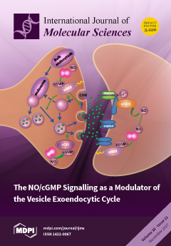
Nitric oxide (NO) and its downstream cGMP-activated pathway have been established as important elements in maintaining high-fidelity synaptic transmission under strong stimulation. NO generated post-synaptically by a neuronal NO synthase (nNOS) can diffuse to the presynaptic terminal triggering the synthesis of cGMP by the soluble guanylyl cyclase (sGC). Then, cGMP activates the cGMP-dependent protein kinases (cGKs) that regulate the synaptic vesicle exoendocytic cycle and transmitter release. View this paper
  • Issues are regarded as officially published after their release is announced to the table of contents alert mailing list.
  • You may sign up for e-mail alerts to receive table of contents of newly released issues.
  • PDF is the official format for papers published in both, html and pdf forms. To view the papers in pdf format, click on the "PDF Full-text" link, and use the free Adobe Reader to open them.
Order results
Result details
Section
Select all
Export citation of selected articles as:
1798 KiB  
Article
Magnetic Beads-Based Sensor with Tailored Sensitivity for Rapid and Single-Step Amperometric Determination of miRNAs
by Eva Vargas 1, Rebeca M. Torrente-Rodríguez 1, Víctor Ruiz-Valdepeñas Montiel 1, Eloy Povedano 1, María Pedrero 1, Juan J. Montoya 2, Susana Campuzano 1,* and José M. Pingarrón 1,*
1 Department of Analytical Chemistry, Faculty of Chemistry, University Complutense of Madrid, 28040 Madrid, Spain
2 Cannan Research and Investment & Faculty of Medicine, University Complutense of Madrid, 28040 Madrid, Spain
Int. J. Mol. Sci. 2017, 18(11), 2151; https://doi.org/10.3390/ijms18112151 - 9 Nov 2017
Cited by 30 | Viewed by 9637
Abstract
This work describes a sensitive amperometric magneto-biosensor for single-step and rapid determination of microRNAs (miRNAs). The developed strategy involves the use of direct hybridization of the target miRNA (miRNA-21) with a specific biotinylated DNA probe immobilized on streptavidin-modified magnetic beads (MBs), and labeling [...] Read more.
This work describes a sensitive amperometric magneto-biosensor for single-step and rapid determination of microRNAs (miRNAs). The developed strategy involves the use of direct hybridization of the target miRNA (miRNA-21) with a specific biotinylated DNA probe immobilized on streptavidin-modified magnetic beads (MBs), and labeling of the resulting heteroduplexes with a specific DNA–RNA antibody and the bacterial protein A (ProtA) conjugated with an horseradish peroxidase (HRP) homopolymer (Poly-HRP40) as an enzymatic label for signal amplification. Amperometric detection is performed upon magnetic capture of the modified MBs onto the working electrode surface of disposable screen-printed carbon electrodes (SPCEs) using the H2O2/hydroquinone (HQ) system. The magnitude of the cathodic signal obtained at −0.20 V (vs. the Ag pseudo-reference electrode) demonstrated linear dependence with the concentration of the synthetic target miRNA over the 1.0 to 100 pM range. The method provided a detection limit (LOD) of 10 attomoles (in a 25 μL sample) without any target miRNA amplification in just 30 min (once the DNA capture probe-MBs were prepared). This approach shows improved sensitivity compared with that of biosensors constructed with the same anti-DNA–RNA Ab as capture instead of a detector antibody and further labeling with a Strep-HRP conjugate instead of the Poly-HRP40 homopolymer. The developed strategy involves a single step working protocol, as well as the possibility to tailor the sensitivity by enlarging the length of the DNA/miRNA heteroduplexes using additional probes and/or performing the labelling with ProtA conjugated with homopolymers prepared with different numbers of HRP molecules. The practical usefulness was demonstrated by determination of the endogenous levels of the mature target miRNA in 250 ng raw total RNA (RNAt) extracted from human mammary epithelial normal (MCF-10A) and cancer (MCF-7) cells and tumor tissues. Full article
(This article belongs to the Collection Regulation by Non-coding RNAs)
Show Figures

Graphical abstract

2854 KiB  
Article
Directed Evolution of Recombinant C-Terminal Truncated Staphylococcus epidermidis Lipase AT2 for the Enhancement of Thermostability
by Jiivittha Veno 1,2, Nor Hafizah Ahmad Kamarudin 1, Mohd Shukuri Mohamad Ali 1,3, Malihe Masomian 1,2 and Raja Noor Zaliha Raja Abd. Rahman 1,2,*
1 Enzyme and Microbial Technology Research Centre, Universiti Putra Malaysia, Serdang 43400, Selangor, Malaysia
2 Department of Microbiology, Faculty of Biotechnology and Biomolecular Science, Universiti Putra Malaysia, Serdang 43400, Selangor, Malaysia
3 Department of Biochemistry, Faculty of Biotechnology and Biomolecular Science, Universiti Putra Malaysia, Serdang 43400, Selangor, Malaysia
Int. J. Mol. Sci. 2017, 18(11), 2202; https://doi.org/10.3390/ijms18112202 - 4 Nov 2017
Cited by 16 | Viewed by 4872
Abstract
In the industrial processes, lipases are expected to operate at temperatures above 45 °C and could retain activity in organic solvents. Hence, a C-terminal truncated lipase from Staphylococcus epidermis AT2 (rT-M386) was engineered by directed evolution. A mutant with glycine-to-cysteine substitution (G210C) demonstrated [...] Read more.
In the industrial processes, lipases are expected to operate at temperatures above 45 °C and could retain activity in organic solvents. Hence, a C-terminal truncated lipase from Staphylococcus epidermis AT2 (rT-M386) was engineered by directed evolution. A mutant with glycine-to-cysteine substitution (G210C) demonstrated a remarkable improvement of thermostability, whereby the mutation enhanced the activity five-fold when compared to the rT-M386 at 50 °C. The rT-M386 and G210C lipases were purified concurrently using GST-affinity chromatography. The biochemical and biophysical properties of both enzymes were investigated. The G210C lipase showed a higher optimum temperature (45 °C) and displayed a more prolonged half-life in the range of 40–60 °C as compared to rT-M386. Both lipases exhibited optimal activity and stability at pH 8. The G210C showed the highest stability in the presence of polar organic solvents at 50 °C compared to the rT-M386. Denatured protein analysis presented a significant change in the molecular ellipticity value above 60 °C, which verified the experimental result on the temperature and thermostability profile of G210C. Full article
(This article belongs to the Special Issue Microbial Enzymes)
Show Figures

Figure 1

2588 KiB  
Article
New Pyrazole-Hydrazone Derivatives: X-ray Analysis, Molecular Structure Investigation via Density Functional Theory (DFT) and Their High In-Situ Catecholase Activity
by Khalid Karrouchi 1,2,3, El Bekkaye Yousfi 4, Nada Kheira Sebbar 5, Youssef Ramli 1, Jamal Taoufik 1, Younes Ouzidan 6, M’hammed Ansar 1, Yahia N. Mabkhot 7,*, Hazem A. Ghabbour 8 and Smaail Radi 2,*
1 Laboratoire de Chimie Thérapeutique, Faculté de Médecine et de Pharmacie, Université Mohammed V, P. O. Box 8007, Rabat 10100, Morocco
2 Laboratoire de Chimie Appliquée et Environnement (LCAE), Faculté des Sciences, Université Mohamed I, P. O. Box 524, Oujda 60000, Morocco
3 Laboratoire National de Contrôle des Médicaments, Direction du Médicament et de la Pharmacie, Ministère de la Santé, P. O. Box 6206, Rabat 10100, Morocco
4 Institution Supérieure des Professions Infirmières et Techniques de Santé, P. O. Box 4806, Oujda 60000, Morocco
5 Laboratoire de Chimie Organique Hétérocyclique, Pharmacochimie, Faculté des sciences, Université Mohammed V, P. O. Box 8007, Rabat 10100, Morocco
6 Laboratoire de Chimie Organique Appliquée, Faculté des Sciences et Techniques, Université Sidi Mohamed Ben Abdellah, P. O. Box 2202, Fès 30000, Morocco
7 Department of Chemistry, Faculty of Science, King Saud University, P.O. Box 2455, Riyadh 11451, Saudi Arabia
8 Department of Pharmaceutical Chemistry, College of Pharmacy, King Saud University, P. O. Box 2457, Riyadh 11451, Saudi Arabia
Int. J. Mol. Sci. 2017, 18(11), 2215; https://doi.org/10.3390/ijms18112215 - 25 Oct 2017
Cited by 46 | Viewed by 3944
Abstract
The development of low-cost catalytic systems that mimic the activity of tyrosinase enzymes (Catechol oxidase) is of great promise for future biochemistry technologic demands. Herein, we report the synthesis of new biomolecules systems based on hydrazone derivatives containing a pyrazole moiety (L1 [...] Read more.
The development of low-cost catalytic systems that mimic the activity of tyrosinase enzymes (Catechol oxidase) is of great promise for future biochemistry technologic demands. Herein, we report the synthesis of new biomolecules systems based on hydrazone derivatives containing a pyrazole moiety (L1L6) with superior catecholase activity. Crystal structures of L1 and L2 biomolecules were determined by X-ray single crystal diffraction (XRD). Optimized geometrical parameters were calculated by density functional theory (DFT) at B3LYP/6–31G (d, p) level and were found to be in good agreement with single crystal XRD data. Copper (II) complexes of the compounds (L1L6), generated in-situ, were investigated for their catalytic activities towards the oxidation reaction of catechol to ortho-quinone with the atmospheric dioxygen, in an attempt to model the activity of the copper containing enzyme tyrosinase. The studies showed that the activities depend on four parameters: the nature of the ligand, the nature of counter anion, the nature of solvent and the concentration of ligand. The Cu(II)-ligands, given here, present the highest catalytic activity (72.920 μmol·L−1·min−1) among the catalysts recently reported in the existing literature. Full article
(This article belongs to the Section Molecular Biophysics)
Show Figures

Graphical abstract

1152 KiB  
Review
Bioactivity of Olive Oil Phenols in Neuroprotection
by Cristina Angeloni 1, Marco Malaguti 2, Maria Cristina Barbalace 2 and Silvana Hrelia 2,*
1 School of Pharmacy, University of Camerino, 62032 Camerino, Italy
2 Department for Life Quality Studies, Alma Mater Studiorum-University of Bologna, 40126 Bologna, Italy
Int. J. Mol. Sci. 2017, 18(11), 2230; https://doi.org/10.3390/ijms18112230 - 25 Oct 2017
Cited by 179 | Viewed by 15305
Abstract
Neurological disorders such as stroke, Alzheimer’s and Parkinson’s diseases are associated with high morbidity and mortality, and few or no effective options are available for their treatment. These disorders share common pathological characteristics like the induction of oxidative stress, abnormal protein aggregation, perturbed [...] Read more.
Neurological disorders such as stroke, Alzheimer’s and Parkinson’s diseases are associated with high morbidity and mortality, and few or no effective options are available for their treatment. These disorders share common pathological characteristics like the induction of oxidative stress, abnormal protein aggregation, perturbed Ca2+ homeostasis, excitotoxicity, inflammation and apoptosis. A large body of evidence supports the beneficial effects of the Mediterranean diet in preventing neurodegeneration. As the Mediterranean diet is characterized by a high consumption of extra-virgin olive oil it has been hypothesized that olive oil, and in particular its phenols, could be responsible for the beneficial effect of the Mediterranean diet. This review provides an updated vision of the beneficial properties of olive oil and olive oil phenols in preventing/counteracting both acute and chronic neurodegenerative diseases. Full article
(This article belongs to the Special Issue Neuroprotective Strategies 2017)
Show Figures

Graphical abstract

2125 KiB  
Article
Chemoenzymatic Preparation and Biophysical Properties of Sulfated Quercetin Metabolites
by Kateřina Valentová 1,*, Kristýna Káňová 1, Florent Di Meo 2,*, Helena Pelantová 1, Christopher Steven Chambers 1, Lenka Rydlová 1, Lucie Petrásková 1, Alena Křenková 1, Josef Cvačka 3, Patrick Trouillas 2,4 and Vladimír Křen 1
1 Institute of Microbiology of the Czech Academy of Sciences, Vídeňská 1083, CZ-14220 Prague, Czech Republic
2 INSERM U850, Univ. Limoges, School of Pharmacy, 2 rue du Docteur Marcland, F-87025 Limoges, France
3 Institute of Organic Chemistry and Biochemistry, Czech Academy of Sciences, Flemingovo nám. 2, CZ-16610 Prague, Czech Republic
4 Regional Centre of Advanced Technologies and Materials, Department of Physical Chemistry, Faculty of Science, Palacký University, tř. 17. listopadu 12, CZ-77146 Olomouc, Czech Republic
Int. J. Mol. Sci. 2017, 18(11), 2231; https://doi.org/10.3390/ijms18112231 - 25 Oct 2017
Cited by 22 | Viewed by 5266
Abstract
Sulfated quercetin derivatives are important authentic standards for metabolic studies. Quercetin-3′-O-sulfate, quercetin-4′-O-sulfate, and quercetin-3-O-sulfate as well as quercetin-di-O-sulfate mixture (quercetin-7,3′-di-O-sulfate, quercetin-7,4′-di-O-sulfate, and quercetin-3′,4′-di-O-sulfate) were synthetized by arylsulfotransferase from Desulfitobacterium [...] Read more.
Sulfated quercetin derivatives are important authentic standards for metabolic studies. Quercetin-3′-O-sulfate, quercetin-4′-O-sulfate, and quercetin-3-O-sulfate as well as quercetin-di-O-sulfate mixture (quercetin-7,3′-di-O-sulfate, quercetin-7,4′-di-O-sulfate, and quercetin-3′,4′-di-O-sulfate) were synthetized by arylsulfotransferase from Desulfitobacterium hafniense. Purified monosulfates and disulfates were fully characterized using MS and NMR and tested for their 1,1-diphenyl-2-picrylhydrazyl (DPPH), 2,2′-azinobis-(3-ethylbenzothiazoline-6-sulfonic acid) (ABTS+) and N,N-dimethyl-p-phenylenediamine (DMPD) radical scavenging, Folin-Ciocalteau reduction (FCR), ferric reducing antioxidant power (FRAP), and anti-lipoperoxidant activities in rat liver microsomes damaged by tert-butylhydroperoxide. Although, as expected, the sulfated metabolites were usually less active than quercetin, they remained still effective antiradical and reducing agents. Quercetin-3′-O-sulfate was more efficient than quercetin-4′-O-sulfate in DPPH and FCR assays. In contrast, quercetin-4′-O-sulfate was the best ferric reductant and lipoperoxidation inhibitor. The capacity to scavenge ABTS+• and DMPD was comparable for all substances, except for disulfates, which were the most efficient. Quantum calculations and molecular dynamics simulations on membrane models supported rationalization of free radical scavenging and lipid peroxidation inhibition. These results clearly showed that individual metabolites of food bioactives can markedly differ in their biological activity. Therefore, a systematic and thorough investigation of all bioavailable metabolites with respect to native compounds is needed when evaluating food health benefits. Full article
(This article belongs to the Special Issue Bioactive Phenolics and Polyphenols 2018)
Show Figures

Graphical abstract

2011 KiB  
Article
Phospholamban Is Downregulated by pVHL-Mediated Degradation through Oxidative Stress in Failing Heart
by Shunichi Yokoe and Michio Asahi *
Department of Pharmacology, Faculty of Medicine, Osaka Medical College, 2-7 Daigaku-machi, Takatsuki, Osaka 569-8686, Japan
Int. J. Mol. Sci. 2017, 18(11), 2232; https://doi.org/10.3390/ijms18112232 - 25 Oct 2017
Cited by 15 | Viewed by 4712
Abstract
The E3 ubiquitin ligase, von Hippel–Lindau (VHL), regulates protein expression by polyubiquitination. Although the protein VHL (pVHL) was reported to be involved in the heart function, the underlying mechanism is unclear. Here, we show that pVHL was upregulated in hearts from two types [...] Read more.
The E3 ubiquitin ligase, von Hippel–Lindau (VHL), regulates protein expression by polyubiquitination. Although the protein VHL (pVHL) was reported to be involved in the heart function, the underlying mechanism is unclear. Here, we show that pVHL was upregulated in hearts from two types of genetically dilated cardiomyopathy (DCM) mice models. In comparison with the wild-type mouse, both DCM mice models showed a significant reduction in the expression of phospholamban (PLN), a potent inhibitor of sarco(endo)plasmic reticulum Ca2+-ATPase, and enhanced interaction between pVHL and PLN. To clarify whether pVHL is involved in PLN degradation in failing hearts, we used carbonylcyanide m-chlorophenylhydrazone (CCCP), a mitochondrial membrane potential (MMP)-lowering reagent, to mimic the heart failure condition in PLN-expressing HEK293 cells and found that CCCP treatment resulted in PLN degradation and increased interaction between PLN and pVHL. However, these effects were reversed with the addition of N-acetyl-l-cysteine. Furthermore, the co-transfection of VHL and PLN in HEK293 cells decreased PLN expression under oxidative stress, whereas knockdown of VHL increased PLN expression both under normal and oxidative stress conditions. Together, we propose that oxidative stress upregulates pVHL expression to induce PLN degradation in failing hearts. Full article
(This article belongs to the Special Issue Ubiquitin System)
Show Figures

Figure 1

2637 KiB  
Article
Synthesis and Radioprotective Activity of Mitochondria Targeted Dihydropyridines In Vitro
by Yurui Zhang 1, Junying Wang 2, Yuanyuan Li 1, Feng Wang 3, Fujun Yang 1 and Wenqing Xu 1,*
1 Tianjin Key Laboratory of Molecular Nuclear Medicine, Institute of Radiation Medicine, Chinese Academy of Medical Sciences and Peking Union Medical College, Tianjin 300192, China
2 Department of Physics, School of Sciences and Tianjin Collaborative Innovation Center of Chemical Science and Engineering, Tianjin University, Tianjin 300350, China
3 Department of Statistics, Tianjin University of Finance and Economics, Tianjin 300222, China
Int. J. Mol. Sci. 2017, 18(11), 2233; https://doi.org/10.3390/ijms18112233 - 25 Oct 2017
Cited by 12 | Viewed by 4850
Abstract
The radiation-induced damage to mitochondrial oxidative respiratory chain could lead to generating of superoxide anions (O2−) and secondary reactive oxygen species (ROS), which are the major resources of continuous ROS production after radiation. Scavenging radiation-induced ROS effectively can help mitochondria to [...] Read more.
The radiation-induced damage to mitochondrial oxidative respiratory chain could lead to generating of superoxide anions (O2−) and secondary reactive oxygen species (ROS), which are the major resources of continuous ROS production after radiation. Scavenging radiation-induced ROS effectively can help mitochondria to maintain their physiological function and relief cells from oxidative stress. Dihydropyridines (DHPs) are biomimetic hydrogen sources that could protect cells against radiation damage. In this study, we designed and synthetized three novel mitochondrial-targeted dihydropyridines (Mito-DHPs) that utilize the mitochondrial membrane potential to enter the organelle and scavenge ROS. MitoTracker confirmed Mito-DHPs accumulation in mitochondria, and the DCFH-DA assay demonstrated effective ROS scavenging activity. In addition, the γ-H2AX and comet assay demonstrated the ability of Mito-DHPs to protect against both radiation and ROS-induced DNA strand breaks. Furthermore, Mito-DHP1 proved to be non-toxic and displayed significant radioprotection activity (p < 0.05) in vitro. Mito-DHPs are therefore promising antioxidants that could penetrate the membrane of mitochondria, scavenge excessive ROS, and protect cells against radiation-induced oxidative damage. Full article
(This article belongs to the Section Biochemistry)
Show Figures

Graphical abstract

7903 KiB  
Article
FGF-2b and h-PL Transform Duct and Non-Endocrine Human Pancreatic Cells into Endocrine Insulin Secreting Cells by Modulating Differentiating Genes
by Giulia Donadel 1,*,†, Donatella Pastore 1,†, David Della-Morte 1,2, Barbara Capuani 1, Marco F. Lombardo 3, Francesca Pacifici 1, Marco Bugliani 4, Fabio A. Grieco 5, Piero Marchetti 4 and Davide Lauro 1
1 Department of Systems Medicine, University of Rome Tor Vergata, 00133 Rome, Italy
2 Department of Human Sciences and Quality of Life Promotion, San Raffaele Roma Open University, 00166 Rome, Italy
3 Agenzia regionale per la protezione ambientale (ARPA) Lazio, Sezione di Roma, 00173 Rome, Italy
4 Endocrinology and Metabolism of Transplantation, Azienda Ospedaliero-Universitaria (A.O.U.) Pisana, 56126 Pisa, Italy
5 Department of Medicine, Surgery and Neuroscience, University of Siena, 53100 Siena, Italy
These authors contributed equally to this work.
Int. J. Mol. Sci. 2017, 18(11), 2234; https://doi.org/10.3390/ijms18112234 - 25 Oct 2017
Cited by 11 | Viewed by 4535
Abstract
Background: Diabetes mellitus (DM) is a multifactorial disease orphan of a cure. Regenerative medicine has been proposed as novel strategy for DM therapy. Human fibroblast growth factor (FGF)-2b controls β-cell clusters via autocrine action, and human placental lactogen (hPL)-A increases functional β-cells. We [...] Read more.
Background: Diabetes mellitus (DM) is a multifactorial disease orphan of a cure. Regenerative medicine has been proposed as novel strategy for DM therapy. Human fibroblast growth factor (FGF)-2b controls β-cell clusters via autocrine action, and human placental lactogen (hPL)-A increases functional β-cells. We hypothesized whether FGF-2b/hPL-A treatment induces β-cell differentiation from ductal/non-endocrine precursor(s) by modulating specific genes expression. Methods: Human pancreatic ductal-cells (PANC-1) and non-endocrine pancreatic cells were treated with FGF-2b plus hPL-A at 500 ng/mL. Cytofluorimetry and Immunofluorescence have been performed to detect expression of endocrine, ductal and acinar markers. Bromodeoxyuridine incorporation and annexin-V quantified cells proliferation and apoptosis. Insulin secretion was assessed by RIA kit, and electron microscopy analyzed islet-like clusters. Results: Increase in PANC-1 duct cells de-differentiation into islet-like aggregates was observed after FGF-2b/hPL-A treatment showing ultrastructure typical of islets-aggregates. These clusters, after stimulation with FGF-2b/hPL-A, had significant (p < 0.05) increase in insulin, C-peptide, pancreatic and duodenal homeobox 1 (PDX-1), Nkx2.2, Nkx6.1, somatostatin, glucagon, and glucose transporter 2 (Glut-2), compared with control cells. Markers of PANC-1 (Cytokeratin-19, MUC-1, CA19-9) were decreased (p < 0.05). These aggregates after treatment with FGF-2b/hPL-A significantly reduced levels of apoptosis. Conclusions: FGF-2b and hPL-A are promising candidates for regenerative therapy in DM by inducing de-differentiation of stem cells modulating pivotal endocrine genes. Full article
(This article belongs to the Special Issue Genome Editing 2018)
Show Figures

Graphical abstract

4546 KiB  
Article
Association of Smoking with Metabolic Volatile Organic Compounds in Exhaled Breath
by Xing Chen 1,*, Fuyuan Wang 1, Liquan Lin 1, Hao Dong 1, Feifei Huang 2, Kanhar Ghulam Muhammad 1, Liying Chen 2 and Olga Y. Gorlova 3
1 Department of Biomedical Engineering, Key Laboratory of Biomedical Engineering of Ministry of Education of China, Zhejiang University, 38 Zheda Road, Zhou Yi Qing Building, Hangzhou 310027, China
2 Department of Family Medicine, Sir Run Run Shaw Hospital, School of Medicine, Zhejiang University, 3 Qingchun E Rd, Hangzhou 310016, China
3 Department of Biomedical Data Science, Geisel School of Medicine at Dartmouth College, One Medical Center Drive, Lebanon, NH 03756, USA
Int. J. Mol. Sci. 2017, 18(11), 2235; https://doi.org/10.3390/ijms18112235 - 25 Oct 2017
Cited by 18 | Viewed by 4157
Abstract
Lung cancer (LC) screening will be more efficient if it is applied to a well-defined high-risk population. Characteristics including metabolic byproducts may be taken into account to access LC risk more precisely. Breath examination provides a non-invasive method to monitor metabolic byproducts. However, [...] Read more.
Lung cancer (LC) screening will be more efficient if it is applied to a well-defined high-risk population. Characteristics including metabolic byproducts may be taken into account to access LC risk more precisely. Breath examination provides a non-invasive method to monitor metabolic byproducts. However, the association between volatile organic compounds (VOCs) in exhaled breath and LC risk or LC risk factors is not studied. Exhaled breath samples from 122 healthy persons, who were given routine annual exam from December 2015 to December 2016, were analyzed using thermal desorption coupled with gas chromatography mass spectrometry (TD-GC-MS). Smoking characteristics, air quality, and other risk factors for lung cancer were collected. Univariate and multivariate analyses were used to evaluate the relationship between VOCs and LC risk factors. 7, 7, 11, and 27 VOCs were correlated with smoking status, smoking intensity, years of smoking, and depth of inhalation, respectively. Exhaled VOCs are related to smoking and might have a potential to evaluate LC risk more precisely. Both an assessment of temporal stability and testing in a prospective study are needed to establish the performance of VOCs such as 2,5-dimethylfuranm and 4-methyloctane as lung cancer risk biomarkers. Full article
(This article belongs to the Special Issue Inhaled Pollutants Modulate Respiratory and Systemic Diseases)
Show Figures

Figure 1

17125 KiB  
Article
Preparation and Characterization of Resorbable Bacterial Cellulose Membranes Treated by Electron Beam Irradiation for Guided Bone Regeneration
by Sung-Jun An 1,†, So-Hyoun Lee 2,†, Jung-Bo Huh 2,*, Sung In Jeong 1, Jong-Seok Park 1, Hui-Jeong Gwon 1, Eun-Sook Kang 3, Chang-Mo Jeong 2 and Youn-Mook Lim 1
1 Advanced Radiation Technology Institute, Korea Atomic Energy Research Institute, 1266 Sinjeong-dong, Jeongeup-si, Jeollabuk-do 56212, Korea
2 Department of Prosthodontics, Dental Research Institute, Institute of Translational Dental Sciences, BK21 PLUS Project, School of Dentistry, Pusan National University, Yangsan 50612, Korea
3 Department of Prosthodontics, In-Je University Haeundae Paik Hospital, Busan 48108, Korea
These authors contributed equally to this work.
Int. J. Mol. Sci. 2017, 18(11), 2236; https://doi.org/10.3390/ijms18112236 - 25 Oct 2017
Cited by 47 | Viewed by 6097
Abstract
Bacterial cellulose (BC) is an excellent biomaterial with many medical applications. In this study, resorbable BC membranes were prepared for guided bone regeneration (GBR) using an irradiation technique for applications in the dental field. Electron beam irradiation (EI) increases biodegradation by severing the [...] Read more.
Bacterial cellulose (BC) is an excellent biomaterial with many medical applications. In this study, resorbable BC membranes were prepared for guided bone regeneration (GBR) using an irradiation technique for applications in the dental field. Electron beam irradiation (EI) increases biodegradation by severing the glucose bonds of BC. BC membranes irradiated at 100 kGy or 300 kGy were used to determine optimal electron beam doses. Electron beam irradiated BC membranes (EI-BCMs) were evaluated by scanning electron microscopy (SEM), attenuated total reflectance-Fourier transform infrared (ATR-FTIR) spectroscopy, thermal gravimetric analysis (TGA), and using wet tensile strength measurements. In addition, in vitro cell studies were conducted in order to confirm the cytocompatibility of EI-BCMs. Cell viabilities of NIH3T3 cells on 100k and 300k EI-BCMs (100 kGy and 300 kGy irradiated BC membranes) were significantly greater than on NI-BCMs after 3 and 7 days (p < 0.05). Bone regeneration by EI-BCMs and their biodegradabilities were also evaluated using in vivo rat calvarial defect models for 4 and 8 weeks. Histometric results showed 100k EI-BCMs exhibited significantly larger new bone area (NBA; %) than 300k EI-BCMs at 8 weeks after implantation (p < 0.05). Mechanical, chemical, and biological analyses showed EI-BCMs effectively interacted with cells and promoted bone regeneration. Full article
(This article belongs to the Special Issue Novel Biomaterials for Tissue Engineering 2018)
Show Figures

Graphical abstract

6666 KiB  
Article
iTRAQ-Based Identification of Proteins Related to Muscle Growth in the Pacific Abalone, Haliotis discus hannai
by Jianfang Huang 1,2, Weiwei You 1,2,3,*, Xuan Luo 1,2,3 and Caihuan Ke 1,2,3,*
1 State Key Laboratory of Marine Environmental Science, Xiamen University, Xiamen 361102, China
2 College of Ocean and Earth Sciences, Xiamen University, Xiamen 361102, China
3 Fujian Collaborative Innovation Center for Exploitation and Utilization of Marine Biological Resources, Xiamen University, Xiamen 361102, China
Int. J. Mol. Sci. 2017, 18(11), 2237; https://doi.org/10.3390/ijms18112237 - 25 Oct 2017
Cited by 10 | Viewed by 3967
Abstract
The abalone Haliotis discus hannai is an important aquaculture species that is grown for human consumption. However, little is known of the genetic mechanisms governing muscle growth in this species, particularly with respect to proteomics. The isobaric tag for relative and absolute quantitation [...] Read more.
The abalone Haliotis discus hannai is an important aquaculture species that is grown for human consumption. However, little is known of the genetic mechanisms governing muscle growth in this species, particularly with respect to proteomics. The isobaric tag for relative and absolute quantitation (iTRAQ) method allows for sensitive and accurate protein quantification. Our study was the first to use iTRAQ-based quantitative proteomics to investigate muscle growth regulation in H. discus hannai. Among the 1904 proteins identified from six samples, 125 proteins were differentially expressed in large specimens of H. discus hannai as compared to small specimens. In the large specimens, 47 proteins were upregulated and 78 were downregulated. Many of the significant Kyoto Encyclopedia of Genes and Genomes (KEGG) pathways, including these differentially expressed proteins, were closely related to muscle growth, including apoptosis, thyroid hormone signaling, regulation of the actin cytoskeleton, and viral myocarditis (p < 0.05). Our quantitative real-time polymerase chain reaction (qRT-PCR) analyses suggested that the alterations in expression levels observed in the differentially expressed proteins were consistent with the alterations observed in the encoding mRNAs, indicating the repeatability of our proteomic approach. Our findings contribute to the knowledge of the molecular mechanisms of muscle growth in H. discus hannai. Full article
(This article belongs to the Section Biochemistry)
Show Figures

Figure 1

711 KiB  
Article
Systemic Inflammation, Oxidative Damage to Nucleic Acids, and Metabolic Syndrome in the Pathogenesis of Psoriasis
by Lenka Borska 1,*, Jan Kremlacek 1, Ctirad Andrys 2, Jan Krejsek 2, Kvetoslava Hamakova 3, Pavel Borsky 1, Vladimir Palicka 4, Vit Rehacek 5, Andrea Malkova 6 and Zdenek Fiala 6
1 Department of Pathological Physiology, Charles University, Faculty of Medicine in Hradec Kralove, 500 03 Hradec Kralove, Czech Republic
2 Department of Clinical Immunology and Allergology, Charles University, Faculty of Medicine in Hradec Kralove, 500 03 Hradec Kralove, Czech Republic
3 Clinic of Dermal and Venereal Diseases, Charles University Hospital Hradec Kralove, 500 05 Hradec Kralove, Czech Republic
4 Institute of Clinical Biochemistry and Diagnostics, Charles University Hospital and Faculty of Medicine in Hradec Kralove, 500 05 Hradec Kralove, Czech Republic
5 Department of Transfusion Medicine, Charles University Hospital in Hradec Kralove, 500 05 Hradec Kralove, Czech Republic
6 Department of Hygiene and Preventive Medicine, Charles University, Faculty of Medicine in Hradec Kralove, 500 03 Hradec Kralove, Czech Republic
Int. J. Mol. Sci. 2017, 18(11), 2238; https://doi.org/10.3390/ijms18112238 - 25 Oct 2017
Cited by 40 | Viewed by 8853
Abstract
In the pathogenesis of psoriasis, systemic inflammation and oxidative stress play mutual roles interrelated with metabolic syndrome (MetS). This study aims to map the selected markers of inflammation (C-reactive protein (CRP)), oxidative damage to nucleic acids (DNA/RNA damage; 8-hydroxy-2′-deoxyguanosine, 8-hydroxyguanosine, and 8-hydroxyguanine), and [...] Read more.
In the pathogenesis of psoriasis, systemic inflammation and oxidative stress play mutual roles interrelated with metabolic syndrome (MetS). This study aims to map the selected markers of inflammation (C-reactive protein (CRP)), oxidative damage to nucleic acids (DNA/RNA damage; 8-hydroxy-2′-deoxyguanosine, 8-hydroxyguanosine, and 8-hydroxyguanine), and the parameters of MetS (waist circumference, fasting glucose, triglycerides, high-density lipoprotein (HDL) cholesterol, diastolic and systolic blood pressure) in a group of 37 patients with psoriasis (62% of MetS) and in 43 healthy controls (42% of MetS). Levels of CRP, DNA/RNA damage, fasting glucose, and triglycerides were significantly elevated in patients. MetS in conjunction with psoriasis was associated with high levels of CRP, significantly higher than in control subjects without MetS. Patients with MetS exhibited further DNA/RNA damage, which was significantly higher in comparison with the control group. Our study supports the independent role of psoriasis and MetS in the increase of CRP and DNA/RNA damage. The psoriasis contributes to an increase in the levels of both effects more significantly than MetS. The psoriasis also diminished the relationship between CRP and oxidative damage to nucleic acids existent in controls. Full article
(This article belongs to the Special Issue Psoriasis)
Show Figures

Graphical abstract

747 KiB  
Review
A Mini-Review on the Effect of Docosahexaenoic Acid (DHA) on Cerulein-Induced and Hypertriglyceridemic Acute Pancreatitis
by Yoo Kyung Jeong and Hyeyoung Kim *
Department of Food and Nutrition, Brian Korea 21 PLUS Project, College of Human Ecology, Yonsei University, Seoul 03722, Korea
Int. J. Mol. Sci. 2017, 18(11), 2239; https://doi.org/10.3390/ijms18112239 - 25 Oct 2017
Cited by 21 | Viewed by 6885
Abstract
Acute pancreatitis refers to the sudden inflammation of the pancreas. It is associated with premature activation and release of digestive enzymes into the pancreatic interstitium and systemic circulation, resulting in pancreatic tissue autodigestion and multiple organ dysfunction, as well as with increased cytokine [...] Read more.
Acute pancreatitis refers to the sudden inflammation of the pancreas. It is associated with premature activation and release of digestive enzymes into the pancreatic interstitium and systemic circulation, resulting in pancreatic tissue autodigestion and multiple organ dysfunction, as well as with increased cytokine production, ultimately leading to deleterious local and systemic effects. Although mechanisms involved in pathogenesis of acute pancreatitis have not been completely elucidated, oxidative stress is regarded as a major risk factor. In human acute pancreatitis, lipid peroxide levels in pancreatic tissues increase. Docosahexaenoic acid (DHA), an omega-3 polyunsaturated fatty acid (C22:6n-3), exerts anti-inflammatory and antioxidant effects on various cells. Previous studies have shown that DHA activates peroxisome proliferator-activated receptor-γ and induces catalase, which inhibits oxidative stress-mediated inflammatory signaling required for cytokine expression in experimental acute pancreatitis using cerulein. Cerulein, a cholecystokinin analog, induces intra-acinar activation of trypsinogen in the pancreas, which results in human acute pancreatitis-like symptoms. Therefore, DHA supplementation may be beneficial for preventing or inhibiting acute pancreatitis development. Since DHA reduces serum triglyceride levels, addition of DHA to lipid-lowering drugs like statins has been investigated to reduce hypertriglyceridemic acute pancreatitis. However, high DHA concentrations increase cytosolic Ca2+, which activates protein kinase C and may induce hyperlipidemic acute pancreatitis. In this review, effect of DHA on cerulein-induced and hypertriglyceridemic acute pancreatitis has been discussed. The relation of high concentration of DHA to hyperlipidemic acute pancreatitis has been included. Full article
(This article belongs to the Special Issue Omega-3 Fatty Acids in Health and Disease: New Knowledge)
Show Figures

Figure 1

5191 KiB  
Article
Comparative Analysis of the Cytology and Transcriptomes of the Cytoplasmic Male Sterility Line H276A and Its Maintainer Line H276B of Cotton (Gossypium barbadense L.)
by Xiangjun Kong 1,†, Dongmei Liu 2,†, Xiaofang Liao 1, Jie Zheng 1, Yong Diao 1, Yiding Liu 1 and Ruiyang Zhou 1,*
1 Key Laboratory of Plant Genetics and Breeding, College of Agriculture, Guangxi University, Nanning 530006, China
2 Key Laboratory of Plant-Microbe Interactions, Department of Life Science and Food, Shangqiu Normal University, Shangqiu 476000, China
These authors contributed equally to this work.
Int. J. Mol. Sci. 2017, 18(11), 2240; https://doi.org/10.3390/ijms18112240 - 25 Oct 2017
Cited by 24 | Viewed by 4355
Abstract
In this study, the tetrad stage of microspore development in a new cotton (Gossypium barbadense L.) cytoplasmic male sterility (CMS) line, H276A, was identified using paraffin sections at the abortion stage. To explore the molecular mechanism underlying CMS in cotton, a comparative [...] Read more.
In this study, the tetrad stage of microspore development in a new cotton (Gossypium barbadense L.) cytoplasmic male sterility (CMS) line, H276A, was identified using paraffin sections at the abortion stage. To explore the molecular mechanism underlying CMS in cotton, a comparative transcriptome analysis between the CMS line H276A and its maintainer line H276B at the tetrad stage was conducted using an Illumina HiSeq 4000 platform. The comparison of H276A with H276B revealed a total of 64,675 genes, which consisted of 59,255 known and 5420 novel genes. An analysis of the two libraries with a given threshold yielded a total of 3603 differentially expressed genes (DEGs), which included 1363 up- and 2240 down-regulated genes. Gene Ontology (GO) annotation showed that 2171 DEGs were distributed into 38 categories, and a Kyoto Encyclopedia of Genes and Genomes (KEGG) analysis showed that 2683 DEGs were classified into 127 groups. Thirteen DEGs were randomly selected and detected by quantitative reverse-transcribed PCR (qRT-PCR), and the results indicated that the transcriptome sequencing results were reliable. The bioinformatic analysis results in conjunction with previously reported data revealed key DEGs that might be associated with the male sterility features of H276A. Our results provide a comprehensive foundation for understanding anther development and will accelerate the study of the molecular mechanisms of CMS in cotton. Full article
(This article belongs to the Section Molecular Plant Sciences)
Show Figures

Figure 1

1230 KiB  
Article
Effects of Late Gestational Fetal Exposure to Dexamethasone Administration on the Postnatal Hypothalamus-Pituitary-Adrenal Axis Response to Hypoglycemia in Pigs
by René Schiffner 1,2,*, Guadalupe L. Rodríguez-González 3, Florian Rakers 1, Marius Nistor 1, Peter W. Nathanielsz 4, Teodora Daneva 5, Matthias Schwab 1, Thomas Lehmann 6 and Martin Schmidt 7
1 Department of Neurology, Jena University Hospital—Friedrich Schiller University, 07747 Jena, Germany
2 Orthopaedic Department, Jena University Hospital—Friedrich Schiller University, 07747 Jena, Germany
3 Reproductive Biology, National Institute of Medical Science and Nutrition, 14000 Mexico City, Mexico
4 Department of Animal Science, University of Wyoming, Laramie, 82071 WY, USA
5 Institute of Biology and Immunology of Reproduction, Bulgarian Academy of Sciences, 1113 Sofia, Bulgaria
6 Institute of Medical Statistics, Computer Sciences and Documentation Science, Jena University Hospital—Friedrich Schiller University, 07743 Jena, Germany
7 Institute for Biochemistry II, Jena University Hospital—Friedrich Schiller University, 07743 Jena, Germany
Int. J. Mol. Sci. 2017, 18(11), 2241; https://doi.org/10.3390/ijms18112241 - 27 Oct 2017
Cited by 12 | Viewed by 4920
Abstract
Background: Prenatal glucocorticoid administration alters the activity of the fetal hypothalamic-pituitary-adrenocortical axis (HPAA), and correspondingly the adenocorticotropic hormone (ACTH) and cortisol levels after birth. The dosages required for these effects are critically discussed. Activation of the HPAA is related to metabolic syndrome and [...] Read more.
Background: Prenatal glucocorticoid administration alters the activity of the fetal hypothalamic-pituitary-adrenocortical axis (HPAA), and correspondingly the adenocorticotropic hormone (ACTH) and cortisol levels after birth. The dosages required for these effects are critically discussed. Activation of the HPAA is related to metabolic syndrome and diabetes mellitus. Hypoglycemia is the classic side effect of antidiabetic treatment. We hypothesized that a low dosage of dexamethasone in late pregnancy alters the HPAA response to hypoglycemia in pigs. Methods: 12 pregnant sows were randomly assigned to two groups which received either a low-dose intramuscular injection (99th and 100th day of gestation) of dexamethasone (0.06 μg/kg body weight) or vehicle. Three months after birth, 18 dexamethasone-treated anaesthetized offspring and 12 control offspring underwent a 75 min hypoglycemic clamp (blood glucose below 4 mmol/L) procedure. Heart rate (HR), blood pressure, ACTH and cortisol levels and body weight (at birth and after three months) were recorded. Results: Dexamethasone-treated animals exhibited significantly elevated ACTH (139.9 ± 12.7 pg/mL) and cortisol (483.1 ± 30.3 nmol/L) levels during hypoglycemia as compared to the control group (41.7 ± 6.5 pg/mL and 257.9 ± 26.7 nmol/L, respectively), as well as an elevated HR (205.5 ± 5.7 bpm) and blood pressure (systolic: 128.6 ± 1.5, diastolic: 85.7 ± 0.7 mmHg) response as compared to the control group (153.2 ± 4.5 bpm; systolic: 118.6 ± 1.6, diastolic: 79.5 ± 1.4 mmHg, respectively; p < 0.001). Conclusions: Low-dose prenatal administration of dexamethasone not only exerts effects on the HPAA (ACTH and cortisol concentration) and vital parameters (HR and diastolic blood pressure) under baseline conditions, but also on ACTH, HR and systolic blood pressure during hypoglycemia. Full article
Show Figures

Figure 1

5252 KiB  
Article
Laminin-Coated Poly(Methyl Methacrylate) (PMMA) Nanofiber Scaffold Facilitates the Enrichment of Skeletal Muscle Myoblast Population
by Nor Kamalia Zahari 1, Ruszymah Binti Haji Idrus 1,2 and Shiplu Roy Chowdhury 1,*
1 Tissue Engineering Centre, Universiti Kebangsaan Malaysia Medical Centre, Cheras 56000, Kuala Lumpur, Malaysia
2 Department of Physiology, Faculty of Medicine, Universiti Kebangsaan Malaysia Medical Centre, Cheras 56000, Kuala Lumpur, Malaysia
Int. J. Mol. Sci. 2017, 18(11), 2242; https://doi.org/10.3390/ijms18112242 - 30 Oct 2017
Cited by 28 | Viewed by 5909
Abstract
Myoblasts, the contractile cells of skeletal muscle, have been invaluable for fundamental studies of muscle development and clinical applications for muscle loss. A major limitation to the myoblast-based therapeutic approach is contamination with non-contractile fibroblasts, which overgrow during cell expansion. To overcome these [...] Read more.
Myoblasts, the contractile cells of skeletal muscle, have been invaluable for fundamental studies of muscle development and clinical applications for muscle loss. A major limitation to the myoblast-based therapeutic approach is contamination with non-contractile fibroblasts, which overgrow during cell expansion. To overcome these limitations, this study was carried out to establish a 3D culture environment using nanofiber scaffolds to enrich the myoblast population during construct formation. Poly(methyl methacrylate) (PMMA) nanofiber (PM) scaffolds were fabricated using electrospinning techniques and coated with extracellular matrix (ECM) proteins, such as collagen or laminin, in the presence or absence of genipin. A mixed population of myoblasts and fibroblasts was isolated from human skeletal muscle tissues and cultured on plain surfaces, as well as coated and non-coated PM scaffolds. PMMA can produce smooth fibers with an average diameter of 360 ± 50 nm. Adsorption of collagen and laminin on PM scaffolds is significantly enhanced in the presence of genipin, which introduces roughness to the nanofiber surface without affecting fiber diameter and mechanical properties. It was also demonstrated that laminin-coated PM scaffolds significantly enhance myoblast proliferation (0.0081 ± 0.0007 h−1) and migration (0.26 ± 0.04 μm/min), while collagen-coated PM scaffolds favors fibroblasts proliferation (0.0097 ± 0.0009 h−1) and migration (0.23 ± 0.03 μm/min). Consequently, the myoblast population was enriched on laminin-coated PM scaffolds throughout the culture process. Therefore, laminin coating of nanofiber scaffolds could be a potential scaffold for the development of a tissue-engineered muscle substitute. Full article
(This article belongs to the Special Issue Cell Colonization in Scaffolds)
Show Figures

Figure 1

2133 KiB  
Article
Impaired Sleep, Circadian Rhythms and Neurogenesis in Diet-Induced Premature Aging
by Alexander J. Stankiewicz 1,2, Erin M. McGowan 2, Lili Yu 1,2 and Irina V. Zhdanova 1,2,*
1 Department of Preclinical Research and Development, BioChron LLC, Worcester, MA 01605, USA
2 Department of Anatomy and Neurobiology, Boston University School of Medicine, Boston, MA 02118, USA
Int. J. Mol. Sci. 2017, 18(11), 2243; https://doi.org/10.3390/ijms18112243 - 26 Oct 2017
Cited by 21 | Viewed by 7413
Abstract
Chronic high caloric intake (HCI) is a risk factor for multiple major human disorders, from diabetes to neurodegeneration. Mounting evidence suggests a significant contribution of circadian misalignment and sleep alterations to this phenomenon. An inverse temporal relationship between sleep, activity, food intake, and [...] Read more.
Chronic high caloric intake (HCI) is a risk factor for multiple major human disorders, from diabetes to neurodegeneration. Mounting evidence suggests a significant contribution of circadian misalignment and sleep alterations to this phenomenon. An inverse temporal relationship between sleep, activity, food intake, and clock mechanisms in nocturnal and diurnal animals suggests that a search for effective therapeutic approaches can benefit from the use of diurnal animal models. Here, we show that, similar to normal aging, HCI leads to the reduction in daily amplitude of expression for core clock genes, a decline in sleep duration, an increase in scoliosis, and anxiety-like behavior. A remarkable decline in adult neurogenesis in 1-year old HCI animals, amounting to only 21% of that in age-matched Control, exceeds age-dependent decline observed in normal 3-year old zebrafish. This is associated with misalignment or reduced amplitude of daily patterns for principal cell cycle regulators, cyclins A and B, and p20, in brain tissue. Together, these data establish HCI in zebrafish as a model for metabolically induced premature aging of sleep, circadian functions, and adult neurogenesis, allowing for a high throughput approach to mechanistic studies and drug trials in a diurnal vertebrate. Full article
(This article belongs to the Special Issue Molecular and Cellular Mechanisms of Circadian Rhythms)
Show Figures

Figure 1

4021 KiB  
Article
Biochemical Analysis of Two Single Mutants that Give Rise to a Polymorphic G6PD A-Double Mutant
by Edson Jiovany Ramírez-Nava 1, Daniel Ortega-Cuellar 2, Hugo Serrano-Posada 3, Abigail González-Valdez 4, America Vanoye-Carlo 5, Beatriz Hernández-Ochoa 6, Edgar Sierra-Palacios 7, Jessica Hernández-Pineda 8, Eduardo Rodríguez-Bustamante 9, Roberto Arreguin-Espinosa 9, Jesús Oria-Hernández 1, Horacio Reyes-Vivas 1, Jaime Marcial-Quino 10,* and Saúl Gómez-Manzo 1,*
1 Laboratorio de Bioquímica Genética, Instituto Nacional de Pediatría, Secretaría de Salud, Mexico City 04530, Mexico
2 Laboratorio de Nutrición Experimental, Instituto Nacional de Pediatría, Secretaría de Salud, Mexico City 04530, Mexico
3 Consejo Nacional de Ciencia y Tecnología (CONACYT), Laboratorio de Agrobiotecnología, Tecnoparque CLQ, Universidad de Colima, Carretera los Limones-Loma de Juárez, Colima 28629, Mexico
4 Departamento de Biología Molecular y Biotecnología, Instituto de Investigaciones Biomédicas, Universidad Nacional Autónoma de México, Mexico City 04510, Mexico
5 Laboratorio de Neurociencias, Instituto Nacional de Pediatría, Secretaría de Salud, Mexico City 04530, Mexico
6 Laboratorio de Inmunoquímica, Hospital Infantil de México Federico Gómez, Secretaría de Salud, Mexico City 06720, Mexico
7 Colegio de Ciencias y Humanidades, Plantel Casa Libertad, Universidad Autónoma de la Ciudad de México, Mexico City 09620, Mexico
8 Departamento de Infectología e Inmunología, Instituto Nacional de Perinatología, Secretaría de Salud, Mexico City 11000, México
9 Departamento de Química de Biomacromoléculas, Instituto de Química, Universidad Nacional Autónoma de Mexico, Mexico City 04510, Mexico
10 Consejo Nacional de Ciencia y Tecnología (CONACYT), Instituto Nacional de Pediatría, Secretaría de Salud, Mexico City 04530, Mexico
Int. J. Mol. Sci. 2017, 18(11), 2244; https://doi.org/10.3390/ijms18112244 - 26 Oct 2017
Cited by 16 | Viewed by 4589
Abstract
Glucose-6-phosphate dehydrogenase (G6PD) is a key regulatory enzyme that plays a crucial role in the regulation of cellular energy and redox balance. Mutations in the gene encoding G6PD cause the most common enzymopathy that drives hereditary nonspherocytic hemolytic anemia. To gain insights into [...] Read more.
Glucose-6-phosphate dehydrogenase (G6PD) is a key regulatory enzyme that plays a crucial role in the regulation of cellular energy and redox balance. Mutations in the gene encoding G6PD cause the most common enzymopathy that drives hereditary nonspherocytic hemolytic anemia. To gain insights into the effects of mutations in G6PD enzyme efficiency, we have investigated the biochemical, kinetic, and structural changes of three clinical G6PD variants, the single mutations G6PD A+ (Asn126AspD) and G6PD Nefza (Leu323Pro), and the double mutant G6PD A− (Asn126Asp + Leu323Pro). The mutants showed lower residual activity (≤50% of WT G6PD) and displayed important kinetic changes. Although all Class III mutants were located in different regions of the three-dimensional structure of the enzyme and were not close to the active site, these mutants had a deleterious effect over catalytic activity and structural stability. The results indicated that the G6PD Nefza mutation was mainly responsible for the functional and structural alterations observed in the double mutant G6PD A−. Moreover, our study suggests that the G6PD Nefza and G6PD A− mutations affect enzyme functions in a similar fashion to those reported for Class I mutations. Full article
(This article belongs to the Section Biochemistry)
Show Figures

Graphical abstract

827 KiB  
Review
Genome Modification Technologies and Their Applications in Avian Species
by Hong Jo Lee 1, Young Min Kim 1, Tamao Ono 2 and Jae Yong Han 1,3,*
1 Department of Agricultural Biotechnology, College of Agriculture and Life Sciences, and Research Institute of Agriculture and Life Sciences, Seoul National University, Seoul 08826, Korea
2 Faculty of Agriculture, Shinshu University, 8304 Minamiminowa, Kamiina, Nagano 399-4598, Japan
3 Institute for Biomedical Sciences, Shinshu University, 8304 Minamiminowa, Kamiina, Nagano 399-4598, Japan
Int. J. Mol. Sci. 2017, 18(11), 2245; https://doi.org/10.3390/ijms18112245 - 26 Oct 2017
Cited by 10 | Viewed by 5632
Abstract
The rapid development of genome modification technology has provided many great benefits in diverse areas of research and industry. Genome modification technologies have also been actively used in a variety of research areas and fields of industry in avian species. Transgenic technologies such [...] Read more.
The rapid development of genome modification technology has provided many great benefits in diverse areas of research and industry. Genome modification technologies have also been actively used in a variety of research areas and fields of industry in avian species. Transgenic technologies such as lentiviral systems and piggyBac transposition have been used to produce transgenic birds for diverse purposes. In recent years, newly developed programmable genome editing tools such as transcription activator-like effector nuclease (TALEN) and clustered regularly interspaced short palindromic repeats (CRISPR)/CRISPR-associated protein 9 (CRISPR/Cas9) have also been successfully adopted in avian systems with primordial germ cell (PGC)-mediated genome modification. These genome modification technologies are expected to be applied to practical uses beyond system development itself. The technologies could be used to enhance economic traits in poultry such as acquiring a disease resistance or producing functional proteins in eggs. Furthermore, novel avian models of human diseases or embryonic development could also be established for research purposes. In this review, we discuss diverse genome modification technologies used in avian species, and future applications of avian biotechnology. Full article
(This article belongs to the Special Issue Genome Editing 2018)
Show Figures

Graphical abstract

1778 KiB  
Article
The Analysis of A Frequent TMPRSS3 Allele Containing P.V116M and P.V291L in A Cis Configuration among Deaf Koreans
by Ah Reum Kim 1,†, Juyong Chung 2,†, Nayoung K. D. Kim 1, Chung Lee 1, Woong-Yang Park 1,3, Doo-Yi Oh 4 and Byung Yoon Choi 4,5,*
1 Samsung Genome Institute, Samsung Medical Center, Seoul 06351, Korea
2 Department of Otorhinolaryngology-Head and Neck Surgery, Wonkwang University College of Medicine, Iksan 54538, Korea
3 Department of Molecular Cell Biology, School of Medicine, Sungkyunkwan University, Seoul 06351, Korea
4 Department of Otorhinolaryngology-Head and Neck Surgery, Seoul National University Bundang Hospital, Seoul National University College of Medicine, Seongnam 13620, Korea
5 Sensory Organ Research Institute, Seoul National University Medical Research Center, Seoul 03080, Korea
These authors contributed equally to this work.
Int. J. Mol. Sci. 2017, 18(11), 2246; https://doi.org/10.3390/ijms18112246 - 26 Oct 2017
Cited by 6 | Viewed by 4331
Abstract
We performed targeted re-sequencing to identify the genetic etiology of early-onset postlingual deafness and encountered a frequent TMPRSS3 allele harboring two variants in a cis configuration. We aimed to evaluate the pathogenicity of the allele. Among 88 cochlear implantees with autosomal recessive non-syndromic [...] Read more.
We performed targeted re-sequencing to identify the genetic etiology of early-onset postlingual deafness and encountered a frequent TMPRSS3 allele harboring two variants in a cis configuration. We aimed to evaluate the pathogenicity of the allele. Among 88 cochlear implantees with autosomal recessive non-syndromic hearing loss, subjects with GJB2 and SLC26A4 mutations were excluded. Thirty-one probands manifesting early-onset postlingual deafness were sorted. Through targeted re-sequencing, we detected two families with a TMPRSS3 mutant allele containing p.V116M and p.V291L in a cis configuration, p.[p.V116M; p.V291L]. A minor allele frequency was calculated and proteolytic activity was measured. A p.[p.V116M; p.V291L] allele demonstrated a significantly higher frequency compared to normal controls and merited attention due to its high frequency (4.84%, 3/62). The first family showed a novel deleterious splice site variant—c.783-1G>A—in a trans allele, while the other showed homozygosity. The progression to deafness was noted within the first decade, suggesting DFNB10. The proteolytic activity was significantly reduced, confirming the severe pathogenicity. This frequent mutant allele significantly contributes to early-onset postlingual deafness in Koreans. For clinical implication and proper auditory rehabilitation, it is important to pay attention to this allele with a severe pathogenic potential. Full article
(This article belongs to the Section Molecular Pathology, Diagnostics, and Therapeutics)
Show Figures

Graphical abstract

657 KiB  
Review
A Systematic Review of Neuroprotective Strategies during Hypovolemia and Hemorrhagic Shock
by Marius Nistor 1,*, Wilhelm Behringer 2, Martin Schmidt 3 and René Schiffner 1,4
1 Department of Neurology, Jena University Hospital, 07747 Jena, Germany
2 Emergency Department, Jena University Hospital, 07747 Jena, Germany
3 Institute for Biochemistry II, Jena University Hospital, 07747 Jena, Germany
4 Orthopedic Department, Jena University Hospital, 07747 Jena, Germany
Int. J. Mol. Sci. 2017, 18(11), 2247; https://doi.org/10.3390/ijms18112247 - 26 Oct 2017
Cited by 13 | Viewed by 8792
Abstract
Severe trauma constitutes a major cause of death and disability, especially in younger patients. The cerebral autoregulatory capacity only protects the brain to a certain extent in states of hypovolemia; thereafter, neurological deficits and apoptosis occurs. We therefore set out to investigate neuroprotective [...] Read more.
Severe trauma constitutes a major cause of death and disability, especially in younger patients. The cerebral autoregulatory capacity only protects the brain to a certain extent in states of hypovolemia; thereafter, neurological deficits and apoptosis occurs. We therefore set out to investigate neuroprotective strategies during haemorrhagic shock. This review was performed in accordance to the PRISMA (Preferred Reporting Items for Systematic Reviews and Meta-Analyses) guidelines. Before the start of the search, a review protocol was entered into the PROSPERO database. A systematic literature search of Pubmed, Web of Science and CENTRAL was performed in August 2017. Results were screened and evaluated by two researchers based on a previously prepared inclusion protocol. Risk of bias was determined by use of SYRCLE’s risk of bias tool. The retrieved results were qualitatively analysed. Of 9093 results, 119 were assessed in full-text form, 16 of them ultimately adhered to the inclusion criteria and were qualitatively analyzed. We identified three subsets of results: (1) hypothermia; (2) fluid therapy and/or vasopressors; and (3) other neuroprotective strategies (piracetam, NHE1-inhibition, aprotinin, human mesenchymal stem cells, remote ischemic preconditioning and sevoflurane). Overall, risk of bias according to SYRCLE’s tool was medium; generally, animal experimental models require more rigorous adherence to the reporting of bias-free study design (randomization, etc.). While the individual study results are promising, the retrieved neuroprotective strategies have to be evaluated within the current scientific context—by doing so, it becomes clear that specific promising neuroprotective strategies during states of haemorrhagic shock remain sparse. This important topic therefore requires more in-depth research. Full article
(This article belongs to the Special Issue Neuroprotective Strategies 2017)
Show Figures

Figure 1

2466 KiB  
Article
Aspidosperma pyrifolium Has Anti-Inflammatory Properties: An Experimental Study in Mice with Peritonitis Induced by Tityus serrulatus Venom or Carrageenan
by Maíra Conceição Jerônimo de Souza Lima 1, Mariana Angélica Oliveira Bitencourt 1, Allanny Alves Furtado 1, Manoela Torres-Rêgo 1, Emerson Michell da Silva Siqueira 2, Ruth Medeiros Oliveira 3, Hugo Alexandre Oliveira Rocha 3, Keyla Borges Ferreira Rocha 4, Arnóbio Antônio da Silva-Júnior 1, Silvana Maria Zucolotto 2 and Matheus De Freitas Fernandes-Pedrosa 1,*
1 Laboratory of Technology and Pharmaceutical Biotechnology (Tecbiofar), Department of Pharmaceutical Sciences, Faculty of Pharmacy, Federal University of Rio Grande do Norte, Rua General Gustavo Cordeiro de Farias, S/N, Petrópolis 59012-570, Natal, Brazil
2 Laboratory of Pharmacognosy (PNBio), Department of Pharmaceutical Sciences, Faculty of Pharmacy, Federal University of Rio Grande do Norte, Rua General Gustavo Cordeiro de Farias, S/N, Petrópolis 59012-570, Natal, Brazil
3 Laboratory of Biotechnology of Natural Biopolymers, Department of Biochemistry, Bioscience Center, Campus Universitário, Federal University of Rio Grande do Norte, Avenida Senador Salgado Filho, 3000, Lagoa Nova 59072-970, Natal, Brazil
4 Laboratory of Pathology, Departament of Pathology, Federal University of Rio Grande do Norte, Rua General Gustavo Cordeiro de Farias, S/N, Petrópolis 59012-570, Natal, Brazil
Int. J. Mol. Sci. 2017, 18(11), 2248; https://doi.org/10.3390/ijms18112248 - 11 Nov 2017
Cited by 14 | Viewed by 4844
Abstract
Scorpions of the genus Tityus are responsible for the majority of envenomation in Brazil, the Tityus serrulatus species being the most common and dangerous in South America. In this approach, we have investigated the ability of the aqueous extract from the leaves of [...] Read more.
Scorpions of the genus Tityus are responsible for the majority of envenomation in Brazil, the Tityus serrulatus species being the most common and dangerous in South America. In this approach, we have investigated the ability of the aqueous extract from the leaves of Aspidosperma pyrifolium in reducing carrageenan-induced inflammation and the inflammation induced by T. serrulatus envenomation in mice. We also evaluated the cytotoxic effects of this extract, using the 3-(4,5-dimethythiazol-2-yl)-2,5-diphenyl-2H-tetrazolium (MTT) assay and the results revealed that the extract is safe. Analysis by High Performance Liquid Chromatography coupled with Diode Array Detector (HPLC-DAD) and Liquid Chromatography Coupled with Mass Spectrometry with Diode Array Detection (LC-DAD-MS) showed one major chemical component, the flavonoid rutin and phenolics compounds. For in vivo studies in carrageenan-induced peritonitis model, mice received extracts, dexamethasone, rutin or saline, before administration of carrageenan. For venom-induced inflammation model, animals received T. serrulatus venom and were, simultaneously, treated with extracts, antivenom, rutin or saline. The extract and rutin showed a reduction in the cell migration into the peritoneal cavity, and in the same way the envenomated animals also showed reduction of edema, inflammatory cell infiltration and vasodilation in lungs. This is an original study revealing the potential action of A. pyrifolium against inflammation caused by Tityus serrulatus venom and carrageenan, revealing that this extract and its bioactive molecules, specifically rutin, may present potential anti-inflammatory application. Full article
(This article belongs to the Special Issue Bioactive Phenolics and Polyphenols 2018)
Show Figures

Graphical abstract

4215 KiB  
Article
Molecular Dynamics Simulations and Dynamic Network Analysis Reveal the Allosteric Unbinding of Monobody to H-Ras Triggered by R135K Mutation
by Duan Ni, Kun Song, Jian Zhang and Shaoyong Lu *
Key Laboratory of Cell Differentiation and Apoptosis of Chinese Ministry of Education, Department of Pathophysiology, School of Medicine, Shanghai Jiao Tong University, Shanghai 200025, China
Int. J. Mol. Sci. 2017, 18(11), 2249; https://doi.org/10.3390/ijms18112249 - 26 Oct 2017
Cited by 27 | Viewed by 6609
Abstract
Ras proteins, as small GTPases, mediate cell proliferation, survival and differentiation. Ras mutations have been associated with a broad spectrum of human cancers and thus targeting Ras represents a potential way forward for cancer therapy. A recently reported monobody NS1 allosterically disrupts the [...] Read more.
Ras proteins, as small GTPases, mediate cell proliferation, survival and differentiation. Ras mutations have been associated with a broad spectrum of human cancers and thus targeting Ras represents a potential way forward for cancer therapy. A recently reported monobody NS1 allosterically disrupts the Ras-mediated signaling pathway, but its efficacy is reduced by R135K mutation in H-Ras. However, the detailed mechanism is unresolved. Here, using molecular dynamics (MD) simulations and dynamic network analysis, we explored the molecular mechanism for the unbinding of NS1 to H-Ras and shed light on the underlying allosteric network in H-Ras. MD simulations revealed that the overall structures of the two complexes did not change significantly, but the H-Ras–NS1 interface underwent significant conformational alteration in the mutant Binding free energy analysis showed that NS1 binding was unfavored after R135K mutation, which resulted in the unfavorable binding of NS1. Furthermore, the critical residues on H-Ras responsible for the loss of binding of NS1 were identified. Importantly, the allosteric networks for these important residues were revealed, which yielded a novel insight into the allosteric regulatory mechanism of H-Ras. Full article
(This article belongs to the Special Issue Proteins and Protein-Ligand Interactions)
Show Figures

Graphical abstract

18796 KiB  
Review
A Review on Konjac Glucomannan Gels: Microstructure and Application
by Dan Yang 1,†, Yi Yuan 1,†, Lin Wang 1, Xiaoshan Wang 2, Ruojun Mu 1, Jie Pang 1,*, Jianbo Xiao 3,* and Yafeng Zheng 1,4,*
1 College of Food Science, Fujian Agriculture and Forestry University, Fuzhou 350002, China
2 College of Materials and Engineering, Fujian Agriculture and Forestry University, Fuzhou 350002, China
3 Institute of Chinese Medical Sciences, State Key Laboratory of Quality Control in Chinese Medicine, University of Macau, Macau 999078, China
4 Fujian Provincial Key Laboratory of Quality Science and Processing Technology in Special Starch, Fujian Agriculture and Forestry University, Fuzhou 350002, China
These authors contributed equally to this work.
Int. J. Mol. Sci. 2017, 18(11), 2250; https://doi.org/10.3390/ijms18112250 - 27 Oct 2017
Cited by 136 | Viewed by 14180
Abstract
Konjac glucomannan (KGM) has attracted extensive attention because of its biodegradable, non-toxic, harmless, and biocompatible features. Its gelation performance is one of its most significant characteristics and enables wide applications of KGM gels in food, chemical, pharmaceutical, materials, and other fields. Herein, different [...] Read more.
Konjac glucomannan (KGM) has attracted extensive attention because of its biodegradable, non-toxic, harmless, and biocompatible features. Its gelation performance is one of its most significant characteristics and enables wide applications of KGM gels in food, chemical, pharmaceutical, materials, and other fields. Herein, different preparation methods of KGM gels and their microstructures were reviewed. In addition, KGM applications have been theoretically modeled for future uses. Full article
Show Figures

Graphical abstract

3053 KiB  
Article
Antifungal Effect of Polygodial on Botrytis cinerea, a Fungal Pathogen Affecting Table Grapes
by Héctor Carrasco 1, Christian Robles-Kelly 2, Julia Rubio 2, Andrés F. Olea 1, Rolando Martínez 3 and Evelyn Silva-Moreno 2,*
1 Instituto de Ciencias Químicas Aplicadas, Facultad de Ingeniería, Universidad Autónoma de Chile, El Llano Subercaseaux 2801, San Miguel, Santiago 8900000, Chile
2 Instituto de Ciencias Biomédicas, Facultad de Medicina, Universidad Autónoma de Chile, El Llano Subercaseaux 2801, San Miguel, Santiago 8900000, Chile
3 Departamento de Ciencias Químicas, Facultad de Ciencias Exactas, Universidad Andrés Bello, Quillota 910, Viña del Mar 2520000, Chile
Int. J. Mol. Sci. 2017, 18(11), 2251; https://doi.org/10.3390/ijms18112251 - 27 Oct 2017
Cited by 18 | Viewed by 4709
Abstract
The antifungal activity of polygodial, a secondary metabolite extracted from Canelo, on mycelial growth of different Botrytis cinerea isolates has been evaluated. The results show that polygodial affects growth of normal and resistant isolates of B. cinerea with EC50 values ranging between [...] Read more.
The antifungal activity of polygodial, a secondary metabolite extracted from Canelo, on mycelial growth of different Botrytis cinerea isolates has been evaluated. The results show that polygodial affects growth of normal and resistant isolates of B. cinerea with EC50 values ranging between 117 and 175 ppm. In addition, polygodial markedly decreases the germination of B. cinerea, i.e., after six hours of incubation the percentage of germination decreases from 92% (control) to 25% and 5% in the presence of 20 ppm and 80 ppm of polygodial, respectively. Morphological studies indicate that conidia treated with polygodial are smaller, with irregular membrane border, and a lot of cell debris, as compared to conidia in the control. The existence of polygodial-induced membrane damage was confirmed by SYTOX® Green uptake assay. Gene expression studies confirm that the effect of polygodial on B. cinerea is mainly attributed to inhibition of germination and appears at early stages of B. cinerea development. On the other hand, drimenol, a drimane with chemical structure quite similar to polygodial, inhibits the mycelial growth efficiently. Thus, both compounds inhibit mycelial growth by different mechanisms. The different antifungal activities of these compounds are discussed in terms of the electronic density on the double bond. Full article
(This article belongs to the Section Biochemistry)
Show Figures

Graphical abstract

881 KiB  
Review
The Role of Toll-Like Receptors and Vitamin D in Cardiovascular Diseases—A Review
by Daria M. Adamczak
Department of Cardiology, Poznan University of Medical Sciences, Dluga Street ½, 61-848 Poznan, Poland
Int. J. Mol. Sci. 2017, 18(11), 2252; https://doi.org/10.3390/ijms18112252 - 27 Oct 2017
Cited by 28 | Viewed by 5974
Abstract
Cardiovascular diseases are the leading cause of mortality worldwide. Therefore, a better understanding of their pathomechanisms and the subsequent implementation of optimal prophylactic and therapeutic strategies are of utmost importance. A growing body of evidence states that low-grade inflammation is a common feature [...] Read more.
Cardiovascular diseases are the leading cause of mortality worldwide. Therefore, a better understanding of their pathomechanisms and the subsequent implementation of optimal prophylactic and therapeutic strategies are of utmost importance. A growing body of evidence states that low-grade inflammation is a common feature for most of the cardiovascular diseases in which the contributing factors are the activation of toll-like receptors (TLRs) and vitamin D deficiency. In this article, available data concerning the association of cardiovascular diseases with TLRs and vitamin D status are reviewed, followed by a discussion of new possible approaches to cardiovascular disease management. Full article
Show Figures

Graphical abstract

14622 KiB  
Article
Comparative Transcriptomic Analysis Reveals Candidate Genes and Pathways Involved in Larval Settlement of the Barnacle Megabalanus volcano
by Guoyong Yan 1,2, Gen Zhang 3, Jiaomei Huang 1, Yi Lan 4, Jin Sun 4, Cong Zeng 1, Yong Wang 1, Pei-Yuan Qian 4 and Lisheng He 1,*
1 Department of Life Sciences, Institute of Deep-sea Science and Engineering, Chinese Academy of Sciences, Sanya 572000, China
2 College of Earth Sciences, University of Chinese Academy of Sciences, Beijing 100864, China
3 The Shenzhen Nobel Science and Technology Service Co., Ltd., Nanshan District, Shenzhen 440305, China
4 Division of Life Sciences, The Hong Kong University of Science and Technology, Clear Water Bay, Kowloon, Hong Kong, China
Int. J. Mol. Sci. 2017, 18(11), 2253; https://doi.org/10.3390/ijms18112253 - 27 Oct 2017
Cited by 13 | Viewed by 5873
Abstract
Megabalanus barnacle is one of the model organisms for marine biofouling research. However, further elucidation of molecular mechanisms underlying larval settlement has been hindered due to the lack of genomic information thus far. In the present study, cDNA libraries were constructed for cyprids, [...] Read more.
Megabalanus barnacle is one of the model organisms for marine biofouling research. However, further elucidation of molecular mechanisms underlying larval settlement has been hindered due to the lack of genomic information thus far. In the present study, cDNA libraries were constructed for cyprids, the key stage for larval settlement, and adults of Megabalanus volcano. After high-throughput sequencing and de novo assembly, 42,620 unigenes were obtained with a N50 value of 1532 bp. These unigenes were annotated by blasting against the NCBI non-redundant (nr), Swiss-Prot, Cluster of Orthologous Groups (COG), and Kyoto Encyclopedia of Genes and Genomes (KEGG) databases. Finally, 19,522, 15,691, 14,459, and 10,914 unigenes were identified correspondingly. There were 22,158 differentially expressed genes (DEGs) identified between two stages. Compared with the cyprid stage, 8241 unigenes were down-regulated and 13,917 unigenes were up-regulated at the adult stage. The neuroactive ligand-receptor interaction pathway (ko04080) was significantly enriched by KEGG enrichment analysis of the DEGs, suggesting that it possibly involved in larval settlement. Potential functions of three conserved allatostatin neuropeptide-receptor pairs and two light-sensitive opsin proteins were further characterized, indicating that they might regulate attachment and metamorphosis at cyprid stage. These results provided a deeper insight into the molecular mechanisms underlying larval settlement of barnacles. Full article
(This article belongs to the Section Biochemistry)
Show Figures

Graphical abstract

1908 KiB  
Review
The Role of Gap Junction-Mediated Endothelial Cell–Cell Interaction in the Crosstalk between Inflammation and Blood Coagulation
by Takayuki Okamoto 1,* and Koji Suzuki 2
1 Department of Pharmacology, Faculty of Medicine, Shimane University, 89-1 Enya-cho, Shimane, Izumo 6938501, Japan
2 Faculty of Pharmaceutical Science, Suzuka University of Medical Science, 3500-3, Minamitamagaki-cho, Mie, Suzuka 5138679, Japan
Int. J. Mol. Sci. 2017, 18(11), 2254; https://doi.org/10.3390/ijms18112254 - 27 Oct 2017
Cited by 43 | Viewed by 10646
Abstract
Endothelial cells (ECs) play a pivotal role in the crosstalk between blood coagulation and inflammation. Endothelial cellular dysfunction underlies the development of vascular inflammatory diseases. Recent studies have revealed that aberrant gap junctions (GJs) and connexin (Cx) hemichannels participate in the progression of [...] Read more.
Endothelial cells (ECs) play a pivotal role in the crosstalk between blood coagulation and inflammation. Endothelial cellular dysfunction underlies the development of vascular inflammatory diseases. Recent studies have revealed that aberrant gap junctions (GJs) and connexin (Cx) hemichannels participate in the progression of cardiovascular diseases such as cardiac infarction, hypertension and atherosclerosis. ECs can communicate with adjacent ECs, vascular smooth muscle cells, leukocytes and platelets via GJs and Cx channels. ECs dynamically regulate the expression of numerous Cxs, as well as GJ functionality, in the context of inflammation. Alterations to either result in various side effects across a wide range of vascular functions. Here, we review the roles of endothelial GJs and Cx channels in vascular inflammation, blood coagulation and leukocyte adhesion. In addition, we discuss the relevant molecular mechanisms that endothelial GJs and Cx channels regulate, both the endothelial functions and mechanical properties of ECs. A better understanding of these processes promises the possibility of pharmacological treatments for vascular pathogenesis. Full article
(This article belongs to the Special Issue Cell-cell Interactions in Blood Vessels)
Show Figures

Graphical abstract

9288 KiB  
Review
Aquaporin Protein-Protein Interactions
by Jennifer Virginia Roche and Susanna Törnroth-Horsefield *
Department of Biochemistry and Structural Biology, Center for Molecular Protein Science, Lund University, Box 124, 221 00 Lund, Sweden
Int. J. Mol. Sci. 2017, 18(11), 2255; https://doi.org/10.3390/ijms18112255 - 27 Oct 2017
Cited by 57 | Viewed by 11152
Abstract
Aquaporins are tetrameric membrane-bound channels that facilitate transport of water and other small solutes across cell membranes. In eukaryotes, they are frequently regulated by gating or trafficking, allowing for the cell to control membrane permeability in a specific manner. Protein–protein interactions play crucial [...] Read more.
Aquaporins are tetrameric membrane-bound channels that facilitate transport of water and other small solutes across cell membranes. In eukaryotes, they are frequently regulated by gating or trafficking, allowing for the cell to control membrane permeability in a specific manner. Protein–protein interactions play crucial roles in both regulatory processes and also mediate alternative functions such as cell adhesion. In this review, we summarize recent knowledge about aquaporin protein–protein interactions; dividing the interactions into three types: (1) interactions between aquaporin tetramers; (2) interactions between aquaporin monomers within a tetramer (hetero-tetramerization); and (3) transient interactions with regulatory proteins. We particularly focus on the structural aspects of the interactions, discussing the small differences within a conserved overall fold that allow for aquaporins to be differentially regulated in an organism-, tissue- and trigger-specific manner. A deep knowledge about these differences is needed to fully understand aquaporin function and regulation in many physiological processes, and may enable design of compounds targeting specific aquaporins for treatment of human disease. Full article
(This article belongs to the Special Issue Aquaporins: Water Channels Essential for Living Organisms)
Show Figures

Graphical abstract

9505 KiB  
Review
The Glycosyltransferases of LPS Core: A Review of Four Heptosyltransferase Enzymes in Context
by Joy M. Cote and Erika A. Taylor *
Department of Chemistry, Wesleyan University, Middletown, CT 06459, USA
Int. J. Mol. Sci. 2017, 18(11), 2256; https://doi.org/10.3390/ijms18112256 - 27 Oct 2017
Cited by 35 | Viewed by 7664
Abstract
Bacterial antibiotic resistance is a rapidly expanding problem in the world today. Functionalization of the outer membrane of Gram-negative bacteria provides protection from extracellular antimicrobials, and serves as an innate resistance mechanism. Lipopolysaccharides (LPS) are a major cell-surface component of Gram-negative bacteria that [...] Read more.
Bacterial antibiotic resistance is a rapidly expanding problem in the world today. Functionalization of the outer membrane of Gram-negative bacteria provides protection from extracellular antimicrobials, and serves as an innate resistance mechanism. Lipopolysaccharides (LPS) are a major cell-surface component of Gram-negative bacteria that contribute to protecting the bacterium from extracellular threats. LPS is biosynthesized by the sequential addition of sugar moieties by a number of glycosyltransferases (GTs). Heptosyltransferases catalyze the addition of multiple heptose sugars to form the core region of LPS; there are at most four heptosyltransferases found in all Gram-negative bacteria. The most studied of the four is HepI. Cells deficient in HepI display a truncated LPS on their cell surface, causing them to be more susceptible to hydrophobic antibiotics. HepI–IV are all structurally similar members of the GT-B structural family, a class of enzymes that have been found to be highly dynamic. Understanding conformational changes of heptosyltransferases are important to efficiently inhibiting them, but also contributing to the understanding of all GT-B enzymes. Finding new and smarter methods to inhibit bacterial growth is crucial, and the Heptosyltransferases may provide an important model for how to inhibit many GT-B enzymes. Full article
(This article belongs to the Special Issue Lipopolysaccharides (LPSs))
Show Figures

Graphical abstract

1910 KiB  
Article
Reg Gene Expression in Periosteum after Fracture and Its In Vitro Induction Triggered by IL-6
by Yasuaki Tohma 1,2, Yoshiko Dohi 3, Ryogo Shobatake 3, Tomoko Uchiyama 3, Maiko Takeda 3,4,5, Shin Takasawa 3,*, Yasuhito Tanaka 2 and Hajime Ohgushi 3,6
1 Department of Orthopedic Surgery, Nara City Hospital, 1-50-1 Higashikidera-cho, Nara, Nara 630-8305, Japan
2 Department of Orthopedic Surgery, Nara Medical University, 840 Shijo-cho, Kashihara, Nara 634-8522, Japan
3 Department of Biochemistry, Nara Medical University, 840 Shijo-cho, Kashihara, Nara 634-8521, Japan
4 Department of Diagnostic Pathology, Nara Medical University, 840 Shijo-cho, Kashihara, Nara 634-8522, Japan
5 Department of Laboratory Medicine and Pathology, National Hospital Organization Kinki-chuo Chest Medical Center, 1180 Nagasone-cho, Kita-ku, Sakai, Osaka 591-8025, Japan
6 Department of Orthopedic Surgery, Ookuma Hospital, 2-17-13 Kuise-honmachi, Amagasaki, Hyogo 660-0814, Japan
Int. J. Mol. Sci. 2017, 18(11), 2257; https://doi.org/10.3390/ijms18112257 - 27 Oct 2017
Cited by 15 | Viewed by 6091
Abstract
The periosteum is a thin membrane that surrounds the outer surface of bones and participates in fracture healing. However, the molecular signals that trigger/initiate the periosteal reaction are not well established. We fractured the rat femoral bone at the diaphysis and fixed it [...] Read more.
The periosteum is a thin membrane that surrounds the outer surface of bones and participates in fracture healing. However, the molecular signals that trigger/initiate the periosteal reaction are not well established. We fractured the rat femoral bone at the diaphysis and fixed it with an intramedullary inserted wire, and the expression of regenerating gene (Reg) I, which encodes a tissue regeneration/growth factor, was analyzed. Neither bone/marrow nor muscle showed Reg I gene expression before or after the fracture. By contrast, the periosteum showed an elevated expression after the fracture, thereby confirming the localization of Reg I expression exclusively in the periosteum around the fractured areas. Expression of the Reg family increased after the fracture, followed by a decrease to basal levels by six weeks, when the fracture had almost healed. In vitro cultures of periosteal cells showed no Reg I expression, but the addition of IL-6 significantly induced Reg I gene expression. The addition of IL-6 also increased the cell number and reduced pro-apoptotic gene expression of Bim. The increased cell proliferation and reduction in Bim gene expression were abolished by transfection with Reg I siRNA, indicating that these IL-6-dependent effects require the Reg I gene expression. These results indicate the involvement of the IL-6/Reg pathway in the osteogenic response of the periosteum, which leads to fracture repair. Full article
(This article belongs to the Collection Programmed Cell Death and Apoptosis)
Show Figures

Figure 1

3438 KiB  
Article
The Peptidylarginine Deiminase Inhibitor Cl-Amidine Suppresses Inducible Nitric Oxide Synthase Expression in Dendritic Cells
by Byungki Jang 1, Akihito Ishigami 2, Yong-Sun Kim 1,3 and Eun-Kyoung Choi 1,4,*
1 Ilsong Institute of Life Science, Hallym University, Anyang, Gyeonggi-do 14066, Korea
2 Molecular Regulation of Aging, Tokyo Metropolitan Institute of Gerontology, Tokyo 173-0015, Japan
3 Department of Microbiology, College of Medicine, Hallym University, Chuncheon, Gangwon-do 24252, Korea
4 Department of Biomedical Gerontology, Graduate School of Hallym University, Chuncheon, Gangwon-do 24252, Korea
Int. J. Mol. Sci. 2017, 18(11), 2258; https://doi.org/10.3390/ijms18112258 - 27 Oct 2017
Cited by 9 | Viewed by 5217
Abstract
The conversion of peptidylarginine into peptidylcitrulline by calcium-dependent peptidylarginine deiminases (PADs) has been implicated in the pathogenesis of a number of diseases, identifying PADs as therapeutic targets for various diseases. The PAD inhibitor Cl-amidine ameliorates the disease course, severity, and clinical manifestation in [...] Read more.
The conversion of peptidylarginine into peptidylcitrulline by calcium-dependent peptidylarginine deiminases (PADs) has been implicated in the pathogenesis of a number of diseases, identifying PADs as therapeutic targets for various diseases. The PAD inhibitor Cl-amidine ameliorates the disease course, severity, and clinical manifestation in multiple disease models, and it also modulates dendritic cell (DC) functions such as cytokine production, antigen presentation, and T cell proliferation. The beneficial effects of Cl-amidine make it an attractive compound for PAD-targeting therapeutic strategies in inflammatory diseases. Here, we found that Cl-amidine inhibited nitric oxide (NO) generation in a time- and dose-dependent manner in maturing DCs activated by lipopolysaccharide (LPS). This suppression of NO generation was independent of changes in NO synthase (NOS) enzyme activity levels but was instead dependent on changes in inducible NO synthase (iNOS) transcription and expression levels. Several upstream signaling pathways for iNOS expression, including the mitogen-activated protein kinase, nuclear factor-κB p65 (NF-κB p65), and hypoxia-inducible factor 1 pathways, were not affected by Cl-amidine. By contrast, the LPS-induced signal transducer and the activator of transcription (STAT) phosphorylation and activator protein-1 (AP-1) transcriptional activities (c-Fos, JunD, and phosphorylated c-Jun) were decreased in Cl-amidine-treated DCs. Inhibition of Janus kinase/STAT signaling dramatically suppressed iNOS expression and NO production, whereas AP-1 inhibition had no effect. These results indicate that Cl-amidine-inhibited STAT activation may suppress iNOS expression. Additionally, we found mildly reduced cyclooxygenase-2 expression and prostaglandin E2 production in Cl-amidine-treated DCs. Our findings indicate that Cl-amidine acts as a novel suppressor of iNOS expression, suggesting that Cl-amidine has the potential to ameliorate the effects of excessive iNOS/NO-linked immune responses. Full article
(This article belongs to the Special Issue Kinase Signal Transduction 2017)
Show Figures

Figure 1

2646 KiB  
Communication
Development of A Chimeric Antigen Receptor Targeting C-Type Lectin-Like Molecule-1 for Human Acute Myeloid Leukemia
by Eduardo Laborda 1, Magdalena Mazagova 1, Sida Shao 2, Xinxin Wang 1, Herlinda Quirino 1, Ashley K. Woods 1, Eric N. Hampton 1, David T. Rodgers 1, Chan Hyuk Kim 1,3,*, Peter G. Schultz 1,2,* and Travis S. Young 1,*
1 Department of Biology, California Institute for Biomedical Research (Calibr), La Jolla, CA 11119, USA
2 Department of Chemistry and The Skaggs Institute for Chemical Biology, The Scripps Research Institute, La Jolla, CA 11119, USA
3 Department of Biological Sciences, Korea Advanced Institute of Science and Technology (KAIST), Daejeon 34141, Korea
Int. J. Mol. Sci. 2017, 18(11), 2259; https://doi.org/10.3390/ijms18112259 - 27 Oct 2017
Cited by 54 | Viewed by 8650
Abstract
The treatment of patients with acute myeloid leukemia (AML) with targeted immunotherapy is challenged by the heterogeneity of the disease and a lack of tumor-exclusive antigens. Conventional immunotherapy targets for AML such as CD33 and CD123 have been proposed as targets for chimeric [...] Read more.
The treatment of patients with acute myeloid leukemia (AML) with targeted immunotherapy is challenged by the heterogeneity of the disease and a lack of tumor-exclusive antigens. Conventional immunotherapy targets for AML such as CD33 and CD123 have been proposed as targets for chimeric antigen receptor (CAR)-engineered T-cells (CAR-T-cells), a therapy that has been highly successful in the treatment of B-cell leukemia and lymphoma. However, CD33 and CD123 are present on hematopoietic stem cells, and targeting with CAR-T-cells has the potential to elicit long-term myelosuppression. C-type lectin-like molecule-1 (CLL1 or CLEC12A) is a myeloid lineage antigen that is expressed by malignant cells in more than 90% of AML patients. CLL1 is not expressed by healthy Hematopoietic Stem Cells (HSCs), and is therefore a promising target for CAR-T-cell therapy. Here, we describe the development and optimization of an anti-CLL1 CAR-T-cell with potent activity on both AML cell lines and primary patient-derived AML blasts in vitro while sparing healthy HSCs. Furthermore, in a disseminated mouse xenograft model using the CLL1-positive HL60 cell line, these CAR-T-cells completely eradicated tumor, thus supporting CLL1 as a promising target for CAR-T-cells to treat AML while limiting myelosuppressive toxicity. Full article
(This article belongs to the Special Issue Chimeric Antigen Receptor (CAR) T Cell Therapy)
Show Figures

Figure 1

4367 KiB  
Article
Network-Driven Proteogenomics Unveils an Aging-Related Imbalance in the Olfactory IκBα-NFκB p65 Complex Functionality in Tg2576 Alzheimer’s Disease Mouse Model
by Maialen Palomino-Alonso 1, Mercedes Lachén-Montes 1,2,3, Andrea González-Morales 1,2,3, Karina Ausín 2,3, Alberto Pérez-Mediavilla 3,4, Joaquín Fernández-Irigoyen 1,2,3,† and Enrique Santamaría 1,2,3,*,†
1 Clinical Neuroproteomics Group, Navarrabiomed, Departamento de Salud, Universidad Pública de Navarra, 31008 Pamplona, Spain
2 Proteored-ISCIII, Proteomics Unit, Navarrabiomed, Departamento de Salud, Universidad Pública de Navarra, 31008 Pamplona, Spain
3 Instituto de Investigación Sanitaria de Navarra (IdiSNA), Navarra Institute for Health Research, 31008 Pamplona, Spain
4 Neurobiology of Alzheimer’s Disease, Neurosciences Division, Center for Applied Medical Research (CIMA), Department of Biochemistry, University of Navarra, 31008 Pamplona, Spain
These authors contributed equally to this work.
Int. J. Mol. Sci. 2017, 18(11), 2260; https://doi.org/10.3390/ijms18112260 - 27 Oct 2017
Cited by 11 | Viewed by 4726
Abstract
Olfaction is often deregulated in Alzheimer’s disease (AD) patients, and is also impaired in transgenic Tg2576 AD mice, which overexpress the Swedish mutated form of human amyloid precursor protein (APP). However, little is known about the molecular mechanisms that accompany the neurodegeneration of [...] Read more.
Olfaction is often deregulated in Alzheimer’s disease (AD) patients, and is also impaired in transgenic Tg2576 AD mice, which overexpress the Swedish mutated form of human amyloid precursor protein (APP). However, little is known about the molecular mechanisms that accompany the neurodegeneration of olfactory structures in aged Tg2576 mice. For that, we have applied proteome- and transcriptome-wide approaches to probe molecular disturbances in the olfactory bulb (OB) dissected from aged Tg2576 mice (18 months of age) as compared to those of age matched wild-type (WT) littermates. Some over-represented biological functions were directly relevant to neuronal homeostasis and processes of learning, cognition, and behavior. In addition to the modulation of CAMP responsive element binding protein 1 (CREB1) and APP interactomes, an imbalance in the functionality of the IκBα-NFκB p65 complex was observed during the aging process in the OB of Tg2576 mice. At two months of age, the phosphorylated isoforms of olfactory IκBα and NFκB p65 were inversely regulated in transgenic mice. However, both phosphorylated proteins were increased at 6 months of age, while a specific drop in IκBα levels was detected in 18-month-old Tg2576 mice, suggesting a transient activation of NFκB in the OB of Tg2576 mice. Taken together, our data provide a metabolic map of olfactory alterations in aged Tg2576 mice, reflecting the progressive effect of APP overproduction and β-amyloid (Aβ) accumulation on the OB homeostasis in aged stages. Full article
(This article belongs to the Special Issue Molecular and Cellular Mechanisms of Aging and Age-Related Disorders)
Show Figures

Graphical abstract

5692 KiB  
Article
P2X4 Receptor-Dependent Ca2+ Influx in Model Human Monocytes and Macrophages
by Janice A. Layhadi and Samuel J. Fountain *
School of Biological Sciences, University of East Anglia, Norwich Research Park, Norwich NR4 7TJ, UK
Int. J. Mol. Sci. 2017, 18(11), 2261; https://doi.org/10.3390/ijms18112261 - 27 Oct 2017
Cited by 37 | Viewed by 6146
Abstract
Monocytes and macrophages express a repertoire of cell surface P2 receptors for adenosine 5′-triphosphate (ATP) a damage-associated molecular pattern molecule (DAMP), which are capable of raising cytoplasmic calcium when activated. This is achieved either through direct permeation (ionotropic P2X receptors) or by mobilizing [...] Read more.
Monocytes and macrophages express a repertoire of cell surface P2 receptors for adenosine 5′-triphosphate (ATP) a damage-associated molecular pattern molecule (DAMP), which are capable of raising cytoplasmic calcium when activated. This is achieved either through direct permeation (ionotropic P2X receptors) or by mobilizing intracellular calcium stores (metabotropic P2Y receptors). Here, a side-by-side comparison to investigate the contribution of P2X4 receptor activation in ATP-evoked calcium responses in model human monocytes and macrophages was performed. The expression of P2X1, P2X4, P2X5 and P2X7 was confirmed by qRT-PCR and immunocytochemistry in both model monocyte and macrophage. ATP evoked a concentration-dependent increase in intracellular calcium in both THP-1 monocyte and macrophages. The sarco/endoplasmic reticulum Ca2+-ATPase inhibitor thasigargin (Tg) responses to the maximal ATP concentration (100 μM) in THP-1 monocytes, and responses in macrophage were significantly attenuated. Tg-resistant ATP-evoked calcium responses in the model macrophage were dependent on extracellular calcium, suggesting a requirement for calcium influx. Ivermectin (IVM) potentiated the magnitude of Tg-resistant component and slowed the decay of response in the model macrophage. The Tg-resistant component was attenuated by P2X4 antagonists 5-BDBD and PSB-12062 but not by the P2X1 antagonist Ro0437626 or the P2X7 antagonist A438079. shRNA-mediated P2X4 knockdown resulted in a significant reduction in Tg-resistant ATP-evoked calcium response as well as reduced sensitivities towards P2X4-specific pharmacological tools, IVM and PSB-12062. Inhibition of endocytosis with dynasore significantly reduced the magnitude of Tg-resistant component but substantially slowed decay response. Inhibition of calcium-dependent exocytosis with vacuolin-1 had no effect on the Tg-resistant component. These pharmacological data suggest that P2X4 receptor activation contributed significantly towards the ionotropic calcium response evoked by ATP of the model human macrophage. Full article
(This article belongs to the Special Issue Calcium Signaling in Human Health and Diseases)
Show Figures

Graphical abstract

2738 KiB  
Article
Gastric Cancer Cell Glycosylation as a Modulator of the ErbB2 Oncogenic Receptor
by Henrique O. Duarte 1,2,3, Meritxell Balmaña 1,2, Stefan Mereiter 1,2, Hugo Osório 1,2,4, Joana Gomes 1,2,* and Celso A. Reis 1,2,3,4,*
1 Instituto de Investigação e Inovação em Saúde, Universidade do Porto, 4200-135 Porto, Portugal
2 Institute of Molecular Pathology and Immunology, University of Porto, 4200-135 Porto, Portugal
3 Institute of Biomedical Sciences Abel Salazar, University of Porto, 4050-313 Porto, Portugal
4 Faculty of Medicine, University of Porto, 4200-319 Porto, Portugal
Int. J. Mol. Sci. 2017, 18(11), 2262; https://doi.org/10.3390/ijms18112262 - 28 Oct 2017
Cited by 30 | Viewed by 6714
Abstract
Aberrant expression and hyperactivation of the human epidermal growth factor receptor 2 (ErbB2) constitute crucial molecular events underpinning gastric neoplastic transformation. Despite ErbB2 extracellular domain being a well-known target for glycosylation, its glycosylation profile and the molecular mechanisms through which it actively tunes [...] Read more.
Aberrant expression and hyperactivation of the human epidermal growth factor receptor 2 (ErbB2) constitute crucial molecular events underpinning gastric neoplastic transformation. Despite ErbB2 extracellular domain being a well-known target for glycosylation, its glycosylation profile and the molecular mechanisms through which it actively tunes tumorigenesis in gastric cancer (GC) cells remain elusive. We aimed at disclosing relevant ErbB2 glycan signatures and their functional impact on receptor’s biology in GC cells. The transcriptomic profile of cancer-relevant glycosylation enzymes, and the expression and activation of the ErbB receptors were characterized in four GC cell lines. Cellular- and receptor-specific glycan profiling of ErbB2-overexpressing NCI-N87 cells unveiled a heterogeneous glycosylation pattern harboring the tumor-associated sialyl Lewis a (SLea) antigen. The expression of SLea and key enzymes integrating its biosynthetic pathway were strongly upregulated in this GC cell line. An association between the expression of ERBB2 and FUT3, a central gene in SLea biosynthesis, was disclosed in GC patients, further highlighting the crosstalk between ErbB2 and SLea expression. Moreover, cellular deglycosylation and CA 19.9 antibody-mediated blocking of SLea drastically altered ErbB2 expression and activation in NCI-N87 cells. Altogether, NCI-N87 cell line constitutes an appealing in vitro model to address glycan-mediated regulation of ErbB2 in GC. Full article
(This article belongs to the Special Issue Kinase Signal Transduction 2017)
Show Figures

Graphical abstract

4895 KiB  
Article
Metformin Potentiates the Benefits of Dietary Restraint: A Metabolomic Study
by Marta Riera-Borrull 1,2, Anabel García-Heredia 1, Salvador Fernández-Arroyo 1,3, Anna Hernández-Aguilera 1, Noemí Cabré 1, Elisabet Cuyàs 3,4, Fedra Luciano-Mateo 1, Jordi Camps 1, Javier A. Menendez 3,4 and Jorge Joven 1,5,*
1 Unitat de Recerca Biomèdica, Hospital Universitari Sant Joan, Institut d’Investigació Sanitària Pere Virgili, Universitat Rovira i Virgili, 43201 Reus, Spain
2 Centro de Investigaciones Biológicas (CIB-CSIC), 28040 Madrid, Spain
3 Molecular Oncology Group, Girona Biomedical Research Insitiute (IDIBGI), 17190 Girona, Spain
4 ProCURE (Program against Cancer Therapeutic Resistance), Metabolism and Cancer Group, Catalan Institute of Oncology, 17190 Girona, Spain
5 The Campus of International Excellence Southern Catalonia, 43003 Tarragona, Spain
Int. J. Mol. Sci. 2017, 18(11), 2263; https://doi.org/10.3390/ijms18112263 - 28 Oct 2017
Cited by 19 | Viewed by 6124
Abstract
Prevention of the metabolic consequences of a chronic energy-dense/high-fat diet (HFD) represents a public health priority. Metformin is a strong candidate to be incorporated in alternative therapeutic approaches. We used a targeted metabolomic approach to assess changes related to the multi-faceted metabolic disturbances [...] Read more.
Prevention of the metabolic consequences of a chronic energy-dense/high-fat diet (HFD) represents a public health priority. Metformin is a strong candidate to be incorporated in alternative therapeutic approaches. We used a targeted metabolomic approach to assess changes related to the multi-faceted metabolic disturbances provoked by HFD. We evaluated the protective effects of metformin and explored how pro-inflammatory and metabolic changes respond when mice rendered obese, glucose-intolerant and hyperlipidemic were switched to diet reversal with or without metformin. Mice treated with metformin and diet-reversal showed a dramatically improved protection against HFD-induced hepatic steatosis, a beneficial effect that was accompanied by a lowering of liver-infiltrating pro-inflammatory macrophages and lower release of pro-inflammatory cytokines. Metformin combined with diet reversal promoted effective weight loss along with better glucose control, lowered levels of circulating cholesterol and triglycerides, and reduced adipose tissue content. Our findings underscored the ability of metformin to target the contribution of branched chain amino acids to adipose tissue metabolism while suppressing mitochondrial-dependent biosynthesis in hepatic tissue. The relationship between adipose tissue and liver might provide clinical potential for combining metformin and dietary modifications to protect against the metabolic damage occurring upon excessive dietary fat intake. Full article
(This article belongs to the Section Biochemistry)
Show Figures

Graphical abstract

1535 KiB  
Review
Regenerative Therapies in Dry Eye Disease: From Growth Factors to Cell Therapy
by Antonio J. Villatoro 1, Viviana Fernández 1, Silvia Claros 1,2, Cristina Alcoholado 1,2, Manuel Cifuentes 1,2, Jesús Merayo-Lloves 3, José A. Andrades 1,2,* and José Becerra 1,2,4
1 Department of Cell Biology, Genetics and Physiology, University of Málaga, IBIMA, 29071 Málaga, Spain
2 Networking Research Center on Bioengineering, Biomaterials and Nanomedicine, (CIBER-BBN), 29071 Málaga, Spain
3 Fundación de Investigación Oftalmológica, 33012 Oviedo, Spain
4 Laboratory of Bioingeneering and Tissue Regeneration, Andalusian Center for Nanomedicine and Biotechnology-BIONAND, 29590 Málaga, Spain
Int. J. Mol. Sci. 2017, 18(11), 2264; https://doi.org/10.3390/ijms18112264 - 28 Oct 2017
Cited by 36 | Viewed by 9999
Abstract
Dry eye syndrome is a complex and insidious pathology with a high level of prevalence among the human population and with a consequently high impact on quality of life and economic cost. Currently, its treatment is symptomatic, mainly based on the control of [...] Read more.
Dry eye syndrome is a complex and insidious pathology with a high level of prevalence among the human population and with a consequently high impact on quality of life and economic cost. Currently, its treatment is symptomatic, mainly based on the control of lubrication and inflammation, with significant limitations. Therefore, the latest research is focused on the development of new biological strategies, with the aim of regenerating affected tissues, or at least restricting the progression of the disease, reducing scar tissue, and maintaining corneal transparency. Therapies range from growth factors and cytokines to the use of different cell sources, in particular mesenchymal stem cells, due to their multipotentiality, trophic, and immunomodulatory properties. We will review the state of the art and the latest advances and results of these promising treatments in this pathology. Full article
(This article belongs to the Special Issue Dry Eye and Ocular Surface Disorders)
Show Figures

Figure 1

2899 KiB  
Article
Angelica sinensis Polysaccharides Ameliorate Stress-Induced Premature Senescence of Hematopoietic Cell via Protecting Bone Marrow Stromal Cells from Oxidative Injuries Caused by 5-Fluorouracil
by Hanxianzhi Xiao 1, Lirong Xiong 1, Xiaoying Song 1, Pengwei Jin 1, Linbo Chen 1, Xiongbin Chen 1, Hui Yao 1, Yaping Wang 1,2 and Lu Wang 1,2,*
1 Laboratory of Stem Cells and Tissue Engineering, Chongqing Medical University, Chongqing 400016, China
2 Department of Histology and Embryology, Chongqing Medical University, Chongqing 400016, China
Int. J. Mol. Sci. 2017, 18(11), 2265; https://doi.org/10.3390/ijms18112265 - 28 Oct 2017
Cited by 52 | Viewed by 6806
Abstract
Myelosuppression is the most common complication of chemotherapy. Decline of self-renewal capacity and stress-induced premature senescence (SIPS) of hematopoietic stem cells (HSCs) induced by chemotherapeutic agents may be the cause of long-term myelosuppression after chemotherapy. Whether the mechanism of SIPS of hematopoietic cells [...] Read more.
Myelosuppression is the most common complication of chemotherapy. Decline of self-renewal capacity and stress-induced premature senescence (SIPS) of hematopoietic stem cells (HSCs) induced by chemotherapeutic agents may be the cause of long-term myelosuppression after chemotherapy. Whether the mechanism of SIPS of hematopoietic cells relates to chemotherapeutic injury occurred in hematopoietic microenvironment (HM) is still not well elucidated. This study explored the protective effect of Angelica sinensis polysaccharide (ASP), an acetone extract polysaccharide found as the major effective ingredients of a traditional Chinese medicinal herb named Chinese Angelica (Dong Quai), on oxidative damage of homo sapiens bone marrow/stroma cell line (HS-5) caused by 5-fluorouracil (5-FU), and the effect of ASP relieving oxidative stress in HM on SIPS of hematopoietic cells. Tumor-suppressive doses of 5-FU inhibited the growth of HS-5 in a dose-dependent and time-dependent manner. 5-FU induced HS-5 apoptosis and also accumulated cellular hallmarks of senescence including cell cycle arrest and typical senescence-associated β-galactosidase positive staining. The intracellular reactive oxygen species (ROS) was increased in 5-FU treated HS-5 cells and coinstantaneous with attenuated antioxidant capacity marked by superoxide dismutase and glutathione peroxidase. Oxidative stress initiated DNA damage indicated by increased γH2AX and 8-OHdG. Oxidative damage of HS-5 cells resulted in declined hematopoietic stimulating factors including stem cell factor (SCF), stromal cell-derived factor (SDF), and granulocyte-macrophage colony-stimulating factor (GM-CSF), however, elevated inflammatory chemokines such as RANTES. In addition, gap junction channel protein expression and mediated intercellular communications were attenuated after 5-FU treatment. Significantly, co-culture on 5-FU treated HS-5 feeder layer resulted in less quantity of human umbilical cord blood-derived hematopoietic cells and CD34+ hematopoietic stem/progenitor cells (HSPCs), and SIPS of hematopoietic cells. However, it is noteworthy that ASP ameliorated SIPS of hematopoietic cells by the mechanism of protecting bone marrow stromal cells from chemotherapeutic injury via mitigating oxidative damage of stromal cells and improving their hematopoietic function. This study provides a new strategy to alleviate the complication of conventional cancer therapy using chemotherapeutic agents. Full article
(This article belongs to the Section Molecular Pathology, Diagnostics, and Therapeutics)
Show Figures

Figure 1

2898 KiB  
Article
Deep Eutectic Solvents (DESs) for the Isolation of Willow Lignin (Salix matsudana cv. Zhuliu)
by Tengfei Li 1, Gaojin Lyu 1,*, Yu Liu 1,*, Rui Lou 2, Lucian A. Lucia 1,3, Guihua Yang 1, Jiachuan Chen 1 and Haroon A. M. Saeed 1,4
1 Key Lab of Pulp and Paper Science and Technology of the Ministry of Education, Qilu University of Technology, Jinan 250353, Shandong, China
2 College of Mechanical and Electronic Engineering, Shaanxi University of Science and Technology, Xi’an 710021, Shaanxi, China
3 Department of Forest Biomaterials, North Carolina State University, Box 8005, Raleigh, NC 27695-8005, USA
4 Center of Fibers, Papers and Recycling, Faculty of Textiles, University of Gezira, Box 20, Wad Medani 79371, Sudan
Int. J. Mol. Sci. 2017, 18(11), 2266; https://doi.org/10.3390/ijms18112266 - 28 Oct 2017
Cited by 124 | Viewed by 7945
Abstract
Deep eutectic solvents (DESs) are a potentially high-value lignin extraction methodology. DESs prepared from choline chloride (ChCl) and three hydrogen-bond donors (HBD)—lactic acid (Lac), glycerol, and urea—were evaluated for isolation of willow (Salix matsudana cv. Zhuliu) lignin. DESs types, mole ratio [...] Read more.
Deep eutectic solvents (DESs) are a potentially high-value lignin extraction methodology. DESs prepared from choline chloride (ChCl) and three hydrogen-bond donors (HBD)—lactic acid (Lac), glycerol, and urea—were evaluated for isolation of willow (Salix matsudana cv. Zhuliu) lignin. DESs types, mole ratio of ChCl to HBD, extraction temperature, and time on the fractionated DES-lignin yield demonstrated that the optimal DES-lignin yield (91.8 wt % based on the initial lignin in willow) with high purity of 94.5% can be reached at a ChCl-to-Lac molar ratio of 1:10, extraction temperature of 120 °C, and time of 12 h. Fourier transform infrared spectroscopy (FT-IR) , 13C-NMR, and 31P-NMR showed that willow lignin extracted by ChCl-Lac was mainly composed of syringyl and guaiacyl units. Serendipitously, a majority of the glucan in willow was preserved after ChCl-Lac treatment. Full article
(This article belongs to the Special Issue The Lignin Challenge: Exploring Innovative Applications)
Show Figures

Figure 1

238 KiB  
Review
Telomeres and Telomerase in Hematopoietic Dysfunction: Prognostic Implications and Pharmacological Interventions
by Theresa Vasko 1, Andrea Kaifie 1, Matthias B. Stope 2, Thomas Kraus 1 and Patrick Ziegler 1,*
1 Institute for Occupational and Social Medicine, RWTH Aachen University, 52074 Aachen, Germany
2 Department of Urology, University Medicine Greifswald, 17475 Greifswald, Germany
Int. J. Mol. Sci. 2017, 18(11), 2267; https://doi.org/10.3390/ijms18112267 - 28 Oct 2017
Cited by 17 | Viewed by 5299
Abstract
Leukocyte telomere length (TL) has been suggested as a marker of biological age in healthy individuals, but can also reflect inherited and acquired hematopoietic dysfunctions or indicate an increased turnover of the hematopoietic stem and progenitor cell compartment. In addition, TL is able [...] Read more.
Leukocyte telomere length (TL) has been suggested as a marker of biological age in healthy individuals, but can also reflect inherited and acquired hematopoietic dysfunctions or indicate an increased turnover of the hematopoietic stem and progenitor cell compartment. In addition, TL is able to predict the response rate of tyrosine kinase inhibitor therapy in chronic myeloid leukemia (CML), indicates clinical outcomes in chronic lymphocytic leukemia (CLL), and can be used as screening tool for genetic sequencing of selected genes in patients with inherited bone marrow failure syndromes (BMFS). In tumor cells and clonal hematopoietic disorders, telomeres are continuously stabilized by reactivation of telomerase, which can selectively be targeted by telomerase-specific therapy. The use of the telomerase inhibitor Imetelstat in patients with essential thrombocythmia or myelofibrosis as well as the use of dendritic cell-based telomerase vaccination in AML patients with complete remissions are promising examples for anti-telomerase targeted strategies in hematologic malignancies. In contrast, the elevation in telomerase levels through treatment with androgens has become an exciting clinical intervention for patients with BMFS. Here, we review recent developments, which highlight the impact of telomeres and telomerase targeted therapies in hematologic dysfunctions. Full article
(This article belongs to the Special Issue Role of Telomeres and Telomerase in Cancer and Aging)
2708 KiB  
Article
The Inhibitory Effects of Cobalt Protoporphyrin IX and Cannabinoid 2 Receptor Agonists in Type 2 Diabetic Mice
by Christina McDonnell 1,2, Sergi Leánez 1,2 and Olga Pol 1,2,*
1 Grup de Neurofarmacologia Molecular, Institut d’Investigació Biomèdica Sant Pau, 08025 Barcelona, Spain
2 Institut de Neurociències, Universitat Autònoma de Barcelona, 08193 Barcelona, Spain
Int. J. Mol. Sci. 2017, 18(11), 2268; https://doi.org/10.3390/ijms18112268 - 28 Oct 2017
Cited by 23 | Viewed by 4018
Abstract
The activation of the transcription factor Nrf2 inhibits neuropathy and modulates the activity of delta-opioid receptors (DOR) in type 2 diabetic mice but the impact of Nrf2/HO-1 pathway on the antinociceptive actions of cannabinoid 2 receptors (CB2R) has not been assessed. Using male [...] Read more.
The activation of the transcription factor Nrf2 inhibits neuropathy and modulates the activity of delta-opioid receptors (DOR) in type 2 diabetic mice but the impact of Nrf2/HO-1 pathway on the antinociceptive actions of cannabinoid 2 receptors (CB2R) has not been assessed. Using male mice BKS.Cg-m+/+Leprdb/J (db/db) we investigated if treatment with cobalt protoporphyrin IX (CoPP), an HO-1 inductor, inhibited mechanical allodynia, hyperglycemia and obesity associated to type 2 diabetes. The antinociceptive effects of JWH-015 and JWH-133 (CB2R agonists) administered with and without CoPP or sulforaphane (SFN), a Nrf2 transcription factor activator, have been also evaluated. The expression of Nrf2, HO-1, NAD(P)H: quinone oxidoreductase 1 (NQO1) and c-Jun N-terminal kinase (JNK) in sciatic nerve and that of the CB2R on the dorsal root ganglia from animals treated with CoPP and/or SFN were assessed. CoPP treatment inhibited allodynia, hyperglycemia and body weight gain in db/db mice by enhancing HO-1/NQO1 levels and reducing JNK phosphorylation. Both CoPP and SFN improved the antiallodynic effects of JWH-015 and JWH-133 and expression of CB2R in db/db mice. Therefore, we concluded that the activation of antioxidant Nrf2/HO-1 pathway potentiate the effects of CB2R agonists and might be suitable for the treatment of painful neuropathy linked to type 2 diabetes. Full article
(This article belongs to the Special Issue Cannabinoid Signaling in Nervous System)
Show Figures

Figure 1

870 KiB  
Review
Hungry Neurons: Metabolic Insights on Seizure Dynamics
by Paolo Bazzigaluppi 1,2,*, Azin Ebrahim Amini 1,4, Iliya Weisspapir 1, Bojana Stefanovic 2 and Peter L. Carlen 1,3
1 Krembil Research Institute, Fundamental Neurobiology, Toronto, ON M5T 2S8, Canada
2 Sunnybrook Research Institute, Medical Biophysics, Toronto, ON M4N 3M5, Canada
3 Department of Medicine & Physiology, and Institute of Biomaterials & Biomedical Engineering (IBBME), University of Toronto, Toronto, ON M5S 1A8, Canada
4 Institute of Biomaterials & Biomedical Engineering (IBBME), University of Toronto, Toronto, ON M5S 3G9, Canada
Int. J. Mol. Sci. 2017, 18(11), 2269; https://doi.org/10.3390/ijms18112269 - 28 Oct 2017
Cited by 30 | Viewed by 8000
Abstract
Epilepsy afflicts up to 1.6% of the population and the mechanisms underlying the appearance of seizures are still not understood. In past years, many efforts have been spent trying to understand the mechanisms underlying the excessive and synchronous firing of neurons. Traditionally, attention [...] Read more.
Epilepsy afflicts up to 1.6% of the population and the mechanisms underlying the appearance of seizures are still not understood. In past years, many efforts have been spent trying to understand the mechanisms underlying the excessive and synchronous firing of neurons. Traditionally, attention was pointed towards synaptic (dys)function and extracellular ionic species (dys)regulation. Recently, novel clinical and preclinical studies explored the role of brain metabolism (i.e., glucose utilization) of seizures pathophysiology revealing (in most cases) reduced metabolism in the inter-ictal period and increased metabolism in the seconds preceding and during the appearance of seizures. In the present review, we summarize the clinical and preclinical observations showing metabolic dysregulation during epileptogenesis, seizure initiation, and termination, and in the inter-ictal period. Recent preclinical studies have shown that 2-Deoxyglucose (2-DG, a glycolysis blocker) is a novel therapeutic approach to reduce seizures. Furthermore, we present initial evidence for the effectiveness of 2-DG in arresting 4-Aminopyridine induced neocortical seizures in vivo in the mouse. Full article
Show Figures

Figure 1

605 KiB  
Review
Mechanisms of Acupuncture Therapy in Ischemic Stroke Rehabilitation: A Literature Review of Basic Studies
by Lina M. Chavez 1, Shiang-Suo Huang 2, Iona MacDonald 1, Jaung-Geng Lin 3, Yu-Chen Lee 1,4,5,* and Yi-Hung Chen 1,5,6,*
1 Graduate Institute of Acupuncture Science, China Medical University, Taichung 40402, Taiwan
2 Department of Pharmacology and Institute of Medicine, College of Medicine, Chung Shan Medical University, Taichung 40402, Taiwan
3 School of Chinese Medicine, China Medical University, Taichung 40402, Taiwan
4 Department of Acupuncture, China Medical University Hospital, Taichung 40402, Taiwan
5 Research Center for Chinese Medicine and Acupuncture, China Medical University, Taichung 40402, Taiwan
6 Department of Photonics and Communication Engineering, Asia University, Taichung 41354, Taiwan
Int. J. Mol. Sci. 2017, 18(11), 2270; https://doi.org/10.3390/ijms18112270 - 28 Oct 2017
Cited by 210 | Viewed by 24588
Abstract
Acupuncture is recommended by the World Health Organization (WHO) as an alternative and complementary strategy for stroke treatment and for improving stroke care. Clinical trial and meta-analysis findings have demonstrated the efficacy of acupuncture in improving balance function, reducing spasticity, and increasing muscle [...] Read more.
Acupuncture is recommended by the World Health Organization (WHO) as an alternative and complementary strategy for stroke treatment and for improving stroke care. Clinical trial and meta-analysis findings have demonstrated the efficacy of acupuncture in improving balance function, reducing spasticity, and increasing muscle strength and general well-being post-stroke. The mechanisms underlying the beneficial effects of acupuncture in stroke rehabilitation remain unclear. The aim of this study was to conduct a literature review, summarize the current known mechanisms in ischemic stroke rehabilitation through acupuncture and electroacupuncture (EA) therapy, and to detail the frequently used acupoints implicated in these effects. The evidence in this review indicates that five major different mechanisms are involved in the beneficial effects of acupuncture/EA on ischemic stroke rehabilitation: (1) Promotion of neurogenesis and cell proliferation in the central nervous system (CNS); (2) Regulation of cerebral blood flow in the ischemic area; (3) Anti-apoptosis in the ischemic area; (4) Regulation of neurochemicals; and, (5) Improvement of impaired long-term potentiation (LTP) and memory after stroke. The most frequently used acupoints in basic studies include Baihui (GV20), Zusanli (ST36), Quchi (LI11), Shuigou (GV26), Dazhui (GV14), and Hegu (LI4). Our findings show that acupuncture exerts a beneficial effect on ischemic stroke through modulation of different mechanisms originating in the CNS. Full article
Show Figures

Graphical abstract

2186 KiB  
Article
Real-Time Observation of the Interaction between Thioflavin T and an Amyloid Protein by Using High-Sensitivity Rheo-NMR
by Naoto Iwakawa 1, Daichi Morimoto 1, Erik Walinda 2, Yasushi Kawata 3, Masahiro Shirakawa 1 and Kenji Sugase 1,*
1 Department of Molecular Engineering, Graduate School of Engineering, Kyoto University, Kyoto-Daigaku Katsura, Nishikyo-ku, Kyoto 615-8510, Japan
2 Department of Molecular and Cellular Physiology, Graduate School of Medicine, Kyoto University, Yoshida Konoe-cho, Sakyo-ku, Kyoto 606-8501, Japan
3 Department of Chemistry and Biotechnology, Graduate School of Engineering, Tottori University, 4-101 Koyama-cho Minami, Tottori 680-8552, Japan
Int. J. Mol. Sci. 2017, 18(11), 2271; https://doi.org/10.3390/ijms18112271 - 28 Oct 2017
Cited by 10 | Viewed by 7352
Abstract
Amyloid fibril formation is associated with numerous neurodegenerative diseases. To elucidate the mechanism of fibril formation, the thioflavin T (ThT) fluorescence assay is widely used. ThT is a fluorescent dye that selectively binds to amyloid fibrils and exhibits fluorescence enhancement, which enables quantitative [...] Read more.
Amyloid fibril formation is associated with numerous neurodegenerative diseases. To elucidate the mechanism of fibril formation, the thioflavin T (ThT) fluorescence assay is widely used. ThT is a fluorescent dye that selectively binds to amyloid fibrils and exhibits fluorescence enhancement, which enables quantitative analysis of the fibril formation process. However, the detailed binding mechanism has remained unclear. Here we acquire real-time profiles of fibril formation of superoxide dismutase 1 (SOD1) using high-sensitivity Rheo-NMR spectroscopy and detect weak and strong interactions between ThT and SOD1 fibrils in a time-dependent manner. Real-time information on the interaction between ThT and fibrils will contribute to the understanding of the binding mechanism of ThT to fibrils. In addition, our method provides an alternative way to analyze fibril formation. Full article
(This article belongs to the Special Issue Proteins and Protein-Ligand Interactions)
Show Figures

Graphical abstract

3450 KiB  
Review
Primary Cilium-Dependent Signaling Mechanisms
by Rajasekharreddy Pala 1,2, Nedaa Alomari 1,2 and Surya M. Nauli 1,2,3,*
1 Department of Biomedical & Pharmaceutical Sciences, Chapman University, Irvine, CA 92618, USA
2 Harry and Diane Rinker Health Science Campus, Chapman University, 9401 Jeronimo Road, Irvine, CA 92618-1908, USA
3 Department of Medicine, University of California Irvine, Irvine, CA 92868, USA
Int. J. Mol. Sci. 2017, 18(11), 2272; https://doi.org/10.3390/ijms18112272 - 28 Oct 2017
Cited by 135 | Viewed by 19757
Abstract
Primary cilia are hair-like organelles and play crucial roles in vertebrate development, organogenesis, health, and many genetic disorders. A primary cilium is a mechano-sensory organelle that responds to mechanical stimuli in the micro-environment. A cilium is also a chemosensor that senses chemical signals [...] Read more.
Primary cilia are hair-like organelles and play crucial roles in vertebrate development, organogenesis, health, and many genetic disorders. A primary cilium is a mechano-sensory organelle that responds to mechanical stimuli in the micro-environment. A cilium is also a chemosensor that senses chemical signals surrounding a cell. The overall function of a cilium is therefore to act as a communication hub to transfer extracellular signals into intracellular responses. Although intracellular calcium has been one of the most studied signaling messengers that transmit extracellular signals into the cells, calcium signaling by various ion channels remains a topic of interest in the field. This may be due to a broad spectrum of cilia functions that are dependent on or independent of utilizing calcium as a second messenger. We therefore revisit and discuss the calcium-dependent and calcium-independent ciliary signaling pathways of Hedgehog, Wnt, PDGFR, Notch, TGF-β, mTOR, OFD1 autophagy, and other GPCR-associated signaling. All of these signaling pathways play crucial roles in various cellular processes, such as in organ and embryonic development, cardiac functioning, planar cell polarity, transactivation, differentiation, the cell cycle, apoptosis, tissue homeostasis, and the immune response. Full article
(This article belongs to the Special Issue Ion Channel and Ion-Related Signaling)
Show Figures

Graphical abstract

1106 KiB  
Case Report
Epileptogenic Brain Malformations and Mutations in Tubulin Genes: A Case Report and Review of the Literature
by Annalisa Mencarelli 1, Paolo Prontera 2, Gabriela Stangoni 2, Elisabetta Mencaroni 1, Nicola Principi 3 and Susanna Esposito 1,*
1 Pediatric Clinic, Department of Surgical and Biomedical Sciences, Università degli Studi di Perugia, 06123 Perugia, Italy
2 Medical Genetics Unit, S. Maria della Misericordia Hospital, and University of Perugia, 06123 Perugia, Italy
3 Pediatric Highly Intensive Care Unit, Università degli Studi di Milano, Fondazione IRCCS Ca’ Granda Ospedale Maggiore Policlinico, 20122 Milano, Italy
Int. J. Mol. Sci. 2017, 18(11), 2273; https://doi.org/10.3390/ijms18112273 - 29 Oct 2017
Cited by 9 | Viewed by 3781
Abstract
Malformations of the cerebral cortex are an important cause of developmental disabilities and epilepsy. Neurological disorders caused by abnormal neuronal migration have been observed to occur with mutations in tubulin genes. The α- and β-tubulin genes encode cytoskeletal proteins, which play a role [...] Read more.
Malformations of the cerebral cortex are an important cause of developmental disabilities and epilepsy. Neurological disorders caused by abnormal neuronal migration have been observed to occur with mutations in tubulin genes. The α- and β-tubulin genes encode cytoskeletal proteins, which play a role in the developing brain. TUBA1A mutations are associated with a wide spectrum of neurological problems, which are characterized by peculiar clinical details and neuroradiologic patterns. This manuscript describes the case of a nine-year-old girl with microcephaly, mild facial dysmorphisms, epileptic seizures, and severe developmental delay, with a de novo heterozygous c.320A>G [p.(His 107 Arg)] mutation in TUBA1A gene, and the clinical aspects and neuroimaging features of “lissencephaly syndrome” are summarized. This case shows that TUBA1A mutations lead to a variety of brain malformations ranging from lissencephaly with perisylvian pachygyria to diffuse posteriorly predominant pachygyria, combined with internal capsule dysgenesis, cerebellar dysplasia, and callosal hypotrophy. This peculiar neuroradiological pattern, in combination with the usually severe clinical presentation, suggests the need for future molecular studies to address the mechanisms of TUBA1A mutation-induced neuropathology. Full article
(This article belongs to the Section Molecular Pathology, Diagnostics, and Therapeutics)
Show Figures

Graphical abstract

9180 KiB  
Article
Evolutionary Conserved Cysteines Function as cis-Acting Regulators of Arabidopsis PIN-FORMED 2 Distribution
by Katarzyna Retzer 1,2,†, Jozef Lacek 2,3,†, Roman Skokan 2,3, Charo I. Del Genio 4,†, Stanislav Vosolsobě 3, Martina Laňková 2, Kateřina Malínská 2, Nataliia Konstantinova 1, Eva Zažímalová 2, Richard M. Napier 4, Jan Petrášek 2,3,* and Christian Luschnig 1,*
1 Department of Applied Genetics and Cell Biology, University of Natural Resources and Life Sciences, Vienna (BOKU), Muthgasse 18, 1190 Wien, Austria
2 Institute of Experimental Botany of the Czech Academy of Sciences, Rozvojová 263, 165 02 Praha 6, Czech Republic
3 Department of Experimental Plant Biology, Faculty of Science, Charles University, Vinicna 5, 128 44 Prague 2, Czech Republic
4 School of Life Sciences, University of Warwick, Gibbet Hill Road, Coventry CV4 7AL, UK
These authors contributed equally to this work.
Int. J. Mol. Sci. 2017, 18(11), 2274; https://doi.org/10.3390/ijms18112274 - 29 Oct 2017
Cited by 27 | Viewed by 7482
Abstract
Coordination of plant development requires modulation of growth responses that are under control of the phytohormone auxin. PIN-FORMED plasma membrane proteins, involved in intercellular transport of the growth regulator, are key to the transmission of such auxin signals and subject to multilevel surveillance [...] Read more.
Coordination of plant development requires modulation of growth responses that are under control of the phytohormone auxin. PIN-FORMED plasma membrane proteins, involved in intercellular transport of the growth regulator, are key to the transmission of such auxin signals and subject to multilevel surveillance mechanisms, including reversible post-translational modifications. Apart from well-studied PIN protein modifications, namely phosphorylation and ubiquitylation, no further post-translational modifications have been described so far. Here, we focused on root-specific Arabidopsis PIN2 and explored functional implications of two evolutionary conserved cysteines, by a combination of in silico and molecular approaches. PIN2 sequence alignments and modeling predictions indicated that both cysteines are facing the cytoplasm and therefore would be accessible to redox status-controlled modifications. Notably, mutant pin2C−A alleles retained functionality, demonstrated by their ability to almost completely rescue defects of a pin2 null allele, whereas high resolution analysis of pin2C−A localization revealed increased intracellular accumulation, and altered protein distribution within plasma membrane micro-domains. The observed effects of cysteine replacements on root growth and PIN2 localization are consistent with a model in which redox status-dependent cysteine modifications participate in the regulation of PIN2 mobility, thereby fine-tuning polar auxin transport. Full article
(This article belongs to the Special Issue Auxin)
Show Figures

Graphical abstract

3965 KiB  
Article
Modulation of the Senescence-Associated Inflammatory Phenotype in Human Fibroblasts by Olive Phenols
by Beatrice Menicacci 1,2, Caterina Cipriani 3, Francesca Margheri 1, Alessandra Mocali 1,* and Lisa Giovannelli 3
1 Department of Biomedical, Experimental and Clinical Science Mario Serio, Experimental Pathology and Oncology Section, University of Florence, Viale Morgagni 50, 50134 Florence, Italy
2 Department of Medical Biotechnologies, University of Siena, 53100 Siena, Italy
3 NEUROFARBA Department, Pharmacology and Toxicology Section, University of Florence, Viale Pieraccini 6, 50139 Florence, Italy
Int. J. Mol. Sci. 2017, 18(11), 2275; https://doi.org/10.3390/ijms18112275 - 30 Oct 2017
Cited by 45 | Viewed by 7638
Abstract
Senescent cells display an increase in the secretion of growth factors, inflammatory cytokines and proteolytic enzymes, termed the “senescence-associated-secretory-phenotype” (SASP), playing a major role in many age-related diseases. The phenolic compounds present in extra-virgin olive oil are inhibitors of oxidative damage and have [...] Read more.
Senescent cells display an increase in the secretion of growth factors, inflammatory cytokines and proteolytic enzymes, termed the “senescence-associated-secretory-phenotype” (SASP), playing a major role in many age-related diseases. The phenolic compounds present in extra-virgin olive oil are inhibitors of oxidative damage and have been reported to play a protective role in inflammation-related diseases. Particularly, hydroxytyrosol and oleuropein are the most abundant and more extensively studied. Pre-senescent human lung (MRC5) and neonatal human dermal (NHDF) fibroblasts were used as cellular model to evaluate the effect of chronic (4–6 weeks) treatment with 1 μM hydroxytyrosol (HT) or 10 μM oleuropein aglycone (OLE) on senescence/inflammation markers. Both phenols were effective in reducing β-galactosidase-positive cell number and p16 protein expression. In addition, senescence/inflammation markers such as IL-6 and metalloprotease secretion, and Ciclooxigenase type 2 (COX-2) and α-smooth-actin levels were reduced by phenol treatments. In NHDF, COX-2 expression, Nuclear Factor κ-light-chain-enhancer of activated B cells (NFκB) protein level and nuclear localization were augmented with culture senescence and decreased by OLE and HT treatment. Furthermore, the inflammatory effect of Tumor Necrosis Factor α (TNFα) exposure was almost completely abolished in OLE- and HT-pre-treated NHDF. Thus, the modulation of the senescence-associated inflammatory phenotype might be an important mechanism underlying the beneficial effects of olive oil phenols. Full article
(This article belongs to the Special Issue Inflammaging and Oxidative Stress in Aging and Age-Related Disorders)
Show Figures

Graphical abstract

1184 KiB  
Article
Deep Eutectic Solvents as Efficient Media for the Extraction and Recovery of Cynaropicrin from Cynara cardunculus L. Leaves
by Emanuelle L. P. De Faria, Rafael S. Do Carmo, Ana Filipa M. Cláudio, Carmen S. R. Freire, Mara G. Freire and Armando J. D. Silvestre *
CICECO—Aveiro Institute of Materials, Chemistry Department, University of Aveiro, 3810-193 Aveiro, Portugal
Int. J. Mol. Sci. 2017, 18(11), 2276; https://doi.org/10.3390/ijms18112276 - 30 Oct 2017
Cited by 35 | Viewed by 5675
Abstract
In recent years a high demand for natural ingredients with nutraceutical properties has been witnessed, for which the development of more environmentally-friendly and cost-efficient extraction solvents and methods play a primary role. In this perspective, in this work, the application of deep eutectic [...] Read more.
In recent years a high demand for natural ingredients with nutraceutical properties has been witnessed, for which the development of more environmentally-friendly and cost-efficient extraction solvents and methods play a primary role. In this perspective, in this work, the application of deep eutectic solvents (DES), composed of quaternary ammonium salts and organic acids, as alternative solvents for the extraction of cynaropicrin from Cynara cardunculus L. leaves was studied. After selecting the most promising DES, their aqueous solutions were investigated, allowing to obtain a maximum cynaropicrin extraction yield of 6.20 wt %, using 70 wt % of water. The sustainability of the extraction process was further optimized by carrying out several extraction cycles, reusing either the biomass or the aqueous solutions of DES. A maximum cynaropicrin extraction yield of 7.76 wt % by reusing the solvent, and of 8.96 wt % by reusing the biomass, have been obtained. Taking advantage of the cynaropicrin solubility limit in aqueous solutions, water was added as an anti-solvent, allowing to recover 73.6 wt % of the extracted cynaropicrin. This work demonstrates the potential of aqueous solutions of DES for the extraction of value-added compounds from biomass and the possible recovery of both the target compounds and solvents. Full article
(This article belongs to the Section Bioactives and Nutraceuticals)
Show Figures

Graphical abstract

2020 KiB  
Review
Ethnobotanic, Ethnopharmacologic Aspects and New Phytochemical Insights into Moroccan Argan Fruits
by Farid Khallouki 1,2,*, Mohamed Eddouks 2, Akdad Mourad 2, Andrea Breuer 1 and Robert Wyn Owen 1,*
1 Divisionof Preventive Oncology, National Center for Tumor Diseases, Im Neuenheimer Feld 460, German Cancer Research Center (DKFZ), Im Neuenheimer Feld 581, D-69120 Heidelberg, Germany
2 Team of Physiology, Nutrition and Endocrinology, Biology Department, FSTE, BP 509 Boutalamine, Errachidia, Morocco
Int. J. Mol. Sci. 2017, 18(11), 2277; https://doi.org/10.3390/ijms18112277 - 30 Oct 2017
Cited by 27 | Viewed by 7654
Abstract
This review summarizes available data on argan fruit botany, geographical distribution, traditional uses, environmental interest, socioeconomic role, phytochemistry, as well as health beneficial effects and examination of future prospects. In particular, ethnomedical uses of argan fruits are carried out throughout Morocco where it [...] Read more.
This review summarizes available data on argan fruit botany, geographical distribution, traditional uses, environmental interest, socioeconomic role, phytochemistry, as well as health beneficial effects and examination of future prospects. In particular, ethnomedical uses of argan fruits are carried out throughout Morocco where it has been used against various diseases. Different classes of bioactive compounds have been characterized including essential oils, fatty acids, triacylglycerols, flavonoids and their newly reported acylglycosyl derivatives, monophenols, phenolic acids, cinnamic acids, saponins, triterpenes, phytosterols, ubiquinone, melatonin, new aminophenols along with vitamin E among other secondary metabolites. The latter have already shown a wide spectrum of in vitro, and ex vivo biologicalactivities including antioxidant, anti-inflammatory, anti-diabetic, antihypertensive, anti-hypercholesterolemia, analgesic, antimicrobial, molluscicidal anti-nociceptive and anticancer potential. Argan flesh (pulp) contains a broad spectrum of polyphenolic compounds which may have utility for incorporation into nutraceuticals and cosmeceuticals relevant to the food, cosmetic and health industries. Further research is recommended, especially on the health beneficial effects of the aminophenols. Full article
(This article belongs to the Special Issue The Beneficial Effects of Plant Oil on Human Health)
Show Figures

Graphical abstract

1655 KiB  
Article
Identification of Two Cytochrome Monooxygenase P450 Genes, CYP321A7 and CYP321A9, from the Tobacco Cutworm Moth (Spodoptera Litura) and Their Expression in Response to Plant Allelochemicals
by Rui-Long Wang 1,2, Ya-Nan He 1,2, Christian Staehelin 3, Shi-Wei Liu 1,2, Yi-Juan Su 1,2,4 and Jia-En Zhang 1,2,4,*
1 Guangdong Engineering Research Center for Modern Eco-Agruculture and Circular Agriculture, Guangzhou 510642, China
2 Key Laboratory of Agro-Environment in the Tropics, Ministry of Agriculture, South China Agricultural University, Guangzhou 510642, China
3 State Key Laboratory of Biocontrol and Guangdong Key Laboratory of Plant Resources, School of Life Sciences, Sun Yat-sen University, East Campus, Guangzhou 510006, China
4 Key Laboratory of Agroecology and Rural Environment of Guangdong Regular Higher Education Institutions, South China Agricultural University, Guangzhou 510642, China
Int. J. Mol. Sci. 2017, 18(11), 2278; https://doi.org/10.3390/ijms18112278 - 30 Oct 2017
Cited by 23 | Viewed by 3900
Abstract
Larvae of the polyphagous tobacco cutworm moth, Spodoptera litura (S. litura), encounter potentially toxic allelochemicals in food. It is therefore important for S. litura to produce detoxification enzymes such as cytochrome P450 monooxygenases (P450s). In this study, we have identified [...] Read more.
Larvae of the polyphagous tobacco cutworm moth, Spodoptera litura (S. litura), encounter potentially toxic allelochemicals in food. It is therefore important for S. litura to produce detoxification enzymes such as cytochrome P450 monooxygenases (P450s). In this study, we have identified two novel cytochrome P450 genes of S. litura, named CYP321A7 and CYP321A9. Phylogenetic analysis indicated that they belong to the CYP321A subfamily. Expression levels of these genes at different development stages were determined by real-time quantitative polymerase chain reaction (PCR). The highest expression was found in the midgut and the fat body. Larvae fed with a diet supplemented with xanthotoxin or coumarin showed a strongly increased expression of CYP321A7 and CYP321A9 in the midgut and fat body as compared to larvae that consumed a control diet. In contrast, larvae consuming a diet containing aflatoxin B1 or quercetin did not induce the expression of these genes. CYP321A7 and CYP321A9 showed different expression profiles with respect to certain allelochemicals. For example, a diet containing cinnamic acid stimulated the expression of CYP321A9, whereas no changes were observed for CYP321A7. We suggest that the fine tuning of P450 gene expression is an important adaptation mechanism that allows polyphagous S. litura larvae to survive in a changing chemical environment. Full article
(This article belongs to the Section Biochemistry)
Show Figures

Figure 1

1997 KiB  
Article
PeaTAR1B: Characterization of a Second Type 1 Tyramine Receptor of the American Cockroach, Periplaneta americana
by Wolfgang Blenau 1, Sabine Balfanz 2 and Arnd Baumann 2,*
1 Faculty of Biology, Phillips University of Marburg, 35037 Marburg, Germany
2 Institute of Complex Systems—Cellular Biophysics (ICS-4), Forschungszentrum Jülich, 52428 Jülich, Germany
Int. J. Mol. Sci. 2017, 18(11), 2279; https://doi.org/10.3390/ijms18112279 - 30 Oct 2017
Cited by 11 | Viewed by 4437
Abstract
The catecholamines norepinephrine and epinephrine regulate important physiological functions in vertebrates. In insects; these neuroactive substances are functionally replaced by the phenolamines octopamine and tyramine. Phenolamines activate specific guanine nucleotide-binding (G) protein-coupled receptors (GPCRs). Type 1 tyramine receptors are better activated by tyramine [...] Read more.
The catecholamines norepinephrine and epinephrine regulate important physiological functions in vertebrates. In insects; these neuroactive substances are functionally replaced by the phenolamines octopamine and tyramine. Phenolamines activate specific guanine nucleotide-binding (G) protein-coupled receptors (GPCRs). Type 1 tyramine receptors are better activated by tyramine than by octopamine. In contrast; type 2 tyramine receptors are almost exclusively activated by tyramine. Functionally; activation of type 1 tyramine receptors leads to a decrease in the intracellular concentration of cAMP ([cAMP]i) whereas type 2 tyramine receptors can mediate Ca2+ signals or both Ca2+ signals and effects on [cAMP]i. Here; we report that the American cockroach (Periplaneta americana) expresses a second type 1 tyramine receptor (PeaTAR1B) in addition to PeaTAR1A (previously called PeaTYR1). When heterologously expressed in flpTM cells; activation of PeaTAR1B by tyramine leads to a concentration-dependent decrease in [cAMP]i. Its activity can be blocked by a series of established antagonists. The functional characterization of two type 1 tyramine receptors from P. americana; PeaTAR1A and PeaTAR1B; which respond to tyramine by changing cAMP levels; is a major step towards understanding the actions of tyramine in cockroach physiology and behavior; particularly in comparison to the effects of octopamine. Full article
(This article belongs to the Special Issue Molecular Entomology of Insects of Economic Importance)
Show Figures

Graphical abstract

8808 KiB  
Article
Comparative Morphology, Transcription, and Proteomics Study Revealing the Key Molecular Mechanism of Camphor on the Potato Tuber Sprouting Effect
by Li-Qin Li 1,†, Xue Zou 1,2,†, Meng-Sheng Deng 1, Jie Peng 1, Xue-Li Huang 1, Xue Lu 1, Chen-Cheng Fang 1 and Xi-Yao Wang 1,*
1 College of Agronomy, Sichuan Agricultural University, Chengdu 611130, China
2 Mianyang Academy of Agricultural Sciences, Mianyang 621023, China
These authors contributed equally to this work.
Int. J. Mol. Sci. 2017, 18(11), 2280; https://doi.org/10.3390/ijms18112280 - 30 Oct 2017
Cited by 25 | Viewed by 5473
Abstract
Sprouting regulation in potato tubers is important for improving commercial value and producing new plants. Camphor shows flexible inhibition of tuber sprouting and prolongs the storage period of potato, but its underlying mechanism remains unknown. The results of the present study suggest that [...] Read more.
Sprouting regulation in potato tubers is important for improving commercial value and producing new plants. Camphor shows flexible inhibition of tuber sprouting and prolongs the storage period of potato, but its underlying mechanism remains unknown. The results of the present study suggest that camphor inhibition caused bud growth deformities and necrosis, but after moving to more ventilated conditions, new sprouts grew from the bud eye of the tuber. Subsequently, the sucrose and fructose contents as well as polyphenol oxidase (PPO) activity were assessed after camphor inhibition. Transcription and proteomics data from dormancy (D), sprouting (S), camphor inhibition (C), and recovery sprouting (R) samples showed changes in the expression levels of approximately 4000 transcripts, and 700 proteins showed different abundances. KEGG (Kyoto encyclopaedia of genes and genomes) pathway analysis of the transcription levels indicated that phytohormone synthesis and signal transduction play important roles in tuber sprouting. Camphor inhibited these processes, particularly for gibberellic acid, brassinosteroids, and ethylene, leading to dysregulation of physiological processes such as cutin, suberine and wax biosynthesis, fatty acid elongation, phenylpropanoid biosynthesis, and starch and sucrose metabolism, resulting in bud necrosis and prolonged storage periods. The KEGG pathway correlation between transcripts and proteins revealed that terpenoid backbone biosynthesis and plant–pathogen interaction pathways showed significant differences in D vs. S samples, but 13 pathways were remarkably different in the D vs. C groups, as camphor inhibition significantly increased both the transcription levels and protein abundance of pathogenesis-related protein PR-10a (or STH-2), the pathogenesis-related P2-like precursor protein, and the kirola-like protein as compared to sprouting. In recovery sprouting, these genes and proteins were decreased at both the transcriptional level and in protein abundance. It was important to find that the inhibitory effect of camphor on potato tuber sprout was reversible, revealing the action mechanism was similar to resistance to pathogen infection. The present study provides a theoretical basis for the application of camphor in prolonging seed potato storage. Full article
(This article belongs to the Section Molecular Plant Sciences)
Show Figures

Graphical abstract

3836 KiB  
Article
Altered Synaptic Membrane Retrieval after Strong Stimulation of Cerebellar Granule Neurons in Cyclic GMP-Dependent Protein Kinase II (cGKII) Knockout Mice
by Andrea Collado-Alsina 1,2, Franz Hofmann 3, José Sánchez-Prieto 1,2 and Magdalena Torres 1,2,*
1 Departamento de Bioquímica, Facultad de Veterinaria, Universidad Complutense, 28040 Madrid, Spain
2 Instituto de Investigación Sanitaria del Hospital Clínico San Carlos (IdISSC), 28040 Madrid, Spain
3 FOR 923, Institut für Pharmakologie und Toxikologie, Technische Universität München, Biedersteiner Str. 29, 80802 Munich, Germany
Int. J. Mol. Sci. 2017, 18(11), 2281; https://doi.org/10.3390/ijms18112281 - 30 Oct 2017
Cited by 3 | Viewed by 5211
Abstract
The nitric oxide (NO)/cyclic guanosine monophosphate (cGMP)/cGMP-dependent protein kinase (cGK) signaling pathway regulates the clustering and the recruitment of proteins and vesicles to the synapse, thereby adjusting the exoendocytic cycle to the intensity of activity. Accordingly, this pathway can accelerate endocytosis following large-scale [...] Read more.
The nitric oxide (NO)/cyclic guanosine monophosphate (cGMP)/cGMP-dependent protein kinase (cGK) signaling pathway regulates the clustering and the recruitment of proteins and vesicles to the synapse, thereby adjusting the exoendocytic cycle to the intensity of activity. Accordingly, this pathway can accelerate endocytosis following large-scale exocytosis, and pre-synaptic cGK type II (cGKII) plays a major role in this process, controlling the homeostatic balance of vesicle exocytosis and endocytosis. We have studied synaptic vesicle recycling in cerebellar granule cells from mice lacking cGKII under strong and sustained stimulation, combining imaging techniques and ultrastructural analyses. The ultrastructure of synapses in the adult mouse cerebellar cortex was also examined in these animals. The lack of cGKII provokes structural changes to synapses in cultured cells and in the cerebellar cortex. Moreover, endocytosis is slowed down in a subset of boutons in these cells when they are stimulated strongly. In addition, from the results obtained with the selective inhibitor of cGKs, KT5823, it can be concluded that cGKI also regulates some aspects of vesicle cycling. Overall, these results confirm the importance of the cGMP pathway in the regulation of vesicle cycling following strong stimulation of cerebellar granule cells. Full article
(This article belongs to the Special Issue cGMP-Signalling in Cells: Molecular and Functional Features)
Show Figures

Graphical abstract

2712 KiB  
Article
Refined Deep-Sea Water Suppresses Inflammatory Responses via the MAPK/AP-1 and NF-κB Signaling Pathway in LPS-Treated RAW 264.7 Macrophage Cells
by So-Young Chun, Kyu-Shik Lee and Kyung-Soo Nam *
Department of Pharmacology, School of Medicine and Intractable Disease Research Center, Dongguk University, Gyeongju 38066, Korea
Int. J. Mol. Sci. 2017, 18(11), 2282; https://doi.org/10.3390/ijms18112282 - 31 Oct 2017
Cited by 26 | Viewed by 7400
Abstract
Atopic dermatitis (AD) is a type of inflammatory skin disease caused by genetics, immune system dysfunction, and environmental stresses. It is, however, still considered to be a refractory disease. Macrophages are inflammatory immune cells that infiltrate the skin and induce inflammation. We investigated [...] Read more.
Atopic dermatitis (AD) is a type of inflammatory skin disease caused by genetics, immune system dysfunction, and environmental stresses. It is, however, still considered to be a refractory disease. Macrophages are inflammatory immune cells that infiltrate the skin and induce inflammation. We investigated the effect of refined deep-sea water (RDSW) on lipopolysaccharide (LPS)-induced inflammatory response in RAW 264.7 macrophage cells. The results showed that RDSW suppressed the expressions of inducible nitric oxide synthase (iNOS) and cyclooxygenase (COX)-2. Furthermore, nitric oxide, a product of iNOS, and prostaglandin (PG) D2 and PGE2, products of COX-2, were significantly inhibited by RDSW in a hardness-dependent manner. Moreover, we found that RDSW reversed the release of histamines and regressed the mRNA expressions and production of pro-inflammatory cytokines, such as tumor necrosis factor-α, interleukin (IL)-1β, IL-6, and IL-10, and vascular endothelial growth factor, in a hardness-dependent manner. We also found that the suppressive effect of RDSW on LPS-induced inflammatory responses was regulated by the inhibition of NF-κB nuclear translocation, and ERK 1/2 and JNK 1/2 mediated the suppression of c-Jun and c-Fos expressions. In conclusion, the present investigation suggests the possibility that RDSW may be used to treat and/or prevent inflammatory diseases, including AD. Full article
(This article belongs to the Special Issue NF-κB and Cancer)
Show Figures

Figure 1

1973 KiB  
Review
Xanthine Oxidase Inhibitors for Improving Renal Function in Chronic Kidney Disease Patients: An Updated Systematic Review and Meta-Analysis
by Anna Pisano 1, Valeria Cernaro 2, Guido Gembillo 2, Graziella D’Arrigo 1, Michele Buemi 2 and Davide Bolignano 1,*
1 CNR-Institute of Clinical Physiology, 89124 Reggio Calabria, Italy
2 Chair of Nephrology, Department of Clinical and Experimental Medicine, University of Messina, 98122 Messina, Italy
Int. J. Mol. Sci. 2017, 18(11), 2283; https://doi.org/10.3390/ijms18112283 - 31 Oct 2017
Cited by 45 | Viewed by 6650
Abstract
Background: Accruing evidence suggests that Xanthine Oxidase inhibitors (XOis) may bring direct renal benefits, besides those related to their hypo-uricemic effect. We hence aimed at performing a systematic review of randomized controlled trials (RCTs) to verify if treatment with XOis may improve renal [...] Read more.
Background: Accruing evidence suggests that Xanthine Oxidase inhibitors (XOis) may bring direct renal benefits, besides those related to their hypo-uricemic effect. We hence aimed at performing a systematic review of randomized controlled trials (RCTs) to verify if treatment with XOis may improve renal outcomes in individuals with chronic kidney disease (CKD). Methods: Ovid-MEDLINE, PubMed and CENTRAL databases were searched for RCTs comparing any XOi to standard therapy or placebo. The primary endpoint of interest was progression to End-Stage Kidney Disease (ESKD); secondary endpoints were changes in serum creatinine, glomerular filtration rate (eGFR), proteinuria and albuminuria. Results: XOis treatment significantly reduced the risk of ESKD compared to the control (3 studies, 204 pts; RR = 0.42; 95% CI, 0.22, 0.80) and also improved eGFR in data pooled from RCTs with long follow-up times (>3 mo.) (4 studies, 357 pts; mean difference (MD) 6.82 mL/min/1.73 m2; 95% CI, 3.50, 10.15) and high methodological quality (blind design) (3 studies, 400 pts; MD 2.61 mL/min/1.73 m2; 95% CI, 0.23, 4.99). Conversely, no definite effects were apparently noticed on serum creatinine, proteinuria and albuminuria. Conclusions: XOis may represent a promising tool for retarding disease progression in CKD patients. Future trials are awaited to confirm the generalizability of these findings to the whole CKD population. Full article
(This article belongs to the Special Issue Advances in Chronic Kidney Disease 2017)
Show Figures

Graphical abstract

4175 KiB  
Article
A Versatile Chemo-Enzymatic Conjugation Approach Yields Homogeneous and Highly Potent Antibody-Drug Conjugates
by Ying Xu 1, Shijie Jin 2, Wenbin Zhao 1, Wenhui Liu 1, Ding Ding 3, Jie Zhou 1,* and Shuqing Chen 1,*
1 Zhejiang Provincial Key Laboratory of Anti-Cancer Drug Research, Institute of Drug Metabolism and Drug Analysis, College of Pharmaceutical Sciences, Zhejiang University, Hangzhou 310058, China
2 College of Pharmaceutical Sciences, Zhejiang Chinese Medical University, Hangzhou 310053, China
3 Formulation and Analysis Laboratory, HisunPharma (Hangzhou) Co. Ltd., Xialian Village, Xukou Town, Fuyang, Hangzhou 311404, China
Int. J. Mol. Sci. 2017, 18(11), 2284; https://doi.org/10.3390/ijms18112284 - 31 Oct 2017
Cited by 21 | Viewed by 5746
Abstract
The therapeutic efficacy of antibodies can be successfully improved through targeted delivery of potent cytotoxic drugs in the form of antibody-drug conjugates. However, conventional conjugation strategies lead to heterogeneous conjugates with undefined stoichiometry and sites, even with considerable batch-to-batch variability. In this study, [...] Read more.
The therapeutic efficacy of antibodies can be successfully improved through targeted delivery of potent cytotoxic drugs in the form of antibody-drug conjugates. However, conventional conjugation strategies lead to heterogeneous conjugates with undefined stoichiometry and sites, even with considerable batch-to-batch variability. In this study, we have developed a chemo-enzymatic strategy by equipping the C-terminus of anti-CD20 ofatumumab with a click handle using Sortase A, followed by ligation of the payload based on a strain-promoted azide-alkyne cycloaddition to produce homogeneous conjugates. The resulting antibody-drug conjugates fully retained their antigen binding capability and proved to be internalized and trafficked to the lysosome, which released the payload with a favorable efficacy in vitro and in vivo. Thus, this reported method is a versatile tool with maximum flexibility for development of antibody-drug conjugates and protein modification. Full article
(This article belongs to the Section Materials Science)
Show Figures

Graphical abstract

966 KiB  
Review
Zinc in Cellular Regulation: The Nature and Significance of “Zinc Signals”
by Wolfgang Maret
Metal Metabolism Group, Departments of Biochemistry and Nutritional Sciences, School of Life Course Sciences, Faculty of Life Sciences and Medicine, King’s College London, Franklin-Wilkins Bldg, 150 Stamford St., London SE1 9NH, UK
Int. J. Mol. Sci. 2017, 18(11), 2285; https://doi.org/10.3390/ijms18112285 - 31 Oct 2017
Cited by 281 | Viewed by 12376
Abstract
In the last decade, we witnessed discoveries that established Zn2+ as a second major signalling metal ion in the transmission of information within cells and in communication between cells. Together with Ca2+ and Mg2+, Zn2+ covers biological regulation [...] Read more.
In the last decade, we witnessed discoveries that established Zn2+ as a second major signalling metal ion in the transmission of information within cells and in communication between cells. Together with Ca2+ and Mg2+, Zn2+ covers biological regulation with redox-inert metal ions over many orders of magnitude in concentrations. The regulatory functions of zinc ions, together with their functions as a cofactor in about three thousand zinc metalloproteins, impact virtually all aspects of cell biology. This article attempts to define the regulatory functions of zinc ions, and focuses on the nature of zinc signals and zinc signalling in pathways where zinc ions are either extracellular stimuli or intracellular messengers. These pathways interact with Ca2+, redox, and phosphorylation signalling. The regulatory functions of zinc require a complex system of precise homeostatic control for transients, subcellular distribution and traffic, organellar homeostasis, and vesicular storage and exocytosis of zinc ions. Full article
(This article belongs to the Special Issue Zinc Signaling in Physiology and Pathogenesis)
Show Figures

Figure 1

935 KiB  
Article
Efficient Generation of Genome-Modified Mice Using Campylobacter jejuni-Derived CRISPR/Cas
by Wataru Fujii *, Arisa Ikeda, Koji Sugiura and Kunihiko Naito *
Department of Animal Resource Sciences, Graduate School of Agricultural and Life Sciences, The University of Tokyo, 1-1-1, Yayoi, Tokyo 113-8657, Japan
Int. J. Mol. Sci. 2017, 18(11), 2286; https://doi.org/10.3390/ijms18112286 - 31 Oct 2017
Cited by 5 | Viewed by 3742
Abstract
Mammalian zygote-mediated genome-engineering by Clustered Regularly Interspaced Short Palindromic Repeat (CRISPR)/Cas is currently used for the generation of genome-modified animals. Here, we report that a Campylobacter jejuni-derived orthologous CRISPR/Cas system recognizes a 5′-NNNVRYAC sequence as a protospacer-adjacent motif in mouse zygotes, and [...] Read more.
Mammalian zygote-mediated genome-engineering by Clustered Regularly Interspaced Short Palindromic Repeat (CRISPR)/Cas is currently used for the generation of genome-modified animals. Here, we report that a Campylobacter jejuni-derived orthologous CRISPR/Cas system recognizes a 5′-NNNVRYAC sequence as a protospacer-adjacent motif in mouse zygotes, and is applicable for efficient generation of knockout mice. Moreover, this novel CRISPR/Cas can be used for zygote-mediated knock-in at a unique locus, suggesting that this system could help to expand the feasibility of the zygote-mediated generation of genome-modified animals. Full article
(This article belongs to the Special Issue Genome Editing 2018)
Show Figures

Graphical abstract

2717 KiB  
Article
The Relevance of the UPS in Fatty Liver Graft Preservation: A New Approach for IGL-1 and HTK Solutions
by Arnau Panisello-Roselló 1, Eva Verde 2, Mohamed Amine Zaouali 1, Marta Flores 2, Norma Alva 2, Alexandre Lopez 3, Emma Folch-Puy 1, Teresa Carbonell 2, Georgina Hotter 1, René Adam 3 and Joan Roselló-Catafau 1,*
1 Experimental Hepatic Ischemia-Reperfusion Unit, Institut d’Investigacions Biomèdiques de Barcelona (IIBB), Spanish National Research Council (CSIC), 08036 Barcelona, Catalonia, Spain
2 Faculty of Biology, Universitat de Barcelona, 08028 Barcelona, Catalonia, Spain
3 Centre Hépato-Biliaire, AP-PH, Hôpital Paul Brousse, 94800 Paris, France
Int. J. Mol. Sci. 2017, 18(11), 2287; https://doi.org/10.3390/ijms18112287 - 31 Oct 2017
Cited by 17 | Viewed by 5188
Abstract
The 26S proteasome is the central proteolytic machinery of the ubiquitin proteasome system (UPS), which is involved in the degradation of ubiquitinated protein substrates. Recently, UPS inhibition has been shown to be a key factor in fatty liver graft preservation during organ cold [...] Read more.
The 26S proteasome is the central proteolytic machinery of the ubiquitin proteasome system (UPS), which is involved in the degradation of ubiquitinated protein substrates. Recently, UPS inhibition has been shown to be a key factor in fatty liver graft preservation during organ cold storage using University of Wisconsin solution (UW) and Institute Georges Lopez (IGL-1) solutions. However, the merits of IGL-1 and histidine-tryptophan-ketoglutarate (HTK) solutions for fatty liver preservation have not been compared. Fatty liver grafts from obese Zücker rats were preserved for 24 h at 4 °C. Aspartate aminotransferase and alanine aminotransferase (AST/ALT), glutamate dehydrogenase (GLDH), ATP, adenosine monophosphate protein kinase (AMPK), e-NOS, proteasome activity and liver polyubiquitinated proteins were determined. IGL-1 solution prevented ATP breakdown during cold-storage preservation of steatotic livers to a greater extent than HTK solution. There were concomitant increases in AMPK activation, e-NOS (endothelial NOS (NO synthase)) expression and UPS inhibition. UPS activity is closely related to the composition of the solution used to preserve the organ. IGL-1 solution provided significantly better protection against ischemia-reperfusion for cold-stored fatty liver grafts than HTK solution. The effect is exerted through the activation of the protective AMPK signaling pathway, an increase in e-NOS expression and a dysregulation of the UPS. Full article
(This article belongs to the Special Issue Ubiquitin System)
Show Figures

Graphical abstract

2431 KiB  
Article
The Complete Chloroplast Genome Sequences of the Medicinal Plant Forsythia suspensa (Oleaceae)
by Wenbin Wang 1,*, Huan Yu 1, Jiahui Wang 2, Wanjun Lei 1, Jianhua Gao 1, Xiangpo Qiu 1 and Jinsheng Wang 1,*
1 College of Life Science, Shanxi Agricultural University, Taigu 030801, China
2 College of Plant Protection, Northwest Agriculture & Forestry University, Yangling 712100, China
Int. J. Mol. Sci. 2017, 18(11), 2288; https://doi.org/10.3390/ijms18112288 - 31 Oct 2017
Cited by 88 | Viewed by 6985
Abstract
Forsythia suspensa is an important medicinal plant and traditionally applied for the treatment of inflammation, pyrexia, gonorrhea, diabetes, and so on. However, there is limited sequence and genomic information available for F. suspensa. Here, we produced the complete chloroplast genomes of F. [...] Read more.
Forsythia suspensa is an important medicinal plant and traditionally applied for the treatment of inflammation, pyrexia, gonorrhea, diabetes, and so on. However, there is limited sequence and genomic information available for F. suspensa. Here, we produced the complete chloroplast genomes of F. suspensa using Illumina sequencing technology. F. suspensa is the first sequenced member within the genus Forsythia (Oleaceae). The gene order and organization of the chloroplast genome of F. suspensa are similar to other Oleaceae chloroplast genomes. The F. suspensa chloroplast genome is 156,404 bp in length, exhibits a conserved quadripartite structure with a large single-copy (LSC; 87,159 bp) region, and a small single-copy (SSC; 17,811 bp) region interspersed between inverted repeat (IRa/b; 25,717 bp) regions. A total of 114 unique genes were annotated, including 80 protein-coding genes, 30 tRNA, and four rRNA. The low GC content (37.8%) and codon usage bias for A- or T-ending codons may largely affect gene codon usage. Sequence analysis identified a total of 26 forward repeats, 23 palindrome repeats with lengths >30 bp (identity > 90%), and 54 simple sequence repeats (SSRs) with an average rate of 0.35 SSRs/kb. We predicted 52 RNA editing sites in the chloroplast of F. suspensa, all for C-to-U transitions. IR expansion or contraction and the divergent regions were analyzed among several species including the reported F. suspensa in this study. Phylogenetic analysis based on whole-plastome revealed that F. suspensa, as a member of the Oleaceae family, diverged relatively early from Lamiales. This study will contribute to strengthening medicinal resource conservation, molecular phylogenetic, and genetic engineering research investigations of this species. Full article
(This article belongs to the Special Issue Chloroplast)
Show Figures

Figure 1

3235 KiB  
Article
Apoptosis Induced by the Curcumin Analogue EF-24 Is Neither Mediated by Oxidative Stress-Related Mechanisms nor Affected by Expression of Main Drug Transporters ABCB1 and ABCG2 in Human Leukemia Cells
by Nikola Skoupa, Petr Dolezel, Eliska Ruzickova and Petr Mlejnek *
Department of Anatomy, Faculty of Medicine and Dentistry, Palacky University Olomouc, Hnevotinska 3, Olomouc 77515, Czech Republic
Int. J. Mol. Sci. 2017, 18(11), 2289; https://doi.org/10.3390/ijms18112289 - 31 Oct 2017
Cited by 18 | Viewed by 4370
Abstract
The synthetic curcumin analogue, 3,5-bis[(2-fluorophenyl)methylene]-4-piperidinone (EF-24), suppresses NF-κB activity and exhibits antiproliferative effects against a variety of cancer cells in vitro. Recently, it was reported that EF-24-induced apoptosis was mediated by a redox-dependent mechanism. Here, we studied the effects of N-acetylcysteine (NAC) [...] Read more.
The synthetic curcumin analogue, 3,5-bis[(2-fluorophenyl)methylene]-4-piperidinone (EF-24), suppresses NF-κB activity and exhibits antiproliferative effects against a variety of cancer cells in vitro. Recently, it was reported that EF-24-induced apoptosis was mediated by a redox-dependent mechanism. Here, we studied the effects of N-acetylcysteine (NAC) on EF-24-induced cell death. We also addressed the question of whether the main drug transporters, ABCB1 and ABCG2, affect the cytotoxic of EF-24. We observed that EF-24 induced cell death with apoptotic hallmarks in human leukemia K562 cells. Importantly, the loss of cell viability was preceded by production of reactive oxygen species (ROS), and by a decrease of reduced glutathione (GSH). However, neither ROS production nor the decrease in GSH predominantly contributed to the EF-24-induced cell death. We found that EF-24 formed an adduct with GSH, which is likely the mechanism contributing to the decrease of GSH. Although NAC abrogated ROS production, decreased GSH and prevented cell death, its protective effect was mainly due to a rapid conversion of intra- and extra-cellular EF-24 into the EF-24-NAC adduct without cytotoxic effects. Furthermore, we found that neither overexpression of ABCB1 nor ABCG2 reduced the antiproliferative effects of EF-24. In conclusion, a redox-dependent-mediated mechanism only marginally contributes to the EF-24-induced apoptosis in K562 cells. The main mechanism of NAC protection against EF-24-induced apoptosis is conversion of cytotoxic EF-24 into the noncytotoxic EF-24-NAC adduct. Neither ABCB1 nor ABCG2 mediated resistance to EF-24. Full article
(This article belongs to the Special Issue The Biology and Treatment of Myeloid Leukaemias)
Show Figures

Graphical abstract

4145 KiB  
Article
Endothelial Mesenchymal Transition in Hypoxic Microvascular Endothelial Cells and Paracrine Induction of Cardiomyocyte Apoptosis Are Mediated via TGFβ1/SMAD Signaling
by Isabella Sniegon, Mona Prieß, Jacqueline Heger, Rainer Schulz and Gerhild Euler *
Institute of Physiology, Justus Liebig University, 35392 Giessen, Germany
Int. J. Mol. Sci. 2017, 18(11), 2290; https://doi.org/10.3390/ijms18112290 - 31 Oct 2017
Cited by 31 | Viewed by 6155
Abstract
Cardiac remodeling plays a crucial role in the development of heart failure after mycocardial infarction. Besides cardiomyocytes, endothelial cells are recognized to contribute to cardiac remodeling. We now investigated processes of endothelial mesenchymal transition (EndoMT) in microvascular endothelial cells of rat (MVEC) under [...] Read more.
Cardiac remodeling plays a crucial role in the development of heart failure after mycocardial infarction. Besides cardiomyocytes, endothelial cells are recognized to contribute to cardiac remodeling. We now investigated processes of endothelial mesenchymal transition (EndoMT) in microvascular endothelial cells of rat (MVEC) under hypoxia and paracrine effects on ventricular cardiomyocytes of adult rat. Exposure of MVECs to hypoxia/reoxygenation enhanced TGFβ/SMAD signaling, since phosphorylation, and thus activation, of SMAD1/5 and SMAD2 increased. This increase was blocked by inhibitors of TGFβ receptor types ALK1 or ALK5. Exposure of ventricular cardiomyocytes to conditioned medium from hypoxic/reoxygenated MVECs enhanced SMAD2 phosphorylation and provoked apoptosis in cardiomyoyctes. Both were blocked by ALK5 inhibition. To analyze autocrine effects of hypoxic TGFβ signaling we investigated EndoMT in MVECs. After 3 days of hypoxia the mesenchymal marker protein α-smooth muscle actin (α-SMA), and the number of α-SMA- and fibroblast specific protein 1 (FSP1)-positive cells increased in MVECs cultures. This was blocked by ALK5 inhibition. Similarly, TGFβ1 provoked enhanced expression of α-SMA and FSP1 in MVECs. In conclusion, hypoxia provokes EndoMT in MVECs via TGFβ1/SMAD2 signaling. Furthermore, release of TGFβ1 from MVECs acts in a paracrine loop on cardiomyocytes and provokes apoptotic death. Thus, in myocardial infarction hypoxic endothelial cells may contribute to cardiac remodeling and heart failure progression by promotion of cardiac fibrosis and cardiomyocytes death. Full article
(This article belongs to the Special Issue TGF-beta Family in Fibrosis and Cancer)
Show Figures

Graphical abstract

2083 KiB  
Review
Synergies of Targeting Tumor Angiogenesis and Immune Checkpoints in Non-Small Cell Lung Cancer and Renal Cell Cancer: From Basic Concepts to Clinical Reality
by Andreas Pircher 1,*, Dominik Wolf 2, Axel Heidenreich 3, Wolfgang Hilbe 4, Renate Pichler 5,† and Isabel Heidegger 5,†
1 Internal Medicine 5, Department of Hematology and Oncology, Medical University Innsbruck, Anichstreet 35, 6020 Innsbruck, Austria
2 Medical Clinic 3, Department of Oncology, Hematology, Immunoncology and Rheumatology, University Hospital Bonn (UKB), 53127 Bonn, Germany
3 Department of Urology, Uro-Oncology, Robot-Assisted and Reconstructive Urologic Surgery, University Hospital Cologne, 50937 Cologne, Germany
4 Department of Internal Medicine I, Center for Oncology, Hematology and Palliative Care, Wilhelminenspital, 1160 Vienna, Austria
5 Department of Urology, Medical University Innsbruck, 6020 Innsbruck, Austria
These authors contributed equally to this work.
Int. J. Mol. Sci. 2017, 18(11), 2291; https://doi.org/10.3390/ijms18112291 - 31 Oct 2017
Cited by 38 | Viewed by 11256
Abstract
In recent years, considerable advances concerning therapeutic strategies in patients with metastatic cancer have been achieved. Particularly in renal cell cancer (RCC) and advanced stage non-small cell lung cancer (NSCLC), immune-activating and antiangiogenic (AA) drugs (i.e., checkpoint antibodies and vascular endothelial growth factor [...] Read more.
In recent years, considerable advances concerning therapeutic strategies in patients with metastatic cancer have been achieved. Particularly in renal cell cancer (RCC) and advanced stage non-small cell lung cancer (NSCLC), immune-activating and antiangiogenic (AA) drugs (i.e., checkpoint antibodies and vascular endothelial growth factor (VEGF)/VEGF receptors (VEGFR) targeting compounds, respectively) have been successfully developed. As immune-effector cells have to enter the tumor, it is tempting to speculate that the combination of immunotherapy with AA treatment may induce synergistic effects. In this short review, we explore the theoretical background and the therapeutic potential of this novel treatment option for patients with advanced RCC or NSCLC. We discuss the growing body of evidence that pro-angiogenic factors negatively modulate the T-cell-mediated immune response and examine the preclinical evidence for testing combined immune-activating and AA therapy concepts in clinical practice. Particular attention will also be paid to potential novel treatment-related adverse events induced by combination treatment. Full article
(This article belongs to the Special Issue Targeting Immune Checkpoints and Immunotherapy)
Show Figures

Graphical abstract

6059 KiB  
Article
Synthesis and Anti-Proliferative Effects of Mono- and Bis-Purinomimetics Targeting Kinases
by Andrea Bistrović 1, Anja Harej 2, Petra Grbčić 2, Mirela Sedić 2,*, Sandra Kraljević Pavelić 2, Mario Cetina 3 and Silvana Raić-Malić 1,*
1 Department of Organic Chemistry, Faculty of Chemical Engineering and Technology, University of Zagreb, Marulićev trg 20, HR-10000 Zagreb, Croatia
2 Department of Biotechnology, Center for High-Throughput Technologies, University of Rijeka, Ulica Radmile Matejčić 2, HR-51000 Rijeka, Croatia
3 Department of Applied Chemistry, Faculty of Textile Technology, University of Zagreb, Prilaz baruna Filipovića 28a, HR-10000 Zagreb, Croatia
Int. J. Mol. Sci. 2017, 18(11), 2292; https://doi.org/10.3390/ijms18112292 - 1 Nov 2017
Cited by 7 | Viewed by 3992
Abstract
A series of mono-pyrrolo[2,3-d]pyrimidines 4a4k, unsymmetrical bis-purine isosteres 5a5e and symmetrical bis-pyrrolo[2,3-d]pyrimidines 6a and 6b connected via di(1,2,3-triazolyl)phenyl linker were synthesized by click chemistry. Whereas mono- 4g and bis-pseudopurine 5e showed selective inhibitory activities [...] Read more.
A series of mono-pyrrolo[2,3-d]pyrimidines 4a4k, unsymmetrical bis-purine isosteres 5a5e and symmetrical bis-pyrrolo[2,3-d]pyrimidines 6a and 6b connected via di(1,2,3-triazolyl)phenyl linker were synthesized by click chemistry. Whereas mono- 4g and bis-pseudopurine 5e showed selective inhibitory activities on cervical carcinoma (HeLa) cells, bis-pyrrolo[2,3-d]pyrimidine 6b exhibited potent and selective anti-proliferative effect in the nanomolar range on pancreatic carcinoma (CFPAC-1) cells. Among these, compound 6b induced a significant reduction in the expression level of CDK9 (cyclin-dependent kinase 9)/cyclin T1 in CFPAC-1 cells concomitant with attenuation of proliferative signaling mediated by c-Raf (rapidly accelerated fibrosarcoma) and p38 MAP (mitogen-activated protein) kinases. Our findings encourage further development of novel structurally related analog of 6b to obtain more selective anticancer agent for treating pancreatic cancer. Full article
(This article belongs to the Section Bioactives and Nutraceuticals)
Show Figures

Graphical abstract

3319 KiB  
Article
The Effect of Aquaporin 1-Inhibition on Vasculogenic Mimicry in Malignant Mesothelioma
by Emily Pulford 1,†, James McEvoy 1,†, Ashleigh Hocking 1, Sarita Prabhakaran 1,2, Kim Griggs 1 and Sonja Klebe 1,2,*
1 Department of Anatomical Pathology, Flinders University, Adelaide 5000, SA, Australia
2 Department of Surgical Pathology, SA Pathology at Flinders Medical Centre, Adelaide 5001, SA, Australia
These authors contributed equally to this work.
Int. J. Mol. Sci. 2017, 18(11), 2293; https://doi.org/10.3390/ijms18112293 - 1 Nov 2017
Cited by 10 | Viewed by 4880
Abstract
Malignant mesothelioma (MM) is an aggressive malignancy of the serosal membranes, with poor overall survival and quality of life. Limited targeted treatment strategies exist due to restricted knowledge of pathogenic pathways. Vasculogenic mimicry (VM) is a newly described phenomenon associated with increased aggressiveness [...] Read more.
Malignant mesothelioma (MM) is an aggressive malignancy of the serosal membranes, with poor overall survival and quality of life. Limited targeted treatment strategies exist due to restricted knowledge of pathogenic pathways. Vasculogenic mimicry (VM) is a newly described phenomenon associated with increased aggressiveness in other malignancies, and has been characterized in MM. Normal mesothelium expresses aquaporin 1 (AQP1) and retained expression has been associated with improved survival in MM. AQP1 is expressed by normal vascular endothelium and is involved in mediating MM cell motility and proliferation. We investigated the role of AQP1 in VM, and its interaction with the pro-angiogenic factor vascular endothelial growth factor A (VEGFA), which is variably expressed in MM. Matrigel VM assays were performed using NCI-H226 and NCI-H28 MM cell lines and primary cells in hypoxia and normoxia. The synthetic blocker AqB050 and siRNA were used to inhibit AQP1, and bevacizumab was used to inhibit VEGF. Inhibition of AQP1 resulted in increased VEGFA secretion by MM cells and reduced VM in MM cell lines in hypoxia but not normoxia. No change in VM was seen in MM primary cells. Combined inhibition of AQP1 and VEGF had no effect on VM in normoxia. In a heterotopic xenograft mouse model, AqB050 treatment did not alter vessel formation. AQP1 may interact with VEGFA and play a role in VM, especially under hypoxic conditions, but the heterogeneity of MM cells may result in different dominant pathways between patients. Full article
(This article belongs to the Special Issue Aquaporins: Water Channels Essential for Living Organisms)
Show Figures

Graphical abstract

746 KiB  
Case Report
Methylmalonyl-CoA Epimerase Deficiency Mimicking Propionic Aciduria
by Lenaig Abily-Donval 1,2, Stéphanie Torre 1, Aurélie Samson 3, Bénédicte Sudrié-Arnaud 3, Cécile Acquaviva 4, Anne-Marie Guerrot 5, Jean-François Benoist 6, Stéphane Marret 1,2, Soumeya Bekri 2,3 and Abdellah Tebani 2,3,*
1 Department of Neonatal Pediatrics and Intensive Care, Rouen University Hospital, 76000 Rouen, France
2 Normandie Université, UNIROUEN, CHU Rouen, INSERM U1245, 76000 Rouen, France
3 Department of Metabolic Biochemistry, Rouen University Hospital, 76000 Rouen, France
4 Service Maladies Héréditaires du Métabolisme, Centre de Biologie et Pathologie Est, Centre Hospitalier Universitaire de Lyon et UMR, 69677 Bron, France
5 Department of Genetics, Rouen University Hospital, 76000 Rouen, France
6 Hormonology and Biochemistry Department, Robert Debré Hospital, AP-HP, 75019 Paris, France
Int. J. Mol. Sci. 2017, 18(11), 2294; https://doi.org/10.3390/ijms18112294 - 1 Nov 2017
Cited by 13 | Viewed by 4529
Abstract
Methylmalonyl-CoA epimerase (MCE) converts d-methylmalonyl-CoA epimer to l-methylmalonyl-CoA epimer in the propionyl-CoA to succinyl-CoA pathway. Only seven cases of MCE deficiency have been described. In two cases, MCE deficiency was combined with sepiapterin reductase deficiency. The reported clinical pictures of isolated [...] Read more.
Methylmalonyl-CoA epimerase (MCE) converts d-methylmalonyl-CoA epimer to l-methylmalonyl-CoA epimer in the propionyl-CoA to succinyl-CoA pathway. Only seven cases of MCE deficiency have been described. In two cases, MCE deficiency was combined with sepiapterin reductase deficiency. The reported clinical pictures of isolated MCE are variable, with two asymptomatic patients and two other patients presenting with metabolic acidosis attacks. For combined MCE and sepiapterin reductase deficiency, the clinical picture is dominated by neurologic alterations. We report isolated MCE deficiency in a boy who presented at five years of age with acute metabolic acidosis. Metabolic investigations were consistent with propionic aciduria (PA). Unexpectedly, propionyl-CoA carboxylase activity was within the reference range. Afterward, apparently intermittent and mild excretion of methylmalonic acid (MMA) was discovered. Methylmalonic pathway gene set analysis using the next-generation sequencing approach allowed identification of the common homozygous nonsense pathogenic variant (c.139C > T-p.Arg47*) in the methylmalonyl-CoA epimerase gene (MCEE). Additional cases of MCE deficiency may help provide better insight regarding the clinical impact of this rare condition. MCE deficiency could be considered a cause of mild and intermittent increases in methylmalonic acid. Full article
(This article belongs to the Section Molecular Pathology, Diagnostics, and Therapeutics)
Show Figures

Graphical abstract

2385 KiB  
Article
Inter-Species Host Gene Expression Differences in Response to Human and Avian Influenza A Virus Strains
by Biruhalem Taye 1,2,3, Dawn Yeo 1, Raphael Tze Chuen Lee 2, Boon Huan Tan 4,5, Richard J. Sugrue 1,* and Sebastian Maurer-Stroh 2,6,*
1 School of Biological Sciences, Nanyang Technological University, 60 Nanyang Drive, Singapore 637551, Singapore
2 Bioinformatics Institute (BII), 30 Biopolis Street, #07-01 Matrix Building, Singapore 138671, Singapore
3 Aklilu Lemma Institute of Pathobiology, Addis Ababa University, Addis Ababa P.O. Box 1176, Ethiopia
4 Detection and Diagnostics Laboratory, DSO National Laboratories, 27 Medical Drive, Singapore 117510, Singapore
5 LKC School of Medicine, Nanyang Technological University, 60 Nanyang Drive, Singapore 637551, Singapore
6 Department of Biological Sciences (DBS), National University Singapore (NUS), 14 Science Drive 4, Singapore 117543, Singapore
Int. J. Mol. Sci. 2017, 18(11), 2295; https://doi.org/10.3390/ijms18112295 - 1 Nov 2017
Cited by 8 | Viewed by 4661
Abstract
Low pathogenic avian influenza (LPAI) viruses are a source of sporadic human infections and could also contribute to future pandemic outbreaks but little is known about inter-species differences in the host responses to these viruses. Here, we studied host gene expression signatures of [...] Read more.
Low pathogenic avian influenza (LPAI) viruses are a source of sporadic human infections and could also contribute to future pandemic outbreaks but little is known about inter-species differences in the host responses to these viruses. Here, we studied host gene expression signatures of cell lines from three species (human, chicken, and canine) in response to six different viruses (H1N1/WSN, H5N2/F59, H5N2/F118, H5N2/F189, H5N3 and H9N2). Comprehensive microarray probe set re-annotation and ortholog mapping of the host genes was necessary to allow comparison over extended functionally annotated gene sets and orthologous pathways. The annotations are made available to the community for commonly used microarray chips. We observe a strong tendency of the response being cell type- rather than virus-specific. In chicken cells, we found up-regulation of host factors inducing virus infectivity (e.g., oxysterol binding protein like 1A (OSBPL1A) and Rho GTPase activating protein 21 (ARHGAP21)) while reducing apoptosis (e.g., mitochondrial ribosomal protein S27 (MRPS27)) and increasing cell proliferation (e.g., COP9 signalosome subunit 2 (COPS2)). On the other hand, increased antiviral, pro-apoptotic and inflammatory signatures have been identified in human cells while cell cycle and metabolic pathways were down-regulated. This signature describes how low pathogenic avian influenza (LPAI) viruses are being tolerated and shed from chicken but potentially causing cellular disruption in mammalian cells. Full article
Show Figures

Graphical abstract

1349 KiB  
Review
Pharmacological Regulation of Neuropathic Pain Driven by Inflammatory Macrophages
by Norikazu Kiguchi *, Daichi Kobayashi, Fumihiro Saika, Shinsuke Matsuzaki and Shiroh Kishioka
Department of Pharmacology, Wakayama Medical University, Wakayama 641-0012, Japan
Int. J. Mol. Sci. 2017, 18(11), 2296; https://doi.org/10.3390/ijms18112296 - 1 Nov 2017
Cited by 81 | Viewed by 14196
Abstract
Neuropathic pain can have a major effect on quality of life but current therapies are often inadequate. Growing evidence suggests that neuropathic pain induced by nerve damage is caused by chronic inflammation. Upon nerve injury, damaged cells secrete pro-inflammatory molecules that activate cells [...] Read more.
Neuropathic pain can have a major effect on quality of life but current therapies are often inadequate. Growing evidence suggests that neuropathic pain induced by nerve damage is caused by chronic inflammation. Upon nerve injury, damaged cells secrete pro-inflammatory molecules that activate cells in the surrounding tissue and recruit circulating leukocytes to the site of injury. Among these, the most abundant cell type is macrophages, which produce several key molecules involved in pain enhancement, including cytokines and chemokines. Given their central role in the regulation of peripheral sensitization, macrophage-derived cytokines and chemokines could be useful targets for the development of novel therapeutics. Inhibition of key pro-inflammatory cytokines and chemokines prevents neuroinflammation and neuropathic pain; moreover, recent studies have demonstrated the effectiveness of pharmacological inhibition of inflammatory (M1) macrophages. Nicotinic acetylcholine receptor ligands and T helper type 2 cytokines that reduce M1 macrophages are able to relieve neuropathic pain. Future translational studies in non-human primates will be crucial for determining the regulatory mechanisms underlying neuroinflammation-associated neuropathic pain. In turn, this knowledge will assist in the development of novel pharmacotherapies targeting macrophage-driven neuroinflammation for the treatment of intractable neuropathic pain. Full article
(This article belongs to the Special Issue Macrophages in Inflammation)
Show Figures

Graphical abstract

931 KiB  
Review
Old and New Biological Therapies for Psoriasis
by Kirsten Rønholt * and Lars Iversen
Department of Dermatology, Aarhus University Hospital, 8000 Aarhus, Denmark
Int. J. Mol. Sci. 2017, 18(11), 2297; https://doi.org/10.3390/ijms18112297 - 1 Nov 2017
Cited by 186 | Viewed by 20051
Abstract
Biological therapy became available for psoriasis with the introduction of alefacept at the beginning of this century. Up to then, systemic treatment options comprised small molecule drugs, targeting the immune system in a non-specific manner. The first biologics targeted T-cell activation and migration [...] Read more.
Biological therapy became available for psoriasis with the introduction of alefacept at the beginning of this century. Up to then, systemic treatment options comprised small molecule drugs, targeting the immune system in a non-specific manner. The first biologics targeted T-cell activation and migration and served as an alternative to small molecules. However, significant improvement in outcome was first accomplished with the introduction of tumor necrosis factor-α inhibitors that were already approved for other inflammatory disorders, including rheumatic diseases. Along with the progress in understanding psoriasis pathogenesis, highly targeted and effective therapies have since developed with the perspective not only to improve but to clear psoriasis. These accomplishments enable future achievement of advanced goals to individualize treatment best suited for each patient. Mechanistic studies with patients treated with the new highly targeted biologics may guide us towards these goals. This review offers an overview of biologics developed for psoriasis and illustrate a historical progress in the treatment of this common chronic inflammatory skin condition. Full article
(This article belongs to the Special Issue Psoriasis)
Show Figures

Figure 1

1427 KiB  
Review
The Emerging Role of the Major Histocompatibility Complex Class I in Amyotrophic Lateral Sclerosis
by Gabriela Bortolança Chiarotto 1, Giovanni Nardo 2, Maria Chiara Trolese 2, Marcondes Cavalcante França Jr. 3, Caterina Bendotti 2,*,† and Alexandre Leite Rodrigues de Oliveira 1,*,†
1 Department of Structural and Functional Biology, Institute of Biology—Unicamp, 13083-865 Campinas, Brazil
2 Department of Neuroscience, IRCCS—Institute of Pharmacological Research Mario Negri, 20156 Milano, Italy
3 Departament of Neurology, Faculty of Medical Sciences—Unicamp, 13083-887 Campinas, Brazil
These authors contributed equally to this work.
Int. J. Mol. Sci. 2017, 18(11), 2298; https://doi.org/10.3390/ijms18112298 - 1 Nov 2017
Cited by 7 | Viewed by 5334
Abstract
Amyotrophic lateral sclerosis (ALS) is a fatal neurodegenerative disease affecting upper and lower motoneurons (MNs). The etiology of the disease is still unknown for most patients with sporadic ALS, while in 5–10% of the familial cases, several gene mutations have been linked to [...] Read more.
Amyotrophic lateral sclerosis (ALS) is a fatal neurodegenerative disease affecting upper and lower motoneurons (MNs). The etiology of the disease is still unknown for most patients with sporadic ALS, while in 5–10% of the familial cases, several gene mutations have been linked to the disease. Mutations in the gene encoding Cu, Zn superoxide dismutase (SOD1), reproducing in animal models a pathological scenario similar to that found in ALS patients, have allowed for the identification of mechanisms relevant to the ALS pathogenesis. Among them, neuroinflammation mediated by glial cells and systemic immune activation play a key role in the progression of the disease, through mechanisms that can be either neuroprotective or neurodetrimental depending on the type of cells and the MN compartment involved. In this review, we will examine and discuss the involvement of major histocompatibility complex class I (MHCI) in ALS concerning its function in the adaptive immunity and its role in modulating the neural plasticity in the central and peripheral nervous system. The evidence indicates that the overexpression of MHCI into MNs protect them from astrocytes’ toxicity in the central nervous system (CNS) and promote the removal of degenerating motor axons accelerating collateral reinnervation of muscles. Full article
(This article belongs to the Special Issue Major Histocompatibility Complex)
Show Figures

Figure 1

515 KiB  
Review
Relation among Aromatase P450 and Tumoral Growth in Human Prolactinomas
by María José García-Barrado 1,2,*, Enrique J. Blanco 2,3, María Carmen Iglesias-Osma 1,2, Marta Carretero-Hernández 3, Leonardo Catalano-Iniesta 2,3, Virginia Sanchez-Robledo 1,2, Manuel Carretero 4, Julio Joaquín Herrero 5, Sixto Carrero 5 and José Carretero 2,3
1 Department of Physiology and Pharmacology, Faculty of Medicine, University of Salamanca, 37007 Salamanca, Spain
2 Laboratory of Neuroendocrinology, INCyL and IBSAL, University of Salamanca, 37007 Salamanca, Spain
3 Department of Human Anatomy and Histology, Faculty of Medicine, University of Salamanca, 37007 Salamanca, Spain
4 Faculty of Human and Social Sciences, Pontifical University of Salamanca, 37002 Salamanca, Spain
5 Department of Surgery, Faculty of Medicine, University of Salamanca, 37007 Salamanca, Spain
Int. J. Mol. Sci. 2017, 18(11), 2299; https://doi.org/10.3390/ijms18112299 - 1 Nov 2017
Cited by 11 | Viewed by 6363
Abstract
The pituitary gland is part of hypothalamic-pituitary–gonadal axis, which controls development, reproduction, and aging in humans and animals. In addition, the pituitary gland is regulated mainly by hormones and neurotransmitters released from the hypothalamus and by systemic hormones secreted by target glands. Aromatase [...] Read more.
The pituitary gland is part of hypothalamic-pituitary–gonadal axis, which controls development, reproduction, and aging in humans and animals. In addition, the pituitary gland is regulated mainly by hormones and neurotransmitters released from the hypothalamus and by systemic hormones secreted by target glands. Aromatase P450, the enzyme responsible for the catabolization of aromatizable androgens to estrogens, is expressed in different parts of body, including the pituitary gland. Moreover, aromatase P450 is involved in sexual dimorphism where alteration in the level of aromatase can initiate a number of diseases in both genders. On the other hand, the direct actions of estrogens, mainly estradiol, are well known for stimulating prolactin release. Numerous studies have shown that changes in the levels of estrogens, among other factors, have been implicated in the genesis and development of prolactinoma. The pituitary gland can produce estradiol locally in several types of endocrine cells, and it is possible that aromatase could be responsible for the maintenance of the population of lactotroph cells and the modulation of the action of central or peripheral regulators. Aromatase overexpression due to inappropriate gene regulation has clinical effects such as the pathogenesis of prolactinomas. The present study reports on the synthesis of pituitary aromatase, its regulation by gonadal steroids, and the physiological roles of aromatase on pituitary endocrine cells. The involvement of aromatase in the pathogenesis of pituitary tumors, mainly prolactinomas, through the auto-paracrine production of estradiol is reviewed. Full article
Show Figures

Graphical abstract

1302 KiB  
Review
Which Plant Proteins Are Involved in Antiviral Defense? Review on In Vivo and In Vitro Activities of Selected Plant Proteins against Viruses
by Oskar Musidlak *, Robert Nawrot and Anna Goździcka-Józefiak
Department of Molecular Virology, Institute of Experimental Biology, Faculty of Biology, Adam Mickiewicz University in Poznań, 61-614 Poznań, Poland
Int. J. Mol. Sci. 2017, 18(11), 2300; https://doi.org/10.3390/ijms18112300 - 1 Nov 2017
Cited by 56 | Viewed by 8306
Abstract
Plants have evolved a variety of defense mechanisms to tackle virus attack. Endogenous plant proteins can function as virus suppressors. Different types of proteins mediate defense responses against plant viruses. Pathogenesis-related (PR) proteins are activated upon pathogen infections or in different stress situations [...] Read more.
Plants have evolved a variety of defense mechanisms to tackle virus attack. Endogenous plant proteins can function as virus suppressors. Different types of proteins mediate defense responses against plant viruses. Pathogenesis-related (PR) proteins are activated upon pathogen infections or in different stress situations and their production is one of many components in plant defense. Ribosome-inactivating proteins (RIPs) suppress translation by enzymatically damaging ribosomes and they have been found to have antiviral activity. RNA-binding proteins (RBPs) bind to target RNAs via specialized RNA-binding domain and can directly or indirectly function in plant defense system against RNA viruses. Proteins involved in silencing machinery, namely Dicer-like (DCL) proteins, Argonaute (AGO) proteins, and RNA-dependent RNA polymerases (RDRs) confer innate antiviral defense in plants as they are able to degrade foreign RNA of viral origin. This review aims to provide a comprehensive and up-to-date picture of plant proteins participating in antiviral defense. As a result we discuss proteins conferring plant antiviral resistance and their potential future applications in different fields of life including agriculture and medicine. Full article
(This article belongs to the Section Biochemistry)
Show Figures

Graphical abstract

1797 KiB  
Article
Heparan Sulfate Biosynthetic System Is Inhibited in Human Glioma Due to EXT1/2 and HS6ST1/2 Down-Regulation
by Victor S. Ushakov 1,2, Alexandra Y. Tsidulko 1, Gabin De La Bourdonnaye 2,3, Galina M. Kazanskaya 1,4, Alexander M. Volkov 4, Roman S. Kiselev 4,5, Vyacheslav V. Kobozev 4, Diana V. Kostromskaya 4, Alexey S. Gaytan 6, Alexei L. Krivoshapkin 4,5,6, Svetlana V. Aidagulova 5 and Elvira V. Grigorieva 1,2,*
1 Institute of Molecular Biology and Biophysics, Novosibirsk 630117, Russia
2 Novosibirsk State University, Novosibirsk 630090, Russia
3 National Institute of Applied Sciences, 31400 Toulouse, France
4 Meshalkin National Medical Research Centre, 630055 Novosibirsk, Russia
5 Novosibirsk State Medical University, 630090 Novosibirsk, Russia
6 European Medical Centre, 129110 Moscow, Russia
Int. J. Mol. Sci. 2017, 18(11), 2301; https://doi.org/10.3390/ijms18112301 - 1 Nov 2017
Cited by 28 | Viewed by 4674
Abstract
Heparan sulfate (HS) is an important component of the extracellular matrix and cell surface, which plays a key role in cell–cell and cell–matrix interactions. Functional activity of HS directly depends on its structure, which determined by a complex system of HS biosynthetic enzymes. [...] Read more.
Heparan sulfate (HS) is an important component of the extracellular matrix and cell surface, which plays a key role in cell–cell and cell–matrix interactions. Functional activity of HS directly depends on its structure, which determined by a complex system of HS biosynthetic enzymes. During malignant transformation, the system can undergo significant changes, but for glioma, HS biosynthesis has not been studied in detail. In this study, we performed a comparative analysis of the HS biosynthetic system in human gliomas of different grades. RT-PCR analysis showed that the overall transcriptional activity of the main HS biosynthesis-involved genes (EXT1, EXT2, NDST1, NDST2, GLCE, HS2ST1, HS3ST1, HS3ST2, HS6ST1, HS6ST2, SULF1, SULF2, HPSE) was decreased by 1.5–2-fold in Grade II-III glioma (p < 0.01) and by 3-fold in Grade IV glioma (glioblastoma multiforme, GBM) (p < 0.05), as compared with the para-tumourous tissue. The inhibition was mainly due to the elongation (a decrease in EXT1/2 expression by 3–4-fold) and 6-O-sulfation steps (a decrease in 6OST1/2 expression by 2–5-fold) of the HS biosynthesis. Heparanase (HPSE) expression was identified in 50% of GBM tumours by immunostaining, and was characterised by a high intratumoural heterogeneity of the presence of the HPSE protein. The detected disorganisation of the HS biosynthetic system in gliomas might be a potential molecular mechanism for the changes of HS structure and content in tumour microenvironments, contributing to the invasion of glioma cells and the development of the disease. Full article
(This article belongs to the Special Issue Glioma Cell Invasion)
Show Figures

Figure 1

853 KiB  
Article
A Multi-Locus Genetic Risk Score for Primary Open-Angle Glaucoma (POAG) Variants Is Associated with POAG Risk in a Mediterranean Population: Inverse Correlations with Plasma Vitamin C and E Concentrations
by Vicente Zanon-Moreno 1,2,3,*, Carolina Ortega-Azorin 1,2, Eva M. Asensio-Marquez 1,2, Jose J. Garcia-Medina 4,5,6, Maria D. Pinazo-Duran 3,4,7, Oscar Coltell 2,8, Jose M. Ordovas 9,10,11 and Dolores Corella 1,2,*
1 Genetic & Molecular Epidemiology Unit, Department of Preventive Medicine & Public Health, School of Medicine, University of Valencia, Avenida Vicente Blasco Ibáñez 15, 46010 Valencia, Spain
2 CIBER Fisiopatología de la Obesidad y Nutrición, Instituto de Salud Carlos III, Calle Sinesio Delgado, 3, 28029 Madrid, Spain
3 Ophthalmology Research Unit “Santiago Grisolia”, Dr. Peset University Hospital, Avenida Gaspar Aguilar 90, 46017 Valencia, Spain
4 Red Temática de Investigación Cooperativa OftaRed, Instituto de Salud Carlos III, Calle Sinesio Delgado, 3, 28029 Madrid, Spain
5 Department of Ophthalmology, University of Murcia, Campus Universitario de Espinardo Building No. 35, 30100 Murcia, Spain
6 Department of Ophthalmology, Reina Sofia University General Hospital, Avenida Intendente Jorge Palacios, 1, 30003 Murcia, Spain
7 Department of Surgery, School of Medicine, University of Valencia, Avenida Vicente Blasco Ibáñez, 15, 46010 Valencia, Spain
8 Department of Computer Languages and Systems, School of Technology and Experimental Sciences, Universitat Jaume I, Avenida Vicent Sos Baynat s/n, 12071 Castellón, Spain
9 Nutrition and Genomics Laboratory, JM-USDA Human Nutrition Research Center on Aging at Tufts University, 711 Washington Street, Boston, MA 02111, USA
10 Department of Cardiovascular Epidemiology and Population Genetics, Centro Nacional de Investigaciones Cardiovasculares (CNIC), Calle Melchor Fernández Almagro, 3, 28029 Madrid, Spain
11 Instituto Madrileño de Estudios Avanzados (IMDEA) Alimentación, Carretera de Canto Blanco 8-E, 28049 Madrid, Spain
add Show full affiliation list remove Hide full affiliation list
Int. J. Mol. Sci. 2017, 18(11), 2302; https://doi.org/10.3390/ijms18112302 - 1 Nov 2017
Cited by 16 | Viewed by 4908
Abstract
Primary open-angle glaucoma (POAG) is a leading cause of blindness worldwide. The genetics of POAG are complex, and population-specific effects have been reported. Although many polymorphisms associated with POAG risk have been reported, few studies have analyzed their additive effects. We investigated, in [...] Read more.
Primary open-angle glaucoma (POAG) is a leading cause of blindness worldwide. The genetics of POAG are complex, and population-specific effects have been reported. Although many polymorphisms associated with POAG risk have been reported, few studies have analyzed their additive effects. We investigated, in a southern European Mediterranean population, the association between relevant POAG polymorphisms, identified by initial genome-wide association studies (GWASs) and POAG risk, both separately and as an aggregated multi-locus genetic risk score (GRS). Also, bearing in mind that oxidative stress is a factor increasingly recognized in the pathogenesis of POAG, we analyzed the potential association of the GRS with plasma concentrations of antioxidant vitamins (C and E). We carried out a case–control study including 391 POAG cases and 383 healthy controls, and analyzed four genetic polymorphisms (rs4656461-TMCO1, rs4236601-CAV1/CAV2, rs2157719-CDKN2B-AS1 and rs3088440-CDKN2A). An unweighted GRS including the four non-linked polymorphisms was constructed. A strong association between the GRS and POAG risk was found. When three categories of the GRS were considered, subjects in the top category of the GRS were 2.92 (95% confidence interval (CI): 1.79–4.77) times more likely to have POAG compared with participants in the bottom category (p < 0.001). Moreover, the GRS was inversely correlated with plasma vitamin C (p = 0.002) and vitamin E (p = 0.001) concentrations, even after additional adjustment for POAG status. In conclusion, we have found a strong association between the GRS and POAG risk in this Mediterranean population. While the additional correlation found between GRS and low levels of vitamins C and E does not indicated a causal relationship, it does suggest the need for new and deeper research into the effects of oxidative stress as a potential mechanism for those associations. Full article
(This article belongs to the Special Issue Human Single Nucleotide Polymorphisms and Disease Diagnostics)
Show Figures

Figure 1

2046 KiB  
Article
Involvement of Opioid System, TRPM8, and ASIC Receptors in Antinociceptive Effect of Arrabidaea brachypoda (DC) Bureau
by Vinícius Peixoto Rodrigues 1, Cláudia Quintino da Rocha 2, Larissa Lucena Périco 1, Raquel De Cássia dos Santos 3, Rie Ohara 1, Catarine Massucato Nishijima 1, Emerson Ferreira Queiroz 4, Jean-Luc Wolfender 4, Lúcia Regina Machado da Rocha 1, Adair Roberto Soares Santos 5, Wagner Vilegas 6 and Clélia Akiko Hiruma-Lima 1,*
1 São Paulo State University (UNESP), Biosciences Institute, Department of Physiology, Botucatu 18618-970, SP, Brazil
2 Federal University of Maranhão, Department of Chemistry, Av. dos Portugueses, 1966—Bacanga, São Luís 65080-805, MA, Brazil
3 Clinical Pharmacology and Gastroenterology Unit (USF), Bragança Paulista 12916-900, SP, Brazil
4 School of Pharmaceutical Sciences, EPGL, University of Geneva, University of Lausanne, CMU–Rue Michel-Servet 1, CH-1211 Geneva 4, Switzerland
5 Federal University of Santa Catarina, Laboratory of Neurobiology of Pain and Inflammation, Department of Physiological Sciences, Center of Biological Sciences, Trindade, Florianopolis 88040-900, SC, Brazil
6 São Paulo State University (UNESP), Biosciences Institute, São Vicente 11330-900, SP, Brazil
Int. J. Mol. Sci. 2017, 18(11), 2304; https://doi.org/10.3390/ijms18112304 - 2 Nov 2017
Cited by 12 | Viewed by 5119
Abstract
Arrabidaea brachypoda (DC) Bureau is a medicinal plant found in Brazil. Known as “cipó-una”, it is popularly used as a natural therapeutic agent against pain and inflammation. This study evaluated the chemical composition and antinociceptive activity of the dichloromethane fraction from the roots [...] Read more.
Arrabidaea brachypoda (DC) Bureau is a medicinal plant found in Brazil. Known as “cipó-una”, it is popularly used as a natural therapeutic agent against pain and inflammation. This study evaluated the chemical composition and antinociceptive activity of the dichloromethane fraction from the roots of A. brachypoda (DEAB) and its mechanism of action. The chemical composition was characterized by high-performance liquid chromatography, and this fraction is composed only of dimeric flavonoids. The antinociceptive effect was evaluated in formalin and hot plate tests after oral administration (10–100 mg/kg) in male Swiss mice. We also investigated the involvement of TRPV1 (transient receptor potential vanilloid 1), TRPA1 (transient receptor potential ankyrin 1), TRPM8 (transient receptor potential melastatin 8), and ASIC (acid-sensing ion channel), as well as the opioidergic, glutamatergic, and supraspinal pathways. Moreover, the nociceptive response was reduced (30 mg/kg) in the early and late phase of the formalin test. DEAB activity appears to involve the opioid system, TRPM8, and ASIC receptors, clearly showing that the DEAB alleviates acute pain in mice and suggesting the involvement of the TRPM8 and ASIC receptors and the opioid system in acute pain relief. Full article
(This article belongs to the Special Issue Bioactive Phenolics and Polyphenols 2018)
Show Figures

Graphical abstract

0 pages, 4582 KiB  
Article
Receptors for Insulin-Like Growth Factor-2 and Androgens as Therapeutic Targets in Triple-Negative Breast Cancer
by Nalo Hamilton 1,2,*, David Austin 3, Diana Márquez-Garbán 2,4, Rudy Sanchez 5, Brittney Chau 6, Kay Foos 7, Yanyuan Wu 3, Jaydutt Vadgama 2,3,4 and Richard Pietras 3,4
1 UCLA School of Nursing, University of California at Los Angeles, Los Angeles, CA 90095, USA
2 UCLA Jonsson Comprehensive Cancer Center, University of California at Los Angeles, Los Angeles, CA 90095, USA
3 Department of Medicine, Division of Cancer Research and Training, Charles Drew University School of Medicine and Science, Los Angeles, CA 90059, USA
4 UCLA David Geffen School of Medicine, Department of Medicine, Division of Hematology-Oncology, University of California at Los Angeles, Los Angeles, CA 90095, USA
5 Department of Biology, California State University Channel Islands, Camarillo, CA 93012, USA
6 Department of Integrative Ecology and Evolutionary Biology and Physiology, UCLA College of Life Sciences, University of California at Los Angeles, Los Angeles, CA 90095, USA
7 Department Physiological, UCLA College of Life Sciences, University of California at Los Angeles, Los Angeles, CA 90095, USA
Int. J. Mol. Sci. 2017, 18(11), 2305; https://doi.org/10.3390/ijms18112305 - 2 Nov 2017
Cited by 16 | Viewed by 4719 | Correction
Abstract
Triple-negative breast cancer (TNBC) occurs in 10–15% of all breast cancer patients, yet it accounts for about half of all breast cancer deaths. There is an urgent need to identify new antitumor targets to provide additional treatment options for patients afflicted with this [...] Read more.
Triple-negative breast cancer (TNBC) occurs in 10–15% of all breast cancer patients, yet it accounts for about half of all breast cancer deaths. There is an urgent need to identify new antitumor targets to provide additional treatment options for patients afflicted with this aggressive disease. Preclinical evidence suggests a critical role for insulin-like growth factor-2 (IGF2) and androgen receptor (AR) in regulating TNBC progression. To advance this work, a panel of TNBC cell lines was investigated with all cell lines showing significant expression of IGF2. Treatment with IGF2 stimulated cell proliferation in vitro (p < 0.05). Importantly, combination treatments with IGF1R inhibitors BMS-754807 and NVP-AEW541 elicited significant inhibition of TNBC cell proliferation (p < 0.001). Based on Annexin-V binding assays, BMS-754807, NVP-AEW541 and enzalutamide induced TNBC cell death (p < 0.005). Additionally, combination of enzalutamide with BMS-754807 or NVP-AEW541 exerted significant reductions in TNBC proliferation even in cells with low AR expression (p < 0.001). Notably, NVP-AEW541 and BMS-754807 reduced AR levels in BT549 TNBC cells. These results provide evidence that IGF2 promotes TNBC cell viability and proliferation, while inhibition of IGF1R/IR and AR pathways contribute to blockade of TNBC proliferation and promotion of apoptosis in vitro. Full article
(This article belongs to the Special Issue IGFs in Health and Disease)
Show Figures

Graphical abstract

260 KiB  
Review
The C-C Chemokines CCL17 and CCL22 and Their Receptor CCR4 in CNS Autoimmunity
by Stefanie Scheu 1, Shafaqat Ali 1, Christina Ruland 2,3, Volker Arolt 2 and Judith Alferink 2,3,4,*
1 Institute of Medical Microbiology and Hospital Hygiene, University of Düsseldorf, 40225 Düsseldorf, Germany
2 Department of Psychiatry, University of Münster, 48149 Münster, Germany
3 Cells in Motion, Cluster of Excellence, University of Münster, 48149 Münster, Germany
4 Department of Psychiatry, Alexian Brothers Hospital, 48149 Münster, Germany
Int. J. Mol. Sci. 2017, 18(11), 2306; https://doi.org/10.3390/ijms18112306 - 2 Nov 2017
Cited by 92 | Viewed by 9396
Abstract
Multiple sclerosis (MS) is a chronic inflammatory demyelinating disease of the central nervous system (CNS). It affects more than two million people worldwide, mainly young adults, and may lead to progressive neurological disability. Chemokines and their receptors have been shown to play critical [...] Read more.
Multiple sclerosis (MS) is a chronic inflammatory demyelinating disease of the central nervous system (CNS). It affects more than two million people worldwide, mainly young adults, and may lead to progressive neurological disability. Chemokines and their receptors have been shown to play critical roles in the pathogenesis of experimental autoimmune encephalomyelitis (EAE), a murine disease model induced by active immunization with myelin proteins or transfer of encephalitogenic CD4+ T cells that recapitulates clinical and neuropathological features of MS. Chemokine ligand-receptor interactions orchestrate leukocyte trafficking and influence multiple pathophysiological cellular processes, including antigen presentation and cytokine production by dendritic cells (DCs). The C-C class chemokines 17 (CCL17) and 22 (CCL22) and their C-C chemokine receptor 4 (CCR4) have been shown to play an important role in homeostasis and inflammatory responses. Here, we provide an overview of the involvement of CCR4 and its ligands in CNS autoimmunity. We review key clinical studies of MS together with experimental studies in animals that have demonstrated functional roles of CCR4, CCL17, and CCL22 in EAE pathogenesis. Finally, we discuss the therapeutic potential of newly developed CCR4 antagonists and a humanized anti-CCR4 antibody for treatment of MS. Full article
(This article belongs to the Special Issue Advances in Multiple Sclerosis 2017)
3747 KiB  
Article
Mutational Biases and GC-Biased Gene Conversion Affect GC Content in the Plastomes of Dendrobium Genus
by Zhitao Niu, Qingyun Xue, Hui Wang, Xuezhu Xie, Shuying Zhu, Wei Liu and Xiaoyu Ding *
College of Life Sciences, Nanjing Normal University, Nanjing 210023, China
Int. J. Mol. Sci. 2017, 18(11), 2307; https://doi.org/10.3390/ijms18112307 - 2 Nov 2017
Cited by 40 | Viewed by 5333
Abstract
The variation of GC content is a key genome feature because it is associated with fundamental elements of genome organization. However, the reason for this variation is still an open question. Different kinds of hypotheses have been proposed to explain the variation of [...] Read more.
The variation of GC content is a key genome feature because it is associated with fundamental elements of genome organization. However, the reason for this variation is still an open question. Different kinds of hypotheses have been proposed to explain the variation of GC content during genome evolution. However, these hypotheses have not been explicitly investigated in whole plastome sequences. Dendrobium is one of the largest genera in the orchid species. Evolutionary studies of the plastomic organization and base composition are limited in this genus. In this study, we obtained the high-quality plastome sequences of D. loddigesii and D. devonianum. The comparison results showed a nearly identical organization in Dendrobium plastomes, indicating that the plastomic organization is highly conserved in Dendrobium genus. Furthermore, the impact of three evolutionary forces—selection, mutational biases, and GC-biased gene conversion (gBGC)—on the variation of GC content in Dendrobium plastomes was evaluated. Our results revealed: (1) consistent GC content evolution trends and mutational biases in single-copy (SC) and inverted repeats (IRs) regions; and (2) that gBGC has influenced the plastome-wide GC content evolution. These results suggest that both mutational biases and gBGC affect GC content in the plastomes of Dendrobium genus. Full article
(This article belongs to the Special Issue Chloroplast)
Show Figures

Graphical abstract

1502 KiB  
Article
Ammonia Reduces Intracellular Asymmetric Dimethylarginine in Cultured Astrocytes Stimulating Its y+LAT2 Carrier-Mediated Loss
by Krzysztof Milewski 1, Małgorzata Bogacińska-Karaś 1, Inez Fręśko 1, Wojciech Hilgier 1, Radosław Jaźwiec 2, Jan Albrecht 1 and Magdalena Zielińska 1,*
1 Department of Neurotoxicology, Mossakowski Medical Research Centre, Polish Academy of Sciences, 02-106 Warsaw, Poland
2 Mass Spectrometry Laboratory, Institute of Biochemistry and Biophysics, Polish Academy of Sciences, 02-106 Warsaw, Poland
Int. J. Mol. Sci. 2017, 18(11), 2308; https://doi.org/10.3390/ijms18112308 - 2 Nov 2017
Cited by 10 | Viewed by 4935
Abstract
Previously we had shown that ammonia stimulates nitric oxide (NO) synthesis in astrocytes by increasing the uptake of the precursor amino acid, arginine via the heteromeric arginine/glutamine transporter y+LAT2. Ammonia also increases the concentration in the brain of the endogenous inhibitor [...] Read more.
Previously we had shown that ammonia stimulates nitric oxide (NO) synthesis in astrocytes by increasing the uptake of the precursor amino acid, arginine via the heteromeric arginine/glutamine transporter y+LAT2. Ammonia also increases the concentration in the brain of the endogenous inhibitor of nitric oxide synthases (NOS), asymmetric dimethylarginine (ADMA), but distribution of ADMA surplus between the intraastrocytic and extracellular compartments of the brain has not been studied. Here we tested the hypothesis that ammonia modulates the distribution of ADMA and its analog symmetric dimethylarginine (SDMA) between the two compartments of the brain by competition with arginine for the y+LAT2 transporter. In extension of the hypothesis we analyzed the ADMA/Arg interaction in endothelial cells forming the blood-brain barrier. We measured by high-performance liquid chromatography (HPLC) and mass spectrometry (MS) technique the concentration of arginine, ADMA and SDMA in cultured cortical astrocytes and in a rat brain endothelial cell line (RBE-4) treated with ammonia and the effect of silencing the expression of a gene coding y+LAT2. We also tested the expression of ADMA metabolism enzymes: protein arginine methyltransferase (PRMT) and dimethylarginine dimethyl aminohydrolase (DDAH) and arginine uptake to astrocytes. Treatment for 48 h with 5 mM ammonia led to an almost 50% reduction of ADMA and SDMA concentration in both cell types, and the effect in astrocytes was substantially attenuated by silencing of the Slc7a6 gene. Moreover, the y+LAT2-dependent component of ammonia-evoked arginine uptake in astrocytes was reduced in the presence of ADMA in the medium. Our results suggest that increased ADMA efflux mediated by upregulated y+LAT2 may be a mechanism by which ammonia interferes with intra-astrocytic (and possibly intra-endothelial cell) ADMA content and subsequently, NO synthesis in both cell types. Full article
(This article belongs to the Special Issue Amino Acids Transport and Metabolism)
Show Figures

Graphical abstract

3939 KiB  
Article
Urinary Metabolomic Profiling to Identify Potential Biomarkers for the Diagnosis of Behcet’s Disease by Gas Chromatography/Time-of-Flight−Mass Spectrometry
by Joong Kyong Ahn 1,†, Jungyeon Kim 2,†, Jiwon Hwang 3, Juhwan Song 2, Kyoung Heon Kim 2,* and Hoon-Suk Cha 4,*
1 Division of Rheumatology, Department of Internal Medicine, Kangbuk Samsung Hospital, Sungkyunkwan University School of Medicine, Seoul 03181, Korea
2 Department of Biotechnology, Graduate School, Korea University, Seoul 02841, Korea
3 Department of Internal Medicine, National Police Hospital, Seoul 05715, Korea
4 Division of Rheumatology, Department of Medicine, Samsung Medical Center, Sungkyunkwan University School of Medicine, Seoul 06351, Korea
These authors contributed equally to this work.
Int. J. Mol. Sci. 2017, 18(11), 2309; https://doi.org/10.3390/ijms18112309 - 2 Nov 2017
Cited by 23 | Viewed by 5276
Abstract
Diagnosing Behcet’s disease (BD) is challenging because of the lack of a diagnostic biomarker. The purposes of this study were to investigate distinctive metabolic changes in urine samples of BD patients and to identify urinary metabolic biomarkers for diagnosis of BD using gas [...] Read more.
Diagnosing Behcet’s disease (BD) is challenging because of the lack of a diagnostic biomarker. The purposes of this study were to investigate distinctive metabolic changes in urine samples of BD patients and to identify urinary metabolic biomarkers for diagnosis of BD using gas chromatography/time-of-flight–mass spectrometry (GC/TOF−MS). Metabolomic profiling of urine samples from 44 BD patients and 41 healthy controls (HC) were assessed using GC/TOF−MS, in conjunction with multivariate statistical analysis. A total of 110 urinary metabolites were identified. The urine metabolite profiles obtained from GC/TOF−MS analysis could distinguish BD patients from the HC group in the discovery set. The parameter values of the orthogonal partial least squared-discrimination analysis (OPLS-DA) model were R2X of 0.231, R2Y of 0.804, and Q2 of 0.598. A biomarker panel composed of guanine, pyrrole-2-carboxylate, 3-hydroxypyridine, mannose, l-citrulline, galactonate, isothreonate, sedoheptuloses, hypoxanthine, and gluconic acid lactone were selected and adequately validated as putative biomarkers of BD (sensitivity 96.7%, specificity 93.3%, area under the curve 0.974). OPLS-DA showed clear discrimination of BD and HC groups by a biomarker panel of ten metabolites in the independent set (accuracy 88%). We demonstrated characteristic urinary metabolic profiles and potential urinary metabolite biomarkers that have clinical value in the diagnosis of BD using GC/TOF−MS. Full article
(This article belongs to the Special Issue Rare Diseases: Molecular Mechanisms and Therapeutic Strategies)
Show Figures

Graphical abstract

2500 KiB  
Editorial
International Journal of Molecular Science 2017 Best Paper Award
by International Journal of Molecular Science Editorial Office
MDPI AG, St. Alban-Anlage 66, 4052 Basel, Switzerland
Int. J. Mol. Sci. 2017, 18(11), 2310; https://doi.org/10.3390/ijms18112310 - 2 Nov 2017
Cited by 3 | Viewed by 7051
Abstract
The Editors of the International Journal of Molecular Sciences have established the Best Paper Award to recognize the most outstanding articles published in the areas of molecular biology, molecular physics and chemistry that have been published in the International Journal of Molecular Sciences [...] Read more.
The Editors of the International Journal of Molecular Sciences have established the Best Paper Award to recognize the most outstanding articles published in the areas of molecular biology, molecular physics and chemistry that have been published in the International Journal of Molecular Sciences. The prizes have been awarded annually since 2012 [...]
Full article
Show Figures

Figure 1

5790 KiB  
Article
Diverse Effects of an Acetylcholinesterase Inhibitor, Donepezil, on Hippocampal Neuronal Death after Pilocarpine-Induced Seizure
by Jeong Hyun Jeong 1,2, Bo Young Choi 1, A Ra Kho 1, Song Hee Lee 1, Dae Ki Hong 1, Sang Hwon Lee 1, Sang Yup Lee 3, Hong Ki Song 4,†, Hui Chul Choi 4,† and Sang Won Suh 1,*
1 Department of Physiology, College of Medicine, Hallym University, Chuncheon 24252, Korea
2 Department of Medical Science, College of Medicine, Hallym University, Chuncheon 24252, Korea
3 Faculty of Medical Sciences, Western University, London, ON N6A 5C1, Canada
4 College of Medicine, Neurology, Hallym University, Chuncheon 24252, Korea
These authors contributed equally to this work.
Int. J. Mol. Sci. 2017, 18(11), 2311; https://doi.org/10.3390/ijms18112311 - 2 Nov 2017
Cited by 15 | Viewed by 5310
Abstract
Epileptic seizures are short episodes of abnormal brain electrical activity. Many survivors of severe epilepsy display delayed neuronal death and permanent cognitive impairment. Donepezil is an acetylcholinesterase inhibitor and is an effective treatment agent for Alzheimer’s disease. However, the role of donepezil in [...] Read more.
Epileptic seizures are short episodes of abnormal brain electrical activity. Many survivors of severe epilepsy display delayed neuronal death and permanent cognitive impairment. Donepezil is an acetylcholinesterase inhibitor and is an effective treatment agent for Alzheimer’s disease. However, the role of donepezil in seizure-induced hippocampal injury remains untested. Temporal lobe epilepsy (TLE) was induced by intraperitoneal injection of pilocarpine (25 mg/kg). Donepezil (2.5 mg/kg/day) was administered by gavage in three different settings: (1) pretreatment for three days before the seizure; (2) for one week immediately after the seizure; and (3) for three weeks from three weeks after the seizure. We found that donepezil showed mixed effects on seizure-induced brain injury, which were dependent on the treatment schedule. Pretreatment with donepezil aggravated neuronal death, oxidative injury, and microglia activation. Early treatment with donepezil for one week showed neither adverse nor beneficial effects; however, a treatment duration of three weeks starting three weeks after the seizure showed a significant reduction in neuronal death, oxidative injury, and microglia activation. In conclusion, donepezil has therapeutic effects when injected for three weeks after seizure activity subsides. Therefore, the present study suggests that the therapeutic use of donepezil for epilepsy patients requires a well-conceived strategy for administration. Full article
(This article belongs to the Special Issue Neuroprotective Strategies 2017)
Show Figures

Graphical abstract

1646 KiB  
Review
Actions of Brain-Derived Neurotrophic Factor and Glucocorticoid Stress in Neurogenesis
by Tadahiro Numakawa 1,2,*, Haruki Odaka 1,3 and Naoki Adachi 4
1 Department of Cell Modulation, Institute of Molecular Embryology and Genetics, Kumamoto University, Kumamoto 860-8555, Japan
2 Department of Mental Disorder Research, National Institute of Neuroscience, National Center of Neurology and Psychiatry (NCNP), Tokyo 187-8551, Japan
3 Department of Life Science and Medical Bioscience, School of Advanced Science and Engineering, Waseda University, Tokyo 169-8050, Japan
4 Department of Biomedical Chemistry, School of Science and Technology, Kwansei Gakuin University, Sanda City, Hyogo 662-8501, Japan
Int. J. Mol. Sci. 2017, 18(11), 2312; https://doi.org/10.3390/ijms18112312 - 2 Nov 2017
Cited by 105 | Viewed by 8290
Abstract
Altered neurogenesis is suggested to be involved in the onset of brain diseases, including mental disorders and neurodegenerative diseases. Neurotrophic factors are well known for their positive effects on the proliferation/differentiation of both embryonic and adult neural stem/progenitor cells (NSCs/NPCs). Especially, brain-derived neurotrophic [...] Read more.
Altered neurogenesis is suggested to be involved in the onset of brain diseases, including mental disorders and neurodegenerative diseases. Neurotrophic factors are well known for their positive effects on the proliferation/differentiation of both embryonic and adult neural stem/progenitor cells (NSCs/NPCs). Especially, brain-derived neurotrophic factor (BDNF) has been extensively investigated because of its roles in the differentiation/maturation of NSCs/NPCs. On the other hand, recent evidence indicates a negative impact of the stress hormone glucocorticoids (GCs) on the cell fate of NSCs/NPCs, which is also related to the pathophysiology of brain diseases, such as depression and autism spectrum disorder. Furthermore, studies including ours have demonstrated functional interactions between neurotrophic factors and GCs in neural events, including neurogenesis. In this review, we show and discuss relationships among the behaviors of NSCs/NPCs, BDNF, and GCs. Full article
(This article belongs to the Special Issue Neuroprotective Strategies 2017)
Show Figures

Figure 1

3142 KiB  
Communication
Effect of Saturated Stearic Acid on MAP Kinase and ER Stress Signaling Pathways during Apoptosis Induction in Human Pancreatic β-Cells Is Inhibited by Unsaturated Oleic Acid
by Jan Šrámek, Vlasta Němcová-Fürstová, Nela Pavlíková and Jan Kovář *
Division of Cell and Molecular Biology & Center for Research of Diabetes, Metabolism and Nutrition, Third Faculty of Medicine, Charles University, Ruská 87, 100 00 Prague, Czech Republic
Int. J. Mol. Sci. 2017, 18(11), 2313; https://doi.org/10.3390/ijms18112313 - 2 Nov 2017
Cited by 21 | Viewed by 4187
Abstract
It has been shown that saturated fatty acids (FAs) have a detrimental effect on pancreatic β-cells function and survival, leading to apoptosis, whereas unsaturated FAs are well tolerated and are even capable of inhibiting the pro-apoptotic effect of saturated FAs. Molecular mechanisms of [...] Read more.
It has been shown that saturated fatty acids (FAs) have a detrimental effect on pancreatic β-cells function and survival, leading to apoptosis, whereas unsaturated FAs are well tolerated and are even capable of inhibiting the pro-apoptotic effect of saturated FAs. Molecular mechanisms of apoptosis induction and regulation by FAs in β-cells remain unclear; however, mitogen-activated protein (MAP) kinase and endoplasmic reticulum (ER) stress signaling pathways may be involved. In this study, we tested how unsaturated oleic acid (OA) affects the effect of saturated stearic acid (SA) on the p38 mitogen-activated protein kinase (MAPK) and extracellular signal-regulated kinase (ERK) pathways as well as the ER stress signaling pathways during apoptosis induction in the human pancreatic β-cells NES2Y. We demonstrated that OA is able to inhibit all effects of SA. OA alone has only minimal or no effects on tested signaling in NES2Y cells. The point of OA inhibitory intervention in SA-induced apoptotic signaling thus seems to be located upstream of the discussed signaling pathways. Full article
(This article belongs to the Special Issue Kinase Signal Transduction 2017)
Show Figures

Figure 1

8365 KiB  
Article
Eburicoic Acid, a Triterpenoid Compound from Antrodia camphorata, Displays Antidiabetic and Antihyperlipidemic Effects in Palmitate-Treated C2C12 Myotubes and in High-Fat Diet-Fed Mice
by Cheng-Hsiu Lin 1,†, Yueh-Hsiung Kuo 2,3,† and Chun-Ching Shih 4,*
1 Department of Internal Medicine, Fengyuan Hospital, Ministry of Health and Welfare, Fengyuan District, Taichung City 42055, Taiwan
2 Department of Chinese Pharmaceutical Sciences and Chinese Medicine Resources, China Medical University, Taichung City 40402, Taiwan
3 Department of Biotechnology, Asia University, Taichung 41354, Taiwan
4 Graduate Institute of Biotechnology and Biomedical Engineering, College of Health Science, Central Taiwan University of Science and Technology, No.666 Buzih Road, Beitun District, Taichung City 40601, Taiwan
These authors equally contributed to this work.
Int. J. Mol. Sci. 2017, 18(11), 2314; https://doi.org/10.3390/ijms18112314 - 2 Nov 2017
Cited by 23 | Viewed by 5735
Abstract
This study was designed to investigate the antidiabetic and antihyperlipidemic effects and mechanisms of eburicoic acid (TRR); one component of Antrodia camphorata in vitro and in an animal model for 14 weeks. Expression levels of membrane glucose transporter type 4 (GLUT4); phospho-5′-adenosine monophosphate-activated [...] Read more.
This study was designed to investigate the antidiabetic and antihyperlipidemic effects and mechanisms of eburicoic acid (TRR); one component of Antrodia camphorata in vitro and in an animal model for 14 weeks. Expression levels of membrane glucose transporter type 4 (GLUT4); phospho-5′-adenosine monophosphate-activated protein kinase (AMPK)/total AMPK; and phospho-Akt/total-Akt in insulin-resistant C2C12 myotube cells were significantly decreased by palmitate; and such decrease was prevented and restored by TRR at different concentrations. A group of control (CON) was on low-fat diet over a period of 14 weeks. Diabetic mice; after high-fat-diet (HFD) induction for 10 weeks; were randomly divided into six groups and were given once a day oral gavage doses of either TRR (at three dosage levels); fenofibrate (Feno) (at 0.25 g/kg body weight); metformin (Metf) (at 0.3 g/kg body weight); or vehicle (distilled water) (HF group) over a period of 4 weeks and still on HFD. Levels of glucose; triglyceride; free fatty acid (FFA); insulin; and leptin in blood were increased in 14-week HFD-fed mice as compared to the CON group; and the increases were prevented by TRR, Feno, or Metf as compared to the HF group. Moreover, HFD-induction displayed a decrease in circulating adiponectin levels, and the decrease was prevented by TRR, Feno, or Metf treatment. The overall effect of TRR is to decrease glucose and triglyceride levels and improved peripheral insulin sensitivity. Eburicoic acid, Feno, and Metf displayed both enhanced expression levels of phospho-AMPK and membrane expression levels of GLUT4 in the skeletal muscle of HFD-fed mice to facilitate glucose uptake with consequent enhanced hepatic expression levels of phospho-AMPK in the liver and phosphorylation of the transcription factor forkhead box protein O1 (FOXO1) but decreased messenger RNA (mRNA) of phosphenolpyruvate carboxykinase (PEPCK) to inhibit hepatic glucose production; resulting in lowered blood glucose levels. Moreover; TRR treatment increased hepatic expression levels of the peroxisome proliferator-activated receptor α (PPARα) to enhance fatty acid oxidation; but displayed a reduction in expressions of hepatic fatty acid synthase (FAS) but an increase in fatty acid oxidation PPARα coincident with a decrease in hepatic mRNA levels of sterol response element binding protein-1c (SREBP-1c); resulting in a decrease in blood triglycerides and amelioration of hepatic ballooning degeneration. Eburicoic acid-treated mice reduced adipose expression levels of lipogenic FAS and peroxisome proliferator-activated receptor γ (PPARγ) and led to decreased adipose lipid accumulation. The present findings demonstrated that TRR exhibits a beneficial therapeutic potential in the treatment of type 2 diabetes and hyperlipidemia. Full article
(This article belongs to the Section Bioactives and Nutraceuticals)
Show Figures

Figure 1

1581 KiB  
Review
Oxidative Stress in Human Atherothrombosis: Sources, Markers and Therapeutic Targets
by Jose Luis Martin-Ventura 1,2,*, Raquel Rodrigues-Diez 3,4, Diego Martinez-Lopez 1, Mercedes Salaices 2,3,4, Luis Miguel Blanco-Colio 1,2 and Ana M. Briones 2,3,4,*
1 Vascular Research Lab, FIIS-Fundación Jiménez Díaz-Autonoma University, 28040 Madrid, Spain
2 Centro de Investigación Biomédica en Red de Enfermedades Cardiovasculares (CIBERCV), Spain
3 Departamento de Farmacología, Facultad de Medicina, Universidad Autónoma de Madrid, 28029 Madrid, Spain
4 Instituto de Investigación Hospital Universitario La Paz (IdiPAZ), 28046 Madrid, Spain
Int. J. Mol. Sci. 2017, 18(11), 2315; https://doi.org/10.3390/ijms18112315 - 3 Nov 2017
Cited by 51 | Viewed by 6189
Abstract
Atherothrombosis remains one of the main causes of morbidity and mortality worldwide. The underlying pathology is a chronic pathological vascular remodeling of the arterial wall involving several pathways, including oxidative stress. Cellular and animal studies have provided compelling evidence of the direct role [...] Read more.
Atherothrombosis remains one of the main causes of morbidity and mortality worldwide. The underlying pathology is a chronic pathological vascular remodeling of the arterial wall involving several pathways, including oxidative stress. Cellular and animal studies have provided compelling evidence of the direct role of oxidative stress in atherothrombosis, but such a relationship is not clearly established in humans and, to date, clinical trials on the possible beneficial effects of antioxidant therapy have provided equivocal results. Nicotinamide adenine dinucleotide phosphate (NADPH) oxidase is one of the main sources of reactive oxygen species (ROS) in human atherothrombosis. Moreover, leukocyte-derived myeloperoxidase (MPO) and red blood cell-derived iron could be involved in the oxidative modification of lipids/lipoproteins (LDL/HDL) in the arterial wall. Interestingly, oxidized lipoproteins, and antioxidants, have been analyzed as potential markers of oxidative stress in the plasma of patients with atherothrombosis. In this review, we will revise sources of ROS, focusing on NADPH oxidase, but also on MPO and iron. We will also discuss the impact of these oxidative systems on LDL and HDL, as well as the value of these modified lipoproteins as circulating markers of oxidative stress in atherothrombosis. We will finish by reviewing some antioxidant systems and compounds as therapeutic strategies to prevent pathological vascular remodeling. Full article
(This article belongs to the Special Issue Oxidative Stress in Vascular Diseases)
Show Figures

Figure 1

8345 KiB  
Technical Note
High Resolution Melting (HRM) for High-Throughput Genotyping—Limitations and Caveats in Practical Case Studies
by Marcin Słomka 1,2, Marta Sobalska-Kwapis 1,2, Monika Wachulec 3, Grzegorz Bartosz 3 and Dominik Strapagiel 1,2,*
1 Biobank Lab, Department of Molecular Biophysics, Faculty of Biology and Environmental Protection, University of Łódź, Pilarskiego 14/16, 90-231 Łódź, Poland
2 BBMRI.pl Consortium, 54-066 Wrocław, Poland
3 Department of Molecular Biophysics, Faculty of Biology and Environmental Protection, University of Łódź, Pomorska 141/143, 90-236 Łódź, Poland
Int. J. Mol. Sci. 2017, 18(11), 2316; https://doi.org/10.3390/ijms18112316 - 3 Nov 2017
Cited by 95 | Viewed by 13903
Abstract
High resolution melting (HRM) is a convenient method for gene scanning as well as genotyping of individual and multiple single nucleotide polymorphisms (SNPs). This rapid, simple, closed-tube, homogenous, and cost-efficient approach has the capacity for high specificity and sensitivity, while allowing easy transition [...] Read more.
High resolution melting (HRM) is a convenient method for gene scanning as well as genotyping of individual and multiple single nucleotide polymorphisms (SNPs). This rapid, simple, closed-tube, homogenous, and cost-efficient approach has the capacity for high specificity and sensitivity, while allowing easy transition to high-throughput scale. In this paper, we provide examples from our laboratory practice of some problematic issues which can affect the performance and data analysis of HRM results, especially with regard to reference curve-based targeted genotyping. We present those examples in order of the typical experimental workflow, and discuss the crucial significance of the respective experimental errors and limitations for the quality and analysis of results. The experimental details which have a decisive impact on correct execution of a HRM genotyping experiment include type and quality of DNA source material, reproducibility of isolation method and template DNA preparation, primer and amplicon design, automation-derived preparation and pipetting inconsistencies, as well as physical limitations in melting curve distinction for alternative variants and careful selection of samples for validation by sequencing. We provide a case-by-case analysis and discussion of actual problems we encountered and solutions that should be taken into account by researchers newly attempting HRM genotyping, especially in a high-throughput setup. Full article
(This article belongs to the Section Biochemistry)
Show Figures

Figure 1

1684 KiB  
Perspective
The Long Noncoding RNA HOTAIR in Breast Cancer: Does Autophagy Play a Role?
by Elżbieta Pawłowska 1, Joanna Szczepanska 2 and Janusz Blasiak 3,*
1 Department of Orthodontics, Medical University of Lodz, 92-216 Lodz, Poland
2 Department of Pediatric Dentistry, Medical University of Lodz, 92-216 Lodz, Poland
3 Department of Molecular Genetics, Faculty of Biology and Environmental Protection, University of Lodz, 90-236 Lodz, Poland
Int. J. Mol. Sci. 2017, 18(11), 2317; https://doi.org/10.3390/ijms18112317 - 3 Nov 2017
Cited by 65 | Viewed by 8742
Abstract
HOTAIR (HOX transcript antisense RNA) plays a critical role in chromatin dynamics through the interaction with histone modifiers resulting in transcriptional gene silencing. The promoter of the HOTAIR gene contains multiple estrogen response elements (EREs) and is transcriptionally activated by estradiol in estrogen [...] Read more.
HOTAIR (HOX transcript antisense RNA) plays a critical role in chromatin dynamics through the interaction with histone modifiers resulting in transcriptional gene silencing. The promoter of the HOTAIR gene contains multiple estrogen response elements (EREs) and is transcriptionally activated by estradiol in estrogen receptor-positive breast cancer cells. HOTAIR competes with BRCA1, a critical protein in breast cancer and is a critical regulator of genes involved in epithelial-to-mesenchymal transition. It mediates an oncogenic action of c-Myc, essential for breast carcinogenesis. The carcinogenic action of HOTAIR was confirmed in breast cancer stem-like cells, in which it was essential for self-renewal and proliferation. Several miRNAs regulate the expression of HOTAIR and HOTAIR interacts with many miRNAs to support cancer transformation. Many studies point at miR-34a as a major component of HOTAIR–miRNAs–cancer cross-talk. The most important role of HOTAIR can be attributed to cancer progression as its overexpression stimulates invasion and metastasis. HOTAIR can regulate autophagy, important for breast cancer cells survival, through the interaction with miRNAs specific for autophagy genes and directly with these genes. The role of HOTAIR-mediated autophagy in breast cancer progression can be underlined by its interaction with matrix metalloproteinases, essential for cancer invasion, and β-catenin can be important for this interaction. Therefore, there are several mechanisms of the interplay between HOTAIR and autophagy important for breast cancer, but further studies are needed to determine more details of this interplay. Full article
Show Figures

Figure 1

5355 KiB  
Review
The Role of Carbohydrates in the Lipopolysaccharide (LPS)/Toll-Like Receptor 4 (TLR4) Signalling
by Florent Cochet and Francesco Peri *
Department of Biotechnology and Biosciences, University of Milano Bicocca, Piazza della Scienza, 2, 20126 Milano, Italy
Int. J. Mol. Sci. 2017, 18(11), 2318; https://doi.org/10.3390/ijms18112318 - 3 Nov 2017
Cited by 128 | Viewed by 14261
Abstract
The interactions between sugar-containing molecules from the bacteria cell wall and pattern recognition receptors (PRR) on the plasma membrane or cytosol of specialized host cells are the first molecular events required for the activation of higher animal’s immune response and inflammation. This review [...] Read more.
The interactions between sugar-containing molecules from the bacteria cell wall and pattern recognition receptors (PRR) on the plasma membrane or cytosol of specialized host cells are the first molecular events required for the activation of higher animal’s immune response and inflammation. This review focuses on the role of carbohydrates of bacterial endotoxin (lipopolysaccharide, LPS, lipooligosaccharide, LOS, and lipid A), in the interaction with the host Toll-like receptor 4/myeloid differentiation factor 2 (TLR4/MD-2) complex. The lipid chains and the phosphorylated disaccharide core of lipid A moiety are responsible for the TLR4 agonist action of LPS, and the specific interaction between MD-2, TLR4, and lipid A are key to the formation of the activated complex (TLR4/MD-2/LPS)2, which starts intracellular signalling leading to nuclear factors activation and to production of inflammatory cytokines. Subtle chemical variations in the lipid and sugar parts of lipid A cause dramatic changes in endotoxin activity and are also responsible for the switch from TLR4 agonism to antagonism. While the lipid A pharmacophore has been studied in detail and its structure-activity relationship is known, the contribution of core saccharides 3-deoxy-d-manno-octulosonic acid (Kdo) and heptosyl-2-keto-3-deoxy-octulosonate (Hep) to TLR4/MD-2 binding and activation by LPS and LOS has been investigated less extensively. This review focuses on the role of lipid A, but also of Kdo and Hep sugars in LPS/TLR4 signalling. Full article
(This article belongs to the Special Issue Molecular Recognition of Carbohydrates)
Show Figures

Figure 1

1465 KiB  
Review
Regulation of Human Breast Cancer by the Long Non-Coding RNA H19
by Jordan Collette 1,2, Xuefen Le Bourhis 1,2 and Eric Adriaenssens 1,2,*
1 INSERM U908, 59655 Villeneuve d’Ascq, France
2 University of Lille, 59655 Villeneuve d’Ascq, France
Int. J. Mol. Sci. 2017, 18(11), 2319; https://doi.org/10.3390/ijms18112319 - 3 Nov 2017
Cited by 80 | Viewed by 9756
Abstract
Breast cancer is one of the most common causes of cancer related deaths in women. Despite the progress in early detection and use of new therapeutic targets associated with development of novel therapeutic options, breast cancer remains a major problem in public health. [...] Read more.
Breast cancer is one of the most common causes of cancer related deaths in women. Despite the progress in early detection and use of new therapeutic targets associated with development of novel therapeutic options, breast cancer remains a major problem in public health. Indeed, even if the survival rate has improved for breast cancer patients, the number of recurrences within five years and the five-year relative survival rate in patients with metastasis remain dramatic. Thus, the discovery of new molecular actors involved in breast progression is essential to improve the management of this disease. Numerous data indicate that long non-coding RNA are implicated in breast cancer development. The oncofetal lncRNA H19 was the first RNA identified as a riboregulator. Studying of this lncRNA revealed its implication in both normal development and diseases. In this review, we summarize the different mechanisms of action of H19 in human breast cancer. Full article
(This article belongs to the Collection Regulation by Non-coding RNAs)
Show Figures

Figure 1

10746 KiB  
Article
Low Temperature Extends the Lifespan of Bursaphelenchus xylophilus through the cGMP Pathway
by Bowen Wang 1, Ling Ma 1,*, Feng Wang 1,*, Buyong Wang 1, Xin Hao 1, Jiayao Xu 1 and Yan Ma 2,*
1 College of Forestry, Northeast Forestry University, Harbin 150040, China
2 College of Management, Harbin University of Commerce, Harbin 150028, China
Int. J. Mol. Sci. 2017, 18(11), 2320; https://doi.org/10.3390/ijms18112320 - 3 Nov 2017
Cited by 12 | Viewed by 3568
Abstract
The causal agent of pine wilt disease, pine wood nematode (PWN) (Bursaphelenchus xylophilus), revealed extended lifespan at low temperature. To discover the molecular mechanism of this phenomenon, we attempted to study the molecular characterization, transcript abundance, and functions of three genes [...] Read more.
The causal agent of pine wilt disease, pine wood nematode (PWN) (Bursaphelenchus xylophilus), revealed extended lifespan at low temperature. To discover the molecular mechanism of this phenomenon, we attempted to study the molecular characterization, transcript abundance, and functions of three genes of the cyclic guanosine monophosphate (cGMP) pathway from B. xylophilus. Three cGMP pathway genes were identified from B. xylophilus. Bioinformatic software was utilized to analyze the characteristics of the three putative proteins. Function of the three genes in cold tolerance was studied with RNA interference (RNAi). The results showed that the deduced protein of Bx-DAF-11 has an adenylate and guanylate cyclase catalytic domain, indicating an ability to bind to extracellular ligands and synthesizing cGMP. Both Bx-TAX-2 and Bx-TAX-4 have cyclic nucleotide-binding domains and ion transport protein domains, illustrating that they are cGMP-gated ion channels. The transcript level of Bx-daf-11, Bx-tax-2, and Bx-tax-4 increased at low temperature. The survival rates of three gene silenced B. xylophilus revealed a significant decrease at low temperature. This study illustrated that the cGMP pathway plays a key role in low-temperature-induced lifespan extension in B. xylophilus. Full article
(This article belongs to the Section Biochemistry)
Show Figures

Graphical abstract

1213 KiB  
Review
Recent Advances in Comprehending the Signaling Pathways Involved in the Progression of Breast Cancer
by Andrea Nicolini 1,*,†, Paola Ferrari 1, Lucrezia Diodati 1 and Angelo Carpi 2,†
1 Department of Oncology, Transplantations and New Technologies in Medicine, University of Pisa, 56126 Pisa, Italy
2 Department of Clinical and Experimental Medicine, University of Pisa, 56126 Pisa, Italy
Retired.
Int. J. Mol. Sci. 2017, 18(11), 2321; https://doi.org/10.3390/ijms18112321 - 3 Nov 2017
Cited by 8 | Viewed by 5255
Abstract
This review describes recent advances in the comprehension of signaling pathways involved in breast cancer progression. Calcium sensing receptor (CaSR), caveolae signaling, signaling referred to hypoxia-inducing factors and disturbances in the apoptotic machinery are related to more general biological mechanisms and are considered [...] Read more.
This review describes recent advances in the comprehension of signaling pathways involved in breast cancer progression. Calcium sensing receptor (CaSR), caveolae signaling, signaling referred to hypoxia-inducing factors and disturbances in the apoptotic machinery are related to more general biological mechanisms and are considered first. The others refer to signaling pathways of more specific biological mechanisms, namely the heparin/heparin-sulfate interactome, over-expression of miRNA-378a-5p, restriction of luminal and basal epithelial cells, fatty-acid synthesis, molecular pathways related to epithelial to mesenchimal transition (EMT), HER-2/neu gene amplification and protein expression, and the expression of other members of the epithelial growth factor receptor family. This progress in basic research is fundamental to foster the ongoing efforts that use the new genotyping technologies, and aim at defining new prognostic and predictive biomarkers for a better personalized management of breast cancer disease. Full article
(This article belongs to the Special Issue Alterations to Signalling Pathways in Cancer Cells)
Show Figures

Figure 1

238 KiB  
Review
The Hypothalamic–Pituitary Axis and Autoantibody Related Disorders
by Cristina Cocco *, Carla Brancia, Giulia Corda and Gian-Luca Ferri
Department of Biomedical Sciences, University of Cagliari, 09042 Monserrato, Cagliari, Italy
Int. J. Mol. Sci. 2017, 18(11), 2322; https://doi.org/10.3390/ijms18112322 - 3 Nov 2017
Cited by 21 | Viewed by 5274
Abstract
This review summarized different studies reporting the presence of autoantibodies reacting against cells of the pituitary (APAs) and/or hypothalamus (AHAs). Both APAs and AHAs have been revealed through immunofluorescence using different kinds of substrates. Autoantibodies against gonadotropic cells were mainly found in patients [...] Read more.
This review summarized different studies reporting the presence of autoantibodies reacting against cells of the pituitary (APAs) and/or hypothalamus (AHAs). Both APAs and AHAs have been revealed through immunofluorescence using different kinds of substrates. Autoantibodies against gonadotropic cells were mainly found in patients affected by cryptorchidism and hypogonadotropic hypogonadism while those against prolactin cells were found in different kinds of patients, the majority without pituitary abnormalities. APAs to growth hormone (GH) cells have been associated with GH deficiency while those against the adrenocorticotropic cells have distinguished central Cushing’s disease patients at risk of incomplete cure after surgical adenoma removal. AHAs to vasopressin cells have identified patients at risk of developing diabetes insipidus. APAs have been also found together with AHAs in patients affected by idiopathic hypopituitarism, but both were also present in different kinds of patients without abnormalities of the hypothalamic–pituitary axis. Despite some data being promising, the clinical use of pituitary and hypothalamus autoantibodies is still limited by the low diagnostic sensitivity, irreproducibility of the results, and the absence of autoantigen/s able to discriminate the autoimmune reaction involving the pituitary or the hypothalamus from the other autoimmune states. Full article
5266 KiB  
Article
Divalent Cations Regulate the Ion Conductance Properties of Diverse Classes of Aquaporins
by Mohamad Kourghi 1, Saeed Nourmohammadi 1, Jinxin V. Pei 1, Jiaen Qiu 2, Samantha McGaughey 2, Stephen D. Tyerman 2, Caitlin S. Byrt 2 and Andrea J. Yool 1,*
1 Adelaide Medical School, University of Adelaide, Adelaide, SA 5005, Australia
2 School of Agriculture, Food and Wine, University of Adelaide, Adelaide, SA 5005, Australia
Int. J. Mol. Sci. 2017, 18(11), 2323; https://doi.org/10.3390/ijms18112323 - 3 Nov 2017
Cited by 57 | Viewed by 5948
Abstract
Aquaporins (AQPs) are known to facilitate water and solute fluxes across barrier membranes. An increasing number of AQPs are being found to serve as ion channels. Ion and water permeability of selected plant and animal AQPs (plant Arabidopsis thaliana AtPIP2;1, AtPIP2;2, AtPIP2;7, human [...] Read more.
Aquaporins (AQPs) are known to facilitate water and solute fluxes across barrier membranes. An increasing number of AQPs are being found to serve as ion channels. Ion and water permeability of selected plant and animal AQPs (plant Arabidopsis thaliana AtPIP2;1, AtPIP2;2, AtPIP2;7, human Homo sapiens HsAQP1, rat Rattus norvegicus RnAQP4, RnAQP5, and fly Drosophila melanogaster DmBIB) were expressed in Xenopus oocytes and examined in chelator-buffered salines to evaluate the effects of divalent cations (Ca2+, Mg2+, Ba2+ and Cd2+) on ionic conductances. AtPIP2;1, AtPIP2;2, HsAQP1 and DmBIB expressing oocytes had ionic conductances, and showed differential sensitivity to block by external Ca2+. The order of potency of inhibition by Ca2+ was AtPIP2;2 > AtPIP2;1 > DmBIB > HsAQP1. Blockage of the AQP cation channels by Ba2+ and Cd2+ caused voltage-sensitive outward rectification. The channels with the highest sensitivity to Ca2+ (AtPIP2;1 and AtPIP2;2) showed a distinctive relief of the Ca2+ block by co-application of excess Ba2+, suggesting that divalent ions act at the same site. Recognizing the regulatory role of divalent cations may enable the discovery of other classes of AQP ion channels, and facilitate the development of tools for modulating AQP ion channels. Modulators of AQPs have potential value for diverse applications including improving salinity tolerance in plants, controlling vector-borne diseases, and intervening in serious clinical conditions involving AQPs, such as cancer metastasis, cardiovascular or renal dysfunction. Full article
(This article belongs to the Special Issue Aquaporins: Water Channels Essential for Living Organisms)
Show Figures

Graphical abstract

3892 KiB  
Article
Hydrogel Film-Immobilized Lactobacillus brevis RK03 for γ-Aminobutyric Acid Production
by Yi-Huang Hsueh 1, Wen-Chang Liaw 2, Jen-Min Kuo 3, Chi-Shin Deng 2 and Chien-Hui Wu 3,*
1 Graduate School of Biotechnology and Bioengineering, Yuan Ze University, Taoyuan City 32003, Taiwan
2 Department of Chemical and Materials Engineering, National Yunlin University of Science and Technology, Yunlin City 64002, Taiwan
3 Department of Seafood Science, National Kaohsiung Marine University, Kaohsiung City 81157, Taiwan
Int. J. Mol. Sci. 2017, 18(11), 2324; https://doi.org/10.3390/ijms18112324 - 3 Nov 2017
Cited by 25 | Viewed by 5439
Abstract
Hydrogels of 2-hydroxyethyl methacrylate/polyethylene glycol diacrylate (HEMA/PEGDA) have been extensively studied for their use in biomedical and pharmaceutical applications owing to their nontoxic and highly hydrophilic characteristics. Recently, cells immobilized by HEMA/PEGDA hydrogels have also been studied for enhanced production in fermentation. Hydrogel [...] Read more.
Hydrogels of 2-hydroxyethyl methacrylate/polyethylene glycol diacrylate (HEMA/PEGDA) have been extensively studied for their use in biomedical and pharmaceutical applications owing to their nontoxic and highly hydrophilic characteristics. Recently, cells immobilized by HEMA/PEGDA hydrogels have also been studied for enhanced production in fermentation. Hydrogel films of HEMA/PEGDA copolymer were generated by Ultraviolet (UV)-initiated photopolymerization. The hydrogel films were used to immobilize viable Lactobacillus brevis RK03 cells for the bioconversion of monosodium glutamate (MSG) to γ-aminobutyric acid (GABA). The mechanical properties and fermentation yields of the L. brevis RK03 cells immobilized on polyacrylate hydrogel films with different monomeric formulations were investigated. Fermentation was carried out in 75 mL de Man, Rogosa and Sharpe (MRS) medium containing various concentrations of MSG. We found that HEMA (93%)/PEGDA (3%) hydrogels (sample H) maximized GABA production. The conversion rate of MSG to GABA reached a maximum value of 98.4% after 240 h. Bioconversion activity gradually declined after 420 h to 83.8% after five cycles of semi-continuous fermentation. Our results suggest that HEMA (93%)/PEGDA (3%) hydrogels have great potential for use in GABA production via semi-continuous fermentation. Full article
(This article belongs to the Section Bioactives and Nutraceuticals)
Show Figures

Graphical abstract

246 KiB  
Review
Estrogen Effects on Wound Healing
by Huann-Cheng Horng 1,2,3,†, Wen-Hsun Chang 1,4,5,†, Chang-Ching Yeh 1,2,6, Ben-Shian Huang 1,2,6, Chia-Pei Chang 1,2, Yi-Jen Chen 1,2,6, Kuan-Hao Tsui 2,7,8 and Peng-Hui Wang 1,2,6,9,*
1 Department of Obstetrics and Gynecology, Taipei Veterans General Hospital, Taipei 112, Taiwan
2 Department of Obstetrics and Gynecology, National Yang-Ming University, Taipei 112, Taiwan
3 Institute of BioMedical Informatics, National Yang-Ming University, Taipei 112, Taiwan
4 Department of Nursing, Taipei Veterans General Hospital, Taipei 112, Taiwan
5 Department of Nursing, National Yang-Ming University, Taipei 112, Taiwan
6 Institute of Clinical Medicine, National Yang-Ming University, Taipei 112, Taiwan
7 Department of Obstetrics and Gynecology, Kaohsiung Veterans General Hospital, Kaohsiung 813, Taiwan
8 Department of Pharmacy and Graduate Institute of Pharmaceutical Technology, Tajen University, Pingtung County 900, Taiwan
9 Department of Medical Research, China Medical University Hospital, Taichung 404, Taiwan
These authors contributed equally to this work.
Int. J. Mol. Sci. 2017, 18(11), 2325; https://doi.org/10.3390/ijms18112325 - 3 Nov 2017
Cited by 123 | Viewed by 9078
Abstract
Wound healing is a physiological process, involving three successive and overlapping phases—hemostasis/inflammation, proliferation, and remodeling—to maintain the integrity of skin after trauma, either by accident or by procedure. Any disruption or unbalanced distribution of these processes might result in abnormal wound healing. Many [...] Read more.
Wound healing is a physiological process, involving three successive and overlapping phases—hemostasis/inflammation, proliferation, and remodeling—to maintain the integrity of skin after trauma, either by accident or by procedure. Any disruption or unbalanced distribution of these processes might result in abnormal wound healing. Many molecular and clinical data support the effects of estrogen on normal skin homeostasis and wound healing. Estrogen deficiency, for example in postmenopausal women, is detrimental to wound healing processes, notably inflammation and re-granulation, while exogenous estrogen treatment may reverse these effects. Understanding the role of estrogen on skin might provide further opportunities to develop estrogen-related therapy for assistance in wound healing. Full article
(This article belongs to the Special Issue Recent Advances in Scar Biology)
Show Figures

Graphical abstract

1068 KiB  
Article
Whole Blood mRNA Expression-Based Prognosis of Metastatic Renal Cell Carcinoma
by Karthik V. Giridhar 1,†, Carlos P. Sosa 2,†, David W. Hillman 3, Cristobal Sanhueza 1,4, Candace L. Dalpiaz 1, Brian A. Costello 1, Fernando J. Quevedo 1, Henry C. Pitot 1, Roxana S. Dronca 1, Donna Ertz 1, John C. Cheville 5, Krishna Vanaja Donkena 2 and Manish Kohli 1,*
1 Department of Oncology, Mayo Clinic, 200 First Street SW, Rochester, MN 55905, USA
2 Biomarker Discovery, Center for Individualized Medicine, Mayo Clinic, Rochester, MN 55905, USA
3 Department of Health Sciences Research, Mayo Clinic, Rochester, MN 55905, USA
4 Department of Oncology, Clínica Santa María, Santiago 8320000, Chile
5 Department of Laboratory Medicine and Pathology, Mayo Clinic, Rochester, MN 55905, USA
These authors contributed equally to this work.
Int. J. Mol. Sci. 2017, 18(11), 2326; https://doi.org/10.3390/ijms18112326 - 3 Nov 2017
Cited by 15 | Viewed by 4265
Abstract
The Memorial Sloan Kettering Cancer Center (MSKCC) prognostic score is based on clinical parameters. We analyzed whole blood mRNA expression in metastatic clear cell renal cell carcinoma (mCCRCC) patients and compared it to the MSKCC score for predicting overall survival. In a discovery [...] Read more.
The Memorial Sloan Kettering Cancer Center (MSKCC) prognostic score is based on clinical parameters. We analyzed whole blood mRNA expression in metastatic clear cell renal cell carcinoma (mCCRCC) patients and compared it to the MSKCC score for predicting overall survival. In a discovery set of 19 patients with mRCC, we performed whole transcriptome RNA sequencing and selected eighteen candidate genes for further evaluation based on associations with overall survival and statistical significance. In an independent validation of set of 47 patients with mCCRCC, transcript expression of the 18 candidate genes were quantified using a customized NanoString probeset. Cox regression multivariate analysis confirmed that two of the candidate genes were significantly associated with overall survival. Higher expression of BAG1 [hazard ratio (HR) of 0.14, p < 0.0001, 95% confidence interval (CI) 0.04–0.36] and NOP56 (HR 0.13, p < 0.0001, 95% CI 0.05–0.34) were associated with better prognosis. A prognostic model incorporating expression of BAG1 and NOP56 into the MSKCC score improved prognostication significantly over a model using the MSKCC prognostic score only (p < 0.0001). Prognostic value of using whole blood mRNA gene profiling in mCCRCC is feasible and should be prospectively confirmed in larger studies. Full article
(This article belongs to the Section Molecular Pathology, Diagnostics, and Therapeutics)
Show Figures

Graphical abstract

1302 KiB  
Article
Genome Analysis and Genetic Stability of the Cryptophlebia leucotreta Granulovirus (CrleGV-SA) after 15 Years of Commercial Use as a Biopesticide
by Marcel Van der Merwe 1,†, Michael D. Jukes 1,†, Lukasz Rabalski 2,*, Caroline Knox 1, John K. Opoku-Debrah 3, Sean D. Moore 4,5, Martyna Krejmer-Rabalska 2, Boguslaw Szewczyk 2 and Martin P. Hill 5
1 Department of Biochemistry and Microbiology, P.O. Box 94, Rhodes University, Grahamstown 6140, South Africa
2 Department of Recombinant Vaccines, Intercollegiate Faculty of Biotechnology University of Gdansk and Medical University of Gdansk, 80-210 Gdansk, Poland
3 River Bioscience, P.O. Box 20388, Humewood, Port Elizabeth 6013, South Africa
4 Citrus Research International, P.O. Box 20285, Humewood, Port Elizabeth 6013, South Africa
5 Department of Zoology and Entomology, P.O. Box 94, Rhodes University, Grahamstown 6140, South Africa
These authors contributed equally to this work.
Int. J. Mol. Sci. 2017, 18(11), 2327; https://doi.org/10.3390/ijms18112327 - 3 Nov 2017
Cited by 7 | Viewed by 4168
Abstract
Thaumatotibia leucotreta Meyrick (Lepidoptera: Tortricidae) is an indigenous pest in southern Africa which attacks citrus fruits and other crops. To control T. leucotreta in South Africa, an integrated pest management (IPM) programme incorporating the baculovirus Cryptophlebia leucotreta granulovirus (CrleGV-SA) as a biopesticide has [...] Read more.
Thaumatotibia leucotreta Meyrick (Lepidoptera: Tortricidae) is an indigenous pest in southern Africa which attacks citrus fruits and other crops. To control T. leucotreta in South Africa, an integrated pest management (IPM) programme incorporating the baculovirus Cryptophlebia leucotreta granulovirus (CrleGV-SA) as a biopesticide has been implemented. This study investigated the genetic stability of a commercially produced CrleGV-SA product that has been applied in the field since 2000. Seven representative full-genome sequences of the CrleGV-SA isolate spanning a 15-year period were generated and compared with one another. Several open reading frames (ORFs) were identified to have acquired single nucleotide polymorphisms (SNPs) during the 15-year period, with three patterns observed and referred to as “stable”, “reversion”, and “unstable switching”. Three insertion events were also identified, two of which occurred within ORFs. Pairwise multiple alignments of these sequences showed an identity ranging from 99.98% to 99.99%. Concentration-response bioassays comparing samples of CrleGV-SA from 2000 and 2015 showed an increase in virulence toward neonate T. leucotreta larvae. The CrleGV-SA genome sequence generated from the 2015 sample was compared to the Cape Verde reference genome, CrleGV-CV3. Several fusion events were identified between ORFs within these genomes. These sequences shared 96.7% pairwise identity, confirming that CrleGV-SA is a genetically distinct isolate. The results of this study indicate that the genome of CrleGV-SA has remained stable over many years, with implications for its continued use as a biopesticide in the field. Furthermore, the study describes the first complete baculovirus genome to be sequenced with the MinION (Oxford Nanopore, Oxford, UK) platform and the first complete genome sequence of the South African CrleGV isolate. Full article
(This article belongs to the Special Issue Molecular Entomology of Insects of Economic Importance)
Show Figures

Graphical abstract

728 KiB  
Review
Vitamin D and VDR in Gynecological Cancers—A Systematic Review
by Eileen Deuster, Udo Jeschke, Yao Ye, Sven Mahner and Bastian Czogalla *
Department of Obstetrics and Gynecology, University Hospital, LMU Munich, Marchioninistr 15, Munich 81377, Germany
Int. J. Mol. Sci. 2017, 18(11), 2328; https://doi.org/10.3390/ijms18112328 - 4 Nov 2017
Cited by 53 | Viewed by 9983
Abstract
In recent years, a vast amount of studies have centered on the role of vitamin D in the pathogenesis of certain types of cancers such as breast, colorectal and lung cancer. Increasing evidence suggests that vitamin D and its receptor play a crucial [...] Read more.
In recent years, a vast amount of studies have centered on the role of vitamin D in the pathogenesis of certain types of cancers such as breast, colorectal and lung cancer. Increasing evidence suggests that vitamin D and its receptor play a crucial role in the development of gynecological cancers. In this review, we systematically analyzed the effect of vitamin D and the vitamin D receptor on endometrial, ovarian, cervical, vulvar and vaginal cancer. Our literature research shows that vitamin D levels and vitamin-D-related pathways affect the risk of gynecological cancers. Numerous ecological studies give evidence on the inverse relationship between UVB exposure and gynecological cancer risk. However, epidemiologic research is still inconclusive for endometrial and ovarian cancer and insufficient for rarer types of gynecological cancers. The vitamin D receptor (VDR) is upregulated in all gynecological cancers, indicating its influence on cancer etiology. The VDR polymorphism FokI (rs2228570) seems to increase the risk of ovarian cancer. Other nuclear receptors, such as the RXR, also influence gynecological cancers. Although there is limited knowledge on the role of the VDR/RXR on the survival of endometrial, cervical, vulvar or vaginal cancer patients, some studies showed that both receptors influence survival. Therefore, we suggest that further studies should focus on the vitamin D- and its hetero dimer receptor RXR in gynecological cancers. Full article
Show Figures

Figure 1

1196 KiB  
Review
Omics Approaches for Identifying Physiological Adaptations to Genome Instability in Aging
by Diletta Edifizi 1,2 and Björn Schumacher 1,2,*
1 Institute for Genome Stability in Ageing and Disease, Medical Faculty, University of Cologne, Joseph-Stelzmann-Str. 26, 50931 Cologne, Germany
2 Cologne Excellence Cluster for Cellular Stress Responses in Ageing-Associated Diseases (CECAD) and Systems Biology of Ageing Cologne, University of Cologne, Joseph-Stelzmann-Str. 26, 50931 Cologne, Germany
Int. J. Mol. Sci. 2017, 18(11), 2329; https://doi.org/10.3390/ijms18112329 - 4 Nov 2017
Cited by 4 | Viewed by 5886
Abstract
DNA damage causally contributes to aging and age-related diseases. The declining functioning of tissues and organs during aging can lead to the increased risk of succumbing to aging-associated diseases. Congenital syndromes that are caused by heritable mutations in DNA repair pathways lead to [...] Read more.
DNA damage causally contributes to aging and age-related diseases. The declining functioning of tissues and organs during aging can lead to the increased risk of succumbing to aging-associated diseases. Congenital syndromes that are caused by heritable mutations in DNA repair pathways lead to cancer susceptibility and accelerated aging, thus underlining the importance of genome maintenance for withstanding aging. High-throughput mass-spectrometry-based approaches have recently contributed to identifying signalling response networks and gaining a more comprehensive understanding of the physiological adaptations occurring upon unrepaired DNA damage. The insulin-like signalling pathway has been implicated in a DNA damage response (DDR) network that includes epidermal growth factor (EGF)-, AMP-activated protein kinases (AMPK)- and the target of rapamycin (TOR)-like signalling pathways, which are known regulators of growth, metabolism, and stress responses. The same pathways, together with the autophagy-mediated proteostatic response and the decline in energy metabolism have also been found to be similarly regulated during natural aging, suggesting striking parallels in the physiological adaptation upon persistent DNA damage due to DNA repair defects and long-term low-level DNA damage accumulation occurring during natural aging. These insights will be an important starting point to study the interplay between signalling networks involved in progeroid syndromes that are caused by DNA repair deficiencies and to gain new understanding of the consequences of DNA damage in the aging process. Full article
(This article belongs to the Special Issue Mechanisms Leading to Genomic Instability)
Show Figures

Figure 1

1476 KiB  
Article
UVA, UVB Light, and Methyl Jasmonate, Alone or Combined, Redirect the Biosynthesis of Glucosinolates, Phenolics, Carotenoids, and Chlorophylls in Broccoli Sprouts
by Melissa Moreira-Rodríguez 1, Vimal Nair 2, Jorge Benavides 1, Luis Cisneros-Zevallos 2 and Daniel A. Jacobo-Velázquez 1,*
1 Tecnológico de Monterrey, Escuela de Ingeniería y Ciencias, Centro de Biotecnología FEMSA, Av. Eugenio Garza Sada 2501 Sur, C.P. 64849 Monterrey, N.L., México
2 Department of Horticultural Sciences, Texas A&M University, College Station, TX 77843-2133, USA
Int. J. Mol. Sci. 2017, 18(11), 2330; https://doi.org/10.3390/ijms18112330 - 4 Nov 2017
Cited by 129 | Viewed by 6938
Abstract
Broccoli sprouts contain health-promoting phytochemicals that can be enhanced by applying ultraviolet light (UV) or phytohormones. The separate and combined effects of methyl jasmonate (MJ), UVA, or UVB lights on glucosinolate, phenolic, carotenoid, and chlorophyll profiles were assessed in broccoli sprouts. Seven-day-old broccoli [...] Read more.
Broccoli sprouts contain health-promoting phytochemicals that can be enhanced by applying ultraviolet light (UV) or phytohormones. The separate and combined effects of methyl jasmonate (MJ), UVA, or UVB lights on glucosinolate, phenolic, carotenoid, and chlorophyll profiles were assessed in broccoli sprouts. Seven-day-old broccoli sprouts were exposed to UVA (9.47 W/m2) or UVB (7.16 W/m2) radiation for 120 min alone or in combination with a 25 µM MJ solution, also applied to sprouts without UV supplementation. UVA + MJ and UVB + MJ treatments increased the total glucosinolate content by ~154% and ~148%, respectively. MJ induced the biosynthesis of indole glucosinolates, especially neoglucobrassicin (~538%), showing a synergistic effect with UVA stress. UVB increased the content of aliphatic and indole glucosinolates, such as glucoraphanin (~78%) and 4-methoxy-glucobrassicin (~177%). UVA increased several phenolics such as gallic acid (~57%) and a kaempferol glucoside (~25.4%). MJ treatment decreased most phenolic levels but greatly induced accumulation of 5-sinapoylquinic acid (~239%). MJ treatments also reduced carotenoid and chlorophyll content, while UVA increased lutein (~23%), chlorophyll b (~31%), neoxanthin (~34%), and chlorophyll a (~67%). Results indicated that UV- and/or MJ-treated broccoli sprouts redirect the carbon flux to the biosynthesis of specific glucosinolates, phenolics, carotenoids, and chlorophylls depending on the type of stress applied. Full article
(This article belongs to the Special Issue Bioactive Phenolics and Polyphenols 2018)
Show Figures

Graphical abstract

672 KiB  
Review
Polyphenol-Rich Dry Common Beans (Phaseolus vulgaris L.) and Their Health Benefits
by Kumar Ganesan and Baojun Xu *
Food Science and Technology Program, Division of Science and Technology, Beijing Normal University—Hong Kong Baptist University United International College, Zhuhai 519087, China
Int. J. Mol. Sci. 2017, 18(11), 2331; https://doi.org/10.3390/ijms18112331 - 4 Nov 2017
Cited by 190 | Viewed by 19821
Abstract
Polyphenols are plant metabolites with potent anti-oxidant properties, which help to reduce the effects of oxidative stress-induced dreaded diseases. The evidence demonstrated that dietary polyphenols are of emerging increasing scientific interest due to their role in the prevention of degenerative diseases in humans. [...] Read more.
Polyphenols are plant metabolites with potent anti-oxidant properties, which help to reduce the effects of oxidative stress-induced dreaded diseases. The evidence demonstrated that dietary polyphenols are of emerging increasing scientific interest due to their role in the prevention of degenerative diseases in humans. Possible health beneficial effects of polyphenols are based on the human consumption and their bioavailability. Common beans (Phaseolus vulgaris L.) are a greater source of polyphenolic compounds with numerous health promoting properties. Polyphenol-rich dry common beans have potential effects on human health, and possess anti-oxidant, anti-diabetic, anti-obesity, anti-inflammatory and anti-mutagenic and anti-carcinogenic properties. Based on the studies, the current comprehensive review aims to provide up-to-date information on the nutritional compositions and health-promoting effect of polyphenol-rich common beans, which help to explore their therapeutic values for future clinical studies. Investigation of common beans and their impacts on human health were obtained from various library databases and electronic searches (Science Direct PubMed, and Google Scholar). Full article
(This article belongs to the Special Issue Bioactive Phenolics and Polyphenols 2018)
Show Figures

Figure 1

2056 KiB  
Article
Development of 1,2,3-Triazole-Based Sphingosine Kinase Inhibitors and Their Evaluation as Antiproliferative Agents
by Angela Corvino 1, Roberta Rosa 2, Giuseppina Maria Incisivo 1, Ferdinando Fiorino 1, Francesco Frecentese 1, Elisa Magli 1, Elisa Perissutti 1, Irene Saccone 1, Vincenzo Santagada 1, Giuseppe Cirino 1, Maria Antonietta Riemma 1, Piero A. Temussi 3, Paola Ciciola 2, Roberto Bianco 2, Giuseppe Caliendo 1, Fiorentina Roviezzo 1 and Beatrice Severino 1,*
1 Department of Pharmacy, School of Medicine, University of Naples Federico II, 80131 Naples, Italy
2 Department of Clinical Medicine and Surgery, Oncology Division, University of Naples Federico II, 80131 Naples, Italy
3 The Wohl Institute, King’s College London, 5 Cutcombe Rd, London SE5 9RT, UK
Int. J. Mol. Sci. 2017, 18(11), 2332; https://doi.org/10.3390/ijms18112332 - 5 Nov 2017
Cited by 5 | Viewed by 4571
Abstract
Two series of N-(aryl)-1-(hydroxyalkyl)pyrrolidine-2-carboxamides (2a2g and 3a3g) and 1,4-disubstituted 1,2,3-triazoles (5a5h and 8a8h) were synthesized. All the compounds, containing a lipophilic tail and a polar headgroup, were evaluated as sphingosine [...] Read more.
Two series of N-(aryl)-1-(hydroxyalkyl)pyrrolidine-2-carboxamides (2a2g and 3a3g) and 1,4-disubstituted 1,2,3-triazoles (5a5h and 8a8h) were synthesized. All the compounds, containing a lipophilic tail and a polar headgroup, were evaluated as sphingosine kinase (SphK) inhibitors by assessing their ability to interfere with the acetylcholine (Ach) induced relaxation of aortic rings pre-contracted with phenylephrine. Moreover, their antiproliferative activity was tested on several cell lines expressing both SphK1 and SphK2. Compounds 5h and 8f, identified as the most efficient antiproliferative agents, showed a different selectivity profile, with 8f being selective for SphK1. Full article
(This article belongs to the Special Issue Sphingolipids: Signals and Disease)
Show Figures

Graphical abstract

6369 KiB  
Article
Simultaneous Determination and Investigation of Nine Fungicides in Fruits Using Diethylenetriamine-Functional Magnetic Core-Shell Polymer Modified Graphene Oxide as an Efficient Adsorbent Coupled to UPLC-HRMS
by Ming-Li Ye * and Yan Zhu
Department of Chemistry, Zhejiang University, Hangzhou 310028, China
Int. J. Mol. Sci. 2017, 18(11), 2333; https://doi.org/10.3390/ijms18112333 - 5 Nov 2017
Cited by 4 | Viewed by 4204
Abstract
In this study, diethylenetriamine-functional magnetic core-shell polymer modified graphene oxide (DETA-MPs-GO) was prepared via precipitation polymerization and amidation reaction, and it was characterized by transmission electron microscopy (TEM), Fourier-transformed infrared spectroscopy (FTIR), and X-ray diffractometer (XRD). Subsequently, a magnetic solid-phase extraction (MSPE) procedure [...] Read more.
In this study, diethylenetriamine-functional magnetic core-shell polymer modified graphene oxide (DETA-MPs-GO) was prepared via precipitation polymerization and amidation reaction, and it was characterized by transmission electron microscopy (TEM), Fourier-transformed infrared spectroscopy (FTIR), and X-ray diffractometer (XRD). Subsequently, a magnetic solid-phase extraction (MSPE) procedure was applied to the as-synthesized DETA-MPs-GO for the detection of nine fungicides in fruit samples, prior to ultra-performance liquid chromatography-high resolution mass spectrometry (UPLC-HRMS). The homogenized fruit samples, spiked with D-labelled internal standards, were firstly extracted by 5 mL of acetonitrile twice and then purified by DETA-MPs-GO adsorbents. The optimization of the adsorption and elution conditions of DETA-MPs-GO toward fungicides was carried out to attain a satisfactory adsorption performance and desorption efficiency. The adsorption mechanism was carefully investigated, and the results revealed that a synergistic adsorption mechanism, including hydrogen bond and a π–π stacking interaction, was confirmed. Moreover, the limits of quantitation (LOQs) of the proposed approach were in the range of 0.01 to 0.30 μg/kg under the optimum conditions. The average recoveries at three spiking levels were 84.9% to 105.2%, with relative standard deviations (RSDs) varying from 0.8% to 8.2% (n = 6). The developed method was successfully utilized for the screening and detection of fungicides in 81 fruit samples purchased from markets. A detailed survey was carried out about the concentration distribution, types of fungicides, and combined use of fungicides in different fruits. Full article
(This article belongs to the Section Biochemistry)
Show Figures

Graphical abstract

419 KiB  
Review
Dietary Zinc Acts as a Sleep Modulator
by Yoan Cherasse * and Yoshihiro Urade
International Institute for Integrative Sleep Medicine (WPI-IIIS), University of Tsukuba, 305-8575 Tsukuba, Japan
Int. J. Mol. Sci. 2017, 18(11), 2334; https://doi.org/10.3390/ijms18112334 - 5 Nov 2017
Cited by 72 | Viewed by 19407
Abstract
While zinc is known to be important for many biological processes in animals at a molecular and physiological level, new evidence indicates that it may also be involved in the regulation of sleep. Recent research has concluded that zinc serum concentration varies with [...] Read more.
While zinc is known to be important for many biological processes in animals at a molecular and physiological level, new evidence indicates that it may also be involved in the regulation of sleep. Recent research has concluded that zinc serum concentration varies with the amount of sleep, while orally administered zinc increases the amount and the quality of sleep in mice and humans. In this review, we provide an exhaustive study of the literature connecting zinc and sleep, and try to evaluate which molecular mechanism is likely to be involved in this phenomenon. A better understanding should provide critical information not only about the way zinc is related to sleep but also about how sleep itself works and what its real function is. Full article
(This article belongs to the Special Issue Zinc Signaling in Physiology and Pathogenesis)
Show Figures

Graphical abstract

991 KiB  
Article
Gene Expression Analysis of the Effect of Ischemic Infarction in Whole Blood
by Ayako Takuma 1, Arata Abe 2, Yoshikazu Saito 1, Chikako Nito 2, Masayuki Ueda 3, Yoshiro Ishimaru 1, Hideki Harada 4, Keiko Abe 1, Kazumi Kimura 2 and Tomiko Asakura 1,*
1 Department of Applied Biological Chemistry, Graduate School of Agricultural and Life Sciences, The University of Tokyo, Bunkyo-ku, Tokyo 113-8657, Japan
2 Department of Neurological Science, Graduate School of Medicine, Nippon Medical School, Bunkyo-ku, Tokyo 113-8602, Japan
3 Department of Neurology and Stroke Medicine, Tokyo Metropolitan Tama Medical Center, Fuchu, Tokyo 183-8524, Japan
4 Department of Anesthesiology, Kurume University School of Medicine, Kurume, Fukuoka 830-0011, Japan
Int. J. Mol. Sci. 2017, 18(11), 2335; https://doi.org/10.3390/ijms18112335 - 5 Nov 2017
Cited by 27 | Viewed by 4216
Abstract
Given the abundance of stroke patients and deaths from stroke worldwide, many studies concerning the aftermath of stroke are being carried out. To reveal the precise effect of ischemic infarction, we conducted a comprehensive gene expression analysis. Alongside a middle cerebral artery occlusion [...] Read more.
Given the abundance of stroke patients and deaths from stroke worldwide, many studies concerning the aftermath of stroke are being carried out. To reveal the precise effect of ischemic infarction, we conducted a comprehensive gene expression analysis. Alongside a middle cerebral artery occlusion (MCAO) Sprague–Dawley rat model, we used a group undergoing sham surgery for comparison, which was the same as MCAO surgery but without blood vessel occlusion. Subsequently, infarction of the brains of MCAO-treated rats occurred, but did not occur in the sham-treated rats. Using whole blood, we carried out DNA microarray analysis, revealing the gene expression alterations caused by stroke. Downregulation of immune pathways and cluster of differentiation (CD) molecules indicated immunodepression. By conducting miRNA microarray analysis, we extracted seven miRNAs as significantly regulated: miR-107-5p, miR-383-5p, miR-24-1-5p, mir-191b, miR-196b-5p, and miR-3552 were upregulated, and mir-194-1 was downregulated. Among these seven miRNAs, three had one target mRNA each that was extracted as differentially expressed, and the expression levels of all pairs were inversely correlated. This indicates the occurrence of miRNA–mRNA regulatory systems in blood: between miR-107-5p and H2A histone family member Z (H2afz), miR-196b-5p and protein tyrosine phosphatase receptor type C (Ptprc), and miR-3552 and serine/arginine-rich splicing factor 2 (Srsf2). Moreover, six miRNAs had matching human miRNAs with similar sequences, which are potential human stroke biomarkers. Full article
(This article belongs to the Section Biochemistry)
Show Figures

Figure 1

1300 KiB  
Review
Oxidative Stress in Cardiovascular Diseases: Involvement of Nrf2 Antioxidant Redox Signaling in Macrophage Foam Cells Formation
by Bee Kee Ooi 1, Bey Hing Goh 2,* and Wei Hsum Yap 1,*
1 School of Biosciences, Taylor’s University, Subang Jaya, Selangor Darul Ehsan 47500, Malaysia
2 School of Pharmacy, Monash University Malaysia, Bandar Sunway, Selangor Darul Ehsan 47500, Malaysia
Int. J. Mol. Sci. 2017, 18(11), 2336; https://doi.org/10.3390/ijms18112336 - 5 Nov 2017
Cited by 101 | Viewed by 12621
Abstract
Oxidative stress is an important risk factor contributing to the pathogenesis of cardiovascular diseases. Oxidative stress that results from excessive reactive oxygen species (ROS) production accounts for impaired endothelial function, a process which promotes atherosclerotic lesion or fatty streaks formation (foam cells). Nuclear [...] Read more.
Oxidative stress is an important risk factor contributing to the pathogenesis of cardiovascular diseases. Oxidative stress that results from excessive reactive oxygen species (ROS) production accounts for impaired endothelial function, a process which promotes atherosclerotic lesion or fatty streaks formation (foam cells). Nuclear factor erythroid 2-related factor 2 (Nrf2) is a transcription factor involved in cellular redox homeostasis. Upon exposure to oxidative stress, Nrf2 is dissociated from its inhibitor Keap-1 and translocated into the nucleus, where it results in the transcriptional activation of cell defense genes. Nrf2 has been demonstrated to be involved in the protection against foam cells formation by regulating the expression of antioxidant proteins (HO-1, Prxs, and GPx1), ATP-binding cassette (ABC) efflux transporters (ABCA1 and ABCG1) and scavenger receptors (scavenger receptor class B (CD36), scavenger receptor class A (SR-A) and lectin-type oxidized LDL receptor (LOX-1)). However, Nrf2 has also been reported to exhibit pro-atherogenic effects. A better understanding on the mechanism of Nrf2 in oxidative stress-induced cardiac injury, as well as the regulation of cholesterol uptake and efflux, are required before it can serve as a novel therapeutic target for cardiovascular diseases prevention and treatment. Full article
(This article belongs to the Special Issue Oxidative Stress in Cardiovascular Disease 2018)
Show Figures

Graphical abstract

815 KiB  
Review
View Point: Semaphorin-3E: An Emerging Modulator of Natural Killer Cell Functions?
by Abdulaziz Alamri, Abdelilah Soussi Gounni and Sam K. P. Kung *
Department of Immunology, Max Rady College of Medicine, Rady Faculty of Health Sciences, University of Manitoba, Winnipeg, MB R3E 0T5, Canada
Int. J. Mol. Sci. 2017, 18(11), 2337; https://doi.org/10.3390/ijms18112337 - 5 Nov 2017
Cited by 8 | Viewed by 4821
Abstract
Semaphorin-3E (Sema-3E) is a member of a large family of proteins originally identified as axon guidance cues in neural development. It is expressed in different cell types, such as immune cells, cancer cells, neural cells, and epithelial cells. Subsequently, dys-regulation of Sema-3E expression [...] Read more.
Semaphorin-3E (Sema-3E) is a member of a large family of proteins originally identified as axon guidance cues in neural development. It is expressed in different cell types, such as immune cells, cancer cells, neural cells, and epithelial cells. Subsequently, dys-regulation of Sema-3E expression has been reported in various biological processes that range from cancers to autoimmune and allergic diseases. Recent work in our laboratories revealed a critical immunoregulatory role of Sema-3E in experimental allergic asthma. We further speculate possible immune modulatory function(s) of Sema-3E on natural killer (NK) cells. Full article
(This article belongs to the Special Issue Natural Killer (NK) Cells)
Show Figures

Figure 1

994 KiB  
Article
Recipient HLA-G +3142 CC Genotype and Concentrations of Soluble HLA-G Impact on Occurrence of CMV Infection after Living-Donor Kidney Transplantation
by Hana Guberina 1,*, Rafael Tomoya Michita 2, Sebastian Dolff 1, Anja Bienholz 3, Mirko Trilling 4, Falko M. Heinemann 2, Peter A. Horn 2, Andreas Kribben 3, Oliver Witzke 1,3 and Vera Rebmann 2
1 Department of Infectious Diseases, University Hospital Essen, University Duisburg-Essen, 45147 Essen, Germany
2 Institute for Transfusion Medicine, University Hospital Essen, University Duisburg-Essen, 45147 Essen, Germany
3 Department of Nephrology, University Hospital Essen, University Duisburg-Essen, 45147 Essen, Germany
4 Institute for Virology, University Hospital Essen, University Duisburg-Essen, 45147 Essen, Germany
Int. J. Mol. Sci. 2017, 18(11), 2338; https://doi.org/10.3390/ijms18112338 - 5 Nov 2017
Cited by 16 | Viewed by 5968
Abstract
The expression modulation of the immunosuppressive non-classical Human leukocyte antigen-G (HLA-G) molecule and its soluble isoforms is an immune evasion strategy being deployed by cytomegalovirus (CMV). The +3142 C>G single nucleotide polymorphism (SNP) located within the 3′ untranslated region (3′UTR) is of crucial [...] Read more.
The expression modulation of the immunosuppressive non-classical Human leukocyte antigen-G (HLA-G) molecule and its soluble isoforms is an immune evasion strategy being deployed by cytomegalovirus (CMV). The +3142 C>G single nucleotide polymorphism (SNP) located within the 3′ untranslated region (3′UTR) is of crucial importance for the regulation of HLA-G expression. Therefore, we analyzed the influence of the +3142 C>G HLA-G SNP on the occurrence of CMV infection in a cohort of 178 living-donor kidney recipients and their 178 corresponding donors. In addition, soluble HLA-G (sHLA-G) levels were quantified before and after transplantation. The presence of the HLA-G +3142 CC genotype in recipients, but not donors of our cohort as along with elevated sHLA-G levels (≥ 6.1 ng/mL) were associated with higher susceptibility to CMV infection after transplantation. Our results provided evidence that i) HLA-G is implicated in the establishment of CMV after living-donor kidney transplantation and ii) recipient HLA-G +3142 CC genotype and sHLA-G concentration levels could represent important predictive risk markers for CMV infection. Full article
(This article belongs to the Special Issue Major Histocompatibility Complex)
Show Figures

Graphical abstract

3983 KiB  
Article
Midkine and NANOG Have Similar Immunohistochemical Expression Patterns and Contribute Equally to an Adverse Prognosis of Oral Squamous Cell Carcinoma
by Hyun-Min Kim 1, Young-Hoon Kang 1,2, June-Ho Byun 1, Si-Jung Jang 3, Gyu-Jin Rho 3, Jong-Sil Lee 4,* and Bong-Wook Park 1,2,*
1 Department of Dentistry, School of Medicine and Institute of Health Science, Gyeongsang National University, Jinju 52727, Korea
2 Department of Oral and Maxillofacial Surgery, Changwon Gyeongsang National University Hospital, Changwon 51472, Korea
3 OBS/Theriogenology and Biotechnology, College of Veterinary Medicine, Gyeongsang National University, Jinju 52828, Korea
4 Department of Pathology, School of Medicine Gyeongsang National University, Jinju 52727, Korea
Int. J. Mol. Sci. 2017, 18(11), 2339; https://doi.org/10.3390/ijms18112339 - 6 Nov 2017
Cited by 17 | Viewed by 4102
Abstract
To increase the overall survival rate and obtain a better prognosis for oral squamous cell carcinoma (OSCC) patients, the detection of more effective and reliable tumor prognostic markers is needed. This study is focused on the analysis of correlation between the clinicopathological features [...] Read more.
To increase the overall survival rate and obtain a better prognosis for oral squamous cell carcinoma (OSCC) patients, the detection of more effective and reliable tumor prognostic markers is needed. This study is focused on the analysis of correlation between the clinicopathological features of OSCCs and the immunohistochemical (IHC) expression patterns of MIDKINE (MK) and NANOG. Sixty-two primary OSCC patients were selected and their pretreatment biopsy specimens were immunohistochemically analyzed for the MK and NANOG proteins. The IHC expression patterns, clinicopathological features, and overall survival rates were assessed to identify any correlations. MK and NANOG showed significantly similar IHC expression patterns: both demonstrated enhanced expression in histologically high-grade and clinically late-stage OSCCs. Weak or negative expression of MK and NANOG was correlated with negative neck node metastasis. Clinicopathologically, late tumor stage, neck node metastasis, high-grade tumor, and palliative treatment groups showed significantly lower overall survival rates. The enhanced expression of MK and NANOG was associated with lower overall survival rates. In particular, enhanced co-detection of MK and NANOG showed significant correlation with poor prognosis. In conclusion, enhanced IHC expression patterns of MK and NANOG in OSCC patients was significantly associated with lower overall survival rates and unfavorable clinicopathological features. These results demonstrate that analysis of IHC expression patterns of MK and NANOG in pretreatment biopsy specimens during the work-up period can provide a more definitive prognosis prediction for each OSCC patient that can help clinicians to develop a more precise individual treatment modality. Full article
(This article belongs to the Special Issue Oral Cancer—Diagnosis and Therapeutics)
Show Figures

Graphical abstract

709 KiB  
Review
Role of Placental VDR Expression and Function in Common Late Pregnancy Disorders
by Julia Knabl 1,2, Aurelia Vattai 1, Yao Ye 1, Julia Jueckstock 1, Stefan Hutter 1, Franz Kainer 2, Sven Mahner 1 and Udo Jeschke 1,*
1 Department of Obstetrics and Gynecology, University Hospital, Ludwig-Maximilians Universität München, 80337 Munich, Germany
2 Department of Obstetrics and Gynecology, Klinik Hallerwiese, 90419 Nürnberg, Germany
Int. J. Mol. Sci. 2017, 18(11), 2340; https://doi.org/10.3390/ijms18112340 - 6 Nov 2017
Cited by 41 | Viewed by 6833
Abstract
Vitamin D, besides its classical role in bone metabolism, plays a distinct role in multiple pathways of the feto-maternal unit. Calcitriol is the major active ligand of the nuclear vitamin D receptor (VDR). The vitamin D receptor (VDR) is expressed in different uteroplacental [...] Read more.
Vitamin D, besides its classical role in bone metabolism, plays a distinct role in multiple pathways of the feto-maternal unit. Calcitriol is the major active ligand of the nuclear vitamin D receptor (VDR). The vitamin D receptor (VDR) is expressed in different uteroplacental parts and exerts a variety of functions in physiologic pregnancy. It regulates decidualisation and implantation, influences hormone secretion and placental immune modulations. This review highlights the role of the vitamin D receptor in physiologic and disturbed pregnancy, as preeclampsia, fetal growth restriction, gestational diabetes and preterm birth. We discuss the existing literature regarding common VDR polymorphisms in these pregnancy disorders. Full article
Show Figures

Figure 1

1350 KiB  
Brief Report
A Simple Method to Decode the Complete 18-5.8-28S rRNA Repeated Units of Green Algae by Genome Skimming
by Geng-Ming Lin 1, Yu-Heng Lai 2, Gilbert Audira 3 and Chung-Der Hsiao 3,4,5,*
1 Laboratory of Marine Biology and Ecology, Third Institute of Oceanography, State Oceanic Administration, Xiamen 361005, China
2 Department of Chemistry, Chinese Culture University, Taipei 11114, Taiwan
3 Department of Bioscience Technology, Chung Yuan Christian University, Chung-Li 32023, Taiwan
4 Center for Biomedical Technology, Chung Yuan Christian University, Chung-Li 32023, Taiwan
5 Center for Nanotechnology, Chung Yuan Christian University, Chung-Li 32023, Taiwan
Int. J. Mol. Sci. 2017, 18(11), 2341; https://doi.org/10.3390/ijms18112341 - 6 Nov 2017
Cited by 6 | Viewed by 6116
Abstract
Green algae, Chlorella ellipsoidea, Haematococcus pluvialis and Aegagropila linnaei (Phylum Chlorophyta) were simultaneously decoded by a genomic skimming approach within 18-5.8-28S rRNA region. Whole genomic DNAs were isolated from green algae and directly subjected to low coverage genome skimming sequencing. After de [...] Read more.
Green algae, Chlorella ellipsoidea, Haematococcus pluvialis and Aegagropila linnaei (Phylum Chlorophyta) were simultaneously decoded by a genomic skimming approach within 18-5.8-28S rRNA region. Whole genomic DNAs were isolated from green algae and directly subjected to low coverage genome skimming sequencing. After de novo assembly and mapping, the size of complete 18-5.8-28S rRNA repeated units for three green algae were ranged from 5785 to 6028 bp, which showed high nucleotide diversity (π is around 0.5–0.6) within ITS1 and ITS2 (Internal Transcribed Spacer) regions. Previously, the evolutional diversity of algae has been difficult to decode due to the inability design universal primers that amplify specific marker genes across diverse algal species. In this study, our method provided a rapid and universal approach to decode the 18-5.8-28S rRNA repeat unit in three green algal species. In addition, the completely sequenced 18-5.8-28S rRNA repeated units provided a solid nuclear marker for phylogenetic and evolutionary analysis for green algae for the first time. Full article
(This article belongs to the Special Issue Chloroplast)
Show Figures

Graphical abstract

1424 KiB  
Review
The Molecular and Phenotypic Basis of the Glioma Invasive Perivascular Niche
by Mohammed Diksin, Stuart J. Smith and Ruman Rahman *
Children’s Brain Tumour Research Centre, School of Medicine, University of Nottingham, Nottingham NG7 2UH, UK
Int. J. Mol. Sci. 2017, 18(11), 2342; https://doi.org/10.3390/ijms18112342 - 6 Nov 2017
Cited by 36 | Viewed by 6539
Abstract
Gliomas are devastating brain cancers that have poor prognostic outcomes for their patients. Short overall patient survival is due to a lack of durable, efficacious treatment options. Such therapeutic difficulties exist, in part, due to several glioma survival adaptations and mechanisms, which allow [...] Read more.
Gliomas are devastating brain cancers that have poor prognostic outcomes for their patients. Short overall patient survival is due to a lack of durable, efficacious treatment options. Such therapeutic difficulties exist, in part, due to several glioma survival adaptations and mechanisms, which allow glioma cells to repurpose paracrine signalling pathways and ion channels within discreet microenvironments. These Darwinian adaptations facilitate invasion into brain parenchyma and perivascular space or promote evasion from anti-cancer defence mechanisms. Ultimately, this culminates in glioma repopulation and migration at distances beyond the original tumour site, which is a considerable obstacle for effective treatment. After an era of failed phase II trials targeting individual signalling pathways, coupled to our increasing knowledge of glioma sub-clonal divergence, combinatorial therapeutic approaches which target multiple molecular pathways and mechanisms will be necessary for better treatment outcomes in treating malignant gliomas. Furthermore, next-generation therapy which focuses on infiltrative tumour phenotypes and disruption of the vascular and perivascular microenvironments harbouring residual disease cells offers optimism for the localised control of malignant gliomas. Full article
(This article belongs to the Special Issue Glioma Cell Invasion)
Show Figures

Figure 1

10330 KiB  
Article
Two Paralogous Genes Encoding Auxin Efflux Carrier Differentially Expressed in Bitter Gourd (Momordica charantia)
by Yi-Li Li 1, Yun-Shan Lin 1, Pung-Ling Huang 1,2 and Yi-Yin Do 1,*
1 Department of Horticulture & Landscape Architecture, National Taiwan University, Taipei 10617, Taiwan
2 Graduate Institute of Biotechnology, Chinese Culture University, Taipei 11114, Taiwan
Int. J. Mol. Sci. 2017, 18(11), 2343; https://doi.org/10.3390/ijms18112343 - 6 Nov 2017
Viewed by 3973
Abstract
The phytohormone auxin regulates various developmental programs in plants, including cell growth, cell division and cell differentiation. The auxin efflux carriers are essential for the auxin transport. To show an involvement of auxin transporters in the coordination of fruit development in bitter gourd, [...] Read more.
The phytohormone auxin regulates various developmental programs in plants, including cell growth, cell division and cell differentiation. The auxin efflux carriers are essential for the auxin transport. To show an involvement of auxin transporters in the coordination of fruit development in bitter gourd, a juicy fruit, we isolated novel cDNAs (referred as McPIN) encoding putative auxin efflux carriers, including McPIN1, McPIN2 (allele of McPIN1) and McPIN3, from developing fruits of bitter gourd. Both McPIN1 and McPIN3 genes possess six exons and five introns. Hydropathy analysis revealed that both polypeptides have two hydrophobic regions with five transmembrane segments and a predominantly hydrophilic core. Phylogenetic analyses revealed that McPIN1 shared the highest homology to the group of Arabidopsis, cucumber and tomato PIN1, while McPIN3 belonged to another group, including Arabidopsis and tomato PIN3 as well as PIN4. This suggests different roles for McPIN1 and McPIN3 in auxin transport involved in the fruit development of bitter gourd. Maximum mRNA levels for both genes were detected in staminate and pistillate flowers. McPIN1 is expressed in a particular period of early fruit development but McPIN3 continues to be expressed until the last stage of fruit ripening. Moreover, these two genes are auxin-inducible and qualified as early auxin-response genes. Their expression patterns suggest that these two auxin transporter genes play a pivotal role in fruit setting and development. Full article
(This article belongs to the Special Issue Auxin)
Show Figures

Figure 1

1524 KiB  
Review
G2/M-Phase Checkpoint Adaptation and Micronuclei Formation as Mechanisms That Contribute to Genomic Instability in Human Cells
by Danî Kalsbeek and Roy M. Golsteyn *
Cancer Cell Laboratory, Department of Biological Sciences, University of Lethbridge, Lethbridge, AB T1K 3M4, Canada
Int. J. Mol. Sci. 2017, 18(11), 2344; https://doi.org/10.3390/ijms18112344 - 6 Nov 2017
Cited by 56 | Viewed by 7348
Abstract
One of the most common characteristics of cancer cells is genomic instability. Recent research has revealed that G2/M-phase checkpoint adaptation—entering mitosis with damaged DNA—contributes to genomic changes in experimental models. When cancer cells are treated with pharmacological concentrations of genotoxic agents, they undergo [...] Read more.
One of the most common characteristics of cancer cells is genomic instability. Recent research has revealed that G2/M-phase checkpoint adaptation—entering mitosis with damaged DNA—contributes to genomic changes in experimental models. When cancer cells are treated with pharmacological concentrations of genotoxic agents, they undergo checkpoint adaptation; however, a small number of cells are able to survive and accumulate micronuclei. These micronuclei harbour damaged DNA, and are able to replicate and reincorporate their DNA into the main nucleus. Micronuclei are susceptible to chromothripsis, which is a phenomenon characterised by extensively rearranged chromosomes that reassemble from pulverized chromosomes in one cellular event. These processes contribute to genomic instability in cancer cells that survive a genotoxic anti-cancer treatment. This review provides insight into checkpoint adaptation and its connection to micronuclei and possibly chromothripsis. Knowledge about these mechanisms is needed to improve the poor cancer treatment outcomes that result from genomic instability. Full article
(This article belongs to the Special Issue Mechanisms Leading to Genomic Instability)
Show Figures

Figure 1

3726 KiB  
Article
Discovering the Deregulated Molecular Functions Involved in Malignant Transformation of Endometriosis to Endometriosis-Associated Ovarian Carcinoma Using a Data-Driven, Function-Based Analysis
by Chia-Ming Chang 1,2,†, Yi-Ping Yang 1,3,†, Jen-Hua Chuang 1,3, Chi-Mu Chuang 1,2, Tzu-Wei Lin 1,3, Peng-Hui Wang 1,2,4, Mu-Hsien Yu 5 and Cheng-Chang Chang 5,*
1 School of Medicine, National Yang-Ming University, Taipei 112, Taiwan
2 Department of Obstetrics and Gynecology, Taipei Veterans General Hospital, Taipei 112, Taiwan
3 Department of Medical Research, Taipei Veterans General Hospital, Taipei 112, Taiwan
4 Department of Medical Research, China Medical University Hospital, Taichung 404, Taiwan
5 Department of Obstetrics and Gynecology, Tri-Service General Hospital, National Defense Medical Center, Taipei 114, Taiwan
These authors contributed equally to this work.
Int. J. Mol. Sci. 2017, 18(11), 2345; https://doi.org/10.3390/ijms18112345 - 6 Nov 2017
Cited by 22 | Viewed by 4711
Abstract
The clinical characteristics of clear cell carcinoma (CCC) and endometrioid carcinoma EC) are concomitant with endometriosis (ES), which leads to the postulation of malignant transformation of ES to endometriosis-associated ovarian carcinoma (EAOC). Different deregulated functional areas were proposed accounting for the pathogenesis of [...] Read more.
The clinical characteristics of clear cell carcinoma (CCC) and endometrioid carcinoma EC) are concomitant with endometriosis (ES), which leads to the postulation of malignant transformation of ES to endometriosis-associated ovarian carcinoma (EAOC). Different deregulated functional areas were proposed accounting for the pathogenesis of EAOC transformation, and there is still a lack of a data-driven analysis with the accumulated experimental data in publicly-available databases to incorporate the deregulated functions involved in the malignant transformation of EOAC. We used the microarray gene expression datasets of ES, CCC and EC downloaded from the National Center for Biotechnology Information Gene Expression Omnibus (NCBI GEO) database. Then, we investigated the pathogenesis of EAOC by a data-driven, function-based analytic model with the quantified molecular functions defined by 1454 Gene Ontology (GO) term gene sets. This model converts the gene expression profiles to the functionome consisting of 1454 quantified GO functions, and then, the key functions involving the malignant transformation of EOAC can be extracted by a series of filters. Our results demonstrate that the deregulated oxidoreductase activity, metabolism, hormone activity, inflammatory response, innate immune response and cell-cell signaling play the key roles in the malignant transformation of EAOC. These results provide the evidence supporting the specific molecular pathways involved in the malignant transformation of EAOC. Full article
Show Figures

Figure 1

2883 KiB  
Article
Modulation of Cell Death Pathways by Hepatitis C Virus Proteins in Huh7.5 Hepatoma Cells
by Olga V. Masalova 1,*, Ekaterina I. Lesnova 1, Pavel N. Solyev 2, Natalia F. Zakirova 2, Vladimir S. Prassolov 2, Sergey N. Kochetkov 2, Alexander V. Ivanov 2,* and Alla A. Kushch 1
1 Ivanovsky Institute of Virology, Gamaleya National Research Center of Epidemiology and Microbiology, Ministry of Health of the Russian Federation, Moscow 123098, Russia
2 Engelhardt Institute of Molecular Biology, Russian Academy of Sciences, Moscow 119991, Russia
Int. J. Mol. Sci. 2017, 18(11), 2346; https://doi.org/10.3390/ijms18112346 - 6 Nov 2017
Cited by 11 | Viewed by 5068
Abstract
The hepatitis C virus (HCV) causes chronic liver disease leading to fibrosis, cirrhosis, and hepatocellular carcinoma. HCV infection triggers various types of cell death which contribute to hepatitis C pathogenesis. However, much is still unknown about the impact of viral proteins on them. [...] Read more.
The hepatitis C virus (HCV) causes chronic liver disease leading to fibrosis, cirrhosis, and hepatocellular carcinoma. HCV infection triggers various types of cell death which contribute to hepatitis C pathogenesis. However, much is still unknown about the impact of viral proteins on them. Here we present the results of simultaneous immunocytochemical analysis of markers of apoptosis, autophagy, and necrosis in Huh7.5 cells expressing individual HCV proteins or their combinations, or harboring the virus replicon. Stable replication of the full-length HCV genome or transient expression of its core, Е1/Е2, NS3 and NS5B led to the death of 20–47% cells, 72 h posttransfection, whereas the expression of the NS4A/B, NS5A or NS3-NS5B polyprotein did not affect cell viability. HCV proteins caused different impacts on the activation of caspases-3, -8 and -9 and on DNA fragmentation. The structural core and E1/E2 proteins promoted apoptosis, whereas non-structural NS4A/B, NS5A, NS5B suppressed apoptosis by blocking various members of the caspase cascade. The majority of HCV proteins also enhanced autophagy, while NS5A also induced necrosis. As a result, the death of Huh7.5 cells expressing the HCV core was induced via apoptosis, the cells expressing NS3 and NS5B via autophagy-associated death, and the cells expressing E1/E2 glycoproteins or harboring HCV the replicon via both apoptosis and autophagy. Full article
(This article belongs to the Special Issue Molecular Mechanism of Infectious Disease)
Show Figures

Figure 1

2304 KiB  
Article
Sevoflurane Postconditioning-Induced Anti-Inflammation via Inhibition of the Toll-Like Receptor-4/Nuclear Factor Kappa B Pathway Contributes to Neuroprotection against Transient Global Cerebral Ischemia in Rats
by Jung-Won Hwang 1, Young-Tae Jeon 1, Young-Jin Lim 2 and Hee-Pyoung Park 2,*
1 Department of Anesthesiology and Pain Medicine, Seoul National University Bundang Hospital, Seoul National University College of Medicine, Seongnam 13620, Korea
2 Department of Anesthesiology and Pain Medicine, Seoul National University Hospital, Seoul National University College of Medicine, Seoul 03080, Korea
Int. J. Mol. Sci. 2017, 18(11), 2347; https://doi.org/10.3390/ijms18112347 - 6 Nov 2017
Cited by 38 | Viewed by 5178
Abstract
The anti-inflammatory actions of sevoflurane postconditioning are suggested as an important mechanism of sevoflurane postconditioning-induced neuroprotection against cerebral ischemia. Here, we determined whether the anti-inflammatory effects of sevoflurane postconditioning were mediated via inhibition of the toll-like receptor (TLR)-4/nuclear factor kappa B (NF-κB) pathway [...] Read more.
The anti-inflammatory actions of sevoflurane postconditioning are suggested as an important mechanism of sevoflurane postconditioning-induced neuroprotection against cerebral ischemia. Here, we determined whether the anti-inflammatory effects of sevoflurane postconditioning were mediated via inhibition of the toll-like receptor (TLR)-4/nuclear factor kappa B (NF-κB) pathway after global transient cerebral ischemia in rats. Forty-five rats were randomly assigned to five groups as follows: (1) control (10 min of ischemia, n = 10); (2) sevoflurane postconditioning (two periods of sevoflurane inhalation after ischemia for 10 min with a wash period of 10 min, n = 10); (3) resatorvid (intraperitoneal injection of a selective TLR-4 antagonist (3 mg/kg) 30 min before ischemia, n = 10); (4) sevoflurane postconditioning plus resatorvid (n = 10), and sham (n = 5). The numbers of necrotic and apoptotic cells in the hippocampal CA1 region, the expression levels of TLR-4, NF-κB, cleaved caspase-3, and tumor necrosis factor alpha (TNF-α) in the anterior part of each brain, and the serum levels of TNF-α, interleukin 6 (IL-6), and interleukin 1 beta (IL-1β) were assessed 1 day after ischemia. The necrotic cell counts and expression levels of TLR-4, NF-κB, caspase-3, and TNF-α in brain tissue as well as serum levels of pro-inflammatory cytokines (TNF-α, IL-6, and IL-1β) were significantly higher in the control group than in the other groups. Our findings suggest that the anti-inflammatory actions of sevoflurane postconditioning via inactivation of the TLR-4/NF-κB pathway and subsequent reduction in pro-inflammatory cytokine production, in part, contribute to sevoflurane postconditioning-induced neuroprotection after global transient cerebral ischemia in rats. Full article
Show Figures

Graphical abstract

10492 KiB  
Article
Fabrication of In Vitro Cancer Microtissue Array on Fibroblast-Layered Nanofibrous Membrane by Inkjet Printing
by Tae-Min Park 1,†, Donggu Kang 2,†, Ilho Jang 3, Won-Soo Yun 4,5, Jin-Hyung Shim 5, Young Hun Jeong 6, Jong-Young Kwak 7, Sik Yoon 8 and Songwan Jin 5,*
1 Research Institute, Femtobiomed Co., Ltd., 700, Pangyo-ro, Seongnam-si, Gyeonggi-do 13516, Korea
2 Department of Mechanical System Engineering, Korea Polytechnic Univsersity, 237 Sangidaehak-ro, Siheung-si, Gyoenggi-do 15073, Korea
3 Department of Advanced Convergence Technology, Korea Polytechnic Univsersity, 237 Sangidaehak-ro, Siheung-si, Gyoenggi-do 15073, Korea
4 Research Institute, T&R Biofab Co., Ltd., 237 Sangidaehak-ro, Siheung-si, Gyoenggi-do 15073, Korea
5 Department of Mechanical Engineering, Korea Polytechnic Univsersity, 237 Sangidaehak-ro, Siheung-si, Gyoenggi-do 15073, Korea
6 School of Mechanical Engineering, Kyungpook National University, 80 Daehak-ro, Buk-gu, Daegu 702-701, Korea
7 Department of Pharmacology, Ajou University School of Medicine, Suwon 442-721, Korea
8 Department of Anatomy, Pusan National University School of Medicine, Yangsan 626-770, Korea
These authors contributed equally to this work.
Int. J. Mol. Sci. 2017, 18(11), 2348; https://doi.org/10.3390/ijms18112348 - 7 Nov 2017
Cited by 21 | Viewed by 4465
Abstract
In general, a drug candidate is evaluated using 2D-cultured cancer cells followed by an animal model. Despite successful preclinical testing, however, most drugs that enter human clinical trials fail. The high failure rates are mainly caused by incompatibility between the responses of the [...] Read more.
In general, a drug candidate is evaluated using 2D-cultured cancer cells followed by an animal model. Despite successful preclinical testing, however, most drugs that enter human clinical trials fail. The high failure rates are mainly caused by incompatibility between the responses of the current models and humans. Here, we fabricated a cancer microtissue array in a multi-well format that exhibits heterogeneous and batch-to-batch structure by continuous deposition of collagen-suspended Hela cells on a fibroblast-layered nanofibrous membrane via inkjet printing. Expression of both Matrix Metalloproteinase 2 (MMP2) and Matrix Metalloproteinase 9 (MMP9) was higher in cancer microtissues than in fibroblast-free microtissues. The fabricated microtissues were treated with an anticancer drug, and high drug resistance to doxorubicin occurred in cancer microtissues but not in fibroblast-free microtissues. These results introduce an inkjet printing fabrication method for cancer microtissue arrays, which can be used for various applications such as early drug screening and gradual 3D cancer studies. Full article
(This article belongs to the Special Issue Nano/Micro-Assisted Regenerative Medicine)
Show Figures

Graphical abstract

1888 KiB  
Article
IGFBP-3 Interacts with the Vitamin D Receptor in Insulin Signaling Associated with Obesity in Visceral Adipose Tissue
by Inmaculada Moreno-Santos 1,2,†, Daniel Castellano-Castillo 1,2,†, María Fernanda Lara 3, Jose Carlos Fernandez-Garcia 1,2, Francisco Jose Tinahones 1,2,* and Manuel Macias-Gonzalez 1,2,*
1 Unidad de Gestión Clínica Endocrinología y Nutrición, Instituto de Investigación Biomédica de Málaga (IBIMA), Complejo Hospitalario de Málaga (Virgen de la Victoria)/Universidad de Málaga, 29010 Málaga, Spain
2 CIBER Pathophysiology of Obesity and Nutrition (CB06/03), 29010 Málaga, Spain
3 Department of Urology, Complejo Hospitalario de Málaga (Virgen de la Victoria)/Universidad de Málaga, 29010 Málaga, Spain
These authors contributed equally to this work.
Int. J. Mol. Sci. 2017, 18(11), 2349; https://doi.org/10.3390/ijms18112349 - 7 Nov 2017
Cited by 14 | Viewed by 4552
Abstract
Adipose tissue has traditionally only been considered as an energy storage organ. Nevertheless, the importance of this tissue in systemic physiology and, especially, in systemic inflammation has been highlighted in recent years. Adipose tissue expresses proteins related to vitamin D (VD) metabolism, and [...] Read more.
Adipose tissue has traditionally only been considered as an energy storage organ. Nevertheless, the importance of this tissue in systemic physiology and, especially, in systemic inflammation has been highlighted in recent years. Adipose tissue expresses proteins related to vitamin D (VD) metabolism, and it has been proposed that it can act as a VD storage tissue. The active form of VD, 1,25-dihydroxyvitamin D3 (1,25(OH)2D3), is able to modify adipocyte and adipose tissue physiology via the VD receptor (VDR), decreasing the expression of pro-inflammatory cytokines in adipose tissue. Moreover, VD deficiency and VDR has been reported to be associated with obesity and diabetes. However, the results of the different studies are not conclusive. Insulin growth binding proteins (IGFBPs) have been identified in adipose tissue, but their roles are poorly understood. Therefore, the objective of this study was to analyze the plasma levels of VD and the gene expression of VDR in the adipose tissue of subjects with morbid obesity (MO) and with different degrees of insulin resistance (IR), as well as the functionality of direct interaction between IGFBP-3 and VDR, which could explain its inhibitory role in adipogenesis. Our results show a novel role of the VD system in the regulation and activation of IGFBP-3 in visceral adipose tissue (VAT) of patients with MO, as a new and alternative mechanism proposed in the insulin signaling associated with obesity. Full article
Show Figures

Figure 1

621 KiB  
Review
The Potential of iPSCs for the Treatment of Premature Aging Disorders
by Claudia Compagnucci * and Enrico Bertini
Department of Neuroscience, Unit of Neuromuscular and Neurodegenerative Diseases, Children’s Research Hospital Bambino Gesù, IRCCS, 00146 Rome, Italy
Int. J. Mol. Sci. 2017, 18(11), 2350; https://doi.org/10.3390/ijms18112350 - 7 Nov 2017
Cited by 9 | Viewed by 5493
Abstract
Premature aging disorders including Hutchinson-Gilford progeria syndrome (HGPS) and Werner syndrome, are a group of rare monogenic diseases leading to reduced lifespan of the patients. Importantly, these disorders mimic several features of physiological aging. Despite the interest on the study of these diseases, [...] Read more.
Premature aging disorders including Hutchinson-Gilford progeria syndrome (HGPS) and Werner syndrome, are a group of rare monogenic diseases leading to reduced lifespan of the patients. Importantly, these disorders mimic several features of physiological aging. Despite the interest on the study of these diseases, the underlying biological mechanisms remain unknown and no treatment is available. Recent studies on HGPS (due to mutations of the LMNA gene encoding for the nucleoskeletal proteins lamin A/C) have reported disruptions in cellular and molecular mechanisms modulating genomic stability and stem cell populations, thus giving the nuclear lamina a relevant function in nuclear organization, epigenetic regulation and in the maintenance of the stem cell pool. In this context, modeling premature aging with induced pluripotent stem cells (iPSCs) offers the possibility to study these disorders during self-renewal and differentiation into relevant cell types. iPSCs generated by cellular reprogramming from adult somatic cells allows researchers to understand pathophysiological mechanisms and enables the performance of drug screenings. Moreover, the recent development of precision genome editing offers the possibility to study the complex mechanisms underlying senescence and the possibility to correct disease phenotypes, paving the way for future therapeutic interventions. Full article
(This article belongs to the Special Issue Disease Modeling Using Human Induced Pluripotent Stem Cells)
Show Figures

Graphical abstract

2100 KiB  
Review
Autophagy Roles in the Modulation of DNA Repair Pathways
by Luciana R. Gomes, Carlos F. M. Menck * and Giovana S. Leandro
Department of Microbiology, Institute of Biomedical Sciences, University of Sao Paulo, Sao Paulo 05508-900, SP, Brazil
Int. J. Mol. Sci. 2017, 18(11), 2351; https://doi.org/10.3390/ijms18112351 - 7 Nov 2017
Cited by 100 | Viewed by 11437
Abstract
Autophagy and DNA repair are biological processes vital for cellular homeostasis maintenance and when dysfunctional, they lead to several human disorders including premature aging, neurodegenerative diseases, and cancer. The interchange between these pathways is complex and it may occur in both directions. Autophagy [...] Read more.
Autophagy and DNA repair are biological processes vital for cellular homeostasis maintenance and when dysfunctional, they lead to several human disorders including premature aging, neurodegenerative diseases, and cancer. The interchange between these pathways is complex and it may occur in both directions. Autophagy is activated in response to several DNA lesions types and it can regulate different mechanisms and molecules involved in DNA damage response (DDR), such as cell cycle checkpoints, cell death, and DNA repair. Thus, autophagy may modulate DNA repair pathways, the main focus of this review. In addition to the already well-documented autophagy positive effects on homologous recombination (HR), autophagy has also been implicated with other DNA repair mechanisms, such as base excision repair (BER), nucleotide excision repair (NER), and mismatch repair (MMR). Given the relevance of these cellular processes, the clinical applications of drugs targeting this autophagy-DNA repair interface emerge as potential therapeutic strategies for many diseases, especially cancer. Full article
(This article belongs to the Special Issue Autophagy at the Intersection of the Immune System and Cancer)
Show Figures

Graphical abstract

3960 KiB  
Article
Dual-Located WHIRLY1 Interacting with LHCA1 Alters Photochemical Activities of Photosystem I and Is Involved in Light Adaptation in Arabidopsis
by Dongmei Huang, Wenfang Lin, Ban Deng, Yujun Ren and Ying Miao *
Center for Molecular Cell and Systems Biology, Fujian Provincial Key Laboratory of Haixia Applied Plant Systems Biology, College of Life Sciences, Fujian Agriculture and Forestry University, Fuzhou 350002, China
Int. J. Mol. Sci. 2017, 18(11), 2352; https://doi.org/10.3390/ijms18112352 - 7 Nov 2017
Cited by 24 | Viewed by 4761
Abstract
Plastid-nucleus-located WHIRLY1 protein plays a role in regulating leaf senescence and is believed to associate with the increase of reactive oxygen species delivered from redox state of the photosynthetic electron transport chain. In order to make sure whether WHIRLY1 plays a role in [...] Read more.
Plastid-nucleus-located WHIRLY1 protein plays a role in regulating leaf senescence and is believed to associate with the increase of reactive oxygen species delivered from redox state of the photosynthetic electron transport chain. In order to make sure whether WHIRLY1 plays a role in photosynthesis, in this study, the performances of photosynthesis were detected in Arabidopsis whirly1 knockout (kowhy1) and plastid localized WHIRLY1 overexpression (oepWHY1) plants. Loss of WHIRLY1 leads to a higher photochemical quantum yield of photosystem I Y(I) and electron transport rate (ETR) and a lower non-photochemical quenching (NPQ) involved in the thermal dissipation of excitation energy of chlorophyll fluorescence than the wild type. Further analyses showed that WHIRLY1 interacts with Light-harvesting protein complex I (LHCA1) and affects the expression of genes encoding photosystem I (PSI) and light harvest complexes (LHCI). Moreover, loss of WHIRLY1 decreases chloroplast NAD(P)H dehydrogenase-like complex (NDH) activity and the accumulation of NDH supercomplex. Several genes encoding the PSI-NDH complexes are also up-regulated in kowhy1 and the whirly1whirly3 double mutant (ko1/3) but steady in oepWHY1 plants. However, under high light conditions (800 μmol m−2 s−1), both kowhy1 and ko1/3 plants show lower ETR than wild-type which are contrary to that under normal light condition. Moreover, the expression of several PSI-NDH encoding genes and ERF109 which is related to jasmonate (JA) response varied in kowhy1 under different light conditions. These results indicate that WHIRLY1 is involved in the alteration of ETR by affecting the activities of PSI and supercomplex formation of PSI with LHCI or NDH and may acting as a communicator between the plastids and the nucleus. Full article
(This article belongs to the Section Molecular Plant Sciences)
Show Figures

Graphical abstract

1382 KiB  
Review
Hepatotoxicity of Herbal Supplements Mediated by Modulation of Cytochrome P450
by Christopher Trent Brewer 1,2 and Taosheng Chen 1,2,*
1 Department of Chemical Biology and Therapeutics, St. Jude Children’s Research Hospital, Memphis, TN 38105, USA
2 Integrated Biomedical Sciences Program, University of Tennessee Health Science Center, Memphis, TN 38163, USA
Int. J. Mol. Sci. 2017, 18(11), 2353; https://doi.org/10.3390/ijms18112353 - 8 Nov 2017
Cited by 72 | Viewed by 11440
Abstract
Herbal supplements are a significant source of drug-drug interactions (DDIs), herb-drug interactions, and hepatotoxicity. Cytochrome P450 (CYP450) enzymes metabolize a large number of FDA-approved pharmaceuticals and herbal supplements. This metabolism of pharmaceuticals and supplements can be augmented by concomitant use of either pharmaceuticals [...] Read more.
Herbal supplements are a significant source of drug-drug interactions (DDIs), herb-drug interactions, and hepatotoxicity. Cytochrome P450 (CYP450) enzymes metabolize a large number of FDA-approved pharmaceuticals and herbal supplements. This metabolism of pharmaceuticals and supplements can be augmented by concomitant use of either pharmaceuticals or supplements. The xenobiotic receptors constitutive androstane receptor (CAR) and the pregnane X receptor (PXR) can respond to xenobiotics by increasing the expression of a large number of genes that are involved in the metabolism of xenobiotics, including CYP450s. Conversely, but not exclusively, many xenobiotics can inhibit the activity of CYP450s. Induction of the expression or inhibition of the activity of CYP450s can result in DDIs and toxicity. Currently, the United States (US) Food and Drug Administration does not require the investigation of the interactions of herbal supplements and CYP450s. This review provides a summary of herbal supplements that inhibit CYP450s, induce the expression of CYP450s, and/or whose toxicity is mediated by CYP450s. Full article
(This article belongs to the Special Issue Hepatotoxicity: Molecular Mechanisms and Pathophysiology)
Show Figures

Graphical abstract

1039 KiB  
Review
miRNAs, Melanoma and Microenvironment: An Intricate Network
by Gabriele Romano * and Lawrence N. Kwong
Department of Translational Molecular Pathology, The University of Texas MD Anderson Cancer Center, Houston, TX 77030, USA
Int. J. Mol. Sci. 2017, 18(11), 2354; https://doi.org/10.3390/ijms18112354 - 7 Nov 2017
Cited by 43 | Viewed by 9625
Abstract
miRNAs are central players in cancer biology and they play a pivotal role in mediating the network communication between tumor cells and their microenvironment. In melanoma, miRNAs can impair or facilitate a wide array of processes, and here we will focus on: the [...] Read more.
miRNAs are central players in cancer biology and they play a pivotal role in mediating the network communication between tumor cells and their microenvironment. In melanoma, miRNAs can impair or facilitate a wide array of processes, and here we will focus on: the epithelial to mesenchymal transition (EMT), the immune milieu, and metabolism. Multiple miRNAs can affect the EMT process, even at a distance, for example through exosome-mediated mechanisms. miRNAs also strongly act on some components of the immune system, regulating the activity of key elements such as antigen presenting cells, and can facilitate an immune evasive/suppressive phenotype. miRNAs are also involved in the regulation of metabolic processes, specifically in response to hypoxic stimuli where they can mediate the metabolic switch from an oxidative to a glycolytic metabolism. Overall, this review discusses and summarizes recent findings on miRNA regulation in the melanoma tumor microenvironment, analyzing their potential diagnostic and therapeutic applications. Full article
(This article belongs to the Collection Regulation by Non-coding RNAs)
Show Figures

Figure 1

2917 KiB  
Review
Collateral Damage Intended—Cancer-Associated Fibroblasts and Vasculature Are Potential Targets in Cancer Therapy
by Ana Cavaco, Maryam Rezaei, Stephan Niland * and Johannes A. Eble *
Institute of Physiological Chemistry and Pathobiochemistry, Münster University Hospital, 48149 Münster, Germany
Int. J. Mol. Sci. 2017, 18(11), 2355; https://doi.org/10.3390/ijms18112355 - 7 Nov 2017
Cited by 33 | Viewed by 7971
Abstract
After oncogenic transformation, tumor cells rewire their metabolism to obtain sufficient energy and biochemical building blocks for cell proliferation, even under hypoxic conditions. Glucose and glutamine become their major limiting nutritional demands. Instead of being autonomous, tumor cells change their immediate environment not [...] Read more.
After oncogenic transformation, tumor cells rewire their metabolism to obtain sufficient energy and biochemical building blocks for cell proliferation, even under hypoxic conditions. Glucose and glutamine become their major limiting nutritional demands. Instead of being autonomous, tumor cells change their immediate environment not only by their metabolites but also by mediators, such as juxtacrine cell contacts, chemokines and other cytokines. Thus, the tumor cells shape their microenvironment as well as induce resident cells, such as fibroblasts and endothelial cells (ECs), to support them. Fibroblasts differentiate into cancer-associated fibroblasts (CAFs), which produce a qualitatively and quantitatively different extracellular matrix (ECM). By their contractile power, they exert tensile forces onto this ECM, leading to increased intratumoral pressure. Moreover, along with enhanced cross-linkage of the ECM components, CAFs thus stiffen the ECM. Attracted by tumor cell- and CAF-secreted vascular endothelial growth factor (VEGF), ECs sprout from pre-existing blood vessels during tumor-induced angiogenesis. Tumor vessels are distinct from EC-lined vessels, because tumor cells integrate into the endothelium or even mimic and replace it in vasculogenic mimicry (VM) vessels. Not only the VM vessels but also the characteristically malformed EC-lined tumor vessels are typical for tumor tissue and may represent promising targets in cancer therapy. Full article
(This article belongs to the Special Issue Chemical and Molecular Approach to Tumor Metastases)
Show Figures

Graphical abstract

5783 KiB  
Article
Effect of Intranasally Delivered rh-VEGF165 on Angiogenesis Following Cerebral Hypoxia-Ischemia in the Cerebral Cortex of Newborn Piglets
by Amit Jain 1,2,†, Panagiotis Kratimenos 1,3,*,†, Ioannis Koutroulis 1,4, Amishi Jain 5, Amulya Buddhavarapu 1,6 and Jahan Ara 1
1 Department of Pediatrics, Drexel University College of Medicine, St. Christopher’s Hospital for Children, Philadelphia, PA 19134, USA
2 Department of Pediatrics, Sanford School of Medicine, University of South Dakota, Sanford Children’s Hospital, Sioux Falls, SD 57105, USA
3 Department of Pediatrics, Division of Neonatology, Children’s National Medical Center, School of Medicine and Health Sciences, George Washington University, Washington, DC 20010, USA
4 Department of Pediatrics and Emergency Medicine, Children’s National Medical Center, School of Medicine and Health Sciences, George Washington University, Washington, DC 20010, USA
5 College of Pharmacy and Allied Health Professions, South Dakota State University, Brookings, SD 57007, USA
6 Department of Pediatrics, Driscoll Children’s Hospital, Texas A&M College of Medicine, Corpus Christi, TX 77807, USA
These authors contributed equally to this work.
Int. J. Mol. Sci. 2017, 18(11), 2356; https://doi.org/10.3390/ijms18112356 - 7 Nov 2017
Cited by 12 | Viewed by 5258
Abstract
Background: Vascular endothelial growth factor (VEGF) stimulates vascular genesis and angiogenesis. Cerebral Hypoxia-Ischemia (HI) leads to the reduction of vasculature in the cerebral cortex of newborn piglets. Objective: The present study tests the hypothesis that post-hypoxia intranasal administration of recombinant human VEGF165 [...] Read more.
Background: Vascular endothelial growth factor (VEGF) stimulates vascular genesis and angiogenesis. Cerebral Hypoxia-Ischemia (HI) leads to the reduction of vasculature in the cerebral cortex of newborn piglets. Objective: The present study tests the hypothesis that post-hypoxia intranasal administration of recombinant human VEGF165 (rh-VEGF165) for 3 days increases the vascular density in the cerebral cortex of newborn piglets without promoting neovascularization. Design/Methods: Ventilated newborn piglets were divided into three groups (n = 5/group): normoxic (Nx), hypoxic-ischemic (HI), and HI treated with intranasal rh-VEGF165rh-VEGF165 (HI-VEGF). HI piglets were exposed to HI (0.05 FiO2) for 30 min. Recombinant h-VEGF165 (100 ng/kg) was administered 15 min after HI and then once daily for 3 days. The animals were perfused transcardially and coronal brains sections were processed for Isolectin, Hoechst, and ki-67 cell proliferation marker staining. To assess the vascular density, 30–35 fields per animal section were manually counted using image J software. Results: The vascular density (vessels/mm2) was 42.0 ± 8.0 in the Nx group, 26.4 ± 4.8 (p < 0.05 vs. Nx) in the HI group, and 46.0 ± 11.9 (p < 0.05 vs. HI) in the HI-VEGF group. When stained for newly formed vessels, via Ki-67 staining, the vascular density was 5.4 ± 3.6 in the Nx group (p < 0.05 vs. HI), 10.2 ± 2.1 in the HI group, and 10.9 ± 2.9 in the HI-VEGF group (p = 0.72 vs. HI). HI resulted in a decrease in vascular density. Intranasal rh-VEGF165rh-VEGF165 resulted in the attenuation of the HI-induced decrease in vascular density. However, rh-VEGF165 did not result in the formation of new vascularity, as evident by ki-67 staining. Conclusions: Intranasal rh-VEGF165 may prevent the HI-induced decrease in the vascular density of the brain and could serve as a promising adjuvant therapy for hypoxic-ischemic encephalopathy (HIE). Full article
(This article belongs to the Special Issue Neuroprotective Strategies 2017)
Show Figures

Graphical abstract

7300 KiB  
Article
miR-1224-5p Mediates Mitochondrial Damage to Affect Silica-Induced Pulmonary Fibrosis by Targeting BECN1
by Qiuyun Wu 1,2,†, Tiantian Xu 2,†, Yi Liu 2, Yan Li 2, Jiali Yuan 2, Wenxi Yao 2, Qi Xu 2, Weiwen Yan 2 and Chunhui Ni 2,*
1 School of Public Health, Xuzhou Medical University, Xuzhou 221004, China
2 Department of Occupational Medicine and Environmental Health, Key Laboratory of Modern Toxicology of Ministry of Education, School of Public Health, Nanjing Medical University, Nanjing 211166, China
These authors contributed equally to this work.
Int. J. Mol. Sci. 2017, 18(11), 2357; https://doi.org/10.3390/ijms18112357 - 7 Nov 2017
Cited by 28 | Viewed by 6454
Abstract
Silicosis is associated with fibroblast proliferation and extracellular matrix deposition in lung tissues. The dysregulation of miR-1224-5p has been implicated in several human cancers; however, the expression and function of miR-1224-5p in silicosis is unknown. The mitochondrial dysfunctions play critical roles in some [...] Read more.
Silicosis is associated with fibroblast proliferation and extracellular matrix deposition in lung tissues. The dysregulation of miR-1224-5p has been implicated in several human cancers; however, the expression and function of miR-1224-5p in silicosis is unknown. The mitochondrial dysfunctions play critical roles in some diseases, but how these processes are regulated in silicosis remains limited. Here, we explored the role of miR-1224-5p in a mouse model of silicosis. We showed that the expression of miR-1224-5p is increased both in lung tissues of silica-induced pulmonary fibrosis and fibroblasts exposed to TGF-β1. Repression of miR-1224-5p expression attenuated silica-induced fibrotic progression in vivo and TGF-β1-induced myofibroblast differentiation in vitro. Additionally, we demonstrated that miR-1224-5p facilitated silica-induced pulmonary fibrosis primarily by repressing one of target genes, BECN1, thereby blocking PARK2 translocation to mitochondria and inducing the accumulation of damaged mitochondria. Furthermore, the activation of PDGFR signal mediated by mitochondrial damage and insufficient mitophagy resulted in myofibroblast differentiation. Collectively, these data indicated that miR-1224-5p exerts key functions in silica-induced pulmonary fibrosis and may represent a potential therapeutic target for silicosis. Full article
(This article belongs to the Section Biochemistry)
Show Figures

Graphical abstract

2199 KiB  
Article
Steroidal Glycosides from Convallaria majalis Whole Plants and Their Cytotoxic Activity
by Yukiko Matsuo 1,*, Daisuke Shinoda 1, Aina Nakamaru 1, Kuni Kamohara 1, Hiroshi Sakagami 2 and Yoshihiro Mimaki 1
1 School of Pharmacy, Tokyo University of Pharmacy and Life Sciences, 1432-1 Horinouchi, Hachioji, Tokyo 192-0392, Japan
2 Research Institute of Odontology, Meikai University, 1-1 Keyaki-dai, Sakado, Saitama 350-0283, Japan
Int. J. Mol. Sci. 2017, 18(11), 2358; https://doi.org/10.3390/ijms18112358 - 7 Nov 2017
Cited by 12 | Viewed by 5208
Abstract
Phytochemical examination of Convallaria majalis (Liliaceae) whole plants yielded 15 steroidal glycosides (115), including nine new compounds (46, 1015) with a lycotetrose unit. The structures of the new compounds were determined using [...] Read more.
Phytochemical examination of Convallaria majalis (Liliaceae) whole plants yielded 15 steroidal glycosides (115), including nine new compounds (46, 1015) with a lycotetrose unit. The structures of the new compounds were determined using two-dimensional Nuclear magnetic resonance (NMR) analyses and chemical methods. The isolated compounds were evaluated for cytotoxicity against HL-60 human promyelocytic leukemia cells, A549 human lung adenocarcinoma cells, and HSC-4 and HSC-2 human oral squamous cell carcinoma cell lines. Of these, (25S)-spirost-5-en-3β-yl O-β-d-glucopyranosyl-(1→2)-O-[β-d-xylopyranosyl-(1→3)]-O-β-d-glucopyranosyl-(1→4)-β-d-galactopyranoside (1) exhibited cytotoxic activity against HL-60, A549, HSC-4, and HSC-2 cells with IC50 values ranging from 0.96 to 3.15 μM. The corresponding furostanol glycoside of 1, (25S)-26-[(β-d-glucopyranosyl)oxy]-22α-hydroxyfurost-5-en-3β-yl O-β-d-glucopyranosyl-(1→2)-O-[β-d-xylopyranosyl-(1→3)]-O-β-d-glucopyranosyl-(1→4)-β-d-galactopyranoside (8), was cytotoxic to the adherent cell lines of A549, HSC-4, and HSC-2 cells with IC50 values of 2.97, 11.04, and 8.25 μM, respectively. The spirostanol lycotetroside (1) caused necrotic cell death in A549 cells in a dose-dependent manner. Alternatively, the furostanol lycotetroside (8) induced apoptotic cell death in A549 cells in a time-dependent manner, as was evident by morphological observations and flow cytometry analyses. Full article
(This article belongs to the Section Bioactives and Nutraceuticals)
Show Figures

Graphical abstract

2043 KiB  
Article
Down-Regulation of Transglutaminase 2 Stimulates Redifferentiation of Dedifferentiated Chondrocytes through Enhancing Glucose Metabolism
by Kyoung-Won Ko 1, Bogyu Choi 1, Sunghyun Park 1, Yoshie Arai 1, Won Chul Choi 2, Joong-Myung Lee 2, Hojae Bae 3, In-Bo Han 4,* and Soo-Hong Lee 1,*
1 Department of Biomedical Science, College of Life Science, CHA University, Seongnam-si 13488, Korea
2 Department of Orthopedic Surgery, Bundang Medical Center, CHA University, Seongnam-si 13496, Gyeonggi-do, Korea
3 College of Animal Bioscience and Technology, Department of Bioindustrial Technologies, Konkuk University, Seoul 05029, Korea
4 Department of Neurosurgery, CHA University, CHA Bundang Medical Center, Seongnam-si 13496, Korea
Int. J. Mol. Sci. 2017, 18(11), 2359; https://doi.org/10.3390/ijms18112359 - 7 Nov 2017
Cited by 6 | Viewed by 4223
Abstract
Expansion of chondrocytes for repair of articular cartilage can lead to dedifferentiation, making it difficult to obtain a sufficient quantity of chondrocytes. Although previous studies have suggested that culture in a three-dimensional environment induces redifferentiation of dedifferentiated chondrocytes, its underlying mechanisms are still [...] Read more.
Expansion of chondrocytes for repair of articular cartilage can lead to dedifferentiation, making it difficult to obtain a sufficient quantity of chondrocytes. Although previous studies have suggested that culture in a three-dimensional environment induces redifferentiation of dedifferentiated chondrocytes, its underlying mechanisms are still poorly understood in terms of metabolism compared with a two-dimensional environment. In this study, we demonstrate that attenuation of transglutaminase 2 (TG2), a multifunctional enzyme, stimulates redifferentiation of dedifferentiated chondrocytes. Fibroblast-like morphological changes increased as TG2 expression increased in passage-dependent manner. When dedifferentiated chondrocytes were cultured in a pellet culture system, TG2 expression was reduced and glycolytic enzyme expression up-regulated. Previous studies demonstrated that TG2 influences energy metabolism, and impaired glycolytic metabolism causes chondrocyte dedifferentiation. Interestingly, TG2 knockdown improved chondrogenic gene expression, glycolytic enzyme expression, and lactate production in a monolayer culture system. Taken together, down-regulation of TG2 is involved in redifferentiaton of dedifferentiated chondrocytes through enhancing glucose metabolism. Full article
(This article belongs to the Section Biochemistry)
Show Figures

Figure 1

249 KiB  
Review
Vitamin D Axis in Inflammatory Bowel Diseases: Role, Current Uses and Future Perspectives
by Rita Del Pinto 1, Claudio Ferri 1 and Fabio Cominelli 2,3,*
1 Department of Life, Health and Environmental Sciences, Division of Internal Medicine and Nephrology, University of L’Aquila, 67100 L’Aquila, Italy
2 Division of Gastrointestinal and Liver Disease, Case Western Reserve University, Cleveland, OH 44106, USA
3 Digestive Health Institute, University Hospitals Cleveland Medical Center, Cleveland, OH 44106, USA
Int. J. Mol. Sci. 2017, 18(11), 2360; https://doi.org/10.3390/ijms18112360 - 7 Nov 2017
Cited by 59 | Viewed by 11258
Abstract
Increasing evidence supports the concept that the vitamin D axis possesses immunoregulatory functions, with vitamin D receptor (VDR) status representing the major determinant of vitamin D’s pleiotropic effects. Vitamin D promotes the production of anti-microbial peptides, including β-defensins and cathelicidins, the shift towards [...] Read more.
Increasing evidence supports the concept that the vitamin D axis possesses immunoregulatory functions, with vitamin D receptor (VDR) status representing the major determinant of vitamin D’s pleiotropic effects. Vitamin D promotes the production of anti-microbial peptides, including β-defensins and cathelicidins, the shift towards Th2 immune responses, and regulates autophagy and epithelial barrier integrity. Impairment of vitamin D-mediated pathways are associated with chronic inflammatory conditions, including inflammatory bowel diseases (IBD). Interestingly, inhibition of vitamin D pathways results in dysbiosis of the gut microbiome, which has mechanistically been implicated in the development of IBD. Herein, we explore the role of the vitamin D axis in immune-mediated diseases, with particular emphasis on its interplay with the gut microbiome in the pathogenesis of IBD. The potential clinical implications and therapeutic relevance of this interaction will also be discussed, including optimizing VDR function, both with vitamin D analogues and probiotics, which may represent a complementary approach to current IBD treatments. Full article
Show Figures

Graphical abstract

4104 KiB  
Article
Overexpression of a New Chitinase Gene EuCHIT2 Enhances Resistance to Erysiphe cichoracearum DC. in Tobacco Plants
by Xuan Dong 1,2, Yichen Zhao 1,3, Xin Ran 1,2, Linxia Guo 1,2 and De-Gang Zhao 1,2,4,*
1 The Key Laboratory of Plant Resources Conservation and Germplasm Innovation in Mountainous Region (Ministry of Education), Institute of Agro-Bioengineering and College of Life Sciences, Guizhou University, Guiyang 550025, China
2 The State Key Laboratory Breeding Base of Green Pesticide and Agricultural Bioengineering, Guizhou University, Guiyang 550025, China
3 The School of Pharmaceutical Science, Guizhou University, Guiyang 550025, China
4 Guizhou Academy of Agricultural Science, Guiyang 550025, China
Int. J. Mol. Sci. 2017, 18(11), 2361; https://doi.org/10.3390/ijms18112361 - 7 Nov 2017
Cited by 35 | Viewed by 7791
Abstract
In this study, we cloned a new chitinase gene, EuCHIT2, from Eucommia ulmoides Oliver (E. ulmoides) using rapid amplification of cDNA ends (RACE) technology and constructed an overexpression vector, pSH-35S-EuCHIT2, to introduce it into tobacco (Nicotiana tabacum [...] Read more.
In this study, we cloned a new chitinase gene, EuCHIT2, from Eucommia ulmoides Oliver (E. ulmoides) using rapid amplification of cDNA ends (RACE) technology and constructed an overexpression vector, pSH-35S-EuCHIT2, to introduce it into tobacco (Nicotiana tabacum cv. Xanthi). Resistance to Erysiphe cichoracearum de Candolle (E.cichoracearum DC) and molecular mechanisms in the transgenic tobacco were determined by drop inoculation, spore counting, determination of physicochemical indicators, and analysis of gene expression. The chitinase activity and resistance to E. cichoracearum DC were significantly higher in the transgenic tobacco than in wild-type tobacco (p < 0.05). The activities of peroxidase (POD) and catalase (CAT), after inoculation with E. cichoracearum DC, were higher in the transgenic tobacco than in the wild-type. Conversely, the malondialdehyde (MDA) content was significantly lower in the transgenic tobacco than the wild-type before and after inoculation. In addition, our study also indicated that the resistance to E. cichoracearum DC might involve the salicylic acid (SA) and jasmonic acid (JA) pathways, because the expression levels of pathogenesis-related gene 1 (PR-1a) and coronatine-insensitive 1 (COI1) were significantly increased and decreased, respectively, after inoculation with E. cichoracearum DC. The present study supports the notion that PR-1a and POD participate in resistance to E. cichoracearum DC in the transgenic tobacco plants. Full article
(This article belongs to the Special Issue Plant Defense Genes Against Biotic Stresses)
Show Figures

Graphical abstract

796 KiB  
Review
ABC Transporters in Cancer Stem Cells: Beyond Chemoresistance
by Romana-Rea Begicevic and Marco Falasca *
Metabolic Signalling Group, School of Biomedical Sciences, Curtin Health Innovation Research Institute, Curtin University, Perth WA 6102, Australia
Int. J. Mol. Sci. 2017, 18(11), 2362; https://doi.org/10.3390/ijms18112362 - 8 Nov 2017
Cited by 268 | Viewed by 15809
Abstract
The efficacy of chemotherapy is one of the main challenges in cancer treatment and one of the major obstacles to overcome in achieving lasting remission and a definitive cure in patients with cancer is the emergence of cancer resistance. Indeed, drug resistance is [...] Read more.
The efficacy of chemotherapy is one of the main challenges in cancer treatment and one of the major obstacles to overcome in achieving lasting remission and a definitive cure in patients with cancer is the emergence of cancer resistance. Indeed, drug resistance is ultimately accountable for poor treatment outcomes and tumour relapse. There are various molecular mechanisms involved in multidrug resistance, such as the change in the activity of membrane transporters primarily belonging to the ATP binding cassette (ABC) transporter family. In addition, it has been proposed that this common feature could be attributed to a subpopulation of slow-cycling cancer stem cells (CSCs), endowed with enhanced tumorigenic potential and multidrug resistance. CSCs are characterized by the overexpression of specific surface markers that vary in different cancer cell types. Overexpression of ABC transporters has been reported in several cancers and more predominantly in CSCs. While the major focus on the role played by ABC transporters in cancer is polarized by their involvement in chemoresistance, emerging evidence supports a more active role of these proteins, in which they release specific bioactive molecules in the extracellular milieu. This review will outline our current understanding of the role played by ABC transporters in CSCs, how their expression is regulated and how they support the malignant metabolic phenotype. To summarize, we suggest that the increased expression of ABC transporters in CSCs may have precise functional roles and provide the opportunity to target, particularly these cells, by using specific ABC transporter inhibitors. Full article
(This article belongs to the Special Issue Physiological and Pathological Roles of ABC Transporters)
Show Figures

Figure 1

1741 KiB  
Article
Effect of AICAR and 5-Fluorouracil on X-ray Repair, Cross-Complementing Group 1 Expression, and Consequent Cytotoxicity Regulation in Human HCT-116 Colorectal Cancer Cells
by Ko-Chao Lee 1, Chien-Tsong Lin 2,3, Shun-Fu Chang 4, Cheng-Nan Chen 5, Jing-Lan Liu 6 and Wen-Shih Huang 7,8,*
1 Department of Colorectal Surgery, Department of Surgery, Chang Gung Memorial Hospital, Kaohsiung Medical Center, Kaohsiung 833, Taiwan
2 Center for General Education, National Formosa University, Yunlin 632, Taiwan
3 Department of Wood Based Materials and Design, National Chiayi University, Chiayi 600, Taiwan
4 Department of Medical Research and Development, Chang Gung Memorial Hospital Chiayi Branch, Chiayi 613, Taiwan
5 Department of Biochemical Science and Technology, National Chiayi University, Chiayi 600, Taiwan
6 Department of Pathology, Chang Gung Memorial Hospital Chiayi Branch, Chiayi 600, Taiwan
7 Graduate Institute of Clinical Medical Sciences, College of Medicine, Chang Gung University, Taoyuan 333, Taiwan
8 Division of Colon and Rectal Surgery, Department of Surgery, Chang Gung Memorial Hospital, Chiayi 613, Taiwan
Int. J. Mol. Sci. 2017, 18(11), 2363; https://doi.org/10.3390/ijms18112363 - 8 Nov 2017
Cited by 10 | Viewed by 3261
Abstract
Colorectal cancer (CRC) is one of the leading causes of cancer mortality and 5-Fluorouracil (5-FU) is the most common chemotherapy agent of CRC. A high level of X-ray repair cross complementing group 1 (XRCC1) in cancer cells has been associated with the drug [...] Read more.
Colorectal cancer (CRC) is one of the leading causes of cancer mortality and 5-Fluorouracil (5-FU) is the most common chemotherapy agent of CRC. A high level of X-ray repair cross complementing group 1 (XRCC1) in cancer cells has been associated with the drug resistance occurrence. Moreover, the activation of adenosine monophosphate (AMP)-activated protein kinase (AMPK) has been indicated to regulate the cancer cell survival. Thus, this study was aimed to examine whether XRCC1 plays a role in the 5-FU/AMPK agonist (AICAR)-induced cytotoxic effect on CRC and the underlying mechanisms. Human HCT-116 colorectal cells were used in this study. It was shown that 5-FU increases the XRCC1 expression in HCT-116 cells and then affects the cell survival through CXCR4/Akt signaling. Moreover, 5-FU combined with AICAR further result in more survival inhibition in HCT-116 cells, accompanied with reduced CXCR4/Akt signaling activity and XRCC1 expression. These results elucidate the role and mechanism of XRCC1 in the drug resistance of HCT-116 cells to 5-FU. We also demonstrate the synergistic inhibitory effect of AMPK on 5-FU-inhibited HCT-116 cell survival under the 5-FU and AICAR co-treatment. Thus, our findings may provide a new notion for the future drug regimen incorporating 5-FU and AMPK agonists for the CRC treatment. Full article
(This article belongs to the Section Biochemistry)
Show Figures

Figure 1

1554 KiB  
Article
A Comprehensive Study on Pyrolysis Mechanism of Substituted β-O-4 Type Lignin Dimers
by Xiaoyan Jiang, Qiang Lu *, Bin Hu, Ji Liu, Changqing Dong and Yongping Yang
National Engineering Laboratory for Biomass Power Generation Equipment, North China Electric Power University, Beijing 102206, China
Int. J. Mol. Sci. 2017, 18(11), 2364; https://doi.org/10.3390/ijms18112364 - 9 Nov 2017
Cited by 36 | Viewed by 5256
Abstract
In order to understand the pyrolysis mechanism of β-O-4 type lignin dimers, a pyrolysis model is proposed which considers the effects of functional groups (hydroxyl, hydroxymethyl and methoxyl) on the alkyl side chain and aromatic ring. Furthermore, five specific β-O [...] Read more.
In order to understand the pyrolysis mechanism of β-O-4 type lignin dimers, a pyrolysis model is proposed which considers the effects of functional groups (hydroxyl, hydroxymethyl and methoxyl) on the alkyl side chain and aromatic ring. Furthermore, five specific β-O-4 type lignin dimer model compounds are selected to investigate their integrated pyrolysis mechanism by density functional theory (DFT) methods, to further understand and verify the proposed pyrolysis model. The results indicate that a total of 11 pyrolysis mechanisms, including both concerted mechanisms and homolytic mechanisms, might occur for the initial pyrolysis of the β-O-4 type lignin dimers. Concerted mechanisms are predominant as compared with homolytic mechanisms throughout unimolecular decomposition pathways. The competitiveness of the eleven pyrolysis mechanisms are revealed via different model compounds, and the proposed pyrolysis model is ranked in full consideration of functional groups effects. The proposed pyrolysis model can provide a theoretical basis to predict the reaction pathways and products during the pyrolysis process of β-O-4 type lignin dimers. Full article
(This article belongs to the Special Issue The Lignin Challenge: Exploring Innovative Applications)
Show Figures

Graphical abstract

718 KiB  
Review
Metabolic Dysfunction and Oxidative Stress in Epilepsy
by Jennifer N. Pearson-Smith and Manisha Patel *
Department of Pharmaceutical Sciences, University of Colorado, Anschutz Medical Campus, Aurora, CO 80045, USA
Int. J. Mol. Sci. 2017, 18(11), 2365; https://doi.org/10.3390/ijms18112365 - 8 Nov 2017
Cited by 196 | Viewed by 9534
Abstract
The epilepsies are a heterogeneous group of disorders characterized by the propensity to experience spontaneous recurrent seizures. Epilepsies can be genetic or acquired, and the underlying mechanisms of seizure initiation, seizure propagation, and comorbid conditions are incompletely understood. Metabolic changes including the production [...] Read more.
The epilepsies are a heterogeneous group of disorders characterized by the propensity to experience spontaneous recurrent seizures. Epilepsies can be genetic or acquired, and the underlying mechanisms of seizure initiation, seizure propagation, and comorbid conditions are incompletely understood. Metabolic changes including the production of reactive species are known to result from prolonged seizures and may also contribute to epilepsy development. In this review, we focus on the evidence that metabolic and redox disruption is both cause and consequence of epileptic seizures. Additionally, we discuss the promise of targeting redox processes as a therapeutic option in epilepsy. Full article
Show Figures

Graphical abstract

2005 KiB  
Review
In Vivo Imaging of Microglial Calcium Signaling in Brain Inflammation and Injury
by Petr Tvrdik 1,* and M. Yashar S. Kalani 2
1 Department of Neurosurgery, University of Utah School of Medicine, Salt Lake City, UT 84132, USA
2 Department of Neurosurgery, University of Virginia School of Medicine, Charlottesville, VA 22908, USA
Int. J. Mol. Sci. 2017, 18(11), 2366; https://doi.org/10.3390/ijms18112366 - 8 Nov 2017
Cited by 43 | Viewed by 9103
Abstract
Microglia, the innate immune sentinels of the central nervous system, are the most dynamic cells in the brain parenchyma. They are the first responders to insult and mediate neuroinflammation. Following cellular damage, microglia extend their processes towards the lesion, modify their morphology, release [...] Read more.
Microglia, the innate immune sentinels of the central nervous system, are the most dynamic cells in the brain parenchyma. They are the first responders to insult and mediate neuroinflammation. Following cellular damage, microglia extend their processes towards the lesion, modify their morphology, release cytokines and other mediators, and eventually migrate towards the damaged area and remove cellular debris by phagocytosis. Intracellular Ca2+ signaling plays important roles in many of these functions. However, Ca2+ in microglia has not been systematically studied in vivo. Here we review recent findings using genetically encoded Ca2+ indicators and two-photon imaging, which have enabled new insights into Ca2+ dynamics and signaling pathways in large populations of microglia in vivo. These new approaches will help to evaluate pre-clinical interventions and immunomodulation for pathological brain conditions such as stroke and neurodegenerative diseases. Full article
(This article belongs to the Special Issue Microglia in Aging and Neurodegenerative Disease)
Show Figures

Graphical abstract

3986 KiB  
Review
Lignin from Micro- to Nanosize: Applications
by Stefan Beisl *, Anton Friedl and Angela Miltner
Institute of Chemical, Environmental and Biological Engineering, TU Wien, 1060 Vienna, Austria
Int. J. Mol. Sci. 2017, 18(11), 2367; https://doi.org/10.3390/ijms18112367 - 8 Nov 2017
Cited by 152 | Viewed by 10917
Abstract
Micro- and nanosize lignin has recently gained interest due to improved properties compared to standard lignin available today. As the second most abundant biopolymer after cellulose, lignin is readily available but used for rather low-value applications. This review focuses on the application of [...] Read more.
Micro- and nanosize lignin has recently gained interest due to improved properties compared to standard lignin available today. As the second most abundant biopolymer after cellulose, lignin is readily available but used for rather low-value applications. This review focuses on the application of micro- and nanostructured lignin in final products or processes that all show potential for high added value. The fields of application are ranging from improvement of mechanical properties of polymer nanocomposites, bactericidal and antioxidant properties and impregnations to hollow lignin drug carriers for hydrophobic and hydrophilic substances. Also, a carbonization of lignin nanostructures can lead to high-value applications such as use in supercapacitors for energy storage. The properties of the final product depend on the surface properties of the nanomaterial and, therefore, on factors like the lignin source, extraction method, and production/precipitation methods, as discussed in this review. Full article
(This article belongs to the Special Issue The Lignin Challenge: Exploring Innovative Applications)
Show Figures

Figure 1

3063 KiB  
Article
Lignin-Modified Carbon Nanotube/Graphene Hybrid Coating as Efficient Flame Retardant
by Kunlin Song 1, Indroneil Ganguly 2, Ivan Eastin 2 and Anthony B. Dichiara 1,*
1 School of Environmental and Forest Sciences (SEFS), University of Washington, 4000 15th Ave NE, Seattle, WA 98195, USA
2 Center of International Trade in Forest Products (CINTRAFOR), School of Environmental and Forest Sciences (SEFS), University of Washington, 4000 15th Ave NE, Seattle, WA 98195, USA
Int. J. Mol. Sci. 2017, 18(11), 2368; https://doi.org/10.3390/ijms18112368 - 8 Nov 2017
Cited by 39 | Viewed by 7284
Abstract
To reduce fire hazards and expand high-value applications of lignocellulosic materials, thin films comprising graphene nanoplatelets (GnPs) and multi-wall carbon nanotubes (CNTs) pre-adsorbed with alkali lignin were deposited by a Meyer rod process. Lightweight and highly flexible papers with increased gas impermeability were [...] Read more.
To reduce fire hazards and expand high-value applications of lignocellulosic materials, thin films comprising graphene nanoplatelets (GnPs) and multi-wall carbon nanotubes (CNTs) pre-adsorbed with alkali lignin were deposited by a Meyer rod process. Lightweight and highly flexible papers with increased gas impermeability were obtained by coating a protective layer of carbon nanomaterials in a randomly oriented and overlapped network structure. Assessment of the thermal and flammability properties of papers containing as low as 4 wt % carbon nanomaterials exhibited self-extinguishing behavior and yielded up to 83.5% and 87.7% reduction in weight loss and burning area, respectively, compared to the blank papers. The maximum burning temperature as measured by infrared pyrometry also decreased from 834 °C to 705 °C with the presence of flame retardants. Furthermore, papers coated with composites of GnPs and CNTs pre-adsorbed with lignin showed enhanced thermal stability and superior fire resistance than samples treated with either component alone. These outstanding flame-retardant properties can be attributed to the synergistic effects between GnPs, CNTs and lignin, enhancing physical barrier characteristics, formation of char and thermal management of the material. These results provide great opportunities for the development of efficient, cost-effective and environmentally sustainable flame retardants. Full article
(This article belongs to the Special Issue The Lignin Challenge: Exploring Innovative Applications)
Show Figures

Graphical abstract

10665 KiB  
Article
The Na, K-ATPase β-Subunit Isoforms Expression in Glioblastoma Multiforme: Moonlighting Roles
by Deborah Rotoli 1,2, Mariana-Mayela Cejas 1, María-del-Carmen Maeso 3, Natalia-Dolores Pérez-Rodríguez 4, Manuel Morales 4,5, Julio Ávila 1, Ali Mobasheri 6 and Pablo Martín-Vasallo 1,*
1 Laboratorio de Biología del Desarrollo, UD de Bioquímica y Biología Molecular and Centro de Investigaciones Biomédicas de Canarias (CIBICAN), Universidad de La Laguna, La Laguna, Av. Astrofísico Sánchez s/n, 38206 La Laguna, Tenerife, Spain
2 CNR–National Research Council, Institute of Endocrinology and Experimental Oncology (IEOS), Via Sergio Pansini, 5-80131 Naples, Italy
3 Service of Pathology, University Hospital Nuestra Señora de Candelaria, 38010 Santa Cruz de Tenerife, Canary Islands, Spain
4 Service of Medical Oncology, University Hospital Nuestra Señora de Candelaria, 38010 Santa Cruz de Tenerife, Canary Islands, Spain
5 Medical Oncology, Hospiten® Hospitals, 38001 Santa Cruz de Tenerife, Tenerife, Spain
6 Faculty of Health and Medical Sciences, University of Surrey, Guildford, Surrey GU2 7XH, UK
Int. J. Mol. Sci. 2017, 18(11), 2369; https://doi.org/10.3390/ijms18112369 - 8 Nov 2017
Cited by 15 | Viewed by 5241
Abstract
Glioblastoma multiforme (GBM) is the most common form of malignant glioma. Recent studies point out that gliomas exploit ion channels and transporters, including Na, K-ATPase, to sustain their singular growth and invasion as they invade the brain parenchyma. Moreover, the different isoforms of [...] Read more.
Glioblastoma multiforme (GBM) is the most common form of malignant glioma. Recent studies point out that gliomas exploit ion channels and transporters, including Na, K-ATPase, to sustain their singular growth and invasion as they invade the brain parenchyma. Moreover, the different isoforms of the β-subunit of Na, K-ATPase have been implicated in regulating cellular dynamics, particularly during cancer progression. The aim of this study was to determine the Na, K-ATPase β subunit isoform subcellular expression patterns in all cell types responsible for microenvironment heterogeneity of GBM using immunohistochemical analysis. All three isoforms, β1, β2/AMOG (Adhesion Molecule On Glia) and β3, were found to be expressed in GBM samples. Generally, β1 isoform was not expressed by astrocytes, in both primary and secondary GBM, although other cell types (endothelial cells, pericytes, telocytes, macrophages) did express this isoform. β2/AMOG and β3 positive expression was observed in the cytoplasm, membrane and nuclear envelope of astrocytes and GFAP (Glial Fibrillary Acidic Protein) negative cells. Interestingly, differences in isoforms expression have been observed between primary and secondary GBM: in secondary GBM, β2 isoform expression in astrocytes was lower than that observed in primary GBM, while the expression of the β3 subunit was more intense. These changes in β subunit isoforms expression in GBM could be related to a different ionic handling, to a different relationship between astrocyte and neuron (β2/AMOG) and to changes in the moonlighting roles of Na, K-ATPase β subunits as adaptor proteins and transcription factors. Full article
(This article belongs to the Special Issue Glioma Cell Invasion)
Show Figures

Graphical abstract

1158 KiB  
Article
Physical, Structural, Barrier, and Antifungal Characterization of Chitosan–Zein Edible Films with Added Essential Oils
by Monserrat Escamilla-García 1, Georgina Calderón-Domínguez 2, Jorge J. Chanona-Pérez 2, Angélica G. Mendoza-Madrigal 3, Prospero Di Pierro 4, Blanca E. García-Almendárez 1, Aldo Amaro-Reyes 1 and Carlos Regalado-González 1,*
1 Department of Food Research and Postgraduate Studies, C.U., Autonomous University of Querétaro, Cerro de las Campanas S/N, Las Campanas, Santiago de Querétaro 76010, Mexico
2 Department of Biochemical Engineering, National Polytechnic Institute, Av. Wilfrido Massieu Esq. Cda. Miguel Stampa S/N, Gustavo A. Madero, Ciudad de México 07738, Mexico
3 Faculty of Nutrition, Research Laboratory, Autonomous University of the State of Morelos, Vista Hermosa S/N, Cuernavaca 62350, Mexico
4 Department of Chemical Sciences, University of Naples “Federico II”, Complesso Universitario di Monte Sant’Angelo, Via Cinthia, 21, 80126 Napoli, Italy
Int. J. Mol. Sci. 2017, 18(11), 2370; https://doi.org/10.3390/ijms18112370 - 8 Nov 2017
Cited by 66 | Viewed by 6443
Abstract
Edible films (EFs) have gained great interest due to their ability to keep foods safe, maintaining their physical and organoleptic properties for a longer time. The aim of this work was to develop EFs based on a chitosan–zein mixture with three different essential [...] Read more.
Edible films (EFs) have gained great interest due to their ability to keep foods safe, maintaining their physical and organoleptic properties for a longer time. The aim of this work was to develop EFs based on a chitosan–zein mixture with three different essential oils (EOs) added: anise, orange, and cinnamon, and to characterize them to establish the relationship between their structural and physical properties. The addition of an EO into an EF significantly affected (p < 0.05) the a* (redness/greenness) and b* (yellowness/blueness) values of the film surface. The EFs presented a refractive index between 1.35 and 1.55, and thus are classified as transparent. The physical properties of EFs with an added EO were improved, and films that incorporated the anise EO showed significantly lower water vapor permeability (1.2 ± 0.1 g mm h−1 m−2 kPa−1) and high hardness (104.3 ± 3.22 MPa). EFs with an added EO were able to inhibit the growth of Penicillium sp. and Rhizopus sp. to a larger extent than without an EO. Films’ structural changes were the result of chemical interactions among amino acid side chains from zein, glucosamine from chitosan, and cinnamaldehyde, anethole, or limonene from the EOs as detected by a Raman analysis. The incorporation of an EO in the EFs’ formulation could represent an alternative use as coatings to enhance the shelf life of food products. Full article
(This article belongs to the Special Issue Advanced Biomaterials for Food Edible Coatings)
Show Figures

Graphical abstract

1119 KiB  
Article
Increased Serum Levels of Fetal Tenascin-C Variants in Patients with Pulmonary Hypertension: Novel Biomarkers Reflecting Vascular Remodeling and Right Ventricular Dysfunction?
by Ilonka Rohm 1, Katja Grün 1, Linda Marleen Müller 1, Daniel Kretzschmar 1, Michael Fritzenwanger 1, Atilla Yilmaz 2, Alexander Lauten 3, Christian Jung 4, P. Christian Schulze 1, Alexander Berndt 5 and Marcus Franz 1,*
1 Department of Internal Medicine I, Division of Cardiology, Angiology, Pneumology and Intensive Medical Care, Jena University Hospital, Friedrich-Schiller-University, 07747 Jena, Germany
2 Department of Internal Medicine II, Division of Cardiology, Elisabeth Klinikum Schmalkalden, 98574 Schmalkalden, Germany
3 Department of Cardiology, Charité–Universitätsmedizin Berlin, 12203 Berlin, Germany
4 Department of Internal Medicine, Division of Cardiology, University Hospital Düsseldorf, Heinrich Heine University, 40225 Düsseldorf, Germany
5 Institute of Pathology, Jena University Hospital, Friedrich-Schiller-University, 07743 Jena, Germany
Int. J. Mol. Sci. 2017, 18(11), 2371; https://doi.org/10.3390/ijms18112371 - 8 Nov 2017
Cited by 17 | Viewed by 4174
Abstract
Pulmonary vascular remodeling is a pathophysiological feature that common to all classes of pulmonary hypertension (PH) and right ventricular dysfunction, which is the major prognosis-limiting factor. Vascular, as well as cardiac tissue remodeling are associated with a re-expression of fetal variants of cellular [...] Read more.
Pulmonary vascular remodeling is a pathophysiological feature that common to all classes of pulmonary hypertension (PH) and right ventricular dysfunction, which is the major prognosis-limiting factor. Vascular, as well as cardiac tissue remodeling are associated with a re-expression of fetal variants of cellular adhesion proteins, including tenascin-C (Tn-C). We analyzed circulating levels of the fetal Tn-C splicing variants B+ and C+ Tn-C in serum of PH patients to evaluate their potential as novel biomarkers reflecting vascular remodeling and right ventricular dysfunction. Serum concentrations of B+ and C+ Tn-C were determined in 80 PH patients and were compared to 40 healthy controls by enzyme-linked immunosorbent assay. Clinical, laboratory, echocardiographic, and functional data were correlated with Tn-C levels. Serum concentrations of both Tn-C variants were significantly elevated in patients with PH (p < 0.05). Significant correlations could be observed between Tn-C and echocardiographic parameters, including systolic pulmonary artery pressure (B+ Tn-C: r = 0.31, p < 0.001, C+ Tn-C: r = 0.26, p = 0.006) and right atrial area (B+ Tn-C: r = 0.46, p < 0.001, C+ Tn-C: r = 0.49, p < 0.001), and laboratory values like BNP (B+ Tn-C: r = 0.45, p < 0.001, C+ Tn-C: r = 0.42, p < 0.001). An inverse correlation was observed between Tn-C variants and 6-minute walk distance as a functional parameter (B+ Tn-C: r = −0.54, p < 0.001, C+ Tn-C: r = −0.43, p < 0.001). In a multivariate analysis, B+ Tn-C, but not C+ Tn-C, was found to be an independent predictor of pulmonary hypertension. Both fetal Tn-C variants may represent novel biomarkers that are capable of estimating both pulmonary vascular remodeling and right ventricular load. The potential beneficial impact of Tn-C variants for risk stratification in patients with PH needs further investigation. Full article
(This article belongs to the Special Issue Extracellular Matrix in Development and Disease)
Show Figures

Graphical abstract

16136 KiB  
Article
Glucosinolate-Derived Isothiocyanates Inhibit Arabidopsis Growth and the Potency Depends on Their Side Chain Structure
by János Urbancsok, Atle M. Bones and Ralph Kissen *
Cell, Molecular Biology and Genomics Group, Department of Biology, Norwegian University of Science and Technology, Høgskoleringen 5, NO-7491 Trondheim, Norway
Int. J. Mol. Sci. 2017, 18(11), 2372; https://doi.org/10.3390/ijms18112372 - 8 Nov 2017
Cited by 25 | Viewed by 5488
Abstract
Isothiocyanates (ITCs), the biologically important glucosinolate breakdown products, can present health-promoting effects, play an important role in plant defense and affect plant cellular mechanisms. Here, we evaluated the biological effects of ITCs on Arabidopsis thaliana by assessing growth parameters after long-term exposure to [...] Read more.
Isothiocyanates (ITCs), the biologically important glucosinolate breakdown products, can present health-promoting effects, play an important role in plant defense and affect plant cellular mechanisms. Here, we evaluated the biological effects of ITCs on Arabidopsis thaliana by assessing growth parameters after long-term exposure to low concentrations of aliphatic and aromatic ITCs, ranging from 1 to 1000 µM. Treatment with the aliphatic allylisothiocyanate (allyl-ITC) led to a significant reduction of root length and fresh weight in a dose-dependent manner and affected the formation of lateral roots. To assess the importance of a hormonal crosstalk in the allyl-ITC-mediated growth reduction, the response of auxin and ethylene mutants was investigated, but our results did not allow us to confirm a role for these hormones. Aromatic ITCs generally led to a more severe growth inhibition than the aliphatic allyl-ITC. Interestingly, we observed a correlation between the length of their side chain and the effect these aromatic ITCs caused on Arabidopsis thaliana, with the greatest inhibitory effect seen for 2-phenylethyl-ITC. Root growth recovered when seedlings were removed from exposure to ITCs. Full article
(This article belongs to the Section Molecular Plant Sciences)
Show Figures

Graphical abstract

620 KiB  
Article
Protein-Protein Interactions Prediction Using a Novel Local Conjoint Triad Descriptor of Amino Acid Sequences
by Jun Wang 1, Long Zhang 1, Lianyin Jia 2, Yazhou Ren 3 and Guoxian Yu 1,*
1 College of Computer and Information Science, Southwest University, Chongqing 400715, China
2 College of Information Engineering and Automation, Kunming University of Science and Technology, Kunming 650000, China
3 SMILE (Statistical Machine Intelligence & Learning) Lab and Big Data Research Center, School of Computer Science and Engineering, University of Electronic Science and Technology of China, Chengdu 610000, China
Int. J. Mol. Sci. 2017, 18(11), 2373; https://doi.org/10.3390/ijms18112373 - 8 Nov 2017
Cited by 40 | Viewed by 5783
Abstract
Protein-protein interactions (PPIs) play crucial roles in almost all cellular processes. Although a large amount of PPIs have been verified by high-throughput techniques in the past decades, currently known PPIs pairs are still far from complete. Furthermore, the wet-lab experiments based techniques for [...] Read more.
Protein-protein interactions (PPIs) play crucial roles in almost all cellular processes. Although a large amount of PPIs have been verified by high-throughput techniques in the past decades, currently known PPIs pairs are still far from complete. Furthermore, the wet-lab experiments based techniques for detecting PPIs are time-consuming and expensive. Hence, it is urgent and essential to develop automatic computational methods to efficiently and accurately predict PPIs. In this paper, a sequence-based approach called DNN-LCTD is developed by combining deep neural networks (DNNs) and a novel local conjoint triad description (LCTD) feature representation. LCTD incorporates the advantage of local description and conjoint triad, thus, it is capable to account for the interactions between residues in both continuous and discontinuous regions of amino acid sequences. DNNs can not only learn suitable features from the data by themselves, but also learn and discover hierarchical representations of data. When performing on the PPIs data of Saccharomyces cerevisiae, DNN-LCTD achieves superior performance with accuracy as 93.12%, precision as 93.75%, sensitivity as 93.83%, area under the receiver operating characteristic curve (AUC) as 97.92%, and it only needs 718 s. These results indicate DNN-LCTD is very promising for predicting PPIs. DNN-LCTD can be a useful supplementary tool for future proteomics study. Full article
(This article belongs to the Special Issue Special Protein Molecules Computational Identification)
Show Figures

Figure 1

2254 KiB  
Article
Macrophage Migration Inhibitory Factor Predicts Outcome in Complex Aortic Surgery
by Alexander Gombert 1,*,†, Christian Stoppe 2,†, Ann Christina Foldenauer 3, Tobias Schuerholz 4, Lukas Martin 2, Johannes Kalder 1, Gereon Schälte 5, Gernot Marx 2, Michael Jacobs 1 and Jochen Grommes 1
1 European Vascular Center Aachen-Maastricht, Department of Vascular Surgery University Hospital Aachen, RWTH Aachen University, Pauwelsstraße 30, 52074 Aachen, Germany
2 Department of Intensive Care and Intermediate Care, University Hospital Aachen, RWTH Aachen University, 52074 Aachen, Germany
3 Department of Medical Statistics, University Hospital Aachen, RWTH Aachen University, 52074 Aachen, Germany
4 Department of Anaesthesia and Intensive Care, University of Rostock, 18059 Rostock, Germany
5 Department of Anesthesiology, University Hospital Aachen, RWTH Aachen University, 52074 Aachen, Germany
These authors contributed equally to this work.
Int. J. Mol. Sci. 2017, 18(11), 2374; https://doi.org/10.3390/ijms18112374 - 9 Nov 2017
Cited by 6 | Viewed by 3537
Abstract
The perioperative inflammatory response is associated with outcome after complex aortic repair. Macrophage migration inhibitory factor (MIF) shows protective effects in ischemia-reperfusion (IR), but also adverse pro-inflammatory effects in acute inflammation, potentially leading to adverse outcome, which should be investigated in this trial. [...] Read more.
The perioperative inflammatory response is associated with outcome after complex aortic repair. Macrophage migration inhibitory factor (MIF) shows protective effects in ischemia-reperfusion (IR), but also adverse pro-inflammatory effects in acute inflammation, potentially leading to adverse outcome, which should be investigated in this trial. This prospective study enrolled 52 patients, of whom 29 (55.7%) underwent open repair (OR) and 23 (44.3%) underwent endovascular repair (ER) between 2014 and 2015. MIF serum levels were measured until 72 h post-operatively. We used linear mixed models and ROC analysis to analyze the MIF time-course and its diagnostic ability. Compared to ER, OR induced higher MIF release perioperatively; at 12 h after ICU admission, MIF levels were similar between groups. MIF course was significantly influenced by baseline MIF level (P = 0.0016) and acute physiology and chronic health evaluation (APACHE) II score (P = 0.0005). MIF level at 24 h after ICU admission showed good diagnostic value regarding patient survival [sensitivity, 80.0% (28.4–99.5%); specificity, 51.2% (35.1–67.1%); AUC, 0.688 (0.534–0.816)] and discharge modality [sensitivity, 87.5% (47.3–99.7%); specificity, 73.7% (56.9–86.6%), AUC, 0.789 (0.644–0.896)]. Increased perioperative MIF-levels are related to an increased risk of adverse outcome in complex aortic surgery and may represent a biomarker for risk stratification in complex aortic surgery. Full article
(This article belongs to the Section Molecular Pathology, Diagnostics, and Therapeutics)
Show Figures

Graphical abstract

2373 KiB  
Review
Elemental Ingredients in the Macrophage Cocktail: Role of ZIP8 in Host Response to Mycobacterium tuberculosis
by Charlie J. Pyle 1, Abul K. Azad 2, Audrey C. Papp 3, Wolfgang Sadee 3, Daren L. Knoell 4,* and Larry S. Schlesinger 2,*
1 Department of Molecular Genetics and Microbiology, Duke University, Durham, NC 27710, USA
2 Texas Biomedical Research Institute, San Antonio, TX 78227, USA
3 Center for Pharmacogenomics, Department of Cancer Biology and Genetics, College of Medicine, The Ohio State University Wexner Medical Center, Columbus, OH 43085, USA
4 College of Pharmacy, The University of Nebraska Medical Center, Omaha, NE 68198-6120, USA
Int. J. Mol. Sci. 2017, 18(11), 2375; https://doi.org/10.3390/ijms18112375 - 9 Nov 2017
Cited by 22 | Viewed by 7465
Abstract
Tuberculosis (TB) is a global epidemic caused by the infection of human macrophages with the world’s most deadly single bacterial pathogen, Mycobacterium tuberculosis (M.tb). M.tb resides in a phagosomal niche within macrophages, where trace element concentrations impact the immune response, bacterial [...] Read more.
Tuberculosis (TB) is a global epidemic caused by the infection of human macrophages with the world’s most deadly single bacterial pathogen, Mycobacterium tuberculosis (M.tb). M.tb resides in a phagosomal niche within macrophages, where trace element concentrations impact the immune response, bacterial metal metabolism, and bacterial survival. The manipulation of micronutrients is a critical mechanism of host defense against infection. In particular, the human zinc transporter Zrt-/Irt-like protein 8 (ZIP8), one of 14 ZIP family members, is important in the flux of divalent cations, including zinc, into the cytoplasm of macrophages. It also has been observed to exist on the membrane of cellular organelles, where it can serve as an efflux pump that transports zinc into the cytosol. ZIP8 is highly inducible in response to M.tb infection of macrophages, and we have observed its localization to the M.tb phagosome. The expression, localization, and function of ZIP8 and other divalent cation transporters within macrophages have important implications for TB prevention and dissemination and warrant further study. In particular, given the importance of zinc as an essential nutrient required for humans and M.tb, it is not yet clear whether ZIP-guided zinc transport serves as a host protective factor or, rather, is targeted by M.tb to enable its phagosomal survival. Full article
(This article belongs to the Special Issue Zinc Signaling in Physiology and Pathogenesis)
Show Figures

Graphical abstract

5820 KiB  
Article
Antibody-Based Protective Immunity against Helminth Infections: Antibody Phage Display Derived Antibodies against BmR1 Antigen
by Anizah Rahumatullah 1, Izzati Zahidah Abdul Karim 1, Rahmah Noordin 1 and Theam Soon Lim 1,2,*
1 Institute for Research in Molecular Medicine (INFORMM), Universiti Sains Malaysia, Minden 11800, Penang, Malaysia
2 Analytical Biochemistry Research Centre, Universiti Sains Malaysia, Minden 11800, Penang, Malaysia
Int. J. Mol. Sci. 2017, 18(11), 2376; https://doi.org/10.3390/ijms18112376 - 22 Nov 2017
Cited by 19 | Viewed by 5026
Abstract
Helminth parasite infections are significantly impacting global health, with more than two billion infections worldwide with a high morbidity rate. The complex life cycle of the nematodes has made host immune response studies against these parasites extremely difficult. In this study, we utilized [...] Read more.
Helminth parasite infections are significantly impacting global health, with more than two billion infections worldwide with a high morbidity rate. The complex life cycle of the nematodes has made host immune response studies against these parasites extremely difficult. In this study, we utilized two phage antibody libraries; the immune and naïve library were used to identify single chain fragment variable (scFv) clones against a specific filarial antigen (BmR1). The V-gene analysis of isolated scFv clones will help shed light on preferential VDJ gene segment usage against the filarial BmR1 antigen in healthy and infected states. The immune library showed the usage of both lambda and kappa light chains. However, the naïve library showed preferential use of the lambda family with different amino acid distributions. The binding characteristics of the scFv clones identified from this work were analyzed by immunoassay and immunoaffinity pull down of BmR1. The work highlights the antibody gene usage pattern of a naïve and immune antibody library against the same antigen as well as the robust nature of the enriched antibodies for downstream applications. Full article
(This article belongs to the Section Biochemistry)
Show Figures

Figure 1

3142 KiB  
Article
Functional Characterization of Waterlogging and Heat Stresses Tolerance Gene Pyruvate decarboxylase 2 from Actinidia deliciosa
by Hui-Ting Luo, Ji-Yu Zhang *, Gang Wang, Zhan-Hui Jia, Sheng-Nan Huang, Tao Wang and Zhong-Ren Guo
Institute of Botany, Jiangsu Province and Chinese Academy of Sciences, Nanjing 210014, China
Int. J. Mol. Sci. 2017, 18(11), 2377; https://doi.org/10.3390/ijms18112377 - 9 Nov 2017
Cited by 16 | Viewed by 5139
Abstract
A previous report showed that both Pyruvate decarboxylase (PDC) genes were significantly upregulated in kiwifruit after waterlogging treatment using Illumina sequencing technology, and that the kiwifruit AdPDC1 gene was required during waterlogging, but might not be required during other environmental stresses. [...] Read more.
A previous report showed that both Pyruvate decarboxylase (PDC) genes were significantly upregulated in kiwifruit after waterlogging treatment using Illumina sequencing technology, and that the kiwifruit AdPDC1 gene was required during waterlogging, but might not be required during other environmental stresses. Here, the function of another PDC gene, named AdPDC2, was analyzed. The expression of the AdPDC2 gene was determined using qRT-PCR, and the results showed that the expression levels of AdPDC2 in the reproductive organs were much higher than those in the nutritive organs. Waterlogging, NaCl, and heat could induce the expression of AdPDC2. Overexpression of kiwifruit AdPDC2 in transgenic Arabidopsis enhanced resistance to waterlogging and heat stresses in five-week-old seedlings, but could not enhance resistance to NaCl and mannitol stresses at the seed germination stage and in early seedlings. These results suggested that the kiwifruit AdPDC2 gene may play an important role in waterlogging resistance and heat stresses in kiwifruit. Full article
(This article belongs to the Special Issue Abiotic Stress and Gene Networks in Plants 2017)
Show Figures

Graphical abstract

2042 KiB  
Article
Alteration of Vascular Responsiveness to Uridine Adenosine Tetraphosphate in Aortas Isolated from Male Diabetic Otsuka Long-Evans Tokushima Fatty Rats: The Involvement of Prostanoids
by Takayuki Matsumoto *, Shota Kobayashi, Makoto Ando, Maika Iguchi, Keisuke Takayanagi, Mihoka Kojima, Kumiko Taguchi and Tsuneo Kobayashi *
Department of Physiology and Morphology, Institute of Medicinal Chemistry, Hoshi University, Shinagawa-ku, Tokyo 142-8501, Japan
Int. J. Mol. Sci. 2017, 18(11), 2378; https://doi.org/10.3390/ijms18112378 - 9 Nov 2017
Cited by 13 | Viewed by 3613
Abstract
We investigated whether responsiveness to dinucleotide uridine adenosine tetraphosphate (Up4A) was altered in aortas from type 2 diabetic Otsuka Long-Evans Tokushima Fatty (OLETF) rats compared with those from age-matched control Long-Evans Tokushima Otsuka (LETO) rats at the chronic stage of disease. [...] Read more.
We investigated whether responsiveness to dinucleotide uridine adenosine tetraphosphate (Up4A) was altered in aortas from type 2 diabetic Otsuka Long-Evans Tokushima Fatty (OLETF) rats compared with those from age-matched control Long-Evans Tokushima Otsuka (LETO) rats at the chronic stage of disease. In OLETF aortas, we observed the following: (1) Up4A-induced contractions were lower than those in the LETO aortas under basal conditions, (2) slight relaxation occurred due to Up4A, but this was not observed in phenylephrine-precontracted LETO aortas, (3) acetylcholine-induced relaxation was reduced (vs. LETO), and (4) prostanoid release (prostaglandin (PG)F, thromboxane (Tx)A2 metabolite, and PGE2) due to Up4A was decreased (vs. LETO). Endothelial denudation suppressed Up4A-induced contractions in the LETO group, but increased the contractions in the OLETF group. Under nitric oxide synthase (NOS) inhibition, Up4A induced contractions in phenylephrine-precontracted aortas; this effect was greater in the LETO group (vs. the OLETF group). The relaxation response induced by Up4A was unmasked by cyclooxygenase inhibitors, especially in the LETO group, but this effect was abolished by NOS inhibition. These results suggest that the relaxant component of the Up4A-mediated response was masked by prostanoids in the LETO aortas and that the LETO and OLETF rats presented different contributions of the endothelium to the response. Full article
Show Figures

Figure 1

1441 KiB  
Review
Regulation and Sensing of Inflammasomes and Their Impact on Intestinal Health
by Nicole Ranson, Dale Kunde and Rajaraman Eri *
School of Health Sciences, University of Tasmania, Launceston, Tasmania 7250, Australia
Int. J. Mol. Sci. 2017, 18(11), 2379; https://doi.org/10.3390/ijms18112379 - 9 Nov 2017
Cited by 28 | Viewed by 5791
Abstract
Pattern recognition receptors such as nucleotide-binding oligomerization domain (NOD)-containing protein receptors (NLRs) and the pyrin and hematopoitic interferon-inducible nuclear protein (HIN) domain (PYHIN) receptors initiate the inflammatory response following cell stress or pathogenic challenge. When activated, some of these receptors oligomerize to form [...] Read more.
Pattern recognition receptors such as nucleotide-binding oligomerization domain (NOD)-containing protein receptors (NLRs) and the pyrin and hematopoitic interferon-inducible nuclear protein (HIN) domain (PYHIN) receptors initiate the inflammatory response following cell stress or pathogenic challenge. When activated, some of these receptors oligomerize to form the structural backbone of a signalling platform known as an inflammasome. Inflammasomes promote the activation of caspase-1 and the maturation of the proinflammatory cytokines, interleukin (IL)-1β and IL-18. The gut dysregulation of the inflammasome complex is thought to be a contributing factor in the development of inflammatory bowel diseases (IBD), such as ulcerative colitis (UC) and Crohn’s disease (CD). The importance of inflammasomes to intestinal health has been emphasized by various inflammasome-deficient mice in dextran sulphate sodium (DSS) models of intestinal inflammation and by the identification of novel potential candidate genes in population-based human studies. In this review, we summarise the most recent findings with regard to the formation, sensing, and regulation of the inflammasome complex and highlight their importance in maintaining intestinal health. Full article
(This article belongs to the Special Issue Inflammation and Cancer 2018)
Show Figures

Graphical abstract

2589 KiB  
Article
Fibrin-Enhanced Canonical Wnt Signaling Directs Plasminogen Expression in Cementoblasts
by Saeed Ur Rahman 1,†, Chan Ho Park 1,†, Jeong-Hwa Baek 1,2, Hyun-Mo Ryoo 1,2 and Kyung Mi Woo 1,2,*
1 Department of Molecular Genetics, Dental Research Institute and BK21 Program, School of Dentistry, Seoul National University, Seoul 08826, Korea
2 Department of Pharmacology & Dental Therapeutics, School of Dentistry, Seoul National University, Seoul 08826, Korea
These authors contributed equally to this work.
Int. J. Mol. Sci. 2017, 18(11), 2380; https://doi.org/10.3390/ijms18112380 - 9 Nov 2017
Cited by 10 | Viewed by 3484
Abstract
Cementum is a mineralized layer on the tooth’s root surface and facilitates the biomechanical anchoring of fibrous connective tissues as a part of tooth-supportive complexes. Previously, we observed that OCCM30 cementoblasts cultured on fibrin matrices underwent apoptosis due to fibrin degradation through the [...] Read more.
Cementum is a mineralized layer on the tooth’s root surface and facilitates the biomechanical anchoring of fibrous connective tissues as a part of tooth-supportive complexes. Previously, we observed that OCCM30 cementoblasts cultured on fibrin matrices underwent apoptosis due to fibrin degradation through the expression of proteases. Here, we demonstrated that OCCM30 on fibrin matrices (OCCM30-fibrin) enhanced canonical Wnt signaling, which directed to plasminogen expression. The OCCM30-fibrin showed higher levels of Wnt3a expression, nuclear translocation of β-catenin, and T-cell factor (TCF) optimal motif (TOP) reporter activity than the cells on tissue culture dishes (OCCM30-TCD), indicating that the OCCM30-fibrin enhanced canonical Wnt/β-catenin signaling. Also, OCCM30-fibrin expressed biomineralization-associated markers at higher levels than OCCM30-TCD, of which levels were further increased with LiCl, a Wnt signaling activator. The OCCM30 cementoblasts simultaneously showed that high levels of plasminogen, a critical component of fibrinolysis, were expressed in the OCCM30-fibrin. Activation of canonical Wnt signaling with LiCl treatment or with forced lymphoid enhancer factor 1 (LEF1)-expression increased the expression of plasminogen. On the contrary, the inhibition of canonical Wnt signaling with siRNAs against Wnt3a or β-catenin abrogated fibrin-enhanced plasminogen expression. Furthermore, there are three conserved putative response elements for the LEF1/β-catenin complex in the plasminogen proximal promoter regions (−900 to +54). Site-directed mutations and chromatin immunoprecipitation indicated that canonical Wnt signaling directed plasminogen expression. Taken together, this study suggests that fibrin-based materials can modulate functional periodontal formations in controlling cementoblast differentiation and fibrin degradation. Full article
(This article belongs to the Section Materials Science)
Show Figures

Graphical abstract

3404 KiB  
Article
Lactobacillus bulgaricus, Lactobacillus rhamnosus and Lactobacillus paracasei Attenuate Salmonella Enteritidis, Salmonella Heidelberg and Salmonella Typhimurium Colonization and Virulence Gene Expression In Vitro
by Muhammed Shafeekh Muyyarikkandy and Mary Anne Amalaradjou *
Department of Animal Science, University of Connecticut, Storrs, CT 06269, USA
Int. J. Mol. Sci. 2017, 18(11), 2381; https://doi.org/10.3390/ijms18112381 - 9 Nov 2017
Cited by 34 | Viewed by 5755
Abstract
Salmonella Enteritidis (SE), Salmonella Typhimurium (ST), and Salmonella Heidelberg (SH) have been responsible for numerous outbreaks associated with the consumption of poultry meat and eggs. Salmonella colonization in chicken is characterized by initial attachment to the cecal epithelial cells (CEC) followed by dissemination [...] Read more.
Salmonella Enteritidis (SE), Salmonella Typhimurium (ST), and Salmonella Heidelberg (SH) have been responsible for numerous outbreaks associated with the consumption of poultry meat and eggs. Salmonella colonization in chicken is characterized by initial attachment to the cecal epithelial cells (CEC) followed by dissemination to the liver, spleen, and oviduct. Since cecal colonization is critical to Salmonella transmission along the food chain continuum, reducing this intestinal association could potentially decrease poultry meat and egg contamination. Hence, this study investigated the efficacy of Lactobacillus delbreuckii sub species bulgaricus (NRRL B548; LD), Lactobacillus paracasei (DUP-13076; LP), and Lactobacillus rhamnosus (NRRL B442; LR) in reducing SE, ST, and SH colonization in CEC and survival in chicken macrophages. Additionally, their effect on expression of Salmonella virulence genes essential for cecal colonization and survival in macrophages was evaluated. All three probiotics significantly reduced Salmonella adhesion and invasion in CEC and survival in chicken macrophages (p < 0.05). Further, the probiotic treatment led to a significant reduction in Salmonella virulence gene expression (p < 0.05). Results of the study indicate that LD, LP, and LR could potentially be used to control SE, ST, and SH colonization in chicken. However, these observations warrant further in vivo validation. Full article
(This article belongs to the Section Biochemistry)
Show Figures

Figure 1

1037 KiB  
Article
Does Locoregional Chemotherapy Still Matter in the Treatment of Advanced Pelvic Melanoma?
by Stefano Guadagni 1,*, Giammaria Fiorentini 2, Marco Clementi 3, Giancarlo Palumbo 3, Paola Palumbo 3, Alessandro Chiominto 3, Stefano Baldoni 3, Francesco Masedu 1, Marco Valenti 1, Ambra Di Tommaso 3, Bianca Fabi 3, Camillo Aliberti 4, Donatella Sarti 2, Veronica Guadagni 5 and Cristina Pellegrini 1
1 Department of Applied Clinical Sciences and Biotechnology, University of L’Aquila, 67100 L’Aquila, Italy
2 Department of Oncology and Hematology, Ospedali Riuniti Marche Nord, 61121 Pesaro, Italy
3 Department of Life, Health and Environmental Sciences, University of L’Aquila, 67100 L’Aquila, Italy
4 Department of Radiology, Institute for the Research and Treatment of Cancer, 35128 Padova, Italy
5 Department of Physiology and Pharmacology, Cumming School of Medicine, University of Calgary, Calgary, AB T2N 4N1, Canada
Int. J. Mol. Sci. 2017, 18(11), 2382; https://doi.org/10.3390/ijms18112382 - 9 Nov 2017
Cited by 8 | Viewed by 3339
Abstract
Pelvic Melanoma relapse occurs in 15% of patients with loco regional metastases, and 25% of cases do not respond to new target-therapy and/or immunotherapy. Melphalan hypoxic pelvic perfusion may, therefore, be an option for these non-responsive patients. Overall median survival time (MST), stratified [...] Read more.
Pelvic Melanoma relapse occurs in 15% of patients with loco regional metastases, and 25% of cases do not respond to new target-therapy and/or immunotherapy. Melphalan hypoxic pelvic perfusion may, therefore, be an option for these non-responsive patients. Overall median survival time (MST), stratified for variables, including BRAF V600E mutation and eligibility for treatments with new immunotherapy drugs, was retrospectively assessed in 41 patients with pelvic melanoma loco regional metastases. They had received a total of 175 treatments with Melphalan hypoxic perfusion and cytoreductive excision. Among the 41 patients, 22 (53.7%) patients exhibited a wild-type BRAF genotype, 11 of which were not eligible for immunotherapy. The first treatment resulted in a 97.5% response-rate in the full cohort and a 100% response-rate in the 22 wild-type BRAF patients. MST was 18 months in the full sample, 20 months for the 22 wild-type BRAF patients and 21 months for the 11 wild-type BRAF patients not eligible for immunotherapy. Melphalan hypoxic perfusion is a potentially effective treatment for patients with pelvic melanoma loco regional metastases that requires confirmation in a larger multicenter study. Full article
(This article belongs to the Special Issue Chemical and Molecular Approach to Tumor Metastases)
Show Figures

Figure 1

1706 KiB  
Review
KLF2 in Regulation of NF-κB-Mediated Immune Cell Function and Inflammation
by Prerana Jha and Hiranmoy Das *
Vascular Biology and Stem Cell Research Laboratory, Department of Biomedical Sciences, School of Pharmacy, Texas Tech University Health Sciences Center, Amarillo, TX 79106, USA
Int. J. Mol. Sci. 2017, 18(11), 2383; https://doi.org/10.3390/ijms18112383 - 10 Nov 2017
Cited by 105 | Viewed by 12031
Abstract
KLF2 (Kruppel-like factor 2) is a member of the zinc finger transcription factor family, which critically regulates embryonic lung development, function of endothelial cells and maintenance of quiescence in T-cells and monocytes. It is expressed in naïve T-cells and monocytes, however its level [...] Read more.
KLF2 (Kruppel-like factor 2) is a member of the zinc finger transcription factor family, which critically regulates embryonic lung development, function of endothelial cells and maintenance of quiescence in T-cells and monocytes. It is expressed in naïve T-cells and monocytes, however its level of expression decreases during activation and differentiation. KLF2 also plays critical regulatory role in various inflammatory diseases and their pathogenesis. Nuclear factor-kappaB (NF-κB) is an important inducer of inflammation and the inflammation is mediated through the transcription of several proinflammatory cytokines, chemokines and adhesion molecules. So, both transcriptional factors KLF2 and NF-κB are being associated with the similar cellular functions and their maintenance. It was shown that KLF2 regulates most of the NF-κB-mediated activities. In this review, we focused on emphasizing the involvement of KLF2 in health and disease states and how they interact with transcriptional master regulator NF-κB. Full article
(This article belongs to the Special Issue NF-κB and Cancer)
Show Figures

Figure 1

4652 KiB  
Article
Infection of a French Population of Aedes albopictus and of Aedes aegypti (Paea Strain) with Zika Virus Reveals Low Transmission Rates to These Vectors’ Saliva
by Faustine Ryckebusch 1, Michèle Berthet 1, Dorothée Missé 2 and Valérie Choumet 1,*
1 Environment and Infectious Risks Unit, Institut Pasteur, 75015 Paris, France
2 MIVEGEC, Institut de Recherche pour le Développement, CNRS, Univ. Montpellier, 34394 Montpellier, France
Int. J. Mol. Sci. 2017, 18(11), 2384; https://doi.org/10.3390/ijms18112384 - 10 Nov 2017
Cited by 15 | Viewed by 4889
Abstract
Disease caused by the Zika virus (ZIKV) is a public health emergency of international concern. Recent epidemics have emerged in different regions of the world and attest to the ability of the virus to spread wherever its vector, Aedes species mosquitoes, can be [...] Read more.
Disease caused by the Zika virus (ZIKV) is a public health emergency of international concern. Recent epidemics have emerged in different regions of the world and attest to the ability of the virus to spread wherever its vector, Aedes species mosquitoes, can be found. We have compared the transmission of ZIKV by Ae. aegypti (PAEA strain originating from Tahiti) and by a French population of Ae. albopictus to better assess their competence and the potential risk of the emergence of ZIKV in Europe. We assessed the transmission of ZIKV by Ae. albopictus in temperatures similar to those in Southern France during the summer. Our study shows that the extrinsic incubation period of Ae. aegypti for transmission was shorter than that of Ae. albopictus. Both vectors were able to transmit ZIKV from 10 to 14 days post-infection. Ae. aegypti, however, had a longer transmission period than the French population of Ae. albopictus. Although the salivary glands of both vectors are highly infected, transmission rates of ZIKV to saliva remain relatively low. These observations may suggest that the risk of emergence of ZIKV in Europe could be low. Full article
(This article belongs to the Section Molecular Pathology, Diagnostics, and Therapeutics)
Show Figures

Figure 1

2231 KiB  
Review
Hereditary Nephrogenic Diabetes Insipidus: Pathophysiology and Possible Treatment. An Update
by Serena Milano 1, Monica Carmosino 2, Andrea Gerbino 1, Maria Svelto 1 and Giuseppe Procino 1,*
1 Department of Biosciences, Biotechnologies and Biopharmaceutics, University of Bari, 70126 Bari, Italy
2 Department of Sciences, University of Basilicata, 85100 Potenza, Italy
Int. J. Mol. Sci. 2017, 18(11), 2385; https://doi.org/10.3390/ijms18112385 - 10 Nov 2017
Cited by 69 | Viewed by 16507
Abstract
Under physiological conditions, excessive loss of water through the urine is prevented by the release of the antidiuretic hormone arginine-vasopressin (AVP) from the posterior pituitary. In the kidney, AVP elicits a number of cellular responses, which converge on increasing the osmotic reabsorption of [...] Read more.
Under physiological conditions, excessive loss of water through the urine is prevented by the release of the antidiuretic hormone arginine-vasopressin (AVP) from the posterior pituitary. In the kidney, AVP elicits a number of cellular responses, which converge on increasing the osmotic reabsorption of water in the collecting duct. One of the key events triggered by the binding of AVP to its type-2 receptor (AVPR2) is the exocytosis of the water channel aquaporin 2 (AQP2) at the apical membrane the principal cells of the collecting duct. Mutations of either AVPR2 or AQP2 result in a genetic disease known as nephrogenic diabetes insipidus, which is characterized by the lack of responsiveness of the collecting duct to the antidiuretic action of AVP. The affected subject, being incapable of concentrating the urine, presents marked polyuria and compensatory polydipsia and is constantly at risk of severe dehydration. The molecular bases of the disease are fully uncovered, as well as the genetic or clinical tests for a prompt diagnosis of the disease in newborns. A real cure for nephrogenic diabetes insipidus (NDI) is still missing, and the main symptoms of the disease are handled with s continuous supply of water, a restrictive diet, and nonspecific drugs. Unfortunately, the current therapeutic options are limited and only partially beneficial. Further investigation in vitro or using the available animal models of the disease, combined with clinical trials, will eventually lead to the identification of one or more targeted strategies that will improve or replace the current conventional therapy and grant NDI patients a better quality of life. Here we provide an updated overview of the genetic defects causing NDI, the most recent strategies under investigation for rescuing the activity of mutated AVPR2 or AQP2, or for bypassing defective AVPR2 signaling and restoring AQP2 plasma membrane expression. Full article
(This article belongs to the Special Issue Aquaporins: Water Channels Essential for Living Organisms)
Show Figures

Figure 1

3118 KiB  
Article
Cigarette Smoke Regulates the Competitive Interactions between NRF2 and BACH1 for Heme Oxygenase-1 Induction
by Wen-Hsin Chang 1,2, Philip Thai 1, Jihao Xu 3, David C. Yang 1,3, Reen Wu 1 and Ching-Hsien Chen 3,4,*
1 Department of Internal Medicine, Division of Pulmonary and Critical Care Medicine and Center for Comparative Respiratory Biology and Medicine, University of California Davis, Davis, CA 95616, USA
2 Institute of Molecular Medicine, National Taiwan University College of Medicine, Taipei 10002, Taiwan
3 Division of Nephrology, Department of Internal Medicine, University of California Davis, Davis, CA 95616, USA
4 Comprehensive Cancer Center, University of California Davis, Davis, CA 95616, USA
Int. J. Mol. Sci. 2017, 18(11), 2386; https://doi.org/10.3390/ijms18112386 - 10 Nov 2017
Cited by 10 | Viewed by 9490
Abstract
Cigarette smoke has been shown to trigger aberrant signaling pathways and pathophysiological processes; however, the regulatory mechanisms underlying smoke-induced gene expression remain to be established. Herein, we observed that two smoke-responsive genes, HO-1 and CYP1A1, are robustly induced upon smoke by different [...] Read more.
Cigarette smoke has been shown to trigger aberrant signaling pathways and pathophysiological processes; however, the regulatory mechanisms underlying smoke-induced gene expression remain to be established. Herein, we observed that two smoke-responsive genes, HO-1 and CYP1A1, are robustly induced upon smoke by different mechanisms in human bronchial epithelia. CYP1A1 is mediated by aryl hydrocarbon receptor signaling, while induction of HO-1 is regulated by oxidative stress, and suppressed by N-acetylcysteine treatment. In light of a pivotal role of NRF2 and BACH1 in response to oxidative stress and regulation of HO-1, we examined if smoke-induced HO-1 expression is modulated through the NRF2/BACH1 axis. We demonstrated that smoke causes significant nuclear translocation of NRF2, but only a slight decrease in nuclear BACH1. Knockdown of NRF2 attenuated smoke-induced HO-1 expression while down-regulation of BACH1 had stimulatory effects on both basal and smoke-induced HO-1 with trivial influence on NRF2 nuclear translocation. Chromatin immunoprecipitation assays showed that smoke augments promoter-specific DNA binding of NRF2 but suppresses BACH1 binding to the HO-1 promoter ARE sites, two of which at −1.0 kb and −2.6 kb are newly identified. These results suggest that the regulation of NRF2 activator and BACH1 repressor binding to the ARE sites are critical for smoke-mediated HO-1 induction. Full article
(This article belongs to the Special Issue Inhaled Pollutants Modulate Respiratory and Systemic Diseases)
Show Figures

Graphical abstract

1148 KiB  
Review
The Dark Side of the Epitranscriptome: Chemical Modifications in Long Non-Coding RNAs
by Roland Jacob, Sindy Zander and Tony Gutschner *
1 Faculty of Medicine, Martin-Luther-University Halle-Wittenberg, 06120 Halle (Saale), Germany
These authors contributed equally to this work.
Int. J. Mol. Sci. 2017, 18(11), 2387; https://doi.org/10.3390/ijms18112387 - 10 Nov 2017
Cited by 90 | Viewed by 9029
Abstract
The broad application of next-generation sequencing technologies in conjunction with improved bioinformatics has helped to illuminate the complexity of the transcriptome, both in terms of quantity and variety. In humans, 70–90% of the genome is transcribed, but only ~2% carries the blueprint for [...] Read more.
The broad application of next-generation sequencing technologies in conjunction with improved bioinformatics has helped to illuminate the complexity of the transcriptome, both in terms of quantity and variety. In humans, 70–90% of the genome is transcribed, but only ~2% carries the blueprint for proteins. Hence, there is a huge class of non-translated transcripts, called long non-coding RNAs (lncRNAs), which have received much attention in the past decade. Several studies have shown that lncRNAs are involved in a plethora of cellular signaling pathways and actively regulate gene expression via a broad selection of molecular mechanisms. Only recently, sequencing-based, transcriptome-wide studies have characterized different types of post-transcriptional chemical modifications of RNAs. These modifications have been shown to affect the fate of RNA and further expand the variety of the transcriptome. However, our understanding of their biological function, especially in the context of lncRNAs, is still in its infancy. In this review, we will focus on three epitranscriptomic marks, namely pseudouridine (Ψ), N6-methyladenosine (m6A) and 5-methylcytosine (m5C). We will introduce writers, readers, and erasers of these modifications, and we will present methods for their detection. Finally, we will provide insights into the distribution and function of these chemical modifications in selected, cancer-related lncRNAs. Full article
(This article belongs to the Collection Regulation by Non-coding RNAs)
Show Figures

Figure 1

3757 KiB  
Article
Inclusion Complexes of Copaiba (Copaifera multijuga Hayne) Oleoresin and Cyclodextrins: Physicochemical Characterization and Anti-Inflammatory Activity
by Jonas Gabriel de Oliveira Pinheiro 1, Emanuella De Aragão Tavares 1, Sofia Santos da Silva 1, Juliana Félix Silva 1, Yasmim Maria Barbosa Gomes de Carvalho 2, Magda Rhayanny Assunção Ferreira 3, Adriano Antunes de Souza Araújo 2, Euzébio Guimarães Barbosa 1, Matheus De Freitas Fernandes Pedrosa 1, Luiz Alberto Lira Soares 3, Eduardo Pereira de Azevedo 4, Valdir Florêncio da Veiga Júnior 5 and Ádley Antonini Neves de Lima 1,*
1 Department of Pharmacy, Federal University of Rio Grande do Norte, Natal, RN 59012-570, Brazil
2 Department of Physiology, Federal University of Sergipe, São Cristóvão, SE 49100-000, Brazil
3 Department of Pharmaceutical Sciences, Federal University of Pernambuco, Recife, PE 50740-520, Brazil
4 Graduate Program in Biotechnology, Laureate International Universities—Universidade Potiguar (UnP), Natal, RN 59056-000, Brazil
5 Department of Chemistry, Institute of Exact Sciences, Federal University of Amazonas, Manaus, AM 69077-000, Brazil
Int. J. Mol. Sci. 2017, 18(11), 2388; https://doi.org/10.3390/ijms18112388 - 18 Nov 2017
Cited by 23 | Viewed by 6038
Abstract
Complexation with cyclodextrins (CDs) is a technique that has been extensively used to increase the aqueous solubility of oils and improve their stability. In addition, this technique has been used to convert oils into solid materials. This work aims to develop inclusion complexes [...] Read more.
Complexation with cyclodextrins (CDs) is a technique that has been extensively used to increase the aqueous solubility of oils and improve their stability. In addition, this technique has been used to convert oils into solid materials. This work aims to develop inclusion complexes of Copaifera multijuga oleoresin (CMO), which presents anti-inflammatory activity, with β-cyclodextrin (β-CD) and hydroxypropyl-β-cyclodextrin (HP-β-CD) by kneading (KND) and slurry (SL) methods. Physicochemical characterization was performed to verify the occurrence of interactions between CMO and the cyclodextrins. Carrageenan-induced hind paw edema in mice was carried out to evaluate the anti-inflammatory activity of CMO alone as well as complexed with CDs. Physicochemical characterization confirmed the formation of inclusion complex of CMO with both β-CD and HP-β-CD by KND and SL methods. Carrageenan-induced paw edema test showed that the anti-inflammatory activity of CMO was maintained after complexation with β-CD and HP-β-CD, where they were able to decrease the levels of nitrite and myeloperoxidase. In conclusion, this study showed that it is possible to produce inclusion complexes of CMO with CDs by KND and SL methods without any change in CMO’s anti-inflammatory activity. Full article
(This article belongs to the Special Issue Effective Mechanisms of Plant Bioactive Essential Fats and Oils)
Show Figures

Graphical abstract

15694 KiB  
Article
Glucocorticoids Cause Gender-Dependent Reversal of Hepatic Fibrosis in the MDR2-Knockout Mouse Model
by Anca D. Petrescu 1, Stephanie Grant 1, Gabriel Frampton 1, Jessica Kain 1, Karam Hadidi 1, Elaina Williams 1, Matthew McMillin 2 and Sharon DeMorrow 1,2,*
1 Department of Medical Physiology, Texas A & M Health Science Center College of Medicine, Temple, TX 76504, USA
2 Central Texas Veterans Health Care System, Temple, TX 76504, USA
Int. J. Mol. Sci. 2017, 18(11), 2389; https://doi.org/10.3390/ijms18112389 - 10 Nov 2017
Cited by 15 | Viewed by 7451
Abstract
Hepatic cholestasis is associated with a significant suppression of the hypothalamus-pituitary-adrenal axis (HPA). In the present study, we tested the hypothesis that activation of the HPA axis by corticosterone treatment can reverse liver inflammation and fibrosis in a multidrug resistance protein 2 knockout [...] Read more.
Hepatic cholestasis is associated with a significant suppression of the hypothalamus-pituitary-adrenal axis (HPA). In the present study, we tested the hypothesis that activation of the HPA axis by corticosterone treatment can reverse liver inflammation and fibrosis in a multidrug resistance protein 2 knockout (MDR2KO) transgenic mouse model of hepatic cholestasis. Friend Virus B NIH-Jackson (FVBN) control and MDR2KO male and female mice were treated with vehicle or corticosterone for two weeks, then serum and liver analyses of hepatic cholestasis markers were performed. Indicators of inflammation, such as increased numbers of macrophages, were determined. MDR2KO mice had lower corticotropin releasing hormone and corticosterone levels than FVBN controls in the serum. There was a large accumulation of CD68 and F4/80 macrophages in MDR2KO mice livers, which indicated greater inflammation compared to FVBNs, an effect reversed by corticosterone treatment. Intrahepatic biliary duct mass, collagen deposition and alpha smooth muscle actin (αSMA) were found to be much higher in livers of MDR2KO mice than in controls; corticosterone treatment significantly decreased these fibrosis markers. When looking at the gender-specific response to corticosterone treatment, male MDR2KO mice tended to have a more pronounced reversal of liver fibrosis than females treated with corticosterone. Full article
Show Figures

Graphical abstract

9622 KiB  
Review
Polyphenol-Rich Lentils and Their Health Promoting Effects
by Kumar Ganesan and Baojun Xu *
Food Science and Technology Program, Beijing Normal University-Hong Kong Baptist University United International College, Zhuhai 519085, China
Int. J. Mol. Sci. 2017, 18(11), 2390; https://doi.org/10.3390/ijms18112390 - 10 Nov 2017
Cited by 156 | Viewed by 19975
Abstract
Lentil (Lens culinaris; Family: Fabaceae) is a potential functional dietary ingredient which has polyphenol-rich content. Several studies have demonstrated that the consumption of lentil is immensely connected to the reduction in the incidence of diseases such as diabetes, obesity, cancers and cardiovascular diseases [...] Read more.
Lentil (Lens culinaris; Family: Fabaceae) is a potential functional dietary ingredient which has polyphenol-rich content. Several studies have demonstrated that the consumption of lentil is immensely connected to the reduction in the incidence of diseases such as diabetes, obesity, cancers and cardiovascular diseases due to its bioactive compounds. There has been increasing scientific interest in the study area of lentils as the functional food due to its high nutritive value, polyphenols, and other bioactive compounds. These polyphenols and the bioactive compounds found in lentil play an important role in the prevention of those degenerative diseases in humans. Besides that, it has health-promoting effects. Based on the in vitro, in-vivo and clinical studies, the present review focuses to provide more information on the nutritional compositions, bioactive compounds including polyphenols and health-promoting effects of lentils. Health-promoting information was gathered and orchestrated at a suitable place in the review. Full article
(This article belongs to the Special Issue Bioactive Phenolics and Polyphenols 2018)
2074 KiB  
Article
Human Mesenchymal Stem Cells Growth and Osteogenic Differentiation on Piezoelectric Poly(vinylidene fluoride) Microsphere Substrates
by R. Sobreiro-Almeida 1,†, M. N. Tamaño-Machiavello 2,†, E. O. Carvalho 1, L. Cordón 3,4, S. Doria 2, L. Senent 3,4,5, D. M. Correia 1,6, C. Ribeiro 1,7,*, S. Lanceros-Méndez 8,9, R. Sabater i Serra 2,10, J. L. Gomez Ribelles 2,10 and A. Sempere 3,4,5
1 Centro/Departamento de Física, Universidade do Minho, 4710-057 Braga, Portugal
2 Centre for Biomaterials and Tissue Engineering, CBIT, Universitat Politècnica de València, 46022 Valencia, Spain
3 Hematology Research Group, Instituto de Investigación Sanitaria La Fe, 46026 Valencia, Spain
4 Centro de Investigación Biomédica en Red de Cáncer (CIBERONC), Instituto Carlos III, 28029 Madrid, Spain
5 Hematology Department, Hospital Universitario y Politécnico La Fe, 46026 Valencia, Spain
6 Centro/Departamento de Química, Universidade do Minho, Campus de Gualtar, 4710-057 Braga, Portugal
7 CEB—Centre of Biological Engineering, University of Minho, Campus de Gualtar, 4710-057 Braga, Portugal
8 BCMaterials, Parque Científico y Tecnológico de Bizkaia, 48160 Derio, Spain
9 IKERBASQUE, Basque Foundation for Science, 48013 Bilbao, Spain
10 Biomedical Research Networking Center on Bioengineering, Biomaterials and Nanomedicine (CIBER-BBN), 46022 Valencia, Spain
These authors contributed equally to this work.
add Show full affiliation list remove Hide full affiliation list
Int. J. Mol. Sci. 2017, 18(11), 2391; https://doi.org/10.3390/ijms18112391 - 11 Nov 2017
Cited by 39 | Viewed by 5336
Abstract
The aim of this work was to determine the influence of the biomaterial environment on human mesenchymal stem cell (hMSC) fate when cultured in supports with varying topography. Poly(vinylidene fluoride) (PVDF) culture supports were prepared with structures ranging between 2D and 3D, based [...] Read more.
The aim of this work was to determine the influence of the biomaterial environment on human mesenchymal stem cell (hMSC) fate when cultured in supports with varying topography. Poly(vinylidene fluoride) (PVDF) culture supports were prepared with structures ranging between 2D and 3D, based on PVDF films on which PVDF microspheres were deposited with varying surface density. Maintenance of multipotentiality when cultured in expansion medium was studied by flow cytometry monitoring the expression of characteristic hMSCs markers, and revealed that cells were losing their characteristic surface markers on these supports. Cell morphology was assessed by scanning electron microscopy (SEM). Alkaline phosphatase activity was also assessed after seven days of culture on expansion medium. On the other hand, osteoblastic differentiation was monitored while culturing in osteogenic medium after cells reached confluence. Osteocalcin immunocytochemistry and alizarin red assays were performed. We show that flow cytometry is a suitable technique for the study of the differentiation of hMSC seeded onto biomaterials, giving a quantitative reliable analysis of hMSC-associated markers. We also show that electrosprayed piezoelectric poly(vinylidene fluoride) is a suitable support for tissue engineering purposes, as hMSCs can proliferate, be viable and undergo osteogenic differentiation when chemically stimulated. Full article
(This article belongs to the Special Issue Novel Biomaterials for Tissue Engineering 2018)
Show Figures

Graphical abstract

1711 KiB  
Review
Dynamic Reorganization of the Cytoskeleton during Apoptosis: The Two Coffins Hypothesis
by Suleva Povea-Cabello 1, Manuel Oropesa-Ávila 1, Patricia De la Cruz-Ojeda 1, Marina Villanueva-Paz 1, Mario De la Mata 1, Juan Miguel Suárez-Rivero 1, Mónica Álvarez-Córdoba 1, Irene Villalón-García 1, David Cotán 1, Patricia Ybot-González 2 and José A. Sánchez-Alcázar 1,*
1 Centro Andaluz de Biología del Desarrollo (CABD), and Centro de Investigación Biomédica en Red: Enfermedades Raras, Instituto de Salud Carlos III, Consejo Superior de Investigaciones Científicas, Universidad Pablo de, Carretera de Utrera Km 1, 41013 Sevilla, Spain
2 Grupo de Neurodesarrollo, Unidad de Gestión de Pediatría, Instituto de Biomedicina de Sevilla (IBIS), Hospital Universitario Virgen del Rocío, 41013 Sevilla, Spain
Int. J. Mol. Sci. 2017, 18(11), 2393; https://doi.org/10.3390/ijms18112393 - 11 Nov 2017
Cited by 78 | Viewed by 7789
Abstract
During apoptosis, cells undergo characteristic morphological changes in which the cytoskeleton plays an active role. The cytoskeleton rearrangements have been mainly attributed to actinomyosin ring contraction, while microtubule and intermediate filaments are depolymerized at early stages of apoptosis. However, recent results have shown [...] Read more.
During apoptosis, cells undergo characteristic morphological changes in which the cytoskeleton plays an active role. The cytoskeleton rearrangements have been mainly attributed to actinomyosin ring contraction, while microtubule and intermediate filaments are depolymerized at early stages of apoptosis. However, recent results have shown that microtubules are reorganized during the execution phase of apoptosis forming an apoptotic microtubule network (AMN). Evidence suggests that AMN is required to maintain plasma membrane integrity and cell morphology during the execution phase of apoptosis. The new “two coffins” hypothesis proposes that both AMN and apoptotic cells can adopt two morphological patterns, round or irregular, which result from different cytoskeleton kinetic reorganization during the execution phase of apoptosis induced by genotoxic agents. In addition, round and irregular-shaped apoptosis showed different biological properties with respect to AMN maintenance, plasma membrane integrity and phagocyte responses. These findings suggest that knowing the type of apoptosis may be important to predict how fast apoptotic cells undergo secondary necrosis and the subsequent immune response. From a pathological point of view, round-shaped apoptosis can be seen as a physiological and controlled type of apoptosis, while irregular-shaped apoptosis can be considered as a pathological type of cell death closer to necrosis. Full article
(This article belongs to the Special Issue Chemically-Induced DNA Damage, Mutagenesis, and Cancer)
Show Figures

Graphical abstract

1552 KiB  
Review
Steroid and Xenobiotic Receptor Signalling in Apoptosis and Autophagy of the Nervous System
by Agnieszka Wnuk and Małgorzata Kajta *
Institute of Pharmacology, Polish Academy of Sciences, Department of Experimental Neuroendocrinology, Smetna Street 12, 31-343 Krakow, Poland
Int. J. Mol. Sci. 2017, 18(11), 2394; https://doi.org/10.3390/ijms18112394 - 11 Nov 2017
Cited by 54 | Viewed by 8442
Abstract
Apoptosis and autophagy are involved in neural development and in the response of the nervous system to a variety of insults. Apoptosis is responsible for cell elimination, whereas autophagy can eliminate the cells or keep them alive, even in conditions lacking trophic factors. [...] Read more.
Apoptosis and autophagy are involved in neural development and in the response of the nervous system to a variety of insults. Apoptosis is responsible for cell elimination, whereas autophagy can eliminate the cells or keep them alive, even in conditions lacking trophic factors. Therefore, both processes may function synergistically or antagonistically. Steroid and xenobiotic receptors are regulators of apoptosis and autophagy; however, their actions in various pathologies are complex. In general, the estrogen (ER), progesterone (PR), and mineralocorticoid (MR) receptors mediate anti-apoptotic signalling, whereas the androgen (AR) and glucocorticoid (GR) receptors participate in pro-apoptotic pathways. ER-mediated neuroprotection is attributed to estrogen and selective ER modulators in apoptosis- and autophagy-related neurodegenerative diseases, such as Alzheimer’s and Parkinson’s diseases, stroke, multiple sclerosis, and retinopathies. PR activation appeared particularly effective in treating traumatic brain and spinal cord injuries and ischemic stroke. Except for in the retina, activated GR is engaged in neuronal cell death, whereas MR signalling appeared to be associated with neuroprotection. In addition to steroid receptors, the aryl hydrocarbon receptor (AHR) mediates the induction and propagation of apoptosis, whereas the peroxisome proliferator-activated receptors (PPARs) inhibit this programmed cell death. Most of the retinoid X receptor-related xenobiotic receptors stimulate apoptotic processes that accompany neural pathologies. Among the possible therapeutic strategies based on targeting apoptosis via steroid and xenobiotic receptors, the most promising are the selective modulators of the ER, AR, AHR, PPARγ agonists, flavonoids, and miRNAs. The prospective therapies to overcome neuronal cell death by targeting autophagy via steroid and xenobiotic receptors are much less recognized. Full article
(This article belongs to the Special Issue Molecular Pathways of Estrogen Receptor Action)
Show Figures

Graphical abstract

1110 KiB  
Review
Impact of Labile Zinc on Heart Function: From Physiology to Pathophysiology
by Belma Turan * and Erkan Tuncay
Department of Biophysics, Ankara University, Faculty of Medicine, 06100 Ankara, Turkey
Int. J. Mol. Sci. 2017, 18(11), 2395; https://doi.org/10.3390/ijms18112395 - 12 Nov 2017
Cited by 30 | Viewed by 5723
Abstract
Zinc plays an important role in biological systems as bound and histochemically reactive labile Zn2+. Although Zn2+ concentration is in the nM range in cardiomyocytes at rest and increases dramatically under stimulation, very little is known about precise mechanisms controlling [...] Read more.
Zinc plays an important role in biological systems as bound and histochemically reactive labile Zn2+. Although Zn2+ concentration is in the nM range in cardiomyocytes at rest and increases dramatically under stimulation, very little is known about precise mechanisms controlling the intracellular distribution of Zn2+ and its variations during cardiac function. Recent studies are focused on molecular and cellular aspects of labile Zn2+ and its homeostasis in mammalian cells and growing evidence clarified the molecular mechanisms underlying Zn2+-diverse functions in the heart, leading to the discovery of novel physiological functions of labile Zn2+ in parallel to the discovery of subcellular localization of Zn2+-transporters in cardiomyocytes. Additionally, important experimental data suggest a central role of intracellular labile Zn2+ in excitation-contraction coupling in cardiomyocytes by shaping Ca2+ dynamics. Cellular labile Zn2+ is tightly regulated against its adverse effects through either Zn2+-transporters, Zn2+-binding molecules or Zn2+-sensors, and, therefore plays a critical role in cellular signaling pathways. The present review summarizes the current understanding of the physiological role of cellular labile Zn2+ distribution in cardiomyocytes and how a remodeling of cellular Zn2+-homeostasis can be important in proper cell function with Zn2+-transporters under hyperglycemia. We also emphasize the recent investigations on Zn2+-transporter functions from the standpoint of human heart health to diseases together with their clinical interest as target proteins in the heart under pathological condition, such as diabetes. Full article
(This article belongs to the Special Issue Zinc Signaling in Physiology and Pathogenesis)
Show Figures

Figure 1

2346 KiB  
Article
Deduction of Novel Genes Potentially Involved in Osteoblasts of Rheumatoid Arthritis Using Next-Generation Sequencing and Bioinformatic Approaches
by Yi-Jen Chen 1,2,3, Wei-An Chang 1,4, Ya-Ling Hsu 5, Chia-Hsin Chen 2,3,6,7,* and Po-Lin Kuo 1,8,*
1 Graduate Institute of Clinical Medicine, College of Medicine, Kaohsiung Medical University, Kaohsiung 807, Taiwan
2 Department of Physical Medicine and Rehabilitation, Kaohsiung Medical University Hospital, Kaohsiung 807, Taiwan
3 Department of Physical Medicine and Rehabilitation, Kaohsiung Municipal Ta-Tung Hospital, Kaohsiung 801, Taiwan
4 Division of Pulmonary and Critical Care Medicine, Kaohsiung Medical University Hospital, Kaohsiung 807, Taiwan
5 Graduate Institute of Medicine, College of Medicine, Kaohsiung Medical University, Kaohsiung 807, Taiwan
6 Department of Physical Medicine and Rehabilitation, School of Medicine, College of Medicine, Kaohsiung Medical University, Kaohsiung 807, Taiwan
7 Orthopaedic Research Center, Kaohsiung Medical University, Kaohsiung 807, Taiwan
8 Institute of Medical Science and Technology, National Sun Yat-Sen University, Kaohsiung 804, Taiwan
Int. J. Mol. Sci. 2017, 18(11), 2396; https://doi.org/10.3390/ijms18112396 - 11 Nov 2017
Cited by 23 | Viewed by 6475
Abstract
The role of osteoblasts in peri-articular bone loss and bone erosion in rheumatoid arthritis (RA) has gained much attention, and microRNAs are hypothesized to play critical roles in the regulation of osteoblast function in RA. The aim of this study is to explore [...] Read more.
The role of osteoblasts in peri-articular bone loss and bone erosion in rheumatoid arthritis (RA) has gained much attention, and microRNAs are hypothesized to play critical roles in the regulation of osteoblast function in RA. The aim of this study is to explore novel microRNAs differentially expressed in RA osteoblasts and to identify genes potentially involved in the dysregulated bone homeostasis in RA. RNAs were extracted from cultured normal and RA osteoblasts for sequencing. Using the next generation sequencing and bioinformatics approaches, we identified 35 differentially expressed microRNAs and 13 differentially expressed genes with potential microRNA–mRNA interactions in RA osteoblasts. The 13 candidate genes were involved mainly in cell–matrix adhesion, as classified by the Gene Ontology. Two genes of interest identified from RA osteoblasts, A-kinase anchoring protein 12 (AKAP12) and leucin rich repeat containing 15 (LRRC15), were found to express more consistently in the related RA synovial tissue arrays in the Gene Expression Omnibus database, with the predicted interactions with miR-183-5p and miR-146a-5p, respectively. The Ingenuity Pathway Analysis identified AKAP12 as one of the genes involved in protein kinase A signaling and the function of chemotaxis, interconnecting with molecules related to neovascularization. The findings indicate new candidate genes as the potential indicators in evaluating therapies targeting chemotaxis and neovascularization to control joint destruction in RA. Full article
(This article belongs to the Special Issue Research of Pathogenesis and Novel Therapeutics in Arthritis)
Show Figures

Figure 1

2471 KiB  
Article
The Complete Mitogenome of the Wood-Feeding Cockroach Cryptocercus meridianus (Blattodea: Cryptocercidae) and Its Phylogenetic Relationship among Cockroach Families
by Weijun Li, Zongqing Wang and Yanli Che *
College of Plant Protection, Southwest University, Beibei, Chongqing 400716, China
Int. J. Mol. Sci. 2017, 18(11), 2397; https://doi.org/10.3390/ijms18112397 - 12 Nov 2017
Cited by 22 | Viewed by 4436
Abstract
In this study, the complete mitochondrial genome of Cryptocercus meridianus was sequenced. The circular mitochondrial genome is 15,322 bp in size and contains 13 protein-coding genes, two ribosomal RNA genes (12S rRNA and 16S rRNA), 22 transfer RNA genes, and one D-loop region. [...] Read more.
In this study, the complete mitochondrial genome of Cryptocercus meridianus was sequenced. The circular mitochondrial genome is 15,322 bp in size and contains 13 protein-coding genes, two ribosomal RNA genes (12S rRNA and 16S rRNA), 22 transfer RNA genes, and one D-loop region. We compare the mitogenome of C. meridianus with that of C. relictus and C. kyebangensis. The base composition of the whole genome was 45.20%, 9.74%, 16.06%, and 29.00% for A, G, C, and T, respectively; it shows a high AT content (74.2%), similar to the mitogenomes of C. relictus and C. kyebangensis. The protein-coding genes are initiated with typical mitochondrial start codons except for cox1 with TTG. The gene order of the C. meridianus mitogenome differs from the typical insect pattern for the translocation of tRNA-SerAGN, while the mitogenomes of the other two Cryptocercus species, C. relictus and C. kyebangensis, are consistent with the typical insect pattern. There are two very long non-coding intergenic regions lying on both sides of the rearranged gene tRNA-SerAGN. The phylogenetic relationships were constructed based on the nucleotide sequence of 13 protein-coding genes and two ribosomal RNA genes. The mitogenome of C. meridianus is the first representative of the order Blattodea that demonstrates rearrangement, and it will contribute to the further study of the phylogeny and evolution of the genus Cryptocercus and related taxa. Full article
(This article belongs to the Section Biochemistry)
Show Figures

Graphical abstract

6576 KiB  
Article
Identification, Expression, and Functional Analysis of the Fructokinase Gene Family in Cassava
by Yuan Yao 1,†, Meng-Ting Geng 2,†, Xiao-Hui Wu 3,†, Chong Sun 4, Yun-Lin Wang 2, Xia Chen 2, Lu Shang 2, Xiao-Hua Lu 2, Zhan Li 2, Rui-Mei Li 1, Shao-Ping Fu 1, Rui-Jun Duan 1, Jiao Liu 1, Xin-Wen Hu 2,* and Jian-Chun Guo 1,*
1 Key Laboratory of Biology and Genetic Resources of Tropical Crops, Ministry of Agriculture, Institute of Tropical Bioscience and Biotechnology, Chinese Academy of Tropical Agricultural Sciences, Haikou 571101, China
2 College of Agriculture, Hainan University, Haikou 570228, China
3 Prisys Biotechnologies Company Limited, Shanghai 201203, China
4 College of Life Science and Biotechnology, Heilongjiang Bayi Agricultural University, Daqing 163319, China
These authors contributed equally to this work.
Int. J. Mol. Sci. 2017, 18(11), 2398; https://doi.org/10.3390/ijms18112398 - 12 Nov 2017
Cited by 24 | Viewed by 4812
Abstract
Fructokinase (FRK) proteins play important roles in catalyzing fructose phosphorylation and participate in the carbohydrate metabolism of storage organs in plants. To investigate the roles of FRKs in cassava tuber root development, seven FRK genes (MeFRK17) were identified, and [...] Read more.
Fructokinase (FRK) proteins play important roles in catalyzing fructose phosphorylation and participate in the carbohydrate metabolism of storage organs in plants. To investigate the roles of FRKs in cassava tuber root development, seven FRK genes (MeFRK17) were identified, and MeFRK16 were isolated. Phylogenetic analysis revealed that the MeFRK family genes can be divided into α (MeFRK 1, 2, 6, 7) and β (MeFRK 3, 4, 5) groups. All the MeFRK proteins have typical conserved regions and substrate binding residues similar to those of the FRKs. The overall predicted three-dimensional structures of MeFRK1–6 were similar, folding into a catalytic domain and a β-sheet ‘‘lid” region, forming a substrate binding cleft, which contains many residues involved in the binding to fructose. The gene and the predicted three-dimensional structures of MeFRK3 and MeFRK4 were the most similar. MeFRK1–6 displayed different expression patterns across different tissues, including leaves, stems, tuber roots, flowers, and fruits. In tuber roots, the expressions of MeFRK3 and MeFRK4 were much higher compared to those of the other genes. Notably, the expression of MeFRK3 and MeFRK4 as well as the enzymatic activity of FRK were higher at the initial and early expanding tuber stages and were lower at the later expanding and mature tuber stages. The FRK activity of MeFRK3 and MeFRK4 was identified by the functional complementation of triple mutant yeast cells that were unable to phosphorylate either glucose or fructose. The gene expression and enzymatic activity of MeFRK3 and MeFRK4 suggest that they might be the main enzymes in fructose phosphorylation for regulating the formation of tuber roots and starch accumulation at the tuber root initial and expanding stages. Full article
(This article belongs to the Section Biochemistry)
Show Figures

Graphical abstract

21545 KiB  
Article
In Situ Synthesis of Silver Nanoparticles in a Hydrogel of Carboxymethyl Cellulose with Phthalated-Cashew Gum as a Promising Antibacterial and Healing Agent
by Ana Karina Marques Fortes Lustosa 1, Antônia Carla De Jesus Oliveira 1, Patrick Veras Quelemes 1, Alexandra Plácido 2, Francilene Vieira Da Silva 3, Irisdalva Sousa Oliveira 3, Miguel Peixoto De Almeida 4, Adriany Das Graças Nascimento Amorim 1, Cristina Delerue-Matos 2, Rita De Cássia Meneses De Oliveira 3, Durcilene Alves Da Silva 1, Peter Eaton 1,4 and José Roberto de Souza De Almeida Leite 1,4,5,*
1 Center for Biodiversity Research and Biotechnology, Biotec, Federal University of Piauí, Av. São Sebastião, 2819, Reis Veloso, 64202-020 Parnaíba-PI, Brazil
2 REQUIMTE/LAQV, Superior Engineering Institute of Porto, Polytechnic Institute of Porto, Rua Dr. António Bernardino de Almeida, 431, 4200-072 Porto, Portugal
3 Medicinal Plants Reserarch Center, NPPM, Federal University of Piauí, Campus Ministro Petrônio Portella, Bairro Ininga, 64049-550 Teresina- PI, Brazil
4 REQUIMTE/LAQV, Department of Chemistry and Biochemistry, Faculty of Sciences of the University of Porto, Rua do Campo Alegre, 4169-007 Porto, Portugal
5 Area Morphology, Faculty of Medicine, University of Brasília (UnB), University campus Darcy Ribeiro, Asa Norte, 70910-900 Brasília-DF, Brazil
Int. J. Mol. Sci. 2017, 18(11), 2399; https://doi.org/10.3390/ijms18112399 - 12 Nov 2017
Cited by 57 | Viewed by 8133
Abstract
Silver nanoparticles have been shown to possess considerable antibacterial activity, but in vivo applications have been limited due to the inherent, but low, toxicity of silver. On the other hand, silver nanoparticles could provide cutaneous protection against infection, due to their ability to [...] Read more.
Silver nanoparticles have been shown to possess considerable antibacterial activity, but in vivo applications have been limited due to the inherent, but low, toxicity of silver. On the other hand, silver nanoparticles could provide cutaneous protection against infection, due to their ability to liberate silver ions via a slow release mechanism, and their broad-spectrum antimicrobial action. Thus, in this work, we describe the development of a carboxymethyl cellulose-based hydrogel containing silver nanoparticles. The nanoparticles were prepared in the hydrogel in situ, utilizing two variants of cashew gum as a capping agent, and sodium borohydride as the reducing agent. This gum is non-toxic and comes from a renewable natural source. The particles and gel were thoroughly characterized through using rheological measurements, UV-vis spectroscopy, nanoparticles tracking analysis, and transmission electron microscopy analysis (TEM). Antibacterial tests were carried out, confirming antimicrobial action of the silver nanoparticle-loaded gels. Furthermore, rat wound-healing models were used and demonstrated that the gels exhibited improved wound healing when compared to the base hydrogel as a control. Thus, these gels are proposed as excellent candidates for use as wound-healing treatments. Full article
(This article belongs to the Section Materials Science)
Show Figures

Graphical abstract

1487 KiB  
Article
UltraPse: A Universal and Extensible Software Platform for Representing Biological Sequences
by Pu-Feng Du 1, Wei Zhao 1, Yang-Yang Miao 1,2, Le-Yi Wei 1 and Likun Wang 3,*
1 School of Computer Science and Technology, Tianjin University, Tianjin 300350, China
2 School of Chemical Engineering, Tianjin University, Tianjin 300350, China
3 Institute of Systems Biomedicine, Beijing Key Laboratory of Tumor Systems Biology, Department of Pathology, School of Basic Medical Sciences, Peking University Health Science Center, Beijing 100191, China
Int. J. Mol. Sci. 2017, 18(11), 2400; https://doi.org/10.3390/ijms18112400 - 14 Nov 2017
Cited by 16 | Viewed by 4159
Abstract
With the avalanche of biological sequences in public databases, one of the most challenging problems in computational biology is to predict their biological functions and cellular attributes. Most of the existing prediction algorithms can only handle fixed-length numerical vectors. Therefore, it is important [...] Read more.
With the avalanche of biological sequences in public databases, one of the most challenging problems in computational biology is to predict their biological functions and cellular attributes. Most of the existing prediction algorithms can only handle fixed-length numerical vectors. Therefore, it is important to be able to represent biological sequences with various lengths using fixed-length numerical vectors. Although several algorithms, as well as software implementations, have been developed to address this problem, these existing programs can only provide a fixed number of representation modes. Every time a new sequence representation mode is developed, a new program will be needed. In this paper, we propose the UltraPse as a universal software platform for this problem. The function of the UltraPse is not only to generate various existing sequence representation modes, but also to simplify all future programming works in developing novel representation modes. The extensibility of UltraPse is particularly enhanced. It allows the users to define their own representation mode, their own physicochemical properties, or even their own types of biological sequences. Moreover, UltraPse is also the fastest software of its kind. The source code package, as well as the executables for both Linux and Windows platforms, can be downloaded from the GitHub repository. Full article
(This article belongs to the Special Issue Special Protein Molecules Computational Identification)
Show Figures

Graphical abstract

2176 KiB  
Article
Spirulina maxima Extract Ameliorates Learning and Memory Impairments via Inhibiting GSK-3β Phosphorylation Induced by Intracerebroventricular Injection of Amyloid-β 1–42 in Mice
by Eun-Jeong Koh 1,†, Kui-Jin Kim 1,†, Ji-Hyeon Song 1,†, Jia Choi 1, Hyeon Yong Lee 2, Do-Hyung Kang 3, Ho Jin Heo 4 and Boo-Yong Lee 1,*
1 Department of Food Science and Biotechnology, College of Life Science, CHA University, Seongnam, Kyonggi 13488, Korea
2 Department of Food Science and Engineering, Seowon University, Cheongju 28674, Korea
3 Jeju International Marine Science Center for Research & Education, Korea Institute of Ocean Science & Technology (KIOST), Jeju 63349, Korea
4 Division of Applied Life Science, Institute of Agriculture and Life Science, Gyeongsang National University, Jinju 52828, Korea
These authors contributed equally to this work.
Int. J. Mol. Sci. 2017, 18(11), 2401; https://doi.org/10.3390/ijms18112401 - 13 Nov 2017
Cited by 38 | Viewed by 7720
Abstract
Spirulina maxima, a microalga containing high levels of protein and many polyphenols, including chlorophyll a and C-phycocyanin, has antioxidant and anti-inflammatory therapeutic effects. However, the mechanisms where by Spirulina maxima ameliorates cognitive disorders induced by amyloid-β 1–42 (Aβ1–42) are not [...] Read more.
Spirulina maxima, a microalga containing high levels of protein and many polyphenols, including chlorophyll a and C-phycocyanin, has antioxidant and anti-inflammatory therapeutic effects. However, the mechanisms where by Spirulina maxima ameliorates cognitive disorders induced by amyloid-β 1–42 (Aβ1–42) are not fully understood. In this study, we investigated whether a 70% ethanol extract of Spirulina maxima (SM70EE) ameliorated cognitive impairments induced by an intracerebroventricular injection of Aβ1–42 in mice. SM70EE increased the step-through latency time in the passive avoidance test and decreased the escape latency time in the Morris water maze test in Aβ1–42-injected mice. SM70EE reduced hippocampal Aβ1–42 levels and inhibited amyloid precursor protein processing-associated factors in Aβ1–42-injected mice. Additionally, acetylcholinesterase activity was suppressed by SM70EE in Aβ1–42-injected mice. Hippocampal glutathione levels were examined to determine the effects of SM70EE on oxidative stress in Aβ1–42-injected mice. SM70EE increased the levels of glutathione and its associated factors that were reduced in Aβ1–42-injected mice. SM70EE also promoted activation of the brain-derived neurotrophic factor/phosphatidylinositol-3 kinase/serine/threonine protein kinase signaling pathway and inhibited glycogen synthase kinase-3β phosphorylation. These findings suggested that SM70EE ameliorated Aβ1–42-induced cognitive impairments by inhibiting the increased phosphorylation of glycogen synthase kinase-3β caused by intracerebroventricular injection of Aβ1–42 in mice. Full article
(This article belongs to the Special Issue The Mechanism of Action of Food Components in Disease Prevention 2017)
Show Figures

Graphical abstract

878 KiB  
Article
Clinical Usefulness of the Platelet-to Lymphocyte Ratio in Patients with Angiosarcoma of the Face and Scalp
by Gen Suzuki 1,*, Hideya Yamazaki 1, Norihiro Aibe 1, Koji Masui 1, Naomi Sasaki 1, Daisuke Shimizu 1, Takuya Kimoto 1, Jun Asai 2, Makoto Wada 2, Satoshi Komori 2, Norito Katoh 2 and Kei Yamada 1
1 Department of Radiology, Kyoto Prefectural University Graduate School of Medical Science, 465 Kajiicho Kawaramachi Hirokoji, Kamigyo-ku, Kyoto 602-8566, Japan
2 Department of Dermatology, Kyoto Prefectural University Graduate School of Medical Science, 465 Kajiicho Kawaramachi Hirokoji, Kamigyo-ku, Kyoto 602-8566, Japan
Int. J. Mol. Sci. 2017, 18(11), 2402; https://doi.org/10.3390/ijms18112402 - 13 Nov 2017
Cited by 8 | Viewed by 3997
Abstract
Angiosarcoma of the face and scalp (ASFS) is an extremely aggressive tumor that frequently metastasizes, often leading to death. The neutrophil-to-lymphocyte ratio (NLR), platelet-to-lymphocyte ratio (PLR), and lymphocyte-to-monocyte ratio (LMR) are inflammatory markers that predict outcome of various cancers. We aimed to examine [...] Read more.
Angiosarcoma of the face and scalp (ASFS) is an extremely aggressive tumor that frequently metastasizes, often leading to death. The neutrophil-to-lymphocyte ratio (NLR), platelet-to-lymphocyte ratio (PLR), and lymphocyte-to-monocyte ratio (LMR) are inflammatory markers that predict outcome of various cancers. We aimed to examine the relationship between pretreatment inflammatory markers and ASFS outcome. We included 17 patients with ASFS and a control group of 56 age- and gender-matched healthy individuals. Total white blood counts, neutrophil, lymphocyte, monocyte, and platelet counts were recorded; NLR, PLR, and LMR were calculated. Kaplan–Meier curves were used to calculate overall survival (OS) and distant metastasis-free survival (DMFS). Optimal cut-off values for each inflammatory marker were calculated using receiver operating curve analysis. Median follow-up was 22 months (range, 6–75). There was a statistically significant difference in absolute neutrophil counts and NLR between patient and control groups. Two-year OS and DMFS rates were 41% and 35%, respectively. In patients with tumors < 10 cm, PLR was highly correlated with DMFS, with the 2-year DMFS for those with a high PLR being 50% compared with 100% for those with a low PLR (p = 0.06). This study suggests that PLR is superior to NLR and LMR, and is a clinically useful marker in patients with ASFS with small tumors. Full article
(This article belongs to the Special Issue Current Advances in Soft Tissue and Bone Sarcoma)
Show Figures

Figure 1

4361 KiB  
Article
Physical Localization of a Locus from Agropyron cristatum Conferring Resistance to Stripe Rust in Common Wheat
by Zhi Zhang 1,†, Liqiang Song 2,†, Haiming Han 1, Shenghui Zhou 1, Jinpeng Zhang 1, Xinming Yang 1, Xiuquan Li 1, Weihua Liu 1 and Lihui Li 1,*
1 National Key Facility for Crop Gene Resources and Genetic Improvement, Institute of Crop Sciences, Chinese Academy of Agricultural Sciences, Beijing 100081, China
2 Center for Agricultural Resources Research, Institute of Genetics and Developmental Biology of Sciences, Shijiazhuang 050022, China
These authors contributed equally to this work.
Int. J. Mol. Sci. 2017, 18(11), 2403; https://doi.org/10.3390/ijms18112403 - 13 Nov 2017
Cited by 22 | Viewed by 3890
Abstract
Stripe rust, caused by Puccinia striiformis f. sp. tritici (Pst), is one of the most destructive diseases of wheat (Triticum aestivum L.) worldwide. Agropyron cristatum (L.) Gaertn. (2n = 28, PPPP), one of the wild relatives of wheat, exhibits [...] Read more.
Stripe rust, caused by Puccinia striiformis f. sp. tritici (Pst), is one of the most destructive diseases of wheat (Triticum aestivum L.) worldwide. Agropyron cristatum (L.) Gaertn. (2n = 28, PPPP), one of the wild relatives of wheat, exhibits resistance to stripe rust. In this study, wheat-A. cristatum 6P disomic addition line 4844-12 also exhibited resistance to stripe rust. To identify the stripe rust resistance locus from A. cristatum 6P, ten translocation lines, five deletion lines and the BC2F2 and BC3F2 populations of two wheat-A. cristatum 6P whole-arm translocation lines were tested with a mixture of two races of Pst in two sites during 2015–2016 and 2016–2017, being genotyped with genomic in situ hybridization (GISH) and molecular markers. The result indicated that the locus conferring stripe rust resistance was located on the terminal 20% of 6P short arm’s length. Twenty-nine 6P-specific sequence-tagged-site (STS) markers mapped on the resistance locus have been acquired, which will be helpful for the fine mapping of the stripe rust resistance locus. The stripe rust-resistant translocation lines were found to carry some favorable agronomic traits, which could facilitate their use in wheat improvement. Collectively, the stripe rust resistance locus from A. cristatum 6P could be a novel resistance source and the screened stripe rust-resistant materials will be valuable for wheat disease breeding. Full article
(This article belongs to the Section Molecular Plant Sciences)
Show Figures

Figure 1

3994 KiB  
Article
Dopaminergic Dysfunction in Mammalian Dopamine Neurons Induced by Simazine Neurotoxicity
by Xueting Li 1, Jia Yu 2, Jianan Li 1, Yanping Wu 1 and Baixiang Li 1,*
1 Department of Toxicology, College of Public Health, Harbin Medical University, Harbin 150081, China
2 Department of Environmental Health, College of Public Health, Harbin Medical University, Harbin 150081, China
Int. J. Mol. Sci. 2017, 18(11), 2404; https://doi.org/10.3390/ijms18112404 - 13 Nov 2017
Cited by 14 | Viewed by 8591
Abstract
Many studies have shown that the pollutant simazine (6-chloro-N,N′-diethyl-1,3,5-triazine-2,4-diamine), which has been overused, inhibits the proliferation of mammalian dopaminergic cells, and affects the developmental differentiation of mammalian dopaminergic neurons. However, few studies have shown the effects of simazine on [...] Read more.
Many studies have shown that the pollutant simazine (6-chloro-N,N′-diethyl-1,3,5-triazine-2,4-diamine), which has been overused, inhibits the proliferation of mammalian dopaminergic cells, and affects the developmental differentiation of mammalian dopaminergic neurons. However, few studies have shown the effects of simazine on dopaminergic metabolism in these cells. Therefore, we aim to examine the metabolic effects of simazine exposure in mouse dopaminergic progenitor neurons (MN9D) at different exposure times. The cells were treated with simazine at 0, 150, 300 and 600 µM for 12, 24 and 48 h, respectively. The content of dopamine in these cells was then examined using the enzyme-linked immunosorbent assay (ELISA) kit. Real-time quantitative polymerase chain reaction (PCR) and western blotting were performed to analyze the mRNA and protein expression of aromatic amino acid decarboxylase (AADC), tyrosine hydroxylase (DYT5b), dopamine transporter (DAT), monoamine vesicular transporter 2 (VMAT2), monoamine oxidase (MAO) and catechol-O-methyl transferase (COMT). The results showed that simazine influenced the metabolism of dopamine and led to a decrease in dopamine level in these cells which may eventually lead to neurological disorders of the dopaminergic system. Full article
(This article belongs to the Section Molecular Toxicology)
Show Figures

Graphical abstract

2641 KiB  
Article
Structure and Functional Analysis of Promoters from Two Liver Isoforms of CPT I in Grass Carp Ctenopharyngodon idella
by Yi-Huan Xu 1, Zhi Luo 1,2,*, Kun Wu 1, Yao-Fang Fan 1, Wen-Jing You 1 and Li-Han Zhang 1
1 Key Laboratory of Freshwater Animal Breeding, Ministry of Agriculture, Fishery College, Huazhong Agricultural University, Wuhan 430070, China
2 Collaborative Innovation Center for Efficient and Health Production of Fisheries, Hunan University of Arts and Science, Changde 415000, China
Int. J. Mol. Sci. 2017, 18(11), 2405; https://doi.org/10.3390/ijms18112405 - 13 Nov 2017
Cited by 28 | Viewed by 3734
Abstract
Carnitine palmitoyltransferase I (CPT I) is a key enzyme involved in the regulation of lipid metabolism and fatty acid β-oxidation. To understand the transcriptional mechanism of CPT Iα1b and CPT Iα2a genes, we cloned the 2695-bp and 2631-bp regions of CPT Iα1b and [...] Read more.
Carnitine palmitoyltransferase I (CPT I) is a key enzyme involved in the regulation of lipid metabolism and fatty acid β-oxidation. To understand the transcriptional mechanism of CPT Iα1b and CPT Iα2a genes, we cloned the 2695-bp and 2631-bp regions of CPT Iα1b and CPT Iα2a promoters of grass carp (Ctenopharyngodon idella), respectively, and explored the structure and functional characteristics of these promoters. CPT Iα1b had two transcription start sites (TSSs), while CPT Iα2a had only one TSS. DNase I foot printing showed that the CPT Iα1b promoter was AT-rich and TATA-less, and mediated basal transcription through an initiator (INR)-independent mechanism. Bioinformatics analysis indicated that specificity protein 1 (Sp1) and nuclear factor Y (NF-Y) played potential important roles in driving basal expression of CPT Iα2a gene. In HepG2 and HEK293 cells, progressive deletion analysis indicated that several regions contained cis-elements controlling the transcription of the CPT Iα1b and CPT Iα2a genes. Moreover, some transcription factors, such as thyroid hormone receptor (TR), hepatocyte nuclear factor 4 (HNF4) and peroxisome proliferator-activated receptor (PPAR) family, were all identified on the CPT Iα1b and CPT Iα2a promoters. The TRα binding sites were only identified on CPT Iα1b promoter, while TRβ binding sites were only identified on CPT Iα2a promoter, suggesting that the transcription of CPT Iα1b and CPT Iα2a was regulated by a different mechanism. Site-mutation and electrophoretic mobility-shift assay (EMSA) revealed that fenofibrate-induced PPARα activation did not bind with predicted PPARα binding sites of CPT I promoters. Additionally, PPARα was not the only member of PPAR family regulating CPT I expression, and PPARγ also regulated the CPT I expression. All of these results provided new insights into the mechanisms for transcriptional regulation of CPT I genes in fish. Full article
(This article belongs to the Section Biochemistry)
Show Figures

Graphical abstract

375 KiB  
Review
Antidepressants and Mood Stabilizers: Novel Research Avenues and Clinical Insights for Bipolar Depression
by In Hee Shim 1, Young Sup Woo 2, Moon-Doo Kim 3 and Won-Myong Bahk 2,*
1 Department of Psychiatry, Cancer Center, Dongnam Institute of Radiological & Medical Sciences, Busan 46033, Korea
2 Department of Psychiatry, College of Medicine, The Catholic University of Korea, Seoul 07345, Korea
3 Department of Psychiatry, School of Medicine, Jeju National University, Jeju 63241, Korea
Int. J. Mol. Sci. 2017, 18(11), 2406; https://doi.org/10.3390/ijms18112406 - 13 Nov 2017
Cited by 15 | Viewed by 10548
Abstract
The concept of the bipolar-spectrum and of mixed features being a bridge between major depressive disorders and bipolar disorders (BDs) has become increasingly important in mood-disorder diagnoses. Under these circumstances, antidepressants (ADs) and mood stabilizers (MSs) should be used with caution in the [...] Read more.
The concept of the bipolar-spectrum and of mixed features being a bridge between major depressive disorders and bipolar disorders (BDs) has become increasingly important in mood-disorder diagnoses. Under these circumstances, antidepressants (ADs) and mood stabilizers (MSs) should be used with caution in the treatment of major depressive episodes (MDEs) and to obtain long-term stability in BDs. Before treating MDEs, screening tools, specific symptom evaluation and medical history should be used to distinguish between bipolarity and mixed features in patients for whom AD monotherapy may present a risk. In these patients, a combination of ADs plus MSs or atypical antipsychotics is recommended, rather than AD monotherapy. Studies evaluating MSs for bipolar depression suggest that lamotrigine is the most reliable treatment and lithium has modest effects; there is a lack of clear evidence regarding the efficacy of valproate and carbamazepine. Recently, significant progress has been made with respect to the pathophysiology of mood disorders and the application of potential biomarkers. There is an opportunity to study novel drug mechanisms through the rediscovery of fast-acting drugs such as ketamine. It is anticipated that future research developments will involve the discovery of potential targets for new drugs and their application to personalized treatments. Full article
Show Figures

Figure 1

6640 KiB  
Article
Lp16-PSP, a Member of YjgF/YER057c/UK114 Protein Family Induces Apoptosis and p21WAF1/CIP1 Mediated G1 Cell Cycle Arrest in Human Acute Promyelocytic Leukemia (APL) HL-60 Cells
by Thomson Patrick Joseph 1,†, Warren Chanda 1,†, Abdullah Faqeer Mohammad 2, Sadia Kanwal 3, Samana Batool 1, Meishan Zhang 1, Mintao Zhong 1 and Min Huang 1,*
1 Department of Microbiology, College of Basic Medical Sciences, Dalian Medical University, Dalian 116044, Liaoning, China
2 Institute of Cancer Stem Cell, Dalian Medical University, Dalian 116044, Liaoning, China
3 Department of Biotechnology, College of Basic Medical Sciences, Dalian Medical University, Dalian 116044, Liaoning, China
These authors contributed equally to this work.
Int. J. Mol. Sci. 2017, 18(11), 2407; https://doi.org/10.3390/ijms18112407 - 13 Nov 2017
Cited by 6 | Viewed by 4668
Abstract
Lp16-PSP (Latcripin 16-Perchloric acid Soluble Protein) from Lentinula edodes strain C91-3 has been reported previously in our laboratory to have selective cytotoxic activity against a panel of human cell lines. Herein, we have used several parameters in order to characterize the Lp16-PSP-induced [...] Read more.
Lp16-PSP (Latcripin 16-Perchloric acid Soluble Protein) from Lentinula edodes strain C91-3 has been reported previously in our laboratory to have selective cytotoxic activity against a panel of human cell lines. Herein, we have used several parameters in order to characterize the Lp16-PSP-induced cell death using human acute promyeloid leukemia (HL-60) as a model cancer. The results of phase contrast microscopy, nuclear examination, DNA fragmentation detection and flow cytometry revealed that high doses of Lp16-PSP resulted in the induction of apoptosis in HL-60 cells. The colorimetric assay showed the activation of caspase-8, -9, and -3 cascade highlighting the involvement of Fas/FasL-related pathway. Whereas, Western blot revealed the cleavage of caspase-3, increased expression of Bax, the release of cytochrome c and decreased expression of Bcl-2 in a dose-dependent manner, suggesting the intrinsic pathway might be involved in Lp16-PSP-induced apoptosis as well. Low doses of Lp16-PSP resulted in the anchorage-independent growth inhibition, induction of G1 phase arrest, accompanied by the increased expression of p21WAF1/CIP1, along with the decreased expression of cyclin D, E, and cdk6. In addition, Lp16-PSP resulted in constitutive translocation inhibition of transcription factor nuclear factor kappa B (NF-κB) into the nucleus by decreasing the phosphorylation of IκBα. All these findings suggested Lp16-PSP as a potential agent against acute promyeloid leukemia; however, further investigations are ultimately needed. Full article
(This article belongs to the Section Biochemistry)
Show Figures

Graphical abstract

1249 KiB  
Review
Regulation of the Rhythmic Emission of Plant Volatiles by the Circadian Clock
by Lanting Zeng 1,2, Xiaoqin Wang 1,2, Ming Kang 1,2, Fang Dong 3 and Ziyin Yang 1,2,*
1 Key Laboratory of South China Agricultural Plant Molecular Analysis and Genetic Improvement & Guangdong Provincial Key Laboratory of Applied Botany, South China Botanical Garden, Chinese Academy of Sciences, Xingke Road 723, Tianhe District, Guangzhou 510650, China
2 University of Chinese Academy of Sciences, No. 19A Yuquan Road, Beijing 100049, China
3 Department of Food, Guangdong Food and Drug Vocational College, Longdongbei Road 321, Tianhe District, Guangzhou 510520, China
Int. J. Mol. Sci. 2017, 18(11), 2408; https://doi.org/10.3390/ijms18112408 - 13 Nov 2017
Cited by 20 | Viewed by 5524
Abstract
Like other organisms, plants have endogenous biological clocks that enable them to organize their metabolic, physiological, and developmental processes. The representative biological clock is the circadian system that regulates daily (24-h) rhythms. Circadian-regulated changes in growth have been observed in numerous plants. Evidence [...] Read more.
Like other organisms, plants have endogenous biological clocks that enable them to organize their metabolic, physiological, and developmental processes. The representative biological clock is the circadian system that regulates daily (24-h) rhythms. Circadian-regulated changes in growth have been observed in numerous plants. Evidence from many recent studies indicates that the circadian clock regulates a multitude of factors that affect plant metabolites, especially emitted volatiles that have important ecological functions. Here, we review recent progress in research on plant volatiles showing rhythmic emission under the regulation of the circadian clock, and on how the circadian clock controls the rhythmic emission of plant volatiles. We also discuss the potential impact of other factors on the circadian rhythmic emission of plant volatiles. Full article
(This article belongs to the Special Issue Molecular and Cellular Mechanisms of Circadian Rhythms)
Show Figures

Graphical abstract

6904 KiB  
Article
Lactobacillus plantarum Enhanced IL-22 Production in Natural Killer (NK) Cells That Protect the Integrity of Intestinal Epithelial Cell Barrier Damaged by Enterotoxigenic Escherichia coli
by Yueqin Qiu 1,2,3,4,5,†, Zongyong Jiang 1,2,3,4,5,†, Shenglan Hu 1,2,3,4,5, Li Wang 1,2,3,4,5, Xianyong Ma 1,2,3,4,5 and Xuefen Yang 1,2,3,4,5,*
1 Institute of Animal Science, Guangdong Academy of Agricultural Sciences, Guangzhou 510640, China
2 State Key Laboratory of Livestock and Poultry Breeding, Guangzhou 510640, China
3 Key Laboratory of Animal Nutrition and Feed Science in South China, Ministry of Agriculture, Guangzhou 510640, China
4 Guangdong Key Laboratory of Animal Breeding and Nutrition, Guangzhou 510640, China
5 Guangdong Public Laboratory of Animal Breeding and Nutrition, Guangzhou 510640, China
These authors contributed equally to this work.
Int. J. Mol. Sci. 2017, 18(11), 2409; https://doi.org/10.3390/ijms18112409 - 13 Nov 2017
Cited by 38 | Viewed by 5287
Abstract
Interleukin (IL)-22-producing Natural Killer (NK) cells protect the gut epithelial cell barrier from pathogens. A strain of probiotics, Lactobacillus plantarum (L. plantarum, LP), was previously found by our laboratory to significantly improve the mucosal barrier integrity and function of the small [...] Read more.
Interleukin (IL)-22-producing Natural Killer (NK) cells protect the gut epithelial cell barrier from pathogens. A strain of probiotics, Lactobacillus plantarum (L. plantarum, LP), was previously found by our laboratory to significantly improve the mucosal barrier integrity and function of the small intestine in pigs. However, it was unclear whether LP benefited the intestinal mucosal barrier via interactions with the intestinal NK cells. The present study, therefore, was focused on the therapeutic effect of NK cells that were stimulated by LP on attenuating enterotoxigenic Escherichia coli (ETEC)-induced the damage to the integrity of the epithelial cell barrier. The results showed that LP can efficiently increase protein levels of the natural cytotoxicity receptor (NCR) family, and the expression levels of IL-22 mRNA and protein in NK cells. Transfer of NK cells stimulated by LP conferred protection against ETEC K88-induced intestinal epithelial barrier damage in NCM460 cells. We found that NK cells stimulated by LP could partially offset the reduction in NCM460 cell monolayers transepithelial electrical resistance (TEER) caused by ETEC K88, and increase ZO-1 and occludin mRNA and protein expressions by ETEC K88-infected NCM460 cells. Furthermore, adding NK cells that were stimulated by LP to ETEC K88-infected NCM460cells, IL-22R1, p-Stat3, and p-Tyk2 expression by NCM460 cells was increased. Mechanistic experiment showed that NK cells stimulated by LP lost the function of maintaining TEER of NCM460 cells challenged with ETEC K88, when polyclonal anti-IL-22 antibody was used to block IL-22 production. Collectively, our results suggested that LP stimulation of NK could enhance IL-22 production, which might be able to provide defense against ETEC-induced damage to the integrity of intestinal epithelial barrier. Full article
(This article belongs to the Section Molecular Pathology, Diagnostics, and Therapeutics)
Show Figures

Figure 1

2021 KiB  
Article
High Expression of STAT3 in Subcutaneous Adipose Tissue Associates with Cardiovascular Risk in Women with Rheumatoid Arthritis
by Mitra Nadali 1,2, Rille Pullerits 1,2,3, Karin M. E. Andersson 1, Sofia Töyrä Silfverswärd 1, Malin C. Erlandsson 1,2,* and Maria I. Bokarewa 1,2
1 Department of Rheumatology and Inflammation Research, Institution of Medicine, Sahlgrenska Academy at University of Gothenburg, 413 46 Gothenburg, Sweden
2 Rheumatology Clinic, Sahlgrenska University Hospital, 413 45 Gothenburg, Sweden
3 Department of Clinical Immunology and Transfusion Medicine, Sahlgrenska University Hospital, 413 46 Gothenburg, Sweden
Int. J. Mol. Sci. 2017, 18(11), 2410; https://doi.org/10.3390/ijms18112410 - 13 Nov 2017
Cited by 7 | Viewed by 3820
Abstract
Despite the predominance of female patients and uncommon obesity, rheumatoid arthritis (RA) is tightly connected to increased cardiovascular morbidity. The aim of this study was to investigate transcriptional activity in the subcutaneous white adipose tissue (WAT) with respect to this disproportionate cardiovascular risk [...] Read more.
Despite the predominance of female patients and uncommon obesity, rheumatoid arthritis (RA) is tightly connected to increased cardiovascular morbidity. The aim of this study was to investigate transcriptional activity in the subcutaneous white adipose tissue (WAT) with respect to this disproportionate cardiovascular risk (CVR) in RA. CVR was estimated in 182 female patients, using the modified Systematic Coronary Risk Evaluation scale, and identified 93 patients with increased CVR. The overall transcriptional activity in WAT was significantly higher in patients with CVR and was presented by higher serum levels of WAT products leptin, resistin and IL-6 (all, p < 0.001). CVR was associated with high WAT-specific transcription of the signal transducer and activator of transcription 3 (STAT3) and the nuclear factor NF-kappa-B p65 subunit (RELA), and with high transcription of serine-threonine kinase B (AKT1) in leukocytes. These findings suggest Interleukin 6 (IL-6) and leptin take part in WAT-specific activation of STAT3. The binary logistic regression analysis confirmed an independent association of CVR with IL-6 in serum, and with STAT3 in WAT. The study shows an association of CVR with transcriptional activity in WAT in female RA patients. It also emphasizes the importance of STAT3 regulatory circuits for WAT-related CVR in RA. Full article
(This article belongs to the Special Issue Adipokines)
Show Figures

Graphical abstract

1285 KiB  
Review
The Impact of Synaptic Zn2+ Dynamics on Cognition and Its Decline
by Atsushi Takeda * and Hanuna Tamano
Department of Neurophysiology, School of Pharmaceutical Sciences, University of Shizuoka, 52-1 Yada, Suruga-ku, Shizuoka 422-8526, Japan
Int. J. Mol. Sci. 2017, 18(11), 2411; https://doi.org/10.3390/ijms18112411 - 14 Nov 2017
Cited by 29 | Viewed by 4018
Abstract
The basal levels of extracellular Zn2+ are in the range of low nanomolar concentrations and less attention has been paid to Zn2+, compared to Ca2+, for synaptic activity. However, extracellular Zn2+ is necessary for synaptic activity. The [...] Read more.
The basal levels of extracellular Zn2+ are in the range of low nanomolar concentrations and less attention has been paid to Zn2+, compared to Ca2+, for synaptic activity. However, extracellular Zn2+ is necessary for synaptic activity. The basal levels of extracellular zinc are age-dependently increased in the rat hippocampus, implying that the basal levels of extracellular Zn2+ are also increased age-dependently and that extracellular Zn2+ dynamics are linked with age-related cognitive function and dysfunction. In the hippocampus, the influx of extracellular Zn2+ into postsynaptic neurons, which is often linked with Zn2+ release from neuron terminals, is critical for cognitive activity via long-term potentiation (LTP). In contrast, the excess influx of extracellular Zn2+ into postsynaptic neurons induces cognitive decline. Interestingly, the excess influx of extracellular Zn2+ more readily occurs in aged dentate granule cells and intracellular Zn2+-buffering, which is assessed with ZnAF-2DA, is weakened in the aged dentate granule cells. Characteristics (easiness) of extracellular Zn2+ influx seem to be linked with the weakened intracellular Zn2+-buffering in the aged dentate gyrus. This paper deals with the impact of synaptic Zn2+ signaling on cognition and its decline in comparison with synaptic Ca2+ signaling. Full article
(This article belongs to the Special Issue Zinc Signaling in Physiology and Pathogenesis)
Show Figures

Graphical abstract

5295 KiB  
Article
Extracts of Crataegus oxyacantha and Rosmarinus officinalis Attenuate Ischemic Myocardial Damage by Decreasing Oxidative Stress and Regulating the Production of Cardiac Vasoactive Agents
by Raúl Enrique Cuevas-Durán 1,†, Juan Carlos Medrano-Rodríguez 1,†, María Sánchez-Aguilar 2, Elizabeth Soria-Castro 3, María Esther Rubio-Ruíz 4, Leonardo Del Valle-Mondragón 2, Alicia Sánchez-Mendoza 2, Juan Carlos Torres-Narvaéz 2, Gustavo Pastelín-Hernández 2 and Luz Ibarra-Lara 2,*
1 Unidad Académica de Medicina Humana y Ciencias de la Salud, Universidad Autónoma de Zacatecas, Zacatecas 98000, Mexico
2 Department of Pharmacology, Instituto Nacional de Cardiología Ignacio Chávez, Juan Badiano 1, Sección XVI, Tlalpan, Mexico City 14080, Mexico
3 Department of Pathology, Instituto Nacional de Cardiología Ignacio Chávez, Juan Badiano 1, Sección XVI, Tlalpan, Mexico City 14080, Mexico
4 Department of Physiology, Instituto Nacional de Cardiología Ignacio Chávez, Juan Badiano 1, Sección XVI, Tlalpan, Mexico City 14080, Mexico
These authors share the first authorship of this paper.
Int. J. Mol. Sci. 2017, 18(11), 2412; https://doi.org/10.3390/ijms18112412 - 14 Nov 2017
Cited by 18 | Viewed by 4561
Abstract
Numerous studies have supported a role for oxidative stress in the development of ischemic damage and endothelial dysfunction. Crataegus oxyacantha (Co) and Rosmarinus officinalis (Ro) extracts are polyphenolic-rich compounds that have proven to be efficient in the treatment of [...] Read more.
Numerous studies have supported a role for oxidative stress in the development of ischemic damage and endothelial dysfunction. Crataegus oxyacantha (Co) and Rosmarinus officinalis (Ro) extracts are polyphenolic-rich compounds that have proven to be efficient in the treatment of cardiovascular diseases. We studied the effect of extracts from Co and Ro on the myocardial damage associated with the oxidative status and to the production of different vasoactive agents. Rats were assigned to the following groups: (a) sham; (b) vehicle-treated myocardial infarction (MI) (MI-V); (c) Ro extract-treated myocardial infarction (MI-Ro); (d) Co extract-treated myocardial infarction (MI-Co); or (e) Ro+Co-treated myocardial infarction (MI-Ro+Co). Ro and Co treatments increased total antioxidant capacity, the expression of superoxide dismutase (SOD)-Cu2+/Zn2+, SOD-Mn2+, and catalase, with the subsequent decline of malondialdehyde and 8-hydroxy-2′-deoxyguanosine levels. The extracts diminished vasoconstrictor peptide levels (angiotensin II and endothelin-1), increased vasodilators agents (angiotensin 1–7 and bradikinin) and improved nitric oxide metabolism. Polyphenol treatment restored the left intraventricular pressure and cardiac mechanical work. We conclude that Ro and Co treatment attenuate morphological and functional ischemic-related changes by both an oxidant load reduction and improvement of the balance between vasoconstrictors and vasodilators. Full article
(This article belongs to the Special Issue Bioactive Phenolics and Polyphenols 2018)
Show Figures

Graphical abstract

1174 KiB  
Review
Targeting Immune Cell Checkpoints during Sepsis
by Naeem K. Patil 1,*, Yin Guo 1, Liming Luan 1 and Edward R. Sherwood 1,2
1 Department of Anesthesiology, Vanderbilt University Medical Center, Nashville, TN 37232, USA
2 Department of Pathology, Microbiology and Immunology, Vanderbilt University Medical Center, Nashville, TN 37232, USA
Int. J. Mol. Sci. 2017, 18(11), 2413; https://doi.org/10.3390/ijms18112413 - 14 Nov 2017
Cited by 144 | Viewed by 11625
Abstract
Immunosuppression is increasingly being recognized as one of the causes of increased morbidity and mortality during sepsis. Both innate and adaptive immune system dysfunction have been shown to cause an impaired ability to eradicate the primary infection and also lead to frequent occurrence [...] Read more.
Immunosuppression is increasingly being recognized as one of the causes of increased morbidity and mortality during sepsis. Both innate and adaptive immune system dysfunction have been shown to cause an impaired ability to eradicate the primary infection and also lead to frequent occurrence of secondary opportunistic infections. Pre-clinical and clinical studies have shown that inhibitory immune checkpoint molecules, including programmed death-1 (PD-1), programmed death ligand-1 (PD-L1), cytotoxic T lymphocyte antigen-4 (CTLA-4), T cell membrane protein-3 (TIM-3), Lymphocyte activation-gene-3 (LAG-3) and 2B4, are upregulated during the course of sepsis. Engagement of these inhibitory molecules on various immune cells has been consistently shown to inhibit innate immune cell functions (e.g., phagocytosis, cytokine production and pathogen clearance) and also lead to impaired T cell competence. In numerous pre-clinical models of sepsis, therapeutic agents aimed at blocking engagement of inhibitory immune checkpoints on immune cells have been shown to improve innate and adaptive immune cell functions, increase host resistance to infection and significantly improve survival. Therefore, immunotherapy with immune cell checkpoint inhibitors holds significant potential for the future of sepsis therapy and merits further investigation. Full article
(This article belongs to the Special Issue Targeting Immune Checkpoints and Immunotherapy)
Show Figures

Figure 1

1676 KiB  
Article
A Role of Sp1 Binding Motifs in Basal and Large T-Antigen-Induced Promoter Activities of Human Polyomavirus HPyV9 and Its Variant UF-1
by Ugo Moens 1,*, Xiaobo Song 2, Marijke Van Ghelue 3, John A. Lednicky 4 and Bernhard Ehlers 5
1 Molecular Inflammation Research Group, Department of Medical Biology, Faculty of Health Sciences, University of Tromsø, 9037 Tromsø, Norway
2 Host Microbe Interaction Research Group, Department of Medical Biology, Faculty of Health Sciences, University of Tromsø, 9037 Tromsø, Norway
3 Department of Medical Genetics, University Hospital Northern-Norway, 9038 Tromsø, Norway
4 Department of Environmental and Global Health, College of Public Health and Health Professions, University of Florida, Gainesville , FL 32603, USA
5 Division 12, Measles, Mumps, Rubella and Viruses Affecting Immunocompromised Patients, Robert Koch Institute, 13353 Berlin, Germany
Int. J. Mol. Sci. 2017, 18(11), 2414; https://doi.org/10.3390/ijms18112414 - 14 Nov 2017
Cited by 5 | Viewed by 3496
Abstract
Human polyomavirus 9 (HPyV9) was originally detected in the serum of a renal transplant patient. Seroepidemiological studies showed that ~20–50% of the human population have antibodies against this virus. HPyV9 has not yet been associated with any disease and little is known about [...] Read more.
Human polyomavirus 9 (HPyV9) was originally detected in the serum of a renal transplant patient. Seroepidemiological studies showed that ~20–50% of the human population have antibodies against this virus. HPyV9 has not yet been associated with any disease and little is known about the route of infection, transmission, host cell tropism, and genomic variability in circulating strains. Recently, the HPyV9 variant UF-1 with an eight base-pair deletion, a thirteen base-pair insertion and with point mutations, creating three putative Sp1 binding sites in the late promoter was isolated from an AIDS patient. Transient transfection studies with a luciferase reporter plasmid driven by HPyV9 or UF1 promoter demonstrated that UF1 early and late promoters were stronger than HPyV9 promoters in most cell lines, and that the UF1 late promoter was more potently activated by HPyV9 large T-antigen (LTAg). Mutation of two Sp1 motifs strongly reduced trans-activation of the late UF1 promoter by HPyV9 LTAg in HeLa cells. In conclusion, the mutations in the UF1 late promoter seem to strengthen its activity and its response to stimulation by HPyV9 LTAg in certain cells. It remains to be investigated whether these promoter changes have an influence on virus replication and affect the possible pathogenic properties of the virus. Full article
(This article belongs to the Special Issue Human Polyomaviruses and Papillomaviruses)
Show Figures

Figure 1

161 KiB  
Editorial
Regulation of Chemokine–Receptor Interactions and Functions
by Martin J. Stone
Infection and Immunity Program, Monash Biomedicine Discovery Institute and Department of Biochemistry and Molecular Biology, Monash University, Clayton VIC 3800, Australia
Int. J. Mol. Sci. 2017, 18(11), 2415; https://doi.org/10.3390/ijms18112415 - 14 Nov 2017
Cited by 19 | Viewed by 3054
Abstract
Inflammation is the body’s response to injury or infection. As early as 2000 years ago, the Roman encyclopaedist Aulus Cornelius Celsus recognised four cardinal signs of this response—redness, heat, swelling and pain; a fifth sign is loss of function.[...] Full article
(This article belongs to the Special Issue Regulation of Chemokine-Receptor Interactions and Functions)
1221 KiB  
Review
Relax, Cool Down and Scaffold: How to Restore Surface Expression of Folding-Deficient Mutant GPCRs and SLC6 Transporters
by H.M. Mazhar Asjad, Shahrooz Nasrollahi-Shirazi, Sonja Sucic, Michael Freissmuth * and Christian Nanoff
Institute of Pharmacology and the Gaston H. Glock Research Laboratories for Exploratory Drug Development, Center of Physiology and Pharmacology, Medical University of Vienna, A-1090 Vienna, Austria
Int. J. Mol. Sci. 2017, 18(11), 2416; https://doi.org/10.3390/ijms18112416 - 14 Nov 2017
Cited by 5 | Viewed by 4510
Abstract
Many diseases arise from mutations, which impair protein folding. The study of folding-deficient variants of G protein-coupled receptors and solute carrier 6 (SLC6) transporters has shed light on the folding trajectory, how it is monitored and how misfolding can be remedied. Reducing the [...] Read more.
Many diseases arise from mutations, which impair protein folding. The study of folding-deficient variants of G protein-coupled receptors and solute carrier 6 (SLC6) transporters has shed light on the folding trajectory, how it is monitored and how misfolding can be remedied. Reducing the temperature lowers the energy barrier between folding intermediates and thereby eliminates stalling along the folding trajectory. For obvious reasons, cooling down is not a therapeutic option. One approach to rescue misfolded variants is to use membrane-permeable orthosteric ligands. Antagonists of GPCRs are—in many instances—effective pharmacochaperones: they restore cell surface expression provided that they enter cells and bind to folding intermediates. Pharmacochaperoning of SLC6 transporters is less readily achieved because the ionic conditions in the endoplasmic reticulum (ER) are not conducive to binding of typical inhibitors. The second approach is to target the heat-shock protein (HSP) relay, which monitors the folding trajectory on the cytosolic side. Importantly, orthosteric ligands and HSP-inhibitors are not mutually exclusive. In fact, pharmacochaperones and HSP-inhibitors can act in an additive or synergistic manner. This was exemplified by rescuing disease-causing, folding-deficient variants of the human dopamine transporters with the HSP70 inhibitor pifithrin-μ and the pharmacochaperone noribogaine in Drosophila melanogaster. Full article
(This article belongs to the Special Issue Molecular Chaperones)
Show Figures

Figure 1

8882 KiB  
Article
Environmental Burkholderia cenocepacia Strain Enhances Fitness by Serial Passages during Long-Term Chronic Airways Infection in Mice
by Alessandra Bragonzi 1, Moira Paroni 1,2, Luisa Pirone 3, Ivan Coladarci 4, Fiorentina Ascenzioni 4 and Annamaria Bevivino 3,*
1 Infections and Cystic Fibrosis Unit, IRCCS San Raffaele Scientific Institute, 20132 Milan, Italy
2 Department of Biosciences, University of Milan, 20133 Milan, Italy
3 Territorial and Production Systems Sustainability Department, ENEA, Italian National Agency for New Technologies, Energy and Sustainable Economic Development, Casaccia Research Center, 00123 Rome, Italy
4 Biology and Biotechnology Department “Charles Darwin”, Sapienza University of Rome, 00185 Rome, Italy
Int. J. Mol. Sci. 2017, 18(11), 2417; https://doi.org/10.3390/ijms18112417 - 14 Nov 2017
Cited by 7 | Viewed by 4869
Abstract
Burkholderia cenocepacia is an important opportunistic pathogen in cystic fibrosis (CF) patients, and has also been isolated from natural environments. In previous work, we explored the virulence and pathogenic potential of environmental B. cenocepacia strains and demonstrated that they do not differ from [...] Read more.
Burkholderia cenocepacia is an important opportunistic pathogen in cystic fibrosis (CF) patients, and has also been isolated from natural environments. In previous work, we explored the virulence and pathogenic potential of environmental B. cenocepacia strains and demonstrated that they do not differ from clinical strains in some pathogenic traits. Here, we investigated the ability of the environmental B. cenocepacia Mex1 strain, isolated from the maize rhizosphere, to persist and increase its virulence after serial passages in a mouse model of chronic infection. B. cenocepacia Mex1 strain, belonging to the recA lineage IIIA, was embedded in agar beads and challenged into the lung of C57Bl/6 mice. The mice were sacrificed after 28 days from infection and their lungs were tested for bacterial loads. Agar beads containing the pool of B. cenocepacia colonies from the four sequential passages were used to infect the mice. The environmental B. cenocepacia strain showed a low incidence of chronic infection after the first passage; after the second, third and fourth passages in mice, its ability to establish chronic infection increased significantly and progressively up to 100%. Colonial morphology analysis and genetic profiling of the Mex1-derived clones recovered after the fourth passage from infected mice revealed that they were indistinguishable from the challenged strain both at phenotypic and genetic level. By testing the virulence of single clones in the Galleria mellonella infection model, we found that two Mex1-derived clones significantly increased their pathogenicity compared to the parental Mex1 strain and behaved similarly to the clinical and epidemic B. cenocepacia LMG16656T. Our findings suggest that serial passages of the environmental B. cenocepacia Mex1 strain in mice resulted in an increased ability to determine chronic lung infection and the appearance of clonal variants with increased virulence in non-vertebrate hosts. Full article
(This article belongs to the Special Issue Lung Diseases: Chronic Respiratory Infections)
Show Figures

Graphical abstract

3773 KiB  
Article
The Small Heat Shock Protein α-Crystallin B Shows Neuroprotective Properties in a Glaucoma Animal Model
by Fabian Anders 1, Aiwei Liu 1, Carolina Mann 1, Julia Teister 1, Jasmin Lauzi 1, Solon Thanos 2, Franz H. Grus 1, Norbert Pfeiffer 1 and Verena Prokosch 1,*
1 Experimental Ophthalmology, Department of Ophthalmology, University Medical Center of the Johannes Gutenberg–University Mainz, Langenbeckstrasse 1, 55131 Mainz, Germany
2 Department of Experimental Ophthalmology, School of Medicine, University of Münster, Albert-Schweitzer-Campus 1, 48149 Münster, Germany
Int. J. Mol. Sci. 2017, 18(11), 2418; https://doi.org/10.3390/ijms18112418 - 14 Nov 2017
Cited by 26 | Viewed by 5226
Abstract
Glaucoma is a neurodegenerative disease that leads to irreversible retinal ganglion cell (RGC) loss and is one of the main causes of blindness worldwide. The pathogenesis of glaucoma remains unclear, and novel approaches for neuroprotective treatments are urgently needed. Previous studies have revealed [...] Read more.
Glaucoma is a neurodegenerative disease that leads to irreversible retinal ganglion cell (RGC) loss and is one of the main causes of blindness worldwide. The pathogenesis of glaucoma remains unclear, and novel approaches for neuroprotective treatments are urgently needed. Previous studies have revealed significant down-regulation of α-crystallin B as an initial reaction to elevated intraocular pressure (IOP), followed by a clear but delayed up-regulation, suggesting that this small heat-shock protein plays a pathophysiological role in the disease. This study analyzed the neuroprotective effect of α-crystallin B in an experimental animal model of glaucoma. Significant IOP elevation induced by episcleral vein cauterization resulted in a considerable impairment of the RGCs and the retinal nerve fiber layer. An intravitreal injection of α-crystallin B at the time of the IOP increase was able to rescue the RGCs, as measured in a functional photopic electroretinogram, retinal nerve fiber layer thickness, and RGC counts. Mass-spectrometry-based proteomics and antibody-microarray measurements indicated that a α-crystallin injection distinctly up-regulated all of the subclasses (α, β, and γ) of the crystallin protein family. The creation of an interactive protein network revealed clear correlations between individual proteins, which showed a regulatory shift resulting from the crystallin injection. The neuroprotective properties of α-crystallin B further demonstrate the potential importance of crystallin proteins in developing therapeutic options for glaucoma. Full article
(This article belongs to the Special Issue Neuroprotective Strategies 2017)
Show Figures

Graphical abstract

3141 KiB  
Article
Comparative Proteomic Analysis of Lysine Acetylation in Fish CIK Cells Infected with Aquareovirus
by Hong Guo 1, Jie Zhang 1,2, Yaping Wang 2, Chen Bu 3, Yanyan Zhou 3 and Qin Fang 1,*
1 State Key Laboratory of Virology, Wuhan Institute of Virology, Chinese Academy of Sciences, Wuhan 430071, China
2 State Key Laboratory of Freshwater Ecology and Biotechnology, Institute of Hydrobiology, Chinese Academy of Sciences, Wuhan 430072, China
3 Jingjie PTM BioLab (Hangzhou) Co., Ltd., Hangzhou 310018, China
Int. J. Mol. Sci. 2017, 18(11), 2419; https://doi.org/10.3390/ijms18112419 - 14 Nov 2017
Cited by 15 | Viewed by 4775
Abstract
Grass carp (Ctenopharyngodon idellus) is an important worldwide commercial freshwater culture species. However, grass carp reovirus (GCRV) causes serious hemorrhagic disease in fingerlings and yearlings of fishes. To understand the molecular pathogenesis of host cells during GCRV infection, intensive proteomic quantification [...] Read more.
Grass carp (Ctenopharyngodon idellus) is an important worldwide commercial freshwater culture species. However, grass carp reovirus (GCRV) causes serious hemorrhagic disease in fingerlings and yearlings of fishes. To understand the molecular pathogenesis of host cells during GCRV infection, intensive proteomic quantification analysis of lysine acetylation in Ctenopharyngodon idella kidney (CIK) cells was performed. Using dimethylation labeling-based quantitative proteomics, 832 acetylated proteins with 1391 lysine acetylation sites were identified in response to GCRV infection, among which 792 proteins with 1323 sites were quantifiable. Bioinformatics analysis showed that differentially expressed lysine acetylated proteins are involved in diverse cellular processes and associated with multifarious functions, suggesting that extensive intracellular activities were changed upon viral infection. In addition, extensive alterations on host–protein interactions at the lysine acetylation level were also detected. Further biological experiments showed that the histone deacetylases (HDAC) inhibitor suberoylanilide hydroxamic acid (SAHA) could significantly suppress the GCRV replication. To our knowledge, this is the first to reveal the proteome-wide changes in host cell acetylome with aquatic virus infection. The results provided in this study laid a basis for further understanding the host response to aquareovirus infection in the post-translational modification aspect by regulating cell lysine acetylation conducive to viral replication. Full article
(This article belongs to the Section Biochemistry)
Show Figures

Figure 1

436 KiB  
Review
Molecular-Targeted Therapies for Epidermal Growth Factor Receptor and Its Resistance Mechanisms
by Toshimitsu Yamaoka *, Motoi Ohba and Tohru Ohmori
Institute of Molecular Oncology, Showa University, 1-5-8 Hatanodai, Shinagawa-ku, Tokyo 142-8555, Japan
Int. J. Mol. Sci. 2017, 18(11), 2420; https://doi.org/10.3390/ijms18112420 - 15 Nov 2017
Cited by 106 | Viewed by 7347
Abstract
Cancer therapies targeting epidermal growth factor receptor (EGFR), such as small-molecule kinase inhibitors and monoclonal antibodies, have been developed as standard therapies for several cancers, such as non-small cell lung cancer, colorectal cancer, pancreatic cancer, breast cancer, and squamous cell carcinoma of the [...] Read more.
Cancer therapies targeting epidermal growth factor receptor (EGFR), such as small-molecule kinase inhibitors and monoclonal antibodies, have been developed as standard therapies for several cancers, such as non-small cell lung cancer, colorectal cancer, pancreatic cancer, breast cancer, and squamous cell carcinoma of the head and neck. Although these therapies can significantly prolong progression-free survival, curative effects are not often achieved because of intrinsic and/or acquired resistance. The resistance mechanisms to EGFR-targeted therapies can be categorized as resistant gene mutations, activation of alternative pathways, phenotypic transformation, and resistance to apoptotic cell death. Analysis of the processes that modulate EGFR signal transduction by EGFR-targeted inhibitors, such as tyrosine kinase inhibitors and monoclonal antibodies, has revealed new therapeutic opportunities and has elucidated novel mechanisms contributing to the discovery of more effective anticancer treatments. In this review, we discuss the roles of EGFR in cancer development, therapeutic strategies for targeting EGFR, and resistance mechanisms to EGFR-targeted therapies, with a focus on cancer therapies for individual patients. Full article
(This article belongs to the Special Issue Kinase Signal Transduction 2017)
Show Figures

Graphical abstract

5364 KiB  
Review
Catalytic Oxidation of Lignins into the Aromatic Aldehydes: General Process Trends and Development Prospects
by Valery E. Tarabanko * and Nikolay Tarabanko
Institute of Chemistry and Chemical Technology SB RAS, Federal Research Center “Krasnoyarsk Science Center SB RAS”, Akademgorodok 50/24, Krasnoyarsk 660036, Russia
Int. J. Mol. Sci. 2017, 18(11), 2421; https://doi.org/10.3390/ijms18112421 - 15 Nov 2017
Cited by 118 | Viewed by 8032
Abstract
This review discusses principal patterns that govern the processes of lignins’ catalytic oxidation into vanillin (3-methoxy-4-hydroxybenzaldehyde) and syringaldehyde (3,5-dimethoxy-4-hydroxybenzaldehyde). It examines the influence of lignin and oxidant nature, temperature, mass transfer, and of other factors on the yield of the aldehydes and the [...] Read more.
This review discusses principal patterns that govern the processes of lignins’ catalytic oxidation into vanillin (3-methoxy-4-hydroxybenzaldehyde) and syringaldehyde (3,5-dimethoxy-4-hydroxybenzaldehyde). It examines the influence of lignin and oxidant nature, temperature, mass transfer, and of other factors on the yield of the aldehydes and the process selectivity. The review reveals that properly organized processes of catalytic oxidation of various lignins are only insignificantly (10–15%) inferior to oxidation by nitrobenzene in terms of yield and selectivity in vanillin and syringaldehyde. Very high consumption of oxygen (and consequentially, of alkali) in the process—over 10 mol per mol of obtained vanillin—is highlighted as an unresolved and unexplored problem: scientific literature reveals almost no studies devoted to the possibilities of decreasing the consumption of oxygen and alkali. Different hypotheses about the mechanism of lignin oxidation into the aromatic aldehydes are discussed, and the mechanism comprising the steps of single-electron oxidation of phenolate anions, and ending with retroaldol reaction of a substituted coniferyl aldehyde was pointed out as the most convincing one. The possibility and development prospects of single-stage oxidative processing of wood into the aromatic aldehydes and cellulose are analyzed. Full article
(This article belongs to the Special Issue The Lignin Challenge: Exploring Innovative Applications)
Show Figures

Graphical abstract

7466 KiB  
Article
Enhancement of Bone Marrow-Derived Mesenchymal Stem Cell Osteogenesis and New Bone Formation in Rats by Obtusilactone A
by Yi-Hsiung Lin 1,2,3, Chung-Yi Chen 4, Liang-Yin Chou 1,2,3, Chung-Hwan Chen 1,5,6,7, Lin Kang 8 and Chau-Zen Wang 1,2,3,9,*
1 Orthopaedic Research Center, Kaohsiung Medical University, Kaohsiung 807, Taiwan
2 Department of Physiology, College of Medicine, Kaohsiung Medical University, Kaohsiung 807, Taiwan
3 Graduate Institute of Medicine, College of Medicine, Kaohsiung Medical University, Kaohsiung 807, Taiwan
4 School of Medical and Health Sciences, Fooyin University, Kaohsiung 807, Taiwan
5 Department of Orthopaedics, College of Medicine, Kaohsiung Medical University, Kaohsiung 807, Taiwan
6 Department of Orthopedics, Kaohsiung Municipal Ta-Tung Hospital, Kaohsiung Medical University, Kaohsiung 807, Taiwan
7 Division of Adult Reconstruction Surgery, Department of Orthopedics, Kaohsiung Medical University Hospital, Kaohsiung Medical University, Kaohsiung 807, Taiwan
8 Department of Obstetrics and Gynecology, National Cheng Kung University Hospital, College of Medicine, National Cheng Kung University, Tainan 701, Taiwan
9 Department of Medical Research, Kaohsiung Medical University Hospital, Kaohsiung 807, Taiwan
Int. J. Mol. Sci. 2017, 18(11), 2422; https://doi.org/10.3390/ijms18112422 - 15 Nov 2017
Cited by 11 | Viewed by 5072
Abstract
The natural pure compound obtusilactone A (OA) was identified in Cinnamomum kotoense Kanehira & Sasaki, and shows effective anti-cancer activity. We studied the effect of OA on osteogenesis of bone marrow-derived mesenchymal stem cells (BMSCs). OA possesses biocompatibility, stimulates Alkaline Phosphatase (ALP) activity [...] Read more.
The natural pure compound obtusilactone A (OA) was identified in Cinnamomum kotoense Kanehira & Sasaki, and shows effective anti-cancer activity. We studied the effect of OA on osteogenesis of bone marrow-derived mesenchymal stem cells (BMSCs). OA possesses biocompatibility, stimulates Alkaline Phosphatase (ALP) activity and facilitates mineralization of BMSCs. Expression of osteogenesis markers BMP2, Runx2, Collagen I, and Osteocalcin was enhanced in OA-treated BMSCs. An in vivo rat model with local administration of OA via needle implantation to bone marrow-residing BMSCs revealed that OA increased the new bone formation and trabecular bone volume in tibias. Micro-CT images and H&E staining showed more trabecular bone at the needle-implanted site in the OA group than the normal saline group. Thus, OA confers an osteoinductive effect on BMSCs via induction of osteogenic marker gene expression, such as BMP2 and Runx2 expression and subsequently elevates ALP activity and mineralization, followed by enhanced trabecular bone formation in rat tibias. Therefore, OA is a potential osteoinductive drug to stimulate new bone formation by BMSCs. Full article
(This article belongs to the Section Biochemistry)
Show Figures

Graphical abstract

274 KiB  
Review
Novel Strategies on Personalized Medicine for Breast Cancer Treatment: An Update
by Carmen W. H. Chan, Bernard M. H. Law, Winnie K. W. So, Ka Ming Chow and Mary M. Y. Waye *
The Nethersole School of Nursing, The Chinese University of Hong Kong, Shatin, The New Territories, Hong Kong, China
Int. J. Mol. Sci. 2017, 18(11), 2423; https://doi.org/10.3390/ijms18112423 - 15 Nov 2017
Cited by 64 | Viewed by 10970
Abstract
Breast cancer is the most common cancer type among women worldwide. With breast cancer patients and survivors being reported to experience a repertoire of symptoms that are detrimental to their quality of life, the development of breast cancer treatment strategies that are effective [...] Read more.
Breast cancer is the most common cancer type among women worldwide. With breast cancer patients and survivors being reported to experience a repertoire of symptoms that are detrimental to their quality of life, the development of breast cancer treatment strategies that are effective with minimal side effects is therefore required. Personalized medicine, the treatment process that is tailored to the individual needs of each patient, is recently gaining increasing attention for its prospect in the development of effective cancer treatment regimens. Indeed, recent studies have identified a number of genes and molecules that may be used as biomarkers for predicting drug response and severity of common cancer-associated symptoms. These would provide useful clues not only for the determination of the optimal drug choice/dosage to be used in personalized treatment, but also for the identification of gene or molecular targets for the development of novel symptom management strategies, which ultimately would lead to the development of more personalized therapies for effective cancer treatment. In this article, recent studies that would provide potential new options for personalized therapies for breast cancer patients and survivors are reviewed. We suggest novel strategies, including the optimization of drug choice/dosage and the identification of genetic changes that are associated with cancer symptom occurrence and severity, which may help in enhancing the effectiveness and acceptability of the currently available cancer therapies. Full article
(This article belongs to the Section Molecular Pathology, Diagnostics, and Therapeutics)
Show Figures

Graphical abstract

8134 KiB  
Article
PDGFRα Regulated by miR-34a and FoxO1 Promotes Adipogenesis in Porcine Intramuscular Preadipocytes through Erk Signaling Pathway
by Yun-Mei Sun, Jin Qin, Shu-Ge Liu, Rui Cai, Xiao-Chang Chen, Xiang-Ming Wang and Wei-Jun Pang *
Laboratory of Animal Fat Deposition & Muscle Development, College of Animal Science and Technology, Northwest A&F University, Yangling 712100, China
Int. J. Mol. Sci. 2017, 18(11), 2424; https://doi.org/10.3390/ijms18112424 - 15 Nov 2017
Cited by 41 | Viewed by 6769
Abstract
Suitable intramuscular fat (IMF) content improves porcine meat quality. The vital genes regulating IMF deposition are necessary for the selection and breeding of an IMF trait. However, the effect and mechanism of PDGFRα on IMF deposition are still unclear. Here, PDGFRα is moderately [...] Read more.
Suitable intramuscular fat (IMF) content improves porcine meat quality. The vital genes regulating IMF deposition are necessary for the selection and breeding of an IMF trait. However, the effect and mechanism of PDGFRα on IMF deposition are still unclear. Here, PDGFRα is moderately expressed in porcine longissimus dorsi muscle (LD), whereas it highly expressed in white adipose tissue (WAT). Moreover, PDGFRα-positive cells were located in the gaps of LD fibers which there were IMF adipocytes. Compared with 180-day-old and lean-type pigs, the levels of PDGFRα were much higher in one-day-old and fat-type pigs. Meanwhile the levels of PDGFRα gradually decreased during IMF preadipocyte differentiation. Furthermore, PDGFRα promoted adipogenic differentiation through activating Erk signaling pathway. Based on PDGFRα upstream regulation analysis, we found that the knockdown of FoxO1 repressed lipogenesis by downregulating PDGFRα, and miR-34a inhibited adipogenesis through targeting PDGFRα. Collectively, PDGFRα is a positive regulator of IMF deposition. Therefore, we suggest that PDGFRα is a possible target to improve meat quality. Full article
(This article belongs to the Section Biochemistry)
Show Figures

Graphical abstract

12726 KiB  
Article
TRPV3 Channel in Keratinocytes in Scars with Post-Burn Pruritus
by Chun Wook Park 1,†, Hyun Ji Kim 1,†, Yong Won Choi 1,†, Bo Young Chung 1, So-Youn Woo 2, Dong-Keun Song 3 and Hye One Kim 1,*
1 Department of Dermatology, Kangnam Sacred Heart Hospital, Hallym University, 1 Singil-ro, Seoul 07441, Korea
2 Department of Microbiology, School of Medicine, Ewha Womans University, 911-1 Mok-Dong, Yang Cheon-Gu, Seoul 158-710, Korea
3 Department of Pharmacology, College of Medicine, Institute of Natural Medicine, Hallym University, Chunchon 200-702, Korea
These authors contributed equally to this work.
Int. J. Mol. Sci. 2017, 18(11), 2425; https://doi.org/10.3390/ijms18112425 - 15 Nov 2017
Cited by 34 | Viewed by 6919
Abstract
Post-burn pruritus is a common and distressing sequela of burn scars. Empirical antipruritic treatments usually fail to have a satisfactory outcome because of their limited selectivity and possible side effects. Therefore, novel drug targets need to be identified. Here, we aimed to investigate [...] Read more.
Post-burn pruritus is a common and distressing sequela of burn scars. Empirical antipruritic treatments usually fail to have a satisfactory outcome because of their limited selectivity and possible side effects. Therefore, novel drug targets need to be identified. Here, we aimed to investigate the possible role of protease-activated receptor 2 (PAR2) and transient receptor potential vanniloid 3 (TRPV3), along with the relation of TRPV3 to thymic stromal lymphopoietin (TSLP). Specimens from normal (unscarred) or burn-scarred (with or without pruritus) tissue were obtained from burn patients for this study. In each sample, the keratinocytes were isolated and cultured, and the intracellular Ca2+ level at the time of stimulation of each factor was quantified and the interaction was screened. PAR2 function was reduced by antagonism of TRPV3. Inhibiting protein kinase A (PKA) and protein kinase C (PKC) reduced TRPV3 function. TSLP mRNA and protein, and TSLPR protein expressions, increased in scars with post-burn pruritus, compared to scars without it or to normal tissues. In addition, TRPV1 or TRPV3 activation induced increased TSLP expression. Conclusively, TRPV3 may contribute to pruritus in burn scars through TSLP, and can be considered a potential therapeutic target for post-burn pruritus. Full article
(This article belongs to the Special Issue Recent Advances in Scar Biology)
Show Figures

Graphical abstract

3791 KiB  
Article
Two Cycloartenol Synthases for Phytosterol Biosynthesis in Polygala tenuifolia Willd
by Mei Lan Jin, Woo Moon Lee and Ok Tae Kim *
Department of Herbal Crop Research, National Institute of Horticultural and Herbal Science, Rural Development Administration, Eumseong 27709, Korea
Int. J. Mol. Sci. 2017, 18(11), 2426; https://doi.org/10.3390/ijms18112426 - 15 Nov 2017
Cited by 13 | Viewed by 5853
Abstract
Oxidosqualene cyclases (OSCs) are enzymes that play a key role in control of the biosynthesis of phytosterols and triterpene saponins. In order to uncover OSC genes from Polygala tenuifolia seedlings induced by methyl jasmonate (MeJA), RNA-sequencing analysis was performed using the Illumina sequencing [...] Read more.
Oxidosqualene cyclases (OSCs) are enzymes that play a key role in control of the biosynthesis of phytosterols and triterpene saponins. In order to uncover OSC genes from Polygala tenuifolia seedlings induced by methyl jasmonate (MeJA), RNA-sequencing analysis was performed using the Illumina sequencing platform. A total of 148,488,632 high-quality reads from two samples (control and the MeJA treated) were generated. We screened genes related to phytosterol and triterpene saponin biosynthesis and analyzed the transcriptional changes of differentially expressed unigene (DEUG) values calculated by fragments per kilobase million (FPKM). In our datasets, two full-length cDNAs of putative OSC genes, PtCAS1, and PtCAS2, were found, in addition to the PtBS (β-amyrin synthase) gene reported in our previous studies and the two cycloartenol synthase genes of P. tenuifolia. All genes were isolated and characterized in yeast cells. The functional expression of the two PtCAS genes in yeast cells showed that the genes all produce a cycloartenol as the sole product. When qRT-PCR analysis from different tissues was performed, the expressions of PtCAS1 and PtCAS2 were highest in flowers and roots, respectively. After MeJA treatment, the transcripts of PtCAS1 and PtCAS2 genes increased by 1.5- and 2-fold, respectively. Given these results, we discuss the potential roles of the two PtCAS genes in relation to triterpenoid biosynthesis. Full article
(This article belongs to the Special Issue Molecular Transformations of Natural Products)
Show Figures

Figure 1

620 KiB  
Review
Treatment Approaches to Moderate to Severe Psoriasis
by Paolo Gisondi *, Micol Del Giglio and Giampiero Girolomoni
Department of Medicine, Section of Dermatology and Venereology, University of Verona, 37129 Verona, Italy
Int. J. Mol. Sci. 2017, 18(11), 2427; https://doi.org/10.3390/ijms18112427 - 16 Nov 2017
Cited by 99 | Viewed by 25173
Abstract
Psoriasis is a common disease, which has a considerable impact on patients and the health care system. Treatment approaches to the disease may be various because some issues are not definitely addressed. Moreover, the therapeutic paradigms are continuously changing because of the recent [...] Read more.
Psoriasis is a common disease, which has a considerable impact on patients and the health care system. Treatment approaches to the disease may be various because some issues are not definitely addressed. Moreover, the therapeutic paradigms are continuously changing because of the recent approval of new treatments for psoriasis such as interleukin (IL)-17 inhibitors and apremilast. In this review, the factors influencing psoriasis severity, the indications for systemic treatments, the overall parameters to be considered in the treatment choice, life style interventions, and the recommendations for the use, screening, and monitoring of systemic therapies available including acitretin, cyclosporine, methotrexate, apremilast, adalimumab, etanercept, infliximab, secukinumab, ixekizumab, and ustekinumab are discussed. Finally, treatment approaches in special patient populations including children, the elderly, pregnant women, patients with a history of neoplasm, and candidates for surgical procedures are reported. Full article
(This article belongs to the Special Issue Psoriasis)
Show Figures

Figure 1

3814 KiB  
Article
Analysis of the Roles of the Arabidopsis nMAT2 and PMH2 Proteins Provided with New Insights into the Regulation of Group II Intron Splicing in Land-Plant Mitochondria
by Michal Zmudjak 1, Sofia Shevtsov 1, Laure D. Sultan 1, Ido Keren 1,2 and Oren Ostersetzer-Biran 1,*
1 Department of Plant and Environmental Sciences, The Alexander Silberman Institute of Life Sciences, The Hebrew University of Jerusalem, Givat-Ram, Jerusalem 91904, Israel
2 Department of Biochemistry and Cell Biology, State University of New York, Stony Brook, NY 11794, USA
Int. J. Mol. Sci. 2017, 18(11), 2428; https://doi.org/10.3390/ijms18112428 - 17 Nov 2017
Cited by 28 | Viewed by 4584
Abstract
Plant mitochondria are remarkable with respect to the presence of numerous group II introns which reside in many essential genes. The removal of the organellar introns from the coding genes they interrupt is essential for respiratory functions, and is facilitated by different enzymes [...] Read more.
Plant mitochondria are remarkable with respect to the presence of numerous group II introns which reside in many essential genes. The removal of the organellar introns from the coding genes they interrupt is essential for respiratory functions, and is facilitated by different enzymes that belong to a diverse set of protein families. These include maturases and RNA helicases related proteins that function in group II intron splicing in different organisms. Previous studies indicate a role for the nMAT2 maturase and the RNA helicase PMH2 in the maturation of different pre-RNAs in Arabidopsis mitochondria. However, the specific roles of these proteins in the splicing activity still need to be resolved. Using transcriptome analyses of Arabidopsis mitochondria, we show that nMAT2 and PMH2 function in the splicing of similar subsets of group II introns. Fractionation of native organellar extracts and pulldown experiments indicate that nMAT2 and PMH2 are associated together with their intron-RNA targets in large ribonucleoprotein particle in vivo. Moreover, the splicing efficiencies of the joint intron targets of nMAT2 and PMH2 are more strongly affected in a double nmat2/pmh2 mutant-line. These results are significant as they may imply that these proteins serve as components of a proto-spliceosomal complex in plant mitochondria. Full article
(This article belongs to the Special Issue Plant Mitochondria)
Show Figures

Figure 1

601 KiB  
Review
The Role of Non-Coding RNAs in Cytoplasmic Male Sterility in Flowering Plants
by Helena Štorchová
Institute of Experimental Botany of the Czech Academy of Sciences, Rozvojová 263, 16502 Prague, Czech Republic
Int. J. Mol. Sci. 2017, 18(11), 2429; https://doi.org/10.3390/ijms18112429 - 16 Nov 2017
Cited by 19 | Viewed by 6691
Abstract
The interactions between mitochondria and nucleus substantially influence plant development, stress response and morphological features. The prominent example of a mitochondrial-nuclear interaction is cytoplasmic male sterility (CMS), when plants produce aborted anthers or inviable pollen. The genes responsible for CMS are located in [...] Read more.
The interactions between mitochondria and nucleus substantially influence plant development, stress response and morphological features. The prominent example of a mitochondrial-nuclear interaction is cytoplasmic male sterility (CMS), when plants produce aborted anthers or inviable pollen. The genes responsible for CMS are located in mitochondrial genome, but their expression is controlled by nuclear genes, called fertility restorers. Recent explosion of high-throughput sequencing methods enabled to study transcriptomic alterations in the level of non-coding RNAs under CMS biogenesis. We summarize current knowledge of the role of nucleus encoded regulatory non-coding RNAs (long non-coding RNA, microRNA as well as small interfering RNA) in CMS. We also focus on the emerging data of non-coding RNAs encoded by mitochondrial genome and their possible involvement in mitochondrial-nuclear interactions and CMS development. Full article
(This article belongs to the Special Issue Plant Mitochondria)
Show Figures

Figure 1

2969 KiB  
Review
Nucleic Acid Aptamers: Emerging Applications in Medical Imaging, Nanotechnology, Neurosciences, and Drug Delivery
by Pascal Röthlisberger 1, Cécile Gasse 2,* and Marcel Hollenstein 1,*
1 Institut Pasteur, Department of Structural Biology and Chemistry, Laboratory for Bioorganic Chemistry of Nucleic Acids, CNRS UMR3523, 28, rue du Docteur Roux, 75724 Paris CEDEX 15, France
2 Institute of Systems & Synthetic Biology, Xenome Team, 5 rue Henri Desbruères Genopole Campus 1, University of Evry, F-91030 Evry, France
Int. J. Mol. Sci. 2017, 18(11), 2430; https://doi.org/10.3390/ijms18112430 - 16 Nov 2017
Cited by 72 | Viewed by 9702
Abstract
Recent progresses in organic chemistry and molecular biology have allowed the emergence of numerous new applications of nucleic acids that markedly deviate from their natural functions. Particularly, DNA and RNA molecules—coined aptamers—can be brought to bind to specific targets with high affinity and [...] Read more.
Recent progresses in organic chemistry and molecular biology have allowed the emergence of numerous new applications of nucleic acids that markedly deviate from their natural functions. Particularly, DNA and RNA molecules—coined aptamers—can be brought to bind to specific targets with high affinity and selectivity. While aptamers are mainly applied as biosensors, diagnostic agents, tools in proteomics and biotechnology, and as targeted therapeutics, these chemical antibodies slowly begin to be used in other fields. Herein, we review recent progress on the use of aptamers in the construction of smart DNA origami objects and MRI and PET imaging agents. We also describe advances in the use of aptamers in the field of neurosciences (with a particular emphasis on the treatment of neurodegenerative diseases) and as drug delivery systems. Lastly, the use of chemical modifications, modified nucleoside triphosphate particularly, to enhance the binding and stability of aptamers is highlighted. Full article
(This article belongs to the Special Issue Aptamers)
Show Figures

Graphical abstract

3355 KiB  
Article
Dysregulated Collagen Homeostasis by Matrix Stiffening and TGF-β1 in Fibroblasts from Idiopathic Pulmonary Fibrosis Patients: Role of FAK/Akt
by Alícia Giménez 1, Paula Duch 1, Marta Puig 1, Marta Gabasa 1,2, Antoni Xaubet 2,3,4 and Jordi Alcaraz 1,4,*
1 Unit of Biophysics and Bioengineering, Department of Biomedicine, School of Medicine, Universitat de Barcelona, 08036 Barcelona, Spain
2 Institut d’Investigacions Biomèdiques August Pi i Sunyer (IDIBAPS), 08036 Barcelona, Spain
3 Pneumology Service, Hospital Clínic, 08036 Barcelona, Spain
4 CIBER de Enfermedades Respiratorias (CIBERES), 28029 Madrid, Spain
Int. J. Mol. Sci. 2017, 18(11), 2431; https://doi.org/10.3390/ijms18112431 - 16 Nov 2017
Cited by 58 | Viewed by 6765
Abstract
Idiopathic pulmonary fibrosis (IPF) is an aggressive disease in which normal lung parenchyma is replaced by a stiff dysfunctional scar rich in activated fibroblasts and collagen-I. We examined how the mechanochemical pro-fibrotic microenvironment provided by matrix stiffening and TGF-β1 cooperates in the transcriptional [...] Read more.
Idiopathic pulmonary fibrosis (IPF) is an aggressive disease in which normal lung parenchyma is replaced by a stiff dysfunctional scar rich in activated fibroblasts and collagen-I. We examined how the mechanochemical pro-fibrotic microenvironment provided by matrix stiffening and TGF-β1 cooperates in the transcriptional control of collagen homeostasis in normal and fibrotic conditions. For this purpose we cultured fibroblasts from IPF patients or control donors on hydrogels with tunable elasticity, including 3D collagen-I gels and 2D polyacrylamide (PAA) gels. We found that TGF-β1 consistently increased COL1A1 while decreasing MMP1 mRNA levels in hydrogels exhibiting pre-fibrotic or fibrotic-like rigidities concomitantly with an enhanced activation of the FAK/Akt pathway, whereas FAK depletion was sufficient to abrogate these effects. We also demonstrate a synergy between matrix stiffening and TGF-β1 that was positive for COL1A1 and negative for MMP1. Remarkably, the COL1A1 expression upregulation elicited by TGF-β1 alone or synergistically with matrix stiffening were higher in IPF-fibroblasts compared to control fibroblasts in association with larger FAK and Akt activities in the former cells. These findings provide new insights on how matrix stiffening and TGF-β1 cooperate to elicit excessive collagen-I deposition in IPF, and support a major role of the FAK/Akt pathway in this cooperation. Full article
(This article belongs to the Special Issue TGF-beta Family in Fibrosis and Cancer)
Show Figures

Figure 1

2768 KiB  
Article
A Genomic Survey of SCPP Family Genes in Fishes Provides Novel Insights into the Evolution of Fish Scales
by Yunyun Lv 1,2, Kazuhiko Kawasaki 3, Jia Li 2, Yanping Li 2, Chao Bian 2, Yu Huang 1,2, Xinxin You 1,2 and Qiong Shi 1,2,4,*
1 BGI Education Center, University of Chinese Academy of Sciences, Shenzhen 518083, China
2 Shenzhen Key Lab of Marine Genomics, Guangdong Provincial Key Lab of Molecular Breeding in Marine Economic Animals, BGI Academy of Marine Sciences, BGI Marine, BGI, Shenzhen 518083, China
3 Department of Anthropology, Penn State University, University Park, PA 16802, USA
4 Laboratory of Aquatic Genomics, College of Life Sciences and Oceanography, Shenzhen University, Shenzhen 518060, China
Int. J. Mol. Sci. 2017, 18(11), 2432; https://doi.org/10.3390/ijms18112432 - 16 Nov 2017
Cited by 14 | Viewed by 4713
Abstract
The family of secretory calcium-binding phosphoproteins (SCPPs) have been considered vital to skeletal tissue mineralization. However, most previous SCPP studies focused on phylogenetically distant animals but not on those closely related species. Here we provide novel insights into the coevolution of SCPP genes [...] Read more.
The family of secretory calcium-binding phosphoproteins (SCPPs) have been considered vital to skeletal tissue mineralization. However, most previous SCPP studies focused on phylogenetically distant animals but not on those closely related species. Here we provide novel insights into the coevolution of SCPP genes and fish scales in 10 species from Otophysi. According to their scale phenotypes, these fishes can be divided into three groups, i.e., scaled, sparsely scaled, and scaleless. We identified homologous SCPP genes in the genomes of these species and revealed an absence of some SCPP members in some genomes, suggesting an uneven evolutionary history of SCPP genes in fishes. In addition, most of these SCPP genes, with the exception of SPP1, individually form one or two gene cluster(s) on each corresponding genome. Furthermore, we constructed phylogenetic trees using maximum likelihood method to estimate their evolution. The phylogenetic topology mostly supports two subclasses in some species, such as Cyprinus carpio, Sinocyclocheilus anshuiensis, S. grahamin, and S. rhinocerous, but not in the other examined fishes. By comparing the gene structures of recently reported candidate genes, SCPP1 and SCPP5, for determining scale phenotypes, we found that the hypothesis is suitable for Astyanax mexicanus, but denied by S. anshuiensis, even though they are both sparsely scaled for cave adaptation. Thus, we conclude that, although different fish species display similar scale phenotypes, the underlying genetic changes however might be diverse. In summary, this paper accelerates the recognition of the SCPP family in teleosts for potential scale evolution. Full article
(This article belongs to the Section Biochemistry)
Show Figures

Graphical abstract

17323 KiB  
Article
RNA Sequencing and Coexpression Analysis Reveal Key Genes Involved in α-Linolenic Acid Biosynthesis in Perilla frutescens Seed
by Tianyuan Zhang 1,2, Chi Song 3, Li Song 2, Zhiwei Shang 1, Sen Yang 1, Dong Zhang 3, Wei Sun 3, Qi Shen 1,* and Degang Zhao 1,2,*
1 Rapeseed Research Institute, Guizhou Academy of Agricultural Sciences, Guiyang 550008, China
2 The Key Laboratory of Plant Resources Conservation and Germplasm Innovation in Mountainous Region (Ministry of Education), Guizhou University, Guiyang 550025, China
3 Institute of Chinese Materia Medica, China Academy of Chinese Medical Sciences, Beijing 100700, China
Int. J. Mol. Sci. 2017, 18(11), 2433; https://doi.org/10.3390/ijms18112433 - 16 Nov 2017
Cited by 30 | Viewed by 5435
Abstract
Perilla frutescen is used as traditional food and medicine in East Asia. Its seeds contain high levels of α-linolenic acid (ALA), which is important for health, but is scarce in our daily meals. Previous reports on RNA-seq of perilla seed had identified fatty [...] Read more.
Perilla frutescen is used as traditional food and medicine in East Asia. Its seeds contain high levels of α-linolenic acid (ALA), which is important for health, but is scarce in our daily meals. Previous reports on RNA-seq of perilla seed had identified fatty acid (FA) and triacylglycerol (TAG) synthesis genes, but the underlying mechanism of ALA biosynthesis and its regulation still need to be further explored. So we conducted Illumina RNA-sequencing in seven temporal developmental stages of perilla seeds. Sequencing generated a total of 127 million clean reads, containing 15.88 Gb of valid data. The de novo assembly of sequence reads yielded 64,156 unigenes with an average length of 777 bp. A total of 39,760 unigenes were annotated and 11,693 unigenes were found to be differentially expressed in all samples. According to Kyoto Encyclopedia of Genes and Genomes (KEGG) pathway analysis, 486 unigenes were annotated in the “lipid metabolism” pathway. Of these, 150 unigenes were found to be involved in fatty acid (FA) biosynthesis and triacylglycerol (TAG) assembly in perilla seeds. A coexpression analysis showed that a total of 104 genes were highly coexpressed (r > 0.95). The coexpression network could be divided into two main subnetworks showing over expression in the medium or earlier and late phases, respectively. In order to identify the putative regulatory genes, a transcription factor (TF) analysis was performed. This led to the identification of 45 gene families, mainly including the AP2-EREBP, bHLH, MYB, and NAC families, etc. After coexpression analysis of TFs with highly expression of FAD2 and FAD3 genes, 162 TFs were found to be significantly associated with two FAD genes (r > 0.95). Those TFs were predicted to be the key regulatory factors in ALA biosynthesis in perilla seed. The qRT-PCR analysis also verified the relevance of expression pattern between two FAD genes and partial candidate TFs. Although it has been reported that some TFs are involved in seed development, more direct evidence is still needed to verify their function. However, these findings can provide clues to reveal the possible molecular mechanisms of ALA biosynthesis and its regulation in perilla seed. Full article
(This article belongs to the Special Issue Molecular Mechanisms in Plant Senescence)
Show Figures

Figure 1

1305 KiB  
Review
Role of Transforming Growth Factor β in Uterine Fibroid Biology
by Michał Ciebiera 1,*, Marta Włodarczyk 2, Małgorzata Wrzosek 2, Błażej Męczekalski 3, Grażyna Nowicka 2, Krzysztof Łukaszuk 4,5, Magdalena Ciebiera 6, Aneta Słabuszewska-Jóźwiak 1 and Grzegorz Jakiel 1
1 First Department of Obstetrics and Gynecology, The Centre of Postgraduate Medical Education, 00-416 Warsaw, Poland
2 Department of Biochemistry and Clinical Chemistry, Department of Pharmacogenomics, Medical University of Warsaw, 02-097 Warsaw, Poland
3 Department of Gynecological Endocrinology, Poznan University of Medical Sciences, 60-513 Poznan, Poland
4 Department of Obstetrics and Gynecological Nursing, Faculty of Health Sciences, Medical University of Gdansk, 80-210 Gdansk, Poland
5 INVICTA Fertility and Reproductive Center, 80-172 Gdansk, Poland
6 Students’ Scientific Association at the I Department of Obstetrics and Gynecology, Medical University of Warsaw, 02-015 Warsaw, Poland
Int. J. Mol. Sci. 2017, 18(11), 2435; https://doi.org/10.3390/ijms18112435 - 17 Nov 2017
Cited by 79 | Viewed by 9159
Abstract
Uterine fibroids (UFs) are benign tumors of the female genital tract made of the smooth muscle of the uterus. UF growth depends mostly on the influence of the steroid hormones and selected growth factors. Transforming growth factor β (TGF-βs) is a polypeptide that [...] Read more.
Uterine fibroids (UFs) are benign tumors of the female genital tract made of the smooth muscle of the uterus. UF growth depends mostly on the influence of the steroid hormones and selected growth factors. Transforming growth factor β (TGF-βs) is a polypeptide that consists of three isoforms: TGF-β1, TGF-β2, and TGF-β3. At present, TGF-β is considered to be one of the key factors in the pathophysiology of UFs. It plays a major role in cellular migration within the tumor, stimulates tumor growth, and enhances tumor metabolism. As a consequence of various dependencies, the synthesis and release of TGF-β in a UF tumor is increased, which results in excessive extracellular matrix production and storage. High concentrations or overexpression of TGF-β mediators may be responsible for clinically symptomatic UFs. The aim of this review was to check the available evidence for the influence of the TGF-β family on UF biology. We conducted their search in PubMed of the National Library of Medicine with the use of the following selected keywords: “uterine fibroid”, “leiomyoma”, and “transforming growth factor β”. After reviewing the titles and abstracts, more than 115 full articles were evaluated. We focused on the TGF-β-related molecular aspects and their influence on the most common symptoms that are associated with UFs. Also, we described how the available data might implicate the current medical management of UFs. Full article
(This article belongs to the Special Issue TGF-beta Family in Fibrosis and Cancer)
Show Figures

Graphical abstract

1193 KiB  
Review
Calcium-Dependent Protein Kinases in Phytohormone Signaling Pathways
by Wuwu Xu and Wenchao Huang *
State Key Laboratory of Hybrid Rice, Key Laboratory for Research and Utilization of Heterosis in Indica Rice, the Ministry of Agriculture, The Yangtze River Valley Hybrid Rice Collaboration & Innovation Center, College of Life Sciences, Wuhan University, Wuhan 430072, China
Int. J. Mol. Sci. 2017, 18(11), 2436; https://doi.org/10.3390/ijms18112436 - 20 Nov 2017
Cited by 48 | Viewed by 7545
Abstract
Calcium-dependent protein kinases (CPKs/CDPKs) are Ca2+-sensors that decode Ca2+ signals into specific physiological responses. Research has reported that CDPKs constitute a large multigene family in various plant species, and play diverse roles in plant growth, development, and stress responses. Although [...] Read more.
Calcium-dependent protein kinases (CPKs/CDPKs) are Ca2+-sensors that decode Ca2+ signals into specific physiological responses. Research has reported that CDPKs constitute a large multigene family in various plant species, and play diverse roles in plant growth, development, and stress responses. Although numerous CDPKs have been exhaustively studied, and many of them have been found to be involved in plant hormone biosynthesis and response mechanisms, a comprehensive overview of the manner in which CDPKs participate in phytohormone signaling pathways, regulating nearly all aspects of plant growth, has not yet been undertaken. In this article, we reviewed the structure of CDPKs and the mechanism of their subcellular localization. Some CDPKs were elucidated to influence the intracellular localization of their substrates. Since little work has been done on the interaction between CDPKs and cytokinin signaling pathways, or on newly defined phytohormones such as brassinosteroids, strigolactones and salicylic acid, this paper mainly focused on discussing the integral associations between CDPKs and five plant hormones: auxins, gibberellins, ethylene, jasmonates, and abscisic acid. A perspective on future work is provided at the end. Full article
(This article belongs to the Special Issue Molecular Mechanisms in Plant Senescence)
Show Figures

Graphical abstract

3356 KiB  
Article
Anti-Inflammatory and Antinociceptive Effects of Ethyl Acetate Fraction of an Edible Red Macroalgae Sarcodia ceylanica
by Chieh-Chih Shih 1,2,†, Hwong-Ru Hwang 3,4,†, Chi-I Chang 5, Huei-Meei Su 6, Pei-Chin Chen 7, Hsiao-Mei Kuo 8, Pei-Jyuan Li 9, Hui-Min David Wang 10,11, Kuan-Hao Tsui 12,13,14, Yu-Chi Lin 15, Shi-Ying Huang 11,16,17,* and Zhi-Hong Wen 7,9,*
1 Department of Marine Biotechnology and Resources, National Sun Yat-sen University, Kaohsiung 80424, Taiwan
2 Department of Marketing and Distribution Management, Fortune Institute of Technology, Kaohsiung 83158, Taiwan
3 Division of Cardiology, Department of Internal Medicine, Pingtung Christian Hospital, Pingtung 90059, Taiwan
4 Division of Cardiology, Department of Internal Medicine, Kaohsiung Veterans General Hospital, Kaohsiung 81362, Taiwan
5 Department of Biological Science and Technology, National Pingtung University of Science and Technology, Pingtung 91201, Taiwan
6 Tungkang Biotechnology Research Center, Fisheries Research Institute, Council of Agriculture, Pingtung 92845, Taiwan
7 Doctoral Degree Program in Marine Biotechnology, National Sun Yat-sen University and Academia Sinica, Kaohsiung 80424, Taiwan
8 Center for Neuroscience, National Sun Yat-sen University, Kaohsiung 80424, Taiwan
9 Marine Biomedical Laboratory and Center for Translational Biopharmaceuticals, Department of Marine Biotechnology and Resources, National Sun Yat-sen University, Kaohsiung 80424, Taiwan
10 Graduate Institute of Biomedical Engineering, National Chung Hsing University, Taichung 40227, Taiwan
11 College of Oceanology and Food Science, Quanzhou Normal University, Quanzhou 362000, China
12 Department of Obstetrics and Gynecology, Kaohsiung Veterans General Hospital, Kaohsiung 81362, Taiwan
13 Department of Obstetrics and Gynecology and Institute of Clinical Medicine, National Yang-Ming University, Taipei 11221, Taiwan
14 Department of Pharmacy and Graduate Institute of Pharmaceutical Technology, Tajen University, Pingtung 90741, Taiwan
15 Division of Chinese Materia Medica Development, National Research Institute of Chinese Medicine, Taipei 112, Taiwan
16 Fujian Province Key Laboratory for the Development of Bioactive Material from Marine Algae, Quanzhou 362000, China
17 Key Laboratory of Inshore Resources Biotechnology (Quanzhou Normal University), Fujian Province University, Quanzhou 362000, China
These authors contributed equally to this work.
add Show full affiliation list remove Hide full affiliation list
Int. J. Mol. Sci. 2017, 18(11), 2437; https://doi.org/10.3390/ijms18112437 - 17 Nov 2017
Cited by 19 | Viewed by 4769
Abstract
Research so far has only shown that edible red macroalgae, Sarcodia ceylanica has the ability to eliminate free radicals and anti-diabetic, anti-bacterial properties. This study was conducted both in vitro and in vivo on the ethyl acetate extract (PD1) of farmed red macroalgae [...] Read more.
Research so far has only shown that edible red macroalgae, Sarcodia ceylanica has the ability to eliminate free radicals and anti-diabetic, anti-bacterial properties. This study was conducted both in vitro and in vivo on the ethyl acetate extract (PD1) of farmed red macroalgae in order to explore its anti-inflammatory properties. In order to study the in vitro anti-inflammatory effects of PD1, we used lipopolysaccharide (LPS) to induce inflammatory responses in murine macrophages. For evaluating the potential in vivo anti-inflammatory and antinociceptive effects of PD1, we used carrageenan-induced rat paw edema to produce inflammatory pain. The in vitro results indicated that PD1 inhibited the LPS-induced pro-inflammatory protein, inducible nitric oxide synthase (iNOS) in macrophages. Oral PD1 can reduce carrageenan-induced paw edema and inflammatory nociception. PD1 can significantly inhibit carrageenan-induced leukocyte infiltration, as well as the protein expression of inflammatory mediators (iNOS, interleukin-1β, and myeloperoxidase) in inflammatory tissue. The above results indicated that PD1 has great potential to be turned into a functional food or used in the development of new anti-inflammatory and antinociceptive agents. The results from this study are expected to help scientists in the continued development of Sarcodia ceylanica for other biomedical applications. Full article
(This article belongs to the Section Bioactives and Nutraceuticals)
Show Figures

Figure 1

1267 KiB  
Review
Non-Catalytic Roles of the Topoisomerase IIα C-Terminal Domain
by Duncan J. Clarke 1,* and Yoshiaki Azuma 2,*
1 Department of Genetics, Cell Biology & Development, University of Minnesota, 420 Washington Ave SE, Minneapolis, MN 55455, USA
2 Department of Molecular Biosciences, University of Kansas, Lawrence, KS 66045, USA
Int. J. Mol. Sci. 2017, 18(11), 2438; https://doi.org/10.3390/ijms18112438 - 17 Nov 2017
Cited by 17 | Viewed by 6698
Abstract
DNA Topoisomerase IIα (Topo IIα) is a ubiquitous enzyme in eukaryotes that performs the strand passage reaction where a double helix of DNA is passed through a second double helix. This unique reaction is critical for numerous cellular processes. However, the enzyme also [...] Read more.
DNA Topoisomerase IIα (Topo IIα) is a ubiquitous enzyme in eukaryotes that performs the strand passage reaction where a double helix of DNA is passed through a second double helix. This unique reaction is critical for numerous cellular processes. However, the enzyme also possesses a C-terminal domain (CTD) that is largely dispensable for the strand passage reaction but is nevertheless important for the fidelity of cell division. Recent studies have expanded our understanding of the roles of the Topo IIα CTD, in particular in mitotic mechanisms where the CTD is modified by Small Ubiquitin-like Modifier (SUMO), which in turn provides binding sites for key regulators of mitosis. Full article
(This article belongs to the Special Issue DNA Topoisomerases)
Show Figures

Graphical abstract

408 KiB  
Article
Changes of Cerebral and/or Peripheral Adenosine A1 Receptor and IGF-I Concentrations under Extended Sleep Duration in Rats
by Mounir Chennaoui 1,2,*, Pierrick J. Arnal 1,2,3, Rodolphe Dorey 1,2, Fabien Sauvet 1,2, Sylvain Ciret 1, Thierry Gallopin 4, Damien Leger 2,5, Catherine Drogou 1,2 and Danielle Gomez-Merino 1,2
1 Institut de Recherche Biomédicale des Armées (IRBA), Département Neurosciences et Contraintes Opérationnelles, 91223 Brétigny sur Orge, France
2 Equipe d’accueil EA7330 VIFASOM, Université Paris Descartes, Hôtel Dieu, (Vigilance Fatigue et Sommeil), 75004 Paris, France
3 Laboratoire de Physiologie de l’Exercice, Université de Lyon, 42000 Saint Etienne, France
4 ESPCI ParisTech, Laboratoire Plasticité du Cerveau, 75005 Paris, France
5 Centre du Sommeil et de la Vigilance, Hôtel Dieu, APHP, 75004 Paris, France
Int. J. Mol. Sci. 2017, 18(11), 2439; https://doi.org/10.3390/ijms18112439 - 17 Nov 2017
Cited by 10 | Viewed by 3473
Abstract
Extended sleep improves sustained attention and reduces sleep pressure in humans. Downregulation of adenosine A1 receptor (A1R) and modulation of the neurotrophic factor insulin growth factor-1 (IGF-I) in brain structures controlling attentional capacities could be involved. In the frontal cortex [...] Read more.
Extended sleep improves sustained attention and reduces sleep pressure in humans. Downregulation of adenosine A1 receptor (A1R) and modulation of the neurotrophic factor insulin growth factor-1 (IGF-I) in brain structures controlling attentional capacities could be involved. In the frontal cortex and hippocampus of rats, we measured adenosine A1R and IGF-I protein concentrations after photoperiod-induced sleep extension. Two groups of twelve rats were adapted over 14 days to a habitual (CON) 12:12 light–dark (LD) schedule and an extended (EXT) 16:8 LD schedule. IGF-I content was also measured in plasma, liver, and skeletal muscle. In EXT, compared to CON rats, A1R content in the frontal cortex was significantly lower (p < 0.05), while IGF-I content was higher (p < 0.001), and no significant change was observed in the hippocampus. IGF-I content in plasma and muscle was higher (p < 0.001 and p < 0.01), while it was lower in liver (p < 0.001). The absolute weight and weight gain were higher in EXT rats (p < 0.01). These data suggest that 14 days under a 16:8 LD photoperiod respectively down- and upregulated cortical A1R and IGF-I levels. This photoperiod induced an anabolic profile with increased weight gain and circulating and muscular IGF-I levels. An extension of sleep duration might favor cerebral and peripheral anabolism, which may help attentional and physical capacities. Full article
(This article belongs to the Special Issue IGFs in Health and Disease)
Show Figures

Graphical abstract

1053 KiB  
Article
Genome-Wide Development of MicroRNA-Based SSR Markers in Medicago truncatula with Their Transferability Analysis and Utilization in Related Legume Species
by Xueyang Min 1, Zhengshe Zhang 1, Yisong Liu 2, Xingyi Wei 1, Zhipeng Liu 1, Yanrong Wang 1 and Wenxian Liu 1,*
1 State Key Laboratory of Grassland Agro-ecosystems, Key Laboratory of Grassland Livestock Industry Innovation, Ministry of Agriculture, College of Pastoral Agricultural Science and Technology, Lanzhou University, Lanzhou 730020, China
2 China Telecom Gansu Wanwei Company, Lanzhou 730030, China
Int. J. Mol. Sci. 2017, 18(11), 2440; https://doi.org/10.3390/ijms18112440 - 18 Nov 2017
Cited by 32 | Viewed by 4846
Abstract
Microsatellite (simple sequence repeats, SSRs) marker is one of the most widely used markers in marker-assisted breeding. As one type of functional markers, MicroRNA-based SSR (miRNA-SSR) markers have been exploited mainly in animals, but the development and characterization of miRNA-SSR markers in plants [...] Read more.
Microsatellite (simple sequence repeats, SSRs) marker is one of the most widely used markers in marker-assisted breeding. As one type of functional markers, MicroRNA-based SSR (miRNA-SSR) markers have been exploited mainly in animals, but the development and characterization of miRNA-SSR markers in plants are still limited. In the present study, miRNA-SSR markers for Medicago truncatula (M. truncatula) were developed and their cross-species transferability in six leguminous species was evaluated. A total of 169 primer pairs were successfully designed from 130 M. truncatula miRNA genes, the majority of which were mononucleotide repeats (70.41%), followed by dinucleotide repeats (14.20%), compound repeats (11.24%) and trinucleotide repeats (4.14%). Functional classification of SSR-containing miRNA genes showed that all targets could be grouped into three Gene Ontology (GO) categories: 17 in biological process, 11 in molecular function, and 14 in cellular component. The miRNA-SSR markers showed high transferability in other six leguminous species, ranged from 74.56% to 90.53%. Furthermore, 25 Mt-miRNA-SSR markers were used to evaluate polymorphisms in 20 alfalfa accessions, and the polymorphism information content (PIC) values ranged from 0.39 to 0.89 with an average of 0.71, the allele number per marker varied from 3 to 18 with an average of 7.88, indicating a high level of informativeness. The present study is the first time developed and characterized of M. truncatula miRNA-SSRs and demonstrated their utility in transferability, these novel markers will be valuable for genetic diversity analysis, marker-assisted selection and genotyping in leguminous species. Full article
(This article belongs to the Section Molecular Plant Sciences)
Show Figures

Graphical abstract

507 KiB  
Review
Neurotrophic and Neuroregenerative Effects of GH/IGF1
by Vittorio Emanuele Bianchi 1,*, Vittorio Locatelli 2 and Laura Rizzi 3
1 Endocrinology and Metabolism, Clinical Center Stella Maris, Strada Rovereta, 42-47891 Falciano, San Marino
2 School of Medicine and Surgery, University of Milano-Bicocca via Cadore, 48-20900 Monza Brianza, Italy
3 Molecular Biology, School of Medicine and Surgery, University of Milano-Bicocca, via Cadore, 48-20900 Monza Brianza, Italy
Int. J. Mol. Sci. 2017, 18(11), 2441; https://doi.org/10.3390/ijms18112441 - 17 Nov 2017
Cited by 138 | Viewed by 11091
Abstract
Introduction. Human neurodegenerative diseases increase progressively with age and present a high social and economic burden. Growth hormone (GH) and insulin-like growth factor-1 (IGF-1) are both growth factors exerting trophic effects on neuronal regeneration in the central nervous system (CNS) and peripheral nervous [...] Read more.
Introduction. Human neurodegenerative diseases increase progressively with age and present a high social and economic burden. Growth hormone (GH) and insulin-like growth factor-1 (IGF-1) are both growth factors exerting trophic effects on neuronal regeneration in the central nervous system (CNS) and peripheral nervous system (PNS). GH and IGF-1 stimulate protein synthesis in neurons, glia, oligodendrocytes, and Schwann cells, and favor neuronal survival, inhibiting apoptosis. This study aims to evaluate the effect of GH and IGF-1 on neurons, and their possible therapeutic clinical applications on neuron regeneration in human subjects. Methods. In the literature, we searched the clinical trials and followed up studies in humans, which have evaluated the effect of GH/IGF-1 on CNS and PNS. The following keywords have been used: “GH/IGF-1” associated with “neuroregeneration”, “amyotrophic lateral sclerosis”, “Alzheimer disease”, “Parkinson’s disease”, “brain”, and “neuron”. Results. Of the retrieved articles, we found nine articles about the effect of GH in healthy patients who suffered from traumatic brain injury (TBI), and six studies (four using IGF-1 and two GH therapy) in patients with amyotrophic lateral sclerosis (ALS). The administration of GH in patients after TBI showed a significantly positive recovery of brain and mental function. Treatment with GH and IGF-1 therapy in ALS produced contradictory results. Conclusions. Although strong findings have shown the positive effects of GH/IGF-1 administration on neuroregeneration in animal models, a very limited number of clinical studies have been conducted in humans. GH/IGF-1 therapy had different effects in patients with TBI, evidencing a high recovery of neurons and clinical outcome, while in ALS patients, the results are contradictory. More complex clinical protocols are necessary to evaluate the effect of GH/IGF-1 efficacy in neurodegenerative diseases. It seems evident that GH and IGF-1 therapy favors the optimal recovery of neurons when a consistent residual activity is still present. Furthermore, the effect of GH/IGF-1 could be mediated by, or be overlapped with that of other hormones, such as estradiol and testosterone. Full article
(This article belongs to the Special Issue IGFs in Health and Disease)
Show Figures

Graphical abstract

2888 KiB  
Review
Computational Methods for Modeling Aptamers and Designing Riboswitches
by Sha Gong 1, Yanli Wang 2, Zhen Wang 2 and Wenbing Zhang 2,*
1 Hubei Key Laboratory of Economic Forest Germplasm Improvement and Resources Comprehensive Utilization, Hubei Collaborative Innovation Center for the Characteristic Resources Exploitation of Dabie Mountains, Huanggang Normal University, Huanggang 438000, China
2 Department of Physics, Wuhan University, Wuhan 430072, China
Int. J. Mol. Sci. 2017, 18(11), 2442; https://doi.org/10.3390/ijms18112442 - 17 Nov 2017
Cited by 29 | Viewed by 6580
Abstract
Riboswitches, which are located within certain noncoding RNA region perform functions as genetic “switches”, regulating when and where genes are expressed in response to certain ligands. Understanding the numerous functions of riboswitches requires computation models to predict structures and structural changes of the [...] Read more.
Riboswitches, which are located within certain noncoding RNA region perform functions as genetic “switches”, regulating when and where genes are expressed in response to certain ligands. Understanding the numerous functions of riboswitches requires computation models to predict structures and structural changes of the aptamer domains. Although aptamers often form a complex structure, computational approaches, such as RNAComposer and Rosetta, have already been applied to model the tertiary (three-dimensional (3D)) structure for several aptamers. As structural changes in aptamers must be achieved within the certain time window for effective regulation, kinetics is another key point for understanding aptamer function in riboswitch-mediated gene regulation. The coarse-grained self-organized polymer (SOP) model using Langevin dynamics simulation has been successfully developed to investigate folding kinetics of aptamers, while their co-transcriptional folding kinetics can be modeled by the helix-based computational method and BarMap approach. Based on the known aptamers, the web server Riboswitch Calculator and other theoretical methods provide a new tool to design synthetic riboswitches. This review will represent an overview of these computational methods for modeling structure and kinetics of riboswitch aptamers and for designing riboswitches. Full article
(This article belongs to the Special Issue Aptamers)
Show Figures

Graphical abstract

2944 KiB  
Article
Combined Effects of Simulated Microgravity and Radiation Exposure on Osteoclast Cell Fusion
by Srinivasan Shanmugarajan 1,2, Ye Zhang 3, Maria Moreno-Villanueva 1,4, Ryan Clanton 5, Larry H. Rohde 2, Govindarajan T. Ramesh 6, Jean D. Sibonga 1 and Honglu Wu 1,*
1 NASA Johnson Space Center, Houston, TX 77058, USA
2 Department of Biological and Environmental Sciences, University of Houston Clear Lake, Houston, TX 77058, USA
3 NASA Kennedy Space Center, Cape Canaveral, FL 32899, USA
4 Department of Biology, University of Konstanz, 78457 Konstanz, Germany
5 Department of Nuclear Engineering, Texas A & M University, College Station, TX 77843, USA
6 Department of Biology, Norfolk State University, Norfolk, VA 23504, USA
Int. J. Mol. Sci. 2017, 18(11), 2443; https://doi.org/10.3390/ijms18112443 - 18 Nov 2017
Cited by 25 | Viewed by 5316
Abstract
The loss of bone mass and alteration in bone physiology during space flight are one of the major health risks for astronauts. Although the lack of weight bearing in microgravity is considered a risk factor for bone loss and possible osteoporosis, organisms living [...] Read more.
The loss of bone mass and alteration in bone physiology during space flight are one of the major health risks for astronauts. Although the lack of weight bearing in microgravity is considered a risk factor for bone loss and possible osteoporosis, organisms living in space are also exposed to cosmic radiation and other environmental stress factors. As such, it is still unclear as to whether and by how much radiation exposure contributes to bone loss during space travel, and whether the effects of microgravity and radiation exposure are additive or synergistic. Bone is continuously renewed through the resorption of old bone by osteoclast cells and the formation of new bone by osteoblast cells. In this study, we investigated the combined effects of microgravity and radiation by evaluating the maturation of a hematopoietic cell line to mature osteoclasts. RAW 264.7 monocyte/macrophage cells were cultured in rotating wall vessels that simulate microgravity on the ground. Cells under static 1g or simulated microgravity were exposed to γ rays of varying doses, and then cultured in receptor activator of nuclear factor-κB ligand (RANKL) for the formation of osteoclast giant multinucleated cells (GMCs) and for gene expression analysis. Results of the study showed that radiation alone at doses as low as 0.1 Gy may stimulate osteoclast cell fusion as assessed by GMCs and the expression of signature genes such as tartrate resistant acid phosphatase (Trap) and dendritic cell-specific transmembrane protein (Dcstamp). However, osteoclast cell fusion decreased for doses greater than 0.5 Gy. In comparison to radiation exposure, simulated microgravity induced higher levels of cell fusion, and the effects of these two environmental factors appeared additive. Interestingly, the microgravity effect on osteoclast stimulatory transmembrane protein (Ocstamp) and Dcstamp expressions was significantly higher than the radiation effect, suggesting that radiation may not increase the synthesis of adhesion molecules as much as microgravity. Full article
(This article belongs to the Special Issue Oxidative Stress and Space Biology: An Organ-Based Approach)
Show Figures

Figure 1

3258 KiB  
Article
Exploration for the Salinity Tolerance-Related Genes from Xero-Halophyte Atriplex canescens Exploiting Yeast Functional Screening System
by Gang Yu 1, Jingtao Li 1, Xinhua Sun 1, Yanzhi Liu 1, Xueliang Wang 1, Hao Zhang 2,* and Hongyu Pan 1,*
1 College of Plant Sciences, Jilin University, Changchun 130062, China
2 College of Resource and Environment, Jilin Agricultural University, Changchun 130062, China
Int. J. Mol. Sci. 2017, 18(11), 2444; https://doi.org/10.3390/ijms18112444 - 17 Nov 2017
Cited by 15 | Viewed by 4256
Abstract
Plant productivity is limited by salinity stress, both in natural and agricultural systems. Identification of salt stress-related genes from halophyte can provide insights into mechanisms of salt stress tolerance in plants. Atriplex canescens is a xero-halophyte that exhibits optimum growth in the presence [...] Read more.
Plant productivity is limited by salinity stress, both in natural and agricultural systems. Identification of salt stress-related genes from halophyte can provide insights into mechanisms of salt stress tolerance in plants. Atriplex canescens is a xero-halophyte that exhibits optimum growth in the presence of 400 mM NaCl. A cDNA library derived from highly salt-treated A. canescens plants was constructed based on a yeast expression system. A total of 53 transgenic yeast clones expressing enhanced salt tolerance were selected from 105 transformants. Their plasmids were sequenced and the gene characteristics were annotated using a BLASTX search. Retransformation of yeast cells with the selected plasmids conferred salt tolerance to the resulting transformants. The expression patterns of 28 of these stress-related genes were further investigated in A. canescens leaves by quantitative reverse transcription-PCR. In this study, we provided a rapid and robust assay system for large-scale screening of genes for varied abiotic stress tolerance with high efficiency in A. canescens. Full article
(This article belongs to the Section Biochemistry)
Show Figures

Figure 1

1898 KiB  
Article
Using Next-Generation Sequencing to Detect Differential Expression Genes in Bradysia odoriphaga after Exposure to Insecticides
by Haoliang Chen 1, Lulu Lin 1, Farman Ali 1,2, Minghui Xie 1, Guangling Zhang 1 and Weihua Su 1,*
1 Institute of Plant Protection and Agro-Products Safety, Anhui Academy of Agricultural Sciences, Hefei 230031, China
2 Department of Agriculture, Abdul Wali Khan University, Mardan 23200, Khyber Pakhtunkhwa, Pakistan
Int. J. Mol. Sci. 2017, 18(11), 2445; https://doi.org/10.3390/ijms18112445 - 17 Nov 2017
Cited by 14 | Viewed by 3805
Abstract
Bradysia odoriphaga (Diptera: Sciaridae) is the most important pest of Chinese chive. Insecticides are used widely and frequently to control B. odoriphaga in China. However, the performance of the insecticides chlorpyrifos and clothianidin in controlling the Chinese chive maggot is quite different. [...] Read more.
Bradysia odoriphaga (Diptera: Sciaridae) is the most important pest of Chinese chive. Insecticides are used widely and frequently to control B. odoriphaga in China. However, the performance of the insecticides chlorpyrifos and clothianidin in controlling the Chinese chive maggot is quite different. Using next generation sequencing technology, different expression unigenes (DEUs) in B. odoriphaga were detected after treatment with chlorpyrifos and clothianidin for 6 and 48 h in comparison with control. The number of DEUs ranged between 703 and 1161 after insecticide treatment. In these DEUs, 370–863 unigenes can be classified into 41–46 categories of gene ontology (GO), and 354–658 DEUs can be mapped into 987–1623 Kyoto Encyclopedia of Genes and Genomes (KEGG) pathways. The expressions of DEUs related to insecticide-metabolism-related genes were analyzed. The cytochrome P450-like unigene group was the largest group in DEUs. Most glutathione S-transferase-like unigenes were down-regulated and most sodium channel-like unigenes were up-regulated after insecticide treatment. Finally, 14 insecticide-metabolism-related unigenes were chosen to confirm the relative expression in each treatment by quantitative Real Time Polymerase Chain Reaction (qRT-PCR). The results of qRT-PCR and RNA Sequencing (RNA-Seq) are fairly well-established. Our results demonstrate that a next-generation sequencing tool facilitates the identification of insecticide-metabolism-related genes and the illustration of the insecticide mechanisms of chlorpyrifos and clothianidin. Full article
(This article belongs to the Section Molecular Toxicology)
Show Figures

Graphical abstract

0 pages, 2734 KiB  
Article
Molecular Alliance of Lymantria dispar Multiple Nucleopolyhedrovirus and a Short Unmodified Antisense Oligonucleotide of Its Anti-Apoptotic IAP-3 Gene: A Novel Approach for Gypsy Moth Control
by Volodymyr V. Oberemok 1, Kateryna V. Laikova 2, Aleksei S. Zaitsev 1, Maksym N. Shumskykh 1, Igor N. Kasich 3, Nikita V. Gal’chinsky 1, Viktoriya V. Bekirova 1, Valentin V. Makarov 4, Alexey A. Agranovsky 5,6, Vladimir A. Gushchin 5,7, Ilya V. Zubarev 8, Anatoly V. Kubyshkin 9, Iryna I. Fomochkina 9, Mikhail V. Gorlov 10 and Oleksii A. Skorokhod 11,12,*
1 Taurida Academy, Department of Biochemistry, V.I. Vernadsky Crimean Federal University, Simferopol 295007, Republic of Crimea
2 Medical Academy, Department of Biochemistry, V.I. Vernadsky Crimean Federal University, Simferopol 295006, Republic of Crimea
3 Medical Academy, Department of Pathological Anatomy, V.I. Vernadsky Crimean Federal University, Simferopol 295006, Republic of Crimea
4 Belozersky Institute of Physico-Chemical Biology, Lomonosov Moscow State University, Moscow 119991, Russia
5 Department of Virology, Lomonosov Moscow State University, Moscow 119991, Russia
6 Center of Bioengineering, Russian Academy of Sciences, Moscow 117312, Russia
7 Translational Biomedicine Laboratory, N. F. Gamaleya Federal Research Centre for Epidemiology and Microbiology, Moscow 123098, Russia
8 Institute of Natural Sciences, Ural Federal University, Chelyabinsk 620083, Russia
9 Medical Academy, Department of General and Clinical Pathophysiology, V.I. Vernadsky Crimean Federal University, Simferopol 295006, Republic of Crimea
10 Department of Polymer Chemistry, Mendeleev University of Chemical Technology of Russia, Moscow 125047, Russia
11 University of Torino, 10124 Torino, Italy
12 Department of Molecular Biosciences, The Wenner-Gren Institute, Stockholm University, 10691 Stockholm, Sweden
add Show full affiliation list remove Hide full affiliation list
Int. J. Mol. Sci. 2017, 18(11), 2446; https://doi.org/10.3390/ijms18112446 - 17 Nov 2017
Cited by 15 | Viewed by 5249
Abstract
Baculovirus IAP (inhibitor-of-apoptosis) genes originated by capture of host genes. Unmodified short antisense DNA oligonucleotides (oligoDNAs) from baculovirus IAP genes can down-regulate specific gene expression profiles in both baculovirus-free and baculovirus-infected insects. In this study, gypsy moth (Lymantria dispar) larvae infected [...] Read more.
Baculovirus IAP (inhibitor-of-apoptosis) genes originated by capture of host genes. Unmodified short antisense DNA oligonucleotides (oligoDNAs) from baculovirus IAP genes can down-regulate specific gene expression profiles in both baculovirus-free and baculovirus-infected insects. In this study, gypsy moth (Lymantria dispar) larvae infected with multiple nucleopolyhedrovirus (LdMNPV), and LdMNPV-free larvae, were treated with oligoDNA antisense to the RING (really interesting new gene) domain of the LdMNPV IAP-3 gene. The results with respect to insect mortality, biomass accumulation, histological studies, RT-PCR, and analysis of DNA apoptotic fragmentation suggest that oligoRING induced increased apoptotic processes in both LdMNPV-free and LdMNPV-infected insect cells, but were more pronounced in the latter. These data open up possibilities for promising new routes of insect pest control using antisense phosphodiester DNA oligonucleotides. Full article
(This article belongs to the Special Issue Molecular Entomology of Insects of Economic Importance)
Show Figures

Figure 1

4172 KiB  
Article
CDX2 Stimulates the Proliferation of Porcine Intestinal Epithelial Cells by Activating the mTORC1 and Wnt/β-Catenin Signaling Pathways
by Hong-Bo Fan 1, Zhen-Ya Zhai 1, Xiang-Guang Li 1, Chun-Qi Gao 1, Hui-Chao Yan 1, Zhe-Sheng Chen 2 and Xiu-Qi Wang 1,*
1 College of Animal Science/Guangdong Provincial Key Laboratory of Animal Nutrition Regulation/National Engineering Research Center for Breeding Swine Industry, South China Agricultural University, Guangzhou 510642, China
2 Department of Pharmaceutical Science, College of Pharmacy and Health Science, St. John’s University, Queens, NY 11439, USA
Int. J. Mol. Sci. 2017, 18(11), 2447; https://doi.org/10.3390/ijms18112447 - 18 Nov 2017
Cited by 27 | Viewed by 6020
Abstract
Caudal type homeobox 2 (CDX2) is expressed in intestinal epithelial cells and plays a role in gut development and homeostasis by regulating cell proliferation. However, whether CDX2 cooperates with the mammalian target of rapamycin complex 1 (mTORC1) and Wnt/β-catenin signaling pathways to stimulate [...] Read more.
Caudal type homeobox 2 (CDX2) is expressed in intestinal epithelial cells and plays a role in gut development and homeostasis by regulating cell proliferation. However, whether CDX2 cooperates with the mammalian target of rapamycin complex 1 (mTORC1) and Wnt/β-catenin signaling pathways to stimulate cell proliferation remains unknown. The objective of this study was to investigate the effect of CDX2 on the proliferation of porcine jejunum epithelial cells (IPEC-J2) and the correlation between CDX2, the mTORC1 and Wnt/β-catenin signaling pathways. CDX2 overexpression and knockdown cell culture models were established to explore the regulation of CDX2 on both pathways. Pathway-specific antagonists were used to verify the effects. The results showed that CDX2 overexpression increased IPEC-J2 cell proliferation and activated both the mTORC1 and Wnt/β-catenin pathways, and that CDX2 knockdown decreased cell proliferation and inhibited both pathways. Furthermore, the mTORC1 and Wnt/β-catenin pathway-specific antagonist rapamycin and XAV939 (3,5,7,8-tetrahydro-2-[4-(trifluoromethyl)]-4H –thiopyrano[4,3-d]pyrimidin-4-one) both suppressed the proliferation of IPEC-J2 cells overexpressing CDX2, and that the combination of rapamycin and XAV939 had an additive effect. Regardless of whether the cells were treated with rapamycin or XAV939 alone or in combination, both mTORC1 and Wnt/β-catenin pathways were down-regulated, accompanied by a decrease in CDX2 expression. Taken together, our data indicate that CDX2 stimulates porcine intestinal epithelial cell proliferation by activating the mTORC1 and Wnt/β-catenin signaling pathways. Full article
(This article belongs to the Special Issue Cell Growth Regulation)
Show Figures

Graphical abstract

5198 KiB  
Review
Sphingosine 1-Phosphate (S1P) Signaling in Glioblastoma Multiforme—A Systematic Review
by Shailaja Mahajan-Thakur 1, Sandra Bien-Möller 1,2, Sascha Marx 2, Henry Schroeder 2 and Bernhard H. Rauch 1,*
1 Department of Pharmacology, University Medicine Greifswald, 17487 Greifswald, Germany
2 Clinic of Neurosurgery, University Medicine Greifswald, 17487 Greifswald, Germany
Int. J. Mol. Sci. 2017, 18(11), 2448; https://doi.org/10.3390/ijms18112448 - 17 Nov 2017
Cited by 47 | Viewed by 11150
Abstract
The multifunctional sphingosine-1-phosphate (S1P) is a lipid signaling molecule and central regulator in the development of several cancer types. In recent years, intriguing information has become available regarding the role of S1P in the progression of Glioblastoma multiforme (GBM), the most aggressive and [...] Read more.
The multifunctional sphingosine-1-phosphate (S1P) is a lipid signaling molecule and central regulator in the development of several cancer types. In recent years, intriguing information has become available regarding the role of S1P in the progression of Glioblastoma multiforme (GBM), the most aggressive and common brain tumor in adults. S1P modulates numerous cellular processes in GBM, such as oncogenesis, proliferation and survival, invasion, migration, metastasis and stem cell behavior. These processes are regulated via a family of five G-protein-coupled S1P receptors (S1PR1-5) and may involve mainly unknown intracellular targets. Distinct expression patterns and multiple intracellular signaling pathways of each S1PR subtype enable S1P to exert its pleiotropic cellular actions. Several studies have demonstrated alterations in S1P levels, the involvement of S1PRs and S1P metabolizing enzymes in GBM pathophysiology. While the tumorigenic actions of S1P involve the activation of several kinases and transcription factors, the specific G-protein (Gi, Gq, and G12/13)-coupled signaling pathways and downstream mediated effects in GBM remain to be elucidated in detail. This review summarizes the recent findings concerning the role of S1P and its receptors in GBM. We further highlight the current insights into the signaling pathways considered fundamental for regulating the cellular processes in GMB and ultimately patient prognosis. Full article
(This article belongs to the Special Issue Sphingolipids: Signals and Disease)
Show Figures

Graphical abstract

537 KiB  
Review
Can Youthful Mesenchymal Stem Cells from Wharton’s Jelly Bring a Breath of Fresh Air for COPD?
by Andrzej M. Janczewski 1,*, Joanna Wojtkiewicz 2,3,4, Ewa Malinowska 1 and Anna Doboszyńska 1
1 Department of Pulmonology, Faculty of Heath Sciences, University of Warmia and Mazury in Olsztyn, Jagiellońska 78, 10-357 Olsztyn, Poland
2 Department of Pathophysiology, Faculty of Medicine, University of Warmia and Mazury in Olsztyn, Warszawska 30, 10-082 Olsztyn, Poland
3 Laboratory for Regenerative Medicine, Faculty of Medicine, University of Warmia and Mazury in Olsztyn, Warszawska 30, 10-082 Olsztyn, Poland
4 Foundation for the Nerve Cells Regeneration, Warszawska 30, 10-082 Olsztyn, Poland
Int. J. Mol. Sci. 2017, 18(11), 2449; https://doi.org/10.3390/ijms18112449 - 18 Nov 2017
Cited by 11 | Viewed by 7192
Abstract
Chronic obstructive pulmonary disease (COPD) is a major global cause of morbidity and mortality, projected to become the 3rd cause of disease mortality worldwide by 2020. COPD is characterized by persistent and not fully reversible airflow limitation that is usually progressive and is [...] Read more.
Chronic obstructive pulmonary disease (COPD) is a major global cause of morbidity and mortality, projected to become the 3rd cause of disease mortality worldwide by 2020. COPD is characterized by persistent and not fully reversible airflow limitation that is usually progressive and is associated with an abnormal chronic inflammatory response of the lung to noxious agents including cigarette smoke. Currently available therapeutic strategies aim to ease COPD symptoms but cannot prevent its progress or regenerate physiological lung structure or function. The urgently needed new approaches for the treatment of COPD include stem cell therapies among which transplantation of mesenchymal stem cells derived from Wharton’s jelly (WJ-MSCs) emerges as a promising therapeutic strategy because of the unique properties of these cells. The present review discusses the main biological properties of WJ-MSCs pertinent to their potential application for the treatment of COPD in the context of COPD pathomechanisms with emphasis on chronic immune inflammatory processes that play key roles in the development and progression of COPD. Full article
Show Figures

Figure 1

1498 KiB  
Review
New Insights into the Microbiota of Moth Pests
by Valeria Mereghetti, Bessem Chouaia and Matteo Montagna *
Dipartimento di Scienze Agrarie e Ambientali, Università degli Studi di Milano, 20122 Milan, Italy
Int. J. Mol. Sci. 2017, 18(11), 2450; https://doi.org/10.3390/ijms18112450 - 18 Nov 2017
Cited by 55 | Viewed by 6449
Abstract
In recent years, next generation sequencing (NGS) technologies have helped to improve our understanding of the bacterial communities associated with insects, shedding light on their wide taxonomic and functional diversity. To date, little is known about the microbiota of lepidopterans, which includes some [...] Read more.
In recent years, next generation sequencing (NGS) technologies have helped to improve our understanding of the bacterial communities associated with insects, shedding light on their wide taxonomic and functional diversity. To date, little is known about the microbiota of lepidopterans, which includes some of the most damaging agricultural and forest pests worldwide. Studying their microbiota could help us better understand their ecology and offer insights into developing new pest control strategies. In this paper, we review the literature pertaining to the microbiota of lepidopterans with a focus on pests, and highlight potential recurrent patterns regarding microbiota structure and composition. Full article
(This article belongs to the Special Issue Molecular Entomology of Insects of Economic Importance)
Show Figures

Figure 1

6951 KiB  
Article
Effects of Dl-3-n-butylphthalide on Cerebral Ischemia Infarction in Rat Model by Mass Spectrometry Imaging
by Run-Zhe Liu 1,2, Chao-Xin Fan 1,2, Zhi-Lin Zhang 1,2, Xin Zhao 1,2, Yi Sun 1,2, Hui-Hui Liu 3, Zong-Xiu Nie 3 and Xiao-Ping Pu 1,2,*
1 National Key Research Laboratory of Natural and Biomimetic Drugs, Peking University, Beijing 100191, China
2 Department of Molecular and Cellular Pharmacology, School of Pharmaceutical Sciences, Peking University, Beijing 100191, China
3 Key Laboratory of Analytical Chemistry for Living Biosystems, Institute of Chemistry Chinese Academy of Sciences, Beijing 100190, China
Int. J. Mol. Sci. 2017, 18(11), 2451; https://doi.org/10.3390/ijms18112451 - 22 Nov 2017
Cited by 30 | Viewed by 7385
Abstract
Dl-3-n-butylphthalide (NBP) is a drug that is used in the treatment of ischaemic stroke. However, to the best of our knowledge, there are no systematic studies investigating the effects of dl-3-n-butylphtalide on the brain metabolism of small molecules. In this study, we first [...] Read more.
Dl-3-n-butylphthalide (NBP) is a drug that is used in the treatment of ischaemic stroke. However, to the best of our knowledge, there are no systematic studies investigating the effects of dl-3-n-butylphtalide on the brain metabolism of small molecules. In this study, we first investigated the effects of dl-3-n-butylphthalide on the spatial distribution of small molecules in the brains of rats with permanent middle cerebral artery occlusion (pMCAO) using matrix-assisted laser desorption ionization time of flight mass spectrometry (MALDI–TOF–MS) imaging. After pMCAO modelling or a sham operation, rats were given four mg/kg of dl-3-n-butylphthalide through the caudal vein or saline once a day for nine days. The degree of neurological deficit in rats was evaluated using the modified neurological severity score (mNSS). MALDI–TOF–MS imaging was used to observe the content and distribution of small molecules related to metabolism during focal cerebral ischaemia. Multiple reaction monitoring (MRM) mode with liquid chromatography tandem mass spectrometry (LC–MS/MS) was used to verify the results obtained from MALDI–TOF–MS imaging. These small molecules were found to be involved in glucose metabolism, ATP metabolism, the glutamate–glutamine cycle, malate aspartate shuttle, oxidative stress, and inorganic ion homeostasis. Of the 13 metabolites identified by MALDI–TOF–MS imaging, seven compounds, ATP, ADP, AMP, GMP, N-acetylaspartic acid, ascorbic acid and glutathione, were further validated by LC–MS/MS. Taken together, these results indicate that dl-3-n-butylphthalide significantly improved ATP metabolism, level of antioxidants, and sodium-potassium ion balance in a rat model of pMCAO. Full article
(This article belongs to the Section Biochemistry)
Show Figures

Graphical abstract

5062 KiB  
Article
The Invasive Region of Glioblastoma Defined by 5ALA Guided Surgery Has an Altered Cancer Stem Cell Marker Profile Compared to Central Tumour
by Stuart J. Smith, Mohammed Diksin, Saachi Chhaya, Shwetha Sairam, Maria A. Estevez-Cebrero and Ruman Rahman *
Children’s Brain Tumour Research Centre, School of Medicine, University of Nottingham, Nottingham NG7 2UH, UK
Int. J. Mol. Sci. 2017, 18(11), 2452; https://doi.org/10.3390/ijms18112452 - 18 Nov 2017
Cited by 39 | Viewed by 5709
Abstract
Glioblastoma, a WHO grade IV astrocytoma, is a highly aggressive and heterogeneous tumour that infiltrates deeply into surrounding brain parenchyma, making complete surgical resection impossible. Despite chemo-radiotherapy, the residual cell population within brain parenchyma post-surgery causes inevitable recurrence. Previously, the tumour core has [...] Read more.
Glioblastoma, a WHO grade IV astrocytoma, is a highly aggressive and heterogeneous tumour that infiltrates deeply into surrounding brain parenchyma, making complete surgical resection impossible. Despite chemo-radiotherapy, the residual cell population within brain parenchyma post-surgery causes inevitable recurrence. Previously, the tumour core has been the focus of research and the basis for targeted therapeutic regimes, which have failed to improve survival in clinical trials. Here, we focus on the invasive margin as defined by the region with 5-aminolevulinic acid (5ALA) (GliolanTM) fluorescence at surgery beyond the T1 enhancing region on magnetic resonance imaging (MRI). This area is hypothesized to constitute unique microenvironmental pressures, and consequently be molecularly distinct to tumour core and enhancing rim regions. We conducted hematoxylin and eosin (H&E), array real time polymerase chain reaction (PCR), and immunohistochemistry staining on various intra-tumour regions of glioblastoma to determine molecular heterogeneity between regions. We analyzed 73 tumour samples from 21 patients and compared cellular density, cell proliferation, and the degree of vascularity. There is a statistically significant difference between the core, invasive margin and other regions for cell density (p < 0.001), cell proliferation (p = 0.029), and vascularity (p = 0.007). Aldehyde dehydrogenase 1 (ALDH1) and Nestin immunohistochemistry were used as a measure of stem-like properties, showing significantly decreased Nestin expression (p < 0.0001) in the invasive margin. Array PCR of the core, rim, and invasive regions showed significantly increased fibroblast growth factor (FGF) and ALDH1 expression in the invasive zone, with elevated hypoxia inducing factor 1-alpha (HIF1α) in the rim region, adjacent to the hypoxic core. The influence of varying microenvironments in the intra-tumour regions is a major key to understanding intra-tumour heterogeneity. This study confirms the distinct molecular composition of the heterogeneous invasive margin and cautions against purported therapy strategies that target candidate glioblastoma stem-like genes that are predominantly expressed in the tumour core. Full characterization of tumour cells in the invasive margin is critical, as these cells may more closely resemble the residual cell population responsible for tumour recurrence. Their unique nature should be considered when developing targeted agents for residual glioblastoma multiforme (GBM). Full article
(This article belongs to the Special Issue Glioma Cell Invasion)
Show Figures

Figure 1

4611 KiB  
Article
In-Depth Glyco-Peptidomics Approach Reveals Unexpected Diversity of Glycosylated Peptides and Atypical Post-Translational Modifications in Dendroaspis angusticeps Snake Venom
by Michel Degueldre 1, Julien Echterbille 1, Nicolas Smargiasso 1, Christian Damblon 2, Charlotte Gouin 3, Gilles Mourier 3, Nicolas Gilles 3, Edwin De Pauw 1 and Loïc Quinton 1,*
1 Mass Spectrometry Laboratory, MolSys Research Unit, University of Liege, 4000 Liege, Belgium
2 Centre de resonance magnétique nucléaire, MolSys Research Unit, University of Liege, 4000 Liege, Belgium
3 Commissariat à l’énergie atomique et aux énergies alternatives, DSV, iBiTec-S, SIMOPRO, 91190 Gif-Sur-Yvette, France
Int. J. Mol. Sci. 2017, 18(11), 2453; https://doi.org/10.3390/ijms18112453 - 18 Nov 2017
Cited by 9 | Viewed by 4080
Abstract
Animal venoms represent a valuable source of bioactive peptides that can be derived into useful pharmacological tools, or even innovative drugs. In this way, the venom of Dendroaspis angusticeps (DA), the Eastern Green Mamba, has been intensively studied during recent years. It mainly [...] Read more.
Animal venoms represent a valuable source of bioactive peptides that can be derived into useful pharmacological tools, or even innovative drugs. In this way, the venom of Dendroaspis angusticeps (DA), the Eastern Green Mamba, has been intensively studied during recent years. It mainly contains hundreds of large toxins from 6 to 9 kDa, each displaying several disulfide bridges. These toxins are the main target of venom-based studies due to their valuable activities obtained by selectively targeting membrane receptors, such as ion channels or G-protein coupled receptors. This study aims to demonstrate that the knowledge of venom composition is still limited and that animal venoms contain unexpected diversity and surprises. A previous study has shown that Dendroaspis angusticeps venom contains not only a cocktail of classical toxins, but also small glycosylated peptides. Following this work, a deep exploration of DA glycopeptidome by a dual nano liquid chromatography coupled to electrospray ionization mass spectrometry (nanoLC-ESI-MS) and Matrix-assisted laser desorption/ionization time of flight mass spectrometry (MALDI-TOF-MS) analyses was initiated. This study reveals unsuspected structural diversity of compounds such as 221 glycopeptides, displaying different glycan structures. Sequence alignments underline structural similarities with natriuretic peptides already characterized in Elapidae venoms. Finally, the presence of an S-cysteinylation and hydroxylation of proline on four glycopeptides, never described to date in snake venoms, is also revealed by proteomics and affined by nuclear magnetic resonance (NMR) experiments. Full article
Show Figures

Figure 1

3387 KiB  
Article
Cytotoxicity of Poly(Alkyl Cyanoacrylate) Nanoparticles
by Einar Sulheim 1,2,*,†, Tore-Geir Iversen 3,4,†, Vu To Nakstad 1, Geir Klinkenberg 1, Håvard Sletta 1, Ruth Schmid 1, Anne Rein Hatletveit 1, Ane Marit Wågbø 1, Anders Sundan 5, Tore Skotland 3,4, Kirsten Sandvig 3,4,6 and Ýrr Mørch 1
1 SINTEF Materials and Chemistry, Sem Sælands vei 2A, 7034 Trondheim, Norway
2 Department of Physics, Norwegian University of Science and Technology, Høgskoleringen 5, 7491 Trondheim, Norway
3 Department of Molecular Cell Biology, Institute for Cancer Research, Oslo University Hospital—The Norwegian Radium Hospital, 0379 Oslo, Norway
4 Center for Cancer Biomedicine, Faculty of Medicine, University of Oslo, 0379 Oslo, Norway
5 Department of Cancer Research and Molecular Medicine, Norwegian University of Science and Technology, 8905 MH, 7491 Trondheim, Norway
6 Department of Biosciences, Faculty of Mathematics and Natural Sciences, University of Oslo, 0316 Oslo, Norway
These authors contributed equally to this work.
Int. J. Mol. Sci. 2017, 18(11), 2454; https://doi.org/10.3390/ijms18112454 - 18 Nov 2017
Cited by 44 | Viewed by 6259
Abstract
Although nanotoxicology has become a large research field, assessment of cytotoxicity is often reduced to analysis of one cell line only. Cytotoxicity of nanoparticles is complex and should, preferentially, be evaluated in several cell lines with different methods and on multiple nanoparticle batches. [...] Read more.
Although nanotoxicology has become a large research field, assessment of cytotoxicity is often reduced to analysis of one cell line only. Cytotoxicity of nanoparticles is complex and should, preferentially, be evaluated in several cell lines with different methods and on multiple nanoparticle batches. Here we report the toxicity of poly(alkyl cyanoacrylate) nanoparticles in 12 different cell lines after synthesizing and analyzing 19 different nanoparticle batches and report that large variations were obtained when using different cell lines or various toxicity assays. Surprisingly, we found that nanoparticles with intermediate degradation rates were less toxic than particles that were degraded faster or more slowly in a cell-free system. The toxicity did not vary significantly with either the three different combinations of polyethylene glycol surfactants or with particle size (range 100–200 nm). No acute pro- or anti-inflammatory activity on cells in whole blood was observed. Full article
(This article belongs to the Special Issue Bioactive Nanoparticles)
Show Figures

Graphical abstract

3181 KiB  
Communication
Identification of Physiological Substrates and Binding Partners of the Plant Mitochondrial Protease FTSH4 by the Trapping Approach
by Magdalena Opalińska 1,*, Katarzyna Parys 1,2 and Hanna Jańska 1,*
1 Faculty of Biotechnology, University of Wroclaw, Fryderyka Joliot-Curie 14A, 50-383 Wroclaw, Poland
2 Present address: Gregor Mendel Institute, Austrian Academy of Sciences, Vienna Biocenter, A-1030 Vienna, Austria
Int. J. Mol. Sci. 2017, 18(11), 2455; https://doi.org/10.3390/ijms18112455 - 18 Nov 2017
Cited by 14 | Viewed by 4327
Abstract
Maintenance of functional mitochondria is vital for optimal cell performance and survival. This is accomplished by distinct mechanisms, of which preservation of mitochondrial protein homeostasis fulfills a pivotal role. In plants, inner membrane-embedded i-AAA protease, FTSH4, contributes to the mitochondrial proteome surveillance. [...] Read more.
Maintenance of functional mitochondria is vital for optimal cell performance and survival. This is accomplished by distinct mechanisms, of which preservation of mitochondrial protein homeostasis fulfills a pivotal role. In plants, inner membrane-embedded i-AAA protease, FTSH4, contributes to the mitochondrial proteome surveillance. Owing to the limited knowledge of FTSH4’s in vivo substrates, very little is known about the pathways and mechanisms directly controlled by this protease. Here, we applied substrate trapping coupled with mass spectrometry-based peptide identification in order to extend the list of FTSH4’s physiological substrates and interaction partners. Our analyses revealed, among several putative targets of FTSH4, novel (mitochondrial pyruvate carrier 4 (MPC4) and Pam18-2) and known (Tim17-2) substrates of this protease. Furthermore, we demonstrate that FTSH4 degrades oxidatively damaged proteins in mitochondria. Our report provides new insights into the function of FTSH4 in the maintenance of plant mitochondrial proteome. Full article
(This article belongs to the Special Issue Plant Mitochondria)
Show Figures

Figure 1

553 KiB  
Review
cGAS/STING Pathway in Cancer: Jekyll and Hyde Story of Cancer Immune Response
by Debojit Bose
Laboratory of RNA Biochemistry, Institute of Chemistry and Biochemistry, Freie Universität Berlin, Takustrasse 6, 14195 Berlin, Germany
Int. J. Mol. Sci. 2017, 18(11), 2456; https://doi.org/10.3390/ijms18112456 - 18 Nov 2017
Cited by 46 | Viewed by 11310
Abstract
The last two decades have witnessed enormous growth in the field of cancer immunity. Mechanistic insights of cancer immunoediting have not only enhanced our understanding but also paved the way to target and/or harness the innate immune system to combat cancer, called cancer [...] Read more.
The last two decades have witnessed enormous growth in the field of cancer immunity. Mechanistic insights of cancer immunoediting have not only enhanced our understanding but also paved the way to target and/or harness the innate immune system to combat cancer, called cancer immunotherapy. Cyclic GMP-AMP synthase (cGAS)/Stimulator of interferon genes(STING) pathway has recently emerged as nodal player in cancer immunity and is currently being explored as potential therapeutic target. Although therapeutic activation of this pathway has shown promising anti-tumor effects in vivo, evidence also indicates the role of this pathway in inflammation mediated carcinogenesis. This review highlights our current understanding of cGAS/STING pathway in cancer, its therapeutic targeting and potential alternate approaches to target this pathway. Optimal therapeutic targeting and artificial tunability of this pathway still demand in depth understanding of cGAS/STING pathway regulation and homeostasis. Full article
(This article belongs to the Section Biochemistry)
Show Figures

Figure 1

4747 KiB  
Article
Cellular Localization of Wheat High Molecular Weight Glutenin Subunits in Transgenic Rice Grain
by Yeong-Min Jo 1,†, Kyoungwon Cho 1,2,†, Hye-Jung Lee 1,3, Sun-Hyung Lim 1, Jin Sun Kim 1, Young-Mi Kim 1,* and Jong-Yeol Lee 1,*
1 National Institute of Agricultural Science, Rural Development Administration, Jeonju 54874, Korea
2 Department of Biotechnology, College of Agriculture and Life Sciences, Chonnam National University, Gwangju 61186, Korea
3 Division of Life Science, College of Life Sciences and Biotechnology, Korea University, Seoul 02841, Korea
These authors contributed equally to this work.
Int. J. Mol. Sci. 2017, 18(11), 2458; https://doi.org/10.3390/ijms18112458 - 18 Nov 2017
Cited by 3 | Viewed by 4211
Abstract
Rice (Oryza sativa L.) is a primary global food cereal. However, when compared to wheat, rice has poor food processing qualities. Dough that is made from rice flour has low viscoelasticity because rice seed lacks storage proteins that are comparable to gluten [...] Read more.
Rice (Oryza sativa L.) is a primary global food cereal. However, when compared to wheat, rice has poor food processing qualities. Dough that is made from rice flour has low viscoelasticity because rice seed lacks storage proteins that are comparable to gluten protein from wheat. Thus, current research efforts aim to improve rice flour processing qualities through the transgenic expression of viscoelastic proteins in rice seeds. In this study, we characterized the transgenic expression of wheat glutenin subunits in rice seeds. The two genes 1Dx5_KK and 1Dy10_JK, which both encode wheat high-molecular-weight glutenin subunits that confer high dough elasticity, were cloned from Korean wheat cultivars KeumKang and JoKyung, respectively. These genes were inserted into binary vectors under the control of the rice endosperm-specific Glu-B1 promoter and were expressed in the high-amylose Korean rice cultivar Koami (Oryza sativa L.). Individual expression of both glutenin subunits was confirmed by SDS-PAGE and immunoblot analyses performed using T3 generation of transgenic rice seeds. The subcellular localization of 1Dx5_KK and 1Dy10_JK in the rice seed endosperm was confirmed by immunofluorescence analysis, indicating that the wheat glutenin subunits accumulate in protein body-II and novel protein body types in the rice seed. These results contribute to our understanding of engineered seed storage proteins in rice. Full article
(This article belongs to the Section Molecular Plant Sciences)
Show Figures

Graphical abstract

808 KiB  
Review
Infectious Agents in Atherosclerotic Cardiovascular Diseases through Oxidative Stress
by Marisa Di Pietro 1, Simone Filardo 1, Francesca Falasca 2, Ombretta Turriziani 2 and Rosa Sessa 1,*
1 Department of Public Health and Infectious Diseases, Sapienza University, Piazzale Aldo Moro, 5, 00185 Rome, Italy
2 Department of Molecular Medicine, Sapienza University, Viale di Porta Tiburtina, 28, 00185 Rome, Italy
Int. J. Mol. Sci. 2017, 18(11), 2459; https://doi.org/10.3390/ijms18112459 - 18 Nov 2017
Cited by 49 | Viewed by 6878
Abstract
Accumulating evidence demonstrates that vascular oxidative stress is a critical feature of atherosclerotic process, potentially triggered by several infectious agents that are considered as risk co-factors for the atherosclerotic cardiovascular diseases (CVDs). C. pneumoniae has been shown to upregulate multiple enzymatic systems capable [...] Read more.
Accumulating evidence demonstrates that vascular oxidative stress is a critical feature of atherosclerotic process, potentially triggered by several infectious agents that are considered as risk co-factors for the atherosclerotic cardiovascular diseases (CVDs). C. pneumoniae has been shown to upregulate multiple enzymatic systems capable of producing reactive oxygen species (ROS) such as NADPH oxidase (NOX) and cyclooxygenase in vascular endothelial cells, NOX and cytochrome c oxidase in macrophages as well as nitric oxide synthase and lipoxygenase in platelets contributing to both early and late stages of atherosclerosis. P. gingivalis seems to be markedly involved in the atherosclerotic process as compared to A. actinomycetemcomitans contributing to LDL oxidation and foam cell formation. Particularly interesting is the evidence describing the NLRP3 inflammasome activation as a new molecular mechanism underlying P. gingivalis-induced oxidative stress and inflammation. Amongst viral agents, immunodeficiency virus-1 and hepatitis C virus seem to have a major role in promoting ROS production, contributing, hence, to the early stages of atherosclerosis including endothelial dysfunction and LDL oxidation. In conclusion, oxidative mechanisms activated by several infectious agents during the atherosclerotic process underlying CVDs are very complex and not well-known, remaining, thus, an attractive target for future research. Full article
(This article belongs to the Special Issue Oxidative Stress in Vascular Diseases)
Show Figures

Graphical abstract

1107 KiB  
Article
Activation of TREK-1, but Not TREK-2, Channel by Mood Stabilizers
by Eun-Jin Kim, Dong Kun Lee, Seong-Geun Hong, Jaehee Han and Dawon Kang *
Department of Physiology, College of Medicine and Institute of Health Sciences, Gyeongsang National University, Jinju 52727, Korea
Int. J. Mol. Sci. 2017, 18(11), 2460; https://doi.org/10.3390/ijms18112460 - 19 Nov 2017
Cited by 18 | Viewed by 4821
Abstract
Earlier studies have demonstrated that the tandem pore domain weak inward rectifying K+ channel (TWIK)-related K+ (TREK)-1 channel is inhibited by antidepressants and is associated with major depression. However, little is known about the effect of mood stabilizers that are commonly [...] Read more.
Earlier studies have demonstrated that the tandem pore domain weak inward rectifying K+ channel (TWIK)-related K+ (TREK)-1 channel is inhibited by antidepressants and is associated with major depression. However, little is known about the effect of mood stabilizers that are commonly used for treatment of bipolar disorder on TREK channels, members of the two-pore domain K+ (K2P) channel family. This study sought to investigate the effect of mood stabilizers on TREK-1 and TREK-2 channels. HEK-293A cells were transfected with human TREK-1 or TREK-2 DNA. The effect of mood stabilizers on TREK-1 and TREK-2 was studied using the patch clamp technique. Changes in TREK protein expression by mood stabilizers were studied in the HT-22 mouse hippocampal neuronal cells using western blot analysis. Lithium chloride (LiCl, 1 mM), gabapentin (100 μM), valproate (100 μM), and carbamazepine (100 μM) increased TREK-1 currents by 31 ± 14%, 25 ± 11%, 28 ± 12%, and 72 ± 12%, respectively, whereas they had no effect on TREK-2 channel activity. In addition, western blot analysis showed LiCl and carbamazepine slightly upregulated TREK-1 expression, but not TREK-2 in the HT-22 cells. These results suggest that TREK-1 could be a potential therapeutic target for treatment of bipolar disorders as well as depression, while TREK-2 is a target well suited for treatment of major depression. Full article
Show Figures

Figure 1

1439 KiB  
Article
Site-Directed Mutagenesis of the Fibronectin Domains in Insulin Receptor-Related Receptor
by Igor E. Deyev 1,*, Natalia A. Chachina 2, Egor S. Zhevlenev 1 and Alexander G. Petrenko 2
1 Group of Molecular Physiology, Institute of Bioorganic Chemistry, Shemyakin—Ovchinnikov Institute of Bioorganic Chemistry Russian Academy of Sciences, 117997 Moscow, Russia
2 Laboratory of Receptor Cell Biology, Institute of Bioorganic Chemistry, Shemyakin—Ovchinnikov Institute of Bioorganic Chemistry Russian Academy of Sciences, 117997 Moscow, Russia
Int. J. Mol. Sci. 2017, 18(11), 2461; https://doi.org/10.3390/ijms18112461 - 19 Nov 2017
Cited by 3 | Viewed by 4219
Abstract
The orphan insulin receptor-related receptor (IRR), in contrast to its close homologs, the insulin receptor (IR) and insulin-like growth factor receptor (IGF-IR) can be activated by mildly alkaline extracellular medium. We have previously demonstrated that IRR activation is defined by its extracellular region, [...] Read more.
The orphan insulin receptor-related receptor (IRR), in contrast to its close homologs, the insulin receptor (IR) and insulin-like growth factor receptor (IGF-IR) can be activated by mildly alkaline extracellular medium. We have previously demonstrated that IRR activation is defined by its extracellular region, involves multiple domains, and shows positive cooperativity with two synergistic sites. By the analyses of point mutants and chimeras of IRR with IR in, we now address the role of the fibronectin type III (FnIII) repeats in the IRR pH-sensing. The first activation site includes the intrinsically disordered subdomain ID (646–716) within the FnIII-2 domain at the C-terminus of IRR alpha subunit together with closely located residues L135, G188, R244, H318, and K319 of L1 and C domains of the second subunit. The second site involves residue T582 of FnIII-1 domain at the top of IRR lambda-shape pyramid together with M406, V407, and D408 from L2 domain within the second subunit. A possible importance of the IRR carbohydrate moiety for its activation was also assessed. IRR is normally less glycosylated than IR and IGF-IR. Swapping both FnIII-2 and FnIII-3 IRR domains with those of IR shifted beta-subunit mass from 68 kDa for IRR to about 100 kDa due to increased glycosylation and abolished the IRR pH response. However, mutations of four asparagine residues, potential glycosylation sites in chimera IRR with swapped FnIII-2/3 domains of IR, decreased the chimera glycosylation and resulted in a partial restoration of IRR pH-sensing activity, suggesting that the extensive glycosylation of FnIII-2/3 provides steric hindrance for the alkali-induced rearrangement of the IRR ectodomain. Full article
(This article belongs to the Special Issue Kinase Signal Transduction 2017)
Show Figures

Figure 1

17868 KiB  
Article
GhMAP3K65, a Cotton Raf-Like MAP3K Gene, Enhances Susceptibility to Pathogen Infection and Heat Stress by Negatively Modulating Growth and Development in Transgenic Nicotiana benthamiana
by Na Zhai, Haihong Jia, Dongdong Liu, Shuchang Liu, Manli Ma, Xingqi Guo and Han Li *
State Key Laboratory of Crop Biology, College of Life Sciences, Shandong Agricultural University, Tai’an 271018, China
Int. J. Mol. Sci. 2017, 18(11), 2462; https://doi.org/10.3390/ijms18112462 - 21 Nov 2017
Cited by 22 | Viewed by 4859
Abstract
Mitogen-activated protein kinase kinase kinases (MAP3Ks), the top components of MAPK cascades, modulate many biological processes, such as growth, development and various environmental stresses. Nevertheless, the roles of MAP3Ks remain poorly understood in cotton. In this study, GhMAP3K65 was identified in cotton, and [...] Read more.
Mitogen-activated protein kinase kinase kinases (MAP3Ks), the top components of MAPK cascades, modulate many biological processes, such as growth, development and various environmental stresses. Nevertheless, the roles of MAP3Ks remain poorly understood in cotton. In this study, GhMAP3K65 was identified in cotton, and its transcription was inducible by pathogen infection, heat stress, and multiple signalling molecules. Silencing of GhMAP3K65 enhanced resistance to pathogen infection and heat stress in cotton. In contrast, overexpression of GhMAP3K65 enhanced susceptibility to pathogen infection and heat stress in transgenic Nicotiana benthamiana. The expression of defence-associated genes was activated in transgenic N. benthamiana plants after pathogen infection and heat stress, indicating that GhMAP3K65 positively regulates plant defence responses. Nevertheless, transgenic N. benthamiana plants impaired lignin biosynthesis and stomatal immunity in their leaves and repressed vitality of their root systems. In addition, the expression of lignin biosynthesis genes and lignin content were inhibited after pathogen infection and heat stress. Collectively, these results demonstrate that GhMAP3K65 enhances susceptibility to pathogen infection and heat stress by negatively modulating growth and development in transgenic N. benthamiana plants. Full article
(This article belongs to the Section Molecular Plant Sciences)
Show Figures

Graphical abstract

5416 KiB  
Editorial
Illumination of a Vision—How Arthur Rimbaud Will Give Us Motivation to Find New Input into Bladder Cancer Biomarker Research
by Thorsten H. Ecke 1,* and Thomas Otto 2
1 Department of Urology, HELIOS Hospital Bad Saarow, 15526 Bad Saarow, Germany
2 Department of Urology, Lukaskrankenhaus Neuss, 41464 Neuss, Germany
Int. J. Mol. Sci. 2017, 18(11), 2463; https://doi.org/10.3390/ijms18112463 - 19 Nov 2017
Cited by 2 | Viewed by 3622
Abstract
Bladder cancer (BC) accounts for approximately 430,000 new cases and 165,000 deaths each year worldwide [...] Full article
Show Figures

Figure 1

2924 KiB  
Review
Green Routes for the Production of Enantiopure Benzylisoquinoline Alkaloids
by Francesca Ghirga 1, Alessandra Bonamore 2,*, Lorenzo Calisti 2, Ilaria D’Acquarica 3,*, Mattia Mori 1, Bruno Botta 3, Alberto Boffi 2 and Alberto Macone 2,*
1 Center for Life Nano Science@Sapienza, Istituto Italiano di Tecnologia, Viale Regina Elena 291, 00161 Rome, Italy
2 Deaprtment of Biochemical Sciences “A.Rossi Fanelli”, Sapienza University of Rome, Pizzale Aldo Moro 5, 00185 Rome, Italy
3 Department of Chemistry and Technology of Drugs, Sapienza University of Rome, Pizzale Aldo Moro 5, 00185 Rome, Italy
Int. J. Mol. Sci. 2017, 18(11), 2464; https://doi.org/10.3390/ijms18112464 - 20 Nov 2017
Cited by 10 | Viewed by 6260
Abstract
Benzylisoquinoline alkaloids (BIAs) are among the most important plant secondary metabolites, in that they include a number of biologically active substances widely employed as pharmaceuticals. Isolation of BIAs from their natural sources is an expensive and time-consuming procedure as they accumulate in very [...] Read more.
Benzylisoquinoline alkaloids (BIAs) are among the most important plant secondary metabolites, in that they include a number of biologically active substances widely employed as pharmaceuticals. Isolation of BIAs from their natural sources is an expensive and time-consuming procedure as they accumulate in very low levels in plant. Moreover, total synthesis is challenging due to the presence of stereogenic centers. In view of these considerations, green and scalable methods for BIA synthesis using fully enzymatic approaches are getting more and more attention. The aim of this paper is to review fully enzymatic strategies for producing the benzylisoquinoline central precursor, (S)-norcoclaurine and its derivatives. Specifically, we will detail the current status of synthesis of BIAs in microbial hosts as well as using isolated and recombinant enzymes. Full article
(This article belongs to the Special Issue Molecular Transformations of Natural Products)
Show Figures

Graphical abstract

482 KiB  
Communication
Clinical Significance of Measuring Global Hydroxymethylation of White Blood Cell DNA in Prostate Cancer: Comparison to PSA in a Pilot Exploratory Study
by Alin Grelus 1,2,†, Dragos V. Nica 1,†, Imola Miklos 1,2, Valerica Belengeanu 1, Ioan Ioiart 1,2 and Cristina Popescu 1,3,*
1 Institute of Life Sciences, “Vasile Goldis” Western University of Arad, Str. Liviu Rebreanu 86, 310045 Arad, Romania
2 Arad County Emergency Clinical Hospital, Str. Andreny Karoly nr. 2–4, 310037 Arad, Romania
3 Faculty of Pharmacy, “Vasile Goldis” Western University of Arad, Str. Liviu Rebreanu 86, 310045 Arad, Romania
These authors contributed equally to this work.
Int. J. Mol. Sci. 2017, 18(11), 2465; https://doi.org/10.3390/ijms18112465 - 20 Nov 2017
Cited by 9 | Viewed by 3510
Abstract
This is the first study investigating the clinical relevance of 5-hydroxymethylcytosine (5hmC) in genomic DNA from white blood cells (WBC) in the context of prostate cancer (PCa) and other prostate pathologies. Using an enzyme-linked immunosorbent assay, we identified significantly different distributions of patients [...] Read more.
This is the first study investigating the clinical relevance of 5-hydroxymethylcytosine (5hmC) in genomic DNA from white blood cells (WBC) in the context of prostate cancer (PCa) and other prostate pathologies. Using an enzyme-linked immunosorbent assay, we identified significantly different distributions of patients with low and elevated 5hmC content in WBC DNA across controls and patients with prostate cancer (PCa), atypical small acinar proliferation (ASAP), and benign prostatic hyperplasia (BPH). The measured values were within the normal range for most PCa patients, while the latter category was predominant for ASAP. We observed a wider heterogeneity in 5hmC content in all of the prostate pathologies analyzed when compared to the healthy age-matched controls. When compared to blood levels of prostate-specific antigen (PSA), this 5hmC-based biomarker had a lower performance in PCa detection than the use of a PSA cut-off of 2.5 nanograms per milliliter (ng/mL). Above this threshold, however, it delineated almost three quarters of PCa patients from controls and patients with other prostate pathologies. Overall, genome-wide 5hmC content of WBC DNA appears to be applicable for detecting non-cancerous prostate diseases, rather than PCa. Our results also suggest a potential clinical usefulness of complementing PSA as a PCa marker by the addition of a set of hydroxymethylation markers in the blood, but further studies are necessary to confirm these findings. Full article
(This article belongs to the Section Biochemistry)
Show Figures

Figure 1

2894 KiB  
Review
The Role of Glyoxalase-I (Glo-I), Advanced Glycation Endproducts (AGEs), and Their Receptor (RAGE) in Chronic Liver Disease and Hepatocellular Carcinoma (HCC)
by Marcus Hollenbach
Department of Medicine, Neurology and Dermatology, Division of Gastroenterology and Rheumatology, University of Leipzig, Liebigstrasse 20, D-04103 Leipzig, Germany
Int. J. Mol. Sci. 2017, 18(11), 2466; https://doi.org/10.3390/ijms18112466 - 20 Nov 2017
Cited by 44 | Viewed by 7372
Abstract
Glyoxalase-I (Glo-I) and glyoxalase-II (Glo-II) comprise the glyoxalase system and are responsible for the detoxification of methylglyoxal (MGO). MGO is formed non-enzymatically as a by-product, mainly in glycolysis, and leads to the formation of advanced glycation endproducts (AGEs). AGEs bind to their receptor, [...] Read more.
Glyoxalase-I (Glo-I) and glyoxalase-II (Glo-II) comprise the glyoxalase system and are responsible for the detoxification of methylglyoxal (MGO). MGO is formed non-enzymatically as a by-product, mainly in glycolysis, and leads to the formation of advanced glycation endproducts (AGEs). AGEs bind to their receptor, RAGE, and activate intracellular transcription factors, resulting in the production of pro-inflammatory cytokines, oxidative stress, and inflammation. This review will focus on the implication of the Glo-I/AGE/RAGE system in liver injury and hepatocellular carcinoma (HCC). AGEs and RAGE are upregulated in liver fibrosis, and the silencing of RAGE reduced collagen deposition and the tumor growth of HCC. Nevertheless, data relating to Glo-I in fibrosis and cirrhosis are preliminary. Glo-I expression was found to be reduced in early and advanced cirrhosis with a subsequent increase of MGO-levels. On the other hand, pharmacological modulation of Glo-I resulted in the reduced activation of hepatic stellate cells and therefore reduced fibrosis in the CCl4-model of cirrhosis. Thus, current research highlighted the Glo-I/AGE/RAGE system as an interesting therapeutic target in chronic liver diseases. These findings need further elucidation in preclinical and clinical studies. Full article
(This article belongs to the Special Issue Glyoxalase System in Health and Disease 2017)
Show Figures

Figure 1

2214 KiB  
Article
Specific Effects of Chronic Dietary Exposure to Chlorpyrifos on Brain Gene Expression—A Mouse Study
by Maria Michela Pallotta 1,†, Raffaele Ronca 1,†, Rosa Carotenuto 1, Immacolata Porreca 2, Mimmo Turano 1, Concetta Ambrosino 2,3 and Teresa Capriglione 1,*
1 Dipartimento di Biologia, Università di Napoli Federico II, Via Cinthia 21, 80126 Napoli, Italy
2 IRGS, Biogem, Via Camporeale, Ariano Irpino, 83031 Avellino, Italy
3 Dipartimento di Scienze e Tecnologie, Università del Sannio, Via Port’Arsa 11, 82100 Benevento, Italy
These authors contributed equally to this work.
Int. J. Mol. Sci. 2017, 18(11), 2467; https://doi.org/10.3390/ijms18112467 - 20 Nov 2017
Cited by 21 | Viewed by 4060
Abstract
chlorpyrifos (CPF) is an organophosphate insecticide used to control pests on a variety of food and feed crops. In mammals, maternal exposure to CPF has been reported to induce cerebral cortex thinning, alteration of long-term brain cognitive function, and Parkinson-like symptoms, but the [...] Read more.
chlorpyrifos (CPF) is an organophosphate insecticide used to control pests on a variety of food and feed crops. In mammals, maternal exposure to CPF has been reported to induce cerebral cortex thinning, alteration of long-term brain cognitive function, and Parkinson-like symptoms, but the mechanisms of these processes are not fully understood. In this study, we aimed to gain a deeper understanding of the alterations induced in the brains of mice chronically exposed to CPF by dietary intake. For our purpose, we analysed F1 offspring (sacrificed at 3 and 8 months) of Mus musculus, treated in utero and postnatally with 3 different doses of CPF (0.1-1-10 mg/kg/day). Using RT2 Profiler PCR Arrays, we evaluated the alterations in the expression of 84 genes associated with neurodegenerative diseases. In the brains of exposed mice, we evidenced a clear dose–response relationship for AChE inhibition and alterations of gene expression. Some of the genes that were steadily down-regulated, such as Pink1, Park 2, Sv2b, Gabbr2, Sept5 and Atxn2, were directly related to Parkinson’s onset. Our experimental results shed light on the possibility that long-term CPF exposure may exert membrane signalling alterations which make brain cells more susceptible to develop neurodegenerative diseases. Full article
(This article belongs to the Section Molecular Toxicology)
Show Figures

Graphical abstract

1953 KiB  
Article
Short-Term Choriocapillaris Changes in Patients with Central Serous Chorioretinopathy after Half-Dose Photodynamic Therapy
by Marco Nassisi, Carlo Lavia, Camilla Alovisi, Luca Musso and Chiara M. Eandi *
Department of Surgical Science, University of Torino, 10126 Torino, Italy
Int. J. Mol. Sci. 2017, 18(11), 2468; https://doi.org/10.3390/ijms18112468 - 20 Nov 2017
Cited by 34 | Viewed by 4283
Abstract
Background: Although photodynamic therapy (PDT) has become the standard treatment for central serous chorioretinopathy (CSC), its mechanism of action remains unclear. It is assumed that PDT induces short-term choriocapillaris (CC) occlusion and long-term choroidal vascular remodeling. In this paper, we describe the short-term [...] Read more.
Background: Although photodynamic therapy (PDT) has become the standard treatment for central serous chorioretinopathy (CSC), its mechanism of action remains unclear. It is assumed that PDT induces short-term choriocapillaris (CC) occlusion and long-term choroidal vascular remodeling. In this paper, we describe the short-term CC changes induced by Half-Dose PDT (HD-PDT) in chronic CSC using optical coherence tomography-angiography (OCTA). Methods: This is a prospective interventional case series. Chronic CSC eyes underwent Spectral-Domain OCT, Fundus Autofluorescence, FA, ICGA (Heidelberg Spectralis, Heidelberg, Germany) and OCTA (RTVue XR Avanti with AngioVue; Optovue Inc., Fremont, CA, USA) before HD-PDT, with follow-up after one hour, one week, and one month. Vascular changes after PDT were analyzed within the CC layer. The CC vessel density was defined as the percentage of an area occupied by flow pixels, using Image J software to obtain measurements by applying a grey level threshold. All pixels with a grey level above the threshold were considered as indicators of blood flow. Results: 20 eyes of 19 patients were included. At baseline the mean CC vessel density was 94.87 ± 2.32%. It significantly differed from the density at 1 week and 1 month (92.79 ± 3.16% and 95.55 ± 2.05%, p < 0.001, respectively), but not with values at 1 h (94.8 ± 2.28%, p = 0.516). Conclusions: CC vessel density was significantly reduced at 1 week as compared with baseline, suggesting a possible short-term effect of PDT on CC perfusion. After 1 month however, the CC vessel density was even higher than the baseline, probably due to a CC recovery. OCTA seems to be useful in the visualization of CC vessels and in confirming the mechanism of action of PDT treatment in eyes with chronic CSC. Full article
(This article belongs to the Special Issue Retinal Diseases: Bridging Basic and Clinical Research)
Show Figures

Graphical abstract

1187 KiB  
Article
Bevacizumab for Patients with Recurrent Multifocal Glioblastomas
by Michael C. Burger 1,*, Stella Breuer 2, Hans C. Cieplik 1, Patrick N. Harter 3, Kea Franz 4, Oliver Bähr 1 and Joachim P. Steinbach 1
1 Dr. Senckenberg Institute of Neurooncology, Goethe University Hospital, 60528 Frankfurt, Germany
2 Institute of Neuroradiology, Goethe University Hospital, 60528 Frankfurt, Germany
3 Institute of Neurology (Edinger Institut), Goethe University Hospital, 60528 Frankfurt, Germany
4 Department of Neurosurgery, Goethe University Hospital, 60528 Frankfurt, Germany
Int. J. Mol. Sci. 2017, 18(11), 2469; https://doi.org/10.3390/ijms18112469 - 20 Nov 2017
Cited by 12 | Viewed by 3087
Abstract
In patients with glioblastoma, antiangiogenic therapy with bevacizumab (BEV) has been shown to improve progression-free survival (PFS), but not overall survival (OS). Especially in patients with an unusual infiltrative phenotype as seen in multifocal glioblastoma, the use of BEV therapy is still more [...] Read more.
In patients with glioblastoma, antiangiogenic therapy with bevacizumab (BEV) has been shown to improve progression-free survival (PFS), but not overall survival (OS). Especially in patients with an unusual infiltrative phenotype as seen in multifocal glioblastoma, the use of BEV therapy is still more controversial. Therefore, we prepared a retrospective case series with 16 patients suffering from a multifocal glioblastoma treated with BEV. We compared these patients to a matched control cohort of 16 patients suffering from glioblastoma with a single lesion treated with BEV. The objective of this study was to evaluate whether the course of disease differs in glioblastoma patients with a multifocal disease pattern compared to those with a single lesion only. Patients were treated with BEV monotherapy or BEV in combination with irinotecan or lomustine (CCNU). Response rates and PFS were similar in both groups. There was a trend for an unfavorable OS in the patient group with multifocal glioblastoma, which was expected due to the generally worse prognosis of multifocal glioblastoma. We investigated whether BEV therapy affects the invasive growth pattern as measured by the appearance of new lesions on magnetic resonance imaging (MRI). Under BEV therapy, there was a trend for a lower frequency of new lesions both in multifocal and solitary glioblastoma. Based on these results, BEV therapy at relapse appears to be justified to no lesser extent in multifocal glioblastoma than in solitary glioblastoma. Full article
(This article belongs to the Section Molecular Pathology, Diagnostics, and Therapeutics)
Show Figures

Graphical abstract

1715 KiB  
Article
A Novel Mutation in the Fibrinogen Bβ Chain (c.490G>A; End of Exon 3) Causes a Splicing Abnormality and Ultimately Leads to Congenital Hypofibrinogenemia
by Chiaki Taira 1,*, Kazuyuki Matsuda 2, Shinpei Arai 2, Mitsutoshi Sugano 2, Takeshi Uehara 3 and Nobuo Okumura 1
1 Department of Health and Medical Sciences, Graduate School of Medicine, Shinshu University, 3-1-1 Asahi, Matsumoto 390-8621, Japan
2 Department of Laboratory Medicine, Shinshu University Hospital, Matsumoto 390-8621, Japan
3 Department of Laboratory Medicine, Graduate School of Medicine, Shinshu University, 3-1-1 Asahi, Matsumoto 390-8621, Japan
Int. J. Mol. Sci. 2017, 18(11), 2470; https://doi.org/10.3390/ijms18112470 - 20 Nov 2017
Cited by 2 | Viewed by 3523
Abstract
We found a novel heterozygous mutation in the fibrinogen Bβ chain (c.490G>A) of a 3-year-old girl with congenital hypofibrinogenemia. To clarify the complex genetic mechanism, we made a mini-gene including a FGB c.490G>A mutation region, transfected it into a Chinese Hamster Ovary (CHO) [...] Read more.
We found a novel heterozygous mutation in the fibrinogen Bβ chain (c.490G>A) of a 3-year-old girl with congenital hypofibrinogenemia. To clarify the complex genetic mechanism, we made a mini-gene including a FGB c.490G>A mutation region, transfected it into a Chinese Hamster Ovary (CHO) cell line, and analyzed reverse transcription (RT) products. The assembly process and secretion were examined using recombinant mutant fibrinogen. Direct sequencing demonstrated that the mutant RT product was 99 bp longer than the wild-type product, and an extra 99 bases were derived from intron 3. In recombinant expression, a mutant Bβ-chain was weakly detected in the transfected CHO cell line, and aberrant fibrinogen was secreted into culture media; however, an aberrant Bβ-chain was not detected in plasma. Since the aberrant Bβ-chain was catabolized faster in cells, the aberrant Bβ-chain in a small amount of secreted fibrinogen may catabolize in the bloodstream. FGB c.490G>A indicated the activation of a cryptic splice site causing the insertion of 99 bp in intron 3. This splicing abnormality led to the production of a Bβ-chain possessing 33 aberrant amino acids, including two Cys residues in the coiled-coil domain. Therefore, a splicing abnormality may cause impaired fibrinogen assembly and secretion. Full article
(This article belongs to the Special Issue Genetic Basis of Fibrinogen Disorders)
Show Figures

Graphical abstract

812 KiB  
Review
Advanced Glycation End Products in the Pathogenesis of Psoriasis
by Anastasia Papagrigoraki, Martina Maurelli, Micol Del Giglio, Paolo Gisondi * and Giampiero Girolomoni
1 Section of Dermatology, Department of Medicine, University of Verona, 37126 Verona, Italy
These authors contributed equally to this work.
Int. J. Mol. Sci. 2017, 18(11), 2471; https://doi.org/10.3390/ijms18112471 - 20 Nov 2017
Cited by 45 | Viewed by 9597
Abstract
Advanced glycation end products (AGEs) are extremely oxidant and biologically reactive compounds, which form through oxidation of sugars, lipids and amino acids to create aldehydes that bind covalently to proteins. AGEs formation and accumulation in human tissues is a physiological process during ageing [...] Read more.
Advanced glycation end products (AGEs) are extremely oxidant and biologically reactive compounds, which form through oxidation of sugars, lipids and amino acids to create aldehydes that bind covalently to proteins. AGEs formation and accumulation in human tissues is a physiological process during ageing but it is enhanced in case of persistent hyperglycemia, hyperlipidemia and oxidative or carbonyl stress, which are common in patients with moderate to severe psoriasis. Exogenous AGEs may derive from foods, UV irradiation and cigarette smoking. AGEs elicit biological functions by activating membrane receptors expressed on epithelial and inflammatory cell surface. AGEs amplify inflammatory response by favoring the release of cytokines and chemokines, the production of reactive oxygen species and the activation of metalloproteases. AGEs levels are increased in the skin and blood of patients with severe psoriasis independently of associated metabolic disorders. Intensified glycation of proteins in psoriasis skin might have a role in fueling cutaneous inflammation. In addition, AGEs released from psoriatic skin may increase metabolic and cardiovascular risk in patients with severe disease. Full article
(This article belongs to the Special Issue Psoriasis)
Show Figures

Figure 1

213 KiB  
Review
Is the Benefit–Risk Ratio for Patients with Transfusion-Dependent Thalassemia Treated by Unrelated Cord Blood Transplantation Favorable?
by Tang-Her Jaing
Division of Hematology and Oncology, Department of Pediatrics, Chang Gung University and Children’s Hospital, 5 Fu-Shin Street, Kwei-Shan, Taoyuan 33302, Taiwan
Int. J. Mol. Sci. 2017, 18(11), 2472; https://doi.org/10.3390/ijms18112472 - 20 Nov 2017
Cited by 4 | Viewed by 3583
Abstract
Transfusion-dependent thalassemia (TDT) is an inherited disorder characterized by absent or defective production of α- or β-hemoglobin chains. If untreated, the disease invariably culminates in death in early infancy due to cardiac failure or overwhelming infection. Although there is clear evidence of good [...] Read more.
Transfusion-dependent thalassemia (TDT) is an inherited disorder characterized by absent or defective production of α- or β-hemoglobin chains. If untreated, the disease invariably culminates in death in early infancy due to cardiac failure or overwhelming infection. Although there is clear evidence of good health-related quality of life and return to normal life style, the choice to undergo hematopoietic stem cell transplantation (HSCT) remains a challenge because of the potential risk of transplant-related mortality (TRM) in TDT. Successful hematopoietic stem cell transplantation may cure the hematological manifestations of TDT, but introduces risks of TRM and morbidity. The low incidence of graft-versus-host disease (GVHD) provides the major rationale for pursuing unrelated cord blood transplantation (CBT). Considerable evidence suggests a lower rate of recurrence after CBT than after transplantation from adult donors. As the TRM, overall survival, and thalassemia-free survival for CBT improve, the utility of this stem cell source will expand to indications that have hitherto rarely used unrelated CBT. This paper summarizes the current progress in understanding the advances in unrelated CBT for thalassemia. Although as yet only in a limited number of patients, the results of unrelated CBT for thalassemia are encouraging. Full article
(This article belongs to the Special Issue Thalassemia in 2017)
2768 KiB  
Article
Akt1 Stimulates Homologous Recombination Repair of DNA Double-Strand Breaks in a Rad51-Dependent Manner
by Katharina Mueck 1,2, Simone Rebholz 1,2, Mozhgan Dehghan Harati 1,2, H. Peter Rodemann 1,2,* and Mahmoud Toulany 1,2,*
1 Division of Radiobiology and Molecular Environmental Research, Department of Radiation Oncology, University of Tuebingen, 72076 Tuebingen, Germany
2 German Cancer Consortium (DKTK), Partner site Tuebingen, and German Cancer Research Center (DKFZ), 69120 Heidelberg, Germany
Int. J. Mol. Sci. 2017, 18(11), 2473; https://doi.org/10.3390/ijms18112473 - 20 Nov 2017
Cited by 37 | Viewed by 5100
Abstract
Akt1 is known to promote non-homologous end-joining (NHEJ)-mediated DNA double-strand break (DSB) repair by stimulation of DNA-PKcs. In the present study, we investigated the effect of Akt1 on homologous recombination (HR)-dependent repair of radiation-induced DSBs in non-small cell lung cancer (NSCLC) cells A549 [...] Read more.
Akt1 is known to promote non-homologous end-joining (NHEJ)-mediated DNA double-strand break (DSB) repair by stimulation of DNA-PKcs. In the present study, we investigated the effect of Akt1 on homologous recombination (HR)-dependent repair of radiation-induced DSBs in non-small cell lung cancer (NSCLC) cells A549 and H460. Akt1-knockdown (Akt1-KD) significantly reduced Rad51 protein level, Rad51 foci formation and its colocalization with γH2AX foci after irradiation. Moreover, Akt1-KD decreased clonogenicity after treatment with Mitomycin C and HR repair, as tested by an HR-reporter assay. Double knockdown of Akt1 and Rad51 did not lead to a further decrease in HR compared to the single knockdown of Rad51. Consequently, Akt1-KD significantly increased the number of residual DSBs after irradiation partially independent of the kinase activity of DNA-PKcs. Likewise, the number of residual BRCA1 foci, indicating unsuccessful HR events, also significantly increased in the irradiated cells after Akt1-KD. Together, the results of the study indicate that Akt1 seems to be a regulatory component in the HR repair of DSBs in a Rad51-dependent manner. Thus, based on this novel role of Akt1 in HR and the previously described role of Akt1 in NHEJ, we propose that targeting Akt1 could be an effective approach to selectively improve the killing of tumor cells by DSB-inducing cytotoxic agents, such as ionizing radiation. Full article
(This article belongs to the Section Molecular Pathology, Diagnostics, and Therapeutics)
Show Figures

Graphical abstract

8652 KiB  
Article
Indole-3-Butyric Acid Induces Ectopic Formation of Metaxylem in the Hypocotyl of Arabidopsis thaliana without Conversion into Indole-3-Acetic Acid and with a Positive Interaction with Ethylene
by Laura Fattorini, Federica Della Rovere, Eleonora Andreini, Marilena Ronzan, Giuseppina Falasca and Maria Maddalena Altamura *
Dipartimento di Biologia Ambientale, Sapienza Università di Roma, 00185 Rome, Italy
Int. J. Mol. Sci. 2017, 18(11), 2474; https://doi.org/10.3390/ijms18112474 - 21 Nov 2017
Cited by 21 | Viewed by 5102
Abstract
The role of the auxins indole-3-acetic acid (IAA) and indole-3-butyric acid (IBA) and of the auxin-interacting phytohormone ethylene, on the ectopic formation of primary xylem (xylogenesis in planta) is still little known. In particular, auxin/ethylene-target tissue(s), modality of the xylary process (trans-differentiation vs. [...] Read more.
The role of the auxins indole-3-acetic acid (IAA) and indole-3-butyric acid (IBA) and of the auxin-interacting phytohormone ethylene, on the ectopic formation of primary xylem (xylogenesis in planta) is still little known. In particular, auxin/ethylene-target tissue(s), modality of the xylary process (trans-differentiation vs. de novo formation), and the kind of ectopic elements formed (metaxylem vs. protoxylem) are currently unknown. It is also unclear whether IBA may act on the process independently of conversion into IAA. To investigate these topics, histological analyses were carried out in the hypocotyls of Arabidopsis wild type seedlings and ech2ibr10 and ein3eil1 mutants, which are blocked in IBA-to-IAA conversion and ethylene signalling, respectively. The seedlings were grown under darkness with either IAA or IBA, combined or not with the ethylene precursor 1-aminocyclopropane-1-carboxylic acid. Adventitious root formation was also investigated because this process may compete with xylogenesis. Our results show that ectopic formation of protoxylem and metaxylem occurred as an indirect process starting from the pericycle periclinal derivatives of the hypocotyl basal part. IAA favoured protoxylem formation, whereas IBA induced ectopic metaxylem with ethylene cooperation through the EIN3EIL1 network. Ectopic metaxylem differentiation occurred independently of IBA-to-IAA conversion as mediated by ECH2 and IBR10, and in the place of IBA-induced adventitious root formation. Full article
(This article belongs to the Special Issue Auxin)
Show Figures

Graphical abstract

2327 KiB  
Article
Effect of Different Skin Penetration Promoters in Halobetasol Propionate Permeation and Retention in Human Skin
by Paulina Carvajal-Vidal 1,2,*, Mireia Mallandrich 1, María Luisa García 1,2 and Ana Cristina Calpena 1,2
1 Department of Pharmacy, Pharmaceutical Technology and Physical Chemistry, Faculty of Pharmacy and Food Sciences, University of Barcelona, 08028 Barcelona, Spain
2 Institute of Nanoscience and Nanotechnology (IN2UB), University of Barcelona, 08028 Barcelona, Spain
Int. J. Mol. Sci. 2017, 18(11), 2475; https://doi.org/10.3390/ijms18112475 - 21 Nov 2017
Cited by 12 | Viewed by 9871
Abstract
Halobetasol propionate (HB) is a potent synthetic corticosteroid used against inflammatory skin diseases, such as dermatitis, eczema, and psoriasis, among others. The aim of this study is to define how the presence of different skin penetration enhancers (nonane, menthone, limonene, azone, carene, decanol, [...] Read more.
Halobetasol propionate (HB) is a potent synthetic corticosteroid used against inflammatory skin diseases, such as dermatitis, eczema, and psoriasis, among others. The aim of this study is to define how the presence of different skin penetration enhancers (nonane, menthone, limonene, azone, carene, decanol, linoleic acid and cetiol) affects the penetration and retention in skin of HB. To determine drug penetration through skin, 5% of each promoter was used in an ex vivo system with human skin on Franz cells. The results showed that the highest permeation occurs in the presence of menthone, followed by nonane. Permeation parameters were determined. The in vivo test was assessed, and the formulation containing HB-menthone presented better anti-inflammatory efficacy. These results are useful to generate a specific treatment according to each patient’s needs, and the inflammatory characteristics of the disease. Full article
(This article belongs to the Special Issue Inflammatory Skin Conditions 2017)
Show Figures

Graphical abstract

1338 KiB  
Article
Effects of Red Palm Oil on Myocardial Antioxidant Enzymes, Nitric Oxide Synthase and Heart Function in Spontaneously Hypertensive Rats
by Emma Katengua-Thamahane 1, Barbara Szeiffova Bacova 2,*, Iveta Bernatova 3, Matus Sykora 2, Vladimir Knezl 4, Jacques Van Rooyen 5 and Narcis Tribulova 2
1 Botho University, Francistown Campus, Plot 6434 Tati River plots, Francistown P/Bag F451, Botswana
2 Institute for Heart Research, Slovak Academy of Sciences, Dúbravská cesta 9, P.O. Box 104, 840 05 Bratislava, Slovakia
3 Institute of Normal and Pathological Physiology, Slovak Academy of Sciences, Sienkiewiczova 1, 813 71 Bratislava, Slovakia
4 Institute of Experimental Pharmacology and Toxicology, Slovak Academy of Sciences, Dúbravská Cesta 9, 841 04 Bratislava, Slovakia
5 Binutra, Unit E 11 Prime Business Park Mocke Rd., Diep River, Cape Town 7806, South Africa
Int. J. Mol. Sci. 2017, 18(11), 2476; https://doi.org/10.3390/ijms18112476 - 21 Nov 2017
Cited by 5 | Viewed by 5132
Abstract
The purpose of this study was to investigate the effect of antioxidants rich red palm oil (RPO) supplementation on cardiac oxidative stress known as crucial factor deteriorating heart function in hypertension. 3-month-old, male spontaneously hypertensive rats (SHR) and normotensive Wistar Kyoto rats (WKY) [...] Read more.
The purpose of this study was to investigate the effect of antioxidants rich red palm oil (RPO) supplementation on cardiac oxidative stress known as crucial factor deteriorating heart function in hypertension. 3-month-old, male spontaneously hypertensive rats (SHR) and normotensive Wistar Kyoto rats (WKY) were fed standard rat chow without or with RPO (0.2 mL/day/5 weeks). General characteristic of rats were registered. Left ventricular tissue (LV) was used to determine expression of superoxide dismutases (SOD1, SOD2) and glutathione peroxidases (Gpx) as well as activity of nitric oxide synthase (NOS). Functional parameters of the heart were examined during basal conditions and at the early-phase of post-ischemic reperfusion using Langendorff-perfused system. RPO intake significantly reduced elevated blood pressure and total NOS activity as well as increased lowered expression of mitochondrial SOD2 in SHR hearts during basal condition. Moreover, RPO supplementation resulted in suppression of elevated heart rate, increase of reduced coronary flow and enhancement of systolic and diastolic heart function at the early-phase of post-ischemic reperfusion. It is concluded that SHR benefit from RPO intake due to decrease of blood pressure, amelioration of oxidative stress and protection of heart function that was deteriorated by post-ischemic reperfusion. Full article
(This article belongs to the Special Issue The Beneficial Effects of Plant Oil on Human Health)
Show Figures

Figure 1

926 KiB  
Article
The Role of RNF213 4810G>A and 4950G>A Variants in Patients with Moyamoya Disease in Korea
by Young Seok Park 1, Hui Jeong An 2, Jung Oh Kim 2, Won Seop Kim 3, In Bo Han 4, Ok Joon Kim 4, Nam Keun Kim 2,* and Dong-Seok Kim 5
1 Department of Neurosurgery, College of Medicine, Chungbuk National University, Cheongju 28644, Korea
2 Department of Biomedical Science, College of Life Science, CHA University, Seongnam 13488, Korea
3 Department of Pediatrics, College of Medicine, Chungbuk National University, Cheongju 28644, Korea
4 Department of Neurology, CHA Bundang Medical Center, School of Medicine, CHA University, Seongnam 13496, Korea
5 Department of Pediatric Neurosurgery, Severance Hospital, Seoul 03722, Korea
Int. J. Mol. Sci. 2017, 18(11), 2477; https://doi.org/10.3390/ijms18112477 - 21 Nov 2017
Cited by 18 | Viewed by 5496
Abstract
Although a founder variant of RNF213 4810G>A is a major genetic risk factor for moyamoya disease (MMD) in East Asians, the frequency and disease susceptibility of RNF213 variants remain largely unknown. This study investigated the mutation analysis of RNF213 (4448, 4810, 4863, and [...] Read more.
Although a founder variant of RNF213 4810G>A is a major genetic risk factor for moyamoya disease (MMD) in East Asians, the frequency and disease susceptibility of RNF213 variants remain largely unknown. This study investigated the mutation analysis of RNF213 (4448, 4810, 4863, and 4950) between Korean MMD and healthy controls. We performed a polymerase chain reaction-restriction fragment length polymorphism analysis. To identify the association between RNF213 gene polymorphisms and MMD disease, we performed statistical analyses such as multivariable logistic regression and Fisher’s exact test. Genetic data from 117 MMD patients were analyzed and compared with 253 healthy controls. We assessed and compared single nucleotide polymorphisms of RNF213 (4448, 4810, 4863, and 4950) between MMD and control groups. We performed genome-wide association studies to investigate the genetic pathophysiology of MMD. Among the RNF213 variants (4448G>A, 4810G>A, 4863G>A, and 4950G>A), RNF213 4810G>A and 4950G>A variants were more frequent in MMD patients. In a subgroup analysis, the RNF213 4810G>A was more frequent in moyamoya disease, and the comparison with GG+AA genotype was also significantly different in moyamoya patients. These results confirm that RNF213 4810G>A and RNF213 4950G>A were more frequent in MMD patients. We have confirmed that RNF213 4810G>A and 4950G>A are strongly associated with Korean MMD in children and adults as well as for the ischemic and hemorrhagic types. Full article
(This article belongs to the Section Molecular Pathology, Diagnostics, and Therapeutics)
Show Figures

Graphical abstract

8213 KiB  
Article
Vascular Endothelial Growth Factor Sequestration Enhances In Vivo Cartilage Formation
by Carolina M. Medeiros Da Cunha 1, Valeria Perugini 2, Petra Bernegger 1, Matteo Centola 1,†, Andrea Barbero 1, Anna L. Guildford 2, Matteo Santin 2, Andrea Banfi 1, Ivan Martin 1 and Anna Marsano 1,*
1 Department of Surgery, University Hospital Basel, and Department of Biomedicine, University of Basel, Hebelstrasse 20, 4031 Basel, Switzerland
2 Brighton Studies in Tissue-mimicry and Aided Regeneration, Centre for Regenerative Medicine and Devices, University of Brighton, Huxley Building Lewes Road, Brighton BN2 4GJ, UK
Current Address: Anika Therapeutics Srl, Corso Stati Uniti 4/U, 35127 Padova, Italy.
Int. J. Mol. Sci. 2017, 18(11), 2478; https://doi.org/10.3390/ijms18112478 - 21 Nov 2017
Cited by 8 | Viewed by 4762
Abstract
Autologous chondrocyte transplantation for cartilage repair still has unsatisfactory clinical outcomes because of inter-donor variability and poor cartilage quality formation. Re-differentiation of monolayer-expanded human chondrocytes is not easy in the absence of potent morphogens. The Vascular Endothelial Growth Factor (VEGF) plays a master [...] Read more.
Autologous chondrocyte transplantation for cartilage repair still has unsatisfactory clinical outcomes because of inter-donor variability and poor cartilage quality formation. Re-differentiation of monolayer-expanded human chondrocytes is not easy in the absence of potent morphogens. The Vascular Endothelial Growth Factor (VEGF) plays a master role in angiogenesis and in negatively regulating cartilage growth by stimulating vascular invasion and ossification. Therefore, we hypothesized that its sole microenvironmental blockade by either VEGF sequestration by soluble VEGF receptor-2 (Flk-1) or by antiangiogenic hyperbranched peptides could improve chondrogenesis of expanded human nasal chondrocytes (NC) freshly seeded on collagen scaffolds. Chondrogenesis of several NC donors was assessed either in vitro or ectopically in nude mice. VEGF blockade appeared not to affect NC in vitro differentiation, whereas it efficiently inhibited blood vessel ingrowth in vivo. After 8 weeks, in vivo glycosaminoglycan deposition was approximately two-fold higher when antiangiogenic approaches were used, as compared to the control group. Our data indicates that the inhibition of VEGF signaling, independently of the specific implementation mode, has profound effects on in vivo NC chondrogenesis, even in the absence of chondroinductive signals during prior culture or at the implantation site. Full article
Show Figures

Graphical abstract

1181 KiB  
Article
Ergostane-Type Sterols from King Trumpet Mushroom (Pleurotus eryngii) and Their Inhibitory Effects on Aromatase
by Takashi Kikuchi 1, Naoki Motoyashiki 1, Takeshi Yamada 1, Kanae Shibatani 2, Kiyofumi Ninomiya 2, Toshio Morikawa 2,* and Reiko Tanaka 1,*
1 Faculty of Pharmaceutical Sciences, Osaka University of Pharmaceutical Sciences, 4-20-1 Nasahara, Takatsuki, Osaka 569-1094, Japan
2 Pharmaceutical Research and Technology Institute, Kindai University, 3-4-1 Kowakae, Higashi-osaka, Osaka 577-8502, Japan
Int. J. Mol. Sci. 2017, 18(11), 2479; https://doi.org/10.3390/ijms18112479 - 21 Nov 2017
Cited by 12 | Viewed by 6471
Abstract
Two new ergostane-type sterols; (22E)-5α,6α-epoxyergosta-8,14,22-triene-3β,7β-diol (1) and 5α,6α-epoxyergost-8(14)-ene-3β,7α-diol (2) were isolated from the fruiting bodies of king trumpet mushroom (Pleurotus eryngii), along with eight known compounds (310). All isolated [...] Read more.
Two new ergostane-type sterols; (22E)-5α,6α-epoxyergosta-8,14,22-triene-3β,7β-diol (1) and 5α,6α-epoxyergost-8(14)-ene-3β,7α-diol (2) were isolated from the fruiting bodies of king trumpet mushroom (Pleurotus eryngii), along with eight known compounds (310). All isolated compounds were evaluated for their inhibitory effects on aromatase. Among them, 4 and 6 exhibited comparable aromatase inhibitory activities to aminoglutethimide. Full article
Show Figures

Graphical abstract

2275 KiB  
Article
Altered Aconitase 2 Activity in Huntington’s Disease Peripheral Blood Cells and Mouse Model Striatum
by Chiung-Mei Chen *, Yih-Ru Wu and Kuo-Hsuan Chang
Department of Neurology, Chang Gung Memorial Hospital, Linkou; Chang-Gung University College of Medicine, Taoyuan 33305, Taiwan
Int. J. Mol. Sci. 2017, 18(11), 2480; https://doi.org/10.3390/ijms18112480 - 21 Nov 2017
Cited by 14 | Viewed by 4647
Abstract
Huntington’s disease (HD) is caused by an unstable cytosine adenine guanine (CAG) trinucleotide repeat expansion encoding a polyglutamine tract in the huntingtin protein. Previously, we identified several up- and down-regulated protein molecules in the striatum of the Hdh(CAG)150 knock-in mice at 16 [...] Read more.
Huntington’s disease (HD) is caused by an unstable cytosine adenine guanine (CAG) trinucleotide repeat expansion encoding a polyglutamine tract in the huntingtin protein. Previously, we identified several up- and down-regulated protein molecules in the striatum of the Hdh(CAG)150 knock-in mice at 16 months of age, a mouse model which is modeling the early human HD stage. Among those molecules, aconitase 2 (Aco2) located in the mitochondrial matrix is involved in the energy generation and susceptible to increased oxidative stress that would lead to inactivation of Aco2 activity. In this study, we demonstrate decreased Aco2 protein level and activity in the brain of both Hdh(CAG)150 and R6/2 mice. Aco2 activity was decreased in striatum of Hdh(CAG)150 mice at 16 months of age as well as R6/2 mice at 7 to 13 weeks of age. Aco2 activity in the striatum of R6/2 mice could be restored by the anti-oxidant, N-acetyl-l-cysteine, supporting that decreased Aco2 activity in HD is probably caused by increased oxidative damage. Decreased Aco2 activity was further found in the peripheral blood mononuclear cells (PBMC) of both HD patients and pre-symptomatic HD mutation (PreHD) carriers, while the decreased Aco2 protein level of PBMC was only present in HD patients. Aco2 activity correlated significantly with motor score, independence scale, and functional capacity of the Unified Huntington’s Disease Rating Scale as well as disease duration. Our study provides a potential biomarker to assess the disease status of HD patients and PreHD carriers. Full article
(This article belongs to the Section Molecular Pathology, Diagnostics, and Therapeutics)
Show Figures

Figure 1

1273 KiB  
Review
Role of Galectin-3 in Bone Cell Differentiation, Bone Pathophysiology and Vascular Osteogenesis
by Carla Iacobini, Claudia Blasetti Fantauzzi, Giuseppe Pugliese * and Stefano Menini
Department of Clinical and Molecular Medicine, La Sapienza University, 00185 Rome, Italy
Int. J. Mol. Sci. 2017, 18(11), 2481; https://doi.org/10.3390/ijms18112481 - 21 Nov 2017
Cited by 37 | Viewed by 10167
Abstract
Galectin-3 is expressed in various tissues, including the bone, where it is considered a marker of chondrogenic and osteogenic cell lineages. Galectin-3 protein was found to be increased in the differentiated chondrocytes of the metaphyseal plate cartilage, where it favors chondrocyte survival and [...] Read more.
Galectin-3 is expressed in various tissues, including the bone, where it is considered a marker of chondrogenic and osteogenic cell lineages. Galectin-3 protein was found to be increased in the differentiated chondrocytes of the metaphyseal plate cartilage, where it favors chondrocyte survival and cartilage matrix mineralization. It was also shown to be highly expressed in differentiating osteoblasts and osteoclasts, in concomitance with expression of osteogenic markers and Runt-related transcription factor 2 and with the appearance of a mature phenotype. Galectin-3 is expressed also by osteocytes, though its function in these cells has not been fully elucidated. The effects of galectin-3 on bone cells were also investigated in galectin-3 null mice, further supporting its role in all stages of bone biology, from development to remodeling. Galectin-3 was also shown to act as a receptor for advanced glycation endproducts, which have been implicated in age-dependent and diabetes-associated bone fragility. Moreover, its regulatory role in inflammatory bone and joint disorders entitles galectin-3 as a possible therapeutic target. Finally, galectin-3 capacity to commit mesenchymal stem cells to the osteoblastic lineage and to favor transdifferentiation of vascular smooth muscle cells into an osteoblast-like phenotype open a new area of interest in bone and vascular pathologies. Full article
(This article belongs to the Special Issue Galectins in Cancer and Translational Medicine)
Show Figures

Figure 1

702 KiB  
Review
Vitamin D and Neurological Diseases: An Endocrine View
by Carolina Di Somma 1, Elisabetta Scarano 2, Luigi Barrea 3, Volha V. Zhukouskaya 2, Silvia Savastano 2, Chiara Mele 4,5, Massimo Scacchi 4,6, Gianluca Aimaretti 5, Annamaria Colao 2 and Paolo Marzullo 4,5,*
1 IRCCS SDN, Napoli Via Gianturco 113, 80143 Naples, Italy
2 Dipartimento di Medicina Clinica e Chirurgia, Divisione di Endocrinologia Università “Federico II” Napoli, 80138 Napoli, Italy
3 I.O.S. & COLEMAN Srl, 80011 Napoli, Italy
4 Division of General Medicine, IRCCS Istituto Auxologico Italiano, Ospedale S. Giuseppe, Via Cadorna 90, 28824 Piancavallo VB, Italy
5 Department of Translational Medicine, University of Piemonte Orientale, Via Solaroli 17, 28100 Novara, Italy
6 Department of Clinical Sciences and Community Health, Università di Milano, 20122 Milano, Italy
Int. J. Mol. Sci. 2017, 18(11), 2482; https://doi.org/10.3390/ijms18112482 - 21 Nov 2017
Cited by 117 | Viewed by 11858
Abstract
Vitamin D system comprises hormone precursors, active metabolites, carriers, enzymes, and receptors involved in genomic and non-genomic effects. In addition to classical bone-related effects, this system has also been shown to activate multiple molecular mediators and elicit many physiological functions. In vitro and [...] Read more.
Vitamin D system comprises hormone precursors, active metabolites, carriers, enzymes, and receptors involved in genomic and non-genomic effects. In addition to classical bone-related effects, this system has also been shown to activate multiple molecular mediators and elicit many physiological functions. In vitro and in vivo studies have, in fact, increasingly focused on the “non-calcemic” actions of vitamin D, which are associated with the maintenance of glucose homeostasis, cardiovascular morbidity, autoimmunity, inflammation, and cancer. In parallel, growing evidence has recognized that a multimodal association links vitamin D system to brain development, functions and diseases. With vitamin D deficiency reaching epidemic proportions worldwide, there is now concern that optimal levels of vitamin D in the bloodstream are also necessary to preserve the neurological development and protect the adult brain. The aim of this review is to highlight the relationship between vitamin D and neurological diseases. Full article
Show Figures

Figure 1

7599 KiB  
Review
Analgesic Mechanisms of Antidepressants for Neuropathic Pain
by Hideaki Obata
Center for Pain Management and Department of Anesthesiology, Fukushima Medical University, 1 Hikarigaoka, Fukushima-City, Fukushima 960-1295, Japan
Int. J. Mol. Sci. 2017, 18(11), 2483; https://doi.org/10.3390/ijms18112483 - 21 Nov 2017
Cited by 245 | Viewed by 30579
Abstract
Tricyclic antidepressants and serotonin noradrenaline reuptake inhibitors are used to treat chronic pain, such as neuropathic pain. Why antidepressants are effective for treatment of neuropathic pain and the precise mechanisms underlying their effects, however, remain unclear. The inhibitory effects of these antidepressants for [...] Read more.
Tricyclic antidepressants and serotonin noradrenaline reuptake inhibitors are used to treat chronic pain, such as neuropathic pain. Why antidepressants are effective for treatment of neuropathic pain and the precise mechanisms underlying their effects, however, remain unclear. The inhibitory effects of these antidepressants for neuropathic pain manifest more quickly than their antidepressive effects, suggesting different modes of action. Recent studies of animal models of neuropathic pain revealed that noradrenaline is extremely important for the inhibition of neuropathic pain. First, increasing noradrenaline in the spinal cord by reuptake inhibition directly inhibits neuropathic pain through α2-adrenergic receptors. Second, increasing noradrenaline acts on the locus coeruleus and improves the function of an impaired descending noradrenergic inhibitory system. Serotonin and dopamine may reinforce the noradrenergic effects to inhibit neuropathic pain. The mechanisms of neuropathic pain inhibition by antidepressants based mainly on experimental findings from animal models of neuropathic pain are discussed in this review. Full article
Show Figures

Graphical abstract

2767 KiB  
Article
Development of An Impedimetric Aptasensor for the Detection of Staphylococcus aureus
by Peggy Reich 1,*, Regina Stoltenburg 2, Beate Strehlitz 3, Dieter Frense 1 and Dieter Beckmann 1
1 Institut für Bioprozess- und Analysenmesstechnik e.V., 37308 Heilbad Heiligenstadt, Germany
2 UFZ – Helmholtz Centre for Environmental Research, 06120 Halle, Germany
3 UFZ – Helmholtz Centre for Environmental Research, 04318 Leipzig, Germany
Int. J. Mol. Sci. 2017, 18(11), 2484; https://doi.org/10.3390/ijms18112484 - 21 Nov 2017
Cited by 64 | Viewed by 7893
Abstract
In combination with electrochemical impedance spectroscopy, aptamer-based biosensors are a powerful tool for fast analytical devices. Herein, we present an impedimetric aptasensor for the detection of the human pathogen Staphylococcus aureus. The used aptamer targets protein A, a surface bound virulence factor [...] Read more.
In combination with electrochemical impedance spectroscopy, aptamer-based biosensors are a powerful tool for fast analytical devices. Herein, we present an impedimetric aptasensor for the detection of the human pathogen Staphylococcus aureus. The used aptamer targets protein A, a surface bound virulence factor of S. aureus. The thiol-modified protein A-binding aptamer was co-immobilized with 6-mercapto-1-hexanol onto gold electrodes by self-assembly. Optimization of the ratio of aptamer to 6-mercapto-1-hexanol resulted in an average density of 1.01 ± 0.44 × 10¹³ aptamer molecules per cm2. As shown with quartz crystal microbalance experiments, the immobilized aptamer retained its functionality to bind recombinant protein A. Our impedimetric biosensor is based on the principle that binding of target molecules to the immobilized aptamer decreases the electron transfer between electrode and ferri-/ferrocyanide in solution, which is measured as an increase of impedance. Microscale thermophoresis measurements showed that addition of the redox probe ferri-/ferrocyanide has no influence on the binding of aptamer and its target. We demonstrated that upon incubation with various concentrations of S. aureus, the charge-transfer resistance increased proportionally. The developed biosensor showed a limit of detection of 10 CFU·mL−1 and results were available within 10 minutes. The biosensor is highly selective, distinguishing non-target bacteria such as Escherichia coli and Staphylococcus epidermidis. This work highlights the immense potential of impedimetric aptasensors for future biosensing applications. Full article
(This article belongs to the Special Issue Aptamers)
Show Figures

Graphical abstract

1261 KiB  
Review
Understanding the Molecular Genetics of Basal Cell Carcinoma
by Cristina Pellegrini, Maria Giovanna Maturo, Lucia Di Nardo, Valeria Ciciarelli, Carlota Gutiérrez García-Rodrigo and Maria Concetta Fargnoli *
1 Department of Dermatology, Department of Biotechnological and Applied Clinical Sciences, University of L’Aquila, 67100 L’Aquila, Italy
The authors contributed equally to this work.
Int. J. Mol. Sci. 2017, 18(11), 2485; https://doi.org/10.3390/ijms18112485 - 22 Nov 2017
Cited by 168 | Viewed by 13754
Abstract
Basal cell carcinoma (BCC) is the most common human cancer and represents a growing public health care problem. Several tumor suppressor genes and proto-oncogenes have been implicated in BCC pathogenesis, including the key components of the Hedgehog pathway, PTCH1 and SMO, [...] Read more.
Basal cell carcinoma (BCC) is the most common human cancer and represents a growing public health care problem. Several tumor suppressor genes and proto-oncogenes have been implicated in BCC pathogenesis, including the key components of the Hedgehog pathway, PTCH1 and SMO, the TP53 tumor suppressor, and members of the RAS proto-oncogene family. Aberrant activation of the Hedgehog pathway represents the molecular driver in basal cell carcinoma pathogenesis, with the majority of BCCs carrying somatic point mutations, mainly ultraviolet (UV)-induced, and/or copy-loss of heterozygosis in the PTCH1 gene. Recent advances in sequencing technology allowed genome-scale approaches to mutation discovery, identifying new genes and pathways potentially involved in BCC carcinogenesis. Mutational and functional analysis suggested PTPN14 and LATS1, both effectors of the Hippo–YAP pathway, and MYCN as new BCC-associated genes. In addition, emerging reports identified frequent non-coding mutations within the regulatory promoter sequences of the TERT and DPH3-OXNAD1 genes. Thus, it is clear that a more complex genetic network of cancer-associated genes than previously hypothesized is involved in BCC carcinogenesis, with a potential impact on the development of new molecular targeted therapies. This article reviews established knowledge and new hypotheses regarding the molecular genetics of BCC pathogenesis. Full article
(This article belongs to the Special Issue Basal Cell Carcinoma)
Show Figures

Figure 1

9557 KiB  
Article
Alterations in Rat Accumbens Endocannabinoid and GABA Content during Fentanyl Treatment: The Role of Ghrelin
by Magdalena Sustkova-Fiserova 1,*, Chrysostomos Charalambous 1, Tereza Havlickova 1, Marek Lapka 1, Pavel Jerabek 1, Nina Puskina 2 and Kamila Syslova 3
1 Department of Pharmacology, Third Faculty of Medicine, Charles University, Ruska 87, 100 34 Prague 10, Czech Republic
2 Department of Addictology, First Faculty of Medicine, Charles University, Apolinarska 4, 128 00 Prague 2, Czech Republic
3 Laboratory of Medicinal Diagnostics, Department of Organic Technology ICT, Technicka 5, 166 28 Prague 6, Czech Republic
Int. J. Mol. Sci. 2017, 18(11), 2486; https://doi.org/10.3390/ijms18112486 - 22 Nov 2017
Cited by 22 | Viewed by 6475
Abstract
The opioid-induced rise of extracellular dopamine, endocannabinoid anandamide and γ-aminobutyric acid (GABA) concentrations triggered by opioids in the nucleus accumbens shell (NACSh) most likely participate in opioid reward. We have previously demonstrated that systemic administration of ghrelin antagonist (JMV2959) significantly decreased morphine-induced dopamine [...] Read more.
The opioid-induced rise of extracellular dopamine, endocannabinoid anandamide and γ-aminobutyric acid (GABA) concentrations triggered by opioids in the nucleus accumbens shell (NACSh) most likely participate in opioid reward. We have previously demonstrated that systemic administration of ghrelin antagonist (JMV2959) significantly decreased morphine-induced dopamine and anandamide (N-arachidonoylethanolamine, AEA) increase in the NACSh. Fentanyl is considered as a µ-receptor-selective agonist. The aim of this study was to test whether JMV2959, a growth hormone secretagogue receptor (GHS-R1A) antagonist, can influence the fentanyl-induced effects on anandamide, 2-arachidonoylglycerol (2-AG) and GABA in the NACSh and specify the involvement of GHS-R1A located in the ventral tegmental area (VTA) and nucleus accumbens (NAC). Using in vivo microdialysis in rats, we have found that pre-treatment with JMV2959 reversed dose dependently fentanyl-induced anandamide increases in the NACSh, resulting in a significant AEA decrease and intensified fentanyl-induced decreases in accumbens 2-AG levels, with both JMV2959 effects more expressed when administered into the NACSh in comparison to the VTA. JMV2959 pre-treatment significantly decreased the fentanyl-evoked accumbens GABA efflux and reduced concurrently monitored fentanyl-induced behavioural stimulation. Our current data encourage further investigation to assess if substances affecting GABA or endocannabinoid concentrations and action, such as GHS-R1A antagonists, can be used to prevent opioid-seeking behaviour. Full article
(This article belongs to the Special Issue Cannabinoid Signaling in Nervous System)
Show Figures

Graphical abstract

3172 KiB  
Case Report
Fast Detection of a BRCA2 Large Genomic Duplication by Next Generation Sequencing as a Single Procedure: A Case Report
by Marcella Nunziato 1,2,†, Flavio Starnone 1,3,†, Barbara Lombardo 1,3, Matilde Pensabene 4, Caterina Condello 4, Francesco Verdesca 1,3, Chiara Carlomagno 5, Sabino De Placido 5, Lucio Pastore 1,3, Francesco Salvatore 1,3,6,* and Valeria D’Argenio 1,3,*
1 CEINGE-Biotecnologie Avanzate, via Gaetano Salvatore 486, 80145 Naples, Italy
2 Department of Movement Sciences and Wellness (DiSMEB), University of Naples Parthenope, via Medina 40, 80133 Naples, Italy
3 Department of Molecular Medicine and Medical Biotechnologies, University of Naples “Federico II”, via Sergio Pansini 5, 80131 Naples, Italy
4 Oncology Division, Department of Clinical Medicine and Surgery, University of Naples “Federico II”, 80131 Naples, Italy
5 Department of Clinical Medicine and Surgery, University of Naples “Federico II”, 80131 Naples, Italy
6 IRCCS-Fondazione SDN, via Emanuele Gianturco 113, 80143 Naples, Italy
These authors contributed equally to this work.
Int. J. Mol. Sci. 2017, 18(11), 2487; https://doi.org/10.3390/ijms18112487 - 22 Nov 2017
Cited by 25 | Viewed by 4931
Abstract
The aim of this study was to verify the reliability of a next generation sequencing (NGS)-based method as a strategy to detect all possible BRCA mutations, including large genomic rearrangements. Genomic DNA was obtained from a peripheral blood sample provided by a patient [...] Read more.
The aim of this study was to verify the reliability of a next generation sequencing (NGS)-based method as a strategy to detect all possible BRCA mutations, including large genomic rearrangements. Genomic DNA was obtained from a peripheral blood sample provided by a patient from Southern Italy with early onset breast cancer and a family history of diverse cancers. BRCA molecular analysis was performed by NGS, and sequence data were analyzed using two software packages. Comparative genomic hybridization (CGH) array was used as confirmatory method. A novel large duplication, involving exons 4–26, of BRCA2 was directly detected in the patient by NGS workflow including quantitative analysis of copy number variants. The duplication observed was also found by CGH array, thus confirming its extent. Large genomic rearrangements can affect the BRCA1/2 genes, and thus contribute to germline predisposition to familial breast and ovarian cancers. The frequency of these mutations could be underestimated because of technical limitations of several routinely used molecular analysis, while their evaluation should be included also in these molecular testing. The NGS-based strategy described herein is an effective procedure to screen for all kinds of BRCA mutations. Full article
(This article belongs to the Section Biochemistry)
Show Figures

Figure 1

2514 KiB  
Article
Extracellular Vesicles Released by Oxidatively Injured or Intact C2C12 Myotubes Promote Distinct Responses Converging toward Myogenesis
by Michele Guescini *, Serena Maggio, Paola Ceccaroli, Michela Battistelli, Giosuè Annibalini, Giovanni Piccoli, Piero Sestili and Vilberto Stocchi
Department of Biomolecular Sciences, University of Urbino Carlo Bo, Via I Maggetti, 26, 61029 Urbino, Italy
Int. J. Mol. Sci. 2017, 18(11), 2488; https://doi.org/10.3390/ijms18112488 - 22 Nov 2017
Cited by 38 | Viewed by 7816
Abstract
Myogenic differentiation is triggered, among other situations, in response to muscle damage for regenerative purposes. It has been shown that during myogenic differentiation, myotubes release extracellular vesicles (EVs) which participate in the signalling pattern of the microenvironment. Here we investigated whether EVs released [...] Read more.
Myogenic differentiation is triggered, among other situations, in response to muscle damage for regenerative purposes. It has been shown that during myogenic differentiation, myotubes release extracellular vesicles (EVs) which participate in the signalling pattern of the microenvironment. Here we investigated whether EVs released by myotubes exposed or not to mild oxidative stress modulate the behaviour of targeted differentiating myoblasts and macrophages to promote myogenesis. We found that EVs released by oxidatively challenged myotubes (H2O2-EVs) are characterized by an increased loading of nucleic acids, mainly DNA. In addition, incubation of myoblasts with H2O2-EVs resulted in a significant decrease of myotube diameter, myogenin mRNA levels and myosin heavy chain expression along with an upregulation of proliferating cell nuclear antigen: these effects collectively lead to an increase of recipient myoblast proliferation. Notably, the EVs from untreated myotubes induced an opposite trend in myoblasts, that is, a slight pro-differentiation effect. Finally, H2O2-EVs were capable of eliciting an increased interleukin 6 mRNA expression in RAW264.7 macrophages. Notably, this is the first demonstration that myotubes communicate with surrounding macrophages via EV release. Collectively, the data reported herein suggest that myotubes, depending on their conditions, release EVs carrying differential signals which could contribute to finely and coherently orchestrate the muscle regeneration process. Full article
(This article belongs to the Section Biochemistry)
Show Figures

Graphical abstract

2316 KiB  
Article
Identification and Initial Characterization of the Effectors of an Anther Smut Fungus and Potential Host Target Proteins
by Venkata S. Kuppireddy 1, Vladimir N. Uversky 2,3, Su San Toh 1,†, Ming-Chang Tsai 1, William C. Beckerson 1, Catarina Cahill 1, Brittany Carman 1 and Michael H. Perlin 1,*
1 Department of Biology, Program on Disease Evolution, University of Louisville, Louisville, KY 40208, USA
2 Department of Molecular Biology and University of South Florida Health Byrd Alzheimer’s Research Institute, Morsani College of Medicine, University of South Florida, Tampa, FL 33612, USA
3 Laboratory of New Methods in Biology, Institute for Biological Instrumentation, Russian Academy of Sciences, Institutskaya Str., 7, Pushchino, Moscow Region 142290, Russia
Current Address: Defence Medical and Environmental Research Institute, DSO National Laboratories, Singapore, Singapore.
Int. J. Mol. Sci. 2017, 18(11), 2489; https://doi.org/10.3390/ijms18112489 - 22 Nov 2017
Cited by 13 | Viewed by 5942
Abstract
(1) Background: Plant pathogenic fungi often display high levels of host specificity and biotrophic fungi; in particular, they must manipulate their hosts to avoid detection and to complete their obligate pathogenic lifecycles. One important strategy of such fungi is the secretion of small [...] Read more.
(1) Background: Plant pathogenic fungi often display high levels of host specificity and biotrophic fungi; in particular, they must manipulate their hosts to avoid detection and to complete their obligate pathogenic lifecycles. One important strategy of such fungi is the secretion of small proteins that serve as effectors in this process. Microbotryum violaceum is a species complex whose members infect members of the Caryophyllaceae; M. lychnidis-dioicae, a parasite on Silene latifolia, is one of the best studied interactions. We are interested in identifying and characterizing effectors of the fungus and possible corresponding host targets; (2) Methods: In silico analysis of the M. lychnidis-dioicae genome and transcriptomes allowed us to predict a pool of small secreted proteins (SSPs) with the hallmarks of effectors, including a lack of conserved protein family (PFAM) domains and also localized regions of disorder. Putative SSPs were tested for secretion using a yeast secretion trap method. We then used yeast two-hybrid analyses for candidate-secreted effectors to probe a cDNA library from a range of growth conditions of the fungus, including infected plants; (3) Results: Roughly 50 SSPs were identified by in silico analysis. Of these, 4 were studied further and shown to be secreted, as well as examined for potential host interactors. One of the putative effectors, MVLG_01732, was found to interact with Arabidopsis thaliana calcium-dependent lipid binding protein (AtCLB) and with cellulose synthase interactive protein 1 orthologues; and (4) Conclusions: The identification of a pool of putative effectors provides a resource for functional characterization of fungal proteins that mediate the delicate interaction between pathogen and host. The candidate targets of effectors, e.g., AtCLB, involved in pollen germination suggest tantalizing insights that could drive future studies. Full article
(This article belongs to the Special Issue Plant Microbe Interaction 2017)
Show Figures

Graphical abstract

2184 KiB  
Review
Nitroxides as Antioxidants and Anticancer Drugs
by Marcin Lewandowski and Krzysztof Gwozdzinski *
Department of Molecular Biophysics, Faculty of Biology and Environmental Protection, University of Lodz, 90-136 Lodz, Poland
Int. J. Mol. Sci. 2017, 18(11), 2490; https://doi.org/10.3390/ijms18112490 - 22 Nov 2017
Cited by 85 | Viewed by 9564
Abstract
Nitroxides are stable free radicals that contain a nitroxyl group with an unpaired electron. In this paper, we present the properties and application of nitroxides as antioxidants and anticancer drugs. The mostly used nitroxides in biology and medicine are a group of heterocyclic [...] Read more.
Nitroxides are stable free radicals that contain a nitroxyl group with an unpaired electron. In this paper, we present the properties and application of nitroxides as antioxidants and anticancer drugs. The mostly used nitroxides in biology and medicine are a group of heterocyclic nitroxide derivatives of piperidine, pyrroline and pyrrolidine. The antioxidant action of nitroxides is associated with their redox cycle. Nitroxides, unlike other antioxidants, are characterized by a catalytic mechanism of action associated with a single electron oxidation and reduction reaction. In biological conditions, they mimic superoxide dismutase (SOD), modulate hemoprotein’s catalase-like activity, scavenge reactive free radicals, inhibit the Fenton and Haber-Weiss reactions and suppress the oxidation of biological materials (peptides, proteins, lipids, etc.). The use of nitroxides as antioxidants against oxidative stress induced by anticancer drugs has also been investigated. The application of nitroxides and their derivatives as anticancer drugs is discussed in the contexts of breast, hepatic, lung, ovarian, lymphatic and thyroid cancers under in vivo and in vitro experiments. In this article, we focus on new natural spin-labelled derivatives such as camptothecin, rotenone, combretastatin, podophyllotoxin and others. The applications of nitroxides in the aging process, cardiovascular disease and pathological conditions were also discussed. Full article
(This article belongs to the Special Issue Inflammaging and Oxidative Stress in Aging and Age-Related Disorders)
Show Figures

Graphical abstract

6084 KiB  
Article
Nitric Oxide Mediates Crosstalk between Interleukin 1β and WNT Signaling in Primary Human Chondrocytes by Reducing DKK1 and FRZB Expression
by Leilei Zhong 1,2, Stefano Schivo 1,3, Xiaobin Huang 1, Jeroen Leijten 1, Marcel Karperien 1 and Janine N. Post 1,*
1 Developmental BioEngineering, MIRA Institute for Biomedical Technology and Technical Medicine, University of Twente, 7522 NB Enschede, The Netherlands
2 Department of Orthopaedic Surgery, University of Pennsylvania, Philadelphia, PA 19104, USA
3 Formal Methods and Tools, CTIT, University of Twente, 7522 NB Enschede, The Netherlands
Int. J. Mol. Sci. 2017, 18(11), 2491; https://doi.org/10.3390/ijms18112491 - 22 Nov 2017
Cited by 29 | Viewed by 5904
Abstract
Interleukin 1 beta (IL1β) and Wingless-Type MMTV Integration Site Family (WNT) signaling are major players in Osteoarthritis (OA) pathogenesis. Despite having a large functional overlap in OA onset and development, the mechanism of IL1β and WNT crosstalk has remained largely unknown. In this [...] Read more.
Interleukin 1 beta (IL1β) and Wingless-Type MMTV Integration Site Family (WNT) signaling are major players in Osteoarthritis (OA) pathogenesis. Despite having a large functional overlap in OA onset and development, the mechanism of IL1β and WNT crosstalk has remained largely unknown. In this study, we have used a combination of computational modeling and molecular biology to reveal direct or indirect crosstalk between these pathways. Specifically, we revealed a mechanism by which IL1β upregulates WNT signaling via downregulating WNT antagonists, DKK1 and FRZB. In human chondrocytes, IL1β decreased the expression of Dickkopf-1 (DKK1) and Frizzled related protein (FRZB) through upregulation of nitric oxide synthase (iNOS), thereby activating the transcription of WNT target genes. This effect could be reversed by iNOS inhibitor 1400W, which restored DKK1 and FRZB expression and their inhibitory effect on WNT signaling. In addition, 1400W also inhibited both the matrix metalloproteinase (MMP) expression and cytokine-induced apoptosis. We concluded that iNOS/NO play a pivotal role in the inflammatory response of human OA through indirect upregulation of WNT signaling. Blocking NO production may inhibit the loss of the articular phenotype in OA by preventing downregulation of the expression of DKK1 and FRZB. Full article
(This article belongs to the Special Issue Research of Pathogenesis and Novel Therapeutics in Arthritis)
Show Figures

Figure 1

9998 KiB  
Article
Argan Oil as an Effective Nutri-Therapeutic Agent in Metabolic Syndrome: A Preclinical Study
by Adil El Midaoui 1,2,*,†, Youssef Haddad 1,†, Younes Filali-Zegzouti 2 and Réjean Couture 1
1 Department of Pharmacology and Physiology, Faculty of Medicine, Université de Montréal, Montréal, QC H3C 3J7, Canada
2 Department of Biology, FST Errachidia, Moulay Ismail University, Errachidia, Morocco
These authors contributed equally to this work.
Int. J. Mol. Sci. 2017, 18(11), 2492; https://doi.org/10.3390/ijms18112492 - 22 Nov 2017
Cited by 9 | Viewed by 6447
Abstract
The present study aims at examining the effects of argan oil on the three main cardiovascular risk factors associated with metabolic syndrome (hypertension, insulin resistance and obesity) and on one of its main complications, neuropathic pain. Male Sprague-Dawley rats had free access to [...] Read more.
The present study aims at examining the effects of argan oil on the three main cardiovascular risk factors associated with metabolic syndrome (hypertension, insulin resistance and obesity) and on one of its main complications, neuropathic pain. Male Sprague-Dawley rats had free access to a drinking solution containing 10% d-glucose or tap water for 12 weeks. The effect of argan oil was compared to that of corn oil given daily by gavage during 12 weeks in glucose-fed rats. Glucose-fed rats showed increases in systolic blood pressure, epididymal fat, plasma levels of triglycerides, leptin, glucose and insulin, insulin resistance, tactile and cold allodynia in association with a rise in superoxide anion production and NADPH oxidase activity in the thoracic aorta, epididymal fat and gastrocnemius muscle. Glucose-fed rats also showed rises in B1 receptor protein expression in aorta and gastrocnemius muscle. Argan oil prevented or significantly reduced all those anomalies with an induction in plasma adiponectin levels. In contrast, the same treatment with corn oil had a positive impact only on triglycerides, leptin, adiponectin and insulin resistance. These data are the first to suggest that argan oil is an effective nutri-therapeutic agent to prevent the cardiovascular risk factors and complications associated with metabolic syndrome. Full article
(This article belongs to the Special Issue The Beneficial Effects of Plant Oil on Human Health)
Show Figures

Figure 1

181 KiB  
Review
The Role of Glucagon-Like Peptide 1 (GLP1) in Type 3 Diabetes: GLP-1 Controls Insulin Resistance, Neuroinflammation and Neurogenesis in the Brain
by Choon Sang Bae and Juhyun Song *
Department of Anatomy, Chonnam National University Medical School, Gwangju 61469, Korea
Int. J. Mol. Sci. 2017, 18(11), 2493; https://doi.org/10.3390/ijms18112493 - 22 Nov 2017
Cited by 54 | Viewed by 8523
Abstract
Alzheimer’s disease (AD), characterized by the aggregation of amyloid-β (Aβ) protein and neuroinflammation, is the most common neurodegenerative disease globally. Previous studies have reported that some AD patients show impaired glucose utilization in brain, leading to cognitive decline. Recently, diabetes-induced dementia has been [...] Read more.
Alzheimer’s disease (AD), characterized by the aggregation of amyloid-β (Aβ) protein and neuroinflammation, is the most common neurodegenerative disease globally. Previous studies have reported that some AD patients show impaired glucose utilization in brain, leading to cognitive decline. Recently, diabetes-induced dementia has been called “type 3 diabetes”, based on features in common with those of type 2 diabetes and the progression of AD. Impaired glucose uptake and insulin resistance in the brain are important issues in type 3 diabetes, because these problems ultimately aggravate memory dysfunction in the brain. Glucagon-like peptide 1 (GLP-1) has been known to act as a critical controller of the glucose metabolism. Several studies have demonstrated that GLP-1 alleviates learning and memory dysfunction by enhancing the regulation of glucose in the AD brain. However, the specific actions of GLP-1 in the AD brain are not fully understood. Here, we review evidences related to the role of GLP-1 in type 3 diabetes. Full article
(This article belongs to the Section Molecular Pathology, Diagnostics, and Therapeutics)
890 KiB  
Review
Proteinase-Activated Receptor 2 May Drive Cancer Progression by Facilitating TGF-β Signaling
by Hendrik Ungefroren 1,2,*, David Witte 1, Bernhard H. Rauch 3, Utz Settmacher 4, Hendrik Lehnert 1, Frank Gieseler 1 and Roland Kaufmann 4
1 First Department of Medicine, University Hospital Schleswig-Holstein, D-23538 Lübeck, Germany
2 Department of General and Thoracic Surgery, University Hospital Schleswig-Holstein, D-24105 Kiel, Germany
3 Department of General Pharmacology, Institute of Pharmacology, University Medicine Greifswald, D-17487 Greifswald, Germany
4 Department of General, Visceral and Vascular Surgery, Jena University Hospital, D-07747 Jena, Germany
Int. J. Mol. Sci. 2017, 18(11), 2494; https://doi.org/10.3390/ijms18112494 - 22 Nov 2017
Cited by 17 | Viewed by 8258
Abstract
The G protein-coupled receptor proteinase-activated receptor 2 (PAR2) has been implicated in various aspects of cellular physiology including inflammation, obesity and cancer. In cancer, it usually acts as a driver of cancer progression in various tumor types by promoting invasion and metastasis in [...] Read more.
The G protein-coupled receptor proteinase-activated receptor 2 (PAR2) has been implicated in various aspects of cellular physiology including inflammation, obesity and cancer. In cancer, it usually acts as a driver of cancer progression in various tumor types by promoting invasion and metastasis in response to activation by serine proteinases. Recently, we discovered another mode through which PAR2 may enhance tumorigenesis: crosstalk with transforming growth factor-β (TGF-β) signaling to promote TGF-β1-induced cell migration/invasion and invasion-associated gene expression in ductal pancreatic adenocarcinoma (PDAC) cells. In this chapter, we review what is known about the cellular TGF-β responses and signaling pathways affected by PAR2 expression, the signaling activities of PAR2 required for promoting TGF-β signaling, and the potential molecular mechanism(s) that underlie(s) the TGF-β signaling–promoting effect. Since PAR2 is activated through various serine proteinases and biased agonists, it may couple TGF-β signaling to a diverse range of other physiological processes that may or may not predispose cells to cancer development such as local inflammation, systemic coagulation and pathogen infection. Full article
(This article belongs to the Special Issue Cancer-Driver G Protein-Coupled Receptors as Therapeutic Targets)
Show Figures

Figure 1

780 KiB  
Review
MicroRNAs Associated with Von Hippel–Lindau Pathway in Renal Cell Carcinoma: A Comprehensive Review
by Lisa-Maria Schanza 1,2, Maximilian Seles 3, Michael Stotz 1, Johannes Fosselteder 1,2, Georg C. Hutterer 3, Martin Pichler 1,2,4 and Verena Stiegelbauer 2,3,*
1 Division of Oncology, Department of Internal Medicine, Medical University of Graz, 8036 Graz, Austria
2 Research Unit of Non-Coding RNA and Genome Editing in Cancer, Division of Oncology, Department of Internal Medicine, Medical University of Graz, 8036 Graz, Austria
3 Department of Urology, Medical University of Graz, 8036 Graz, Austria
4 Department of Experimental Therapeutics, The University of Texas MD Anderson Cancer Center, Houston, TX 77054, USA
Int. J. Mol. Sci. 2017, 18(11), 2495; https://doi.org/10.3390/ijms18112495 - 22 Nov 2017
Cited by 40 | Viewed by 7819
Abstract
Renal cell carcinoma (RCC) are the most common renal neoplasia and can be divided into three main histologic subtypes, among which clear cell RCC is by far the most common form of kidney cancer. Despite substantial advances over the last decade in the [...] Read more.
Renal cell carcinoma (RCC) are the most common renal neoplasia and can be divided into three main histologic subtypes, among which clear cell RCC is by far the most common form of kidney cancer. Despite substantial advances over the last decade in the understanding of RCC biology, surgical treatments, and targeted and immuno-therapies in the metastatic setting, the prognosis for advanced RCC patients remains poor. One of the major problems with RCC treatment strategies is inherent or acquired resistance towards therapeutic agents over time. The discovery of microRNAs (miRNAs), a class of small, non-coding, single-stranded RNAs that play a crucial role in post-transcriptional regulation, has added new dimensions to the development of novel diagnostic and treatment tools. Because of an association between Von Hippel–Lindau (VHL) genes with chromosomal loss in 3p25-26 and clear cell RCC, miRNAs have attracted considerable scientific interest over the last years. The loss of VHL function leads to constitutional activation of the hypoxia inducible factor (HIF) pathway and to consequent expression of numerous angiogenic and carcinogenic factors. Since miRNAs represent key players of carcinogenesis, tumor cell invasion, angiogenesis, as well as in development of metastases in RCC, they might serve as potential therapeutic targets. Several miRNAs are already known to be dysregulated in RCC and have been linked to biological processes involved in tumor angiogenesis and response to anti-cancer therapies. This review summarizes the role of different miRNAs in RCC angiogenesis and their association with the VHL gene, highlighting their potential role as novel drug targets. Full article
(This article belongs to the Collection Regulation by Non-coding RNAs)
Show Figures

Graphical abstract

4157 KiB  
Article
Quercetin Mitigates Inflammatory Responses Induced by Vascular Endothelial Growth Factor in Mouse Retinal Photoreceptor Cells through Suppression of Nuclear Factor Kappa B
by Minsup Lee 1, Seohyeon Yun 1, Hyesook Lee 1 and Jaewook Yang 1,2,3,*
1 T2B Infrastructure Center for Ocular Disease, Inje University Busan Paik Hospital, Busan 47392, Korea
2 Department of Ophthalmology, Inje University College of Medicine, Busan 47392, Korea
3 EYEBio Korea, Busan 47392, Korea
Int. J. Mol. Sci. 2017, 18(11), 2497; https://doi.org/10.3390/ijms18112497 - 22 Nov 2017
Cited by 40 | Viewed by 5819
Abstract
Retinal vascular endothelial growth factor (VEGF) increased by neovascularization is well known as a pathogenic factor in ocular neovascular diseases. However, it is still unclear how retinal neurons are damaged by VEGF. The aims of this study are to demonstrate the inflammatory protein [...] Read more.
Retinal vascular endothelial growth factor (VEGF) increased by neovascularization is well known as a pathogenic factor in ocular neovascular diseases. However, it is still unclear how retinal neurons are damaged by VEGF. The aims of this study are to demonstrate the inflammatory protein expression regulated by VEGF using mouse photoreceptor-derived cells and the protective effect of quercetin against VEGF-induced inflammatory response. Expression and phosphorylation of protein and expression of mRNA were detected by immunoblot and reverse transcriptase polymerase chain reaction. VEGF-induced degradation of limiting membrane and translocation of nuclear factor kappa B (NF-κB) were analyzed by immunocytochemistry. VEGF treatment activated angiogenic signaling pathway in photoreceptor cells. In addition, adhesion molecules and matrix metalloproteinases were increased in VEGF-treated photoreceptor cells. All these events were reversed by quercetin. Zona occludins-1 and β-catenin decreased by VEGF were recovered by quercetin. NF-κB signaling pathway regulated by VEGF through phosphorylations of mitogen-activated protein kinases (MAPK) and protein kinase B (Akt) was suppressed by quercetin. These results suggest that quercetin suppressed VEGF-induced excessive inflammatory response in retinal photoreceptor cells by inactivation of NF-κB signals through inhibition of MAPKs and Akt. These data may provide a basic information for development of pharmaceuticals or nutraceuticals for treatment of retinal diseases caused by excessive VEGF. Full article
(This article belongs to the Section Biochemistry)
Show Figures

Figure 1

1204 KiB  
Article
miRNome Profiling in Bicuspid Aortic Valve-Associated Aortopathy by Next-Generation Sequencing
by Andrea Borghini *, Ilenia Foffa, Silvia Pulignani, Cecilia Vecoli, Lamia Ait-Ali and Maria Grazia Andreassi
1 CNR Institute of Clinical Physiology, 56124 Pisa, Italy
These authors contributed equally to this work.
Int. J. Mol. Sci. 2017, 18(11), 2498; https://doi.org/10.3390/ijms18112498 - 22 Nov 2017
Cited by 17 | Viewed by 4278
Abstract
The molecular mechanisms underlying thoracic aortic aneurysm (TAA) in patients with bicuspid aortic valve (BAV) are incompletely characterized. MicroRNAs (miRNAs) may play a major role in the different pathogenesis of aortopathy. We sought to employ next-generation sequencing to analyze the entire miRNome in [...] Read more.
The molecular mechanisms underlying thoracic aortic aneurysm (TAA) in patients with bicuspid aortic valve (BAV) are incompletely characterized. MicroRNAs (miRNAs) may play a major role in the different pathogenesis of aortopathy. We sought to employ next-generation sequencing to analyze the entire miRNome in TAA tissue from patients with BAV and tricuspid aortic valve (TAV). In the discovery stage, small RNA sequencing was performed using the Illumina MiSeq platform in 13 TAA tissue samples (seven patients with BAV and six with TAV). Gene ontology (GO) and KEGG pathway analysis were used to identify key pathways and biological functions. Validation analysis was performed by qRT-PCR in an independent cohort of 30 patients with BAV (26 males; 59.5 ± 12 years) and 30 patients with TAV (16 males; 68.5 ± 9.5 years). Bioinformatic analysis identified a total of 489 known mature miRNAs and five novel miRNAs. Compared to TAV samples, 12 known miRNAs were found to be differentially expressed in BAV, including two up-regulated and 10 down-regulated (FDR-adjusted p-value ≤ 0.05 and fold change ≥  1.5). GO and KEGG pathway enrichment analysis (FDR-adjusted p-value < 0.05) identified different target genes and pathways linked to BAV and aneurysm formation, including Hippo signaling pathway, ErbB signaling, TGF-beta signaling and focal adhesion. Validation analysis of selected miRNAs confirmed the significant down-regulation of miR-424-3p (p = 0.01) and miR-3688-3p (p = 0.03) in BAV patients as compared to TAV patients. Our study provided the first in-depth screening of the whole miRNome in TAA specimens and identified specific dysregulated miRNAs in BAV patients. Full article
(This article belongs to the Section Molecular Pathology, Diagnostics, and Therapeutics)
Show Figures

Graphical abstract

485 KiB  
Editorial
Metabolic Epilepsies—Commemorative Issue in Honor of Professor Uwe Heinemann
by Richard Kovács 1 and Wolfram S. Kunz 2,*
1 Institute for Neurophysiology, Charité—Universitätsmedizin Berlin, Corporate Member of Freie Universität Berlin, Humboldt-Universität zu Berlin, and Berlin Institute of Health, Berlin, Charitéplatz 1, 10117 Berlin, Germany
2 Department of Epileptology and Life & Brain Center, University of Bonn, Sigmund-Freud-Str. 25, D-53105 Bonn, Germany
Int. J. Mol. Sci. 2017, 18(11), 2499; https://doi.org/10.3390/ijms18112499 - 22 Nov 2017
Viewed by 3557
Abstract
Epilepsy is a very frequent, severe, and disabling neurological disorder with has a considerable disease burden worldwide [...]
Full article
Show Figures

Figure 1

Previous Issue
Next Issue
Back to TopTop