Communication

Effect of Saturated Stearic Acid on MAP Kinase and ER Stress Signaling Pathways during Apoptosis Induction in Human Pancreatic β-Cells Is Inhibited by Unsaturated Oleic Acid

Division of Cell and Molecular Biology & Center for Research of Diabetes, Metabolism and Nutrition, Third Faculty of Medicine, Charles University, Ruská 87, 100 00 Prague, Czech Republic
*
Author to whom correspondence should be addressed.
Int. J. Mol. Sci. 2017, 18(11), 2313; https://doi.org/10.3390/ijms18112313
Submission received: 27 September 2017 / Revised: 27 October 2017 / Accepted: 31 October 2017 / Published: 2 November 2017
(This article belongs to the Special Issue Kinase Signal Transduction 2017)

Abstract

:
It has been shown that saturated fatty acids (FAs) have a detrimental effect on pancreatic β-cells function and survival, leading to apoptosis, whereas unsaturated FAs are well tolerated and are even capable of inhibiting the pro-apoptotic effect of saturated FAs. Molecular mechanisms of apoptosis induction and regulation by FAs in β-cells remain unclear; however, mitogen-activated protein (MAP) kinase and endoplasmic reticulum (ER) stress signaling pathways may be involved. In this study, we tested how unsaturated oleic acid (OA) affects the effect of saturated stearic acid (SA) on the p38 mitogen-activated protein kinase (MAPK) and extracellular signal-regulated kinase (ERK) pathways as well as the ER stress signaling pathways during apoptosis induction in the human pancreatic β-cells NES2Y. We demonstrated that OA is able to inhibit all effects of SA. OA alone has only minimal or no effects on tested signaling in NES2Y cells. The point of OA inhibitory intervention in SA-induced apoptotic signaling thus seems to be located upstream of the discussed signaling pathways.

1. Introduction

Increased levels of fatty acids (FAs) in blood are considered to be one of the main factors responsible for pancreatic β-cell death in type 2 diabetes [1,2,3,4,5]. Our previous studies as well as other studies have shown that this effect of FAs is related to the saturation of particular FAs. Saturated FAs (palmitic or stearic acid) have detrimental effects on pancreatic β-cells survival, leading to apoptosis, whereas unsaturated FAs (oleic or palmitoleic acid) are well tolerated and even capable of inhibiting the pro-apoptotic effect of saturated FAs [2,4,5,6,7,8]. Molecular mechanisms of apoptosis induction and its regulation by FAs in β-cells remain unclear [9]. However, it was shown that apoptosis induced by saturated FAs can be mediated by p38 MAPK (mitogen-activated protein kinase) and ERK (extracellular signal-regulated kinases) MAPK signaling pathways [10,11,12,13,14] as well as by endoplasmic reticulum (ER) stress signaling [15,16].
The p38 MAPK pathway becomes mainly activated by the dual-specific MKK3/6 (mitogen-activated protein kinase kinase 3 and 6) kinase due to various extracellular stimuli such as chemical stresses (reviewed in [17]). p38 MAPK regulates the activity of MAPKAPK-2 (MAPK-activated protein kinase 2) which is involved in nuclear export of activated p38 MAPK [18]. It may also affect the activation of some proteins such as NF-κB (nuclear factor kappa B) [19] or caspase-3 [20]. These proteins are involved in the regulation of apoptosis induction.
The ERK pathway is mostly activated by growth factors. Growth factors, acting through receptor kinases and adaptor protein son of sevenless (SOS), activate Ras GTPase, which is responsible for c-Raf phosphorylation [21]. This kinase consequently phosphorylates MEK1/2 (mitogen-activated protein kinase/ERK kinase), which leads to the phosphorylation and thus activation of ERK1/2 kinase. Like p38 MAPK, ERK1/2 can affect the activation of various proteins that also regulate apoptosis, such as Fox03a (forkhead box O3a) or several proteins of the Bcl-2 (B-cell lymphoma) family [22].
ER stress signaling is represented by three known pathways. Their activation seems to begin on the ER membrane by the activation of three proteins: (1) IRE1α (inositol-requiring protein 1α); (2) PERK (protein kinase RNA (PKR)-like ER kinase); and (3) ATF6 (activating transcription factor 6). Activated IRE1α causes unconventional splicing of mRNA for XBP1 (X-box binding protein 1), which leads to the translation of active transcription factor (XBP1s). It also leads to JNK (c-Jun N-terminal kinase) activation by phosphorylation, which further phosphorylates c-Jun. Activated PERK results in the inhibition of protein translation via eIF2α (eukaryotic initiation factor 2α) phosphorylation. ATF6 translocation to the nucleus, where ATF6 functions as a transcription factor, represents the activation of the ATF6 pathway. The main purpose of these signaling patterns is to restore ER homeostasis, e.g., by decreasing protein translation and increasing the expression of chaperones, such as the ER chaperone BiP (immunoglobulin heavy chain-binding protein) [23]. However, failure of these responses leads to apoptosis induction by not fully clear mechanisms. The proposed mediator is transcription factor CHOP (CCAAT-enhancer-binding protein (C/EBP) homologous protein) [24].
In the present study, we tested how unsaturated OA affects the effect of saturated SA on the p38 MAPK and ERK pathways as well as the ER stress signaling pathways during apoptosis induction in the human pancreatic β-cells NES2Y. We demonstrate that OA is able to inhibit all effects of SA. OA alone had minimal or no effects on tested signaling in NES2Y cells. The point of OA inhibitory intervention in SA-induced apoptotic signaling thus seems to be located upstream of the discussed signaling pathways.

2. Results

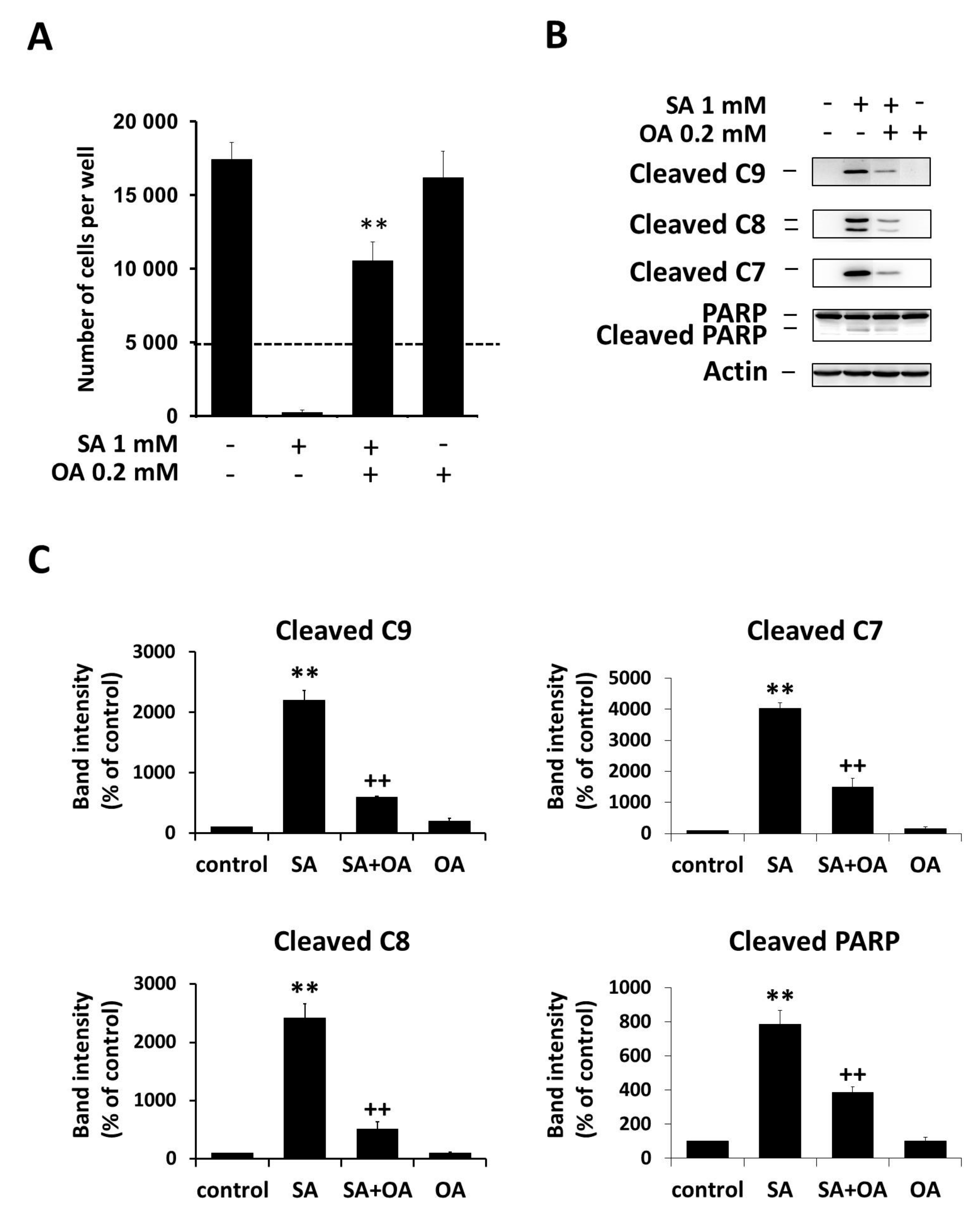
2.1. Effect of Oleic Acid on Stearic Acid-Induced Apoptosis

We showed that OA (0.2 mM) inhibited SA-induced (1 mM) cell death of NES2Y cells. OA applied alone has no or only minimal effects on NES2Y cells (Figure 1A). Cell death induced by SA is associated with the activation of initiator caspase-9 and -8 as well as executioner caspase-7 and poly ADP-ribose polymerase (PARP) (substrate of caspase-7) cleavage. Thus, it can be characterized as apoptosis. The activation of caspases as well as PARP cleavage is inhibited by the co-incubation of OA (Figure 1B,C). Cleavage of caspase-2 and -3 was not tested, since we found previously that there is nearly no activation of caspase-3 in NES2Y cells after SA exposure, and that caspase-2 does not play a key role in SA-induced apoptosis [8,25].

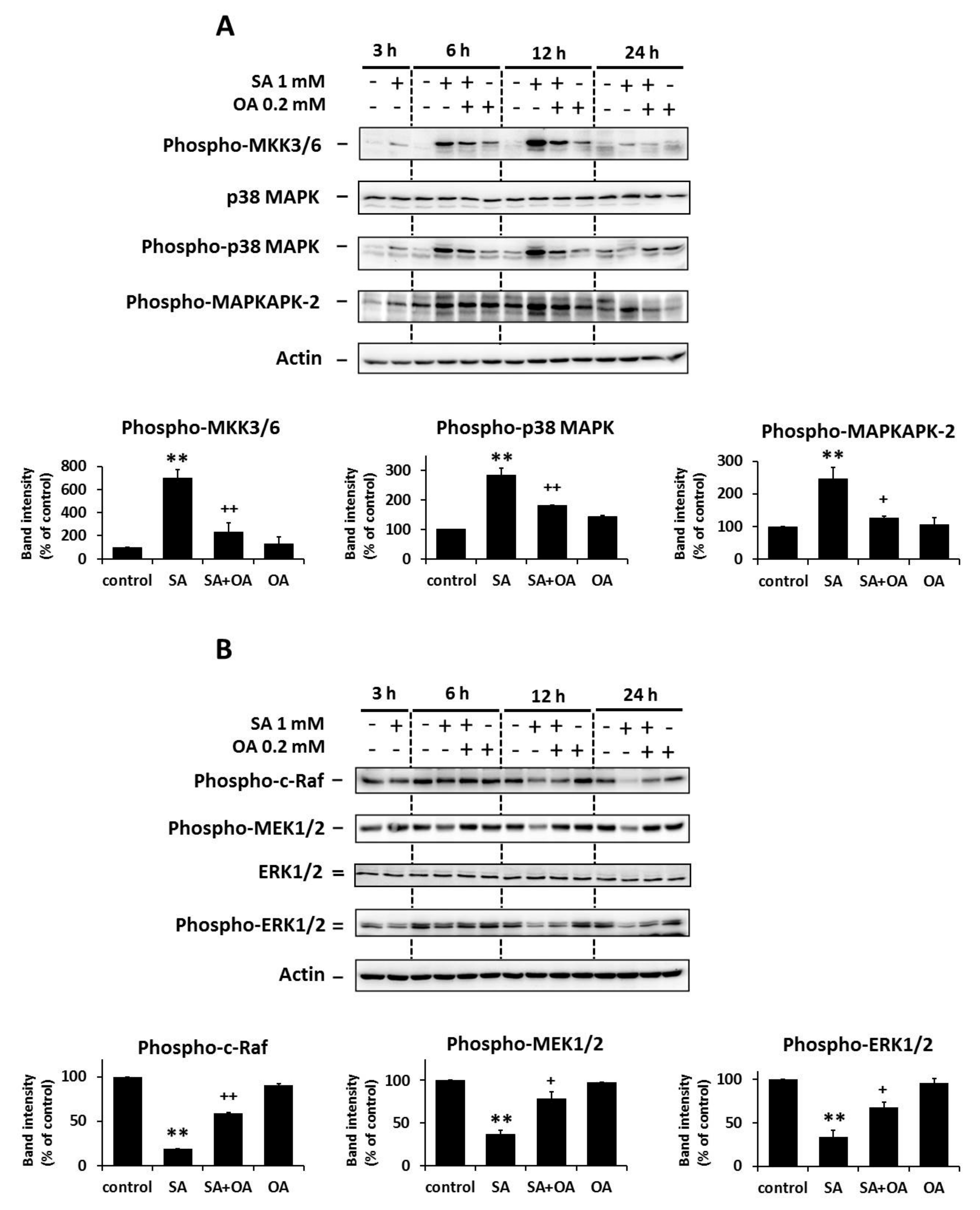
2.2. Effect of Oleic Acid on the Activation of the p38 MAPK and the Inhibition of the ERK Signaling Pathways by Stearic Acids

We previously showed the activation of the p38 MAPK signaling pathway and the inhibition of the ERK signaling pathway within 24 h of the application of apoptosis-inducing SA (1 mM) in NES2Y β-cells [14]. In order to test whether OA (0.2 mM) could interfere with the effect of SA, we assessed the activation of the p38 MAPK signaling pathway and inhibition of the ERK signaling pathway within 24 h after OA co-incubation with SA in NES2Y cells.
The effect of SA on the activation of the p38 pathway members, i.e., an increase in the level of phosphorylated MKK3/6, p38 MAPK, and MAPKAPK-2, was inhibited by co-incubation with OA as early as 12 h after the treatment in all of the tested proteins. OA applied alone resulted in low or nearly no changes in the level of phosphorylated p38 MAPK pathway members. No change was detected in the level of total p38 MAPK within 24 h of FA treatment (Figure 2A).
The effect of SA on the inhibition of the ERK pathway members, i.e., a decrease in the level of phosphorylated c-Raf, MEK1/2, and ERK1/2, was also inhibited by OA co-incubation within 24 h of the treatment. Separate application of OA resulted in nearly no effect on the level of phosphorylated ERK pathway members. We did not detect any change in the level of total ERK1/2 within 24 h of FA treatment (Figure 2B).

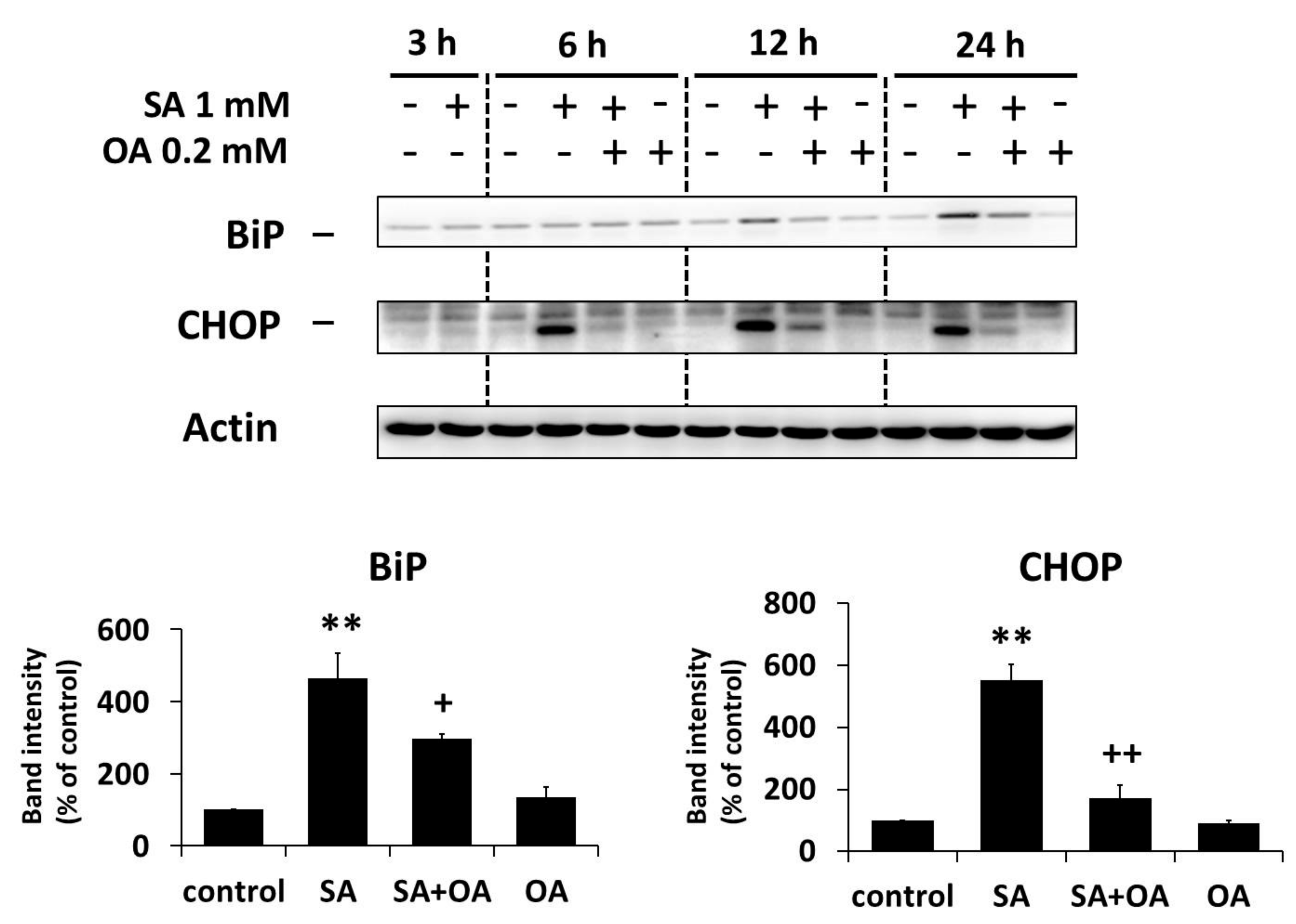
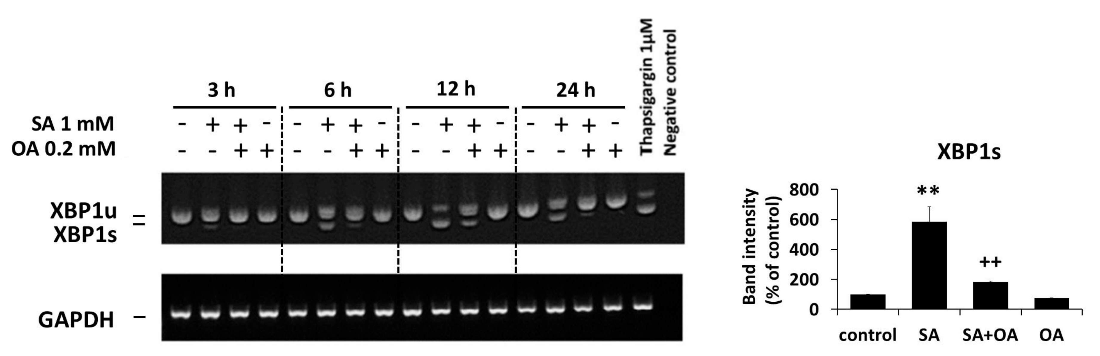
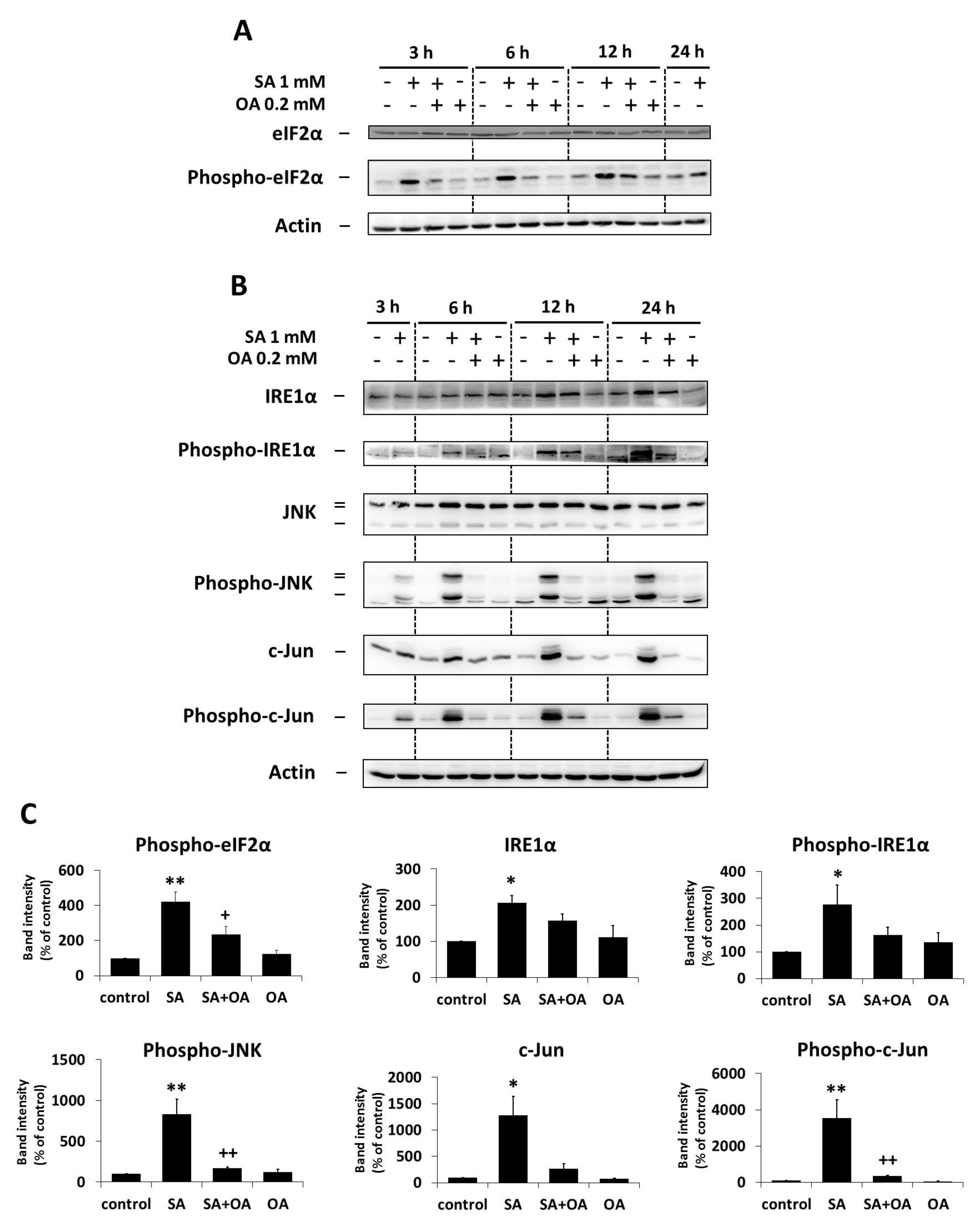
2.3. Effect of Oleic Acid on the Activation of the ER Stress Signaling Pathways by Stearic Acid

We previously documented the activation of ER stress signaling pathways (IRE1α and PERK pathways) within 24 h of apoptosis-inducing SA (1 mM) treatment in NES2Y β-cells [25]. In order to test whether OA (0.2 mM) could affect the SA-induced activation of ER stress signaling pathways, we assessed the activation of members of the ER stress IRE1α pathway (the levels of phospho-IRE1α, phospho-JNK, phospho-c-Jun) and the PERK pathway (the level of phospho-eIF2α) within 24 h after OA co-incubation with SA in NES2Y cells. The level of XBP1 splicing as well as the level of two downstream effector molecules, CHOP and BiP (known as ER stress markers), were also tested.
The effect of SA on the activation of ER stress pathways, i.e., an increase in the levels of phospho-IRE1α (and IRE1α), phospho-JNK (JNK level was not changed), phospho-c-Jun (and c-Jun), phospho-eIF2α (eIF2α level was not changed), CHOP and BiP, as well as the induction of XBP1 splicing, was significantly inhibited by OA co-incubation within 24 h of the treatment. Separate application of OA resulted in nearly no changes in the level of tested proteins and XBP1 splicing (Figure 3, Figure 4 and Figure 5).

3. Discussion

We, as well as other authors, have shown that unsaturated FAs have nearly no detrimental effect on pancreatic β-cells, but that they are in fact capable of inhibiting the pro-apoptotic effect of saturated FAs [2,4,5,6,7,8]. The molecular mechanisms involved in the modulation of saturated FAs-induced apoptosis by unsaturated FAs in β-cells are not fully clear. However, it seems that the effect of unsaturated FAs is mediated by the regulation of some signaling pathways rather than by their direct interference with the metabolism of saturated FAs [6,26,27]. Therefore, in this paper we tested the modulation of the effects of apoptosis-inducing saturated SA on the p38 MAPK pathway, the ERK pathway, and also on ER stress signaling pathways IRE1α and PERK by unsaturated OA.
Our data showed that the activation of the p38 MAPK pathway, inhibition of the ERK pathway, and activation of the IRE1α and PERK ER stress pathways by SA were inhibited by OA (see Figure 2, Figure 3, Figure 4 and Figure 5). The inhibitory effect of OA on the effect of saturated FAs on the p38 MAPK and ERK pathways has not been demonstrated, to our knowledge, until now. On the other hand, the inhibitory effect of OA on palmitate-induced ER stress was already documented in BRIN-BD11 and INS-1E rat pancreatic β-cells [7,28]. However, our study concerning the effect of SA presented more data.
We also provided a pilot experiment (using confocal microscopy), in which we tested the effect of OA on the SA-induced activation of the ATF6 pathway of ER stress signaling. We found (data not shown) that SA-induced ATF6 activation and translocation into the nucleus did not seem to be inhibited by OA co-incubation. Separate administration of OA had no effect on ATF6 translocation. However, this finding was not confirmed by an independent method.
Our findings concerning both MAP kinase signaling pathways as well as ER stress signaling pathways support the fact that the point of OA inhibitory intervention in SA-induced apoptotic signaling is located upstream of this signaling. Our preliminary data, as well as some previous studies [29,30], indicate that the point of intervention may be located on the plasma membrane where FAs affect membrane fluidity [31]. We hypothesize that saturated FAs with rigid and straight acyl chain conformation reduces membrane fluidity after incorporation into the lipid bilayer. This could alter the capability of membrane receptor(s) (receptor tyrosine kinases) to dimerize and thus transfer signals. Signals in the case of receptor tyrosine kinases can be signals for the stimulation of proliferation and/or pro-survival signals. Concerning a possible mechanism of OA inhibitory intervention in SA signaling leading to apoptosis induction, we speculate that the inhibitory effect of OA can be generated simply by increasing membrane fluidity, which compensate for the effect of SA.
In conclusion, we have shown, employing human pancreatic β-cells NES2Y, that unsaturated OA is able to inhibit the effects of saturated SA on the activation of the p38 MAPK signaling pathway and inhibition of the ERK signaling pathway, as well as activation of the IRE1α and PERK ER stress signaling pathways. OA alone had minimal or no effects on tested signaling in NES2Y cells. The point of OA inhibitory intervention in SA-induced apoptotic signaling thus seems to be located upstream of the discussed signaling pathways.

4. Materials and Methods

4.1. Materials

Chemicals were from Sigma-Aldrich (St. Louis, MO, USA), unless otherwise stated. The following primary and secondary antibodies for Western blot analysis were used: anti-phospho-MKK3/6 (#9236), anti-p38 MAPK (#8690), anti-phospho-p38 MAPK (#4511), anti-phospho-MAPKAPK-2 (#3007), anti-phospho-c-Raf (#9427), anti-phospho-MEK1/2 (#9154), anti-ERK1/2 (#5013), anti-phospho-ERK1/2 (#4370), anti-BiP (#3177), anti-CHOP (#2895), anti-eIF2α (#9722), anti-phospho-eIF2α (#9261), anti-IRE1α (#3294), anti-SAPK/JNK (#9258), anti-phospho-SAPK/JNK (#4668), anti-c-Jun (#9165) and anti-phospho-c-Jun (#9261), anti-PARP (#9542), anti-cleaved caspase-7 (#9491), anti-cleaved caspase-8 (#9496), anti-cleaved caspase-9 (#9505) from Cell Signaling Technology (Danvers, MA, USA), anti-phospho-IRE1α (ab 48187) and anti-GAPDH (ab9485) from Abcam (Cambridge, UK), and anti-actin (clone AC-40).

4.2. Cells and Culture Conditions

The human pancreatic β-cell line NES2Y employed in the experiments was kindly provided by Roger F. James (Department of Infection, Immunity and Inflammation, University of Leicester) [5,32]. NES2Y cells are proliferating cells secreting insulin with a defect in glucose responsiveness. Cells were regularly maintained in an RPMI 1640-based culture medium [33]. A defined serum-free medium [34] supplemented with FAs (1 mM SA, a combination of 1 mM SA and 0.2 mM OA, or 0.2 mM OA alone) bound to a 2% FA-free bovine serum albumin (BSA) was used in experiments [5]. Stock solutions containing FAs bound to 10% BSA in a serum-free medium were made as described previously [5] and diluted to the required concentration of FA and BSA prior to experiments. Molar ratios of FA/BSA used in experiments were lower than the ratios known to exceed the binding capacity of BSA [35].
Our previous studies showed that SA, at a concentration of 1 mM, leads to the activation of the p38 MAPK and ER stress signaling pathways and to the inhibition of the ERK signaling pathway within 24 h of the treatment [8,14]. Therefore, all assessments were performed within 24 h of treatment, except for the assessment of cell growth and viability. Since the physiological concentration of SA in adult serum seems to vary between 0.110–1.170 mM [36,37], we used 1 mM concentration of SA to simulate an elevated level of SA. An increased level of circulating FAs is a common characteristic of obese individuals [38], and a connection between obesity and type 2 diabetes mellitus (in addition to insulin resistance) has been clearly presented [38,39]. Finally, 0.2 mM concentration of OA was used, since this was the lowest concentration sufficient to inhibit the detrimental effects of SA [8].

4.3. Assessment of the Effect of Oleic Acid on the Effects of Stearic Acid on Cell Growth and Viability

Cells were seeded into the wells of a 96-well plate at a concentration of 5 × 10³ cells/100 μL of culture media. After a 24-h pre-incubation period (allowing cells to attach) the culture medium was substituted with a serum-free medium containing 2% BSA with or without fatty acid(s). The control medium contained 2% BSA only. After 96 h of incubation, the number of living cells was assessed using a hemocytometer counting system, after staining with trypan blue.

4.4. Western Blot Analysis

Cells (approximately 1 × 10⁶ cells per sample) were seeded and, after a 24-h pre-incubation period (allowing cells to attach), the culture medium was substituted with a serum-free medium containing 2% BSA with or without FA(s) (SA, a combination of SA and OA, or OA alone) at required concentrations. The control medium contained 2% BSA only. After the required incubation period, cells were harvested and Western blot analysis was performed as described previously [8]. All primary antibodies were used in a 1:1000 dilution. The chemiluminescent signal was detected using a Carestream Gel Logic 4000 PRO Imaging System equipped with Carestream Molecular Imaging Software (Carestream Health, New Haven, CT, USA), which was used for image acquisition. Image Master™ 2D Platinum 6.0 software (GE Healthcare, Uppsala, Sweden) was used to obtain data for densitometric analyses.

4.5. Assessment of XBP1 mRNA Splicing

Cells (approximately 1 × 10⁶ cells per sample) were seeded and, after 24 h pre-incubation, FAs and thapsigargin were applied as described above (see Section 4.4). After 3, 6, 12, and 24 h incubation, the cells were harvested and the splicing of XBP1 mRNA was ascertained by RT-PCR as described previously [8,40] using GAPDH as a housekeeping gene [41].

4.6. Statistical Analysis

The statistical significance of observed differences was determined using the Tukey test.

Acknowledgments

This work was supported by research projects GAUK 1270213, PRVOUK P31, PROGRES Q36 and UNCE 204015 from the Charles University, Prague, Czech Republic, and by grant 14-00630P from the Grant Agency of the Czech Republic. We thank Roger F. James (Department of Infection, Immunity, and Inflammation, University of Leicester) for providing the NES2Y cell line and for reading the manuscript.

Author Contributions

Jan Šrámek carried out Western blot experiments and wrote the manuscript; Vlasta Němcová-Fürstová helped with Western blot experiments; Nela Pavlíková carried out densitometric analyses; Jan Kovář coordinated experiments and helped to complete the manuscript.

Conflicts of Interest

The authors declare no conflict of interest.

Abbreviations

ATF6Activating transcription factor 6
Bcl-2B-cell lymphoma
BiPImmunoglobulin heavy chain-binding protein
CHOPCCAAT-enhancer-binding protein (C/EBP) homologous protein
eIF2αEukaryotic initiation factor 2α
ERK1/2Extracellular signal-regulated kinase 1/2
EREndoplasmic reticulum
FAFatty acid
Foxo03aForkhead box 03a
GAPDHGlyceraldehyde-3-phosphate dehydrogenase
IRE1αInositol-requiring protein 1
JNKc-Jun N-terminal kinase
MAPMitogen-activated protein
MAPKMitogen-activated protein kinase
MAPKAPKMAP kinase-activated protein kinase
MEK1/2Mitogen-activated protein kinase/ERK kinase
NF-κBNuclear factor kappa B
PARPPoly ADP-ribose polymerase
PERKProtein kinase RNA (PKR)-like ER kinase
SAStearic acid
SEMStandard error of the mean
SOSSon of sevenless
TGThapsigargin
T2DMType 2 diabetes mellitus
OAOleic acid
XBP1X-box binding protein 1

References

  1. Lupi, R.; Dotta, F.; Marselli, L.; Del Guerra, S.; Masini, M.; Santangelo, C.; Patane, G.; Boggi, U.; Piro, S.; Anello, M.; et al. Prolonged exposure to free fatty acids has cytostatic and pro-apoptotic effects on human pancreatic islets: Evidence that beta-cell death is caspase mediated, partially dependent on ceramide pathway, and Bcl-2 regulated. Diabetes 2002, 51, 1437–1442. [Google Scholar] [CrossRef] [PubMed]
  2. Maedler, K.; Oberholzer, J.; Bucher, P.; Spinas, G.A.; Donath, M.Y. Monounsaturated fatty acids prevent the deleterious effects of palmitate and high glucose on human pancreatic beta-cell turnover and function. Diabetes 2003, 52, 726–733. [Google Scholar] [CrossRef] [PubMed]
  3. Azevedo-Martins, A.K.; Monteiro, A.P.; Lima, C.L.; Lenzen, S.; Curi, R. Fatty acid-induced toxicity and neutral lipid accumulation in insulin-producing RINm5F cells. Toxicol. In Vitro 2006, 20, 1106–1113. [Google Scholar] [CrossRef] [PubMed]
  4. Welters, H.J.; Diakogiannaki, E.; Mordue, J.M.; Tadayyon, M.; Smith, S.A.; Morgan, N.G. Differential protective effects of palmitoleic acid and cAMP on caspase activation and cell viability in pancreatic beta-cells exposed to palmitate. Apoptosis 2006, 11, 1231–1238. [Google Scholar] [CrossRef] [PubMed]
  5. Fürstova, V.; Kopska, T.; James, R.F.; Kovar, J. Comparison of the effect of individual saturated and unsaturated fatty acids on cell growth and death induction in the human pancreatic β-cell line NES2Y. Life Sci. 2008, 82, 684–691. [Google Scholar] [CrossRef] [PubMed]
  6. Welters, H.J.; Tadayyon, M.; Scarpello, J.H.; Smith, S.A.; Morgan, N.G. Mono-unsaturated fatty acids protect against beta-cell apoptosis induced by saturated fatty acids, serum withdrawal or cytokine exposure. FEBS Lett. 2004, 560, 103–108. [Google Scholar] [CrossRef]
  7. Diakogiannaki, E.; Welters, H.J.; Morgan, N.G. Differential regulation of the endoplasmic reticulum stress response in pancreatic beta-cells exposed to long-chain saturated and monounsaturated fatty acids. J. Endocrinol. 2008, 197, 553–563. [Google Scholar] [CrossRef] [PubMed]
  8. Nemcova-Fürstova, V.; James, R.F.L.; Kovar, J. Inhibitory effect of unsaturated fatty acids on saturated fatty acids-induced apoptosis in human pancreatic β-cells: Activation of caspases and ER stress induction. Cell. Physiol. Biochem. 2011, 27, 525–538. [Google Scholar] [CrossRef] [PubMed]
  9. Biden, T.J.; Boslem, E.; Chu, K.Y.; Sue, N. Lipotoxic endoplasmic reticulum stress, β cell failure, and type 2 diabetes mellitus. Trends Endocrinol. Metab. 2014, 25, 389–398. [Google Scholar] [CrossRef] [PubMed]
  10. Simon, M.N.; Azevedo-Martins, A.K.; Amanso, A.M.; Carvalho, C.R.O.; Curim, R. Persistent activation of Akt or ERK prevents the toxicity induced by saturated and polyunsaturated fatty acids in RINm5F beta-cells. Toxicol. In Vitro 2008, 22, 1018–1024. [Google Scholar] [CrossRef] [PubMed]
  11. Cvjeticanin, T.; Stojanovic, I.; Timotijevic, G.; Stosic-Grujicic, S.; Miljkovic, D. T cells cooperate with palmitic acid in induction of beta cell apoptosis. BMC Immunol. 2009, 29, 1–10. [Google Scholar] [CrossRef] [PubMed]
  12. Plaisance, V.; Perret, V.; Favrea, D.; Abderrahmania, A.; Yanga, J.-Y.; Widmanna, C.; Regazzi, R. Role of the transcriptional factor C/EBP in free fatty acid-elicited beta cell failure. Mol. Cell. Endocrinol. 2009, 305, 47–55. [Google Scholar] [CrossRef] [PubMed]
  13. Yuan, H.; Zhang, X.; Huang, X.; Lu, Y.; Tang, W.; Man, Y.; Wang, S.; Xi, J.; Li, J. NADPH Oxidase 2-Derived Reactive Oxygen Species Mediate FFAs-Induced Dysfunction and Apoptosis of β-Cells via JNK, p38 MAPK and p53 Pathways. PLoS ONE 2010, 5, e15726. [Google Scholar] [CrossRef] [PubMed]
  14. Sramek, J.; Nemcova-Fürstova, V.; Balusikova, K.; Daniel, P.; Jelinek, M.; James, R.F.; Kovar, J. p38 MAPK is activated but does not play a key role during apoptosis induction by saturated fatty Acid in human pancreatic β-cells. Int. J. Mol. Sci. 2016, 17, 159. [Google Scholar] [CrossRef] [PubMed]
  15. Cnop, M.; Ladrière, L.; Igoillo-Esteve, M.; Moura, R.F.; Cunha, D.A. Causes and cures for endoplasmic reticulum stress in lipotoxic β-cell dysfunction. Diabetes Obes. Metab. 2010, 12, S76–S82. [Google Scholar] [CrossRef] [PubMed]
  16. Fonseca, S.G.; Gromada, J.; Urano, F. Endoplasmic reticulum stress and pancreatic β-cell death. Trends Endocrinol. Metab. 2011, 22, 266–274. [Google Scholar] [CrossRef] [PubMed]
  17. Zarubin, T.; Han, J. Activation and signaling of the p38 MAP kinase pathway. Cell Res. 2005, 15, 11–18. [Google Scholar] [CrossRef] [PubMed]
  18. Ben-Levy, R.; Hooper, S.; Wilson, R.; Paterson, H.F.; Marshall, C.J. Nuclear export of the stress-activated protein kinase p38 mediated by its substrate MAPKAP kinase-2. Curr. Biol. 1998, 8, 1049–1057. [Google Scholar] [CrossRef]
  19. Sakai, N.; Wada, T.; Furuichi, K.; Iwata, Y.; Yoshimoto, K.; Kitagawa, K.; Kokubo, S.; Kobayashi, M.; Takeda, S.; Kida, H.; et al. p38 MAPK phosphoryloation and NF-κB activation in human crescentic glomerulonephris. Nephrol. Dial. Transplant. 2002, 17, 998–1004. [Google Scholar] [CrossRef] [PubMed]
  20. Zhuang, S.; Demirs, J.T.; Kochevar, I.E. p38 mitogen-activated protein kinase mediates bid cleavage, mitochondrial dysfunction, and caspase-3 activation during apoptosis induced by singlet oxygen but not by hydrogen peroxide. J. Biol. Chem. 2000, 275, 25939–25948. [Google Scholar] [CrossRef] [PubMed]
  21. Chang, F.; Steelman, L.S.; Lee, J.T.; Shelton, J.G.; Navolanic, P.M.; Blalock, W.L.; Franklin, R.A.; McCubrey, J.A. Signal transduction mediated by the Ras/Raf/MEK/ERK pathway from cytokine receptors to transcription factors: Potential targeting for therapeutic intervention. Leukemia 2003, 17, 1263–1293. [Google Scholar] [CrossRef] [PubMed]
  22. Lu, Z.; Xu, S. ERK1/2 MAP kinases in cell survival and apoptosis. IUBMB Life 2006, 58, 621–631. [Google Scholar] [CrossRef] [PubMed]
  23. Hetz, C.; Martinon, F.; Rodriguez, D.; Glimcher, L.H. The unfolded protein response: Integrating stress signals through the stress sensor IRE1α. Physiol. Rev. 2011, 91, 1219–1243. [Google Scholar] [CrossRef] [PubMed]
  24. Tabas, I.; Ron, D. Integrating the mechanisms of apoptosis induced by endoplasmic reticulum stress. Nat. Cell. Biol. 2011, 13, 184–190. [Google Scholar] [CrossRef] [PubMed]
  25. Nemcova-Fürstova, V.; Balusikova, K.; Sramek, J.; James, R.F.; Kovar, J. Caspase-2 and JNK Activated by Saturated Fatty Acids are Not Involved in Apoptosis Induction but Modulate ER Stress in Human Pancreatic β-cells. Cell. Physiol. Biochem. 2013, 31, 277–289. [Google Scholar] [CrossRef] [PubMed]
  26. Newsholme, P.; Keane, D.; Welters, H.J.; Morgan, N.G. Life and death decisions of the pancreatic beta-cell: The role of fatty acids. Clin. Sci. 2007, 112, 27–42. [Google Scholar] [CrossRef] [PubMed]
  27. Morgan, N.G. Fatty acids and beta-cell toxicity. Curr. Opin. Clin. Nutr. Metab. Care 2009, 12, 117–122. [Google Scholar] [CrossRef] [PubMed]
  28. Sommerweiss, D.; Gorski, T.; Richter, S.; Garten, A.; Kiess, W. Oleate rescues INS-1E β-cells from palmitate-induced apoptosis by preventing activation of the unfolded protein response. Biochem. Biophys. Res. Commun. 2013, 441, 770–776. [Google Scholar] [CrossRef] [PubMed]
  29. Vacaresse, N.; Lajoie-Mazenc, I.; Auge, N.; Suc, I.; Frisach, M.-F.; Salvayre, R.; Nègre-Salvayre, A. Activation of epithelial growth factor receptor pathway by unsaturated fatty acids. Circ. Res. 1995, 85, 892–899. [Google Scholar] [CrossRef]
  30. Holzer, R.G.; Park, E.J.; Li, N.; Tran, H.; Chen, M.; Choi, C.; Solinas, G.; Karin, M. Saturated fatty acids induce c-Src clustering within membrane subdomains, leading to JNK activation. Cell 2011, 147, 173–184. [Google Scholar] [CrossRef] [PubMed]
  31. Karnovsky, M.J.; Kleinfeld, A.M.; Hoover, R.L.; Klausner, R.D. The concept of lipid domains in membranes. J. Cell. Biol. 1982, 94, 1–6. [Google Scholar] [CrossRef] [PubMed]
  32. Macfarlane, W.M.; Cragg, H.; Docherty, H.M.; Read, M.L.; James, R.F.; Aynsley-Green, A.; Docherty, K. Impaired expression of transcription factor IUF1 in a pancreatic beta-cell line derived from a patient with persistent hyperinsulinaemic hypoglycaemia of infancy (nesidioblastosis). FEBS Lett. 1997, 413, 304–308. [Google Scholar] [CrossRef]
  33. Musilkova, J.; Kovar, J. Additive stimulatory effect of extracellular calcium and potassium on non-transferrin ferric iron uptake by HeLa and K562 cells. Biochim. Biophys. Acta 2001, 1514, 117–126. [Google Scholar] [CrossRef]
  34. Kovar, J.; Franek, F. Growth-stimulating effect of transferrin on a hybridoma cell line: Relation to transferrin iron-transporting function. Exp. Cell. Res. 1989, 182, 358–369. [Google Scholar] [CrossRef]
  35. Cnop, M.; Hannaert, J.C.; Hoorens, A.; Eizirik, D.L.; Pipeleers, D.G. Inverse relationship between cytotoxicity of free fatty acids in pancreatic islet cells and cellular triglyceride accumulation. Diabetes 2001, 50, 1771–1777. [Google Scholar] [CrossRef] [PubMed]
  36. Lagerstedt, S.A.; Hinrichs, D.R.; Batt, S.M.; Magera, M.J.; Rinaldo, P.; McConnell, J.P. Quantitative determination of plasma c8-c26 total fatty acids for the biochemical diagnosis of nutritional and metabolic disorders. Mol. Genet. Metab. 2001, 73, 38–45. [Google Scholar] [CrossRef] [PubMed]
  37. Abdelmagid, S.A.; Clarke, S.E.; Nielsen, D.E.; Badawi, A.; El-Sohemy, A.; Mutch, D.M.; Ma, D.W.L. Comprehensive profiling of plasma fatty acid concentrations in young healthy Canadian adults. PLoS ONE 2015, 10, e0116195. [Google Scholar] [CrossRef] [PubMed]
  38. Kahn, S.E.; Hull, R.L.; Utzschneider, K.M. Mechanisms linking obesity to insulin resistance and type 2 diabetes. Nature 2006, 444, 840–846. [Google Scholar] [CrossRef] [PubMed]
  39. Steyn, N.P.; Mann, J.; Bennett, P.H.; Temple, N.; Zimmet, P.; Tuomilehto, J.; Lindstrom, J.; Louheranta, A. Diet, nutrition and the prevention of type 2 diabetes. Public Health Nutr. 2004, 7, 147–165. [Google Scholar] [CrossRef] [PubMed]
  40. Gao, Y.Y.; Liu, B.Q.; Niu, X.F.; Zhuang, Y.; Wang, H.Q. Resveratrol-induced cytotoxicity in human Burkitt’s lymphoma cells is coupled to the unfolded protein response. BMC Cancer 2010, 10, 445. [Google Scholar]
  41. Balusikova, K.; Neubauerova, J.; Dostalikova-Cimburova, M.; Horak, J.; Kovar, J. Differing expression of genes involved in non-transferrin iron transport across plasma membrane in various cell types under iron deficiency and excess. Mol. Cell. Biochem. 2009, 321, 123–133. [Google Scholar] [CrossRef] [PubMed]
Figure 1. Effect of 1 mM stearic acid (SA), 1 mM stearic acid applied simultaneously with 0.2 mM oleic acid (OA), and 0.2 mM oleic acid (see Section 4) on (A) cell growth and viability, (B) the level of cleaved caspase-9 (C9), caspase-8 (C8), caspase-7 (C7), and PARP in NES2Y cells. Cells incubated without fatty acids represented control cells. (A) Cells were seeded at a concentration of 5 × 10³ cells/100 µL of culture medium per well of 96-well plate (see Section 4). The number of living cells was determined after 96 h of incubation. The number of cells of the inoculum is shown as a dashed line. Each column represents the mean of four separate cultures ± standard error of the mean (SEM). ** p < 0.01 when comparing the effect of 1 mM stearic acid applied together with 0.2 mM oleic acid and the effect of 1 mM stearic acid alone. (B) After 18 h of incubation (see Section 4), the levels of individual proteins were assessed using Western blot analysis and relevant antibodies (see Section 4). Actin was included to confirm equal protein loading. The data presented were obtained in one representative experiment from at least three independent experiments. (C) Densitometric analysis of data from Western blotting are also shown. Each column represents the mean of three experimental values ± SEM. ** p < 0.01 when comparing the effect of SA with control cells, ++ p < 0.05 when comparing the effect of SA plus OA with the effect of SA alone.
Ijms 18 02313 g001
Figure 2. Effect of 1 mM stearic acid (SA), 1 mM stearic acid applied simultaneously with 0.2 mM oleic acid (OA), and 0.2 mM oleic acid (see Section 4) on (A) the levels of phospho-MKK3/6, p38 MAPK, phospho-p38 MAPK, and phospho-MAPKAPK-2 (the p38 MAPK signaling pathway); and (B) the levels of phospho-c-Raf, phospho-MEK1/2, ERK1/2, and phospho-ERK1/2 (the ERK signaling pathway) in NES2Y cells. Cells incubated without fatty acids represented control cells. After 3, 6, 12, and 24 h of incubation (see Section 4), the levels of individual proteins were assessed using Western blot analysis and relevant antibodies (see Section 4). Actin was included to confirm equal protein loading. The data presented were obtained in one representative experiment from at least three independent experiments. Densitometric analysis of data from Western blotting is also shown. The analysis was carried out for 12 h after fatty acids application in the case of the p38 MAPK pathway and for 24 h in the case of the ERK pathway. Each column represents the mean of three experimental values ± SEM. ** p < 0.01 when comparing the effect of SA with control cells, + p < 0.05, ++ p < 0.01 when comparing the effect of SA plus OA with the effect of SA alone.
Ijms 18 02313 g002
Figure 3. Effect of 1 mM stearic acid (SA), 1 mM stearic acid applied simultaneously with 0.2 mM oleic acid (OA), and 0.2 mM oleic acid (see Section 4) on the level of ER stress markers BiP and CHOP in NES2Y cells. Cells incubated without fatty acids represented control cells. After 3, 6, 12, and 24 h of incubation (see Section 4), the levels of individual proteins were assessed using Western blot analysis and relevant antibodies (see Section 4). Actin was included to confirm equal protein loading. The data presented were obtained in one representative experiment from at least three independent experiments. Densitometric analysis of data from Western blotting is also shown. The analysis was carried out for 24 h after fatty acids application. Each column represents the mean of three experimental values ± SEM. ** p < 0.01 when comparing the effect of SA with control cells, + p < 0.05, ++ p < 0.01 when comparing the effect of SA plus OA with the effect of SA alone.
Ijms 18 02313 g003
Figure 4. Effect of 1 mM stearic acid (SA), 1 mM stearic acid applied simultaneously with 0.2 mM oleic acid (OA), and 0.2 mM oleic acid (see Section 4) on the levels of (A) eIF2α and phospho-eIF2α, and (B) IRE1α, phospho-IRE1α, JNK, phospho-JNK, c-Jun, and phospho-c-Jun in NES2Y cells. Cells incubated without fatty acids represented control cells. After 3, 6, 12, and 24 h of incubation (see Section 4) (A,B), the levels of individual proteins were assessed using Western blot analysis and relevant antibodies (see Section 4). Actin was included to confirm equal protein loading. (C) Densitometric analysis of data from Western blotting is also shown. The analysis was carried out for 6 h after fatty acids application in the case of phospho-eIF2α, for 12 h in the case of phospho-JNK, c-Jun and phospho-c-Jun, and for 24 h in the case of IRE1α and phospho-IRE1α. Each column represents the mean of three experimental values ± SEM. * p < 0.05, ** p < 0.01 when comparing the effect of SA with control cells. + p < 0.05, ++ p < 0.01 when comparing the effect of SA plus OA with the effect of SA alone.
Ijms 18 02313 g004
Figure 5. Effect of 1 mM stearic acid (SA), 1 mM stearic acid applied simultaneously with 0.2 mM oleic acid (OA), and 0.2 mM oleic acid (see Section 4) on XBP1 splicing in NES2Y cells. Cells incubated without fatty acids represented control cells. After 3, 6, 12, and 24 h of incubation (see Section 4), the XBP1 splicing was assessed by RT-PCR using relevant primers (see Section 4). NES2Y cells were treated with 1 μM thapsigargin as a positive control of XBP1 splicing. GAPDH (glyceraldehyde-3-phosphate dehydrogenase) was used as a control gene for RT-PCR. The data presented were obtained in one representative experiment from at least three independent experiments. Densitometric analysis of data from RT-PCR is also shown. The analysis was carried out for 24 h after fatty acids application. Each column represents the mean of three experimental values ± SEM. ** p < 0.01 when comparing the effect of SA with control cells, ++ p < 0.01 when comparing the effect of SA plus OA with the effect of SA alone.
Ijms 18 02313 g005

