Next Issue
Volume 19, January
Previous Issue
Volume 18, November
 
 
ijms-logo

Journal Browser

Journal Browser

Int. J. Mol. Sci., Volume 18, Issue 12 (December 2017) – 280 articles

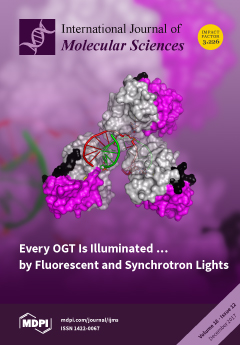
Cover Story (view full-size image): The unprecedented crystal structure of Mycobacterium tuberculosis O6-alkylguanine-DNA alkyltransferase (OGT) in complex, with a modified double-stranded DNA (dsDNA) reveals molecular details of the cooperative DNA binding mechanism of this suicidal enzyme. A peculiar supramolecular assembly can be observed in the OGT–dsDNA crystal lattice in which three different protein units are oriented onto the same DNA molecule. In particular, one protein unit binds a modified guanine base, while both additional protein monomers flip out a deoxyadenosine residue. View this paper
  • Issues are regarded as officially published after their release is announced to the table of contents alert mailing list.
  • You may sign up for e-mail alerts to receive table of contents of newly released issues.
  • PDF is the official format for papers published in both, html and pdf forms. To view the papers in pdf format, click on the "PDF Full-text" link, and use the free Adobe Reader to open them.
Order results
Result details
Section
Select all
Export citation of selected articles as:
2082 KiB  
Article
Leptin Stimulates Prolactin mRNA Expression in the Goldfish Pituitary through a Combination of the PI3K/Akt/mTOR, MKK3/6/p38MAPK and MEK1/2/ERK1/2 Signalling Pathways
by Aifen Yan, Yanfeng Chen, Shuang Chen, Shuisheng Li, Yong Zhang, Jirong Jia, Hui Yu, Lian Liu, Fang Liu, Chaoqun Hu, Dongsheng Tang and Ting Chen
Int. J. Mol. Sci. 2017, 18(12), 2781; https://doi.org/10.3390/ijms18122781 - 20 Dec 2017
Cited by 13 | Viewed by 6178
Abstract
Leptin actions at the pituitary level have been extensively investigated in mammalian species, but remain insufficiently characterized in lower vertebrates, especially in teleost fish. Prolactin (PRL) is a pituitary hormone of central importance to osmoregulation in fish. Using goldfish as a model, we [...] Read more.
Leptin actions at the pituitary level have been extensively investigated in mammalian species, but remain insufficiently characterized in lower vertebrates, especially in teleost fish. Prolactin (PRL) is a pituitary hormone of central importance to osmoregulation in fish. Using goldfish as a model, we examined the global and brain-pituitary distribution of a leptin receptor (lepR) and examined the relationship between expression of lepR and major pituitary hormones in different pituitary regions. The effects of recombinant goldfish leptin-AI and leptin-AII on PRL mRNA expression in the pituitary were further analysed, and the mechanisms underlying signal transduction for leptin-induced PRL expression were determined by pharmacological approaches. Our results showed that goldfish lepR is abundantly expressed in the brain-pituitary regions, with highly overlapping PRL transcripts within the pituitary. Recombinant goldfish leptin-AI and leptin-AII proteins could stimulate PRL mRNA expression in dose- and time-dependent manners in the goldfish pituitary, by both intraperitoneal injection and primary cell incubation approaches. Moreover, the PI3K/Akt/mTOR, MKK3/6/p38MAPK, and MEK1/2/ERK1/2—but not JAK2/STAT 1, 3 and 5 cascades—were involved in leptin-induced PRL mRNA expression in the goldfish pituitary. Full article
(This article belongs to the Section Biochemistry)
Show Figures

Graphical abstract

1047 KiB  
Article
Evaluation of Biosynthesis, Accumulation and Antioxidant Activityof Vitamin E in Sweet Corn (Zea mays L.) during Kernel Development
by Lihua Xie, Yongtao Yu, Jihua Mao, Haiying Liu, Jian Guang Hu, Tong Li, Xinbo Guo and Rui Hai Liu
Int. J. Mol. Sci. 2017, 18(12), 2780; https://doi.org/10.3390/ijms18122780 - 20 Dec 2017
Cited by 26 | Viewed by 4596
Abstract
Sweet corn kernels were used in this research to study the dynamics of vitamin E, by evaluatingthe expression levels of genes involved in vitamin E synthesis, the accumulation of vitamin E, and the antioxidant activity during the different stage of kernel development. Results [...] Read more.
Sweet corn kernels were used in this research to study the dynamics of vitamin E, by evaluatingthe expression levels of genes involved in vitamin E synthesis, the accumulation of vitamin E, and the antioxidant activity during the different stage of kernel development. Results showed that expression levels of ZmHPT and ZmTC genes increased, whereas ZmTMT gene dramatically decreased during kernel development. The contents of all the types of vitamin E in sweet corn had a significant upward increase during kernel development, and reached the highest level at 30 days after pollination (DAP). Amongst the eight isomers of vitamin E, the content of γ-tocotrienol was the highest, and increased by 14.9 folds, followed by α-tocopherolwith an increase of 22 folds, and thecontents of isomers γ-tocopherol, α-tocotrienol, δ-tocopherol,δ-tocotrienol, and β-tocopherol were also followed during kernel development. The antioxidant activity of sweet corn during kernel development was increased, and was up to 101.8 ± 22.3 μmol of α-tocopherol equivlent/100 g in fresh weight (FW) at 30 DAP. There was a positive correlation between vitamin E contents and antioxidant activity in sweet corn during the kernel development, and a negative correlation between the expressions of ZmTMT gene and vitamin E contents. These results revealed the relations amongst the content of vitamin E isomers and the gene expression, vitamin E accumulation, and antioxidant activity. The study can provide a harvesting strategy for vitamin E bio-fortification in sweet corn. Full article
(This article belongs to the Special Issue Molecular Transformations of Natural Products)
Show Figures

Graphical abstract

7626 KiB  
Article
Anomalous Behavior of Hyaluronan Crosslinking Due to the Presence of Excess Phospholipids in the Articular Cartilage System of Osteoarthritis
by Piotr Bełdowski, Piotr Weber, Tomasz Andrysiak, Wayne K. Augé II, Damian Ledziński, Tristan De Leon and Adam Gadomski
Int. J. Mol. Sci. 2017, 18(12), 2779; https://doi.org/10.3390/ijms18122779 - 20 Dec 2017
Cited by 13 | Viewed by 3917
Abstract
Lubrication of articular cartilage is a complex multiscale phenomenon in synovial joint organ systems. In these systems, synovial fluid properties result from synergistic interactions between a variety of molecular constituent. Two molecular classes in particular are of importance in understanding lubrication mechanisms: hyaluronic [...] Read more.
Lubrication of articular cartilage is a complex multiscale phenomenon in synovial joint organ systems. In these systems, synovial fluid properties result from synergistic interactions between a variety of molecular constituent. Two molecular classes in particular are of importance in understanding lubrication mechanisms: hyaluronic acid and phospholipids. The purpose of this study is to evaluate interactions between hyaluronic acid and phospholipids at various functionality levels during normal and pathological synovial fluid conditions. Molecular dynamic simulations of hyaluronic acid and phospholipids complexes were performed with the concentration of hyaluronic acid set at a constant value for two organizational forms, extended (normal) and coiled (pathologic). The results demonstrated that phospholipids affect the crosslinking mechanisms of hyaluronic acid significantly and the influence is higher during pathological conditions. During normal conditions, hyaluronic acid and phospholipid interactions seem to have no competing mechanism to that of the interaction between hyaluronic acid to hyaluronic acid. On the other hand, the structures formed under pathologic conditions were highly affected by phospholipid concentration. Full article
(This article belongs to the Section Molecular Biophysics)
Show Figures

Figure 1

711 KiB  
Review
Iron Overload and Chelation Therapy in Non-Transfusion Dependent Thalassemia
by Rayan Bou-Fakhredin, Abdul-Hamid Bazarbachi, Bachar Chaya, Joseph Sleiman, Maria Domenica Cappellini and Ali T. Taher
Int. J. Mol. Sci. 2017, 18(12), 2778; https://doi.org/10.3390/ijms18122778 - 20 Dec 2017
Cited by 22 | Viewed by 5973
Abstract
Iron overload (IOL) due to increased intestinal iron absorption constitutes a major clinical problem in patients with non-transfusion-dependent thalassemia (NTDT), which is a cumulative process with advancing age. Current models for iron metabolism in patients with NTDT suggest that suppression of serum hepcidin [...] Read more.
Iron overload (IOL) due to increased intestinal iron absorption constitutes a major clinical problem in patients with non-transfusion-dependent thalassemia (NTDT), which is a cumulative process with advancing age. Current models for iron metabolism in patients with NTDT suggest that suppression of serum hepcidin leads to an increase in iron absorption and subsequent release of iron from the reticuloendothelial system, leading to depletion of macrophage iron, relatively low levels of serum ferritin, and liver iron loading. The consequences of IOL in patients with NTDT are multiple and multifactorial. Accurate and reliable methods of diagnosis and monitoring of body iron levels are essential, and the method of choice for measuring iron accumulation will depend on the patient’s needs and on the available facilities. Iron chelation therapy (ICT) remains the backbone of NTDT management and is one of the most effective and practical ways of decreasing morbidity and mortality. The aim of this review is to describe the mechanism of IOL in NTDT, and the clinical complications that can develop as a result, in addition to the current and future therapeutic options available for the management of IOL in NTDT. Full article
(This article belongs to the Special Issue Thalassemia in 2017)
Show Figures

Figure 1

2158 KiB  
Article
Lipopolysaccharide-Induced Acute Kidney Injury Is Dependent on an IL-18 Receptor Signaling Pathway
by Yuji Nozaki, Shoichi Hino, Jinhai Ri, Kenji Sakai, Yasuaki Nagare, Mai Kawanishi, Kaoru Niki, Masanori Funauchi and Itaru Matsumura
Int. J. Mol. Sci. 2017, 18(12), 2777; https://doi.org/10.3390/ijms18122777 - 20 Dec 2017
Cited by 19 | Viewed by 5875
Abstract
The proinflammatory cytokine interleukin (IL)-18 is an important mediator of the organ failure induced by endotoxemia. IL-18 (known as an interferon-gamma (IFN-γ) inducing factor), and other inflammatory cytokines have important roles in lipopolysaccharide (LPS)-induced acute kidney injury (AKI). We investigated the effect of [...] Read more.
The proinflammatory cytokine interleukin (IL)-18 is an important mediator of the organ failure induced by endotoxemia. IL-18 (known as an interferon-gamma (IFN-γ) inducing factor), and other inflammatory cytokines have important roles in lipopolysaccharide (LPS)-induced acute kidney injury (AKI). We investigated the effect of inflammatory cytokines and Toll-like receptor 4 (TLR4) expression, an event that is accompanied by an influx of monocytes, including CD4+ T cells and antigen-presenting cells (APCs) in IL-18Rα knockout (KO) mice and wild-type (WT) mice after LPS injection. In the acute advanced phase, the IL-18Rα KO mice showed a higher survival rate and a suppressed increase of blood urea nitrogen, increased levels of proinflammatory cytokines such as IFN-γ and IL-18, the infiltration of CD4+ T cells and the expression of kidney injury molecule-1 as an AKI marker. In that phase, the renal mRNA expression of the M1 macrophage phenotype and C-C chemokine receptor type 7 as the maturation marker of dendritic cells (DCs) was also significantly decreased in the IL-18Rα KO mice, although there were small numbers of F4/80+ cells and DCs in the kidney. Conversely, there were no significant differences in the expressions of mRNA and protein TLR4 after LPS injection between the WT and IL-18Rα KO groups. Our results demonstrated that the IL-18Rα-mediated signaling pathway plays critical roles in CD4+ T cells and APCs and responded more quickly to IFN-γ and IL-18 than TLR4 stimulation in the pathogenesis of LPS-induced AKI. Full article
(This article belongs to the Special Issue Signaling Pathway of Immune Cells and Immune Disorder)
Show Figures

Figure 1

2793 KiB  
Article
The Role of PAR2 in TGF-β1-Induced ERK Activation and Cell Motility
by Hendrik Ungefroren, David Witte, Christian Fiedler, Thomas Gädeken, Roland Kaufmann, Hendrik Lehnert, Frank Gieseler and Bernhard H. Rauch
Int. J. Mol. Sci. 2017, 18(12), 2776; https://doi.org/10.3390/ijms18122776 - 20 Dec 2017
Cited by 19 | Viewed by 5639
Abstract
Background: Recently, the expression of proteinase-activated receptor 2 (PAR2) has been shown to be essential for activin receptor-like kinase 5 (ALK5)/SMAD-mediated signaling and cell migration by transforming growth factor (TGF)-β1. However, it is not known whether activation of non-SMAD TGF-β signaling (e.g., RAS–RAF–MEK–extracellular [...] Read more.
Background: Recently, the expression of proteinase-activated receptor 2 (PAR2) has been shown to be essential for activin receptor-like kinase 5 (ALK5)/SMAD-mediated signaling and cell migration by transforming growth factor (TGF)-β1. However, it is not known whether activation of non-SMAD TGF-β signaling (e.g., RAS–RAF–MEK–extracellular signal-regulated kinase (ERK) signaling) is required for cell migration and whether it is also dependent on PAR2. Methods: RNA interference was used to deplete cells of PAR2, followed by xCELLigence technology to measure cell migration, phospho-immunoblotting to assess ERK1/2 activation, and co-immunoprecipitation to detect a PAR2–ALK5 physical interaction. Results: Inhibition of ERK signaling with the MEK inhibitor U0126 blunted the ability of TGF-β1 to induce migration in pancreatic cancer Panc1 cells. ERK activation in response to PAR2 agonistic peptide (PAR2–AP) was strong and rapid, while it was moderate and delayed in response to TGF-β1. Basal and TGF-β1-dependent ERK, but not SMAD activation, was blocked by U0126 in Panc1 and other cell types indicating that ERK activation is downstream or independent of SMAD signaling. Moreover, cellular depletion of PAR2 in HaCaT cells strongly inhibited TGF-β1-induced ERK activation, while the biased PAR2 agonist GB88 at 10 and 100 µM potentiated TGF-β1-dependent ERK activation and cell migration. Finally, we provide evidence for a physical interaction between PAR2 and ALK5. Our data show that both PAR2–AP- and TGF-β1-induced cell migration depend on ERK activation, that PAR2 expression is crucial for TGF-β1-induced ERK activation, and that the functional cooperation of PAR2 and TGF-β1 involves a physical interaction between PAR2 and ALK5. Full article
(This article belongs to the Special Issue TGF-beta Family in Fibrosis and Cancer)
Show Figures

Graphical abstract

6165 KiB  
Article
Evaluation of the Expression of Amine Oxidase Proteins in Breast Cancer
by Woo Young Sun, Junjeong Choi, Yoon Jin Cha and Ja Seung Koo
Int. J. Mol. Sci. 2017, 18(12), 2775; https://doi.org/10.3390/ijms18122775 - 20 Dec 2017
Cited by 28 | Viewed by 6132
Abstract
We aimed to evaluate the expression of amine oxidase proteins in breast cancer and their clinical implications. We performed immunohistochemical staining of amine oxidase proteins (LOX, lysyl oxidase, AOC3, amine oxidase, MAOA, monoamine oxidase A, MAOB, monoamine oxidase B). Based on their hormone [...] Read more.
We aimed to evaluate the expression of amine oxidase proteins in breast cancer and their clinical implications. We performed immunohistochemical staining of amine oxidase proteins (LOX, lysyl oxidase, AOC3, amine oxidase, MAOA, monoamine oxidase A, MAOB, monoamine oxidase B). Based on their hormone receptors, such as estrogen receptor (ER) and progesterone receptor (PR), human epidermal growth factor receptor 2 (HER-2), and Ki-67 immunohistochemical staining, breast cancer was divided into four molecular subtypes: luminal A, luminal B, HER-2 type, and triple-negative breast cancer (TNBC). Luminal A was observed in 380 cases (49.4%), luminal B in 224 (29.1%), HER-2 type in 68 (8.8%), and TNBC in 98 (12.7%). Stromal AOC3, MAO-A, and MAO-B expression varied according to molecular subtypes. Stromal AOC3 expression was high in luminal B and HER-2 type and MAO-A expression was high in luminal A and luminal B (p < 0.001). MAO-B expression was higher in TNBC than in other subtypes (p = 0.020). LOX positivity was associated with high histological grade (p < 0.001) and high Ki-67 labeling index (LI) (p = 0.009), and stromal AOC3 positivity was associated with high histological grade (p = 0.001), high Ki-67 LI (p < 0.001), and HER-2 positivity (p = 0.002). MAO-A positivity was related to low histological grade (p < 0.001), ER positivity, PR positivity (p < 0.001), and low Ki-67 LI (p < 0.001). In univariate analysis, MAO-A positivity was related to short disease-free survival in HER-2 type (p = 0.013), AOC3 negativity was related to short disease-free survival and overall survival in ER-positive breast cancer, PR-positive breast cancer, HER-2-negative breast cancer, and lymph node metastasis. In conclusion, the expression of amine oxidase proteins varies depending on the molecular subtype of breast cancer. Stromal AOC3 expression was high in luminal B and HER-2 type, and MAO-A expression was high in luminal A and luminal B. Full article
Show Figures

Graphical abstract

756 KiB  
Review
Molecular Determinants of Malignant Brain Cancers: From Intracellular Alterations to Invasion Mediated by Extracellular Vesicles
by Gabriella Schiera, Carlo Maria Di Liegro and Italia Di Liegro
Int. J. Mol. Sci. 2017, 18(12), 2774; https://doi.org/10.3390/ijms18122774 - 20 Dec 2017
Cited by 18 | Viewed by 6252
Abstract
Malignant glioma cells invade the surrounding brain parenchyma, by migrating along the blood vessels, thus promoting cancer growth. The biological bases of these activities are grounded in profound alterations of the metabolism and the structural organization of the cells, which consequently acquire the [...] Read more.
Malignant glioma cells invade the surrounding brain parenchyma, by migrating along the blood vessels, thus promoting cancer growth. The biological bases of these activities are grounded in profound alterations of the metabolism and the structural organization of the cells, which consequently acquire the ability to modify the surrounding microenvironment, by altering the extracellular matrix and affecting the properties of the other cells present in the brain, such as normal glial-, endothelial- and immune-cells. Most of the effects on the surrounding environment are probably exerted through the release of a variety of extracellular vesicles (EVs), which contain many different classes of molecules, from genetic material to defined species of lipids and enzymes. EV-associated molecules can be either released into the extracellular matrix (ECM) and/or transferred to neighboring cells: as a consequence, both deep modifications of the recipient cell phenotype and digestion of ECM components are obtained, thus causing cancer propagation, as well as a general brain dysfunction. In this review, we first analyze the main intracellular and extracellular transformations required for glioma cell invasion into the brain parenchyma; then we discuss how these events may be attributed, at least in part, to EVs that, like the pawns of a dramatic chess game with cancer, open the way to the tumor cells themselves. Full article
(This article belongs to the Special Issue Glioma Cell Invasion)
Show Figures

Graphical abstract

3978 KiB  
Article
Lym-1 Chimeric Antigen Receptor T Cells Exhibit Potent Anti-Tumor Effects against B-Cell Lymphoma
by Long Zheng, Peisheng Hu, Brandon Wolfe, Caryn Gonsalves, Luqing Ren, Leslie A. Khawli, Harvey R. Kaslow and Alan L. Epstein
Int. J. Mol. Sci. 2017, 18(12), 2773; https://doi.org/10.3390/ijms18122773 - 20 Dec 2017
Cited by 2 | Viewed by 8099
Abstract
T cells expressing chimeric antigen receptors (CARs) recognizing CD19 epitopes have produced remarkable anti-tumor effects in patients with B-cell malignancies. However, cancer cells lacking recognized epitopes can emerge, leading to relapse and death. Thus, CAR T cells targeting different epitopes on different antigens [...] Read more.
T cells expressing chimeric antigen receptors (CARs) recognizing CD19 epitopes have produced remarkable anti-tumor effects in patients with B-cell malignancies. However, cancer cells lacking recognized epitopes can emerge, leading to relapse and death. Thus, CAR T cells targeting different epitopes on different antigens could improve immunotherapy. The Lym-1 antibody targets a conformational epitope of Human Leukocyte Antigen-antigen D Related (HLA-DR) on the surface of human B-cell lymphomas. Lym-1 CAR T cells were thus generated for evaluation of cytotoxic activity towards lymphoma cells in vitro and in vivo. Human T cells from healthy donors were transduced to express a Lym-1 CAR, and assessed for epitope-driven function in culture and towards Raji xenografts in NOD-scidIL2Rgammanull (NSG) mice. Lym-1 CAR T cells exhibited epitope-driven activation and lytic function against human B-cell lymphoma cell lines in culture and mediated complete regression of Raji/Luciferase-Green fluorescent protein (Raji/Luc-GFP) in NSG mice with similar or better reactivity than CD19 CAR T cells. Lym-1 CAR transduction of T cells is a promising immunotherapy for patients with Lym-1 epitope positive B-cell malignancies. Full article
(This article belongs to the Special Issue Chimeric Antigen Receptor (CAR) T Cell Therapy)
Show Figures

Graphical abstract

3442 KiB  
Review
Nrf2, the Master Regulator of Anti-Oxidative Responses
by Sandra Vomund, Anne Schäfer, Michael J. Parnham, Bernhard Brüne and Andreas Von Knethen
Int. J. Mol. Sci. 2017, 18(12), 2772; https://doi.org/10.3390/ijms18122772 - 20 Dec 2017
Cited by 428 | Viewed by 14241
Abstract
Tight regulation of inflammation is very important to guarantee a balanced immune response without developing chronic inflammation. One of the major mediators of the resolution of inflammation is the transcription factor: the nuclear factor erythroid 2-like 2 (Nrf2). Stabilized following oxidative stress, Nrf2 [...] Read more.
Tight regulation of inflammation is very important to guarantee a balanced immune response without developing chronic inflammation. One of the major mediators of the resolution of inflammation is the transcription factor: the nuclear factor erythroid 2-like 2 (Nrf2). Stabilized following oxidative stress, Nrf2 induces the expression of antioxidants as well as cytoprotective genes, which provoke an anti-inflammatory expression profile, and is crucial for the initiation of healing. In view of this fundamental modulatory role, it is clear that both hyper- or hypoactivation of Nrf2 contribute to the onset of chronic diseases. Understanding the tight regulation of Nrf2 expression/activation and its interaction with signaling pathways, known to affect inflammatory processes, will facilitate development of therapeutic approaches to prevent Nrf2 dysregulation and ameliorate chronic inflammatory diseases. We discuss in this review the principle mechanisms of Nrf2 regulation with a focus on inflammation and autophagy, extending the role of dysregulated Nrf2 to chronic diseases and tumor development. Full article
(This article belongs to the Special Issue Nrf2 in Redox Signaling: A Double Edged Sword)
Show Figures

Figure 1

178 KiB  
Addendum
Addendum: Cechová, M. et al. Towards Better Understanding of Pea Seed Dormancy Using Laser Desorption/Ionization Mass Spectrometry. Int. J. Mol. Sci. 2017, 18, 2196
by Monika Cechová, Markéta Válková, Iveta Hradilová, Anna Janská, Aleš Soukup, Petr Smýkal and Petr Bednář
Int. J. Mol. Sci. 2017, 18(12), 2771; https://doi.org/10.3390/ijms18122771 - 20 Dec 2017
Cited by 24 | Viewed by 2847
Abstract
It has been brought to our attention that one funding project of Ministry of Education, Youth and Sports of the Czech Republic (LO1417) was missing in the Acknowledgement section of our published paper [1], and therefore we would like to add it and [...] Read more.
It has been brought to our attention that one funding project of Ministry of Education, Youth and Sports of the Czech Republic (LO1417) was missing in the Acknowledgement section of our published paper [1], and therefore we would like to add it and report the Acknowledgements as follows [...] Full article
(This article belongs to the Special Issue Metabolomics in the Plant Sciences 2017)
2159 KiB  
Review
ω-3 Long Chain Polyunsaturated Fatty Acids as Sensitizing Agents and Multidrug Resistance Revertants in Cancer Therapy
by Paola Antonia Corsetto, Irma Colombo, Joanna Kopecka, Angela Maria Rizzo and Chiara Riganti
Int. J. Mol. Sci. 2017, 18(12), 2770; https://doi.org/10.3390/ijms18122770 - 20 Dec 2017
Cited by 44 | Viewed by 7098
Abstract
Chemotherapy efficacy is strictly limited by the resistance of cancer cells. The ω-3 long chain polyunsaturated fatty acids (ω-3 LCPUFAs) are considered chemosensitizing agents and revertants of multidrug resistance by pleiotropic, but not still well elucidated, mechanisms. Nowadays, it is accepted that alteration [...] Read more.
Chemotherapy efficacy is strictly limited by the resistance of cancer cells. The ω-3 long chain polyunsaturated fatty acids (ω-3 LCPUFAs) are considered chemosensitizing agents and revertants of multidrug resistance by pleiotropic, but not still well elucidated, mechanisms. Nowadays, it is accepted that alteration in gene expression, modulation of cellular proliferation and differentiation, induction of apoptosis, generation of reactive oxygen species, and lipid peroxidation are involved in ω-3 LCPUFA chemosensitizing effects. A crucial mechanism in the control of cell drug uptake and efflux is related to ω-3 LCPUFA influence on membrane lipid composition. The incorporation of docosahexaenoic acid in the lipid rafts produces significant changes in their physical-chemical properties affecting content and functions of transmembrane proteins, such as growth factors, receptors and ATP-binding cassette transporters. Of note, ω-3 LCPUFAs often alter the lipid compositions more in chemoresistant cells than in chemosensitive cells, suggesting a potential adjuvant role in the treatment of drug resistant cancers. Full article
(This article belongs to the Special Issue Omega-3 Fatty Acids in Health and Disease: New Knowledge)
Show Figures

Graphical abstract

5597 KiB  
Article
Genome-Wide Identification, Evolutionary and Expression Analyses of the GALACTINOL SYNTHASE Gene Family in Rapeseed and Tobacco
by Yonghai Fan, Mengna Yu, Miao Liu, Rui Zhang, Wei Sun, Mingchao Qian, Huichun Duan, Wei Chang, Jinqi Ma, Cunmin Qu, Kai Zhang, Bo Lei and Kun Lu
Int. J. Mol. Sci. 2017, 18(12), 2768; https://doi.org/10.3390/ijms18122768 - 20 Dec 2017
Cited by 26 | Viewed by 4981
Abstract
Galactinol synthase (GolS) is a key enzyme in raffinose family oligosaccharide (RFO) biosynthesis. The finding that GolS accumulates in plants exposed to abiotic stresses indicates RFOs function in environmental adaptation. However, the evolutionary relationships and biological functions of GolS family in rapeseed ( [...] Read more.
Galactinol synthase (GolS) is a key enzyme in raffinose family oligosaccharide (RFO) biosynthesis. The finding that GolS accumulates in plants exposed to abiotic stresses indicates RFOs function in environmental adaptation. However, the evolutionary relationships and biological functions of GolS family in rapeseed (Brassica napus) and tobacco (Nicotiana tabacum) remain unclear. In this study, we identified 20 BnGolS and 9 NtGolS genes. Subcellular localization predictions showed that most of the proteins are localized to the cytoplasm. Phylogenetic analysis identified a lost event of an ancient GolS copy in the Solanaceae and an ancient duplication event leading to evolution of GolS4/7 in the Brassicaceae. The three-dimensional structures of two GolS proteins were conserved, with an important DxD motif for binding to UDP-galactose (uridine diphosphate-galactose) and inositol. Expression profile analysis indicated that BnGolS and NtGolS genes were expressed in most tissues and highly expressed in one or two specific tissues. Hormone treatments strongly induced the expression of most BnGolS genes and homologous genes in the same subfamilies exhibited divergent-induced expression. Our study provides a comprehensive evolutionary analysis of GolS genes among the Brassicaceae and Solanaceae as well as an insight into the biological function of GolS genes in hormone response in plants. Full article
(This article belongs to the Section Biochemistry)
Show Figures

Figure 1

4061 KiB  
Article
High-Throughput RNA-Seq Data Analysis of the Single Nucleotide Polymorphisms (SNPs) and Zygomorphic Flower Development in Pea (Pisum sativum L.)
by Keyuan Jiao, Xin Li, Wuxiu Guo, Shihao Su and Da Luo
Int. J. Mol. Sci. 2017, 18(12), 2710; https://doi.org/10.3390/ijms18122710 - 20 Dec 2017
Cited by 7 | Viewed by 5027
Abstract
Pea (Pisum sativum L.) is a model plant that has been used in classical genetics and organ development studies. However, its large and complex genome has hindered research investigations in pea. Here, we generated transcriptomes from different tissues or organs of three [...] Read more.
Pea (Pisum sativum L.) is a model plant that has been used in classical genetics and organ development studies. However, its large and complex genome has hindered research investigations in pea. Here, we generated transcriptomes from different tissues or organs of three pea accessions using next-generation sequencing to assess single nucleotide polymorphisms (SNPs), and further investigated petal differentially expressed genes to elucidate the mechanisms regulating floral zygomorphy. Eighteen samples were sequenced, which yielded a total of 617 million clean reads, and de novo assembly resulted in 87,137 unigenes. A total of 9044 high-quality SNPs were obtained among the three accessions, and a consensus map was constructed. We further discovered several dorsoventral asymmetrically expressed genes that were confirmed by qRT-PCR among different petals, including previously reported three CYC-like proliferating cell factor (TCP) genes. One MADS-box gene was highly expressed in dorsal petals, and several MYB factors were predominantly expressed among dorsal, lateral, and/or ventral petals, together with a ventrally expressed TCP gene. In sum, our comprehensive database complements the existing resources for comparative genetic mapping and facilitates future investigations in legume zygomorphic flower development. Full article
(This article belongs to the Section Molecular Plant Sciences)
Show Figures

Graphical abstract

1990 KiB  
Article
Differential Expression of VvLOXA Diversifies C6 Volatile Profiles in Some Vitis vinifera Table Grape Cultivars
by Xu Qian, Lei Sun, Xiao-Qing Xu, Bao-Qing Zhu and Hai-Ying Xu
Int. J. Mol. Sci. 2017, 18(12), 2705; https://doi.org/10.3390/ijms18122705 - 20 Dec 2017
Cited by 21 | Viewed by 4972
Abstract
C6 volatiles are synthesized through lipoxygenase-hydroperoxide lyase (LOX-HPL) pathway and these volatiles play important roles in the aromatic quality of grape berries. This study investigated the evolution of both C6 volatiles and the key genes in the LOX-HPL pathway in different table grape [...] Read more.
C6 volatiles are synthesized through lipoxygenase-hydroperoxide lyase (LOX-HPL) pathway and these volatiles play important roles in the aromatic quality of grape berries. This study investigated the evolution of both C6 volatiles and the key genes in the LOX-HPL pathway in different table grape cultivars during the berry development period, and further assessed the correlation between the accumulation of C6 volatiles and the expression of these genes in these cultivars. Results showed that hexanal, (E)-2-hexenal, (E)-2-hexen-1-ol and (Z)-3-hexen-1-ol were found to be the dominant C6 volatiles in these ripened grape cultivars under two consecutive vintages, and their flavor notes were incorporated in the overall aroma of these cultivars. The cultivar “Xiangfei” showed the most abundant level of C6 aldehydes and C6 acid, whereas the cultivar “Tamina” and “Moldova” possessed the highest C6 alcohol content. The “Muscat of Alexandria” cultivar was found to contain the highest level of C6 esters. C6 volatiles were grouped into three evolutionary patterns in these cultivars during berry development, and their evolution was consistent with the evolution of the LOX-HPL pathway genes’ expression. Pearson’s correlation analysis indicated that the LOX-HPL-pathway-related genes were correlated to the accumulation of C6 volatiles in these cultivars, and VvLOXA appeared to be an important gene that regulated the synthesis of all C6 volatiles. Full article
(This article belongs to the Section Molecular Plant Sciences)
Show Figures

Graphical abstract

5021 KiB  
Article
Comparative Analysis of Fruit Ripening-Related miRNAs and Their Targets in Blueberry Using Small RNA and Degradome Sequencing
by Yanming Hou, Lulu Zhai, Xuyan Li, Yu Xue, Jingjing Wang, Pengjie Yang, Chunmei Cao, Hongxue Li, Yuhai Cui and Shaomin Bian
Int. J. Mol. Sci. 2017, 18(12), 2767; https://doi.org/10.3390/ijms18122767 - 19 Dec 2017
Cited by 35 | Viewed by 4771
Abstract
MicroRNAs (miRNAs) play vital roles in the regulation of fruit development and ripening. Blueberry is an important small berry fruit crop with economical and nutritional value. However, nothing is known about the miRNAs and their targets involved in blueberry fruit ripening. In this [...] Read more.
MicroRNAs (miRNAs) play vital roles in the regulation of fruit development and ripening. Blueberry is an important small berry fruit crop with economical and nutritional value. However, nothing is known about the miRNAs and their targets involved in blueberry fruit ripening. In this study, using high-throughput sequencing of small RNAs, 84 known miRNAs belonging to 28 families and 16 novel miRNAs were identified in white fruit (WF) and blue fruit (BF) libraries, which represent fruit ripening onset and in progress, respectively. Among them, 41 miRNAs were shown to be differentially expressed during fruit maturation, and 16 miRNAs representing 16 families were further chosen to validate the sRNA sequencing data by stem-loop qRT-PCR. Meanwhile, 178 targets were identified for 41 known and 7 novel miRNAs in WF and BF libraries using degradome sequencing, and targets of miR160 were validated using RLM-RACE (RNA Ligase-Mediated (RLM)-Rapid Amplification of cDNA Ends) approach. Moreover, the expression patterns of 6 miRNAs and their targets were examined during fruit development and ripening. Finally, integrative analysis of miRNAs and their targets revealed a complex miRNA-mRNA regulatory network involving a wide variety of biological processes. The findings will facilitate future investigations of the miRNA-mediated mechanisms that regulate fruit development and ripening in blueberry. Full article
(This article belongs to the Section Molecular Plant Sciences)
Show Figures

Graphical abstract

11119 KiB  
Article
Effects and Mechanisms of Total Flavonoids from Blumea balsamifera (L.) DC. on Skin Wound in Rats
by Yuxin Pang, Yan Zhang, Luqi Huang, Luofeng Xu, Kai Wang, Dan Wang, Lingliang Guan, Yingbo Zhang, Fulai Yu, Zhenxia Chen and Xiaoli Xie
Int. J. Mol. Sci. 2017, 18(12), 2766; https://doi.org/10.3390/ijms18122766 - 19 Dec 2017
Cited by 38 | Viewed by 6728
Abstract
Chinese herbal medicine (CHM) evolved through thousands of years of practice and was popular not only among the Chinese population, but also most countries in the world. Blumea balsamifera (L.) DC. as a traditional treatment for wound healing in Li Nationality Medicine has [...] Read more.
Chinese herbal medicine (CHM) evolved through thousands of years of practice and was popular not only among the Chinese population, but also most countries in the world. Blumea balsamifera (L.) DC. as a traditional treatment for wound healing in Li Nationality Medicine has a long history of nearly 2000 years. This study was to evaluate the effects of total flavonoids from Blumea balsamifera (L.) DC. on skin excisional wound on the back of Sprague-Dawley rats, reveal its chemical constitution, and postulate its action mechanism. The rats were divided into five groups and the model groups were treated with 30% glycerol, the positive control groups with Jing Wan Hong (JWH) ointment, and three treatment groups with high dose (2.52 g·kg−1), medium dose (1.26 g·kg−1), and low dose (0.63 g·kg−1) of total flavonoids from B. balsamifera. During 10 consecutive days of treatment, the therapeutic effects of rates were evaluated. On day 1, day 3, day 5, day 7, and day 10 after treatment, skin samples were taken from all the rats for further study. Significant increases of granulation tissue, fibroblast, and capillary vessel proliferation were observed at day 7 in the high dose and positive control groups, compared with the model group, with the method of 4% paraformaldehyde for histopathological examination and immunofluorescence staining. To reveal the action mechanisms of total flavonoids on wound healing, the levels of CD68, vascular endothelial growth factor (VEGF), transforming growth factor-β1 (TGF-β1), and hydroxyproline were measured at different days. Results showed that total flavonoids had significant effects on rat skin excisional wound healing compared with controls, especially high dose ones (p < 0.05). Furthermore, the total flavonoid extract was investigated phytochemically, and twenty-seven compounds were identified from the total flavonoid sample by ultra-high-performance liquid chromatography coupled with quadrupole time-of-flight mass spectrometry/diode array detector (UPLC-Q-TOF-MS/DAD), including 16 flavonoid aglucons, five flavonoid glycosides (main peaks in chromatogram), five chlorogenic acid analogs, and 1 coumarin. Reports show that flavonoid glycoside possesses therapeutic effects of curing wounds by inducing neovascularization, and chlorogenic acid also has anti-inflammatory and wound healing activities; we postulated that all the ingredients in total flavonoids sample maybe exert a synergetic effect on wound curing. Accompanied with detection of four growth factors, the upregulation of these key growth factors may be the mechanism of therapeutic activities of total flavonoids. The present study confirmed undoubtedly that flavonoids were the main active constituents that contribute to excisional wound healing, and suggested its action mechanism of improving expression levels of growth factors at different healing phases. Full article
(This article belongs to the Special Issue Novel Biomaterials for Tissue Engineering 2018)
Show Figures

Figure 1

1234 KiB  
Article
Overexpression and Down-Regulation of Barley Lipoxygenase LOX2.2 Affects Jasmonate-Regulated Genes and Aphid Fecundity
by Aleksandra Losvik, Lisa Beste, Robert Glinwood, Emelie Ivarson, Jennifer Stephens, Li-Hua Zhu and Lisbeth Jonsson
Int. J. Mol. Sci. 2017, 18(12), 2765; https://doi.org/10.3390/ijms18122765 - 19 Dec 2017
Cited by 26 | Viewed by 5035
Abstract
Aphids are pests on many crops and depend on plant phloem sap as their food source. In an attempt to find factors improving plant resistance against aphids, we studied the effects of overexpression and down-regulation of the lipoxygenase gene LOX2.2 in barley ( [...] Read more.
Aphids are pests on many crops and depend on plant phloem sap as their food source. In an attempt to find factors improving plant resistance against aphids, we studied the effects of overexpression and down-regulation of the lipoxygenase gene LOX2.2 in barley (Hordeum vulgare L.) on the performance of two aphid species. A specialist, bird cherry-oat aphid (Rhopalosiphum padi L.) and a generalist, green peach aphid (Myzus persicae Sulzer) were studied. LOX2.2 overexpressing lines showed up-regulation of some other jasmonic acid (JA)-regulated genes, and antisense lines showed down-regulation of such genes. Overexpression or suppression of LOX2.2 did not affect aphid settling or the life span on the plants, but in short term fecundity tests, overexpressing plants supported lower aphid numbers and antisense plants higher aphid numbers. The amounts and composition of released volatile organic compounds did not differ between control and LOX2.2 overexpressing lines. Up-regulation of genes was similar for both aphid species. The results suggest that LOX2.2 plays a role in the activation of JA-mediated responses and indicates the involvement of LOX2.2 in basic defense responses. Full article
(This article belongs to the Special Issue Plant Defense Genes Against Biotic Stresses)
Show Figures

Graphical abstract

2834 KiB  
Article
Vitamin D and Its Analogues Decrease Amyloid-β (Aβ) Formation and Increase Aβ-Degradation
by Marcus O. W. Grimm, Andrea Thiel, Anna A. Lauer, Jakob Winkler, Johannes Lehmann, Liesa Regner, Christopher Nelke, Daniel Janitschke, Céline Benoist, Olga Streidenberger, Hannah Stötzel, Kristina Endres, Christian Herr, Christoph Beisswenger, Heike S. Grimm, Robert Bals, Frank Lammert and Tobias Hartmann
Int. J. Mol. Sci. 2017, 18(12), 2764; https://doi.org/10.3390/ijms18122764 - 19 Dec 2017
Cited by 67 | Viewed by 7424
Abstract
Alzheimer’s disease (AD) is characterized by extracellular plaques in the brain, mainly consisting of amyloid-β (Aβ), as derived from sequential cleavage of the amyloid precursor protein. Epidemiological studies suggest a tight link between hypovitaminosis of the secosteroid vitamin D and AD. Besides decreased [...] Read more.
Alzheimer’s disease (AD) is characterized by extracellular plaques in the brain, mainly consisting of amyloid-β (Aβ), as derived from sequential cleavage of the amyloid precursor protein. Epidemiological studies suggest a tight link between hypovitaminosis of the secosteroid vitamin D and AD. Besides decreased vitamin D level in AD patients, an effect of vitamin D on Aβ-homeostasis is discussed. However, the exact underlying mechanisms remain to be elucidated and nothing is known about the potential effect of vitamin D analogues. Here we systematically investigate the effect of vitamin D and therapeutically used analogues (maxacalcitol, calcipotriol, alfacalcidol, paricalcitol, doxercalciferol) on AD-relevant mechanisms. D2 and D3 analogues decreased Aβ-production and increased Aβ-degradation in neuroblastoma cells or vitamin D deficient mouse brains. Effects were mediated by affecting the Aβ-producing enzymes BACE1 and γ-secretase. A reduced secretase activity was accompanied by a decreased BACE1 protein level and nicastrin expression, an essential component of the γ-secretase. Vitamin D and analogues decreased β-secretase activity, not only in mouse brains with mild vitamin D hypovitaminosis, but also in non-deficient mouse brains. Our results further strengthen the link between AD and vitamin D, suggesting that supplementation of vitamin D or vitamin D analogues might have beneficial effects in AD prevention. Full article
Show Figures

Graphical abstract

3186 KiB  
Article
A Genome-Wide Association Study and Complex Network Identify Four Core Hub Genes in Bipolar Disorder
by Zengyan Xie, Xianyan Yang, Xiaoya Deng, Mingyue Ma and Kunxian Shu
Int. J. Mol. Sci. 2017, 18(12), 2763; https://doi.org/10.3390/ijms18122763 - 19 Dec 2017
Cited by 9 | Viewed by 5448
Abstract
Bipolar disorder is a common and severe mental illness with unsolved pathophysiology. A genome-wide association study (GWAS) has been used to find a number of risk genes, but it is difficult for a GWAS to find genes indirectly associated with a disease. To [...] Read more.
Bipolar disorder is a common and severe mental illness with unsolved pathophysiology. A genome-wide association study (GWAS) has been used to find a number of risk genes, but it is difficult for a GWAS to find genes indirectly associated with a disease. To find core hub genes, we introduce a network analysis after the GWAS was conducted. Six thousand four hundred fifty eight single nucleotide polymorphisms (SNPs) with p < 0.01 were sifted out from Wellcome Trust Case Control Consortium (WTCCC) dataset and mapped to 2045 genes, which are then compared with the protein–protein network. One hundred twelve genes with a degree >17 were chosen as hub genes from which five significant modules and four core hub genes (FBXL13, WDFY2, bFGF, and MTHFD1L) were found. These core hub genes have not been reported to be directly associated with BD but may function by interacting with genes directly related to BD. Our method engenders new thoughts on finding genes indirectly associated with, but important for, complex diseases. Full article
Show Figures

Graphical abstract

8205 KiB  
Article
Potent Inhibition of miR-34b on Migration and Invasion in Metastatic Prostate Cancer Cells by Regulating the TGF-β Pathway
by Li-li Fang, Bao-fei Sun, Li-rong Huang, Hai-bo Yuan, Shuo Zhang, Jing Chen, Zi-jiang Yu and Heng Luo
Int. J. Mol. Sci. 2017, 18(12), 2762; https://doi.org/10.3390/ijms18122762 - 19 Dec 2017
Cited by 36 | Viewed by 4184
Abstract
The importance of miRNAs in the progression of prostate cancer (PCa) has further been supported by the finding that miRNAs have been identified as potential oncogenes or tumor suppressors in PCa. Indeed, in eukaryotes, miRNAs have been found to regulate and control gene [...] Read more.
The importance of miRNAs in the progression of prostate cancer (PCa) has further been supported by the finding that miRNAs have been identified as potential oncogenes or tumor suppressors in PCa. Indeed, in eukaryotes, miRNAs have been found to regulate and control gene expression by degrading mRNA at the post-transcriptional level. In this study, we investigated the expression of miR-34 family members, miR-34b and miR-34c, in different PCa cell lines, and discussed the molecular mechanism of miR-34b in the invasion and migration of PCa cells in vitro. The difference analyses of the transcriptome between the DU145 and PC3 cell lines demonstrated that both miR-34b and -34c target critical pathways that are involved in metabolism, such as proliferation, and migration, and invasion. The molecular expression of miR-34b/c were lower in PC3 cells. Moreover, over-expression of miR-34b/c in PC3 cells caused profound phenotypic changes, including decreased cell proliferation, migration and invasion. Moreover, the players that regulate expression levels of transforming growth factor-β (TGF-β), TGF-β receptor 1 (TGF-βR1), and p53 or phosphorylation levels of mothers against decapentaplegic 3 (SMAD3) in the TGF-β/Smad3 signaling pathway have yet to be elucidated, and will provide novel tools for diagnosis and treatment of metastatic PCa. Full article
(This article belongs to the Section Biochemistry)
Show Figures

Graphical abstract

3838 KiB  
Article
Functional Analysis of Human Hub Proteins and Their Interactors Involved in the Intrinsic Disorder-Enriched Interactions
by Gang Hu, Zhonghua Wu, Vladimir N. Uversky and Lukasz Kurgan
Int. J. Mol. Sci. 2017, 18(12), 2761; https://doi.org/10.3390/ijms18122761 - 19 Dec 2017
Cited by 80 | Viewed by 5903
Abstract
Some of the intrinsically disordered proteins and protein regions are promiscuous interactors that are involved in one-to-many and many-to-one binding. Several studies have analyzed enrichment of intrinsic disorder among the promiscuous hub proteins. We extended these works by providing a detailed functional characterization [...] Read more.
Some of the intrinsically disordered proteins and protein regions are promiscuous interactors that are involved in one-to-many and many-to-one binding. Several studies have analyzed enrichment of intrinsic disorder among the promiscuous hub proteins. We extended these works by providing a detailed functional characterization of the disorder-enriched hub protein-protein interactions (PPIs), including both hubs and their interactors, and by analyzing their enrichment among disease-associated proteins. We focused on the human interactome, given its high degree of completeness and relevance to the analysis of the disease-linked proteins. We quantified and investigated numerous functional and structural characteristics of the disorder-enriched hub PPIs, including protein binding, structural stability, evolutionary conservation, several categories of functional sites, and presence of over twenty types of posttranslational modifications (PTMs). We showed that the disorder-enriched hub PPIs have a significantly enlarged number of disordered protein binding regions and long intrinsically disordered regions. They also include high numbers of targeting, catalytic, and many types of PTM sites. We empirically demonstrated that these hub PPIs are significantly enriched among 11 out of 18 considered classes of human diseases that are associated with at least 100 human proteins. Finally, we also illustrated how over a dozen specific human hubs utilize intrinsic disorder for their promiscuous PPIs. Full article
Show Figures

Figure 1

2063 KiB  
Review
Galectin-7 in Epithelial Homeostasis and Carcinomas
by Tamara Advedissian, Frédérique Deshayes and Mireille Viguier
Int. J. Mol. Sci. 2017, 18(12), 2760; https://doi.org/10.3390/ijms18122760 - 19 Dec 2017
Cited by 28 | Viewed by 5620
Abstract
Galectins are small unglycosylated soluble lectins distributed both inside and outside the cells. They share a conserved domain for the recognition of carbohydrates (CRD). Although galectins have a common affinity for β-galatosides, they exhibit different binding preferences for complex glycans. First described twenty [...] Read more.
Galectins are small unglycosylated soluble lectins distributed both inside and outside the cells. They share a conserved domain for the recognition of carbohydrates (CRD). Although galectins have a common affinity for β-galatosides, they exhibit different binding preferences for complex glycans. First described twenty years ago, galectin-7 is a prototypic galectin, with a single CRD, able to form divalent homodimers. This lectin, which is mainly expressed in stratified epithelia, has been described in epithelial tissues as being involved in apoptotic responses, in proliferation and differentiation but also in cell adhesion and migration. Most members of the galectins family have been associated with cancer biology. One of the main functions of galectins in cancer is their immunomodulating potential and anti-angiogenic activity. Indeed, galectin-1 and -3, are already targeted in clinical trials. Another relevant function of galectins in tumour progression is their ability to regulate cell migration and cell adhesion. Among these galectins, galectin-7 is abnormally expressed in various cancers, most prominently in carcinomas, and is involved in cancer progression and metastasis but its precise functions in tumour biology remain poorly understood. In this issue, we will focus on the physiological functions of galectin-7 in epithelia and present the alterations of galectin-7 expression in carcinomas with the aim to describe its possible functions in tumour progression. Full article
(This article belongs to the Special Issue Galectins in Cancer and Translational Medicine)
Show Figures

Figure 1

1091 KiB  
Review
Control of Nucleotide Metabolism Enables Mutant p53’s Oncogenic Gain-of-Function Activity
by Valentina Schmidt, Rachana Nagar and Luis A. Martinez
Int. J. Mol. Sci. 2017, 18(12), 2759; https://doi.org/10.3390/ijms18122759 - 19 Dec 2017
Cited by 11 | Viewed by 6248
Abstract
Since its discovery as an oncoprotein in 1979, investigation into p53’s many identities has completed a full circle and today it is inarguably the most extensively studied tumor suppressor (wild-type p53 form or WTp53) and oncogene (mutant p53 form or mtp53) in cancer [...] Read more.
Since its discovery as an oncoprotein in 1979, investigation into p53’s many identities has completed a full circle and today it is inarguably the most extensively studied tumor suppressor (wild-type p53 form or WTp53) and oncogene (mutant p53 form or mtp53) in cancer research. After the p53 protein was declared “Molecule of the Year” by Science in 1993, the p53 field exploded and a plethora of excellent reviews is now available on every aspect of p53 genetics and functional repertoire in a cell. Nevertheless, new functions of p53 continue to emerge. Here, we discuss a novel mechanism that contributes to mtp53’s Gain of Functions GOF (gain-of-function) activities and involves the upregulation of both nucleotide de novo synthesis and nucleoside salvage pathways. Full article
(This article belongs to the Special Issue Emerging Non-Canonical Functions and Regulation of p53)
Show Figures

Figure 1

4307 KiB  
Article
Microsomal Prostaglandin E Synthase-1 Facilitates an Intercellular Interaction between CD4+ T Cells through IL-1β Autocrine Function in Experimental Autoimmune Encephalomyelitis
by Takako Takemiya, Chisen Takeuchi and Marumi Kawakami
Int. J. Mol. Sci. 2017, 18(12), 2758; https://doi.org/10.3390/ijms18122758 - 19 Dec 2017
Cited by 7 | Viewed by 5684
Abstract
Microsomal prostaglandin synthetase-1 (mPGES-1) is an inducible terminal enzyme that produces prostaglandin E2 (PGE2). In our previous study, we investigated the role of mPGES-1 in the inflammation and demyelination observed in experimental autoimmune encephalomyelitis (EAE), an animal model of multiple [...] Read more.
Microsomal prostaglandin synthetase-1 (mPGES-1) is an inducible terminal enzyme that produces prostaglandin E2 (PGE2). In our previous study, we investigated the role of mPGES-1 in the inflammation and demyelination observed in experimental autoimmune encephalomyelitis (EAE), an animal model of multiple sclerosis, using mPGES-1-deficient (mPGES-1−/−) and wild-type (wt) mice. We found that mPGES-1 facilitated inflammation, demyelination, and paralysis and was induced in vascular endothelial cells and macrophages and microglia around inflammatory foci. Here, we investigated the role of interleukin-1β (IL-1β) in the intercellular mechanism stimulated by mPGES-1 in EAE spinal cords in the presence of inflammation. We found that the area invaded by CD4-positive (CD4+) T cells was extensive, and that PGE2 receptors EP1–4 were more induced in activated CD4+ T cells of wt mice than in those of mPGES-1−/− mice. Moreover, IL-1β and IL-1 receptor 1 (IL-1r1) were produced by 65% and 48% of CD4+ T cells in wt mice and by 44% and 27% of CD4+ T cells in mPGES-1−/− mice. Furthermore, interleukin-17 (IL-17) was released from the activated CD4+ T cells. Therefore, mPGES-1 stimulates an intercellular interaction between CD4+ T cells by upregulating the autocrine function of IL-1β in activated CD4+ T cells, which release IL-17 to facilitate axonal and myelin damage in EAE mice. Full article
(This article belongs to the Special Issue Advances in Multiple Sclerosis 2017)
Show Figures

Graphical abstract

6622 KiB  
Article
Biopersistence of NiO and TiO2 Nanoparticles Following Intratracheal Instillation and Inhalation
by Takako Oyabu, Toshihiko Myojo, Byeong-Woo Lee, Takami Okada, Hiroto Izumi, Yukiko Yoshiura, Taisuke Tomonaga, Yun-Shan Li, Kazuaki Kawai, Manabu Shimada, Masaru Kubo, Kazuhiro Yamamoto, Kenji Kawaguchi, Takeshi Sasaki and Yasuo Morimoto
Int. J. Mol. Sci. 2017, 18(12), 2757; https://doi.org/10.3390/ijms18122757 - 19 Dec 2017
Cited by 25 | Viewed by 4104
Abstract
The hazards of various types of nanoparticles with high functionality have not been fully assessed. We investigated the usefulness of biopersistence as a hazard indicator of nanoparticles by performing inhalation and intratracheal instillation studies and comparing the biopersistence of two nanoparticles with different [...] Read more.
The hazards of various types of nanoparticles with high functionality have not been fully assessed. We investigated the usefulness of biopersistence as a hazard indicator of nanoparticles by performing inhalation and intratracheal instillation studies and comparing the biopersistence of two nanoparticles with different toxicities: NiO and TiO2 nanoparticles with high and low toxicity among nanoparticles, respectively. In the 4-week inhalation studies, the average exposure concentrations were 0.32 and 1.65 mg/m3 for NiO, and 0.50 and 1.84 mg/m3 for TiO2. In the instillation studies, 0.2 and 1.0 mg of NiO nanoparticles and 0.2, 0.36, and 1.0 mg of TiO2 were dispersed in 0.4 mL water and instilled to rats. After the exposure, the lung burden in each of five rats was determined by Inductively Coupled Plasma-Atomic Emission Spectrometer (ICP-AES) from 3 days to 3 months for inhalation studies and to 6 months for instillation studies. In both the inhalation and instillation studies, NiO nanoparticles persisted for longer in the lung compared with TiO2 nanoparticles, and the calculated biological half times (BHTs) of the NiO nanoparticles was longer than that of the TiO2 nanoparticles. Biopersistence also correlated with histopathological changes, inflammatory response, and other biomarkers in bronchoalveolar lavage fluid (BALF) after the exposure to nanoparticles. These results suggested that the biopersistence is a good indicator of the hazards of nanoparticles. Full article
Show Figures

Figure 1

551 KiB  
Review
Adjunctive Therapy Approaches for Ischemic Stroke: Innovations to Expand Time Window of Treatment
by Talia Knecht, Jacob Story, Jeffrey Liu, Willie Davis, Cesar V. Borlongan and Ike C. Dela Peña
Int. J. Mol. Sci. 2017, 18(12), 2756; https://doi.org/10.3390/ijms18122756 - 19 Dec 2017
Cited by 40 | Viewed by 6673
Abstract
Tissue plasminogen activator (tPA) thrombolysis remains the gold standard treatment for ischemic stroke. A time-constrained therapeutic window, with the drug to be given within 4.5 h after stroke onset, and lethal side effects associated with delayed treatment, most notably hemorrhagic transformation (HT), limit [...] Read more.
Tissue plasminogen activator (tPA) thrombolysis remains the gold standard treatment for ischemic stroke. A time-constrained therapeutic window, with the drug to be given within 4.5 h after stroke onset, and lethal side effects associated with delayed treatment, most notably hemorrhagic transformation (HT), limit the clinical use of tPA. Co-administering tPA with other agents, including drug or non-drug interventions, has been proposed as a practical strategy to address the limitations of tPA. Here, we discuss the pharmacological and non-drug approaches that were examined to mitigate the complications—especially HT—associated with delayed tPA treatment. The pharmacological treatments include those that preserve the blood-brain barrier (e.g., atovarstatin, batimastat, candesartan, cilostazol, fasudil, minocycline, etc.), enhance vascularization and protect the cerebrovasculature (e.g., coumarin derivate IMM-H004 and granulocyte-colony stimulating factor (G-CSF)), and exert their effects through other modes of action (e.g., oxygen transporters, ascorbic acid, etc.). The non-drug approaches include stem cell treatments and gas therapy with multi-pronged biological effects. Co-administering tPA with the abovementioned therapies showed promise in attenuating delayed tPA-induced side effects and stroke-induced neurological and behavioral deficits. Thus, adjunctive treatment approach is an innovative therapeutic modality that can address the limitations of tPA treatment and potentially expand the time window for ischemic stroke therapy. Full article
(This article belongs to the Special Issue Molecular Research on Neurodegenerative Diseases)
Show Figures

Figure 1

5062 KiB  
Review
Metabolic Pathways of the Warburg Effect in Health and Disease: Perspectives of Choice, Chain or Chance
by Jorge S. Burns and Gina Manda
Int. J. Mol. Sci. 2017, 18(12), 2755; https://doi.org/10.3390/ijms18122755 - 19 Dec 2017
Cited by 117 | Viewed by 12963
Abstract
Focus on the Warburg effect, initially descriptive of increased glycolysis in cancer cells, has served to illuminate mitochondrial function in many other pathologies. This review explores our current understanding of the Warburg effect’s role in cancer, diabetes and ageing. We highlight how it [...] Read more.
Focus on the Warburg effect, initially descriptive of increased glycolysis in cancer cells, has served to illuminate mitochondrial function in many other pathologies. This review explores our current understanding of the Warburg effect’s role in cancer, diabetes and ageing. We highlight how it can be regulated through a chain of oncogenic events, as a chosen response to impaired glucose metabolism or by chance acquisition of genetic changes associated with ageing. Such chain, choice or chance perspectives can be extended to help understand neurodegeneration, such as Alzheimer’s disease, providing clues with scope for therapeutic intervention. It is anticipated that exploration of Warburg effect pathways in extreme conditions, such as deep space, will provide further insights crucial for comprehending complex metabolic diseases, a frontier for medicine that remains equally significant for humanity in space and on earth. Full article
(This article belongs to the Special Issue Oxidative Stress and Space Biology: An Organ-Based Approach)
Show Figures

Graphical abstract

2530 KiB  
Article
Flavonoids of Kudzu Root Fermented by Eurtotium cristatum Protected Rat Pheochromocytoma Line 12 (PC12) Cells against H2O2-Induced Apoptosis
by Bo Zhang, Wen Li and Mingsheng Dong
Int. J. Mol. Sci. 2017, 18(12), 2754; https://doi.org/10.3390/ijms18122754 - 19 Dec 2017
Cited by 26 | Viewed by 4264
Abstract
Novel bioactive components have greatly attracted attention as they demonstrate health benefits. Reversed-phase high performance liquid chromatography (RP-HPLC) showed that isoflavonoid compounds of kudzu root (Pueraria lobata) fermented by Eurtotium cristatum and extracted using de-ionized water were higher active compared with [...] Read more.
Novel bioactive components have greatly attracted attention as they demonstrate health benefits. Reversed-phase high performance liquid chromatography (RP-HPLC) showed that isoflavonoid compounds of kudzu root (Pueraria lobata) fermented by Eurtotium cristatum and extracted using de-ionized water were higher active compared with non-fermented. A model of H2O2-inducd cell damage was built using rat pheochromocytoma line 12 (PC12) cell to observe the protective effect of non-fermented kudzu root (Pueraria lobata) (NFK) and fermented kudzu root (Pueraria lobata) (FK). Cell viability and apoptosis were analyzed through inverted microscopy and flow cytometry. The level of lactate dehydrogenase, catalase activity, superoxide dismutase, glutathione, and reactive oxygen species (ROS) were evaluated. Results showed that NFK and FK could significantly protect PC12 cell against damage caused by H2O2-induced oxidative stress. The intracellular antioxidant system was increased, protected the cell membrane inhibit H2O2-induced apoptosis by scavenging of ROS. Moreover, NFK and FK regulated the cell cycle to prevent cell apoptosis. Isoflavonoid from the kudzu root especially fermented kudzu root with E. cristatum are potentially therapeutic drugs against diseases induced by oxidative damage. Full article
(This article belongs to the Section Biochemistry)
Show Figures

Graphical abstract

5647 KiB  
Article
Sulfuretin Attenuates MPP+-Induced Neurotoxicity through Akt/GSK3β and ERK Signaling Pathways
by Ramesh Pariyar, Ramakanta Lamichhane, Hyun Ju Jung, Sung Yeon Kim and Jungwon Seo
Int. J. Mol. Sci. 2017, 18(12), 2753; https://doi.org/10.3390/ijms18122753 - 19 Dec 2017
Cited by 23 | Viewed by 8746
Abstract
Parkinson’s disease (PD) is the second most common neurodegenerative disease. It is caused by the death of dopaminergic neurons in the substantia nigra pars compacta. Oxidative stress and mitochondrial dysfunction contribute to the loss of dopaminergic neurons in PD. Sulfuretin is a potent [...] Read more.
Parkinson’s disease (PD) is the second most common neurodegenerative disease. It is caused by the death of dopaminergic neurons in the substantia nigra pars compacta. Oxidative stress and mitochondrial dysfunction contribute to the loss of dopaminergic neurons in PD. Sulfuretin is a potent antioxidant that is reported to be beneficial in the treatment of neurodegenerative diseases. In this study, we examined the protective effect of sulfuretin against 1-methyl-4-phenyl pyridinium (MPP+)-induced cell model of PD in SH-SY5Y cells and the underlying molecular mechanisms. Sulfuretin significantly decreased MPP+-induced apoptotic cell death, accompanied by a reduction in caspase 3 activity and polyADP-ribose polymerase (PARP) cleavage. Furthermore, it attenuated MPP+-induced production of intracellular reactive oxygen species (ROS) and disruption of mitochondrial membrane potential (MMP). Consistently, sulfuretin decreased p53 expression and the Bax/Bcl-2 ratio. Moreover, sulfuretin significantly increased the phosphorylation of Akt, GSK3β, and ERK. Pharmacological inhibitors of PI3K/Akt and ERK abolished the cytoprotective effects of sulfuretin against MPP+. An inhibitor of GSK3β mimicked sulfuretin-induced protection against MPP+. Taken together, these results suggest that sulfuretin significantly attenuates MPP+-induced neurotoxicity through Akt/GSK3β and ERK signaling pathways in SH-SY5Y cells. Our findings suggest that sulfuretin might be one of the potential candidates for the treatment of PD. Full article
(This article belongs to the Special Issue Neuroprotective Strategies 2017)
Show Figures

Graphical abstract

Previous Issue
Next Issue
Back to TopTop