Int. J. Mol. Sci. 2018, 19(6), 1547; https://doi.org/10.3390/ijms19061547 - 23 May 2018
Cited by 53 | Viewed by 6981
Abstract
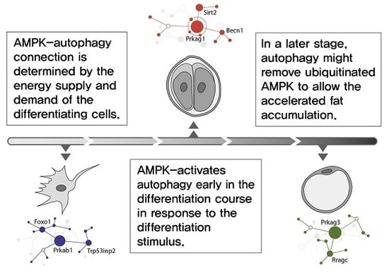
Metformin has been reported to have body weight lowering effects while treating type 2 diabetes. However, limited studies examined the effects of metformin on adipogenesis in vitro, and available data are inconclusive and contradictory. In this study, we examined the effects of a
[...] Read more.
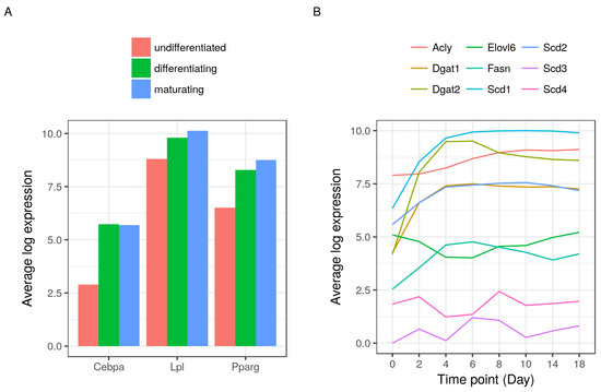
Metformin has been reported to have body weight lowering effects while treating type 2 diabetes. However, limited studies examined the effects of metformin on adipogenesis in vitro, and available data are inconclusive and contradictory. In this study, we examined the effects of a variety of concentrations of metformin on adipocyte differentiation of 3T3-L1 preadipocytes and found metformin exhibits a dual effect on adipogenesis. Metformin at lower concentrations (1.25–2.5 mM) significantly induced adipogenesis while at higher concentrations (5–10 mM) metformin significantly inhibited adipogenesis in 3T3-L1 cells. The biphasic effect of different doses of metformin on adipogenesis was accompanied by increasing or decreasing the expression of adipogenic and lipogenic genes including peroxisome proliferator-activated receptor (PPARγ), CCAAT/enhancer binding protein α (C/EBPα), and fatty acid synthase (FASN) at both messenger RNA (mRNA) and protein levels. Furthermore, only the higher concentrations of metformin induced the phosphorylation of adenosine 5′-monophosphate (AMP)-activated protein kinase (AMPK), p38, and c-Jun N-terminal kinase (JNK) and reduced the phosphorylation of extracellular regulated protein kinases (ERK) and Akt. Pretreatment with compound C, a specific AMPK inhibitor, significantly countered high concentration of metformin-induced inhibition of adipogenesis. Taken together, these findings demonstrate that the effect of metformin on adipocyte differentiation is biphasic and dose-dependent. Lower concentrations of metformin induce adipogenesis, which could be mediated in an AMPK-independent manner, while higher concentrations of metformin inhibit adipogenesis via AMPK activation.
Full article
(This article belongs to the Section Biochemistry)
►
Show Figures

Graphical abstract


