Next Issue
Volume 17, October
Previous Issue
Volume 17, August
 
 
ijms-logo

Journal Browser

Journal Browser

Int. J. Mol. Sci., Volume 17, Issue 9 (September 2016) – 206 articles

Cover Story (view full-size image): 
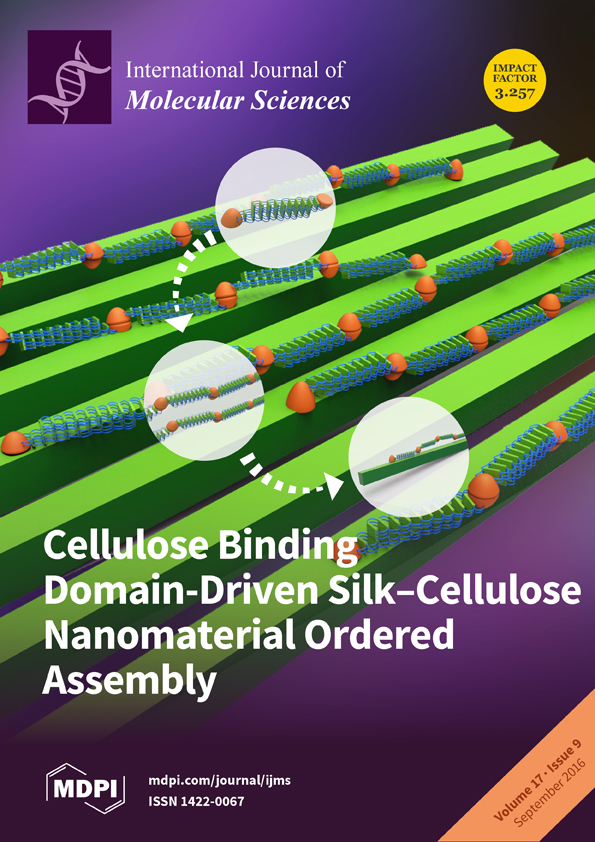
Cellulose Binding Domain-Driven Silk–Cellulose Nanomaterial Ordered Assembly Cellulose binding domain (CBD) plays a central role in the higher molecular order of silk–cellulose nanocomposites. Its ability to form dimers and mimic the non-repetitive spider silk terminal function enables formation of aligned nano-silk fibers. At a higher level, CBD specifically binds cellulose and mediates silk–cellulose aligned composite assembly. Cover image by Dr. Noam Atias. View this paper.
  • Issues are regarded as officially published after their release is announced to the table of contents alert mailing list.
  • You may sign up for e-mail alerts to receive table of contents of newly released issues.
  • PDF is the official format for papers published in both, html and pdf forms. To view the papers in pdf format, click on the "PDF Full-text" link, and use the free Adobe Reader to open them.
Order results
Result details
Section
Select all
Export citation of selected articles as:
11 pages, 734 KiB  
Article
ADAM9 Expression Is Associate with Glioma Tumor Grade and Histological Type, and Acts as a Prognostic Factor in Lower-Grade Gliomas
by Xing Fan 1,†, Yongheng Wang 1,2,†, Chuanbao Zhang 1, Li Liu 3, Sen Yang 4, Yinyan Wang 1,5, Xing Liu 1, Zenghui Qian 1, Shengyu Fang 1, Hui Qiao 1,*,‡ and Tao Jiang 1,5,*,‡
1 Beijing Neurosurgical Institute, Capital Medical University, Beijing 100050, China
2 Department of Neurosurgery, Qinhuangdao First Hospital, Qinhuangdao 066000, China
3 Department of Ophthalmology, Qinhuangdao First Hospital, Qinhuangdao 066000, China
4 Department of Radiotherapy, Qinhuangdao First Hospital, Qinhuangdao 066000, China
5 Department of Neurosurgery, Beijing Tiantan Hospital, Capital Medical University, Beijing100050, China
Int. J. Mol. Sci. 2016, 17(9), 1276; https://doi.org/10.3390/ijms17091276 - 26 Aug 2016
Cited by 29 | Viewed by 6155
Abstract
The A disintegrin and metalloproteinase 9 (ADAM9) protein has been suggested to promote carcinoma invasion and appears to be overexpressed in various human cancers. However, its role has rarely been investigated in gliomas and, thus, in the current study we have evaluated ADAM9 [...] Read more.
The A disintegrin and metalloproteinase 9 (ADAM9) protein has been suggested to promote carcinoma invasion and appears to be overexpressed in various human cancers. However, its role has rarely been investigated in gliomas and, thus, in the current study we have evaluated ADAM9 expression in gliomas and examined the relevance of its expression in the prognosis of glioma patients. Clinical characteristics, RNA sequence data, and the case follow-ups were reviewed for 303 patients who had histological, confirmed gliomas. The ADAM9 expression between lower-grade glioma (LGG) and glioblastoma (GBM) patients was compared and its association with progression-free survival (PFS) and overall survival (OS) was assessed to evaluate its prognostic value. Our data suggested that GBM patients had significantly higher expression of ADAM9 in comparison to LGG patients (p < 0.001, t-test). In addition, among the LGG patients, aggressive astrocytic tumors displayed significantly higher ADAM9 expression than oligodendroglial tumors (p < 0.001, t-test). Moreover, high ADAM9 expression also correlated with poor clinical outcome (p < 0.001 and p < 0.001, log-rank test, for PFS and OS, respectively) in LGG patients. Further, multivariate analysis suggested ADAM9 expression to be an independent marker of poor survival (p = 0.002 and p = 0.003, for PFS and OS, respectively). These results suggest that ADAM9 mRNA expression is associated with tumor grade and histological type in gliomas and can serve as an independent prognostic factor, specifically in LGG patients. Full article
(This article belongs to the Special Issue Big Data for Oncology)
Show Figures

Figure 1

14 pages, 4865 KiB  
Article
Flavonoids Extracted from Licorice Prevents Colitis-Associated Carcinogenesis in AOM/DSS Mouse Model
by Xiaowei Huo, Dongyu Liu, Li Gao, Liyong Li and Li Cao *
Institute of Medicinal Plant Development, Chinese Academy of Medical Sciences and Peking Union Medical College, Beijing 100193, China
Int. J. Mol. Sci. 2016, 17(9), 1343; https://doi.org/10.3390/ijms17091343 - 24 Aug 2016
Cited by 41 | Viewed by 8400
Abstract
Inflammatory bowel disease (IBD) is generally considered as a major risk factor in the progression of colitis-associated carcinogenesis (CAC). Thus, it is well accepted that ameliorating inflammation creates a potential to achieve an inhibitory effect on CAC. Licorice flavonoids (LFs) possess strong anti-inflammatory [...] Read more.
Inflammatory bowel disease (IBD) is generally considered as a major risk factor in the progression of colitis-associated carcinogenesis (CAC). Thus, it is well accepted that ameliorating inflammation creates a potential to achieve an inhibitory effect on CAC. Licorice flavonoids (LFs) possess strong anti-inflammatory activity, making it possible to investigate its pharmacologic role in suppressing CAC. The purpose of the present study was to evaluate the anti-tumor potential of LFs, and further explore the underlying mechanisms. Firstly, an azoxymethane (AOM)/dextran sulfate sodium (DSS)-induced mouse model was established and administered with or without LFs for 10 weeks, and then the severity of CAC was examined macroscopically and histologically. Subsequently, the effects of LFs on expression of proteins associated with apoptosis and proliferation, levels of inflammatory cytokine, expression of phosphorylated-Janus kinases 2 (p-Jak2) and phosphorylated-signal transducer and activator of transcription 3 (p-Stat3), and activation of nuclear factor-κB (NFκB) and P53 were assessed. We found that LFs could significantly reduce tumorigenesis induced by AOM/DSS. Further study revealed that LFs treatment substantially reduced activation of NFκB and P53, and subsequently suppressed production of inflammatory cytokines and phosphorylation of Jak2 and Stat3 in AOM/DSS-induced mice. Taken together, LFs treatment alleviated AOM/DSS induced CAC via P53 and NFκB/IL-6/Jak2/Stat3 pathways, highlighting the potential of LFs in preventing CAC. Full article
(This article belongs to the Special Issue The Mechanism of Action of Food Components in Disease Prevention)
Show Figures

Graphical abstract

13 pages, 1600 KiB  
Article
Family Growth and Survival Response to Two Simulated Water Temperature Environments in the Sea Urchin Strongylocentrotus intermedius
by Yaqing Chang *, Xiaofei Tian, Weijie Zhang, Fenjie Han, Shun Chen, Mi Zhou, Zhenguo Pang, Shoubing Qi and Wenping Feng
Key Laboratory of Mariculture & Stock Enhancement in North China’s Sea, Ministry of Agriculture, Dalian Ocean University, Dalian 116023, China
Int. J. Mol. Sci. 2016, 17(9), 1356; https://doi.org/10.3390/ijms17091356 - 29 Aug 2016
Cited by 19 | Viewed by 5040
Abstract
Heat tolerance is a target trait in the selective breeding of the sea urchin Strongylocentrotus intermedius, as it plays an important role in the survival and growth of cultured S. intermedius during summer. We investigated family growth and survival response to two [...] Read more.
Heat tolerance is a target trait in the selective breeding of the sea urchin Strongylocentrotus intermedius, as it plays an important role in the survival and growth of cultured S. intermedius during summer. We investigated family growth and survival response to two temperature treatments to evaluate the genotype by temperature interaction (GEI) in the family selection of S. intermedius. Sea urchins from 11 families were exposed to two simulated water temperature environments—high temperature (HE) and control temperature (CE)—for 12 months, with each experiment divided into four periods (P1, stress-free period I; P2, stress-full high period; P3, stress-response period; and P4, stress-free period II) based on the temperature changes and the survival. Test diameter (TD), body weight (BW), and survival rate (SR) in HE and CE were measured monthly. Effects of family, temperature, and family-temperature interaction on TD, BW, SR, and specific growth rate (SGR) for BW were examined. In CE, BW differed significantly between families in P2, P3, and P4, while TD differed significantly between families in P3 and P4 (p < 0.05). In HE, family had significant effects on BW in P4, and on TD in P3 and P4, while temperature had significant effects on SR, TD, and BW in P3 and P4 (p < 0.05). GEI effects were not significant for TD or BW; however, family ranking changes revealed the existence of GEI in SR. The GEI results indicate the necessity of applying family selection in CE and HE for SR, but not for TD or BW. These results may provide a guide for aquaculture and selective breeding of S. intermedius under temperature pressure. Full article
(This article belongs to the Special Issue Gene–Environment Interactions)
Show Figures

Graphical abstract

45 pages, 2033 KiB  
Review
Physical, Chemical and Biochemical Modifications of Protein-Based Films and Coatings: An Extensive Review
by Joël Zink 1, Tom Wyrobnik 1, Tobias Prinz 1 and Markus Schmid 1,2,*
1 Fraunhofer Institute for Process Engineering and Packaging IVV, Giggenhauser Strasse 35, Freising 85354, Germany
2 Chair of Food Packaging Technology, Technische Universität München, Weihenstephaner Steig 22, Freising 85354, Germany
Int. J. Mol. Sci. 2016, 17(9), 1376; https://doi.org/10.3390/ijms17091376 - 23 Aug 2016
Cited by 221 | Viewed by 14168
Abstract
Protein-based films and coatings are an interesting alternative to traditional petroleum-based materials. However, their mechanical and barrier properties need to be enhanced in order to match those of the latter. Physical, chemical, and biochemical methods can be used for this purpose. The aim [...] Read more.
Protein-based films and coatings are an interesting alternative to traditional petroleum-based materials. However, their mechanical and barrier properties need to be enhanced in order to match those of the latter. Physical, chemical, and biochemical methods can be used for this purpose. The aim of this article is to provide an overview of the effects of various treatments on whey, soy, and wheat gluten protein-based films and coatings. These three protein sources have been chosen since they are among the most abundantly used and are well described in the literature. Similar behavior might be expected for other protein sources. Most of the modifications are still not fully understood at a fundamental level, but all the methods discussed change the properties of the proteins and resulting products. Mastering these modifications is an important step towards the industrial implementation of protein-based films. Full article
(This article belongs to the Section Materials Science)
Show Figures

Figure 1

12 pages, 6891 KiB  
Article
MicroRNA-381 Regulates Chondrocyte Hypertrophy by Inhibiting Histone Deacetylase 4 Expression
by Weishen Chen, Puyi Sheng, Zhiyu Huang, Fangang Meng, Yan Kang, Guangxin Huang, Zhiqi Zhang, Weiming Liao * and Ziji Zhang *
Department of Joint Surgery, First Affiliated Hospital of Sun Yat-sen University, Guangzhou 510080, China
Int. J. Mol. Sci. 2016, 17(9), 1377; https://doi.org/10.3390/ijms17091377 - 23 Aug 2016
Cited by 37 | Viewed by 6452
Abstract
Chondrocyte hypertrophy, regulated by Runt-related transcription factor 2 (RUNX2) and matrix metalloproteinase 13 (MMP13), is a crucial step in cartilage degeneration and osteoarthritis (OA) pathogenesis. We previously demonstrated that microRNA-381 (miR-381) promotes MMP13 expression during chondrogenesis and contributes to cartilage degeneration; however, the [...] Read more.
Chondrocyte hypertrophy, regulated by Runt-related transcription factor 2 (RUNX2) and matrix metalloproteinase 13 (MMP13), is a crucial step in cartilage degeneration and osteoarthritis (OA) pathogenesis. We previously demonstrated that microRNA-381 (miR-381) promotes MMP13 expression during chondrogenesis and contributes to cartilage degeneration; however, the mechanism underlying this process remained unclear. In this study, we observed divergent expression of miR-381 and histone deacetylase 4 (HDAC4), an enzyme that directly inhibits RUNX2 and MMP13 expression, during late-stage chondrogenesis of ATDC5 cells, as well as in prehypertrophic and hypertrophic chondrocytes during long bone development in E16.5 mouse embryos. We therefore investigated whether this miRNA regulates HDAC4 expression during chondrogenesis. Notably, overexpression of miR-381 inhibited HDAC4 expression but promoted RUNX2 expression. Moreover, transfection of SW1353 cells with an miR-381 mimic suppressed the activity of a reporter construct containing the 3′-untranslated region (3′-UTR) of HDAC4. Conversely, treatment with a miR-381 inhibitor yielded increased HDAC4 expression and decreased RUNX2 expression. Lastly, knockdown of HDAC4 expression resulted in increased RUNX2 and MMP13 expression in SW1353 cells. Collectively, our results indicate that miR-381 epigenetically regulates MMP13 and RUNX2 expression via targeting of HDAC4, thereby suggesting the possibilities of inhibiting miR-381 to control chondrocyte hypertrophy and cartilage degeneration. Full article
(This article belongs to the Collection Regulation by Non-coding RNAs)
Show Figures

Graphical abstract

13 pages, 2548 KiB  
Article
Human TRIB2 Oscillates during the Cell Cycle and Promotes Ubiquitination and Degradation of CDC25C
by Kai Ling Liang 1,2, Roberto Paredes 3, Ruaidhri Carmody 4, Patrick A. Eyers 5, Stefan Meyer 3,6, Tommie V. McCarthy 2 and Karen Keeshan 1,*
1 Paul O’Gorman Leukemia Research Centre, Institute of Cancer Sciences, College of Medical, Veterinary and Life Sciences, University of Glasgow, Glasgow G12 0ZD, UK
2 School of Biochemistry and Cell Biology, University College Cork, Cork, Ireland
3 Stem Cell and Leukaemia Proteomics Laboratory, University of Manchester, Manchester M20 3LJ, UK
4 Institute of Infection, Immunity and Inflammation, College of Medical, Veterinary and Life Sciences, University of Glasgow, Glasgow G12 8TA, UK
5 Department of Biochemistry, Institute of Integrative Biology, University of Liverpool, Liverpool L69 7ZB, UK
6 Paediatric and Adolescent Oncology, Royal Manchester Children’s and Christie Hospital, University of Manchester, Manchester M13 9WL, UK
Int. J. Mol. Sci. 2016, 17(9), 1378; https://doi.org/10.3390/ijms17091378 - 23 Aug 2016
Cited by 19 | Viewed by 8038
Abstract
Tribbles homolog 2 (TRIB2) is a member of the mammalian Tribbles family of serine/threonine pseudokinases (TRIB1-3). Studies of TRIB2 indicate that many of the molecular interactions between the single Drosophila Tribbles (Trbl) protein and interacting partners are evolutionary conserved. In this study, we [...] Read more.
Tribbles homolog 2 (TRIB2) is a member of the mammalian Tribbles family of serine/threonine pseudokinases (TRIB1-3). Studies of TRIB2 indicate that many of the molecular interactions between the single Drosophila Tribbles (Trbl) protein and interacting partners are evolutionary conserved. In this study, we examined the relationship between TRIB2 and cell division cycle 25 (CDC25) family of dual-specificity protein phosphatases (mammalian homologues of Drosophila String), which are key physiological cell cycle regulators. Using co-immunoprecipitation we demonstrate that TRIB2 interacts with CDC25B and CDC25C selectively. Forced overexpression of TRIB2 caused a marked decrease in total CDC25C protein levels. Following inhibition of the proteasome, CDC25C was stabilized in the nuclear compartment. This implicates TRIB2 as a regulator of nuclear CDC25C turnover. In complementary ubiquitination assays, we show that TRIB2-mediated degradation of CDC25C is associated with lysine-48-linked CDC25C polyubiquitination driven by the TRIB2 kinase-like domain. A cell cycle associated role for TRIB2 is further supported by the cell cycle regulated expression of TRIB2 protein levels. Our findings reveal mitotic CDC25C as a new target of TRIB2 that is degraded via the ubiquitin proteasome system. Inappropriate CDC25C regulation could mechanistically underlie TRIB2 mediated regulation of cellular proliferation in neoplastic cells. Full article
(This article belongs to the Special Issue Advances in Molecular Oncology)
Show Figures

Graphical abstract

16 pages, 928 KiB  
Review
Micronutrient Antioxidants and Nonalcoholic Fatty Liver Disease
by Guanliang Chen, Yinhua Ni, Naoto Nagata, Liang Xu and Tsuguhito Ota *
Department of Cell Metabolism and Nutrition, Brain/Liver Interface Medicine Research Center, Kanazawa University, Kanazawa, Ishikawa 920-8640, Japan
Int. J. Mol. Sci. 2016, 17(9), 1379; https://doi.org/10.3390/ijms17091379 - 23 Aug 2016
Cited by 58 | Viewed by 13935
Abstract
Nonalcoholic fatty liver disease (NAFLD) is one of the most important chronic liver diseases worldwide and has garnered increasing attention in recent decades. NAFLD is characterized by a wide range of liver changes, from simple steatosis to nonalcoholic steatohepatitis, cirrhosis, and hepatocellular carcinoma. [...] Read more.
Nonalcoholic fatty liver disease (NAFLD) is one of the most important chronic liver diseases worldwide and has garnered increasing attention in recent decades. NAFLD is characterized by a wide range of liver changes, from simple steatosis to nonalcoholic steatohepatitis, cirrhosis, and hepatocellular carcinoma. The blurred pathogenesis of NAFLD is very complicated and involves lipid accumulation, insulin resistance, inflammation, and fibrogenesis. NAFLD is closely associated with complications such as obesity, diabetes, steatohepatitis, and liver fibrosis. During the progression of NAFLD, reactive oxygen species (ROS) are activated and induce oxidative stress. Recent attempts at establishing effective NAFLD therapy have identified potential micronutrient antioxidants that may reduce the accumulation of ROS and finally ameliorate the disease. In this review, we present the molecular mechanisms involved in the pathogenesis of NAFLD and introduce some dietary antioxidants that may be used to prevent or cure NAFLD, such as vitamin D, E, and astaxanthin. Full article
(This article belongs to the Special Issue Macro- and Micro-nutrient Antioxidants)
Show Figures

Graphical abstract

16 pages, 6248 KiB  
Article
Anti-Inflammatory Mechanism of Neural Stem Cell Transplantation in Spinal Cord Injury
by Zhijian Cheng 1,†, Wen Zhu 2,†, Kai Cao 1, Fei Wu 1, Jin Li 1, Guoyu Wang 1, Haopen Li 1, Ming Lu 3, Yi Ren 4,* and Xijing He 1,*
1 Department of Orthopedics, The Second Affiliated Hospital of Xi’an Jiaotong University, Xi’an 710004, China
2 Intensive Care Unit, The First People’s Hospital of Xianyang City, Xianyang 710021, China
3 Neurosurgery Department, The Second Affiliated Hospital of Hunan Normal University, Changsha 410003, China
4 Department of Biomedical Sciences, Florida State University, College of Medicine, Tallahassee, FL 32306, USA
Int. J. Mol. Sci. 2016, 17(9), 1380; https://doi.org/10.3390/ijms17091380 - 23 Aug 2016
Cited by 94 | Viewed by 9993
Abstract
Neural stem cell (NSC) transplantation has been proposed to promote functional recovery after spinal cord injury. However, a detailed understanding of the mechanisms of how NSCs exert their therapeutic plasticity is lacking. We transplanted mouse NSCs into the injured spinal cord seven days [...] Read more.
Neural stem cell (NSC) transplantation has been proposed to promote functional recovery after spinal cord injury. However, a detailed understanding of the mechanisms of how NSCs exert their therapeutic plasticity is lacking. We transplanted mouse NSCs into the injured spinal cord seven days after SCI, and the Basso Mouse Scale (BMS) score was performed to assess locomotor function. The anti-inflammatory effects of NSC transplantation was analyzed by immunofluorescence staining of neutrophil and macrophages and the detection of mRNA levels of tumor necrosis factor-α (TNF-α), interleukin-1β (IL-1β), interleukin-6 (IL-6) and interleukin-12 (IL-12). Furthermore, bone marrow-derived macrophages (BMDMs) were co-cultured with NSCs and followed by analyzing the mRNA levels of inducible nitric oxide synthase (iNOS), TNF-α, IL-1β, IL-6 and IL-10 with quantitative real-time PCR. The production of TNF-α and IL-1β by BMDMs was examined using the enzyme-linked immunosorbent assay (ELISA). Transplanted NSCs had significantly increased BMS scores (p < 0.05). Histological results showed that the grafted NSCs migrated from the injection site toward the injured area. NSCs transplantation significantly reduced the number of neutrophils and iNOS+/Mac-2+ cells at the epicenter of the injured area (p < 0.05). Meanwhile, mRNA levels of TNF-α, IL-1β, IL-6 and IL-12 in the NSCs transplantation group were significantly decreased compared to the control group. Furthermore, NSCs inhibited the iNOS expression of BMDMs and the release of inflammatory factors by macrophages in vitro (p < 0.05). These results suggest that NSC transplantation could modulate SCI-induced inflammatory responses and enhance neurological function after SCI via reducing M1 macrophage activation and infiltrating neutrophils. Thus, this study provides a new insight into the mechanisms responsible for the anti-inflammatory effect of NSC transplantation after SCI. Full article
(This article belongs to the Special Issue Advances in Cell Transplantation)
Show Figures

Graphical abstract

16 pages, 3585 KiB  
Article
Transcriptional Induction of Metallothionein by Tris(pentafluorophenyl)stibane in Cultured Bovine Aortic Endothelial Cells
by Tomoya Fujie 1, Masaki Murakami 1, Eiko Yoshida 1, Shuji Yasuike 2,*, Tomoki Kimura 3, Yasuyuki Fujiwara 4, Chika Yamamoto 5 and Toshiyuki Kaji 1,*
1 Department of Environmental Health, Faculty of Pharmaceutical Sciences, Tokyo University of Science, 2641 Yamazaki, Noda 278-8510, Japan
2 Laboratory of Organic and Medicinal Chemistry, School of Pharmaceutical Sciences, Aichi Gakuin University, 1-100 Kusumoto-cho, Chikusa-ku, Nagoya 464-8650, Japan
3 Depertment of Life Science, Faculty of Science and Engineering, Setsunan University, 17-8 Ikedanakamachi, Neyagawa 572-8508, Japan
4 Department of Environmental Health, School of Pharmacy, Tokyo University of Pharmacy and Life Sciences, 1432-1 Horinouchi, Hachioji 192-0392, Japan
5 Department of Environmental Health, Faculty of Pharmaceutical Sciences, Toho University, 2-2-1 Miyama, Funabashi 274-8510, Japan
Int. J. Mol. Sci. 2016, 17(9), 1381; https://doi.org/10.3390/ijms17091381 - 23 Aug 2016
Cited by 22 | Viewed by 6704
Abstract
Vascular endothelial cells cover the luminal surface of blood vessels and contribute to the prevention of vascular disorders such as atherosclerosis. Metallothionein (MT) is a low molecular weight, cysteine-rich, metal-binding, inducible protein, which protects cells from the toxicity of heavy metals and active [...] Read more.
Vascular endothelial cells cover the luminal surface of blood vessels and contribute to the prevention of vascular disorders such as atherosclerosis. Metallothionein (MT) is a low molecular weight, cysteine-rich, metal-binding, inducible protein, which protects cells from the toxicity of heavy metals and active oxygen species. Endothelial MT is not induced by inorganic zinc. Adequate tools are required to investigate the mechanisms underlying endothelial MT induction. In the present study, we found that an organoantimony compound, tris(pentafluorophenyl)stibane, induces gene expression of MT-1A and MT-2A, which are subisoforms of MT in bovine aortic endothelial cells. The data reveal that MT-1A is induced by activation of both the MTF-1–MRE and Nrf2–ARE pathways, whereas MT-2A expression requires only activation of the MTF-1–MRE pathway. The present data suggest that the original role of MT-1 is to protect cells from heavy metal toxicity and oxidative stress in the biological defense system, while that of MT-2 is to regulate intracellular zinc metabolism. Full article
(This article belongs to the Special Issue Metalloproteins 2017)
Show Figures

Graphical abstract

8 pages, 761 KiB  
Article
Antiplatelet Usage Impacts Clot Density in Acute Anterior Circulation Ischemic Stroke
by Slaven Pikija 1, Jozef Magdic 2, Anita Lukic 3, Catharina Schreiber 4, Johannes Sebastian Mutzenbach 1, Mark R. McCoy 5 and Johann Sellner 1,6,*
1 Department of Neurology, Christian Doppler Medical Center, Paracelsus Medical University, Salzburg 5020, Austria
2 Department of Neurology, Univerzitetni Klinični Center, Maribor 2000, Slovenia
3 Department of Anesthesiology, General Hospital Varazdin, Varazdin 42000, Croatia
4 Department of Cardiac Surgery, Salzburger Landeskliniken, Paracelsus Medical University, Salzburg 5020, Austria
5 Division of Neuroradiology, Christian Doppler Medical Center, Paracelsus Medical University, Salzburg 5020, Austria
6 Department of Neurology, Klinikum rechts der Isar, Technische Universität München, München 81675, Germany
Int. J. Mol. Sci. 2016, 17(9), 1382; https://doi.org/10.3390/ijms17091382 - 23 Aug 2016
Cited by 11 | Viewed by 5590
Abstract
We explored whether clot density in middle cerebral artery (MCA) occlusion is related to clinical variables, stroke etiology, blood constituents, and prestroke medication. We performed a retrospective chart review of patients with acute ischemic stroke of the anterior circulation admitted to two Central [...] Read more.
We explored whether clot density in middle cerebral artery (MCA) occlusion is related to clinical variables, stroke etiology, blood constituents, and prestroke medication. We performed a retrospective chart review of patients with acute ischemic stroke of the anterior circulation admitted to two Central European stroke centers. The acquisition of non-contrast enhanced CT (NECT) and CT angiography (CTA) within 4.5 h of symptom onset was obligatory. We assessed the site of MCA occlusion as well as density, area, and length of the clot in 150 patients. The Hounsfield unit values for the clot were divided with contralateral MCA segment to yield relative Hounsfield Unit ratio (rHU). The site of the vessel occlusion (M1 vs. M2) and antiplatelet usage, but not stroke etiology, significantly influenced rHU. We found an inverse correlation of rHU with erythrocyte count (p < 0.001). The multivariate analysis revealed that a higher rHU (i.e., clot being more hyperdense) was more likely with the use of antiplatelets (OR 4.24, CI 1.10–16.31, p = 0.036). Erythrocyte (OR 0.18, CI 0.05–0.55, p = 0.003), and thrombocyte counts (OR 0.99, CI 0.98–0.99, p = 0.029) were associated with odds for more hypodense clots (lower rHU). Our study disclosed that antiplatelet therapy impacts the composition of intracranial clots of the anterior circulation. Full article
(This article belongs to the Special Issue Vascular Biology and Therapeutics)
Show Figures

Figure 1

18 pages, 4237 KiB  
Article
CTHRSSVVC Peptide as a Possible Early Molecular Imaging Target for Atherosclerosis
by Rosemeire A. Silva 1,*, Ricardo J. Giordano 2, Paulo S. Gutierrez 3, Viviane Z. Rocha 4, Martina Rudnicki 5, Patrick Kee 6, Dulcinéia S. P. Abdalla 5, Pedro Puech-Leão 7, Bruno Caramelli 4, Wadih Arap 8, Renata Pasqualini 8, José C. Meneghetti 9, Fabio L. N. Marques 10, Menka Khoobchandani 11, Kattesh V. Katti 11, Ademar B. Lugão 12 and Jorge Kalil 1,*
1 Laboratory of Immunology, Heart Institute (InCor), Hospital das Clínicas da Faculdade de Medicina da Universidade de São Paulo, São Paulo 05403-000, Brazil
2 Chemistry Institute, Biochemistry Department, University of Sao Paulo, Sao Paulo 05508-000, Brazil
3 Laboratory of Pathology, Heart Institute (InCor), Hospital das Clínicas da Faculdade de Medicina da Universidade de São Paulo, São Paulo 05403-000, Brazil
4 Clinical Division, Heart Institute (InCor), Hospital das Clínicas da Faculdade de Medicina da Universidade de São Paulo, São Paulo 05403-000, Brazil
5 Department of Clinical and Toxicological Analyses, Faculty of Pharmaceutical Sciences University of São Paulo, São Paulo 05508-000, Brazil
6 Division of Cardiology, Department of Internal Medicine, The University of Texas Health Science Center at Houston, Houston, TX 77054, USA
7 Division of Vascular and Endovascular Surgery, University of São Paulo Medical School, São Paulo 05403-000, Brazil
8 University of New Mexico Comprehensive Cancer Center, Division of Hematology/Oncology and Division of Molecular Medicine, Department of Internal Medicine, University of New Mexico School of Medicine Albuquerque, NM 87131, USA
9 Medicine Nuclear Service and Molecular Image, Heart Institute (InCor), Hospital das Clínicas da Faculdade de Medicina da Universidade de São Paulo, São Paulo 05403-000, Brazil
10 Departamento de Radiologia e Oncologia, Faculdade de Medicina da Universidade de São Paulo (LIM43), São Paulo 05403-911, Brazil
11 Institute of Green Nanotechnology, Department of Radiology and Chemistry, University of Missouri, Columbia, MO 65211, USA
12 Nuclear and Energy Research Institute—IPEN/CNEN/São Paulo 05508-000, Brazil
add Show full affiliation list remove Hide full affiliation list
Int. J. Mol. Sci. 2016, 17(9), 1383; https://doi.org/10.3390/ijms17091383 - 24 Aug 2016
Cited by 11 | Viewed by 6451
Abstract
The purpose of our work was to select phages displaying peptides capable of binding to vascular markers present in human atheroma, and validate their capacity to target the vascular markers in vitro and in low-density lipoprotein receptor knockout (LDLr−/−) mouse model [...] Read more.
The purpose of our work was to select phages displaying peptides capable of binding to vascular markers present in human atheroma, and validate their capacity to target the vascular markers in vitro and in low-density lipoprotein receptor knockout (LDLr−/−) mouse model of atherosclerosis. By peptide fingerprinting on human atherosclerotic tissues, we selected and isolated four different peptides sequences, which bind to atherosclerotic lesions and share significant similarity to known human proteins with prominent roles in atherosclerosis. The CTHRSSVVC-phage peptide displayed the strongest reactivity with human carotid atherosclerotic lesions (p < 0.05), when compared to tissues from normal carotid arteries. This peptide sequence shares similarity to a sequence present in the fifth scavenger receptor cysteine-rich (SRCR) domain of CD163, which appeared to bind to CD163, and subsequently, was internalized by macrophages. Moreover, the CTHRSSVVC-phage targets atherosclerotic lesions of a low-density lipoprotein receptor knockout (LDLr−/−) mouse model of atherosclerosis in vivo to High-Fat diet group versus Control group. Tetraazacyclododecane-1,4,7,10-tetraacetic acid-CTHRSSVVC peptide (DOTA-CTHRSSVVC) was synthesized and labeled with 111InCl3 in >95% yield as determined by high performance liquid chromatography (HPLC), to validate the binding of the peptide in atherosclerotic plaque specimens. The results supported our hypothesis that CTHRSSVVC peptide has a remarkable sequence for the development of theranostics approaches in the treatment of atherosclerosis and other diseases. Full article
(This article belongs to the Special Issue Drug Delivery and Antimicrobial Agents)
Show Figures

Graphical abstract

7 pages, 451 KiB  
Article
Heads or Tails: Genotyping of Hepatitis C Virus Concerning the 2k/1b Circulating Recombinant Form
by Wim Schuermans 1,†, Hans Orlent 2,†, Isabelle Desombere 3, Patrick Descheemaeker 4, Hans Van Vlierberghe 5, Anja Geerts 5, Xavier Verhelst 5, Marijke Reynders 4,*,‡ and Elizaveta Padalko 1,6,‡
1 Department of Clinical Chemistry, Microbiology and Immunology, Ghent University and Hospital, Ghent 9000, Belgium
2 Department of Gastroenterology and Hepatology, AZ Sint-Jan Bruges-Ostend, Bruges 8000, Belgium
3 Center for Vaccinology, Ghent University and Hospital, Ghent 9000, Belgium
4 Department of Laboratory Medicine, Clinical Microbiology, AZ Sint-Jan Bruges-Ostend, Bruges 8000, Belgium
5 Department of Gastroenterology and Hepatology, Ghent University and Hospital, Ghent 8000, Belgium
6 School of Life Sciences, Hasselt University, Diepenbeek 3590, Belgium
Int. J. Mol. Sci. 2016, 17(9), 1384; https://doi.org/10.3390/ijms17091384 - 23 Aug 2016
Cited by 11 | Viewed by 7171
Abstract
As different hepatitis C virus (HCV) genotypes respond differently to initiated therapy, correct HCV genotyping is essential. A potential risk for misclassification of the intergenotypic HCV circulating recombinant form (CRF) 2k/1b strains exists, depending on the genotyping method used. The aim was to [...] Read more.
As different hepatitis C virus (HCV) genotypes respond differently to initiated therapy, correct HCV genotyping is essential. A potential risk for misclassification of the intergenotypic HCV circulating recombinant form (CRF) 2k/1b strains exists, depending on the genotyping method used. The aim was to investigate the differences in HCV genotyping methods with regard to CRF 2k/1b and to gain insight in the prevalence of the CRF 2k/1b. Genotyping results by Versant HCV Genotype Assay were compared with nonstructural protein 5B (NS5B) sequencing. In total, from November 2001 until March 2015, 3296 serum samples were analyzed by Versant HCV Genotype Assay. As misclassified CRF is harbored among HCV genotype 2, we further focused our search on 142 (4.3%) samples positive for HCV genotype 2. On 116 (81.7%) retrieved samples, the NS5B sequencing was performed. Twelve out of the 116 retrieved samples (10.3%) were classified as CRF 2k/1b by sequencing of the NS5B region. Ten of these 12 samples were originally misclassified as genotype 2a or 2c, while 2 of them were misclassified as genotype 2. Our results show that the current prevalence of CRF 2k/1b is underestimated. The importance of correct HCV genotyping is emphasized, considering the tailored choice of treatment regimen and overall prognosis. Full article
(This article belongs to the Special Issue Hepatitis Virus Infection and Research)
Show Figures

Graphical abstract

20 pages, 1095 KiB  
Article
A Genomics-Based Model for Prediction of Severe Bioprosthetic Mitral Valve Calcification
by Anastasia V. Ponasenko 1, Maria V. Khutornaya 1, Anton G. Kutikhin 1,*, Natalia V. Rutkovskaya 1, Anna V. Tsepokina 1, Natalia V. Kondyukova 1, Arseniy E. Yuzhalin 1,2 and Leonid S. Barbarash 1
1 Research Institute for Complex Issues of Cardiovascular Diseases, Sosnovy Boulvevard 6, Kemerovo 650002, Russia
2 Department of Oncology, Cancer Research UK and Medical Research Council Oxford Institute for Radiation Oncology, University of Oxford, Old Road Campus Research Building, Roosevelt Drive, Oxford OX3 7DQ, UK
Int. J. Mol. Sci. 2016, 17(9), 1385; https://doi.org/10.3390/ijms17091385 - 31 Aug 2016
Cited by 9 | Viewed by 5063
Abstract
Severe bioprosthetic mitral valve calcification is a significant problem in cardiovascular surgery. Unfortunately, clinical markers did not demonstrate efficacy in prediction of severe bioprosthetic mitral valve calcification. Here, we examined whether a genomics-based approach is efficient in predicting the risk of severe bioprosthetic [...] Read more.
Severe bioprosthetic mitral valve calcification is a significant problem in cardiovascular surgery. Unfortunately, clinical markers did not demonstrate efficacy in prediction of severe bioprosthetic mitral valve calcification. Here, we examined whether a genomics-based approach is efficient in predicting the risk of severe bioprosthetic mitral valve calcification. A total of 124 consecutive Russian patients who underwent mitral valve replacement surgery were recruited. We investigated the associations of the inherited variation in innate immunity, lipid metabolism and calcium metabolism genes with severe bioprosthetic mitral valve calcification. Genotyping was conducted utilizing the TaqMan assay. Eight gene polymorphisms were significantly associated with severe bioprosthetic mitral valve calcification and were therefore included into stepwise logistic regression which identified male gender, the T/T genotype of the rs3775073 polymorphism within the TLR6 gene, the C/T genotype of the rs2229238 polymorphism within the IL6R gene, and the A/A genotype of the rs10455872 polymorphism within the LPA gene as independent predictors of severe bioprosthetic mitral valve calcification. The developed genomics-based model had fair predictive value with area under the receiver operating characteristic (ROC) curve of 0.73. In conclusion, our genomics-based approach is efficient for the prediction of severe bioprosthetic mitral valve calcification. Full article
(This article belongs to the Special Issue Human Single Nucleotide Polymorphisms and Disease Diagnostics)
Show Figures

Graphical abstract

15 pages, 3973 KiB  
Article
Antiviral Activity of a Novel Compound CW-33 against Japanese Encephalitis Virus through Inhibiting Intracellular Calcium Overload
by Su-Hua Huang 1,†, Jin-Cherng Lien 2,†, Chao-Jung Chen 3,†, Yu-Ching Liu 3, Ching-Ying Wang 4, Chia-Fong Ping 4, Yu-Fong Lin 4, An-Cheng Huang 5,* and Cheng-Wen Lin 1,4,*
1 Department of Biotechnology, Asia University, Wufeng, Taichung 413, Taiwan
2 School of Pharmacy, China Medical University, Taichung 404, Taiwan
3 Proteomics Core Laboratory, Department of Medical Research, China Medical University Hospital, Taichung 404, Taiwan
4 Department of Medical Laboratory Science and Biotechnology, China Medical University, Taichung 404, Taiwan
5 Department of Nursing, St. Mary’s Junior College of Medicine, Nursing and Management, Yilan County 266, Taiwan
Int. J. Mol. Sci. 2016, 17(9), 1386; https://doi.org/10.3390/ijms17091386 - 24 Aug 2016
Cited by 22 | Viewed by 5877
Abstract
Japanese encephalitis virus (JEV), a mosquito-borne flavivirus, has five genotypes (I, II, III, IV, and V). JEV genotype I circulates widely in some Asian countries. However, current JEV vaccines based on genotype III strains show low neutralizing capacities against genotype I variants. In [...] Read more.
Japanese encephalitis virus (JEV), a mosquito-borne flavivirus, has five genotypes (I, II, III, IV, and V). JEV genotype I circulates widely in some Asian countries. However, current JEV vaccines based on genotype III strains show low neutralizing capacities against genotype I variants. In addition, JE has no specific treatment, except a few supportive treatments. Compound CW-33, an intermediate synthesized derivative of furoquinolines, was investigated for its antiviral activities against JEV in this study. CW-33 exhibited the less cytotoxicity to Syrian baby hamster kidney (BHK-21) and human medulloblastoma (TE761) cells. CW-33 dose-dependently reduced the cytopathic effect and apoptosis of JEV-infected cells. Supernatant virus yield assay pinpointed CW-33 as having potential anti-JEV activity with IC50 values ranging from 12.7 to 38.5 μM. Time-of-addition assay with CW-33 indicated that simultaneous and post-treatment had no plaque reduction activity, but continuous and simultaneous treatments proved to have highly effective antiviral activity, with IC50 values of 32.7 and 48.5 μM, respectively. CW-33 significantly moderated JEV-triggered Ca2+ overload, which correlated with the recovery of mitochondria membrane potential as well as the activation of Akt/mTOR and Jak/STAT1 signals in treated infected cells. Phosphopeptide profiling by LC-MS/MS revealed that CW-33 upregulated proteins from the enzyme modulator category, such as protein phosphatase inhibitor 2 (I-2), Rho GTPase-activating protein 35, ARF GTPase-activating protein GIT2, and putative 3-phosphoinositide-dependent protein kinase 2. These enzyme modulators identified were associated with the activation of Akt/mTOR and Jak/STAT1 signals. Meanwhile, I-2 treatment substantially inhibited the apoptosis of JEV-infected cells. The results demonstrated that CW-33 exhibited a significant potential in the development of anti-JEV agents. Full article
(This article belongs to the Section Bioactives and Nutraceuticals)
Show Figures

Figure 1

17 pages, 1482 KiB  
Review
Guidance of Signaling Activations by Cadherins and Integrins in Epithelial Ovarian Cancer Cells
by Francesca Roggiani, Delia Mezzanzanica, Katia Rea * and Antonella Tomassetti *
Unit of Molecular Therapies, Department of Experimental Oncology and Molecular Medicine, Fondazione IRCCS Istituto Nazionale dei Tumori, Via Amadeo 42, Milan 20133, Italy
Int. J. Mol. Sci. 2016, 17(9), 1387; https://doi.org/10.3390/ijms17091387 - 23 Aug 2016
Cited by 18 | Viewed by 7812
Abstract
Epithelial ovarian cancer (EOC) is the deadliest tumor among gynecological cancer in the industrialized countries. The EOC incidence and mortality have remained unchanged over the last 30 years, despite the progress in diagnosis and treatment. In order to develop novel and more effective [...] Read more.
Epithelial ovarian cancer (EOC) is the deadliest tumor among gynecological cancer in the industrialized countries. The EOC incidence and mortality have remained unchanged over the last 30 years, despite the progress in diagnosis and treatment. In order to develop novel and more effective therapeutic approaches, the molecular mechanisms involved in EOC progression have been thoroughly investigated in the last few decades. At the late stage, peritoneal metastases originate from the attachment of small clusters of cancer cells that shed from the primary site and carried by the ascites adhere to the abdominal peritoneum or omentum. This behavior suggests that cell–cell or cell–matrix adhesion mechanisms regulate EOC growth and dissemination. Complex downstream signalings, which might be influenced by functional cross-talk between adhesion molecules and co-expressed and activated signaling proteins, can affect the proliferation/survival and the migration/invasion of EOC cells. This review aimed to define the impact of the mechanisms of cell–cell, through cadherins, and cell–extracellular matrix adhesion, through integrins, on the signaling cascades induced by membrane receptors and cytoplasmic proteins known to have a role in the proliferation, migration and invasion of EOC cells. Finally, some novel approaches using peptidomimetic ligands to cadherin and integrins are summarized. Full article
(This article belongs to the Special Issue Gynecologic Oncology: From Molecular Mechanisms to Targeted Therapies)
Show Figures

Graphical abstract

15 pages, 1531 KiB  
Article
Effects of Hormone Therapy on Oxidative Stress in Postmenopausal Women with Metabolic Syndrome
by Martha A. Sánchez-Rodríguez 1, Mariano Zacarías-Flores 2, Lizett Castrejón-Delgado 1, Ana Karen Ruiz-Rodríguez 1 and Víctor Manuel Mendoza-Núñez 1,*
1 Unidad de Investigación en Gerontología, Facultad de Estudios Superiores Zaragoza, Universidad Nacional Autónoma de México, Guelatao No. 66, Col. Ejército de Oriente, Ciudad de México, México CP 09230, Mexico
2 División de Ginecología y Obstetricia, Hospital General Dr. Gustavo Baz Prada, Instituto de Salud del Estado de México, Av. Bordo de Xochiaca esq. Adolfo López Mateos S/N, Col. Tamaulipas, Nezahualcóyotl, Estado de México, México CP 57300, Mexico
Int. J. Mol. Sci. 2016, 17(9), 1388; https://doi.org/10.3390/ijms17091388 - 24 Aug 2016
Cited by 12 | Viewed by 6973
Abstract
The aim of this study was to determine the effect of oral hormone therapy (HT) on oxidative stress (OS) in postmenopausal women with metabolic syndrome (MetS). A randomized, double blind, placebo-controlled trial was carried out. We formed four groups of 25 women each; [...] Read more.
The aim of this study was to determine the effect of oral hormone therapy (HT) on oxidative stress (OS) in postmenopausal women with metabolic syndrome (MetS). A randomized, double blind, placebo-controlled trial was carried out. We formed four groups of 25 women each; healthy (HW) and MetS women (MSW) were assigned to HT (1 mg/day of estradiol valerate plus 5 mg/10 day of medroxiprogesterone) or placebo. We measured plasma lipoperoxides, erythrocyte superoxide dismutase and glutathione peroxidase, total plasma antioxidant status and uric acid, as OS markers. Alternative cut-off values of each parameter were defined and a stress score (SS) ranging from 0 to 7 was used as total OS. MetS was defined according to National Cholesterol Education Program Adult Treatment Panel III (NCEP-ATPIII) criteria. Participants were seen at baseline, 3 and 6 months. After 6 months, MetS decreased in MSW-HT (48%), their triglycerides and high-density lipoprotein cholesterol (HDL-c) improved; in the other groups no difference was found. SS in MSW-HT decreased (3.8 ± 0.3 to 1.7 ± 0.3, p < 0.05) and OS was also reduced (44%), this effect was evident since 3 mo. HW-HT with high OS also decreased (40%). In placebo groups there was no change. Our findings suggest that HT improve lipids and OS associated to MetS in postmenopausal women. Full article
(This article belongs to the Special Issue Macro- and Micro-nutrient Antioxidants)
Show Figures

Graphical abstract

15 pages, 3240 KiB  
Article
Hypoxia Suppresses Spontaneous Mineralization and Osteogenic Differentiation of Mesenchymal Stem Cells via IGFBP3 Up-Regulation
by Ji Hye Kim 1,2, Sei Mee Yoon 1,3, Sun U. Song 4, Sang Gyu Park 5, Won-Serk Kim 6, In Guk Park 7, Jinu Lee 1,* and Jong-Hyuk Sung 1,2,*
1 College of Pharmacy, Yonsei Institute of Pharmaceutical Sciences, Yonsei University, Incheon 21983, Korea
2 Stemore Co., Ltd., Incheon 21983, Korea
3 Department of Integrated OMICS for Biomedical Sciences, Yonsei University, Seoul 03722, Korea
4 Inha University School of Medicine, Translational Research Center and Inha Research, Institute for Medical Sciences, Incheon 21983, Korea
5 College of Pharmacy, Ajou University, Suwon 16499, Korea
6 Department of Dermatology, Kangbuk Samsung Hospital, Sungkyunkwan University School of Medicine, Seoul 03722, Korea
7 Department of Biochemistry, Stony Brook University, Stony Brook, NY 11790, USA
Int. J. Mol. Sci. 2016, 17(9), 1389; https://doi.org/10.3390/ijms17091389 - 24 Aug 2016
Cited by 33 | Viewed by 7012
Abstract
Hypoxia has diverse stimulatory effects on human adipose-derived stem cells (ASCs). In the present study, we investigated whether hypoxic culture conditions (2% O2) suppress spontaneous mineralization and osteogenic differentiation of ASCs. We also investigated signaling pathways and molecular mechanisms involved in [...] Read more.
Hypoxia has diverse stimulatory effects on human adipose-derived stem cells (ASCs). In the present study, we investigated whether hypoxic culture conditions (2% O2) suppress spontaneous mineralization and osteogenic differentiation of ASCs. We also investigated signaling pathways and molecular mechanisms involved in this process. We found that hypoxia suppressed spontaneous mineralization and osteogenic differentiation of ASCs, and up-regulated mRNA and protein expression of Insulin-like growth factor binding proteins (IGFBPs) in ASCs. Although treatment with recombinant IGFBPs did not affect osteogenic differentiation of ASCs, siRNA-mediated inhibition of IGFBP3 attenuated hypoxia-suppressed osteogenic differentiation of ASCs. In contrast, overexpression of IGFBP3 via lentiviral vectors inhibited ASC osteogenic differentiation. These results indicate that hypoxia suppresses spontaneous mineralization and osteogenic differentiation of ASCs via intracellular IGFBP3 up-regulation. We determined that reactive oxygen species (ROS) generation followed by activation of the MAPK and PI3K/Akt pathways play pivotal roles in IGFBP3 expression under hypoxia. For example, ROS scavengers and inhibitors for MAPK and PI3K/Akt pathways attenuated the hypoxia-induced IGFBP3 expression. Inhibition of Elk1 and NF-κB through siRNA transfection also led to down-regulation of IGFBP3 mRNA expression. We next addressed the proliferative potential of ASCs with overexpressed IGFBP3, but IGFBP3 overexpression reduced the proliferation of ASCs. In addition, hypoxia reduced the osteogenic differentiation of bone marrow-derived clonal mesenchymal stem cells. Collectively, our results indicate that hypoxia suppresses the osteogenic differentiation of mesenchymal stem cells via IGFBP3 up-regulation. Full article
(This article belongs to the Section Biochemistry)
Show Figures

Graphical abstract

13 pages, 3610 KiB  
Article
Assessing the Effects of Acute Amyloid β Oligomer Exposure in the Rat
by Ryan S. Wong, David F. Cechetto and Shawn N. Whitehead *
Vulnerable Brain Laboratory, Department of Anatomy and Cell Biology, Schulich School of Medicine and Dentistry, University of Western Ontario, 1151 Richmond St, London, ON N6A 5C1, Canada
Int. J. Mol. Sci. 2016, 17(9), 1390; https://doi.org/10.3390/ijms17091390 - 24 Aug 2016
Cited by 18 | Viewed by 5894
Abstract
Alzheimer’s disease (AD) is the most common form of dementia, yet there are no therapeutic treatments that can either cure or delay its onset. Currently, the pathogenesis of AD is still uncertain, especially with respect to how the disease develops from a normal [...] Read more.
Alzheimer’s disease (AD) is the most common form of dementia, yet there are no therapeutic treatments that can either cure or delay its onset. Currently, the pathogenesis of AD is still uncertain, especially with respect to how the disease develops from a normal healthy brain. Amyloid β oligomers (AβO) are highly neurotoxic proteins and are considered potential initiators to the pathogenesis of AD. Rat brains were exposed to AβO via bilateral intracerebroventricular injections. Rats were then euthanized at either 1, 3, 7 or 21-days post surgery. Rat behavioural testing was performed using the Morris water maze and open field tests. Post-mortem brain tissue was immunolabelled for Aβ, microglia, and cholinergic neurons. Rats exposed to AβO showed deficits in spatial learning and anxiety-like behaviour. Acute positive staining for Aβ was only observed in the corpus callosum surrounding the lateral ventricles. AβO exposed rat brains also showed a delayed increase in activated microglia within the corpus callosum and a decreased number of cholinergic neurons within the basal forebrain. Acute exposure to AβO resulted in mild learning and memory impairments with co-concomitant white matter pathology within the corpus callosum and cholinergic cell loss within the basal forebrain. Results suggest that acute exposure to AβO in the rat may be a useful tool in assessing the early phases for the pathogenesis of AD. Full article
(This article belongs to the Special Issue Amyloid-beta and Neurological Diseases)
Show Figures

Figure 1

7 pages, 477 KiB  
Review
Bone Density as a Marker of Response to Radiotherapy in Bone Metastatic Lesions: A Review of the Published Data
by Vassilis Kouloulias 1,*, Zoi Liakouli 2, Anna Zygogianni 2, Kyriaki Mystakidou 2 and John R. Kouvaris 2
1 2nd Department Radiology, Radiotherapy Unit, Medical School, National Kapodistrian University of Athens, ATTIKON University Hospital, 12462 Athens, Greece
2 1st Department Radiology, Radiotherapy Unit, Medical School, National Kapodistrian University of Athens, Aretaieion University Hospital, 11528 Athens, Greece
Int. J. Mol. Sci. 2016, 17(9), 1391; https://doi.org/10.3390/ijms17091391 - 24 Aug 2016
Cited by 9 | Viewed by 4673
Abstract
Metastases to the bone are presenting in a great percentage of patients with cancer, causing a variety of symptoms, affecting the quality of life and survival of patients. A multidisciplinary approach from different health providers is required for treatment, including radiation oncologists, medical [...] Read more.
Metastases to the bone are presenting in a great percentage of patients with cancer, causing a variety of symptoms, affecting the quality of life and survival of patients. A multidisciplinary approach from different health providers is required for treatment, including radiation oncologists, medical oncologists and surgeons. The role of radiotherapy in the management of bone metastases has long been established through multiple randomized trials. The estimation of response to the therapy is subjective and is based on the palliation of the symptoms that the patients report. However, a quantification of the tumor burden and response to the treatment with the use of an objective method to measure those parameters is a clinical expectation in oncology. The change in bone density in affected areas (mainly lytic) after local radiotherapy, representing the cellular changes that have occurred, is a promising marker of response to treatment. Full article
(This article belongs to the Special Issue Cellular and Molecular Mechanisms of Bone Metastasis)
Show Figures

Graphical abstract

8 pages, 980 KiB  
Article
Enhancing Signal Output and Avoiding BOD/Toxicity Combined Shock Interference by Operating a Microbial Fuel Cell Sensor with an Optimized Background Concentration of Organic Matter
by Yong Jiang, Peng Liang *, Panpan Liu, Yanhong Bian, Bo Miao, Xueliang Sun, Helan Zhang and Xia Huang
State Key Joint Laboratory of Environment Simulation and Pollution Control School of Environment, Tsinghua University, Beijing 100084, China
Int. J. Mol. Sci. 2016, 17(9), 1392; https://doi.org/10.3390/ijms17091392 - 24 Aug 2016
Cited by 36 | Viewed by 6133
Abstract
In the monitoring of pollutants in an aquatic environment, it is important to preserve water quality safety. Among the available analysis methods, the microbial fuel cell (MFC) sensor has recently been used as a sustainable and on-line electrochemical microbial biosensor for biochemical oxygen [...] Read more.
In the monitoring of pollutants in an aquatic environment, it is important to preserve water quality safety. Among the available analysis methods, the microbial fuel cell (MFC) sensor has recently been used as a sustainable and on-line electrochemical microbial biosensor for biochemical oxygen demand (BOD) and toxicity, respectively. However, the effect of the background organic matter concentration on toxicity monitoring when using an MFC sensor is not clear and there is no effective strategy available to avoid the signal interference by the combined shock of BOD and toxicity. Thus, the signal interference by the combined shock of BOD and toxicity was systematically studied in this experiment. The background organic matter concentration was optimized in this study and it should be fixed at a high level of oversaturation for maximizing the signal output when the current change (ΔI) is selected to correlate with the concentration of a toxic agent. When the inhibition ratio (IR) is selected, on the other hand, it should be fixed as low as possible near the detection limit for maximizing the signal output. At least two MFC sensors operated with high and low organic matter concentrations and a response chart generated from pre-experiment data were both required to make qualitative distinctions of the four types of combined shock caused by a sudden change in BOD and toxicity. Full article
(This article belongs to the Special Issue Bioelectrochemical Systems)
Show Figures

Graphical abstract

17 pages, 1780 KiB  
Article
Toxic Effects of the Major Components of Diesel Exhaust in Human Alveolar Basal Epithelial Cells (A549)
by Pavel Rossner 1,*, Simona Strapacova 2, Jitka Stolcpartova 1,3, Jana Schmuczerova 1, Alena Milcova 1, Jiri Neca 2, Veronika Vlkova 1, Tana Brzicova 1, Miroslav Machala 2 and Jan Topinka 1
1 Institute of Experimental Medicine, Academy of Sciences of the Czech Republic, Videnska 1083, 142 20 Prague, Czech Republic
2 Veterinary Research Institute, Hudcova 70, 621 00 Brno, Czech Republic
3 Institute for Environmental Studies, Faculty of Science, Charles University, Benatska 2, 128 01 Prague 2, Czech Republic
Int. J. Mol. Sci. 2016, 17(9), 1393; https://doi.org/10.3390/ijms17091393 - 26 Aug 2016
Cited by 36 | Viewed by 5891
Abstract
We investigated the toxicity of benzo[a]pyrene (B[a]P), 1-nitropyrene (1-NP) and 3-nitrobenzanthrone (3-NBA) in A549 cells. Cells were treated for 4 h and 24 h with: B[a]P (0.1 and 1 μM), 1-NP (1 and 10 μM) and 3-NBA (0.5 and 5 μM). Bulky DNA [...] Read more.
We investigated the toxicity of benzo[a]pyrene (B[a]P), 1-nitropyrene (1-NP) and 3-nitrobenzanthrone (3-NBA) in A549 cells. Cells were treated for 4 h and 24 h with: B[a]P (0.1 and 1 μM), 1-NP (1 and 10 μM) and 3-NBA (0.5 and 5 μM). Bulky DNA adducts, lipid peroxidation, DNA and protein oxidation and mRNA expression of CYP1A1, CYP1B1, NQO1, POR, AKR1C2 and COX2 were analyzed. Bulky DNA adducts were induced after both treatment periods; the effect of 1-NP was weak. 3-NBA induced high levels of bulky DNA adducts even after 4-h treatment, suggesting rapid metabolic activation. Oxidative DNA damage was not affected. 1-NP caused protein oxidation and weak induction of lipid peroxidation after 4-h incubation. 3-NBA induced lipid peroxidation after 24-h treatment. Unlike B[a]P, induction of the aryl hydrocarbon receptor, measured as mRNA expression levels of CYP1A1 and CYP1B1, was low after treatment with polycyclic aromatic hydrocarbon (PAH) nitro-derivatives. All test compounds induced mRNA expression of NQO1, POR, and AKR1C2 after 24-h treatment. AKR1C2 expression indicates involvement of processes associated with reactive oxygen species generation. This was supported further by COX2 expression induced by 24-h treatment with 1-NP. In summary, 3-NBA was the most potent genotoxicant, whereas 1-NP exhibited the strongest oxidative properties. Full article
(This article belongs to the Special Issue Molecular Research on Global Climate Change and Atmospheric Pollution)
Show Figures

Figure 1

18 pages, 5540 KiB  
Article
TLR4-Activated MAPK-IL-6 Axis Regulates Vascular Smooth Muscle Cell Function
by Guan-Lin Lee 1,2, Jing-Yiing Wu 1, Chien-Sung Tsai 3, Chih-Yuan Lin 3, Yi-Ting Tsai 3, Chin-Sheng Lin 4, Yi-Fu Wang 1,5, Shaw-Fang Yet 1, Yu-Juei Hsu 6,7 and Cheng-Chin Kuo 1,2,5,8,*
1 Institute of Cellular and System Medicine, National Health Research Institutes, Zhunan 35053, Taiwan
2 Graduate Institutes of Life Sciences, National Defense Medical Center, Neihu, Taipei 11490, Taiwan
3 Division of Cardiovascular Surgery, Department of Surgery, Tri-Service General Hospital, National Defense Medical Center, Neihu, Taipei 11490, Taiwan
4 Division of Cardiology, Department of Medicine, Tri-Service General Hospital, National Defense Medical Center, Neihu, Taipei 11490, Taiwan
5 Institute of Molecular Medicine, National Tsing Hua University, Hsinchu 30013, Taiwan
6 Division of Nephrology, Department of Medicine, Tri-Service General Hospital, National Defense Medical Center, Neihu, Taipei 11490, Taiwan
7 Department of Biochemistry, National Defense Medical Center, Neihu, Taipei 11490, Taiwan
8 Graduate Institute of Basic Medical Science, China Medical University, Taichung 40402, Taiwan
Int. J. Mol. Sci. 2016, 17(9), 1394; https://doi.org/10.3390/ijms17091394 - 24 Aug 2016
Cited by 42 | Viewed by 9272
Abstract
Migration of vascular smooth muscle cells (VSMCs) into the intima is considered to be a vital event in the pathophysiology of atherosclerosis. Despite substantial evidence supporting the pathogenic role of Toll-like receptor 4 (TLR4) in the progression of atherogenesis, its function in the [...] Read more.
Migration of vascular smooth muscle cells (VSMCs) into the intima is considered to be a vital event in the pathophysiology of atherosclerosis. Despite substantial evidence supporting the pathogenic role of Toll-like receptor 4 (TLR4) in the progression of atherogenesis, its function in the regulation of VSMC migration remains unclear. The goal of the present study was to elucidate the mechanism by which TLR4 regulates VSMC migration. Inhibitor experiments revealed that TLR4-induced IL-6 secretion and VSMC migration were mediated via the concerted actions of MyD88 and TRIF on the activation of p38 MAPK and ERK1/2 signaling. Neutralizing anti-IL-6 antibodies abrogated TLR4-driven VSMC migration and F-actin polymerization. Blockade of p38 MAPK or ERK1/2 signaling cascade inhibited TLR4 agonist-mediated activation of cAMP response element binding protein (CREB). Moreover, siRNA-mediated suppression of CREB production repressed TLR4-induced IL-6 production and VSMC migration. Rac-1 inhibitor suppressed TLR4-driven VSMC migration but not IL-6 production. Importantly, the serum level of IL-6 and TLR4 endogenous ligand HMGB1 was significantly higher in patients with coronary artery diseases (CAD) than in healthy subjects. Serum HMGB1 level was positively correlated with serum IL-6 level in CAD patients. The expression of both HMGB1 and IL-6 was clearly detected in the atherosclerotic tissue of the CAD patients. Additionally, there was a positive association between p-CREB and HMGB1 in mouse atherosclerotic tissue. Based on our findings, we concluded that, upon ligand binding, TLR4 activates p38 MAPK and ERK1/2 signaling through MyD88 and TRIF in VSMCs. These signaling pathways subsequently coordinate an additive augmentation of CREB-driven IL-6 production, which in turn triggers Rac-1-mediated actin cytoskeleton to promote VSMC migration. Full article
(This article belongs to the Special Issue Kinase Signal Transduction)
Show Figures

Graphical abstract

18 pages, 3870 KiB  
Review
Oridonin, a Promising ent-Kaurane Diterpenoid Lead Compound
by Dahong Li 1, Tong Han 1, Jie Liao 1, Xu Hu 1, Shengtao Xu 2, Kangtao Tian 1, Xiaoke Gu 3, Keguang Cheng 4,*, Zhanlin Li 1, Huiming Hua 1,* and Jinyi Xu 2,*
1 Key Laboratory of Structure-Based Drug Design & Discovery, Ministry of Education, and School of Traditional Chinese Materia Medica, Shenyang Pharmaceutical University, Shenyang 110016, China
2 Department of Medicinal Chemistry and State Key Laboratory of Natural Medicines, China Pharmaceutical University, Nanjing 210009, China
3 Jiangsu Key Laboratory of New Drug Research and Clinical Pharmacy, Xuzhou Medical College, Xuzhou 221004, China
4 State Key Laboratory for the Chemistry and Molecular Engineering of Medicinal Resources, and School of Chemistry and Pharmacy, Guangxi Normal University, Guilin 541004, China
Int. J. Mol. Sci. 2016, 17(9), 1395; https://doi.org/10.3390/ijms17091395 - 24 Aug 2016
Cited by 81 | Viewed by 8858
Abstract
Oridonin belongs to ent-kaurane tetracyclic diterpenoid and was first isolated from Isodon species. It exhibits inhibitory activities against a variety of tumor cells, and pharmacological study shows that oridonin could inhibit cell proliferation, DNA, RNA and protein synthesis of cancer cells, induce [...] Read more.
Oridonin belongs to ent-kaurane tetracyclic diterpenoid and was first isolated from Isodon species. It exhibits inhibitory activities against a variety of tumor cells, and pharmacological study shows that oridonin could inhibit cell proliferation, DNA, RNA and protein synthesis of cancer cells, induce apoptosis and exhibit an antimutagenic effect. In addition, the large amount of the commercially-available supply is also very important for the natural lead oridonin. Moreover, the good stability, suitable molecular weight and drug-like property guarantee its further generation of a natural-like compound library. Oridonin has become the hot molecule in recent years, and from the year 2010, more than 200 publications can be found. In this review, we summarize the synthetic medicinal chemistry work of oridonin from the first publication 40 years ago and share our research experience of oridonin for about 10 years, which may provide useful information to those who are interested in this research field. Full article
(This article belongs to the Section Bioactives and Nutraceuticals)
Show Figures

Figure 1

12 pages, 755 KiB  
Article
Highly Accurate Prediction of Protein-Protein Interactions via Incorporating Evolutionary Information and Physicochemical Characteristics
by Zheng-Wei Li 1,†, Zhu-Hong You 2,*,†, Xing Chen 3,*, Jie Gui 4 and Ru Nie 1
1 School of Computer Science and Technology, China University of Mining and Technology, Xuzhou 21116, China
2 Xinjiang Technical Institute of Physics and Chemistry, Chinese Academy of Science, Urumqi 830011, China
3 School of Information and Electrical Engineering, China University of Mining and Technology, Xuzhou 21116, China
4 Institute of Intelligent Machines, Chinese Academy of Sciences, Hefei 230031, China
Int. J. Mol. Sci. 2016, 17(9), 1396; https://doi.org/10.3390/ijms17091396 - 25 Aug 2016
Cited by 37 | Viewed by 6260
Abstract
Protein-protein interactions (PPIs) occur at almost all levels of cell functions and play crucial roles in various cellular processes. Thus, identification of PPIs is critical for deciphering the molecular mechanisms and further providing insight into biological processes. Although a variety of high-throughput experimental [...] Read more.
Protein-protein interactions (PPIs) occur at almost all levels of cell functions and play crucial roles in various cellular processes. Thus, identification of PPIs is critical for deciphering the molecular mechanisms and further providing insight into biological processes. Although a variety of high-throughput experimental techniques have been developed to identify PPIs, existing PPI pairs by experimental approaches only cover a small fraction of the whole PPI networks, and further, those approaches hold inherent disadvantages, such as being time-consuming, expensive, and having high false positive rate. Therefore, it is urgent and imperative to develop automatic in silico approaches to predict PPIs efficiently and accurately. In this article, we propose a novel mixture of physicochemical and evolutionary-based feature extraction method for predicting PPIs using our newly developed discriminative vector machine (DVM) classifier. The improvements of the proposed method mainly consist in introducing an effective feature extraction method that can capture discriminative features from the evolutionary-based information and physicochemical characteristics, and then a powerful and robust DVM classifier is employed. To the best of our knowledge, it is the first time that DVM model is applied to the field of bioinformatics. When applying the proposed method to the Yeast and Helicobacter pylori (H. pylori) datasets, we obtain excellent prediction accuracies of 94.35% and 90.61%, respectively. The computational results indicate that our method is effective and robust for predicting PPIs, and can be taken as a useful supplementary tool to the traditional experimental methods for future proteomics research. Full article
(This article belongs to the Special Issue Proteins and Protein-Ligand Interactions)
Show Figures

Graphical abstract

14 pages, 2671 KiB  
Article
Calcium-Sensing Receptor in Human Peripheral Blood T Lymphocytes Is Involved in the AMI Onset and Progression through the NF-κB Signaling Pathway
by Jing-Ya Zeng 1, Jing-Jing Du 2, Ying Pan 1, Jian Wu 3, Hai-Liang Bi 1, Bao-Hong Cui 1, Tai-Yu Zhai 1, Yong Sun 3,* and Yi-Hua Sun 1,*
1 Department of Clinical Laboratory, Harbin Medical University Cancer Hospital, Harbin 150086, China
2 Blood Transfusion Department, the First Affiliated Hospital of Harbin Medical University, Harbin 150086, China
3 Department of Cardiology, the Second Affiliated Hospital of Harbin Medical University, Harbin 150086, China
Int. J. Mol. Sci. 2016, 17(9), 1397; https://doi.org/10.3390/ijms17091397 - 24 Aug 2016
Cited by 17 | Viewed by 6804
Abstract
Acute myocardial infarction (AMI) is a condition triggered by an inflammatory process that seriously affects human health. Calcium-sensing receptor (CaSR) in T lymphocytes is involved during the inflammation reaction. However, the relationship between them is not very clear. In this study, we collected [...] Read more.
Acute myocardial infarction (AMI) is a condition triggered by an inflammatory process that seriously affects human health. Calcium-sensing receptor (CaSR) in T lymphocytes is involved during the inflammation reaction. However, the relationship between them is not very clear. In this study, we collected human peripheral blood T lymphocytes from patients with AMI and in different stages of percutaneous coronary intervention (PCI) (at the onset of AMI, the first day after PCI (PCI-1), PCI-3, and PCI-5) to study the CaSR and NF-κB pathway protein expression, cytokine release and T cell apoptosis. The results showed that the expressions of CaSR, P-p65, Caspase-12, and the secretions of Th-1 and Th-2 type cytokines were increased at the onset of AMI, especially on the PCI-1. Meanwhile, the apoptosis rate of CD3+, CD4+ and CD8+ T lymphocytes also increased. However, from PCI-3, all the indicators began to decline. In addition, we also found that positive CaSR small interfering RNA (siRNA) transfection in T lymphocytes and NF-κB pathway blocker Bay-11-7082 reversed the increased expressions of CaSR, P-p65, Caspase-12, reduced the secretions of Th-1 and Th-2 type cytokines, and decreased T lymphocytes apoptosis rate not only in the AMI patients but also in the normal controls. All of these results indicated that CaSR in the human peripheral blood T lymphocytes were involved in the AMI onset and progression, which probably was related to the NF-κB pathway. Our study demonstrated the relationship between AMI and CaSR, and will provide new effective prevention theory and new targets for drug treatment. Full article
(This article belongs to the Collection G Protein-Coupled Receptor Signaling and Regulation)
Show Figures

Graphical abstract

17 pages, 5617 KiB  
Article
Regulatory Cell Populations in Relapsing-Remitting Multiple Sclerosis (RRMS) Patients: Effect of Disease Activity and Treatment Regimens
by Maria Rodi 1,†, Nikolaos Dimisianos 2,†, Anne-Lise De Lastic 1, Panagiota Sakellaraki 1, George Deraos 3, John Matsoukas 3, Panagiotis Papathanasopoulos 2 and Athanasia Mouzaki 1,*
1 Division of Hematology, Department of Internal Medicine, Faculty of Medicine, University of Patras, Patras GR-26500, Greece
2 Department of Neurology, Faculty of Medicine & University Hospital, University of Patras, Patras GR-26500, Greece
3 Eldrug S.A., Pharmaceutical Company, Platani, Patras GR-26504, Greece
Int. J. Mol. Sci. 2016, 17(9), 1398; https://doi.org/10.3390/ijms17091398 - 25 Aug 2016
Cited by 26 | Viewed by 6858
Abstract
Multiple sclerosis (MS) is a demyelinating disease of the central nervous system (CNS) of autoimmune etiology that results from an imbalance between CNS-specific T effector cells and peripheral suppressive mechanisms mediated by regulatory cells (RC). In this research, we collected blood samples from [...] Read more.
Multiple sclerosis (MS) is a demyelinating disease of the central nervous system (CNS) of autoimmune etiology that results from an imbalance between CNS-specific T effector cells and peripheral suppressive mechanisms mediated by regulatory cells (RC). In this research, we collected blood samples from 83 relapsing remitting MS (RRMS) patients and 45 healthy persons (HC), to assess the sizes of their RC populations, including CD4+CD25highFoxp3+ (nTregs), CD3+CD4+HLAG+, CD3+CD8+CD28, CD3+CD56+, and CD56bright cells, and how RC are affected by disease activity (acute phase or remission) and types of treatment (methylprednisolone, interferon, or natalizumab). In addition, we isolated peripheral blood mononuclear cells (PBMC) and cultured them with peptides mapping to myelin antigens, to determine RC responsiveness to autoantigens. The results showed decreased levels of nTregs in patients in the acute phase ± methylprednisolone and in remission + natalizumab, but HC levels in patients in remission or receiving interferon. Patients + interferon had the highest levels of CD3+CD4+HLAG+ and CD3+CD8+CD28 RC, and patients in the acute phase + methylprednisolone the lowest. Patients in remission had the highest levels of CD3+CD56+, and patients in remission + natalizumab the highest levels of CD56bright cells. Only nTregs responded to autoantigens in culture, regardless of disease activity or treatment. The highest suppressive activity was exhibited by nTregs from patients in remission. In conclusion, in RRMS disease activity and type of treatment affect different RC populations. nTregs respond to myelin antigens, indicating that it is possible to restore immunological tolerance through nTreg induction. Full article
(This article belongs to the Special Issue Advances in Multiple Sclerosis 2016)
Show Figures

Graphical abstract

18 pages, 258 KiB  
Review
Expression, Distribution and Role of Aquaporin Water Channels in Human and Animal Stomach and Intestines
by Cui Zhu 1, Zhuang Chen 1 and Zongyong Jiang 1,2,*
1 Agro-Biological Gene Research Center, Guangdong Academy of Agricultural Sciences, Guangzhou 510640, China
2 Institute of Animal Science, Guangdong Academy of Agricultural Sciences, Guangzhou 510640, China
Int. J. Mol. Sci. 2016, 17(9), 1399; https://doi.org/10.3390/ijms17091399 - 29 Aug 2016
Cited by 139 | Viewed by 9603
Abstract
Stomach and intestines are involved in the secretion of gastrointestinal fluids and the absorption of nutrients and fluids, which ensure normal gut functions. Aquaporin water channels (AQPs) represent a major transcellular route for water transport in the gastrointestinal tract. Until now, at least [...] Read more.
Stomach and intestines are involved in the secretion of gastrointestinal fluids and the absorption of nutrients and fluids, which ensure normal gut functions. Aquaporin water channels (AQPs) represent a major transcellular route for water transport in the gastrointestinal tract. Until now, at least 11 AQPs (AQP1–11) have been found to be present in the stomach, small and large intestines. These AQPs are distributed in different cell types in the stomach and intestines, including gastric epithelial cells, gastric glands cells, absorptive epithelial cells (enterocytes), goblet cells and Paneth cells. AQP1 is abundantly distributed in the endothelial cells of the gastrointestinal tract. AQP3 and AQP4 are mainly distributed in the basolateral membrane of epithelial cells in the stomach and intestines. AQP7, AQP8, AQP10 and AQP11 are distributed in the apical of enterocytes in the small and large intestines. Although AQP-null mice displayed almost no phenotypes in gastrointestinal tracts, the alterations of the expression and localization of these AQPs have been shown to be associated with the pathology of gastrointestinal disorders, which suggests that AQPs play important roles serving as potential therapeutic targets. Therefore, this review provides an overview of the expression, localization and distribution of AQPs in the stomach, small and large intestine of human and animals. Furthermore, this review emphasizes the potential roles of AQPs in the physiology and pathophysiology of stomach and intestines. Full article
(This article belongs to the Special Issue Aquaporin)
15 pages, 436 KiB  
Review
Kinase Signaling in Apoptosis Induced by Saturated Fatty Acids in Pancreatic β-Cells
by Jan Šrámek *, Vlasta Němcová-Fürstová and Jan Kovář *
Division of Cell and Molecular Biology & Center for Research of Diabetes, Metabolism and Nutrition, Third Faculty of Medicine, Charles University in Prague, Ruská 87, Prague 11000, Czech Republic
Int. J. Mol. Sci. 2016, 17(9), 1400; https://doi.org/10.3390/ijms17091400 - 12 Sep 2016
Cited by 36 | Viewed by 9128
Abstract
Pancreatic β-cell failure and death is considered to be one of the main factors responsible for type 2 diabetes. It is caused by, in addition to hyperglycemia, chronic exposure to increased concentrations of fatty acids, mainly saturated fatty acids. Molecular mechanisms of apoptosis [...] Read more.
Pancreatic β-cell failure and death is considered to be one of the main factors responsible for type 2 diabetes. It is caused by, in addition to hyperglycemia, chronic exposure to increased concentrations of fatty acids, mainly saturated fatty acids. Molecular mechanisms of apoptosis induction by saturated fatty acids in β-cells are not completely clear. It has been proposed that kinase signaling could be involved, particularly, c-Jun N-terminal kinase (JNK), protein kinase C (PKC), p38 mitogen-activated protein kinase (p38 MAPK), extracellular signal-regulated kinase (ERK), and Akt kinases and their pathways. In this review, we discuss these kinases and their signaling pathways with respect to their possible role in apoptosis induction by saturated fatty acids in pancreatic β-cells. Full article
(This article belongs to the Special Issue Kinase Signal Transduction)
Show Figures

Graphical abstract

23 pages, 2225 KiB  
Review
Multifaceted Roles of ALG-2 in Ca2+-Regulated Membrane Trafficking
by Masatoshi Maki *, Terunao Takahara and Hideki Shibata
Department of Applied Molecular Biosciences, Graduate School of Bioagricultural Sciences, Nagoya University, Furo-cho, Chikusa-ku, Nagoya 464-8601, Japan
Int. J. Mol. Sci. 2016, 17(9), 1401; https://doi.org/10.3390/ijms17091401 - 26 Aug 2016
Cited by 47 | Viewed by 10770
Abstract
ALG-2 (gene name: PDCD6) is a penta-EF-hand Ca2+-binding protein and interacts with a variety of proteins in a Ca2+-dependent fashion. ALG-2 recognizes different types of identified motifs in Pro-rich regions by using different hydrophobic pockets, but other unknown modes [...] Read more.
ALG-2 (gene name: PDCD6) is a penta-EF-hand Ca2+-binding protein and interacts with a variety of proteins in a Ca2+-dependent fashion. ALG-2 recognizes different types of identified motifs in Pro-rich regions by using different hydrophobic pockets, but other unknown modes of binding are also used for non-Pro-rich proteins. Most ALG-2-interacting proteins associate directly or indirectly with the plasma membrane or organelle membranes involving the endosomal sorting complex required for transport (ESCRT) system, coat protein complex II (COPII)-dependent ER-to-Golgi vesicular transport, and signal transduction from membrane receptors to downstream players. Binding of ALG-2 to targets may induce conformational change of the proteins. The ALG-2 dimer may also function as a Ca2+-dependent adaptor to bridge different partners and connect the subnetwork of interacting proteins. Full article
(This article belongs to the Special Issue Metalloproteins 2017)
Show Figures

Graphical abstract

17 pages, 1156 KiB  
Article
Sexually Dimorphic Gene Expression Associated with Growth and Reproduction of Tongue Sole (Cynoglossus semilaevis) Revealed by Brain Transcriptome Analysis
by Pingping Wang 1,†, Min Zheng 1,2,†, Jian Liu 3, Yongzhuang Liu 3, Jianguo Lu 1,* and Xiaowen Sun 1
1 Heilongjiang River Fisheries Research Institute, Chinese Academy of Fishery Sciences, Harbin 150070, China
2 Environmental Engineering Program, Department of Civil Engineering, Auburn University, Auburn, AL 36849, USA
3 School of Computer Science and Technology, Harbin Institute of Technology, Harbin 150001, China
Int. J. Mol. Sci. 2016, 17(9), 1402; https://doi.org/10.3390/ijms17091402 - 26 Aug 2016
Cited by 23 | Viewed by 6340
Abstract
In this study, we performed a comprehensive analysis of the transcriptome of one- and two-year-old male and female brains of Cynoglossus semilaevis by high-throughput Illumina sequencing. A total of 77,066 transcripts, corresponding to 21,475 unigenes, were obtained with a N50 value of 4349 [...] Read more.
In this study, we performed a comprehensive analysis of the transcriptome of one- and two-year-old male and female brains of Cynoglossus semilaevis by high-throughput Illumina sequencing. A total of 77,066 transcripts, corresponding to 21,475 unigenes, were obtained with a N50 value of 4349 bp. Of these unigenes, 33 genes were found to have significant differential expression and potentially associated with growth, from which 18 genes were down-regulated and 12 genes were up-regulated in two-year-old males, most of these genes had no significant differences in expression among one-year-old males and females and two-year-old females. A similar analysis was conducted to look for genes associated with reproduction; 25 genes were identified, among them, five genes were found to be down regulated and 20 genes up regulated in two-year-old males, again, most of the genes had no significant expression differences among the other three. The performance of up regulated genes in Gene Ontology (GO) and Kyoto Encyclopedia of Genes and Genomes (KEGG) pathway enrichment analysis was significantly different between two-year-old males and females. Males had a high gene expression in genetic information processing, while female’s highly expressed genes were mainly enriched on organismal systems. Our work identified a set of sex-biased genes potentially associated with growth and reproduction that might be the candidate factors affecting sexual dimorphism of tongue sole, laying the foundation to understand the complex process of sex determination of this economic valuable species. Full article
(This article belongs to the Special Issue Fish Molecular Biology)
Show Figures

Graphical abstract

12 pages, 1686 KiB  
Article
Biomass Smoke Exposure Enhances Rhinovirus-Induced Inflammation in Primary Lung Fibroblasts
by Sarah J. Capistrano 1,2, Razia Zakarya 1,2, Hui Chen 1 and Brian G. Oliver 1,2,*
1 Molecular Biosciences, School of Life Sciences, University of Technology Sydney, Sydney, NSW 2007, Australia
2 Respiratory Cellular and Molecular Biology, Woolcock Institute of Medical Research, Sydney, NSW 2037, Australia
Int. J. Mol. Sci. 2016, 17(9), 1403; https://doi.org/10.3390/ijms17091403 - 25 Aug 2016
Cited by 24 | Viewed by 5644
Abstract
Biomass smoke is one of the major air pollutants and contributors of household air pollution worldwide. More than 3 billion people use biomass fuels for cooking and heating, while other sources of exposure are from the occurrence of bushfires and occupational conditions. Persistent [...] Read more.
Biomass smoke is one of the major air pollutants and contributors of household air pollution worldwide. More than 3 billion people use biomass fuels for cooking and heating, while other sources of exposure are from the occurrence of bushfires and occupational conditions. Persistent biomass smoke exposure has been associated with acute lower respiratory infection (ALRI) as a major environmental risk factor. Children under the age of five years are the most susceptible in developing severe ALRI, which accounts for 940,000 deaths globally. Around 90% of cases are attributed to viral infections, such as influenza, adenovirus, and rhinovirus. Although several epidemiological studies have generated substantial evidence of the association of biomass smoke and respiratory infections, the underlying mechanism is still unknown. Using an in vitro model, primary human lung fibroblasts were stimulated with biomass smoke extract (BME), specifically investigating hardwood and softwood types, and human rhinovirus-16 for 24 h. Production of pro-inflammatory mediators, such as IL-6 and IL-8, were measured via ELISA. Firstly, we found that hardwood and softwood smoke extract (1%) up-regulate IL-6 and IL-8 release (p ≤ 0.05). In addition, human rhinovirus-16 further increased biomass smoke-induced IL-8 in fibroblasts, in comparison to the two stimulatory agents alone. We also investigated the effect of biomass smoke on viral susceptibility by measuring viral load, and found no significant changes between BME exposed and non-exposed infected fibroblasts. Activated signaling pathways for IL-6 and IL-8 production by BME stimulation were examined using signaling pathway inhibitors. p38 MAPK inhibitor SB239063 significantly attenuated IL-6 and IL-8 release the most (p ≤ 0.05). This study demonstrated that biomass smoke can modulate rhinovirus-induced inflammation during infection, which can alter the severity of the disease. The mechanism by which biomass smoke exposure increases inflammation in the lungs can be targeted and inhibited via p38 MAP kinase pathway. Full article
(This article belongs to the Special Issue Inhaled Pollutants Modulate Respiratory and Systemic Diseases)
Show Figures

Figure 1

13 pages, 8355 KiB  
Article
Wnt/β-Catenin Signaling Mediated-UCH-L1 Expression in Podocytes of Diabetic Nephropathy
by Hongxia Zhang 1, Weili Luo 2, Yonghong Sun 3, Yanchun Qiao 3, Liying Zhang 1, Zhilian Zhao 4 and Shijun Lv 1,*
1 Department of Clinic Pathology, Weifang Medical University, Weifang 261053, China
2 Division of Nephrology, Zhongshan Hospital, Fudan University, Shanghai 200032, China
3 Department of Pathology, Affiliated Hospital of Weifang Medical University, Weifang 261031, China
4 Department of Pathology, Maternal and Child Care Service Centre of Changyi, Changyi 261300, China
Int. J. Mol. Sci. 2016, 17(9), 1404; https://doi.org/10.3390/ijms17091404 - 25 Aug 2016
Cited by 36 | Viewed by 6438
Abstract
Increasing studies identified podocyte injury as a key early risk factor resulting in diabetic nephropathy (DN). The ubiquitin carboxy-terminal hydrolase 1 (UCH-L1) participates in podocyte differentiation and injury, which is elevated in the podocytes of a variety of nephritis. Whether UCH-L1 expression is [...] Read more.
Increasing studies identified podocyte injury as a key early risk factor resulting in diabetic nephropathy (DN). The ubiquitin carboxy-terminal hydrolase 1 (UCH-L1) participates in podocyte differentiation and injury, which is elevated in the podocytes of a variety of nephritis. Whether UCH-L1 expression is positively related to podocyte injury of DN remains unclear. In this study, elevated expression of UCH-L1 and its intrinsic mechanism in high glucose (HG)-stimulated murine podocytes were investigated using western blot and real-time quantitative PCR. Kidney biopsies of DN patients and health individuals were stained by immunofluorescence (IF) method. The morphological and functional changes of podocytes were tested by F-actin staining and cell migration assay. Results demonstrated that HG induced upregulation of UCH-L1 and activation of the Wnt/β-catenin signaling pathway in podocytes. However, blocking of the Wnt pathway by dickkopf related protein 1 (DKK1) eliminated the above changes. Furthermore, IF staining confirmed that, compared with healthy individuals, the expression of UCH-L1 and β-catenin were obviously increased in kidney biopsy of DN patients. Overexpression of UCH-L1 remodeled its actin cytoskeleton, increased its cell migration and impacted its important proteins. All the findings manifested that Wnt/β-catenin/UCH-L1 may be a new potential therapy method in the treatment of DN in future. Full article
(This article belongs to the Special Issue Diabetic Complications: Pathophysiology, Mechanisms, and Therapies)
Show Figures

Graphical abstract

18 pages, 1793 KiB  
Review
In Vitro Co-Culture Models of Breast Cancer Metastatic Progression towards Bone
by Chiara Arrigoni 1, Simone Bersini 1, Mara Gilardi 1,2 and Matteo Moretti 1,3,4,5,*
1 Cell and Tissue Engineering Laboratory, IRCCS Istituto Ortopedico Galeazzi, via Galeazzi 4, 20161 Milano, Italy
2 Department of Biotechnology and Biosciences, PhD School in Life Sciences, University of Milano-Bicocca, 20126 Milano, Italy
3 Regenerative Medicine Technologies Lab, Ente Ospedaliero Cantonale, 6900 Lugano, Switzerland
4 Swiss Institute of Regenerative Medicine, 6900 Lugano, Switzerland
5 Fondazione Cardiocentro Ticino, 6900 Lugano, Switzerland
Int. J. Mol. Sci. 2016, 17(9), 1405; https://doi.org/10.3390/ijms17091405 - 25 Aug 2016
Cited by 37 | Viewed by 16415
Abstract
Advanced breast cancer frequently metastasizes to bone through a multistep process involving the detachment of cells from the primary tumor, their intravasation into the bloodstream, adhesion to the endothelium and extravasation into the bone, culminating with the establishment of a vicious cycle causing [...] Read more.
Advanced breast cancer frequently metastasizes to bone through a multistep process involving the detachment of cells from the primary tumor, their intravasation into the bloodstream, adhesion to the endothelium and extravasation into the bone, culminating with the establishment of a vicious cycle causing extensive bone lysis. In recent years, the crosstalk between tumor cells and secondary organs microenvironment is gaining much attention, being indicated as a crucial aspect in all metastatic steps. To investigate the complex interrelation between the tumor and the microenvironment, both in vitro and in vivo models have been exploited. In vitro models have some advantages over in vivo, mainly the possibility to thoroughly dissect in controlled conditions and with only human cells the cellular and molecular mechanisms underlying the metastatic progression. In this article we will review the main results deriving from in vitro co-culture models, describing mechanisms activated in the crosstalk between breast cancer and bone cells which drive the different metastatic steps. Full article
(This article belongs to the Special Issue Cellular and Molecular Mechanisms of Bone Metastasis)
Show Figures

Figure 1

12 pages, 4530 KiB  
Article
Saponin-Based Nanoemulsification Improves the Antioxidant Properties of Vitamin A and E in AML-12 Cells
by Qaisra Naheed Choudhry, Mi Jeong Kim, Tae Gyun Kim, Jeong Hoon Pan, Jun Ho Kim, Sung Jin Park, Jin Hyup Lee and Young Jun Kim *
Department of Food and Biotechnology, Korea University, 2511 Sejongro, Jochiwon, Sejong 339-700, Korea
Int. J. Mol. Sci. 2016, 17(9), 1406; https://doi.org/10.3390/ijms17091406 - 26 Aug 2016
Cited by 20 | Viewed by 5881
Abstract
Our work aimed to investigate the protective effects of saponin-based nanoemulsions of vitamin A and E against oxidative stress-induced cellular damage in AML-12 cells. Saponin nanoemulsions of vitamin A (SAN) and vitamin E (SEN) were prepared by high-pressure homogenization and characterized in terms [...] Read more.
Our work aimed to investigate the protective effects of saponin-based nanoemulsions of vitamin A and E against oxidative stress-induced cellular damage in AML-12 cells. Saponin nanoemulsions of vitamin A (SAN) and vitamin E (SEN) were prepared by high-pressure homogenization and characterized in terms of size, zeta potential, and polydispersity index. SEN and SAN protect AML-12 cells against oxidative stress-induced cellular damage more efficiently via scavenging reactive oxygen species (ROS), and reducing DNA damage, protein carbonylation, and lipid peroxidation. These results provide valuable information for the development of nanoemulsion-based delivery systems that would improve the antioxidant properties of vitamin A and E. Full article
(This article belongs to the Section Bioactives and Nutraceuticals)
Show Figures

Graphical abstract

10 pages, 1750 KiB  
Article
Efficacy of Polyvalent Human Immunoglobulins in an Animal Model of Neuromyelitis Optica Evoked by Intrathecal Anti-Aquaporin 4 Antibodies
by Benedikt Grünewald 1,2,3,*, Jeffrey L. Bennett 4, Klaus V. Toyka 3, Claudia Sommer 3 and Christian Geis 1,2,3
1 Hans-Berger Department of Neurology, Jena University Hospital, Am Klinikum 1, 07747 Jena, Germany
2 Integrated Research and Treatment Center—Center for Sepsis Control and Care (CSCC), Jena University Hospital, Am Klinikum 1, 07747 Jena, Germany
3 Department of Neurology, University Hospital Würzburg, Josef-Schneider-Straße 11, 97080 Würzburg, Germany
4 Departments of Neurology and Ophthalmology, University of Colorado Denver, Aurora, CO 80045, USA
Int. J. Mol. Sci. 2016, 17(9), 1407; https://doi.org/10.3390/ijms17091407 - 26 Aug 2016
Cited by 12 | Viewed by 6418
Abstract
Neuromyelitis Optica Spectrum Disorders (NMOSD) are associated with autoantibodies (ABs) targeting the astrocytic aquaporin-4 water channels (AQP4-ABs). These ABs have a direct pathogenic role by initiating a variety of immunological and inflammatory processes in the course of disease. In a recently-established animal model, [...] Read more.
Neuromyelitis Optica Spectrum Disorders (NMOSD) are associated with autoantibodies (ABs) targeting the astrocytic aquaporin-4 water channels (AQP4-ABs). These ABs have a direct pathogenic role by initiating a variety of immunological and inflammatory processes in the course of disease. In a recently-established animal model, chronic intrathecal passive-transfer of immunoglobulin G from NMOSD patients (NMO-IgG), or of recombinant human AQP4-ABs (rAB-AQP4), provided evidence for complementary and immune-cell independent effects of AQP4-ABs. Utilizing this animal model, we here tested the effects of systemically and intrathecally applied pooled human immunoglobulins (IVIg) using a preventive and a therapeutic paradigm. In NMO-IgG animals, prophylactic application of systemic IVIg led to a reduced median disease score of 2.4 on a 0–10 scale, in comparison to 4.1 with sham treatment. Therapeutic IVIg, applied systemically after the 10th intrathecal NMO-IgG injection, significantly reduced the disease score by 0.8. Intrathecal IVIg application induced a beneficial effect in animals with NMO-IgG (median score IVIg 1.6 vs. sham 3.7) or with rAB-AQP4 (median score IVIg 2.0 vs. sham 3.7). We here provide evidence that treatment with IVIg ameliorates disease symptoms in this passive-transfer model, in analogy to former studies investigating passive-transfer animal models of other antibody-mediated disorders. Full article
(This article belongs to the Special Issue Aquaporin)
Show Figures

Graphical abstract

13 pages, 220 KiB  
Review
Extracellular Vesicles as New Players in Cellular Senescence
by Lorena Urbanelli 1,*, Sandra Buratta 1, Krizia Sagini 1, Brunella Tancini 1 and Carla Emiliani 1,2,*
1 Department of Chemistry, Biology and Biotechnology, University of Perugia, Via del Giochetto, 06123 Perugia, Italy
2 Centro di Eccellenza sui Materiali Innovativi Nanostrutturati (CEMIN), University of Perugia, Via Elce di Sotto 8, 06123 Perugia, Italy
Int. J. Mol. Sci. 2016, 17(9), 1408; https://doi.org/10.3390/ijms17091408 - 26 Aug 2016
Cited by 103 | Viewed by 10829
Abstract
Cell senescence is associated with the secretion of many factors, the so-called “senescence-associated secretory phenotype”, which may alter tissue microenvironment, stimulating the organism to clean up senescent cells and replace them with newly divided ones. Therefore, although no longer dividing, these cells are [...] Read more.
Cell senescence is associated with the secretion of many factors, the so-called “senescence-associated secretory phenotype”, which may alter tissue microenvironment, stimulating the organism to clean up senescent cells and replace them with newly divided ones. Therefore, although no longer dividing, these cells are still metabolically active and influence the surrounding tissue. Much attention has been recently focused not only on soluble factors released by senescent cells, but also on extracellular vesicles as conveyors of senescence signals outside the cell. Here, we give an overview of the role of extracellular vesicles in biological processes and signaling pathways related to senescence and aging. Full article
(This article belongs to the Special Issue Focus on Extracellular Vesicles)
Show Figures

Graphical abstract

19 pages, 2225 KiB  
Article
The Profiling and Identification of the Absorbed Constituents and Metabolites of Guizhi Decoction in Rat Plasma and Urine by Rapid Resolution Liquid Chromatography Combined with Quadrupole-Time-of-Flight Mass Spectrometry
by Hongjun Xiang 1, Lishi Zhang 2, Jiannan Song 2, Bin Fan 3, Yinglan Nie 3, Dong Bai 2,* and Haimin Lei 1,*
1 School of Chinese Pharmacy, Beijing University of Chinese Medicine, Beijing 100102, China
2 Institute of Basic Theory for Chinese Medicine, China Academy of Chinese Medical Sciences, Beijing 100700, China
3 China Experimental Research Center, China Academy of Chinese Medical Sciences, Beijing 100700, China
Int. J. Mol. Sci. 2016, 17(9), 1409; https://doi.org/10.3390/ijms17091409 - 12 Sep 2016
Cited by 30 | Viewed by 6268
Abstract
Guizhi decoction (GZD), a well-known traditional Chinese medicine (TCM) prescription consisting of Ramulus Cinnamomi, Radix Paeoniae Alba, Radix Glycyrrhizae, Fructus Jujubae and Rhizoma Zingiberis Recens, is usually used for the treatment of common colds, influenza, and other pyretic conditions in the clinic. However, [...] Read more.
Guizhi decoction (GZD), a well-known traditional Chinese medicine (TCM) prescription consisting of Ramulus Cinnamomi, Radix Paeoniae Alba, Radix Glycyrrhizae, Fructus Jujubae and Rhizoma Zingiberis Recens, is usually used for the treatment of common colds, influenza, and other pyretic conditions in the clinic. However, the absorbed ingredients and metabolic compounds of GZD have not been reported. In this paper, a method incorporating rapid resolution liquid chromatography (RRLC) with quadrupole-time-of-flight mass spectrometry (Q-TOF-MS) was used to identify ingredients after oral administration of GZD. Identification of the primary components in GZD, drug-containing serum and urine samples was carried out in order to investigate the assimilation and metabolites of the decoction in vivo. By comparing the total ion chromatograms (TICs) of GZD, a total of 71 constituents were detected or characterized. By comparing TICs of blank and dosed rat plasma, a total of 15 constituents were detected and identified as prototypes according to their retention time (tR) and MS, MS/MS data. Based on this, neutral loss scans of 80 and 176 Da in samples of rat plasma and urine helped us to identify most of the metabolites. Results showed that the predominant metabolic pathways of (epi) catechin and gallic acid were sulfation, methylation, glucuronidation and dehydroxylation; the major metabolic pathways of flavone were hydrolysis, sulfation and glucuronidation. Furthermore, degradation, oxidation and ring fission were found to often occur in the metabolism process of GZD in vivo. Full article
(This article belongs to the Section Bioactives and Nutraceuticals)
Show Figures

Graphical abstract

13 pages, 2656 KiB  
Article
Matrine Exerts a Strong Anti-Arthritic Effect on Type II Collagen-Induced Arthritis in Rats by Inhibiting Inflammatory Responses
by Jiang Pu 1,†, Fan-Fu Fang 2,†, Xiu-Qing Li 3, Zhi-Heng Shu 3, Yi-Ping Jiang 3, Ting Han 3, Wei Peng 4,* and Cheng-Jian Zheng 3,*
1 Administrative Office, Changhai Hospital, Second Military Medical University, 168 Changhai Road, Shanghai 200433, China
2 Department of Rehabilitation, Changhai Hospital of TCM, Second Military Medical University, Shanghai 200433, China
3 Department of Pharmacognosy, School of Pharmacy, Second Military Medical University, 325 Guohe Road, Shanghai 200433, China
4 College of Pharmacy, Chengdu University of Traditional Chinese Medicine, Chengdu 610075, China
Int. J. Mol. Sci. 2016, 17(9), 1410; https://doi.org/10.3390/ijms17091410 - 26 Aug 2016
Cited by 51 | Viewed by 8230
Abstract
To investigate anti-arthritic effects of matrine isolated from the roots of S. flavescens on type II collagen-induced arthritis (CIA) in rats and to explore its related potential mechanisms, CIA rats were established and administered with matrine (20, 40 or 80 mg/kg/days, for 30 [...] Read more.
To investigate anti-arthritic effects of matrine isolated from the roots of S. flavescens on type II collagen-induced arthritis (CIA) in rats and to explore its related potential mechanisms, CIA rats were established and administered with matrine (20, 40 or 80 mg/kg/days, for 30 days). Subsequently, blood was collected to determine serum levels of TNF-α, IL-1β, IL-6, IL-8, IL-17A, IL-10, MMP-2, MMP-3 and MMP-9, and hind paws and knee joints were collected for histopathological examination. Furthermore, indices of the thymus and spleen were determined, and synovial tissues were collected to determine the protein expressions of p-IκB, IκB, Cox-2 and iNOS. Our results indicated that matrine significantly suppressed inflammatory reactions and synovial tissue destruction. Matrine inhibited paw swelling, arthritis indices and weight loss in CIA rats. Additionally, matrine decreased the levels of TNF-α, IL-1β, IL-6, IL-8, IL-17A, MMP-2, MMP-3 and MMP-9. Matrine also down-regulated expressions of p-IκB, Cox-2, and iNOS but up-regulated IκB in synovial tissues in CIA rats. The results suggested matrine possesses an anti-arthritic effect in CIA rats via inhibiting the release of pro-inflammatory cytokines and proteins that promote the NF-κB pathway. Full article
(This article belongs to the Section Bioactives and Nutraceuticals)
Show Figures

Graphical abstract

14 pages, 2522 KiB  
Review
Aquaporin-4 in Astroglial Cells in the CNS and Supporting Cells of Sensory Organs—A Comparative Perspective
by Corinna Gleiser 1, Andreas Wagner 1, Petra Fallier-Becker 2, Hartwig Wolburg 2, Bernhard Hirt 1 and Andreas F. Mack 1,*
1 Institute of Clinical Anatomy and Cell Analysis, Eberhard Karls Universität Tübingen, 72074 Tübingen, Germany
2 Institute of Pathology and Neuropathology, Eberhard Karls Universität Tübingen, 72076 Tubingen, Germany
Int. J. Mol. Sci. 2016, 17(9), 1411; https://doi.org/10.3390/ijms17091411 - 26 Aug 2016
Cited by 51 | Viewed by 8384
Abstract
The main water channel of the brain, aquaporin-4 (AQP4), is one of the classical water-specific aquaporins. It is expressed in many epithelial tissues in the basolateral membrane domain. It is present in the membranes of supporting cells in most sensory organs in a [...] Read more.
The main water channel of the brain, aquaporin-4 (AQP4), is one of the classical water-specific aquaporins. It is expressed in many epithelial tissues in the basolateral membrane domain. It is present in the membranes of supporting cells in most sensory organs in a specifically adapted pattern: in the supporting cells of the olfactory mucosa, AQP4 occurs along the basolateral aspects, in mammalian retinal Müller cells it is highly polarized. In the cochlear epithelium of the inner ear, it is expressed basolaterally in some cells but strictly basally in others. Within the central nervous system, aquaporin-4 (AQP4) is expressed by cells of the astroglial family, more specifically, by astrocytes and ependymal cells. In the mammalian brain, AQP4 is located in high density in the membranes of astrocytic endfeet facing the pial surface and surrounding blood vessels. At these locations, AQP4 plays a role in the maintenance of ionic homeostasis and volume regulation. This highly polarized expression has not been observed in the brain of fish where astroglial cells have long processes and occur mostly as radial glial cells. In the brain of the zebrafish, AQP4 immunoreactivity is found along the radial extent of astroglial cells. This suggests that the polarized expression of AQP4 was not present at all stages of evolution. Thus, a polarized expression of AQP4 as part of a control mechanism for a stable ionic environment and water balanced occurred at several locations in supporting and glial cells during evolution. This initially basolateral membrane localization of AQP4 is shifted to highly polarized expression in astrocytic endfeet in the mammalian brain and serves as a part of the neurovascular unit to efficiently maintain homeostasis. Full article
(This article belongs to the Special Issue Aquaporin)
Show Figures

Graphical abstract

12 pages, 816 KiB  
Article
Synthesis, PASS-Predication and in Vitro Antimicrobial Activity of Benzyl 4-O-benzoyl-α-l-rhamnopyranoside Derivatives
by Mohammed Mahbubul Matin 1,*, Amit R. Nath 2, Omar Saad 3, Mohammad M. H. Bhuiyan 1, Farkaad A. Kadir 4, Sharifah Bee Abd Hamid 2, Abeer A. Alhadi 5, Md. Eaqub Ali 2 and Wageeh A. Yehye 2,*
1 Organic Research Laboratory, Department of Chemistry, University of Chittagong, Chittagong 4331, Bangladesh
2 Nanotechnology & Catalysis Research Centre (NANOCAT), University of Malaya, Block 3A, Institute of Postgraduate Studies Building, Kuala Lumpur 50603, Malaysia
3 Department of Pharmacy, Faculty of Medicine, University of Malaya, Kuala Lumpur 50603, Malaysia
4 Division of Human Biology, School of Medicine, International Medical University, Kuala Lumpur 57000, Malaysia
5 Department of Chemistry, Faculty of Science, University of Malaya, Kuala Lumpur 50603, Malaysia
Int. J. Mol. Sci. 2016, 17(9), 1412; https://doi.org/10.3390/ijms17091412 - 27 Aug 2016
Cited by 31 | Viewed by 6195
Abstract
Benzyl α-l-rhamnopyranoside 4, obtained by both conventional and microwave assisted glycosidation techniques, was subjected to 2,3-O-isopropylidene protection to yield compound 5 which on benzoylation and subsequent deprotection of isopropylidene group gave the desired 4-O-benzoylrhamnopyranoside 7 in [...] Read more.
Benzyl α-l-rhamnopyranoside 4, obtained by both conventional and microwave assisted glycosidation techniques, was subjected to 2,3-O-isopropylidene protection to yield compound 5 which on benzoylation and subsequent deprotection of isopropylidene group gave the desired 4-O-benzoylrhamnopyranoside 7 in reasonable yield. Di-O-acetyl derivative of benzoate 7 was prepared to get newer rhamnopyranoside. The structure activity relationship (SAR) of the designed compounds was performed along with the prediction of activity spectra for substances (PASS) training set. Experimental studies based on antimicrobial activities verified the predictions obtained by the PASS software. Protected rhamnopyranosides 5 and 6 exhibited slight distortion from regular 1C4 conformation, probably due to the fusion of pyranose and isopropylidene ring. Synthesized rhamnopyranosides 48 were employed as test chemicals for in vitro antimicrobial evaluation against eight human pathogenic bacteria and two fungi. Antimicrobial and SAR study showed that the rhamnopyranosides were prone against fungal organisms as compared to that of the bacterial pathogens. Interestingly, PASS prediction of the rhamnopyranoside derivatives 48 were 0.49 < Pa < 0.60 (where Pa is probability ‘to be active’) as antibacterial and 0.65 < Pa < 0.73 as antifungal activities, which showed significant agreement with experimental data, suggesting rhamnopyranoside derivatives 48 were more active against pathogenic fungi as compared to human pathogenic bacteria thus, there is a more than 50% chance that the rhamnopyranoside derivative structures 48 have not been reported with antimicrobial activity, making it a possible valuable lead compound. Full article
(This article belongs to the Special Issue Translational Molecular Medicine & Molecular Drug Discovery)
Show Figures

Graphical abstract

32 pages, 2599 KiB  
Review
Sanguinaria canadensis: Traditional Medicine, Phytochemical Composition, Biological Activities and Current Uses
by Andrew Croaker 1,*, Graham J. King 1, John H. Pyne 2, Shailendra Anoopkumar-Dukie 3 and Lei Liu 1,*
1 Southern Cross Plant Science, Southern Cross University, Lismore, NSW 2480, Australia
2 School of Medicine, The University of Queensland, St. Lucia, QLD 4072, Australia
3 School of Pharmacy, Griffith University, Gold Coast Campus, Gold Coast, QLD 4222, Australia
Int. J. Mol. Sci. 2016, 17(9), 1414; https://doi.org/10.3390/ijms17091414 - 27 Aug 2016
Cited by 85 | Viewed by 20424
Abstract
Sanguinaria canadensis, also known as bloodroot, is a traditional medicine used by Native Americans to treat a diverse range of clinical conditions. The plants rhizome contains several alkaloids that individually target multiple molecular processes. These bioactive compounds, mechanistically correlate with the plant’s [...] Read more.
Sanguinaria canadensis, also known as bloodroot, is a traditional medicine used by Native Americans to treat a diverse range of clinical conditions. The plants rhizome contains several alkaloids that individually target multiple molecular processes. These bioactive compounds, mechanistically correlate with the plant’s history of ethnobotanical use. Despite their identification over 50 years ago, the alkaloids of S. canadensis have not been developed into successful therapeutic agents. Instead, they have been associated with clinical toxicities ranging from mouthwash induced leukoplakia to cancer salve necrosis and treatment failure. This review explores the historical use of S. canadensis, the molecular actions of the benzophenanthridine and protopin alkaloids it contains, and explores natural alkaloid variation as a possible rationale for the inconsistent efficacy and toxicities encountered by S. canadensis therapies. Current veterinary and medicinal uses of the plant are studied with an assessment of obstacles to the pharmaceutical development of S. canadensis alkaloid based therapeutics. Full article
(This article belongs to the Section Bioactives and Nutraceuticals)
Show Figures

Graphical abstract

23 pages, 811 KiB  
Review
Molecular Mechanisms of Bone Metastasis: Which Targets Came from the Bench to the Bedside?
by Sandra Casimiro 1, Arlindo R. Ferreira 1,2, André Mansinho 2, Irina Alho 1 and Luis Costa 1,2,*
1 Instituto de Medicina Molecular, Faculdade de Medicina, Universidade de Lisboa, 1649-028 Lisbon, Portugal
2 Oncology Division, Hospital de Santa Maria, Centro Hospitalar Lisboa Norte, 1649-028 Lisbon, Portugal
Int. J. Mol. Sci. 2016, 17(9), 1415; https://doi.org/10.3390/ijms17091415 - 27 Aug 2016
Cited by 38 | Viewed by 8535
Abstract
Bone metastases ultimately result from a complex interaction between cancer cells and bone microenvironment. However, prior to the colonization of the bone, cancer cells must succeed through a series of steps that will allow them to detach from the primary tumor, enter into [...] Read more.
Bone metastases ultimately result from a complex interaction between cancer cells and bone microenvironment. However, prior to the colonization of the bone, cancer cells must succeed through a series of steps that will allow them to detach from the primary tumor, enter into circulation, recognize and adhere to specific endothelium, and overcome dormancy. We now know that as important as the metastatic cascade, tumor cells prime the secondary organ microenvironment prior to their arrival, reflecting the existence of specific metastasis-initiating cells in the primary tumor and circulating osteotropic factors. The deep comprehension of the molecular mechanisms of bone metastases may allow the future development of specific anti-tumoral therapies, but so far the approved and effective therapies for bone metastatic disease are mostly based in bone-targeted agents, like bisphosphonates, denosumab and, for prostate cancer, radium-223. Bisphosphonates and denosumab have proven to be effective in blocking bone resorption and decreasing morbidity; furthermore, in the adjuvant setting, these agents can decrease bone relapse after breast cancer surgery in postmenopausal women. In this review, we will present and discuss some examples of applied knowledge from the bench to the bed side in the field of bone metastasis. Full article
(This article belongs to the Special Issue Cellular and Molecular Mechanisms of Bone Metastasis)
Show Figures

Graphical abstract

14 pages, 1209 KiB  
Article
Detection of Natural Resistance-Associated Substitutions by Ion Semiconductor Technology in HCV1b Positive, Direct-Acting Antiviral Agents-Naïve Patients
by Nadia Marascio 1,2,*, Grazia Pavia 1, Alessio Strazzulla 3, Tim Dierckx 2, Lize Cuypers 2, Bram Vrancken 2, Giorgio Settimo Barreca 1, Teresa Mirante 4, Donatella Malanga 5, Duarte Mendes Oliveira 5, Anne-Mieke Vandamme 2,6, Carlo Torti 3, Maria Carla Liberto 1, Alfredo Focà 1 and The SINERGIE-UMG Study Group
1 Department of Health Sciences, Institute of Microbiology, School of Medicine, University of Magna Graecia, Viale Europa, Germaneto, 88100 Catanzaro, Italy
2 Katholieke Universiteit (KU) Leuven–University of Leuven, Department of Microbiology and Immunology, Rega Institute for Medical Research, Clinical and Epidemiological Virology, 3000 Leuven, Belgium
3 Department of Medical and Surgical Sciences, Unit of Infectious and Tropical Diseases, School of Medicine, University of Magna Graecia, Viale Europa, Germaneto, 88100 Catanzaro, Italy
4 Centro di Servizio Interdipartimentale (CIS)-Genomica funzionale e Patologia Molecolare, University of Magna Graecia, Viale Europa, Germaneto, 88100 Catanzaro, Italy
5 Department of Experimental and Clinical Medicine, University of Magna Graecia, Viale Europa, Germaneto, 88100 Catanzaro, Italy
6 Center for Global Health and Tropical Medicine, Institute for Hygiene and Tropical Medicine, University Nova de Lisboa, Rua da Junqueira 100, 1349-008 Lisbon, Portugal
Int. J. Mol. Sci. 2016, 17(9), 1416; https://doi.org/10.3390/ijms17091416 - 27 Aug 2016
Cited by 18 | Viewed by 6096
Abstract
Naturally occurring resistance-associated substitutions (RASs) can negatively impact the response to direct-acting antivirals (DAAs) agents-based therapies for hepatitis C virus (HCV) infection. Herein, we set out to characterize the RASs in the HCV1b genome from serum samples of DAA-naïve patients in the context [...] Read more.
Naturally occurring resistance-associated substitutions (RASs) can negatively impact the response to direct-acting antivirals (DAAs) agents-based therapies for hepatitis C virus (HCV) infection. Herein, we set out to characterize the RASs in the HCV1b genome from serum samples of DAA-naïve patients in the context of the SINERGIE (South Italian Network for Rational Guidelines and International Epidemiology, 2014) project. We deep-sequenced the NS3/4A protease region of the viral population using the Ion Torrent Personal Genome Machine, and patient-specific majority rule consensus sequence summaries were constructed with a combination of freely available next generation sequencing data analysis software. We detected NS3/4A protease major and minor variants associated with resistance to boceprevir (V36L), telaprevir (V36L, I132V), simeprevir (V36L), and grazoprevir (V36L, V170I). Furthermore, we sequenced part of HCV NS5B polymerase using Sanger-sequencing and detected a natural RAS for dasabuvir (C316N). This mutation could be important for treatment strategies in cases of previous therapy failure. Full article
(This article belongs to the Special Issue Precision Medicine—From Bench to Bedside)
Show Figures

Graphical abstract

9 pages, 1081 KiB  
Article
Bioinformatics Identification of Drug Resistance-Associated Gene Pairs in Mycobacterium tuberculosis
by Ze-Jia Cui, Qing-Yong Yang, Hong-Yu Zhang, Qiang Zhu * and Qing-Ye Zhang *
Hubei Key Laboratory of Agricultural Bioinformatics, College of Informatics, Huazhong Agricultural University, Wuhan 430070, China
Int. J. Mol. Sci. 2016, 17(9), 1417; https://doi.org/10.3390/ijms17091417 - 27 Aug 2016
Cited by 23 | Viewed by 6681
Abstract
Tuberculosis is a chronic infectious disease caused by Mycobacterium tuberculosis (Mtb). Due to the extensive use of anti-tuberculosis drugs and the development of mutations, the emergence and spread of multidrug-resistant tuberculosis is recognized as one of the most dangerous threats to [...] Read more.
Tuberculosis is a chronic infectious disease caused by Mycobacterium tuberculosis (Mtb). Due to the extensive use of anti-tuberculosis drugs and the development of mutations, the emergence and spread of multidrug-resistant tuberculosis is recognized as one of the most dangerous threats to global tuberculosis control. Some single mutations have been identified to be significantly linked with drug resistance. However, the prior research did not take gene-gene interactions into account, and the emergence of transmissible drug resistance is connected with multiple genetic mutations. In this study we use the bioinformatics software GBOOST (The Hong Kong University, Clear Water Bay, Kowloon, Hong Kong, China) to calculate the interactions of Single Nucleotide Polymorphism (SNP) pairs and identify gene pairs associated with drug resistance. A large part of the non-synonymous mutations in the drug target genes that were included in the screened gene pairs were confirmed by previous reports, which lent sound solid credits to the effectiveness of our method. Notably, most of the identified gene pairs containing drug targets also comprise Pro-Pro-Glu (PPE) family proteins, suggesting that PPE family proteins play important roles in the drug resistance of Mtb. Therefore, this study provides deeper insights into the mechanisms underlying anti-tuberculosis drug resistance, and the present method is useful for exploring the drug resistance mechanisms for other microorganisms. Full article
(This article belongs to the Section Molecular Pathology, Diagnostics, and Therapeutics)
Show Figures

Graphical abstract

17 pages, 4613 KiB  
Article
Ruscogenin Attenuates Cerebral Ischemia-Induced Blood-Brain Barrier Dysfunction by Suppressing TXNIP/NLRP3 Inflammasome Activation and the MAPK Pathway
by Guosheng Cao 1, Nan Jiang 1, Yang Hu 1, Yuanyuan Zhang 1, Guangyun Wang 1, Mingzhu Yin 1, Xiaonan Ma 2, Kecheng Zhou 1, Jin Qi 1, Boyang Yu 1,* and Junping Kou 1,*
1 Jiangsu Key Laboratory of Traditional Chinese Medicine Evaluation and Translational Research, Department of Complex Prescription of Traditional Chinese Medicine, China Pharmaceutical University, Nanjing 211198, China
2 Cellular and Molecular Biology Center, China Pharmaceutical University, Nanjing 211198, China
Int. J. Mol. Sci. 2016, 17(9), 1418; https://doi.org/10.3390/ijms17091418 - 29 Aug 2016
Cited by 163 | Viewed by 12211
Abstract
Ruscogenin, an important steroid sapogenin derived from Ophiopogon japonicus, has been shown to inhibit cerebral ischemic injury. However, its potential molecular action on blood-brain barrier (BBB) dysfunction after stroke remains unclear. This study aimed to investigate the effects of ruscogenin on BBB [...] Read more.
Ruscogenin, an important steroid sapogenin derived from Ophiopogon japonicus, has been shown to inhibit cerebral ischemic injury. However, its potential molecular action on blood-brain barrier (BBB) dysfunction after stroke remains unclear. This study aimed to investigate the effects of ruscogenin on BBB dysfunction and the underlying mechanisms in middle cerebral artery occlusion/reperfusion (MCAO/R)-injured mice and oxygen–glucose deprivation/reoxygenation (OGD/R)-injured mouse brain microvascular endothelial cells (bEnd.3). The results demonstrated that administration of ruscogenin (10 mg/kg) decreased the brain infarction and edema, improved neurological deficits, increased cerebral brain flow (CBF), ameliorated histopathological damage, reduced evans blue (EB) leakage and upregulated the expression of tight junctions (TJs) in MCAO/R-injured mice. Meanwhile, ruscogenin (0.1–10 µM) treatment increased cell viability and trans-endothelial electrical resistance (TEER) value, decreased sodium fluorescein leakage, and modulated the TJs expression in OGD/R-induced bEnd.3 cells. Moreover, ruscogenin also inhibited the expression of interleukin-1β (IL-1β) and caspase-1, and markedly suppressed the expression of Nucleotide-binding domain (NOD)-like receptor family, pyrin domain containing 3 (NLRP3) and thiredoxin-interactive protein (TXNIP) in vivo and in vitro. Furthermore, ruscogenin decreased reactive oxygen species (ROS) generation and inhibited the mitogen-activated protein kinase (MAPK) pathway in OGD/R-induced bEnd.3 cells. Our findings provide some new insights into its potential application for the prevention and treatment of ischemic stroke. Full article
(This article belongs to the Special Issue Natural Anti-Inflammatory Agents)
Show Figures

Graphical abstract

20 pages, 2692 KiB  
Article
Quantitative Proteomic Analysis of Escherichia coli Heat-Labile Toxin B Subunit (LTB) with Enterovirus 71 (EV71) Subunit VP1
by Lin Liu 1, Yongping Ma 1,*, Huicong Zhou 1 and Mingjun Wu 2
1 Key Laboratory of Biochemistry and Molecular Biology, The Molecular Medicine and Cancer Research Center, Chongqing Medical University, Chongqing 400016, China
2 Institute of Life Science, Chongqing Medical University, Chongqing 400016, China
Int. J. Mol. Sci. 2016, 17(9), 1419; https://doi.org/10.3390/ijms17091419 - 27 Aug 2016
Cited by 5 | Viewed by 6195
Abstract
The nontoxic heat-labile toxin (LT) B subunit (LTB) was used as mucosal adjuvant experimentally. However, the mechanism of LTB adjuvant was still unclear. The LTB and enterovirus 71 (EV71) VP1 subunit (EVP1) were constructed in pET32 and expressed in E. coli BL21, respectively. [...] Read more.
The nontoxic heat-labile toxin (LT) B subunit (LTB) was used as mucosal adjuvant experimentally. However, the mechanism of LTB adjuvant was still unclear. The LTB and enterovirus 71 (EV71) VP1 subunit (EVP1) were constructed in pET32 and expressed in E. coli BL21, respectively. The immunogenicity of purified EVP1 and the adjuvanticity of LTB were evaluated via intranasal immunization EVP1 plus LTB in Balb/c mice. In order to elucidate the proteome change triggered by the adjuvant of LTB, the proteomic profiles of LTB, EVP1, and LTB plus EVP1 were quantitatively analyzed by iTRAQ-LC-MS/MS (isobaric tags for relative and absolute quantitation; liquid chromatography-tandem mass spectrometry) in murine macrophage RAW264.7. The proteomic data were analyzed by bioinformatics and validated by western blot analysis. The predicted protein interactions were confirmed using LTB pull-down and the LTB processing pathway was validated by confocal microscopy. The results showed that LTB significantly boosted EVP1 specific systematic and mucosal antibodies. A total of 3666 differential proteins were identified in the three groups. Pathway enrichment of proteomic data predicted that LTB upregulated the specific and dominant MAPK (mitogen-activated protein kinase) signaling pathway and the protein processing in endoplasmic reticulum (PPER) pathway, whereas LTB or EVP1 did not significantly upregulate these two signaling pathways. Confocal microscopy and LTB pull-down assays confirmed that the LTB adjuvant was endocytosed and processed through endocytosis (ENS)-lysosomal-endoplasmic reticulum (ER) system. Full article
(This article belongs to the Special Issue Advances in Proteomic Research)
Show Figures

Graphical abstract

15 pages, 2413 KiB  
Article
Cleome rutidosperma and Euphorbia thymifolia Suppress Inflammatory Response via Upregulation of Phase II Enzymes and Modulation of NF-κB and JNK Activation in LPS-Stimulated BV2 Microglia
by Hsiou-Yu Ding 1,†, Pei-Shan Wu 2,† and Ming-Jiuan Wu 2,*
1 Department of Cosmetic Science, Chia Nan University of Pharmacy and Science, Tainan 717, Taiwan
2 Department of Biotechnology, Chia Nan University of Pharmacy and Science, Tainan 717, Taiwan
Int. J. Mol. Sci. 2016, 17(9), 1420; https://doi.org/10.3390/ijms17091420 - 27 Aug 2016
Cited by 17 | Viewed by 6366
Abstract
Cleome rutidosperma DC. and Euphorbia thymifolia L. are herbal medicines used in traditional Indian and Chinese medicine to treat various illnesses. Reports document that they have antioxidant and anti-inflammatory activities; nonetheless, the molecular mechanisms involved in their anti-inflammatory actions have not yet been [...] Read more.
Cleome rutidosperma DC. and Euphorbia thymifolia L. are herbal medicines used in traditional Indian and Chinese medicine to treat various illnesses. Reports document that they have antioxidant and anti-inflammatory activities; nonetheless, the molecular mechanisms involved in their anti-inflammatory actions have not yet been elucidated. The anti-neuroinflammatory activities and underlying mechanisms of ethanol extracts of Cleome rutidosperma (CR) and Euphorbia thymifolia (ET) were studied using lipopolysaccharide (LPS)-stimulated microglial cell line BV2. The morphology changes and production of pro-inflammatory mediators were assayed. Gene expression of inflammatory genes such as inducible nitric oxide synthase (iNOS), cyclooxygenase (COX)-2, interleukin (IL)-1β, and CC chemokine ligand (CCL)-2, as well as phase II enzymes such as heme oxygenase (HO)-1, the modifier subunit of glutamate cysteine ligase (GCLM) and NAD(P)H quinone dehydrogenase 1 (NQO1), were further investigated using reverse transcription quantitative-PCR (RT-Q-PCR) and Western blotting. The effects of CR and ET on mitogen activated protein kinases (MAPKs) and nuclear factor (NF)-κB signaling pathways were examined using Western blotting and specific inhibitors. CR and ET suppressed BV2 activation, down-regulated iNOS and COX-2 expression and inhibited nitric oxide (NO) overproduction without affecting cell viability. They reduced LPS-mediated tumor necrosis factor (TNF) and IL-6 production, attenuated IL-1β and CCL2 expression, but upregulated HO-1, GCLM and NQO1 expression. They also inhibited p65 NF-κB phosphorylation and modulated Jun-N terminal kinase (JNK) activation in BV2 cells. SP600125, the JNK inhibitor, significantly augmented the anti-IL-6 activity of ET. NF-κB inhibitor, Bay 11-7082, enhanced the anti-IL-6 effects of both CR and ET. Znpp, a competitive inhibitor of HO-1, attenuated the anti-NO effects of CR and ET. Our results show that CR and ET exhibit anti-neuroinflammatory activities by inhibiting pro-inflammatory mediator expression and production, upregulating HO-1, GCLM and NQO1, blocking NF-κB and modulating JNK signaling pathways. They may offer therapeutic potential for suppressing overactivated microglia and alleviating neurodegeneration. Full article
(This article belongs to the Special Issue Natural Anti-Inflammatory Agents)
Show Figures

Graphical abstract

14 pages, 1073 KiB  
Article
Extraction, Chemical Composition, and Antifungal Activity of Essential Oil of Bitter Almond
by Huiling Geng 1,2,*,†, Xinchi Yu 1,†, Ailin Lu 1,†, Haoqiang Cao 1, Bohang Zhou 1, Le Zhou 1,* and Zhong Zhao 2,*
1 College of Science, Northwest A & F University, Yangling 712100, Shaanxi, China
2 Key Laboratory of Environment and Ecology in Western China of Ministry of Education, Yangling 712100, Shaanxi, China
Int. J. Mol. Sci. 2016, 17(9), 1421; https://doi.org/10.3390/ijms17091421 - 29 Aug 2016
Cited by 54 | Viewed by 8389
Abstract
The essential oil from the powder residual of dried bitter almond, a novel and environmentally-friendly fungicide, was successfully extracted in a 0.7% yield by hydro-distillation under optimized conditions. The chemical composition of bitter almond essential oil (BAEO) was analyzed by gas chromatography–mass spectrometry [...] Read more.
The essential oil from the powder residual of dried bitter almond, a novel and environmentally-friendly fungicide, was successfully extracted in a 0.7% yield by hydro-distillation under optimized conditions. The chemical composition of bitter almond essential oil (BAEO) was analyzed by gas chromatography–mass spectrometry (GC–MS). Twenty-one different components representing 99.90% of the total essential oil were identified, of which benzaldehyde (62.52%), benzoic acid (14.80%), and hexadecane (3.97%) were the most abundant components. Furthermore, the in vitro and in vivo antifungal activities of BAEO against common plant pathogenic fungi were evaluated by the mycelium linear growth rate method and pot test, respectively. It was documented that 1 mg/mL of BAEO could variously inhibit all tested pathogenic fungi with the inhibition rates of 44.8%~100%. Among the tested 19 strains of fungi, the median effective concentration (EC50) values of BAEO against Alternaria brassicae and Alternaria solani were only 50.2 and 103.2 μg/mL, respectively, which were higher than those of other fungi. The in vivo antifungal activity of BAEO against Gloeosporium orbiculare was much higher than Blumeria graminis. The protective efficacy for the former was up to 98.07% at 10 mg/mL and the treatment efficacy was 93.41% at 12 mg/mL. The above results indicated that BAEO has the great potential to be developed as a botanical and agricultural fungicide. Full article
(This article belongs to the Section Molecular Plant Sciences)
Show Figures

Figure 1

11 pages, 1027 KiB  
Article
Chemotactic Activity of Cyclophilin A in the Skin Mucus of Yellow Catfish (Pelteobagrus fulvidraco) and Its Active Site for Chemotaxis
by Farman Ullah Dawar 1, Jiagang Tu 1, Yang Xiong 1, Jiangfeng Lan 1, Xing Xing Dong 1, Xiaoling Liu 1,2, Muhammad Nasir Khan Khattak 3, Jie Mei 1,2,* and Li Lin 1,2,*
1 College of Fisheries, Huazhong Agricultural University, Wuhan 430070, China
2 Freshwater Aquaculture Collaborative Innovation Center of Hubei Province, Wuhan 430070, China
3 Department of Zoology, Hazara University, Mansehra 21300, Pakistan
Int. J. Mol. Sci. 2016, 17(9), 1422; https://doi.org/10.3390/ijms17091422 - 29 Aug 2016
Cited by 16 | Viewed by 5729
Abstract
Fish skin mucus is a dynamic barrier for invading pathogens with a variety of anti-microbial enzymes, including cyclophilin A (CypA), a multi-functional protein with peptidyl-prolyl cis/trans isomerase (PPIase) activity. Beside various other immunological functions, CypA induces leucocytes migration in vitro in [...] Read more.
Fish skin mucus is a dynamic barrier for invading pathogens with a variety of anti-microbial enzymes, including cyclophilin A (CypA), a multi-functional protein with peptidyl-prolyl cis/trans isomerase (PPIase) activity. Beside various other immunological functions, CypA induces leucocytes migration in vitro in teleost. In the current study, we have discovered several novel immune-relevant proteins in yellow catfish skin mucus by mass spectrometry (MS). The CypA present among them was further detected by Western blot. Moreover, the CypA present in the skin mucus displayed strong chemotactic activity for yellow catfish leucocytes. Interestingly, asparagine (like arginine in mammals) at position 69 was the critical site in yellow catfish CypA involved in leucocyte attraction. These novel efforts do not only highlight the enzymatic texture of skin mucus, but signify CypA to be targeted for anti-inflammatory therapeutics. Full article
(This article belongs to the Section Biochemistry)
Show Figures

Graphical abstract

12 pages, 624 KiB  
Article
Antimicrobial Resistance Profile of Planktonic and Biofilm Cells of Staphylococcus aureus and Coagulase-Negative Staphylococci
by Adilson De Oliveira 1, Valéria Cataneli Pereira 1, Luiza Pinheiro 1,2, Danilo Flávio Moraes Riboli 1, Katheryne Benini Martins 1 and Maria De Lourdes Ribeiro de Souza da Cunha 1,*
1 Departamento de Microbiologia e Imunologia, Instituto de Biociências, UNESP—Univ Estadual Paulista, Rua Professor Doutor Antonio Celso Wagner Zanin, s/n, Botucatu, São Paulo-CEP 18618-689, Brazil
2 Departamento de Anatomia Patológica, Instituto Lauro de Souza Lima, Bauru 17034-971, Brazil
Int. J. Mol. Sci. 2016, 17(9), 1423; https://doi.org/10.3390/ijms17091423 - 1 Sep 2016
Cited by 57 | Viewed by 8042
Abstract
The objective of the present study was to determine the antimicrobial resistance profile of planktonic and biofilm cells of Staphylococcus aureus and coagulase-negative staphylococci (CoNS). Two hundred Staphylococcus spp. strains were studied, including 50 S. aureus and 150 CoNS strains (50 S. [...] Read more.
The objective of the present study was to determine the antimicrobial resistance profile of planktonic and biofilm cells of Staphylococcus aureus and coagulase-negative staphylococci (CoNS). Two hundred Staphylococcus spp. strains were studied, including 50 S. aureus and 150 CoNS strains (50 S. epidermidis, 20 S. haemolyticus, 20 S. warneri, 20 S. hominis, 20 S. lugdunensis, and 20 S. saprophyticus). Biofilm formation was investigated by adherence to polystyrene plates. Positive strains were submitted to the broth microdilution method to determine the minimum inhibitory concentration (MIC) for planktonic and biofilm cells and the minimal bactericidal concentration for biofilm cells (MBCB). Forty-nine Staphylococcus spp. strains (14 S. aureus, 13 S. epidermidis, 13 S. saprophyticus, 3 S. haemolyticus, 1 S. hominis, 3 S. warneri, and 2 S. lugdunensis) were biofilm producers. These isolates were evaluated regarding their resistance profile. Determination of planktonic cell MIC identified three (21.4%) S. aureus strains that were resistant to oxacillin and six (42.8%) that were resistant to erythromycin. Among the CoNS, 31 (88.6%) strains were resistant to oxacillin, 14 (40%) to erythromycin, 18 (51.4%) to gentamicin, and 8 (22.8%) to sulfamethoxazole/trimethoprim. None of the planktonic isolates were resistant to vancomycin or linezolid. MICs were 2-, 4-, 8-, and up to 16-fold higher for biofilm cells than for planktonic cells. This observation was more common for vancomycin and erythromycin. The MBCB ranged from 8 to >256 µg/mL for oxacillin, 128 to >128 µg/mL for vancomycin, 256 to >256 µg/mL for erythromycin and gentamicin, >64 µg/mL for linezolid, and 32/608 to >32/608 µg/mL for sulfamethoxazole/trimethoprim. The results showed considerably higher MICs for S. aureus and CoNS biofilm cells compared to planktonic cells. Analysis of MBCM confirmed that even high concentrations of vancomycin were unable to eliminate the biofilms of S. aureus and CoNS species. Linezolid was the most effective drug in inhibiting staphylococci in the biofilm, without an increase in the MIC, when compared to planktonic cells. None of the isolates were resistant to this drug. Full article
(This article belongs to the Special Issue Drug Delivery and Antimicrobial Agents)
Show Figures

Figure 1

15 pages, 4410 KiB  
Article
The piggyBac-Based Gene Delivery System Can Confer Successful Production of Cloned Porcine Blastocysts with Multigene Constructs
by Masahiro Sato 1,*, Kosuke Maeda 2, Miyu Koriyama 2, Emi Inada 3, Issei Saitoh 4, Hiromi Miura 5, Masato Ohtsuka 6,7, Shingo Nakamura 8, Takayuki Sakurai 9, Satoshi Watanabe 10 and Kazuchika Miyoshi 2
1 Section of Gene Expression Regulation, Frontier Science Research Center, Kagoshima University, Kagoshima 890-8544, Japan
2 Laboratory of Animal Reproduction, Faculty of Agriculture, Kagoshima University, Kagoshima 890-0065, Japan
3 Department of Pediatric Dentistry, Graduate School of Medical and Dental Sciences, Kagoshima University, Kagoshima 890-8544, Japan
4 Division of Pediatric Dentistry, Department of Oral Health Sciences, Course for Oral Life Science, Graduate School of Medical and Dental Sciences, Niigata University, Niigata 951-8514, Japan
5 Department of Regenerative Medicine, Basic Medical Science, School of Medicine, Tokai University, Kanagawa 259-1193, Japan
6 Division of Basic Molecular Science and Molecular Medicine, School of Medicine, Tokai University, Kanagawa 259-1193, Japan
7 The Institute of Medical Sciences, Tokai University, Kanagawa 259-1193, Japan
8 Division of Biomedical Engineering, National Defense Medical College Research Institute, Saitama 359-8513, Japan
9 Department of Cardiovascular Research, Graduate school of Medicine, Shinshu University, Nagano 390-8621, Japan
10 Animal Genome Research Unit, Division of Animal Science, National Institute of Agrobiological Sciences, Ibaraki 305-8602, Japan
Int. J. Mol. Sci. 2016, 17(9), 1424; https://doi.org/10.3390/ijms17091424 - 30 Aug 2016
Cited by 9 | Viewed by 6284
Abstract
The introduction of multigene constructs into single cells is important for improving the performance of domestic animals, as well as understanding basic biological processes. In particular, multigene constructs allow the engineering and integration of multiple genes related to xenotransplantation into the porcine genome. [...] Read more.
The introduction of multigene constructs into single cells is important for improving the performance of domestic animals, as well as understanding basic biological processes. In particular, multigene constructs allow the engineering and integration of multiple genes related to xenotransplantation into the porcine genome. The piggyBac (PB) transposon system allows multiple genes to be stably integrated into target genomes through a single transfection event. However, to our knowledge, no attempt to introduce multiple genes into a porcine genome has been made using this system. In this study, we simultaneously introduced seven transposons into a single porcine embryonic fibroblast (PEF). PEFs were transfected with seven transposons containing genes for five drug resistance proteins and two (red and green) fluorescent proteins, together with a PB transposase expression vector, pTrans (experimental group). The above seven transposons (without pTrans) were transfected concomitantly (control group). Selection of these transfected cells in the presence of multiple selection drugs resulted in the survival of several clones derived from the experimental group, but not from the control. PCR analysis demonstrated that approximately 90% (12/13 tested) of the surviving clones possessed all of the introduced transposons. Splinkerette PCR demonstrated that the transposons were inserted through the TTAA target sites of PB. Somatic cell nuclear transfer (SCNT) using a PEF clone with multigene constructs demonstrated successful production of cloned blastocysts expressing both red and green fluorescence. These results indicate the feasibility of this PB-mediated method for simultaneous transfer of multigene constructs into the porcine cell genome, which is useful for production of cloned transgenic pigs expressing multiple transgenes. Full article
(This article belongs to the Section Biochemistry)
Show Figures

Figure 1

16 pages, 6625 KiB  
Article
Intra-Peritoneal Administration of Mitochondrial DNA Provokes Acute Lung Injury and Systemic Inflammation via Toll-Like Receptor 9
by Lemeng Zhang 1, Songyun Deng 1, Shuangping Zhao 1,*, Yuhang Ai 1, Lina Zhang 1, Pinhua Pan 2, Xiaoli Su 2, Hongyi Tan 2 and Dongdong Wu 2
1 Department of Intensive Care Unit, Xiangya Hospital, Central South University, Changsha 410008, China
2 Department of Respiratory Medicines, Xiangya Hospital, Central South University, Changsha 410008, China
Int. J. Mol. Sci. 2016, 17(9), 1425; https://doi.org/10.3390/ijms17091425 - 30 Aug 2016
Cited by 58 | Viewed by 8796
Abstract
The pathogenesis of sepsis is complex. Mitochondrial dysfunction, which is responsible for energy metabolism, intrinsic apoptotic pathway, oxidative stress, and systemic inflammatory responses, is closely related with severe sepsis induced death. Mitochondria DNA (mtDNA) contain un-methylated cytosine phosphate guanine (CpG) motifs, which exhibit [...] Read more.
The pathogenesis of sepsis is complex. Mitochondrial dysfunction, which is responsible for energy metabolism, intrinsic apoptotic pathway, oxidative stress, and systemic inflammatory responses, is closely related with severe sepsis induced death. Mitochondria DNA (mtDNA) contain un-methylated cytosine phosphate guanine (CpG) motifs, which exhibit immune stimulatory capacities. The aim of this study was to investigate the role and mechanism of mtDNA release on lipopolysaccharide (LPS) induced acute lung injury (ALI) and systemic inflammation. Following LPS injection, plasma mtDNA copies peak at 8 h. Compared with wild-type (WT) mice, mtDNA in toll like receptor 4 knockout (TLR4 KO) mice were significantly decreased. MtDNA intra-peritoneal administration causes apparent ALI as demonstrated by increased lung injury score, bronchoalveolar lavage fluid (BALF) total protein and wet/dry (W/D) ratio; mtDNA injection also directly provokes systemic inflammation, as demonstrated by increased IL-1β, IL-6, high-mobility group protein B1 (HMGB1) level; while nuclear DNA (nDNA) could not induce apparent ALI and systemic inflammation. However, compared with WT mice, TLR4 KO could not protect from mtDNA induced ALI and systemic inflammation. Specific TLR9 inhibitor, ODN 2088 pretreatment can significantly attenuate mtDNA induced ALI and systemic inflammation, as demonstrated by improved lung injury score, decreased lung wet/dry ratio, BALF total protein concentration, and decreased systemic level of IL-1β, IL-6 and HMGB1. MtDNA administration activates the expression of p-P38 mitogen-activated protein kinases (MAPK) in lung tissue and specific TLR9 inhibitor pretreatment can attenuate this activation. Thus, LPS-induced mtDNA release occurs in a TLR4-dependent manner, and mtDNA causes acute lung injury and systemic inflammation in a TLR9-dependent and TLR4-independent manner. Full article
(This article belongs to the Section Molecular Pathology, Diagnostics, and Therapeutics)
Show Figures

Graphical abstract

24 pages, 1177 KiB  
Article
Integrative Analysis of Metabolomic, Proteomic and Genomic Data to Reveal Functional Pathways and Candidate Genes for Drip Loss in Pigs
by Julia Welzenbach 1, Christiane Neuhoff 1, Hanna Heidt 1,2, Mehmet Ulas Cinar 1,3, Christian Looft 1, Karl Schellander 1, Ernst Tholen 1 and Christine Große-Brinkhaus 1,*
1 Institute of Animal Science, University of Bonn, Endenicher Allee 15, 53115 Bonn, Germany
2 Institute for Organic Agriculture Luxembourg, Association sans but lucratif (A.S.B.L.), 13 Rue Gabriel Lippmann, L-5365 Munsbach, Luxembourg
3 Department of Animal Science, Faculty of Agriculture, Erciyes University, Talas Bulvari No. 99, 38039 Kayseri, Turkey
Int. J. Mol. Sci. 2016, 17(9), 1426; https://doi.org/10.3390/ijms17091426 - 30 Aug 2016
Cited by 31 | Viewed by 8193
Abstract
The aim of this study was to integrate multi omics data to characterize underlying functional pathways and candidate genes for drip loss in pigs. The consideration of different omics levels allows elucidating the black box of phenotype expression. Metabolite and protein profiling was [...] Read more.
The aim of this study was to integrate multi omics data to characterize underlying functional pathways and candidate genes for drip loss in pigs. The consideration of different omics levels allows elucidating the black box of phenotype expression. Metabolite and protein profiling was applied in Musculus longissimus dorsi samples of 97 Duroc × Pietrain pigs. In total, 126 and 35 annotated metabolites and proteins were quantified, respectively. In addition, all animals were genotyped with the porcine 60 k Illumina beadchip. An enrichment analysis resulted in 10 pathways, amongst others, sphingolipid metabolism and glycolysis/gluconeogenesis, with significant influence on drip loss. Drip loss and 22 metabolic components were analyzed as intermediate phenotypes within a genome-wide association study (GWAS). We detected significantly associated genetic markers and candidate genes for drip loss and for most of the metabolic components. On chromosome 18, a region with promising candidate genes was identified based on SNPs associated with drip loss, the protein “phosphoglycerate mutase 2” and the metabolite glycine. We hypothesize that association studies based on intermediate phenotypes are able to provide comprehensive insights in the genetic variation of genes directly involved in the metabolism of performance traits. In this way, the analyses contribute to identify reliable candidate genes. Full article
(This article belongs to the Special Issue Exploring the Genotype–Phenotype Map to Explain Complex Traits)
Show Figures

Graphical abstract

13 pages, 8154 KiB  
Article
MicroRNA-378 Alleviates Cerebral Ischemic Injury by Negatively Regulating Apoptosis Executioner Caspase-3
by Nan Zhang 1,2, Jie Zhong 2, Song Han 2, Yun Li 2, Yanling Yin 2 and Junfa Li 2,*
1 Department of Human Anatomy, School of Basic Medical Sciences, Capital Medical University, Beijing 100069, China
2 Department of Neurobiology and Center of Stroke, Beijing Institute for Brain Disorders, Capital Medical University, Beijing 100069, China
Int. J. Mol. Sci. 2016, 17(9), 1427; https://doi.org/10.3390/ijms17091427 - 2 Sep 2016
Cited by 47 | Viewed by 6124
Abstract
miRNAs have been linked to many human diseases, including ischemic stroke, and are being pursued as clinical diagnostics and therapeutic targets. Among the aberrantly expressed miRNAs in our previous report using large-scale microarray screening, the downregulation of miR-378 in the peri-infarct region of [...] Read more.
miRNAs have been linked to many human diseases, including ischemic stroke, and are being pursued as clinical diagnostics and therapeutic targets. Among the aberrantly expressed miRNAs in our previous report using large-scale microarray screening, the downregulation of miR-378 in the peri-infarct region of middle cerebral artery occluded (MCAO) mice can be reversed by hypoxic preconditioning (HPC). In this study, the role of miR-378 in the ischemic injury was further explored. We found that miR-378 levels significantly decreased in N2A cells following oxygen-glucose deprivation (OGD) treatment. Overexpression of miR-378 significantly enhanced cell viability, decreased TUNEL-positive cells and the immunoreactivity of cleaved-caspase-3. Conversely, downregulation of miR-378 aggravated OGD-induced apoptosis and ischemic injury. By using bioinformatic algorithms, we discovered that miR-378 may directly bind to the predicted 3′-untranslated region (UTR) of Caspase-3 gene. The protein level of caspase-3 increased significantly upon OGD treatment, and can be downregulated by pri-miR-378 transfection. The luciferase reporter assay confirmed the binding of miR-378 to the 3′-UTR of Caspase-3 mRNA and repressed its translation. In addition, miR-378 agomir decreased cleaved-caspase-3 ratio, reduced infarct volume and neural cell death induced by MCAO. Furthermore, caspase-3 knockdown could reverse anti-miR-378 mediated neuronal injury. Taken together, our data demonstrated that miR-378 attenuated ischemic injury by negatively regulating the apoptosis executioner, caspase-3, providing a potential therapeutic target for ischemic stroke. Full article
(This article belongs to the Collection Regulation by Non-coding RNAs)
Show Figures

Graphical abstract

19 pages, 1460 KiB  
Article
A New Folding Kinetic Mechanism for Human Transthyretin and the Influence of the Amyloidogenic V30M Mutation
by Catarina S. H. Jesus 1,2,†, Zaida L. Almeida 1,2,†, Daniela C. Vaz 1,3, Tiago Q. Faria 2 and Rui M. M. Brito 1,2,*
1 Chemistry Department and Coimbra Chemistry Centre, Faculty of Science and Technology, University of Coimbra, Coimbra 3004-535, Portugal
2 Center for Neuroscience and Cell Biology, University of Coimbra, Coimbra 3004-504, Portugal
3 Health Research Unit, School of Health Sciences, Leiria 2411-901, Portugal
Int. J. Mol. Sci. 2016, 17(9), 1428; https://doi.org/10.3390/ijms17091428 - 31 Aug 2016
Cited by 7 | Viewed by 6659
Abstract
Protein aggregation into insoluble amyloid fibrils is the hallmark of several neurodegenerative diseases, chief among them Alzheimer’s and Parkinson’s. Although caused by different proteins, these pathologies share some basic molecular mechanisms with familial amyloidotic polyneuropathy (FAP), a rare hereditary neuropathy caused by amyloid [...] Read more.
Protein aggregation into insoluble amyloid fibrils is the hallmark of several neurodegenerative diseases, chief among them Alzheimer’s and Parkinson’s. Although caused by different proteins, these pathologies share some basic molecular mechanisms with familial amyloidotic polyneuropathy (FAP), a rare hereditary neuropathy caused by amyloid formation and deposition by transthyretin (TTR) in the peripheral and autonomic nervous systems. Among the amyloidogenic TTR mutations known, V30M-TTR is the most common in FAP. TTR amyloidogenesis (ATTR) is triggered by tetramer dissociation, followed by partial unfolding and aggregation of the low conformational stability monomers formed. Thus, tetramer dissociation kinetics, monomer conformational stability and competition between refolding and aggregation pathways do play a critical role in ATTR. Here, we propose a new model to analyze the refolding kinetics of WT-TTR and V30M-TTR, showing that at pH and protein concentrations close to physiological, a two-step mechanism with a unimolecular first step followed by a second-order second step adjusts well to the experimental data. Interestingly, although sharing the same kinetic mechanism, V30M-TTR refolds at a much slower rate than WT-TTR, a feature that may favor the formation of transient species leading to kinetic partition into amyloidogenic pathways and, thus, significantly increasing the probability of amyloid formation in vivo. Full article
(This article belongs to the Special Issue Protein Folding)
Show Figures

Graphical abstract

10 pages, 815 KiB  
Article
Impact of Phosphate, Potassium, Yeast Extract, and Trace Metals on Chitosan and Metabolite Production by Mucor indicus
by Zahra Safaei 1, Keikhosro Karimi 1 and Akram Zamani 2,*
1 Department of Chemical Engineering, Isfahan University of Technology, Isfahan 84156-83111, Iran
2 Swedish Centre for Resource Recovery, University of Borås, Borås 50190, Sweden
Int. J. Mol. Sci. 2016, 17(9), 1429; https://doi.org/10.3390/ijms17091429 - 30 Aug 2016
Cited by 16 | Viewed by 5956
Abstract
In this study the effects of phosphate, potassium, yeast extract, and trace metals on the growth of Mucor indicus and chitosan, chitin, and metabolite production by the fungus were investigated. Maximum yield of chitosan (0.32 g/g cell wall) was obtained in a phosphate-free [...] Read more.
In this study the effects of phosphate, potassium, yeast extract, and trace metals on the growth of Mucor indicus and chitosan, chitin, and metabolite production by the fungus were investigated. Maximum yield of chitosan (0.32 g/g cell wall) was obtained in a phosphate-free medium. Reversely, cell growth and ethanol formation by the fungus were positively affected in the presence of phosphate. In a phosphate-free medium, the highest chitosan content (0.42 g/g cell wall) and cell growth (0.66 g/g sugar) were obtained at 2.5 g/L of KOH. Potassium concentration had no significant effect on ethanol and glycerol yields. The presence of trace metals significantly increased the chitosan yield at an optimal phosphate and potassium concentration (0.50 g/g cell wall). By contrast, production of ethanol by the fungus was negatively affected (0.33 g/g sugars). A remarkable increase in chitin and decrease in chitosan were observed in the absence of yeast extract and concentrations lower than 2 g/L. The maximum chitosan yield of 51% cell wall was obtained at 5 g/L of yeast extract when the medium contained no phosphate, 2.5 g/L KOH, and 1 mL/L trace metal solution. Full article
(This article belongs to the Special Issue Chitins 2016)
Show Figures

Graphical abstract

23 pages, 4003 KiB  
Review
Lipid Droplets: A Key Cellular Organelle Associated with Cancer Cell Survival under Normoxia and Hypoxia
by Shiro Koizume * and Yohei Miyagi
Molecular Pathology and Genetics Division, Kanagawa Cancer Center Research Institute, 2-3-2 Nakao, Asahi-ku, Yokohama 241-8515, Japan
Int. J. Mol. Sci. 2016, 17(9), 1430; https://doi.org/10.3390/ijms17091430 - 31 Aug 2016
Cited by 163 | Viewed by 17811
Abstract
The Warburg effect describes the phenomenon by which cancer cells obtain energy from glycolysis even under normoxic (O2-sufficient) conditions. Tumor tissues are generally exposed to hypoxia owing to inefficient and aberrant vasculature. Cancer cells have multiple molecular mechanisms to adapt to [...] Read more.
The Warburg effect describes the phenomenon by which cancer cells obtain energy from glycolysis even under normoxic (O2-sufficient) conditions. Tumor tissues are generally exposed to hypoxia owing to inefficient and aberrant vasculature. Cancer cells have multiple molecular mechanisms to adapt to such stress conditions by reprogramming the cellular metabolism. Hypoxia-inducible factors are major transcription factors induced in cancer cells in response to hypoxia that contribute to the metabolic changes. In addition, cancer cells within hypoxic tumor areas have reduced access to serum components such as nutrients and lipids. However, the effect of such serum factor deprivation on cancer cell biology in the context of tumor hypoxia is not fully understood. Cancer cells are lipid-rich under normoxia and hypoxia, leading to the increased generation of a cellular organelle, the lipid droplet (LD). In recent years, the LD-mediated stress response mechanisms of cancer cells have been revealed. This review focuses on the production and functions of LDs in various types of cancer cells in relation to the associated cellular environment factors including tissue oxygenation status and metabolic mechanisms. This information will contribute to the current understanding of how cancer cells adapt to diverse tumor environments to promote their survival. Full article
(This article belongs to the Special Issue Tumor Microenvironment and Metabolism)
Show Figures

Graphical abstract

14 pages, 6125 KiB  
Article
A Long Noncoding RNA ZEB1-AS1 Promotes Tumorigenesis and Predicts Poor Prognosis in Glioma
by Qiao-Li Lv 1,2, Lei Hu 1,2, Shu-Hui Chen 3, Bao Sun 1,2, Meng-Long Fu 1,2, Chong-Zhen Qin 4, Qiang Qu 5, Gui-Hua Wang 3, Chen-Jie He 1,2 and Hong-Hao Zhou 1,2,*
1 Department of Clinical Pharmacology, Xiangya Hospital, Central South University, Changsha 410008, China
2 Institute of Clinical Pharmacology, Hunan Key Laboratory of Pharmacogenetics, Central South University, Changsha 410078, China
3 Department of Oncology, Changsha Central Hospital, Changsha 410008, China
4 Department of Pharmacy, The First Affiliated Hospital of Zhengzhou University, Central South University, Zhengzhou 450052, China
5 Department of Pharmacy, Xiangya Hospital, Central South University, Changsha 410008, China
Int. J. Mol. Sci. 2016, 17(9), 1431; https://doi.org/10.3390/ijms17091431 - 30 Aug 2016
Cited by 95 | Viewed by 8794
Abstract
Emerging studies show that long noncoding RNAs (lncRNAs) have important roles in carcinogenesis. lncRNA ZEB1 antisense 1 (ZEB1-AS1) is a novel lncRNA, whose clinical significance, biological function, and underlying mechanism remains unclear in glioma. Here, we found that ZEB1-AS1 was highly expressed in [...] Read more.
Emerging studies show that long noncoding RNAs (lncRNAs) have important roles in carcinogenesis. lncRNA ZEB1 antisense 1 (ZEB1-AS1) is a novel lncRNA, whose clinical significance, biological function, and underlying mechanism remains unclear in glioma. Here, we found that ZEB1-AS1 was highly expressed in glioma tissues, being closely related to clinical stage of glioma. Moreover, patients with high ZEB1-AS1 levels had poor prognoses, with the evidence provided by multivariate Cox regression analysis indicating that ZEB1-AS1 expression could serve as an independent prognostic factor in glioma patients. Functionally, silencing of ZEB1-AS1 could significantly inhibit cell proliferation, migration, and invasion, as well as promote apoptosis. Knockdown of ZEB1-AS1 significantly induced the G0/G1 phase arrest and correspondingly decreased the percentage of S phase cells. Further analysis indicated that ZEB1-AS1 could regulate the cell cycle by inhibiting the expression of G1/S transition key regulators, such as Cyclin D1 and CDK2. Furthermore, ZEB1-AS1 functioned as an important regulator of migration and invasion via activating epithelial to mesenchymal transition (EMT) through up-regulating the expression of ZEB1, MMP2, MMP9, N-cadherin, and Integrin-β1 as well as decreasing E-cadherin levels in the metastatic progression of glioma. Additionally, forced down-regulation of ZEB1-AS1 could dramatically promote apoptosis by increasing the expression level of Bax and reducing Bcl-2 expression in glioma. Taken together, our data suggest that ZEB1-AS1 may serve as a new prognostic biomarker and therapeutic target of glioma. Full article
(This article belongs to the Collection Regulation by Non-coding RNAs)
Show Figures

Graphical abstract

15 pages, 5955 KiB  
Article
β1 Integrin as a Prognostic and Predictive Marker in Triple-Negative Breast Cancer
by Hsin-Ling Yin 1,2, Chun-Chieh Wu 1, Chih-Hung Lin 1, Chee-Yin Chai 1,2,3, Ming-Feng Hou 3,4,5, Shu-Jyuan Chang 3, Hung-Pei Tsai 3, Wen-Chun Hung 4,6, Mei-Ren Pan 4,5,7,* and Chi-Wen Luo 1,4,*
1 Department of Pathology, Kaohsiung Medical University Hospital, Kaohsiung Medical University, 807 Kaohsiung, Taiwan
2 Department of Pathology, Faculty of Medicine, Collage of Medicine, Kaohsiung Medical University, 807 Kaohsiung, Taiwan
3 Graduate Institute of Medicine, College of Medicine, Kaohsiung Medical University, 807 Kaohsiung, Taiwan
4 Cancer Center, Kaohsiung Medical University Hospital, 807 Kaohsiung, Taiwan
5 Graduate Institute of Clinical Medicine, Kaohsiung Medical University, 807 Kaohsiung, Taiwan
6 National Institute of Cancer Research, National Health Research Institutes, 704 Tainan, Taiwan
7 Research Center for Environmental Medicine, Kaohsiung Medical University, 807 Kaohsiung, Taiwan
Int. J. Mol. Sci. 2016, 17(9), 1432; https://doi.org/10.3390/ijms17091432 - 31 Aug 2016
Cited by 52 | Viewed by 7571
Abstract
Triple negative breast cancer (TNBC) displays higher risk of recurrence and distant metastasis. Due to absence of estrogen receptor (ER), progesterone receptor (PR) and human epidermal growth factor receptor 2 (HER2), TNBC lacks clinically established targeted therapies. Therefore, understanding of the mechanism underlying [...] Read more.
Triple negative breast cancer (TNBC) displays higher risk of recurrence and distant metastasis. Due to absence of estrogen receptor (ER), progesterone receptor (PR) and human epidermal growth factor receptor 2 (HER2), TNBC lacks clinically established targeted therapies. Therefore, understanding of the mechanism underlying the aggressive behaviors of TNBC is required for the design of individualized strategies and the elongation of overall survival duration. Here, we supported a positive correlation between β1 integrin and malignant behaviors such as cell migration, invasion, and drug resistance. We found that silencing of β1 integrin inhibited cell migration, invasion, and increased the sensitivity to anti-cancer drug. In contrast, activation of β1 integrin increased cell migration, invasion, and decreased the sensitivity to anti-cancer drug. Furthermore, we found that silencing of β1 integrin abolished Focal adhesion kinese (FAK) mediated cell survival. Overexpression of FAK could restore cisplatin-induced apoptosis in β1 integrin-depleted cells. Consistent to in vitro data, β1 integrin expression was also positively correlated with FAK (p = 0.031) in clinical tissue. More importantly, β1 integrin expression was significantly correlated with patient outcome. In summary, our study indicated that β1 integrin could regulate TNBC cells migration, invasion, drug sensitivity, and be a potential prognostic biomarker in TNBC patient survival. Full article
(This article belongs to the Special Issue Integrins in Cancer)
Show Figures

Graphical abstract

13 pages, 563 KiB  
Review
Targeting the Tumor Microenvironment: The Protumor Effects of IL-17 Related to Cancer Type
by Joseph Fabre 1,2,3,*, Jerome Giustiniani 1,2, Christian Garbar 1,2, Frank Antonicelli 2, Yacine Merrouche 1,2, Armand Bensussan 4,5,6,*, Martine Bagot 4,5 and Reem Al-Dacak 5
1 Institut Jean Godinot, Unicancer, 1 rue du Général Koenig, F-51726 Reims, France
2 Université Reims-Champagne-Ardenne, DERM-I-C, EA7319, 51 rue Cognacq-Jay, F-51095 Reims, France
3 Centre Hospitalo-Universitaire Henri Mondor, Service de Radiothérapie, 51 Avenue du Maréchal de Lattre de Tassigny, F-94010 Créteil, France
4 Institut National de la Santé et de la Recherche Médicale (INSERM) U976, Hôpital Saint Louis, F-75010 Paris, France
5 Université Paris Diderot, Sorbonne Paris Cité, Laboratoire Immunologie Dermatologie & Oncologie, UMR-S 976, F-75475 Paris, France
6 OREGA Biotech, 69130 Ecully, France
Int. J. Mol. Sci. 2016, 17(9), 1433; https://doi.org/10.3390/ijms17091433 - 30 Aug 2016
Cited by 118 | Viewed by 10444
Abstract
The inflammatory process contributes to immune tolerance as well as to tumor progression and metastasis. By releasing extracellular signals, cancerous cells constantly shape their surrounding microenvironment through their interactions with infiltrating immune cells, stromal cells and components of extracellular matrix. Recently, the pro-inflammatory [...] Read more.
The inflammatory process contributes to immune tolerance as well as to tumor progression and metastasis. By releasing extracellular signals, cancerous cells constantly shape their surrounding microenvironment through their interactions with infiltrating immune cells, stromal cells and components of extracellular matrix. Recently, the pro-inflammatory interleukin 17 (IL-17)-producing T helper lymphocytes, the Th17 cells, and the IL-17/IL-17 receptor (IL-17R) axis gained special attention. The IL-17 family comprises at least six members, IL-17A, IL-17B, IL-17C, IL-17D, IL-17E (also called IL-25), and IL-17F. Secreted as disulfide-linked homo- or heterodimers, the IL-17 bind to the IL-17R, a type I cell surface receptor, of which there are five variants, IL-17RA to IL-17RE. This review focuses on the current advances identifying the promoting role of IL-17 in carcinogenesis, tumor metastasis and resistance to chemotherapy of diverse solid cancers. While underscoring the IL-17/IL-17R axis as promising immunotherapeutic target in the context of cancer managing, this knowledge calls upon further in vitro and in vivo studies that would allow the development and implementation of novel strategies to combat tumors. Full article
Show Figures

Graphical abstract

25 pages, 894 KiB  
Review
Importance of Estrogenic Signaling and Its Mediated Receptors in Prostate Cancer
by Kin-Mang Lau * and Ka-Fai To *
Department of Anatomical and Cellular Pathology, State Key Laboratory of Oncology in Southern China, and Sir YK Pao Centre for Cancer, The Chinese University of Hong Kong, Hong Kong, China
Int. J. Mol. Sci. 2016, 17(9), 1434; https://doi.org/10.3390/ijms17091434 - 31 Aug 2016
Cited by 35 | Viewed by 7967
Abstract
Prostate cancer (PCa) treatment was first established by Huggins and Hodges in 1941, primarily described as androgen deprivation via interference of testicular androgen production. The disease remains incurable with relapse of hormone-refractory cancer after treatments. Epidemiological and clinical studies disclosed the importance of [...] Read more.
Prostate cancer (PCa) treatment was first established by Huggins and Hodges in 1941, primarily described as androgen deprivation via interference of testicular androgen production. The disease remains incurable with relapse of hormone-refractory cancer after treatments. Epidemiological and clinical studies disclosed the importance of estrogens in PCa. Discovery of estrogen receptor ERβ prompted direct estrogenic actions, in conjunction with ERα, on PCa cells. Mechanistically, ERs upon ligand binding transactivate target genes at consensus genomic sites via interactions with various transcriptional co-regulators to mold estrogenic signaling. With animal models, Noble revealed estrogen dependencies of PCa, providing insight into potential uses of antiestrogens in the treatment. Subsequently, various clinical trials were conducted and molecular and functional consequences of antiestrogen treatment in PCa were delineated. Besides, estrogens can also trigger rapid non-genomic signaling responses initiated at the plasma membrane, at least partially via an orphan G-protein-coupled receptor GPR30. Activation of GPR30 significantly inhibited in vitro and in vivo PCa cell growth and the underlying mechanism was elucidated. Currently, molecular networks of estrogenic and antiestrogenic signaling via ERα, ERβ and GPR30 in PCa have not been fully deciphered. This crucial information could be beneficial to further developments of effective estrogen- and antiestrogen-based therapy for PCa patients. Full article
(This article belongs to the Special Issue Advances in Molecular Oncology)
Show Figures

Figure 1

13 pages, 2900 KiB  
Article
The Iron Chelator, Dp44mT, Effectively Inhibits Human Oral Squamous Cell Carcinoma Cell Growth in Vitro and in Vivo
by Jehn-Chuan Lee 1,2,†, Kun-Chun Chiang 3,†, Tsui-Hsia Feng 4, Yu-Jen Chen 5, Sung-Ting Chuang 6, Ke-Hung Tsui 7, Li-Chuan Chung 8,* and Horng-Heng Juang 6,7,*
1 Department of Otolaryngology, Mackay Memorial Hospital, Taipei 105, Taiwan
2 School of Medicine, Mackay Medical College, New Taipei City 207, Taiwan
3 Zebrafish Center, Department of General Surgery, Chang Gung Memorial Hospital and University, Keelung 204, Taiwan
4 School of Nursing, College of Medicine, Chang Gung University, Kwei-Shan, Tao-Yuan 333, Taiwan
5 Department of Radiation On Cology, Mackay Memorial Hospital, Taipei 105, Taiwan
6 Department of Anatomy, College of Medicine, Chang Gung University, 259 Wen-Hua 1st Road, Kwei-Shan, Tao-Yuan 333, Taiwan
7 Department of Urology, Chang Gung Memorial Hospital-Linkou, Kwei-Shan, Tao-Yuan 244, Taiwan
8 Department of General Education Center, Mackay Medicine, Nursing and Management College, New Taipei City 207, Taiwan
Int. J. Mol. Sci. 2016, 17(9), 1435; https://doi.org/10.3390/ijms17091435 - 31 Aug 2016
Cited by 47 | Viewed by 6881
Abstract
Oral squamous cell carcinoma (OSCC) is a common malignancy with a growing worldwide incidence and prevalence. The N-myc downstream regulated gene (NDRG) family of NDRG1, 2, 3, and mammary serine protease inhibitor (Maspin) gene are well-known modulators in the neoplasia process. Current [...] Read more.
Oral squamous cell carcinoma (OSCC) is a common malignancy with a growing worldwide incidence and prevalence. The N-myc downstream regulated gene (NDRG) family of NDRG1, 2, 3, and mammary serine protease inhibitor (Maspin) gene are well-known modulators in the neoplasia process. Current research has considered iron chelators as new anti-cancer agents; however, the anticancer activities of iron chelators and their target genes in OSCC have not been well investigated. We showed that iron chelators (Dp44mT, desferrioxamine (DFO), and deferasirox) all significantly inhibit SAS cell growth. Flow cytometry further indicated that Dp44mT inhibition of SAS cells growth was partly due to induction of G1 cell cycle arrest. Iron chelators enhanced expressions of NDRG1 and NDRG3 while repressing cyclin D1 expression in OSCC cells. The in vivo antitumor effect on OSCC and safety of Dp44mT were further confirmed through a xenograft animal model. The Dp44mT treatment also increased Maspin protein levels in SAS and OECM-1 cells. NDRG3 knockdown enhanced the growth of OECM-1 cells in vitro and in vivo. Our results indicated that NDRG3 is a tumor suppressor gene in OSCC cells, and Dp44mT could be a promising therapeutic agent for OSCC treatment. Full article
(This article belongs to the Special Issue Recent Advances in Metal Based Drugs)
Show Figures

Graphical abstract

10 pages, 1244 KiB  
Article
Antimicrobial and Antioxidant Activity of Chitosan/Hydroxypropyl Methylcellulose Film-Forming Hydrosols Hydrolyzed by Cellulase
by Anna Zimoch-Korzycka *, Łukasz Bobak and Andrzej Jarmoluk
Department of Animal Products Technology and Quality Management, Wrocław University of Environmental and Life Sciences, 37 Chelmonskiego St., 51-630 Wrocław, Poland
Int. J. Mol. Sci. 2016, 17(9), 1436; https://doi.org/10.3390/ijms17091436 - 6 Sep 2016
Cited by 31 | Viewed by 6950
Abstract
The aim of this study was to evaluate the impact of cellulase (C) on the biological activity of chitosan/hydroxypropyl methylcellulose (CH/HPMC) film-forming hydrosols. The hydrolytic activity of cellulase in two concentrations (0.05% and 0.1%) was verified by determination of the progress of polysaccharide [...] Read more.
The aim of this study was to evaluate the impact of cellulase (C) on the biological activity of chitosan/hydroxypropyl methylcellulose (CH/HPMC) film-forming hydrosols. The hydrolytic activity of cellulase in two concentrations (0.05% and 0.1%) was verified by determination of the progress of polysaccharide hydrolysis, based on viscosity measurement and reducing sugar-ends assay. The 2,2-diphenyl-1-picrylhydrazyl (DPPH) free radical scavenging effect, the ferric reducing antioxidant power (FRAP), and microbial reduction of Pseudomonas fluorescens, Yersinia enterocolitica, Bacillus cereus, and Staphylococcus aureus were studied. During the first 3 h of reaction, relative reducing sugar concentration increased progressively, and viscosity decreased rapidly. With increasing amount of enzyme from 0.05% to 0.1%, the reducing sugar concentration increased, and the viscosity decreased significantly. The scavenging effect of film-forming solutions was improved from 7.6% at time 0 and without enzyme to 52.1% for 0.1% cellulase after 20 h of reaction. A significant effect of cellulase addition and reaction time on antioxidant power of the tested film-forming solutions was also reported. Film-forming hydrosols with cellulase exhibited a bacteriostatic effect on all tested bacteria, causing a total reduction. Full article
(This article belongs to the Special Issue Antimicrobial Polymers 2016)
Show Figures

Graphical abstract

18 pages, 2050 KiB  
Article
Phyllanthus emblica Fruit Extract Activates Spindle Assembly Checkpoint, Prevents Mitotic Aberrations and Genomic Instability in Human Colon Epithelial NCM460 Cells
by Xihan Guo 1,2 and Xu Wang 1,2,*
1 School of Life Sciences, Yunnan University, Kunming 650091, China
2 School of Life Sciences, The Engineering Research Center of Sustainable Development and Utilization of Biomass Energy, Yunnan Normal University, Kunming 650500, China
Int. J. Mol. Sci. 2016, 17(9), 1437; https://doi.org/10.3390/ijms17091437 - 3 Sep 2016
Cited by 30 | Viewed by 8561
Abstract
The fruit of Phyllanthus emblica Linn. (PE) has been widely consumed as a functional food and folk medicine in Southeast Asia due to its remarkable nutritional and pharmacological effects. Previous research showed PE delays mitotic progress and increases genomic instability (GIN) in human [...] Read more.
The fruit of Phyllanthus emblica Linn. (PE) has been widely consumed as a functional food and folk medicine in Southeast Asia due to its remarkable nutritional and pharmacological effects. Previous research showed PE delays mitotic progress and increases genomic instability (GIN) in human colorectal cancer cells. This study aimed to investigate the similar effects of PE by the biomarkers related to spindle assembly checkpoint (SAC), mitotic aberrations and GIN in human NCM460 normal colon epithelial cells. Cells were treated with PE and harvested differently according to the biomarkers observed. Frequencies of micronuclei (MN), nucleoplasmic bridge (NPB) and nuclear bud (NB) in cytokinesis-block micronucleus assay were used as indicators of GIN. Mitotic aberrations were assessed by the biomarkers of chromosome misalignment, multipolar division, chromosome lagging and chromatin bridge. SAC activity was determined by anaphase-to- metaphase ratio (AMR) and the expression of core SAC gene budding uninhibited by benzimidazoles related 1 (BubR1). Compared with the control, PE-treated cells showed (1) decreased incidences of MN, NPB and NB (p < 0.01); (2) decreased frequencies of all mitotic aberration biomarkers (p < 0.01); and (3) decreased AMR (p < 0.01) and increased BubR1 expression (p < 0.001). The results revealed PE has the potential to protect human normal colon epithelial cells from mitotic and genomic damages partially by enhancing the function of SAC. Full article
Show Figures

Graphical abstract

14 pages, 919 KiB  
Review
PMEL Amyloid Fibril Formation: The Bright Steps of Pigmentation
by Christin Bissig 1,2,†, Leila Rochin 3,† and Guillaume Van Niel 1,2,*
1 Institut Curie, Paris Sciences et Lettres Research University, UMR144, Centre de Recherche, 26 rue d’ULM, Paris F-75231, France
2 Centre National de la Recherche Scientifique, UMR144, Paris F-75248, France
3 Department of Cellular and Molecular Physiology, Institute of Translational Medicine, University of Liverpool, Liverpool L69 3BX, UK
Int. J. Mol. Sci. 2016, 17(9), 1438; https://doi.org/10.3390/ijms17091438 - 31 Aug 2016
Cited by 82 | Viewed by 11276
Abstract
In pigment cells, melanin synthesis takes place in specialized organelles, called melanosomes. The biogenesis and maturation of melanosomes is initiated by an unpigmented step that takes place prior to the initiation of melanin synthesis and leads to the formation of luminal fibrils deriving [...] Read more.
In pigment cells, melanin synthesis takes place in specialized organelles, called melanosomes. The biogenesis and maturation of melanosomes is initiated by an unpigmented step that takes place prior to the initiation of melanin synthesis and leads to the formation of luminal fibrils deriving from the pigment cell-specific pre-melanosomal protein (PMEL). In the lumen of melanosomes, PMEL fibrils optimize sequestration and condensation of the pigment melanin. Interestingly, PMEL fibrils have been described to adopt a typical amyloid-like structure. In contrast to pathological amyloids often associated with neurodegenerative diseases, PMEL fibrils represent an emergent category of physiological amyloids due to their beneficial cellular functions. The formation of PMEL fibrils within melanosomes is tightly regulated by diverse mechanisms, such as PMEL traffic, cleavage and sorting. These mechanisms revealed increasing analogies between the formation of physiological PMEL fibrils and pathological amyloid fibrils. In this review we summarize the known mechanisms of PMEL fibrillation and discuss how the recent understanding of physiological PMEL amyloid formation may help to shed light on processes involved in pathological amyloid formation. Full article
(This article belongs to the Special Issue Biochemistry and Mechanisms of Melanogenesis)
Show Figures

Graphical abstract

8 pages, 3791 KiB  
Article
Adhesion, Proliferation and Migration of NIH/3T3 Cells on Modified Polyaniline Surfaces
by Petra Rejmontová, Zdenka Capáková, Nikola Mikušová, Nela Maráková, Věra Kašpárková, Marián Lehocký and Petr Humpolíček *
Centre of Polymer Systems, Tomas Bata University in Zlín, třída Tomáše Bati 5678, 760 01 Zlín, Czech Republic
Int. J. Mol. Sci. 2016, 17(9), 1439; https://doi.org/10.3390/ijms17091439 - 15 Sep 2016
Cited by 28 | Viewed by 5666
Abstract
Polyaniline shows great potential and promises wide application in the biomedical field thanks to its intrinsic conductivity and material properties, which closely resemble natural tissues. Surface properties are crucial, as these predetermine any interaction with biological fluids, proteins and cells. An advantage of [...] Read more.
Polyaniline shows great potential and promises wide application in the biomedical field thanks to its intrinsic conductivity and material properties, which closely resemble natural tissues. Surface properties are crucial, as these predetermine any interaction with biological fluids, proteins and cells. An advantage of polyaniline is the simple modification of its surface, e.g., by using various dopant acids. An investigation was made into the adhesion, proliferation and migration of mouse embryonic fibroblasts on pristine polyaniline films and films doped with sulfamic and phosphotungstic acids. In addition, polyaniline films supplemented with poly (2-acrylamido-2-methyl-1-propanesulfonic) acid at various ratios were tested. Results showed that the NIH/3T3 cell line was able to adhere, proliferate and migrate on the pristine polyaniline films as well as those films doped with sulfamic and phosphotungstic acids; thus, utilization of said forms in biomedicine appears promising. Nevertheless, incorporating poly (2-acrylamido-2-methyl-1-propanesulfonic) acid altered the surface properties of the polyaniline films and significantly affected cell behavior. In order to reveal the crucial factor influencing the surface/cell interaction, cell behavior is discussed in the context of the surface energy of individual samples. It was clearly demonstrated that the lesser the difference between the surface energy of the sample and cell, the more cyto-compatible the surface is. Full article
(This article belongs to the Section Materials Science)
Show Figures

Graphical abstract

40 pages, 4241 KiB  
Review
Surface Modified Multifunctional and Stimuli Responsive Nanoparticles for Drug Targeting: Current Status and Uses
by Panoraia I. Siafaka 1, Neslihan Üstündağ Okur 2, Evangelos Karavas 3 and Dimitrios N. Bikiaris 1,*
1 Laboratory of Polymer Chemistry and Technology, Department of Chemistry, Aristotle University of Thessaloniki, Thessaloniki 54124, Macedonia, Greece
2 Department of Pharmaceutical Technology, School of Pharmacy, Istanbul Medipol University, Beykoz 34810, Istanbul, Turkey
3 Pharmathen Company, Dervenakion Str. 6, Attiki 15351, Greece
Int. J. Mol. Sci. 2016, 17(9), 1440; https://doi.org/10.3390/ijms17091440 - 31 Aug 2016
Cited by 168 | Viewed by 13882
Abstract
Nanocarriers, due to their unique features, are of increased interest among researchers working with pharmaceutical formulations. Polymeric nanoparticles and nanocapsules, involving non-toxic biodegradable polymers, liposomes, solid lipid nanoparticles, and inorganic–organic nanomaterials, are among the most used carriers for drugs for a broad spectrum [...] Read more.
Nanocarriers, due to their unique features, are of increased interest among researchers working with pharmaceutical formulations. Polymeric nanoparticles and nanocapsules, involving non-toxic biodegradable polymers, liposomes, solid lipid nanoparticles, and inorganic–organic nanomaterials, are among the most used carriers for drugs for a broad spectrum of targeted diseases. In fact, oral, injectable, transdermal-dermal and ocular formulations mainly consist of the aforementioned nanomaterials demonstrating promising characteristics such as long circulation, specific targeting, high drug loading capacity, enhanced intracellular penetration, and so on. Over the last decade, huge advances in the development of novel, safer and less toxic nanocarriers with amended properties have been made. In addition, multifunctional nanocarriers combining chemical substances, vitamins and peptides via coupling chemistry, inorganic particles coated by biocompatible materials seem to play a key role considering that functionalization can enhance characteristics such as biocompatibility, targetability, environmental friendliness, and intracellular penetration while also have limited side effects. This review aims to summarize the “state of the art” of drug delivery carriers in nanosize, paying attention to their surface functionalization with ligands and other small or polymeric compounds so as to upgrade active and passive targeting, different release patterns as well as cell targeting and stimuli responsibility. Lastly, future aspects and potential uses of nanoparticulated drug systems are outlined. Full article
(This article belongs to the Special Issue Frontiers of Marine Biomaterials)
Show Figures

Graphical abstract

26 pages, 5088 KiB  
Article
Property Characterization and Photocatalytic Activity Evaluation of BiGdO3 Nanoparticles under Visible Light Irradiation
by Jingfei Luan *, Yue Shen, Lingyan Zhang and Ningbin Guo
State Key Laboratory of Pollution Control and Resource Reuse, School of the Environment, Nanjing University, Nanjing 210093, China
Int. J. Mol. Sci. 2016, 17(9), 1441; https://doi.org/10.3390/ijms17091441 - 8 Sep 2016
Cited by 13 | Viewed by 6003
Abstract
BiGdO3 nanoparticles were prepared by a solid-state reaction method and applied in photocatalytic degradation of dyes in this study. BiGdO3 was characterized by X-ray powder diffraction, X-ray photoelectron spectroscopy, scanning electron microscopy, Brunauer-Emmett-Teller, UV-Vis diffuse reflectance spectroscopy and transmission electron microscopy. [...] Read more.
BiGdO3 nanoparticles were prepared by a solid-state reaction method and applied in photocatalytic degradation of dyes in this study. BiGdO3 was characterized by X-ray powder diffraction, X-ray photoelectron spectroscopy, scanning electron microscopy, Brunauer-Emmett-Teller, UV-Vis diffuse reflectance spectroscopy and transmission electron microscopy. The results showed that BiGdO3 crystallized well with the fluorite-type structure, a face-centered cubic crystal system and a space group Fm3m 225. The lattice parameter of BiGdO3 was 5.465 angstrom. The band gap of BiGdO3 was estimated to be 2.25 eV. BiGdO3 showed a strong optical absorption during the visible light region. Moreover, the photocatalytic activity of BiGdO3 was evaluated by photocatalytic degradation of direct dyes in aqueous solution under visible light irradiation. BiGdO3 demonstrated excellent photocatalytic activity in degrading Direct Orange 26 (DO-26) or Direct Red 23 (DR-23) under visible light irradiation. The photocatalytic degradation of DO-26 or DR-23 followed the first-order reaction kinetics, and the first-order rate constant was 0.0046 or 0.0023 min−1 with BiGdO3 as catalyst. The degradation intermediates of DO-26 were observed and the possible photocatalytic degradation pathway of DO-26 under visible light irradiation was provided. The effect of various operational parameters on the photocatalytic activity and the stability of BiGdO3 particles were also discussed in detail. BiGdO3/(visible light) photocatalysis system was confirmed to be suitable for textile industry wastewater treatment. Full article
(This article belongs to the Section Materials Science)
Show Figures

Graphical abstract

14 pages, 3252 KiB  
Article
De Novo Analysis of the Transcriptome of Meloidogyne enterolobii to Uncover Potential Target Genes for Biological Control
by Xiangyang Li 1,2,†, Dan Yang 1,†, Junhai Niu 3,4, Jianlong Zhao 1 and Heng Jian 1,*
1 Department of Plant Pathology, China Agricultural University, Beijing 100193, China
2 Beijing University of Agriculture, Beijing 102206, China
3 Tropical Crops Genetic Resources Institute, Chinese Academy of Tropical Agricultural Sciences, Danzhou 571737, China
4 Hainan Engineering Technology Research Center for Tropical Ornamental Plant Germplasm Innovation and Utilization, Danzhou 571737, China
Int. J. Mol. Sci. 2016, 17(9), 1442; https://doi.org/10.3390/ijms17091442 - 1 Sep 2016
Cited by 16 | Viewed by 5928
Abstract
Meloidogyne enterolobii is one of the obligate biotrophic root-knot nematodes that has the ability to reproduce on many economically-important crops. We carried out de novo sequencing of the transcriptome of M. enterolobii using Roche GS FLX and obtained 408,663 good quality reads that [...] Read more.
Meloidogyne enterolobii is one of the obligate biotrophic root-knot nematodes that has the ability to reproduce on many economically-important crops. We carried out de novo sequencing of the transcriptome of M. enterolobii using Roche GS FLX and obtained 408,663 good quality reads that were assembled into 8193 contigs and 31,860 singletons. We compared the transcripts in different nematodes that were potential targets for biological control. These included the transcripts that putatively coded for CAZymes, kinases, neuropeptide genes and secretory proteins and those that were involved in the RNAi pathway and immune signaling. Typically, 75 non-membrane secretory proteins with signal peptides secreted from esophageal gland cells were identified as putative effectors, three of which were preliminarily examined using a PVX (pGR107)-based high-throughput transient plant expression system in Nicotiana benthamiana (N. benthamiana). Results showed that these candidate proteins suppressed the programmed cell death (PCD) triggered by the pro-apoptosis protein BAX, and one protein also caused necrosis, suggesting that they might suppress plant immune responses to promote pathogenicity. In conclusion, the current study provides comprehensive insight into the transcriptome of M. enterolobii for the first time and lays a foundation for further investigation and biological control strategies. Full article
(This article belongs to the Section Biochemistry)
Show Figures

Graphical abstract

17 pages, 4027 KiB  
Article
The Impact of Soy Isoflavones on MCF-7 and MDA-MB-231 Breast Cancer Cells Using a Global Metabolomic Approach
by Alina Uifălean 1,2, Stefanie Schneider 2, Philipp Gierok 2, Corina Ionescu 3, Cristina Adela Iuga 1,4,* and Michael Lalk 2
1 Department of Pharmaceutical Analysis, Faculty of Pharmacy, “Iuliu Hațieganu” University of Medicine and Pharmacy, Louis Pasteur Street 6, Cluj-Napoca 400349, Romania
2 Institute of Biochemistry, Ernst-Moritz-Arndt-University, Felix-Hausdorff Street 4, Greifswald 17487, Germany
3 Department of Pharmaceutical Biochemistry and Clinical Laboratory, Faculty of Pharmacy, “Iuliu Hațieganu” University of Medicine and Pharmacy, Louis Pasteur Street 6, Cluj-Napoca 400349, Romania
4 MedFuture Research Center for Advanced Medicine, “Iuliu Hațieganu” University of Medicine and Pharmacy, Louis Pasteur Street 4-6, Gh. Marinescu Street 23, Cluj-Napoca 400349, Romania
Int. J. Mol. Sci. 2016, 17(9), 1443; https://doi.org/10.3390/ijms17091443 - 31 Aug 2016
Cited by 60 | Viewed by 8902
Abstract
Despite substantial research, the understanding of the chemopreventive mechanisms of soy isoflavones remains challenging. Promising tools, such as metabolomics, can provide now a deeper insight into their biochemical mechanisms. The purpose of this study was to offer a comprehensive assessment of the metabolic [...] Read more.
Despite substantial research, the understanding of the chemopreventive mechanisms of soy isoflavones remains challenging. Promising tools, such as metabolomics, can provide now a deeper insight into their biochemical mechanisms. The purpose of this study was to offer a comprehensive assessment of the metabolic alterations induced by genistein, daidzein and a soy seed extract on estrogen responsive (MCF-7) and estrogen non-responsive breast cancer cells (MDA-MB-231), using a global metabolomic approach. The 3-(4,5-dimethylthiazol-2-yl)-2,5-diphenyltetrazolium bromide (MTT) assay showed that all test compounds induced a biphasic effect on MCF-7 cells and only a dose-dependent inhibitory effect on MDA-MB-231 cells. Proton nuclear magnetic resonance (1H-NMR) profiling of extracellular metabolites and gas chromatography-mass spectrometry (GC-MS) profiling of intracellular metabolites confirmed that all test compounds shared similar metabolic mechanisms. Exposing MCF-7 cells to stimulatory concentrations of isoflavones led to increased intracellular levels of 6-phosphogluconate and ribose 5-phosphate, suggesting a possible upregulation of the pentose phosphate pathway. After exposure to inhibitory doses of isoflavones, a significant decrease in glucose uptake was observed, especially for MCF-7 cells. In MDA-MB-231 cells, the glutamine uptake was significantly restricted, leading to alterations in protein biosynthesis. Understanding the metabolomic alterations of isoflavones represents a step forward in considering soy and soy derivates as functional foods in breast cancer chemoprevention. Full article
(This article belongs to the Special Issue Advances in Molecular Research of Functional and Nutraceutical Food)
Show Figures

Graphical abstract

16 pages, 895 KiB  
Article
Sugarcane Serine Peptidase Inhibitors, Serine Peptidases, and Clp Protease System Subunits Associated with Sugarcane Borer (Diatraea saccharalis) Herbivory and Wounding
by Ane H. Medeiros 1,2,†, Fabiana B. Mingossi 2,†, Renata O. Dias 2,†, Flávia P. Franco 2, Renato Vicentini 3, Marcia O. Mello 2,4, Daniel S. Moura 5 and Marcio C. Silva-Filho 2,*
1 Departamento de Ciências da Natureza, Matemática e Educação, Universidade Federal de São Carlos, Araras, 13600-970 São Paulo, Brazil
2 Departamento de Genética, Escola Superior de Agricultura Luiz de Queiroz, Universidade de São Paulo, Piracicaba, 13418-260 São Paulo, Brazil
3 Systems Biology Laboratory, Center for Molecular Biology and Genetic Engineering, State University of Campinas, Campinas, 13083-970 São Paulo, Brazil
4 Monsanto do Brasil, Campinas, 13069-380 São Paulo, Brazil
5 Departamento de Ciências Biológicas, Escola Superior de Agricultura Luiz de Queiroz, Universidade de São Paulo, Piracicaba, 13400-918 São Paulo, Brazil
Int. J. Mol. Sci. 2016, 17(9), 1444; https://doi.org/10.3390/ijms17091444 - 1 Sep 2016
Cited by 7 | Viewed by 5317
Abstract
Sugarcane’s (Saccharum spp.) response to Diatraea saccharalis (F.) (Lepidoptera: (Crambidae) herbivory was investigated using a macroarray spotted with 248 sugarcane Expressed Sequence Tags (ESTs) encoding serine peptidase inhibitors, serine peptidases. and Clp protease system subunits. Our results showed that after nine hours [...] Read more.
Sugarcane’s (Saccharum spp.) response to Diatraea saccharalis (F.) (Lepidoptera: (Crambidae) herbivory was investigated using a macroarray spotted with 248 sugarcane Expressed Sequence Tags (ESTs) encoding serine peptidase inhibitors, serine peptidases. and Clp protease system subunits. Our results showed that after nine hours of herbivory, 13 sugarcane genes were upregulated and nine were downregulated. Among the upregulated genes, nine were similar to serine peptidase inhibitors and four were similar to Bowman-Birk Inhibitors (BBIs). Phylogenetic analysis revealed that these sequences belong to a phylogenetic group of sugarcane BBIs that are potentially involved in plant defense against insect predation. The remaining four upregulated genes included serine peptidases and one homolog to the Arabidopsis AAA+ chaperone subunit ClpD, which is a member of the Clp protease system. Among the downregulated genes, five were homologous to serine peptidases and four were homologous to Arabidopsis Clp subunits (three homologous to Clp AAA+ chaperones and one to a ClpP-related ClpR subunit). Although the roles of serine peptidase inhibitors in plant defenses against herbivory have been extensively investigated, the roles of plant serine peptidases and the Clp protease system represent a new and underexplored field of study. The up- and downregulated D. saccharalis genes presented in this study may be candidate genes for the further investigation of the sugarcane response to herbivory. Full article
(This article belongs to the Special Issue Plant-Insect Interactions)
Show Figures

Graphical abstract

15 pages, 8518 KiB  
Article
Exposure to Zinc Sulfate Results in Differential Effects on Olfactory Sensory Neuron Subtypes in Adult Zebrafish
by James T. Hentig and Christine A. Byrd-Jacobs *
Department of Biological Sciences, Western Michigan University, 1903 W Michigan Ave, Kalamazoo, MI 49008, USA
Int. J. Mol. Sci. 2016, 17(9), 1445; https://doi.org/10.3390/ijms17091445 - 31 Aug 2016
Cited by 31 | Viewed by 8428
Abstract
Zinc sulfate is a known olfactory toxicant, although its specific effects on the olfactory epithelium of zebrafish are unknown. Olfactory organs of adult zebrafish were exposed to zinc sulfate and, after 2, 3, 5, 7, 10 or 14 days, fish were processed for [...] Read more.
Zinc sulfate is a known olfactory toxicant, although its specific effects on the olfactory epithelium of zebrafish are unknown. Olfactory organs of adult zebrafish were exposed to zinc sulfate and, after 2, 3, 5, 7, 10 or 14 days, fish were processed for histological, immunohistochemical, ultrastructural, and behavioral analyses. Severe morphological disruption of the olfactory organ was observed two days following zinc sulfate exposure, including fusion of lamellae, epithelial inflammation, and significant loss of anti-calretinin labeling. Scanning electron microscopy revealed the apical surface of the sensory region was absent of ciliated structures, but microvilli were still present. Behavioral analysis showed significant loss of the ability to perceive bile salts and some fish also had no response to amino acids. Over the next several days, olfactory organ morphology, epithelial structure, and anti-calretinin labeling returned to control-like conditions, although the ability to perceive bile salts remained lost until day 14. Thus, exposure to zinc sulfate results in rapid degeneration of the olfactory organ, followed by restoration of morphology and function within two weeks. Zinc sulfate appears to have a greater effect on ciliated olfactory sensory neurons than on microvillous olfactory sensory neurons, suggesting differential effects on sensory neuron subtypes. Full article
(This article belongs to the Special Issue Zebrafish: A Model for Toxicological Research)
Show Figures

Graphical abstract

14 pages, 1106 KiB  
Review
Epigenetic Regulation of Bone Remodeling and Its Impacts in Osteoporosis
by Chafik Ghayor 1 and Franz E. Weber 1,2,3,*
1 Oral Biotechnology & Bioengineering, Center for Dental Medicine, Cranio-Maxillofacial and Oral Surgery, University of Zurich, Zurich 8032, Switzerland
2 CABMM, Center for Applied Biotechnology and Molecular Medicine, University of Zurich, Zurich 8057, Switzerland
3 Zurich Center for Integrative Human Physiology (ZIHP), University of Zurich, Zurich 8057, Switzerland
Int. J. Mol. Sci. 2016, 17(9), 1446; https://doi.org/10.3390/ijms17091446 - 1 Sep 2016
Cited by 70 | Viewed by 10613
Abstract
Epigenetics describes mechanisms which control gene expression and cellular processes without changing the DNA sequence. The main mechanisms in epigenetics are DNA methylation in CpG-rich promoters, histone modifications and non-coding RNAs (ncRNAs). DNA methylation modifies the function of the DNA and correlates with [...] Read more.
Epigenetics describes mechanisms which control gene expression and cellular processes without changing the DNA sequence. The main mechanisms in epigenetics are DNA methylation in CpG-rich promoters, histone modifications and non-coding RNAs (ncRNAs). DNA methylation modifies the function of the DNA and correlates with gene silencing. Histone modifications including acetylation/deacetylation and phosphorylation act in diverse biological processes such as transcriptional activation/inactivation and DNA repair. Non-coding RNAs play a large part in epigenetic regulation of gene expression in addition to their roles at the transcriptional and post-transcriptional level. Osteoporosis is the most common skeletal disorder, characterized by compromised bone strength and bone micro-architectural deterioration that predisposes the bones to an increased risk of fracture. It is most often caused by an increase in bone resorption that is not sufficiently compensated by a corresponding increase in bone formation. Nowadays it is well accepted that osteoporosis is a multifactorial disorder and there are genetic risk factors for osteoporosis and bone fractures. Here we review emerging evidence that epigenetics contributes to the machinery that can alter DNA structure, gene expression, and cellular differentiation during physiological and pathological bone remodeling. Full article
(This article belongs to the Section Biochemistry)
Show Figures

Graphical abstract

34 pages, 357 KiB  
Review
Toxin-Induced Experimental Models of Learning and Memory Impairment
by Sandeep Vasant More, Hemant Kumar, Duk-Yeon Cho, Yo-Sep Yun and Dong-Kug Choi *
Department of Biotechnology, College of Biomedical and Health Science, Konkuk University, Chungju 27478, Korea
Int. J. Mol. Sci. 2016, 17(9), 1447; https://doi.org/10.3390/ijms17091447 - 1 Sep 2016
Cited by 91 | Viewed by 11236
Abstract
Animal models for learning and memory have significantly contributed to novel strategies for drug development and hence are an imperative part in the assessment of therapeutics. Learning and memory involve different stages including acquisition, consolidation, and retrieval and each stage can be characterized [...] Read more.
Animal models for learning and memory have significantly contributed to novel strategies for drug development and hence are an imperative part in the assessment of therapeutics. Learning and memory involve different stages including acquisition, consolidation, and retrieval and each stage can be characterized using specific toxin. Recent studies have postulated the molecular basis of these processes and have also demonstrated many signaling molecules that are involved in several stages of memory. Most insights into learning and memory impairment and to develop a novel compound stems from the investigations performed in experimental models, especially those produced by neurotoxins models. Several toxins have been utilized based on their mechanism of action for learning and memory impairment such as scopolamine, streptozotocin, quinolinic acid, and domoic acid. Further, some toxins like 6-hydroxy dopamine (6-OHDA), 1-methyl-4-phenyl-1,2,3,6-tetrahydropyridine (MPTP) and amyloid-β are known to cause specific learning and memory impairment which imitate the disease pathology of Parkinson’s disease dementia and Alzheimer’s disease dementia. Apart from these toxins, several other toxins come under a miscellaneous category like an environmental pollutant, snake venoms, botulinum, and lipopolysaccharide. This review will focus on the various classes of neurotoxin models for learning and memory impairment with their specific mechanism of action that could assist the process of drug discovery and development for dementia and cognitive disorders. Full article
(This article belongs to the Special Issue Molecular Research in Neurotoxicology)
11 pages, 1701 KiB  
Article
Novel Anti-Melanogenesis Properties of Polydeoxyribonucleotide, a Popular Wound Healing Booster
by Tai Kyung Noh 1,†, Bo Young Chung 2,†, Su Yeon Kim 1, Mi Hye Lee 1, Moon Jung Kim 3, Choon Shik Youn 4, Mi Woo Lee 1 and Sung Eun Chang 1,*
1 Department of Dermatology, Asan Medical Center, University of Ulsan College of Medicine, Seoul 05505, Korea
2 Department of Dermatology, College of medicine, Hallym University Kangnam Sacred Heart Hospital, Seoul 07441, Korea
3 MJ All Skin Clinic, Seoul 04537, Korea
4 YeMiWon Clinic, Seoul 06280, Korea
Int. J. Mol. Sci. 2016, 17(9), 1448; https://doi.org/10.3390/ijms17091448 - 1 Sep 2016
Cited by 34 | Viewed by 13756
Abstract
Polydeoxyribonucleotide (PDRN), a deoxyribonucleotide polymer, is popularly used for faster healing of cutaneous wounds and boosting of neocollagenesis of photoaged skin among current dermatologic practitioners. Some patients receiving PDRN injection treatment also reported improvement of photoaging-associated mottled pigmentation (PMP). To investigate the effect [...] Read more.
Polydeoxyribonucleotide (PDRN), a deoxyribonucleotide polymer, is popularly used for faster healing of cutaneous wounds and boosting of neocollagenesis of photoaged skin among current dermatologic practitioners. Some patients receiving PDRN injection treatment also reported improvement of photoaging-associated mottled pigmentation (PMP). To investigate the effect of PDRN on cutaneous melanogenesis, we examined the effect of PDRN and an available product (Placentex®) containing PDRN on melanogenesis using human melanocytes-keratinocytes cocultures and mouse melanocytes. Melanin content, tyrosinase activity, and levels of microphthalmia-associated transcription factor (MITF), tyrosinase, and tyrosinase-related protein (TRP-1) were determined. Intracellular signaling pathways were assessed by Western blotting. PDRN and Placentex® led to decreases in melanin content, tyrosinase activity, and MITF and TRP-1 expression with concomitant increases in phosphorylated forms of extracellular signal-regulated protein kinase (ERK) and AKT in mouse melanocytes. More importantly, both PDRN and Placentex® significantly suppressed the melanin content in human melanocyte–keratinocyte cocultures. Clinical evaluation of six female patients with facial hyperpigmentation after three sessions of intradermal PDRN injections using a 5-point scale revealed that PDRN led to more than noticeable improvements in hyperpigmented lesions. This is the first study to demonstrate that PDRN, which is known for its wound-healing properties, may have novel anti-melanogenesis and potential skin whitening properties. Full article
(This article belongs to the Special Issue Biochemistry and Mechanisms of Melanogenesis)
Show Figures

Graphical abstract

9 pages, 2284 KiB  
Article
The Methoxyflavonoid Isosakuranetin Suppresses UV-B-Induced Matrix Metalloproteinase-1 Expression and Collagen Degradation Relevant for Skin Photoaging
by Hana Jung 1, Eunjoo H. Lee 1, Tae Hoon Lee 1,* and Man-Ho Cho 2,*
1 Graduate School of East-West Medical Sciences, Kyung Hee University, Yongin 17104, Korea
2 Graduate School of Biotechnology, Kyung Hee University, Yongin 17104, Korea
Int. J. Mol. Sci. 2016, 17(9), 1449; https://doi.org/10.3390/ijms17091449 - 1 Sep 2016
Cited by 22 | Viewed by 6789
Abstract
Solar ultraviolet (UV) radiation is a main extrinsic factor for skin aging. Chronic exposure of the skin to UV radiation causes the induction of matrix metalloproteinases (MMPs), such as MMP-1, and consequently results in alterations of the extracellular matrix (ECM) and skin photoaging. [...] Read more.
Solar ultraviolet (UV) radiation is a main extrinsic factor for skin aging. Chronic exposure of the skin to UV radiation causes the induction of matrix metalloproteinases (MMPs), such as MMP-1, and consequently results in alterations of the extracellular matrix (ECM) and skin photoaging. Flavonoids are considered as potent anti-photoaging agents due to their UV-absorbing and antioxidant properties and inhibitory activity against UV-mediated MMP induction. To identify anti-photoaging agents, in the present study we examined the preventative effect of methoxyflavonoids, such as sakuranetin, isosakuranetin, homoeriodictyol, genkwanin, chrysoeriol and syringetin, on UV-B-induced skin photo-damage. Of the examined methoxyflavonoids, pretreatment with isosakuranetin strongly suppressed the UV-B-mediated induction of MMP-1 in human keratinocytes in a concentration-dependent manner. Isosakuranetin inhibited UV-B-induced phosphorylation of mitogen-activated protein kinase (MAPK) signaling components, ERK1/2, JNK1/2 and p38 proteins. This result suggests that the ERK1/2 kinase pathways likely contribute to the inhibitory effects of isosakuranetin on UV-induced MMP-1 production in human keratinocytes. Isosakuranetin also prevented UV-B-induced degradation of type-1 collagen in human dermal fibroblast cells. Taken together, our findings suggest that isosakuranetin has the potential for development as a protective agent for skin photoaging through the inhibition of UV-induced MMP-1 production and collagen degradation. Full article
(This article belongs to the Section Bioactives and Nutraceuticals)
Show Figures

Figure 1

20 pages, 276 KiB  
Review
Lactate as a Metabolite and a Regulator in the Central Nervous System
by Patrizia Proia 1, Carlo Maria Di Liegro 2, Gabriella Schiera 2, Anna Fricano 2 and Italia Di Liegro 3,*
1 Department of Psychological, Pedagogical and Educational Sciences, Sport and Exercise Sciences Research Unit, University of Palermo, Palermo I-90128, Italy
2 Department of Biological Chemical and Pharmaceutical Sciences and Technologies (STEBICEF), University of Palermo (UNIPA), Palermo I-90128, Italy
3 Department of Experimental Biomedicine and Clinical Neurosciences (BIONEC), University of Palermo, Palermo I-90127, Italy
Int. J. Mol. Sci. 2016, 17(9), 1450; https://doi.org/10.3390/ijms17091450 - 1 Sep 2016
Cited by 228 | Viewed by 23321
Abstract
More than two hundred years after its discovery, lactate still remains an intriguing molecule. Considered for a long time as a waste product of metabolism and the culprit behind muscular fatigue, it was then recognized as an important fuel for many cells. In [...] Read more.
More than two hundred years after its discovery, lactate still remains an intriguing molecule. Considered for a long time as a waste product of metabolism and the culprit behind muscular fatigue, it was then recognized as an important fuel for many cells. In particular, in the nervous system, it has been proposed that lactate, released by astrocytes in response to neuronal activation, is taken up by neurons, oxidized to pyruvate and used for synthesizing acetyl-CoA to be used for the tricarboxylic acid cycle. More recently, in addition to this metabolic role, the discovery of a specific receptor prompted a reconsideration of its role, and lactate is now seen as a sort of hormone, even involved in processes as complex as memory formation and neuroprotection. As a matter of fact, exercise offers many benefits for our organisms, and seems to delay brain aging and neurodegeneration. Now, exercise induces the production and release of lactate into the blood which can reach the liver, the heart, and also the brain. Can lactate be a beneficial molecule produced during exercise, and offer neuroprotection? In this review, we summarize what we have known on lactate, discussing the roles that have been attributed to this molecule over time. Full article
(This article belongs to the Section Biochemistry)
Show Figures

Graphical abstract

16 pages, 2209 KiB  
Article
Effect of Novel Compound LX519290, a Derivative of l-allo Threonine, on Antioxidant Potential in Vitro and in Vivo
by Kun Chun 1,2,†, Md Badrul Alam 1,†, Hyeong-U Son 1 and Sang-Han Lee 1,*
1 Department of Food Science & Biotechnology, Kyungpook National University, Daegu 41566, Korea
2 Research and Development (R&D) Center, Huons Co., Ltd., Ansan 15588, Korea
Int. J. Mol. Sci. 2016, 17(9), 1451; https://doi.org/10.3390/ijms17091451 - 1 Sep 2016
Cited by 9 | Viewed by 5344
Abstract
We investigated the antioxidative activity of LX519290, a derivative of l-allo threonine, in vitro and in vivo. To evaluate the antioxidative activity of LX519290, we performed several in vitro assays (2,2-diphenyl-1-picrylhydrazyl (DPPH) and 2,2′-azino-bis-(3-ethylbenzthiazoline-6-sulphonic acid) (ABTS) radical-scavenging assays, a ferric reducing [...] Read more.
We investigated the antioxidative activity of LX519290, a derivative of l-allo threonine, in vitro and in vivo. To evaluate the antioxidative activity of LX519290, we performed several in vitro assays (2,2-diphenyl-1-picrylhydrazyl (DPPH) and 2,2′-azino-bis-(3-ethylbenzthiazoline-6-sulphonic acid) (ABTS) radical-scavenging assays, a ferric reducing antioxidant power assay, cupric-reducing antioxidant capacity, and oxygen radical absorbance capacity assay) and evaluated inhibition against the generation of nitric oxide (NO) and reactive oxygen species (ROS) in murine macrophage (RAW264.7) cells. The results showed that LX519290 possessed very strong radical scavenging activity and reducing power, and inhibited NO and ROS generation in a dose-dependent manner without showing any cytotoxicity. LX519290 treatment also increased the total thiol content and glutathione S-transferases (GST) activities in RAW264.7 cells. Finally, we also determined whether LX519290 affects the mRNA levels of antioxidant enzymes in vitro and in vivo. The expression of superoxide dismutase (SOD), glutathione peroxidase (GPx), and catalase (CAT) were markedly higher in the sample-treated group than in the oxidative stress group. LX519290 treatment also increased the transcriptional and translational activities of NF-E2-related factor-2 (Nrf-2) with corresponding increases in the transcriptional and translational activities of haeme oxygenase-1 (HO-1). Collectively, the data demonstrated that LX519290 has potent antioxidative activity, decreases NO and ROS generation, increases total thiol content and GST activities in RAW264.7 cells, and increases the transcriptional and translational levels of antioxidant enzymes in vitro and in vivo. Full article
(This article belongs to the Section Bioactives and Nutraceuticals)
Show Figures

Graphical abstract

18 pages, 4003 KiB  
Review
Controlled Synthesis of Polyions of Heavy Main-Group Elements in Ionic Liquids
by Matthias F. Groh 1, Alexander Wolff 1, Matthias A. Grasser 1 and Michael Ruck 1,2,*
1 Department of Chemistry and Food Chemistry, Technische Universität Dresden, 01062 Dresden, Germany
2 Max Planck Institute for Chemical Physics of Solids, Nöthnitzer Strasse 40, 01187 Dresden, Germany
Int. J. Mol. Sci. 2016, 17(9), 1452; https://doi.org/10.3390/ijms17091452 - 1 Sep 2016
Cited by 41 | Viewed by 8626
Abstract
Ionic liquids (ILs) have been proven to be valuable reaction media for the synthesis of inorganic materials among an abundance of other applications in different fields of chemistry. Up to now, the syntheses have remained mostly “black boxes”; and researchers have to resort [...] Read more.
Ionic liquids (ILs) have been proven to be valuable reaction media for the synthesis of inorganic materials among an abundance of other applications in different fields of chemistry. Up to now, the syntheses have remained mostly “black boxes”; and researchers have to resort to trial-and-error in order to establish a new synthetic route to a specific compound. This review comprises decisive reaction parameters and techniques for the directed synthesis of polyions of heavy main-group elements (fourth period and beyond) in ILs. Several families of compounds are presented ranging from polyhalides over carbonyl complexes and selenidostannates to homo and heteropolycations. Full article
(This article belongs to the Special Issue Ionic Liquids 2016 and Selected Papers from ILMAT III)
Show Figures

Graphical abstract

25 pages, 6878 KiB  
Article
Alginate and Algal-Based Beads for the Sorption of Metal Cations: Cu(II) and Pb(II)
by Shengye Wang 1, Thierry Vincent 1, Catherine Faur 2 and Eric Guibal 1,*
1 Ecole des mines d’Alès, Centre des Matériaux des Mines d’Alès (C2MA), Pôle Matériaux Polymères Avancés (MPA) 6, Avenue de Clavières, Alès F-30319 Cedex, France
2 Institut Européen des Membranes-IEM (UMR 5635 CNRS-ENSCM-UM2)-Equipe Génie des Procédés Membranaires, Université Montpellier cc047, Place Eugene Bataillon, Montpellier 34095 Cedex 5, France
Int. J. Mol. Sci. 2016, 17(9), 1453; https://doi.org/10.3390/ijms17091453 - 1 Sep 2016
Cited by 81 | Viewed by 8867
Abstract
Alginate and algal-biomass (Laminaria digitata) beads were prepared by homogeneous Ca ionotropic gelation. In addition, glutaraldehyde-crosslinked poly (ethyleneimine) (PEI) was incorporated into algal beads. The three sorbents were characterized by scanning electron microscopy (SEM) coupled with energy dispersive X-ray analysis (EDX): [...] Read more.
Alginate and algal-biomass (Laminaria digitata) beads were prepared by homogeneous Ca ionotropic gelation. In addition, glutaraldehyde-crosslinked poly (ethyleneimine) (PEI) was incorporated into algal beads. The three sorbents were characterized by scanning electron microscopy (SEM) coupled with energy dispersive X-ray analysis (EDX): the sorption occurs in the whole mass of the sorbents. Sorption experiments were conducted to evaluate the impact of pH, sorption isotherms, and uptake kinetics. A special attention was paid to the effect of drying (air-drying vs. freeze-drying) on the mass transfer properties. For alginate, freeze drying is required for maintaining the porosity of the hydrogel, while for algal-based sorbents the swelling of the material minimizes the impact of the drying procedure. The maximum sorption capacities observed from experiments were 415, 296 and 218 mg Pb g−1 and 112, 77 and 67 mg Cu g−1 for alginate, algal and algal/PEI beads respectively. Though the sorption capacities of algal-beads decreased slightly (compared to alginate beads), the greener and cheaper one-pot synthesis of algal beads makes this sorbent more competitive for environmental applications. PEI in algal beads decreases the sorption properties in the case of the sorption of metal cations under selected experimental conditions. Full article
(This article belongs to the Special Issue Frontiers of Marine Biomaterials)
Show Figures

Graphical abstract

18 pages, 831 KiB  
Review
Isothiocyanates Are Promising Compounds against Oxidative Stress, Neuroinflammation and Cell Death that May Benefit Neurodegeneration in Parkinson’s Disease
by Giulia Sita 1,*, Patrizia Hrelia 1, Andrea Tarozzi 2 and Fabiana Morroni 1
1 Department of Pharmacy and Biotechnology, Alma Mater Studiorum-University of Bologna, via Irnerio 48, 40126 Bologna, Italy
2 Department for Life Quality Studies, Alma Mater Studiorum-University of Bologna, Corso d’Augusto, 237, 47900 Rimini, Italy
Int. J. Mol. Sci. 2016, 17(9), 1454; https://doi.org/10.3390/ijms17091454 - 1 Sep 2016
Cited by 51 | Viewed by 8962
Abstract
Parkinson’s disease (PD) is recognized as the second most common neurodegenerative disorder and is characterized by a slow and progressive degeneration of dopaminergic neurons in the substantia nigra. Despite intensive research, the mechanisms involved in neuronal loss are not completely understood yet; however, [...] Read more.
Parkinson’s disease (PD) is recognized as the second most common neurodegenerative disorder and is characterized by a slow and progressive degeneration of dopaminergic neurons in the substantia nigra. Despite intensive research, the mechanisms involved in neuronal loss are not completely understood yet; however, misfolded proteins, oxidative stress, excitotoxicity and inflammation play a pivotal role in the progression of the pathology. Neuroinflammation may have a greater function in PD pathogenesis than initially believed, taking part in the cascade of events that leads to neuronal death. To date, no efficient therapy, able to arrest or slow down PD, is available. In this context, the need to find novel strategies to counteract neurodegenerative progression by influencing diseases’ pathogenesis is becoming increasingly clear. Isothiocyanates (ITCs) have already shown interesting properties in detoxification, inflammation, apoptosis and cell cycle regulation through the induction of phase I and phase II enzyme systems. Moreover, ITCs may be able to modulate several key points in oxidative and inflammatory evolution. In view of these considerations, the aim of the present review is to describe ITCs as pleiotropic compounds capable of preventing and modulating the evolution of PD. Full article
(This article belongs to the Special Issue Natural Anti-Inflammatory Agents)
Show Figures

Graphical abstract

16 pages, 1409 KiB  
Article
Exogenous Ghrelin Accelerates the Healing of Acetic Acid-Induced Colitis in Rats
by Aleksandra Matuszyk 1,2, Piotr Ceranowicz 1,*, Zygmunt Warzecha 1, Jakub Cieszkowski 1, Dagmara Ceranowicz 1,3, Krystyna Gałązka 4, Joanna Bonior 5, Jolanta Jaworek 5, Krzysztof Bartuś 6, Krzysztof Gil 7, Rafał Olszanecki 8 and Artur Dembiński 1
1 Department of Physiology, Faculty of Medicine, Jagiellonian University Medical College, 16 Grzegórzecka St., 31-531 Cracow, Poland
2 Department of Anatomy, Faculty of Medicine, Jagiellonian University Medical College, 31-034 Cracow, Poland
3 Department of Pediatrics, Gastroenterology and Nutrition, University Children’s Hospital, Faculty of Medicine, Jagiellonian University Medical College, 30-663 Cracow, Poland
4 Department of Pathomorphology, Faculty of Medicine, Jagiellonian University Medical College, 31-531 Cracow, Poland
5 Department of Medical Physiology Faculty of Health Sciences, Jagiellonian University Medical College, 31-126 Cracow, Poland
6 Department of Cardiovascular Surgery and Transplantology, Jagiellonian University, JP II Hospital, 31-202 Cracow, Poland
7 Department of Pathophysiology, Faculty of Medicine, Jagiellonian University Medical College, 31-121 Cracow, Poland
8 Department of Pharmacology, Faculty of Medicine, Jagiellonian University Medical College, 31-531 Cracow, Poland
Int. J. Mol. Sci. 2016, 17(9), 1455; https://doi.org/10.3390/ijms17091455 - 1 Sep 2016
Cited by 47 | Viewed by 5967
Abstract
Previous studies have shown that ghrelin reduces colonic inflammation induced by trinitrobenzene sulfonic acid and dextran sodium sulfate. In the present study we determined the effect of treatment with ghrelin on the course of acetic acid-induced colitis in rats. Rectal administration of 3% [...] Read more.
Previous studies have shown that ghrelin reduces colonic inflammation induced by trinitrobenzene sulfonic acid and dextran sodium sulfate. In the present study we determined the effect of treatment with ghrelin on the course of acetic acid-induced colitis in rats. Rectal administration of 3% acetic acid solution led to induction of colitis in all animals. Damage of the colonic wall was accompanied by an increase in mucosal concentration of pro-inflammatory interleukin-1β (IL-1β) and tumor necrosis factor-α (TNF-α), as well mucosal activity of myeloperoxidase. Moreover, induction of colitis led to a reduction in colonic blood flow and DNA synthesis. Administration of ghrelin after induction of colitis led to faster regeneration of the colonic wall and reduction in colonic levels of IL-1β, TNF-α, and myeloperoxidase. In addition, treatment with ghrelin improved mucosal DNA synthesis and blood flow. Our study disclosed that ghrelin exhibits a strong anti-inflammatory and healing effect in acetic acid-induced colitis. Our current observation in association with previous findings that ghrelin exhibits curative effect in trinitrobenzene sulfonic acid- and dextran sodium sulfate-induced colitis suggest that therapeutic effect of ghrelin in the colon is universal and independent of the primary cause of colitis. Full article
(This article belongs to the Special Issue Neurobiological Perspectives on Ghrelin)
Show Figures

Figure 1

21 pages, 1931 KiB  
Article
Evaluation of Anthelmintic Activity and Composition of Pumpkin (Cucurbita pepo L.) Seed Extracts—In Vitro and in Vivo Studies
by Maciej Grzybek 1,2,*, Wirginia Kukula-Koch 3,*, Aneta Strachecka 4, Aleksandra Jaworska 5,6, Andrew M. Phiri 7,8, Jerzy Paleolog 9 and Krzysztof Tomczuk 1
1 Department of Parasitology and Invasive Diseases, Faculty of Veterinary Medicine, University of Life Sciences in Lublin, 12 Akademicka Street, 20-950 Lublin, Poland
2 Department of Molecular Biology, Institute of Genetics and Animal Breeding, Polish Academy of Science, Jastrzebiec, 05-552 Magdalenka, Poland
3 Chair and Department of Pharmacognosy with Medicinal Plants Unit, Medical University of Lublin, 20-084 Lublin, Poland
4 Faculty of Biology and Animal Breeding, Department of Biological Basis of Animal Production, University of Life Sciences in Lublin, 20-950 Lublin, Poland
5 Faculty of Chemistry, Jagiellonian University, 30-060 Krakow, Poland
6 Jagiellonian Centre for Experimental Therapeutics (JCET), Jagiellonian University, 30-348 Krakow, Poland
7 School of Life Sciences, University of Nottingham, Nottingham NG7 2RD, UK
8 Department of Clinical Studies, School of Veterinary Medicine, University of Zambia, P.O. Box 32379 Lusaka, Zambia
9 Department of Zoology, Animal Ecology & Wildlife Management, Faculty of Biology and Animal Breeding, University of Life Sciences in Lublin, 13 Akademicka Street, 20-950 Lublin, Poland
Int. J. Mol. Sci. 2016, 17(9), 1456; https://doi.org/10.3390/ijms17091456 - 1 Sep 2016
Cited by 68 | Viewed by 19492
Abstract
A significant number of studies report growing resistance in nematodes thriving in both humans and livestock. This study was conducted to evaluate the in vitro and in vivo anthelmintic efficiency of Curcubita pepo (C. pepo) L. hot water extract (HWE), cold [...] Read more.
A significant number of studies report growing resistance in nematodes thriving in both humans and livestock. This study was conducted to evaluate the in vitro and in vivo anthelmintic efficiency of Curcubita pepo (C. pepo) L. hot water extract (HWE), cold water extract (CWE) or ethanol extract (ETE) on two model nematodes: Caenorhabditis elegans (C. elegans) and Heligmosoides bakeri (H. bakeri). Methods: Raman, IR and LC-MS spectroscopy analyses were performed on the studied plant material to deliver qualitative and quantitative data on the composition of the obtained extracts: ETE, HWE and CWE. The in vitro activity evaluation showed an impact of C. pepo extracts on C. elegans and different developmental stages of H. bakeri. The following in vivo experiments on mice infected with H. bakeri confirmed inhibitory properties of the most active pumpkin extract selected by the in vitro study. All of the extracts were found to contain cucurbitine, aminoacids, fatty acids, and-for the first time-berberine and palmatine were identified. All C. pepo seed extracts exhibited a nematidicidal potential in vitro, affecting the survival of L1 and L2 H. bakeri larvae. The ETE was the strongest and demonstrated a positive effect on H. bakeri eggs hatching and marked inhibitory properties against worm motility, compared to a PBS control. No significant effects of pumpkin seed extracts on C. elegans integrity or motility were found. The EtOH extract in the in vivo studies showed anthelmintic properties against both H. bakeri fecal egg counts and adult worm burdens. The highest egg counts reduction was observed for the 8 g/kg dose (IC50 against H. bakeri = 2.43; 95% Cl = 2.01–2.94). A decrease in faecal egg counts (FEC) was accompanied by a significant reduction in worm burden of the treated mice compared to the control group. Conclusions: Pumpkin seed extracts may be used to control of Gastrointestinal (G.I.) nematode infections. This relatively inexpensive alternative to the currently available chemotherapeutic should be considered as a novel drug candidate in the nearest future. Full article
Show Figures

Graphical abstract

14 pages, 3354 KiB  
Article
Liquid Hybridization and Solid Phase Detection: A Highly Sensitive and Accurate Strategy for MicroRNA Detection in Plants and Animals
by Fosheng Li 1, Lanju Mei 1, Cheng Zhan 1, Qiang Mao 1,2, Min Yao 1, Shenghua Wang 1, Lin Tang 1,* and Fang Chen 1,3,*
1 Key Laboratory of Bio-resources and Eco-environment, Ministry of Education, College of Life Science, Sichuan University, Chengdu 610000, Sichuan, China
2 Chengdu Botanical Garden, Chengdu 610083, Sichuan, China
3 National and Local Joint Engineering Laboratory for Energy Plant Bio-Oil Production and Application, Chengdu 610000, Sichuan, China
Int. J. Mol. Sci. 2016, 17(9), 1457; https://doi.org/10.3390/ijms17091457 - 1 Sep 2016
Cited by 5 | Viewed by 8714
Abstract
MicroRNAs (miRNAs) play important roles in nearly every aspect of biology, including physiological, biochemical, developmental and pathological processes. Therefore, a highly sensitive and accurate method of detection of miRNAs has great potential in research on theory and application, such as the clinical approach [...] Read more.
MicroRNAs (miRNAs) play important roles in nearly every aspect of biology, including physiological, biochemical, developmental and pathological processes. Therefore, a highly sensitive and accurate method of detection of miRNAs has great potential in research on theory and application, such as the clinical approach to medicine, animal and plant production, as well as stress response. Here, we report a strategic method to detect miRNAs from multicellular organisms, which mainly includes liquid hybridization and solid phase detection (LHSPD); it has been verified in various species and is much more sensitive than traditional biotin-labeled Northern blots. By using this strategy and chemiluminescent detection with digoxigenin (DIG)-labeled or biotin-labeled oligonucleotide probes, as low as 0.01–0.25 fmol [for DIG-CDP Star (disodium2-chloro-5-(4-methoxyspiro{1,2-dioxetane-3,2′-(5′-chloro)tricyclo[3.3.1.13,7]decan}-4-yl)phenyl phosphate) system], 0.005–0.1 fmol (for biotin-CDP Star system), or 0.05–0.5 fmol (for biotin-luminol system) of miRNA can be detected and one-base difference can be distinguished between miRNA sequences. Moreover, LHSPD performed very well in the quantitative analysis of miRNAs, and the whole process can be completed within about 9 h. The strategy of LHSPD provides an effective solution for rapid, accurate, and sensitive detection and quantitative analysis of miRNAs in plants and animals. Full article
(This article belongs to the Section Biochemistry)
Show Figures

Figure 1

15 pages, 3195 KiB  
Article
Cancer-Associated Fibroblasts Modify the Response of Prostate Cancer Cells to Androgen and Anti-Androgens in Three-Dimensional Spheroid Culture
by Theresa Eder 1,2,3, Anja Weber 1, Hannes Neuwirt 4, Georg Grünbacher 1, Christian Ploner 5, Helmut Klocker 1, Natalie Sampson 1 and Iris E. Eder 1,*
1 Division of Experimental Urology, Department of Urology, Medical University of Innsbruck, Anichstrasse 35, 6020 Innsbruck, Austria
2 Translational Radio Oncology Laboratory, Department of Radio oncology and Radiotherapy, Charité University Hospital, 10117 Berlin, Germany
3 German Cancer Research Center (DKFZ), Heidelberg and German Cancer Consortium (DKTK) Partner Site, 10117 Berlin, Germany
4 Department of Internal Medicine IV-Nephrology and Hypertension, Medical University of Innsbruck, Anichstrasse 35, 6020 Innsbruck, Austria
5 Department of Plastic, Reconstructive & Aesthetic Surgery, Medical University of Innsbruck, Anichstrasse 35, 6020 Innsbruck, Austria
Int. J. Mol. Sci. 2016, 17(9), 1458; https://doi.org/10.3390/ijms17091458 - 1 Sep 2016
Cited by 62 | Viewed by 9783
Abstract
Androgen receptor (AR) targeting remains the gold standard treatment for advanced prostate cancer (PCa); however, treatment resistance remains a major clinical problem. To study the therapeutic effects of clinically used anti-androgens we characterized herein a tissue-mimetic three-dimensional (3D) in vitro model whereby PCa [...] Read more.
Androgen receptor (AR) targeting remains the gold standard treatment for advanced prostate cancer (PCa); however, treatment resistance remains a major clinical problem. To study the therapeutic effects of clinically used anti-androgens we characterized herein a tissue-mimetic three-dimensional (3D) in vitro model whereby PCa cells were cultured alone or with PCa-associated fibroblasts (CAFs). Notably, the ratio of PCa cells to CAFs significantly increased in time in favor of the tumor cells within the spheroids strongly mimicking PCa in vivo. Despite this loss of CAFs, the stromal cells, which were not sensitive to androgen and even stimulated by the anti-androgens, significantly influenced the sensitivity of PCa cells to androgen and to the anti-androgens bicalutamide and enzalutamide. In particular, DuCaP cells lost sensitivity to enzalutamide when co-cultured with CAFs. In LAPC4/CAF and LNCaP/CAF co-culture spheroids the impact of the CAFs was less pronounced. In addition, 3D spheroids exhibited a significant increase in E-cadherin and substantial expression of vimentin in co-culture spheroids, whereas AR levels remained unchanged or even decreased. In LNCaP/CAF spheroids we further found increased Akt signaling that could be inhibited by the phosphatidyl-inositol 3 kinase (PI3K) inhibitor LY294002, thereby overcoming the anti-androgen resistance of the spheroids. Our data show that CAFs influence drug response of PCa cells with varying impact and further suggest this spheroid model is a valuable in vitro drug testing tool. Full article
(This article belongs to the Special Issue Advances in Molecular Oncology)
Show Figures

Graphical abstract

15 pages, 221 KiB  
Review
Circulating MicroRNAs: A Next-Generation Clinical Biomarker for Digestive System Cancers
by Tsutomu Kawaguchi, Shuhei Komatsu *,†, Daisuke Ichikawa, Masahiro Tsujiura, Hiroki Takeshita, Shoji Hirajima, Mahito Miyamae, Wataru Okajima, Takuma Ohashi, Taisuke Imamura, Jun Kiuchi, Hirotaka Konishi, Atsushi Shiozaki, Kazuma Okamoto and Eigo Otsuji
Division of Digestive Surgery, Department of Surgery, Kyoto Prefectural University of Medicine, 465 Kajii-cho, Kawaramachihirokoji, Kamigyo-ku, Kyoto 602-8566, Japan
Int. J. Mol. Sci. 2016, 17(9), 1459; https://doi.org/10.3390/ijms17091459 - 1 Sep 2016
Cited by 72 | Viewed by 7789
Abstract
MicroRNAs (miRNAs) are short noncoding RNAs that post-transcriptionally regulate gene expression and play important roles in various physiological and developmental processes such as oncogenic or tumor suppressive regulators. Specific miRNA expression signatures have been identified in a number of human cancers. Cell-free miRNAs [...] Read more.
MicroRNAs (miRNAs) are short noncoding RNAs that post-transcriptionally regulate gene expression and play important roles in various physiological and developmental processes such as oncogenic or tumor suppressive regulators. Specific miRNA expression signatures have been identified in a number of human cancers. Cell-free miRNAs have recently been stably detected in plasma and serum (circulating miRNAs), and their presence in blood has attracted the attention of researchers due to their potential as non-invasive biomarkers. Circulating miRNAs have emerged as tumor-associated biomarkers that reflect not only the existence of early-stage tumors, but also the dynamics and status of advanced stage tumors, tumor recurrence, and drug sensitivities. This methodology for liquid biopsy may provide non-invasive and reproductive biomarkers and individualized therapeutic strategies for cancer patients. We herein review the current phase of biological and clinical research on the circulating miRNAs of solid cancers, particularly digestive tract cancers, and discuss future perspectives. The present review may be beneficial for future research on miRNAs used to detect various cancers. Full article
(This article belongs to the Special Issue Liquid Biopsy for Clinical Application)
Show Figures

Graphical abstract

10 pages, 1036 KiB  
Article
Metabolomics Analysis of the Larval Head of the Silkworm, Bombyx mori
by Yi Li 1, Xin Wang 1, Quanmei Chen 2, Yong Hou 1, Qingyou Xia 1 and Ping Zhao 1,*
1 State Key Laboratory of Silkworm Genome Biology, Southwest University, Chongqing 400716, China
2 Department of Biochemistry & Molecular Biology, Chongqing Medical University, Chongqing 400016, China
Int. J. Mol. Sci. 2016, 17(9), 1460; https://doi.org/10.3390/ijms17091460 - 20 Sep 2016
Cited by 20 | Viewed by 6630
Abstract
The head, which performs many biological functions, is the most complicated structure of an insect. Development, locomotor behavior, food intake, environmental sensing, and signal transduction are all controlled by the insect’s head. As a well-studied insect in Lepidoptera, the silkworm head has an [...] Read more.
The head, which performs many biological functions, is the most complicated structure of an insect. Development, locomotor behavior, food intake, environmental sensing, and signal transduction are all controlled by the insect’s head. As a well-studied insect in Lepidoptera, the silkworm head has an additional function of spinning silk fibers. To understand which molecules are involved in these physiological activities, we performed a metabolomics analysis of silkworm heads. By integrating GC-MS and LC-MS/MS, 90 metabolites were identified in the larval heads of silkworms. These were classified into 13 categories, including amino acids, sugars, organic acids, nucleotides, alcohols, and fatty acids. Informatics analysis revealed that these metabolites are involved in cellular processes, environmental information processing, genetic information processing, human diseases, metabolism, organismal systems, and other pathways. The identified metabolites and pathways are involved in biological processes such as signal transduction, carbohydrate metabolism, endocrine activities, and sensory activities; reflecting the functions of various organs in silkworm heads. Thus, our findings provide references which elucidate the potential functions of the silkworm head and will be of great value for the metabolomics research of silkworms and other insects. Full article
(This article belongs to the Section Biochemistry)
Show Figures

Graphical abstract

14 pages, 1234 KiB  
Article
New Approach in Translational Medicine: Effects of Electrolyzed Reduced Water (ERW) on NF-κB/iNOS Pathway in U937 Cell Line under Altered Redox State
by Sara Franceschelli 1, Daniela Maria Pia Gatta 1, Mirko Pesce 2, Alessio Ferrone 1, Antonia Patruno 1, Maria Anna De Lutiis 1, Alfredo Grilli 2, Mario Felaco 1, Fausto Croce 3 and Lorenza Speranza 1,*
1 Department of Medicine and Science of Aging, University of Gabriele D’Annunzio, 66100 Chieti, Italy
2 Medicine and Health Science School, University of Gabriele D’Annunzio, 66100 Chieti, Italy
3 Department of Farmacy, University of Gabriele D’Annunzio, 66100 Chieti, Italy
Int. J. Mol. Sci. 2016, 17(9), 1461; https://doi.org/10.3390/ijms17091461 - 1 Sep 2016
Cited by 23 | Viewed by 8601
Abstract
It is known that increased levels of reactive oxygen species (ROS) and reactive nitrogen species (RNS) can exert harmful effects, altering the cellular redox state. Electrolyzed Reduced Water (ERW) produced near the cathode during water electrolysis exhibits high pH, high concentration of dissolved [...] Read more.
It is known that increased levels of reactive oxygen species (ROS) and reactive nitrogen species (RNS) can exert harmful effects, altering the cellular redox state. Electrolyzed Reduced Water (ERW) produced near the cathode during water electrolysis exhibits high pH, high concentration of dissolved hydrogen and an extremely negative redox potential. Several findings indicate that ERW had the ability of a scavenger free radical, which results from hydrogen molecules with a high reducing ability and may participate in the redox regulation of cellular function. We investigated the effect of ERW on H2O2-induced U937 damage by evaluating the modulation of redox cellular state. Western blotting and spectrophotometrical analysis showed that ERW inhibited oxidative stress by restoring the antioxidant capacity of superoxide dismutase, catalase and glutathione peroxidase. Consequently, ERW restores the ability of the glutathione reductase to supply the cell of an important endogenous antioxidant, such as GSH, reversing the inhibitory effect of H2O2 on redox balance of U937 cells. Therefore, this means a reduction of cytotoxicity induced by peroxynitrite via a downregulation of the NF-κB/iNOS pathway and could be used as an antioxidant for preventive and therapeutic application. In conclusion, ERW can protect the cellular redox balance, reducing the risk of several diseases with altered cellular homeostasis such as inflammation. Full article
(This article belongs to the Special Issue Translational Molecular Medicine & Molecular Drug Discovery)
Show Figures

Figure 1

17 pages, 1501 KiB  
Review
Perillyl Alcohol and Its Drug-Conjugated Derivatives as Potential Novel Methods of Treating Brain Metastases
by Thomas C. Chen 1, Clovis O. Da Fonseca 2 and Axel H. Schönthal 3,*
1 Department of Neurological Surgery, Keck School of Medicine, University of Southern California, Los Angeles, CA 90089, USA
2 Department of General and Specialized Surgery, Antonio Pedro University Hospital, Fluminense Federal University, Niterói, RJ 24220, Brazil
3 Department of Molecular Microbiology & Immunology; Keck School of Medicine, University of Southern California, Los Angeles, CA 90089, USA
Int. J. Mol. Sci. 2016, 17(9), 1463; https://doi.org/10.3390/ijms17091463 - 2 Sep 2016
Cited by 44 | Viewed by 6974
Abstract
Metastasis to the central nervous system remains difficult to treat, and such patients are faced with a dismal prognosis. The blood-brain barrier (BBB), despite being partially compromised within malignant lesions in the brain, still retains much of its barrier function and prevents most [...] Read more.
Metastasis to the central nervous system remains difficult to treat, and such patients are faced with a dismal prognosis. The blood-brain barrier (BBB), despite being partially compromised within malignant lesions in the brain, still retains much of its barrier function and prevents most chemotherapeutic agents from effectively reaching the tumor cells. Here, we review some of the recent developments aimed at overcoming this obstacle in order to more effectively deliver chemotherapeutic agents to the intracranial tumor site. These advances include intranasal delivery to achieve direct nose-to-brain transport of anticancer agents and covalent modification of existing drugs to support enhanced penetration of the BBB. In both of these areas, use of the natural product perillyl alcohol, a monoterpene with anticancer properties, contributed to promising new results, which will be discussed here. Full article
(This article belongs to the Special Issue Brain Metastasis 2016)
Show Figures

Graphical abstract

6 pages, 169 KiB  
Editorial
Infectious Diseases: Pathophysiology, Diagnostics and Prevention
by Susanna Esposito
Pediatric Highly Intensive Care Unit, Department of Pathophysiology and Transplantation, Università degli Studi di Milano, Fondazione IRCCS Ca’ Granda Ospedale Maggiore Policlinico, 20122 Milan, Italy
Int. J. Mol. Sci. 2016, 17(9), 1464; https://doi.org/10.3390/ijms17091464 - 2 Sep 2016
Cited by 15 | Viewed by 6862
Abstract
Infectious diseases occur very frequently in children and adults. Novel diagnostic methods have permitted us to expand our knowledge on their epidemiology and pathophysiology [1].[...] Full article
9 pages, 612 KiB  
Communication
Restoration of Immune Responsiveness to Glioma by Vaccination of Mice with Established Brain Gliomas with a Semi-Allogeneic Vaccine
by Sebastiano Gattoni-Celli 1,2 and M. Rita I. Young 1,3,*
1 Research Service (151), Ralph H. Johnson Veterans Affairs Medical Center, 109 Bee Street, Charleston, SC 29401, USA
2 Department of Radiation Oncology, Medical University of South Carolina, 173 Ashley Avenue, Charleston, SC 29425, USA
3 Department of Otolaryngology—Head and Neck Surgery, Medical University of South Carolina, 173 Ashley Avenue, Charleston, SC 29425, USA
Int. J. Mol. Sci. 2016, 17(9), 1465; https://doi.org/10.3390/ijms17091465 - 2 Sep 2016
Cited by 5 | Viewed by 4645
Abstract
Prior studies had shown the clinical efficacy of a semi-allogeneic glioma vaccine in mice with lethal GL261 gliomas. This was confirmed in the present study. As subcutaneous vaccination resulted in protection against tumor in the brain, the present study assessed the impact of [...] Read more.
Prior studies had shown the clinical efficacy of a semi-allogeneic glioma vaccine in mice with lethal GL261 gliomas. This was confirmed in the present study. As subcutaneous vaccination resulted in protection against tumor in the brain, the present study assessed the impact of this vaccination of mice bearing established GL261 brain gliomas on their cytokine production upon in vitro exposure to tumor-derived products. Mice with established GL261 brain gliomas were vaccinated subcutaneously with H-2b GL261 glioma cells fused with H-2d RAG-neo cells or with a mock vaccine of phosphate-buffered saline. The results of these analyses show that the presence of GL261 tumor-conditioned medium resulted in increased production of Th1, inflammatory and inhibitory cytokines by spleen cells from control mice and from vaccinated glioma-bearing mice. In contrast, spleen cells of tumor-bearing, mock-vaccinated mice produced lower levels of cytokines in the presence of tumor-conditioned media. However, these results also show that there was not a heightened level of cytokine production in the presence of tumor-conditioned medium by spleen cells of vaccinated mice over the production by spleen cells of control mice. Overall, these results show that vaccination slows growth of the GL261 tumors to the point where GL261-vaccinated mice do not show the signs of morbidly or splenic dysfunction exhibited by unvaccinated, late stage glioma-bearing mice. Full article
Show Figures

Figure 1

18 pages, 6867 KiB  
Article
Preparation of Silk Sericin/Lignin Blend Beads for the Removal of Hexavalent Chromium Ions
by Hyo Won Kwak 1, Munju Shin 2, Haesung Yun 2 and Ki Hoon Lee 1,2,3,*
1 Research Institute of Agriculture and Life Sciences, Seoul National University, Seoul 151-921, Korea
2 Department of Biosystems & Biomaterials Science and Engineering, Seoul National University, Seoul 151-921, Korea
3 Center for Food and Bioconvergence, Seoul National University, Seoul 151-921, Korea
Int. J. Mol. Sci. 2016, 17(9), 1466; https://doi.org/10.3390/ijms17091466 - 2 Sep 2016
Cited by 70 | Viewed by 8146
Abstract
In the present study, novel adsorbents having high adsorption capability and reusability were prepared using agricultural by-products: silk sericin and lignin. Silk sericin and lignin blend beads were successfully prepared using simple coagulation methods for the removal of hexavalent chromium (Cr(VI)) from aqueous [...] Read more.
In the present study, novel adsorbents having high adsorption capability and reusability were prepared using agricultural by-products: silk sericin and lignin. Silk sericin and lignin blend beads were successfully prepared using simple coagulation methods for the removal of hexavalent chromium (Cr(VI)) from aqueous solution. A 1 M lithium chloride (LiCl)/dimethyl sulfoxide (DMSO) solvent system successfully dissolved both sericin and lignin and had sufficient viscosity for bead preparation. Compared to the conventional sericin bead adsorbent, sericin/lignin blend beads showed higher Cr(VI) adsorption capacity. The amount of lignin added to the adsorbent greatly affected the adsorption capacity of the beads, and a 50:50 sericin/lignin blend ratio was optimal. Adsorption behavior followed the Freundlich isotherm, which means the adsorption of Cr(VI) occurred on the heterogeneous surface. Cr(VI) adsorption capability increased with temperature because of thermodynamic-kinetic effects. In addition, over 90% of Cr(VI) ions were recovered from the Cr(VI) adsorbed sericin/lignin beads in a 1 M NaOH solution. The adsorption-desorption recycling process was stable for more than seven cycles, and the recycling efficiency was 82%. It is expected that the sericin/lignin beads could be successfully applied in wastewater remediation especially for hazardous Cr(VI) ions in industrial wastewater. Full article
(This article belongs to the Special Issue Silk-Based Materials: From Production to Characterization)
Show Figures

Graphical abstract

13 pages, 3324 KiB  
Article
Aberrant JAK/STAT Signaling Suppresses TFF1 and TFF2 through Epigenetic Silencing of GATA6 in Gastric Cancer
by Cheng-Shyong Wu 1,†, Kuo-Liang Wei 1,†, Jian-Liang Chou 1, Chung-Kuang Lu 1, Ching-Chuan Hsieh 2, Jora M. J. Lin 3, Yi-Fang Deng 1, Wan-Ting Hsu 3, Hui-Min David Wang 4,‡,§,||,¶, Chung-Hang Leung 5, Dik-Lung Ma 6, Chin Li 3 and Michael W. Y. Chan 3,*
1 Department of Gastroenterology and Hepatology, Chiayi Chang Gung Memorial Hospital, Chiayi 613, Taiwan
2 Department of Surgery, Chiayi Chang Gung Memorial Hospital, Chiayi 613, Taiwan
3 Department of Life Science, National Chung Cheng University, 168 University Road, Min Hsiung, Chiayi 621, Taiwan
4 Department of Fragrance and Cosmetic Science, Kaohsiung Medical University, Kaohsiung 807, Taiwan
5 State Key Laboratory of Quality Research in Chinese Medicine, Institute of Chinese Medical Sciences, University of Macau, Macau, China
6 Department of Chemistry, Hong Kong Baptist University, Kowloon Tong, Hong Kong, China
Int. J. Mol. Sci. 2016, 17(9), 1467; https://doi.org/10.3390/ijms17091467 - 2 Sep 2016
Cited by 28 | Viewed by 6441
Abstract
Aberrant Janus kinase (JAK)/signal transducer and activator of transcription (STAT) signaling is crucial to the development of gastric cancer. In this study, we examined the role of STAT3 in the expression and methylation of its targets in gastric cancer patients. Results from RNA [...] Read more.
Aberrant Janus kinase (JAK)/signal transducer and activator of transcription (STAT) signaling is crucial to the development of gastric cancer. In this study, we examined the role of STAT3 in the expression and methylation of its targets in gastric cancer patients. Results from RNA sequencing identified an inverse correlation between the expression of STAT3 and GATA6 in 23 pairs of gastric cancer patient samples. We discovered that the expression of GATA6 is epigenetically silenced through promoter methylation in gastric cancer cell lines. Interestingly, the inhibition of STAT3 using a novel STAT3 inhibitor restored the expression of GATA6 and its targets, trefoil factors 1 and 2 (TFF1/2). Moreover, disruption of STAT3 binding to GATA6 promoter by small hairpin RNA restored GATA6 expression in AGS cells. A clinically significant correlation was also observed between the expression of GATA6 and TFF1/2 among tissue samples from 60 gastric cancer patients. Finally, bisulfite pyrosequencing revealed GATA6 methylation in 65% (39/60) of the patients, and those with higher GATA6 methylation tended to have shorter overall survival. In conclusion, we demonstrated that aberrant JAK/STAT signaling suppresses TFF1/2 partially through the epigenetic silencing of GATA6. Therapeutic intervention of STAT3 in reversing the epigenetic status of GATA6 could benefit the treatment of gastric cancer and is worthy of further investigation. Full article
(This article belongs to the Special Issue Cancer Epigenetics)
Show Figures

Figure 1

29 pages, 1481 KiB  
Review
Melanoma Brain Metastasis: Mechanisms, Models, and Medicine
by David A. Kircher 1,†, Mark R. Silvis 2,†, Joseph H. Cho 1 and Sheri L. Holmen 1,2,3,*
1 Department of Oncological Sciences, University of Utah Health Sciences Center, Salt Lake City, UT 84112, USA
2 Department of Surgery, University of Utah Health Sciences Center, Salt Lake City, UT 84112, USA
3 Huntsman Cancer Institute, University of Utah Health Sciences Center, Salt Lake City, UT 84112, USA
Int. J. Mol. Sci. 2016, 17(9), 1468; https://doi.org/10.3390/ijms17091468 - 2 Sep 2016
Cited by 50 | Viewed by 13112
Abstract
The development of brain metastases in patients with advanced stage melanoma is common, but the molecular mechanisms responsible for their development are poorly understood. Melanoma brain metastases cause significant morbidity and mortality and confer a poor prognosis; traditional therapies including whole brain radiation, [...] Read more.
The development of brain metastases in patients with advanced stage melanoma is common, but the molecular mechanisms responsible for their development are poorly understood. Melanoma brain metastases cause significant morbidity and mortality and confer a poor prognosis; traditional therapies including whole brain radiation, stereotactic radiotherapy, or chemotherapy yield only modest increases in overall survival (OS) for these patients. While recently approved therapies have significantly improved OS in melanoma patients, only a small number of studies have investigated their efficacy in patients with brain metastases. Preliminary data suggest that some responses have been observed in intracranial lesions, which has sparked new clinical trials designed to evaluate the efficacy in melanoma patients with brain metastases. Simultaneously, recent advances in our understanding of the mechanisms of melanoma cell dissemination to the brain have revealed novel and potentially therapeutic targets. In this review, we provide an overview of newly discovered mechanisms of melanoma spread to the brain, discuss preclinical models that are being used to further our understanding of this deadly disease and provide an update of the current clinical trials for melanoma patients with brain metastases. Full article
(This article belongs to the Special Issue Brain Metastasis 2016)
Show Figures

Graphical abstract

20 pages, 272 KiB  
Review
Advances in Integrating Traditional and Omic Biomarkers When Analyzing the Effects of the Mediterranean Diet Intervention in Cardiovascular Prevention
by Montserrat Fitó 1,2, Olle Melander 3,4, José Alfredo Martínez 2,5, Estefanía Toledo 2,6, Christian Carpéné 7 and Dolores Corella 2,8,*
1 Cardiovascular Risk and Nutrition Research (REGICOR Group), Institut Hospital del Mar d’Investigacions Mèdiques (IMIM), 08003 Barcelona, Spain
2 CIBER de Fisiopatología de la Obesidad y Nutrición (CIBEROBN), 28029 Madrid, Spain
3 Department of Clinical Sciences, Lund University, 22100 Lund, Sweden
4 Department of Internal Medicine, Skåne University Hospital, 22241 Lund, Sweden
5 Department of Nutrition and Food Sciences, University of Navarra, 31009 Pamplona, Spain
6 Department of Preventive Medicine and Public Health, University of Navarra, 31009 Pamplona, Spain
7 INSERM U1048, Institute of Metabolic and Cardiovascular Diseases (I2MC), Rangueil Hospital, 31442 Toulouse, France
8 Department of Preventive Medicine and Public Health, University of Valencia, 46010 Valencia, Spain
Int. J. Mol. Sci. 2016, 17(9), 1469; https://doi.org/10.3390/ijms17091469 - 2 Sep 2016
Cited by 38 | Viewed by 8676
Abstract
Intervention with Mediterranean diet (MedDiet) has provided a high level of evidence in primary prevention of cardiovascular events. Besides enhancing protection from classical risk factors, an improvement has also been described in a number of non-classical ones. Benefits have been reported on biomarkers [...] Read more.
Intervention with Mediterranean diet (MedDiet) has provided a high level of evidence in primary prevention of cardiovascular events. Besides enhancing protection from classical risk factors, an improvement has also been described in a number of non-classical ones. Benefits have been reported on biomarkers of oxidation, inflammation, cellular adhesion, adipokine production, and pro-thrombotic state. Although the benefits of the MedDiet have been attributed to its richness in antioxidants, the mechanisms by which it exercises its beneficial effects are not well known. It is thought that the integration of omics including genomics, transcriptomics, epigenomics, and metabolomics, into studies analyzing nutrition and cardiovascular diseases will provide new clues regarding these mechanisms. However, omics integration is still in its infancy. Currently, some single-omics analyses have provided valuable data, mostly in the field of genomics. Thus, several gene-diet interactions in determining both intermediate (plasma lipids, etc.) and final cardiovascular phenotypes (stroke, myocardial infarction, etc.) have been reported. However, few studies have analyzed changes in gene expression and, moreover very few have focused on epigenomic or metabolomic biomarkers related to the MedDiet. Nevertheless, these preliminary results can help to better understand the inter-individual differences in cardiovascular risk and dietary response for further applications in personalized nutrition. Full article
(This article belongs to the Special Issue Advances in Nutritional Epidemiology)
Show Figures

Graphical abstract

9 pages, 2245 KiB  
Article
Cannibalism Affects Core Metabolic Processes in Helicoverpa armigera Larvae—A 2D NMR Metabolomics Study
by Fredd Vergara 1,*, Amiu Shino 1 and Jun Kikuchi 1,2,3,*
1 RIKEN Center for Sustainable Resource Science, 1-7-22 Suehiro-cho, Tsurumi-ku, Yokohama 230-0045, Japan
2 Graduate School of Medical Life Science, Yokohama City University, Yokohama 230-0045, Japan
3 Graduate School of Bioagricultural Sciences, Nagoya University, Nagoya 464-8601, Japan
Int. J. Mol. Sci. 2016, 17(9), 1470; https://doi.org/10.3390/ijms17091470 - 2 Sep 2016
Cited by 10 | Viewed by 6237
Abstract
Cannibalism is known in many insect species, yet its impact on insect metabolism has not been investigated in detail. This study assessed the effects of cannibalism on the metabolism of fourth-instar larvae of the non-predatory insect Helicoverpa armigera (Lepidotera: Noctuidea). Two groups of [...] Read more.
Cannibalism is known in many insect species, yet its impact on insect metabolism has not been investigated in detail. This study assessed the effects of cannibalism on the metabolism of fourth-instar larvae of the non-predatory insect Helicoverpa armigera (Lepidotera: Noctuidea). Two groups of larvae were analyzed: one group fed with fourth-instar larvae of H. armigera (cannibal), the other group fed with an artificial plant diet. Water-soluble small organic compounds present in the larvae were analyzed using two-dimensional nuclear magnetic resonance (NMR) and principal component analysis (PCA). Cannibalism negatively affected larval growth. PCA of NMR spectra showed that the metabolic profiles of cannibal and herbivore larvae were statistically different with monomeric sugars, fatty acid- and amino acid-related metabolites as the most variable compounds. Quantitation of 1H-13C HSQC (Heteronuclear Single Quantum Coherence) signals revealed that the concentrations of glucose, glucono-1,5-lactone, glycerol phosphate, glutamine, glycine, leucine, isoleucine, lysine, ornithine, proline, threonine and valine were higher in the herbivore larvae. Full article
(This article belongs to the Special Issue Metabolomics in the Plant Sciences)
Show Figures

Figure 1

10 pages, 1073 KiB  
Article
T-Tubular Electrical Defects Contribute to Blunted β-Adrenergic Response in Heart Failure
by Claudia Crocini 1,2,*, Raffaele Coppini 3, Cecilia Ferrantini 4, Ping Yan 5, Leslie M. Loew 5, Corrado Poggesi 4, Elisabetta Cerbai 3, Francesco S. Pavone 1,6 and Leonardo Sacconi 1,2
1 European Laboratory for Non-Linear Spectroscopy, Florence 50019, Italy
2 National Institute of Optics, National Research Council, Florence 50125, Italy
3 Division of Pharmacology, Department “NeuroFarBa”, University of Florence, Florence 50139, Italy
4 Division of Physiology, Department of Experimental and Clinical Medicine, University of Florence, Florence 50134, Italy
5 R. D. Berlin Center for Cell Analysis and Modeling, University of Connecticut Health Center, Farmington, CT 06030, USA
6 Department of Physics and Astronomy, University of Florence, Sesto Fiorentino 50019, Italy
Int. J. Mol. Sci. 2016, 17(9), 1471; https://doi.org/10.3390/ijms17091471 - 3 Sep 2016
Cited by 13 | Viewed by 5065
Abstract
Alterations of the β-adrenergic signalling, structural remodelling, and electrical failure of T-tubules are hallmarks of heart failure (HF). Here, we assess the effect of β-adrenoceptor activation on local Ca2+ release in electrically coupled and uncoupled T-tubules in ventricular myocytes from HF rats. [...] Read more.
Alterations of the β-adrenergic signalling, structural remodelling, and electrical failure of T-tubules are hallmarks of heart failure (HF). Here, we assess the effect of β-adrenoceptor activation on local Ca2+ release in electrically coupled and uncoupled T-tubules in ventricular myocytes from HF rats. We employ an ultrafast random access multi-photon (RAMP) microscope to simultaneously record action potentials and Ca2+ transients from multiple T-tubules in ventricular cardiomyocytes from a HF rat model of coronary ligation compared to sham-operated rats as a control. We confirmed that β-adrenergic stimulation increases the frequency of Ca2+ sparks, reduces Ca2+ transient variability, and hastens the decay of Ca2+ transients: all these effects are similarly exerted by β-adrenergic stimulation in control and HF cardiomyocytes. Conversely, β-adrenergic stimulation in HF cells accelerates a Ca2+ rise exclusively in the proximity of T-tubules that regularly conduct the action potential. The delayed Ca2+ rise found at T-tubules that fail to conduct the action potential is instead not affected by β-adrenergic signalling. Taken together, these findings indicate that HF cells globally respond to β-adrenergic stimulation, except at T-tubules that fail to conduct action potentials, where the blunted effect of the β-adrenergic signalling may be directly caused by the lack of electrical activity. Full article
(This article belongs to the Special Issue Improvement of Cardiac Function in Heart Failure 2017)
Show Figures

Graphical abstract

13 pages, 3016 KiB  
Article
Analysis of Large Seeds from Three Different Medicago truncatula Ecotypes Reveals a Potential Role of Hormonal Balance in Final Size Determination of Legume Grains
by Kaustav Bandyopadhyay 1, Orhan Uluçay 1,2, Muhammet Şakiroğlu 2, Michael K. Udvardi 1 and Jerome Verdier 1,3,*
1 The Samuel Roberts Noble Foundation, Plant Biology Division, 2510 Sam Noble Parkway, Ardmore, OK 73401, USA
2 Kafkas University–Faculty of Engineering and Architecture, Central Campus, Kars 36100, Turkey
3 Shanghai Center for Plant Stress Biology, Shanghai Institutes of Biological Sciences, Chinese Academy of Sciences, 3888 Chenhua Road, Shanghai 201602, China
Int. J. Mol. Sci. 2016, 17(9), 1472; https://doi.org/10.3390/ijms17091472 - 8 Sep 2016
Cited by 10 | Viewed by 6706
Abstract
Legume seeds are important as protein and oil source for human diet. Understanding how their final seed size is determined is crucial to improve crop yield. In this study, we analyzed seed development of three accessions of the model legume, Medicago truncatula, [...] Read more.
Legume seeds are important as protein and oil source for human diet. Understanding how their final seed size is determined is crucial to improve crop yield. In this study, we analyzed seed development of three accessions of the model legume, Medicago truncatula, displaying contrasted seed size. By comparing two large seed accessions to the reference accession A17, we described mechanisms associated with large seed size determination and potential factors modulating the final seed size. We observed that early events during embryogenesis had a major impact on final seed size and a delayed heart stage embryo development resulted to large seeds. We also observed that the difference in seed growth rate was mainly due to a difference in embryo cell number, implicating a role of cell division rate. Large seed accessions could be explained by an extended period of cell division due to a longer embryogenesis phase. According to our observations and recent reports, we observed that auxin (IAA) and abscisic acid (ABA) ratio could be a key determinant of cell division regulation at the end of embryogenesis. Overall, our study highlights that timing of events occurring during early seed development play decisive role for final seed size determination. Full article
(This article belongs to the Special Issue Pulses)
Show Figures

Graphical abstract

8 pages, 4592 KiB  
Communication
Plant Defensins NaD1 and NaD2 Induce Different Stress Response Pathways in Fungi
by Peter M. Dracatos 1,*, Jennifer Payne 2, Antonio Di Pietro 3, Marilyn A. Anderson 2 and Kim M. Plummer 4
1 Plant Breeding Institute Cobbitty, The University of Sydney, Private Bag 4011, Narellan, NSW 2567, Australia
2 La Trobe Institute for Molecular Science, La Trobe University, Melbourne, VIC 3086, Australia
3 Departamento de Genética, Campus de Excelencia Internacional Agroalimentario ceiA3, Universidad de Córdoba, Córdoba 14071, Spain
4 Department of Animal, Plant and Soil Sciences, AgriBio, La Trobe University, Bundoora, VIC 3083, Australia
Int. J. Mol. Sci. 2016, 17(9), 1473; https://doi.org/10.3390/ijms17091473 - 3 Sep 2016
Cited by 14 | Viewed by 5518
Abstract
Nicotiana alata defensins 1 and 2 (NaD1 and NaD2) are plant defensins from the ornamental tobacco that have antifungal activity against a variety of fungal pathogens. Some plant defensins interact with fungal cell wall O-glycosylated proteins. Therefore, we investigated if this was [...] Read more.
Nicotiana alata defensins 1 and 2 (NaD1 and NaD2) are plant defensins from the ornamental tobacco that have antifungal activity against a variety of fungal pathogens. Some plant defensins interact with fungal cell wall O-glycosylated proteins. Therefore, we investigated if this was the case for NaD1 and NaD2, by assessing the sensitivity of the three Aspergillus nidulans (An) O-mannosyltransferase (pmt) knockout (KO) mutants (AnpmtA, AnpmtB, and AnpmtC). AnpmtA was resistant to both defensins, while AnpmtC was resistant to NaD2 only, suggesting NaD1 and NaD2 are unlikely to have a general interaction with O-linked side chains. Further evidence of this difference in the antifungal mechanism was provided by the dissimilarity of the NaD1 and NaD2 sensitivities of the Fusarium oxysporum f. sp. lycopersici (Fol) signalling knockout mutants from the cell wall integrity (CWI) and high osmolarity glycerol (HOG) mitogen-activated protein kinase (MAPK) pathways. HOG pathway mutants were sensitive to both NaD1 and NaD2, while CWI pathway mutants only displayed sensitivity to NaD2. Full article
(This article belongs to the Section Molecular Plant Sciences)
Show Figures

Graphical abstract

12 pages, 994 KiB  
Article
Influence of Preoperative Serum Aspartate Aminotransferase (AST) Level on the Prognosis of Patients with Non-Small Cell Lung Cancer
by Shu-Lin Chen, Ning Xue, Mian-Tao Wu, Hao Chen, Xia He, Jian-Pei Li, Wan-Li Liu * and Shu-Qin Dai *
State Key Laboratory of Oncology in South China, Collaborative Innovation Center for Cancer Medicine, Sun Yat-sen University Cancer Center, Guangzhou 510060, China
Int. J. Mol. Sci. 2016, 17(9), 1474; https://doi.org/10.3390/ijms17091474 - 3 Sep 2016
Cited by 27 | Viewed by 6778
Abstract
The purpose of this work is to analyze preoperative serum aspartate aminotransferase (AST) levels and their effect on the prognosis of patients with non-small cell lung cancer (NSCLC) after surgical operation. These analyses were performed retrospectively in patients with NSCLC followed by surgery; [...] Read more.
The purpose of this work is to analyze preoperative serum aspartate aminotransferase (AST) levels and their effect on the prognosis of patients with non-small cell lung cancer (NSCLC) after surgical operation. These analyses were performed retrospectively in patients with NSCLC followed by surgery; participants were recruited between January 2004 and January 2008. All clinical information and laboratory results were collected from medical records. We explored the association between preoperative serum AST and recurrence-free survival (RFS), and the overall survival (OS) of NSCLC patients. Kaplan–Meier analysis and Cox multivariate analysis, stratified by the AST median value, were used to evaluate the prognostic effect. A chi-squared test was performed to compare clinical characteristics in different subgroups. A p-value of ≤0.05 was considered to be statistically significant. A total of 231 patients were enrolled. The median RFS and OS were 22 and 59 months, respectively. The AST levels were divided into two groups, using a cut-off value of 19 U/L: High AST (>19 U/L), n = 113 vs. low AST (≤19 U/L), n = 118. Multivariate analysis indicated that preoperative serum AST > 19 U/L (hazard ratio (HR) = 0.685, 95% confidence interval (CI): 0.493–0.994, p = 0.046 for RFS, HR = 0.646, 95% CI: 0.438–0.954, p = 0.028 for OS) was an independent prognostic factor for both RFS and OS. High preoperative serum AST levels may serve as a valuable marker to predict the prognosis of NSCLC after operation. Full article
Show Figures

Figure 1

17 pages, 10133 KiB  
Article
Comparative Proteomic Analysis of Mature Pollen in Triploid and Diploid Populus deltoides
by Xiao-Ling Zhang 1,†, Jin Zhang 1,2,*,†, Ying-Hua Guo 1, Pei Sun 1, Hui-Xia Jia 1,2, Wei Fan 1, Meng-Zhu Lu 1,2 and Jian-Jun Hu 1,2,*
1 State Key Laboratory of Tree Genetics and Breeding, Key Laboratory of Tree Breeding and Cultivation of State Forestry Administration, Research Institute of Forestry, Chinese Academy of Forestry, Beijing 100091, China
2 Collaborative Innovation Center of Sustainable Forestry in Southern China, Nanjing Forestry University, Nanjing 210037, China
Int. J. Mol. Sci. 2016, 17(9), 1475; https://doi.org/10.3390/ijms17091475 - 3 Sep 2016
Cited by 10 | Viewed by 6741
Abstract
Ploidy affects plant growth vigor and cell size, but the relative effects of pollen fertility and allergenicity between triploid and diploid have not been systematically examined. Here we performed comparative analyses of fertility, proteome, and abundances of putative allergenic proteins of pollen in [...] Read more.
Ploidy affects plant growth vigor and cell size, but the relative effects of pollen fertility and allergenicity between triploid and diploid have not been systematically examined. Here we performed comparative analyses of fertility, proteome, and abundances of putative allergenic proteins of pollen in triploid poplar ‘ZhongHuai1’ (‘ZH1’, triploid) and ‘ZhongHuai2’ (‘ZH2’, diploid) generated from the same parents. The mature pollen was sterile in triploid poplar ‘ZH1’. By applying two-dimensional gel electrophoresis (2-DE), a total of 72 differentially expressed protein spots (DEPs) were detected in triploid poplar pollen. Among them, 24 upregulated and 43 downregulated proteins were identified in triploid poplar pollen using matrix-assisted laser desorption/ionisation coupled with time of-flight tandem mass spectrometer analysis (MALDI-TOF/TOF MS/MS). The main functions of these DEPs were related with “S-adenosylmethionine metabolism”, “actin cytoskeleton organization”, or “translational elongation”. The infertility of triploid poplar pollen might be related to its abnormal cytoskeletal system. In addition, the abundances of previously identified 28 putative allergenic proteins were compared among three poplar varieties (‘ZH1’, ‘ZH2’, and ‘2KEN8‘). Most putative allergenic proteins were downregulated in triploid poplar pollen. This work provides an insight into understanding the protein regulation mechanism of pollen infertility and low allergenicity in triploid poplar, and gives a clue to improving poplar polyploidy breeding and decreasing the pollen allergenicity. Full article
(This article belongs to the Special Issue Plant Proteomic Research)
Show Figures

Graphical abstract

13 pages, 4406 KiB  
Article
MRI Dynamically Evaluates the Therapeutic Effect of Recombinant Human MANF on Ischemia/Reperfusion Injury in Rats
by Xian-Yun Wang 1,†, Meng-Meng Song 2,3,†, Si-Xing Bi 1, Yu-Jun Shen 2,3, Yu-Xian Shen 2,3,* and Yong-Qiang Yu 1,*
1 The First Affiliated Hospital, Anhui Medical University, 218 Jixi Road, Hefei 230032, China
2 School of Basic Medical Sciences, Anhui Medical University, 81 Meishan Road, Hefei 230032, China
3 Biopharmaceutical Institute, Anhui Medical University, 81 Meishan Road, Hefei 230032, China
Int. J. Mol. Sci. 2016, 17(9), 1476; https://doi.org/10.3390/ijms17091476 - 5 Sep 2016
Cited by 21 | Viewed by 6123
Abstract
As an endoplasmic reticulum (ER) stress-inducible protein, mesencephalic astrocyte-derived neurotrophic factor (MANF) has been proven to protect dopaminergic neurons and nondopaminergic cells. Our previous studies had shown that MANF protected against ischemia/reperfusion injury. Here, we developed a magnetic resonance imaging (MRI) technology to [...] Read more.
As an endoplasmic reticulum (ER) stress-inducible protein, mesencephalic astrocyte-derived neurotrophic factor (MANF) has been proven to protect dopaminergic neurons and nondopaminergic cells. Our previous studies had shown that MANF protected against ischemia/reperfusion injury. Here, we developed a magnetic resonance imaging (MRI) technology to dynamically evaluate the therapeutic effects of MANF on ischemia/reperfusion injury. We established a rat focal ischemic model by using middle cerebral artery occlusion (MCAO). MRI was performed to investigate the dynamics of lesion formation. MANF protein was injected into the right lateral ventricle at 3 h after reperfusion following MCAO for 90 min, when the obvious lesion firstly appeared according to MRI investigation. T2-weighted imaging for evaluating the therapeutic effects of MANF protein was performed in ischemia/reperfusion injury rats on Days 1, 2, 3, 5, and 7 post-reperfusion combined with histology methods. The results indicated that the administration of MANF protein at the early stage after ischemia/reperfusion injury decreased the mortality, improved the neurological function, reduced the cerebral infarct volume, and alleviated the brain tissue injury. The findings collected from MRI are consistent with the morphological and pathological changes, which suggest that MRI is a useful technology for evaluating the therapeutic effects of drugs. Full article
(This article belongs to the Section Molecular Toxicology)
Show Figures

Graphical abstract

14 pages, 3876 KiB  
Article
Nicotine Suppressed Fetal Adrenal StAR Expression via YY1 Mediated-Histone Deacetylation Modification Mechanism
by Lian Liu 1,2, Jian-Fei Wang 1, Jie Fan 1, Yi-Song Rao 1, Fang Liu 1, You-E Yan 1,* and Hui Wang 1
1 Department of Pharmacology, Basic Medical School of Wuhan University, Wuhan 430071, China
2 Department of Pharmacology, Medical School of Yangtze University, Jingzhou 434000, China
Int. J. Mol. Sci. 2016, 17(9), 1477; https://doi.org/10.3390/ijms17091477 - 3 Sep 2016
Cited by 20 | Viewed by 5344
Abstract
Steroidogenic acute regulatory (StAR) protein plays a pivotal role in steroidogenesis. Previously, we have demonstrated that prenatal nicotine exposure suppressed fetal adrenal steroidogenesis via steroidogenic factor 1 deacetylation. This study further explored the potential role of the transcriptional repressor Yin Yang 1 (YY1) [...] Read more.
Steroidogenic acute regulatory (StAR) protein plays a pivotal role in steroidogenesis. Previously, we have demonstrated that prenatal nicotine exposure suppressed fetal adrenal steroidogenesis via steroidogenic factor 1 deacetylation. This study further explored the potential role of the transcriptional repressor Yin Yang 1 (YY1) in nicotine-mediated StAR inhibition. Nicotine was subcutaneously administered (1.0 mg/kg) to pregnant rats twice per day and NCI-H295A cells were treated with nicotine. StAR and YY1 expression were analyzed by real-time PCR, immunohistochemistry, and Western blotting. Histone modifications and the interactions between the YY1 and StAR promoter were assessed using chromatin immunoprecipitation (ChIP). Prenatal nicotine exposure increased YY1 expression and suppressed StAR expression. ChIP assay showed that there was a decreasing trend for histone acetylation at the StAR promoter in fetal adrenal glands, whereas H3 acetyl-K14 at the YY1 promoter presented an increasing trend following nicotine exposure. Furthermore, in nicotine-treated NCI-H295A cells, nicotine enhanced YY1 expression and inhibited StAR expression. ChIP assay showed that histone acetylation decreased at the StAR promoter in NCI-H295A cells and that the interaction between the YY1 and StAR promoter increased. These data indicated that YY1-medicated histone deacetylation modification in StAR promoters might play an important role in the inhibitory effect of nicotine on StAR expression. Full article
(This article belongs to the Section Biochemistry)
Show Figures

Graphical abstract

12 pages, 1696 KiB  
Article
Pereskia aculeata Muller (Cactaceae) Leaves: Chemical Composition and Biological Activities
by Lucèia Fàtima Souza 1,2, Lucia Caputo 1, Ingrid Bergman Inchausti De Barros 2, Florinda Fratianni 3, Filomena Nazzaro 3 and Vincenzo De Feo 1,*
1 Department of Pharmacy, University of Salerno, 84084 Fisciano (Salerno), Italy
2 Department of Agronomy, University of Rio Grande do Sul (UFRGS), 91501-970 Porto Alegre, Brazil
3 Istituto di Scienze dell’Alimentazione, Consiglio Nazionale delle Ricerche (ISA-CNR), 83100 Avellino, Italy
Int. J. Mol. Sci. 2016, 17(9), 1478; https://doi.org/10.3390/ijms17091478 - 3 Sep 2016
Cited by 38 | Viewed by 9079
Abstract
The aims of this work were to study the chemical composition of the essential oil from the leaves of Pereskia aculeata and to evaluate some biological activities of three leaf extracts. The phenolic content, antioxidant activity, and in vitro antimicrobial and antifungal activities [...] Read more.
The aims of this work were to study the chemical composition of the essential oil from the leaves of Pereskia aculeata and to evaluate some biological activities of three leaf extracts. The phenolic content, antioxidant activity, and in vitro antimicrobial and antifungal activities were determined. The methanol extract showed antioxidant activity (EC50 7.09 mg/mL) and high polyphenols content (15.04 ± 0.31 mg gallic acid equivalents (GAE)/g). The petroleum ether extract exhibited potent antibacterial activity against Escherichia coli, whereas the chloroform extract showed inhibitory activity against Bacillus cereus and Staphylococcus aureus. The petroleum ether and methanol extracts were more effective in inhibiting the growth of Aspergillus versicolor. The possible cytotoxicity of extracts on neuroblastoma SH-SY5Y cancer cell line and the influence on adenylate cyclase (ADCY) expression was also studied. P. aculeata chloroform extract showed antiproliferative activity with an IC50 value of 262.83 µg/mL. Treatments of SH-SY5Y neuroblastoma cells with 100 µg/mL of methanol extract significantly reduced ADCY1 expression. Full article
(This article belongs to the Section Bioactives and Nutraceuticals)
Show Figures

Graphical abstract

10 pages, 730 KiB  
Review
Roles of Voltage-Gated Tetrodotoxin-Sensitive Sodium Channels NaV1.3 and NaV1.7 in Diabetes and Painful Diabetic Neuropathy
by Linlin Yang, Quanmin Li *, Xinming Liu and Shiguang Liu
Department of Endocrinology, The General Hospital of the PLA Rocket Force, Beijing 100088, China
Int. J. Mol. Sci. 2016, 17(9), 1479; https://doi.org/10.3390/ijms17091479 - 5 Sep 2016
Cited by 22 | Viewed by 7114
Abstract
Diabetes mellitus (DM) is a common chronic medical problem worldwide; one of its complications is painful peripheral neuropathy, which can substantially erode quality of life and increase the cost of management. Despite its clinical importance, the pathogenesis of painful diabetic neuropathy (PDN) is [...] Read more.
Diabetes mellitus (DM) is a common chronic medical problem worldwide; one of its complications is painful peripheral neuropathy, which can substantially erode quality of life and increase the cost of management. Despite its clinical importance, the pathogenesis of painful diabetic neuropathy (PDN) is complex and incompletely understood. Voltage-gated sodium channels (VGSCs) link many physiological processes to electrical activity by controlling action potentials in all types of excitable cells. Two isoforms of VGSCs, NaV1.3 and NaV1.7, which are encoded by the sodium voltage-gated channel alpha subunit 3 and 9 (Scn3A and Scn9A) genes, respectively, have been identified in both peripheral nociceptive neurons of dorsal root ganglion (DRG) and pancreatic islet cells. Recent advances in our understanding of tetrodotoxin-sensitive (TTX-S) sodium channels NaV1.3 and NaV1.7 lead to the rational doubt about the cause–effect relation between diabetes and painful neuropathy. In this review, we summarize the roles of NaV1.3 and NaV1.7 in islet cells and DRG neurons, discuss the link between DM and painful neuropathy, and present a model, which may provide a starting point for further studies aimed at identifying the mechanisms underlying diabetes and painful neuropathy. Full article
(This article belongs to the Special Issue Diabetic Complications: Pathophysiology, Mechanisms, and Therapies)
Show Figures

Figure 1

14 pages, 3482 KiB  
Article
Affinity of nat/68Ga-Labelled Curcumin and Curcuminoid Complexes for β-Amyloid Plaques: Towards the Development of New Metal-Curcumin Based Radiotracers
by Sara Rubagotti 1, Stefania Croci 2, Erika Ferrari 3,*, Michele Iori 1, Pier C. Capponi 1, Luca Lorenzini 4, Laura Calzà 4, Annibale Versari 1 and Mattia Asti 1
1 Nuclear Medicine Unit, Oncology and Advanced Technologies Department, Arcispedale Santa Maria Nuova-IRCCS, 42123 Reggio Emilia, Italy
2 Clinical Immunology, Allergy, and Advanced Biotechnologies Unit, Diagnostic Imaging and Laboratory Medicine Department, IRCCS-Arcispedale Santa Maria Nuova, 42123 Reggio Emilia, Italy
3 Department of Chemical and Geological Sciences, University of Modena, 41125 Modena, Italy
4 Health Sciences and Technologies-Interdepartmental Center for Industrial Research (HST-ICIR), University of Bologna, 40126 Ozzano Emilia, Italy
Int. J. Mol. Sci. 2016, 17(9), 1480; https://doi.org/10.3390/ijms17091480 - 6 Sep 2016
Cited by 14 | Viewed by 6563
Abstract
Curcumin derivatives labelled with fluorine-18 or technetium-99m have recently shown their potential as diagnostic tools for Alzheimer’s disease. Nevertheless, no study by exploiting the labelling with gallium-68 has been performed so far, in spite of its suitable properties (positron emitter, generator produced radionuclide). [...] Read more.
Curcumin derivatives labelled with fluorine-18 or technetium-99m have recently shown their potential as diagnostic tools for Alzheimer’s disease. Nevertheless, no study by exploiting the labelling with gallium-68 has been performed so far, in spite of its suitable properties (positron emitter, generator produced radionuclide). Herein, an evaluation of the affinity for synthetic β-amyloid fibrils and for amyloid plaques of three nat/68Ga-labelled curcumin analogues, namely curcumin curcumin (CUR), bis-dehydroxy-curcumin (bDHC) and diacetyl-curcumin (DAC), was performed. Affinity and specificity were tested in vitro on amyloid synthetic fibrils by using gallium-68 labelled compounds. Post-mortem brain cryosections from Tg2576 mice were used for the ex vivo visualization of amyloid plaques. The affinity of 68Ga(CUR)2+, 68Ga(DAC)2+, and 68Ga(bDHC)2+ for synthetic β-amyloid fibrils was moderate and their uptake could be observed in vitro. On the other hand, amyloid plaques could not be visualized on brain sections of Tg2576 mice after injection, probably due to the low stability of the complexes in vivo and of a hampered passage through the blood–brain barrier. Like curcumin, all nat/68Ga-curcuminoid complexes maintain a high affinity for β-amyloid plaques. However, structural modifications are still needed to improve their applicability as radiotracers in vivo. Full article
(This article belongs to the Special Issue Recent Advances in Metal Based Drugs)
Show Figures

Graphical abstract

15 pages, 397 KiB  
Review
Diabetes and Hypertension Consistently Predict the Presence and Extent of Coronary Artery Calcification in Symptomatic Patients: A Systematic Review and Meta-Analysis
by Rachel Nicoll 1, Ying Zhao 2, Pranvera Ibrahimi 1, Gunilla Olivecrona 3 and Michael Henein 1,*
1 Department of Public Health and Clinical Medicine, Umea University and Heart Centre, Umea SE-901-87, Sweden
2 Department of Ultrasound, Beijing Anzhen Hospital, Capital Medical University, Beijing 100029, China
3 Department of Medical Biosciences, Umea University, Umea SE-901-87, Sweden
Int. J. Mol. Sci. 2016, 17(9), 1481; https://doi.org/10.3390/ijms17091481 - 6 Sep 2016
Cited by 41 | Viewed by 6037
Abstract
Background: The relationship of conventional cardiovascular risk factors (age, gender, ethnicity, diabetes, dyslipidaemia, hypertension, obesity, exercise, and the number of risk factors) to coronary artery calcification (CAC) presence and extent has never before been assessed in a systematic review and meta-analysis. Methods: We [...] Read more.
Background: The relationship of conventional cardiovascular risk factors (age, gender, ethnicity, diabetes, dyslipidaemia, hypertension, obesity, exercise, and the number of risk factors) to coronary artery calcification (CAC) presence and extent has never before been assessed in a systematic review and meta-analysis. Methods: We included only English language studies that assessed at least three conventional risk factors apart from age, gender, and ethnicity, but excluded studies in which all patients had another confirmed condition such as renal disease. Results: In total, 10 studies, comprising 15,769 patients, were investigated in the systematic review and seven studies, comprising 12,682 patients, were included in the meta-analysis, which demonstrated the importance of diabetes and hypertension as predictors of CAC presence and extent, with age also predicting CAC presence. Male gender, dyslipidaemia, family history of coronary artery disease, obesity, and smoking were overall not predictive of either CAC presence or extent, despite dyslipidaemia being a key risk factor for coronary artery disease (CAD). Conclusion: Diabetes and hypertension consistently predict the presence and extent of CAC in symptomatic patients. Full article
(This article belongs to the Special Issue Atherosclerosis and Vascular Imaging 2016)
Show Figures

Figure 1

15 pages, 657 KiB  
Article
Pharmacogenetics Biomarkers and Their Specific Role in Neoadjuvant Chemoradiotherapy Treatments: An Exploratory Study on Rectal Cancer Patients
by Eva Dreussi 1, Erika Cecchin 1, Jerry Polesel 2, Vincenzo Canzonieri 3, Marco Agostini 4,5,6, Caterina Boso 7, Claudio Belluco 8, Angela Buonadonna 9, Sara Lonardi 10, Francesca Bergamo 10, Sara Gagno 1, Elena De Mattia 1, Salvatore Pucciarelli 4, Antonino De Paoli 11 and Giuseppe Toffoli 1,*
1 Experimental and Clinical Pharmacology, Centro di Riferimento Oncologico, National Cancer Institute, Aviano 33081, Italy
2 Unit of Cancer Epidemiology, Centro di Riferimento Oncologico, National Cancer Institute, Aviano 33081, Italy
3 Pathology, Centro di Riferimento Oncologico, National Cancer Institute, Aviano 33081, Italy
4 First Surgical Clinic Section, Department of Surgical, Oncological and Gastroenterological Sciences, University of Padua, Padova 35128, Italy
5 Nano-Inspired Biomedicine Laboratory, Institute of Pediatric Research-Città della Speranza, Corso Stati Uniti 4, Padova 35127, Italy
6 Department of Nanomedicine, The Methodist Hospital Research Institute, 6670 Bertner Avenue, Houston, TX 77030, USA
7 Radiation Oncology, Istituto Oncologico Veneto—IRCCS, Padova 35128, Italy
8 Surgical Oncology, Centro di Riferimento Oncologico, National Cancer Institute, Aviano 33081, Italy
9 Medical Oncology B, Centro di Riferimento Oncologico, National Cancer Institute, Aviano 33801, Italy
10 Medical Oncology 1, Istituto Oncologico Veneto—IRCCS, Padova 35128, Italy
11 Radiation Oncology, Centro di Riferimento Oncologico, National Cancer Institute, Aviano 33081, Italy
add Show full affiliation list remove Hide full affiliation list
Int. J. Mol. Sci. 2016, 17(9), 1482; https://doi.org/10.3390/ijms17091482 - 5 Sep 2016
Cited by 11 | Viewed by 6024
Abstract
Background: Pathological complete response (pCR) to neoadjuvant chemoradiotherapy (CRT) in locally advanced rectal cancer (LARC) is still ascribed to a minority of patients. A pathway based-approach could highlight the predictive role of germline single nucleotide polymorphisms (SNPs). The primary aim of this [...] Read more.
Background: Pathological complete response (pCR) to neoadjuvant chemoradiotherapy (CRT) in locally advanced rectal cancer (LARC) is still ascribed to a minority of patients. A pathway based-approach could highlight the predictive role of germline single nucleotide polymorphisms (SNPs). The primary aim of this study was to define new predictive biomarkers considering treatment specificities. Secondary aim was to determine new potential predictive biomarkers independent from radiotherapy (RT) dosage and cotreatment with oxaliplatin. Methods: Thirty germ-line SNPs in twenty-one genes were selected according to a pathway-based approach. Genetic analyses were performed on 280 LARC patients who underwent fluoropyrimidine-based CRT. The potential predictive role of these SNPs in determining pathological tumor response was tested in Group 1 (94 patients undergoing also oxaliplatin), Group 2 (73 patients treated with high RT dosage), Group 3 (113 patients treated with standard RT dosage), and in the pooled population (280 patients). Results: Nine new predictive biomarkers were identified in the three groups. The most promising one was rs3136228-MSH6 (p = 0.004) arising from Group 3. In the pooled population, rs1801133-MTHFR showed only a trend (p = 0.073). Conclusion: This exploratory study highlighted new potential predictive biomarkers of neoadjuvant CRT and underlined the importance to strictly define treatment peculiarities in pharmacogenetic analyses. Full article
(This article belongs to the Special Issue Pharmacogenetics and Personalized Medicine 2016)
Show Figures

Graphical abstract

13 pages, 2867 KiB  
Review
Mammalian Metallothionein-2A and Oxidative Stress
by Xue-Bin Ling, Hong-Wei Wei, Jun Wang, Yue-Qiong Kong, Yu-You Wu, Jun-Li Guo, Tian-Fa Li * and Ji-Ke Li *
Department of Cardiovascular Institute, Affiliated Hospital of Hainan Medical College, Haikou 570102, China
Int. J. Mol. Sci. 2016, 17(9), 1483; https://doi.org/10.3390/ijms17091483 - 6 Sep 2016
Cited by 106 | Viewed by 11546
Abstract
Mammalian metallothionein-2A (MT2A) has received considerable attention in recent years due to its crucial pathophysiological role in anti-oxidant, anti-apoptosis, detoxification and anti-inflammation. For many years, most studies evaluating the effects of MT2A have focused on reactive oxygen species (ROS), as second messengers that [...] Read more.
Mammalian metallothionein-2A (MT2A) has received considerable attention in recent years due to its crucial pathophysiological role in anti-oxidant, anti-apoptosis, detoxification and anti-inflammation. For many years, most studies evaluating the effects of MT2A have focused on reactive oxygen species (ROS), as second messengers that lead to oxidative stress injury of cells and tissues. Recent studies have highlighted that oxidative stress could activate mitogen-activated protein kinases (MAPKs), and MT2A, as a mediator of MAPKs, to regulate the pathogenesis of various diseases. However, the molecule mechanism of MT2A remains elusive. A deeper understanding of the functional, biochemical and molecular characteristics of MT2A would be identified, in order to bring new opportunities for oxidative stress therapy. Full article
(This article belongs to the Special Issue Metalloproteins)
Show Figures

Graphical abstract

15 pages, 4770 KiB  
Article
Effect of Thyrotropin on Osteopontin, Integrin αvβ3, and VCAM-1 in the Endothelium via Activation of Akt
by Yumeng Yan, Fengwei Jiang, Yaxin Lai, Haoyu Wang, Aihua Liu, Chuyuan Wang, Yuanyuan Zhang, Weiping Teng and Zhongyan Shan *
Department of Endocrinology and Metabolism, Institute of Endocrinology, Liaoning Provincial Key Laboratory of Endocrine Diseases, The First Affiliated Hospital of China Medical University, China Medical University, No. 155 Nanjing North Street, Shenyang 110001, Liaoning, China
Int. J. Mol. Sci. 2016, 17(9), 1484; https://doi.org/10.3390/ijms17091484 - 20 Sep 2016
Cited by 7 | Viewed by 6187
Abstract
Numerous epidemiological studies have shown that subclinical hypothyroidism (SCH) can impair endothelial function and cause dyslipidemia. Studies have evaluated the effects of thyroid stimulating hormone (TSH) on endothelial cells, but the mechanism underlying the proatherosclerotic effect of increased TSH levels remains unclear. In [...] Read more.
Numerous epidemiological studies have shown that subclinical hypothyroidism (SCH) can impair endothelial function and cause dyslipidemia. Studies have evaluated the effects of thyroid stimulating hormone (TSH) on endothelial cells, but the mechanism underlying the proatherosclerotic effect of increased TSH levels remains unclear. In the present study, SCH rat models were established in thyroidectomized Wistar rats that were given ʟ-T4 daily. The results showed that in vivo, the expression of osteopontin (OPN) vascular cell adhesion molecule (VCAM-1), and levels of integrin αvβ3 in the aortic tissue in SCH and Hypothyroidism (CH) groups was higher than in the control group. However, the effect in the SCH group was higher than in the CH group. In vitro, results showed that different concentration and time gradients of TSH stimulation could increase the expression of OPN, VCAM-1, and integrin αvβ3, and this was accompanied by extracellular signal regulated kinase 1/2 (Erk1/2) and Akt activation in human umbilical vein endothelial cells (HUVECs). TSH induced elevation of these proatherosclerotic factors was partially suppressed by a specific Akt inhibitor but not by a specific Erk inhibitor. Findings suggested that the endothelial dysfunction caused by SCH was related to increased proatherosclerotic factors induced by TSH via Akt activation. Full article
(This article belongs to the Section Biochemistry)
Show Figures

Graphical abstract

20 pages, 275 KiB  
Review
Current Status of Long Non-Coding RNAs in Human Breast Cancer
by Stefanie Cerk 1,2, Daniela Schwarzenbacher 1,2, Jan Basri Adiprasito 1,2, Michael Stotz 1,2, Georg C. Hutterer 3, Armin Gerger 1, Hui Ling 4, George Adrian Calin 4 and Martin Pichler 1,2,4,*
1 Division of Oncology, Department of Internal Medicine, Medical University of Graz, Graz 8026, Austria
2 Research Unit of Non-coding RNA and Genome Editing in Cancer, Medical University of Graz, Graz 8036, Austria
3 Department of Urology, Medical University of Graz, Graz 8036, Austria
4 Department of Experimental Therapeutics, The University of Texas MD Anderson Cancer Center, Houston, TX 77054, USA
Int. J. Mol. Sci. 2016, 17(9), 1485; https://doi.org/10.3390/ijms17091485 - 6 Sep 2016
Cited by 64 | Viewed by 7145
Abstract
Breast cancer represents a major health burden in Europe and North America, as recently published data report breast cancer as the second leading cause of cancer related death in women worldwide. Breast cancer is regarded as a highly heterogeneous disease in terms of [...] Read more.
Breast cancer represents a major health burden in Europe and North America, as recently published data report breast cancer as the second leading cause of cancer related death in women worldwide. Breast cancer is regarded as a highly heterogeneous disease in terms of clinical course and biological behavior and can be divided into several molecular subtypes, with different prognosis and treatment responses. The discovery of numerous non-coding RNAs has dramatically changed our understanding of cell biology, especially the pathophysiology of cancer. Long non-coding RNAs (lncRNAs) are non-protein-coding transcripts >200 nucleotides in length. Several studies have demonstrated their role as key regulators of gene expression, cell biology and carcinogenesis. Deregulated expression levels of lncRNAs have been observed in various types of cancers including breast cancer. lncRNAs are involved in cancer initiation, progression, and metastases. In this review, we summarize the recent literature to highlight the current status of this class of long non-coding lncRNAs in breast cancer. Full article
(This article belongs to the Collection Regulation by Non-coding RNAs)
15 pages, 2210 KiB  
Article
Regulation of Translocator Protein 18 kDa (TSPO) Expression in Rat and Human Male Germ Cells
by Gurpreet Manku 1,2 and Martine Culty 1,2,3,*
1 The Research Institute of the McGill University Health Centre, McGill University, Montreal, QC H4A 3J1, Canada
2 Departments of Medicine, McGill University, Montreal, QC H4A 3J1, Canada
3 Pharmacology & Therapeutics, McGill University, Montreal, QC H3G 1Y6, Canada
Int. J. Mol. Sci. 2016, 17(9), 1486; https://doi.org/10.3390/ijms17091486 - 6 Sep 2016
Cited by 9 | Viewed by 6033
Abstract
Translocator protein 18 kDa (TSPO) is a high affinity cholesterol- and drug-binding protein highly expressed in steroidogenic cells, such as Leydig cells, where it plays a role in cholesterol mitochondrial transport. We have previously shown that TSPO is expressed in postnatal day 3 [...] Read more.
Translocator protein 18 kDa (TSPO) is a high affinity cholesterol- and drug-binding protein highly expressed in steroidogenic cells, such as Leydig cells, where it plays a role in cholesterol mitochondrial transport. We have previously shown that TSPO is expressed in postnatal day 3 rat gonocytes, precursors of spermatogonial stem cells. Gonocytes undergo regulated phases of proliferation and migration, followed by retinoic acid (RA)-induced differentiation. Understanding these processes is important since their disruption may lead to the formation of carcinoma in situ, a precursor of testicular germ cell tumors (TGCTs). Previously, we showed that TSPO ligands do not regulate gonocyte proliferation. In the present study, we found that TSPO expression is downregulated in differentiating gonocytes. Similarly, in F9 embryonal carcinoma cells, a mouse TGCT cell line with embryonic stem cell properties, there is a significant decrease in TSPO expression during RA-induced differentiation. Silencing TSPO expression in gonocytes increased the stimulatory effect of RA on the expression of the differentiation marker Stra8, suggesting that TSPO exerts a repressive role on differentiation. Furthermore, in normal human testes, TSPO was located not only in Leydig cells, but also in discrete spermatogenic phases such as the forming acrosome of round spermatids. By contrast, seminomas, the most common type of TGCT, presented high levels of TSPO mRNA. TSPO protein was expressed in the cytoplasmic compartment of seminoma cells, identified by their nuclear expression of the transcription factors OCT4 and AP2G. Thus, TSPO appears to be tightly regulated during germ cell differentiation, and to be deregulated in seminomas, suggesting a role in germ cell development and pathology. Full article
(This article belongs to the Special Issue Translocator Protein (TSPO))
Show Figures

Graphical abstract

12 pages, 1084 KiB  
Article
Serum Calcium and the Risk of Breast Cancer: Findings from the Swedish AMORIS Study and a Meta-Analysis of Prospective Studies
by Wahyu Wulaningsih 1,†, Harkiran K. Sagoo 1,†, Mustafa Hamza 1, Jennifer Melvin 1, Lars Holmberg 1,2,3, Hans Garmo 1,3, Håkan Malmström 4, Mats Lambe 3,5, Niklas Hammar 4,6, Göran Walldius 7, Ingmar Jungner 8 and Mieke Van Hemelrijck 1,4,*
1 Division of Cancer Studies, Cancer Epidemiology Group, King’s College London, London SE1 9RT, UK
2 Department of Surgical Sciences, Uppsala University Hospital, Uppsala 751 85, Sweden
3 Regional Cancer Centre, Uppsala 751 83, Sweden
4 Unit of Epidemiology, Institute of Environmental Medicine, Karolinska Institutet, Stockholm 171 77, Sweden
5 Department of Medical Epidemiology and Biostatistics, Karolinska Institutet, Stockholm 171 77, Sweden
6 AstraZeneca R&D, Mölndal 431 50, Sweden
7 Unit of Cardiovascular Epidemiology, Institute of Environmental Medicine, Karolinska Institutet, Stockholm 171 77, Sweden
8 Department of Medicine, Clinical Epidemiological Unit, Karolinska Institutet and CALAB Research, Stockholm 171 77, Sweden
Int. J. Mol. Sci. 2016, 17(9), 1487; https://doi.org/10.3390/ijms17091487 - 6 Sep 2016
Cited by 32 | Viewed by 6641
Abstract
To investigate the association between serum calcium and risk of breast cancer using a large cohort and a systematic review with meta-analysis. From the Swedish Apolipoprotein Mortality Risk (AMORIS) Study we included 229,674 women who had baseline measurements of serum total calcium and [...] Read more.
To investigate the association between serum calcium and risk of breast cancer using a large cohort and a systematic review with meta-analysis. From the Swedish Apolipoprotein Mortality Risk (AMORIS) Study we included 229,674 women who had baseline measurements of serum total calcium and albumin. Multivariable Cox regression was used to assess the association between total and albumin-corrected calcium and breast cancer risk. For the systematic review, an electronic search of MEDLINE and EMBASE databases was performed to identify other prospective cohorts assessing the relationship between serum calcium and breast cancer risk. We pooled the results of our AMORIS cohort with other eligible studies in a meta-analysis using a random effects model. I2 test was used to assess heterogeneity. In the AMORIS study, 10,863 women were diagnosed with breast cancer (mean follow-up: 19 years). We found an inverse association between total serum calcium and breast cancer when comparing the fourth quartile to the first quartile (HR: 0.94, 95% CI: 0.88–0.99, p value for trend 0.04) and similar results using albumin-corrected calcium. In the systematic review, we identified another two prospective cohorts evaluating pre-diagnostic serum total calcium and breast cancer. Combining these studies and our findings in AMORIS in a meta-analysis showed a protective effect of serum calcium against breast cancer, with a summary RR of 0.80 (95% CI: 0.66–0.97). No substantial heterogeneity was observed. Our findings in AMORIS and the meta-analysis support an inverse association between serum calcium and breast cancer risk, which warrants mechanistic investigations. Full article
(This article belongs to the Special Issue Advances in Molecular Oncology)
Show Figures

Figure 1

8 pages, 183 KiB  
Editorial
Drug, Herb, and Dietary Supplement Hepatotoxicity
by Rolf Teschke 1,* and Raúl J. Andrade 2,3,*
1 Department of Internal Medicine II, Division of Gastroenterology and Hepatology, Klinikum Hanau, Academic Teaching Hospital of the Medical Faculty, Goethe University Frankfurt/Main, Frankfurt am Main, D-63450 Hanau, Germany
2 Liver Unit Gastroenterology Service, Institute for Biomedical Research of Malaga (IBIMA), Virgen de la Victoria University Hospital and School of Medicine, 29010 Malaga, Spain
3 Centro de Investigación Biomédica en Red de Enfermedades Digestivas y Hepáticas (CIBERehd), 28029 Madrid, Spain
Int. J. Mol. Sci. 2016, 17(9), 1488; https://doi.org/10.3390/ijms17091488 - 6 Sep 2016
Cited by 25 | Viewed by 6040
Abstract
The past decade has witnessed drugs, herbs, and dietary supplements share the common feature of potential liver injury in a few susceptible individuals.[...] Full article
(This article belongs to the Special Issue Drug, Herb, and Dietary Supplement Hepatotoxicity)
20 pages, 2097 KiB  
Review
Escaping Antiangiogenic Therapy: Strategies Employed by Cancer Cells
by Mauricio P. Pinto 1,*, Paula Sotomayor 2, Gonzalo Carrasco-Avino 3, Alejandro H. Corvalan 4,5 and Gareth I. Owen 1,5,6,7,8
1 Department of Physiology, Faculty of Biological Sciences, Pontificia Universidad Católica de Chile, Santiago 8331150, Chile
2 Center for Integrative Medicine and Innovative Science, Facultad de Medicina, Universidad Andrés Bello, Santiago 8370071, Chile
3 Department of Pathology, Faculty of Medicine, Universidad de Chile, Santiago 8380456, Chile
4 Department of Hematology-Oncology, Faculty of Medicine, Pontificia Universidad Católica de Chile, Santiago 8330032, Chile
5 Center UC Investigation in Oncology (CITO), Pontificia Universidad Católica de Chile, Santiago 8330023, Chile
6 Biomedical Research Consortium of Chile, Santiago 8331150, Chile
7 Millennium Institute on Immunology & Immunotherapy, Santiago 8331150, Chile
8 Advanced Center for Chronic Diseases (ACCDiS), Universidad de Chile, Santiago 8380492, Chile
Int. J. Mol. Sci. 2016, 17(9), 1489; https://doi.org/10.3390/ijms17091489 - 6 Sep 2016
Cited by 60 | Viewed by 8998
Abstract
Tumor angiogenesis is widely recognized as one of the “hallmarks of cancer”. Consequently, during the last decades the development and testing of commercial angiogenic inhibitors has been a central focus for both basic and clinical cancer research. While antiangiogenic drugs are now incorporated [...] Read more.
Tumor angiogenesis is widely recognized as one of the “hallmarks of cancer”. Consequently, during the last decades the development and testing of commercial angiogenic inhibitors has been a central focus for both basic and clinical cancer research. While antiangiogenic drugs are now incorporated into standard clinical practice, as with all cancer therapies, tumors can eventually become resistant by employing a variety of strategies to receive nutrients and oxygen in the event of therapeutic assault. Herein, we concentrate and review in detail three of the principal mechanisms of antiangiogenic therapy escape: (1) upregulation of compensatory/alternative pathways for angiogenesis; (2) vasculogenic mimicry; and (3) vessel co-option. We suggest that an understanding of how a cancer cell adapts to antiangiogenic therapy may also parallel the mechanisms employed in the bourgeoning tumor and isolated metastatic cells delivering responsible for residual disease. Finally, we speculate on strategies to adapt antiangiogenic therapy for future clinical uses. Full article
(This article belongs to the Special Issue Vascular Biology and Therapeutics)
Show Figures

Figure 1

10 pages, 1341 KiB  
Review
The Ultimaster Biodegradable-Polymer Sirolimus-Eluting Stent: An Updated Review of Clinical Evidence
by Alberto Chisari 1, Anna Maria Pistritto 2, Raffaele Piccolo 3, Alessio La Manna 4 and Gian Battista Danzi 1,*
1 Division of Cardiology, Santa Corona Hospital, Via XXV Aprile, 38, Pietra Ligure 17027, Italy
2 Division of Cardiology, San Paolo Hospital, Savona 17100, Italy
3 Department of Cardiology, Bern University Hospital, University of Bern, Bern 3010, Switzerland
4 Cardiovascular Department, Ferrarotto Hospital, University of Catania, Catania 95124, Italy
Int. J. Mol. Sci. 2016, 17(9), 1490; https://doi.org/10.3390/ijms17091490 - 6 Sep 2016
Cited by 42 | Viewed by 9665
Abstract
The Ultimaster coronary stent system (Terumo Corporation, Tokyo, Japan) represents a new iteration in drug-eluting stent (DES) technology that has recently received the Conformité Européenne (CE) mark approval for clinical use. The Ultimaster is a thin-strut, cobalt chromium, biodegradable-polymer, sirolimus-eluting coronary stent. The [...] Read more.
The Ultimaster coronary stent system (Terumo Corporation, Tokyo, Japan) represents a new iteration in drug-eluting stent (DES) technology that has recently received the Conformité Européenne (CE) mark approval for clinical use. The Ultimaster is a thin-strut, cobalt chromium, biodegradable-polymer, sirolimus-eluting coronary stent. The high elasticity of the biodegradable-polymer (PDLLA-PCL) and the abluminal gradient coating technology are additional novel features of this coronary device. The Ultimaster DES has undergone extensive clinical evaluation in two studies: The CENTURY I and II trials. Results from these two landmark studies suggested an excellent efficacy and safety profile of the Ultimaster DES across several lesion and patient subsets, with similar clinical outcomes to contemporary, new-generation DES. The aim of this review is to summarize the rationale behind this novel DES technology and to provide an update of available evidence about the clinical performance of the Ultimaster DES. Full article
(This article belongs to the Special Issue Biodegradable Materials 2017)
Show Figures

Figure 1

16 pages, 7150 KiB  
Article
BL-038, a Benzofuran Derivative, Induces Cell Apoptosis in Human Chondrosarcoma Cells through Reactive Oxygen Species/Mitochondrial Dysfunction and the Caspases Dependent Pathway
by Ju-Fang Liu 1, Chien-Yu Chen 2, Hsien-Te Chen 3,4, Chih-Shiang Chang 2,* and Chih-Hsin Tang 5,6,7,*
1 Central Laboratory, Shin-Kong Wu Ho-Su Memorial Hospital, Taipei 111, Taiwan
2 School of Pharmacy, College of Pharmacy, China Medical University, Taichung 404, Taiwan
3 Department of Orthopedic Surgery, China Medical University Hospital, Taichung 404, Taiwan
4 School of Chinese Medicine, China Medical University, Taichung 404, Taiwan
5 Graduate Institute of Basic Medical Science, China Medical University, Taichung 404, Taiwan
6 Department of Pharmacology, School of Medicine, China Medical University, Taichung 404, Taiwan
7 Department of Biotechnology, College of Health Science, Asia University, Taichung 413, Taiwan
Int. J. Mol. Sci. 2016, 17(9), 1491; https://doi.org/10.3390/ijms17091491 - 7 Sep 2016
Cited by 16 | Viewed by 6540
Abstract
Chondrosarcoma is a highly malignant cartilage-forming bone tumor that has the capacity to invade locally and cause distant metastasis. Moreover, chondrosarcoma is intrinsically resistant to conventional chemotherapy or radiotherapy. The novel benzofuran derivative, BL-038 (2-amino-3-(2,6-dichlorophenyl)-6-(4-methoxyphenyl)benzofuran-4-yl acetate), has been evaluated for its anticancer effects [...] Read more.
Chondrosarcoma is a highly malignant cartilage-forming bone tumor that has the capacity to invade locally and cause distant metastasis. Moreover, chondrosarcoma is intrinsically resistant to conventional chemotherapy or radiotherapy. The novel benzofuran derivative, BL-038 (2-amino-3-(2,6-dichlorophenyl)-6-(4-methoxyphenyl)benzofuran-4-yl acetate), has been evaluated for its anticancer effects in human chondrosarcoma cells. BL-038 caused cell apoptosis in two human chondrosarcoma cell lines, JJ012 and SW1353, but not in primary chondrocytes. Treatment of chondrosarcoma with BL-038 also induced reactive oxygen species (ROS) production. Furthermore, BL-038 decreased mitochondrial membrane potential (MMP) and changed mitochondrial-related apoptosis, by downregulating the anti-apoptotic activity members (Bcl-2, Bcl-xL) and upregulating pro-apoptotic members (Bax, Bak) of the B-cell lymphoma 2 (Bcl-2) family of proteins, key regulators of the apoptotic machinery in cells. These results demonstrate that in human chondrosarcoma cells, the apoptotic and cytotoxic effects of BL-038 are mediated by the intrinsic mitochondria-mediated apoptotic pathway, which in turn causes the release of cytochrome c, the activation of caspase-9 and caspase-3, and the cleavage of poly (ADP-ribose) polymerase (PARP), to elicit apoptosis response. Our results show that the benzofuran derivative BL-038 induces apoptosis in chondrosarcoma cells. Full article
(This article belongs to the Special Issue Translational Molecular Medicine & Molecular Drug Discovery)
Show Figures

Graphical abstract

17 pages, 5133 KiB  
Article
Identifying Virulence-Associated Genes Using Transcriptomic and Proteomic Association Analyses of the Plant Parasitic Nematode Bursaphelenchus mucronatus
by Lifeng Zhou 1,2, Fengmao Chen 1,2,*, Hongyang Pan 3, Jianren Ye 1,2, Xuejiao Dong 1,2, Chunyan Li 1,2 and Fengling Lin 1,2
1 Collaborative Innovation Center of Sustainable Forestry in Southern China, College of Forestry, Nanjing Forestry University, Nanjing 210037, China
2 Institute of Forest Protection, College of Forestry, Nanjing Forestry University, Nanjing 210037, China
3 General Station of Forest Pest Management, The State Forestry Administration, Shenyang 110034, China
Int. J. Mol. Sci. 2016, 17(9), 1492; https://doi.org/10.3390/ijms17091492 - 7 Sep 2016
Cited by 13 | Viewed by 5307
Abstract
Bursaphelenchus mucronatus (B. mucronatus) isolates that originate from different regions may vary in their virulence, but their virulence-associated genes and proteins are poorly understood. Thus, we conducted an integrated study coupling RNA-Seq and isobaric tags for relative and absolute quantitation (iTRAQ) [...] Read more.
Bursaphelenchus mucronatus (B. mucronatus) isolates that originate from different regions may vary in their virulence, but their virulence-associated genes and proteins are poorly understood. Thus, we conducted an integrated study coupling RNA-Seq and isobaric tags for relative and absolute quantitation (iTRAQ) to analyse transcriptomic and proteomic data of highly and weakly virulent B. mucronatus isolates during the pathogenic processes. Approximately 40,000 annotated unigenes and 5000 proteins were gained from the isolates. When we matched all of the proteins with their detected transcripts, a low correlation coefficient of r = 0.138 was found, indicating probable post-transcriptional gene regulation involved in the pathogenic processes. A functional analysis showed that five differentially expressed proteins which were all highly expressed in the highly virulent isolate were involved in the pathogenic processes of nematodes. Peroxiredoxin, fatty acid- and retinol-binding protein, and glutathione peroxidase relate to resistance against plant defence responses, while β-1,4-endoglucanase and expansin are associated with the breakdown of plant cell walls. Thus, the pathogenesis of B. mucronatus depends on its successful survival in host plants. Our work adds to the understanding of B. mucronatus’ pathogenesis, and will aid in controlling B. mucronatus and other pinewood nematode species complexes in the future. Full article
(This article belongs to the Section Molecular Plant Sciences)
Show Figures

Graphical abstract

17 pages, 249 KiB  
Review
Advances and Future Applications of Augmented Peripheral Nerve Regeneration
by Salazar Jones 1, Howard M. Eisenberg 1 and Xiaofeng Jia 1,2,3,4,5,*
1 Department of Neurosurgery, University of Maryland School of Medicine, Baltimore, MD 21201, USA
2 Department of Orthopaedics, University of Maryland School of Medicine, Baltimore, MD 21201, USA
3 Department of Anatomy and Neurobiology, University of Maryland School of Medicine, Baltimore, MD 21201, USA
4 Department of Biomedical Engineering, The Johns Hopkins University School of Medicine, Baltimore, MD 21205, USA
5 Department of Anesthesiology and Critical Care Medicine, The Johns Hopkins University School of Medicine, Baltimore, MD 21205, USA
Int. J. Mol. Sci. 2016, 17(9), 1494; https://doi.org/10.3390/ijms17091494 - 7 Sep 2016
Cited by 86 | Viewed by 8469
Abstract
Peripheral nerve injuries remain a significant source of long lasting morbidity, disability, and economic costs. Much research continues to be performed in areas related to improving the surgical outcomes of peripheral nerve repair. In this review, the physiology of peripheral nerve regeneration and [...] Read more.
Peripheral nerve injuries remain a significant source of long lasting morbidity, disability, and economic costs. Much research continues to be performed in areas related to improving the surgical outcomes of peripheral nerve repair. In this review, the physiology of peripheral nerve regeneration and the multitude of efforts to improve surgical outcomes are discussed. Improvements in tissue engineering that have allowed for the use of synthetic conduits seeded with neurotrophic factors are highlighted. Selected pre-clinical and available clinical data using cell based methods such as Schwann cell, undifferentiated, and differentiated stem cell transplantation to guide and enhance peripheral nerve regeneration are presented. The limitations that still exist in the utility of neurotrophic factors and cell-based therapies are outlined. Strategies that are most promising for translation into the clinical arena are suggested. Full article
(This article belongs to the Special Issue Peripheral Nerve Regeneration: From Bench to Bedside)
Show Figures

Graphical abstract

17 pages, 1997 KiB  
Review
Physiological and Pathogenic Roles of Prolyl Isomerase Pin1 in Metabolic Regulations via Multiple Signal Transduction Pathway Modulations
by Yusuke Nakatsu 1, Yasuka Matsunaga 1, Takeshi Yamamotoya 1, Koji Ueda 1, Yuki Inoue 1, Keiichi Mori 1, Hideyuki Sakoda 2, Midori Fujishiro 3, Hiraku Ono 4, Akifumi Kushiyama 5 and Tomoichiro Asano 1,*
1 Department of Medical Science, Graduate School of Medicine, University of Hiroshima, 1-2-3 Kasumi, Minami-ku, Hiroshima City, Hiroshima 734-8551, Japan
2 Division of Neurology, Respirology, Endocrinology and Metabolism, Department of Internal Medicine, Faculty of Medicine, University of Miyazaki, 5200 Kihara, Kiyotake, Miyazaki 889-1692, Japan
3 Division of Diabetes and Metabolic Diseases, Nihon University School of Medicine, Itabashi, Tokyo 173-8610, Japan
4 Department of Endocrinology and Diabetes, School of Medicine, Saitama Medical University, Moroyama, Saitama 350-0495, Japan
5 Division of Diabetes and Metabolism, The Institute for Adult Diseases, Asahi Life Foundation, Chuo-ku, Tokyo 103-0002, Japan
Int. J. Mol. Sci. 2016, 17(9), 1495; https://doi.org/10.3390/ijms17091495 - 7 Sep 2016
Cited by 44 | Viewed by 11120
Abstract
Prolyl isomerases are divided into three groups, the FKBP family, Cyclophilin and the Parvulin family (Pin1 and Par14). Among these isomerases, Pin1 is a unique prolyl isomerase binding to the motif including pSer/pThr-Pro that is phosphorylated by kinases. Once bound, Pin1 modulates the [...] Read more.
Prolyl isomerases are divided into three groups, the FKBP family, Cyclophilin and the Parvulin family (Pin1 and Par14). Among these isomerases, Pin1 is a unique prolyl isomerase binding to the motif including pSer/pThr-Pro that is phosphorylated by kinases. Once bound, Pin1 modulates the enzymatic activity, protein stability or subcellular localization of target proteins by changing the cis- and trans-formations of proline. Several studies have examined the roles of Pin1 in the pathogenesis of cancers and Alzheimer’s disease. On the other hand, recent studies have newly demonstrated Pin1 to be involved in regulating glucose and lipid metabolism. Interestingly, while Pin1 expression is markedly increased by high-fat diet feeding, Pin1 KO mice are resistant to diet-induced obesity, non-alcoholic steatohepatitis and diabetic vascular dysfunction. These phenomena result from the binding of Pin1 to several key factors regulating metabolic functions, which include insulin receptor substrate-1, AMPK, Crtc2 and NF-κB p65. In this review, we focus on recent advances in elucidating the physiological roles of Pin1 as well as the pathogenesis of disorders involving this isomerase, from the viewpoint of the relationships between signal transductions and metabolic functions. Full article
(This article belongs to the Special Issue Kinase Signal Transduction)
Show Figures

Graphical abstract

11 pages, 1395 KiB  
Article
Subchondral Bone Plate Changes More Rapidly than Trabecular Bone in Osteoarthritis
by Zaitunnatakhin Zamli 1, Kate Robson Brown 2 and Mohammed Sharif 3,*
1 Centre for Comparative and Clinical Anatomy, University of Bristol, Bristol BS2 8EJ, UK
2 Imaging Laboratory, Department of Archaeology and Anthropology, University of Bristol, Bristol BS8 1UU, UK
3 School of Clinical Sciences, University of Bristol, Musculoskeletal Research Unit, Learning and Research Building, Southmead Hospital, Bristol BS10 5NB, UK
Int. J. Mol. Sci. 2016, 17(9), 1496; https://doi.org/10.3390/ijms17091496 - 7 Sep 2016
Cited by 17 | Viewed by 6203
Abstract
Osteoarthritis (OA) is the most common joint disorder, characterised by focal loss of cartilage and increased subchondral bone remodelling at early OA stages of the disease. We have investigated the temporal and the spatial relationship between bone remodelling in subchondral bone plate (Sbp) [...] Read more.
Osteoarthritis (OA) is the most common joint disorder, characterised by focal loss of cartilage and increased subchondral bone remodelling at early OA stages of the disease. We have investigated the temporal and the spatial relationship between bone remodelling in subchondral bone plate (Sbp) and trabecular bone (Tb) in Dunkin Hartley (DH, develop OA early) and the Bristol Strain 2 (BS2, control which develop OA late) guinea pigs. Right tibias were dissected from six male animals of each strain, at 10, 16, 24 and 30 weeks of age. Micro-computed tomography was used to quantify the growth plate thickness (GpTh), subchondral bone plate thickness (SbpTh) and trabecular bone thickness (TbTh), and bone mineral density (BMD) in both Sbp and Tb. The rate of change was calculated for 10–16 weeks, 16–24 weeks and 24–30 weeks. The rate of changes in Sbp and Tb thickness at the earliest time interval (10–16 weeks) were significantly greater in DH guinea pigs than in the growth-matched control strain (BS2). The magnitude of these differences was greater in the medial side than the lateral side (DH: 22.7 and 14.75 µm/week, BS2: 5.63 and 6.67 µm/week, respectively). Similarly, changes in the BMD at the earliest time interval was greater in the DH strain than the BS2, again more pronounced in the disease prone medial compartment (DH: 0.0698 and 0.0372 g/cm3/week, BS2: 0.00457 and 0.00772 g/cm3/week, respectively). These changes observed preceded microscopic and cellular signs of disease as previously reported. The rapid early changes in SbpTh, TbTh, Sbp BMD and Tb BMD in the disease prone DH guinea pigs compared with the BS2 control strain suggest a link to early OA pathology. This is corroborated by the greater relative changes in subchondral bone in the medial compared with the lateral compartment. Full article
(This article belongs to the Section Molecular Pathology, Diagnostics, and Therapeutics)
Show Figures

Figure 1

15 pages, 8668 KiB  
Article
Comparative Study of Ultrasonication-Induced and Naturally Self-Assembled Silk Fibroin-Wool Keratin Hydrogel Biomaterials
by Trang Vu 1,2,3, Ye Xue 2, Trinh Vuong 1, Matthew Erbe 1, Christopher Bennet 1, Ben Palazzo 1, Lucas Popielski 1, Nelson Rodriguez 1 and Xiao Hu 1,2,4,*
1 Department of Physics and Astronomy, Rowan University, Glassboro, NJ 08028, USA
2 Department of Biomedical Engineering, Rowan University, Glassboro, NJ 08028, USA
3 Department of Chemical Engineering, Rowan University, Glassboro, NJ 08028, USA
4 Department of Biomedical and Translational Sciences, Rowan University, Glassboro, NJ 08028, USA
Int. J. Mol. Sci. 2016, 17(9), 1497; https://doi.org/10.3390/ijms17091497 - 7 Sep 2016
Cited by 50 | Viewed by 10373
Abstract
This study reports the formation of biocompatible hydrogels using protein polymers from natural silk cocoon fibroins and sheep wool keratins. Silk fibroin protein contains β-sheet secondary structures, allowing for the formation of physical cross-linkers in the hydrogels. Comparative studies were performed on two [...] Read more.
This study reports the formation of biocompatible hydrogels using protein polymers from natural silk cocoon fibroins and sheep wool keratins. Silk fibroin protein contains β-sheet secondary structures, allowing for the formation of physical cross-linkers in the hydrogels. Comparative studies were performed on two groups of samples. In the first group, ultrasonication was used to induce a quick gelation of a protein aqueous solution, enhancing the ability of Bombyx mori silk fibroin chains to quickly entrap the wool keratin protein molecules homogenously. In the second group, silk/keratin mixtures were left at room temperature for days, resulting in naturally-assembled gelled solutions. It was found that silk/wool blended solutions can form hydrogels at different mixing ratios, with perfectly interconnected gel structure when the wool content was less than 30 weight percent (wt %) for the first group (ultrasonication), and 10 wt % for the second group (natural gel). Differential scanning calorimetry (DSC) and temperature modulated DSC (TMDSC) were used to confirm that the fibroin/keratin hydrogel system was well-blended without phase separation. Fourier transform infrared spectroscopy (FTIR) was used to investigate the secondary structures of blended protein gels. It was found that intermolecular β-sheet contents significantly increase as the system contains more silk for both groups of samples, resulting in stable crystalline cross-linkers in the blended hydrogel structures. Scanning electron microscopy (SEM) and atomic force microscopy (AFM) were used to analyze the samples’ characteristic morphology on both micro- and nanoscales, which showed that ultrasonic waves can significantly enhance the cross-linker formation and avoid phase separation between silk and keratin molecules in the blended systems. With the ability to form cross-linkages non-chemically, these silk/wool hydrogels may be economically useful for various biomedical applications, thanks to the good biocompatibility of protein molecules and the various characteristics of hydrogel systems. Full article
(This article belongs to the Special Issue Silk-Based Materials: From Production to Characterization)
Show Figures

Graphical abstract

23 pages, 615 KiB  
Review
Moving Past Anti-VEGF: Novel Therapies for Treating Diabetic Retinopathy
by Mark T. Bolinger and David A. Antonetti *
Departments of Ophthalmology and Visual Sciences, Kellogg Eye Center, and Molecular and Integrative Physiology, University of Michigan, Ann Arbor, MI 48105, USA
Int. J. Mol. Sci. 2016, 17(9), 1498; https://doi.org/10.3390/ijms17091498 - 7 Sep 2016
Cited by 92 | Viewed by 12576
Abstract
Diabetic retinopathy is the leading cause of blindness in working age adults, and is projected to be a significant future health concern due to the rising incidence of diabetes. The recent advent of anti-vascular endothelial growth factor (VEGF) antibodies has revolutionized the treatment [...] Read more.
Diabetic retinopathy is the leading cause of blindness in working age adults, and is projected to be a significant future health concern due to the rising incidence of diabetes. The recent advent of anti-vascular endothelial growth factor (VEGF) antibodies has revolutionized the treatment of diabetic retinopathy but a significant subset of patients fail to respond to treatment. Accumulating evidence indicates that inflammatory cytokines and chemokines other than VEGF may contribute to the disease process. The current review examines the presence of non-VEGF cytokines in the eyes of patients with diabetic retinopathy and highlights mechanistic pathways in relevant animal models. Finally, novel drug targets including components of the kinin–kallikrein system and emerging treatments such as anti-HPTP (human protein tyrosine phosphatase) β antibodies are discussed. Recognition of non-VEGF contributions to disease pathogenesis may lead to novel therapeutics to enhance existing treatments for patients who do not respond to anti-VEGF therapies. Full article
(This article belongs to the Special Issue Diabetic Complications: Pathophysiology, Mechanisms, and Therapies)
Show Figures

Figure 1

11 pages, 864 KiB  
Article
Synthesis and Characterization of Some New Bis-Pyrazolyl-Thiazoles Incorporating the Thiophene Moiety as Potent Anti-Tumor Agents
by Sobhi M. Gomha 1, Mastoura M. Edrees 2,3 and Farag M. A. Altalbawy 4,*
1 Department of Chemistry, Faculty of Science, Cairo University, Giza 12613, Egypt
2 Department of Organic Chemistry, National Organization for Drug Control and Research (NODCAR), Giza 12311, Egypt
3 Faculty of Science, King Khalid University, Abha 61413, Saudi Arabia
4 Department of Measurements and Environmental Applications, National Institute of Laser Enhanced Sciences (NILES), Cairo University, Giza 12613, Egypt
Int. J. Mol. Sci. 2016, 17(9), 1499; https://doi.org/10.3390/ijms17091499 - 7 Sep 2016
Cited by 88 | Viewed by 5114
Abstract
A new series of 1,4-bis(1-(5-(aryldiazenyl)thiazol-2-yl)-5-(thiophen-2-yl)-4,5-dihydro-1H-pyrazol-3-yl)benzenes 3ai were synthesized via reaction of 5,5′-(1,4-phenylene)bis(3-(thiophen-2-yl)-4,5-dihydro-1H-pyrazole-1-carbothioamide) (1) with hydrazonoyl halides 2ai. In addition, reaction of 1 with ethyl chloroacetate afforded bis-thiazolone [...] Read more.
A new series of 1,4-bis(1-(5-(aryldiazenyl)thiazol-2-yl)-5-(thiophen-2-yl)-4,5-dihydro-1H-pyrazol-3-yl)benzenes 3ai were synthesized via reaction of 5,5′-(1,4-phenylene)bis(3-(thiophen-2-yl)-4,5-dihydro-1H-pyrazole-1-carbothioamide) (1) with hydrazonoyl halides 2ai. In addition, reaction of 1 with ethyl chloroacetate afforded bis-thiazolone derivative 8 as the end product. Reaction of compound 8 with methyl glyoxalate gave bis-thiazolone derivative 10. The structures of the newly synthesized compounds were established on the basis of spectroscopic evidences and their alternative syntheses. All the synthesized compounds were evaluated for their anti-tumor activities against hepatocellular carcinoma (HepG2) cell lines, and the results revealed promising activities of compounds 3g, 5e, 3e, 10, 5f, 3i, and 3f with IC50 equal 1.37 ± 0.15, 1.41 ± 0.17, 1.62 ± 0.20, 1.86 ± 0.20, 1.93 ± 0.08, 2.03 ± 0.25, and 2.09 ± 0.19 μM, respectively. Full article
(This article belongs to the Section Biochemistry)
Show Figures

Scheme 1

13 pages, 1639 KiB  
Article
Impact of Wisteria floribunda Agglutinin-Positive Mac-2-Binding Protein in Patients with Hepatitis C Virus-Related Compensated Liver Cirrhosis
by Kunihiro Hasegawa, Ryo Takata, Hiroki Nishikawa, Hirayuki Enomoto *, Akio Ishii, Yoshinori Iwata, Yuho Miyamoto, Noriko Ishii, Yukihisa Yuri, Chikage Nakano, Takashi Nishimura, Kazunori Yoh, Nobuhiro Aizawa, Yoshiyuki Sakai, Naoto Ikeda, Tomoyuki Takashima, Hiroko Iijima and Shuhei Nishiguchi
Division of Hepatobiliary and Pancreatic disease, Department of Internal Medicine, Hyogo College of Medicine, Hyogo 663-8501, Japan
Int. J. Mol. Sci. 2016, 17(9), 1500; https://doi.org/10.3390/ijms17091500 - 12 Sep 2016
Cited by 15 | Viewed by 6038
Abstract
We aimed to examine the effect of Wisteria floribunda agglutinin-positive Mac-2-binding protein (WFA+-M2BP) level on survival comparing with other laboratory liver fibrosis markers in hepatitis C virus (HCV)-related compensated liver cirrhosis (LC) (n = 165). For assessing prognostic performance of [...] Read more.
We aimed to examine the effect of Wisteria floribunda agglutinin-positive Mac-2-binding protein (WFA+-M2BP) level on survival comparing with other laboratory liver fibrosis markers in hepatitis C virus (HCV)-related compensated liver cirrhosis (LC) (n = 165). For assessing prognostic performance of continuous fibrosis markers, we adapted time-dependent receiver operating characteristics (ROC) curves for clinical outcome. In time-dependent ROC analysis, annual area under the ROCs (AUROCs) were plotted. We also calculated the total sum of AUROCs in all time-points (TAAT score) in each fibrosis marker. WFA+-M2BP value ranged from 0.66 cutoff index (COI) to 19.95 COI (median value, 5.29 COI). Using ROC analysis for survival, the optimal cutoff point for WFA+-M2BP was 6.15 COI (AUROC = 0.79348, sensitivity = 80.0%, specificity = 74.78%). The cumulative five-year survival rate in patients with WFA+-M2BP ≥ 6.15 COI (n = 69) was 43.99%, while that in patients with WFA+-M2BP < 6.15 COI (n = 96) was 88.40% (p < 0.0001). In the multivariate analysis, absence of hepatocellular carcinoma (p = 0.0008), WFA+-M2BP < 6.15 COI (p = 0.0132), achievement of sustained virological response (p < 0.0001) and des-γ-carboxy prothrombin < 41 mAU/mL (p = 0.0018) were significant favorable predictors linked to survival. In time-dependent ROC analysis in all cases, WFA+-M2BP had the highest TAAT score among liver fibrosis markers. In conclusion, WFA+-M2BP can be a useful predictor in HCV-related compensated LC. Full article
(This article belongs to the Special Issue Hepatitis Virus Infection and Research)
Show Figures

Figure 1

10 pages, 3866 KiB  
Article
Downregulation of FOXP1 Inhibits Cell Proliferation in Hepatocellular Carcinoma by Inducing G1/S Phase Cell Cycle Arrest
by Xin Wang 1,2, Ji Sun 1,2, Meiling Cui 2, Fangyu Zhao 2, Chao Ge 2, Taoyang Chen 3, Ming Yao 2 and Jinjun Li 2,*
1 Shanghai Medical College, Fudan University, Shanghai 200032, China
2 State Key Laboratory of Oncogenes and Related Genes, Shanghai Cancer Institute, Renji Hospital, Shanghai Jiaotong University School of Medicine, 25/Ln 2200, Xietu Road, Shanghai 200032, China
3 Pathological Section, Qidong Liver Cancer Institute, Qidong 226200, China
Int. J. Mol. Sci. 2016, 17(9), 1501; https://doi.org/10.3390/ijms17091501 - 8 Sep 2016
Cited by 63 | Viewed by 7968
Abstract
Forkhead box P1 (FOXP1) belongs to a family of winged-helix transcription factors that are involved in the processes of cellular proliferation, differentiation, metabolism, and longevity. FOXP1 can affect cell proliferation and migratory ability in hepatocellular carcinoma (HCC) in vitro. However, little is known [...] Read more.
Forkhead box P1 (FOXP1) belongs to a family of winged-helix transcription factors that are involved in the processes of cellular proliferation, differentiation, metabolism, and longevity. FOXP1 can affect cell proliferation and migratory ability in hepatocellular carcinoma (HCC) in vitro. However, little is known about the mechanism of FOXP1 in the proliferation of HCC cells. This study aimed to further explore the function of FOXP1 on the proliferation of HCC cells as well as the relevant mechanism involved. Western blot analysis, tumor xenograft models, and flow cytometry analysis were performed to elucidate the function of FOXP1 in the regulation of cell proliferation in human HCC. We observed that silencing FOXP1 significantly suppressed the growth ability of HCC cells both in vitro and in vivo. In addition, knockdown of FOXP1 induced G1/S phase arrest, and the expression of total and phosphorylated Rb (active type) as well as the levels of E2F1 were markedly decreased at 24 h; however, other proteins, including cyclin-dependent kinase (CDK) 4 and 6 and cyclin D1 did not show noticeable changes. In conclusion, downregulation of FOXP1 inhibits cell proliferation in hepatocellular carcinoma by inducing G1/S phase cell cycle arrest, and the decrease in phosphorylated Rb is the main contributor to this G1/S phase arrest. Full article
(This article belongs to the Special Issue Molecular Mechanisms of Human Liver Diseases)
Show Figures

Figure 1

34 pages, 1271 KiB  
Review
Pharmacogenomics in Pediatric Oncology: Review of Gene—Drug Associations for Clinical Use
by Vid Mlakar 1,*, Patricia Huezo-Diaz Curtis 1, Chakradhara Rao Satyanarayana Uppugunduri 1, Maja Krajinovic 2,3,4 and Marc Ansari 1,5
1 Cansearch Research Laboratory, Geneva University Medical School, Avenue de la Roseraie 64, 1205 Geneva, Switzerland
2 Charles-Bruneau Cancer Center, Centre hospitalier universitaire Sainte-Justine, 4515 Rue de Rouen, Montreal, QC H1V 1H1, Canada
3 Department of Pediatrics, University of Montreal, 2900 Boulevard Edouard-Montpetit, Montreal, QC H3T 1J4, Canada
4 Department of Pharmacology, Faculty of Medicine, University of Montreal, 2900 Boulevard Edouard-Montpetit, Montreal, QC H3T 1J4, Canada
5 Pediatric Department, Onco-Hematology Unit, Geneva University Hospital, Rue Willy-Donzé 6, 1205 Geneva, Switzerland
Int. J. Mol. Sci. 2016, 17(9), 1502; https://doi.org/10.3390/ijms17091502 - 8 Sep 2016
Cited by 37 | Viewed by 11076
Abstract
During the 3rd congress of the European Society of Pharmacogenomics and Personalised Therapy (ESPT) in Budapest in 2015, a preliminary meeting was held aimed at establishing a pediatric individualized treatment in oncology and hematology committees. The main purpose was to facilitate the transfer [...] Read more.
During the 3rd congress of the European Society of Pharmacogenomics and Personalised Therapy (ESPT) in Budapest in 2015, a preliminary meeting was held aimed at establishing a pediatric individualized treatment in oncology and hematology committees. The main purpose was to facilitate the transfer and harmonization of pharmacogenetic testing from research into clinics, to bring together basic and translational research and to educate health professionals throughout Europe. The objective of this review was to provide the attendees of the meeting as well as the larger scientific community an insight into the compiled evidence regarding current pharmacogenomics knowledge in pediatric oncology. This preliminary evaluation will help steer the committee’s work and should give the reader an idea at which stage researchers and clinicians are, in terms of personalizing medicine for children with cancer. From the evidence presented here, future recommendations to achieve this goal will also be suggested. Full article
(This article belongs to the Special Issue Precision Medicine—From Bench to Bedside)
Show Figures

Graphical abstract

17 pages, 12369 KiB  
Article
Experimental Dissection of Metalloproteinase Inhibition-Mediated and Toxic Effects of Phenanthroline on Zebrafish Development
by Tonya R. Ellis and Bryan D. Crawford *
Department of Biology, University of New Brunswick, Fredericton, NB E3B 5A3, Canada
Int. J. Mol. Sci. 2016, 17(9), 1503; https://doi.org/10.3390/ijms17091503 - 8 Sep 2016
Cited by 12 | Viewed by 7268
Abstract
Metalloproteinases are zinc-dependent endopeptidases that function as primary effectors of tissue remodeling, cell-signaling, and many other roles. Their regulation is ferociously complex, and is exquisitely sensitive to their molecular milieu, making in vivo studies challenging. Phenanthroline (PhN) is an inexpensive, broad-spectrum inhibitor of [...] Read more.
Metalloproteinases are zinc-dependent endopeptidases that function as primary effectors of tissue remodeling, cell-signaling, and many other roles. Their regulation is ferociously complex, and is exquisitely sensitive to their molecular milieu, making in vivo studies challenging. Phenanthroline (PhN) is an inexpensive, broad-spectrum inhibitor of metalloproteinases that functions by chelating the catalytic zinc ion, however its use in vivo has been limited due to suspected off-target effects. PhN is very similar in structure to phenanthrene (PhE), a well-studied poly aromatic hydrocarbon (PAH) known to cause toxicity in aquatic animals by activating the aryl hydrocarbon receptor (AhR). We show that zebrafish are more sensitive to PhN than PhE, and that PhN causes a superset of the effects caused by PhE. Morpholino knock-down of the AhR rescues the effects of PhN that are shared with PhE, suggesting these are due to PAH toxicity. The effects of PhN that are not shared with PhE (specifically disruption of neural crest development and angiogenesis) involve processes known to depend on metalloproteinase activity. Furthermore these PhN-specific effects are not rescued by AhR knock-down, suggesting that these are bona fide effects of metalloproteinase inhibition, and that PhN can be used as a broad spectrum metalloproteinase inhibitor for studies with zebrafish in vivo. Full article
(This article belongs to the Special Issue Metalloproteins 2017)
Show Figures

Graphical abstract

11 pages, 6045 KiB  
Article
Six2 Is a Coordinator of LiCl-Induced Cell Proliferation and Apoptosis
by Jianing Liu 1,†, Pan Ju 1,†, Yuru Zhou 1,2,†, Ya Zhao 1,3,†, Yajun Xie 1, Yaoshui Long 1, Yuping Gu 1, Dongsheng Ni 1, Zhongshi Lyv 1, Zhaomin Mao 1, Jin Hao 1, Yiman Li 1, Qianya Wan 1, Quist Kanyomse 1, Yamin Liu 1, Yue Xiang 4, Ruoli Wang 4, Xiangling Chen 4, Junman Zhang 4, Xihan Liu 4, Hui Zhao 5, Qin Zhou 1 and Ge Li 1,6,*add Show full author list remove Hide full author list
1 Division of Molecular Nephrology and the Creative Training Center for Undergraduates, the Ministry of Education Key Laboratory of Clinical Diagnostics, School of Laboratory Medicine, Chongqing Medical University, Chongqing 400016, China
2 The fifth Clinical College of Medicine, Chongqing Medical University, Chongqing 400016, China
3 Department of Laboratory Medicine, the First Hospital of Xi’an, Xi’an 710002, China
4 Department of scientific and technological activity, Chongqing Yucai Middle School, Chongqing 400016, China
5 Key Laboratory for Regenerative Medicine, Ministry of Education, School of Biomedical Sciences, Faculty of Medicine, The Chinese University of Hong Kong, Hong Kong, China
6 The Center of Experimental Teaching Management, Chongqing Medical University, Chongqing 400016, China
Int. J. Mol. Sci. 2016, 17(9), 1504; https://doi.org/10.3390/ijms17091504 - 8 Sep 2016
Cited by 8 | Viewed by 6291
Abstract
The metanephric mesenchyme (MM) cells are a subset of kidney progenitor cells and play an essential role in mesenchymal-epithelial transition (MET), the key step of nephron generation. Six2, a biological marker related to Wnt signaling pathway, promotes the proliferation, inhibits the apoptosis [...] Read more.
The metanephric mesenchyme (MM) cells are a subset of kidney progenitor cells and play an essential role in mesenchymal-epithelial transition (MET), the key step of nephron generation. Six2, a biological marker related to Wnt signaling pathway, promotes the proliferation, inhibits the apoptosis and maintains the un-differentiation of MM cells. Besides, LiCl is an activator of Wnt signaling pathway. However, the role of LiCl in cellular regulation of MM cells remains unclear, and the relationship between LiCl and Six2 in this process is also little known. Here, we performed EdU assay and flow cytometry assay to, respectively, detect the proliferation and apoptosis of MM cells treated with LiCl of increasing dosages. In addition, reverse transcription-PCR (RT-PCR) and Western-blot were conducted to measure the expression of Six2 and some maker genes of Wnt and bone-morphogenetic-protein (BMP) signaling pathway. Furthermore, luciferase assay was also carried out to detect the transcriptional regulation of Six2. Then we found LiCl promoted MM cell proliferation at low-concentration (10, 20, 30, and 40 mM). The expression of Six2 was dose-dependently increased in low-concentration (10, 20, 30, and 40 mM) at both mRNA and protein level. In addition, both of cell proliferation and Six2 expression in MM cells declined when dosage reached high-concentration (50 mM). However, Six2 knock-down converted the proliferation reduction at 50 mM. Furthermore, Six2 deficiency increased the apoptosis of MM cells, compared with negative control cells at relative LiCl concentration. However, the abnormal rise of apoptosis at 30 mM of LiCl concentration implies that it might be the reduction of GSK3β that increased cell apoptosis. Together, these demonstrate that LiCl can induce the proliferation and apoptosis of MM cells coordinating with Six2. Full article
(This article belongs to the Section Bioinorganic Chemistry)
Show Figures

Graphical abstract

13 pages, 573 KiB  
Conference Report
Circulating Tumor Cells (CTC) and Cell-Free DNA (cfDNA) Workshop 2016: Scientific Opportunities and Logistics for Cancer Clinical Trial Incorporation
by Lori E. Lowes 1,2, Scott V. Bratman 3,4, Ryan Dittamore 5, Susan Done 6,7, Shana O. Kelley 8,9,10, Sabine Mai 11, Ryan D. Morin 12, Alexander W. Wyatt 13 and Alison L. Allan 1,14,*
1 London Regional Cancer Program, London Health Sciences Centre, London, ON N6K 4L6, Canada
2 Special Hematology/Flow Cytometry, London Health Sciences Centre, London, ON N6K 4L6, Canada
3 Departments of Radiation Oncology and Medical Biophysics, University of Toronto, Toronto, ON M5G 1L7, Canada
4 Princess Margaret Cancer Centre, University Health Network, Toronto, ON M5G 1L7, Canada
5 Epic Sciences Inc., San Diego, CA 92121, USA
6 Campbell Family Institute for Breast Cancer Research and Laboratory Medicine Program, University Health Network, Toronto, ON M5G 2C4, Canada
7 Departments of Laboratory Medicine and Pathobiology, and Medical Biophysics, Faculty of Medicine, University of Toronto, Toronto, ON M5G 2C4, Canada
8 Department of Pharmaceutical Sciences, Leslie Dan Faculty of Pharmacy, University of Toronto, Toronto, ON M5S 3M2, Canada
9 Department of Biochemistry, Faculty of Medicine, University of Toronto, Toronto, ON M5S 3M2, Canada
10 Department of Chemistry, Faculty of Arts and Science, University of Toronto, Toronto, ON M5S 3M2, Canada
11 Manitoba Institute of Cell Biology, Cancer Care Manitoba, University of Manitoba, Winnipeg, MB R3E 0V9, Canada
12 Department of Molecular Biology and Biochemistry, Simon Fraser University, Burnaby, BC V5A 1S6, Canada
13 Vancouver Prostate Centre and Department of Urologic Sciences, University of British Columbia, Vancouver, BC V6H 3Z6, Canada
14 Departments of Anatomy & Cell Biology and Oncology, Schulich School of Medicine and Dentistry, University of Western Ontario, London, ON N6K 4L6, Canada
add Show full affiliation list remove Hide full affiliation list
Int. J. Mol. Sci. 2016, 17(9), 1505; https://doi.org/10.3390/ijms17091505 - 8 Sep 2016
Cited by 55 | Viewed by 15243
Abstract
Despite the identification of circulating tumor cells (CTCs) and cell-free DNA (cfDNA) as potential blood-based biomarkers capable of providing prognostic and predictive information in cancer, they have not been incorporated into routine clinical practice. This resistance is due in part to technological limitations [...] Read more.
Despite the identification of circulating tumor cells (CTCs) and cell-free DNA (cfDNA) as potential blood-based biomarkers capable of providing prognostic and predictive information in cancer, they have not been incorporated into routine clinical practice. This resistance is due in part to technological limitations hampering CTC and cfDNA analysis, as well as a limited understanding of precisely how to interpret emergent biomarkers across various disease stages and tumor types. In recognition of these challenges, a group of researchers and clinicians focused on blood-based biomarker development met at the Canadian Cancer Trials Group (CCTG) Spring Meeting in Toronto, Canada on 29 April 2016 for a workshop discussing novel CTC/cfDNA technologies, interpretation of data obtained from CTCs versus cfDNA, challenges regarding disease evolution and heterogeneity, and logistical considerations for incorporation of CTCs/cfDNA into clinical trials, and ultimately into routine clinical use. The objectives of this workshop included discussion of the current barriers to clinical implementation and recent progress made in the field, as well as fueling meaningful collaborations and partnerships between researchers and clinicians. We anticipate that the considerations highlighted at this workshop will lead to advances in both basic and translational research and will ultimately impact patient management strategies and patient outcomes. Full article
(This article belongs to the Special Issue Circulating Tumor Cells)
Show Figures

Figure 1

26 pages, 914 KiB  
Review
Development of Novel Immunotherapies for Multiple Myeloma
by Ensaf M. Al-Hujaily 1, Robyn A. A. Oldham 1,2, Parameswaran Hari 3 and Jeffrey A. Medin 1,2,4,5,6,*
1 Department of Pediatrics, Medical College of Wisconsin, Milwaukee, WI 53226, USA
2 Department of Medical Biophysics, University of Toronto, Toronto, ON M5G 1L7, Canada
3 Department of Medicine, Division of Hematology/Oncology, Medical College of Wisconsin, Milwaukee, WI 53226, USA
4 The Institute of Medical Sciences, University of Toronto, Toronto, ON M5S 1A8, Canada
5 Department of Biochemistry, Medical College of Wisconsin, Milwaukee, WI 53226, USA
6 Cancer Center, Medical College of Wisconsin, Milwaukee, WI 53226, USA
Int. J. Mol. Sci. 2016, 17(9), 1506; https://doi.org/10.3390/ijms17091506 - 8 Sep 2016
Cited by 23 | Viewed by 9349
Abstract
Multiple myeloma (MM) is a disorder of terminally differentiated plasma cells characterized by clonal expansion in the bone marrow (BM). It is the second-most common hematologic malignancy. Despite significant advances in therapeutic strategies, MM remains a predominantly incurable disease emphasizing the need for [...] Read more.
Multiple myeloma (MM) is a disorder of terminally differentiated plasma cells characterized by clonal expansion in the bone marrow (BM). It is the second-most common hematologic malignancy. Despite significant advances in therapeutic strategies, MM remains a predominantly incurable disease emphasizing the need for the development of new treatment regimens. Immunotherapy is a promising treatment modality to circumvent challenges in the management of MM. Many novel immunotherapy strategies, such as adoptive cell therapy and monoclonal antibodies, are currently under investigation in clinical trials, with some already demonstrating a positive impact on patient survival. In this review, we will summarize the current standards of care and discuss major new approaches in immunotherapy for MM. Full article
Show Figures

Figure 1

9 pages, 982 KiB  
Review
Biased and Unbiased Methods for the Detection of Off-Target Cleavage by CRISPR/Cas9: An Overview
by Francisco Martin 1,2,*, Sabina Sánchez-Hernández 1, Alejandra Gutiérrez-Guerrero 1, Javier Pinedo-Gomez 1 and Karim Benabdellah 1,2,*
1 Genomic Medicine Department, GENYO—Centre for Genomics and Oncological Research Pfizer-Universidad de Granada-Junta de Andalucía, Avda de la Ilustración 114, 18007 Granada, Spain
2 LentiStem Biotech, GENYO, Avda de la Ilustración 114, 18007 Granada, Spain
Int. J. Mol. Sci. 2016, 17(9), 1507; https://doi.org/10.3390/ijms17091507 - 8 Sep 2016
Cited by 75 | Viewed by 12033
Abstract
The clustered regularly interspaced short palindromic repeat (CRISPR)-associated protein 9 endonuclease (Cas9) derived from bacterial adaptive immune systems is a revolutionary tool used in both basic and applied science. It is a versatile system that enables the genome of different species to be [...] Read more.
The clustered regularly interspaced short palindromic repeat (CRISPR)-associated protein 9 endonuclease (Cas9) derived from bacterial adaptive immune systems is a revolutionary tool used in both basic and applied science. It is a versatile system that enables the genome of different species to be modified by generating double strand breaks (DSBs) at specific locations. However, all of the CRISPR/Cas9 systems can also produce DSBs at off-target sites that differ substantially from on-target sites. The generation of DSBs in locations outside the intended site can produce mutations that need to be carefully monitored, especially when using these tools for therapeutic purposes. However, off-target analyses of the CRISPR/Cas9 system have been very challenging, particularly when performed directly in cells. In this manuscript, we review the different strategies developed to identify off-targets generated by CRISPR/cas9 systems and other specific nucleases (ZFNs, TALENs) in real target cells. Full article
(This article belongs to the Special Issue Genome Editing)
Show Figures

Figure 1

34 pages, 14758 KiB  
Review
Virtual Screening Approaches towards the Discovery of Toll-Like Receptor Modulators
by Lucía Pérez-Regidor, Malik Zarioh, Laura Ortega and Sonsoles Martín-Santamaría *
Department of Chemical & Physical Biology, Centro de Investigaciones Biológicas, CIB-CSIC, C/Ramiro de Maeztu, 9, 28040 Madrid, Spain
Int. J. Mol. Sci. 2016, 17(9), 1508; https://doi.org/10.3390/ijms17091508 - 9 Sep 2016
Cited by 34 | Viewed by 13163
Abstract
This review aims to summarize the latest efforts performed in the search for novel chemical entities such as Toll-like receptor (TLR) modulators by means of virtual screening techniques. This is an emergent research field with only very recent (and successful) contributions. Identification of [...] Read more.
This review aims to summarize the latest efforts performed in the search for novel chemical entities such as Toll-like receptor (TLR) modulators by means of virtual screening techniques. This is an emergent research field with only very recent (and successful) contributions. Identification of drug-like molecules with potential therapeutic applications for the treatment of a variety of TLR-regulated diseases has attracted considerable interest due to the clinical potential. Additionally, the virtual screening databases and computational tools employed have been overviewed in a descriptive way, widening the scope for researchers interested in the field. Full article
Show Figures

Graphical abstract

22 pages, 5728 KiB  
Article
Testosterone-Mediated Endocrine Function and TH1/TH2 Cytokine Balance after Prenatal Exposure to Perfluorooctane Sulfonate: By Sex Status
by Shou-Qiang Zhong 1, Zan-Xiong Chen 1, Min-Li Kong 1, Yan-Qi Xie 1, Yang Zhou 2, Xiao-Di Qin 2, Gunther Paul 3, Xiao-Wen Zeng 2 and Guang-Hui Dong 2,*
1 Department of Gynaecology and Obstetrics, Maternal and Child Health Hospital of Maoming City, Maoming 525000, Guangdong, China
2 Guangzhou Key Laboratory of Environmental Pollution and Health Risk Assessment, Department of Preventive Medicine, School of Public Health, Sun Yat-sen University, Guangzhou 510080, Guangdong, China
3 Faculty of Health, School of Public Health and Social Work, Queensland University of Technology, Kelvin Grove, QLD 4059, Australia
Int. J. Mol. Sci. 2016, 17(9), 1509; https://doi.org/10.3390/ijms17091509 - 12 Sep 2016
Cited by 23 | Viewed by 7375
Abstract
Little information exists about the evaluation of potential developmental immunotoxicity induced by perfluorooctane sulfonate (PFOS), a synthetic persistent and increasingly ubiquitous environmental contaminant. To assess potential sex-specific impacts of PFOS on immunological health in the offspring, using male and female C57BL/6 mice, pups [...] Read more.
Little information exists about the evaluation of potential developmental immunotoxicity induced by perfluorooctane sulfonate (PFOS), a synthetic persistent and increasingly ubiquitous environmental contaminant. To assess potential sex-specific impacts of PFOS on immunological health in the offspring, using male and female C57BL/6 mice, pups were evaluated for developmental immunotoxic effects after maternal oral exposure to PFOS (0.1, 1.0 and 5.0 mg PFOS/kg/day) during Gestational Days 1–17. Spontaneous TH1/TH2-type cytokines, serum levels of testosterone and estradiol were evaluated in F1 pups at four and eight weeks of age. The study showed that male pups were more sensitive to the effects of PFOS than female pups. At eight weeks of age, an imbalance in TH1/TH2-type cytokines with excess TH2 cytokines (IL-4) was found only in male pups. As for hormone levels, PFOS treatment in utero significantly decreased serum testosterone levels and increased estradiol levels only in male pups, and a significant interaction between sex and PFOS was observed for serum testosterone at both four weeks of age (pinteraction = 0.0049) and eight weeks of age (pinteraction = 0.0227) and for estradiol alternation at four weeks of age (pinteraction = 0.0351). In conclusion, testosterone-mediated endocrine function may be partially involved in the TH1/TH2 imbalance induced by PFOS, and these deficits are detectable among both young and adult mice and may affect males more than females. Full article
(This article belongs to the Special Issue Molecular Research on Global Climate Change and Atmospheric Pollution)
Show Figures

Graphical abstract

9 pages, 894 KiB  
Article
Increased Serum Levels of Anti-Carbamylated 78-kDa Glucose-Regulated Protein Antibody in Patients with Rheumatoid Arthritis
by Hui-Chun Yu 1, Pei-Hsuan Lai 2, Ning-Sheng Lai 2,3, Hsien-Bin Huang 4, Malcolm Koo 1,5 and Ming-Chi Lu 2,3,*
1 Department of Medical Research, Dalin Tzu Chi Hospital, Buddhist Tzu Chi Medical Foundation, Dalin, Chiayi 62247, Taiwan
2 Division of Allergy, Immunology and Rheumatology, Dalin Tzu Chi Hospital, Buddhist Tzu Chi Medical Foundation, Dalin, Chiayi 62247, Taiwan
3 School of Medicine, Tzu Chi University, Hualien 97004, Taiwan
4 Department of Life Science and Institute of Molecular Biology, National Chung Cheng University, Minxiong, Chiayi 62102, Taiwan
5 Dalla Lana School of Public Health, University of Toronto, Toronto, ON M5T 3M7, Canada
Int. J. Mol. Sci. 2016, 17(9), 1510; https://doi.org/10.3390/ijms17091510 - 8 Sep 2016
Cited by 18 | Viewed by 4576
Abstract
The objective of this study was to investigate the presence and titer of anti-carbamylated 78-kDa glucose-regulated protein (anti-CarGRP78) antibody in serum from controls, and patients with rheumatoid arthritis (RA), systemic lupus erythematosus (SLE) and primary Sjögren syndrome (pSS). Thirty-three RA patients, 20 SLE [...] Read more.
The objective of this study was to investigate the presence and titer of anti-carbamylated 78-kDa glucose-regulated protein (anti-CarGRP78) antibody in serum from controls, and patients with rheumatoid arthritis (RA), systemic lupus erythematosus (SLE) and primary Sjögren syndrome (pSS). Thirty-three RA patients, 20 SLE patients, 20 pSS patients, and 20 controls were enrolled from our outpatient clinic. GRP78 was cloned and carbamylated. Serum titers of anti- cyclic citrullinated peptides (anti-CCP), anti-GRP78, and anti-CarGRP78 were measured with an enzyme-linked immunosorbent assay. No differences in serum titers of anti-GRP78 antibody in patients with RA, SLE, or pSS compared with the controls were observed. Serum levels of anti-carGRP78 antibody in patients with RA, but not SLE or pSS, were significantly higher compared with the controls (OD405 0.15 ± 0.08 versus 0.11 ± 0.03, p = 0.033). There was a positive correlation between the serum levels of anti-GRP78 antibody, but not anti-CarGRP78 antibody, with the levels of anti-CCP antibody in patients with RA. Both anti-GRP78 and anti-carGRP78 antibodies failed to correlate with C-reactive protein levels in patients with RA. In conclusion, we demonstrated the presence of anti-CarGRP78 antibody in patients with RA. In addition, the serum titer of anti-CarGRP78 antibody was significantly elevated in patients with RA compared with the controls. Anti-CarGRP78 antibody could also be detected in patients with SLE or pSS. Full article
(This article belongs to the Section Molecular Pathology, Diagnostics, and Therapeutics)
Show Figures

Figure 1

44 pages, 7430 KiB  
Review
Molecular Imaging of Vulnerable Atherosclerotic Plaques in Animal Models
by Sara Gargiulo, Matteo Gramanzini and Marcello Mancini *
Institute of Biostructure and Bioimaging, National Research Council, Via T. De Amicis 95, 80145 Naples, Italy
Int. J. Mol. Sci. 2016, 17(9), 1511; https://doi.org/10.3390/ijms17091511 - 9 Sep 2016
Cited by 32 | Viewed by 15088
Abstract
Atherosclerosis is characterized by intimal plaques of the arterial vessels that develop slowly and, in some cases, may undergo spontaneous rupture with subsequent heart attack or stroke. Currently, noninvasive diagnostic tools are inadequate to screen atherosclerotic lesions at high risk of acute complications. [...] Read more.
Atherosclerosis is characterized by intimal plaques of the arterial vessels that develop slowly and, in some cases, may undergo spontaneous rupture with subsequent heart attack or stroke. Currently, noninvasive diagnostic tools are inadequate to screen atherosclerotic lesions at high risk of acute complications. Therefore, the attention of the scientific community has been focused on the use of molecular imaging for identifying vulnerable plaques. Genetically engineered murine models such as ApoE−/− and ApoE−/−Fbn1C1039G+/− mice have been shown to be useful for testing new probes targeting biomarkers of relevant molecular processes for the characterization of vulnerable plaques, such as vascular endothelial growth factor receptor (VEGFR)-1, VEGFR-2, intercellular adhesion molecule (ICAM)-1, P-selectin, and integrins, and for the potential development of translational tools to identify high-risk patients who could benefit from early therapeutic interventions. This review summarizes the main animal models of vulnerable plaques, with an emphasis on genetically altered mice, and the state-of-the-art preclinical molecular imaging strategies. Full article
(This article belongs to the Special Issue Atherosclerosis and Vascular Imaging 2016)
Show Figures

Graphical abstract

12 pages, 819 KiB  
Article
Secondary Metabolites of the Endophytic Fungus Lachnum abnorme from Ardisia cornudentata
by Hsun-Shuo Chang 1,2, Chu-Hung Lin 2, Yi-Shuan Chen 1, Hui-Chun Wang 1, Hing-Yuen Chan 3, Sung-Yuan Hsieh 3, Ho-Cheng Wu 1, Ming-Jen Cheng 3,*, Gwo-Fang Yuan 3, Shan-Yu Lin 2, Yue-Jin Lin 2 and Ih-Sheng Chen 1,2,*
1 Graduate Institute of Natural Products, College of Pharmacy, Kaohsiung Medical University, Kaohsiung 807, Taiwan
2 School of Pharmacy, College of Pharmacy, Kaohsiung Medical University, Kaohsiung 807, Taiwan
3 Bioresource Collection and Research Center, Food Industry Research and Development Institute, Hsinchu 300, Taiwan
Int. J. Mol. Sci. 2016, 17(9), 1512; https://doi.org/10.3390/ijms17091512 - 8 Sep 2016
Cited by 12 | Viewed by 7679
Abstract
Fractionation of an EtOAc-soluble fraction of the solid fermentate of an endophytic fungus, Lachnum abnorme Mont. BCRC 09F0006, derived from the endemic plant, Ardisia cornudentata Mez. (Myrsinaceae), resulted in the isolation of three new chromones, lachnochromonins D–F (13), one [...] Read more.
Fractionation of an EtOAc-soluble fraction of the solid fermentate of an endophytic fungus, Lachnum abnorme Mont. BCRC 09F0006, derived from the endemic plant, Ardisia cornudentata Mez. (Myrsinaceae), resulted in the isolation of three new chromones, lachnochromonins D–F (13), one novel compound, lachabnormic acid (4), along with nine known compounds (513). Their structures were elucidated by spectroscopic analyses. Alternariol-3,9-dimethyl ether (6) was given the correct data as well as 2D spectral analyses for the first time. This is the first report of the isolation of one unprecedented compound 4 from Lachnum genus, while all known compounds were also found for the first time from Lachnum. The effects of some isolates (3, 4, 79, 10, and 13) on the inhibition of nitric oxide (NO) production in lipopolysaccharide (LPS)-activated RAW 264.7 murine macrophages were also evaluated. Several compounds exhibited weak inhibitory activity on lipopolysaccharide (LPS)-stimulated NO production in RAW 264.7 macrophages. Full article
(This article belongs to the Section Bioactives and Nutraceuticals)
Show Figures

Graphical abstract

14 pages, 670 KiB  
Review
Ion Channels in Brain Metastasis
by Lukas Klumpp 1,2, Efe C. Sezgin 1, Franziska Eckert 1 and Stephan M. Huber 1,*
1 Department of Radiation Oncology, University of Tübingen, 72076 Tübingen, Germany
2 Dr. Margarete Fischer-Bosch-Institute of Clinical Pharmacology, 70376 Stuttgart, Germany
Int. J. Mol. Sci. 2016, 17(9), 1513; https://doi.org/10.3390/ijms17091513 - 8 Sep 2016
Cited by 30 | Viewed by 7374
Abstract
Breast cancer, lung cancer and melanoma exhibit a high metastatic tropism to the brain. Development of brain metastases severely worsens the prognosis of cancer patients and constrains curative treatment options. Metastasizing to the brain by cancer cells can be dissected in consecutive processes [...] Read more.
Breast cancer, lung cancer and melanoma exhibit a high metastatic tropism to the brain. Development of brain metastases severely worsens the prognosis of cancer patients and constrains curative treatment options. Metastasizing to the brain by cancer cells can be dissected in consecutive processes including epithelial–mesenchymal transition, evasion from the primary tumor, intravasation and circulation in the blood, extravasation across the blood–brain barrier, formation of metastatic niches, and colonization in the brain. Ion channels have been demonstrated to be aberrantly expressed in tumor cells where they regulate neoplastic transformation, malignant progression or therapy resistance. Moreover, many ion channel modulators are FDA-approved drugs and in clinical use proposing ion channels as druggable targets for future anti-cancer therapy. The present review article aims to summarize the current knowledge on the function of ion channels in the different processes of brain metastasis. The data suggest that certain channel types involving voltage-gated sodium channels, ATP-release channels, ionotropic neurotransmitter receptors and gap junction-generating connexins interfere with distinct processes of brain metastazation. Full article
(This article belongs to the Special Issue Brain Metastasis 2016)
Show Figures

Graphical abstract

14 pages, 1830 KiB  
Article
Cellular Metabolomics Revealed the Cytoprotection of Amentoflavone, a Natural Compound, in Lipopolysaccharide-Induced Injury of Human Umbilical Vein Endothelial Cells
by Weifeng Yao 1,2, Hui Li 1,2, Qinan Liu 1,2, Ye Gao 2, Jin Dai 3, Beihua Bao 1,2, Li Zhang 1,2,* and Anwei Ding 1,2
1 Jiangsu Collaborative Innovation Center of Chinese Medicinal Resources Industrialization, and National and Local Collaborative Engineering Center of Chinese Medicinal Resources Industrialization and Formulae Innovative Medicine, Nanjing University of Chinese Medicine, Nanjing 210023, China
2 School of Pharmacy, Nanjing University of Chinese Medicine, Nanjing 210023, China
3 Department of Pathology, University of Washington, Seattle, WA 98195, USA
Int. J. Mol. Sci. 2016, 17(9), 1514; https://doi.org/10.3390/ijms17091514 - 9 Sep 2016
Cited by 16 | Viewed by 6150
Abstract
Amentoflavone is one of the important bioactive flavonoids in the ethylacetate extract of “Cebaiye”, which is a blood cooling and hematostatic herb in traditional Chinese medicine. The previous work in our group has demonstrated that the ethylacetate extract of Cebaiye has a notable [...] Read more.
Amentoflavone is one of the important bioactive flavonoids in the ethylacetate extract of “Cebaiye”, which is a blood cooling and hematostatic herb in traditional Chinese medicine. The previous work in our group has demonstrated that the ethylacetate extract of Cebaiye has a notable antagonistic effect on the injury induced by lipopolysaccharide (LPS) to human umbilical vein endothelial cells (HUVECs). The present investigation was designed to assess the effects and possible mechanism of cytoprotection of amentoflavone via metabolomics. Ultra-performance liquid chromatography/quadrupole time of flight-mass spectrometry (UPLC/QTOF-MS) coupled with multivariate data analysis was used to characterize the variations in the metabolites of HUVECs in response to exposure to LPS and amentoflavone treatment. Seven putative metabolites (glycine, argininosuccinic acid, putrescine, ornithine, spermidine, 5-oxoproline and dihydrouracil) were discovered in cells incubated with LPS and/or amentoflavone. Functional pathway analysis uncovered that the changes of these metabolites related to various significant metabolic pathways (glutathione metabolism, arginine and proline metabolism, β-alanine metabolism and glycine, serine and threonine metabolism), which may explain the potential cytoprotection function of amentoflavone. These findings also demonstrate that cellular metabolomics through UPLC/QTOF-MS is a powerful tool for detecting variations in a range of intracellular compounds upon toxin and/or drug exposure. Full article
(This article belongs to the Section Bioactives and Nutraceuticals)
Show Figures

Graphical abstract

12 pages, 1352 KiB  
Article
Digital PCR Panel for Sensitive Hematopoietic Chimerism Quantification after Allogeneic Stem Cell Transplantation
by Tanja Stahl 1, Caroline Rothe 2, Manja U. Böhme 2, Aloisa Kohl 1, Nicolaus Kröger 1 and Boris Fehse 1,*
1 Department of Stem Cell Transplantation, University Medical Center Hamburg-Eppendorf, 20246 Hamburg, Germany
2 Biotype Diagnostic GmbH, 01109 Dresden, Germany
Int. J. Mol. Sci. 2016, 17(9), 1515; https://doi.org/10.3390/ijms17091515 - 9 Sep 2016
Cited by 32 | Viewed by 9671
Abstract
Accurate and sensitive determination of hematopoietic chimerism is a crucial diagnostic measure after allogeneic stem cell transplantation to monitor engraftment and potentially residual disease. Short tandem repeat (STR) amplification, the current “gold standard” for chimerism assessment facilitates reliable accuracy, but is hampered by [...] Read more.
Accurate and sensitive determination of hematopoietic chimerism is a crucial diagnostic measure after allogeneic stem cell transplantation to monitor engraftment and potentially residual disease. Short tandem repeat (STR) amplification, the current “gold standard” for chimerism assessment facilitates reliable accuracy, but is hampered by its limited sensitivity (≥1%). Digital PCR (dPCR) has been shown to combine exact quantification and high reproducibility over a very wide measurement range with excellent sensitivity (routinely ≤0.1%) and thus represents a promising alternative to STR analysis. We here aimed at developing a whole panel of digital-PCR based assays for routine diagnostic. To this end, we tested suitability of 52 deletion/insertion polymorphisms (DIPs) for duplex analysis in combination with either a reference gene or a Y-chromosome specific PCR. Twenty-nine DIPs with high power of discrimination and good performance were identified, optimized and technically validated. We tested the newly established assays on retrospective patient samples that were in parallel also measured by STR amplification and found excellent correlation. Finally, a screening plate for initial genotyping with DIP-specific duplex dPCR assays was designed for convenient assay selection. In conclusion, we have established a comprehensive dPCR system for precise and high-sensitivity measurement of hematopoietic chimerism, which should be highly useful for clinical routine diagnostics. Full article
(This article belongs to the Special Issue Advances in Cell Transplantation)
Show Figures

Graphical abstract

13 pages, 3450 KiB  
Article
Alliin Attenuated RANKL-Induced Osteoclastogenesis by Scavenging Reactive Oxygen Species through Inhibiting Nox1
by Yueqi Chen 1,2, Jingjing Sun 1, Ce Dou 1, Nan Li 1, Fei Kang 1, Yuan Wang 1, Zhen Cao 1, Xiaochao Yang 1 and Shiwu Dong 1,*
1 Department of Biomedical Materials Science, School of Biomedical Engineering, Third Military Medical University, Gaotanyan Street No. 30, Chongqing 400038, China
2 Student Camp Four, Third Military Medical University, Chongqing 400038, China
Int. J. Mol. Sci. 2016, 17(9), 1516; https://doi.org/10.3390/ijms17091516 - 20 Sep 2016
Cited by 45 | Viewed by 6861
Abstract
The healthy skeleton requires a perfect coordination of the formation and degradation of bone. Metabolic bone disease like osteoporosis is resulted from the imbalance of bone formation and/or bone resorption. Osteoporosis also reflects lower level of bone matrix, which is contributed by up-regulated [...] Read more.
The healthy skeleton requires a perfect coordination of the formation and degradation of bone. Metabolic bone disease like osteoporosis is resulted from the imbalance of bone formation and/or bone resorption. Osteoporosis also reflects lower level of bone matrix, which is contributed by up-regulated osteoclast-mediated bone resorption. It is reported that monocytes/macrophage progenitor cells or either hematopoietic stem cells (HSCs) gave rise to multinucleated osteoclasts. Thus, inhibition of osteoclastic bone resorption generally seems to be a predominant therapy for treating osteoporosis. Recently, more and more natural compounds have been discovered, which have the ability of inhibiting osteoclast differentiation and fusion. Alliin (S-allyl-l-cysteine sulfoxides, SACSO) is the major component of aged garlic extract (AGE), bearing broad-spectrum natural antioxidant properties. However, its effects on bone health have not yet been explored. Hence, we designed the current study to explore its effects and role in receptor activator of nuclear factor-κB ligand (RANKL)-induced osteoclast fusion and differentiation. It was revealed that alliin had an inhibitory effect in osteoclasteogenesis with a dose-dependent manner via blocking the c-Fos-NFATc1 signaling pathway. In addition, alliin decreased the generation of reactive oxygen species (ROS) and down-regulated the expression of NADPH oxidase 1 (Nox1). The overall results revealed that alliin could be a potential therapeutic agent in the treatment of osteoporosis. Full article
(This article belongs to the Special Issue The Mechanism of Action of Food Components in Disease Prevention)
Show Figures

Figure 1

16 pages, 5692 KiB  
Article
Glycerin-Induced Conformational Changes in Bombyx mori Silk Fibroin Film Monitored by 13C CP/MAS NMR and 1H DQMAS NMR
by Tetsuo Asakura *, Masanori Endo, Misaki Hirayama, Hiroki Arai, Akihiro Aoki and Yugo Tasei
Department of Biotechnology, Tokyo University of Agriculture and Technology, Koganei, Tokyo 184-8488, Japan
Int. J. Mol. Sci. 2016, 17(9), 1517; https://doi.org/10.3390/ijms17091517 - 9 Sep 2016
Cited by 14 | Viewed by 6748
Abstract
In order to improve the stiff and brittle characteristics of pure Bombyx mori (B. mori) silk fibroin (SF) film in the dry state, glycerin (Glyc) has been used as a plasticizer. However, there have been very limited studies on the structural [...] Read more.
In order to improve the stiff and brittle characteristics of pure Bombyx mori (B. mori) silk fibroin (SF) film in the dry state, glycerin (Glyc) has been used as a plasticizer. However, there have been very limited studies on the structural characterization of the Glyc-blended SF film. In this study, 13C Cross Polarization/Magic Angle Spinning nuclear magnetic resonance (CP/MAS NMR) was used to monitor the conformational changes in the films by changing the Glyc concentration. The presence of only 5 wt % Glyc in the film induced a significant conformational change in SF where Silk I* (repeated type II β-turn and no α-helix) newly appeared. Upon further increase in Glyc concentration, the percentage of Silk I* increased linearly up to 9 wt % Glyc and then tended to be almost constant (30%). This value (30%) was the same as the fraction of Ala residue within the Silk I* form out of all Ala residues of SF present in B. mori mature silkworm. The 1H DQMAS NMR spectra of Glyc-blended SF films confirmed the appearance of Silk I* in the Glyc-blended SF film. A structural model of Glyc-SF complex including the Silk I* form was proposed with the guidance of the Molecular Dynamics (MD) simulation using 1H–1H distance constraints obtained from the 1H Double-Quantum Magic Angle Spinning (DQMAS) NMR spectra. Full article
(This article belongs to the Special Issue Silk-Based Materials: From Production to Characterization)
Show Figures

Graphical abstract

14 pages, 1515 KiB  
Article
Susceptibility and Immune Defence Mechanisms of Rhynchophorus ferrugineus (Olivier) (Coleoptera: Curculionidae) against Entomopathogenic Fungal Infections
by Abid Hussain 1, Muhammad Rizwan-ul-Haq 1, Hassan Al-Ayedh 2 and Ahmed Mohammed AlJabr 1,*
1 Laboratory of Bio-control and Molecular Biology, Department of Arid Land Agriculture, College of Agricultural and Food Sciences, King Faisal University, Hofuf 31982, Saudi Arabia
2 Life science and Environment Research Institute, King Abdulaziz City for Science and Technology, P.O. Box 6086, Riyadh 11442, Saudi Arabia
Int. J. Mol. Sci. 2016, 17(9), 1518; https://doi.org/10.3390/ijms17091518 - 9 Sep 2016
Cited by 37 | Viewed by 6372
Abstract
Insects infected with entomopathogenic fungi, experience physiological changes that influence their growth and immune defence. The potential of nine isolates of entomopathogenic fungi was evaluated after determining percent germination and relative conidial hydrophobicity. However, nutritional indices were evaluated after immersing eighth-instar Rhynchophorus ferrugineus [...] Read more.
Insects infected with entomopathogenic fungi, experience physiological changes that influence their growth and immune defence. The potential of nine isolates of entomopathogenic fungi was evaluated after determining percent germination and relative conidial hydrophobicity. However, nutritional indices were evaluated after immersing eighth-instar Rhynchophorus ferrugineus larvae into each isolate suspension (1 × 10⁷ conidia/mL). The results showed that isolates B6884 and M9374 had 44.51% and 39.02% higher conidial hydrophobicity compared with isolate I03011 (least virulent). The results of nutritional index assays revealed a significant reduction in growth indices after infection with different isolates. Compared with control, B6884 and M9374 greatly decreased larval growth by reducing the efficacy of conversion of ingested food (36%–47%) and Efficacy of conversion of digested food (50%–63%). Furthermore, only isolate B6884 induced 100% mortality within 12 days. Compared with control, isolate I03011, possessing the lowest conidial hydrophobicity, only reduced 0.29% of the efficacy of conversion of ingested food (ECI) and 0.48% of the efficacy of conversion of digested food (ECD). Similarly, transcriptomic analysis of genes related to the Red palm weevil (RPW) immune response, including pathogen recognition receptors (C-type lectin and endo-beta-1,4-glucanse), signal modulator (Serine protease-like protein), signal transductors (Calmodulin-like protein and EF-hand domain containing protein) and effectors (C-type lysozyme, Cathepsin L., Defensin-like protein, Serine carboxypeptidase, and Thaumatin-like protein), was significantly increased in larval samples infected with B6884 and M9374. These results suggest that for an isolate to be virulent, conidial hydrophobicity and germination should also be considered during pathogen selection, as these factors could significantly impact host growth and immune defence mechanisms. Full article
(This article belongs to the Special Issue Host-Microbe Interaction)
Show Figures

Graphical abstract

2 pages, 146 KiB  
Commentary
A MicroRNA that Regulates TLR-Mediated Fibrosis
by Laura Duffy and Steven O’Reilly *
Faculty of Health and Life Sciences, Immunology and Cell Biology group, Northumbria University, Ellison Building, Newcastle Upon Tyne NE1 8ST, UK
Int. J. Mol. Sci. 2016, 17(9), 1519; https://doi.org/10.3390/ijms17091519 - 9 Sep 2016
Cited by 1 | Viewed by 4008
Abstract
Hepatic damage can be caused by an array of factors which, if sustained, can lead to hepatic fibrosis.[...] Full article
(This article belongs to the Collection Regulation by Non-coding RNAs)
13 pages, 1918 KiB  
Article
Early Prediction of Therapy Response to Abiraterone Acetate Using PSA Subforms in Patients with Castration Resistant Prostate Cancer
by Katrin Schlack 1,*, Laura-Maria Krabbe 1,2, Manfred Fobker 3, Andres Jan Schrader 1, Axel Semjonow 1 and Martin Boegemann 1
1 Department of Urology, Prostate Center, University Hospital Muenster, Albert-Schweitzer-Campus 1, GB A1, Muenster D-48149, Germany
2 Department of Urology, University of Texas Southwestern Medical Center, Dallas, TX 75390-9110, USA
3 Center for Laboratory Medicine, University Hospital Muenster, Albert-Schweitzer-Campus 1, GB A1, Muenster D-48149, Germany
Int. J. Mol. Sci. 2016, 17(9), 1520; https://doi.org/10.3390/ijms17091520 - 9 Sep 2016
Cited by 8 | Viewed by 5200
Abstract
The purpose of this study was to evaluate the prognostic ability of early changes of total prostate specific antigen (tPSA), free PSA (fPSA), [−2]proPSA and the Prostate Health Index (PHI) following initiation of Abiraterone-therapy in men with castration resistant prostate cancer (mCRPC). In [...] Read more.
The purpose of this study was to evaluate the prognostic ability of early changes of total prostate specific antigen (tPSA), free PSA (fPSA), [−2]proPSA and the Prostate Health Index (PHI) following initiation of Abiraterone-therapy in men with castration resistant prostate cancer (mCRPC). In 25 patients, PSA-subforms were analyzed before and at 8–12 weeks under therapy as prognosticators of progression-free-survival (PFS) and overall survival (OS). Comparing patients with a PFS < vs. ≥12 months by using Mann–Whitney–Wilcoxon Tests, the relative-median-change of tPSA (−0.1% vs. −86.8%; p = 0.02), fPSA (12.1% vs. −55.3%; p = 0.03) and [−2]proPSA (8.1% vs. −59.3%; p = 0.05) differed significantly. For men with ≤ vs. >15 months of OS there was a non-significant trend for a difference in the relative-median-change of fPSA (17.0% vs. −46.3%; p = 0.06). In Kaplan–Meier analyses, declining fPSA and [−2]proPSA were associated with a longer median PFS (13 months, 95% confidence interval (CI): 9.6–16.4 vs. 10 months, 95% CI: 3.5–16.5; p = 0.11), respectively. Correspondingly, decreasing fPSA and [−2]proPSA values indicated an OS of 32 months (95% CI: not reached (NR)) compared to 21 months in men with rising values (95% CI: 7.7–34.3; p = 0.14), respectively. We concluded that the addition of fPSA- and [−2]proPSA-changes to tPSA-information might be further studied as potential markers of early Abiraterone response in mCRPC patients. Full article
(This article belongs to the Special Issue Diagnostic, Prognostic and Predictive Biomarkers in Prostate Cancer)
Show Figures

Figure 1

22 pages, 410 KiB  
Review
Polyphenols and Sunburn
by Suzana Saric 1 and Raja K. Sivamani 2,*
1 School of Medicine, University of California, Davis, Sacramento, CA 95817, USA
2 Department of Dermatology, University of California, Davis, Sacramento, CA 95816, USA
Int. J. Mol. Sci. 2016, 17(9), 1521; https://doi.org/10.3390/ijms17091521 - 9 Sep 2016
Cited by 58 | Viewed by 13819
Abstract
Polyphenols are antioxidant molecules found in many foods such as green tea, chocolate, grape seeds, and wine. Polyphenols have antioxidant, anti-inflammatory, and antineoplastic properties. Growing evidence suggests that polyphenols may be used for the prevention of sunburns as polyphenols decrease the damaging effects [...] Read more.
Polyphenols are antioxidant molecules found in many foods such as green tea, chocolate, grape seeds, and wine. Polyphenols have antioxidant, anti-inflammatory, and antineoplastic properties. Growing evidence suggests that polyphenols may be used for the prevention of sunburns as polyphenols decrease the damaging effects of ultraviolet A (UVA) and ultraviolet B (UVB) radiation on the skin. This review was conducted to examine the evidence for use of topically and orally ingested polyphenols in prevention of sunburns. The PubMed database was searched for studies that examined polyphenols and its effects on sunburns. Of the 27 studies found, 15 met the inclusion criteria. Seven studies were conducted on human subjects and eight on animals (mice and rats). Eleven studies evaluated the effects of topical polyphenols, two studies examined ingested polyphenols, and two studies examined both topical and ingested polyphenols. Polyphenol sources included the following plant origins: green tea, white tea, cocoa, Romanian propolis (RP), Calluna vulgaris (Cv), grape seeds, honeybush, and Lepidium meyenii (maca). Eight studies examined green tea. Overall, based on the studies, there is evidence that polyphenols in both oral and topical form may provide protection from UV damage and sunburn, and thus are beneficial to skin health. However, current studies are limited and further research is necessary to evaluate the efficacy, mechanism of action, and potential side effects of various forms and concentrations of polyphenols. Full article
(This article belongs to the Special Issue Nutrients and Phytochemicals for Skin Health)
Show Figures

Figure 1

10 pages, 486 KiB  
Brief Report
Prognostic Value of Serum Caspase-Cleaved Cytokeratin-18 Levels before Liver Transplantation for One-Year Survival of Patients with Hepatocellular Carcinoma
by Leonardo Lorente 1,*, Sergio T. Rodriguez 2, Pablo Sanz 3, Antonia Pérez-Cejas 4, Javier Padilla 3, Dácil Díaz 5, Antonio González 5, María M. Martín 2, Alejandro Jiménez 6 and Manuel A. Barrera 3
1 Intensive Care Unit, Hospital Universitario de Canarias, Ofra, s/n. La Laguna, Tenerife 38320, Spain
2 Intensive Care Unit, Hospital Universitario Nuestra Señora Candelaria, Crta Rosario s/n, Santa Cruz Tenerife 38010, Spain
3 Deparment of Surgery, Hospital Universitario Nuestra Señora de Candelaria, Crta Rosario s/n, Santa Cruz Tenerife 38010, Spain
4 Laboratory Deparment, Hospital Universitario de Canarias, Ofra, s/n. La Laguna, Tenerife 38320, Spain
5 Deparment of Digestive, Hospital Universitario Nuestra Señora de Candelaria, Crta Rosario s/n, Santa Cruz Tenerife 38010, Spain
6 Research Unit, Hospital Universitario de Canarias, Ofra, s/n. La Laguna, Tenerife 38320, Spain
Int. J. Mol. Sci. 2016, 17(9), 1524; https://doi.org/10.3390/ijms17091524 - 9 Sep 2016
Cited by 17 | Viewed by 3937
Abstract
Cytokeratin (CK)-18 is the major intermediate filament protein in the liver and during hepatocyte apoptosis is cleaved by the action of caspases; the resulting fragments are released into the blood as caspase-cleaved cytokeratin (CCCK)-18. Higher circulating levels of CCCK-18 have been found in [...] Read more.
Cytokeratin (CK)-18 is the major intermediate filament protein in the liver and during hepatocyte apoptosis is cleaved by the action of caspases; the resulting fragments are released into the blood as caspase-cleaved cytokeratin (CCCK)-18. Higher circulating levels of CCCK-18 have been found in patients with hepatocellular carcinoma (HCC) than in healthy controls and than in cirrhotic patients. However, it is unknown whether serum CCCK-18 levels before liver transplantation (LT) in patients with HCC could be used as a prognostic biomarker of one-year survival, and this was the objective of our study with 135 patients. At one year after LT, non-survivors showed higher serum CCCK-18 levels than survivors (p = 0.001). On binary logistic regression analysis, serum CCCK-18 levels >384 U/L were associated with death at one year (odds ratio = 19.801; 95% confidence interval = 5.301–73.972; p < 0.001) after controlling for deceased donor age. The area under the receiver operating characteristic (ROC) curve of serum CCCK-18 levels to predict death at one year was 77% (95% CI = 69%–84%; p < 0.001). The new finding of our study was that serum levels of CCCK-18 before LT in patients with HCC could be used as prognostic biomarker of survival. Full article
(This article belongs to the Section Molecular Pathology, Diagnostics, and Therapeutics)
Show Figures

Figure 1

19 pages, 3107 KiB  
Article
In Vitro Assessment of the Expression and T Cell Immunogenicity of the Tumor-Associated Antigens BORIS, MUC1, hTERT, MAGE-A3 and Sp17 in Uterine Cancer
by Anke Vanderstraeten 1,†, Sandra Tuyaerts 1,*,†, Tina Everaert 1, Rieta Van Bree 1, Godelieve Verbist 2, Cathérine Luyten 1 and Frederic Amant 1,2
1 Department of Oncology, Gynecologic Oncology, Campus Gasthuisberg, Sandra Tuyaerts, KU Leuven—University of Leuven, Herestraat 49 Box 818, B-3000 Leuven, Belgium
2 Department of Gynecology and Obstetrics, Division Gynecologic Oncology, University Hospitals Leuven, B-3000 Leuven, Belgium
Int. J. Mol. Sci. 2016, 17(9), 1525; https://doi.org/10.3390/ijms17091525 - 9 Sep 2016
Cited by 3 | Viewed by 5621
Abstract
Background: While immunotherapy moved to the forefront of treatment of various cancers, it remains underexplored for uterine cancer. This might be due to the small patient population with advanced endometrial carcinoma and uterine sarcoma. Data about immunotherapeutic targets are scarce in endometrial carcinoma [...] Read more.
Background: While immunotherapy moved to the forefront of treatment of various cancers, it remains underexplored for uterine cancer. This might be due to the small patient population with advanced endometrial carcinoma and uterine sarcoma. Data about immunotherapeutic targets are scarce in endometrial carcinoma and lacking in uterine sarcoma. Methods: Expression of five tumor-associated antigens (TAA) (BORIS, MUC1, hTERT, MAGE-A3 and Sp17) was validated in uterine tumor samples by immunohistochemistry (IHC) and/or quantitative reverse-transcriptase polymerase chain reaction (qRT-PCR). TAA immunogenicity was analyzed by determining spontaneous T cell responses towards overlapping peptide pools covering the whole TAA in patient blood. Results: At mRNA level, MAGE-A3 and Sp17 were overexpressed in a minority of patients and BORIS was moderately overexpressed (26% in endometrial carcinoma and 62% in uterine sarcoma). hTERT was overexpressed in the vast majority of tumors. On protein level, MUC1 was upregulated in primary, recurrent and metastatic EMCAR and in metastatic US tumors. hTERT protein was highly expressed in both normal and malignant tissue. Spontaneous TAA-specific T cell responses were detected in a minority of patients, except for hTERT to which T cell responses occurred more frequently. Conclusions: These data point to MUC1 and hTERT as most suitable targets based on expression levels and T cell immunogenicity for use in immunotherapeutic regimens. Full article
Show Figures

Graphical abstract

15 pages, 3296 KiB  
Article
Characteristics of Three Thioredoxin Genes and Their Role in Chilling Tolerance of Harvested Banana Fruit
by Fuwang Wu, Qing Li, Huiling Yan, Dandan Zhang, Guoxiang Jiang, Yueming Jiang and Xuewu Duan *
Key Laboratory of Plant Resources Conservation and Sustainable Utilization/Guangdong Provincial Key Laboratory of Applied Botany, South China Botanical Garden, Chinese Academy of Sciences, Guangzhou 510650, China
Int. J. Mol. Sci. 2016, 17(9), 1526; https://doi.org/10.3390/ijms17091526 - 9 Sep 2016
Cited by 24 | Viewed by 6354
Abstract
Thioredoxins (Trxs) are small proteins with a conserved redox active site WCGPC and are involved in a wide range of cellular redox processes. However, little information on the role of Trx in regulating low-temperature stress of harvested fruit is available. In this study, [...] Read more.
Thioredoxins (Trxs) are small proteins with a conserved redox active site WCGPC and are involved in a wide range of cellular redox processes. However, little information on the role of Trx in regulating low-temperature stress of harvested fruit is available. In this study, three full-length Trx cDNAs, designated MaTrx6, MaTrx9 and MaTrx12, were cloned from banana (Musa acuminata) fruit. Phylogenetic analysis and protein sequence alignments showed that MaTrx6 was grouped to h2 type with a typical active site of WCGPC, whereas MaTrx9 and MaTrx12 were assigned to atypical cys his-rich Trxs (ACHT) and h3 type with atypical active sites of GCAGC and WCSPC, respectively. Subcellular localization indicated that MaTrx6 and MaTrx12 were located in the plasma membrane and cytoplasm, respectively, whereas MaTrx9 showed a dual cytoplasmic and chloroplast localization. Application of ethylene induced chilling tolerance of harvested banana fruit, whereas 1-MCP, an inhibitor of ethylene perception, aggravated the development of chilling injury. RT-qPCR analysis showed that expression of MaTrx12 was up-regulated and down-regulated in ethylene- and 1-MCP-treated banana fruit at low temperature, respectively. Furthermore, heterologous expression of MaTrx12 in cytoplasmic Trx-deficient Saccharomyces cerevisiae strain increased the viability of the strain under H2O2. These results suggest that MaTrx12 plays an important role in the chilling tolerance of harvested banana fruit, possibly by regulating redox homeostasis. Full article
(This article belongs to the Section Molecular Plant Sciences)
Show Figures

Graphical abstract

14 pages, 12946 KiB  
Article
Small GTPases and Stress Responses of vvran1 in the Straw Mushroom Volvariella volvacea
by Jun-Jie Yan 1,†, Bin Xie 1,†, Lei Zhang 1, Shao-Jie Li 2, Arend F. Van Peer 1, Ta-Ju Wu 2, Bing-Zhi Chen 1 and Bao-Gui Xie 1,*
1 Mycological Research Center, College of Life Sciences, Fujian Agriculture and Forestry University, Fuzhou 350002, China
2 State Key Laboratory of Mycology, Institute of Microbiology, Chinese Academy of Sciences, Beijing 100080, China
Int. J. Mol. Sci. 2016, 17(9), 1527; https://doi.org/10.3390/ijms17091527 - 10 Sep 2016
Cited by 7 | Viewed by 6007
Abstract
Small GTPases play important roles in the growth, development and environmental responses of eukaryotes. Based on the genomic sequence of the straw mushroom Volvariella volvacea, 44 small GTPases were identified. A clustering analysis using human small GTPases as the references revealed that [...] Read more.
Small GTPases play important roles in the growth, development and environmental responses of eukaryotes. Based on the genomic sequence of the straw mushroom Volvariella volvacea, 44 small GTPases were identified. A clustering analysis using human small GTPases as the references revealed that V. volvacea small GTPases can be grouped into five families: nine are in the Ras family, 10 are in the Rho family, 15 are in the Rab family, one is in the Ran family and nine are in the Arf family. The transcription of vvran1 was up-regulated upon hydrogen peroxide (H2O2) stress, and could be repressed by diphenyleneiodonium chloride (DPI), a NADPH oxidase-specific inhibitor. The number of vvran1 transcripts also increased upon cold stress. Diphenyleneiodonium chloride, but not the superoxide dismutase (SOD) inhibitor diethy dithiocarbamate (DDC), could suppress the up-regulation of vvran1 gene expression to cold stress. These results combined with the high correlations between gene expression and superoxide anion (O2) generation indicated that vvran1 could be one of the candidate genes in the downstream of O2 mediated pathways that are generated by NADPH oxidase under low temperature and oxidative stresses. Full article
(This article belongs to the Section Biochemistry)
Show Figures

Graphical abstract

10 pages, 908 KiB  
Article
Identification of Bioactivity, Volatile and Fatty Acid Profile in Supercritical Fluid Extracts of Mexican arnica
by J. Saúl García-Pérez, Sara P. Cuéllar-Bermúdez, Alejandra Arévalo-Gallegos, José Rodríguez-Rodríguez, Hafiz M. N. Iqbal and Roberto Parra-Saldivar *
Tecnologico de Monterrey, Campus Monterrey, Ave. Eugenio Garza Sada 2501, 64849 Monterrey, N.L., Mexico
Int. J. Mol. Sci. 2016, 17(9), 1528; https://doi.org/10.3390/ijms17091528 - 12 Sep 2016
Cited by 13 | Viewed by 6188
Abstract
Supercritical fluid extraction (SFE) is a sustainable technique used for the extraction of lipophilic metabolites such as pigments and fatty acids. Arnica plant is considered a potential candidate material with high antioxidant and antimicrobial activities. Therefore, in this study, a locally available Heterotheca [...] Read more.
Supercritical fluid extraction (SFE) is a sustainable technique used for the extraction of lipophilic metabolites such as pigments and fatty acids. Arnica plant is considered a potential candidate material with high antioxidant and antimicrobial activities. Therefore, in this study, a locally available Heterotheca inuloides, also known as Mexican arnica, was analyzed for the extraction of high-value compounds. Based on different pressure (P), temperature (T), and co-solvent (CoS), four treatments (T) were prepared. A maximum 7.13% yield was recovered from T2 (T = 60 °C, P = 10 MPa, CoS = 8 g/min), followed by 6.69% from T4 (T = 60 °C, P = 30 MPa, CoS = 4 g/min). Some bioactive sesquiterpenoids such as 7-hydroxycadalene, caryophyllene and δ-cadinene were identified in the extracts by GC/MS. The fatty acid profile revealed that the main components were palmitic acid (C16:0), followed by linoleic acid (C18:2ω6c), α-linolenic acid (C18:3ω3) and stearic acid (C18:0) differing in percent yield per treatment. Antibacterial activities were determined by the agar diffusion method, indicating that all the treatments exerted strong antibacterial activity against S. aureus, C. albicans, and E. coli strains. The antioxidant capacity of the extracts was also measured by three in vitro assays, DPPH, TEAC and FRAP, using Trolox as a standard. Results showed high antioxidant capacity enabling pharmaceutical applications of Mexican arnica. Full article
Show Figures

Graphical abstract

12 pages, 2280 KiB  
Article
Analysis of Small RNAs in Streptococcus mutans under Acid Stress—A New Insight for Caries Research
by Shanshan Liu 1,2, Ye Tao 1,2, Lixia Yu 1,2, Peilin Zhuang 1,2, Qinghui Zhi 1,2, Yan Zhou 1,2 and Huancai Lin 1,2,*
1 Department of Preventive Dentistry, Guanghua School of Stomatology, Sun Yat-Sen University, 56 Ling Yuan Road West, Guangzhou 510055, China
2 Guangdong Provincial Key Laboratory of Stomatology, Sun Yat-Sen University, Guangzhou 510055, China
Int. J. Mol. Sci. 2016, 17(9), 1529; https://doi.org/10.3390/ijms17091529 - 14 Sep 2016
Cited by 22 | Viewed by 8348
Abstract
Streptococcus mutans (S. mutans) is the major clinical pathogen responsible for dental caries. Its acid tolerance has been identified as a significant virulence factor for its survival and cariogenicity in acidic conditions. Small RNAs (sRNAs) are recognized as key regulators of [...] Read more.
Streptococcus mutans (S. mutans) is the major clinical pathogen responsible for dental caries. Its acid tolerance has been identified as a significant virulence factor for its survival and cariogenicity in acidic conditions. Small RNAs (sRNAs) are recognized as key regulators of virulence and stress adaptation. Here, we constructed three libraries of sRNAs with small size exposed to acidic conditions for the first time, followed by verification using qRT-PCR. The levels of two sRNAs and target genes predicted to be bioinformatically related to acid tolerance were further evaluated under different acid stress conditions (pH 7.5, 6.5, 5.5, and 4.5) at three time points (0.5, 1, and 2 h). Meanwhile, bacterial growth characteristics and vitality were assessed. We obtained 1879 sRNAs with read counts of at least 100. One hundred and ten sRNAs were perfectly mapped to reported msRNAs in S. mutans. Ten out of 18 sRNAs were validated by qRT-PCR. The survival of bacteria declined as the acid was increased from pH 7.5 to 4.5 at each time point. The bacteria can proliferate under each pH except pH 4.5 with time. The levels of sRNAs gradually decreased from pH 7.5 to 5.5, and slightly increased in pH 4.5; however, the expression levels of target mRNAs were up-regulated in acidic conditions than in pH 7.5. These results indicate that some sRNAs are specially induced at acid stress conditions, involving acid adaptation, and provide a new insight into exploring the complex acid tolerance for S. mutans. Full article
(This article belongs to the Section Biochemistry)
Show Figures

Graphical abstract

13 pages, 5825 KiB  
Article
Sulforaphane, a Dietary Isothiocyanate, Induces G2/M Arrest in Cervical Cancer Cells through CyclinB1 Downregulation and GADD45β/CDC2 Association
by Ya-Min Cheng 1,2,*, Ching-Chou Tsai 3,4 and Yi-Chiang Hsu 5,6,*
1 Department of Obstetrics and Gynecology, National Cheng Kung University Hospital, College of Medicine, National Cheng Kung University, Tainan 70403, Taiwan
2 Institute of Clinical Medicine, College of Medicine, National Cheng Kung University, Tainan 70403, Taiwan
3 Division of Gynecologic Oncology, Department of Obstetrics and Gynecology, Chang Gung Memorial Hospital and Chang Gung University College of Medicine, Kaohsiung 83301, Taiwan
4 Department of Obstetrics and Gynecology, Chang Gung Memorial Hospital, Chang Gung University College of Medicine, Chiayi 61333, Taiwan
5 Graduate Institute of Medical Science, College of Health Sciences, Chang Jung Christian University, Tainan 71101, Taiwan
6 Bachelor Degree Program of Medical Sciences Industry, College of Health Sciences, Chang Jung Christian University, Tainan 71101, Taiwan
Int. J. Mol. Sci. 2016, 17(9), 1530; https://doi.org/10.3390/ijms17091530 - 12 Sep 2016
Cited by 59 | Viewed by 5773
Abstract
Globally, cervical cancer is the most common malignancy affecting women. The main treatment methods for this type of cancer include conization or hysterectomy procedures. Sulforaphane (SFN) is a natural, compound-based drug derived from dietary isothiocyanates which has previously been shown to possess potent [...] Read more.
Globally, cervical cancer is the most common malignancy affecting women. The main treatment methods for this type of cancer include conization or hysterectomy procedures. Sulforaphane (SFN) is a natural, compound-based drug derived from dietary isothiocyanates which has previously been shown to possess potent anti-tumor and chemopreventive effects against several types of cancer. The present study investigated the effects of SFN on anti-proliferation and G2/M phase cell cycle arrest in cervical cancer cell lines (Cx, CxWJ, and HeLa). We found that cytotoxicity is associated with an accumulation of cells in the G2/M phases of the cell-cycle. Treatment with SFN led to cell cycle arrest as well as the down-regulation of Cyclin B1 expression, but not of CDC2 expression. In addition, the effects of GADD45β gene activation in cell cycle arrest increase proportionally with the dose of SFN; however, mitotic delay and the inhibition of proliferation both depend on the dosage of SFN used to treat cancer cells. These results indicate that SFN may delay the development of cancer by arresting cell growth in the G2/M phase via down-regulation of Cyclin B1 gene expression, dissociation of the cyclin B1/CDC2 complex, and up-regulation of GADD45β proteins. Full article
(This article belongs to the Section Bioactives and Nutraceuticals)
Show Figures

Figure 1

18 pages, 1072 KiB  
Review
Cancer Salivary Biomarkers for Tumours Distant to the Oral Cavity
by Óscar Rapado-González 1, Blanca Majem 2, Laura Muinelo-Romay 3, Rafa López-López 3,4 and María Mercedes Suarez-Cunqueiro 1,*
1 Department of Surgery and Surgical Medical Specialties, Medicine and Dentistry School, University of Santiago de Compostela, Santiago de Compostela 15782, Spain
2 Biomedical Research Unit in Gynecology, Vall Hebron Research Institute (VHIR) and University Hospital, Autonomous University of Barcelona, Barcelona 08035, Spain
3 Liquid Biopsy Analysis Unit, Health Research Institute of Santiago (IDIS), Complexo Hospitalario Universitario de Santiago de Compostela (SERGAS), Santiago de Compostela 15706, Spain
4 Translational Medical Oncology, Health Research Institute of Santiago (IDIS), Complexo Hospitalario Universitario de Santiago de Compostela (SERGAS), Santiago de Compostela 15706, Spain
Int. J. Mol. Sci. 2016, 17(9), 1531; https://doi.org/10.3390/ijms17091531 - 12 Sep 2016
Cited by 30 | Viewed by 9945
Abstract
The analysis of saliva as a diagnostic approach for systemic diseases was proposed just two decades ago, but recently great interest in the field has emerged because of its revolutionary potential as a liquid biopsy and its usefulness as a non-invasive sampling method. [...] Read more.
The analysis of saliva as a diagnostic approach for systemic diseases was proposed just two decades ago, but recently great interest in the field has emerged because of its revolutionary potential as a liquid biopsy and its usefulness as a non-invasive sampling method. Multiple molecules isolated in saliva have been proposed as cancer biomarkers for diagnosis, prognosis, drug monitoring and pharmacogenetic studies. In this review, we focus on the current status of the salivary diagnostic biomarkers for different cancers distant to the oral cavity, noting their potential use in the clinic and their applicability in personalising cancer therapies. Full article
(This article belongs to the Special Issue Liquid Biopsy for Clinical Application)
Show Figures

Graphical abstract

9 pages, 1247 KiB  
Communication
A Protocol to Enhance INS1E and MIN6 Functionality—The Use of Theophylline
by Milou Groot Nibbelink 1,*, Giulia Marchioli 1, Lorenzo Moroni 2, Marcel Karperien 1 and Aart Van Apeldoorn 1
1 Developmental BioEngineering, MIRA Institute of Biomedical Technology and Technical Medicine, University of Twente, Enschede 7522 NB, The Netherlands
2 Department of Complex Tissue Regeneration, MERLN Institute for Technology Inspired Regenerative Medicine, Maastricht University, Maastricht 6200 MD, The Netherlands
Int. J. Mol. Sci. 2016, 17(9), 1532; https://doi.org/10.3390/ijms17091532 - 12 Sep 2016
Cited by 8 | Viewed by 6154
Abstract
In vitro research in the field of type I diabetes is frequently limited by the availability of a functional model for islets of Langerhans. This method shows that by the addition of theophylline to the glucose buffers, mouse insulinoma MIN6 and rat insulinoma [...] Read more.
In vitro research in the field of type I diabetes is frequently limited by the availability of a functional model for islets of Langerhans. This method shows that by the addition of theophylline to the glucose buffers, mouse insulinoma MIN6 and rat insulinoma INS1E pseudo-islets can serve as a model for islets of Langerhans for in vitro research. The effect of theophylline is dose- and cell line-dependent, resulting in a minimal stimulation index of five followed by a rapid return to baseline insulin secretion by reducing glucose concentrations after a first high glucose stimulation. This protocol solves issues concerning in vitro research for type I diabetes as donors and the availability of primary islets of Langerhans are limited. To avoid the limitations of using human donor material, cell lines represent a valid alternative. Many different β cell lines have been reported, but the lack of reproducible responsiveness to glucose stimulation remains a challenge. Full article
(This article belongs to the Section Biochemistry)
Show Figures

Graphical abstract

11 pages, 1068 KiB  
Review
Apoptosis in Porcine Pluripotent Cells: From ICM to iPSCs
by Eunhye Kim 1,2 and Sang-Hwan Hyun 1,2,*
1 Institute for Stem Cell & Regenerative Medicine (ISCRM), Chungbuk National University, Cheongju 28644, Chungbuk, Korea
2 Laboratory of Veterinary Embryology and Biotechnology(VETEMBIO), Veterinary Medical Center and Collage of Veterinary Medicine, Chungbuk National University, Cheongju 28644, Chungbuk, Korea
Int. J. Mol. Sci. 2016, 17(9), 1533; https://doi.org/10.3390/ijms17091533 - 12 Sep 2016
Cited by 5 | Viewed by 7626
Abstract
Pigs have great potential to provide preclinical models for human disease in translational research because of their similarities with humans. In this regard, porcine pluripotent cells, which are able to differentiate into cells of all three primary germ layers, might be a suitable [...] Read more.
Pigs have great potential to provide preclinical models for human disease in translational research because of their similarities with humans. In this regard, porcine pluripotent cells, which are able to differentiate into cells of all three primary germ layers, might be a suitable animal model for further development of regenerative medicine. Here, we describe the current state of knowledge on apoptosis in pluripotent cells including inner cell mass (ICM), epiblast, embryonic stem cells (ESCs), and induced pluripotent stem cells (iPSCs). Information is focused on the apoptotic phenomenon in pluripotency, maintenance, and differentiation of pluripotent stem cells and reprogramming of somatic cells in pigs. Additionally, this review examines the multiple roles of apoptosis and summarizes recent progress in porcine pluripotent cells. Full article
(This article belongs to the Collection Programmed Cell Death and Apoptosis)
Show Figures

Graphical abstract

34 pages, 4821 KiB  
Review
Silver Nanoparticles: Synthesis, Characterization, Properties, Applications, and Therapeutic Approaches
by Xi-Feng Zhang 1, Zhi-Guo Liu 1, Wei Shen 2 and Sangiliyandi Gurunathan 3,*
1 College of Biological and Pharmaceutical Engineering, Wuhan Polytechnic University, Wuhan 430023, China
2 Key Laboratory of Animal Reproduction and Germplasm Enhancement in Universities of Shandong, College of Animal Science and Technology, Qingdao Agricultural University, Qingdao 266109, China
3 Department of Stem Cell and Regenerative Biotechnology, Konkuk University, Seoul 143-701, Korea
Int. J. Mol. Sci. 2016, 17(9), 1534; https://doi.org/10.3390/ijms17091534 - 13 Sep 2016
Cited by 2582 | Viewed by 95709
Abstract
Recent advances in nanoscience and nanotechnology radically changed the way we diagnose, treat, and prevent various diseases in all aspects of human life. Silver nanoparticles (AgNPs) are one of the most vital and fascinating nanomaterials among several metallic nanoparticles that are involved in [...] Read more.
Recent advances in nanoscience and nanotechnology radically changed the way we diagnose, treat, and prevent various diseases in all aspects of human life. Silver nanoparticles (AgNPs) are one of the most vital and fascinating nanomaterials among several metallic nanoparticles that are involved in biomedical applications. AgNPs play an important role in nanoscience and nanotechnology, particularly in nanomedicine. Although several noble metals have been used for various purposes, AgNPs have been focused on potential applications in cancer diagnosis and therapy. In this review, we discuss the synthesis of AgNPs using physical, chemical, and biological methods. We also discuss the properties of AgNPs and methods for their characterization. More importantly, we extensively discuss the multifunctional bio-applications of AgNPs; for example, as antibacterial, antifungal, antiviral, anti-inflammatory, anti-angiogenic, and anti-cancer agents, and the mechanism of the anti-cancer activity of AgNPs. In addition, we discuss therapeutic approaches and challenges for cancer therapy using AgNPs. Finally, we conclude by discussing the future perspective of AgNPs. Full article
(This article belongs to the Special Issue Bioactive Nanoparticles)
Show Figures

Graphical abstract

17 pages, 2605 KiB  
Article
Renal Protective Effects of Low Molecular Weight of Inonotus obliquus Polysaccharide (LIOP) on HFD/STZ-Induced Nephropathy in Mice
by Yen-Jung Chou 1, Wei-Chih Kan 2,3, Chieh-Min Chang 1, Yi-Jen Peng 4, Hsien-Yi Wang 2,5, Wen-Chun Yu 6, Yu-Hsuan Cheng 7, Yu-Rou Jhang 7, Hsia-Wei Liu 8,* and Jiunn-Jye Chuu 7,9,*
1 Department of Traditional Chinese Medicine, En Chu Kong Hospital, New Taipei City 237, Taiwan
2 Division of Nephrology, Department of Medicine; Chi-Mei Medical Center, Tainan 710, Taiwan
3 Department of Biological Science and Technology, Chung Hwa University of Medical Technology, Tainan 717, Taiwan
4 Department of Pathology, Tri-Service General Hospital, National Defense Medical Center, Taipei 114, Taiwan
5 Department of Sports Management, College of Leisure and Recreation Management, Chia Nan University of Pharmacy and Science, Tainan 717, Taiwan
6 Graduate Institute of Life Sciences, National Defense Medical Center, Taipei 114, Taiwan
7 Institute of Biotechnology, Southern Taiwan University of Science and Technology, Tainan 710, Taiwan
8 Department of Life Science, Fu Jen Catholic University, New Taipei City 242, Taiwan
9 Pharmacy, Wei Gong Memorial Hospital, Miaoli 351, Taiwan
Int. J. Mol. Sci. 2016, 17(9), 1535; https://doi.org/10.3390/ijms17091535 - 13 Sep 2016
Cited by 46 | Viewed by 8459
Abstract
Diabetic nephropathy (DN) is the leading cause of end-stage renal disease in diabetes mellitus. Oxidative stress, insulin resistance and pro-inflammatory cytokines have been shown to play an important role in pathogeneses of renal damage on type 2 diabetes mellitus (DM). Inonotus obliquus (IO) [...] Read more.
Diabetic nephropathy (DN) is the leading cause of end-stage renal disease in diabetes mellitus. Oxidative stress, insulin resistance and pro-inflammatory cytokines have been shown to play an important role in pathogeneses of renal damage on type 2 diabetes mellitus (DM). Inonotus obliquus (IO) is a white rot fungus that belongs to the family Hymenochaetaceae; it has been used as an edible mushroom and exhibits many biological activities including anti-tumor, anti-oxidant, anti-inflammatory and anti-hyperglycemic properties. Especially the water-soluble Inonotus obliquus polysaccharides (IOPs) have been previously reported to significantly inhibit LPS-induced inflammatory cytokines in mice and protect from streptozotocin (STZ)-induced diabetic rats. In order to identify the nephroprotective effects of low molecular weight of IOP fraction (LIOP), from the fruiting bodies of Inonotus obliquus, high-fat diet (HFD) plus STZ-induced type 2-like diabetic nephropathy C57BL/6 mice were investigated in this study. Our data showed that eight weeks of administration of 10–100 kDa, LIOP (300 mg/kg) had progressively increased their sensitivity to glucose (less insulin tolerance), reduced triglyceride levels, elevated the HDL/LDL ratio and decreased urinary albumin/creatinine ratio(ACR) compared to the control group. By pathological and immunohistochemical examinations, it was indicated that LIOP can restore the integrity of the glomerular capsules and increase the numbers of glomerular mesangial cells, associated with decreased expression of TGF-β on renal cortex in mice. Consistently, three days of LIOP (100 μg/mL) incubation also provided protection against STZ + AGEs-induced glucotoxicity in renal tubular cells (LLC-PK1), while the levels of NF-κB and TGF-β expression significantly decreased in a dose-dependent manner. Our findings demonstrate that LIOP treatment could ameliorate glucolipotoxicity-induced renal fibrosis, possibly partly via the inhibition of NF-κB/TGF-β1 signaling pathway in diabetic nephropathy mice. Full article
(This article belongs to the Special Issue The Mechanism of Action of Food Components in Disease Prevention)
Show Figures

Figure 1

14 pages, 3899 KiB  
Article
Linear and Non-Linear Optical Imaging of Cancer Cells with Silicon Nanoparticles
by Elen Tolstik 1,*, Liubov A. Osminkina 2,3, Denis Akimov 1, Maksim B. Gongalsky 2, Andrew A. Kudryavtsev 4, Victor Yu. Timoshenko 2,3, Rainer Heintzmann 1,5, Vladimir Sivakov 1 and Jürgen Popp 1,5
1 Leibniz Institute of Photonic Technology, Jena 07745, Germany
2 Physics Department, Lomonosov Moscow State University, Moscow 119991, Russia
3 Interational Laboratory “Bio-Nanophotonics”, National Research Nuclear University “Moscow Engineering Physics Institute”, Moscow 115409, Russia
4 Institute of Theoretical and Experimental Biophysics, Russian Academy of Science, Pushino 142290, Russia
5 Institute of Physical Chemistry, Abbe Center of Photonics, Friedrich-Schiller-University, Jena 07743, Germany
Int. J. Mol. Sci. 2016, 17(9), 1536; https://doi.org/10.3390/ijms17091536 - 12 Sep 2016
Cited by 31 | Viewed by 7476
Abstract
New approaches for visualisation of silicon nanoparticles (SiNPs) in cancer cells are realised by means of the linear and nonlinear optics in vitro. Aqueous colloidal solutions of SiNPs with sizes of about 10–40 nm obtained by ultrasound grinding of silicon nanowires were introduced [...] Read more.
New approaches for visualisation of silicon nanoparticles (SiNPs) in cancer cells are realised by means of the linear and nonlinear optics in vitro. Aqueous colloidal solutions of SiNPs with sizes of about 10–40 nm obtained by ultrasound grinding of silicon nanowires were introduced into breast cancer cells (MCF-7 cell line). Further, the time-varying nanoparticles enclosed in cell structures were visualised by high-resolution structured illumination microscopy (HR-SIM) and micro-Raman spectroscopy. Additionally, the nonlinear optical methods of two-photon excited fluorescence (TPEF) and coherent anti-Stokes Raman scattering (CARS) with infrared laser excitation were applied to study the localisation of SiNPs in cells. Advantages of the nonlinear methods, such as rapid imaging, which prevents cells from overheating and larger penetration depth compared to the single-photon excited HR-SIM, are discussed. The obtained results reveal new perspectives of the multimodal visualisation and precise detection of the uptake of biodegradable non-toxic SiNPs by cancer cells and they are discussed in view of future applications for the optical diagnostics of cancer tumours. Full article
(This article belongs to the Special Issue Inorganic Nanostructures in Biological Systems)
Show Figures

Graphical abstract

16 pages, 3111 KiB  
Article
Comprehensive Proteomic Analysis of Spider Dragline Silk from Black Widows: A Recipe to Build Synthetic Silk Fibers
by Camille Larracas, Ryan Hekman, Simmone Dyrness, Alisa Arata, Caroline Williams, Taylor Crawford and Craig A. Vierra *
Department of Biological Sciences, University of the Pacific, Stockton, CA 95211, USA
Int. J. Mol. Sci. 2016, 17(9), 1537; https://doi.org/10.3390/ijms17091537 - 13 Sep 2016
Cited by 21 | Viewed by 6608
Abstract
The outstanding material properties of spider dragline silk fibers have been attributed to two spidroins, major ampullate spidroins 1 and 2 (MaSp1 and MaSp2). Although dragline silk fibers have been treated with different chemical solvents to elucidate the relationship between protein structure and [...] Read more.
The outstanding material properties of spider dragline silk fibers have been attributed to two spidroins, major ampullate spidroins 1 and 2 (MaSp1 and MaSp2). Although dragline silk fibers have been treated with different chemical solvents to elucidate the relationship between protein structure and fiber mechanics, there has not been a comprehensive proteomic analysis of the major ampullate (MA) gland, its spinning dope, and dragline silk using a wide range of chaotropic agents, inorganic salts, and fluorinated alcohols to elucidate their complete molecular constituents. In these studies, we perform in-solution tryptic digestions of solubilized MA glands, spinning dope and dragline silk fibers using five different solvents, followed by nano liquid chromatography coupled to tandem mass spectrometry (LC-MS/MS) analysis with an Orbitrap Fusion™ Tribrid™. To improve protein identification, we employed three different tryptic peptide fragmentation modes, which included collision-induced dissociation (CID), electron transfer dissociation (ETD), and high energy collision dissociation (HCD) to discover proteins involved in the silk assembly pathway and silk fiber. In addition to MaSp1 and MaSp2, we confirmed the presence of a third spidroin, aciniform spidroin 1 (AcSp1), widely recognized as the major constituent of wrapping silk, as a product of dragline silk. Our findings also reveal that MA glands, spinning dope, and dragline silk contain at least seven common proteins: three members of the Cysteine-Rich Protein Family (CRP1, CRP2 and CRP4), cysteine-rich secretory protein 3 (CRISP3), fasciclin and two uncharacterized proteins. In summary, this study provides a proteomic blueprint to construct synthetic silk fibers that most closely mimic natural fibers. Full article
(This article belongs to the Special Issue Silk-Based Materials: From Production to Characterization)
Show Figures

Graphical abstract

16 pages, 1323 KiB  
Review
Extraordinary Adaptive Plasticity of Colorado Potato Beetle: “Ten-Striped Spearman” in the Era of Biotechnological Warfare
by Aleksandar Cingel 1, Jelena Savić 1,*, Jelica Lazarević 2, Tatjana Ćosić 1, Martin Raspor 1, Ann Smigocki 3 and Slavica Ninković 1
1 Plant Physiology Department, Institute for Biological Research “Siniša Stanković”, University of Belgrade, Bulevar despota Stefana 142, 11060 Belgrade, Serbia
2 Insect Physiology and Biochemistry Department, Institute for Biological Research “Siniša Stanković”, University of Belgrade, Bulevar despota Stefana 142, 11060 Belgrade, Serbia
3 Molecular Plant Pathology Laboratory, USDA-ARS, 10300 Baltimore Avenue, Beltsville, MD 20705, USA
Int. J. Mol. Sci. 2016, 17(9), 1538; https://doi.org/10.3390/ijms17091538 - 13 Sep 2016
Cited by 38 | Viewed by 11412
Abstract
Expanding from remote areas of Mexico to a worldwide scale, the ten-striped insect, the Colorado potato beetle (CPB, Leptinotarsa decemlineata Say), has risen from being an innocuous beetle to a prominent global pest. A diverse life cycle, phenotypic plasticity, adaptation to adverse conditions, [...] Read more.
Expanding from remote areas of Mexico to a worldwide scale, the ten-striped insect, the Colorado potato beetle (CPB, Leptinotarsa decemlineata Say), has risen from being an innocuous beetle to a prominent global pest. A diverse life cycle, phenotypic plasticity, adaptation to adverse conditions, and capability to detoxify or tolerate toxins make this insect appear to be virtually “indestructible”. With increasing advances in molecular biology, tools of biotechnological warfare were deployed to combat CPB. In the last three decades, genetically modified potato has created a new challenge for the beetle. After reviewing hundreds of scientific papers dealing with CPB control, it became clear that even biotechnological means of control, if used alone, would not defeat the Colorado potato beetle. This control measure once again appears to be provoking the potato beetle to exhibit its remarkable adaptability. Nonetheless, the potential for adaptation to these techniques has increased our knowledge of this pest and thus opened possibilities for devising more sustainable CPB management programs. Full article
(This article belongs to the Special Issue Plant-Insect Interactions)
Show Figures

Graphical abstract

20 pages, 6618 KiB  
Article
Characterization of Post-Translational Modifications to Calsequestrins of Cardiac and Skeletal Muscle
by Kevin M. Lewis 1, Gerhard R. Munske 2, Samuel S. Byrd 3, Jeehoon Kang 4, Hyun-Jai Cho 4, Eduardo Ríos 5 and ChulHee Kang 1,2,3,*
1 Department of Chemistry, Washington State University, Pullman, WA 99164, USA
2 School of Molecular Biosciences, Washington State University, Pullman, WA 99164, USA
3 The Gene and Linda Voiland School of Chemical Engineering and Bioengineering, Washington State University, Pullman, WA 99164, USA
4 Division of Cardiology, Department of Internal Medicine, Seoul National University, Seoul 110-744, Korea
5 Department of Molecular Biophysics and Physiology, Rush University, Chicago, IL 60612, USA
Int. J. Mol. Sci. 2016, 17(9), 1539; https://doi.org/10.3390/ijms17091539 - 13 Sep 2016
Cited by 10 | Viewed by 6060
Abstract
Calsequestrin is glycosylated and phosphorylated during its transit to its final destination in the junctional sarcoplasmic reticulum. To determine the significance and universal profile of these post-translational modifications to mammalian calsequestrin, we characterized, via mass spectrometry, the glycosylation and phosphorylation of skeletal muscle [...] Read more.
Calsequestrin is glycosylated and phosphorylated during its transit to its final destination in the junctional sarcoplasmic reticulum. To determine the significance and universal profile of these post-translational modifications to mammalian calsequestrin, we characterized, via mass spectrometry, the glycosylation and phosphorylation of skeletal muscle calsequestrin from cattle (B. taurus), lab mice (M. musculus) and lab rats (R. norvegicus) and cardiac muscle calsequestrin from cattle, lab rats and humans. On average, glycosylation of skeletal calsequestrin consisted of two N-acetylglucosamines and one mannose (GlcNAc2Man1), while cardiac calsequestrin had five additional mannoses (GlcNAc2Man6). Skeletal calsequestrin was not phosphorylated, while the C-terminal tails of cardiac calsequestrin contained between zero to two phosphoryls, indicating that phosphorylation of cardiac calsequestrin may be heterogeneous in vivo. Static light scattering experiments showed that the Ca2+-dependent polymerization capabilities of native bovine skeletal calsequestrin are enhanced, relative to the non-glycosylated, recombinant isoform, which our crystallographic studies suggest may be due to glycosylation providing a dynamic “guiderail”-like scaffold for calsequestrin polymerization. Glycosylation likely increases a polymerization/depolymerization response to changing Ca2+ concentrations, and proper glycosylation, in turn, guarantees both effective Ca2+ storage/buffering of the sarcoplasmic reticulum and localization of calsequestrin (Casq) at its target site. Full article
(This article belongs to the Special Issue Calcium Regulation and Sensing)
Show Figures

Graphical abstract

24 pages, 3194 KiB  
Article
Biodiversity, Phylogeny, and Antifungal Functions of Endophytic Fungi Associated with Zanthoxylum bungeanum
by Peiqin Li 1,*, Zhou Wu 1, Tao Liu 1 and Yanan Wang 2
1 Department of Forest Protection, College of Forestry, Northwest A&F University, Yangling 712100, China
2 Department of Landscape Architecture, College of Landscape Architecture and Arts, Northwest A&F University, Yangling 712100, China
Int. J. Mol. Sci. 2016, 17(9), 1541; https://doi.org/10.3390/ijms17091541 - 13 Sep 2016
Cited by 50 | Viewed by 8268
Abstract
This study investigated the biodiversity, phylogeny, and antifungal activity of endophytic fungi isolated from Zanthoxylum bungeanum. A total of 940 isolates obtained were grouped into 93 morphotypes, 43 species, and 23 genera, which were authenticated by molecular identification based on rDNA internal [...] Read more.
This study investigated the biodiversity, phylogeny, and antifungal activity of endophytic fungi isolated from Zanthoxylum bungeanum. A total of 940 isolates obtained were grouped into 93 morphotypes, 43 species, and 23 genera, which were authenticated by molecular identification based on rDNA internal transcribed spacer (ITS) sequence analysis. A high diversity of endophytic fungi from Z. bungeanum are observed with high species richness S (43), Margalef index D′ (6.1351), Shannon–Wiener index H′ (3.2743), Simpson diversity index Ds (0.9476), PIE index (0.9486), and evenness Pielou index J (0.8705) but a low dominant index λ (0.0524). Significant tissue specificity of the endophytic fungi was observed in Z. bungeanum, and the highest species richness and diversity indexes were obtained in the stem. Phylogenetic analyses of the 93 endophytic isolates were carried out by the neighbor-joining (NJ) method to demonstrate their evolutionary processes. Antifungal activities of endophytic fungi were assayed and eight endophytic isolates showed strong and long-lasting inhibition against host pathogenic fungi Fusarium sambucinum and Pseudocercospora zanthoxyli. Here, for the first time, we systematically demonstrate the biodiversity, phylogeny, and antifungal activity of endophytic fungi associated with Z. bungeanum and reveal the value of sampling different tissues of a given plant to obtain the greatest endophyte species diversity, which might offer a framework for further investigation and utilization of endophytic fungi as aunique source of interesting and useful bioactive compounds. Full article
(This article belongs to the Section Molecular Plant Sciences)
Show Figures

Graphical abstract

22 pages, 9332 KiB  
Article
Convergent Effects of Resveratrol and PYK2 on Prostate Cells
by Andrea Conte 1,2, Annamaria Kisslinger 2, Claudio Procaccini 2, Simona Paladino 1,3, Olimpia Oliviero 4, Francesca De Amicis 5,6, Deriggio Faicchia 7, Dominga Fasano 1, Marilena Caputo 1, Giuseppe Matarese 1, Giovanna Maria Pierantoni 1,* and Donatella Tramontano 1,*
1 Department of Molecular Medicine and Medical Biotechnologies, University of Naples “Federico II”, 80131 Naples, Italy
2 Institute of Experimental Oncology and Endocrinology, National Research Council of Italy, 80131 Naples, Italy
3 Centro di Ingegneria Genetica (CEINGE)—Biotecnologie Avanzate, 80131 Naples, Italy
4 Institute of Polymers, Composites and Biomaterials, National Research Council of Italy, 80131 Naples, Italy
5 Centro Sanitario, University of Calabria, 87036 Rende (CS), Italy
6 Department of Pharmacy, Health Science and Nutrition, University of Calabria, 87036 Rende (CS), Italy
7 Department of Medical and Translational Science, University Federico II of Naples, 80131 Naples, Italy
Int. J. Mol. Sci. 2016, 17(9), 1542; https://doi.org/10.3390/ijms17091542 - 13 Sep 2016
Cited by 19 | Viewed by 6408
Abstract
Resveratrol, a dietary polyphenol, is under consideration as chemopreventive and chemotherapeutic agent for several diseases, including cancer. However, its mechanisms of action and its effects on non-tumor cells, fundamental to understand its real efficacy as chemopreventive agent, remain largely unknown. Proline-rich tyrosine kinase [...] Read more.
Resveratrol, a dietary polyphenol, is under consideration as chemopreventive and chemotherapeutic agent for several diseases, including cancer. However, its mechanisms of action and its effects on non-tumor cells, fundamental to understand its real efficacy as chemopreventive agent, remain largely unknown. Proline-rich tyrosine kinase 2 (PYK2), a non-receptor tyrosine kinase acting as signaling mediator of different stimuli, behaves as tumor-suppressor in prostate. Since, PYK2 and RSV share several fields of interaction, including oxidative stress, we have investigated their functional relationship in human non-transformed prostate EPN cells and in their tumor-prone counterpart EPN-PKM, expressing a PYK2 dead-kinase mutant. We show that RSV has a strong biological activity in both cell lines, decreasing ROS production, inducing morphological changes and reversible growth arrest, and activating autophagy but not apoptosis. Interestingly, the PYK2 mutant increases basal ROS and autophagy levels, and modulates the intensity of RSV effects. In particular, the anti-oxidant effect of RSV is more potent in EPN than in EPN-PKM, whereas its anti-proliferative and pro-autophagic effects are more significant in EPN-PKM. Consistently, PYK2 depletion by RNAi replicates the effects of the PKM mutant. Taken together, our results reveal that PYK2 and RSV act on common cellular pathways and suggest that RSV effects on prostate cells may depend on mutational-state or expression levels of PYK2 that emerges as a possible mediator of RSV mechanisms of action. Moreover, the observation that resveratrol effects are reversible and not associated to apoptosis in tumor-prone EPN-PKM cells suggests caution for its use in humans. Full article
(This article belongs to the Special Issue The Mechanism of Action of Food Components in Disease Prevention)
Show Figures

Figure 1

10 pages, 207 KiB  
Review
Targeted Therapies for Brain Metastases from Breast Cancer
by Vyshak Alva Venur and José Pablo Leone *
Division of Hematology, Oncology, Blood and Bone Marrow Transplantation, Department of Internal Medicine, University of Iowa Hospitals and Clinics, Iowa City, IA 52242, USA
Int. J. Mol. Sci. 2016, 17(9), 1543; https://doi.org/10.3390/ijms17091543 - 13 Sep 2016
Cited by 82 | Viewed by 14115
Abstract
The discovery of various driver pathways and targeted small molecule agents/antibodies have revolutionized the management of metastatic breast cancer. Currently, the major targets of clinical utility in breast cancer include the human epidermal growth factor receptor 2 (HER2) and epidermal growth factor receptor [...] Read more.
The discovery of various driver pathways and targeted small molecule agents/antibodies have revolutionized the management of metastatic breast cancer. Currently, the major targets of clinical utility in breast cancer include the human epidermal growth factor receptor 2 (HER2) and epidermal growth factor receptor (EGFR), vascular endothelial growth factor (VEGF) receptor, mechanistic target of rapamycin (mTOR) pathway, and the cyclin-dependent kinase 4/6 (CDK-4/6) pathway. Brain metastasis, however, remains a thorn in the flesh, leading to morbidity, neuro-cognitive decline, and interruptions in the management of systemic disease. Approximately 20%–30% of patients with metastatic breast cancer develop brain metastases. Surgery, whole brain radiation therapy, and stereotactic radiosurgery are the traditional treatment options for patients with brain metastases. The therapeutic paradigm is changing due to better understanding of the blood brain barrier and the advent of tyrosine kinase inhibitors and monoclonal antibodies. Several of these agents are in clinical practice and several others are in early stage clinical trials. In this article, we will review the common targetable pathways in the management of breast cancer patients with brain metastases, and the current state of the clinical development of drugs against these pathways. Full article
(This article belongs to the Special Issue Brain Metastasis 2016)
Show Figures

Graphical abstract

23 pages, 1196 KiB  
Review
Review on Bifidobacterium bifidum BGN4: Functionality and Nutraceutical Applications as a Probiotic Microorganism
by Seockmo Ku 1,2, Myeong Soo Park 3, Geun Eog Ji 1,4,* and Hyun Ju You 1,5,*
1 Department of Food and Nutrition, Research Institute of Human Ecology, Seoul National University, Seoul 151-742, Korea
2 Laboratory of Renewable Resources Engineering, Department of Agricultural and Biological Engineering, Purdue University, West Lafayette, IN 47907-2022, USA
3 Department of Hotel Culinary Arts, Yeonsung University, Anyang 430-749, Korea
4 Research Center, BIFIDO Co., Ltd., Hongcheon 250-804, Korea
5 Institute of Health and Environment, Graduate School of Public Health, Seoul National University, Seoul 151-742, Korea
Int. J. Mol. Sci. 2016, 17(9), 1544; https://doi.org/10.3390/ijms17091544 - 14 Sep 2016
Cited by 92 | Viewed by 16492
Abstract
Bifidobacterium bifidum BGN4 is a probiotic strain that has been used as a major ingredient to produce nutraceutical products and as a dairy starter since 2000. The various bio-functional effects and potential for industrial application of B. bifidum BGN4 has been characterized and [...] Read more.
Bifidobacterium bifidum BGN4 is a probiotic strain that has been used as a major ingredient to produce nutraceutical products and as a dairy starter since 2000. The various bio-functional effects and potential for industrial application of B. bifidum BGN4 has been characterized and proven by in vitro (i.e., phytochemical bio-catalysis, cell adhesion and anti-carcinogenic effects on cell lines, and immunomodulatory effects on immune cells), in vivo (i.e., suppressed allergic responses in mouse model and anti-inflammatory bowel disease), and clinical studies (eczema in infants and adults with irritable bowel syndrome). Recently, the investigation of the genome sequencing was finished and this data potentially clarifies the biochemical characteristics of B. bifidum BGN4 that possibly illustrate its nutraceutical functionality. However, further systematic research should be continued to gain insight for academic and industrial applications so that the use of B. bifidum BGN4 could be expanded to result in greater benefit. This review deals with multiple studies on B. bifidum BGN4 to offer a greater understanding as a probiotic microorganism available in functional food ingredients. In particular, this work considers the potential for commercial application, physiological characterization and exploitation of B. bifidum BGN4 as a whole. Full article
(This article belongs to the Section Bioactives and Nutraceuticals)
Show Figures

Graphical abstract

15 pages, 2449 KiB  
Article
Correlations of Hepatic Hemodynamics, Liver Function, and Fibrosis Markers in Nonalcoholic Fatty Liver Disease: Comparison with Chronic Hepatitis Related to Hepatitis C Virus
by Ryuta Shigefuku 1,*,†, Hideaki Takahashi 1,2,†, Hiroyasu Nakano 1, Tsunamasa Watanabe 1, Kotaro Matsunaga 1, Nobuyuki Matsumoto 1, Masaki Kato 1, Ryo Morita 1, Yousuke Michikawa 1, Tomohiro Tamura 1,2, Tetsuya Hiraishi 1,3, Nobuhiro Hattori 1, Yohei Noguchi 1,2, Kazunari Nakahara 1, Hiroki Ikeda 1, Toshiya Ishii 1,3, Chiaki Okuse 1,3, Shigeru Sase 4, Fumio Itoh 1 and Michihiro Suzuki 1,3
1 Division of Gastroenterology and Hepatology, St. Marianna University School of Medicine, Kanagawa, Kawasaki 216-8511, Japan
2 Division of Gastroenterology, St. Marianna University School of Medicine, Yokohama City Seibu Hospital, Kanagawa, Yokohama 241-0811, Japan
3 Division of Gastroenterology and Hepatology, Department of Internal Medicine, Kawasaki Municipal Tama Hospital, Kanagawa, Kawasaki 214-8525, Japan
4 Anzai Medical Company, Ltd., Tokyo 141-0033, Japan
Int. J. Mol. Sci. 2016, 17(9), 1545; https://doi.org/10.3390/ijms17091545 - 14 Sep 2016
Cited by 26 | Viewed by 7043
Abstract
The progression of chronic liver disease differs by etiology. The aim of this study was to elucidate the difference in disease progression between chronic hepatitis C (CHC) and nonalcoholic fatty liver disease (NAFLD) by means of fibrosis markers, liver function, and hepatic tissue [...] Read more.
The progression of chronic liver disease differs by etiology. The aim of this study was to elucidate the difference in disease progression between chronic hepatitis C (CHC) and nonalcoholic fatty liver disease (NAFLD) by means of fibrosis markers, liver function, and hepatic tissue blood flow (TBF). Xenon computed tomography (Xe-CT) was performed in 139 patients with NAFLD and 152 patients with CHC (including liver cirrhosis (LC)). The cutoff values for fibrosis markers were compared between NAFLD and CHC, and correlations between hepatic TBF and liver function tests were examined at each fibrosis stage. The cutoff values for detection of the advanced fibrosis stage were lower in NAFLD than in CHC. Although portal venous TBF (PVTBF) correlated with liver function tests, PVTBF in initial LC caused by nonalcoholic steatohepatitis (NASH-LC) was significantly lower than that in hepatitis C virus (C-LC) (p = 0.014). Conversely, the liver function tests in NASH-LC were higher than those in C-LC (p < 0.05). It is important to recognize the difference between NAFLD and CHC. We concluded that changes in hepatic blood flow occurred during the earliest stage of hepatic fibrosis in patients with NAFLD; therefore, patients with NAFLD need to be followed carefully. Full article
(This article belongs to the Special Issue Non-Alcoholic Fatty Liver Disease Research 2016)
Show Figures

Graphical abstract

11 pages, 1166 KiB  
Article
Preparation and Evaluation of Enteric-Coated Chitosan Derivative-Based Microparticles Loaded with Salmon Calcitonin as an Oral Delivery System
by Hiraku Onishi * and Ayako Tokuyasu
Department of Drug Delivery Research, Hoshi University, 2-4-41 Ebara, Shinagawa-ku, Tokyo 142-8501, Japan
Int. J. Mol. Sci. 2016, 17(9), 1546; https://doi.org/10.3390/ijms17091546 - 13 Sep 2016
Cited by 8 | Viewed by 5117
Abstract
Background: The production of protein drugs has recently increased due to advances in biotechnology, but their clinical use is generally limited to parenteral administration due to low absorption in non-parenteral administration. Therefore, non-parenteral delivery systems allowing sufficient absorption draw much attention. Methods: Microparticles [...] Read more.
Background: The production of protein drugs has recently increased due to advances in biotechnology, but their clinical use is generally limited to parenteral administration due to low absorption in non-parenteral administration. Therefore, non-parenteral delivery systems allowing sufficient absorption draw much attention. Methods: Microparticles (MP) were prepared using chitosan-4-thio-butylamidine conjugate (Ch-TBA), trimethyl-chitosan (TMC), and chitosan (Ch). Using salmon calcitonin (sCT) as a model protein drug, Ch-TBA-, Ch-TBA/TMC (4/1)-, and Ch-based MP were produced, and their Eudragit L100 (Eud)-coated MP, named Ch-TBA-MP/Eud, Ch-TBA/TMC-MP/Eud, and Ch-MP/Eud, respectively, were prepared as oral delivery systems. These enteric-coated microparticles were examined in vitro and in vivo. Results: All microparticles before and after enteric coating had a submicron size (600–800 nm) and micrometer size (1300–1500 nm), respectively. In vitro release patterns were similar among all microparticles; release occurred gradually, and the release rate was slower at pH 1.2 than at pH 6.8. In oral ingestion, Ch-TBA-MP/Eud suppressed plasma Ca levels most effectively among the microparticles tested. The relative effectiveness of Ch-TBA-MP/Eud to the intramuscular injection was 8.6%, while the sCT solution showed no effectiveness. Conclusion: The results suggest that Eud-coated Ch-TBA-based microparticles should have potential as an oral delivery system of protein drugs. Full article
(This article belongs to the Special Issue Chitins 2016)
Show Figures

Graphical abstract

16 pages, 1510 KiB  
Article
Dose and Radioadaptive Response Analysis of Micronucleus Induction in Mouse Bone Marrow
by Laura A. Bannister *, Rebecca R. Mantha, Yvonne Devantier, Eugenia S. Petoukhov, Chantal L. A. Brideau, Mandy L. Serran and Dmitry Y. Klokov
Canadian Nuclear Laboratories, Radiobiology and Health, Chalk River, ON K0J1J0, Canada
Int. J. Mol. Sci. 2016, 17(9), 1548; https://doi.org/10.3390/ijms17091548 - 13 Sep 2016
Cited by 16 | Viewed by 7639
Abstract
Enhanced cellular DNA repair efficiency and suppression of genomic instability have been proposed as mechanisms underlying radio-adaptive responses following low-dose radiation exposures. We previously showed that low-dose γ irradiation does not generate radio-adaptation by lowering radiation-induced cytogenetic damage in mouse spleen. Since radiation [...] Read more.
Enhanced cellular DNA repair efficiency and suppression of genomic instability have been proposed as mechanisms underlying radio-adaptive responses following low-dose radiation exposures. We previously showed that low-dose γ irradiation does not generate radio-adaptation by lowering radiation-induced cytogenetic damage in mouse spleen. Since radiation may exert tissue-specific effects, we extended these results here by examining the effects of γ radiation on cytogenetic damage and proliferative index in bone marrow erythrocytes of C57BL/6 and BALB/c mice. In C57BL/6 mice, the induction of micronuclei in polychromatic erythrocytes (MN-PCE) was observed at radiation doses of 100 mGy and greater, and suppression of erythroblast maturation occurred at doses of >500 mGy. A linear dose–response relationship for MN-PCE frequencies in C57BL/6 mice was established for radiation doses between 100 mGy and 1 Gy, with departure from linearity at doses of >1 Gy. BALB/c mice exhibited increased MN-PCE frequencies above baseline following a 20 mGy radiation exposure but did not exhibit radio-sensitivity relative to C57BL/6 mice following 2 Gy exposure. Radio-adaptation of bone marrow erythrocytes was not observed in either strain of mice exposed to low-dose priming γ irradiation (single doses of 20 mGy or 100 mGy or multiple 20 mGy doses) administered at various times prior to acute 2 Gy irradiation, confirming the lack of radio-adaptive response for induction of cytogenetic damage or suppression or erythrocyte proliferation/maturation in bone marrow of these mouse strains. Full article
(This article belongs to the Collection Radiation Toxicity in Cells)
Show Figures

Figure 1

14 pages, 3306 KiB  
Article
Molecular and Biochemical Analysis of Two Rice Flavonoid 3’-Hydroxylase to Evaluate Their Roles in Flavonoid Biosynthesis in Rice Grain
by Sangkyu Park 1, Min Ji Choi 1, Jong Yeol Lee 1, Jae Kwang Kim 2, Sun-Hwa Ha 3 and Sun-Hyung Lim 1,*
1 National Institute of Agricultural Science, Rural Development Administration, JeonJu 54874, Korea
2 Division of Life Sciences, College of Life Sciences and Bioengineering, Incheon National University, Incheon 22012, Korea
3 Department of Genetic Engineering and Graduate School of Biotechnology, Kyung Hee University, Yongin 17104, Korea
Int. J. Mol. Sci. 2016, 17(9), 1549; https://doi.org/10.3390/ijms17091549 - 13 Sep 2016
Cited by 42 | Viewed by 8266
Abstract
Anthocyanins and proanthocyanidins, the major flavonoids in black and red rice grains, respectively, are mainly derived from 3′,4′-dihydroxylated leucocyanidin. 3′-Hydroxylation of flavonoids in rice is catalyzed by flavonoid 3′-hydroxylase (F3′H: EC 1.14.13.21). We isolated cDNA clones of the two rice F3′H genes ( [...] Read more.
Anthocyanins and proanthocyanidins, the major flavonoids in black and red rice grains, respectively, are mainly derived from 3′,4′-dihydroxylated leucocyanidin. 3′-Hydroxylation of flavonoids in rice is catalyzed by flavonoid 3′-hydroxylase (F3′H: EC 1.14.13.21). We isolated cDNA clones of the two rice F3′H genes (CYP75B3 and CYP75B4) from Korean varieties of white, black, and red rice. Sequence analysis revealed allelic variants of each gene containing one or two amino acid substitutions. Heterologous expression in yeast demonstrated that CYP75B3 preferred kaempferol to other substrates, and had a low preference for dihydrokaempferol. CYP75B4 exhibited a higher preference for apigenin than for other substrates. CYP75B3 from black rice showed an approximately two-fold increase in catalytic efficiencies for naringenin and dihydrokaempferol compared to CYP75B3s from white and red rice. The F3′H activity of CYP75B3 was much higher than that of CYP75B4. Gene expression analysis showed that CYP75B3, CYP75B4, and most other flavonoid pathway genes were predominantly expressed in the developing seeds of black rice, but not in those of white and red rice, which is consistent with the pigmentation patterns of the seeds. The expression levels of CYP75B4 were relatively higher than those of CYP75B3 in the developing seeds, leaves, and roots of white rice. Full article
(This article belongs to the Section Molecular Plant Sciences)
Show Figures

Graphical abstract

15 pages, 5820 KiB  
Article
Astroglial Activation by an Enriched Environment after Transplantation of Mesenchymal Stem Cells Enhances Angiogenesis after Hypoxic-Ischemic Brain Injury
by Sung-Rae Cho 1,2,3,4, Hwal Suh 5, Ji Hea Yu 1,2, Hyongbum (Henry) Kim 2,5,6, Jung Hwa Seo 1,2,4,7,* and Cheong Hoon Seo 7,*
1 Department and Research Institute of Rehabilitation Medicine, Yonsei University College of Medicine, Seoul 03722, Korea
2 Brain Korea 21 PLUS Project for Medical Science, Yonsei University College of Medicine, Seoul 03722, Korea
3 Rehabilitation Institute of Neuromuscular Disease, Yonsei University College of Medicine, Seoul 03722, Korea
4 Yonsei Stem Cell Research Center, Avison Biomedical Research Center, Seoul 03722, Korea
5 Graduate Program of Nano Science and Technology, Yonsei University, Seoul 03722, Korea
6 Department of Pharmacology, Yonsei University College of Medicine, Seoul 03722, Korea
7 Department of Rehabilitation Medicine, Hallym University Burn Institute, Burn Center, Hangang Sacred Heart Hospital, Seoul 03722, Korea
Int. J. Mol. Sci. 2016, 17(9), 1550; https://doi.org/10.3390/ijms17091550 - 14 Sep 2016
Cited by 37 | Viewed by 8850
Abstract
Transplantation of mesenchymal stem cells (MSCs) has paracrine effects; however, the effects are known to be largely limited. Here we investigated the combination effects of cell transplantation and enriched environment (EE) in a model of hypoxic-ischemic brain injury. Brain damage was induced in [...] Read more.
Transplantation of mesenchymal stem cells (MSCs) has paracrine effects; however, the effects are known to be largely limited. Here we investigated the combination effects of cell transplantation and enriched environment (EE) in a model of hypoxic-ischemic brain injury. Brain damage was induced in seven-day-old mice by unilateral carotid artery ligation and exposure to hypoxia (8% O2 for 90 min). At six weeks of age, the mice were randomly assigned to four groups: phosphate-buffered saline (PBS)-control (CON), PBS-EE, MSC-CON, and MSC-EE. Rotarod and grip strength tests were performed to evaluate neurobehavioral functions. Histologic evaluations were also performed to confirm the extent of astrocyte activation and endogenous angiogenesis. An array-based multiplex ELISA and Western blot were used to identify growth factors in vivo and in vitro. Two weeks after treatment, levels of astrocyte density and angiogenic factors were increased in MSC-EE mice, but glial scarring was not increased. Eight weeks after treatment, angiogenesis was increased, and behavioral outcomes were synergistically improved in the MSC-EE group. Astrocytes co-cultured with MSCs expressed higher levels of angiogenic factors than astrocytes cultured alone. The mechanisms of this synergistic effect included enhanced repair processes, such as increased endogenous angiogenesis and upregulation of angiogenic factors released from activated astrocytes. Full article
(This article belongs to the Special Issue Advances in Cell Transplantation)
Show Figures

Graphical abstract

11 pages, 908 KiB  
Article
Expression of Transglutaminase in Foreskin of Children with Balanitis Xerotica Obliterans
by Tiziana Russo 1, Monica Currò 2, Anna Barbera 2, Daniela Caccamo 2, Pietro Antonuccio 1, Salvatore Arena 1, Angela Simona Montalto 1, Saveria Parisi 1, Lucia Marseglia 1, Eloisa Gitto 1, Riccardo Ientile 2, Pietro Impellizzeri 1,* and Carmelo Romeo 1
1 Department of Human Pathology of Adult and Childhood “Gaetano Barresi”, University of Messina, Messina 98125, Italy
2 Department of Biomedical and Dental Sciences and Morphofunctional Imaging, University of Messina, Messina 98125, Italy
Int. J. Mol. Sci. 2016, 17(9), 1551; https://doi.org/10.3390/ijms17091551 - 14 Sep 2016
Cited by 7 | Viewed by 15396
Abstract
Balanitis xerotica obliterans (BXO) is a chronic inflammatory skin disorder of unclear etiology. The etiology and the exact molecular mechanisms underlying the disease are still unknown. The human transglutaminase (TG) family consists of several proteins with catalytic activity essential for biological processes. In [...] Read more.
Balanitis xerotica obliterans (BXO) is a chronic inflammatory skin disorder of unclear etiology. The etiology and the exact molecular mechanisms underlying the disease are still unknown. The human transglutaminase (TG) family consists of several proteins with catalytic activity essential for biological processes. In the present research we investigated the transcript levels of three TGs in patients operated on for congenital phimosis without or with histologically confirmed BXO; Thirty children with acquired phimosis were enrolled. The removed foreskins were sent both for histological diagnosis and for quantitative real-time PCR to evaluate the transcript levels of keratinocyte (TG1), tissue (TG2), and epidermal (TG3) transglutaminase; We observed a decrease in TG1 and TG3 transcripts by about 70% (p < 0.001) in foreskins from patients with BXO (n = 15) in comparison with patients without BXO (n = 15) and an increase in TG2 mRNA levels by 2.9 folds (p < 0.001); Reduced expression of both TG1 and TG3 was associated with the altered structure of the foreskin in BXO and can be a consequence of damage to keratinocytes. Increased expression of TG2 can be the result of chronic inflammation. TG2 overexpression can play a pivotal role in triggering and maintaining the inflammatory response in BXO patients. Full article
(This article belongs to the Section Molecular Pathology, Diagnostics, and Therapeutics)
Show Figures

Graphical abstract

23 pages, 1077 KiB  
Review
Targeted Cancer Therapy: Vital Oncogenes and a New Molecular Genetic Paradigm for Cancer Initiation Progression and Treatment
by Rudolph E. Willis
OncoStem Biotherapeutics LLC, 423 W 127th St., New York, NY 10027, USA
Int. J. Mol. Sci. 2016, 17(9), 1552; https://doi.org/10.3390/ijms17091552 - 14 Sep 2016
Cited by 26 | Viewed by 11580
Abstract
It has been declared repeatedly that cancer is a result of molecular genetic abnormalities. However, there has been no working model describing the specific functional consequences of the deranged genomic processes that result in the initiation and propagation of the cancer process during [...] Read more.
It has been declared repeatedly that cancer is a result of molecular genetic abnormalities. However, there has been no working model describing the specific functional consequences of the deranged genomic processes that result in the initiation and propagation of the cancer process during carcinogenesis. We no longer need to question whether or not cancer arises as a result of a molecular genetic defect within the cancer cell. The legitimate questions are: how and why? This article reviews the preeminent data on cancer molecular genetics and subsequently proposes that the sentinel event in cancer initiation is the aberrant production of fused transcription activators with new molecular properties within normal tissue stem cells. This results in the production of vital oncogenes with dysfunctional gene activation transcription properties, which leads to dysfunctional gene regulation, the aberrant activation of transduction pathways, chromosomal breakage, activation of driver oncogenes, reactivation of stem cell transduction pathways and the activation of genes that result in the hallmarks of cancer. Furthermore, a novel holistic molecular genetic model of cancer initiation and progression is presented along with a new paradigm for the approach to personalized targeted cancer therapy, clinical monitoring and cancer diagnosis. Full article
(This article belongs to the Special Issue Advances in Molecular Oncology)
Show Figures

Graphical abstract

32 pages, 2283 KiB  
Review
The Flexibility of Ectopic Lipids
by Hannah Loher 1, Roland Kreis 2, Chris Boesch 2 and Emanuel Christ 1,*
1 Division of Endocrinology, Diabetes and Clinical Nutrition, Inselspital, Bern University Hospital, University of Bern, CH-3010 Bern, Switzerland
2 Department of Clinical Research & Institute of Interventional, Diagnostic and Pediatric Radiology, University of Bern, CH-3010 Bern, Switzerland
Int. J. Mol. Sci. 2016, 17(9), 1554; https://doi.org/10.3390/ijms17091554 - 14 Sep 2016
Cited by 27 | Viewed by 6919
Abstract
In addition to the subcutaneous and the visceral fat tissue, lipids can also be stored in non-adipose tissue such as in hepatocytes (intrahepatocellular lipids; IHCL), skeletal (intramyocellular lipids; IMCL) or cardiac muscle cells (intracardiomyocellular lipids; ICCL). Ectopic lipids are flexible fuel stores that [...] Read more.
In addition to the subcutaneous and the visceral fat tissue, lipids can also be stored in non-adipose tissue such as in hepatocytes (intrahepatocellular lipids; IHCL), skeletal (intramyocellular lipids; IMCL) or cardiac muscle cells (intracardiomyocellular lipids; ICCL). Ectopic lipids are flexible fuel stores that can be depleted by physical exercise and repleted by diet. They are related to obesity and insulin resistance. Quantification of IMCL was initially performed invasively, using muscle biopsies with biochemical and/or histological analysis. 1H-magnetic resonance spectroscopy (1H-MRS) is now a validated method that allows for not only quantifying IMCL non-invasively and repeatedly, but also assessing IHCL and ICCL. This review summarizes the current available knowledge on the flexibility of ectopic lipids. The available evidence suggests a complex interplay between quantitative and qualitative diet, fat availability (fat mass), insulin action, and physical exercise, all important factors that influence the flexibility of ectopic lipids. Furthermore, the time frame of the intervention on these parameters (short-term vs. long-term) appears to be critical. Consequently, standardization of physical activity and diet are critical when assessing ectopic lipids in predefined clinical situations. Full article
(This article belongs to the Section Biochemistry)
Show Figures

Figure 1

27 pages, 4147 KiB  
Review
Omics-Based Strategies in Precision Medicine: Toward a Paradigm Shift in Inborn Errors of Metabolism Investigations
by Abdellah Tebani 1,2,3, Carlos Afonso 3, Stéphane Marret 2,4 and Soumeya Bekri 1,2,*
1 Department of Metabolic Biochemistry, Rouen University Hospital, 76031 Rouen, France
2 Normandie University, UNIROUEN, INSERM, CHU Rouen, Laboratoire NeoVasc ERI28, 76000 Rouen, France
3 Normandie University, UNIROUEN, INSA Rouen, CNRS, COBRA, 76000 Rouen, France
4 Department of Neonatal Pediatrics, Intensive Care and Neuropediatrics, Rouen University Hospital, 76031 Rouen, France
Int. J. Mol. Sci. 2016, 17(9), 1555; https://doi.org/10.3390/ijms17091555 - 14 Sep 2016
Cited by 158 | Viewed by 18927
Abstract
The rise of technologies that simultaneously measure thousands of data points represents the heart of systems biology. These technologies have had a huge impact on the discovery of next-generation diagnostics, biomarkers, and drugs in the precision medicine era. Systems biology aims to achieve [...] Read more.
The rise of technologies that simultaneously measure thousands of data points represents the heart of systems biology. These technologies have had a huge impact on the discovery of next-generation diagnostics, biomarkers, and drugs in the precision medicine era. Systems biology aims to achieve systemic exploration of complex interactions in biological systems. Driven by high-throughput omics technologies and the computational surge, it enables multi-scale and insightful overviews of cells, organisms, and populations. Precision medicine capitalizes on these conceptual and technological advancements and stands on two main pillars: data generation and data modeling. High-throughput omics technologies allow the retrieval of comprehensive and holistic biological information, whereas computational capabilities enable high-dimensional data modeling and, therefore, accessible and user-friendly visualization. Furthermore, bioinformatics has enabled comprehensive multi-omics and clinical data integration for insightful interpretation. Despite their promise, the translation of these technologies into clinically actionable tools has been slow. In this review, we present state-of-the-art multi-omics data analysis strategies in a clinical context. The challenges of omics-based biomarker translation are discussed. Perspectives regarding the use of multi-omics approaches for inborn errors of metabolism (IEM) are presented by introducing a new paradigm shift in addressing IEM investigations in the post-genomic era. Full article
(This article belongs to the Special Issue Precision Medicine—From Bench to Bedside)
Show Figures

Graphical abstract

10 pages, 1141 KiB  
Article
Identification of Reference Genes for RT-qPCR Data Normalization in Cannabis sativa Stem Tissues
by Lauralie Mangeot-Peter 1, Sylvain Legay 1, Jean-Francois Hausman 1, Sergio Esposito 2 and Gea Guerriero 1,*
1 Environmental Research and Innovation (ERIN), Luxembourg Institute of Science and Technology (LIST), L-4362 Esch/Alzette, Luxembourg
2 Dipartimento di Biologia, Università di Napoli “Federico II”, Via Cinthia, I-80126 Napoli, Italy
Int. J. Mol. Sci. 2016, 17(9), 1556; https://doi.org/10.3390/ijms17091556 - 15 Sep 2016
Cited by 36 | Viewed by 7695
Abstract
Gene expression profiling via quantitative real-time PCR is a robust technique widely used in the life sciences to compare gene expression patterns in, e.g., different tissues, growth conditions, or after specific treatments. In the field of plant science, real-time PCR is the gold [...] Read more.
Gene expression profiling via quantitative real-time PCR is a robust technique widely used in the life sciences to compare gene expression patterns in, e.g., different tissues, growth conditions, or after specific treatments. In the field of plant science, real-time PCR is the gold standard to study the dynamics of gene expression and is used to validate the results generated with high throughput techniques, e.g., RNA-Seq. An accurate relative quantification of gene expression relies on the identification of appropriate reference genes, that need to be determined for each experimental set-up used and plant tissue studied. Here, we identify suitable reference genes for expression profiling in stems of textile hemp (Cannabis sativa L.), whose tissues (isolated bast fibres and core) are characterized by remarkable differences in cell wall composition. We additionally validate the reference genes by analysing the expression of putative candidates involved in the non-oxidative phase of the pentose phosphate pathway and in the first step of the shikimate pathway. The goal is to describe the possible regulation pattern of some genes involved in the provision of the precursors needed for lignin biosynthesis in the different hemp stem tissues. The results here shown are useful to design future studies focused on gene expression analyses in hemp. Full article
(This article belongs to the Section Molecular Plant Sciences)
Show Figures

Figure 1

22 pages, 1338 KiB  
Review
ER Stress-Mediated Signaling: Action Potential and Ca2+ as Key Players
by Entaz Bahar 1, Hyongsuk Kim 2 and Hyonok Yoon 1,*
1 College of Pharmacy, Research Institute of Pharmaceutical Sciences, Gyeongsang National University, Jinju 52828, Gyeongnam, Korea
2 Department of Electronics Engineering, Chonbuk National University, Jeonju 54896, Jeonbuk, Korea
Int. J. Mol. Sci. 2016, 17(9), 1558; https://doi.org/10.3390/ijms17091558 - 15 Sep 2016
Cited by 212 | Viewed by 22113
Abstract
The proper functioning of the endoplasmic reticulum (ER) is crucial for multiple cellular activities and survival. Disturbances in the normal ER functions lead to the accumulation and aggregation of unfolded proteins, which initiates an adaptive response, the unfolded protein response (UPR), in order [...] Read more.
The proper functioning of the endoplasmic reticulum (ER) is crucial for multiple cellular activities and survival. Disturbances in the normal ER functions lead to the accumulation and aggregation of unfolded proteins, which initiates an adaptive response, the unfolded protein response (UPR), in order to regain normal ER functions. Failure to activate the adaptive response initiates the process of programmed cell death or apoptosis. Apoptosis plays an important role in cell elimination, which is essential for embryogenesis, development, and tissue homeostasis. Impaired apoptosis can lead to the development of various pathological conditions, such as neurodegenerative and autoimmune diseases, cancer, or acquired immune deficiency syndrome (AIDS). Calcium (Ca2+) is one of the key regulators of cell survival and it can induce ER stress-mediated apoptosis in response to various conditions. Ca2+ regulates cell death both at the early and late stages of apoptosis. Severe Ca2+ dysregulation can promote cell death through apoptosis. Action potential, an electrical signal transmitted along the neurons and muscle fibers, is important for conveying information to, from, and within the brain. Upon the initiation of the action potential, increased levels of cytosolic Ca2+ (depolarization) lead to the activation of the ER stress response involved in the initiation of apoptosis. In this review, we discuss the involvement of Ca2+ and action potential in ER stress-mediated apoptosis. Full article
(This article belongs to the Collection Programmed Cell Death and Apoptosis)
Show Figures

Graphical abstract

16 pages, 2281 KiB  
Article
DNA Interaction Studies of Selected Polyamine Conjugates
by Marta Szumilak 1,*, Anna Merecz 2, Malgorzata Strek 3, Andrzej Stanczak 1, Tadeusz W. Inglot 4 and Boleslaw T. Karwowski 2
1 Department of Hospital Pharmacy, Faculty of Pharmacy, Medical University of Lodz, 1 Muszynskiego Street, 90-151 Lodz, Poland
2 Food Science Department, Faculty of Pharmacy, Medical University of Lodz, 1 Muszynskiego Street, 90-151 Lodz, Poland
3 Department of Nucleic Acids Biochemistry, Medical University of Lodz, 251 Pomorska Street, 92-213 Lodz, Poland
4 Department of Medicinal Chemistry, Medical University of Lublin, 4 Jaczewskiego Street, 20-090 Lublin, Poland
Int. J. Mol. Sci. 2016, 17(9), 1560; https://doi.org/10.3390/ijms17091560 - 19 Sep 2016
Cited by 18 | Viewed by 5840
Abstract
The interaction of polyamine conjugates with DNA double helix has been studied. Binding properties were examined by ethidium bromide (EtBr) displacement and DNA unwinding/topoisomerase I/II (Topo I/II) activity assays, as well as dsDNA thermal stability studies and circular dichroism spectroscopy. Genotoxicity of the [...] Read more.
The interaction of polyamine conjugates with DNA double helix has been studied. Binding properties were examined by ethidium bromide (EtBr) displacement and DNA unwinding/topoisomerase I/II (Topo I/II) activity assays, as well as dsDNA thermal stability studies and circular dichroism spectroscopy. Genotoxicity of the compounds was estimated by a comet assay. It has been shown that only compound 2a can interact with dsDNA via an intercalative binding mode as it displaced EtBr from the dsDNA-dye complex, with Kapp = 4.26 × 10⁶ M−1; caused an increase in melting temperature; changed the circular dichroism spectrum of dsDNA; converted relaxed plasmid DNA into a supercoiled molecule in the presence of Topo I and reduced the amount of short oligonucleotide fragments in the comet tail. Furthermore, preliminary theoretical study has shown that interaction of the discussed compounds with dsDNA depends on molecule linker length and charge distribution over terminal aromatic chromophores. Full article
(This article belongs to the Section Biochemistry)
Show Figures

Graphical abstract

23 pages, 1327 KiB  
Review
Rosacea: Molecular Mechanisms and Management of a Chronic Cutaneous Inflammatory Condition
by Yu Ri Woo 1, Ji Hong Lim 1, Dae Ho Cho 2 and Hyun Jeong Park 1,*
1 Department of Dermatology, Yeouido St. Mary’s Hospital, College of Medicine, The Catholic University of Korea, Seoul 07345, Korea
2 Department of Life Science, Sookmyung Women’s University, Seoul 04310, Korea
Int. J. Mol. Sci. 2016, 17(9), 1562; https://doi.org/10.3390/ijms17091562 - 15 Sep 2016
Cited by 157 | Viewed by 22859
Abstract
Rosacea is a chronic cutaneous inflammatory disease that affects the facial skin. Clinically, rosacea can be categorized into papulopustular, erythematotelangiectatic, ocular, and phymatous rosacea. However, the phenotypic presentations of rosacea are more heterogeneous. Although the pathophysiology of rosacea remains to be elucidated, immunologic [...] Read more.
Rosacea is a chronic cutaneous inflammatory disease that affects the facial skin. Clinically, rosacea can be categorized into papulopustular, erythematotelangiectatic, ocular, and phymatous rosacea. However, the phenotypic presentations of rosacea are more heterogeneous. Although the pathophysiology of rosacea remains to be elucidated, immunologic alterations and neurovascular dysregulation are thought to have important roles in initiating and strengthening the clinical manifestations of rosacea. In this article, we present the possible molecular mechanisms of rosacea based on recent laboratory and clinical studies. We describe the genetic predisposition for rosacea along with its associated diseases, triggering factors, and suggested management options in detail based on the underlying molecular biology. Understanding the molecular pathomechanisms of rosacea will likely aid toward better comprehending its complex pathogenesis. Full article
(This article belongs to the Special Issue Inflammatory Skin Conditions)
Show Figures

Figure 1

11 pages, 754 KiB  
Article
Acquisition of Flavescence Dorée Phytoplasma by Scaphoideus titanus Ball from Different Grapevine Varieties
by Luciana Galetto 1,†, Dimitrios E. Miliordos 2,†, Mattia Pegoraro 2, Dario Sacco 2, Flavio Veratti 1, Cristina Marzachì 1,* and Domenico Bosco 2
1 Istituto per la Protezione Sostenibile delle Piante, Consiglio Nazionale delle Ricerche (CNR), Strada delle Cacce 73, 10135 Turin, Italy
2 Dipartimento di Scienze Agrarie, Forestali e Agroalimentari (DISAFA), Università degli Studi di Torino, Largo Paolo Braccini 2, 10095 Grugliasco, Italy
Int. J. Mol. Sci. 2016, 17(9), 1563; https://doi.org/10.3390/ijms17091563 - 15 Sep 2016
Cited by 20 | Viewed by 6380
Abstract
Flavescence dorée (FD) is a threat for wine production in the vineyard landscape of Piemonte, Langhe-Roero and Monferrato, Italy. Spread of the disease is dependent on complex interactions between insect, plant and phytoplasma. In the Piemonte region, wine production is based on local [...] Read more.
Flavescence dorée (FD) is a threat for wine production in the vineyard landscape of Piemonte, Langhe-Roero and Monferrato, Italy. Spread of the disease is dependent on complex interactions between insect, plant and phytoplasma. In the Piemonte region, wine production is based on local cultivars. The role of six local grapevine varieties as a source of inoculum for the vector Scaphoideus titanus was investigated. FD phytoplasma (FDP) load was compared among red and white varieties with different susceptibility to FD. Laboratory-reared healthy S. titanus nymphs were caged for acquisition on infected plants to measure phytoplasma acquisition efficiency following feeding on different cultivars. FDP load for Arneis was significantly lower than for other varieties. Acquisition efficiency depended on grapevine variety and on FDP load in the source plants, and there was a positive interaction for acquisition between variety and phytoplasma load. S. titanus acquired FDP with high efficiency from the most susceptible varieties, suggesting that disease diffusion correlates more with vector acquisition efficiency than with FDP load in source grapevines. In conclusion, although acquisition efficiency depends on grapevine variety and on FDP load in the plant, even varieties supporting low FDP multiplication can be highly susceptible and good sources for vector infection, while poorly susceptible varieties may host high phytoplasma loads. Full article
(This article belongs to the Special Issue Plant-Insect Interactions)
Show Figures

Figure 1

9 pages, 1531 KiB  
Communication
Plant-to-Plant Variability in Root Metabolite Profiles of 19 Arabidopsis thaliana Accessions Is Substance-Class-Dependent
by Susann Mönchgesang 1,*,†, Nadine Strehmel 1,†, Diana Trutschel 1,2,3, Lore Westphal 1, Steffen Neumann 1 and Dierk Scheel 1,*
1 Leibniz Institute of Plant Biochemistry, Stress and Developmental Biology, Weinberg 3, 06120 Halle (Saale), Germany
2 Institute of Computer Science, Martin-Luther-University Halle-Wittenberg, Von-Seckendorff-Platz 1, 06120 Halle (Saale), Germany
3 German Center for Neurodegenerative Disesaes, Stockumer Straße 12, 58453 Witten, Germany
Int. J. Mol. Sci. 2016, 17(9), 1565; https://doi.org/10.3390/ijms17091565 - 16 Sep 2016
Cited by 22 | Viewed by 6986
Abstract
Natural variation of secondary metabolism between different accessions of Arabidopsis thaliana (A. thaliana) has been studied extensively. In this study, we extended the natural variation approach by including biological variability (plant-to-plant variability) and analysed root metabolic patterns as well as their [...] Read more.
Natural variation of secondary metabolism between different accessions of Arabidopsis thaliana (A. thaliana) has been studied extensively. In this study, we extended the natural variation approach by including biological variability (plant-to-plant variability) and analysed root metabolic patterns as well as their variability between plants and naturally occurring accessions. To screen 19 accessions of A. thaliana, comprehensive non-targeted metabolite profiling of single plant root extracts was performed using ultra performance liquid chromatography/electrospray ionization quadrupole time-of-flight mass spectrometry (UPLC/ESI-QTOF-MS) and gas chromatography/electron ionization quadrupole mass spectrometry (GC/EI-QMS). Linear mixed models were applied to dissect the total observed variance. All metabolic profiles pointed towards a larger plant-to-plant variability than natural variation between accessions and variance of experimental batches. Ratios of plant-to-plant to total variability were high and distinct for certain secondary metabolites. None of the investigated accessions displayed a specifically high or low biological variability for these substance classes. This study provides recommendations for future natural variation analyses of glucosinolates, flavonoids, and phenylpropanoids and also reference data for additional substance classes. Full article
(This article belongs to the Special Issue Metabolomics in the Plant Sciences)
Show Figures

Graphical abstract

22 pages, 2564 KiB  
Review
The Role of Galectin-1 in Cancer Progression, and Synthetic Multivalent Systems for the Study of Galectin-1
by Jonathan M. Cousin and Mary J. Cloninger *
Department of Chemistry and Biochemistry, Montana State University, Bozeman, MT 59717, USA
Int. J. Mol. Sci. 2016, 17(9), 1566; https://doi.org/10.3390/ijms17091566 - 16 Sep 2016
Cited by 114 | Viewed by 12224
Abstract
This review discusses the role of galectin-1 in the tumor microenvironment. First, the structure and function of galectin-1 are discussed. Galectin-1, a member of the galectin family of lectins, is a functionally dimeric galactoside-binding protein. Although galectin-1 has both intracellular and extracellular functions, [...] Read more.
This review discusses the role of galectin-1 in the tumor microenvironment. First, the structure and function of galectin-1 are discussed. Galectin-1, a member of the galectin family of lectins, is a functionally dimeric galactoside-binding protein. Although galectin-1 has both intracellular and extracellular functions, the defining carbohydrate-binding role occurs extracellularly. In this review, the extracellular roles of galectin-1 in cancer processes are discussed. In particular, the importance of multivalent interactions in galectin-1 mediated cellular processes is reviewed. Multivalent interactions involving galectin-1 in cellular adhesion, mobility and invasion, tumor-induced angiogenesis, and apoptosis are presented. Although the mechanisms of action of galectin-1 in these processes are still not well understood, the overexpression of galectin-1 in cancer progression indicates that the role of galectin-1 is significant. To conclude this review, synthetic frameworks that have been used to modulate galectin-1 processes are reviewed. Small molecule oligomers of carbohydrates, carbohydrate-functionalized pseudopolyrotaxanes, cyclodextrins, calixarenes, and glycodendrimers are presented. These synthetic multivalent systems serve as important tools for studying galectin-1 mediated cancer cellular functions. Full article
(This article belongs to the Special Issue Glycan–Receptor Interaction)
Show Figures

Graphical abstract

14 pages, 3221 KiB  
Article
Advanced Glycation End-Products Induce Apoptosis of Vascular Smooth Muscle Cells: A Mechanism for Vascular Calcification
by Sayo Koike 1, Shozo Yano 2,*, Sayuri Tanaka 1, Abdullah M. Sheikh 2, Atsushi Nagai 2 and Toshitsugu Sugimoto 1
1 Department of Internal Medicine 1, Shimane University Faculty of Medicine, Shimane 693-8501, Japan
2 Department of Laboratory Medicine, Shimane University Faculty of Medicine, Shimane 693-8501, Japan
Int. J. Mol. Sci. 2016, 17(9), 1567; https://doi.org/10.3390/ijms17091567 - 16 Sep 2016
Cited by 51 | Viewed by 10656
Abstract
Vascular calcification, especially medial artery calcification, is associated with cardiovascular death in patients with diabetes mellitus and chronic kidney disease (CKD). To determine the underlying mechanism of vascular calcification, we have demonstrated in our previous report that advanced glycation end-products (AGEs) stimulated calcium [...] Read more.
Vascular calcification, especially medial artery calcification, is associated with cardiovascular death in patients with diabetes mellitus and chronic kidney disease (CKD). To determine the underlying mechanism of vascular calcification, we have demonstrated in our previous report that advanced glycation end-products (AGEs) stimulated calcium deposition in vascular smooth muscle cells (VSMCs) through excessive oxidative stress and phenotypic transition into osteoblastic cells. Since AGEs can induce apoptosis, in this study we investigated its role on VSMC apoptosis, focusing mainly on the underlying mechanisms. A rat VSMC line (A7r5) was cultured, and treated with glycolaldehyde-derived AGE-bovine serum albumin (AGE3-BSA). Apoptotic cells were identified by Terminal deoxynucleotidyl transferase dUTP nick end labeling (TUNEL) staining. To quantify apoptosis, an enzyme-linked immunosorbent assay (ELISA) for histone-complexed DNA fragments was employed. Real-time PCR was performed to determine the mRNA levels. Treatment of A7r5 cells with AGE3-BSA from 100 µg/mL concentration markedly increased apoptosis, which was suppressed by Nox inhibitors. AGE3-BSA significantly increased the mRNA expression of NAD(P)H oxidase components including Nox4 and p22phox, and these findings were confirmed by protein levels using immunofluorescence. Dihydroethidisum assay showed that compared with cBSA, AGE3-BSA increased reactive oxygen species level in A7r5 cells. Furthermore, AGE3-induced apoptosis was significantly inhibited by siRNA-mediated knockdown of Nox4 or p22phox. Double knockdown of Nox4 and p22phox showed a similar inhibitory effect on apoptosis as single gene silencing. Thus, our results demonstrated that NAD(P)H oxidase-derived oxidative stress are involved in AGEs-induced apoptosis of VSMCs. These findings might be important to understand the pathogenesis of vascular calcification in diabetes and CKD. Full article
(This article belongs to the Collection Programmed Cell Death and Apoptosis)
Show Figures

Graphical abstract

14 pages, 4990 KiB  
Article
Molecular Characterization and Growth Association of Two Apolipoprotein A-Ib Genes in Common Carp (Cyprinus carpio)
by Xinhua Wang 1,2, Xiaomu Yu 1 and Jingou Tong 1,*
1 State Key Laboratory of Freshwater Ecology and Biotechnology, Institute of Hydrobiology, the Chinese Academy of Sciences, Wuhan 430072, China
2 Graduate School, University of Chinese Academy of Sciences, Beijing 100049, China
Int. J. Mol. Sci. 2016, 17(9), 1569; https://doi.org/10.3390/ijms17091569 - 16 Sep 2016
Cited by 9 | Viewed by 4738
Abstract
Apolipoprotein A-I (ApoA-I) is functionally involved in the transportation and metabolism of lipids in vertebrates. In this study, two isoforms of apoA-Ib in common carp (Cyprinus carpio L.) were characterized. Sequence comparison and phylogenetic analysis showed that C. carpio ApoA-Ib is [...] Read more.
Apolipoprotein A-I (ApoA-I) is functionally involved in the transportation and metabolism of lipids in vertebrates. In this study, two isoforms of apoA-Ib in common carp (Cyprinus carpio L.) were characterized. Sequence comparison and phylogenetic analysis showed that C. carpio ApoA-Ib is relatively conserved within cyprinid fishes. During embryonic development, C. carpio apoA-Ib was first expressed at the stage of multi-cells, and the highest mRNA level was observed at the stage of optic vesicle. A ubiquitous expression pattern was detected in various tissues with extreme predominance in the liver. Significantly different expression levels were observed between light and heavy body weight groups and also in the compensatory growth test. Seventeen and eight single-nucleotide polymorphisms (SNPs) were identified in matured mRNA of the C. carpio apoA-Ib.1 and apoA-Ib.2, respectively. Two of these SNPs (apoA-Ib.2-g.183A>T and apoA-Ib.2-g.1753C>T) were significantly associated with body weight and body length in two populations of common carp. These results indicate that apoA-Ib may play an important role in the modulation of growth and development in common carp. Full article
(This article belongs to the Section Biochemistry)
Show Figures

Graphical abstract

21 pages, 2881 KiB  
Review
Biology, Pest Status, Microbiome and Control of Kudzu Bug (Hemiptera: Heteroptera: Plataspidae): A New Invasive Pest in the U.S.
by Anirudh Dhammi 1, Jaap B. Van Krestchmar 2, Loganathan Ponnusamy 1, Jack S. Bacheler 1, Dominic D. Reisig 1, Ames Herbert 3, Alejandro I. Del Pozo-Valdivia 1 and R. Michael Roe 1,*
1 Department of Entomology and Plant Pathology, North Carolina State University, Raleigh, NC 27695, USA
2 Center for Integrated Pest Management, North Carolina State University, Raleigh, NC 27606, USA
3 Tidewater Agricultural Research & Extension Center, Suffolk, VA 23437, USA
1 University of Georgia: Athens, GA, USA
Int. J. Mol. Sci. 2016, 17(9), 1570; https://doi.org/10.3390/ijms17091570 - 16 Sep 2016
Cited by 21 | Viewed by 10185
Abstract
Soybean is an important food crop, and insect integrated pest management (IPM) is critical to the sustainability of this production system. In recent years, the introduction into the United States of the kudzu bug currently identified as Megacopta cribraria (F.), poses a threat [...] Read more.
Soybean is an important food crop, and insect integrated pest management (IPM) is critical to the sustainability of this production system. In recent years, the introduction into the United States of the kudzu bug currently identified as Megacopta cribraria (F.), poses a threat to soybean production. The kudzu bug was first discovered in the state of Georgia, U.S. in 2009 and since then has spread to most of the southeastern states. Because it was not found in the North American subcontinent before this time, much of our knowledge of this insect comes from research done in its native habitat. However, since the U.S. introduction, studies have been undertaken to improve our understanding of the kudzu bug basic biology, microbiome, migration patterns, host selection and management in its expanding new range. Researchers are not only looking at developing IPM strategies for the kudzu bug in soybean, but also at its unique relationship with symbiotic bacteria. Adult females deposit bacterial packets with their eggs, and the neonates feed on these packets to acquire the bacteria, Candidatus Ishikawaella capsulata. The kudzu bug should be an informative model to study the co-evolution of insect function and behavior with that of a single bacteria species. We review kudzu bug trapping and survey methods, the development of bioassays for insecticide susceptibility, insecticide efficacy, host preferences, impact of the pest on urban environments, population expansion, and the occurrence of natural enemies. The identity of the kudzu bug in the U.S. is not clear. We propose that the kudzu bug currently accepted as M. cribraria in the U.S. is actually Megacopta punctatissima, with more work needed to confirm this hypothesis. Full article
(This article belongs to the Special Issue Plant-Insect Interactions)
Show Figures

Graphical abstract

21 pages, 4973 KiB  
Article
Immunoglobulin Tau Heavy Chain (IgT) in Flounder, Paralichthys olivaceus: Molecular Cloning, Characterization, and Expression Analyses
by Yang Du 1, Xiaoqian Tang 1, Wenbin Zhan 1,2, Jing Xing 1 and Xiuzhen Sheng 1,*
1 Laboratory of Pathology and Immunology of Aquatic Animals, KLM, Ocean University of China, 5 Yushan Road, Qingdao 266003, China
2 Function Laboratory for Marine Fisheries Science and Food Production Processes, Qingdao National Laboratory for Marine Science and Technology, No. 1 Wenhai Road, Aoshanwei Town, Jimo, Qingdao 266071, China
Int. J. Mol. Sci. 2016, 17(9), 1571; https://doi.org/10.3390/ijms17091571 - 17 Sep 2016
Cited by 42 | Viewed by 6577
Abstract
Immunoglobulin tau (IgT) is a new teleost immunoglobulin isotype, and its potential function in adaptive immunity is not very clear. In the present study, the membrane-bound and secreted IgT (mIgT and sIgT) heavy chain genes were cloned for the first time and characterized [...] Read more.
Immunoglobulin tau (IgT) is a new teleost immunoglobulin isotype, and its potential function in adaptive immunity is not very clear. In the present study, the membrane-bound and secreted IgT (mIgT and sIgT) heavy chain genes were cloned for the first time and characterized in flounder (Paralichthys olivaceus), and found the nucleic acid sequence were exactly same in the Cτ1–Cτ4 constant domains of mIgT and sIgT, but different in variable regions and the C-terminus. The amino acid sequence of mIgT shared higher similarity with Bovichtus diacanthus (51.2%) and Dicentrarchus labrax (45.0%). Amino acid of flounder IgT, IgM, and IgD heavy chain was compared and the highest similarity was found between IgT Cτ1 and IgM Cμ1 (38%). In healthy flounder, the transcript levels of IgT mRNA were the highest in gill, spleen, and liver, and higher in peripheral blood leucocytes, skin, and hindgut. After infection and vaccination with Edwardsiella tarda via intraperitoneal injection and immersion, the qRT-PCR analysis demonstrated that the IgT mRNA level was significantly upregulated in all tested tissues, with similar dynamic tendency that increased firstly and then decreased, and higher in gill, skin, hindgut, liver, and stomach in immersion than in the injection group, but no significant difference existed in spleen and head kidney between immersion and injection groups. These results revealed that IgT responses could be simultaneously induced in both mucosal and systemic tissues after infection/vaccination via injection and immersion route, but IgT might play a more important role in mucosal immunity than in systemic immunity. Full article
(This article belongs to the Section Biochemistry)
Show Figures

Figure 1

1 pages, 139 KiB  
Correction
Correction: G. Bradley Schaefer. Clinical Genetic Aspects of ASD Spectrum Disorders. Int. J. Mol. Sci. 2016, 17, 180
by G. Bradley Schaefer
University of Arkansas for Medical Sciences, Arkansas Children’s Hospital, 1 Children’s Way, Slot 512-22, Little Rock, AR 72202, USA
Int. J. Mol. Sci. 2016, 17(9), 1572; https://doi.org/10.3390/ijms17091572 - 19 Sep 2016
Cited by 11 | Viewed by 3639
Abstract
The author wishes to make a change to the published paper [1].[...] Full article
16 pages, 7183 KiB  
Article
Spider Silk-CBD-Cellulose Nanocrystal Composites: Mechanism of Assembly
by Sigal Meirovitch 1,†, Zvi Shtein 1,†, Tal Ben-Shalom 1, Shaul Lapidot 1, Carmen Tamburu 2, Xiao Hu 3,4, Jonathan A. Kluge 3, Uri Raviv 2, David L. Kaplan 3 and Oded Shoseyov 1,*
1 The Robert H. Smith Institute of Plant Sciences and Genetics in Agriculture, Robert H. Smith Faculty of Agriculture, Food and Environment, The Hebrew University of Jerusalem, P.O. Box 12, Rehovot 76100, Israel
2 The Institute of Chemistry, The Hebrew University of Jerusalem, Edmond J. Safra Campus, Givat Ram, Jerusalem 91904, Israel
3 Department of Biomedical Engineering, 4 Colby Street, Tufts University, Medford, MA 02155, USA
4 Department of Physics and Astronomy, Rowan University, Glassboro, NJ 08028, USA
Int. J. Mol. Sci. 2016, 17(9), 1573; https://doi.org/10.3390/ijms17091573 - 18 Sep 2016
Cited by 16 | Viewed by 10004
Abstract
The fabrication of cellulose-spider silk bio-nanocomposites comprised of cellulose nanocrystals (CNCs) and recombinant spider silk protein fused to a cellulose binding domain (CBD) is described. Silk-CBD successfully binds cellulose, and unlike recombinant silk alone, silk-CBD self-assembles into microfibrils even in the absence of [...] Read more.
The fabrication of cellulose-spider silk bio-nanocomposites comprised of cellulose nanocrystals (CNCs) and recombinant spider silk protein fused to a cellulose binding domain (CBD) is described. Silk-CBD successfully binds cellulose, and unlike recombinant silk alone, silk-CBD self-assembles into microfibrils even in the absence of CNCs. Silk-CBD-CNC composite sponges and films show changes in internal structure and CNC alignment related to the addition of silk-CBD. The silk-CBD sponges exhibit improved thermal and structural characteristics in comparison to control recombinant spider silk sponges. The glass transition temperature (Tg) of the silk-CBD sponge was higher than the control silk sponge and similar to native dragline spider silk fibers. Gel filtration analysis, dynamic light scattering (DLS), small angle X-ray scattering (SAXS) and cryo-transmission electron microscopy (TEM) indicated that silk-CBD, but not the recombinant silk control, formed a nematic liquid crystalline phase similar to that observed in native spider silk during the silk spinning process. Silk-CBD microfibrils spontaneously formed in solution upon ultrasonication. We suggest a model for silk-CBD assembly that implicates CBD in the central role of driving the dimerization of spider silk monomers, a process essential to the molecular assembly of spider-silk nanofibers and silk-CNC composites. Full article
(This article belongs to the Special Issue Silk-Based Materials: From Production to Characterization)
Show Figures

Graphical abstract

34 pages, 1048 KiB  
Review
Molecular Pathogenesis of NASH
by Alessandra Caligiuri, Alessandra Gentilini and Fabio Marra *
Dipartimento di Medicina Sperimentale e Clinica, Università degli Studi di Firenze, Firenze 50121, Italy
Int. J. Mol. Sci. 2016, 17(9), 1575; https://doi.org/10.3390/ijms17091575 - 20 Sep 2016
Cited by 170 | Viewed by 18101
Abstract
Nonalcoholic steatohepatitis (NASH) is the main cause of chronic liver disease in the Western world and a major health problem, owing to its close association with obesity, diabetes, and the metabolic syndrome. NASH progression results from numerous events originating within the liver, as [...] Read more.
Nonalcoholic steatohepatitis (NASH) is the main cause of chronic liver disease in the Western world and a major health problem, owing to its close association with obesity, diabetes, and the metabolic syndrome. NASH progression results from numerous events originating within the liver, as well as from signals derived from the adipose tissue and the gastrointestinal tract. In a fraction of NASH patients, disease may progress, eventually leading to advanced fibrosis, cirrhosis and hepatocellular carcinoma. Understanding the mechanisms leading to NASH and its evolution to cirrhosis is critical to identifying effective approaches for the treatment of this condition. In this review, we focus on some of the most recent data reported on the pathogenesis of NASH and its fibrogenic progression, highlighting potential targets for treatment or identification of biomarkers of disease progression. Full article
(This article belongs to the Special Issue Non-Alcoholic Fatty Liver Disease Research 2016)
Show Figures

Graphical abstract

23 pages, 5100 KiB  
Review
Reactivities of Quinone Methides versus o-Quinones in Catecholamine Metabolism and Eumelanin Biosynthesis
by Manickam Sugumaran
Department of Biology, University of Massachusetts Boston, Boston, MA 02125, USA
Int. J. Mol. Sci. 2016, 17(9), 1576; https://doi.org/10.3390/ijms17091576 - 20 Sep 2016
Cited by 78 | Viewed by 16676
Abstract
Melanin is an important biopolymeric pigment produced in a vast majority of organisms. Tyrosine and its hydroxylated product, dopa, form the starting material for melanin biosynthesis. Earlier studies by Raper and Mason resulted in the identification of dopachrome and dihydroxyindoles as important intermediates [...] Read more.
Melanin is an important biopolymeric pigment produced in a vast majority of organisms. Tyrosine and its hydroxylated product, dopa, form the starting material for melanin biosynthesis. Earlier studies by Raper and Mason resulted in the identification of dopachrome and dihydroxyindoles as important intermediates and paved way for the establishment of well-known Raper–Mason pathway for the biogenesis of brown to black eumelanins. Tyrosinase catalyzes the oxidation of tyrosine as well as dopa to dopaquinone. Dopaquinone thus formed, undergoes intramolecular cyclization to form leucochrome, which is further oxidized to dopachrome. Dopachrome is either converted into 5,6-dihydroxyindole by decarboxylative aromatization or isomerized into 5,6-dihydroxyindole-2-carboxylic acid. Oxidative polymerization of these two dihydroxyindoles eventually produces eumelanin pigments via melanochrome. While the role of quinones in the biosynthetic pathway is very well acknowledged, that of isomeric quinone methides, however, remained marginalized. This review article summarizes the key role of quinone methides during the oxidative transformation of a vast array of catecholamine derivatives and brings out the importance of these transient reactive species during the melanogenic process. In addition, possible reactions of quinone methides at various stages of melanogenesis are discussed. Full article
(This article belongs to the Special Issue Biochemistry and Mechanisms of Melanogenesis)
Show Figures

Graphical abstract

14 pages, 1562 KiB  
Review
Recent Advances in Antimicrobial Polymers: A Mini-Review
by Keng-Shiang Huang 1, Chih-Hui Yang 2, Shu-Ling Huang 3, Cheng-You Chen 3, Yuan-Yi Lu 3 and Yung-Sheng Lin 3,*
1 The School of Chinese Medicine for Post-Baccalaureate, I-Shou University, Kaohsiung 84001, Taiwan
2 Department of Biological Science and Technology, I-Shou University, Kaohsiung 84001, Taiwan
3 Department of Chemical Engineering, National United University, Miaoli 36003, Taiwan
Int. J. Mol. Sci. 2016, 17(9), 1578; https://doi.org/10.3390/ijms17091578 - 20 Sep 2016
Cited by 254 | Viewed by 14338
Abstract
Human safety and well-being is threatened by microbes causing numerous infectious diseases resulting in a large number of deaths every year. Despite substantial progress in antimicrobial drugs, many infectious diseases remain difficult to treat. Antimicrobial polymers offer a promising antimicrobial strategy for fighting [...] Read more.
Human safety and well-being is threatened by microbes causing numerous infectious diseases resulting in a large number of deaths every year. Despite substantial progress in antimicrobial drugs, many infectious diseases remain difficult to treat. Antimicrobial polymers offer a promising antimicrobial strategy for fighting pathogens and have received considerable attention in both academic and industrial research. This mini-review presents the advances made in antimicrobial polymers since 2013. Antimicrobial mechanisms exhibiting either passive or active action and polymer material types containing bound or leaching antimicrobials are introduced. This article also addresses the applications of these antimicrobial polymers in the medical, food, and textile industries. Full article
(This article belongs to the Special Issue Antimicrobial Polymers 2016)
Show Figures

Graphical abstract

8 pages, 721 KiB  
Communication
Pre-Analytical Considerations for Successful Next-Generation Sequencing (NGS): Challenges and Opportunities for Formalin-Fixed and Paraffin-Embedded Tumor Tissue (FFPE) Samples
by Gladys Arreaza 1,†, Ping Qiu 1,*,†, Ling Pang 1, Andrew Albright 1, Lewis Z. Hong 2, Matthew J. Marton 1 and Diane Levitan 1
1 Translational Medicine, Merck Research Laboratories, Merck & Co., Inc., Kenilworth, NJ 07033, USA
2 Translational Biomarkers, Merck Research Laboratories, Merck Sharp & Dohme, Singapore 609927, Singapore
Int. J. Mol. Sci. 2016, 17(9), 1579; https://doi.org/10.3390/ijms17091579 - 20 Sep 2016
Cited by 73 | Viewed by 9856
Abstract
In cancer drug discovery, it is important to investigate the genetic determinants of response or resistance to cancer therapy as well as factors that contribute to adverse events in the course of clinical trials. Despite the emergence of new technologies and the ability [...] Read more.
In cancer drug discovery, it is important to investigate the genetic determinants of response or resistance to cancer therapy as well as factors that contribute to adverse events in the course of clinical trials. Despite the emergence of new technologies and the ability to measure more diverse analytes (e.g., circulating tumor cell (CTC), circulating tumor DNA (ctDNA), etc.), tumor tissue is still the most common and reliable source for biomarker investigation. Because of its worldwide use and ability to preserve samples for many decades at ambient temperature, formalin-fixed, paraffin-embedded tumor tissue (FFPE) is likely to be the preferred choice for tissue preservation in clinical practice for the foreseeable future. Multiple analyses are routinely performed on the same FFPE samples (such as Immunohistochemistry (IHC), in situ hybridization, RNAseq, DNAseq, TILseq, Methyl-Seq, etc.). Thus, specimen prioritization and optimization of the isolation of analytes is critical to ensure successful completion of each assay. FFPE is notorious for producing suboptimal DNA quality and low DNA yield. However, commercial vendors tend to request higher DNA sample mass than what is actually required for downstream assays, which restricts the breadth of biomarker work that can be performed. We evaluated multiple genomics service laboratories to assess the current state of NGS pre-analytical processing of FFPE. Significant differences in pre-analytical capabilities were observed. Key aspects are highlighted and recommendations are made to improve the current practice in translational research. Full article
(This article belongs to the Special Issue Next-Generation Sequencing for Clinical Application)
Show Figures

Figure 1

16 pages, 549 KiB  
Review
Clinical Application of Circulating Tumour Cells in Prostate Cancer: From Bench to Bedside and Back
by Luis León-Mateos 1,*, María Vieito 2, Urbano Anido 3, Rafael López López 3 and Laura Muinelo Romay 3,*
1 Axencia Galega de Coñecemento en Saúde (ACIS), SERGAS, Avda, Fernando de Casa Novoa, Santiago de Compostela 15707, Spain
2 London Regional Cancer Program, London Health Sciences Centre, London, ON N6A 4L6, Canada
3 Translational Medical Oncology/Liquid Biopsy Analysis Unit, Health Research Institute of Santiago (IDIS), Complexo Hospitalario Universitario de Santiago de Compostela (SERGAS), Trav. Choupana s/n, Santiago de Compostela 15706, Spain
Int. J. Mol. Sci. 2016, 17(9), 1580; https://doi.org/10.3390/ijms17091580 - 20 Sep 2016
Cited by 12 | Viewed by 7825
Abstract
Prostate cancer is the most common cancer in men worldwide. To improve future drug development and patient management, surrogate biomarkers associated with relevant outcomes are required. Circulating tumour cells (CTCs) are tumour cells that can enter the circulatory system, and are principally responsible [...] Read more.
Prostate cancer is the most common cancer in men worldwide. To improve future drug development and patient management, surrogate biomarkers associated with relevant outcomes are required. Circulating tumour cells (CTCs) are tumour cells that can enter the circulatory system, and are principally responsible for the development of metastasis at distant sites. In recent years, interest in detecting CTCs as a surrogate biomarker has ghiiukjrown. Clinical studies have revealed that high levels of CTCs in the blood correlate with disease progression in patients with prostate cancer; however, their predictive value for monitoring therapeutic response is less clear. Despite the important progress in CTC clinical development, there are critical requirements for the implementation of their analysis as a routine oncology tool. The goal of the present review is to provide an update on the advances in the clinical validation of CTCs as a surrogate biomarker and to discuss the principal obstacles and main challenges to their inclusion in clinical practice. Full article
(This article belongs to the Special Issue Circulating Tumor Cells)
Show Figures

Figure 1

22 pages, 2141 KiB  
Article
Candida antarctica Lipase B Immobilized onto Chitin Conjugated with POSS® Compounds: Useful Tool for Rapeseed Oil Conversion
by Jakub Zdarta 1, Marcin Wysokowski 1,*, Małgorzata Norman 1, Agnieszka Kołodziejczak-Radzimska 1, Dariusz Moszyński 2, Hieronim Maciejewski 3,4, Hermann Ehrlich 5 and Teofil Jesionowski 1,*
1 Institute of Chemical Technology and Engineering, Faculty of Chemical Technology, Poznan University of Technology, Berdychowo 4, Poznan 60965, Poland
2 Institute of Chemical and Environment Engineering, Faculty of Chemical Technology and Engineering, West Pomeranian University of Technology, Pulaskiego 10, Szczecin 70322, Poland
3 Faculty of Chemistry, Adam Mickiewicz University in Poznan, Umultowska 89b, Poznan 61614, Poland
4 Poznan Science and Technology Park, Adam Mickiewicz University Foundation, Rubiez 46, Poznan 61612, Poland
5 Institute of Experimental Physics, Technische Universität Bergakademie Freiberg, Leipziger Str. 23, Freiberg 09599, Germany
Int. J. Mol. Sci. 2016, 17(9), 1581; https://doi.org/10.3390/ijms17091581 - 20 Sep 2016
Cited by 17 | Viewed by 6870
Abstract
A new method is proposed for the production of a novel chitin-polyhedral oligomeric silsesquioxanes (POSS) enzyme support. Analysis by such techniques as X-ray photoelectron spectroscopy (XPS) and Raman spectroscopy confirmed the effective functionalization of the chitin surface. The resulting hybrid carriers were used [...] Read more.
A new method is proposed for the production of a novel chitin-polyhedral oligomeric silsesquioxanes (POSS) enzyme support. Analysis by such techniques as X-ray photoelectron spectroscopy (XPS) and Raman spectroscopy confirmed the effective functionalization of the chitin surface. The resulting hybrid carriers were used in the process of immobilization of the lipase type b from Candida antarctica (CALB). Fourier transform infrared spectroscopy (FTIR) confirmed the effective immobilization of the enzyme. The tests of the catalytic activity showed that the resulting support-biocatalyst systems remain hydrolytically active (retention of the hydrolytic activity up to 87% for the chitin + Methacryl POSS® cage mixture (MPOSS) + CALB after 24 h of the immobilization), as well as represents good thermal and operational stability, and retain over 80% of its activity in a wide range of temperatures (30–60 °C) and pH (6–9). Chitin-POSS-lipase systems were used in the transesterification processes of rapeseed oil at various reaction conditions. Produced systems allowed the total conversion of the oil to fatty acid methyl esters (FAME) and glycerol after 24 h of the process at pH 10 and a temperature 40 °C, while the Methacryl POSS® cage mixture (MPOSS) was used as a chitin-modifying agent. Full article
(This article belongs to the Special Issue Frontiers of Marine Biomaterials)
Show Figures

Graphical abstract

16 pages, 2253 KiB  
Article
Salinity-Induced Variation in Biochemical Markers Provides Insight into the Mechanisms of Salt Tolerance in Common (Phaseolus vulgaris) and Runner (P. coccineus) Beans
by Mohamad Al Hassan 1, Mihaela Morosan 1,2, María Del Pilar López-Gresa 1, Jaime Prohens 3,*, Oscar Vicente 1 and Monica Boscaiu 4
1 Instituto de Biología Molecular y Celular de Plantas, Universitat Politècnica de València-Consejo Superior de Investigaciones Científicas (UPV-CSIC), 46022 Valencia, Spain
2 Faculty of Horticulture, University of Agricultural Sciences and Veterinary Medicine (USAMV), 400372 Cluj-Napoca, Romania
3 Instituto de Conservación y Mejora de la Agrodiversidad Valenciana, Universitat Politècnica de València (UPV), 46022 Valencia, Spain
4 Instituto Agroforestal Mediterráneo, Universitat Politècnica de València (UPV), 46022 Valencia, Spain
Int. J. Mol. Sci. 2016, 17(9), 1582; https://doi.org/10.3390/ijms17091582 - 20 Sep 2016
Cited by 55 | Viewed by 7718
Abstract
The evaluation of biochemical markers is important for the understanding of the mechanisms of tolerance to salinity of Phaseolus beans. We have evaluated several growth parameters in young plants of three Phaseolus vulgaris cultivars subjected to four salinity levels (0, 50, 100, and [...] Read more.
The evaluation of biochemical markers is important for the understanding of the mechanisms of tolerance to salinity of Phaseolus beans. We have evaluated several growth parameters in young plants of three Phaseolus vulgaris cultivars subjected to four salinity levels (0, 50, 100, and 150 mM NaCl); one cultivar of P. coccineus, a closely related species reported as more salt tolerant than common bean, was included as external reference. Biochemical parameters evaluated in leaves of young plants included the concentrations of ions (Na+, K+, and Cl), osmolytes (proline, glycine betaine, and total soluble sugars), and individual soluble carbohydrates. Considerable differences were found among cultivars, salinity levels, and in their interaction for most traits. In general, the linear component of the salinity factor for the growth parameters and biochemical markers was the most important. Large differences in the salinity response were found, with P. vulgaris cultivars “The Prince” and “Maxidor” being, respectively, the most susceptible and tolerant ones. Our results support that salt stress tolerance in beans is mostly based on restriction of Na+ (and, to a lesser extent, also of Cl) transport to shoots, and on the accumulation of myo-inositol for osmotic adjustment. These responses to stress during vegetative growth appear to be more efficient in the tolerant P. vulgaris cultivar “Maxidor”. Proline accumulation is a reliable marker of the level of salt stress affecting Phaseolus plants, but does not seem to be directly related to stress tolerance mechanisms. These results provide useful information on the responses to salinity of Phaseolus. Full article
(This article belongs to the Special Issue Pulses)
Show Figures

Figure 1

21 pages, 1761 KiB  
Article
Alterations in Serum Polyunsaturated Fatty Acids and Eicosanoids in Patients with Mild to Moderate Chronic Obstructive Pulmonary Disease (COPD)
by Bjoern Titz 1,†, Karsta Luettich 1,†, Patrice Leroy 1, Stephanie Boue 1, Gregory Vuillaume 1, Terhi Vihervaara 2, Kim Ekroos 2, Florian Martin 1, Manuel C. Peitsch 1 and Julia Hoeng 1,*
1 Philip Morris International Research and Development, Philip Morris Products S.A. (Part of Philip Morris International Group of Companies), Quai Jeanrenaud 5, 2000 Neuchatel, Switzerland
2 Zora Biosciences Oy, 02150 Espoo, Finland
Int. J. Mol. Sci. 2016, 17(9), 1583; https://doi.org/10.3390/ijms17091583 - 20 Sep 2016
Cited by 32 | Viewed by 7981
Abstract
Smoking is a major risk factor for several diseases including chronic obstructive pulmonary disease (COPD). To better understand the systemic effects of cigarette smoke exposure and mild to moderate COPD—and to support future biomarker development—we profiled the serum lipidomes of healthy smokers, smokers [...] Read more.
Smoking is a major risk factor for several diseases including chronic obstructive pulmonary disease (COPD). To better understand the systemic effects of cigarette smoke exposure and mild to moderate COPD—and to support future biomarker development—we profiled the serum lipidomes of healthy smokers, smokers with mild to moderate COPD (GOLD stages 1 and 2), former smokers, and never-smokers (n = 40 per group) (ClinicalTrials.gov registration: NCT01780298). Serum lipidome profiling was conducted with untargeted and targeted mass spectrometry-based lipidomics. Guided by weighted lipid co-expression network analysis, we identified three main trends comparing smokers, especially those with COPD, with non-smokers: a general increase in glycero(phospho)lipids, including triglycerols; changes in fatty acid desaturation (decrease in ω-3 polyunsaturated fatty acids, and an increase in monounsaturated fatty acids); and an imbalance in eicosanoids (increase in 11,12- and 14,15-DHETs (dihydroxyeicosatrienoic acids), and a decrease in 9- and 13-HODEs (hydroxyoctadecadienoic acids)). The lipidome profiles supported classification of study subjects as smokers or non-smokers, but were not sufficient to distinguish between smokers with and without COPD. Overall, our study yielded further insights into the complex interplay between smoke exposure, lung disease, and systemic alterations in serum lipid profiles. Full article
(This article belongs to the Section Molecular Toxicology)
Show Figures

Graphical abstract

17 pages, 2008 KiB  
Review
Neuroprotection, Growth Factors and BDNF-TrkB Signalling in Retinal Degeneration
by Atsuko Kimura, Kazuhiko Namekata *, Xiaoli Guo, Chikako Harada and Takayuki Harada
Visual Research Project, Tokyo Metropolitan Institute of Medical Science, 2-1-6 Kamikitazawa, Setagaya-ku, Tokyo 156-8506, Japan
Int. J. Mol. Sci. 2016, 17(9), 1584; https://doi.org/10.3390/ijms17091584 - 20 Sep 2016
Cited by 170 | Viewed by 14250
Abstract
Neurotrophic factors play key roles in the development and survival of neurons. The potent neuroprotective effects of neurotrophic factors, including brain-derived neurotrophic factor (BDNF), ciliary neurotrophic factor (CNTF), glial cell-line derived neurotrophic factor (GDNF) and nerve growth factor (NGF), suggest that they are [...] Read more.
Neurotrophic factors play key roles in the development and survival of neurons. The potent neuroprotective effects of neurotrophic factors, including brain-derived neurotrophic factor (BDNF), ciliary neurotrophic factor (CNTF), glial cell-line derived neurotrophic factor (GDNF) and nerve growth factor (NGF), suggest that they are good therapeutic candidates for neurodegenerative diseases. Glaucoma is a neurodegenerative disease of the eye that causes irreversible blindness. It is characterized by damage to the optic nerve, usually due to high intraocular pressure (IOP), and progressive degeneration of retinal neurons called retinal ganglion cells (RGCs). Current therapy for glaucoma focuses on reduction of IOP, but neuroprotection may also be beneficial. BDNF is a powerful neuroprotective agent especially for RGCs. Exogenous application of BDNF to the retina and increased BDNF expression in retinal neurons using viral vector systems are both effective in protecting RGCs from damage. Furthermore, induction of BDNF expression by agents such as valproic acid has also been beneficial in promoting RGC survival. In this review, we discuss the therapeutic potential of neurotrophic factors in retinal diseases and focus on the differential roles of glial and neuronal TrkB in neuroprotection. We also discuss the role of neurotrophic factors in neuroregeneration. Full article
(This article belongs to the Special Issue Neurotrophic Factors—Historical Perspective and New Directions)
Show Figures

Graphical abstract

12 pages, 597 KiB  
Review
Protein Kinases and Parkinson’s Disease
by Syed Jafar Mehdi 1, Hector Rosas-Hernandez 2, Elvis Cuevas 2, Susan M. Lantz 2, Steven W. Barger 1,3, Sumit Sarkar 2, Merle G. Paule 2, Syed F. Ali 2 and Syed Z. Imam 1,2,*
1 Department of Geriatrics, University of Arkansas for Medical Sciences, Little Rock, AR 72205, USA
2 Division of Neurotoxicology, National Center for Toxicological Research/US Food and Drug Administration, Jefferson, AR 72079, USA
3 Geriatric Research Education and Clinical Center, Central Arkansas Veterans Healthcare System, Little Rock, AR 72205, USA
Int. J. Mol. Sci. 2016, 17(9), 1585; https://doi.org/10.3390/ijms17091585 - 20 Sep 2016
Cited by 29 | Viewed by 8223
Abstract
Currently, the lack of new drug candidates for the treatment of major neurological disorders such as Parkinson’s disease has intensified the search for drugs that can be repurposed or repositioned for such treatment. Typically, the search focuses on drugs that have been approved [...] Read more.
Currently, the lack of new drug candidates for the treatment of major neurological disorders such as Parkinson’s disease has intensified the search for drugs that can be repurposed or repositioned for such treatment. Typically, the search focuses on drugs that have been approved and are used clinically for other indications. Kinase inhibitors represent a family of popular molecules for the treatment and prevention of various cancers, and have emerged as strong candidates for such repurposing because numerous serine/threonine and tyrosine kinases have been implicated in the pathobiology of Parkinson’s disease. This review focuses on various kinase-dependent pathways associated with the expression of Parkinson’s disease pathology, and evaluates how inhibitors of these pathways might play a major role as effective therapeutic molecules. Full article
(This article belongs to the Special Issue Neuroprotective Strategies 2016)
Show Figures

Figure 1

13 pages, 879 KiB  
Article
Differential Impact of Hyperglycemia in Critically Ill Patients: Significance in Acute Myocardial Infarction but Not in Sepsis?
by Bernhard Wernly 1, Michael Lichtenauer 1, Marcus Franz 2, Bjoern Kabisch 2, Johanna Muessig 3, Maryna Masyuk 3, Malte Kelm 3, Uta C. Hoppe 1 and Christian Jung 3,*
1 Clinic of Internal Medicine II, Department of Cardiology, Paracelsus Medical University of Salzburg, Salzburg A-5020, Austria
2 Clinic of Internal Medicine I, Department of Cardiology, Jena University Hospital, Thuringia 07743, Germany
3 Division of Cardiology, Pulmonology, and Vascular Medicine, Medical Faculty, University Duesseldorf, Düsseldorf 40225, Germany
Int. J. Mol. Sci. 2016, 17(9), 1586; https://doi.org/10.3390/ijms17091586 - 21 Sep 2016
Cited by 16 | Viewed by 6353
Abstract
Hyperglycemia is a common condition in critically ill patients admitted to an intensive care unit (ICU). These patients represent an inhomogeneous collective and hyperglycemia might need different evaluation depending on the underlying disorder. To elucidate this, we investigated and compared associations of severe [...] Read more.
Hyperglycemia is a common condition in critically ill patients admitted to an intensive care unit (ICU). These patients represent an inhomogeneous collective and hyperglycemia might need different evaluation depending on the underlying disorder. To elucidate this, we investigated and compared associations of severe hyperglycemia (>200 mg/dL) and mortality in patients admitted to an ICU for acute myocardial infarction (AMI) or sepsis as the two most frequent admission diagnoses. From 2006 to 2009, 2551 patients 69 (58–77) years; 1544 male; 337 patients suffering from type 2 diabetes (T2DM)) who were admitted because of either AMI or sepsis to an ICU in a tertiary care hospital were investigated retrospectively. Follow-up of patients was performed between May 2013 and November 2013. In a Cox regression analysis, maximum glucose concentration at the day of admission was associated with mortality in the overall cohort (HR = 1.006, 95% CI: 1.004–1.009; p < 0.001) and in patients suffering from myocardial infarction (HR = 1.101, 95% CI: 1.075–1.127; p < 0.001) but only in trend in patients admitted to an ICU for sepsis (HR = 1.030, 95% CI: 0.998–1.062; p = 0.07). Severe hyperglycemia was associated with adverse intra-ICU mortality in the overall cohort (23% vs. 13%; p < 0.001) and patients admitted for AMI (15% vs. 5%; p < 0.001) but not for septic patients (39% vs. 40%; p = 0.48). A medical history of type 2 diabetes (n = 337; 13%) was not associated with increased intra-ICU mortality (15% vs. 15%; p = 0.93) but in patients with severe hyperglycemia and/or a known medical history of type 2 diabetes considered in combination, an increased mortality in AMI patients (intra-ICU 5% vs. 13%; p < 0.001) but not in septic patients (intra-ICU 38% vs. 41%; p = 0.53) could be evidenced. The presence of hyperglycemia in critically ill patients has differential impact within the different etiological groups. Hyperglycemia in AMI patients might identify a sicker patient collective suffering from pre-diabetes or undiagnosed diabetes with its’ known adverse consequences, especially in the long-term. Hyperglycemia in sepsis might be considered as adaptive survival mechanism to hypo-perfusion and consecutive lack of glucose in peripheral cells. AMI patients with hyperglycemic derailment during an ICU-stay should be closely followed-up and extensively screened for diabetes to improve patients’ outcome. Full article
(This article belongs to the Special Issue Diabetic Complications: Pathophysiology, Mechanisms, and Therapies)
Show Figures

Graphical abstract

10 pages, 411 KiB  
Review
Cancer Cell Fusion: Mechanisms Slowly Unravel
by Felicite K. Noubissi 1 and Brenda M. Ogle 2,3,4,5,6,*
1 Department of Biology, Jackson State University, Jackson, MS 39217, USA
2 Department of Biomedical Engineering, University of Minnesota-Twin Cities, Minneapolis, MN 55455, USA
3 Stem Cell Institute, University of Minnesota-Twin Cities, Minneapolis, MN 55455, USA
4 Masonic Cancer Center, University of Minnesota-Twin Cities, Minneapolis, MN 55455, USA
5 Lillehei Heart Institute, University of Minnesota-Twin Cities, Minneapolis, MN 55455, USA
6 Institute for Engineering and Medicine, University of Minnesota-Twin Cities, Minneapolis, MN 55455, USA
Int. J. Mol. Sci. 2016, 17(9), 1587; https://doi.org/10.3390/ijms17091587 - 21 Sep 2016
Cited by 43 | Viewed by 6586
Abstract
Although molecular mechanisms and signaling pathways driving invasion and metastasis have been studied for many years, the origin of the population of metastatic cells within the primary tumor is still not well understood. About a century ago, Aichel proposed that cancer cell fusion [...] Read more.
Although molecular mechanisms and signaling pathways driving invasion and metastasis have been studied for many years, the origin of the population of metastatic cells within the primary tumor is still not well understood. About a century ago, Aichel proposed that cancer cell fusion was a mechanism of cancer metastasis. This hypothesis gained some support over the years, and recently became the focus of many studies that revealed increasing evidence pointing to the possibility that cancer cell fusion probably gives rise to the metastatic phenotype by generating widespread genetic and epigenetic diversity, leading to the emergence of critical populations needed to evolve resistance to the treatment and development of metastasis. In this review, we will discuss the clinical relevance of cancer cell fusion, describe emerging mechanisms of cancer cell fusion, address why inhibiting cancer cell fusion could represent a critical line of attack to limit drug resistance and to prevent metastasis, and suggest one new modality for doing so. Full article
(This article belongs to the Special Issue Cell Fusion in Cancer)
Show Figures

Figure 1

16 pages, 3166 KiB  
Article
Anthocyanin Attenuates Doxorubicin-Induced Cardiomyotoxicity via Estrogen Receptor-α/β and Stabilizes HSF1 to Inhibit the IGF-IIR Apoptotic Pathway
by Pei-Chen Huang 1,2, Wei-Wen Kuo 3, Chia-Yao Shen 4, Yu-Feng Chen 5, Yueh-Min Lin 6,7, Tsung-Jung Ho 8, V. Vijaya Padma 9, Jeng-Fan Lo 10, Chih-Yang Huang 11,† and Chih-Yang Huang 12,13,14,*,†
1 Graduate Institute of Clinical Medical Science, China Medical University, Taichung 40402, Taiwan
2 Department of Obstetrics and Gynecology, China Medical University Hospital, China Medical University, Taichung 40402, Taiwan
3 Department of Biological Science and Technology, China Medical University, Taichung 40402, Taiwan
4 Department of Nursing, Mei Ho University, Pingguang Road, Pingtung 91202, Taiwan
5 Section of Cardiology, Yuan Rung Hospital, Yuanlin 51045, Taiwan
6 Department of Pathology, Changhua Christian Hospital, Changhua 500, Taiwan
7 Jen-Teh Junior College of Medicine, Nursing and Management, Miaoli 35664, Taiwan
8 Chinese Medicine Department, China Medical University Beigang Hospital, Taichung 40402, Taiwan
9 Department of Biotechnology, Bharathiar University, Coimbatore 641046, India
10 Institute of Oral Biology, National Yang-Ming University, Taipei 11221, Taiwan
11 Translation Research Core, China Medical University Hospital, China Medical University, Taichung 40402, Taiwan
12 Graduate Institute of Basic Medical Science, China Medical University, Taichung 40402, Taiwan
13 Graduate Institute of Chinese Medical Science, China Medical University, Hsueh-Shih Road, Taichung 40402, Taiwan
14 Department of Health and Nutrition Biotechnology, Asia University, Taichung 40402, Taiwan
add Show full affiliation list remove Hide full affiliation list
Int. J. Mol. Sci. 2016, 17(9), 1588; https://doi.org/10.3390/ijms17091588 - 21 Sep 2016
Cited by 34 | Viewed by 8017
Abstract
Doxorubicin (Dox) is extensively used for chemotherapy in different types of cancer, but its use is limited to because of its cardiotoxicity. Our previous studies found that doxorubicin-induced insulin-like growth factor II receptor (IGF-IIR) accumulation causes cardiomyocytes apoptosis via down-regulation of HSF1 pathway. [...] Read more.
Doxorubicin (Dox) is extensively used for chemotherapy in different types of cancer, but its use is limited to because of its cardiotoxicity. Our previous studies found that doxorubicin-induced insulin-like growth factor II receptor (IGF-IIR) accumulation causes cardiomyocytes apoptosis via down-regulation of HSF1 pathway. In these studies, we demonstrated a new mechanism through which anthocyanin protects cardiomyoblast cells against doxorubicin-induced injury. We found that anthocyanin decreased IGF-IIR expression via estrogen receptors and stabilized heat shock factor 1 (HSF1) to inhibit caspase 3 activation and apoptosis of cardiomyocytes. Therefore, the phytoestrogen from plants has been considered as another potential treatment for heart failure. It has been reported that the natural compound anthocyanin (ACN) has the ability to reduce the risk of cardiovascular disease (CVD). Here, we demonstrated that anthocyanin acts as a cardioprotective drug against doxorubicin-induced heart failure by attenuating cardiac apoptosis via estrogen receptors to stabilize HSF1 expression and down-regulated IGF-IIR-induced cardiomyocyte apoptosis. Full article
(This article belongs to the Special Issue Anthocyanins)
Show Figures

Graphical abstract

12 pages, 588 KiB  
Article
Multiplex Gene Expression Profiling of 16 Target Genes in Neoplastic and Non-Neoplastic Canine Mammary Tissues Using Branched-DNA Assay
by Florenza Lüder Ripoli 1,2, Susanne Conradine Hammer 1,2, Annika Mohr 1,2, Saskia Willenbrock 1, Marion Hewicker-Trautwein 3, Bertram Brenig 4, Hugo Murua Escobar 2 and Ingo Nolte 1,*
1 Small Animal Clinic, University of Veterinary Medicine Hannover, Hannover D-30559, Germany
2 Hematology Oncology and Palliative Medicine, Clinic III, University of Rostock, Rostock D-18057, Germany
3 Department of Pathology, University of Veterinary Medicine Hannover, Hannover D-30559, Germany
4 Institute of Veterinary Medicine, Georg-August-University Göttingen, Göttingen D-37077, Germany
Int. J. Mol. Sci. 2016, 17(9), 1589; https://doi.org/10.3390/ijms17091589 - 21 Sep 2016
Cited by 4 | Viewed by 5744
Abstract
Mammary gland tumors are one of the most common neoplasms in female dogs, and certain breeds are prone to develop the disease. The use of biomarkers in canines is still restricted to research purposes. Therefore, the necessity to analyze gene profiles in different [...] Read more.
Mammary gland tumors are one of the most common neoplasms in female dogs, and certain breeds are prone to develop the disease. The use of biomarkers in canines is still restricted to research purposes. Therefore, the necessity to analyze gene profiles in different mammary entities in large sample sets is evident in order to evaluate the strength of potential markers serving as future prognostic factors. The aim of the present study was to analyze the gene expression of 16 target genes (BRCA1, BRCA2, FOXO3, GATA4, HER2, HMGA1, HMGA2, HMGB1, MAPK1, MAPK3, MCL1, MYC, PFDN5, PIK3CA, PTEN, and TP53) known to be involved in human and canine mammary neoplasm development. Expression was analyzed in 111 fresh frozen (FF) and in 170 formalin-fixed, paraffin-embedded (FFPE) specimens of neoplastic and non-neoplastic canine mammary tissues using a multiplexed branched-DNA (b-DNA) assay. TP53, FOXO3, PTEN, and PFDN5 expression revealed consistent results with significant low expression in malignant tumors. The possibility of utilizing them as predictive factors as well as for assisting in the choice of an adequate gene therapy may help in the development of new and improved approaches in canine mammary tumors. Full article
(This article belongs to the Section Molecular Pathology, Diagnostics, and Therapeutics)
Show Figures

Figure 1

18 pages, 3002 KiB  
Article
Involvement of Ca2+ Signaling in the Synergistic Effects between Muscarinic Receptor Antagonists and β2-Adrenoceptor Agonists in Airway Smooth Muscle
by Kentaro Fukunaga 1, Hiroaki Kume 2,*, Tetsuya Oguma 1, Wataru Shigemori 1, Yuji Tohda 2, Emiko Ogawa 1 and Yasutaka Nakano 1
1 Division of Respiratory Medicine, Department of Internal Medicine, Shiga University of Medical Science, Seta, Tsukinowa-cho, Otsu, Shiga 520-2192, Japan
2 Department of Respiratory Medicine and Allergology, Kindai University Faculty of Medicine, 377-2 Ohnohigashi, Osakasayama, Osaka 589-8511, Japan
Int. J. Mol. Sci. 2016, 17(9), 1590; https://doi.org/10.3390/ijms17091590 - 21 Sep 2016
Cited by 12 | Viewed by 10715
Abstract
Long-acting muscarinic antagonists (LAMAs) and short-acting β2-adrenoceptor agonists (SABAs) play important roles in remedy for COPD. To propel a translational research for development of bronchodilator therapy, synergistic effects between SABAs with LAMAs were examined focused on Ca2+ signaling using simultaneous [...] Read more.
Long-acting muscarinic antagonists (LAMAs) and short-acting β2-adrenoceptor agonists (SABAs) play important roles in remedy for COPD. To propel a translational research for development of bronchodilator therapy, synergistic effects between SABAs with LAMAs were examined focused on Ca2+ signaling using simultaneous records of isometric tension and F340/F380 in fura-2-loaded tracheal smooth muscle. Glycopyrronium (3 nM), a LAMA, modestly reduced methacholine (1 μM)-induced contraction. When procaterol, salbutamol and SABAs were applied in the presence of glycopyrronium, relaxant effects of these SABAs are markedly enhanced, and percent inhibition of tension was much greater than the sum of those for each agent and those expected from the BI theory. In contrast, percent inhibition of F340/F380 was not greater than those values. Bisindolylmaleimide, an inhibitor of protein kinase C (PKC), significantly increased the relaxant effect of LAMA without reducing F340/F380. Iberiotoxin, an inhibitor of large-conductance Ca2+-activated K+ (KCa) channels, significantly suppressed the effects of these combined agents with reducing F340/F380. In conclusion, combination of SABAs with LAMAs synergistically enhances inhibition of muscarinic contraction via decreasing both Ca2+ sensitization mediated by PKC and Ca2+ dynamics mediated by KCa channels. PKC and KCa channels may be molecular targets for cross talk between β2-adrenoceptors and muscarinic receptors. Full article
(This article belongs to the Collection G Protein-Coupled Receptor Signaling and Regulation)
Show Figures

Graphical abstract

31 pages, 2095 KiB  
Review
Structure-Based Reverse Vaccinology Failed in the Case of HIV Because it Disregarded Accepted Immunological Theory
by Marc H. V. Van Regenmortel
UMR 7242 Biotechnologie et Signalisation Cellulaire, Université de Strasbourg-CNRS, 300, Boulevard Sébastien Brant, CS 10413, 67412 Illkirch Cedex, France
Int. J. Mol. Sci. 2016, 17(9), 1591; https://doi.org/10.3390/ijms17091591 - 21 Sep 2016
Cited by 29 | Viewed by 7581
Abstract
Two types of reverse vaccinology (RV) should be distinguished: genome-based RV for bacterial vaccines and structure-based RV for viral vaccines. Structure-based RV consists in trying to generate a vaccine by first determining the crystallographic structure of a complex between a viral epitope and [...] Read more.
Two types of reverse vaccinology (RV) should be distinguished: genome-based RV for bacterial vaccines and structure-based RV for viral vaccines. Structure-based RV consists in trying to generate a vaccine by first determining the crystallographic structure of a complex between a viral epitope and a neutralizing monoclonal antibody (nMab) and then reconstructing the epitope by reverse molecular engineering outside the context of the native viral protein. It is based on the unwarranted assumption that the epitope designed to fit the nMab will have acquired the immunogenic capacity to elicit a polyclonal antibody response with the same protective capacity as the nMab. After more than a decade of intensive research using this type of RV, this approach has failed to deliver an effective, preventive HIV-1 vaccine. The structure and dynamics of different types of HIV-1 epitopes and of paratopes are described. The rational design of an anti-HIV-1 vaccine is shown to be a misnomer since investigators who claim that they design a vaccine are actually only improving the antigenic binding capacity of one epitope with respect to only one paratope and not the immunogenic capacity of an epitope to elicit neutralizing antibodies. Because of the degeneracy of the immune system and the polyspecificity of antibodies, each epitope studied by the structure-based RV procedure is only one of the many epitopes that the particular nMab is able to recognize and there is no reason to assume that this nMab must have been elicited by this one epitope of known structure. Recent evidence is presented that the trimeric Env spikes of the virus possess such an enormous plasticity and intrinsic structural flexibility that it is it extremely difficult to determine which Env regions are the best candidate vaccine immunogens most likely to elicit protective antibodies. Full article
(This article belongs to the Special Issue Reverse Vaccinology)
Show Figures

Figure 1

11 pages, 404 KiB  
Article
McDonald Criteria 2010 and 2005 Compared: Persistence of High Oligoclonal Band Prevalence Despite Almost Doubled Diagnostic Sensitivity
by Philipp Schwenkenbecher 1,†, Anastasia Sarikidi 1,†, Ulrich Wurster 1, Paul Bronzlik 2, Kurt-Wolfram Sühs 1, Peter Raab 2, Martin Stangel 1, Refik Pul 1,‡ and Thomas Skripuletz 1,*,‡
1 Clinical Neuroimmunology and Neurochemistry, Department of Neurology, Hannover Medical School, Hannover 30625, Germany
2 Department of Diagnostic and Interventional Neuroradiology, Hannover Medical School, Hannover 30625, Germany
Int. J. Mol. Sci. 2016, 17(9), 1592; https://doi.org/10.3390/ijms17091592 - 21 Sep 2016
Cited by 34 | Viewed by 7051
Abstract
The 2010 McDonald criteria were developed to allow a more rapid diagnosis of relapsing-remitting multiple sclerosis (MS) by only one MRI of the brain. Although cerebrospinal fluid (CSF) is not a mandatory part of the latest criteria, the evidence of an intrathecal humoral [...] Read more.
The 2010 McDonald criteria were developed to allow a more rapid diagnosis of relapsing-remitting multiple sclerosis (MS) by only one MRI of the brain. Although cerebrospinal fluid (CSF) is not a mandatory part of the latest criteria, the evidence of an intrathecal humoral immunoreaction in the form of oligoclonal bands (OCB) is crucial in the diagnostic workup. To date, the impact of the 2010 McDonald criteria on the prevalence of OCB has not been investigated. We retrospectively evaluated data of 325 patients with a clinical relapse suggestive of demyelination that were treated in a German university hospital between 2010 and 2015. One hundred thirty-six patients (42%) were diagnosed with MS and 189 patients with CIS when the criteria of 2010 were applied. The criteria of 2005 allowed only 70 patients (22%) to be designated as MS. In contrast, the prevalence of OCB was marginal affected in MS patients with 96% for the criteria of 2010 and 98.5% for the criteria of 2005. In conclusion, OCB are prevalent in most MS patients and reflect the chronic inflammatory nature of the disease. We recommend CSF examination to exclude alternative diagnoses and reevaluation of the diagnosis MS in patients with negative OCB. Full article
(This article belongs to the Special Issue Advances in Multiple Sclerosis 2016)
Show Figures

Graphical abstract

Previous Issue
Next Issue
Back to TopTop