Next Issue
Volume 18, March
Previous Issue
Volume 18, January
 
 
ijms-logo

Journal Browser

Journal Browser

Int. J. Mol. Sci., Volume 18, Issue 2 (February 2017) – 240 articles

Cover Story (view full-size image): 
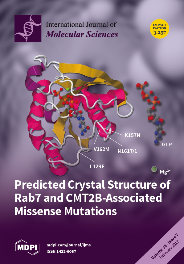
Five missense point mutations occurring near the nucleotide-binding pocket are associated with Charcot-Marie-Tooth Disease 2B peripheral sensory neuropathy. It is believed that these mutations cause an increase in Rab7 activation, leading to alteration in axonal trafficking and signaling of neurotrophic factors in peripheral sensory neurons. A detailed understanding of the molecular and cellular mechanisms underlying these mutations is crucial to developing potential therapies for CMT2B. View this paper.
  • Issues are regarded as officially published after their release is announced to the table of contents alert mailing list.
  • You may sign up for e-mail alerts to receive table of contents of newly released issues.
  • PDF is the official format for papers published in both, html and pdf forms. To view the papers in pdf format, click on the "PDF Full-text" link, and use the free Adobe Reader to open them.
Order results
Result details
Section
Select all
Export citation of selected articles as:

Editorial

Jump to: Research, Review, Other

1326 KiB  
Editorial
Pulses, Healthy, and Sustainable Food Sources for Feeding the Planet
by Marcello Iriti and Elena Maria Varoni
Int. J. Mol. Sci. 2017, 18(2), 255; https://doi.org/10.3390/ijms18020255 - 25 Jan 2017
Cited by 44 | Viewed by 5485
Abstract
Pulses, a subgroup of legumes, are plant foods from the Fabaceae (Leguminosae) family. According to FAO (Food and Agriculture Organization of the United Nations), pulses are annual leguminous crops, used for both food and feed, yielding from 1 to 12 grains or seeds [...] Read more.
Pulses, a subgroup of legumes, are plant foods from the Fabaceae (Leguminosae) family. According to FAO (Food and Agriculture Organization of the United Nations), pulses are annual leguminous crops, used for both food and feed, yielding from 1 to 12 grains or seeds of variable size, shape, and color within a pod.[...] Full article
(This article belongs to the Special Issue Pulses)
Show Figures

Figure 1

Research

Jump to: Editorial, Review, Other

12111 KiB  
Article
Gypenoside XVII Prevents Atherosclerosis by Attenuating Endothelial Apoptosis and Oxidative Stress: Insight into the ERα-Mediated PI3K/Akt Pathway
by Ke Yang, Haijing Zhang, Yun Luo, Jingyi Zhang, Min Wang, Ping Liao, Li Cao, Peng Guo, Guibo Sun and Xiaobo Sun
Int. J. Mol. Sci. 2017, 18(2), 77; https://doi.org/10.3390/ijms18020077 - 9 Feb 2017
Cited by 69 | Viewed by 7044
Abstract
Phytoestrogens are estrogen-like compounds of plant origin. The pharmacological activities of phytoestrogens are predominantly due to their antioxidant, anti-inflammatory and lipid-lowering properties, which are mediated via the estrogen receptors (ERs): estrogen receptor alpha (ERα) and estrogen receptor beta (ERβ) and possibly G protein-coupled [...] Read more.
Phytoestrogens are estrogen-like compounds of plant origin. The pharmacological activities of phytoestrogens are predominantly due to their antioxidant, anti-inflammatory and lipid-lowering properties, which are mediated via the estrogen receptors (ERs): estrogen receptor alpha (ERα) and estrogen receptor beta (ERβ) and possibly G protein-coupled estrogen receptor 1 (GPER). Gypenoside XVII (GP-17) is a phytoestrogen that is widely used to prevent cardiovascular disease, including atherosclerosis, but the mechanism underlying these therapeutic effects is largely unclear. This study aimed to assess the anti-atherogenic effects of GP-17 and its mechanisms in vivo and in vitro. In vivo experiments showed that GP-17 significantly decreased blood lipid levels, increased the expression of antioxidant enzymes and decreased atherosclerotic lesion size in ApoE−/− mice. In vitro experiments showed that GP-17 significantly prevented oxidized low-density lipoprotein (Ox-LDL)-induced endothelial injury. The underlying protective mechanisms of GP-17 were mediated by restoring the normal redox state, up-regulating of the ratio of Bcl-2 to Bax and inhibiting the expression of cleaved caspase-3 in Ox-LDL-induced human umbilical vein endothelial cell (HUVEC) injury. Notably, we found that GP-17 treatment predominantly up-regulated the expression of ERα but not ERβ. However, similar to estrogen, the protective effect of GP-17 could be blocked by the ER antagonist ICI182780 and the phosphatidylinositol 3-kinase (PI3K) antagonist LY294002. Taken together, these results suggest that, due to its antioxidant properties, GP-17 could alleviate atherosclerosis via the ERα-mediated PI3K/Akt pathway. Full article
(This article belongs to the Section Biochemistry)
Show Figures

Graphical abstract

1315 KiB  
Article
N-Glycoprofiling Analysis for Carbohydrate Composition and Site-Occupancy Determination in a Poly-Glycosylated Protein: Human Thyrotropin of Different Origins
by Maria Teresa C. P. Ribela, Renata Damiani, Felipe D. Silva, Eliana R. Lima, João E. Oliveira, Cibele N. Peroni, Peter A. Torjesen, Carlos R. Soares and Paolo Bartolini
Int. J. Mol. Sci. 2017, 18(2), 131; https://doi.org/10.3390/ijms18020131 - 3 Feb 2017
Cited by 5 | Viewed by 4882
Abstract
Human thyrotropin (hTSH) is a glycoprotein with three potential glycosylation sites: two in the α-subunit and one in the β-subunit. These sites are not always occupied and occupancy is frequently neglected in glycoprotein characterization, even though it is related to folding, trafficking, initiation [...] Read more.
Human thyrotropin (hTSH) is a glycoprotein with three potential glycosylation sites: two in the α-subunit and one in the β-subunit. These sites are not always occupied and occupancy is frequently neglected in glycoprotein characterization, even though it is related to folding, trafficking, initiation of inflammation and host defense, as well as congenital disorders of glycosylation (CDG). For the first time N-glycoprofiling analysis was applied to the site-occupancy determination of two native pituitary hTSH, in comparison with three recombinant preparations of hTSH, a widely used biopharmaceutical. A single methodology provided the: (i) average N-glycan mass; (ii) mass fraction of each monosaccharide and of sulfate; and (iii) percent carbohydrate. The results indicate that the occupancy (65%–87%) and carbohydrate mass (12%–19%) can be up to 34%–57% higher in recombinant hormones. The average glycan mass is 24% lower in pituitary hTSH and contains ~3-fold fewer moles of galactose (p < 0.005) and sialic acid (p < 0.01). One of the two native preparations, which had the smallest glycan mass together with the lowest occupancy and GalNAc, sulfate, Gal and sialic acid contents, also presented the lowest in vivo bioactivity and circulatory half-life. The methodology described, comparing a recombinant biopharmaceutical to its native equivalent, can be applied to any physiologically or clinical relevant glycoprotein. Full article
(This article belongs to the Special Issue Glycan–Receptor Interaction)
Show Figures

Figure 1

2811 KiB  
Article
Phenotypic and Functional Alterations of Hematopoietic Stem and Progenitor Cells in an In Vitro Leukemia-Induced Microenvironment
by Jean-Paul Vernot, Ximena Bonilla, Viviana Rodriguez-Pardo and Natalia-Del Pilar Vanegas
Int. J. Mol. Sci. 2017, 18(2), 199; https://doi.org/10.3390/ijms18020199 - 14 Feb 2017
Cited by 11 | Viewed by 4719
Abstract
An understanding of the cell interactions occurring in the leukemic microenvironment and their functional consequences for the different cell players has therapeutic relevance. By co-culturing mesenchymal stem cells (MSC) with the REH acute lymphocytic leukemia (ALL) cell line, we have established an in [...] Read more.
An understanding of the cell interactions occurring in the leukemic microenvironment and their functional consequences for the different cell players has therapeutic relevance. By co-culturing mesenchymal stem cells (MSC) with the REH acute lymphocytic leukemia (ALL) cell line, we have established an in vitro leukemic niche for the functional evaluation of hematopoietic stem/progenitor cells (HSPC, CD34+ cells). We showed that the normal homeostatic control exerted by the MSC over the HSPC is considerably lost in this leukemic microenvironment: HSPC increased their proliferation rate and adhesion to MSC. The adhesion molecules CD54 and CD44 were consequently upregulated in HSPC from the leukemic niche. Consequently, with this adhesive phenotype, HSPC showed less Stromal derived factor-1 (SDF-1)-directed migration. Interestingly, multipotency was severely affected with an important reduction in the absolute count and the percentage of primitive progenitor colonies. It was possible to simulate most of these HSPC alterations by incubation of MSC with a REH-conditioned medium, suggesting that REH soluble factors and their effect on MSC are important for the observed changes. Of note, these HSPC alterations were reproduced when primary leukemic cells from an ALL type B (ALL-B) patient were used to set up the leukemic niche. These results suggest that a general response is induced in the leukemic niche to the detriment of HSPC function and in favor of leukemic cell support. This in vitro leukemic niche could be a valuable tool for the understanding of the molecular events responsible for HSPC functional failure and a useful scenario for therapeutic evaluation. Full article
(This article belongs to the Section Biochemistry)
Show Figures

Graphical abstract

5070 KiB  
Article
Comprehensive Analysis of Rice Laccase Gene (OsLAC) Family and Ectopic Expression of OsLAC10 Enhances Tolerance to Copper Stress in Arabidopsis
by Qingquan Liu, Le Luo, Xiaoxiao Wang, Zhenguo Shen and Luqing Zheng
Int. J. Mol. Sci. 2017, 18(2), 209; https://doi.org/10.3390/ijms18020209 - 30 Jan 2017
Cited by 106 | Viewed by 7750
Abstract
Laccases are encoded by a multigene family and widely distributed in plant genomes where they play roles oxidizing monolignols to produce higher-order lignin involved in plant development and stress responses. We identified 30 laccase genes (OsLACs) from rice, which can be [...] Read more.
Laccases are encoded by a multigene family and widely distributed in plant genomes where they play roles oxidizing monolignols to produce higher-order lignin involved in plant development and stress responses. We identified 30 laccase genes (OsLACs) from rice, which can be divided into five subfamilies, mostly expressed during early development of the endosperm, growing roots, and stems. OsLACs can be induced by hormones, salt, drought, and heavy metals stresses. The expression level of OsLAC10 increased 1200-fold after treatment with 20 μM Cu for 12 h. The laccase activities of OsLAC10 were confirmed in an Escherichia coli expression system. Lignin accumulation increased in the roots of Arabidopsis over-expressing OsLAC10 (OsLAC10-OX) compared to wild-type controls. After growth on 1/2 Murashige and Skoog (MS) medium containing toxic levels of Cu for seven days, roots of the OsLAC10-OX lines were significantly longer than those of the wild type. Compared to control plants, the Cu concentration decreased significantly in roots of the OsLAC10-OX line under hydroponic conditions. These results provided insights into the evolutionary expansion and functional divergence of OsLAC family. In addition, OsLAC10 is likely involved in lignin biosynthesis, and reduces the uptake of Cu into roots required for Arabidopsis to develop tolerance to Cu. Full article
(This article belongs to the Special Issue Abiotic Stress and Gene Networks in Plants 2017)
Show Figures

Figure 1

1855 KiB  
Article
Identification and Characterization of Lipopolysaccharide Induced TNFα Factor from Blunt Snout Bream, Megalobrama amblycephala
by Yina Lv, Xinying Xiang, Yuhong Jiang, Leilei Tang, Yi Zhou, Huan Zhong, Jun Xiao and Jinpeng Yan
Int. J. Mol. Sci. 2017, 18(2), 233; https://doi.org/10.3390/ijms18020233 - 15 Feb 2017
Cited by 7 | Viewed by 4230
Abstract
Lipopolysaccharide induced TNFα factor (LITAF) is an important transcription factor responsible for regulation of tumor necrosis factor α. In this study, a novel litaf gene (designated as Malitaf) was identified and characterized from blunt snout bream, Megalobrama amblycephala. The full-length cDNA of [...] Read more.
Lipopolysaccharide induced TNFα factor (LITAF) is an important transcription factor responsible for regulation of tumor necrosis factor α. In this study, a novel litaf gene (designated as Malitaf) was identified and characterized from blunt snout bream, Megalobrama amblycephala. The full-length cDNA of Malitaf was of 956 bp, encoding a polypeptide of 161 amino acids with high similarity to other known LITAFs. A phylogenetic tree also showed that Malitaf significantly clustered with those of other teleost, indicating that Malitaf was a new member of fish LITAF family. The putative maLITAF protein possessed a highly conserved LITAF domain with two CXXC motifs. The mRNA transcripts of Malitaf were detected in all examined tissues of healthy M. amblycephala, including kidney, head kidney, muscle, liver, spleen, gill, and heart, and with the highest expression in immune organs: spleen and head kidney. The expression level of Malitaf in spleen was rapidly up-regulated and peaked (1.29-fold, p < 0.05) at 2 h after lipopolysaccharide (LPS) stimulation. Followed the stimulation of Malitaf, Matnfα transcriptional level was also transiently induced to a high level (51.74-fold, p < 0.001) at 4 h after LPS stimulation. Taken together, we have identified a putative fish LITAF ortholog, which was a constitutive and inducible immune response gene involved in M. amblycephala innate immunity during the course of a pathogenic infection. Full article
(This article belongs to the Special Issue Lipopolysaccharides (LPSs))
Show Figures

Figure 1

1078 KiB  
Article
RNA-Seq Analyses for Two Silkworm Strains Reveals Insight into Their Susceptibility and Resistance to Beauveria bassiana Infection
by Dongxu Xing, Qiong Yang, Liang Jiang, Qingrong Li, Yang Xiao, Mingqiang Ye and Qingyou Xia
Int. J. Mol. Sci. 2017, 18(2), 234; https://doi.org/10.3390/ijms18020234 - 10 Feb 2017
Cited by 56 | Viewed by 5655
Abstract
The silkworm Bombyx mori is an economically important species. White muscardine caused by Beauveria bassiana is the main fungal disease in sericulture, and understanding the silkworm responses to B. bassiana infection is of particular interest. Herein, we investigated the molecular mechanisms underlying these [...] Read more.
The silkworm Bombyx mori is an economically important species. White muscardine caused by Beauveria bassiana is the main fungal disease in sericulture, and understanding the silkworm responses to B. bassiana infection is of particular interest. Herein, we investigated the molecular mechanisms underlying these responses in two silkworm strains Haoyue (HY, sensitive to B. bassiana) and Kang 8 (K8, resistant to B. bassiana) using an RNA-seq approach. For each strain, three biological replicates for immersion treatment, two replicates for injection treatment and three untreated controls were collected to generate 16 libraries for sequencing. Differentially expressed genes (DEGs) between treated samples and untreated controls, and between the two silkworm strains, were identified. DEGs and the enriched Kyoto Encyclopedia of Genes and Genomes (KEGG) pathways of the two strains exhibited an obvious difference. Several genes encoding cuticle proteins, serine proteinase inhibitors (SPI) and antimicrobial peptides (AMP) and the drug metabolism pathway involved in toxin detoxification were considered to be related to the resistance of K8 to B. bassiana. These results revealed insight into the resistance and susceptibility of two silkworm strains against B. bassiana infection and provided a roadmap for silkworm molecular breeding to enhance its resistance to B. bassiana. Full article
(This article belongs to the Section Biochemistry)
Show Figures

Graphical abstract

2701 KiB  
Article
Serum Metabolomic Profiling Identifies Characterization of Non-Obstructive Azoospermic Men
by Zhe Zhang, Yingwei Zhang, Changjie Liu, Mingming Zhao, Yuzhuo Yang, Han Wu, Hongliang Zhang, Haocheng Lin, Lemin Zheng and Hui Jiang
Int. J. Mol. Sci. 2017, 18(2), 238; https://doi.org/10.3390/ijms18020238 - 25 Jan 2017
Cited by 28 | Viewed by 5434
Abstract
Male infertility is considered a common health problem, and non-obstructive azoospermia with unclear pathogenesis is one of the most challenging tasks for clinicians. The objective of this study was to investigate the differential serum metabolic pattern in non-obstructive azoospermic men and to determine [...] Read more.
Male infertility is considered a common health problem, and non-obstructive azoospermia with unclear pathogenesis is one of the most challenging tasks for clinicians. The objective of this study was to investigate the differential serum metabolic pattern in non-obstructive azoospermic men and to determine potential biomarkers related to spermatogenic dysfunction. Serum samples from patients with non-obstructive azoospermia (n = 22) and healthy controls (n = 31) were examined using high-performance liquid chromatography-tandem mass spectrometry (HPLC-MS/MS). Serum metabolomic profiling could differentiate non-obstructive azoospermic patients from healthy control subjects. A total of 24 metabolites were screened and identified as potential markers, many of which are involved in energy production, oxidative stress and cell apoptosis in spermatogenesis. Moreover, the results showed that various metabolic pathways, including d-glutamine and d-glutamate metabolism, taurine and hypotaurine metabolism, pyruvate metabolism, the citrate cycle and alanine, aspartate and glutamate metabolism, were disrupted in patients with non-obstructive azoospermia. Our results indicated that the serum metabolic disorders may contribute to the etiology of non-obstructive azoospermia. This study suggested that serum metabolomics could identify unique metabolic patterns of non-obstructive azoospermia and provide novel insights into the pathogenesis underlying male infertility. Full article
(This article belongs to the Special Issue Molecular Research on Urology)
Show Figures

Graphical abstract

1297 KiB  
Article
DFT Study of the Structure, Reactivity, Natural Bond Orbital and Hyperpolarizability of Thiazole Azo Dyes
by Osman I. Osman
Int. J. Mol. Sci. 2017, 18(2), 239; https://doi.org/10.3390/ijms18020239 - 1 Feb 2017
Cited by 29 | Viewed by 6639
Abstract
The structure, reactivity, natural bond orbital (NBO), linear and nonlinear optical (NLO) properties of three thiazole azo dyes (A, B and C) were monitored by applying B3LYP, CAM-B3LYP and ωB97XD functionals with 6-311++G** and aug-cc-pvdz basis sets. The geometrical parameters,dipolemoments,HOMO-LUMO(highest occupied molecular orbital,lowest [...] Read more.
The structure, reactivity, natural bond orbital (NBO), linear and nonlinear optical (NLO) properties of three thiazole azo dyes (A, B and C) were monitored by applying B3LYP, CAM-B3LYP and ωB97XD functionals with 6-311++G** and aug-cc-pvdz basis sets. The geometrical parameters,dipolemoments,HOMO-LUMO(highest occupied molecular orbital,lowest unoccupied molecular orbital) energy gaps, absorption wavelengths and total hyperpolarizabilities were investigated in carbon tetrachloride (CCl4) chloroform (CHCl3), dichloromethane (CH2Cl2) and dimethlysulphoxide (DMSO). The donor methoxyphenyl group deviates from planarity with the thiazole azo moiety by ca. 38◦; while the acceptor dicyanovinyl, indandione and dicyanovinylindanone groups diverge by ca. 6◦. The HOMOs for the three dyes are identical. They spread over the methoxyphenyl donor moiety, the thiazole and benzene rings as π-bonding orbitals. The LUMOs are shaped up by the nature of the acceptor moieties. The LUMOs of the A, B and C dyes extend over the indandione, malononitrile and dicyanovinylindanone acceptor moieties, respectively, as π-antibonding orbitals. The HOMO-LUMO splittings showed that Dye C is much more reactive than dyes A and B. Compared to dyes A and B, Dye C yielded a longer maximum absorption wavelength because of the stabilization of its LUMOs relative to those of the other two. The three dyes show solvatochromism accompanied by significant increases in hyperpolarizability. The enhancement of the total hyperpolarizability of C compared to those of A and B is due to the cumulative action of the long π-conjugation of the indanone ring and the stronger electron-withdrawing ability of the dicyanovinyl moiety that form the dicyanovinylindanone acceptor group. These findings are facilitated by a natural bond orbital (NBO) technique. The very high total hyperpolarizabilities of the three dyes define their potent nonlinear optical (NLO) behaviour. Full article
(This article belongs to the Collection Chemical Bond and Bonding)
Show Figures

Graphical abstract

349 KiB  
Article
Polymorphisms of Dopamine Receptor Genes and Risk of L-Dopa–Induced Dyskinesia in Parkinson’s Disease
by Cristoforo Comi, Marco Ferrari, Franca Marino, Luca Magistrelli, Roberto Cantello, Giulio Riboldazzi, Maria Laura Ester Bianchi, Giorgio Bono and Marco Cosentino
Int. J. Mol. Sci. 2017, 18(2), 242; https://doi.org/10.3390/ijms18020242 - 24 Jan 2017
Cited by 32 | Viewed by 6025
Abstract
L-dopa–induced dyskinesia (LID) is a frequent motor complication of Parkinson’s disease (PD), associated with a negative prognosis. Previous studies showed an association between dopamine receptor (DR) gene (DR) variants and LID, the results of which have not been confirmed. The present [...] Read more.
L-dopa–induced dyskinesia (LID) is a frequent motor complication of Parkinson’s disease (PD), associated with a negative prognosis. Previous studies showed an association between dopamine receptor (DR) gene (DR) variants and LID, the results of which have not been confirmed. The present study is aimed to determine whether genetic differences of DR are associated with LID in a small but well-characterized cohort of PD patients. To this end we enrolled 100 PD subjects, 50 with and 50 without LID, matched for age, gender, disease duration and dopaminergic medication in a case-control study. We conducted polymerase chain reaction for single nucleotide polymorphisms (SNP) in both D1-like (DRD1A48G; DRD1C62T and DRD5T798C) and D2-like DR (DRD2G2137A, DRD2C957T, DRD3G25A, DRD3G712C, DRD4C616G and DRD4nR VNTR 48bp) analyzed genomic DNA. Our results showed that PD patients carrying allele A at DRD3G3127A had an increased risk of LID (OR 4.9; 95% CI 1.7–13.9; p = 0.004). The present findings may provide valuable information for personalizing pharmacological therapy in PD patients. Full article
(This article belongs to the Special Issue Human Single Nucleotide Polymorphisms and Disease Diagnostics)
Show Figures

Graphical abstract

2648 KiB  
Article
Identifying the Long-Term Role of Inducible Nitric Oxide Synthase after Contusive Spinal Cord Injury Using a Transgenic Mouse Model
by Dominic M. Maggio, Amanpreet Singh, J. Bryan Iorgulescu, Drew H. Bleicher, Mousumi Ghosh, Michael M. Lopez, Luis M. Tuesta, Govinder Flora, W. Dalton Dietrich and Damien D. Pearse
Int. J. Mol. Sci. 2017, 18(2), 245; https://doi.org/10.3390/ijms18020245 - 25 Jan 2017
Cited by 8 | Viewed by 7169
Abstract
Inducible nitric oxide synthase (iNOS) is a potent mediator of oxidative stress during neuroinflammation triggered by neurotrauma or neurodegeneration. We previously demonstrated that acute iNOS inhibition attenuated iNOS levels and promoted neuroprotection and functional recovery after spinal cord injury (SCI). The present study [...] Read more.
Inducible nitric oxide synthase (iNOS) is a potent mediator of oxidative stress during neuroinflammation triggered by neurotrauma or neurodegeneration. We previously demonstrated that acute iNOS inhibition attenuated iNOS levels and promoted neuroprotection and functional recovery after spinal cord injury (SCI). The present study investigated the effects of chronic iNOS ablation after SCI using inos-null mice. iNOS−/− knockout and wild-type (WT) control mice underwent a moderate thoracic (T8) contusive SCI. Locomotor function was assessed weekly, using the Basso Mouse Scale (BMS), and at the endpoint (six weeks), by footprint analysis. At the endpoint, the volume of preserved white and gray matter, as well as the number of dorsal column axons and perilesional blood vessels rostral to the injury, were quantified. At weeks two and three after SCI, iNOS−/− mice exhibited a significant locomotor improvement compared to WT controls, although a sustained improvement was not observed during later weeks. At the endpoint, iNOS−/− mice showed significantly less preserved white and gray matter, as well as fewer dorsal column axons and perilesional blood vessels, compared to WT controls. While short-term antagonism of iNOS provides histological and functional benefits, its long-term ablation after SCI may be deleterious, blocking protective or reparative processes important for angiogenesis and tissue preservation. Full article
(This article belongs to the Special Issue Neuroprotective Strategies 2016)
Show Figures

Figure 1

3879 KiB  
Article
Phenylarsine Oxide Can Induce the Arsenite-Resistance Mutant PML Protein Solubility Changes
by Yu Han Jiang, Ye Jia Chen, Chao Wang, Yong Fei Lan, Chang Yang, Qian Qian Wang, Liaqat Hussain, Yasen Maimaitiying, Khairul Islam and Hua Naranmandura
Int. J. Mol. Sci. 2017, 18(2), 247; https://doi.org/10.3390/ijms18020247 - 25 Jan 2017
Cited by 7 | Viewed by 5047
Abstract
Arsenic trioxide (As2O3) has recently become one of the most effective drugs for treatment of patient with acute promyelocytic leukemia (APL), and its molecular mechanism has also been largely investigated. However, it has been reported that As2O [...] Read more.
Arsenic trioxide (As2O3) has recently become one of the most effective drugs for treatment of patient with acute promyelocytic leukemia (APL), and its molecular mechanism has also been largely investigated. However, it has been reported that As2O3 resistant patients are frequently found in relapsed APL after consolidation therapy, which is due to the point mutations in B-box type 2 motifs of promyelocytic leukemia (PML) gene. In the present study, we for the first time establish whether organic arsenic species phenylarsine oxide (PAO) could induce the mutant PML-IV (A216V) protein solubility changes and degradation. Here, three different PML protein variants (i.e., PML-IV, PML-V and mutant PML-A216V) were overexpressed in HEK293T cells and then exposed to PAO in time- and dose-dependent manners. Interestingly, PAO is found to have potential effect on induction of mutant PML-IV (A216V) protein solubility changes and degradation, but no appreciable effects were found following exposure to high concentrations of iAsIII, dimethylarsinous acid (DMAIII) and adriamycin (doxorubicin), even though they cause cell death. Our current data strongly indicate that PAO has good effects on the mutant PML protein solubility changes, and it may be helpful for improving the therapeutic strategies for arsenic-resistant APL treatments in the near future. Full article
Show Figures

Graphical abstract

7668 KiB  
Article
Interaction of New-Developed TiO2-Based Photocatalytic Nanoparticles with Pathogenic Microorganisms and Human Dermal and Pulmonary Fibroblasts
by Ionela Cristina Nica, Miruna Silvia Stan, Marcela Popa, Mariana Carmen Chifiriuc, Veronica Lazar, Gratiela G. Pircalabioru, Iuliana Dumitrescu, Madalina Ignat, Marcel Feder, Liviu Cristian Tanase, Ionel Mercioniu, Lucian Diamandescu and Anca Dinischiotu
Int. J. Mol. Sci. 2017, 18(2), 249; https://doi.org/10.3390/ijms18020249 - 25 Jan 2017
Cited by 26 | Viewed by 5208
Abstract
TiO2-based photocatalysts were obtained during previous years in order to limit pollution and to ease human daily living conditions due to their special properties. However, obtaining biocompatible photocatalysts is still a key problem, and the mechanism of their toxicity recently received [...] Read more.
TiO2-based photocatalysts were obtained during previous years in order to limit pollution and to ease human daily living conditions due to their special properties. However, obtaining biocompatible photocatalysts is still a key problem, and the mechanism of their toxicity recently received increased attention. Two types of TiO2 nanoparticles co-doped with 1% of iron and nitrogen (TiO2-1% Fe–N) atoms were synthesized in hydrothermal conditions at pH of 8.5 (HT1) and 5.5 (HT2), and their antimicrobial activity and cytotoxic effects exerted on human pulmonary and dermal fibroblasts were assessed. These particles exhibited significant microbicidal and anti-biofilm activity, suggesting their potential application for microbial decontamination of different environments. In addition, our results demonstrated the biocompatibility of TiO2-1% Fe–N nanoparticles at low doses on lung and dermal cells, which may initiate oxidative stress through dose accumulation. Although no significant changes were observed between the two tested photocatalysts, the biological response was cell type specific and time- and dose-dependent; the lung cells proved to be more sensitive to nanoparticle exposure. Taken together, these experimental data provide useful information for future photocatalytic applications in the industrial, food, pharmaceutical, and medical fields. Full article
Show Figures

Figure 1

5674 KiB  
Article
Influence of Transgenic Metallothionein-1 on Gliosis, CA1 Neuronal Loss, and Brain Metal Levels of the Tg2576 Mouse Model of Alzheimer’s Disease
by Gemma Comes, Yasmina Manso, Anna Escrig, Olaya Fernandez-Gayol, Paula Sanchis, Amalia Molinero, Mercedes Giralt, Javier Carrasco and Juan Hidalgo
Int. J. Mol. Sci. 2017, 18(2), 251; https://doi.org/10.3390/ijms18020251 - 26 Jan 2017
Cited by 8 | Viewed by 5334
Abstract
The mouse model of Alzheimer’s disease (AD), Tg2576 mice (APP), has provided valuable information, such as the role of the metallothionein (MT) family in their behavioral and amyloidosis phenotypes. In this study, we further characterize the role of MT-1 by crossing Mt1-overexpressing [...] Read more.
The mouse model of Alzheimer’s disease (AD), Tg2576 mice (APP), has provided valuable information, such as the role of the metallothionein (MT) family in their behavioral and amyloidosis phenotypes. In this study, we further characterize the role of MT-1 by crossing Mt1-overexpressing mice with Tg2576 mice (APPTgMT). In 14-month-old mice, MT-1(/2) protein levels were dramatically increased by Mt1 overexpression throughout the cortex (Cx), which showed a prominent caudal-rostral gradient, and the hippocampus (HC). There was a trend for MT-1(/2) immunostaining to be increased in the areas surrounding the amyloid plaques in control male mice but not in Mt1-overexpressing mice. Gliosis was elicited by the amyloid plaques, but the effects of Mt1 overexpression were modest. However, in hippocampal western blots the microglial marker Iba-1 was increased in old male APPTgMT mice compared to APP-wild type (APPWT) mice, and the opposite was observed in young mice. Hippocampal CA1 neuronal loss was observed in Tg2576 mice, but was unaffected by Mt1 overexpression. Aging increased Zn and Cu levels differently depending on brain area, sex, and genotype. Thus, the effects of Mt1 overexpression on the phenotype of Tg2576 mice here studied are modest. Full article
(This article belongs to the Special Issue Metalloproteins 2017)
Show Figures

Graphical abstract

6462 KiB  
Article
Genetic Variation in Choline-Metabolizing Enzymes Alters Choline Metabolism in Young Women Consuming Choline Intakes Meeting Current Recommendations
by Ariel B. Ganz, Vanessa V. Cohen, Camille C. Swersky, Julie Stover, Gerardo A. Vitiello, Jessica Lovesky, Jasmine C. Chuang, Kelsey Shields, Vladislav G. Fomin, Yusnier S. Lopez, Sanjay Mohan, Anita Ganti, Bradley Carrier, Olga V. Malysheva and Marie A. Caudill
Int. J. Mol. Sci. 2017, 18(2), 252; https://doi.org/10.3390/ijms18020252 - 26 Jan 2017
Cited by 44 | Viewed by 7956
Abstract
Single nucleotide polymorphisms (SNPs) in choline metabolizing genes are associated with disease risk and greater susceptibility to organ dysfunction under conditions of dietary choline restriction. However, the underlying metabolic signatures of these variants are not well characterized and it is unknown whether genotypic [...] Read more.
Single nucleotide polymorphisms (SNPs) in choline metabolizing genes are associated with disease risk and greater susceptibility to organ dysfunction under conditions of dietary choline restriction. However, the underlying metabolic signatures of these variants are not well characterized and it is unknown whether genotypic differences persist at recommended choline intakes. Thus, we sought to determine if common genetic risk factors alter choline dynamics in pregnant, lactating, and non-pregnant women consuming choline intakes meeting and exceeding current recommendations. Women (n = 75) consumed 480 or 930 mg choline/day (22% as a metabolic tracer, choline-d9) for 10–12 weeks in a controlled feeding study. Genotyping was performed for eight variant SNPs and genetic differences in metabolic flux and partitioning of plasma choline metabolites were evaluated using stable isotope methodology. CHKA rs10791957, CHDH rs9001, CHDH rs12676, PEMT rs4646343, PEMT rs7946, FMO3 rs2266782, SLC44A1 rs7873937, and SLC44A1 rs3199966 altered the use of choline as a methyl donor; CHDH rs9001 and BHMT rs3733890 altered the partitioning of dietary choline between betaine and phosphatidylcholine synthesis via the cytidine diphosphate (CDP)-choline pathway; and CHKA rs10791957, CHDH rs12676, PEMT rs4646343, PEMT rs7946 and SLC44A1 rs7873937 altered the distribution of dietary choline between the CDP-choline and phosphatidylethanolamine N-methyltransferase (PEMT) denovo pathway. Such metabolic differences may contribute to disease pathogenesis and prognosis over the long-term. Full article
(This article belongs to the Special Issue Nutrigenomics of Risk Factors for Disease)
Show Figures

Figure 1

3818 KiB  
Article
Influence of Knee Immobilization on Chondrocyte Apoptosis and Histological Features of the Anterior Cruciate Ligament Insertion and Articular Cartilage in Rabbits
by Hirotaka Mutsuzaki, Hiromi Nakajima, Yasuyoshi Wadano, Syogo Furuhata and Masataka Sakane
Int. J. Mol. Sci. 2017, 18(2), 253; https://doi.org/10.3390/ijms18020253 - 26 Jan 2017
Cited by 12 | Viewed by 5348
Abstract
This study examined the influence of immobilization on chondrocyte apoptosis and histological features of the anterior cruciate ligament (ACL) insertion and knee articular cartilage in rabbits. Forty-eight male Japanese white rabbits were assigned to an immobilization (n = 24) or sham ( [...] Read more.
This study examined the influence of immobilization on chondrocyte apoptosis and histological features of the anterior cruciate ligament (ACL) insertion and knee articular cartilage in rabbits. Forty-eight male Japanese white rabbits were assigned to an immobilization (n = 24) or sham (n = 24) group. Rabbits in the immobilization group underwent complete unilateral surgical knee immobilization and rabbits in the sham group underwent a sham surgery. The average thickness of the glycosaminoglycan (GAG) stained red area by safranin O staining, the chondrocyte apoptosis rate and the chondrocyte proliferation rate in the cartilage layer in the ACL insertion and the articular cartilage of the medial tibial condyle were measured at one, two, four and eight weeks in six animals from each group. In the ACL insertion, the chondrocyte apoptosis rate was higher in the immobilization group than in the sham group at two and eight weeks after surgery (p < 0.05). The chondrocyte proliferation rate gradually decreased from two weeks to eight weeks in the immobilization group. The GAG layer was thinner in the immobilization group than in the sham group at two, four and eight weeks after surgery (p < 0.05). In the articular cartilage, the chondrocyte apoptosis rate in the immobilization group was higher than in the sham group at four and eight weeks after surgery (p < 0.05). The GAG layer was significantly thinner in the immobilization group than that in the sham group at four and eight weeks after surgery (p < 0.05). Knee immobilization significantly increased chondrocyte apoptosis at two and eight weeks after surgery in the ACL insertion and at four and eight weeks after surgery in the articular cartilage of the medial tibial condyle, and decreased GAG layer thickness from two to eight weeks after surgery in the ACL insertion and from four to eight weeks after surgery in the articular cartilage. Full article
(This article belongs to the Special Issue Apoptotic Chondrocytes and Osteoarthritis)
Show Figures

Figure 1

4231 KiB  
Article
Dimerized Translationally Controlled Tumor Protein-Binding Peptide Ameliorates Atopic Dermatitis in NC/Nga Mice
by Xing-Hai Jin, Juhyeon Lim, Dong Hae Shin, Jeehye Maeng and Kyunglim Lee
Int. J. Mol. Sci. 2017, 18(2), 256; https://doi.org/10.3390/ijms18020256 - 26 Jan 2017
Cited by 13 | Viewed by 7265
Abstract
Our previous study showed that dimerized translationally controlled tumor protein (dTCTP) plays a role in the pathogenesis of allergic diseases, such as asthma and allergic rhinitis. A 7-mer peptide, called dTCTP-binding peptide 2 (dTBP2), binds to dTCTP and inhibits its cytokine-like effects. We [...] Read more.
Our previous study showed that dimerized translationally controlled tumor protein (dTCTP) plays a role in the pathogenesis of allergic diseases, such as asthma and allergic rhinitis. A 7-mer peptide, called dTCTP-binding peptide 2 (dTBP2), binds to dTCTP and inhibits its cytokine-like effects. We therefore examined the protective effects of dTBP2 in house dust mite-induced atopic dermatitis (AD)-like skin lesions in Nishiki-nezumi Cinnamon/Nagoya (NC/Nga) mice. We found that topical administration of dTBP2 significantly reduced the AD-like skin lesions formation and mast cell infiltration in NC/Nga mice, similarly to the response seen in the Protopic (tacrolimus)-treated group. Treatment with dTBP2 also decreased the serum levels of IgE and reduced IL-17A content in skin lesions and inhibited the expression of mRNAs of interleukin IL-4, IL-5, IL-6, IL-13, macrophage-derived chemokine (MDC), thymus and activation-regulated chemokine (TARC) and thymic stromal lymphopoietin (TSLP). These findings indicate that dTBP2 not only inhibits the release of Th2 cytokine but also suppresses the production of proinflammatory cytokines in AD-like skin lesions in NC/Nga mice, by inhibiting TCTP dimer, in allergic responses. Therefore, dTCTP is a therapeutic target for AD and dTBP2 appears to have a potential role in the treatment of AD. Full article
(This article belongs to the Section Biochemistry)
Show Figures

Graphical abstract

239 KiB  
Article
Plasma Triglyceride Levels May Be Modulated by Gene Expression of IQCJ, NXPH1, PHF17 and MYB in Humans
by Bastien Vallée Marcotte, Frédéric Guénard, Hubert Cormier, Simone Lemieux, Patrick Couture, Iwona Rudkowska and Marie-Claude Vohl
Int. J. Mol. Sci. 2017, 18(2), 257; https://doi.org/10.3390/ijms18020257 - 26 Jan 2017
Cited by 13 | Viewed by 4561
Abstract
A genome-wide association study (GWAS) by our group identified loci associated with the plasma triglyceride (TG) response to ω-3 fatty acid (FA) supplementation in IQCJ, NXPH1, PHF17 and MYB. Our aim is to investigate potential mechanisms underlying the associations between [...] Read more.
A genome-wide association study (GWAS) by our group identified loci associated with the plasma triglyceride (TG) response to ω-3 fatty acid (FA) supplementation in IQCJ, NXPH1, PHF17 and MYB. Our aim is to investigate potential mechanisms underlying the associations between single nucleotide polymorphisms (SNPs) in the four genes and TG levels following ω-3 FA supplementation. 208 subjects received 3 g/day of ω-3 FA (1.9–2.2 g of EPA and 1.1 g of docosahexaenoic acid (DHA)) for six weeks. Plasma TG were measured before and after the intervention. 67 SNPs were selected to increase the density of markers near GWAS hits. Genome-wide expression and methylation analyses were conducted on respectively 30 and 35 participants’ blood sample together with in silico analyses. Two SNPs of IQCJ showed different affinities to splice sites depending on alleles. Expression levels were influenced by genotype for one SNP in NXPH1 and one in MYB. Associations between 12 tagged SNPs of IQCJ, 26 of NXPH1, seven of PHF17 and four of MYB and gene-specific CpG site methylation levels were found. The response of plasma TG to ω-3 FA supplementation may be modulated by the effect of DNA methylation on expression levels of genes revealed by GWAS. Full article
(This article belongs to the Special Issue Gene-Diet Interactions in Chronic Diseases)
577 KiB  
Article
Unexpectedly Higher Morbidity and Mortality of Hospitalized Elderly Patients Associated with Rhinovirus Compared with Influenza Virus Respiratory Tract Infection
by Ivan F. N. Hung, Anna Jinxia Zhang, Kelvin K. W. To, Jasper F. W. Chan, Shawn H. S. Zhu, Ricky Zhang, Tuen-Ching Chan, Kwok-Hung Chan and Kwok-Yung Yuen
Int. J. Mol. Sci. 2017, 18(2), 259; https://doi.org/10.3390/ijms18020259 - 26 Jan 2017
Cited by 29 | Viewed by 7905
Abstract
Rhinovirus is a common cause of upper and lower respiratory tract infections in adults, especially among the elderly and immunocompromised. Nevertheless, its clinical characteristics and mortality risks have not been well described. A retrospective analysis on a prospective cohort was conducted in a [...] Read more.
Rhinovirus is a common cause of upper and lower respiratory tract infections in adults, especially among the elderly and immunocompromised. Nevertheless, its clinical characteristics and mortality risks have not been well described. A retrospective analysis on a prospective cohort was conducted in a single teaching hospital center over a one-year period. We compared adult patients hospitalized for pneumonia caused by rhinovirus infection with those hospitalized for influenza infection during the same period. All recruited patients were followed up for at least 3 months up to 15 months. Independent risk factors associated with mortality for rhinovirus infection were identified. Between 1 March 2014 and 28 February 2015, a total of 1946 patients were consecutively included for analysis. Of these, 728 patients were hospitalized for rhinovirus infection and 1218 patients were hospitalized for influenza infection. Significantly more rhinovirus patients were elderly home residents and had chronic lung diseases (p < 0.001), whereas more influenza patients had previous stroke (p = 0.02); otherwise, there were no differences in the Charlson comorbidity indexes between the two groups. More patients in the rhinovirus group developed pneumonia complications (p = 0.03), required oxygen therapy, and had a longer hospitalization period (p < 0.001), whereas more patients in the influenza virus group presented with fever (p < 0.001) and upper respiratory tract symptoms of cough and sore throat (p < 0.001), and developed cardiovascular complications (p < 0.001). The 30-day (p < 0.05), 90-day (p < 0.01), and 1-year (p < 0.01) mortality rate was significantly higher in the rhinovirus group than the influenza virus group. Intensive care unit admission (odds ratio (OR): 9.56; 95% confidence interval (C.I.) 2.17–42.18), elderly home residents (OR: 2.60; 95% C.I. 1.56–4.33), requirement of oxygen therapy during hospitalization (OR: 2.62; 95% C.I. 1.62–4.24), and hemoglobin level <13.3 g/dL upon admission (OR: 2.43; 95% C.I. 1.16–5.12) were independent risk factors associated with 1-year mortality in patients hospitalized for rhinovirus infection. Rhinovirus infection in the adults was associated with significantly higher mortality and longer hospitalization when compared with influenza virus infection. Institutionalized older adults were particularly at risk. More stringent infection control among health care workers in elderly homes could lower the infection rate before an effective vaccine and antiviral become available. Full article
(This article belongs to the Special Issue Pneumonia: Pathogenesis, Diagnostics, Therapeutics, and Prevention)
Show Figures

Graphical abstract

11719 KiB  
Article
Wisteria floribunda Agglutinin and Its Reactive-Glycan-Carrying Prostate-Specific Antigen as a Novel Diagnostic and Prognostic Marker of Prostate Cancer
by Kazuhisa Hagiwara, Yuki Tobisawa, Takatoshi Kaya, Tomonori Kaneko, Shingo Hatakeyama, Kazuyuki Mori, Yasuhiro Hashimoto, Takuya Koie, Yoshihiko Suda, Chikara Ohyama and Tohru Yoneyama
Int. J. Mol. Sci. 2017, 18(2), 261; https://doi.org/10.3390/ijms18020261 - 26 Jan 2017
Cited by 38 | Viewed by 6313
Abstract
Wisteria floribunda agglutinin (WFA) preferably binds to LacdiNAc glycans, and its reactivity is associated with tumor progression. The aim of this study to examine whether the serum LacdiNAc carrying prostate-specific antigen–glycosylation isomer (PSA-Gi) and WFA-reactivity of tumor tissue can be applied as a [...] Read more.
Wisteria floribunda agglutinin (WFA) preferably binds to LacdiNAc glycans, and its reactivity is associated with tumor progression. The aim of this study to examine whether the serum LacdiNAc carrying prostate-specific antigen–glycosylation isomer (PSA-Gi) and WFA-reactivity of tumor tissue can be applied as a diagnostic and prognostic marker of prostate cancer (PCa). Between 2007 and 2016, serum PSA-Gi levels before prostate biopsy (Pbx) were measured in 184 biopsy-proven benign prostatic hyperplasia patients and 244 PCa patients using an automated lectin-antibody immunoassay. WFA-reactivity on tumor was analyzed in 260 radical prostatectomy (RP) patients. Diagnostic and prognostic performance of serum PSA-Gi was evaluated using area under the receiver-operator characteristic curve (AUC). Prognostic performance of WFA-reactivity on tumor was evaluated via Cox proportional hazards regression analysis and nomogram. The AUC of serum PSA-Gi detecting PCa and predicting Pbx Grade Group (GG) 3 and GG ≥ 3 after RP was much higher than those of conventional PSA. Multivariate analysis showed that WFA-reactivity on prostate tumor was an independent risk factor of PSA recurrence. The nomogram was a strong model for predicting PSA-free survival provability with a c-index ≥0.7. Serum PSA-Gi levels and WFA-reactivity on prostate tumor may be a novel diagnostic and pre- and post-operative prognostic biomarkers of PCa, respectively. Full article
(This article belongs to the Special Issue Diagnostic, Prognostic and Predictive Biomarkers in Prostate Cancer)
Show Figures

Figure 1

4153 KiB  
Article
Deguelin Potentiates Apoptotic Activity of an EGFR Tyrosine Kinase Inhibitor (AG1478) in PIK3CA-Mutated Head and Neck Squamous Cell Carcinoma
by Yuh Baba, Toyonobu Maeda, Atsuko Suzuki, Satoshi Takada, Masato Fujii and Yasumasa Kato
Int. J. Mol. Sci. 2017, 18(2), 262; https://doi.org/10.3390/ijms18020262 - 26 Jan 2017
Cited by 14 | Viewed by 6275
Abstract
Head and neck squamous cell carcinoma (HNSCC) is known to be intrinsically resistant to inhibitors for epidermal growth factor receptor (EGFR). Until now, clinical outcomes for HNSCC using EGFR inhibitors as single agents have yielded disappointing results. Here, we aimed to study whether [...] Read more.
Head and neck squamous cell carcinoma (HNSCC) is known to be intrinsically resistant to inhibitors for epidermal growth factor receptor (EGFR). Until now, clinical outcomes for HNSCC using EGFR inhibitors as single agents have yielded disappointing results. Here, we aimed to study whether combinatorial treatment using AG1478 (EGFR tyrosine kinase inhibitor) and deguelin, which is a rotenoid isolated from the African plant Mundulea sericea, could enhance the anti-tumor effects of AG1478 in HNSCC. For Ca9-22 cells with EGFR, KRAS, and PIK3CA wild types, AG1478 alone suppressed both phosphorylated levels of ERK and AKT and induced apoptosis. On the contrary, for HSC-4 cells with EGFR and KRAS wild types, and a PIK3CA mutant, AG1478 alone did not suppress the phosphorylated level of AKT nor induce apoptosis, while it suppressed ERK phosphorylation. Forced expression of constitutively active PIK3CA (G1633A mutation) significantly reduced the apoptotic effect of AG1478 on the PIK3CA wild-type Ca9-22 cells. When HSC-4 cells with the PIK3CA G1633A mutation were treated with a combination of AG1478 and deguelin, combination effects on apoptosis induction were observed through the inhibition of the AKT pathway. These results suggest that the combination of EGFR tyrosine kinase inhibitor with deguelin is a potential therapeutic approach to treat PIK3CA-mutated HNSCC. Full article
(This article belongs to the Special Issue Alterations to Signalling Pathways in Cancer Cells)
Show Figures

Graphical abstract

2566 KiB  
Article
Mrpl10 and Tbp Are Suitable Reference Genes for Peripheral Nerve Crush Injury
by Yaxian Wang, Qianqian Shan, Yali Meng, Jiacheng Pan and Sheng Yi
Int. J. Mol. Sci. 2017, 18(2), 263; https://doi.org/10.3390/ijms18020263 - 27 Jan 2017
Cited by 16 | Viewed by 5099
Abstract
Peripheral nerve injury triggers the dysregulation of a large number of genes at multiple sites, including neurons, peripheral nerve stump, and the target organ. Housekeeping genes were frequently used as reference genes to normalize the expression values of target genes. Suitable selection of [...] Read more.
Peripheral nerve injury triggers the dysregulation of a large number of genes at multiple sites, including neurons, peripheral nerve stump, and the target organ. Housekeeping genes were frequently used as reference genes to normalize the expression values of target genes. Suitable selection of housekeeping genes that are stably expressed after nerve injury minimizes bias elicited by reference genes and thus helps to better and more sensitively reflect gene expression changes. However, many housekeeping genes have been used as reference genes without testing the expression patterns of themselves. In the current study, we calculated the expression stability of nine commonly used housekeeping genes, such as 18S (18S ribosomal RNA), Actb (β-actin), CypA (cyclophilin A), Gapdh (glyceraldehydes-3-phosphate dehydrogenase), Hprt (hypoxanthine guanine phosphoribosyl transferase), Pgk1 (phosphoglycerate kinase 1), Tbp (TATA box binding protein), Ubc (ubiquitin C), YwhaZ (tyrosine 3-monooxygenase/tryptophan 5-monooxygenase activation), and four newly identified housekeeping genes, including Ankrd27 (Ankyrin repeat domain 27), Mrpl10 (mitochondrial ribosomal protein L10), Rictor (rapamycin-insensitive companion of mTOR, Complex 2), and Ubxn 11 (UBX domain protein 11), in both distal sciatic nerve samples and dorsal root ganglion (DRG) samples after sciatic nerve injury. Our results suggested that following peripheral nerve injury, Mrpl10 and Tbp might be used as suitable reference genes for sciatic nerve stump and DRGs, respectively. Full article
(This article belongs to the Section Biochemistry)
Show Figures

Graphical abstract

2096 KiB  
Article
Lannea coromandelica (Houtt.) Merr. Induces Heme Oxygenase 1 (HO-1) Expression and Reduces Oxidative Stress via the p38/c-Jun N-Terminal Kinase–Nuclear Factor Erythroid 2-Related Factor 2 (p38/JNK–NRF2)-Mediated Antioxidant Pathway
by Md Badrul Alam, Kyoo-Ri Kwon, Seok-Hyun Lee and Sang-Han Lee
Int. J. Mol. Sci. 2017, 18(2), 266; https://doi.org/10.3390/ijms18020266 - 29 Jan 2017
Cited by 15 | Viewed by 6088
Abstract
The leaves of Lannea coromandelica (Houtt.) Merr. are used in the Garo, Pahan, and Teli tribal communities of Bangladesh as a traditional medicinal plant to treat hepatitis, diabetes, ulcers, heart disease, and dysentery. However, there have been limited phytochemical and biological studies on [...] Read more.
The leaves of Lannea coromandelica (Houtt.) Merr. are used in the Garo, Pahan, and Teli tribal communities of Bangladesh as a traditional medicinal plant to treat hepatitis, diabetes, ulcers, heart disease, and dysentery. However, there have been limited phytochemical and biological studies on the bark of L. coromandelica. This study aimed to investigate the antioxidant activities of L. coromandelica bark extract (LCBE) and the underlying mechanism using RAW 264.7 cells. The LCBE was analysed by high-pressure liquid chromatography (HPLC) to detect its key polyphenolic compounds. Various in vitro antioxidant assays were performed using RAW 264.7 cells to assess the antioxidant effects of the LCBE and to understand the underlying molecular mechanism. HPLC revealed the presence of gallic acid, (−)-epigallocatechin-3-gallate, catechin, chlorogenic acid, and caffeic acid in the LCBE. The extract showed a very potent capacity to scavenge numerous free radicals through hydrogen atom transfer and/or electron donation and also quenched cellular reactive oxygen species (ROS) generation without showing any toxicity. The LCBE was found to combat the oxidative stress by enhancing the expression, at both transcriptional and translational levels, of primary antioxidant enzymes as well as phase II detoxifying enzymes, especially heme oxygenase 1, through the upregulation of the nuclear factor erythroid 2-related factor 2 (NRF2)-mediated pathway in RAW 264.7 cells via the phosphorylation of p38 kinase and c-Jun N-terminal kinase (JNK). The LCBE exhibited strong antioxidant activities and mitigated the cellular ROS production. These results provide scientific evidence of its potential as an ideal applicant for a cost-effective, readily available, and natural phytochemical, as well as a strategy for preventing diseases associated with oxidative stress and attenuating disease progress. Full article
(This article belongs to the Section Bioactives and Nutraceuticals)
Show Figures

Figure 1

2311 KiB  
Article
Metabolomic Fingerprinting in the Comprehensive Study of Liver Changes Associated with Onion Supplementation in Hypercholesterolemic Wistar Rats
by Diana González-Peña, Danuta Dudzik, Antonia García, Begoña De Ancos, Coral Barbas and Concepción Sánchez-Moreno
Int. J. Mol. Sci. 2017, 18(2), 267; https://doi.org/10.3390/ijms18020267 - 28 Jan 2017
Cited by 33 | Viewed by 6444
Abstract
The consumption of functional ingredients has been suggested to be a complementary tool for the prevention and management of liver disease. In this light, processed onion can be considered as a source of multiple bioactive compounds with hepatoprotective properties. The liver fingerprint of [...] Read more.
The consumption of functional ingredients has been suggested to be a complementary tool for the prevention and management of liver disease. In this light, processed onion can be considered as a source of multiple bioactive compounds with hepatoprotective properties. The liver fingerprint of male Wistar rats (n = 24) fed with three experimental diets (control (C), high-cholesterol (HC), and high-cholesterol enriched with onion (HCO) diets) was obtained through a non-targeted, multiplatform metabolomics approach to produce broad metabolite coverage. LC-MS, CE-MS and GC-MS results were subjected to univariate and multivariate analyses, providing a list of significant metabolites. All data were merged in order to figure out the most relevant metabolites that were modified by the onion ingredient. Several relevant metabolic changes and related metabolic pathways were found to be impacted by both HC and HCO diet. The model highlighted several metabolites (such as hydroxybutyryl carnitine and palmitoyl carnitine) modified by the HCO diet. These findings could suggest potential impairments in the energy−lipid metabolism, perturbations in the tricarboxylic acid cycle (TCA) cycle and β-oxidation modulated by the onion supplementation in the core of hepatic dysfunction. Metabolomics shows to be a valuable tool to evaluate the effects of complementary dietetic approaches directed to hepatic damage amelioration or non-alcoholic fatty liver disease (NAFLD) prevention. Full article
(This article belongs to the Special Issue New Foodomics Approaches in Food Science)
Show Figures

Graphical abstract

1725 KiB  
Article
Uridine Triphosphate Thio Analogues Inhibit Platelet P2Y12 Receptor and Aggregation
by Dursun Gündüz, Christian Tanislav, Daniel Sedding, Mariana Parahuleva, Sentot Santoso, Christian Troidl, Christian W. Hamm and Muhammad Aslam
Int. J. Mol. Sci. 2017, 18(2), 269; https://doi.org/10.3390/ijms18020269 - 29 Jan 2017
Cited by 7 | Viewed by 5943
Abstract
Platelet P2Y12 is an important adenosine diphosphate (ADP) receptor that is involved in agonist-induced platelet aggregation and is a valuable target for the development of anti-platelet drugs. Here we characterise the effects of thio analogues of uridine triphosphate (UTP) on ADP-induced platelet [...] Read more.
Platelet P2Y12 is an important adenosine diphosphate (ADP) receptor that is involved in agonist-induced platelet aggregation and is a valuable target for the development of anti-platelet drugs. Here we characterise the effects of thio analogues of uridine triphosphate (UTP) on ADP-induced platelet aggregation. Using human platelet-rich plasma, we demonstrate that UTP inhibits P2Y12 but not P2Y1 receptors and antagonises 10 µM ADP-induced platelet aggregation in a concentration-dependent manner with an IC50 value of ~250 °µM. An eight-fold higher platelet inhibitory activity was observed with a 2-thio analogue of UTP (2S-UTP), with an IC50 of 30 µM. The 4-thio analogue (4S-UTP) with an IC50 of 7.5 µM was 33-fold more effective. A three-fold decrease in inhibitory activity, however, was observed by introducing an isobutyl group at the 4S- position. A complete loss of inhibition was observed with thio-modification of the γ phosphate of the sugar moiety, which yields an enzymatically stable analogue. The interaction of UTP analogues with P2Y12 receptor was verified by P2Y12 receptor binding and cyclic AMP (cAMP) assays. These novel data demonstrate for the first time that 2- and 4-thio analogues of UTP are potent P2Y12 receptor antagonists that may be useful for therapeutic intervention. Full article
(This article belongs to the Special Issue Mechanisms of Platelet Thrombus Formation)
Show Figures

Figure 1

2914 KiB  
Article
Expansion of T Cells with Interleukin-21 for Adoptive Immunotherapy of Murine Mammary Carcinoma
by Christine K. Zoon, Wen Wan, Laura Graham and Harry D. Bear
Int. J. Mol. Sci. 2017, 18(2), 270; https://doi.org/10.3390/ijms18020270 - 29 Jan 2017
Cited by 8 | Viewed by 4840
Abstract
We previously demonstrated that culturing antigen-sensitized draining lymph node (DLN) lymphocytes from BALB/c mice in interleukin (IL)-7/15 after activation with bryostatin/ionomycin (B/I) is superior to culture in IL-2 for expansion, differentiation to cluster of differentiation (CD)8+ cells and anti-tumor activity. We sought to [...] Read more.
We previously demonstrated that culturing antigen-sensitized draining lymph node (DLN) lymphocytes from BALB/c mice in interleukin (IL)-7/15 after activation with bryostatin/ionomycin (B/I) is superior to culture in IL-2 for expansion, differentiation to cluster of differentiation (CD)8+ cells and anti-tumor activity. We sought to determine whether the substitution or addition of IL-21 to culture had a similar effect. DLN lymphocytes were antigen-sensitized with 4T1 mammary carcinoma 10 days prior to harvest, activated with B/I, and expanded in culture for 7 days with either IL-2, IL-21, IL-2/21, IL-7/15, or IL-7/15/21. Cellular expansion, phenotype, interferon (IFN)-γ responses, and in vivo anti-tumor activity were compared. We found that T cells grown in IL7/15/21 demonstrated significantly greater lymphocyte expansion than IL-2, IL-21, IL-2/21, and IL-7/15 (38.4-fold vs. 5.5, 6.6, 9.5, and 23.9-fold, respectively). Of these expanded cells, IL-7/15/21 significantly expanded the greatest percentage of CD8+ cells (67.1% vs. 22.2%, 47.2%, 47.4%, and 55.3%, respectively), and the greatest number of T central memory cells (TCM) compared to IL-2, IL-21 and IL-2/21 (45.8% vs. 11.1%, 7.7%, and 12.1%, respectively). IL-21 and IL-2/21-expanded T cells preferentially differentiated into T naïve cells (TN) vs. those expanded in IL-2, IL-7/15 and IL-7/15/21 (27.6% and 23.2% vs. 1.7%, 4.5%, and 10.4%, respectively), and demonstrated the highest IFN-γ levels in vitro. In vivo adoptive immunotherapy (AIT) experiments demonstrated anti-tumor efficacy was equally effective using IL-2, IL-21, IL-2/21, IL-7/15 and IL-7/15/21-cultured lymphocytes vs. control or cyclophosphamide alone, even at lower doses or with greater initial size of tumor prior to treatment. Full article
(This article belongs to the Special Issue Targeting Immune Checkpoints and Immunotherapy)
Show Figures

Figure 1

2724 KiB  
Article
Dimethyl Fumarate Therapy Significantly Improves the Responsiveness of T Cells in Multiple Sclerosis Patients for Immunoregulation by Regulatory T Cells
by Janine Schlöder, Carsten Berges, Felix Luessi and Helmut Jonuleit
Int. J. Mol. Sci. 2017, 18(2), 271; https://doi.org/10.3390/ijms18020271 - 28 Jan 2017
Cited by 25 | Viewed by 8751
Abstract
Multiple sclerosis (MS) is a chronic autoimmune disease caused by an insufficient suppression of autoreactive T lymphocytes. One reason for the lack of immunological control is the reduced responsiveness of T effector cells (Teff) for the suppressive properties of regulatory T cells (Treg), [...] Read more.
Multiple sclerosis (MS) is a chronic autoimmune disease caused by an insufficient suppression of autoreactive T lymphocytes. One reason for the lack of immunological control is the reduced responsiveness of T effector cells (Teff) for the suppressive properties of regulatory T cells (Treg), a process termed Treg resistance. Here we investigated whether the disease-modifying therapy of relapsing-remitting MS (RRMS) with dimethyl fumarate (DMF) influences the sensitivity of T cells in the peripheral blood of patients towards Treg-mediated suppression. We demonstrated that DMF restores responsiveness of Teff to the suppressive function of Treg in vitro, presumably by down-regulation of interleukin-6R (IL-6R) expression on T cells. Transfer of human immune cells into immunodeficient mice resulted in a lethal graft-versus-host reaction triggered by human CD4+ Teff. This systemic inflammation can be prevented by activated Treg after transfer of immune cells from DMF-treated MS patients, but not after injection of Treg-resistant Teff from therapy-naïve MS patients. Furthermore, after DMF therapy, proliferation and expansion of T cells and the immigration into the spleen of the animals is reduced and modulated by activated Treg. In summary, our data reveals that DMF therapy significantly improves the responsiveness of Teff in MS patients to immunoregulation. Full article
(This article belongs to the Special Issue Advances in Multiple Sclerosis 2016)
Show Figures

Graphical abstract

1139 KiB  
Article
SpidermiR: An R/Bioconductor Package for Integrative Analysis with miRNA Data
by Claudia Cava, Antonio Colaprico, Gloria Bertoli, Alex Graudenzi, Tiago C. Silva, Catharina Olsen, Houtan Noushmehr, Gianluca Bontempi, Giancarlo Mauri and Isabella Castiglioni
Int. J. Mol. Sci. 2017, 18(2), 274; https://doi.org/10.3390/ijms18020274 - 27 Jan 2017
Cited by 45 | Viewed by 8077
Abstract
Gene Regulatory Networks (GRNs) control many biological systems, but how such network coordination is shaped is still unknown. GRNs can be subdivided into basic connections that describe how the network members interact e.g., co-expression, physical interaction, co-localization, genetic influence, pathways, and shared protein [...] Read more.
Gene Regulatory Networks (GRNs) control many biological systems, but how such network coordination is shaped is still unknown. GRNs can be subdivided into basic connections that describe how the network members interact e.g., co-expression, physical interaction, co-localization, genetic influence, pathways, and shared protein domains. The important regulatory mechanisms of these networks involve miRNAs. We developed an R/Bioconductor package, namely SpidermiR, which offers an easy access to both GRNs and miRNAs to the end user, and integrates this information with differentially expressed genes obtained from The Cancer Genome Atlas. Specifically, SpidermiR allows the users to: (i) query and download GRNs and miRNAs from validated and predicted repositories; (ii) integrate miRNAs with GRNs in order to obtain miRNA–gene–gene and miRNA–protein–protein interactions, and to analyze miRNA GRNs in order to identify miRNA–gene communities; and (iii) graphically visualize the results of the analyses. These analyses can be performed through a single interface and without the need for any downloads. The full data sets are then rapidly integrated and processed locally. Full article
(This article belongs to the Special Issue Transcriptome Profiling in Human Diseases)
Show Figures

Graphical abstract

6788 KiB  
Article
Ixeris dentata (Thunb. Ex Thunb.) Nakai Extract Inhibits Proliferation and Induces Apoptosis in Breast Cancer Cells through Akt/NF-κB Pathways
by Seong-Ah Shin, Hae-Nim Lee, Gang-Sik Choo, Hyeong-Jin Kim, Jeong-Hwan Che and Ji-Youn Jung
Int. J. Mol. Sci. 2017, 18(2), 275; https://doi.org/10.3390/ijms18020275 - 27 Jan 2017
Cited by 20 | Viewed by 4521
Abstract
Ixeris dentata (Thunb. Ex Thunb.) Nakai (ID) exhibits various physiological activities, and its related plant derived-products are expected to represent promising cancer therapeutic agents. However, the anticancer effects of ID extract on breast cancer cells classified as estrogen receptor (ER), progesterone receptor (PR), [...] Read more.
Ixeris dentata (Thunb. Ex Thunb.) Nakai (ID) exhibits various physiological activities, and its related plant derived-products are expected to represent promising cancer therapeutic agents. However, the anticancer effects of ID extract on breast cancer cells classified as estrogen receptor (ER), progesterone receptor (PR), and human epidermal growth factor receptor 2 (HER2) are still unknown. In this study, we investigated the anti-cancer effects and analyzed the molecular mechanism of ID extract in T47D, MCF-7 (ER-, PR-positive, HER2-negative), SK-BR-3(ER-, PR-negative, HER2-positive), and MDA-MB-231 (Triple-negative) through in vitro studies. Additionally, we examined its anti-tumor effects through in vivo studies. Our findings indicated that ID extract-induced apoptosis was mediated via various survival pathways on four breast cancer cells by identifying the factors including Bcl-2 family, phospho-Akt and phospho-nuclear factor-κB (NF-κB). Based on in vitro findings that induced apoptosis via Akt-NF-κB signaling, we investigated the effects of ID extract on mice bearing MDA-MB-231 cells. The results showed that ID extract significantly decreased MDA-MB-231 tumor volume and weight via inducing apoptosis by suppressing phospho-Akt. Overall, these results indicate that ID extract induces apoptosis through the Akt-NFκB signaling pathway in MDA-MB-231 breast cancer cells and tumors, and it may serve as a therapeutic agent for triple-negative human breast cancer.
Academic Editor: Terrence Piva Full article
(This article belongs to the Section Bioactives and Nutraceuticals)
Show Figures

Figure 1

1425 KiB  
Article
Development and Long-Term Stability of a Novel Microbial Fuel Cell BOD Sensor with MnO2 Catalyst
by Shailesh Kharkwal, Yi Chao Tan, Min Lu and How Yong Ng
Int. J. Mol. Sci. 2017, 18(2), 276; https://doi.org/10.3390/ijms18020276 - 28 Jan 2017
Cited by 38 | Viewed by 6068
Abstract
A novel microbial fuel cell (MFC)-based biosensor was designed for continuous monitoring of biochemical oxygen demand (BOD) in real wastewater. To lower the material cost, manganese dioxide (MnO2) was tested as an innovative cathode catalyst for oxygen reduction in a single [...] Read more.
A novel microbial fuel cell (MFC)-based biosensor was designed for continuous monitoring of biochemical oxygen demand (BOD) in real wastewater. To lower the material cost, manganese dioxide (MnO2) was tested as an innovative cathode catalyst for oxygen reduction in a single chamber air-cathode MFC, and two different crystalline structures obtained during synthesis of MnO2 (namely β- and γ-MnO2) were compared. The BOD sensor was studied in a comprehensive way, using both sodium acetate solution and real domestic wastewater (DWW). The optimal performance of the sensor was obtained with a β-MnO2 catalyst, with R2 values of 0.99 and 0.98 using sodium acetate solution and DWW, respectively. The BOD values predicted by the β-MnO2 biosensor for DWW were in agreement with the BOD5 values, determined according to standard methods, with slight variations in the range from 3% to 12%. Finally, the long-term stability of the BOD biosensor was evaluated over 1.5 years. To the best of our knowledge, this is the first report of an MFC BOD sensor using an MnO2 catalyst at the cathode; the feasibility of using a low-cost catalyst in an MFC for online measurement of BOD in real wastewater broadens the scope of applications for such devices. Full article
(This article belongs to the Special Issue Bioelectrochemical Systems)
Show Figures

Graphical abstract

5267 KiB  
Article
Acidosis Activates Endoplasmic Reticulum Stress Pathways through GPR4 in Human Vascular Endothelial Cells
by Lixue Dong, Elizabeth A. Krewson and Li V. Yang
Int. J. Mol. Sci. 2017, 18(2), 278; https://doi.org/10.3390/ijms18020278 - 27 Jan 2017
Cited by 68 | Viewed by 9831
Abstract
Acidosis commonly exists in the tissue microenvironment of various pathophysiological conditions such as tumors, inflammation, ischemia, metabolic disease, and respiratory disease. For instance, the tumor microenvironment is characterized by acidosis and hypoxia due to tumor heterogeneity, aerobic glycolysis (the “Warburg effect”), and the [...] Read more.
Acidosis commonly exists in the tissue microenvironment of various pathophysiological conditions such as tumors, inflammation, ischemia, metabolic disease, and respiratory disease. For instance, the tumor microenvironment is characterized by acidosis and hypoxia due to tumor heterogeneity, aerobic glycolysis (the “Warburg effect”), and the defective vasculature that cannot efficiently deliver oxygen and nutrients or remove metabolic acid byproduct. How the acidic microenvironment affects the function of blood vessels, however, is not well defined. GPR4 (G protein-coupled receptor 4) is a member of the proton-sensing G protein-coupled receptors and it has high expression in endothelial cells (ECs). We have previously reported that acidosis induces a broad inflammatory response in ECs. Acidosis also increases the expression of several endoplasmic reticulum (ER) stress response genes such as CHOP (C/EBP homologous protein) and ATF3 (activating transcription factor 3). In the current study, we have examined acidosis/GPR4- induced ER stress pathways in human umbilical vein endothelial cells (HUVEC) and other types of ECs. All three arms of the ER stress/unfolded protein response (UPR) pathways were activated by acidosis in ECs as an increased expression of phosphorylated eIF2α (eukaryotic initiation factor 2α), phosphorylated IRE1α (inositol-requiring enzyme 1α), and cleaved ATF6 upon acidic pH treatment was observed. The expression of other downstream mediators of the UPR, such as ATF4, ATF3, and spliced XBP-1 (X box-binding protein 1), was also induced by acidosis. Through genetic and pharmacological approaches to modulate the expression level or activity of GPR4 in HUVEC, we found that GPR4 plays an important role in mediating the ER stress response induced by acidosis. As ER stress/UPR can cause inflammation and cell apoptosis, acidosis/GPR4-induced ER stress pathways in ECs may regulate vascular growth and inflammatory response in the acidic microenvironment. Full article
(This article belongs to the Special Issue Tumor Microenvironment and Metabolism)
Show Figures

Figure 1

4212 KiB  
Article
Oral Administration of Surface-Deacetylated Chitin Nanofibers and Chitosan Inhibit 5-Fluorouracil-Induced Intestinal Mucositis in Mice
by Ryo Koizumi, Kazuo Azuma, Hironori Izawa, Minoru Morimoto, Kosuke Ochi, Takeshi Tsuka, Tomohiro Imagawa, Tomohiro Osaki, Norihiko Ito, Yoshiharu Okamoto, Hiroyuki Saimoto and Shinsuke Ifuku
Int. J. Mol. Sci. 2017, 18(2), 279; https://doi.org/10.3390/ijms18020279 - 27 Jan 2017
Cited by 27 | Viewed by 4857
Abstract
This study investigated the prophylactic effects of orally administered surface-deacetylated chitin nanofibers (SDACNFs) and chitosan against 5-fluorouracil (5-FU)-induced intestinal mucositis, which is a common side effect of 5-FU chemotherapy. SDACNFs and chitosan abolished histological abnormalities associated with intestinal mucositis and suppressed hypoproliferation and [...] Read more.
This study investigated the prophylactic effects of orally administered surface-deacetylated chitin nanofibers (SDACNFs) and chitosan against 5-fluorouracil (5-FU)-induced intestinal mucositis, which is a common side effect of 5-FU chemotherapy. SDACNFs and chitosan abolished histological abnormalities associated with intestinal mucositis and suppressed hypoproliferation and apoptosis of intestinal crypt cells. These results indicate that SDACNF and chitosan are useful agents for preventing mucositis induced by anti-cancer drugs. Full article
(This article belongs to the Special Issue Chitins 2016)
Show Figures

Figure 1

2676 KiB  
Article
α6 Integrin (α6high)/Transferrin Receptor (CD71)low Keratinocyte Stem Cells Are More Potent for Generating Reconstructed Skin Epidermis Than Rapid Adherent Cells
by Elodie Metral, Nicolas Bechetoille, Frédéric Demarne, Walid Rachidi and Odile Damour
Int. J. Mol. Sci. 2017, 18(2), 282; https://doi.org/10.3390/ijms18020282 - 27 Jan 2017
Cited by 17 | Viewed by 6803
Abstract
The epidermis basal layer is composed of two keratinocyte populations: Keratinocyte Stem cells (KSC) and Transitory Amplifying (TA) cells that arise from KSC division. Unfortunately, no specific marker exists to differ between KSC and TA cells. Here, we aimed at comparing two different [...] Read more.
The epidermis basal layer is composed of two keratinocyte populations: Keratinocyte Stem cells (KSC) and Transitory Amplifying (TA) cells that arise from KSC division. Unfortunately, no specific marker exists to differ between KSC and TA cells. Here, we aimed at comparing two different methods that pretended to isolate these two populations: (i) the rapid adhesion method on coated substrate and (ii) the flow cytometry method, which is based on the difference in cell surface expressions of the α6 integrin and transferrin receptor (CD71). Then, we compared different parameters that are known to discriminate KSC and TA populations. Interestingly, we showed that both methods allow enrichment in stem cells. However, cell sorting by flow cytometry (α6high/CD71low) phenotype leads to a better enrichment of KSC since the colony forming efficiency is five times increased versus total cell suspension, whereas it is only 1.4 times for the adhesion method. Moreover, α6high/CD71low cells give rise to a thicker pluristratified epithelium with lower seeding density and display a low Ki67 positive cells number, showing that they have reached the balance between proliferation and differentiation. We clearly demonstrated that cells isolated by a rapid adherent method are not the same population as KSC isolated by flow cytometry following α6high/CD71low phenotype. Full article
(This article belongs to the Section Biochemistry)
Show Figures

Graphical abstract

1906 KiB  
Article
Early Healing Events after Periodontal Surgery: Observations on Soft Tissue Healing, Microcirculation, and Wound Fluid Cytokine Levels
by Doğan Kaner, Mouaz Soudan, Han Zhao, Georg Gaßmann, Anna Schönhauser and Anton Friedmann
Int. J. Mol. Sci. 2017, 18(2), 283; https://doi.org/10.3390/ijms18020283 - 27 Jan 2017
Cited by 20 | Viewed by 7070
Abstract
Early wound healing after periodontal surgery with or without enamel matrix derivative/biphasic calcium phosphate (EMD/BCP) was characterized in terms of soft tissue closure, changes of microcirculation, and expression of pro- and anti-inflammatory cytokines in gingival crevicular fluid/wound fluid (GCF/WF). Periodontal surgery was carried [...] Read more.
Early wound healing after periodontal surgery with or without enamel matrix derivative/biphasic calcium phosphate (EMD/BCP) was characterized in terms of soft tissue closure, changes of microcirculation, and expression of pro- and anti-inflammatory cytokines in gingival crevicular fluid/wound fluid (GCF/WF). Periodontal surgery was carried out in 30 patients (18 patients: application of EMD/BCP for regeneration of bony defects; 12 patients: surgical crown lengthening (SCL)). Healthy sites were observed as untreated controls. GCF/WF samples were collected during two post-surgical weeks. Flap microcirculation was measured using laser Doppler flowmetry (LDF). Soft tissue healing was evaluated after two weeks. GCF/WF levels of interleukin 1β (IL-1β), tumour necrosis factor (TNF-α), IL-6, and IL-10 were determined using a multiplex immunoassay. Surgery caused similar reductions of flap microcirculation followed by recovery within two weeks in both EMD/BCP and SCL groups. GCF/WF and pro-inflammatory cytokine levels were immediately increased after surgery, and returned only partially to baseline levels within the two-week observation period. Levels of IL-10 were temporarily reduced in all surgical sites. Flap dehiscence caused prolonged elevated levels of GCF/WF, IL-1β, and TNF-α. These findings show that periodontal surgery triggers an immediate inflammatory reaction corresponding to the early inflammatory phase of wound healing, and these inflammation measures are temporary in case of maintained closure of the flap. However, flap dehiscence causes prolonged inflammatory exudation from the periodontal wound. If the biological pre-conditions for periodontal wound healing are considered important for the clinical outcome, care should be taken to maintain primary closure of the flap. Full article
(This article belongs to the Special Issue Wound Repair and Regeneration)
Show Figures

Figure 1

2010 KiB  
Article
Impact of Polyunsaturated Fatty Acids on miRNA Profiles of Monocytes/Macrophages and Endothelial Cells—A Pilot Study
by Claudia Roessler, Kevin Kuhlmann, Christine Hellwing, Anja Leimert and Julia Schumann
Int. J. Mol. Sci. 2017, 18(2), 284; https://doi.org/10.3390/ijms18020284 - 28 Jan 2017
Cited by 33 | Viewed by 5178
Abstract
Alteration of miRNAs and dietary polyunsaturated fatty acids (PUFAs) underlies vascular inflammation. PUFAs are known to be incorporated into the cell membrane of monocytes/macrophages or endothelial cells, the major cellular players of vascular diseases, thereby affecting cellular signal transduction. Nevertheless, there are no [...] Read more.
Alteration of miRNAs and dietary polyunsaturated fatty acids (PUFAs) underlies vascular inflammation. PUFAs are known to be incorporated into the cell membrane of monocytes/macrophages or endothelial cells, the major cellular players of vascular diseases, thereby affecting cellular signal transduction. Nevertheless, there are no investigations concerning the PUFA impact on miRNA expression by these cells. With regard to the key role miRNAs play for overall cellular functionality, this study aims to elucidate whether PUFAs affect miRNA expression profiles. To this end, the monocyte/macrophage cell line RAW264.7 and the endothelial cell line TIME were enriched with either docosahexaenoic acid (DHA; n3-PUFA) or arachidonic acid (AA; n6-PUFA) until reaching a stable incorporation into the plasma membrane and, at least in part, exposed to an inflammatory milieu. Expressed miRNAs were determined by deep sequencing, and compared to unsupplemented/unstimulated controls. Data gained clearly show that PUFAs in fact modulate miRNA expression of both cell types analyzed regardless the presence/absence of an inflammatory stimulator. Moreover, certain miRNAs already linked to vascular inflammation were found to be affected by cellular PUFA enrichment. Hence, vascular inflammation appears to be influenced by dietary fatty acids, inter alia, via PUFA-mediated modulation of the type and amount of miRNAs synthesized by cells involved in the inflammatory process. Full article
(This article belongs to the Special Issue microRNA Regulation 2017)
Show Figures

Graphical abstract

9184 KiB  
Article
Insulin Production and Resistance in Different Models of Diet-Induced Obesity and Metabolic Syndrome
by Salamah M. Alwahsh, Benjamin J. Dwyer, Shareen Forbes, David H. Van Thiel, Philip J. Starkey Lewis and Giuliano Ramadori
Int. J. Mol. Sci. 2017, 18(2), 285; https://doi.org/10.3390/ijms18020285 - 28 Jan 2017
Cited by 33 | Viewed by 7015
Abstract
The role of the liver and the endocrine pancreas in development of hyperinsulinemia in different types of obesity remains unclear. Sedentary rats (160 g) were fed a low-fat-diet (LFD, chow 13% kcal fat), high-fat-diet (HFD, 35% fat), or HFD+ 30% ethanol+ 30% fructose [...] Read more.
The role of the liver and the endocrine pancreas in development of hyperinsulinemia in different types of obesity remains unclear. Sedentary rats (160 g) were fed a low-fat-diet (LFD, chow 13% kcal fat), high-fat-diet (HFD, 35% fat), or HFD+ 30% ethanol+ 30% fructose (HF-EFr, 22% fat). Overnight-fasted rats were culled after one, four or eight weeks. Pancreatic and hepatic mRNAs were isolated for subsequent RT-PCR analysis. After eight weeks, body weights increased three-fold in the LFD group, 2.8-fold in the HFD group, and 2.4-fold in the HF-EFr (p < 0.01). HF-EFr-fed rats had the greatest liver weights and consumed less food during Weeks 4–8 (p < 0.05). Hepatic-triglyceride content increased progressively in all groups. At Week 8, HOMA-IR values, fasting serum glucose, C-peptide, and triglycerides levels were significantly increased in LFD-fed rats compared to that at earlier time points. The greatest plasma levels of glucose, triglycerides and leptin were observed in the HF-EFr at Week 8. Gene expression of pancreatic-insulin was significantly greater in the HFD and HF-EFr groups versus the LFD. Nevertheless, insulin: C-peptide ratios and HOMA-IR values were substantially higher in HF-EFr. Hepatic gene-expression of insulin-receptor-substrate-1/2 was downregulated in the HF-EFr. The expression of phospho-ERK-1/2 and inflammatory-mediators were greatest in the HF-EFr-fed rats. Chronic intake of both LFD and HFD induced obesity, MetS, and intrahepatic-fat accumulation. The hyperinsulinemia is the strongest in rats with the lowest body weights, but having the highest liver weights. This accompanies the strongest increase of pancreatic insulin production and the maximal decrease of hepatic insulin signaling, which is possibly secondary to hepatic fat deposition, inflammation and other factors. Full article
(This article belongs to the Section Molecular Pathology, Diagnostics, and Therapeutics)
Show Figures

Figure 1

6240 KiB  
Article
High-Level γ-Glutamyl-Hydrolase (GGH) Expression is Linked to Poor Prognosis in ERG Negative Prostate Cancer
by Nathaniel Melling, Masoud Rashed, Cornelia Schroeder, Claudia Hube-Magg, Martina Kluth, Dagmar Lang, Ronald Simon, Christina Möller-Koop, Stefan Steurer, Guido Sauter, Frank Jacobsen, Franziska Büscheck, Corinna Wittmer, Till Clauditz, Till Krech, Maria Christina Tsourlakis, Sarah Minner, Hartwig Huland, Markus Graefen, Lars Budäus, Imke Thederan, Georg Salomon, Thorsten Schlomm and Waldemar Wilczakadd Show full author list remove Hide full author list
Int. J. Mol. Sci. 2017, 18(2), 286; https://doi.org/10.3390/ijms18020286 - 29 Jan 2017
Cited by 25 | Viewed by 6873
Abstract
γ-glutamyl-hydrolase (GGH) is a ubiquitously-expressed enzyme that regulates intracellular folate metabolism for cell proliferation, DNA synthesis, and repair. Employing GGH immunohistochemistry on a tissue microarray with 12,427 prostate cancers, we found that GGH expression was negative to low in normal prostate epithelium, whereas [...] Read more.
γ-glutamyl-hydrolase (GGH) is a ubiquitously-expressed enzyme that regulates intracellular folate metabolism for cell proliferation, DNA synthesis, and repair. Employing GGH immunohistochemistry on a tissue microarray with 12,427 prostate cancers, we found that GGH expression was negative to low in normal prostate epithelium, whereas 88.3% of our 10,562 interpretable cancers showed GGH expression. GGH staining was considered as low intensity in 49.6% and as high intensity in 38.6% of cancers. High GGH expression was linked to the TMPRSS2:ERG-fusion positive subset of cancers (p < 0.0001), advanced pathological tumor stage, and high Gleason grade (p < 0.0001 each). Further analysis revealed that these associations were merely driven by the subset of ERG-negative cancers, High GGH expression was weakly linked to early biochemical recurrence in ERG negative cancers (p < 0.0001) and independent from established histo-pathological parameters. Moreover, GGH expression was linked to features of genetic instability, including presence of recurrent deletions at 3p, 5q, 6q, and 10q (PTEN, p ≤ 0.01 each), as well as to accelerated cell proliferation as measured by Ki67 immunohistochemistry (p < 0.0001). In conclusion, the results of our study identify GGH as an ERG subtype specific molecular marker with modest prognostic relevance, which may have clinical relevance if analyzed in combination with other molecular markers. Full article
(This article belongs to the Special Issue Advances in Molecular Oncology)
Show Figures

Figure 1

3674 KiB  
Article
Decreased Expression of Vitamin D Receptor Affects an Immune Response in Primary Biliary Cholangitis via the VDR-miRNA155-SOCS1 Pathway
by Agnieszka Kempinska-Podhorodecka, Malgorzata Milkiewicz, Urszula Wasik, Joanna Ligocka, Michał Zawadzki, Marek Krawczyk and Piotr Milkiewicz
Int. J. Mol. Sci. 2017, 18(2), 289; https://doi.org/10.3390/ijms18020289 - 29 Jan 2017
Cited by 53 | Viewed by 5949
Abstract
Primary biliary cholangitis (PBC) is an immune-mediated cholestatic disease. Vitamin D receptor (VDR)-dependent signaling constrains an inflammatory response by targeting the miRNA155-SOCS1 (suppressor of cytokine signaling 1) axis. The VDR-miRNA155-SOCS1 pathway was investigated in the context of the autoimmune response associated with PBC. [...] Read more.
Primary biliary cholangitis (PBC) is an immune-mediated cholestatic disease. Vitamin D receptor (VDR)-dependent signaling constrains an inflammatory response by targeting the miRNA155-SOCS1 (suppressor of cytokine signaling 1) axis. The VDR-miRNA155-SOCS1 pathway was investigated in the context of the autoimmune response associated with PBC. Human liver tissues from non-cirrhotic PBC (n = 22), cirrhotic PBC (n = 22), cirrhotic primary sclerosing cholangitis (PSC, n=13), controls (n = 23), and peripheral blood mononuclear cells (PBMC) obtained from PBC (n = 16) and PSC (n = 10) patients and healthy subjects (n = 11) were used for molecular analyses. VDR mRNA and protein expressions were substantially reduced in PBC livers (51% and 59%, respectively). Correspondingly, the decrease of SOCS1 protein expression in PBC livers, after normalization to a marker of lymphocytes and forkhead family transcriptional regulator box P3 (FOXP3, marker of Treg), was observed, and this phenomenon was accompanied by enhanced miRNA155 expression. In PSC livers, protein expressions of VDR and SOCS1 were comparable to the controls. However, in PBM cells, protein expressions of VDR and SOCS1 were considerably decreased in both PBC and PSC. We demonstrated that VDR/miRNA155-modulated SOCS1 expression is decreased in PBC which may lead to insufficient negative regulation of cytokine signaling. These findings suggest that the decreased VDR signaling in PBC could be of importance in the pathogenesis of PBC. Full article
(This article belongs to the Special Issue Molecular Mechanisms of Human Liver Diseases)
Show Figures

Figure 1

1163 KiB  
Article
A Mixture of Persistent Organic Pollutants and Perfluorooctanesulfonic Acid Induces Similar Behavioural Responses, but Different Gene Expression Profiles in Zebrafish Larvae
by Abdolrahman Khezri, Thomas W. K. Fraser, Rasoul Nourizadeh-Lillabadi, Jorke H. Kamstra, Vidar Berg, Karin E. Zimmer and Erik Ropstad
Int. J. Mol. Sci. 2017, 18(2), 291; https://doi.org/10.3390/ijms18020291 - 29 Jan 2017
Cited by 40 | Viewed by 5676
Abstract
Persistent organic pollutants (POPs) are widespread in the environment and some may be neurotoxic. As we are exposed to complex mixtures of POPs, we aimed to investigate how a POP mixture based on Scandinavian human blood data affects behaviour and neurodevelopment during early [...] Read more.
Persistent organic pollutants (POPs) are widespread in the environment and some may be neurotoxic. As we are exposed to complex mixtures of POPs, we aimed to investigate how a POP mixture based on Scandinavian human blood data affects behaviour and neurodevelopment during early life in zebrafish. Embryos/larvae were exposed to a series of sub-lethal doses and behaviour was examined at 96 h post fertilization (hpf). In order to determine the sensitivity window to the POP mixture, exposure models of 6 to 48 and 48 to 96 hpf were used. The expression of genes related to neurological development was also assessed. Results indicate that the POP mixture increases the swimming speed of larval zebrafish following exposure between 48 to 96 hpf. This behavioural effect was associated with the perfluorinated compounds, and more specifically with perfluorooctanesulfonic acid (PFOS). The expression of genes related to the stress response, GABAergic, dopaminergic, histaminergic, serotoninergic, cholinergic systems and neuronal maintenance, were altered. However, there was little overlap in those genes that were significantly altered by the POP mixture and PFOS. Our findings show that the POP mixture and PFOS can have a similar effect on behaviour, yet alter the expression of genes relevant to neurological development differently. Full article
(This article belongs to the Special Issue Zebrafish: A Model for Toxicological Research)
Show Figures

Figure 1

1285 KiB  
Article
Antioxidant and Cytoprotective Activities of Fucus spiralis Seaweed on a Human Cell in Vitro Model
by Susete Pinteus, Joana Silva, Celso Alves, André Horta, Olivier P. Thomas and Rui Pedrosa
Int. J. Mol. Sci. 2017, 18(2), 292; https://doi.org/10.3390/ijms18020292 - 29 Jan 2017
Cited by 28 | Viewed by 5817
Abstract
Antioxidants play an important role as Reactive Oxygen Species (ROS) chelating agents and, therefore, the screening for potent antioxidants from natural sources as potential protective agents is of great relevance. The main aim of this study was to obtain antioxidant-enriched fractions from the [...] Read more.
Antioxidants play an important role as Reactive Oxygen Species (ROS) chelating agents and, therefore, the screening for potent antioxidants from natural sources as potential protective agents is of great relevance. The main aim of this study was to obtain antioxidant-enriched fractions from the common seaweed Fucus spiralis and evaluate their activity and efficiency in protecting human cells (MCF-7 cells) on an oxidative stress condition induced by H2O2. Five fractions, F1–F5, were obtained by reversed-phase vacuum liquid chromatography. F3, F4 and F5 revealed the highest phlorotannin content, also showing the strongest antioxidant effects. The cell death induced by H2O2 was reduced by all fractions following the potency order F4 > F2 > F3 > F5 > F1. Only fraction F4 completely inhibited the H2O2 effect. To understand the possible mechanisms of action of these fractions, the cellular production of H2O2, the mitochondrial membrane potential and the caspase 9 activity were studied. Fractions F3 and F4 presented the highest reduction on H2O2 cell production. All fractions decreased both caspase-9 activity and cell membrane depolarization (except F1). Taken all together, the edible F. spiralis reveal that they provide protection against oxidative stress induced by H2O2 on the human MCF-7 cellular model, probably acting as upstream blockers of apoptosis. Full article
(This article belongs to the Special Issue Nutraceuticals in Human Health and Disease)
Show Figures

Figure 1

2310 KiB  
Article
Metabolomic Biomarkers in Urine of Cushing’s Syndrome Patients
by Alicja Kotłowska, Tomasz Puzyn, Krzysztof Sworczak, Piotr Stepnowski and Piotr Szefer
Int. J. Mol. Sci. 2017, 18(2), 294; https://doi.org/10.3390/ijms18020294 - 29 Jan 2017
Cited by 34 | Viewed by 6581
Abstract
Cushing’s syndrome (CS) is a disease which results from excessive levels of cortisol in the human body. The disorder is associated with various signs and symptoms which are also common for the general population not suffering from compound hypersecretion. Thus, more sensitive and [...] Read more.
Cushing’s syndrome (CS) is a disease which results from excessive levels of cortisol in the human body. The disorder is associated with various signs and symptoms which are also common for the general population not suffering from compound hypersecretion. Thus, more sensitive and selective methods are required for the diagnosis of CS. This follow-up study was conducted to determine which steroid metabolites could serve as potential indicators of CS and possible subclinical hypercortisolism in patients diagnosed with so called non-functioning adrenal incidentalomas (AIs). Urine samples from negative controls (n = 37), patients with CS characterized by hypercortisolism and excluding iatrogenic CS (n = 16), and patients with non-functioning AIs with possible subclinical Cushing’s syndrome (n = 25) were analyzed using gas chromatography-mass spectrometry (GC/MS) and gas chromatograph equipped with flame ionization detector (GC/FID). Statistical and multivariate methods were applied to investigate the profile differences between examined individuals. The analyses revealed hormonal differences between patients with CS and the rest of examined individuals. The concentrations of selected metabolites of cortisol, androgens, and pregnenetriol were elevated whereas the levels of tetrahydrocortisone were decreased for CS when opposed to the rest of the study population. Moreover, after analysis of potential confounding factors, it was also possible to distinguish six steroid hormones which discriminated CS patients from other study subjects. The obtained discriminant functions enabled classification of CS patients and AI group characterized by mild hypersecretion of cortisol metabolites. It can be concluded that steroid hormones selected by applying urinary profiling may serve the role of potential biomarkers of CS and can aid in its early diagnosis. Full article
(This article belongs to the Section Molecular Pathology, Diagnostics, and Therapeutics)
Show Figures

Figure 1

2634 KiB  
Article
Whole Exome Sequencing in Psoriasis Patients Contributes to Studies of Acitretin Treatment Difference
by Xingchen Zhou, Yijing He, Yehong Kuang, Jie Li, Jianglin Zhang, Mingliang Chen, Wangqing Chen, Juan Su, Shuang Zhao, Panpan Liu, Menglin Chen, Minxue Shen, Xiaoping Chen, Wu Zhu and Xiang Chen
Int. J. Mol. Sci. 2017, 18(2), 295; https://doi.org/10.3390/ijms18020295 - 29 Jan 2017
Cited by 9 | Viewed by 5139 | Correction
Abstract
Psoriasis vulgaris is an immune-mediated inflammatory skin disease. Although acitretin is a widely used synthetic retinoid for moderate to severe psoriasis, little is known about patients’ genetics in response to this drug. In this study, 179 patients were enrolled in either the discovery [...] Read more.
Psoriasis vulgaris is an immune-mediated inflammatory skin disease. Although acitretin is a widely used synthetic retinoid for moderate to severe psoriasis, little is known about patients’ genetics in response to this drug. In this study, 179 patients were enrolled in either the discovery set (13 patients) or replication set (166 patients). The discovery set was sequenced by whole exome sequencing and sequential validation was conducted in the replication set by MassArray assays. Four SNPs (single nucleotide polymorphisms) (rs1105223T>C in CRB2, rs11086065A>G in ANKLE1, rs3821414T>C in ARHGEF3, rs1802073 T>G in SFRP4) were found to be significantly associated with acitretin response in either co-dominant or dominant models via multivariable logistic regression analysis, while CRB2 rs1105223CC (OR = 4.10, 95% CI = 1.46–11.5, p = 0.007) and ANKLE1 rs11086065AG/GG (OR = 2.76, 95% CI = 1.42–5.37, p = 0.003) were associated with no response to acitretin after 8-week treatment. Meanwhile, ARHGEF3 rs3821414CT/CC (OR = 0.25, 95% CI = 0.10–0.68, p = 0.006) and SFRP4 rs1802073GG/GT (OR = 2.40, 95% CI, 1.23–4.70, p = 0.011) were associated with a higher response rate. Four new genetic variations with potential influences on the response to acitretin were found in this study which may serve as genetic markers for acitretin in psoriasis patients. Full article
(This article belongs to the Special Issue Next-Generation Sequencing for Clinical Application)
Show Figures

Figure 1

10299 KiB  
Article
Polyphenolic Extract of Euphorbia supina Attenuates Manganese-Induced Neurotoxicity by Enhancing Antioxidant Activity through Regulation of ER Stress and ER Stress-Mediated Apoptosis
by Entaz Bahar, Geum-Hwa Lee, Kashi Raj Bhattarai, Hwa-Young Lee, Min-Kyung Choi, Harun-Or Rashid, Ji-Ye Kim, Han-Jung Chae and Hyonok Yoon
Int. J. Mol. Sci. 2017, 18(2), 300; https://doi.org/10.3390/ijms18020300 - 30 Jan 2017
Cited by 40 | Viewed by 7904
Abstract
Manganese (Mn) is an important trace element present in human body, which acts as an enzyme co-factor or activator in various metabolic reactions. While essential in trace amounts, excess levels of Mn in human brain can produce neurotoxicity, including idiopathic Parkinson’s disease (PD)-like [...] Read more.
Manganese (Mn) is an important trace element present in human body, which acts as an enzyme co-factor or activator in various metabolic reactions. While essential in trace amounts, excess levels of Mn in human brain can produce neurotoxicity, including idiopathic Parkinson’s disease (PD)-like extrapyramidal manganism symptoms. This study aimed to investigate the protective role of polyphenolic extract of Euphorbia supina (PPEES) on Mn-induced neurotoxicity and the underlying mechanism in human neuroblastoma SKNMC cells and Sprague-Dawley (SD) male rat brain. PPEES possessed significant amount of total phenolic and flavonoid contents. PPEES also showed significant antioxidant activity in 1,1-diphenyl-2-picrylhydrazyl (DPPH) radical scavenging and reducing power capacity (RPC) assays. Our results showed that Mn treatment significantly reduced cell viability and increased lactate dehydrogenase (LDH) level, which was attenuated by PPEES pretreatment at 100 and 200 µg/mL. Additionally, PPEES pretreatment markedly attenuated Mn-induced antioxidant status alteration by resolving the ROS, MDA and GSH levels and SOD and CAT activities. PPEES pretreatment also significantly attenuated Mn-induced mitochondrial membrane potential (ΔΨm) and apoptosis. Meanwhile, PPEES pretreatment significantly reversed the Mn-induced alteration in the GRP78, GADD34, XBP-1, CHOP, Bcl-2, Bax and caspase-3 activities. Furthermore, administration of PPEES (100 and 200 mg/kg) to Mn exposed rats showed improvement of histopathological alteration in comparison to Mn-treated rats. Moreover, administration of PPEES to Mn exposed rats showed significant reduction of 8-OHdG and Bax immunoreactivity. The results suggest that PPEES treatment reduces Mn-induced oxidative stress and neuronal cell loss in SKNMC cells and in the rat brain. Therefore, PPEES may be considered as potential treat-ment in Mn-intoxicated patients. Full article
(This article belongs to the Special Issue Biological Activity of Natural Secondary Metabolite Products)
Show Figures

Graphical abstract

1307 KiB  
Article
Comparisons of Transcriptional Profiles of Gut Genes between Cry1Ab-Resistant and Susceptible Strains of Ostrinia nubilalis Revealed Genes Possibly Related to the Adaptation of Resistant Larvae to Transgenic Cry1Ab Corn
by Jianxiu Yao, Yu-Cheng Zhu, Nanyan Lu, Lawrent L. Buschman and Kun Yan Zhu
Int. J. Mol. Sci. 2017, 18(2), 301; https://doi.org/10.3390/ijms18020301 - 30 Jan 2017
Cited by 9 | Viewed by 3695
Abstract
A microarray developed on the basis of 2895 unique transcripts from larval gut was used to compare gut gene expression profiles between a laboratory-selected Cry1Ab-resistant (R) strain and its isoline susceptible (S) strain of the European corn borer (Ostrinia nubilalis) after [...] Read more.
A microarray developed on the basis of 2895 unique transcripts from larval gut was used to compare gut gene expression profiles between a laboratory-selected Cry1Ab-resistant (R) strain and its isoline susceptible (S) strain of the European corn borer (Ostrinia nubilalis) after the larvae were fed the leaves of transgenic corn (MON810) expressing Cry1Ab or its non-transgenic isoline for 6 h. We revealed 398 gut genes differentially expressed (i.e., either up- or down-regulated genes with expression ratio ≥2.0) in S-strain, but only 264 gut genes differentially expressed in R-strain after being fed transgenic corn leaves. Although the percentages of down-regulated genes among the total number of differentially expressed genes (50% in S-strain and 45% in R-strain) were similar between the R- and S-strains, the expression ratios of down-regulated genes were much higher in S-strain than in R-strain. We revealed that 17 and 9 significantly up- or down-regulated gut genes from S and R-strain, respectively, including serine proteases and aminopeptidases. These genes may be associated with Cry1Ab toxicity by degradation, binding, and cellular defense. Overall, our study suggests enhanced adaptation of Cry1Ab-resistant larvae on transgenic Cry1Ab corn as revealed by lower number and lower ratios of differentially expressed genes in R-strain than in S-strain of O. nubilalis. Full article
(This article belongs to the Section Biochemistry)
Show Figures

Graphical abstract

1244 KiB  
Communication
Antimicrobial and Hemolytic Studies of a Series of Polycations Bearing Quaternary Ammonium Moieties: Structural and Topological Effects
by Judith Mayr, Jürgen Bachl, Jens Schlossmann and David Díaz Díaz
Int. J. Mol. Sci. 2017, 18(2), 303; https://doi.org/10.3390/ijms18020303 - 30 Jan 2017
Cited by 19 | Viewed by 4598
Abstract
A series of polycations bearing quaternary ammonium moieties have shown antimicrobial activity against the Gram-negative bacterium Escherichia coli. Different polymer topologies governed by a disubstituted aromatic core as well as different diamine-based linkers were found to influence the antimicrobial properties. Moreover, the hemolytic [...] Read more.
A series of polycations bearing quaternary ammonium moieties have shown antimicrobial activity against the Gram-negative bacterium Escherichia coli. Different polymer topologies governed by a disubstituted aromatic core as well as different diamine-based linkers were found to influence the antimicrobial properties. Moreover, the hemolytic activity against human red blood cells was measured and demonstrated good biocompatibility and selectivity of these polycations for bacteria over mammalian cells. Full article
(This article belongs to the Special Issue Antimicrobial Polymers 2016)
Show Figures

Graphical abstract

1906 KiB  
Article
Jun Dimerization Protein 2 Activates Mc2r Transcriptional Activity: Role of Phosphorylation and SUMOylation
by Chiung-Min Wang, Raymond X. Wang, Runhua Liu and Wei-Hsiung Yang
Int. J. Mol. Sci. 2017, 18(2), 304; https://doi.org/10.3390/ijms18020304 - 31 Jan 2017
Cited by 7 | Viewed by 4467
Abstract
Jun dimerization protein 2 (JDP2), a basic leucine zipper transcription factor, is involved in numerous biological and cellular processes such as cancer development and regulation, cell-cycle regulation, skeletal muscle and osteoclast differentiation, progesterone receptor signaling, and antibacterial immunity. Though JDP2 is widely expressed [...] Read more.
Jun dimerization protein 2 (JDP2), a basic leucine zipper transcription factor, is involved in numerous biological and cellular processes such as cancer development and regulation, cell-cycle regulation, skeletal muscle and osteoclast differentiation, progesterone receptor signaling, and antibacterial immunity. Though JDP2 is widely expressed in mammalian tissues, its function in gonads and adrenals (such as regulation of steroidogenesis and adrenal development) is largely unknown. Herein, we find that JDP2 mRNA and proteins are expressed in mouse adrenal gland tissues. Moreover, overexpression of JDP2 in Y1 mouse adrenocortical cancer cells increases the level of melanocortin 2 receptor (MC2R) protein. Notably, Mc2r promoter activity is activated by JDP2 in a dose-dependent manner. Next, by mapping the Mc2r promoter, we show that cAMP response elements (between −1320 and −720-bp) are mainly required for Mc2r activation by JDP2 and demonstrate that −830-bp is the major JDP2 binding site by real-time chromatin immunoprecipitation (ChIP) analysis. Mutations of cAMP response elements on Mc2r promoter disrupts JDP2 effect. Furthermore, we demonstrate that removal of phosphorylation of JDP2 results in attenuated transcriptional activity of Mc2r. Finally, we show that JDP2 is a candidate for SUMOylation and SUMOylation affects JDP2-mediated Mc2r transcriptional activity. Taken together, JDP2 acts as a novel transcriptional activator of the mouse Mc2r gene, suggesting that JDP2 may have physiological functions as a novel player in MC2R-mediated steroidogenesis as well as cell signaling in adrenal glands. Full article
(This article belongs to the Section Biochemistry)
Show Figures

Graphical abstract

4785 KiB  
Article
Anti-Fibrotic Effect of Losartan, an Angiotensin II Receptor Blocker, Is Mediated through Inhibition of ER Stress via Up-Regulation of SIRT1, Followed by Induction of HO-1 and Thioredoxin
by Hyosang Kim, Chung Hee Baek, Raymond Bok Lee, Jai Won Chang, Won Seok Yang and Sang Koo Lee
Int. J. Mol. Sci. 2017, 18(2), 305; https://doi.org/10.3390/ijms18020305 - 31 Jan 2017
Cited by 33 | Viewed by 5976
Abstract
Endoplasmic reticulum (ER) stress is increasingly identified as modulator of fibrosis. Losartan, an angiotensin II receptor blocker, has been widely used as the first choice of treatment in chronic renal diseases. We postulated that anti-fibrotic effect of losartan is mediated through inhibition of [...] Read more.
Endoplasmic reticulum (ER) stress is increasingly identified as modulator of fibrosis. Losartan, an angiotensin II receptor blocker, has been widely used as the first choice of treatment in chronic renal diseases. We postulated that anti-fibrotic effect of losartan is mediated through inhibition of ER stress via SIRT1 (silent mating type information regulation 2 homolog 1) hemeoxygenase-1 (HO-1)/thioredoxin pathway. Renal tubular cells, tunicamycin (TM)-induced ER stress, and unilateral ureteral obstruction (UUO) mouse model were used. Expression of ER stress was assessed by Western blot analysis and immunohistochemical stain. ER stress was induced by chemical ER stress inducer, tunicamycin, and non-chemical inducers such as TGF-β, angiotensin II, high glucose, and albumin. Losartan suppressed the TM-induced ER stress, as shown by inhibition of TM-induced expression of GRP78 (glucose related protein 78) and p-eIF2α (phosphospecific-eukaryotic translation initiation factor-2α), through up-regulation of SIRT1 via HO-1 and thioredoxin. Losartan also suppressed the ER stress by non-chemical inducers. In both animal models, losartan reduced the tubular expression of GRP78, which were abolished by pretreatment with sirtinol (SIRT1 inhibitor). Sirtinol also blocked the inhibitory effect of losartan on the UUO-induced renal fibrosis. These findings provide new insights into renoprotective effects of losartan and suggest that SIRT1, HO-1, and thioredoxin may be potential pharmacological targets in kidney diseases under excessive ER stress condition. Full article
(This article belongs to the Section Biochemistry)
Show Figures

Graphical abstract

8676 KiB  
Article
Cisplatin, Oxaliplatin, and Kiteplatin Subcellular Effects Compared in a Plant Model
by Paride Papadia, Fabrizio Barozzi, James D. Hoeschele, Gabriella Piro, Nicola Margiotta and Gian-Pietro Di Sansebastiano
Int. J. Mol. Sci. 2017, 18(2), 306; https://doi.org/10.3390/ijms18020306 - 31 Jan 2017
Cited by 5 | Viewed by 4796
Abstract
The immediate visual comparison of platinum chemotherapeutics’ effects in eukaryotic cells using accessible plant models of transgenic Arabidopsis thaliana is reported. The leading anticancer drug cisplatin, a third generation drug used for colon cancer, oxaliplatin and kiteplatin, promising Pt-based anticancer drugs effective against [...] Read more.
The immediate visual comparison of platinum chemotherapeutics’ effects in eukaryotic cells using accessible plant models of transgenic Arabidopsis thaliana is reported. The leading anticancer drug cisplatin, a third generation drug used for colon cancer, oxaliplatin and kiteplatin, promising Pt-based anticancer drugs effective against resistant lines, were administered to transgenic A. thaliana plants monitoring their effects on cells from different tissues. The transgenic plants’ cell cytoskeletons were labelled by the green fluorescent protein (GFP)-tagged microtubule-protein TUA6 (TUA6-GFP), while the vacuolar organization was evidenced by two soluble chimerical GFPs (GFPChi and AleuGFP) and one transmembrane GFP-tagged tonoplast intrinsic protein 1-1 (TIP1.1-GFP). The three drugs showed easily recognizable effects on plant subcellular organization, thereby providing evidence for a differentiated drug targeting. Genetically modified A. thaliana are confirmed as a possible rapid and low-cost screening tool for better understanding the mechanism of action of human anticancer drugs. Full article
(This article belongs to the Section Molecular Plant Sciences)
Show Figures

Figure 1

2210 KiB  
Article
Evaluation of EphA2 and EphB4 as Targets for Image-Guided Colorectal Cancer Surgery
by Marieke A. Stammes, Hendrica A. J. M. Prevoo, Meyke C. Ter Horst, Stéphanie A. Groot, Cornelis J. H. Van de Velde, Alan B. Chan, Lioe-Fee De Geus-Oei, Peter J. K. Kuppen, Alexander L. Vahrmeijer, Elena B. Pasquale and Cornelis F. M. Sier
Int. J. Mol. Sci. 2017, 18(2), 307; https://doi.org/10.3390/ijms18020307 - 3 Feb 2017
Cited by 12 | Viewed by 5637
Abstract
Targeted image-guided oncologic surgery (IGOS) relies on the recognition of cell surface-associated proteins, which should be abundantly present on tumor cells but preferably absent on cells in surrounding healthy tissue. The transmembrane receptor tyrosine kinase EphA2, a member of the A class of [...] Read more.
Targeted image-guided oncologic surgery (IGOS) relies on the recognition of cell surface-associated proteins, which should be abundantly present on tumor cells but preferably absent on cells in surrounding healthy tissue. The transmembrane receptor tyrosine kinase EphA2, a member of the A class of the Eph receptor family, has been reported to be highly overexpressed in several tumor types including breast, lung, brain, prostate, and colon cancer and is considered amongst the most promising cell membrane-associated tumor antigens by the NIH. Another member of the Eph receptor family belonging to the B class, EphB4, has also been found to be upregulated in multiple cancer types. In this study, EphA2 and EphB4 are evaluated as targets for IGOS of colorectal cancer by immunohistochemistry (IHC) using a tissue microarray (TMA) consisting of 168 pairs of tumor and normal tissue. The IHC sections were scored for staining intensity and percentage of cells stained. The results show a significantly enhanced staining intensity and more widespread distribution in tumor tissue compared with adjacent normal tissue for EphA2 as well as EphB4. Based on its more consistently higher score in colorectal tumor tissue compared to normal tissue, EphB4 appears to be a promising candidate for IGOS of colorectal cancer. In vitro experiments using antibodies on human colon cancer cells confirmed the possibility of EphB4 as target for imaging. Full article
Show Figures

Graphical abstract

3507 KiB  
Article
Arabidopsis RabF1 (ARA6) Is Involved in Salt Stress and Dark-Induced Senescence (DIS)
by Congfei Yin, Sazzad Karim, Hongsheng Zhang and Henrik Aronsson
Int. J. Mol. Sci. 2017, 18(2), 309; https://doi.org/10.3390/ijms18020309 - 1 Feb 2017
Cited by 16 | Viewed by 6898
Abstract
Arabidopsis small GTPase RabF1 (ARA6) functions in endosomal vesicle transport and may play a crucial role in recycling and degradation of molecules, thus involved in stress responses. Here we have reported that complementary overexpression lines RabF1OE (overexpression), GTPase mutants RabF1Q93L (constitutively active) and [...] Read more.
Arabidopsis small GTPase RabF1 (ARA6) functions in endosomal vesicle transport and may play a crucial role in recycling and degradation of molecules, thus involved in stress responses. Here we have reported that complementary overexpression lines RabF1OE (overexpression), GTPase mutants RabF1Q93L (constitutively active) and RabF1S47N (dominant negative) lines show longer root growth than wild-type, rabF1 knockout and N-myristoylation deletion (Δ1−29, N-terminus) complementary overexpression mutant plants under salt induced stress, which indicates that N-myristoylation of RabF1 is indispensable for salt tolerance. Moreover, RabF1 is highly expressed during senescence and RabF1OE lines were more tolerant of dark-induced senescence (DIS) than wild-type and rabF1. Full article
(This article belongs to the Special Issue Unconventional Proteins and Membranes Traffic)
Show Figures

Figure 1

1190 KiB  
Article
Enhancing the Biological Relevance of Machine Learning Classifiers for Reverse Vaccinology
by Ashley I. Heinson, Yawwani Gunawardana, Bastiaan Moesker, Carmen C. Denman Hume, Elena Vataga, Yper Hall, Elena Stylianou, Helen McShane, Ann Williams, Mahesan Niranjan and Christopher H. Woelk
Int. J. Mol. Sci. 2017, 18(2), 312; https://doi.org/10.3390/ijms18020312 - 1 Feb 2017
Cited by 36 | Viewed by 6558
Abstract
Reverse vaccinology (RV) is a bioinformatics approach that can predict antigens with protective potential from the protein coding genomes of bacterial pathogens for subunit vaccine design. RV has become firmly established following the development of the BEXSERO® vaccine against Neisseria meningitidis serogroup B. [...] Read more.
Reverse vaccinology (RV) is a bioinformatics approach that can predict antigens with protective potential from the protein coding genomes of bacterial pathogens for subunit vaccine design. RV has become firmly established following the development of the BEXSERO® vaccine against Neisseria meningitidis serogroup B. RV studies have begun to incorporate machine learning (ML) techniques to distinguish bacterial protective antigens (BPAs) from non-BPAs. This research contributes significantly to the RV field by using permutation analysis to demonstrate that a signal for protective antigens can be curated from published data. Furthermore, the effects of the following on an ML approach to RV were also assessed: nested cross-validation, balancing selection of non-BPAs for subcellular localization, increasing the training data, and incorporating greater numbers of protein annotation tools for feature generation. These enhancements yielded a support vector machine (SVM) classifier that could discriminate BPAs (n = 200) from non-BPAs (n = 200) with an area under the curve (AUC) of 0.787. In addition, hierarchical clustering of BPAs revealed that intracellular BPAs clustered separately from extracellular BPAs. However, no immediate benefit was derived when training SVM classifiers on data sets exclusively containing intra- or extracellular BPAs. In conclusion, this work demonstrates that ML classifiers have great utility in RV approaches and will lead to new subunit vaccines in the future. Full article
(This article belongs to the Special Issue Reverse Vaccinology)
Show Figures

Figure 1

371 KiB  
Article
Embryological Results of Couples Undergoing ICSI-ET Treatments with Males Carrying the Single Nucleotide Polymorphism rs175080 of the MLH3 Gene
by George Anifandis, Ourania Markandona, Konstantinos Dafopoulos, Christina Messini, Aspasia Tsezou, Marina Dimitraki, Panagiotis Georgoulias, Alexandros Daponte and Ioannis Messinis
Int. J. Mol. Sci. 2017, 18(2), 314; https://doi.org/10.3390/ijms18020314 - 2 Feb 2017
Cited by 7 | Viewed by 4269
Abstract
Human MLH3 (hMLH3) gene has been suggested to play a role in the DNA mismatch repair mechanism, while it may also be associated with abnormal spermatogenesis and subsequently male infertility. The aim of the present study was to investigate possible relationships between the [...] Read more.
Human MLH3 (hMLH3) gene has been suggested to play a role in the DNA mismatch repair mechanism, while it may also be associated with abnormal spermatogenesis and subsequently male infertility. The aim of the present study was to investigate possible relationships between the single nucleotide polymorphism (SNP) rs175080 in the MLH3 gene of males and the embryological results in couples undergoing intracytoplasmatic sperm injection-embryo transfer (ICSI-ET) treatments. A total of 132 men volunteered for the study and gave written informed consent. All couples were subjected to ICSI-ET treatments in the years 2010 to 2012. The couples were divided into three groups according to the genotype of their husbands: the wild type GG (n = 28), the heterozygotic type GA (n = 72) and the mutant type AA (n = 32). Significantly lower sperm concentration and progressive motility were observed in the AA group as compared to the other two groups (Concentration: 14.57 ± 4.9 mil/mL in AA, 38.3 ± 5.4 mil/mL in GA and 41.03 ± 6.8 mil/mL in GG, p < 0.05, mean ± standard error of the mean—SEM). However, significantly better embryological results (mean score of embryo quality–MSEQ) were found in the AA (8.12 ± 0.5) and the GA group (7.36 ± 0.4) as compared to the GG group (5.82 ± 0.7), (p < 0.05). Clinical pregnancy rate was significantly higher in the AA genotype group (43.8%) and the GA group (30.6%) than in the GG group (14.3%), (p < 0.05). Live birth rate was not different. It is suggested for the first time that the deteriorating effect of the mutant type on sperm characteristics does not impact on embryo development after fertilization in vitro. Full article
(This article belongs to the Special Issue Human Single Nucleotide Polymorphisms and Disease Diagnostics)
Show Figures

Figure 1

2765 KiB  
Article
Valproate Attenuates Endoplasmic Reticulum Stress-Induced Apoptosis in SH-SY5Y Cells via the AKT/GSK3β Signaling Pathway
by Zhengmao Li, Fenzan Wu, Xie Zhang, Yi Chai, Daqing Chen, Yuetao Yang, Kebin Xu, Jiayu Yin, Rui Li, Hongxue Shi, Zhouguang Wang, Xiaokun Li, Jian Xiao and Hongyu Zhang
Int. J. Mol. Sci. 2017, 18(2), 315; https://doi.org/10.3390/ijms18020315 - 8 Feb 2017
Cited by 44 | Viewed by 7914
Abstract
Endoplasmic reticulum (ER) stress-induced apoptosis plays an important role in a range of neurological disorders, such as neurodegenerative diseases, spinal cord injury, and diabetic neuropathy. Valproate (VPA), a typical antiepileptic drug, is commonly used in the treatment of bipolar disorder and epilepsy. Recently, [...] Read more.
Endoplasmic reticulum (ER) stress-induced apoptosis plays an important role in a range of neurological disorders, such as neurodegenerative diseases, spinal cord injury, and diabetic neuropathy. Valproate (VPA), a typical antiepileptic drug, is commonly used in the treatment of bipolar disorder and epilepsy. Recently, VPA has been reported to exert neurotrophic effects and promote neurite outgrowth, but its molecular mechanism is still unclear. In the present study, we investigated whether VPA inhibited ER stress and promoted neuroprotection and neuronal restoration in SH-SY5Y cells and in primary rat cortical neurons, respectively, upon exposure to thapsigargin (TG). In SH-SY5Y cells, cell viability was detected by the 3-(4,5-dimethyl-2-thiazolyl)-2,5-diphenyl-2-H-tetrazolium bromide (MTT) assay, and the expression of ER stress-related apoptotic proteins such as glucose‑regulated protein (GRP78), C/EBP homologous protein (CHOP), and cleaved caspase-12/-3 were analyzed with Western blot analyses and immunofluorescence assays. To explore the pathway involved in VPA-induced cell proliferation, we also examined p-AKT, GSK3β, p-JNK and MMP-9. Moreover, to detect the effect of VPA in primary cortical neurons, immunofluorescence staining of β-III tubulin and Anti-NeuN was analyzed in primary cultured neurons exposed to TG. Our results demonstrated that VPA administration improved cell viability in cells exposed to TG. In addition, VPA increased the levels of GRP78 and p-AKT and decreased the levels of ATF6, XBP-1, GSK3β, p-JNK and MMP-9. Furthermore, the levels of the ER stress-induced apoptosis response proteins CHOP, cleaved caspase-12 and cleaved caspase-3 were inhibited by VPA treatment. Meanwhile, VPA administration also increased the ratio of Bcl-2/Bax. Moreover, VPA can maintain neurite outgrowth of primary cortical neurons. Collectively, the neurotrophic effect of VPA is related to the inhibition of ER stress-induced apoptosis in SH-SY5Y cells and the maintenance of neuronal growth. Collectively, our results suggested a new approach for the therapeutic function of VPA in neurological disorders and neuroprotection. Full article
(This article belongs to the Collection Programmed Cell Death and Apoptosis)
Show Figures

Graphical abstract

9096 KiB  
Communication
Erythroid Differentiation Regulator 1 as a Novel Biomarker for Hair Loss Disorders
by Yu Ri Woo, Sewon Hwang, Seo Won Jeong, Dae Ho Cho and Hyun Jeong Park
Int. J. Mol. Sci. 2017, 18(2), 316; https://doi.org/10.3390/ijms18020316 - 3 Feb 2017
Cited by 3 | Viewed by 6177
Abstract
Erythroid differentiation regulator 1 (Erdr1) is known to be involved in the inflammatory process via regulating the immune system in many cutaneous disorders, such as psoriasis and rosacea. However, the role of Erdr1 in various hair loss disorders remains unclear. The aim of [...] Read more.
Erythroid differentiation regulator 1 (Erdr1) is known to be involved in the inflammatory process via regulating the immune system in many cutaneous disorders, such as psoriasis and rosacea. However, the role of Erdr1 in various hair loss disorders remains unclear. The aim of this study was to investigate the putative role of Erdr1 in alopecias. Skin samples from 21 patients with hair loss disorders and five control subjects were retrieved, in order to assess their expression levels of Erdr1. Results revealed that expression of Erdr1 was significantly downregulated in the epidermis and hair follicles of patients with hair loss disorders, when compared to that in the control group. In particular, the expression of Erdr1 was significantly decreased in patients with alopecia areata. We propose that Erdr1 downregulation might be involved in the pathogenesis of hair loss, and could be considered as a novel biomarker for hair loss disorders. Full article
(This article belongs to the Special Issue Inflammatory Skin Conditions)
Show Figures

Figure 1

4838 KiB  
Article
Extending Immunological Profiling in the Gilthead Sea Bream, Sparus aurata, by Enriched cDNA Library Analysis, Microarray Design and Initial Studies upon the Inflammatory Response to PAMPs
by Sebastian Boltaña, Barbara Castellana, Giles Goetz, Lluis Tort, Mariana Teles, Victor Mulero, Beatriz Novoa, Antonio Figueras, Frederick W. Goetz, Cristian Gallardo-Escarate, Josep V. Planas and Simon Mackenzie
Int. J. Mol. Sci. 2017, 18(2), 317; https://doi.org/10.3390/ijms18020317 - 3 Feb 2017
Cited by 4 | Viewed by 5079
Abstract
This study describes the development and validation of an enriched oligonucleotide-microarray platform for Sparus aurata (SAQ) to provide a platform for transcriptomic studies in this species. A transcriptome database was constructed by assembly of gilthead sea bream sequences derived from public repositories of [...] Read more.
This study describes the development and validation of an enriched oligonucleotide-microarray platform for Sparus aurata (SAQ) to provide a platform for transcriptomic studies in this species. A transcriptome database was constructed by assembly of gilthead sea bream sequences derived from public repositories of mRNA together with reads from a large collection of expressed sequence tags (EST) from two extensive targeted cDNA libraries characterizing mRNA transcripts regulated by both bacterial and viral challenge. The developed microarray was further validated by analysing monocyte/macrophage activation profiles after challenge with two Gram-negative bacterial pathogen-associated molecular patterns (PAMPs; lipopolysaccharide (LPS) and peptidoglycan (PGN)). Of the approximately 10,000 EST sequenced, we obtained a total of 6837 EST longer than 100 nt, with 3778 and 3059 EST obtained from the bacterial-primed and from the viral-primed cDNA libraries, respectively. Functional classification of contigs from the bacterial- and viral-primed cDNA libraries by Gene Ontology (GO) showed that the top five represented categories were equally represented in the two libraries: metabolism (approximately 24% of the total number of contigs), carrier proteins/membrane transport (approximately 15%), effectors/modulators and cell communication (approximately 11%), nucleoside, nucleotide and nucleic acid metabolism (approximately 7.5%) and intracellular transducers/signal transduction (approximately 5%). Transcriptome analyses using this enriched oligonucleotide platform identified differential shifts in the response to PGN and LPS in macrophage-like cells, highlighting responsive gene-cassettes tightly related to PAMP host recognition. As observed in other fish species, PGN is a powerful activator of the inflammatory response in S. aurata macrophage-like cells. We have developed and validated an oligonucleotide microarray (SAQ) that provides a platform enriched for the study of gene expression in S. aurata with an emphasis upon immunity and the immune response. Full article
(This article belongs to the Section Biochemistry)
Show Figures

Graphical abstract

15786 KiB  
Article
Bifidobacterium adolescentis Exerts Strain-Specific Effects on Constipation Induced by Loperamide in BALB/c Mice
by Linlin Wang, Lujun Hu, Qi Xu, Boxing Yin, Dongsheng Fang, Gang Wang, Jianxin Zhao, Hao Zhang and Wei Chen
Int. J. Mol. Sci. 2017, 18(2), 318; https://doi.org/10.3390/ijms18020318 - 20 Feb 2017
Cited by 106 | Viewed by 10048
Abstract
Constipation is one of the most common gastrointestinal complaints worldwide. This study was performed to determine whether Bifidobacterium adolescentis exerts inter-strain differences in alleviating constipation induced by loperamide in BALB/c mice and to analyze the main reasons for these differences. BALB/c mice underwent [...] Read more.
Constipation is one of the most common gastrointestinal complaints worldwide. This study was performed to determine whether Bifidobacterium adolescentis exerts inter-strain differences in alleviating constipation induced by loperamide in BALB/c mice and to analyze the main reasons for these differences. BALB/c mice underwent gavage with B. adolescentis (CCFM 626, 667, and 669) once per day for 17 days. The primary outcome measures included related constipation indicators, and the secondary outcome measures were the basic biological characteristics of the strains, the concentration changes of short-chain fatty acids in feces, and the changes in the fecal flora. B. adolescentis CCFM 669 and 667 relieved constipation symptoms by adhering to intestinal epithelial cells, growing quickly in vitro and increasing the concentrations of propionic and butyric acids. The effect of B. adolescentis on the gut microbiota in mice with constipation was investigated via 16S rRNA metagenomic analysis. The results revealed that the relative abundance of Lactobacillus increased and the amount of Clostridium decreased in the B. adolescentis CCFM 669 and 667 treatment groups. In conclusion, B. adolescentis exhibits strain-specific effects in the alleviation of constipation, mostly due to the strains’ growth rates, adhesive capacity and effects on the gut microbiome and microenvironment. Full article
(This article belongs to the Section Biochemistry)
Show Figures

Graphical abstract

1955 KiB  
Article
LC-MS/MS Analysis Unravels Deep Oxidation of Manganese Superoxide Dismutase in Kidney Cancer
by Zuohui Zhao, Kazem M. Azadzoi, Han-Pil Choi, Ruirui Jing, Xin Lu, Cuiling Li, Fengqin Wang, Jiaju Lu and Jing-Hua Yang
Int. J. Mol. Sci. 2017, 18(2), 319; https://doi.org/10.3390/ijms18020319 - 4 Feb 2017
Cited by 15 | Viewed by 5908
Abstract
Manganese superoxide dismutase (MNSOD) is one of the major scavengers of reactive oxygen species (ROS) in mitochondria with pivotal regulatory role in ischemic disorders, inflammation and cancer. Here we report oxidative modification of MNSOD in human renal cell carcinoma (RCC) by the shotgun [...] Read more.
Manganese superoxide dismutase (MNSOD) is one of the major scavengers of reactive oxygen species (ROS) in mitochondria with pivotal regulatory role in ischemic disorders, inflammation and cancer. Here we report oxidative modification of MNSOD in human renal cell carcinoma (RCC) by the shotgun method using data-dependent liquid chromatography tandem mass spectrometry (LC-MS/MS). While 5816 and 5571 proteins were identified in cancer and adjacent tissues, respectively, 208 proteins were found to be up- or down-regulated (p < 0.05). Ontological category, interaction network and Western blotting suggested a close correlation between RCC-mediated proteins and oxidoreductases such as MNSOD. Markedly, oxidative modifications of MNSOD were identified at histidine (H54 and H55), tyrosine (Y58), tryptophan (W147, W149, W205 and W210) and asparagine (N206 and N209) residues additional to methionine. These oxidative insults were located at three hotspots near the hydrophobic pocket of the manganese binding site, of which the oxidation of Y58, W147 and W149 was up-regulated around three folds and the oxidation of H54 and H55 was detected in the cancer tissues only (p < 0.05). When normalized to MNSOD expression levels, relative MNSOD enzymatic activity was decreased in cancer tissues, suggesting impairment of MNSOD enzymatic activity in kidney cancer due to modifications. Thus, LC-MS/MS analysis revealed multiple oxidative modifications of MNSOD at different amino acid residues that might mediate the regulation of the superoxide radicals, mitochondrial ROS scavenging and MNSOD activity in kidney cancer. Full article
(This article belongs to the Special Issue Advances in Proteomic Research)
Show Figures

Figure 1

2012 KiB  
Article
Plumbagin Suppresses α-MSH-Induced Melanogenesis in B16F10 Mouse Melanoma Cells by Inhibiting Tyrosinase Activity
by Taek-In Oh, Jeong-Mi Yun, Eun-Ji Park, Young-Seon Kim, Yoon-Mi Lee and Ji-Hong Lim
Int. J. Mol. Sci. 2017, 18(2), 320; https://doi.org/10.3390/ijms18020320 - 3 Feb 2017
Cited by 38 | Viewed by 12180
Abstract
Recent studies have shown that plumbagin has anti-inflammatory, anti-allergic, antibacterial, and anti-cancer activities; however, it has not yet been shown whether plumbagin suppresses alpha-melanocyte stimulating hormone (α-MSH)-induced melanin synthesis to prevent hyperpigmentation. In this study, we demonstrated that plumbagin significantly suppresses α-MSH-stimulated melanin [...] Read more.
Recent studies have shown that plumbagin has anti-inflammatory, anti-allergic, antibacterial, and anti-cancer activities; however, it has not yet been shown whether plumbagin suppresses alpha-melanocyte stimulating hormone (α-MSH)-induced melanin synthesis to prevent hyperpigmentation. In this study, we demonstrated that plumbagin significantly suppresses α-MSH-stimulated melanin synthesis in B16F10 mouse melanoma cells. To understand the inhibitory mechanism of plumbagin on melanin synthesis, we performed cellular or cell-free tyrosinase activity assays and analyzed melanogenesis-related gene expression. We demonstrated that plumbagin directly suppresses tyrosinase activity independent of the transcriptional machinery associated with melanogenesis, which includes micropthalmia-associated transcription factor (MITF), tyrosinase (TYR), and tyrosinase-related protein 1 (TYRP1). We also investigated whether plumbagin was toxic to normal human keratinocytes (HaCaT) and lens epithelial cells (B3) that may be injured by using skin-care cosmetics. Surprisingly, lower plumbagin concentrations (0.5–1 μM) effectively inhibited melanin synthesis and tyrosinase activity but do not cause toxicity in keratinocytes, lens epithelial cells, and B16F10 mouse melanoma cells, suggesting that plumbagin is safe for dermal application. Taken together, these results suggest that the inhibitory effect of plumbagin to pigmentation may make it an acceptable and safe component for use in skin-care cosmetic formulations used for skin whitening. Full article
(This article belongs to the Special Issue Nutrients and Phytochemicals for Skin Health)
Show Figures

Graphical abstract

2732 KiB  
Article
Concurrent Autophagy Inhibition Overcomes the Resistance of Epidermal Growth Factor Receptor Tyrosine Kinase Inhibitors in Human Bladder Cancer Cells
by Minyong Kang, Kyoung-Hwa Lee, Hye Sun Lee, Chang Wook Jeong, Cheol Kwak, Hyeon Hoe Kim and Ja Hyeon Ku
Int. J. Mol. Sci. 2017, 18(2), 321; https://doi.org/10.3390/ijms18020321 - 4 Feb 2017
Cited by 28 | Viewed by 6353
Abstract
Despite the potential therapeutic efficacy of epithelial growth factor receptor (EGFR) inhibitors in the treatment of advanced stage bladder cancer, there currently is no clear evidence to support this hypothesis. In this study, we investigate whether the concurrent treatment of autophagy-blocking agents with [...] Read more.
Despite the potential therapeutic efficacy of epithelial growth factor receptor (EGFR) inhibitors in the treatment of advanced stage bladder cancer, there currently is no clear evidence to support this hypothesis. In this study, we investigate whether the concurrent treatment of autophagy-blocking agents with EGFR inhibitors exerts synergistic anti-cancer effects in T24 and J82 human bladder cancer cells. Lapatinib and gefitinib were used as EGFR inhibitors, and bafilomycin A1 (BFA1), chloroquine (CQ) and 3-methyladenine (3-MA) were used as the pharmacologic inhibitors of autophagy activities. To assess the proliferative and self-renewal capabilities, the Cell Counting Kit-8 (CCK-8) assay and a clonogenic assay were performed, respectively. To examine apoptotic cell death, flow cytometry using annexin-V/propidium iodide (PI) was used. To measure the autophagy activities, the expression levels of LC3I and II was determined by Western blot analysis. To validate the synergistic effects of autophagy inhibition with EGFR inhibitors, we specifically blocked key autophagy regulatory gene ATG12 by transfection of small interference RNA and examined the phenotypic changes. Of note, lapatinib and gefitinib triggered autophagy activities in T24 and J82 human bladder cancer cells, as indicated by upregulation of LC3II. More importantly, inhibiting autophagy activities with pharmacologic inhibitors (BFA1, CQ or 3-MA) remarkably reduced the cell viabilities and clonal proliferation of T24 and J82 cells, compared to those treated with either of the agents alone. We also obtained similar results of the enhanced anti-cancer effects of EGFR inhibitors by suppressing the expression of ATG12. Notably, the apoptotic assay showed that synergistic anti-cancer effects were induced via the increase of apoptotic cell death. In summary, concomitant inhibition of autophagy activities potentiated the anti-cancer effects of EGFR inhibitors in human bladder cancer cells, indicating a novel therapeutic strategy to treat advanced bladder cancer. Full article
(This article belongs to the Section Biochemistry)
Show Figures

Graphical abstract

2953 KiB  
Article
Anti-Osteoclastic Activity of Artemisia capillaris Thunb. Extract Depends upon Attenuation of Osteoclast Differentiation and Bone Resorption-Associated Acidification Due to Chlorogenic Acid, Hyperoside, and Scoparone
by Sang-Hyun Lee, Jung-Yun Lee, Young-In Kwon and Hae-Dong Jang
Int. J. Mol. Sci. 2017, 18(2), 322; https://doi.org/10.3390/ijms18020322 - 4 Feb 2017
Cited by 22 | Viewed by 6215
Abstract
The present study attempts to elucidate the anti-osteoporotic activity of Artemisia capillaris Thunb. in the form of anti-osteoclastic effect and responsible bioactive compounds. The contents of chlorogenic acid, caffeic acid, hyperoside, isoquercitrin, isochlorogenic acid A, and scoparone in Artemisia capillaris hydroethanolic extract (ACHE) [...] Read more.
The present study attempts to elucidate the anti-osteoporotic activity of Artemisia capillaris Thunb. in the form of anti-osteoclastic effect and responsible bioactive compounds. The contents of chlorogenic acid, caffeic acid, hyperoside, isoquercitrin, isochlorogenic acid A, and scoparone in Artemisia capillaris hydroethanolic extract (ACHE) were 38.53, 0.52, 4.07, 3.03, 13.90, and 6.59 mg/g, respectively. ACHE diminished osteoclast differentiation and bone resorption due to chlorogenic acid, hyperoside, and scoparone. In addition, ACHE attenuated acidification as well as reducing tumor necrosis factor receptor-associated factor 6 (TRAF6) expression and its association with vacuolar H+-adenosine triphosphatase (V-ATPase). Furthermore, chlorogenic acid, hyperoside, and scoparone from A. capillaris abrogated the association of V-ATPase with TRAF6, suggesting that the blockage of bone resorption by A. capillaris was partially mediated by reducing acidification through down-regulating interaction of V-ATPase with TRAF6 due to scoparone as well as chlorogenic acid and hyperoside. These results imply that the anti-osteoclastic effect of A. capillaris through down-regulating osteoclast differentiation and bone resorption may contribute to its anti-osteoporotic effect. Full article
Show Figures

Figure 1

1687 KiB  
Article
A Dual-Modality System for Both Multi-Color Ultrasound-Switchable Fluorescence and Ultrasound Imaging
by Jayanth Kandukuri, Shuai Yu, Bingbing Cheng, Venugopal Bandi, Francis D’Souza, Kytai T. Nguyen, Yi Hong and Baohong Yuan
Int. J. Mol. Sci. 2017, 18(2), 323; https://doi.org/10.3390/ijms18020323 - 4 Feb 2017
Cited by 14 | Viewed by 5301
Abstract
Simultaneous imaging of multiple targets (SIMT) in opaque biological tissues is an important goal for molecular imaging in the future. Multi-color fluorescence imaging in deep tissues is a promising technology to reach this goal. In this work, we developed a dual-modality imaging system [...] Read more.
Simultaneous imaging of multiple targets (SIMT) in opaque biological tissues is an important goal for molecular imaging in the future. Multi-color fluorescence imaging in deep tissues is a promising technology to reach this goal. In this work, we developed a dual-modality imaging system by combining our recently developed ultrasound-switchable fluorescence (USF) imaging technology with the conventional ultrasound (US) B-mode imaging. This dual-modality system can simultaneously image tissue acoustic structure information and multi-color fluorophores in centimeter-deep tissue with comparable spatial resolutions. To conduct USF imaging on the same plane (i.e., x-z plane) as US imaging, we adopted two 90°-crossed ultrasound transducers with an overlapped focal region, while the US transducer (the third one) was positioned at the center of these two USF transducers. Thus, the axial resolution of USF is close to the lateral resolution, which allows a point-by-point USF scanning on the same plane as the US imaging. Both multi-color USF and ultrasound imaging of a tissue phantom were demonstrated. Full article
(This article belongs to the Special Issue Cancer Molecular Imaging in the Era of Precision Medicine)
Show Figures

Graphical abstract

2614 KiB  
Article
Sphingosine Kinase-1 Involves the Inhibitory Action of HIF-1α by Chlorogenic Acid in Hypoxic DU145 Cells
by Myoung-Sun Lee, Seon-Ok Lee, Kyu-Ri Kim and Hyo-Jeong Lee
Int. J. Mol. Sci. 2017, 18(2), 325; https://doi.org/10.3390/ijms18020325 - 4 Feb 2017
Cited by 22 | Viewed by 8583
Abstract
Hypoxia enhances cancer development in a solid tumor. Hypoxia-inducible factor-1 α (HIF-1α) is a transcription factor that is dominantly expressed under hypoxia in solid tumor cells and is a key factor that regulates tumor. HIF-1α regulates several target genes involved in many aspects [...] Read more.
Hypoxia enhances cancer development in a solid tumor. Hypoxia-inducible factor-1 α (HIF-1α) is a transcription factor that is dominantly expressed under hypoxia in solid tumor cells and is a key factor that regulates tumor. HIF-1α regulates several target genes involved in many aspects of cancer progression, including angiogenesis, metastasis, anti-apoptosis and cell proliferation as well as imparts resistance to cancer treatment. In this study, we assessed Crataegus Pinnatifida Bunge var. typical Schneider ethanol extract (CPE) for its anti-cancer effects in hypoxia-induced DU145 human prostate cancer cell line. CPE decreased the abundance of HIF-1α and sphingosine kinase-1 (SPHK-1) in hypoxia-induced prostate cancer DU145 cells. CPE decreased HIF-1α and SPHK-1 as well as SPHK-1 activity. Chlorogenic acid (CA) is one of four major compounds of CPE. Compared to CPE, CA significantly decreased the expression of HIF-1α and SPHK-1 as well as SPHK-1 activity in hypoxia-induced DU145 cells. Furthermore, CA decreased phosphorylation AKT and GSK-3β, which are associated with HIF-1α stabilization and affected SPHK-1 in a concentration-dependent manner. We confirmed the mechanism of CA-induced inhibition of HIF-1α by SPHK-1 signaling pathway using SPHK-1 siRNA and SPHK inhibitor (SKI). CA decreased the secretion and cellular expression of VEGF, thus inhibiting hypoxia-induced angiogenesis. Treatment of DU145cells with SPHK1 siRNA and CA for 48 h decreased cancer cell growth, and the inhibitory action of SPHK siRNA and CA on cell growth was confirmed by decrease in the abundance of Proliferating cell nuclear antigen (PCNA). Full article
(This article belongs to the Special Issue Advances in Molecular Oncology)
Show Figures

Figure 1

6585 KiB  
Article
Reduced SHARPIN and LUBAC Formation May Contribute to CCl4- or Acetaminophen-Induced Liver Cirrhosis in Mice
by Takeshi Yamamotoya, Yusuke Nakatsu, Yasuka Matsunaga, Toshiaki Fukushima, Hiroki Yamazaki, Sunao Kaneko, Midori Fujishiro, Takako Kikuchi, Akifumi Kushiyama, Fuminori Tokunaga, Tomoichiro Asano and Hideyuki Sakoda
Int. J. Mol. Sci. 2017, 18(2), 326; https://doi.org/10.3390/ijms18020326 - 4 Feb 2017
Cited by 10 | Viewed by 5825
Abstract
Linear ubiquitin chain assembly complex (LUBAC), composed of SHARPIN (SHANK-associated RH domain-interacting protein), HOIL-1L (longer isoform of heme-oxidized iron-regulatory protein 2 ubiquitin ligase-1), and HOIP (HOIL-1L interacting protein), forms linear ubiquitin on nuclear factor-κB (NF-κB) essential modulator (NEMO) and induces NF-κB pathway activation. [...] Read more.
Linear ubiquitin chain assembly complex (LUBAC), composed of SHARPIN (SHANK-associated RH domain-interacting protein), HOIL-1L (longer isoform of heme-oxidized iron-regulatory protein 2 ubiquitin ligase-1), and HOIP (HOIL-1L interacting protein), forms linear ubiquitin on nuclear factor-κB (NF-κB) essential modulator (NEMO) and induces NF-κB pathway activation. SHARPIN expression and LUBAC formation were significantly reduced in the livers of mice 24 h after the injection of either carbon tetrachloride (CCl4) or acetaminophen (APAP), both of which produced the fulminant hepatitis phenotype. To elucidate its pathological significance, hepatic SHARPIN expression was suppressed in mice by injecting shRNA adenovirus via the tail vein. Seven days after this transduction, without additional inflammatory stimuli, substantial inflammation and fibrosis with enhanced hepatocyte apoptosis occurred in the livers. A similar but more severe phenotype was observed with suppression of HOIP, which is responsible for the E3 ligase activity of LUBAC. Furthermore, in good agreement with these in vivo results, transduction of Hepa1-6 hepatoma cells with SHARPIN, HOIL-1L, or HOIP shRNA adenovirus induced apoptosis of these cells in response to tumor necrosis factor-α (TNFα) stimulation. Thus, LUBAC is essential for the survival of hepatocytes, and it is likely that reduction of LUBAC is a factor promoting hepatocyte death in addition to the direct effect of drug toxicity. Full article
(This article belongs to the Special Issue Molecular Research on Drug Induced Liver Injury)
Show Figures

Figure 1

2231 KiB  
Article
Isolation and Characterization of Gramineae and Fabaceae Soda Lignins
by Juan Domínguez-Robles, Rafael Sánchez, Eduardo Espinosa, Davide Savy, Pierluigi Mazzei, Alessandro Piccolo and Alejandro Rodríguez
Int. J. Mol. Sci. 2017, 18(2), 327; https://doi.org/10.3390/ijms18020327 - 4 Feb 2017
Cited by 52 | Viewed by 6833
Abstract
Some agricultural residues such as wheat or barley straw, as well as certain fast-growing plants like Leucaena leucocephala and Chamaecytisus proliferus, could be used as raw materials for the paper industry as an alternative to traditional plants (eucalyptus, pine, etc.). In the [...] Read more.
Some agricultural residues such as wheat or barley straw, as well as certain fast-growing plants like Leucaena leucocephala and Chamaecytisus proliferus, could be used as raw materials for the paper industry as an alternative to traditional plants (eucalyptus, pine, etc.). In the present study, four types of lignin obtained from the spent liquors produced by the pulping processes using the abovementioned feedstocks were isolated and characterized. Lignin samples were acquired through an acid precipitation from these spent liquors. The characterization of the precipitated lignin samples were performed using a Fourier transform infrared spectroscopy (FT-IR) and both liquid- and solid-state nuclear magnetic resonance spectroscopy (NMR) to analyse the chemical structure, and thermogravimetric analysis (TGA) for determining the thermal properties. Additionally, chemical composition of lignin fractions was also measured. Even though they were of different botanical origin, all the studied samples except for wheat straw lignin had a similar chemical composition and thermal behaviour, and identical chemical structure. Wheat straw lignin showed a greater amount of Klason lignin and lower carbohydrate content. Furthermore, this lignin sample showed a higher thermal stability and significantly different cross-peak patterns in the 2D-NMR experiments. The molecular structures corresponding to p-coumarate (PCA), ferulate (FA) and cinnamyl aldehyde end-groups (J) were only detected in wheat isolated lignin. Full article
(This article belongs to the Special Issue The Lignin Challenge: Exploring Innovative Applications)
Show Figures

Graphical abstract

2848 KiB  
Article
The Dynamics of the Defense Strategy of Pea Induced by Exogenous Nitric Oxide in Response to Aphid Infestation
by Agnieszka Woźniak, Magda Formela, Piotr Bilman, Katarzyna Grześkiewicz, Waldemar Bednarski, Łukasz Marczak, Dorota Narożna, Katarzyna Dancewicz, Van Chung Mai, Beata Borowiak-Sobkowiak, Jolanta Floryszak-Wieczorek, Beata Gabryś and Iwona Morkunas
Int. J. Mol. Sci. 2017, 18(2), 329; https://doi.org/10.3390/ijms18020329 - 5 Feb 2017
Cited by 22 | Viewed by 6654
Abstract
The aim of this study was to investigate the effect of exogenous nitric oxide (NO), i.e., S-nitrosoglutathione (GSNO) and sodium nitroprusside (SNP), on the metabolic status of Pisum sativum L. cv. Cysterski leaves infested by Acyrthosiphon pisum Harris, population demographic parameters and [...] Read more.
The aim of this study was to investigate the effect of exogenous nitric oxide (NO), i.e., S-nitrosoglutathione (GSNO) and sodium nitroprusside (SNP), on the metabolic status of Pisum sativum L. cv. Cysterski leaves infested by Acyrthosiphon pisum Harris, population demographic parameters and A. pisum feeding activity. A reduction in the level of semiquinone radicals in pea seedling leaves pretreated with exogenous NO occurred 24 h after A. pisum infestation, which was earlier than in non-pretreated leaves. A decrease in the level of O2•− was observed in leaves pretreated with GSNO and infested by aphids at 48 and 72 h post-infestation (hpi). Directly after the pretreatment with GSNO, an increase in the level of metal ions was recorded. NO considerably induced the relative mRNA levels for phenylalanine ammonia-lyase in 24-h leaves pretreated with NO donors, both non-infested and infested. NO stimulated the accumulation of pisatin in leaves until 24 h. The Electrical Penetration Graph revealed a reduction in the feeding activity of the pea aphid on leaves pretreated with NO. The present study showed that foliar application of NO donors induced sequentially defense reactions of pea against A. pisum and had a deterrent effect on aphid feeding and limited the population growth rate. Full article
(This article belongs to the Special Issue Plant-Insect Interactions)
Show Figures

Graphical abstract

6997 KiB  
Article
Behavior of Gingival Fibroblasts on Titanium Implant Surfaces in Combination with either Injectable-PRF or PRP
by Xuzhu Wang, Yufeng Zhang, Joseph Choukroun, Shahram Ghanaati and Richard J. Miron
Int. J. Mol. Sci. 2017, 18(2), 331; https://doi.org/10.3390/ijms18020331 - 4 Feb 2017
Cited by 83 | Viewed by 8249
Abstract
Various strategies have been employed to speed tissue regeneration using bioactive molecules. Interestingly, platelet concentrates derived from a patient’s own blood have been utilized as a regenerative strategy in recent years. In the present study, a novel liquid platelet formulation prepared without the [...] Read more.
Various strategies have been employed to speed tissue regeneration using bioactive molecules. Interestingly, platelet concentrates derived from a patient’s own blood have been utilized as a regenerative strategy in recent years. In the present study, a novel liquid platelet formulation prepared without the use of anti-coagulants (injectable-platelet-rich fibrin, i-PRF) was compared to standard platelet-rich plasma (PRP) with gingival fibroblasts cultured on smooth and roughened titanium implant surfaces. Standard PRP and i-PRF (centrifuged at 700 rpm (60× g) for 3 min) were compared by assays for fibroblast biocompatibility, migration, adhesion, proliferation, as well as expression of platelet-derived growth factor (PDGF), transforming growth factor-β (TGF-β), collagen1 (COL1) and fibronectin (FN). The results demonstrate that i-PRF induced significantly higher cell migration, as well as higher messenger RNA (mRNA) levels of PDGF, TGF-β, collagen1 and fibronectin when compared to PRP. Furthermore, collagen1 synthesis was highest in the i-PRF group. These findings demonstrate that liquid platelet concentrates can be formulated without the use of anticoagulants and present much translational potential for future research. Future animal and clinical trials are now necessary to further investigate the potential of utilizing i-PRF for soft tissue regenerative protocols in combination with various biomaterials. Full article
(This article belongs to the Section Biochemistry)
Show Figures

Figure 1

6901 KiB  
Article
Proteomic Analysis of Various Rat Ocular Tissues after Ischemia–Reperfusion Injury and Possible Relevance to Acute Glaucoma
by Hsin-Yi Chen, Hsiu-Chuan Chou, Shing-Jyh Chang, En-Chi Liao, Yi-Ting Tsai, Yu-Shan Wei, Ji-Min Li, Li-Hsun Lin, Meng-Wei Lin, Ying-Jen Chen, Yu-Sheng Chen, Chih-Chun Lin, Yi-Shiuan Wang, Mei-Lan Ko and Hong-Lin Chan
Int. J. Mol. Sci. 2017, 18(2), 334; https://doi.org/10.3390/ijms18020334 - 5 Feb 2017
Cited by 12 | Viewed by 5227
Abstract
Glaucoma is a group of eye diseases that can cause vision loss and optical nerve damage. To investigate the protein expression alterations in various intraocular tissues (i.e., the cornea, conjunctiva, uvea, retina, and sclera) during ischemia–reperfusion (IR) injury, this study performed a proteomic [...] Read more.
Glaucoma is a group of eye diseases that can cause vision loss and optical nerve damage. To investigate the protein expression alterations in various intraocular tissues (i.e., the cornea, conjunctiva, uvea, retina, and sclera) during ischemia–reperfusion (IR) injury, this study performed a proteomic analysis to qualitatively investigate such alterations resulting from acute glaucoma. The IR injury model combined with the proteomic analysis approach of two-dimensional difference gel electrophoresis (2D-DIGE) and matrix-assisted laser desorption/ionization time-of-flight mass spectrometry (MALDI-TOF MS) was used to monitor the protein expression alterations in two groups of specimens (an IR injury group and a control group). The analysis results revealed 221 unique differentially expressed proteins of a total of 1481 proteins in the cornea between the two groups. In addition, 97 of 1206 conjunctival proteins, 90 of 1354 uveal proteins, 61 of 1180 scleral proteins, and 37 of 1204 retinal proteins were differentially expressed. These findings imply that different ocular tissues have different tolerances against IR injury. To sum up, this study utilized the acute glaucoma model combined with 2D-DIGE and MALDI-TOF MS to investigate the IR injury affected protein expression on various ocular tissues, and based on the ratio of protein expression alterations, the alterations in the ocular tissues were in the following order: the cornea, conjunctiva, uvea, sclera, and retina. Full article
(This article belongs to the Special Issue Advances in Proteomic Research)
Show Figures

Graphical abstract

2972 KiB  
Article
Developing and Evaluating a Flexible Wireless Microcoil Array Based Integrated Interface for Epidural Cortical Stimulation
by Xing Wang, Sharjeel A. Chaudhry, Wensheng Hou and Xiaofeng Jia
Int. J. Mol. Sci. 2017, 18(2), 335; https://doi.org/10.3390/ijms18020335 - 5 Feb 2017
Cited by 2 | Viewed by 5421
Abstract
Stroke leads to serious long-term disability. Electrical epidural cortical stimulation has made significant improvements in stroke rehabilitation therapy. We developed a preliminary wireless implantable passive interface, which consists of a stimulating surface electrode, receiving coil, and single flexible passive demodulated circuit printed by [...] Read more.
Stroke leads to serious long-term disability. Electrical epidural cortical stimulation has made significant improvements in stroke rehabilitation therapy. We developed a preliminary wireless implantable passive interface, which consists of a stimulating surface electrode, receiving coil, and single flexible passive demodulated circuit printed by flexible printed circuit (FPC) technique and output pulse voltage stimulus by inductively coupling an external circuit. The wireless implantable board was implanted in cats’ unilateral epidural space for electrical stimulation of the primary visual cortex (V1) while the evoked responses were recorded on the contralateral V1 using a needle electrode. The wireless implantable board output stable monophasic voltage stimuli. The amplitude of the monophasic voltage output could be adjusted by controlling the voltage of the transmitter circuit within a range of 5–20 V. In acute experiment, cortico-cortical evoked potential (CCEP) response was recorded on the contralateral V1. The amplitude of N2 in CCEP was modulated by adjusting the stimulation intensity of the wireless interface. These results demonstrated that a wireless interface based on a microcoil array can offer a valuable tool for researchers to explore electrical stimulation in research and the dura mater-electrode interface can effectively transmit electrical stimulation. Full article
(This article belongs to the Special Issue Neurological Injuries’ Monitoring, Tracking and Treatment 2016)
Show Figures

Graphical abstract

4880 KiB  
Article
Exogenous PTHrP Repairs the Damaged Fracture Healing of PTHrP+/− Mice and Accelerates Fracture Healing of Wild Mice
by Yinhe Wang, Xin Fang, Chun Wang, Congzhu Ding, Hua Lin, Anlong Liu, Lei Wang and Yang Cao
Int. J. Mol. Sci. 2017, 18(2), 337; https://doi.org/10.3390/ijms18020337 - 6 Feb 2017
Cited by 6 | Viewed by 5727
Abstract
Bone fracture healing is a complicated physiological regenerative process initiated in response to injury and is similar to bone development. To demonstrate whether an exogenous supply of parathyroid hormone–related protein (PTHrP) helps in bone fracture healing, closed mid-diaphyseal femur fractures were created and [...] Read more.
Bone fracture healing is a complicated physiological regenerative process initiated in response to injury and is similar to bone development. To demonstrate whether an exogenous supply of parathyroid hormone–related protein (PTHrP) helps in bone fracture healing, closed mid-diaphyseal femur fractures were created and stabilized with intramedullary pins in eight-week-old wild-type (WT) PTHrP+/+ and PTHrP+/− mice. After administering PTHrP for two weeks, callus tissue properties were analyzed at one, two, and four weeks post-fracture (PF) by various methods. Bone formation–related genes and protein expression levels were evaluated by real-time reverse transcriptase–polymerase chain reaction and Western blots. At two weeks PF, mineral density of callus, bony callus areas, mRNA levels of alkaline phosphatase (ALP), type I collagen, Runt-related transcription factor 2 (Runx-2), and protein levels of Runx-2 and insulin-like growth factor-1 decreased in PTHrP+/− mice compared with WT mice. At four weeks PF, total collagen-positive bony callus areas, osteoblast number, ALP-positive areas, and type I collagen-positive areas all decreased in PTHrP+/− mice. At both two and four weeks PF, tartrate-resistant acid phosphatase–positive osteoclast number and surface decreased a little in PTHrP+/− mice. The study indicates that exogenous PTHrP provided by subcutaneous injection could redress impaired bone fracture healing, leading to mutation of activated PTHrP by influencing callus areas, endochondral bone formation, osteoblastic bone formation, and bone turnover. Full article
(This article belongs to the Special Issue Musculoskeletal Diseases Therapy)
Show Figures

Graphical abstract

2927 KiB  
Article
Zinc Transporter 3 (ZnT3) in the Enteric Nervous System of the Porcine Ileum in Physiological Conditions and during Experimental Inflammation
by Sławomir Gonkowski, Maciej Rowniak and Joanna Wojtkiewicz
Int. J. Mol. Sci. 2017, 18(2), 338; https://doi.org/10.3390/ijms18020338 - 7 Feb 2017
Cited by 6 | Viewed by 5135
Abstract
Zinc transporter 3 (ZnT3) is a member of the solute-linked carrier 30 (SLC 30) zinc transporter family. It is closely linked to the nervous system, where it takes part in the transport of zinc ions from the cytoplasm to the synaptic vesicles. ZnT3 [...] Read more.
Zinc transporter 3 (ZnT3) is a member of the solute-linked carrier 30 (SLC 30) zinc transporter family. It is closely linked to the nervous system, where it takes part in the transport of zinc ions from the cytoplasm to the synaptic vesicles. ZnT3 has also been observed in the enteric nervous system (ENS), but its reactions in response to pathological factors remain unknown. This study, based on the triple immunofluorescence technique, describes changes in ZnT3-like immunoreactive (ZnT3-LI) enteric neurons in the porcine ileum, caused by chemically-induced inflammation. The inflammatory process led to a clear increase in the percentage of neurons immunoreactive to ZnT3 in all “kinds” of intramural enteric plexuses, i.e., myenteric (MP), outer submucous (OSP) and inner submucous (ISP) plexuses. Moreover, a wide range of other active substances was noted in ZnT3-LI neurons under physiological and pathological conditions, and changes in neurochemical characterisation of ZnT3+ cells in response to inflammation depended on the “kind” of enteric plexus. The obtained results show that ZnT3 is present in the ENS in a relatively numerous and diversified neuronal population, not only in physiological conditions, but also during inflammation. The reasons for the observed changes are not clear; they may be connected with the functions of zinc ions and their homeostasis disturbances in pathological processes. On the other hand, they may be due to adaptive and/or neuroprotective processes within the pathologically altered gastrointestinal tract. Full article
(This article belongs to the Special Issue Zinc Signaling in Physiology and Pathogenesis)
Show Figures

Graphical abstract

3514 KiB  
Article
High Specific Selectivity and Membrane-Active Mechanism of Synthetic Cationic Hybrid Antimicrobial Peptides Based on the Peptide FV7
by Tingting Tan, Di Wu, Weizhong Li, Xin Zheng, Weifen Li and Anshan Shan
Int. J. Mol. Sci. 2017, 18(2), 339; https://doi.org/10.3390/ijms18020339 - 6 Feb 2017
Cited by 51 | Viewed by 8479
Abstract
Hybrid peptides integrating different functional domains of peptides have many advantages, such as remarkable antimicrobial activity, lower hemolysis and ideal cell selectivity, compared with natural antimicrobial peptides. FV7 (FRIRVRV-NH2), a consensus amphiphilic sequence was identified as being analogous to host defense [...] Read more.
Hybrid peptides integrating different functional domains of peptides have many advantages, such as remarkable antimicrobial activity, lower hemolysis and ideal cell selectivity, compared with natural antimicrobial peptides. FV7 (FRIRVRV-NH2), a consensus amphiphilic sequence was identified as being analogous to host defense peptides. In this study, we designed a series of hybrid peptides FV7-LL-37 (17–29) (FV-LL), FV7-magainin 2 (9–21) (FV-MA) and FV7-cecropin A (1–8) (FV-CE) by combining the FV7 sequence with the small functional sequences LL-37 (17–29) (LL), magainin 2 (9–21) (MA) and cecropin A (1–8) (CE) which all come from well-described natural peptides. The results demonstrated that the synthetic hybrid peptides, in particular FV-LL, had potent antibacterial activities over a wide range of Gram-negative and Gram-positive bacteria with lower hemolytic activity than other peptides. Furthermore, fluorescent spectroscopy indicated that the hybrid peptide FV-LL exhibited marked membrane destruction by inducing outer and inner bacterial membrane permeabilization, while scanning electron microscopy (SEM) and transmission electron microscopy (TEM) demonstrated that FV-LL damaged membrane integrity by disrupting the bacterial membrane. Inhibiting biofilm formation assays also showed that FV-LL had similar anti-biofilm activity compared with the functional peptide sequence FV7. Synthetic cationic hybrid peptides based on FV7 could provide new models for combining different functional domains and demonstrate effective avenues to screen for novel antimicrobial agents. Full article
(This article belongs to the Special Issue Antimicrobial Polymers 2016)
Show Figures

Graphical abstract

2523 KiB  
Article
Effect of Silver Diamine Fluoride and Potassium Iodide Treatment on Secondary Caries Prevention and Tooth Discolouration in Cervical Glass Ionomer Cement Restoration
by Irene Shuping Zhao, May Lei Mei, Michael F. Burrow, Edward Chin-Man Lo and Chun-Hung Chu
Int. J. Mol. Sci. 2017, 18(2), 340; https://doi.org/10.3390/ijms18020340 - 6 Feb 2017
Cited by 69 | Viewed by 10080
Abstract
This study investigated the effect of silver diamine fluoride (SDF) and potassium iodide (KI) treatment on secondary caries prevention and tooth discolouration in glass ionomer cement (GIC) restoration. Cervical GIC restorations were done on 30 premolars with: Group 1, SDF + KI; Group [...] Read more.
This study investigated the effect of silver diamine fluoride (SDF) and potassium iodide (KI) treatment on secondary caries prevention and tooth discolouration in glass ionomer cement (GIC) restoration. Cervical GIC restorations were done on 30 premolars with: Group 1, SDF + KI; Group 2, SDF (positive control); Group 3, no treatment (negative control). After cariogenic biofilm challenge, the demineralisation of dentine adjacent to the restoration was evaluated using micro-computed tomography (micro-CT) and Fourier transform infrared (FTIR) spectroscopy. The colour of dentine adjacent to the restoration was assessed using CIELAB system at different time points. Total colour change (∆E) was calculated and was visible if ∆E > 3.7. Micro-CT showed the outer lesion depths for Groups 1, 2 and 3 were 91 ± 7 µm, 80 ± 7 µm and 119 ± 8 µm, respectively (p < 0.001; Group 2 < Group 1 < Group 3). FTIR found that there was a significant difference in amide I-to-hydrogen phosphate ratio among the three groups (p < 0.001; Group 2 < Group 1 < Group 3). ∆E of Groups 1, 2 and 3 after biofilm challenge were 22.5 ± 4.9, 70.2 ± 8.3 and 2.9 ± 0.9, respectively (p < 0.001; Group 3 < Group 1 < Group 2). SDF + KI treatment reduced secondary caries formation on GIC restoration, but it was not as effective as SDF treatment alone. Moreover, a perceptible staining on the restoration margin was observed, but the intensity of discolouration was less than that with solely SDF treatment. Full article
(This article belongs to the Section Bioinorganic Chemistry)
Show Figures

Figure 1

394 KiB  
Article
Effects of a Combination of Berberis aristata, Silybum marianum and Monacolin on Lipid Profile in Subjects at Low Cardiovascular Risk; A Double-Blind, Randomized, Placebo-Controlled Trial
by Derosa Giuseppe, D’Angelo Angela, Romano Davide and Maffioli Pamela
Int. J. Mol. Sci. 2017, 18(2), 343; https://doi.org/10.3390/ijms18020343 - 7 Feb 2017
Cited by 18 | Viewed by 4627
Abstract
The aim of this study was to evaluate the efficacy and safety of an anti-hypercholesterolemic agent containing Berberis aristata, Silybum marianum and monacolin K and KA in a sample of Caucasian patients at low cardiovascular risk according to Framingham score. The primary [...] Read more.
The aim of this study was to evaluate the efficacy and safety of an anti-hypercholesterolemic agent containing Berberis aristata, Silybum marianum and monacolin K and KA in a sample of Caucasian patients at low cardiovascular risk according to Framingham score. The primary outcome was to evaluate the effects of this nutraceutical combination on lipid profile; the secondary outcome was to evaluate the effect on some inflammatory markers, in particular high sensitivity C-reactive protein and tumor necrosis factor-α interleukin-6. One hundred and forty-three patients were randomized to placebo or Berberol® K, once a day, during the dinner, for 3 months, in a randomized, double-blind, placebo-controlled trial. We recorded a significant reduction of fasting plasma glucose with Berberol® K compared to placebo (−12.2%, p < 0.05). Moreover, we recorded an increase of fasting plasma insulin with Berberol® K both compared to baseline and to placebo (+9.9%, p < 0.05). Accordingly, the homeostasis model assessment (HOMA) index obtained after treatment with Berberol® K was lower than the one in the placebo group (−2.8%, p < 0.05). No variations of lipid profile were observed with placebo, while there was a significant decrease of total cholesterol (−20.5%, p < 0.05), triglycerides (−17.7%, p < 0.05), and low density lipoprotein (LDL) cholestero (−27.8%, p < 0.05) with Berberol® K, compared to placebo. There was a decrease of high sensitivity C-reactive protein (−30.8%, p < 0.05), and interleukin-6 (−25.0%, p < 0.05), with Berberol® K compared to placebo. In conclusion, combining different hypocholesterolemic nutraceutical agents such as Berberis aristata, Silybum marianum and monacolin K and KA could be effective and safe to obtain a reduction of lipid profile and an improvement of inflammatory parameters. Full article
(This article belongs to the Section Bioactives and Nutraceuticals)
Show Figures

Figure 1

2896 KiB  
Article
Rett Syndrome: A Focus on Gut Microbiota
by Elisa Borghi, Francesca Borgo, Marco Severgnini, Miriam Nella Savini, Maria Cristina Casiraghi and Aglaia Vignoli
Int. J. Mol. Sci. 2017, 18(2), 344; https://doi.org/10.3390/ijms18020344 - 7 Feb 2017
Cited by 52 | Viewed by 8883
Abstract
Rett syndrome (RTT) is an X-linked neurodevelopmental disorder affecting 1 in 10,000 live female births. Changes in microbiota composition, as observed in other neurological disorders such as autism spectrum disorders, may account for several symptoms typically associated with RTT. We studied the relationship [...] Read more.
Rett syndrome (RTT) is an X-linked neurodevelopmental disorder affecting 1 in 10,000 live female births. Changes in microbiota composition, as observed in other neurological disorders such as autism spectrum disorders, may account for several symptoms typically associated with RTT. We studied the relationship between disease phenotypes and microbiome by analyzing diet, gut microbiota, and short-chain fatty acid (SCFA) production. We enrolled eight RTT patients and 10 age- and sex-matched healthy women, all without dietary restrictions. The microbiota was characterized by 16S rRNA gene sequencing, and SCFAs concentration was determined by gas chromatographic analysis. The RTT microbiota showed a lower α diversity, an enrichment in Bacteroidaceae, Clostridium spp., and Sutterella spp., and a slight depletion in Ruminococcaceae. Fecal SCFA concentrations were similar, but RTT samples showed slightly higher concentrations of butyrate and propionate, and significant higher levels in branched-chain fatty acids. Daily caloric intake was similar in the two groups, but macronutrient analysis showed a higher protein content in RTT diets. Microbial function prediction suggested in RTT subjects an increased number of microbial genes encoding for propionate and butyrate, and amino acid metabolism. A full understanding of these critical features could offer new, specific strategies for managing RTT-associated symptoms, such as dietary intervention or pre/probiotic supplementation. Full article
(This article belongs to the Section Molecular Pathology, Diagnostics, and Therapeutics)
Show Figures

Graphical abstract

19830 KiB  
Article
Micro-Computed Tomography Detection of Gold Nanoparticle-Labelled Mesenchymal Stem Cells in the Rat Subretinal Layer
by Pooi Ling Mok, Sue Ngein Leow, Avin Ee-Hwan Koh, Hairul Harun Mohd Nizam, Suet Lee Shirley Ding, Chi Luu, Raduan Ruhaslizan, Hon Seng Wong, Wan Haslina Wan Abdul Halim, Min Hwei Ng, Ruszymah Binti Hj. Idrus, Shiplu Roy Chowdhury, Catherine Mae-Lynn Bastion, Suresh Kumar Subbiah, Akon Higuchi, Abdullah A. Alarfaj and Kong Yong Then
Int. J. Mol. Sci. 2017, 18(2), 345; https://doi.org/10.3390/ijms18020345 - 8 Feb 2017
Cited by 24 | Viewed by 8732
Abstract
Mesenchymal stem cells are widely used in many pre-clinical and clinical settings. Despite advances in molecular technology; the migration and homing activities of these cells in in vivo systems are not well understood. Labelling mesenchymal stem cells with gold nanoparticles has no cytotoxic [...] Read more.
Mesenchymal stem cells are widely used in many pre-clinical and clinical settings. Despite advances in molecular technology; the migration and homing activities of these cells in in vivo systems are not well understood. Labelling mesenchymal stem cells with gold nanoparticles has no cytotoxic effect and may offer suitable indications for stem cell tracking. Here, we report a simple protocol to label mesenchymal stem cells using 80 nm gold nanoparticles. Once the cells and particles were incubated together for 24 h, the labelled products were injected into the rat subretinal layer. Micro-computed tomography was then conducted on the 15th and 30th day post-injection to track the movement of these cells, as visualized by an area of hyperdensity from the coronal section images of the rat head. In addition, we confirmed the cellular uptake of the gold nanoparticles by the mesenchymal stem cells using transmission electron microscopy. As opposed to other methods, the current protocol provides a simple, less labour-intensive and more efficient labelling mechanism for real-time cell tracking. Finally, we discuss the potential manipulations of gold nanoparticles in stem cells for cell replacement and cancer therapy in ocular disorders or diseases. Full article
Show Figures

Figure 1

7620 KiB  
Article
Anti-Inflammatory Activity of Sanghuangporus sanghuang Mycelium
by Wang-Ching Lin, Jeng-Shyan Deng, Shyh-Shyun Huang, Sheng-Hua Wu, Chin-Chu Chen, Wan-Rong Lin, Hui-Yi Lin and Guan-Jhong Huang
Int. J. Mol. Sci. 2017, 18(2), 347; https://doi.org/10.3390/ijms18020347 - 7 Feb 2017
Cited by 61 | Viewed by 7009
Abstract
Acute lung injury (ALI) is characterized by inflammation of the lung tissue and oxidative injury caused by excessive accumulation of reactive oxygen species. Studies have suggested that anti-inflammatory or antioxidant agents could be used for the treatment of ALI with a good outcome. [...] Read more.
Acute lung injury (ALI) is characterized by inflammation of the lung tissue and oxidative injury caused by excessive accumulation of reactive oxygen species. Studies have suggested that anti-inflammatory or antioxidant agents could be used for the treatment of ALI with a good outcome. Therefore, our study aimed to test whether the mycelium extract of Sanghuangporus sanghuang (SS-1), believed to exhibit antioxidant and anti-inflammatory properties, could be used against the excessive inflammatory response associated with lipopolysaccharides (LPS)-induced ALI in mice and to investigate its possible mechanism of action. The experimental results showed that the administration of SS-1 could inhibit LPS-induced inflammation. SS-1 could reduce the number of inflammatory cells, inhibit myeloperoxidase (MPO) activity, regulate the TLR4/PI3K/Akt/mTOR pathway and the signal transduction of NF-κB and MAPK pathways in the lung tissue, and inhibit high mobility group box-1 protein 1 (HNGB1) activity in BALF. In addition, SS-1 could affect the synthesis of antioxidant enzymes Heme oxygenase 1 (HO-1) and Thioredoxin-1 (Trx-1) in the lung tissue and regulate signal transduction in the KRAB-associated protein-1 (KAP1)/nuclear factor erythroid-2-related factor Nrf2/Kelch Like ECH associated Protein 1 (Keap1) pathway. Histological results showed that administration of SS-1 prior to induction could inhibit the large-scale LPS-induced neutrophil infiltration of the lung tissue. Therefore, based on all experimental results, we propose that SS-1 exhibits a protective effect against LPS-induced ALI in mice. The mycelium of S. sanghuang can potentially be used for the treatment or prevention of inflammation-related diseases. Full article
(This article belongs to the Special Issue Natural Anti-Inflammatory Agents)
Show Figures

Graphical abstract

1966 KiB  
Article
Perindopril Induces TSP-1 Expression in Hypertensive Patients with Endothelial Dysfunction in Chronic Treatment
by Valentina Buda, Minodora Andor, Lucian Petrescu, Carmen Cristescu, Dana Emilia Baibata, Mirela Voicu, Melania Munteanu, Ioana Citu, Calin Muntean, Octavian Cretu and Mirela Cleopatra Tomescu
Int. J. Mol. Sci. 2017, 18(2), 348; https://doi.org/10.3390/ijms18020348 - 7 Feb 2017
Cited by 14 | Viewed by 5227
Abstract
Thrombospondin-1 (TSP-1) is a potent endogenous inhibitor of both physiological and pathological angiogenesis, widely studied as a target in drug development for treating cancer. Several studies performed in the cardiovascular field on TSP-1 are contradictory, the role of TSP-1 in the physiopathology of [...] Read more.
Thrombospondin-1 (TSP-1) is a potent endogenous inhibitor of both physiological and pathological angiogenesis, widely studied as a target in drug development for treating cancer. Several studies performed in the cardiovascular field on TSP-1 are contradictory, the role of TSP-1 in the physiopathology of cardiovascular disorders (CVDs) being, for the moment, incompletely understood and may be due to the presence of several domains in its structure which can stimulate many cellular receptors. It has been reported to inhibit NO-mediated signaling and to act on the angiogenesis, tissue perfusion, endothelial cell proliferation, and homeostasis, so we aimed to quantify the effect Perindopril has on TSP-1 plasma levels in hypertensive patients with endothelial dysfunction in comparison with other antihypertensive drugs, such as beta blockers, calcium channel blockers, and diuretics, in a chronic treatment. As a conclusion, patients under treatment with Perindopril had increased plasma levels of TSP-1 compared with other hypertensive patients and with the control group. The results of this study confirms the pleiotropic properties of Perindopril: anti-proliferative, anti-inflammatory, with effects showed by quantifying a single biomarker: TSP-1. Full article
(This article belongs to the Special Issue Liquid Biopsy for Clinical Application)
Show Figures

Figure 1

4113 KiB  
Article
AKT Axis, miR-21, and RECK Play Pivotal Roles in Dihydroartemisinin Killing Malignant Glioma Cells
by Ying-Ying Shao, Tao-Lan Zhang, Lan-Xiang Wu, He-Cun Zou, Shuang Li, Jin Huang and Hong-Hao Zhou
Int. J. Mol. Sci. 2017, 18(2), 350; https://doi.org/10.3390/ijms18020350 - 10 Feb 2017
Cited by 22 | Viewed by 5644 | Correction
Abstract
Dihydroartemisinin (DHA), a semi-synthetic derivative of artemisinin, is known to play important roles in inhibiting proliferation rate, inducing apoptosis, as well as hindering the metastasis and invasion of glioma cells, but the underlying mechanisms are still unclear so far. In this study, methyl [...] Read more.
Dihydroartemisinin (DHA), a semi-synthetic derivative of artemisinin, is known to play important roles in inhibiting proliferation rate, inducing apoptosis, as well as hindering the metastasis and invasion of glioma cells, but the underlying mechanisms are still unclear so far. In this study, methyl thiazolyl tetrazolium (MTT), colony-forming, wound healing, invasion, and apoptosis assays were performed to investigate the effect of DHA on malignant glioma cells. Results showed that DHA induced apoptosis of malignant glioma cells through Protein Kinase B (AKT) axis, induced death of malignant glioma cells by downregulating miR-21, and inhibited the invasion of malignant glioma cells corresponding with up-regulation of the reversion-inducing-cysteine-rich protein with kazal motifs (RECK). These results revealed that AKT axis, miR-21, and RECK play pivotal roles in DHA killing malignant glioma cells, suggesting that DHA is a potential agent for treating glioma. Full article
(This article belongs to the Special Issue Translational Molecular Medicine & Molecular Drug Discovery)
Show Figures

Graphical abstract

1668 KiB  
Article
Induction of Syndecan-4 by Organic–Inorganic Hybrid Molecules with a 1,10-Phenanthroline Structure in Cultured Vascular Endothelial Cells
by Takato Hara, Takayuki Kojima, Hiroka Matsuzaki, Takehiro Nakamura, Eiko Yoshida, Yasuyuki Fujiwara, Chika Yamamoto, Shinichi Saito and Toshiyuki Kaji
Int. J. Mol. Sci. 2017, 18(2), 352; https://doi.org/10.3390/ijms18020352 - 8 Feb 2017
Cited by 17 | Viewed by 5359
Abstract
Organic–inorganic hybrid molecules constitute analytical tools used in biological systems. Vascular endothelial cells synthesize and secrete proteoglycans, which are macromolecules consisting of a core protein and glycosaminoglycan side chains. Although the expression of endothelial proteoglycans is regulated by several cytokines/growth factors, there may [...] Read more.
Organic–inorganic hybrid molecules constitute analytical tools used in biological systems. Vascular endothelial cells synthesize and secrete proteoglycans, which are macromolecules consisting of a core protein and glycosaminoglycan side chains. Although the expression of endothelial proteoglycans is regulated by several cytokines/growth factors, there may be alternative pathways for proteoglycan synthesis aside from downstream pathways activated by these cytokines/growth factors. Here, we investigated organic–inorganic hybrid molecules to determine a variant capable of analyzing the expression of syndecan-4, a transmembrane heparan-sulfate proteoglycan, and identified 1,10-phenanthroline (o-Phen) with or without zinc (Zn-Phen) or rhodium (Rh-Phen). Bovine aortic endothelial cells in culture were treated with these compounds, and the expression of syndecan-4 mRNA and core proteins was determined by real-time reverse transcription polymerase chain reaction and Western blot analysis, respectively. Our findings indicated that o-Phen and Zn-Phen specifically and strongly induced syndecan-4 expression in cultured vascular endothelial cells through activation of the hypoxia-inducible factor-1α/β pathway via inhibition of prolyl hydroxylase-domain-containing protein 2. These results demonstrated an alternative pathway involved in mediating induction of endothelial syndecan-4 expression and revealed organic–inorganic hybrid molecules as effective tools for analyzing biological systems. Full article
Show Figures

Graphical abstract

6422 KiB  
Article
Antidepressant Effects of Aripiprazole Augmentation for Cilostazol-Treated Mice Exposed to Chronic Mild Stress after Ischemic Stroke
by Yu Ri Kim, Ha Neui Kim, Ki Whan Hong, Hwa Kyoung Shin and Byung Tae Choi
Int. J. Mol. Sci. 2017, 18(2), 355; https://doi.org/10.3390/ijms18020355 - 8 Feb 2017
Cited by 10 | Viewed by 5919
Abstract
The aim of this study was to determine the effects and underlying mechanism of aripiprazole (APZ) augmentation for cilostazol (CLS)-treated post-ischemic stroke mice that were exposed to chronic mild stress (CMS). Compared to treatment with either APZ or CLS alone, the combined treatment [...] Read more.
The aim of this study was to determine the effects and underlying mechanism of aripiprazole (APZ) augmentation for cilostazol (CLS)-treated post-ischemic stroke mice that were exposed to chronic mild stress (CMS). Compared to treatment with either APZ or CLS alone, the combined treatment resulted in a greater reduction in depressive behaviors, including anhedonia, despair-like behaviors, and memory impairments. This treatment also significantly reduced atrophic changes in the striatum, cortex, and midbrain of CMS-treated ischemic mice, and inhibited neuronal cell apoptosis, particularly in the striatum and the dentate gyrus of the hippocampus. Greater proliferation of neuronal progenitor cells was also observed in the ipsilateral striatum of the mice receiving combined treatment compared to mice receiving either drug alone. Phosphorylation of the cyclic adenosine monophosphate response element binding protein (CREB) was increased in the striatum, hippocampus, and midbrain of mice receiving combined treatment compared to treatment with either drug alone, particularly in the neurons of the striatum and hippocampus, and dopaminergic neurons of the midbrain. Our results suggest that APZ may augment the antidepressant effects of CLS via co-regulation of the CREB signaling pathway, resulting in the synergistic enhancement of their neuroprotective effects. Full article
(This article belongs to the Special Issue Translational Molecular Medicine & Molecular Drug Discovery)
Show Figures

Figure 1

5560 KiB  
Article
Cell Imaging Counting as a Novel Ex Vivo Approach for Investigating Drug-Induced Hepatotoxicity in Zebrafish Larvae
by Xuan-Bac Nguyen, Stanislav Kislyuk, Duc-Hung Pham, Angela Kecskés, Jan Maes, Deirdre Cabooter, Pieter Annaert, Peter De Witte and Annelii Ny
Int. J. Mol. Sci. 2017, 18(2), 356; https://doi.org/10.3390/ijms18020356 - 8 Feb 2017
Cited by 11 | Viewed by 6108
Abstract
Drug-induced liver injury (DILI) is the most common reason for failures during the drug development process and for safety-related withdrawal of drugs from the pharmaceutical market. Therefore, having tools and techniques that can detect hepatotoxic properties in drug candidates at an early discovery [...] Read more.
Drug-induced liver injury (DILI) is the most common reason for failures during the drug development process and for safety-related withdrawal of drugs from the pharmaceutical market. Therefore, having tools and techniques that can detect hepatotoxic properties in drug candidates at an early discovery stage is highly desirable. In this study, cell imaging counting was used to measure in a fast, straightforward, and unbiased way the effect of paracetamol and tetracycline, (compounds known to cause hepatotoxicity in humans) on the amount of DsRed-labeled hepatocytes recovered by protease digestion from Tg(fabp10a:DsRed) transgenic zebrafish. The outcome was in general comparable with the results obtained using two reference methods, i.e., visual analysis of liver morphology by fluorescence microscopy and size analysis of fluorescent 2D liver images. In addition, our study shows that administering compounds into the yolk is relevant in the framework of hepatotoxicity testing. Taken together, cell imaging counting provides a novel and rapid tool for screening hepatotoxicants in early stages of drug development. This method is also suitable for testing of other organ-related toxicities subject to the organs and tissues expressing fluorescent proteins in transgenic zebrafish lines. Full article
(This article belongs to the Special Issue Zebrafish: A Model for Toxicological Research)
Show Figures

Graphical abstract

2820 KiB  
Article
Silencing of Two Insulin Receptor Genes Disrupts Nymph-Adult Transition of Alate Brown Citrus Aphid
by Bi-Yue Ding, Feng Shang, Qiang Zhang, Ying Xiong, Qun Yang, Jin-Zhi Niu, Guy Smagghe and Jin-Jun Wang
Int. J. Mol. Sci. 2017, 18(2), 357; https://doi.org/10.3390/ijms18020357 - 21 Feb 2017
Cited by 38 | Viewed by 5654
Abstract
Insulin receptors play key roles in growth, development, and polymorphism in insects. Here, we report two insulin receptor genes (AcInR1 and AcInR2) from the brown citrus aphid, Aphis (Toxoptera) citricidus. Transcriptional analyses showed that AcInR1 increased during the [...] Read more.
Insulin receptors play key roles in growth, development, and polymorphism in insects. Here, we report two insulin receptor genes (AcInR1 and AcInR2) from the brown citrus aphid, Aphis (Toxoptera) citricidus. Transcriptional analyses showed that AcInR1 increased during the nymph–adult transition in alate aphids, while AcInR2 had the highest expression level in second instar nymphs. AcInR1 is important in aphid development from fourth instar nymphs to adults as verified by dsRNA feeding mediated RNAi. The silencing of AcInR1 or/and AcInR2 produced a variety of phenotypes including adults with normal wings, malformed wings, under-developed wings, and aphids failing to develop beyond the nymphal stages. Silencing of AcInR1 or AcInR2 alone, and co-silencing of both genes, resulted in 73% or 60%, and 87% of aphids with problems in the transition from nymph to normal adult. The co-silencing of AcInR1 and AcInR2 resulted in 62% dead nymphs, but no mortality occurred by silencing of AcInR1 or AcInR2 alone. Phenotypes of adults in the dsInR1 and dsInR2 were similar. The results demonstrate that AcInR1 and AcInR2 are essential for successful nymph–adult transition in alate aphids and show that RNAi methods may be useful for the management of this pest. Full article
(This article belongs to the Section Biochemistry)
Show Figures

Graphical abstract

2912 KiB  
Article
Evidence of a DHA Signature in the Lipidome and Metabolome of Human Hepatocytes
by Veronica Ghini, Mattia Di Nunzio, Leonardo Tenori, Veronica Valli, Francesca Danesi, Francesco Capozzi, Claudio Luchinat and Alessandra Bordoni
Int. J. Mol. Sci. 2017, 18(2), 359; https://doi.org/10.3390/ijms18020359 - 8 Feb 2017
Cited by 22 | Viewed by 5597
Abstract
Cell supplementation with bioactive molecules often causes a perturbation in the whole intracellular environment. Omics techniques can be applied for the assessment of this perturbation. In this study, the overall effect of docosahexaenoic acid (DHA) supplementation on cultured human hepatocyte lipidome and metabolome [...] Read more.
Cell supplementation with bioactive molecules often causes a perturbation in the whole intracellular environment. Omics techniques can be applied for the assessment of this perturbation. In this study, the overall effect of docosahexaenoic acid (DHA) supplementation on cultured human hepatocyte lipidome and metabolome has been investigated using nuclear magnetic resonance (NMR) in combination with traditional techniques. The effect of two additional bioactives sharing with DHA the lipid-lowering effect—propionic acid (PRO) and protocatechuic acid (PCA)—has also been evaluated in the context of possible synergism. NMR analysis of the cell lipid extracts showed that DHA supplementation, alone or in combination with PCA or PRO, strongly altered the cell lipid profile. The perfect discrimination between cells receiving DHA (alone or in combination) and the other cells reinforced the idea of a global rearrangement of the lipid environment induced by DHA. Notably, gas chromatography and fluorimetric analyses confirmed the strong discrimination obtained by NMR. The DHA signature was evidenced not only in the cell lipidome, but also in the metabolome. Results reported herein indicate that NMR, combined with other techniques, represents a fundamental approach to studying the effect of bioactive supplementation, particularly in the case of molecules with a broad spectrum of mechanisms of action. Full article
(This article belongs to the Special Issue Lipidomics and Glycomics: New Advances in Food Science and Nutrition)
Show Figures

Graphical abstract

6467 KiB  
Article
Multinucleated Giant Cancer Cells Produced in Response to Ionizing Radiation Retain Viability and Replicate Their Genome
by Razmik Mirzayans, Bonnie Andrais, April Scott, Ying W. Wang, Piyush Kumar and David Murray
Int. J. Mol. Sci. 2017, 18(2), 360; https://doi.org/10.3390/ijms18020360 - 8 Feb 2017
Cited by 49 | Viewed by 6445
Abstract
Loss of wild-type p53 function is widely accepted to be permissive for the development of multinucleated giant cells. However, whether therapy-induced multinucleation is associated with cancer cell death or survival remains controversial. Herein, we demonstrate that exposure of p53-deficient or p21WAF1 (p21)-deficient [...] Read more.
Loss of wild-type p53 function is widely accepted to be permissive for the development of multinucleated giant cells. However, whether therapy-induced multinucleation is associated with cancer cell death or survival remains controversial. Herein, we demonstrate that exposure of p53-deficient or p21WAF1 (p21)-deficient solid tumor-derived cell lines to ionizing radiation (between 2 and 8 Gy) results in the development of multinucleated giant cells that remain adherent to the culture dish for long times post-irradiation. Somewhat surprisingly, single-cell observations revealed that virtually all multinucleated giant cells that remain adherent for the duration of the experiments (up to three weeks post-irradiation) retain viability and metabolize 3-(4,5-dimethylthiazol-2-yl)-2,5-diphenyl-tetrazolium bromide (MTT), and the majority (>60%) exhibit DNA synthesis. We further report that treatment of multinucleated giant cells with pharmacological activators of apoptosis (e.g., sodium salicylate) triggers their demise. Our observations reinforce the notion that radiation-induced multinucleation may reflect a survival mechanism for p53/p21-deficient cancer cells. With respect to evaluating radiosensitivity, our observations underscore the importance of single-cell experimental approaches (e.g., single-cell MTT) as the creation of viable multinucleated giant cells complicates the interpretation of the experimental data obtained by commonly-used multi-well plate colorimetric assays. Full article
Show Figures

Graphical abstract

2192 KiB  
Article
Single-Nucleotide Polymorphism of PPARγ, a Protein at the Crossroads of Physiological and Pathological Processes
by Maria Petrosino, Laura Lori, Alessandra Pasquo, Clorinda Lori, Valerio Consalvi, Velia Minicozzi, Silvia Morante, Antonio Laghezza, Alessandra Giorgi, Davide Capelli and Roberta Chiaraluce
Int. J. Mol. Sci. 2017, 18(2), 361; https://doi.org/10.3390/ijms18020361 - 10 Feb 2017
Cited by 12 | Viewed by 5265
Abstract
Genome polymorphisms are responsible for phenotypic differences between humans and for individual susceptibility to genetic diseases and therapeutic responses. Non-synonymous single-nucleotide polymorphisms (nsSNPs) lead to protein variants with a change in the amino acid sequence that may affect the structure and/or function of [...] Read more.
Genome polymorphisms are responsible for phenotypic differences between humans and for individual susceptibility to genetic diseases and therapeutic responses. Non-synonymous single-nucleotide polymorphisms (nsSNPs) lead to protein variants with a change in the amino acid sequence that may affect the structure and/or function of the protein and may be utilized as efficient structural and functional markers of association to complex diseases. This study is focused on nsSNP variants of the ligand binding domain of PPARγ a nuclear receptor in the superfamily of ligand inducible transcription factors that play an important role in regulating lipid metabolism and in several processes ranging from cellular differentiation and development to carcinogenesis. Here we selected nine nsSNPs variants of the PPARγ ligand binding domain, V290M, R357A, R397C, F360L, P467L, Q286P, R288H, E324K, and E460K, expressed in cancer tissues and/or associated with partial lipodystrophy and insulin resistance. The effects of a single amino acid change on the thermodynamic stability of PPARγ, its spectral properties, and molecular dynamics have been investigated. The nsSNPs PPARγ variants show alteration of dynamics and tertiary contacts that impair the correct reciprocal positioning of helices 3 and 12, crucially important for PPARγ functioning. Full article
(This article belongs to the Special Issue Human Single Nucleotide Polymorphisms and Disease Diagnostics)
Show Figures

Figure 1

14173 KiB  
Article
Localisation Microscopy of Breast Epithelial ErbB-2 Receptors and Gap Junctions: Trafficking after γ-Irradiation, Neuregulin-1β, and Trastuzumab Application
by Götz Pilarczyk, Ines Nesnidal, Manuel Gunkel, Margund Bach, Felix Bestvater and Michael Hausmann
Int. J. Mol. Sci. 2017, 18(2), 362; https://doi.org/10.3390/ijms18020362 - 9 Feb 2017
Cited by 14 | Viewed by 4743
Abstract
In cancer, vulnerable breast epithelium malignance tendency correlates with number and activation of ErbB receptor tyrosine kinases. In the presented work, we observe ErbB receptors activated by irradiation-induced DNA injury or neuregulin- 1 β application, or alternatively, attenuated by a therapeutic antibody using [...] Read more.
In cancer, vulnerable breast epithelium malignance tendency correlates with number and activation of ErbB receptor tyrosine kinases. In the presented work, we observe ErbB receptors activated by irradiation-induced DNA injury or neuregulin- 1 β application, or alternatively, attenuated by a therapeutic antibody using high resolution fluorescence localization microscopy. The gap junction turnover coinciding with ErbB receptor activation and co-transport is simultaneously recorded. DNA injury caused by 4 Gray of 6 MeV photon γ -irradiation or alternatively neuregulin- 1 β application mobilized ErbB receptors in a nucleograde fashion—a process attenuated by trastuzumab antibody application. This was accompanied by increased receptor density, indicating packing into transport units. Factors mobilizing ErbB receptors also mobilized plasma membrane resident gap junction channels. The time course of ErbB receptor activation and gap junction mobilization recapitulates the time course of non-homologous end-joining DNA repair. We explain our findings under terms of DNA injury-induced membrane receptor tyrosine kinase activation and retrograde trafficking. In addition, we interpret the phenomenon of retrograde co-trafficking of gap junction connexons stimulated by ErbB receptor activation. Full article
(This article belongs to the Section Biochemistry)
Show Figures

Graphical abstract

1096 KiB  
Article
Role of Cortico-Cancellous Heterologous Bone in Human Periodontal Ligament Stem Cell Xeno-Free Culture Studied by Synchrotron Radiation Phase-Contrast Microtomography
by Serena Mazzoni, Sara Mohammadi, Giuliana Tromba, Francesca Diomede, Adriano Piattelli, Oriana Trubiani and Alessandra Giuliani
Int. J. Mol. Sci. 2017, 18(2), 364; https://doi.org/10.3390/ijms18020364 - 10 Feb 2017
Cited by 19 | Viewed by 4430
Abstract
This study was designed to quantitatively demonstrate via three-dimensional (3D) images, through the Synchrotron Radiation Phase-Contrast Microtomography (SR-PhC-MicroCT), the osteoinductive properties of a cortico-cancellous scaffold (Osteobiol Dual Block—DB) cultured with human Periodontal Ligament Stem Cells (hPDLSCs) in xeno-free media. In vitro cultures of [...] Read more.
This study was designed to quantitatively demonstrate via three-dimensional (3D) images, through the Synchrotron Radiation Phase-Contrast Microtomography (SR-PhC-MicroCT), the osteoinductive properties of a cortico-cancellous scaffold (Osteobiol Dual Block—DB) cultured with human Periodontal Ligament Stem Cells (hPDLSCs) in xeno-free media. In vitro cultures of hPDLSCs, obtained from alveolar crest and horizontal fibers of the periodontal ligament, were seeded onto DB scaffolds and cultured in xeno-free media for three weeks. 3D images were obtained by SR-PhC-microCT after one and three weeks from culture beginning. MicroCT data were successively processed with a phase-retrieval algorithm based on the Transport of Intensity Equation (TIE). The chosen experimental method, previously demonstratively applied for the 3D characterization of the same constructs in not xeno-free media, quantitatively monitored also in this case the early stages of bone formation in basal and differentiating conditions. Interestingly, it quantitatively showed in the xeno-free environment a significant acceleration of the mineralization process, regardless of the culture (basal/differentiating) medium. This work showed in 3D that the DB guides the osteogenic differentiation of hPDLSCs in xeno-free cultures, in agreement with 2D observations and functional studies previously performed by some of the authors. Indeed, here we fully proved in 3D that expanded hPDLSCs, using xeno-free media formulation, not only provide the basis for Good Manufacturing Practice (preserving the stem cells’ morphological features and their ability to differentiate into mesenchymal lineage) but have to be considered, combined to DB scaffolds, as interesting candidates for potential clinical use in new custom made tissue-engineered constructs. Full article
Show Figures

Figure 1

1080 KiB  
Article
Transcriptional and Behavioral Responses of Zebrafish Larvae to Microcystin-LR Exposure
by Eleni Tzima, Iliana Serifi, Ioanna Tsikari, Ainhoa Alzualde, Ioannis Leonardos and Thomais Papamarcaki
Int. J. Mol. Sci. 2017, 18(2), 365; https://doi.org/10.3390/ijms18020365 - 9 Feb 2017
Cited by 16 | Viewed by 6372
Abstract
Microcystins are cyclic heptapeptides that constitute a diverse group of toxins produced by cyanobacteria. One of the most toxic variants of this family is microcystin-LR (MCLR) which is a potent inhibitor of protein phosphatase 2A (PP2A) and induces cytoskeleton alterations. In this study, [...] Read more.
Microcystins are cyclic heptapeptides that constitute a diverse group of toxins produced by cyanobacteria. One of the most toxic variants of this family is microcystin-LR (MCLR) which is a potent inhibitor of protein phosphatase 2A (PP2A) and induces cytoskeleton alterations. In this study, zebrafish larvae exposed to 500 μg/L of MCLR for four days exhibited a 40% reduction of PP2A activity compared to the controls, indicating early effects of the toxin. Gene expression profiling of the MCLR-exposed larvae using microarray analysis revealed that keratin 96 (krt96) was the most downregulated gene, consistent with the well-documented effects of MCLR on cytoskeleton structure. In addition, our analysis revealed upregulation in all genes encoding for the enzymes of the retinal visual cycle, including rpe65a (retinal pigment epithelium-specific protein 65a), which is critical for the larval vision. Quantitative real-time PCR (qPCR) analysis confirmed the microarray data, showing that rpe65a was significantly upregulated at 50 μg/L and 500 μg/L MCLR in a dose-dependent manner. Consistent with the microarray data, MCLR-treated larvae displayed behavioral alterations such as weakening response to the sudden darkness and hypoactivity in the dark. Our work reveals new molecular targets for MCLR and provides further insights into the molecular mechanisms of MCLR toxicity during early development. Full article
(This article belongs to the Special Issue Zebrafish: A Model for Toxicological Research)
Show Figures

Figure 1

1082 KiB  
Article
Twist2 Is Upregulated in Early Stages of Repair Following Acute Kidney Injury
by Elizabeth A. Grunz-Borgmann, LaNita A. Nichols, Xinhui Wang and Alan R. Parrish
Int. J. Mol. Sci. 2017, 18(2), 368; https://doi.org/10.3390/ijms18020368 - 10 Feb 2017
Cited by 4 | Viewed by 4280
Abstract
The aging kidney is a marked by a number of structural and functional changes, including an increased susceptibility to acute kidney injury (AKI). Previous studies from our laboratory have shown that aging male Fischer 344 rats (24 month) are more susceptible to apoptosis-mediated [...] Read more.
The aging kidney is a marked by a number of structural and functional changes, including an increased susceptibility to acute kidney injury (AKI). Previous studies from our laboratory have shown that aging male Fischer 344 rats (24 month) are more susceptible to apoptosis-mediated injury than young counterparts. In the current studies, we examined the initial injury and early recovery phases of mercuric chloride-induced AKI. Interestingly, the aging kidney had decreased serum creatinine compared to young controls 1 day following mercuric chloride injury, but by day 4, serum creatinine was significantly elevated, suggesting that the aging kidney did not recover from injury. This conclusion is supported by the findings that serum creatinine and kidney injury molecule-1 (Kim-1) gene expression remain elevated compared to young controls at 10 days post-injury. To begin to elucidate mechanism(s) underlying dysrepair in the aging kidney, we examined the expression of Twist2, a helix-loop-helix transcription factor that may mediate renal fibrosis. Interestingly, Twist2 gene expression was elevated following injury in both young and aged rats, and Twist2 protein expression is elevated by mercuric chloride in vitro. Full article
(This article belongs to the Special Issue Nephrotoxicity)
Show Figures

Graphical abstract

4764 KiB  
Article
Chitosan-Recombinamer Layer-by-Layer Coatings for Multifunctional Implants
by Jeevan Prasaad Govindharajulu, Xi Chen, Yuping Li, Jose Carlos Rodriguez-Cabello, Mrinal Battacharya and Conrado Aparicio
Int. J. Mol. Sci. 2017, 18(2), 369; https://doi.org/10.3390/ijms18020369 - 9 Feb 2017
Cited by 54 | Viewed by 6329
Abstract
The main clinical problems for dental implants are (1) formation of biofilm around the implant—a condition known as peri-implantitis and (2) inadequate bone formation around the implant—lack of osseointegration. Therefore, developing an implant to overcome these problems is of significant interest to the [...] Read more.
The main clinical problems for dental implants are (1) formation of biofilm around the implant—a condition known as peri-implantitis and (2) inadequate bone formation around the implant—lack of osseointegration. Therefore, developing an implant to overcome these problems is of significant interest to the dental community. Chitosan has been reported to have good biocompatibility and anti-bacterial activity. An osseo-inductive recombinant elastin-like biopolymer (P-HAP), that contains a peptide derived from the protein statherin, has been reported to induce biomineralization and osteoblast differentiation. In this study, chitosan/P-HAP bi-layers were built on a titanium surface using a layer-by-layer (LbL) assembly technique. The difference in the water contact angle between consecutive layers, the representative peaks in diffuse reflectance infrared Fourier transform spectroscopy (DRIFTS), X-ray photoelectron spectroscopy (XPS), and the changes in the topography between surfaces with a different number of bi-layers observed using atomic force microscopy (AFM), all indicated the successful establishment of chitosan/P-HAP LbL assembly on the titanium surface. The LbL-modified surfaces showed increased biomineralization, an appropriate mouse pre-osteoblastic cell response, and significant anti-bacterial activity against Streptococcus gordonii, a primary colonizer of tissues in the oral environment Full article
(This article belongs to the Special Issue Antimicrobial Polymers 2016)
Show Figures

Figure 1

5226 KiB  
Article
Inhibition of Autophagy by Deguelin Sensitizes Pancreatic Cancer Cells to Doxorubicin
by Xiao Dong Xu, Yan Zhao, Min Zhang, Rui Zhi He, Xiu Hui Shi, Xing Jun Guo, Cheng Jian Shi, Feng Peng, Min Wang, Min Shen, Xin Wang, Xu Li and Ren Yi Qin
Int. J. Mol. Sci. 2017, 18(2), 370; https://doi.org/10.3390/ijms18020370 - 10 Feb 2017
Cited by 53 | Viewed by 7396
Abstract
Pancreatic cancer is the fourth most common cause of cancer mortality worldwide. Furthermore, patients with pancreatic cancer experience limited benefit from current chemotherapeutic approaches because of drug resistance. Therefore, an effective therapeutic strategy for patients with pancreatic cancer is urgently required. Deguelin is [...] Read more.
Pancreatic cancer is the fourth most common cause of cancer mortality worldwide. Furthermore, patients with pancreatic cancer experience limited benefit from current chemotherapeutic approaches because of drug resistance. Therefore, an effective therapeutic strategy for patients with pancreatic cancer is urgently required. Deguelin is a natural chemopreventive drug that exerts potent antiproliferative activity in solid tumors by inducing cell death. However, the molecular mechanisms underlying this activity have not been fully elucidated. Here we show that deguelin blocks autophagy and induces apoptosis in pancreatic cancer cells in vitro. Autophagy induced by doxorubicin plays a protective role in pancreatic cancer cells, and suppressing autophagy by chloroquine or silencing autophagy protein 5 enhanced doxorubicin-induced cell death. Similarly, inhibition of autophagy by deguelin also chemosensitized pancreatic cancer cell lines to doxorubicin. These findings suggest that deguelin has potent anticancer effects against pancreatic cancer and potentiates the anti-cancer effects of doxorubicin. These findings provide evidence that combined treatment with deguelin and doxorubicin represents an effective strategy for treating pancreatic cancer. Full article
(This article belongs to the Section Biochemistry)
Show Figures

Graphical abstract

2497 KiB  
Article
Immunoinformatics Features Linked to Leishmania Vaccine Development: Data Integration of Experimental and In Silico Studies
by Rory C. F. Brito, Frederico G. Guimarães, João P. L. Velloso, Rodrigo Corrêa-Oliveira, Jeronimo C. Ruiz, Alexandre B. Reis and Daniela M. Resende
Int. J. Mol. Sci. 2017, 18(2), 371; https://doi.org/10.3390/ijms18020371 - 10 Feb 2017
Cited by 22 | Viewed by 6480
Abstract
Leishmaniasis is a wide-spectrum disease caused by parasites from Leishmania genus. There is no human vaccine available and it is considered by many studies as apotential effective tool for disease control. To discover novel antigens, computational programs have been used in reverse vaccinology [...] Read more.
Leishmaniasis is a wide-spectrum disease caused by parasites from Leishmania genus. There is no human vaccine available and it is considered by many studies as apotential effective tool for disease control. To discover novel antigens, computational programs have been used in reverse vaccinology strategies. In this work, we developed a validation antigen approach that integrates prediction of B and T cell epitopes, analysis of Protein-Protein Interaction (PPI) networks and metabolic pathways. We selected twenty candidate proteins from Leishmania tested in murine model, with experimental outcome published in the literature. The predictions for CD4+ and CD8+ T cell epitopes were correlated with protection in experimental outcomes. We also mapped immunogenic proteins on PPI networks in order to find Kyoto Encyclopedia of Genes and Genomes (KEGG) pathways associated with them. Our results suggest that non-protective antigens have lowest frequency of predicted T CD4+ and T CD8+ epitopes, compared with protective ones. T CD4+ and T CD8+ cells are more related to leishmaniasis protection in experimental outcomes than B cell predicted epitopes. Considering KEGG analysis, the proteins considered protective are connected to nodes with few pathways, including those associated with ribosome biosynthesis and purine metabolism. Full article
(This article belongs to the Special Issue Reverse Vaccinology)
Show Figures

Graphical abstract

5491 KiB  
Article
n-Butylidenephthalide Regulated Tumor Stem Cell Genes EZH2/AXL and Reduced Its Migration and Invasion in Glioblastoma
by Ssu-Yin Yen, Hong-Meng Chuang, Mao-Hsuan Huang, Shinn-Zong Lin, Tzyy-Wen Chiou and Horng-Jyh Harn
Int. J. Mol. Sci. 2017, 18(2), 372; https://doi.org/10.3390/ijms18020372 - 10 Feb 2017
Cited by 22 | Viewed by 5282
Abstract
Glioblastoma (GBM) is one of the most common and aggressive types of brain tumor. Due to its highly recurrent rate and poor prognosis, the overall survival time with this type of tumor is only 20–21 months. Recent knowledge suggests that its recurrence is [...] Read more.
Glioblastoma (GBM) is one of the most common and aggressive types of brain tumor. Due to its highly recurrent rate and poor prognosis, the overall survival time with this type of tumor is only 20–21 months. Recent knowledge suggests that its recurrence is in part due to the presence of cancer stem cells (CSCs), which display radioresistant, chemoresistant, self-renewal and tumorigenic potential. Enhancers of Zeste 2 (EZH2) and AXL receptor tyrosine kinase (AXL) are both highly expressed in GBM. Additionally, they are an essential regulator involved in CSCs maintenance, migration, invasion, epithelial-to-mesenchymal transition (EMT), stemness, metastasis and patient survival. In this study, we used a small molecule, n-butylidenephthalide (BP), to assess the anti-GBM stem-like cells potential, and then tried to find out the associated genes involved with regulation in migration and invasion. We demonstrated that BP reduced the expression of AXL and stemness related genes in a dose-dependent manner. The migratory and invasive capabilities of GBM stem-like cells could be reduced by AXL/EZH2. Finally, in the overexpression of AXL, EZH2 and Sox2 by transfection in GBM stem-like cells, we found that AXL/EZH2/TGF-ꞵ1, but not Sox2, might be a key regulator in tumor invasion, migration and EMT. These results might help in the development of a new anticancer compound and can be a target for treating GBM. Full article
(This article belongs to the Special Issue Tumor Targeting Therapy and Selective Killing)
Show Figures

Graphical abstract

3966 KiB  
Article
Soy-Leaf Extract Exerts Atheroprotective Effects via Modulation of Krüppel-Like Factor 2 and Adhesion Molecules
by Jong-Min Han, Hua Li, Moon-Hee Cho, Seung-Hwa Baek, Chul-Ho Lee, Ho-Yong Park and Tae-Sook Jeong
Int. J. Mol. Sci. 2017, 18(2), 373; https://doi.org/10.3390/ijms18020373 - 10 Feb 2017
Cited by 24 | Viewed by 4573
Abstract
Soy-leaf extracts exert their cardioprotective effects by inducing endothelium-dependent vasodilation in the arteries, and they favorably modulate the serum lipid profile. In this study, we investigated the atheroprotective effects of an ethanol extract of soy leaf (ESL) in human umbilical vein endothelial cells [...] Read more.
Soy-leaf extracts exert their cardioprotective effects by inducing endothelium-dependent vasodilation in the arteries, and they favorably modulate the serum lipid profile. In this study, we investigated the atheroprotective effects of an ethanol extract of soy leaf (ESL) in human umbilical vein endothelial cells (HUVECs) and high-cholesterol diet (HCD)-fed low-density lipoprotein receptor deficient (LDLR−/−) mice. ESL induced the expression of Krüppel-like factor 2 (KLF2), an endothelial transcription factor, and endothelial nitric oxide synthase (eNOS), and suppressed the expression of vascular cell adhesion molecule-1 (VCAM-1) and intercellular adhesion molecule-1 (ICAM-1) through moderate inflammatory signal activation, not only in tumor necrosis factor-α (TNF-α)-stimulated HUVECs but also in 7-ketocholesterol (7-KC)-stimulated HUVECs. ESL supplementation reduced aortic lesion formation in Western diet-fed LDLR−/− mice by 46% (p < 0.01) compared to the HCD group. ESL also markedly decreased the aortic expression levels of VCAM-1, ICAM-1, monocyte chemotactic protein-1 (MCP-1), TNF-α, IL-6, IL-1β, matrix metallopeptidase 9 (MMP-9), and fractalkine, while the expression of KLF2 was significantly increased. These results suggest that ESL supplementation has potential for preventing HCD-induced atherosclerosis effectively. Full article
(This article belongs to the Special Issue Nutraceuticals in Human Health and Disease)
Show Figures

Graphical abstract

6405 KiB  
Article
Effects of the Activin A–Follistatin System on Myocardial Cell Apoptosis through the Endoplasmic Reticulum Stress Pathway in Heart Failure
by Miao Liu, Cuiying Mao, Jiayu Li, Fanglei Han and Ping Yang
Int. J. Mol. Sci. 2017, 18(2), 374; https://doi.org/10.3390/ijms18020374 - 10 Feb 2017
Cited by 16 | Viewed by 5456
Abstract
Background: A previous study suggested that activin A inhibited myocardial cell apoptosis. This study thus aimed to explore the effects of the activin A–follistatin system on myocardial cell apoptosis in heart failure (HF) rats in order to determine whether or not the mechanism [...] Read more.
Background: A previous study suggested that activin A inhibited myocardial cell apoptosis. This study thus aimed to explore the effects of the activin A–follistatin system on myocardial cell apoptosis in heart failure (HF) rats in order to determine whether or not the mechanism operates through the endoplasmic reticulum stress (ERS) pathway. Methods: Myocardial infarction (MI) by vascular deprivation was used to induce HF. The enzyme-linked immunosorbent assay was used to detect activin A, follistatin and brain natriuretic peptide (BNP) contents in serum. Immunohistochemical staining for activin A, follistatin, CCAAT-enhancer-binding protein (C/EBP) homologous protein (CHOP) and caspase-3 was performed on the myocardial tissue. The activin A-stimulated apoptosis of H9c2 cells was tested by flow cytometry. Western blot was used to detect the expression levels of activin A, follistatin and ERS-related proteins. Results: It was found that the high expression of activin A could cause activin A–follistatin system imbalance, inducing myocardial cell apoptosis via ERS in vivo. When HF developed to a certain stage, the expression of follistatin was upregulated to antagonize the expression of activin A. Activin A inhibited cardiomyocyte apoptosis with a low concentration and promoted apoptosis with a high concentration in vitro, also via ERS. Conclusion: Activin A–follistatin system participated in ERS-mediated myocardial cell apoptosis in HF. Full article
(This article belongs to the Special Issue Improvement of Cardiac Function in Heart Failure 2017)
Show Figures

Figure 1

1208 KiB  
Article
Cocoa and Grape Seed Byproducts as a Source of Antioxidant and Anti-Inflammatory Proanthocyanidins
by María De La Luz Cádiz-Gurrea, Isabel Borrás-Linares, Jesús Lozano-Sánchez, Jorge Joven, Salvador Fernández-Arroyo and Antonio Segura-Carretero
Int. J. Mol. Sci. 2017, 18(2), 376; https://doi.org/10.3390/ijms18020376 - 10 Feb 2017
Cited by 96 | Viewed by 10803
Abstract
Phenolic compounds, which are secondary plant metabolites, are considered an integral part of the human diet. Physiological properties of dietary polyphenols have come to the attention in recent years. Especially, proanthocyanidins (ranging from dimers to decamers) have demonstrated potential interactions with biological systems, [...] Read more.
Phenolic compounds, which are secondary plant metabolites, are considered an integral part of the human diet. Physiological properties of dietary polyphenols have come to the attention in recent years. Especially, proanthocyanidins (ranging from dimers to decamers) have demonstrated potential interactions with biological systems, such as antiviral, antibacterial, molluscicidal, enzyme-inhibiting, antioxidant, and radical-scavenging properties. Agroindustry produces a considerable amount of phenolic-rich sources, and the ability of polyphenolic structures to interacts with other molecules in living organisms confers their beneficial properties. Cocoa wastes and grape seeds and skin byproducts are a source of several phenolic compounds, particularly mono-, oligo-, and polymeric proanthocyanidins. The aim of this work is to compare the phenolic composition of Theobroma cacao and Vitis vinifera grape seed extracts by high pressure liquid chromatography coupled to a quadrupole time-of-flight mass spectrometer and equipped with an electrospray ionization interface (HPLC-ESI-QTOF-MS) and its phenolic quantitation in order to evaluate the proanthocyanidin profile. The antioxidant capacity was measured by different methods, including electron transfer and hydrogen atom transfer-based mechanisms, and total phenolic and flavan-3-ol contents were carried out by Folin–Ciocalteu and Vanillin assays. In addition, to assess the anti-inflammatory capacity, the expression of MCP-1 in human umbilical vein endothelial cells was measured. Full article
(This article belongs to the Special Issue New Foodomics Approaches in Food Science)
Show Figures

Graphical abstract

816 KiB  
Article
Polyphenol Oxidases in Crops: Biochemical, Physiological and Genetic Aspects
by Francesca Taranto, Antonella Pasqualone, Giacomo Mangini, Pasquale Tripodi, Monica Marilena Miazzi, Stefano Pavan and Cinzia Montemurro
Int. J. Mol. Sci. 2017, 18(2), 377; https://doi.org/10.3390/ijms18020377 - 10 Feb 2017
Cited by 296 | Viewed by 18206
Abstract
Enzymatic browning is a colour reaction occurring in plants, including cereals, fruit and horticultural crops, due to oxidation during postharvest processing and storage. This has a negative impact on the colour, flavour, nutritional properties and shelf life of food products. Browning is usually [...] Read more.
Enzymatic browning is a colour reaction occurring in plants, including cereals, fruit and horticultural crops, due to oxidation during postharvest processing and storage. This has a negative impact on the colour, flavour, nutritional properties and shelf life of food products. Browning is usually caused by polyphenol oxidases (PPOs), following cell damage caused by senescence, wounding and the attack of pests and pathogens. Several studies indicated that PPOs play a role in plant immunity, and emerging evidence suggested that PPOs might also be involved in other physiological processes. Genomic investigations ultimately led to the isolation of PPO homologs in several crops, which will be possibly characterized at the functional level in the near future. Here, focusing on the botanic families of Poaceae and Solanaceae, we provide an overview on available scientific literature on PPOs, resulting in useful information on biochemical, physiological and genetic aspects. Full article
(This article belongs to the Section Molecular Plant Sciences)
Show Figures

Graphical abstract

4043 KiB  
Article
Photoluminescent Gold Nanoclusters in Cancer Cells: Cellular Uptake, Toxicity, and Generation of Reactive Oxygen Species
by Marija Matulionyte, Dominyka Dapkute, Laima Budenaite, Greta Jarockyte and Ricardas Rotomskis
Int. J. Mol. Sci. 2017, 18(2), 378; https://doi.org/10.3390/ijms18020378 - 10 Feb 2017
Cited by 31 | Viewed by 6758
Abstract
In recent years, photoluminescent gold nanoclusters have attracted considerable interest in both fundamental biomedical research and practical applications. Due to their ultrasmall size, unique molecule-like optical properties, and facile synthesis gold nanoclusters have been considered very promising photoluminescent agents for biosensing, bioimaging, and [...] Read more.
In recent years, photoluminescent gold nanoclusters have attracted considerable interest in both fundamental biomedical research and practical applications. Due to their ultrasmall size, unique molecule-like optical properties, and facile synthesis gold nanoclusters have been considered very promising photoluminescent agents for biosensing, bioimaging, and targeted therapy. Yet, interaction of such ultra-small nanoclusters with cells and other biological objects remains poorly understood. Therefore, the assessment of the biocompatibility and potential toxicity of gold nanoclusters is of major importance before their clinical application. In this study, the cellular uptake, cytotoxicity, and intracellular generation of reactive oxygen species (ROS) of bovine serum albumin-encapsulated (BSA-Au NCs) and 2-(N-morpholino) ethanesulfonic acid (MES)capped photoluminescent gold nanoclusters (Au-MES NCs) were investigated. The results showed that BSA-Au NCs accumulate in cells in a similar manner as BSA alone, indicating an endocytotic uptake mechanism while ultrasmall Au-MES NCs were distributed homogeneously throughout the whole cell volume including cell nucleus. The cytotoxicity of BSA-Au NCs was negligible, demonstrating good biocompatibility of such BSA-protected Au NCs. In contrast, possibly due to ultrasmall size and thin coating layer, Au-MES NCs exhibited exposure time-dependent high cytotoxicity and higher reactivity which led to highly increased generation of reactive oxygen species. The results demonstrate the importance of the coating layer to biocompatibility and toxicity of ultrasmall photoluminescent gold nanoclusters. Full article
(This article belongs to the Special Issue Bioactive Nanoparticles)
Show Figures

Figure 1

1628 KiB  
Article
Phytochemical Analysis of Agrimonia pilosa Ledeb, Its Antioxidant Activity and Aldose Reductase Inhibitory Potential
by Set Byeol Kim, Seung Hwan Hwang, Hong-Won Suh and Soon Sung Lim
Int. J. Mol. Sci. 2017, 18(2), 379; https://doi.org/10.3390/ijms18020379 - 10 Feb 2017
Cited by 40 | Viewed by 5672
Abstract
The aim of this study was to determine aldose reductase (AR) inhibitory activity and 1,1-diphenyl-2-picrylhydrazyl (DPPH) free radical scavenging activity of compounds from Agrimonia pilosa Ledeb (AP). We isolated agrimoniin (AM), four flavonoid glucosides and two flavonoid glucuronides from the n-butanol fraction [...] Read more.
The aim of this study was to determine aldose reductase (AR) inhibitory activity and 1,1-diphenyl-2-picrylhydrazyl (DPPH) free radical scavenging activity of compounds from Agrimonia pilosa Ledeb (AP). We isolated agrimoniin (AM), four flavonoid glucosides and two flavonoid glucuronides from the n-butanol fraction of AP 50% methanol extract. In addition to isolated compounds, the AR-inhibitory activity and the DPPH free radical scavenging activity of catechin, 5-flavonoids, and 4-flavonoid glucosides (known components of AP) against rat lens AR (RLAR) and DPPH assay were measured. AM showed IC50 values of 1.6 and 13.0 μM against RLAR and DPPH scavenging activity, respectively. Additionally, AM, luteolin-7-O-glucuronide (LGN), quercitrin (QU), luteolin (LT) and afzelin (AZ) showed high inhibitory activity against AR and were first observed to decrease sorbitol accumulation in the rat lens under high-sorbitol conditions ex vivo with inhibitory values of 47.6%, 91.8%, 76.9%, 91.8% and 93.2%, respectively. Inhibition of recombinant human AR by AM, LGN and AZ exhibited a noncompetitive inhibition pattern. Based on our results, AP and its constituents may play partial roles in RLAR and oxidative radical inhibition. Our results suggest that AM, LGN, QU, LT and AZ may potentially be used as natural drugs for treating diabetic complications. Full article
(This article belongs to the Special Issue Biological Activity of Natural Secondary Metabolite Products)
Show Figures

Graphical abstract

1278 KiB  
Article
The Role of the Neutrophil to Lymphocyte Ratio for Survival Outcomes in Patients with Metastatic Castration-Resistant Prostate Cancer Treated with Abiraterone
by Martin Boegemann, Katrin Schlack, Stefan Thomes, Julie Steinestel, Kambiz Rahbar, Axel Semjonow, Andres Jan Schrader, Martin Aringer and Laura-Maria Krabbe
Int. J. Mol. Sci. 2017, 18(2), 380; https://doi.org/10.3390/ijms18020380 - 11 Feb 2017
Cited by 27 | Viewed by 4677
Abstract
The purpose of this study was to examine the prognostic capability of baseline neutrophil-to-lymphocyte-ratio (NLR) and NLR-change under Abiraterone in metastatic castration-resistant prostate cancer patients. The impact of baseline NLR and change after eight weeks of treatment on progression-free survival (PFS) and overall [...] Read more.
The purpose of this study was to examine the prognostic capability of baseline neutrophil-to-lymphocyte-ratio (NLR) and NLR-change under Abiraterone in metastatic castration-resistant prostate cancer patients. The impact of baseline NLR and change after eight weeks of treatment on progression-free survival (PFS) and overall survival (OS) was analyzed using Kaplan-Meier-estimates and Cox-regression. 79 men with baseline NLR <5 and 17 with NLR >5 were analyzed. In baseline analysis of PFS NLR >5 was associated with non-significantly shorter median PFS (five versus 10 months) (HR: 1.6 (95%CI:0.9–2.8); p = 0.11). After multivariate adjustment (MVA), ECOG > 0–1, baseline LDH>upper limit of normal (UNL) and presence of visceral metastases were independent prognosticators. For OS, NLR >5 was associated with shorter survival (seven versus 19 months) (HR: 2.3 (95%CI:1.3–4.0); p < 0.01). In MVA, ECOG > 0–1 and baseline LDH > UNL remained independent prognosticators. After 8 weeks of Abiraterone NLR-change to <5 prognosticated worse PFS (five versus 12 months) (HR: 4.1 (95%CI:1.1–15.8); p = 0.04). MVA showed a trend towards worse PFS for NLR-change to <5 (p = 0.11). NLR-change to <5 led to non-significant shorter median OS (seven versus 16 months) (HR: 2.3 (95%CI:0.7–7.1); p = 0.15). MVA showed non-significant difference for OS. We concluded baseline NLR <5 is associated with improved survival. In contrast, in patients with baseline NLR >5, NLR-change to <5 after eight weeks of Abiraterone was associated with worse survival and should be interpreted carefully. Full article
(This article belongs to the Special Issue Diagnostic, Prognostic and Predictive Biomarkers in Prostate Cancer)
Show Figures

Figure 1

3146 KiB  
Article
The Mechanisms and Biomedical Applications of an NIR BODIPY-Based Switchable Fluorescent Probe
by Bingbing Cheng, Venugopal Bandi, Shuai Yu, Francis D’Souza, Kytai T. Nguyen, Yi Hong, Liping Tang and Baohong Yuan
Int. J. Mol. Sci. 2017, 18(2), 384; https://doi.org/10.3390/ijms18020384 - 11 Feb 2017
Cited by 21 | Viewed by 6213
Abstract
Highly environment-sensitive fluorophores have been desired for many biomedical applications. Because of the noninvasive operation, high sensitivity, and high specificity to the microenvironment change, they can be used as excellent probes for fluorescence sensing/imaging, cell tracking/imaging, molecular imaging for cancer, and so on [...] Read more.
Highly environment-sensitive fluorophores have been desired for many biomedical applications. Because of the noninvasive operation, high sensitivity, and high specificity to the microenvironment change, they can be used as excellent probes for fluorescence sensing/imaging, cell tracking/imaging, molecular imaging for cancer, and so on (i.e., polarity, viscosity, temperature, or pH measurement). In this work, investigations of the switching mechanism of a recently reported near-infrared environment-sensitive fluorophore, ADP(CA)2, were conducted. Besides, multiple potential biomedical applications of this switchable fluorescent probe have been demonstrated, including wash-free live-cell fluorescence imaging, in vivo tissue fluorescence imaging, temperature sensing, and ultrasound-switchable fluorescence (USF) imaging. The fluorescence of the ADP(CA)2 is extremely sensitive to the microenvironment, especially polarity and viscosity. Our investigations showed that the fluorescence of ADP(CA)2 can be switched on by low polarity, high viscosity, or the presence of protein and surfactants. In wash-free live-cell imaging, the fluorescence of ADP(CA)2 inside cells was found much brighter than the dye-containing medium and was retained for at least two days. In all of the fluorescence imaging applications conducted in this study, high target-to-noise (>5-fold) was achieved. In addition, a high temperature sensitivity (73-fold per Celsius degree) of ADP(CA)2-based temperature probes was found in temperature sensing. Full article
(This article belongs to the Special Issue Cancer Molecular Imaging in the Era of Precision Medicine)
Show Figures

Figure 1

1041 KiB  
Article
Effect of Photon Hormesis on Dose Responses to Alpha Particles in Zebrafish Embryos
by Candy Yuen Ping Ng, Shuk Han Cheng and Kwan Ngok Yu
Int. J. Mol. Sci. 2017, 18(2), 385; https://doi.org/10.3390/ijms18020385 - 11 Feb 2017
Cited by 4 | Viewed by 4626
Abstract
Photon hormesis refers to the phenomenon where the biological effect of ionizing radiation with a high linear energy transfer (LET) value is diminished by photons with a low LET value. The present paper studied the effect of photon hormesis from X-rays on dose [...] Read more.
Photon hormesis refers to the phenomenon where the biological effect of ionizing radiation with a high linear energy transfer (LET) value is diminished by photons with a low LET value. The present paper studied the effect of photon hormesis from X-rays on dose responses to alpha particles using embryos of the zebrafish (Danio rerio) as the in vivo vertebrate model. The toxicity of these ionizing radiations in the zebrafish embryos was assessed using the apoptotic counts at 20, 24, or 30 h post fertilization (hpf) revealed through acridine orange (AO) staining. For alpha-particle doses ≥ 4.4 mGy, the additional X-ray dose of 10 mGy significantly reduced the number of apoptotic cells at 24 hpf, which proved the presence of photon hormesis. Smaller alpha-particle doses might not have inflicted sufficient aggregate damages to trigger photon hormesis. The time gap T between the X-ray (10 mGy) and alpha-particle (4.4 mGy) exposures was also studied. Photon hormesis was present when T ≤ 30 min, but was absent when T = 60 min, at which time repair of damage induced by alpha particles would have completed to prevent their interactions with those induced by X-rays. Finally, the drop in the apoptotic counts at 24 hpf due to photon hormesis was explained by bringing the apoptotic events earlier to 20 hpf, which strongly supported the removal of aberrant cells through apoptosis as an underlying mechanism for photon hormesis. Full article
(This article belongs to the Special Issue Zebrafish: A Model for Toxicological Research)
Show Figures

Figure 1

3703 KiB  
Article
Vanillin Protects Dopaminergic Neurons against Inflammation-Mediated Cell Death by Inhibiting ERK1/2, P38 and the NF-κB Signaling Pathway
by Xuan Yan, Dian-Feng Liu, Xiang-Yang Zhang, Dong Liu, Shi-Yao Xu, Guang-Xin Chen, Bing-Xu Huang, Wen-Zhi Ren, Wei Wang, Shou-Peng Fu and Ju-Xiong Liu
Int. J. Mol. Sci. 2017, 18(2), 389; https://doi.org/10.3390/ijms18020389 - 12 Feb 2017
Cited by 55 | Viewed by 5970
Abstract
Neuroinflammation plays a very important role in the pathogenesis of Parkinson’s disease (PD). After activation, microglia produce pro-inflammatory mediators that damage surrounding neurons. Consequently, the inhibition of microglial activation might represent a new therapeutic approach of PD. Vanillin has been shown to protect [...] Read more.
Neuroinflammation plays a very important role in the pathogenesis of Parkinson’s disease (PD). After activation, microglia produce pro-inflammatory mediators that damage surrounding neurons. Consequently, the inhibition of microglial activation might represent a new therapeutic approach of PD. Vanillin has been shown to protect dopaminergic neurons, but the mechanism is still unclear. Herein, we further study the underlying mechanisms in lipopolysaccharide (LPS)-induced PD models. In vivo, we firstly established rat models of PD by unilateral injection of LPS into substantia nigra (SN), and then examined the role of vanillin in motor dysfunction, microglial activation and degeneration of dopaminergic neurons. In vitro, murine microglial BV-2 cells were treated with vanillin prior to the incubation of LPS, and then the inflammatory responses and the related signaling pathways were analyzed. The in vivo results showed that vanillin markedly improved the motor dysfunction, suppressed degeneration of dopaminergic neurons and inhibited microglial over-activation induced by LPS intranigral injection. The in vitro studies demonstrated that vanillin reduces LPS-induced expression of inducible nitric oxide (iNOS), cyclooxygenase-2 (COX-2), IL-1β, and IL-6 through regulating ERK1/2, p38 and NF-κB signaling. Collectively, these data indicated that vanillin has a role in protecting dopaminergic neurons via inhibiting inflammatory activation. Full article
(This article belongs to the Section Bioactives and Nutraceuticals)
Show Figures

Graphical abstract

3072 KiB  
Article
Plasma Gelsolin Induced Glomerular Fibrosis via the TGF-β1/Smads Signal Transduction Pathway in IgA Nephropathy
by Lei Zhang, Changsong Han, Fei Ye, Yan He, Yinji Jin, Tianzhen Wang, Yiqi Wu, Yang Jiang, Fengmin Zhang and Xiaoming Jin
Int. J. Mol. Sci. 2017, 18(2), 390; https://doi.org/10.3390/ijms18020390 - 12 Feb 2017
Cited by 18 | Viewed by 5220
Abstract
Glomerular fibrosis has been shown to be closely related to the progression and prognosis of IgA nephropathy (IgAN). However, mechanism underlying IgAN glomerular fibrosis remains unclear. Recently, our study showed that plasma gelsolin (pGSN) was decreased in the serum of an IgAN mouse [...] Read more.
Glomerular fibrosis has been shown to be closely related to the progression and prognosis of IgA nephropathy (IgAN). However, mechanism underlying IgAN glomerular fibrosis remains unclear. Recently, our study showed that plasma gelsolin (pGSN) was decreased in the serum of an IgAN mouse model and that pGSN deposition was found in the glomeruli. Another cytokine, TGF-β1, which is closely related to glomerular fibrosis, was also found to be highly expressed in the glomeruli. In the present study, we report that pGSN induces glomerular fibrosis through the TGF-β1/Smads signal transduction pathway. This is supported by the following findings: human mesangial cells (HMCs) show remarkable morphological changes and proliferation in response to co-stimulation with pGSN and polymeric IgA1 (pIgA1) from IgAN patients compared to other controls. Moreover, ELISA assays showed that more TGF-β1 secretion was found in HMCs supernatants in the co-stimulation group. Further experiments showed increased TGF-β1, Smad3, p-Smad2/3, Smad4, and collagen 1 and decreased Smad7 expression in the co-stimulation group. Our present study implied that the synergistic effect of pGSN and pIgA induced glomerular fibrosis via the TGF-β1/Smads signal transduction pathway. This might be a potential mechanism for the glomerular fibrosis observed in IgAN patients. Full article
(This article belongs to the Section Molecular Pathology, Diagnostics, and Therapeutics)
Show Figures

Graphical abstract

6099 KiB  
Article
Identification of Autophagy-Related Genes and Their Regulatory miRNAs Associated with Celiac Disease in Children
by Sergio Comincini, Federico Manai, Cristina Meazza, Sara Pagani, Carolina Martinelli, Noemi Pasqua, Gloria Pelizzo, Marco Biggiogera and Mauro Bozzola
Int. J. Mol. Sci. 2017, 18(2), 391; https://doi.org/10.3390/ijms18020391 - 12 Feb 2017
Cited by 34 | Viewed by 6216
Abstract
Celiac disease (CD) is a severe genetic autoimmune disorder, affecting about one in 100 people, where the ingestion of gluten leads to damage in the small intestine. Diagnosing CD is quite complex and requires blood tests and intestinal biopsy examinations. Controversy exists regarding [...] Read more.
Celiac disease (CD) is a severe genetic autoimmune disorder, affecting about one in 100 people, where the ingestion of gluten leads to damage in the small intestine. Diagnosing CD is quite complex and requires blood tests and intestinal biopsy examinations. Controversy exists regarding making the diagnosis without biopsy, due to the large spectrum of manifesting symptoms; furthermore, small-intestinal gastroscopy examinations have a relatively complex management in the pediatric population. To identify novel molecular markers useful to increase the sensitivity and specificity in the diagnosis of pediatric CD patients, the expression levels of two key autophagy executor genes (ATG7 and BECN1) and their regulatory validated miRNAs (miR-17 and miR-30a, respectively) were analyzed by relative quantitative real-time-PCR on a cohort of confirmed CD patients compared to age-related controls. Among the investigated targets, the non-parametric Mann–Whitney U test and ROC analysis indicated the highest significant association of BECN1 with CD status in the blood, while in intestinal biopsies, all of the investigated sequences were positively associated with CD diagnosis. Nomogram-based analysis showed nearly opposite expression trends in blood compared to intestine tissue, while hierarchical clustering dendrograms enabled identifying CD and control subgroups based on specific genes and miRNA expression signatures. Next, using an established in vitro approach, through digested gliadin administration in Caco-2 cells, we also highlighted that the modulation of miR-17 endogenous levels using enriched exosomes increased the intracellular autophagosome content, thereby altering the autophagic status. Altogether, these results highlighted novel molecular markers that might be useful to increase the accuracy in CD diagnosis and in molecular-based stratification of the patients, further reinforcing the functional involvement of the regulation of the autophagy process within a digestive and autoimmune-related disorder as CD. Full article
(This article belongs to the Special Issue microRNA Regulation 2017)
Show Figures

Graphical abstract

1506 KiB  
Article
Hazelnut (Corylus avellana L.) Shells Extract: Phenolic Composition, Antioxidant Effect and Cytotoxic Activity on Human Cancer Cell Lines
by Tiziana Esposito, Francesca Sansone, Silvia Franceschelli, Pasquale Del Gaudio, Patrizia Picerno, Rita Patrizia Aquino and Teresa Mencherini
Int. J. Mol. Sci. 2017, 18(2), 392; https://doi.org/10.3390/ijms18020392 - 13 Feb 2017
Cited by 61 | Viewed by 8457
Abstract
Hazelnut shells, a by-product of the kernel industry processing, are reported to contain high amount of polyphenols. However, studies on the chemical composition and potential effects on human health are lacking. A methanol hazelnut shells extract was prepared and dried. Our investigation allowed [...] Read more.
Hazelnut shells, a by-product of the kernel industry processing, are reported to contain high amount of polyphenols. However, studies on the chemical composition and potential effects on human health are lacking. A methanol hazelnut shells extract was prepared and dried. Our investigation allowed the isolation and characterization of different classes of phenolic compounds, including neolignans, and a diarylheptanoid, which contribute to a high total polyphenol content (193.8 ± 3.6 mg of gallic acid equivalents (GAE)/g of extract). Neolignans, lawsonicin and cedrusin, a cyclic diarylheptanoid, carpinontriol B, and two phenol derivatives, C-veratroylglycol, and β-hydroxypropiovanillone, were the main components of the extract (0.71%–2.93%, w/w). The biological assays suggested that the extract could be useful as a functional ingredient in food technology and pharmaceutical industry showing an in vitro scavenging activity against the radical 1,1-diphenyl-2-picrylhydrazyl radical (DPPH) (EC50 = 31.7 μg/mL with respect to α-tocopherol EC50 = 10.1 μg/mL), and an inhibitory effect on the growth of human cancer cell lines A375, SK-Mel-28 and HeLa (IC50 = 584, 459, and 526 μg/mL, respectively). The expression of cleaved forms of caspase-3 and poly(ADP-ribose) polymerase-1 (PARP-1) suggested that the extract induced apoptosis through caspase-3 activation in both human malignant melanoma (SK-Mel-28) and human cervical cancer (HeLa) cell lines. The cytotoxic activity relies on the presence of the neolignans (balanophonin), and phenol derivatives (gallic acid), showing a pro-apoptotic effect on the tested cell lines, and the neolignan, cedrusin, with a cytotoxic effect on A375 and HeLa cells. Full article
(This article belongs to the Special Issue Biological Activity of Natural Secondary Metabolite Products)
Show Figures

Figure 1

1498 KiB  
Article
Defense Responses in Grapevine (cv. Mourvèdre) after Inoculation with the Botryosphaeria Dieback Pathogens Neofusicoccum parvum and Diplodia seriata and Their Relationship with Flowering
by Alessandro Spagnolo, Vincenzo Mondello, Philippe Larignon, Sandra Villaume, Fanja Rabenoelina, Christophe Clément and Florence Fontaine
Int. J. Mol. Sci. 2017, 18(2), 393; https://doi.org/10.3390/ijms18020393 - 13 Feb 2017
Cited by 29 | Viewed by 4423
Abstract
As a result of the increasing economic impact of grapevine trunk diseases on viticulture worldwide, efficient and viable control strategies are urgently needed. However, understanding both plant-pathogen interactions and plant physiological changes related to these diseases is fundamental to such an achievement. In [...] Read more.
As a result of the increasing economic impact of grapevine trunk diseases on viticulture worldwide, efficient and viable control strategies are urgently needed. However, understanding both plant-pathogen interactions and plant physiological changes related to these diseases is fundamental to such an achievement. In this study, we analyzed the effect of inoculation with the Botryosphaeria dieback fungal agents, Neofusicoccum parvum and Diplodia seriata, with and without inflorescence removal at the onset of G stage (separated clusters), I stage (flowering) and M stage (veraison). A measure of lesion size and real-time reverse-transcription polymerase chain reaction-based analysis were carried out. The results clearly show the importance of inflorescences in the development of lesions associated with Botryosphaeria dieback pathogens inoculated on green stems of adult vines, especially at the onset of flowering. At flowering, the biggest necroses were observed with the inflorescences present, as well as an activation of the studied defense responses. Thus, an ineffective response to the pathogen could be consistent with a possible metabolic reprogramming linked to the host phenophase. Full article
(This article belongs to the Section Biochemistry)
Show Figures

Figure 1

2560 KiB  
Article
A Lipid Emulsion Reverses Toxic-Dose Bupivacaine-Induced Vasodilation during Tyrosine Phosphorylation-Evoked Contraction in Isolated Rat Aortae
by Seong-Ho Ok, Soo Hee Lee, Seong-Chun Kwon, Mun Hwan Choi, Il-Woo Shin, Sebin Kang, Miyeong Park, Jeong-Min Hong and Ju-Tae Sohn
Int. J. Mol. Sci. 2017, 18(2), 394; https://doi.org/10.3390/ijms18020394 - 13 Feb 2017
Cited by 4 | Viewed by 4125
Abstract
The goal of this in vitro study was to examine the effect of a lipid emulsion on toxic-dose bupivacaine-induced vasodilation in a model of tyrosine phosphatase inhibitor sodium orthovanadate-induced contraction in endothelium-denuded rat aortae and to elucidate the associated cellular mechanism. The effect [...] Read more.
The goal of this in vitro study was to examine the effect of a lipid emulsion on toxic-dose bupivacaine-induced vasodilation in a model of tyrosine phosphatase inhibitor sodium orthovanadate-induced contraction in endothelium-denuded rat aortae and to elucidate the associated cellular mechanism. The effect of a lipid emulsion on vasodilation induced by a toxic dose of a local anesthetic during sodium orthovanadate-induced contraction was examined. In addition, the effects of various inhibitors, either bupivacaine alone or a lipid emulsion plus bupivacaine, on protein kinase phosphorylation induced by sodium orthovanadate in rat aortic vascular smooth muscle cells was examined. A lipid emulsion reversed the vasodilation induced by bupivacaine during sodium orthovanadate-induced contraction. The lipid emulsion attenuated the bupivacaine-mediated inhibition of the sodium orthovanadate-induced phosphorylation of protein tyrosine, c-Jun NH2-terminal kinase (JNK), myosin phosphatase target subunit 1 (MYPT1), phospholipase C (PLC) γ-1 and extracellular signal-regulated kinase (ERK). These results suggest that a lipid emulsion reverses toxic-dose bupivacaine-induced vasodilation during sodium orthovanadate-induced contraction via the activation of a pathway involving either tyrosine kinase, JNK, Rho-kinase and MYPT1 or tyrosine kinase, PLC γ-1 and ERK, and this reversal is associated with the lipid solubility of the local anesthetic and the induction of calcium sensitization. Full article
(This article belongs to the Section Molecular Toxicology)
Show Figures

Graphical abstract

13473 KiB  
Article
Shp2 Plays a Critical Role in IL-6-Induced EMT in Breast Cancer Cells
by Xuan Sun, Jie Zhang, Zhiyong Wang, Wei Ji, Ran Tian, Fei Zhang and Ruifang Niu
Int. J. Mol. Sci. 2017, 18(2), 395; https://doi.org/10.3390/ijms18020395 - 13 Feb 2017
Cited by 30 | Viewed by 8904
Abstract
Accumulative evidence demonstrates that the protein tyrosine phosphatase Shp2 functions as a powerful tumor promoter in many types of cancers. Abnormal expression of Shp2 has been implicated in many human malignancies. Overexpression of Shp2 in cancer tissues is correlated with cancer metastasis, resistance [...] Read more.
Accumulative evidence demonstrates that the protein tyrosine phosphatase Shp2 functions as a powerful tumor promoter in many types of cancers. Abnormal expression of Shp2 has been implicated in many human malignancies. Overexpression of Shp2 in cancer tissues is correlated with cancer metastasis, resistance to targeted therapy, and poor prognosis. The well-known function of Shp2 is its positive role in regulating cellular signaling initiated by growth factors and cytokines, including interleukin-6 (IL-6). Several recent studies have shown that Shp2 is required for epithelial-mesenchymal transition (EMT), triggered by growth factors. However, whether Shp2 is involved in IL-6-signaling-promoted breast cancer EMT and progression, remains undefined. In this study, we showed that exogenous and endogenous IL-6 can enhance breast cancer invasion and migration, through the promotion of EMT. IL-6 also induces the activation of Erk1/2 and the phosphorylation of Shp2. Knockdown of Shp2 attenuated the IL-6-induced downregulation of E-cadherin, as well as IL-6-promoted cell migration and invasion. Moreover, by using Shp2 phosphatase mutants, phosphor-tyrosine mimicking, and deficiency mutants, we provided evidence that the phosphatase activity of Shp2 and its tyrosine phosphorylation, are necessary for the IL-6-induced downregulation of E-cadherin and the phosphorylation of Erk1/2. Our findings uncover an important function that links Shp2 to IL-6-promoted breast cancer progression. Full article
Show Figures

Graphical abstract

887 KiB  
Article
Preparation of Nanocellulose Reinforced Chitosan Films, Cross-Linked by Adipic Acid
by Pouria Falamarzpour, Tayebeh Behzad and Akram Zamani
Int. J. Mol. Sci. 2017, 18(2), 396; https://doi.org/10.3390/ijms18020396 - 13 Feb 2017
Cited by 50 | Viewed by 6744
Abstract
Adipic acid, an abundant and nontoxic compound, was used to dissolve and cross-link chitosan. After the preparation of chitosan films through casting technique, the in situ amidation reaction was performed at 80–100 °C as verified by Fourier transform infrared (FT-IR). The reaction was [...] Read more.
Adipic acid, an abundant and nontoxic compound, was used to dissolve and cross-link chitosan. After the preparation of chitosan films through casting technique, the in situ amidation reaction was performed at 80–100 °C as verified by Fourier transform infrared (FT-IR). The reaction was accompanied by the release of water which was employed to investigate the reaction kinetics. Accordingly, the reaction rate followed the first-order model and Arrhenius equation, and the activation energy was calculated to be 18 kJ/mol. Furthermore, the mechanical properties of the chitosan films were comprehensively studied. First, optimal curing conditions (84 °C, 93 min) were introduced through a central composite design. In order to evaluate the effects of adipic acid, the mechanical properties of physically cross-linked (uncured), chemically cross-linked (cured), and uncross-linked (prepared by acetic acid) films were compared. The use of adipic acid improved the tensile strength of uncured and chemically cross-linked films more than 60% and 113%, respectively. Finally, the effect of cellulose nanofibrils (CNFs) on the mechanical performance of cured films, in the presence of glycerol as a plasticizer, was investigated. The plasticized chitosan films reinforced by 5 wt % CNFs showed superior properties as a promising material for the development of chitosan-based biomaterials. Full article
(This article belongs to the Special Issue Chitins 2016)
Show Figures

Figure 1

423 KiB  
Article
Increased Serum CD14 Level Is Associated with Depletion of TNF-α in Monocytes in Migraine Patients during Interictal Period
by Slawomir Michalak, Alicja Kalinowska-Lyszczarz, Danuta Wegrzyn, Adam Niezgoda, Jacek Losy, Krystyna Osztynowicz and Wojciech Kozubski
Int. J. Mol. Sci. 2017, 18(2), 398; https://doi.org/10.3390/ijms18020398 - 13 Feb 2017
Cited by 7 | Viewed by 3603
Abstract
The aim of the present study was to investigate the levels of circulating CD14 in relation to the expression of tumor necrosis factor alpha (TNF-α) in monocytes, and serum levels of TNF-α and macrophage inflammatory protein-1 (MIP-1) in migraine patients. Numerous studies revealed [...] Read more.
The aim of the present study was to investigate the levels of circulating CD14 in relation to the expression of tumor necrosis factor alpha (TNF-α) in monocytes, and serum levels of TNF-α and macrophage inflammatory protein-1 (MIP-1) in migraine patients. Numerous studies revealed controversial changes in the components of the immune system during attacks and the interictal period in migraine patients. Our study included 40 migraineurs and 39 controls. The levels of TNF-α, MIP-1 and CD14 were measured in peripheral monocytes and in sera with the Enzyme-Linked Immunosorbent Assay (ELISA) method, and the monocyte expression of TNF-α was also analysed by immunostaining. Serum CD14 concentrations were higher and the expression of TNF-α in monocytes was decreased in migraineurs. The serum MIP-1 level correlated with Verbal Rating Scale (VRS); the MIP-1:CD14 ratio in monocytes correlated with Visual Analogue Scale (VAS); the MIP-1:CD14 ratio correlated with Migraine Severity (MIGSEV)-Pain scores; and serum CD14 concentration correlated with migraine duration in years. Increased serum CD14 and depletion of TNF-α in monocytes can orchestrate other components of the immune system during the interictal period. Full article
(This article belongs to the Section Molecular Pathology, Diagnostics, and Therapeutics)
Show Figures

Graphical abstract

2081 KiB  
Article
The Effect of Metformin and GANT61 Combinations on the Radiosensitivity of Prostate Cancer Cells
by Annelies Gonnissen, Sofie Isebaert, Chad M. McKee, Ruth J. Muschel and Karin Haustermans
Int. J. Mol. Sci. 2017, 18(2), 399; https://doi.org/10.3390/ijms18020399 - 13 Feb 2017
Cited by 27 | Viewed by 4848
Abstract
The anti-diabetes drug metformin has been shown to have anti-neoplastic effects in several tumor models through its effects on energy metabolism and protein synthesis. Recent studies show that metformin also targets Hedgehog (Hh) signaling, a developmental pathway re-activated in several tumor types, including [...] Read more.
The anti-diabetes drug metformin has been shown to have anti-neoplastic effects in several tumor models through its effects on energy metabolism and protein synthesis. Recent studies show that metformin also targets Hedgehog (Hh) signaling, a developmental pathway re-activated in several tumor types, including prostate cancer (PCa). Furthermore, we and others have shown that Hh signaling is an important target for radiosensitization. Here, we evaluated the combination of metformin and the Hh inhibitor GANT61 (GLI-ANTagonist 61) with or without ionizing radiation in three PCa cell lines (PC3, DU145, 22Rv1). The effect on proliferation, radiosensitivity, apoptosis, cell cycle distribution, reactive oxygen species production, DNA repair, gene and protein expression was investigated. Furthermore, this treatment combination was also assessed in vivo. Metformin was shown to interact with Hh signaling by inhibiting the effector protein glioma-associated oncogene homolog 1 (GLI1) in PCa cells both in vitro and in vivo. The combination of metformin and GANT61 significantly inhibited PCa cell growth in vitro and enhanced the radiation response of 22Rv1 cells compared to either single agent. Nevertheless, neither the growth inhibitory effect nor the radiosensitization effect of the combination treatment observed in vitro was seen in vivo. Although the interaction between metformin and Hh signaling seems to be promising from a therapeutic point of view in vitro, more research is needed when implementing this combination strategy in vivo. Full article
(This article belongs to the Special Issue Diagnostic, Prognostic and Predictive Biomarkers in Prostate Cancer)
Show Figures

Figure 1

2485 KiB  
Article
In Vitro Characterization of the Two-Stage Non-Classical Reassembly Pathway of S-Layers
by Andreas Breitwieser, Jagoba Iturri, Jose-Luis Toca-Herrera, Uwe B. Sleytr and Dietmar Pum
Int. J. Mol. Sci. 2017, 18(2), 400; https://doi.org/10.3390/ijms18020400 - 14 Feb 2017
Cited by 12 | Viewed by 4404
Abstract
The recombinant bacterial surface layer (S-layer) protein rSbpA of Lysinibacillus sphaericus CCM 2177 is an ideal model system to study non-classical nucleation and growth of protein crystals at surfaces since the recrystallization process may be separated into two distinct steps: (i) adsorption of [...] Read more.
The recombinant bacterial surface layer (S-layer) protein rSbpA of Lysinibacillus sphaericus CCM 2177 is an ideal model system to study non-classical nucleation and growth of protein crystals at surfaces since the recrystallization process may be separated into two distinct steps: (i) adsorption of S-layer protein monomers on silicon surfaces is completed within 5 min and the amount of bound S-layer protein sufficient for the subsequent formation of a closed crystalline monolayer; (ii) the recrystallization process is triggered—after washing away the unbound S-layer protein—by the addition of a CaCl2 containing buffer solution, and completed after approximately 2 h. The entire self-assembly process including the formation of amorphous clusters, the subsequent transformation into crystalline monomolecular arrays, and finally crystal growth into extended lattices was investigated by quartz crystal microbalance with dissipation (QCM-D) and atomic force microscopy (AFM). Moreover, contact angle measurements showed that the surface properties of S-layers change from hydrophilic to hydrophobic as the crystallization proceeds. This two-step approach is new in basic and application driven S-layer research and, most likely, will have advantages for functionalizing surfaces (e.g., by spray-coating) with tailor-made biological sensing layers. Full article
(This article belongs to the Section Molecular Recognition)
Show Figures

Graphical abstract

5421 KiB  
Communication
A Conjugate Based on Anti-HER2 Diaffibody and Auristatin E Targets HER2-Positive Cancer Cells
by Anna M. Serwotka-Suszczak, Alicja M. Sochaj-Gregorczyk, Jerzy Pieczykolan, Daniel Krowarsch, Filip Jelen and Jacek Otlewski
Int. J. Mol. Sci. 2017, 18(2), 401; https://doi.org/10.3390/ijms18020401 - 14 Feb 2017
Cited by 15 | Viewed by 5818 | Correction
Abstract
Antibody-drug conjugates (ADCs) have recently emerged as efficient and selective cancer treatment therapeutics. Currently, alternative forms of drug carriers that can replace monoclonal antibodies are under intensive investigation. Here, a cytotoxic conjugate of an anti-HER2 (Human Epidermal Growth Factor Receptor 2) diaffibody with [...] Read more.
Antibody-drug conjugates (ADCs) have recently emerged as efficient and selective cancer treatment therapeutics. Currently, alternative forms of drug carriers that can replace monoclonal antibodies are under intensive investigation. Here, a cytotoxic conjugate of an anti-HER2 (Human Epidermal Growth Factor Receptor 2) diaffibody with monomethyl-auristatin E (MMAE) is proposed as a potential anticancer therapeutic. The anti-HER2 diaffibody was based on the ZHER2:4 affibody amino acid sequence. The anti-HER2 diaffibody has been expressed as a His-tagged protein in E. coli and purified by Ni-nitrilotriacetyl (Ni-NTA) agarose chromatography. The molecule was properly folded, and the high affinity and specificity of its interaction with HER2 was confirmed by surface plasmon resonance (SPR) and flow cytometry, respectively. The (ZHER2:4)2DCS-MMAE conjugate was obtained by coupling the maleimide group linked with MMAE to cysteines, which were introduced in a drug conjugation sequence (DCS). Cytotoxicity of the conjugate was evaluated using the 3-(4,5-dimethyl-2-thiazolyl)-2,5-diphenyl-2-H-tetrazolium bromide MTT assay and the xCELLigence Real-Time Cell Analyzer. Our experiments demonstrated that the conjugate delivered auristatin E specifically to HER2-positive tumor cells, which finally led to their death. These results indicate that the cytotoxic diaffibody conjugate is a highly potent molecule for the treatment of various types of cancer overexpressing HER2 receptors. Full article
(This article belongs to the Section Molecular Recognition)
Show Figures

Graphical abstract

3293 KiB  
Article
An In Silico Identification of Common Putative Vaccine Candidates against Treponema pallidum: A Reverse Vaccinology and Subtractive Genomics Based Approach
by Arun Kumar Jaiswal, Sandeep Tiwari, Syed Babar Jamal, Debmalya Barh, Vasco Azevedo and Siomar C. Soares
Int. J. Mol. Sci. 2017, 18(2), 402; https://doi.org/10.3390/ijms18020402 - 14 Feb 2017
Cited by 39 | Viewed by 8049
Abstract
Sexually transmitted infections (STIs) are caused by a wide variety of bacteria, viruses, and parasites that are transmitted from one person to another primarily by vaginal, anal, or oral sexual contact. Syphilis is a serious disease caused by a sexually transmitted infection. Syphilis [...] Read more.
Sexually transmitted infections (STIs) are caused by a wide variety of bacteria, viruses, and parasites that are transmitted from one person to another primarily by vaginal, anal, or oral sexual contact. Syphilis is a serious disease caused by a sexually transmitted infection. Syphilis is caused by the bacterium Treponema pallidum subspecies pallidum. Treponema pallidum (T. pallidum) is a motile, gram-negative spirochete, which can be transmitted both sexually and from mother to child, and can invade virtually any organ or structure in the human body. The current worldwide prevalence of syphilis emphasizes the need for continued preventive measures and strategies. Unfortunately, effective measures are limited. In this study, we focus on the identification of vaccine targets and putative drugs against syphilis disease using reverse vaccinology and subtractive genomics. We compared 13 strains of T. pallidum using T. pallidum Nichols as the reference genome. Using an in silicoapproach, four pathogenic islands were detected in the genome of T. pallidum Nichols. We identified 15 putative antigenic proteins and sixdrug targets through reverse vaccinology and subtractive genomics, respectively, which can be used as candidate therapeutic targets in the future. Full article
(This article belongs to the Special Issue Reverse Vaccinology)
Show Figures

Figure 1

3489 KiB  
Article
A Plant Proteinase Inhibitor from Enterolobium contortisiliquum Attenuates Pulmonary Mechanics, Inflammation and Remodeling Induced by Elastase in Mice
by Osmar Aparecido Theodoro-Júnior, Renato Fraga Righetti, Rafael Almeida-Reis, Bruno Tadeu Martins-Oliveira, Leandro Vilela Oliva, Carla Máximo Prado, Beatriz Mangueira Saraiva-Romanholo, Edna Aparecida Leick, Nathalia Montouro Pinheiro, Yara Aparecida Lobo, Mílton De Arruda Martins, Maria Luiza Vilela Oliva and Iolanda De Fátima Lopes Calvo Tibério
Int. J. Mol. Sci. 2017, 18(2), 403; https://doi.org/10.3390/ijms18020403 - 14 Feb 2017
Cited by 25 | Viewed by 4485
Abstract
Proteinase inhibitors have been associated with anti-inflammatory and antioxidant activities and may represent a potential therapeutic treatment for emphysema. Our aim was to evaluate the effects of a plant Kunitz proteinase inhibitor, Enterolobium contortisiliquum trypsin inhibitor (EcTI), on several aspects of experimental elastase-induced [...] Read more.
Proteinase inhibitors have been associated with anti-inflammatory and antioxidant activities and may represent a potential therapeutic treatment for emphysema. Our aim was to evaluate the effects of a plant Kunitz proteinase inhibitor, Enterolobium contortisiliquum trypsin inhibitor (EcTI), on several aspects of experimental elastase-induced pulmonary inflammation in mice. C57/Bl6 mice were intratracheally administered elastase (ELA) or saline (SAL) and were treated intraperitoneally with EcTI (ELA-EcTI, SAL-EcTI) on days 1, 14 and 21. On day 28, pulmonary mechanics, exhaled nitric oxide (ENO) and number leucocytes in the bronchoalveolar lavage fluid (BALF) were evaluated. Subsequently, lung immunohistochemical staining was submitted to morphometry. EcTI treatment reduced responses of the mechanical respiratory system, number of cells in the BALF, and reduced tumor necrosis factor-α (TNF-α), matrix metalloproteinase-9 (MMP-9), matrix metalloproteinase-12 (MMP-12), tissue inhibitor of matrix metalloproteinase (TIMP-1), endothelial nitric oxide synthase (eNOS) and inducible nitric oxide synthase (iNOS)-positive cells and volume proportion of isoprostane, collagen and elastic fibers in the airways and alveolar walls compared with the ELA group. EcTI treatment reduced elastase induced pulmonary inflammation, remodeling, oxidative stress and mechanical alterations, suggesting that this inhibitor may be a potential therapeutic tool for chronic obstructive pulmonary disease (COPD) management. Full article
(This article belongs to the Section Molecular Plant Sciences)
Show Figures

Figure 1

8572 KiB  
Article
Effects of Postnatal Enriched Environment in a Model of Parkinson’s Disease in Adult Rats
by Adel Jungling, Dora Reglodi, Zsofia Nozomi Karadi, Gabor Horvath, Jozsef Farkas, Balazs Gaszner and Andrea Tamas
Int. J. Mol. Sci. 2017, 18(2), 406; https://doi.org/10.3390/ijms18020406 - 14 Feb 2017
Cited by 33 | Viewed by 5539
Abstract
Environmental enrichment is a widespread neuroprotective strategy during development and also in the mature nervous system. Several research groups have described that enriched environment in adult rats has an impact on the progression of Parkinson’s disease (PD). The aim of our present study [...] Read more.
Environmental enrichment is a widespread neuroprotective strategy during development and also in the mature nervous system. Several research groups have described that enriched environment in adult rats has an impact on the progression of Parkinson’s disease (PD). The aim of our present study was to examine the effects of early, postnatal environmental enrichment after 6-hydroxydopamine-induced (6-OHDA) lesion of the substantia nigra in adulthood. Newborn Wistar rats were divided into control and enriched groups according to their environmental conditions. For environmental enrichment, during the first five postnatal weeks animals were placed in larger cages and exposed to intensive complex stimuli. Dopaminergic cell loss, and hypokinetic and asymmetrical signs were evaluated after inducing PD with unilateral injections of 6-OHDA in three-month-old animals. Treatment with 6-OHDA led to a significant cell loss in the substantia nigra of control animals, however, postnatal enriched circumstances could rescue the dopaminergic cells. Although there was no significant difference in the percentage of surviving cells between 6-OHDA-treated control and enriched groups, the slightly less dopaminergic cell loss in the enriched group compared to control animals resulted in less severe hypokinesia. Our investigation is the first to provide evidence for the neuroprotective effect of postnatal enriched environment in PD later in life. Full article
(This article belongs to the Special Issue Neuroprotective Strategies 2016)
Show Figures

Graphical abstract

4159 KiB  
Article
Evaluation of Not-Activated and Activated PRP in Hair Loss Treatment: Role of Growth Factor and Cytokine Concentrations Obtained by Different Collection Systems
by Pietro Gentile, John P. Cole, Megan A. Cole, Simone Garcovich, Alessandra Bielli, Maria Giovanna Scioli, Augusto Orlandi, Chiara Insalaco and Valerio Cervelli
Int. J. Mol. Sci. 2017, 18(2), 408; https://doi.org/10.3390/ijms18020408 - 14 Feb 2017
Cited by 151 | Viewed by 17602
Abstract
Platelet rich plasma (PRP) was tested as a potential therapy for androgenetic alopecia (AGA) through two different clinical protocols in which one population (18 participants) received half-head treatment with autologous non-activated PRP (A-PRP) produced by CPunT Preparation System (Biomed Device, Modena, Italy) and [...] Read more.
Platelet rich plasma (PRP) was tested as a potential therapy for androgenetic alopecia (AGA) through two different clinical protocols in which one population (18 participants) received half-head treatment with autologous non-activated PRP (A-PRP) produced by CPunT Preparation System (Biomed Device, Modena, Italy) and the other half-head with placebo, and a second separated population in which all participants (n = 6, 3 participants per group) received treatment with calcium-activated PRP (AA-PRP) produced from one of two different PRP collection devices (Regen Blood Cell Therapy or Arthrex Angel System). For the A-PRP study, three treatments were administered over 30-day intervals. Trichoscan analysis of patients, three months post-treatment, showed a clinical improvement in the number of hairs in the target area (36 ± 3 hairs) and in total hair density (65± 5 hair cm2), whereas negligible improvements in hair count (1.1± 1.4 hairs) and density (1.9 ± 10.2 hair cm2) were seen in the region of the scalp that received placebo. Microscopic evaluation conducted two weeks after treatment showed also an increase in epidermal thickness, Ki67+ keratinocytes, and in the number of follicles. The AA-PRP treatment groups received a singular set of injections, and six months after the treatments were administered, notable differences in clinical outcomes were obtained from the two PRP collection devices (+90 ± 6 hair cm2 versus -73 ± 30 hair cm2 hair densities, Regen versus Arthrex). Growth factor concentrations in AA-PRP prepared from the two collection devices did not differ significantly upon calcium activation. Full article
(This article belongs to the Section Biochemistry)
Show Figures

Graphical abstract

5500 KiB  
Article
The Cell Death Triggered by the Nuclear Localized RxLR Effector PITG_22798 from Phytophthora infestans Is Suppressed by the Effector AVR3b
by Hongyang Wang, Yajuan Ren, Jing Zhou, Juan Du, Juan Hou, Rui Jiang, Haixia Wang, Zhendong Tian and Conghua Xie
Int. J. Mol. Sci. 2017, 18(2), 409; https://doi.org/10.3390/ijms18020409 - 14 Feb 2017
Cited by 29 | Viewed by 6422
Abstract
Phytopathogenic oomycetes, such as Phytophthora infestans, potentially secrete many RxLR effector proteins into plant cells to modulate plant immune responses and promote colonization. However, the molecular mechanisms by which these RxLR effectors suppress plant immune responses are largely unknown. Here we describe [...] Read more.
Phytopathogenic oomycetes, such as Phytophthora infestans, potentially secrete many RxLR effector proteins into plant cells to modulate plant immune responses and promote colonization. However, the molecular mechanisms by which these RxLR effectors suppress plant immune responses are largely unknown. Here we describe an RxLR effector PITG_22798 (Gene accession: XM_002998349) that was upregulated during early infection of potato by P. infestans. By employment of agroinfiltration, we observed that PITG_22798 triggers cell death in Nicotiana benthamiana. Confocal microscopic examination showed that PITG_22798-GFP (Green Fluorescent Protein) located in the host nucleus when expressed transiently in N. benthamiana leaves. A nuclear localization signal (NLS) domain of PITG_22798 is important for nuclear localization and cell death-inducing activity. Sequence alignment and transient expression showed that PITG_22798 from diverse P. infestans isolates are conserved, and transient expression of PITG_22798 enhances P. infestans colonization of N. benthamiana leaves, which suggests that PITG_22798 contributes to P. infestans infection. PITG_22798-triggered cell death is dependent on SGT1-mediated signaling and is suppressed by the P. infestans avirulence effector 3b (AVR3b). The present research provides a clue for further investigation of how P. infestans effector PITG_22798 associates with and modulates host immunity. Full article
(This article belongs to the Section Molecular Plant Sciences)
Show Figures

Graphical abstract

951 KiB  
Article
Expression of Iron-Related Proteins Differentiate Non-Cancerous and Cancerous Breast Tumors
by Sara Pizzamiglio, Maida De Bortoli, Elena Taverna, Michele Signore, Silvia Veneroni, William Chi-shing Cho, Rosaria Orlandi, Paolo Verderio and Italia Bongarzone
Int. J. Mol. Sci. 2017, 18(2), 410; https://doi.org/10.3390/ijms18020410 - 14 Feb 2017
Cited by 23 | Viewed by 5537
Abstract
We have previously reported hepcidin and ferritin increases in the plasma of breast cancer patients, but not in patients with benign breast disease. We hypothesized that these differences in systemic iron homeostasis may reflect alterations in different iron-related proteins also play a key [...] Read more.
We have previously reported hepcidin and ferritin increases in the plasma of breast cancer patients, but not in patients with benign breast disease. We hypothesized that these differences in systemic iron homeostasis may reflect alterations in different iron-related proteins also play a key biochemical and regulatory role in breast cancer. Thus, here we explored the expression of a bundle of molecules involved in both iron homeostasis and tumorigenesis in tissue samples. Enzyme-linked immunosorbent assay (ELISA) or reverse-phase protein array (RPPA), were used to measure the expression of 20 proteins linked to iron processes in 24 non-cancerous, and 56 cancerous, breast tumors. We found that cancerous tissues had higher level of hepcidin than benign lesions (p = 0.012). The univariate analysis of RPPA data highlighted the following seven proteins differentially expressed between non-cancerous and cancerous breast tissue: signal transducer and transcriptional activator 5 (STAT5), signal transducer and activator of transcription 3 (STAT3), bone morphogenetic protein 6 (BMP6), cluster of differentiation 74 (CD74), transferrin receptor (TFRC), inhibin alpha (INHA), and STAT5_pY694. These findings were confirmed for STAT5, STAT3, BMP6, CD74 and INHA when adjusting for age. The multivariate statistical analysis indicated an iron-related 10-protein panel effective in separating non-cancerous from cancerous lesions including STAT5, STAT5_pY694, myeloid differentiation factor 88 (MYD88), CD74, iron exporter ferroportin (FPN), high mobility group box 1 (HMGB1), STAT3_pS727, TFRC, ferritin heavy chain (FTH), and ferritin light chain (FTL). Our results showed an association between some iron-related proteins and the type of tumor tissue, which may provide insight in strategies for using iron chelators to treat breast cancer. Full article
(This article belongs to the Special Issue Precision Medicine—From Bench to Bedside)
Show Figures

Figure 1

1097 KiB  
Article
Preventive Effects of Pentoxifylline on the Development of Colonic Premalignant Lesions in Obese and Diabetic Mice
by Kazufumi Fukuta, Yohei Shirakami, Akinori Maruta, Koki Obara, Soichi Iritani, Nobuhiko Nakamura, Takahiro Kochi, Masaya Kubota, Hiroyasu Sakai, Takuji Tanaka and Masahito Shimizu
Int. J. Mol. Sci. 2017, 18(2), 413; https://doi.org/10.3390/ijms18020413 - 15 Feb 2017
Cited by 9 | Viewed by 5182
Abstract
Obesity and its related metabolic abnormalities, including enhanced oxidative stress and chronic inflammation, are closely related to colorectal tumorigenesis. Pentoxifylline (PTX), a methylxanthine derivative, has been reported to suppress the production of tumor necrosis factor (TNF)-α and possess anti-inflammatory properties. The present study [...] Read more.
Obesity and its related metabolic abnormalities, including enhanced oxidative stress and chronic inflammation, are closely related to colorectal tumorigenesis. Pentoxifylline (PTX), a methylxanthine derivative, has been reported to suppress the production of tumor necrosis factor (TNF)-α and possess anti-inflammatory properties. The present study investigated the effects of PTX on the development of carcinogen-induced colorectal premalignant lesions in obese and diabetic mice. Male C57BL/KsJ-db/db mice, which are severely obese and diabetic, were administered weekly subcutaneous injections of the colonic carcinogen azoxymethane (15 mg/kg body weight) for four weeks and then received drinking water containing 125 or 500 ppm PTX for eight weeks. At the time of sacrifice, PTX administration markedly suppressed the development of premalignant lesions in the colorectum. The levels of oxidative stress markers were significantly decreased in the PTX-treated group compared with those in the untreated control group. In PTX-administered mice, the mRNA expression levels of cyclooxygenase (COX)-2, interleukin (IL)-6, and TNF-α, and the number of proliferating cell nuclear antigen (PCNA)-positive cells in the colonic mucosa, were significantly reduced. These observations suggest that PTX attenuated chronic inflammation and oxidative stress, and prevented the development of colonic tumorigenesis in an obesity-related colon cancer model. Full article
(This article belongs to the Special Issue Inflammation and Cancer)
Show Figures

Graphical abstract

1843 KiB  
Article
Biofilm Formation and Immunomodulatory Activity of Proteus mirabilis Clinically Isolated Strains
by Alessandra Fusco, Lorena Coretti, Vittoria Savio, Elisabetta Buommino, Francesca Lembo and Giovanna Donnarumma
Int. J. Mol. Sci. 2017, 18(2), 414; https://doi.org/10.3390/ijms18020414 - 15 Feb 2017
Cited by 39 | Viewed by 7295
Abstract
Urinary tract infections (UTIs) and catheter-associated UTIs (CAUTIs) are the principal hospital-acquired infections. Proteus mirabilis is characterized by several virulence factors able to promote adhesion and biofilm formation and ameliorate the colonization of urinary tract and the formation of crystalline biofilms on the [...] Read more.
Urinary tract infections (UTIs) and catheter-associated UTIs (CAUTIs) are the principal hospital-acquired infections. Proteus mirabilis is characterized by several virulence factors able to promote adhesion and biofilm formation and ameliorate the colonization of urinary tract and the formation of crystalline biofilms on the abiotic surface of the urinary catheters. Since, to date, the role of P. mirabilis in the etiopathogenesis of different types of urinary tract infections is not well established, in this study we sought to characterize two different clinically isolated strains of P. mirabilis (PM1 and PM2) with distinctive phenotypes and analyzed various virulence factors possibly implicated in the ability to induce UTIs and CAUTIs. In particular, we analyzed motility, biofilm formation both on abiotic and biotic surfaces of PM1 and PM2 and paralleled these parameters with the ability to induce an inflammatory response in an epithelial cell model. Results showed that PM1 displayed major motility and a capacity to form biofilm and was associated with an anti-inflammatory response of host cells. Conversely, PM2 exhibited lack motility and a had slower organization in biofilm but promoted an increase of proinflammatory cytokine expression in infected epithelial cells. Our study provides data useful to start uncovering the pathologic basis of P. mirabilis-associated urinary infections. The evidence of different virulence factors expressed by PM1 and PM2 highlights the possibility to use precise and personalized therapies targeting specific virulence pathways. Full article
(This article belongs to the Special Issue Host-Microbe Interaction)
Show Figures

Graphical abstract

1332 KiB  
Article
Azacitidine for Front-Line Therapy of Patients with AML: Reproducible Efficacy Established by Direct Comparison of International Phase 3 Trial Data with Registry Data from the Austrian Azacitidine Registry of the AGMT Study Group
by Lisa Pleyer, Hartmut Döhner, Hervé Dombret, John F. Seymour, Andre C. Schuh, CL Beach, Arlene S. Swern, Sonja Burgstaller, Reinhard Stauder, Michael Girschikofsky, Heinz Sill, Konstantin Schlick, Josef Thaler, Britta Halter, Sigrid Machherndl Spandl, Armin Zebisch, Angelika Pichler, Michael Pfeilstöcker, Eva M. Autzinger, Alois Lang, Klaus Geissler, Daniela Voskova, Wolfgang R. Sperr, Sabine Hojas, Inga M. Rogulj, Johannes Andel and Richard Greiladd Show full author list remove Hide full author list
Int. J. Mol. Sci. 2017, 18(2), 415; https://doi.org/10.3390/ijms18020415 - 15 Feb 2017
Cited by 49 | Viewed by 7770
Abstract
We recently published a clinically-meaningful improvement in median overall survival (OS) for patients with acute myeloid leukaemia (AML), >30% bone marrow (BM) blasts and white blood cell (WBC) count ≤15 G/L, treated with front-line azacitidine versus conventional care regimens within a phase 3 [...] Read more.
We recently published a clinically-meaningful improvement in median overall survival (OS) for patients with acute myeloid leukaemia (AML), >30% bone marrow (BM) blasts and white blood cell (WBC) count ≤15 G/L, treated with front-line azacitidine versus conventional care regimens within a phase 3 clinical trial (AZA-AML-001; NCT01074047; registered: February 2010). As results obtained in clinical trials are facing increased pressure to be confirmed by real-world data, we aimed to test whether data obtained in the AZA-AML-001 trial accurately represent observations made in routine clinical practice by analysing additional AML patients treated with azacitidine front-line within the Austrian Azacitidine Registry (AAR; NCT01595295; registered: May 2012) and directly comparing patient-level data of both cohorts. We assessed the efficacy of front-line azacitidine in a total of 407 patients with newly-diagnosed AML. Firstly, we compared data from AML patients with WBC ≤ 15 G/L and >30% BM blasts included within the AZA-AML-001 trial treated with azacitidine (“AML-001” cohort; n = 214) with AAR patients meeting the same inclusion criteria (“AAR (001-like)” cohort; n = 95). The current analysis thus represents a new sub-analysis of the AML-001 trial, which is directly compared with a new sub-analysis of the AAR. Baseline characteristics, azacitidine application, response rates and OS were comparable between all patient cohorts within the trial or registry setting. Median OS was 9.9 versus 10.8 months (p = 0.616) for “AML-001” versus “AAR (001-like)” cohorts, respectively. Secondly, we pooled data from both cohorts (n = 309) and assessed the outcome. Median OS of the pooled cohorts was 10.3 (95% confidence interval: 8.7, 12.6) months, and the one-year survival rate was 45.8%. Thirdly, we compared data from AAR patients meeting AZA-AML-001 trial inclusion criteria (n = 95) versus all AAR patients with World Health Organization (WHO)-defined AML (“AAR (WHO-AML)” cohort; n = 193). Within the registry population, median OS for AAR patients meeting trial inclusion criteria versus all WHO-AML patients was 10.8 versus 11.8 months (p = 0.599), respectively. We thus tested and confirmed the efficacy of azacitidine as a front-line agent in patients with AML, >30% BM blasts and WBC ≤ 15 G/L in a routine clinical practice setting. We further show that the efficacy of azacitidine does not appear to be limited to AML patients who meet stringent clinical trial inclusion criteria, but instead appears efficacious as front-line treatment in all patients with WHO-AML. Full article
(This article belongs to the Special Issue The Biology and Treatment of Myeloid Leukaemias)
Show Figures

Figure 1

978 KiB  
Communication
Clinical and Molecular Evidence of ABCC11 Protein Expression in Axillary Apocrine Glands of Patients with Axillary Osmidrosis
by Yu Toyoda, Tappei Takada, Tsuneaki Gomi, Hiroshi Nakagawa, Toshihisa Ishikawa and Hiroshi Suzuki
Int. J. Mol. Sci. 2017, 18(2), 417; https://doi.org/10.3390/ijms18020417 - 15 Feb 2017
Cited by 19 | Viewed by 6361
Abstract
Accumulating evidence suggests that the risk of axillary osmidrosis is governed by a non-synonymous single nucleotide polymorphism (SNP) 538G>A in human ATP-binding cassette C11 (ABCC11) gene. However, little data are available for the expression of ABCC11 protein in human axillary apocrine [...] Read more.
Accumulating evidence suggests that the risk of axillary osmidrosis is governed by a non-synonymous single nucleotide polymorphism (SNP) 538G>A in human ATP-binding cassette C11 (ABCC11) gene. However, little data are available for the expression of ABCC11 protein in human axillary apocrine glands that produce apocrine sweat—a source of odor from the armpits. To determine the effect of the non-synonymous SNP ABCC11 538G>A (G180R) on the ABCC11 in vivo, we generated transiently ABCC11-expressing transgenic mice with adenovirus vector, and examined the protein levels of each ABCC11 in the mice with immunoblotting using an anti-ABCC11 antibody we have generated in the present study. Furthermore, we examined the expression of ABCC11 protein in human axillary apocrine glands extracted from axillary osmidrosis patients carrying each ABCC11 genotype: 538GG, GA, and AA. Analyses of transiently ABCC11-expressing transgenic mice showed that ABCC11 538G>A diminishes the ABCC11 protein levels in vivo. Consistently, ABCC11 protein was detected in the human axillary apocrine glands of the 538GG homozygote or 538GA heterozygote, not in the 538AA homozygote. These findings would contribute to a better understanding of the molecular basis of axillary osmidrosis. Full article
(This article belongs to the Special Issue Physiological and Pathological Roles of ABC Transporters)
Show Figures

Figure 1

5014 KiB  
Article
Genome-Wide Prediction of DNA Methylation Using DNA Composition and Sequence Complexity in Human
by Chengchao Wu, Shixin Yao, Xinghao Li, Chujia Chen and Xuehai Hu
Int. J. Mol. Sci. 2017, 18(2), 420; https://doi.org/10.3390/ijms18020420 - 16 Feb 2017
Cited by 8 | Viewed by 5866
Abstract
DNA methylation plays a significant role in transcriptional regulation by repressing activity. Change of the DNA methylation level is an important factor affecting the expression of target genes and downstream phenotypes. Because current experimental technologies can only assay a small proportion of CpG [...] Read more.
DNA methylation plays a significant role in transcriptional regulation by repressing activity. Change of the DNA methylation level is an important factor affecting the expression of target genes and downstream phenotypes. Because current experimental technologies can only assay a small proportion of CpG sites in the human genome, it is urgent to develop reliable computational models for predicting genome-wide DNA methylation. Here, we proposed a novel algorithm that accurately extracted sequence complexity features (seven features) and developed a support-vector-machine-based prediction model with integration of the reported DNA composition features (trinucleotide frequency and GC content, 65 features) by utilizing the methylation profiles of embryonic stem cells in human. The prediction results from 22 human chromosomes with size-varied windows showed that the 600-bp window achieved the best average accuracy of 94.7%. Moreover, comparisons with two existing methods further showed the superiority of our model, and cross-species predictions on mouse data also demonstrated that our model has certain generalization ability. Finally, a statistical test of the experimental data and the predicted data on functional regions annotated by ChromHMM found that six out of 10 regions were consistent, which implies reliable prediction of unassayed CpG sites. Accordingly, we believe that our novel model will be useful and reliable in predicting DNA methylation. Full article
(This article belongs to the Section Biochemistry)
Show Figures

Figure 1

3155 KiB  
Article
Metabolic Effects of Berries with Structurally Diverse Anthocyanins
by John Overall, Sierra A. Bonney, Mickey Wilson, Arnold Beermann III, Mary H. Grace, Debora Esposito, Mary Ann Lila and Slavko Komarnytsky
Int. J. Mol. Sci. 2017, 18(2), 422; https://doi.org/10.3390/ijms18020422 - 15 Feb 2017
Cited by 94 | Viewed by 12179
Abstract
Overconsumption of energy dense foods and sedentary lifestyle are considered as major causes of obesity-associated insulin resistance and abnormal glucose metabolism. Results from both cohort studies and randomized trials suggested that anthocyanins from berries may lower metabolic risks, however these reports are equivocal. [...] Read more.
Overconsumption of energy dense foods and sedentary lifestyle are considered as major causes of obesity-associated insulin resistance and abnormal glucose metabolism. Results from both cohort studies and randomized trials suggested that anthocyanins from berries may lower metabolic risks, however these reports are equivocal. The present study was designed to examine effects of six berries with structurally diverse anthocyanin profiles (normalized to 400 µg/g total anthocyanin content) on development of metabolic risk factors in the C57BL/6 mouse model of polygenic obesity. Diets supplemented with blackberry (mono-glycosylated cyanidins), black raspberry (acylated mono-glycosylated cyanidins), blackcurrant (mono- and di-glycosylated cyanidins and delphinidins), maqui berry (di-glycosylated delphinidins), Concord grape (acylated mono-glycosylated delphinidins and petunidins), and blueberry (mono-glycosylated delphinidins, malvidins, and petunidins) showed a prominent discrepancy between biological activities of delphinidin/malvidin-versus cyanidin-type anthocyanins that could be explained by differences in their structure and metabolism in the gut. Consumption of berries also resulted in a strong shift in the gastrointestinal bacterial communities towards obligate anaerobes that correlated with decrease in the gastrointestinal luminal oxygen and oxidative stress. Further work is needed to understand mechanisms that lead to nearly anoxic conditions in the gut lumens, including the relative contributions of host, diet and/or microbial oxidative activity, and their implication to human health. Full article
(This article belongs to the Special Issue Anthocyanins)
Show Figures

Graphical abstract

5435 KiB  
Article
A Gin4-Like Protein Kinase GIL1 Involvement in Hyphal Growth, Asexual Development, and Pathogenesis in Fusarium graminearum
by Dan Yu, Shijie Zhang, Xiaoping Li, Jin-Rong Xu, Zachary Schultzhaus and Qiaojun Jin
Int. J. Mol. Sci. 2017, 18(2), 424; https://doi.org/10.3390/ijms18020424 - 16 Feb 2017
Cited by 7 | Viewed by 4661
Abstract
Fusarium graminearum is the main causal agent of Fusarium head blight (FHB) on wheat and barley. In a previous study, a GIN4-like protein kinase gene, GIL1, was found to be important for plant infection and sexual reproduction. In this study we further [...] Read more.
Fusarium graminearum is the main causal agent of Fusarium head blight (FHB) on wheat and barley. In a previous study, a GIN4-like protein kinase gene, GIL1, was found to be important for plant infection and sexual reproduction. In this study we further characterized the functions of GIL1 kinase in different developmental processes. The Δgil1 mutants were reduced in growth, conidiation, and virulence, and formed whitish and compact colonies. Although phialide formation was rarely observed in the mutants, deletion of GIL1 resulted in increased hyphal branching and increased tolerance to cell wall and cell membrane stresses. The Δgil1 mutants produced straight, elongated conidia lacking of distinct foot cells and being delayed in germination. Compared with the wild type, some compartments in the vegetative hyphae of Δgil1 mutants had longer septal distances and increased number of nuclei, suggesting GIL1 is related to cytokinesis and septation. Localization of the GIL1-GFP fusion proteins to the septum and hyphal branching and fusion sites further supported its roles in septation and branching. Overall, our results indicate that GIL1 plays a role in vegetative growth and plant infection in F. graminearum, and is involved in septation and hyphal branching. Full article
(This article belongs to the Section Molecular Plant Sciences)
Show Figures

Graphical abstract

4789 KiB  
Article
Chemoprevention of Rat Mammary Carcinogenesis by Apiaceae Spices
by Farrukh Aqil, Jeyaprakash Jeyabalan, Radha Munagala, Srivani Ravoori, Manicka V. Vadhanam, David J. Schultz and Ramesh C. Gupta
Int. J. Mol. Sci. 2017, 18(2), 425; https://doi.org/10.3390/ijms18020425 - 16 Feb 2017
Cited by 15 | Viewed by 5458
Abstract
Scientific evidence suggests that many herbs and spices have medicinal properties that alleviate symptoms or prevent disease. In this study, we examined the chemopreventive effects of the Apiaceae spices, anise, caraway, and celery seeds against 17β-estrogen (E2)-mediated mammary tumorigenesis in an ACI (August-Copenhagen [...] Read more.
Scientific evidence suggests that many herbs and spices have medicinal properties that alleviate symptoms or prevent disease. In this study, we examined the chemopreventive effects of the Apiaceae spices, anise, caraway, and celery seeds against 17β-estrogen (E2)-mediated mammary tumorigenesis in an ACI (August-Copenhagen Irish) rat model. Female ACI rats were given either control diet (AIN 93M) or diet supplemented with 7.5% (w/w) of anise, caraway, or celery seed powder. Two weeks later, one half of the animals in each group received subcutaneous silastic implants of E2. Diet intake and body weight were recorded weekly, and animals were euthanized after 3 and 12 weeks. E2-treatment showed significantly (2.1- and 3.4-fold) enhanced growth of pituitary gland at 3 and 12 weeks, respectively. All test spices significantly offset the pituitary growth by 12 weeks, except celery which was effective as early as three weeks. Immunohistochemical analysis for proliferative cell nuclear antigen (PCNA) in mammary tissues showed significant reduction in E2-mediated mammary cell proliferation. Test spices reduced the circulating levels of both E2 and prolactin at three weeks. This protection was more pronounced at 12 weeks, with celery eliciting the highest effect. RT-PCR and western blot analysis were performed to determine the potential molecular targets of the spices. Anise and caraway diets significantly offset estrogen-mediated overexpression of both cyclin D1 and estrogen receptor α (ERα). The effect of anise was modest. Likewise, expression of CYP1B1 and CYP1A1 was inhibited by all test spices. Based on short-term molecular markers, caraway was selected over other spices based on its enhanced effect on estrogen-associated pathway. Therefore, a tumor-end point study in ACI rats was conducted with dietary caraway. Tumor palpation from 12 weeks onwards revealed tumor latency of 29 days in caraway-treated animals compared with first tumor appearance at 92 days in control group. At the end of the study (25 weeks), the tumor incidence was 96% in the control group compared with only 70% in the caraway group. A significant reduction in tumor volume (661 ± 123 vs. 313 ± 81 mm3) and tumor multiplicity (4.2 ± 0.4 vs. 2.5 ± 0.5 tumors/animal) was also observed in the caraway group compared with the control group. Together, our data show dietary caraway can significantly delay and prevent the hormonal mammary tumorigenesis by modulating different cellular and molecular targets. Full article
Show Figures

Graphical abstract

948 KiB  
Article
Synthesis of 8-Aryl-O-methylcyanidins and Their Usage for Dye-Sensitized Solar Cell Devices
by Yuki Kimura, Kin-ichi Oyama, Yasujiro Murata, Atsushi Wakamiya and Kumi Yoshida
Int. J. Mol. Sci. 2017, 18(2), 427; https://doi.org/10.3390/ijms18020427 - 16 Feb 2017
Cited by 9 | Viewed by 4470
Abstract
Anthocyanins as natural pigments are colorful and environmentally compatible dyes for dye-sensitized solar cells (DSSCs). To increase the efficiency, we designed and synthesized unnatural O-methylflavonols and O-methylcyanidins that possess an aryl group at the 8-position. We synthesized per-O-methylquercetin [...] Read more.
Anthocyanins as natural pigments are colorful and environmentally compatible dyes for dye-sensitized solar cells (DSSCs). To increase the efficiency, we designed and synthesized unnatural O-methylflavonols and O-methylcyanidins that possess an aryl group at the 8-position. We synthesized per-O-methylquercetin from quercetin, then using selective demethylation prepared various O-methylquercetins. Using the Suzuki-Miyaura coupling reaction, 8-arylation of per-O-methylquercetin was achieved. Using a LiAlH4 reduction or Clemmensen reduction, these flavonols were transformed to the corresponding cyanidin derivatives in satisfactory yields. Using these dyes, we fabricated DSSCs, and their efficiency was investigated. The efficiency of tetra-O-methylflavonol was 0.31%. However, the introduction of the 8-aryl residue increased the efficiency to 1.04%. In comparison to these flavonols, O-methylcyanidins exhibited a lower efficiency of 0.05% to 0.52%. The introduction of the 8-aryl group into the cyanidin derivatives did not result in a remarkable increase in the efficiency. These phenomena may be due to the poor fit of the HOMO-LUMO level of the dyes to the TiO2 conduction band. Full article
(This article belongs to the Special Issue Anthocyanins)
Show Figures

Graphical abstract

2410 KiB  
Article
Differential Gene Expression in the Meristem and during Early Fruit Growth of Pisum sativum L. Identifies Potential Targets for Breeding
by Annu Smitha Ninan, Anish Shah, Jiancheng Song and Paula E. Jameson
Int. J. Mol. Sci. 2017, 18(2), 428; https://doi.org/10.3390/ijms18020428 - 16 Feb 2017
Cited by 13 | Viewed by 5639
Abstract
For successful molecular breeding it is important to identify targets to the gene family level, and in the specific species of interest, in this case Pisum sativum L. The cytokinins have been identified as a key breeding target due to their influence on [...] Read more.
For successful molecular breeding it is important to identify targets to the gene family level, and in the specific species of interest, in this case Pisum sativum L. The cytokinins have been identified as a key breeding target due to their influence on plant architecture, and on seed size and sink activity. We focused on the cytokinin biosynthetic gene family (the IPTs) and the gene family key to the destruction of cytokinins (the CKXs), as well as other gene families potentially affected by changing cytokinin levels. These included key meristem genes (WUS and BAM1) and the transporter gene families, sucrose transporters (SUTs) and amino acid permeases (AAPs). We used reverse transcription quantitative PCR (RT-qPCR) to monitor gene expression in the vegetative meristem and in pre- and post-fertilisation young pea fruits. PsWUS expression was specific to the shoot apical meristem while PsBAM1 was highly expressed in the shoot apical meristem (SAM) but was also expressed at a low level in the young fruit. Differential expression was shown between genes and within gene families for IPT, CKX, SUT, and AAP. PsCKX7 showed strong gene family member-specific expression in the SAM, and was also expressed in young pea fruits. We suggest that PsCKX7 is a potential target for downregulation via molecular breeding or gene editing. Full article
(This article belongs to the Special Issue Pulses)
Show Figures

Graphical abstract

2522 KiB  
Article
Succinimide Formation from an NGR-Containing Cyclic Peptide: Computational Evidence for Catalytic Roles of Phosphate Buffer and the Arginine Side Chain
by Ryota Kirikoshi, Noriyoshi Manabe and Ohgi Takahashi
Int. J. Mol. Sci. 2017, 18(2), 429; https://doi.org/10.3390/ijms18020429 - 16 Feb 2017
Cited by 16 | Viewed by 7474
Abstract
The Asn-Gly-Arg (NGR) motif and its deamidation product isoAsp-Gly-Arg (isoDGR) have recently attracted considerable attention as tumor-targeting ligands. Because an NGR-containing peptide and the corresponding isoDGR-containing peptide target different receptors, the spontaneous NGR deamidation can be used in dual [...] Read more.
The Asn-Gly-Arg (NGR) motif and its deamidation product isoAsp-Gly-Arg (isoDGR) have recently attracted considerable attention as tumor-targeting ligands. Because an NGR-containing peptide and the corresponding isoDGR-containing peptide target different receptors, the spontaneous NGR deamidation can be used in dual targeting strategies. It is well known that the Asn deamidation proceeds via a succinimide derivative. In the present study, we computationally investigated the mechanism of succinimide formation from a cyclic peptide, c[CH2CO-NGRC]-NH2, which has recently been shown to undergo rapid deamidation in a phosphate buffer. An H2PO4 ion was explicitly included in the calculations. We employed the density functional theory using the B3LYP functional. While geometry optimizations were performed in the gas phase, hydration Gibbs energies were calculated by the SM8 (solvation model 8) continuum model. We have found a pathway leading to the five-membered ring tetrahedral intermediate in which both the H2PO4 ion and the Arg side chain act as catalyst. This intermediate, once protonated at the NH2 group on the five-membered ring, was shown to easily undergo NH3 elimination leading to the succinimide formation. This study is the first to propose a possible catalytic role for the Arg side chain in the NGR deamidation. Full article
(This article belongs to the Special Issue Chemical Bond and Bonding 2016)
Show Figures

Figure 1

1723 KiB  
Article
Analysis of Epidermal Growth Factor Receptor Related Gene Expression Changes in a Cellular and Animal Model of Parkinson’s Disease
by In-Su Kim, Sushruta Koppula, Shin-Young Park and Dong-Kug Choi
Int. J. Mol. Sci. 2017, 18(2), 430; https://doi.org/10.3390/ijms18020430 - 16 Feb 2017
Cited by 15 | Viewed by 5213
Abstract
We employed transcriptome analysis of epidermal growth factor receptor related gene expression changes in cellular and animal models of Parkinson’s disease (PD). We used a well-known Parkinsonian toxin 1-methyl-4-phenylpyridine (MPP+) to induce neuronal apoptosis in the human neuroblastoma SH-SY5Y cell line. [...] Read more.
We employed transcriptome analysis of epidermal growth factor receptor related gene expression changes in cellular and animal models of Parkinson’s disease (PD). We used a well-known Parkinsonian toxin 1-methyl-4-phenylpyridine (MPP+) to induce neuronal apoptosis in the human neuroblastoma SH-SY5Y cell line. The MPP+-treatment of SH-SY5Y cells was capable of inducing neuro-apoptosis, but it remains unclear what kinds of transcriptional genes are affected by MPP+ toxicity. Therefore the pathways that were significantly perturbed in MPP+ treated human neuroblastoma SH-SY5Y cells were identified based on genome-wide gene expression data at two time points (24 and 48 h). We found that the Epidermal Growth Factor Receptor (EGFR) pathway-related genes showed significantly differential expression at all time points. The EGFR pathway has been linked to diverse cellular events such as proliferation, differentiation, and apoptosis. Further, to evaluate the functional significance of the altered EGFR related gene expression observed in MPP+-treated SH-SY5Y cells, the EGFR related GJB2 (Cx26) gene expression was analyzed in an MPP+-intoxicated animal PD model. Our findings identify that the EGFR signaling pathway and its related genes, such as Cx26, might play a significant role in dopaminergic (DAergic) neuronal cell death during the process of neuro-apoptosis and therefore can be focused on as potential targets for therapeutic intervention. Full article
(This article belongs to the Special Issue Transcriptome Profiling in Human Diseases)
Show Figures

Graphical abstract

5518 KiB  
Article
Transfer of Anti-Rotavirus Antibodies during Pregnancy and in Milk Following Maternal Vaccination with a Herpes Simplex Virus Type-1 Amplicon Vector
by Anita F. Meier, Mark Suter, Elisabeth M. Schraner, Bruno M. Humbel, Kurt Tobler, Mathias Ackermann and Andrea S. Laimbacher
Int. J. Mol. Sci. 2017, 18(2), 431; https://doi.org/10.3390/ijms18020431 - 16 Feb 2017
Cited by 5 | Viewed by 5388
Abstract
Rotaviruses (RVs) are important enteric pathogens of newborn humans and animals, causing diarrhea and in rare cases death, especially in very young individuals. Rotavirus vaccines presently used are modified live vaccines that lack complete biological safety. Previous work from our laboratory suggested that [...] Read more.
Rotaviruses (RVs) are important enteric pathogens of newborn humans and animals, causing diarrhea and in rare cases death, especially in very young individuals. Rotavirus vaccines presently used are modified live vaccines that lack complete biological safety. Previous work from our laboratory suggested that vaccines based on in situ produced, non-infectious rotavirus-like particles (RVLPs) are efficient while being entirely safe. However, using either vaccine, active mucosal immunization cannot induce protective immunity in newborns due to their immature immune system. We therefore hypothesized that offspring from vaccinated dams are passively immunized either by transfer of maternal antibodies during pregnancy or by taking up antibodies from milk. Using a codon optimized polycistronic gene expression cassette packaged into herpesvirus particles, the simultaneous expression of the RV capsid genes led to the intracellular formation of RVLPs in various cell lines. Vaccinated dams developed a strong RV specific IgG antibody response determined in sera and milk of both mother and pups. Moreover, sera of naïve pups nursed by vaccinated dams also had RV specific antibodies suggesting a lactogenic transfer of antibodies. Although full protection of pups was not achieved in this mouse model, our observations are important for the development of improved vaccines against RV in humans as well as in various animal species. Full article
(This article belongs to the Special Issue Reverse Vaccinology)
Show Figures

Figure 1

3894 KiB  
Article
Low-Intensity Extracorporeal Shock Wave Therapy Enhances Brain-Derived Neurotrophic Factor Expression through PERK/ATF4 Signaling Pathway
by Bohan Wang, Hongxiu Ning, Amanda B. Reed-Maldonado, Jun Zhou, Yajun Ruan, Tie Zhou, Hsun Shuan Wang, Byung Seok Oh, Lia Banie, Guiting Lin and Tom F. Lue
Int. J. Mol. Sci. 2017, 18(2), 433; https://doi.org/10.3390/ijms18020433 - 16 Feb 2017
Cited by 48 | Viewed by 6196
Abstract
Low-intensity extracorporeal shock wave therapy (Li-ESWT) is used in the treatment of erectile dysfunction, but its mechanisms are not well understood. Previously, we found that Li-ESWT increased the expression of brain-derived neurotrophic factor (BDNF). Here we assessed the underlying signaling pathways in Schwann [...] Read more.
Low-intensity extracorporeal shock wave therapy (Li-ESWT) is used in the treatment of erectile dysfunction, but its mechanisms are not well understood. Previously, we found that Li-ESWT increased the expression of brain-derived neurotrophic factor (BDNF). Here we assessed the underlying signaling pathways in Schwann cells in vitro and in penis tissue in vivo after nerve injury. The result indicated that BDNF were significantly increased by the Li-ESWT after nerve injury, as well as the expression of BDNF in Schwann cells (SCs, RT4-D6P2T) in vitro. Li-ESWT activated the protein kinase RNA-like endoplasmic reticulum (ER) kinase (PERK) pathway by increasing the phosphorylation levels of PERK and eukaryotic initiation factor 2a (eIF2α), and enhanced activating transcription factor 4 (ATF4) in an energy-dependent manner. In addition, GSK2656157—an inhibitor of PERK—effectively inhibited the effect of Li-ESWT on the phosphorylation of PERK, eIF2α, and the expression of ATF4. Furthermore, silencing ATF4 dramatically attenuated the effect of Li-ESWT on the expression of BDNF, but had no effect on hypoxia-inducible factor (HIF)1α or glial cell-derived neurotrophic factor (GDNF) in Schwann cells. In conclusion, our findings shed new light on the underlying mechanisms by which Li-ESWT may stimulate the expression of BDNF through activation of PERK/ATF4 signaling pathway. This information may help to refine the use of Li-ESWT to further improve its clinical efficacy. Full article
(This article belongs to the Special Issue Brain-Derived Neurotrophic Factor)
Show Figures

Figure 1

3365 KiB  
Article
Proteome Characteristics of Non-Alcoholic Steatohepatitis Liver Tissue and Associated Hepatocellular Carcinomas
by Anna Kakehashi, Vasily E. Stefanov, Naomi Ishii, Takahiro Okuno, Hideki Fujii, Kazuaki Kawai, Norifumi Kawada and Hideki Wanibuchi
Int. J. Mol. Sci. 2017, 18(2), 434; https://doi.org/10.3390/ijms18020434 - 17 Feb 2017
Cited by 20 | Viewed by 6625
Abstract
To uncover mechanisms of nonalcoholic steatohepatitis (NASH) associated hepatocarcinogenesis, we compared the proteomes of human NASH-associated liver biopsies, resected hepatocellular carcinomas (HCCs) and HCCs of HCV+ patients with normal liver tissue of patients with gastrointestinal tumor metastasis, in formalin-fixed paraffin-embedded samples obtained [...] Read more.
To uncover mechanisms of nonalcoholic steatohepatitis (NASH) associated hepatocarcinogenesis, we compared the proteomes of human NASH-associated liver biopsies, resected hepatocellular carcinomas (HCCs) and HCCs of HCV+ patients with normal liver tissue of patients with gastrointestinal tumor metastasis, in formalin-fixed paraffin-embedded samples obtained after surgery in our hospital during the period from 2006 to 2011. In addition, proteome analysis of liver tumors in male STAM NASH-model mice was performed. Similar changes in the proteome spectrum such as overexpression of enzymes involved in lipid, cholesterol and bile acid biosynthesis and examples associated with suppression of fatty acid oxidation and catabolism, alcohol metabolism, mitochondrial function as well as low expression levels of cytokeratins 8 and 18 were observed in both human NASH biopsies and NASH HCCs, but not HCV+ HCCs. Alterations in downstream protein expression pointed to significant activation of transforming growth factor β, SMAD family member 3, β-catenin, Nrf2, SREBP-LXRα and nuclear receptor-interacting protein 1 (NRIP1), and inhibition of PPARs and p53 in human NASH biopsies and/or HCCs, suggesting their involvement in accumulation of lipids, development of fibrosis, oxidative stress, cell proliferation and suppression of apoptosis in NASH hepatocarcinogenesis. In STAM mice, PPARs inhibition was not obvious, while expression of cytokeratins 8 and 18 was elevated, indicative of essential differences between human and mouse NASH pathogenesis. Full article
Show Figures

Graphical abstract

1528 KiB  
Article
Proteomic Analysis of Mitochondria-Enriched Fraction Isolated from the Frontal Cortex and Hippocampus of Apolipoprotein E Knockout Mice Treated with Alda-1, an Activator of Mitochondrial Aldehyde Dehydrogenase (ALDH2)
by Aneta Stachowicz, Rafał Olszanecki, Maciej Suski, Katarzyna Głombik, Agnieszka Basta-Kaim, Dariusz Adamek and Ryszard Korbut
Int. J. Mol. Sci. 2017, 18(2), 435; https://doi.org/10.3390/ijms18020435 - 17 Feb 2017
Cited by 6 | Viewed by 4894
Abstract
The role of different genotypes of apolipoprotein E (apoE) in the etiology of Alzheimer’s disease is widely recognized. It has been shown that altered functioning of apoE may promote 4-hydroxynonenal modification of mitochondrial proteins, which may result in mitochondrial dysfunction, aggravation of oxidative [...] Read more.
The role of different genotypes of apolipoprotein E (apoE) in the etiology of Alzheimer’s disease is widely recognized. It has been shown that altered functioning of apoE may promote 4-hydroxynonenal modification of mitochondrial proteins, which may result in mitochondrial dysfunction, aggravation of oxidative stress, and neurodegeneration. Mitochondrial aldehyde dehydrogenase (ALDH2) is an enzyme considered to perform protective function in mitochondria by the detoxification of the end products of lipid peroxidation, such as 4-hydroxynonenal and other reactive aldehydes. The goal of our study was to apply a differential proteomics approach in concert with molecular and morphological techniques to elucidate the changes in the frontal cortex and hippocampus of apolipoprotein E knockout (apoE−/−) mice upon treatment with Alda-1—a small molecular weight activator of ALDH2. Despite the lack of significant morphological changes in the brain of apoE−/− mice as compared to age-matched wild type animals, the proteomic and molecular approach revealed many changes in the expression of genes and proteins, indicating the impairment of energy metabolism, neuroplasticity, and neurogenesis in brains of apoE−/− mice. Importantly, prolonged treatment of apoE−/− mice with Alda-1 led to the beneficial changes in the expression of genes and proteins related to neuroplasticity and mitochondrial function. The pattern of alterations implies mitoprotective action of Alda-1, however, the accurate functional consequences of the revealed changes require further research. Full article
(This article belongs to the Section Biochemistry)
Show Figures

Graphical abstract

9858 KiB  
Article
Advanced Glycation End Products Inhibit the Proliferation of Human Umbilical Vein Endothelial Cells by Inhibiting Cathepsin D
by Yuan Li, Ye Chang, Ning Ye, Dongxue Dai, Yintao Chen, Naijin Zhang, Guozhe Sun and Yingxian Sun
Int. J. Mol. Sci. 2017, 18(2), 436; https://doi.org/10.3390/ijms18020436 - 17 Feb 2017
Cited by 19 | Viewed by 5007
Abstract
We aimed to investigate the effect of advanced glycation end products (AGEs) on the proliferation and migration ability of human umbilical vein endothelial cells (HUVECs). Cell proliferation was detected by methyl thiazolyl tetrazolium (MTT) assay, real-time cell analyzer and 5-Ethynyl-2′-deoxyuridine (EdU) staining. Cell [...] Read more.
We aimed to investigate the effect of advanced glycation end products (AGEs) on the proliferation and migration ability of human umbilical vein endothelial cells (HUVECs). Cell proliferation was detected by methyl thiazolyl tetrazolium (MTT) assay, real-time cell analyzer and 5-Ethynyl-2′-deoxyuridine (EdU) staining. Cell migration was detected by wound-healing and transwell assay. AGEs significantly inhibited the proliferation and migration of HUVECs in a time-and dose-dependent way. Western blotting revealed that AGEs dramatically increased the expression of microtubule-associated protein 1 light chain 3 (LC3) II/I and p62. Immunofluorescence of p62 and acridine orange staining revealed that AGEs significantly increased the expression of p62 and the accumulation of autophagic vacuoles, respectively. Chloroquine (CQ) could further promote the expression of LC3 II/I and p62, increase the accumulation of autophagic vacuoles and promote cell injury induced by AGEs. In addition, AGEs reduced cathepsin D (CTSD) expression in a time-dependent way. Overexpression of wild-type CTSD significantly decreased the ratio of LC 3 II/I as well as p62 accumulation induced by AGEs, but overexpression of catalytically inactive mutant CTSD had no such effects. Only overexpression of wild-type CTSD could restore the proliferation of HUVECs inhibited by AGEs. However, overexpression of both wild-type CTSD and catalytically inactive mutant CTSD could promote the migration of HUVECs inhibited by AGEs. Collectively, our study found that AGEs inhibited the proliferation and migration in HUVECs and promoted autophagic flux, which in turn played a protective role against AGEs-induced cell injury. CTSD, in need of its catalytic activity, may promote proliferation in AGEs-treated HUVECs independent of the autophagy-lysosome pathway. Meanwhile, CTSD could improve the migration of AGEs-treated HUVECs regardless of its enzymatic activity. Full article
(This article belongs to the Section Biochemistry)
Show Figures

Figure 1

4840 KiB  
Article
Effects of Remote Ischemic Preconditioning on Heme Oxygenase-1 Expression and Cutaneous Wound Repair
by Niels A. J. Cremers, Kimberley E. Wever, Ronald J. Wong, René E. M. Van Rheden, Eline A. Vermeij, Gooitzen M. Van Dam, Carine E. Carels, Ditte M. S. Lundvig and Frank A. D. T. G. Wagener
Int. J. Mol. Sci. 2017, 18(2), 438; https://doi.org/10.3390/ijms18020438 - 17 Feb 2017
Cited by 6 | Viewed by 5382
Abstract
Skin wounds may lead to scar formation and impaired functionality. Remote ischemic preconditioning (RIPC) can induce the anti-inflammatory enzyme heme oxygenase-1 (HO-1) and protect against tissue injury. We aim to improve cutaneous wound repair by RIPC treatment via induction of HO-1. RIPC was [...] Read more.
Skin wounds may lead to scar formation and impaired functionality. Remote ischemic preconditioning (RIPC) can induce the anti-inflammatory enzyme heme oxygenase-1 (HO-1) and protect against tissue injury. We aim to improve cutaneous wound repair by RIPC treatment via induction of HO-1. RIPC was applied to HO-1-luc transgenic mice and HO-1 promoter activity and mRNA expression in skin and several other organs were determined in real-time. In parallel, RIPC was applied directly or 24h prior to excisional wounding in mice to investigate the early and late protective effects of RIPC on cutaneous wound repair, respectively. HO-1 promoter activity was significantly induced on the dorsal side and locally in the kidneys following RIPC treatment. Next, we investigated the origin of this RIPC-induced HO-1 promoter activity and demonstrated increased mRNA in the ligated muscle, heart and kidneys, but not in the skin. RIPC did not change HO-1 mRNA and protein levels in the wound 7 days after cutaneous injury. Both early and late RIPC did not accelerate wound closure nor affect collagen deposition. RIPC induces HO-1 expression in several organs, but not the skin, and did not improve excisional wound repair, suggesting that the skin is insensitive to RIPC-mediated protection. Full article
(This article belongs to the Special Issue Wound Repair and Regeneration)
Show Figures

Graphical abstract

391 KiB  
Article
Determination of VEGFR-2 (KDR) 604A>G Polymorphism in Pancreatic Disorders
by Vlad Pădureanu, Mihail Virgil Boldeanu, Ioana Streaţă, Mihai Gabriel Cucu, Isabela Siloşi, Lidia Boldeanu, Maria Bogdan, Anca Ştefania Enescu, Maria Forţofoiu, Aurelia Enescu, Elena Mădălina Dumitrescu, Dragoş Alexandru, Valeriu Marian Şurlin, Mircea Cătălin Forţofoiu, Ileana Octavia Petrescu, Florin Petrescu, Mihai Ioana, Marius Eugen Ciurea and Adrian Săftoiu
Int. J. Mol. Sci. 2017, 18(2), 439; https://doi.org/10.3390/ijms18020439 - 17 Feb 2017
Cited by 10 | Viewed by 4404
Abstract
Pancreatic disorders have a high prevalence worldwide. Despite the fact that screening methods became more effective and the knowledge we have nowadays about pancreatic diseases has enhanced, their incidence remains high. Our purpose was to determine whether single nucleotide polymorphism (SNP) of VEGFR-2/KDR [...] Read more.
Pancreatic disorders have a high prevalence worldwide. Despite the fact that screening methods became more effective and the knowledge we have nowadays about pancreatic diseases has enhanced, their incidence remains high. Our purpose was to determine whether single nucleotide polymorphism (SNP) of VEGFR-2/KDR (vascular endothelial growth factor receptor 2/kinase insert domain receptor) influences susceptibility to develop pancreatic pathology. Genomic DNA was extracted from blood samples collected from patients diagnosed with acute pancreatitis (n = 110), chronic pancreatitis (n = 25), pancreatic cancer (n = 82) and healthy controls (n = 232). VEGFR-2 (KDR) 604A>G (rs2071559) polymorphism frequency was determined with TaqMan allelic discrimination assays. Statistical assessment was performed by associating genetic polymorphism with clinical and pathological data. In both pancreatic disorders and healthy control groups the polymorphism we studied was in Hardy-Weinberg equilibrium. Association between increased risk for pancreatic disorders and studied polymorphism was statistically significant. KDR 604AG and AG + GG genotypes were more prevalent in acute pancreatitis and pancreatic cancer patients than in controls. These genotypes influence disease development in a low rate. No association was found between chronic pancreatitis and KDR 604AG and AG + GG genotypes. In Romanian cohort, we found an association between the KDR 604A→G polymorphism and acute pancreatitis and pancreatic cancer. Carriers of the -604G variant allele were more frequent among acute pancreatitis and pancreatic cancer than among controls, suggesting that KDR 604G allele may confer an increased risk for these diseases. In the future, more extensive studies on larger groups are necessary, in order to clarify the role of VEGFR2 polymorphisms in pancreatic pathology. Full article
(This article belongs to the Special Issue Pancreatic Disorders)
Show Figures

Figure 1

3957 KiB  
Article
FKBP51 Immunohistochemical Expression: A New Prognostic Biomarker for OSCC?
by Daniela Russo, Francesco Merolla, Massimo Mascolo, Gennaro Ilardi, Simona Romano, Silvia Varricchio, Virginia Napolitano, Angela Celetti, Loredana Postiglione, Pier Paolo Di Lorenzo, Luigi Califano, Giovanni Orabona Dell’Aversana, Fabio Astarita, Maria Fiammetta Romano and Stefania Staibano
Int. J. Mol. Sci. 2017, 18(2), 443; https://doi.org/10.3390/ijms18020443 - 18 Feb 2017
Cited by 31 | Viewed by 5333
Abstract
Up-to-date, several molecular markers of prognosis have been studied in Oral Squamous Cell Carcinoma (OSCC), but none entered in the clinical setting. Therapy of OSCC tumors mainly relies on surgery, radiotherapy and partially on chemotherapy; there is an urgent need for biomarkers able [...] Read more.
Up-to-date, several molecular markers of prognosis have been studied in Oral Squamous Cell Carcinoma (OSCC), but none entered in the clinical setting. Therapy of OSCC tumors mainly relies on surgery, radiotherapy and partially on chemotherapy; there is an urgent need for biomarkers able to better stratify OSCC patients’ risk to address targeted therapeutic strategies. The role of immune response in the pathogenesis and biological behavior of OSCC has been investigated by several authors, and promising results have been obtained with immune checkpoint inhibitors. We already investigated the role of the immune modulator FK506-binding protein 51 (FKBP51), a FK506-binding immunophilin, in cutaneous melanoma biology, and its expression in several human solid tumors. In the present study, we aimed to assess the value of FKBP51 expression in OSCC tumor cells as a marker of outcome. We collected clinical data from 72 patients who underwent surgery for Squamous Cell Carcinoma (SCC) of the tongue, floor, lips and palate. FKBP51 expression was assessed by immunohistochemistry on paraffin-embedded tumor tissues. In addition, we evaluated the human papillomavirus (HPV) status of primary tumors by immunohistochemistry, viral subtyping and In Situ Hybridization (ISH) assay. We found that high FKBP51-expressing tumors characterized the OSCCs with the worst prognosis: the high immunohistochemical expression of FKBP51 associated with death occurring within five years from the diagnosis with a sensitivity of 88.46% and a specificity of 91.67%. The estimated positive predictive value of the test was 88.45% and negative predictive value 91.67%. We tested FKBP51 mRNA presence, by RT-PCR assay, in a selected series of OSCC tumors, and we found that mRNA correlated well to the protein expression and to the clinical outcome. Applying the Bayes formula, we estimated an 88% probability of dying within five years from the diagnosis of OSCC patients with a high FKBP51 immunohistochemical (IHC) test result (>51% of FKBP51 positive tumor cells). On the basis of our analysis, we propose tumor tissue expression of FKBP51 protein as a reliable prognostic marker for OSCC tumors. Full article
Show Figures

Figure 1

3102 KiB  
Article
Vegfa Impacts Early Myocardium Development in Zebrafish
by Diqi Zhu, Yabo Fang, Kun Gao, Jie Shen, Tao P. Zhong and Fen Li
Int. J. Mol. Sci. 2017, 18(2), 444; https://doi.org/10.3390/ijms18020444 - 21 Feb 2017
Cited by 13 | Viewed by 6699
Abstract
Vascular endothelial growth factor A (Vegfa) signaling regulates cardiovascular development. However, the cellular mechanisms of Vegfa signaling in early cardiogenesis remain poorly understood. The present study aimed to understand the differential functions and mechanisms of Vegfa signaling in cardiac development. A loss-of-function approach [...] Read more.
Vascular endothelial growth factor A (Vegfa) signaling regulates cardiovascular development. However, the cellular mechanisms of Vegfa signaling in early cardiogenesis remain poorly understood. The present study aimed to understand the differential functions and mechanisms of Vegfa signaling in cardiac development. A loss-of-function approach was utilized to study the effect of Vegfa signaling in cardiogenesis. Both morphants and mutants for vegfaa display defects in cardiac looping and chamber formation, especially the ventricle. Vegfa regulates the heart morphogenesis in a dose-dependent manner. Furthermore, the initial fusion of the bilateral myocardium population is delayed rather than endocardium. The results demonstrate that Vegfa signaling plays a direct impact on myocardium fusion, indicating that it is the initial cause of the heart defects. The heart morphogenesis is regulated by Vegfa in a dose-dependent manner, and later endocardium defects may be secondary to impaired myocardium–endocardium crosstalk. Full article
(This article belongs to the Section Biochemistry)
Show Figures

Figure 1

1225 KiB  
Article
Time, Concentration, and pH-Dependent Transport and Uptake of Anthocyanins in a Human Gastric Epithelial (NCI-N87) Cell Line
by Allison A. Atnip, Gregory T. Sigurdson, Joshua Bomser and M. Mónica Giusti
Int. J. Mol. Sci. 2017, 18(2), 446; https://doi.org/10.3390/ijms18020446 - 18 Feb 2017
Cited by 22 | Viewed by 5562
Abstract
Anthocyanins are the largest class of water soluble plant pigments and a common part of the human diet. They may have many potential health benefits, including antioxidant, anti-inflammatory, anti-cancer, and cardioprotective activities. However, anthocyanin metabolism is not well understood. Studies suggest that anthocyanins [...] Read more.
Anthocyanins are the largest class of water soluble plant pigments and a common part of the human diet. They may have many potential health benefits, including antioxidant, anti-inflammatory, anti-cancer, and cardioprotective activities. However, anthocyanin metabolism is not well understood. Studies suggest that anthocyanins absorption may occur in the stomach, in which the acidic pH favors anthocyanin stability. A gastric epithelial cell line (NCI-N87) has been used to study the behavior of anthocyanins at a pH range of 3.0–7.4. This work examines the effects of time (0–3 h), concentration (50–1500 µM), and pH (3.0, 5.0, 7.4) on the transport and uptake of anthocyanins using NCI-N87 cells. Anthocyanins were transported from the apical to basolateral side of NCI-N87 cells in time and dose dependent manners. Over the treatment time of 3 h the rate of transport increased, especially with higher anthocyanin concentrations. The non-linear rate of transport may suggest an active mechanism for the transport of anthocyanins across the NCI-N87 monolayer. At apical pH 3.0, higher anthocyanin transport was observed compared to pH 5.0 and 7.4. Reduced transport of anthocyanins was found to occur at apical pH 5.0. Full article
(This article belongs to the Special Issue Anthocyanins)
Show Figures

Graphical abstract

3086 KiB  
Article
Exhaled Breath Metabolomics for the Diagnosis of Pneumonia in Intubated and Mechanically-Ventilated Intensive Care Unit (ICU)-Patients
by Pouline M. P. Van Oort, Sanne De Bruin, Hans Weda, Hugo H. Knobel, Marcus J. Schultz, Lieuwe D. Bos and On Behalf of the MARS Consortium
Int. J. Mol. Sci. 2017, 18(2), 449; https://doi.org/10.3390/ijms18020449 - 19 Feb 2017
Cited by 48 | Viewed by 7328
Abstract
The diagnosis of hospital-acquired pneumonia remains challenging. We hypothesized that analysis of volatile organic compounds (VOCs) in exhaled breath could be used to diagnose pneumonia or the presence of pathogens in the respiratory tract in intubated and mechanically-ventilated intensive care unit patients. In [...] Read more.
The diagnosis of hospital-acquired pneumonia remains challenging. We hypothesized that analysis of volatile organic compounds (VOCs) in exhaled breath could be used to diagnose pneumonia or the presence of pathogens in the respiratory tract in intubated and mechanically-ventilated intensive care unit patients. In this prospective, single-centre, cross-sectional cohort study breath from mechanically ventilated patients was analysed using gas chromatography-mass spectrometry. Potentially relevant VOCs were selected with a p-value < 0.05 and an area under the receiver operating characteristics curve (AUROC) above 0.7. These VOCs were used for principal component analysis and partial least square discriminant analysis (PLS-DA). AUROC was used as a measure of accuracy. Ninety-three patients were included in the study. Twelve of 145 identified VOCs were significantly altered in patients with pneumonia compared to controls. In colonized patients, 52 VOCs were significantly different. Partial least square discriminant analysis classified patients with modest accuracy (AUROC: 0.73 (95% confidence interval (CI): 0.57–0.88) after leave-one-out cross-validation). For determining the colonization status of patients, the model had an AUROC of 0.69 (95% CI: 0.57–0.82) after leave-one-out cross-validation. To conclude, exhaled breath analysis can be used to discriminate pneumonia from controls with a modest to good accuracy. Furthermore breath profiling could be used to predict the presence and absence of pathogens in the respiratory tract. These findings need to be validated externally. Full article
(This article belongs to the Special Issue Pneumonia: Pathogenesis, Diagnostics, Therapeutics, and Prevention)
Show Figures

Graphical abstract

1141 KiB  
Article
Quantitative Determination of Stilbenoids and Dihydroisocoumarins in Shorea roxburghii and Evaluation of Their Hepatoprotective Activity
by Kiyofumi Ninomiya, Saowanee Chaipech, Yusuke Kunikata, Ryohei Yagi, Yutana Pongpiriyadacha, Osamu Muraoka and Toshio Morikawa
Int. J. Mol. Sci. 2017, 18(2), 451; https://doi.org/10.3390/ijms18020451 - 20 Feb 2017
Cited by 24 | Viewed by 5756
Abstract
A simultaneous quantitative analytical method for 13 stilbenoids including (−)-hopeaphenol (1), (+)-isohopeaphenol (2), hemsleyanol D (3), (−)-ampelopsin H (4), vaticanols A (5), E (6), and G (7), (+)-α-viniferin ( [...] Read more.
A simultaneous quantitative analytical method for 13 stilbenoids including (−)-hopeaphenol (1), (+)-isohopeaphenol (2), hemsleyanol D (3), (−)-ampelopsin H (4), vaticanols A (5), E (6), and G (7), (+)-α-viniferin (8), pauciflorol A (9), hopeafuran (10), (−)-balanocarpol (11), (−)-ampelopsin A (12), and trans-resveratrol 10-C-β-d-glucopyranoside (13), and two dihydroisocoumarins, phayomphenols A1 (14) and A2 (15) in the extract of Shorea roxburghii (dipterocarpaceae) was developed. According to the established protocol, distributions of these 15 polyphenols (115) in the bark and wood parts of S. roxburghii and a related plant Cotylelobium melanoxylon were evaluated. In addition, the principal polyphenols (1, 2, 8, 1315) exhibited hepatoprotective effects against d-galactosamine (d-galN)/lipopolysaccharide (LPS)-induced liver injury in mice at a dose of 100 or 200 mg/kg, p.o. To characterize the mechanisms of action, the isolates were examined in in vitro studies assessing their effects on (i) d-GalN-induced cytotoxicity in primary cultured mouse hepatocytes; (ii) LPS-induced nitric oxide (NO) production in mouse peritoneal macrophages; and (iii) tumor necrosis factor-α (TNF-α)-induced cytotoxicity in L929 cells. The mechanisms of action of these polyphenols (1, 2, and 8) were suggested to be dependent on the inhibition of LPS-induced macrophage activation and reduction of sensitivity of hepatocytes to TNF-α. However, none of the isolates reduced the cytotoxicity caused by d-GalN. Full article
(This article belongs to the Special Issue Biological Activity of Natural Secondary Metabolite Products)
Show Figures

Graphical abstract

442 KiB  
Article
Application of Chromatographic and Spectroscopic Methods towards the Quality Assessment of Ginger (Zingiber officinale) Rhizomes from Ecological Plantations
by Wojciech Koch, Wirginia Kukula-Koch, Zbigniew Marzec, Elwira Kasperek, Lucyna Wyszogrodzka-Koma, Wojciech Szwerc and Yoshinori Asakawa
Int. J. Mol. Sci. 2017, 18(2), 452; https://doi.org/10.3390/ijms18020452 - 20 Feb 2017
Cited by 43 | Viewed by 6771
Abstract
The usefulness of ginger in the food industry and pharmacotherapy is strictly related to its content of various components. The study elucidates the chemical composition of Zingiber officinale rhizomes cultivated on ecological plantations on Shikoku Island (Japan). GC-MS analysis of terpene content, LC-MS [...] Read more.
The usefulness of ginger in the food industry and pharmacotherapy is strictly related to its content of various components. The study elucidates the chemical composition of Zingiber officinale rhizomes cultivated on ecological plantations on Shikoku Island (Japan). GC-MS analysis of terpene content, LC-MS determination of phenolic content, and the determination of 12 elements using AAS spectrometry were performed to give more detailed insight into the samples. Ninety-five percent of terpene composition was elucidated, with zingiberene as the most abundant sesquiterpene (37.9%); the quantification of gingerols and shogaols was performed, showing the highest contribution of 6-gingerol (268.3 mg/kg); a significant K (43,963 mg/kg of dry mass) and Mn (758.4 mg/kg of dry mass) content was determined in the elemental analysis of the rhizomes and low concentration of toxic elements (Cd, Ni and Pb) remaining below the safe level values recommended by European Commission Directives. The main phenolic compound was (6)-gingerol, which is characteristic of fresh rhizomes and is responsible for their taste and aroma. Surprisingly, high amounts of (6)-shogaol were determined, even though this phenolic compound usually occurs in old or processed material and not in fresh rhizomes. Sesquiterpenes were the major fraction of volatiles. The highest concentrations were determined for α-zingiberene, β-sesquiphellandrene, (E,E)-α-farnesene, geranial, and ar-curcumene. The volatiles composition of ginger cultivated on Shikoku Island is specific and strongly differs from plants cultivated in China, Nigeria, or Australia. The elemental composition of ginger rhizomes grown in ecological plantations is more beneficial for human health compared to products grown in normal cultivars, as the products contain high amounts of potassium and manganese and are characterized by low sodium content and lower levels of toxic heavy metals. Full article
(This article belongs to the Special Issue Analytical Techniques in Plant and Food Analysis)
Show Figures

Figure 1

2296 KiB  
Article
The Application of a Modified d-ROMs Test for Measurement of Oxidative Stress and Oxidized High-Density Lipoprotein
by Fumiaki Ito, Tomoyuki Ito, Chinatsu Suzuki, Tomoyo Yahata, Kazuyuki Ikeda and Kenji Hamaoka
Int. J. Mol. Sci. 2017, 18(2), 454; https://doi.org/10.3390/ijms18020454 - 21 Feb 2017
Cited by 20 | Viewed by 7577
Abstract
Reactive oxygen species (ROS) are involved in the initiation and progression of atherosclerosis. ROS-derived hydroperoxides, as an indicator of ROS production, have been measured by using the diacron reactive oxygen metabolites (d-ROMs) test, which requires iron-containing transferrin in the reaction mixture. In this [...] Read more.
Reactive oxygen species (ROS) are involved in the initiation and progression of atherosclerosis. ROS-derived hydroperoxides, as an indicator of ROS production, have been measured by using the diacron reactive oxygen metabolites (d-ROMs) test, which requires iron-containing transferrin in the reaction mixture. In this study we developed a modified d-ROMs test, termed the Fe-ROMs test, where iron ions were exogenously added to the reaction mixture. This modification is expected to exclude the assay variation that comes from different blood iron levels in individuals. In addition, this Fe-ROMs test was helpful for determining the class of plasma lipoproteins that are hydroperoxidized. Low-density lipoprotein/very low-density lipoprotein (LDL/VLDL) and high-density lipoprotein (HDL) were purified by use of an LDL/VLDL purification kit and the dextran sulfate-Mg2+ precipitation method, respectively; their hydroperoxide contents were assessed by performing the Fe-ROMs test. The majority of the hydroperoxides were detected only in the HDL fraction, not in the LDL/VLDL. Further detailed analysis of HDLs by size-exclusion high-performance liquid chromatography revealed that the hydroperoxide-containing molecules were small-sized HDLs. Because HDL was shown to be the principal vehicle for the plasma hydroperoxides, this Fe-ROMs test is a beneficial method for the assessment of oxidized-HDL levels. Indeed, Fe-ROMs levels were strongly associated with the levels of oxidized HDL, which were determined by performing the malondialdehyde-modified HDL enzyme immunoassay. In conclusion, the Fe-ROMs test using plasma itself or the HDL fraction after dextran sulfate-Mg2+ precipitation is useful to assess the functionality of HDL, because the oxidation of HDL impairs its antiatherogenic capacity. Full article
(This article belongs to the Section Molecular Pathology, Diagnostics, and Therapeutics)
Show Figures

Graphical abstract

4895 KiB  
Article
High-Frequency Repetitive Transcranial Magnetic Stimulation (rTMS) Improves Functional Recovery by Enhancing Neurogenesis and Activating BDNF/TrkB Signaling in Ischemic Rats
by Jing Luo, Haiqing Zheng, Liying Zhang, Qingjie Zhang, Lili Li, Zhong Pei and Xiquan Hu
Int. J. Mol. Sci. 2017, 18(2), 455; https://doi.org/10.3390/ijms18020455 - 20 Feb 2017
Cited by 113 | Viewed by 9803
Abstract
Repetitive transcranial magnetic stimulation (rTMS) has rapidly become an attractive therapeutic approach for stroke. However, the mechanisms underlying this remain elusive. This study aimed to investigate whether high-frequency rTMS improves functional recovery mediated by enhanced neurogenesis and activation of brain-derived neurotrophic factor (BDNF)/tropomyosin-related [...] Read more.
Repetitive transcranial magnetic stimulation (rTMS) has rapidly become an attractive therapeutic approach for stroke. However, the mechanisms underlying this remain elusive. This study aimed to investigate whether high-frequency rTMS improves functional recovery mediated by enhanced neurogenesis and activation of brain-derived neurotrophic factor (BDNF)/tropomyosin-related kinase B (TrkB) pathway and to compare the effect of conventional 20 Hz rTMS and intermittent theta burst stimulation (iTBS) on ischemic rats. Rats after rTMS were sacrificed seven and 14 days after middle cerebral artery occlusion (MCAO), following evaluation of neurological function. Neurogenesis was measured using specific markers: Ki67, Nestin, doublecortin (DCX), NeuN and glial fibrillary acidic protein (GFAP), and the expression levels of BDNF were visualized by Western blotting and RT-PCR analysis. Both high-frequency rTMS methods significantly improved neurological function and reduced infarct volume. Moreover, 20 Hz rTMS and iTBS significantly promoted neurogenesis, shown by an increase of Ki67/DCX, Ki67/Nestin, and Ki67/NeuN-positive cells in the peri-infarct striatum. These beneficial effects were accompanied by elevated protein levels of BDNF and phosphorylated-TrkB. In conclusion, high-frequency rTMS improves functional recovery possibly by enhancing neurogenesis and activating BDNF/TrkB signaling pathway and conventional 20 Hz rTMS is better than iTBS at enhancing neurogenesis in ischemic rats. Full article
(This article belongs to the Section Molecular Pathology, Diagnostics, and Therapeutics)
Show Figures

Graphical abstract

2553 KiB  
Article
Sex-Specificity of Mineralocorticoid Target Gene Expression during Renal Development, and Long-Term Consequences
by Laurence Dumeige, Caroline Storey, Lyvianne Decourtye, Melanie Nehlich, Christophe Lhadj, Say Viengchareun, Laurent Kappeler, Marc Lombès and Laetitia Martinerie
Int. J. Mol. Sci. 2017, 18(2), 457; https://doi.org/10.3390/ijms18020457 - 21 Feb 2017
Cited by 11 | Viewed by 4682
Abstract
Sex differences have been identified in various biological processes, including hypertension. The mineralocorticoid signaling pathway is an important contributor to early arterial hypertension, however its sex-specific expression has been scarcely studied, particularly with respect to the kidney. Basal systolic blood pressure (SBP) and [...] Read more.
Sex differences have been identified in various biological processes, including hypertension. The mineralocorticoid signaling pathway is an important contributor to early arterial hypertension, however its sex-specific expression has been scarcely studied, particularly with respect to the kidney. Basal systolic blood pressure (SBP) and heart rate (HR) were measured in adult male and female mice. Renal gene expression studies of major players of mineralocorticoid signaling were performed at different developmental stages in male and female mice using reverse transcription quantitative PCR (RT-qPCR), and were compared to those of the same genes in the lung, another mineralocorticoid epithelial target tissue that regulates ion exchange and electrolyte balance. The role of sex hormones in the regulation of these genes was also investigated in differentiated KC3AC1 renal cells. Additionally, renal expression of the 11 β-hydroxysteroid dehydrogenase type 2 (11βHSD2) protein, a regulator of mineralocorticoid specificity, was measured by immunoblotting and its activity was indirectly assessed in the plasma using liquid-chromatography coupled to mass spectrometry in tandem (LC-MSMS) method. SBP and HR were found to be significantly lower in females compared to males. This was accompanied by a sex- and tissue-specific expression profile throughout renal development of the mineralocorticoid target genes serum and glucocorticoid-regulated kinase 1 (Sgk1) and glucocorticoid-induced leucine zipper protein (Gilz), together with Hsd11b2, Finally, the implication of sex hormones in this sex-specific expression profile was demonstrated in vitro, most notably for Gilz mRNA expression. We demonstrate a tissue-specific, sex-dependent and developmentally-regulated pattern of expression of the mineralocorticoid pathway that could have important implications in physiology and pathology. Full article
(This article belongs to the Special Issue Molecular Research on Hypertension)
Show Figures

Graphical abstract

1989 KiB  
Article
Antibiotic Resistance Determinant-Focused Acinetobacter baumannii Vaccine Designed Using Reverse Vaccinology
by Zhaohui Ni, Yan Chen, Edison Ong and Yongqun He
Int. J. Mol. Sci. 2017, 18(2), 458; https://doi.org/10.3390/ijms18020458 - 21 Feb 2017
Cited by 52 | Viewed by 9510
Abstract
As one of the most influential and troublesome human pathogens, Acinetobacter baumannii (A. baumannii) has emerged with many multidrug-resistant strains. After collecting 33 complete A. baumannii genomes and 84 representative antibiotic resistance determinants, we used the Vaxign reverse vaccinology approach to [...] Read more.
As one of the most influential and troublesome human pathogens, Acinetobacter baumannii (A. baumannii) has emerged with many multidrug-resistant strains. After collecting 33 complete A. baumannii genomes and 84 representative antibiotic resistance determinants, we used the Vaxign reverse vaccinology approach to predict classical type vaccine candidates against A. baumannii infections and new type vaccine candidates against antibiotic resistance. Our genome analysis identified 35 outer membrane or extracellular adhesins that are conserved among all 33 genomes, have no human protein homology, and have less than 2 transmembrane helices. These 35 antigens include 11 TonB dependent receptors, 8 porins, 7 efflux pump proteins, and 2 fimbrial proteins (FilF and CAM87009.1). CAM86003.1 was predicted to be an adhesin outer membrane protein absent from 3 antibiotic-sensitive strains and conserved in 21 antibiotic-resistant strains. Feasible anti-resistance vaccine candidates also include one extracellular protein (QnrA), 3 RND type outer membrane efflux pump proteins, and 3 CTX-M type β-lactamases. Among 39 β-lactamases, A. baumannii CTX-M-2, -5, and -43 enzymes are predicted as adhesins and better vaccine candidates than other β-lactamases to induce preventive immunity and enhance antibiotic treatments. This report represents the first reverse vaccinology study to systematically predict vaccine antigen candidates against antibiotic resistance for a microbial pathogen. Full article
(This article belongs to the Special Issue Reverse Vaccinology)
Show Figures

Graphical abstract

1603 KiB  
Communication
Programmed Death Ligand 1 (PD-L1) Tumor Expression Is Associated with a Better Prognosis and Diabetic Disease in Triple Negative Breast Cancer Patients
by Gerardo Botti, Francesca Collina, Giosuè Scognamiglio, Federica Rao, Valentina Peluso, Rossella De Cecio, Michela Piezzo, Gabriella Landi, Michelino De Laurentiis, Monica Cantile and Maurizio Di Bonito
Int. J. Mol. Sci. 2017, 18(2), 459; https://doi.org/10.3390/ijms18020459 - 21 Feb 2017
Cited by 73 | Viewed by 7254
Abstract
Triple Negative Breast Cancers (TNBC) subtype is an aggressive disease with poor clinical outcome. The only treatment available is surgery followed by chemotherapy or radiotherapy. Programmed death-ligand 1 (PD-L1) is a trans-membrane protein expressed on a wide variety of cells including immune cells, [...] Read more.
Triple Negative Breast Cancers (TNBC) subtype is an aggressive disease with poor clinical outcome. The only treatment available is surgery followed by chemotherapy or radiotherapy. Programmed death-ligand 1 (PD-L1) is a trans-membrane protein expressed on a wide variety of cells including immune cells, epithelial and vascular endothelial cells. Recently, PD-1/PD-L1 pathway signaling was described as an adaptive immune resistance mechanism enacted by the tumor cells to evade the immune response. Its presence on tumor cell membranes, acquired for this reason, through time, is an important prognostic value. However, data available in the literature about PD-L1 immunohistochemical expression in breast cancer are often discordant and not uniform, probably for the use of different antibodies clones and the high molecular heterogeneity of the different tumor types. The absence of target therapies, in particular for TNBC, has shifted the clinical attention mainly on the role of PD-L1 in this subtype of breast cancer. In this study, we evaluated tumor and TIL (tumor infiltrating lymphocytes) PDL-1 expression in a series of TNBC, included in Tissue Micro Arrays (TMAs), to define its real prognostic value, optimizing immunohistochemistry method with an “approved for diagnostic assay” antibody. PD-L1 expression directly correlated with proliferation index (Ki-67), glycemia, the presence of diabetes and indirectly with menopausal status, presence of lymph node metastasis and relapse. The analysis of Kaplan–Meier showed that an increased PD-L1 expression was strongly associated with better disease-free survival (DFS) but not correlated with overall survival (OS). Our data confirmed that PD-L1 could be an important marker for prognostic stratification and for planning immune checkpoint inhibitors therapies in patients with TNBC. Full article
(This article belongs to the Section Molecular Pathology, Diagnostics, and Therapeutics)
Show Figures

Figure 1

4006 KiB  
Article
Inhibition of CCAR1, a Coactivator of β-Catenin, Suppresses the Proliferation and Migration of Gastric Cancer Cells
by Te-Sheng Chang, Kuo-Liang Wei, Chung-Kuang Lu, Yi-Hsing Chen, Ying-Tung Cheng, Shui-Yi Tung, Cheng-Shyong Wu and Ming-Ko Chiang
Int. J. Mol. Sci. 2017, 18(2), 460; https://doi.org/10.3390/ijms18020460 - 21 Feb 2017
Cited by 24 | Viewed by 6530
Abstract
The aberrant activation of Wnt signaling has been implicated in a variety of human cancers, including gastric cancer. Given the current hypothesis that cancer arises from cancer stem cells (CSCs), targeting the critical signaling pathways that support CSC self-renewal appears to be a [...] Read more.
The aberrant activation of Wnt signaling has been implicated in a variety of human cancers, including gastric cancer. Given the current hypothesis that cancer arises from cancer stem cells (CSCs), targeting the critical signaling pathways that support CSC self-renewal appears to be a useful approach for cancer therapy. Cell cycle and apoptosis regulator 1 (CCAR1) is a transcriptional coactivator which has been shown to be a component of Wnt/β-catenin signaling, and which plays an important role in transcriptional regulation by β-catenin. However, the function and clinical significance of CCAR1 in gastric cancer have not been elucidated. Here, we show that elevated CCAR1 nuclear expression correlates with the occurrence of gastric cancer. In addition, RNAi-mediated CCAR1 reduction not only suppressed the cell growth and increased apoptosis in AGS and MKN28 cells, but also reduced the migration and invasion ability of these cells. Furthermore, an in vivo xenograft assay revealed that the expression level of CCAR1 was critical for tumorigenesis. Our data demonstrates that CCAR1 contributes to carcinogenesis in gastric cancer and is required for the survival of gastric cancer cells. Moreover, CCAR1 may serve as a diagnostic marker and a potential therapeutic target. Full article
(This article belongs to the Collection Programmed Cell Death and Apoptosis)
Show Figures

Graphical abstract

3404 KiB  
Article
Caffeic Acid Expands Anti-Tumor Effect of Metformin in Human Metastatic Cervical Carcinoma HTB-34 Cells: Implications of AMPK Activation and Impairment of Fatty Acids De Novo Biosynthesis
by Malgorzata Tyszka-Czochara, Pawel Konieczny and Marcin Majka
Int. J. Mol. Sci. 2017, 18(2), 462; https://doi.org/10.3390/ijms18020462 - 21 Feb 2017
Cited by 50 | Viewed by 7302
Abstract
The efficacy of cancer treatments is often limited and associated with substantial toxicity. Appropriate combination of drug targeting specific mechanisms may regulate metabolism of tumor cells to reduce cancer cell growth and to improve survival. Therefore, we investigated the effects of anti-diabetic drug [...] Read more.
The efficacy of cancer treatments is often limited and associated with substantial toxicity. Appropriate combination of drug targeting specific mechanisms may regulate metabolism of tumor cells to reduce cancer cell growth and to improve survival. Therefore, we investigated the effects of anti-diabetic drug Metformin (Met) and a natural compound caffeic acid (trans-3,4-dihydroxycinnamic acid, CA) alone and in combination to treat an aggressive metastatic human cervical HTB-34 (ATCC CRL­1550) cancer cell line. CA at concentration of 100 µM, unlike Met at 10 mM, activated 5'-adenosine monophosphate-activated protein kinase (AMPK). What is more, CA contributed to the fueling of mitochondrial tricarboxylic acids (TCA) cycle with pyruvate by increasing Pyruvate Dehydrogenase Complex (PDH) activity, while Met promoted glucose catabolism to lactate. Met downregulated expression of enzymes of fatty acid de novo synthesis, such as ATP Citrate Lyase (ACLY), Fatty Acid Synthase (FAS), Fatty Acyl-CoA Elongase 6 (ELOVL6), and Stearoyl-CoA Desaturase-1 (SCD1) in cancer cells. In conclusion, CA mediated reprogramming of glucose processing through TCA cycle via oxidative decarboxylation. The increased oxidative stress, as a result of CA treatment, sensitized cancer cells and, acting on cell biosynthesis and bioenergetics, made HTB-34 cells more susceptible to Met and successfully inhibited neoplastic cells. The combination of Metformin and caffeic acid to suppress cervical carcinoma cells by two independent mechanisms may provide a promising approach to cancer treatment. Full article
(This article belongs to the Special Issue Gynecologic Oncology: From Molecular Mechanisms to Targeted Therapies)
Show Figures

Graphical abstract

7601 KiB  
Article
Cryptomphalus aspersa Mollusc Egg Extract Promotes Regenerative Effects in Human Dermal Papilla Stem Cells
by María Teresa Alameda, Esther Morel, Concepción Parrado, Salvador González and Ángeles Juarranz
Int. J. Mol. Sci. 2017, 18(2), 463; https://doi.org/10.3390/ijms18020463 - 21 Feb 2017
Cited by 9 | Viewed by 6129
Abstract
The aim of this study was to test, by an in vitro approach, whether a natural extract derived from eggs of the mollusc Cryptomphalus aspersa (e-CAF) that seems to present regenerative properties, can enhance the mobilization of human hair dermal papilla cells (HHDPCs) [...] Read more.
The aim of this study was to test, by an in vitro approach, whether a natural extract derived from eggs of the mollusc Cryptomphalus aspersa (e-CAF) that seems to present regenerative properties, can enhance the mobilization of human hair dermal papilla cells (HHDPCs) and play a role on tissue repair and regeneration. We have tested HHDPCs proliferation by the 3-[4,5-dimethylthiazol-2-yl]-2,5-diphenyltetrazolium-bromide (MTT) assay; cell migration by using a wound healing assay, as well as the modulation of the expression of cytoskeletal (F-actin and vimentin) and cell adhesion to the extracellular matrix (ECM) (vinculin and P-FAK) proteins. We also explored whether e-CAF could lead HHDPCs to keratinocytes and/or fibroblasts by evaluating the expression of specific markers. We have compared these e-CAF effects with those induced by TGFβ1, implicated in regulation of cell proliferation and migration. e-CAF promotes proliferation and migration of HDDPCs cells in a time- and dose-dependent manner; it also increases the migratory behavior and the expression of adhesion molecules. These results support the fact that e-CAF could play a role on skin regeneration and be used for the prevention or repair of damaged tissue, either due to external causes or as a result of cutaneous aging. Full article
(This article belongs to the Section Bioactives and Nutraceuticals)
Show Figures

Graphical abstract

6466 KiB  
Article
The Adverse Effects of Triptolide on the Reproductive System of Caenorhabditis elegans: Oogenesis Impairment and Decreased Oocyte Quality
by Qinli Ruan, Yun Xu, Rui Xu, Jiaying Wang, Yongqing Hua, Meng Wang and Jinao Duan
Int. J. Mol. Sci. 2017, 18(2), 464; https://doi.org/10.3390/ijms18020464 - 21 Feb 2017
Cited by 12 | Viewed by 4672
Abstract
Previous studies have revealed that Triptolide damages female reproductive capacity, but the mechanism is unclear. In this study, we used Caenorhabditis elegans to investigate the effects of Triptolide on the germline and explore its possible mechanisms. Our data show that exposure for 4 [...] Read more.
Previous studies have revealed that Triptolide damages female reproductive capacity, but the mechanism is unclear. In this study, we used Caenorhabditis elegans to investigate the effects of Triptolide on the germline and explore its possible mechanisms. Our data show that exposure for 4 h to 50 and 100 mg/L Triptolide reduced C. elegans fertility, led to depletion and inactivation of spermatids with the changes in the expression levels of related genes, and increased the number of unfertilized oocytes through damaging chromosomes and DNA damage repair mechanisms. After 24 and 48 h of the 4 h exposure to 50 and 100 mg/L Triptolide, we observed shrink in distal tip cells, an increase in the number of apoptotic cells, a decrease in the number of mitotic germ cells and oocytes in diakinesis stage, and chromatin aggregates in −1 oocytes. Moreover, expression patterns of the genes associated with mitotic germ cell proliferation, apoptosis, and oocyte quality were altered after Triptolide exposure. Therefore, Triptolide may damage fertility of nematodes by hampering the development of oocytes at different developmental stages. Alterations in the expression patterns of genes involved in oocyte development may explain the corresponding changes in oocyte development in nematodes exposed to Triptolide. Full article
(This article belongs to the Section Biochemistry)
Show Figures

Graphical abstract

2660 KiB  
Article
Exome Sequencing in a Family with Luminal-Type Breast Cancer Underpinned by Variation in the Methylation Pathway
by Nicole Van der Merwe, Armand V. Peeters, Fredrieka M. Pienaar, Juanita Bezuidenhout, Susan J. Van Rensburg and Maritha J. Kotze
Int. J. Mol. Sci. 2017, 18(2), 467; https://doi.org/10.3390/ijms18020467 - 22 Feb 2017
Cited by 15 | Viewed by 7170
Abstract
Panel-based next generation sequencing (NGS) is currently preferred over whole exome sequencing (WES) for diagnosis of familial breast cancer, due to interpretation challenges caused by variants of uncertain clinical significance (VUS). There is also no consensus on the selection criteria for WES. In [...] Read more.
Panel-based next generation sequencing (NGS) is currently preferred over whole exome sequencing (WES) for diagnosis of familial breast cancer, due to interpretation challenges caused by variants of uncertain clinical significance (VUS). There is also no consensus on the selection criteria for WES. In this study, a pathology-supported genetic testing (PSGT) approach was used to select two BRCA1/2 mutation-negative breast cancer patients from the same family for WES. Homozygosity for the MTHFR 677 C>T mutation detected during this PSGT pre-screen step was considered insufficient to cause bilateral breast cancer in the index case and her daughter diagnosed with early-onset breast cancer (<30 years). Extended genetic testing using WES identified the RAD50 R385C missense mutation in both cases. This rare variant with a minor allele frequency (MAF) of <0.001 was classified as a VUS after exclusion in an affected cousin and extended genotyping in 164 unrelated breast cancer patients and 160 controls. Detection of functional polymorphisms (MAF > 5%) in the folate pathway in all three affected family members is consistent with inheritance of the luminal-type breast cancer in the family. PSGT assisted with the decision to pursue extended genetic testing and facilitated clinical interpretation of WES aimed at reduction of recurrence risk. Full article
(This article belongs to the Special Issue Next-Generation Sequencing for Clinical Application)
Show Figures

Figure 1

3970 KiB  
Article
Heat Shock Factor 1 Depletion Sensitizes A172 Glioblastoma Cells to Temozolomide via Suppression of Cancer Stem Cell-Like Properties
by Chang-Nim Im, Hye Hyeon Yun and Jeong-Hwa Lee
Int. J. Mol. Sci. 2017, 18(2), 468; https://doi.org/10.3390/ijms18020468 - 22 Feb 2017
Cited by 18 | Viewed by 8803
Abstract
Heat shock factor 1 (HSF1), a transcription factor activated by various stressors, regulates proliferation and apoptosis by inducing expression of target genes, such as heat shock proteins and Bcl-2 (B-cell lymphoma 2) interacting cell death suppressor (BIS). HSF1 also directly interacts with BIS, [...] Read more.
Heat shock factor 1 (HSF1), a transcription factor activated by various stressors, regulates proliferation and apoptosis by inducing expression of target genes, such as heat shock proteins and Bcl-2 (B-cell lymphoma 2) interacting cell death suppressor (BIS). HSF1 also directly interacts with BIS, although it is still unclear whether this interaction is critical in the regulation of glioblastoma stem cells (GSCs). In this study, we examined whether small interfering RNA-mediated BIS knockdown decreased protein levels of HSF1 and subsequent nuclear localization under GSC-like sphere (SP)-forming conditions. Consistent with BIS depletion, HSF1 knockdown also reduced sex determining region Y (SRY)-box 2 (SOX2) expression, a marker of stemness, accompanying the decrease in SP-forming ability and matrix metalloprotease 2 (MMP2) activity. When HSF1 or BIS knockdown was combined with temozolomide (TMZ) treatment, a standard drug used in glioblastoma therapy, apoptosis increased, as measured by an increase in poly (ADP-ribose) polymerase (PARP) cleavage, whereas cancer stem-like properties, such as colony-forming activity and SOX2 protein expression, decreased. Taken together, our findings suggest that targeting BIS or HSF1 could be a viable therapeutic strategy for GSCs resistant to conventional TMZ treatment. Full article
(This article belongs to the Special Issue Cancer Stem Cells)
Show Figures

Graphical abstract

3652 KiB  
Article
An Automated Micro-Total Immunoassay System for Measuring Cancer-Associated α2,3-linked Sialyl N-Glycan-Carrying Prostate-Specific Antigen May Improve the Accuracy of Prostate Cancer Diagnosis
by Tomokazu Ishikawa, Tohru Yoneyama, Yuki Tobisawa, Shingo Hatakeyama, Tatsuo Kurosawa, Kenji Nakamura, Shintaro Narita, Koji Mitsuzuka, Wilhelmina Duivenvoorden, Jehonathan H. Pinthus, Yasuhiro Hashimoto, Takuya Koie, Tomonori Habuchi, Yoichi Arai and Chikara Ohyama
Int. J. Mol. Sci. 2017, 18(2), 470; https://doi.org/10.3390/ijms18020470 - 22 Feb 2017
Cited by 40 | Viewed by 7073
Abstract
The low specificity of the prostate-specific antigen (PSA) for early detection of prostate cancer (PCa) is a major issue worldwide. The aim of this study to examine whether the serum PCa-associated α2,3-linked sialyl N-glycan-carrying PSA (S2,3PSA) ratio measured by automated micro-total immunoassay [...] Read more.
The low specificity of the prostate-specific antigen (PSA) for early detection of prostate cancer (PCa) is a major issue worldwide. The aim of this study to examine whether the serum PCa-associated α2,3-linked sialyl N-glycan-carrying PSA (S2,3PSA) ratio measured by automated micro-total immunoassay systems (μTAS system) can be applied as a diagnostic marker of PCa. The μTAS system can utilize affinity-based separation involving noncovalent interaction between the immunocomplex of S2,3PSA and Maackia amurensis lectin to simultaneously determine concentrations of free PSA and S2,3PSA. To validate quantitative performance, both recombinant S2,3PSA and benign-associated α2,6-linked sialyl N-glycan-carrying PSA (S2,6PSA) purified from culture supernatant of PSA cDNA transiently-transfected Chinese hamster ovary (CHO)-K1 cells were used as standard protein. Between 2007 and 2016, fifty patients with biopsy-proven PCa were pair-matched for age and PSA levels, with the same number of benign prostatic hyperplasia (BPH) patients used to validate the diagnostic performance of serum S2,3PSA ratio. A recombinant S2,3PSA- and S2,6PSA-spiked sample was clearly discriminated by μTAS system. Limit of detection of S2,3PSA was 0.05 ng/mL and coefficient variation was less than 3.1%. The area under the curve (AUC) for detection of PCa for the S2,3PSA ratio (%S2,3PSA) with cutoff value 43.85% (AUC; 0.8340) was much superior to total PSA (AUC; 0.5062) using validation sample set. Although the present results are preliminary, the newly developed μTAS platform for measuring %S2,3PSA can achieve the required assay performance specifications for use in the practical and clinical setting and may improve the accuracy of PCa diagnosis. Additional validation studies are warranted. Full article
(This article belongs to the Special Issue Diagnostic, Prognostic and Predictive Biomarkers in Prostate Cancer)
Show Figures

Graphical abstract

3145 KiB  
Article
Gomisin N Inhibits Melanogenesis through Regulating the PI3K/Akt and MAPK/ERK Signaling Pathways in Melanocytes
by Jae Kyoung Chae, Lalita Subedi, Minsun Jeong, Yong Un Park, Chul Young Kim, Hakwon Kim and Sun Yeou Kim
Int. J. Mol. Sci. 2017, 18(2), 471; https://doi.org/10.3390/ijms18020471 - 22 Feb 2017
Cited by 40 | Viewed by 9362
Abstract
Gomisin N, one of the lignan compounds found in Schisandra chinensis has been shown to possess anti-oxidative, anti-tumorigenic, and anti-inflammatory activities in various studies. Here we report, for the first time, the anti-melenogenic efficacy of Gomisin N in mammalian cells as well as [...] Read more.
Gomisin N, one of the lignan compounds found in Schisandra chinensis has been shown to possess anti-oxidative, anti-tumorigenic, and anti-inflammatory activities in various studies. Here we report, for the first time, the anti-melenogenic efficacy of Gomisin N in mammalian cells as well as in zebrafish embryos. Gomisin N significantly reduced the melanin content without cellular toxicity. Although it was not capable of modulating the catalytic activity of mushroom tyrosinase in vitro, Gomisin N downregulated the expression levels of key proteins that function in melanogenesis. Gomisin N downregulated melanocortin 1 receptor (MC1R), adenylyl cyclase 2, microphthalmia-associated transcription factor (MITF), tyrosinase, tyrosinase-related protein-1 (TRP-1), and tyrosinase-related protein-2 (TRP-2). In addition, Gomisin N-treated Melan-A cells exhibited increased p-Akt and p-ERK levels, which implies that the activation of the PI3K/Akt and MAPK/ERK pathways may function to inhibit melanogenesis. We also validated that Gomisin N reduced melanin production by repressing the expression of MITF, tyrosinase, TRP-1, and TRP-2 in mouse and human cells as well as in developing zebrafish embryos. Collectively, we conclude that Gomisin N inhibits melanin synthesis by repressing the expression of MITF and melanogenic enzymes, probably through modulating the PI3K/Akt and MAPK/ERK pathways. Full article
(This article belongs to the Special Issue Biochemistry and Mechanisms of Melanogenesis)
Show Figures

Graphical abstract

2595 KiB  
Article
Next-Generation Sequencing Approach in Methylation Analysis of HNF1B and GATA4 Genes: Searching for Biomarkers in Ovarian Cancer
by Ivana Bubancova, Helena Kovarikova, Jan Laco, Ema Ruszova, Ondrej Dvorak, Vladimir Palicka and Marcela Chmelarova
Int. J. Mol. Sci. 2017, 18(2), 474; https://doi.org/10.3390/ijms18020474 - 22 Feb 2017
Cited by 24 | Viewed by 5753
Abstract
DNA methylation is well-known to be associated with ovarian cancer (OC) and has great potential to serve as a biomarker in monitoring response to therapy and for disease screening. The purpose of this study was to investigate methylation of HNF1B and GATA4 and [...] Read more.
DNA methylation is well-known to be associated with ovarian cancer (OC) and has great potential to serve as a biomarker in monitoring response to therapy and for disease screening. The purpose of this study was to investigate methylation of HNF1B and GATA4 and correlate detected methylation with clinicopathological characteristic of OC patients. The study group consisted of 64 patients with OC and 35 control patients. To determine the most important sites of HNF1B and GATA4, we used next-generation sequencing. For further confirmation of detected methylation of selected regions, we used high-resolution melting analysis and methylation-specific real-time polymerase chain reaction (PCR). Selected regions of HNF1B and GATA4 were completely methylation free in all control samples, whereas methylation-positive pattern was observed in 32.8% (HNF1B) and 45.3% (GATA4) of OC samples. Evaluating both genes together, we were able to detect methylation in 65.6% of OC patients. We observed a statistically significant difference in HNF1B methylation between samples with different stages of OC. We also detected subtype specific methylation in GATA4 and a decrease of methylation in late stages of OC. The combination of unmethylated HNF1B and methylated GATA4 was associated with longer overall survival. In our study, we employed innovative approach of methylation analysis of HNF1B and GATA4 to search for possible epigenetic biomarkers. We confirmed the significance of the HNF1B and GATA4 hypermethylation with emphasis on the need of selecting the most relevant sites for analysis. We suggest selected CpGs to be further examined as a potential positive prognostic factor. Full article
(This article belongs to the Section Biochemistry)
Show Figures

Graphical abstract

Review

Jump to: Editorial, Research, Other

1576 KiB  
Review
Cross-Talk between Cancer Cells and the Tumour Microenvironment: The Role of the 5-Lipoxygenase Pathway
by Gillian Y. Moore and Graham P. Pidgeon
Int. J. Mol. Sci. 2017, 18(2), 236; https://doi.org/10.3390/ijms18020236 - 24 Jan 2017
Cited by 87 | Viewed by 15567
Abstract
5-lipoxygenase is an enzyme responsible for the synthesis of a range of bioactive lipids signalling molecules known collectively as eicosanoids. 5-lipoxygenase metabolites such as 5-hydroxyeicosatetraenoic acid (5-HETE) and a number of leukotrienes are mostly derived from arachidonic acid and have been shown to [...] Read more.
5-lipoxygenase is an enzyme responsible for the synthesis of a range of bioactive lipids signalling molecules known collectively as eicosanoids. 5-lipoxygenase metabolites such as 5-hydroxyeicosatetraenoic acid (5-HETE) and a number of leukotrienes are mostly derived from arachidonic acid and have been shown to be lipid mediators of inflammation in different pathological states including cancer. Upregulated 5-lipoxygenase expression and metabolite production is found in a number of cancer types and has been shown to be associated with increased tumorigenesis. 5-lipoxygenase activity is present in a number of diverse cell types of the immune system and connective tissue. In this review, we discuss potential routes through which cancer cells may utilise the 5-lipoxygenase pathway to interact with the tumour microenvironment during the development and progression of a tumour. Furthermore, immune-derived 5-lipoxygenase signalling can drive both pro- and anti-tumour effects depending on the immune cell subtype and an overview of evidence for these opposing effects is presented. Full article
(This article belongs to the Special Issue Tumor Microenvironment and Metabolism)
Show Figures

Graphical abstract

1818 KiB  
Review
GGCX-Associated Phenotypes: An Overview in Search of Genotype-Phenotype Correlations
by Eva Y. G. De Vilder, Jens Debacker and Olivier M. Vanakker
Int. J. Mol. Sci. 2017, 18(2), 240; https://doi.org/10.3390/ijms18020240 - 25 Jan 2017
Cited by 32 | Viewed by 7319
Abstract
Gamma-carboxylation, performed by gamma-glutamyl carboxylase (GGCX), is an enzymatic process essential for activating vitamin K-dependent proteins (VKDP) with important functions in various biological processes. Mutations in the encoding GGCX gene are associated with multiple phenotypes, amongst which vitamin K-dependent coagulation factor deficiency (VKCFD1) [...] Read more.
Gamma-carboxylation, performed by gamma-glutamyl carboxylase (GGCX), is an enzymatic process essential for activating vitamin K-dependent proteins (VKDP) with important functions in various biological processes. Mutations in the encoding GGCX gene are associated with multiple phenotypes, amongst which vitamin K-dependent coagulation factor deficiency (VKCFD1) is best known. Other patients have skin, eye, heart or bone manifestations. As genotype–phenotype correlations were never described, literature was systematically reviewed in search of patients with at least one GGCX mutation with a phenotypic description, resulting in a case series of 47 patients. Though this number was too low for statistically valid correlations—a frequent problem in orphan diseases—we demonstrate the crucial role of the horizontally transferred transmembrane domain in developing cardiac and bone manifestations. Moreover, natural history suggests ageing as the principal determinant to develop skin and eye symptoms. VKCFD1 symptoms seemed more severe in patients with both mutations in the same protein domain, though this could not be linked to a more perturbed coagulation factor function. Finally, distinct GGCX functional domains might be dedicated to carboxylation of very specific VKDP. In conclusion, this systematic review suggests that there indeed may be genotype–phenotype correlations for GGCX-related phenotypes, which can guide patient counseling and management. Full article
Show Figures

Figure 1

1685 KiB  
Review
Inhaled Pollutants: The Molecular Scene behind Respiratory and Systemic Diseases Associated with Ultrafine Particulate Matter
by Hussein Traboulsi, Necola Guerrina, Matthew Iu, Dusica Maysinger, Parisa Ariya and Carolyn J. Baglole
Int. J. Mol. Sci. 2017, 18(2), 243; https://doi.org/10.3390/ijms18020243 - 24 Jan 2017
Cited by 124 | Viewed by 11408
Abstract
Air pollution of anthropogenic origin is largely from the combustion of biomass (e.g., wood), fossil fuels (e.g., cars and trucks), incinerators, landfills, agricultural activities and tobacco smoke. Air pollution is a complex mixture that varies in space and time, and contains hundreds of [...] Read more.
Air pollution of anthropogenic origin is largely from the combustion of biomass (e.g., wood), fossil fuels (e.g., cars and trucks), incinerators, landfills, agricultural activities and tobacco smoke. Air pollution is a complex mixture that varies in space and time, and contains hundreds of compounds including volatile organic compounds (e.g., benzene), metals, sulphur and nitrogen oxides, ozone and particulate matter (PM). PM0.1 (ultrafine particles (UFP)), those particles with a diameter less than 100 nm (includes nanoparticles (NP)) are considered especially dangerous to human health and may contribute significantly to the development of numerous respiratory and cardiovascular diseases such as chronic obstructive pulmonary disease (COPD) and atherosclerosis. Some of the pathogenic mechanisms through which PM0.1 may contribute to chronic disease is their ability to induce inflammation, oxidative stress and cell death by molecular mechanisms that include transcription factors such as nuclear factor κB (NF-κB) and nuclear factor (erythroid-derived 2)-like 2 (Nrf2). Epigenetic mechanisms including non-coding RNA (ncRNA) may also contribute towards the development of chronic disease associated with exposure to PM0.1. This paper highlights emerging molecular concepts associated with inhalational exposure to PM0.1 and their ability to contribute to chronic respiratory and systemic disease. Full article
(This article belongs to the Special Issue Inhaled Pollutants Modulate Respiratory and Systemic Diseases)
Show Figures

Figure 1

1614 KiB  
Review
Mesenchymal Stem Cell Therapy for Inflammatory Skin Diseases: Clinical Potential and Mode of Action
by Tae-Hoon Shin, Hyung-Sik Kim, Soon Won Choi and Kyung-Sun Kang
Int. J. Mol. Sci. 2017, 18(2), 244; https://doi.org/10.3390/ijms18020244 - 25 Jan 2017
Cited by 74 | Viewed by 16381
Abstract
Inflammatory skin disorders that cause serious deterioration of the quality of life have become one of the major public concerns. Despite their significance, there is no fundamental cure to date. Mesenchymal stem cells (MSCs) possess unique immunomodulatory properties which make them a promising [...] Read more.
Inflammatory skin disorders that cause serious deterioration of the quality of life have become one of the major public concerns. Despite their significance, there is no fundamental cure to date. Mesenchymal stem cells (MSCs) possess unique immunomodulatory properties which make them a promising tool for the treatment of various inflammatory diseases. Our recent preclinical and clinical studies have shown that MSCs can be successfully used for the treatment of atopic dermatitis (AD), one of the major inflammatory skin diseases. This observation along with similar reports from other groups revealed the efficacy and underlying mechanisms of MSCs in inflammatory dermatosis. In addition, it has been proposed that cell priming or gene transduction can be novel strategies for the development of next-generation high-efficacy MSCs for treating inflammatory skin diseases. We discuss here existing evidence that demonstrates the regulatory properties of MSCs on immune responses under inflammatory conditions. Full article
(This article belongs to the Special Issue Inflammatory Skin Conditions)
Show Figures

Graphical abstract

1392 KiB  
Review
Mblk-1 Transcription Factor Family: Its Roles in Various Animals and Regulation by NOL4 Splice Variants in Mammals
by Seika Takayanagi-Kiya, Taketoshi Kiya, Takekazu Kunieda and Takeo Kubo
Int. J. Mol. Sci. 2017, 18(2), 246; https://doi.org/10.3390/ijms18020246 - 25 Jan 2017
Cited by 14 | Viewed by 4880
Abstract
Transcription factors play critical roles in regulation of neural development and functions. A transcription factor Mblk-1 was previously reported from a screen for factors possibly important for the higher brain functions of the honeybee. This review first summarizes how Mblk-1 was identified, and [...] Read more.
Transcription factors play critical roles in regulation of neural development and functions. A transcription factor Mblk-1 was previously reported from a screen for factors possibly important for the higher brain functions of the honeybee. This review first summarizes how Mblk-1 was identified, and then provides an overview of the studies of Mblk-1 and their homologs. Mblk-1 family proteins are found broadly in animals and are shown to affect transcription activities. Studies have revealed that the mammalian homologs can interact with several cofactors and together regulate transcription. Interestingly, a recent study using the mouse homologs, Mlr1 and Mlr2, showed that one of their cofactor proteins, NOL4, have several splice variants with different effects on the transactivation activities of Mlr proteins. These findings suggest that there is an additional layer of the regulation of Mblk-1 family proteins by cofactor splice variants and provide novel insights into our current understanding of the roles of the conserved transcription factor family. Full article
(This article belongs to the Section Biochemistry)
Show Figures

Graphical abstract

1808 KiB  
Review
The Function of the Mitochondrial Calcium Uniporter in Neurodegenerative Disorders
by Yajin Liao, Yuan Dong and Jinbo Cheng
Int. J. Mol. Sci. 2017, 18(2), 248; https://doi.org/10.3390/ijms18020248 - 10 Feb 2017
Cited by 50 | Viewed by 8244
Abstract
The mitochondrial calcium uniporter (MCU)—a calcium uniporter on the inner membrane of mitochondria—controls the mitochondrial calcium uptake in normal and abnormal situations. Mitochondrial calcium is essential for the production of adenosine triphosphate (ATP); however, excessive calcium will induce mitochondrial dysfunction. Calcium homeostasis disruption [...] Read more.
The mitochondrial calcium uniporter (MCU)—a calcium uniporter on the inner membrane of mitochondria—controls the mitochondrial calcium uptake in normal and abnormal situations. Mitochondrial calcium is essential for the production of adenosine triphosphate (ATP); however, excessive calcium will induce mitochondrial dysfunction. Calcium homeostasis disruption and mitochondrial dysfunction is observed in many neurodegenerative disorders. However, the role and regulatory mechanism of the MCU in the development of these diseases are obscure. In this review, we summarize the role of the MCU in controlling oxidative stress-elevated mitochondrial calcium and its function in neurodegenerative disorders. Inhibition of the MCU signaling pathway might be a new target for the treatment of neurodegenerative disorders. Full article
(This article belongs to the Special Issue Metalloproteins 2017)
Show Figures

Graphical abstract

1445 KiB  
Review
The Pleiotropic Role of L1CAM in Tumor Vasculature
by Francesca Angiolini and Ugo Cavallaro
Int. J. Mol. Sci. 2017, 18(2), 254; https://doi.org/10.3390/ijms18020254 - 26 Jan 2017
Cited by 17 | Viewed by 6092
Abstract
Angiogenesis, the formation of new vessels, is a key step in the development, invasion, and dissemination of solid tumors and, therefore, represents a viable target in the context of antitumor therapy. Indeed, antiangiogenic approaches have given promising results in preclinical models and entered [...] Read more.
Angiogenesis, the formation of new vessels, is a key step in the development, invasion, and dissemination of solid tumors and, therefore, represents a viable target in the context of antitumor therapy. Indeed, antiangiogenic approaches have given promising results in preclinical models and entered the clinical practice. However, in patients, the results obtained so far with antiangiogenic drugs have not completely fulfilled expectations, especially because their effect has been transient with tumors developing resistance and evasion mechanisms. A better understanding of the mechanisms that underlie tumor vascularization and the functional regulation of cancer vessels is a prerequisite for the development of novel and alternative antiangiogenic treatments. The L1 cell adhesion molecule (L1CAM), a cell surface glycoprotein previously implicated in the development and plasticity of the nervous system, is aberrantly expressed in the vasculature of various cancer types. L1CAM plays multiple pro-angiogenic roles in the endothelial cells of tumor-associated vessels, thus emerging as a potential therapeutic target. In addition, L1CAM prevents the maturation of cancer vasculature and its inhibition promotes vessel normalization, a process that is thought to improve the therapeutic response of tumors to cytotoxic drugs. We here provide an overview on tumor angiogenesis and antiangiogenic therapies and summarize the current knowledge on the biological role of L1CAM in cancer vasculature. Finally, we highlight the clinical implications of targeting L1CAM as a novel antiangiogenic and vessel-normalizing approach. Full article
(This article belongs to the Special Issue Tumor Microenvironment and Metabolism)
Show Figures

Graphical abstract

440 KiB  
Review
HDAC Inhibitors and RECK Modulate Endoplasmic Reticulum Stress in Tumor Cells
by Yun Chen, Ya-Hui Tsai and Sheng-Hong Tseng
Int. J. Mol. Sci. 2017, 18(2), 258; https://doi.org/10.3390/ijms18020258 - 26 Jan 2017
Cited by 22 | Viewed by 5696
Abstract
In the tumor microenvironment hypoxia and nutrient deprived states can induce endoplasmic reticulum (ER) stress. If ER stress is not relieved, the tumor cells may become apoptotic. Therefore, targeting ER homeostasis is a potential strategy for cancer treatment. Various chemotherapeutic agents including histone [...] Read more.
In the tumor microenvironment hypoxia and nutrient deprived states can induce endoplasmic reticulum (ER) stress. If ER stress is not relieved, the tumor cells may become apoptotic. Therefore, targeting ER homeostasis is a potential strategy for cancer treatment. Various chemotherapeutic agents including histone deacetylase (HDAC) inhibitors can induce ER stress to cause cell death in cancers. Some HDAC inhibitors can prevent HDAC from binding to the specificity protein 1-binding site of the promoter of reversion-inducing cysteine-rich protein with Kazal motifs (RECK) and up-regulate RECK expression. Up-regulation of RECK expression by HDAC inhibitors has been observed in various cancer types. RECK is a tumor and metastasis suppressor gene and is critical for regulating tumor cell invasiveness and metastasis. RECK also modulates ER stress via binding to and sequestering glucose-regulated protein 78 protein, so that the transmembrane sensors, such as protein kinase RNA-like ER kinase are released to activate eukaryotic translational initiation factor 2α phosphorylation and enhance ER stress. Therefore, HDAC inhibitors may directly induce ER stress or indirectly induce this stress by up-regulating RECK in cancer cells. Full article
(This article belongs to the Special Issue Modulators of Endoplasmic Reticulum Stress 2016)
Show Figures

Figure 1

3640 KiB  
Review
Review: Receptor Targeted Nuclear Imaging of Breast Cancer
by Simone U. Dalm, John Fred Verzijlbergen and Marion De Jong
Int. J. Mol. Sci. 2017, 18(2), 260; https://doi.org/10.3390/ijms18020260 - 26 Jan 2017
Cited by 28 | Viewed by 8927
Abstract
Receptor targeted nuclear imaging directed against molecular markers overexpressed on breast cancer (BC) cells offers a sensitive and specific method for BC imaging. Currently, a few targets such as estrogen receptor (ER), progesterone receptor (PR), human epidermal growth factor receptor 2 (HER2), somatostatin [...] Read more.
Receptor targeted nuclear imaging directed against molecular markers overexpressed on breast cancer (BC) cells offers a sensitive and specific method for BC imaging. Currently, a few targets such as estrogen receptor (ER), progesterone receptor (PR), human epidermal growth factor receptor 2 (HER2), somatostatin receptor (SSTR), and the gastrin releasing peptide receptor (GRPR) are being investigated for this purpose. Expression of these targets is BC subtype dependent and information that can be gained from lesion visualization is dependent on the target; ER-targeting radiotracers, e.g., can be used to monitor response to anti-estrogen treatment. Here we give an overview of the studies currently under investigation for receptor targeted nuclear imaging of BC. Main findings of imaging studies are summarized and (potential) purposes of lesion visualization by targeting these molecular markers are discussed. Since BC is a very heterogeneous disease and molecular target expression can vary per subtype, but also during disease progression or under influence of treatment, radiotracers for selected imaging purposes should be chosen carefully. Full article
(This article belongs to the Special Issue Cancer Molecular Imaging in the Era of Precision Medicine)
Show Figures

Graphical abstract

1722 KiB  
Review
Circulating Cell Free Tumor DNA Detection as a Routine Tool forLung Cancer Patient Management
by Julie A. Vendrell, Frédéric Tran Mau-Them, Benoît Béganton, Sylvain Godreuil, Peter Coopman and Jérôme Solassol
Int. J. Mol. Sci. 2017, 18(2), 264; https://doi.org/10.3390/ijms18020264 - 29 Jan 2017
Cited by 75 | Viewed by 9596
Abstract
Circulating tumoral DNA (ctDNA), commonly named “liquid biopsy”, has emerged as a new promising noninvasive tool to detect biomarker in several cancers including lung cancer. Applications involving molecular analysis of ctDNA in lung cancer have increased and encompass diagnosis, response to treatment, acquired [...] Read more.
Circulating tumoral DNA (ctDNA), commonly named “liquid biopsy”, has emerged as a new promising noninvasive tool to detect biomarker in several cancers including lung cancer. Applications involving molecular analysis of ctDNA in lung cancer have increased and encompass diagnosis, response to treatment, acquired resistance and prognosis prediction, while bypassing the problem of tumor heterogeneity. ctDNA may then help perform dynamic genetic surveillance in the era of precision medicine through indirect tumoral genomic information determination. The aims of this review were to examine the recent technical developments that allowed the detection of genetic alterations of ctDNA in lung cancer. Furthermore, we explored clinical applications in patients with lung cancer including treatment efficiency monitoring, acquired therapy resistance mechanisms and prognosis value. Full article
(This article belongs to the Special Issue Liquid Biopsy for Clinical Application)
Show Figures

Figure 1

1339 KiB  
Review
Role of Antioxidants in Neonatal Hypoxic–Ischemic Brain Injury: New Therapeutic Approaches
by Olatz Arteaga, Antonia Álvarez, Miren Revuelta, Francisco Santaolalla, Andoni Urtasun and Enrique Hilario
Int. J. Mol. Sci. 2017, 18(2), 265; https://doi.org/10.3390/ijms18020265 - 28 Jan 2017
Cited by 90 | Viewed by 9533
Abstract
Hypoxic–ischemic brain damage is an alarming health and economic problem in spite of the advances in neonatal care. It can cause mortality or detrimental neurological disorders such as cerebral palsy, motor impairment and cognitive deficits in neonates. When hypoxia–ischemia occurs, a multi-faceted cascade [...] Read more.
Hypoxic–ischemic brain damage is an alarming health and economic problem in spite of the advances in neonatal care. It can cause mortality or detrimental neurological disorders such as cerebral palsy, motor impairment and cognitive deficits in neonates. When hypoxia–ischemia occurs, a multi-faceted cascade of events starts out, which can eventually cause cell death. Lower levels of oxygen due to reduced blood supply increase the production of reactive oxygen species, which leads to oxidative stress, a higher concentration of free cytosolic calcium and impaired mitochondrial function, triggering the activation of apoptotic pathways, DNA fragmentation and cell death. The high incidence of this type of lesion in newborns can be partly attributed to the fact that the developing brain is particularly vulnerable to oxidative stress. Since antioxidants can safely interact with free radicals and terminate that chain reaction before vital molecules are damaged, exogenous antioxidant therapy may have the potential to diminish cellular damage caused by hypoxia–ischemia. In this review, we focus on the neuroprotective effects of antioxidant treatments against perinatal hypoxic–ischemic brain injury, in the light of the most recent advances. Full article
(This article belongs to the Section Bioactives and Nutraceuticals)
Show Figures

Figure 1

1747 KiB  
Review
Integral Characterization of Defective BDNF/TrkB Signalling in Neurological and Psychiatric Disorders Leads the Way to New Therapies
by Gonzalo S. Tejeda and Margarita Díaz-Guerra
Int. J. Mol. Sci. 2017, 18(2), 268; https://doi.org/10.3390/ijms18020268 - 28 Jan 2017
Cited by 90 | Viewed by 18696
Abstract
Enhancement of brain-derived neurotrophic factor (BDNF) signalling has great potential in therapy for neurological and psychiatric disorders. This neurotrophin not only attenuates cell death but also promotes neuronal plasticity and function. However, an important challenge to this approach is the persistence of aberrant [...] Read more.
Enhancement of brain-derived neurotrophic factor (BDNF) signalling has great potential in therapy for neurological and psychiatric disorders. This neurotrophin not only attenuates cell death but also promotes neuronal plasticity and function. However, an important challenge to this approach is the persistence of aberrant neurotrophic signalling due to a defective function of the BDNF high-affinity receptor, tropomyosin-related kinase B (TrkB), or downstream effectors. Such changes have been already described in several disorders, but their importance as pathological mechanisms has been frequently underestimated. This review highlights the relevance of an integrative characterization of aberrant BDNF/TrkB pathways for the rational design of therapies that by combining BDNF and TrkB targets could efficiently promote neurotrophic signalling. Full article
(This article belongs to the Special Issue Brain-Derived Neurotrophic Factor)
Show Figures

Graphical abstract

2357 KiB  
Review
Neurogenic Traditional Chinese Medicine as a Promising Strategy for the Treatment of Alzheimer’s Disease
by Sravan Gopalkrishnashetty Sreenivasmurthy, Jing-Yi Liu, Ju-Xian Song, Chuan-Bin Yang, Sandeep Malampati, Zi-Ying Wang, Ying-Yu Huang and Min Li
Int. J. Mol. Sci. 2017, 18(2), 272; https://doi.org/10.3390/ijms18020272 - 28 Jan 2017
Cited by 46 | Viewed by 11810
Abstract
Hippocampal neurogenesis plays a critical role in the formation of new neurons during learning and memory development. Attenuation of neurogenesis in the brain is one of the primary causes of dementia in Alzheimer’s disease (AD), and, conversely, modulating the process of hippocampal neurogenesis [...] Read more.
Hippocampal neurogenesis plays a critical role in the formation of new neurons during learning and memory development. Attenuation of neurogenesis in the brain is one of the primary causes of dementia in Alzheimer’s disease (AD), and, conversely, modulating the process of hippocampal neurogenesis benefit patients with AD. Traditional Chinese medicine (TCM), particularly herbal medicine, has been in use for thousands of years in Asia and many regions of the world for the treatment of cancer, cardiovascular diseases and neurodegenerative diseases. In this review, we summarize the role of neurotrophic factors, signal transducing factors, epigenetic modulators and neurotransmitters in neurogenesis, and we also discuss the functions of several Chinese herbs and their active molecules in activating multiple pathways involved in neurogenesis. TCM herbs target pathways such as Notch, Wnt, Sonic Hedgehog and receptor tyrosine kinase pathway, leading to activation of a signaling cascade that ultimately enhances the transcription of several important genes necessary for neurogenesis. Given these pathway activating effects, the use of TCM herbs could be an effective therapeutic strategy for the treatment of AD. Full article
(This article belongs to the Special Issue Translational Molecular Medicine & Molecular Drug Discovery)
Show Figures

Graphical abstract

1343 KiB  
Review
From Belly to Brain: Targeting the Ghrelin Receptor in Appetite and Food Intake Regulation
by Ken Howick, Brendan T. Griffin, John F. Cryan and Harriët Schellekens
Int. J. Mol. Sci. 2017, 18(2), 273; https://doi.org/10.3390/ijms18020273 - 27 Jan 2017
Cited by 106 | Viewed by 21181
Abstract
Ghrelin is the only known peripherally-derived orexigenic hormone, increasing appetite and subsequent food intake. The ghrelinergic system has therefore received considerable attention as a therapeutic target to reduce appetite in obesity as well as to stimulate food intake in conditions of anorexia, malnutrition [...] Read more.
Ghrelin is the only known peripherally-derived orexigenic hormone, increasing appetite and subsequent food intake. The ghrelinergic system has therefore received considerable attention as a therapeutic target to reduce appetite in obesity as well as to stimulate food intake in conditions of anorexia, malnutrition and cachexia. As the therapeutic potential of targeting this hormone becomes clearer, it is apparent that its pleiotropic actions span both the central nervous system and peripheral organs. Despite a wealth of research, a therapeutic compound specifically targeting the ghrelin system for appetite modulation remains elusive although some promising effects on metabolic function are emerging. This is due to many factors, ranging from the complexity of the ghrelin receptor (Growth Hormone Secretagogue Receptor, GHSR-1a) internalisation and heterodimerization, to biased ligand interactions and compensatory neuroendocrine outputs. Not least is the ubiquitous expression of the GHSR-1a, which makes it impossible to modulate centrallymediated appetite regulation without encroaching on the various peripheral functions attributable to ghrelin. It is becoming clear that ghrelin’s central signalling is critical for its effects on appetite, body weight regulation and incentive salience of food. Improving the ability of ghrelin ligands to penetrate the blood brain barrier would enhance central delivery to GHSR-1a expressing brain regions, particularly within the mesolimbic reward circuitry. Full article
(This article belongs to the Special Issue Neurobiological Perspectives on Ghrelin)
Show Figures

Graphical abstract

748 KiB  
Review
The Effect of High Pressure Techniques on the Stability of Anthocyanins in Fruit and Vegetables
by Krystian Marszałek, Łukasz Woźniak, Bartosz Kruszewski and Sylwia Skąpska
Int. J. Mol. Sci. 2017, 18(2), 277; https://doi.org/10.3390/ijms18020277 - 27 Jan 2017
Cited by 111 | Viewed by 10115
Abstract
Anthocyanins are a group of phenolic compounds responsible for red, blue and violet colouration of many fruits, vegetables and flowers. The high content of these pigments is important as it influences directly their health promoting properties as well as the sensory quality of [...] Read more.
Anthocyanins are a group of phenolic compounds responsible for red, blue and violet colouration of many fruits, vegetables and flowers. The high content of these pigments is important as it influences directly their health promoting properties as well as the sensory quality of the product; however they are prone to degradation by, inter alia, elevated temperature and tissue enzymes. The traditional thermal methods of food preservation cause significant losses of these pigments. Thus, novel non-thermal techniques such as high pressure processing, high pressure carbon dioxide and high pressure homogenization are under consideration. In this review, the authors attempted to summarize the current knowledge of the impact of high pressure techniques on the stability of anthocyanins during processing and storage of fruit and vegetable products. Furthermore, the effect of the activity of enzymes involved in the degradation of these compounds has been described. The conclusions including comparisons of pressure-based methods with high temperature preservation techniques were presented. Full article
(This article belongs to the Special Issue Anthocyanins)
Show Figures

Graphical abstract

410 KiB  
Review
Hormetic Response to Low-Dose Radiation: Focus on the Immune System and Its Clinical Implications
by Jiuwei Cui, Guozi Yang, Zhenyu Pan, Yuguang Zhao, Xinyue Liang, Wei Li and Lu Cai
Int. J. Mol. Sci. 2017, 18(2), 280; https://doi.org/10.3390/ijms18020280 - 27 Jan 2017
Cited by 67 | Viewed by 9611
Abstract
The interrelationship between ionizing radiation and the immune system is complex, multifactorial, and dependent on radiation dose/quality and immune cell type. High-dose radiation usually results in immune suppression. On the contrary, low-dose radiation (LDR) modulates a variety of immune responses that have exhibited [...] Read more.
The interrelationship between ionizing radiation and the immune system is complex, multifactorial, and dependent on radiation dose/quality and immune cell type. High-dose radiation usually results in immune suppression. On the contrary, low-dose radiation (LDR) modulates a variety of immune responses that have exhibited the properties of immune hormesis. Although the underlying molecular mechanism is not fully understood yet, LDR has been used clinically for the treatment of autoimmune diseases and malignant tumors. These advancements in preclinical and clinical studies suggest that LDR-mediated immune modulation is a well-orchestrated phenomenon with clinical potential. We summarize recent developments in the understanding of LDR-mediated immune modulation, with an emphasis on its potential clinical applications. Full article
(This article belongs to the Special Issue Hormesis and Transhormesis in Toxicology and Risk Assessment)
Show Figures

Graphical abstract

401 KiB  
Review
Radiation-Induced Organizing Pneumonia: A Characteristic Disease that Requires Symptom-Oriented Management
by Keisuke Otani, Yuji Seo and Kazuhiko Ogawa
Int. J. Mol. Sci. 2017, 18(2), 281; https://doi.org/10.3390/ijms18020281 - 27 Jan 2017
Cited by 14 | Viewed by 6159
Abstract
Radiation-induced organizing pneumonia (RIOP) is an inflammatory lung disease that is occasionally observed after irradiation to the breast. It is a type of secondary organizing pneumonia that is characterized by infiltrates outside the irradiated volume that are sometimes migratory. Corticosteroids work acutely, but [...] Read more.
Radiation-induced organizing pneumonia (RIOP) is an inflammatory lung disease that is occasionally observed after irradiation to the breast. It is a type of secondary organizing pneumonia that is characterized by infiltrates outside the irradiated volume that are sometimes migratory. Corticosteroids work acutely, but relapse of pneumonia is often experienced. Management of RIOP should simply be symptom-oriented, and the use of corticosteroids should be limited to severe symptoms from the perspective not only of cost-effectiveness but also of cancer treatment. Once steroid therapy is started, it takes a long time to stop it due to frequent relapses. We review RIOP from the perspective of its diagnosis, epidemiology, molecular pathogenesis, and patient management. Full article
(This article belongs to the Special Issue Pneumonia: Pathogenesis, Diagnostics, Therapeutics, and Prevention)
Show Figures

Figure 1

2809 KiB  
Review
The Glia Response after Peripheral Nerve Injury: A Comparison between Schwann Cells and Olfactory Ensheathing Cells and Their Uses for Neural Regenerative Therapies
by Matthew J. Barton, James St John, Mary Clarke, Alison Wright and Jenny Ekberg
Int. J. Mol. Sci. 2017, 18(2), 287; https://doi.org/10.3390/ijms18020287 - 29 Jan 2017
Cited by 81 | Viewed by 14875
Abstract
The peripheral nervous system (PNS) exhibits a much larger capacity for regeneration than the central nervous system (CNS). One reason for this difference is the difference in glial cell types between the two systems. PNS glia respond rapidly to nerve injury by clearing [...] Read more.
The peripheral nervous system (PNS) exhibits a much larger capacity for regeneration than the central nervous system (CNS). One reason for this difference is the difference in glial cell types between the two systems. PNS glia respond rapidly to nerve injury by clearing debris from the injury site, supplying essential growth factors and providing structural support; all of which enhances neuronal regeneration. Thus, transplantation of glial cells from the PNS is a very promising therapy for injuries to both the PNS and the CNS. There are two key types of PNS glia: olfactory ensheathing cells (OECs), which populate the olfactory nerve, and Schwann cells (SCs), which are present in the rest of the PNS. These two glial types share many similar morphological and functional characteristics but also exhibit key differences. The olfactory nerve is constantly turning over throughout life, which means OECs are continuously stimulating neural regeneration, whilst SCs only promote regeneration after direct injury to the PNS. This review presents a comparison between these two PNS systems in respect to normal physiology, developmental anatomy, glial functions and their responses to injury. A thorough understanding of the mechanisms and differences between the two systems is crucial for the development of future therapies using transplantation of peripheral glia to treat neural injuries and/or disease. Full article
(This article belongs to the Special Issue Peripheral Nerve Regeneration: From Bench to Bedside)
Show Figures

Graphical abstract

5894 KiB  
Review
Adenosine Monophosphate (AMP)-Activated Protein Kinase: A New Target for Nutraceutical Compounds
by Fabiola Marín-Aguilar, Luis E. Pavillard, Francesca Giampieri, Pedro Bullón and Mario D. Cordero
Int. J. Mol. Sci. 2017, 18(2), 288; https://doi.org/10.3390/ijms18020288 - 29 Jan 2017
Cited by 62 | Viewed by 12147
Abstract
Abstract: Adenosine monophosphate-activated protein kinase (AMPK) is an important energy sensor which is activated by increases in adenosine monophosphate (AMP)/adenosine triphosphate (ATP) ratio and/or adenosine diphosphate (ADP)/ATP ratio, and increases different metabolic pathways such as fatty acid oxidation, glucose transport and mitochondrial biogenesis. [...] Read more.
Abstract: Adenosine monophosphate-activated protein kinase (AMPK) is an important energy sensor which is activated by increases in adenosine monophosphate (AMP)/adenosine triphosphate (ATP) ratio and/or adenosine diphosphate (ADP)/ATP ratio, and increases different metabolic pathways such as fatty acid oxidation, glucose transport and mitochondrial biogenesis. In this sense, AMPK maintains cellular energy homeostasis by induction of catabolism and inhibition of ATP-consuming biosynthetic pathways to preserve ATP levels. Several studies indicate a reduction of AMPK sensitivity to cellular stress during aging and this could impair the downstream signaling and the maintenance of the cellular energy balance and the stress resistance. However, several diseases have been related with an AMPK dysfunction. Alterations in AMPK signaling decrease mitochondrial biogenesis, increase cellular stress and induce inflammation, which are typical events of the aging process and have been associated to several pathological processes. In this sense, in the last few years AMPK has been identified as a very interesting target and different nutraceutical compounds are being studied for an interesting potential effect on AMPK induction. In this review, we will evaluate the interaction of the different nutraceutical compounds to induce the AMPK phosphorylation and the applications in diseases such as cancer, type II diabetes, neurodegenerative diseases or cardiovascular diseases. Full article
Show Figures

Graphical abstract

3399 KiB  
Review
LOX-1 and Its Splice Variants: A New Challenge for Atherosclerosis and Cancer-Targeted Therapies
by Barbara Rizzacasa, Elena Morini, Sabina Pucci, Michela Murdocca, Giuseppe Novelli and Francesca Amati
Int. J. Mol. Sci. 2017, 18(2), 290; https://doi.org/10.3390/ijms18020290 - 29 Jan 2017
Cited by 33 | Viewed by 8879
Abstract
Alternative splicing (AS) is a process in which precursor messenger RNA (pre-mRNA) splicing sites are differentially selected to diversify the protein isoform population. Changes in AS patterns have an essential role in normal development, differentiation and response to physiological stimuli. It is documented [...] Read more.
Alternative splicing (AS) is a process in which precursor messenger RNA (pre-mRNA) splicing sites are differentially selected to diversify the protein isoform population. Changes in AS patterns have an essential role in normal development, differentiation and response to physiological stimuli. It is documented that AS can generate both “risk” and “protective” splice variants that can contribute to the pathogenesis of several diseases including atherosclerosis. The main endothelial receptor for oxidized low-density lipoprotein (ox-LDLs) is LOX-1 receptor protein encoded by the OLR1 gene. When OLR1 undergoes AS events, it generates three variants: OLR1, OLR1D4 and LOXIN. The latter lacks exon 5 and two-thirds of the functional domain. Literature data demonstrate a protective role of LOXIN in pathologies correlated with LOX-1 overexpression such as atherosclerosis and tumors. In this review, we summarize recent developments in understanding of OLR1 AS while also highlighting data warranting further investigation of this process as a novel therapeutic target. Full article
(This article belongs to the Special Issue Pre-mRNA Splicing 2016)
Show Figures

Graphical abstract

1362 KiB  
Review
Lung Infections in Systemic Rheumatic Disease: Focus on Opportunistic Infections
by Manuela Di Franco, Bruno Lucchino, Martina Spaziante, Cristina Iannuccelli, Guido Valesini and Giancarlo Iaiani
Int. J. Mol. Sci. 2017, 18(2), 293; https://doi.org/10.3390/ijms18020293 - 29 Jan 2017
Cited by 29 | Viewed by 5741
Abstract
Systemic rheumatic diseases have significant morbidity and mortality, due in large part to concurrent infections. The lung has been reported among the most frequent sites of infection in patients with rheumatic disease, who are susceptible to developing pneumonia sustained both by common pathogens [...] Read more.
Systemic rheumatic diseases have significant morbidity and mortality, due in large part to concurrent infections. The lung has been reported among the most frequent sites of infection in patients with rheumatic disease, who are susceptible to developing pneumonia sustained both by common pathogens and by opportunistic microorganisms. Patients with rheumatic disease show a peculiar vulnerability to infectious complications. This is due in part to intrinsic disease-related immune dysregulation and in part to the immunosuppressive treatments. Several therapeutic agents have been associated to a wide spectrum of infections, complicating the management of rheumatic diseases. This review discusses the most frequent pulmonary infections encountered in rheumatic diseases, focusing on opportunistic agents, consequent diagnostic challenges and appropriate therapeutic strategies. Full article
(This article belongs to the Special Issue Pneumonia: Pathogenesis, Diagnostics, Therapeutics, and Prevention)
Show Figures

Figure 1

895 KiB  
Review
Recurrent Pneumonia in Children: A Reasoned Diagnostic Approach and a Single Centre Experience
by Silvia Montella, Adele Corcione and Francesca Santamaria
Int. J. Mol. Sci. 2017, 18(2), 296; https://doi.org/10.3390/ijms18020296 - 29 Jan 2017
Cited by 31 | Viewed by 12812
Abstract
Recurrent pneumonia (RP), i.e., at least two episodes of pneumonia in one year or three episodes ever with intercritical radiographic clearing of densities, occurs in 7.7%–9% of children with community-acquired pneumonia. In RP, the challenge is to discriminate between children with self-limiting or [...] Read more.
Recurrent pneumonia (RP), i.e., at least two episodes of pneumonia in one year or three episodes ever with intercritical radiographic clearing of densities, occurs in 7.7%–9% of children with community-acquired pneumonia. In RP, the challenge is to discriminate between children with self-limiting or minor problems, that do not require a diagnostic work-up, and those with an underlying disease. The aim of the current review is to discuss a reasoned diagnostic approach to RP in childhood. Particular emphasis has been placed on which children should undergo a diagnostic work-up and which tests should be performed. A pediatric case series is also presented, in order to document a single centre experience of RP. A management algorithm for the approach to children with RP, based on the evidence from a literature review, is proposed. Like all algorithms, it is not meant to replace clinical judgment, but it should drive physicians to adopt a systematic approach to pediatric RP and provide a useful guide to the clinician. Full article
(This article belongs to the Special Issue Pneumonia: Pathogenesis, Diagnostics, Therapeutics, and Prevention)
Show Figures

Figure 1

8667 KiB  
Review
An Integrated View of Aristolochic Acid Nephropathy: Update of the Literature
by Inès Jadot, Anne-Emilie Declèves, Joëlle Nortier and Nathalie Caron
Int. J. Mol. Sci. 2017, 18(2), 297; https://doi.org/10.3390/ijms18020297 - 29 Jan 2017
Cited by 150 | Viewed by 12490
Abstract
The term “aristolochic acid nephropathy” (AAN) is used to include any form of toxic interstitial nephropathy that is caused either by ingestion of plants containing aristolochic acids (AA) as part of traditional phytotherapies (formerly known as “Chinese herbs nephropathy”), or by the environmental [...] Read more.
The term “aristolochic acid nephropathy” (AAN) is used to include any form of toxic interstitial nephropathy that is caused either by ingestion of plants containing aristolochic acids (AA) as part of traditional phytotherapies (formerly known as “Chinese herbs nephropathy”), or by the environmental contaminants in food (Balkan endemic nephropathy). It is frequently associated with urothelial malignancies. Although products containing AA have been banned in most of countries, AAN cases remain regularly reported all over the world. Moreover, AAN incidence is probably highly underestimated given the presence of AA in traditional herbal remedies worldwide and the weak awareness of the disease. During these two past decades, animal models for AAN have been developed to investigate underlying molecular and cellular mechanisms involved in AAN pathogenesis. Indeed, a more-in-depth understanding of these processes is essential to develop therapeutic strategies aimed to reduce the global and underestimated burden of this disease. In this regard, our purpose was to build a broad overview of what is currently known about AAN. To achieve this goal, we aimed to summarize the latest data available about underlying pathophysiological mechanisms leading to AAN development with a particular emphasis on the imbalance between vasoactive factors as well as a focus on the vascular events often not considered in AAN. Full article
(This article belongs to the Special Issue Nephrotoxicity)
Show Figures

Graphical abstract

458 KiB  
Review
The Use of Gene Modification and Advanced Molecular Structure Analyses towards Improving Alfalfa Forage
by Yaogeng Lei, Abdelali Hannoufa and Peiqiang Yu
Int. J. Mol. Sci. 2017, 18(2), 298; https://doi.org/10.3390/ijms18020298 - 29 Jan 2017
Cited by 47 | Viewed by 7086
Abstract
Abstract: Alfalfa is one of the most important legume forage crops in the world. In spite of its agronomic and nutritive advantages, alfalfa has some limitations in the usage of pasture forage and hay supplement. High rapid degradation of protein in alfalfa poses [...] Read more.
Abstract: Alfalfa is one of the most important legume forage crops in the world. In spite of its agronomic and nutritive advantages, alfalfa has some limitations in the usage of pasture forage and hay supplement. High rapid degradation of protein in alfalfa poses a risk of rumen bloat to ruminants which could cause huge economic losses for farmers. Coupled with the relatively high lignin content, which impedes the degradation of carbohydrate in rumen, alfalfa has unbalanced and asynchronous degradation ratio of nitrogen to carbohydrate (N/CHO) in rumen. Genetic engineering approaches have been used to manipulate the expression of genes involved in important metabolic pathways for the purpose of improving the nutritive value, forage yield, and the ability to resist abiotic stress. Such gene modification could bring molecular structural changes in alfalfa that are detectable by advanced structural analytical techniques. These structural analyses have been employed in assessing alfalfa forage characteristics, allowing for rapid, convenient and cost-effective analysis of alfalfa forage quality. In this article, we review two major obstacles facing alfalfa utilization, namely poor protein utilization and relatively high lignin content, and highlight genetic studies that were performed to overcome these drawbacks, as well as to introduce other improvements to alfalfa quality. We also review the use of advanced molecular structural analysis in the assessment of alfalfa forage for its potential usage in quality selection in alfalfa breeding. Full article
(This article belongs to the Special Issue Pulses)
Show Figures

Graphical abstract

920 KiB  
Review
Role of Aquaporin 1 Signalling in Cancer Development and Progression
by Yoko Tomita, Hilary Dorward, Andrea J. Yool, Eric Smith, Amanda R. Townsend, Timothy J. Price and Jennifer E. Hardingham
Int. J. Mol. Sci. 2017, 18(2), 299; https://doi.org/10.3390/ijms18020299 - 29 Jan 2017
Cited by 84 | Viewed by 9235
Abstract
Cancer is a major health burden worldwide. Despite the advances in our understanding of its pathogenesis and continued improvement in cancer management and outcomes, there remains a strong clinical demand for more accurate and reliable biomarkers of metastatic progression and novel therapeutic targets [...] Read more.
Cancer is a major health burden worldwide. Despite the advances in our understanding of its pathogenesis and continued improvement in cancer management and outcomes, there remains a strong clinical demand for more accurate and reliable biomarkers of metastatic progression and novel therapeutic targets to abrogate angiogenesis and tumour progression. Aquaporin 1 (AQP1) is a small hydrophobic integral transmembrane protein with a predominant role in trans-cellular water transport. Recently, over-expression of AQP1 has been associated with many types of cancer as a distinctive clinical prognostic factor. This has prompted researchers to evaluate the link between AQP1 and cancer biological functions. Available literature implicates the role of AQP1 in tumour cell migration, invasion and angiogenesis. This article reviews the current understanding of AQP1-facilitated tumour development and progression with a focus on regulatory mechanisms and downstream signalling pathways. Full article
(This article belongs to the Special Issue Aquaporin)
Show Figures

Graphical abstract

376 KiB  
Review
Gene–Environment Interactions in Preventive Medicine: Current Status and Expectations for the Future
by Hiroto Narimatsu
Int. J. Mol. Sci. 2017, 18(2), 302; https://doi.org/10.3390/ijms18020302 - 30 Jan 2017
Cited by 12 | Viewed by 6543
Abstract
The progression of many common disorders involves a complex interplay of multiple factors, including numerous different genes and environmental factors. Gene–environmental cohort studies focus on the identification of risk factors that cannot be discovered by conventional epidemiological methodologies. Such epidemiological methodologies preclude precise [...] Read more.
The progression of many common disorders involves a complex interplay of multiple factors, including numerous different genes and environmental factors. Gene–environmental cohort studies focus on the identification of risk factors that cannot be discovered by conventional epidemiological methodologies. Such epidemiological methodologies preclude precise predictions, because the exact risk factors can be revealed only after detailed analyses of the interactions among multiple factors, that is, between genes and environmental factors. To date, these cohort studies have reported some promising results. However, the findings do not yet have sufficient clinical significance for the development of precise, personalized preventive medicine. Especially, some promising preliminary studies have been conducted in terms of the prevention of obesity. Large-scale validation studies of those preliminary studies, using a prospective cohort design and long follow-ups, will produce useful and practical evidence for the development of preventive medicine in the future. Full article
(This article belongs to the Special Issue Gene–Environment Interactions)
Show Figures

Graphical abstract

1695 KiB  
Review
Next-Generation Sequencing in Oncology: Genetic Diagnosis, Risk Prediction and Cancer Classification
by Rick Kamps, Rita D. Brandão, Bianca J. van den Bosch, Aimee D. C. Paulussen, Sofia Xanthoulea, Marinus J. Blok and Andrea Romano
Int. J. Mol. Sci. 2017, 18(2), 308; https://doi.org/10.3390/ijms18020308 - 31 Jan 2017
Cited by 335 | Viewed by 32263
Abstract
Next-generation sequencing (NGS) technology has expanded in the last decades with significant improvements in the reliability, sequencing chemistry, pipeline analyses, data interpretation and costs. Such advances make the use of NGS feasible in clinical practice today. This review describes the recent technological developments [...] Read more.
Next-generation sequencing (NGS) technology has expanded in the last decades with significant improvements in the reliability, sequencing chemistry, pipeline analyses, data interpretation and costs. Such advances make the use of NGS feasible in clinical practice today. This review describes the recent technological developments in NGS applied to the field of oncology. A number of clinical applications are reviewed, i.e., mutation detection in inherited cancer syndromes based on DNA-sequencing, detection of spliceogenic variants based on RNA-sequencing, DNA-sequencing to identify risk modifiers and application for pre-implantation genetic diagnosis, cancer somatic mutation analysis, pharmacogenetics and liquid biopsy. Conclusive remarks, clinical limitations, implications and ethical considerations that relate to the different applications are provided. Full article
(This article belongs to the Special Issue Next-Generation Sequencing for Clinical Application)
Show Figures

Figure 1

1608 KiB  
Review
Epigenetic Alterations in Parathyroid Cancers
by Chiara Verdelli and Sabrina Corbetta
Int. J. Mol. Sci. 2017, 18(2), 310; https://doi.org/10.3390/ijms18020310 - 1 Feb 2017
Cited by 27 | Viewed by 5335
Abstract
Parathyroid cancers (PCas) are rare malignancies representing approximately 0.005% of all cancers. PCas are a rare cause of primary hyperparathyroidism, which is the third most common endocrine disease, mainly related to parathyroid benign tumors. About 90% of PCas are hormonally active hypersecreting parathormone [...] Read more.
Parathyroid cancers (PCas) are rare malignancies representing approximately 0.005% of all cancers. PCas are a rare cause of primary hyperparathyroidism, which is the third most common endocrine disease, mainly related to parathyroid benign tumors. About 90% of PCas are hormonally active hypersecreting parathormone (PTH); consequently patients present with complications of severe hypercalcemia. Pre-operative diagnosis is often difficult due to clinical features shared with benign parathyroid lesions. Surgery provides the current best chance of cure, though persistent or recurrent disease occurs in about 50% of patients with PCas. Somatic inactivating mutations of CDC73/HRPT2 gene, encoding parafibromin, are the most frequent genetic anomalies occurring in PCas. Recently, the aberrant DNA methylation signature and microRNA expression profile have been identified in PCas, providing evidence that parathyroid malignancies are distinct entities from parathyroid benign lesions, showing an epigenetic signature resembling some embryonic aspects. The present paper reviews data about epigenetic alterations in PCas, up to now limited to DNA methylation, chromatin regulators and microRNA profile. Full article
(This article belongs to the Special Issue Cancer Epigenetics)
Show Figures

Graphical abstract

1484 KiB  
Review
An Overview of Multimodal Neuroimaging Using Nanoprobes
by Sriram Sridhar, Sachin Mishra, Miklós Gulyás, Parasuraman Padmanabhan and Balázs Gulyás
Int. J. Mol. Sci. 2017, 18(2), 311; https://doi.org/10.3390/ijms18020311 - 1 Feb 2017
Cited by 7 | Viewed by 4832
Abstract
Nanomaterials have gained tremendous significance as contrast agents for both anatomical and functional preclinical bio-imaging. Contrary to conventional medical practices, molecular imaging plays an important role in exploring the affected cells, thus providing precision medical solutions. It has been observed that incorporating nanoprobes [...] Read more.
Nanomaterials have gained tremendous significance as contrast agents for both anatomical and functional preclinical bio-imaging. Contrary to conventional medical practices, molecular imaging plays an important role in exploring the affected cells, thus providing precision medical solutions. It has been observed that incorporating nanoprobes improves the overall efficacy of the diagnosis and treatment processes. These nano-agents and tracers are therefore often incorporated into preclinical therapeutic and diagnostic applications. Multimodal imaging approaches are well equipped with nanoprobes to explore neurological disorders, as they can display more than one type of characteristic in molecular imaging. Multimodal imaging systems are explored by researchers as they can provide both anatomical and functional details of tumors and affected tissues. In this review, we present the state-of-the-art research concerning multimodal imaging systems and nanoprobes for neuroimaging applications. Full article
(This article belongs to the Special Issue Cancer Molecular Imaging in the Era of Precision Medicine)
Show Figures

Figure 1

1307 KiB  
Review
Bacterial-Derived Polymer Poly-y-Glutamic Acid (y-PGA)-Based Micro/Nanoparticles as a Delivery System for Antimicrobials and Other Biomedical Applications
by Ibrahim R. Khalil, Alan T. H. Burns, Iza Radecka, Marek Kowalczuk, Tamara Khalaf, Grazyna Adamus, Brian Johnston and Martin P. Khechara
Int. J. Mol. Sci. 2017, 18(2), 313; https://doi.org/10.3390/ijms18020313 - 2 Feb 2017
Cited by 66 | Viewed by 8837
Abstract
In the past decade, poly-γ-glutamic acid (γ-PGA)-based micro/nanoparticles have garnered remarkable attention as antimicrobial agents and for drug delivery, owing to their controlled and sustained-release properties, low toxicity, as well as biocompatibility with tissue and cells. γ-PGA is a naturally occurring biopolymer produced [...] Read more.
In the past decade, poly-γ-glutamic acid (γ-PGA)-based micro/nanoparticles have garnered remarkable attention as antimicrobial agents and for drug delivery, owing to their controlled and sustained-release properties, low toxicity, as well as biocompatibility with tissue and cells. γ-PGA is a naturally occurring biopolymer produced by several gram-positive bacteria that, due to its biodegradable, non-toxic and non-immunogenic properties, has been used successfully in the medical, food and wastewater industries. Moreover, its carboxylic group on the side chains can offer an attachment point to conjugate antimicrobial and various therapeutic agents, or to chemically modify the solubility of the biopolymer. The unique characteristics of γ-PGA have a promising future for medical and pharmaceutical applications. In the present review, the structure, properties and micro/nanoparticle preparation methods of γ-PGA and its derivatives are covered. Also, we have highlighted the impact of micro/nanoencapsulation or immobilisation of antimicrobial agents and various disease-related drugs on biodegradable γ-PGA micro/nanoparticles. Full article
(This article belongs to the Special Issue Antimicrobial Polymers 2016)
Show Figures

Figure 1

1041 KiB  
Review
Charcot Marie Tooth 2B Peripheral Sensory Neuropathy: How Rab7 Mutations Impact NGF Signaling?
by Harry Liu and Chengbiao Wu
Int. J. Mol. Sci. 2017, 18(2), 324; https://doi.org/10.3390/ijms18020324 - 4 Feb 2017
Cited by 15 | Viewed by 6033
Abstract
Charcot-Marie-Tooth 2B peripheral sensory neuropathy (CMT2B) is a debilitating autosomal dominant hereditary sensory neuropathy. Patients with this disease lose pain sensation and frequently need amputation. Axonal dysfunction and degeneration of peripheral sensory neurons is a major clinical manifestation of CMT2B. However, the cellular [...] Read more.
Charcot-Marie-Tooth 2B peripheral sensory neuropathy (CMT2B) is a debilitating autosomal dominant hereditary sensory neuropathy. Patients with this disease lose pain sensation and frequently need amputation. Axonal dysfunction and degeneration of peripheral sensory neurons is a major clinical manifestation of CMT2B. However, the cellular and molecular pathogenic mechanisms remain undefined. CMT2B is caused by missense point mutations (L129F, K157N, N161T/I, V162M) in Rab7 GTPase. Strong evidence suggests that the Rab7 mutation(s) enhances the cellular levels of activated Rab7 proteins, thus resulting in increased lysosomal activity and autophagy. As a consequence, trafficking and signaling of neurotrophic factors such as nerve growth factor (NGF) in the long axons of peripheral sensory neurons are particularly vulnerable to premature degradation. A “gain of toxicity” model has, thus, been proposed based on these observations. However, studies of fly photo-sensory neurons indicate that the Rab7 mutation(s) causes a “loss of function”, resulting in haploinsufficiency. In the review, we summarize experimental evidence for both hypotheses. We argue that better models (rodent animals and human neurons) of CMT2B are needed to precisely define the disease mechanisms. Full article
(This article belongs to the Special Issue Neurotrophic Factors—Historical Perspective and New Directions)
Show Figures

Figure 1

598 KiB  
Review
Potential Uses of Wild Germplasms of Grain Legumes for Crop Improvement
by Nacira Muñoz, Ailin Liu, Leo Kan, Man-Wah Li and Hon-Ming Lam
Int. J. Mol. Sci. 2017, 18(2), 328; https://doi.org/10.3390/ijms18020328 - 4 Feb 2017
Cited by 50 | Viewed by 8199
Abstract
Challenged by population increase, climatic change, and soil deterioration, crop improvement is always a priority in securing food supplies. Although the production of grain legumes is in general lower than that of cereals, the nutritional value of grain legumes make them important components [...] Read more.
Challenged by population increase, climatic change, and soil deterioration, crop improvement is always a priority in securing food supplies. Although the production of grain legumes is in general lower than that of cereals, the nutritional value of grain legumes make them important components of food security. Nevertheless, limited by severe genetic bottlenecks during domestication and human selection, grain legumes, like other crops, have suffered from a loss of genetic diversity which is essential for providing genetic materials for crop improvement programs. Illustrated by whole-genome-sequencing, wild relatives of crops adapted to various environments were shown to maintain high genetic diversity. In this review, we focused on nine important grain legumes (soybean, peanut, pea, chickpea, common bean, lentil, cowpea, lupin, and pigeonpea) to discuss the potential uses of their wild relatives as genetic resources for crop breeding and improvement, and summarized the various genetic/genomic approaches adopted for these purposes. Full article
(This article belongs to the Special Issue Pulses)
Show Figures

Figure 1

464 KiB  
Review
The Fungal Mycobiome and Its Interaction with Gut Bacteria in the Host
by Qi Hui Sam, Matthew Wook Chang and Louis Yi Ann Chai
Int. J. Mol. Sci. 2017, 18(2), 330; https://doi.org/10.3390/ijms18020330 - 4 Feb 2017
Cited by 146 | Viewed by 14378
Abstract
The advent of sequencing technology has endowed us with the capacity to study microbes constituting the human commensal community that were previously non-culturable. Much of the initial works have concentrated on the bacterial flora constituting the gut microbiome, since specimens are readily accessible [...] Read more.
The advent of sequencing technology has endowed us with the capacity to study microbes constituting the human commensal community that were previously non-culturable. Much of the initial works have concentrated on the bacterial flora constituting the gut microbiome, since specimens are readily accessible in health and disease. Less, however, is understood of the “silent population”—the fungal species, also known as the mycobiome. Living in symbiosis with bacteria as commensals in our body, it is perceivable that the mycobiome exerts an inadvertent influence on the microbiome. We review here the recent knowledge gained from study of the interaction between the mycobiome and microbiome in health and disease susceptibility, immunity, and consequences from antimicrobial treatment. Full article
(This article belongs to the Special Issue Host-Microbe Interaction)
Show Figures

Graphical abstract

2032 KiB  
Review
MicroRNAs: New Insight in Modulating Follicular Atresia: A Review
by Tesfaye Worku, Zia Ur Rehman, Hira Sajjad Talpur, Dinesh Bhattarai, Farman Ullah, Ngabu Malobi, Tesfaye Kebede and Liguo Yang
Int. J. Mol. Sci. 2017, 18(2), 333; https://doi.org/10.3390/ijms18020333 - 9 Feb 2017
Cited by 49 | Viewed by 7356
Abstract
Our understanding of the post-transcriptional mechanisms involved in follicular atresia is limited; however, an important development has been made in understanding the biological regulatory networks responsible for mediating follicular atresia. MicroRNAs have come to be seen as a key regulatory actor in determining [...] Read more.
Our understanding of the post-transcriptional mechanisms involved in follicular atresia is limited; however, an important development has been made in understanding the biological regulatory networks responsible for mediating follicular atresia. MicroRNAs have come to be seen as a key regulatory actor in determining cell fate in a wide range of tissues in normal and pathological processes. Profiling studies of miRNAs during follicular atresia and development have identified several putative miRNAs enriched in apoptosis signaling pathways. Subsequent in vitro and/or in vivo studies of granulosa cells have elucidated the functional role of some miRNAs along with their molecular pathways. In particular, the regulatory roles of some miRNAs have been consistently observed during studies of follicular cellular apoptosis. Continued work should gradually lead to better understanding of the role of miRNAs in this field. Ultimately, we expect this understanding will have substantial benefits for fertility management at both the in vivo or/and in vitro levels. The stable nature of miRNA holds remarkable promise in clinical use as a diagnostic tool and in reproductive medicine to solve the ever-increasing fertility problem. In this review, we summarize current knowledge of the involvement of miRNAs in follicular atresia, discuss the challenges for further work and pinpoint areas for future research. Full article
(This article belongs to the Collection Regulation by Non-coding RNAs)
Show Figures

Graphical abstract

665 KiB  
Review
The Current State of Nanoparticle-Induced Macrophage Polarization and Reprogramming Research
by Xiaoyuan Miao, Xiangfeng Leng and Qiu Zhang
Int. J. Mol. Sci. 2017, 18(2), 336; https://doi.org/10.3390/ijms18020336 - 6 Feb 2017
Cited by 147 | Viewed by 13843
Abstract
Macrophages are vital regulators of the host defense in organisms. In response to different local microenvironments, resting macrophages (M0) can be polarized into different phenotypes, pro-inflammatory (M1) or anti-inflammatory (M2), and perform different roles in different physiological or pathological conditions. Polarized macrophages can [...] Read more.
Macrophages are vital regulators of the host defense in organisms. In response to different local microenvironments, resting macrophages (M0) can be polarized into different phenotypes, pro-inflammatory (M1) or anti-inflammatory (M2), and perform different roles in different physiological or pathological conditions. Polarized macrophages can also be further reprogrammed by reversing their phenotype according to the changed milieu. Macrophage polarization and reprogramming play essential roles in maintaining the steady state of the immune system and are involved in the processes of many diseases. As foreign substances, nanoparticles (NPs) mainly target macrophages after entering the body. NPs can perturb the polarization and reprogramming of macrophages, affect their immunological function and, therefore, affect the pathological process of disease. Optimally-designed NPs for the modulation of macrophage polarization and reprogramming might provide new solutions for treating diseases. Systematically investigating how NPs affect macrophage polarization is crucial for understanding the regulatory effects of NPs on immune cells in vivo. In this review, macrophage polarization by NPs is summarized and discussed. Full article
(This article belongs to the Special Issue Bioactive Nanoparticles)
Show Figures

Graphical abstract

327 KiB  
Review
Combined Use of Delamanid and Bedaquiline to Treat Multidrug-Resistant and Extensively Drug-Resistant Tuberculosis: A Systematic Review
by Giovanni Battista Migliori, Emanuele Pontali, Giovanni Sotgiu, Rosella Centis, Lia D’Ambrosio, Simon Tiberi, Marina Tadolini and Susanna Esposito
Int. J. Mol. Sci. 2017, 18(2), 341; https://doi.org/10.3390/ijms18020341 - 7 Feb 2017
Cited by 48 | Viewed by 8441
Abstract
The new drugs delamanid and bedaquiline are increasingly being used to treat multidrug-resistant (MDR-) and extensively drug-resistant tuberculosis (XDR-TB). The World Health Organization, based on lack of evidence, recommends their use under specific conditions and not in combination. No systematic review has yet [...] Read more.
The new drugs delamanid and bedaquiline are increasingly being used to treat multidrug-resistant (MDR-) and extensively drug-resistant tuberculosis (XDR-TB). The World Health Organization, based on lack of evidence, recommends their use under specific conditions and not in combination. No systematic review has yet evaluated the efficacy, safety, and tolerability of delamanid and bedaquiline used in combination. A search of peer-reviewed, scientific evidence was carried out, aimed at evaluating the efficacy/effectiveness, safety, and tolerability of delamanid and bedaquiline-containing regimens in individuals with pulmonary/extrapulmonary disease, which were bacteriologically confirmed as M/XDR-TB. We used PubMed to identify any relevant manuscripts in English up to the 23 December 2016, excluding editorials and reviews. Three out of 75 manuscripts retrieved satisfied the inclusion criteria, whilst 72 were excluded for dealing with only one drug (three studies), being recommendations (one study) or identifying need for their use (one study), focusing on drug resistance aspects (six studies) or being generic reviews/other studies (61 papers). The studies retrieved reported two XDR-TB cases observed for six months and achieving consistent sputum smear and culture conversion. Case 2 experienced a short break of bedaquiline, which was re-started after introducing verapamil. After a transient and symptom-free increase of the QT interval from week 5 to 17, it then decreased below the 500 ms threshold. Full article
(This article belongs to the Special Issue Pneumonia: Pathogenesis, Diagnostics, Therapeutics, and Prevention)
Show Figures

Figure 1

2126 KiB  
Review
Mechanisms of Regulation of the Chemokine-Receptor Network
by Martin J. Stone, Jenni A. Hayward, Cheng Huang, Zil E. Huma and Julie Sanchez
Int. J. Mol. Sci. 2017, 18(2), 342; https://doi.org/10.3390/ijms18020342 - 7 Feb 2017
Cited by 198 | Viewed by 13990
Abstract
The interactions of chemokines with their G protein-coupled receptors promote the migration of leukocytes during normal immune function and as a key aspect of the inflammatory response to tissue injury or infection. This review summarizes the major cellular and biochemical mechanisms by which [...] Read more.
The interactions of chemokines with their G protein-coupled receptors promote the migration of leukocytes during normal immune function and as a key aspect of the inflammatory response to tissue injury or infection. This review summarizes the major cellular and biochemical mechanisms by which the interactions of chemokines with chemokine receptors are regulated, including: selective and competitive binding interactions; genetic polymorphisms; mRNA splice variation; variation of expression, degradation and localization; down-regulation by atypical (decoy) receptors; interactions with cell-surface glycosaminoglycans; post-translational modifications; oligomerization; alternative signaling responses; and binding to natural or pharmacological inhibitors. Full article
(This article belongs to the Special Issue Regulation of Chemokine-Receptor Interactions and Functions)
Show Figures

Graphical abstract

480 KiB  
Review
The Role of Inhaled Loxapine in the Treatment of Acute Agitation in Patients with Psychiatric Disorders: A Clinical Review
by Domenico De Berardis, Michele Fornaro, Laura Orsolini, Felice Iasevoli, Carmine Tomasetti, Andrea De Bartolomeis, Nicola Serroni, Alessandro Valchera, Alessandro Carano, Federica Vellante, Stefano Marini, Monica Piersanti, Giampaolo Perna, Giovanni Martinotti and Massimo Di Giannantonio
Int. J. Mol. Sci. 2017, 18(2), 349; https://doi.org/10.3390/ijms18020349 - 8 Feb 2017
Cited by 26 | Viewed by 8416
Abstract
Loxapine is a first generation antipsychotic, belonging to the dibenzoxazepine class. Recently, loxapine has been reformulated at a lower dose, producing an inhaled powder that can be directly administered to the lungs to treat the agitation associated with psychiatric disorders, such as schizophrenia [...] Read more.
Loxapine is a first generation antipsychotic, belonging to the dibenzoxazepine class. Recently, loxapine has been reformulated at a lower dose, producing an inhaled powder that can be directly administered to the lungs to treat the agitation associated with psychiatric disorders, such as schizophrenia and bipolar disorder. Thus, the aim of this narrative and clinical mini-review was to evaluate the efficacy and tolerability of inhaled loxapine in the treatment of acute agitation in patients with psychiatric disorders. The efficacy of inhaled loxapine has been evaluated in one Phase II trial on patients with schizophrenia, and in two Phase III trials in patients with schizophrenia and bipolar disorder. Moreover, there are two published case series on patients with borderline personality disorder and dual diagnosis patients. Inhaled loxapine has proven to be effective and generally well tolerated when administered to agitated patients with schizophrenia and bipolar disorder. Two case series have suggested that inhaled loxapine may also be useful to treat agitation in patients with borderline personality disorder and with dual diagnosis, but further studies are needed to clarify this point. However, the administration of inhaled loxapine requires at least some kind of patient collaboration, and is not recommended in the treatment of severe agitation in totally uncooperative patients. Moreover, the drug-related risk of bronchospasm must always be kept in mind when planning to use inhaled loxapine, leading to a careful patient assessment prior to, and after, administration. Also, the higher costs of inhaled loxapine, when compared to oral and intramuscular medications, should be taken into account when selecting it for the treatment of agitation. Full article
(This article belongs to the Special Issue Antipsychotics)
Show Figures

Figure 1

1226 KiB  
Review
Advances in Molecular Imaging of Locally Delivered Targeted Therapeutics for Central Nervous System Tumors
by Umberto Tosi, Christopher S. Marnell, Raymond Chang, William C. Cho, Richard Ting, Uday B. Maachani and Mark M. Souweidane
Int. J. Mol. Sci. 2017, 18(2), 351; https://doi.org/10.3390/ijms18020351 - 8 Feb 2017
Cited by 17 | Viewed by 7608
Abstract
Thanks to the recent advances in the development of chemotherapeutics, the morbidity and mortality of many cancers has decreased significantly. However, compared to oncology in general, the field of neuro-oncology has lagged behind. While new molecularly targeted chemotherapeutics have emerged, the impermeability of [...] Read more.
Thanks to the recent advances in the development of chemotherapeutics, the morbidity and mortality of many cancers has decreased significantly. However, compared to oncology in general, the field of neuro-oncology has lagged behind. While new molecularly targeted chemotherapeutics have emerged, the impermeability of the blood–brain barrier (BBB) renders systemic delivery of these clinical agents suboptimal. To circumvent the BBB, novel routes of administration are being applied in the clinic, ranging from intra-arterial infusion and direct infusion into the target tissue (convection enhanced delivery (CED)) to the use of focused ultrasound to temporarily disrupt the BBB. However, the current system depends on a “wait-and-see” approach, whereby drug delivery is deemed successful only when a specific clinical outcome is observed. The shortcomings of this approach are evident, as a failed delivery that needs immediate refinement cannot be observed and corrected. In response to this problem, new theranostic agents, compounds with both imaging and therapeutic potential, are being developed, paving the way for improved and monitored delivery to central nervous system (CNS) malignancies. In this review, we focus on the advances and the challenges to improve early cancer detection, selection of targeted therapy, and evaluation of therapeutic efficacy, brought forth by the development of these new agents. Full article
(This article belongs to the Special Issue Cancer Molecular Imaging in the Era of Precision Medicine)
Show Figures

Figure 1

911 KiB  
Review
Excitotoxins, Mitochondrial and Redox Disturbances in Multiple Sclerosis
by Cecilia Rajda, Dániel Pukoli, Zsuzsanna Bende, Zsófia Majláth and László Vécsei
Int. J. Mol. Sci. 2017, 18(2), 353; https://doi.org/10.3390/ijms18020353 - 8 Feb 2017
Cited by 50 | Viewed by 8088
Abstract
Multiple sclerosis (MS) is a chronic inflammatory disease of the central nervous system (CNS). There is increasing evidence that MS is not only characterized by immune mediated inflammatory reactions, but also by neurodegenerative processes. There is cumulating evidence that neurodegenerative processes, for example [...] Read more.
Multiple sclerosis (MS) is a chronic inflammatory disease of the central nervous system (CNS). There is increasing evidence that MS is not only characterized by immune mediated inflammatory reactions, but also by neurodegenerative processes. There is cumulating evidence that neurodegenerative processes, for example mitochondrial dysfunction, oxidative stress, and glutamate (Glu) excitotoxicity, seem to play an important role in the pathogenesis of MS. The alteration of mitochondrial homeostasis leads to the formation of excitotoxins and redox disturbances. Mitochondrial dysfunction (energy disposal failure, apoptosis, etc.), redox disturbances (oxidative stress and enhanced reactive oxygen and nitrogen species production), and excitotoxicity (Glu mediated toxicity) may play an important role in the progression of the disease, causing axonal and neuronal damage. This review focuses on the mechanisms of mitochondrial dysfunction (including mitochondrial DNA (mtDNA) defects and mitochondrial structural/functional changes), oxidative stress (including reactive oxygen and nitric species), and excitotoxicity that are involved in MS and also discusses the potential targets and tools for therapeutic approaches in the future. Full article
(This article belongs to the Special Issue Advances in Multiple Sclerosis 2016)
Show Figures

Graphical abstract

293 KiB  
Review
The Interplay between Inflammation, Coagulation and Endothelial Injury in the Early Phase of Acute Pancreatitis: Clinical Implications
by Paulina Dumnicka, Dawid Maduzia, Piotr Ceranowicz, Rafał Olszanecki, Ryszard Drożdż and Beata Kuśnierz-Cabala
Int. J. Mol. Sci. 2017, 18(2), 354; https://doi.org/10.3390/ijms18020354 - 8 Feb 2017
Cited by 126 | Viewed by 8755
Abstract
Acute pancreatitis (AP) is an inflammatory disease with varied severity, ranging from mild local inflammation to severe systemic involvement resulting in substantial mortality. Early pathologic events in AP, both local and systemic, are associated with vascular derangements, including endothelial activation and injury, dysregulation [...] Read more.
Acute pancreatitis (AP) is an inflammatory disease with varied severity, ranging from mild local inflammation to severe systemic involvement resulting in substantial mortality. Early pathologic events in AP, both local and systemic, are associated with vascular derangements, including endothelial activation and injury, dysregulation of vasomotor tone, increased vascular permeability, increased leukocyte migration to tissues, and activation of coagulation. The purpose of the review was to summarize current evidence regarding the interplay between inflammation, coagulation and endothelial dysfunction in the early phase of AP. Practical aspects were emphasized: (1) we summarized available data on diagnostic usefulness of the markers of endothelial dysfunction and activated coagulation in early prediction of severe AP; (2) we reviewed in detail the results of experimental studies and clinical trials targeting coagulation-inflammation interactions in severe AP. Among laboratory tests, d-dimer and angiopoietin-2 measurements seem the most useful in early prediction of severe AP. Although most clinical trials evaluating anticoagulants in treatment of severe AP did not show benefits, they also did not show significantly increased bleeding risk. Promising results of human trials were published for low molecular weight heparin treatment. Several anticoagulants that proved beneficial in animal experiments are thus worth testing in patients. Full article
(This article belongs to the Special Issue Pancreatic Disorders)
718 KiB  
Review
Astrocytic Pathological Calcium Homeostasis and Impaired Vesicle Trafficking in Neurodegeneration
by Nina Vardjan, Alexej Verkhratsky and Robert Zorec
Int. J. Mol. Sci. 2017, 18(2), 358; https://doi.org/10.3390/ijms18020358 - 8 Feb 2017
Cited by 24 | Viewed by 5727
Abstract
Although the central nervous system (CNS) consists of highly heterogeneous populations of neurones and glial cells, clustered into diverse anatomical regions with specific functions, there are some conditions, including alertness, awareness and attention that require simultaneous, coordinated and spatially homogeneous activity within a [...] Read more.
Although the central nervous system (CNS) consists of highly heterogeneous populations of neurones and glial cells, clustered into diverse anatomical regions with specific functions, there are some conditions, including alertness, awareness and attention that require simultaneous, coordinated and spatially homogeneous activity within a large area of the brain. During such events, the brain, representing only about two percent of body mass, but consuming one fifth of body glucose at rest, needs additional energy to be produced. How simultaneous energy procurement in a relatively extended area of the brain takes place is poorly understood. This mechanism is likely to be impaired in neurodegeneration, for example in Alzheimer’s disease, the hallmark of which is brain hypometabolism. Astrocytes, the main neural cell type producing and storing glycogen, a form of energy in the brain, also hold the key to metabolic and homeostatic support in the central nervous system and are impaired in neurodegeneration, contributing to the slow decline of excitation-energy coupling in the brain. Many mechanisms are affected, including cell-to-cell signalling. An important question is how changes in cellular signalling, a process taking place in a rather short time domain, contribute to the neurodegeneration that develops over decades. In this review we focus initially on the slow dynamics of Alzheimer’s disease, and on the activity of locus coeruleus, a brainstem nucleus involved in arousal. Subsequently, we overview much faster processes of vesicle traffic and cytosolic calcium dynamics, both of which shape the signalling landscape of astrocyte-neurone communication in health and neurodegeneration. Full article
(This article belongs to the Special Issue Calcium Regulation and Sensing)
Show Figures

Figure 1

2260 KiB  
Review
Detecting Blood-Based Biomarkers in Metastatic Breast Cancer: A Systematic Review of Their Current Status and Clinical Utility
by A. M. Sofie Berghuis, Hendrik Koffijberg, Jai Prakash, Leon W. M. M. Terstappen and Maarten J. IJzerman
Int. J. Mol. Sci. 2017, 18(2), 363; https://doi.org/10.3390/ijms18020363 - 9 Feb 2017
Cited by 34 | Viewed by 9103
Abstract
Reviews on circulating biomarkers in breast cancer usually focus on one single biomarker or a selective group of biomarkers. An overview summarizing the discovery and evaluation of all blood-based biomarkers in metastatic breast cancer is lacking. This systematic review aims to identify the [...] Read more.
Reviews on circulating biomarkers in breast cancer usually focus on one single biomarker or a selective group of biomarkers. An overview summarizing the discovery and evaluation of all blood-based biomarkers in metastatic breast cancer is lacking. This systematic review aims to identify the available evidence of known blood-based biomarkers in metastatic breast cancer, regarding their clinical utility and state-of-the-art position in the validation process. The initial search yielded 1078 original studies, of which 420 were assessed for eligibility. A total of 320 studies were included in the final synthesis. A Development, Evaluation and Application Chart (DEAC) of all biomarkers was developed. Most studies focus on identifying new biomarkers and search for relations between these biomarkers and traditional molecular characteristics. Biomarkers are usually investigated in only one study (68.8%). Only 9.8% of all biomarkers was investigated in more than five studies. Circulating tumor cells, gene expression within tumor cells and the concentration of secreted proteins are the most frequently investigated biomarkers in liquid biopsies. However, there is a lack of studies focusing on identifying the clinical utility of these biomarkers, by which the additional value still seems to be limited according to the investigated evidence. Full article
(This article belongs to the Special Issue Precision Medicine—From Bench to Bedside)
Show Figures

Figure 1

1831 KiB  
Review
Splicing and Polyadenylation of Human Papillomavirus Type 16 mRNAs
by Chengjun Wu, Naoko Kajitani and Stefan Schwartz
Int. J. Mol. Sci. 2017, 18(2), 366; https://doi.org/10.3390/ijms18020366 - 9 Feb 2017
Cited by 31 | Viewed by 5252
Abstract
The human papillomavirus type 16 (HPV16) life cycle can be divided into an early stage in which the HPV16 genomic DNA is replicated, and a late stage in which the HPV16 structural proteins are synthesized and virions are produced. A strong coupling between [...] Read more.
The human papillomavirus type 16 (HPV16) life cycle can be divided into an early stage in which the HPV16 genomic DNA is replicated, and a late stage in which the HPV16 structural proteins are synthesized and virions are produced. A strong coupling between the viral life cycle and the differentiation state of the infected cell is highly characteristic of all HPVs. The switch from the HPV16 early gene expression program to the late requires a promoter switch, a polyadenylation signal switch and a shift in alternative splicing. A number of cis-acting RNA elements on the HPV16 mRNAs and cellular and viral factors interacting with these elements are involved in the control of HPV16 gene expression. This review summarizes our knowledge of HPV16 cis-acting RNA elements and cellular and viral trans-acting factors that regulate HPV16 gene expression at the level of splicing and polyadenylation. Full article
(This article belongs to the Special Issue Pre-mRNA Splicing 2016)
Show Figures

Graphical abstract

1478 KiB  
Review
Role of Autophagy and Apoptosis in Non-Small-Cell Lung Cancer
by Guangbo Liu, Fen Pei, Fengqing Yang, Lingxiao Li, Amit Dipak Amin, Songnian Liu, J. Ross Buchan and William C. Cho
Int. J. Mol. Sci. 2017, 18(2), 367; https://doi.org/10.3390/ijms18020367 - 10 Feb 2017
Cited by 310 | Viewed by 18877
Abstract
Non-small-cell lung cancer (NSCLC) constitutes 85% of all lung cancers, and is the leading cause of cancer-related death worldwide. The poor prognosis and resistance to both radiation and chemotherapy warrant further investigation into the molecular mechanisms of NSCLC and the development of new, [...] Read more.
Non-small-cell lung cancer (NSCLC) constitutes 85% of all lung cancers, and is the leading cause of cancer-related death worldwide. The poor prognosis and resistance to both radiation and chemotherapy warrant further investigation into the molecular mechanisms of NSCLC and the development of new, more efficacious therapeutics. The processes of autophagy and apoptosis, which induce degradation of proteins and organelles or cell death upon cellular stress, are crucial in the pathophysiology of NSCLC. The close interplay between autophagy and apoptosis through shared signaling pathways complicates our understanding of how NSCLC pathophysiology is regulated. The apoptotic effect of autophagy is controversial as both inhibitory and stimulatory effects have been reported in NSCLC. In addition, crosstalk of proteins regulating both autophagy and apoptosis exists. Here, we review the recent advances of the relationship between autophagy and apoptosis in NSCLC, aiming to provide few insights into the discovery of novel pathogenic factors and the development of new cancer therapeutics. Full article
(This article belongs to the Special Issue Advances in Molecular Oncology)
Show Figures

Graphical abstract

1916 KiB  
Review
Recognition of Local DNA Structures by p53 Protein
by Václav Brázda and Jan Coufal
Int. J. Mol. Sci. 2017, 18(2), 375; https://doi.org/10.3390/ijms18020375 - 10 Feb 2017
Cited by 31 | Viewed by 8240 | Correction
Abstract
p53 plays critical roles in regulating cell cycle, apoptosis, senescence and metabolism and is commonly mutated in human cancer. These roles are achieved by interaction with other proteins, but particularly by interaction with DNA. As a transcription factor, p53 is well known to [...] Read more.
p53 plays critical roles in regulating cell cycle, apoptosis, senescence and metabolism and is commonly mutated in human cancer. These roles are achieved by interaction with other proteins, but particularly by interaction with DNA. As a transcription factor, p53 is well known to bind consensus target sequences in linear B-DNA. Recent findings indicate that p53 binds with higher affinity to target sequences that form cruciform DNA structure. Moreover, p53 binds very tightly to non-B DNA structures and local DNA structures are increasingly recognized to influence the activity of wild-type and mutant p53. Apart from cruciform structures, p53 binds to quadruplex DNA, triplex DNA, DNA loops, bulged DNA and hemicatenane DNA. In this review, we describe local DNA structures and summarize information about interactions of p53 with these structural DNA motifs. These recent data provide important insights into the complexity of the p53 pathway and the functional consequences of wild-type and mutant p53 activation in normal and tumor cells. Full article
(This article belongs to the Special Issue Emerging Non-Canonical Functions and Regulation of p53)
Show Figures

Figure 1

485 KiB  
Review
Developmental Origins of Chronic Kidney Disease: Should We Focus on Early Life?
by You-Lin Tain and Chien-Ning Hsu
Int. J. Mol. Sci. 2017, 18(2), 381; https://doi.org/10.3390/ijms18020381 - 11 Feb 2017
Cited by 71 | Viewed by 8769
Abstract
Chronic kidney disease (CKD) is becoming a global burden, despite recent advances in management. CKD can begin in early life by so-called “developmental programming” or “developmental origins of health and disease” (DOHaD). Early-life insults cause structural and functional changes in the developing kidney, [...] Read more.
Chronic kidney disease (CKD) is becoming a global burden, despite recent advances in management. CKD can begin in early life by so-called “developmental programming” or “developmental origins of health and disease” (DOHaD). Early-life insults cause structural and functional changes in the developing kidney, which is called renal programming. Epidemiological and experimental evidence supports the proposition that early-life adverse events lead to renal programming and make subjects vulnerable to developing CKD and its comorbidities in later life. In addition to low nephron endowment, several mechanisms have been proposed for renal programming. The DOHaD concept opens a new window to offset the programming process in early life to prevent the development of adult kidney disease, namely reprogramming. Here, we review the key themes on the developmental origins of CKD. We have particularly focused on the following areas: evidence from human studies support fetal programming of kidney disease; insight from animal models of renal programming; hypothetical mechanisms of renal programming; alterations of renal transcriptome in response to early-life insults; and the application of reprogramming interventions to prevent the programming of kidney disease. Full article
(This article belongs to the Special Issue Advances in Chronic Kidney Disease 2017)
Show Figures

Figure 1

923 KiB  
Review
Endoplasmic Reticulum (ER) Stress and Endocrine Disorders
by Daisuke Ariyasu, Hiderou Yoshida and Yukihiro Hasegawa
Int. J. Mol. Sci. 2017, 18(2), 382; https://doi.org/10.3390/ijms18020382 - 11 Feb 2017
Cited by 81 | Viewed by 10234
Abstract
The endoplasmic reticulum (ER) is the organelle where secretory and membrane proteins are synthesized and folded. Unfolded proteins that are retained within the ER can cause ER stress. Eukaryotic cells have a defense system called the “unfolded protein response” (UPR), which protects cells [...] Read more.
The endoplasmic reticulum (ER) is the organelle where secretory and membrane proteins are synthesized and folded. Unfolded proteins that are retained within the ER can cause ER stress. Eukaryotic cells have a defense system called the “unfolded protein response” (UPR), which protects cells from ER stress. Cells undergo apoptosis when ER stress exceeds the capacity of the UPR, which has been revealed to cause human diseases. Although neurodegenerative diseases are well-known ER stress-related diseases, it has been discovered that endocrine diseases are also related to ER stress. In this review, we focus on ER stress-related human endocrine disorders. In addition to diabetes mellitus, which is well characterized, several relatively rare genetic disorders such as familial neurohypophyseal diabetes insipidus (FNDI), Wolfram syndrome, and isolated growth hormone deficiency type II (IGHD2) are discussed in this article. Full article
(This article belongs to the Special Issue Modulators of Endoplasmic Reticulum Stress 2016)
Show Figures

Graphical abstract

1506 KiB  
Review
Splice Variants of the RTK Family: Their Role in Tumour Progression and Response to Targeted Therapy
by Cherine Abou-Fayçal, Anne-Sophie Hatat, Sylvie Gazzeri and Beatrice Eymin
Int. J. Mol. Sci. 2017, 18(2), 383; https://doi.org/10.3390/ijms18020383 - 11 Feb 2017
Cited by 39 | Viewed by 6680
Abstract
Receptor tyrosine kinases (RTKs) belong to a family of transmembrane receptors that display tyrosine kinase activity and trigger the activation of downstream signalling pathways mainly involved in cell proliferation and survival. RTK amplification or somatic mutations leading to their constitutive activation and oncogenic [...] Read more.
Receptor tyrosine kinases (RTKs) belong to a family of transmembrane receptors that display tyrosine kinase activity and trigger the activation of downstream signalling pathways mainly involved in cell proliferation and survival. RTK amplification or somatic mutations leading to their constitutive activation and oncogenic properties have been reported in various tumour types. Numerous RTK-targeted therapies have been developed to counteract this hyperactivation. Alternative splicing of pre-mRNA has recently emerged as an important contributor to cancer development and tumour maintenance. Interestingly, RTKs are alternatively spliced. However, the biological functions of RTK splice variants, as well as the upstream signals that control their expression in tumours, remain to be understood. More importantly, it remains to be determined whether, and how, these splicing events may affect the response of tumour cells to RTK-targeted therapies, and inversely, whether these therapies may impact these splicing events. In this review, we will discuss the role of alternative splicing of RTKs in tumour progression and response to therapies, with a special focus on two major RTKs that control proliferation, survival, and angiogenesis, namely, epidermal growth factor receptor (EGFR) and vascular endothelial growth factor receptor-1 (VEGFR1). Full article
(This article belongs to the Special Issue Pre-mRNA Splicing 2016)
Show Figures

Graphical abstract

4306 KiB  
Review
The Role of Nerve Growth Factor (NGF) and Its Precursor Forms in Oral Wound Healing
by Karl Schenck, Olav Schreurs, Katsuhiko Hayashi and Kristen Helgeland
Int. J. Mol. Sci. 2017, 18(2), 386; https://doi.org/10.3390/ijms18020386 - 11 Feb 2017
Cited by 27 | Viewed by 8213
Abstract
Nerve growth factor (NGF) and its different precursor forms are secreted into human saliva by salivary glands and are also produced by an array of cells in the tissues of the oral cavity. The major forms of NGF in human saliva are forms [...] Read more.
Nerve growth factor (NGF) and its different precursor forms are secreted into human saliva by salivary glands and are also produced by an array of cells in the tissues of the oral cavity. The major forms of NGF in human saliva are forms of pro-nerve growth factor (pro-NGF) and not mature NGF. The NGF receptors tropomyosin-related kinase A (TrkA) and p75 neurotrophin receptor (p75NTR) are widely expressed on cells in the soft tissues of the human oral cavity, including keratinocytes, endothelial cells, fibroblasts and leukocytes, and in ductal and acinar cells of all types of salivary glands. In vitro models show that NGF can contribute at most stages in the oral wound healing process: restitution, cell survival, apoptosis, cellular proliferation, inflammation, angiogenesis and tissue remodeling. NGF may therefore take part in the effective wound healing in the oral cavity that occurs with little scarring. As pro-NGF forms appear to be the major form of NGF in human saliva, efforts should be made to study its function, specifically in the process of wound healing. In addition, animal and clinical studies should be initiated to examine if topical application of pro-NGF or NGF can be a therapy for chronic oral ulcerations and wounds. Full article
(This article belongs to the Special Issue Neurotrophic Factors—Historical Perspective and New Directions)
Show Figures

Figure 1

1713 KiB  
Review
Redox Proteomics and Platelet Activation: Understanding the Redox Proteome to Improve Platelet Quality for Transfusion
by Giona Sonego, Mélanie Abonnenc, Jean-Daniel Tissot, Michel Prudent and Niels Lion
Int. J. Mol. Sci. 2017, 18(2), 387; https://doi.org/10.3390/ijms18020387 - 11 Feb 2017
Cited by 33 | Viewed by 7185
Abstract
Blood banks use pathogen inactivation (PI) technologies to increase the safety of platelet concentrates (PCs). The characteristics of PI-treated PCs slightly differ from those of untreated PCs, but the underlying reasons are not well understood. One possible cause is the generation of oxidative [...] Read more.
Blood banks use pathogen inactivation (PI) technologies to increase the safety of platelet concentrates (PCs). The characteristics of PI-treated PCs slightly differ from those of untreated PCs, but the underlying reasons are not well understood. One possible cause is the generation of oxidative stress during the PI process. This is of great interest since reactive oxygen species (ROS) act as second messengers in platelet functions. Furthermore, there are links between protein oxidation and phosphorylation, another mechanism that is critical for cell regulation. Current research efforts focus on understanding the underlying mechanisms and identifying new target proteins. Proteomics technologies represent powerful tools for investigating signaling pathways involving ROS and post-translational modifications such as phosphorylation, while quantitative techniques enable the comparison of the platelet resting state versus the stimulated state. In particular, redox cysteine is a key player in platelet activation upon stimulation by different agonists. This review highlights the experiments that have provided insights into the roles of ROS in platelet function and the implications for platelet transfusion, and potentially in diseases such as inflammation and platelet hyperactivity. The review also describes the implication of redox mechanism in platelet storage considerations. Full article
(This article belongs to the Special Issue Advances in Proteomic Research)
Show Figures

Figure 1

224 KiB  
Review
Pneumonia, Acute Respiratory Distress Syndrome, and Early Immune-Modulator Therapy
by Kyung-Yil Lee
Int. J. Mol. Sci. 2017, 18(2), 388; https://doi.org/10.3390/ijms18020388 - 11 Feb 2017
Cited by 94 | Viewed by 10219
Abstract
Acute respiratory distress syndrome (ARDS) is caused by infectious insults, such as pneumonia from various pathogens or related to other noninfectious events. Clinical and histopathologic characteristics are similar across severely affected patients, suggesting that a common mode of immune reaction may be involved [...] Read more.
Acute respiratory distress syndrome (ARDS) is caused by infectious insults, such as pneumonia from various pathogens or related to other noninfectious events. Clinical and histopathologic characteristics are similar across severely affected patients, suggesting that a common mode of immune reaction may be involved in the immunopathogenesis of ARDS. There may be etiologic substances that have an affinity for respiratory cells and induce lung cell injury in cases of ARDS. These substances originate not only from pathogens, but also from injured host cells. At the molecular level, these substances have various sizes and biochemical characteristics, classifying them as protein substances and non-protein substances. Immune cells and immune proteins may recognize and act on these substances, including pathogenic proteins and peptides, depending upon the size and biochemical properties of the substances (this theory is known as the protein-homeostasis-system hypothesis). The severity or chronicity of ARDS depends on the amount of etiologic substances with corresponding immune reactions, the duration of the appearance of specific immune cells, or the repertoire of specific immune cells that control the substances. Therefore, treatment with early systemic immune modulators (corticosteroids and/or intravenous immunoglobulin) as soon as possible may reduce aberrant immune responses in the potential stage of ARDS. Full article
(This article belongs to the Special Issue Pneumonia: Pathogenesis, Diagnostics, Therapeutics, and Prevention)
3194 KiB  
Review
Calcium Signaling in Interstitial Cells: Focus on Telocytes
by Beatrice Mihaela Radu, Adela Banciu, Daniel Dumitru Banciu, Mihai Radu, Dragos Cretoiu and Sanda Maria Cretoiu
Int. J. Mol. Sci. 2017, 18(2), 397; https://doi.org/10.3390/ijms18020397 - 13 Feb 2017
Cited by 26 | Viewed by 6352
Abstract
In this review, we describe the current knowledge on calcium signaling pathways in interstitial cells with a special focus on interstitial cells of Cajal (ICCs), interstitial Cajal-like cells (ICLCs), and telocytes. In detail, we present the generation of Ca2+ oscillations, the inositol [...] Read more.
In this review, we describe the current knowledge on calcium signaling pathways in interstitial cells with a special focus on interstitial cells of Cajal (ICCs), interstitial Cajal-like cells (ICLCs), and telocytes. In detail, we present the generation of Ca2+ oscillations, the inositol triphosphate (IP3)/Ca2+ signaling pathway and modulation exerted by cytokines and vasoactive agents on calcium signaling in interstitial cells. We discuss the physiology and alterations of calcium signaling in interstitial cells, and in particular in telocytes. We describe the physiological contribution of calcium signaling in interstitial cells to the pacemaking activity (e.g., intestinal, urinary, uterine or vascular pacemaking activity) and to the reproductive function. We also present the pathological contribution of calcium signaling in interstitial cells to the aortic valve calcification or intestinal inflammation. Moreover, we summarize the current knowledge of the role played by calcium signaling in telocytes in the uterine, cardiac and urinary physiology, and also in various pathologies, including immune response, uterine and cardiac pathologies. Full article
(This article belongs to the Special Issue Calcium Regulation and Sensing)
Show Figures

Figure 1

1668 KiB  
Review
Promising Targets for Cancer Immunotherapy: TLRs, RLRs, and STING-Mediated Innate Immune Pathways
by Kai Li, Shuai Qu, Xi Chen, Qiong Wu and Ming Shi
Int. J. Mol. Sci. 2017, 18(2), 404; https://doi.org/10.3390/ijms18020404 - 14 Feb 2017
Cited by 121 | Viewed by 15136
Abstract
Malignant cancers employ diverse and intricate immune evasion strategies, which lead to inadequately effective responses of many clinical cancer therapies. However, emerging data suggest that activation of the tolerant innate immune system in cancer patients is able, at least partially, to counteract tumor-induced [...] Read more.
Malignant cancers employ diverse and intricate immune evasion strategies, which lead to inadequately effective responses of many clinical cancer therapies. However, emerging data suggest that activation of the tolerant innate immune system in cancer patients is able, at least partially, to counteract tumor-induced immunosuppression, which indicates triggering of the innate immune response as a novel immunotherapeutic strategy may result in improved therapeutic outcomes for cancer patients. The promising innate immune targets include Toll-like Receptors (TLRs), RIG-I-like Receptors (RLRs), and Stimulator of Interferon Genes (STING). This review discusses the antitumor properties of TLRs, RLRs, and STING-mediated innate immune pathways, as well as the promising innate immune targets for potential application in cancer immunotherapy. Full article
(This article belongs to the Special Issue Targeting Immune Checkpoints and Immunotherapy)
Show Figures

Graphical abstract

2936 KiB  
Review
Tumor Microenvironment, a Paradigm in Hepatocellular Carcinoma Progression and Therapy
by Maryam Tahmasebi Birgani and Vinicio Carloni
Int. J. Mol. Sci. 2017, 18(2), 405; https://doi.org/10.3390/ijms18020405 - 14 Feb 2017
Cited by 139 | Viewed by 9964
Abstract
Hepatocellular carcinoma (HCC) is among the most lethal and prevalent cancers in the human population. Different etiological factors such as hepatitis B and C virus, alcohol and diabetes cause liver injury followed by inflammation, necrosis and hepatocytes proliferation. Continuous cycles of this destructive–regenerative [...] Read more.
Hepatocellular carcinoma (HCC) is among the most lethal and prevalent cancers in the human population. Different etiological factors such as hepatitis B and C virus, alcohol and diabetes cause liver injury followed by inflammation, necrosis and hepatocytes proliferation. Continuous cycles of this destructive–regenerative process culminates in liver cirrhosis which is characterized by regenerating nodules that progress to dysplastic nodules and ultimately HCC. Despite its significance, there is only an elemental understanding of the pathogenetic mechanisms, and there are only limited therapeutic options. Therefore, the study of the involved molecular mechanisms can open a new insight to define more effective treatment strategies. A variety of alterations have been reported in HCC patients, particularly the cancer-associated microenvironment components including immune cells, fibroblast cells, endothelial cells and extracellular matrix can support the neoplastic cells to proliferate, growth and invade. This review summarizes the current state of knowledge and highlights the principal challenges that are relevant to controlling this milieu. Full article
(This article belongs to the Special Issue Tumor Microenvironment and Metabolism)
Show Figures

Graphical abstract

4351 KiB  
Review
Use of Mature miRNA Strand Selection in miRNAs Families in Cervical Cancer Development
by Angelica Judith Granados-López, José Luis Ruiz-Carrillo, Luis Steven Servín-González, José Luis Martínez-Rodríguez, Claudia Araceli Reyes-Estrada, Rosalinda Gutiérrez-Hernández and Jesús Adrián López
Int. J. Mol. Sci. 2017, 18(2), 407; https://doi.org/10.3390/ijms18020407 - 14 Feb 2017
Cited by 25 | Viewed by 6164
Abstract
Aberrant miRNA expression is well recognized as a cancer hallmark, nevertheless miRNA function and expression does not always correlate in patients tissues and cell lines studies. In addition to this issue, miRNA strand usage conduces to increased cell signaling pathways modulation diversifying cellular [...] Read more.
Aberrant miRNA expression is well recognized as a cancer hallmark, nevertheless miRNA function and expression does not always correlate in patients tissues and cell lines studies. In addition to this issue, miRNA strand usage conduces to increased cell signaling pathways modulation diversifying cellular processes regulation. In cervical cancer, 20 miRNA families are involved in carcinogenesis induction and development to this moment. These families have 5p and 3p strands with different nucleotide (nt) chain sizes. In general, mature 5p strands are larger: two miRNAs of 24 nt, 24 miRNAs of 23 nt, 35 miRNAs of 22 nt and three miRNAs of 21 nt. On the other hand, the 3p strands lengths observed are: seven miRNAs of 23 nt, 50 miRNAs of 22 nt, six miRNAs of 21 nt and four miRNAs of 20 nt. Based on the analysis of the 20 miRNA families associated with cervical cancer, 67 3p strands and 65 5p strands are selected suggesting selectivity and specificity mechanisms regulating cell processes like proliferation, apoptosis, migration, invasion, metabolism and Warburg effect. The insight reviewed here could be used in the miRNA based therapy, diagnosis and prognosis approaches. Full article
(This article belongs to the Special Issue microRNA Regulation 2017)
Show Figures

Figure 1

841 KiB  
Review
Role of Splice Variants of Gtf2i, a Transcription Factor Localizing at Postsynaptic Sites, and Its Relation to Neuropsychiatric Diseases
by Yoshinori Shirai, Weidong Li and Tatsuo Suzuki
Int. J. Mol. Sci. 2017, 18(2), 411; https://doi.org/10.3390/ijms18020411 - 15 Feb 2017
Cited by 5 | Viewed by 5750
Abstract
We previously reported that various mRNAs were associated with postsynaptic density (PSD) purified from rat forebrain. Among the thousands of PSD-associated mRNAs, we highlight the biology of the general transcription factor II-I (Gtf2i) mRNA, focusing on the significance of its versatile [...] Read more.
We previously reported that various mRNAs were associated with postsynaptic density (PSD) purified from rat forebrain. Among the thousands of PSD-associated mRNAs, we highlight the biology of the general transcription factor II-I (Gtf2i) mRNA, focusing on the significance of its versatile splicing for targeting its own mRNA into dendrites, regulation of translation, and the effects of Gtf2i expression level as well as its relationship with neuropsychiatric disorders. Full article
(This article belongs to the Special Issue Pre-mRNA Splicing 2016)
Show Figures

Graphical abstract

1212 KiB  
Review
Big Data Analytics for Genomic Medicine
by Karen Y. He, Dongliang Ge and Max M. He
Int. J. Mol. Sci. 2017, 18(2), 412; https://doi.org/10.3390/ijms18020412 - 15 Feb 2017
Cited by 122 | Viewed by 13304
Abstract
Genomic medicine attempts to build individualized strategies for diagnostic or therapeutic decision-making by utilizing patients’ genomic information. Big Data analytics uncovers hidden patterns, unknown correlations, and other insights through examining large-scale various data sets. While integration and manipulation of diverse genomic data and [...] Read more.
Genomic medicine attempts to build individualized strategies for diagnostic or therapeutic decision-making by utilizing patients’ genomic information. Big Data analytics uncovers hidden patterns, unknown correlations, and other insights through examining large-scale various data sets. While integration and manipulation of diverse genomic data and comprehensive electronic health records (EHRs) on a Big Data infrastructure exhibit challenges, they also provide a feasible opportunity to develop an efficient and effective approach to identify clinically actionable genetic variants for individualized diagnosis and therapy. In this paper, we review the challenges of manipulating large-scale next-generation sequencing (NGS) data and diverse clinical data derived from the EHRs for genomic medicine. We introduce possible solutions for different challenges in manipulating, managing, and analyzing genomic and clinical data to implement genomic medicine. Additionally, we also present a practical Big Data toolset for identifying clinically actionable genetic variants using high-throughput NGS data and EHRs. Full article
(This article belongs to the Special Issue Precision Medicine—From Bench to Bedside)
Show Figures

Graphical abstract

576 KiB  
Review
The Search for Dietary Supplements to Elevate or Activate Circulating Paraoxonases
by José M. Lou-Bonafonte, Clara Gabás-Rivera, María A. Navarro and Jesús Osada
Int. J. Mol. Sci. 2017, 18(2), 416; https://doi.org/10.3390/ijms18020416 - 15 Feb 2017
Cited by 17 | Viewed by 5325
Abstract
Low levels of paraoxonase 1 (PON1) have been associated with the development of several pathological conditions, whereas high levels have been shown to be anti-atherosclerotic in mouse models. These findings suggest that PON1 could be a good surrogate biomarker. The other members of [...] Read more.
Low levels of paraoxonase 1 (PON1) have been associated with the development of several pathological conditions, whereas high levels have been shown to be anti-atherosclerotic in mouse models. These findings suggest that PON1 could be a good surrogate biomarker. The other members of the family, namely PON2 and PON3, the role of which has been much less studied, deserve more attention. This paper provides a systematic review of current evidence concerning dietary supplements in that regard. Preliminary studies indicate that the response to dietary supplements may have a nutrigenetic aspect that will need to be considered in large population studies or in clinical trials. A wide range of plant preparations have been found to have a positive action, with pomegranate and some of its components being the best characterized and Aronia melanocarpa one of the most active. Flavonoids are found in the composition of all active extracts, with catechins and genistein being the most promising agents for increasing PON1 activity. However, some caveats regarding the dose, length of treatment, bioavailability, and stability of these compounds in formulations still need to be addressed. Once these issues have been resolved, these compounds could be included as nutraceuticals and functional foods capable of increasing PON1 activity, thereby helping with the long-term prevention of atherosclerosis and other chronic ailments. Full article
(This article belongs to the Section Bioactives and Nutraceuticals)
Show Figures

Graphical abstract

764 KiB  
Review
Targeting Angiogenesis in Biliary Tract Cancers: An Open Option
by Valeria Simone, Oronzo Brunetti, Luigi Lupo, Mario Testini, Eugenio Maiorano, Michele Simone, Vito Longo, Christian Rolfo, Marc Peeters, Aldo Scarpa, Amalia Azzariti, Antonio Russo, Domenico Ribatti and Nicola Silvestris
Int. J. Mol. Sci. 2017, 18(2), 418; https://doi.org/10.3390/ijms18020418 - 15 Feb 2017
Cited by 51 | Viewed by 6217
Abstract
Biliary tract cancers (BTCs) are characterized by a bad prognosis and the armamentarium of drugs for their treatment is very poor. Although the inflammatory status of biliary tract represents the first step in the cancerogenesis, the microenvironment also plays a key role in [...] Read more.
Biliary tract cancers (BTCs) are characterized by a bad prognosis and the armamentarium of drugs for their treatment is very poor. Although the inflammatory status of biliary tract represents the first step in the cancerogenesis, the microenvironment also plays a key role in the pathogenesis of BTCs, promoting tumor angiogenesis, invasion and metastasis. Several molecules, such as vascular endothelial growth factor (VEGF) and fibroblast growth factor (FGF), are involved in the angiogenesis process and their expression on tumor samples has been explored as prognostic marker in both cholangiocarcinoma and gallbladder cancer. Recent studies evaluated the genomic landscape of BTCs and evidenced that aberrations in several genes enrolled in the pro-angiogenic signaling, such as FGF receptor-2 (FGFR-2), are characteristic of BTCs. New drugs targeting the signaling pathways involved in angiogenesis have been tested in preclinical studies both in vitro and in vivo with promising results. Moreover, several clinical studies tested monoclonal antibodies against VEGF and tyrosine kinase inhibitors targeting the VEGF and the MEK/ERK pathways. Herein, we evaluate both the pathogenic mechanisms of BTCs focused on angiogenesis and the preclinical and clinical data available regarding the use of new anti-angiogenic drugs in these malignancies. Full article
(This article belongs to the Special Issue Tumor Microenvironment and Metabolism)
Show Figures

Figure 1

2739 KiB  
Review
Antimicrobial Treatment of Polymeric Medical Devices by Silver Nanomaterials and Related Technology
by Markéta Polívková, Tomáš Hubáček, Marek Staszek, Václav Švorčík and Jakub Siegel
Int. J. Mol. Sci. 2017, 18(2), 419; https://doi.org/10.3390/ijms18020419 - 15 Feb 2017
Cited by 88 | Viewed by 8311
Abstract
Antimicrobial biocompatible polymers form a group of highly desirable materials in medicinal technology that exhibit interesting thermal and mechanical properties, and high chemical resistance. There are numerous types of polymers with antimicrobial activity or antimicrobial properties conferred through their proper modification. In this [...] Read more.
Antimicrobial biocompatible polymers form a group of highly desirable materials in medicinal technology that exhibit interesting thermal and mechanical properties, and high chemical resistance. There are numerous types of polymers with antimicrobial activity or antimicrobial properties conferred through their proper modification. In this review, we focus on the second type of polymers, especially those whose antimicrobial activity is conferred by nanotechnology. Nanotechnology processing is a developing area that exploits the antibacterial effects of broad-scale compounds, both organic and inorganic, to form value-added medical devices. This work gives an overview of nanostructured antimicrobial agents, especially silver ones, used together with biocompatible polymers as effective antimicrobial composites in healthcare. The bactericidal properties of non-conventional antimicrobial agents are compared with those of conventional ones and the advantages and disadvantages are discussed. Full article
(This article belongs to the Special Issue Antimicrobial Polymers 2016)
Show Figures

Figure 1

831 KiB  
Review
Methylglyoxal-Derived Advanced Glycation Endproducts in Multiple Sclerosis
by Suzan Wetzels, Kristiaan Wouters, Casper G. Schalkwijk, Tim Vanmierlo and Jerome J. A. Hendriks
Int. J. Mol. Sci. 2017, 18(2), 421; https://doi.org/10.3390/ijms18020421 - 15 Feb 2017
Cited by 50 | Viewed by 8946
Abstract
Multiple sclerosis (MS) is a demyelinating disease of the central nervous system (CNS). The activation of inflammatory cells is crucial for the development of MS and is shown to induce intracellular glycolytic metabolism in pro-inflammatory microglia and macrophages, as well as CNS-resident astrocytes. [...] Read more.
Multiple sclerosis (MS) is a demyelinating disease of the central nervous system (CNS). The activation of inflammatory cells is crucial for the development of MS and is shown to induce intracellular glycolytic metabolism in pro-inflammatory microglia and macrophages, as well as CNS-resident astrocytes. Advanced glycation endproducts (AGEs) are stable endproducts formed by a reaction of the dicarbonyl compounds methylglyoxal (MGO) and glyoxal (GO) with amino acids in proteins, during glycolysis. This suggests that, in MS, MGO-derived AGEs are formed in glycolysis-driven cells. MGO and MGO-derived AGEs can further activate inflammatory cells by binding to the receptor for advanced glycation endproducts (RAGE). Recent studies have revealed that AGEs are increased in the plasma and brain of MS patients. Therefore, AGEs might contribute to the inflammatory status in MS. Moreover, the main detoxification system of dicarbonyl compounds, the glyoxalase system, seems to be affected in MS patients, which may contribute to high MGO-derived AGE levels. Altogether, evidence is emerging for a contributing role of AGEs in the pathology of MS. In this review, we provide an overview of the current knowledge on the involvement of AGEs in MS. Full article
(This article belongs to the Special Issue Glyoxalase System)
Show Figures

Graphical abstract

249 KiB  
Review
Antiretroviral Treatment in HIV-1-Positive Mothers: Neurological Implications in Virus-Free Children
by Antonio Victor Campos Coelho, Paola Maura Tricarico, Fulvio Celsi and Sergio Crovella
Int. J. Mol. Sci. 2017, 18(2), 423; https://doi.org/10.3390/ijms18020423 - 15 Feb 2017
Cited by 13 | Viewed by 5999
Abstract
Since the worldwide introduction of antiretroviral therapy (ART) in human immunodeficiency virus type 1, HIV-1-positive mothers, together with HIV-1 testing prior to pregnancy, caesarian birth and breastfeeding cessation with replacement feeding, a reduction of HIV-1 mother-to-child transmission (MTCT) has been observed in the [...] Read more.
Since the worldwide introduction of antiretroviral therapy (ART) in human immunodeficiency virus type 1, HIV-1-positive mothers, together with HIV-1 testing prior to pregnancy, caesarian birth and breastfeeding cessation with replacement feeding, a reduction of HIV-1 mother-to-child transmission (MTCT) has been observed in the last few years. As such, an increasing number of children are being exposed in utero to ART. Several questions have arisen concerning the neurological effects of ART exposure in utero, considering the potential effect of antiretroviral drugs on the central nervous system, a structure which is in continuous development in the fetus and characterized by great plasticity. This review aims at discussing the possible neurological impairment of children exposed to ART in utero, focusing attention on the drugs commonly used for HIV-1 MTCT prevention, clinical reports of ART neurotoxicity in children born to HIV-1-positive mothers, and neurologic effects of protease inhibitors (PIs), especially ritonavir-“boosted” lopinavir (LPV/r) in cell and animal central nervous system models evaluating the potential neurotoxic effect of ART. Finally, we present the findings of a meta-analysis to assess the effects on the neurodevelopment of children exposed to ART in utero. Full article
534 KiB  
Review
Developmental Programming of Adult Disease: Reprogramming by Melatonin?
by You-Lin Tain, Li-Tung Huang and Chien-Ning Hsu
Int. J. Mol. Sci. 2017, 18(2), 426; https://doi.org/10.3390/ijms18020426 - 16 Feb 2017
Cited by 55 | Viewed by 6611
Abstract
Adult-onset chronic non-communicable diseases (NCDs) can originate from early life through so-called the “developmental origins of health and disease” (DOHaD) or “developmental programming”. The DOHaD concept offers the “reprogramming” strategy to shift the treatment from adulthood to early life, before clinical disease is [...] Read more.
Adult-onset chronic non-communicable diseases (NCDs) can originate from early life through so-called the “developmental origins of health and disease” (DOHaD) or “developmental programming”. The DOHaD concept offers the “reprogramming” strategy to shift the treatment from adulthood to early life, before clinical disease is apparent. Melatonin, an endogenous indoleamine produced by the pineal gland, has pleiotropic bioactivities those are beneficial in a variety of human diseases. Emerging evidence support that melatonin is closely inter-related to other proposed mechanisms contributing to the developmental programming of a variety of chronic NCDs. Recent animal studies have begun to unravel the multifunctional roles of melatonin in many experimental models of developmental programming. Even though some progress has been made in research on melatonin as a reprogramming strategy to prevent DOHaD-related NCDs, future human studies should aim at filling the translational gap between animal models and clinical trials. Here, we review several key themes on the reprogramming effects of melatonin in DOHaD research. We have particularly focused on the following areas: mechanisms of developmental programming; the interrelationship between melatonin and mechanisms underlying developmental programming; pathophysiological roles of melatonin in pregnancy and fetal development; and insight provided by animal models to support melatonin as a reprogramming therapy. Rates of NCDs are increasing faster than anticipated all over the world. Hence, there is an urgent need to understand reprogramming mechanisms of melatonin and to translate experimental research into clinical practice for halting a growing list of DOHaD-related NCDs. Full article
(This article belongs to the Special Issue Melatonin and Its Analogues: Experimental and Clinical Aspects)
Show Figures

Figure 1

3323 KiB  
Review
Alternative Splicing in Plant Genes: A Means of Regulating the Environmental Fitness of Plants
by Xudong Shang, Ying Cao and Ligeng Ma
Int. J. Mol. Sci. 2017, 18(2), 432; https://doi.org/10.3390/ijms18020432 - 20 Feb 2017
Cited by 126 | Viewed by 14587
Abstract
Gene expression can be regulated through transcriptional and post-transcriptional mechanisms. Transcription in eukaryotes produces pre-mRNA molecules, which are processed and spliced post-transcriptionally to create translatable mRNAs. More than one mRNA may be produced from a single pre-mRNA by alternative splicing (AS); thus, AS [...] Read more.
Gene expression can be regulated through transcriptional and post-transcriptional mechanisms. Transcription in eukaryotes produces pre-mRNA molecules, which are processed and spliced post-transcriptionally to create translatable mRNAs. More than one mRNA may be produced from a single pre-mRNA by alternative splicing (AS); thus, AS serves to diversify an organism’s transcriptome and proteome. Previous studies of gene expression in plants have focused on the role of transcriptional regulation in response to environmental changes. However, recent data suggest that post-transcriptional regulation, especially AS, is necessary for plants to adapt to a changing environment. In this review, we summarize recent advances in our understanding of AS during plant development in response to environmental changes. We suggest that alternative gene splicing is a novel means of regulating the environmental fitness of plants. Full article
(This article belongs to the Special Issue Pre-mRNA Splicing 2016)
Show Figures

Graphical abstract

828 KiB  
Review
Taste Receptors Mediate Sinonasal Immunity and Respiratory Disease
by Jennifer E. Douglas and Noam A. Cohen
Int. J. Mol. Sci. 2017, 18(2), 437; https://doi.org/10.3390/ijms18020437 - 17 Feb 2017
Cited by 19 | Viewed by 6698
Abstract
The bitter taste receptor T2R38 has been shown to play a role in the pathogenesis of chronic rhinosinusitis (CRS), where the receptor functions to enhance upper respiratory innate immunity through a triad of beneficial immune responses. Individuals with a functional version of T2R38 [...] Read more.
The bitter taste receptor T2R38 has been shown to play a role in the pathogenesis of chronic rhinosinusitis (CRS), where the receptor functions to enhance upper respiratory innate immunity through a triad of beneficial immune responses. Individuals with a functional version of T2R38 are tasters for the bitter compound phenylthiocarbamide (PTC) and exhibit an anti-microbial response in the upper airway to certain invading pathogens, while those individuals with a non-functional version of the receptor are PTC non-tasters and lack this beneficial response. The clinical ramifications are significant, with the non-taster genotype being an independent risk factor for CRS requiring surgery, poor quality-of-life (QOL) improvements post-operatively, and decreased rhinologic QOL in patients with cystic fibrosis. Furthermore, indirect evidence suggests that non-tasters also have a larger burden of biofilm formation. This new data may influence the clinical management of patients with infectious conditions affecting the upper respiratory tract and possibly at other mucosal sites throughout the body. Full article
(This article belongs to the Special Issue Lung Diseases: Chronic Respiratory Infections)
Show Figures

Figure 1

1124 KiB  
Review
Matrix Metalloproteinases as Regulators of Periodontal Inflammation
by Cavalla Franco, Hernández-Ríos Patricia, Sorsa Timo, Biguetti Claudia and Hernández Marcela
Int. J. Mol. Sci. 2017, 18(2), 440; https://doi.org/10.3390/ijms18020440 - 17 Feb 2017
Cited by 213 | Viewed by 10587
Abstract
Periodontitis are infectious diseases characterized by immune-mediated destruction of periodontal supporting tissues and tooth loss. Matrix metalloproteinases (MMPs) are key proteases involved in destructive periodontal diseases. The study and interest in MMP has been fuelled by emerging evidence demonstrating the broad spectrum of [...] Read more.
Periodontitis are infectious diseases characterized by immune-mediated destruction of periodontal supporting tissues and tooth loss. Matrix metalloproteinases (MMPs) are key proteases involved in destructive periodontal diseases. The study and interest in MMP has been fuelled by emerging evidence demonstrating the broad spectrum of molecules that can be cleaved by them and the myriad of biological processes that they can potentially regulate. The huge complexity of MMP functions within the ‘protease web’ is crucial for many physiologic and pathologic processes, including immunity, inflammation, bone resorption, and wound healing. Evidence points out that MMPs assemble in activation cascades and besides their classical extracellular matrix substrates, they cleave several signalling molecules—such as cytokines, chemokines, and growth factors, among others—regulating their biological functions and/or bioavailability during periodontal diseases. In this review, we provide an overview of emerging evidence of MMPs as regulators of periodontal inflammation. Full article
(This article belongs to the Special Issue Metalloproteins 2017)
Show Figures

Graphical abstract

2214 KiB  
Review
A Review of Gaucher Disease Pathophysiology, Clinical Presentation and Treatments
by Jérôme Stirnemann, Nadia Belmatoug, Fabrice Camou, Christine Serratrice, Roseline Froissart, Catherine Caillaud, Thierry Levade, Leonardo Astudillo, Jacques Serratrice, Anaïs Brassier, Christian Rose, Thierry Billette de Villemeur and Marc G. Berger
Int. J. Mol. Sci. 2017, 18(2), 441; https://doi.org/10.3390/ijms18020441 - 17 Feb 2017
Cited by 502 | Viewed by 60896
Abstract
Gaucher disease (GD, ORPHA355) is a rare, autosomal recessive genetic disorder. It is caused by a deficiency of the lysosomal enzyme, glucocerebrosidase, which leads to an accumulation of its substrate, glucosylceramide, in macrophages. In the general population, its incidence is approximately 1/40,000 to [...] Read more.
Gaucher disease (GD, ORPHA355) is a rare, autosomal recessive genetic disorder. It is caused by a deficiency of the lysosomal enzyme, glucocerebrosidase, which leads to an accumulation of its substrate, glucosylceramide, in macrophages. In the general population, its incidence is approximately 1/40,000 to 1/60,000 births, rising to 1/800 in Ashkenazi Jews. The main cause of the cytopenia, splenomegaly, hepatomegaly, and bone lesions associated with the disease is considered to be the infiltration of the bone marrow, spleen, and liver by Gaucher cells. Type-1 Gaucher disease, which affects the majority of patients (90% in Europe and USA, but less in other regions), is characterized by effects on the viscera, whereas types 2 and 3 are also associated with neurological impairment, either severe in type 2 or variable in type 3. A diagnosis of GD can be confirmed by demonstrating the deficiency of acid glucocerebrosidase activity in leukocytes. Mutations in the GBA1 gene should be identified as they may be of prognostic value in some cases. Patients with type-1 GD—but also carriers of GBA1 mutation—have been found to be predisposed to developing Parkinson’s disease, and the risk of neoplasia associated with the disease is still subject to discussion. Disease-specific treatment consists of intravenous enzyme replacement therapy (ERT) using one of the currently available molecules (imiglucerase, velaglucerase, or taliglucerase). Orally administered inhibitors of glucosylceramide biosynthesis can also be used (miglustat or eliglustat). Full article
Show Figures

Figure 1

928 KiB  
Review
Essential Roles of E3 Ubiquitin Ligases in p53 Regulation
by Sanam Sane and Khosrow Rezvani
Int. J. Mol. Sci. 2017, 18(2), 442; https://doi.org/10.3390/ijms18020442 - 17 Feb 2017
Cited by 56 | Viewed by 8509
Abstract
The ubiquitination pathway and proteasomal degradation machinery dominantly regulate p53 tumor suppressor protein stability, localization, and functions in both normal and cancerous cells. Selective E3 ubiquitin ligases dominantly regulate protein levels and activities of p53 in a large range of physiological conditions and [...] Read more.
The ubiquitination pathway and proteasomal degradation machinery dominantly regulate p53 tumor suppressor protein stability, localization, and functions in both normal and cancerous cells. Selective E3 ubiquitin ligases dominantly regulate protein levels and activities of p53 in a large range of physiological conditions and in response to cellular changes induced by exogenous and endogenous stresses. The regulation of p53’s functions by E3 ubiquitin ligases is a complex process that can lead to positive or negative regulation of p53 protein in a context- and cell type-dependent manner. Accessory proteins bind and modulate E3 ubiquitin ligases, adding yet another layer of regulatory control for p53 and its downstream functions. This review provides a comprehensive understanding of p53 regulation by selective E3 ubiquitin ligases and their potential to be considered as a new class of biomarkers and therapeutic targets in diverse types of cancers. Full article
(This article belongs to the Special Issue Emerging Non-Canonical Functions and Regulation of p53)
Show Figures

Graphical abstract

227 KiB  
Review
PET Imaging for Initial Staging and Therapy Assessment in Multiple Myeloma Patients
by Clément Bailly, Rodolphe Leforestier, Bastien Jamet, Thomas Carlier, Mickael Bourgeois, François Guérard, Cyrille Touzeau, Philippe Moreau, Michel Chérel, Françoise Kraeber-Bodéré and Caroline Bodet-Milin
Int. J. Mol. Sci. 2017, 18(2), 445; https://doi.org/10.3390/ijms18020445 - 18 Feb 2017
Cited by 24 | Viewed by 4778
Abstract
Multiple myeloma (MM) is a hematological neoplasm characterized by the clonal proliferation of malignant plasma cells in the bone marrow. MM results in diffuse or focal bone infiltration and extramedullary lesions. Over the past two decades, advances have been made with regard to [...] Read more.
Multiple myeloma (MM) is a hematological neoplasm characterized by the clonal proliferation of malignant plasma cells in the bone marrow. MM results in diffuse or focal bone infiltration and extramedullary lesions. Over the past two decades, advances have been made with regard to the diagnosis, staging, treatment, and imaging of MM. Computed tomography (CT) and magnetic resonance imaging (MRI) are currently recommended as the most effective imaging modalities at diagnostic. Yet, recent data from the literature suggest that positron emission tomography combined with computed tomography (PET/CT) using 18F-deoxyglucose (FDG) is a promising technique for initial staging and therapeutic monitoring in this pathology. This paper reviews the recent advances as well as the potential place of a more specific radiopharmaceutical in MM. Full article
223 KiB  
Review
Biomarkers in Pediatric Community-Acquired Pneumonia
by Nicola Principi and Susanna Esposito
Int. J. Mol. Sci. 2017, 18(2), 447; https://doi.org/10.3390/ijms18020447 - 19 Feb 2017
Cited by 67 | Viewed by 9306
Abstract
Community-acquired pneumonia (CAP) is an infectious disease caused by bacteria, viruses, or a combination of these infectious agents. The severity of the clinical manifestations of CAP varies significantly. Consequently, both the differentiation of viral from bacterial CAP cases and the accurate assessment and [...] Read more.
Community-acquired pneumonia (CAP) is an infectious disease caused by bacteria, viruses, or a combination of these infectious agents. The severity of the clinical manifestations of CAP varies significantly. Consequently, both the differentiation of viral from bacterial CAP cases and the accurate assessment and prediction of disease severity are critical for effectively managing individuals with CAP. To solve questionable cases, several biomarkers indicating the etiology and severity of CAP have been studied. Unfortunately, only a few studies have examined the roles of these biomarkers in pediatric practice. The main aim of this paper is to detail current knowledge regarding the use of biomarkers to diagnose and treat CAP in children, analyzing the most recently published relevant studies. Despite several attempts, the etiologic diagnosis of pediatric CAP and the estimation of the potential outcome remain unsolved problems in most cases. Among traditional biomarkers, procalcitonin (PCT) appears to be the most effective for both selecting bacterial cases and evaluating the severity. However, a precise cut-off separating bacterial from viral and mild from severe cases has not been defined. The three-host protein assay based on C-reactive protein (CRP), tumor necrosis factor-related apoptosis-inducing ligand (TRAIL), plasma interferon-γ protein-10 (IP-10), and micro-array-based whole genome expression arrays might offer more advantages in comparison with former biomarkers. However, further studies are needed before the routine use of those presently in development can be recommended. Full article
(This article belongs to the Special Issue Pneumonia: Pathogenesis, Diagnostics, Therapeutics, and Prevention)
4326 KiB  
Review
Integrating Pharmacoproteomics into Early-Phase Clinical Development: State-of-the-Art, Challenges, and Recommendations
by Savita Nandal and Tal Burt
Int. J. Mol. Sci. 2017, 18(2), 448; https://doi.org/10.3390/ijms18020448 - 19 Feb 2017
Cited by 16 | Viewed by 6264
Abstract
Pharmacoproteomics is the study of disease-modifying and toxicity parameters associated with therapeutic drug administration, using analysis of quantitative and temporal changes to specific, predetermined, and select proteins, or to the proteome as a whole. Pharmacoproteomics is a rapidly evolving field, with progress in [...] Read more.
Pharmacoproteomics is the study of disease-modifying and toxicity parameters associated with therapeutic drug administration, using analysis of quantitative and temporal changes to specific, predetermined, and select proteins, or to the proteome as a whole. Pharmacoproteomics is a rapidly evolving field, with progress in analytic technologies enabling processing of complex interactions of large number of unique proteins and effective use in clinical trials. Nevertheless, our analysis of clinicaltrials.gov and PubMed shows that the application of proteomics in early-phase clinical development is minimal and limited to few therapeutic areas, with oncology predominating. We review the history, technologies, current usage, challenges, and potential for future use, and conclude with recommendations for integration of pharmacoproteomic in early-phase drug development. Full article
(This article belongs to the Special Issue Pharmacogenetics and Personalized Medicine 2016)
Show Figures

Figure 1

2233 KiB  
Review
The Interplay of LncRNA-H19 and Its Binding Partners in Physiological Process and Gastric Carcinogenesis
by Li Zhang, Yuhang Zhou, Tingting Huang, Alfred S. L. Cheng, Jun Yu, Wei Kang and Ka Fai To
Int. J. Mol. Sci. 2017, 18(2), 450; https://doi.org/10.3390/ijms18020450 - 20 Feb 2017
Cited by 76 | Viewed by 8359
Abstract
Long non-coding RNA (lncRNA), a novel and effective modulator in carcinogenesis, has become a study hotspot in recent years. The imprinted oncofetal lncRNA H19 is one of the first identified imprinted lncRNAs with a high expression level in embryogenesis but is barely detectable [...] Read more.
Long non-coding RNA (lncRNA), a novel and effective modulator in carcinogenesis, has become a study hotspot in recent years. The imprinted oncofetal lncRNA H19 is one of the first identified imprinted lncRNAs with a high expression level in embryogenesis but is barely detectable in most tissues after birth. Aberrant alterations of H19 expression have been demonstrated in various tumors, including gastric cancer (GC), implicating a crucial role of H19 in cancer progression. As one of the top malignancies in the world, GC has already become a serious concern to public health with poor prognosis. The regulatory roles of H19 in gastric carcinogenesis have been explored by various research groups, which leads to the development of GC therapy. This review comprehensively summarizes the current knowledge of H19 in tumorigenesis, especially in GC pathogenesis, with emphasis on the underneath molecular mechanisms depicted from its functional partners. Furthermore, the accumulated knowledge of H19 will provide better understanding on targeted therapy of GC. Full article
Show Figures

Graphical abstract

475 KiB  
Review
Bioinformatics Approaches for Fetal DNA Fraction Estimation in Noninvasive Prenatal Testing
by Xianlu Laura Peng and Peiyong Jiang
Int. J. Mol. Sci. 2017, 18(2), 453; https://doi.org/10.3390/ijms18020453 - 20 Feb 2017
Cited by 57 | Viewed by 11909
Abstract
The discovery of cell-free fetal DNA molecules in plasma of pregnant women has created a paradigm shift in noninvasive prenatal testing (NIPT). Circulating cell-free DNA in maternal plasma has been increasingly recognized as an important proxy to detect fetal abnormalities in a noninvasive [...] Read more.
The discovery of cell-free fetal DNA molecules in plasma of pregnant women has created a paradigm shift in noninvasive prenatal testing (NIPT). Circulating cell-free DNA in maternal plasma has been increasingly recognized as an important proxy to detect fetal abnormalities in a noninvasive manner. A variety of approaches for NIPT using next-generation sequencing have been developed, which have been rapidly transforming clinical practices nowadays. In such approaches, the fetal DNA fraction is a pivotal parameter governing the overall performance and guaranteeing the proper clinical interpretation of testing results. In this review, we describe the current bioinformatics approaches developed for estimating the fetal DNA fraction and discuss their pros and cons. Full article
(This article belongs to the Special Issue Precision Medicine—From Bench to Bedside)
Show Figures

Graphical abstract

563 KiB  
Review
Effects and Side Effects of Using Sorafenib and Sunitinib in the Treatment of Metastatic Renal Cell Carcinoma
by Caroline Randrup Hansen, Daniela Grimm, Johann Bauer, Markus Wehland and Nils E. Magnusson
Int. J. Mol. Sci. 2017, 18(2), 461; https://doi.org/10.3390/ijms18020461 - 21 Feb 2017
Cited by 67 | Viewed by 7994
Abstract
In recent years, targeted therapies have proven beneficial in terms of progression-free survival (PFS) and overall survival (OS) in the treatment of metastatic renal cell carcinoma (mRCC). The tyrosine kinase inhibitors (TKIs) sorafenib and sunitinib are included in international clinical guidelines as first-line [...] Read more.
In recent years, targeted therapies have proven beneficial in terms of progression-free survival (PFS) and overall survival (OS) in the treatment of metastatic renal cell carcinoma (mRCC). The tyrosine kinase inhibitors (TKIs) sorafenib and sunitinib are included in international clinical guidelines as first-line and second-line therapy in mRCC. Hypertension is an adverse effect of these drugs and the degree of hypertension associates with the anti-tumour effect. Studies have compared newer targeted drugs to sorafenib and sunitinib in terms of PFS, OS, quality of life and safety profiles. Phase III studies presented promising response rates and acceptable safety profiles of axitinib and tivozanib compared to sorafenib, and a phase II study reported greater efficacy using a combination of bevacizumab and IFN-α compared to sunitinib. Treatment with nintedanib exhibited a notably low prevalence of hypertension compared to sunitinib. The use of sorafenib and sunitinib are challenged by new drugs, but do not appear likely to be substituted in the near future. To clarify whether newer targeted drugs should replace sorafenib and sunitinib, more research is needed. This manuscript reviews the current utility and adverse effects of sorafenib and sunitinib and newer targeted therapies in the treatment of mRCC. Full article
Show Figures

Graphical abstract

2107 KiB  
Review
The Temporal Pattern, Flux, and Function of Autophagy in Spinal Cord Injury
by Kailiang Zhou, Charles A. Sansur, Huazi Xu and Xiaofeng Jia
Int. J. Mol. Sci. 2017, 18(2), 466; https://doi.org/10.3390/ijms18020466 - 21 Feb 2017
Cited by 53 | Viewed by 10759
Abstract
Previous studies have indicated that autophagy plays a critical role in spinal cord injury (SCI), including traumatic spinal cord injury (TSCI) and ischemia-reperfusion spinal cord injury (IRSCI). However, while the understanding of mechanisms underlying autophagy in SCI has progressed, there remain several controversial [...] Read more.
Previous studies have indicated that autophagy plays a critical role in spinal cord injury (SCI), including traumatic spinal cord injury (TSCI) and ischemia-reperfusion spinal cord injury (IRSCI). However, while the understanding of mechanisms underlying autophagy in SCI has progressed, there remain several controversial points: (1) temporal pattern results of autophagic activation after SCI are not consistent across studies; (2) effect of accumulation of autophagosomes due to the blockade or enhancement of autophagic flux is uncertain; (3) overall effect of enhanced autophagy remains undefined, with both beneficial and detrimental outcomes reported in SCI literature. In this review, the temporal pattern of autophagic activation, autophagic flux, autophagic cell death, relationship between autophagy and apoptosis, and pharmacological intervention of autophagy in TSCI (contusion injury, compression injury and hemisection injury) and IRSCI are discussed. Types of SCI and severity appear to contribute to differences in outcomes regarding temporal pattern, flux, and function of autophagy. With future development of specific strategies on autophagy intervention, autophagy may play an important role in improving functional recovery in patients with SCI. Full article
(This article belongs to the Special Issue Neurological Injuries’ Monitoring, Tracking and Treatment 2016)
Show Figures

Graphical abstract

750 KiB  
Review
Potential Modes of Intercellular α-Synuclein Transmission
by Dario Valdinocci, Rowan A. W. Radford, Sue Maye Siow, Roger S. Chung and Dean L. Pountney
Int. J. Mol. Sci. 2017, 18(2), 469; https://doi.org/10.3390/ijms18020469 - 22 Feb 2017
Cited by 80 | Viewed by 10882
Abstract
Intracellular aggregates of the α-synuclein protein result in cell loss and dysfunction in Parkinson’s disease and atypical Parkinsonism, such as multiple system atrophy and dementia with Lewy bodies. Each of these neurodegenerative conditions, known collectively as α-synucleinopathies, may be characterized by a different [...] Read more.
Intracellular aggregates of the α-synuclein protein result in cell loss and dysfunction in Parkinson’s disease and atypical Parkinsonism, such as multiple system atrophy and dementia with Lewy bodies. Each of these neurodegenerative conditions, known collectively as α-synucleinopathies, may be characterized by a different suite of molecular triggers that initiate pathogenesis. The mechanisms whereby α-synuclein aggregates mediate cytotoxicity also remain to be fully elucidated. However, recent studies have implicated the cell-to-cell spread of α-synuclein as the major mode of disease propagation between brain regions during disease progression. Here, we review the current evidence for different modes of α-synuclein cellular release, movement and uptake, including exocytosis, exosomes, tunneling nanotubes, glymphatic flow and endocytosis. A more detailed understanding of the major modes by which α-synuclein pathology spreads throughout the brain may provide new targets for therapies that halt the progression of disease. Full article
(This article belongs to the Special Issue Neuronal Protein Homeostasis in Health and Disease)
Show Figures

Graphical abstract

1064 KiB  
Review
The Importance of Endophenotypes to Evaluate the Relationship between Genotype and External Phenotype
by Marinus F. W. Te Pas, Ole Madsen, Mario P. L. Calus and Mari A. Smits
Int. J. Mol. Sci. 2017, 18(2), 472; https://doi.org/10.3390/ijms18020472 - 22 Feb 2017
Cited by 26 | Viewed by 8066
Abstract
With the exception of a few Mendelian traits, almost all phenotypes (traits) in livestock science are quantitative or complex traits regulated by the expression of many genes. For most of the complex traits, differential expression of genes, rather than genomic variation in the [...] Read more.
With the exception of a few Mendelian traits, almost all phenotypes (traits) in livestock science are quantitative or complex traits regulated by the expression of many genes. For most of the complex traits, differential expression of genes, rather than genomic variation in the gene coding sequences, is associated with the genotype of a trait. The expression profiles of the animal’s transcriptome, proteome and metabolome represent endophenotypes that influence/regulate the externally-observed phenotype. These expression profiles are generated by interactions between the animal’s genome and its environment that range from the cellular, up to the husbandry environment. Thus, understanding complex traits requires knowledge about not only genomic variation, but also environmental effects that affect genome expression. Gene products act together in physiological pathways and interaction networks (of pathways). Due to the lack of annotation of the functional genome and ontologies of genes, our knowledge about the various biological systems that contribute to the development of external phenotypes is sparse. Furthermore, interaction with the animals’ microbiome, especially in the gut, greatly influences the external phenotype. We conclude that a detailed understanding of complex traits requires not only understanding of variation in the genome, but also its expression at all functional levels. Full article
(This article belongs to the Special Issue Exploring the Genotype–Phenotype Map to Explain Complex Traits)
Show Figures

Graphical abstract

496 KiB  
Review
Current Insights into Long Non-Coding RNAs (LncRNAs) in Prostate Cancer
by Maria A. Smolle, Thomas Bauernhofer, Karl Pummer, George A. Calin and Martin Pichler
Int. J. Mol. Sci. 2017, 18(2), 473; https://doi.org/10.3390/ijms18020473 - 22 Feb 2017
Cited by 71 | Viewed by 7491
Abstract
The importance of long non-coding RNAs (lncRNAs) in the pathogenesis of various malignancies has been uncovered over the last few years. Their dysregulation often contributes to or is a result of tumour progression. In prostate cancer, the most common malignancy in men, lncRNAs [...] Read more.
The importance of long non-coding RNAs (lncRNAs) in the pathogenesis of various malignancies has been uncovered over the last few years. Their dysregulation often contributes to or is a result of tumour progression. In prostate cancer, the most common malignancy in men, lncRNAs can promote castration resistance, cell proliferation, invasion, and metastatic spread. Expression patterns of lncRNAs often change during tumour progression; their expression levels may constantly rise (e.g., HOX transcript antisense RNA, HOTAIR), or steadily decrease (e.g., downregulated RNA in cancer, DRAIC). In prostate cancer, lncRNAs likewise have diagnostic (e.g., prostate cancer antigen 3, PCA3), prognostic (e.g., second chromosome locus associated with prostate-1, SChLAP1), and predictive (e.g., metastasis-associated lung adenocarcinoma transcript-1, MALAT-1) functions. Considering their dynamic role in prostate cancer, lncRNAs may also serve as therapeutic targets, helping to prevent development of castration resistance, maintain stable disease, and prohibit metastatic spread. Full article
(This article belongs to the Collection Regulation by Non-coding RNAs)
Show Figures

Figure 1

Other

177 KiB  
Commentary
Testing Human Skin and Respiratory Sensitizers—What Is Good Enough?
by Anki Malmborg and Carl A. K. Borrebaeck
Int. J. Mol. Sci. 2017, 18(2), 241; https://doi.org/10.3390/ijms18020241 - 24 Jan 2017
Cited by 3 | Viewed by 4984
Abstract
Alternative methods for accurate in vitro assessment of skin and respiratory sensitizers are urgently needed. Sensitization is a complex biological process that cannot be evaluated accurately using single events or biomarkers, since the information content is too restricted in these measurements. On the [...] Read more.
Alternative methods for accurate in vitro assessment of skin and respiratory sensitizers are urgently needed. Sensitization is a complex biological process that cannot be evaluated accurately using single events or biomarkers, since the information content is too restricted in these measurements. On the contrary, if the tremendous information content harbored in DNA/mRNA could be mined, most complex biological processes could be elucidated. Genomic technologies available today, including transcriptional profiling and next generation sequencing, have the power to decipher sensitization, when used in the right context. Thus, a genomic test platform has been developed, denoted the Genomic Allergen Rapid Detection (GARD) assay. Due to the high informational content of the GARD test, accurate predictions of both the skin and respiratory sensitizing capacity of chemicals, have been demonstrated. Based on a matured dendritic cell line, acting as a human-like reporter system, information about potency has also been acquired. Consequently, multiparametric diagnostic technologies are disruptive test principles that can change the way in which the next generation of alternative methods are designed. Full article
(This article belongs to the Special Issue Inflammatory Skin Conditions)
449 KiB  
Hypothesis
Increased Dicarbonyl Stress as a Novel Mechanism of Multi-Organ Failure in Critical Illness
by Bas C. T. Van Bussel, Marcel C. G. Van de Poll, Casper G. Schalkwijk and Dennis C. J. J. Bergmans
Int. J. Mol. Sci. 2017, 18(2), 346; https://doi.org/10.3390/ijms18020346 - 7 Feb 2017
Cited by 9 | Viewed by 4423
Abstract
Molecular pathological pathways leading to multi-organ failure in critical illness are progressively being unravelled. However, attempts to modulate these pathways have not yet improved the clinical outcome. Therefore, new targetable mechanisms should be investigated. We hypothesize that increased dicarbonyl stress is such a [...] Read more.
Molecular pathological pathways leading to multi-organ failure in critical illness are progressively being unravelled. However, attempts to modulate these pathways have not yet improved the clinical outcome. Therefore, new targetable mechanisms should be investigated. We hypothesize that increased dicarbonyl stress is such a mechanism. Dicarbonyl stress is the accumulation of dicarbonyl metabolites (i.e., methylglyoxal, glyoxal, and 3-deoxyglucosone) that damages intracellular proteins, modifies extracellular matrix proteins, and alters plasma proteins. Increased dicarbonyl stress has been shown to impair the renal, cardiovascular, and central nervous system function, and possibly also the hepatic and respiratory function. In addition to hyperglycaemia, hypoxia and inflammation can cause increased dicarbonyl stress, and these conditions are prevalent in critical illness. Hypoxia and inflammation have been shown to drive the rapid intracellular accumulation of reactive dicarbonyls, i.e., through reduced glyoxalase-1 activity, which is the key enzyme in the dicarbonyl detoxification enzyme system. In critical illness, hypoxia and inflammation, with or without hyperglycaemia, could thus increase dicarbonyl stress in a way that might contribute to multi-organ failure. Thus, we hypothesize that increased dicarbonyl stress in critical illness, such as sepsis and major trauma, contributes to the development of multi-organ failure. This mechanism has the potential for new therapeutic intervention in critical care. Full article
(This article belongs to the Special Issue Glyoxalase System)
Show Figures

Figure 1

Previous Issue
Next Issue
Back to TopTop