Micro-Computed Tomography Detection of Gold Nanoparticle-Labelled Mesenchymal Stem Cells in the Rat Subretinal Layer
Abstract
:1. Introduction
2. Results
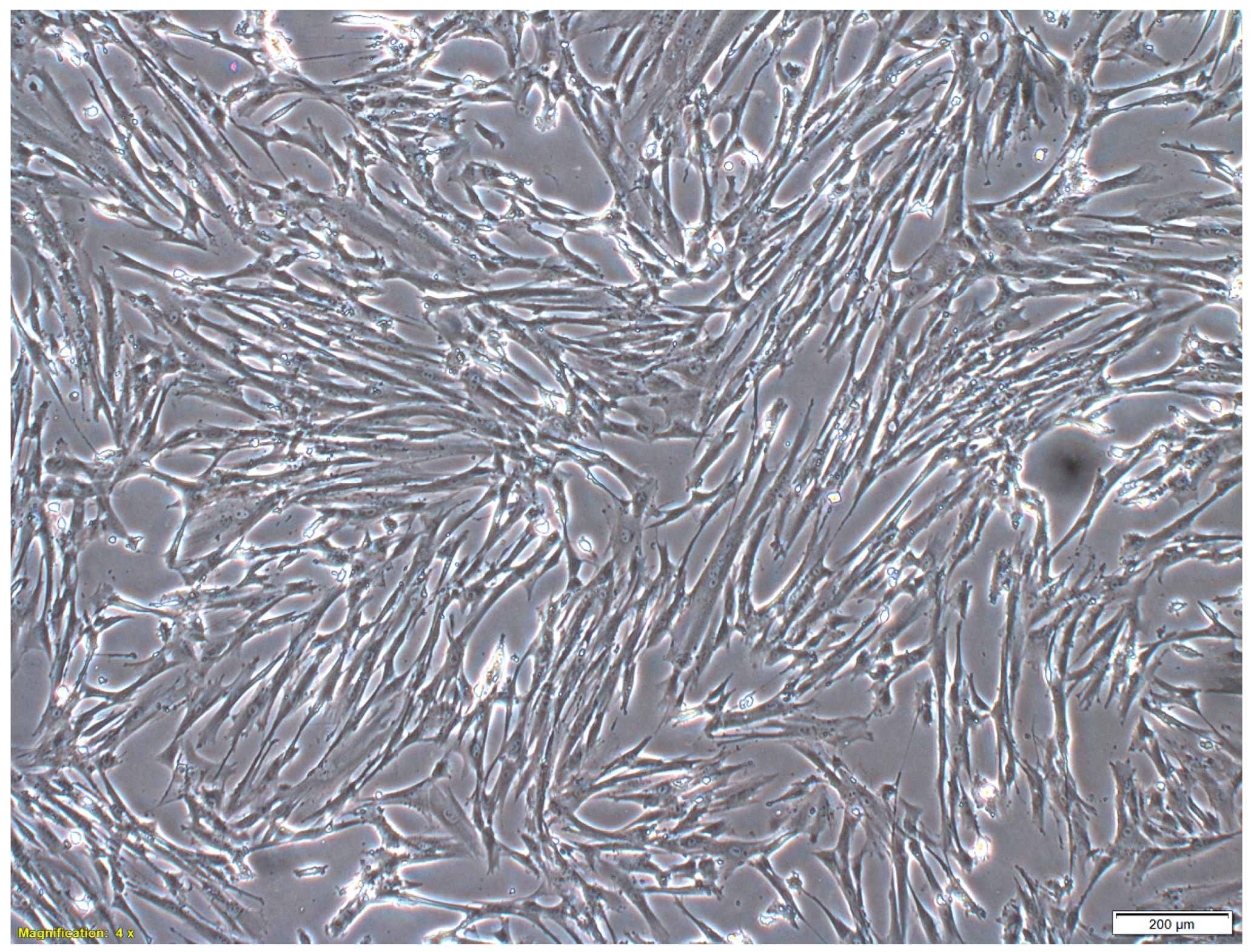
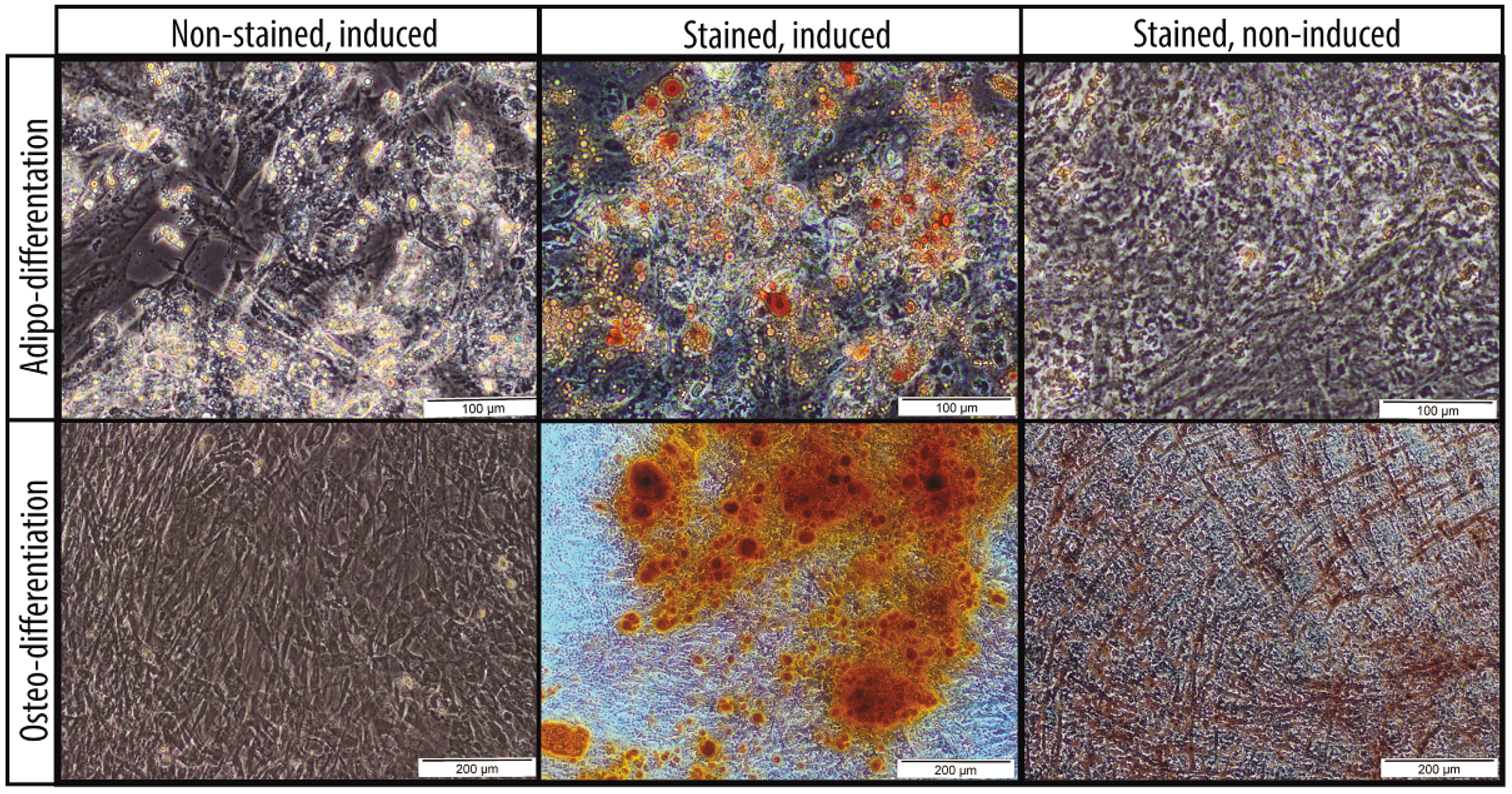
2.1. Characteristics of MSCs
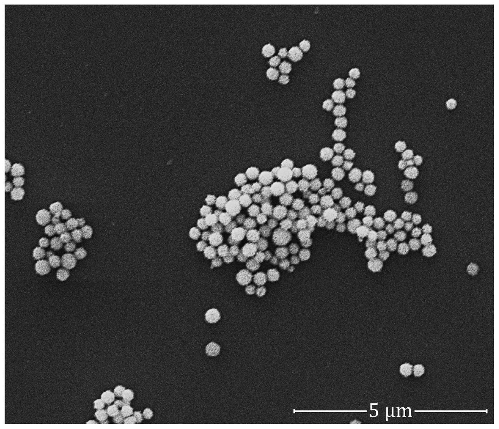
2.2. Quality of GNPs
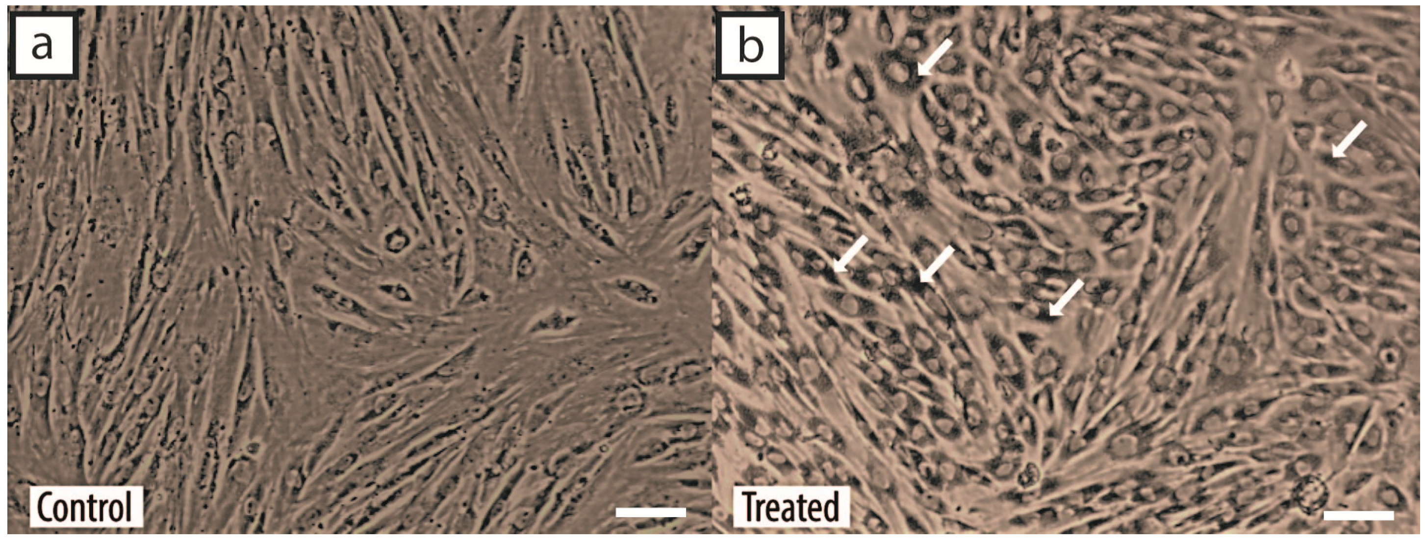
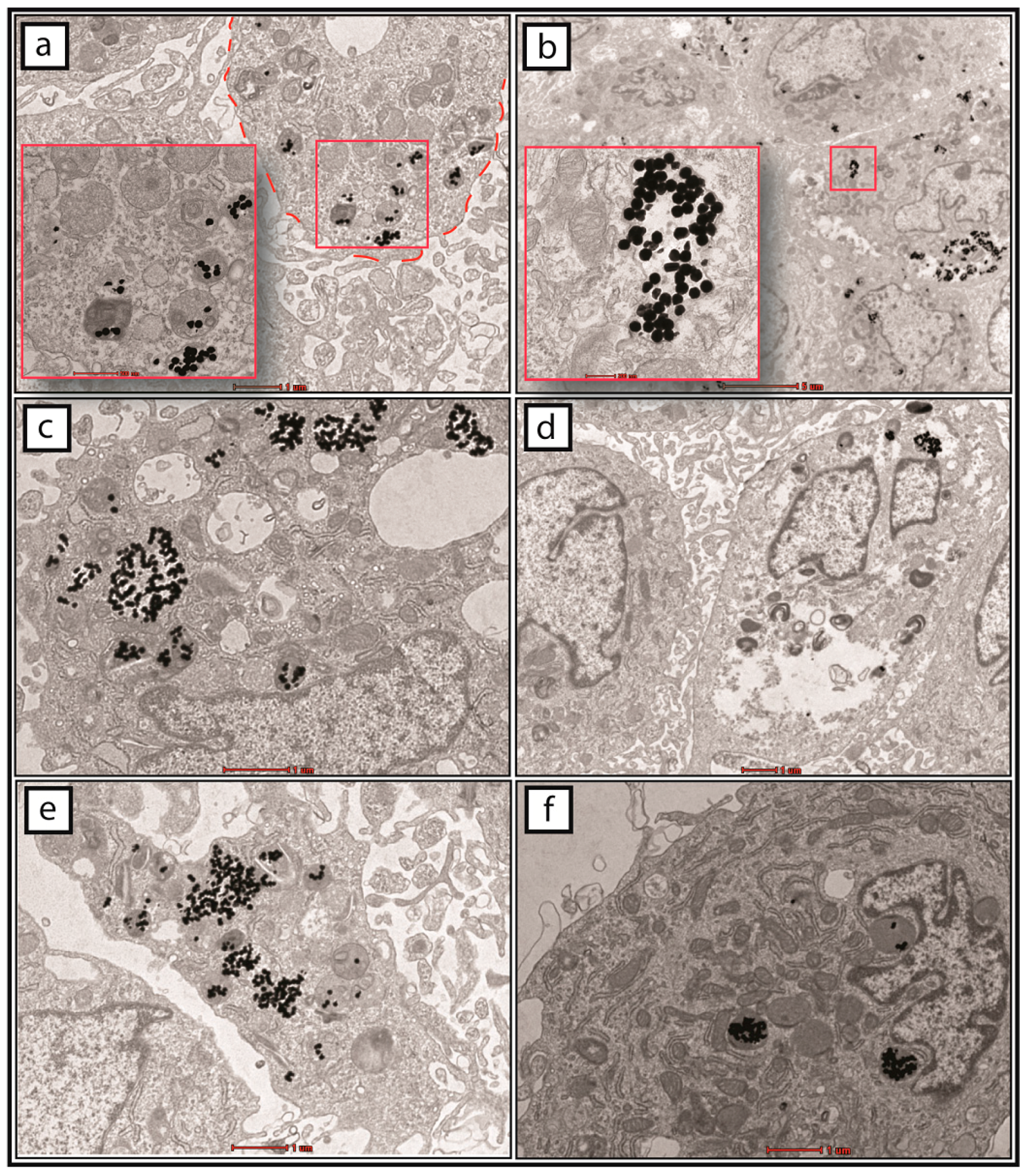
2.3. Cellular Uptake of GNPs by MSCs
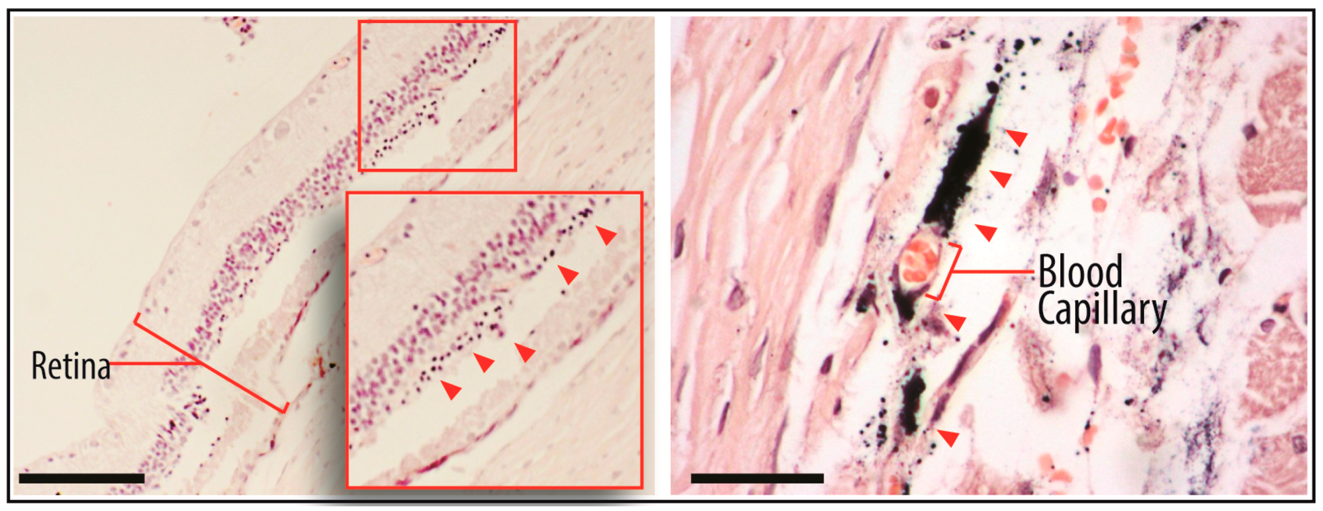
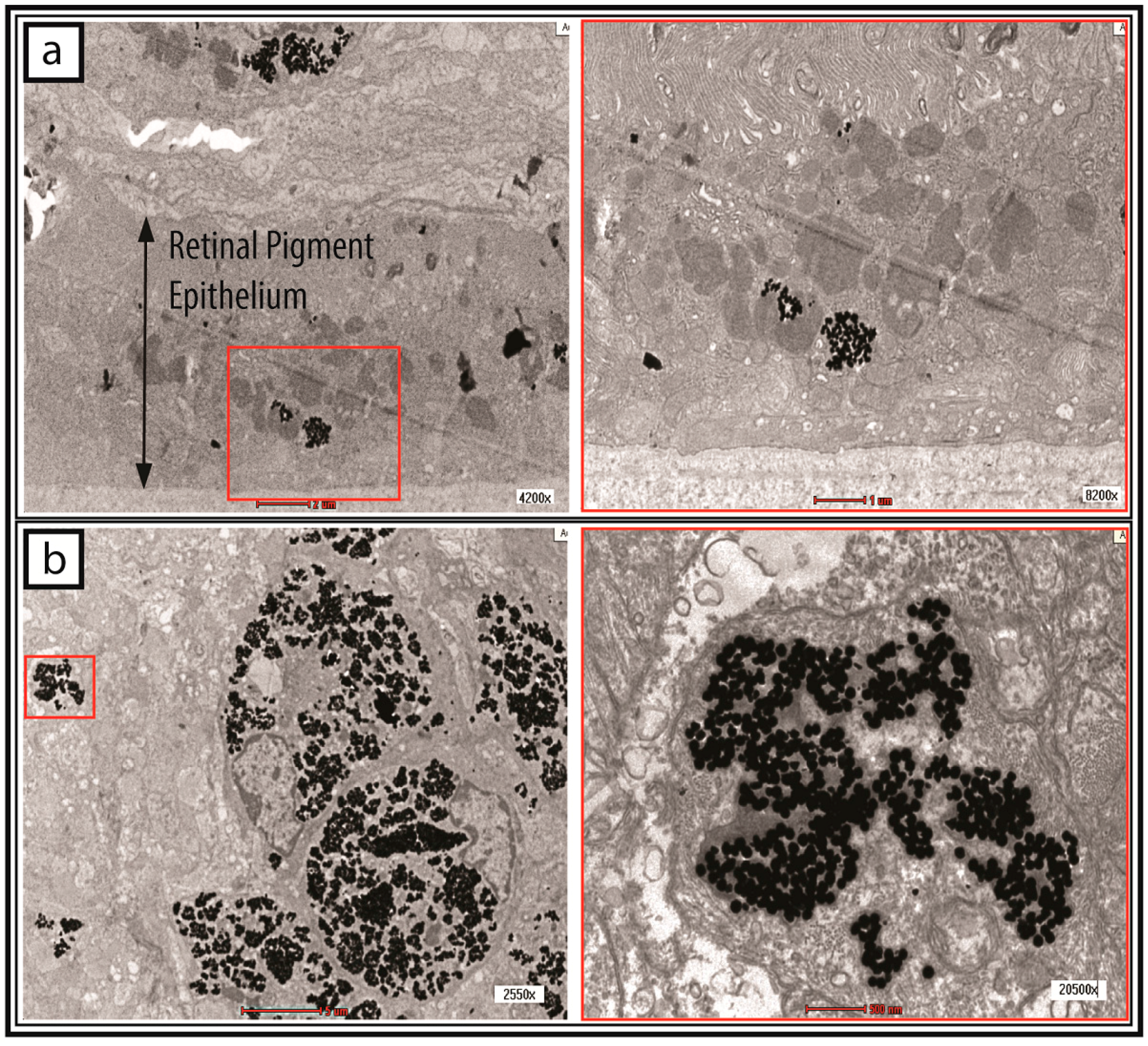
2.4. Tracking of GNP-Labelled MSCs by Micro-CT
3. Discussion
4. Materials and Methods
4.1. MSC Culture Conditions
4.2. MSC Characterization
4.3. GNP Labelling: Determination of the Quality of Colloidal GNPs
4.4. GNP Labelling: Cell Incubation with GNPs
4.5. GNP Lableling: Determination of the Cellular Uptake of GNPs
4.6. Subretinal Injection of GNP-Labelled MSCs
4.7. Micro-CT of Treated Rats
4.8. TEM of Injected MSCs
5. Conclusions
Acknowledgments
Author Contributions
Conflicts of Interest
References
- Figueroa, F.E.; Carrión, F.; Villanueva, S.; Khoury, M. Mesenchymal stem cell treatment for autoimmune diseases: A critical review. Biol. Res. 2016, 45, 269–277. [Google Scholar] [CrossRef] [PubMed]
- Karantalis, V.; Hare, J.M. Use of mesenchymal stem cells for therapy of cardiac disease. Circ. Res. 2015, 116, 1413–1430. [Google Scholar] [CrossRef] [PubMed]
- Joyce, N.; Annett, G.; Wirthlin, L.; Olson, S.; Bauer, G.; Nolta, J. Mesenchymal stem cells for the treatment of neurodegenerative disease. Regen. Med. 2010, 5, 933–946. [Google Scholar] [CrossRef] [PubMed]
- Kim, N.; Cho, S.G. Clinical applications of mesenchymal stem cells. Korean J. Intern. Med. 2013, 28, 387–402. [Google Scholar] [CrossRef] [PubMed]
- Leow, S.N.; Luu, C.D.; Hairul Nizam, M.H.; Mok, P.; Ruhaslizan, R.; Wong, H.S.; Wan Abdul Halim, W.; Ng, M.H.; Ruszymah, B.; Chowdhury, S.R.; et al. Safety and efficacy of human Wharton’s Jelly-derived mesenchymal stem cells therapy for retinal degeneration. PLoS ONE 2015, 10, e0128973. [Google Scholar] [CrossRef] [PubMed]
- Bae, K.S.; Park, J.B.; Kim, H.S.; Kim, D.S.; Park, D.J.; Kang, S.J. Neuron-like differentiation of bone marrow-derived mesenchymal stem cells. Yonsei Med. J. 2011, 52, 401–412. [Google Scholar] [CrossRef] [PubMed]
- Duscher, D.; Barrera, J.; Wong, V.W.; Maan, Z.N.; Whittam, A.J.; Januszyk, M.; Gurtner, G.C. Stem cells in wound healing: The future of regenerative medicine? A Mini-Review. Gerontology 2015, 62, 216–225. [Google Scholar] [CrossRef] [PubMed]
- Eggenhofer, E.; Luk, F.; Dahlke, M.H.; Hoogduijn, M.J. The life and fate of mesenchymal stem cells. Front. Immunol. 2014, 5, 148. [Google Scholar] [CrossRef] [PubMed]
- Karp, J.M.; Leng Teo, G.S. Mesenchymal stem cell homing: The devil is in the details. Cell Stem Cell 2009, 4, 206–216. [Google Scholar] [CrossRef] [PubMed]
- Srivastava, A.K.; Bulte, J.W.M. Seeing stem cells at work in vivo. Stem Cell Rev. Rep. 2014, 10, 127–144. [Google Scholar] [CrossRef] [PubMed]
- Bhirde, A.; Xie, J.; Swierczewska, M.; Chen, X.; Carpenter, M.K.; Frey-Vasconcells, J.; Rao, M.S.; Marin-Garcia, J.; Goldenthal, M.J.; Hart, L.S.; et al. Nanoparticles for cell labeling. Nanoscale 2011, 3, 142–153. [Google Scholar] [CrossRef] [PubMed]
- Dreaden, E.C.; Alkilany, A.M.; Huang, X.; Murphy, C.J.; El-Sayed, M.A. The golden age: Gold nanoparticles for biomedicine. Chem. Soc. Rev. 2012, 41, 2740–2779. [Google Scholar] [CrossRef] [PubMed]
- Ricles, L.M.; Nam, S.Y.; Treviño, E.A.; Emelianov, S.Y.; Suggs, L.J. A dual gold nanoparticle system for mesenchymal stem cell tracking. J. Mater. Chem. B Mater. Biol. Med. 2014, 2, 8220–8230. [Google Scholar] [CrossRef] [PubMed]
- Mok, P.L.; Leong, C.F.; Cheong, S.K. Cellular mechanisms of emerging applications of mesenchymal stem cells. Malays. J. Pathol. 2013, 35, 17–32. [Google Scholar] [PubMed]
- Ricles, L.M.; Nam, S.Y.; Sokolov, K.; Emelianov, S.Y.; Suggs, L.J. Function of mesenchymal stem cells following loading of gold nanotracers. Int. J. Nanomed. 2011, 6, 407–416. [Google Scholar] [CrossRef] [PubMed]
- Li, J.; Li, J.J.; Zhang, J.; Wang, X.; Kawazoe, N.; Chen, G. Gold nanoparticle size and shape influence on osteogenesis of mesenchymal stem cells. Nanoscale 2016, 8, 7992–8007. [Google Scholar] [CrossRef] [PubMed]
- Connor, E.E.; Mwamuka, J.; Gole, A.; Murphy, C.J.; Wyatt, M.D. Gold nanoparticles are taken up by human cells but do not cause acute cytotoxicity. Small 2005, 1, 325–327. [Google Scholar] [CrossRef] [PubMed]
- Murph, S.; Jacobs, S.; Liu, J.; Hu, T.C.; Siegfired, M.; Serkiz, S.M.; Hudson, J. Manganese-gold nanoparticles as an MRI positive contrast agent in mesenchymal stem cell labeling. J. Nanopart. Res. 2012, 14, 658. [Google Scholar] [CrossRef]
- Kohl, Y.; Gorjup, E.; Katsen-Globa, A.; Büchel, C.; von Briesen, H.; Thielecke, H. Effect of gold nanoparticles on adipogenic differentiation of human mesenchymal stem cells. J. Nanopart. Res. 2011, 13, 6789–6803. [Google Scholar] [CrossRef]
- Choi, S.Y.; Song, M.S.; Ryu, P.D.; Lam, A.T.; Joo, S.W.; Lee, S.Y. Gold nanoparticles promote osteogenic differentiation in human adipose-derived mesenchymal stem cells through the Wnt/β-catenin signaling pathway. Int. J. Nanomed. 2015, 10, 4383–4392. [Google Scholar]
- Kang, S.; Bhang, S.H.; Hwang, S.; Yoon, J.K.; Song, J.; Jang, H.K.; Kim, S.; Kim, B.S. Mesenchymal stem cells aggregate and deliver gold nanoparticles to tumors for photothermal therapy. ACS Nano 2015, 9, 9678–9690. [Google Scholar] [CrossRef] [PubMed]
- D’Alessio, F.; Mirabelli, P.; Gorrese, M.; Scalia, G.; Gemei, M.; Mariotti, E.; di Noto, R.; Martinelli, P.; Fortunato, G.; Paladini, D.; et al. Polychromatic flow cytometry analysis of CD34+ hematopoietic stem cells in cryopreserved early preterm human cord blood samples. Cytom. Part A 2011, 79, 14–24. [Google Scholar] [CrossRef] [PubMed]
- Wu, Q.; Jinde, K.; Endoh, M.; Sakai, H. Clinical significance of costimulatory molecules CD80/CD86 expression in IgA nephropathy. Kidney Int. 2004, 65, 888–896. [Google Scholar] [CrossRef] [PubMed]
- Dominici, M.; Le Blanc, K.; Mueller, I.; Slaper-Cortenbach, I.; Marini, F.; Krause, D.; Deans, R.; Keating, A.; Prockop, D.; Horwitz, E. Minimal criteria for defining multipotent mesenchymal stromal cells. The International Society for Cellular Therapy position statement. Cytotherapy 2006, 8, 315–317. [Google Scholar] [CrossRef] [PubMed]
- Accomasso, L.; Gallina, C.; Turinetto, V.; Giachino, C. Stem cell tracking with nanoparticles for regenerative medicine purposes: An overview. Stem Cells Int. 2016, 2016, 7920358. [Google Scholar] [CrossRef] [PubMed]
- Meir, R.; Motiei, M.; Popovtzer, R. Gold nanoparticles for in vivo cell tracking. Nanomedicine 2014, 9, 2059–2069. [Google Scholar] [CrossRef] [PubMed]
- Huefner, A.; Septiadi, D.; Wilts, B.D.; Patel, I.I.; Kuan, W.; Fragniere, A.; Barker, R.A.; Mahajan, S. Gold nanoparticles explore cells: Cellular uptake and their use as intracellular probes. Methods 2014, 68, 354–363. [Google Scholar] [CrossRef] [PubMed]
- Chithrani, D. Intracellular uptake, transport, and processing of gold nanostructures. Mol. Membr. Biol. 2010, 27, 299–311. [Google Scholar] [CrossRef] [PubMed]
- Chen, H.; Wang, L.; Yeh, J.; Wu, X.; Cao, Z.; Wang, Y.A.; Zhang, M.; Yang, L.; Mao, H. Reducing non-specific binding and uptake of nanoparticles and improving cell targeting with an antifouling PEO-b-PγMPS copolymer coating. Biomaterials 2010, 31, 5397–5407. [Google Scholar] [CrossRef] [PubMed]
- Longmire, M.; Choyke, P.L.; Kobayashi, H. Clearance properties of nano-sized particles and molecules as imaging agents: Considerations and caveats. Nanomedicine 2008, 3, 703–717. [Google Scholar] [CrossRef] [PubMed]
- Duan, P.; Xu, H.; Zeng, Y.; Wang, Y.; Yin, Z.Q. Human bone marrow stromal cells can differentiate to a retinal pigment epithelial phenotype when co-cultured with pig retinal pigment epithelium using a transwell system. Cell. Physiol. Biochem. 2013, 31, 601–613. [Google Scholar] [CrossRef] [PubMed]
- Vossmerbaeumer, U.; Ohnesorge, S.; Kuehl, S.; Haapalahti, M.; Kluter, H.; Jonas, J.B.; Thierse, H.J.; Bieback, K. Retinal pigment epithelial phenotype induced in human adipose tissue-derived mesenchymal stromal cells. Cytotherapy 2009, 11, 177–188. [Google Scholar] [CrossRef] [PubMed]
- Yang, L.; Zhou, Q.; Wang, Y.; Wang, Y.Q. Differentiation of human bone marrow-derived mesenchymal stem cells into neural-like cells by co-culture with retinal pigmented epithelial cells. Int. J. Ophthalmol. 2010, 3, 23–27. [Google Scholar] [PubMed]
- Kevany, B.M.; Palczewski, K. Phagocytosis of retinal rod and cone photoreceptors. Physiology 2010, 25, 8–15. [Google Scholar] [CrossRef] [PubMed]
- Guo, F.; Ding, Y.; Caberoy, N.; Alvarado, G.; Wang, F.; Chen, R.; Li, W. ABCF1 extrinsically regulates retinal pigment epithelial cell phagocytosis Running title: ABCF1 is a novel phagocytosis ligand. Mol. Biol. Cell. 2015, 26, 2311–2320. [Google Scholar] [CrossRef] [PubMed]
- He, X.; Cai, J.; Liu, B.; Zhong, Y.; Qin, Y. Cellular magnetic resonance imaging contrast generated by the ferritin heavy chain genetic reporter under the control of a Tet-On switch. Stem Cell Res. Ther. 2015, 6, 207. [Google Scholar] [CrossRef] [PubMed]
- Pereira, S.M.; Moss, D.; Williams, S.R.; Murray, P.; Taylor, A. Overexpression of the MRI reporter genes ferritin and transferrin receptor affect iron homeostasis and produce limited contrast in mesenchymal stem cells. Int. J. Mol. Sci. 2015, 16, 15481–15496. [Google Scholar] [CrossRef] [PubMed]
- Amsalem, Y.; Mardor, Y.; Feinberg, M.S.; Landa, N.; Miller, L.; Daniels, D.; Ocherashvilli, A.; Holbova, R.; Yosef, O.; Barbash, I.M.; et al. Iron-oxide labeling and outcome of transplanted mesenchymal stem cells in the infarcted myocardium. Circulation 2007, 116, 138–145. [Google Scholar] [CrossRef] [PubMed]
- Collins, M.C.; Gunst, P.R.; Cascio, W.E.; Kypson, A.P.; Muller-Borer, B.J. Labeling and imaging mesenchymal stem cells with quantum dots. Methods Mol. Biol. 2012, 906, 199–210. [Google Scholar] [PubMed]
- Fan, J.; Li, W.; Hung, W.; Hung, W.; Chen, C.; Yeh, J. Cytotoxicity and differentiation effects of gold nanoparticles to human bone marrow mesenchymal stem cells. Biomed. Eng. Appl. Basis Commun. 2011, 23, 141–152. [Google Scholar] [CrossRef]
- Sivan, P.P.; Syed, S.; Mok, P.L.; Higuchi, A.; Murugan, K.; Alarfaj, A.A.; Munusamy, M.A.; Hamat, R.H.; Umezawa, A.; Kumar, S. Stem Cell Therapy for Treatment of Ocular Disorders. Stem Cells Int. 2016, 2016, 8304879. [Google Scholar] [CrossRef] [PubMed]
- Wang, M.; Li, Z.; Qiao, H.; Chen, L.; Fan, Y. Effect of Gold/Fe3O4 Nanoparticles on biocompatibility and neural differentiation of rat olfactory bulb neural stem cells. J. Nanomater. 2013, 2013, 867426. [Google Scholar]
- Paviolo, C.; Haycock, J.W.; Yong, J.; Yu, A.; McArthur, S.L.; Stoddart, P.R. Plasmonic properties of gold nanoparticles can promote neuronal activity. In Proceedings of the SPIE International Society for Optics and Photonics, San Diego, CA, USA, 25–29 August 2013; Jansen, E.D., Thomas, R.J., Eds.; p. 85790C.
- Huang, X.; El-Sayed, M.A. Gold nanoparticles: Optical properties and implementations in cancer diagnosis and photothermal therapy. J. Adv. Res. 2010, 1, 13–28. [Google Scholar] [CrossRef]
- Kharlamov, A.; Perrish, A.; Gabinsky, J. Silica-gold nanoparticles and mesenchymal stem cells versus composite ferro-magnetic approach for management of atherosclerotic plaque and artery remodeling. Circulation 2011, 124, A8303. [Google Scholar]
- Plasmonic Photothermal and Stem Cell Therapy of Atherosclerosis versus Stenting (NANOM PCI). Available online: https://clinicaltrials.gov/ct2/show/NCT01436123 (accessed on 30 July 2016).
- Sarkar, D.; Vemula, P.K.; Zhao, W.; Gupta, A.; Karnik, R.; Karp, J.M. Engineered mesenchymal stem cells with self-assembled vesicles for systemic cell targeting. Biomaterials 2010, 31, 5266–5274. [Google Scholar] [CrossRef] [PubMed]
- Khademhosseini, A.; Borenstein, J.; Toner, M.; Takayama, S. Micro and Nanoengineering of the Cell Microenvironment; Artech House Publishers: Norwood, MA, USA, 2008. [Google Scholar]
- Qureshi, S. β-lactamase: An ideal reporter system for monitoring gene expression in live eukaryotic cells. BioTechniques 2007, 42, 91–95. [Google Scholar] [CrossRef] [PubMed]
- Smale, S. Chloramphenicol acetyltransferase assay. Cold Spring Harb. Protoc. 2010, 5. [Google Scholar] [CrossRef] [PubMed]
- Ude, C.; Shamsul, B.; Ng, M.; Chen, H.C.; Norhamdan, M.Y.; Aminuddin, B.S.; Ruszymah, B.H.I. Bone marrow and adipose stem cells can be tracked with PKH26 until post staining passage 6 in in vitro and in vivo. Tissue Cell 2012, 44, 156–163. [Google Scholar] [CrossRef] [PubMed]
- Johnsson, C.; Festin, R.; Tufveson, G.; Tötterman, T.H. Ex vivo PKH26-labelling of lymphocytes for studies of cell migration in vivo. Scand. J. Immunol. 1997, 45, 511–514. [Google Scholar] [CrossRef] [PubMed]
- Xu, R.; Huang, L.; Wei, W.; Chen, X.; Zhang, X.; Zhang, X. Real-time imaging and tracking of ultrastable organic dye nanoparticles in living cells. Biomaterials 2016, 93, 38–47. [Google Scholar] [CrossRef] [PubMed]
- Martin-Fernandez, M.L.; Clarke, D.T. Single molecule fluorescence detection and tracking in mammalian cells: The state-of-the-art and future perspectives. Int. J. Mol. Sci. 2012, 11, 14742–14765. [Google Scholar] [CrossRef] [PubMed]
- Muthana, M.; Kennerley, A.J.; Hughes, R.; Fagnano, E.; Richardson, J.; Paul, M.; Murdoch, C.; Wright, F.; Payne, C.; Lythgoe, M.F.; et al. Directing cell therapy to anatomic target sites in vivo with magnetic resonance targeting. Nat. Commun. 2015, 6, 8009. [Google Scholar] [CrossRef] [PubMed]
- Jensen, E. Types of imaging, part 2: An overview of fluorescence microscopy. Anat. Rec. 2012, 295, 1621–1627. [Google Scholar] [CrossRef] [PubMed]
- Progatzky, F.; Dallman, M.J.; Lo Celso, C. From seeing to believing: Labelling strategies for in vivo cell-tracking experiments. Interface Focus 2013, 3, 20130001. [Google Scholar] [CrossRef] [PubMed]
- Edmundson, M.; Capeness, M.; Horsfall, L. Exploring the potential of metallic nanoparticles within synthetic biology. New Biotechnol. 2014, 3, 572–577. [Google Scholar] [CrossRef] [PubMed]
- Li, J.; Kawazoe, N.; Chen, G. Gold nanoparticles with different charge and moiety induce differential cell response on mesenchymal stem cell osteogenesis. Biomaterials 2015, 54, 226–236. [Google Scholar] [CrossRef] [PubMed]


| Cell Labelling Technology | Advantages/Disadvantages | References |
|---|---|---|
| Viral or non-viral reporter gene systems | Advantages: Allows monitoring of co-expressed genes. | [49] |
| Disadvantages: Labour-intensive and time-consuming in the preparation of transduced cell clones. Radioactive substance is required in a conventional chloramphenicol acetyl transferase reporter system, which is potentially hazardous. | [16,50] | |
| Free organic dyes e.g., PKH26, carboxyfluorescein succinimidyl ester (CFSE) | Advantages: Simple cell-labelling protocol. Long-term cell tracking in both in vitro and in vivo systems, e.g., PKH26. | [51] |
| Disadvantages: Possible transfer of dye from labelled to unlabelled cells. | [52] | |
| Organic dye nanoparticles | Advantages: Suitable for living-cell imaging as it demonstrates high fluorescence intensity, large Stokes shift, photostability, and emission in the near-infrared range. | [53] |
| Disadvantages: High tendency of organic dye to stick to the cell substrate. | [54] | |
| Superparamagnetic iron oxide nanoparticles (SPIO) | Advantages: Direct tissue targeting of SPIO-labelled cells is feasible with use of an appropriate magnetic field. | [55] |
| Disadvantages: Requires cross-linking with a membrane-translocating signal peptide (e.g., HIV-1 Tat protein) or co-incubation with transfection agents to facilitate cellular uptake. | [25] | |
| Semiconductor quantum dots | Advantages: Photostable, possesses size-controlled fluorescence, and the emitted fluorescence has a long lifetime. | [56] |
| Disadvantages: High cost of reagents; generation of free radicals may cause cellular toxicity. | [57,58] | |
| Noble metallic nanoparticles e.g., gold nanoparticles | Advantages: Simple cell-labelling protocol and no acute cellular toxicity demonstrated. | [59] |
| Disadvantages: Different shapes and sizes could affect stem cell differentiation potential. | [16,40] |
© 2017 by the authors. Licensee MDPI, Basel, Switzerland. This article is an open access article distributed under the terms and conditions of the Creative Commons Attribution (CC BY) license ( http://creativecommons.org/licenses/by/4.0/).
Share and Cite
Mok, P.L.; Leow, S.N.; Koh, A.E.-H.; Mohd Nizam, H.H.; Ding, S.L.S.; Luu, C.; Ruhaslizan, R.; Wong, H.S.; Halim, W.H.W.A.; Ng, M.H.; et al. Micro-Computed Tomography Detection of Gold Nanoparticle-Labelled Mesenchymal Stem Cells in the Rat Subretinal Layer. Int. J. Mol. Sci. 2017, 18, 345. https://doi.org/10.3390/ijms18020345
Mok PL, Leow SN, Koh AE-H, Mohd Nizam HH, Ding SLS, Luu C, Ruhaslizan R, Wong HS, Halim WHWA, Ng MH, et al. Micro-Computed Tomography Detection of Gold Nanoparticle-Labelled Mesenchymal Stem Cells in the Rat Subretinal Layer. International Journal of Molecular Sciences. 2017; 18(2):345. https://doi.org/10.3390/ijms18020345
Chicago/Turabian StyleMok, Pooi Ling, Sue Ngein Leow, Avin Ee-Hwan Koh, Hairul Harun Mohd Nizam, Suet Lee Shirley Ding, Chi Luu, Raduan Ruhaslizan, Hon Seng Wong, Wan Haslina Wan Abdul Halim, Min Hwei Ng, and et al. 2017. "Micro-Computed Tomography Detection of Gold Nanoparticle-Labelled Mesenchymal Stem Cells in the Rat Subretinal Layer" International Journal of Molecular Sciences 18, no. 2: 345. https://doi.org/10.3390/ijms18020345
APA StyleMok, P. L., Leow, S. N., Koh, A. E.-H., Mohd Nizam, H. H., Ding, S. L. S., Luu, C., Ruhaslizan, R., Wong, H. S., Halim, W. H. W. A., Ng, M. H., Idrus, R. B. H., Chowdhury, S. R., Bastion, C. M.-L., Subbiah, S. K., Higuchi, A., Alarfaj, A. A., & Then, K. Y. (2017). Micro-Computed Tomography Detection of Gold Nanoparticle-Labelled Mesenchymal Stem Cells in the Rat Subretinal Layer. International Journal of Molecular Sciences, 18(2), 345. https://doi.org/10.3390/ijms18020345


