Next Issue
Volume 18, June
Previous Issue
Volume 18, April
 
 
ijms-logo

Journal Browser

Journal Browser

Int. J. Mol. Sci., Volume 18, Issue 5 (May 2017) – 207 articles

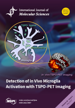
Cover Story (view full-size image): The figure shows the molecular basis and principles of TSPO-PET imaging. The 18 kDa Translocator Protein (TSPO) is an outer mitochondrial membrane protein which is well-known to be over-expressed by microglia during activation, thus being a sensitive hallmark of neuroinflammation. PET-based molecular imaging techniques targeting neuroinflammation take advantage of this and are unique tools for the in vivo visualization and quantification of TSPO over-expression. View this paper.
  • Issues are regarded as officially published after their release is announced to the table of contents alert mailing list.
  • You may sign up for e-mail alerts to receive table of contents of newly released issues.
  • PDF is the official format for papers published in both, html and pdf forms. To view the papers in pdf format, click on the "PDF Full-text" link, and use the free Adobe Reader to open them.
Order results
Result details
Section
Select all
Export citation of selected articles as:
19 pages, 6353 KiB  
Article
3D-QSAR, Molecular Docking and Molecular Dynamics Simulation of Pseudomonas aeruginosa LpxC Inhibitors
by Ke Zuo 1,†, Li Liang 1,†, Wenyi Du 1, Xin Sun 1, Wei Liu 1, Xiaojun Gou 1,*, Hua Wan 2 and Jianping Hu 1,3,*
1 College of Pharmacy and Biological Engineering, Key Laboratory of Medicinal and Edible Plants Resources Development of Sichuan Education Department, Sichuan Industrial Institute of Antibiotics, Chengdu University, Chengdu 610106, China
2 College of Mathematics and Informatics, South China Agricultural University, Guangzhou 510642, China
3 College of Chemistry, Leshan Normal University, Leshan 614004, China
These authors contributed equally to this work.
Int. J. Mol. Sci. 2017, 18(5), 761; https://doi.org/10.3390/ijms18050761 - 6 May 2017
Cited by 40 | Viewed by 6907
Abstract
As an important target for the development of novel antibiotics, UDP-3-O-(R-3-hydroxymyristoyl)-N-acetylglucosamine deacetylase (LpxC) has been widely studied. Pyridone methylsulfone hydroxamate (PMH) compounds can effectively inhibit the catalytic activity of LpxC. In this work, the three-dimensional quantitative structure-activity [...] Read more.
As an important target for the development of novel antibiotics, UDP-3-O-(R-3-hydroxymyristoyl)-N-acetylglucosamine deacetylase (LpxC) has been widely studied. Pyridone methylsulfone hydroxamate (PMH) compounds can effectively inhibit the catalytic activity of LpxC. In this work, the three-dimensional quantitative structure-activity relationships of PMH inhibitors were explored and models with good predictive ability were established using comparative molecular field analysis and comparative molecular similarity index analysis methods. The effect of PMH inhibitors’ electrostatic potential on the inhibitory ability of Pseudomonas aeruginosa LpxC (PaLpxC) is revealed at the molecular level via molecular electrostatic potential analyses. Then, two molecular dynamics simulations for the PaLpxC and PaLpxC-inhibitor systems were also performed respectively to investigate the key residues of PaLpxC hydrolase binding to water molecules. The results indicate that orderly alternative water molecules can form stable hydrogen bonds with M62, E77, T190, and H264 in the catalytic center, and tetracoordinate to zinc ion along with H78, H237, and D241. It was found that the conformational transition space of PaLpxC changes after association with PMH inhibitors through free energy landscape and cluster analyses. Finally, a possible inhibitory mechanism of PMH inhibitors was proposed, based on our molecular simulation. This paper will provide a theoretical basis for the molecular design of LpxC-targeting antibiotics. Full article
(This article belongs to the Section Molecular Recognition)
Show Figures

Figure 1

25 pages, 8660 KiB  
Article
Renalase Protects against Renal Fibrosis by Inhibiting the Activation of the ERK Signaling Pathways
by Yiru Wu, Liyan Wang, Dai Deng, Qidong Zhang and Wenhu Liu *
Department of Nephrology, Affiliated Beijing Friendship Hospital, Faculty of Kidney Diseases, Capital Medical University, No. 95 Yong An Road, Xi Cheng District, Beijing 100050, China
Int. J. Mol. Sci. 2017, 18(5), 855; https://doi.org/10.3390/ijms18050855 - 27 Apr 2017
Cited by 58 | Viewed by 5324
Abstract
Renal interstitial fibrosis is a common pathway for the progression of chronic kidney disease (CKD) to end-stage renal disease. Renalase, acting as a signaling molecule, has been reported to have cardiovascular and renal protective effects. However, its role in renal fibrosis remains unknown. [...] Read more.
Renal interstitial fibrosis is a common pathway for the progression of chronic kidney disease (CKD) to end-stage renal disease. Renalase, acting as a signaling molecule, has been reported to have cardiovascular and renal protective effects. However, its role in renal fibrosis remains unknown. In this study, we evaluated the therapeutic efficacy of renalase in rats with complete unilateral ureteral obstruction (UUO) and examined the inhibitory effects of renalase on transforming growth factor-β1 (TGF-β1)-induced epithelial–mesenchymal transition (EMT) in human proximal renal tubular epithelial (HK-2) cells. We found that in the UUO model, the expression of renalase was markedly downregulated and adenoviral-mediated expression of renalase significantly attenuated renal interstitial fibrosis, as evidenced by the maintenance of E-cadherin expression and suppressed expression of α-smooth muscle actin (α-SMA), fibronectin and collagen-I. In vitro, renalase inhibited TGF-β1-mediated upregulation of α-SMA and downregulation of E-cadherin. Increased levels of Phospho-extracellular regulated protein kinases (p-ERK1/2) in TGF-β1-stimulated cells were reversed by renalase cotreatment. When ERK1 was overexpressed, the inhibition of TGF-β1-induced EMT and fibrosis mediated by renalase was attenuated. Our study provides the first evidence that renalase can ameliorate renal interstitial fibrosis by suppression of tubular EMT through inhibition of the ERK pathway. These results suggest that renalase has potential renoprotective effects in renal interstitial fibrosis and may be an effective agent for slowing CKD progression. Full article
(This article belongs to the Section Biochemistry)
Show Figures

Figure 1

15 pages, 1886 KiB  
Article
Natural Killer Cells Response to IL-2 Stimulation Is Distinct between Ascites with the Presence or Absence of Malignant Cells in Ovarian Cancer Patients
by Rodrigo Fernandes Da Silva 1, Adriana Yoshida 1, Daniela Maira Cardozo 1, Rodrigo Menezes Jales 1, Silke Paust 2, Sophie Derchain 1 and Fernando Guimarães 3,*
1 Faculty of Medical Sciences, University of Campinas, 13083-887 Campinas, Brazil
2 Center for Human Immunobiology, Department of Pediatrics, Texas Children’s Hospital and Baylor College of Medicine, Houston, TX 77030, USA
3 Women´s Hospital “Professor Doutor José Aristodemo Pinotti”–Centro de Atenção Integral à Saúde da Mulher (CAISM), University of Campinas, 13083-881 Campinas, Brazil
Int. J. Mol. Sci. 2017, 18(5), 856; https://doi.org/10.3390/ijms18050856 - 17 May 2017
Cited by 19 | Viewed by 5632
Abstract
Peritoneal ascites are a distinguishable feature of patients with advanced epithelial ovarian cancer (EOC). The presence of different lymphocyte subsets has been reported in EOC-associated ascites, which also can or not contain malignant cells. The goal of this study was to analyze the [...] Read more.
Peritoneal ascites are a distinguishable feature of patients with advanced epithelial ovarian cancer (EOC). The presence of different lymphocyte subsets has been reported in EOC-associated ascites, which also can or not contain malignant cells. The goal of this study was to analyze the functional characteristics of natural killer (NK) cells from EOC-associated ascites in terms of their expression of activating receptors and ascites’ contents of lymphocyte subtypes, cytokine profile and presence of EOC cells. NK cell function was evaluated by the expression of the degranulation marker CD107a in resting and interleukin (IL)-2 stimulated NK cells from ascites and blood. Degranulation of NK cells from EOC cell-free ascites was significantly (p < 0.05) higher than all the other groups, either in their resting state or after IL-2 stimulation, suggesting a previous local stimulation. In contrast, treatment with IL-2 had no effect on NK cells from ascites with EOC cells. The amount of regulatory T cells was significantly higher in ascites with EOC cells compared to EOC cell-free ascites. Ascites with EOC cells also had higher levels of tumor necrosis factor (TNF)-α, suggesting inflammation related to the malignancy. In conclusion, the functional performance of NK cells was distinct between EOC cell-free ascites and ascites with EOC cells. The impairment of NK cell response to IL-2 in ascites with EOC cells was consistent with an immunosuppressive tumor microenvironment. Full article
(This article belongs to the Special Issue Natural Killer (NK) Cells)
Show Figures

Figure 1

11 pages, 4468 KiB  
Article
Selection, Characterization and Interaction Studies of a DNA Aptamer for the Detection of Bifidobacterium bifidum
by Lujun Hu 1, Linlin Wang 1, Wenwei Lu 1,2, Jianxin Zhao 1,2, Hao Zhang 1,2 and Wei Chen 1,2,3,*
1 State Key Laboratory of Food Science and Technology, School of Food Science and Technology, Jiangnan University, Wuxi 214122, China
2 International Joint Research Center for Probiotics & Gut Health, Jiangnan University, Wuxi 214122, China
3 Beijing Innovation Centre of Food Nutrition and Human Health, Beijing Technology and Business University, Beijing 100048, China
Int. J. Mol. Sci. 2017, 18(5), 883; https://doi.org/10.3390/ijms18050883 - 25 Apr 2017
Cited by 20 | Viewed by 5662
Abstract
A whole-bacterium-based SELEX (Systematic Evolution of Ligands by Exponential Enrichment) procedure was adopted in this study for the selection of an ssDNA aptamer that binds to Bifidobacterium bifidum. After 12 rounds of selection targeted against B. bifidum, 30 sequences were obtained [...] Read more.
A whole-bacterium-based SELEX (Systematic Evolution of Ligands by Exponential Enrichment) procedure was adopted in this study for the selection of an ssDNA aptamer that binds to Bifidobacterium bifidum. After 12 rounds of selection targeted against B. bifidum, 30 sequences were obtained and divided into seven families according to primary sequence homology and similarity of secondary structure. Four FAM (fluorescein amidite) labeled aptamer sequences from different families were selected for further characterization by flow cytometric analysis. The results reveal that the aptamer sequence CCFM641-5 demonstrated high-affinity and specificity for B. bifidum compared with the other sequences tested, and the estimated Kd value was 10.69 ± 0.89 nM. Additionally, sequence truncation experiments of the aptamer CCFM641-5 led to the conclusion that the 5′-primer and 3′-primer binding sites were essential for aptamer-target binding. In addition, the possible component of the target B. bifidum, bound by the aptamer CCFM641-5, was identified as a membrane protein by treatment with proteinase. Furthermore, to prove the potential application of the aptamer CCFM641-5, a colorimetric bioassay of the sandwich-type structure was used to detect B. bifidum. The assay had a linear range of 104 to 107 cfu/mL (R2 = 0.9834). Therefore, the colorimetric bioassay appears to be a promising method for the detection of B. bifidum based on the aptamer CCFM641-5. Full article
(This article belongs to the Special Issue Aptamers)
Show Figures

Graphical abstract

16 pages, 4325 KiB  
Article
Integrative RNA- and miRNA-Profile Analysis Reveals a Likely Role of BR and Auxin Signaling in Branch Angle Regulation of B. napus
by Hongtao Cheng 1, Mengyu Hao 1, Wenxiang Wang 1, Desheng Mei 1, Rachel Wells 2, Jia Liu 1, Hui Wang 1, Shifei Sang 1, Min Tang 1, Rijin Zhou 1, Wen Chu 1, Li Fu 1 and Qiong Hu 1,*
1 Oil Crops Research Institute of Chinese Academy of Agricultural Sciences/Key Laboratory for Biological Sciences and Genetic Improvement of Oil Crops, Ministry of Agriculture, Wuhan 430062, China
2 John Innes Centre, Norwich Research Park, Norwich NR4 7UH, UK
Int. J. Mol. Sci. 2017, 18(5), 887; https://doi.org/10.3390/ijms18050887 - 8 May 2017
Cited by 27 | Viewed by 6049
Abstract
Oilseed rape (Brassica napus L.) is the second largest oilseed crop worldwide and one of the most important oil crops in China. As a component of plant architecture, branch angle plays an important role in yield performance, especially under high-density planting conditions. [...] Read more.
Oilseed rape (Brassica napus L.) is the second largest oilseed crop worldwide and one of the most important oil crops in China. As a component of plant architecture, branch angle plays an important role in yield performance, especially under high-density planting conditions. However, the mechanisms underlying the regulation of branch angle are still largely not understood. Two oilseed rape lines with significantly different branch angles were used to conduct RNA- and miRNA-profiling at two developmental stages, identifying differential expression of a large number of genes involved in auxin- and brassinosteroid (BR)-related pathways. Many auxin response genes, including AUX1, IAA, GH3, and ARF, were enriched in the compact line. However, a number of genes involved in BR signaling transduction and biosynthesis were down-regulated. Differentially expressed miRNAs included those involved in auxin signaling transduction. Expression patterns of most target genes were fine-tuned by related miRNAs, such as miR156, miR172, and miR319. Some miRNAs were found to be differentially expressed at both developmental stages, including three miR827 members. Our results provide insight that auxin- and BR-signaling may play a pivotal role in branch angle regulation. Full article
(This article belongs to the Section Molecular Plant Sciences)
Show Figures

Figure 1

16 pages, 6972 KiB  
Article
Effects of 3D-Printed Polycaprolactone/β-Tricalcium Phosphate Membranes on Guided Bone Regeneration
by Jin-Hyung Shim 1,†, Joo-Yun Won 2,†, Jung-Hyung Park 3,†, Ji-Hyeon Bae 3,†, Geunseon Ahn 2, Chang-Hwan Kim 2, Dong-Hyuk Lim 1, Dong-Woo Cho 4, Won-Soo Yun 1, Eun-Bin Bae 3, Chang-Mo Jeong 3 and Jung-Bo Huh 3,*
1 Department of Mechanical Engineering, Korea Polytechnic University, 237 Sangidaehak-Ro, Siheung-Si, Gyeonggi-Do 15073, Korea
2 Research Institute, T&R Biofab Co., Ltd., 237 Sangidaehak-Ro, Siheung-Si, Gyeonggi-Do 15073, Korea
3 Department of Prosthodontics, Dental Research Institute, Institute of Translational Dental Sciences, BK21 PLUS Project, School of Dentistry, Pusan National University, 49 Pusan University-Ro, Yangsan-Si, Gyeongsangnam-Do 50612, Korea
4 Department of Mechanical Engineering, Pohang University of Science and Technology (POSTECH), 77 Cheong-Am-Ro, Nam-Gu, Pohang-Si, Gyeongsangbuk-Do 37673, Korea
These authors contributed equally to this work.
Int. J. Mol. Sci. 2017, 18(5), 899; https://doi.org/10.3390/ijms18050899 - 25 Apr 2017
Cited by 105 | Viewed by 9214
Abstract
This study was conducted to compare 3D-printed polycaprolactone (PCL) and polycaprolactone/β-tricalcium phosphate (PCL/β-TCP) membranes with a conventional commercial collagen membrane in terms of their abilities to facilitate guided bone regeneration (GBR). Fabricated membranes were tested for dry and wet mechanical properties. Fibroblasts and [...] Read more.
This study was conducted to compare 3D-printed polycaprolactone (PCL) and polycaprolactone/β-tricalcium phosphate (PCL/β-TCP) membranes with a conventional commercial collagen membrane in terms of their abilities to facilitate guided bone regeneration (GBR). Fabricated membranes were tested for dry and wet mechanical properties. Fibroblasts and preosteoblasts were seeded into the membranes and rates and patterns of proliferation were analyzed using a kit-8 assay and by scanning electron microscopy. Osteogenic differentiation was verified by alizarin red S and alkaline phosphatase (ALP) staining. An in vivo experiment was performed using an alveolar bone defect beagle model, in which defects in three dogs were covered with different membranes. CT and histological analyses at eight weeks after surgery revealed that 3D-printed PCL/β-TCP membranes were more effective than 3D-printed PCL, and substantially better than conventional collagen membranes in terms of biocompatibility and bone regeneration and, thus, at facilitating GBR. Full article
(This article belongs to the Special Issue Three-dimensional (3D) Bioprinting of Tissues and Organs)
Show Figures

Figure 1

14 pages, 3664 KiB  
Article
A Long-Term Treatment with Arachidonyl-2′-Chloroethylamide Combined with Valproate Increases Neurogenesis in a Mouse Pilocarpine Model of Epilepsy
by Marta Andres-Mach 1,*, Mirosław Zagaja 1, Agnieszka Haratym-Maj 2, Radosław Rola 2,3, Maciej Maj 4, Joanna Haratym 5, Monika Dudra-Jastrzębska 2,6 and Jarogniew J. Łuszczki 1,6
1 Isobolographic Analysis Laboratory, Institute of Rural Health, Jaczewskiego 2, 20-950 Lublin, Poland
2 Department of Physiopathology, Institute of Rural Health, Jaczewskiego 2, 20-950 Lublin, Poland
3 Department of Neurological Surgery, Medical University of Lublin, Jaczewskiego 8, 20-090 Lublin, Poland
4 Department of Biopharmacy, Medical University of Lublin, Chodzki 4A, 20-090 Lublin, Poland
5 Department of Anestesiology and Intensive Care Medicine, Hollycross Cancer Center, Artwińskiego 3, 25-734 Kielce, Poland
6 Department of Pathophysiology, Medical University of Lublin, Jaczewskiego 8, 20-090 Lublin, Poland
Int. J. Mol. Sci. 2017, 18(5), 900; https://doi.org/10.3390/ijms18050900 - 25 Apr 2017
Cited by 24 | Viewed by 5850
Abstract
Rational polytherapy in the treatment of refractory epilepsy has been the main therapeutic modality for several years. In treatment with two or more antiepileptic drugs (AEDs), it is of particular importance that AEDs be selected based on their high anticonvulsant properties, minimal side [...] Read more.
Rational polytherapy in the treatment of refractory epilepsy has been the main therapeutic modality for several years. In treatment with two or more antiepileptic drugs (AEDs), it is of particular importance that AEDs be selected based on their high anticonvulsant properties, minimal side effects, and impact on the formation of new neurons. The aim of the study was to conduct an in vivo evaluation of the relationship between treatments with synthetic cannabinoid arachidonyl-2′-chloroethylamide (ACEA) alone or in combination with valproic acid (VPA) and hippocampal neurogenesis in a mouse pilocarpine model of epilepsy. All studies were performed on adolescent male CB57/BL mice with using the following drugs: VPA (10 mg/kg), ACEA (10 mg/kg), phenylmethylsulfonyl fluoride (PMSF—a substance protecting ACEA against degradation by fatty acid hydrolase, 30 mg/kg), pilocarpine (PILO, a single dose of 290 mg/kg) and methylscopolamine (30 min before PILO to stop peripheral cholinergic effects of pilocarpine, 1 mg/kg). We evaluated the process of neurogenesis after a 10-day treatment with ACEA and VPA, alone and in combination. We observed a decrease of neurogenesis in the PILO control group as compared to the healthy control mice. Furthermore, ACEA + PMSF alone and in combination with VPA significantly increased neurogenesis compared to the PILO control group. In contrast, VPA 10-day treatment had no impact on the level of neurons in comparison to the PILO control group. The combination of ACEA, PMSF and VPA considerably stimulated the process of creating new cells, particularly neurons, while chronic administration of VPA itself had no influence on neurogenesis in the mouse pilocarpine model of epilepsy. The obtained results enabled an in vivo evaluation of neurogenesis after treatment with antiepileptic drugs in an experimental model of epilepsy. Full article
(This article belongs to the Special Issue Cannabinoid Signaling in Nervous System)
Show Figures

Graphical abstract

48 pages, 5265 KiB  
Review
Collagenous Extracellular Matrix Biomaterials for Tissue Engineering: Lessons from the Common Sea Urchin Tissue
by Kheng Lim Goh 1,2,* and David F. Holmes 3
1 Newcastle University Singapore, SIT Building at Nanyang Polytechnic, 172A Ang Mo Kio Avenue 8 #05-01, Singapore 567739, Singapore
2 Newcastle University, School of Mechanical & Systems Engineering, Stephenson Building, Claremont Road, Newcastle upon Tyne NE1 7RU, UK
3 Manchester University, Wellcome Trust Centre for Cell Matrix Research, B.3016 Michael Smith Building, Faculty of Life Sciences, Oxford Road, Manchester M13 9PT, UK
Int. J. Mol. Sci. 2017, 18(5), 901; https://doi.org/10.3390/ijms18050901 - 25 Apr 2017
Cited by 44 | Viewed by 7871
Abstract
Scaffolds for tissue engineering application may be made from a collagenous extracellular matrix (ECM) of connective tissues because the ECM can mimic the functions of the target tissue. The primary sources of collagenous ECM material are calf skin and bone. However, these sources [...] Read more.
Scaffolds for tissue engineering application may be made from a collagenous extracellular matrix (ECM) of connective tissues because the ECM can mimic the functions of the target tissue. The primary sources of collagenous ECM material are calf skin and bone. However, these sources are associated with the risk of having bovine spongiform encephalopathy or transmissible spongiform encephalopathy. Alternative sources for collagenous ECM materials may be derived from livestock, e.g., pigs, and from marine animals, e.g., sea urchins. Collagenous ECM of the sea urchin possesses structural features and mechanical properties that are similar to those of mammalian ones. However, even more intriguing is that some tissues such as the ligamentous catch apparatus can exhibit mutability, namely rapid reversible changes in the tissue mechanical properties. These tissues are known as mutable collagenous tissues (MCTs). The mutability of these tissues has been the subject of on-going investigations, covering the biochemistry, structural biology and mechanical properties of the collagenous components. Recent studies point to a nerve-control system for regulating the ECM macromolecules that are involved in the sliding action of collagen fibrils in the MCT. This review discusses the key attributes of the structure and function of the ECM of the sea urchin ligaments that are related to the fibril-fibril sliding action—the focus is on the respective components within the hierarchical architecture of the tissue. In this context, structure refers to size, shape and separation distance of the ECM components while function is associated with mechanical properties e.g., strength and stiffness. For simplicity, the components that address the different length scale from the largest to the smallest are as follows: collagen fibres, collagen fibrils, interfibrillar matrix and collagen molecules. Application of recent theories of stress transfer and fracture mechanisms in fibre reinforced composites to a wide variety of collagen reinforcing (non-mutable) connective tissue, has allowed us to draw general conclusions concerning the mechanical response of the MCT at specific mechanical states, namely the stiff and complaint states. The intent of this review is to provide the latest insights, as well as identify technical challenges and opportunities, that may be useful for developing methods for effective mechanical support when adapting decellularised connective tissues from the sea urchin for tissue engineering or for the design of a synthetic analogue. Full article
(This article belongs to the Special Issue Frontiers of Marine Biomaterials)
Show Figures

Figure 1

2 pages, 139 KiB  
Correction
Correction: Chon-Kit Chou, et al. The Regulations of Deubiquitinase USP15 and Its Pathophysiological Mechanisms in Diseases. Int. J. Mol. Sci. 2017, 18, 483
by Chon-Kit Chou 1,2,†, Yu-Ting Chang 2,†, Michal Korinek 1,2, Yei-Tsung Chen 3, Ya-Ting Yang 2, Steve Leu 2,4, I-Ling Lin 5, Chin-Ju Tang 2 and Chien-Chih Chiu 2,6,7,8,9,*
1 Graduate Institute of Natural Products, Kaohsiung Medical University, Kaohsiung 807, Taiwan
2 Department of Biotechnology, Kaohsiung Medical University, Kaohsiung 807, Taiwan
3 Department of Medicine, Yong Loo Lin School of Medicine, National University of Singapore, Singapore 117, Singapore
4 Institute for Translational Research in Biomedicine, Kaohsiung Chang Gung Memorial Hospital, Kaohsiung 807, Taiwan
5 Department of Medical Laboratory Science and Biotechnology, Kaohsiung Medical University, Kaohsiung 807, Taiwan
6 Center of Excellence for Environmental Medicine, Kaohsiung Medical University, Kaohsiung 807, Taiwan
7 Graduate Institute of Medicine, College of Medicine, Kaohsiung Medical University, Kaohsiung 807, Taiwan
8 Translational Research Center, Cancer Center and Department of Medical Research, Kaohsiung Medical University Hospital, Kaohsiung 807, Taiwan
9 Department of Biological Sciences, National Sun Yat-sen University, Kaohsiung 804, Taiwan
These authors contributed equally to this work.
Int. J. Mol. Sci. 2017, 18(5), 902; https://doi.org/10.3390/ijms18050902 - 25 Apr 2017
Cited by 4 | Viewed by 3774
Abstract
We would like to make a clarification to our article [1] entitled“ The Regulations of Deubiquitinase USP15 and Its Pathophysiological Mechanismsin Diseases” [...] Full article
15 pages, 4679 KiB  
Article
188Re-Liposome Can Induce Mitochondrial Autophagy and Reverse Drug Resistance for Ovarian Cancer: From Bench Evidence to Preliminary Clinical Proof-of-Concept
by Chia-Ming Chang 1,2,†, Keng-Li Lan 3,4,5,6,†, Wen-Sheng Huang 7, Yi-Jang Lee 3,8, Te-Wei Lee 9, Chih-Hsien Chang 3,9 and Chi-Mu Chuang 1,2,*
1 School of Medicine, National Yang-Ming University, Taipei 112, Taiwan
2 Department of Obstetrics and Gynecology, Taipei Veterans General Hospital, Taipei 112, Taiwan
3 Department of Biomedical Imaging and Radiological Sciences, National Yang-Ming University, Taipei 112, Taiwan
4 Institute of Traditional Medicine, School of Medicine, National Yang-Ming University, Taipei 112, Taiwan
5 Department of Oncology, Taipei Veterans General Hospital, Taipei 112, Taiwan
6 Institute of Oral Biology, National Yang-Ming University, Taipei 112, Taiwan
7 Departments of Nuclear Medicine Radiology, Taipei Veterans General Hospital, Taipei 112, Taiwan
8 Biophotonics and Molecular Imaging Research Center (BMIRC), National Yang-Ming University, Taipei 112, Taiwan
9 Isotope Application Division, Institute of Nuclear Energy Research, Taoyuan 325, Taiwan
These authors contributed equally to this work.
Int. J. Mol. Sci. 2017, 18(5), 903; https://doi.org/10.3390/ijms18050903 - 25 Apr 2017
Cited by 38 | Viewed by 6005
Abstract
Despite standard treatment, about 70% of ovarian cancer will recur. Cancer stem cells (CSCs) have been implicated in the drug-resistance mechanism. Several drug resistance mechanisms have been proposed, and among these, autophagy plays a crucial role for the maintenance and tumorigenicity of CSCs. [...] Read more.
Despite standard treatment, about 70% of ovarian cancer will recur. Cancer stem cells (CSCs) have been implicated in the drug-resistance mechanism. Several drug resistance mechanisms have been proposed, and among these, autophagy plays a crucial role for the maintenance and tumorigenicity of CSCs. Compared to their differentiated counterparts, CSCs have been demonstrated to display a significantly higher level of autophagy flux. Moreover, mitophagy, a specific type of autophagy that selectively degrades excessive or damaged mitochondria, is shown to contribute to cancer progression and recurrence in several types of tumors. Nanomedicine has been shown to tackle the CSCs problem by overcoming drug resistance. In this work, we developed a nanomedicine, 188Re-liposome, which was demonstrated to target autophagy and mitophagy in the tumor microenvironment. Of note, the inhibition of autophagy and mitophagy could lead to significant tumor inhibition in two xenograft animal models. Lastly, we presented two cases of recurrent ovarian cancer, both in drug resistance status that received a level I dose from a phase I clinical trial. Both cases developing drug resistance showed drug sensitivity to 188Re-liposome. These results suggest that inhibition of autophagy and mitophagy by a nanomedicine may be a novel strategy to overcome drug resistance in ovarian cancer. Full article
(This article belongs to the Special Issue Gynecologic Oncology: From Molecular Mechanisms to Targeted Therapies)
Show Figures

Graphical abstract

23 pages, 922 KiB  
Review
Of Oestrogens and Sperm: A Review of the Roles of Oestrogens and Oestrogen Receptors in Male Reproduction
by Pavla Dostalova 1, Eva Zatecka 1 and Katerina Dvorakova-Hortova 1,2,*
1 Group of Reproductive Biology, Institute of Biotechnology CAS, v.v.i., BIOCEV, Prumyslova 595, 25250 Vestec, Czech Republic
2 Department of Zoology, Faculty of Science, Charles University, Vinicna 7, 12844 Prague 2, Czech Republic
Int. J. Mol. Sci. 2017, 18(5), 904; https://doi.org/10.3390/ijms18050904 - 25 Apr 2017
Cited by 62 | Viewed by 8768
Abstract
The crucial role that oestrogens play in male reproduction has been generally accepted; however, the exact mechanism of their action is not entirely clear and there is still much more to be clarified. The oestrogen response is mediated through oestrogen receptors, as well [...] Read more.
The crucial role that oestrogens play in male reproduction has been generally accepted; however, the exact mechanism of their action is not entirely clear and there is still much more to be clarified. The oestrogen response is mediated through oestrogen receptors, as well as classical oestrogen receptors’ variants, and their specific co-expression plays a critical role. The importance of oestrogen signalling in male fertility is indicated by the adverse effects of selected oestrogen-like compounds, and their interaction with oestrogen receptors was proven to cause pathologies. The aims of this review are to summarise the current knowledge on oestrogen signalling during spermatogenesis and sperm maturation and discuss the available information on oestrogen receptors and their splice variants. An overview is given of species-specific differences including in humans, along with a detailed summary of the methodology outcome, including all the genetically manipulated models available to date. This review provides coherent information on the recently discovered mechanisms of oestrogens’ and oestrogen receptors’ effects and action in both testicular somatic and germ cells, as well as in mature sperm, available for mammals, including humans. Full article
(This article belongs to the Section Biochemistry)
Show Figures

Graphical abstract

20 pages, 1515 KiB  
Review
Matrix Metalloproteinase Gene Activation Resulting from Disordred Epigenetic Mechanisms in Rheumatoid Arthritis
by Yasuto Araki 1,2,* and Toshihide Mimura 1,2
1 Department of Rheumatology and Applied Immunology, Faculty of Medicine, Saitama Medical University, Saitama 350-0495, Japan
2 Project Research Division, Research Center for Genomic Medicine, Saitama Medical University, Saitama 350-0495, Japan
Int. J. Mol. Sci. 2017, 18(5), 905; https://doi.org/10.3390/ijms18050905 - 25 Apr 2017
Cited by 101 | Viewed by 9939
Abstract
Matrix metalloproteinases (MMPs) are implicated in the degradation of extracellular matrix (ECM). Rheumatoid arthritis (RA) synovial fibroblasts (SFs) produce matrix-degrading enzymes, including MMPs, which facilitate cartilage destruction in the affected joints in RA. Epigenetic mechanisms contribute to change in the chromatin state, resulting [...] Read more.
Matrix metalloproteinases (MMPs) are implicated in the degradation of extracellular matrix (ECM). Rheumatoid arthritis (RA) synovial fibroblasts (SFs) produce matrix-degrading enzymes, including MMPs, which facilitate cartilage destruction in the affected joints in RA. Epigenetic mechanisms contribute to change in the chromatin state, resulting in an alteration of gene transcription. Recently, MMP gene activation has been shown to be caused in RASFs by the dysregulation of epigenetic changes, such as histone modifications, DNA methylation, and microRNA (miRNA) signaling. In this paper, we review the role of MMPs in the pathogenesis of RA as well as the disordered epigenetic mechanisms regulating MMP gene activation in RASFs. Full article
(This article belongs to the Special Issue Metalloproteins 2017)
Show Figures

Figure 1

10 pages, 525 KiB  
Article
Real-World Experiences with the Combination Treatment of Ledipasvir plus Sofosbuvir for 12 Weeks in HCV Genotype 1-Infected Japanese Patients: Achievement of a Sustained Virological Response in Previous Users of Peginterferon plus Ribavirin with HCV NS3/4A Inhibitors
by Tatsuo Kanda 1,*, Shin Yasui 1, Masato Nakamura 1, Eiichiro Suzuki 1, Makoto Arai 1, Yoshihiko Ooka 1, Sadahisa Ogasawara 1, Tetsuhiro Chiba 1, Tomoko Saito 1, Yuki Haga 1, Koji Takahashi 1, Reina Sasaki 1, Shuang Wu 1, Shingo Nakamoto 2, Akinobu Tawada 3, Hitoshi Maruyama 1, Fumio Imazeki 3, Naoya Kato 1 and Osamu Yokosuka 1,†
1 Department of Gastroenterology, Graduate School of Medicine, Chiba University, 1-8-1 Inohana, Chuo-ku, Chiba 260-8670, Japan
2 Department of Molecular Virology, Graduate School of Medicine, Chiba University, 1-8-1 Inohana, Chuo-ku, Chiba 260-8670, Japan
3 Safety and Health Organization, Chiba University, 1-33 Yayoicho, Inage-ku, Chiba 263-8522, Japan
Present address: Japan Community Health Care Organization Funabashi Central Hospital, 6-13-10 Kaijin, Funabashi 273-8556, Japan.
Int. J. Mol. Sci. 2017, 18(5), 906; https://doi.org/10.3390/ijms18050906 - 25 Apr 2017
Cited by 27 | Viewed by 5406
Abstract
The aim of this study was to characterize the treatment response and serious adverse events of ledipasvir plus sofosbuvir therapies in Japanese patients infected with hepatitis C virus (HCV) genotype 1 (GT1). This retrospective study analyzed 240 Japanese HCV GT1 patients treated for [...] Read more.
The aim of this study was to characterize the treatment response and serious adverse events of ledipasvir plus sofosbuvir therapies in Japanese patients infected with hepatitis C virus (HCV) genotype 1 (GT1). This retrospective study analyzed 240 Japanese HCV GT1 patients treated for 12 weeks with 90 mg of ledipasvir plus 400 mg of sofosbuvir daily. Sustained virological response at 12 weeks post-treatment (SVR12) was achieved in 236 of 240 (98.3%) patients. Among treatment-naïve patients, SVR12 was achieved in 136 of 138 (98.6%) patients, and among treatment-experienced patients, SVR12 was achieved in 100 of 102 (98.0%) patients. In patients previously treated with peginterferon plus ribavirin with various HCV NS3/4A inhibitors, 100% SVR rates (25/25) were achieved. Two relapsers had HCV NS5A resistance-associated variants (RAVs), but no HCV NS5B-S282 was observed after they relapsed. We experienced two patients with cardiac events during treatment. In conclusion, combination of ledipasvir plus sofosbuvir for 12 weeks is a potential therapy for HCV GT1 patients. Caution is needed for HCV NS5A RAVs, which were selected by HCV NS5A inhibitors and cardiac adverse events. Full article
(This article belongs to the Special Issue Hepatitis Virus Infection and Research)
Show Figures

Figure 1

24 pages, 8234 KiB  
Review
A Review: Proteomics in Retinal Artery Occlusion, Retinal Vein Occlusion, Diabetic Retinopathy and Acquired Macular Disorders
by Lasse Jørgensen Cehofski 1,2,*, Bent Honoré 2,3 and Henrik Vorum 1,2
1 Department of Ophthalmology, Aalborg University Hospital, Hobrovej 18-22, 9000 Aalborg, Denmark
2 Department of Clinical Medicine, Aalborg University, Søndre Skovvej 15, 9000 Aalborg, Denmark
3 Department of Biomedicine, Aarhus University, Ole Worms Allé 3, Building 1182, 024, 8000 Aarhus C, Denmark
Int. J. Mol. Sci. 2017, 18(5), 907; https://doi.org/10.3390/ijms18050907 - 28 Apr 2017
Cited by 47 | Viewed by 11391
Abstract
Retinal artery occlusion (RAO), retinal vein occlusion (RVO), diabetic retinopathy (DR) and age-related macular degeneration (AMD) are frequent ocular diseases with potentially sight-threatening outcomes. In the present review we discuss major findings of proteomic studies of RAO, RVO, DR and AMD, including an [...] Read more.
Retinal artery occlusion (RAO), retinal vein occlusion (RVO), diabetic retinopathy (DR) and age-related macular degeneration (AMD) are frequent ocular diseases with potentially sight-threatening outcomes. In the present review we discuss major findings of proteomic studies of RAO, RVO, DR and AMD, including an overview of ocular proteome changes associated with anti-vascular endothelial growth factor (VEGF) treatments. Despite the severe outcomes of RAO, the proteome of the disease remains largely unstudied. There is also limited knowledge about the proteome of RVO, but proteomic studies suggest that RVO is associated with remodeling of the extracellular matrix and adhesion processes. Proteomic studies of DR have resulted in the identification of potential therapeutic targets such as carbonic anhydrase-I. Proliferative diabetic retinopathy is the most intensively studied stage of DR. Proteomic studies have established VEGF, pigment epithelium-derived factor (PEDF) and complement components as key factors associated with AMD. The aim of this review is to highlight the major milestones in proteomics in RAO, RVO, DR and AMD. Through large-scale protein analyses, proteomics is bringing new important insights into these complex pathological conditions. Full article
(This article belongs to the Special Issue Advances in Proteomic Research)
Show Figures

Figure 1

13 pages, 4154 KiB  
Review
Prevention of Colorectal Cancer by Targeting Obesity-Related Disorders and Inflammation
by Yohei Shirakami 1,2,*, Masaya Ohnishi 2, Hiroyasu Sakai 2, Takuji Tanaka 3 and Masahito Shimizu 2
1 Department of Informative Clinical Medicine, Gifu University Graduate School of Medicine, Gifu 501-1194, Japan
2 Department of Gastroenterology, Gifu University Graduate School of Medicine, Gifu 501-1194, Japan
3 Department of Pathological Diagnosis, Gifu Municipal Hospital, Gifu 500-8513, Japan
Int. J. Mol. Sci. 2017, 18(5), 908; https://doi.org/10.3390/ijms18050908 - 26 Apr 2017
Cited by 11 | Viewed by 6896
Abstract
Colorectal cancer is a major healthcare concern worldwide. Many experimental and clinical studies have been conducted to date to discover agents that help in the prevention of this disease. Chronic inflammation in colonic mucosa and obesity, and its related metabolic abnormalities, are considered [...] Read more.
Colorectal cancer is a major healthcare concern worldwide. Many experimental and clinical studies have been conducted to date to discover agents that help in the prevention of this disease. Chronic inflammation in colonic mucosa and obesity, and its related metabolic abnormalities, are considered to increase the risk of colorectal cancer. Therefore, treatments targeting these factors might be a promising strategy to prevent the development of colorectal cancer. Among a number of functional foods, various phytochemicals, including tea catechins, which have anti-inflammatory and anti-obesity properties, and medicinal agents that ameliorate metabolic disorders, might also be beneficial in the prevention of colorectal cancer. In this review article, we summarize the strategies for preventing colorectal cancer by targeting obesity-related disorders and inflammation through nutraceutical and pharmaceutical approaches, and discuss the mechanisms of several phytochemicals and medicinal drugs used in basic and clinical research, especially focusing on the effects of green tea catechins. Full article
(This article belongs to the Special Issue Inflammation and Cancer)
Show Figures

Graphical abstract

14 pages, 1726 KiB  
Review
Targeting the Epidermal Growth Factor Receptor in Addition to Chemotherapy in Patients with Advanced Pancreatic Cancer: A Systematic Review and Meta-Analysis
by Jaseela Chiramel 1, Alison C. Backen 1, Rille Pihlak 1,2, Angela Lamarca 1, Melissa Frizziero 1, Noor-ul-Ain Tariq 1,2, Richard A. Hubner 1, Juan W. Valle 1,2, Eitan Amir 3 and Mairéad G. McNamara 1,2,*
1 Department of Medical Oncology, The Christie NHS Foundation Trust, Manchester M20 4BX, UK
2 Division of Molecular & Clinical Cancer Sciences, University of Manchester, Manchester M20 4BX, UK
3 Department of Medical Oncology, Princess Margaret Cancer Centre/University of Toronto, 610 University Avenue, Toronto, ON M5G 2M9, Canada
Int. J. Mol. Sci. 2017, 18(5), 909; https://doi.org/10.3390/ijms18050909 - 26 Apr 2017
Cited by 24 | Viewed by 5727
Abstract
Overexpression of epidermal growth factor receptors (EGFR) occurs in >90% of pancreatic ductal adenocarcinomas (PDACs) and is associated with a poorer prognosis. A systematic review of electronic databases identified studies exploring the addition of EGFR-targeted treatment to chemotherapy in patients with locally advanced [...] Read more.
Overexpression of epidermal growth factor receptors (EGFR) occurs in >90% of pancreatic ductal adenocarcinomas (PDACs) and is associated with a poorer prognosis. A systematic review of electronic databases identified studies exploring the addition of EGFR-targeted treatment to chemotherapy in patients with locally advanced (LA)/metastatic PDAC. Efficacy, safety and tolerability of EGFR-targeted therapy were explored using meta-analysis of randomised controlled trials (RCTs). Meta-regression was utilised to explore factors associated with improved prognosis (all studies) and benefit from EGFR-targeted therapy (RCTs). Twenty-eight studies (7 RCTs and 21 cohort studies) comprising 3718 patients were included. The addition of EGFR-targeted treatment to chemotherapy did not improve progression-free (pooled hazard ratio (HR): 0.90, p = 0.15) or overall survival (HR: 0.94, p = 0.18). EGFR-targeted therapy was associated with increased treatment-related deaths (pooled odds ratio (OR): 5.18, p = 0.007), and grade (G)3/4 rash (OR: 4.82, p = 0.03). There was a borderline significant increase in G3/4 diarrhoea (OR: 1.75, p = 0.06), but no effect on treatment discontinuation without progression (OR: 0.87, p = 0.25). Neither G3/4 rash nor diarrhoea were associated with increased survival benefit from EGFR-targeted therapy. The effect of EGFR-targeted therapy on overall survival (OS) appeared greater in studies with a greater proportion of LA rather than metastatic patients (R = −0.69, p < 0.001). Further studies in unselected patients with advanced PDAC are not warranted. The benefit from EGFR inhibitors may be limited to patient subgroups not yet clearly defined. Full article
(This article belongs to the Special Issue Pancreatic Disorders)
Show Figures

Figure 1

13 pages, 2888 KiB  
Article
miR-103 Promotes Proliferation and Metastasis by Targeting KLF4 in Gastric Cancer
by Jie Zheng 1,*, Yuzhen Liu 1, Yanchun Qiao 2, Liying Zhang 1 and Shijun Lu 1,*
1 Department of Diagnostic Pathology, Weifang Medical University, Weifang 261053, China
2 Department of Pathology, Affiliated Hospital of Weifang Medical University, Weifang 261053, China
Int. J. Mol. Sci. 2017, 18(5), 910; https://doi.org/10.3390/ijms18050910 - 26 Apr 2017
Cited by 61 | Viewed by 5292
Abstract
MicroRNAs (miRNAs) play important roles in the cancer development and progression; overexpression of miR-103 has been identified in various tumors. However, its biological function and regulatory mechanism involved in modulation of human gastric cancer (GC) remain largely unknown. This study aimed to confirm [...] Read more.
MicroRNAs (miRNAs) play important roles in the cancer development and progression; overexpression of miR-103 has been identified in various tumors. However, its biological function and regulatory mechanism involved in modulation of human gastric cancer (GC) remain largely unknown. This study aimed to confirm clinical significance of miR-103 and investigate its biological role and underlying mechanism in GC. Real-time quantitative PCR (qRT-PCR) revealed miR-103 was highly expressed in GC tissues and cell lines. miR-103 expression was correlated closely with tumor size, Lauren’s classification, and lymph node metastasis. Importantly, Kaplan-Meier analysis revealed that high expression of miR-103 was significantly associated with poor overall survival and disease-free survival of GC patients. Downregulation of miR-103 by transfecting with miR-103 inhibitor significantly suppressed cell proliferation, induced apoptosis, inhibited migration and invasion in vitro and in vivo. Furthermore, miRNA target databases and luciferase reporter assay confirmed that Krüppel-like Factor-4 (KLF4) was a direct target of miR-103 in GC, and there was a significant inverse correlation between miR-103 and KLF4 expression in GC tissues. Moreover, KLF4 downregulation could rescue miR-103’s oncogenic effect on GC cell proliferation, apoptosis, migration, and invasion. Therefore, these results suggested that miR-103 overexpression could contribute to tumor progression by suppressing KLF4, and it might serve as a promising candidate for the prognosis of GC patients. Full article
(This article belongs to the Section Biochemistry)
Show Figures

Figure 1

11 pages, 844 KiB  
Review
Radiation and Thyroid Cancer
by Elisabetta Albi 1,*, Samuela Cataldi 1, Andrea Lazzarini 2, Michela Codini 1, Tommaso Beccari 1, Francesco Saverio Ambesi-Impiombato 3 and Francesco Curcio 3
1 Department of Pharmaceutical Science, University of Perugia, 06123 Perugia, Italy
2 Research Center and Analysis Laboratory CRABiON, 06073 Perugia, Italy
3 Department of Clinical and Biological Sciences, University of Udine, 33100 Udine, Italy
Int. J. Mol. Sci. 2017, 18(5), 911; https://doi.org/10.3390/ijms18050911 - 26 Apr 2017
Cited by 94 | Viewed by 12538
Abstract
Radiation-induced damage is a complex network of interlinked signaling pathways, which may result in apoptosis, cell cycle arrest, DNA repair, and cancer. The development of thyroid cancer in response to radiation, from nuclear catastrophes to chemotherapy, has long been an object of study. [...] Read more.
Radiation-induced damage is a complex network of interlinked signaling pathways, which may result in apoptosis, cell cycle arrest, DNA repair, and cancer. The development of thyroid cancer in response to radiation, from nuclear catastrophes to chemotherapy, has long been an object of study. A basic overview of the ionizing and non-ionizing radiation effects of the sensitivity of the thyroid gland on radiation and cancer development has been provided. In this review, we focus our attention on experiments in cell cultures exposed to ionizing radiation, ultraviolet light, and proton beams. Studies on the involvement of specific genes, proteins, and lipids are also reported. This review also describes how lipids are regulated in response to the radiation-induced damage and how they are involved in thyroid cancer etiology, invasion, and migration and how they can be used as both diagnostic markers and drug targets. Full article
(This article belongs to the Special Issue Current Knowledge in Thyroid Cancer—From Bench to Bedside)
Show Figures

Figure 1

13 pages, 1860 KiB  
Review
Small RNA Pathways That Protect the Somatic Genome
by Seogang Hyun
Department of Life Science, Chung-Ang University, Seoul 06974, Korea
Int. J. Mol. Sci. 2017, 18(5), 912; https://doi.org/10.3390/ijms18050912 - 26 Apr 2017
Cited by 18 | Viewed by 6003
Abstract
Transposable elements (TEs) are DNA elements that can change their position within the genome, with the potential to create mutations and destabilize the genome. As such, special molecular systems have been adopted in animals to control TE activity in order to protect the [...] Read more.
Transposable elements (TEs) are DNA elements that can change their position within the genome, with the potential to create mutations and destabilize the genome. As such, special molecular systems have been adopted in animals to control TE activity in order to protect the genome. PIWI proteins, in collaboration with PIWI-interacting RNAs (piRNAs), are well known to play a critical role in silencing germline TEs. Although initially thought to be germline-specific, the role of PIWI–piRNA pathways in controlling TEs in somatic cells has recently begun to be explored in various organisms, together with the role of endogenous small interfering RNAs (endo-siRNAs). This review summarizes recent results suggesting that these small RNA pathways have been critically implicated in the silencing of somatic TEs underlying various physiological traits, with a special focus on the Drosophila model organism. Full article
(This article belongs to the Collection Regulation by Non-coding RNAs)
Show Figures

Figure 1

19 pages, 4227 KiB  
Review
Residue Modification and Mass Spectrometry for the Investigation of Structural and Metalation Properties of Metallothionein and Cysteine-Rich Proteins
by Gordon W. Irvine and Martin J. Stillman *
Department of Chemistry, The University of Western Ontario, London, ON N6A 3K7, Canada
Int. J. Mol. Sci. 2017, 18(5), 913; https://doi.org/10.3390/ijms18050913 - 26 Apr 2017
Cited by 12 | Viewed by 5979
Abstract
Structural information regarding metallothioneins (MTs) has been hard to come by due to its highly dynamic nature in the absence of metal-thiolate cluster formation and crystallization difficulties. Thus, typical spectroscopic methods for structural determination are limited in their usefulness when applied to MTs. [...] Read more.
Structural information regarding metallothioneins (MTs) has been hard to come by due to its highly dynamic nature in the absence of metal-thiolate cluster formation and crystallization difficulties. Thus, typical spectroscopic methods for structural determination are limited in their usefulness when applied to MTs. Mass spectrometric methods have revolutionized our understanding of protein dynamics, structure, and folding. Recently, advances have been made in residue modification mass spectrometry in order to probe the hard-to-characterize structure of apo- and partially metalated MTs. By using different cysteine specific alkylation reagents, time dependent electrospray ionization mass spectrometry (ESI-MS), and step-wise “snapshot” ESI-MS, we are beginning to understand the dynamics of the conformers of apo-MT and related species. In this review we highlight recent papers that use these and similar techniques for structure elucidation and attempt to explain in a concise manner the data interpretations of these complex methods. We expect increasing resolution in our picture of the structural conformations of metal-free MTs as these techniques are more widely adopted and combined with other promising tools for structural elucidation. Full article
(This article belongs to the Special Issue Metallothioneins in Bioinorganic Chemistry: Recent Developments)
Show Figures

Graphical abstract

17 pages, 3462 KiB  
Article
Exploring the Behavioral and Metabolic Phenotype Generated by Re-Introduction of the Ghrelin Receptor in the Ventral Tegmental Area
by Louise J. Skov 1,2,†, Morten Jensen 1,2,†, Søren H. Christiansen 3, Cecilia Ratner 1,2, David P. D. Woldbye 3 and Birgitte Holst 1,2,*
1 Section for Metabolic Receptology, The Novo Nordisk Foundation Center for Basic Metabolic Research, University of Copenhagen, 2200 Copenhagen, Denmark
2 Department of Biomedical Sciences, University of Copenhagen, 2200 Copenhagen, Denmark
3 Center for Neuroscience, University of Copenhagen, 2200 Copenhagen, Denmark
These authors contributed equally to this work.
Int. J. Mol. Sci. 2017, 18(5), 914; https://doi.org/10.3390/ijms18050914 - 26 Apr 2017
Cited by 15 | Viewed by 7165
Abstract
Ghrelin receptor (Ghr-R) signaling in neurons of the ventral tegmental area (VTA) can modulate dopaminergic function and the reward-related effects of both palatable foods and drugs of abuse. In this study, we re-introduced the Ghr-R in VTA neurons in Ghr-R knockout mice (Ghr-R [...] Read more.
Ghrelin receptor (Ghr-R) signaling in neurons of the ventral tegmental area (VTA) can modulate dopaminergic function and the reward-related effects of both palatable foods and drugs of abuse. In this study, we re-introduced the Ghr-R in VTA neurons in Ghr-R knockout mice (Ghr-RVTA mice) to specifically study the importance of the constitutively active Ghr-R for VTA neuronal signaling. Our results showed that re-introduction of the Ghr-R in the VTA had no impact on body weight or food intake under basal conditions. However, during novel environment stress Ghr-RVTA mice showed increased food intake and energy expenditure compared to Ghr-R knockout mice, demonstrating the significance of Ghr-R signaling in the response to stress. Ghr-RVTA mice also showed increased cocaine-induced locomotor activity compared to Ghr-R knockout mice, highlighting the importance of ghrelin signaling for the reward-related effects of activation of VTA neurons. Overall, our data suggest that re-introduction of the Ghr-R in the mesolimbic reward system of Ghr-R knockout mice increases the level of activation induced by both cocaine and novelty stress. Full article
(This article belongs to the Special Issue Neurobiological Perspectives on Ghrelin)
Show Figures

Graphical abstract

21 pages, 834 KiB  
Review
NutrimiRAging: Micromanaging Nutrient Sensing Pathways through Nutrition to Promote Healthy Aging
by Víctor Micó 1, Laura Berninches 1, Javier Tapia 1 and Lidia Daimiel 1,2,*
1 Nutritional Genomics of Cardiovascular Disease and Obesity Fundation IMDEA Food, CEI UAM + CSIC, 28049 Madrid, Spain
2 Department of Nutrition and Bromatology, CEU San Pablo University, Boadilla del Monte, 28668 Madrid, Spain
Int. J. Mol. Sci. 2017, 18(5), 915; https://doi.org/10.3390/ijms18050915 - 26 Apr 2017
Cited by 36 | Viewed by 9618
Abstract
Current sociodemographic predictions point to a demographic shift in developed and developing countries that will result in an unprecedented increase of the elderly population. This will be accompanied by an increase in age-related conditions that will strongly impair human health and quality of [...] Read more.
Current sociodemographic predictions point to a demographic shift in developed and developing countries that will result in an unprecedented increase of the elderly population. This will be accompanied by an increase in age-related conditions that will strongly impair human health and quality of life. For this reason, aging is a major concern worldwide. Healthy aging depends on a combination of individual genetic factors and external environmental factors. Diet has been proved to be a powerful tool to modulate aging and caloric restriction has emerged as a valuable intervention in this regard. However, many questions about how a controlled caloric restriction intervention affects aging-related processes are still unanswered. Nutrient sensing pathways become deregulated with age and lose effectiveness with age. These pathways are a link between diet and aging. Thus, fully understanding this link is a mandatory step before bringing caloric restriction into practice. MicroRNAs have emerged as important regulators of cellular functions and can be modified by diet. Some microRNAs target genes encoding proteins and enzymes belonging to the nutrient sensing pathways and, therefore, may play key roles in the modulation of the aging process. In this review, we aimed to show the relationship between diet, nutrient sensing pathways and microRNAs in the context of aging. Full article
(This article belongs to the Special Issue Gene-Diet Interactions in Chronic Diseases)
Show Figures

Graphical abstract

15 pages, 2350 KiB  
Article
Antiplatelet Activity of a Newly Synthesized Novel Ruthenium (II): A Potential Role for Akt/JNK Signaling
by Themmila Khamrang 1,2,†, Kuo-Chen Hung 2,3,4,†, Chih-Hsuan Hsia 2,3, Cheng-Ying Hsieh 2,3, Marappan Velusamy 1, Thanasekaran Jayakumar 2,3,* and Joen-Rong Sheu 2,3,*
1 Department of Chemistry, North Eastern Hill University, Shillong 793022, India
2 Graduate Institute of Medical Sciences, College of Medicine, Taipei Medical University, Taipei 110, Taiwan
3 Department of Pharmacology, College of Medicine, Taipei Medical University, Taipei 110, Taiwan
4 Gastroenterologic Surgery Division, Department of Surgery, Yuan’s General Hospital, Kaohsiung 249, Taiwan
These authors contributed equally to this work.
Int. J. Mol. Sci. 2017, 18(5), 916; https://doi.org/10.3390/ijms18050916 - 27 Apr 2017
Cited by 12 | Viewed by 4953
Abstract
In oncotherapy, ruthenium complexes are considered as potential alternatives for platinum compounds, and have been proved as promising anticancer drugs with high efficacy and lesser side effects. Platelet activation plays a major role in cancer metastasis and progression. Hence, this study explored the [...] Read more.
In oncotherapy, ruthenium complexes are considered as potential alternatives for platinum compounds, and have been proved as promising anticancer drugs with high efficacy and lesser side effects. Platelet activation plays a major role in cancer metastasis and progression. Hence, this study explored the effect of a newly synthesized ruthenium complex, [Ru(η6-cymene)(L)Cl]BF4(TQ5), where L = 4-phenyl-2-pyridin-2-yl-quinazoline), on human platelet activation. TQ5 (3–5 µM) inhibited concentration-dependent collagen-induced platelet aggregation in washed human platelets. However, this compound only inhibited platelet aggregation at a maximum concentration of 500 and 100 µM against thrombin and 9,11-dideoxy-11α, 9α-epoxymethanoprostaglandin (U46619)-induced stimulation, respectively. TQ5 inhibited collagen-induced ATP release and calcium mobilization ([Ca2+]i), without inducing cell cytotoxicity. In addition, neither SQ22536, an adenylate cyclase inhibitor, nor 1H-[1,2,4] oxadiazolo [4,3-a]quinoxalin-1-one (ODQ), a guanylate cyclase inhibitor, significantly reversed the TQ5-mediated inhibition of platelet aggregation. TQ5 inhibited the collagen-induced phosphorylation of protein kinase B (Akt) and c-Jun N-terminal kinase (JNK), but did not effectively inhibit extracellular signal-regulated kinase 1/2 (ERK1/2) and p38-mitogen-activated protein kinase (p38-MAPK) in human platelets. Additionally, TQ5 significantly prolonged the closure time in whole blood and increased the occlusion time of thrombotic platelet plug formation in mice. This study demonstrates, for the first time, that a newly synthesized ruthenium complex, TQ5, exhibits potent antiplatelet activity by hindering ATP release and [Ca2+]i, and by decreasing the activation of Akt/JNK signals. Together, these results suggest that TQ5 could be developed as a therapeutic agent that helps prevent or treat thromboembolic disorders, since it is found to be potently more effective than a well-established antithrombotic aspirin. Full article
(This article belongs to the Special Issue Recent Advances in Metal Based Drugs)
Show Figures

Figure 1

13 pages, 6896 KiB  
Article
Sulfiredoxin May Promote Cervical Cancer Metastasis via Wnt/β-Catenin Signaling Pathway
by Kangyun Lan, Yuni Zhao, Yue Fan, Binbin Ma, Shanshan Yang, Qin Liu, Hua Linghu and Hui Wang *
Department of Obstetrics and Gynecology, The First Affiliated Hospital of Chongqing Medical University, Chongqing 400016, China
Int. J. Mol. Sci. 2017, 18(5), 917; https://doi.org/10.3390/ijms18050917 - 27 Apr 2017
Cited by 35 | Viewed by 5443
Abstract
The abnormal elevation of sulfiredoxin (Srx/SRXN1)—an antioxidant enzyme whose main function is to protect against oxidative stress—has been shown to be closely correlated with the progression of several types of cancer, including human cervical cancer. However, the molecular mechanism by which Srx promotes [...] Read more.
The abnormal elevation of sulfiredoxin (Srx/SRXN1)—an antioxidant enzyme whose main function is to protect against oxidative stress—has been shown to be closely correlated with the progression of several types of cancer, including human cervical cancer. However, the molecular mechanism by which Srx promotes tumor progression, especially cancer metastasis in cervical cancer, has not been elucidated. Here, we show that Srx expression gradually increases during the progression of human cervical cancer and its expression level is closely correlated with lymph node metastasis. Our study also reveals a significant positive correlation between the expression of Srx and β-catenin in cervical cancer tissues. Loss-of-function studies demonstrate that Srx knockdown using a lentiviral vector-mediated specific shRNA decreases the migration and invasion capacity in HeLa (human papilloma virus 18 type cervical cancer cell line) and SiHa SiHa (cervical squamous cancer cell line). Notably, the exact opposite effects were observed in gain-of-function experiments in C-33A cells. Mechanistically, downregulation or upregulation of Srx leads to an altered expression of proteins associated with the Wnt/β-catenin signaling pathway. Furthermore, blockage of the Wnt/β-catenin signaling pathway contributed to attenuated Srx expression and resulted in significant inhibition of cell migration and invasion in cervical cancer cell lines. Combined, Srx might be an oncoprotein in cervical cancer, playing critical roles in activating the Wnt/β-catenin signaling pathway; it may therefore be a therapeutic target for cervical cancer. Full article
(This article belongs to the Special Issue Chemical and Molecular Approach to Tumor Metastases)
Show Figures

Figure 1

15 pages, 1588 KiB  
Review
Role of the Vanins–Myeloperoxidase Axis in Colorectal Carcinogenesis
by Francesco Mariani and Luca Roncucci *
Department of Diagnostic and Clinical Medicine, and Public Health, University of Modena and Reggio Emilia, Via Del Pozzo 71, I-41125 Modena, Italy
Int. J. Mol. Sci. 2017, 18(5), 918; https://doi.org/10.3390/ijms18050918 - 27 Apr 2017
Cited by 23 | Viewed by 6707
Abstract
The presence of chronic inflammation in the colonic mucosa leads to an increased risk of cancer. Among proteins involved in the regulation of mucosal inflammation and that may contribute both to structural damage of the intestinal mucosa and to intestinal carcinogenesis, there are [...] Read more.
The presence of chronic inflammation in the colonic mucosa leads to an increased risk of cancer. Among proteins involved in the regulation of mucosal inflammation and that may contribute both to structural damage of the intestinal mucosa and to intestinal carcinogenesis, there are myeloperoxidase (MPO) and vanins. The infiltration of colonic mucosa by neutrophils may promote carcinogenesis through MPO, a key enzyme contained in the lysosomes of neutrophils that regulates local inflammation and the generation of reactive oxygen species (ROS) and mutagenic species. The human vanin gene family consists of three genes: vanin-1, vanin-2 and vanin-3. All vanin molecules are pantetheinases, that hydrolyze pantetheine into pantothenic acid (vitamin B5), and cysteamine, a sulfhydryl compound. Vanin-1 loss confers an increased resistance to stress and acute intestinal inflammation, while vanin-2 regulates adhesion and transmigration of activated neutrophils. The metabolic product of these enzymes has a prominent role in the inflammation processes by affecting glutathione levels, inducing ulcers through a reduction in mucosal blood flow and oxygenation, decreasing local defense mechanisms, and in carcinogenesis by damaging DNA and regulating pathways involved in cell apoptosis, metabolism and growth, as Nrf2 and HIF-1α. Full article
(This article belongs to the Special Issue Inflammation and Cancer)
Show Figures

Figure 1

15 pages, 7771 KiB  
Article
Thymoquinone Defeats Diabetes-Induced Testicular Damage in Rats Targeting Antioxidant, Inflammatory and Aromatase Expression
by Mustafa S. Atta 1, Essam A. Almadaly 2, Ali H. El-Far 3, Rasha M. Saleh 4, Doaa H. Assar 5, Soad K. Al Jaouni 6 and Shaker A. Mousa 7,*
1 Department of Physiology, Faculty of Veterinary Medicine, Kafrelsheikh University, Kafrelsheikh 33516, Egypt
2 Department of Theriogenology, Faculty of Veterinary Medicine, Kafrelsheikh University, Kafrelsheikh 33516, Egypt
3 Department of Biochemistry, Faculty of Veterinary Medicine, Damanhour University, Damanhour 22511, Egypt
4 Department of Animal Physiology, Faculty of Veterinary Medicine, Mansoura University, Mansoura 35516, Egypt
5 Department of Clinical Pathology, Faculty of Veterinary Medicine, Kafrelsheikh University, Kafrelsheikh 33516, Egypt
6 Hematology/Pediatric Oncology, King Abdulaziz University Hospital and Scientific Chair of Yousef Abdullatif Jameel of Prophetic Medicine Application, Faculty of Medicine, King Abdulaziz University, Jeddah 21589, Saudi Arabia
7 Pharmaceutical Research Institute, Albany College of Pharmacy and Health Sciences, Rensselaer, NY 12144, USA
Int. J. Mol. Sci. 2017, 18(5), 919; https://doi.org/10.3390/ijms18050919 - 27 Apr 2017
Cited by 102 | Viewed by 8951
Abstract
Antioxidants have valuable effects on the process of spermatogenesis, particularly with diabetes mellitus (DM). Therefore, the present study investigated the impact and the intracellular mechanisms by which thymoquinone (TQ) works against diabetes-induced testicular deteriorations in rats. Wistar male rats (n = 60) [...] Read more.
Antioxidants have valuable effects on the process of spermatogenesis, particularly with diabetes mellitus (DM). Therefore, the present study investigated the impact and the intracellular mechanisms by which thymoquinone (TQ) works against diabetes-induced testicular deteriorations in rats. Wistar male rats (n = 60) were randomly allocated into four groups; Control, Diabetic (streptozotocin (STZ)-treated rats where diabetes was induced by intraperitoneal injection of STZ, 65 mg/kg), Diabetic + TQ (diabetic rats treated with TQ (50 mg/kg) orally once daily), and TQ (non-diabetic rats treated with TQ) for 12 weeks. Results revealed that TQ significantly improved the sperm parameters with a reduction in nitric oxide (NO) and malondialdehyde (MDA) levels in testicular tissue. Also, it increased testicular reduced glutathione (GSH) levels and superoxide dismutase (SOD) activity. Interestingly, TQ induced downregulation of testicular inducible nitric oxide synthase (iNOS) and nuclear factor kappa-B (NF-κB) and significantly upregulated the aromatase protein expression levels in testicles in comparison with the diabetic rats. In conclusion, TQ treatment exerted a protective effect against reproductive dysfunction induced by diabetes not only through its powerful antioxidant and hypoglycemic effects but also through its downregulation of testicular iNOS and NF-κB along with upregulation of aromatase expression levels in diabetic rats. Full article
Show Figures

Figure 1

14 pages, 4666 KiB  
Article
YKL-40-Induced Inhibition of miR-590-3p Promotes Interleukin-18 Expression and Angiogenesis of Endothelial Progenitor Cells
by Te-Mao Li 1, Shan-Chi Liu 2, Ya-Hsin Huang 2, Chien-Chung Huang 3,4, Chin-Jung Hsu 1,5, Chun-Hao Tsai 5,6, Shih-Wei Wang 7 and Chih-Hsin Tang 2,6,8,*
1 School of Chinese Medicine, China Medical University, Taichung 40402, Taiwan
2 Graduate Institute of Basic Medical Science, China Medical University, Taichung 40402, Taiwan
3 Division of Immunology and Rheumatology, Department of Internal Medicine, China Medical University Hospital, Taichung 40402, Taiwan
4 Graduate Institute of Clinical Medical Science, China Medical University, Taichung 40402, Taiwan
5 Department of Orthopedic Surgery, China Medical University Hospital, Taichung 40402, Taiwan
6 School of Medicine, China Medical University, Taichung 40402, Taiwan
7 Department of Medicine, Mackay Medical College, New Taipei City 25160, Taiwan
8 Department of Biotechnology, College of Health Science, Asia University, Taichung 40402, Taiwan
Int. J. Mol. Sci. 2017, 18(5), 920; https://doi.org/10.3390/ijms18050920 - 27 Apr 2017
Cited by 44 | Viewed by 4814
Abstract
YKL-40, also known as human cartilage glycoprotein-39 or chitinase-3-like-1, is a pro-inflammatory protein that is highly expressed in rheumatoid arthritis (RA) patients. Angiogenesis is a critical step in the pathogenesis of RA, promoting the infiltration of inflammatory cells into joints and providing oxygen [...] Read more.
YKL-40, also known as human cartilage glycoprotein-39 or chitinase-3-like-1, is a pro-inflammatory protein that is highly expressed in rheumatoid arthritis (RA) patients. Angiogenesis is a critical step in the pathogenesis of RA, promoting the infiltration of inflammatory cells into joints and providing oxygen and nutrients to RA pannus. In this study, we examined the effects of YKL-40 in the production of the pro-inflammatory cytokine interleukin-18 (IL-18), and the stimulation of angiogenesis and accumulation of osteoblasts. We observed that YKL-40 induces IL-18 production in osteoblasts and thereby stimulates angiogenesis of endothelial progenitor cells (EPCs). We found that this process occurs through the suppression of miR-590-3p via the focal adhesion kinase (FAK)/PI3K/Akt signaling pathway. YKL-40 inhibition reduced angiogenesis in in vivo models of angiogenesis: the chick embryo chorioallantoic membrane (CAM) and Matrigel plug models. We report that YKL-40 stimulates IL-18 expression in osteoblasts and facilitates EPC angiogenesis. Full article
(This article belongs to the Section Molecular Pathology, Diagnostics, and Therapeutics)
Show Figures

Figure 1

15 pages, 720 KiB  
Article
Chemical Composition and Biological Activities of Mono- and Heterofloral Bee Pollen of Different Geographical Origins
by Jucilene Silva Araújo 1, Emerson Dechechi Chambó 2, Maria Angélica Pereira de Carvalho Costa 1, Samira Maria Peixoto Cavalcante da Silva 1, Carlos Alfredo Lopes de Carvalho 1 and Leticia M. Estevinho 3,4,*
1 Centro de Ciências Agrárias, Ambientais e Biológicas, Universidade Federal do Recôncavo da Bahia, Cruz das Almas 44380-000, Bahia, Brazil
2 Instituto de Natureza e Cultura, Universidade Federal do Amazonas, Benjamin Constant 69630-000, Amazonas, Brazil
3 Departamento de Biologia e Biotecnologia, Escola Superior Agrária, Instituto Politécnico de Bragança, Bragança 5301-855, Portugal
4 Centro de Biologia Molecular e Ambiental, Universidade do Minho, Campus de Gualtar, Braga 4710-057, Portugal
Int. J. Mol. Sci. 2017, 18(5), 921; https://doi.org/10.3390/ijms18050921 - 27 Apr 2017
Cited by 71 | Viewed by 7424
Abstract
Recent research shows variations in pollen chemical constituents and, consequently, in their therapeutic properties. Mono and multifloral bee pollen extracts were investigated for antioxidant and enzyme inhibitory activity properties, phenolic compounds and fatty acid composition. Generally, Eucalyptus spp. and multifloral extracts exhibited potent [...] Read more.
Recent research shows variations in pollen chemical constituents and, consequently, in their therapeutic properties. Mono and multifloral bee pollen extracts were investigated for antioxidant and enzyme inhibitory activity properties, phenolic compounds and fatty acid composition. Generally, Eucalyptus spp. and multifloral extracts exhibited potent inhibitory activity against α-amylase, acetylcholinesterase, tyrosinase, lipoxygenase, lipase and hyaluronidase. On the other hand, Miconia spp. demonstrated higher antihemolytic activity. Cocos nucifera and Miconia spp. extracts exhibited important antioxidant properties in the different assays (ABTS, DPPH, β-carotene/linoleic acid and reducing power). Moreover, these extracts had greater amounts of total phenols and flavonoids in comparison to others. The increase in antioxidant activity (decrease in EC50 values) was accompanied by an increase in the amount of total phenols in the extracts. The pollen extracts contained linoleic acid and α-linolenic acid as major fatty acids, followed by palmitic acid, and oleic acid. In this study, differences were observed in both chemical constituents and biological activities of the samples related to the geographical and botanical origin of bee pollen. Full article
(This article belongs to the Special Issue Nutraceuticals in Human Health and Disease)
Show Figures

Figure 1

23 pages, 4229 KiB  
Article
Transcriptomic Analysis of Calcium Remodeling in Colorectal Cancer
by Enrique Pérez-Riesgo 1,2, Lucía G. Gutiérrez 1,2, Daniel Ubierna 1, Alberto Acedo 3, Mary P. Moyer 4, Lucía Núñez 1,2 and Carlos Villalobos 1,*
1 Institute of Molecular Biology and Genetics (IBGM), National Research Council (CSIC), 47003 Valladolid, Spain
2 Department of Biochemistry and Molecular Biology and Physiology, University of Valladolid, 47005 Valladolid, Spain
3 AC-Gen Reading Life, 47011 Valladolid, Spain
4 INCELL Corporation, San Antonio, TX 78249, USA
Int. J. Mol. Sci. 2017, 18(5), 922; https://doi.org/10.3390/ijms18050922 - 27 Apr 2017
Cited by 41 | Viewed by 8097
Abstract
Colorectal cancer (CRC) cells undergo the remodeling of intracellular Ca2+ homeostasis, which contributes to cancer hallmarks such as enhanced proliferation, invasion and survival. Ca2+ remodeling includes critical changes in store-operated Ca2+ entry (SOCE) and Ca2+ store content. Some changes [...] Read more.
Colorectal cancer (CRC) cells undergo the remodeling of intracellular Ca2+ homeostasis, which contributes to cancer hallmarks such as enhanced proliferation, invasion and survival. Ca2+ remodeling includes critical changes in store-operated Ca2+ entry (SOCE) and Ca2+ store content. Some changes have been investigated at the molecular level. However, since nearly 100 genes are involved in intracellular Ca2+ transport, a comprehensive view of Ca2+ remodeling in CRC is lacking. We have used Next Generation Sequencing (NGS) to investigate differences in expression of 77 selected gene transcripts involved in intracellular Ca2+ transport in CRC. To this end, mRNA from normal human colonic NCM460 cells and human colon cancer HT29 cells was isolated and used as a template for transcriptomic sequencing and expression analysis using Ion Torrent technology. After data transformation and filtering, exploratory analysis revealed that both cell types were well segregated. In addition, differential gene expression using R and bioconductor packages show significant differences in expression of selected voltage-operated Ca2+ channels and store-operated Ca2+ entry players, transient receptor potential (TRP) channels, Ca2+ release channels, Ca2+ pumps, Na+/Ca2+ exchanger isoforms and genes involved in mitochondrial Ca2+ transport. These data provide the first comprehensive transcriptomic analysis of Ca2+ remodeling in CRC. Full article
Show Figures

Graphical abstract

23 pages, 2214 KiB  
Review
Regulation of G Protein-Coupled Receptors by Ubiquitination
by Kamila Skieterska 1, Pieter Rondou 1,2,3 and Kathleen Van Craenenbroeck 1,*
1 Laboratory of GPCR Expression and Signal Transduction (L-GEST), Ghent University, Proeftuinstraat 86, 9000 Ghent, Belgium
2 Center for Medical Genetics Ghent; Ghent University, De Pintelaan 185, 9000 Ghent, Belgium
3 Cancer Research Institute Ghent (CRIG), Ghent University Hospital, Medical Research Building 2, De Pintelaan 185, 9000 Ghent, Belgium
Int. J. Mol. Sci. 2017, 18(5), 923; https://doi.org/10.3390/ijms18050923 - 27 Apr 2017
Cited by 44 | Viewed by 9940
Abstract
G protein-coupled receptors (GPCRs) comprise the largest family of membrane receptors that control many cellular processes and consequently often serve as drug targets. These receptors undergo a strict regulation by mechanisms such as internalization and desensitization, which are strongly influenced by posttranslational modifications. [...] Read more.
G protein-coupled receptors (GPCRs) comprise the largest family of membrane receptors that control many cellular processes and consequently often serve as drug targets. These receptors undergo a strict regulation by mechanisms such as internalization and desensitization, which are strongly influenced by posttranslational modifications. Ubiquitination is a posttranslational modification with a broad range of functions that is currently gaining increased appreciation as a regulator of GPCR activity. The role of ubiquitination in directing GPCRs for lysosomal degradation has already been well-established. Furthermore, this modification can also play a role in targeting membrane and endoplasmic reticulum-associated receptors to the proteasome. Most recently, ubiquitination was also shown to be involved in GPCR signaling. In this review, we present current knowledge on the molecular basis of GPCR regulation by ubiquitination, and highlight the importance of E3 ubiquitin ligases, deubiquitinating enzymes and β-arrestins. Finally, we discuss classical and newly-discovered functions of ubiquitination in controlling GPCR activity. Full article
(This article belongs to the Collection G Protein-Coupled Receptor Signaling and Regulation)
Show Figures

Figure 1

19 pages, 2911 KiB  
Review
Near Infrared Fluorescence Imaging in Nano-Therapeutics and Photo-Thermal Evaluation
by Mukti Vats 1, Sumit Kumar Mishra 2, Mahdieh Shojaei Baghini 2, Deepak S. Chauhan 1, Rohit Srivastava 1 and Abhijit De 2,*
1 Department of Biosciences and Bioengineering, Indian Institute of Technology Bombay, Powai, Mumbai 410210, India
2 Molecular Functional Imaging Laboratory, Advanced Centre for Treatment, Research and Education in Cancer (ACTREC), Tata Memorial Centre, Kharghar, Mumbai 410210, India
Int. J. Mol. Sci. 2017, 18(5), 924; https://doi.org/10.3390/ijms18050924 - 28 Apr 2017
Cited by 48 | Viewed by 9761
Abstract
The unresolved and paramount challenge in bio-imaging and targeted therapy is to clearly define and demarcate the physical margins of tumor tissue. The ability to outline the healthy vital tissues to be carefully navigated with transection while an intraoperative surgery procedure is performed [...] Read more.
The unresolved and paramount challenge in bio-imaging and targeted therapy is to clearly define and demarcate the physical margins of tumor tissue. The ability to outline the healthy vital tissues to be carefully navigated with transection while an intraoperative surgery procedure is performed sets up a necessary and under-researched goal. To achieve the aforementioned objectives, there is a need to optimize design considerations in order to not only obtain an effective imaging agent but to also achieve attributes like favorable water solubility, biocompatibility, high molecular brightness, and a tissue specific targeting approach. The emergence of near infra-red fluorescence (NIRF) light for tissue scale imaging owes to the provision of highly specific images of the target organ. The special characteristics of near infra-red window such as minimal auto-fluorescence, low light scattering, and absorption of biomolecules in tissue converge to form an attractive modality for cancer imaging. Imparting molecular fluorescence as an exogenous contrast agent is the most beneficial attribute of NIRF light as a clinical imaging technology. Additionally, many such agents also display therapeutic potentials as photo-thermal agents, thus meeting the dual purpose of imaging and therapy. Here, we primarily discuss molecular imaging and therapeutic potentials of two such classes of materials, i.e., inorganic NIR dyes and metallic gold nanoparticle based materials. Full article
(This article belongs to the Special Issue Cancer Molecular Imaging in the Era of Precision Medicine)
Show Figures

Graphical abstract

14 pages, 1146 KiB  
Article
Genome-Wide Analyses of MicroRNA Profiling in Interleukin-27 Treated Monocyte-Derived Human Dendritic Cells Using Deep Sequencing: A Pilot Study
by Xiaojun Hu, Qian Chen, Bharatwaj Sowrirajan, Marjorie Bosche, Tomozumi Imamichi * and Brad T. Sherman
1 Laboratory of Human Retrovirology and Immunoinformatics, Applied and Developmental Research Directorate, Leidos Biomedical Research, Inc., Frederick National Laboratory for Cancer Research, Frederick, MD 21702, USA
These authors contributed equally to this work.
Int. J. Mol. Sci. 2017, 18(5), 925; https://doi.org/10.3390/ijms18050925 - 28 Apr 2017
Cited by 13 | Viewed by 5003
Abstract
MicroRNAs (miRNAs) regulate gene expression and thereby influence cell fate and function. Recent studies suggest that an abundant class of miRNAs play important roles in immune cells, such as T cells, natural killer (NK) cells, B cells, and dendritic cells (DCs). Interleukin (IL)-27 [...] Read more.
MicroRNAs (miRNAs) regulate gene expression and thereby influence cell fate and function. Recent studies suggest that an abundant class of miRNAs play important roles in immune cells, such as T cells, natural killer (NK) cells, B cells, and dendritic cells (DCs). Interleukin (IL)-27 is a member of the IL-12 family of cytokines with broad anti-viral effects. It is a potent inhibitor of HIV-1 infection in CD4+ T cells and macrophages, as well as monocyte-derived immature dendritic cells (iDCs). This pilot study compared miRNA profiles between iDCs and IL-27-treated iDCs (27DCs) using deep sequencing methods and identified 46 known miRNAs that were significantly differentially expressed in 27DCs: 36 were upregulated and 10 downregulated by IL-27. Many of the potential target genes of these miRNAs are involved in IL-27 associated pathways, such as JAK/STAT, MAPKs, and PI3K and several were also previously reported to be involved in the regulation of human DC function. This study found that these miRNAs also potentially target several viral genomes and therefore may have antiviral effects. Four of these differential miRNAs (miR-99a-5p, miR-222-3p, miR-138-5p, and miR-125b-5p) were validated using quantitative reverse transcription PCR (RT-qPCR). Twenty-two novel miRNAs were discovered from deep sequencing and confirmed using RT-qPCR. This study furthers the understanding of the role of IL-27 in immunity and lays a foundation for future characterization of the role of specific miRNAs in DCs. Full article
(This article belongs to the Special Issue Transcriptome Profiling in Human Diseases)
Show Figures

Graphical abstract

16 pages, 2122 KiB  
Article
Estrogen Modulates Specific Life and Death Signals Induced by LH and hCG in Human Primary Granulosa Cells In Vitro
by Livio Casarini 1,2,*, Laura Riccetti 1, Francesco De Pascali 1, Lisa Gilioli 1, Marco Marino 1,2,3, Eugenia Vecchi 1, Daria Morini 4, Alessia Nicoli 4, Giovanni Battista La Sala 4,5 and Manuela Simoni 1,2,3
1 Unit of Endocrinology, Department of Biomedical, Metabolic and Neural Sciences, University of Modena and Reggio Emilia, NOCSAE, via P. Giardini 1355, 41126 Modena, Italy
2 Center for Genomic Research, University of Modena and Reggio Emilia, Via G. Campi 287, 41125 Modena, Italy
3 Department of Medicine, Endocrinology, Metabolism and Geriatrics, Azienda USL di Modena, 41124 Modena, Italy
4 Units of Obstetrics and Gynecology, IRCCS-Arcispedale Santa Maria Nuova, Via Risorgimento 80, 42123 Reggio Emilia, Italy
5 Department of Medical and Surgical Sciences for Children and Adults, University of Modena and Reggio Emilia, Via del Pozzo 71, 41124 Modena, Italy
Int. J. Mol. Sci. 2017, 18(5), 926; https://doi.org/10.3390/ijms18050926 - 28 Apr 2017
Cited by 63 | Viewed by 6855
Abstract
Luteinizing hormone (LH) and human chorionic gonadotropin (hCG) are glycoprotein hormones used for assisted reproduction acting on the same receptor (LHCGR) and mediating different intracellular signaling. We evaluated the pro- and anti-apoptotic effect of 100 pM LH or hCG, in the presence or [...] Read more.
Luteinizing hormone (LH) and human chorionic gonadotropin (hCG) are glycoprotein hormones used for assisted reproduction acting on the same receptor (LHCGR) and mediating different intracellular signaling. We evaluated the pro- and anti-apoptotic effect of 100 pM LH or hCG, in the presence or in the absence of 200 pg/mL 17β-estradiol, in long-term, serum-starved human primary granulosa cells (hGLC) and a transfected granulosa cell line overexpressing LHCGR (hGL5/LHCGR). To this purpose, phospho-extracellular-regulated kinase 1/2 (pERK1/2), protein kinase B (pAKT), cAMP-responsive element binding protein (pCREB) activation and procaspase 3 cleavage were evaluated over three days by Western blotting, along with the expression of target genes by real-time PCR and cell viability by colorimetric assay. We found that LH induced predominant pERK1/2 and pAKT activation STARD1, CCND2 and anti-apoptotic XIAP gene expression, while hCG mediated more potent CREB phosphorylation, expression of CYP19A1 and procaspase 3 cleavage than LH. Cell treatment by LH is accompanied by increased (serum-starved) cell viability, while hCG decreased the number of viable cells. The hCG-specific, pro-apoptotic effect was blocked by a physiological dose of 17β-estradiol, resulting in pAKT activation, lack of procaspase 3 cleavage and increased cell viability. These results confirm that relatively high levels of steroidogenic pathway activation are linked to pro-apoptotic signals in vitro, which may be counteracted by other factors, i.e., estrogens. Full article
(This article belongs to the Special Issue hCG—An Endocrine, Regulator of Gestation and Cancer)
Show Figures

Figure 1

17 pages, 12922 KiB  
Article
Identification and Expression Profiling of the Auxin Response Factors in Dendrobium officinale under Abiotic Stresses
by Zhehao Chen 1, Ye Yuan 1, Di Fu 1, Chenjia Shen 1,2 and Yanjun Yang 1,2,*
1 College of Life and Environmental Sciences, Hangzhou Normal University, Hangzhou 310036, China
2 Zhejiang Provincial Key Laboratory for Genetic Improvement and Quality Control of Medicinal Plants, Hangzhou Normal University, Hangzhou 310036, China
Int. J. Mol. Sci. 2017, 18(5), 927; https://doi.org/10.3390/ijms18050927 - 4 May 2017
Cited by 30 | Viewed by 5576
Abstract
Auxin response factor (ARF) proteins play roles in plant responses to diverse environmental stresses by binding specifically to the auxin response element in the promoters of target genes. Using our latest public Dendrobium transcriptomes, a comprehensive characterization and analysis of 14 DnARF genes [...] Read more.
Auxin response factor (ARF) proteins play roles in plant responses to diverse environmental stresses by binding specifically to the auxin response element in the promoters of target genes. Using our latest public Dendrobium transcriptomes, a comprehensive characterization and analysis of 14 DnARF genes were performed. Three selected DnARFs, including DnARF1, DnARF4, and DnARF6, were confirmed to be nuclear proteins according to their transient expression in epidermal cells of Nicotiana benthamiana leaves. Furthermore, the transcription activation abilities of DnARF1, DnARF4, and DnARF6 were tested in a yeast system. Our data showed that DnARF6 is a transcriptional activator in Dendrobium officinale. To uncover the basic information of DnARF gene responses to abiotic stresses, we analyzed their expression patterns under various hormones and abiotic treatments. Based on our data, several hormones and significant stress responsive DnARF genes have been identified. Since auxin and ARF genes have been identified in many plant species, our data is imperative to reveal the function of ARF mediated auxin signaling in the adaptation to the challenging Dendrobium environment. Full article
(This article belongs to the Section Molecular Plant Sciences)
Show Figures

Figure 1

20 pages, 1143 KiB  
Review
Significance of Wild-Type p53 Signaling in Suppressing Apoptosis in Response to Chemical Genotoxic Agents: Impact on Chemotherapy Outcome
by Razmik Mirzayans *, Bonnie Andrais, Piyush Kumar and David Murray
Department of Oncology, University of Alberta, Cross Cancer Institute, Edmonton, AB T6G 1Z2, Canada
Int. J. Mol. Sci. 2017, 18(5), 928; https://doi.org/10.3390/ijms18050928 - 28 Apr 2017
Cited by 52 | Viewed by 9954
Abstract
Our genomes are subject to potentially deleterious alterations resulting from endogenous sources (e.g., cellular metabolism, routine errors in DNA replication and recombination), exogenous sources (e.g., radiation, chemical agents), and medical diagnostic and treatment applications. Genome integrity and cellular homeostasis are maintained through an [...] Read more.
Our genomes are subject to potentially deleterious alterations resulting from endogenous sources (e.g., cellular metabolism, routine errors in DNA replication and recombination), exogenous sources (e.g., radiation, chemical agents), and medical diagnostic and treatment applications. Genome integrity and cellular homeostasis are maintained through an intricate network of pathways that serve to recognize the DNA damage, activate cell cycle checkpoints and facilitate DNA repair, or eliminate highly injured cells from the proliferating population. The wild-type p53 tumor suppressor and its downstream effector p21WAF1 (p21) are key regulators of these responses. Although extensively studied for its ability to control cell cycle progression, p21 has emerged as a multifunctional protein capable of downregulating p53, suppressing apoptosis, and orchestrating prolonged growth arrest through stress-induced premature senescence. Studies with solid tumors and solid tumor-derived cell lines have revealed that such growth-arrested cancer cells remain viable, secrete growth-promoting factors, and can give rise to progeny with stem-cell-like properties. This article provides an overview of the mechanisms by which p53 signaling suppresses apoptosis following genotoxic stress, facilitating repair of genomic injury under physiological conditions but having the potential to promote tumor regrowth in response to cancer chemotherapy. Full article
(This article belongs to the Special Issue Chemically-Induced DNA Damage, Mutagenesis, and Cancer)
Show Figures

Figure 1

25 pages, 1941 KiB  
Article
Molecular Ghrelin System in the Pancreatic Acinar Cells: The Role of the Polypeptide, Caerulein and Sensory Nerves
by Joanna Bonior 1, Piotr Ceranowicz 2,*, Ryszard Gajdosz 3, Beata Kuśnierz-Cabala 4, Piotr Pierzchalski 1, Zygmunt Warzecha 2, Artur Dembiński 2, Michał Pędziwiatr 5, Michalina Kot 1, Anna Leja-Szpak 1, Katarzyna Nawrot-Porąbka 1, Paweł Link-Lenczowski 1, Rafał Olszanecki 6, Krzysztof Bartuś 7 and Jolanta Jaworek 1
1 Department of Medical Physiology, Faculty of Health Sciences, Jagiellonian University Medical College, 12 Michałowskiego St., 31-126 Krakow, Poland
2 Department of Physiology, Faculty of Medicine, Jagiellonian University Medical College, 16 Grzegórzecka St., 31-531 Krakow, Poland
3 Department of Emergency Medical Care, Faculty of Health Sciences, Jagiellonian University Medical College, 12 Michałowskiego St., 31-126 Krakow, Poland
4 Department of Diagnostics, Chair of Clinical Biochemistry, Faculty of Medicine Jagiellonian University Medical College, 15 A Kopernika St., 31-501 Krakow, Poland
5 2nd Department of Surgery, Faculty of Medicine, Jagiellonian University Medical College, 21 Kopernika St., 31-501 Krakow, Poland
6 Department of Pharmacology, Faculty of Medicine, Jagiellonian University Medical College, 16 Grzegórzecka St., 31-531 Krakow, Poland
7 Department of Cardiovascular Surgery and Transplantology, Faculty of Medicine, Jagiellonian University, JP II Hospital, 80 Prądnicka St., 31-202 Krakow, Poland
Int. J. Mol. Sci. 2017, 18(5), 929; https://doi.org/10.3390/ijms18050929 - 2 May 2017
Cited by 12 | Viewed by 5320
Abstract
Ghrelin (GHRL) is an endogenous ligand for the growth hormone secretagogue receptor (GHS-R). Experimental studies showed that GHRL protects the stomach and pancreas against acute damage, but the effect of GHRL on pancreatic acinar cells was still undetermined. Aim: To investigate the effect [...] Read more.
Ghrelin (GHRL) is an endogenous ligand for the growth hormone secretagogue receptor (GHS-R). Experimental studies showed that GHRL protects the stomach and pancreas against acute damage, but the effect of GHRL on pancreatic acinar cells was still undetermined. Aim: To investigate the effect of GHRL and caerulein on the functional ghrelin system in pancreatic acinar cells taking into account the role of sensory nerves (SN). Methods: Experiments were carried out on isolated pancreatic acinar cells and AR42J cells. Before acinar cells isolation, GHRL was administered intraperitoneally at a dose of 50 µg/kg to rats with intact SN or with capsaicin deactivation of SN (CDSN). After isolation, pancreatic acinar cells were incubated in caerulein-free or caerulein containing solution. AR42J cells were incubated under basal conditions and stimulated with caerulein, GHRL or a combination of the above. Results: Incubation of isolated acinar cells with caerulein inhibited GHS-R and GHRL expression at the level of mRNA and protein in those cells. Either in rats with intact SN or with CDSN, administration of GHRL before isolation of acinar cells increased expression of GHRL and GHS-R in those cells and reversed the caerulein-induced reduction in expression of those parameters. Similar upregulation of GHS-R and GHRL was observed after administration of GHRL in AR42J cells. Conclusions: GHRL stimulates its own expression and expression of its receptor in isolated pancreatic acinar cells and AR42J cells on the positive feedback pathway. This mechanism seems to participate in the pancreatoprotective effect of GHRL in the course of acute pancreatitis. Full article
(This article belongs to the Special Issue Pancreatic Disorders)
Show Figures

Figure 1

14 pages, 410 KiB  
Review
Hydroxytyrosol and Cytoprotection: A Projection for Clinical Interventions
by Francisca Echeverría 1, Macarena Ortiz 2, Rodrigo Valenzuela 1,* and Luis A. Videla 3
1 Nutrition Department, Faculty of Medicine, University of Chile, Independencia 1027, Independencia, Santiago 8380453, Chile
2 Nutrition and Dietetics School, Faculty of Health Sciences, Catholic University of Maule, Merced 333, Curicó 3340000, Chile
3 Molecular and Clinical Pharmacology Program, Institute of Biomedical Sciences, Faculty of Medicine, University of Chile, Independencia 1027, Independencia, Santiago 8380453, Chile
Int. J. Mol. Sci. 2017, 18(5), 930; https://doi.org/10.3390/ijms18050930 - 28 Apr 2017
Cited by 97 | Viewed by 8521
Abstract
Hydroxytyrosol (HT) ((3,4-Dihydroxyphenyl)ethanol) is a polyphenol mainly present in extra virgin olive oil (EVOO) but also in red wine. It has a potent antioxidant effect related to hydrogen donation, and the ability to improve radical stability. The phenolic content of olive oil varies [...] Read more.
Hydroxytyrosol (HT) ((3,4-Dihydroxyphenyl)ethanol) is a polyphenol mainly present in extra virgin olive oil (EVOO) but also in red wine. It has a potent antioxidant effect related to hydrogen donation, and the ability to improve radical stability. The phenolic content of olive oil varies between 100 and 600 mg/kg, due to multiple factors (place of cultivation, climate, variety of the olive and level of ripening at the time of harvest), with HT and its derivatives providing half of that content. When consumed, EVOO’s phenolic compounds are hydrolyzed in the stomach and intestine, increasing levels of free HT which is then absorbed in the small intestine, forming phase II metabolites. It has been demonstrated that HT consumption is safe even at high doses, and that is not genotoxic or mutagenic in vitro. The beneficial effects of HT have been studied in humans, as well as cellular and animal models, mostly in relation to consumption of EVOO. Many properties, besides its antioxidant capacity, have been attributed to this polyphenol. The aim of this review was to assess the main properties of HT for human health with emphasis on those related to the possible prevention and/or treatment of non-communicable diseases. Full article
Show Figures

Graphical abstract

23 pages, 1702 KiB  
Review
Peripheral Nerve Fibers and Their Neurotransmitters in Osteoarthritis Pathology
by Susanne Grässel * and Dominique Muschter
Department of Orthopedic Surgery, Exp. Orthopedics, ZMB/Biopark 1, University of Regensburg, 93053 Regensburg, Germany
Int. J. Mol. Sci. 2017, 18(5), 931; https://doi.org/10.3390/ijms18050931 - 28 Apr 2017
Cited by 86 | Viewed by 13384
Abstract
The importance of the nociceptive nervous system for maintaining tissue homeostasis has been known for some time, and it has also been suggested that organogenesis and tissue repair are under neuronal control. Changes in peripheral joint innervation are supposed to be partly responsible [...] Read more.
The importance of the nociceptive nervous system for maintaining tissue homeostasis has been known for some time, and it has also been suggested that organogenesis and tissue repair are under neuronal control. Changes in peripheral joint innervation are supposed to be partly responsible for degenerative alterations in joint tissues which contribute to development of osteoarthritis. Various resident cell types of the musculoskeletal system express receptors for sensory and sympathetic neurotransmitters, allowing response to peripheral neuronal stimuli. Among them are mesenchymal stem cells, synovial fibroblasts, bone cells and chondrocytes of different origin, which express distinct subtypes of adrenoceptors (AR), receptors for vasoactive intestinal peptide (VIP), substance P (SP) and calcitonin gene-related peptide (CGRP). Some of these cell types synthesize and secrete neuropeptides such as SP, and they are positive for tyrosine-hydroxylase (TH), the rate limiting enzyme for biosynthesis of catecholamines. Sensory and sympathetic neurotransmitters are involved in the pathology of inflammatory diseases such as rheumatoid arthritis (RA) which manifests mainly in the joints. In addition, they seem to play a role in pathogenesis of priori degenerative joint disorders such as osteoarthritis (OA). Altogether it is evident that sensory and sympathetic neurotransmitters have crucial trophic effects which are critical for joint tissue and bone homeostasis. They modulate articular cartilage, subchondral bone and synovial tissue properties in physiological and pathophysiological conditions, in addition to their classical neurological features. Full article
(This article belongs to the Special Issue Apoptotic Chondrocytes and Osteoarthritis)
Show Figures

Graphical abstract

8 pages, 827 KiB  
Review
Adverse Health Effects of Thirdhand Smoke: From Cell to Animal Models
by Bo Hang 1,*, Pin Wang 1,2, Yue Zhao 1, Altaf Sarker 1, Ahmed Chenna 3, Yankai Xia 4, Antoine M. Snijders 1 and Jian-Hua Mao 1
1 Biological Systems and Engineering Division, Lawrence Berkeley National Laboratory, Berkeley, CA 94720, USA
2 Department of Gastroenterology, Drum Tower Clinical Medical School of Nanjing Medical University, Nanjing 210008, China
3 LabCorp Specialty Testing Group, Monogram Biosciences Inc., South San Francisco, CA 94080, USA
4 State Key Laboratory of Reproductive Medicine, Institute of Toxicology, Nanjing Medical University, Nanjing 211166, China
Int. J. Mol. Sci. 2017, 18(5), 932; https://doi.org/10.3390/ijms18050932 - 28 Apr 2017
Cited by 37 | Viewed by 7657
Abstract
The newly identified smoke hazard, thirdhand smoke (THS), has gained public attention in recent years but its health impact and biological effects are largely unknown. THS may be defined by “the four Rs”: tobacco chemicals that remain, react, re-emit, and/or are resuspended long [...] Read more.
The newly identified smoke hazard, thirdhand smoke (THS), has gained public attention in recent years but its health impact and biological effects are largely unknown. THS may be defined by “the four Rs”: tobacco chemicals that remain, react, re-emit, and/or are resuspended long after active smoking has ceased. This review summarizes recent research progress in the effects of THS on genotoxicity, metabolism and early life development using cellular and animal models. We first reported that THS generated in laboratory systems caused significant DNA damage in human cell lines. Our finding that THS significantly induces oxidative base lesions has been confirmed in skin wounds of mice models exposed to THS. THS also induced metabolomic changes in human reproductive cell lines. Furthermore, we demonstrated that early exposure to THS not only negatively impacts body weight in both male and female mice, but also induces persistent changes to immunological parameters in peripheral blood in these mice. These results indicate that THS is genotoxic at realistic experimental doses and that there may be a window of susceptibility for some forms of cellular damage induced by THS. Full article
(This article belongs to the Special Issue Inhaled Pollutants Modulate Respiratory and Systemic Diseases)
Show Figures

Figure 1

15 pages, 1588 KiB  
Review
Fueling Inflamm-Aging through Mitochondrial Dysfunction: Mechanisms and Molecular Targets
by Anna Picca 1, Angela Maria Serena Lezza 2, Christiaan Leeuwenburgh 3, Vito Pesce 2, Riccardo Calvani 1,*, Francesco Landi 1, Roberto Bernabei 1 and Emanuele Marzetti 1
1 Department of Geriatrics, Neuroscience and Orthopedics, Catholic University of the Sacred Heart School of Medicine, 00168 Rome, Italy
2 Department of Biosciences, Biotechnology and Biopharmaceutics, University of Bari, 70125 Bari, Italy
3 Department of Aging and Geriatric Research, Institute on Aging, Division of Biology of Aging, University of Florida, Gainesville, FL 32611, USA
Int. J. Mol. Sci. 2017, 18(5), 933; https://doi.org/10.3390/ijms18050933 - 28 Apr 2017
Cited by 154 | Viewed by 15785
Abstract
Among the complex determinants of aging, mitochondrial dysfunction has been in the spotlight for a long time. As the hub for many cellular functions, the maintenance of an adequate pool of functional mitochondria is crucial for tissue homeostasis. Their unique role in energy [...] Read more.
Among the complex determinants of aging, mitochondrial dysfunction has been in the spotlight for a long time. As the hub for many cellular functions, the maintenance of an adequate pool of functional mitochondria is crucial for tissue homeostasis. Their unique role in energy supply makes these organelles essential, especially in those tissues strictly dependent on oxidative metabolism. Mitochondrial quality control (MQC) is ensured by pathways related to protein folding and degradation as well as by processes involving the entire organelle, such as biogenesis, dynamics, and mitophagy. Dysfunctional MQC, oxidative stress and inflammation are hallmarks of senescence and chronic degenerative diseases. One of the consequences of age-related failing MQC and oxidative stress is the release of mitochondria-derived damage-associated molecular patterns (DAMPs). Through their bacterial ancestry, these molecules contribute to mounting an inflammatory response by interacting with receptors similar to those involved in pathogen-associated responses. Mitochondrial DAMPs, especially cell-free mitochondrial DNA, have recently become the subject of intensive research because of their possible involvement in conditions associated with inflammation, such as aging and degenerative diseases. Here, we review the contribution of mitochondrial DAMPs to inflammation and discuss some of the mechanisms at the basis of their generation. Full article
(This article belongs to the Special Issue Immunology of Aging)
Show Figures

Figure 1

14 pages, 19911 KiB  
Article
Fasting Enhances the Contrast of Bone Metastatic Lesions in 18F-Fluciclovine-PET: Preclinical Study Using a Rat Model of Mixed Osteolytic/Osteoblastic Bone Metastases
by Shuntaro Oka 1,*, Masaru Kanagawa 1, Yoshihiro Doi 1, David M. Schuster 2, Mark M. Goodman 2 and Hirokatsu Yoshimura 1
1 Research Center, Nihon Medi-Physics Co., Ltd., 3-1 Kitasode, Sodegaura, Chiba 299-0266, Japan
2 Division of Nuclear Medicine and Molecular Imaging, Department of Radiology and Imaging Sciences, Emory University, Atlanta, GA 30329, USA
Int. J. Mol. Sci. 2017, 18(5), 934; https://doi.org/10.3390/ijms18050934 - 29 Apr 2017
Cited by 13 | Viewed by 6370
Abstract
18F-fluciclovine (trans-1-amino-3-18F-fluorocyclobutanecarboxylic acid) is an amino acid positron emission tomography (PET) tracer used for cancer staging (e.g., prostate and breast). Patients scheduled to undergo amino acid-PET are usually required to fast before PET tracer administration. However, there have [...] Read more.
18F-fluciclovine (trans-1-amino-3-18F-fluorocyclobutanecarboxylic acid) is an amino acid positron emission tomography (PET) tracer used for cancer staging (e.g., prostate and breast). Patients scheduled to undergo amino acid-PET are usually required to fast before PET tracer administration. However, there have been no reports addressing whether fasting improves fluciclovine-PET imaging. In this study, the authors investigated the influence of fasting on fluciclovine-PET using triple-tracer autoradiography with 14C-fluciclovine, [5,6-3H]-2-fluoro-2-deoxy-d-glucose (3H-FDG), and 99mTc-hydroxymethylene diphosphonate (99mTc-HMDP) in a rat breast cancer model of mixed osteolytic/osteoblastic bone metastases in which the animals fasted overnight. Lesion accumulation of each tracer was evaluated using the target-to-background (muscle) ratio. The mean ratios of 14C-fluciclovine in osteolytic lesions were 4.6 ± 0.8 and 2.8 ± 0.6, respectively, with and without fasting, while those for 3H-FDG were 6.9 ± 2.5 and 5.1 ± 2.0, respectively. In the peri-tumor bone formation regions (osteoblastic), where 99mTc-HMDP accumulated, the ratios of 14C-fluciclovine were 4.3 ± 1.4 and 2.4 ± 0.7, respectively, and those of 3H-FDG were 6.2 ± 3.8 and 3.3 ± 2.2, respectively, with and without fasting. These results suggest that fasting before 18F-fluciclovine-PET improves the contrast between osteolytic and osteoblastic bone metastatic lesions and background, as well as 18F-FDG-PET. Full article
(This article belongs to the Special Issue Diagnostic, Prognostic and Predictive Biomarkers in Prostate Cancer)
Show Figures

Graphical abstract

13 pages, 23368 KiB  
Article
Nobiletin Inhibits Angiogenesis by Regulating Src/FAK/STAT3-Mediated Signaling through PXN in ER+ Breast Cancer Cells
by Nipin Sp 1, Dong Young Kang 1, Youn Hee Joung 1, Jong Hwan Park 1, Wan Seop Kim 1, Hak Kyo Lee 2, Ki-Duk Song 2, Yeong-Min Park 3 and Young Mok Yang 1,*
1 Department of Pathology, School of Medicine, Institute of Biomedical Science and Technology, Konkuk University, Seoul 05029, Korea
2 Department of Animal Biotechnology, Chonbuk National University, Jeonju 54896, Korea
3 Department of Immunology, School of Medicine, Konkuk University, Chungju 27478, Korea
Int. J. Mol. Sci. 2017, 18(5), 935; https://doi.org/10.3390/ijms18050935 - 30 Apr 2017
Cited by 77 | Viewed by 7543
Abstract
Tumor angiogenesis is one of the major hallmarks of tumor progression. Nobiletin is a natural flavonoid isolated from citrus peel that has anti-angiogenic activity. Steroid receptor coactivator (Src) is an intracellular tyrosine kinase so that focal adhesion kinase (FAK) binds to Src to [...] Read more.
Tumor angiogenesis is one of the major hallmarks of tumor progression. Nobiletin is a natural flavonoid isolated from citrus peel that has anti-angiogenic activity. Steroid receptor coactivator (Src) is an intracellular tyrosine kinase so that focal adhesion kinase (FAK) binds to Src to play a role in tumor angiogenesis. Signal transducer and activator of transcription 3 (STAT3) is a marker for tumor angiogenesis which interacts with Src. Paxillin (PXN) acts as a downstream target for both FAK and STAT3. The main goal of this study was to assess inhibition of tumor angiogenesis by nobiletin in estrogen receptor positive (ER+) breast cancer cells via Src, FAK, and STAT3-mediated signaling through PXN. Treatment with nobiletin in MCF-7 and T47D breast cancer cells inhibited angiogenesis markers, based on western blotting and RT-PCR. Validation of in vitro angiogenesis in the human umbilical vein endothelial cells (HUVEC) endothelial cell line proved the anti-angiogenic activity of nobiletin. Electrophoretic mobility shift assay and the ChIP assay showed that nobiletin inhibits STAT3/DNA binding activity and STAT3 binding to a novel binding site of the PXN gene promoter. We also investigated the migration and invasive ability of nobiletin in ER+ cells. Nobiletin inhibited tumor angiogenesis by regulating Src, FAK, and STAT3 signaling through PXN in ER+ breast cancer cells. Full article
(This article belongs to the Special Issue Tumor Targeting Therapy and Selective Killing)
Show Figures

Figure 1

13 pages, 4074 KiB  
Article
Pan-Cancer Mutational and Transcriptional Analysis of the Integrator Complex
by Antonio Federico 1,2, Monica Rienzo 3, Ciro Abbondanza 4, Valerio Costa 1, Alfredo Ciccodicola 1,2,† and Amelia Casamassimi 4,*,†
1 Institute of Genetics and Biophysics “Adriano Buzzati Traverso”, CNR, 80131 Naples, Italy
2 Department of Science and Technology, University of Naples “Parthenope”, 80143 Naples, Italy
3 Department of Environmental, Biological, and Pharmaceutical Sciences and Technologies, University of Campania “Luigi Vanvitelli”, 81100 Caserta, Italy
4 Department of Biochemistry, Biophysics and General Pathology, University of Campania “Luigi Vanvitelli”, Via L. De Crecchio, 80138 Naples, Italy
These two authors contributed equally to this work as co-last authors.
Int. J. Mol. Sci. 2017, 18(5), 936; https://doi.org/10.3390/ijms18050936 - 29 Apr 2017
Cited by 44 | Viewed by 6896
Abstract
The integrator complex has been recently identified as a key regulator of RNA Polymerase II-mediated transcription, with many functions including the processing of small nuclear RNAs, the pause-release and elongation of polymerase during the transcription of protein coding genes, and the biogenesis of [...] Read more.
The integrator complex has been recently identified as a key regulator of RNA Polymerase II-mediated transcription, with many functions including the processing of small nuclear RNAs, the pause-release and elongation of polymerase during the transcription of protein coding genes, and the biogenesis of enhancer derived transcripts. Moreover, some of its components also play a role in genome maintenance. Thus, it is reasonable to hypothesize that their functional impairment or altered expression can contribute to malignancies. Indeed, several studies have described the mutations or transcriptional alteration of some Integrator genes in different cancers. Here, to draw a comprehensive pan-cancer picture of the genomic and transcriptomic alterations for the members of the complex, we reanalyzed public data from The Cancer Genome Atlas. Somatic mutations affecting Integrator subunit genes and their transcriptional profiles have been investigated in about 11,000 patients and 31 tumor types. A general heterogeneity in the mutation frequencies was observed, mostly depending on tumor type. Despite the fact that we could not establish them as cancer drivers, INTS7 and INTS8 genes were highly mutated in specific cancers. A transcriptome analysis of paired (normal and tumor) samples revealed that the transcription of INTS7, INTS8, and INTS13 is significantly altered in several cancers. Experimental validation performed on primary tumors confirmed these findings. Full article
(This article belongs to the Special Issue Transcriptome Profiling in Human Diseases)
Show Figures

Graphical abstract

20 pages, 3538 KiB  
Article
Applying Unconventional Secretion in Ustilago maydis for the Export of Functional Nanobodies
by Marius Terfrüchte 1,2, Michèle Reindl 1,2, Silke Jankowski 1,2, Parveen Sarkari 3, Michael Feldbrügge 1,2 and Kerstin Schipper 1,2,*
1 Institute for Microbiology, Cluster for Excellence on Plant Sciences, Heinrich Heine University Düsseldorf, 40204 Düsseldorf, Germany
2 Bioeconomy Science Center (BioSC), c/o Forschungszentrum Jülich, 52425 Jülich, Germany
3 Department of Biotechnology (DBT), Muthgasse 18, 1190 Vienna, Austria
Int. J. Mol. Sci. 2017, 18(5), 937; https://doi.org/10.3390/ijms18050937 - 29 Apr 2017
Cited by 21 | Viewed by 7366
Abstract
Exploiting secretory pathways for production of heterologous proteins is highly advantageous with respect to efficient downstream processing. In eukaryotic systems the vast majority of heterologous proteins for biotechnological application is exported via the canonical endoplasmic reticulum–Golgi pathway. In the endomembrane system target proteins [...] Read more.
Exploiting secretory pathways for production of heterologous proteins is highly advantageous with respect to efficient downstream processing. In eukaryotic systems the vast majority of heterologous proteins for biotechnological application is exported via the canonical endoplasmic reticulum–Golgi pathway. In the endomembrane system target proteins are often glycosylated and may thus be modified with foreign glycan patterns. This can be destructive for their activity or cause immune reactions against therapeutic proteins. Hence, using unconventional secretion for protein expression is an attractive alternative. In the fungal model Ustilago maydis, chitinase Cts1 is secreted via an unconventional pathway connected to cell separation which can be used to co-export heterologous proteins. Here, we apply this mechanism for the production of nanobodies. First, we achieved expression and unconventional secretion of a functional nanobody directed against green fluorescent protein (Gfp). Second, we found that Cts1 binds to chitin and that this feature can be applied to generate a Gfp-trap. Thus, we demonstrated the dual use of Cts1 serving both as export vehicle and as purification tag. Finally, we established and optimized the production of a nanobody against botulinum toxin A and hence describe the first pharmaceutically relevant target exported by Cts1-mediated unconventional secretion. Full article
(This article belongs to the Special Issue Unconventional Proteins and Membranes Traffic)
Show Figures

Graphical abstract

22 pages, 533 KiB  
Article
Clock Genes and Altered Sleep–Wake Rhythms: Their Role in the Development of Psychiatric Disorders
by Annaëlle Charrier 1,*, Bertrand Olliac 2,3, Pierre Roubertoux 4 and Sylvie Tordjman 1,5
1 Pôle Hospitalo-Universitaire de Psychiatrie de l’Enfant et de l’Adolescent (PHUPEA), Université de Rennes 1, Centre Hospitalier Guillaume-Régnier, 154 Rue de Châtillon, Rennes 35000, France
2 Pôle Universitaire de Psychiatrie de l’Enfant et de l’Adolescent, Centre Hospitalier Esquirol, Limoges 87025, France
3 INSERM, U1094, Tropical Neuroepidemiology, Limoges 87000, France
4 Aix Marseille Université, INSERM, GMGF UMR_S 910, Marseille 13385, France
5 Laboratoire Psychologie de la Perception (LPP), Université Paris Descartes, CNRS UMR 8158, Paris 75270, France
Int. J. Mol. Sci. 2017, 18(5), 938; https://doi.org/10.3390/ijms18050938 - 29 Apr 2017
Cited by 138 | Viewed by 16291
Abstract
In mammals, the circadian clocks network (central and peripheral oscillators) controls circadian rhythms and orchestrates the expression of a range of downstream genes, allowing the organism to anticipate and adapt to environmental changes. Beyond their role in circadian rhythms, several studies have highlighted [...] Read more.
In mammals, the circadian clocks network (central and peripheral oscillators) controls circadian rhythms and orchestrates the expression of a range of downstream genes, allowing the organism to anticipate and adapt to environmental changes. Beyond their role in circadian rhythms, several studies have highlighted that circadian clock genes may have a more widespread physiological effect on cognition, mood, and reward-related behaviors. Furthermore, single nucleotide polymorphisms in core circadian clock genes have been associated with psychiatric disorders (such as autism spectrum disorder, schizophrenia, anxiety disorders, major depressive disorder, bipolar disorder, and attention deficit hyperactivity disorder). However, the underlying mechanisms of these associations remain to be ascertained and the cause–effect relationships are not clearly established. The objective of this article is to clarify the role of clock genes and altered sleep–wake rhythms in the development of psychiatric disorders (sleep problems are often observed at early onset of psychiatric disorders). First, the molecular mechanisms of circadian rhythms are described. Then, the relationships between disrupted circadian rhythms, including sleep–wake rhythms, and psychiatric disorders are discussed. Further research may open interesting perspectives with promising avenues for early detection and therapeutic intervention in psychiatric disorders. Full article
Show Figures

Figure 1

15 pages, 1755 KiB  
Article
Magnetic Hyperthermia and Oxidative Damage to DNA of Human Hepatocarcinoma Cells
by Filippo Cellai 1, Armelle Munnia 1, Jessica Viti 1, Saer Doumett 2, Costanza Ravagli 2, Elisabetta Ceni 3, Tommaso Mello 3, Simone Polvani 3, Roger W. Giese 4, Giovanni Baldi 2, Andrea Galli 3 and Marco E. M. Peluso 1,*
1 Cancer Risk Factor Branch, Regional Cancer Prevention Laboratory, ISPO-Cancer Research and Prevention Institute, Florence 50139, Italy
2 Nanobiotechnology Department, Colorobbia Consulting-Cericol, Sovigliana, Vinci 50053, Italy
3 Department of Experimental and Clinical Biomedical Sciences, University of Florence, Florence 50139, Italy
4 Department of Pharmaceutical Sciences in the Bouve College of Health Sciences, Barnett Institute, Northeastern University, Boston, MA 02115, USA
Int. J. Mol. Sci. 2017, 18(5), 939; https://doi.org/10.3390/ijms18050939 - 29 Apr 2017
Cited by 21 | Viewed by 6295
Abstract
Nanotechnology is addressing major urgent needs for cancer treatment. We conducted a study to compare the frequency of 3-(2-deoxy-β-d-erythro-pentafuranosyl)pyrimido[1,2-α]purin-10(3H)-one deoxyguanosine (M1dG) and 8-oxo-7,8-dihydro-2′-deoxyguanosine (8-oxodG) adducts, biomarkers of oxidative stress and/or lipid peroxidation, on human hepatocarcinoma HepG2 cells exposed to [...] Read more.
Nanotechnology is addressing major urgent needs for cancer treatment. We conducted a study to compare the frequency of 3-(2-deoxy-β-d-erythro-pentafuranosyl)pyrimido[1,2-α]purin-10(3H)-one deoxyguanosine (M1dG) and 8-oxo-7,8-dihydro-2′-deoxyguanosine (8-oxodG) adducts, biomarkers of oxidative stress and/or lipid peroxidation, on human hepatocarcinoma HepG2 cells exposed to increasing levels of Fe3O4-nanoparticles (NPs) versus untreated cells at different lengths of incubations, and in the presence of increasing exposures to an alternating magnetic field (AMF) of 186 kHz using 32P-postlabeling. The levels of oxidative damage tended to increase significantly after ≥24 h of incubations compared to controls. The oxidative DNA damage tended to reach a steady-state after treatment with 60 μg/mL of Fe3O4-NPs. Significant dose–response relationships were observed. A greater adduct production was observed after magnetic hyperthermia, with the highest amounts of oxidative lesions after 40 min exposure to AMF. The effects of magnetic hyperthermia were significantly increased with exposure and incubation times. Most important, the levels of oxidative lesions in AMF exposed NP treated cells were up to 20-fold greater relative to those observed in nonexposed NP treated cells. Generation of oxidative lesions may be a mechanism by which magnetic hyperthermia induces cancer cell death. Full article
(This article belongs to the Special Issue Chemically-Induced DNA Damage, Mutagenesis, and Cancer)
Show Figures

Graphical abstract

12 pages, 225 KiB  
Review
Natalizumab in Multiple Sclerosis: Long-Term Management
by Marinella Clerico, Carlo Alberto Artusi, Alessandra Di Liberto, Simona Rolla, Valentina Bardina, Pierangelo Barbero, Stefania Federica De Mercanti * and Luca Durelli
Clinical and Biological Sciences Department, University of Torino, Orbassano (TO) 10043, Italy
Int. J. Mol. Sci. 2017, 18(5), 940; https://doi.org/10.3390/ijms18050940 - 29 Apr 2017
Cited by 66 | Viewed by 8793
Abstract
Natalizumab is a monoclonal antibody highly effective in the treatment of relapsing remitting multiple sclerosis (RRMS) patients. Despite its effectiveness, there are growing concerns regarding the risk of progressive multifocal leukoencephalopathy (PML), a brain infection caused by John Cunningham virus (JCV), particularly after [...] Read more.
Natalizumab is a monoclonal antibody highly effective in the treatment of relapsing remitting multiple sclerosis (RRMS) patients. Despite its effectiveness, there are growing concerns regarding the risk of progressive multifocal leukoencephalopathy (PML), a brain infection caused by John Cunningham virus (JCV), particularly after 24 doses and in patients who previously received immunosuppressive drugs. Long-term natalizumab treated, immunosuppressive-pretreated, and JCV antibody-positive patients are asked to rediscuss natalizumab continuation or withdrawal after 24 doses. Until now, there has not been a clear strategy that should be followed to avoid PML risk and in parallel reduce clinical and radiological rebound activity. In this review, we analyzed the results of clinical trials and case reports in relation to the following situations: natalizumab continuation, natalizumab discontinuation followed by full therapeutic suspension or switch to other first or second line MS treatments. Quitting all MS treatment after natalizumab increases MS activity occurrence. The results regarding the therapeutic switch are not homogeneous, so at the moment there are no established guidelines regarding natalizumab treatment after 24 administrations; the choice is currently based on the professional experience of the neurologist, and on patients’ clinical features and preferences. Full article
(This article belongs to the Special Issue Advances in Multiple Sclerosis 2017)
10 pages, 1486 KiB  
Short Note
Unbalance between Excitation and Inhibition in Phenylketonuria, a Genetic Metabolic Disease Associated with Autism
by Antonella De Jaco 1,*, Dalila Mango 2, Federica De Angelis 1, Flores Lietta Favaloro 1, Diego Andolina 3,4, Robert Nisticò 5, Elena Fiori 2,3,6, Marco Colamartino 3,4 and Tiziana Pascucci 3,4
1 Department of Biology and Biotechnologies “Charles Darwin”, Sapienza University of Rome, 00185 Rome, Italy
2 EBRI-European Brain Research Institute, 00143 Rome, Italy
3 Department of Psychology, “Daniel Bovet”, Neurobiology Research Center, Sapienza University of Rome, 00185 Rome, Italy
4 Foundation Santa Lucia, IRCCS, 00143 Rome, Italy
5 Department of Biology, University of Tor Vergata, 00133 Rome, Italy
6 Cell Biology and Neurobiology Institute, National Research Council, 00143 Rome, Italy
Int. J. Mol. Sci. 2017, 18(5), 941; https://doi.org/10.3390/ijms18050941 - 29 Apr 2017
Cited by 13 | Viewed by 7276
Abstract
Phenylketonuria (PKU) is the most common genetic metabolic disease with a well-documented association with autism spectrum disorders. It is characterized by the deficiency of the phenylalanine hydroxylase activity, causing plasmatic hyperphenylalaninemia and variable neurological and cognitive impairments. Among the potential pathophysiological mechanisms implicated [...] Read more.
Phenylketonuria (PKU) is the most common genetic metabolic disease with a well-documented association with autism spectrum disorders. It is characterized by the deficiency of the phenylalanine hydroxylase activity, causing plasmatic hyperphenylalaninemia and variable neurological and cognitive impairments. Among the potential pathophysiological mechanisms implicated in autism spectrum disorders is the excitation/inhibition (E/I) imbalance which might result from alterations in excitatory/inhibitory synapse development, synaptic transmission and plasticity, downstream signalling pathways, and intrinsic neuronal excitability. Here, we investigated functional and molecular alterations in the prefrontal cortex (pFC) of BTBR-Pahenu2 (ENU2) mice, the animal model of PKU. Our data show higher frequency of inhibitory transmissions and significant reduced frequency of excitatory transmissions in the PKU-affected mice in comparison to wild type. Moreover, in the pFC of ENU2 mice, we reported higher levels of the post-synaptic cell-adhesion proteins neuroligin1 and 2. Altogether, our data point toward an imbalance in the E/I neurotransmission favouring inhibition in the pFC of ENU2 mice, along with alterations of the molecular components involved in the organization of cortical synapse. In addition to being the first evidence of E/I imbalance within cortical areas of a mouse model of PKU, our study provides further evidence of E/I imbalance in animal models of pathology associated with autism spectrum disorders. Full article
Show Figures

Graphical abstract

20 pages, 18954 KiB  
Article
Rutin-Enriched Extract from Coriandrum sativum L. Ameliorates Ionizing Radiation-Induced Hematopoietic Injury
by Xiaodan Han, Xiaolei Xue, Yu Zhao, Yuan Li, Weili Liu, Junling Zhang * and Saijun Fan *
Tianjin Key Laboratory of Radiation Medicine and Molecular Nuclear Medicine, Institute of Radiation Medicine, Peking Union Medical College and Chinese Academy of Medical Science, Tianjin 300192, China
Int. J. Mol. Sci. 2017, 18(5), 942; https://doi.org/10.3390/ijms18050942 - 29 Apr 2017
Cited by 30 | Viewed by 6489
Abstract
Hematopoietic injury is a major cause of mortality in radiation accidents and a primary side effect in patients undergoing radiotherapy. Ionizing radiation (IR)-induced myelosuppression is largely attributed to the injury of hematopoietic stem and progenitor cells (HSPCs). Coriander is a culinary herb with [...] Read more.
Hematopoietic injury is a major cause of mortality in radiation accidents and a primary side effect in patients undergoing radiotherapy. Ionizing radiation (IR)-induced myelosuppression is largely attributed to the injury of hematopoietic stem and progenitor cells (HSPCs). Coriander is a culinary herb with multiple pharmacological effects and has been widely used in traditional medicine. In this study, flavonoids were identified as the main component of coriander extract with rutin being the leading compound (rutin-enriched coriander extract; RE-CE). We evaluated the radioprotective effect of RE-CE against IR-induced HSPCs injury. Results showed that RE-CE treatment markedly improved survival, ameliorated organ injuries and myelosuppression, elevated HSPCs frequency, and promoted differentiation and proliferation of HSPCs in irradiated mice. The protective role of RE-CE in hematopoietic injury is probably attributed to its anti-apoptotic and anti-DNA damage effect in irradiated HSPCs. Moreover, these changes were associated with reduced reactive oxygen species (ROS) and enhanced antioxidant enzymatic activities in irradiated HSPCs. Collectively, these findings demonstrate that RE-CE is able to ameliorate IR-induced hematopoietic injury partly by reducing IR-induced oxidative stress. Full article
Show Figures

Figure 1

12 pages, 1361 KiB  
Article
Hypoxia Mediates Differential Response to Anti-EGFR Therapy in HNSCC Cells
by Emilia Wiechec 1,*, Katharina Tiefenböck Hansson 2,†, Lisa Alexandersson 2,†, Jan-Ingvar Jönsson 1,4 and Karin Roberg 1,2,3
1 Department of Clinical and Experimental Medicine, Division of Cell Biology, Linköping University, Linköping 581 85, Sweden
2 Department of ENT—Head and Neck Surgery, County Council of Östergötland, Linköping 581 85, Sweden
3 Division of Oto-Rhino-Laryngology and Head & Neck Surgery, Linköping 581 85, Sweden
4 Linköping Integrative Regenerative Medicine Centre, Linköping University, Linköping 581 85, Sweden
These authors contributed equally to this work.
Int. J. Mol. Sci. 2017, 18(5), 943; https://doi.org/10.3390/ijms18050943 - 29 Apr 2017
Cited by 25 | Viewed by 5868
Abstract
Despite advances in the head and neck squamous cell carcinoma (HNSCC) treatment modalities, drug resistance and cancer recurrence are often reported. Hypoxia signaling through hypoxia-inducible factor 1 (HIF-1) promotes angiogenesis and metastasis by inducing epithelial-mesenchymal-transition (EMT). The aim of this study was to [...] Read more.
Despite advances in the head and neck squamous cell carcinoma (HNSCC) treatment modalities, drug resistance and cancer recurrence are often reported. Hypoxia signaling through hypoxia-inducible factor 1 (HIF-1) promotes angiogenesis and metastasis by inducing epithelial-mesenchymal-transition (EMT). The aim of this study was to evaluate the impact of hypoxia on response to therapy as well as EMT and expression of stem cell markers in HNSCC cells. Five HNSCC cell lines (UT-SCC-2, UT-SCC-14, LK0412, LK0827, and LK0923) were selected for this study. The treatment sensitivity for radiation, cisplatin, cetuximab, and dasatinib was assessed by crystal violet assay. Gene expression of EMT and cancer stem cell (CSC) markers as well as protein level of EGFR signaling molecules were analyzed by qPCR and western blotting, respectively. Unlike UT-SCC-14 and LK0827, the LK0412 cell line became significantly more sensitive to cetuximab in hypoxic conditions. This cetuximab sensitivity was efficiently reversed after suppression of HIF-1α with siRNA. Additionally, hypoxia-induced EMT and expression of stem cell markers in HNSCC cells was partially revoked by treatment with cetuximab or knockdown of HIF-1α. In summary, our study shows that hypoxia might have a positive influence on the anti-EGFR therapy effectiveness in HNSCC. However, due to heterogeneity of HNSCC lesions, targeting HIF-1α may not be sufficient to mediate such a response. Further studies identifying a trait of hypoxia-specific response to cetuximab in HNSCC are advisable. Full article
(This article belongs to the Special Issue Cancer Stem Cells)
Show Figures

Figure 1

6 pages, 178 KiB  
Review
Effect of High Glucose Levels on White Adipose Cells and Adipokines—Fuel for the Fire
by Alexander Sorisky
Chronic Disease Program, Ottawa Hospital Research Institute, and Departments of Medicine and of Biochemistry, Microbiology & Immunology, University of Ottawa, Ottawa, ON K1H 8L6, Canada
Int. J. Mol. Sci. 2017, 18(5), 944; https://doi.org/10.3390/ijms18050944 - 29 Apr 2017
Cited by 12 | Viewed by 4224
Abstract
White adipocytes release adipokines that influence metabolic and vascular health. Hypertrophic obesity is associated with adipose tissue malfunctioning, leading to inflammation and insulin resistance. When pancreatic islet β cells can no longer compensate, the blood glucose concentration rises (hyperglycemia), resulting in type 2 [...] Read more.
White adipocytes release adipokines that influence metabolic and vascular health. Hypertrophic obesity is associated with adipose tissue malfunctioning, leading to inflammation and insulin resistance. When pancreatic islet β cells can no longer compensate, the blood glucose concentration rises (hyperglycemia), resulting in type 2 diabetes. Hyperglycaemia may further aggravate adipose cell dysfunction in ~90% of patients with type 2 diabetes who are obese or overweight. This review will focus on the effects of high glucose levels on human adipose cells and the regulation of adipokines. Full article
(This article belongs to the Special Issue Adipokines)
9 pages, 1628 KiB  
Article
CIK Cells and HDAC Inhibitors in Multiple Myeloma
by David Stephan 1,2, Hans Weiher 2 and Ingo G.H. Schmidt-Wolf 1,*
1 Department of Internal Medicine III, Center for Integrated Oncology (CIO), University Hospital Bonn, Rheinische Friedrich-Wilhelms-Universität Bonn, Sigmund-Freud-Straße 25, 53105 Bonn, Germany
2 Hochschule Bonn-Rhein-Sieg, Von-Liebig-Straße 20, 53359 Rheinbach, Germany
Int. J. Mol. Sci. 2017, 18(5), 945; https://doi.org/10.3390/ijms18050945 - 29 Apr 2017
Cited by 11 | Viewed by 4689
Abstract
Multiple myeloma is the second most common hematological malignancy. Despite all the progress made in treating multiple myeloma, it still remains an incurable disease. Patients are left with a median survival of 4–5 years. The combined treatment of multiple myeloma with histone deacetylase [...] Read more.
Multiple myeloma is the second most common hematological malignancy. Despite all the progress made in treating multiple myeloma, it still remains an incurable disease. Patients are left with a median survival of 4–5 years. The combined treatment of multiple myeloma with histone deacetylase inhibitors and cytokine-induced killer cells provides a promising targeted treatment option for patients. This study investigated the impact of a combined treatment compared to treatment with histone deacetylase inhibitors. The experiments revealed that a treatment with histone deacetylase (HDAC) inhibitors could reduce cell viability to 59% for KMS 18 cell line and 46% for the U-266 cell line. The combined treatment led to a decrease of cell viability to 33% for KMS 18 and 27% for the U-266 cell line, thus showing a significantly better efficacy than the single treatment. Full article
(This article belongs to the Special Issue Natural Killer T (NKT) Cells)
Show Figures

Figure 1

17 pages, 955 KiB  
Review
Unconventional Secretion of Heat Shock Proteins in Cancer
by Tiago Góss Santos, Vilma Regina Martins and Glaucia Noeli Maroso Hajj *
International Research Center, A.C. Camargo Cancer Center, São Paulo 01508-010, Brazil
Int. J. Mol. Sci. 2017, 18(5), 946; https://doi.org/10.3390/ijms18050946 - 29 Apr 2017
Cited by 59 | Viewed by 9629
Abstract
Heat shock proteins (HSPs) are abundant cellular proteins involved with protein homeostasis. They have both constitutive and inducible isoforms, whose expression levels are further increased by stress conditions, such as temperature elevation, reduced oxygen levels, infection, inflammation and exposure to toxic substances. In [...] Read more.
Heat shock proteins (HSPs) are abundant cellular proteins involved with protein homeostasis. They have both constitutive and inducible isoforms, whose expression levels are further increased by stress conditions, such as temperature elevation, reduced oxygen levels, infection, inflammation and exposure to toxic substances. In these situations, HSPs exert a pivotal role in offering protection, preventing cell death and promoting cell recovery. Although the majority of HSPs functions are exerted in the cytoplasm and organelles, several lines of evidence reveal that HSPs are able to induce cell responses in the extracellular milieu. HSPs do not possess secretion signal peptides, and their secretion was subject to widespread skepticism until the demonstration of the role of unconventional secretion forms such as exosomes. Secretion of HSPs may confer immune system modulation and be a cell-to-cell mediated form of increasing stress resistance. Thus, there is a wide potential for secreted HSPs in resistance of cancer therapy and in the development new therapeutic strategies. Full article
(This article belongs to the Special Issue Unconventional Proteins and Membranes Traffic)
Show Figures

Graphical abstract

15 pages, 745 KiB  
Article
Clinical Correlates and Prognostic Value of Plasma Galectin-3 Levels in Degenerative Aortic Stenosis: A Single-Center Prospective Study of Patients Referred for Invasive Treatment
by Beata Bobrowska 1,2, Ewa Wieczorek-Surdacka 3, Olga Kruszelnicka 4, Bernadeta Chyrchel 1,2, Andrzej Surdacki 1,2,*,† and Dariusz Dudek 1,2,†
1 Second Department of Cardiology, Faculty of Medicine, Jagiellonian University Medical College, 17 Kopernika Street, 31-501 Cracow, Poland
2 Second Department of Cardiology and Cardiovascular Interventions, University Hospital, 17 Kopernika Street, 31-501 Cracow, Poland
3 Department of Nephrology, University Hospital, 15C Kopernika Street, 31-501 Cracow, Poland
4 Department of Coronary Artery Disease and Heart Failure, John Paul II Hospital, 80 Prądnicka Street, 31-202 Cracow, Poland
Joint senior authors.
Int. J. Mol. Sci. 2017, 18(5), 947; https://doi.org/10.3390/ijms18050947 - 29 Apr 2017
Cited by 11 | Viewed by 4854
Abstract
Galectin-3 (Gal-3), a β-galactoside-binding lectin, has been implicated in myocardial fibrosis, development of left ventricular (LV) dysfunction and transition from compensated LV hypertrophy to overt heart failure (HF), being a novel prognostic marker in HF. Risk stratification is crucial for the choice of [...] Read more.
Galectin-3 (Gal-3), a β-galactoside-binding lectin, has been implicated in myocardial fibrosis, development of left ventricular (LV) dysfunction and transition from compensated LV hypertrophy to overt heart failure (HF), being a novel prognostic marker in HF. Risk stratification is crucial for the choice of the optimal therapy in degenerative aortic stenosis (AS), affecting elderly subjects with coexistent diseases. Our aim was to assess correlates and prognostic value of circulating Gal-3 in real-world patients with degenerative AS referred for invasive treatment. Gal-3 levels were measured at admission in 80 consecutive patients with symptomatic degenerative AS (mean age: 79 ± 8 years; aortic valve area (AVA) index: 0.4 ± 0.1 cm2/m2). The therapeutic strategy was chosen following a dedicated multidisciplinary team-oriented approach, including surgical valve replacement (n = 11), transcatheter valve implantation (n = 19), balloon aortic valvuloplasty (BAV) (n = 25) and optimal medical therapy (n = 25). Besides routine echocardiographic indices, valvulo-arterial impedance (Zva), an index of global LV afterload, was computed. There were 22 deaths over a median follow-up of 523 days. Baseline Gal-3 correlated negatively with estimated glomerular filtration rate (eGFR) (r = −0.61, p < 0.001) and was unrelated to age, symptomatic status, AVA index, LV ejection fraction, LV mass index or Zva. For the study group as a whole, Gal-3 tended to predict mortality (Gal-3 >17.8 vs. Gal-3 <17.8 ng/mL; hazard ratio (HR): 2.03 (95% confidence interval, 0.88–4.69), p = 0.09), which was abolished upon adjustment for eGFR (HR: 1.70 (0.61–4.73), p = 0.3). However, in post-BAV patients multivariate-adjusted pre-procedural Gal-3 was associated with worse survival (HR: 7.41 (1.52–36.1), p = 0.01) regardless of eGFR. In conclusion, the inverse eGFR–Gal-3 relationship underlies a weak association between Gal-3 and adverse outcome in patients with degenerative AS referred for invasive therapy irrespective of type of treatment employed. In contrast, pre-procedural Gal-3 appears an independent mortality predictor in high-risk AS patients undergoing BAV. Full article
(This article belongs to the Special Issue Improvement of Cardiac Function in Heart Failure 2017)
Show Figures

Figure 1

9 pages, 499 KiB  
Article
Clinical Utility of the Adrenocorticotropin Stimulation Test with/without Dexamethasone Suppression for Definitive and Subtype Diagnosis of Primary Aldosteronism
by Kosuke Inoue, Masao Omura, Chiho Sugisawa, Yuya Tsurutani, Jun Saito and Tetsuo Nishikawa *
Endocrinology and Diabetes Center, Yokohama Rosai Hospital, Yokohama 222-0036, Japan
Int. J. Mol. Sci. 2017, 18(5), 948; https://doi.org/10.3390/ijms18050948 - 30 Apr 2017
Cited by 11 | Viewed by 4940
Abstract
The adrenocorticotropin (ACTH) stimulation test (AST) has been reported to be useful for diagnosing primary aldosteronism (PA), particularly for differentiating PA subtypes under 1-mg dexamethasone suppression (DS). The aim of our study was to clarify the effect of 1-mg DS on AST results. [...] Read more.
The adrenocorticotropin (ACTH) stimulation test (AST) has been reported to be useful for diagnosing primary aldosteronism (PA), particularly for differentiating PA subtypes under 1-mg dexamethasone suppression (DS). The aim of our study was to clarify the effect of 1-mg DS on AST results. A retrospective cohort study was conducted using data for 48 patients (PA: 30/48). We estimated the difference in plasma aldosterone concentration (PAC) responsiveness to ACTH stimulation with single (AST alone) and combined (AST under 1-mg DS) tests within the same patient. We compared the diagnostic accuracy of these two tests for PA and the laterality of hyperaldosteronism. We found no differences in PAC responsiveness to ACTH stimulation between single and combined tests, and observed a significant positive linear relationship (30 min, R2 = 0.75, p-value < 0.01). Both tests showed the highest diagnostic accuracy for PA following 30 min of ACTH stimulation. The ability to detect the laterality of hyperaldosteronism was inconsistent and differed according to the two definitions: lateralization ratio and the absolute aldosterone levels in adrenal venous sampling. PAC responsiveness to ACTH stimulation was similar for AST with and without 1-mg DS. AST can be performed under both conditions with similar accuracy to detect PA. Full article
Show Figures

Figure 1

11 pages, 2839 KiB  
Article
Antiproliferative and Apoptotic Potential of Cyanidin-Based Anthocyanins on Melanoma Cells
by Dumitriţa Rugină 1, Daniela Hanganu 2, Zoriţa Diaconeasa 3, Flaviu Tăbăran 1, Cristina Coman 3, Loredana Leopold 3, Andrea Bunea 4 and Adela Pintea 1,*
1 Faculty of Veterinary Medicine, University of Agricultural Sciences and Veterinary Medicine Cluj-Napoca, Mănăştur Street 3-5, 400372 Cluj-Napoca, Romania
2 Faculty of Pharmacy, “Iuliu Haţieganu” University of Medicine and Pharmacy, Victor Babeș Street 8, 400012 Cluj-Napoca, Romania
3 Faculty of Food Science and Technology, University of Agricultural Sciences and Veterinary Medicine, Mănăştur Street 3-5, 400372 Cluj-Napoca, Romania
4 Faculty of Animal Science and Biotechnology, University of Agricultural Sciences and Veterinary Medicine, Mănăştur Street 3-5, 400372 Cluj-Napoca, Romania
Int. J. Mol. Sci. 2017, 18(5), 949; https://doi.org/10.3390/ijms18050949 - 30 Apr 2017
Cited by 32 | Viewed by 5570
Abstract
Elderberries are known for their high anthocyanins content, which have been shown to possess anti-proliferative and anti-cancer effects. Anthocyanins enriched extract (AEE) was obtained from elderberries and was characterized by LC/DAD/ESI-MS analysis. Five cyanidin-based anthocyanins were identified, among which Cy-3-O-samb was [...] Read more.
Elderberries are known for their high anthocyanins content, which have been shown to possess anti-proliferative and anti-cancer effects. Anthocyanins enriched extract (AEE) was obtained from elderberries and was characterized by LC/DAD/ESI-MS analysis. Five cyanidin-based anthocyanins were identified, among which Cy-3-O-samb was the major compound (51%). The total anthocyanins content of AEE was 495 mg Cy-3-O-samb/100 g FW. AEE inhibited proliferation of metastatic B16-F10 murine melanoma cells, in a concentration-dependent manner, with an IC50 of 264.3 μg/mL. LDH (lactate dehydrogenase), as a marker of membrane integrity, increased 74% in B16-F10 cells treated with 250 μg/mL AEE, compared to control. It was observed that apoptosis is the mechanism of melanoma cell death after AEE treatment, confirmed morphologically by acridine orange/ethidium bromide double staining and TUNEL analysis. These results indicate that elderberry-derived anthocyanins might be utilized in future applications as topical adjuvant in skin cancer therapy. Full article
(This article belongs to the Special Issue Anthocyanins)
Show Figures

Graphical abstract

17 pages, 424 KiB  
Review
Hypoxia, HIF, and Associated Signaling Networks in Chronic Kidney Disease
by Jing Liu 1,2, Qingqing Wei 2, Chunyuan Guo 2, Guie Dong 2, Yu Liu 1, Chengyuan Tang 1 and Zheng Dong 1,2,*
1 Department of Nephrology, The Second Xiangya Hospital, Central South University, Changsha 410011, China
2 Department of Cellular Biology & Anatomy, Medical College of Georgia at Augusta University and Charlie Norwood VA Medical Center, Augusta, GA 30912, USA
Int. J. Mol. Sci. 2017, 18(5), 950; https://doi.org/10.3390/ijms18050950 - 30 Apr 2017
Cited by 101 | Viewed by 6687
Abstract
The pathogenesis of chronic kidney disease (CKD) is complex and apparently multifactorial. Hypoxia or decrease in oxygen supply in kidney tissues has been implicated in CKD. Hypoxia inducible factors (HIF) are a small family of transcription factors that are mainly responsive to hypoxia [...] Read more.
The pathogenesis of chronic kidney disease (CKD) is complex and apparently multifactorial. Hypoxia or decrease in oxygen supply in kidney tissues has been implicated in CKD. Hypoxia inducible factors (HIF) are a small family of transcription factors that are mainly responsive to hypoxia and mediate hypoxic response. HIF plays a critical role in renal fibrosis during CKD through the modulation of gene transcription, crosstalk with multiple signaling pathways, epithelial-mesenchymal transition, and epigenetic regulation. Moreover, HIF also contributes to the development of various pathological conditions associated with CKD, such as anemia, inflammation, aberrant angiogenesis, and vascular calcification. Treatments targeting HIF and related signaling pathways for CKD therapy are being developed with promising clinical benefits, especially for anemia. This review presents an updated analysis of hypoxia response, HIF, and their associated signaling network involved in the pathogenesis of CKD. Full article
(This article belongs to the Special Issue Advances in Chronic Kidney Disease 2017)
Show Figures

Graphical abstract

13 pages, 3135 KiB  
Article
Expression of Root Genes in Arabidopsis Seedlings Grown by Standard and Improved Growing Methods
by Yanli Qu 1,†, Shuai Liu 1,†, Wenlong Bao 1, Xian Xue 1,2, Zhengwen Ma 1, Ken Yokawa 3, František Baluška 4 and Yinglang Wan 1,*
1 College of Biological Sciences and Biotechnology, Beijing Forestry University, 35 Qinghua East Road, Haidian District, Beijing 100083, China
2 College of Agriculture, Henan University of Science and Technology, Luoyang 471003, China
3 Department of Biological Sciences, Tokyo Metropolitan University, Tokyo 192-0397, Japan
4 Institute of Cellular and Molecular Botany, University of Bonn, D-53115 Bonn, Germany
These authors contributed equally to this work.
Int. J. Mol. Sci. 2017, 18(5), 951; https://doi.org/10.3390/ijms18050951 - 3 May 2017
Cited by 15 | Viewed by 6402
Abstract
Roots of Arabidopsis thaliana seedlings grown in the laboratory using the traditional plant-growing culture system (TPG) were covered to maintain them in darkness. This new method is based on a dark chamber and is named the improved plant-growing method (IPG). We measured the [...] Read more.
Roots of Arabidopsis thaliana seedlings grown in the laboratory using the traditional plant-growing culture system (TPG) were covered to maintain them in darkness. This new method is based on a dark chamber and is named the improved plant-growing method (IPG). We measured the light conditions in dark chambers, and found that the highest light intensity was dramatically reduced deeper in the dark chamber. In the bottom and side parts of dark chambers, roots were almost completely shaded. Using the high-throughput RNA sequencing method on the whole RNA extraction from roots, we compared the global gene expression levels in roots of seedlings from these two conditions and identified 141 differently expressed genes (DEGs) between them. According to the KEGG (Kyoto Encyclopedia of Genes and Genomes) enrichment, the flavone and flavonol biosynthesis and flavonoid biosynthesis pathways were most affected among all annotated pathways. Surprisingly, no genes of known plant photoreceptors were identified as DEGs by this method. Considering that the light intensity was decreased in the IPG system, we collected four sections (1.5 cm for each) of Arabidopsis roots grown in TPG and IPG conditions, and the spatial-related differential gene expression levels of plant photoreceptors and polar auxin transporters, including CRY1, CRY2, PHYA, PHYB, PHOT1, PHOT2, and UVR8 were analyzed by qRT-PCR. Using these results, we generated a map of the spatial-related expression patterns of these genes under IPG and TPG conditions. The expression levels of light-related genes in roots is highly sensitive to illumination and it provides a background reference for selecting an improved culture method for laboratory-maintained Arabidopsis seedlings. Full article
(This article belongs to the Special Issue Selected Papers from the 6th National Plant Protein Research Congress)
Show Figures

Figure 1

13 pages, 3886 KiB  
Article
Inhibitory Effects of Dimethyllirioresinol, Epimagnolin A, Eudesmin, Fargesin, and Magnolin on Cytochrome P450 Enzyme Activities in Human Liver Microsomes
by Ju-Hyun Kim, Soon-Sang Kwon, Hyeon-Uk Jeong and Hye Suk Lee *
Drug Metabolism and Bioanalysis Laboratory, College of Pharmacy, The Catholic University of Korea, Bucheon 420-743, Korea
Int. J. Mol. Sci. 2017, 18(5), 952; https://doi.org/10.3390/ijms18050952 - 1 May 2017
Cited by 15 | Viewed by 5546
Abstract
Magnolin, epimagnolin A, dimethyllirioresinol, eudesmin, and fargesin are pharmacologically active tetrahydrofurofuranoid lignans found in Flos Magnoliae. The inhibitory potentials of dimethyllirioresinol, epimagnolin A, eudesmin, fargesin, and magnolin on eight major human cytochrome P450 (CYP) enzyme activities in human liver microsomes were evaluated using [...] Read more.
Magnolin, epimagnolin A, dimethyllirioresinol, eudesmin, and fargesin are pharmacologically active tetrahydrofurofuranoid lignans found in Flos Magnoliae. The inhibitory potentials of dimethyllirioresinol, epimagnolin A, eudesmin, fargesin, and magnolin on eight major human cytochrome P450 (CYP) enzyme activities in human liver microsomes were evaluated using liquid chromatography–tandem mass spectrometry to determine the inhibition mechanisms and inhibition potency. Fargesin inhibited CYP2C9-catalyzed diclofenac 4’-hydroxylation with a Ki value of 16.3 μM, and it exhibited mechanism-based inhibition of CYP2C19-catalyzed [S]-mephenytoin 4’-hydroxylation (Ki, 3.7 μM; kinact, 0.102 min−1), CYP2C8-catalyzed amodiaquine N-deethylation (Ki, 10.7 μM; kinact, 0.082 min−1), and CYP3A4-catalyzed midazolam 1’-hydroxylation (Ki, 23.0 μM; kinact, 0.050 min−1) in human liver microsomes. Fargesin negligibly inhibited CYP1A2-catalyzed phenacetin O-deethylation, CYP2A6-catalyzed coumarin 7-hydroxylation, CYP2B6-catalyzed bupropion hydroxylation, and CYP2D6-catalyzed bufuralol 1’-hydroxylation at 100 μM in human liver microsomes. Dimethyllirioresinol weakly inhibited CYP2C19 and CYP2C8 with IC50 values of 55.1 and 85.0 μM, respectively, without inhibition of CYP1A2, CYP2A6, CYP2B6, CYP2C9, CYP2D6, and CYP3A4 activities at 100 μM. Epimagnolin A, eudesmin, and magnolin showed no the reversible and time-dependent inhibition of eight major CYP activities at 100 μM in human liver microsomes. These in vitro results suggest that it is necessary to investigate the potentials of in vivo fargesin-drug interaction with CYP2C8, CYP2C9, CYP2C19, and CYP3A4 substrates. Full article
Show Figures

Figure 1

18 pages, 2230 KiB  
Article
Chemical Profile and Antioxidant, Anti-Inflammatory, Antimutagenic and Antimicrobial Activities of Geopropolis from the Stingless Bee Melipona orbignyi
by Helder Freitas dos Santos 1, Jaqueline Ferreira Campos 1, Cintia Miranda dos Santos 1, José Benedito Perrella Balestieri 1, Denise Brentan Silva 2, Carlos Alexandre Carollo 2, Kely De Picoli Souza 1, Leticia Miranda Estevinho 3,4 and Edson Lucas Dos Santos 1,*
1 Research group on Biotechnology and Bioprospecting Applied to Metabolism (GEBBAM), Federal University of Grande Dourados, Rodovia Dourados Itahum, Km 12, 79804-970 Dourados, MS, Brazil
2 Laboratory of Natural Products and Mass Spectrometry, Federal University of Mato Grosso do Sul, Cidade Universitária, 79070-900 Campo Grande, MS, Brazil
3 Polytechnic Institute of Bragança, Agricultural College of Bragança, Campus Santa Apolónia, E 5301-855 Bragança, Portugal
4 Centre of Molecular and Environmental Biology, Biology Department, Minho University, Campus de Gualtar, 4710-057 Braga, Portugal
Int. J. Mol. Sci. 2017, 18(5), 953; https://doi.org/10.3390/ijms18050953 - 3 May 2017
Cited by 62 | Viewed by 8524
Abstract
Geopropolis is a resin mixed with mud, produced only by stingless bees. Despite being popularly known for its medicinal properties, few scientific studies have proven its biological activities. In this context, the objective of this study was to determine the chemical composition and [...] Read more.
Geopropolis is a resin mixed with mud, produced only by stingless bees. Despite being popularly known for its medicinal properties, few scientific studies have proven its biological activities. In this context, the objective of this study was to determine the chemical composition and antioxidant, anti-inflammatory, antimutagenic and antimicrobial activities of the Melipona orbignyi geopropolis. The hydroalcoholic extract of geopropolis (HEGP) was prepared and its chemical composition determined by high performance liquid chromatography coupled to diode array detector and mass spectrometry (HPLC-DAD-MS). The antioxidant activity was determined by the capture of free radicals and inhibition of lipid peroxidation in human erythrocytes. The anti-inflammatory activity was evaluated by the inhibition of the hyaluronidase enzyme and the antimutagenic action was investigated in Saccharomyces cerevisiae colonies. The antimicrobial activities were determined against bacteria and yeasts, isolated from reference strains and hospital origin. The chemical composition of HEGP included flavonoids, derivatives of glycosylated phenolic acids and terpenoids. HEGP showed high antioxidant activity, it inhibited the activity of the inflammatory enzyme hyaluronidase and reduced the mutagenic effects in S. cerevisiae. In relation to the antimicrobial activity, it promoted the death of all microorganisms evaluated. In conclusion, this study reveals for the first time the chemical composition of the HEGP of M. orbignyi and demonstrates its pharmacological properties. Full article
(This article belongs to the Special Issue Nutraceuticals in Human Health and Disease)
Show Figures

Figure 1

14 pages, 4398 KiB  
Article
Tubacin, an HDAC6 Selective Inhibitor, Reduces the Replication of the Japanese Encephalitis Virus via the Decrease of Viral RNA Synthesis
by Chien-Yi Lu 1,†, Yi-Chih Chang 1,†, Chun-Hung Hua 2,†, Chieh Chuang 1, Su-Hua Huang 3, Szu-Hao Kung 4, Mann-Jen Hour 5,* and Cheng-Wen Lin 1,3,*
1 Department of Medical Laboratory Science and Biotechnology, China Medical University, Taichung 40402, Taiwan
2 Department of Otolaryngology, China Medical University Hospital, Taichung 40402, Taiwan
3 Department of Biotechnology, Asia University, Taichung 41354, Taiwan
4 Department of Biotechnology and Laboratory Science in Medicine, National Yang Ming University, Taipei 11221, Taiwan
5 School of Pharmacy, China Medical University, Taichung 40402, Taiwan
The authors contributed equally to this work.
Int. J. Mol. Sci. 2017, 18(5), 954; https://doi.org/10.3390/ijms18050954 - 1 May 2017
Cited by 34 | Viewed by 6452
Abstract
Japanese encephalitis virus (JEV), a neurotropic flavivirus, annually causes over 30,000 Japanese Encephalitis (JE) cases in East and Southeast Asia. Histone deacetylases (HDACs) modulate lysine acetylation of histones and non-histone proteins, regulating many processes including inflammation and antiviral immune response. This study investigated [...] Read more.
Japanese encephalitis virus (JEV), a neurotropic flavivirus, annually causes over 30,000 Japanese Encephalitis (JE) cases in East and Southeast Asia. Histone deacetylases (HDACs) modulate lysine acetylation of histones and non-histone proteins, regulating many processes including inflammation and antiviral immune response. This study investigated antiviral activity of pan- and selective-HDAC inhibitors as host-targeting agents against JEV. Among HDAC inhibitors, selective HDAC6 inhibitors (tubastatin-A (TBSA) and tubacin) concentration-dependently inhibited JEV-induced cytopathic effect and apoptosis, as well as reduced virus yield in human cerebellar medulloblastoma cells. The 50% inhibitory concentration (IC50) values of virus yield was 0.26 μM for tubacin and 1.75 μM for TBSA, respectively. Tubacin (IC50 of 1.52 μM), but not TBSA, meaningfully blocked the production of intracellular infectious virus particles. In time-of-addition assays, the greatest potency of antiviral activity was observed in the mode of pre-treatment with tubacin (IC50 of 1.89 μM) compared to simultaneous (IC50 of 4.88 μM) and post-treatment (IC50 of 2.05 μM) modes. Interestingly, tubacin induced the hyperacetylation of a HDAC6 substrate Hsp90 and reduced the interaction of Hsp90 with JEV NS5 protein. Novobiocin, an Hsp90 inhibitor, diminished the NS5 protein amount and virus replication in JEV-infected cells. Meantime, tubacin suppressed the NS5 expression and antisense RNA genome synthesis in infected cells. Tubacin-induced Hsp90 hyperacetylation was suggested to influence the NS5 activity in JEV replication. Therefore, tubacin had a high potential of a host-targeting agent against JEV, exhibiting preventive and therapeutic activities against JEV infection. Full article
Show Figures

Figure 1

14 pages, 3804 KiB  
Article
Induced Pluripotent Stem Cells Derived from a CLN5 Patient Manifest Phenotypic Characteristics of Neuronal Ceroid Lipofuscinoses
by Kristiina Uusi-Rauva 1,2,†, Tea Blom 1,†, Carina Von Schantz-Fant 3, Tomas Blom 4, Anu Jalanko 1 and Aija Kyttälä 1,*
1 National Institute for Health and Welfare, Genomics and Biomarkers Unit, P.O. Box 104, 00251 Helsinki, Finland
2 Folkhälsan Institute of Genetics, P.O. Box 63, University of Helsinki, 00014 Helsinki, Finland
3 Institute for Molecular Medicine Finland, FIMM, Tukholmankatu 8, 00290 Helsinki, Finland
4 Department of Anatomy, Faculty of Medicine, University of Helsinki, Haartmaninkatu 8, 00290 Helsinki, Finland
These authors contributed equally to the work.
Int. J. Mol. Sci. 2017, 18(5), 955; https://doi.org/10.3390/ijms18050955 - 1 May 2017
Cited by 31 | Viewed by 6292
Abstract
Neuronal ceroid lipofuscinoses (NCLs) are autosomal recessive progressive encephalopathies caused by mutations in at least 14 different genes. Despite extensive studies performed in different NCL animal models, the molecular mechanisms underlying neurodegeneration in NCLs remain poorly understood. To model NCL in human cells, [...] Read more.
Neuronal ceroid lipofuscinoses (NCLs) are autosomal recessive progressive encephalopathies caused by mutations in at least 14 different genes. Despite extensive studies performed in different NCL animal models, the molecular mechanisms underlying neurodegeneration in NCLs remain poorly understood. To model NCL in human cells, we generated induced pluripotent stem cells (iPSCs) by reprogramming skin fibroblasts from a patient with CLN5 (ceroid lipofuscinosis, neuronal, 5) disease, the late infantile variant form of NCL. These CLN5 patient-derived iPSCs (CLN5Y392X iPSCs) harbouring the most common CLN5 mutation, c.1175_1176delAT (p.Tyr392X), were further differentiated into neural lineage cells, the most affected cell type in NCLs. The CLN5Y392X iPSC-derived neural lineage cells showed accumulation of autofluorescent storage material and subunit C of the mitochondrial ATP synthase, both representing the hallmarks of many forms of NCLs, including CLN5 disease. In addition, we detected abnormalities in the intracellular organelles and aberrations in neuronal sphingolipid transportation, verifying the previous findings obtained from Cln5-deficient mouse macrophages. Therefore, patient-derived iPSCs provide a suitable model to study the mechanisms of NCL diseases. Full article
Show Figures

Figure 1

20 pages, 3045 KiB  
Review
Association of Extracellular Membrane Vesicles with Cutaneous Wound Healing
by Uyen Thi Trang Than 1,2,3, Dominic Guanzon 1,2,3, David Leavesley 3,4 and Tony Parker 1,2,3,*
1 Tissue Repair and Translational Physiology Program, Institute of Health and Biomedical Innovation, Queensland University of Technology, Kelvin Grove, Queensland 4059, Australia
2 School of Biomedical Science, Faculty of Health, Queensland University of Technology, Kelvin Grove, Queensland 4059, Australia
3 Wound Management Innovation Cooperative Research Centre, 25 Donkin, West End, Queensland 4101 Australia
4 Institute of Medical Biology—Agency for Science, Technology and Research, 8A Biomedical Grove, Singapore 138648, Singapore
Int. J. Mol. Sci. 2017, 18(5), 956; https://doi.org/10.3390/ijms18050956 - 1 May 2017
Cited by 85 | Viewed by 10529
Abstract
Extracellular vesicles (EVs) are membrane-enclosed vesicles that are released into the extracellular environment by various cell types, which can be classified as apoptotic bodies, microvesicles and exosomes. EVs have been shown to carry DNA, small RNAs, proteins and membrane lipids which are derived [...] Read more.
Extracellular vesicles (EVs) are membrane-enclosed vesicles that are released into the extracellular environment by various cell types, which can be classified as apoptotic bodies, microvesicles and exosomes. EVs have been shown to carry DNA, small RNAs, proteins and membrane lipids which are derived from the parental cells. Recently, several studies have demonstrated that EVs can regulate many biological processes, such as cancer progression, the immune response, cell proliferation, cell migration and blood vessel tube formation. This regulation is achieved through the release and transport of EVs and the transfer of their parental cell-derived molecular cargo to recipient cells. This thereby influences various physiological and sometimes pathological functions within the target cells. While intensive investigation of EVs has focused on pathological processes, the involvement of EVs in normal wound healing is less clear; however, recent preliminarily investigations have produced some initial insights. This review will provide an overview of EVs and discuss the current literature regarding the role of EVs in wound healing, especially, their influence on coagulation, cell proliferation, migration, angiogenesis, collagen production and extracellular matrix remodelling. Full article
(This article belongs to the Special Issue Wound Repair and Regeneration)
Show Figures

Figure 1

16 pages, 5420 KiB  
Article
Protective Effect of Fragaria ananassa Crude Extract on Cadmium-Induced Lipid Peroxidation, Antioxidant Enzymes Suppression, and Apoptosis in Rat Testes
by Mohammed I. Y. Elmallah 1,2, Manal F. Elkhadragy 3,4,5, Ebtesam M. Al-Olayan 3 and Ahmed E. Abdel Moneim 4,*
1 Chemistry Department, Faculty of Science, Helwan University, Cairo 11795, Egypt
2 Marine Natural Product Unit (MNPRU), Faculty of Science, Helwan University, Cairo 11795, Egypt
3 Department of Zoology, College of Science, King Saud University, Riyadh 11451, Saudi Arabia
4 Department of Zoology and Entomology, Faculty of Science, Helwan University, Cairo 11795, Egypt
5 Chair Vaccines Research of Infectious Diseases, Faculty of Science, King Saud University, Riyadh 11472, Saudi Arabia
Int. J. Mol. Sci. 2017, 18(5), 957; https://doi.org/10.3390/ijms18050957 - 5 May 2017
Cited by 77 | Viewed by 7280
Abstract
Cadmium is a deleterious environmental pollutant that threats both animals and human health. Oxidative stress and elevated levels of reactive oxygen species (ROS) have recently been reported to be the main cause of cellular damage as a result of cadmium exposure. We investigate, [...] Read more.
Cadmium is a deleterious environmental pollutant that threats both animals and human health. Oxidative stress and elevated levels of reactive oxygen species (ROS) have recently been reported to be the main cause of cellular damage as a result of cadmium exposure. We investigate, here, the protective effect of strawberry crude extracts on cadmium-induced oxidative damage of testes in rats. Four groups (n = 8) of 32 adult male Wistar rats weighing 160–180 g were used. The control group received 0.9% saline solution all over the experimental period (5 days). Group 2 was intraperitoneally injected with 6.5 mg/kg CdCl2. Group 3 was provided only with an oral administration of strawberry methanolic extract (SME) at a dose of 250 mg/kg. Group 4 was treated with SME before cadmium injection with the same mentioned doses. It was shown that cadmium exposure results in a significant decrease in both relative testicular weight and serum testosterone level. Analyzing the oxidative damaging effect of cadmium on the testicular tissue revealed the induction of oxidative stress markers represented in the elevated level of lipid peroxidation (LPO), nitric oxide (NO), and a decrease in the reduced glutathione (GSH) content. Considering cadmium toxicity, the level of the antioxidant enzyme activities including catalase (CAT), superoxide dismutase (SOD2), glutathione peroxidase (GPx1), and glutathione reductase (GR) were markedly decreased. Moreover, gene expression analysis indicated significant upregulation of the pro-apoptotic proteins, bcl-2-associated-X-protein (BAX), and tumor necrosis factor-α (TNFA) in response to cadmium intoxication, while significant downregulation of the anti-apoptotic, B-cell lymphoma 2 (BCL2) gene was detected. Immunohistochemistry of the testicular tissue possessed positive immunostaining for the increased level of TNF-α, but decreased number of proliferating cell nuclear antigen (PCNA) stained cells. Administration of SME debilitated the deleterious effect of cadmium via reduction of both LPO and NO levels followed by a significant enhancement in the gene expression level of CAT, SOD2, GPX1, GR, nuclear factor-erythroid 2-related factor 2 (NFE2L2), heme oxygenase-1 (HMOX1), Bcl-2, and PCNA. In addition, the SME treated group revealed a significant increase in the level of testosterone and GSH accompanied by a marked decrease in the gene expression level of Bax and TNF-α. In terms of the summarized results, the SME of Fragaria ananassa has a protective effect against cadmium-induced oxidative damage of testes. Full article
(This article belongs to the Special Issue Correlation between Nutrition, Oxidative Stress and Disease)
Show Figures

Graphical abstract

14 pages, 4616 KiB  
Article
Comparative Proteomics of Rubber Latex Revealed Multiple Protein Species of REF/SRPP Family Respond Diversely to Ethylene Stimulation among Different Rubber Tree Clones
by Zheng Tong 1, Dan Wang 1, Yong Sun 1, Qian Yang 1, Xueru Meng 1, Limin Wang 1, Weiqiang Feng 2, Ling Li 3,4,5, Eve Syrkin Wurtele 3,4 and Xuchu Wang 1,2,3,*
1 Institute of Tropical Biosciences and Biotechnology, Chinese Academy of Tropical Agricultural Sciences, Haikou 571101, China
2 College of Agriculture, Hainan University, Haikou 570228, China
3 Department of Genetics, Development and Cell Biology, Iowa State University, Ames, IA 50011, USA
4 Center for Metabolic Biology, Iowa State University, Ames, IA 50011, USA
5 Department of Biological Sciences, Mississippi State University, Starkville, MS 39762, USA
Int. J. Mol. Sci. 2017, 18(5), 958; https://doi.org/10.3390/ijms18050958 - 2 May 2017
Cited by 32 | Viewed by 7177
Abstract
Rubber elongation factor (REF) and small rubber particle protein (SRPP) are two key factors for natural rubber biosynthesis. To further understand the roles of these proteins in rubber formation, six different genes for latex abundant REF or SRPP proteins, including REF138,175,258 and [...] Read more.
Rubber elongation factor (REF) and small rubber particle protein (SRPP) are two key factors for natural rubber biosynthesis. To further understand the roles of these proteins in rubber formation, six different genes for latex abundant REF or SRPP proteins, including REF138,175,258 and SRPP117,204,243, were characterized from Hevea brasiliensis Reyan (RY) 7-33-97. Sequence analysis showed that REFs have a variable and long N-terminal, whereas SRPPs have a variable and long C-terminal beyond the REF domain, and REF258 has a β subunit of ATPase in its N-terminal. Through two-dimensional electrophoresis (2-DE), each REF/SRPP protein was separated into multiple protein spots on 2-DE gels, indicating they have multiple protein species. The abundance of REF/SRPP proteins was compared between ethylene and control treatments or among rubber tree clones with different levels of latex productivity by analyzing 2-DE gels. The total abundance of each REF/SRPP protein decreased or changed a little upon ethylene stimulation, whereas the abundance of multiple protein species of the same REF/SRPP changed diversely. Among the three rubber tree clones, the abundance of the protein species also differed significantly. Especially, two protein species of REF175 or REF258 were ethylene-responsive only in the high latex productivity clone RY 8-79 instead of in RY 7-33-97 and PR 107. Some individual protein species were positively related to ethylene stimulation and latex productivity. These results suggested that the specific protein species could be more important than others for rubber production and post-translational modifications might play important roles in rubber biosynthesis. Full article
(This article belongs to the Special Issue Selected Papers from the 6th National Plant Protein Research Congress)
Show Figures

Graphical abstract

7 pages, 689 KiB  
Article
Subcutaneous and Visceral Adipose Tissue Secretions from Extremely Obese Men and Women both Acutely Suppress Muscle Insulin Signaling
by Ousseynou Sarr 1,†, Rachel Joyce Strohm 1,†, Tara Lynn MacDonald 1, Nicholas Gaudio 1, John Kenneth Reed 2, Jules Foute-Nelong 2, David James Dyck 1 and David Michael Mutch 1,*
1 Department of Human Health and Nutritional Sciences, University of Guelph, Guelph, ON N1G 2W1, Canada
2 Guelph General Hospital, Guelph, ON N1E 4J4, Canada
These authors contributed equally to this work.
Int. J. Mol. Sci. 2017, 18(5), 959; https://doi.org/10.3390/ijms18050959 - 2 May 2017
Cited by 17 | Viewed by 5482
Abstract
Adipose tissue plays a key role in the development of type-2 diabetes via the secretion of adipokines. The current study investigated if secretion media derived from intact visceral (VAT) and subcutaneous (SAT) adipose tissues from extremely obese men and women differently suppressed insulin [...] Read more.
Adipose tissue plays a key role in the development of type-2 diabetes via the secretion of adipokines. The current study investigated if secretion media derived from intact visceral (VAT) and subcutaneous (SAT) adipose tissues from extremely obese men and women differently suppressed insulin signaling in human skeletal myotubes derived from a healthy, non-diabetic male and female donor, respectively. Adipose tissue samples were collected from men and women during laparoscopic bariatric surgery. In general, secretion media collected from both SAT and VAT depots caused impaired insulin signaling in myotubes, independent of sex. In females, this was true regardless of the protein kinase B (Akt) phosphorylation site (Akt Thr308 and Akt Ser473) assessed (p < 0.01). In males, both SAT and VAT secretion media reduced Akt Thr308 activation in insulin-stimulated myotubes compared to controls (p < 0.001); however, only the VAT secretion media impaired Akt Ser473 phosphorylation. Independent of sex, 13 out of 18 detected cytokines, chemokines, and growth factors were more abundant in VAT versus SAT secretion media (p < 0.01). Both SAT and VAT secretion media from obese men and women acutely suppress insulin signaling in myotubes, despite different secretion profiles. We propose that this crosstalk model will help to extend our understanding of the interplay between adipose and muscle, as well as the pathogenesis of type-2 diabetes. Full article
(This article belongs to the Special Issue Adipokines)
Show Figures

Graphical abstract

19 pages, 1969 KiB  
Review
Molecular Mechanism of Quorum-Sensing in Enterococcus faecalis: Its Role in Virulence and Therapeutic Approaches
by Liaqat Ali 1,2,3, Mohsan Ullah Goraya 1,2, Yasir Arafat 2, Muhammad Ajmal 3, Ji-Long Chen 1,4 and Daojin Yu 1,*
1 College of Animal Sciences, Fujian Agriculture and Forestry University, Fuzhou 350002, China
2 College of Life Sciences, Fujian Agriculture and Forestry University, Fuzhou 350002, China
3 Department of Biosciences, Faculty of Science, COMSATS Institute of Information Technology, Islamabad 45550, Pakistan
4 CAS Key Laboratory of Pathogenic Microbiology and Immunology, Institute of Microbiology, Chinese Academy of Sciences (CAS), Beijing 100101, China
Int. J. Mol. Sci. 2017, 18(5), 960; https://doi.org/10.3390/ijms18050960 - 3 May 2017
Cited by 139 | Viewed by 13926
Abstract
Quorum-sensing systems control major virulence determinants in Enterococcus faecalis, which causes nosocomial infections. The E. faecalis quorum-sensing systems include several virulence factors that are regulated by the cytolysin operon, which encodes the cytolysin toxin. In addition, the E. faecalis Fsr [...] Read more.
Quorum-sensing systems control major virulence determinants in Enterococcus faecalis, which causes nosocomial infections. The E. faecalis quorum-sensing systems include several virulence factors that are regulated by the cytolysin operon, which encodes the cytolysin toxin. In addition, the E. faecalis Fsr regulator system controls the expression of gelatinase, serine protease, and enterocin O16. The cytolysin and Fsr virulence factor systems are linked to enterococcal diseases that affect the health of humans and other host models. Therefore, there is substantial interest in understanding and targeting these regulatory pathways to develop novel therapies for enterococcal infection control. Quorum-sensing inhibitors could be potential therapeutic agents for attenuating the pathogenic effects of E. faecalis. Here, we discuss the regulation of cytolysin, the LuxS system, and the Fsr system, their role in E. faecalis-mediated infections, and possible therapeutic approaches to prevent E. faecalis infection. Full article
(This article belongs to the Section Molecular Toxicology)
Show Figures

Graphical abstract

29 pages, 933 KiB  
Review
Mutant p53 Protein and the Hippo Transducers YAP and TAZ: A Critical Oncogenic Node in Human Cancers
by Maria Ferraiuolo, Lorena Verduci, Giovanni Blandino and Sabrina Strano *
Molecular Chemoprevention and Oncogenomic and Epigenetic Units, Italian National Cancer Institute “Regina Elena”, 00144 Rome, Italy
Int. J. Mol. Sci. 2017, 18(5), 961; https://doi.org/10.3390/ijms18050961 - 3 May 2017
Cited by 45 | Viewed by 11153
Abstract
p53 protein is a well-known tumor suppressor factor that regulates cellular homeostasis. As it has several and key functions exerted, p53 is known as “the guardian of the genome” and either loss of function or gain of function mutations in the TP53 coding [...] Read more.
p53 protein is a well-known tumor suppressor factor that regulates cellular homeostasis. As it has several and key functions exerted, p53 is known as “the guardian of the genome” and either loss of function or gain of function mutations in the TP53 coding protein sequence are involved in cancer onset and progression. The Hippo pathway is a key regulator of developmental and regenerative physiological processes but if deregulated can induce cell transformation and cancer progression. The p53 and Hippo pathways exert a plethora of fine-tuned functions that can apparently be in contrast with each other. In this review, we propose that the p53 status can affect the Hippo pathway function by switching its outputs from tumor suppressor to oncogenic activities. In detail, we discuss: (a) the oncogenic role of the protein complex mutant p53/YAP; (b) TAZ oncogenic activation mediated by mutant p53; (c) the therapeutic potential of targeting mutant p53 to impair YAP and TAZ oncogenic functions in human cancers. Full article
(This article belongs to the Special Issue Emerging Non-Canonical Functions and Regulation of p53)
Show Figures

Graphical abstract

12 pages, 1054 KiB  
Article
Long-Term Follow-Up of Resistance-Associated Substitutions in Hepatitis C Virus in Patients in Which Direct Acting Antiviral-Based Therapy Failed
by Kanako Yoshida, Hoang Hai, Akihiro Tamori *, Yuga Teranishi, Ritsuzo Kozuka, Hiroyuki Motoyama, Etsushi Kawamura, Atsushi Hagihara, Sawako Uchida-Kobayashi, Hiroyasu Morikawa, Masaru Enomoto, Yoshiki Murakami and Norifumi Kawada
1 Department of Hepatology, Osaka City University Graduate School of Medicine, Osaka 545-8585, Japan
These authors contributed equally to this work.
Int. J. Mol. Sci. 2017, 18(5), 962; https://doi.org/10.3390/ijms18050962 - 3 May 2017
Cited by 4 | Viewed by 4549
Abstract
We evaluated the transition of dominant resistance-associated substitutions (RASs) in hepatitis C virus during long-term follow-up after the failure of DAAs (direct acting antivirals)-based therapy. RASs in non-structure (NS)3/4A, NS5A, NS5B, and deletions in NS5A from 20 patients who failed simeprevir/pegylated-interferon/ribavirin (SMV/PEG-IFN/RBV) and [...] Read more.
We evaluated the transition of dominant resistance-associated substitutions (RASs) in hepatitis C virus during long-term follow-up after the failure of DAAs (direct acting antivirals)-based therapy. RASs in non-structure (NS)3/4A, NS5A, NS5B, and deletions in NS5A from 20 patients who failed simeprevir/pegylated-interferon/ribavirin (SMV/PEG-IFN/RBV) and 25 patients who failed daclatasvir/asunaprevir (DCV/ASV) treatment were examined by direct sequencing. With respect to SMV/PEG-IFN/RBV treatment, RAS was detected at D168 in NS3/4A but not detected in NS5A and NS5B at treatment failure in 16 of 20 patients. During the median follow-up period of 64 weeks, the RAS at D168 became less dominant in 9 of 16 patients. Among 25 DCV/ASV failures, RASs at D168, L31, and Y93 were found in 57.1%, 72.2%, and 76.9%, respectively. NS5A deletions were detected in 3 of 10 patients treated previously with SMV/PEG-IFN/RBV. The number of RASs in the breakthrough patients exceeded that in relapsers (mean 3.9 vs. 2.7, p < 0.05). RAS at D168 in NS3/4A became less dominant in 6 of 15 patients within 80 weeks. Y93H emerged at the time of relapse, then decreased gradually by 99% at 130 weeks post-treatment. Emerged RASs were associated with the clinical course of treatment and could not be detected during longer follow-up. Full article
(This article belongs to the Special Issue Hepatitis Virus Infection and Research)
Show Figures

Figure 1

14 pages, 4981 KiB  
Communication
Mycoviruses in the Plant Pathogen Ustilaginoidea virens Are Not Correlated with the Genetic Backgrounds of Its Hosts
by Jie Zhong 1, Chuan Yuan Cheng 1, Bi Da Gao 1, Qian Zhou 1,* and Hong Jian Zhu 1,2,*
1 Hunan Provincial Key Laboratory for Biology and Control of Plant Diseases and Insect Pests, Hunan Agricultural University, Nongda Road 1, Furong District, Changsha 410128, China
2 Southern Regional Collaborative Innovation Center for Grain and Oil Crops in China, Hunan Agricultural University, Nongda Road 1, Furong District, Changsha 410128, China
Int. J. Mol. Sci. 2017, 18(5), 963; https://doi.org/10.3390/ijms18050963 - 3 May 2017
Cited by 6 | Viewed by 4513
Abstract
Ustilaginoidea virens, the causal agent of rice false smut, is one of the most devastating grain diseases that causes loss of yield in most rice-growing areas worldwide. In this study, we performed a dsRNA screen to isolate mycoviruses from 35 U. virens [...] Read more.
Ustilaginoidea virens, the causal agent of rice false smut, is one of the most devastating grain diseases that causes loss of yield in most rice-growing areas worldwide. In this study, we performed a dsRNA screen to isolate mycoviruses from 35 U. virens strains. The results revealed that 34 of the tested isolates were infected by various dsRNA elements, displaying highly viral diversity and mixed infections. We characterized a 5.3 kbp dsRNA from a typical isolate containing dsRNA segments with sizes ranging from 0.5 to 5.3 kbp. Sequence analysis of its genomic properties indicated that it is a novel victorivirus, named Ustilaginoidea virens RNA virus 5 (UvRV5), that belongs to the family Totiviridae. RT-PCR detection was performed and indicated that not all the dsRNA bands that were 5.3 kbp in size contained UvRV5. Moreover, the genetic relatedness of all the U. virens strains was estimated according to phylogenetic analysis of the partial intergenic spacer region (IGS) sequences. However, concordance was not found between the dsRNA profiles and the IGS-based genetic relatedness of their host fungi. Full article
(This article belongs to the Section Molecular Plant Sciences)
Show Figures

Figure 1

12 pages, 2305 KiB  
Article
Nimodipine but Not Nifedipine Promotes Expression of Fatty Acid 2-Hydroxylase in a Surgical Stress Model Based on Neuro2a Cells
by Eva Herzfeld *, Lea Speh, Christian Strauss and Christian Scheller
Department of Neurosurgery, Martin-Luther University of Halle-Wittenberg, Ernst-Grube-Str. 40, 06120 Halle (Saale), Germany
Int. J. Mol. Sci. 2017, 18(5), 964; https://doi.org/10.3390/ijms18050964 - 3 May 2017
Cited by 11 | Viewed by 6910
Abstract
Nimodipine is well characterized for the management of aneurysmal subarachnoid hemorrhage and has been shown to promote a better outcome and less delayed ischemic neurological deficits. Animal and clinical trials show neuroprotective efficacy following nerve injuries. We showed a neuroprotective effect on Neuro2a [...] Read more.
Nimodipine is well characterized for the management of aneurysmal subarachnoid hemorrhage and has been shown to promote a better outcome and less delayed ischemic neurological deficits. Animal and clinical trials show neuroprotective efficacy following nerve injuries. We showed a neuroprotective effect on Neuro2a cells. Subsequent microarray analysis revealed—among others—fatty acid 2-hydroxylase (FA2H) upregulated by nimodipine in vitro, which is a component of myelin synthesis. Differentiated Neuro2a cells were analyzed for nimodipine-mediated survival considering stress treatment in comparison to nifedipine-treatment. Cell survival was determined by measurement of LDH activity in the culture medium. Nimodipine decreased surgery-like stress-induced cell death of differentiated Neuro2a cells. Neuro2a cell culture was analyzed for changes in FA2H expression induced by nimodipine or nifedipine in surgery-like stress conditions. We analyzed expression levels of FA2H mRNA and protein by qPCR using fa2h specific primers or a FA2H-specific antibody in nimodipine or nifedipine non- and pre-treated Neuro2a cell culture, respectively. Nimodipine but not nifedipine increases FA2H protein levels and also significantly increases mRNA levels of FA2H in both undifferentiated and differentiated Neuro2a cells. Our findings indicate that higher expression of FA2H induced by nimodipine may cause higher survival of Neuro2a cells stressed with surgery-like stressors. Full article
(This article belongs to the Special Issue Neuroprotective Strategies 2017)
Show Figures

Figure 1

11 pages, 215 KiB  
Review
Aged Lymphatic Vessels and Mast Cells in Perilymphatic Tissues
by Sarit Pal, Cynthia J. Meininger and Anatoliy A. Gashev *
Department of Medical Physiology, College of Medicine, Texas A&M University Health Science Center, Temple, TX 76504, USA
Int. J. Mol. Sci. 2017, 18(5), 965; https://doi.org/10.3390/ijms18050965 - 3 May 2017
Cited by 17 | Viewed by 5539
Abstract
This review provides a comprehensive summary of research on aging-associated alterations in lymphatic vessels and mast cells in perilymphatic tissues. Aging alters structure (by increasing the size of zones with low muscle cell investiture), ultrastructure (through loss of the glycocalyx), and proteome composition [...] Read more.
This review provides a comprehensive summary of research on aging-associated alterations in lymphatic vessels and mast cells in perilymphatic tissues. Aging alters structure (by increasing the size of zones with low muscle cell investiture), ultrastructure (through loss of the glycocalyx), and proteome composition with a concomitant increase in permeability of aged lymphatic vessels. The contractile function of aged lymphatic vessels is depleted with the abolished role of nitric oxide and an increased role of lymphatic-born histamine in flow-dependent regulation of lymphatic phasic contractions and tone. In addition, aging induces oxidative stress in lymphatic vessels and facilitates the spread of pathogens from these vessels into perilymphatic tissues. Aging causes the basal activation of perilymphatic mast cells, which, in turn, restricts recruitment/activation of immune cells in perilymphatic tissues. This aging-associated basal activation of mast cells limits proper functioning of the mast cell/histamine/NF-κB axis that is essential for the regulation of lymphatic vessel transport and barrier functions as well as for both the interaction and trafficking of immune cells near and within lymphatic collecting vessels. Cumulatively, these changes play important roles in the pathogenesis of alterations in inflammation and immunity associated with aging. Full article
(This article belongs to the Special Issue Immunology of Aging)
24 pages, 4258 KiB  
Article
Nanosecond-Pulsed DBD Plasma-Generated Reactive Oxygen Species Trigger Immunogenic Cell Death in A549 Lung Carcinoma Cells through Intracellular Oxidative Stress
by Abraham Lin 1, Billy Truong 1, Sohil Patel 1, Nagendra Kaushik 2, Eun Ha Choi 2, Gregory Fridman 1, Alexander Fridman 1 and Vandana Miller 1,*
1 C. & J. Nyheim Plasma Institute, Drexel University, Philadelphia, PA 19104, USA
2 Plasma Bioscience Research Center, Kwangwoon University, Seoul 139791, Korea
Int. J. Mol. Sci. 2017, 18(5), 966; https://doi.org/10.3390/ijms18050966 - 3 May 2017
Cited by 174 | Viewed by 10619
Abstract
A novel application for non-thermal plasma is the induction of immunogenic cancer cell death for cancer immunotherapy. Cells undergoing immunogenic death emit danger signals which facilitate anti-tumor immune responses. Although pathways leading to immunogenic cell death are not fully understood; oxidative stress is [...] Read more.
A novel application for non-thermal plasma is the induction of immunogenic cancer cell death for cancer immunotherapy. Cells undergoing immunogenic death emit danger signals which facilitate anti-tumor immune responses. Although pathways leading to immunogenic cell death are not fully understood; oxidative stress is considered to be part of the underlying mechanism. Here; we studied the interaction between dielectric barrier discharge plasma and cancer cells for oxidative stress-mediated immunogenic cell death. We assessed changes to the intracellular oxidative environment after plasma treatment and correlated it to emission of two danger signals: surface-exposed calreticulin and secreted adenosine triphosphate. Plasma-generated reactive oxygen and charged species were recognized as the major effectors of immunogenic cell death. Chemical attenuators of intracellular reactive oxygen species successfully abrogated oxidative stress following plasma treatment and modulated the emission of surface-exposed calreticulin. Secreted danger signals from cells undergoing immunogenic death enhanced the anti-tumor activity of macrophages. This study demonstrated that plasma triggers immunogenic cell death through oxidative stress pathways and highlights its potential development for cancer immunotherapy. Full article
(This article belongs to the Special Issue Tumor Targeting Therapy and Selective Killing)
Show Figures

Graphical abstract

11 pages, 1917 KiB  
Article
Studies on Trans-Resveratrol/Carboxymethylated (1,3/1,6)-β-d-Glucan Association for Aerosol Pharmaceutical Applications
by Antonio Francioso 1,*, Riccardo Cossi 2, Sergio Fanelli 1, Paola Mastromarino 3 and Luciana Mosca 1
1 Department of Biochemical Sciences, “Sapienza” University of Rome, Piazzale Aldo Moro, 5, 00185 Roma, Italy
2 QI Technologies, Via Monte D’Oro 2/A, Pomezia, 00071 Rome, Italy
3 Department of Public Health and Infectious Diseases, “Sapienza” University of Rome, Piazzale Aldo Moro, 5, 00185 Roma, Italy
Int. J. Mol. Sci. 2017, 18(5), 967; https://doi.org/10.3390/ijms18050967 - 3 May 2017
Cited by 11 | Viewed by 5235
Abstract
A resveratrol/carboxymethylated glucan (CM-glucan) combination is known to inhibit rhinovirus replication and expression of inflammatory mediators in nasal epithelia. The aim of this study was to develop an aerosol formulation containing an association of the two molecules which could reach the lower respiratory [...] Read more.
A resveratrol/carboxymethylated glucan (CM-glucan) combination is known to inhibit rhinovirus replication and expression of inflammatory mediators in nasal epithelia. The aim of this study was to develop an aerosol formulation containing an association of the two molecules which could reach the lower respiratory tract. Mass median aerodynamic diameter (MMAD) of a resveratrol/CM-glucan combination was lower than that shown by resveratrol or CM-glucan alone (2.83 versus 3.28 and 2.96 µm, respectively). The resveratrol/CM-glucan association results in the finest and most monodispersed particles in comparison to the two single components. The association also evidenced lower values for all particle size distribution parameters, suggesting that the pharmacological synergy observed in previous studies may be accompanied by a pharmaceutical one. Moreover, we showed that the CM-glucan matrix did not exert an inhibitory effect on resveratrol nebulization, demonstrating the good suitability of these two molecules in association for simultaneous aerosol volatilization. Full article
(This article belongs to the Special Issue Glucan: New Perspectives on Biochemistry and Application)
Show Figures

Figure 1

12 pages, 2494 KiB  
Article
Atg7 Regulates Brain Angiogenesis via NF-κB-Dependent IL-6 Production
by Shi-Fang Zhuang, Dong-Xin Liu, Hui-Jie Wang, Shu-Hong Zhang, Jia-Yi Wei, Wen-Gang Fang, Ke Zhang, Liu Cao, Wei-Dong Zhao * and Yu-Hua Chen *
1 Department of Developmental Cell Biology, Key Laboratory of Cell Biology, Ministry of Public Health, and Key Laboratory of Medical Cell Biology, Ministry of Education, China Medical University, 77 Puhe Road, Shenbei New District, Shenyang 110122, China
These authors contributed equally to this study.
Int. J. Mol. Sci. 2017, 18(5), 968; https://doi.org/10.3390/ijms18050968 - 3 May 2017
Cited by 28 | Viewed by 6877
Abstract
The formation of brain vasculature is an essential step during central nervous system development. The molecular mechanism underlying brain angiogenesis remains incompletely understood. The role of Atg7, an autophagy-related protein, in brain angiogenesis was investigated in this study. We found that the microvessel [...] Read more.
The formation of brain vasculature is an essential step during central nervous system development. The molecular mechanism underlying brain angiogenesis remains incompletely understood. The role of Atg7, an autophagy-related protein, in brain angiogenesis was investigated in this study. We found that the microvessel density in mice brains with endothelial-specific knockout of Atg7 (Atg7 EKO) was significantly decreased compared to wild-type control. Consistently, in vitro angiogenesis assays showed that Atg7 knockdown impaired angiogenesis in brain microvascular endothelial cells. Further results indicated that knockdown of Atg7 reduced interleukin-6 (IL-6) expression in brain microvascular endothelial cells, which is mediated by NF-κB-dependent transcriptional control. Interestingly, exogenous IL-6 restored the impaired angiogenesis and reduced cell motility caused by Atg7 knockdown. These results demonstrated that Atg7 has proangiogenic activity in brain angiogenesis which is mediated by IL-6 production in a NF-κB-dependent manner. Full article
(This article belongs to the Special Issue Vascular Biology and Therapeutics)
Show Figures

Graphical abstract

10 pages, 3790 KiB  
Article
Study of Hydroquinone Mediated Cytotoxicity and Hypopigmentation Effects from UVB-Irradiated Arbutin and DeoxyArbutin
by Nai-Fang Chang 1,†, Yi-Shyan Chen 1,†, Ying-Ju Lin 2,3, Ting-Hsuan Tai 1, An-Ni Chen 1, Chen-Hsuan Huang 1 and Chih-Chien Lin 1,*
1 Department of Cosmetic Science, Providence University, 200, Sec. 7, Taiwan Boulevard, Shalu Dist., Taichung 43301, Taiwan
2 Department of Medical Research, China Medical University Hospital, 2 Yuh-Der Road, Taichung 40447, Taiwan
3 School of Chinese Medicine, China Medical University, 91 Hsueh-Shih Road, Taichung 40402, Taiwan
These authors contributed equally to this work.
Int. J. Mol. Sci. 2017, 18(5), 969; https://doi.org/10.3390/ijms18050969 - 3 May 2017
Cited by 26 | Viewed by 9689
Abstract
Arbutin (Arb) and deoxyArbutin (dA) are both effective hypopigmentation agents. However, they are glucoside derivatives of hydroquinone (HQ), which may be decayed into HQ under higher energy environments. Therefore, safety and toxicity are very important issues when considering the usage of these compounds. [...] Read more.
Arbutin (Arb) and deoxyArbutin (dA) are both effective hypopigmentation agents. However, they are glucoside derivatives of hydroquinone (HQ), which may be decayed into HQ under higher energy environments. Therefore, safety and toxicity are very important issues when considering the usage of these compounds. However, no study has verified the properties of Ultra-Violet B (UVB)-irradiated Arb and dA. In this work, we investigated the cytotoxicity and hypopigmentation effects of UVB-irradiated Arb and dA in Detroit 551 human fibroblast cells and B16-F10 mouse melanoma cells. The results showed that UVB-irradiated Arb and dA have strong cytotoxicity for the fibroblast cells, especially for dA, the caspase-3 is also activated by the treatment of UVB-irradiated dA in Detroit 551 cells. The results correlated with the produced HQ. In addition, UVB-irradiated Arb and dA suppressed the production of melanin in melanoma cells; this is due to the release of HQ that compensates for the UVB triggered Arb and dA decomposition. Full article
(This article belongs to the Section Bioactives and Nutraceuticals)
Show Figures

Graphical abstract

13 pages, 1619 KiB  
Article
Effects of Hydroxylated Polybrominated Diphenyl Ethers in Developing Zebrafish Are Indicative of Disruption of Oxidative Phosphorylation
by Jessica Legradi 1,*,†, Marinda Van Pomeren 2,†, Anna-Karin Dahlberg 3,‡ and Juliette Legler 4,†
1 Environment and Health, VU University, 1081 HV Amsterdam, The Netherlands
2 Institute of Environmental Sciences (CML), Leiden University, 2300 RA Leiden, The Netherlands
3 Department of Aquatic Sciences and Assessment, Swedish University of Agricultural Sciences, SE-750 07 Uppsala, Sweden
4 Institute for Environment, Health and Societies, College of Health and Life Sciences, Brunel University, London UB8 3PH, UK
Former address: Institute for Environmental Studies, VU University, 1081 HV Amsterdam, The Netherlands.
Former address: Department of Materials and Environmental Chemistry, Environmental Chemistry Unit, Stockholm University, SE-106 91 Stockholm, Sweden.
Int. J. Mol. Sci. 2017, 18(5), 970; https://doi.org/10.3390/ijms18050970 - 3 May 2017
Cited by 16 | Viewed by 5415
Abstract
Hydroxylated polybrominated diphenyl ethers (OH-PBDEs) have been detected in humans and wildlife. Using in vitro models, we recently showed that OH-PBDEs disrupt oxidative phosphorylation (OXPHOS), an essential process in energy metabolism. The goal of the current study was to determine the in vivo [...] Read more.
Hydroxylated polybrominated diphenyl ethers (OH-PBDEs) have been detected in humans and wildlife. Using in vitro models, we recently showed that OH-PBDEs disrupt oxidative phosphorylation (OXPHOS), an essential process in energy metabolism. The goal of the current study was to determine the in vivo effects of OH-PBDE reported in marine wildlife. To this end, we exposed zebrafish larvae to 17 OH-PBDEs from fertilisation to 6 days of age, and determined developmental toxicity as well as OXPHOS disruption potential with a newly developed assay of oxygen consumption in living embryos. We show here that all OH-PBDEs tested, both individually and as mixtures, resulted in a concentration-dependant delay in development in zebrafish embryos. The most potent substances were 6-OH-BDE47 and 6′-OH-BDE49 (No-Effect-Concentration: 0.1 and 0.05 µM). The first 24 h of development were the most sensitive, resulting in significant and irreversible developmental delay. All substances increased oxygen consumption, an effect indicative of OXPHOS disruption. Our results suggest that the induced developmental delay may be caused by disruption of OXPHOS. Though further studies are needed, our findings suggest that the environmental concentrations of some OH-PBDEs found in Baltic Sea wildlife in the Baltic Sea may be of toxicological concern. Full article
(This article belongs to the Special Issue Zebrafish: A Model for Toxicological Research)
Show Figures

Graphical abstract

18 pages, 2261 KiB  
Article
Zampanolide, a Microtubule-Stabilizing Agent, Is Active in Resistant Cancer Cells and Inhibits Cell Migration
by Jessica J. Field 1,2,†, Peter T. Northcote 1,3, Ian Paterson 4, Karl-Heinz Altmann 5, J. Fernando Díaz 6 and John H. Miller 1,2,*
1 Centre for Biodiscovery and Schools, Victoria University of Wellington, PO Box 600, Wellington 6140, New Zealand
2 Biological Sciences, Victoria University of Wellington, PO Box 600, Wellington 6140, New Zealand
3 Chemical and Physical Sciences, Victoria University of Wellington, PO Box 600, Wellington 6140, New Zealand
4 Department of Chemistry, Cambridge University, Cambridge CB2 1EW, UK
5 Department of Chemistry and Applied Biosciences, Swiss Federal Institute of Technology (ETH), Zürich 8093, Switzerland
6 Centro de Investigaciones Biológicas (CIB), CSIC, Madrid 28040, Spain
Present address: Amgen Inc., 1120 Veterans Blvd, South San Francisco, CA 94080, USA.
Int. J. Mol. Sci. 2017, 18(5), 971; https://doi.org/10.3390/ijms18050971 - 3 May 2017
Cited by 26 | Viewed by 5716
Abstract
Zampanolide, first discovered in a sponge extract in 1996 and later identified as a microtubule-stabilizing agent in 2009, is a covalent binding secondary metabolite with potent, low nanomolar activity in mammalian cells. Zampanolide was not susceptible to single amino acid mutations at the [...] Read more.
Zampanolide, first discovered in a sponge extract in 1996 and later identified as a microtubule-stabilizing agent in 2009, is a covalent binding secondary metabolite with potent, low nanomolar activity in mammalian cells. Zampanolide was not susceptible to single amino acid mutations at the taxoid site of β-tubulin in human ovarian cancer 1A9 cells, despite evidence that it selectively binds to the taxoid site. As expected, it did not synergize with other taxoid site microtubule-stabilizing agents (paclitaxel, ixabepilone, discodermolide), but surprisingly also did not synergize in 1A9 cells with laulimalide/peloruside binding site agents either. Efforts to generate a zampanolide-resistant cell line were unsuccessful. Using a standard wound scratch assay in cell culture, it was an effective inhibitor of migration of human umbilical vein endothelial cells (HUVEC) and fibroblast cells (D551). These properties of covalent binding, the ability to inhibit cell growth in paclitaxel and epothilone resistant cells, and the ability to inhibit cell migration suggest that it would be of interest to investigate zampanolide in preclinical animal models to determine if it is effective in vivo at preventing tumor growth and metastasis. Full article
(This article belongs to the Special Issue Microtubule-Targeting Agents)
Show Figures

Graphical abstract

14 pages, 1374 KiB  
Review
Impact of Gravity on Thyroid Cells
by Elisabetta Albi 1, Marcus Krüger 2, Ruth Hemmersbach 3, Andrea Lazzarini 4, Samuela Cataldi 1, Michela Codini 1, Tommaso Beccari 1, Francesco Saverio Ambesi-Impiombato 5 and Francesco Curcio 5,*
1 Department of Pharmaceutical Science, University of Perugia, San Costanzo, via Romana, 06121 Perugia, Italy
2 Clinic and Policlinic for Plastic, Aesthetic and Hand Surgery, Otto-von-Guericke-University, Leipziger Str. 44, 39120 Magdeburg, Germany
3 German Aerospace Center (DLR), Institute of Aerospace Medicine, Gravitational Biology, Linder Höhe, 51147 Cologne, Germany
4 Laboratory of Nuclear Lipid BioPathology, CRABiON, Perugia, via Ponchielli 4, 06073 Perugia, Italy
5 Dipartimento di Area Medica (DAME), University of Udine, p.le M. Kolbe 4, 33100 Udine, Italy
Int. J. Mol. Sci. 2017, 18(5), 972; https://doi.org/10.3390/ijms18050972 - 4 May 2017
Cited by 26 | Viewed by 6512
Abstract
Physical and mental health requires a correct functioning of the thyroid gland, which controls cardiovascular, musculoskeletal, nervous, and immune systems, and affects behavior and cognitive functions. Microgravity, as occurs during space missions, induces morphological and functional changes within the thyroid gland. Here, we [...] Read more.
Physical and mental health requires a correct functioning of the thyroid gland, which controls cardiovascular, musculoskeletal, nervous, and immune systems, and affects behavior and cognitive functions. Microgravity, as occurs during space missions, induces morphological and functional changes within the thyroid gland. Here, we review relevant experiments exposing cell cultures (normal and cancer thyroid cells) to simulated and real microgravity, as well as wild-type and transgenic mice to hypergravity and spaceflight conditions. Well-known mechanisms of damage are presented and new ones, such as changes of gene expression for extracellular matrix and cytoskeleton proteins, thyrocyte phenotype, sensitivity of thyrocytes to thyrotropin due to thyrotropin receptor modification, parafollicular cells and calcitonin production, sphingomyelin metabolism, and the expression and movement of cancer molecules from thyrocytes to colloids are highlighted. The identification of new mechanisms of thyroid injury is essential for the development of countermeasures, both on the ground and in space, against thyroid cancer. We also address the question whether normal and cancer cells show a different sensitivity concerning changes of environmental conditions. Full article
(This article belongs to the Special Issue Current Knowledge in Thyroid Cancer—From Bench to Bedside)
Show Figures

Graphical abstract

13 pages, 8992 KiB  
Article
Galectin-3 in Peripheral Artery Disease. Relationships with Markers of Oxidative Stress and Inflammation
by Isabel Fort-Gallifa 1,2, Anna Hernández-Aguilera 1, Anabel García-Heredia 1, Noemí Cabré 1, Fedra Luciano-Mateo 1, Josep M. Simó 2, Vicente Martín-Paredero 3, Jordi Camps 1,* and Jorge Joven 1
1 Biochemical Research Unit, Hospital Universitari de Sant Joan, Institut d’Investigació Sanitària Pere Virgili, Universitat Rovira i Virgili, C. Sant Joan s/n, Reus, 43201 Catalonia, Spain
2 Reference Laboratory of Catalonia South, Hospital Universitari de Sant Joan, Institut d’Investigació Sanitària Pere Virgili, Universitat Rovira i Virgili, Av. Cambra de Comerç 42, Reus, 43204 Catalonia, Spain
3 Service of Angiology, Vascular Surgery and Endosurgery, Hospital Universitari Joan XXIII, Institut d’Investigació Sanitària Pere Virgili, Universitat Rovira i Virgili, C. Dr. Mallafré Guasch 4, Tarragona, 43005 Catalonia, Spain
Int. J. Mol. Sci. 2017, 18(5), 973; https://doi.org/10.3390/ijms18050973 - 4 May 2017
Cited by 38 | Viewed by 5176
Abstract
Galectin-3 is a modulator of oxidative stress, inflammation, and fibrogenesis involved in the pathogenesis of vascular diseases. The present study sought to characterize, in patients with peripheral artery disease (PAD), the localization of galectin-3 in arterial tissue, and to analyze the relationships between [...] Read more.
Galectin-3 is a modulator of oxidative stress, inflammation, and fibrogenesis involved in the pathogenesis of vascular diseases. The present study sought to characterize, in patients with peripheral artery disease (PAD), the localization of galectin-3 in arterial tissue, and to analyze the relationships between the circulating levels of galectin-3 and oxidative stress and inflammation. It also sought to compare the diagnostic accuracy of galectin-3 with that of other biochemical markers of this disease. We analyzed femoral or popliteal arteries from 50 PAD patients, and four control arteries. Plasma from 86 patients was compared with that from 72 control subjects. We observed differences in the expression of galectin-3 in normal arteries, and arteries from patients with PAD, with a displacement of the expression from the adventitia to the media, and the intima. In addition, plasma galectin-3 concentration was increased in PAD patients, and correlated with serologic markers of oxidative stress (F2-isoprostanes), and inflammation [chemokine (C−C motif) ligand 2, C-reactive protein, β-2-microglobulin]. We conclude that the determination of galectin-3 has good diagnostic accuracy in the assessment of PAD and compares well with other analytical parameters currently in use. Full article
(This article belongs to the Special Issue Oxidative Stress in Vascular Diseases)
Show Figures

Figure 1

20 pages, 1201 KiB  
Review
Transposable Elements in Human Cancer: Causes and Consequences of Deregulation
by Sumadi Lukman Anwar 1,2,3,*, Wahyu Wulaningsih 3,4,5 and Ulrich Lehmann 2,*
1 Division of Surgical Oncology, Department of Surgery Faculty of Medicine, Universitas Gadjah Mada, Yogyakarta 55281, Indonesia
2 Institute of Pathology, Medizinische Hochschule Hannover, Hannover 30625, Germany
3 PILAR (Philippine and Indonesian Scholar) Research and Education, 20 Station Road, Cambridge CB1 2JD, UK
4 MRC (Medical Research Council) Unit for Lifelong Health and Ageing, University College London, London WC1B 5JU, UK
5 Division of Haematology/Oncology, Faculty of Medicine Universitas Gadjah Mada, Yogyakarta 55281, Indonesia
Int. J. Mol. Sci. 2017, 18(5), 974; https://doi.org/10.3390/ijms18050974 - 4 May 2017
Cited by 117 | Viewed by 14082
Abstract
Transposable elements (TEs) comprise nearly half of the human genome and play an essential role in the maintenance of genomic stability, chromosomal architecture, and transcriptional regulation. TEs are repetitive sequences consisting of RNA transposons, DNA transposons, and endogenous retroviruses that can invade the [...] Read more.
Transposable elements (TEs) comprise nearly half of the human genome and play an essential role in the maintenance of genomic stability, chromosomal architecture, and transcriptional regulation. TEs are repetitive sequences consisting of RNA transposons, DNA transposons, and endogenous retroviruses that can invade the human genome with a substantial contribution in human evolution and genomic diversity. TEs are therefore firmly regulated from early embryonic development and during the entire course of human life by epigenetic mechanisms, in particular DNA methylation and histone modifications. The deregulation of TEs has been reported in some developmental diseases, as well as for different types of human cancers. To date, the role of TEs, the mechanisms underlying TE reactivation, and the interplay with DNA methylation in human cancers remain largely unexplained. We reviewed the loss of epigenetic regulation and subsequent genomic instability, chromosomal aberrations, transcriptional deregulation, oncogenic activation, and aberrations of non-coding RNAs as the potential mechanisms underlying TE deregulation in human cancers. Full article
(This article belongs to the Special Issue Cancer Epigenetics)
Show Figures

Graphical abstract

18 pages, 13564 KiB  
Article
Decrease of miR-195 Promotes Chondrocytes Proliferation and Maintenance of Chondrogenic Phenotype via Targeting FGF-18 Pathway
by Yong Wang 1,2,†, Tao Yang 1,†, Yadong Liu 1, Wei Zhao 2, Zhen Zhang 1, Ming Lu 1 and Weiguo Zhang 1,*
1 Department of Joint Surgery, The First Affiliated Hospital of Dalian Medical University, Dalian 116011, China
2 The 4th Department of Orthopedic Surgery, The Central Hospital Affiliated to Shenyang Medical College, Shenyang 110024, China
These authors contributed equally to this work.
Int. J. Mol. Sci. 2017, 18(5), 975; https://doi.org/10.3390/ijms18050975 - 4 May 2017
Cited by 27 | Viewed by 6007
Abstract
Slow growth and rapid loss of chondrogenic phenotypes are the major problems affecting chronic cartilage lesions. The role of microRNA-195 (miR-195) and its detailed working mechanism in the fore-mentioned process remains unknown. Fibroblastic growth factor 18 (FGF-18) plays a key role in cartilage [...] Read more.
Slow growth and rapid loss of chondrogenic phenotypes are the major problems affecting chronic cartilage lesions. The role of microRNA-195 (miR-195) and its detailed working mechanism in the fore-mentioned process remains unknown. Fibroblastic growth factor 18 (FGF-18) plays a key role in cartilage homeostasis; whether miR-195 could regulate FGF-18 and its downstream signal pathway in chondrocyte proliferation and maintenance of chondrogenic phenotypes still remains unclear. The present research shows elevated miR-195 but depressed FGF-18 expressed in joint fluid specimens of 20 patients with chronic cartilage lesions and in CH1M and CH3M chondrocytes when compared with that in joint fluid specimens without cartilage lesions and in CH1W and CH2W chondrocytes, respectively. The following loss of function test revealed that downregulation of miR-195 by transfection of miR-195 inhibitors promoted chondrocyte proliferation and expression of a type II collagen α I chain (Col2a1)/aggrecan. Through the online informatics analysis we theoretically predicted that miR-195 could bind to a FGF-18 3′ untranslated region (3′UTR), also, we verified that a miR-195 could regulate the FGF-18 and its downstream pathway. The constructed dual luciferase assay further confirmed that FGF-18 was a direct target of miR-195. The executed anti-sense experiment displayed that miR-195 could regulate chondrocyte proliferation and Col2a1/aggrecan expression via the FGF-18 pathway. Finally, through an in vivo anterior cruciate ligament transection (ACLT) model, downregulation of miR-195 presented a significantly protective effect on chronic cartilage lesions. Evaluating all of the outcomes of the current research revealed that a decrease of miR-195 protected chronic cartilage lesions by promoting chondrocyte proliferation and maintenance of chondrogenic phenotypes via the targeting of the FGF-18 pathway and that the miR-195/FGF-18 axis could be a potential target in the treatment of cartilage lesions. Full article
(This article belongs to the Special Issue Advances in Bone and Cartilage Research)
Show Figures

Graphical abstract

17 pages, 9062 KiB  
Article
Protease Expression Levels in Prostate Cancer Tissue Can Explain Prostate Cancer-Associated Seminal Biomarkers—An Explorative Concept Study
by Jochen Neuhaus 1,*,†, Eric Schiffer 2,†, Ferdinando Mannello 3, Lars-Christian Horn 4, Roman Ganzer 5 and Jens-Uwe Stolzenburg 5
1 Department of Urology, Research Laboratory, University of Leipzig, Liebigstraße 19, 04103 Leipzig, Germany
2 Numares AG, Regensburg, Am BioPark 9, 93053 Regensburg, Germany
3 Department of Biomolecular Sciences, University “Carlo Bo”, Via O. Ubaldini 7, 61029 Urbino (PU), Italy
4 Institute of Pathology, University Hospital Leipzig, Liebigstraße 24, 04103 Leipzig, Germany
5 Department of Urology, University Hospital Leipzig, Liebigstraße 20, 04103 Leipzig, Germany
These authors contributed equally to this work.
Int. J. Mol. Sci. 2017, 18(5), 976; https://doi.org/10.3390/ijms18050976 - 4 May 2017
Cited by 14 | Viewed by 6188
Abstract
Previously, we described prostate cancer (PCa) detection (83% sensitivity; 67% specificity) in seminal plasma by CE-MS/MS. Moreover, advanced disease was distinguished from organ-confined tumors with 80% sensitivity and 82% specificity. The discovered biomarkers were naturally occurring fragments of larger seminal proteins, predominantly semenogelin [...] Read more.
Previously, we described prostate cancer (PCa) detection (83% sensitivity; 67% specificity) in seminal plasma by CE-MS/MS. Moreover, advanced disease was distinguished from organ-confined tumors with 80% sensitivity and 82% specificity. The discovered biomarkers were naturally occurring fragments of larger seminal proteins, predominantly semenogelin 1 and 2, representing endpoints of the ejaculate liquefaction. Here we identified proteases putatively involved in PCa specific protein cleavage, and examined gene expression and tissue protein levels, jointly with cell localization in normal prostate (nP), benign prostate hyperplasia (BPH), seminal vesicles and PCa using qPCR, Western blotting and confocal laser scanning microscopy. We found differential gene expression of chymase (CMA1), matrix metalloproteinases (MMP3, MMP7), and upregulation of MMP14 and tissue inhibitors (TIMP1 and TIMP2) in BPH. In contrast tissue protein levels of MMP14 were downregulated in PCa. MMP3/TIMP1 and MMP7/TIMP1 ratios were decreased in BPH. In seminal vesicles, we found low-level expression of most proteases and, interestingly, we also detected TIMP1 and low levels of TIMP2. We conclude that MMP3 and MMP7 activity is different in PCa compared to BPH due to fine regulation by their inhibitor TIMP1. Our findings support the concept of seminal plasma biomarkers as non-invasive tool for PCa detection and risk stratification. Full article
(This article belongs to the Special Issue Diagnostic, Prognostic and Predictive Biomarkers in Prostate Cancer)
Show Figures

Graphical abstract

10 pages, 3565 KiB  
Article
miR-365 Ameliorates Dexamethasone-Induced Suppression of Osteogenesis in MC3T3-E1 Cells by Targeting HDAC4
by Daohua Xu 1,2,†, Yun Gao 2,†, Nan Hu 2,3, Longhuo Wu 2,4 and Qian Chen 2,5,*
1 Department of Pharmacology, Guangdong Medical University, Dongguan 523808, China
2 Department of Orthopaedics, Warren Alpert Medical School of Brown University/Rhode Island Hospital, Providence, RI 02903, USA
3 Department of Rheumatology, the First Affiliated Hospital of Xi’an Jiaotong University, Xi’an 710061, China
4 College of Pharmacy, Gannan Medical University, Ganzhou 341000, China
5 Bone and Joint Research Center, the First Affiliated Hospital and Frontier Institute of Science and Technology, Xi’an Jiaotong University, Xi’an 710061, China
These authors contributed equally to this work.
Int. J. Mol. Sci. 2017, 18(5), 977; https://doi.org/10.3390/ijms18050977 - 4 May 2017
Cited by 52 | Viewed by 6124
Abstract
Glucocorticoid administration is the leading cause of secondary osteoporosis. In this study, we tested the hypotheses that histone deacetylase 4 (HDAC4) is associated with glucocorticoid-induced bone loss and that HDAC4 dependent bone loss can be ameliorated by miRNA-365. Our previous studies showed that [...] Read more.
Glucocorticoid administration is the leading cause of secondary osteoporosis. In this study, we tested the hypotheses that histone deacetylase 4 (HDAC4) is associated with glucocorticoid-induced bone loss and that HDAC4 dependent bone loss can be ameliorated by miRNA-365. Our previous studies showed that miR-365 mediates mechanical stimulation of chondrocyte proliferation and differentiation by targeting HDAC4. However, it is not clear whether miR-365 has an effect on glucocorticoid-induced osteoporosis. We have shown that, in MC3T3-E1 osteoblasts, dexamethasone (DEX) treatment decreased the expression of miR-365, which is accompanied by the decrease of cell viability in a dose-dependent manner. Transfection of miR-365 ameliorated DEX-induced inhibition of MC3T3-E1 cell viability and alkaline phosphatase activity, and attenuated the suppressive effect of DEX on runt-related transcription factor 2 (Runx2), osteopontin (OPN), and collagen 1a1 (Col1a1) osteogenic gene expression. In addition, miR-365 decreased the expression of HDAC4 mRNA and protein by direct targeting the 3′-untranslated regions (3′-UTR) of HDAC4 mRNA in osteoblasts. MiR-365 increased Runx2 expression and such stimulatory effect could be reversed by HDAC4 over-expression in osteoblasts. Collectively, our findings indicate that miR-365 ameliorates DEX-induced suppression of cell viability and osteogenesis by regulating the expression of HDAC4 in osteoblasts, suggesting miR-365 might be a novel therapeutic agent for treatment of glucocorticoid-induced osteoporosis. Full article
(This article belongs to the Collection Regulation by Non-coding RNAs)
Show Figures

Graphical abstract

13 pages, 4292 KiB  
Article
Goblet Cells Contribute to Ocular Surface Immune Tolerance—Implications for Dry Eye Disease
by Flavia L. Barbosa 1, Yangyan Xiao 1,2, Fang Bian 1, Terry G. Coursey 1, Byung Yi Ko 1,3, Hans Clevers 4, Cintia S. De Paiva 1 and Stephen C. Pflugfelder 1,*
1 Department of Ophthalmology, Baylor College of Medicine, Houston, TX 77030, USA
2 Department of Ophthalmology, the Second Xiangya Hospital, Central South University, Changsha 410011, China
3 Department of Ophthalmology, Konyang University Hospital, College of Medicine, Konyang University, Daejeon 302-718, Korea
4 Hubrecht Institute, 3584 CT Utrecht, The Netherlands
Int. J. Mol. Sci. 2017, 18(5), 978; https://doi.org/10.3390/ijms18050978 - 5 May 2017
Cited by 83 | Viewed by 8257
Abstract
Conjunctival goblet cell (GC) loss in dry eye is associated with ocular surface inflammation. This study investigated if conjunctival GCs contribute to ocular surface immune tolerance. Antigens applied to the ocular surface, imaged by confocal microscopy, passed into the conjunctival stroma through goblet [...] Read more.
Conjunctival goblet cell (GC) loss in dry eye is associated with ocular surface inflammation. This study investigated if conjunctival GCs contribute to ocular surface immune tolerance. Antigens applied to the ocular surface, imaged by confocal microscopy, passed into the conjunctival stroma through goblet cell associated passages (GAPs) in wild type C57BL/6 (WT), while ovalbumin (OVA) was retained in the epithelium of SAM pointed domain containing ETS transcription factor (Spdef) knockout mice (Spdef/) that lack GCs and are a novel model of dry eye. Stimulated GC degranulation increased antigen binding to GC mucins. Induction of tolerance to topically applied OVA measured by cutaneous delayed type hypersensitivity (DTH) was observed in WT, but not Spdef/. OTII CD4+ T cells primed by dendritic cells (DCs) from the conjunctival draining lymph nodes of Spdef/ had greater IFN-γ production and lower Foxp3 positivity than those primed by WT DCs. These findings indicate that conjunctival GCs contribute to ocular surface immune tolerance by modulating antigen distribution and antigen specific immune response. GC loss may contribute to the abrogation of ocular surface immune tolerance that is observed in dry eye. Full article
(This article belongs to the Special Issue Dry Eye and Ocular Surface Disorders)
Show Figures

Figure 1

27 pages, 1163 KiB  
Review
Nanomedicine Strategies to Target Tumor-Associated Macrophages
by Karin Binnemars-Postma 1, Gert Storm 1,2 and Jai Prakash 1,*
1 Targeted Therapeutics, Biomaterials Science and Technology, MIRA Institute for Biomedical Technology and Technical Medicine, University of Twente, 7522NB Enschede, The Netherlands
2 Department of Pharmaceutics, Utrecht University, 3584CS Utrecht, The Netherlands
Int. J. Mol. Sci. 2017, 18(5), 979; https://doi.org/10.3390/ijms18050979 - 4 May 2017
Cited by 87 | Viewed by 10740
Abstract
In recent years, the influence of the tumor microenvironment (TME) on cancer progression has been better understood. Macrophages, one of the most important cell types in the TME, exist in different subtypes, each of which has a different function. While classically activated M1 [...] Read more.
In recent years, the influence of the tumor microenvironment (TME) on cancer progression has been better understood. Macrophages, one of the most important cell types in the TME, exist in different subtypes, each of which has a different function. While classically activated M1 macrophages are involved in inflammatory and malignant processes, activated M2 macrophages are more involved in the wound-healing processes occurring in tumors. Tumor-associated macrophages (TAM) display M2 macrophage characteristics and support tumor growth and metastasis by matrix remodeling, neo-angiogenesis, and suppressing local immunity. Due to their detrimental role in tumor growth and metastasis, selective targeting of TAM for the treatment of cancer may prove to be beneficial in the treatment of cancer. Due to the plastic nature of macrophages, their activities may be altered to inhibit tumor growth. In this review, we will discuss the therapeutic options for the modulation and targeting of TAM. Different therapeutic strategies to deplete, inhibit recruitment of, or re-educate TAM will be discussed. Current strategies for the targeting of TAM using nanomedicine are reviewed. Passive targeting using different nanoparticle systems is described. Since TAM display a number of upregulated surface proteins compared to non-TAM, specific targeting using targeting ligands coupled to nanoparticles is discussed in detail. Full article
(This article belongs to the Special Issue Tumor Microenvironment and Metabolism)
Show Figures

Graphical abstract

2 pages, 137 KiB  
Letter
Letter to the Editor: Bioinformatics Analysis in Mice with Diet-Induced Nonalcoholic Steatohepatitis Treated with Astaxanthin and Vitamin E
by Chenyu Li and Yan Xu *
Department of Nephrology, The Affiliated Hospital of Qingdao University, Qingdao 266003, China
Int. J. Mol. Sci. 2017, 18(5), 980; https://doi.org/10.3390/ijms18050980 - 4 May 2017
Cited by 1 | Viewed by 3287
Abstract
To the Editor, We read with great interest the article by Kobori and colleagues [1], “Hepatic Transcriptome Profiles of Mice with Diet-Induced Nonalcoholic Steatohepatitis Treated with Astaxanthin and Vitamin E,” which appeared on 30 March 2017 in International Journal of Molecular Sciences.[...] Full article
11 pages, 19159 KiB  
Article
Melatonin and Vitamin D Interfere with the Adipogenic Fate of Adipose-Derived Stem Cells
by Valentina Basoli 1,2,3,†, Sara Santaniello 1,2,†, Sara Cruciani 1,3, Giorgio Carlo Ginesu 4, Maria Laura Cossu 4, Alessandro Palmerio Delitala 5, Pier Andrea Serra 4,6, Carlo Ventura 2,7 and Margherita Maioli 1,2,7,8,*
1 Department of Biomedical Sciences, University of Sassari, Viale San Pietro 43/B, 07100 Sassari, Italy
2 Laboratory of Molecular Biology and Stem Cell Engineering, National Institute of Biostructures and Biosystems, Via Massarenti, 40138 Bologna, Italy
3 Department of Biotechnology, University of Natural Resources and Life Sciences Vienna, Muthgasse 18, A-1190 Vienna, Austria
4 Clinical and Experimental Medicine Department, University of Sassari, Viale San Pietro 8, 07100 Sassari, Italy
5 Azienda Ospedaliero-Universitaria di Sassari, Viale San Pietro 8, 07100 Sassari, Italy
6 Center for Developmental Biology and Reprogramming (CEDEBIOR), Department of Biomedical Sciences, University of Sassari, Viale San Pietro 43/B, 07100 Sassari, Italy
7 Stem Wave Institute for Tissue Healing (SWITH), Gruppo VillaMaria and Ettore Sansavini Health Science Foundation, Via Provinciale per Cotignola 9, 48022 Lugo, Ravenna, Italy
8 Istituto di Ricerca Genetica e Biomedica, Consiglio Nazionale delle Ricerche (CNR), Monserrato, 09042 Cagliari, Italy
These authors contributed equally to this work.
Int. J. Mol. Sci. 2017, 18(5), 981; https://doi.org/10.3390/ijms18050981 - 5 May 2017
Cited by 66 | Viewed by 7987
Abstract
Adipose-derived stem cells (ADSCs) represent one of the cellular populations resident in adipose tissue. They can be recruited under certain stimuli and committed to become preadipocytes, and then mature adipocytes. Controlling stem cell differentiation towards the adipogenic phenotype could have a great impact [...] Read more.
Adipose-derived stem cells (ADSCs) represent one of the cellular populations resident in adipose tissue. They can be recruited under certain stimuli and committed to become preadipocytes, and then mature adipocytes. Controlling stem cell differentiation towards the adipogenic phenotype could have a great impact on future drug development aimed at counteracting fat depots. Stem cell commitment can be influenced by different molecules, such as melatonin, which we have previously shown to be an osteogenic inducer. Here, we aimed at evaluating the effects elicited by melatonin, even in the presence of vitamin D, on ADSC adipogenesis assessed in a specific medium. The transcription of specific adipogenesis orchestrating genes, such as aP2, peroxisome proliferator-activated receptor γ (PPAR-γ), and that of adipocyte-specific genes, including lipoprotein lipase (LPL) and acyl-CoA thioesterase 2 (ACOT2), was significantly inhibited in cells that had been treated in the presence of melatonin and vitamin D, alone or in combination. Protein content and lipid accumulation confirmed a reduction in adipogenesis in ADSCs that had been grown in adipogenic conditions, but in the presence of melatonin and/or vitamin D. Our findings indicate the role of melatonin and vitamin D in deciding stem cell fate, and disclose novel therapeutic approaches against fat depots. Full article
(This article belongs to the Special Issue Melatonin and Its Analogues: Experimental and Clinical Aspects)
Show Figures

Figure 1

12 pages, 3393 KiB  
Article
Redox Regulation of the Tumor Suppressor PTEN by Hydrogen Peroxide and Tert-Butyl Hydroperoxide
by Ying Zhang 1,2,†, Seong-Jeong Han 1,2,3,†, Iha Park 1,2, Inyoung Kim 1,2, Kee-Oh Chay 1,2, Seok Mo Kim 4, Dong Il Jang 3, Tae-Hoon Lee 5 and Seung-Rock Lee 1,2,*
1 Department of Biochemistry, Chonnam National University Medical School, Gwangju 501-190, Korea
2 Department of Biomedical Sciences, Research Center for Aging and Geriatrics, Research Institute of Medical Sciences, Chonnam National University Medical School, Gwangju 501-190, Korea
3 COTDE Inc. 19-3, Ugakgol-gil, Susin-myeon, Cheonan-si, Chungcheongnam-do 330-882, Korea
4 Department of Obstetrics and Gynecology, Chonnam National University Medical School, Gwangju 501-190, Korea
5 Department of Biochemistry, Dental Science Research Institute, School of Dentistry, Chonnam National University and Korea Mouse Phenotype Center, Gwangju 500-757, Korea
These authors contributed equally to this work.
Int. J. Mol. Sci. 2017, 18(5), 982; https://doi.org/10.3390/ijms18050982 - 10 May 2017
Cited by 16 | Viewed by 7426
Abstract
Organic peroxides and hydroperoxides are skin tumor promoters. Free radical derivatives from these compounds are presumed to be the prominent mediators of tumor promotion. However, the molecular targets of these species are unknown. Phosphatase and tensin homologs deleted on chromosome 10 (PTEN) are [...] Read more.
Organic peroxides and hydroperoxides are skin tumor promoters. Free radical derivatives from these compounds are presumed to be the prominent mediators of tumor promotion. However, the molecular targets of these species are unknown. Phosphatase and tensin homologs deleted on chromosome 10 (PTEN) are tumor suppressors that play important roles in cell growth, proliferation, and cell survival by negative regulation of phosphoinositol-3-kinase/protein kinase B signaling. PTEN is reversibly oxidized in various cells by exogenous and endogenous hydrogen peroxide. Oxidized PTEN is converted back to the reduced form by cellular reducing agents, predominantly by the thioredoxin (Trx) system. Here, the role of tert-butyl hydroperoxide (t-BHP) in redox regulation of PTEN was analyzed by using cell-based and in vitro assays. Exposure to t-BHP led to oxidation of recombinant PTEN. In contrast to H2O2, PTEN oxidation by t-BHP was irreversible in HeLa cells. However, oxidized PTEN was reduced by exogenous Trx system. Taken together, these results indicate that t-BHP induces PTEN oxidation and inhibits Trx system, which results in irreversible PTEN oxidation in HeLa cells. Collectively, these results suggest a novel mechanism of t-BHP in the promotion of tumorigenesis. Full article
(This article belongs to the Special Issue Alterations to Signalling Pathways in Cancer Cells)
Show Figures

Graphical abstract

17 pages, 1838 KiB  
Review
The Use of “Omics” in Lactation Research in Dairy Cows
by Shanshan Li, Quanjuan Wang, Xiujuan Lin, Xiaolu Jin, Lan Liu, Caihong Wang, Qiong Chen, Jianxin Liu and Hongyun Liu *
1 Institute of Dairy Science, College of Animal Sciences, Zhejiang University, Hangzhou 310058, China
These authors contributed equally to this work.
Int. J. Mol. Sci. 2017, 18(5), 983; https://doi.org/10.3390/ijms18050983 - 5 May 2017
Cited by 33 | Viewed by 10620
Abstract
“Omics” is the application of genomics, transcriptomics, proteomics, and metabolomics in biological research. Over the years, tremendous amounts of biological information has been gathered regarding the changes in gene, mRNA and protein expressions as well as metabolites in different physiological conditions and regulations, [...] Read more.
“Omics” is the application of genomics, transcriptomics, proteomics, and metabolomics in biological research. Over the years, tremendous amounts of biological information has been gathered regarding the changes in gene, mRNA and protein expressions as well as metabolites in different physiological conditions and regulations, which has greatly advanced our understanding of the regulation of many physiological and pathophysiological processes. The aim of this review is to comprehensively describe the advances in our knowledge regarding lactation mainly in dairy cows that were obtained from the “omics” studies. The “omics” technologies have continuously been preferred as the technical tools in lactation research aiming to develop new nutritional, genetic, and management strategies to improve milk production and milk quality in dairy cows. Full article
(This article belongs to the Special Issue Advances in Proteomic Research)
Show Figures

Figure 1

24 pages, 1127 KiB  
Review
Dicarbonyls and Advanced Glycation End-Products in the Development of Diabetic Complications and Targets for Intervention
by Sebastian Brings 1,2,*, Thomas Fleming 1,3,4, Marc Freichel 5, Martina U. Muckenthaler 6, Stephan Herzig 4,7 and Peter P. Nawroth 1,3,4,7
1 Department of Medicine I and Clinical Chemistry, University Hospital Heidelberg, 69120 Heidelberg, Germany
2 Department of Nuclear Medicine, University Hospital Heidelberg, 69120 Heidelberg, Germany
3 German Center for Diabetes Research (DZD), 85764 Munich-Neuherberg, Germany
4 Joint Heidelberg-IDC Translational Diabetes Program, Helmholtz Center, 85764 Munich-Neuherberg, Germany
5 Institute of Pharmacology, University of Heidelberg, 69120 Heidelberg, Germany
6 Molecular Medicine Partnership Unit, University of Heidelberg, 69120 Heidelberg, Germany
7 Institute for Diabetes and Cancer (IDC), Helmholtz Center, 85764 Munich-Neuherberg, Germany
Int. J. Mol. Sci. 2017, 18(5), 984; https://doi.org/10.3390/ijms18050984 - 5 May 2017
Cited by 170 | Viewed by 13501
Abstract
Advanced glycation end-products (AGEs) are non-enzymatic protein and amino acid adducts as well as DNA adducts which form from dicarbonyls and glucose. AGE formation is enhanced in diabetes and is associated with the development of diabetic complications. In the current review, we discuss [...] Read more.
Advanced glycation end-products (AGEs) are non-enzymatic protein and amino acid adducts as well as DNA adducts which form from dicarbonyls and glucose. AGE formation is enhanced in diabetes and is associated with the development of diabetic complications. In the current review, we discuss mechanisms that lead to enhanced AGE levels in the context of diabetes and diabetic complications. The methylglyoxal-detoxifying glyoxalase system as well as alternative pathways of AGE detoxification are summarized. Therapeutic approaches to interfere with different pathways of AGE formation are presented. Full article
(This article belongs to the Special Issue Glyoxalase System in Health and Disease 2017)
Show Figures

Figure 1

11 pages, 6927 KiB  
Article
Effects of Gelatin Hydrogel Containing Anti-Transforming Growth Factor-β Antibody in a Canine Filtration Surgery Model
by Michiko Maeda 1, Shota Kojima 1,*, Tetsuya Sugiyama 1,2, Denan Jin 3, Shinji Takai 3, Hidehiro Oku 1, Ryohsuke Kohmoto 1, Mari Ueki 1 and Tsunehiko Ikeda 1
1 Department of Ophthalmology, Osaka Medical College, Takatsuki-City, Osaka 569-8686, Japan
2 Nakano Eye Clinic of Kyoto Medical Cooperative, Kyoto 604-8404, Japan
3 Department of Innovative Medicine, Osaka Medical College, Takatsuki-City, Osaka 569-8686, Japan
Int. J. Mol. Sci. 2017, 18(5), 985; https://doi.org/10.3390/ijms18050985 - 5 May 2017
Cited by 14 | Viewed by 4393
Abstract
In this present study, we investigated the effect of a controlled release of anti-transforming growth factor β (TGF-β) antibody on intraocular pressure (IOP), bleb formation, and conjunctival scarring in a canine glaucoma filtration surgery model using gelatin hydrogel (GH). Glaucoma surgery models were [...] Read more.
In this present study, we investigated the effect of a controlled release of anti-transforming growth factor β (TGF-β) antibody on intraocular pressure (IOP), bleb formation, and conjunctival scarring in a canine glaucoma filtration surgery model using gelatin hydrogel (GH). Glaucoma surgery models were made in 14 eyes of 14 beagles and divided into the following two groups: (1) subconjunctival implantation of anti-TGF-β antibody-loaded GH (GH-TGF-β group, n = 7), and (2) subconjunctival implantation of GH alone (GH group, n = 7). IOP and bleb features were then assessed in each eye at 2- and 4-weeks postoperative, followed by histological evaluation. We found that IOP was significantly reduced at 4-weeks postoperative in the two groups (p < 0.05) and that IOP in the GH-TGF-β-group eyes was significantly lower than that in the GH-group eyes (p = 0.006). In addition, the bleb score at 4-weeks postoperative was significantly higher in the GH-TGF-β group than in the GH group (p < 0.05), and the densities of fibroblasts, proliferative-cell nuclear antigen (PCNA)-positive cells, mast cells, and TGF-β-positive cells were significantly lower in the GH-TGF-β group than in the GH group. The findings of this study suggest that, compared with the GH-group eyes, implantation of anti-TGF-β antibody-loaded GH maintains IOP reduction and bleb formation by suppressing conjunctival scarring due to the proliferation of fibroblasts for a longer time period via a sustained release of anti-TGF-β antibody from GH. Full article
(This article belongs to the Special Issue Recent Advances in Scar Biology)
Show Figures

Graphical abstract

18 pages, 2297 KiB  
Article
Oral Treatment with the Ghrelin Receptor Agonist HM01 Attenuates Cachexia in Mice Bearing Colon-26 (C26) Tumors
by Fabienne O. Villars 1,2, Claudio Pietra 3, Claudio Giuliano 3, Thomas A. Lutz 1,2 and Thomas Riediger 1,2,*
1 Institute of Veterinary Physiology, University of Zurich, 8057 Zurich, Switzerland
2 Zurich Center for Human Integrative Physiology, University of Zurich, 8057 Zurich, Switzerland
3 Helsinn Healthcare SA, 6912 Pazzallo–Lugano, Switzerland
Int. J. Mol. Sci. 2017, 18(5), 986; https://doi.org/10.3390/ijms18050986 - 5 May 2017
Cited by 34 | Viewed by 7920
Abstract
The gastrointestinal hormone ghrelin reduces energy expenditure and stimulates food intake. Ghrelin analogs are a possible treatment against cancer anorexia-cachexia syndrome (CACS). This study aimed to investigate whether oral treatment with the non-peptidergic ghrelin receptor agonist HM01 counteracts CACS in colon-26 (C26) tumor-bearing [...] Read more.
The gastrointestinal hormone ghrelin reduces energy expenditure and stimulates food intake. Ghrelin analogs are a possible treatment against cancer anorexia-cachexia syndrome (CACS). This study aimed to investigate whether oral treatment with the non-peptidergic ghrelin receptor agonist HM01 counteracts CACS in colon-26 (C26) tumor-bearing mice. The C26 tumor model is characterized by pronounced body weight (BW) loss and muscle wasting in the absence of severe anorexia. We analyzed the time course of BW loss, body composition, muscle mass, bone mineral density, and the cytokines interleukin-6 (IL-6) and macrophage-inhibitory cytokine-1 (MIC-1). Moreover, we measured the expression of the muscle degradation markers muscle RING-finger-protein-1 (MuRF-1) and muscle atrophy F-box (MAFbx). After tumor inoculation, MIC-1 levels increased earlier than IL-6 and both cytokines were elevated before MuRF-1/MAFbx expression increased. Oral HM01 treatment increased BW, fat mass, and neuronal hypothalamic activity in healthy mice. In tumor-bearing mice, HM01 increased food intake, BW, fat mass, muscle mass, and bone mineral density while it decreased energy expenditure. These effects appeared to be independent of IL-6, MIC-1, MuRF-1 or MAFbx, which were not affected by HM01. Therefore, HM01 counteracts cachectic body weight loss under inflammatory conditions and is a promising compound for the treatment of cancer cachexia in the absence of severe anorexia. Full article
(This article belongs to the Special Issue Neurobiological Perspectives on Ghrelin)
Show Figures

Figure 1

14 pages, 6414 KiB  
Article
AF1q Mediates Tumor Progression in Colorectal Cancer by Regulating AKT Signaling
by Jingwei Hu, Guodong Li, Liang Liu, Yatao Wang, Xiaolan Li and Jianping Gong *
Cancer Research Institute, Tongji Hospital, Huazhong University of Science and Technology, 1095 Jiefang Ave., Wuhan 430030, China
Int. J. Mol. Sci. 2017, 18(5), 987; https://doi.org/10.3390/ijms18050987 - 5 May 2017
Cited by 15 | Viewed by 5131
Abstract
The up-regulation of ALL1-fused gene from chromosome 1q (AF1q) is commonly seen in aggressive hematologic malignancies as well as in several solid tumor tissues. However, its expression and intrinsic function in human colorectal cancer (CRC) remains largely undefined. To explore the role [...] Read more.
The up-regulation of ALL1-fused gene from chromosome 1q (AF1q) is commonly seen in aggressive hematologic malignancies as well as in several solid tumor tissues. However, its expression and intrinsic function in human colorectal cancer (CRC) remains largely undefined. To explore the role of AF1q in human CRC progression, AF1q expression was analyzed in human CRC tissue samples and CRC cell lines. Clinical specimens revealed that AF1q was up-regulated in human CRC tissues, and that this up-regulation was associated with tumor metastasis and late tumor, lymph node, metastasis (TNM) stage. AF1q knockdown by shRNA inhibited tumor cell proliferation, migration, invasion, and epithelial-mesenchymal transition in vitro, as well as tumorigenesis and liver metastasis in vivo, whereas these effects were reversed following AF1q overexpression. These AF1q-mediated effects were modulated by the protein kinase B (AKT) signaling pathway, and inhibition of AKT signaling attenuated AF1q-induced tumor promotion. Thus, AF1q contributes to CRC tumorigenesis and progression through the activation of the AKT signaling pathway. AF1q might therefore serve as a promising new target in the treatment of CRC. Full article
Show Figures

Figure 1

10 pages, 4463 KiB  
Article
Melatonin Promotes the In Vitro Development of Microinjected Pronuclear Mouse Embryos via Its Anti-Oxidative and Anti-Apoptotic Effects
by Xiuzhi Tian, Feng Wang, Lu Zhang, Pengyun Ji, Jing Wang, Dongying Lv, Guangdong Li, Menglong Chai, Zhengxing Lian and Guoshi Liu *
1 National Engineering Laboratory for Animal Breeding, Key Laboratory of Animal Genetics and Breeding of the Ministry of Agriculture, Beijing Key Laboratory for Animal Genetic Improvement, College of Animal Science and Technology, China Agricultural University, Beijing 100193, China
These authors contributed equally to the work.
Int. J. Mol. Sci. 2017, 18(5), 988; https://doi.org/10.3390/ijms18050988 - 5 May 2017
Cited by 36 | Viewed by 6185
Abstract
CRISPR/Cas9 (Clustered regularly interspaced short palindromic repeats) combined with pronuclear microinjection has become the most effective method for producing transgenic animals. However, the relatively low embryo developmental rate limits its application. In the current study, it was observed that 10−7 M melatonin [...] Read more.
CRISPR/Cas9 (Clustered regularly interspaced short palindromic repeats) combined with pronuclear microinjection has become the most effective method for producing transgenic animals. However, the relatively low embryo developmental rate limits its application. In the current study, it was observed that 10−7 M melatonin is considered an optimum concentration and significantly promoted the in vitro development of murine microinjected pronuclear embryos, as indicated by the increased blastocyst rate, hatching blastocyst rate and blastocyst cell number. When these blastocysts were implanted into recipient mice, the pregnancy rate and birth rate were significantly higher than those of the microinjected control, respectively. Mechanistic studies revealed that melatonin treatment reduced reactive oxygen species (ROS) production and cellular apoptosis during in vitro embryo development and improved the quality of the blastocysts. The implantation of quality-improved blastocysts led to elevated pregnancy and birth rates. In conclusion, the results revealed that the anti-oxidative and anti-apoptotic activities of melatonin improved the quality of microinjected pronuclear embryos and subsequently increased both the efficiency of embryo implantation and the birth rate of the pups. Therefore, the melatonin supplementation may provide a novel alternative method for generating large numbers of transgenic mice and this method can probably be used in human-assisted reproduction and genome editing. Full article
(This article belongs to the Special Issue Melatonin and Its Analogues: Experimental and Clinical Aspects)
Show Figures

Graphical abstract

17 pages, 12458 KiB  
Article
Drug-Loadable Calcium Alginate Hydrogel System for Use in Oral Bone Tissue Repair
by Luyuan Chen 1,2, Renze Shen 3, Satoshi Komasa 2, Yanxiang Xue 1, Bingyu Jin 1, Yepo Hou 1, Joji Okazaki 2 and Jie Gao 1,4,*
1 College of Stomatology, Southern Medical University, 1838 Guangzhou Avenue North, Guangzhou 510515, China
2 Department of Removable Prosthodontics and Occlusion, Osaka Dental University, 8-1, Kuzuhahanazonocho, Hirakata, Osaka 573-1121, Japan
3 Department of Stomatology, Zhongshan Hospital, Medical College of Xiamen University, Xiamen 361000, China
4 Department of Endodontics, Guangzhou Medical University, 195 Dongfeng West Road, Guangzhou 510000, China
Int. J. Mol. Sci. 2017, 18(5), 989; https://doi.org/10.3390/ijms18050989 - 6 May 2017
Cited by 65 | Viewed by 7602
Abstract
This study developed a drug-loadable hydrogel system with high plasticity and favorable biological properties to enhance oral bone tissue regeneration. Hydrogels of different calcium alginate concentrations were prepared. Their swelling ratio, degradation time, and bovine serum albumin (BSA) release rate were measured. Human [...] Read more.
This study developed a drug-loadable hydrogel system with high plasticity and favorable biological properties to enhance oral bone tissue regeneration. Hydrogels of different calcium alginate concentrations were prepared. Their swelling ratio, degradation time, and bovine serum albumin (BSA) release rate were measured. Human periodontal ligament cells (hPDLCs) and bone marrow stromal cells (BMSCs) were cultured with both calcium alginate hydrogels and polylactic acid (PLA), and then we examined the proliferation of cells. Inflammatory-related factor gene expressions of hPDLCs and osteogenesis-related gene expressions of BMSCs were observed. Materials were implanted into the subcutaneous tissue of rabbits to determine the biosecurity properties of the materials. The materials were also implanted in mandibular bone defects and then scanned using micro-CT. The calcium alginate hydrogels caused less inflammation than the PLA. The number of mineralized nodules and the expression of osteoblast-related genes were significantly higher in the hydrogel group compared with the control group. When the materials were implanted in subcutaneous tissue, materials showed favorable biocompatibility. The calcium alginate hydrogels had superior osteoinductive bone ability to the PLA. The drug-loadable calcium alginate hydrogel system is a potential bone defect reparation material for clinical dental application. Full article
(This article belongs to the Section Materials Science)
Show Figures

Figure 1

18 pages, 4184 KiB  
Article
Folic Acid Supplementation Delays Atherosclerotic Lesion Development by Modulating MCP1 and VEGF DNA Methylation Levels In Vivo and In Vitro
by Shanshan Cui 1, Wen Li 1, Xin Lv 1, Pengyan Wang 1, Yuxia Gao 2,* and Guowei Huang 1,*
1 Department of Nutrition and Food Science, School of Public Health, Tianjin Medical University, 22 Qixiangtai Road, Heping District, Tianjin 300070, China
2 Department of Cardiology, General Hospital of Tianjin Medical University, Tianjin 300052, China
Int. J. Mol. Sci. 2017, 18(5), 990; https://doi.org/10.3390/ijms18050990 - 5 May 2017
Cited by 46 | Viewed by 7484
Abstract
The pathogenesis of atherosclerosis has been partly acknowledged to result from aberrant epigenetic mechanisms. Accordingly, low folate levels are considered to be a contributing factor to promoting vascular disease because of deregulation of DNA methylation. We hypothesized that increasing the levels of folic [...] Read more.
The pathogenesis of atherosclerosis has been partly acknowledged to result from aberrant epigenetic mechanisms. Accordingly, low folate levels are considered to be a contributing factor to promoting vascular disease because of deregulation of DNA methylation. We hypothesized that increasing the levels of folic acid may act via an epigenetic gene silencing mechanism to ameliorate atherosclerosis. Here, we investigated the atheroprotective effects of folic acid and the resultant methylation status in high-fat diet-fed ApoE knockout mice and in oxidized low-density lipoprotein-treated human umbilical vein endothelial cells. We analyzed atherosclerotic lesion histology, folate concentration, homocysteine concentration, S-adenosylmethionine (SAM) and S-adenosylhomocysteine (SAH), and DNA methyltransferase activity, as well as monocyte chemotactic protein-1 (MCP1) and vascular endothelial growth factor (VEGF) expression and promoter methylation. Folic acid reduced atherosclerotic lesion size in ApoE knockout mice. The underlying folic acid protective mechanism appears to operate through regulating the normal homocysteine state, upregulating the SAM: SAH ratio, elevating DNA methyltransferase activity and expression, altering MCP1 and VEGF promoter methylation, and inhibiting MCP1 and VEGF expression. We conclude that folic acid supplementation effectively prevented atherosclerosis by modifying DNA methylation through the methionine cycle, improving DNA methyltransferase activity and expression, and thus changing the expression of atherosclerosis-related genes. Full article
(This article belongs to the Special Issue Correlation between Nutrition, Oxidative Stress and Disease)
Show Figures

Graphical abstract

16 pages, 3994 KiB  
Article
High Concentration of Melatonin Regulates Leaf Development by Suppressing Cell Proliferation and Endoreduplication in Arabidopsis
by Qiannan Wang, Bang An, Haitao Shi *, Hongli Luo * and Chaozu He *
1 Hainan Key Laboratory for Sustainable Utilization of Tropical Bioresources and College of Biology, Institute of Tropical Agriculture and Forestry, Hainan University, Haikou 570228, China
These authors contributed equally to this work.
Int. J. Mol. Sci. 2017, 18(5), 991; https://doi.org/10.3390/ijms18050991 - 5 May 2017
Cited by 28 | Viewed by 6002
Abstract
N-acetyl-5-methoxytryptamine (Melatonin), as a crucial messenger in plants, functions in adjusting biological rhythms, stress tolerance, plant growth and development. Several studies have shown the retardation effect of exogenous melatonin treatment on plant growth and development. However, the in vivo role of melatonin [...] Read more.
N-acetyl-5-methoxytryptamine (Melatonin), as a crucial messenger in plants, functions in adjusting biological rhythms, stress tolerance, plant growth and development. Several studies have shown the retardation effect of exogenous melatonin treatment on plant growth and development. However, the in vivo role of melatonin in regulating plant leaf growth and the underlying mechanism are still unclear. In this study, we found that high concentration of melatonin suppressed leaf growth in Arabidopsis by reducing both cell size and cell number. Further kinetic analysis of the fifth leaves showed that melatonin remarkably inhibited cell division rate. Additionally, flow cytometic analysis indicated that melatonin negatively regulated endoreduplication during leaf development. Consistently, the expression analysis revealed that melatonin regulated the transcriptional levels of key genes of cell cycle and ribosome. Taken together, this study suggests that high concentration of melatonin negatively regulated the leaf growth and development in Arabidopsis, through modulation of endoreduplication and the transcripts of cell cycle and ribosomal key genes. Full article
(This article belongs to the Special Issue Melatonin and Its Analogues: Experimental and Clinical Aspects)
Show Figures

Figure 1

13 pages, 3202 KiB  
Review
Soft-MS and Computational Mapping of Oleuropein
by Luigi Gentile 1,2, Nicola A. Uccella 3,4 and Ganapathy Sivakumar 5,*
1 Chemistry and Chemical Technology Department, University of Calabria, P. Bucci 12C, 87036 Rende, Italy
2 MEMEG, Department of Biology, Lund University, 223 62 Lund, Sweden
3 DIMEG Department, University of Calabria, P. Bucci 42C, 87036 Rende, Italy
4 IRESMO Foundation Group, via Petrozza 16A, 87040 Montalto Uffugo, Italy
5 Department of Engineering Technology, College of Technology, University of Houston, Houston, TX 77204, USA
Int. J. Mol. Sci. 2017, 18(5), 992; https://doi.org/10.3390/ijms18050992 - 6 May 2017
Cited by 19 | Viewed by 6368
Abstract
Olive oil and table olives are rich sources of biophenols, which provides a unique taste, aroma and potential health benefits. Specifically, green olive drupes are enriched with oleuropein, a bioactive biophenol secoiridoid. Olive oil contains hydrolytic derivatives such as hydroxytyrosol, oleacein and elenolate [...] Read more.
Olive oil and table olives are rich sources of biophenols, which provides a unique taste, aroma and potential health benefits. Specifically, green olive drupes are enriched with oleuropein, a bioactive biophenol secoiridoid. Olive oil contains hydrolytic derivatives such as hydroxytyrosol, oleacein and elenolate from oleuropein as well as tyrosol and oleocanthal from ligstroside. Biophenol secoiridoids are categorized by the presence of elenoic acid or its derivatives in their molecular structure. Medical studies suggest that olive biophenol secoiridoids could prevent cancer, obesity, osteoporosis, and neurodegeneration. Therefore, understanding the biomolecular dynamics of oleuropein can potentially improve olive-based functional foods and nutraceuticals. This review provides a critical assessment of oleuropein biomolecular mechanism and computational mapping that could contribute to nutrigenomics. Full article
(This article belongs to the Special Issue Analytical Techniques in Plant and Food Analysis)
Show Figures

Figure 1

14 pages, 1780 KiB  
Review
Molecular Imaging of Neuroinflammation in Neurodegenerative Dementias: The Role of In Vivo PET Imaging
by Chiara Cerami 1,2,*, Leonardo Iaccarino 2,3 and Daniela Perani 2,3,4
1 Clinical Neuroscience Department, San Raffaele Turro Hospital, Milan 20121-20162, Italy
2 Division of Neuroscience, San Raffaele Scientific Institute, Milan 20121-20162, Italy
3 Faculty of Psychology and Molecular Medicine Doctoral Course, Vita-Salute San Raffaele University, Milan 20121-20162, Italy
4 Nuclear Medicine Unit, San Raffaele Hospital, Milan 20121-20162, Italy
Int. J. Mol. Sci. 2017, 18(5), 993; https://doi.org/10.3390/ijms18050993 - 5 May 2017
Cited by 65 | Viewed by 11823
Abstract
Neurodegeneration elicits neuroinflammatory responses to kill pathogens, clear debris and support tissue repair. Neuroinflammation is a dynamic biological response characterized by the recruitment of innate and adaptive immune system cells in the site of tissue damage. Resident microglia and infiltrating immune cells partake [...] Read more.
Neurodegeneration elicits neuroinflammatory responses to kill pathogens, clear debris and support tissue repair. Neuroinflammation is a dynamic biological response characterized by the recruitment of innate and adaptive immune system cells in the site of tissue damage. Resident microglia and infiltrating immune cells partake in the restoration of central nervous system homeostasis. Nevertheless, their activation may shift to chronic and aggressive responses, which jeopardize neuron survival and may contribute to the disease process itself. Positron Emission Tomography (PET) molecular imaging represents a unique tool contributing to in vivo investigating of neuroinflammatory processes in patients. In the present review, we first provide an overview on the molecular basis of neuroinflammation in neurodegenerative diseases with emphasis on microglia activation, astrocytosis and the molecular targets for PET imaging. Then, we review the state-of-the-art of in vivo PET imaging for neuroinflammation in dementia conditions associated with different proteinopathies, such as Alzheimer’s disease, frontotemporal lobar degeneration and Parkinsonian spectrum. Full article
(This article belongs to the Special Issue Microglia in Aging and Neurodegenerative Disease)
Show Figures

Graphical abstract

1 pages, 128 KiB  
Reply
Reply to the Letter to the Editor by Li et al.: Bioinformatics Analysis in Mice with Diet-Induced Nonalcoholic Steatohepatitis Treated with Astaxanthin and Vitamin E
by Masuko Kobori 1,*, Yumiko Takahashi 1, Mutsumi Sakurai 1, Yinhua Ni 2, Guanliang Chen 2, Mayumi Nagashimada 2, Shuichi Kaneko 2 and Tsuguhito Ota 2,*
1 Food Research Institute, National Agriculture and Food Resrach Organization, Tsukuba, Ibaraki 305-8642, Japan
2 Department of Cell Metabolism and Nutrition, Brain/Liver Interface Medicine Research Center, Kanazawa University, Kanazawa, Ishikawa 920-8640, Japan
Int. J. Mol. Sci. 2017, 18(5), 994; https://doi.org/10.3390/ijms18050994 - 5 May 2017
Viewed by 3090
Abstract
Reply: Thank you for the valuable comments. The purpose of our study is not to prove the significant difference between the groups.[...] Full article
15 pages, 2098 KiB  
Article
Biomarker and Histopathology Evaluation of Patients with Recurrent Glioblastoma Treated with Galunisertib, Lomustine, or the Combination of Galunisertib and Lomustine
by David Capper 1,*, Andreas Von Deimling 2, Alba A. Brandes 3, Antoine F. Carpentier 4, Santosh Kesari 5, Juan M. Sepulveda-Sanchez 6, Helen R. Wheeler 7, Olivier Chinot 8, Lawrence Cher 9, Joachim P. Steinbach 10, Pol Specenier 11, Jordi Rodon 12, Ann Cleverly 13, Claire Smith 13, Ivelina Gueorguieva 13, Colin Miles 13, Susan C. Guba 14, Durisala Desaiah 14, Shawn T. Estrem 14, Michael M. Lahn 14 and Wolfgang Wick 15add Show full author list remove Hide full author list
1 Department of Neuropathology, Charité Universitätsmedizin Berlin, Charitéplatz 1, 10117 Berlin, Germany
2 Department of Neuropathology, University Hospital Heidelberg and Clinical Cooperation Unit Neuropathology, German Cancer Consortium (DKTK), German Cancer Research Center (DKFZ), 69120 Heidelberg, Germany
3 Medical Oncology Department, Bellaria-Maggiore Hospitals, Azienda USL—IRCCS Institute of Neurological Sciences, 40139 Bologna, Italy
4 Assistance Publique-Hôpitaux de Paris (AP-HP) & Paris 13 University, Hôpital Avicenne, Service de Neurologie, 93009 Bobigny, France
5 UC San Diego Health System, La Jolla, CA 92103, USA
6 Hospital Universitario 12 de Octubre, 28041 Madrid, Spain
7 Department of Oncology, Royal North Shore Hospital, St. Leonards, NSW 2065, Australia
8 CHU Hôpital De La Timone, Rue Saint Pierre, 13385 Marseille, France
9 Austin Hospital, Heidelberg, VIC 3084, Australia
10 Dr. Senckenberg Institute of Neurooncology, University Hospital Frankfurt, 60590 Frankfurt, Germany
11 Antwerp University Hospital, Wilrijkstraat 10, 2650 Edegem, Belgium
12 Medical Oncology, Vall d’Hebron University Hospital, Calle Natzaret, 115-117, 08035 Barcelona, Spain
13 Eli Lilly and Company, Erl Wood Manor, Windlesham GU20 6PH, UK
14 Eli Lilly and Company, Indianapolis, IN 46285, USA
15 Department of Neurology, University Hospital Heidelberg, 69120 Heidelberg, Germany
add Show full affiliation list remove Hide full affiliation list
Int. J. Mol. Sci. 2017, 18(5), 995; https://doi.org/10.3390/ijms18050995 - 6 May 2017
Cited by 27 | Viewed by 6562
Abstract
Galunisertib, a Transforming growth factor-βRI (TGF-βRI) kinase inhibitor, blocks TGF-β-mediated tumor growth in glioblastoma. In a three-arm study of galunisertib (300 mg/day) monotherapy (intermittent dosing; each cycle =14 days on/14 days off), lomustine monotherapy, and galunisertib plus lomustine therapy, baseline tumor tissue was [...] Read more.
Galunisertib, a Transforming growth factor-βRI (TGF-βRI) kinase inhibitor, blocks TGF-β-mediated tumor growth in glioblastoma. In a three-arm study of galunisertib (300 mg/day) monotherapy (intermittent dosing; each cycle =14 days on/14 days off), lomustine monotherapy, and galunisertib plus lomustine therapy, baseline tumor tissue was evaluated to identify markers associated with tumor stage (e.g., histopathology, Ki67, glial fibrillary acidic protein) and TGF-β-related signaling (e.g., pSMAD2). Other pharmacodynamic assessments included chemokine, cytokine, and T cell subsets alterations. 158 patients were randomized to galunisertib plus lomustine (n = 79), galunisertib (n = 39) and placebo+lomustine (n = 40). In 127 of these patients, tissue was adequate for central pathology review and biomarker work. Isocitrate dehydrogenase (IDH1) negative glioblastoma patients with baseline pSMAD2+ in cytoplasm had median overall survival (OS) 9.5 months vs. 6.9 months for patients with no tumor pSMAD2 expression (p = 0.4574). Eight patients were IDH1 R132H+ and had a median OS of 10.4 months compared to 6.9 months for patients with negative IDH1 R132H (p = 0.5452). IDH1 status was associated with numerically higher plasma macrophage-derived chemokine (MDC/CCL22), higher whole blood FOXP3, and reduced tumor CD3+ T cell counts. Compared to the baseline, treatment with galunisertib monotherapy preserved CD4+ T cell counts, eosinophils, lymphocytes, and the CD4/CD8 ratio. The T-regulatory cell compartment was associated with better OS with MDC/CCL22 as a prominent prognostic marker. Full article
(This article belongs to the Special Issue TGF-beta Family in Fibrosis and Cancer)
Show Figures

Figure 1

12 pages, 5959 KiB  
Article
First Mitochondrial Genome from Nemouridae (Plecoptera) Reveals Novel Features of the Elongated Control Region and Phylogenetic Implications
by Zhi-Teng Chen 1 and Yu-Zhou Du 1,2,*
1 School of Horticulture and Plant Protection & Institute of Applied Entomology, Yangzhou University, Yangzhou 225009, China
2 Joint International Research Laboratory of Agriculture and Agri-Product Safety, The Ministry of Education, Yangzhou University, Yangzhou 25009, China
Int. J. Mol. Sci. 2017, 18(5), 996; https://doi.org/10.3390/ijms18050996 - 5 May 2017
Cited by 38 | Viewed by 4794
Abstract
The complete mitochondrial genome (mitogenome) of Nemoura nankinensis (Plecoptera: Nemouridae) was sequenced as the first reported mitogenome from the family Nemouridae. The N. nankinensis mitogenome was the longest (16,602 bp) among reported plecopteran mitogenomes, and it contains 37 genes including 13 protein-coding genes [...] Read more.
The complete mitochondrial genome (mitogenome) of Nemoura nankinensis (Plecoptera: Nemouridae) was sequenced as the first reported mitogenome from the family Nemouridae. The N. nankinensis mitogenome was the longest (16,602 bp) among reported plecopteran mitogenomes, and it contains 37 genes including 13 protein-coding genes (PCGs), 22 transfer RNA (tRNA) genes and two ribosomal RNA (rRNA) genes. Most PCGs used standard ATN as start codons, and TAN as termination codons. All tRNA genes of N. nankinensis could fold into the cloverleaf secondary structures except for trnSer (AGN), whose dihydrouridine (DHU) arm was reduced to a small loop. There was also a large non-coding region (control region, CR) in the N. nankinensis mitogenome. The 1751 bp CR was the longest and had the highest A+T content (81.8%) among stoneflies. A large tandem repeat region, five potential stem-loop (SL) structures, four tRNA-like structures and four conserved sequence blocks (CSBs) were detected in the elongated CR. The presence of these tRNA-like structures in the CR has never been reported in other plecopteran mitogenomes. These novel features of the elongated CR in N. nankinensis may have functions associated with the process of replication and transcription. Finally, phylogenetic reconstruction suggested that Nemouridae was the sister-group of Capniidae. Full article
(This article belongs to the Section Biochemistry)
Show Figures

Figure 1

18 pages, 773 KiB  
Article
Antimicrobial, Optical and Mechanical Properties of Chitosan–Starch Films with Natural Extracts
by Jessica I. Lozano-Navarro 1,*, Nancy P. Díaz-Zavala 1, Carlos Velasco-Santos 2, Ana L. Martínez-Hernández 2, Beatriz I. Tijerina-Ramos 1, Margarita García-Hernández 1, José L. Rivera-Armenta 1, Ulises Páramo-García 1 and Adriana I. Reyes-de la Torre 1
1 Tecnológico Nacional de México-Instituto Tecnológico de Ciudad Madero, Centro de Investigación en Petroquímica, Prolongación Bahía de Aldair, Ave. de las Bahías, Parque de la Pequeña y Mediana Industria, Altamira, Tamaulipas C.P. 89600, Mexico
2 Tecnológico Nacional de México-Instituto Tecnológico de Querétaro, División de Estudios de Posgrado e Investigación, Av. Tecnológico s/n esquina Gral. Mariano Escobedo, Col. Centro Histórico, Querétaro, Querétaro C.P. 76000, Mexico
Int. J. Mol. Sci. 2017, 18(5), 997; https://doi.org/10.3390/ijms18050997 - 5 May 2017
Cited by 98 | Viewed by 8853
Abstract
Natural extracts possess several kinds of antioxidants (anthocyanins, betalains, thymol, carvacrol, and resveratrol) that have also demonstrated antimicrobial properties. In order to study these properties, extracts from cranberry, blueberry, beetroot, pomegranate, oregano, pitaya, and resveratrol (from grapes) were obtained. Growth inhibition tests of [...] Read more.
Natural extracts possess several kinds of antioxidants (anthocyanins, betalains, thymol, carvacrol, and resveratrol) that have also demonstrated antimicrobial properties. In order to study these properties, extracts from cranberry, blueberry, beetroot, pomegranate, oregano, pitaya, and resveratrol (from grapes) were obtained. Growth inhibition tests of mesophilic aerobes, coliforms, and fungi were conducted in films prepared from the extracts in accordance with Mexican Official Norms (NOM). Optical properties such as transparency and opacity, mechanical properties, and pH were also analyzed in these materials. The films with beetroot, cranberry, and blueberry extracts demonstrated the best antimicrobial activity against various bacteria and fungi in comparison with unmodified chitosan–starch film. This study shows that the addition of antioxidants improved the antimicrobial performance of these films. It was also found that antimicrobial properties are inherent to the films. These polymers combined with the extracts effectively inhibit or reduce microorganism growth from human and environmental contact; therefore, previous sterilization could be unnecessary in comparison with traditional plastics. The presence of extracts decreased transmittance percentages at 280 and 400 nm, as well as the transparency values, while increasing their opacity values, providing better UV–VIS light barrier properties. Despite diminished glass transition temperatures (Tg), the values obtained are still adequate for food packaging applications. Full article
(This article belongs to the Special Issue Antimicrobial Polymers 2016)
Show Figures

Graphical abstract

19 pages, 996 KiB  
Review
Epigenetic Bases of Aberrant Glycosylation in Cancer
by Fabio Dall’Olio 1,* and Marco Trinchera 2,*
1 Department of Experimental, Diagnostic and Specialty Medicine (DIMES), University of Bologna, 40126 Bologna, Italy
2 Department of Medicine and Surgery (DMC), University of Insubria, 21100 Varese, Italy
Int. J. Mol. Sci. 2017, 18(5), 998; https://doi.org/10.3390/ijms18050998 - 6 May 2017
Cited by 36 | Viewed by 7787
Abstract
In this review, the sugar portions of glycoproteins, glycolipids, and glycosaminoglycans constitute the glycome, and the genes involved in their biosynthesis, degradation, transport and recognition are referred to as “glycogenes”. The extreme complexity of the glycome requires the regulatory layer to be provided [...] Read more.
In this review, the sugar portions of glycoproteins, glycolipids, and glycosaminoglycans constitute the glycome, and the genes involved in their biosynthesis, degradation, transport and recognition are referred to as “glycogenes”. The extreme complexity of the glycome requires the regulatory layer to be provided by the epigenetic mechanisms. Almost all types of cancers present glycosylation aberrations, giving rise to phenotypic changes and to the expression of tumor markers. In this review, we discuss how cancer-associated alterations of promoter methylation, histone methylation/acetylation, and miRNAs determine glycomic changes associated with the malignant phenotype. Usually, increased promoter methylation and miRNA expression induce glycogene silencing. However, treatment with demethylating agents sometimes results in silencing, rather than in a reactivation of glycogenes, suggesting the involvement of distant methylation-dependent regulatory elements. From a therapeutic perspective aimed at the normalization of the malignant glycome, it appears that miRNA targeting of cancer-deranged glycogenes can be a more specific and promising approach than the use of drugs, which broad target methylation/acetylation. A very specific type of glycosylation, the addition of GlcNAc to serine or threonine (O-GlcNAc), is not only regulated by epigenetic mechanisms, but is an epigenetic modifier of histones and transcription factors. Thus, glycosylation is both under the control of epigenetic mechanisms and is an integral part of the epigenetic code. Full article
(This article belongs to the Special Issue Cancer Epigenetics)
Show Figures

Figure 1

12 pages, 1133 KiB  
Article
Incomplete Segregation of MSH6 Frameshift Variants with Phenotype of Lynch Syndrome
by Raffaella Liccardo 1, Marina De Rosa 1, Giovanni Battista Rossi 2, Nicola Carlomagno 3, Paola Izzo 1,4 and Francesca Duraturo 1,*
1 Department of Molecular Medicine and Medical Biotechnology, Federico II University Medical School, 80131 Naples, Italy
2 Endoscopy Unit, Fondazione Pascale National Institute for Study and Care of Tumors, 80131 Naples, Italy
3 General Surgery Unit—Advanced Biomedical Science Department, Federico II University Medical School, 80131 Naples, Italy
4 CEINGE-Biotecnologie Avanzate, 80145 Naples, Italy
Int. J. Mol. Sci. 2017, 18(5), 999; https://doi.org/10.3390/ijms18050999 - 6 May 2017
Cited by 19 | Viewed by 5996
Abstract
Abstract: Lynch syndrome (LS), the most frequent form of hereditary colorectal cancer, involves mutations in mismatch repair genes. The aim of this study was to identify mutations in MSH6 from 97 subjects negative for mutations in MLH1 and MSH2. By direct [...] Read more.
Abstract: Lynch syndrome (LS), the most frequent form of hereditary colorectal cancer, involves mutations in mismatch repair genes. The aim of this study was to identify mutations in MSH6 from 97 subjects negative for mutations in MLH1 and MSH2. By direct sequencing, we identified 27 MSH6 variants, of which, nine were novel. To verify the pathogenicity of these novel variants, we performed in silico and segregation analyses. Three novel variants were predicted by in silico analysis as damaging mutations and segregated with the disease phenotype; while a novel frameshift deletion variant that was predicted to yield a premature stop codon did not segregate with the LS phenotype in three of four cases in the family. Interestingly, another frame-shift variant identified in this study, already described in the literature, also did not segregate with the LS phenotype in one of two affected subjects in the family. In all affected subjects of both families, no mutation was detected in other MMR genes. Therefore, it is expected that within these families, other genetic factors contribute to the disease either alone or in combination with MSH6 variants. We conclude that caution should be exercised in counseling for MSH6-associated LS family members. Full article
Show Figures

Figure 1

15 pages, 1875 KiB  
Article
Interleukin 17A Promotes Lymphocytes Adhesion and Induces CCL2 and CXCL1 Release from Brain Endothelial Cells
by Dagmara Weronika Wojkowska *,†, Piotr Szpakowski and Andrzej Glabinski
1 Department of Neurology and Stroke, Medical University of Lodz, Zeromskiego 113, 90-549 Lodz, Poland
These authors contributed equally to this work.
Int. J. Mol. Sci. 2017, 18(5), 1000; https://doi.org/10.3390/ijms18051000 - 8 May 2017
Cited by 65 | Viewed by 7323
Abstract
The nature of the interaction between Th17 cells and the blood–brain barrier (BBB) is critical for the development of autoimmune inflammation in the central nervous system (CNS). Tumor necrosis factor alpha (TNF-α) or interleukin 17 (IL-17) stimulation is known to enhance the adherence [...] Read more.
The nature of the interaction between Th17 cells and the blood–brain barrier (BBB) is critical for the development of autoimmune inflammation in the central nervous system (CNS). Tumor necrosis factor alpha (TNF-α) or interleukin 17 (IL-17) stimulation is known to enhance the adherence of Th17 cells to the brain endothelium. The brain endothelial cells (bEnd.3) express Vascular cell adhesion molecule 1 (VCAM-1), the receptor responsible for inflammatory cell adhesion, which binds very late antigen 4 (VLA-4) on migrating effector lymphocytes at the early stage of brain inflammation. The present study examines the effect of the pro-inflammatory cytokines TNF-α and IL-17 on the adherence of Th17 cells to bEnd.3. The bEnd.3 cells were found to increase production of CCL2 and CXCL1 after stimulation by pro-inflammatory cytokines, while CCL2, CCL5, CCL20 and IL17 induced Th17 cell migration through a bEnd.3 monolayer. This observation may suggest potential therapeutic targets for the prevention of autoimmune neuroinflammation development in the CNS. Full article
(This article belongs to the Special Issue Cell-cell Interactions in Blood Vessels)
Show Figures

Graphical abstract

17 pages, 1261 KiB  
Article
Molecular Pathways Involved in the Amelioration of Myocardial Injury in Diabetic Rats by Kaempferol
by Kapil Suchal 1,†, Salma Malik 1,†, Sana Irfan Khan 1, Rajiv Kumar Malhotra 1, Sameer N. Goyal 2, Jagriti Bhatia 1, Shreesh Ojha 3,* and Dharamvir Singh Arya 1,*
1 Cardiovascular Research Laboratory, Department of Pharmacology, All India Institute of Medical Sciences, New Delhi 110029, India
2 Department of Pharmacology, R.C. Patel Institute of Pharmaceutical Education and Research, Shirpur, Maharashtra 425405, India
3 Department of Pharmacology and Therapeutics, College of Medicine and Health Sciences, United Arab Emirates University, Al Ain, Abu Dhabi 17666, UAE
These authors contributed equally to this work.
Int. J. Mol. Sci. 2017, 18(5), 1001; https://doi.org/10.3390/ijms18051001 - 15 May 2017
Cited by 96 | Viewed by 7711
Abstract
There is growing evidence that chronic hyperglycemia leads to the formation of advanced glycation end products (AGEs) which exerts its effect via interaction with the receptor for advanced glycation end products (RAGE). AGE-RAGE activation results in oxidative stress and inflammation. It is well [...] Read more.
There is growing evidence that chronic hyperglycemia leads to the formation of advanced glycation end products (AGEs) which exerts its effect via interaction with the receptor for advanced glycation end products (RAGE). AGE-RAGE activation results in oxidative stress and inflammation. It is well known that this mechanism is involved in the pathogenesis of cardiovascular disease in diabetes. Kaempferol, a dietary flavonoid, is known to possess antioxidant, anti-apoptotic, and anti-inflammatory activities. However, little is known about the effect of kaempferol on myocardial ischemia-reperfusion (IR) injury in diabetic rats. Diabetes was induced in male albino Wistar rats using streptozotocin (70 mg/kg; i.p.), and rats with glucose level >250 mg/dL were considered as diabetic. Diabetic rats were treated with vehicle (2 mL/kg; i.p.) and kaempferol (20 mg/kg; i.p.) daily for a period of 28 days and on the 28th day, ischemia was produced by one-stage ligation of the left anterior descending coronary artery for 45 min followed by reperfusion for 60 min. After completion of surgery, rats were sacrificed and the heart tissue was processed for biochemical, morphological, and molecular studies. Kaempferol pretreatment significantly reduced hyperglycemia, maintained hemodynamic function, suppressed AGE-RAGE axis activation, normalized oxidative stress, and preserved morphological alterations. In addition, there was decreased level of inflammatory markers (tumor necrosis factor-α (TNF-α), interleukin-6 (IL-6), and NF-κB), inhibition of active c-Jun N-terminal kinase (JNK) and p38 proteins, and activation of Extracellular signal regulated kinase 1/2 (ERK1/2) a prosurvival kinase. Furthermore, it also attenuated apoptosis by reducing the expression of pro-apoptotic proteins (Bax and Caspase-3), Terminal deoxynucleotidyl transferase dUTP nick end labeling (TUNEL) positive cells, and increasing the level of anti-apoptotic protein (Bcl-2). In conclusion, kaempferol attenuated myocardial ischemia-reperfusion injury in diabetic rats by reducing AGE-RAGE/ mitogen activated protein kinase (MAPK) induced oxidative stress and inflammation. Full article
(This article belongs to the Special Issue Correlation between Nutrition, Oxidative Stress and Disease)
Show Figures

Graphical abstract

11 pages, 10636 KiB  
Communication
An Additive Effect of Promoting Thermogenic Gene Expression in Mice Adipose-Derived Stromal Vascular Cells by Combination of Rosiglitazone and CL316,243
by You-Lei Li 1,†, Xiao Li 1,†, Tian-Tuan Jiang 1,2, Jia-Min Fan 3, Xue-Li Zheng 1, Xin-E Shi 1, Tai-Yong Yu 1, Gui-Yan Chu 1 and Gong-She Yang 1,*
1 Laboratory of Animal Fat Deposition and Muscle Development, College of Animal Science and Technology, Northwest A&F University, Yangling 712100, Shaanxi, China
2 College of Animal Science and Technology, Gansu Agriculture University, Lanzhou 730070, Gansu, China
3 College of Life Sciences, Northwest A&F University, Yangling 712100, Shaanxi, China
These authors contributed equally to this work.
Int. J. Mol. Sci. 2017, 18(5), 1002; https://doi.org/10.3390/ijms18051002 - 8 May 2017
Cited by 31 | Viewed by 6177
Abstract
It is well-documented that CL316,243 (a β3 agonist) or rosiglitazone (a PPARγ agonist) can induce white adipocyte populations to brown-like adipocytes, thus increasing energy consumption and combating obesity. However, whether there is a combined effect remains unknown. In the present study, stromal vascular [...] Read more.
It is well-documented that CL316,243 (a β3 agonist) or rosiglitazone (a PPARγ agonist) can induce white adipocyte populations to brown-like adipocytes, thus increasing energy consumption and combating obesity. However, whether there is a combined effect remains unknown. In the present study, stromal vascular cells of inguinal white adipose tissue (iWAT-SVCs for short) from mice were cultured and induced into browning by CL316,243, rosiglitazone, or both. Results showed that a combination of CL316,243 and rosiglitazone significantly upregulated the expression of the core thermogenic gene Ucp1 as well as genes related with mitochondrial function (Cidea, Cox5b, Cox7a1, Cox8b, and Cycs), compared with the treatment of CL316,243 or rosiglitazone alone. Moreover, co-treatment with rosiglitazone could reverse the downregulation of Adiponectin resulting from CL316,243 stimuli alone. Taken together, a combination of rosiglitazone and CL316,243 can produce an additive effect of promoting thermogenic gene expression and an improvement of insulin sensitivity in mouse iWAT-SVCs. Full article
(This article belongs to the Section Biochemistry)
Show Figures

Graphical abstract

20 pages, 940 KiB  
Review
Oral Mucositis: Melatonin Gel an Effective New Treatment
by Ahmed Esmat Abdel Moneim 1, Ana Guerra-Librero 2, Javier Florido 2, Ying-Qiang Shen 2, Beatriz Fernández-Gil 2, Darío Acuña-Castroviejo 2,3 and Germaine Escames 2,3,*
1 Department of Zoology and Entomology, Faculty of Science, Helwan University, 11795 Cairo, Egypt
2 Centro de Investigación Biomédica, Universidad de Granada, 18016 Granada, Spain
3 CIBERfes, Ibs.Granada, and UGC de Laboratorios Clínicos, Complejo Hospitalario de Granada, 18014 Granada, Spain
Int. J. Mol. Sci. 2017, 18(5), 1003; https://doi.org/10.3390/ijms18051003 - 7 May 2017
Cited by 45 | Viewed by 17966
Abstract
The current treatment for cervico-facial cancer involves radio and/or chemotherapy. Unfortunately, cancer therapies can lead to local and systemic complications such as mucositis, which is the most common dose-dependent complication in the oral cavity and gastrointestinal tract. Mucositis can cause a considerably reduced [...] Read more.
The current treatment for cervico-facial cancer involves radio and/or chemotherapy. Unfortunately, cancer therapies can lead to local and systemic complications such as mucositis, which is the most common dose-dependent complication in the oral cavity and gastrointestinal tract. Mucositis can cause a considerably reduced quality of life in cancer patients already suffering from physical and psychological exhaustion. However, the role of melatonin in the treatment of mucositis has recently been investigated, and offers an effective alternative therapy in the prevention and/or management of radio and/or chemotherapy-induced mucositis. This review focuses on the pathobiology and management of mucositis in order to improve the quality of cancer patients’ lives. Full article
(This article belongs to the Special Issue Melatonin and Its Analogues: Experimental and Clinical Aspects)
Show Figures

Graphical abstract

17 pages, 1553 KiB  
Article
Cultivar-Specific Changes in Primary and Secondary Metabolites in Pak Choi (Brassica Rapa, Chinensis Group) by Methyl Jasmonate
by Moo Jung Kim 1, Yu-Chun Chiu 1, Na Kyung Kim 2, Hye Min Park 2, Choong Hwan Lee 2, John A. Juvik 3 and Kang-Mo Ku 1,*
1 Division of Plant and Soil Sciences, West Virginia University, Morgantown, WV 26506, USA
2 Department of Bioscience and Biotechnology, Konkuk University, Seoul 143-701, Korea
3 Department of Crop Sciences, University of Illinois at Urbana-Champaign, Urbana, IL 61801, USA
Int. J. Mol. Sci. 2017, 18(5), 1004; https://doi.org/10.3390/ijms18051004 - 7 May 2017
Cited by 52 | Viewed by 7246
Abstract
Glucosinolates, their hydrolysis products and primary metabolites were analyzed in five pak choi cultivars to determine the effect of methyl jasmonate (MeJA) on metabolite flux from primary metabolites to glucosinolates and their hydrolysis products. Among detected glucosinolates (total 14 glucosinolates; 9 aliphatic, 4 [...] Read more.
Glucosinolates, their hydrolysis products and primary metabolites were analyzed in five pak choi cultivars to determine the effect of methyl jasmonate (MeJA) on metabolite flux from primary metabolites to glucosinolates and their hydrolysis products. Among detected glucosinolates (total 14 glucosinolates; 9 aliphatic, 4 indole and 1 aromatic glucosinolates), indole glucosinolate concentrations (153–229%) and their hydrolysis products increased with MeJA treatment. Changes in the total isothiocyanates by MeJA were associated with epithiospecifier protein activity estimated as nitrile formation. Goitrin, a goitrogenic compound, significantly decreased by MeJA treatment in all cultivars. Changes in glucosinolates, especially aliphatic, significantly differed among cultivars. Primary metabolites including amino acids, organic acids and sugars also changed with MeJA treatment in a cultivar-specific manner. A decreased sugar level suggests that they might be a carbon source for secondary metabolite biosynthesis in MeJA-treated pak choi. The result of the present study suggests that MeJA can be an effective agent to elevate indole glucosinolates and their hydrolysis products and to reduce a goitrogenic compound in pak choi. The total glucosinolate concentration was the highest in “Chinese cabbage” in the control group (32.5 µmol/g DW), but indole glucosinolates increased the greatest in “Asian” when treated with MeJA. Full article
(This article belongs to the Special Issue Biological Activity of Natural Secondary Metabolite Products)
Show Figures

Graphical abstract

16 pages, 4670 KiB  
Article
Combining Low Temperature Fluorescence DNA-Hybridization, Immunostaining, and Super-Resolution Localization Microscopy for Nano-Structure Analysis of ALU Elements and Their Influence on Chromatin Structure
by Matthias Krufczik 1, Aaron Sievers 1, Annkathrin Hausmann 1, Jin-Ho Lee 1, Georg Hildenbrand 1,2, Wladimir Schaufler 3 and Michael Hausmann 1,*
1 Kirchhoff-Institute for Physics, Heidelberg University, Im Neuenheimer Feld 227, 69120 Heidelberg, Germany
2 Department of Radiation Oncology, University Medical Center Mannheim, Heidelberg University, Theodor-Kutzer-Ufer 3-5, 68159 Mannheim, Germany
3 German Cancer Research Center (DKFZ), Im Neuenheimer Feld 280, 69120 Heidelberg, Germany
Int. J. Mol. Sci. 2017, 18(5), 1005; https://doi.org/10.3390/ijms18051005 - 7 May 2017
Cited by 24 | Viewed by 6286
Abstract
Immunostaining and fluorescence in situ hybridization (FISH) are well established methods for specific labelling of chromatin in the cell nucleus. COMBO-FISH (combinatorial oligonucleotide fluorescence in situ hybridization) is a FISH method using computer designed oligonucleotide probes specifically co-localizing at given target sites. In [...] Read more.
Immunostaining and fluorescence in situ hybridization (FISH) are well established methods for specific labelling of chromatin in the cell nucleus. COMBO-FISH (combinatorial oligonucleotide fluorescence in situ hybridization) is a FISH method using computer designed oligonucleotide probes specifically co-localizing at given target sites. In combination with super resolution microscopy which achieves spatial resolution far beyond the Abbe Limit, it allows new insights into the nano-scaled structure and organization of the chromatin of the nucleus. To avoid nano-structural changes of the chromatin, the COMBO-FISH labelling protocol was optimized omitting heat treatment for denaturation of the target. As an example, this protocol was applied to ALU elements—dispersed short stretches of DNA which appear in different kinds in large numbers in primate genomes. These ALU elements seem to be involved in gene regulation, genomic diversity, disease induction, DNA repair, etc. By computer search, we developed a unique COMBO-FISH probe which specifically binds to ALU consensus elements and combined this DNA–DNA labelling procedure with heterochromatin immunostainings in formaldehyde-fixed cell specimens. By localization microscopy, the chromatin network-like arrangements of ALU oligonucleotide repeats and heterochromatin antibody labelling sites were simultaneously visualized and quantified. This novel approach which simultaneously combines COMBO-FISH and immunostaining was applied to chromatin analysis on the nanoscale after low-linear-energy-transfer (LET) radiation exposure at different doses. Dose-correlated curves were obtained from the amount of ALU representing signals, and the chromatin re-arrangements during DNA repair after irradiation were quantitatively studied on the nano-scale. Beyond applications in radiation research, the labelling strategy of immunostaining and COMBO-FISH with localization microscopy will also offer new potentials for analyses of subcellular elements in combination with other specific chromatin targets. Full article
(This article belongs to the Section Biochemistry)
Show Figures

Graphical abstract

12 pages, 2526 KiB  
Article
Inflammation Downregulates UCP1 Expression in Brown Adipocytes Potentially via SIRT1 and DBC1 Interaction
by Mark K. Nøhr 1,2,*, Natalia Bobba 3, Bjørn Richelsen 1,2, Sten Lund 1,2 and Steen B. Pedersen 1,2
1 Institute of Clinical Medicine, Aarhus University, 8000 Aarhus C, Denmark
2 Department of Endocrinology and Internal Medicine, Aarhus University Hospital, 8000 Aarhus C, Denmark
3 Laboratory of Metabolic Diseases and Aging, Institut Pasteur Montevideo, Mataojo 2020, 11400 Montevideo, Uruguay
Int. J. Mol. Sci. 2017, 18(5), 1006; https://doi.org/10.3390/ijms18051006 - 8 May 2017
Cited by 48 | Viewed by 8142
Abstract
Brown adipose tissue thermogenesis at the cost of energy is not only important for the development of obesity, but also possesses great promise in anti-obesity treatment. Uncoupling protein 1 (UCP1) expression has been reported to be under control of the intracellular deacetylase SIRT1. [...] Read more.
Brown adipose tissue thermogenesis at the cost of energy is not only important for the development of obesity, but also possesses great promise in anti-obesity treatment. Uncoupling protein 1 (UCP1) expression has been reported to be under control of the intracellular deacetylase SIRT1. Here, we investigated the effect and mechanism of inflammation and sirtuin-1 (SIRT1) activation on the induction of thermogenic genes in immortalized brown adipocytes incubated with LPS or IL1β and mice with elevated inflammatory tone. In vitro stimulation of brown adipocytes with dibutyryl cyclic adenosine monophosthate (dbcAMP) reduced the expression of deleted in breast cancer-1 (Dbc1) (SIRT1 inhibitor) and increased the Ucp1 expression. Silencing of SIRT1 attenuated dbcAMP induction of Ucp1. In contrast, IL1β increased the expression of Dbc1 and greatly reduced the induction of Ucp1. Similarly, in vivo studies revealed decreased expression of Ucp1 in brown adipose tissue (BAT) in mice chronically infused with LPS. Resveratrol, a known SIRT1 activator, partly rescued the Ucp1 downregulation by inflammation in both the cell cultures and mice. Here, we describe how the expression of Ucp1 in BAT is controlled via SIRT1 and is reduced under inflammation and can be rescued by SIRT1 activation by resveratrol. We suggest the reduced UCP1 expression under inflammation is mediated by the increased expression of DBC1, which inhibits SIRT1 activity. Full article
(This article belongs to the Section Molecular Pathology, Diagnostics, and Therapeutics)
Show Figures

Graphical abstract

12 pages, 1382 KiB  
Article
Chloramidine/Bisindolylmaleimide-I-Mediated Inhibition of Exosome and Microvesicle Release and Enhanced Efficacy of Cancer Chemotherapy
by Uchini S. Kosgodage 1, Rita P. Trindade 2, Paul R. Thompson 3, Jameel M. Inal 1,* and Sigrun Lange 2,4,*
1 Cellular and Molecular Immunology Research Centre, School of Human Sciences, London Metropolitan University, 166-220 Holloway Road, London N7 8DB, UK
2 University College London School of Pharmacy, 29-39 Brunswick Square, London WC1N 1AX, UK
3 Department of Biochemistry and Molecular Pharmacology, University of Massachusetts Medical School, Worcester, MA 01655, USA
4 Department of Biomedical Sciences, University of Westminster, 115, New Cavendish Street, London W1W 6UW, UK
Int. J. Mol. Sci. 2017, 18(5), 1007; https://doi.org/10.3390/ijms18051007 - 9 May 2017
Cited by 135 | Viewed by 10099
Abstract
Microvesicle (MV) release from tumour cells influences drug retention, contributing to cancer drug resistance. Strategically regulating MV release may increase drug retention within cancer cells and allow for lower doses of chemotherapeutic drugs. The contribution of exosomes to drug retention still remains unknown. [...] Read more.
Microvesicle (MV) release from tumour cells influences drug retention, contributing to cancer drug resistance. Strategically regulating MV release may increase drug retention within cancer cells and allow for lower doses of chemotherapeutic drugs. The contribution of exosomes to drug retention still remains unknown. Potential exosome and MV (EMV) biogenesis inhibitors, tested on human prostate cancer (PC3) cells for their capacity to inhibit EMV release, were also tested on PC3 and MCF-7 (breast cancer) cells for improving chemotherapy. Agents inhibiting EMV release most significantly, whilst maintaining cell viability, were chloramidine (Cl-amidine; 50 µM) and bisindolylmaleimide-I (10 µM). Apoptosis mediated by the chemotherapy drug 5-fluorouracil (5-FU) was significantly enhanced in PC3 cells in the presence of both these EMV inhibitors, resulting in a 62% (Cl-amidine + 5-FU) and 59% (bisindolylmaleimide-I + 5-FU) decrease in numbers of viable PC3 cells compared to 5-FU alone after 24 h. For MCF-7 cells, there were similar increased reductions of viable cells compared to 5-FU treatment alone ranging from 67% (Cl-amidine + 5-FU) to 58% (bisindolylmaleimide-I + 5-FU). Using combinatory treatment, the two EMV inhibitors further reduced the number of viable cancer cells tested. Neither inhibitor affected cell viability. Combining selected EMV inhibitors may pose as a novel strategy to enhance the efficacy of chemotherapeutic drug-mediated apoptosis. Full article
Show Figures

Graphical abstract

14 pages, 1334 KiB  
Article
Carotid Artery Stenting and Blood–Brain Barrier Permeability in Subjects with Chronic Carotid Artery Stenosis
by Arkadiusz Szarmach 1, Grzegorz Halena 2, Mariusz Kaszubowski 3, Maciej Piskunowicz 4, Michal Studniarek 4,5, Piotr Lass 6, Edyta Szurowska 1 and Pawel J. Winklewski 7,8,*
1 2nd Department of Radiology, Medical University of Gdansk, Gdansk 80-210, Poland
2 Department of Cardiovascular Surgery, Medical University of Gdansk, Gdansk 80-210, Poland
3 Department of Economic Sciences, Faculty of Management and Economics, Gdansk University of Technology, Gdansk 80-210, Poland
4 1st Department of Radiology, Medical University of Gdansk, Gdansk 80-210, Poland
5 Department of Diagnostic Imaging, Medical University of Warsaw, Warsaw 03-242, Poland
6 Department of Nuclear Medicine, Medical University of Gdansk, Gdansk 80-210, Poland
7 Institute of Human Physiology, Medical University of Gdansk, Gdansk 80-210, Poland
8 Department of Clinical Sciences, Institute of Health Sciences, Pomeranian University of Slupsk, Slupsk 76-200, Poland
Int. J. Mol. Sci. 2017, 18(5), 1008; https://doi.org/10.3390/ijms18051008 - 8 May 2017
Cited by 19 | Viewed by 4572
Abstract
Failure of the blood-brain barrier (BBB) is a critical event in the development and progression of diseases such as acute ischemic stroke, chronic ischemia or small vessels disease that affect the central nervous system. It is not known whether BBB breakdown in subjects [...] Read more.
Failure of the blood-brain barrier (BBB) is a critical event in the development and progression of diseases such as acute ischemic stroke, chronic ischemia or small vessels disease that affect the central nervous system. It is not known whether BBB breakdown in subjects with chronic carotid artery stenosis can be restrained with postoperative recovery of cerebral perfusion. The aim of the study was to assess the short-term effect of internal carotid artery stenting on basic perfusion parameters and permeability surface area-product (PS) in such a population. Forty subjects (23 males) with stenosis of >70% within a single internal carotid artery and neurological symptoms who underwent a carotid artery stenting procedure were investigated. Differences in the following computed tomography perfusion (CTP) parameters were compared before and after surgery: global cerebral blood flow (CBF), cerebral blood volume (CBV), mean transit time (MTT), time to peak (TTP) and PS. PS acquired by CTP is used to measure the permeability of the BBB to contrast material. In all baseline cases, the CBF and CBV values were low, while MTT and TTP were high on both the ipsi- and contralateral sides compared to reference values. PS was approximately twice the normal value. CBF was higher (+6.14%), while MTT was lower (−9.34%) on the contralateral than on the ipsilateral side. All perfusion parameters improved after stenting on both the ipsilateral (CBF +22.66%; CBV +18.98%; MTT −16.09%, TTP −7.62%) and contralateral (CBF +22.27%, CBV +19.72%, MTT −14.65%, TTP −7.46%) sides. PS decreased by almost half: ipsilateral −48.11%, contralateral −45.19%. The decline in BBB permeability was symmetrical on the ipsi- and contralateral sides to the stenosis. Augmented BBB permeability can be controlled by surgical intervention in humans. Full article
(This article belongs to the Special Issue Cerebral Blood Flow and Metabolism)
Show Figures

Figure 1

15 pages, 4765 KiB  
Article
Interaction of Biliverdin Chromophore with Near-Infrared Fluorescent Protein BphP1-FP Engineered from Bacterial Phytochrome
by Olesya V. Stepanenko 1, Olga V. Stepanenko 1, Irina M. Kuznetsova 1,2, Daria M. Shcherbakova 3, Vladislav V. Verkhusha 3,4,* and Konstantin K. Turoverov 1,2,*
1 Laboratory of Structural Dynamics, Stability and Folding of Proteins, Institute of Cytology, Russian Academy of Sciences, 4 Tikhoretsky ave., St. Petersburg 194064, Russian
2 Department of Biophysics, Peter the Great St. Petersburg Polytechnic University, 29 Polytechnicheskaya st., St. Petersburg 195251, Russian
3 Department of Anatomy and Structural Biology, Albert Einstein College of Medicine, 1300 Morris Park ave., Bronx, NY 10461, USA
4 Department of Biochemistry and Developmental Biology, Faculty of Medicine, University of Helsinki, 8 Haartmaninkatu st., Helsinki 00290, Finland
Int. J. Mol. Sci. 2017, 18(5), 1009; https://doi.org/10.3390/ijms18051009 - 8 May 2017
Cited by 13 | Viewed by 7824
Abstract
Near-infrared (NIR) fluorescent proteins (FPs) designed from PAS (Per-ARNT-Sim repeats) and GAF (cGMP phosphodiesterase/adenylate cyclase/FhlA transcriptional activator) domains of bacterial phytochromes covalently bind biliverdin (BV) chromophore via one or two Cys residues. We studied BV interaction with a series of NIR FP variants [...] Read more.
Near-infrared (NIR) fluorescent proteins (FPs) designed from PAS (Per-ARNT-Sim repeats) and GAF (cGMP phosphodiesterase/adenylate cyclase/FhlA transcriptional activator) domains of bacterial phytochromes covalently bind biliverdin (BV) chromophore via one or two Cys residues. We studied BV interaction with a series of NIR FP variants derived from the recently reported BphP1-FP protein. The latter was engineered from a bacterial phytochrome RpBphP1, and has two reactive Cys residues (Cys15 in the PAS domain and Cys256 in the GAF domain), whereas its mutants contain single Cys residues either in the PAS domain or in the GAF domain, or no Cys residues. We characterized BphP1-FP and its mutants biochemically and spectroscopically in the absence and in the presence of denaturant. We found that all BphP1-FP variants are monomers. We revealed that spectral properties of the BphP1-FP variants containing either Cys15 or Cys256, or both, are determined by the covalently bound BV chromophore only. Consequently, this suggests an involvement of the inter-monomeric allosteric effects in the BV interaction with monomers in dimeric NIR FPs, such as iRFPs. Likely, insertion of the Cys15 residue, in addition to the Cys256 residue, in dimeric NIR FPs influences BV binding by promoting the BV chromophore covalent cross-linking to both PAS and GAF domains. Full article
(This article belongs to the Special Issue Fluorescent Proteins)
Show Figures

Graphical abstract

19 pages, 9309 KiB  
Article
Inhibiting HDAC1 Enhances the Anti-Cancer Effects of Statins through Downregulation of GGTase-Iβ Expression
by Ran Li 1,2 and Ye-Hua Gan 1,2,*
1 Central Laboratory, Peking University School and Hospital of Stomatology, 22 Zhongguancun Avenue South, Haidian District, Beijing 100081, China
2 Department of Oral & Maxillofacial Surgery, Peking University School and Hospital of Stomatology, 22 Zhongguancun Avenue South, Haidian District, Beijing 100081, China
Int. J. Mol. Sci. 2017, 18(5), 1010; https://doi.org/10.3390/ijms18051010 - 8 May 2017
Cited by 6 | Viewed by 4525
Abstract
Hydroxy-methyl-glutaryl-coenzyme A (HMG-CoA) reductase inhibitors, namely statins, are potential anti-tumor agents. Previously, we showed that a pan-histone deacetylase (HDAC) inhibitor enhances the anti-tumor effects of the HMG-CoA inhibitor. However, the underlying mechanisms were not fully understood. Cancer cell lines (CAL-27 and SACC-83) were [...] Read more.
Hydroxy-methyl-glutaryl-coenzyme A (HMG-CoA) reductase inhibitors, namely statins, are potential anti-tumor agents. Previously, we showed that a pan-histone deacetylase (HDAC) inhibitor enhances the anti-tumor effects of the HMG-CoA inhibitor. However, the underlying mechanisms were not fully understood. Cancer cell lines (CAL-27 and SACC-83) were exposed to pan-HDAC inhibitor, or HDAC1 inhibitor, or geranylgeranyl transferase type I (GGTase-I) inhibitor alone or in combination with statin. Cell viability, apoptosis, migration, and invasion were assessed by Cell Count Kit-8, 4′,6-diamidino-2-phenylindole staining, and transwell assay, respectively. A xenograft model was used for assessing tumor growth in vivo. Western blot and real-time PCR were used to assess the expression of genes. We observed that inhibiting HDAC1 could enhance the anti-tumor effects of statins both in vitro and in vivo. Inhibiting HDAC1 blocked the statin-induced upregulation of geranylgeranyl transferase type Iβ subunit (GGTase-Iβ), resulting in an enhancement of the anti-cancer effects of statin. Overexpression of GGTase-Iβ or constitutively active RhoA abolished the enhancement by inhibiting HDAC1 on anti-tumor effects of statins. The HDAC1 inhibitor failed to enhance cytotoxicity in non-tumor primary cells treated with statin. Inhibiting HDAC1 enhanced the anti-cancer effects of statins through downregulation of GGTase-Iβ expression, and thus further inactivation of RhoA. A combination of statin with HDAC1 or GGTase-I inhibitor would be a new strategy for cancer chemotherapy. Full article
(This article belongs to the Section Biochemistry)
Show Figures

Graphical abstract

17 pages, 1362 KiB  
Review
Salinity Response in Chloroplasts: Insights from Gene Characterization
by Jinwei Suo 1, Qi Zhao 2, Lisa David 3, Sixue Chen 3 and Shaojun Dai 1,2,*
1 Alkali Soil Natural Environmental Science Center, Northeast Forestry University, Key Laboratory of Saline-alkali Vegetation Ecology Restoration in Oil Field, Ministry of Education, Harbin 150040, China
2 Development Center of Plant Germplasm Resources, College of Life and Environmental Sciences, Shanghai Normal University, Shanghai 200234, China
3 Department of Biology, Genetics Institute, Plant Molecular and Cellular Biology Program, Interdisciplinary Center for Biotechnology Research, University of Florida, Gainesville, FL 32610, USA
Int. J. Mol. Sci. 2017, 18(5), 1011; https://doi.org/10.3390/ijms18051011 - 8 May 2017
Cited by 102 | Viewed by 8266
Abstract
Salinity is a severe abiotic stress limiting agricultural yield and productivity. Plants have evolved various strategies to cope with salt stress. Chloroplasts are important photosynthesis organelles, which are sensitive to salinity. An understanding of molecular mechanisms in chloroplast tolerance to salinity is of [...] Read more.
Salinity is a severe abiotic stress limiting agricultural yield and productivity. Plants have evolved various strategies to cope with salt stress. Chloroplasts are important photosynthesis organelles, which are sensitive to salinity. An understanding of molecular mechanisms in chloroplast tolerance to salinity is of great importance for genetic modification and plant breeding. Previous studies have characterized more than 53 salt-responsive genes encoding important chloroplast-localized proteins, which imply multiple vital pathways in chloroplasts in response to salt stress, such as thylakoid membrane organization, the modulation of photosystem II (PS II) activity, carbon dioxide (CO2) assimilation, photorespiration, reactive oxygen species (ROS) scavenging, osmotic and ion homeostasis, abscisic acid (ABA) biosynthesis and signaling, and gene expression regulation, as well as protein synthesis and turnover. This review presents an overview of salt response in chloroplasts revealed by gene characterization efforts. Full article
(This article belongs to the Special Issue Selected Papers from the 6th National Plant Protein Research Congress)
Show Figures

Graphical abstract

12 pages, 2746 KiB  
Article
Atractylenolide-I Protects Human SH-SY5Y Cells from 1-Methyl-4-Phenylpyridinium-Induced Apoptotic Cell Death
by Sandeep Vasant More and Dong-Kug Choi *
Department of Biotechnology, College of Biomedical and Health Science, Konkuk University, Chungju 380-701, Korea
Int. J. Mol. Sci. 2017, 18(5), 1012; https://doi.org/10.3390/ijms18051012 - 8 May 2017
Cited by 19 | Viewed by 6050
Abstract
Oxidative stress and apoptosis are the major mechanisms that induce dopaminergic cell death. Our study investigates the protective effects of atractylenolide-I (ATR-I) on 1-methyl-4-phenylpyridinium (MPP+)-induced cytotoxicity in human dopaminergic SH-SY5Y cells, as well as its underlying mechanism. Our experimental data indicates [...] Read more.
Oxidative stress and apoptosis are the major mechanisms that induce dopaminergic cell death. Our study investigates the protective effects of atractylenolide-I (ATR-I) on 1-methyl-4-phenylpyridinium (MPP+)-induced cytotoxicity in human dopaminergic SH-SY5Y cells, as well as its underlying mechanism. Our experimental data indicates that ATR-I significantly inhibits the loss of cell viability induced by MPP+ in SH-SY5Y cells. To further unravel the mechanism, we examined the effect of ATR-I on MPP+-induced apoptotic cell death characterized by an increase in the Bax/Bcl-2 mRNA ratio, the release of cytochrome-c, and the activation of caspase-3 leading to elevated levels of cleaved poly(ADP-ribose) polymerase (PARP) resulting in SH-SY5Y cell death. Our results demonstrated that ATR-I decreases the level of pro-apoptotic proteins induced by MPP+ and also restored Bax/Bcl-2 mRNA levels, which are critical for inducing apoptosis. In addition, ATR-I demonstrated a significant increase in the protein expression of heme-oxygenase in MPP+-treated SH-SY5Y cells. These results suggest that the pharmacological effect of ATR-I may be, at least in part, caused by the reduction in pro-apoptotic signals and also by induction of anti-oxidant protein. Full article
(This article belongs to the Section Bioactives and Nutraceuticals)
Show Figures

Figure 1

14 pages, 7335 KiB  
Article
Imipramine Protects against Bone Loss by Inhibition of Osteoblast-Derived Microvesicles
by Lili Deng 1,*, Ying Peng 1, Yuhai Jiang 2, Yu Wu 3, Yuedi Ding 1, Yaping Wang 2, Dong Xu 1 and Qiang Fu 1,*
1 Key Laboratory of Nuclear Medicine, Ministry of Health, Jiangsu Institute of Nuclear Medicine, Wuxi 214063, Jiangsu, China
2 Wuxi Second People’s Hospital of Nanjing Medical University, Wuxi 214123, Jiangsu, China
3 Wuxi Medical School, Jiangnan University, Wux i214122, Jiangsu, China
Int. J. Mol. Sci. 2017, 18(5), 1013; https://doi.org/10.3390/ijms18051013 - 8 May 2017
Cited by 41 | Viewed by 5451
Abstract
The maintenance of bone homeostasis is largely dependent upon cellular communication between osteoclasts and osteoblasts. Microvesicles (MVs) represent a novel mechanism for osteoblasts and osteoclasts communication, as has been demonstrated in our previous study. Sphingomyelinases catalyze the hydrolysis of sphingomyelin, which leads to [...] Read more.
The maintenance of bone homeostasis is largely dependent upon cellular communication between osteoclasts and osteoblasts. Microvesicles (MVs) represent a novel mechanism for osteoblasts and osteoclasts communication, as has been demonstrated in our previous study. Sphingomyelinases catalyze the hydrolysis of sphingomyelin, which leads to increased membrane fluidity and facilitates MV generation. This effect can be inhibited by imipramine, an inhibitor of acid sphingomyelinase (ASM), which is also known as a member of tricyclic antidepressants (TCAs). A recent study has reported that in vitro treatment of imipramine blocked MVs release from glial cells. However, whether imipramine has this effect on osteoblast-derived MVs and whether it is involved in MV generation in vivo is unclear. Here, our investigations found that imipramine slightly reduced the expression of osteoblast differentiation of related genes, but did not impact parathyroid hormone (PTH) regulation for these genes and also did not affect receptor activator of nuclear factor-κB ligand (RANKL)-mediated osteoclast formation; however, imipramine treatment blocked MVs released from osteoblasts and inhibited MV-induced osteoclast formation. In vivo, mice administrated with imipramine were protected from ovariectomy-induced bone loss as evaluated by various bone structural parameters and serum levels of biochemical markers. Our results suggest that inhibiting the production of MVs containing RANKL in vivo is very important for preventing bone loss. Full article
Show Figures

Graphical abstract

13 pages, 1092 KiB  
Review
Effects of Melatonin and Its Analogues on Pancreatic Inflammation, Enzyme Secretion, and Tumorigenesis
by Jolanta Jaworek 1, Anna Leja-Szpak 1, Katarzyna Nawrot-Porąbka 1, Joanna Szklarczyk 1, Michalina Kot 1, Piotr Pierzchalski 1, Marta Góralska 1, Piotr Ceranowicz 2,*, Zygmunt Warzecha 2, Artur Dembinski 2 and Joanna Bonior 1
1 Department of Medical Physiology, Faculty of Health Sciences, Jagiellonian University Medical College, 31-126 Kraków, Poland
2 Department of Physiology, Faculty of Medicine, Jagiellonian University Medical College, 31-531 Kraków, Poland
Int. J. Mol. Sci. 2017, 18(5), 1014; https://doi.org/10.3390/ijms18051014 - 8 May 2017
Cited by 44 | Viewed by 9641
Abstract
Melatonin is an indoleamine produced from the amino acid l-tryptophan, whereas metabolites of melatonin are known as kynuramines. One of the best-known kynuramines is N1-acetyl-N1-formyl-5-methoxykynuramine (AFMK). Melatonin has attracted scientific attention as a potent antioxidant and protector [...] Read more.
Melatonin is an indoleamine produced from the amino acid l-tryptophan, whereas metabolites of melatonin are known as kynuramines. One of the best-known kynuramines is N1-acetyl-N1-formyl-5-methoxykynuramine (AFMK). Melatonin has attracted scientific attention as a potent antioxidant and protector of tissue against oxidative stress. l-Tryptophan and kynuramines share common beneficial features with melatonin. Melatonin was originally discovered as a pineal product, has been detected in the gastrointestinal tract, and its receptors have been identified in the pancreas. The role of melatonin in the pancreatic gland is not explained, however several arguments support the opinion that melatonin is probably implicated in the physiology and pathophysiology of the pancreas. (1) Melatonin stimulates pancreatic enzyme secretion through the activation of entero-pancreatic reflex and cholecystokinin (CCK) release. l-Tryptophan and AFMK are less effective than melatonin in the stimulation of pancreatic exocrine function; (2) Melatonin is a successful pancreatic protector, which prevents the pancreas from developing of acute pancreatitis and reduces pancreatic damage. This effect is related to its direct and indirect antioxidant action, to the strengthening of immune defense, and to the modulation of apoptosis. Like melatonin, its precursor and AFMK are able to mimic its protective effect, and it is commonly accepted that all these substances create an antioxidant cascade to intensify the pancreatic protection and acinar cells viability; (3) In pancreatic cancer cells, melatonin and AFMK activated a signal transduction pathway for apoptosis and stimulated heat shock proteins. The role of melatonin and AFMK in pancreatic tumorigenesis remains to be elucidated. Full article
(This article belongs to the Special Issue Melatonin and Its Analogues: Experimental and Clinical Aspects)
Show Figures

Graphical abstract

14 pages, 800 KiB  
Article
The Dose–Response Association between Nitrogen Dioxide Exposure and Serum Interleukin-6 Concentrations
by Jennifer L. Perret 1,2,*, Gayan Bowatte 1, Caroline J. Lodge 1, Luke D. Knibbs 3, Lyle C. Gurrin 1, Rangi Kandane-Rathnayake 4, David P. Johns 5,6, Adrian J. Lowe 1, John A. Burgess 1, Bruce R. Thompson 7, Paul S. Thomas 8, Richard Wood-Baker 5, Stephen Morrison 9, Graham G. Giles 10, Guy Marks 11, James Markos 12, Mimi L. K. Tang 13,14,15, Michael J. Abramson 16, E. Haydn Walters 1,5,6, Melanie C. Matheson 1,† and Shyamali C. Dharmage 1,†add Show full author list remove Hide full author list
1 Allergy and Lung Health Unit, Center for Epidemiology and Biostatistics, the University of Melbourne, Melbourne, Victoria 3010, Australia
2 Institute for Breathing and Sleep (IBAS), Heidelberg, Melbourne, Victoria 3084, Australia
3 School of Public Health, the University of Queensland, Herston, Queensland 4006, Australia
4 School of Clinical Sciences at Monash Health, Monash University, Melbourne, Victoria 3004, Australia
5 School of Medicine, University of Tasmania, Hobart, Tasmania 7001, Australia
6 “Breathe Well” Center of Research Excellence for Chronic Respiratory Disease and Lung Ageing, School of Medicine, University of Tasmania, Hobart, Tasmania 7005, Australia
7 Allergy, Immunology and Respiratory Medicine, the Alfred Hospital, Melbourne, Victoria 3004, Australia
8 Prince of Wales’ Hospital Clinical School and School of Medicine Sciences, Faculty of Medicine, University of New South Wales, Sydney, NSW 2052, Australia
9 Department of Medicine, University of Queensland, Brisbane, Queensland 4072, Australia
10 Cancer Epidemiological Center, Cancer Council Victoria, Melbourne, Victoria 3053, Australia
11 South West Sydney Clinical School, the University of NSW, Liverpool, NSW 2170, Australia
12 Department of Respiratory Medicine, Launceston General Hospital, Launceston, Tasmania 7250, Australia
13 Department of Allergy and Immunology, Royal Children’s Hospital, Parkville, Victoria 3052, Australia
14 Allergy and Immune Disorders, Murdoch Children’s Research Institute, Parkville, Victoria 3052, Australia
15 Department of Paediatrics, the University of Melbourne, Victoria 3010, Australia
16 School of Public Health & Preventive Medicine, Monash University, Melbourne, Victoria 3004, Australia
These authors contributed equally to this work.
add Show full affiliation list remove Hide full affiliation list
Int. J. Mol. Sci. 2017, 18(5), 1015; https://doi.org/10.3390/ijms18051015 - 8 May 2017
Cited by 33 | Viewed by 6670
Abstract
Systemic inflammation is an integral part of chronic obstructive pulmonary disease (COPD), and air pollution is associated with cardiorespiratory mortality, yet the interrelationships are not fully defined. We examined associations between nitrogen dioxide (NO2) exposure (as a marker of traffic-related air [...] Read more.
Systemic inflammation is an integral part of chronic obstructive pulmonary disease (COPD), and air pollution is associated with cardiorespiratory mortality, yet the interrelationships are not fully defined. We examined associations between nitrogen dioxide (NO2) exposure (as a marker of traffic-related air pollution) and pro-inflammatory cytokines, and investigated effect modification and mediation by post-bronchodilator airflow obstruction (post-BD-AO) and cardiovascular risk. Data from middle-aged participants in the Tasmanian Longitudinal Health Study (TAHS, n = 1389) were analyzed by multivariable logistic regression, using serum interleukin (IL)-6, IL-8 and tumor necrosis factor-α (TNF-α) as the outcome. Mean annual NO2 exposure was estimated at residential addresses using a validated satellite-based land-use regression model. Post-BD-AO was defined by post-BD forced expiratory ratio (FEV1/FVC) < lower limit of normal, and cardiovascular risk by a history of either cerebrovascular or ischaemic heart disease. We found a positive association with increasing serum IL-6 concentration (geometric mean 1.20 (95% CI: 1.1 to 1.3, p = 0.001) per quartile increase in NO2). This was predominantly a direct relationship, with little evidence for either effect modification or mediation via post-BD-AO, or for the small subgroup who reported cardiovascular events. However, there was some evidence consistent with serum IL-6 being on the causal pathway between NO2 and cardiovascular risk. These findings raise the possibility that the interplay between air pollution and systemic inflammation may differ between post-BD airflow obstruction and cardiovascular diseases. Full article
(This article belongs to the Special Issue Inhaled Pollutants Modulate Respiratory and Systemic Diseases)
Show Figures

Graphical abstract

13 pages, 773 KiB  
Article
Impaired Platelet Aggregation and Rebalanced Hemostasis in Patients with Chronic Hepatitis C Virus Infection
by Nick S. Nielsen 1, Sofie Jespersen 1, Julie C. Gaardbo 1, Caroline J. Arnbjerg 1, Mette R. Clausen 2, Mette Kjær 2, Jan Gerstoft 1, Vibe Ballegaard 1, Sisse R. Ostrowski 3 and Susanne D. Nielsen 1,*
1 Department of Infectious Diseases, University Hospital of Copenhagen, Rigshospitalet, 2100 København, Denmark
2 Department of Hepatology, University Hospital of Copenhagen, Rigshospitalet, 2100 København, Denmark
3 Department of Clinical Immunology, University Hospital of Copenhagen, Rigshospitalet, 2100 København, Denmark
Int. J. Mol. Sci. 2017, 18(5), 1016; https://doi.org/10.3390/ijms18051016 - 8 May 2017
Cited by 14 | Viewed by 4263
Abstract
Increased risk of both cardiovascular disease (CVD) and bleeding has been found in patients with chronic hepatitis C (CHC) infection, and a re-balanced hemostasis has been proposed. The aim of this study was to investigate functional whole blood coagulation and platelet function in [...] Read more.
Increased risk of both cardiovascular disease (CVD) and bleeding has been found in patients with chronic hepatitis C (CHC) infection, and a re-balanced hemostasis has been proposed. The aim of this study was to investigate functional whole blood coagulation and platelet function in CHC infection. The prospective study included 82 patients with CHC infection (39 with advanced liver fibrosis and 43 with no or mild liver fibrosis) and 39 healthy controls. A total of 33 patients were treated for CHC infection and achieved sustained virological response (SVR). Baseline and post-treatment blood samples were collected. Hemostasis was assessed by both standard coagulation tests and functional whole blood hemostatic assays (thromboelastograhy (TEG), and platelet aggregation (Multiplate). Patients with CHC and advanced fibrosis had impaired platelet aggregation both compared to patients with no or mild fibrosis and to healthy controls. Patients with CHC and advanced fibrosis also had lower antithrombin, platelet count, and coagulation factors II-VII-X compared to healthy controls. In contrast, TEG did not differ between groups. In treated patients achieving SVR, post-treatment platelet count was higher than pre-treatment counts (p = 0.033) and ADPtest, ASPItest, and RISTOhightest all increased post treatment (all p < 0.05). All Multiplate tests values, however, remained below those in the healthy controls. CHC-infected patients displayed evidence of rebalanced hemostasis with only partly hemostatic normalization in patients achieving SVR. The implications of rebalanced hemostasis and especially the impact on risk of CVD and bleeding warrants further studies. Full article
(This article belongs to the Section Molecular Pathology, Diagnostics, and Therapeutics)
Show Figures

Figure 1

19 pages, 1290 KiB  
Review
Exploiting Epigenetic Alterations in Prostate Cancer
by Simon J. Baumgart and Bernard Haendler *
Drug Discovery, Bayer AG, Müllerstr. 178, 13353 Berlin, Germany
Int. J. Mol. Sci. 2017, 18(5), 1017; https://doi.org/10.3390/ijms18051017 - 9 May 2017
Cited by 29 | Viewed by 7873
Abstract
Prostate cancer affects an increasing number of men worldwide and is a leading cause of cancer-associated deaths. Beside genetic mutations, many epigenetic alterations including DNA and histone modifications have been identified in clinical prostate tumor samples. They have been linked to aberrant activity [...] Read more.
Prostate cancer affects an increasing number of men worldwide and is a leading cause of cancer-associated deaths. Beside genetic mutations, many epigenetic alterations including DNA and histone modifications have been identified in clinical prostate tumor samples. They have been linked to aberrant activity of enzymes and reader proteins involved in these epigenetic processes, leading to the search for dedicated inhibitory compounds. In the wake of encouraging anti-tumor efficacy results in preclinical models, epigenetic modulators addressing different targets are now being tested in prostate cancer patients. In addition, the assessment of microRNAs as stratification biomarkers, and early clinical trials evaluating suppressor microRNAs as potential prostate cancer treatment are being discussed. Full article
(This article belongs to the Special Issue Cancer Epigenetics)
Show Figures

Graphical abstract

25 pages, 1116 KiB  
Review
Drug-Induced Liver Injury: Cascade of Events Leading to Cell Death, Apoptosis or Necrosis
by Andrea Iorga 1,2,†, Lily Dara 1,2,† and Neil Kaplowitz 1,2,*
1 Research Center for Liver Disease, Keck School of Medicine, University of Southern California, Los Angeles, CA 90033, USA
2 Division of Gastrointestinal and Liver Diseases, Department of Medicine, Keck School of Medicine, University of Southern California, Los Angeles, CA 90033, USA
These authors contributed equally to this work.
Int. J. Mol. Sci. 2017, 18(5), 1018; https://doi.org/10.3390/ijms18051018 - 9 May 2017
Cited by 219 | Viewed by 20960
Abstract
Drug-induced liver injury (DILI) can broadly be divided into predictable and dose dependent such as acetaminophen (APAP) and unpredictable or idiosyncratic DILI (IDILI). Liver injury from drug hepatotoxicity (whether idiosyncratic or predictable) results in hepatocyte cell death and inflammation. The cascade of events [...] Read more.
Drug-induced liver injury (DILI) can broadly be divided into predictable and dose dependent such as acetaminophen (APAP) and unpredictable or idiosyncratic DILI (IDILI). Liver injury from drug hepatotoxicity (whether idiosyncratic or predictable) results in hepatocyte cell death and inflammation. The cascade of events leading to DILI and the cell death subroutine (apoptosis or necrosis) of the cell depend largely on the culprit drug. Direct toxins to hepatocytes likely induce oxidative organelle stress (such as endoplasmic reticulum (ER) and mitochondrial stress) leading to necrosis or apoptosis, while cell death in idiosyncratic DILI (IDILI) is usually the result of engagement of the innate and adaptive immune system (likely apoptotic), involving death receptors (DR). Here, we review the hepatocyte cell death pathways both in direct hepatotoxicity such as in APAP DILI as well as in IDILI. We examine the known signaling pathways in APAP toxicity, a model of necrotic liver cell death. We also explore what is known about the genetic basis of IDILI and the molecular pathways leading to immune activation and how these events can trigger hepatotoxicity and cell death. Full article
(This article belongs to the Special Issue Molecular Research on Drug Induced Liver Injury)
Show Figures

Figure 1

22 pages, 2038 KiB  
Review
Growth Hormone Resistance—Special Focus on Inflammatory Bowel Disease
by Christoffer Soendergaard 1, Jonathan A. Young 2,3 and John J. Kopchick 2,4,*
1 Novo Nordisk A/S, Global Research, Maaloev 2760, Denmark
2 Edison Biotechnology Institute, Ohio University, Athens, OH 45701, USA
3 Department of Biological Sciences, Ohio University, Athens, OH 45701, USA
4 Department of Biomedical Sciences, Heritage College of Osteopathic Medicine, Ohio University, Athens, OH 45701, USA
Int. J. Mol. Sci. 2017, 18(5), 1019; https://doi.org/10.3390/ijms18051019 - 9 May 2017
Cited by 37 | Viewed by 11664
Abstract
Growth hormone (GH) plays major anabolic and catabolic roles in the body and is important for regulating several aspects of growth. During an inflammatory process, cells may develop a state of GH resistance during which their response to GH stimulation is limited. In [...] Read more.
Growth hormone (GH) plays major anabolic and catabolic roles in the body and is important for regulating several aspects of growth. During an inflammatory process, cells may develop a state of GH resistance during which their response to GH stimulation is limited. In this review, we will emphasize specific mechanisms governing the formation of GH resistance in the active phase of inflammatory bowel disease. The specific molecular effects mediated through individual inflammatory mediators and processes will be highlighted to provide an overview of the transcriptional, translational and post-translational inflammation-mediated impacts on the GH receptor (GHR) along with the impacts on GH-induced intracellular signaling. We also will review GH’s effects on mucosal healing and immune cells in the context of experimental colitis, human inflammatory bowel disease and in patients with short bowel syndrome. Full article
(This article belongs to the Special Issue Growth Hormone: Therapeutic Possibilities)
Show Figures

Figure 1

14 pages, 3930 KiB  
Article
Characterization of Copy Number Variation’s Potential Role in Marek’s Disease
by Lingyang Xu 1,2,3,†, Yanghua He 1,†, Yi Ding 1,†, Guirong Sun 1,4, Jose Adrian Carrillo 1, Yaokun Li 1,5, Mona M. Ghaly 1,6, Li Ma 1, Huanmin Zhang 7,*, George E. Liu 2,* and Jiuzhou Song 1,*
1 Department of Animal & Avian Sciences, University of Maryland, College Park, MD 20742, USA
2 Animal Genomics and Improvement Laboratory, USDA-ARS, Beltsville, MD 20705, USA
3 Institute of Animal Science, Chinese Academy of Agricultural Sciences, Beijing 100864, China
4 College of Animal Science and Veterinary Medicine, Henan Agricultural University, Henan Innovative Engineering Research Center of Poultry Germplasm Resource, No. 95 Wenhua Road, Zhengzhou 450002, China
5 College of Animal Science and Technology, Northwest A & F University, Yangling 712100, China
6 Department of Animal Production, Faculty of Agriculture, Cairo University, Giza, Egypt
7 USDA, Agriculture Research Service, Avian Disease and Oncology Laboratory, East Lansing, MI 48823, USA
These authors contributed equally to this work.
Int. J. Mol. Sci. 2017, 18(5), 1020; https://doi.org/10.3390/ijms18051020 - 9 May 2017
Cited by 9 | Viewed by 5985
Abstract
Marek’s Disease (MD) is a highly contagious pathogenic and oncogenic disease primarily affecting chickens. Chicken Lines 63 and 72, as well as their recombinant congenic strains (RCS) with varied susceptibility to MD, are ideal models to study the complex mechanisms of genetic resistance [...] Read more.
Marek’s Disease (MD) is a highly contagious pathogenic and oncogenic disease primarily affecting chickens. Chicken Lines 63 and 72, as well as their recombinant congenic strains (RCS) with varied susceptibility to MD, are ideal models to study the complex mechanisms of genetic resistance to MD. In this study, we investigated copy number variation (CNV) in these inbred chicken lines using the Affymetrix Axiom HD 600 K SNP genotyping array. We detected 393 CNV segments across all ten chicken lines, of which 12 CNVs were specifically identified in Line 72. We then assessed genetic structure based on CNV and observed markedly different patterns. Finally, we validated two deletion events in Line 72 and correlated them with genes expression using qPCR and RNA-seq, respectively. Our combined results indicated that these two CNV deletions were likely to contribute to MD susceptibility. Full article
(This article belongs to the Special Issue Exploring the Genotype–Phenotype Map to Explain Complex Traits)
Show Figures

Graphical abstract

17 pages, 932 KiB  
Review
Diverging Concepts and Novel Perspectives in Regenerative Medicine
by Maurizio Muraca 1,2,*, Martina Piccoli 2, Chiara Franzin 2, Anna Maria Tolomeo 1, Marcin Jurga 3, Michela Pozzobon 1,2 and Giorgio Perilongo 1
1 Department of Women’s and Children’s Health, University of Padova, Via Giustiniani 3, 35128 Padova, Italy
2 Institute of Pediatric Research “Città della Speranza”, Corso Stati Uniti 4, 35127 Padova, Italy
3 The Cell Factory, a Company of Esperite, Galileilaan 19, 2845 Niel, Belgium
Int. J. Mol. Sci. 2017, 18(5), 1021; https://doi.org/10.3390/ijms18051021 - 9 May 2017
Cited by 17 | Viewed by 6299
Abstract
Regenerative medicine has rapidly evolved, due to progress in cell and molecular biology allowing the isolation, characterization, expansion, and engineering of cells as therapeutic tools. Despite past limited success in the clinical translation of several promising preclinical results, this novel field is now [...] Read more.
Regenerative medicine has rapidly evolved, due to progress in cell and molecular biology allowing the isolation, characterization, expansion, and engineering of cells as therapeutic tools. Despite past limited success in the clinical translation of several promising preclinical results, this novel field is now entering a phase of renewed confidence and productivity, marked by the commercialization of the first cell therapy products. Ongoing issues in the field include the use of pluripotent vs. somatic and of allogenic vs. autologous stem cells. Moreover, the recognition that several of the observed beneficial effects of cell therapy are not due to integration of the transplanted cells, but rather to paracrine signals released by the exogenous cells, is generating new therapeutic perspectives in the field. Somatic stem cells are outperforming embryonic and induced pluripotent stem cells in clinical applications, mainly because of their more favorable safety profile. Presently, both autologous and allogeneic somatic stem cells seem to be equally safe and effective under several different conditions. Recognition that a number of therapeutic effects of transplanted cells are mediated by paracrine signals, and that such signals can be found in extracellular vesicles isolated from culture media, opens novel therapeutic perspectives in the field of regenerative medicine. Full article
(This article belongs to the Special Issue Advances in Cell Transplantation)
Show Figures

Graphical abstract

17 pages, 1350 KiB  
Review
Mesenchymal Stem Cell Derived Extracellular Vesicles: A Role in Hematopoietic Transplantation?
by Luciana De Luca 1,*,†, Stefania Trino 1,†, Ilaria Laurenzana 1, Daniela Lamorte 1, Antonella Caivano 1, Luigi Del Vecchio 2,3 and Pellegrino Musto 4
1 Laboratory of Preclinical and Translational Research, IRCCS—Referral Cancer Center of Basilicata (CROB), 85028 Rionero in Vulture, Italy
2 CEINGE Biotecnologie Avanzate s.c.a r.l., 80147 Naples, Italy
3 Department of Molecular Medicine and Medical Biotechnologies, University of Naples Federico II, 80138 Napoli, Italy
4 Scientific Direction, IRCCS—Referral Cancer Center of Basilicata (CROB), 85028 Rionero in Vulture, Italy
These authors contributed equally to this work.
Int. J. Mol. Sci. 2017, 18(5), 1022; https://doi.org/10.3390/ijms18051022 - 9 May 2017
Cited by 41 | Viewed by 9044
Abstract
Mesenchymal stem cells (MSCs) are a heterogeneous cellular population containing different progenitors able to repair tissues, support hematopoiesis, and modulate immune and inflammatory responses. Several clinical trials have used MSCs in allogeneic hematopoietic stem cell transplantation (allo-HSCT) to prevent hematopoietic stem cell (HSC) [...] Read more.
Mesenchymal stem cells (MSCs) are a heterogeneous cellular population containing different progenitors able to repair tissues, support hematopoiesis, and modulate immune and inflammatory responses. Several clinical trials have used MSCs in allogeneic hematopoietic stem cell transplantation (allo-HSCT) to prevent hematopoietic stem cell (HSC) engraftment failure, reduce aplasia post chemotherapy, and to control graft versus host disease (GvHD). The efficacy of MSCs is linked to their immune suppressive and anti-inflammatory properties primarily due to the release of soluble factors. Recent studies indicate that most of these effects are mediated by extracellular vesicles (EVs). MSC-EVs have therefore therapeutic effects in regenerative medicine, tumor inhibition, and immune-regulation. MSC-EVs may offer specific advantages for patient safety, such as lower propensity to trigger innate and adaptive immune responses. It has been also shown that MSC-EVs can prevent or treat acute-GvHD by modulating the immune-response and, combined with HSCs, may contribute to the hematopoietic microenvironment reconstitution. Finally, MSC-EVs may provide a new potential therapeutic option (e.g., transplantation, gene therapy) for different diseases, particularly hematological malignancies. In this review, we will describe MSC and MSC-EVs role in improving allo-HSCT procedures and in treating GvHD. Full article
Show Figures

Figure 1

22 pages, 872 KiB  
Review
Unraveling Prion Protein Interactions with Aptamers and Other PrP-Binding Nucleic Acids
by Bruno Macedo * and Yraima Cordeiro *
Faculty of Pharmacy, Federal University of Rio de Janeiro (UFRJ), Av. Carlos Chagas Filho 373, Bloco B, Subsolo, Sala 17, Rio de Janeiro, RJ 21941-902, Brazil
Int. J. Mol. Sci. 2017, 18(5), 1023; https://doi.org/10.3390/ijms18051023 - 17 May 2017
Cited by 38 | Viewed by 7246
Abstract
Transmissible spongiform encephalopathies (TSEs) are a group of neurodegenerative disorders that affect humans and other mammals. The etiologic agents common to these diseases are misfolded conformations of the prion protein (PrP). The molecular mechanisms that trigger the structural conversion of the normal cellular [...] Read more.
Transmissible spongiform encephalopathies (TSEs) are a group of neurodegenerative disorders that affect humans and other mammals. The etiologic agents common to these diseases are misfolded conformations of the prion protein (PrP). The molecular mechanisms that trigger the structural conversion of the normal cellular PrP (PrPC) into the pathogenic conformer (PrPSc) are still poorly understood. It is proposed that a molecular cofactor would act as a catalyst, lowering the activation energy of the conversion process, therefore favoring the transition of PrPC to PrPSc. Several in vitro studies have described physical interactions between PrP and different classes of molecules, which might play a role in either PrP physiology or pathology. Among these molecules, nucleic acids (NAs) are highlighted as potential PrP molecular partners. In this context, the SELEX (Systematic Evolution of Ligands by Exponential Enrichment) methodology has proven extremely valuable to investigate PrP–NA interactions, due to its ability to select small nucleic acids, also termed aptamers, that bind PrP with high affinity and specificity. Aptamers are single-stranded DNA or RNA oligonucleotides that can be folded into a wide range of structures (from harpins to G-quadruplexes). They are selected from a nucleic acid pool containing a large number (1014–1016) of random sequences of the same size (~20–100 bases). Aptamers stand out because of their potential ability to bind with different affinities to distinct conformations of the same protein target. Therefore, the identification of high-affinity and selective PrP ligands may aid the development of new therapies and diagnostic tools for TSEs. This review will focus on the selection of aptamers targeted against either full-length or truncated forms of PrP, discussing the implications that result from interactions of PrP with NAs, and their potential advances in the studies of prions. We will also provide a critical evaluation, assuming the advantages and drawbacks of the SELEX (Systematic Evolution of Ligands by Exponential Enrichment) technique in the general field of amyloidogenic proteins. Full article
(This article belongs to the Special Issue Aptamers)
Show Figures

Graphical abstract

21 pages, 2466 KiB  
Review
Calcium Dynamics Mediated by the Endoplasmic/Sarcoplasmic Reticulum and Related Diseases
by Florence N. Reddish, Cassandra L. Miller, Rakshya Gorkhali and Jenny J. Yang *
Department of Chemistry, Center for Diagnostics and Therapeutics (CDT), Georgia State University, Atlanta, GA 30303, USA
Int. J. Mol. Sci. 2017, 18(5), 1024; https://doi.org/10.3390/ijms18051024 - 10 May 2017
Cited by 48 | Viewed by 10857
Abstract
The flow of intracellular calcium (Ca2+) is critical for the activation and regulation of important biological events that are required in living organisms. As the major Ca2+ repositories inside the cell, the endoplasmic reticulum (ER) and the sarcoplasmic reticulum (SR) [...] Read more.
The flow of intracellular calcium (Ca2+) is critical for the activation and regulation of important biological events that are required in living organisms. As the major Ca2+ repositories inside the cell, the endoplasmic reticulum (ER) and the sarcoplasmic reticulum (SR) of muscle cells are central in maintaining and amplifying the intracellular Ca2+ signal. The morphology of these organelles, along with the distribution of key calcium-binding proteins (CaBPs), regulatory proteins, pumps, and receptors fundamentally impact the local and global differences in Ca2+ release kinetics. In this review, we will discuss the structural and morphological differences between the ER and SR and how they influence localized Ca2+ release, related diseases, and the need for targeted genetically encoded calcium indicators (GECIs) to study these events. Full article
(This article belongs to the Special Issue Calcium Regulation and Sensing)
Show Figures

Graphical abstract

10 pages, 3079 KiB  
Article
Plasticizing Effects of Polyamines in Protein-Based Films
by Mohammed Sabbah 1,2, Prospero Di Pierro 1,*, C. Valeria L. Giosafatto 1, Marilena Esposito 1, Loredana Mariniello 1, Carlos Regalado-Gonzales 3 and Raffaele Porta 1
1 Department of Chemical Sciences, University of Naples “Federico II”, 80126 Naples, Italy
2 Department of Nutrition and Food Technology, An-Najah National University, P.O. Box: 7 Nablus, Palestine
3 Departamento de Investigacion y Posgrado en Alimentos, Facultad de Quimica, Universidad Autonoma de Queretaro, 76010 Queretaro, Mexico
Int. J. Mol. Sci. 2017, 18(5), 1026; https://doi.org/10.3390/ijms18051026 - 10 May 2017
Cited by 20 | Viewed by 5087
Abstract
Zeta potential and nanoparticle size were determined on film forming solutions of native and heat-denatured proteins of bitter vetch as a function of pH and of different concentrations of the polyamines spermidine and spermine, both in the absence and presence of the plasticizer [...] Read more.
Zeta potential and nanoparticle size were determined on film forming solutions of native and heat-denatured proteins of bitter vetch as a function of pH and of different concentrations of the polyamines spermidine and spermine, both in the absence and presence of the plasticizer glycerol. Our results showed that both polyamines decreased the negative zeta potential of all samples under pH 8.0 as a consequence of their ionic interaction with proteins. At the same time, they enhanced the dimension of nanoparticles under pH 8.0 as a result of macromolecular aggregations. By using native protein solutions, handleable films were obtained only from samples containing either a minimum of 33 mM glycerol or 4 mM spermidine, or both compounds together at lower glycerol concentrations. However, 2 mM spermidine was sufficient to obtain handleable film by using heat-treated samples without glycerol. Conversely, brittle materials were obtained by spermine alone, thus indicating that only spermidine was able to act as an ionic plasticizer. Lastly, both polyamines, mainly spermine, were found able to act as “glycerol-like” plasticizers at concentrations higher than 5 mM under experimental conditions at which their amino groups are undissociated. Our findings open new perspectives in obtaining protein-based films by using aliphatic polycations as components. Full article
(This article belongs to the Special Issue Biodegradable Materials 2017)
Show Figures

Graphical abstract

13 pages, 3081 KiB  
Article
The Effect of VPA on Increasing Radiosensitivity in Osteosarcoma Cells and Primary-Culture Cells from Chemical Carcinogen-Induced Breast Cancer in Rats
by Guochao Liu 1,†, Hui Wang 1,†, Fengmei Zhang 1,†, Youjia Tian 1, Zhujun Tian 1, Zuchao Cai 1, David Lim 2 and Zhihui Feng 1,*
1 Department of Occupational Health and Occupational Medicine, School of Public Health, Shandong University, Jinan 250012, China
2 Flinders Rural Health South Australia, Victor Harbor, SA 5211, Australia
These authors contributed equally to this work.
Int. J. Mol. Sci. 2017, 18(5), 1027; https://doi.org/10.3390/ijms18051027 - 10 May 2017
Cited by 24 | Viewed by 6752
Abstract
This study explored whether valproic acid (VPA, a histone deacetylase inhibitor) could radiosensitize osteosarcoma and primary-culture tumor cells, and determined the mechanism of VPA-induced radiosensitization. The working system included osteosarcoma cells (U2OS) and primary-culture cells from chemical carcinogen (DMBA)-induced breast cancer in rats; [...] Read more.
This study explored whether valproic acid (VPA, a histone deacetylase inhibitor) could radiosensitize osteosarcoma and primary-culture tumor cells, and determined the mechanism of VPA-induced radiosensitization. The working system included osteosarcoma cells (U2OS) and primary-culture cells from chemical carcinogen (DMBA)-induced breast cancer in rats; and clonogenic survival, immunofluorescence, fluorescent in situ hybridization (FISH) for chromosome aberrations, and comet assays were used in this study. It was found that VPA at the safe or critical safe concentration of 0.5 or 1.0 mM VPA could result in the accumulation of more ionizing radiation (IR)-induced DNA double strand breaks, and increase the cell radiosensitivity. VPA-induced radiosensitivity was associated with the inhibition of DNA repair activity in the working systems. In addition, the chromosome aberrations including chromosome breaks, chromatid breaks, and radial structures significantly increased after the combination treatment of VPA and IR. Importantly, the results obtained by primary-culture cells from the tissue of chemical carcinogen-induced breast cancer in rats further confirmed our findings. The data in this study demonstrated that VPA at a safe dose was a radiosensitizer for osteosarcoma and primary-culture tumor cells through suppressing DNA-double strand breaks repair function. Full article
(This article belongs to the Special Issue Chemically-Induced DNA Damage, Mutagenesis, and Cancer)
Show Figures

Figure 1

20 pages, 10200 KiB  
Review
NGF and Its Receptors in the Regulation of Inflammatory Response
by Gaetana Minnone 1, Fabrizio De Benedetti 1 and Luisa Bracci-Laudiero 1,2,*
1 Division of Rheumatology and Immuno-Rheumatology Research Laboratories, Bambino Gesù Children’s Hospital, 00146 Rome, Italy
2 Institute of Translational Pharmacology, Consiglio Nazionale delle Ricerche (CNR), 00133 Rome, Italy
Int. J. Mol. Sci. 2017, 18(5), 1028; https://doi.org/10.3390/ijms18051028 - 11 May 2017
Cited by 256 | Viewed by 18298
Abstract
There is growing interest in the complex relationship between the nervous and immune systems and how its alteration can affect homeostasis and result in the development of inflammatory diseases. A key mediator in cross-talk between the two systems is nerve growth factor (NGF), [...] Read more.
There is growing interest in the complex relationship between the nervous and immune systems and how its alteration can affect homeostasis and result in the development of inflammatory diseases. A key mediator in cross-talk between the two systems is nerve growth factor (NGF), which can influence both neuronal cell function and immune cell activity. The up-regulation of NGF described in inflamed tissues of many diseases can regulate innervation and neuronal activity of peripheral neurons, inducing the release of immune-active neuropeptides and neurotransmitters, but can also directly influence innate and adaptive immune responses. Expression of the NGF receptors tropomyosin receptor kinase A (TrkA) and p75 neurotrophin receptor (p75NTR) is dynamically regulated in immune cells, suggesting a varying requirement for NGF depending on their state of differentiation and functional activity. NGF has a variety of effects that can be either pro-inflammatory or anti-inflammatory. This apparent contradiction can be explained by considering NGF as part of an endogenous mechanism that, while activating immune responses, also activates pathways necessary to dampen the inflammatory response and limit tissue damage. Decreases in TrkA expression, such as that recently demonstrated in immune cells of arthritis patients, might prevent the activation by NGF of regulatory feed-back mechanisms, thus contributing to the development and maintenance of chronic inflammation. Full article
(This article belongs to the Special Issue Neurotrophic Factors—Historical Perspective and New Directions)
Show Figures

Figure 1

13 pages, 736 KiB  
Article
PCVMZM: Using the Probabilistic Classification Vector Machines Model Combined with a Zernike Moments Descriptor to Predict Protein–Protein Interactions from Protein Sequences
by Yanbin Wang 1,†, Zhuhong You 1,*,†, Xiao Li 1,*, Xing Chen 2, Tonghai Jiang 1 and Jingting Zhang 3
1 Xinjiang Technical Institutes of Physics and Chemistry, Chinese Academy of Science, Urumqi 830011, China
2 School of Information and Control Engineering, China University of Mining and Technology, Xuzhou 221116, China
3 Department of Mathematics and Statistics, Henan University, Kaifeng 100190, China
These authors contributed equally to this work.
Int. J. Mol. Sci. 2017, 18(5), 1029; https://doi.org/10.3390/ijms18051029 - 11 May 2017
Cited by 59 | Viewed by 8139
Abstract
Protein–protein interactions (PPIs) are essential for most living organisms’ process. Thus, detecting PPIs is extremely important to understand the molecular mechanisms of biological systems. Although many PPIs data have been generated by high-throughput technologies for a variety of organisms, the whole interatom is [...] Read more.
Protein–protein interactions (PPIs) are essential for most living organisms’ process. Thus, detecting PPIs is extremely important to understand the molecular mechanisms of biological systems. Although many PPIs data have been generated by high-throughput technologies for a variety of organisms, the whole interatom is still far from complete. In addition, the high-throughput technologies for detecting PPIs has some unavoidable defects, including time consumption, high cost, and high error rate. In recent years, with the development of machine learning, computational methods have been broadly used to predict PPIs, and can achieve good prediction rate. In this paper, we present here PCVMZM, a computational method based on a Probabilistic Classification Vector Machines (PCVM) model and Zernike moments (ZM) descriptor for predicting the PPIs from protein amino acids sequences. Specifically, a Zernike moments (ZM) descriptor is used to extract protein evolutionary information from Position-Specific Scoring Matrix (PSSM) generated by Position-Specific Iterated Basic Local Alignment Search Tool (PSI-BLAST). Then, PCVM classifier is used to infer the interactions among protein. When performed on PPIs datasets of Yeast and H. Pylori, the proposed method can achieve the average prediction accuracy of 94.48% and 91.25%, respectively. In order to further evaluate the performance of the proposed method, the state-of-the-art support vector machines (SVM) classifier is used and compares with the PCVM model. Experimental results on the Yeast dataset show that the performance of PCVM classifier is better than that of SVM classifier. The experimental results indicate that our proposed method is robust, powerful and feasible, which can be used as a helpful tool for proteomics research. Full article
(This article belongs to the Special Issue Special Protein Molecules Computational Identification)
Show Figures

Graphical abstract

12 pages, 1605 KiB  
Article
The Potential of Triterpenoids from Loquat Leaves (Eriobotrya japonica) for Prevention and Treatment of Skin Disorder
by Hui Tan, Tamrakar Sonam and Kuniyoshi Shimizu *
Department of Agro-environmental Sciences, Faculty of Agriculture, Kyushu University, 6-10-1 Hakozaki, Higashi-ku, Fukuoka 812-8581, Japan
Int. J. Mol. Sci. 2017, 18(5), 1030; https://doi.org/10.3390/ijms18051030 - 11 May 2017
Cited by 53 | Viewed by 8119
Abstract
The leaves of loquat (Eriobotrya japonica) possess high medicinal value and have been used as traditional medicines. However, there are no evidence-based studies on the skin-care effects of E. japonica leaves. To explore new biological activities of E. japonica leaves against [...] Read more.
The leaves of loquat (Eriobotrya japonica) possess high medicinal value and have been used as traditional medicines. However, there are no evidence-based studies on the skin-care effects of E. japonica leaves. To explore new biological activities of E. japonica leaves against skin disorder and to gain a better understanding of the chemical components associated with bioactivities, we evaluated 18 triterpenoids from E. japonica leaves on anti-melanogenesis, anti-acne, anti-allergy and anti-aging activities. Our results revealed that eight compounds showed anti-melanogenesis activity, of which ursolic acid (1) and maslinic acid (7) were the most potent with the similar selective index to that of arbutin. Structure–activity relationship and possible mechanism of active compounds were proposed. Twelve compounds exhibited anti-acne effect; ursolic acid (1), maslinic acid (7), corosolic acid (8) and euscaphic acid (12) showed highest activities against P. acnes. Four compounds displayed anti-allergy and anti-inflammatory activity; 3-epicorosolic acid (9) and euscaphic acid (12) showed marked activity against β-hexosaminidase release. Finally, ursolic acid (1), pomolic acid (10), colosolic acid (8) and its methylated derivative (6) exhibited the highest anti-aging activity by stimulating collagen and hyaluronic acid (HA) production. Our findings provide valuable evidence that E. japonica leaves have potential applications as ingredients of function foods or cosmetics for health benefits and a number of triterpenoids may play an important role in these bioactivities. Full article
(This article belongs to the Section Bioactives and Nutraceuticals)
Show Figures

Graphical abstract

15 pages, 3001 KiB  
Article
Redistribution of Cerebral Blood Flow during Severe Hypovolemia and Reperfusion in a Sheep Model: Critical Role of α1-Adrenergic Signaling
by René Schiffner 1,2,*,†, Sabine Juliane Bischoff 3,†, Thomas Lehmann 4, Florian Rakers 2, Sven Rupprecht 2, Juliane Reiche 5, Georg Matziolis 1, Harald Schubert 3, Matthias Schwab 2, Otmar Huber 5 and Martin Schmidt 5
1 Orthopedic Department, Campus Eisenberg, Jena University Hospital-Friedrich Schiller University, 07607 Eisenberg, Germany
2 Department of Neurology; Jena University Hospital-Friedrich Schiller University, 07743 Jena, Germany
3 Institute for Laboratory Animal Sciences and Welfare, Jena University Hospital-Friedrich Schiller University, 07743 Jena, Germany
4 Institute of Medical Statistics, Computer Sciences and Documentation Science, Jena University Hospital-Friedrich Schiller University, 07743 Jena, Germany
5 Institute for Biochemistry II, Jena University Hospital-Friedrich Schiller University, 07743 Jena, Germany
These authors contributed equally to this work.
Int. J. Mol. Sci. 2017, 18(5), 1031; https://doi.org/10.3390/ijms18051031 - 11 May 2017
Cited by 9 | Viewed by 5859
Abstract
Background: Maintenance of brain circulation during shock is sufficient to prevent subcortical injury but the cerebral cortex is not spared. This suggests area-specific regulation of cerebral blood flow (CBF) during hemorrhage. Methods: Cortical and subcortical CBF were continuously measured during blood loss (≤50%) [...] Read more.
Background: Maintenance of brain circulation during shock is sufficient to prevent subcortical injury but the cerebral cortex is not spared. This suggests area-specific regulation of cerebral blood flow (CBF) during hemorrhage. Methods: Cortical and subcortical CBF were continuously measured during blood loss (≤50%) and subsequent reperfusion using laser Doppler flowmetry. Blood gases, mean arterial blood pressure (MABP), heart rate and renal blood flow were also monitored. Urapidil was used for α1A-adrenergic receptor blockade in dosages, which did not modify the MABP-response to blood loss. Western blot and quantitative reverse transcription polymerase chain reactions were used to determine adrenergic receptor expression in brain arterioles. Results: During hypovolemia subcortical CBF was maintained at 81 ± 6% of baseline, whereas cortical CBF decreased to 40 ± 4% (p < 0.001). Reperfusion led to peak CBFs of about 70% above baseline in both brain regions. α1A-Adrenergic blockade massively reduced subcortical CBF during hemorrhage and reperfusion, and prevented hyperperfusion during reperfusion in the cortex. α1A-mRNA expression was significantly higher in the cortex, whereas α1D-mRNA expression was higher in the subcortex (p < 0.001). Conclusions: α1-Adrenergic receptors are critical for perfusion redistribution: activity of the α1A-receptor subtype is a prerequisite for redistribution of CBF, whereas the α1D-receptor subtype may determine the magnitude of redistribution responses. Full article
(This article belongs to the Special Issue Cerebral Blood Flow and Metabolism)
Show Figures

Figure 1

9 pages, 1693 KiB  
Conference Report
Aldosterone and Mineralocorticoid Receptors—Physiology and Pathophysiology
by John W. Funder 1,2
1 Hudson Institute of Medical Research, 27–31 Wright St, Clayton 3168, Victoria, Australia
2 Monash University, Clayton 3800, Victoria, Australia
Int. J. Mol. Sci. 2017, 18(5), 1032; https://doi.org/10.3390/ijms18051032 - 11 May 2017
Cited by 120 | Viewed by 9305
Abstract
Aldosterone is a uniquely terrestrial hormone, first appearing in lungfish, which have both gills and lungs. Mineralocorticoid receptors (MRs), on the other hand, evolved much earlier, and are found in cartilaginous and bony fish, presumptive ligand cortisol. MRs have equivalent high affinity for [...] Read more.
Aldosterone is a uniquely terrestrial hormone, first appearing in lungfish, which have both gills and lungs. Mineralocorticoid receptors (MRs), on the other hand, evolved much earlier, and are found in cartilaginous and bony fish, presumptive ligand cortisol. MRs have equivalent high affinity for aldosterone, progesterone, and cortisol; in epithelia, despite much higher cortisol circulating levels, aldosterone selectively activates MRs by co-expression of the enzyme 11β-hydroxysteroid dehydrogenase, Type 11. In tissues in which the enzyme is not expressed, MRs are overwhelmingly occupied but not activated by cortisol, which normally thus acts as an MR antagonist; in tissue damage, however, cortisol mimics aldosterone and acts as an MR agonist. The risk profile for primary aldosteronism (PA) is much higher than that in age-, sex-, and blood pressure-matched essential hypertensives. High levels of aldosterone per se are not the problem: in chronic sodium deficiency, as seen in the monsoon season in the highlands of New Guinea, plasma aldosterone levels are extraordinarily high, but cause neither hypertension nor cardiovascular damage. Such damage occurs when aldosterone levels are out of the normal feedback control, and are inappropriately elevated for the salt status of the individual (or experimental animal). The question thus remains of how excess salt can synergize with elevated aldosterone levels to produce deleterious cardiovascular effects. One possible mechanism is through the agency of the elusive ouabain-like factors (OLFs). Such factors are secreted from the adrenal in response to ACTH (adrenalocortical tropic hormone), to angiotensin via AT2R, and—the polar opposite of aldosterone—to sodium loading. They act on blood vessels to cause vasoconstriction and thus elevate blood pressure to dump excess sodium through pressure natriuresis. Their levels are chronically elevated in PA in response to the continually elevated sodium status, and they thus act to constrict coronary and systemic arteries. In the context of the elevated blood volume and total body sodium in a PA patient, this raises blood pressure and acts as the proximate cause of cardiovascular damage. If this is the case, it would appear to offer new insights into therapy for PA. One would be the use of digibindin, or its more recent successors as antagonists of OLFs acting on Na/K ATPase at the vessel wall. A second would be to routinely combine a low dose MR antagonist, an ENaC inhibitor, and sodium restriction as first-line therapy for bilateral aldosterone overproduction. Finally, for unilateral cases post-surgery, there is good reason to include low-dose MRs in drug therapy if required, given the ability of cortisol in damaged blood vessels to mimic aldosterone vasoconstrictor action. Full article
Show Figures

Figure 1

16 pages, 252 KiB  
Review
Regulation of TH17 Cells and Associated Cytokines in Wound Healing, Tissue Regeneration, and Carcinogenesis
by Leonie Brockmann 1, Anastasios D. Giannou 1, Nicola Gagliani 1,2,3 and Samuel Huber 1,*
1 I. Department of Medicine, University Medical Center Hamburg-Eppendorf, 20246 Hamburg, Germany
2 Department of General, Visceral and Thoracic Surgery, University Medical Center Hamburg-Eppendorf, 20246 Hamburg, Germany
3 Department of Medicine Solna (MedS), Karolinska Institute, 17177 Stochkolm, Sweden
Int. J. Mol. Sci. 2017, 18(5), 1033; https://doi.org/10.3390/ijms18051033 - 11 May 2017
Cited by 127 | Viewed by 9018
Abstract
Wound healing is a crucial process which protects our body against permanent damage and invasive infectious agents. Upon tissue damage, inflammation is an early event which is orchestrated by a multitude of innate and adaptive immune cell subsets including TH17 cells. [...] Read more.
Wound healing is a crucial process which protects our body against permanent damage and invasive infectious agents. Upon tissue damage, inflammation is an early event which is orchestrated by a multitude of innate and adaptive immune cell subsets including TH17 cells. TH17 cells and TH17 cell associated cytokines can impact wound healing positively by clearing pathogens and modulating mucosal surfaces and epithelial cells. Injury of the gut mucosa can cause fast expansion of TH17 cells and their induction from naïve T cells through Interleukin (IL)-6, TGF-β, and IL-1β signaling. TH17 cells produce various cytokines, such as tumor necrosis factor (TNF)-α, IL-17, and IL-22, which can promote cell survival and proliferation and thus tissue regeneration in several organs including the skin, the intestine, and the liver. However, TH17 cells are also potentially pathogenic if not tightly controlled. Failure of these control mechanisms can result in chronic inflammatory conditions, such as Inflammatory Bowel Disease (IBD), and can ultimately promote carcinogenesis. Therefore, there are several mechanisms which control TH17 cells. One control mechanism is the regulation of TH17 cells via regulatory T cells and IL-10. This mechanism is especially important in the intestine to terminate immune responses and maintain homeostasis. Furthermore, TH17 cells have the potential to convert from a pro-inflammatory phenotype to an anti-inflammatory phenotype by changing their cytokine profile and acquiring IL-10 production, thereby limiting their own pathological potential. Finally, IL-22, a signature cytokine of TH17 cells, can be controlled by an endogenous soluble inhibitory receptor, Interleukin 22 binding protein (IL-22BP). During tissue injury, the production of IL-22 by TH17 cells is upregulated in order to promote tissue regeneration. To limit the regenerative program, which could promote carcinogenesis, IL-22BP is upregulated during the later phase of regeneration in order to terminate the effects of IL-22. This delicate balance secures the beneficial effects of IL-22 and prevents its potential pathogenicity. An important future goal is to understand the precise mechanisms underlying the regulation of TH17 cells during inflammation, wound healing, and carcinogenesis in order to design targeted therapies for a variety of diseases including infections, cancer, and immune mediated inflammatory disease. Full article
(This article belongs to the Special Issue Wound Repair and Regeneration)
13 pages, 3223 KiB  
Article
An AAAG-Rich Oligodeoxynucleotide Rescues Mice from Bacterial Septic Peritonitis by Interfering Interferon Regulatory Factor 5
by Shuang Gao 1,3, Xin Li 1, Shu Nie 1, Lei Yang 1, Liqun Tu 2, Boqi Dong 2, Peiyan Zhao 1, Yangyang Wang 1, Yongli Yu 2, Liying Wang 1,* and Shucheng Hua 4,*
1 Department of Molecular Biology in College of Basic Medical Sciences and Institute of Pediatrics in First Hospital, Jilin University, Changchun 130021, China
2 Department of Immunology, College of Basic Medical Sciences, Jilin University, Changchun 130021, China
3 Department of Endodontics, Jilin Provincial Key Laboratory of Tooth Development and Bone Remodeling, School and Hospital of Stomatology, Jilin University, Changchun 130021, China
4 Department of Respiratory Medicine, The First Hospital of Jilin University, Changchun 130021, China
Int. J. Mol. Sci. 2017, 18(5), 1034; https://doi.org/10.3390/ijms18051034 - 11 May 2017
Cited by 12 | Viewed by 4618
Abstract
A previous study found that an AAAG-rich Oligodeoxynucleotide (ODN), designated as MS19, could lessen the acute lung inflammatory injury (ALII) in mice infected by influenza viruses. Bioinformatics analysis found that MS19 is consensus with the binding site of interferon regulatory factor 5 (IRF5) [...] Read more.
A previous study found that an AAAG-rich Oligodeoxynucleotide (ODN), designated as MS19, could lessen the acute lung inflammatory injury (ALII) in mice infected by influenza viruses. Bioinformatics analysis found that MS19 is consensus with the binding site of interferon regulatory factor 5 (IRF5) in the regulatory elements of pro-inflammatory genes. This study established a septic peritonitis model in Institute of Cancer Research (ICR) mice infected with Escherichia coli (E. coli), and found that MS19 prolonged the survival of the mice and down-regulated the expression of inducible nitric oxide synthase (iNOS), interleukin-6 (IL-6), and tumor necrosis factor α (TNF-α). In cultured RAW264.7 cells, MS19 significantly reduced the expression of iNOS, IRF5, IL-6, and TNF-α and inhibited the nuclear translocation of IRF5. This data may provide a new insight for understanding how MS19 reduces the excessive inflammatory responses in sepsis. Full article
(This article belongs to the Section Biochemistry)
Show Figures

Graphical abstract

15 pages, 4336 KiB  
Article
Circulating Cell-Free DNA and Circulating Tumor Cells as Prognostic and Predictive Biomarkers in Advanced Non-Small Cell Lung Cancer Patients Treated with First-Line Chemotherapy
by Simona Coco 1,†, Angela Alama 1,†, Irene Vanni 1,*, Vincenzo Fontana 2, Carlo Genova 1,3, Maria Giovanna Dal Bello 1, Anna Truini 1,3, Erika Rijavec 1, Federica Biello 1, Claudio Sini 1, Giovanni Burrafato 1, Claudia Maggioni 1, Giulia Barletta 1 and Francesco Grossi 1
1 Lung Cancer Unit, IRCCS AOU San Martino—IST Istituto Nazionale per la Ricerca sul Cancro, L.go R. Benzi 10, 16132 Genova, Italy
2 Clinical Epidemiology Unit, IRCCS AOU San Martino—IST Istituto Nazionale per la Ricerca sul Cancro, L.go R. Benzi 10, 16132 Genova, Italy
3 Department of Internal Medicine and Medical Specialties (DIMI), University of Genova, IRCCS AOU San Martino—IST Istituto Nazionale per la Ricerca sul Cancro, L.go R. Benzi 10, 16132 Genova, Italy
These authors contributed equally to this work.
Int. J. Mol. Sci. 2017, 18(5), 1035; https://doi.org/10.3390/ijms18051035 - 11 May 2017
Cited by 41 | Viewed by 6527
Abstract
Cell-free DNA (cfDNA) and circulating tumor cells (CTCs) are promising prognostic and predictive biomarkers in non-small cell lung cancer (NSCLC). In this study, we examined the prognostic role of cfDNA and CTCs, in separate and joint analyses, in NSCLC patients receiving first line [...] Read more.
Cell-free DNA (cfDNA) and circulating tumor cells (CTCs) are promising prognostic and predictive biomarkers in non-small cell lung cancer (NSCLC). In this study, we examined the prognostic role of cfDNA and CTCs, in separate and joint analyses, in NSCLC patients receiving first line chemotherapy. Seventy-three patients with advanced NSCLC were enrolled in this study. CfDNA and CTC were analyzed at baseline and after two cycles of chemotherapy. Plasma cfDNA quantification was performed by quantitative PCR (qPCR) whereas CTCs were isolated by the ScreenCell Cyto (ScreenCell, Paris, France) device and enumerated according to malignant features. Patients with baseline cfDNA higher than the median value (96.3 hTERT copy number) had a significantly worse overall survival (OS) and double the risk of death (hazard ratio (HR): 2.14; 95% confidence limits (CL) = 1.24–3.68; p-value = 0.006). Conversely, an inverse relationship between CTC median baseline number (6 CTC/3 mL of blood) and OS was observed. In addition, we found that in patients reporting stable disease (SD), the baseline cfDNA and CTCs were able to discriminate patients at high risk of poor survival. cfDNA demonstrated a more reliable biomarker than CTCs in the overall population. In the subgroup of SD patients, both biomarkers identified patients at high risk of poor prognosis who might deserve additional/alternative therapeutic interventions. Full article
Show Figures

Figure 1

27 pages, 1551 KiB  
Review
Theranostic Probes for Targeting Tumor Microenvironment: An Overview
by Musafar Gani Sikkandhar 1, Anu Maashaa Nedumaran 1, Roopa Ravichandar 1, Satnam Singh 1, Induja Santhakumar 1, Zheng Cong Goh 1, Sachin Mishra 1, Govindaraju Archunan 2, Balázs Gulyás 1,* and Parasuraman Padmanabhan 1,*
1 Lee Kong Chian School of Medicine, Nanyang Technological University, 59 Nanyang Drive, Singapore 636921, Singapore
2 Centre for Pheromone Technology, Department of Animal Science, Bharathidasan University, Tiruchirappalli 620024, India
Int. J. Mol. Sci. 2017, 18(5), 1036; https://doi.org/10.3390/ijms18051036 - 11 May 2017
Cited by 48 | Viewed by 8022
Abstract
Long gone is the time when tumors were thought to be insular masses of cells, residing independently at specific sites in an organ. Now, researchers gradually realize that tumors interact with the extracellular matrix (ECM), blood vessels, connective tissues, and immune cells in [...] Read more.
Long gone is the time when tumors were thought to be insular masses of cells, residing independently at specific sites in an organ. Now, researchers gradually realize that tumors interact with the extracellular matrix (ECM), blood vessels, connective tissues, and immune cells in their environment, which is now known as the tumor microenvironment (TME). It has been found that the interactions between tumors and their surrounds promote tumor growth, invasion, and metastasis. The dynamics and diversity of TME cause the tumors to be heterogeneous and thus pose a challenge for cancer diagnosis, drug design, and therapy. As TME is significant in enhancing tumor progression, it is vital to identify the different components in the TME such as tumor vasculature, ECM, stromal cells, and the lymphatic system. This review explores how these significant factors in the TME, supply tumors with the required growth factors and signaling molecules to proliferate, invade, and metastasize. We also examine the development of TME-targeted nanotheranostics over the recent years for cancer therapy, diagnosis, and anticancer drug delivery systems. This review further discusses the limitations and future perspective of nanoparticle based theranostics when used in combination with current imaging modalities like Optical Imaging, Magnetic Resonance Imaging (MRI) and Nuclear Imaging (Positron Emission Tomography (PET) and Single Photon Emission Computer Tomography (SPECT)). Full article
(This article belongs to the Special Issue Cancer Molecular Imaging in the Era of Precision Medicine)
Show Figures

Graphical abstract

19 pages, 4252 KiB  
Article
Selective Expression of Flt3 within the Mouse Hematopoietic Stem Cell Compartment
by Ciaran James Mooney 1, Alan Cunningham 1, Panagiotis Tsapogas 2, Kai-Michael Toellner 1 and Geoffrey Brown 1,3,*
1 Institute of Immunology and Immunotherapy, College of Medical and Dental Sciences, University of Birmingham, Edgbaston, Birmingham B15 2TT, UK
2 Developmental and Molecular Immunology, Department of Biomedicine, University of Basel, Basel 4058, Switzerland
3 Institute of Clinical Sciences, College of Medical and Dental Sciences, University of Birmingham, Edgbaston, Birmingham B15 2TT, UK
Int. J. Mol. Sci. 2017, 18(5), 1037; https://doi.org/10.3390/ijms18051037 - 12 May 2017
Cited by 34 | Viewed by 10702
Abstract
The fms-like tyrosine kinase 3 (Flt3) is a cell surface receptor that is expressed by various hematopoietic progenitor cells (HPC) and Flt3-activating mutations are commonly present in acute myeloid and lymphoid leukemias. These findings underscore the importance of Flt3 to steady-state and malignant [...] Read more.
The fms-like tyrosine kinase 3 (Flt3) is a cell surface receptor that is expressed by various hematopoietic progenitor cells (HPC) and Flt3-activating mutations are commonly present in acute myeloid and lymphoid leukemias. These findings underscore the importance of Flt3 to steady-state and malignant hematopoiesis. In this study, the expression of Flt3 protein and Flt3 mRNA by single cells within the hematopoietic stem cell (HSC) and HPC bone marrow compartments of C57/BL6 mice was investigated using flow cytometry and the quantitative reverse transcription polymerase chain reaction. Flt3 was heterogeneously expressed by almost all of the populations studied, including long-term reconstituting HSC and short-term reconstituting HSC. The erythropoietin receptor (EpoR) and macrophage colony-stimulating factor receptor (M-CSFR) were also found to be heterogeneously expressed within the multipotent cell compartments. Co-expression of the mRNAs encoding Flt3 and EpoR rarely occurred within these compartments. Expression of both Flt3 and M-CSFR protein at the surface of single cells was more commonly observed. These results emphasize the heterogeneous nature of HSC and HPC and the new sub-populations identified are important to understanding the origin and heterogeneity of the acute myeloid leukemias. Full article
(This article belongs to the Special Issue The Biology and Treatment of Myeloid Leukaemias)
Show Figures

Graphical abstract

17 pages, 7023 KiB  
Article
The Selective Centrifugation Ensures a Better In Vitro Isolation of ASCs and Restores a Soft Tissue Regeneration In Vivo
by Francesco De Francesco 1,2,*, Antonio Guastafierro 1, Gianfranco Nicoletti 1, Sergio Razzano 1, Michele Riccio 2,† and Giuseppe A. Ferraro 1,†
1 Multidisciplinary Department of Medical-Surgical and Dental Specialties, Second University of Naples, Naples 80128, Italy
2 Department of Reconstructive Plastic Surgery-Hand Surgery, AOU “Ospedali Riuniti”, Ancona 60126, Italy
The authors contributed equally to these work.
Int. J. Mol. Sci. 2017, 18(5), 1038; https://doi.org/10.3390/ijms18051038 - 12 May 2017
Cited by 18 | Viewed by 5557
Abstract
Autologous fat grafting procedures in plastic surgery have been extensively used to reinforce soft tissue in congenital or acquired tissue impairments. With this background, the aim of this study is firstly to examine the impact of a selective centrifugation on existing adipose stem [...] Read more.
Autologous fat grafting procedures in plastic surgery have been extensively used to reinforce soft tissue in congenital or acquired tissue impairments. With this background, the aim of this study is firstly to examine the impact of a selective centrifugation on existing adipose stem cells (ASCs) in terms of stemness profile maintenance and, secondly, to investigate the effect of restoring volume in reconstruction on patients affected by soft tissue damage. After centrifugation, the fat graft products were separated into two layers and subsequently examined in vitro for the expression of CD34, CD90, CD117, CD105, CD29, CD31, CD44, CD73, CD133, CD14 and CD45 markers by flow cytometry and gene expression analyses were performed for Sox2, WNT3A, END, CD44, FUT4, COLL1, CTNNB1, hbEGF, KRTLG, MMP2 and VIM genes. The results showed that in the middle-high density (MHD) layer there was a peak concentration of ASCs, compared to another layer obtained after centrifugation. Research carried out on patients under treatment for soft tissue regeneration using cells obtained from MHD layer selection will be fundamental in comparative analysis. These studies will lead to an adequate standardization of outcomes, provided that treatment is performed through cell selection. Therefore, a unique procedure in tissue reconstruction and regeneration through fat grafting is presented here. Full article
Show Figures

Figure 1

35 pages, 4802 KiB  
Review
Chronic Kidney Disease and Exposure to Nephrotoxic Metals
by Sarah E. Orr and Christy C. Bridges *
Mercer University School of Medicine, Division of Basic Medical Sciences, 1550 College St., Macon, GA 31207, USA
Int. J. Mol. Sci. 2017, 18(5), 1039; https://doi.org/10.3390/ijms18051039 - 12 May 2017
Cited by 326 | Viewed by 16525
Abstract
Chronic kidney disease (CKD) is a common progressive disease that is typically characterized by the permanent loss of functional nephrons. As injured nephrons become sclerotic and die, the remaining healthy nephrons undergo numerous structural, molecular, and functional changes in an attempt to compensate [...] Read more.
Chronic kidney disease (CKD) is a common progressive disease that is typically characterized by the permanent loss of functional nephrons. As injured nephrons become sclerotic and die, the remaining healthy nephrons undergo numerous structural, molecular, and functional changes in an attempt to compensate for the loss of diseased nephrons. These compensatory changes enable the kidney to maintain fluid and solute homeostasis until approximately 75% of nephrons are lost. As CKD continues to progress, glomerular filtration rate decreases, and remaining nephrons are unable to effectively eliminate metabolic wastes and environmental toxicants from the body. This inability may enhance mortality and/or morbidity of an individual. Environmental toxicants of particular concern are arsenic, cadmium, lead, and mercury. Since these metals are present throughout the environment and exposure to one or more of these metals is unavoidable, it is important that the way in which these metals are handled by target organs in normal and disease states is understood completely. Full article
(This article belongs to the Special Issue Advances in Chronic Kidney Disease 2017)
Show Figures

Figure 1

10 pages, 215 KiB  
Review
Differential Immune Responses to New World and Old World Mammalian Arenaviruses
by Hinh Ly
Department of Veterinary and Biomedical Sciences, University of Minnesota, Twin Cities, 1988 Fitch Ave., Ste 295, Saint Paul, MN 55108, USA
Int. J. Mol. Sci. 2017, 18(5), 1040; https://doi.org/10.3390/ijms18051040 - 12 May 2017
Cited by 15 | Viewed by 5179
Abstract
Some New World (NW) and Old World (OW) mammalian arenaviruses are emerging, zoonotic viruses that can cause lethal hemorrhagic fever (HF) infections in humans. While these are closely related RNA viruses, the infected hosts appear to mount different types of immune responses against [...] Read more.
Some New World (NW) and Old World (OW) mammalian arenaviruses are emerging, zoonotic viruses that can cause lethal hemorrhagic fever (HF) infections in humans. While these are closely related RNA viruses, the infected hosts appear to mount different types of immune responses against them. Lassa virus (LASV) infection, for example, results in suppressed immune function in progressive disease stage, whereas patients infected with Junín virus (JUNV) develop overt pro-inflammatory cytokine production. These viruses have also evolved different molecular strategies to evade host immune recognition and activation. This paper summarizes current progress in understanding the differential immune responses to pathogenic arenaviruses and how the information can be exploited toward the development of vaccines against them. Full article
18 pages, 17636 KiB  
Article
Structure, Expression, and Functional Analysis of the Hexokinase Gene Family in Cassava
by Meng-Ting Geng 1,†, Yuan Yao 2,†, Yun-Lin Wang 1,†, Xiao-Hui Wu 3, Chong Sun 4, Rui-Mei Li 2, Shao-Ping Fu 2, Rui-Jun Duan 2, Jiao Liu 2, Xin-Wen Hu 1,* and Jian-Chun Guo 2,*
1 College of Agriculture, Hainan University, Haikou 570228, China
2 Key Laboratory of Biology and Genetic Resources of Tropical Crops, Ministry of Agriculture, Institute of Tropical Bioscience and Biotechnology, Chinese Academy of Tropical Agricultural Sciences, Haikou 571101, China
3 Prisys Biotechnologies Company Limited, Shanghai 201203, China
4 College of Life Science and Biotechnology, Heilongjiang Bayi Agricultural University, Daqing 163319, China
These authors contributed equally to this work.
Int. J. Mol. Sci. 2017, 18(5), 1041; https://doi.org/10.3390/ijms18051041 - 12 May 2017
Cited by 28 | Viewed by 7920
Abstract
Hexokinase (HXK) proteins play important roles in catalyzing hexose phosphorylation and sugar sensing and signaling. To investigate the roles of HXKs in cassava tuber root development, seven HXK genes (MeHXK1–7) were isolated and analyzed. A phylogenetic analysis revealed that the MeHXK [...] Read more.
Hexokinase (HXK) proteins play important roles in catalyzing hexose phosphorylation and sugar sensing and signaling. To investigate the roles of HXKs in cassava tuber root development, seven HXK genes (MeHXK1–7) were isolated and analyzed. A phylogenetic analysis revealed that the MeHXK family can be divided into five subfamilies of plant HXKs. MeHXKs were clearly divided into type A (MeHXK1) and type B (MeHXK2–7) based on their N-terminal sequences. MeHXK1–5 all had typical conserved regions and similar protein structures to the HXKs of other plants; while MeHXK6–7 lacked some of the conserved regions. An expression analysis of the MeHXK genes in cassava organs or tissues demonstrated that MeHXK2 is the dominant HXK in all the examined tissues (leaves, stems, fruits, tuber phloems, and tuber xylems). Notably, the expression of MeHXK2 and the enzymatic activity of HXK were higher at the initial and expanding tuber stages, and lower at the mature tuber stage. Furthermore, the HXK activity of MeHXK2 was identified by functional complementation of the HXK-deficient yeast strain YSH7.4-3C (hxk1, hxk2, glk1). The gene expression and enzymatic activity of MeHXK2 suggest that it might be the main enzyme for hexose phosphorylation during cassava tuber root development, which is involved in sucrose metabolism to regulate the accumulation of starch. Full article
(This article belongs to the Section Molecular Plant Sciences)
Show Figures

Graphical abstract

12 pages, 1402 KiB  
Article
BDNF Binds Its Pro-Peptide with High Affinity and the Common Val66Met Polymorphism Attenuates the Interaction
by Koichi Uegaki 1,2,†, Haruko Kumanogoh 1,2,†, Toshiyuki Mizui 1,2,†, Takatsugu Hirokawa 2,3,4, Yasuyuki Ishikawa 2,5,* and Masami Kojima 1,2,6,*
1 Biomedical Research Institute (BMD), National Institute of Advanced Industrial Science and Technology (AIST), 1-8-31 Midorioka, Ikeda, Osaka 563-8577, Japan
2 Core Research for Evolutional Science and Technology (CREST), Science and Technology Agency (JST), Kawaguchi, Saitama 332-0012, Japan
3 Molecular Profiling Research Center for Drug Discovery, National Institute of Advanced Industrial Science and Technology (AIST), Tokyo 135-0064, Japan
4 Division of Biomedical Science, Faculty of Medicine, University of Tsukuba, 1-1-1 Tennodai, Tsukuba-shi, Ibaraki 305-8575, Japan
5 Department of Systems Life Engineering, Maebashi Institute of Technology 460-1, Kamisadori, Maebashi 370-0816, Japan
6 Graduate School of Frontier Bioscience, Osaka University, Suita 565-0871, Japan
These authors contributed equally to this work.
Int. J. Mol. Sci. 2017, 18(5), 1042; https://doi.org/10.3390/ijms18051042 - 12 May 2017
Cited by 29 | Viewed by 6692
Abstract
Most growth factors are initially synthesized as precursors then cleaved into bioactive mature domains and pro-domains, but the biological roles of pro-domains are poorly understood. In the present study, we investigated the pro-domain (or pro-peptide) of brain-derived neurotrophic factor (BDNF), which promotes neuronal [...] Read more.
Most growth factors are initially synthesized as precursors then cleaved into bioactive mature domains and pro-domains, but the biological roles of pro-domains are poorly understood. In the present study, we investigated the pro-domain (or pro-peptide) of brain-derived neurotrophic factor (BDNF), which promotes neuronal survival, differentiation and synaptic plasticity. The BDNF pro-peptide is a post-processing product of the precursor BDNF. Using surface plasmon resonance and biochemical experiments, we first demonstrated that the BDNF pro-peptide binds to mature BDNF with high affinity, but not other neurotrophins. This interaction was more enhanced at acidic pH than at neutral pH, suggesting that the binding is significant in intracellular compartments such as trafficking vesicles rather than the extracellular space. The common Val66Met BDNF polymorphism results in a valine instead of a methionine in the pro-domain, which affects human brain functions and the activity-dependent secretion of BDNF. We investigated the influence of this variation on the interaction between BDNF and the pro-peptide. Interestingly, the Val66Met polymorphism stabilized the heterodimeric complex of BDNF and its pro-peptide. Furthermore, compared with the Val-containing pro-peptide, the complex with the Met-type pro-peptide was more stable at both acidic and neutral pH, suggesting that the Val66Met BDNF polymorphism forms a more stable complex. A computational modeling provided an interpretation to the role of the Val66Met mutation in the interaction of BDNF and its pro-peptide. Lastly, we performed electrophysiological experiments, which indicated that the BDNF pro-peptide, when pre-incubated with BDNF, attenuated the ability of BDNF to inhibit hippocampal long-term depression (LTD), suggesting a possibility that the BDNF pro-peptide may interact directly with BDNF and thereby inhibit its availability. It was previously reported that the BDNF pro-domain exerts a chaperone-like function and assists the folding of the BDNF protein. However, our results suggest a new role for the BDNF pro-domain (or pro-peptide) following proteolytic cleave of precursor BDNF, and provide insight into the Val66Met polymorphism. Full article
(This article belongs to the Special Issue Neurotrophic Factors—Historical Perspective and New Directions)
Show Figures

Figure 1

11 pages, 968 KiB  
Article
HepPar1-Positive Circulating Microparticles Are Increased in Subjects with Hepatocellular Carcinoma and Predict Early Recurrence after Liver Resection
by Valeria Abbate 1, Margherita Marcantoni 2, Felice Giuliante 3, Fabio M. Vecchio 4, Ilaria Gatto 2, Caterina Mele 3, Antonio Saviano 1, Damiano Arciuolo 4, Eleonora Gaetani 1, Maria C. Ferrari 2, Igor Giarretta 2, Francesco Ardito 3, Laura Riccardi 1, Alberto Nicoletti 1, Francesca R. Ponziani 1, Antonio Gasbarrini 1, Maurizio Pompili 1,† and Roberto Pola 2,*,†
1 Division of Internal Medicine and Gastroenterology, Catholic University School of Medicine, A. Gemelli University Hospital, Rome 00168, Italy
2 Division of Vascular Medicine, Catholic University School of Medicine, A. Gemelli University Hospital, Rome 00168, Italy
3 Hepatobiliary Surgery Unit, and Catholic University School of Medicine, A. Gemelli University Hospital, Rome 00168, Italy
4 Department of Pathology, Catholic University School of Medicine, A. Gemelli University Hospital, Rome 00168, Italy
These authors contributed equally to this work.
Int. J. Mol. Sci. 2017, 18(5), 1043; https://doi.org/10.3390/ijms18051043 - 12 May 2017
Cited by 25 | Viewed by 6011
Abstract
Circulating microparticles (MPs) are novel potential biomarkers in cancer patients. Their role in hepatocellular carcinoma (HCC) is under intensive investigation. In this study, we tested the hypothesis that MPs expressing the antigen HepPar1 are increased in the blood of subjects with HCC and [...] Read more.
Circulating microparticles (MPs) are novel potential biomarkers in cancer patients. Their role in hepatocellular carcinoma (HCC) is under intensive investigation. In this study, we tested the hypothesis that MPs expressing the antigen HepPar1 are increased in the blood of subjects with HCC and may serve as markers of early recurrence after liver resection (LR). We studied 15 patients affected by HCC undergoing LR, and used flow cytometry to assess the number of circulating HepPar1+ MPs. Ten subjects without HCC (five with liver cirrhosis and five with healthy livers) were used as controls. After LR, HCC patients underwent a follow-up to check for early recurrence, which occurred in seven cases. The number of circulating HepPar1+ MPs was significantly higher in subjects affected by HCC, compared to individuals without cancer (p < 0.01). We also found that, among HCC patients, the number of circulating HepPar1+ MPs, measured before LR, was significantly higher in those who displayed early recurrence compared to those without recurrence (p = 0.02). Of note, other types of circulating MPs, such as those derived from endothelial cells (CD144+) or those produced by the activated endothelium (CD144+/CD62+), were not associated with HCC, nor could they predict HCC recurrence. HepPar1+ MPs deserve further investigation as novel biomarkers of disease and prognosis in HCC patients. Full article
(This article belongs to the Special Issue Molecular Mechanisms of Human Liver Diseases)
Show Figures

Graphical abstract

21 pages, 11675 KiB  
Article
Adiponectin Is Involved in Connective Tissue Growth Factor-Induced Proliferation, Migration and Overproduction of the Extracellular Matrix in Keloid Fibroblasts
by Limin Luo 1,2,†, Jun Li 3,4,†, Han Liu 2, Xiaoqing Jian 1, Qianlei Zou 1, Qing Zhao 1, Qu Le 1, Hongdou Chen 1, Xinghua Gao 1 and Chundi He 1,*
1 Department of Dermatology, No. 1 Hospital of China Medical University, Key Laboratory of Immunodermatology, Ministry of Health (China Medical University), 155 North Nanjing Street, Shenyang 110001, China
2 Department of Dermatology, Dongfeng General Hospital, Hubei University of Medicine, Shiyan 442000, China
3 Department of Cardiology, Taihe Hospital, Hubei University of Medicine, Shiyan 442000, China
4 Department of Cardiology, the First Hospital of Peking University, Beijing 100034, China
These authors contributed equally to this work.
Int. J. Mol. Sci. 2017, 18(5), 1044; https://doi.org/10.3390/ijms18051044 - 12 May 2017
Cited by 41 | Viewed by 7867
Abstract
Adiponectin, an adipocyte-derived hormone, exerts pleiotropic biological effects on metabolism, inflammation, vascular homeostasis, apoptosis and immunity. Recently, adiponectin has been suggested to attenuate the progression of human dermal fibrosis. Connective tissue growth factor (CTGF) is induced in keloids and is thought to be [...] Read more.
Adiponectin, an adipocyte-derived hormone, exerts pleiotropic biological effects on metabolism, inflammation, vascular homeostasis, apoptosis and immunity. Recently, adiponectin has been suggested to attenuate the progression of human dermal fibrosis. Connective tissue growth factor (CTGF) is induced in keloids and is thought to be participated in the formation of keloid fibrosis. However, the roles played by adiponectin in keloids remain unclear. In this study, we explored the effects of adiponectin on CTGF-induced cell proliferation, migration and the deposition of extracellular matrix (ECM) and their associated intracellular signalling pathways in keloid fibroblasts (KFs). We also explored possible mechanisms of keloid pathogenesis. Primary fibroblast cultures were established from foreskin biopsies and skin biopsies from patients with keloids. The expression of adiponectin and adiponectin receptors (adipoRs) was evaluated by reverse transcription-PCR (RT-PCR), quantitative real-time RT-PCR, immunofluorescence staining, and immunohistochemical analysis. Next, KFs and normal dermal fibroblasts (NFs) were treated with CTGF in the presence or absence of adiponectin. A cell counting kit-8 (CCK-8) and the Transwell assay were used to examine cell proliferation and migration. The level of the collagen I, fibronectin (FN) and α-smooth muscle actin (α-SMA) mRNAs and proteins were determined by quantitative real-time RT-PCR and western blotting. The effects of RNA interference (RNAi) targeting the adipoR genes were detected. Phosphorylation of adenosine 5′-monophosphate (AMP)-activated protein kinase (AMPK), mitogen-activated protein kinase (MAPK) and phosphatidylinositol 3 kinase-protein kinase (PI3K-Akt) were examined by western blotting to further investigate the signalling pathways. Furthermore, inhibitors of signal transduction pathways were investigated. The expression levels of adiponectin and adipoRs were significantly decreased in keloids compared with those in normal skin tissue. Adiponectin suppressed the CTGF-induced KFs, but not NFs, proliferation, migration and ECM production. Moreover, adiponectin inhibited the phosphorylation of AMPK, p38 and extracellular-regulated kinase (ERK), but not that of Jun N-terminal kinase (JNK) or Akt, in CTGF-treated KFs. The activity of adiponectin-mediated signalling pathways was attenuated by small interfering RNAs (siRNAs) targeting adipoR1 (but not siRNAs targeting adipoR2, T-cadherin or calreticulin), AMPK (Compound C), p38 (SB203580) inhibitors, and mitogen-activated protein kinase kinase (MEK) inhibitor (PD98059). Based on our results, adiponectin suppresses CTGF-induced KFs proliferation, migration and ECM overproduction. One of the underlying mechanisms is the activation of the adipoR1, AMPK, p38, and ERK signalling pathways. Therefore, adiponectin may play an important role in the progression of keloids, suggesting a potential novel target for keloid treatment. Full article
(This article belongs to the Section Biochemistry)
Show Figures

Graphical abstract

19 pages, 1883 KiB  
Article
Determination of Genes Related to Uveitis by Utilization of the Random Walk with Restart Algorithm on a Protein–Protein Interaction Network
by Shiheng Lu 1, Yan Yan 1, Zhen Li 1, Lei Chen 2, Jing Yang 3, Yuhang Zhang 4, Shaopeng Wang 3 and Lin Liu 1,*
1 Department of Ophthalmology, Ren Ji Hospital, School of Medicine, Shanghai Jiao Tong University, Shanghai 200127, China
2 College of Information Engineering, Shanghai Maritime University, Shanghai 201306, China
3 School of Life Sciences, Shanghai University, Shanghai 200444, China
4 Institute of Health Sciences, Shanghai Institutes for Biological Sciences, Chinese Academy of Sciences, Shanghai 200031, China
Int. J. Mol. Sci. 2017, 18(5), 1045; https://doi.org/10.3390/ijms18051045 - 13 May 2017
Cited by 19 | Viewed by 5732
Abstract
Uveitis, defined as inflammation of the uveal tract, may cause blindness in both young and middle-aged people. Approximately 10–15% of blindness in the West is caused by uveitis. Therefore, a comprehensive investigation to determine the disease pathogenesis is urgent, as it will thus [...] Read more.
Uveitis, defined as inflammation of the uveal tract, may cause blindness in both young and middle-aged people. Approximately 10–15% of blindness in the West is caused by uveitis. Therefore, a comprehensive investigation to determine the disease pathogenesis is urgent, as it will thus be possible to design effective treatments. Identification of the disease genes that cause uveitis is an important requirement to achieve this goal. To begin to answer this question, in this study, a computational method was proposed to identify novel uveitis-related genes. This method was executed on a large protein–protein interaction network and employed a popular ranking algorithm, the Random Walk with Restart (RWR) algorithm. To improve the utility of the method, a permutation test and a procedure for selecting core genes were added, which helped to exclude false discoveries and select the most important candidate genes. The five-fold cross-validation was adopted to evaluate the method, yielding the average F1-measure of 0.189. In addition, we compared our method with a classic GBA-based method to further indicate its utility. Based on our method, 56 putative genes were chosen for further assessment. We have determined that several of these genes (e.g., CCL4, Jun, and MMP9) are likely to be important for the pathogenesis of uveitis. Full article
(This article belongs to the Special Issue Special Protein Molecules Computational Identification)
Show Figures

Graphical abstract

20 pages, 5114 KiB  
Article
Identifying the Genes Regulated by AtWRKY6 Using Comparative Transcript and Proteomic Analysis under Phosphorus Deficiency
by Li-Qin Li, Lu-Ping Huang, Gang Pan, Lun Liu, Xi-Yao Wang and Li-Ming Lu *
College of Agronomy, Sichuan Agricultural University, Chengdu 611130, China
Int. J. Mol. Sci. 2017, 18(5), 1046; https://doi.org/10.3390/ijms18051046 - 12 May 2017
Cited by 23 | Viewed by 5131
Abstract
Phosphorus (P) is an important mineral nutrient for plant growth and development. Overexpressing AtWRKY6 (35S:WRKY6-9) was more sensitive and wrky6 (wrky6-1) was more resistant under low Pi conditions. To better understand the function of AtWRKY6 under low phosphate stress conditions, we [...] Read more.
Phosphorus (P) is an important mineral nutrient for plant growth and development. Overexpressing AtWRKY6 (35S:WRKY6-9) was more sensitive and wrky6 (wrky6-1) was more resistant under low Pi conditions. To better understand the function of AtWRKY6 under low phosphate stress conditions, we applied two-dimensional gel electrophoresis (2-DE) to analyse differentially expressed proteins in the shoots and roots between wild type, 35S:WRKY6-9 and wrky6-1 after phosphorus deficiency treatment for three days. The results showed 88 differentially abundant protein spots, which were identified between the shoots and roots of 35S:WRKY6-9 and wrky6-1 plants. In addition, 59 differentially expressed proteins were identified in the leaves and roots of 35S:WRKY6-9 plants. After analysis, 9 genes with W-box elements in their promoter sequences were identified in the leaves, while 6 genes with W-box elements in their promoter sequences were identified in the roots. A total of 8 genes were identified as potential target genes according to the quantitative PCR (QPCR) and two dimension difference gel electrophoresis, (2D-DIGE) results, including ATP synthase, gln synthetase, nitrilase, 14-3-3 protein, carbonic anhydrases 2, and tryptophan synthase. These results provide important information concerning the AtWRKY6 regulation network and reveal potential vital target genes of AtWRKY6 under low phosphorus stress. two dimension difference gel electrophoresis, 2D-DIGE Full article
(This article belongs to the Special Issue Selected Papers from the 6th National Plant Protein Research Congress)
Show Figures

Figure 1

11 pages, 2542 KiB  
Article
CTC-mRNA (AR-V7) Analysis from Blood Samples—Impact of Blood Collection Tube and Storage Time
by Alison W. S. Luk 1, Yafeng Ma 1, Pei N. Ding 1,2,3, Francis P. Young 1,4, Wei Chua 2, Bavanthi Balakrishnar 2, Daniel T. Dransfield 5,†, Paul de Souza 1,2,3,4 and Therese M. Becker 1,3,4,*
1 Centre for Circulating Tumour Cell Diagnostics and Research, Ingham Institute for Applied Medical Research, 1 Campbell St., Liverpool, NSW 2170, Australia
2 Department of Medical Oncology, Liverpool Hospital, Elizabeth St & Goulburn St, Liverpool, NSW 2170, Australia
3 Western Sydney University Clinical School, Elizabeth St, Liverpool, NSW 2170, Australia
4 South Western Clinical School, University of New South Wales, Goulburn St., Liverpool, NSW 2170, Australia
5 Tokai Pharmaceuticals, Inc., 255 State Street, 6th Floor, Boston, MA 0210, USA
Current address: Siamab Therapeutics, 90 Bridge Street, Suite 100, Newton, MA 02458, USA.
Int. J. Mol. Sci. 2017, 18(5), 1047; https://doi.org/10.3390/ijms18051047 - 12 May 2017
Cited by 25 | Viewed by 6899
Abstract
Circulating tumour cells (CTCs) are an emerging resource for monitoring cancer biomarkers. New technologies for CTC isolation and biomarker detection are increasingly sensitive, however, the ideal blood storage conditions to preserve CTC-specific mRNA biomarkers remains undetermined. Here we tested the preservation of tumour [...] Read more.
Circulating tumour cells (CTCs) are an emerging resource for monitoring cancer biomarkers. New technologies for CTC isolation and biomarker detection are increasingly sensitive, however, the ideal blood storage conditions to preserve CTC-specific mRNA biomarkers remains undetermined. Here we tested the preservation of tumour cells and CTC-mRNA over time in common anticoagulant ethylene-diamine-tetra-acetic acid (EDTA) and acid citrate dextrose solution B (Citrate) blood tubes compared to preservative-containing blood tubes. Blood samples spiked with prostate cancer cells were processed after 0, 24, 30, and 48 h storage at room temperature. The tumour cell isolation efficiency and the mRNA levels of the prostate cancer biomarkers androgen receptor variant 7 (AR-V7) and total AR, as well as epithelial cell adhesion molecule (EpCAM) were measured. Spiked cells were recovered across all storage tube types and times. Surprisingly, tumour mRNA biomarkers were readily detectable after 48 h storage in EDTA and Citrate tubes, but not in preservative-containing tubes. Notably, AR-V7 expression was detected in prostate cancer patient blood samples after 48 h storage in EDTA tubes at room temperature. This important finding presents opportunities for measuring AR-V7 expression from clinical trial patient samples processed within 48 h—a much more feasible timeframe compared to previous recommendations. Full article
(This article belongs to the Special Issue Diagnostic, Prognostic and Predictive Biomarkers in Prostate Cancer)
Show Figures

Figure 1

20 pages, 3888 KiB  
Article
Valproic Acid Induces Endocytosis-Mediated Doxorubicin Internalization and Shows Synergistic Cytotoxic Effects in Hepatocellular Carcinoma Cells
by Subbroto Kumar Saha, Yingfu Yin, Kyeongseok Kim, Gwang-Mo Yang, Ahmed Abdal Dayem, Hye Yeon Choi and Ssang-Goo Cho *
Department of Stem Cell and Regenerative Biotechnology, Incurable Disease Animal Model & Stem Cell Institute (IDASI), Konkuk University, Seoul 05029, Korea
Int. J. Mol. Sci. 2017, 18(5), 1048; https://doi.org/10.3390/ijms18051048 - 12 May 2017
Cited by 37 | Viewed by 5965
Abstract
Valproic acid (VPA), a well-known histone deacetylase (HDAC) inhibitor, is used as an anti-cancer drug for various cancers, but the synergistic anti-cancer effect of VPA and doxorubicin (DOX) combination treatment and its potential underlying mechanism in hepatocellular carcinoma (HCC) remain to be elucidated. [...] Read more.
Valproic acid (VPA), a well-known histone deacetylase (HDAC) inhibitor, is used as an anti-cancer drug for various cancers, but the synergistic anti-cancer effect of VPA and doxorubicin (DOX) combination treatment and its potential underlying mechanism in hepatocellular carcinoma (HCC) remain to be elucidated. Here, we evaluate the mono- and combination-therapy effects of VPA and DOX in HCC and identify a specific and efficient, synergistic anti-proliferative effect of the VPA and DOX combination in HCC cells, especially HepG2 cells; this effect was not apparent in MIHA cells, a normal hepatocyte cell line. The calculation of the coefficient of drug interaction confirmed the significant synergistic effect of the combination treatment. Concurrently, the synergistic apoptotic cell death caused by the VPA and DOX combination treatment was confirmed by Hoechst nuclear staining and Western blot analysis of caspase-3 and poly (ADP-ribose) polymerase (PARP) activation. Co-treatment with VPA and DOX enhanced reactive oxygen species (ROS) generation and autophagy, which were clearly attenuated by ROS and autophagy inhibitors, respectively. Furthermore, as an indication of the mechanism underlying the synergistic effect, we observed that DOX internalization, which was induced in the VPA and DOX combination-treated group, occurred via by the caveolae-mediated endocytosis pathway. Taken together, our study uncovered the potential effect of the VPA and DOX combination treatment with regard to cell death, including induction of cellular ROS, autophagy, and the caveolae-mediated endocytosis pathway. Therefore, these results present novel implications in drug delivery research for the treatment of HCC. Full article
(This article belongs to the Section Molecular Toxicology)
Show Figures

Graphical abstract

31 pages, 12334 KiB  
Article
A Histopathological Scheme for the Quantitative Scoring of Intervertebral Disc Degeneration and the Therapeutic Utility of Adult Mesenchymal Stem Cells for Intervertebral Disc Regeneration
by Cindy C. Shu 1, Margaret M. Smith 1, Susan M. Smith 1, Andrew J. Dart 2, Christopher B. Little 1,3 and James Melrose 1,3,4,*
1 Raymond Purves Bone and Joint Research Laboratory, Kolling Institute, Northern Sydney Local Health District, St. Leonards, NSW 2065, Australia
2 Faculty of Veterinary Science, University Veterinary Teaching Hospital, University of Sydney, Camden, NSW 2050, Australia
3 Sydney Medical School, Northern, The University of Sydney, Royal North Shore Hospital, St. Leonards, NSW 2065, Australia
4 Graduate School of Biomedical Engineering, University of New South Wales, Kensington, NSW 2052, Australia
Int. J. Mol. Sci. 2017, 18(5), 1049; https://doi.org/10.3390/ijms18051049 - 12 May 2017
Cited by 39 | Viewed by 9555
Abstract
The purpose of this study was to develop a quantitative histopathological scoring scheme to evaluate disc degeneration and regeneration using an ovine annular lesion model of experimental disc degeneration. Toluidine blue and Haematoxylin and Eosin (H&E) staining were used to evaluate cellular morphology: [...] Read more.
The purpose of this study was to develop a quantitative histopathological scoring scheme to evaluate disc degeneration and regeneration using an ovine annular lesion model of experimental disc degeneration. Toluidine blue and Haematoxylin and Eosin (H&E) staining were used to evaluate cellular morphology: (i) disc structure/lesion morphology; (ii) proteoglycan depletion; (iii) cellular morphology; (iv) blood vessel in-growth; (v) cell influx into lesion; and (vi) cystic degeneration/chondroid metaplasia. Three study groups were examined: 5 × 5 mm lesion; 6 × 20 mm lesion; and 6 × 20 mm lesion plus mesenchymal stem cell (MSC) treatment. Lumbar intervertebral discs (IVDs) were scored under categories (i–vi) to provide a cumulative score, which underwent statistical analysis using STATA software. Focal proteoglycan depletion was associated with 5 × 5 mm annular rim lesions, bifurcations, annular delamellation, concentric and radial annular tears and an early influx of blood vessels and cells around remodeling lesions but the inner lesion did not heal. Similar features in 6 × 20 mm lesions occurred over a 3–6-month post operative period. MSCs induced a strong recovery in discal pathology with a reduction in cumulative histopathology degeneracy score from 15.2 to 2.7 (p = 0.001) over a three-month recovery period but no recovery in carrier injected discs. Full article
(This article belongs to the Special Issue Advances in Bone and Cartilage Research)
Show Figures

Graphical abstract

13 pages, 2109 KiB  
Article
Specific MicroRNA Pattern in Colon Tissue of Young Children with Eosinophilic Colitis
by Zoltán Kiss 1, Nóra Judit Béres 1, Erna Sziksz 1,2, Bálint Tél 1, Katalin Borka 3, András Arató 1, Attila J. Szabó 1,2 and Gábor Veres 1,*
1 1st Department of Pediatrics, Semmelweis University, Budapest H-1083, Hungary
2 MTA-SE Pediatrics and Nephrology Research Group, Budapest H-1083, Hungary
3 2nd Department of Pathology, Semmelweis University, Budapest H-1091, Hungary
Int. J. Mol. Sci. 2017, 18(5), 1050; https://doi.org/10.3390/ijms18051050 - 12 May 2017
Cited by 2 | Viewed by 5379
Abstract
Eosinophilic colitis (EC) is a common cause of haematochezia in infants and young children. The exact pathomechanism is not understood, and the diagnosis is challenging. The role of microRNAs as key class of regulators of mRNA expression and translation in patients with EC [...] Read more.
Eosinophilic colitis (EC) is a common cause of haematochezia in infants and young children. The exact pathomechanism is not understood, and the diagnosis is challenging. The role of microRNAs as key class of regulators of mRNA expression and translation in patients with EC has not been explored. Therefore, the aim of the present study was to explore the miRNA profile in EC with respect to eosinophilic inflammation. Patients enrolled in the study (n = 10) had persistent rectal bleeding, and did not respond to elimination dietary treatment. High-throughput microRNA sequencing was carried out on colonic biopsy specimens of children with EC (EC: n = 4) and controls (C: n = 4) as a preliminary screening of the miRNA profile. Based on the next-generation sequencing (NGS) results and literature data, a potentially relevant panel of miRNAs were selected for further measurements by real-time reverse transcription (RT)-PCR (EC: n = 14, C: n = 10). Validation by RT-PCR resulted in significantly altered expression of miR-21, -31, -99b, -125a, -146a, -184, -221, -223, and -559 compared to controls (p ≤ 0.05). Elevation in miR-21, -99b, -146a, -221, and -223 showed statistically significant correlation to the extent of tissue eosinophilia. Based on our results, we conclude that the dysregulated miRNAs have a potential role in the regulation of apoptosis by targeting Protein kinase B/Mechanistic target of rapamycin (AKT/mTOR)-related pathways in inflammation by modulating Nuclear factor kappa-light-chain-enhancer of activated B cells (NF-κB)-related signalling and eosinophil cell recruitment and activation, mainly by regulating the expression of the chemoattractant eotaxin and the adhesion molecule CD44. Our results could serve as a basis for further extended research exploring the pathomechanism of EC. Full article
(This article belongs to the Collection Regulation by Non-coding RNAs)
Show Figures

Figure 1

15 pages, 543 KiB  
Review
The Roles of Glutamine in the Intestine and Its Implication in Intestinal Diseases
by Min-Hyun Kim 1 and Hyeyoung Kim 2,*
1 Food Science and Human Nutrition Department, Center for Nutritional Sciences, College of Agricultural and Life Sciences, University of Florida, Gainesville, FL 32611, USA
2 Department of Food and Nutrition, Brain Korea 21 PLUS Project, College of Human Ecology, Yonsei University, Seoul 03722, Korea
Int. J. Mol. Sci. 2017, 18(5), 1051; https://doi.org/10.3390/ijms18051051 - 12 May 2017
Cited by 269 | Viewed by 51644
Abstract
Glutamine, the most abundant free amino acid in the human body, is a major substrate utilized by intestinal cells. The roles of glutamine in intestinal physiology and management of multiple intestinal diseases have been reported. In gut physiology, glutamine promotes enterocyte proliferation, regulates [...] Read more.
Glutamine, the most abundant free amino acid in the human body, is a major substrate utilized by intestinal cells. The roles of glutamine in intestinal physiology and management of multiple intestinal diseases have been reported. In gut physiology, glutamine promotes enterocyte proliferation, regulates tight junction proteins, suppresses pro-inflammatory signaling pathways, and protects cells against apoptosis and cellular stresses during normal and pathologic conditions. As glutamine stores are depleted during severe metabolic stress including trauma, sepsis, and inflammatory bowel diseases, glutamine supplementation has been examined in patients to improve their clinical outcomes. In this review, we discuss the physiological roles of glutamine for intestinal health and its underlying mechanisms. In addition, we discuss the current evidence for the efficacy of glutamine supplementation in intestinal diseases. Full article
(This article belongs to the Special Issue Nutrigenomics of Risk Factors for Disease)
Show Figures

Graphical abstract

14 pages, 2990 KiB  
Article
Isolation of Rice Bran Lectins and Characterization of Their Unique Behavior in Caco-2 Cells
by Hajime Nakata 1, Ching Yu Lin 1, Maryam Abolhassani 1, Tomohisa Ogawa 1, Hiroaki Tateno 2, Jun Hirabayashi 2 and Koji Muramoto 1,*
1 Graduate School of Life Sciences, Tohoku University, Katahira 2-1-1, Aoba-ku, Sendai 980-8577, Japan
2 National Institute of Advanced Industrial Science and Technology, 1-1-1 Umezono, Ibaraki 305-8568, Japan
Int. J. Mol. Sci. 2017, 18(5), 1052; https://doi.org/10.3390/ijms18051052 - 13 May 2017
Cited by 13 | Viewed by 5061
Abstract
Rice bran lectins, named as RBA1 and RBA2, were isolated from Oryza sativa in two chromatography steps: affinity chromatography and cation-exchange chromatography. RBA1 was found to be composed of a covalently linked heterodimer of 20- and 12-kDa subunits, and RBA2 was a noncovalently [...] Read more.
Rice bran lectins, named as RBA1 and RBA2, were isolated from Oryza sativa in two chromatography steps: affinity chromatography and cation-exchange chromatography. RBA1 was found to be composed of a covalently linked heterodimer of 20- and 12-kDa subunits, and RBA2 was a noncovalently linked dimer of 12-kDa subunits. Both RBA1 and RBA2 bound to desialylated complex glycoproteins such as fetuin, α1-acid glycoprotein, and transferrin, and agalactosylated complex glycoproteins such as agalacto fetuin, agalacto-α1-acid glycoprotein, and agalacto-transferrin, in addition to chitooligosacchrides. RBAs were heat stable up to 80 °C and stable at pH 4–10. RBA1 increased the transport of the fluorescent marker, rhodamine 123, which is known to be transported via the P-glycoprotein-mediated efflux pathway across human intestinal Caco-2 cell monolayers. Furthermore, RBA1 itself was transported to the basolateral side of the monolayers via an endocytotic pathway. Full article
(This article belongs to the Special Issue Plant Lectins: From Model Species to Crop Plants)
Show Figures

Figure 1

10 pages, 744 KiB  
Article
Melatonin Secretion Is Increased in Children with Severe Traumatic Brain Injury
by Lucia Marseglia 1,*, Gabriella D’Angelo 1, Sara Manti 1, Immacolata Rulli 1, Vincenzo Salvo 1, Giuseppe Buonocore 2, Russel J. Reiter 3 and Eloisa Gitto 1
1 Department of Human Pathology in Adult and Developmental Age, University of Messina, 98125 Messina, Italy
2 Department of Molecular and Developmental Medicine, University of Siena, 53100 Siena, Italy
3 Department of Cellular and Structural Biology, University of Texas Health Science Center at San Antonio, San Antonio, 78229 TX, USA
Int. J. Mol. Sci. 2017, 18(5), 1053; https://doi.org/10.3390/ijms18051053 - 13 May 2017
Cited by 23 | Viewed by 5017
Abstract
Background: Traumatic brain injury (TBI) is a leading cause of death and disability in children. Oxidative stress plays a significant role in brain damage and melatonin exhibits both direct and indirect antioxidant effects. The primary aim of the present study was to evaluate [...] Read more.
Background: Traumatic brain injury (TBI) is a leading cause of death and disability in children. Oxidative stress plays a significant role in brain damage and melatonin exhibits both direct and indirect antioxidant effects. The primary aim of the present study was to evaluate serum melatonin levels in children with severe TBI in comparison to critically ill children admitted to the Pediatric Intensive Care Unit for conditions other than TBI. Methods: Twenty-four children were evaluated, equally divided into severe TBI and no-TBI. Blood samples for serum melatonin analysis were collected at 22:00, 01:00, 03:00, 05:00, 08:00, and 12:00. Results: Mean serum melatonin peaks in children of the TBI group were higher compared to the values of no-TBI critically ill children (495 ± 102 vs. 294 ± 119 pg/mL, p = 0.0002). Furthermore, the difference was even more significant in comparison to values reported in literature for healthy age-matched children (495 ± 102 vs. 197 ± 71 pg/mL, p < 0.0001). Conclusion: This study has shown that endogenous serum melatonin levels dramatically increase in children after severe TBI. This elevation is likely to represent a response to oxidative stress and/or inflammation due to severe head injury. Full article
(This article belongs to the Special Issue Melatonin and Its Analogues: Experimental and Clinical Aspects)
Show Figures

Figure 1

10 pages, 6408 KiB  
Communication
First Insights into Human Fingertip Regeneration by Echo-Doppler Imaging and Wound Microenvironment Assessment
by Paris Jafari, Camillo Muller, Anthony Grognuz, Lee Ann Applegate, Wassim Raffoul, Pietro G. Di Summa and Sébastien Durand *,‡
1 Plastic and Hand Surgery Department, Lausanne University Hospital, 1011 Lausanne, Switzerland
These authors contributed equally to this work.
Int. J. Mol. Sci. 2017, 18(5), 1054; https://doi.org/10.3390/ijms18051054 - 13 May 2017
Cited by 14 | Viewed by 7770
Abstract
Fingertip response to trauma represents a fascinating example of tissue regeneration. Regeneration derives from proliferative mesenchymal cells (blastema) that subsequently differentiate into soft and skeletal tissues. Clinically, conservative treatment of the amputated fingertip under occlusive dressing can shift the response to tissue loss [...] Read more.
Fingertip response to trauma represents a fascinating example of tissue regeneration. Regeneration derives from proliferative mesenchymal cells (blastema) that subsequently differentiate into soft and skeletal tissues. Clinically, conservative treatment of the amputated fingertip under occlusive dressing can shift the response to tissue loss from a wound repair process towards regeneration. When analyzing by Immunoassay the wound exudate from occlusive dressings, the concentrations of brain-derived neurotrophic factor (BDNF) and leukemia inhibitory factor (LIF) were higher in fingertip exudates than in burn wounds (used as controls for wound repair versus regeneration). Vascular endothelial growth factor A (VEGF-A) and platelet-derived growth factor (PDGF) were highly expressed in both samples in comparable levels. In our study, pro-inflammatory cytokines were relatively higher expressed in regenerative fingertips than in the burn wound exudates while chemokines were present in lower levels. Functional, vascular and mechanical properties of the regenerated fingertips were analyzed three months after trauma and the data were compared to the corresponding fingertip on the collateral uninjured side. While sensory recovery and morphology (pulp thickness and texture) were similar to uninjured sides, mechanical parameters (elasticity, vascularization) were increased in the regenerated fingertips. Further studies should be done to clarify the importance of inflammatory cells, immunity and growth factors in determining the outcome of the regenerative process and its influence on the clinical outcome. Full article
(This article belongs to the Special Issue Wound Repair and Regeneration)
Show Figures

Figure 1

13 pages, 1236 KiB  
Article
Supervised Clustering of Adipokines and Hormonal Receptors Predict Prognosis in a Population of Obese Women with Type 1 Endometrial Cancer
by Jennifer Uzan 1,2, Enora Laas 3, Issam Abd Alsamad 2, Dounia Skalli 1, Dhouha Mansouri 2, Bassam Haddad 1 and Cyril Touboul 1,4,*
1 Department of Obstetrics and Gynecology, Centre Hospitalier Intercommunal de Créteil, 40 Avenue de Verdun, 94000 Créteil, France
2 Department of Pathology, Centre Hospitalier Intercommunal de Créteil, 40 Avenue de Verdun, 94000 Créteil, France
3 Department of Surgery, Institut Curie, 26 rue d’Ulm, 75005 Paris, France
4 INSERM/Paris 7 U965 “Carcinose, Angiogénèse-Recherche Translationnelle”, Centre Hospitalier Universitaire Lariboisière, Assistance Publique des Hôpitaux de Paris (AP-HP), 2 rue Ambroise Paré, 75010 Paris, France
Int. J. Mol. Sci. 2017, 18(5), 1055; https://doi.org/10.3390/ijms18051055 - 13 May 2017
Cited by 6 | Viewed by 4793
Abstract
Obesity is a major risk factor for endometrial cancer (EC). Yet, its impact on prognosis is controversial. Obesity is associated with metabolic and hormonal dysregulation as well as adipokines increase. The aim of this study was to characterize the expression of biological factors [...] Read more.
Obesity is a major risk factor for endometrial cancer (EC). Yet, its impact on prognosis is controversial. Obesity is associated with metabolic and hormonal dysregulation as well as adipokines increase. The aim of this study was to characterize the expression of biological factors related to obesity within the tumor and evaluate their impact on prognosis. One hundred and thirty-six patients, including 55 obese patients, with endometrioid type I EC operated by total hysterectomy were included in this retrospective study conducted in a Tertiary teaching hospital between 2000 and 2013. Immunohistochemistry (IHC) study was performed on type I EC tumor samples using five adipokines (SPARC, RBP4 (Retinol Binding Protein 4), adiponectin, TNF α, IL-6) and hormonal receptors (estrogen receptor and progesterone receptor). Supervised clustering of immunohistochemical markers was performed to identify clusters that could be associated with prognostic groups. The prognosis of the obese population was not different from the prognosis of the general population. Adipokine expression within tumors was not different in these two populations. In obese population, we found three clusters where co-expression was associated with a recurrence group in comparison with a non-recurrence group and four clusters where co-expression was associated with the high risk FIGO (International Federation of Gynecology and Obstetrics) stage I group in comparison of low risk FIGO stage I group. While obesity does not appear as a prognostic factor in endometrioid type I EC, the co-expression of biological factors in IHC on hysterectomy specimens allowed to distinguish two prognostic groups in obese population. Full article
(This article belongs to the Special Issue Gynecologic Oncology: From Molecular Mechanisms to Targeted Therapies)
Show Figures

Figure 1

20 pages, 3928 KiB  
Article
Molecular Characterization and Expression Profiling of Tomato GRF Transcription Factor Family Genes in Response to Abiotic Stresses and Phytohormones
by Khadiza Khatun 1, Arif Hasan Khan Robin 2, Jong-In Park 2, Ujjal Kumar Nath 2, Chang Kil Kim 3, Ki-Byung Lim 3, Ill Sup Nou 2 and Mi-Young Chung 1,4,*
1 Department of Agricultural Industry Economy and Education, Sunchon National University, 413 Jungangno, Suncheon, Jeonnam 540-950, Korea
2 Department of Horticulture, Sunchon National University, 413 Jungangno, Suncheon, Jeonnam 540-950, Korea
3 Department of Horticultural Science, Kyungpook National University, Daegu 702-701, Korea
4 Department of Agricultural Education, Sunchon National University, 413 Jungangno, Suncheon, Jeonnam 540-950, Korea
Int. J. Mol. Sci. 2017, 18(5), 1056; https://doi.org/10.3390/ijms18051056 - 13 May 2017
Cited by 61 | Viewed by 8173
Abstract
Growth regulating factors (GRFs) are plant-specific transcription factors that are involved in diverse biological and physiological processes, such as growth, development and stress and hormone responses. However, the roles of GRFs in vegetative and reproductive growth, development and stress responses in tomato ( [...] Read more.
Growth regulating factors (GRFs) are plant-specific transcription factors that are involved in diverse biological and physiological processes, such as growth, development and stress and hormone responses. However, the roles of GRFs in vegetative and reproductive growth, development and stress responses in tomato (Solanum lycopersicum) have not been extensively explored. In this study, we characterized the 13 SlGRF genes. In silico analysis of protein motif organization, intron–exon distribution, and phylogenetic classification confirmed the presence of GRF proteins in tomato. The tissue-specific expression analysis revealed that most of the SlGRF genes were preferentially expressed in young and growing tissues such as flower buds and meristems, suggesting that SlGRFs are important during growth and development of these tissues. Some of the SlGRF genes were preferentially expressed in fruits at distinct developmental stages suggesting their involvement in fruit development and the ripening process. The strong and differential expression of different SlGRFs under NaCl, drought, heat, cold, abscisic acid (ABA), and jasmonic acid (JA) treatment, predict possible functions for these genes in stress responses in addition to their growth regulatory functions. Further, differential expression of SlGRF genes upon gibberellic acid (GA3) treatment indicates their probable function in flower development and stress responses through a gibberellic acid (GA)-mediated pathway. The results of this study provide a basis for further functional analysis and characterization of this important gene family in tomato. Full article
(This article belongs to the Special Issue Abiotic Stress and Gene Networks in Plants 2017)
Show Figures

Figure 1

14 pages, 1731 KiB  
Review
New Functions of APC/C Ubiquitin Ligase in the Nervous System and Its Role in Alzheimer’s Disease
by Tanja Fuchsberger, Ana Lloret * and Jose Viña
Department of Physiology, Faculty of Medicine, University of Valencia, INCLIVA Avda. Blasco Ibañez 15, 46010 Valencia, Spain
Int. J. Mol. Sci. 2017, 18(5), 1057; https://doi.org/10.3390/ijms18051057 - 14 May 2017
Cited by 25 | Viewed by 8525
Abstract
The E3 ubiquitin ligase Anaphase Promoting Complex/Cyclosome (APC/C) regulates important processes in cells, such as the cell cycle, by targeting a set of substrates for degradation. In the last decade, APC/C has been related to several major functions in the nervous system, including [...] Read more.
The E3 ubiquitin ligase Anaphase Promoting Complex/Cyclosome (APC/C) regulates important processes in cells, such as the cell cycle, by targeting a set of substrates for degradation. In the last decade, APC/C has been related to several major functions in the nervous system, including axon guidance, synaptic plasticity, neurogenesis, and neuronal survival. Interestingly, some of the identified APC/C substrates have been related to neurodegenerative diseases. There is an accumulation of some degradation targets of APC/C in Alzheimer’s disease (AD) brains, which suggests a dysregulation of the protein complex in the disorder. Moreover, recently evidence has been provided for an inactivation of APC/C in AD. It has been shown that oligomers of the AD-related peptide, Aβ, induce degradation of the APC/C activator subunit cdh1, in vitro in neurons in culture and in vivo in the mouse hippocampus. Furthermore, in the AD mouse model APP/PS1, lower cdh1 levels were observed in pyramidal neurons in CA1 when compared to age-matched wildtype mice. In this review, we provide a complete list of APC/C substrates that are involved in the nervous system and we discuss their functions. We also summarize recent studies that show neurobiological effects in cdh1 knockout mouse models. Finally, we discuss the role of APC/C in the pathophysiology of AD. Full article
(This article belongs to the Special Issue Neuroprotective Strategies 2017)
Show Figures

Graphical abstract

19 pages, 3036 KiB  
Article
Osteopontin Deficiency Suppresses Intestinal Tumor Development in Apc-Deficient Min Mice
by Rikako Ishigamori 1, Masami Komiya 2, Shinji Takasu 3, Michihiro Mutoh 2, Toshio Imai 1 and Mami Takahashi 1,*
1 Central Animal Division, National Cancer Center Research Institute, 5-1-1, Tsukiji, Chuo-ku, Tokyo 104-0045, Japan
2 Epidemiology and Prevention Division, Research Center for Cancer Prevention and Screening, National Cancer Center, 5-1-1, Tsukiji, Chuo-ku, Tokyo 104-0045, Japan
3 Division of Pathology, National Institute of Health Science, 1-18-1 Kamiyoga, Setagaya-ku, Tokyo 158-8501, Japan
Int. J. Mol. Sci. 2017, 18(5), 1058; https://doi.org/10.3390/ijms18051058 - 14 May 2017
Cited by 19 | Viewed by 5041
Abstract
Osteopontin (OPN) is a secreted phosphoglycoprotein, and is a transcriptional target of aberrant Wnt signaling. OPN is upregulated in human colon cancers, and is suggested to enhance cancer progression. In this study, the effect of deficiency of OPN on intestinal tumor development in [...] Read more.
Osteopontin (OPN) is a secreted phosphoglycoprotein, and is a transcriptional target of aberrant Wnt signaling. OPN is upregulated in human colon cancers, and is suggested to enhance cancer progression. In this study, the effect of deficiency of OPN on intestinal tumor development in Apc-deficient Min mice was investigated. At 16 weeks of age, the number of small intestinal polyps in Min/OPN(+/−) and Min/OPN(−/−) mice was lower than that of Min/OPN(+/+) mice. Colorectal tumor incidences and multiplicities in Min/OPN(+/−) and Min/OPN(−/−) mice were significantly lower than those in Min/OPN(+/+) mice, being 48% and 0.6 ± 0.8, 50% and 0.8 ± 0.9 vs. 80% and 1.6 ± 1.7, respectively. OPN expression in colorectal tumors was strongly upregulated in Min/OPN(+/+) compared to adjacent non-tumor parts, but was decreased in Min/OPN(+/−) and not detected in Min/OPN(−/−). Targets of OPN, matrix metalloproteinases (MMPs)-3, -9, and -13 were lowered by OPN deficiency. Macrophage marker F4/80 in colorectal tumors was also lowered by OPN deficiency. MMP-9 expression was observed in tumor cells and tumor-infiltrating neutrophils. These results indicate that induction of OPN by aberrant Wnt signaling could enhance colorectal tumor development in part by upregulation of MMP-3, -9, and -13 and infiltration of macrophage and neutrophils. Suppression of OPN expression could contribute to tumor prevention, but complete deficiency of OPN may cause some adverse effects. Full article
(This article belongs to the Special Issue Inflammation and Cancer)
Show Figures

Graphical abstract

8 pages, 184 KiB  
Review
Human Chorionic Gonadotropin: The Pregnancy Hormone and More
by Charalampos Theofanakis, Petros Drakakis, Alexandros Besharat and Dimitrios Loutradis *
IVF Unit, 1st Department of Obstetrics & Gynecology, National & Kapodistrian University of Athens, Alexandra Hospital, 11528 Athens, Greece
Int. J. Mol. Sci. 2017, 18(5), 1059; https://doi.org/10.3390/ijms18051059 - 14 May 2017
Cited by 44 | Viewed by 13497
Abstract
To thoroughly review the uses of human chorionic gonadotropin (hCG) related to the process of reproduction and also assess new, non-traditional theories. Review of the international literature and research studies. hCG and its receptor, LH/CGR, are expressed in numerous sites of the reproductive [...] Read more.
To thoroughly review the uses of human chorionic gonadotropin (hCG) related to the process of reproduction and also assess new, non-traditional theories. Review of the international literature and research studies. hCG and its receptor, LH/CGR, are expressed in numerous sites of the reproductive tract, both in gonadal and extra-goanadal tissues, promoting oocyte maturation, fertilization, implantation and early embryo development. Moreover, hCG seems to have a potential role as an anti-rejection agent in solid organ transplantation. Future research needs to focus extensively on the functions of hCG and its receptor LH/CGR, in an effort to reveal known, as well as unknown clinical potentials. Full article
(This article belongs to the Special Issue hCG—An Endocrine, Regulator of Gestation and Cancer)
20 pages, 2392 KiB  
Article
Analysis of Different Approaches for the Selection of Reference Genes in RT-qPCR Experiments: A Case Study in Skeletal Muscle of Growing Mice
by Verónica G. Piazza 1, Andrzej Bartke 2, Johanna G. Miquet 1 and Ana I. Sotelo 1,*
1 Universidad de Buenos Aires, Consejo Nacional de Investigaciones Científicas y Técnicas, Instituto de Química y Fisicoquímica Biológicas (IQUIFIB), Facultad de Farmacia y Bioquímica, Buenos Aires C1113AAD, Argentina
2 Department of Internal Medicine and Physiology, Division of Geriatric Research, School of Medicine, Southern Illinois University, Springfield, IL 62794-9628, USA
Int. J. Mol. Sci. 2017, 18(5), 1060; https://doi.org/10.3390/ijms18051060 - 16 May 2017
Cited by 21 | Viewed by 6981
Abstract
The reliability of reverse transcription-quantitative PCR (RT-qPCR) results in gene expression studies depends on the approaches used to account for non-biological variations. In order to find a proper normalization strategy for the study of genes related to growth hormone signaling in skeletal muscle [...] Read more.
The reliability of reverse transcription-quantitative PCR (RT-qPCR) results in gene expression studies depends on the approaches used to account for non-biological variations. In order to find a proper normalization strategy for the study of genes related to growth hormone signaling in skeletal muscle of growing mice, nine unrelated genes were evaluated as internal controls. According to the most used algorithms–geNorm, the Comparative ΔCq method, NormFinder and BestKeeper–GSK3B, YWHAZ, RPL13A and RN18S were found as the most stable. However, the relative expression levels of eight of the potential reference genes assessed decreased with age in cDNA samples obtained from the same amount of total RNA. In a different approach to analyze this apparent discrepancy, experiments were performed with cDNA obtained from equal amounts of purified mRNA. Since the decline was still observed, the hypothesis of an age-related change in mRNA to total RNA ratio that could account for the systematic decrease was rejected. Differences among experimental groups could be due to a substantial increase with age in highly expressed mRNAs, which would bias the quantitation of the remaining genes. Consequently, those reference genes reflecting this dilution effect, which would have been discarded considering their variable relative expression levels, arose as suitable internal controls. Full article
(This article belongs to the Section Biochemistry)
Show Figures

Graphical abstract

17 pages, 3430 KiB  
Article
Upregulated Autophagy in Sertoli Cells of Ethanol-Treated Rats Is Associated with Induction of Inducible Nitric Oxide Synthase (iNOS), Androgen Receptor Suppression and Germ Cell Apoptosis
by Akio Horibe, Nabil Eid *, Yuko Ito, Hitomi Hamaoka, Yoshihisa Tanaka and Yoichi Kondo
Department of Anatomy and Cell Biology, Division of Life Sciences, Osaka Medical College, 2-7 Daigaku-machi, Takatsuki, Osaka 569-8686, Japan
Int. J. Mol. Sci. 2017, 18(5), 1061; https://doi.org/10.3390/ijms18051061 - 15 May 2017
Cited by 31 | Viewed by 7061
Abstract
This study was conducted to investigate the autophagic response of Sertoli cells (SCs) to acute ethanol toxicity using in vivo and in vitro models. Adult Wistar rats were intraperitoneally injected with either 5 g/kg ethanol or phosphate-buffered saline (for the control group) and [...] Read more.
This study was conducted to investigate the autophagic response of Sertoli cells (SCs) to acute ethanol toxicity using in vivo and in vitro models. Adult Wistar rats were intraperitoneally injected with either 5 g/kg ethanol or phosphate-buffered saline (for the control group) and sacrificed 0, 3, 6 and 24 h after injection. Compared to the control group, enhanced germ cell apoptosis was observed in the ethanol-treated rats (ETRs) in association with upregulation of iNOS and reduced expression of androgen receptor protein levels in SCs, which were resistant to apoptosis. Meanwhile, autophagy was upregulated in ETR SCs (peaking at 24 h) compared to the control group, as evidenced by transcription factor EB (TFEB) nuclear translocation, enhanced expression of microtubule-associated protein 1 light chain3-II (LC3-II), lysosome-associated membrane protein-2 (LAMP-2), pan cathepsin protein levels and reduced expression of p62. This upregulation of SC autophagy was confirmed ultrastructurally by enhanced formation of autophagic vacuoles and by immunofluorescent double labelling of autophagosomal and lysosomal markers. Study of cultured SCs confirmed enhanced autophagic response to ethanol toxicity, which was cytoprotective based on decreased viability of SCs upon blocking autophagy with 3-methyladenine (3-MA). The results highlighted the molecular mechanisms of prosurvival autophagy in ETR SCs for the first time, and may have significant implications for male fertility. Full article
(This article belongs to the Collection Programmed Cell Death and Apoptosis)
Show Figures

Graphical abstract

23 pages, 303 KiB  
Review
Inhaled Antibiotic Therapy in Chronic Respiratory Diseases
by Diego J. Maselli 1,2, Holly Keyt 1,2 and Marcos I. Restrepo 1,2,*
1 Division of Pulmonary Diseases & Critical Care Medicine, South Texas Veterans Health Care System, San Antonio, TX 78229, USA
2 University of Texas Health at San Antonio, San Antonio, TX 78240, USA
Int. J. Mol. Sci. 2017, 18(5), 1062; https://doi.org/10.3390/ijms18051062 - 16 May 2017
Cited by 85 | Viewed by 12328
Abstract
The management of patients with chronic respiratory diseases affected by difficult to treat infections has become a challenge in clinical practice. Conditions such as cystic fibrosis (CF) and non-CF bronchiectasis require extensive treatment strategies to deal with multidrug resistant pathogens that include Pseudomonas [...] Read more.
The management of patients with chronic respiratory diseases affected by difficult to treat infections has become a challenge in clinical practice. Conditions such as cystic fibrosis (CF) and non-CF bronchiectasis require extensive treatment strategies to deal with multidrug resistant pathogens that include Pseudomonas aeruginosa, Methicillin-resistant Staphylococcus aureus, Burkholderia species and non-tuberculous Mycobacteria (NTM). These challenges prompted scientists to deliver antimicrobial agents through the pulmonary system by using inhaled, aerosolized or nebulized antibiotics. Subsequent research advances focused on the development of antibiotic agents able to achieve high tissue concentrations capable of reducing the bacterial load of difficult-to-treat organisms in hosts with chronic respiratory conditions. In this review, we focus on the evidence regarding the use of antibiotic therapies administered through the respiratory system via inhalation, nebulization or aerosolization, specifically in patients with chronic respiratory diseases that include CF, non-CF bronchiectasis and NTM. However, further research is required to address the potential benefits, mechanisms of action and applications of inhaled antibiotics for the management of difficult-to-treat infections in patients with chronic respiratory diseases. Full article
(This article belongs to the Special Issue Lung Diseases: Chronic Respiratory Infections)
15 pages, 4870 KiB  
Article
Ginsenoside Rb2 Alleviates Hepatic Lipid Accumulation by Restoring Autophagy via Induction of Sirt1 and Activation of AMPK
by Qi Huang 1,2,†, Ting Wang 1,†, Liu Yang 1,2 and He-Yao Wang 1,*
1 State Key Laboratory of Drug Research, Shanghai Institute of Materia Medica, Chinese Academy of Sciences, Shanghai 201203, China
2 University of Chinese Academy of Sciences, Beijing 100049, China
These authors contributed equally to this work.
Int. J. Mol. Sci. 2017, 18(5), 1063; https://doi.org/10.3390/ijms18051063 - 19 May 2017
Cited by 106 | Viewed by 8644
Abstract
Although Panax ginseng is a famous traditional Chinese medicine and has been widely used to treat a variety of metabolic diseases including hyperglycemia, hyperlipidemia, and hepatosteatosis, the effective mediators and molecular mechanisms remain largely unknown. In this study we found that ginsenoside Rb2, [...] Read more.
Although Panax ginseng is a famous traditional Chinese medicine and has been widely used to treat a variety of metabolic diseases including hyperglycemia, hyperlipidemia, and hepatosteatosis, the effective mediators and molecular mechanisms remain largely unknown. In this study we found that ginsenoside Rb2, one of the major ginsenosides in Panax ginseng, was able to prevent hepatic lipid accumulation through autophagy induction both in vivo and in vitro. Treatment of male db/db mice with Rb2 significantly improved glucose tolerance, decreased hepatic lipid accumulation, and restored hepatic autophagy. In vitro, Rb2 (50 µmol/L) obviously increased autophagic flux in HepG2 cells and primary mouse hepatocytes, and consequently reduced the lipid accumulation induced by oleic acid in combination with high glucose. Western blotting analysis showed that Rb2 partly reversed the high fatty acid in combination with high glucose (OA)-induced repression of autophagic pathways including AMP-activated protein kinase (AMPK) and silent information regulator 1 (sirt1). Furthermore, pharmacological inhibition of the sirt1 or AMPK pathways attenuated these beneficial effects of Rb2 on hepatic autophagy and lipid accumulation. Taken together, these results suggested that Rb2 alleviated hepatic lipid accumulation by restoring autophagy via the induction of sirt1 and activation of AMPK, and resulted in improved nonalcoholic fatty liver disease (NAFLD) and glucose tolerance. Full article
(This article belongs to the Section Bioactives and Nutraceuticals)
Show Figures

Figure 1

17 pages, 2752 KiB  
Article
First Report of a Fatal Case Associated with EV-D68 Infection in Hong Kong and Emergence of an Interclade Recombinant in China Revealed by Genome Analysis
by Cyril C. Y. Yip 1, Janice Y. C. Lo 2, Siddharth Sridhar 1, David C. Lung 3, Shik Luk 4, Kwok-Hung Chan 1, Jasper F. W. Chan 1,5,6,7,8, Vincent C. C. Cheng 1, Patrick C. Y. Woo 1,5,6,7,8, Kwok-Yung Yuen 1,5,6,7,8,* and Susanna K. P. Lau 1,5,6,7,8,*
1 Department of Microbiology, Li Ka Shing Faculty of Medicine, The University of Hong Kong, Hong Kong, China
2 Centre for Health Protection, Department of Health, Hong Kong, China
3 Department of Pathology, Tuen Mun Hospital, Hong Kong, China
4 Department of Pathology, Princess Margaret Hospital, Hong Kong, China
5 State Key Laboratory of Emerging Infectious Diseases, The University of Hong Kong, Hong Kong, China
6 Collaborative Innovation Center for Diagnosis and Treatment of Infectious Diseases, The University of Hong Kong, Hong Kong, China
7 Carol Yu Centre for Infection, The University of Hong Kong, Hong Kong, China
8 Research Centre of Infection and Immunology, The University of Hong Kong, Hong Kong, China
Int. J. Mol. Sci. 2017, 18(5), 1065; https://doi.org/10.3390/ijms18051065 - 16 May 2017
Cited by 30 | Viewed by 6020
Abstract
A fatal case associated with enterovirus D68 (EV-D68) infection affecting a 10-year-old boy was reported in Hong Kong in 2014. To examine if a new strain has emerged in Hong Kong, we sequenced the partial genome of the EV-D68 strain identified from the [...] Read more.
A fatal case associated with enterovirus D68 (EV-D68) infection affecting a 10-year-old boy was reported in Hong Kong in 2014. To examine if a new strain has emerged in Hong Kong, we sequenced the partial genome of the EV-D68 strain identified from the fatal case and the complete VP1, and partial 5′UTR and 2C sequences of nine additional EV-D68 strains isolated from patients in Hong Kong. Sequence analysis indicated that a cluster of strains including the previously recognized A2 strains should belong to a separate clade, clade D, which is further divided into subclades D1 and D2. Among the 10 EV-D68 strains, 7 (including the fatal case) belonged to the previously described, newly emerged subclade B3, 2 belonged to subclade B1, and 1 belonged to subclade D1. Three EV-D68 strains, each from subclades B1, B3, and D1, were selected for complete genome sequencing and recombination analysis. While no evidence of recombination was noted among local strains, interclade recombination was identified in subclade D2 strains detected in mainland China in 2008 with VP2 acquired from clade A. This study supports the reclassification of subclade A2 into clade D1, and demonstrates interclade recombination between clades A and D2 in EV-D68 strains from China. Full article
Show Figures

Graphical abstract

20 pages, 1992 KiB  
Review
dFmr1 Plays Roles in Small RNA Pathways of Drosophila melanogaster
by Valeria Specchia, Simona D’Attis, Antonietta Puricella and Maria Pia Bozzetti *
Dipartimento di Scienze e Tecnologie Biologiche ed Ambientali (DiSTeBA)—University of Salento, 73100 Lecce, Italy
Int. J. Mol. Sci. 2017, 18(5), 1066; https://doi.org/10.3390/ijms18051066 - 16 May 2017
Cited by 12 | Viewed by 5740
Abstract
Fragile-X syndrome is the most common form of inherited mental retardation accompanied by other phenotypes, including macroorchidism. The disorder originates with mutations in the Fmr1 gene coding for the FMRP protein, which, with its paralogs FXR1 and FXR2, constitute a well-conserved family [...] Read more.
Fragile-X syndrome is the most common form of inherited mental retardation accompanied by other phenotypes, including macroorchidism. The disorder originates with mutations in the Fmr1 gene coding for the FMRP protein, which, with its paralogs FXR1 and FXR2, constitute a well-conserved family of RNA-binding proteins. Drosophila melanogaster is a good model for the syndrome because it has a unique fragile X-related gene: dFmr1. Recently, in addition to its confirmed role in the miRNA pathway, a function for dFmr1 in the piRNA pathway, operating in Drosophila gonads, has been established. In this review we report a summary of the piRNA pathways occurring in gonads with a special emphasis on the relationship between the piRNA genes and the crystal-Stellate system; we also analyze the roles of dFmr1 in the Drosophila gonads, exploring their genetic and biochemical interactions to reveal some unexpected connections. Full article
(This article belongs to the Collection Regulation by Non-coding RNAs)
Show Figures

Graphical abstract

17 pages, 2125 KiB  
Review
The Role of Phytochemicals in the Inflammatory Phase of Wound Healing
by Ahmed Shah 1 and Saeid Amini-Nik 2,3,4,*
1 Faculty of Medicine, University of Toronto, Toronto, ON M5S 1A8, Canada
2 Department of Surgery, University of Toronto, Toronto, ON M5S 1A8, Canada
3 Department of Laboratory Medicine and Pathobiology (LMP), University of Toronto, Toronto, ON M5S 1A8, Canada
4 Sunnybrook Research Institute, Sunnybrook Health Science Centre, Toronto, ON M5S 1A8, Canada
Int. J. Mol. Sci. 2017, 18(5), 1068; https://doi.org/10.3390/ijms18051068 - 16 May 2017
Cited by 122 | Viewed by 13615
Abstract
Historically, plant-based products have been the basis of medicine since before the advent of modern Western medicine. Wound dressings made of honey, curcumin and other phytochemical-rich compounds have been traditionally used. Recently, the mechanisms behind many of these traditional therapies have come to [...] Read more.
Historically, plant-based products have been the basis of medicine since before the advent of modern Western medicine. Wound dressings made of honey, curcumin and other phytochemical-rich compounds have been traditionally used. Recently, the mechanisms behind many of these traditional therapies have come to light. In this review, we show that in the context of wound healing, there is a global theme of anti-inflammatory and antioxidant phytochemicals in traditional medicine. Although promising, we discuss the limitations of using some of these phytochemicals in order to warrant more research, ideally in randomized clinical trial settings. Full article
(This article belongs to the Special Issue Nutrients and Phytochemicals for Skin Health)
Show Figures

Figure 1

21 pages, 6534 KiB  
Article
The Regulatory Capacity of Bivalent Genes—A Theoretical Approach
by Torsten Thalheim 1, Maria Herberg 1, Markus Loeffler 2 and Joerg Galle 1,*
1 Interdisciplinary Centre for Bioinformatics, University Leipzig, Haertelstr. 16-18, 04107 Leipzig, Germany
2 Institute for Medical Informatics, Statistics and Epidemiology, University Leipzig, Haertelstr. 16-18, 04107 Leipzig, Germany
Int. J. Mol. Sci. 2017, 18(5), 1069; https://doi.org/10.3390/ijms18051069 - 17 May 2017
Cited by 11 | Viewed by 5261
Abstract
Bivalent genes are frequently associated with developmental and lineage specification processes. Resolving their bivalency enables fast changes in their expression, which potentially can trigger cell fate decisions. Here, we provide a theoretical model of bivalency that allows for predictions on the occurrence, stability [...] Read more.
Bivalent genes are frequently associated with developmental and lineage specification processes. Resolving their bivalency enables fast changes in their expression, which potentially can trigger cell fate decisions. Here, we provide a theoretical model of bivalency that allows for predictions on the occurrence, stability and regulatory capacity of this prominent modification state. We suggest that bivalency enables balanced gene expression heterogeneity that constitutes a prerequisite of robust lineage priming in somatic stem cells. Moreover, we demonstrate that interactions between the histone and DNA methylation machineries together with the proliferation activity control the stability of the bivalent state and can turn it into an unmodified state. We suggest that deregulation of these interactions underlies cell transformation processes as associated with acute myeloid leukemia (AML) and provide a model of AML blast formation following deregulation of the Ten-eleven Translocation (TET) pathway. Full article
(This article belongs to the Special Issue Cancer Epigenetics)
Show Figures

Graphical abstract

16 pages, 1411 KiB  
Review
Edible Plants and Their Influence on the Gut Microbiome and Acne
by Ashley K. Clark 1, Kelly N. Haas 2 and Raja K. Sivamani 2,3,*
1 School of Medicine, University of California–Davis, Sacramento, CA 95816, USA
2 Department of Dermatology, University of California–Davis, Sacramento, CA 95816, USA
3 Department of Biological Sciences, California State University, Sacramento, CA 95819, USA
Int. J. Mol. Sci. 2017, 18(5), 1070; https://doi.org/10.3390/ijms18051070 - 17 May 2017
Cited by 44 | Viewed by 38397
Abstract
Acne vulgaris affects most people at some point in their lives. Due to unclear etiology, likely with multiple factors, targeted and low-risk treatments have yet to be developed. In this review, we explore the multiple causes of acne and how plant-based foods and [...] Read more.
Acne vulgaris affects most people at some point in their lives. Due to unclear etiology, likely with multiple factors, targeted and low-risk treatments have yet to be developed. In this review, we explore the multiple causes of acne and how plant-based foods and supplements can control these. The proposed causative factors include insulin resistance, sex hormone imbalances, inflammation and microbial dysbiosis. There is an emerging body of work on the human gut microbiome and how it mediates feedback between the foods we eat and our bodies. The gut microbiome is also an important mediator of inflammation in the gut and systemically. A low-glycemic load diet, one rich in plant fibers and low in processed foods, has been linked to an improvement in acne, possibly through gut changes or attenuation of insulin levels. Though there is much interest in the human microbiome, there is much more unknown, especially along the gut-skin axis. Collectively, the evidence suggests that approaches such as plant-based foods and supplements may be a viable alternative to the current first line standard of care for moderate acne, which typically includes antibiotics. Though patient compliance with major dietary changes is likely much lower than with medications, it is a treatment avenue that warrants further study and development. Full article
(This article belongs to the Special Issue Nutrients and Phytochemicals for Skin Health)
Show Figures

Graphical abstract

11 pages, 840 KiB  
Article
Examining the Overlap between Autism Spectrum Disorder and 22q11.2 Deletion Syndrome
by Opal Ousley 1,*, A. Nichole Evans 2, Samuel Fernandez-Carriba 2,3, Erica L. Smearman 4, Kimberly Rockers 1,5, Michael J. Morrier 1, David W. Evans 6, Karlene Coleman 2 and Joseph Cubells 1,5
1 Emory Autism Center, Department of Psychiatry and Behavioral Sciences, Emory University School of Medicine, 1551 Shoup Court, Atlanta, GA 30322, USA
2 Marcus Autism Center, Children’s Healthcare of Atlanta, 1920 Briarcliff Road, Atlanta, GA 30329, USA
3 Marcus Autism Center, Department of Pediatrics, Emory University School of Medicine, 1920 Briarcliff Road, Atlanta, GA 30329, USA
4 Department of Psychology, Emory University, Psychology and Interdisciplinary Studies (PAIS) Building, 36 Eagle Row, Atlanta, GA 30322, USA
5 Department of Human Genetics, Emory University School of Medicine, 615 Michael Street, Whitehead Biomedical Research Building, Suite 301, Atlanta, GA 30322, USA
6 Department of Psychology, Bucknell University, 1 Dent Drive, Lewisburg, PA 17837, USA
Int. J. Mol. Sci. 2017, 18(5), 1071; https://doi.org/10.3390/ijms18051071 - 18 May 2017
Cited by 38 | Viewed by 7584
Abstract
22q11.2 deletion syndrome (22q11.2DS) is a genomic disorder reported to associate with autism spectrum disorders (ASDs) in 15–50% of cases; however, others suggest that individuals with 22q11.2DS present psychiatric or behavioral features associated with ASDs, but do not meet full criteria for ASD [...] Read more.
22q11.2 deletion syndrome (22q11.2DS) is a genomic disorder reported to associate with autism spectrum disorders (ASDs) in 15–50% of cases; however, others suggest that individuals with 22q11.2DS present psychiatric or behavioral features associated with ASDs, but do not meet full criteria for ASD diagnoses. Such wide variability in findings may arise in part due to methodological differences across studies. Our study sought to determine whether individuals with 22q11.2DS meet strict ASD diagnostic criteria using research-based guidelines from the Collaborative Programs of Excellence in Autism (CPEA), which required a gathering of information from three sources: the Autism Diagnostic Interview-Revised (ADI-R), the Autism Diagnostic Observational Schedule (ADOS), and a clinician’s best-estimate diagnosis. Our study examined a cohort of children, adolescents, and young adults (n = 56) with 22q11.2DS, who were ascertained irrespective of parents’ behavioral or developmental concerns, and found that 17.9% (n = 10) of the participants met CPEA criteria for an ASD diagnosis, and that a majority showed some level of social-communication impairment or the presence of repetitive behaviors. We conclude that strictly defined ASDs occur in a substantial proportion of individuals with 22q11.2DS, and recommend that all individuals with 22q11.2DS be screened for ASDs during early childhood. Full article
Show Figures

Graphical abstract

28 pages, 16212 KiB  
Article
Brain RNA-Seq Profiling of the Mucopolysaccharidosis Type II Mouse Model
by Marika Salvalaio 1,2, Francesca D’Avanzo 1,2,*, Laura Rigon 1,2, Alessandra Zanetti 1,2, Michela D’Angelo 3, Giorgio Valle 3, Maurizio Scarpa 1,2,4 and Rosella Tomanin 1,2
1 Women’s and Children’s Health Department, University of Padova, Via Giustiniani 3, 35128 Padova, Italy
2 Pediatric Research Institute—Città della Speranza, Corso Stati Uniti 4, 35127 Padova, Italy
3 CRIBI Biotechnology Center, University of Padova, Viale G. Colombo 3, 35121 Padova, Italy
4 Brains for Brain Foundation, Via Giustiniani 3, 35128 Padova, Italy
Int. J. Mol. Sci. 2017, 18(5), 1072; https://doi.org/10.3390/ijms18051072 - 17 May 2017
Cited by 33 | Viewed by 7377
Abstract
Lysosomal storage disorders (LSDs) are a group of about 50 genetic metabolic disorders, mainly affecting children, sharing the inability to degrade specific endolysosomal substrates. This results in failure of cellular functions in many organs, including brain that in most patients may go through [...] Read more.
Lysosomal storage disorders (LSDs) are a group of about 50 genetic metabolic disorders, mainly affecting children, sharing the inability to degrade specific endolysosomal substrates. This results in failure of cellular functions in many organs, including brain that in most patients may go through progressive neurodegeneration. In this study, we analyzed the brain of the mouse model for Hunter syndrome, a LSD mostly presenting with neurological involvement. Whole transcriptome analysis of the cerebral cortex and midbrain/diencephalon/hippocampus areas was performed through RNA-seq. Genes known to be involved in several neurological functions showed a significant differential expression in the animal model for the disease compared to wild type. Among the pathways altered in both areas, axon guidance, calcium homeostasis, synapse and neuroactive ligand–receptor interaction, circadian rhythm, neuroinflammation and Wnt signaling were the most significant. Application of RNA sequencing to dissect pathogenic alterations of complex syndromes allows to photograph perturbations, both determining and determined by these disorders, which could simultaneously occur in several metabolic and biochemical pathways. Results also emphasize the common, altered pathways between neurodegenerative disorders affecting elderly and those associated with pediatric diseases of genetic origin, perhaps pointing out a general common course for neurodegeneration, independent from the primary triggering cause. Full article
Show Figures

Graphical abstract

14 pages, 1754 KiB  
Article
Synthesis and Antiradical Activity of Isoquercitrin Esters with Aromatic Acids and Their Homologues
by Eva Heřmánková-Vavříková, Alena Křenková, Lucie Petrásková, Christopher Steven Chambers, Jakub Zápal, Marek Kuzma, Kateřina Valentová * and Vladimír Křen
Laboratory of Biotransformation, Institute of Microbiology, Czech Academy of Sciences, Vídeňská 1083, CZ-142 20 Prague, Czech Republic
Int. J. Mol. Sci. 2017, 18(5), 1074; https://doi.org/10.3390/ijms18051074 - 17 May 2017
Cited by 20 | Viewed by 6123
Abstract
Isoquercitrin, (IQ, quercetin-3-O-β-d-glucopyranoside) is known for strong chemoprotectant activities. Acylation of flavonoid glucosides with carboxylic acids containing an aromatic ring brings entirely new properties to these compounds. Here, we describe the chemical and enzymatic synthesis of a series of [...] Read more.
Isoquercitrin, (IQ, quercetin-3-O-β-d-glucopyranoside) is known for strong chemoprotectant activities. Acylation of flavonoid glucosides with carboxylic acids containing an aromatic ring brings entirely new properties to these compounds. Here, we describe the chemical and enzymatic synthesis of a series of IQ derivatives at the C-6″. IQ benzoate, phenylacetate, phenylpropanoate and cinnamate were prepared from respective vinyl esters using Novozym 435 (Lipase B from Candida antarctica immobilized on acrylic resin). The enzymatic procedure gave no products with “hydroxyaromatic” acids, their vinyl esters nor with their benzyl-protected forms. A chemical protection/deprotection method using Steglich reaction yielded IQ 4-hydroxybenzoate, vanillate and gallate. In case of p-coumaric, caffeic, and ferulic acid, the deprotection lead to the saturation of the double bonds at the phenylpropanoic moiety and yielded 4-hydroxy-, 3,4-dihydroxy- and 3-methoxy-4-hydroxy-phenylpropanoates. Reducing capacity of the cinnamate, gallate and 4-hydroxyphenylpropanoate towards Folin-Ciocalteau reagent was significantly lower than that of IQ, while other derivatives displayed slightly better or comparable capacity. Compared to isoquercitrin, most derivatives were less active in 1,1-diphenyl-2-picrylhydrazyl (DPPH) radical scavenging, but they showed significantly better 2,2′-azinobis-(3-ethylbenzothiazoline-6-sulfonic acid, ABTS) scavenging activity and were substantially more active in the inhibition of tert-butylhydroperoxide induced lipid peroxidation of rat liver microsomes. The most active compounds were the hydroxyphenylpropanoates. Full article
(This article belongs to the Special Issue Molecular Transformations of Natural Products)
Show Figures

Graphical abstract

6 pages, 174 KiB  
Review
hCG Triggering in ART: An Evolutionary Concept
by Anat Hershko Klement 1,2,* and Adrian Shulman 1,2
1 Department of Obstetrics and Gynecology, Meir Medical Center, Kfar Saba 4428164, Israel
2 The Sackler School of Medicine, Tel Aviv University, Tel Aviv-Yafo 6997801, Israel
Int. J. Mol. Sci. 2017, 18(5), 1075; https://doi.org/10.3390/ijms18051075 - 17 May 2017
Cited by 16 | Viewed by 7221
Abstract
Human chorionic gonadotropin (hCG) is no longer a single, omnipotent ovulation triggering option. Gonadotropin releasing hormone (GnRH) agonist, initially presented as a substitute for hCG, has led to a new era of administering GnRH agonist followed by hCG triggering. According to this new [...] Read more.
Human chorionic gonadotropin (hCG) is no longer a single, omnipotent ovulation triggering option. Gonadotropin releasing hormone (GnRH) agonist, initially presented as a substitute for hCG, has led to a new era of administering GnRH agonist followed by hCG triggering. According to this new concept, GnRH agonist enables successful ovum maturation, while hCG supports the luteal phase and pregnancy until placental shift. Full article
(This article belongs to the Special Issue hCG—An Endocrine, Regulator of Gestation and Cancer)
16 pages, 19272 KiB  
Article
Nrf2 Inhibits Periodontal Ligament Stem Cell Apoptosis under Excessive Oxidative Stress
by Yanli Liu 1,†, Hongxu Yang 2,†, Yi Wen 3,†, Bingyi Li 4, Yinhua Zhao 1, Jing Xing 4, Min Zhang 1,* and Yongjin Chen 1,*
1 State Key Laboratory of Military Stomatology and National Clinical Research Center for Oral Diseases and Shaanxi International Joint Research Center for Oral Diseases, Department of General Dentistry and Emergency, School of Stomatology, the Fourth Military Medical University, 145 Changle West Road, Xi’an 710032, China
2 State Key Laboratory of Military Stomatology and National Clinical Research Center for Oral Diseases and Shaanxi International Joint Research Center for Oral Diseases, Department of Oral Anatomy and Physiology and TMD, School of Stomatology, the Fourth Military Medical University, 145 Changle West Road, Xi’an 710032, China
3 State Key Laboratory of Military Stomatology and National Clinical Research Center for Oral Diseases and Shaanxi Clinical Research Center for Oral Diseases, Department of Orthodontics, School of Stomatology, the Fourth Military Medical University, 145 Changle West Road, Xi’an 710032, China
4 The cadet brigade, the Fourth Military Medical University, 169 Changle West Road, Xi’an 710032, China
Theses authors contributed equally to this work.
Int. J. Mol. Sci. 2017, 18(5), 1076; https://doi.org/10.3390/ijms18051076 - 17 May 2017
Cited by 48 | Viewed by 10050
Abstract
The present study aimed to analyze novel mechanisms underlying Nrf2-mediated anti-apoptosis in periodontal ligament stem cells (PDLSCs) in the periodontitis oxidative microenvironment. We created an oxidative stress model with H2O2-treated PDLSCs. We used real-time PCR, Western blotting, TUNEL staining, [...] Read more.
The present study aimed to analyze novel mechanisms underlying Nrf2-mediated anti-apoptosis in periodontal ligament stem cells (PDLSCs) in the periodontitis oxidative microenvironment. We created an oxidative stress model with H2O2-treated PDLSCs. We used real-time PCR, Western blotting, TUNEL staining, fluorogenic assay and transfer genetics to confirm the degree of oxidative stress and apoptosis as well as the function of nuclear factor-erythroid 2-related factor 2 (Nrf2). We demonstrated that with upregulated levels of reactive oxygen species (ROS) and malondialdehyde (MDA), the effect of oxidative stress was obvious under H2O2 treatment. Oxidative molecules were altered after the H2O2 exposure, whereby the signaling of Nrf2 was activated with an increase in its downstream effectors, heme oxygenase-1 (HO-1), NAD(P)H:quinone oxidoreductase 1 (NQO1) and γ-glutamyl cysteine synthetase (γ-GCS). Additionally, the apoptosis levels gradually increased with oxidative stress by the upregulation of caspase-9, caspase-3, Bax and c-Fos levels in addition to the downregulation of Bcl-2. However, there was no alterations in levels of caspase-8. The enhanced antioxidant effect could not mitigate the occurrence of apoptosis. Furthermore, Nrf2 overexpression effectively improved the anti-oxidative levels and increased cell proliferation. At the same time, overexpression effectively restrained TUNEL staining and decreased the molecular levels of caspase-9, caspase-3, Bax and c-Fos, but not that of caspase-8. In contrast, silencing the expression of Nrf2 levels had the opposite effect. Collectively, Nrf2 alleviates PDLSCs via its effects on regulating oxidative stress and anti-intrinsic apoptosis by the activation of oxidative enzymes. Full article
(This article belongs to the Section Biochemistry)
Show Figures

Graphical abstract

19 pages, 1005 KiB  
Article
Biofilm is a Major Virulence Determinant in Bacterial Colonization of Chronic Skin Ulcers Independently from the Multidrug Resistant Phenotype
by Enea Gino Di Domenico 1,*, Ilaria Farulla 1, Grazia Prignano 1, Maria Teresa Gallo 1, Matteo Vespaziani 1, Ilaria Cavallo 1, Isabella Sperduti 2, Martina Pontone 1, Valentina Bordignon 1, Laura Cilli 1, Alessandra De Santis 1, Fabiola Di Salvo 1, Fulvia Pimpinelli 1, Ilaria Lesnoni La Parola 3, Luigi Toma 4 and Fabrizio Ensoli 1
1 Clinical Pathology and Microbiology, San Gallicano Institute, Istituto di Ricovero e Cura a Carattere Scientifico (IRCCS), 00144 Rome, Italy
2 Biostatistics, San Gallicano Institute, Istituto di Ricovero e Cura a Carattere Scientifico (IRCCS), 00144 Rome, Italy
3 Department of Dermatology, San Gallicano Institute, Istituto di Ricovero e Cura a Carattere Scientifico (IRCCS), 00144 Rome, Italy
4 Infectious Disease Consultant, Regina Elena National Cancer Institute, Istituto di Ricovero e Cura a Carattere Scientifico (IRCCS), 00144 Rome, Italy
Int. J. Mol. Sci. 2017, 18(5), 1077; https://doi.org/10.3390/ijms18051077 - 17 May 2017
Cited by 108 | Viewed by 9793
Abstract
Bacterial biofilm is a major factor in delayed wound healing and high levels of biofilm production have been repeatedly described in multidrug resistant organisms (MDROs). Nevertheless, a quantitative correlation between biofilm production and the profile of antimicrobial drug resistance in delayed wound healing [...] Read more.
Bacterial biofilm is a major factor in delayed wound healing and high levels of biofilm production have been repeatedly described in multidrug resistant organisms (MDROs). Nevertheless, a quantitative correlation between biofilm production and the profile of antimicrobial drug resistance in delayed wound healing remains to be determined. Microbial identification, antibiotic susceptibility and biofilm production were assessed in 135 clinical isolates from 87 patients. Gram-negative bacteria were the most represented microorganisms (60.8%) with MDROs accounting for 31.8% of the total isolates. Assessment of biofilm production revealed that 80% of the strains were able to form biofilm. A comparable level of biofilm production was found with both MDRO and not-MDRO with no significant differences between groups. All the methicillin-resistant Staphylococcus aureus (MRSA) and 80% of Pseudomonas aeruginosa MDR strains were found as moderate/high biofilm producers. Conversely, less than 17% of Klebsiella pneumoniae extended-spectrum beta-lactamase (ESBL), Escherichia coli-ESBL and Acinetobacter baumannii were moderate/high biofilm producers. Notably, those strains classified as non-biofilm producers, were always associated with biofilm producer bacteria in polymicrobial colonization. This study shows that biofilm producers were present in all chronic skin ulcers, suggesting that biofilm represents a key virulence determinant in promoting bacterial persistence and chronicity of ulcerative lesions independently from the MDRO phenotype. Full article
(This article belongs to the Special Issue Biofilm Formation)
Show Figures

Figure 1

14 pages, 246 KiB  
Article
Parents’ Attitudes toward Clinical Genetic Testing for Autism Spectrum Disorder—Data from a Norwegian Sample
by Jarle Johannessen 1,5,*,†, Terje Nærland 1,4,†, Sigrun Hope 1,6, Tonje Torske 10, Anne Lise Høyland 7,8, Jana Strohmaier 9, Arvid Heiberg 3, Marcella Rietschel 9, Srdjan Djurovic 1,3,11 and Ole A. Andreassen 1,2
1 NORMENT, KG Jebsen Centre for Psychosis Research, University of Oslo, Oslo 0424, Norway
2 Division of Mental Health and Addiction, Oslo University Hospital, Oslo 0315, Norway
3 Department of Medical Genetics, Oslo University Hospital, Oslo 0424, Norway
4 NevSom, Department of Rare Disorders and Disabilities, Oslo University Hospital, Oslo 0424, Norway
5 Autism Society Norway, Oslo 0609, Norway
6 Department of Neurohabilitation, Oslo University Hospital, Oslo 0424, Norway
7 Regional Centre for Child and Youth Mental Health and Child Welfare, Department of Mental Health, Faculty of Medicine and Health Sciences, Norwegian University of Science and Technology, Trondheim 7491, Norway
8 Department of Pediatrics, St. Olavs Hospital, Trondheim University Hospital, Trondheim 7006, Norway
9 Department of Genetic Epidemiology in Psychiatry, Central Institute of Mental Health, Medical Faculty Mannheim, University of Heidelberg, Mannheim 68159, Germany
10 Division of Mental Health and Addiction, Vestre Viken Hospital Trust, Drammen 3004, Norway
11 Department of Clinical Science, University of Bergen, Bergen 5021, Norway
The authors contributed equally to this work.
add Show full affiliation list remove Hide full affiliation list
Int. J. Mol. Sci. 2017, 18(5), 1078; https://doi.org/10.3390/ijms18051078 - 18 May 2017
Cited by 33 | Viewed by 10139
Abstract
Clinical genetic testing (CGT) of children with autism spectrum disorder (ASD) may have positive and negative effects. Knowledge about parents’ attitudes is needed to ensure good involvement of caregivers, which is crucial for accurate diagnosis and effective clinical management. This study aimed to [...] Read more.
Clinical genetic testing (CGT) of children with autism spectrum disorder (ASD) may have positive and negative effects. Knowledge about parents’ attitudes is needed to ensure good involvement of caregivers, which is crucial for accurate diagnosis and effective clinical management. This study aimed to assess parents’ attitudes toward CGT for ASD. Parent members of the Norwegian Autism Society were given a previously untested questionnaire and 1455 answered. Linear regression analyses were conducted to evaluate contribution of parent and child characteristics to attitude statements. Provided it could contribute to a casual explanation of their child’s ASD, 76% would undergo CGT. If it would improve the possibilities for early interventions, 74% were positive to CGT. Between 49–67% agreed that CGT could have a negative impact on health insurance, increase their concern for the child’s future and cause family conflicts. Parents against CGT (9%) were less optimistic regarding positive effects, but not more concerned with negative impacts. The severity of the children’s ASD diagnosis had a weak positive association with parent’s positive attitudes to CGT (p-values range from <0.001 to 0.975). Parents prefer that CGT is offered to those having a child with ASD (65%), when the child’s development deviates from normal (48%), or before pregnancy (36%). A majority of the parents of children with ASD are positive to CGT due to possibilities for an etiological explanation. Full article
12 pages, 2387 KiB  
Article
[18F]-Fluorinated Carboplatin and [111In]-Liposome for Image-Guided Drug Delivery
by Narottam Lamichhane, Gajanan K. Dewkar, Gobalakrishnan Sundaresan, Rebecca N. Mahon and Jamal Zweit *
Center for Molecular Imaging, Department of Radiology, Virginia Commonwealth University, 1101 E. Marshall Street, Richmond, VA 23298-0031, USA
Int. J. Mol. Sci. 2017, 18(5), 1079; https://doi.org/10.3390/ijms18051079 - 18 May 2017
Cited by 33 | Viewed by 6489
Abstract
Radiolabeled liposomes have been employed as diagnostic tools to monitor in vivo distribution of liposomes in real-time, which helps in optimizing the therapeutic efficacy of the liposomal drug delivery. This work utilizes the platform of [111In]-Liposome as a drug delivery vehicle, [...] Read more.
Radiolabeled liposomes have been employed as diagnostic tools to monitor in vivo distribution of liposomes in real-time, which helps in optimizing the therapeutic efficacy of the liposomal drug delivery. This work utilizes the platform of [111In]-Liposome as a drug delivery vehicle, encapsulating a novel 18F-labeled carboplatin drug derivative ([18F]-FCP) as a dual-molecular imaging tool as both a radiolabeled drug and radiolabeled carrier. The approach has the potential for clinical translation in individual patients using a dual modal approach of clinically-relevant radionuclides of 18F positron emission tomography (PET) and 111In single photon emission computed tomography (SPECT). [111In]-Liposome was synthesized and evaluated in vivo by biodistribution and SPECT imaging. The [18F]-FCP encapsulated [111In]-Liposome nano-construct was investigated, in vivo, using an optimized dual-tracer PET and SPECT imaging in a nude mouse. The biodistribution data and SPECT imaging showed spleen and liver uptake of [111In]-Liposome and the subsequent clearance of activity with time. Dual-modality imaging of [18F]-FCP encapsulated [111In]-Liposome showed significant uptake in liver and spleen in both PET and SPECT images. Qualitative analysis of SPECT images and quantitative analysis of PET images showed the same pattern of activity during the imaging period and demonstrated the feasibility of dual-tracer imaging of a single dual-labeled nano-construct. Full article
(This article belongs to the Special Issue Cancer Molecular Imaging in the Era of Precision Medicine)
Show Figures

Figure 1

17 pages, 3244 KiB  
Article
Bordetella holmesii: Lipid A Structures and Corresponding Genomic Sequences Comparison in Three Clinical Isolates and the Reference Strain ATCC 51541
by Valérie Bouchez 1, Sami AlBitar-Nehmé 2,†, Alexey Novikov 3, Nicole Guiso 1 and Martine Caroff 2,3,*
1 Institut Pasteur, Unité de Prévention et Thérapies Moléculaires des Maladies Humaines, 25 rue du Dr Roux, 75724 Paris, France
2 Institute for integrative Biology of the Cell (I2BC), Commissariat à l’Energie Atomique (CEA), Centre National de la Recherche Scientifique (CNRS), Université Paris-Sud, Université Paris-Saclay, 91405 Orsay, France
3 LPS-BioSciences, I2BC, Bâtiment 409, Université de Paris-Sud, 91405 Orsay, France
Present address: Dipartimento di Biologia e Biotecnologie “C. Darwin”, Sapienza-Università di Roma, Piazzale Aldo Moro 5, 00185 Roma, Italy.
Int. J. Mol. Sci. 2017, 18(5), 1080; https://doi.org/10.3390/ijms18051080 - 18 May 2017
Cited by 6 | Viewed by 5583
Abstract
Bordetella holmesii can cause invasive infections but can also be isolated from the respiratory tract of patients with whooping-cough like symptoms. For the first time, we describe the lipid A structure of B. holmesii reference strain ATCC 51541 (alias NCTC12912 or CIP104394) and [...] Read more.
Bordetella holmesii can cause invasive infections but can also be isolated from the respiratory tract of patients with whooping-cough like symptoms. For the first time, we describe the lipid A structure of B. holmesii reference strain ATCC 51541 (alias NCTC12912 or CIP104394) and those of three French B. holmesii clinical isolates originating from blood (Bho1) or from respiratory samples (FR4020 and FR4101). They were investigated using chemical analyses, gas chromatography–mass spectrometry (GC–MS), and matrix-assisted laser desorption ionization–mass spectrometry (MALDI–MS). The analyses revealed a common bisphosphorylated β-(1→6)-linked d-glucosamine disaccharide with hydroxytetradecanoic acid in amide linkages. Similar to B. avium, B. hinzii and B. trematum lipids A, the hydroxytetradecanoic acid at the C-2′ position are carrying in secondary linkage a 2-hydroxytetradecanoic acid residue resulting of post-traductional biosynthesis modifications. The three clinical isolates displayed characteristic structural traits compared to the ATCC 51541 reference strain: the lipid A phosphate groups are more or less modified with glucosamine in the isolates and reference strain, but the presence of 10:0(3-OH) is only observed in the isolates. This trait was only described in B. pertussis and B. parapertussis strains, as well as in B. petrii isolates by the past. The genetic bases for most of the key structural elements of lipid A were analyzed and supported the structural data. Full article
(This article belongs to the Special Issue Lipopolysaccharides (LPSs))
Show Figures

Figure 1

15 pages, 886 KiB  
Article
Chemical Diversity, Biological Activity, and Genetic Aspects of Three Ocotea Species from the Amazon
by Joyce Kelly Da Silva 1, Rafaela Da Trindade 1, Edith Cibelle Moreira 2, José Guilherme S. Maia 3, Noura S. Dosoky 4, Rebecca S. Miller 5, Leland J. Cseke 5 and William N. Setzer 4,*
1 Programa de Pós-Graduação em Biotecnologia, Universidade Federal do Pará, 66075-900 Belém, Brazil
2 Instituto de Estudos em Saúde e Biológicas, Universidade Federal do Sul e Sudeste do Pará, 68501-970 Marabá, Brazil
3 Programa de Pós-Graduação em Recursos Naturais da Amazônia, Universidade Federal do Oeste do Pará, 68035-110 Santarém, Brazil
4 Department of Chemistry, University of Alabama in Huntsville, Huntsville, AL 35899, USA
5 Department of Biological Sciences, University of Alabama in Huntsville, Huntsville, AL 35899, USA
Int. J. Mol. Sci. 2017, 18(5), 1081; https://doi.org/10.3390/ijms18051081 - 18 May 2017
Cited by 27 | Viewed by 5852
Abstract
Ocotea species present economic importance and biological activities attributed to their essential oils (EOs) and extracts. For this reason, various strategies have been developed for their conservation. The chemical compositions of the essential oils and matK DNA sequences of O. caudata, O. [...] Read more.
Ocotea species present economic importance and biological activities attributed to their essential oils (EOs) and extracts. For this reason, various strategies have been developed for their conservation. The chemical compositions of the essential oils and matK DNA sequences of O. caudata, O. cujumary, and O. caniculata were subjected to comparison with data from O. floribunda, O. veraguensis, and O. whitei, previously reported. The multivariate analysis of chemical composition classified the EOs into two main clusters. Group I was characterized by the presence of α-pinene (9.8–22.5%) and β-pinene (9.7–21.3%) and it includes O. caudata, O. whitei, and O. floribunda. In group II, the oils of O. cujumary and O. caniculata showed high similarity due amounts of β-caryophyllene (22.2% and 18.9%, respectively). The EO of O. veraguensis, rich in p-cymene (19.8%), showed minor similarity among all samples. The oils displayed promising antimicrobial and cytotoxic activities against Escherichia coli (minimum inhibitory concentration (MIC) < 19.5 µg·mL−1) and MCF-7 cells (median inhibitory concentration (IC50) ≅ 65.0 µg·mL−1), respectively. The analysis of matK gene displayed a good correlation with the main class of chemical compounds present in the EOs. However, the matK gene data did not show correlation with specific compounds. Full article
(This article belongs to the Special Issue Effective Mechanisms of Plant Bioactive Essential Fats and Oils)
Show Figures

Graphical abstract

22 pages, 3353 KiB  
Review
Neurotrauma: The Crosstalk between Neurotrophins and Inflammation in the Acutely Injured Brain
by Lindolfo Da Silva Meirelles, Daniel Simon and Andrea Regner *
Programa de Pós-Graduação em Biologia Celular e Molecular Aplicada à Saúde, Universidade Luterana do Brasil (ULBRA), Canoas CEP 92425-900, Brazil
Int. J. Mol. Sci. 2017, 18(5), 1082; https://doi.org/10.3390/ijms18051082 - 18 May 2017
Cited by 55 | Viewed by 7288
Abstract
Traumatic brain injury (TBI) is a major cause of morbidity and mortality among young individuals worldwide. Understanding the pathophysiology of neurotrauma is crucial for the development of more effective therapeutic strategies. After the trauma occurs, immediate neurologic damage is produced by the traumatic [...] Read more.
Traumatic brain injury (TBI) is a major cause of morbidity and mortality among young individuals worldwide. Understanding the pathophysiology of neurotrauma is crucial for the development of more effective therapeutic strategies. After the trauma occurs, immediate neurologic damage is produced by the traumatic forces; this primary injury triggers a secondary wave of biochemical cascades together with metabolic and cellular changes, called secondary neural injury. In the scenario of the acutely injured brain, the ongoing secondary injury results in ischemia and edema culminating in an uncontrollable increase in intracranial pressure. These areas of secondary injury progression, or areas of “traumatic penumbra”, represent crucial targets for therapeutic interventions. Neurotrophins are a class of signaling molecules that promote survival and/or maintenance of neurons. They also stimulate axonal growth, synaptic plasticity, and neurotransmitter synthesis and release. Therefore, this review focuses on the role of neurotrophins in the acute post-injury response. Here, we discuss possible endogenous neuroprotective mechanisms of neurotrophins in the prevailing environment surrounding the injured areas, and highlight the crosstalk between neurotrophins and inflammation with focus on neurovascular unit cells, particularly pericytes. The perspective is that neurotrophins may represent promising targets for research on neuroprotective and neurorestorative processes in the short-term following TBI. Full article
(This article belongs to the Special Issue Neurotrophic Factors—Historical Perspective and New Directions)
Show Figures

Graphical abstract

15 pages, 571 KiB  
Review
SGLT2 Inhibitors as a Therapeutic Option for Diabetic Nephropathy
by Daiji Kawanami 1,*, Keiichiro Matoba 1, Yusuke Takeda 1, Yosuke Nagai 1, Tomoyo Akamine 1,2, Tamotsu Yokota 1, Kazunori Sango 2 and Kazunori Utsunomiya 1
1 Division of Diabetes, Metabolism and Endocrinology, Department of Internal Medicine, Jikei University School of Medicine, 3-25-8 Nishi-shinbashi, Minato-ku, Tokyo 105-8461, Japan
2 Diabetic Neuropathy Project, Department of Sensory and Motor Systems, Tokyo Metropolitan Institute of Medical Science, 2-1-6 Kamikitazawa, Setagaya-ku, Tokyo 156-8506, Japan
Int. J. Mol. Sci. 2017, 18(5), 1083; https://doi.org/10.3390/ijms18051083 - 18 May 2017
Cited by 141 | Viewed by 13745
Abstract
Diabetic nephropathy (DN) is a major cause of end-stage renal disease (ESRD) worldwide. Glycemic and blood pressure (BP) control are important but not sufficient to attenuate the incidence and progression of DN. Sodium–glucose cotransporter (SGLT) 2 inhibitors are a new class of glucose-lowering [...] Read more.
Diabetic nephropathy (DN) is a major cause of end-stage renal disease (ESRD) worldwide. Glycemic and blood pressure (BP) control are important but not sufficient to attenuate the incidence and progression of DN. Sodium–glucose cotransporter (SGLT) 2 inhibitors are a new class of glucose-lowering agent suggested to exert renoprotective effects in glucose lowering-dependent and independent fashions. Experimental studies have shown that SGLT2 inhibitors attenuate DN in animal models of both type 1 diabetes (T1D) and type 2 diabetes (T2D), indicating a potential renoprotective effect beyond glucose reduction. Renoprotection by SGLT2 inhibitors has been demonstrated in T2D patients with a high cardiovascular risk in randomized controlled trials (RCTs). These favorable effects of SGLT2 inhibitors are explained by several potential mechanisms, including the attenuation of glomerular hyperfiltration, inflammation and oxidative stress. In this review article, we discuss the renoprotective effects of SGLT2 inhibitors by integrating experimental findings with the available clinical data. Full article
(This article belongs to the Section Biochemistry)
Show Figures

Figure 1

10 pages, 220 KiB  
Review
Idiopathic Inflammatory Myopathies: A Review of the Classification and Impact of Pathogenesis
by Dana E. Mandel *, Charles J. Malemud and Ali D. Askari
Department of Medicine, Division of Rheumatic Diseases, Case Western Reserve University School of Medicine and University Hospitals Cleveland Medical Center, Cleveland, OH 44106-5076, USA
Int. J. Mol. Sci. 2017, 18(5), 1084; https://doi.org/10.3390/ijms18051084 - 18 May 2017
Cited by 46 | Viewed by 10939
Abstract
Idiopathic inflammatory myopathies (IIMs) are a group of autoimmune muscle diseases with significant morbidity and mortality. This review details and updates the pathogenesis and emerging importance of myositis-specific antibodies in the development of IIMs. An increase in the understanding of how these myositis-specific [...] Read more.
Idiopathic inflammatory myopathies (IIMs) are a group of autoimmune muscle diseases with significant morbidity and mortality. This review details and updates the pathogenesis and emerging importance of myositis-specific antibodies in the development of IIMs. An increase in the understanding of how these myositis-specific antibodies play a role in IIMs has led to the further categorization of IIMs from the traditional polymyositis versus dermatomyositis, to additional subcategories of IIMs such as necrotizing autoimmune myositis (NAM). The diagnosis of IIMs, including manual muscle testing, laboratory studies, and non-invasive imaging have become important in classifying IIM subtypes and for identifying disease severity. Treatment has evolved from an era where glucocorticoid therapy was the only option to a time now that includes traditional steroid-sparing agents along with immunoglobulin therapy and biologics, such as rituximab. Full article
(This article belongs to the Special Issue Musculoskeletal Diseases Therapy)
Show Figures

Graphical abstract

12 pages, 3136 KiB  
Article
Assembly of Hepatocyte Spheroids Using Magnetic 3D Cell Culture for CYP450 Inhibition/Induction
by Pujan K. Desai 1, Hubert Tseng 1,2 and Glauco R. Souza 1,2,*
1 Nano3D Biosciences, Houston, TX 77030, USA
2 Department of Internal Medicine, University of Texas Health Science Center at Houston, Houston, TX 77030, USA
Int. J. Mol. Sci. 2017, 18(5), 1085; https://doi.org/10.3390/ijms18051085 - 18 May 2017
Cited by 45 | Viewed by 10433
Abstract
There is a significant need for in vitro methods to study drug-induced liver injury that are rapid, reproducible, and scalable for existing high-throughput systems. However, traditional monolayer and suspension cultures of hepatocytes are difficult to handle and risk the loss of phenotype. Generally, [...] Read more.
There is a significant need for in vitro methods to study drug-induced liver injury that are rapid, reproducible, and scalable for existing high-throughput systems. However, traditional monolayer and suspension cultures of hepatocytes are difficult to handle and risk the loss of phenotype. Generally, three-dimensional (3D) cell culture platforms help recapitulate native liver tissue phenotype, but suffer from technical limitations for high-throughput screening, including scalability, speed, and handling. Here, we developed a novel assay for cytochrome P450 (CYP450) induction/inhibition using magnetic 3D cell culture that overcomes the limitations of other platforms by aggregating magnetized cells with magnetic forces. With this platform, spheroids can be rapidly assembled and easily handled, while replicating native liver function. We assembled spheroids of primary human hepatocytes in a 384-well format and maintained this culture over five days, including a 72 h induction period with known CYP450 inducers/inhibitors. CYP450 activity and viability in the spheroids were assessed and compared in parallel with monolayers. CYP450 activity was induced/inhibited in spheroids as expected, separate from any toxic response. Spheroids showed a significantly higher baseline level of CYP450 activity and induction over monolayers. Positive staining in spheroids for albumin and multidrug resistance-associated protein (MRP2) indicates the preservation of hepatocyte function within spheroids. The study presents a proof-of-concept for the use of magnetic 3D cell culture for the assembly and handling of novel hepatic tissue models. Full article
(This article belongs to the Special Issue Three-dimensional (3D) Bioprinting of Tissues and Organs)
Show Figures

Figure 1

17 pages, 2164 KiB  
Article
Cellular Repair of DNA–DNA Cross-Links Induced by 1,2,3,4-Diepoxybutane
by Lisa N. Chesner 1, Amanda Degner 2, Dewakar Sangaraju 2, Shira Yomtoubian 1, Susith Wickramaratne 2, Bhaskar Malayappan 2, Natalia Tretyakova 2 and Colin Campbell 1,*
1 Department of Pharmacology, University of Minnesota, Minneapolis, MN 55455, USA
2 Department of Medicinal Chemistry, University of Minnesota, Minneapolis, MN 55455, USA
Int. J. Mol. Sci. 2017, 18(5), 1086; https://doi.org/10.3390/ijms18051086 - 18 May 2017
Cited by 11 | Viewed by 6203
Abstract
Xenobiotic-induced interstrand DNA–DNA cross-links (ICL) interfere with transcription and replication and can be converted to toxic DNA double strand breaks. In this work, we investigated cellular responses to 1,4-bis-(guan-7-yl)-2,3-butanediol (bis-N7G-BD) cross-links induced by 1,2,3,4-diepoxybutane (DEB). High pressure liquid chromatography [...] Read more.
Xenobiotic-induced interstrand DNA–DNA cross-links (ICL) interfere with transcription and replication and can be converted to toxic DNA double strand breaks. In this work, we investigated cellular responses to 1,4-bis-(guan-7-yl)-2,3-butanediol (bis-N7G-BD) cross-links induced by 1,2,3,4-diepoxybutane (DEB). High pressure liquid chromatography electrospray ionization tandem mass spectrometry (HPLC-ESI+-MS/MS) assays were used to quantify the formation and repair of bis-N7G-BD cross-links in wild-type Chinese hamster lung fibroblasts (V79) and the corresponding isogenic clones V-H1 and V-H4, deficient in the XPD and FANCA genes, respectively. Both V-H1 and V-H4 cells exhibited enhanced sensitivity to DEB-induced cell death and elevated bis-N7G-BD cross-links. However, relatively modest increases of bis-N7G-BD adduct levels in V-H4 clones did not correlate with their hypersensitivity to DEB. Further, bis-N7G-BD levels were not elevated in DEB-treated human clones with defects in the XPA or FANCD2 genes. Comet assays and γ-H2AX focus analyses conducted with hamster cells revealed that ICL removal was associated with chromosomal double strand break formation, and that these breaks persisted in V-H4 cells as compared to control cells. Our findings suggest that ICL repair in cells with defects in the Fanconi anemia repair pathway is associated with aberrant re-joining of repair-induced double strand breaks, potentially resulting in lethal chromosome rearrangements. Full article
(This article belongs to the Section Biochemistry)
Show Figures

Figure 1

16 pages, 4109 KiB  
Article
The Lys-Asp-Tyr Triad within the Mite Allergen Der p 1 Propeptide Is a Critical Structural Element for the pH-Dependent Initiation of the Protease Maturation
by Andy Chevigné 1,2, Vincenzo Campizi 1,2, Martyna Szpakowska 2, David Bourry 3, Marie-Eve Dumez 1,2, José C. Martins 3, André Matagne 4, Moreno Galleni 1 and Alain Jacquet 5,*
1 Macromolécules Biologiques, Centre for Protein Engineering, University of Liège, B-4000 Liège, Belgium
2 Department of Infection and Immunity, Luxembourg Institute of Health (LIH), L-4354 Esch-sur-Alzette, Luxembourg
3 NMR and Structure Analysis Unit, Department of Organic Chemistry, Ghent University, B-9000 Ghent, Belgium
4 Laboratoire d’Enzymologie, Centre for Protein Engineering, University of Liège, B-4000 Liège, Belgium
5 Department of Medicine, Faculty of Medicine, Chulalongkorn University, Bangkok 10330, Thailand
Int. J. Mol. Sci. 2017, 18(5), 1087; https://doi.org/10.3390/ijms18051087 - 20 May 2017
Cited by 6 | Viewed by 5797
Abstract
The major house dust mite allergen, Der p 1, is a papain-like cysteine protease expressed as an inactive precursor, proDer p 1, carrying an N-terminal propeptide with a unique structure. The maturation of the zymogen into an enzymatically-active form of Der p 1 [...] Read more.
The major house dust mite allergen, Der p 1, is a papain-like cysteine protease expressed as an inactive precursor, proDer p 1, carrying an N-terminal propeptide with a unique structure. The maturation of the zymogen into an enzymatically-active form of Der p 1 is a multistep autocatalytic process initiated under acidic conditions through conformational changes of the propeptide, leading to the loss of its inhibitory ability and its subsequent gradual cleavage. The aims of this study were to characterize the residues present in the Der p 1 propeptide involved in the initiation of the zymogen maturation process, but also to assess the impact of acidic pH on the propeptide structure, the activity of Der p 1 and the fate of the propeptide. Using various complementary enzymatic and structural approaches, we demonstrated that a structural triad K17p-D51p-Y19p within the N-terminal domain of the propeptide is essential for its stabilization and the sensing of pH changes. Particularly, the protonation of D51p under acidic conditions unfolds the propeptide through disruption of the K17p-D51p salt bridge, reduces its inhibition capacity and unmasks the buried residues K17p and Y19p constituting the first maturation cleavage site of the zymogen. Our results also evidenced that this triad acts in a cooperative manner with other propeptide pH-responsive elements, including residues E56p and E80p, to promote the propeptide unfolding and/or to facilitate its proteolysis. Furthermore, we showed that acidic conditions modify Der p 1 proteolytic specificity and confirmed that the formation of the first intermediate represents the limiting step of the in vitro Der p 1 maturation process. Altogether, our results provide new insights into the early events of the mechanism of proDer p 1 maturation and identify a unique structural triad acting as a stabilizing and a pH-sensing regulatory element. Full article
(This article belongs to the Special Issue Proteolysis in Allergic Sensitization and Th2 Response)
Show Figures

Graphical abstract

13 pages, 2768 KiB  
Article
Inhibition of Autophagy Promotes Salinomycin-Induced Apoptosis via Reactive Oxygen Species-Mediated PI3K/AKT/mTOR and ERK/p38 MAPK-Dependent Signaling in Human Prostate Cancer Cells
by Kwang-Youn Kim 1,2,†, Kwang-Il Park 3,†, Sang-Hun Kim 2, Sun-Nyoung Yu 2, Sul-Gi Park 2, Young Woo Kim 1, Young-Kyo Seo 4, Jin-Yeul Ma 3 and Soon-Cheol Ahn 2,5,*
1 Department of Herbal Formula, Medical Research Center (MRC-GHF), College of Oriental Medicine, Daegu Haany University, Gyeongsan 38610, Korea
2 Department of Microbiology & Immunology, Pusan National University School of Medicine, Yangsan 50612, Korea
3 Korean Medicine (KM)-Application Center, Korea Institute of Oriental Medicine (KIOM), Daegu 41062, Korea
4 School of Life Sciences, Ulsan National Institute of Science and Technology, Ulsan 44919, Korea
5 Immunoregulatory Therapeutics Group in Brain Busan 21 Project, Pusan National University, Yangsan 50612, Korea
These authors contributed equally to this work.
Int. J. Mol. Sci. 2017, 18(5), 1088; https://doi.org/10.3390/ijms18051088 - 18 May 2017
Cited by 171 | Viewed by 12964
Abstract
Recently, the interplay between autophagy and apoptosis has become an important factor in chemotherapy for cancer treatment. Inhibition of autophagy may be an effective strategy to improve the treatment of chemo-resistant cancer by consistent exposure to chemotherapeutic drugs. However, no reports have clearly [...] Read more.
Recently, the interplay between autophagy and apoptosis has become an important factor in chemotherapy for cancer treatment. Inhibition of autophagy may be an effective strategy to improve the treatment of chemo-resistant cancer by consistent exposure to chemotherapeutic drugs. However, no reports have clearly elucidated the underlying mechanisms. Therefore, in this study, we assessed whether salinomycin, a promising anticancer drug, induces apoptosis and elucidated potential antitumor mechanisms in chemo-resistant prostate cancer cells. Cell viability assay, Western blot, annexin V/propidium iodide assay, acridine orange (AO) staining, caspase-3 activity assay, reactive oxygen species (ROS) production, and mitochondrial membrane potential were assayed. Our data showed that salinomycin alters the sensitivity of prostate cancer cells to autophagy. Pretreatment with 3-methyladenine (3-MA), an autophagy inhibitor, enhanced the salinomycin-induced apoptosis. Notably, salinomycin decreased phosphorylated of AKT and phosphorylated mammalian target of rapamycin (mTOR) in prostate cancer cells. Pretreatment with LY294002, an autophagy and PI3K inhibitor, enhanced the salinomycin-induced apoptosis by decreasing the AKT and mTOR activities and suppressing autophagy. However, pretreatment with PD98059 and SB203580, an extracellular signal-regulated kinases (ERK), and p38 inhibitors, suppressed the salinomycin-induced autophagy by reversing the upregulation of ERK and p38. In addition, pretreatment with N-acetyl-l-cysteine (NAC), an antioxidant, inhibited salinomycin-induced autophagy by suppressing ROS production. Our results suggested that salinomycin induces apoptosis, which was related to ROS-mediated autophagy through regulation of the PI3K/AKT/mTOR and ERK/p38 MAPK signaling pathways. Full article
(This article belongs to the Special Issue Alterations to Signalling Pathways in Cancer Cells)
Show Figures

Graphical abstract

17 pages, 13117 KiB  
Article
Autophagy Regulates Proteasome Inhibitor-Induced Pigmentation in Human Embryonic Stem Cell-Derived Retinal Pigment Epithelial Cells
by Kati Juuti-Uusitalo 1, Ali Koskela 2, Niko Kivinen 2, Johanna Viiri 2, Juha M. T. Hyttinen 2, Mika Reinisalo 3, Arto Koistinen 4, Hannu Uusitalo 5, Debasish Sinha 6, Heli Skottman 1 and Kai Kaarniranta 2,7,*
1 Faculty of Medicine and Life Sciences, BioMediTech, University of Tampere, 33014 Tampere, Finland
2 Department of Ophthalmology, Institute of Clinical Medicine, University of Eastern Finland, 70211 Kuopio, Finland
3 School of Pharmacy, University of Eastern Finland, 70211 Kuopio, Finland
4 SIBS Labs, University of Eastern Finland, 70211 Kuopio, Finland
5 Department of Ophthalmology, University of Tampere, SILK, TAUH Eye Center, Tampere University Hospital, 33014 Tampere, Finland
6 The Wilmer Eye Institute, The Johns Hopkins University School of Medicine, Baltimore, MD 21287, USA
7 Department of Ophthalmology, Kuopio University Hospital, 70029 Kuopio, Finland
Int. J. Mol. Sci. 2017, 18(5), 1089; https://doi.org/10.3390/ijms18051089 - 19 May 2017
Cited by 11 | Viewed by 8111
Abstract
The impairment of autophagic and proteasomal cleansing together with changes in pigmentation has been documented in retinal pigment epithelial (RPE) cell degeneration. However, the function and co-operation of these mechanisms in melanosome-containing RPE cells is still unclear. We show that inhibition of proteasomal [...] Read more.
The impairment of autophagic and proteasomal cleansing together with changes in pigmentation has been documented in retinal pigment epithelial (RPE) cell degeneration. However, the function and co-operation of these mechanisms in melanosome-containing RPE cells is still unclear. We show that inhibition of proteasomal degradation with MG-132 or autophagy with bafilomycin A1 increased the accumulation of premelanosomes and autophagic structures in human embryonic stem cell (hESC)-derived RPE cells. Consequently, upregulation of the autophagy marker p62 (also known as sequestosome-1, SQSTM1) was confirmed in Western blot and perinuclear staining. Interestingly, cells treated with the adenosine monophosphatedependent protein kinase activator, AICAR (5-Aminoimidazole-4-carboxamide ribonucleotide), decreased the proteasome inhibitor-induced accumulation of premelanosomes, increased the amount of autophagosomes and eradicated the protein expression of p62 and LC3 (microtubule-associated protein 1A/1B-light chain 3). These results revealed that autophagic machinery is functional in hESC-RPE cells and may regulate cellular pigmentation with proteasomes. Full article
(This article belongs to the Special Issue Retinal Diseases: Bridging Basic and Clinical Research)
Show Figures

Figure 1

21 pages, 3927 KiB  
Article
Synthesis and Characterization of Dual-Sensitive Fluorescent Nanogels for Enhancing Drug Delivery and Tracking Intracellular Drug Delivery
by Szu-Yuan Wu 1,2,3,4, Tilahun Ayane Debele 5, Yu-Chih Kao 5 and Hsieh-Chih Tsai 5,*
1 Institute of Toxicology, College of Medicine, National Taiwan University, Taipei 106, Taiwan
2 Department of Radiation Oncology, Wan Fang Hospital, Taipei Medical University, Taipei 116, Taiwan
3 Department of Internal Medicine, School of Medicine, College of Medicine, Taipei Medical University, Taipei 110, Taiwan
4 Department of Biotechnology, Hungkuang University, Taichung 433, Taiwan
5 Graduate Institute of Applied Science and Technology, National Taiwan University of Science and Technology, Taipei 106, Taiwan
Int. J. Mol. Sci. 2017, 18(5), 1090; https://doi.org/10.3390/ijms18051090 - 19 May 2017
Cited by 12 | Viewed by 6291
Abstract
Here, dual-sensitive fluorescent branched alginate-polyethyleneimine copolymer (bAPSC) nanogels were synthesized from thiolated alginate and stearoyl-derivatized branched polyethyleneimine. The formation of bAPSC conjugates was confirmed through proton nuclear magnetic resonance and Fourier transform infrared spectroscopy, whereas dynamic light scattering was used to measure the [...] Read more.
Here, dual-sensitive fluorescent branched alginate-polyethyleneimine copolymer (bAPSC) nanogels were synthesized from thiolated alginate and stearoyl-derivatized branched polyethyleneimine. The formation of bAPSC conjugates was confirmed through proton nuclear magnetic resonance and Fourier transform infrared spectroscopy, whereas dynamic light scattering was used to measure the particle size and ζ potential of the nanogels. The fluorescent properties of the nanogels were confirmed through fluorescent spectroscopy and microscopy. In addition to the excitation-dependent fluorescence behavior, the fluorescence emission intensity of bAPSC was altered by both pH and γ-irradiation. This intensity was higher at a lower pH than at a higher pH, and it slightly decreased after γ-irradiation. The drug loading and encapsulation efficiency of bAPSC were 25.9% and 11.2%, respectively. An in vitro drug release study revealed that the synthesized nanogels release their doxorubicin (Dox) contents in a time-dependent manner, and the drug release was higher after 96 h of incubation. Approximately 43.74% and 88.36% of Dox was released after 96 h of incubation at pH 5.5 in the absence and presence of glutathione (GSH), respectively. However, relatively lower drug release, approximately 21.6% and 16%, was observed in the presence and absence of GSH at pH 7.4, respectively. Fluorescence microscopy confirmed that Dox-loaded bAPSC nanogels were internalized by HeLa cells, and drug distribution was easily tracked using fluorescent materials without additional probing agents. Moreover, cellular cytotoxicity and hemolysis results revealed less cytotoxicity and hemocompatibility of the synthesized nanogels, confirming that they are the most favorable alternative drug carriers for drug delivery systems. Full article
(This article belongs to the Section Materials Science)
Show Figures

Graphical abstract

12 pages, 1615 KiB  
Article
Exploration of the Esophageal Mucosal Barrier in Non-Erosive Reflux Disease
by Nicolaas F. Rinsma 1, Ricard Farré 2,3, Fred J. Troost 1, Montserrat Elizalde 1, Daniel Keszthelyi 1, Zsuzsanna Helyes 4,5,6, Ad A. Masclee 1 and José M. Conchillo 1,*
1 Division of Gastroenterology-Hepatology, Department of Internal Medicine, School of Nutrition and Translational Research in Metabolism (NUTRIM), Maastricht University Medical Center, 6229 HX Maastricht, The Netherlands
2 Translational Research Center for Gastrointestinal Disorders, Catholic University Leuven, 3000 Leuven, Belgium
3 Centro de Investigación Biomédica en Red de Enfermedades Hepáticas y Digestivas (Ciberehd), Instituto de Salud Carlos III, 28029 Madrid, Spain
4 Department of Pharmacology and Pharmacotherapy, University of Pécs, 7623 Pécs, Hungary
5 János Szentágothai Research Center, University of Pécs, 7623 Pécs, Hungary
6 MTA-PTE NAP B Chronic Pain Research Group, Faculty of Medicine, University of Pécs, 7623 Pécs, Hungary
Int. J. Mol. Sci. 2017, 18(5), 1091; https://doi.org/10.3390/ijms18051091 - 19 May 2017
Cited by 15 | Viewed by 5812
Abstract
In the absence of visible mucosal damage, it is hypothesized that the esophageal mucosal barrier is functionally impaired in patients with non-erosive reflux disease (NERD). The aim of the present study was to perform an exploratory analysis of the mucosal barrier in NERD [...] Read more.
In the absence of visible mucosal damage, it is hypothesized that the esophageal mucosal barrier is functionally impaired in patients with non-erosive reflux disease (NERD). The aim of the present study was to perform an exploratory analysis of the mucosal barrier in NERD compared to erosive esophagitis (EE) and controls. A second aim was to explore TRPV1 gene transcription in relation to the mucosal barrier function and heartburn symptoms. In this prospective study, 10 NERD patients, 11 patients with active erosive esophagitis and 10 healthy volunteers were included. Biopsies from non-eroded mucosa were obtained for (1) ex vivo analyses (Ussing chamber) of transepithelial electrical resistance (TEER) and permeability (2) gene transcription of tight-junction proteins and transient receptor potential vanilloid subfamily member 1 (TRPV1). No differences in TEER or permeability were found between NERD and healthy volunteers, whereas TEER was lower in patients with erosive esophagitis. TRPV1 gene transcription was not significantly different between EE, NERD and controls. Conclusions: esophageal mucosal barrier function and TRPV1 transcription is not significantly altered in NERD patients. Future research is needed to explore other potential mechanisms that may account for the high symptom burden in these patients. Full article
(This article belongs to the Special Issue Gastroesophageal Reflux Disease: It Is More than Just Heartburn)
Show Figures

Figure 1

17 pages, 3640 KiB  
Article
A Novel Combination of Withaferin A and Sulforaphane Inhibits Epigenetic Machinery, Cellular Viability and Induces Apoptosis of Breast Cancer Cells
by Kendra J. Royston 1,2, Neha Udayakumar 1, Kayla Lewis 1 and Trygve O. Tollefsbol 1,2,3,4,5,*
1 Department of Biology, University of Alabama at Birmingham, 1300 University Boulevard, Birmingham, AL 35294, USA
2 Comprehensive Cancer Center, University of Alabama Birmingham, 1802 6th Avenue South, Birmingham, AL 35294, USA
3 Comprehensive Center for Healthy Aging, University of Alabama Birmingham, 1530 3rd Avenue South, Birmingham, AL 35294, USA
4 Nutrition Obesity Research Center, University of Alabama Birmingham, 1675 University Boulevard, Birmingham, AL 35294, USA
5 Comprehensive Diabetes Center, University of Alabama Birmingham, 1825 University Boulevard, Birmingham, AL 35294, USA
Int. J. Mol. Sci. 2017, 18(5), 1092; https://doi.org/10.3390/ijms18051092 - 19 May 2017
Cited by 78 | Viewed by 9436
Abstract
With cancer often classified as a disease that has an important epigenetic component, natural compounds that have the ability to regulate the epigenome become ideal candidates for study. Humans have a complex diet, which illustrates the need to elucidate the mechanisms of interaction [...] Read more.
With cancer often classified as a disease that has an important epigenetic component, natural compounds that have the ability to regulate the epigenome become ideal candidates for study. Humans have a complex diet, which illustrates the need to elucidate the mechanisms of interaction between these bioactive compounds in combination. The natural compounds withaferin A (WA), from the Indian winter cherry, and sulforaphane (SFN), from cruciferous vegetables, have numerous anti-cancer effects and some report their ability to regulate epigenetic processes. Our study is the first to investigate the combinatorial effects of low physiologically achievable concentrations of WA and SFN on breast cancer cell proliferation, histone deacetylase1 (HDAC1) and DNA methyltransferases (DNMTs). No adverse effects were observed on control cells at optimal concentrations. There was synergistic inhibition of cellular viability in MCF-7 cells and a greater induction of apoptosis with the combinatorial approach than with either compound administered alone in both MDA-MB-231 and MCF-7 cells. HDAC expression was down-regulated at multiple levels. Lastly, we determined the combined effects of these bioactive compounds on the pro-apoptotic BAX and anti-apoptotic BCL-2 and found decreases in BCL-2 and increases in BAX. Taken together, our findings demonstrate the ability of low concentrations of combinatorial WA and SFN to promote cancer cell death and regulate key epigenetic modifiers in human breast cancer cells. Full article
(This article belongs to the Special Issue Cancer Epigenetics)
Show Figures

Graphical abstract

11 pages, 213 KiB  
Review
A Review of the Impact of Maternal Obesity on the Cognitive Function and Mental Health of the Offspring
by Laura Contu and Cheryl A. Hawkes *
School of Life, Health and Chemical Sciences, Science, Technology, Engineering and Mathematics (STEM) Faculty, Open University, Walton Hall, Milton Keynes MK7 6AA, UK
Int. J. Mol. Sci. 2017, 18(5), 1093; https://doi.org/10.3390/ijms18051093 - 19 May 2017
Cited by 127 | Viewed by 15056
Abstract
Globally, more than 20% of women of reproductive age are currently estimated to be obese. Children born to obese mothers are at higher risk of developing obesity, coronary heart disease, diabetes, stroke, and asthma in adulthood. Increasing clinical and experimental evidence suggests that [...] Read more.
Globally, more than 20% of women of reproductive age are currently estimated to be obese. Children born to obese mothers are at higher risk of developing obesity, coronary heart disease, diabetes, stroke, and asthma in adulthood. Increasing clinical and experimental evidence suggests that maternal obesity also affects the health and function of the offspring brain across the lifespan. This review summarizes the current findings from human and animal studies that detail the impact of maternal obesity on aspects of learning, memory, motivation, affective disorders, attention-deficit hyperactivity disorder, autism spectrum disorders, and neurodegeneration in the offspring. Epigenetic mechanisms that may contribute to this mother–child interaction are also discussed. Full article
(This article belongs to the Special Issue Nutrigenomics of Risk Factors for Disease)
Show Figures

Graphical abstract

11 pages, 1399 KiB  
Article
The Impact of SMAD4 Loss on Outcome in Patients with Advanced Pancreatic Cancer Treated with Systemic Chemotherapy
by Steffen Ormanns 1,*, Michael Haas 2, Anna Remold 1,2, Stephan Kruger 2, Stefan Holdenrieder 3,4, Thomas Kirchner 1,5, Volker Heinemann 2,5 and Stefan Boeck 2,5,*
1 Institute of Pathology, Ludwig-Maximilians Universität München, Thalkirchner Str. 36, 80337 Munich, Germany
2 Department of Internal Medicine III and Comprehensive Cancer Center, Klinikum Grosshadern, Ludwig-Maximilians Universität München, Marchioninistr. 15, 81377 Munich, Germany
3 Institute of Laboratory Medicine, German Heart Centre Munich, Technische Universität München, 80333 Munich, Germany
4 Institute of Clinical Chemistry and Clinical Pharmacology, Universitätsklinikum Bonn, 53127 Bonn, Germany
5 Deutsches Konsortium für Translationale Krebsforschung (DKTK, German Cancer Consortium), German Cancer Research Center (DKFZ), 69120 Heidelberg, Germany
Int. J. Mol. Sci. 2017, 18(5), 1094; https://doi.org/10.3390/ijms18051094 - 19 May 2017
Cited by 21 | Viewed by 6876
Abstract
The role of the tumor suppressor mothers against decapentaplegic homolog 4 (SMAD4) has not yet been defined in patients (pts) with advanced pancreatic cancer (aPC). This translational research study was designed to evaluate the impact of tumoral SMAD4 loss on clinicopathological parameters and [...] Read more.
The role of the tumor suppressor mothers against decapentaplegic homolog 4 (SMAD4) has not yet been defined in patients (pts) with advanced pancreatic cancer (aPC). This translational research study was designed to evaluate the impact of tumoral SMAD4 loss on clinicopathological parameters and outcome in PC patients receiving palliative chemotherapy. Using immunohistochemistry, we examined SMAD4 expression in tumor tissue of 143 aPC pts treated within completed prospective clinical and biomarker trials. In uni- and multivariate analyses, SMAD4 expression status was correlated to clinicopathological patient characteristics and outcome. At chemotherapy initiation, 128 pts had metastatic PC; most pts (n = 99) received a gemcitabine-based regimen. SMAD4 loss was detected in 92 pts (64%); patient characteristics such as gender, age, tumor grading, disease stage or number of metastatic sites had no significant impact on tumoral SMAD4 status. In univariate analyses, SMAD4 loss had no impact on overall survival (hazard ratio (HR) 1.008, p = 0.656); however, we observed a prolonged progression-free survival (HR 1.565, p = 0.038) in pts with tumoral SMAD4 loss. This finding was confirmed in multivariate analyses (HR 1.790, p = 0.040), but only for gemcitabine-treated pts. In contrast to previous studies in resectable PC, loss of SMAD4 expression was not associated with a negative outcome in patients with advanced PC receiving systemic chemotherapy. Full article
(This article belongs to the Special Issue Pancreatic Disorders)
Show Figures

Figure 1

17 pages, 5062 KiB  
Article
Isorhynchophylline, a Potent Plant Alkaloid, Induces Apoptotic and Anti-Metastatic Effects in Human Hepatocellular Carcinoma Cells through the Modulation of Diverse Cell Signaling Cascades
by Hanwool Lee 1, Seung Ho Baek 1,2, Jong Hyun Lee 1, Chulwon Kim 1, Jeong-Hyeon Ko 1, Seok-Geun Lee 1, Arunachalam Chinnathambi 3, Sulaiman Ali Alharbi 3, Woong Mo Yang 1, Jae-Young Um 1, Gautam Sethi 3,4,5,* and Kwang Seok Ahn 1,*
1 College of Korean Medicine, Kyung Hee University, 24 Kyungheedae-ro, Dongdaemun-gu, Seoul 02447, Korea
2 College of Korean Medicine, Woosuk University, 46 Eoeun-ro, Wansan-gu, Jeonju-si, Jeollabuk-do 54987, Korea
3 Department of Botany and Microbiology, College of Science, King Saud University, Riyadh 11451, Saudi Arabia
4 School of Biomedical Sciences, Curtin Health Innovation Research Institute, Curtin University, Perth, WA 6009, Australia
5 Department of Pharmacology, Yong Loo Lin School of Medicine, National University of Singapore, Singapore 117600, Singapore
Int. J. Mol. Sci. 2017, 18(5), 1095; https://doi.org/10.3390/ijms18051095 - 19 May 2017
Cited by 49 | Viewed by 7732
Abstract
Isorhynchophylline (Rhy) is an active pharmacological component of Uncaria rhynchophylla that has been reported previously to exert significant antihypertensive and neuroprotective effects. However, very little is known about its potential anti-cancer activities. This study was carried out to evaluate the anticancer effects of [...] Read more.
Isorhynchophylline (Rhy) is an active pharmacological component of Uncaria rhynchophylla that has been reported previously to exert significant antihypertensive and neuroprotective effects. However, very little is known about its potential anti-cancer activities. This study was carried out to evaluate the anticancer effects of Rhy against various human carcinoma cell lines. We found that Rhy exhibited substantial cytotoxic effect against human hepatocellular carcinoma HepG2 cells when compared with other human carcinoma cell lines including those of lung, pancreas, prostate, head and neck, breast, multiple myeloma, brain and renal cell carcinoma. Rhy induced apoptosis as characterized by accumulation of cells in sub G1 phase; positive Annexin V binding; activation of caspase-8, -9, and -3; and cleavage of PARP (poly-ADP ribose polymerase). This effect of Rhy correlated with the down-regulation of various proteins that mediated cell proliferation, cell survival, metastasis, and angiogenesis. Moreover, cell proliferation, migration, and constitutive CXCR4 (C-X-C chemokine receptor type 4), MMP-9 (Matrix metallopeptidase-9), and MMP-2 expression were inhibited upon Rhy treatment. We further investigated the effect of Rhy on the oncogenic cell signaling cascades through phospho-kinase array profiling assay. Rhy was found to abrogate phospho-p38, ERK, JNK, CREB, c-Jun, Akt, and STAT3 signals, but interestingly enhanced phospho-p53 signal. Overall, our results indicate, for the first time, that Rhy could exert anticancer and anti-metastatic effects through regulation of multiple signaling cascades in hepatocellular carcinoma cells. Full article
Show Figures

Graphical abstract

25 pages, 4808 KiB  
Article
The 1-Tosylpentan-3-one Protects against 6-Hydroxydopamine-Induced Neurotoxicity
by Chien-Jen Kao 1,2,3,†, Wu-Fu Chen 1,4,†, Bo-Lin Guo 1, Chien-Wei Feng 1,5,6, Han-Chun Hung 1,5,6, Wen-Ya Yang 1, Chun-Sung Sung 7,8, Kuan-Hao Tsui 9,10,11,12, Hsin Chu 3, Nan-Fu Chen 13,14,* and Zhi-Hong Wen 1,5,*
1 Department of Marine Biotechnology and Resources, National Sun Yat-sen University, Kaohsiung 804, Taiwan
2 Department of Internal Medicine, Gangshan Branch of Armed Forces Kaohsiung General Hospital, No. 1, Dayi 2nd Rd., Gangshan Dist., Kaohsiung City 820, Taiwan
3 National Defense Medical Center, Department of Internal Medicine of Gangshan Branch of Kaohsiung Armed Forces General Hospital, Kaohsiung 82049, Taiwan
4 Department of Neurosurgery, Kaohsiung Chang Gung Memorial Hospital and Chang Gung University College of Medicine, Kaohsiung 804, Taiwan
5 Doctoral Degree Program in Marine Biotechnology, National Sun Yat-sen University, 70 Lien-Hai Road, Kaohsiung 804, Taiwan
6 Doctoral Degree Program in Marine Biotechnology, Academia Sinica, 128 Academia Road, Section 2, Nankang, Taipei 115, Taiwan
7 Department of Anesthesiology, Taipei Veterans General Hospital, No. 201, Section 2, Shipai Road, Taipei 11217, Taiwan
8 School of Medicine, National Yang-Ming University, No. 155, Section 2, Linong Street, Taipei 11221, Taiwan
9 Department of Obstetrics and Gynecology, Kaohsiung Veterans General Hospital, Kaohsiung 804, Taiwan
10 Department of Obstetrics and Gynecology and Institute of Clinical Medicine, National Yang-Ming University, Taipei 112, Taiwan
11 Department of Biological Science, National Sun Yat-sen University, Kaohsiung 804, Taiwan
12 Department of Pharmacy and Master Program, College of Pharmacy and Health Care, Tajen University, Pingtung 900, Taiwan
13 Division of Neurosurgery, Department of Surgery, Kaohsiung Armed Forces General Hospital, Kaohsiung 804, Taiwan
14 Department of Neurological Surgery, Tri-Service General Hospital, National Defense Medical Center, Taipei 114, Taiwan
These authors contributed equally to this work.
add Show full affiliation list remove Hide full affiliation list
Int. J. Mol. Sci. 2017, 18(5), 1096; https://doi.org/10.3390/ijms18051096 - 19 May 2017
Cited by 15 | Viewed by 7487
Abstract
Previous studies have demonstrated that the marine compound austrasulfone, isolated from the soft coral Cladiella australis, exerts a neuroprotective effect. The intermediate product in the synthesis of austrasulfone, dihydroaustrasulfone alcohol, attenuates several inflammatory responses. The present study uses in vitro and in [...] Read more.
Previous studies have demonstrated that the marine compound austrasulfone, isolated from the soft coral Cladiella australis, exerts a neuroprotective effect. The intermediate product in the synthesis of austrasulfone, dihydroaustrasulfone alcohol, attenuates several inflammatory responses. The present study uses in vitro and in vivo methods to investigate the neuroprotective effect of dihydroaustrasulfone alcohol-modified 1-tosylpentan-3-one (1T3O). Results from in vitro experiments show that 1T3O effectively inhibits 6-hydroxydopamine-induced (6-OHDA-induced) activation of both p38 mitogen-activated protein kinase (MAPK) and caspase-3 in SH-SY5Y cells; and enhances nuclear factor erythroid 2–related factor 2 (Nrf2) and heme oxygenase-1 (HO-1) expression via phosphoinositide 3-kinase (PI3K)/protein kinase B (Akt) signaling. Hoechst staining and Terminal deoxynucleotidyl transferase dUTP nick end labeling (TUNEL) staining results reveal that 1T3O significantly inhibits 6-OHDA-induced apoptosis. In addition, the addition of an Akt or HO-1 inhibitor decreases the protective effect of 1T3O. Thus, we hypothesize that the anti-apoptotic activity of 1T3O in neuronal cells is mediated through the regulation of the Akt and HO-1 signaling pathways. In vivo experiments show that 1T3O can reverse 6-OHDA-induced reduction in locomotor behavior ability in zebrafish larvae, and inhibit 6-OHDA-induced tumor necrosis factor-alpha (TNF-α) increase at the same time. According to our in vitro and in vivo results, we consider that 1T3O exerts its anti-apoptotic activities at SH-SY5Y cells after 6-OHDA challenges, probably via the regulation of anti-oxidative signaling pathways. Therefore, this compound may be a promising therapeutic agent for neurodegenerations. Full article
(This article belongs to the Special Issue Neuroprotective Strategies 2017)
Show Figures

Graphical abstract

12 pages, 4402 KiB  
Article
How Ketamine Affects Livers of Pregnant Mice and Developing Mice?
by Hoi Man Cheung 1,2, Tony Chin Hung Chow 1,2 and David Tai Wai Yew 1,2,*
1 School of Chinese Medicine, The Chinese University of Hong Kong, Shatin, Hong Kong, China
2 Hong Kong College of Technology, 2 On Shing Street, Ma On Shan, Shatin, Hong Kong, China
Int. J. Mol. Sci. 2017, 18(5), 1098; https://doi.org/10.3390/ijms18051098 - 19 May 2017
Cited by 4 | Viewed by 6425
Abstract
It is well known that ketamine abuse can induce liver damage in adult addicts, but the effects of ketamine abuse in pregnant mothers on their offspring have received less attention. In this study, we investigated the effects of 5-day ketamine injections (30 mg/kg) [...] Read more.
It is well known that ketamine abuse can induce liver damage in adult addicts, but the effects of ketamine abuse in pregnant mothers on their offspring have received less attention. In this study, we investigated the effects of 5-day ketamine injections (30 mg/kg) to pregnant Institute for Cancer Research (ICR) mice during early gestation or mid-gestation on the aspartate aminotransferase (AST) and alkaline phosphatase (ALP) activities of the mothers and the offspring. We also looked into whether administering ketamine treatment to the mothers had any effects on the extent of fibrosis, cell proliferation and cell death in the livers of the newborns. No significant biochemical differences were found between treatment and control groups in the mothers. In the offspring, ketamine treatment mildly suppressed the gradual increase of hepatic AST activity in neonates during liver maturation. Measurements of hepatic ALP activity and lactic acid dehydrogenase (LDH) immunoreactivity revealed that ketamine treatment may lead to increased cell death. Proliferation of liver cells of the newborns was also retarded as shown by reduced proliferative cell nuclear antigen (PCNA) immunoreactivity in the ketamine groups. No obvious fibrosis was evident. Thus, we demonstrated that ketamine administration to pregnant mice suppressed hepatic development and also induced liver cell death of the offspring. Full article
(This article belongs to the Section Molecular Pathology, Diagnostics, and Therapeutics)
Show Figures

Graphical abstract

11 pages, 2017 KiB  
Article
High-Mobility Group Box 1 Disrupts Metabolic Function with Cigarette Smoke Exposure in a Ceramide-Dependent Manner
by Oliver J. Taylor 1, Mikayla O. Thatcher 1, Sheryl T. Carr 1, Jonathan L. Gibbs 1, Annie M. Trumbull 1, Mitchell E. Harrison 1, Duane R. Winden 2, Mackenzie J. Pearson 3, Trevor S. Tippetts 3, William L. Holland 3, Paul R. Reynolds 1 and Benjamin T. Bikman 1,*
1 Department of Physiology and Developmental Biology, Brigham Young University, Provo, UT 84602, USA
2 College of Dental Medicine, Roseman University of Health Sciences, South Jordan, UT 84095, USA
3 Touchstone Diabetes Center, Department of Internal Medicine, The University of Texas Southwestern Medical Center, Dallas, TX 75390; USA
Int. J. Mol. Sci. 2017, 18(5), 1099; https://doi.org/10.3390/ijms18051099 - 20 May 2017
Cited by 12 | Viewed by 6512
Abstract
We have previously found that cigarette smoke disrupts metabolic function, in part, by increasing muscle ceramide accrual. To further our understanding of this, we sought to determine the role of the cytokine high-mobility group box 1 (HMGB1), which is increased with smoke exposure, [...] Read more.
We have previously found that cigarette smoke disrupts metabolic function, in part, by increasing muscle ceramide accrual. To further our understanding of this, we sought to determine the role of the cytokine high-mobility group box 1 (HMGB1), which is increased with smoke exposure, in smoke-induced muscle metabolic perturbations. To test this theory, we determined HMGB1 from lungs of human smokers, as well as from lung cells from mice exposed to cigarette smoke. We also treated cells and mice directly with HMGB1, in the presence or absence of myriocin, an inhibitor of serine palmitoyltransferase, the rate-limiting enzyme in ceramide biosynthesis. Outcomes included assessments of insulin resistance and muscle mitochondrial function. HMGB1 was significantly increased in both human lungs and rodent alveolar macrophages. Further testing revealed that HMGB1 treatment elicited a widespread increase in ceramide species and reduction in myotube mitochondrial respiration, an increase in reactive oxygen species, and reduced insulin-stimulated Akt phosphorylation. Inhibition of ceramide biosynthesis with myriocin was protective. In mice, by comparing treatments of HMGB1 injections with or without myriocin, we found that HMGB1 injections resulted in increased muscle ceramides, especially C16 and C24, which were necessary for reduced muscle mitochondrial respiration and compromised insulin and glucose tolerance. In conclusion, HMGB1 may be a necessary intermediate in the ceramide-dependent metabolic consequences of cigarette smoke exposure. Full article
(This article belongs to the Special Issue Inhaled Pollutants Modulate Respiratory and Systemic Diseases)
Show Figures

Graphical abstract

16 pages, 1025 KiB  
Review
Target Therapies for Uterine Carcinosarcomas: Current Evidence and Future Perspectives
by Salvatore Giovanni Vitale 1, Antonio Simone Laganà 1, Stella Capriglione 2, Roberto Angioli 2, Valentina Lucia La Rosa 3,*, Salvatore Lopez 2, Gaetano Valenti 4, Fabrizio Sapia 4, Giuseppe Sarpietro 4, Salvatore Butticè 5, Carmelo Tuscano 6, Daniele Fanale 7, Alessandro Tropea 4 and Diego Rossetti 8
1 Unit of Gynecology and Obstetrics, Department of Human Pathology in Adulthood and Childhood “Gaetano Barresi”, University of Messina, 98125 Messina, Italy
2 Department of Obstetrics and Gynecology, Campus Bio Medico University of Rome, 00128 Rome, Italy
3 Unit of Psychodiagnostics and Clinical Psychology, University of Catania, 95124 Catania, Italy
4 Department of General Surgery and Medical Surgical Specialties, University of Catania, 95124 Catania, Italy
5 Department of Human Pathology, Unit of Urology, University of Messina, 98124 Messina, Italy
6 Radiation Oncology Department, AO “Bianchi-Melacrino-Morelli”, 89124 Reggio Calabria, Italy
7 Department of Surgical, Oncological and Oral Sciences, Section of Medical Oncology, University of Palermo, 90127 Palermo, Italy
8 Unit of Gynecology and Obstetrics, Desenzano del Garda Hospital, Section of Gavardo, 25085 Gavardo, Brescia, Italy
Int. J. Mol. Sci. 2017, 18(5), 1100; https://doi.org/10.3390/ijms18051100 - 20 May 2017
Cited by 38 | Viewed by 8646
Abstract
Carcinosarcomas (CS) in gynecology are very infrequent and represent only 2–5% of uterine cancers. Despite surgical cytoreduction and subsequent chemotherapy being the primary treatment for uterine CS, the overall five-year survival rate is 30 ± 9% and recurrence is extremely common (50–80%). Due [...] Read more.
Carcinosarcomas (CS) in gynecology are very infrequent and represent only 2–5% of uterine cancers. Despite surgical cytoreduction and subsequent chemotherapy being the primary treatment for uterine CS, the overall five-year survival rate is 30 ± 9% and recurrence is extremely common (50–80%). Due to the poor prognosis of CS, new strategies have been developed in the last few decades, targeting known dysfunctional molecular pathways for immunotherapy. In this paper, we aimed to gather the available evidence on the latest therapies for the treatment of CS. We performed a systematic review using the terms “uterine carcinosarcoma”, “uterine Malignant Mixed Müllerian Tumors”, “target therapies”, “angiogenesis therapy”, “cancer stem cell therapy”, “prognostic biomarker”, and “novel antibody-drug”. Based on our results, the differential expression and accessibility of epithelial cell adhesion molecule-1 on metastatic/chemotherapy-resistant CS cells in comparison to normal tissues and Human Epidermal Growth Factor Receptor 2 (HER2) open up new possibilities in the field of target therapy. Nevertheless, future investigations are needed to clarify the impact of these new therapies on survival rate and medium-/long-term outcomes. Full article
(This article belongs to the Special Issue Advances in Molecular Oncology)
Show Figures

Graphical abstract

19 pages, 6438 KiB  
Article
Analysis of Gene Expression Signatures in Cancer-Associated Stroma from Canine Mammary Tumours Reveals Molecular Homology to Human Breast Carcinomas
by Julia Ettlin 1, Elena Clementi 1, Parisa Amini 1, Alexandra Malbon 2 and Enni Markkanen 1,*
1 Institute of Veterinary Pharmacology and Toxicology, Vetsuisse Faculty, University of Zürich, Winterthurerstr. 260, 8057 Zürich, Switzerland
2 Institute of Veterinary Pathology, Vetsuisse Faculty, University of Zürich, Winterthurerstr. 268, 8057 Zürich, Switzerland
Int. J. Mol. Sci. 2017, 18(5), 1101; https://doi.org/10.3390/ijms18051101 - 20 May 2017
Cited by 40 | Viewed by 11949
Abstract
Cancer-associated stroma (CAS) plays a key role in cancer initiation and progression. Spontaneously occurring canine mammary carcinomas are viewed as excellent models of human breast carcinomas. Considering the importance of CAS for human cancer, it likely plays a central role in canine tumours [...] Read more.
Cancer-associated stroma (CAS) plays a key role in cancer initiation and progression. Spontaneously occurring canine mammary carcinomas are viewed as excellent models of human breast carcinomas. Considering the importance of CAS for human cancer, it likely plays a central role in canine tumours as well. So far, however, canine CAS lacks characterisation, and it remains unclear whether the biology between CAS from canine and human tumours is comparable. In this proof-of-principle study, using laser-capture microdissection, we isolated CAS and normal stroma from 13 formalin-fixed paraffin embedded canine simple mammary carcinomas and analysed the expression of seven known human CAS markers by RT-qPCR (Reverse Transcription quantitative PCR) and validated some targets by immunohistochemistry. We found that Col1a1 (Collagen1α1), αSMA (alpha Smooth Muscle Actin), FAP (Fibroblast activation protein), PDGFRβ (Platelet-derived growth factor receptor beta), and Caveolin-1 were significantly upregulated in canine CAS, and the expression of CXCL12 (Stromal cell derived factor 1) significantly decreased, whereas MMP2 (Matrix Metalloproteinase 1) and IL6 (Interleukin 6) did not change. Our results suggest strong similarities in CAS biology in canine and human mammary carcinomas but also reveal some differences. To the best of our knowledge, this is the first report to provide a comprehensive expression analysis of the most important CAS markers in canine simple mammary carcinomas and further supports the validity of the dog as model for human cancer. Full article
(This article belongs to the Section Molecular Pathology, Diagnostics, and Therapeutics)
Show Figures

Graphical abstract

53 pages, 3304 KiB  
Review
Nanoparticles as Theranostic Vehicles in Experimental and Clinical Applications—Focus on Prostate and Breast Cancer
by Jörgen Elgqvist 1,2
1 Department of Medical Physics and Biomedical Engineering, Sahlgrenska University Hospital, 413 45 Gothenburg, Sweden
2 Department of Physics, University of Gothenburg, 412 96 Gothenburg, Sweden
Int. J. Mol. Sci. 2017, 18(5), 1102; https://doi.org/10.3390/ijms18051102 - 20 May 2017
Cited by 62 | Viewed by 12582
Abstract
Prostate and breast cancer are the second most and most commonly diagnosed cancer in men and women worldwide, respectively. The American Cancer Society estimates that during 2016 in the USA around 430,000 individuals were diagnosed with one of these two types of cancers, [...] Read more.
Prostate and breast cancer are the second most and most commonly diagnosed cancer in men and women worldwide, respectively. The American Cancer Society estimates that during 2016 in the USA around 430,000 individuals were diagnosed with one of these two types of cancers, and approximately 15% of them will die from the disease. In Europe, the rate of incidences and deaths are similar to those in the USA. Several different more or less successful diagnostic and therapeutic approaches have been developed and evaluated in order to tackle this issue and thereby decrease the death rates. By using nanoparticles as vehicles carrying both diagnostic and therapeutic molecular entities, individualized targeted theranostic nanomedicine has emerged as a promising option to increase the sensitivity and the specificity during diagnosis, as well as the likelihood of survival or prolonged survival after therapy. This article presents and discusses important and promising different kinds of nanoparticles, as well as imaging and therapy options, suitable for theranostic applications. The presentation of different nanoparticles and theranostic applications is quite general, but there is a special focus on prostate cancer. Some references and aspects regarding breast cancer are however also presented and discussed. Finally, the prostate cancer case is presented in more detail regarding diagnosis, staging, recurrence, metastases, and treatment options available today, followed by possible ways to move forward applying theranostics for both prostate and breast cancer based on promising experiments performed until today. Full article
(This article belongs to the Special Issue Diagnostic, Prognostic and Predictive Biomarkers in Prostate Cancer)
Show Figures

Figure 1

20 pages, 289 KiB  
Review
Immunobiology of Newcastle Disease Virus and Its Use for Prophylactic Vaccination in Poultry and as Adjuvant for Therapeutic Vaccination in Cancer Patients
by Volker Schirrmacher
Immunological and Oncological Center Cologne (IOZK), Hohenstaufenring 30-32, D-50674 Köln, Germany
Int. J. Mol. Sci. 2017, 18(5), 1103; https://doi.org/10.3390/ijms18051103 - 20 May 2017
Cited by 34 | Viewed by 7447
Abstract
Newcastle disease (ND) is one of the most important diseases of poultry worldwide. In the last decades, molecular research has gained a lot of new information about its causative agent, newcastle disease virus (NDV). In poultry industry, certain strains of NDV have [...] Read more.
Newcastle disease (ND) is one of the most important diseases of poultry worldwide. In the last decades, molecular research has gained a lot of new information about its causative agent, newcastle disease virus (NDV). In poultry industry, certain strains of NDV have been used for preventive vaccination for more than 60 years. NDV has also been applied to cancer patients with beneficial effects for about 50 years, but this is less well known. The molecular basis for these differential effects of NDV in birds and man have been elucidated in the last decades and are explained in this review. The anti-neoplastic and immune-stimulatory properties in non-permissive hosts such as mouse and man have to do with the strong type I interferon responses induced in these foreign species. Additionally, NDV has the potential to break various types of tumor resistances and also to affect liver fibrosis. A main section is devoted to the benefits of clinical application of NDV and NDV-based vaccines to cancer patients. Reverse genetics technology allowed developing NDV into a vector suitable for gene therapy. Examples will be provided in which genetically engineered NDV is being used successfully as vector against new emerging viruses. Full article
(This article belongs to the Section Molecular Pathology, Diagnostics, and Therapeutics)
29 pages, 13202 KiB  
Article
Dual-Component Gelatinous Peptide/Reactive Oligomer Formulations as Conduit Material and Luminal Filler for Peripheral Nerve Regeneration
by Caroline Kohn-Polster 1,2, Divya Bhatnagar 3, Derek J. Woloszyn 3,4, Matthew Richtmyer 3, Annett Starke 1, Alexandra H. Springwald 1, Sandra Franz 2,5, Michaela Schulz-Siegmund 1,2, Hilton M. Kaplan 3, Joachim Kohn 3,* and Michael C. Hacker 1,2,*
1 Institute of Pharmacy, Pharmaceutical Technology, Leipzig University, 04317 Leipzig, Germany
2 Collaborative Research Center (SFB-TR67), Matrixengineering Leipzig and Dresden, Germany
3 New Jersey Center for Biomaterials, Rutgers, The State University of New Jersey, Piscataway, NJ 08854-8066, USA
4 Boston University School of Medicine, Boston University, Boston, MA 02118, USA
5 Department of Dermatology, Venereology and Allergology of Medical Faculty of Leipzig University, 04317 Leipzig, Germany
Int. J. Mol. Sci. 2017, 18(5), 1104; https://doi.org/10.3390/ijms18051104 - 21 May 2017
Cited by 17 | Viewed by 8064
Abstract
Toward the next generation of nerve guidance conduits (NGCs), novel biomaterials and functionalization concepts are required to address clinical demands in peripheral nerve regeneration (PNR). As a biological polymer with bioactive motifs, gelatinous peptides are promising building blocks. In combination with an anhydride-containing [...] Read more.
Toward the next generation of nerve guidance conduits (NGCs), novel biomaterials and functionalization concepts are required to address clinical demands in peripheral nerve regeneration (PNR). As a biological polymer with bioactive motifs, gelatinous peptides are promising building blocks. In combination with an anhydride-containing oligomer, a dual-component hydrogel system (cGEL) was established. First, hollow cGEL tubes were fabricated by a continuous dosing and templating process. Conduits were characterized concerning their mechanical strength, in vitro and in vivo degradation and biocompatibility. Second, cGEL was reformulated as injectable shear thinning filler for established NGCs, here tyrosine-derived polycarbonate-based braided conduits. Thereby, the formulation contained the small molecule LM11A-31. The biofunctionalized cGEL filler was assessed regarding building block integration, mechanical properties, in vitro cytotoxicity, and growth permissive effects on human adipose tissue-derived stem cells. A positive in vitro evaluation motivated further application of the filler material in a sciatic nerve defect. Compared to the empty conduit and pristine cGEL, the functionalization performed superior, though the autologous nerve graft remains the gold standard. In conclusion, LM11A-31 functionalized cGEL filler with extracellular matrix (ECM)-like characteristics and specific biochemical cues holds great potential to support PNR. Full article
(This article belongs to the Special Issue Peripheral Nerve Regeneration: From Bench to Bedside 2017)
Show Figures

Graphical abstract

Previous Issue
Next Issue
Back to TopTop