Next Issue
Volume 17, July
Previous Issue
Volume 17, May
 
 
ijms-logo

Journal Browser

Journal Browser

Int. J. Mol. Sci., Volume 17, Issue 6 (June 2016) – 200 articles

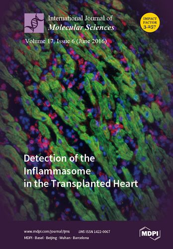
Cover Story (view full-size image): Detection of the Inflammasome in the Transplanted Heart.
Bioptic samples are used to monitor the type and severity of organ rejection. The fluorescent staining of a bioptic sample is depicted in this picture, to detect the presence of the inflammasome scaffold protein, ASC (in red). The inflammasome is part of the innate immune response. The co-staining of the cardiac alpha-actin (in green) is used to mark the cardiac myocytes. Myocytes are surrounded by infiltrating cells (leukocytes), visible through the nuclear staining (blue). In this particular picture, the myocardial structure is disrupted and ASC positivity is detected in both cardiomyocytes and non-cardiomyocyte cells.
By Adolfo Gabriele Mauro. View this article.
  • Issues are regarded as officially published after their release is announced to the table of contents alert mailing list.
  • You may sign up for e-mail alerts to receive table of contents of newly released issues.
  • PDF is the official format for papers published in both, html and pdf forms. To view the papers in pdf format, click on the "PDF Full-text" link, and use the free Adobe Reader to open them.
Order results
Result details
Section
Select all
Export citation of selected articles as:

Research

Jump to: Review, Other

16 pages, 2205 KiB  
Article
A Theoretical Study of the Hydration of Methane, from the Aqueous Solution to the sI Hydrate-Liquid Water-Gas Coexistence
by Daniel Porfirio Luis, Alcione García-González and Humberto Saint-Martin
Int. J. Mol. Sci. 2016, 17(6), 378; https://doi.org/10.3390/ijms17060378 - 26 May 2016
Cited by 11 | Viewed by 6273
Abstract
Monte Carlo and molecular dynamics simulations were done with three recent water models TIP4P/2005 (Transferable Intermolecular Potential with 4 Points/2005), TIP4P/Ice (Transferable Intermolecular Potential with 4 Points/ Ice) and TIP4Q (Transferable Intermolecular Potential with 4 charges) combined with two models for methane: an [...] Read more.
Monte Carlo and molecular dynamics simulations were done with three recent water models TIP4P/2005 (Transferable Intermolecular Potential with 4 Points/2005), TIP4P/Ice (Transferable Intermolecular Potential with 4 Points/ Ice) and TIP4Q (Transferable Intermolecular Potential with 4 charges) combined with two models for methane: an all-atom one OPLS-AA (Optimal Parametrization for the Liquid State) and a united-atom one (UA); a correction for the C–O interaction was applied to the latter and used in a third set of simulations. The models were validated by comparison to experimental values of the free energy of hydration at 280, 300, 330 and 370 K, all under a pressure of 1 bar, and to the experimental radial distribution functions at 277, 283 and 291 K, under a pressure of 145 bar. Regardless of the combination rules used for σC,O, good agreement was found, except when the correction to the UA model was applied. Thus, further simulations of the sI hydrate were performed with the united-atom model to compare the thermal expansivity to the experiment. A final set of simulations was done with the UA methane model and the three water models, to study the sI hydrate-liquid water-gas coexistence at 80, 230 and 400 bar. The melting temperatures were compared to the experimental values. The results show the need to perform simulations with various different models to attain a reliable and robust molecular image of the systems of interest. Full article
(This article belongs to the Special Issue Solution Chemical Kinetics)
Show Figures

Figure 1

14 pages, 4410 KiB  
Article
The Transcription Factor OsWRKY45 Negatively Modulates the Resistance of Rice to the Brown Planthopper Nilaparvata lugens
by Jiayi Huangfu, Jiancai Li, Ran Li, Meng Ye, Peng Kuai, Tongfang Zhang and Yonggen Lou
Int. J. Mol. Sci. 2016, 17(6), 697; https://doi.org/10.3390/ijms17060697 - 31 May 2016
Cited by 68 | Viewed by 9726
Abstract
WRKY transcription factors play a central role not only in plant growth and development but also in plant stress responses. However, the role of WRKY transcription factors in herbivore-induced plant defenses and their underlying mechanisms, especially in rice, remains largely unclear. Here, we [...] Read more.
WRKY transcription factors play a central role not only in plant growth and development but also in plant stress responses. However, the role of WRKY transcription factors in herbivore-induced plant defenses and their underlying mechanisms, especially in rice, remains largely unclear. Here, we cloned a rice WRKY gene OsWRKY45, whose expression was induced by mechanical wounding, by infestation of the brown planthopper (BPH, Nilaparvata lugens) and by treatment with jasmonic acid (JA) or salicylic acid (SA). The antisense expression of OsWRKY45 (as-wrky) enhanced BPH-induced levels of H2O2 and ethylene, reduced feeding and oviposition preference as well as the survival rate of BPH, and delayed the development of BPH nymphs. Consistently, lower population densities of BPH on as-wrky lines, compared to those on wild-type (WT) plants, were observed in field experiments. On the other hand, as-wrky lines in the field had lower susceptibility to sheath blight (caused by Rhizoctonia solani) but higher susceptibility to rice blast (caused by Magnaporthe oryzae) than did WT plants. These findings suggest that OsWRKY45 plays important but contrasting roles in regulating the resistance of rice to pathogens and herbivores, and attention should be paid if OsWRKY45 is used to develop disease or herbivore-resistant rice. Full article
(This article belongs to the Special Issue Plant Innate Immunity)
Show Figures

Graphical abstract

18 pages, 596 KiB  
Article
A ‘High Risk’ Lifestyle Pattern Is Associated with Metabolic Syndrome among Qatari Women of Reproductive Age: A Cross-Sectional National Study
by Mohammed Al Thani, Al Anoud Al Thani, Walaa Al-Chetachi, Badria Al Malki, Shamseldin A. H. Khalifa, Ahmad Haj Bakri, Nahla Hwalla, Lara Nasreddine and Farah Naja
Int. J. Mol. Sci. 2016, 17(6), 698; https://doi.org/10.3390/ijms17060698 - 2 Jun 2016
Cited by 20 | Viewed by 6081
Abstract
This study investigated the effect of lifestyle patterns, as a combination of diet, physical activity and smoking, on Metabolic Syndrome (MetS) among Qatari women of childbearing age (n = 418), a population group particularly vulnerable to the health sequela of this syndrome. [...] Read more.
This study investigated the effect of lifestyle patterns, as a combination of diet, physical activity and smoking, on Metabolic Syndrome (MetS) among Qatari women of childbearing age (n = 418), a population group particularly vulnerable to the health sequela of this syndrome. Using data from the National WHO STEPwise survey conducted in Qatar in 2012, Principal Component Factor Analysis was performed to derive lifestyle patterns with survey variables related to the frequency of consumption of 13 foods/food groups, physical activity levels, and smoking status. MetS was diagnosed using ATPIII criteria. Three lifestyle patterns were identified: ‘High Risk’ pattern, characterized by intakes of fast foods, sweets and sugar sweetened beverages, in addition to lower levels of physical activity and higher smoking prevalence; ‘Prudent’ pattern, driven mainly by higher intakes of fruits, vegetables, fish, and whole grains; and ‘Traditional’ pattern which included beans, meat, dairy products, and a low prevalence of smoking. Among these three lifestyle patterns, only the ‘High Risk’ was associated with MetS, whereby subjects belonging to the third tertile of this pattern’s score had 2.5 times the odds of MetS compared to those belonging to the first tertile. The findings of this study demonstrated the synergy among high risk behaviors among Qatari women in increasing the odds of MetS; the latter being a major risk factor for cardiovascular diseases. Full article
(This article belongs to the Special Issue Advances in Nutritional Epidemiology)
Show Figures

Graphical abstract

14 pages, 2263 KiB  
Article
Pesticides Drive Stochastic Changes in the Chemoreception and Neurotransmission System of Marine Ectoparasites
by Gustavo Núñez-Acuña, Sebastián Boltaña and Cristian Gallardo-Escárate
Int. J. Mol. Sci. 2016, 17(6), 700; https://doi.org/10.3390/ijms17060700 - 31 May 2016
Cited by 10 | Viewed by 5260
Abstract
Scientific efforts to elucidate the mechanisms of chemical communication between organisms in marine environments are increasing. This study applied novel molecular technology to outline the effects of two xenobiotic drugs, deltamethrin (DM) and azamethiphos (AZA), on the neurotransmission system of the copepod ectoparasite [...] Read more.
Scientific efforts to elucidate the mechanisms of chemical communication between organisms in marine environments are increasing. This study applied novel molecular technology to outline the effects of two xenobiotic drugs, deltamethrin (DM) and azamethiphos (AZA), on the neurotransmission system of the copepod ectoparasite Caligus rogercresseyi. Transcriptome sequencing and bioinformatics analyses were conducted to evaluate treatment effects on the glutamatergic synaptic pathway of the parasite, which is closely related to chemoreception and neurotransmission. After drug treatment with DM or AZA, stochastic mRNA expression patterns of glutamatergic synapse pathway components were observed. Both DM and AZA promoted a down-regulation of the glutamate-ammonia ligase, and DM activated a metabotropic glutamate receptor that is a suggested inhibitor of neurotransmission. Furthermore, the delousing drugs drove complex rearrangements in the distribution of mapped reads for specific metabotropic glutamate receptor domains. This study introduces a novel methodological approach that produces high-quality results from transcriptomic data. Using this approach, DM and AZA were found to alter the expression of numerous mRNAs tightly linked to the glutamatergic signaling pathway. These data suggest possible new targets for xenobiotic drugs that play key roles in the delousing effects of antiparasitics in sea lice. Full article
(This article belongs to the Section Molecular Pathology, Diagnostics, and Therapeutics)
Show Figures

Graphical abstract

14 pages, 2770 KiB  
Article
Transcriptome and Difference Analysis of Fenpropathrin Resistant Predatory Mite, Neoseiulus barkeri (Hughes)
by Lin Cong, Fei Chen, Shijiang Yu, Lili Ding, Juan Yang, Ren Luo, Huixia Tian, Hongjun Li, Haoqiang Liu and Chun Ran
Int. J. Mol. Sci. 2016, 17(6), 704; https://doi.org/10.3390/ijms17060704 - 27 May 2016
Cited by 18 | Viewed by 5455
Abstract
Several fenpropathrin-resistant predatory mites have been reported. However, the molecular mechanism of the resistance remains unknown. In the present study, the Neoseiulus barkeri (N. barkeri) transcriptome was generated using the Illumina sequencing platform, 34,211 unigenes were obtained, and 15,987 were manually [...] Read more.
Several fenpropathrin-resistant predatory mites have been reported. However, the molecular mechanism of the resistance remains unknown. In the present study, the Neoseiulus barkeri (N. barkeri) transcriptome was generated using the Illumina sequencing platform, 34,211 unigenes were obtained, and 15,987 were manually annotated. After manual annotation, attentions were attracted to resistance-related genes, such as voltage-gated sodium channel (VGSC), cytochrome P450s (P450s), and glutathione S-transferases (GSTs). A polymorphism analysis detected two point mutations (E1233G and S1282G) in the linker region between VGSC domain II and III. In addition, 43 putative P450 genes and 10 putative GST genes were identified from the transcriptome. Among them, two P450 genes, NbCYP4EV2 and NbCYP4EZ1, and four GST genes, NbGSTd01, NbGSTd02, NbGSTd03 and NbGSTm03, were remarkably overexpressed 3.64–46.69-fold in the fenpropathrin resistant strain compared to that in the susceptible strain. These results suggest that fenpropathrin resistance in N. barkeri is a complex biological process involving many genetic changes and provide new insight into the N. barkeri resistance mechanism. Full article
(This article belongs to the Section Biochemistry)
Show Figures

Graphical abstract

13 pages, 1829 KiB  
Article
Synthesis, Biological Activity, and Apoptotic Properties of NO-Donor/Enmein-Type ent-Kauranoid Hybrids
by Dahong Li, Xu Hu, Tong Han, Shengtao Xu, Tingting Zhou, Zhenzhong Wang, Keguang Cheng, Zhanlin Li, Huiming Hua, Wei Xiao and Jinyi Xu
Int. J. Mol. Sci. 2016, 17(6), 747; https://doi.org/10.3390/ijms17060747 - 24 May 2016
Cited by 15 | Viewed by 5328
Abstract
Herein, we reported on a series of synthetic nitric oxide-releasing enmein-type diterpenoid hybrids (9ai). All the target compounds showed potent antibacterial activity against selected Gram-positive bacteria S. aureus and B. subtilis. The antiproliferative activity against human tumor K562, [...] Read more.
Herein, we reported on a series of synthetic nitric oxide-releasing enmein-type diterpenoid hybrids (9ai). All the target compounds showed potent antibacterial activity against selected Gram-positive bacteria S. aureus and B. subtilis. The antiproliferative activity against human tumor K562, MGC-803, CaEs-17 and Bel-7402 cells, and human normal liver cells L-02 was tested and the structure activity relationships (SARs) were also concluded. Compounds 9b and 9d showed the best activity against S. aureus and B. subtilis with the same minimal inhibitory concentrations (MICs) of 4 and 2 μg/mL, respectively. The derivative 9f displayed IC50 values of 1.68, 1.11, 3.60 and 0.72 μM against the four cancer cell lines above and 18.80 μM against normal liver cells L-02; meanwhile, 9f also released a high level of NO at the time point of 60 min of 22.24 μmol/L. Furthermore, it was also found that 9f induced apoptosis via the mitochondria-related pathway and arrested cell cycle of Bel-7402 cells at S phase. These findings might be important to explore new chemical entities for the main causes of in-hospital mortality of S. aureus infection, combined with a solid tumor. Full article
Show Figures

Graphical abstract

14 pages, 4599 KiB  
Article
MicroRNA-24 Attenuates Neointimal Hyperplasia in the Diabetic Rat Carotid Artery Injury Model by Inhibiting Wnt4 Signaling Pathway
by Jian Yang, Zhixing Fan, Jun Yang, Jiawang Ding, Chaojun Yang and Lihua Chen
Int. J. Mol. Sci. 2016, 17(6), 765; https://doi.org/10.3390/ijms17060765 - 24 May 2016
Cited by 29 | Viewed by 5838
Abstract
The long-term stimulation of hyperglycemia greatly increases the incidence of vascular restenosis (RS) after angioplasty. Neointimal hyperplasia after vascular injury is the pathological cause of RS, but its mechanism has not been elucidated. MicroRNA-24 (miR-24) has low expression in the injured carotid arteries [...] Read more.
The long-term stimulation of hyperglycemia greatly increases the incidence of vascular restenosis (RS) after angioplasty. Neointimal hyperplasia after vascular injury is the pathological cause of RS, but its mechanism has not been elucidated. MicroRNA-24 (miR-24) has low expression in the injured carotid arteries of diabetic rats. However, the role of miR-24 in the vascular system is unknown. In this study, we explore whether over-expression of miR-24 could attenuate neointimal formation in streptozotocin (STZ)-induced diabetic rats. Adenovirus (Ad-miR-24-GFP) was used to deliver the miR-24 gene to injured carotid arteries in diabetic rats. The level of neointimal hyperplasia was examined by hematoxylin-eosin (HE) staining. Vascular smooth muscle cell (VSMC) proliferation in the neointima was evaluated by immunostaining for proliferating cell nuclear antigen (PCNA). The mRNA levels of miR-24, PCNA, wingless-type MMTV integration site family member 4 (Wnt4), disheveled-1 (Dvl-1), β-catenin and cell cycle-associated molecules (Cyclin D1, p21) were determined by Quantitative Real-Time PCR (qRT-PCR). PCNA, Wnt4, Dvl-1, β-catenin, Cyclin D1 and p21 protein levels were measured by Western blotting analysis. STZ administration decreased plasma insulin and increased fasting blood glucose in Sprague-Dawley (SD) rats. The expression of miR-24 was decreased in the carotid artery after a balloon injury in diabetic rats, and adenoviral transfection (Ad-miR-24-GFP) increased the expression of miR-24. Over-expression of miR-24 suppressed VSMC proliferation and neointimal hyperplasia in diabetic rats at 14 days. Furthermore, compared with Sham group, the mRNA and protein levels of PCNA, Wnt4, Dvl-1, β-catenin, and Cyclin D1 were strikingly up-regulated in the carotid arteries of diabetic rats after a balloon injury. Interestingly, up-regulation of miR-24 significantly reduced the mRNA and protein levels of these above molecules. In contrast, the change trend in p21 mRNA and protein levels was opposite after a balloon injury. However, over-expression of miR-24 after gene delivery increased the mRNA and protein levels of p21. We conclude that over-expression of miR-24 could attenuate VSMC proliferation and neointimal hyperplasia after vascular injuries in diabetic rats. This result is possibly related to the regulation of the expression of Cyclin D1 and p21 through the Wnt4/Dvl-1/β-catenin signaling pathway. Full article
(This article belongs to the Special Issue Molecular Research on Obesity and Diabetes)
Show Figures

Figure 1

14 pages, 5906 KiB  
Article
Effects of Endobacterium (Stenotrophomonas maltophilia) on Pathogenesis-Related Gene Expression of Pine Wood Nematode (Bursaphelenchus xylophilus) and Pine Wilt Disease
by Long-Xi He, Xiao-Qin Wu, Qi Xue and Xiu-Wen Qiu
Int. J. Mol. Sci. 2016, 17(6), 778; https://doi.org/10.3390/ijms17060778 - 25 May 2016
Cited by 26 | Viewed by 6437
Abstract
Pine wilt disease (PWD) caused by the pine wood nematode (PWN), Bursaphelenchus xylophilus, is responsible for devastating epidemics in pine trees in Asia and Europe. Recent studies showed that bacteria carried by the PWN might be involved in PWD. However, the molecular [...] Read more.
Pine wilt disease (PWD) caused by the pine wood nematode (PWN), Bursaphelenchus xylophilus, is responsible for devastating epidemics in pine trees in Asia and Europe. Recent studies showed that bacteria carried by the PWN might be involved in PWD. However, the molecular mechanism of the interaction between bacteria and the PWN remained unclear. Now that the whole genome of B. xylophilus (Bursaphelenchus xylophilus) is published, transcriptome analysis is a unique method to study the role played by bacteria in PWN. In this study, the transcriptome of aseptic B. xylophilus, B. xylophilus treated with endobacterium (Stenotrophomonas maltophilia NSPmBx03) and fungus B. xylophilus were sequenced. We found that 61 genes were up-regulated and 830 were down-regulated in B. xylophilus after treatment with the endobacterium; 178 genes were up-regulated and 1122 were down-regulated in fungus B. xylophilus compared with aseptic B. xylophilus. Gene Ontology and Kyoto Encyclopedia of Genes and Genomes analyses were used to study the significantly changed biological functions and pathways for these differentially expressed genes. Many pathogenesis-related genes, including glutathinone S-transferase, pectate lyase, ATP-binding cassette transporter and cytochrome P450, were up-regulated after B. xylophilus were treated with the endobacterium. In addition, we found that bacteria enhanced the virulence of PWN. These findings indicate that endobacteria might play an important role in the development and virulence of PWN and will improve our understanding of the regulatory mechanisms involved in the interaction between bacteria and the PWN. Full article
(This article belongs to the Special Issue Microbial Genomics and Metabolomics)
Show Figures

Graphical abstract

14 pages, 1761 KiB  
Article
Goldfish Leptin-AI and Leptin-AII: Function and Central Mechanism in Feeding Control
by Ai-Fen Yan, Ting Chen, Shuang Chen, Chun-Hua Ren, Chao-Qun Hu, Yi-Ming Cai, Fang Liu and Dong-Sheng Tang
Int. J. Mol. Sci. 2016, 17(6), 783; https://doi.org/10.3390/ijms17060783 - 30 May 2016
Cited by 48 | Viewed by 6838
Abstract
In mammals, leptin is a peripheral satiety factor that inhibits feeding by regulating a variety of appetite-related hormones in the brain. However, most of the previous studies examining leptin in fish feeding were performed with mammalian leptins, which share very low sequence homologies [...] Read more.
In mammals, leptin is a peripheral satiety factor that inhibits feeding by regulating a variety of appetite-related hormones in the brain. However, most of the previous studies examining leptin in fish feeding were performed with mammalian leptins, which share very low sequence homologies with fish leptins. To elucidate the function and mechanism of endogenous fish leptins in feeding regulation, recombinant goldfish leptin-AI and leptin-AII were expressed in methylotrophic yeast and purified by immobilized metal ion affinity chromatography (IMAC). By intraperitoneal (IP) injection, both leptin-AI and leptin-AII were shown to inhibit the feeding behavior and to reduce the food consumption of goldfish in 2 h. In addition, co-treatment of leptin-AI or leptin-AII could block the feeding behavior and reduce the food consumption induced by neuropeptide Y (NPY) injection. High levels of leptin receptor (lepR) mRNA were detected in the hypothalamus, telencephalon, optic tectum and cerebellum of the goldfish brain. The appetite inhibitory effects of leptins were mediated by downregulating the mRNA levels of orexigenic NPY, agouti-related peptide (AgRP) and orexin and upregulating the mRNA levels of anorexigenic cocaine-amphetamine-regulated transcript (CART), cholecystokinin (CCK), melanin-concentrating hormone (MCH) and proopiomelanocortin (POMC) in different areas of the goldfish brain. Our study, as a whole, provides new insights into the functions and mechanisms of leptins in appetite control in a fish model. Full article
(This article belongs to the Special Issue Fish Molecular Biology)
Show Figures

Graphical abstract

12 pages, 12443 KiB  
Article
Hypoxia Inducible Factor 1 (HIF-1) Recruits Macrophage to Activate Pancreatic Stellate Cells in Pancreatic Ductal Adenocarcinoma
by Na Li, Yang Li, Zengxun Li, Chongbiao Huang, Yanhui Yang, Mingxiao Lang, Junli Cao, Wenna Jiang, Yu Xu, Jie Dong and He Ren
Int. J. Mol. Sci. 2016, 17(6), 799; https://doi.org/10.3390/ijms17060799 - 3 Jun 2016
Cited by 86 | Viewed by 9215
Abstract
Hypoxia inducible factor 1 (HIF-1) is a transcription factor composed of two subunits, namely, HIF-1α and HIF-1β, in which HIF-1β is constitutively expressed. HIF-1 upregulates several hypoxia-responsive proteins, including angiogenesis factors, glycolysis solution enzymes, and cell survival proteins. HIF-1 is also associated with [...] Read more.
Hypoxia inducible factor 1 (HIF-1) is a transcription factor composed of two subunits, namely, HIF-1α and HIF-1β, in which HIF-1β is constitutively expressed. HIF-1 upregulates several hypoxia-responsive proteins, including angiogenesis factors, glycolysis solution enzymes, and cell survival proteins. HIF-1 is also associated with the degree of inflammation in the tumor region, but the exact mechanism remains unclear. This study aims to identify the molecular mechanism of recruiting monocytes/macrophages by HIF-1α in pancreatic ductal adenocarcinoma (PDAC) and the effects of macrophages on pancreatic stellate cells (PSCs). Immunohistochemistry (IHC) was performed for cluster of differentiation 68 (CD68), HIF-1α, and chemical chemokines 2 (CCL2). Western blot, real-time quantitative reverse transcription polymerase chain reaction (qRT-PCR), chromatin immunoprecipitation assay, and The Cancer Genome Atlas (TCGA) were used to verify the correlation between HIF-1α and CCL2 at protein and nucleic acid levels. Monocytes/macrophages were co-cultured with PSCs to observe their interaction. Samples showed significant correlation between CD68 and HIF-1α (t-test, p < 0.05). HIF-1α recruited monocytes/macrophages by promoting CCL2 secretion. Moreover, macrophages could accelerate the activation of PSCs. HIF-1α might promote inflammation and fibrosis of PDAC through CCL2 secretion, which may provide a novel target to treat PDAC patients. Full article
Show Figures

Graphical abstract

17 pages, 4112 KiB  
Article
Cardiac Stem Cell Secretome Protects Cardiomyocytes from Hypoxic Injury Partly via Monocyte Chemotactic Protein-1-Dependent Mechanism
by Chi-Yeon Park, Seung-Cheol Choi, Jong-Ho Kim, Ji-Hyun Choi, Hyung Joon Joo, Soon Jun Hong and Do-Sun Lim
Int. J. Mol. Sci. 2016, 17(6), 800; https://doi.org/10.3390/ijms17060800 - 24 May 2016
Cited by 15 | Viewed by 7610
Abstract
Cardiac stem cells (CSCs) were known to secrete diverse paracrine factors leading to functional improvement and beneficial left ventricular remodeling via activation of the endogenous pro-survival signaling pathway. However, little is known about the paracrine factors secreted by CSCs and their roles in [...] Read more.
Cardiac stem cells (CSCs) were known to secrete diverse paracrine factors leading to functional improvement and beneficial left ventricular remodeling via activation of the endogenous pro-survival signaling pathway. However, little is known about the paracrine factors secreted by CSCs and their roles in cardiomyocyte survival during hypoxic condition mimicking the post-myocardial infarction environment. We established Sca-1+/CD31− human telomerase reverse transcriptase-immortalized CSCs (Sca-1+/CD31− CSCshTERT), evaluated their stem cell properties, and paracrine potential in cardiomyocyte survival during hypoxia-induced injury. Sca-1+/CD31− CSCshTERT sustained proliferation ability even after long-term culture exceeding 100 population doublings, and represented multi-differentiation potential into cardiomyogenic, endothelial, adipogenic, and osteogenic lineages. Dominant factors secreted from Sca-1+/CD31− CSCshTERT were EGF, TGF-β1, IGF-1, IGF-2, MCP-1, HGF R, and IL-6. Among these, MCP-1 was the most predominant factor in Sca-1+/CD31− CSCshTERT conditioned medium (CM). Sca-1+/CD31− CSCshTERT CM increased survival and reduced apoptosis of HL-1 cardiomyocytes during hypoxic injury. MCP-1 silencing in Sca-1+/CD31− CSCshTERT CM resulted in a significant reduction in cardiomyocyte apoptosis. We demonstrated that Sca-1+/CD31− CSCshTERT exhibited long-term proliferation capacity and multi-differentiation potential. Sca-1+/CD31− CSCshTERT CM protected cardiomyocytes from hypoxic injury partly via MCP-1-dependent mechanism. Thus, they are valuable sources for in vitro and in vivo studies in the cardiovascular field. Full article
Show Figures

Graphical abstract

12 pages, 7999 KiB  
Article
Kidney Injury Molecule-1 Is Specifically Expressed in Cystically-Transformed Proximal Tubules of the PKD/Mhm (cy/+) Rat Model of Polycystic Kidney Disease
by Stefan Gauer, Anja Urbschat, Norbert Gretz, Sigrid C. Hoffmann, Bettina Kränzlin, Helmut Geiger and Nicholas Obermüller
Int. J. Mol. Sci. 2016, 17(6), 802; https://doi.org/10.3390/ijms17060802 - 24 May 2016
Cited by 14 | Viewed by 8571
Abstract
Expression of kidney injury molecule-1 (Kim-1) is rapidly upregulated following tubular injury, constituting a biomarker for acute kidney damage. We examined the renal localization of Kim-1 expression in PKD/Mhm (polycystic kidney disease, Mannheim) (cy/+) rats (cy: mutated allel, +: wild type allel), an [...] Read more.
Expression of kidney injury molecule-1 (Kim-1) is rapidly upregulated following tubular injury, constituting a biomarker for acute kidney damage. We examined the renal localization of Kim-1 expression in PKD/Mhm (polycystic kidney disease, Mannheim) (cy/+) rats (cy: mutated allel, +: wild type allel), an established model for autosomal dominant polycystic kidney disease, with chronic, mainly proximal tubulointerstitial alterations. For immunohistochemistry or Western blot analysis, kidneys of male adult heterozygously-affected (cy/+) and unaffected (+/+) littermates were perfusion-fixed or directly removed. Kim-1 expression was determined using peroxidase- or fluorescence-linked immunohistochemistry (alone or in combination with markers for tubule segments or differentiation). Compared to (+/+), only in (cy/+) kidneys, a chronic expression of Kim-1 could be detected by Western blot analysis, which was histologically confined to an apical cellular localization in areas of cystically-transformed proximal tubules with varying size and morphology, but not in distal tubular segments. Kim-1 was expressed by cystic epithelia exhibiting varying extents of dedifferentiation, as shown by double labeling with aquaporin-1, vimentin or osteopontin, yielding partial cellular coexpression. In this model, in contrast to other known molecules indicating renal injury and/or repair mechanisms, the chronic renal expression of Kim-1 is strictly confined to proximal cysts. Its exact role in interfering with tubulo-interstitial alterations in polycystic kidney disease warrants future investigations. Full article
(This article belongs to the Special Issue Advances in Chronic Kidney Disease)
Show Figures

Graphical abstract

13 pages, 5187 KiB  
Article
A Chloroplast-Localized Rubredoxin Family Protein Gene from Puccinellia tenuiflora (PutRUB) Increases NaCl and NaHCO3 Tolerance by Decreasing H2O2 Accumulation
by Ying Li, Panpan Liu, Tetsuo Takano and Shenkui Liu
Int. J. Mol. Sci. 2016, 17(6), 804; https://doi.org/10.3390/ijms17060804 - 30 May 2016
Cited by 12 | Viewed by 5844
Abstract
Rubredoxin is one of the simplest iron–sulfur (Fe–S) proteins. It is found primarily in strict anaerobic bacteria and acts as a mediator of electron transfer participation in different biochemical reactions. The PutRUB gene encoding a chloroplast-localized rubredoxin family protein was screened from a [...] Read more.
Rubredoxin is one of the simplest iron–sulfur (Fe–S) proteins. It is found primarily in strict anaerobic bacteria and acts as a mediator of electron transfer participation in different biochemical reactions. The PutRUB gene encoding a chloroplast-localized rubredoxin family protein was screened from a yeast full-length cDNA library of Puccinellia tenuiflora under NaCl and NaHCO3 stress. We found that PutRUB expression was induced by abiotic stresses such as NaCl, NaHCO3, CuCl2 and H2O2. These findings suggested that PutRUB might be involved in plant responses to adversity. In order to study the function of this gene, we analyzed the phenotypic and physiological characteristics of PutRUB transgenic plants treated with NaCl and NaHCO3. The results showed that PutRUB overexpression inhibited H2O2 accumulation, and enhanced transgenic plant adaptability to NaCl and NaHCO3 stresses. This indicated PutRUB might be involved in maintaining normal electron transfer to reduce reactive oxygen species (ROS) accumulation. Full article
(This article belongs to the Section Molecular Plant Sciences)
Show Figures

Figure 1

12 pages, 2227 KiB  
Article
The Protective Effect of Grape-Seed Proanthocyanidin Extract on Oxidative Damage Induced by Zearalenone in Kunming Mice Liver
by Miao Long, Shu-Hua Yang, Jian-Xin Han, Peng Li, Yi Zhang, Shuang Dong, Xinliang Chen, Jiayi Guo, Jun Wang and Jian-Bin He
Int. J. Mol. Sci. 2016, 17(6), 808; https://doi.org/10.3390/ijms17060808 - 25 May 2016
Cited by 76 | Viewed by 7102
Abstract
Although grape-seed proanthocyanidin extract (GSPE) demonstrates strong anti-oxidant activity, little research has been done to clearly reveal the protective effects on the hepatotoxicity caused by zearalenone (ZEN). This study is to explore the protective effect of GSPE on ZEN-induced oxidative damage of liver [...] Read more.
Although grape-seed proanthocyanidin extract (GSPE) demonstrates strong anti-oxidant activity, little research has been done to clearly reveal the protective effects on the hepatotoxicity caused by zearalenone (ZEN). This study is to explore the protective effect of GSPE on ZEN-induced oxidative damage of liver in Kunming mice and the possible protective molecular mechanism of GSPE. The results indicated that GSPE could greatly reduce the ZEN-induced increase of serum aspartate aminotransferase (AST) and alanine aminotransferase (ALT) activities. GSPE also significantly decreased the content of MDA but enhanced the activities of antioxidant enzymes SOD and GSH-Px. The analysis indicated that ZEN decreased both mRNA expression levels and protein expression levels of nuclear erythroid2-related factor2 (Nrf2). Nrf2 is considered to be an essential antioxidative transcription factor, as downstream GSH-Px, γ-glutamyl cysteine synthetase (γ-GCS), hemeoxygenase-1 (HO-1), and quinone oxidoreductase 1 (NQO1) decreased simultaneously, whereas the pre-administration of GSPE groups was shown to elevate these expressions. The results indicated that GSPE exerted a protective effect on ZEN-induced hepatic injury and the mechanism might be related to the activation of the Nrf2/ARE signaling pathway. Full article
(This article belongs to the Section Bioactives and Nutraceuticals)
Show Figures

Graphical abstract

11 pages, 3527 KiB  
Article
Targeted Gene Knockin in Porcine Somatic Cells Using CRISPR/Cas Ribonucleoproteins
by Ki-Eun Park, Chi-Hun Park, Anne Powell, Jessica Martin, David M. Donovan and Bhanu P. Telugu
Int. J. Mol. Sci. 2016, 17(6), 810; https://doi.org/10.3390/ijms17060810 - 26 May 2016
Cited by 15 | Viewed by 8086
Abstract
The pig is an ideal large animal model for genetic engineering applications. A relatively short gestation interval and large litter size makes the pig a conducive model for generating and propagating genetic modifications. The domestic pig also shares close similarity in anatomy, physiology, [...] Read more.
The pig is an ideal large animal model for genetic engineering applications. A relatively short gestation interval and large litter size makes the pig a conducive model for generating and propagating genetic modifications. The domestic pig also shares close similarity in anatomy, physiology, size, and life expectancy, making it an ideal animal for modeling human diseases. Often, however, the technical difficulties in generating desired genetic modifications such as targeted knockin of short stretches of sequences or transgenes have impeded progress in this field. In this study, we have investigated and compared the relative efficiency of CRISPR/Cas ribonucleoproteins in engineering targeted knockin of pseudo attP sites downstream of a ubiquitously expressed COL1A gene in porcine somatic cells and generated live fetuses by somatic cell nuclear transfer (SCNT). By leveraging these knockin pseudo attP sites, we have demonstrated subsequent phiC31 integrase mediated integration of green fluorescent protein (GFP) transgene into the site. This work for the first time created an optimized protocol for CRISPR/Cas mediated knockin in porcine somatic cells, while simultaneously creating a stable platform for future transgene integration and generating transgenic animals. Full article
(This article belongs to the Special Issue Genome Editing)
Show Figures

Figure 1

12 pages, 6526 KiB  
Article
EIYMNVPV Motif is Essential for A1CF Nucleus Localization and A1CF (-8aa) Promotes Proliferation of MDA-MB-231 Cells via Up-Regulation of IL-6
by Li Zhou, Jin Hao, Yue Yuan, Rui Peng, Honglian Wang, Dongsheng Ni, Yuping Gu, Liyuan Huang, Zhaomin Mao, Zhongshi Lyu, Yao Du, Zhicheng Liu, Yiman Li, Pan Ju, Yaoshui Long, Jianing Liu and Qin Zhou
Int. J. Mol. Sci. 2016, 17(6), 811; https://doi.org/10.3390/ijms17060811 - 25 May 2016
Cited by 9 | Viewed by 6409
Abstract
Apobec-1 complementation factor (A1CF) is a heterogeneous nuclear ribonuceloprotein (hnRNP) and mediates apolipoprotein-B mRNA editing. A1CF can promote the regeneration of the liver by post-transcriptionally stabilizing Interleukin-6 (IL-6) mRNA. It also contains two transcriptional variants-A1CF64 and A1CF65, distinguished by the appearance [...] Read more.
Apobec-1 complementation factor (A1CF) is a heterogeneous nuclear ribonuceloprotein (hnRNP) and mediates apolipoprotein-B mRNA editing. A1CF can promote the regeneration of the liver by post-transcriptionally stabilizing Interleukin-6 (IL-6) mRNA. It also contains two transcriptional variants-A1CF64 and A1CF65, distinguished by the appearance of a 24-nucleotide motif which contributes to the corresponding eight-amino acid motif of EIYMNVPV. For the first time, we demonstrated that the EIYMNVPV motif was essential for A1CF nucleus localization, A1CF deficient of the EIYMNVPV motif, A1CF (-8aa) showed cytoplasm distribution. More importantly, we found that A1CF (-8aa), but not its full-length counterpart, can promote proliferation of MDA-MB-231 cells accompanied with increased level of IL-6 mRNA. Furthermore, silencing of IL-6 attenuated A1CF (-8aa)-induced proliferation in MDA-MB-231 cells. In conclusion, notably, these findings suggest that A1CF (-8aa) promoted proliferation of MDA-MB-231 cells in vitro viewing IL-6 as a target. Thus, the EIYMNVPV motif could be developed as a potential target for basal-like breast cancer therapy. Full article
(This article belongs to the Special Issue Molecular Classification of Human Cancer: Diagnosis and Treatment)
Show Figures

Graphical abstract

30 pages, 15608 KiB  
Article
Physico-Chemical and Structural Interpretation of Discrete Derivative Indices on N-Tuples Atoms
by Oscar Martínez-Santiago, Yovani Marrero-Ponce, Stephen J. Barigye, Huong Le Thi Thu, F. Javier Torres, Cesar H. Zambrano, Jorge L. Muñiz Olite, Maykel Cruz-Monteagudo, Ricardo Vivas-Reyes, Liliana Vázquez Infante and Luis M. Artiles Martínez
Int. J. Mol. Sci. 2016, 17(6), 812; https://doi.org/10.3390/ijms17060812 - 27 May 2016
Cited by 7 | Viewed by 6465
Abstract
This report examines the interpretation of the Graph Derivative Indices (GDIs) from three different perspectives (i.e., in structural, steric and electronic terms). It is found that the individual vertex frequencies may be expressed in terms of the geometrical and electronic reactivity [...] Read more.
This report examines the interpretation of the Graph Derivative Indices (GDIs) from three different perspectives (i.e., in structural, steric and electronic terms). It is found that the individual vertex frequencies may be expressed in terms of the geometrical and electronic reactivity of the atoms and bonds, respectively. On the other hand, it is demonstrated that the GDIs are sensitive to progressive structural modifications in terms of: size, ramifications, electronic richness, conjugation effects and molecular symmetry. Moreover, it is observed that the GDIs quantify the interaction capacity among molecules and codify information on the activation entropy. A structure property relationship study reveals that there exists a direct correspondence between the individual frequencies of atoms and Hückel’s Free Valence, as well as between the atomic GDIs and the chemical shift in NMR, which collectively validates the theory that these indices codify steric and electronic information of the atoms in a molecule. Taking in consideration the regularity and coherence found in experiments performed with the GDIs, it is possible to say that GDIs possess plausible interpretation in structural and physicochemical terms. Full article
Show Figures

Graphical abstract

10 pages, 2775 KiB  
Article
Crystal Structure of Cytochrome P450 (CYP105P2) from Streptomyces peucetius and Its Conformational Changes in Response to Substrate Binding
by Chang Woo Lee, Joo-Ho Lee, Hemraj Rimal, Hyun Park, Jun Hyuck Lee and Tae-Jin Oh
Int. J. Mol. Sci. 2016, 17(6), 813; https://doi.org/10.3390/ijms17060813 - 25 May 2016
Cited by 11 | Viewed by 6117
Abstract
Cytochrome P450 monooxygenases (CYP, EC 1.14.14.1) belong to a large family of enzymes that catalyze the hydroxylation of various substrates. Here, we present the crystal structure of CYP105P2 isolated from Streptomyces peucetius ATCC27952 at a 2.1 Å resolution. The structure shows the presence [...] Read more.
Cytochrome P450 monooxygenases (CYP, EC 1.14.14.1) belong to a large family of enzymes that catalyze the hydroxylation of various substrates. Here, we present the crystal structure of CYP105P2 isolated from Streptomyces peucetius ATCC27952 at a 2.1 Å resolution. The structure shows the presence of a pseudo-ligand molecule in the active site, which was co-purified fortuitously and is presumed to be a biphenyl derivative. Comparison with previously determined substrate-bound CYP structures showed that binding of the ligand produces large and distinctive conformational changes in α2–α3, α7–α9, and the C-terminal loop regions. This structural flexibility confirms our previous observation that CYP105P2 can accommodate a broad range of ligands. The structure complexed with a pseudo-ligand provides the first molecular view of CYP105P2–ligand interactions, and it indicates the involvement of hydrophobic residues (Pro82, Ala181, Met187, Leu189, Leu193, and Ile236) in the interactions between hydrophobic ligands and CYP105P2. These results provide useful insights into the structural changes involved in the recognition of different ligands by CYP105P2. Full article
(This article belongs to the Section Physical Chemistry, Theoretical and Computational Chemistry)
Show Figures

Graphical abstract

14 pages, 1308 KiB  
Article
Role of Mitochondrial DNA Copy Number Alteration in Human Renal Cell Carcinoma
by Chen-Sung Lin, Hui-Ting Lee, Ming-Huei Lee, Siao-Cian Pan, Chen-Yeh Ke, Allen Wen-Hsiang Chiu and Yau-Huei Wei
Int. J. Mol. Sci. 2016, 17(6), 814; https://doi.org/10.3390/ijms17060814 - 25 May 2016
Cited by 60 | Viewed by 9193
Abstract
We investigated the role of mitochondrial DNA (mtDNA) copy number alteration in human renal cell carcinoma (RCC). The mtDNA copy numbers of paired cancer and non-cancer parts from five resected RCC kidneys after radical nephrectomy were determined by quantitative polymerase chain reaction (Q-PCR). [...] Read more.
We investigated the role of mitochondrial DNA (mtDNA) copy number alteration in human renal cell carcinoma (RCC). The mtDNA copy numbers of paired cancer and non-cancer parts from five resected RCC kidneys after radical nephrectomy were determined by quantitative polymerase chain reaction (Q-PCR). An RCC cell line, 786-O, was infected by lentiviral particles to knock down mitochondrial transcriptional factor A (TFAM). Null target (NT) and TFAM-knockdown (TFAM-KD) represented the control and knockdown 786-O clones, respectively. Protein or mRNA expression levels of TFAM; mtDNA-encoded NADH dehydrogenase subunit 1 (ND1), ND6 and cytochrome c oxidase subunit 2 (COX-2); nuclear DNA (nDNA)-encoded succinate dehydrogenase subunit A (SDHA); v-akt murine thymoma viral oncogene homolog 1 gene (AKT)-encoded AKT and v-myc myelocytomatosis viral oncogene homolog gene (c-MYC)-encoded MYC; glycolytic enzymes including hexokinase II (HK-II), glucose 6-phosphate isomerase (GPI), phosphofructokinase (PFK), and lactate dehydrogenase subunit A (LDHA); and hypoxia-inducible factors the HIF-1α and HIF-2α, pyruvate dehydrogenase kinase 1 (PDK1), and pyruvate dehydrogenase E1 component α subunit (PDHA1) were analyzed by Western blot or Q-PCR. Bioenergetic parameters of cellular metabolism, basal mitochondrial oxygen consumption rate (mOCRB) and basal extracellular acidification rate (ECARB), were measured by a Seahorse XFe-24 analyzer. Cell invasiveness was evaluated by a trans-well migration assay and vimentin expression. Doxorubicin was used as a chemotherapeutic agent. The results showed a decrease of mtDNA copy numbers in resected RCC tissues (p = 0.043). The TFAM-KD clone expressed lower mtDNA copy number (p = 0.034), lower mRNA levels of TFAM (p = 0.008), ND1 (p = 0.007), and ND6 (p = 0.017), and lower protein levels of TFAM and COX-2 than did the NT clone. By contrast, the protein levels of HIF-2α, HK-II, PFK, LDHA, AKT, MYC and vimentin; trans-well migration activity (p = 0.007); and drug resistance to doxorubicin (p = 0.008) of the TFAM-KD clone were significantly higher than those of the NT clone. Bioenergetically, the TFAM-KD clone expressed lower mOCRB (p = 0.009) but higher ECARB (p = 0.037) than did the NT clone. We conclude that a reduction of mtDNA copy number and decrease of respiratory function of mitochondria in RCC might be compensated for by an increase of enzymes and factors that are involved in the upregulation of glycolysis to confer RCC more invasive and a drug-resistant phenotype in vitro. Full article
(This article belongs to the Special Issue Tumor Microenvironment and Metabolism)
Show Figures

Graphical abstract

19 pages, 1703 KiB  
Article
Metabolomic Profiling of Bradyrhizobium diazoefficiens-Induced Root Nodules Reveals Both Host Plant-Specific and Developmental Signatures
by Martina Lardi, Valérie Murset, Hans-Martin Fischer, Socorro Mesa, Christian H. Ahrens, Nicola Zamboni and Gabriella Pessi
Int. J. Mol. Sci. 2016, 17(6), 815; https://doi.org/10.3390/ijms17060815 - 27 May 2016
Cited by 39 | Viewed by 8828
Abstract
Bradyrhizobium diazoefficiens is a nitrogen-fixing endosymbiont, which can grow inside root-nodule cells of the agriculturally important soybean and other host plants. Our previous studies described B. diazoefficiens host-specific global expression changes occurring during legume infection at the transcript and protein level. In order [...] Read more.
Bradyrhizobium diazoefficiens is a nitrogen-fixing endosymbiont, which can grow inside root-nodule cells of the agriculturally important soybean and other host plants. Our previous studies described B. diazoefficiens host-specific global expression changes occurring during legume infection at the transcript and protein level. In order to further characterize nodule metabolism, we here determine by flow injection–time-of-flight mass spectrometry analysis the metabolome of (i) nodules and roots from four different B. diazoefficiens host plants; (ii) soybean nodules harvested at different time points during nodule development; and (iii) soybean nodules infected by two strains mutated in key genes for nitrogen fixation, respectively. Ribose (soybean), tartaric acid (mungbean), hydroxybutanoyloxybutanoate (siratro) and catechol (cowpea) were among the metabolites found to be specifically elevated in one of the respective host plants. While the level of C4-dicarboxylic acids decreased during soybean nodule development, we observed an accumulation of trehalose-phosphate at 21 days post infection (dpi). Moreover, nodules from non-nitrogen-fixing bacteroids (nifA and nifH mutants) showed specific metabolic alterations; these were also supported by independent transcriptomics data. The alterations included signs of nitrogen limitation in both mutants, and an increased level of a phytoalexin in nodules induced by the nifA mutant, suggesting that the tissue of these nodules exhibits defense and stress reactions. Full article
(This article belongs to the Special Issue Molecular Signals in Nodulation Control)
Show Figures

Graphical abstract

15 pages, 2616 KiB  
Article
MT1-MMP Inhibits the Activity of Bst-2 via Their Cytoplasmic Domains Dependent Interaction
by Long Fan, Li Liu, Cuicui Zhu, Qingyi Zhu, Shan Lu and Ping Liu
Int. J. Mol. Sci. 2016, 17(6), 818; https://doi.org/10.3390/ijms17060818 - 26 May 2016
Cited by 18 | Viewed by 6266
Abstract
Bst-2 (bone marrow stromal cell antigen 2) is a type II membrane protein, and it acts as a tetherin to inhibit virion releasing from infectious cells. Membrane type-1 matrix metalloproteinase (MT1-MMP) is a protease. It plays a pivotal role in cellular growth and [...] Read more.
Bst-2 (bone marrow stromal cell antigen 2) is a type II membrane protein, and it acts as a tetherin to inhibit virion releasing from infectious cells. Membrane type-1 matrix metalloproteinase (MT1-MMP) is a protease. It plays a pivotal role in cellular growth and migration by activating proMMP-2 into active MMP2. Our results here elaborate that MT1-MMP inhibits the tetherin activity of Bst-2 by interacting with Bst-2, and the cytoplasmic domains of both Bst-2 and MT1-MMP play critical roles within this interaction. Based on our experimental data, the assays for virion release and co-immunoprecipitation have clearly demonstrated that the activity of Bst-2 is markedly inhibited by MT1-MMP via their interaction; and both the N-terminal domain of Bst-2 and the C-terminal domain of MT1-MMP are important in the interaction. Immunostaining and Confocal Microscopy assay shows that MT1-MMP interacts with Bst-2 to form granular particles trafficking into cytoplasm from membrane and, finally, results in Bst-2 and MT1-MMP both being inhibited. In addition, mutant experiments elucidate that the N-terminal domain of Bst-2 is not only important in relating to the activity of Bst-2 itself, but is important for inhibiting the MT1-MMP/proMMP2/MMP2 pathway. These findings suggest that MT1-MMP is a novel inhibitor of Bst-2 in MT1-MMP expressed cell lines and also indicate that both the N-terminal domain of Bst-2 and the C-terminal domain of MT1-MMP are crucial in down-regulation. Full article
(This article belongs to the Special Issue Metalloproteins)
Show Figures

Graphical abstract

15 pages, 4580 KiB  
Article
Computational Studies of a Mechanism for Binding and Drug Resistance in the Wild Type and Four Mutations of HIV-1 Protease with a GRL-0519 Inhibitor
by Guodong Hu, Aijing Ma, Xianghua Dou, Liling Zhao and Jihua Wang
Int. J. Mol. Sci. 2016, 17(6), 819; https://doi.org/10.3390/ijms17060819 - 27 May 2016
Cited by 34 | Viewed by 6175
Abstract
Drug resistance of mutations in HIV-1 protease (PR) is the most severe challenge to the long-term efficacy of HIV-1 PR inhibitor in highly active antiretroviral therapy. To elucidate the molecular mechanism of drug resistance associated with mutations (D30N, I50V, I54M, and V82A) and [...] Read more.
Drug resistance of mutations in HIV-1 protease (PR) is the most severe challenge to the long-term efficacy of HIV-1 PR inhibitor in highly active antiretroviral therapy. To elucidate the molecular mechanism of drug resistance associated with mutations (D30N, I50V, I54M, and V82A) and inhibitor (GRL-0519) complexes, we have performed five molecular dynamics (MD) simulations and calculated the binding free energies using the molecular mechanics Poisson–Boltzmann surface area (MM-PBSA) method. The ranking of calculated binding free energies is in accordance with the experimental data. The free energy spectra of each residue and inhibitor interaction for all complexes show a similar binding model. Analysis based on the MD trajectories and contribution of each residues show that groups R2 and R3 mainly contribute van der Waals energies, while groups R1 and R4 contribute electrostatic interaction by hydrogen bonds. The drug resistance of D30N can be attributed to the decline in binding affinity of residues 28 and 29. The size of Val50 is smaller than Ile50 causes the residue to move, especially in chain A. The stable hydrophobic core, including the side chain of Ile54 in the wild type (WT) complex, became unstable in I54M because the side chain of Met54 is flexible with two alternative conformations. The binding affinity of Ala82 in V82A decreases relative to Val82 in WT. The present study could provide important guidance for the design of a potent new drug resisting the mutation inhibitors. Full article
(This article belongs to the Special Issue Proteins and Protein-Ligand Interactions)
Show Figures

Graphical abstract

11 pages, 898 KiB  
Article
The Complete Chloroplast Genome Sequences of the Medicinal Plant Pogostemon cablin
by Yang He, Hongtao Xiao, Cao Deng, Liang Xiong, Jian Yang and Cheng Peng
Int. J. Mol. Sci. 2016, 17(6), 820; https://doi.org/10.3390/ijms17060820 - 6 Jun 2016
Cited by 62 | Viewed by 6912
Abstract
Pogostemon cablin, the natural source of patchouli alcohol, is an important herb in the Lamiaceae family. Here, we present the entire chloroplast genome of P. cablin. This genome, with 38.24% GC content, is 152,460 bp in length. The genome presents a [...] Read more.
Pogostemon cablin, the natural source of patchouli alcohol, is an important herb in the Lamiaceae family. Here, we present the entire chloroplast genome of P. cablin. This genome, with 38.24% GC content, is 152,460 bp in length. The genome presents a typical quadripartite structure with two inverted repeats (each 25,417 bp in length), separated by one small and one large single-copy region (17,652 and 83,974 bp in length, respectively). The chloroplast genome encodes 127 genes, of which 107 genes are single-copy, including 79 protein-coding genes, four rRNA genes, and 24 tRNA genes. The genome structure, GC content, and codon usage of this chloroplast genome are similar to those of other species in the family, except that it encodes less protein-coding genes and tRNA genes. Phylogenetic analysis reveals that P. cablin diverged from the Scutellarioideae clade about 29.45 million years ago (Mya). Furthermore, most of the simple sequence repeats (SSRs) are short polyadenine or polythymine repeats that contribute to high AT content in the chloroplast genome. Complete sequences and annotation of P. cablin chloroplast genome will facilitate phylogenic, population and genetic engineering research investigations involving this particular species. Full article
(This article belongs to the Section Molecular Plant Sciences)
Show Figures

Graphical abstract

19 pages, 3894 KiB  
Article
Metabolic Fingerprinting to Assess the Impact of Salinity on Carotenoid Content in Developing Tomato Fruits
by Lieven Van Meulebroek, Jochen Hanssens, Kathy Steppe and Lynn Vanhaecke
Int. J. Mol. Sci. 2016, 17(6), 821; https://doi.org/10.3390/ijms17060821 - 26 May 2016
Cited by 15 | Viewed by 8025
Abstract
As the presence of health-promoting substances has become a significant aspect of tomato fruit appreciation, this study investigated nutrient solution salinity as a tool to enhance carotenoid accumulation in cherry tomato fruit (Solanum lycopersicum L. cv. Juanita). Hereby, a key objective was [...] Read more.
As the presence of health-promoting substances has become a significant aspect of tomato fruit appreciation, this study investigated nutrient solution salinity as a tool to enhance carotenoid accumulation in cherry tomato fruit (Solanum lycopersicum L. cv. Juanita). Hereby, a key objective was to uncover the underlying mechanisms of carotenoid metabolism, moving away from typical black box research strategies. To this end, a greenhouse experiment with five salinity treatments (ranging from 2.0 to 5.0 decisiemens (dS) m−1) was carried out and a metabolomic fingerprinting approach was applied to obtain valuable insights on the complicated interactions between salinity treatments, environmental conditions, and the plant’s genetic background. Hereby, several hundreds of metabolites were attributed a role in the plant’s salinity response (at the fruit level), whereby the overall impact turned out to be highly depending on the developmental stage. In addition, 46 of these metabolites embraced a dual significance as they were ascribed a prominent role in carotenoid metabolism as well. Based on the specific mediating actions of the retained metabolites, it could be determined that altered salinity had only marginal potential to enhance carotenoid accumulation in the concerned tomato fruit cultivar. This study invigorates the usefulness of metabolomics in modern agriculture, for instance in modeling tomato fruit quality. Moreover, the metabolome changes that were caused by the different salinity levels may enclose valuable information towards other salinity-related plant processes as well. Full article
(This article belongs to the Special Issue Metabolomics in the Plant Sciences)
Show Figures

Graphical abstract

17 pages, 11801 KiB  
Article
The Effect of Hydrofluoric Acid Etching Duration on the Surface Micromorphology, Roughness, and Wettability of Dental Ceramics
by Ravikumar Ramakrishnaiah, Abdulaziz A. Alkheraif, Darshan Devang Divakar, Jukka P. Matinlinna and Pekka K. Vallittu
Int. J. Mol. Sci. 2016, 17(6), 822; https://doi.org/10.3390/ijms17060822 - 27 May 2016
Cited by 133 | Viewed by 12371
Abstract
The current laboratory study is evaluating the effect of hydrofluoric acid etching duration on the surface characteristics of five silica-based glass ceramics. Changes in the pore pattern, crystal structure, roughness, and wettability were compared and evaluated. Seventy-five rectangularly shaped specimens were cut from [...] Read more.
The current laboratory study is evaluating the effect of hydrofluoric acid etching duration on the surface characteristics of five silica-based glass ceramics. Changes in the pore pattern, crystal structure, roughness, and wettability were compared and evaluated. Seventy-five rectangularly shaped specimens were cut from each material (IPS e-max™, Dentsply Celtra™, Vita Suprinity™, Vita mark II™, and Vita Suprinity FC™); the sectioned samples were finished, polished, and ultrasonically cleaned. Specimens were randomly assigned into study groups: control (no etching) and four experimental groups (20, 40, 80 and 160 s of etching). The etched surfaces’ microstructure including crystal structure, pore pattern, pore depth, and pore width was studied under a scanning electron microscope, and the surface roughness and wettability were analyzed using a non-contact surface profilometer and a contact angle measuring device, respectively. The results were statistically analyzed using one-way analysis of variance (ANOVA) and the post hoc Tukey’s test. The results showed a significant change in the pore number, pore pattern, crystal structure, surface roughness, and wettability with increased etching duration. Etching for a short time resulted in small pores, and etching for longer times resulted in wider, irregular grooves. A significant increase in the surface roughness and wettability was observed with an increase in the etching duration. The findings also suggested a strong association between the surface roughness and wettability. Full article
(This article belongs to the Special Issue Molecular Research on Dental Materials and Biomaterials)
Show Figures

Graphical abstract

14 pages, 2220 KiB  
Article
Expression and Functional Properties of an Anti-Triazophos High-Affinity Single-Chain Variable Fragment Antibody with Specific Lambda Light Chain
by Rui Liu, Xiao Liang, Dandan Xiang, Yirong Guo, Yihua Liu and Guonian Zhu
Int. J. Mol. Sci. 2016, 17(6), 823; https://doi.org/10.3390/ijms17060823 - 7 Jun 2016
Cited by 8 | Viewed by 5004
Abstract
Triazophos is a widely used organophosphorous insecticide that has potentially adverse effects to organisms. In the present study, a high-affinity single-chain variable fragment (scFv) antibody with specific lambda light chain was developed for residue monitoring. First, the specific variable regions were correctly amplified [...] Read more.
Triazophos is a widely used organophosphorous insecticide that has potentially adverse effects to organisms. In the present study, a high-affinity single-chain variable fragment (scFv) antibody with specific lambda light chain was developed for residue monitoring. First, the specific variable regions were correctly amplified from a hybridoma cell line 8C10 that secreted monoclonal antibody (mAb) against triazophos. The regions were then assembled as scFv via splicing by overlap extension polymerase chain reaction. Subsequently, the recombinant anti-triazophos scFv-8C10 was successfully expressed in Escherichia coli strain HB2151 in soluble form, purified through immobilized metal ion affinity chromatography, and verified via Western blot and peptide mass fingerprinting analyses. Afterward, an indirect competitive enzyme-linked immunosorbent assay was established based on the purified anti-triazophos scFv-8C10 antibody. The assay exhibited properties similar to those based on the parent mAb, with a high sensitivity (IC50 of 1.73 ng/mL) to triazophos and no cross reaction for other organophosphorus pesticides; it was reliable in detecting triazophos residues in spiked water samples. Moreover, kinetic measurement using a surface plasmon resonance biosensor indicated that the purified scFv-8C10 antibody had a high affinity of 1.8 × 10⁻¹⁰ M and exhibited good binding stability. Results indicated that the recombinant high-affinity scFv-8C10 antibody was an effective detection material that would be promising for monitoring triazophos residues in environment samples. Full article
(This article belongs to the Section Biochemistry)
Show Figures

Graphical abstract

13 pages, 2932 KiB  
Article
Cloning, Expression and 3D Structure Prediction of Chitinase from Chitinolyticbacter meiyuanensis SYBC-H1
by Zhikui Hao, Hangui Wu, Meiling Yang, Jianjun Chen, Limin Xi, Weijie Zhao, Jialin Yu, Jiayang Liu, Xiangru Liao and Qingguo Huang
Int. J. Mol. Sci. 2016, 17(6), 825; https://doi.org/10.3390/ijms17060825 - 26 May 2016
Cited by 12 | Viewed by 5801
Abstract
Two CHI genes from Chitinolyticbacter meiyuanensis SYBC-H1 encoding chitinases were identified and their protein 3D structures were predicted. According to the amino acid sequence alignment, CHI1 gene encoding 166 aa had a structural domain similar to the GH18 type II chitinase, and CHI2 [...] Read more.
Two CHI genes from Chitinolyticbacter meiyuanensis SYBC-H1 encoding chitinases were identified and their protein 3D structures were predicted. According to the amino acid sequence alignment, CHI1 gene encoding 166 aa had a structural domain similar to the GH18 type II chitinase, and CHI2 gene encoding 383 aa had the same catalytic domain as the glycoside hydrolase family 19 chitinase. In this study, CHI2 chitinase were expressed in Escherichia coli BL21 cells, and this protein was purified by ammonium sulfate precipitation, DEAE-cellulose, and Sephadex G-100 chromatography. Optimal activity of CHI2 chitinase occurred at a temperature of 40 °C and a pH of 6.5. The presence of metal ions Fe3+, Fe2+, and Zn2+ inhibited CHI2 chitinase activity, while Na+ and K+ promoted its activity. Furthermore, the presence of EGTA, EDTA, and β-mercaptoethanol significantly increased the stability of CHI2 chitinase. The CHI2 chitinase was active with p-NP-GlcNAc, with the Km and Vm values of 23.0 µmol/L and 9.1 mM/min at a temperature of 37 °C, respectively. Additionally, the CHI2 chitinase was characterized as an N-acetyl glucosaminidase based on the hydrolysate from chitin. Overall, our results demonstrated CHI2 chitinase with remarkable biochemical properties is suitable for bioconversion of chitin waste. Full article
(This article belongs to the Section Bioactives and Nutraceuticals)
Show Figures

Figure 1

10 pages, 4763 KiB  
Article
Melanoma Cells Can Adopt the Phenotype of Stromal Fibroblasts and Macrophages by Spontaneous Cell Fusion in Vitro
by Lajos V. Kemény, Zsuzsanna Kurgyis, Tünde Buknicz, Gergely Groma, Ádám Jakab, Kurt Zänker, Thomas Dittmar, Lajos Kemény and István B. Németh
Int. J. Mol. Sci. 2016, 17(6), 826; https://doi.org/10.3390/ijms17060826 - 2 Jun 2016
Cited by 26 | Viewed by 7102
Abstract
After the removal of primary cutaneous melanoma some patients develop local recurrences, even after having histologically tumor-free re-excision. A potential explanation behind this phenomenon is that tumor cells switch their phenotype, making their recognition via standard histopathological assessments extremely difficult. Tumor-stromal cell fusion [...] Read more.
After the removal of primary cutaneous melanoma some patients develop local recurrences, even after having histologically tumor-free re-excision. A potential explanation behind this phenomenon is that tumor cells switch their phenotype, making their recognition via standard histopathological assessments extremely difficult. Tumor-stromal cell fusion has been proposed as a potential mechanism for tumor cells to acquire mesenchymal traits; therefore, we hypothesized that melanoma cells could acquire fibroblast- and macrophage-like phenotypes via cell fusion. We show that melanoma cells spontaneously fuse with human dermal fibroblasts and human peripheral blood monocytes in vitro. The hybrid cells’ nuclei contain chromosomes from both parental cells and are indistinguishable from the parental fibroblasts or macrophages based on their morphology and immunophenotype, as they could lose the melanoma specific MART1 marker, but express the fibroblast marker smooth muscle actin or the macrophage marker CD68. Our results suggest that, by spontaneous cell fusion in vitro, tumor cells can adopt the morphology and immunophenotype of stromal cells while still carrying oncogenic, tumor-derived genetic information. Therefore, melanoma–stromal cell fusion might play a role in missing tumor cells by routine histopathological assessments. Full article
(This article belongs to the Special Issue Cell Fusion in Cancer)
Show Figures

Figure 1

17 pages, 1247 KiB  
Article
Fast Modeling of Binding Affinities by Means of Superposing Significant Interaction Rules (SSIR) Method
by Emili Besalú
Int. J. Mol. Sci. 2016, 17(6), 827; https://doi.org/10.3390/ijms17060827 - 26 May 2016
Cited by 7 | Viewed by 5047
Abstract
The Superposing Significant Interaction Rules (SSIR) method is described. It is a general combinatorial and symbolic procedure able to rank compounds belonging to combinatorial analogue series. The procedure generates structure-activity relationship (SAR) models and also serves as an inverse SAR tool. The method [...] Read more.
The Superposing Significant Interaction Rules (SSIR) method is described. It is a general combinatorial and symbolic procedure able to rank compounds belonging to combinatorial analogue series. The procedure generates structure-activity relationship (SAR) models and also serves as an inverse SAR tool. The method is fast and can deal with large databases. SSIR operates from statistical significances calculated from the available library of compounds and according to the previously attached molecular labels of interest or non-interest. The required symbolic codification allows dealing with almost any combinatorial data set, even in a confidential manner, if desired. The application example categorizes molecules as binding or non-binding, and consensus ranking SAR models are generated from training and two distinct cross-validation methods: leave-one-out and balanced leave-two-out (BL2O), the latter being suited for the treatment of binary properties. Full article
Show Figures

Figure 1

10 pages, 2221 KiB  
Article
Structural Characterization of Heme Environmental Mutants of CgHmuT that Shuttles Heme Molecules to Heme Transporters
by Norifumi Muraki, Chihiro Kitatsuji, Mariko Ogura, Takeshi Uchida, Koichiro Ishimori and Shigetoshi Aono
Int. J. Mol. Sci. 2016, 17(6), 829; https://doi.org/10.3390/ijms17060829 - 27 May 2016
Cited by 9 | Viewed by 5960
Abstract
Corynebacteria contain a heme uptake system encoded in hmuTUV genes, in which HmuT protein acts as a heme binding protein to transport heme to the cognate transporter HmuUV. The crystal structure of HmuT from Corynebacterium glutamicum (CgHmuT) reveals that heme is accommodated in [...] Read more.
Corynebacteria contain a heme uptake system encoded in hmuTUV genes, in which HmuT protein acts as a heme binding protein to transport heme to the cognate transporter HmuUV. The crystal structure of HmuT from Corynebacterium glutamicum (CgHmuT) reveals that heme is accommodated in the central cleft with His141 and Tyr240 as the axial ligands and that Tyr240 forms a hydrogen bond with Arg242. In this work, the crystal structures of H141A, Y240A, and R242A mutants were determined to understand the role of these residues for the heme binding of CgHmuT. Overall and heme environmental structures of these mutants were similar to those of the wild type, suggesting that there is little conformational change in the heme-binding cleft during heme transport reaction with binding and the dissociation of heme. A loss of one axial ligand or the hydrogen bonding interaction with Tyr240 resulted in an increase in the redox potential of the heme for CgHmuT to be reduced by dithionite, though the wild type was not reduced under physiological conditions. These results suggest that the heme environmental structure stabilizes the ferric heme binding in CgHmuT, which will be responsible for efficient heme uptake under aerobic conditions where Corynebacteria grow. Full article
(This article belongs to the Special Issue Metalloproteins)
Show Figures

Graphical abstract

11 pages, 2173 KiB  
Article
Spatial Partitioning of miRNAs Is Related to Sequence Similarity in Overall Transcriptome
by William Seffens, Fisseha Abebe, Chad Evans and Xiao-Qian Wang
Int. J. Mol. Sci. 2016, 17(6), 830; https://doi.org/10.3390/ijms17060830 - 8 Jun 2016
Cited by 2 | Viewed by 5233
Abstract
RNAs have been shown to exhibit differential enrichment between nuclear, cytoplasmic, and exosome fractions. A current fundamental question asks why non-coding RNA partition into different spatial compartments. We report on the analysis of cellular compartment models with miRNA data sources for spatial-mechanistic modeling [...] Read more.
RNAs have been shown to exhibit differential enrichment between nuclear, cytoplasmic, and exosome fractions. A current fundamental question asks why non-coding RNA partition into different spatial compartments. We report on the analysis of cellular compartment models with miRNA data sources for spatial-mechanistic modeling to address the broad area of multi-scalar cellular communication by miRNAs. We show that spatial partitioning of miRNAs is related to sequence similarity to the overall transcriptome. This has broad implications in biological informatics for gene regulation and provides a deeper understanding of nucleotide sequence structure and RNA language meaning for human pathologies resulting from changes in gene expression. Full article
(This article belongs to the Special Issue MicroRNA Regulation)
Show Figures

Graphical abstract

16 pages, 4970 KiB  
Article
Expression and Characterization of a Novel Glycerophosphodiester Phosphodiesterase from Pyrococcus furiosus DSM 3638 That Possesses Lysophospholipase D Activity
by Fanghua Wang, Linhui Lai, Yanhua Liu, Bo Yang and Yonghua Wang
Int. J. Mol. Sci. 2016, 17(6), 831; https://doi.org/10.3390/ijms17060831 - 30 May 2016
Cited by 9 | Viewed by 5632
Abstract
Glycerophosphodiester phosphodiesterases (GDPD) are enzymes which degrade various glycerophosphodiesters to produce glycerol-3-phosphate and the corresponding alcohol moiety. Apart from this, a very interesting finding is that this enzyme could be used in the degradation of toxic organophosphorus esters, which has resulted in much [...] Read more.
Glycerophosphodiester phosphodiesterases (GDPD) are enzymes which degrade various glycerophosphodiesters to produce glycerol-3-phosphate and the corresponding alcohol moiety. Apart from this, a very interesting finding is that this enzyme could be used in the degradation of toxic organophosphorus esters, which has resulted in much attention on the biochemical and application research of GDPDs. In the present study, a novel GDPD from Pyrococcus furiosus DSM 3638 (pfGDPD) was successfully expressed in Escherichia coli and biochemically characterized. This enzyme hydrolyzed bis(p-nitrophenyl) phosphate, one substrate analogue of organophosphorus diester, with an optimal reaction temperature 55 °C and pH 8.5. The activity of pfGDPD was strongly dependent on existing of bivalent cations. It was strongly stimulated by Mn2+ ions, next was Co2+ and Ni2+ ions. Further investigations were conducted on its substrate selectivity towards different phospholipids. The results indicated that except of glycerophosphorylcholine (GPC), this enzyme also possessed lysophospholipase D activity toward both sn1-lysophosphatidylcholine (1-LPC) and sn2-lysophosphatidylcholine (2-LPC). Higher activity was found for 1-LPC than 2-LPC; however, no hydrolytic activity was found for phosphatidylcholine (PC). Molecular docking based on the 3D-modeled structure of pfGDPD was conducted in order to provide a structural foundation for the substrate selectivity. Full article
(This article belongs to the Section Biochemistry)
Show Figures

Figure 1

11 pages, 683 KiB  
Article
Serum Galectin-9 and Galectin-3-Binding Protein in Acute Dengue Virus Infection
by Kuan-Ting Liu, Yao-Hua Liu, Yen-Hsu Chen, Chun-Yu Lin, Chung-Hao Huang, Meng-Chi Yen and Po-Lin Kuo
Int. J. Mol. Sci. 2016, 17(6), 832; https://doi.org/10.3390/ijms17060832 - 27 May 2016
Cited by 40 | Viewed by 7214
Abstract
Dengue fever is a serious threat for public health and induces various inflammatory cytokines and mediators, including galectins and glycoproteins. Diverse immune responses and immunological pathways are induced in different phases of dengue fever progression. However, the status of serum galectins and glycoproteins [...] Read more.
Dengue fever is a serious threat for public health and induces various inflammatory cytokines and mediators, including galectins and glycoproteins. Diverse immune responses and immunological pathways are induced in different phases of dengue fever progression. However, the status of serum galectins and glycoproteins is not fully determined. The aim of this study was to investigate the serum concentration and potential interaction of soluble galectin-1, galectin-3, galectin-9, galectin-3 binding protein (galectin-3BP), glycoprotein 130 (gp130), and E-, L-, and P-selectin in patients with dengue fever in acute febrile phase. In this study, 317 febrile patients (187 dengue patients, 150 non-dengue patients that included 48 patients with bacterial infection and 102 patients with other febrile illness) who presented to the emergency department and 20 healthy controls were enrolled. Our results showed the levels of galectin-9 and galectin-3BP were significantly higher in dengue patients than those in healthy controls. Lower serum levels of galectin-1, galectin-3, and E-, L-, and P-selectin in dengue patients were detected compared to bacteria-infected patients, but not to healthy controls. In addition, strong correlation between galectin-9 and galectin-3BP was observed in dengue patients. In summary, our study suggested galectin-9 and galectin-3BP might be critical inflammatory mediators in acute dengue virus infection. Full article
(This article belongs to the Special Issue Glycan–Receptor Interaction)
Show Figures

Graphical abstract

15 pages, 237 KiB  
Article
Association of Genetic Polymorphisms of Renin–Angiotensin–Aldosterone System-Related Genes with Arterio-Venous Fistula Malfunction in Hemodialysis Patients
by Yu-Wei Chen, Yu-Te Wu, Jhin-Shyaun Lin, Wu-Chang Yang, Yung-Ho Hsu, Kuo-Hua Lee, Shou-Ming Ou, Yung-Tai Chen, Chia-Jen Shih, Pui-Ching Lee, Chia-Hao Chan, Ming-Yi Chung and Chih-Ching Lin
Int. J. Mol. Sci. 2016, 17(6), 833; https://doi.org/10.3390/ijms17060833 - 27 May 2016
Cited by 25 | Viewed by 6762
Abstract
Hemodialysis (HD) is the most commonly-used renal replacement therapy for patients with end-stage renal disease worldwide. Arterio-venous fistula (AVF) is the vascular access of choice for HD patients with lowest risk of infection and thrombosis. In addition to environmental factors, genetic factors may [...] Read more.
Hemodialysis (HD) is the most commonly-used renal replacement therapy for patients with end-stage renal disease worldwide. Arterio-venous fistula (AVF) is the vascular access of choice for HD patients with lowest risk of infection and thrombosis. In addition to environmental factors, genetic factors may also contribute to malfunction of AVF. Previous studies have demonstrated the effect of genotype polymorphisms of angiotensin converting enzyme on vascular access malfunction. We conducted a multicenter, cross-sectional study to evaluate the association between genetic polymorphisms of renin-angiotensin-aldosterone system and AVF malfunction. Totally, 577 patients were enrolled. Their mean age was 60 years old and 53% were male. HD patients with AVF malfunction had longer duration of HD (92.5 ± 68.1 vs. 61.2 ± 51.9 months, p < 0.001), lower prevalence of hypertension (44.8% vs. 55.3%, p = 0.025), right-sided (31.8% vs. 18.4%, p = 0.002) and upper arm AVF (26.6% vs. 9.7%, p < 0.001), and higher mean dynamic venous pressure (DVP) (147.8 ± 28.3 vs. 139.8 ± 30.0, p = 0.021). In subgroup analysis of different genders, location of AVF and DVP remained significant clinical risk factors of AVF malfunction in univariate and multivariate binary logistic regression in female HD patients. Among male HD patients, univariate binary logistic regression analysis revealed that right-side AVF and upper arm location are two important clinical risk factors. In addition, two single nucleotide polymorphisms (SNPs), rs275653 (Odds ratio 1.90, p = 0.038) and rs1492099 (Odds ratio 2.29, p = 0.017) of angiotensin II receptor 1 (AGTR1), were associated with increased risk of AVF malfunction. After adjustment for age and other clinical factors, minor allele-containing genotype polymorphisms (AA and CA) of rs1492099 still remained to be a significant risk factor of AVF malfunction (Odds ratio 3.63, p = 0.005). In conclusion, we demonstrated that rs1492099, a SNP of AGTR1 gene, could be a potential genetic risk factor of AVF malfunction in male HD patients. Full article
(This article belongs to the Special Issue Advances in Chronic Kidney Disease)
Show Figures

Graphical abstract

11 pages, 2015 KiB  
Article
Mast Cell Tryptase Contributes to Pancreatic Cancer Growth through Promoting Angiogenesis via Activation of Angiopoietin-1
by Xiangjie Guo, Liqin Zhai, Ruobing Xue, Jieru Shi, Qiang Zeng and Cairong Gao
Int. J. Mol. Sci. 2016, 17(6), 834; https://doi.org/10.3390/ijms17060834 - 27 May 2016
Cited by 49 | Viewed by 8350
Abstract
Pancreatic cancer is a highly lethal malignancy and one of the leading causes of cancer-related death. During the development and progression of cancer, tumor angiogenesis plays a crucial role. A great deal of evidence has revealed that human mast cells (MCs) contributed to [...] Read more.
Pancreatic cancer is a highly lethal malignancy and one of the leading causes of cancer-related death. During the development and progression of cancer, tumor angiogenesis plays a crucial role. A great deal of evidence has revealed that human mast cells (MCs) contributed to tumor angiogenesis through releasing several pro-angiogenetic factors, among which tryptase is one of the most active. However, the role of mast cell tryptase (MCT) in human pancreatic cancer angiogenesis is still not well documented. In this study, we examined the MCT levels in serum from pancreatic cancer patients and evaluated the correlationship of the MCT level and tumor angiogenesis. In addition, the effect of MCT on endothelial cell proliferation and tube formation was investigated both in vitro and in nude mice bearing pancreatic tumor. It was found that MCT contributes to endothelial cell growth and tube formation via up-regulation of angiopoietin-1 expression. Moreover, using the MCT inhibitor nafamostat, tryptase-induced angiogenesis was obviously suppressed both in vitro and in vivo. Our findings suggest that MCT plays an important role in pancreatic cancer angiogenesis and tumor growth via activating the angiopoietin-1 pathway, and tryptase inhibitors may be evaluated as an effective anti-angiogenetic approach in pancreatic cancer therapy. Full article
(This article belongs to the Special Issue Tumor Microenvironment and Metabolism)
Show Figures

Graphical abstract

8 pages, 4130 KiB  
Article
Follicle Loss and Apoptosis in Cyclophosphamide-Treated Mice: What’s the Matter?
by Xiu-Ying Chen, He-Xia Xia, Hai-Yun Guan, Bin Li and Wei Zhang
Int. J. Mol. Sci. 2016, 17(6), 836; https://doi.org/10.3390/ijms17060836 - 30 May 2016
Cited by 87 | Viewed by 7893
Abstract
With increasing numbers of young female cancer survivors following chemotherapy, chemotherapy-induced fertility loss must be considered. Menstrual disorder and infertility are of particular concern in female cancer patients. We showed that treatment with the alkylating agent cyclophosphamide (CTX) could cause severe primordial follicle [...] Read more.
With increasing numbers of young female cancer survivors following chemotherapy, chemotherapy-induced fertility loss must be considered. Menstrual disorder and infertility are of particular concern in female cancer patients. We showed that treatment with the alkylating agent cyclophosphamide (CTX) could cause severe primordial follicle loss and growing follicle apoptosis, resulting in loss of ovarian reserve. SPF C57BL/6 female mice were treated with a single dose of 120 mg/kg of CTX or saline as a control, and both sides of ovaries were collected three or seven days after injection. Following CTX treatment, the ovaries were mostly composed of collapsed oocytes and presented marked cortical fibrosis and a reduced number of follicles, especially primordial follicles. The loss of primordial follicles was confirmed by primordial follicle counting, immunohistochemistry and Western blot detection of DDx4/MVH. Follicle apoptosis was tested by a TUNEL assay and the number of TUNEL-positive follicle cells increased, as expected, in CTX-treated mice. Furthermore, expression of APAF-1 and cleaved caspase-3 was also increased after CTX treatment. Analysis of the PI3K/Akt/mTOR signaling pathway showed that CTX increased phosphorylation of Akt, mTOR and downstream proteins without affecting total levels. These results demonstrated that the CTX treatment led to the hyperactivation of the PI3K/Akt/mTOR signaling pathway in ovaries which may be related to primordial follicle loss and growing follicle apoptosis. Full article
(This article belongs to the Special Issue Hormesis and Transhormesis in Toxicology and Risk Assessment)
Show Figures

Graphical abstract

17 pages, 3459 KiB  
Article
Tumor Necrosis Factor-Related Apoptosis-Inducing Ligand-Induced Apoptosis in Prostate Cancer Cells after Treatment with Xanthohumol—A Natural Compound Present in Humulus lupulus L.
by Małgorzata Kłósek, Anna Mertas, Wojciech Król, Dagmara Jaworska, Jan Szymszal and Ewelina Szliszka
Int. J. Mol. Sci. 2016, 17(6), 837; https://doi.org/10.3390/ijms17060837 - 22 Jun 2016
Cited by 49 | Viewed by 7040
Abstract
TRAIL (tumor necrosis factor-related apoptosis-inducing ligand) is an endogenous ligand, which plays role in immune surveillance and anti-tumor immunity. It has ability to selectively kill tumor cells showing no toxicity to normal cells. We tested the apoptotic and cytotoxic activities of xanthohumol, a [...] Read more.
TRAIL (tumor necrosis factor-related apoptosis-inducing ligand) is an endogenous ligand, which plays role in immune surveillance and anti-tumor immunity. It has ability to selectively kill tumor cells showing no toxicity to normal cells. We tested the apoptotic and cytotoxic activities of xanthohumol, a prenylated chalcone found in Humulus lupulus on androgen-sensitive human prostate adenocarcinoma cells (LNCaP) in combination with TRAIL. Cytotoxicity was measured by 3-(4,5-dimethylthiazol-2-yl)-2,5-diphenyltetrazolium bromide tetrazolium reduction assay (MTT) and lactate dehydrogenase assay (LDH). The expression of death receptors (DR4/TRAIL-R1 and DR5/TRAIL-R2) and apoptosis were detected using flow cytometry. We examined mitochondrial membrane potential (ΔΨm) by DePsipher reagent using fluorescence microscopy. The intracellular expression of proteins was evaluated by Western blotting. Our study showed that xanthohumol enhanced cytotoxic and apoptotic effects of TRAIL. The tested compounds activated caspases-3, -8, -9, Bid, and increased the expression of Bax. They also decreased expression of Bcl-xL and decreased mitochondrial membrane potential, while the expression of death receptors was not changed. The findings suggest that xanthohumol is a compound of potential use in chemoprevention of prostate cancer due to its sensitization of cancer cells to TRAIL-mediated apoptosis. Full article
(This article belongs to the Special Issue The Mechanism of Action of Food Components in Disease Prevention)
Show Figures

Graphical abstract

18 pages, 2771 KiB  
Article
Synthesis of New Hydrated Geranylphenols and in Vitro Antifungal Activity against Botrytis cinerea
by Mauricio Soto, Luis Espinoza, María I. Chávez, Katy Díaz, Andrés F. Olea and Lautaro Taborga
Int. J. Mol. Sci. 2016, 17(6), 840; https://doi.org/10.3390/ijms17060840 - 3 Jun 2016
Cited by 5 | Viewed by 5062
Abstract
Geranylated hydroquinones and other geranylated compounds isolated from Aplydium species have shown interesting biological activities. This fact has prompted a number of studies where geranylated phenol derivatives have been synthesized in order to assay their bioactivities. In this work, we report the synthesis [...] Read more.
Geranylated hydroquinones and other geranylated compounds isolated from Aplydium species have shown interesting biological activities. This fact has prompted a number of studies where geranylated phenol derivatives have been synthesized in order to assay their bioactivities. In this work, we report the synthesis of a series of new hydrated geranylphenols using two different synthetic approaches and their inhibitory effects on the mycelial growth of Botrytis cinerea. Five new hydrated geranylphenols were obtained by direct coupling reaction between geraniol and phenol in dioxane/water and using BF3·Et2O as the catalyst or by the reaction of a geranylated phenol with BF3·Et2O. Two new geranylated quinones were also obtained. The synthesis and structural elucidation of all new compounds is presented. All hydrated geranylphenols efficiently inhibit the mycelial growth of B. cinerea. Their activity is higher than that observed for non-hydrated compounds. These results indicate that structural modification on the geranyl chain brings about an enhancement of the inhibition effect of geranylated phenol derivatives. Full article
(This article belongs to the Section Green Chemistry)
Show Figures

Graphical abstract

18 pages, 3775 KiB  
Article
Comparative Mitogenomic Analysis of Species Representing Six Subfamilies in the Family Tenebrionidae
by Hong-Li Zhang, Bing-Bing Liu, Xiao-Yang Wang, Zhi-Ping Han, Dong-Xu Zhang and Cai-Na Su
Int. J. Mol. Sci. 2016, 17(6), 841; https://doi.org/10.3390/ijms17060841 - 31 May 2016
Cited by 14 | Viewed by 4927
Abstract
To better understand the architecture and evolution of the mitochondrial genome (mitogenome), mitogenomes of ten specimens representing six subfamilies in Tenebrionidae were selected, and comparative analysis of these mitogenomes was carried out in this study. Ten mitogenomes in this family share a similar [...] Read more.
To better understand the architecture and evolution of the mitochondrial genome (mitogenome), mitogenomes of ten specimens representing six subfamilies in Tenebrionidae were selected, and comparative analysis of these mitogenomes was carried out in this study. Ten mitogenomes in this family share a similar gene composition, gene order, nucleotide composition, and codon usage. In addition, our results show that nucleotide bias was strongly influenced by the preference of codon usage for A/T rich codons which significantly correlated with the G + C content of protein coding genes (PCGs). Evolutionary rate analyses reveal that all PCGs have been subjected to a purifying selection, whereas 13 PCGs displayed different evolution rates, among which ATPase subunit 8 (ATP8) showed the highest evolutionary rate. We inferred the secondary structure for all RNA genes of Tenebrio molitor (Te2) and used this as the basis for comparison with the same genes from other Tenebrionidae mitogenomes. Some conserved helices (stems) and loops of RNA structures were found in different domains of ribosomal RNAs (rRNAs) and the cloverleaf structure of transfer RNAs (tRNAs). With regard to the AT-rich region, we analyzed tandem repeat sequences located in this region and identified some essential elements including T stretches, the consensus motif at the flanking regions of T stretch, and the secondary structure formed by the motif at the 3′ end of T stretch in major strand, which are highly conserved in these species. Furthermore, phylogenetic analyses using mitogenomic data strongly support the relationships among six subfamilies: ((Tenebrionidae incertae sedis + (Diaperinae + Tenebrioninae)) + (Pimeliinae + Lagriinae)), which is consistent with phylogenetic results based on morphological traits. Full article
(This article belongs to the Section Biochemistry)
Show Figures

Graphical abstract

11 pages, 2330 KiB  
Article
Knockdown of AMPKα2 Promotes Pulmonary Arterial Smooth Muscle Cells Proliferation via mTOR/Skp2/p27Kip1 Signaling Pathway
by Rui Ke, Lu Liu, Yanting Zhu, Shaojun Li, Xinming Xie, Fangwei Li, Yang Song, Lan Yang, Li Gao and Manxiang Li
Int. J. Mol. Sci. 2016, 17(6), 844; https://doi.org/10.3390/ijms17060844 - 31 May 2016
Cited by 15 | Viewed by 6273
Abstract
It has been shown that activation of adenosine monophosphate-activated protein kinase (AMPK) suppresses proliferation of a variety of tumor cells as well as nonmalignant cells. In this study, we used post-transcriptional gene silencing with small interfering RNA (siRNA) to specifically examine the effect [...] Read more.
It has been shown that activation of adenosine monophosphate-activated protein kinase (AMPK) suppresses proliferation of a variety of tumor cells as well as nonmalignant cells. In this study, we used post-transcriptional gene silencing with small interfering RNA (siRNA) to specifically examine the effect of AMPK on pulmonary arterial smooth muscle cells (PASMCs) proliferation and to further elucidate its underlying molecular mechanisms. Our results showed that knockdown of AMPKα2 promoted primary cultured PASMCs proliferation; this was accompanied with the elevation of phosphorylation of mammalian target of rapamycin (mTOR) and S-phase kinase-associated protein 2 (Skp2) protein level and reduction of p27Kip1. Importantly, prior silencing of mTOR with siRNA abolished AMPKα2 knockdown-induced Skp2 upregulation, p27Kip1 reduction as well as PASMCs proliferation. Furthermore, pre-depletion of Skp2 by siRNA also eliminated p27Kip1 downregulation and PASMCs proliferation caused by AMPKα2 knockdown. Taken together, our study indicates that AMPKα2 isoform plays an important role in regulation of PASMCs proliferation by modulating mTOR/Skp2/p27Kip1 axis, and suggests that activation of AMPKα2 might have potential value in the prevention and treatment of pulmonary arterial hypertension. Full article
(This article belongs to the Special Issue Kinase Signal Transduction)
Show Figures

Graphical abstract

10 pages, 472 KiB  
Article
Effects of a Bovine Lactoferrin Formulation from Cow’s Milk on Menstrual Distress in Volunteers: A Randomized, Crossover Study
by Hiroshi M. Ueno, Ran Emilie Yoshise, Tomohiro Sugino, Osami Kajimoto and Toshiya Kobayashi
Int. J. Mol. Sci. 2016, 17(6), 845; https://doi.org/10.3390/ijms17060845 - 31 May 2016
Cited by 6 | Viewed by 7193
Abstract
Dysmenorrhea is a highly prevalent complaint and highly undiagnosed gynecologic condition. Dairy products have a potential in the management of menstrual distress, and bovine lactoferrin can help the subjective dysphoria associated with dysmenorrhea. In the present study, we aimed to investigate the effects [...] Read more.
Dysmenorrhea is a highly prevalent complaint and highly undiagnosed gynecologic condition. Dairy products have a potential in the management of menstrual distress, and bovine lactoferrin can help the subjective dysphoria associated with dysmenorrhea. In the present study, we aimed to investigate the effects of a lactoferrin formulation isolated from cow’s milk on menstrual symptoms in volunteers. A double-blind, randomized, placebo-controlled, crossover study of the iron-lactoferrin complex (FeLf) was performed in thirty-five healthy Japanese women. Participants received the 150 mg FeLf (per day) or placebo from day ten of the luteal phase to day four of the follicular phase. The Moos Menstrual Distress Questionnaire (MDQ) was measured for menstrual distress, and heart rate variability was measured as an index of autonomic nerve balance during menses. A visual analog scale for menstrual pain, and a verbal rating scale for quality of life during the first three days of menstruation were measured. The MDQ score for the automatic nervous system subscale was lower and the parasympathetic nervous system activity was greater in FeLf than in placebo for intention-to-treat or per-protocol populations. The other variables were not different between the groups. No treatment-related side effects were observed during the study. The results indicate that FeLf can provide a beneficial effect on the psychological symptoms in women affected by menstrual distress. Full article
(This article belongs to the Special Issue Advances in Molecular Research of Functional and Nutraceutical Food)
Show Figures

Graphical abstract

18 pages, 2851 KiB  
Article
Longitudinal Effects of Embryonic Exposure to Cocaine on Morphology, Cardiovascular Physiology, and Behavior in Zebrafish
by Eric J. Mersereau, Cody A. Boyle, Shelby Poitra, Ana Espinoza, Joclyn Seiler, Robert Longie, Lisa Delvo, Megan Szarkowski, Joshua Maliske, Sarah Chalmers, Diane C. Darland and Tristan Darland
Int. J. Mol. Sci. 2016, 17(6), 847; https://doi.org/10.3390/ijms17060847 - 31 May 2016
Cited by 12 | Viewed by 6799
Abstract
A sizeable portion of the societal drain from cocaine abuse results from the complications of in utero drug exposure. Because of challenges in using humans and mammalian model organisms as test subjects, much debate remains about the impact of in utero cocaine exposure. [...] Read more.
A sizeable portion of the societal drain from cocaine abuse results from the complications of in utero drug exposure. Because of challenges in using humans and mammalian model organisms as test subjects, much debate remains about the impact of in utero cocaine exposure. Zebrafish offer a number of advantages as a model in longitudinal toxicology studies and are quite sensitive physiologically and behaviorally to cocaine. In this study, we have used zebrafish to model the effects of embryonic pre-exposure to cocaine on development and on subsequent cardiovascular physiology and cocaine-induced conditioned place preference (CPP) in longitudinal adults. Larval fish showed a progressive decrease in telencephalic size with increased doses of cocaine. These treated larvae also showed a dose dependent response in heart rate that persisted 24 h after drug cessation. Embryonic cocaine exposure had little effect on overall health of longitudinal adults, but subtle changes in cardiovascular physiology were seen including decreased sensitivity to isoproterenol and increased sensitivity to cocaine. These longitudinal adult fish also showed an embryonic dose-dependent change in CPP behavior, suggesting an increased sensitivity. These studies clearly show that pre-exposure during embryonic development affects subsequent cocaine sensitivity in longitudinal adults. Full article
(This article belongs to the Special Issue Zebrafish: A Model for Toxicological Research)
Show Figures

Graphical abstract

11 pages, 5332 KiB  
Article
miR-218 Involvement in Cardiomyocyte Hypertrophy Is Likely through Targeting REST
by Jing-Jing Liu, Cui-Mei Zhao, Zhi-Gang Li, Yu-Mei Wang, Wei Miao, Xiu-Juan Wu, Wen-Jing Wang, Chang Liu, Duo Wang, Kang Wang, Li Li and Lu-Ying Peng
Int. J. Mol. Sci. 2016, 17(6), 848; https://doi.org/10.3390/ijms17060848 - 31 May 2016
Cited by 20 | Viewed by 6399
Abstract
MicroRNAs (miRNAs) have been identified as key players in cardiomyocyte hypertrophy, which is associated with significant risks of heart failure. However, many microRNAs are still not recognized for their functions in pathophysiological processes. In this study, we evaluated effects of miR-218 in cardiomyocyte [...] Read more.
MicroRNAs (miRNAs) have been identified as key players in cardiomyocyte hypertrophy, which is associated with significant risks of heart failure. However, many microRNAs are still not recognized for their functions in pathophysiological processes. In this study, we evaluated effects of miR-218 in cardiomyocyte hypertrophy using both in vitro and in vivo models. We found that miR-218 was evidently downregulated in a transverse aortic constriction (TAC) mouse model. Overexpression of miR-218 is sufficient to reduce hypertrophy, whereas the suppression of miR-218 aggravates hypertrophy in primary cardiomyocytes induced by isoprenaline (ISO). In addition, we identified RE1-silencing transcription factor (REST) as a novel target of miR-218; it negatively regulated the expression of REST in hypertrophic cardiomyocytes and the TAC model. These results showed that miR-218 plays a crucial role in cardiomyocyte hypertrophy, likely via targeting REST, suggesting a potential candidate target for interfering hypertrophy. Full article
(This article belongs to the Collection Regulation by Non-coding RNAs)
Show Figures

Graphical abstract

17 pages, 5274 KiB  
Article
Protective Effect of Ginsenoside Rg1 on Hematopoietic Stem/Progenitor Cells through Attenuating Oxidative Stress and the Wnt/β-Catenin Signaling Pathway in a Mouse Model of d-Galactose-induced Aging
by Jing Li, Dachuan Cai, Xin Yao, Yanyan Zhang, Linbo Chen, Pengwei Jing, Lu Wang and Yaping Wang
Int. J. Mol. Sci. 2016, 17(6), 849; https://doi.org/10.3390/ijms17060849 - 9 Jun 2016
Cited by 78 | Viewed by 8096
Abstract
Stem cell senescence is an important and current hypothesis accounting for organismal aging, especially the hematopoietic stem cell (HSC). Ginsenoside Rg1 is the main active pharmaceutical ingredient of ginseng, which is a traditional Chinese medicine. This study explored the protective effect of ginsenoside [...] Read more.
Stem cell senescence is an important and current hypothesis accounting for organismal aging, especially the hematopoietic stem cell (HSC). Ginsenoside Rg1 is the main active pharmaceutical ingredient of ginseng, which is a traditional Chinese medicine. This study explored the protective effect of ginsenoside Rg1 on Sca-1+ hematopoietic stem/progenitor cells (HSC/HPCs) in a mouse model of d-galactose-induced aging. The mimetic aging mouse model was induced by continuous injection of d-gal for 42 days, and the C57BL/6 mice were respectively treated with ginsenoside Rg1, Vitamin E or normal saline after 7 days of d-gal injection. Compared with those in the d-gal administration alone group, ginsenoside Rg1 protected Sca-1+ HSC/HPCs by decreasing SA-β-Gal and enhancing the colony forming unit-mixture (CFU-Mix), and adjusting oxidative stress indices like reactive oxygen species (ROS), total anti-oxidant (T-AOC), superoxide dismutase (SOD), glutathione peroxidase (GSH-px) and malondialdehyde (MDA). In addition, ginsenoside Rg1 decreased β-catenin and c-Myc mRNA expression and enhanced the phosphorylation of GSK-3β. Moreover, ginsenoside Rg1 down-regulated advanced glycation end products (AGEs), 4-hydroxynonenal (4-HNE), phospho-histone H2A.X (r-H2A.X), 8-OHdG, p16Ink4a, Rb, p21Cip1/Waf1 and p53 in senescent Sca-1+ HSC/HPCs. Our findings indicated that ginsenoside Rg1 can improve the resistance of Sca-1+ HSC/HPCs in a mouse model of d-galactose-induced aging through the suppression of oxidative stress and excessive activation of the Wnt/β-catenin signaling pathway, and reduction of DNA damage response, p16Ink4a-Rb and p53-p21Cip1/Waf1 signaling. Full article
(This article belongs to the Section Bioactives and Nutraceuticals)
Show Figures

Graphical abstract

13 pages, 7870 KiB  
Article
Toxicity of Pekinenin C from Euphorbia Pekinensis Radix on Rat Small Intestinal Crypt Epithelial Cell and Its Apoptotic Mechanism
by Yudan Cao, Fangfang Cheng, Weifeng Yao, Beihua Bao, Kaicheng Zhang, Li Zhang and Anwei Ding
Int. J. Mol. Sci. 2016, 17(6), 850; https://doi.org/10.3390/ijms17060850 - 2 Jun 2016
Cited by 21 | Viewed by 6849
Abstract
Pekinenin C is a casbane diterpenoid separated from the root of the traditional Chinese medicine, Euphorbia pekinensis Rupr., which is used as drug for the treatment of edema, ascites, and hydrothorax. Whereas pekinenin C exhibits severe cytotoxicity, the exact toxicity mechanism is unclear. [...] Read more.
Pekinenin C is a casbane diterpenoid separated from the root of the traditional Chinese medicine, Euphorbia pekinensis Rupr., which is used as drug for the treatment of edema, ascites, and hydrothorax. Whereas pekinenin C exhibits severe cytotoxicity, the exact toxicity mechanism is unclear. In this study, the effects of pekinenin C on cell inhibition, cell cycle, and cell apoptosis were examined to explain its toxic mechanism. The proliferation of IEC-6 cells was accessed via MTT colorimetric assay after incubated with different concentrations of pekinenin C. Pekinenin C-treated IEC-6 cells labeled with RNase/PI and Annexin V/PI were analyzed by flow cytometric analyses for evaluation of cell cycle distribution and cell apoptosis, respectively. The apoptosis mechanism of pekinenin C on IEC-6 was investigated through assaying the activities of caspase-3, 8, 9 by enzyme-linked immunosorbent assay (ELISA), protein expression of Bax, Bcl-2, apoptosis-inducing factor (AIF), Apaf-1, Fas-associated death domain (FADD) and type 1-associated death domain (TRADD) by Western-blot, mRNA expression of Fas receptor (FasR), Fas ligand (FasL), tumor necrosis factor receptor (TNFR1) and NF-κB by RT-PCR. The results showed that pekinenin C has exhibited obvious IEC-6 cells toxicity and the IC50 value was 2.1 μg·mL−1. Typical apoptosis characteristics were observed under a transmission electron microscopy, and it was found that pekinenin C could cause G0/G1 phase arrest in IEC-6 cells in a dose-dependent manner and induce apoptosis of IEC-6 cells. Additionally, pekinenin C could increase the expressions of Bax, AIF, Apaf-1, FasR, FasL, TNFR1 and NF-κB, suppress the expression of Bcl-2, FADD and TRADD, then activate caspase-3, 8, 9 cascades, and at last result in apoptosis. These results demonstrated that pekinenin C effectively promoted cell apoptosis, and induced IEC-6 cells apoptosis through both the mitochondrial and death receptor pathways. Full article
Show Figures

Figure 1

13 pages, 12448 KiB  
Article
Host Plants Identification for Adult Agrotis ipsilon, a Long-Distance Migratory Insect
by Yongqiang Liu, Xiaowei Fu, Limi Mao, Zhenlong Xing and Kongming Wu
Int. J. Mol. Sci. 2016, 17(6), 851; https://doi.org/10.3390/ijms17060851 - 2 Jun 2016
Cited by 35 | Viewed by 6945
Abstract
In this study, we determined the host relationship of Agrotis ipsilon moths by identifying pollen species adhering them during their long-distance migration. Pollen carried by A. ipsilon moths was collected from 2012 to 2014 on a small island in the center of the [...] Read more.
In this study, we determined the host relationship of Agrotis ipsilon moths by identifying pollen species adhering them during their long-distance migration. Pollen carried by A. ipsilon moths was collected from 2012 to 2014 on a small island in the center of the Bohai Strait, which is a seasonal migration pathway of this pest species. Genomic DNA of single pollen grains was amplified by using whole genome amplification technology, and a portion of the chloroplast rbcL sequence was then amplified from this material. Pollen species were identified by a combination of DNA barcoding and pollen morphology. We found 28 species of pollen from 18 families on the tested moths, mainly from Angiosperm, Dicotyledoneae. From this, we were able to determine that these moths visit woody plants more than herbaceous plants that they carry more pollen in the early and late stages of the migration season, and that the amounts of pollen transportation were related to moth sex, moth body part, and plant species. In general, 31% of female and 26% of male moths were found to be carrying pollen. Amounts of pollen on the proboscis was higher for female than male moths, while the reverse was true for pollen loads on the antennae. This work provides a new approach to study the interactions between noctuid moth and their host plants. Identification of plant hosts for adult moths furthers understanding of the coevolution processes between moths and their host plants. Full article
(This article belongs to the Special Issue Plant-Insect Interactions)
Show Figures

Graphical abstract

11 pages, 1147 KiB  
Communication
Changes in the Arabidopsis thaliana Proteome Implicate cAMP in Biotic and Abiotic Stress Responses and Changes in Energy Metabolism
by May Alqurashi, Chris Gehring and Claudius Marondedze
Int. J. Mol. Sci. 2016, 17(6), 852; https://doi.org/10.3390/ijms17060852 - 1 Jun 2016
Cited by 35 | Viewed by 8087
Abstract
The second messenger 3′,5′-cyclic adenosine monophosphate (cAMP) is increasingly recognized as having many different roles in plant responses to environmental stimuli. To gain further insights into these roles, Arabidopsis thaliana cell suspension culture was treated with 100 nM of cell permeant 8-bromo-cAMP for [...] Read more.
The second messenger 3′,5′-cyclic adenosine monophosphate (cAMP) is increasingly recognized as having many different roles in plant responses to environmental stimuli. To gain further insights into these roles, Arabidopsis thaliana cell suspension culture was treated with 100 nM of cell permeant 8-bromo-cAMP for 5 or 10 min. Here, applying mass spectrometry and comparative proteomics, 20 proteins were identified as differentially expressed and we noted a specific bias in proteins with a role in abiotic stress, particularly cold and salinity, biotic stress as well as proteins with a role in glycolysis. These findings suggest that cAMP is sufficient to elicit specific stress responses that may in turn induce complex changes to cellular energy homeostasis. Full article
(This article belongs to the Special Issue Plant Proteomic Research)
Show Figures

Graphical abstract

20 pages, 12319 KiB  
Article
Identification of Protein–Excipient Interaction Hotspots Using Computational Approaches
by Teresa S. Barata, Cheng Zhang, Paul A. Dalby, Steve Brocchini and Mire Zloh
Int. J. Mol. Sci. 2016, 17(6), 853; https://doi.org/10.3390/ijms17060853 - 1 Jun 2016
Cited by 21 | Viewed by 7678
Abstract
Protein formulation development relies on the selection of excipients that inhibit protein–protein interactions preventing aggregation. Empirical strategies involve screening many excipient and buffer combinations using force degradation studies. Such methods do not readily provide information on intermolecular interactions responsible for the protective effects [...] Read more.
Protein formulation development relies on the selection of excipients that inhibit protein–protein interactions preventing aggregation. Empirical strategies involve screening many excipient and buffer combinations using force degradation studies. Such methods do not readily provide information on intermolecular interactions responsible for the protective effects of excipients. This study describes a molecular docking approach to screen and rank interactions allowing for the identification of protein–excipient hotspots to aid in the selection of excipients to be experimentally screened. Previously published work with Drosophila Su(dx) was used to develop and validate the computational methodology, which was then used to determine the formulation hotspots for Fab A33. Commonly used excipients were examined and compared to the regions in Fab A33 prone to protein–protein interactions that could lead to aggregation. This approach could provide information on a molecular level about the protective interactions of excipients in protein formulations to aid the more rational development of future formulations. Full article
(This article belongs to the Special Issue Proteins and Protein-Ligand Interactions)
Show Figures

Graphical abstract

13 pages, 921 KiB  
Article
Home-Processed Red Beetroot (Beta vulgaris L.) Products: Changes in Antioxidant Properties and Bioaccessibility
by Burcu Guldiken, Gamze Toydemir, Kubra Nur Memis, Sena Okur, Dilek Boyacioglu and Esra Capanoglu
Int. J. Mol. Sci. 2016, 17(6), 858; https://doi.org/10.3390/ijms17060858 - 1 Jun 2016
Cited by 106 | Viewed by 11640
Abstract
In this study, the effects of home-processing on the antioxidant properties and in vitro bioaccessibility of red beetroot bioactives were investigated. For this purpose, fresh red beetroot and six different home-processed red beetroot products—including boiled, oven-dried, pickled, pureed, juice-processed, and jam-processed—were analyzed and [...] Read more.
In this study, the effects of home-processing on the antioxidant properties and in vitro bioaccessibility of red beetroot bioactives were investigated. For this purpose, fresh red beetroot and six different home-processed red beetroot products—including boiled, oven-dried, pickled, pureed, juice-processed, and jam-processed—were analyzed and compared for their total phenolic (TP) and total flavonoid (TF) contents, total antioxidant capacities (TAC), and individual anthocyanin contents. In addition, bioaccessibility of red beetroot antioxidants was determined using an in vitro simulated gastrointestinal digestion method. Dried, pureed, and fresh red beetroot samples had the highest TP, TF, and TAC values, which were 347 ± 23 mg gallic acid equivalent (GAE)/100 g, 289 ± 53 mg rutin equivalent (RE)/100 g, 3889 ± 982 mg trolox equivalent antioxidant capacity (TEAC)/100 g, respectively. The in vitro digestion method revealed the highest recovery for TP (16%) and TAC (1.3%) in jam. This study provides comparative data to evaluate the effects of various home-processing techniques on antioxidant potential of red beetroot products. Full article
(This article belongs to the Special Issue Macro- and Micro-nutrient Antioxidants)
Show Figures

Graphical abstract

12 pages, 1843 KiB  
Article
Stable Toll-Like Receptor 10 Knockdown in THP-1 Cells Reduces TLR-Ligand-Induced Proinflammatory Cytokine Expression
by Hai Van Le and Jae Young Kim
Int. J. Mol. Sci. 2016, 17(6), 859; https://doi.org/10.3390/ijms17060859 - 1 Jun 2016
Cited by 16 | Viewed by 11759
Abstract
Toll-like receptor 10 (TLR10) is the only orphan receptor whose natural ligand and function are unknown among the 10 human TLRs. In this study, to test whether TLR10 recognizes some known TLR ligands, we established a stable TLR10 knockdown human monocytic cell line [...] Read more.
Toll-like receptor 10 (TLR10) is the only orphan receptor whose natural ligand and function are unknown among the 10 human TLRs. In this study, to test whether TLR10 recognizes some known TLR ligands, we established a stable TLR10 knockdown human monocytic cell line THP-1 using TLR10 short hairpin RNA lentiviral particle and puromycin selection. Among 60 TLR10 knockdown clones that were derived from each single transduced cell, six clones were randomly selected, and then one of those clones, named E7, was chosen for the functional study. E7 exhibited approximately 50% inhibition of TLR10 mRNA and protein expression. Of all the TLRs, only the expression of TLR10 changed significantly in this cell line. Additionally, phorbol 12-myristate 13-acetate-induced macrophage differentiation of TLR10 knockdown cells was not affected in the knockdown cells. When exposed to TLR ligands, such as synthetic diacylated lipoprotein (FSL-1), lipopolysaccharide (LPS), and flagellin, significant induction of proinflammatory cytokine gene expression including Interleukin-8 (IL-8), Interleukin-1 beta (IL-1β), Tumor necrosis factor-alpha (TNF-α) and Chemokine (C–C Motif) Ligand 20 (CCL20) expression, was found in the control THP-1 cells, whereas the TLR10 knockdown cells exhibited a significant reduction in the expression of IL-8, IL-1β, and CCL20. TNF-α was the only cytokine for which the expression did not decrease in the TLR10 knockdown cells from that measured in the control cells. Analysis of putative binding sites for transcription factors using a binding-site-prediction program revealed that the TNF-α promoter does not have putative binding sites for AP-1 or c-Jun, comprising a major transcription factor along with NF-κB for TLR signaling. Our results suggest that TLR10 is involved in the recognition of FSL-1, LPS, and flagellin and TLR-ligand-induced expression of TNF-α does not depend on TLR10. Full article
(This article belongs to the Special Issue Proteins and Protein-Ligand Interactions)
Show Figures

Figure 1

9 pages, 782 KiB  
Article
Influence of Ionic Liquids on an Iron(III) Catalyzed Three-Component Coupling/Hydroarylation/Dehydrogenation Tandem Reaction
by Maren Muntzeck and René Wilhelm
Int. J. Mol. Sci. 2016, 17(6), 860; https://doi.org/10.3390/ijms17060860 - 1 Jun 2016
Cited by 11 | Viewed by 6485
Abstract
A three-component oxidative dehydrogenation tandem reaction via the coupling and hydroarylation of benzaldehyde, aniline and phenylacetylene to a quinoline derivate was catalyzed by an iron-containing ionic liquid. The reaction was air mediated and could be performed under neat conditions. The iron(III) of the [...] Read more.
A three-component oxidative dehydrogenation tandem reaction via the coupling and hydroarylation of benzaldehyde, aniline and phenylacetylene to a quinoline derivate was catalyzed by an iron-containing ionic liquid. The reaction was air mediated and could be performed under neat conditions. The iron(III) of the ionic liquid was the oxidizing species. Full article
(This article belongs to the Special Issue Ionic Liquids 2016 and Selected Papers from ILMAT III)
Show Figures

Graphical abstract

10 pages, 2107 KiB  
Article
Aquaporin-11 (AQP11) Expression in the Mouse Brain
by Shin Koike, Yasuko Tanaka, Toshiyuki Matsuzaki, Yoshiyuki Morishita and Kenichi Ishibashi
Int. J. Mol. Sci. 2016, 17(6), 861; https://doi.org/10.3390/ijms17060861 - 1 Jun 2016
Cited by 26 | Viewed by 7444
Abstract
Aquaporin-11 (AQP11) is an intracellular aquaporin expressed in various tissues, including brain tissues in mammals. While AQP11-deficient mice have developed fatal polycystic kidneys at one month old, the role of AQP11 in the brain was not well appreciated. In this study, we examined [...] Read more.
Aquaporin-11 (AQP11) is an intracellular aquaporin expressed in various tissues, including brain tissues in mammals. While AQP11-deficient mice have developed fatal polycystic kidneys at one month old, the role of AQP11 in the brain was not well appreciated. In this study, we examined the AQP11 expression in the mouse brain and the brain phenotype of AQP11-deficient mice. AQP11 messenger ribonucleic acid (mRNA) and protein were expressed in the brain, but much less than in the thymus and kidney. Immunostaining showed that AQP11 was localized at the epithelium of the choroid plexus and at the endothelium of the brain capillary, suggesting that AQP11 may be involved in water transport at the choroid plexus and blood-brain barrier (BBB) in the brain. The expression of AQP4, another brain AQP expressed at the BBB, was decreased by half in AQP11-deficient mice, thereby suggesting the presence of the interaction between AQP11 and AQP4. The brain of AQP11-deficient mice, however, did not show any morphological abnormalities and the function of the BBB was intact. Our findings provide a novel insight into a water transport mechanism mediated by AQPs in the brain, which may lead to a new therapy for brain edema. Full article
(This article belongs to the Special Issue Aquaporin)
Show Figures

Graphical abstract

14 pages, 709 KiB  
Article
High DRC Levels Are Associated with Let-7b Overexpression in Women with Breast Cancer
by Jarline Encarnación, Carmen Ortiz, Ralphdy Vergne, Wanda Vargas, Domenico Coppola and Jaime L. Matta
Int. J. Mol. Sci. 2016, 17(6), 865; https://doi.org/10.3390/ijms17060865 - 2 Jun 2016
Cited by 12 | Viewed by 5367
Abstract
Nucleotide Excision Repair (NER) is a critical pathway involved in breast cancer (BC). We have previously published that a low DNA repair capacity (DRC) is associated with a higher risk of BC in Puerto Rican women. Let-7b belongs to a miRNA family with [...] Read more.
Nucleotide Excision Repair (NER) is a critical pathway involved in breast cancer (BC). We have previously published that a low DNA repair capacity (DRC) is associated with a higher risk of BC in Puerto Rican women. Let-7b belongs to a miRNA family with tumor suppressor activity that targets oncogenes. We isolated miRNAs from plasma of 153 Puerto Rican women with and without BC. DRC was measured in lymphocytes by means of a host cell reactivation assay. These women were divided into four groups according to their DRC level: High (>3.8%) and low (<3.8%). The four groups consisted of BC patients with high (n = 35) and low (n = 43) DRC and controls with high (n = 39) and low (n = 36) DRC. Epidemiologic data were collected at initial BC diagnosis and almost five years after diagnosis. A significant difference in Let-7b expression was found in BC patients with high DRC versus the remaining groups (p < 0.001). Thus, our data reveal a possible role of Let-7b on DRC during breast carcinogenesis. Our study is innovative because it provides the first evidence that Let-7b may play role in DRC regulation (through the NER repair pathway) in BC. Full article
(This article belongs to the Special Issue DNA Damage and Repair in Degenerative Diseases 2016)
Show Figures

Graphical abstract

20 pages, 6994 KiB  
Article
PAMP Activity of Cerato-Platanin during Plant Interaction: An -Omic Approach
by Simone Luti, Anna Caselli, Cosimo Taiti, Nadia Bazihizina, Cristina Gonnelli, Stefano Mancuso and Luigia Pazzagli
Int. J. Mol. Sci. 2016, 17(6), 866; https://doi.org/10.3390/ijms17060866 - 2 Jun 2016
Cited by 28 | Viewed by 6425
Abstract
Cerato-platanin (CP) is the founder of a fungal protein family consisting in non-catalytic secreted proteins, which work as virulence factors and/or as elicitors of defense responses and systemic resistance, thus acting as PAMPs (pathogen-associated molecular patterns). Moreover, CP has been defined an expansin-like [...] Read more.
Cerato-platanin (CP) is the founder of a fungal protein family consisting in non-catalytic secreted proteins, which work as virulence factors and/or as elicitors of defense responses and systemic resistance, thus acting as PAMPs (pathogen-associated molecular patterns). Moreover, CP has been defined an expansin-like protein showing the ability to weaken cellulose aggregates, like the canonical plant expansins do. Here, we deepen the knowledge on CP PAMP activity by the use of a multi-disciplinary approach: proteomic analysis, VOC (volatile organic compound) measurements, and gas exchange determination. The treatment of Arabidopsis with CP induces a differential profile either in protein expression or in VOC emission, as well changes in photosynthetic activity. In agreement with its role of defense activator, CP treatment induces down-expression of enzymes related to primary metabolism, such as RuBisCO, triosephosphate isomerase, and ATP-synthase, and reduces the photosynthesis rate. Conversely, CP increases expression of defense-related proteins and emission of some VOCs. Interestingly, CP exposure triggered the increase in enzymes involved in GSH metabolism and redox homeostasis (glutathione S-transferase, thioredoxin, Cys-peroxiredoxin, catalase) and in enzymes related to the “glucosinolate-myrosinase” system, which are the premise for synthesis of defence compounds, such as camalexin and some VOCs, respectively. The presented results are in agreement with the accepted role of CP as a PAMP and greatly increase the knowledge of plant primary defences induced by a purified fungal elicitor. Full article
(This article belongs to the Special Issue Plant Innate Immunity)
Show Figures

Graphical abstract

12 pages, 4300 KiB  
Article
v-Src Causes Chromosome Bridges in a Caffeine-Sensitive Manner by Generating DNA Damage
by Masayoshi Ikeuchi, Yasunori Fukumoto, Takuya Honda, Takahisa Kuga, Youhei Saito, Naoto Yamaguchi and Yuji Nakayama
Int. J. Mol. Sci. 2016, 17(6), 871; https://doi.org/10.3390/ijms17060871 - 2 Jun 2016
Cited by 16 | Viewed by 6418
Abstract
An increase in Src activity is commonly observed in epithelial cancers. Aberrant activation of the kinase activity is associated with malignant progression. However, the mechanisms that underlie the Src-induced malignant progression of cancer are not completely understood. We show here that v-Src, an [...] Read more.
An increase in Src activity is commonly observed in epithelial cancers. Aberrant activation of the kinase activity is associated with malignant progression. However, the mechanisms that underlie the Src-induced malignant progression of cancer are not completely understood. We show here that v-Src, an oncogene that was first identified from a Rous sarcoma virus and a mutant variant of c-Src, leads to an increase in the number of anaphase and telophase cells having chromosome bridges. v-Src increases the number of γH2AX foci, and this increase is inhibited by treatment with PP2, a Src kinase inhibitor. v-Src induces the phosphorylation of KAP1 at Ser824, Chk2 at Thr68, and Chk1 at Ser345, suggesting the activation of the ATM/ATR pathway. Caffeine decreases the number of cells having chromosome bridges at a concentration incapable of inhibiting Chk1 phosphorylation at Ser345. These results suggest that v-Src induces chromosome bridges via generation of DNA damage and the subsequent DNA damage response, possibly by homologous recombination. A chromosome bridge gives rise to the accumulation of DNA damage directly through chromosome breakage and indirectly through cytokinesis failure-induced multinucleation. We propose that v-Src-induced chromosome bridge formation is one of the causes of the v-Src-induced malignant progression of cancer cells. Full article
(This article belongs to the Special Issue DNA Damage and Repair in Degenerative Diseases 2016)
Show Figures

Graphical abstract

19 pages, 3241 KiB  
Article
Dehydroeburicoic Acid from Antrodia camphorata Prevents the Diabetic and Dyslipidemic State via Modulation of Glucose Transporter 4, Peroxisome Proliferator-Activated Receptor α Expression and AMP-Activated Protein Kinase Phosphorylation in High-Fat-Fed Mice
by Yueh-Hsiung Kuo, Cheng-Hsiu Lin and Chun-Ching Shih
Int. J. Mol. Sci. 2016, 17(6), 872; https://doi.org/10.3390/ijms17060872 - 3 Jun 2016
Cited by 27 | Viewed by 7536
Abstract
This study investigated the potential effects of dehydroeburicoic acid (TT), a triterpenoid compound from Antrodia camphorata, in vitro and examined the effects and mechanisms of TT on glucose and lipid homeostasis in high-fat-diet (HFD)-fed mice. The in vitro study examined the effects [...] Read more.
This study investigated the potential effects of dehydroeburicoic acid (TT), a triterpenoid compound from Antrodia camphorata, in vitro and examined the effects and mechanisms of TT on glucose and lipid homeostasis in high-fat-diet (HFD)-fed mice. The in vitro study examined the effects of a MeOH crude extract (CruE) of A. camphorata and Antcin K (AnK; the main constituent of fruiting body of this mushroom) on membrane glucose transporter 4 (GLUT4) and phospho-Akt in C2C12 myoblasts cells. The in vitro study demonstrated that treatment with CruE, AnK and TT increased the membrane levels of glucose transporter 4 (GLUT4) and phospho-Akt at different concentrations. The animal experiments were performed for 12 weeks. Diabetic mice were randomly divided into six groups after 8 weeks of HFD-induction and treated with daily oral gavage doses of TT (at three dose levels), fenofibrate (Feno) (at 0.25 g/kg body weight), metformin (Metf) (at 0.3 g/kg body weight) or vehicle for another 4 weeks while on an HFD diet. HFD-fed mice exhibited increased blood glucose levels. TT treatment dramatically lowered blood glucose levels by 34.2%~43.4%, which was comparable to the antidiabetic agent-Metf (36.5%). TT-treated mice reduced the HFD-induced hyperglycemia, hypertriglyceridemia, hyperinsulinemia, hyperleptinemia, and hypercholesterolemia. Membrane levels of GLUT4 were significantly higher in CruE-treated groups in vitro. Skeletal muscle membrane levels of GLUT4 were significantly higher in TT-treated mice. These groups of mice also displayed lower mRNA levels of glucose-6-phosphatase (G6 Pase), an inhibitor of hepatic glucose production. The combination of these agents produced a net hypoglycemic effect in TT-treated mice. TT treatment enhanced the expressions of hepatic and skeletal muscle AMP-activated protein kinase (AMPK) phosphorylation in mice. TT-treated mice exhibited enhanced expression of hepatic fatty acid oxidation enzymes, including peroxisome proliferator-activated receptor α (PPARα) and increased mRNA levels of carnitine palmitoyl transferase Ia (CPT-1a). These mice also exhibited decreased expression levels of lipogenic fatty acid synthase (FAS) in liver and adipose tissue and reduced mRNA levels of hepatic adipocyte fatty acid binding protein 2 (aP2) and glycerol-3-phosphate acyltransferase (GPAT). These alterations resulted in a reduction in fat stores within the liver and lower triglyceride levels in blood. Our results demonstrate that TT is an excellent therapeutic approach for the treatment of type 2 diabetes and hypertriglyceridemia. Full article
(This article belongs to the Special Issue Translational Molecular Medicine & Molecular Drug Discovery)
Show Figures

Graphical abstract

13 pages, 2461 KiB  
Article
Ras-Related Nuclear Protein Ran3B Gene Is Involved in Hormone Responses in the Embryogenic Callus of Dimocarpus longan Lour.
by Qilin Tian, Yuling Lin, Dongmin Zhang, Ruilian Lai and Zhongxiong Lai
Int. J. Mol. Sci. 2016, 17(6), 873; https://doi.org/10.3390/ijms17060873 - 3 Jun 2016
Cited by 5 | Viewed by 5550
Abstract
Ras-related guanosine triphosphate (GTP)-binding nuclear protein (Ran) GTPases function as molecular switches and regulate diverse cellular events in eukaryotes. Our previous work suggested that DlRan3B is active during longan (Dimocarpus longan Lour.) somatic embryogenesis (SE) processes. Herein, subcellular localization of DlRan3B was [...] Read more.
Ras-related guanosine triphosphate (GTP)-binding nuclear protein (Ran) GTPases function as molecular switches and regulate diverse cellular events in eukaryotes. Our previous work suggested that DlRan3B is active during longan (Dimocarpus longan Lour.) somatic embryogenesis (SE) processes. Herein, subcellular localization of DlRan3B was found to be localized in the nucleus and expression profiling of DlRan3B was performed during longan SE and after exposure to plant hormones (indoleacetic acid (IAA), gibberellin A3 (GA3), salicylic acid (SA), methyl jasmonte (MeJA), and abscisic acid (ABA)). We cloned and sequenced 1569 bp of 5′-flanking sequence of DlRan3B (GenBank: JQ279697). Bioinformatic analysis indicated that the promoter contained plant hormone-related regulatory elements. Deletion analysis and responses to hormones identified stimulative and repressive regulatory elements in the DlRan3B promoter. The key elements included those responding to auxin, gibberellin, SA, MeJA, and ABA. DlRan3B was located in the nucleus and accumulated in the late stage of longan SE. The expression of DlRan3B was significantly induced by IAA, GA3, and ABA, but suppressed by SA and MeJA. Promoter transcription was induced by IAA and GA3, but suppressed by SA. Thus, DlRan3B might participate in auxin, gibberellin, and ABA responses during longan late SE, and DlRan3B is involved in phytohormone responsiveness. Full article
(This article belongs to the Section Molecular Plant Sciences)
Show Figures

Graphical abstract

20 pages, 20003 KiB  
Article
Inhaled Cadmium Oxide Nanoparticles: Their in Vivo Fate and Effect on Target Organs
by Jana Dumkova, Lucie Vrlikova, Zbynek Vecera, Barbora Putnova, Bohumil Docekal, Pavel Mikuska, Petr Fictum, Ales Hampl and Marcela Buchtova
Int. J. Mol. Sci. 2016, 17(6), 874; https://doi.org/10.3390/ijms17060874 - 3 Jun 2016
Cited by 33 | Viewed by 7577
Abstract
The increasing amount of heavy metals used in manufacturing equivalently increases hazards of environmental pollution by industrial products such as cadmium oxide (CdO) nanoparticles. Here, we aimed to unravel the CdO nanoparticle destiny upon their entry into lungs by inhalations, with the main [...] Read more.
The increasing amount of heavy metals used in manufacturing equivalently increases hazards of environmental pollution by industrial products such as cadmium oxide (CdO) nanoparticles. Here, we aimed to unravel the CdO nanoparticle destiny upon their entry into lungs by inhalations, with the main focus on the ultrastructural changes that the nanoparticles may cause to tissues of the primary and secondary target organs. We indeed found the CdO nanoparticles to be transported from the lungs into secondary target organs by blood. In lungs, inhaled CdO nanoparticles caused significant alterations in parenchyma tissue including hyperemia, enlarged pulmonary septa, congested capillaries, alveolar emphysema and small areas of atelectasis. Nanoparticles were observed in the cytoplasm of cells lining bronchioles, in the alveolar spaces as well as inside the membranous pneumocytes and in phagosomes of lung macrophages. Nanoparticles even penetrated through the membrane into some organelles including mitochondria and they also accumulated in the cytoplasmic vesicles. In livers, inhalation caused periportal inflammation and local hepatic necrosis. Only minor changes such as diffusely thickened filtration membrane with intramembranous electron dense deposits were observed in kidney. Taken together, inhaled CdO nanoparticles not only accumulated in lungs but they were also transported to other organs causing serious damage at tissue as well as cellular level. Full article
(This article belongs to the Special Issue Cellular Toxicity of Nanoparticles)
Show Figures

Graphical abstract

10 pages, 2731 KiB  
Communication
Neonatal Heart-Enriched miR-708 Promotes Differentiation of Cardiac Progenitor Cells in Rats
by Shengqiong Deng, Qian Zhao, Xianjin Zhou, Lin Zhang, Luer Bao, Lixiao Zhen, Yuzhen Zhang, Huimin Fan, Zhongmin Liu and Zuoren Yu
Int. J. Mol. Sci. 2016, 17(6), 875; https://doi.org/10.3390/ijms17060875 - 7 Jun 2016
Cited by 15 | Viewed by 5444
Abstract
Cardiovascular disease is becoming the leading cause of death throughout the world. However, adult hearts have limited potential for regeneration after pathological injury, partly due to the quiescent status of stem/progenitor cells. Reactivation of cardiac stem/progenitor cells to create more myocyte progeny is [...] Read more.
Cardiovascular disease is becoming the leading cause of death throughout the world. However, adult hearts have limited potential for regeneration after pathological injury, partly due to the quiescent status of stem/progenitor cells. Reactivation of cardiac stem/progenitor cells to create more myocyte progeny is one of the key steps in the regeneration of a damaged heart. In this study, miR-708 was identified to be enriched in the neonatal cardiomyocytes of rats, but this has not yet been proven in adult humans. A lower level of miR-708 in c-kit(+) stem/progenitor cells was detected compared to non-progenitors. Overexpression of miR-708 induced cardiomyocyte differentiation of cardiac stem/progenitor cells. This finding strengthened the potential of applying miRNAs in the regeneration of injured hearts, and this indicates that miR-708 could be a novel candidate for treatment of heart diseases. Full article
(This article belongs to the Special Issue MicroRNA Regulation)
Show Figures

Graphical abstract

9 pages, 8879 KiB  
Article
An Efficient, Versatile, and Safe Access to Supported Metallic Nanoparticles on Porous Silicon with Ionic Liquids
by Walid Darwich, Paul-Henri Haumesser, Catherine C. Santini and Frédéric Gaillard
Int. J. Mol. Sci. 2016, 17(6), 876; https://doi.org/10.3390/ijms17060876 - 3 Jun 2016
Cited by 1 | Viewed by 4775
Abstract
The metallization of porous silicon (PSi) is generally realized through physical vapor deposition (PVD) or electrochemical processes using aqueous solutions. The former uses a strong vacuum and does not allow for a conformal deposition into the pores. In the latter, the water used [...] Read more.
The metallization of porous silicon (PSi) is generally realized through physical vapor deposition (PVD) or electrochemical processes using aqueous solutions. The former uses a strong vacuum and does not allow for a conformal deposition into the pores. In the latter, the water used as solvent causes oxidation of the silicon during the reduction of the salt precursors. Moreover, as PSi is hydrophobic, the metal penetration into the pores is restricted to the near-surface region. Using a solution of organometallic (OM) precursors in ionic liquid (IL), we have developed an easy and efficient way to fully metallize the pores throughout the several-µm-thick porous Si. This process affords supported metallic nanoparticles characterized by a narrow size distribution. This process is demonstrated for different metals (Pt, Pd, Cu, and Ru) and can probably be extended to other metals. Moreover, as no reducing agent is necessary (the decomposition in an argon atmosphere at 50 °C is fostered by surface silicon hydride groups borne by PSi), the safety and the cost of the process are improved. Full article
(This article belongs to the Special Issue Ionic Liquids 2016 and Selected Papers from ILMAT III)
Show Figures

Graphical abstract

11 pages, 831 KiB  
Article
MicroRNA-31 Emerges as a Predictive Biomarker of Pathological Response and Outcome in Locally Advanced Rectal Cancer
by Cristina Caramés, Ion Cristobal, Víctor Moreno, Juan P. Marín, Paula González-Alonso, Blanca Torrejón, Pablo Minguez, Ana Leon, José I. Martín, Roberto Hernández, Manuel Pedregal, María J. Martín, Delia Cortés, Damian García-Olmo, María J. Fernández, Federico Rojo and Jesús García-Foncillas
Int. J. Mol. Sci. 2016, 17(6), 878; https://doi.org/10.3390/ijms17060878 - 3 Jun 2016
Cited by 31 | Viewed by 6278
Abstract
Neoadjuvant chemoradiotherapy (CRT) followed by total mesorectal excision has emerged as the standard treatment for locally advanced rectal cancer (LARC) patients. However, many cases do not respond to neoadjuvant CRT, suffering unnecessary toxicities and surgery delays. Thus, identification of predictive biomarkers for neoadjuvant [...] Read more.
Neoadjuvant chemoradiotherapy (CRT) followed by total mesorectal excision has emerged as the standard treatment for locally advanced rectal cancer (LARC) patients. However, many cases do not respond to neoadjuvant CRT, suffering unnecessary toxicities and surgery delays. Thus, identification of predictive biomarkers for neoadjuvant CRT is a current clinical need. In the present study, microRNA-31 expression was measured in formalin-fixed paraffin-embedded (FFPE) biopsies from 78 patients diagnosed with LARC who were treated with neoadjuvant CRT. Then, the obtained results were correlated with clinical and pathological characteristics and outcome. High microRNA-31 (miR-31) levels were found overexpressed in 34.2% of cases. Its overexpression significantly predicted poor pathological response (p = 0.018) and worse overall survival (OS) (p = 0.008). The odds ratio for no pathological response among patients with miR-31 overexpression was 0.18 (Confidence Interval = 0.06 to 0.57; p = 0.003). Multivariate analysis corroborated the clinical impact of miR-31 in determining pathological response to neoadjuvant CRT as well as OS. Altogether, miR-31 quantification emerges as a novel valuable clinical tool to predict both pathological response and outcome in LARC patients. Full article
(This article belongs to the Special Issue Advances in Molecular Oncology)
Show Figures

Figure 1

20 pages, 17104 KiB  
Article
Preventive Effects of Rhodiola rosea L. on Bleomycin-Induced Pulmonary Fibrosis in Rats
by Ke Zhang, Xiao-Ping Si, Jian Huang, Jian Han, Xu Liang, Xiao-Bo Xu, Yi-Ting Wang, Guo-Yu Li, Hang-Yu Wang and Jin-Hui Wang
Int. J. Mol. Sci. 2016, 17(6), 879; https://doi.org/10.3390/ijms17060879 - 3 Jun 2016
Cited by 41 | Viewed by 6885
Abstract
Rhodiola rosea L. (RRL) possesses a wide range of pharmacological properties, including lung-protective activity, and has been utilized in folk medicine for several 100 years. However, the lung-protective mechanism remains unclear. This study investigated the possible lung-protective activity mechanism of RRL in a [...] Read more.
Rhodiola rosea L. (RRL) possesses a wide range of pharmacological properties, including lung-protective activity, and has been utilized in folk medicine for several 100 years. However, the lung-protective mechanism remains unclear. This study investigated the possible lung-protective activity mechanism of RRL in a pulmonary fibrosis (PF) rat model. Lung fibrotic injury was induced in Sprague–Dawley rats by single intratracheal instillation of saline containing bleomycin (BLM; 5 mg/kg). The rats were administered 125, 250, or 500 mg/kg of a 95% ethanol extract of RRL for 28 days. The animals were killed to detect changes in body weight, serum levels of glutathione (GSH) and total superoxide dismutase (T-SOD), as well as lung tissue hydroxyproline (HYP) content. Tumor necrosis factor-α (TNF-α), transforming growth factor-β1 (TGF-β1), and interleukin 6 (IL-6) levels were measured in bronchoalveolar lavage fluid (BALF) by enzyme-linked immunosorbent assay. Hematoxylin and eosin, Masson’s trichrome, and immunohistochemical staining were performed to observe the histopathological changes in lung tissues. Additionally, target-related proteins were measured by Western blotting. RRL alleviated the loss of body weight induced by instilling BLM in PF rats, particularly at the 500 mg/kg per day dose. RRL reduced HYP (p < 0.01) and increased GSH and T-SOD contents. BALF levels of TNF-α, TGF-β1, and IL-6 decreased significantly in the RRL-treated groups. Expression levels of matrix metalloproteinase-9 (MMP-9) and α-smooth muscle actin decreased significantly in a dose-dependent manner in response to RRL. Moreover, the levels of TGF-β1 and tissue inhibitor of metalloproteinase-1 in lung tissues also decreased in the RRL-treated groups. RRL alleviated BLM-induced PF in rats. Our results reveal that the protective effects of RRL against fibrotic lung injury in rats are correlated with its anti-inflammatory, antioxidative, and anti-fibrotic properties. MMP-9 may play important roles in BLM-induced PF. Full article
(This article belongs to the Section Bioactives and Nutraceuticals)
Show Figures

Graphical abstract

10 pages, 4200 KiB  
Article
Development of an in Silico Model of DPPH• Free Radical Scavenging Capacity: Prediction of Antioxidant Activity of Coumarin Type Compounds
by Elizabeth Goya Jorge, Anita Maria Rayar, Stephen J. Barigye, María Elisa Jorge Rodríguez and Maité Sylla-Iyarreta Veitía
Int. J. Mol. Sci. 2016, 17(6), 881; https://doi.org/10.3390/ijms17060881 - 7 Jun 2016
Cited by 18 | Viewed by 8324
Abstract
A quantitative structure-activity relationship (QSAR) study of the 2,2-diphenyl-l-picrylhydrazyl (DPPH•) radical scavenging ability of 1373 chemical compounds, using DRAGON molecular descriptors (MD) and the neural network technique, a technique based on the multilayer multilayer perceptron (MLP), was developed. The built model demonstrated a [...] Read more.
A quantitative structure-activity relationship (QSAR) study of the 2,2-diphenyl-l-picrylhydrazyl (DPPH•) radical scavenging ability of 1373 chemical compounds, using DRAGON molecular descriptors (MD) and the neural network technique, a technique based on the multilayer multilayer perceptron (MLP), was developed. The built model demonstrated a satisfactory performance for the training ( R 2 = 0.713 ) and test set ( Q ext 2 = 0.654 ) , respectively. To gain greater insight on the relevance of the MD contained in the MLP model, sensitivity and principal component analyses were performed. Moreover, structural and mechanistic interpretation was carried out to comprehend the relationship of the variables in the model with the modeled property. The constructed MLP model was employed to predict the radical scavenging ability for a group of coumarin-type compounds. Finally, in order to validate the model’s predictions, an in vitro assay for one of the compounds (4-hydroxycoumarin) was performed, showing a satisfactory proximity between the experimental and predicted pIC50 values. Full article
Show Figures

Graphical abstract

13 pages, 3484 KiB  
Article
A Nonlinear Model for Gene-Based Gene-Environment Interaction
by Jian Sa, Xu Liu, Tao He, Guifen Liu and Yuehua Cui
Int. J. Mol. Sci. 2016, 17(6), 882; https://doi.org/10.3390/ijms17060882 - 4 Jun 2016
Cited by 3 | Viewed by 6005
Abstract
A vast amount of literature has confirmed the role of gene-environment (G×E) interaction in the etiology of complex human diseases. Traditional methods are predominantly focused on the analysis of interaction between a single nucleotide polymorphism (SNP) and an environmental variable. Given that genes [...] Read more.
A vast amount of literature has confirmed the role of gene-environment (G×E) interaction in the etiology of complex human diseases. Traditional methods are predominantly focused on the analysis of interaction between a single nucleotide polymorphism (SNP) and an environmental variable. Given that genes are the functional units, it is crucial to understand how gene effects (rather than single SNP effects) are influenced by an environmental variable to affect disease risk. Motivated by the increasing awareness of the power of gene-based association analysis over single variant based approach, in this work, we proposed a sparse principle component regression (sPCR) model to understand the gene-based G×E interaction effect on complex disease. We first extracted the sparse principal components for SNPs in a gene, then the effect of each principal component was modeled by a varying-coefficient (VC) model. The model can jointly model variants in a gene in which their effects are nonlinearly influenced by an environmental variable. In addition, the varying-coefficient sPCR (VC-sPCR) model has nice interpretation property since the sparsity on the principal component loadings can tell the relative importance of the corresponding SNPs in each component. We applied our method to a human birth weight dataset in Thai population. We analyzed 12,005 genes across 22 chromosomes and found one significant interaction effect using the Bonferroni correction method and one suggestive interaction. The model performance was further evaluated through simulation studies. Our model provides a system approach to evaluate gene-based G×E interaction. Full article
(This article belongs to the Special Issue Gene–Environment Interactions)
Show Figures

Graphical abstract

14 pages, 3154 KiB  
Article
Transcriptional Profiling and miRNA-Target Network Analysis Identify Potential Biomarkers for Efficacy Evaluation of Fuzheng-Huayu Formula-Treated Hepatitis B Caused Liver Cirrhosis
by Qilong Chen, Feizhen Wu, Mei Wang, Shu Dong, Yamin Liu, Yiyu Lu, Yanan Song, Qianmei Zhou, Ping Liu, Yunquan Luo and Shibing Su
Int. J. Mol. Sci. 2016, 17(6), 883; https://doi.org/10.3390/ijms17060883 - 3 Jun 2016
Cited by 19 | Viewed by 5933
Abstract
Fuzheng-Huayu (FZHY) formula has been found to have a satisfactory effect on hepatitis B-caused cirrhosis (HBC) treatment. However, the efficacy evaluation of FZHY is often challenging. In this study, a randomized, double-blind and placebo-controlled trial was used to evaluate the therapeutic efficacy of [...] Read more.
Fuzheng-Huayu (FZHY) formula has been found to have a satisfactory effect on hepatitis B-caused cirrhosis (HBC) treatment. However, the efficacy evaluation of FZHY is often challenging. In this study, a randomized, double-blind and placebo-controlled trial was used to evaluate the therapeutic efficacy of FZHY in HBC treatment. In the trial, 35 medical indexes were detected, and 14 indexes had a statistically-significant difference before compared to after the trial. Importantly, the Child-Pugh score also demonstrated FZHY having therapeutic efficacy. Furthermore, the microRNA (miRNA) profiles of 12 serum samples were detected in FZHY groups, and 112 differential-expressed (DE) miRNAs were determined. Using predicted miRNA targets, 13 kernel miRNAs were identified from the established miRNA-target network. Subsequently, quantitative Real-time Polymerase Chain Reaction (qRT-PCR) was used to validate the expression level of 13 identified miRNAs in the trials. The results showed that nine miRNAs have a statistically-significant difference before compared to after FZHY treatment. By means of a logistic regression model, a miRNA panel with hsa-miR-18a-5p, -326, -1182 and -193b-5p was established, and it can clearly improve the accuracy of the efficacy evaluation of FZHY. This study suggested that the particular miRNAs can act as potential biomarkers and obviously increase the diagnostic accuracy for drug evaluation in HBC treatment progression. Full article
(This article belongs to the Section Molecular Pathology, Diagnostics, and Therapeutics)
Show Figures

Graphical abstract

12 pages, 3631 KiB  
Article
Development of EST Intron-Targeting SNP Markers for Panax ginseng and Their Application to Cultivar Authentication
by Hongtao Wang, Guisheng Li, Woo-Saeng Kwon and Deok-Chun Yang
Int. J. Mol. Sci. 2016, 17(6), 884; https://doi.org/10.3390/ijms17060884 - 4 Jun 2016
Cited by 8 | Viewed by 5345
Abstract
Panax ginseng is one of the most valuable medicinal plants in the Orient. The low level of genetic variation has limited the application of molecular markers for cultivar authentication and marker-assisted selection in cultivated ginseng. To exploit DNA polymorphism within ginseng cultivars, ginseng [...] Read more.
Panax ginseng is one of the most valuable medicinal plants in the Orient. The low level of genetic variation has limited the application of molecular markers for cultivar authentication and marker-assisted selection in cultivated ginseng. To exploit DNA polymorphism within ginseng cultivars, ginseng expressed sequence tags (ESTs) were searched against the potential intron polymorphism (PIP) database to predict the positions of introns. Intron-flanking primers were then designed in conserved exon regions and used to amplify across the more variable introns. Sequencing results showed that single nucleotide polymorphisms (SNPs), as well as indels, were detected in four EST-derived introns, and SNP markers specific to “Gopoong” and “K-1” were first reported in this study. Based on cultivar-specific SNP sites, allele-specific polymerase chain reaction (PCR) was conducted and proved to be effective for the authentication of ginseng cultivars. Additionally, the combination of a simple NaOH-Tris DNA isolation method and real-time allele-specific PCR assay enabled the high throughput selection of cultivars from ginseng fields. The established real-time allele-specific PCR assay should be applied to molecular authentication and marker assisted selection of P. ginseng cultivars, and the EST intron-targeting strategy will provide a potential approach for marker development in species without whole genomic DNA sequence information. Full article
(This article belongs to the Section Molecular Plant Sciences)
Show Figures

Graphical abstract

13 pages, 6986 KiB  
Article
Cypermethrin Induces Macrophages Death through Cell Cycle Arrest and Oxidative Stress-Mediated JNK/ERK Signaling Regulated Apoptosis
by Fang Huang, Qiaoyun Liu, Shujun Xie, Jian Xu, Bo Huang, Yihua Wu and Dajing Xia
Int. J. Mol. Sci. 2016, 17(6), 885; https://doi.org/10.3390/ijms17060885 - 17 Jun 2016
Cited by 64 | Viewed by 8657
Abstract
Cypermethrin is one of the most highly effective synthetic pyrethroid insecticides. The toxicity of cypermethrin to the reproductive and nervous systems has been well studied. However, little is known about the toxic effect of cypermethrin on immune cells such as macrophages. Here, we [...] Read more.
Cypermethrin is one of the most highly effective synthetic pyrethroid insecticides. The toxicity of cypermethrin to the reproductive and nervous systems has been well studied. However, little is known about the toxic effect of cypermethrin on immune cells such as macrophages. Here, we investigated the cytotoxicity of cypermethrin on macrophages and the underlying molecular mechanisms. We found that cypermethrin reduced cell viability and induced apoptosis in RAW 264.7 cells. Cypermethrin also increased reactive oxygen species (ROS) production and DNA damage in a dose-dependent manner. Moreover, cypermethrin-induced G1 cell cycle arrest was associated with an enhanced expression of p21, wild-type p53, and down-regulation of cyclin D1, cyclin E and CDK4. In addition, cypermethrin treatment activated MAPK signal pathways by inducing c-Jun N-terminal kinase (JNK) and extracellular regulated protein kinases 1/2 ERK1/2 phosphorylation, and increased the cleaved poly ADP-ribose polymerase (PARP). Further, pretreatment with antioxidant N-acetylcysteine (NAC) effectively abrogated cypermethrin-induced cell cytotoxicity, G1 cell cycle arrest, DNA damage, PARP activity, and JNK and ERK1/2 activation. The specific JNK inhibitor (SP600125) and ERK1/2 inhibitor (PD98059) effectively reversed the phosphorylation level of JNK and ERK1/2, and attenuated the apoptosis. Taken together, these data suggested that cypermethrin caused immune cell death via inducing cell cycle arrest and apoptosis regulated by ROS-mediated JNK/ERK pathway. Full article
(This article belongs to the Section Molecular Toxicology)
Show Figures

Graphical abstract

23 pages, 5249 KiB  
Article
Gene Set-Based Functionome Analysis of Pathogenesis in Epithelial Ovarian Serous Carcinoma and the Molecular Features in Different FIGO Stages
by Chia-Ming Chang, Chi-Mu Chuang, Mong-Lien Wang, Ming-Jie Yang, Cheng-Chang Chang, Ming-Shyen Yen and Shih-Hwa Chiou
Int. J. Mol. Sci. 2016, 17(6), 886; https://doi.org/10.3390/ijms17060886 - 6 Jun 2016
Cited by 9 | Viewed by 5646
Abstract
Serous carcinoma (SC) is the most common subtype of epithelial ovarian carcinoma and is divided into four stages by the Federation of Gynecologists and Obstetrics (FIGO) staging system. Currently, the molecular functions and biological processes of SC at different FIGO stages have not [...] Read more.
Serous carcinoma (SC) is the most common subtype of epithelial ovarian carcinoma and is divided into four stages by the Federation of Gynecologists and Obstetrics (FIGO) staging system. Currently, the molecular functions and biological processes of SC at different FIGO stages have not been quantified. Here, we conducted a whole-genome integrative analysis to investigate the functions of SC at different stages. The function, as defined by the GO term or canonical pathway gene set, was quantified by measuring the changes in the gene expressional order between cancerous and normal control states. The quantified function, i.e., the gene set regularity (GSR) index, was utilized to investigate the pathogenesis and functional regulation of SC at different FIGO stages. We showed that the informativeness of the GSR indices was sufficient for accurate pattern recognition and classification for machine learning. The function regularity presented by the GSR indices showed stepwise deterioration during SC progression from FIGO stage I to stage IV. The pathogenesis of SC was centered on cell cycle deregulation and accompanied with multiple functional aberrations as well as their interactions. Full article
(This article belongs to the Special Issue Big Data for Oncology)
Show Figures

Graphical abstract

15 pages, 1497 KiB  
Article
Regulation of Small RNAs and Corresponding Targets in Nod Factor-Induced Phaseolus vulgaris Root Hair Cells
by Damien Formey, José Ángel Martín-Rodríguez, Alfonso Leija, Olivia Santana, Carmen Quinto, Luis Cárdenas and Georgina Hernández
Int. J. Mol. Sci. 2016, 17(6), 887; https://doi.org/10.3390/ijms17060887 - 4 Jun 2016
Cited by 22 | Viewed by 9744
Abstract
A genome-wide analysis identified the set of small RNAs (sRNAs) from the agronomical important legume Phaseolus vulgaris (common bean), including novel P. vulgaris-specific microRNAs (miRNAs) potentially important for the regulation of the rhizobia-symbiotic process. Generally, novel miRNAs are difficult to identify and [...] Read more.
A genome-wide analysis identified the set of small RNAs (sRNAs) from the agronomical important legume Phaseolus vulgaris (common bean), including novel P. vulgaris-specific microRNAs (miRNAs) potentially important for the regulation of the rhizobia-symbiotic process. Generally, novel miRNAs are difficult to identify and study because they are very lowly expressed in a tissue- or cell-specific manner. In this work, we aimed to analyze sRNAs from common bean root hairs (RH), a single-cell model, induced with pure Rhizobium etli nodulation factors (NF), a unique type of signal molecule. The sequence analysis of samples from NF-induced and control libraries led to the identity of 132 mature miRNAs, including 63 novel miRNAs and 1984 phasiRNAs. From these, six miRNAs were significantly differentially expressed during NF induction, including one novel miRNA: miR-RH82. A parallel degradome analysis of the same samples revealed 29 targets potentially cleaved by novel miRNAs specifically in NF-induced RH samples; however, these novel miRNAs were not differentially accumulated in this tissue. This study reveals Phaseolus vulgaris-specific novel miRNA candidates and their corresponding targets that meet all criteria to be involved in the regulation of the early nodulation events, thus setting the basis for exploring miRNA-mediated improvement of the common bean–rhizobia symbiosis. Full article
(This article belongs to the Special Issue Molecular Signals in Nodulation Control)
Show Figures

Graphical abstract

13 pages, 788 KiB  
Article
Molecular Characterization of the Complete Genome of Three Basal-BR Isolates of Turnip mosaic virus Infecting Raphanus sativus in China
by Fuxiang Zhu, Ying Sun, Yan Wang, Hongyu Pan, Fengting Wang, Xianghui Zhang, Yanhua Zhang and Jinliang Liu
Int. J. Mol. Sci. 2016, 17(6), 888; https://doi.org/10.3390/ijms17060888 - 4 Jun 2016
Cited by 16 | Viewed by 5881
Abstract
Turnip mosaic virus (TuMV) infects crops of plant species in the family Brassicaceae worldwide. TuMV isolates were clustered to five lineages corresponding to basal-B, basal-BR, Asian-BR, world-B and OMs. Here, we determined the complete genome sequences of three TuMV basal-BR isolates infecting radish [...] Read more.
Turnip mosaic virus (TuMV) infects crops of plant species in the family Brassicaceae worldwide. TuMV isolates were clustered to five lineages corresponding to basal-B, basal-BR, Asian-BR, world-B and OMs. Here, we determined the complete genome sequences of three TuMV basal-BR isolates infecting radish from Shandong and Jilin Provinces in China. Their genomes were all composed of 9833 nucleotides, excluding the 3′-terminal poly(A) tail. They contained two open reading frames (ORFs), with the large one encoding a polyprotein of 3164 amino acids and the small overlapping ORF encoding a PIPO protein of 61 amino acids, which contained the typically conserved motifs found in members of the genus Potyvirus. In pairwise comparison with 30 other TuMV genome sequences, these three isolates shared their highest identities with isolates from Eurasian countries (Germany, Italy, Turkey and China). Recombination analysis showed that the three isolates in this study had no “clear” recombination. The analyses of conserved amino acids changed between groups showed that the codons in the TuMV out group (OGp) and OMs group were the same at three codon sites (852, 1006, 1548), and the other TuMV groups (basal-B, basal-BR, Asian-BR, world-B) were different. This pattern suggests that the codon in the OMs progenitor did not change but that in the other TuMV groups the progenitor sequence did change at divergence. Genetic diversity analyses indicate that the PIPO gene was under the highest selection pressure and the selection pressure on P3N-PIPO and P3 was almost the same. It suggests that most of the selection pressure on P3 was probably imposed through P3N-PIPO. Full article
(This article belongs to the Section Molecular Plant Sciences)
Show Figures

Graphical abstract

18 pages, 7271 KiB  
Article
Intravenous Administration Is an Effective and Safe Route for Cancer Gene Therapy Using the Bifidobacterium-Mediated Recombinant HSV-1 Thymidine Kinase and Ganciclovir
by Huicong Zhou, Zhiliang He, Changdong Wang, Tingting Xie, Lin Liu, Chuanyang Liu, Fangzhou Song and Yongping Ma
Int. J. Mol. Sci. 2016, 17(6), 891; https://doi.org/10.3390/ijms17060891 - 6 Jun 2016
Cited by 21 | Viewed by 6708
Abstract
The herpes simplex virus thymidine kinase/ganciclovir (HSV TK/GCV) system is one of the best studied cancer suicide gene therapy systems. Our previous study showed that caspase 3 expression was upregulated and bladder tumor growth was significantly reduced in rats treated with a combination [...] Read more.
The herpes simplex virus thymidine kinase/ganciclovir (HSV TK/GCV) system is one of the best studied cancer suicide gene therapy systems. Our previous study showed that caspase 3 expression was upregulated and bladder tumor growth was significantly reduced in rats treated with a combination of Bifidobacterium (BF) and HSV TK/GCV (BF-rTK/GCV). However, it was raised whether the BF-mediated recombinant thymidine kinase combined with ganciclovir (BF-rTK/GCV) was safe to administer via venous for cancer gene therapy. To answer this question, the antitumor effects of BF-rTK/GCV were mainly evaluated in a xenograft nude mouse model bearing MKN-45 gastric tumor cells. The immune response, including analysis of cytokine profiles, was analyzed to evaluate the safety of intramuscular and intravenous injection of BF-rTK in BALB/c mice. The results suggested that gastric tumor growth was significantly inhibited in vivo by BF-rTK/GCV. However, the BF-rTK/GCV had no effect on mouse body weight, indicating that the treatment was safe for the host. The results of cytokine profile analysis indicated that intravenous injection of a low dose of BF-rTK resulted in a weaker cytokine response than that obtained with intramuscular injection. Furthermore, immunohistochemical analysis showed that intravenous administration did not affect the expression of immune-associated TLR2 and TLR4. Finally, the BF-rTK/GCV inhibited vascular endothelial growth factor (VEGF) expression in mouse model, which is helpful for inhibiting of tumor angiogenesis. That meant intravenous administration of BF-rTK/GCV was an effective and safe way for cancer gene therapy. Full article
(This article belongs to the Special Issue Translational Molecular Medicine & Molecular Drug Discovery)
Show Figures

Graphical abstract

16 pages, 3508 KiB  
Article
Arabidopsis Myrosinase Genes AtTGG4 and AtTGG5 Are Root-Tip Specific and Contribute to Auxin Biosynthesis and Root-Growth Regulation
by Lili Fu, Meng Wang, Bingying Han, Deguan Tan, Xuepiao Sun and Jiaming Zhang
Int. J. Mol. Sci. 2016, 17(6), 892; https://doi.org/10.3390/ijms17060892 - 7 Jun 2016
Cited by 36 | Viewed by 6252
Abstract
Plant myrosinases (β-thioglucoside glucohydrolases) are classified into two subclasses, Myr I and Myr II. The biological function of Myr I has been characterized as a major biochemical defense against insect pests and pathogens in cruciferous plants. However, the biological function of Myr II [...] Read more.
Plant myrosinases (β-thioglucoside glucohydrolases) are classified into two subclasses, Myr I and Myr II. The biological function of Myr I has been characterized as a major biochemical defense against insect pests and pathogens in cruciferous plants. However, the biological function of Myr II remains obscure. We studied the function of two Myr II member genes AtTGG4 and AtTGG5 in Arabidopsis. RT-PCR showed that both genes were specifically expressed in roots. GUS-assay revealed that both genes were expressed in the root-tip but with difference: AtTGG4 was expressed in the elongation zone of the root-tip, while AtTGG5 was expressed in the whole root-tip. Moreover, myrosin cells that produce and store the Myr I myrosinases in aboveground organs were not observed in roots, and AtTGG4 and AtTGG5 were expressed in all cells of the specific region. A homozygous double mutant line tgg4tgg5 was obtained through cross-pollination between two T-DNA insertion lines, tgg4E8 and tgg5E12, by PCR-screening in the F2 and F3 generations. Analysis of myrosinase activity in roots of mutants revealed that AtTGG4 and AtTGG5 had additive effects and contributed 35% and 65% myrosinase activity in roots of the wild type Col-0, respectively, and myrosinase activity in tgg4tgg5 was severely repressed. When grown in Murashiege & Skoog (MS) medium or in soil with sufficient water, Col-0 had the shortest roots, and tgg4tgg5 had the longest roots, while tgg4E8 and tgg5E12 had intermediate root lengths. In contrast, when grown in soil with excessive water, Col-0 had the longest roots, and tgg4tgg5 had the shortest roots. These results suggested that AtTGG4 and AtTGG5 regulated root growth and had a role in flood tolerance. The auxin-indicator gene DR5::GUS was then introduced into tgg4tgg5 by cross-pollination. DR5::GUS expression patterns in seedlings of F1, F2, and F3 generations indicated that AtTGG4 and AtTGG5 contributed to auxin biosynthesis in roots. The proposed mechanism is that indolic glucosinolate is transported to the root-tip and converted to indole-3-acetonitrile (IAN) in the tryptophan-dependent pathways by AtTGG4 and AtTGG5, and IAN is finally converted to indole-3-acetic acid (IAA) by nitrilases in the root-tip. This mechanism guarantees the biosynthesis of IAA in correct cells of the root-tip and, thus, a correct auxin gradient is formed for healthy development of roots. Full article
(This article belongs to the Section Molecular Plant Sciences)
Show Figures

Graphical abstract

16 pages, 5254 KiB  
Article
miR-126 Regulation of Angiogenesis in Age-Related Macular Degeneration in CNV Mouse Model
by Lei Wang, Amy Yi Wei Lee, Jonathan P. Wigg, Hitesh Peshavariya, Ping Liu and Hong Zhang
Int. J. Mol. Sci. 2016, 17(6), 895; https://doi.org/10.3390/ijms17060895 - 7 Jun 2016
Cited by 53 | Viewed by 7024
Abstract
miR-126 has recently been implicated in modulating angiogenic factors in vascular development. Understandings its biological significance might enable development of therapeutic interventions for diseases like age-related macular degeneration (AMD). We aimed to determine the role of miR-126 in AMD using a laser-induced choroidal [...] Read more.
miR-126 has recently been implicated in modulating angiogenic factors in vascular development. Understandings its biological significance might enable development of therapeutic interventions for diseases like age-related macular degeneration (AMD). We aimed to determine the role of miR-126 in AMD using a laser-induced choroidal neovascularization (CNV) mouse model. CNV was induced by laser photocoagulation in C57BL/6 mice. The CNV mice were transfected with scrambled miR or miR-126 mimic. The expression of miR-126, vascular endothelial growth factor-A (VEGF-A), Kinase insert domain receptor (KDR) and Sprouty-related EVH1 domain-containing protein 1 (SPRED-1) in ocular tissues were analyzed by qPCR and Western blot. The overexpression effects of miR-126 were also proven on human microvascular endothelial cells (HMECs). miR-126 showed a significant decrease in CNV mice (p < 0.05). Both mRNA and protein levels of VEGF-A, KDR and SPRED-1 were upregulated with CNV; these changes were ameliorated by restoration of miR-126 (p < 0.05). CNV was reduced after miR-126 transfection. Transfection of miR-126 reduced the HMECs 2D-capillary-like tube formation (p < 0.01) and migration (p < 0.01). miR-126 has been shown to be a negative modulator of angiogenesis in the eye. All together these results high lights the therapeutic potential of miR-126 suggests that it may contribute as a putative therapeutic target for AMD in humans. Full article
(This article belongs to the Special Issue MicroRNA Regulation)
Show Figures

Graphical abstract

8 pages, 1780 KiB  
Communication
A Systematic Comparison of Purification and Normalization Protocols for Quantitative MicroRNA Expressional Profiling in Insulin-Producing Cells
by Anna Lindeløv Vestergaard, Maaike Blankestijn, Jonathan Lucien Stahl, Emil Marek Heymans Pallesen, Claus Heiner Bang-Berthelsen, Flemming Pociot, Guy Wayne Novotny, Morten Lundh and Thomas Mandrup-Poulsen
Int. J. Mol. Sci. 2016, 17(6), 896; https://doi.org/10.3390/ijms17060896 - 7 Jun 2016
Cited by 2 | Viewed by 4773
Abstract
As microRNAs (miRs) are gaining increasing attention as key regulators of cellular processes, expressional quantification is widely applied. However, in the processing of relatively quantified data, the importance of testing the stability of several reference mRNAs and/or miRs and choosing among these for [...] Read more.
As microRNAs (miRs) are gaining increasing attention as key regulators of cellular processes, expressional quantification is widely applied. However, in the processing of relatively quantified data, the importance of testing the stability of several reference mRNAs and/or miRs and choosing among these for normalization is often overlooked, potentially leading to biased results. Here, we have optimized the purification of miR-enriched total RNA from pancreatic insulin-producing INS-1 cells. Additionally, we optimized and analyzed miR expression by a qPCR-based microarray and by specific qPCR and tested the stability of candidate reference mRNAs and miRs. Hence, this study gives a widely applicable example on how to easily and systematically test and decide how to normalize miR quantification. We suggest that caution in the interpretation of miR quantification studies that do not comprise stability analysis should be exerted. Full article
(This article belongs to the Special Issue MicroRNA Regulation)
Show Figures

Graphical abstract

23 pages, 8787 KiB  
Article
Functional Characterization of Soybean Glyma04g39610 as a Brassinosteroid Receptor Gene and Evolutionary Analysis of Soybean Brassinosteroid Receptors
by Suna Peng, Ping Tao, Feng Xu, Aiping Wu, Weige Huo and Jinxiang Wang
Int. J. Mol. Sci. 2016, 17(6), 897; https://doi.org/10.3390/ijms17060897 - 7 Jun 2016
Cited by 21 | Viewed by 6141
Abstract
Brassinosteroids (BR) play important roles in plant growth and development. Although BR receptors have been intensively studied in Arabidopsis, the BR receptors in soybean remain largely unknown. Here, in addition to the known receptor gene Glyma06g15270 (GmBRI1a), we identified five [...] Read more.
Brassinosteroids (BR) play important roles in plant growth and development. Although BR receptors have been intensively studied in Arabidopsis, the BR receptors in soybean remain largely unknown. Here, in addition to the known receptor gene Glyma06g15270 (GmBRI1a), we identified five putative BR receptor genes in the soybean genome: GmBRI1b, GmBRL1a, GmBRL1b, GmBRL2a, and GmBRL2b. Analysis of their expression patterns by quantitative real-time PCR showed that they are ubiquitously expressed in primary roots, lateral roots, stems, leaves, and hypocotyls. We used rapid amplification of cDNA ends (RACE) to clone GmBRI1b (Glyma04g39160), and found that the predicted amino acid sequence of GmBRI1b showed high similarity to those of AtBRI1 and pea PsBRI1. Structural modeling of the ectodomain also demonstrated similarities between the BR receptors of soybean and Arabidopsis. GFP-fusion experiments verified that GmBRI1b localizes to the cell membrane. We also explored GmBRI1b function in Arabidopsis through complementation experiments. Ectopic over-expression of GmBRI1b in Arabidopsis BR receptor loss-of-function mutant (bri1-5 bak1-1D) restored hypocotyl growth in etiolated seedlings; increased the growth of stems, leaves, and siliques in light; and rescued the developmental defects in leaves of the bri1-6 mutant, and complemented the responses of BR biosynthesis-related genes in the bri1-5 bak1-D mutant grown in light. Bioinformatics analysis demonstrated that the six BR receptor genes in soybean resulted from three gene duplication events during evolution. Phylogenetic analysis classified the BR receptors in dicots and monocots into three subclades. Estimation of the synonymous (Ks) and the nonsynonymous substitution rate (Ka) and selection pressure (Ka/Ks) revealed that the Ka/Ks of BR receptor genes from dicots and monocots were less than 1.0, indicating that BR receptor genes in plants experienced purifying selection during evolution. Full article
(This article belongs to the Special Issue Pulses)
Show Figures

Graphical abstract

13 pages, 2212 KiB  
Article
All-Trans Retinoic Acid Modulates DNA Damage Response and the Expression of the VEGF-A and MKI67 Genes in ARPE-19 Cells Subjected to Oxidative Stress
by Paulina Tokarz, Agnieszka Wanda Piastowska-Ciesielska, Kai Kaarniranta and Janusz Blasiak
Int. J. Mol. Sci. 2016, 17(6), 898; https://doi.org/10.3390/ijms17060898 - 14 Jun 2016
Cited by 36 | Viewed by 8805
Abstract
Age-related macular degeneration (AMD) is characterized by the progressive degradation of photoreceptors and retinal pigment epithelium (RPE) cells. ARPE-19 is an RPE cell line established as an in vitro model for the study of AMD pathogenesis. Oxidative stress is an AMD pathogenesis factor [...] Read more.
Age-related macular degeneration (AMD) is characterized by the progressive degradation of photoreceptors and retinal pigment epithelium (RPE) cells. ARPE-19 is an RPE cell line established as an in vitro model for the study of AMD pathogenesis. Oxidative stress is an AMD pathogenesis factor that induces DNA damage. Thus, the oxidative stress-mediated DNA damage response (DDR) of ARPE-19 cells can be important in AMD pathogenesis. The metabolism of retinoids—which regulates cell proliferation, differentiation, and the visual cycle in the retina—was reported to be disturbed in AMD patients. In the present work, we studied the effect of all-trans retinoic acid (ATRA, a retinoid) on DDR in ARPE-19 cells subjected to oxidative stress. We observed that ATRA increased the level of reactive oxygen species (ROS), alkali-labile sites in DNA, DNA single-strand breaks, and cell death evoked by oxidative stress. ATRA did not modulate DNA repair or the distribution of cells in cell cycle in the response of ARPE-19 cells to oxidative stress. ATRA induced autophagy in the absence of oxidative stress, but had no effect on this process in the stress. ATRA induced over-expression of proliferation marker MKI67 and neovascularization marker VEGF-A. In conclusion, ATRA increased oxidative stress in ARPE-19 cells, resulting in more lesions to their DNA and cell death. Moreover, ATRA can modulate some properties of these cells, including neovascularization, which is associated with the exudative form of AMD. Therefore, ATRA can be important in the prevention, diagnosis, and therapy of AMD. Full article
(This article belongs to the Special Issue DNA Damage and Repair in Degenerative Diseases)
Show Figures

Graphical abstract

12 pages, 551 KiB  
Article
Isoquercitrin Esters with Mono- or Dicarboxylic Acids: Enzymatic Preparation and Properties
by Eva Vavříková, Fanny Langschwager, Lubica Jezova-Kalachova, Alena Křenková, Barbora Mikulová, Marek Kuzma, Vladimír Křen and Kateřina Valentová
Int. J. Mol. Sci. 2016, 17(6), 899; https://doi.org/10.3390/ijms17060899 - 7 Jun 2016
Cited by 18 | Viewed by 6488
Abstract
A series of isoquercitrin (quercetin-3-O-β-d-glucopyranoside) esters with mono- or dicarboxylic acids was designed to modulate hydro- and lipophilicity and biological properties. Esterification of isoquercitrin was accomplished by direct chemoenzymatic reaction using Novozym 435 (lipase from Candida antarctica), which [...] Read more.
A series of isoquercitrin (quercetin-3-O-β-d-glucopyranoside) esters with mono- or dicarboxylic acids was designed to modulate hydro- and lipophilicity and biological properties. Esterification of isoquercitrin was accomplished by direct chemoenzymatic reaction using Novozym 435 (lipase from Candida antarctica), which accepted C5- to C12-dicarboxylic acids; the shorter ones, such as oxalic (C2), malonic (C3), succinic (C4) and maleic (C4) acids were not substrates of the lipase. Lipophilicity of monocarboxylic acid derivatives, measured as log P, increased with the chain length. Esters with glutaric and adipic acids exhibited hydrophilicity, and the dodecanedioic acid hemiester was more lipophilic. All derivatives were less able to reduce Folin–Ciocalteau reagent (FCR) and scavenge DPPH (1,1-diphenyl-2-picrylhydrazyl) than isoquercitrin; ABTS (2,2′-azinobis-(3-ethylbenzothiazoline-6-sulfonic acid)) radical-scavenging activity was comparable. Dodecanoate and palmitate were the least active in FCR and ABTS scavenging; dodecanoate and hemiglutarate were the strongest DPPH scavengers. In contrast, most derivatives were much better inhibitors of microsomal lipoperoxidation than isoquercitrin; butyrate and hexanoate were the most efficient. Anti-lipoperoxidant activity of monocarboxylic derivatives, except acetates, decreased with increasing aliphatic chain. The opposite trend was noted for dicarboxylic acid hemiesters, isoquercitrin hemidodecanedioate being the most active. Overall, IQ butyrate, hexanoate and hemidodecanedioate are the most promising candidates for further studies. Full article
(This article belongs to the Special Issue Macro- and Micro-nutrient Antioxidants)
Show Figures

Graphical abstract

13 pages, 222 KiB  
Article
A High Diet Quality Based on Dietary Recommendations Is Not Associated with Lower Incidence of Type 2 Diabetes in the Malmö Diet and Cancer Cohort
by Emmanuel Mandalazi, Isabel Drake, Elisabet Wirfält, Marju Orho-Melander and Emily Sonestedt
Int. J. Mol. Sci. 2016, 17(6), 901; https://doi.org/10.3390/ijms17060901 - 8 Jun 2016
Cited by 21 | Viewed by 6181
Abstract
A high diet quality index based on Swedish nutrition recommendations has previously been associated with reduced risk of cardiovascular disease and mortality in the Malmö Diet and Cancer (MDC) cohort. The aim of the present study was to investigate whether this diet quality [...] Read more.
A high diet quality index based on Swedish nutrition recommendations has previously been associated with reduced risk of cardiovascular disease and mortality in the Malmö Diet and Cancer (MDC) cohort. The aim of the present study was to investigate whether this diet quality index was associated with the risk for type 2 diabetes. Of 26,868 participants (44–74 years) in the MDC cohort study, 3838 type 2 diabetes cases were identified from registers during 17 years of follow-up. A diet quality index (from a modified diet history method) was constructed based on adherence to the recommended intakes of saturated fat, polyunsaturated fat, fish, fiber, fruit and vegetables, and sucrose. After adjusting for potential confounders, we observed no significant association between the diet quality index and type 2 diabetes risk. The HR for the highest vs. lowest index category was 1.06 (95% CI: 0.94, 1.20; p-trend = 0.56). Because of the protective associations shown for cardiovascular disease and mortality, the specific dietary components that were chosen to represent adherence to the recommendations may be less applicable to type 2 diabetes risk. Full article
(This article belongs to the Special Issue Advances in Nutritional Epidemiology)
11 pages, 1610 KiB  
Article
High-Level Expression of Recombinant Bovine Lactoferrin in Pichia pastoris with Antimicrobial Activity
by Blanca Iglesias-Figueroa, Norberto Valdiviezo-Godina, Tania Siqueiros-Cendón, Sugey Sinagawa-García, Sigifredo Arévalo-Gallegos and Quintín Rascón-Cruz
Int. J. Mol. Sci. 2016, 17(6), 902; https://doi.org/10.3390/ijms17060902 - 9 Jun 2016
Cited by 46 | Viewed by 12156
Abstract
In this study, bovine lactoferrin (bLf), an iron-binding glycoprotein considered an important nutraceutical protein because of its several properties, was expressed in Pichia pastoris KM71-H under AOX1 promoter control, using pJ902 as the recombinant plasmid. Dot blotting analysis revealed the expression of recombinant [...] Read more.
In this study, bovine lactoferrin (bLf), an iron-binding glycoprotein considered an important nutraceutical protein because of its several properties, was expressed in Pichia pastoris KM71-H under AOX1 promoter control, using pJ902 as the recombinant plasmid. Dot blotting analysis revealed the expression of recombinant bovine lactoferrin (rbLf) in Pichia pastoris. After Bach fermentation and purification by molecular exclusion, we obtained an expression yield of 3.5 g/L of rbLf. rbLf and predominantly pepsin-digested rbLf (rbLfcin) demonstrated antibacterial activity against Escherichia coli (E. coli) BL21DE3, Staphylococcus aureus (S. aureus) FRI137, and, in a smaller percentage, Pseudomonas aeruginosa (Ps. Aeruginosa) ATCC 27833. The successful expression and characterization of functional rbLf expressed in Pichia pastoris opens a prospect for the development of natural antimicrobial agents produced recombinantly. Full article
(This article belongs to the Special Issue Advances in Molecular Research of Functional and Nutraceutical Food)
Show Figures

Graphical abstract

15 pages, 2439 KiB  
Article
The Effect of Analogues of 1α,25-Dihydroxyvitamin D2 on the Regrowth and Gene Expression of Human Colon Cancer Cells Refractory to 5-Fluorouracil
by Jacek Neska, Paweł Swoboda, Małgorzata Przybyszewska, Agnieszka Kotlarz, Narasimha Rao Bolla, Joanna Miłoszewska, Monika Anna Grygorowicz, Andrzej Kutner and Sergiusz Markowicz
Int. J. Mol. Sci. 2016, 17(6), 903; https://doi.org/10.3390/ijms17060903 - 14 Jun 2016
Cited by 13 | Viewed by 5870
Abstract
This study aimed to evaluate the capacity of hypocalcemic analogues of 1α,25-dihydroxyvitamin D2 (1,25D2) and 1α,25-dihydroxyvitamin D3 (1,25D3) to inhibit regrowth and regulate the stemness-related gene expression in colon cancer cells undergoing renewal after exposure to 5-fluorouracil (5-FU). All of the [...] Read more.
This study aimed to evaluate the capacity of hypocalcemic analogues of 1α,25-dihydroxyvitamin D2 (1,25D2) and 1α,25-dihydroxyvitamin D3 (1,25D3) to inhibit regrowth and regulate the stemness-related gene expression in colon cancer cells undergoing renewal after exposure to 5-fluorouracil (5-FU). All of the tested analogues of 1,25D2 equally potently decreased the clonogenicity and the proliferative activity of HT-29 cells which survived the exposure to 5-FU, but differently regulated gene expression of these cells during their renewal. 1,25D2 and analogues (PRI-1907 and PRI-1917), as well as 1,25D3 and analogue PRI-2191, decreased the relative expression level of several stemness-related genes, such as NANOG, OCT3/4, PROM1, SOX2, ALDHA1, CXCR4, in HT-29/5-FU cells during their renewal, in comparison to untreated HT-29/5-FU cells. The other 1,25D2 analogues (PRI-1906 and PRI-1916) were not capable of downregulating the expression of these stemness-related genes as the analogues PRI-1907 and PRI-1917 did. All of the tested vitamin D analogues upregulated CDH1, the gene encoding E-cadherin associated with epithelial phenotype. Out of the series of analogues studied, side-chain branched analogues of 1,25D2 (PRI-1907, PRI-1917) and the analogue of 1,25D3 (PRI-2191) might be used to target cancer cells with stem-like phenotypes that survive conventional chemotherapy. Full article
Show Figures

Graphical abstract

9 pages, 2107 KiB  
Article
p38 MAPK Inhibitor Insufficiently Attenuates HSC Senescence Administered Long-Term after 6 Gy Total Body Irradiation in Mice
by Lu Lu, Yue-Ying Wang, Jun-Ling Zhang, De-Guan Li and Ai-Min Meng
Int. J. Mol. Sci. 2016, 17(6), 905; https://doi.org/10.3390/ijms17060905 - 8 Jun 2016
Cited by 24 | Viewed by 9936
Abstract
Senescent hematopoietic stem cells (HSCs) accumulate with age and exposure to stress, such as total-body irradiation (TBI), which may cause long-term myelosuppression in the clinic. However, the methods available for long-term myelosuppression remain limited. Previous studies have demonstrated that sustained p38 mitogen-activated protein [...] Read more.
Senescent hematopoietic stem cells (HSCs) accumulate with age and exposure to stress, such as total-body irradiation (TBI), which may cause long-term myelosuppression in the clinic. However, the methods available for long-term myelosuppression remain limited. Previous studies have demonstrated that sustained p38 mitogen-activated protein kinases (p38 MAPK) activation in HSCs following exposure to TBI in mice and the administration of its inhibitor twenty-four hours after TBI may partially prevent long-term myelosuppression. However, long-term myelosuppression is latent and identified long after the administration of radiation. In this study, we investigated the effects of SB203580 (a small molecule inhibitor of p38 MAPK) on long-term myelosuppression induced by TBI. Mice with hematopoietic injury were injected intraperitoneally with SB203580 every other day five times beginning 70 days after 6 Gy of 137Cs γ ray TBI. Our results at 80 days demonstrated that SB203580 did not significantly improve the TBI-induced long-term reduction of peripheral blood cell and bone marrow nucleated cell (BMNC) counts, or defects in hematopoietic progenitor cells (HPCs) and HSC clonogenic function. SB203580 reduced reactive oxygen species (ROS) production and p-p38 expression; however, SB203580 had no effect on p16 expression in the HSCs of mice. In conclusion, these findings suggest that treatment with SB203580 70 days after TBI in mice inhibits the ROS-p38 oxidative stress pathway; however, it has no therapeutic effect on long-term myelosuppression induced by TBI. Full article
(This article belongs to the Special Issue Kinase Signal Transduction)
Show Figures

Graphical abstract

10 pages, 1886 KiB  
Article
The Effects of Magnesium Ions on the Enzymatic Synthesis of Ligand-Bearing Artificial DNA by Template-Independent Polymerase
by Yusuke Takezawa, Teruki Kobayashi and Mitsuhiko Shionoya
Int. J. Mol. Sci. 2016, 17(6), 906; https://doi.org/10.3390/ijms17060906 - 8 Jun 2016
Cited by 21 | Viewed by 8948
Abstract
A metal-mediated base pair, composed of two ligand-bearing nucleotides and a bridging metal ion, is one of the most promising components for developing DNA-based functional molecules. We have recently reported an enzymatic method to synthesize hydroxypyridone (H)-type ligand-bearing artificial DNA strands. [...] Read more.
A metal-mediated base pair, composed of two ligand-bearing nucleotides and a bridging metal ion, is one of the most promising components for developing DNA-based functional molecules. We have recently reported an enzymatic method to synthesize hydroxypyridone (H)-type ligand-bearing artificial DNA strands. Terminal deoxynucleotidyl transferase (TdT), a template-independent DNA polymerase, was found to oligomerize H nucleotides to afford ligand-bearing DNAs, which were subsequently hybridized through copper-mediated base pairing (H–CuIIH). In this study, we investigated the effects of a metal cofactor, MgII ion, on the TdT-catalyzed polymerization of H nucleotides. At a high MgII concentration (10 mM), the reaction was halted after several H nucleotides were appended. In contrast, at lower MgII concentrations, H nucleotides were further appended to the H-tailed product to afford longer ligand-bearing DNA strands. An electrophoresis mobility shift assay revealed that the binding affinity of TdT to the H-tailed DNAs depends on the MgII concentration. In the presence of excess MgII ions, TdT did not bind to the H-tailed strands; thus, further elongation was impeded. This is possibly because the interaction with MgII ions caused folding of the H-tailed strands into unfavorable secondary structures. This finding provides an insight into the enzymatic synthesis of longer ligand-bearing DNA strands. Full article
(This article belongs to the Special Issue Applied Bioinorganic Chemistry and Selected Papers from 13th ISABC)
Show Figures

Graphical abstract

12 pages, 2347 KiB  
Article
A New Route of Fucoidan Immobilization on Low Density Polyethylene and Its Blood Compatibility and Anticoagulation Activity
by Kadir Ozaltin, Marián Lehocký, Petr Humpolíček, Jana Pelková and Petr Sáha
Int. J. Mol. Sci. 2016, 17(6), 908; https://doi.org/10.3390/ijms17060908 - 9 Jun 2016
Cited by 25 | Viewed by 6629
Abstract
Beside biomaterials’ bulk properties, their surface properties are equally important to control interfacial biocompatibility. However, due to the inadequate interaction with tissue, they may cause foreign body reaction. Moreover, surface induced thrombosis can occur when biomaterials are used for blood containing applications. Surface [...] Read more.
Beside biomaterials’ bulk properties, their surface properties are equally important to control interfacial biocompatibility. However, due to the inadequate interaction with tissue, they may cause foreign body reaction. Moreover, surface induced thrombosis can occur when biomaterials are used for blood containing applications. Surface modification of the biomaterials can bring enhanced surface properties in biomedical applications. Sulfated polysaccharide coatings can be used to avoid surface induced thrombosis which may cause vascular occlusion (blocking the blood flow by blood clot), which results in serious health problems. Naturally occurring heparin is one of the sulfated polysaccharides most commonly used as an anticoagulant, but its long term usage causes hemorrhage. Marine sourced sulfated polysaccharide fucoidan is an alternative anticoagulant without the hemorrhage drawback. Heparin and fucoidan immobilization onto a low density polyethylene surface after functionalization by plasma has been studied. Surface energy was demonstrated by water contact angle test and chemical characterizations were carried out by Fourier transform infrared spectroscopy and X-ray photoelectron spectroscopy. Surface morphology was monitored by scanning electron microscope and atomic force microscope. Finally, their anticoagulation activity was examined for prothrombin time (PT), activated partial thromboplastin time (aPTT), and thrombin time (TT). Full article
(This article belongs to the Special Issue Frontiers of Marine Biomaterials)
Show Figures

Graphical abstract

13 pages, 5461 KiB  
Article
The CLC-2 Chloride Channel Modulates ECM Synthesis, Differentiation, and Migration of Human Conjunctival Fibroblasts via the PI3K/Akt Signaling Pathway
by Lixia Sun, Yaru Dong, Jing Zhao, Yuan Yin and Yajuan Zheng
Int. J. Mol. Sci. 2016, 17(6), 910; https://doi.org/10.3390/ijms17060910 - 9 Jun 2016
Cited by 23 | Viewed by 6531
Abstract
Recent evidence suggests that chloride channels are critical for cell proliferation, migration, and differentiation. We examined the effects of transforming growth factor (TGF)-β1 on chloride channel expression and associations with human conjunctival fibroblast (HConF) biology. To investigate the potential role of chloride channel [...] Read more.
Recent evidence suggests that chloride channels are critical for cell proliferation, migration, and differentiation. We examined the effects of transforming growth factor (TGF)-β1 on chloride channel expression and associations with human conjunctival fibroblast (HConF) biology. To investigate the potential role of chloride channel (CLC)-2 in migration, transition to myofibroblasts and extracellular matrix (ECM) synthesis of HconF, a small interfering RNA (siRNA) approach was applied. TGF-β1-induced migration and transition of fibroblasts to myofibroblasts characterized by α-smooth muscle actin (α-SMA) expression, supported by increased endogenous expression of CLC-2 protein and mRNA transcripts. ECM (collagen I and fibronectin) synthesis in HConF was enhanced by TGF-β1. CLC-2 siRNA treatment reduced TGF-β1-induced cell migration, transition of fibroblasts to myofibroblasts, and ECM synthesis of HConF. CLC-2 siRNA treatment in the presence of TGF-β1 inhibited phosphorylation of PI3K and Akt in HConF. These findings demonstrate that CLC-2 chloride channels are important for TGF-β1-induced migration, differentiation, and ECM synthesis via PI3K/Akt signaling in HConF. Full article
(This article belongs to the Special Issue Kinase Signal Transduction)
Show Figures

Graphical abstract

11 pages, 397 KiB  
Article
Elevated Preoperative Serum Alanine Aminotransferase/Aspartate Aminotransferase (ALT/AST) Ratio Is Associated with Better Prognosis in Patients Undergoing Curative Treatment for Gastric Adenocarcinoma
by Shu-Lin Chen, Jian-Pei Li, Lin-Fang Li, Tao Zeng and Xia He
Int. J. Mol. Sci. 2016, 17(6), 911; https://doi.org/10.3390/ijms17060911 - 9 Jun 2016
Cited by 47 | Viewed by 7279
Abstract
The level of anine aminotransferase/aspartate aminotransferase (ALT/AST) ratio in the serum was often used to assess liver injury. Whether the ALT/AST ratio (LSR) was associated with prognosis for gastric adenocarcinoma (GA) has not been reported in the literature. Our aim was to investigate [...] Read more.
The level of anine aminotransferase/aspartate aminotransferase (ALT/AST) ratio in the serum was often used to assess liver injury. Whether the ALT/AST ratio (LSR) was associated with prognosis for gastric adenocarcinoma (GA) has not been reported in the literature. Our aim was to investigate the prognostic value of the preoperative LSR in patients with GA. A retrospective study was performed in 231 patients with GA undergoing curative resection. The medical records collected include clinical information and laboratory results. We investigated the correlations between the preoperative LSR and overall survival (OS). Survival analysis was conducted with the Kaplan–Meier method, and Cox regression analysis was used to determine significant independent prognostic factors for predicting survival. A p value of <0.05 was considered to be statistically significant. A total of 231 patients were finally enrolled. The median overall survival was 47 months. Multivariate analysis indicated that preoperative LSR was an independent prognostic factor in GA. Patients with LSR ≤ 0.80 had a greater risk of death than those with LSR > 0.80. The LSR was independently associated with OS in patients with GA (hazard ratio: 0.610; 95% confidence interval: 0.388–0.958; p = 0.032), along with tumor stages (hazard ratio: 3.118; 95% confidence interval: 2.044–4.756; p < 0.001) and distant metastases (hazard ratio: 1.957; 95% confidence interval: 1.119–3.422; p = 0.019). Our study first established a connection between the preoperative LSR and patients undergoing curative resection for GA, suggesting that LSR was a simple, inexpensive, and easily measurable marker as a prognostic factor, and may help to identify high-risk patients for treatment decisions. Full article
(This article belongs to the Special Issue Liquid Biopsy for Clinical Application)
Show Figures

Graphical abstract

22 pages, 6516 KiB  
Article
Studies to Prevent Degradation of Recombinant Fc-Fusion Protein Expressed in Mammalian Cell Line and Protein Characterization
by Sanjukta Chakrabarti, Colin J. Barrow, Rupinder K. Kanwar, Venkata Ramana and Jagat R. Kanwar
Int. J. Mol. Sci. 2016, 17(6), 913; https://doi.org/10.3390/ijms17060913 - 9 Jun 2016
Cited by 34 | Viewed by 11207
Abstract
Clipping of recombinant proteins is a major issue in animal cell cultures. A recombinant Fc-fusion protein, VEGFR1(D1–D3)-Fc expressed in CHOK1SV GS-KO cells was observed to be undergoing clippings in lab scale cultures. Partial cleaving of expressed protein initiated early on in cell culture [...] Read more.
Clipping of recombinant proteins is a major issue in animal cell cultures. A recombinant Fc-fusion protein, VEGFR1(D1–D3)-Fc expressed in CHOK1SV GS-KO cells was observed to be undergoing clippings in lab scale cultures. Partial cleaving of expressed protein initiated early on in cell culture and was observed to increase over time in culture and also on storage. In this study, a few parameters were explored in a bid to inhibit clipping in the fusion protein The effects of culture temperature, duration of culture, the addition of an anti-clumping agent, ferric citrate and use of protease inhibitor cocktail on inhibition of proteolysis of the Fc fusion were studied. Lowering of culture temperature from 37 to 30 °C alone appears to be the best solution for reducing protein degradation from the quality, cost and regulatory points of view. The obtained Fc protein was characterized and found to be in its stable folded state, exhibiting a high affinity for its ligand and also biological and functional activities. Full article
(This article belongs to the Section Bioinorganic Chemistry)
Show Figures

Graphical abstract

19 pages, 8849 KiB  
Article
In Silico Prediction of Cytochrome P450-Drug Interaction: QSARs for CYP3A4 and CYP2C9
by Serena Nembri, Francesca Grisoni, Viviana Consonni and Roberto Todeschini
Int. J. Mol. Sci. 2016, 17(6), 914; https://doi.org/10.3390/ijms17060914 - 9 Jun 2016
Cited by 60 | Viewed by 8503
Abstract
Cytochromes P450 (CYP) are the main actors in the oxidation of xenobiotics and play a crucial role in drug safety, persistence, bioactivation, and drug-drug/food-drug interaction. This work aims to develop Quantitative Structure-Activity Relationship (QSAR) models to predict the drug interaction with two of [...] Read more.
Cytochromes P450 (CYP) are the main actors in the oxidation of xenobiotics and play a crucial role in drug safety, persistence, bioactivation, and drug-drug/food-drug interaction. This work aims to develop Quantitative Structure-Activity Relationship (QSAR) models to predict the drug interaction with two of the most important CYP isoforms, namely 2C9 and 3A4. The presented models are calibrated on 9122 drug-like compounds, using three different modelling approaches and two types of molecular description (classical molecular descriptors and binary fingerprints). For each isoform, three classification models are presented, based on a different approach and with different advantages: (1) a very simple and interpretable classification tree; (2) a local (k-Nearest Neighbor) model based classical descriptors and; (3) a model based on a recently proposed local classifier (N-Nearest Neighbor) on binary fingerprints. The salient features of the work are (1) the thorough model validation and the applicability domain assessment; (2) the descriptor interpretation, which highlighted the crucial aspects of P450-drug interaction; and (3) the consensus aggregation of models, which largely increased the prediction accuracy. Full article
Show Figures

Graphical abstract

11 pages, 1684 KiB  
Article
The Interactions of CPP–ACP with Saliva
by Noorjahan Laila Huq, Helen Myroforidis, Keith J. Cross, David P. Stanton, Paul D. Veith, Brent R. Ward and Eric C. Reynolds
Int. J. Mol. Sci. 2016, 17(6), 915; https://doi.org/10.3390/ijms17060915 - 9 Jun 2016
Cited by 30 | Viewed by 10605
Abstract
The repair of early dental caries lesions has been demonstrated by the application of the remineralisation technology based on casein phosphopeptide-stabilised amorphous calcium phosphate complexes (CPP–ACP). These complexes consist of an amorphous calcium phosphate mineral phase stabilised and encapsulated by the self-assembly of [...] Read more.
The repair of early dental caries lesions has been demonstrated by the application of the remineralisation technology based on casein phosphopeptide-stabilised amorphous calcium phosphate complexes (CPP–ACP). These complexes consist of an amorphous calcium phosphate mineral phase stabilised and encapsulated by the self-assembly of milk-derived phosphopeptides. During topical application of CPP–ACP complexes in the oral cavity, the CPP encounters the enamel pellicle consisting of salivary proteins and peptides. However the interactions of the CPP with the enamel salivary pellicle are not known. The studies presented here reveal that the predominant peptides of CPP–ACP complexes do interact with specific salivary proteins and peptides of the enamel pellicle, and provide a mechanism by which the CPP–ACP complexes are localised at the tooth surface to promote remineralisation. Full article
(This article belongs to the Special Issue Applied Bioinorganic Chemistry and Selected Papers from 13th ISABC)
Show Figures

Graphical abstract

15 pages, 2274 KiB  
Article
Prolonged Morphine Exposure Induces Increased Firm Adhesion in an in Vitro Model of the Blood–Brain Barrier
by Marianne Strazza, Vanessa Pirrone, Brian Wigdahl, Will Dampier, Wei Lin, Rui Feng, Monique E. Maubert, Babette Weksler, Ignacio A. Romero, Pierre-Olivier Couraud and Michael R. Nonnemacher
Int. J. Mol. Sci. 2016, 17(6), 916; https://doi.org/10.3390/ijms17060916 - 9 Jun 2016
Cited by 19 | Viewed by 7098
Abstract
The blood–brain barrier (BBB) has been defined as a critically important protective barrier that is involved in providing essential biologic, physiologic, and immunologic separation between the central nervous system (CNS) and the periphery. Insults to the BBB can cause overall barrier damage or [...] Read more.
The blood–brain barrier (BBB) has been defined as a critically important protective barrier that is involved in providing essential biologic, physiologic, and immunologic separation between the central nervous system (CNS) and the periphery. Insults to the BBB can cause overall barrier damage or deregulation of the careful homeostasis maintained between the periphery and the CNS. These insults can, therefore, yield numerous phenotypes including increased overall permeability, interendothelial gap formation, alterations in cytokine and chemokine secretion, and accelerated cellular passage. The current studies expose the human brain microvascular endothelial cell line, hCMEC/D3, to prolonged morphine exposure and aim to uncover the mechanisms underlying alterations in barrier function in vitro. These studies show alterations in the mRNA and protein levels of the cellular adhesion molecules (CAMs) intercellular adhesion molecule-1, vascular cell adhesion molecule-1, and activated leukocyte cell adhesion molecule that correlate with an increased firm adhesion of the CD3+ subpopulation of peripheral blood mononuclear cells (PBMCs). Overall, these studies suggest that prolonged morphine exposure may result in increased cell migration into the CNS, which may accelerate pathological processes in many diseases that involve the BBB. Full article
Show Figures

Graphical abstract

12 pages, 2743 KiB  
Article
Finding a Potential Dipeptidyl Peptidase-4 (DPP-4) Inhibitor for Type-2 Diabetes Treatment Based on Molecular Docking, Pharmacophore Generation, and Molecular Dynamics Simulation
by Harika Meduru, Yeng-Tseng Wang, Jeffrey J. P. Tsai and Yu-Ching Chen
Int. J. Mol. Sci. 2016, 17(6), 920; https://doi.org/10.3390/ijms17060920 - 13 Jun 2016
Cited by 40 | Viewed by 11060
Abstract
Dipeptidyl peptidase-4 (DPP-4) is the vital enzyme that is responsible for inactivating intestinal peptides glucagon like peptide-1 (GLP-1) and Gastric inhibitory polypeptide (GIP), which stimulates a decline in blood glucose levels. The aim of this study was to explore the inhibition activity of [...] Read more.
Dipeptidyl peptidase-4 (DPP-4) is the vital enzyme that is responsible for inactivating intestinal peptides glucagon like peptide-1 (GLP-1) and Gastric inhibitory polypeptide (GIP), which stimulates a decline in blood glucose levels. The aim of this study was to explore the inhibition activity of small molecule inhibitors to DPP-4 following a computational strategy based on docking studies and molecular dynamics simulations. The thorough docking protocol we applied allowed us to derive good correlation parameters between the predicted binding affinities (pKi) of the DPP-4 inhibitors and the experimental activity values (pIC50). Based on molecular docking receptor-ligand interactions, pharmacophore generation was carried out in order to identify the binding modes of structurally diverse compounds in the receptor active site. Consideration of the permanence and flexibility of DPP-4 inhibitor complexes by means of molecular dynamics (MD) simulation specified that the inhibitors maintained the binding mode observed in the docking study. The present study helps generate new information for further structural optimization and can influence the development of new DPP-4 inhibitors discoveries in the treatment of type-2 diabetes. Full article
(This article belongs to the Section Physical Chemistry, Theoretical and Computational Chemistry)
Show Figures

Graphical abstract

12 pages, 1083 KiB  
Article
Persistence of HCV in Acutely-Infected Patients Depletes C24-Ceramide and Upregulates Sphingosine and Sphinganine Serum Levels
by Georgios Grammatikos, Julia Dietz, Nerea Ferreiros, Alexander Koch, Georg Dultz, Dimitra Bon, Ioannis Karakasiliotis, Thomas Lutz, Gaby Knecht, Peter Gute, Eva Herrmann, Stefan Zeuzem, Penelope Mavromara, Christoph Sarrazin and Josef Pfeilschifter
Int. J. Mol. Sci. 2016, 17(6), 922; https://doi.org/10.3390/ijms17060922 - 13 Jun 2016
Cited by 17 | Viewed by 6784
Abstract
Hepatitis C virus (HCV) substantially affects lipid metabolism, and remodeling of sphingolipids appears to be essential for HCV persistence in vitro. The aim of the current study is the evaluation of serum sphingolipid variations during acute HCV infection. We enrolled prospectively 60 [...] Read more.
Hepatitis C virus (HCV) substantially affects lipid metabolism, and remodeling of sphingolipids appears to be essential for HCV persistence in vitro. The aim of the current study is the evaluation of serum sphingolipid variations during acute HCV infection. We enrolled prospectively 60 consecutive patients with acute HCV infection, most of them already infected with human immunodeficiency virus (HIV), and serum was collected at the time of diagnosis and longitudinally over a six-month period until initiation of antiviral therapy or confirmed spontaneous clearance. Quantification of serum sphingolipids was performed by liquid chromatography-tandem mass spectrometry (LC-MS/MS). Spontaneous clearance was observed in 11 out of 60 patients (18.3%), a sustained viral response (SVR) in 43 out of 45 patients (95.5%) receiving an antiviral treatment after follow-up, whereas persistence of HCV occurred in six out of 60 patients (10%). C24-ceramide (C24-Cer)-levels increased at follow-up in patients with spontaneous HCV eradication (p < 0.01), as compared to baseline. Sphingosine and sphinganine values were significantly upregulated in patients unable to clear HCV over time compared to patients with spontaneous clearance of HCV infection on follow-up (p = 0.013 and 0.006, respectively). In summary, the persistence of HCV after acute infection induces a downregulation of C24Cer and a simultaneous elevation of serum sphingosine and sphinganine concentrations. Full article
(This article belongs to the Special Issue Molecular Mechanisms of Human Liver Diseases)
Show Figures

Figure 1

11 pages, 3236 KiB  
Article
Metabolic Responses of Poplar to Apripona germari (Hope) as Revealed by Metabolite Profiling
by Lijuan Wang, Liangjian Qu, Liwei Zhang, Jianjun Hu, Fang Tang and Mengzhu Lu
Int. J. Mol. Sci. 2016, 17(6), 923; https://doi.org/10.3390/ijms17060923 - 20 Jun 2016
Cited by 16 | Viewed by 6830
Abstract
Plants have developed biochemical responses to adapt to biotic stress. To characterize the resistance mechanisms in poplar tree against Apripona germari, comprehensive metabolomic changes of poplar bark and xylem in response to A. germari infection were examined by gas chromatography time-of-flight mass [...] Read more.
Plants have developed biochemical responses to adapt to biotic stress. To characterize the resistance mechanisms in poplar tree against Apripona germari, comprehensive metabolomic changes of poplar bark and xylem in response to A. germari infection were examined by gas chromatography time-of-flight mass spectrometry (GC–TOF/MS). It was found that, four days after feeding (stage I), A. germari infection brought about changes in various metabolites, such as phenolics, amino acids and sugars in both bark and xylem. Quinic acid, epicatechin, epigallocatechin and salicin might play a role in resistance response in bark, while coniferyl alcohol, ferulic acid and salicin contribute resistance in xylem. At feeding stages II when the larvae fed for more than one month, fewer defensive metabolites were induced, but levels of many intermediates of glycolysis and the tricarboxylic acid (TCA) cycle were reduced, especially in xylem. These results suggested that the defense strategies against A. germari might depend mainly on the early defense responses in poplar. In addition, it was found that bark and xylem in infected trees accumulated higher levels of salicylic acid and 4-aminobutyric acid, respectively, these tissues displaying a direct and systemic reaction against A. germari. However, the actual role of the two metabolites in A. germari-induced defense in poplar requires further investigation. Full article
(This article belongs to the Special Issue Metabolomics in the Plant Sciences)
Show Figures

Graphical abstract

15 pages, 2916 KiB  
Article
Comprehensive and Quantitative Proteomic Analysis of Metamorphosis-Related Proteins in the Veined Rapa Whelk, Rapana venosa
by Hao Song, Hai-Yan Wang and Tao Zhang
Int. J. Mol. Sci. 2016, 17(6), 924; https://doi.org/10.3390/ijms17060924 - 15 Jun 2016
Cited by 20 | Viewed by 7027
Abstract
Larval metamorphosis of the veined rapa whelk (Rapana venosa) is a pelagic to benthic transition that involves considerable structural and physiological changes. Because metamorphosis plays a pivotal role in R. venosa commercial breeding and natural populations, the endogenous proteins that drive [...] Read more.
Larval metamorphosis of the veined rapa whelk (Rapana venosa) is a pelagic to benthic transition that involves considerable structural and physiological changes. Because metamorphosis plays a pivotal role in R. venosa commercial breeding and natural populations, the endogenous proteins that drive this transition attract considerable interest. This study is the first to perform a comprehensive and quantitative proteomic analysis related to metamorphosis in a marine gastropod. We analyzed the proteomes of competent R. venosa larvae and post-larvae, resulting in the identification of 5312 proteins, including 470 that were downregulated and 668 that were upregulated after metamorphosis. The differentially expressed proteins reflected multiple processes involved in metamorphosis, including cytoskeleton and cell adhesion, ingestion and digestion, stress response and immunity, as well as specific tissue development. Our data improve understanding of the physiological traits controlling R. venosa metamorphosis and provide a solid basis for further study. Full article
(This article belongs to the Special Issue Advances in Proteomic Research)
Show Figures

Graphical abstract

17 pages, 3254 KiB  
Article
Predominant Expression of Hybrid N-Glycans Has Distinct Cellular Roles Relative to Complex and Oligomannose N-Glycans
by M. Kristen Hall, Douglas A. Weidner, Yong Zhu, Sahil Dayal, Austin A. Whitman and Ruth A. Schwalbe
Int. J. Mol. Sci. 2016, 17(6), 925; https://doi.org/10.3390/ijms17060925 - 13 Jun 2016
Cited by 11 | Viewed by 6761
Abstract
Glycosylation modulates growth, maintenance, and stress signaling processes. Consequently, altered N-glycosylation is associated with reduced fitness and disease. Therefore, expanding our understanding of N-glycans in altering biological processes is of utmost interest. Herein, clustered regularly interspaced short palindromic repeats/caspase9 (CRISPR/Cas9) technology [...] Read more.
Glycosylation modulates growth, maintenance, and stress signaling processes. Consequently, altered N-glycosylation is associated with reduced fitness and disease. Therefore, expanding our understanding of N-glycans in altering biological processes is of utmost interest. Herein, clustered regularly interspaced short palindromic repeats/caspase9 (CRISPR/Cas9) technology was employed to engineer a glycosylation mutant Chinese Hamster Ovary (CHO) cell line, K16, which expresses predominantly hybrid type N-glycans. This newly engineered cell line enabled us to compare N-glycan effects on cellular properties of hybrid type N-glycans, to the well-established Pro5 and Lec1 cell lines, which express complex and oligomannose types of N-glycans, respectively. Lectin binding studies revealed the predominant N-glycan expressed in K16 is hybrid type. Cell dissociation and migration assays demonstrated the greatest strength of cell–cell adhesion and fastest migratory rates for oligomannose N-glycans, and these properties decreased as oligomannose type were converted to hybrid type, and further decreased upon conversion to complex type. Next, we examined the roles of three general types of N-glycans on ectopic expression of E-cadherin, a cell–cell adhesion protein. Microscopy revealed more functional E-cadherin at the cell–cell border when N-glycans were oligomannose and these levels decreased as the oligomannose N-glycans were processed to hybrid and then to complex. Thus, we provide evidence that all three general types of N-glycans impact plasma membrane architecture and cellular properties. Full article
(This article belongs to the Special Issue Glycan–Receptor Interaction)
Show Figures

Graphical abstract

14 pages, 2117 KiB  
Article
Cell Type-Specific Modulation of Respiratory Chain Supercomplex Organization
by Dayan Sun, Bin Li, Ruyi Qiu, Hezhi Fang and Jianxin Lyu
Int. J. Mol. Sci. 2016, 17(6), 926; https://doi.org/10.3390/ijms17060926 - 21 Jun 2016
Cited by 31 | Viewed by 8645
Abstract
Respiratory chain complexes are organized into large supercomplexes among which supercomplex In + IIIn + IVn is the only one that can directly transfer electrons from NADH to oxygen. Recently, it was reported that the formation of supercomplex In + IIIn + IVn [...] Read more.
Respiratory chain complexes are organized into large supercomplexes among which supercomplex In + IIIn + IVn is the only one that can directly transfer electrons from NADH to oxygen. Recently, it was reported that the formation of supercomplex In + IIIn + IVn in mice largely depends on their genetic background. However, in this study, we showed that the composition of supercomplex In + IIIn + IVn is well conserved in various mouse and human cell lines. Strikingly, we found that a minimal supercomplex In + IIIn, termed “lowest supercomplex” (LSC) in this study because of its migration at the lowest position close to complex V dimers in blue native polyacrylamide gel electrophoresis, was associated with complex IV to form a supercomplex In + IIIn + IVn in some, but not all of the human and mouse cells. In addition, we observed that the 3697G>A mutation in mitochondrial-encoded NADH dehydrogenase 1 (ND1) in one patient with Leigh’s disease specifically affected the assembly of supercomplex In + IIIn + IVn containing LSC, leading to decreased cellular respiration and ATP generation. In conclusion, we showed the existence of LSC In + IIIn + IVn and impairment of this supercomplex causes disease. Full article
(This article belongs to the Section Biochemistry)
Show Figures

Graphical abstract

22 pages, 4294 KiB  
Article
Theoretical Studies on Structures, Properties and Dominant Debromination Pathways for Selected Polybrominated Diphenyl Ethers
by Lingyun Li, Jiwei Hu, Xuedan Shi, Wenqian Ruan, Jin Luo and Xionghui Wei
Int. J. Mol. Sci. 2016, 17(6), 927; https://doi.org/10.3390/ijms17060927 - 16 Jun 2016
Cited by 28 | Viewed by 5880
Abstract
The B3LYP/6-311+G(d)-SDD method, which considers the relativistic effect of bromine, was adopted for the calculations of the selected polybrominated diphenyl ethers (PBDEs) in the present study, in which the B3LYP/6-311+G(d) method was also applied. The calculated values and experimental data for structural parameters [...] Read more.
The B3LYP/6-311+G(d)-SDD method, which considers the relativistic effect of bromine, was adopted for the calculations of the selected polybrominated diphenyl ethers (PBDEs) in the present study, in which the B3LYP/6-311+G(d) method was also applied. The calculated values and experimental data for structural parameters of the selected PBDEs were compared to find the suitable theoretical methods for their structural optimization. The results show that the B3LYP/6-311+G(d) method can give the better results (with the root mean square errors (RMSEs) of 0.0268 for the C–Br bond and 0.0161 for the C–O bond) than the B3LYP/6-311+G(d)-SDD method. Then, the B3LYP/6-311+G(d) method was applied to predict the structures for the other selected PBDEs (both neutral and anionic species). The lowest unoccupied molecular orbital (LUMO) and the electron affinity are of a close relationship. The electron affinities (vertical electron affinity and adiabatic electron affinity) were discussed to study their electron capture abilities. To better estimate the conversion of configuration for PBDEs, the configuration transition states for BDE-5, BDE-22 and BDE-47 were calculated at the B3LYP/ 6-311+G(d) level in both gas phase and solution. The possible debromination pathway for BDE-22 were also studied, which have bromine substituents on two phenyl rings and the bromine on meta-position prefers to depart from the phenyl ring. The reaction profile of the electron-induced reductive debromination for BDE-22 were also shown in order to study its degradation mechanism. Full article
(This article belongs to the Section Physical Chemistry, Theoretical and Computational Chemistry)
Show Figures

Graphical abstract

18 pages, 3896 KiB  
Article
Systematic Identification, Evolution and Expression Analysis of the Zea mays PHT1 Gene Family Reveals Several New Members Involved in Root Colonization by Arbuscular Mycorrhizal Fungi
by Fang Liu, Yunjian Xu, Huanhuan Jiang, Chaosheng Jiang, Yibin Du, Cheng Gong, Wei Wang, Suwen Zhu, Guomin Han and Beijiu Cheng
Int. J. Mol. Sci. 2016, 17(6), 930; https://doi.org/10.3390/ijms17060930 - 13 Jun 2016
Cited by 117 | Viewed by 9005
Abstract
The Phosphate Transporter1 (PHT1) family of genes plays pivotal roles in the uptake of inorganic phosphate from soils. However, there is no comprehensive report on the PHT1 family in Zea mays based on the whole genome. In the present study, a [...] Read more.
The Phosphate Transporter1 (PHT1) family of genes plays pivotal roles in the uptake of inorganic phosphate from soils. However, there is no comprehensive report on the PHT1 family in Zea mays based on the whole genome. In the present study, a total of 13 putative PHT1 genes (ZmPHT1;1 to 13) were identified in the inbred line B73 genome by bioinformatics methods. Then, their function was investigated by a yeast PHO84 mutant complementary experiment and qRT-PCR. Thirteen ZmPHT1 genes distributed on six chromosomes (1, 2, 5, 7, 8 and 10) were divided into two paralogues (Class A and Class B). ZmPHT1;1/ZmPHT1;9 and ZmPHT1;9/ZmPHT1;13 are produced from recent segmental duplication events. ZmPHT1;1/ZmPHT1;13 and ZmPHT1;8/ZmPHT1;10 are produced from early segmental duplication events. All 13 putative ZmPHT1s can completely or partly complement the yeast Pi-uptake mutant, and they were obviously induced in maize under low Pi conditions, except for ZmPHT1;1 (p < 0.01), indicating that the overwhelming majority of ZmPHT1 genes can respond to a low Pi condition. ZmPHT1;2, ZmPHT1;4, ZmPHT1;6, ZmPHT1;7, ZmPHT1;9 and ZmPHT1;11 were up-regulated by arbuscular mycorrhizal fungi (AMF), implying that these genes might participate in mediating Pi absorption and/or transport. Analysis of the promoters revealed that the MYCS and P1BS element are widely distributed on the region of different AMF-inducible ZmPHT1 promoters. In light of the above results, five of 13 ZmPHT1 genes were newly-identified AMF-inducible high-affinity phosphate transporters in the maize genome. Our results will lay a foundation for better understanding the PHT1 family evolution and the molecular mechanisms of inorganic phosphate transport under AMF inoculation. Full article
(This article belongs to the Section Molecular Plant Sciences)
Show Figures

Graphical abstract

15 pages, 5188 KiB  
Article
Topoisomerase II Inhibitors Can Enhance Baculovirus-Mediated Gene Expression in Mammalian Cells through the DNA Damage Response
by Ming-Kun Liu, Jhe-Jhih Lin, Chung-Yung Chen, Szu-Cheng Kuo, Yu-Ming Wang, Hong-Lin Chan and Tzong Yuan Wu
Int. J. Mol. Sci. 2016, 17(6), 931; https://doi.org/10.3390/ijms17060931 - 14 Jun 2016
Cited by 22 | Viewed by 5693
Abstract
BacMam is an insect-derived recombinant baculovirus that can deliver genes into mammalian cells. BacMam vectors carrying target genes are able to enter a variety of cell lines by endocytosis, but the level of expression of the transgene depends on the cell line and [...] Read more.
BacMam is an insect-derived recombinant baculovirus that can deliver genes into mammalian cells. BacMam vectors carrying target genes are able to enter a variety of cell lines by endocytosis, but the level of expression of the transgene depends on the cell line and the state of the transduced cells. In this study, we demonstrated that the DNA damage response (DDR) could act as an alternative pathway to boost the transgene(s) expression by BacMam and be comparable to the inhibitors of histone deacetylase. Topoisomerase II (Top II) inhibitor-induced DDR can enhance the CMV-IE/enhancer mediated gene expression up to 12-fold in BacMam-transduced U-2OS cells. The combination of a Top II inhibitor, VM-26, can also augment the killing efficiency of a p53-expressing BacMam vector in U-2OS osteosarcoma cells. These results open a new avenue to facilitate the application of BacMam for gene delivery and therapy. Full article
(This article belongs to the Special Issue DNA Damage and Repair in Degenerative Diseases)
Show Figures

Graphical abstract

12 pages, 417 KiB  
Article
Effect of a Prebiotic Formulation on Frailty Syndrome: A Randomized, Double-Blind Clinical Trial
by Cristina Buigues, Julio Fernández-Garrido, Leo Pruimboom, Aldert J. Hoogland, Rut Navarro-Martínez, Mary Martínez-Martínez, Yolanda Verdejo, Mari Carmen Mascarós, Carlos Peris and Omar Cauli
Int. J. Mol. Sci. 2016, 17(6), 932; https://doi.org/10.3390/ijms17060932 - 14 Jun 2016
Cited by 180 | Viewed by 15530
Abstract
Aging can result in major changes in the composition and metabolic activities of bacterial populations in the gastrointestinal system and result in impaired function of the immune system. We assessed the efficacy of prebiotic Darmocare Pre® (Bonusan Besloten Vennootschap (BV), Numansdorp, The [...] Read more.
Aging can result in major changes in the composition and metabolic activities of bacterial populations in the gastrointestinal system and result in impaired function of the immune system. We assessed the efficacy of prebiotic Darmocare Pre® (Bonusan Besloten Vennootschap (BV), Numansdorp, The Netherlands) to evaluate whether the regular intake of this product can improve frailty criteria, functional status and response of the immune system in elderly people affected by the frailty syndrome. The study was a placebo-controlled, randomized, double blind design in sixty older participants aged 65 and over. The prebiotic product was composed of a mixture of inulin plus fructooligosaccharides and was compared with placebo (maltodextrin). Participants were randomized to a parallel group intervention of 13 weeks’ duration with a daily intake of Darmocare Pre® or placebo. Either prebiotic or placebo were administered after breakfast (between 9–10 a.m.) dissolved in a glass of water carefully stirred just before drinking. The primary outcome was to study the effect on frailty syndrome. The secondary outcomes were effect on functional and cognitive behavior and sleep quality. Moreover, we evaluated whether prebiotic administration alters blood parameters (haemogram and biochemical analysis). The overall rate of frailty was not significantly modified by Darmocare Pre® administration. Nevertheless, prebiotic administration compared with placebo significantly improved two frailty criteria, e.g., exhaustion and handgrip strength (p < 0.01 and p < 0.05, respectively). No significant effects were observed in functional and cognitive behavior or sleep quality. The use of novel therapeutic approaches influencing the gut microbiota–muscle–brain axis could be considered for treatment of the frailty syndrome. Full article
(This article belongs to the Special Issue Advances in Molecular Research of Functional and Nutraceutical Food)
Show Figures

Graphical abstract

0 pages, 3706 KiB  
Article
Licoricidin, an Active Compound in the Hexane/Ethanol Extract of Glycyrrhiza uralensis, Inhibits Lung Metastasis of 4T1 Murine Mammary Carcinoma Cells
by So Young Park, Soo Jin Kwon, Soon Sung Lim, Jin-Kyu Kim, Ki Won Lee and Jung Han Yoon Park
Int. J. Mol. Sci. 2016, 17(6), 934; https://doi.org/10.3390/ijms17060934 - 14 Jun 2016
Cited by 40 | Viewed by 8430 | Correction
Abstract
Licorice extracts containing glycyrrhizin exhibit anti-carcinogenic properties. Because glycyrrhizin induces severe hypokalemia and hypertension, we prepared a hexane/ethanol extract of Glycyrrhiza uralensis (HEGU) that lacks glycyrrhizin, and showed that HEGU induces apoptosis and G1 cell cycle arrest and inhibits migration of DU145 human [...] Read more.
Licorice extracts containing glycyrrhizin exhibit anti-carcinogenic properties. Because glycyrrhizin induces severe hypokalemia and hypertension, we prepared a hexane/ethanol extract of Glycyrrhiza uralensis (HEGU) that lacks glycyrrhizin, and showed that HEGU induces apoptosis and G1 cell cycle arrest and inhibits migration of DU145 human prostate cancer cells. Our previous in vitro studies identified two active components in HEGU: isoangustone A, which induces apoptosis and G1 cycle arrest, and licoricidin, which inhibits metastasis. This study examined whether HEGU and licoricidin inhibit metastasis using the 4T1 mammary cancer model. Both HEGU and licoricidin treatment reduced pulmonary metastasis and the expression of CD45, CD31, HIF-1α, iNOS, COX-2, and VEGF-A in tumor tissues. Additionally, a decrease in protein expression of VEGF-R2, VEGF-C, VEGF-R3, and LYVE-1 was noted in tumor tissues of licoricidin-treated mice. Furthermore, the blood concentrations of MMP-9, ICAM-1, VCAM-1, and VEGF-A were decreased in HEGU-treated mice. In vitro 4T1 cell culture results showed that both HEGU and licoricidin inhibited cell migration, MMP-9 secretion, and VCAM expression. The present study demonstrates that the licoricidin in HEGU inhibits lung metastasis of 4T1 mammary carcinoma cells, which may be mediated via inhibition of cancer cell migration, tumor angiogenesis, and lymphangiogenesis. Full article
(This article belongs to the Special Issue The Mechanism of Action of Food Components in Disease Prevention)
Show Figures

Graphical abstract

13 pages, 5189 KiB  
Article
The Protective Effects of 5-Methoxytryptamine-α-lipoic Acid on Ionizing Radiation-Induced Hematopoietic Injury
by Deguan Li, Zhenyuan Tian, Weisheng Tang, Junling Zhang, Lu Lu, Zhaojin Sun, Zewei Zhou and Feiyue Fan
Int. J. Mol. Sci. 2016, 17(6), 935; https://doi.org/10.3390/ijms17060935 - 14 Jun 2016
Cited by 39 | Viewed by 6313
Abstract
Antioxidants are prospective radioprotectors because of their ability to scavenge radiation-induced reactive oxygen species (ROS). The hematopoietic system is widely studied in radiation research because of its high radiosensitivity. In the present study, we describe the beneficial effects of 5-methoxytryptamine-α-lipoic acid (MLA), which [...] Read more.
Antioxidants are prospective radioprotectors because of their ability to scavenge radiation-induced reactive oxygen species (ROS). The hematopoietic system is widely studied in radiation research because of its high radiosensitivity. In the present study, we describe the beneficial effects of 5-methoxytryptamine-α-lipoic acid (MLA), which was synthesized from melatonin and α-lipoic acid, against radiation-induced hematopoietic injury. MLA administration significantly enhanced the survival rate of mice after 7.2 Gy total body irradiation. The results showed that MLA not only markedly increased the numbers and clonogenic potential of hematopoietic cells but also decreased DNA damage, as determined by flow cytometric analysis of histone H2AX phosphorylation. In addition, MLA decreased the levels of ROS in hematopoietic cells by inhibiting NOX4 expression. These data demonstrate that MLA prevents radiation-induced hematopoietic syndrome by increasing the number and function of and by inhibiting DNA damage and ROS production in hematopoietic cells. These data suggest MLA is beneficial for the protection of radiation injuries. Full article
(This article belongs to the Collection Radiation Toxicity in Cells)
Show Figures

Graphical abstract

24 pages, 2097 KiB  
Article
Meta-Analysis of Differential Connectivity in Gene Co-Expression Networks in Multiple Sclerosis
by Teresa Maria Creanza, Maria Liguori, Sabino Liuni, Nicoletta Nuzziello and Nicola Ancona
Int. J. Mol. Sci. 2016, 17(6), 936; https://doi.org/10.3390/ijms17060936 - 15 Jun 2016
Cited by 7 | Viewed by 7642
Abstract
Differential gene expression analyses to investigate multiple sclerosis (MS) molecular pathogenesis cannot detect genes harboring genetic and/or epigenetic modifications that change the gene functions without affecting their expression. Differential co-expression network approaches may capture changes in functional interactions resulting from these alterations. We [...] Read more.
Differential gene expression analyses to investigate multiple sclerosis (MS) molecular pathogenesis cannot detect genes harboring genetic and/or epigenetic modifications that change the gene functions without affecting their expression. Differential co-expression network approaches may capture changes in functional interactions resulting from these alterations. We re-analyzed 595 mRNA arrays from publicly available datasets by studying changes in gene co-expression networks in MS and in response to interferon (IFN)-β treatment. Interestingly, MS networks show a reduced connectivity relative to the healthy condition, and the treatment activates the transcription of genes and increases their connectivity in MS patients. Importantly, the analysis of changes in gene connectivity in MS patients provides new evidence of association for genes already implicated in MS by single-nucleotide polymorphism studies and that do not show differential expression. This is the case of amiloride-sensitive cation channel 1 neuronal (ACCN1) that shows a reduced number of interacting partners in MS networks, and it is known for its role in synaptic transmission and central nervous system (CNS) development. Furthermore, our study confirms a deregulation of the vitamin D system: among the transcription factors that potentially regulate the deregulated genes, we find TCF3 and SP1 that are both involved in vitamin D3-induced p27Kip1 expression. Unveiling differential network properties allows us to gain systems-level insights into disease mechanisms and may suggest putative targets for the treatment. Full article
(This article belongs to the Special Issue Advances in Multiple Sclerosis 2016)
Show Figures

Graphical abstract

23 pages, 4881 KiB  
Article
N-Glycosylation of Human R-Spondin 1 Is Required for Efficient Secretion and Stability but Not for Its Heparin Binding Ability
by Chiung-Fang Chang, Li-Sung Hsu, Chieh-Yu Weng, Chih-Kai Chen, Shu-Ying Wang, Yi-Hwa Chou, Yan-Yu Liu, Zi-Xiu Yuan, Wen-Ying Huang, Ho Lin, Yau-Hung Chen and Jen-Ning Tsai
Int. J. Mol. Sci. 2016, 17(6), 937; https://doi.org/10.3390/ijms17060937 - 14 Jun 2016
Cited by 10 | Viewed by 10638
Abstract
R-spondin 1 (Rspo1) plays an essential role in stem cell biology by potentiating Wnt signaling activity. Despite the fact that Rspo1 holds therapeutic potential for a number of diseases, its biogenesis is not fully elucidated. All Rspo proteins feature two amino-terminal furin-like repeats, [...] Read more.
R-spondin 1 (Rspo1) plays an essential role in stem cell biology by potentiating Wnt signaling activity. Despite the fact that Rspo1 holds therapeutic potential for a number of diseases, its biogenesis is not fully elucidated. All Rspo proteins feature two amino-terminal furin-like repeats, which are responsible for Wnt signal potentiation, and a thrombospondin type 1 (TSR1) domain that can provide affinity towards heparan sulfate proteoglycans. Using chemical inhibitors, deglycosylase and site-directed mutagenesis, we found that human Rspo1 and Rspo3 are both N-glycosylated at N137, a site near the C-terminus of the furin repeat 2 domain, and Rspo2 is N-glycosylated at N160, a position near the N-terminus of TSR1 domain. Elimination of N-glycosylation at these sites affects their accumulation in media but have no effect on the ability towards heparin. Introduction of the N-glycosylation site to Rspo2 mutant at the position homologous to N137 in Rspo1 restored full glycosylation and rescued the accumulation defect of nonglycosylated Rspo2 mutant in media. Similar effect can be observed in the N137 Rspo1 or Rspo3 mutant engineered with Rspo2 N-glycosylation site. The results highlight the importance of N-glycosylation at these two positions in efficient folding and secretion of Rspo family. Finally, we further showed that human Rspo1 is subjected to endoplasmic reticulum (ER) quality control in N-glycan-dependent manner. While N-glycan of Rspo1 plays a role in its intracellular stability, it had little effect on secreted Rspo1. Our findings provide evidence for the critical role of N-glycosylation in the biogenesis of Rspo1. Full article
(This article belongs to the Special Issue Glycosylation and Glycoproteins)
Show Figures

Graphical abstract

26 pages, 5012 KiB  
Article
Genome-Wide Discriminatory Information Patterns of Cytosine DNA Methylation
by Robersy Sanchez and Sally A. Mackenzie
Int. J. Mol. Sci. 2016, 17(6), 938; https://doi.org/10.3390/ijms17060938 - 17 Jun 2016
Cited by 11 | Viewed by 6630
Abstract
Cytosine DNA methylation (CDM) is a highly abundant, heritable but reversible chemical modification to the genome. Herein, a machine learning approach was applied to analyze the accumulation of epigenetic marks in methylomes of 152 ecotypes and 85 silencing mutants of Arabidopsis thaliana. [...] Read more.
Cytosine DNA methylation (CDM) is a highly abundant, heritable but reversible chemical modification to the genome. Herein, a machine learning approach was applied to analyze the accumulation of epigenetic marks in methylomes of 152 ecotypes and 85 silencing mutants of Arabidopsis thaliana. In an information-thermodynamics framework, two measurements were used: (1) the amount of information gained/lost with the CDM changes I R and (2) the uncertainty of not observing a SNP L C R . We hypothesize that epigenetic marks are chromosomal footprints accounting for different ontogenetic and phylogenetic histories of individual populations. A machine learning approach is proposed to verify this hypothesis. Results support the hypothesis by the existence of discriminatory information (DI) patterns of CDM able to discriminate between individuals and between individual subpopulations. The statistical analyses revealed a strong association between the topologies of the structured population of Arabidopsis ecotypes based on I R and on LCR, respectively. A statistical-physical relationship between I R and L C R was also found. Results to date imply that the genome-wide distribution of CDM changes is not only part of the biological signal created by the methylation regulatory machinery, but ensures the stability of the DNA molecule, preserving the integrity of the genetic message under continuous stress from thermal fluctuations in the cell environment. Full article
Show Figures

Figure 1

16 pages, 4395 KiB  
Article
Mitochondria Synthesize Melatonin to Ameliorate Its Function and Improve Mice Oocyte’s Quality under in Vitro Conditions
by Changjiu He, Jing Wang, Zhenzhen Zhang, Minghui Yang, Yu Li, Xiuzhi Tian, Teng Ma, Jingli Tao, Kuanfeng Zhu, Yukun Song, Pengyun Ji and Guoshi Liu
Int. J. Mol. Sci. 2016, 17(6), 939; https://doi.org/10.3390/ijms17060939 - 14 Jun 2016
Cited by 185 | Viewed by 10160
Abstract
The physiology of oocyte in vitro maturation remains elusive. Generally, the oocytes have a very low maturation rate under in vitro conditions. In the current study, we found that melatonin promotes the maturation of oocytes in which mitochondria play a pivotal role. It [...] Read more.
The physiology of oocyte in vitro maturation remains elusive. Generally, the oocytes have a very low maturation rate under in vitro conditions. In the current study, we found that melatonin promotes the maturation of oocytes in which mitochondria play a pivotal role. It was identified that; (1) mitochondria are the major sites for melatonin synthesis in oocytes and they synthesize large amounts of melatonin during their maturation; (2) melatonin improves mitochondrial function by increased mtDNA copy, mitochondrial membrane potential (ΔΨm) and mitochondrial distribution and ATP production in oocytes; (3) the meiotic spindle assembly is enhanced; (4) melatonin reduces ROS production and inhibits 8-oxodG formation, thereby protecting potential DNA mutation from oxidative damage. As a result, melatonin improves the quality of oocytes, significantly accelerates the developmental ability of IVF embryo. The results provide novel knowledge on the physiology of oocyte’s maturation, especially under in vitro conditions. Full article
(This article belongs to the Section Biochemistry)
Show Figures

Graphical abstract

18 pages, 2615 KiB  
Article
Voltage-Activated Calcium Channels as Functional Markers of Mature Neurons in Human Olfactory Neuroepithelial Cells: Implications for the Study of Neurodevelopment in Neuropsychiatric Disorders
by Héctor Solís-Chagoyán, Edgar Flores-Soto, Jorge Reyes-García, Marcela Valdés-Tovar, Eduardo Calixto, Luis M. Montaño and Gloria Benítez-King
Int. J. Mol. Sci. 2016, 17(6), 941; https://doi.org/10.3390/ijms17060941 - 14 Jun 2016
Cited by 7 | Viewed by 5964
Abstract
In adulthood, differentiation of precursor cells into neurons continues in several brain structures as well as in the olfactory neuroepithelium. Isolated precursors allow the study of the neurodevelopmental process in vitro. The aim of this work was to determine whether the expression [...] Read more.
In adulthood, differentiation of precursor cells into neurons continues in several brain structures as well as in the olfactory neuroepithelium. Isolated precursors allow the study of the neurodevelopmental process in vitro. The aim of this work was to determine whether the expression of functional Voltage-Activated Ca2+ Channels (VACC) is dependent on the neurodevelopmental stage in neuronal cells obtained from the human olfactory epithelium of a single healthy donor. The presence of channel-forming proteins in Olfactory Sensory Neurons (OSN) was demonstrated by immunofluorescent labeling, and VACC functioning was assessed by microfluorometry and the patch-clamp technique. VACC were immunodetected only in OSN. Mature neurons responded to forskolin with a five-fold increase in Ca2+. By contrast, in precursor cells, a subtle response was observed. The involvement of VACC in the precursors’ response was discarded for the absence of transmembrane inward Ca2+ movement evoked by step depolarizations. Data suggest differential expression of VACC in neuronal cells depending on their developmental stage and also that the expression of these channels is acquired by OSN during maturation, to enable specialized functions such as ion movement triggered by membrane depolarization. The results support that VACC in OSN could be considered as a functional marker to study neurodevelopment. Full article
(This article belongs to the Special Issue Calcium Regulation and Sensing)
Show Figures

Graphical abstract

9 pages, 1982 KiB  
Article
Preparation and Characterization of a Polyclonal Antibody against Human Actin Filament-Associated Protein-120 kD
by Yujian Chen, Yong Liu, Jiayu Guo, Tao Tang, Jian Gao, Tao Huang, Bin Wang and Shaojun Liu
Int. J. Mol. Sci. 2016, 17(6), 942; https://doi.org/10.3390/ijms17060942 - 17 Jun 2016
Cited by 3 | Viewed by 4541
Abstract
Actin filament-associated protein-120kD (AFAP-120) is an alternatively spliced isoform of actin filament-associated protein-110kD (AFAP-110) and contains an additional neuronal insert (NINS) fragment in addition to identical domains to the AFAP-110. Unlike AFAP-110 widely expressed in tissues, AFAP-120 is specifically expressed in the nervous [...] Read more.
Actin filament-associated protein-120kD (AFAP-120) is an alternatively spliced isoform of actin filament-associated protein-110kD (AFAP-110) and contains an additional neuronal insert (NINS) fragment in addition to identical domains to the AFAP-110. Unlike AFAP-110 widely expressed in tissues, AFAP-120 is specifically expressed in the nervous system and plays a role in organizing dynamic actin structures during neuronal differentiation. However, anti-AFAP-120 antibody is still commercially unavailable, and this may hinder the function research for AFAP-120. In this study, we simultaneously used the ABCpred online server and the BepiPred 1.0 server to predict B-cell epitopes in the exclusive NINS sequence of human AFAP-120 protein, and found that a 16aa-peptide sequence was the consensus epitope predicted by both tools. This peptide was chemically synthesized and used as an immunogen to develop polyclonal antibody against AFAP-120 (anti-AFAP-120). The sensitivity and specificity of anti-AFAP-120 were analyzed with immunoblotting, immunoprecipitation, and immunofluorescence assays. Our results indicated that anti-AFAP-120 could react with over-expressed and endogenous human AFAP-120 protein under denatured condition, but not with human AFAP-110 protein. Moreover, native human AFAP-120 protein could also be recognized by the anti-AFAP-120 antibody. These results suggested that the prepared anit-AFAP-120 antibody would be a useful tool for studying the biochemical and biological functions of AFAP-120. Full article
(This article belongs to the Section Biochemistry)
Show Figures

Graphical abstract

0 pages, 3243 KiB  
Article
RETRACTED: Inhibition of SDF-1α/CXCR4 Signalling in Subchondral Bone Attenuates Post-Traumatic Osteoarthritis
by Yonghui Dong, Hui Liu, Xuejun Zhang, Fei Xu, Liang Qin, Peng Cheng, Hui Huang, Fengjing Guo, Qing Yang and Anmin Chen
Int. J. Mol. Sci. 2016, 17(6), 943; https://doi.org/10.3390/ijms17060943 - 16 Jun 2016
Cited by 48 | Viewed by 7698 | Retraction
Abstract
Previous studies showed that SDF-1α is a catabolic factor that can infiltrate cartilage, decrease proteoglycan content, and increase MMP-13 activity. Inhibiting the SDF-1α/CXCR4 signalling pathway can attenuate the pathogenesis of osteoarthritis (OA). Recent studies have also shown that SDF-1α enhances chondrocyte proliferation and [...] Read more.
Previous studies showed that SDF-1α is a catabolic factor that can infiltrate cartilage, decrease proteoglycan content, and increase MMP-13 activity. Inhibiting the SDF-1α/CXCR4 signalling pathway can attenuate the pathogenesis of osteoarthritis (OA). Recent studies have also shown that SDF-1α enhances chondrocyte proliferation and maturation. These results appear to be contradictory. In the current study, we used a destabilisation OA animal model to investigate the effects of SDF-1α/CXCR4 signalling in the tibial subchondral bone and the OA pathological process. Post-traumatic osteoarthritis (PTOA) mice models were prepared by transecting the anterior cruciate ligament (ACLT), or a sham surgery was performed, in a total of 30 mice. Mice were treated with phosphate buffer saline (PBS) or AMD3100 (an inhibitor of CXCR4) and sacrificed at 30 days post ACLT or sham surgery. Tibial subchondral bone status was quantified by micro-computed tomography (μCT). Knee-joint histology was analysed to examine the articular cartilage and joint degeneration. The levels of SDF-1α and collagen type I c-telopeptidefragments (CTX-I) were quantified by ELISA. Bone marrow mononuclear cells (BMMCs) were used to clarify the effects of SDF-1α on osteoclast formation and activity in vivo. μCT analysis revealed significant loss of trabecular bone from tibial subchondral bone post-ACLT, which was effectively prevented by AMD3100. AMD3100 could partially prevent bone loss and articular cartilage degeneration. Serum biomarkers revealed an increase in SDF-1α and bone resorption, which were also reduced by AMD3100. SDF-1α can promote osteoclast formation and the expression oftartrate resistant acid phosphatase (TRAP), cathepsin K (CK), and matrix metalloproteinase (MMP)-9 in osteoclasts by activating the MAPK pathway, including ERK and p38, but not JNK. In conclusion, inhibition of SDF-1α/CXCR4signalling was able to prevent trabecular bone loss and attenuated cartilage degeneration in PTOA mice. Full article
(This article belongs to the Section Molecular Pathology, Diagnostics, and Therapeutics)
Show Figures

Graphical abstract

13 pages, 5262 KiB  
Article
The Effect of MCP-1/CCR2 on the Proliferation and Senescence of Epidermal Constituent Cells in Solar Lentigo
by Woo Jin Lee, Soo Youn Jo, Mi Hye Lee, Chong Hyun Won, Mi Woo Lee, Jee Ho Choi and Sung Eun Chang
Int. J. Mol. Sci. 2016, 17(6), 948; https://doi.org/10.3390/ijms17060948 - 15 Jun 2016
Cited by 24 | Viewed by 6500
Abstract
Solar lentigo (SL) is a representative photoaging skin disorder. Alteration of the main epidermal constituent cells—keratinocytes and melanocytes—in relation to the photoaged dermal environment or chemokine/cytokine network is suggested as its pathogenesis. Among these, we focused on monocyte chemoattractant protein-1 (MCP-1), as it [...] Read more.
Solar lentigo (SL) is a representative photoaging skin disorder. Alteration of the main epidermal constituent cells—keratinocytes and melanocytes—in relation to the photoaged dermal environment or chemokine/cytokine network is suggested as its pathogenesis. Among these, we focused on monocyte chemoattractant protein-1 (MCP-1), as it is known to be associated with tissue aging. For the first time, we report that the MCP-1 receptor, CCR2, is expressed in normal human melanocytes. In SL tissue, there was an increase of CCR2+Melan A+ melanocytes with positivity to Rb protein compared to peri-lesional normal skin. MCP-1 induced the proliferation of normal human melanocytes without a significant change in the melanin content. MCP-1 treatment in normal human keratinocytes showed an increase in senescence-associated β-galactosidase staining and p53 and p21 protein expressions. In summary, MCP-1 may participate in the development of SL by affecting epidermal constituent cells, for example, by inducing melanocyte proliferation and keratinocyte senescence. Full article
(This article belongs to the Section Biochemistry)
Show Figures

Figure 1

10 pages, 628 KiB  
Article
Bioavailability of an R-α-Lipoic Acid/γ-Cyclodextrin Complex in Healthy Volunteers
by Naoko Ikuta, Hinako Okamoto, Takahiro Furune, Yukiko Uekaji, Keiji Terao, Ryota Uchida, Kosuke Iwamoto, Atsushi Miyajima, Takashi Hirota and Norihiro Sakamoto
Int. J. Mol. Sci. 2016, 17(6), 949; https://doi.org/10.3390/ijms17060949 - 15 Jun 2016
Cited by 21 | Viewed by 7592
Abstract
R-α-lipoic acid (R-LA) is a cofactor of mitochondrial enzymes and a very strong antioxidant. R-LA is available as a functional food ingredient but is unstable against heat or acid. Stabilized R-LA was prepared through complexation with γ-cyclodextrin (CD), yielding R-LA/CD. R-LA/CD was orally [...] Read more.
R-α-lipoic acid (R-LA) is a cofactor of mitochondrial enzymes and a very strong antioxidant. R-LA is available as a functional food ingredient but is unstable against heat or acid. Stabilized R-LA was prepared through complexation with γ-cyclodextrin (CD), yielding R-LA/CD. R-LA/CD was orally administered to six healthy volunteers and showed higher plasma levels with an area under the plasma concentration-time curve that was 2.5 times higher than that after oral administration of non-complexed R-LA, although the time to reach the maximum plasma concentration and half-life did not differ. Furthermore, the plasma glucose level after a single oral administration of R-LA/CD or R-LA was not affected and no side effects were observed. These results indicate that R-LA/CD could be easily absorbed in the intestine. In conclusion, γ-CD complexation is a promising technology for delivering functional but unstable ingredients like R-LA. Full article
(This article belongs to the Section Biochemistry)
Show Figures

Graphical abstract

13 pages, 2324 KiB  
Article
Duplication and Remolding of tRNA Genes in the Mitochondrial Genome of Reduvius tenebrosus (Hemiptera: Reduviidae)
by Pei Jiang, Hu Li, Fan Song, Yao Cai, Jianyun Wang, Jinpeng Liu and Wanzhi Cai
Int. J. Mol. Sci. 2016, 17(6), 951; https://doi.org/10.3390/ijms17060951 - 16 Jun 2016
Cited by 46 | Viewed by 7192
Abstract
Most assassin bugs are predators that act as important natural enemies of insect pests. Mitochondrial (mt) genomes of these insects are double-strand circular DNAs that encode 37 genes. In the present study, we explore the duplication and rearrangement of tRNA genes in the [...] Read more.
Most assassin bugs are predators that act as important natural enemies of insect pests. Mitochondrial (mt) genomes of these insects are double-strand circular DNAs that encode 37 genes. In the present study, we explore the duplication and rearrangement of tRNA genes in the mt genome of Reduvius tenebrosus, the first mt genome from the subfamily Reduviinae. The gene order rearranges from CR (control region)-trnI-trnQ-trnM-ND2 to CR-trnQ-trnI2-trnI1-trnM-ND2. We identified 23 tRNA genes, including 22 tRNAs commonly found in insects and an additional trnI (trnI2), which has high sequence similarity to trnM. We found several pseudo genes, such as pseudo-trnI, pseudo-CR, and pseudo-ND2, in the hotspot region of gene rearrangement (between the control region and ND2). These features provided evidence that this novel gene order could be explained by the tandem duplication/random loss (TDRL) model. The tRNA duplication/anticodon mutation mechanism further explains the presence of trnI2, which is remolded from a duplicated trnM in the TDRL process (through an anticodon mutation of CAT to GAT). Our study also raises new questions as to whether the two events proceed simultaneously and if the remolded tRNA gene is fully functional. Significantly, the duplicated tRNA gene in the mitochondrial genome has evolved independently at least two times within assassin bugs. Full article
(This article belongs to the Section Biochemistry)
Show Figures

Graphical abstract

23 pages, 4340 KiB  
Article
Novel Double-Hit Model of Radiation and Hyperoxia-Induced Oxidative Cell Damage Relevant to Space Travel
by Ralph A. Pietrofesa, Anastasia Velalopoulou, Stacey L. Lehman, Evguenia Arguiri, Pantelis Solomides, Cameron J. Koch, Om P. Mishra, Constantinos Koumenis, Thomas J. Goodwin and Melpo Christofidou-Solomidou
Int. J. Mol. Sci. 2016, 17(6), 953; https://doi.org/10.3390/ijms17060953 - 16 Jun 2016
Cited by 11 | Viewed by 6267
Abstract
Spaceflight occasionally requires multiple extravehicular activities (EVA) that potentially subject astronauts to repeated changes in ambient oxygen superimposed on those of space radiation exposure. We thus developed a novel in vitro model system to test lung cell damage following repeated exposure to radiation [...] Read more.
Spaceflight occasionally requires multiple extravehicular activities (EVA) that potentially subject astronauts to repeated changes in ambient oxygen superimposed on those of space radiation exposure. We thus developed a novel in vitro model system to test lung cell damage following repeated exposure to radiation and hyperoxia. Non-tumorigenic murine alveolar type II epithelial cells (C10) were exposed to >95% O2 for 8 h only (O2), 0.25 Gy ionizing γ-radiation (IR) only, or a double-hit combination of both challenges (O2 + IR) followed by 16 h of normoxia (ambient air containing 21% O2 and 5% CO2) (1 cycle = 24 h, 2 cycles = 48 h). Cell survival, DNA damage, apoptosis, and indicators of oxidative stress were evaluated after 1 and 2 cycles of exposure. We observed a significant (p < 0.05) decrease in cell survival across all challenge conditions along with an increase in DNA damage, determined by Comet analysis and H2AX phosphorylation, and apoptosis, determined by Annexin-V staining, relative to cells unexposed to hyperoxia or radiation. DNA damage (GADD45α and cleaved-PARP), apoptotic (cleaved caspase-3 and BAX), and antioxidant (HO-1 and Nqo1) proteins were increased following radiation and hyperoxia exposure after 1 and 2 cycles of exposure. Importantly, exposure to combination challenge O2 + IR exacerbated cell death and DNA damage compared to individual exposures O2 or IR alone. Additionally levels of cell cycle proteins phospho-p53 and p21 were significantly increased, while levels of CDK1 and Cyclin B1 were decreased at both time points for all exposure groups. Similarly, proteins involved in cell cycle arrest was more profoundly changed with the combination challenges as compared to each stressor alone. These results correlate with a significant 4- to 6-fold increase in the ratio of cells in G2/G1 after 2 cycles of exposure to hyperoxic conditions. We have characterized a novel in vitro model of double-hit, low-level radiation and hyperoxia exposure that leads to oxidative lung cell injury, DNA damage, apoptosis, and cell cycle arrest. Full article
(This article belongs to the Section Biochemistry)
Show Figures

Graphical abstract

13 pages, 2942 KiB  
Article
Enriched Astaxanthin Extract from Haematococcus pluvialis Augments Growth Factor Secretions to Increase Cell Proliferation and Induces MMP1 Degradation to Enhance Collagen Production in Human Dermal Fibroblasts
by Hsin-Yu Chou, Chelsea Lee, Jian-Liang Pan, Zhi-Hong Wen, Shu-Hung Huang, Chi-Wei John Lan, Wang-Ta Liu, Tzyh-Chyuan Hour, You-Cheng Hseu, Byeong Hee Hwang, Kuo-Chen Cheng and Hui-Min David Wang
Int. J. Mol. Sci. 2016, 17(6), 955; https://doi.org/10.3390/ijms17060955 - 16 Jun 2016
Cited by 54 | Viewed by 12371
Abstract
Among many antioxidants that are used for the repairing of oxidative stress induced skin damages, we identified the enriched astaxanthin extract (EAE) from Haematococcus pluvialis as a viable ingredient. EAE was extracted from the red microalgae through supercritical fluid carbon dioxide extraction. To [...] Read more.
Among many antioxidants that are used for the repairing of oxidative stress induced skin damages, we identified the enriched astaxanthin extract (EAE) from Haematococcus pluvialis as a viable ingredient. EAE was extracted from the red microalgae through supercritical fluid carbon dioxide extraction. To compare the effectiveness, EAE wastreated on human dermal fibroblasts with other components, phorbol 12-myristate 13-acetate (PMA), and doxycycline. With sirius red staining and quantitative real-time polymerase chain reaction (qRT-PCR), we found that PMA decreased the collagen concentration and production while overall the addition of doxycycline and EAE increased the collagen concentration in a trial experiments. EAE increased collagen contents through inhibited MMP1 and MMP3 mRNA expression and induced TIMP1, the antagonists of MMPs protein, gene expression. As for when tested for various proteins through western blotting, it was seen that the addition of EAE increased the expression of certain proteins that promote cell proliferation. Testing those previous solutions using growth factor assay, it was noticeable that EAE had a positive impact on cell proliferation and vascular endothelial growth factor (VEGF) than doxycycline, indicating that it was a better alternative treatment for collagen production. To sum up, the data confirmed the possible applications as medical cosmetology agentsand food supplements. Full article
(This article belongs to the Special Issue Advances in Molecular Research of Functional and Nutraceutical Food)
Show Figures

Graphical abstract

21 pages, 2152 KiB  
Article
Human Milk Cells Contain Numerous miRNAs that May Change with Milk Removal and Regulate Multiple Physiological Processes
by Mohammed Alsaweed, Ching Tat Lai, Peter E. Hartmann, Donna T. Geddes and Foteini Kakulas
Int. J. Mol. Sci. 2016, 17(6), 956; https://doi.org/10.3390/ijms17060956 - 17 Jun 2016
Cited by 71 | Viewed by 9126
Abstract
Human milk (HM) is a complex biofluid conferring nutritional, protective and developmental components for optimal infant growth. Amongst these are maternal cells, which change in response to feeding and were recently shown to be a rich source of miRNAs. We used next generation [...] Read more.
Human milk (HM) is a complex biofluid conferring nutritional, protective and developmental components for optimal infant growth. Amongst these are maternal cells, which change in response to feeding and were recently shown to be a rich source of miRNAs. We used next generation sequencing to characterize the cellular miRNA profile of HM collected before and after feeding. HM cells conserved higher miRNA content than the lipid and skim HM fractions or other body fluids, in accordance with previous studies. In total, 1467 known mature and 1996 novel miRNAs were identified, with 89 high-confidence novel miRNAs. HM cell content was higher post-feeding (p < 0.05), and was positively associated with total miRNA content (p = 0.014) and species number (p < 0.001). This coincided with upregulation of 29 known and 2 novel miRNAs, and downregulation of 4 known and 1 novel miRNAs post-feeding, but no statistically significant change in expression was found for the remaining miRNAs. These findings suggest that feeding may influence the miRNA content of HM cells. The most highly and differentially expressed miRNAs were key regulators of milk components, with potential diagnostic value in lactation performance. They are also involved in the control of body fluid balance, thirst, appetite, immune response, and development, implicating their functional significance for the infant. Full article
(This article belongs to the Special Issue Next-Generation Sequencing for Clinical Application)
Show Figures

Graphical abstract

13 pages, 337 KiB  
Article
Pediatric Tuberculosis in Italian Children: Epidemiological and Clinical Data from the Italian Register of Pediatric Tuberculosis
by Luisa Galli, Laura Lancella, Chiara Tersigni, Elisabetta Venturini, Elena Chiappini, Barbara Maria Bergamini, Margherita Codifava, Cristina Venturelli, Giulia Tosetti, Caterina Marabotto, Laura Cursi, Elena Boccuzzi, Silvia Garazzino, Pier Angelo Tovo, Michele Pinon, Daniele Le Serre, Laura Castiglioni, Andrea Lo Vecchio, Alfredo Guarino, Eugenia Bruzzese, Giuseppe Losurdo, Elio Castagnola, Grazia Bossi, Gian Luigi Marseglia, Susanna Esposito, Samantha Bosis, Rita Grandolfo, Valentina Fiorito, Piero Valentini, Danilo Buonsenso, Raffaele Domenici, Marco Montesanti, Filippo Maria Salvini, Enrica Riva, Icilio Dodi, Francesca Maschio, Luisa Abbagnato, Elisa Fiumana, Chiara Fornabaio, Patrizia Ballista, Vincenzo Portelli, Gabriella Bottone, Nicola Palladino, Mariella Valenzise, Barbara Vecchi, Maria Di Gangi, Carla Lupi, Alberto Villani and Maurizio De Martinoadd Show full author list remove Hide full author list
Int. J. Mol. Sci. 2016, 17(6), 960; https://doi.org/10.3390/ijms17060960 - 17 Jun 2016
Cited by 37 | Viewed by 9617
Abstract
Tuberculosis (TB) is one of the leading causes of death worldwide. Over the last decades, TB has also emerged in the pediatric population. Epidemiologic data of childhood TB are still limited and there is an urgent need of more data on very large [...] Read more.
Tuberculosis (TB) is one of the leading causes of death worldwide. Over the last decades, TB has also emerged in the pediatric population. Epidemiologic data of childhood TB are still limited and there is an urgent need of more data on very large cohorts. A multicenter study was conducted in 27 pediatric hospitals, pediatric wards, and public health centers in Italy using a standardized form, covering the period of time between 1 January 2010 and 31 December 2012. Children with active TB, latent TB, and those recently exposed to TB or recently adopted/immigrated from a high TB incidence country were enrolled. Overall, 4234 children were included; 554 (13.1%) children had active TB, 594 (14.0%) latent TB and 3086 (72.9%) were uninfected. Among children with active TB, 481 (86.8%) patients had pulmonary TB. The treatment of active TB cases was known for 96.4% (n = 534) of the cases. Overall, 210 (39.3%) out of these 534 children were treated with three and 216 (40.4%) with four first-line drugs. Second-line drugs where used in 87 (16.3%) children with active TB. Drug-resistant strains of Mycobacterium tuberculosis were reported in 39 (7%) children. Improving the surveillance of childhood TB is important for public health care workers and pediatricians. A non-negligible proportion of children had drug-resistant TB and was treated with second-line drugs, most of which are off-label in the pediatric age. Future efforts should concentrate on improving active surveillance, diagnostic tools, and the availability of antitubercular pediatric formulations, also in low-endemic countries. Full article
Show Figures

Graphical abstract

21 pages, 7839 KiB  
Article
MicroRNA Expression Profiling in CCl4-Induced Liver Fibrosis of Mus musculus
by Jeongeun Hyun, Jungwook Park, Sihyung Wang, Jieun Kim, Hyun-Hee Lee, Young-Su Seo and Youngmi Jung
Int. J. Mol. Sci. 2016, 17(6), 961; https://doi.org/10.3390/ijms17060961 - 17 Jun 2016
Cited by 30 | Viewed by 8226
Abstract
Liver fibrosis is a major pathological feature of chronic liver diseases, including liver cancer. MicroRNAs (miRNAs), small noncoding RNAs, regulate gene expression posttranscriptionally and play important roles in various kinds of diseases; however, miRNA-associated hepatic fibrogenesis and its acting mechanisms are poorly investigated. [...] Read more.
Liver fibrosis is a major pathological feature of chronic liver diseases, including liver cancer. MicroRNAs (miRNAs), small noncoding RNAs, regulate gene expression posttranscriptionally and play important roles in various kinds of diseases; however, miRNA-associated hepatic fibrogenesis and its acting mechanisms are poorly investigated. Therefore, we performed an miRNA microarray in the fibrotic livers of Mus musculus treated with carbon-tetrachloride (CCl4) and analyzed the biological functions engaged by the target genes of differentially-expressed miRNAs through gene ontology (GO) and in-depth pathway enrichment analysis. Herein, we found that four miRNAs were upregulated and four miRNAs were downregulated more than two-fold in CCl4-treated livers compared to a control liver. Eight miRNAs were predicted to target a total of 4079 genes. GO analysis revealed that those target genes were located in various cellular compartments, including cytoplasm, nucleolus and cell surface, and they were involved in protein-protein or protein-DNA bindings, which influence the signal transductions and gene transcription. Furthermore, pathway enrichment analysis demonstrated that the 72 subspecialized signaling pathways were associated with CCl4-induced liver fibrosis and were mostly classified into metabolic function-related pathways. These results suggest that CCl4 induces liver fibrosis by disrupting the metabolic pathways. In conclusion, we presented several miRNAs and their biological processes that might be important in the progression of liver fibrosis; these findings help increase the understanding of liver fibrogenesis and provide novel ideas for further studies of the role of miRNAs in liver fibrosis. Full article
(This article belongs to the Section Biochemistry)
Show Figures

Graphical abstract

14 pages, 5627 KiB  
Article
Enhancement of Matrix Metalloproteinase-2 (MMP-2) as a Potential Chondrogenic Marker during Chondrogenic Differentiation of Human Adipose-Derived Stem Cells
by Yoshie Arai, Sunghyun Park, Bogyu Choi, Kyoung-Won Ko, Won Chul Choi, Joong-Myung Lee, Dong-Wook Han, Hun-Kuk Park, Inbo Han, Jong Hun Lee and Soo-Hong Lee
Int. J. Mol. Sci. 2016, 17(6), 963; https://doi.org/10.3390/ijms17060963 - 17 Jun 2016
Cited by 18 | Viewed by 7151
Abstract
Human adipose-derived stem cells (hASCs) have a capacity to undergo adipogenic, chondrogenic, and osteogenic differentiation. Recently, hASCs were applied to various fields including cell therapy for tissue regeneration. However, it is hard to predict the direction of differentiation of hASCs in real-time. Matrix [...] Read more.
Human adipose-derived stem cells (hASCs) have a capacity to undergo adipogenic, chondrogenic, and osteogenic differentiation. Recently, hASCs were applied to various fields including cell therapy for tissue regeneration. However, it is hard to predict the direction of differentiation of hASCs in real-time. Matrix metalloproteinases (MMPs) are one family of proteolytic enzymes that plays a pivotal role in regulating the biology of stem cells. MMPs secreted by hASCs are expected to show different expression patterns depending on the differentiation state of hASCs because biological functions exhibit different patterns during the differentiation of stem cells. Here, we investigated proteolytic enzyme activity, especially MMP-2 activity, in hASCs during their differentiation. The activities of proteolytic enzymes and MMP-2 were higher during chondrogenic differentiation than during adipogenic and osteogenic differentiation. During chondrogenic differentiation, mRNA expression of MMP-2 and the level of the active form of MMP-2 were increased, which also correlated with Col II. It is concluded that proteolytic enzyme activity and the level of the active form of MMP-2 were increased during chondrogenic differentiation, which was accelerated in the presence of Col II protein. According to our findings, MMP-2 could be a candidate maker for real-time detection of chondrogenic differentiation of hASCs. Full article
(This article belongs to the Special Issue Metalloproteins)
Show Figures

Graphical abstract

12 pages, 2163 KiB  
Article
Benzbromarone, Quercetin, and Folic Acid Inhibit Amylin Aggregation
by Laura C. López, Olga Varea, Susanna Navarro, José A. Carrodeguas, Natalia Sanchez de Groot, Salvador Ventura and Javier Sancho
Int. J. Mol. Sci. 2016, 17(6), 964; https://doi.org/10.3390/ijms17060964 - 18 Jun 2016
Cited by 40 | Viewed by 8686
Abstract
Human Amylin, or islet amyloid polypeptide (hIAPP), is a small hormone secreted by pancreatic β-cells that forms aggregates under insulin deficiency metabolic conditions, and it constitutes a pathological hallmark of type II diabetes mellitus. In type II diabetes patients, amylin is abnormally increased, [...] Read more.
Human Amylin, or islet amyloid polypeptide (hIAPP), is a small hormone secreted by pancreatic β-cells that forms aggregates under insulin deficiency metabolic conditions, and it constitutes a pathological hallmark of type II diabetes mellitus. In type II diabetes patients, amylin is abnormally increased, self-assembled into amyloid aggregates, and ultimately contributes to the apoptotic death of β-cells by mechanisms that are not completely understood. We have screened a library of approved drugs in order to identify inhibitors of amylin aggregation that could be used as tools to investigate the role of amylin aggregation in type II diabetes or as therapeutics in order to reduce β-cell damage. Interestingly, three of the compounds analyzed—benzbromarone, quercetin, and folic acid—are able to slow down amylin fiber formation according to Thioflavin T binding, turbidimetry, and Transmission Electron Microscopy assays. In addition to the in vitro assays, we have tested the effect of these compounds in an amyloid toxicity cell culture model and we have found that one of them, quercetin, has the ability to partly protect cultured pancreatic insulinoma cells from the cytotoxic effect of amylin. Our data suggests that quercetin can contribute to reduce oxidative damage in pancreatic insulinoma β cells by modulating the aggregation propensity of amylin. Full article
(This article belongs to the Special Issue Protein Folding)
Show Figures

Figure 1

12 pages, 663 KiB  
Article
Antibacterial Activity of Juglone against Staphylococcus aureus: From Apparent to Proteomic
by Jiayi Wang, Yuhuan Cheng, Rina Wu, Donghua Jiang, Bing Bai, Dehong Tan, Tingcai Yan, Xiyun Sun, Qi Zhang and Zhaoxia Wu
Int. J. Mol. Sci. 2016, 17(6), 965; https://doi.org/10.3390/ijms17060965 - 18 Jun 2016
Cited by 56 | Viewed by 9148
Abstract
The proportion of foodborne disease caused by pathogenic microorganisms is rising worldwide, with staphylococcal food poisoning being one of the main causes of this increase. Juglone is a plant-derived 1,4-naphthoquinone with confirmed antibacterial and antitumor activities. However, the specific mechanism underlying its antibacterial [...] Read more.
The proportion of foodborne disease caused by pathogenic microorganisms is rising worldwide, with staphylococcal food poisoning being one of the main causes of this increase. Juglone is a plant-derived 1,4-naphthoquinone with confirmed antibacterial and antitumor activities. However, the specific mechanism underlying its antibacterial activity against Staphylococcus aureus remains unclear. To elucidate the mechanism underlying its antibacterial activity, isobaric tags for relative and absolute quantitation methods of quantitative proteomics were applied for analysis of the 53 proteins that were differentially expressed after treatment with juglone. Combined with verification experiments, such as detection of changes in DNA and RNA content and quantification of oxidative damage, our results suggested that juglone effectively increased the protein expression of oxidoreductase and created a peroxidative environment within the cell, significantly reducing cell wall formation and increasing membrane permeability. We hypothesize that juglone binds to DNA and reduces DNA transcription and replication directly. This is the first study to adopt a proteomic approach to investigate the antibacterial mechanism of juglone. Full article
(This article belongs to the Section Bioactives and Nutraceuticals)
Show Figures

Graphical abstract

17 pages, 8870 KiB  
Article
Expression of Stipa purpurea SpCIPK26 in Arabidopsis thaliana Enhances Salt and Drought Tolerance and Regulates Abscisic Acid Signaling
by Yanli Zhou, Xudong Sun, Yunqiang Yang, Xiong Li, Ying Cheng and Yongping Yang
Int. J. Mol. Sci. 2016, 17(6), 966; https://doi.org/10.3390/ijms17060966 - 22 Jun 2016
Cited by 16 | Viewed by 7307
Abstract
Stipa purpurea (S. purpurea) is the dominant plant species in the alpine steppe of the Qinghai-Tibet Plateau, China. It is highly resistant to cold and drought conditions. However, the underlying mechanisms regulating the stress tolerance are unknown. In this study, a [...] Read more.
Stipa purpurea (S. purpurea) is the dominant plant species in the alpine steppe of the Qinghai-Tibet Plateau, China. It is highly resistant to cold and drought conditions. However, the underlying mechanisms regulating the stress tolerance are unknown. In this study, a CIPK gene from S. purpurea (SpCIPK26) was isolated. The SpCIPK26 coding region consisted of 1392 bp that encoded 464 amino acids. The protein has a highly conserved catalytic structure and regulatory domain. The expression of SpCIPK26 was induced by drought and salt stress. SpCIPK26 overexpression in Arabidopsis thaliana (A. thaliana) plants provided increased tolerance to drought and salt stress in an abscisic acid (ABA)-dependent manner. Compared with wild-type A. thaliana plants, SpCIPK26-overexpressing plants had higher survival rates, water potentials, and photosynthetic efficiency (Fv/Fm), as well as lower levels of reactive oxygen species (ROS) following exposure to drought and salt stress. Gene expression analyses indicated stress-inducible genes (RD29A, RD29B, and ABF2) and a ROS-scavenger gene (CAT1) were upregulated in SpCIPK26-overexpressing plants after stress treatments. All of these marker genes are associated with ABA-responsive cis-acting elements. Additionally, the similarities in the gene expression patterns following ABA, mannitol, and NaCl treatments suggest SpCIPK26 has an important role during plant responses to drought and salt stress and in regulating ABA signaling. Full article
(This article belongs to the Special Issue Kinase Signal Transduction)
Show Figures

Graphical abstract

13 pages, 735 KiB  
Article
TSPO Ligand-Methotrexate Prodrug Conjugates: Design, Synthesis, and Biological Evaluation
by Valentino Laquintana, Nunzio Denora, Annalisa Cutrignelli, Mara Perrone, Rosa Maria Iacobazzi, Cosimo Annese, Antonio Lopalco, Angela Assunta Lopedota and Massimo Franco
Int. J. Mol. Sci. 2016, 17(6), 967; https://doi.org/10.3390/ijms17060967 - 18 Jun 2016
Cited by 9 | Viewed by 7167
Abstract
The 18-kDa translocator protein (TSPO) is a potential mitochondrial target for drug delivery to tumors overexpressing TSPO, including brain cancers, and selective TSPO ligands have been successfully used to selectively deliver drugs into the target. Methotrexate (MTX) is an anticancer drug of choice [...] Read more.
The 18-kDa translocator protein (TSPO) is a potential mitochondrial target for drug delivery to tumors overexpressing TSPO, including brain cancers, and selective TSPO ligands have been successfully used to selectively deliver drugs into the target. Methotrexate (MTX) is an anticancer drug of choice for the treatment of several cancers, but its permeability through the blood brain barrier (BBB) is poor, making it unsuitable for the treatment of brain tumors. Therefore, in this study, MTX was selected to achieve two TSPO ligand-MTX conjugates (TSPO ligand α-MTX and TSPO ligand γ-MTX), potentially useful for the treatment of TSPO-rich cancers, including brain tumors. In this work, we have presented the synthesis, the physicochemical characterizations, as well as the in vitro stabilities of the new TSPO ligand-MTX conjugates. The binding affinity for TSPO and the selectivity versus central-type benzodiazepine receptor (CBR) was also investigated. The cytotoxicity of prepared conjugates was evaluated on MTX-sensitive human and rat glioma cell lines overexpressing TSPO. The estimated coefficients of lipophilicity and the stability studies of the conjugates confirm that the synthesized molecules are stable enough in buffer solution at pH 7.4, as well in physiological medium, and show an increased lipophilicity compared to the MTX, compatible with a likely ability to cross the blood brain barrier. The latter feature of two TSPO ligand-MTX conjugates was also confirmed by in vitro permeability studies conducted on Madin-Darby canine kidney cells transfected with the human MDR1 gene (MDCK-MDR1) monolayers. TSPO ligand-MTX conjugates have shown to possess a high binding affinity for TSPO, with IC50 values ranging from 7.2 to 40.3 nM, and exhibited marked toxicity against glioma cells overexpressing TSPO, in comparison with the parent drug MTX. Full article
(This article belongs to the Special Issue Translocator Protein (TSPO))
Show Figures

Graphical abstract

12 pages, 1852 KiB  
Article
Selection of Suitable Reference Genes for Quantitative Real-Time PCR Normalization in Three Types of Rat Adipose Tissue
by Wan-Xia Zhang, Jie Fan, Jing Ma, Yi-Song Rao, Li Zhang and You-E Yan
Int. J. Mol. Sci. 2016, 17(6), 968; https://doi.org/10.3390/ijms17060968 - 22 Jun 2016
Cited by 47 | Viewed by 10357
Abstract
Quantitative real-time PCR (qRT-PCR) is the most classical technique in the field of gene expression study. This method requires an appropriate reference gene to normalize mRNA levels. In this study, the expression stability of four frequently-used reference genes in epididymal white adipose tissue [...] Read more.
Quantitative real-time PCR (qRT-PCR) is the most classical technique in the field of gene expression study. This method requires an appropriate reference gene to normalize mRNA levels. In this study, the expression stability of four frequently-used reference genes in epididymal white adipose tissue (eWAT), inguinal beige adipose tissue (iBeAT) and brown adipose tissue (BAT) from obese and lean rats were evaluated by geNorm, NormFinder and BestKeeper. Based on the Minimum Information for Publication of Quantitative Real-Time PCR Experiments (MIQE) guidelines, the two most stable reference genes were recommended in each type of adipose tissue. Two target genes were applied to test the stability of the reference genes. The geNorm and NormFinder results revealed that GAPDH and 36B4 exhibited the highest expression stabilities in eWAT, while 36B4 and β-actin had the highest expression stabilities in iBeAT and BAT. According to the results of the BestKeeper analysis, 36B4 was the most stable gene in eWAT, iBeAT and BAT, in terms of the coefficient of variance. In terms of the coefficient of correlation, GAPDH, 36B4 and β-actin were the most stable genes in eWAT, iBeAT and BAT, respectively. Additionally, expected results and statistical significance were obtained using a combination of two suitable reference genes for data normalization. In conclusion, 36B4 and GAPDH, in combination, are the best reference genes for eWAT, while 36B4 and β-actin are two most suitable reference genes for both iBeAT and BAT. We recommend using these reference genes accordingly. Full article
(This article belongs to the Section Biochemistry)
Show Figures

Graphical abstract

19 pages, 7372 KiB  
Article
Enhancement of Anti-Inflammatory Activity of Curcumin Using Phosphatidylserine-Containing Nanoparticles in Cultured Macrophages
by Ji Wang, Yu-Xia Kang, Wen Pan, Wan Lei, Bin Feng and Xiao-Juan Wang
Int. J. Mol. Sci. 2016, 17(6), 969; https://doi.org/10.3390/ijms17060969 - 20 Jun 2016
Cited by 45 | Viewed by 10333
Abstract
Macrophages are one kind of innate immune cells, and produce a variety of inflammatory cytokines in response to various stimuli, such as oxidized low density lipoprotein found in the pathogenesis of atherosclerosis. In this study, the effect of phosphatidylserine on anti-inflammatory activity of [...] Read more.
Macrophages are one kind of innate immune cells, and produce a variety of inflammatory cytokines in response to various stimuli, such as oxidized low density lipoprotein found in the pathogenesis of atherosclerosis. In this study, the effect of phosphatidylserine on anti-inflammatory activity of curcumin-loaded nanostructured lipid carriers was investigated using macrophage cultures. Different amounts of phosphatidylserine were used in the preparation of curcumin nanoparticles, their physicochemical properties and biocompatibilities were then compared. Cellular uptake of the nanoparticles was investigated using a confocal laser scanning microscope and flow cytometry analysis in order to determine the optimal phosphatidylserine concentration. In vitro anti-inflammatory activities were evaluated in macrophages to test whether curcumin and phosphatidylserine have interactive effects on macrophage lipid uptake behavior and anti-inflammatory responses. Here, we showed that macrophage uptake of phosphatidylserine-containing nanostructured lipid carriers increased with increasing amount of phosphatidylserine in the range of 0%–8%, and decreased when the phosphatidylserine molar ratio reached over 12%. curcumin-loaded nanostructured lipid carriers significantly inhibited lipid accumulation and pro-inflammatory factor production in cultured macrophages, and evidently promoted release of anti-inflammatory cytokines, when compared with curcumin or phosphatidylserine alone. These results suggest that the delivery system using PS-based nanoparticles has great potential for efficient delivery of drugs such as curcumin, specifically targeting macrophages and modulation of their anti-inflammatory functions. Full article
(This article belongs to the Section Materials Science)
Show Figures

Graphical abstract

14 pages, 5241 KiB  
Article
Differential Apoptosis Radiosensitivity of Neural Progenitors in Adult Mouse Hippocampus
by Yu-Qing Li, Zoey Cheng and Shun Wong
Int. J. Mol. Sci. 2016, 17(6), 970; https://doi.org/10.3390/ijms17060970 - 20 Jun 2016
Cited by 18 | Viewed by 8989
Abstract
Mammalian tissue-specific stem cells and progenitors demonstrate differential DNA damage response. Neural progenitors in dentate gyrus of the hippocampus are known to undergo apoptosis after irradiation. Using a mouse model of hippocampal neuronal development, we characterized the apoptosis sensitivity of the different neural [...] Read more.
Mammalian tissue-specific stem cells and progenitors demonstrate differential DNA damage response. Neural progenitors in dentate gyrus of the hippocampus are known to undergo apoptosis after irradiation. Using a mouse model of hippocampal neuronal development, we characterized the apoptosis sensitivity of the different neural progenitor subpopulations in adult mouse dentate gyrus after irradiation. Two different bromodeoxyuridine incorporation paradigms were used for cell fate mapping. We identified two apoptosis sensitive neural progenitor subpopulations after irradiation. The first represented non-proliferative and non-newborn neuroblasts and immature neurons that expressed doublecortin, calretinin or both. The second consisted of proliferative intermediate neural progenitors. The putative radial glia-like neural stem cells or type-1 cells, regardless of proliferation status, were apoptosis resistant after irradiation. There was no evidence of radiation-induced apoptosis in the absence of the Trp53 (p53) gene but absence of Cdkn1a (p21) did not alter the apoptotic response. Upregulation of nuclear p53 was observed in neuroblasts after irradiation. We conclude that adult hippocampal neural progenitors may demonstrate differential p53-dependent apoptosis sensitivity after irradiation. Full article
(This article belongs to the Collection Programmed Cell Death and Apoptosis)
Show Figures

Graphical abstract

19 pages, 4322 KiB  
Article
New Insights into Mechanisms and Functions of Chemokine (C-X-C Motif) Receptor 4 Heteromerization in Vascular Smooth Muscle
by Ann E. Evans, Abhishek Tripathi, Heather M. LaPorte, Lioubov I. Brueggemann, Abhay Kumar Singh, Lauren J. Albee, Kenneth L. Byron, Nadya I. Tarasova, Brian F. Volkman, Thomas Yoonsang Cho, Vadim Gaponenko and Matthias Majetschak
Int. J. Mol. Sci. 2016, 17(6), 971; https://doi.org/10.3390/ijms17060971 - 20 Jun 2016
Cited by 31 | Viewed by 7794
Abstract
Recent evidence suggests that C-X-C chemokine receptor type 4 (CXCR4) heteromerizes with α1A/B-adrenoceptors (AR) and atypical chemokine receptor 3 (ACKR3) and that CXCR4:α1A/B-AR heteromers are important for α1-AR function in vascular smooth muscle cells (VSMC). Structural determinants [...] Read more.
Recent evidence suggests that C-X-C chemokine receptor type 4 (CXCR4) heteromerizes with α1A/B-adrenoceptors (AR) and atypical chemokine receptor 3 (ACKR3) and that CXCR4:α1A/B-AR heteromers are important for α1-AR function in vascular smooth muscle cells (VSMC). Structural determinants for CXCR4 heteromerization and functional consequences of CXCR4:α1A/B-AR heteromerization in intact arteries, however, remain unknown. Utilizing proximity ligation assays (PLA) to visualize receptor interactions in VSMC, we show that peptide analogs of transmembrane-domain (TM) 2 and TM4 of CXCR4 selectively reduce PLA signals for CXCR4:α1A-AR and CXCR4:ACKR3 interactions, respectively. While both peptides inhibit CXCL12-induced chemotaxis, only the TM2 peptide inhibits phenylephrine-induced Ca2+-fluxes, contraction of VSMC and reduces efficacy of phenylephrine to constrict isolated arteries. In a Cre-loxP mouse model to delete CXCR4 in VSMC, we observed 60% knockdown of CXCR4. PLA signals for CXCR4:α1A/B-AR and CXCR4:ACKR3 interactions in VSMC, however, remained constant. Our observations point towards TM2/4 of CXCR4 as possible contact sites for heteromerization and suggest that TM-derived peptide analogs permit selective targeting of CXCR4 heteromers. A molecular dynamics simulation of a receptor complex in which the CXCR4 homodimer interacts with α1A-AR via TM2 and with ACKR3 via TM4 is presented. Our findings further imply that CXCR4:α1A-AR heteromers are important for intrinsic α1-AR function in intact arteries and provide initial and unexpected insights into the regulation of CXCR4 heteromerization in VSMC. Full article
(This article belongs to the Collection G Protein-Coupled Receptor Signaling and Regulation)
Show Figures

Graphical abstract

12 pages, 2652 KiB  
Article
Mobilization of Intracellular Copper by Gossypol and Apogossypolone Leads to Reactive Oxygen Species-Mediated Cell Death: Putative Anticancer Mechanism
by Haseeb Zubair, Shafquat Azim, Husain Yar Khan, Mohammad Fahad Ullah, Daocheng Wu, Ajay Pratap Singh, Sheikh Mumtaz Hadi and Aamir Ahmad
Int. J. Mol. Sci. 2016, 17(6), 973; https://doi.org/10.3390/ijms17060973 - 20 Jun 2016
Cited by 18 | Viewed by 7711
Abstract
There is compelling evidence that serum, tissue and intracellular levels of copper are elevated in all types of cancer. Copper has been suggested as an important co-factor for angiogenesis. It is also a major metal ion present inside the nucleus, bound to DNA [...] Read more.
There is compelling evidence that serum, tissue and intracellular levels of copper are elevated in all types of cancer. Copper has been suggested as an important co-factor for angiogenesis. It is also a major metal ion present inside the nucleus, bound to DNA bases, particularly guanine. We have earlier proposed that the interaction of phenolic-antioxidants with intracellular copper leads to the generation of reactive oxygen species (ROS) that ultimately serve as DNA cleaving agents. To further validate our hypothesis we show here that the antioxidant gossypol and its semi-synthetic derivative apogossypolone induce copper-mediated apoptosis in breast MDA-MB-231, prostate PC3 and pancreatic BxPC-3 cancer cells, through the generation of ROS. MCF10A breast epithelial cells refractory to the cytotoxic property of these compounds become sensitized to treatment against gossypol, as well as apogossypolone, when pre-incubated with copper. Our present results confirm our earlier findings and strengthen our hypothesis that plant-derived antioxidants mobilize intracellular copper instigating ROS-mediated cellular DNA breakage. As cancer cells exist under significant oxidative stress, this increase in ROS-stress to cytotoxic levels could be a successful anticancer approach. Full article
(This article belongs to the Special Issue Recent Advances in Metal Based Drugs)
Show Figures

Graphical abstract

17 pages, 10352 KiB  
Article
Toxicological Effects of Caco-2 Cells Following Short-Term and Long-Term Exposure to Ag Nanoparticles
by Ni Chen, Zheng-Mei Song, Huan Tang, Wen-Song Xi, Aoneng Cao, Yuanfang Liu and Haifang Wang
Int. J. Mol. Sci. 2016, 17(6), 974; https://doi.org/10.3390/ijms17060974 - 21 Jun 2016
Cited by 58 | Viewed by 9508
Abstract
Extensive utilization increases the exposure of humans to Ag nanoparticles (NPs) via the oral pathway. To comprehensively address the action of Ag NPs to the gastrointestinal systems in real situations, i.e., the long-term low-dose exposure, we evaluated and compared the toxicity of [...] Read more.
Extensive utilization increases the exposure of humans to Ag nanoparticles (NPs) via the oral pathway. To comprehensively address the action of Ag NPs to the gastrointestinal systems in real situations, i.e., the long-term low-dose exposure, we evaluated and compared the toxicity of three Ag NPs (20–30 nm with different surface coatings) to the human intestine cell Caco-2 after 1-day and 21-day exposures, using various biological assays. In both the short- and long-term exposures, the variety of surface coating predominated the toxicity of Ag NPs in a descending order of citrate-coated Ag NP (Ag-CIT), bare Ag NP (Ag-B), and poly (N-vinyl-2-pyrrolidone)-coated Ag NP (Ag-PVP). The short-term exposure induced cell growth inhibition and death. The cell viability loss appeared after cells were exposed to 0.7 μg/mL Ag-CIT, 0.9 μg/mL Ag-B or >1.0 μg/mL Ag-PVP for 24 h. The short-term and higher-dose exposure also induced reactive oxygen species (ROS) generation, mitochondrial damage, cell membrane leakage, apoptosis, and inflammation (IL-8 level). The long-term exposure only inhibited the cell proliferation. After 21-day exposure to 0.4 μg/mL Ag-CIT, the cell viability dropped to less than 50%, while cells exposed to 0.5 μg/mL Ag-PVP remained normal as the control. Generally, 0.3 μg/mL is the non-toxic dose for the long-term exposure of Caco-2 cells to Ag NPs in this study. However, cells presented inflammation after exposure to Ag NPs with the non-toxic dose in the long-term exposure. Full article
(This article belongs to the Special Issue Cellular Toxicity of Nanoparticles)
Show Figures

Graphical abstract

18 pages, 3837 KiB  
Article
Rhizobacterial Strain Bacillus megaterium BOFC15 Induces Cellular Polyamine Changes that Improve Plant Growth and Drought Resistance
by Cheng Zhou, Zhongyou Ma, Lin Zhu, Xin Xiao, Yue Xie, Jian Zhu and Jianfei Wang
Int. J. Mol. Sci. 2016, 17(6), 976; https://doi.org/10.3390/ijms17060976 - 21 Jun 2016
Cited by 184 | Viewed by 9939
Abstract
Plant-growth-promoting rhizobacteria can improve plant growth, development, and stress adaptation. However, the underlying mechanisms are still largely unclear. We investigated the effects of Bacillus megaterium BOFC15 on Arabidopsis plants. BOFC15 produced and secreted spermidine (Spd), a type of polyamine (PA) that plays an [...] Read more.
Plant-growth-promoting rhizobacteria can improve plant growth, development, and stress adaptation. However, the underlying mechanisms are still largely unclear. We investigated the effects of Bacillus megaterium BOFC15 on Arabidopsis plants. BOFC15 produced and secreted spermidine (Spd), a type of polyamine (PA) that plays an important role in plant growth. Moreover, BOFC15 induced changes in the cellular PAs of plants that promoted an increase of free Spd and spermine levels. However, these effects were remarkably abolished by the addition of dicyclohexylamine (DCHA), a Spd biosynthetic inhibitor. Additionally, the inoculation with BOFC15 remarkably increased plant biomass, improved root system architecture, and augmented photosynthetic capacity. Inoculated plants also displayed stronger ability to tolerate drought stress than non-inoculated (control) plants. Abscisic acid (ABA) content was notably higher in the inoculated plants than in the control plants under drought stress and polyethylene glycol (PEG)-induced stress conditions. However, the BOFC15-induced ABA synthesis was markedly inhibited by DCHA. Thus, microbial Spd participated in the modulation of the ABA levels. The Spd-producing BOFC15 improved plant drought tolerance, which was associated with altered cellular ABA levels and activated adaptive responses. Full article
(This article belongs to the Section Molecular Plant Sciences)
Show Figures

Graphical abstract

15 pages, 3318 KiB  
Article
Multi-Elemental Profiling of Tibial and Maxillary Trabecular Bone in Ovariectomised Rats
by Pingping Han, Shifeier Lu, Yinghong Zhou, Karine Moromizato, Zhibin Du, Thor Friis and Yin Xiao
Int. J. Mol. Sci. 2016, 17(6), 977; https://doi.org/10.3390/ijms17060977 - 21 Jun 2016
Cited by 3 | Viewed by 6539
Abstract
Atomic minerals are the smallest components of bone and the content of Ca, being the most abundant mineral in bone, correlates strongly with the risk of osteoporosis. Postmenopausal women have a far greater risk of suffering from OP due to low Ca concentrations [...] Read more.
Atomic minerals are the smallest components of bone and the content of Ca, being the most abundant mineral in bone, correlates strongly with the risk of osteoporosis. Postmenopausal women have a far greater risk of suffering from OP due to low Ca concentrations in their bones and this is associated with low bone mass and higher bone fracture rates. However, bone strength is determined not only by Ca level, but also a number of metallic and non-metallic elements in bone. Thus, in this study, the difference of metallic and non-metallic elements in ovariectomy-induced osteoporosis tibial and maxillary trabecular bone was investigated in comparison with sham operated normal bone by laser ablation inductively-coupled plasma mass spectrometry using a rat model. The results demonstrated that the average concentrations of 25Mg, 28Si, 39K, 47Ti, 56Fe, 59Co, 77Se, 88Sr, 137Ba, and 208Pb were generally higher in tibia than those in maxilla. Compared with the sham group, Ovariectomy induced more significant changes of these elements in tibia than maxilla, indicating tibial trabecular bones are more sensitive to changes of circulating estrogen. In addition, the concentrations of 28Si, 77Se, 208Pb, and Ca/P ratios were higher in tibia and maxilla in ovariectomised rats than those in normal bone at all time-points. The present study indicates that ovariectomy could significantly impact the element distribution and concentrations between tibia and maxilla. Full article
(This article belongs to the Special Issue Molecular Research on Dental Materials and Biomaterials)
Show Figures

Graphical abstract

9 pages, 2232 KiB  
Article
Melanoma-Derived BRAFV600E Mutation in Peritumoral Stromal Cells: Implications for in Vivo Cell Fusion
by Zsuzsanna Kurgyis, Lajos V. Kemény, Tünde Buknicz, Gergely Groma, Judit Oláh, Ádám Jakab, Hilda Polyánka, Kurt Zänker, Thomas Dittmar, Lajos Kemény and István B. Németh
Int. J. Mol. Sci. 2016, 17(6), 980; https://doi.org/10.3390/ijms17060980 - 21 Jun 2016
Cited by 13 | Viewed by 5574
Abstract
Melanoma often recurs in patients after the removal of the primary tumor, suggesting the presence of recurrent tumor-initiating cells that are undetectable using standard diagnostic methods. As cell fusion has been implicated to facilitate the alteration of a cell’s phenotype, we hypothesized that [...] Read more.
Melanoma often recurs in patients after the removal of the primary tumor, suggesting the presence of recurrent tumor-initiating cells that are undetectable using standard diagnostic methods. As cell fusion has been implicated to facilitate the alteration of a cell’s phenotype, we hypothesized that cells in the peritumoral stroma having a stromal phenotype that initiate recurrent tumors might originate from the fusion of tumor and stromal cells. Here, we show that in patients with BRAFV600E melanoma, melanoma antigen recognized by T-cells (MART1)-negative peritumoral stromal cells express BRAFV600E protein. To confirm the presence of the oncogene at the genetic level, peritumoral stromal cells were microdissected and screened for the presence of BRAFV600E with a mutation-specific polymerase chain reaction. Interestingly, cells carrying the BRAFV600E mutation were not only found among cells surrounding the primary tumor but were also present in the stroma of melanoma metastases as well as in a histologically tumor-free re-excision sample from a patient who subsequently developed a local recurrence. We did not detect any BRAFV600E mutation or protein in the peritumoral stroma of BRAFWT melanoma. Therefore, our results suggest that peritumoral stromal cells contain melanoma-derived oncogenic information, potentially as a result of cell fusion. These hybrid cells display the phenotype of stromal cells and are therefore undetectable using routine histological assessments. Our results highlight the importance of genetic analyses and the application of mutation-specific antibodies in the identification of potentially recurrent-tumor-initiating cells, which may help better predict patient survival and disease outcome. Full article
(This article belongs to the Special Issue Cell Fusion in Cancer)
Show Figures

Graphical abstract

16 pages, 6761 KiB  
Article
Characteristics and Antitumor Activity of Morchella esculenta Polysaccharide Extracted by Pulsed Electric Field
by Chao Liu, Yonghai Sun, Qian Mao, Xiaolei Guo, Peng Li, Yang Liu and Na Xu
Int. J. Mol. Sci. 2016, 17(6), 986; https://doi.org/10.3390/ijms17060986 - 22 Jun 2016
Cited by 117 | Viewed by 7964
Abstract
Polysaccharides from Morchella esculenta have been proven to be functional and helpful for humans. The purpose of this study was to investigate the chemical structure and anti-proliferating and antitumor activities of a Morchella esculenta polysaccharide (MEP) extracted by pulsed electric field (PEF) in [...] Read more.
Polysaccharides from Morchella esculenta have been proven to be functional and helpful for humans. The purpose of this study was to investigate the chemical structure and anti-proliferating and antitumor activities of a Morchella esculenta polysaccharide (MEP) extracted by pulsed electric field (PEF) in submerged fermentation. The endo-polysaccharide was separated and purified by column chromatography and Gel permeation chromatography, and analyzed by gas chromatography. The MEP with an average molecular weight of 81,835 Da consisted of xylose, glucose, mannose, rhamnose and galactose at the ratio of 5.4:5.0:6.5:7.8:72.3. Structure of MEP was further analyzed by Fourier-transform infrared spectroscopy and 1H and 13C liquid-state nuclear magnetic resonance spectroscopy. Apoptosis tests proved that MEP could inhibit the proliferation and growth of human colon cancer HT-29 cells in a time- and dose-dependent manner within 48 h. This study provides more information on chemical structure of anti-proliferating polysaccharides isolated from Morchella esculenta. Full article
(This article belongs to the Section Biochemistry)
Show Figures

Figure 1

9 pages, 742 KiB  
Communication
Serum Levels of Substance P and Mortality in Patients with a Severe Acute Ischemic Stroke
by Leonardo Lorente, María M. Martín, Teresa Almeida, Antonia Pérez-Cejas, Luis Ramos, Mónica Argueso, Marta Riaño-Ruiz, Jordi Solé-Violán and Mariano Hernández
Int. J. Mol. Sci. 2016, 17(6), 991; https://doi.org/10.3390/ijms17060991 - 22 Jun 2016
Cited by 26 | Viewed by 5470
Abstract
Substance P (SP), a member of tachykinin family, is involved in the inflammation of the central nervous system and in the appearance of cerebral edema. Higher serum levels of SP have been found in 18 patients with cerebral ischemia compared with healthy controls. [...] Read more.
Substance P (SP), a member of tachykinin family, is involved in the inflammation of the central nervous system and in the appearance of cerebral edema. Higher serum levels of SP have been found in 18 patients with cerebral ischemia compared with healthy controls. The aim of our multi-center study was to analyze the possible association between serum levels of SP and mortality in ischemic stroke patients. We included patients with malignant middle cerebral artery infarction (MMCAI) and a Glasgow Coma Scale (GCS) lower than 9. Non-surviving patients at 30 days (n = 31) had higher serum concentrations of SP levels at diagnosis of severe MMCAI than survivors (n = 30) (p < 0.001). We found in multiple regression an association between serum concentrations of SP higher than 362 pg/mL and mortality at 30 days (Odds Ratio = 5.33; 95% confidence interval = 1.541–18.470; p = 0.008) after controlling for age and GCS. Thus, the major novel finding of our study was the association between serum levels of SP and mortality in patients suffering from severe acute ischemic stroke. Full article
(This article belongs to the Special Issue Neurological Injuries’ Monitoring, Tracking and Treatment 2016)
Show Figures

Graphical abstract

11 pages, 399 KiB  
Article
Molecular Characteristics of Methicillin-Resistant Staphylococcus epidermidis on the Abdominal Skin of Females before Laparotomy
by Pin-Jia Wang, Cheng-Bin Xie, Feng-Hui Sun, Li-Juan Guo, Min Dai, Xi Cheng and Yong-Xin Ma
Int. J. Mol. Sci. 2016, 17(6), 992; https://doi.org/10.3390/ijms17060992 - 22 Jun 2016
Cited by 15 | Viewed by 7036
Abstract
Staphylococcus epidermidis, especially methicillin-resistant strains, may be the source of surgical site infections and may be a reservoir of staphylococcal cassette chromosome mec (SCCmec) for S. aureus. The aim of this study was to investigate the prevalence of methicillin-resistant [...] Read more.
Staphylococcus epidermidis, especially methicillin-resistant strains, may be the source of surgical site infections and may be a reservoir of staphylococcal cassette chromosome mec (SCCmec) for S. aureus. The aim of this study was to investigate the prevalence of methicillin-resistant S. epidermidis (MRSE) on the abdominal skin of females before laparotomy and determine the molecular characteristics and antimicrobial susceptibility patterns of these isolates. MRSE was found in 54 of 157 isolates based on mecA gene detection, and there was no difference in icaA gene carriage rate between MRSE and methicillin-susceptible S. epidermidis (MSSE) isolates. Antimicrobial susceptibility profiles were determined by broth microdilution antimicrobial susceptibility testing according to the latest CLSI manuals. All MRSE isolates had unfavorable antimicrobial susceptibility patterns. Twenty-three MRSE strains (42.6%) were multi-drug resistant. SCCmec typing and pulsed field gel electrophoresis (PFGE) typing was performed. Thirty-nine (72.2%) had a single SCCmec type, whereas 1.9% had two types. Fourteen strains (25.9%) were non-typeable (NT). The most frequent MRSE genotype was SCCmec type IVa. High diversity with PFGE patterns was obtained for MRSE, and there were no isolates exhibiting identical pulsotype. The results confirm that methicillin-resistant strains are frequently present among S. epidermidis on the abdominal skin of females before laparotomy. Moreover, resistance profiles seem to have no association with the SCCmec types or PFGE types for most common antibiotics. Full article
(This article belongs to the Special Issue Inflammatory Skin Conditions)
Show Figures

Graphical abstract

Review

Jump to: Research, Other

16 pages, 682 KiB  
Review
Plant Metabolomics: An Indispensable System Biology Tool for Plant Science
by Jun Hong, Litao Yang, Dabing Zhang and Jianxin Shi
Int. J. Mol. Sci. 2016, 17(6), 767; https://doi.org/10.3390/ijms17060767 - 1 Jun 2016
Cited by 287 | Viewed by 16796
Abstract
As genomes of many plant species have been sequenced, demand for functional genomics has dramatically accelerated the improvement of other omics including metabolomics. Despite a large amount of metabolites still remaining to be identified, metabolomics has contributed significantly not only to the understanding [...] Read more.
As genomes of many plant species have been sequenced, demand for functional genomics has dramatically accelerated the improvement of other omics including metabolomics. Despite a large amount of metabolites still remaining to be identified, metabolomics has contributed significantly not only to the understanding of plant physiology and biology from the view of small chemical molecules that reflect the end point of biological activities, but also in past decades to the attempts to improve plant behavior under both normal and stressed conditions. Hereby, we summarize the current knowledge on the genetic and biochemical mechanisms underlying plant growth, development, and stress responses, focusing further on the contributions of metabolomics to practical applications in crop quality improvement and food safety assessment, as well as plant metabolic engineering. We also highlight the current challenges and future perspectives in this inspiring area, with the aim to stimulate further studies leading to better crop improvement of yield and quality. Full article
(This article belongs to the Section Molecular Plant Sciences)
Show Figures

Graphical abstract

24 pages, 9224 KiB  
Review
Recent Advances in Developing Small Molecules Targeting Nucleic Acid
by Maolin Wang, Yuanyuan Yu, Chao Liang, Aiping Lu and Ge Zhang
Int. J. Mol. Sci. 2016, 17(6), 779; https://doi.org/10.3390/ijms17060779 - 30 May 2016
Cited by 74 | Viewed by 9381
Abstract
Nucleic acids participate in a large number of biological processes. However, current approaches for small molecules targeting protein are incompatible with nucleic acids. On the other hand, the lack of crystallization of nucleic acid is the limiting factor for nucleic acid drug design. [...] Read more.
Nucleic acids participate in a large number of biological processes. However, current approaches for small molecules targeting protein are incompatible with nucleic acids. On the other hand, the lack of crystallization of nucleic acid is the limiting factor for nucleic acid drug design. Because of the improvements in crystallization in recent years, a great many structures of nucleic acids have been reported, providing basic information for nucleic acid drug discovery. This review focuses on the discovery and development of small molecules targeting nucleic acids. Full article
Show Figures

Graphical abstract

14 pages, 238 KiB  
Review
Adverse Biological Effect of TiO2 and Hydroxyapatite Nanoparticles Used in Bone Repair and Replacement
by Jiangxue Wang, Liting Wang and Yubo Fan
Int. J. Mol. Sci. 2016, 17(6), 798; https://doi.org/10.3390/ijms17060798 - 24 May 2016
Cited by 60 | Viewed by 8631
Abstract
The adverse biological effect of nanoparticles is an unavoidable scientific problem because of their small size and high surface activity. In this review, we focus on nano-hydroxyapatite and TiO2 nanoparticles (NPs) to clarify the potential systemic toxicological effect and cytotoxic response of [...] Read more.
The adverse biological effect of nanoparticles is an unavoidable scientific problem because of their small size and high surface activity. In this review, we focus on nano-hydroxyapatite and TiO2 nanoparticles (NPs) to clarify the potential systemic toxicological effect and cytotoxic response of wear nanoparticles because they are attractive materials for bone implants and are widely investigated to promote the repair and reconstruction of bone. The wear nanoparticles would be prone to binding with proteins to form protein-particle complexes, to interacting with visible components in the blood including erythrocytes, leukocytes, and platelets, and to being phagocytosed by macrophages or fibroblasts to deposit in the local tissue, leading to the formation of fibrous local pseudocapsules. These particles would also be translocated to and disseminated into the main organs such as the lung, liver and spleen via blood circulation. The inflammatory response, oxidative stress, and signaling pathway are elaborated to analyze the potential toxicological mechanism. Inhibition of the oxidative stress response and signaling transduction may be a new therapeutic strategy for wear debris–mediated osteolysis. Developing biomimetic materials with better biocompatibility is our goal for orthopedic implants. Full article
(This article belongs to the Special Issue Cellular Toxicity of Nanoparticles)
15 pages, 3000 KiB  
Review
Biological Rhythms in the Skin
by Mary S. Matsui, Edward Pelle, Kelly Dong and Nadine Pernodet
Int. J. Mol. Sci. 2016, 17(6), 801; https://doi.org/10.3390/ijms17060801 - 24 May 2016
Cited by 83 | Viewed by 18645
Abstract
Circadian rhythms, ≈24 h oscillations in behavior and physiology, are reflected in all cells of the body and function to optimize cellular functions and meet environmental challenges associated with the solar day. This multi-oscillatory network is entrained by the master pacemaker located in [...] Read more.
Circadian rhythms, ≈24 h oscillations in behavior and physiology, are reflected in all cells of the body and function to optimize cellular functions and meet environmental challenges associated with the solar day. This multi-oscillatory network is entrained by the master pacemaker located in the suprachiasmatic nucleus (SCN) of the hypothalamus, which directs an organism’s rhythmic expression of physiological functions and behavior via a hierarchical system. This system has been highly conserved throughout evolution and uses transcriptional–translational autoregulatory loops. This master clock, following environmental cues, regulates an organism’s sleep pattern, body temperature, cardiac activity and blood pressure, hormone secretion, oxygen consumption and metabolic rate. Mammalian peripheral clocks and clock gene expression have recently been discovered and are present in all nucleated cells in our body. Like other essential organ of the body, the skin also has cycles that are informed by this master regulator. In addition, skin cells have peripheral clocks that can function autonomously. First described in 2000 for skin, this review summarizes some important aspects of a rapidly growing body of research in circadian and ultradian (an oscillation that repeats multiple times during a 24 h period) cutaneous rhythms, including clock mechanisms, functional manifestations, and stimuli that entrain or disrupt normal cycling. Some specific relationships between disrupted clock signaling and consequences to skin health are discussed in more depth in the other invited articles in this IJMS issue on Sleep, Circadian Rhythm and Skin. Full article
(This article belongs to the Special Issue Sleep, Circadian Rhythm and Skin)
Show Figures

Graphical abstract

12 pages, 1065 KiB  
Review
NAFLD and NASH in HCV Infection: Prevalence and Significance in Hepatic and Extrahepatic Manifestations
by Luigi Elio Adinolfi, Luca Rinaldi, Barbara Guerrera, Luciano Restivo, Aldo Marrone, Mauro Giordano and Rosa Zampino
Int. J. Mol. Sci. 2016, 17(6), 803; https://doi.org/10.3390/ijms17060803 - 25 May 2016
Cited by 106 | Viewed by 10898
Abstract
The aim of this paper is to review and up to date the prevalence of hepatitis C virus (HCV)-associated non-alcoholic fatty liver disease (NAFLD) and non-alcoholic steatohepatitis (NASH) and their significance in both accelerating progression of HCV-related liver disease and development of HCV-associated [...] Read more.
The aim of this paper is to review and up to date the prevalence of hepatitis C virus (HCV)-associated non-alcoholic fatty liver disease (NAFLD) and non-alcoholic steatohepatitis (NASH) and their significance in both accelerating progression of HCV-related liver disease and development of HCV-associated extrahepatic diseases. The reported mean prevalence of HCV-related NAFLD was 55%, whereas NASH was reported in 4%–10% of cases. HCV genotype 3 directly induces fatty liver deposition, namely “viral steatosis” and it is associated with the highest prevalence and degree of severity, whereas, HCV non-3 genotype infection showed lower prevalence of steatosis, which is associated with metabolic factors and insulin resistance. The host’s genetic background predisposes him or her to the development of steatosis. HCV’s impairment of lipid and glucose metabolism causes fatty liver accumulation; this seems to be a viral strategy to optimize its life cycle. Irrespective of insulin resistance, HCV-associated NAFLD, in a degree-dependent manner, contributes towards accelerating the liver fibrosis progression and development of hepatocellular carcinoma by inducing liver inflammation and oxidative stress. Furthermore, NAFLD is associated with the presence of metabolic syndrome, type 2 diabetes, and atherosclerosis. In addition, HCV-related “metabolic steatosis” impairs the response rate to interferon-based treatment, whereas it seems that “viral steatosis” may harm the response rate to new oral direct antiviral agents. In conclusion, a high prevalence of NAFLD occurs in HCV infections, which is, at least in part, induced by the virus, and that NAFLD significantly impacts progression of the liver disease, therapeutic response, and some extrahepatic diseases. Full article
(This article belongs to the Special Issue Non-Alcoholic Fatty Liver Disease Research 2016)
Show Figures

Graphical abstract

18 pages, 1969 KiB  
Review
The Fundamental Role of NOX Family Proteins in Plant Immunity and Their Regulation
by Ya-Jing Wang, Xiao-Yong Wei, Xiu-Qing Jing, Yan-Li Chang, Chun-Hong Hu, Xiang Wang and Kun-Ming Chen
Int. J. Mol. Sci. 2016, 17(6), 805; https://doi.org/10.3390/ijms17060805 - 27 May 2016
Cited by 24 | Viewed by 11249
Abstract
NADPH oxidases (NOXs), also known as respiratory burst oxidase homologs (RBOHs), are the major source of reactive oxygen species (ROS), and are involved in many important processes in plants such as regulation of acclimatory signaling and programmed cell death (PCD). Increasing evidence shows [...] Read more.
NADPH oxidases (NOXs), also known as respiratory burst oxidase homologs (RBOHs), are the major source of reactive oxygen species (ROS), and are involved in many important processes in plants such as regulation of acclimatory signaling and programmed cell death (PCD). Increasing evidence shows that NOXs play crucial roles in plant immunity and their functions in plant immune responses are not as separate individuals but with other signal molecules such as kinases, Rac/Rop small GTPases and hormones, mediating a series of signal transmissions. In a similar way, NOX-mediated signaling also participates in abiotic stress response of plants. We summarized here the complex role and regulation mechanism of NOXs in mediating plant immune response, and the viewpoint that abiotic stress response of plants may be a kind of special plant immunity is also proposed. Full article
(This article belongs to the Special Issue Plant Innate Immunity)
Show Figures

Graphical abstract

18 pages, 910 KiB  
Review
The Role of Cell-Penetrating Peptide and Transferrin on Enhanced Delivery of Drug to Brain
by Gitanjali Sharma, Sushant Lakkadwala, Amit Modgil and Jagdish Singh
Int. J. Mol. Sci. 2016, 17(6), 806; https://doi.org/10.3390/ijms17060806 - 25 May 2016
Cited by 87 | Viewed by 9739
Abstract
The challenge of effectively delivering therapeutic agents to brain has led to an entire field of active research devoted to overcome the blood brain barrier (BBB) and efficiently deliver drugs to brain. This review focusses on exploring the facets of a novel platform [...] Read more.
The challenge of effectively delivering therapeutic agents to brain has led to an entire field of active research devoted to overcome the blood brain barrier (BBB) and efficiently deliver drugs to brain. This review focusses on exploring the facets of a novel platform designed for the delivery of drugs to brain. The platform was constructed based on the hypothesis that a combination of receptor-targeting agent, like transferrin protein, and a cell-penetrating peptide (CPP) will enhance the delivery of associated therapeutic cargo across the BBB. The combination of these two agents in a delivery vehicle has shown significantly improved (p < 0.05) translocation of small molecules and genes into brain as compared to the vehicle with only receptor-targeting agents. The comprehensive details of the uptake mechanisms and properties of various CPPs are illustrated here. The application of this technology, in conjunction with nanotechnology, can potentially open new horizons for the treatment of central nervous system disorders. Full article
(This article belongs to the Special Issue Cell-Penetrating Peptides 2016)
Show Figures

Graphical abstract

12 pages, 497 KiB  
Review
Relevance of Endoplasmic Reticulum Stress Cell Signaling in Liver Cold Ischemia Reperfusion Injury
by Emma Folch-Puy, Arnau Panisello, Joan Oliva, Alexandre Lopez, Carlos Castro Benítez, René Adam and Joan Roselló-Catafau
Int. J. Mol. Sci. 2016, 17(6), 807; https://doi.org/10.3390/ijms17060807 - 25 May 2016
Cited by 36 | Viewed by 9467
Abstract
The endoplasmic reticulum (ER) is involved in calcium homeostasis, protein folding and lipid biosynthesis. Perturbations in its normal functions lead to a condition called endoplasmic reticulum stress (ERS). This can be triggered by many physiopathological conditions such as alcoholic steatohepatitis, insulin resistance or [...] Read more.
The endoplasmic reticulum (ER) is involved in calcium homeostasis, protein folding and lipid biosynthesis. Perturbations in its normal functions lead to a condition called endoplasmic reticulum stress (ERS). This can be triggered by many physiopathological conditions such as alcoholic steatohepatitis, insulin resistance or ischemia-reperfusion injury. The cell reacts to ERS by initiating a defensive process known as the unfolded protein response (UPR), which comprises cellular mechanisms for adaptation and the safeguarding of cell survival or, in cases of excessively severe stress, for the initiation of the cell death program. Recent experimental data suggest the involvement of ERS in ischemia/reperfusion injury (IRI) of the liver graft, which has been considered as one of major problems influencing outcome after liver transplantation. The purpose of this review is to summarize updated data on the molecular mechanisms of ERS/UPR and the consequences of this pathology, focusing specifically on solid organ preservation and liver transplantation models. We will also discuss the potential role of ERS, beyond the simple adaptive response and the regulation of cell death, in the modification of cell functional properties and phenotypic changes. Full article
(This article belongs to the Special Issue Modulators of Endoplasmic Reticulum Stress)
Show Figures

Graphical abstract

22 pages, 1203 KiB  
Review
The miRacle in Pancreatic Cancer by miRNAs: Tiny Angels or Devils in Disease Progression
by Zuhair Hawa, Inamul Haque, Arnab Ghosh, Snigdha Banerjee, LaCoiya Harris and Sushanta K. Banerjee
Int. J. Mol. Sci. 2016, 17(6), 809; https://doi.org/10.3390/ijms17060809 - 26 May 2016
Cited by 21 | Viewed by 8344
Abstract
Pancreatic ductal adenocarcinoma (PDAC) is an aggressive malignancy with increasing incidence and high mortality. Surgical resection is the only potentially curative treatment of patients with PDAC. Because of the late presentation of the disease, about 20 percent of patients are candidates for this [...] Read more.
Pancreatic ductal adenocarcinoma (PDAC) is an aggressive malignancy with increasing incidence and high mortality. Surgical resection is the only potentially curative treatment of patients with PDAC. Because of the late presentation of the disease, about 20 percent of patients are candidates for this treatment. The average survival of resected patients is between 12 and 20 months, with a high probability of relapse. Standard chemo and radiation therapies do not offer significant improvement of the survival of these patients. Furthermore, novel treatment options aimed at targeting oncogenes or growth factors in pancreatic cancer have proved unsuccessful. Thereby, identifying new biomarkers that can detect early stages of this disease is of critical importance. Among these biomarkers, microRNAs (miRNAs) have supplied a profitable recourse and become an attractive focus of research in PDAC. MiRNAs regulate many genes involved in the development of PDAC through mRNA degradation or translation inhibition. The possibility of intervention in the molecular mechanisms of miRNAs regulation could begin a new generation of PDAC therapies. This review summarizes the reports describing miRNAs involvement in cellular processes involving pancreatic carcinogenesis and their utility in diagnosis, survival and therapeutic potential in pancreatic cancer. Full article
(This article belongs to the Special Issue MicroRNA Regulation)
Show Figures

Graphical abstract

22 pages, 945 KiB  
Review
Applications of Fourier Transform Ion Cyclotron Resonance (FT-ICR) and Orbitrap Based High Resolution Mass Spectrometry in Metabolomics and Lipidomics
by Manoj Ghaste, Robert Mistrik and Vladimir Shulaev
Int. J. Mol. Sci. 2016, 17(6), 816; https://doi.org/10.3390/ijms17060816 - 25 May 2016
Cited by 131 | Viewed by 14131
Abstract
Metabolomics, along with other “omics” approaches, is rapidly becoming one of the major approaches aimed at understanding the organization and dynamics of metabolic networks. Mass spectrometry is often a technique of choice for metabolomics studies due to its high sensitivity, reproducibility and wide [...] Read more.
Metabolomics, along with other “omics” approaches, is rapidly becoming one of the major approaches aimed at understanding the organization and dynamics of metabolic networks. Mass spectrometry is often a technique of choice for metabolomics studies due to its high sensitivity, reproducibility and wide dynamic range. High resolution mass spectrometry (HRMS) is a widely practiced technique in analytical and bioanalytical sciences. It offers exceptionally high resolution and the highest degree of structural confirmation. Many metabolomics studies have been conducted using HRMS over the past decade. In this review, we will explore the latest developments in Fourier transform mass spectrometry (FTMS) and Orbitrap based metabolomics technology, its advantages and drawbacks for using in metabolomics and lipidomics studies, and development of novel approaches for processing HRMS data. Full article
(This article belongs to the Special Issue Fourier Transform Mass Spectrometry in Molecular Sciences)
Show Figures

Figure 1

15 pages, 608 KiB  
Review
Nutritional and Hormonal Regulation of Citrate and Carnitine/Acylcarnitine Transporters: Two Mitochondrial Carriers Involved in Fatty Acid Metabolism
by Anna M. Giudetti, Eleonora Stanca, Luisa Siculella, Gabriele V. Gnoni and Fabrizio Damiano
Int. J. Mol. Sci. 2016, 17(6), 817; https://doi.org/10.3390/ijms17060817 - 25 May 2016
Cited by 45 | Viewed by 15003
Abstract
The transport of solutes across the inner mitochondrial membrane is catalyzed by a family of nuclear-encoded membrane-embedded proteins called mitochondrial carriers (MCs). The citrate carrier (CiC) and the carnitine/acylcarnitine transporter (CACT) are two members of the MCs family involved in fatty acid metabolism. [...] Read more.
The transport of solutes across the inner mitochondrial membrane is catalyzed by a family of nuclear-encoded membrane-embedded proteins called mitochondrial carriers (MCs). The citrate carrier (CiC) and the carnitine/acylcarnitine transporter (CACT) are two members of the MCs family involved in fatty acid metabolism. By conveying acetyl-coenzyme A, in the form of citrate, from the mitochondria to the cytosol, CiC contributes to fatty acid and cholesterol synthesis; CACT allows fatty acid oxidation, transporting cytosolic fatty acids, in the form of acylcarnitines, into the mitochondrial matrix. Fatty acid synthesis and oxidation are inversely regulated so that when fatty acid synthesis is activated, the catabolism of fatty acids is turned-off. Malonyl-CoA, produced by acetyl-coenzyme A carboxylase, a key enzyme of cytosolic fatty acid synthesis, represents a regulator of both metabolic pathways. CiC and CACT activity and expression are regulated by different nutritional and hormonal conditions. Defects in the corresponding genes have been directly linked to various human diseases. This review will assess the current understanding of CiC and CACT regulation; underlining their roles in physio-pathological conditions. Emphasis will be placed on the molecular basis of the regulation of CiC and CACT associated with fatty acid metabolism. Full article
(This article belongs to the Special Issue Translocator Protein (TSPO))
Show Figures

Figure 1

10 pages, 740 KiB  
Review
Heterogeneous Pathology of Melasma and Its Clinical Implications
by Soon-Hyo Kwon, Young-Ji Hwang, Soo-Keun Lee and Kyoung-Chan Park
Int. J. Mol. Sci. 2016, 17(6), 824; https://doi.org/10.3390/ijms17060824 - 26 May 2016
Cited by 142 | Viewed by 26239
Abstract
Melasma is a commonly acquired hypermelanosis that affects sun-exposed areas of the skin, with frequent facial involvement. Its histologic manifestations are evident in the epidermis, extracellular matrix, and dermis. In addition to epidermal pigmentation, pathologic findings of melasma include extracellular matrix abnormality, especially [...] Read more.
Melasma is a commonly acquired hypermelanosis that affects sun-exposed areas of the skin, with frequent facial involvement. Its histologic manifestations are evident in the epidermis, extracellular matrix, and dermis. In addition to epidermal pigmentation, pathologic findings of melasma include extracellular matrix abnormality, especially solar elastosis. The disrupted basement membrane has been described in melasma with variable incidences. In the dermis, an increase in vascularity and an increase in the number of mast cells were observed, indicating that dermal factors have critical roles in the pathogenesis of melasma, despite the fact that melasma is characterized by epidermal hyperpigmentation. This review discusses such histologic characteristics of melasma, with consideration to their implications for melasma treatment. Full article
(This article belongs to the Special Issue Biochemistry and Mechanisms of Melanogenesis)
Show Figures

Graphical abstract

16 pages, 4269 KiB  
Review
Dendritic-Tumor Fusion Cell-Based Cancer Vaccines
by Shigeo Koido
Int. J. Mol. Sci. 2016, 17(6), 828; https://doi.org/10.3390/ijms17060828 - 26 May 2016
Cited by 58 | Viewed by 8996
Abstract
Dendritic cells (DCs) are potent antigen-presenting cells (APCs) that play a critical role in the induction of antitumor immunity. Therefore, various strategies have been developed to deliver tumor-associated antigens (TAAs) to DCs as cancer vaccines. The fusion of DCs and whole tumor cells [...] Read more.
Dendritic cells (DCs) are potent antigen-presenting cells (APCs) that play a critical role in the induction of antitumor immunity. Therefore, various strategies have been developed to deliver tumor-associated antigens (TAAs) to DCs as cancer vaccines. The fusion of DCs and whole tumor cells to generate DC-tumor fusion cells (DC-tumor FCs) is an alternative strategy to treat cancer patients. The cell fusion method allows DCs to be exposed to the broad array of TAAs originally expressed by whole tumor cells. DCs then process TAAs endogenously and present them through major histocompatibility complex (MHC) class I and II pathways in the context of costimulatory molecules, resulting in simultaneous activation of both CD4+ and CD8+ T cells. DC-tumor FCs require optimized enhanced immunogenicity of both DCs and whole tumor cells. In this context, an effective fusion strategy also needs to produce immunogenic DC-tumor FCs. We discuss the potential ability of DC-tumor FCs and the recent progress in improving clinical outcomes by DC-tumor FC-based cancer vaccines. Full article
(This article belongs to the Special Issue Cell Fusion in Cancer)
Show Figures

Figure 1

19 pages, 263 KiB  
Review
Phenolic Compounds in the Potato and Its Byproducts: An Overview
by Hazal Akyol, Ylenia Riciputi, Esra Capanoglu, Maria Fiorenza Caboni and Vito Verardo
Int. J. Mol. Sci. 2016, 17(6), 835; https://doi.org/10.3390/ijms17060835 - 27 May 2016
Cited by 260 | Viewed by 18480
Abstract
The potato (Solanum tuberosum L.) is a tuber that is largely used for food and is a source of different bioactive compounds such as starch, dietary fiber, amino acids, minerals, vitamins, and phenolic compounds. Phenolic compounds are synthetized by the potato plant [...] Read more.
The potato (Solanum tuberosum L.) is a tuber that is largely used for food and is a source of different bioactive compounds such as starch, dietary fiber, amino acids, minerals, vitamins, and phenolic compounds. Phenolic compounds are synthetized by the potato plant as a protection response from bacteria, fungi, viruses, and insects. Several works showed that these potato compounds exhibited health-promoting effects in humans. However, the use of the potato in the food industry submits this vegetable to different processes that can alter the phenolic content. Moreover, many of these compounds with high bioactivity are located in the potato’s skin, and so are eliminated as waste. In this review the most recent articles dealing with phenolic compounds in the potato and potato byproducts, along with the effects of harvesting, post-harvest, and technological processes, have been reviewed. Briefly, the phenolic composition, main extraction, and determination methods have been described. In addition, the “alternative” food uses and healthy properties of potato phenolic compounds have been addressed. Full article
(This article belongs to the Special Issue Advances in Molecular Research of Functional and Nutraceutical Food)
11 pages, 456 KiB  
Review
Hereditary Predispositions to Myelodysplastic Syndrome
by Sarah A. Bannon and Courtney D. DiNardo
Int. J. Mol. Sci. 2016, 17(6), 838; https://doi.org/10.3390/ijms17060838 - 30 May 2016
Cited by 66 | Viewed by 11385
Abstract
Myelodysplastic syndromes (MDS) are heterogeneous clonal hematopoietic disorders characterized by ineffective hematopoiesis, bone marrow dysplasia, and peripheral cytopenias. Familial forms of MDS have traditionally been considered rare, especially in adults; however, the increasing availability of somatic and germline genetic analyses has identified multiple [...] Read more.
Myelodysplastic syndromes (MDS) are heterogeneous clonal hematopoietic disorders characterized by ineffective hematopoiesis, bone marrow dysplasia, and peripheral cytopenias. Familial forms of MDS have traditionally been considered rare, especially in adults; however, the increasing availability of somatic and germline genetic analyses has identified multiple susceptibility loci. Bone marrow failure syndromes have been well-described in the pediatric setting, e.g., Fanconi anemia (FA), dyskeratosis congenita (DC), Diamond–Blackfan anemia (DBA), and Shwachman–Diamond syndrome (SBS), hallmarked by clinically-recognizable phenotypes (e.g., radial ray anomalies in FA) and significantly increased risks for MDS and/or acute myeloid leukemia (AML) in the setting of bone marrow failure. However, additional families with multiple cases of MDS or AML have long been reported in the medical literature with little known regarding potential hereditary etiologies. Over the last decade, genomic investigation of such families has revealed multiple genes conferring inherited risks for MDS and/or AML as the primary malignancy, including RUNX1, ANKRD26, DDX41, ETV6, GATA2, and SRP72. As these syndromes are increasingly appreciated in even apparently de novo presentations of MDS, it is important for hematologists/oncologists to become familiar with these newly-described syndromes. Herein, we provide a review of familial MDS syndromes and practical aspects of management in patients with predisposition syndromes. Full article
(This article belongs to the Special Issue Molecular and Cellular Mechanisms of Myelodysplastic Syndrome)
Show Figures

Figure 1

15 pages, 943 KiB  
Review
Targeting DNA Damage Response in the Radio(Chemo)therapy of Non-Small Cell Lung Cancer
by Ling Li, Tao Zhu, Yuan-Feng Gao, Wei Zheng, Chen-Jing Wang, Ling Xiao, Ma-Sha Huang, Ji-Ye Yin, Hong-Hao Zhou and Zhao-Qian Liu
Int. J. Mol. Sci. 2016, 17(6), 839; https://doi.org/10.3390/ijms17060839 - 31 May 2016
Cited by 61 | Viewed by 10113
Abstract
Lung cancer is the leading cause of cancer death worldwide due to its high incidence and mortality. As the most common lung cancer, non-small cell lung cancer (NSCLC) is a terrible threat to human health. Despite improvements in diagnosis and combined treatments including [...] Read more.
Lung cancer is the leading cause of cancer death worldwide due to its high incidence and mortality. As the most common lung cancer, non-small cell lung cancer (NSCLC) is a terrible threat to human health. Despite improvements in diagnosis and combined treatments including surgical resection, radiotherapy and chemotherapy, the overall survival for NSCLC patients still remains poor. DNA damage is considered to be the primary cause of lung cancer development and is normally recognized and repaired by the intrinsic DNA damage response machinery. The role of DNA repair pathways in radio(chemo)therapy-resistant cancers has become an area of significant interest in the clinical setting. Meanwhile, some studies have proved that genetic and epigenetic factors can alter the DNA damage response and repair, which results in changes of the radiation and chemotherapy curative effect in NSCLC. In this review, we focus on the effect of genetic polymorphisms and epigenetic factors such as miRNA regulation and lncRNA regulation participating in DNA damage repair in response to radio(chemo)therapy in NSCLC. These may provide novel information on the radio(chemo)therapy of NSCLC based on the individual DNA damage response. Full article
(This article belongs to the Special Issue DNA Damage and Repair in Degenerative Diseases 2016)
Show Figures

Graphical abstract

28 pages, 1680 KiB  
Review
MicroRNAs: Key Regulators in the Central Nervous System and Their Implication in Neurological Diseases
by Dan-Dan Cao, Lu Li and Wai-Yee Chan
Int. J. Mol. Sci. 2016, 17(6), 842; https://doi.org/10.3390/ijms17060842 - 28 May 2016
Cited by 157 | Viewed by 12776
Abstract
MicroRNAs (miRNAs) are a class of small, well-conserved noncoding RNAs that regulate gene expression post-transcriptionally. They have been demonstrated to regulate a lot of biological pathways and cellular functions. Many miRNAs are dynamically regulated during central nervous system (CNS) development and are spatially [...] Read more.
MicroRNAs (miRNAs) are a class of small, well-conserved noncoding RNAs that regulate gene expression post-transcriptionally. They have been demonstrated to regulate a lot of biological pathways and cellular functions. Many miRNAs are dynamically regulated during central nervous system (CNS) development and are spatially expressed in adult brain indicating their essential roles in neural development and function. In addition, accumulating evidence strongly suggests that dysfunction of miRNAs contributes to neurological diseases. These observations, together with their gene regulation property, implicated miRNAs to be the key regulators in the complex genetic network of the CNS. In this review, we first focus on the ways through which miRNAs exert the regulatory function and how miRNAs are regulated in the CNS. We then summarize recent findings that highlight the versatile roles of miRNAs in normal CNS physiology and their association with several types of neurological diseases. Subsequently we discuss the limitations of miRNAs research based on current studies as well as the potential therapeutic applications and challenges of miRNAs in neurological disorders. We endeavor to provide an updated description of the regulatory roles of miRNAs in normal CNS functions and pathogenesis of neurological diseases. Full article
(This article belongs to the Special Issue MicroRNA Regulation)
Show Figures

Graphical abstract

28 pages, 1523 KiB  
Review
Nutraceutical Properties of Olive Oil Polyphenols. An Itinerary from Cultured Cells through Animal Models to Humans
by Stefania Rigacci and Massimo Stefani
Int. J. Mol. Sci. 2016, 17(6), 843; https://doi.org/10.3390/ijms17060843 - 31 May 2016
Cited by 227 | Viewed by 23444
Abstract
The increasing interest in the Mediterranean diet hinges on its healthy and anti-ageing properties. The composition of fatty acids, vitamins and polyphenols in olive oil, a key component of this diet, is considered a key feature of its healthy properties. Therefore, it is [...] Read more.
The increasing interest in the Mediterranean diet hinges on its healthy and anti-ageing properties. The composition of fatty acids, vitamins and polyphenols in olive oil, a key component of this diet, is considered a key feature of its healthy properties. Therefore, it is of significance that the Rod of Asclepius lying on a world map surrounded by olive tree branches has been chosen by the World Health Organization as a symbol of both peace and well-being. This review travels through most of the current and past research, recapitulating the biochemical and physiological correlations of the beneficial properties of olive tree (Olea europaea) polyphenols and their derivatives found in olive oil. The factors influencing the content and beneficial properties of olive oil polyphenols will also be taken into account together with their bioavailability. Finally, the data on the clinical and epidemiological relevance of olive oil and its polyphenols for longevity and against age- and lifestyle-associated pathologies such as cancer, cardiovascular, metabolic and neurodegenerative diseases are reviewed. Full article
(This article belongs to the Special Issue Advances in Molecular Research of Functional and Nutraceutical Food)
Show Figures

Graphical abstract

10 pages, 2154 KiB  
Review
Human Saliva Collection Devices for Proteomics: An Update
by Zohaib Khurshid, Sana Zohaib, Shariq Najeeb, Muhammad Sohail Zafar, Paul D. Slowey and Khalid Almas
Int. J. Mol. Sci. 2016, 17(6), 846; https://doi.org/10.3390/ijms17060846 - 6 Jun 2016
Cited by 127 | Viewed by 22224
Abstract
There has been a rapid growth in the interest and adaptation of saliva as a diagnostic specimen over the last decade, and in the last few years in particular, there have been major developments involving the application of saliva as a clinically relevant [...] Read more.
There has been a rapid growth in the interest and adaptation of saliva as a diagnostic specimen over the last decade, and in the last few years in particular, there have been major developments involving the application of saliva as a clinically relevant specimen. Saliva provides a “window” into the oral and systemic health of an individual, and like other bodily fluids, saliva can be analyzed and studied to diagnose diseases. With the advent of new, more sensitive technologies to detect smaller concentrations of analytes in saliva relative to blood levels, there have been a number of critical developments in the field that we will describe. In particular, recent advances in standardized saliva collection devices that were not available three to four years ago, have made it easy for safe, simple, and non-invasive collection of samples to be carried out from patients. With the availability of these new technologies, we believe that in the next decade salivary proteomics will make it possible to predict and diagnose oral as well as systemic diseases, cancer, and infectious diseases, among others. The aim of this article is to review recent developments and advances in the area of saliva specimen collection devices and applications that will advance the field of proteomics. Full article
(This article belongs to the Special Issue Molecular Research on Dental Materials and Biomaterials)
Show Figures

Graphical abstract

17 pages, 1330 KiB  
Review
Activation of DNA Damage Response Induced by the Kaposi’s Sarcoma-Associated Herpes Virus
by Enea Gino Di Domenico, Luigi Toma, Valentina Bordignon, Elisabetta Trento, Giovanna D’Agosto, Paola Cordiali-Fei and Fabrizio Ensoli
Int. J. Mol. Sci. 2016, 17(6), 854; https://doi.org/10.3390/ijms17060854 - 1 Jun 2016
Cited by 11 | Viewed by 7051
Abstract
The human herpes virus 8 (HHV-8), also known as Kaposi sarcoma-associated herpes virus (KSHV), can infect endothelial cells often leading to cell transformation and to the development of tumors, namely Kaposi’s sarcoma (KS), primary effusion lymphoma (PEL), and the plasmablastic variant of multicentric [...] Read more.
The human herpes virus 8 (HHV-8), also known as Kaposi sarcoma-associated herpes virus (KSHV), can infect endothelial cells often leading to cell transformation and to the development of tumors, namely Kaposi’s sarcoma (KS), primary effusion lymphoma (PEL), and the plasmablastic variant of multicentric Castleman’s disease. KSHV is prevalent in areas such as sub-Saharan Africa and the Mediterranean region presenting distinct genotypes, which appear to be associated with differences in disease manifestation, according to geographical areas. In infected cells, KSHV persists in a latent episomal form. However, in a limited number of cells, it undergoes spontaneous lytic reactivation to ensure the production of new virions. During both the latent and the lytic cycle, KSHV is programmed to express genes which selectively modulate the DNA damage response (DDR) through the activation of the ataxia telangiectasia mutated (ATM) pathway and by phosphorylating factors associated with the DDR, including the major tumor suppressor protein p53 tumor suppressor p53. This review will focus on the interplay between the KSHV and the DDR response pathway throughout the viral lifecycle, exploring the putative molecular mechanism/s that may contribute to malignant transformation of host cells. Full article
(This article belongs to the Special Issue DNA Damage and Repair in Degenerative Diseases 2016)
Show Figures

Graphical abstract

9 pages, 203 KiB  
Review
Update on the Management of Pediatric Acute Osteomyelitis and Septic Arthritis
by Luca Castellazzi, Marco Mantero and Susanna Esposito
Int. J. Mol. Sci. 2016, 17(6), 855; https://doi.org/10.3390/ijms17060855 - 1 Jun 2016
Cited by 132 | Viewed by 16279
Abstract
Acute osteomyelitis and septic arthritis are two infections whose frequencies are increasing in pediatric patients. Acute osteomyelitis and septic arthritis need to be carefully assessed, diagnosed, and treated to avoid devastating sequelae. Traditionally, the treatment of acute osteoarticular infection in pediatrics was based [...] Read more.
Acute osteomyelitis and septic arthritis are two infections whose frequencies are increasing in pediatric patients. Acute osteomyelitis and septic arthritis need to be carefully assessed, diagnosed, and treated to avoid devastating sequelae. Traditionally, the treatment of acute osteoarticular infection in pediatrics was based on prolonged intravenous anti-infective therapy. However, results from clinical trials have suggested that in uncomplicated cases, a short course of a few days of parenteral antibiotics followed by oral therapy is safe and effective. The aim of this review is to provide clinicians an update on recent controversies and advances regarding the management of acute osteomyelitis and septic arthritis in children. In recent years, the emergence of bacterial species resistant to commonly used antibiotics that are particularly aggressive highlights the necessity for further research to optimize treatment approaches and to develop new molecules able to fight the war against acute osteoarticular infection in pediatric patients. Full article
17 pages, 726 KiB  
Review
DNA Damage and Repair in Schizophrenia and Autism: Implications for Cancer Comorbidity and Beyond
by Enni Markkanen, Urs Meyer and Grigory L. Dianov
Int. J. Mol. Sci. 2016, 17(6), 856; https://doi.org/10.3390/ijms17060856 - 1 Jun 2016
Cited by 61 | Viewed by 9896
Abstract
Schizophrenia and autism spectrum disorder (ASD) are multi-factorial and multi-symptomatic psychiatric disorders, each affecting 0.5%–1% of the population worldwide. Both are characterized by impairments in cognitive functions, emotions and behaviour, and they undermine basic human processes of perception and judgment. Despite decades of [...] Read more.
Schizophrenia and autism spectrum disorder (ASD) are multi-factorial and multi-symptomatic psychiatric disorders, each affecting 0.5%–1% of the population worldwide. Both are characterized by impairments in cognitive functions, emotions and behaviour, and they undermine basic human processes of perception and judgment. Despite decades of extensive research, the aetiologies of schizophrenia and ASD are still poorly understood and remain a significant challenge to clinicians and scientists alike. Adding to this unsatisfactory situation, patients with schizophrenia or ASD often develop a variety of peripheral and systemic disturbances, one prominent example of which is cancer, which shows a direct (but sometimes inverse) comorbidity in people affected with schizophrenia and ASD. Cancer is a disease characterized by uncontrolled proliferation of cells, the molecular origin of which derives from mutations of a cell’s DNA sequence. To counteract such mutations and repair damaged DNA, cells are equipped with intricate DNA repair pathways. Oxidative stress, oxidative DNA damage, and deficient repair of oxidative DNA lesions repair have been proposed to contribute to the development of schizophrenia and ASD. In this article, we summarize the current evidence of cancer comorbidity in these brain disorders and discuss the putative roles of oxidative stress, DNA damage and DNA repair in the aetiopathology of schizophrenia and ASD. Full article
(This article belongs to the Special Issue DNA Damage and Repair in Degenerative Diseases 2016)
Show Figures

Figure 1

13 pages, 993 KiB  
Review
Role of Hedgehog Signaling Pathway in NASH
by Mariana Verdelho Machado and Anna Mae Diehl
Int. J. Mol. Sci. 2016, 17(6), 857; https://doi.org/10.3390/ijms17060857 - 1 Jun 2016
Cited by 39 | Viewed by 9424
Abstract
Non-alcoholic fatty liver disease (NAFLD) is the number one cause of chronic liver disease in the Western world. Although only a minority of patients will ultimately develop end-stage liver disease, it is not yet possible to efficiently predict who will progress and, most [...] Read more.
Non-alcoholic fatty liver disease (NAFLD) is the number one cause of chronic liver disease in the Western world. Although only a minority of patients will ultimately develop end-stage liver disease, it is not yet possible to efficiently predict who will progress and, most importantly, effective treatments are still unavailable. Better understanding of the pathophysiology of this disease is necessary to improve the clinical management of NAFLD patients. Epidemiological data indicate that NAFLD prognosis is determined by an individual’s response to lipotoxic injury, rather than either the severity of exposure to lipotoxins, or the intensity of liver injury. The liver responds to injury with a synchronized wound-healing response. When this response is abnormal, it leads to pathological scarring, resulting in progressive fibrosis and cirrhosis, rather than repair. The hedgehog pathway is a crucial player in the wound-healing response. In this review, we summarize the pre-clinical and clinical evidence, which demonstrate the role of hedgehog pathway dysregulation in NAFLD pathogenesis, and the preliminary data that place the hedgehog pathway as a potential target for the treatment of this disease. Full article
(This article belongs to the Special Issue Non-Alcoholic Fatty Liver Disease Research 2016)
Show Figures

Figure 1

23 pages, 2424 KiB  
Review
Review on Graph Clustering and Subgraph Similarity Based Analysis of Neurological Disorders
by Jaya Thomas, Dongmin Seo and Lee Sael
Int. J. Mol. Sci. 2016, 17(6), 862; https://doi.org/10.3390/ijms17060862 - 1 Jun 2016
Cited by 14 | Viewed by 6371
Abstract
How can complex relationships among molecular or clinico-pathological entities of neurological disorders be represented and analyzed? Graphs seem to be the current answer to the question no matter the type of information: molecular data, brain images or neural signals. We review a wide [...] Read more.
How can complex relationships among molecular or clinico-pathological entities of neurological disorders be represented and analyzed? Graphs seem to be the current answer to the question no matter the type of information: molecular data, brain images or neural signals. We review a wide spectrum of graph representation and graph analysis methods and their application in the study of both the genomic level and the phenotypic level of the neurological disorder. We find numerous research works that create, process and analyze graphs formed from one or a few data types to gain an understanding of specific aspects of the neurological disorders. Furthermore, with the increasing number of data of various types becoming available for neurological disorders, we find that integrative analysis approaches that combine several types of data are being recognized as a way to gain a global understanding of the diseases. Although there are still not many integrative analyses of graphs due to the complexity in analysis, multi-layer graph analysis is a promising framework that can incorporate various data types. We describe and discuss the benefits of the multi-layer graph framework for studies of neurological disease. Full article
(This article belongs to the Section Molecular Pathology, Diagnostics, and Therapeutics)
Show Figures

Graphical abstract

18 pages, 728 KiB  
Review
Prevalence and Predictors of Clozapine-Associated Constipation: A Systematic Review and Meta-Analysis
by Ayala Shirazi, Brendon Stubbs, Lucia Gomez, Susan Moore, Fiona Gaughran, Robert J. Flanagan, James H. MacCabe and John Lally
Int. J. Mol. Sci. 2016, 17(6), 863; https://doi.org/10.3390/ijms17060863 - 2 Jun 2016
Cited by 100 | Viewed by 14236
Abstract
Constipation is a frequently overlooked side effect of clozapine treatment that can prove fatal. We conducted a systematic review and meta-analysis to estimate the prevalence and risk factors for clozapine-associated constipation. Two authors performed a systematic search of major electronic databases from January [...] Read more.
Constipation is a frequently overlooked side effect of clozapine treatment that can prove fatal. We conducted a systematic review and meta-analysis to estimate the prevalence and risk factors for clozapine-associated constipation. Two authors performed a systematic search of major electronic databases from January 1990 to March 2016 for articles reporting the prevalence of constipation in adults treated with clozapine. A random effects meta-analysis was conducted. A total of 32 studies were meta-analyzed, establishing a pooled prevalence of clozapine-associated constipation of 31.2% (95% CI: 25.6–37.4) (n = 2013). People taking clozapine were significantly more likely to be constipated versus other antipsychotics (OR 3.02 (CI: 1.91–4.77), p < 0.001, n = 11 studies). Meta-regression identified two significant study-level factors associated with constipation prevalence: significantly higher (p = 0.02) rates of constipation were observed for those treated in inpatient versus outpatient or mixed settings and for those studies in which constipation was a primary or secondary outcome measure (36.9%) compared to studies in which constipation was not a specified outcome measure (24.8%, p = 0.048). Clozapine-associated constipation is common and approximately three times more likely than with other antipsychotics. Screening and preventative strategies should be established and appropriate symptomatic treatment applied when required. Full article
(This article belongs to the Special Issue Antipsychotics)
Show Figures

Graphical abstract

9 pages, 203 KiB  
Review
The MicroRNA-21 in Autoimmune Diseases
by Shaowen Wang, Xiaochun Wan and Qingguo Ruan
Int. J. Mol. Sci. 2016, 17(6), 864; https://doi.org/10.3390/ijms17060864 - 3 Jun 2016
Cited by 73 | Viewed by 6883
Abstract
MicroRNA-21 (miR-21) is an oncomiR and significantly upregulated in a wide range of cancers. It is strongly involved in apoptosis and oncogenesis, since most of its reported targets are tumor suppressors. Recently, miR-21 was found to be correlated with the pathogenesis of autoimmune [...] Read more.
MicroRNA-21 (miR-21) is an oncomiR and significantly upregulated in a wide range of cancers. It is strongly involved in apoptosis and oncogenesis, since most of its reported targets are tumor suppressors. Recently, miR-21 was found to be correlated with the pathogenesis of autoimmune diseases and may play an essential role in regulating autoimmune responses. In particular, miR-21 promotes Th17 cell differentiation, which mediates the development of multiple autoimmune diseases. In this article, we review the current research on the mechanisms that regulate miR-21 expression, the potential of miR-21 as a diagnostic biomarker for autoimmune disease and the mechanisms by which miR-21 promotes the development of autoimmune disease. We also discussed the therapeutic potential of targeting miR-21 in treating patients with autoimmune disease. Full article
(This article belongs to the Special Issue MicroRNA Regulation)
18 pages, 1460 KiB  
Review
Biochemical Engineering Approaches for Increasing Viability and Functionality of Probiotic Bacteria
by Huu-Thanh Nguyen, Dieu-Hien Truong, Sonagnon Kouhoundé, Sokny Ly, Hary Razafindralambo and Frank Delvigne
Int. J. Mol. Sci. 2016, 17(6), 867; https://doi.org/10.3390/ijms17060867 - 2 Jun 2016
Cited by 51 | Viewed by 9119
Abstract
The literature presents a growing body of evidence demonstrating the positive effect of probiotics on health. Probiotic consumption levels are rising quickly in the world despite the fluctuation of their viability and functionality. Technological methods aiming at improving probiotic characteristics are thus highly [...] Read more.
The literature presents a growing body of evidence demonstrating the positive effect of probiotics on health. Probiotic consumption levels are rising quickly in the world despite the fluctuation of their viability and functionality. Technological methods aiming at improving probiotic characteristics are thus highly wanted. However, microbial metabolic engineering toolbox is not available for this kind of application. On the other hand, basic microbiology teaches us that bacteria are able to exhibit adaptation to external stresses. It is known that adequately applied sub-lethal stress, i.e., controlled in amplitude and frequency at a given stage of the culture, is able to enhance microbial robustness. This property could be potentially used to improve the viability of probiotic bacteria, but some technical challenges still need to be overcome before any industrial implementation. This review paper investigates the different technical tools that can be used in order to define the proper condition for improving viability of probiotic bacteria and their implementation at the industrial scale. Based on the example of Bifidobacterium bifidum, potentialities for simultaneously improving viability, but also functionality of probiotics will be described. Full article
(This article belongs to the Special Issue Bioprocess Engineering)
Show Figures

Graphical abstract

20 pages, 2824 KiB  
Review
Role of Matrix Metalloproteinases in Photoaging and Photocarcinogenesis
by Pavida Pittayapruek, Jitlada Meephansan, Ornicha Prapapan, Mayumi Komine and Mamitaro Ohtsuki
Int. J. Mol. Sci. 2016, 17(6), 868; https://doi.org/10.3390/ijms17060868 - 2 Jun 2016
Cited by 842 | Viewed by 39495
Abstract
Matrix metalloproteinases (MMPs) are zinc-containing endopeptidases with an extensive range of substrate specificities. Collectively, these enzymes are able to degrade various components of extracellular matrix (ECM) proteins. Based on their structure and substrate specificity, they can be categorized into five main subgroups, namely [...] Read more.
Matrix metalloproteinases (MMPs) are zinc-containing endopeptidases with an extensive range of substrate specificities. Collectively, these enzymes are able to degrade various components of extracellular matrix (ECM) proteins. Based on their structure and substrate specificity, they can be categorized into five main subgroups, namely (1) collagenases (MMP-1, MMP-8 and MMP-13); (2) gelatinases (MMP-2 and MMP-9); (3) stromelysins (MMP-3, MMP-10 and MMP-11); (4) matrilysins (MMP-7 and MMP-26); and (5) membrane-type (MT) MMPs (MMP-14, MMP-15, and MMP-16). The alterations made to the ECM by MMPs might contribute in skin wrinkling, a characteristic of premature skin aging. In photocarcinogenesis, degradation of ECM is the initial step towards tumor cell invasion, to invade both the basement membrane and the surrounding stroma that mainly comprises fibrillar collagens. Additionally, MMPs are involved in angiogenesis, which promotes cancer cell growth and migration. In this review, we focus on the present knowledge about premature skin aging and skin cancers such as basal cell carcinoma (BCC), squamous cell carcinoma (SCC), and melanoma, with our main focus on members of the MMP family and their functions. Full article
(This article belongs to the Special Issue Metalloproteins)
Show Figures

Graphical abstract

13 pages, 616 KiB  
Review
Mechanism of Action and Applications of Interleukin 24 in Immunotherapy
by Leah Persaud, Dayenny De Jesus, Oliver Brannigan, Maria Richiez-Paredes, Jeannette Huaman, Giselle Alvarado, Linda Riker, Gissete Mendez, Jordan Dejoie and Moira Sauane
Int. J. Mol. Sci. 2016, 17(6), 869; https://doi.org/10.3390/ijms17060869 - 2 Jun 2016
Cited by 67 | Viewed by 10273
Abstract
Interleukin 24 (IL-24) is an important pleiotropic immunoregulatory cytokine, whose gene is located in human chromosome 1q32-33. IL-24’s signaling pathways have diverse biological functions related to cell differentiation, proliferation, development, apoptosis, and inflammation, placing it at the center of an active area of [...] Read more.
Interleukin 24 (IL-24) is an important pleiotropic immunoregulatory cytokine, whose gene is located in human chromosome 1q32-33. IL-24’s signaling pathways have diverse biological functions related to cell differentiation, proliferation, development, apoptosis, and inflammation, placing it at the center of an active area of research. IL-24 is well known for its apoptotic effect in cancer cells while having no such effect on normal cells. IL-24 can also be secreted by both immune and non-immune cells. Downstream effects of IL-24, after binding to the IL-20 receptor, can occur dependently or independently of the JAK/STAT signal transduction pathway, which is classically involved in cytokine-mediated activities. After exogenous addition of IL-24, apoptosis is induced in tumor cells independently of the JAK/STAT pathway. We have shown that IL-24 binds to Sigma 1 Receptor and this event induces endoplasmic reticulum stress, calcium mobilization, reactive oxygen species generation, p38MAPK activity, and ceramide production. Here we review IL-24’s role in autoimmunity, infectious disease response, wound repair, and vascular disease. Detailed understanding of the pleiotropic roles of IL-24 signaling can assist in the selection of more accurate therapeutic approaches, as well as targeting of appropriate cell types in treatment strategy development, and ultimately achieve desired therapeutic effects. Full article
(This article belongs to the Collection Programmed Cell Death and Apoptosis)
Show Figures

Graphical abstract

13 pages, 946 KiB  
Review
Metabolomic Studies of Oral Biofilm, Oral Cancer, and Beyond
by Jumpei Washio and Nobuhiro Takahashi
Int. J. Mol. Sci. 2016, 17(6), 870; https://doi.org/10.3390/ijms17060870 - 2 Jun 2016
Cited by 48 | Viewed by 8818
Abstract
Oral diseases are known to be closely associated with oral biofilm metabolism, while cancer tissue is reported to possess specific metabolism such as the ‘Warburg effect’. Metabolomics might be a useful method for clarifying the whole metabolic systems that operate in oral biofilm [...] Read more.
Oral diseases are known to be closely associated with oral biofilm metabolism, while cancer tissue is reported to possess specific metabolism such as the ‘Warburg effect’. Metabolomics might be a useful method for clarifying the whole metabolic systems that operate in oral biofilm and oral cancer, however, technical limitations have hampered such research. Fortunately, metabolomics techniques have developed rapidly in the past decade, which has helped to solve these difficulties. In vivo metabolomic analyses of the oral biofilm have produced various findings. Some of these findings agreed with the in vitro results obtained in conventional metabolic studies using representative oral bacteria, while others differed markedly from them. Metabolomic analyses of oral cancer tissue not only revealed differences between metabolomic profiles of cancer and normal tissue, but have also suggested a specific metabolic system operates in oral cancer tissue. Saliva contains a variety of metabolites, some of which might be associated with oral or systemic disease; therefore, metabolomics analysis of saliva could be useful for identifying disease-specific biomarkers. Metabolomic analyses of the oral biofilm, oral cancer, and saliva could contribute to the development of accurate diagnostic, techniques, safe and effective treatments, and preventive strategies for oral and systemic diseases. Full article
(This article belongs to the Special Issue Metabolomic Technologies in Medicine)
Show Figures

Graphical abstract

8 pages, 194 KiB  
Review
Involvement and Clinical Aspects of MicroRNA in Osteosarcoma
by Ram Mohan Ram Kumar, Aleksandar Boro and Bruno Fuchs
Int. J. Mol. Sci. 2016, 17(6), 877; https://doi.org/10.3390/ijms17060877 - 3 Jun 2016
Cited by 65 | Viewed by 5563
Abstract
Osteosarcoma (OS) is the most common primary bone cancer in children and adolescents, but its pathogenesis has been difficult to establish because of its well-known heterogeneous nature. OS has been associated with genetic and cytogenetic abnormalities, which include function-impairing mutations in tumor suppressors [...] Read more.
Osteosarcoma (OS) is the most common primary bone cancer in children and adolescents, but its pathogenesis has been difficult to establish because of its well-known heterogeneous nature. OS has been associated with genetic and cytogenetic abnormalities, which include function-impairing mutations in tumor suppressors and the activation of oncogenes. OS tumorigenesis has been linked to alterations of several genes characterized by a high level of genetic instability and recurrent DNA amplifications and deletions. MicroRNAs (miRNAs), 18–25-nucleotide noncoding RNAs, are critical for various biological processes like differentiation, cell growth and cell death. Dysregulation of miRNA expression leads to phenotypic and genotypic changes in cells, which leads to cancer. Studies on miRNAs have initiated a significant effect in both diagnosis and treatment of cancer. This review focuses on the current knowledge of clinical applications of miRNAs for the better diagnosis and management of OS. Full article
(This article belongs to the Special Issue MicroRNA in Various Disease States as Biomarkers)
26 pages, 1183 KiB  
Review
Tetrapyrroles as Endogenous TSPO Ligands in Eukaryotes and Prokaryotes: Comparisons with Synthetic Ligands
by Leo Veenman, Alex Vainshtein, Nasra Yasin, Maya Azrad and Moshe Gavish
Int. J. Mol. Sci. 2016, 17(6), 880; https://doi.org/10.3390/ijms17060880 - 4 Jun 2016
Cited by 40 | Viewed by 9038
Abstract
The 18 kDa translocator protein (TSPO) is highly 0conserved in eukaryotes and prokaryotes. Since its discovery in 1977, numerous studies established the TSPO’s importance for life essential functions. For these studies, synthetic TSPO ligands typically are applied. Tetrapyrroles present endogenous ligands for the [...] Read more.
The 18 kDa translocator protein (TSPO) is highly 0conserved in eukaryotes and prokaryotes. Since its discovery in 1977, numerous studies established the TSPO’s importance for life essential functions. For these studies, synthetic TSPO ligands typically are applied. Tetrapyrroles present endogenous ligands for the TSPO. Tetrapyrroles are also evolutionarily conserved and regulate multiple functions. TSPO and tetrapyrroles regulate each other. In animals TSPO-tetrapyrrole interactions range from effects on embryonic development to metabolism, programmed cell death, response to stress, injury and disease, and even to life span extension. In animals TSPOs are primarily located in mitochondria. In plants TSPOs are also present in plastids, the nuclear fraction, the endoplasmic reticulum, and Golgi stacks. This may contribute to translocation of tetrapyrrole intermediates across organelles’ membranes. As in animals, plant TSPO binds heme and protoporphyrin IX. TSPO-tetrapyrrole interactions in plants appear to relate to development as well as stress conditions, including salt tolerance, abscisic acid-induced stress, reactive oxygen species homeostasis, and finally cell death regulation. In bacteria, TSPO is important for switching from aerobic to anaerobic metabolism, including the regulation of photosynthesis. As in mitochondria, in bacteria TSPO is located in the outer membrane. TSPO-tetrapyrrole interactions may be part of the establishment of the bacterial-eukaryote relationships, i.e., mitochondrial-eukaryote and plastid-plant endosymbiotic relationships. Full article
(This article belongs to the Special Issue Translocator Protein (TSPO))
Show Figures

Graphical abstract

10 pages, 1302 KiB  
Review
Could Vitamin D Analogues Be Used to Target Leukemia Stem Cells?
by Idoia García-Ramírez, Alberto Martín-Lorenzo, Inés González-Herrero, Guillermo Rodriguez-Hernández, Carolina Vicente-Dueñas and Isidro Sánchez-García
Int. J. Mol. Sci. 2016, 17(6), 889; https://doi.org/10.3390/ijms17060889 - 6 Jun 2016
Cited by 3 | Viewed by 5119
Abstract
Leukemic stem cells (LSCs) are defined as cells that possess the ability to self-renew and give rise to the differentiated cancer cells that comprise the tumor. These LSCs seem to show chemo-resistance and radio-resistance leading to the failure of conventional cancer therapies. Current [...] Read more.
Leukemic stem cells (LSCs) are defined as cells that possess the ability to self-renew and give rise to the differentiated cancer cells that comprise the tumor. These LSCs seem to show chemo-resistance and radio-resistance leading to the failure of conventional cancer therapies. Current therapies are directed at the fast growing tumor mass leaving the LSC fraction untouched. Eliminating LSCs, the root of cancer origin and recurrence, is considered to be a hopeful approach to improve survival or even to cure cancer patients. In order to achieve this, the characterization of LSCs is a prerequisite in order to develop LSC-based therapies to eliminate them. Here we review if vitamin D analogues may allow an avenue to target the LSCs. Full article
Show Figures

Graphical abstract

15 pages, 411 KiB  
Review
Ectopic Expression of Testis Germ Cell Proteins in Cancer and Its Potential Role in Genomic Instability
by Aaraby Yoheswaran Nielsen and Morten Frier Gjerstorff
Int. J. Mol. Sci. 2016, 17(6), 890; https://doi.org/10.3390/ijms17060890 - 6 Jun 2016
Cited by 36 | Viewed by 7177
Abstract
Genomic instability is a hallmark of human cancer and an enabling factor for the genetic alterations that drive cancer development. The processes involved in genomic instability resemble those of meiosis, where genetic material is interchanged between homologous chromosomes. In most types of human [...] Read more.
Genomic instability is a hallmark of human cancer and an enabling factor for the genetic alterations that drive cancer development. The processes involved in genomic instability resemble those of meiosis, where genetic material is interchanged between homologous chromosomes. In most types of human cancer, epigenetic changes, including hypomethylation of gene promoters, lead to the ectopic expression of a large number of proteins normally restricted to the germ cells of the testis. Due to the similarities between meiosis and genomic instability, it has been proposed that activation of meiotic programs may drive genomic instability in cancer cells. Some germ cell proteins with ectopic expression in cancer cells indeed seem to promote genomic instability, while others reduce polyploidy and maintain mitotic fidelity. Furthermore, oncogenic germ cell proteins may indirectly contribute to genomic instability through induction of replication stress, similar to classic oncogenes. Thus, current evidence suggests that testis germ cell proteins are implicated in cancer development by regulating genomic instability during tumorigenesis, and these proteins therefore represent promising targets for novel therapeutic strategies. Full article
(This article belongs to the Special Issue DNA Damage and Repair in Degenerative Diseases 2016)
Show Figures

Figure 1

23 pages, 1264 KiB  
Review
Cancer Stem Cells: The Potential Targets of Chinese Medicines and Their Active Compounds
by Ming Hong, Hor Yue Tan, Sha Li, Fan Cheung, Ning Wang, Tadashi Nagamatsu and Yibin Feng
Int. J. Mol. Sci. 2016, 17(6), 893; https://doi.org/10.3390/ijms17060893 - 7 Jun 2016
Cited by 39 | Viewed by 8630
Abstract
The pivotal role of cancer stem cells (CSCs) in the initiation and progression of malignancies has been rigorously validated, and the specific methods for identifying and isolating the CSCs from the parental cancer population have also been rapidly developed in recent years. This [...] Read more.
The pivotal role of cancer stem cells (CSCs) in the initiation and progression of malignancies has been rigorously validated, and the specific methods for identifying and isolating the CSCs from the parental cancer population have also been rapidly developed in recent years. This review aims to provide an overview of recent research progress of Chinese medicines (CMs) and their active compounds in inhibiting tumor progression by targeting CSCs. A great deal of CMs and their active compounds, such as Antrodia camphorate, berberine, resveratrol, and curcumin have been shown to regress CSCs, in terms of reversing drug resistance, inducing cell death and inhibiting cell proliferation as well as metastasis. Furthermore, one of the active compounds in coptis, berbamine may inhibit tumor progression by modulating microRNAs to regulate CSCs. The underlying molecular mechanisms and related signaling pathways involved in these processes were also discussed and concluded in this paper. Overall, the use of CMs and their active compounds may be a promising therapeutic strategy to eradicate cancer by targeting CSCs. However, further studies are needed to clarify the potential of clinical application of CMs and their active compounds as complementary and alternative therapy in this field. Full article
Show Figures

Graphical abstract

8 pages, 199 KiB  
Review
Dismantling the Taboo against Vaccines in Pregnancy
by Maurizio De Martino
Int. J. Mol. Sci. 2016, 17(6), 894; https://doi.org/10.3390/ijms17060894 - 7 Jun 2016
Cited by 24 | Viewed by 8120
Abstract
Vaccinating pregnant women in order to protect them, the fetus, and the child has become universal in no way at all. Prejudice in health professionals add to fears of women and their families. Both these feelings are not supported by even the smallest [...] Read more.
Vaccinating pregnant women in order to protect them, the fetus, and the child has become universal in no way at all. Prejudice in health professionals add to fears of women and their families. Both these feelings are not supported by even the smallest scientific data. Harmlessness for the mother and the child has been observed for seasonal, pandemic, or quadrivalent influenza, mono, combined polysaccharide or conjugated meningococcal or pneumococcal, tetanus toxoid, acellular pertussis, human papillomavirus, cholera, hepatitis A, Japanese encephalitis, rabies, anthrax, smallpox, yellow fever, mumps, measles and rubella combined, typhoid fever, inactivated or attenuated polio vaccines, and Bacillus Calmétte Guerin vaccines. Instead, the beneficial effects of influenza vaccine for the mother and the child as well as of pertussis vaccine for the child have been demonstrated. Obstetrician-gynecologists, general practitioners, and midwives must incorporate vaccination into their standard clinical care. Strong communication strategies effective at reducing parental vaccine hesitancy and approval of regulatory agencies for use of vaccines during pregnancy are needed. It must be clear that the lack of pre-licensure studies in pregnant women and, consequently, the lack of a statement about the use of the vaccine in pregnant women does not preclude its use in pregnancy. Full article
Show Figures

Graphical abstract

10 pages, 213 KiB  
Review
Rheumatoid Arthritis: The Stride from Research to Clinical Practice
by Ill-Min Chung, Sarada Ketharnathan, Muthu Thiruvengadam and Govindasamy Rajakumar
Int. J. Mol. Sci. 2016, 17(6), 900; https://doi.org/10.3390/ijms17060900 - 8 Jun 2016
Cited by 32 | Viewed by 7040
Abstract
Over 70 different genetic variants with a significant association with rheumatoid arthritis (RA) have been discovered. Anti-citrullination protein antibodies (ACPA)-positive RA variants are more well-defined than their ACPA-negative counterparts. The human leukocyte antigen, HLA-DRB1 locus remains the prime suspect in anti-citrullination protein antibodies [...] Read more.
Over 70 different genetic variants with a significant association with rheumatoid arthritis (RA) have been discovered. Anti-citrullination protein antibodies (ACPA)-positive RA variants are more well-defined than their ACPA-negative counterparts. The human leukocyte antigen, HLA-DRB1 locus remains the prime suspect in anti-citrullination protein antibodies (ACPA)—positive RA. Different HLA-DRB1 alleles are linked to RA susceptibility across different ethnicities. With evolving techniques, like genome-wide association studies (GWAS) and single nucleotide polymorphism (SNP) arrays, more non-HLA susceptibility loci have been identified for both types of RA. However, the functional significance of only a handful of these variants is known. Their roles include increasing susceptibility to RA or in determining the speed at which the disease progresses. Additionally, a couple of variations are associated with protection from RA. Defining such clear-cut biological functions can aid in the clinical diagnosis and treatment of RA. Recent research has focused on the implication of microRNAs, with miR-146a widely studied. In addition to disease susceptibility, genetic variations that influence the efficacy and toxicity of anti-RA agents have also been identified. Polymorphisms in the MTHFR gene influence the effectiveness of methotrexate, the first line of therapy in RA. Larger studies are, however, needed to identify potential biomarkers for early disease identification and monitoring disease progression. Full article
(This article belongs to the Special Issue Precision Medicine—From Bench to Bedside)
31 pages, 334 KiB  
Review
Neuroprotective and Therapeutic Strategies against Parkinson’s Disease: Recent Perspectives
by Sumit Sarkar, James Raymick and Syed Imam
Int. J. Mol. Sci. 2016, 17(6), 904; https://doi.org/10.3390/ijms17060904 - 8 Jun 2016
Cited by 174 | Viewed by 15594
Abstract
Parkinsonism is a progressive motor disease that affects 1.5 million Americans and is the second most common neurodegenerative disease after Alzheimer’s. Typical neuropathological features of Parkinson’s disease (PD) include degeneration of dopaminergic neurons located in the pars compacta of the substantia nigra that [...] Read more.
Parkinsonism is a progressive motor disease that affects 1.5 million Americans and is the second most common neurodegenerative disease after Alzheimer’s. Typical neuropathological features of Parkinson’s disease (PD) include degeneration of dopaminergic neurons located in the pars compacta of the substantia nigra that project to the striatum (nigro-striatal pathway) and depositions of cytoplasmic fibrillary inclusions (Lewy bodies) which contain ubiquitin and α-synuclein. The cardinal motor signs of PD are tremors, rigidity, slow movement (bradykinesia), poor balance, and difficulty in walking (Parkinsonian gait). In addition to motor symptoms, non-motor symptoms that include autonomic and psychiatric as well as cognitive impairments are pressing issues that need to be addressed. Several different mechanisms play an important role in generation of Lewy bodies; endoplasmic reticulum (ER) stress induced unfolded proteins, neuroinflammation and eventual loss of dopaminergic neurons in the substantia nigra of mid brain in PD. Moreover, these diverse processes that result in PD make modeling of the disease and evaluation of therapeutics against this devastating disease difficult. Here, we will discuss diverse mechanisms that are involved in PD, neuroprotective and therapeutic strategies currently in clinical trial or in preclinical stages, and impart views about strategies that are promising to mitigate PD pathology. Full article
(This article belongs to the Special Issue Neuroprotective Strategies 2016)
Show Figures

Graphical abstract

18 pages, 774 KiB  
Review
Reconstruction and Application of Protein–Protein Interaction Network
by Tong Hao, Wei Peng, Qian Wang, Bin Wang and Jinsheng Sun
Int. J. Mol. Sci. 2016, 17(6), 907; https://doi.org/10.3390/ijms17060907 - 8 Jun 2016
Cited by 67 | Viewed by 11513
Abstract
The protein-protein interaction network (PIN) is a useful tool for systematic investigation of the complex biological activities in the cell. With the increasing interests on the proteome-wide interaction networks, PINs have been reconstructed for many species, including virus, bacteria, plants, animals, and humans. [...] Read more.
The protein-protein interaction network (PIN) is a useful tool for systematic investigation of the complex biological activities in the cell. With the increasing interests on the proteome-wide interaction networks, PINs have been reconstructed for many species, including virus, bacteria, plants, animals, and humans. With the development of biological techniques, the reconstruction methods of PIN are further improved. PIN has gradually penetrated many fields in biological research. In this work we systematically reviewed the development of PIN in the past fifteen years, with respect to its reconstruction and application of function annotation, subsystem investigation, evolution analysis, hub protein analysis, and regulation mechanism analysis. Due to the significant role of PIN in the in-depth exploration of biological process mechanisms, PIN will be preferred by more and more researchers for the systematic study of the protein systems in various kinds of organisms. Full article
(This article belongs to the Special Issue Proteins and Protein-Ligand Interactions)
Show Figures

Graphical abstract

7 pages, 408 KiB  
Review
The Role of miRNA in Papillary Thyroid Cancer in the Context of miRNA Let-7 Family
by Ewelina Perdas, Robert Stawski, Dariusz Nowak and Maria Zubrzycka
Int. J. Mol. Sci. 2016, 17(6), 909; https://doi.org/10.3390/ijms17060909 - 15 Jun 2016
Cited by 49 | Viewed by 7048
Abstract
Papillary thyroid carcinoma (PTC) is the most common endocrine malignancy. RET/PTC rearrangement is the most common genetic modification identified in this category of cancer, increasing proliferation and dedifferentiation by the activation of the RET/PTC-RAS-BRAF-MAPK-ERK signaling pathway. Recently, let-7 miRNA was found to reduce [...] Read more.
Papillary thyroid carcinoma (PTC) is the most common endocrine malignancy. RET/PTC rearrangement is the most common genetic modification identified in this category of cancer, increasing proliferation and dedifferentiation by the activation of the RET/PTC-RAS-BRAF-MAPK-ERK signaling pathway. Recently, let-7 miRNA was found to reduce RAS levels, acting as a tumor suppressor gene. Circulating miRNA profiles of the let-7 family may be used as novel noninvasive diagnostic, prognostic, treatment and surveillance markers for PTC. Full article
(This article belongs to the Special Issue MicroRNA Regulation)
Show Figures

Graphical abstract

11 pages, 1047 KiB  
Review
Neuropathy and Diabetic Foot Syndrome
by Maren Volmer-Thole and Ralf Lobmann
Int. J. Mol. Sci. 2016, 17(6), 917; https://doi.org/10.3390/ijms17060917 - 10 Jun 2016
Cited by 292 | Viewed by 35997
Abstract
Diabetic foot ulceration is a serious complication of diabetes mellitus worldwide and the most common cause of hospitalization in diabetic patients. The etiology of diabetic foot ulcerations is complex due to their multifactorial nature; in the pathophysiology of diabetic foot ulceration polyneuropathy is [...] Read more.
Diabetic foot ulceration is a serious complication of diabetes mellitus worldwide and the most common cause of hospitalization in diabetic patients. The etiology of diabetic foot ulcerations is complex due to their multifactorial nature; in the pathophysiology of diabetic foot ulceration polyneuropathy is important. Proper adherence to standard treatment strategies and interdisciplinary cooperation can reduce the still high rates of major amputations. Full article
(This article belongs to the Special Issue Diabetic Complications: Pathophysiology, Mechanisms, and Therapies)
Show Figures

Figure 1

21 pages, 3058 KiB  
Review
Nutrigenomics and Beef Quality: A Review about Lipogenesis
by Marcio M. Ladeira, Jon P. Schoonmaker, Mateus P. Gionbelli, Júlio C. O. Dias, Tathyane R. S. Gionbelli, José Rodolfo R. Carvalho and Priscilla D. Teixeira
Int. J. Mol. Sci. 2016, 17(6), 918; https://doi.org/10.3390/ijms17060918 - 10 Jun 2016
Cited by 77 | Viewed by 12800
Abstract
The objective of the present review is to discuss the results of published studies that show how nutrition affects the expression of genes involved in lipid metabolism and how diet manipulation might change marbling and composition of fat in beef. Several key points [...] Read more.
The objective of the present review is to discuss the results of published studies that show how nutrition affects the expression of genes involved in lipid metabolism and how diet manipulation might change marbling and composition of fat in beef. Several key points in the synthesis of fat in cattle take place at the molecular level, and the association of nutritional factors with the modulation of this metabolism is one of the recent targets of nutrigenomic research. Within this context, special attention has been paid to the study of nuclear receptors associated with fatty acid metabolism. Among the transcription factors involved in lipid metabolism, the peroxisome proliferator-activated receptors (PPARs) and sterol regulatory element-binding proteins (SREBPs) stand out. The mRNA synthesis of these transcription factors is regulated by nutrients, and their metabolic action might be potentiated by diet components and change lipogenesis in muscle. Among the options for dietary manipulation with the objective to modulate lipogenesis, the use of different sources of polyunsaturated fatty acids, starch concentrations, forage ratios and vitamins stand out. Therefore, special care must be exercised in feedlot feed management, mainly when the goal is to produce high marbling beef. Full article
(This article belongs to the Special Issue Nutrigenetics and Nutrigenomics)
Show Figures

Graphical abstract

22 pages, 548 KiB  
Review
Potential Benefits of Dietary Fibre Intervention in Inflammatory Bowel Disease
by Celestine Wong, Philip J. Harris and Lynnette R. Ferguson
Int. J. Mol. Sci. 2016, 17(6), 919; https://doi.org/10.3390/ijms17060919 - 14 Jun 2016
Cited by 104 | Viewed by 21439
Abstract
Intestinal dysbiosis is thought to be an important cause of disease progression and the gastrointestinal symptoms experienced in patients with inflammatory bowel disease (IBD). Inflammation appears to be a major contributor in perpetuating a dysregulated gut microbiota. Although current drug therapies can significantly [...] Read more.
Intestinal dysbiosis is thought to be an important cause of disease progression and the gastrointestinal symptoms experienced in patients with inflammatory bowel disease (IBD). Inflammation appears to be a major contributor in perpetuating a dysregulated gut microbiota. Although current drug therapies can significantly induce and maintain disease remission, there is no cure for these diseases. Nevertheless, ongoing human studies investigating dietary fibre interventions may potentially prove to exert beneficial outcomes for IBD. Postulated mechanisms include direct interactions with the gut mucosa through immunomodulation, or indirectly through the microbiome. Component species of the microbiome may degrade dietary-fibre polysaccharides and ferment the products to form short-chain fatty acids such as butyrate. Prebiotic dietary fibres may also act more directly by altering the composition of the microbiome. Longer term benefits in reducing the risk of more aggressive disease or colorectal cancer may require other dietary fibre sources such as wheat bran or psyllium. By critically examining clinical trials that have used dietary fibre supplements or dietary patterns containing specific types or amounts of dietary fibres, it may be possible to assess whether varying the intake of specific dietary fibres may offer an efficient treatment for IBD patients. Full article
(This article belongs to the Special Issue Advances in Nutritional Epidemiology)
Show Figures

Graphical abstract

15 pages, 1829 KiB  
Review
Flavonoids as Cytokine Modulators: A Possible Therapy for Inflammation-Related Diseases
by Nayely Leyva-López, Erick P. Gutierrez-Grijalva, Dulce L. Ambriz-Perez and J. Basilio Heredia
Int. J. Mol. Sci. 2016, 17(6), 921; https://doi.org/10.3390/ijms17060921 - 9 Jun 2016
Cited by 288 | Viewed by 16255
Abstract
High levels of cytokines, such as interleukin (IL)-1β, tumor necrosis factor (TNF)-α and IL-6, are associated with chronic diseases like rheumatoid arthritis, asthma, atherosclerosis, Alzheimer’s disease and cancer; therefore cytokine inhibition might be an important target for the treatment of these diseases. Most [...] Read more.
High levels of cytokines, such as interleukin (IL)-1β, tumor necrosis factor (TNF)-α and IL-6, are associated with chronic diseases like rheumatoid arthritis, asthma, atherosclerosis, Alzheimer’s disease and cancer; therefore cytokine inhibition might be an important target for the treatment of these diseases. Most drugs used to alleviate some inflammation-related symptoms act by inhibiting cyclooxygenases activity or by blocking cytokine receptors. Nevertheless, these drugs have secondary effects when used on a long-term basis. It has been mentioned that flavonoids, namely quercetin, apigenin and luteolin, reduce cytokine expression and secretion. In this regard, flavonoids may have therapeutical potential in the treatment of inflammation-related diseases as cytokine modulators. This review is focused on current research about the effect of flavonoids on cytokine modulation and the description of the way these compounds exert their effect. Full article
(This article belongs to the Special Issue Advances in Molecular Research of Functional and Nutraceutical Food)
Show Figures

Graphical abstract

15 pages, 243 KiB  
Review
Effects of Probiotics and Synbiotics on Obesity, Insulin Resistance Syndrome, Type 2 Diabetes and Non-Alcoholic Fatty Liver Disease: A Review of Human Clinical Trials
by Maria Jose Sáez-Lara, Candido Robles-Sanchez, Francisco Javier Ruiz-Ojeda, Julio Plaza-Diaz and Angel Gil
Int. J. Mol. Sci. 2016, 17(6), 928; https://doi.org/10.3390/ijms17060928 - 13 Jun 2016
Cited by 276 | Viewed by 32689
Abstract
The use of probiotics and synbiotics in the prevention and treatment of different disorders has dramatically increased over the last decade. Both probiotics and synbiotics are well known ingredients of functional foods and nutraceuticals and may provide beneficial health effects because they can [...] Read more.
The use of probiotics and synbiotics in the prevention and treatment of different disorders has dramatically increased over the last decade. Both probiotics and synbiotics are well known ingredients of functional foods and nutraceuticals and may provide beneficial health effects because they can influence the intestinal microbial ecology and immunity. The present study reviews the effects of probiotics and synbiotics on obesity, insulin resistance syndrome (IRS), type 2 diabetes (T2D) and non-alcoholic fatty liver disease (NAFLD) in human randomized clinical trials. Select probiotics and synbiotics provided beneficial effects in patients with obesity, mainly affecting the body mass index and fat mass. Some probiotics had beneficial effects on IRS, decreasing the cell adhesion molecule-1 levels, and the synbiotics decreased the insulin resistance and plasma lipid levels. Moreover, select probiotics improved the carbohydrate metabolism, fasting blood glucose, insulin sensitivity and antioxidant status and also reduced metabolic stress in subjects with T2D. Some probiotics and synbiotics improved the liver and metabolic parameters in patients with NAFLD. The oral intake of probiotics and synbiotics as co-adjuvants for the prevention and treatment of obesity, IRS, T2D and NAFLD is partially supported by the data shown in the present review. However, further studies are required to understand the precise mechanism of how probiotics and synbiotics affect these metabolic disorders. Full article
(This article belongs to the Special Issue Advances in Molecular Research of Functional and Nutraceutical Food)
Show Figures

Graphical abstract

17 pages, 941 KiB  
Review
Toxicological Considerations, Toxicity Assessment, and Risk Management of Inhaled Nanoparticles
by Shahnaz Bakand and Amanda Hayes
Int. J. Mol. Sci. 2016, 17(6), 929; https://doi.org/10.3390/ijms17060929 - 14 Jun 2016
Cited by 183 | Viewed by 12904
Abstract
Novel engineered nanoparticles (NPs), nanomaterial (NM) products and composites, are continually emerging worldwide. Many potential benefits are expected from their commercial applications; however, these benefits should always be balanced against risks. Potential toxic effects of NM exposure have been highlighted, but, as there [...] Read more.
Novel engineered nanoparticles (NPs), nanomaterial (NM) products and composites, are continually emerging worldwide. Many potential benefits are expected from their commercial applications; however, these benefits should always be balanced against risks. Potential toxic effects of NM exposure have been highlighted, but, as there is a lack of understanding about potential interactions of nanomaterials (NMs) with biological systems, these side effects are often ignored. NPs are able to translocate to the bloodstream, cross body membrane barriers effectively, and affect organs and tissues at cellular and molecular levels. NPs may pass the blood–brain barrier (BBB) and gain access to the brain. The interactions of NPs with biological milieu and resulted toxic effects are significantly associated with their small size distribution, large surface area to mass ratio (SA/MR), and surface characteristics. NMs are able to cross tissue and cell membranes, enter into cellular compartments, and cause cellular injury as well as toxicity. The extremely large SA/MR of NPs is also available to undergo reactions. An increased surface area of the identical chemical will increase surface reactivity, adsorption properties, and potential toxicity. This review explores biological pathways of NPs, their toxic potential, and underlying mechanisms responsible for such toxic effects. The necessity of toxicological risk assessment to human health should be emphasised as an integral part of NM design and manufacture. Full article
(This article belongs to the Special Issue Cellular Toxicity of Nanoparticles)
Show Figures

Figure 1

20 pages, 694 KiB  
Review
Resilience of Soil Microbial Communities to Metals and Additional Stressors: DNA-Based Approaches for Assessing “Stress-on-Stress” Responses
by Hamed Azarbad, Cornelis A. M. Van Gestel, Maria Niklińska, Ryszard Laskowski, Wilfred F. M. Röling and Nico M. Van Straalen
Int. J. Mol. Sci. 2016, 17(6), 933; https://doi.org/10.3390/ijms17060933 - 14 Jun 2016
Cited by 71 | Viewed by 9838
Abstract
Many microbial ecology studies have demonstrated profound changes in community composition caused by environmental pollution, as well as adaptation processes allowing survival of microbes in polluted ecosystems. Soil microbial communities in polluted areas with a long-term history of contamination have been shown to [...] Read more.
Many microbial ecology studies have demonstrated profound changes in community composition caused by environmental pollution, as well as adaptation processes allowing survival of microbes in polluted ecosystems. Soil microbial communities in polluted areas with a long-term history of contamination have been shown to maintain their function by developing metal-tolerance mechanisms. In the present work, we review recent experiments, with specific emphasis on studies that have been conducted in polluted areas with a long-term history of contamination that also applied DNA-based approaches. We evaluate how the “costs” of adaptation to metals affect the responses of metal-tolerant communities to other stress factors (“stress-on-stress”). We discuss recent studies on the stability of microbial communities, in terms of resistance and resilience to additional stressors, focusing on metal pollution as the initial stress, and discuss possible factors influencing the functional and structural stability of microbial communities towards secondary stressors. There is increasing evidence that the history of environmental conditions and disturbance regimes play central roles in responses of microbial communities towards secondary stressors. Full article
(This article belongs to the Special Issue Microbial Genomics and Metabolomics)
Show Figures

Graphical abstract

14 pages, 1292 KiB  
Review
Hepatitis B Virus X Protein and Hepatocarcinogenesis
by Shuaichen Liu, Samantha S. Y. Koh and Caroline G. L. Lee
Int. J. Mol. Sci. 2016, 17(6), 940; https://doi.org/10.3390/ijms17060940 - 14 Jun 2016
Cited by 63 | Viewed by 10208
Abstract
Chronic hepatitis B virus (HBV) infection is one of the most associated factors in hepatocarcinogenesis. HBV is able to integrate into the host genome and encode the multi-functional hepatitis B virus x protein (HBx). Although the mechanism between HBx and carcinogenesis is still [...] Read more.
Chronic hepatitis B virus (HBV) infection is one of the most associated factors in hepatocarcinogenesis. HBV is able to integrate into the host genome and encode the multi-functional hepatitis B virus x protein (HBx). Although the mechanism between HBx and carcinogenesis is still elusive, recent studies have shown that HBx was able to influence various signaling pathways, as well as epigenetic and genetic processes. This review will examine and summarize recent literature about HBx’s role in these various processes. Full article
(This article belongs to the Special Issue Molecular Mechanisms of Human Liver Diseases)
Show Figures

Figure 1

13 pages, 454 KiB  
Review
Immune Mechanisms in Myelodysplastic Syndrome
by Andreas Glenthøj, Andreas Due Ørskov, Jakob Werner Hansen, Sine Reker Hadrup, Casey O’Connell and Kirsten Grønbæk
Int. J. Mol. Sci. 2016, 17(6), 944; https://doi.org/10.3390/ijms17060944 - 15 Jun 2016
Cited by 60 | Viewed by 10044
Abstract
Myelodysplastic syndrome (MDS) is a spectrum of diseases, characterized by debilitating cytopenias and a propensity of developing acute myeloid leukemia. Comprehensive sequencing efforts have revealed a range of mutations characteristic, but not specific, of MDS. Epidemiologically, autoimmune diseases are common in patients with [...] Read more.
Myelodysplastic syndrome (MDS) is a spectrum of diseases, characterized by debilitating cytopenias and a propensity of developing acute myeloid leukemia. Comprehensive sequencing efforts have revealed a range of mutations characteristic, but not specific, of MDS. Epidemiologically, autoimmune diseases are common in patients with MDS, fueling hypotheses of common etiological mechanisms. Both innate and adaptive immune pathways are overly active in the hematopoietic niche of MDS. Although supportive care, growth factors, and hypomethylating agents are the mainstay of MDS treatment, some patients—especially younger low-risk patients with HLA-DR15 tissue type—demonstrate impressive response rates after immunosuppressive therapy. This is in contrast to higher-risk MDS patients, where several immune activating treatments, such as immune checkpoint inhibitors, are in the pipeline. Thus, the dual role of immune mechanisms in MDS is challenging, and rigorous translational studies are needed to establish the value of immune manipulation as a treatment of MDS. Full article
(This article belongs to the Special Issue Molecular and Cellular Mechanisms of Myelodysplastic Syndrome)
Show Figures

Graphical abstract

36 pages, 330 KiB  
Review
Potential Diagnostic, Prognostic and Therapeutic Targets of MicroRNAs in Human Gastric Cancer
by Ming-Ming Tsai, Chia-Siu Wang, Chung-Ying Tsai, Hsiang-Wei Huang, Hsiang-Cheng Chi, Yang-Hsiang Lin, Pei-Hsuan Lu and Kwang-Huei Lin
Int. J. Mol. Sci. 2016, 17(6), 945; https://doi.org/10.3390/ijms17060945 - 16 Jun 2016
Cited by 116 | Viewed by 9917
Abstract
Human gastric cancer (GC) is characterized by a high incidence and mortality rate, largely because it is normally not identified until a relatively advanced stage owing to a lack of early diagnostic biomarkers. Gastroscopy with biopsy is the routine method for screening, and [...] Read more.
Human gastric cancer (GC) is characterized by a high incidence and mortality rate, largely because it is normally not identified until a relatively advanced stage owing to a lack of early diagnostic biomarkers. Gastroscopy with biopsy is the routine method for screening, and gastrectomy is the major therapeutic strategy for GC. However, in more than 30% of GC surgical patients, cancer has progressed too far for effective medical resection. Thus, useful biomarkers for early screening or detection of GC are essential for improving patients’ survival rate. MicroRNAs (miRNAs) play an important role in tumorigenesis. They contribute to gastric carcinogenesis by altering the expression of oncogenes and tumor suppressors. Because of their stability in tissues, serum/plasma and other body fluids, miRNAs have been suggested as novel tumor biomarkers with suitable clinical potential. Recently, aberrantly expressed miRNAs have been identified and tested for clinical application in the management of GC. Aberrant miRNA expression profiles determined with miRNA microarrays, quantitative reverse transcription-polymerase chain reaction and next-generation sequencing approaches could be used to establish sample specificity and to identify tumor type. Here, we provide an up-to-date summary of tissue-based GC-associated miRNAs, describing their involvement and that of their downstream targets in tumorigenic and biological processes. We examine correlations among significant clinical parameters and prognostic indicators, and discuss recurrence monitoring and therapeutic options in GC. We also review plasma/serum-based, GC-associated, circulating miRNAs and their clinical applications, focusing especially on early diagnosis. By providing insights into the mechanisms of miRNA-related tumor progression, this review will hopefully aid in the identification of novel potential therapeutic targets. Full article
(This article belongs to the Special Issue MicroRNA Regulation)
22 pages, 7675 KiB  
Review
Kynurenine Aminotransferase Isozyme Inhibitors: A Review
by Alireza Nematollahi, Guanchen Sun, Gayan S. Jayawickrama and W. Bret Church
Int. J. Mol. Sci. 2016, 17(6), 946; https://doi.org/10.3390/ijms17060946 - 15 Jun 2016
Cited by 48 | Viewed by 8890
Abstract
Kynurenine aminotransferase isozymes (KATs 1–4) are members of the pyridoxal-5’-phosphate (PLP)-dependent enzyme family, which catalyse the permanent conversion of l-kynurenine (l-KYN) to kynurenic acid (KYNA), a known neuroactive agent. As KATs are found in the mammalian brain and have key [...] Read more.
Kynurenine aminotransferase isozymes (KATs 1–4) are members of the pyridoxal-5’-phosphate (PLP)-dependent enzyme family, which catalyse the permanent conversion of l-kynurenine (l-KYN) to kynurenic acid (KYNA), a known neuroactive agent. As KATs are found in the mammalian brain and have key roles in the kynurenine pathway, involved in different categories of central nervous system (CNS) diseases, the KATs are prominent targets in the quest to treat neurodegenerative and cognitive impairment disorders. Recent studies suggest that inhibiting these enzymes would produce effects beneficial to patients with these conditions, as abnormally high levels of KYNA are observed. KAT-1 and KAT-3 share the highest sequence similarity of the isozymes in this family, and their active site pockets are also similar. Importantly, KAT-2 has the major role of kynurenic acid production (70%) in the human brain, and it is considered therefore that suitable inhibition of this isozyme would be most effective in managing major aspects of CNS diseases. Human KAT-2 inhibitors have been developed, but the most potent of them, chosen for further investigations, did not proceed in clinical studies due to the cross toxicity caused by their irreversible interaction with PLP, the required cofactor of the KAT isozymes, and any other PLP-dependent enzymes. As a consequence of the possibility of extensive undesirable adverse effects, it is also important to pursue KAT inhibitors that reversibly inhibit KATs and to include a strategy that seeks compounds likely to achieve substantial interaction with regions of the active site other than the PLP. The main purpose of this treatise is to review the recent developments with the inhibitors of KAT isozymes. This treatise also includes analyses of their crystallographic structures in complex with this enzyme family, which provides further insight for researchers in this and related studies. Full article
(This article belongs to the Section Biochemistry)
Show Figures

Graphical abstract

36 pages, 1102 KiB  
Review
A Guide to Non-Alcoholic Fatty Liver Disease in Childhood and Adolescence
by Jonathan L. Temple, Paul Cordero, Jiawei Li, Vi Nguyen and Jude A. Oben
Int. J. Mol. Sci. 2016, 17(6), 947; https://doi.org/10.3390/ijms17060947 - 15 Jun 2016
Cited by 137 | Viewed by 14199
Abstract
Non-Alcoholic Fatty Liver Disease (NAFLD) is now the most prevalent form of chronic liver disease, affecting 10%–20% of the general paediatric population. Within the next 10 years it is expected to become the leading cause of liver pathology, liver failure and indication for [...] Read more.
Non-Alcoholic Fatty Liver Disease (NAFLD) is now the most prevalent form of chronic liver disease, affecting 10%–20% of the general paediatric population. Within the next 10 years it is expected to become the leading cause of liver pathology, liver failure and indication for liver transplantation in childhood and adolescence in the Western world. While our understanding of the pathophysiological mechanisms underlying this disease remains limited, it is thought to be the hepatic manifestation of more widespread metabolic dysfunction and is strongly associated with a number of metabolic risk factors, including insulin resistance, dyslipidaemia, cardiovascular disease and, most significantly, obesity. Despite this, ”paediatric” NAFLD remains under-studied, under-recognised and, potentially, undermanaged. This article will explore and evaluate our current understanding of NAFLD in childhood and adolescence and how it differs from adult NAFLD, in terms of its epidemiology, pathophysiology, natural history, diagnosis and clinical management. Given the current absence of definitive radiological and histopathological diagnostic tests, maintenance of a high clinical suspicion by all members of the multidisciplinary team in primary and specialist care settings remains the most potent of diagnostic tools, enabling early diagnosis and appropriate therapeutic intervention. Full article
(This article belongs to the Special Issue Non-Alcoholic Fatty Liver Disease Research 2016)
Show Figures

Graphical abstract

24 pages, 585 KiB  
Review
A Review of the Latest Advances in Encrypted Bioactive Peptides from Protein-Rich Waste
by Ailton Cesar Lemes, Luisa Sala, Joana Da Costa Ores, Anna Rafaela Cavalcante Braga, Mariana Buranelo Egea and Kátia Flávia Fernandes
Int. J. Mol. Sci. 2016, 17(6), 950; https://doi.org/10.3390/ijms17060950 - 16 Jun 2016
Cited by 189 | Viewed by 12532
Abstract
Bioactive peptides are considered the new generation of biologically active regulators that not only prevent the mechanism of oxidation and microbial degradation in foods but also enhanced the treatment of various diseases and disorders, thus increasing quality of life. This review article emphasizes [...] Read more.
Bioactive peptides are considered the new generation of biologically active regulators that not only prevent the mechanism of oxidation and microbial degradation in foods but also enhanced the treatment of various diseases and disorders, thus increasing quality of life. This review article emphasizes recent advances in bioactive peptide technology, such as: (i) new strategies for transforming bioactive peptides from residual waste into added-value products; (ii) nanotechnology for the encapsulation, protection and release of controlled peptides; and (iii) use of techniques of large-scale recovery and purification of peptides aiming at future applications to pharmaceutical and food industries. Full article
(This article belongs to the Special Issue Macro- and Micro-nutrient Antioxidants)
Show Figures

Graphical abstract

13 pages, 797 KiB  
Review
Roles of PTEN with DNA Repair in Parkinson’s Disease
by Mako Ogino, Mayuko Ichimura, Noriko Nakano, Akari Minami, Yasuko Kitagishi and Satoru Matsuda
Int. J. Mol. Sci. 2016, 17(6), 954; https://doi.org/10.3390/ijms17060954 - 15 Jun 2016
Cited by 41 | Viewed by 9364
Abstract
Oxidative stress is considered to play key roles in aging and pathogenesis of many neurodegenerative diseases such as Parkinson’s disease, which could bring DNA damage by cells. The DNA damage may lead to the cell apoptosis, which could contribute to the degeneration of [...] Read more.
Oxidative stress is considered to play key roles in aging and pathogenesis of many neurodegenerative diseases such as Parkinson’s disease, which could bring DNA damage by cells. The DNA damage may lead to the cell apoptosis, which could contribute to the degeneration of neuronal tissues. Recent evidence suggests that PTEN (phosphatase and tensin homolog on chromosome 10) may be involved in the pathophysiology of the neurodegenerative disorders. Since PTEN expression appears to be one dominant determinant of the neuronal cell death, PTEN should be a potential molecular target of novel therapeutic strategies against Parkinson’s disease. In addition, defects in DNA damage response and DNA repair are often associated with modulation of hormone signaling pathways. Especially, many observations imply a role for estrogen in a regulation of the DNA repair action. In the present review, we have attempted to summarize the function of DNA repair molecules at a viewpoint of the PTEN signaling pathway and the hormone related functional modulation of cells, providing a broad interpretation on the molecular mechanisms for treatment of Parkinson’s disease. Particular attention will be paid to the mechanisms proposed to explain the health effects of food ingredients against Parkinson’s disease related to reduce oxidative stress for an efficient therapeutic intervention. Full article
(This article belongs to the Special Issue DNA Damage and Repair in Degenerative Diseases 2016)
Show Figures

Graphical abstract

9 pages, 419 KiB  
Review
Aquaporins in Urinary Extracellular Vesicles (Exosomes)
by Sayaka Oshikawa, Hiroko Sonoda and Masahiro Ikeda
Int. J. Mol. Sci. 2016, 17(6), 957; https://doi.org/10.3390/ijms17060957 - 17 Jun 2016
Cited by 38 | Viewed by 8139
Abstract
Since the successful characterization of urinary extracellular vesicles (uEVs) by Knepper’s group in 2004, these vesicles have been a focus of intense basic and translational research worldwide, with the aim of developing novel biomarkers and therapeutics for renal disease. Along with these studies, [...] Read more.
Since the successful characterization of urinary extracellular vesicles (uEVs) by Knepper’s group in 2004, these vesicles have been a focus of intense basic and translational research worldwide, with the aim of developing novel biomarkers and therapeutics for renal disease. Along with these studies, there is growing evidence that aquaporins (AQPs), water channel proteins, in uEVs have the potential to be diagnostically useful. In this review, we highlight current knowledge of AQPs in uEVs from their discovery to clinical application. Full article
(This article belongs to the Special Issue Aquaporin)
Show Figures

Graphical abstract

24 pages, 2193 KiB  
Review
Targeting the Innate Immune Response to Improve Cardiac Graft Recovery after Heart Transplantation: Implications for the Donation after Cardiac Death
by Stefano Toldo, Mohammed Quader, Fadi N. Salloum, Eleonora Mezzaroma and Antonio Abbate
Int. J. Mol. Sci. 2016, 17(6), 958; https://doi.org/10.3390/ijms17060958 - 17 Jun 2016
Cited by 30 | Viewed by 14165
Abstract
Heart transplantation (HTx) is the ultimate treatment for end-stage heart failure. The number of patients on waiting lists for heart transplants, however, is much higher than the number of available organs. The shortage of donor hearts is a serious concern since the population [...] Read more.
Heart transplantation (HTx) is the ultimate treatment for end-stage heart failure. The number of patients on waiting lists for heart transplants, however, is much higher than the number of available organs. The shortage of donor hearts is a serious concern since the population affected by heart failure is constantly increasing. Furthermore, the long-term success of HTx poses some challenges despite the improvement in the management of the short-term complications and in the methods to limit graft rejection. Myocardial injury occurs during transplantation. Injury initiated in the donor as result of brain or cardiac death is exacerbated by organ procurement and storage, and is ultimately amplified by reperfusion injury at the time of transplantation. The innate immune system is a mechanism of first-line defense against pathogens and cell injury. Innate immunity is activated during myocardial injury and produces deleterious effects on the heart structure and function. Here, we briefly discuss the role of the innate immunity in the initiation of myocardial injury, with particular focus on the Toll-like receptors and inflammasome, and how to potentially expand the donor population by targeting the innate immune response. Full article
(This article belongs to the Special Issue Improvement of Cardiac Function in Heart Failure 2017)
Show Figures

Graphical abstract

24 pages, 294 KiB  
Review
The Potential for Microalgae as Bioreactors to Produce Pharmaceuticals
by Na Yan, Chengming Fan, Yuhong Chen and Zanmin Hu
Int. J. Mol. Sci. 2016, 17(6), 962; https://doi.org/10.3390/ijms17060962 - 17 Jun 2016
Cited by 207 | Viewed by 16843
Abstract
As photosynthetic organisms, microalgae can efficiently convert solar energy into biomass. Microalgae are currently used as an important source of valuable natural biologically active molecules, such as carotenoids, chlorophyll, long-chain polyunsaturated fatty acids, phycobiliproteins, carotenoids and enzymes. Significant advances have been achieved in [...] Read more.
As photosynthetic organisms, microalgae can efficiently convert solar energy into biomass. Microalgae are currently used as an important source of valuable natural biologically active molecules, such as carotenoids, chlorophyll, long-chain polyunsaturated fatty acids, phycobiliproteins, carotenoids and enzymes. Significant advances have been achieved in microalgae biotechnology over the last decade, and the use of microalgae as bioreactors for expressing recombinant proteins is receiving increased interest. Compared with the bioreactor systems that are currently in use, microalgae may be an attractive alternative for the production of pharmaceuticals, recombinant proteins and other valuable products. Products synthesized via the genetic engineering of microalgae include vaccines, antibodies, enzymes, blood-clotting factors, immune regulators, growth factors, hormones, and other valuable products, such as the anticancer agent Taxol. In this paper, we briefly compare the currently used bioreactor systems, summarize the progress in genetic engineering of microalgae, and discuss the potential for microalgae as bioreactors to produce pharmaceuticals. Full article
(This article belongs to the Special Issue Plant-Derived Pharmaceuticals by Molecular Farming 2016)
Show Figures

Graphical abstract

19 pages, 571 KiB  
Review
Role of Receptor Tyrosine Kinase Signaling in Renal Fibrosis
by Feng Liu and Shougang Zhuang
Int. J. Mol. Sci. 2016, 17(6), 972; https://doi.org/10.3390/ijms17060972 - 20 Jun 2016
Cited by 49 | Viewed by 13281
Abstract
Renal fibrosis can be induced in different renal diseases, but ultimately progresses to end stage renal disease. Although the pathophysiologic process of renal fibrosis have not been fully elucidated, it is characterized by glomerulosclerosis and/or tubular interstitial fibrosis, and is believed to be [...] Read more.
Renal fibrosis can be induced in different renal diseases, but ultimately progresses to end stage renal disease. Although the pathophysiologic process of renal fibrosis have not been fully elucidated, it is characterized by glomerulosclerosis and/or tubular interstitial fibrosis, and is believed to be caused by the proliferation of renal inherent cells, including glomerular epithelial cells, mesangial cells, and endothelial cells, along with defective kidney repair, renal interstitial fibroblasts activation, and extracellular matrix deposition. Receptor tyrosine kinases (RTKs) regulate a variety of cell physiological processes, including metabolism, growth, differentiation, and survival. Many studies from in vitro and animal models have provided evidence that RTKs play important roles in the pathogenic process of renal fibrosis. It is also showed that tyrosine kinases inhibitors (TKIs) have anti-fibrotic effects in basic research and clinical trials. In this review, we summarize the evidence for involvement of specific RTKs in renal fibrosis process and the employment of TKIs as a therapeutic approach for renal fibrosis. Full article
(This article belongs to the Special Issue Advances in Chronic Kidney Disease)
Show Figures

Figure 1

12 pages, 1371 KiB  
Review
Role of mTOR Inhibitors in Kidney Disease
by Moto Kajiwara and Satohiro Masuda
Int. J. Mol. Sci. 2016, 17(6), 975; https://doi.org/10.3390/ijms17060975 - 21 Jun 2016
Cited by 54 | Viewed by 10373
Abstract
The first compound that inhibited the mammalian target of rapamycin (mTOR), sirolimus (rapamycin) was discovered in the 1970s as a soil bacterium metabolite collected on Easter Island (Rapa Nui). Because sirolimus showed antiproliferative activity, researchers investigated its molecular target and identified the TOR1 [...] Read more.
The first compound that inhibited the mammalian target of rapamycin (mTOR), sirolimus (rapamycin) was discovered in the 1970s as a soil bacterium metabolite collected on Easter Island (Rapa Nui). Because sirolimus showed antiproliferative activity, researchers investigated its molecular target and identified the TOR1 and TOR2. The mTOR consists of mTOR complex 1 (mTORC1) and mTORC2. Rapalogues including sirolimus, everolimus, and temsirolimus exert their effect mainly on mTORC1, whereas their inhibitory effect on mTORC2 is mild. To obtain compounds with more potent antiproliferative effects, ATP-competitive inhibitors of mTOR targeting both mTORC1 and mTORC2 have been developed and tested in clinical trials as anticancer drugs. Currently, mTOR inhibitors are used as anticancer drugs against several solid tumors, and immunosuppressive agents for transplantation of various organs. This review discusses the role of mTOR inhibitors in renal disease with a particular focus on renal cancer, diabetic nephropathy, and kidney transplantation. Full article
(This article belongs to the Special Issue Advances in Chronic Kidney Disease)
Show Figures

Graphical abstract

16 pages, 260 KiB  
Review
Strategies to Optimize Adult Stem Cell Therapy for Tissue Regeneration
by Shan Liu, Jingli Zhou, Xuan Zhang, Yang Liu, Jin Chen, Bo Hu, Jinlin Song and Yuanyuan Zhang
Int. J. Mol. Sci. 2016, 17(6), 982; https://doi.org/10.3390/ijms17060982 - 21 Jun 2016
Cited by 133 | Viewed by 14154
Abstract
Stem cell therapy aims to replace damaged or aged cells with healthy functioning cells in congenital defects, tissue injuries, autoimmune disorders, and neurogenic degenerative diseases. Among various types of stem cells, adult stem cells (i.e., tissue-specific stem cells) commit to becoming [...] Read more.
Stem cell therapy aims to replace damaged or aged cells with healthy functioning cells in congenital defects, tissue injuries, autoimmune disorders, and neurogenic degenerative diseases. Among various types of stem cells, adult stem cells (i.e., tissue-specific stem cells) commit to becoming the functional cells from their tissue of origin. These cells are the most commonly used in cell-based therapy since they do not confer risk of teratomas, do not require fetal stem cell maneuvers and thus are free of ethical concerns, and they confer low immunogenicity (even if allogenous). The goal of this review is to summarize the current state of the art and advances in using stem cell therapy for tissue repair in solid organs. Here we address key factors in cell preparation, such as the source of adult stem cells, optimal cell types for implantation (universal mesenchymal stem cells vs. tissue-specific stem cells, or induced vs. non-induced stem cells), early or late passages of stem cells, stem cells with endogenous or exogenous growth factors, preconditioning of stem cells (hypoxia, growth factors, or conditioned medium), using various controlled release systems to deliver growth factors with hydrogels or microspheres to provide apposite interactions of stem cells and their niche. We also review several approaches of cell delivery that affect the outcomes of cell therapy, including the appropriate routes of cell administration (systemic, intravenous, or intraperitoneal vs. local administration), timing for cell therapy (immediate vs. a few days after injury), single injection of a large number of cells vs. multiple smaller injections, a single site for injection vs. multiple sites and use of rodents vs. larger animal models. Future directions of stem cell-based therapies are also discussed to guide potential clinical applications. Full article
Show Figures

Graphical abstract

15 pages, 639 KiB  
Review
ROS and Brain Gliomas: An Overview of Potential and Innovative Therapeutic Strategies
by Mariagrazia Rinaldi, Maria Caffo, Letteria Minutoli, Herbert Marini, Rosaria Viola Abbritti, Francesco Squadrito, Vincenzo Trichilo, Andrea Valenti, Valeria Barresi, Domenica Altavilla, Marcello Passalacqua and Gerardo Caruso
Int. J. Mol. Sci. 2016, 17(6), 984; https://doi.org/10.3390/ijms17060984 - 22 Jun 2016
Cited by 115 | Viewed by 9804
Abstract
Reactive oxygen species (ROS) represent reactive products belonging to the partial reduction of oxygen. It has been reported that ROS are involved in different signaling pathways to control cellular stability. Under normal conditions, the correct function of redox systems leads to the prevention [...] Read more.
Reactive oxygen species (ROS) represent reactive products belonging to the partial reduction of oxygen. It has been reported that ROS are involved in different signaling pathways to control cellular stability. Under normal conditions, the correct function of redox systems leads to the prevention of cell oxidative damage. When ROS exceed the antioxidant defense system, cellular stress occurs. The cellular redox impairment is strictly related to tumorigenesis. Tumor cells, through the generation of hydrogen peroxide, tend to the alteration of cell cycle phases and, finally to cancer progression. In adults, the most common form of primary malignant brain tumors is represented by gliomas. The gliomagenesis is characterized by numerous molecular processes all characterized by an altered production of growth factor receptors. The difficulty to treat brain cancer depends on several biological mechanisms such as failure of drug delivery through the blood-brain barrier, tumor response to chemotherapy, and intrinsic resistance of tumor cells. Understanding the mechanisms of ROS action could allow the formulation of new therapeutic protocols to treat brain gliomas. Full article
(This article belongs to the Section Molecular Pathology, Diagnostics, and Therapeutics)
Show Figures

Graphical abstract

13 pages, 1602 KiB  
Review
RNA Secondary Structure Modulates FMRP’s Bi-Functional Role in the MicroRNA Pathway
by Phillip Kenny and Stephanie Ceman
Int. J. Mol. Sci. 2016, 17(6), 985; https://doi.org/10.3390/ijms17060985 - 22 Jun 2016
Cited by 24 | Viewed by 9601
Abstract
MicroRNAs act by post-transcriptionally regulating the gene expression of 30%–60% of mammalian genomes. MicroRNAs are key regulators in all cellular processes, though the mechanism by which the cell activates or represses microRNA-mediated translational regulation is poorly understood. In this review, we discuss the [...] Read more.
MicroRNAs act by post-transcriptionally regulating the gene expression of 30%–60% of mammalian genomes. MicroRNAs are key regulators in all cellular processes, though the mechanism by which the cell activates or represses microRNA-mediated translational regulation is poorly understood. In this review, we discuss the RNA binding protein Fragile X Mental Retardation Protein (FMRP) and its role in microRNA-mediated translational regulation. Historically, FMRP is known to function as a translational suppressor. However, emerging data suggests that FMRP has both an agonistic and antagonistic role in regulating microRNA-mediated translational suppression. This bi-functional role is dependent on FMRP’s interaction with the RNA helicase Moloney leukemia virus 10 (MOV10), which modifies the structural landscape of bound mRNA, therefore facilitating or inhibiting its association with the RNA-Induced Silencing Complex. Full article
(This article belongs to the Special Issue MicroRNA Regulation)
Show Figures

Graphical abstract

18 pages, 545 KiB  
Review
Bone Metastasis from Renal Cell Carcinoma
by Szu-Chia Chen and Po-Lin Kuo
Int. J. Mol. Sci. 2016, 17(6), 987; https://doi.org/10.3390/ijms17060987 - 22 Jun 2016
Cited by 71 | Viewed by 10434
Abstract
About one-third of patients with advanced renal cell carcinoma (RCC) have bone metastasis that are often osteolytic and cause substantial morbidity, such as pain, pathologic fracture, spinal cord compression and hypercalcemia. The presence of bone metastasis in RCC is also associated with poor [...] Read more.
About one-third of patients with advanced renal cell carcinoma (RCC) have bone metastasis that are often osteolytic and cause substantial morbidity, such as pain, pathologic fracture, spinal cord compression and hypercalcemia. The presence of bone metastasis in RCC is also associated with poor prognosis. Bone-targeted treatment using bisphosphonate and denosumab can reduce skeletal complications in RCC, but does not cure the disease or improve survival. Elucidating the molecular mechanisms of tumor-induced changes in the bone microenvironment is needed to develop effective treatment. The “vicious cycle” hypothesis has been used to describe how tumor cells interact with the bone microenvironment to drive bone destruction and tumor growth. Tumor cells secrete factors like parathyroid hormone-related peptide, transforming growth factor-β and vascular endothelial growth factor, which stimulate osteoblasts and increase the production of the receptor activator of nuclear factor κB ligand (RANKL). In turn, the overexpression of RANKL leads to increased osteoclast formation, activation and survival, thereby enhancing bone resorption. This review presents a general survey on bone metastasis in RCC by natural history, interaction among the immune system, bone and tumor, molecular mechanisms, bone turnover markers, therapies and healthcare burden. Full article
(This article belongs to the Special Issue Cellular and Molecular Mechanisms of Bone Metastasis)
Show Figures

Figure 1

19 pages, 265 KiB  
Review
The Obesity-Breast Cancer Conundrum: An Analysis of the Issues
by Shawna B. Matthews and Henry J. Thompson
Int. J. Mol. Sci. 2016, 17(6), 989; https://doi.org/10.3390/ijms17060989 - 22 Jun 2016
Cited by 48 | Viewed by 12212
Abstract
Breast cancer develops over a timeframe of 2–3 decades prior to clinical detection. Given this prolonged latency, it is somewhat unexpected from a biological perspective that obesity has no effect or reduces the risk for breast cancer in premenopausal women yet increases the [...] Read more.
Breast cancer develops over a timeframe of 2–3 decades prior to clinical detection. Given this prolonged latency, it is somewhat unexpected from a biological perspective that obesity has no effect or reduces the risk for breast cancer in premenopausal women yet increases the risk for breast cancer in postmenopausal women. This conundrum is particularly striking in light of the generally negative effects of obesity on breast cancer outcomes, including larger tumor size at diagnosis and poorer prognosis in both pre- and postmenopausal women. This review and analysis identifies factors that may contribute to this apparent conundrum, issues that merit further investigation, and characteristics of preclinical models for breast cancer and obesity that should be considered if animal models are used to deconstruct the conundrum. Full article
(This article belongs to the Special Issue Advances in Nutritional Epidemiology)
Show Figures

Graphical abstract

25 pages, 773 KiB  
Review
DNA Damage and Pulmonary Hypertension
by Benoît Ranchoux, Jolyane Meloche, Roxane Paulin, Olivier Boucherat, Steeve Provencher and Sébastien Bonnet
Int. J. Mol. Sci. 2016, 17(6), 990; https://doi.org/10.3390/ijms17060990 - 22 Jun 2016
Cited by 93 | Viewed by 10715
Abstract
Pulmonary hypertension (PH) is defined by a mean pulmonary arterial pressure over 25 mmHg at rest and is diagnosed by right heart catheterization. Among the different groups of PH, pulmonary arterial hypertension (PAH) is characterized by a progressive obstruction of distal pulmonary arteries, [...] Read more.
Pulmonary hypertension (PH) is defined by a mean pulmonary arterial pressure over 25 mmHg at rest and is diagnosed by right heart catheterization. Among the different groups of PH, pulmonary arterial hypertension (PAH) is characterized by a progressive obstruction of distal pulmonary arteries, related to endothelial cell dysfunction and vascular cell proliferation, which leads to an increased pulmonary vascular resistance, right ventricular hypertrophy, and right heart failure. Although the primary trigger of PAH remains unknown, oxidative stress and inflammation have been shown to play a key role in the development and progression of vascular remodeling. These factors are known to increase DNA damage that might favor the emergence of the proliferative and apoptosis-resistant phenotype observed in PAH vascular cells. High levels of DNA damage were reported to occur in PAH lungs and remodeled arteries as well as in animal models of PH. Moreover, recent studies have demonstrated that impaired DNA-response mechanisms may lead to an increased mutagen sensitivity in PAH patients. Finally, PAH was linked with decreased breast cancer 1 protein (BRCA1) and DNA topoisomerase 2-binding protein 1 (TopBP1) expression, both involved in maintaining genome integrity. This review aims to provide an overview of recent evidence of DNA damage and DNA repair deficiency and their implication in PAH pathogenesis. Full article
(This article belongs to the Special Issue DNA Damage and Repair in Degenerative Diseases 2016)
Show Figures

Figure 1

Other

Jump to: Research, Review

7 pages, 585 KiB  
Case Report
Next Generation Sequencing Approach in a Prenatal Case of Cardio-Facio-Cutaneus Syndrome
by Mafalda Mucciolo, Claudio Dello Russo, Laura D’Emidio, Alvaro Mesoraca and Claudio Giorlandino
Int. J. Mol. Sci. 2016, 17(6), 952; https://doi.org/10.3390/ijms17060952 - 16 Jun 2016
Cited by 7 | Viewed by 5962
Abstract
Cardiofaciocutaneous syndrome (CFCS) belongs to a group of developmental disorders due to defects in the Ras/Mitogen-Activated Protein Kinase (RAS/MAPK) signaling pathway named RASophaties. While postnatal presentation of these disorders is well known, the prenatal and neonatal characteristics are less recognized. Noonan syndrome, Costello [...] Read more.
Cardiofaciocutaneous syndrome (CFCS) belongs to a group of developmental disorders due to defects in the Ras/Mitogen-Activated Protein Kinase (RAS/MAPK) signaling pathway named RASophaties. While postnatal presentation of these disorders is well known, the prenatal and neonatal characteristics are less recognized. Noonan syndrome, Costello syndrome, and CFCS diagnosis should be considered in pregnancies with a normal karyotype and in the case of ultrasound findings such as increased nuchal translucency, polyhydramnios, macrosomia and cardiac defect. Because all the RASopathies share similar clinical features, their molecular characterization is complex, time consuming and expensive. Here we report a case of CFCS prenatally diagnosed through Next Generation Prenatal Diagnosis (NGPD), a new targeted approach that allows us to concurrently investigate all the genes involved in the RASophaties. Full article
(This article belongs to the Special Issue Next-Generation Sequencing for Clinical Application)
Show Figures

Graphical abstract

6 pages, 1417 KiB  
Letter
The Chlorophyll a Fluorescence Modulated by All-Trans-β-Carotene in the Process of Photosystem II
by Tianyu Li, Ye Zhang, Nan Gong, Zuowei Li, Chenglin Sun and Zhiwei Men
Int. J. Mol. Sci. 2016, 17(6), 978; https://doi.org/10.3390/ijms17060978 - 21 Jun 2016
Cited by 8 | Viewed by 7837
Abstract
Modulating the chlorophyll a (Chl-a) fluorescence by all-trans-β-Carotene (β-Car) in the polarity and non-polarity solutions was investigated. The fluorescence intensity of Chl-a decreased as the concentration of β-Car increased. The excited electronic levels of Chl-a and β-Car became much closer owing to the [...] Read more.
Modulating the chlorophyll a (Chl-a) fluorescence by all-trans-β-Carotene (β-Car) in the polarity and non-polarity solutions was investigated. The fluorescence intensity of Chl-a decreased as the concentration of β-Car increased. The excited electronic levels of Chl-a and β-Car became much closer owing to the solvent effect, which led to the electron transfer between both two molecules. A electron-separated pair Chl·Chl+ that is not luminous was formed due to electron transfer. The solution of Chl-a and β-car in C3H6O was similar to the internal environment of chloroplast. We conclude that the polar solvent is good for the fluorescent modulation in photosystem II. Full article
(This article belongs to the Special Issue Chemical Bond and Bonding 2016)
Show Figures

Graphical abstract

Previous Issue
Next Issue
Back to TopTop