Next Issue
Volume 17, September
Previous Issue
Volume 17, July
 
 
ijms-logo

Journal Browser

Journal Browser

Int. J. Mol. Sci., Volume 17, Issue 8 (August 2016) – 186 articles

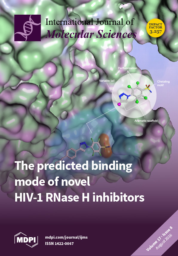
Cover Story (view full-size image): We identified original tetrafluoro-4-[4-(aryl)-1H-1,2,3-triazol-1-yl]benzenesulfonamides as potential inhibitors of the HIV-1 ribonuclease H (RNase H) function of the reverse transcriptase enzyme, which represents a suitable target for drug development. Spectroscopic analyses and molecular modeling studies indicated that such derivatives can mechanistically act by interfering with the two-metal ions through direct coordination of the metal cofactors in the enzyme catalytic site. View this paper.
  • Issues are regarded as officially published after their release is announced to the table of contents alert mailing list.
  • You may sign up for e-mail alerts to receive table of contents of newly released issues.
  • PDF is the official format for papers published in both, html and pdf forms. To view the papers in pdf format, click on the "PDF Full-text" link, and use the free Adobe Reader to open them.
Order results
Result details
Section
Select all
Export citation of selected articles as:
2138 KiB  
Review
Antioxidant Activity of γ-Oryzanol: A Complex Network of Interactions
by Igor Otavio Minatel 1, Fabiane Valentini Francisqueti 2, Camila Renata Corrêa 2 and Giuseppina Pace Pereira Lima 1,*
1 Department of Chemistry and Biochemistry, Institute of Bioscience, Sao Paulo State University, Botucatu 18618-689, Brazil
2 Department of Pathology, Botucatu Medical School, Sao Paulo State University, Botucatu 18618-970, Brazil
Int. J. Mol. Sci. 2016, 17(8), 1107; https://doi.org/10.3390/ijms17081107 - 9 Aug 2016
Cited by 100 | Viewed by 10813
Abstract
γ-oryzanol (Orz), a steryl ferulate extracted from rice bran layer, exerts a wide spectrum of biological activities. In addition to its antioxidant activity, Orz is often associated with cholesterol-lowering, anti-inflammatory, anti-cancer and anti-diabetic effects. In recent years, the usefulness of Orz has been [...] Read more.
γ-oryzanol (Orz), a steryl ferulate extracted from rice bran layer, exerts a wide spectrum of biological activities. In addition to its antioxidant activity, Orz is often associated with cholesterol-lowering, anti-inflammatory, anti-cancer and anti-diabetic effects. In recent years, the usefulness of Orz has been studied for the treatment of metabolic diseases, as it acts to ameliorate insulin activity, cholesterol metabolism, and associated chronic inflammation. Previous studies have shown the direct action of Orz when downregulating the expression of genes that encode proteins related to adiposity (CCAAT/enhancer binding proteins (C/EBPs)), inflammatory responses (nuclear factor kappa-B (NF-κB)), and metabolic syndrome (peroxisome proliferator-activated receptors (PPARs)). It is likely that this wide range of beneficial activities results from a complex network of interactions and signals triggered, and/or inhibited by its antioxidant properties. This review focuses on the significance of Orz in metabolic disorders, which feature remarkable oxidative imbalance, such as impaired glucose metabolism, obesity, and inflammation. Full article
(This article belongs to the Special Issue Macro- and Micro-nutrient Antioxidants)
Show Figures

Graphical abstract

4059 KiB  
Article
Influences of Electromagnetic Energy on Bio-Energy Transport through Protein Molecules in Living Systems and Its Experimental Evidence
by Xiaofeng Pang 1,*, Shude Chen 2, Xianghui Wang 2 and Lisheng Zhong 3
1 Institute of Physical Electrons, University of Electronic Science and Technology of China, Chengdu 610054, China
2 Department of Physics, East China Normal University, Shanghai 200062, China
3 State Key Laboratory of Electrical Insulation and Power Equipment, Xi’an Jiaotong University, Xi’an 710049, China
Int. J. Mol. Sci. 2016, 17(8), 1130; https://doi.org/10.3390/ijms17081130 - 25 Jul 2016
Cited by 16 | Viewed by 5998
Abstract
The influences of electromagnetic fields (EMFs) on bio-energy transport and its mechanism of changes are investigated through analytic and numerical simulation and experimentation. Bio-energy transport along protein molecules is performed by soliton movement caused by the dipole–dipole electric interactions between neighboring amino acid [...] Read more.
The influences of electromagnetic fields (EMFs) on bio-energy transport and its mechanism of changes are investigated through analytic and numerical simulation and experimentation. Bio-energy transport along protein molecules is performed by soliton movement caused by the dipole–dipole electric interactions between neighboring amino acid residues. As such, EMFs can affect the structure of protein molecules and change the properties of the bio-energy transported in living systems. This mechanism of biological effect from EMFs involves the amino acid residues in protein molecules. To study and reveal this mechanism, we simulated numerically the features of the movement of solitons along protein molecules with both a single chain and with three channels by using the Runge–Kutta method and Pang’s soliton model under the action of EMFs with the strengths of 25,500, 51,000, 76,500, and 102,000 V/m in the single-chain protein, as well as 17,000, 25,500, and 34,000 V/m in the three-chain protein, respectively. Results indicate that electric fields (EFs) depress the binding energy of the soliton, decrease its amplitude, and change its wave form. Also, the soliton disperses at 102,000 V/m in a single-chain protein and at 25,500 and 34,000 V/m in three-chain proteins. These findings signify that the influence of EMFs on the bio-energy transport cannot be neglected; however, these variations depend on both the strength and the direction of the EF in the EMF. This direction influences the biological effects of EMF, which decrease with increases in the angle between the direction of the EF and that of the dipole moment of amino acid residues; however, randomness at the macroscopic level remains. Lastly, we experimentally confirm the existence of a soliton and the validity of our conclusion by using the infrared spectra of absorption of the collagens, which is activated by another type of EF. Thus, we can affirm that both the described mechanism and the corresponding theory are correct and that EMFs or EFs can influence the features of energy transport in living systems and thus have certain biological effects. Full article
(This article belongs to the Section Physical Chemistry, Theoretical and Computational Chemistry)
Show Figures

Figure 1

6668 KiB  
Article
The Exon Junction Complex Controls the Efficient and Faithful Splicing of a Subset of Transcripts Involved in Mitotic Cell-Cycle Progression
by Kazuhiro Fukumura 1,2,*, Shunichi Wakabayashi 3,4, Naoyuki Kataoka 5,6, Hiroshi Sakamoto 1, Yutaka Suzuki 4, Kenta Nakai 3,4, Akila Mayeda 2 and Kunio Inoue 1
1 Department of Biology, Graduate School of Science, Kobe University, 1-1 Rokkodaicho, Nadaku, Kobe 657-8501, Japan
2 Division of Gene Expression Mechanism, Institute for Comprehensive Medical Science (ICMS), Fujita Health University, Toyoake, Aichi 470-1192, Japan
3 Human Genome Center, Institute of Medical Science, The University of Tokyo, 4-6-1 Shirokanedai, Minato-ku, Tokyo 108-8639, Japan
4 Department of Computational Biology and Medical Sciences, Graduate School of Frontier Sciences, The University of Tokyo, Kashiwa, Chiba 277-8561, Japan
5 Laboratory for Malignancy Control Research, Medical Innovation Center, Kyoto University Graduate School of Medicine, Kyoto 606-8507, Japan
6 Laboratory of Cell Regulation, Departments of Applied Animal Sciences and Applied Biological Chemistry, Graduate School of Agriculture and Life Sciences, The University of Tokyo, 1-1-1 Yayoi, Bunkyo-ku, Tokyo 113-8657, Japan
Int. J. Mol. Sci. 2016, 17(8), 1153; https://doi.org/10.3390/ijms17081153 - 2 Aug 2016
Cited by 24 | Viewed by 12692
Abstract
The exon junction complex (EJC) that is deposited onto spliced mRNAs upstream of exon–exon junctions plays important roles in multiple post-splicing gene expression events, such as mRNA export, surveillance, localization, and translation. However, a direct role for the human EJC in pre-mRNA splicing [...] Read more.
The exon junction complex (EJC) that is deposited onto spliced mRNAs upstream of exon–exon junctions plays important roles in multiple post-splicing gene expression events, such as mRNA export, surveillance, localization, and translation. However, a direct role for the human EJC in pre-mRNA splicing has not been fully understood. Using HeLa cells, we depleted one of the EJC core components, Y14, and the resulting transcriptome was analyzed by deep sequencing (RNA-Seq) and confirmed by RT–PCR. We found that Y14 is required for efficient and faithful splicing of a group of transcripts that is enriched in short intron-containing genes involved in mitotic cell-cycle progression. Tethering of EJC core components (Y14, eIF4AIII or MAGOH) to a model reporter pre-mRNA harboring a short intron showed that these core components are prerequisites for the splicing activation. Taken together, we conclude that the EJC core assembled on pre-mRNA is critical for efficient and faithful splicing of a specific subset of short introns in mitotic cell cycle-related genes. Full article
(This article belongs to the Special Issue Pre-mRNA Splicing 2016)
Show Figures

Graphical abstract

2836 KiB  
Article
A Female-Biased Odorant Receptor from Apolygus lucorum (Meyer-Dür) Tuned to Some Plant Odors
by Zhixiang Zhang 1,2, Meiping Zhang 1,*, Shuwei Yan 2, Guirong Wang 2 and Yang Liu 2,*
1 School of Life Science, Shanxi Normal University, Linfen 041000, China
2 State Key Laboratory for Biology of Plant Diseases and Insect Pests, Institute of Plant Protection, Chinese Academy of Agricultural Sciences, Beijing 100193, China
Int. J. Mol. Sci. 2016, 17(8), 1165; https://doi.org/10.3390/ijms17081165 - 28 Jul 2016
Cited by 17 | Viewed by 6077
Abstract
Apolygus lucorum (Meyer-Dür) (Hemiptera: Miridae) is a serious pest of cotton, jujube, grape and many other crops around the world. Understanding how olfactory information directs this insect to its host plants may provide environment-friendly approaches to the control of its population in agriculture. [...] Read more.
Apolygus lucorum (Meyer-Dür) (Hemiptera: Miridae) is a serious pest of cotton, jujube, grape and many other crops around the world. Understanding how olfactory information directs this insect to its host plants may provide environment-friendly approaches to the control of its population in agriculture. In our study, we cloned an odorant receptor gene, AlucOR46, that was specifically expressed in antennae and female-biased. Functional expression of AlucOR46 in Xenopus oocytes showed that it is tuned to six plant volatiles (S)-(−)-Limonene, (R)-(+)-Limonene, (E)-2-Hexenal, (E)-3-Hexenol, 1-Heptanol and (1R)-(−)-Myrtenol. Electroantennogram (EAG) recordings revealed that all six compounds could elicit electrophysiological responses from the antennae of A. lucorum, higher in females. Our results are in agreement with previous reports showing that (E)-2-Hexenal could attract female A. lucorum in behavior experiments. These results suggest that AlucOR46 might play an important role in locating the host plants of A. lucorum and therefore represents a suitable target for green pest control. Full article
(This article belongs to the Special Issue Membrane Protein Based Biosensors 2016)
Show Figures

Graphical abstract

2772 KiB  
Article
Cytoprotection against Hypoxic and/or MPP+ Injury: Effect of δ–Opioid Receptor Activation on Caspase 3
by Yuan Xu 1,†, Feng Zhi 1,†, Naiyuan Shao 1, Rong Wang 1, Yilin Yang 1,* and Ying Xia 2,*
1 Modern Medical Research Center, The Third Affiliated Hospital of Soochow University, Changzhou 213000, Jiangsu, China
2 Department of Neurosurgery, The University of Texas McGovern Medical School, Houston, TX 77030, USA
These authors contributed equally to this work.
Int. J. Mol. Sci. 2016, 17(8), 1179; https://doi.org/10.3390/ijms17081179 - 9 Aug 2016
Cited by 16 | Viewed by 6274
Abstract
The pathological changes of Parkinson’s disease (PD) are, at least partially, associated with the dysregulation of PTEN-induced putative kinase 1 (PINK1) and caspase 3. Since hypoxic and neurotoxic insults are underlying causes of PD, and since δ-opioid receptor (DOR) is neuroprotective against hypoxic/ischemic [...] Read more.
The pathological changes of Parkinson’s disease (PD) are, at least partially, associated with the dysregulation of PTEN-induced putative kinase 1 (PINK1) and caspase 3. Since hypoxic and neurotoxic insults are underlying causes of PD, and since δ-opioid receptor (DOR) is neuroprotective against hypoxic/ischemic insults, we sought to determine whether DOR activation could protect the cells from damage induced by hypoxia and/or MPP+ by regulating PINK1 and caspase 3 expressions. We exposed PC12 cells to either severe hypoxia (0.5%–1% O2) for 24–48 h or to MPP+ at different concentrations (0.5, 1, 2 mM) and then detected the levels of PINK1 and cleaved caspase 3. Both hypoxia and MPP+ reduced cell viability, progressively suppressed the expression of PINK1 and increased the cleaved caspase 3. DOR activation using UFP-512, effectively protected the cells from hypoxia and/or MPP+ induced injury, reversed the reduction in PINK1 protein and significantly attenuated the increase in the cleaved caspase 3. On the other hand, the application of DOR antagonist, naltrindole, greatly decreased cell viability and increased cleaved caspase 3. These findings suggest that DOR is cytoprotective against both hypoxia and MPP+ through the regulation of PINK1 and caspase 3 pathways. Full article
(This article belongs to the Special Issue Neuroprotective Strategies 2016)
Show Figures

Figure 1

640 KiB  
Article
Serum Concentrations of Endothelin-1 and Matrix Metalloproteinases-2, -9 in Pre-Hypertensive and Hypertensive Patients with Type 2 Diabetes
by Krasimir Kostov 1,*, Alexander Blazhev 2, Milena Atanasova 2 and Anelia Dimitrova 1
1 Department of Physiology and Pathophysiology, Medical University-Pleven, 1 Kliment Ohridski Str., 5800 Pleven, Bulgaria
2 Division of Biology, Medical University-Pleven, 1 Kliment Ohridski Str., 5800 Pleven, Bulgaria
Int. J. Mol. Sci. 2016, 17(8), 1182; https://doi.org/10.3390/ijms17081182 - 1 Aug 2016
Cited by 26 | Viewed by 6474
Abstract
Endothelin-1 (ET-1) is one of the most potent vasoconstrictors known to date. While its plasma or serum concentrations are elevated in some forms of experimental and human hypertension, this is not a consistent finding in all forms of hypertension. Matrix metalloproteinases -2 and [...] Read more.
Endothelin-1 (ET-1) is one of the most potent vasoconstrictors known to date. While its plasma or serum concentrations are elevated in some forms of experimental and human hypertension, this is not a consistent finding in all forms of hypertension. Matrix metalloproteinases -2 and -9 (MMP-2 and MMP-9), which degrade collagen type IV of the vascular basement membrane, are responsible for vascular remodeling, inflammation, and atherosclerotic complications, including in type 2 diabetes (T2D). In our study, we compared concentrations of ET-1, MMP-2, and MMP-9 in pre-hypertensive (PHTN) and hypertensive (HTN) T2D patients with those of healthy normotensive controls (N). ET-1, MMP-2, and MMP-9 were measured by ELISA. Concentrations of ET-1 in PHTN and N were very similar, while those in HTN were significantly higher. Concentrations of MMP-2 and MMP-9 in PHTN and HTN were also significantly higher compared to N. An interesting result in our study is that concentrations of MMP-2 and MMP-9 in HTN were lower compared to PHTN. In conclusion, we showed that increased production of ET-1 in patients with T2D can lead to long-lasting increases in blood pressure (BP) and clinical manifestation of hypertension. We also demonstrated that increased levels of MMP-2 and MMP-9 in pre-hypertensive and hypertensive patients with T2D mainly reflect the early vascular changes in extracellular matrix (ECM) turnover. Full article
(This article belongs to the Special Issue Liquid Biopsy for Clinical Application)
Show Figures

Graphical abstract

10045 KiB  
Article
Accumulation and Toxicity of Superparamagnetic Iron Oxide Nanoparticles in Cells and Experimental Animals
by Greta Jarockyte 1, Egle Daugelaite 1, Marius Stasys 1, Urte Statkute 1, Vilius Poderys 1, Ting-Chen Tseng 2, Shan-Hui Hsu 2, Vitalijus Karabanovas 1,3 and Ricardas Rotomskis 1,4,*
1 Biomedical Physics Laboratory of National Cancer Institute, Baublio 3B, LT08406 Vilnius, Lithuania
2 Institute of Polymer Science and Engineering, National Taiwan University, No. 1, Roosevelt Road Sec. 4, Taipei 10617, Taiwan
3 Department of Chemistry and Bioengineering, Vilnius Gediminas Technical University, LT-10223 Vilnius, Lithuania
4 Biophotonics group of Laser Research Centre, Vilnius University, Sauletekio 9, c.3, LT-10222 Vilnius, Lithuania
Int. J. Mol. Sci. 2016, 17(8), 1193; https://doi.org/10.3390/ijms17081193 - 19 Aug 2016
Cited by 83 | Viewed by 7866
Abstract
The uptake and distribution of negatively charged superparamagnetic iron oxide (Fe3O4) nanoparticles (SPIONs) in mouse embryonic fibroblasts NIH3T3, and magnetic resonance imaging (MRI) signal influenced by SPIONs injected into experimental animals, were visualized and investigated. Cellular uptake and distribution [...] Read more.
The uptake and distribution of negatively charged superparamagnetic iron oxide (Fe3O4) nanoparticles (SPIONs) in mouse embryonic fibroblasts NIH3T3, and magnetic resonance imaging (MRI) signal influenced by SPIONs injected into experimental animals, were visualized and investigated. Cellular uptake and distribution of the SPIONs in NIH3T3 after staining with Prussian Blue were investigated by a bright-field microscope equipped with digital color camera. SPIONs were localized in vesicles, mostly placed near the nucleus. Toxicity of SPION nanoparticles tested with cell viability assay (XTT) was estimated. The viability of NIH3T3 cells remains approximately 95% within 3–24 h of incubation, and only a slight decrease of viability was observed after 48 h of incubation. MRI studies on Wistar rats using a clinical 1.5 T MRI scanner were showing that SPIONs give a negative contrast in the MRI. The dynamic MRI measurements of the SPION clearance from the injection site shows that SPIONs slowly disappear from injection sites and only a low concentration of nanoparticles was completely eliminated within three weeks. No functionalized SPIONs accumulate in cells by endocytic mechanism, none accumulate in the nucleus, and none are toxic at a desirable concentration. Therefore, they could be used as a dual imaging agent: as contrast agents for MRI and for traditional optical biopsy by using Prussian Blue staining. Full article
(This article belongs to the Special Issue Inorganic Nanostructures in Biological Systems)
Show Figures

Graphical abstract

4954 KiB  
Article
Akt Activation Correlates with Snail Expression and Potentially Determines the Recurrence of Prostate Cancer in Patients at Stage T2 after a Radical Prostatectomy
by Wei-Yu Chen 1,2,3, Kuo-Tai Hua 4, Wei-Jiunn Lee 5,6, Yung-Wei Lin 7, Yen-Nien Liu 8, Chi-Long Chen 1,2,*, Yu-Ching Wen 6,7,* and Ming-Hsien Chien 1,5,*
1 Graduate Institute of Clinical Medicine, College of Medicine, Taipei Medical University, Taipei 110, Taiwan
2 Department of Pathology, School of Medicine, College of Medicine, Taipei Medical University, Taipei 110, Taiwan
3 Department of Pathology, Wan Fang Hospital, Taipei Medical University, Taipei 116, Taiwan
4 Graduate Institute of Toxicology, College of Medicine, National Taiwan University, Taipei 100, Taiwan
5 Department of Medical Education and Research, Wan Fang Hospital, Taipei Medical University, Taipei 116, Taiwan
6 Department of Urology, School of Medicine, College of Medicine, Taipei Medical University, Taipei 110, Taiwan
7 Department of Urology, Wan Fang Hospital, Taipei Medical University, Taipei 116, Taiwan
8 Graduate Institute of Cancer Biology and Drug Discovery, College of Medical Science and Technology, Taipei Medical University, Taipei 110, Taiwan
Int. J. Mol. Sci. 2016, 17(8), 1194; https://doi.org/10.3390/ijms17081194 - 23 Jul 2016
Cited by 3 | Viewed by 5097
Abstract
Our previous work demonstrated the epithelial-mesenchymal transition factor, Snail, is a potential marker for predicting the recurrence of localized prostate cancer (PCa). Akt activation is important for Snail stabilization and transcription in PCa. The purpose of this study was to retrospectively investigate the [...] Read more.
Our previous work demonstrated the epithelial-mesenchymal transition factor, Snail, is a potential marker for predicting the recurrence of localized prostate cancer (PCa). Akt activation is important for Snail stabilization and transcription in PCa. The purpose of this study was to retrospectively investigate the relationship between the phosphorylated level of Akt (p-Akt) in radical prostatectomy (RP) specimens and cancer biochemical recurrence (BCR). Using a tissue microarray and immunohistochemistry, the expression of p-Akt was measured in benign and neoplastic tissues from RP specimens in 53 patients whose cancer was pathologically defined as T2 without positive margins. Herein, we observed that the p-Akt level was higher in PCa than in benign tissues and was significantly associated with the Snail level. A high p-Akt image score (≥8) was significantly correlated with a higher histological Gleason sum, Snail image score, and preoperative prostate-specific antigen (PSA) value. Moreover, the high p-Akt image score and Gleason score sum (≥7) showed similar discriminatory abilities for BCR according to a receiver-operator characteristic curve analysis and were correlated with worse recurrence-free survival according to a log-rank test (p < 0.05). To further determine whether a high p-Akt image score could predict the risk of BCR, a Cox proportional hazard model showed that only a high p-Akt image score (hazard ratio (HR): 3.12, p = 0.05) and a high Gleason score sum (≥7) (HR: 1.18, p = 0.05) but not a high preoperative PSA value (HR: 0.62, p = 0.57) were significantly associated with a higher risk of developing BCR. Our data indicate that, for localized PCa patients after an RP, p-Akt can serve as a potential prognostic marker that improves predictions of BCR-free survival. Full article
(This article belongs to the Special Issue Diagnostic, Prognostic and Predictive Biomarkers in Prostate Cancer)
Show Figures

Graphical abstract

3796 KiB  
Article
Comparative Analysis for the Presence of IgG Anti-Aquaporin-1 in Patients with NMO-Spectrum Disorders
by Ismael Sánchez Gomar 1, María Díaz Sánchez 2, Antonio José Uclés Sánchez 2, José Luis Casado Chocán 2, Nela Suárez-Luna 1, Reposo Ramírez-Lorca 1, Javier Villadiego 1, Juan José Toledo-Aral 1,3 and Miriam Echevarría 1,4,*
1 Instituto de Biomedicina de Sevilla (IBiS), Hospital Universitario Virgen del Rocío/CSIC/Universidad de Sevilla, Seville 41013, Spain
2 Unidad de Gestión Clínica de Neurociencias, Servicio de Neurología del Hospital Universitario Virgen del Rocío, Seville 41013, Spain
3 Centro de Investigación Biomédica en Red sobre Enfermedades Neurodegenerativas (CIBERNED), Madrid 28029, Spain
4 Centro de Investigación Biomédica en Red sobre Enfermedades Respiratorias (CIBERES), Madrid 28029, Spain
Int. J. Mol. Sci. 2016, 17(8), 1195; https://doi.org/10.3390/ijms17081195 - 23 Jul 2016
Cited by 12 | Viewed by 6940
Abstract
Detection of IgG anti-Aquaporin-4 (AQP4) in serum of patients with Neuromyelitis optica syndrome disorders (NMOSD) has improved diagnosis of these processes and differentiation from Multiple sclerosis (MS). Recent findings also claim that a subgroup of patients with NMOSD, serum negative for IgG-anti-AQP4, present [...] Read more.
Detection of IgG anti-Aquaporin-4 (AQP4) in serum of patients with Neuromyelitis optica syndrome disorders (NMOSD) has improved diagnosis of these processes and differentiation from Multiple sclerosis (MS). Recent findings also claim that a subgroup of patients with NMOSD, serum negative for IgG-anti-AQP4, present antibodies anti-AQP1 instead. Explore the presence of IgG-anti-AQP1 using a previously developed cell-based assay (CBA) highly sensitive to IgG-anti-AQP4. Serum of 205 patients diagnosed as NMOSD (8), multiple sclerosis (94), optic neuritis (39), idiopathic myelitis (29), other idiopathic demyelinating disorders of the central nervous system (9), other neurological diseases (18) and healthy controls (8), were used in a CBA over fixed HEK cells transfected with hAQP1-EGFP or hM23-AQP4-EGFP, treated with Triton X-100 and untreated. ELISA was also performed. Analysis of serum with our CBA indicated absence of anti-AQP1 antibodies, whereas in cells pretreated with detergent, noisy signal made reliable detection impossible. ELISA showed positive results in few serums. The low number of NMOSD serums included in our study reduces its power to conclude the specificity of AQP1 antibodies as new biomarkers of NMOSD. Our study does not sustain detection of anti-AQP1 in serum of NMOSD patients but further experiments are expected. Full article
(This article belongs to the Special Issue Aquaporin)
Show Figures

Graphical abstract

6614 KiB  
Article
Homocysteine Aggravates Cortical Neural Cell Injury through Neuronal Autophagy Overactivation following Rat Cerebral Ischemia-Reperfusion
by Yaqian Zhao, Guowei Huang, Shuang Chen, Yun Gou, Zhiping Dong and Xumei Zhang *
Department of Nutrition and Food Science, School of Public Health, Tianjin Medical University, Tianjin 30070, China
Int. J. Mol. Sci. 2016, 17(8), 1196; https://doi.org/10.3390/ijms17081196 - 23 Jul 2016
Cited by 58 | Viewed by 8179
Abstract
Elevated homocysteine (Hcy) levels have been reported to be involved in neurotoxicity after ischemic stroke. However, the underlying mechanisms remain incompletely understood to date. In the current study, we hypothesized that neuronal autophagy activation may be involved in the toxic effect of Hcy [...] Read more.
Elevated homocysteine (Hcy) levels have been reported to be involved in neurotoxicity after ischemic stroke. However, the underlying mechanisms remain incompletely understood to date. In the current study, we hypothesized that neuronal autophagy activation may be involved in the toxic effect of Hcy on cortical neurons following cerebral ischemia. Brain cell injury was determined by hematoxylin-eosin (HE) staining and TdT-mediated dUTP Nick-End Labeling (TUNEL) staining. The level and localization of autophagy were detected by transmission electron microscopy, western blot and immunofluorescence double labeling. The oxidative DNA damage was revealed by immunofluorescence of 8-Hydroxy-2′-deoxyguanosine (8-OHdG). Hcy treatment aggravated neuronal cell death, significantly increased the formation of autophagosomes and the expression of LC3B and Beclin-1 in the brain cortex after middle cerebral artery occlusion-reperfusion (MCAO). Immunofluorescence analysis of LC3B and Beclin-1 distribution indicated that their expression occurred mainly in neurons (NeuN-positive) and hardly in astrocytes (GFAP-positive). 8-OHdG expression was also increased in the ischemic cortex of Hcy-treated animals. Conversely, LC3B and Beclin-1 overexpression and autophagosome accumulation caused by Hcy were partially blocked by the autophagy inhibitor 3-methyladenine (3-MA). Hcy administration enhanced neuronal autophagy, which contributes to cell death following cerebral ischemia. The oxidative damage-mediated autophagy may be a molecular mechanism underlying neuronal cell toxicity of elevated Hcy level. Full article
(This article belongs to the Section Molecular Toxicology)
Show Figures

Graphical abstract

2269 KiB  
Communication
Expression Analysis of PIN Genes in Root Tips and Nodules of Medicago truncatula
by Izabela Sańko-Sawczenko, Barbara Łotocka and Weronika Czarnocka *
Department of Botany, Faculty of Agriculture and Biology, Warsaw University of Life Sciences—SGGW, Nowoursynowska 159, 02-776 Warsaw, Poland
Int. J. Mol. Sci. 2016, 17(8), 1197; https://doi.org/10.3390/ijms17081197 - 25 Jul 2016
Cited by 23 | Viewed by 5283
Abstract
Polar auxin transport is dependent on the family of PIN-formed proteins (PINs), which are membrane transporters of anionic indole-3-acetic acid (IAA). It is assumed that polar auxin transport may be essential in the development and meristematic activity maintenance of Medicago truncatula [...] Read more.
Polar auxin transport is dependent on the family of PIN-formed proteins (PINs), which are membrane transporters of anionic indole-3-acetic acid (IAA). It is assumed that polar auxin transport may be essential in the development and meristematic activity maintenance of Medicago truncatula (M. truncatula) root nodules. However, little is known about the involvement of specific PIN proteins in M. truncatula nodulation. Using real-time quantitative PCR, we analyzed the expression patterns of all previously identified MtPIN genes and compared them between root nodules and root tips of M. truncatula. Our results demonstrated significant differences in the expression level of all 11 genes (MtPIN1MtPIN11) between examined organs. Interestingly, MtPIN9 was the only PIN gene with higher expression level in root nodules compared to root tips. This result is the first indication of PIN9 transporter potential involvement in M. truncatula nodulation. Moreover, relatively high expression level in root nodules was attributed to MtPINs encoding orthologs of Arabidopsis thaliana PIN5 subclade. PIN proteins from this subclade have been found to localize in the endoplasmic reticulum, which may indicate that the development and meristematic activity maintenance of M. truncatula root nodules is associated with intracellular homeostasis of auxins level and their metabolism in the endoplasmic reticulum. Full article
(This article belongs to the Special Issue Molecular Signals in Nodulation Control)
Show Figures

Figure 1

2117 KiB  
Article
Screening and Validation of Housekeeping Genes of the Root and Cotyledon of Cunninghamia lanceolata under Abiotic Stresses by Using Quantitative Real-Time PCR
by Wenlong Bao, Yanli Qu, Xiaoyi Shan * and Yinglang Wan *
1 College of Biological Sciences and Biotechnology, Beijing Forestry University, 35 Qinghua East Road, Haidian District, Beijing 100083, China
These authors contributed equally to this work.
Int. J. Mol. Sci. 2016, 17(8), 1198; https://doi.org/10.3390/ijms17081198 - 28 Jul 2016
Cited by 29 | Viewed by 5186
Abstract
Cunninghamia lanceolata (Chinese fir) is a fast-growing and commercially important conifer of the Cupressaceae family. Due to the unavailability of complete genome sequences and relatively poor genetic background information of the Chinese fir, it is necessary to identify and analyze the expression levels [...] Read more.
Cunninghamia lanceolata (Chinese fir) is a fast-growing and commercially important conifer of the Cupressaceae family. Due to the unavailability of complete genome sequences and relatively poor genetic background information of the Chinese fir, it is necessary to identify and analyze the expression levels of suitable housekeeping genes (HKGs) as internal reference for precise analysis. Based on the results of database analysis and transcriptome sequencing, we have chosen five candidate HKGs (Actin, GAPDH, EF1a, 18S rRNA, and UBQ) with conservative sequences in the Chinese fir and related species for quantitative analysis. The expression levels of these HKGs in roots and cotyledons under five different abiotic stresses in different time intervals were measured by qRT-PCR. The data were statistically analyzed using the following algorithms: NormFinder, BestKeeper, and geNorm. Finally, RankAggreg was applied to merge the sequences generated from three programs and rank these according to consensus sequences. The expression levels of these HKGs showed variable stabilities under different abiotic stresses. Among these, Actin was the most stable internal control in root, and GAPDH was the most stable housekeeping gene in cotyledon. We have also described an experimental procedure for selecting HKGs based on the de novo sequencing database of other non-model plants. Full article
(This article belongs to the Section Molecular Plant Sciences)
Show Figures

Graphical abstract

6188 KiB  
Article
Moracin C, A Phenolic Compound Isolated from Artocarpus heterophyllus, Suppresses Lipopolysaccharide-Activated Inflammatory Responses in Murine Raw264.7 Macrophages
by Xue Yao 1,†, Dang Wu 1,†, Ningning Dong 1, Ping Ouyang 1, Jiaqian Pu 1, Qian Hu 1, Jingyuan Wang 1, Weiqiang Lu 2,* and Jin Huang 1,*
1 Shanghai Key Laboratory of New Drug Design, School of Pharmacy, East China University of Science and Technology, 130 Mei Long Road, Shanghai 200237, China
2 Shanghai Key Laboratory of Regulatory Biology, Institute of Biomedical Sciences and School of Life Sciences, East China Normal University, 500 Dongchuan Road, Shanghai 200241, China
These authors contributed equally to this manuscript.
Int. J. Mol. Sci. 2016, 17(8), 1199; https://doi.org/10.3390/ijms17081199 - 25 Jul 2016
Cited by 44 | Viewed by 7724
Abstract
Artocarpus heterophyllus, a popular tropical fruit commonly known as the jackfruit tree, is normally planted in subtropical or tropical areas. Since a variety of phytochemicals isolated from A. heterophyllus have been found to possess potently anti-inflammatory, antiviral and antimalarial activities, researchers have [...] Read more.
Artocarpus heterophyllus, a popular tropical fruit commonly known as the jackfruit tree, is normally planted in subtropical or tropical areas. Since a variety of phytochemicals isolated from A. heterophyllus have been found to possess potently anti-inflammatory, antiviral and antimalarial activities, researchers have devoted much interest to its potential pharmaceutical value. However, the exact mechanism underlying its anti-inflammatory activity is not well characterized. In this study, seven natural products isolated from A. heterophyllus, including 25-Hydroxycycloart-23-en-3-one (HY), Artocarpin (AR), Dadahol A (DA), Morachalcone A (MA), Artoheterophyllin B (AB), Cycloheterophyllin (CY) and Moracin C (MC) were collected. Lipopolysaccharide (LPS)-stimulated inflammatory response in RAW264.7 macrophages were used in this study. Among these compounds, MC significantly inhibited LPS-activated reactive oxygen species (ROS) and nitric oxide (NO) release without marked cytotoxicity. Furthermore, MC effectively reduced LPS stimulated up-regulation of mRNA and protein expression of inducible nitric oxide synthase (iNOS), cyclooxygenase-2 (COX-2), and serval pro-inflammatory cytokines (interleukin-1β (IL-1β), interleukin-6 (IL-6) and tumor necrosis factor α (TNF-α)). Mechanistic studies revealed that the anti-inflammatory effect of MC was associated with the activation of the mitogen activated protein kinases (MAPKs) (including p38, ERK and JNK) and nuclear factor-κB (NF-κB) pathways, especially reducing the nuclear translocation of NF-κB p65 subunit as revealed by nuclear separation experiment and confocal microscopy. Full article
(This article belongs to the Special Issue The Mechanism of Action of Food Components in Disease Prevention)
Show Figures

Graphical abstract

5934 KiB  
Article
Non-Ligand-Induced Dimerization is Sufficient to Initiate the Signalling and Endocytosis of EGF Receptor
by George Kourouniotis, Yi Wang, Steven Pennock, Xinmei Chen and Zhixiang Wang *
1 Department of Medical Genetics and Signal Transduction Research Group, Faculty of Medicine and Dentistry, University of Alberta, Edmonton, AB T6G 2H7, Canada
These authors contributed equally to this work.
Int. J. Mol. Sci. 2016, 17(8), 1200; https://doi.org/10.3390/ijms17081200 - 25 Jul 2016
Cited by 9 | Viewed by 6411
Abstract
The binding of epidermal growth factor (EGF) to EGF receptor (EGFR) stimulates cell mitogenesis and survival through various signalling cascades. EGF also stimulates rapid EGFR endocytosis and its eventual degradation in lysosomes. The immediate events induced by ligand binding include receptor dimerization, activation [...] Read more.
The binding of epidermal growth factor (EGF) to EGF receptor (EGFR) stimulates cell mitogenesis and survival through various signalling cascades. EGF also stimulates rapid EGFR endocytosis and its eventual degradation in lysosomes. The immediate events induced by ligand binding include receptor dimerization, activation of intrinsic tyrosine kinase and autophosphorylation. However, in spite of intensified efforts, the results regarding the roles of these events in EGFR signalling and internalization is still very controversial. In this study, we constructed a chimeric EGFR by replacing its extracellular domain with leucine zipper (LZ) and tagged a green fluorescent protein (GFP) at its C-terminus. We showed that the chimeric LZ-EGFR-GFP was constitutively dimerized. The LZ-EGFR-GFP dimer autophosphorylated each of its five well-defined C-terminal tyrosine residues as the ligand-induced EGFR dimer does. Phosphorylated LZ-EGFR-GFP was localized to both the plasma membrane and endosomes, suggesting it is capable of endocytosis. We also showed that LZ-EGFR-GFP activated major signalling proteins including Src homology collagen-like (Shc), extracellular signal-regulated kinase (ERK) and Akt. Moreover, LZ-EGFR-GFP was able to stimulate cell proliferation. These results indicate that non-ligand induced dimerization is sufficient to activate EGFR and initiate cell signalling and EGFR endocytosis. We conclude that receptor dimerization is a critical event in EGF-induced cell signalling and EGFR endocytosis. Full article
(This article belongs to the Section Biochemistry)
Show Figures

Figure 1

2742 KiB  
Article
Anti-Diabetic Effect of Portulaca oleracea L. Polysaccharideandits Mechanism in Diabetic Rats
by Yu Bai 1,*, Xueli Zang 2, Jinshu Ma 3 and Guangyu Xu 4
1 Pharmaceutical College, Jilin Medical University, Jilin 132011, China
2 Department of Pharmacy, Changchun Medical College, Changchun 130033, China
3 Department of Pathology, China-Japan Union Hospital of Jilin University, Changchun 130033, China
4 College of Pharmacy, Beihua University, Jilin 132013, China
Int. J. Mol. Sci. 2016, 17(8), 1201; https://doi.org/10.3390/ijms17081201 - 25 Jul 2016
Cited by 73 | Viewed by 8116
Abstract
Diabetes mellitus (DM) is a metabolic syndrome caused by multiple genetic and environmental factors. Traditional Chinese medicine preparations have shown a comprehensive and function-regulating characteristic. Purslane (Portulaca oleracea L.) is an annual succulent herb. Currently, there have been some related reports on [...] Read more.
Diabetes mellitus (DM) is a metabolic syndrome caused by multiple genetic and environmental factors. Traditional Chinese medicine preparations have shown a comprehensive and function-regulating characteristic. Purslane (Portulaca oleracea L.) is an annual succulent herb. Currently, there have been some related reports on the treatment of diabetes with purslane. The current study was designed to separate and purify the polysaccharide, a systematic study of its physical and chemical properties, antioxidant activity, and anti-diabetic mechanism, in order to provide a theoretical basis for the development of drugs of purslane. A crude water soluble polysaccharide extracted from purslane was named CPOP (crude Portulaca oleracea L. polysaccharide). Effects of CPOP on bodyweight, glucose tolerance test (GTT), fasting blood glucose (FBG), fasting serum insulin (FINS), insulin sensitivity index (ISI), interleukin-6 (IL-6), tumor necrosis factor-α (TNF-α), methane dicarboxylic aldehyde (MDA), and superoxygen dehydrogenises (SOD) were investigated. The results indicate that the oral administration of CPOP could significantly increase the body weight and significantly improve the glucose tolerance in diabetic rats. Meanwhile, CPOP could significantly reduce the FBG level, and elevate the FINS level and ISI value in diabetic rats. In addition, CPOP could significantly reduce TNF-α and IL-6 levels in diabetic rats; CPOP could also reduce MDA and SOD activities in the liver tissue of diabetic rats. These results suggest that the anti-diabetic effect of CPOP may be associated with its antioxidant and anti-inflammatory effects. Full article
(This article belongs to the Special Issue Diabetic Complications: Pathophysiology, Mechanisms, and Therapies)
Show Figures

Graphical abstract

1120 KiB  
Review
Role of Lipids in the Onset, Progression and Treatment of Periodontal Disease. A Systematic Review of Studies in Humans
by Alfonso Varela-López 1, Francesca Giampieri 2, Pedro Bullón 3, Maurizio Battino 2 and José L. Quiles 1,*
1 Department of Physiology, Institute of Nutrition and Food Technology “Jose Mataix”, University of Granada, Biomedical Research Center, Avda. Conocimiento s/n, 18100 Armilla, Spain
2 Dipartimento di Scienze Cliniche Specialistiche ed Odontostomatologiche (DISCO)-Sez. Biochimica, Facoltà di Medicina, Università Politecnica delle Marche, 60131 Ancona, Italy
3 Department of Stomalogy, Dental School, University of Sevilla, C/Avicena s/n, 41009 Sevilla, Spain
Int. J. Mol. Sci. 2016, 17(8), 1202; https://doi.org/10.3390/ijms17081202 - 25 Jul 2016
Cited by 24 | Viewed by 6664
Abstract
The risk of different oral problems (root caries, tooth mobility, and tooth loss) can be increased by the presence of periodontal disease, which has also been associated with a growing list of systemic diseases. The presence of some bacteria is the primary etiology [...] Read more.
The risk of different oral problems (root caries, tooth mobility, and tooth loss) can be increased by the presence of periodontal disease, which has also been associated with a growing list of systemic diseases. The presence of some bacteria is the primary etiology of this disease; a susceptible host is also necessary for disease initiation. In this respect, the progression of periodontal disease and healing of the periodontal tissues can be modulated by nutritional status. To clarify the role of lipids in the establishment, progression, and/or treatment of this pathology, a systematic review was conducted of English-written literature in PubMed until May 2016, which included research on the relationship of these dietary components with the onset and progression of periodontal disease. According to publication type, randomized-controlled trials, cohort, case-control and cross-sectional studies were included. Among all the analyzed components, those that have any effect on oxidative stress and/or inflammation seem to be the most interesting according to current evidence. On one hand, there is quite a lot of information in favor of a positive role of n-3 fatty acids, due to their antioxidant and immunomodulatory effects. On the other hand, saturated fat-rich diets increase oxidative stress as well the as intensity and duration of inflammatory processes, so they must be avoided. Full article
(This article belongs to the Special Issue Macro- and Micro-nutrient Antioxidants)
Show Figures

Graphical abstract

1621 KiB  
Article
Genome-Wide Identification and Characterization of Carboxypeptidase Genes in Silkworm (Bombyx mori)
by Junhong Ye, Yi Li, Hua-Wei Liu, Jifu Li, Zhaoming Dong, Qingyou Xia and Ping Zhao *
State Key Laboratory of Silkworm Genome Biology, Southwest University, Chongqing 400716, China
Int. J. Mol. Sci. 2016, 17(8), 1203; https://doi.org/10.3390/ijms17081203 - 28 Jul 2016
Cited by 10 | Viewed by 5135
Abstract
The silkworm (Bombyx mori) is an economically-important insect that can secrete silk. Carboxypeptidases have been found in various metazoan species and play important roles in physiological and biochemical reactions. Here, we analyzed the silkworm genome database and characterized 48 carboxypeptidases, including [...] Read more.
The silkworm (Bombyx mori) is an economically-important insect that can secrete silk. Carboxypeptidases have been found in various metazoan species and play important roles in physiological and biochemical reactions. Here, we analyzed the silkworm genome database and characterized 48 carboxypeptidases, including 34 metal carboxypeptidases (BmMCP1BmMCP34) and 14 serine carboxypeptidases (BmSCP1BmSCP14), to better understand their diverse functions. Compared to other insects, our results indicated that carboxypeptidases from silkworm have more family members. These silkworm carboxypeptidases could be divided into four families: Peptidase_M2 carboxypeptidases, Peptidase_M14 carboxypeptidases, Peptidase_S10 carboxypeptidases and Peptidase_S28 carboxypeptidases. Microarray analysis showed that the carboxypeptidases had distinct expression patterns, whereas quantitative real-time PCR demonstrated that the expression level of 13 carboxypeptidases significantly decreased after starvation and restored after re-feeding. Overall, our study provides new insights into the functional and evolutionary features of silkworm carboxypeptidases. Full article
(This article belongs to the Section Biochemistry)
Show Figures

Graphical abstract

6886 KiB  
Article
Genome-Wide Identification and Expression Analysis of Two-Component System Genes in Tomato
by Yanjun He 1,†, Xue Liu 1,†, Lei Ye 1, Changtian Pan 1, Lifei Chen 1, Tao Zou 1,2 and Gang Lu 1,2,*
1 Department of Horticulture, Zhejiang University, Hangzhou 310058, China
2 Key Laboratory of Horticultural Plant Growth, Development and Biotechnology, Agricultural Ministry of China, Hangzhou 310058, China
These authors contributed equally to this work.
Int. J. Mol. Sci. 2016, 17(8), 1204; https://doi.org/10.3390/ijms17081204 - 26 Jul 2016
Cited by 43 | Viewed by 6285
Abstract
The two-component system (TCS), which comprises histidine kinases (HKs), phosphotransfers (HPs), and response regulator proteins (RRs), plays pivotal roles in regulating plant growth, development, and responses to biotic and abiotic stresses. TCS genes have been comprehensively identified and investigated in various crops but [...] Read more.
The two-component system (TCS), which comprises histidine kinases (HKs), phosphotransfers (HPs), and response regulator proteins (RRs), plays pivotal roles in regulating plant growth, development, and responses to biotic and abiotic stresses. TCS genes have been comprehensively identified and investigated in various crops but poorly characterized in tomato. In this work, a total of 65 TCS genes consisting of 20 HK(L)s, six HPs, and 39 RRs were identified from tomato genome. The classification, gene structures, conserved domains, chromosome distribution, phylogenetic relationship, gene duplication events, and subcellular localization of the TCS gene family were predicted and analyzed in detail. The amino acid sequences of tomato TCS family members, except those of type-B RRs, are highly conserved. The gene duplication events of the TCS family mainly occurred in the RR family. Furthermore, the expansion of RRs was attributed to both segment and tandem duplication. The subcellular localizations of the selected green fluorescent protein (GFP) fusion proteins exhibited a diverse subcellular targeting, thereby confirming their predicted divergent functionality. The majority of TCS family members showed distinct organ- or development-specific expression patterns. In addition, most of TCS genes were induced by abiotic stresses and exogenous phytohormones. The full elucidation of TCS elements will be helpful for comprehensive analysis of the molecular biology and physiological role of the TCS superfamily. Full article
(This article belongs to the Section Molecular Plant Sciences)
Show Figures

Graphical abstract

2408 KiB  
Review
Effects of Secondary Plant Metabolites on Microbial Populations: Changes in Community Structure and Metabolic Activity in Contaminated Environments
by Lucie Musilova 1,*, Jakub Ridl 2, Marketa Polivkova 1, Tomas Macek 1 and Ondrej Uhlik 1,*
1 Department of Biochemistry and Microbiology, Faculty of Food and Biochemical Technology, University of Chemistry and Technology Prague, Technicka 3, 166 28 Prague, Czech Republic
2 Department of Genomics and Bioinformatics, Institute of Molecular Genetics of the Czech Academy of Sciences, Videnska 1083, 142 20 Prague, Czech Republic
Int. J. Mol. Sci. 2016, 17(8), 1205; https://doi.org/10.3390/ijms17081205 - 29 Jul 2016
Cited by 95 | Viewed by 10995
Abstract
Secondary plant metabolites (SPMEs) play an important role in plant survival in the environment and serve to establish ecological relationships between plants and other organisms. Communication between plants and microorganisms via SPMEs contained in root exudates or derived from litter decomposition is an [...] Read more.
Secondary plant metabolites (SPMEs) play an important role in plant survival in the environment and serve to establish ecological relationships between plants and other organisms. Communication between plants and microorganisms via SPMEs contained in root exudates or derived from litter decomposition is an example of this phenomenon. In this review, the general aspects of rhizodeposition together with the significance of terpenes and phenolic compounds are discussed in detail. We focus specifically on the effect of SPMEs on microbial community structure and metabolic activity in environments contaminated by polychlorinated biphenyls (PCBs) and polyaromatic hydrocarbons (PAHs). Furthermore, a section is devoted to a complex effect of plants and/or their metabolites contained in litter on bioremediation of contaminated sites. New insights are introduced from a study evaluating the effects of SPMEs derived during decomposition of grapefruit peel, lemon peel, and pears on bacterial communities and their ability to degrade PCBs in a long-term contaminated soil. The presented review supports the “secondary compound hypothesis” and demonstrates the potential of SPMEs for increasing the effectiveness of bioremediation processes. Full article
(This article belongs to the Section Molecular Plant Sciences)
Show Figures

Graphical abstract

3573 KiB  
Article
Full-Length cDNA Cloning, Molecular Characterization and Differential Expression Analysis of Lysophospholipase I from Ovis aries
by Nan-Nan Liu 1,†, Zeng-Shan Liu 1,†, Pan Hu 1,†, Ying Zhang 1,†, Shi-Ying Lu 1, Yan-Song Li 1, Yong-Jie Yang 2, Dong-Song Zhang 3, Yu Zhou 1 and Hong-Lin Ren 1,*
1 Key Laboratory of Zoonosis Research, Ministry of Education/Institute of Zoonosis/College of Veterinary Medicine, College of Animal Sciences, Jilin University, Xi An Da Lu 5333, Changchun 130062, China
2 Department of Food Science, College of Agriculture, Yanbian University, Yanji 133002, China
3 Animal Husbandry and Veterinary Unit of Xiangyang Town, Liuhe 135305, China
These authors contributed equally to this work.
Int. J. Mol. Sci. 2016, 17(8), 1206; https://doi.org/10.3390/ijms17081206 - 28 Jul 2016
Cited by 2 | Viewed by 4517
Abstract
Lysophospholipase I (LYPLA1) is an important protein with multiple functions. In this study, the full-length cDNA of the LYPLA1 gene from Ovis aries (OaLypla1) was cloned using primers and rapid amplification of cDNA ends (RACE) technology. The full-length OaLypla1 was 2457 [...] Read more.
Lysophospholipase I (LYPLA1) is an important protein with multiple functions. In this study, the full-length cDNA of the LYPLA1 gene from Ovis aries (OaLypla1) was cloned using primers and rapid amplification of cDNA ends (RACE) technology. The full-length OaLypla1 was 2457 bp with a 5′-untranslated region (UTR) of 24 bp, a 3′-UTR of 1740 bp with a poly (A) tail, and an open reading frame (ORF) of 693 bp encoding a protein of 230 amino acid residues with a predicted molecular weight of 24,625.78 Da. Phylogenetic analysis showed that the OaLypla1 protein shared a high amino acid identity with LYPLA1 of Bos taurus. The recombinant OaLypla1 protein was expressed and purified, and its phospholipase activity was identified. Monoclonal antibodies (mAb) against OaLypla1 that bound native OaLypla1 were generated. Real-time PCR analysis revealed that OaLypla1 was constitutively expressed in the liver, spleen, lung, kidney, and white blood cells of sheep, with the highest level in the kidney. Additionally, the mRNA levels of OaLypla1 in the buffy coats of sheep challenged with virulent or avirulent Brucella strains were down-regulated compared to untreated sheep. The results suggest that OaLypla1 may have an important physiological role in the host response to bacteria. The function of OaLypla1 in the host response to bacterial infection requires further study in the future. Full article
(This article belongs to the Section Biochemistry)
Show Figures

Graphical abstract

437 KiB  
Article
Chicken-Specific Kinome Array Reveals that Salmonella enterica Serovar Enteritidis Modulates Host Immune Signaling Pathways in the Cecum to Establish a Persistence Infection
by Michael H. Kogut 1, Christina L. Swaggerty 1, James Allen Byrd 1, Ramesh Selvaraj 2 and Ryan J. Arsenault 3,*
1 Southern Plains Agricultural Resarch Center, United States Department of Agriculture, Agricultural Research Service, College Station, TX 77845, USA
2 Ohio Agricultural Research Center, The Ohio State University, Wooster, OH 44691, USA
3 Department of Animal and Food Sciences, University of Delaware, Newark, DE 19716, USA
Int. J. Mol. Sci. 2016, 17(8), 1207; https://doi.org/10.3390/ijms17081207 - 27 Jul 2016
Cited by 35 | Viewed by 6768
Abstract
Non-typhoidal Salmonella enterica induces an early, short-lived pro-inflammatory response in chickens that is asymptomatic of clinical disease and results in a persistent colonization of the gastrointestinal (GI) tract that transmits infections to naïve hosts via fecal shedding of bacteria. The underlying mechanisms that [...] Read more.
Non-typhoidal Salmonella enterica induces an early, short-lived pro-inflammatory response in chickens that is asymptomatic of clinical disease and results in a persistent colonization of the gastrointestinal (GI) tract that transmits infections to naïve hosts via fecal shedding of bacteria. The underlying mechanisms that control this persistent colonization of the ceca of chickens by Salmonella are only beginning to be elucidated. We hypothesize that alteration of host signaling pathways mediate the induction of a tolerance response. Using chicken-specific kinomic immune peptide arrays and quantitative RT-PCR of infected cecal tissue, we have previously evaluated the development of disease tolerance in chickens infected with Salmonella enterica serovar Enteritidis (S. Enteritidis) in a persistent infection model (4–14 days post infection). Here, we have further outlined the induction of an tolerance defense strategy in the cecum of chickens infected with S. Enteritidis beginning around four days post-primary infection. The response is characterized by alterations in the activation of T cell signaling mediated by the dephosphorylation of phospholipase c-γ1 (PLCG1) that inhibits NF-κB signaling and activates nuclear factor of activated T-cells (NFAT) signaling and blockage of interferon-γ (IFN-γ) production through the disruption of the JAK-STAT signaling pathway (dephosphorylation of JAK2, JAK3, and STAT4). Further, we measured a significant down-regulation reduction in IFN-γ mRNA expression. These studies, combined with our previous findings, describe global phenotypic changes in the avian cecum of Salmonella Enteritidis-infected chickens that decreases the host responsiveness resulting in the establishment of persistent colonization. The identified tissue protein kinases also represent potential targets for future antimicrobial compounds for decreasing Salmonella loads in the intestines of food animals before going to market. Full article
(This article belongs to the Special Issue Host-Microbe Interaction)
Show Figures

Figure 1

905 KiB  
Review
One-Carbon Metabolism in Prostate Cancer: The Role of Androgen Signaling
by Joshua M. Corbin 1 and Maria J. Ruiz-Echevarría 2,*
1 Department of Pathology, Oklahoma University Health Sciences Center, Oklahoma City, OK 73104, USA
2 Department of Pathology, Oklahoma University Health Sciences Center and Stephenson Cancer Center, Oklahoma City, OK 73104, USA
Int. J. Mol. Sci. 2016, 17(8), 1208; https://doi.org/10.3390/ijms17081208 - 27 Jul 2016
Cited by 40 | Viewed by 14817
Abstract
Cancer cell metabolism differs significantly from the metabolism of non-transformed cells. This altered metabolic reprogramming mediates changes in the uptake and use of nutrients that permit high rates of proliferation, growth, and survival. The androgen receptor (AR) plays an essential role in the [...] Read more.
Cancer cell metabolism differs significantly from the metabolism of non-transformed cells. This altered metabolic reprogramming mediates changes in the uptake and use of nutrients that permit high rates of proliferation, growth, and survival. The androgen receptor (AR) plays an essential role in the establishment and progression of prostate cancer (PCa), and in the metabolic adaptation that takes place during this progression. In its role as a transcription factor, the AR directly affects the expression of several effectors and regulators of essential catabolic and biosynthetic pathways. Indirectly, as a modulator of the one-carbon metabolism, the AR can affect epigenetic processes, DNA metabolism, and redox balance, all of which are important factors in tumorigenesis. In this review, we focus on the role of AR-signaling on one-carbon metabolism in tumorigenesis. Clinical implications of one-carbon metabolism and AR-targeted therapies for PCa are discussed in this context. Full article
(This article belongs to the Special Issue Tumor Microenvironment and Metabolism)
Show Figures

Figure 1

2517 KiB  
Article
Liposomes Loaded with Hydrophobic Iron Oxide Nanoparticles: Suitable T2 Contrast Agents for MRI
by Raquel Martínez-González, Joan Estelrich and Maria Antònia Busquets *
Department of Pharmacy, Pharmaceutical Technology and Physical Chemistry, IN2UB, Faculty of Pharmacy, Avda Joan XXIII, 27-31, 08028 Barcelona, Spain
Int. J. Mol. Sci. 2016, 17(8), 1209; https://doi.org/10.3390/ijms17081209 - 27 Jul 2016
Cited by 46 | Viewed by 6878
Abstract
There has been a recent surge of interest in the use of superparamagnetic iron oxide nanoparticles (SPIONs) as contrast agents (CAs) for magnetic resonance imaging (MRI), due to their tunable properties and their low toxicity compared with other CAs such as gadolinium. SPIONs [...] Read more.
There has been a recent surge of interest in the use of superparamagnetic iron oxide nanoparticles (SPIONs) as contrast agents (CAs) for magnetic resonance imaging (MRI), due to their tunable properties and their low toxicity compared with other CAs such as gadolinium. SPIONs exert a strong influence on spin-spin T2 relaxation times by decreasing the MR signal in the regions to which they are delivered, consequently yielding darker images or negative contrast. Given the potential of these nanoparticles to enhance detection of alterations in soft tissues, we studied the MRI response of hydrophobic or hydrophilic SPIONs loaded into liposomes (magnetoliposomes) of different lipid composition obtained by sonication. These hybrid nanostructures were characterized by measuring several parameters such as size and polydispersity, and number of SPIONs encapsulated or embedded into the lipid systems. We then studied the influence of acyl chain length as well as its unsaturation, charge, and presence of cholesterol in the lipid bilayer at high field strength (7 T) to mimic the conditions used in preclinical assays. Our results showed a high variability depending on the nature of the magnetic particles. Focusing on the hydrophobic SPIONs, the cholesterol-containing samples showed a slight reduction in r2, while unsaturation of the lipid acyl chain and inclusion of a negatively charged lipid into the bilayer appeared to yield a marked increase in negative contrast, thus rendering these magnetoliposomes suitable candidates as CAs, especially as a liver CA. Full article
(This article belongs to the Special Issue Inorganic Nanostructures in Biological Systems)
Show Figures

Graphical abstract

1872 KiB  
Article
Stage-Related Defense Response Induction in Tomato Plants by Nesidiocoris tenuis
by Mario Naselli 1, Alberto Urbaneja 2, Gaetano Siscaro 1, Josep A. Jaques 3, Lucia Zappalà 1, Víctor Flors 3 and Meritxell Pérez-Hedo 2,3,*
1 Dipartimento di Agricoltura, Alimentazione e Ambiente (Di3A), University of Catania, Via Santa Sofia 100, 95123 Catania, Italy
2 Unidad Asociada de Entomología UJI-IVIA, Centro de Protección Vegetal y Biotecnología, Instituto Valenciano de Investigaciones Agrarias (IVIA), Carretera de Moncada-Náquera Km. 4.5, Moncada, 46113 Valencia, Spain
3 Unitat Associada d’Entomologia UJI-IVIA, Departament de Ciències Agràries i del Medi Natural, Universitat Jaume I, UJI, Campus del Riu Sec, 12071 Castelló de la Plana, Spain
Int. J. Mol. Sci. 2016, 17(8), 1210; https://doi.org/10.3390/ijms17081210 - 27 Jul 2016
Cited by 50 | Viewed by 6732
Abstract
The beneficial effects of direct predation by zoophytophagous biological control agents (BCAs), such as the mirid bug Nesidiocoris tenuis, are well-known. However, the benefits of zoophytophagous BCAs’ relation with host plants, via induction of plant defensive responses, have not been investigated until [...] Read more.
The beneficial effects of direct predation by zoophytophagous biological control agents (BCAs), such as the mirid bug Nesidiocoris tenuis, are well-known. However, the benefits of zoophytophagous BCAs’ relation with host plants, via induction of plant defensive responses, have not been investigated until recently. To date, only the females of certain zoophytophagous BCAs have been demonstrated to induce defensive plant responses in tomato plants. The aim of this work was to determine whether nymphs, adult females, and adult males of N. tenuis are able to induce defense responses in tomato plants. Compared to undamaged tomato plants (i.e., not exposed to the mirid), plants on which young or mature nymphs, or adult males or females of N. tenuis fed and developed were less attractive to the whitefly Bemisia tabaci, but were more attractive to the parasitoid Encarsia formosa. Female-exposed plants were more repellent to B. tabaci and more attractive to E. formosa than were male-exposed plants. When comparing young- and mature-nymph-exposed plants, the same level of repellence was obtained for B. tabaci, but mature-nymph-exposed plants were more attractive to E. formosa. The repellent effect is attributed to the signaling pathway of abscisic acid, which is upregulated in N. tenuis-exposed plants, whereas the parasitoid attraction was attributed to the activation of the jasmonic acid signaling pathway. Our results demonstrate that all motile stages of N. tenuis can trigger defensive responses in tomato plants, although these responses may be slightly different depending on the stage considered. Full article
(This article belongs to the Special Issue Plant-Insect Interactions)
Show Figures

Figure 1

7029 KiB  
Article
Glutathione Transferases Superfamily: Cold-Inducible Expression of Distinct GST Genes in Brassica oleracea
by Harshavardhanan Vijayakumar 1, Senthil Kumar Thamilarasan 1, Ashokraj Shanmugam 1, Sathishkumar Natarajan 1, Hee-Jeong Jung 1, Jong-In Park 1, HyeRan Kim 2, Mi-Young Chung 3 and Ill-Sup Nou 1,*
1 Department of Horticulture, Sunchon National University, 255, Jungang-ro, Suncheon 57922, Korea
2 Plant Systems Engineering Center, Korea Research Institute of Bioscience and Biotechnology (KRIBB), 125 Gwahangno, Daejeon 34141, Korea
3 Department of Agricultural Education, Sunchon National University, 255, Jungang-ro, Suncheon 57922, Korea
Int. J. Mol. Sci. 2016, 17(8), 1211; https://doi.org/10.3390/ijms17081211 - 27 Jul 2016
Cited by 45 | Viewed by 7221
Abstract
Plants, as sessile organisms, can suffer serious growth and developmental consequences under cold stress conditions. Glutathione transferases (GSTs, EC 2.5.1.18) are ubiquitous and multifunctional conjugating proteins, which play a major role in stress responses by preventing oxidative damage by reactive oxygen species (ROS). [...] Read more.
Plants, as sessile organisms, can suffer serious growth and developmental consequences under cold stress conditions. Glutathione transferases (GSTs, EC 2.5.1.18) are ubiquitous and multifunctional conjugating proteins, which play a major role in stress responses by preventing oxidative damage by reactive oxygen species (ROS). Currently, understanding of their function(s) during different biochemical and signaling pathways under cold stress condition remain unclear. In this study, using combined computational strategy, we identified 65 Brassica oleracea glutathione transferases (BoGST) and characterized them based on evolutionary analysis into 11 classes. Inter-species and intra-species duplication was evident between BoGSTs and Arabidopsis GSTs. Based on localization analyses, we propose possible pathways in which GST genes are involved during cold stress. Further, expression analysis of the predicted putative functions for GST genes were investigated in two cold contrasting genotypes (cold tolerance and susceptible) under cold condition, most of these genes were highly expressed at 6 h and 1 h in the cold tolerant (CT) and cold susceptible (CS) lines, respectively. Overall, BoGSTU19, BoGSTU24, BoGSTF10 are candidate genes highly expressed in B. oleracea. Further investigation of GST superfamily in B. oleracea will aid in understanding complex mechanism underlying cold tolerance in plants. Full article
(This article belongs to the Section Molecular Plant Sciences)
Show Figures

Graphical abstract

2151 KiB  
Article
Design of Phosphonated Imidazolium-Based Ionic Liquids Grafted on γ-Alumina: Potential Model for Hybrid Membranes
by Marie-Alix Pizzoccaro 1, Martin Drobek 1, Eddy Petit 1, Gilles Guerrero 2,*, Peter Hesemann 2 and Anne Julbe 1,*
1 Institut Européen des Membranes, UMR-5635 CNRS-UM-ENSCM, Université de Montpellier (CC047), Place Eugène Bataillon, F-34095 Montpellier cedex 5, France
2 Institut Charles Gerhardt, UMR-5253 CNRS-UM-ENSCM, Université de Montpellier (CC1701), Place Eugène Bataillon, F-34095 Montpellier cedex 5, France
Int. J. Mol. Sci. 2016, 17(8), 1212; https://doi.org/10.3390/ijms17081212 - 27 Jul 2016
Cited by 12 | Viewed by 5916
Abstract
Imidazolium bromide-based ionic liquids bearing phosphonyl groups on the cationic part were synthesized and grafted on γ-alumina (γ-Al2O3) powders. These powders were prepared as companion samples of conventional mesoporous γ-alumina membranes, in order to favor a possible transfer of [...] Read more.
Imidazolium bromide-based ionic liquids bearing phosphonyl groups on the cationic part were synthesized and grafted on γ-alumina (γ-Al2O3) powders. These powders were prepared as companion samples of conventional mesoporous γ-alumina membranes, in order to favor a possible transfer of the results to supported membrane materials, which could be used for CO2 separation applications. Effective grafting was demonstrated using energy dispersive X-ray spectrometry (EDX), N2 adsorption measurements, fourier transform infrared spectroscopy (FTIR), and special attention was paid to 31P and 13C solid state nuclear magnetic resonance spectroscopy (NMR). Full article
(This article belongs to the Special Issue Ionic Liquids 2016 and Selected Papers from ILMAT III)
Show Figures

Graphical abstract

835 KiB  
Review
Aquaporins in Health and Disease: An Overview Focusing on the Gut of Different Species
by Alessandra Pelagalli 1,2,*, Caterina Squillacioti 3, Nicola Mirabella 3 and Rosaria Meli 4
1 Department of Advanced Biomedical Sciences, University of Naples “Federico II”, Via Pansini 5, 80131 Naples, Italy
2 Institute of Biostructures and Bioimages, National Research Council, Via De Amicis 95, 80131 Naples, Italy
3 Department of Veterinary Medicine and Animal Productions, University of Naples “Federico II”, Via Veterinaria 1, 80137 Naples, Italy
4 Department of Pharmacy, University of Naples “Federico II”, Via D. Montesano 49, 80131 Naples, Italy
Int. J. Mol. Sci. 2016, 17(8), 1213; https://doi.org/10.3390/ijms17081213 - 27 Jul 2016
Cited by 47 | Viewed by 7356
Abstract
Aquaporins (AQPs) play a pivotal role in gut homeostasis since their distribution and function is modulated both in physiological and in pathophysiological conditions. The transport of water and solutes through gut epithelia is essential for osmoregulation and digestive and absorptive functions. This passage [...] Read more.
Aquaporins (AQPs) play a pivotal role in gut homeostasis since their distribution and function is modulated both in physiological and in pathophysiological conditions. The transport of water and solutes through gut epithelia is essential for osmoregulation and digestive and absorptive functions. This passage is regulated by different AQP isoforms and characterized by their peculiar distribution in the gastrointestinal tract. To date, AQP localization has been identified in the gut and associated organs of several mammalian species by different techniques (immunohistochemical, western blotting, and RT-PCR). The present review describes the modulation of AQP expression, distribution, and function in gut pathophysiology. At the same time, the comparative description of AQP in animal species sheds light on the full range of AQP functions and the screening of their activity as transport modulators, diagnostic biomarkers, and drug targets. Moreover, the phenotype of knockout mice for several AQPs and their compensatory role and the use of specific AQP inhibitors have been also reviewed. The reported data could be useful to design future research in both basic and clinical fields. Full article
(This article belongs to the Special Issue Aquaporin)
Show Figures

Graphical abstract

2722 KiB  
Review
Acute Generalized Exanthematous Pustulosis: Pathogenesis, Genetic Background, Clinical Variants and Therapy
by Laurence Feldmeyer *, Kristine Heidemeyer and Nikhil Yawalkar
Department of Dermatology, Inselspital, Bern University Hospital, University of Bern, Bern 3010, Switzerland
Int. J. Mol. Sci. 2016, 17(8), 1214; https://doi.org/10.3390/ijms17081214 - 27 Jul 2016
Cited by 131 | Viewed by 19312
Abstract
Acute generalized exanthematous pustulosis (AGEP) is a severe, usually drug-related reaction, characterized by an acute onset of mainly small non-follicular pustules on an erythematous base and spontaneous resolution usually within two weeks. Systemic involvement occurs in about 20% of cases. The course is [...] Read more.
Acute generalized exanthematous pustulosis (AGEP) is a severe, usually drug-related reaction, characterized by an acute onset of mainly small non-follicular pustules on an erythematous base and spontaneous resolution usually within two weeks. Systemic involvement occurs in about 20% of cases. The course is mostly benign, and only in rare cases complications lead to life-threatening situations. Recent studies highlight the importance of genetic variations in interleukin-36 receptor antagonist gene (IL-36RN) in the pathogenesis of this disease. The physiopathology of AGEP remains unclear, but an involvement of innate and acquired immune cells together with resident cells (keratinocytes), which recruit and activate neutrophils via production of cytokines/chemokines such as IL-17, IL-36, granulocyte-macrophage colony-stimulating factor (GM-CSF), tumor necrosis factor alpha (TNFα) and chemokine (C-X-C motif) ligand 8 (CXCL8)/IL-8, has been postulated. Treatment is based on the removal of the causative drug, supportive care, infection prevention and use of potent topical or systemic steroids. Full article
(This article belongs to the Special Issue Inflammatory Skin Conditions)
Show Figures

Figure 1

527 KiB  
Article
A Machine Learning Approach for Hot-Spot Detection at Protein-Protein Interfaces
by Rita Melo 1,2, Robert Fieldhouse 3, André Melo 4, João D. G. Correia 1, Maria Natália D. S. Cordeiro 4, Zeynep H. Gümüş 3, Joaquim Costa 5, Alexandre M. J. J. Bonvin 6 and Irina S. Moreira 2,6,*
1 Centro de Ciências e Tecnologias Nucleares, Instituto Superior Técnico, Universidade de Lisboa, Estrada Nacional 10 (ao km 139,7), 2695-066 Bobadela LRS, Portugal
2 CNC—Center for Neuroscience and Cell Biology; Rua Larga, Faculdade de Medicina, Polo I, 1ºandar, Universidade de Coimbra, 3004-504 Coimbra, Portugal
3 Department of Genetics and Genomics and Icahn Institute for Genomics and Multiscale Biology, Icahn School of Medicine at Mount Sinai, New York, NY 10029, USA
4 REQUIMTE (Rede de Química e Tecnologia), Faculdade de Ciências da Universidade do Porto, Departamento de Química e Bioquímica, Rua do Campo Alegre, 4169-007 Porto, Portugal
5 CMUP/FCUP, Centro de Matemática da Universidade do Porto, Faculdade de Ciências, Rua do Campo Alegre, 4169-007 Porto, Portugal
6 Bijvoet Center for Biomolecular Research, Faculty of Science—Chemistry, Utrecht University, Utrecht 3584CH, The Netherlands
Int. J. Mol. Sci. 2016, 17(8), 1215; https://doi.org/10.3390/ijms17081215 - 27 Jul 2016
Cited by 44 | Viewed by 9749
Abstract
Understanding protein-protein interactions is a key challenge in biochemistry. In this work, we describe a more accurate methodology to predict Hot-Spots (HS) in protein-protein interfaces from their native complex structure compared to previous published Machine Learning (ML) techniques. Our model is trained on [...] Read more.
Understanding protein-protein interactions is a key challenge in biochemistry. In this work, we describe a more accurate methodology to predict Hot-Spots (HS) in protein-protein interfaces from their native complex structure compared to previous published Machine Learning (ML) techniques. Our model is trained on a large number of complexes and on a significantly larger number of different structural- and evolutionary sequence-based features. In particular, we added interface size, type of interaction between residues at the interface of the complex, number of different types of residues at the interface and the Position-Specific Scoring Matrix (PSSM), for a total of 79 features. We used twenty-seven algorithms from a simple linear-based function to support-vector machine models with different cost functions. The best model was achieved by the use of the conditional inference random forest (c-forest) algorithm with a dataset pre-processed by the normalization of features and with up-sampling of the minor class. The method has an overall accuracy of 0.80, an F1-score of 0.73, a sensitivity of 0.76 and a specificity of 0.82 for the independent test set. Full article
Show Figures

Graphical abstract

1793 KiB  
Article
Distinct Effects of Nalmefene on Dopamine Uptake Rates and Kappa Opioid Receptor Activity in the Nucleus Accumbens Following Chronic Intermittent Ethanol Exposure
by Jamie H. Rose 1, Anushree N. Karkhanis 1, Björn Steiniger-Brach 2 and Sara R. Jones 1,*
1 Department of Physiology and Pharmacology Wake Forest School of Medicine, Winston-Salem, NC 27157, USA
2 H. Lundbeck A/S, Ottiliavej 9, 2500 Valby, Denmark
Int. J. Mol. Sci. 2016, 17(8), 1216; https://doi.org/10.3390/ijms17081216 - 27 Jul 2016
Cited by 21 | Viewed by 9979
Abstract
The development of pharmacotherapeutics that reduce relapse to alcohol drinking in patients with alcohol dependence is of considerable research interest. Preclinical data support a role for nucleus accumbens (NAc) κ opioid receptors (KOR) in chronic intermittent ethanol (CIE) exposure-induced increases in ethanol intake. [...] Read more.
The development of pharmacotherapeutics that reduce relapse to alcohol drinking in patients with alcohol dependence is of considerable research interest. Preclinical data support a role for nucleus accumbens (NAc) κ opioid receptors (KOR) in chronic intermittent ethanol (CIE) exposure-induced increases in ethanol intake. Nalmefene, a high-affinity KOR partial agonist, reduces drinking in at-risk patients and relapse drinking in rodents, potentially due to its effects on NAc KORs. However, the effects of nalmefene on accumbal dopamine transmission and KOR function are poorly understood. We investigated the effects of nalmefene on dopamine transmission and KORs using fast scan cyclic voltammetry in NAc brain slices from male C57BL/6J mice following five weeks of CIE or air exposure. Nalmefene concentration-dependently reduced dopamine release similarly in air and CIE groups, suggesting that dynorphin tone may not be present in brain slices. Further, nalmefene attenuated dopamine uptake rates to a greater extent in brain slices from CIE-exposed mice, suggesting that dopamine transporter-KOR interactions may be fundamentally altered following CIE. Additionally, nalmefene reversed the dopamine-decreasing effects of a maximal concentration of a KOR agonist selectively in brain slices of CIE-exposed mice. It is possible that nalmefene may attenuate withdrawal-induced increases in ethanol consumption by modulation of dopamine transmission through KORs. Full article
(This article belongs to the Special Issue Alcoholism: Molecular Mechanisms and Treatment Strategies)
Show Figures

Graphical abstract

1372 KiB  
Article
Characterization and Discrimination of Ancient Grains: A Metabolomics Approach
by Laura Righetti 1, Josep Rubert 2,*, Gianni Galaverna 1, Silvia Folloni 3, Roberto Ranieri 3, Milena Stranska-Zachariasova 2, Jana Hajslova 2,* and Chiara Dall’Asta 1,*
1 Department of Food Science, University of Parma, Parco Area delle Scienze 95/A, 43124 Parma, Italy
2 Department of Food Analysis and Nutrition, Faculty of Food and Biochemical Technology, University of Chemistry and Technology, Prague, Technicka 3, 166 28 Prague 6, Czech Republic
3 Open Fields Srl, Strada Consortile 2, Collecchio, 43044 Parma, Italy
Int. J. Mol. Sci. 2016, 17(8), 1217; https://doi.org/10.3390/ijms17081217 - 27 Jul 2016
Cited by 43 | Viewed by 7089
Abstract
Hulled, or ancient, wheats were the earliest domesticated wheats by mankind and the ancestors of current wheats. Their cultivation drastically decreased during the 1960s; however, the increasing demand for a healthy and equilibrated diet led to rediscovering these grains. Our aim was to [...] Read more.
Hulled, or ancient, wheats were the earliest domesticated wheats by mankind and the ancestors of current wheats. Their cultivation drastically decreased during the 1960s; however, the increasing demand for a healthy and equilibrated diet led to rediscovering these grains. Our aim was to use a non-targeted metabolomic approach to discriminate and characterize similarities and differences between ancient Triticum varieties. For this purpose, 77 hulled wheat samples from three different varieties were collected: Garfagnana T. turgidum var. dicoccum L. (emmer), ID331 T. monococcum L. (einkorn) and Rouquin T. spelta L. (spelt). The ultra high performance liquid chromatography coupled to high resolution tandem mass spectrometry (UHPLC-QTOF) metabolomics approach highlighted a pronounced sample clustering according to the wheat variety, with an excellent predictability (Q2), for all the models built. Fifteen metabolites were tentatively identified based on accurate masses, isotopic pattern, and product ion spectra. Among these, alkylresorcinols (ARs) were found to be significantly higher in spelt and emmer, showing different homologue composition. Furthermore, phosphatidylcholines (PC) and lysophosphatidylcholines (lysoPC) levels were higher in einkorn variety. The results obtained in this study confirmed the importance of ARs as markers to distinguish between Triticum species and revealed their values as cultivar markers, being not affected by the environmental influences. Full article
(This article belongs to the Special Issue New Foodomics Approaches in Food Science)
Show Figures

Graphical abstract

214 KiB  
Article
The Impact of Nonalcoholic Fatty Liver Disease on Renal Function in Children with Overweight/Obesity
by Lucia Pacifico 1,*, Enea Bonci 2, Gian Marco Andreoli 1, Michele Di Martino 3, Alessia Gallozzi 1, Ester De Luca 1 and Claudio Chiesa 4
1 Policlinico Umberto I Hospital, Sapienza University of Rome, Viale Regina Elena 324, 00161 Rome, Italy
2 Department of Experimental Medicine, Sapienza University of Rome, Viale Regina Elena 324, 00161 Rome, Italy
3 Department of Radiological Sciences, Sapienza University of Rome, Viale Regina Elena 324, 00161 Rome, Italy
4 Institute of Translational Pharmacology, National Research Council, Via Fosso del Cavaliere 100, 00133 Rome, Italy
Int. J. Mol. Sci. 2016, 17(8), 1218; https://doi.org/10.3390/ijms17081218 - 27 Jul 2016
Cited by 40 | Viewed by 5903
Abstract
The association between nonalcoholic fatty liver disease (NAFLD) and chronic kidney disease has attracted interest and attention over recent years. However, no data are available in children. We determined whether children with NAFLD show signs of renal functional alterations, as determined by estimated [...] Read more.
The association between nonalcoholic fatty liver disease (NAFLD) and chronic kidney disease has attracted interest and attention over recent years. However, no data are available in children. We determined whether children with NAFLD show signs of renal functional alterations, as determined by estimated glomerular filtration rate (eGFR) and urinary albumin excretion. We studied 596 children with overweight/obesity, 268 with NAFLD (hepatic fat fraction ≥5% on magnetic resonance imaging) and 328 without NAFLD, and 130 healthy normal-weight controls. Decreased GFR was defined as eGFR < 90 mL/min/1.73 m2. Abnormal albuminuria was defined as urinary excretion of ≥30 mg/24 h of albumin. A greater prevalence of eGFR < 90 mL/min/1.73 m2 was observed in patients with NAFLD compared to those without liver involvement and healthy subjects (17.5% vs. 6.7% vs. 0.77%; p < 0.0001). The proportion of children with abnormal albuminuria was also higher in the NAFLD group compared to those without NAFLD, and controls (9.3% vs. 4.0% vs. 0; p < 0.0001). Multivariate logistic regression analysis revealed that NAFLD was associated with decreased eGFR and/or microalbuminuria (odds ratio, 2.54 (confidence interval, 1.16–5.57); p < 0.05) independently of anthropometric and clinical variables. Children with NAFLD are at risk for early renal dysfunction. Recognition of this abnormality in the young may help to prevent the ongoing development of the disease. Full article
(This article belongs to the Special Issue Non-Alcoholic Fatty Liver Disease Research 2016)
Show Figures

Graphical abstract

4585 KiB  
Article
Antiproliferative and Pro-Apoptotic Effect of Novel Nitro-Substituted Hydroxynaphthanilides on Human Cancer Cell Lines
by Tereza Kauerova 1, Jiri Kos 2, Tomas Gonec 2, Josef Jampilek 3 and Peter Kollar 1,*
1 Department of Human Pharmacology and Toxicology, Faculty of Pharmacy, University of Veterinary and Pharmaceutical Sciences Brno, Palackeho 1946/1, 612 42 Brno, Czech Republic
2 Department of Chemical Drugs, Faculty of Pharmacy, University of Veterinary and Pharmaceutical Sciences Brno, Palackeho 1946/1, 612 42 Brno, Czech Republic
3 Department of Pharmaceutical Chemistry, Faculty of Pharmacy, Comenius University, Odbojarov 10, 832 32 Bratislava, Slovakia
Int. J. Mol. Sci. 2016, 17(8), 1219; https://doi.org/10.3390/ijms17081219 - 28 Jul 2016
Cited by 31 | Viewed by 4577
Abstract
Ring-substituted hydroxynaphthanilides are considered as cyclic analogues of salicylanilides, compounds possessing a wide range of pharmacological activities, including promising anticancer properties. The aim of this study was to evaluate the potential anticancer effect of novel nitro-substituted hydroxynaphthanilides with a special focus on structure-activity [...] Read more.
Ring-substituted hydroxynaphthanilides are considered as cyclic analogues of salicylanilides, compounds possessing a wide range of pharmacological activities, including promising anticancer properties. The aim of this study was to evaluate the potential anticancer effect of novel nitro-substituted hydroxynaphthanilides with a special focus on structure-activity relationships. The antiproliferative effect was assessed by Water Soluble Tetrazolium Salts-1 (WST-1) assay, and cytotoxicity was evaluated via dye exclusion test. Flow cytometry was used for cell cycle analysis and detection of apoptosis using Annexin V-FITC/PI assay. Protein expression was estimated by Western blotting. Our data indicate that the potential to cause the antiproliferative effect increases with the shift of the nitro substituent from the ortho- to the para-position. The most potent compounds, 3-hydroxy-N-(3-nitrophenyl)naphthalene-2-carboxamide (2), and 2-hydroxy-N-(4-nitrophenyl)-naphthalene-1-carboxamide (6) showed antiproliferative activity against THP-1 and MCF-7 cancer cells without affecting the proliferation of 3T3-L1 non-tumour cells. Compounds 2 and 6 induced the accumulation of THP-1 and MCF-7 cells in G1 phase associated with the downregulation of cyclin E1 protein levels, while the levels of cyclin B1 were not affected. Moreover, compound 2 was found to exert the pro-apoptotic effect on the THP-1 cells. These results suggest that hydroxynaphthanilides might represent a potential model structure for the development of novel anticancer agents. Full article
(This article belongs to the Section Bioactives and Nutraceuticals)
Show Figures

Graphical abstract

7103 KiB  
Article
Theoretical and Kinetic Tools for Selecting Effective Antioxidants: Application to the Protection of Omega-3 Oils with Natural and Synthetic Phenols
by Romain Guitard, Véronique Nardello-Rataj * and Jean-Marie Aubry *
Univ. Lille, CNRS, Centrale Lille, ENSCL, Univ. Artois, UMR 8181–UCCS-Unité de Catalyse et Chimie du Solide, F-59000 Lille, France
Int. J. Mol. Sci. 2016, 17(8), 1220; https://doi.org/10.3390/ijms17081220 - 29 Jul 2016
Cited by 34 | Viewed by 7282
Abstract
Radical-scavenging antioxidants play crucial roles in the protection of unsaturated oils against autoxidation and, especially, edible oils rich in omega-3 because of their high sensitivity to oxygen. Two complementary tools are employed to select, among a large set of natural and synthetic phenols, [...] Read more.
Radical-scavenging antioxidants play crucial roles in the protection of unsaturated oils against autoxidation and, especially, edible oils rich in omega-3 because of their high sensitivity to oxygen. Two complementary tools are employed to select, among a large set of natural and synthetic phenols, the most promising antioxidants. On the one hand, density functional theory (DFT) calculations provide bond dissociation enthalpies (BDEs) of 70 natural (i.e., tocopherols, hydroxybenzoic and cinnamic acids, flavonoids, stilbenes, lignans, and coumarins) and synthetic (i.e., 2,6-di-tert-butyl-4-methylphenol (BHT), 3-tert-butyl-4-hydroxyanisol (BHA), and tert-butylhydroquinone (TBHQ)) phenols. These BDEs are discussed on the basis of structure–activity relationships with regard to their potential antioxidant activities. On the other hand, the kinetic rate constants and number of hydrogen atoms released per phenol molecule are measured by monitoring the reaction of phenols with 2,2-diphenyl-1-picrylhydrazyl (DPPH) radical. The comparison of the results obtained with these two complementary methods allows highlighting the most promising antioxidants. Finally, the antioxidant effectiveness of the best candidates is assessed by following the absorption of oxygen by methyl esters of linseed oil containing 0.5 mmol L−1 of antioxidant and warmed at 90 °C under oxygen atmosphere. Under these conditions, some natural phenols namely epigallocatechin gallate, myricetin, rosmarinic and carnosic acids were found to be more effective antioxidants than α-tocopherol. Full article
(This article belongs to the Special Issue Tocopherols and Tocotrienols: Metabolism and Properties)
Show Figures

Graphical abstract

2400 KiB  
Article
Synthesis, Crystal Study, and Anti-Proliferative Activity of Some 2-Benzimidazolylthioacetophenones towards Triple-Negative Breast Cancer MDA-MB-468 Cells as Apoptosis-Inducing Agents
by Hatem A. Abdel-Aziz 1,*, Wagdy M. Eldehna 2, Hazem Ghabbour 3,4, Ghada H. Al-Ansary 5, Areej M. Assaf 6 and Abdullah Al-Dhfyan 7
1 Department of Applied Organic Chemistry, National Research Center, Dokki, Giza 12622, Egypt
2 Department of Pharmaceutical Chemistry, Faculty of Pharmacy, Egyptian Russian University, Badr City, Cairo 11829, Egypt
3 Department of Pharmaceutical Chemistry, College of Pharmacy, King Saud University, P.O. Box 2457, Riyadh 11451, Saudi Arabia
4 Department of Medicinal Chemistry, Faculty of Pharmacy, Mansoura University, Mansoura 35516, Egypt
5 Department of Pharmaceutical Chemistry, Faculty of Pharmacy, Ain Shams University, Abbassia, Cairo 11566, Egypt
6 Department of Biopharmaceutics and Clinical Pharmacy, Faculty of Pharmacy, The University of Jordan, Amman 11942, Jordan
7 Stem Cell & Tissue Re-Engineering Program, Research Center, King Faisal Specialized Hospital & Research Center, MBC-03, P.O. Box 3354, Riyadh 11211, Saudi Arabia
Int. J. Mol. Sci. 2016, 17(8), 1221; https://doi.org/10.3390/ijms17081221 - 29 Jul 2016
Cited by 22 | Viewed by 5399
Abstract
On account of its poor prognosis and deficiency of therapeutic stratifications, triple negative breast cancer continues to form the causative platform of an incommensurate number of breast cancer deaths. Aiming at the development of potent anticancer agents as a continuum of our previous [...] Read more.
On account of its poor prognosis and deficiency of therapeutic stratifications, triple negative breast cancer continues to form the causative platform of an incommensurate number of breast cancer deaths. Aiming at the development of potent anticancer agents as a continuum of our previous efforts, a novel series of 2-((benzimidazol-2-yl)thio)-1-arylethan-1-ones 5aw was synthesized and evaluated for its anti-proliferative activity towards triple negative breast cancer (TNBC) MDA-MB-468 cells. Compound 5k was the most active analog against MDA-MB-468 (IC50 = 19.90 ± 1.37 µM), with 2.1-fold increased activity compared to 5-fluorouracil (IC50 = 41.26 ± 3.77 µM). Compound 5k was able to induce apoptosis in MDA-MB-468, as evidenced by the marked boosting in the percentage of florecsein isothiocyanate annexin V (Annexin V–FITC)-positive apoptotic cells (upper right (UR) + lower right (LR)) by 2.8-fold in comparison to control accompanied by significant increase in the proportion of cells at pre-G1 (the first gap phase) by 8.13-fold in the cell-cycle analysis. Moreover, a quantitative structure activity relationship (QSAR) model was established to investigate the structural requirements orchestrating the anti-proliferative activity. Finally, we established a theoretical kinetic study. Full article
(This article belongs to the Special Issue Translational Molecular Medicine & Molecular Drug Discovery)
Show Figures

Graphical abstract

14203 KiB  
Article
Minocycline Loaded Hybrid Composites Nanoparticles for Mesenchymal Stem Cells Differentiation into Osteogenesis
by Allister Yingwei Tham 1,†, Chinnasamy Gandhimathi 1,†, Jayaraman Praveena 1, Jayarama Reddy Venugopal 2, Seeram Ramakrishna 2 and Srinivasan Dinesh Kumar 1,*
1 Cellular and Molecular Epigenetics Lab, Lee Kong Chian School of Medicine, Nanyang Technological University, Singapore 636921, Singapore
2 Center for Nanofibers and Nanotechnology, Department of Mechanical Engineering, National University of Singapore, Singapore 119260, Singapore
These authors contributed equally to this work.
Int. J. Mol. Sci. 2016, 17(8), 1222; https://doi.org/10.3390/ijms17081222 - 28 Jul 2016
Cited by 15 | Viewed by 5840
Abstract
Bone transplants are used to treat fractures and increase new tissue development in bone tissue engineering. Grafting of massive implantations showing slow curing rate and results in cell death for poor vascularization. The potentials of biocomposite scaffolds to mimic extracellular matrix (ECM) and [...] Read more.
Bone transplants are used to treat fractures and increase new tissue development in bone tissue engineering. Grafting of massive implantations showing slow curing rate and results in cell death for poor vascularization. The potentials of biocomposite scaffolds to mimic extracellular matrix (ECM) and including new biomaterials could produce a better substitute for new bone tissue formation. A purpose of this study is to analyze polycaprolactone/silk fibroin/hyaluronic acid/minocycline hydrochloride (PCL/SF/HA/MH) nanoparticles initiate human mesenchymal stem cells (MSCs) proliferation and differentiation into osteogenesis. Electrospraying technique was used to develop PCL, PCL/SF, PCL/SF/HA and PCL/SF/HA/MH hybrid biocomposite nanoparticles and characterization was analyzed by field emission scanning electron microscope (FESEM), contact angle and Fourier transform infrared spectroscopy (FT-IR). The obtained results proved that the particle diameter and water contact angle obtained around 0.54 ± 0.12 to 3.2 ± 0.18 µm and 43.93 ± 10.8° to 133.1 ± 12.4° respectively. The cell proliferation and cell-nanoparticle interactions analyzed using (3-(4,5-dimethyl thiazol-2-yl)-5-(3-carboxymethoxyphenyl)-2-(4-sulfophenyl)-2H-tetrazolium inner salt) MTS assay (Promega, Madison, WI, USA), FESEM for cell morphology and 5-Chloromethylfluorescein diacetate (CMFDA) dye for imaging live cells. Osteogenic differentiation was proved by expression of osteocalcin, alkaline phosphatase activity (ALP) and mineralization was confirmed by using alizarin red (ARS). The quantity of cells was considerably increased in PCL/SF/HA/MH nanoparticles when compare to all other biocomposite nanoparticles and the cell interaction was observed more on PCL/SF/HA/MH nanoparticles. The electrosprayed PCL/SF/HA/MH biocomposite nanoparticle significantly initiated increased cell proliferation, osteogenic differentiation and mineralization, which provide huge potential for bone tissue engineering. Full article
(This article belongs to the Special Issue Silk-Based Materials: From Production to Characterization)
Show Figures

Graphical abstract

1007 KiB  
Review
Incretin-Based Therapies for Diabetic Complications: Basic Mechanisms and Clinical Evidence
by Daiji Kawanami 1,*, Keiichiro Matoba 1, Kazunori Sango 2 and Kazunori Utsunomiya 1
1 Division of Diabetes, Metabolism and Endocrinology, Department of Internal Medicine, Jikei University School of Medicine, 3-25-8 Nishi-shinbashi, Minato-ku, Tokyo 105-8461, Japan
2 Diabetic Neuropathy Project, Department of Sensory and Motor Systems, Tokyo Metropolitan Institute of Medical Science, 2-1-6 Kamikitazawa, Setagaya-ku, Tokyo 156-8506, Japan
Int. J. Mol. Sci. 2016, 17(8), 1223; https://doi.org/10.3390/ijms17081223 - 29 Jul 2016
Cited by 37 | Viewed by 8405
Abstract
An increase in the rates of morbidity and mortality associated with diabetic complications is a global concern. Glycemic control is important to prevent the development and progression of diabetic complications. Various classes of anti-diabetic agents are currently available, and their pleiotropic effects on [...] Read more.
An increase in the rates of morbidity and mortality associated with diabetic complications is a global concern. Glycemic control is important to prevent the development and progression of diabetic complications. Various classes of anti-diabetic agents are currently available, and their pleiotropic effects on diabetic complications have been investigated. Incretin-based therapies such as dipeptidyl peptidase (DPP)-4 inhibitors and glucagon-like peptide-1 receptor agonists (GLP-1RA) are now widely used in the treatment of patients with type 2 diabetes. A series of experimental studies showed that incretin-based therapies have beneficial effects on diabetic complications, independent of their glucose-lowering abilities, which are mediated by anti-inflammatory and anti-oxidative stress properties. Based on these findings, clinical studies to assess the effects of DPP-4 inhibitors and GLP-1RA on diabetic microvascular and macrovascular complications have been performed. Several but not all studies have provided evidence to support the beneficial effects of incretin-based therapies on diabetic complications in patients with type 2 diabetes. We herein discuss the experimental and clinical evidence of incretin-based therapy for diabetic complications. Full article
(This article belongs to the Special Issue Diabetic Complications: Pathophysiology, Mechanisms, and Therapies)
Show Figures

Graphical abstract

953 KiB  
Article
Genetic Mapping of a Major Resistance Gene to Pea Aphid (Acyrthosipon pisum) in the Model Legume Medicago truncatula
by Lars G. Kamphuis 1,2,*, Su-Min Guo 1,3, Ling-Ling Gao 1 and Karam B. Singh 1,2
1 Commenwealth Scientific and Industrial Research Organisation, Agriculture and Food, 147 Underwood Avenue, Floreat, WA 6014, Australia
2 University of Western Australia Insititute of Agriculture, 35 Stirling Highway, Crawley, WA 6009, Australia
3 Boyce Thompson Institute for Plant Research, Ithaca, NY 14853, USA
Int. J. Mol. Sci. 2016, 17(8), 1224; https://doi.org/10.3390/ijms17081224 - 29 Jul 2016
Cited by 11 | Viewed by 5187
Abstract
Resistance to the Australian pea aphid (PA; Acyrthosiphon pisum) biotype in cultivar Jester of the model legume Medicago truncatula is mediated by a single dominant gene and is phloem-mediated. The genetic map position for this resistance gene, APR (Acyrthosiphon pisum resistance), [...] Read more.
Resistance to the Australian pea aphid (PA; Acyrthosiphon pisum) biotype in cultivar Jester of the model legume Medicago truncatula is mediated by a single dominant gene and is phloem-mediated. The genetic map position for this resistance gene, APR (Acyrthosiphon pisum resistance), is provided and shows that APR maps 39 centiMorgans (cM) distal of the A. kondoi resistance (AKR) locus, which mediates resistance to a closely related species of the same genus bluegreen aphid (A. kondoi). The APR region on chromosome 3 is dense in classical nucleotide binding site leucine-rich repeats (NLRs) and overlaps with the region harbouring the RAP1 gene which confers resistance to a European PA biotype in the accession Jemalong A17. Further screening of a core collection of M. truncatula accessions identified seven lines with strong resistance to PA. Allelism experiments showed that the single dominant resistance to PA in M. truncatula accessions SA10481 and SA1516 are allelic to SA10733, the donor of the APR locus in cultivar Jester. While it remains unclear whether there are multiple PA resistance genes in an R-gene cluster or the resistance loci identified in the other M. truncatula accessions are allelic to APR, the introgression of APR into current M. truncatula cultivars will provide more durable resistance to PA. Full article
(This article belongs to the Special Issue Plant-Insect Interactions)
Show Figures

Graphical abstract

524 KiB  
Review
The Metabolic Role of Gut Microbiota in the Development of Nonalcoholic Fatty Liver Disease and Cardiovascular Disease
by Marco Sanduzzi Zamparelli 1, Debora Compare 1, Pietro Coccoli 1, Alba Rocco 1, Olga Maria Nardone 1, Giuseppe Marrone 2, Antonio Gasbarrini 2, Antonio Grieco 2, Gerardo Nardone 2,* and Luca Miele 2,*
1 Department of Clinical Medicine and Surgery, Gastroenterology Unit, Federico II University of Naples, 80131 Napoli, Italy
2 Internal Medicine and Gastroenterology Area, Fondazione Policlinico Universitario A. Gemelli, Catholic University of Rome, 00168 Rome, Italy
Int. J. Mol. Sci. 2016, 17(8), 1225; https://doi.org/10.3390/ijms17081225 - 29 Jul 2016
Cited by 48 | Viewed by 8367
Abstract
The prevalence of metabolic disorders, such as type 2 diabetes (T2D), obesity, and non-alcoholic fatty liver disease (NAFLD), which are common risk factors for cardiovascular disease (CVD), has dramatically increased worldwide over the last decades. Although dietary habit is the main etiologic factor, [...] Read more.
The prevalence of metabolic disorders, such as type 2 diabetes (T2D), obesity, and non-alcoholic fatty liver disease (NAFLD), which are common risk factors for cardiovascular disease (CVD), has dramatically increased worldwide over the last decades. Although dietary habit is the main etiologic factor, there is an imperfect correlation between dietary habits and the development of metabolic disease. Recently, research has focused on the role of the microbiome in the development of these disorders. Indeed, gut microbiota is implicated in many metabolic functions and an altered gut microbiota is reported in metabolic disorders. Here we provide evidence linking gut microbiota and metabolic diseases, focusing on the pathogenetic mechanisms underlying this association. Full article
(This article belongs to the Special Issue Molecular Mechanisms of Human Liver Diseases)
Show Figures

Graphical abstract

4652 KiB  
Article
Peroxisome Proliferator-Activated Receptor γ Expression Is Inversely Associated with Macroscopic Vascular Invasion in Human Hepatocellular Carcinoma
by Hui-Tzu Hsu 1,2, Ming-Ta Sung 3, Chih-Chun Lee 1,4, Yin-Ju Kuo 5,6, Chin-Wen Chi 1,3, Hsin-Chen Lee 1 and Cheng-Yuan Hsia 6,7,*
1 Department and Institute of Pharmacology, School of Medicine, National Yang-Ming University, Taipei 112, Taiwan
2 Program in Molecular Medicine, National Yang-Ming University and Academia Sinica, Taipei 112, Taiwan
3 Department of Medical Research, Taipei Veterans General Hospital, Taipei 112, Taiwan
4 Department of Surgery, Koo Foundation Sun Yat-Sen Cancer Center, Taipei 112, Taiwan
5 Department of Pathology, Taipei Veterans General Hospital, Taipei 112, Taiwan
6 Faculty of Medicine, School of Medicine, National Yang-Ming University, Taipei 112, Taiwan
7 Department of Surgery, Taipei Veterans General Hospital, Taipei 112, Taiwan
Int. J. Mol. Sci. 2016, 17(8), 1226; https://doi.org/10.3390/ijms17081226 - 29 Jul 2016
Cited by 21 | Viewed by 5086
Abstract
Peroxisome proliferator-activated receptor γ (PPARγ) is a ligand-activated nuclear receptor that regulates cellular lipid and glucose metabolism and also plays an inhibitory role in various cancers. However, the role of PPARγ in hepatocellular carcinoma (HCC) remains controversial. This study aimed to investigate the [...] Read more.
Peroxisome proliferator-activated receptor γ (PPARγ) is a ligand-activated nuclear receptor that regulates cellular lipid and glucose metabolism and also plays an inhibitory role in various cancers. However, the role of PPARγ in hepatocellular carcinoma (HCC) remains controversial. This study aimed to investigate the prognostic value of PPARγ in HCC and its role in inhibiting tumor progression, namely, HCC cell growth, migration, and angiogenesis. Immunohistochemical PPARγ staining was examined in 83 HCC specimens to investigate the clinicopathological correlations between PPARγ expression and various parameters. The functional role of PPARγ was determined via PPARγ overexpression and knockdown in HCC cells. Patients with low HCC tissue PPARγ expression were significantly younger (p = 0.006), and exhibited more tumor numbers (p = 0.038), more macroscopic vascular invasion (MVI) (p = 0.008), and more advanced TNM (size of primary tumor, number of regional lymph nodes, and distant metastasis) stages at diagnosis (p = 0.013) than patients with high HCC tissue PPARγ expression. PPARγ knockdown increased HCC cell growth, migration, and angiogenesis, while PPARγ overexpression reduced HCC cell growth, migration, and angiogenesis. These results suggest that low PPARγ expression is an independent predictor of more MVI in HCC patients. PPARγ contributes to the suppression of HCC cell growth, migration, and angiogenesis. Therefore, PPARγ may be a therapeutic target in HCC patients. Full article
(This article belongs to the Special Issue Molecular Mechanisms of Human Liver Diseases)
Show Figures

Graphical abstract

208 KiB  
Communication
Association between IRS1 Gene Polymorphism and Autism Spectrum Disorder: A Pilot Case-Control Study in Korean Males
by Hae Jeong Park 1, Su Kang Kim 1, Won Sub Kang 2, Jin Kyung Park 2, Young Jong Kim 2, Min Nam 3, Jong Woo Kim 2 and Joo-Ho Chung 1,*
1 Kohwang Medical Research Institute, School of Medicine, Kyung Hee University, Seoul 02447, Korea
2 Department of Neuropsychiatry, School of Medicine, Kyung Hee University, Seoul 02447, Korea
3 Seoul Metropolitan Eunpyeong Hospital, Seoul 06801, Korea
Int. J. Mol. Sci. 2016, 17(8), 1227; https://doi.org/10.3390/ijms17081227 - 29 Jul 2016
Cited by 15 | Viewed by 4715
Abstract
The insulin-like growth factor (IGF) pathway is thought to play an important role in brain development. Altered levels of IGFs and their signaling regulators have been shown in autism spectrum disorder (ASD) patients. In this study, we investigated whether coding region single-nucleotide polymorphisms [...] Read more.
The insulin-like growth factor (IGF) pathway is thought to play an important role in brain development. Altered levels of IGFs and their signaling regulators have been shown in autism spectrum disorder (ASD) patients. In this study, we investigated whether coding region single-nucleotide polymorphisms (cSNPs) of the insulin receptor substrates (IRS1 and IRS2), key mediators of the IGF pathway, were associated with ASD in Korean males. Two cSNPs (rs1801123 of IRS1, and rs4773092 of IRS2) were genotyped using direct sequencing in 180 male ASD patients and 147 male control subjects. A significant association between rs1801123 of IRS1 and ASD was shown in additive (p = 0.022, odds ratio (OR) = 0.66, 95% confidence interval (CI) = 0.46–0.95) and dominant models (p = 0.013, OR = 0.57, 95% CI = 0.37–0.89). Allele frequency analysis also showed an association between rs1801123 and ASD (p = 0.022, OR = 0.66, 95% CI = 0.46–0.94). These results suggest that IRS1 may contribute to the susceptibility of ASD in Korean males. Full article
Show Figures

Graphical abstract

5127 KiB  
Article
Urinary Dopamine as a Potential Index of the Transport Activity of Multidrug and Toxin Extrusion in the Kidney
by Moto Kajiwara 1,*, Tsuyoshi Ban 2, Kazuo Matsubara 2, Yoichi Nakanishi 3 and Satohiro Masuda 1
1 Department of Pharmacy, Kyushu University Hospital, 3-1-1 Maidashi, Higashi-ku, Fukuoka 812-8582, Japan
2 Department of Clinical Pharmacology and Therapeutics, Kyoto University Hospital, 54 Kawaharacho, Shogoin, Sakyo-ku, Kyoto 606-8507, Japan
3 Research Institute for Diseases of the Chest, Graduate School of Medical Sciences, Kyushu University, 3-1-1 Maidashi, Higashi-ku, Fukuoka 812-8582, Japan
Int. J. Mol. Sci. 2016, 17(8), 1228; https://doi.org/10.3390/ijms17081228 - 30 Jul 2016
Cited by 9 | Viewed by 6879
Abstract
Dopamine is a cationic natriuretic catecholamine synthesized in proximal tubular cells (PTCs) of the kidney before secretion into the lumen, a key site of its action. However, the molecular mechanisms underlying dopamine secretion into the lumen remain unclear. Multidrug and toxin extrusion (MATE) [...] Read more.
Dopamine is a cationic natriuretic catecholamine synthesized in proximal tubular cells (PTCs) of the kidney before secretion into the lumen, a key site of its action. However, the molecular mechanisms underlying dopamine secretion into the lumen remain unclear. Multidrug and toxin extrusion (MATE) is a H+/organic cation antiporter that is highly expressed in the brush border membrane of PTCs and mediates the efflux of organic cations, including metformin and cisplatin, from the epithelial cells into the urine. Therefore, we hypothesized that MATE mediates dopamine secretion, a cationic catecholamine, into the tubule lumen, thereby regulating natriuresis. Here, we show that [3H]dopamine uptake in human (h) MATE1-, hMATE-2K- and mouse (m) MATE-expressing cells exhibited saturable kinetics. Fluid retention and decreased urinary excretion of dopamine and Na+ were observed in Mate1-knockout mice compared to that in wild-type mice. Imatinib, a MATE inhibitor, inhibited [3H]dopamine uptake by hMATE1-, hMATE2-K- and mMATE1-expressing cells in a concentration-dependent manner. At clinically-relevant concentrations, imatinib inhibited [3H]dopamine uptake by hMATE1- and hMATE2-K-expressing cells. The urinary excretion of dopamine and Na+ decreased and fluid retention occurred in imatinib-treated mice. In conclusion, MATE transporters secrete renally-synthesized dopamine, and therefore, urinary dopamine has the potential to be an index of the MATE transporter activity. Full article
(This article belongs to the Special Issue Biomarkers in Drug-Induced Organ Injury)
Show Figures

Graphical abstract

1342 KiB  
Review
The Interactions of Aquaporins and Mineral Nutrients in Higher Plants
by Min Wang 1, Lei Ding 2, Limin Gao 1, Yingrui Li 1, Qirong Shen 1 and Shiwei Guo 1,*
1 Jiangsu Key Lab for Organic Waste Utilization, Nanjing Agricultural University, Nanjing 210095, China
2 Institut des Sciences de la Vie, Université Catholique de Louvain, Louvain-la-Neuve B-1348, Belgium
Int. J. Mol. Sci. 2016, 17(8), 1229; https://doi.org/10.3390/ijms17081229 - 29 Jul 2016
Cited by 72 | Viewed by 8923
Abstract
Aquaporins, major intrinsic proteins (MIPs) present in the plasma and intracellular membranes, facilitate the transport of small neutral molecules across cell membranes in higher plants. Recently, progress has been made in understanding the mechanisms of aquaporin subcellular localization, transport selectivity, and gating properties. [...] Read more.
Aquaporins, major intrinsic proteins (MIPs) present in the plasma and intracellular membranes, facilitate the transport of small neutral molecules across cell membranes in higher plants. Recently, progress has been made in understanding the mechanisms of aquaporin subcellular localization, transport selectivity, and gating properties. Although the role of aquaporins in maintaining the plant water status has been addressed, the interactions between plant aquaporins and mineral nutrients remain largely unknown. This review highlights the roles of various aquaporin orthologues in mineral nutrient uptake and transport, as well as the regulatory effects of mineral nutrients on aquaporin expression and activity, and an integrated link between aquaporins and mineral nutrient metabolism was identified. Full article
(This article belongs to the Section Molecular Plant Sciences)
Show Figures

Graphical abstract

4998 KiB  
Article
Is Upregulation of Aquaporin 4-M1 Isoform Responsible for the Loss of Typical Orthogonal Arrays of Particles in Astrocytomas?
by Petra Fallier-Becker 1, Maike Nieser 1,*, Ulrike Wenzel 1, Rainer Ritz 2 and Susan Noell 3
1 Institute of Pathology and Neuropathology, University Hospital of Tuebingen, Liebermeisterstr. 8, 72076 Tuebingen, Germany
2 Department of Neurosurgery, Schwarzwald-Baar Klinikum, Klinikstr. 11, 78052 Villingen-Schwenningen, Germany
3 Department of Neurosurgery, University Hospital of Tuebingen, Hoppe-Seyler-Str. 3, 72076 Tuebingen, Germany
Int. J. Mol. Sci. 2016, 17(8), 1230; https://doi.org/10.3390/ijms17081230 - 29 Jul 2016
Cited by 10 | Viewed by 5105
Abstract
The astrocytic endfoot membranes of the healthy blood-brain barrier—contacting the capillary—are covered with a large number of the water channel aquaporin 4 (AQP4). They form orthogonal arrays of particles (OAPs), which consist of AQP4 isoform M1 and M23. Under pathologic conditions, AQP4 is [...] Read more.
The astrocytic endfoot membranes of the healthy blood-brain barrier—contacting the capillary—are covered with a large number of the water channel aquaporin 4 (AQP4). They form orthogonal arrays of particles (OAPs), which consist of AQP4 isoform M1 and M23. Under pathologic conditions, AQP4 is distributed over the whole cell and no or only small OAPs are found. From cell culture experiments, it is known that cells transfected only with AQP4-M1 do not form OAPs or only small ones. We hypothesized that in astrocytomas the situation may be comparable to the in vitro experiments expecting an upregulation of AQP4-M1. Quantitative Real-time PCR (qRT-PCR) of different graded astrocytomas revealed an upregulation of both isoforms AQP4 M1 and M23 in all astrocytomas investigated. In freeze fracture replicas of low-grade malignancy astrocytomas, more OAPs than in high-grade malignancy astrocytomas were found. In vitro, cultured glioma cells did not express AQP4, whereas healthy astrocytes revealed a slight upregulation of both isoforms and only a few OAPs in freeze fracture analysis. Taken together, we found a correlation between the decrease of OAPs and increasing grade of malignancy of astrocytomas but this was not consistent with an upregulation of AQP4-M1 in relation to AQP4 M23. Full article
(This article belongs to the Special Issue Aquaporin)
Show Figures

Figure 1

3838 KiB  
Communication
SMA Human iPSC-Derived Motor Neurons Show Perturbed Differentiation and Reduced miR-335-5p Expression
by Michela Murdocca 1, Silvia Anna Ciafrè 1,*, Paola Spitalieri 1, Rosa Valentina Talarico 1, Massimo Sanchez 2, Giuseppe Novelli 1 and Federica Sangiuolo 1,*
1 Department of Biomedicine and Prevention, University of Rome Tor Vergata, Via Montpellier, 1, 00133 Rome, Italy
2 Department of Cell Biology and Neurosciences, Istituto Superiore di Sanità, 00161 Rome, Italy
Int. J. Mol. Sci. 2016, 17(8), 1231; https://doi.org/10.3390/ijms17081231 - 30 Jul 2016
Cited by 20 | Viewed by 6339
Abstract
Spinal Muscular Atrophy (SMA) is a neuromuscular disease caused by mutations in the Survival Motor Neuron 1 gene, resulting in very low levels of functional Survival of Motor Neuron (SMN) protein. SMA human induced Pluripotent Stem Cells (hiPSCs) represent a useful and valid [...] Read more.
Spinal Muscular Atrophy (SMA) is a neuromuscular disease caused by mutations in the Survival Motor Neuron 1 gene, resulting in very low levels of functional Survival of Motor Neuron (SMN) protein. SMA human induced Pluripotent Stem Cells (hiPSCs) represent a useful and valid model for the study of the disorder, as they provide in vitro the target cells. MicroRNAs (miRNAs) are often reported as playing a key role in regulating neuronal differentiation and fate specification. In this study SMA hiPSCs have been differentiated towards early motor neurons and their molecular and immunocytochemical profile were compared to those of wild type cells. Cell cycle proliferation was also evaluated by fluorescence-activated cell sorting (FACS). SMA hiPSCs showed an increased proliferation rate and also higher levels of stem cell markers. Moreover; when differentiated towards early motor neurons they expressed lower levels of NCAM and MN specific markers. The expression of miR-335-5p; already identified to control self-renewal or differentiation of mouse embryonic stem cells (mESCs); resulted to be reduced during the early steps of differentiation of SMA hiPSCs compared to wild type cells. These results suggest that we should speculate a role of this miRNA both in stemness characteristic and in differentiation efficiency of these cells. Full article
(This article belongs to the Collection Regulation by Non-coding RNAs)
Show Figures

Figure 1

1284 KiB  
Review
Circulating Organ-Specific MicroRNAs Serve as Biomarkers in Organ-Specific Diseases: Implications for Organ Allo- and Xeno-Transplantation
by Ming Zhou 1,2, Hidetaka Hara 3, Yifan Dai 4, Lisha Mou 1,*, David K. C. Cooper 3, Changyou Wu 2 and Zhiming Cai 1,*
1 Shenzhen Xenotransplantation Medical Engineering Research and Development Center, Shenzhen Second People’s Hospital, First Affiliated Hospital of Shenzhen University, Shenzhen 518039, China
2 Institute of Immunology, Zhongshan School of Medicine, Guangdong Provincial Key Laboratory of Organ Donation and Transplant Immunology, Sun Yat-sen University, Guangzhou 510275, China
3 Thomas E. Starzl Transplantation Institute, University of Pittsburgh, Pittsburgh, PA 15261, USA
4 Jiangsu Key Laboratory of Xenotransplantation, Nanjing Medical University, Nanjing 210029, China
Int. J. Mol. Sci. 2016, 17(8), 1232; https://doi.org/10.3390/ijms17081232 - 1 Aug 2016
Cited by 41 | Viewed by 7547
Abstract
Different cell types possess different miRNA expression profiles, and cell/tissue/organ-specific miRNAs (or profiles) indicate different diseases. Circulating miRNA is either actively secreted by living cells or passively released during cell death. Circulating cell/tissue/organ-specific miRNA may serve as a non-invasive biomarker for allo- or [...] Read more.
Different cell types possess different miRNA expression profiles, and cell/tissue/organ-specific miRNAs (or profiles) indicate different diseases. Circulating miRNA is either actively secreted by living cells or passively released during cell death. Circulating cell/tissue/organ-specific miRNA may serve as a non-invasive biomarker for allo- or xeno-transplantation to monitor organ survival and immune rejection. In this review, we summarize the proof of concept that circulating organ-specific miRNAs serve as non-invasive biomarkers for a wide spectrum of clinical organ-specific manifestations such as liver-related disease, heart-related disease, kidney-related disease, and lung-related disease. Furthermore, we summarize how circulating organ-specific miRNAs may have advantages over conventional methods for monitoring immune rejection in organ transplantation. Finally, we discuss the implications and challenges of applying miRNA to monitor organ survival and immune rejection in allo- or xeno-transplantation. Full article
(This article belongs to the Special Issue Biomarkers in Drug-Induced Organ Injury)
Show Figures

Graphical abstract

147 KiB  
Correction
Correction: Yan Chen, et al. Dual Agent Loaded PLGA Nanoparticles Enhanced Antitumor Activity in a Multidrug-Resistant Breast Tumor Eenograft Model. Int. J. Mol. Sci. 2014, 15, 2761–2772.
by Yan Chen 1,†, Xue-Lian Zheng 2,†, Dai-Long Fang 1, Yang Yang 1, Jin-Kun Zhang 1, Hui-Li Li 1, Bei Xu 1, Yi Lei 1, Ke Ren 3 and Xiang-Rong Song 1,*
1 State Key Laboratory of Biotherapy, West China Hospital, Sichuan University, Chengdu 610041, Sichuan, China
2 Key Laboratory of Obstetric & Gynecologic and Pediatric Diseases and Birth Defects of Ministry of Education, West China Second University Hospital, Sichuan University, Chengdu 610041, Sichuan, China
3 Department of Pharmaceutical Sciences, University of Nebraska Medical Center, Omaha, NE 68198, USA
These authors contributed equally to the work.
Int. J. Mol. Sci. 2016, 17(8), 1233; https://doi.org/10.3390/ijms17081233 - 29 Jul 2016
Cited by 1 | Viewed by 4268
Abstract
Theauthorswishtomakeachangetotheirpublishedpaper[1].[...] Full article
797 KiB  
Review
Molecular Mechanisms of Cutaneous Inflammatory Disorder: Atopic Dermatitis
by Jung Eun Kim 1, Jong Sic Kim 2, Dae Ho Cho 3 and Hyun Jeong Park 2,*
1 Department of Dermatology, St. Paul’s Hospital, College of Medicine, The Catholic University of Korea, Seoul 02559, Korea
2 Department of Dermatology, Yeouido St. Mary’s Hospital, College of Medicine, The Catholic University of Korea, 62 Yeouido-dong, Yeongdeungpo-gu, Seoul 07345, Korea
3 Department of Life Science, SookmyungWomen’s University, Seoul 140-742, Korea
Int. J. Mol. Sci. 2016, 17(8), 1234; https://doi.org/10.3390/ijms17081234 - 30 Jul 2016
Cited by 94 | Viewed by 17523
Abstract
Atopic dermatitis (AD) is a multifactorial inflammatory skin disease resulting from interactions between genetic susceptibility and environmental factors. The pathogenesis of AD is poorly understood, and the treatment of recalcitrant AD is still challenging. There is accumulating evidence for new gene polymorphisms related [...] Read more.
Atopic dermatitis (AD) is a multifactorial inflammatory skin disease resulting from interactions between genetic susceptibility and environmental factors. The pathogenesis of AD is poorly understood, and the treatment of recalcitrant AD is still challenging. There is accumulating evidence for new gene polymorphisms related to the epidermal barrier function and innate and adaptive immunity in patients with AD. Newly-found T cells and dendritic cell subsets, cytokines, chemokines and signaling pathways have extended our understanding of the molecular pathomechanism underlying AD. Genetic changes caused by environmental factors have been shown to contribute to the pathogenesis of AD. We herein present a review of the genetics, epigenetics, barrier dysfunction and immunological abnormalities in AD with a focus on updated molecular biology. Full article
(This article belongs to the Special Issue Inflammatory Skin Conditions)
Show Figures

Graphical abstract

1514 KiB  
Article
Biological Effect of a Hybrid Anticancer Agent Based on Kinase and Histone Deacetylase Inhibitors on Triple-Negative (MDA-MB231) Breast Cancer Cells
by Mariangela Librizzi 1, John Spencer 2 and Claudio Luparello 1,*
1 Dipartimento di Scienze e Tecnologie Biologiche, Chimiche, Farmaceutiche (STEBICEF), Edificio 16, Università di Palermo, Viale delle Scienze, Palermo 90128, Italy
2 Department of Chemistry, School of Life Sciences, University of Sussex, Falmer, Brighton BN1 9QJ, UK
Int. J. Mol. Sci. 2016, 17(8), 1235; https://doi.org/10.3390/ijms17081235 - 30 Jul 2016
Cited by 13 | Viewed by 4990
Abstract
We examined the effects of the histone deacetylase inhibitor (HDACi) suberoylanilide hydroxamic acid (SAHA) combined with the vascular endothelial growth factor receptor-1/2 inhibitor (3Z)-5-hydroxy-3-(1H-pyrrol-2-ylmethylidene)-2,3-dihydro-1H-indol-2-one on MDA-MB-231 breast cancer cells (triple-negative) in the form of both a cocktail [...] Read more.
We examined the effects of the histone deacetylase inhibitor (HDACi) suberoylanilide hydroxamic acid (SAHA) combined with the vascular endothelial growth factor receptor-1/2 inhibitor (3Z)-5-hydroxy-3-(1H-pyrrol-2-ylmethylidene)-2,3-dihydro-1H-indol-2-one on MDA-MB-231 breast cancer cells (triple-negative) in the form of both a cocktail of the separate compounds and a chemically synthesized hybrid (N-hydroxy-N'-[(3Z)-2-oxo-3-(1H-pyrrol-2-ylmethylidene)-2,3-dihydro-1H-indol-5-yl]octanediamide). Comparative flow cytometric and Western blot analyses were performed on cocktail- and hybrid-treated cells to evaluate cell cycle distribution, autophagy/apoptosis modulation, and mitochondrial metabolic state in order to understand the cellular basis of the cytotoxic effect. Cell cycle analysis showed a perturbation of the rate of progression through the cycle, with aspects of redistribution of cells over different cycle phases for the two treatments. In addition, the results suggest that the two distinct classes of compounds under investigation could induce cell death by different preferential pathways, i.e., autophagy inhibition (the cocktail) or apoptosis promotion (the hybrid), thus confirming the enhanced potential of the hybrid approach vs. the combination approach in finely tuning the biological activities of target cells and also showing the hybrid compound as an additional promising drug-like molecule for the prevention or therapy of “aggressive” breast carcinoma. Full article
(This article belongs to the Special Issue Kinase Signal Transduction)
Show Figures

Figure 1

2765 KiB  
Review
Is the Mouse a Good Model of Human PPARγ-Related Metabolic Diseases?
by Attila Pap 1, Ixchelt Cuaranta-Monroy 1, Matthew Peloquin 2 and Laszlo Nagy 1,2,3,*
1 Department of Biochemistry and Molecular Biology, Faculty of Medicine, University of Debrecen, Debrecen H-4012, Hungary
2 Sanford Burnham Prebys Medical Discovery Institute at Lake Nona, Orlando, FL 32877, USA
3 MTA-DE “Lendulet” Immunogenomics Research Group, University of Debrecen, Debrecen H-4012, Hungary
Int. J. Mol. Sci. 2016, 17(8), 1236; https://doi.org/10.3390/ijms17081236 - 30 Jul 2016
Cited by 16 | Viewed by 8666
Abstract
With the increasing number of patients affected with metabolic diseases such as type 2 diabetes, obesity, atherosclerosis and insulin resistance, academic researchers and pharmaceutical companies are eager to better understand metabolic syndrome and develop new drugs for its treatment. Many studies have focused [...] Read more.
With the increasing number of patients affected with metabolic diseases such as type 2 diabetes, obesity, atherosclerosis and insulin resistance, academic researchers and pharmaceutical companies are eager to better understand metabolic syndrome and develop new drugs for its treatment. Many studies have focused on the nuclear receptor peroxisome proliferator-activated receptor gamma (PPARγ), which plays a crucial role in adipogenesis and lipid metabolism. These studies have been able to connect this transcription factor to several human metabolic diseases. Due to obvious limitations concerning experimentation in humans, animal models—mainly mouse models—have been generated to investigate the role of PPARγ in different tissues. This review focuses on the metabolic features of human and mouse PPARγ-related diseases and the utility of the mouse as a model. Full article
Show Figures

Graphical abstract

1175 KiB  
Article
VEGF and FGF2 Improve Revascularization, Survival, and Oocyte Quality of Cryopreserved, Subcutaneously-Transplanted Mouse Ovarian Tissues
by Sheng-Hsiang Li 1,2,†, Yuh-Ming Hwu 1,2,3,4,†, Chung-Hao Lu 3, Hsiao-Ho Chang 1, Cheng-En Hsieh 3 and Robert Kuo-Kuang Lee 1,3,5,*
1 Department of Medical Research, Mackay Memorial Hospital, Tamsui District, New Taipei City 251, Taiwan
2 Mackay Junior College of Medicine, Nursing, and Management, Beitou District, Taipei City 112, Taiwan
3 Department of Obstetrics and Gynecology, Mackay Memorial Hospital, Taipei City 104, Taiwan
4 Mackay Medical College, Sanzhi District, New Taipei City 252, Taiwan
5 Department of Obstetrics and Gynecology, Taipei Medical University, Taipei City 110, Taiwan
These authors contributed equally to this work.
Int. J. Mol. Sci. 2016, 17(8), 1237; https://doi.org/10.3390/ijms17081237 - 30 Jul 2016
Cited by 33 | Viewed by 5817
Abstract
This study was conducted to investigate the effect of the vascular endothelial growth factor (VEGF) and fibroblast growth factor 2 (FGF2) on revascularization, survival, and oocyte quality of cryopreserved, subcutaneously-transplanted mouse ovarian tissue. Autologous subcutaneous transplantation of vitrified-thawed mouse ovarian tissues treated with [...] Read more.
This study was conducted to investigate the effect of the vascular endothelial growth factor (VEGF) and fibroblast growth factor 2 (FGF2) on revascularization, survival, and oocyte quality of cryopreserved, subcutaneously-transplanted mouse ovarian tissue. Autologous subcutaneous transplantation of vitrified-thawed mouse ovarian tissues treated with (experimental group) or without (control group) VEGF and FGF2 was performed. After transplantation to the inguinal region for two or three weeks, graft survival, angiogenesis, follicle development, and oocyte quality were examined after gonadotropin administration. VEGF coupled with FGF2 (VEGF/FGF2) promoted revascularization and significantly increased the survival rate of subcutaneously-transplanted cryopreserved ovarian tissues compared with untreated controls. The two growth factors did not show long-term effects on the ovarian grafts. In contrast to the untreated ovarian grafts, active folliculogenesis was revealed as the number of follicles at various stages and of mature oocytes in antral follicles after gonadotropin administration were remarkably higher in the VEGF/FGF2-treated groups. Although the fertilization rate was similar between the VEGF/FGF2 and control groups, the oocyte quality was much better in the VEGF/FGF2-treated grafts as demonstrated by the higher ratio of blastocyst development. Introducing angiogenic factors, such as VEGF and FGF2, may be a promising strategy to improve revascularization, survival, and oocyte quality of cryopreserved, subcutaneously-transplanted mouse ovarian tissue. Full article
(This article belongs to the Special Issue Advances in Cell Transplantation)
Show Figures

Graphical abstract

1770 KiB  
Communication
Navicula sp. Sulfated Polysaccharide Gels Induced by Fe(III): Rheology and Microstructure
by Diana Fimbres-Olivarría 1, José Antonio López-Elías 1, Elizabeth Carvajal-Millán 2,*, Jorge Alberto Márquez-Escalante 2, Luis Rafael Martínez-Córdova 1, Anselmo Miranda-Baeza 3, Fernando Enríquez-Ocaña 1, José Eduardo Valdéz-Holguín 1 and Francisco Brown-Bojórquez 4
1 DICTUS, Department of Scientific and Technological Investigations, University of Sonora, Hermosillo, Sonora 83000, Mexico
2 CIAD, A.C., Research Center for Food and Development, Hermosillo, Sonora 83000, Mexico
3 UES, State University of Sonora, Navojoa, Sonora 85875, Mexico
4 Department of Polymers and Materials, University of Sonora, Hermosillo, Sonora 83000, Mexico
Int. J. Mol. Sci. 2016, 17(8), 1238; https://doi.org/10.3390/ijms17081238 - 30 Jul 2016
Cited by 20 | Viewed by 6417
Abstract
A sulfated polysaccharide extracted from Navicula sp. presented a yield of 4.4 (% w/w dry biomass basis). Analysis of the polysaccharide using gas chromatography showed that this polysaccharide contained glucose (29%), galactose (21%), rhamnose (10%), xylose (5%) and mannose (4%). This [...] Read more.
A sulfated polysaccharide extracted from Navicula sp. presented a yield of 4.4 (% w/w dry biomass basis). Analysis of the polysaccharide using gas chromatography showed that this polysaccharide contained glucose (29%), galactose (21%), rhamnose (10%), xylose (5%) and mannose (4%). This polysaccharide presented an average molecular weight of 107 kDa. Scanning electron microscopy (SEM) micrographs showed that the lyophilized Navicula sp. polysaccharide is an amorphous solid with particles of irregular shapes and sharp angles. The polysaccharide at 1% (w/v) solution in water formed gels in the presence of 0.4% (w/v) FeCl3, showing elastic and viscous moduli of 1 and 0.7 Pa, respectively. SEM analysis performed on the lyophilized gel showed a compact pore structure, with a pore size of approximately 150 nm. Very few studies on the gelation of sulfated polysaccharides using trivalent ions exist in the literature, and, to the best of our knowledge, this study is the first to describe the gelation of sulfated polysaccharides extracted from Navicula sp. Full article
(This article belongs to the Special Issue Biodegradable Materials 2017)
Show Figures

Figure 1

229 KiB  
Article
A Next-Generation Sequencing Approach to Identify Gene Mutations in Early- and Late-Onset Hypertrophic Cardiomyopathy Patients of an Italian Cohort
by Speranza Rubattu 1,2,*,†, Cristina Bozzao 1,†, Ermelinda Pennacchini 1,‡, Erika Pagannone 1, Beatrice Maria Musumeci 1, Maria Piane 1, Aldo Germani 1, Camilla Savio 1, Pietro Francia 1, Massimo Volpe 1,2, Camillo Autore 1,*,† and Luciana Chessa 1,†
1 Department of Clinical and Molecular Medicine, School of Medicine and Psychology, University Sapienza of Rome, 00185 Rome, Italy
2 Department of Angiocardioneurology, IRCCS Neuromed, 86077 Pozzilli, Italy
These authors contributed equally to this work.
Present address: Universitatsklinik fur Kardiologie, Inselspital, Freiburgstrasse 4, 3010 Bern, Switzerland.
Int. J. Mol. Sci. 2016, 17(8), 1239; https://doi.org/10.3390/ijms17081239 - 30 Jul 2016
Cited by 20 | Viewed by 6251
Abstract
Sequencing of sarcomere protein genes in patients fulfilling the clinical diagnostic criteria for hypertrophic cardiomyopathy (HCM) identifies a disease-causing mutation in 35% to 60% of cases. Age at diagnosis and family history may increase the yield of mutations screening. In order to assess [...] Read more.
Sequencing of sarcomere protein genes in patients fulfilling the clinical diagnostic criteria for hypertrophic cardiomyopathy (HCM) identifies a disease-causing mutation in 35% to 60% of cases. Age at diagnosis and family history may increase the yield of mutations screening. In order to assess whether Next-Generation Sequencing (NGS) may fulfil the molecular diagnostic needs in HCM, we included 17 HCM-related genes in a sequencing panel run on PGM IonTorrent. We selected 70 HCM patients, 35 with early (≤25 years) and 35 with late (≥65 years) diagnosis of disease onset. All samples had a 98.6% average of target regions, with coverage higher than 20× (mean coverage 620×). We identified 41 different mutations (seven of them novel) in nine genes: MYBPC3 (17/41 = 41%); MYH7 (10/41 = 24%); TNNT2, CAV3 and MYH6 (3/41 = 7.5% each); TNNI3 (2/41 = 5%); GLA, MYL2, and MYL3 (1/41=2.5% each). Mutation detection rate was 30/35 (85.7%) in early-onset and 8/35 (22.9%) in late-onset HCM patients, respectively (p < 0.0001). The overall detection rate for patients with positive family history was 84%, and 90.5% in patients with early disease onset. In our study NGS revealed higher mutations yield in patients with early onset and with a family history of HCM. Appropriate patient selection can increase the yield of genetic testing and make diagnostic testing cost-effective. Full article
(This article belongs to the Special Issue Next-Generation Sequencing for Clinical Application)
5792 KiB  
Article
Coordination Environment of Cu(II) Ions Bound to N-Terminal Peptide Fragments of Angiogenin Protein
by Antonio Magrì 1, Alessia Munzone 2, Massimiliano Peana 3, Serenella Medici 3, Maria Antonietta Zoroddu 3, Orjan Hansson 4, Cristina Satriano 2,*, Enrico Rizzarelli 1,2 and Diego La Mendola 5,*
1 Institute of Biostructures and Bioimages, National Council of Research ( CNR), Via P. Gaifami 18, 95126 Catania, Italy
2 Department of Chemical Sciences, University of Catania, Viale A. Doria 6, 95125 Catania, Italy
3 Department of Chemistry and Pharmacy, University of Sassari, Via Vienna 2, 07100 Sassari, Italy
4 Department of Chemistry and Molecular Biology, University of Gothenburg, Medicinaregatan 9C, 41390 Göteborg, Sweden
5 Department of Pharmacy, University of Pisa, Via Bonanno Pisano 6, 56126 Pisa, Italy
Int. J. Mol. Sci. 2016, 17(8), 1240; https://doi.org/10.3390/ijms17081240 - 1 Aug 2016
Cited by 28 | Viewed by 6610
Abstract
Angiogenin (Ang) is a potent angiogenic factor, strongly overexpressed in patients affected by different types of cancers. The specific Ang cellular receptors have not been identified, but it is known that Ang–actin interaction induces changes both in the cell cytoskeleton and in the [...] Read more.
Angiogenin (Ang) is a potent angiogenic factor, strongly overexpressed in patients affected by different types of cancers. The specific Ang cellular receptors have not been identified, but it is known that Ang–actin interaction induces changes both in the cell cytoskeleton and in the extracellular matrix. Most in vitro studies use the recombinant form (r-Ang) instead of the form that is normally present in vivo (“wild-type”, wt-Ang). The first residue of r-Ang is a methionine, with a free amino group, whereas wt-Ang has a glutamic acid, whose amino group spontaneously cyclizes in the pyro-glutamate form. The Ang biological activity is influenced by copper ions. To elucidate the role of such a free amino group on the protein–copper binding, we scrutinized the copper(II) complexes with the peptide fragments Ang(1–17) and AcAng(1–17), which encompass the sequence 1–17 of angiogenin (QDNSRYTHFLTQHYDAK-NH2), with free amino and acetylated N-terminus, respectively. Potentiometric, ultraviolet-visible (UV-vis), nuclear magnetic resonance (NMR) and circular dichroism (CD) studies demonstrate that the two peptides show a different metal coordination environment. Confocal microscopy imaging of neuroblastoma cells with the actin staining supports the spectroscopic results, with the finding of different responses in the cytoskeleton organization upon the interaction, in the presence or not of copper ions, with the free amino and the acetylated N-terminus peptides. Full article
(This article belongs to the Special Issue Metalloproteins)
Show Figures

Graphical abstract

3217 KiB  
Article
Evaluation of Pulmonary Toxicity of Zinc Oxide Nanoparticles Following Inhalation and Intratracheal Instillation
by Yasuo Morimoto 1,*, Hiroto Izumi 1, Yukiko Yoshiura 1, Taisuke Tomonaga 1, Takako Oyabu 2, Toshihiko Myojo 2, Kazuaki Kawai 3, Kazuhiro Yatera 4, Manabu Shimada 5, Masaru Kubo 5, Kazuhiro Yamamoto 6, Shinichi Kitajima 7, Etsushi Kuroda 8, Kenji Kawaguchi 6 and Takeshi Sasaki 6
1 Department of Occupational Pneumology, Institute of Industrial Ecological Sciences, University of Occupational and Environmental Health, 1-1 Iseigaoka, Yahata-nishi-ku, Kitakyushu, Fukuoka 807-8555, Japan
2 Department of Environmental Health Engineering, Institute of Industrial Ecological Sciences, University of Occupational and Environmental Health, 1-1 Iseigaoka, Yahata-nishi-ku, Kitakyushu, Fukuoka 807-8555, Japan
3 Department of Environmental Oncology, Institute of Industrial Ecological Sciences, University of Occupational and Environmental Health, 1-1 Iseigaoka, Yahata-nishi-ku, Kitakyushu, Fukuoka 807-8555, Japan
4 Department of Respiratory Medicine, School of Medicine, University of Occupational and Environmental Health, 1-1 Iseigaoka, Yahata-nishi-ku, Kitakyushu, Fukuoka 807-8555, Japan
5 Department of Chemical Engineering, Hiroshima University, Higashi-Hiroshima 739-8528, Japan
6 National Institute of Advanced Industrial Science and Technology (AIST), 1-1-1 Higashi, Tsukuba, Ibaraki 305-8565, Japan
7 National Sanatorium Hoshizuka Keiaien, 4204 Hoshizuka-cho, Kanoya, Kagoshima 893-8502, Japan
8 Laboratory of Vaccine Science, WPI Immunology Frontier Research Center, 6F IFReC Research Building, Osaka University, 3-1 Yamada-oka, Suita, Osaka 565-0871, Japan
Int. J. Mol. Sci. 2016, 17(8), 1241; https://doi.org/10.3390/ijms17081241 - 1 Aug 2016
Cited by 58 | Viewed by 6936
Abstract
We conducted inhalation and intratracheal instillation studies of zinc oxide (ZnO) nanoparticles in order to examine their pulmonary toxicity. F344 rats were received intratracheal instillation at 0.2 or 1 mg of ZnO nanoparticles with a primary diameter of 35 nm that were well-dispersed [...] Read more.
We conducted inhalation and intratracheal instillation studies of zinc oxide (ZnO) nanoparticles in order to examine their pulmonary toxicity. F344 rats were received intratracheal instillation at 0.2 or 1 mg of ZnO nanoparticles with a primary diameter of 35 nm that were well-dispersed in distilled water. Cell analysis and chemokines in bronchoalveolar lavage fluid (BALF) were analyzed at three days, one week, one month, three months, and six months after the instillation. As the inhalation study, rats were exposed to a concentration of inhaled ZnO nanoparticles (2 and 10 mg/m3) for four weeks (6 h/day, 5 days/week). The same endpoints as in the intratracheal instillation study were analyzed at three days, one month, and three months after the end of the exposure. In the intratracheal instillation study, both the 0.2 and the 1.0 mg ZnO groups had a transient increase in the total cell and neutrophil count in the BALF and in the expression of cytokine-induced neutrophil chemoattractant (CINC)-1, CINC-2, chemokine for neutrophil, and heme oxygenase-1 (HO-1), an oxidative stress marker, in the BALF. In the inhalation study, transient increases in total cell and neutrophil count, CINC-1,-2 and HO-1 in the BALF were observed in the high concentration groups. Neither of the studies of ZnO nanoparticles showed persistent inflammation in the rat lung, suggesting that well-dispersed ZnO nanoparticles have low toxicity. Full article
(This article belongs to the Special Issue Inorganic Nanostructures in Biological Systems)
Show Figures

Graphical abstract

1250 KiB  
Review
The Impact of Anti-Epileptic Drugs on Growth and Bone Metabolism
by Hueng-Chuen Fan 1,2, Herng-Shen Lee 3, Kai-Ping Chang 4, Yi-Yen Lee 5,6, Hsin-Chuan Lai 1,2, Pi-Lien Hung 7, Hsiu-Fen Lee 8 and Ching-Shiang Chi 1,2,*
1 Department of Pediatrics, Tungs’ Taichung Metroharbor Hospital, Wuchi, 435 Taichung, Taiwan
2 Department of Nursing, Jen-Teh Junior College of Medicine, Nursing and Management, 356 Miaoli, Taiwan
3 Department of Pathology and Laboratory Medicine, Kaohsiung Veterans General Hospital, 813 Kaohsiung, Taiwan
4 Department of Pediatrics, Taipei Veterans General Hospital, 112 Taipei, Taiwan
5 Division of Pediatric Neurosurgery, Neurological Institute, Taipei Veterans General Hospital, 112 Taipei, Taiwan
6 Faculty of Medicine, National Yang-Ming University, 112 Taipei, Taiwan
7 Department of Pediatrics, Kaohsiung Chang Gung Medical Center, 833 Kaohsiung, Taiwan
8 Department of Pediatrics, Taichung Veterans General Hospital, 407 Taichung, Taiwan
Int. J. Mol. Sci. 2016, 17(8), 1242; https://doi.org/10.3390/ijms17081242 - 1 Aug 2016
Cited by 73 | Viewed by 15365
Abstract
Epilepsy is a common neurological disorder worldwide and anti-epileptic drugs (AEDs) are always the first choice for treatment. However, more than 50% of patients with epilepsy who take AEDs have reported bone abnormalities. Cytochrome P450 (CYP450) isoenzymes are induced by AEDs, especially the [...] Read more.
Epilepsy is a common neurological disorder worldwide and anti-epileptic drugs (AEDs) are always the first choice for treatment. However, more than 50% of patients with epilepsy who take AEDs have reported bone abnormalities. Cytochrome P450 (CYP450) isoenzymes are induced by AEDs, especially the classical AEDs, such as benzodiazepines (BZDs), carbamazepine (CBZ), phenytoin (PT), phenobarbital (PB), and valproic acid (VPA). The induction of CYP450 isoenzymes may cause vitamin D deficiency, hypocalcemia, increased fracture risks, and altered bone turnover, leading to impaired bone mineral density (BMD). Newer AEDs, such as levetiracetam (LEV), oxcarbazepine (OXC), lamotrigine (LTG), topiramate (TPM), gabapentin (GP), and vigabatrin (VB) have broader spectra, and are safer and better tolerated than the classical AEDs. The effects of AEDs on bone health are controversial. This review focuses on the impact of AEDs on growth and bone metabolism and emphasizes the need for caution and timely withdrawal of these medications to avoid serious disabilities. Full article
Show Figures

Graphical abstract

2990 KiB  
Article
Femtosecond Laser Patterning of the Biopolymer Chitosan for Biofilm Formation
by Regina Estevam-Alves 1,2, Paulo Henrique Dias Ferreira 3, Andrey C. Coatrini 1,2, Osvaldo N. Oliveira 1, Carla Raquel Fontana 4,* and Cleber Renato Mendonca 1,*
1 São Carlos Institute of Physics, University of São Paulo (USP), São Carlos 13566-590, SP, Brazil
2 Department of Materials Engineering, School of Engineering of São Carlos (USP), São Carlos 13563-120, SP, Brazil
3 Physics Department, Federal University of São Carlos (UFSCAR), São Carlos 13565-905, SP, Brazil
4 Faculdade de Ciencias Farmaceuticas, UNESP—Univ. Estadual Paulista, Campus Araraquara, Departamento de Analises Clinicas, Araraquara 14800-903, SP, Brazil
Int. J. Mol. Sci. 2016, 17(8), 1243; https://doi.org/10.3390/ijms17081243 - 19 Aug 2016
Cited by 12 | Viewed by 5286
Abstract
Controlling microbial growth is crucial for many biomedical, pharmaceutical and food industry applications. In this paper, we used a femtosecond laser to microstructure the surface of chitosan, a biocompatible polymer that has been explored for applications ranging from antimicrobial action to drug delivery. [...] Read more.
Controlling microbial growth is crucial for many biomedical, pharmaceutical and food industry applications. In this paper, we used a femtosecond laser to microstructure the surface of chitosan, a biocompatible polymer that has been explored for applications ranging from antimicrobial action to drug delivery. The influence of energy density on the features produced on chitosan was investigated by optical and atomic force microscopies. An increase in the hydrophilic character of the chitosan surface was attained upon laser micromachining. Patterned chitosan films were used to observe Staphylococcus aureus (ATCC 25923) biofilm formation, revealing an increase in the biofilm formation in the structured regions. Our results indicate that fs-laser micromachining is an attractive option to pattern biocompatible surfaces, and to investigate basic aspects of the relationship between surface topography and bacterial adhesion. Full article
(This article belongs to the Special Issue Chitins 2016)
Show Figures

Figure 1

2761 KiB  
Article
Salvianolic Acid A, as a Novel ETA Receptor Antagonist, Shows Inhibitory Effects on Tumor in Vitro
by Qiao Zhang 1, Shifeng Wang 1, Yangyang Yu 1, Shengnan Sun 2, Yuxin Zhang 1, Yanling Zhang 1, Wei Yang 3, Shiyou Li 4,* and Yanjiang Qiao 1,*
1 School of Chinese Materia Medica, Beijing University of Chinese Medicine, No. 6 Wangjingzhonghuan South Road, Chaoyang District, Beijing 100102, China
2 Pharmacogenetics, HD Biosciences, Co., Ltd., 590 Ruiqing Road, Zhangjiang Hi-Tech Park East Campus, Pudong New Area, Shanghai 201201, China
3 Technical Department, ACEA Biosciences Inc., No. 5 Sandunxiyuan Road, Hangzhou 310030, China
4 Beijing Institute of Genomics, Chinese Academy of Sciences, No. 1 Beichen West Road, Chaoyang District, Beijing 100101, China
Int. J. Mol. Sci. 2016, 17(8), 1244; https://doi.org/10.3390/ijms17081244 - 2 Aug 2016
Cited by 15 | Viewed by 6686
Abstract
Endothelin-1 (ET-1) autocrine and paracrine signaling modulate cell proliferation of tumor cells by activating its receptors, endothelin A receptor (ETAR) and endothelin B receptor (ETBR). Dysregulation of ETAR activation promotes tumor development and progression. The potential of [...] Read more.
Endothelin-1 (ET-1) autocrine and paracrine signaling modulate cell proliferation of tumor cells by activating its receptors, endothelin A receptor (ETAR) and endothelin B receptor (ETBR). Dysregulation of ETAR activation promotes tumor development and progression. The potential of ETAR antagonists and the dual-ETAR and ETBR antagonists as therapeutic approaches are under preclinical and clinical studies. Salvianolic acid A (Sal A) is a hydrophilic polyphenolic derivative isolated from Salvia miltiorrhiza Bunge (Danshen), which has been reported as an anti-cancer and cardio-protective herbal medicine. In this study, we demonstrate that Sal A inhibits ETAR activation induced by ET-1 in both recombinant and endogenous ETAR expression cell lines. The IC50 values were determined as 5.7 µM in the HEK293/ETAR cell line and 3.14 µM in HeLa cells, respectively. Furthermore, our results showed that Sal A suppressed cell proliferation and extended the doubling times of multiple cancer cells, including HeLa, DU145, H1975, and A549 cell lines. In addition, Sal A inhibited proliferation of DU145 cell lines stimulated by exogenous ET-1 treatment. Moreover, the cytotoxicity and cardio-toxicity of Sal A were assessed in human umbilical vein endothelial cells (HUVEC) and Human-induced pluripotent stem cell-derived cardiomyocytes (hiPS-CMs), which proved that Sal A demonstrates no cytotoxicity or cardiotoxicity. Collectively, our findings indicate that Sal A is a novel anti-cancer candidate through targeting ETAR. Full article
(This article belongs to the Special Issue Translational Molecular Medicine & Molecular Drug Discovery)
Show Figures

Figure 1

1010 KiB  
Article
Impact of HIV Infection and Anti-Retroviral Therapy on the Immune Profile of and Microbial Translocation in HIV-Infected Children in Vietnam
by Xiuqiong Bi 1,†, Azumi Ishizaki 1,†, Lam Van Nguyen 2, Kazunori Matsuda 3, Hung Viet Pham 2, Chung Thi Thu Phan 2, Kiyohito Ogata 3, Thuy Thi Thanh Giang 2, Thuy Thi Bich Phung 2, Tuyen Thi Nguyen 2, Masaharu Tokoro 4, An Nhat Pham 2, Dung Thi Khanh Khu 2 and Hiroshi Ichimura 1,*
1 Department of Viral Infection and International Health, Graduate School of Medical Sciences, Kanazawa University, Kanazawa 920-8640, Japan
2 National Hospital of Pediatrics, Hanoi 100-000, Vietnam
3 Yakult Central Institute, Tokyo 186-8650, Japan
4 Department of Parasitology, Graduate School of Medical Sciences, Kanazawa University, Kanazawa 920-8640, Japan
These authors contributed equally to this work.
Int. J. Mol. Sci. 2016, 17(8), 1245; https://doi.org/10.3390/ijms17081245 - 2 Aug 2016
Cited by 8 | Viewed by 5615
Abstract
CD4+ T-lymphocyte destruction, microbial translocation, and systemic immune activation are the main mechanisms of the pathogenesis of human immunodeficiency virus type 1 (HIV) infection. To investigate the impact of HIV infection and antiretroviral therapy (ART) on the immune profile of and microbial [...] Read more.
CD4+ T-lymphocyte destruction, microbial translocation, and systemic immune activation are the main mechanisms of the pathogenesis of human immunodeficiency virus type 1 (HIV) infection. To investigate the impact of HIV infection and antiretroviral therapy (ART) on the immune profile of and microbial translocation in HIV-infected children, 60 HIV vertically infected children (31 without ART: HIV(+) and 29 with ART: ART(+)) and 20 HIV-uninfected children (HIV(−)) aged 2–12 years were recruited in Vietnam, and their blood samples were immunologically and bacteriologically analyzed. Among the HIV(+) children, the total CD4+-cell and their subset (type 1 helper T-cell (Th1)/Th2/Th17) counts were inversely correlated with age (all p < 0.05), whereas regulatory T-cell (Treg) counts and CD4/CD8 ratios had become lower, and the CD38+HLA (human leukocyte antigen)-DR+CD8+- (activated CD8+) cell percentage and plasma soluble CD14 (sCD14, a monocyte activation marker) levels had become higher than those of HIV(−) children by the age of 2 years; the CD4/CD8 ratio was inversely correlated with the plasma HIV RNA load and CD8+-cell activation status. Among the ART(+) children, the total CD4+-cell and Th2/Th17/Treg-subset counts and the CD4/CD8 ratio gradually increased, with estimated ART periods of normalization being 4.8–8.3 years, whereas Th1 counts and the CD8+-cell activation status normalized within 1 year of ART initiation. sCD14 levels remained high even after ART initiation. The detection frequency of bacterial 16S/23S ribosomal DNA/RNA in blood did not differ between HIV-infected and -uninfected children. Thus, in children, HIV infection caused a rapid decrease in Treg counts and the early activation of CD8+ cells and monocytes, and ART induced rapid Th1 recovery and early CD8+-cell activation normalization but had little effect on monocyte activation. The CD4/CD8 ratio could therefore be an additional marker for ART monitoring. Full article
Show Figures

Graphical abstract

4361 KiB  
Article
Upregulation of Human ST8Sia VI (α2,8-Sialyltransferase) Gene Expression by Physcion in SK-N-BE(2)-C Human Neuroblastoma Cells
by Hyun-Kyoung Yoon 1,†, Hyun-Kyu An 1,†, Min Jung Ko 2, Kyoung-Sook Kim 1, Seo-Won Mun 1, Dong-Hyun Kim 1, Cheol Min Kim 3, Cheorl-Ho Kim 4, Young Whan Choi 2,* and Young-Choon Lee 1,*
1 Department of Medicinal Biotechnology, College of Health Sciences, Dong-A university, Busan 49315, Korea
2 Department of Horticultural Bioscience, Pusan National University, Miryang 50463, Korea
3 Research Center for Anti-Aging Technology Development, Pusan National University, Busan 46241, Korea
4 Molecular and Cellular Glycobiology Unit, Department of Biological Sciences, SungKyunKwan University, Kyunggi-Do 16419, Korea
These authors contributed equally to this work.
Int. J. Mol. Sci. 2016, 17(8), 1246; https://doi.org/10.3390/ijms17081246 - 2 Aug 2016
Cited by 8 | Viewed by 4975
Abstract
In this research, we firstly demonstrated that physcion, an anthraquinone derivative, specifically increased the expression of the human α2,8-sialyltransferase (hST8Sia VI) gene in SK-N-BE(2)-C human neuroblastoma cells. To establish the mechanism responsible for the up-regulation of hST8Sia VI gene expression in physcion-treated SK-N-BE(2)-C [...] Read more.
In this research, we firstly demonstrated that physcion, an anthraquinone derivative, specifically increased the expression of the human α2,8-sialyltransferase (hST8Sia VI) gene in SK-N-BE(2)-C human neuroblastoma cells. To establish the mechanism responsible for the up-regulation of hST8Sia VI gene expression in physcion-treated SK-N-BE(2)-C cells, the putative promoter region of the hST8Sia VI gene was functionally characterized. Promoter analysis with serially truncated fragments of the 5′-flanking region showed that the region between −320 and −240 is crucial for physcion-induced transcription of hST8Sia VI in SK-N-BE(2)-C cells. Putative binding sites for transcription factors Pax-5 and NF-Y are located at this region. The Pax-5 binding site at −262 to −256 was essential for the expression of the hST8Sia VI gene by physcion in SK-N-BE(2)-C cells. Moreover, the transcription of hST8Sia VI induced by physcion in SK-N-BE(2)-C cells was inhibited by extracellular signal-regulated protein kinase (ERK) inhibitor U0126 and p38 mitogen-activated protein kinase (MAPK) inhibitor SB203580, but not c-Jun N-terminal kinase (JNK) inhibitor SP600125. These results suggest that physcion upregulates hST8Sia VI gene expression via ERK and p38 MAPK pathways in SK-N-BE(2)-C cells. Full article
(This article belongs to the Special Issue Glycan–Receptor Interaction)
Show Figures

Graphical abstract

717 KiB  
Article
Conformation-Independent QSPR Approach for the Soil Sorption Coefficient of Heterogeneous Compounds
by José F. Aranda 1, Juan C. Garro Martinez 2, Eduardo A. Castro 1 and Pablo R. Duchowicz 1,*
1 Instituto de Investigaciones Fisicoquímicas Teóricas y Aplicadas (INIFTA), CONICET, UNLP, Diag. 113 y 64, Sucursal 4, C.C. 16, La Plata 1900, Argentina
2 Instituto Multidisciplinario de Investigaciones Biológicas IMIBIO-SL (CCT San Luis), Departamento de Química, Universidad Nacional de San Luis, Chacabuco 917, San Luis 5700, Argentina
Int. J. Mol. Sci. 2016, 17(8), 1247; https://doi.org/10.3390/ijms17081247 - 3 Aug 2016
Cited by 20 | Viewed by 4459
Abstract
We predict the soil sorption coefficient for a heterogeneous set of 643 organic non-ionic compounds by means of Quantitative Structure-Property Relationships (QSPR). A conformation-independent representation of the chemical structure is established. The 17,538 molecular descriptors derived with PaDEL and EPI Suite softwares are [...] Read more.
We predict the soil sorption coefficient for a heterogeneous set of 643 organic non-ionic compounds by means of Quantitative Structure-Property Relationships (QSPR). A conformation-independent representation of the chemical structure is established. The 17,538 molecular descriptors derived with PaDEL and EPI Suite softwares are simultaneously analyzed through linear regressions obtained with the Replacement Method variable subset selection technique. The best predictive three-descriptors QSPR is developed on a reduced training set of 93 chemicals, having an acceptable predictive capability on 550 test set compounds. We also establish a model with a single optimal descriptor derived from CORAL freeware. The present approach compares fairly well with a previously reported one that uses Dragon descriptors. Full article
Show Figures

Graphical abstract

626 KiB  
Article
Association of Serum Uric Acid Concentration with Diabetic Retinopathy and Albuminuria in Taiwanese Patients with Type 2 Diabetes Mellitus
by Ching-Chao Liang 1, Pi-Chen Lin 2, Mei-Yueh Lee 2,3,4, Szu-Chia Chen 3,4,5, Shyi-Jang Shin 2,4,6, Pi-Jung Hsiao 2,4, Kun-Der Lin 2,4,7 and Wei-Hao Hsu 2,3,*
1 Department of Laboratory Technology, Kaohsiung Municipal CiJin Hospital, Kaohsiung 805, Taiwan
2 Division of Endocrinology and Metabolism, Department of Internal Medicine, Kaohsiung Medical University Hospital, Kaohsiung 807, Taiwan
3 Department of Internal Medicine, Kaohsiung Municipal Hsiao-Kang Hospital, Kaohsiung 812, Taiwan
4 Graduate Institute of Clinical Medicine, College of Medicine, Kaohsiung Medical University, Kaohsiung 807, Taiwan
5 Division of Nephrology, Department of Internal Medicine, Kaohsiung Medical University Hospital, Kaohsiung Medical University, Kaohsiung 807, Taiwan
6 Center for Lipid and Glycomedicine Research, Kaohsiung Medical University, Kaohsiung 807, Taiwan
7 Department of Internal Medicine, Kaohsiung Municipal Ta-Tung Hospital, Kaohsiung 801, Taiwan
Int. J. Mol. Sci. 2016, 17(8), 1248; https://doi.org/10.3390/ijms17081248 - 2 Aug 2016
Cited by 41 | Viewed by 5348
Abstract
Patients with type 2 diabetes mellitus (DM) may experience chronic microvascular complications such as diabetic retinopathy (DR) and diabetic nephropathy (DN) during their lifetime. In clinical studies, serum uric acid concentration has been found to be associated with DR and DN. The goal [...] Read more.
Patients with type 2 diabetes mellitus (DM) may experience chronic microvascular complications such as diabetic retinopathy (DR) and diabetic nephropathy (DN) during their lifetime. In clinical studies, serum uric acid concentration has been found to be associated with DR and DN. The goal of this study was to evaluate the relationship between the increases in serum uric acid level and the severity of DR and albuminuria in Taiwanese patients with type 2 DM. We recorded serum uric acid concentration, the severity of DR, and the severity of albuminuria by calculating urinary albumin-to-creatinine ratio (UACR) in 385 patients with type 2 DM. In multivariate logistic regression analysis, a high uric acid concentration was a risk factor for albuminuria (odds ratio (OR), 1.227; 95% confidence interval (CI) = 1.015–1.482; p = 0.034) and DR (OR, 1.264; 95% CI = 1.084–1.473; p = 0.003). We also demonstrated that there was a higher concentration of serum uric acid in the patients with more severe albuminuria and DR. In conclusion, an increased serum uric acid level was significantly correlated with the severity of albuminuria and DR in Taiwanese patients with type 2 DM. Full article
(This article belongs to the Special Issue Diabetic Complications: Pathophysiology, Mechanisms, and Therapies)
Show Figures

Figure 1

1717 KiB  
Review
Aquaporin-4 and Cerebrovascular Diseases
by Heling Chu 1,†, Chuyi Huang 2,†, Hongyan Ding 1, Jing Dong 1, Zidan Gao 1, Xiaobo Yang 1,3, Yuping Tang 1,* and Qiang Dong 1,*
1 Department of Neurology, Huashan Hospital, State Key Laboratory of Medical Neurobiology, Fudan University, No. 12 Mid. Wulumuqi Road, Shanghai 200040, China
2 Department of Neurology, Shanghai Jiaotong University Affiliated Sixth People’s Hospital, No. 600 Yishan Road, Shanghai 200030, China
3 Department of Neurology, Jinshan Hospital, Fudan University, No. 1508 Longhang Road, Shanghai 201508, China
These authors contributed equally to this work.
Int. J. Mol. Sci. 2016, 17(8), 1249; https://doi.org/10.3390/ijms17081249 - 11 Aug 2016
Cited by 50 | Viewed by 9280
Abstract
Cerebrovascular diseases are conditions caused by problems with brain vasculature, which have a high morbidity and mortality. Aquaporin-4 (AQP4) is the most abundant water channel in the brain and crucial for the formation and resolution of brain edema. Considering brain edema is an [...] Read more.
Cerebrovascular diseases are conditions caused by problems with brain vasculature, which have a high morbidity and mortality. Aquaporin-4 (AQP4) is the most abundant water channel in the brain and crucial for the formation and resolution of brain edema. Considering brain edema is an important pathophysiological change after stoke, AQP4 is destined to have close relation with cerebrovascular diseases. However, this relation is not limited to brain edema due to other biological effects elicited by AQP4. Till now, multiple studies have investigated roles of AQP4 in cerebrovascular diseases. This review focuses on expression of AQP4 and the effects of AQP4 on brain edema and neural cells injuries in cerebrovascular diseases including cerebral ischemia, intracerebral hemorrhage and subarachnoid hemorrhage. In the current review, we pay more attention to the studies of recent years directly from cerebrovascular diseases animal models or patients, especially those using AQP4 gene knockout mice. This review also elucidates the potential of AQP4as an excellent therapeutic target. Full article
(This article belongs to the Special Issue Aquaporin)
Show Figures

Graphical abstract

1381 KiB  
Communication
A Novel Isothermal Assay of Borrelia burgdorferi by Recombinase Polymerase Amplification with Lateral Flow Detection
by Wei Liu 1,2, Hui-Xin Liu 1,2, Lin Zhang 1,2, Xue-Xia Hou 1,2, Kang-Lin Wan 1,2 and Qin Hao 1,2,*
1 State Key Laboratory of Infectious Diseases Prevention and Control, National Institute for Communicable Disease Control and Prevention, Chinese Center for Disease Control and Prevention, Beijing 102206, China
2 Collaborative Innovation Center for Diagnosis and Treatment of Infectious Diseases, Hangzhou 310003, China
Int. J. Mol. Sci. 2016, 17(8), 1250; https://doi.org/10.3390/ijms17081250 - 3 Aug 2016
Cited by 23 | Viewed by 6046
Abstract
A novel isothermal detection for recombinase polymerase amplification with lateral flow (LF-RPA) was established for Borrelia burgdorferi (B. burgdorferi) detection in this study. This assay with high sensitivity and specificity can get a visible result without any additional equipment in 30 [...] Read more.
A novel isothermal detection for recombinase polymerase amplification with lateral flow (LF-RPA) was established for Borrelia burgdorferi (B. burgdorferi) detection in this study. This assay with high sensitivity and specificity can get a visible result without any additional equipment in 30 min. We designed a pair of primers according to recA gene of B. burgdorferi strains and a methodology evaluation was performed. The results showed that the RPA assay based on the recA gene was successfully applied in B. burgdorferi detection, and its specific amplification was only achieved from the genomic DNA of B. burgdorferi. The detection limit of the new assay was about 25 copies of the B. burgdorferi genomic DNA. Twenty Lyme borreliosis patients’ serum samples were detected by LF-RPA assay, real-time qPCR and nested-PCR. Results showed the LF-RPA assay is more effective than nested-PCR for its shorter reaction time and considerably higher detection rate. This method is of great value in clinical rapid detection for Lyme borreliosis. Using the RPA assay might be a megatrend for DNA detection in clinics and endemic regions. Full article
(This article belongs to the Section Biochemistry)
Show Figures

Figure 1

1681 KiB  
Article
Drought-Induced Leaf Proteome Changes in Switchgrass Seedlings
by Zhujia Ye 1, Sasikiran Sangireddy 1, Ikenna Okekeogbu 1, Suping Zhou 1,*, Chih-Li Yu 2, Dafeng Hui 2, Kevin J. Howe 3, Tara Fish 3 and Theodore W. Thannhauser 3,*
1 Department of Agricultural Sciences, Tennessee State University, 3500 John Merritt Blvd, Nashville, TN 37209, USA
2 Department of Biological Sciences, Tennessee State University, 3500 John Merritt Blvd, Nashville, TN 37209, USA
3 Functional & Comparative Proteomics Center, USDA-ARS, Cornell University, Ithaca, NY 14853, USA
Int. J. Mol. Sci. 2016, 17(8), 1251; https://doi.org/10.3390/ijms17081251 - 2 Aug 2016
Cited by 15 | Viewed by 7063
Abstract
Switchgrass (Panicum virgatum) is a perennial crop producing deep roots and thus highly tolerant to soil water deficit conditions. However, seedling establishment in the field is very susceptible to prolonged and periodic drought stress. In this study, a “sandwich” system simulating [...] Read more.
Switchgrass (Panicum virgatum) is a perennial crop producing deep roots and thus highly tolerant to soil water deficit conditions. However, seedling establishment in the field is very susceptible to prolonged and periodic drought stress. In this study, a “sandwich” system simulating a gradual water deletion process was developed. Switchgrass seedlings were subjected to a 20-day gradual drought treatment process when soil water tension was increased to 0.05 MPa (moderate drought stress) and leaf physiological properties had expressed significant alteration. Drought-induced changes in leaf proteomes were identified using the isobaric tags for relative and absolute quantitation (iTRAQ) labeling method followed by nano-scale liquid chromatography mass spectrometry (nano-LC-MS/MS) analysis. Additionally, total leaf proteins were processed using a combinatorial library of peptide ligands to enrich for lower abundance proteins. Both total proteins and those enriched samples were analyzed to increase the coverage of the quantitative proteomics analysis. A total of 7006 leaf proteins were identified, and 257 (4% of the leaf proteome) expressed a significant difference (p < 0.05, fold change <0.6 or >1.7) from the non-treated control to drought-treated conditions. These proteins are involved in the regulation of transcription and translation, cell division, cell wall modification, phyto-hormone metabolism and signaling transduction pathways, and metabolic pathways of carbohydrates, amino acids, and fatty acids. A scheme of abscisic acid (ABA)-biosynthesis and ABA responsive signal transduction pathway was reconstructed using these drought-induced significant proteins, showing systemic regulation at protein level to deploy the respective mechanism. Results from this study, in addition to revealing molecular responses to drought stress, provide a large number of proteins (candidate genes) that can be employed to improve switchgrass seedling growth and establishment under soil drought conditions (Data are available via ProteomeXchange with identifier PXD004675). Full article
(This article belongs to the Special Issue Plant Proteomic Research)
Show Figures

Graphical abstract

1912 KiB  
Article
Essential Oil of Cymbopogon nardus (L.) Rendle: A Strategy to Combat Fungal Infections Caused by Candida Species
by Luciani Gaspar De Toledo 1, Matheus Aparecido Dos Santos Ramos 1, Larissa Spósito 1, Elza Maria Castilho 2, Fernando Rogério Pavan 1, Érica De Oliveira Lopes 1, Guilherme Julião Zocolo 3, Francisca Aliny Nunes Silva 3, Tigressa Helena Soares 3, André Gonzaga Dos Santos 4, Taís Maria Bauab 1,* and Margarete Teresa Gottardo De Almeida 2
1 Department of Biological Sciences, School of Pharmaceutical Sciences, Universidade Estadual Paulista, Rodovia Araraquara-Jaú, km. 01, Araraquara, 14800-903 São Paulo, Brazil
2 Department of Infectious Diseases, Faculty of Medicine of São José do Rio Preto, São José do Rio Preto, 15090-000 São Paulo, Brazil
3 Brazilian Agricultural Research Corporation, Embrapa Tropical Agroindustry, 60511-110 Fortaleza, Brazil
4 Department of Natural Active Principles and Toxicology, School of Pharmaceutical Sciences, Universidade Estadual Paulista, Araraquara, 14800-903 São Paulo, Brazil
Int. J. Mol. Sci. 2016, 17(8), 1252; https://doi.org/10.3390/ijms17081252 - 9 Aug 2016
Cited by 67 | Viewed by 8874
Abstract
Background: The incidence of fungal infections, especially those caused by Candida yeasts, has increased over the last two decades. However, the indicated therapy for fungal control has limitations. Hence, medicinal plants have emerged as an alternative in the search for new antifungal agents [...] Read more.
Background: The incidence of fungal infections, especially those caused by Candida yeasts, has increased over the last two decades. However, the indicated therapy for fungal control has limitations. Hence, medicinal plants have emerged as an alternative in the search for new antifungal agents as they present compounds, such as essential oils, with important biological effects. Published data demonstrate important pharmacological properties of the essential oil of Cymbopogon nardus (L.) Rendle; these include anti-tumor, anti-nociceptive, and antibacterial activities, and so an investigation of this compound against pathogenic fungi is interesting. Objective: The aim of this study was to evaluate the chemical composition and biological potential of essential oil (EO) obtained from the leaves of C. nardus focusing on its antifungal profile against Candida species. Methods: The EO was obtained by hydrodistillation and analyzed by gas chromatography-mass spectrometry (GC-MS). Testing of the antifungal potential against standard and clinical strains was performed by determining the minimal inhibitory concentration (MIC), time-kill, inhibition of Candida albicans hyphae growth, and inhibition of mature biofilms. Additionally, the cytotoxicity was investigated by the IC50 against HepG-2 (hepatic) and MRC-5 (fibroblast) cell lines. Results: According to the chemical analysis, the main compounds of the EO were the oxygen-containing monoterpenes: citronellal, geranial, geraniol, citronellol, and neral. The results showed important antifungal potential for all strains tested with MIC values ranging from 250 to 1000 μg/mL, except for two clinical isolates of C. tropicalis (MIC > 1000 μg/mL). The time-kill assay showed that the EO inhibited the growth of the yeast and inhibited hyphal formation of C. albicans strains at concentrations ranging from 15.8 to 1000 μg/mL. Inhibition of mature biofilms of strains of C. albicans, C. krusei and C. parapsilosis occurred at a concentration of 10× MIC. The values of the IC50 for the EO were 96.6 μg/mL (HepG-2) and 33.1 μg/mL (MRC-5). Conclusion: As a major virulence mechanism is attributed to these types of infections, the EO is a promising compound to inhibit Candida species, especially considering its action against biofilm. Full article
(This article belongs to the Section Bioactives and Nutraceuticals)
Show Figures

Graphical abstract

3555 KiB  
Article
Electrochemical Determination of Food Preservative Nitrite with Gold Nanoparticles/p-Aminothiophenol-Modified Gold Electrode
by Ayşem Üzer 1, Şener Sağlam 1, Ziya Can 1, Erol Erçağ 1 and Reşat Apak 1,2,*
1 Analytical Chemistry Division, Chemistry Department, Faculty of Engineering, Istanbul University, Avcilar, 34320 Istanbul, Turkey
2 Turkish Academy of Sciences (TUBA) Piyade st. No: 27, 06690 Çankaya Ankara, Turkey
Int. J. Mol. Sci. 2016, 17(8), 1253; https://doi.org/10.3390/ijms17081253 - 2 Aug 2016
Cited by 50 | Viewed by 8660
Abstract
Due to the negative impact of nitrate and nitrite on human health, their presence exceeding acceptable levels is not desired in foodstuffs. Thus, nitrite determination at low concentrations is a major challenge in electroanalytical chemistry, which can be achieved by fast, cheap, and [...] Read more.
Due to the negative impact of nitrate and nitrite on human health, their presence exceeding acceptable levels is not desired in foodstuffs. Thus, nitrite determination at low concentrations is a major challenge in electroanalytical chemistry, which can be achieved by fast, cheap, and safe electrochemical sensors. In this work, the working electrode (Au) was functionalized with p-aminothiophenol (p-ATP) and modified with gold nanoparticles (Au-NPs) to manufacture the final (Au/p-ATP-Aunano) electrode in a two-step procedure. In the first step, p-ATP was electropolymerized on the electrode surface to obtain a polyaminothiophenol (PATP) coating. In the second step, Au/p-ATP-Aunano working electrode was prepared by coating the surface with the use of HAuCl4 solution and cyclic voltammetry. Determination of aqueous nitrite samples was performed with the proposed electrode (Au/p-ATP-Aunano) using square wave voltammetry (SWV) in pH 4 buffer medium. Characteristic peak potential of nitrite samples was 0.76 V, and linear calibration curves of current intensity versus concentration was linear in the range of 0.5–50 mg·L−1 nitrite with a limit of detection (LOD) of 0.12 mg·L−1. Alternatively, nitrite in sausage samples could be colorimetrically determined with high sensitivity by means of p-ATP‒modified gold nanoparticles (AuNPs) and naphthylethylene diamine as coupling agents for azo-dye formation due to enhanced charge-transfer interactions with the AuNPs surface. The slopes of the calibration lines in pure NO2 solution and in sausage sample solution, to which different concentrations of NO2 standards were added, were not significantly different from each other, confirming the robustness and interference tolerance of the method. The proposed voltammetric sensing method was validated against the colorimetric nanosensing method in sausage samples. Full article
(This article belongs to the Special Issue Macro- and Micro-nutrient Antioxidants)
Show Figures

Graphical abstract

2905 KiB  
Article
Pharmacological Activities of Ruthenium Complexes Related to Their NO Scavenging Properties
by Anna Castellarin 1,2, Sonia Zorzet 2, Alberta Bergamo 1 and Gianni Sava 1,2,*
1 Callerio Foundation Onlus, via A. Fleming 22-31, 34127 Trieste, Italy
2 Department of Life Sciences, University of Trieste, 34127 Trieste, Italy
Int. J. Mol. Sci. 2016, 17(8), 1254; https://doi.org/10.3390/ijms17081254 - 2 Aug 2016
Cited by 10 | Viewed by 4892
Abstract
Angiogenesis is considered responsible for the growth of primary tumours and of their metastases. With the present study, the effects of three ruthenium compounds, potassiumchlorido (ethylendiamminotetraacetate)rutenate(III) (RuEDTA), sodium (bis-indazole)tetrachloro-ruthenate(III), Na[trans-RuCl4Ind2] (KP1339) and trans-imidazoledimethylsulphoxidetetrachloro-ruthenate (NAMI-A), are studied [...] Read more.
Angiogenesis is considered responsible for the growth of primary tumours and of their metastases. With the present study, the effects of three ruthenium compounds, potassiumchlorido (ethylendiamminotetraacetate)rutenate(III) (RuEDTA), sodium (bis-indazole)tetrachloro-ruthenate(III), Na[trans-RuCl4Ind2] (KP1339) and trans-imidazoledimethylsulphoxidetetrachloro-ruthenate (NAMI-A), are studied in vitro in models mimicking the angiogenic process. The ruthenium compounds reduced the production and the release of nitrosyls from either healthy macrophages and immortalized EA.hy926 endothelial cells. The effects of NAMI-A are qualitatively similar and sometimes quantitatively superior to those of RuEDTA and KP1339. NAMI-A reduces the production and release of nitric oxide (NO) by the EA.hy926 endothelial cells and correspondingly inhibits their invasive ability; it also strongly inhibits the angiogenesis in matrigel sponges implanted subcutaneously in healthy mice. Taken together, these data support the anti-angiogenic activity of the tested ruthenium compounds and they contribute to explain the selective activity of NAMI-A against solid tumour metastases, the tumour compartment on which angiogenesis is strongly involved. This anti-angiogenic effect may also contribute to the inhibition of the release of metastatic cells from the primary tumour. Investigations on the anti-angiogenic effects of NAMI-A at this level will increase knowledge of its pharmacological properties and it will give a further impulse to the development of this class of innovative metal-based drugs. Full article
(This article belongs to the Special Issue Recent Advances in Metal Based Drugs)
Show Figures

Graphical abstract

832 KiB  
Article
Serum Interleukin-18, Fetuin-A, Soluble Intercellular Adhesion Molecule-1, and Endothelin-1 in Ankylosing Spondylitis, Psoriatic Arthritis, and SAPHO Syndrome
by Hanna Przepiera-Będzak 1,*, Katarzyna Fischer 2 and Marek Brzosko 1
1 Department of Rheumatology, Internal Medicine and Geriatrics, Pomeranian Medical University in Szczecin, Unii Lubelskiej 1, Szczecin 71-252, Poland
2 Independent Laboratory of Rheumatic Diagnostics, Pomeranian Medical University in Szczecin, Unii Lubelskiej 1, Szczecin 71-252, Poland
Int. J. Mol. Sci. 2016, 17(8), 1255; https://doi.org/10.3390/ijms17081255 - 3 Aug 2016
Cited by 37 | Viewed by 5519
Abstract
To examine serum interleukin 18 (IL-18), fetuin-A, soluble intercellular adhesion molecule-1 (sICAM-1), and endothelin-1 (ET-1) levels in ankylosing spondylitis (AS), psoriatic arthritis (PsA), and Synovitis Acne Pustulosis Hyperostosis Osteitis syndrome (SAPHO). We studied 81 AS, 76 PsA, and 34 SAPHO patients. We measured [...] Read more.
To examine serum interleukin 18 (IL-18), fetuin-A, soluble intercellular adhesion molecule-1 (sICAM-1), and endothelin-1 (ET-1) levels in ankylosing spondylitis (AS), psoriatic arthritis (PsA), and Synovitis Acne Pustulosis Hyperostosis Osteitis syndrome (SAPHO). We studied 81 AS, 76 PsA, and 34 SAPHO patients. We measured serum IL-18, fetuin-A, sICAM-1, ET-1, IL-6, IL-23, vascular endothelial growth factor (VEGF), and epidermal growth factor (EGF). IL-18 levels were higher in AS (p = 0.001), PsA (p = 0.0003), and SAPHO (p = 0.01) than in controls, and were positively correlated with CRP (p = 0.03), VEGF (p = 0.03), and total cholesterol (TC, p = 0.006) in AS and with IL-6 (p = 0.03) in PsA. Serum fetuin-A levels were lower in AS (p = 0.001) and PsA (p = 0.001) than in controls, and negatively correlated with C-reactive protein (CRP) in AS (p = 0.04) and SAPHO (p = 0.03). sICAM-1 positively correlated with CRP (p = 0.01), erythrocyte sedimentation rate (ESR, p = 0.01), and IL-6 (p = 0.008) in AS, and with IL-6 (p = 0.001) in SAPHO. Serum ET-1 levels were lower in AS (p = 0.0005) than in controls. ET-1 positively correlated with ESR (p = 0.04) and Disease Activity Score 28 (DAS28, p = 0.003) in PsA. In spondyloarthritis, markers of endothelial function correlated with disease activity and TC. Full article
(This article belongs to the Special Issue Mechanism of Action and Applications of Cytokines in Immunotherapy)
Show Figures

Graphical abstract

2829 KiB  
Article
Suppression of Lipid Accumulation by Indole-3-Carbinol Is Associated with Increased Expression of the Aryl Hydrocarbon Receptor and CYP1B1 Proteins in Adipocytes and with Decreased Adipocyte-Stimulated Endothelial Tube Formation
by Mei-Lin Wang 1, Shyh-Hsiang Lin 1, Yuan-Yu Hou 2 and Yue-Hwa Chen 1,3,*
1 School of Nutrition and Health Sciences, Taipei Medical University, 250 Wu-Hsing Street, Taipei 110, Taiwan
2 Department of Food and Beverage Management, Mackay Medicine, Nursing and Management College, Taipei 112, Taiwan
3 Cancer Research Center, Taipei Medical University Hospital, Taipei Medical University, 250 Wu-Hsing Street, Taipei 110, Taiwan
Int. J. Mol. Sci. 2016, 17(8), 1256; https://doi.org/10.3390/ijms17081256 - 3 Aug 2016
Cited by 19 | Viewed by 5351
Abstract
This study investigated the effects of indole-3-carbinol (I3C) on adipogenesis- and angiogenesis-associated factors in mature adipocytes. The cross-talk between mature adipocytes and endothelial cells (ECs) was also explored by cultivating ECs in a conditioned medium (CM) by using I3C-treated adipocytes. The results revealed [...] Read more.
This study investigated the effects of indole-3-carbinol (I3C) on adipogenesis- and angiogenesis-associated factors in mature adipocytes. The cross-talk between mature adipocytes and endothelial cells (ECs) was also explored by cultivating ECs in a conditioned medium (CM) by using I3C-treated adipocytes. The results revealed that I3C significantly inhibited triglyceride accumulation in mature adipocytes in association with significantly increased expression of AhR and CYP1B1 proteins as well as slightly decreased nuclear factor erythroid-derived factor 2–related factor 2, hormone-sensitive lipase, and glycerol-3-phosphate dehydrogenase expression by mature adipocytes. Furthermore, I3C inhibited CM-stimulated endothelial tube formation, which was accompanied by the modulated secretion of angiogenic factors in adipocytes, including vascular endothelial growth factor, interleukin-6, matrix metalloproteinases, and nitric oxide. In conclusion, I3C reduced lipid droplet accumulation in adipocytes and suppressed adipocyte-stimulated angiogenesis in ECs, suggesting that I3C is a potential therapeutic agent for treating obesity and obesity-associated disorders. Full article
Show Figures

Graphical abstract

366 KiB  
Review
Docosahexaenoic Acid Induces Oxidative DNA Damage and Apoptosis, and Enhances the Chemosensitivity of Cancer Cells
by Eun Ah Song and Hyeyoung Kim *
Department of Food and Nutrition, Brain Korea 21 PLUS Project, College of Human Ecology, Yonsei University, Seoul 03722, Korea
Int. J. Mol. Sci. 2016, 17(8), 1257; https://doi.org/10.3390/ijms17081257 - 3 Aug 2016
Cited by 54 | Viewed by 7602
Abstract
The human diet contains low amounts of ω-3 polyunsaturated fatty acids (PUFAs) and high amounts of ω-6 PUFAs, which has been reported to contribute to the incidence of cancer. Epidemiological studies have shown that a high consumption of fish oil or ω-3 PUFAs [...] Read more.
The human diet contains low amounts of ω-3 polyunsaturated fatty acids (PUFAs) and high amounts of ω-6 PUFAs, which has been reported to contribute to the incidence of cancer. Epidemiological studies have shown that a high consumption of fish oil or ω-3 PUFAs reduced the risk of colon, pancreatic, and endometrial cancers. The ω-3 PUFA, docosahexaenoic acid (DHA), shows anticancer activity by inducing apoptosis of some human cancer cells without toxicity against normal cells. DHA induces oxidative stress and oxidative DNA adduct formation by depleting intracellular glutathione (GSH) and decreasing the mitochondrial function of cancer cells. Oxidative DNA damage and DNA strand breaks activate DNA damage responses to repair the damaged DNA. However, excessive DNA damage beyond the capacity of the DNA repair processes may initiate apoptotic signaling pathways and cell cycle arrest in cancer cells. DHA shows a variable inhibitory effect on cancer cell growth depending on the cells’ molecular properties and degree of malignancy. It has been shown to affect DNA repair processes including DNA-dependent protein kinases and mismatch repair in cancer cells. Moreover, DHA enhanced the efficacy of anticancer drugs by increasing drug uptake and suppressing survival pathways in cancer cells. In this review, DHA-induced oxidative DNA damage, apoptotic signaling, and enhancement of chemosensitivity in cancer cells will be discussed based on recent studies. Full article
(This article belongs to the Special Issue DNA Damage and Repair in Degenerative Diseases 2016)
Show Figures

Figure 1

464 KiB  
Review
Bioactivities and Health Benefits of Wild Fruits
by Ya Li 1, Jiao-Jiao Zhang 1, Dong-Ping Xu 1, Tong Zhou 1, Yue Zhou 1, Sha Li 2 and Hua-Bin Li 1,3,*
1 Guangdong Provincial Key Laboratory of Food, Nutrition and Health, School of Public Health, Sun Yat-Sen University, Guangzhou 510080, China
2 School of Chinese Medicine, The University of Hong Kong, Hong Kong, China
3 South China Sea Bioresource Exploitation and Utilization Collaborative Innovation Center, Sun Yat-Sen University, Guangzhou 510006, China
Int. J. Mol. Sci. 2016, 17(8), 1258; https://doi.org/10.3390/ijms17081258 - 4 Aug 2016
Cited by 108 | Viewed by 12688
Abstract
Wild fruits are exotic or underutilized. Wild fruits contain many bioactive compounds, such as anthocyanins and flavonoids. Many studies have shown that wild fruits possess various bioactivities and health benefits, such as free radical scavenging, antioxidant, anti-inflammatory, antimicrobial, and anticancer activity. Therefore, wild [...] Read more.
Wild fruits are exotic or underutilized. Wild fruits contain many bioactive compounds, such as anthocyanins and flavonoids. Many studies have shown that wild fruits possess various bioactivities and health benefits, such as free radical scavenging, antioxidant, anti-inflammatory, antimicrobial, and anticancer activity. Therefore, wild fruits have the potential to be developed into functional foods or pharmaceuticals to prevent and treat several chronic diseases. In the present article, we review current knowledge about the bioactivities and health benefits of wild fruits, which is valuable for the exploitation and utilization of wild fruits. Full article
Show Figures

Graphical abstract

4848 KiB  
Article
Fibroblast-Derived Extracellular Matrix Induces Chondrogenic Differentiation in Human Adipose-Derived Mesenchymal Stromal/Stem Cells in Vitro
by Kevin Dzobo 1,2,*, Taegyn Turnley 1,2, Andrew Wishart 1,2, Arielle Rowe 1, Karlien Kallmeyer 3, Fiona A. Van Vollenstee 3, Nicholas E. Thomford 4, Collet Dandara 4, Denis Chopera 5, Michael S. Pepper 3 and M. Iqbal Parker 1,2
1 International Centre for Genetic Engineering and Biotechnology (ICGEB), Cape Town Component, Anzio Road, Observatory, Cape Town 7925, South Africa
2 Division of Medical Biochemistry, Institute of Infectious Disease and Molecular Medicine, Faculty of Health Sciences, University of Cape Town, Anzio Road, Observatory, Cape Town 7925, South Africa
3 Department of Immunology, Institute for Cellular and Molecular Medicine, South African Medical Research Council (SAMRC) Extramural Unit for Stem Cell Research and Therapy, Faculty of Health Sciences, University of Pretoria, Pretoria 0002, South Africa
4 Division of Human Genetics, Department of Pathology, Institute of Infectious Diseases and Molecular Medicine, Faculty of Health Sciences, University of Cape Town, Anzio Road, Observatory, Cape Town 7925, South Africa
5 Division of Immunology, Faculty of Health Sciences, University of Cape Town, Anzio Road, Observatory, Cape Town 7925, South Africa
Int. J. Mol. Sci. 2016, 17(8), 1259; https://doi.org/10.3390/ijms17081259 - 3 Aug 2016
Cited by 47 | Viewed by 7444
Abstract
Mesenchymal stromal/stem cells (MSCs) represent an area being intensively researched for tissue engineering and regenerative medicine applications. MSCs may provide the opportunity to treat diseases and injuries that currently have limited therapeutic options, as well as enhance present strategies for tissue repair. The [...] Read more.
Mesenchymal stromal/stem cells (MSCs) represent an area being intensively researched for tissue engineering and regenerative medicine applications. MSCs may provide the opportunity to treat diseases and injuries that currently have limited therapeutic options, as well as enhance present strategies for tissue repair. The cellular environment has a significant role in cellular development and differentiation through cell–matrix interactions. The aim of this study was to investigate the behavior of adipose-derived MSCs (ad-MSCs) in the context of a cell-derived matrix so as to model the in vivo physiological microenvironment. The fibroblast-derived extracellular matrix (fd-ECM) did not affect ad-MSC morphology, but reduced ad-MSC proliferation. Ad-MSCs cultured on fd-ECM displayed decreased expression of integrins α2 and β1 and subsequently lost their multipotency over time, as shown by the decrease in CD44, Octamer-binding transcription factor 4 (OCT4), SOX2, and NANOG gene expression. The fd-ECM induced chondrogenic differentiation in ad-MSCs compared to control ad-MSCs. Loss of function studies, through the use of siRNA and a mutant Notch1 construct, revealed that ECM-mediated ad-MSCs chondrogenesis requires Notch1 and β-catenin signaling. The fd-ECM also showed anti-senescence effects on ad-MSCs. The fd-ECM is a promising approach for inducing chondrogenesis in ad-MSCs and chondrogenic differentiated ad-MSCs could be used in stem cell therapy procedures. Full article
(This article belongs to the Section Materials Science)
Show Figures

Graphical abstract

4503 KiB  
Article
Bioinformatics and Microarray Analysis of miRNAs in Aged Female Mice Model Implied New Molecular Mechanisms for Impaired Fracture Healing
by Bing He 1,2,†, Zong-Kang Zhang 3,†, Jin Liu 1,2,†, Yi-Xin He 1, Tao Tang 4, Jie Li 3, Bao-Sheng Guo 1,2, Ai-Ping Lu 1,2,*, Bao-Ting Zhang 3,* and Ge Zhang 1,2,*
1 Institute for Advancing Translational Medicine in Bone & Joint Diseases, School of Chinese Medicine, Hong Kong Baptist University, Hong Kong, China
2 Institute of Integrated Bioinformedicine & Translational Science, HKBU Shenzhen Research Institute and Continuing Education, Shenzhen 518000, China
3 School of Chinese Medicine, Faculty of Medicine, The Chinese University of Hong Kong, Hong Kong, China
4 Department of Obstetrics and Gynaecology, Faculty of Medicine, The Chinese University of Hong Kong, Hong Kong, China
These authors contributed equally to this work.
Int. J. Mol. Sci. 2016, 17(8), 1260; https://doi.org/10.3390/ijms17081260 - 3 Aug 2016
Cited by 28 | Viewed by 7989
Abstract
Impaired fracture healing in aged females is still a challenge in clinics. MicroRNAs (miRNAs) play important roles in fracture healing. This study aims to identify the miRNAs that potentially contribute to the impaired fracture healing in aged females. Transverse femoral shaft fractures were [...] Read more.
Impaired fracture healing in aged females is still a challenge in clinics. MicroRNAs (miRNAs) play important roles in fracture healing. This study aims to identify the miRNAs that potentially contribute to the impaired fracture healing in aged females. Transverse femoral shaft fractures were created in adult and aged female mice. At post-fracture 0-, 2- and 4-week, the fracture sites were scanned by micro computed tomography to confirm that the fracture healing was impaired in aged female mice and the fracture calluses were collected for miRNA microarray analysis. A total of 53 significantly differentially expressed miRNAs and 5438 miRNA-target gene interactions involved in bone fracture healing were identified. A novel scoring system was designed to analyze the miRNA contribution to impaired fracture healing (RCIFH). Using this method, 11 novel miRNAs were identified to impair fracture healing at 2- or 4-week post-fracture. Thereafter, function analysis of target genes was performed for miRNAs with high RCIFH values. The results showed that high RCIFH miRNAs in aged female mice might impair fracture healing not only by down-regulating angiogenesis-, chondrogenesis-, and osteogenesis-related pathways, but also by up-regulating osteoclastogenesis-related pathway, which implied the essential roles of these high RCIFH miRNAs in impaired fracture healing in aged females, and might promote the discovery of novel therapeutic strategies. Full article
(This article belongs to the Special Issue Translational Molecular Medicine & Molecular Drug Discovery)
Show Figures

Figure 1

3908 KiB  
Article
Cerebral Hyperperfusion after Revascularization Inhibits Development of Cerebral Ischemic Lesions Due to Artery-to-Artery Emboli during Carotid Exposure in Endarterectomy for Patients with Preoperative Cerebral Hemodynamic Insufficiency: Revisiting the “Impaired Clearance of Emboli” Concept
by Kentaro Fujimoto 1, Yoshiyasu Matsumoto 1, Kohki Oikawa 1, Jun-ichi Nomura 1, Yasuyoshi Shimada 1, Shunrou Fujiwara 1, Kazunori Terasaki 2, Masakazu Kobayashi 1, Kenji Yoshida 1 and Kuniaki Ogasawara 1,*
1 Department of Neurosurgery, School of Medicine, Iwate Medical University, 19-1 Uchmaru, 020-8505 Morioka, Japan
2 Cyclotron Research Center, School of Medicine, Iwate Medical University, 19-1 Uchmaru, 020-8505 Morioka, Japan
Int. J. Mol. Sci. 2016, 17(8), 1261; https://doi.org/10.3390/ijms17081261 - 3 Aug 2016
Cited by 8 | Viewed by 4463
Abstract
The purpose of the present study was to determine whether cerebral hyperperfusion after revascularization inhibits development of cerebral ischemic lesions due to artery-to-artery emboli during exposure of the carotid arteries in carotid endarterectomy (CEA). In patients undergoing CEA for internal carotid artery stenosis [...] Read more.
The purpose of the present study was to determine whether cerebral hyperperfusion after revascularization inhibits development of cerebral ischemic lesions due to artery-to-artery emboli during exposure of the carotid arteries in carotid endarterectomy (CEA). In patients undergoing CEA for internal carotid artery stenosis (≥70%), cerebral blood flow (CBF) was measured using single-photon emission computed tomography (SPECT) before and immediately after CEA. Microembolic signals (MES) were identified using transcranial Doppler during carotid exposure. Diffusion-weighted magnetic resonance imaging (DWI) was performed within 24 h after surgery. Of 32 patients with a combination of reduced cerebrovascular reactivity to acetazolamide on preoperative brain perfusion SPECT and MES during carotid exposure, 14 (44%) showed cerebral hyperperfusion (defined as postoperative CBF increase ≥100% compared with preoperative values), and 16 (50%) developed DWI-characterized postoperative cerebral ischemic lesions. Postoperative cerebral hyperperfusion was significantly associated with the absence of DWI-characterized postoperative cerebral ischemic lesions (95% confidence interval, 0.001–0.179; p = 0.0009). These data suggest that cerebral hyperperfusion after revascularization inhibits development of cerebral ischemic lesions due to artery-to-artery emboli during carotid exposure in CEA, supporting the “impaired clearance of emboli” concept. Blood pressure elevation following carotid declamping would be effective when embolism not accompanied by cerebral hyperperfusion occurs during CEA. Full article
(This article belongs to the Special Issue Atherosclerosis and Vascular Imaging 2016)
Show Figures

Graphical abstract

6242 KiB  
Article
Electrospun Poly(l-lactide)/Poly(ethylene glycol) Scaffolds Seeded with Human Amniotic Mesenchymal Stem Cells for Urethral Epithelium Repair
by Xiaokui Lv, Qianping Guo, Fengxuan Han, Chunyang Chen, Christopher Ling, Weiguo Chen * and Bin Li *
Departments of Urology and Orthopaedic Surgery, The First Affiliated Hospital, Orthopaedic Institute, Soochow University, 708 Renmin Rd., Suzhou 215007, China
Int. J. Mol. Sci. 2016, 17(8), 1262; https://doi.org/10.3390/ijms17081262 - 9 Aug 2016
Cited by 27 | Viewed by 5614
Abstract
Tissue engineering-based urethral replacement holds potential for repairing large segmental urethral defects, which remains a great challenge at present. This study aims to explore the potential of combining biodegradable poly(l-lactide) (PLLA)/poly(ethylene glycol) (PEG) scaffolds and human amniotic mesenchymal cells (hAMSCs) for [...] Read more.
Tissue engineering-based urethral replacement holds potential for repairing large segmental urethral defects, which remains a great challenge at present. This study aims to explore the potential of combining biodegradable poly(l-lactide) (PLLA)/poly(ethylene glycol) (PEG) scaffolds and human amniotic mesenchymal cells (hAMSCs) for repairing urethral defects. PLLA/PEG fibrous scaffolds with various PEG fractions were fabricated via electrospinning. The scaffolds were then seeded with hAMSCs prior to implantation in New Zealand male rabbits that had 2.0 cm-long defects in the urethras. The rabbits were randomly divided into three groups. In group A, hAMSCs were grown on PLLA/PEG scaffolds for two days and then implanted to the urethral defects. In group B, only the PLLA/PEG scaffolds were used to rebuild the rabbit urethral defect. In group C, the urethral defect was reconstructed using a regular urethral reparation technique. The repair efficacy was compared among the three groups by examining the urethral morphology, tissue reconstruction, luminal patency, and complication incidence (including calculus formation, urinary fistula, and urethral stricture) using histological evaluation and urethral radiography methods. Findings from this study indicate that hAMSCs-loaded PLLA/PEG scaffolds resulted in the best urethral defect repair in rabbits, which predicts the promising application of a tissue engineering approach for urethral repair. Full article
(This article belongs to the Section Materials Science)
Show Figures

Graphical abstract

1409 KiB  
Article
Integrating Insect Life History and Food Plant Phenology: Flexible Maternal Choice Is Adaptive
by Minghui Fei 1, Jeffrey A. Harvey 1,2, Berhane T. Weldegergis 3, Tzeyi Huang 3, Kimmy Reijngoudt 3, Louise M. Vet 1,3 and Rieta Gols 3,*
1 Department of Terrestrial Ecology, Netherlands Institute of Ecology, Droevendaalsesteeg 10, 6708 PB Wageningen, The Netherlands
2 Section Animal Ecology, Department of Ecological Sciences, VU University Amsterdam, De Boelelaan 1085, 1081 HV Amsterdam, The Netherlands
3 Laboratory of Entomology, Wageningen University, Droevendaalsesteeg 1, 6708 PB Wageningen, The Netherlands
Int. J. Mol. Sci. 2016, 17(8), 1263; https://doi.org/10.3390/ijms17081263 - 3 Aug 2016
Cited by 6 | Viewed by 6146
Abstract
Experience of insect herbivores and their natural enemies in the natal habitat is considered to affect their likelihood of accepting a similar habitat or plant/host during dispersal. Growing phenology of food plants and the number of generations in the insects further determines lability [...] Read more.
Experience of insect herbivores and their natural enemies in the natal habitat is considered to affect their likelihood of accepting a similar habitat or plant/host during dispersal. Growing phenology of food plants and the number of generations in the insects further determines lability of insect behavioural responses at eclosion. We studied the effect of rearing history on oviposition preference in a multivoltine herbivore (Pieris brassicae), and foraging behaviour in the endoparasitoid wasp (Cotesia glomerata) a specialist enemy of P. brassicae. Different generations of the insects are obligatorily associated with different plants in the Brassicaceae, e.g., Brassica rapa, Brassica nigra and Sinapis arvensis, exhibiting different seasonal phenologies in The Netherlands. Food plant preference of adults was examined when the insects had been reared on each of the three plant species for one generation. Rearing history only marginally affected oviposition preference of P. brassicae butterflies, but they never preferred the plant on which they had been reared. C. glomerata had a clear preference for host-infested B. rapa plants, irrespective of rearing history. Higher levels of the glucosinolate breakdown product 3-butenyl isothiocyanate in the headspace of B. rapa plants could explain enhanced attractiveness. Our results reveal the potential importance of flexible plant choice for female multivoltine insects in nature. Full article
(This article belongs to the Special Issue Plant-Insect Interactions)
Show Figures

Graphical abstract

1606 KiB  
Article
Droplet Digital PCR Based Androgen Receptor Variant 7 (AR-V7) Detection from Prostate Cancer Patient Blood Biopsies
by Yafeng Ma 1, Alison Luk 1, Francis P. Young 1,2, David Lynch 1,3, Wei Chua 4, Bavanthi Balakrishnar 4, Paul De Souza 1,2,3,4 and Therese M. Becker 1,2,3,*
1 Centre for Circulating Tumor Cell Diagnostics and Research, Ingham Institute for Applied Medical Research, 1 Campbell St., Liverpool, NSW 2170, Australia
2 South Western Clinical School, University of New South Wales, Goulburn St., Liverpool, NSW 2170, Australia
3 Western Sydney University Clinical School, Elizabeth St., Liverpool, NSW 2170, Australia
4 Department of Medical Oncology, Liverpool Hospital, Elizabeth St & Goulburn St., Liverpool, NSW 2170, Australia
Int. J. Mol. Sci. 2016, 17(8), 1264; https://doi.org/10.3390/ijms17081264 - 4 Aug 2016
Cited by 52 | Viewed by 10965
Abstract
Androgen receptor splice variant V7 (AR-V7) was recently identified as a valuable predictive biomarker in metastatic castrate-resistant prostate cancer. Here, we report a new, sensitive and accurate screen for AR-V7 mRNA expression directly from circulating tumor cells (CTCs): We combined EpCAM-based immunomagnetic CTC [...] Read more.
Androgen receptor splice variant V7 (AR-V7) was recently identified as a valuable predictive biomarker in metastatic castrate-resistant prostate cancer. Here, we report a new, sensitive and accurate screen for AR-V7 mRNA expression directly from circulating tumor cells (CTCs): We combined EpCAM-based immunomagnetic CTC isolation using the IsoFlux microfluidic platform with droplet digital polymerase chain reaction (ddPCR) to analyze total AR and AR-V7 expression from prostate cancer patients CTCs. We demonstrate that AR-V7 is reliably detectable in enriched CTC samples with as little as five CTCs, even considering tumor heterogeneity, and confirm detection of AR-V7 in CTC samples from advanced prostate cancer (PCa) patients with AR-V7 detection limited to castrate resistant disease status in our sample set. Sensitive molecular analyses of circulating tumor cells (CTCs) or circulating tumor nucleic acids present exciting strategies to detect biomarkers, such as AR-V7 from non-invasive blood samples, so-called blood biopsies. Full article
(This article belongs to the Special Issue Circulating Tumor Cells)
Show Figures

Figure 1

1000 KiB  
Review
The Role of Dietary Inflammatory Index in Cardiovascular Disease, Metabolic Syndrome and Mortality
by Miguel Ruiz-Canela 1,2,3,*, Maira Bes-Rastrollo 1,2,3 and Miguel A. Martínez-González 1,2,3
1 Department of Preventive Medicine and Public Health, University of Navarra, Pamplona 31008, Spain
2 IDISNA (Navarra Health Research Institute), Pamplona 31008, Spain
3 Centro de Investigación Biomédica en Red Fisiopatología de la Obesidad y Nutrición (CIBERobn), Instituto de Salud Carlos III, Madrid 28029, Spain
Int. J. Mol. Sci. 2016, 17(8), 1265; https://doi.org/10.3390/ijms17081265 - 3 Aug 2016
Cited by 122 | Viewed by 12978
Abstract
Inflammation is an underlying pathophysiological process in chronic diseases, such as obesity, type 2 diabetes mellitus and cardiovascular disease. In fact, a number of systematic reviews have shown the association between inflammatory biomarkers, such as CRP, IL-1β, IL-6, TNF-α, IL-4, or IL-10, and [...] Read more.
Inflammation is an underlying pathophysiological process in chronic diseases, such as obesity, type 2 diabetes mellitus and cardiovascular disease. In fact, a number of systematic reviews have shown the association between inflammatory biomarkers, such as CRP, IL-1β, IL-6, TNF-α, IL-4, or IL-10, and cardio-metabolic diseases. Diet is one of the main lifestyle-related factors which modulates the inflammatory process. Different individual foods and dietary patterns can have a beneficial health effect associated with their anti-inflammatory properties. The dietary inflammatory index (DII) was recently developed to estimate the inflammatory potential of overall diet. The aim of this review is to examine the findings of recent papers that have investigated the association between the DII, cardio-metabolic risk factors and cardiovascular disease. The relevance of the DII score in the association between inflammation and cardio-metabolic diseases is critically appraised, as well as its role in the context of healthy dietary patterns. We conclude that the DII score seems to be a useful tool to appraise the inflammatory capacity of the diet and to better understand the relationships between diet, inflammation, and cardio-metabolic diseases. Full article
(This article belongs to the Special Issue Advances in Nutritional Epidemiology)
Show Figures

Graphical abstract

211 KiB  
Review
Circulating Tumor Cells in the Adenocarcinoma of the Esophagus
by Giulia Gallerani * and Francesco Fabbri
Biosciences Laboratory, Istituto Scientifico Romagnolo per lo Studio e la Cura dei Tumori (IRST) IRCCS, Via P. Maroncelli 40, Meldola 47014, FC, Italy
Int. J. Mol. Sci. 2016, 17(8), 1266; https://doi.org/10.3390/ijms17081266 - 4 Aug 2016
Cited by 14 | Viewed by 4410
Abstract
Circulating tumor cells (CTCs) are elements of indisputable significance as they seem to be responsible for the onset of metastasis. Despite this, research into CTCs and their clinical application have been hindered by their rarity and heterogeneity at the molecular and cellular level, [...] Read more.
Circulating tumor cells (CTCs) are elements of indisputable significance as they seem to be responsible for the onset of metastasis. Despite this, research into CTCs and their clinical application have been hindered by their rarity and heterogeneity at the molecular and cellular level, and also by a lack of technical standardization. Esophageal adenocarcinoma (EAC) is a highly aggressive cancer that is often diagnosed at an advanced stage. Its incidence has increased so much in recent years that new diagnostic, prognostic and predictive biomarkers are urgently needed. Preliminary findings suggest that CTCs could represent an effective, non-invasive, real-time assessable biomarker in all stages of EAC. This review provides an overview of EAC and CTC characteristics and reports the main research results obtained on CTCs in this setting. The need to carry out further basic and translational research in this area to confirm the clinical usefulness of CTCs and to provide oncologists with a tool to improve therapeutic strategies for EAC patients was herein highlighted. Full article
(This article belongs to the Special Issue Circulating Tumor Cells)
1079 KiB  
Article
Challenges in Translating GWAS Results to Clinical Care
by Laura B. Scheinfeldt 1,2,3,*, Tara J. Schmidlen 3, Norman P. Gerry 3,4 and Michael F. Christman 3
1 Institute for Genomics and Evolutionary Medicine, Temple University, Philadelphia, PA 19122, USA
2 Department of Biology, Temple University, Philadelphia, PA 19122, USA
3 The Coriell Institute for Medical Research, Camden, NJ 08103, USA
4 Advanced BioMedical Laboratories, Cinnaminson, NJ 08007, USA
Int. J. Mol. Sci. 2016, 17(8), 1267; https://doi.org/10.3390/ijms17081267 - 4 Aug 2016
Cited by 9 | Viewed by 4393
Abstract
Clinical genetic testing for Mendelian disorders is standard of care in many cases; however, it is less clear to what extent and in which situations clinical genetic testing may improve preventive efforts, diagnosis and/or prognosis of complex disease. One challenge is that much [...] Read more.
Clinical genetic testing for Mendelian disorders is standard of care in many cases; however, it is less clear to what extent and in which situations clinical genetic testing may improve preventive efforts, diagnosis and/or prognosis of complex disease. One challenge is that much of the reported research relies on tag single nucleotide polymorphisms (SNPs) to act as proxies for assumed underlying functional variants that are not yet known. Here we use coronary artery disease and melanoma as case studies to evaluate how well reported genetic risk variants tag surrounding variants across population samples in the 1000 Genomes Project Phase 3 data. We performed a simulation study where we randomly assigned a “functional” variant and evaluated how often this simulated functional variant was correctly tagged in diverse population samples. Our results indicate a relatively large error rate when generalizing increased genetic risk of complex disease across diverse population samples, even when generalizing within geographic regions. Our results further highlight the importance of including diverse populations in genome-wide association studies. Future work focused on identifying functional variants will eliminate the need for tag SNPs; however, until functional variants are known, caution should be used in the interpretation of genetic risk for complex disease using tag SNPs. Full article
(This article belongs to the Special Issue Precision Medicine—From Bench to Bedside)
Show Figures

Figure 1

814 KiB  
Article
Parentage-Based Group Composition and Dispersal Pattern Studies of the Yangtze Finless Porpoise Population in Poyang Lake
by Minmin Chen 1,2, Yang Zheng 1,3, Yujiang Hao 1, Zhigang Mei 1, Kexiong Wang 1, Qingzhong Zhao 1, Jinsong Zheng 1,* and Ding Wang 1,*
1 The Key Laboratory of Aquatic Biodiversity and Conservation of the Chinese Academy of Sciences, Institute of Hydrobiology of the Chinese Academy of Sciences, Wuhan 430072, China
2 Research Center of Aquatic Organism Conservation and Water Ecosystem Restoration in Anhui Province, School of Life Sciences, Anqing Normal University, Anqing 246133, China
3 University of the Chinese Academy of Sciences, Beijing 100049, China
Int. J. Mol. Sci. 2016, 17(8), 1268; https://doi.org/10.3390/ijms17081268 - 11 Aug 2016
Cited by 10 | Viewed by 4382
Abstract
Social behaviors are poorly known for the critically endangered Yangtze finless porpoise (YFP, Neophocaena asiaeorientalis asiaeorientalis). Here, group composition and dispersal patterns of the YFP population living in the Poyang Lake were studied by parentage-based pedigree analyses using 21 microsatellite loci and [...] Read more.
Social behaviors are poorly known for the critically endangered Yangtze finless porpoise (YFP, Neophocaena asiaeorientalis asiaeorientalis). Here, group composition and dispersal patterns of the YFP population living in the Poyang Lake were studied by parentage-based pedigree analyses using 21 microsatellite loci and a 597 bp segment of the mitochondrial DNA control region. In this study, 21 potential mother-offspring pairs and six potential father-offspring pairs (including two potential parents-offspring pairs) were determined, among which 12 natural mother-offspring groups and a maternal group of three generations were found. No genetically-determined fathers were found associated with their offspring. This study also found that maternally related porpoises at the reproductive state tend to group together. This suggest maternal relationship and reproductive state may be factors for grouping in the YFP population. In natural mother-offspring groups, male offspring were all younger than two years old, which suggest male offspring may leave their mothers at approximately two years of age, or at least they were not in tight association with their mothers as they may have been under two years old. However, female offspring can stay longer with their mothers and can reproduce in the natal group. Full article
(This article belongs to the Section Biochemistry)
Show Figures

Graphical abstract

4397 KiB  
Article
Punicalagin Mollifies Lead Acetate-Induced Oxidative Imbalance in Male Reproductive System
by Faiza Rao 1,2, Yiwen Zhai 1,2 and Fei Sun 3,*
1 Institute of Immunology and CAS (Chinese Academy of Sciences) Key Laboratory of Innate Immunity and Chronic Disease, Innovation Center for Cell Biology, School of Life Sciences and Medical Center, University of Science and Technology of China, Hefei 230027, Anhui, China
2 Hefei National Laboratory for Physical Sciences at Microscale, Hefei 230027, Anhui, China
3 International Peace Maternity &Child Health Hospital. School of Medicine, Shanghai Jiaotong University, Shanghai 200025, China
Int. J. Mol. Sci. 2016, 17(8), 1269; https://doi.org/10.3390/ijms17081269 - 11 Aug 2016
Cited by 15 | Viewed by 5481
Abstract
Punicalagin (PU) is a known antioxidant. The present study examined PU to protect against lead-induced oxidative stress (OS) testicular damage in mice. Significant increase in lipid peroxidation (LPO) after intraperitoneal injection of lead acetate (LA) indicated enormous generation of reactive oxygen species (ROS). [...] Read more.
Punicalagin (PU) is a known antioxidant. The present study examined PU to protect against lead-induced oxidative stress (OS) testicular damage in mice. Significant increase in lipid peroxidation (LPO) after intraperitoneal injection of lead acetate (LA) indicated enormous generation of reactive oxygen species (ROS). Lead-induced OS has a direct effect on the differentiation of spermatogonial cells, showing a significant decline in sperm count. Supplementation of PU significantly changes values of LPO and glutathione (GSH) with a concomitant increase in sperm count, a marked decrease in the abnormal sperms, and a decline in the morphologically abnormal sperm population. Moreover, the histopathological evaluation of testes and epididymides showed severe changes in mice treated with LA. PU significantly induced nuclear factor erythroid-2 related factor 2-like 2 (Nrf2) expression and phase II enzymes, and data suggest that PU may inhibit OS through Nrf2 activation. The fertility test proved that PU might play an important role in male infertility treatment, especially in the type of infertility induced by OS. Full article
(This article belongs to the Section Bioactives and Nutraceuticals)
Show Figures

Graphical abstract

1970 KiB  
Review
Using Proteomics to Understand How Leishmania Parasites Survive inside the Host and Establish Infection
by Patrícia Sampaio Tavares Veras 1,2,* and Juliana Perrone Bezerra de Menezes 1
1 Laboratório de Patologia e Biointervenção, Instituto Gonçalo Moniz, FIOCRUZ, Salvador 40296-710, Brazil
2 Instituto Nacional de Ciência e Tecnologia para Doenças Tropicais (INCT-DT), Salvador 40110-160, Brazil
Int. J. Mol. Sci. 2016, 17(8), 1270; https://doi.org/10.3390/ijms17081270 - 19 Aug 2016
Cited by 31 | Viewed by 9695
Abstract
Leishmania is a protozoan parasite that causes a wide range of different clinical manifestations in mammalian hosts. It is a major public health risk on different continents and represents one of the most important neglected diseases. Due to the high toxicity of the [...] Read more.
Leishmania is a protozoan parasite that causes a wide range of different clinical manifestations in mammalian hosts. It is a major public health risk on different continents and represents one of the most important neglected diseases. Due to the high toxicity of the drugs currently used, and in the light of increasing drug resistance, there is a critical need to develop new drugs and vaccines to control Leishmania infection. Over the past few years, proteomics has become an important tool to understand the underlying biology of Leishmania parasites and host interaction. The large-scale study of proteins, both in parasites and within the host in response to infection, can accelerate the discovery of new therapeutic targets. By studying the proteomes of host cells and tissues infected with Leishmania, as well as changes in protein profiles among promastigotes and amastigotes, scientists hope to better understand the biology involved in the parasite survival and the host-parasite interaction. This review demonstrates the feasibility of proteomics as an approach to identify new proteins involved in Leishmania differentiation and intracellular survival. Full article
(This article belongs to the Special Issue Advances in Proteomic Research)
Show Figures

Graphical abstract

1708 KiB  
Article
The FKBP5 Gene Affects Alcohol Drinking in Knockout Mice and Is Implicated in Alcohol Drinking in Humans
by Bin Qiu 1, Susan E. Luczak 2, Tamara L. Wall 3,4,5, Aaron M. Kirchhoff 6, Yuxue Xu 1, Mimy Y. Eng 5, Robert B. Stewart 7, Weinian Shou 8, Stephen L. Boehm 7, Julia A. Chester 9, Weidong Yong 1,8,* and Tiebing Liang 10,*
1 Institute of Laboratory Animal Science, Chinese Academy of Medical Sciences (CAMS), Peking Union Medical College (PUMC), Beijing 100021, China
2 Department of Psychology, University of Southern California, Los Angeles, CA 90089, USA
3 Department of Psychiatry, University of California, San Diego, CA 92037, USA
4 Psychology Service, Veterans Affairs San Diego Healthcare System, San Diego, CA 92161, USA
5 Veterans Medical Research Foundation, San Diego, CA 92161, USA
6 Immunology and Microbial Science Department, Research Technician, The Scripps Research Institute, Scripps Clinic South Driveway, La Jolla, CA 92037, USA
7 Department of Psychology, Indiana University-Purdue University Indianapolis, Indianapolis, IN 46202, USA
8 Departments of Pediatrics and Medicine, Indiana University School of Medicine, Indianapolis, IN 46202, USA
9 Department of Psychological Sciences, Purdue University, West Lafayette, IN 47907, USA
10 Department of Medicine, Indiana University School of Medicine Gatch Hall, Indianapolis, IN 46202, USA
Int. J. Mol. Sci. 2016, 17(8), 1271; https://doi.org/10.3390/ijms17081271 - 5 Aug 2016
Cited by 24 | Viewed by 10210
Abstract
FKBP5 encodes FK506-binding protein 5, a glucocorticoid receptor (GR)-binding protein implicated in various psychiatric disorders and alcohol withdrawal severity. The purpose of this study is to characterize alcohol preference and related phenotypes in Fkbp5 knockout (KO) mice and to examine the role of [...] Read more.
FKBP5 encodes FK506-binding protein 5, a glucocorticoid receptor (GR)-binding protein implicated in various psychiatric disorders and alcohol withdrawal severity. The purpose of this study is to characterize alcohol preference and related phenotypes in Fkbp5 knockout (KO) mice and to examine the role of FKBP5 in human alcohol consumption. The following experiments were performed to characterize Fkpb5 KO mice. (1) Fkbp5 KO and wild-type (WT) EtOH consumption was tested using a two-bottle choice paradigm; (2) The EtOH elimination rate was measured after intraperitoneal (IP) injection of 2.0 g/kg EtOH; (3) Blood alcohol concentration (BAC) was measured after 3 h limited access of alcohol; (4) Brain region expression of Fkbp5 was identified using LacZ staining; (5) Baseline corticosterone (CORT) was assessed. Additionally, two SNPs, rs1360780 (C/T) and rs3800373 (T/G), were selected to study the association of FKBP5 with alcohol consumption in humans. Participants were college students (n = 1162) from 21–26 years of age with Chinese, Korean or Caucasian ethnicity. The results, compared to WT mice, for KO mice exhibited an increase in alcohol consumption that was not due to differences in taste sensitivity or alcohol metabolism. Higher BAC was found in KO mice after 3 h of EtOH access. Fkbp5 was highly expressed in brain regions involved in the regulation of the stress response, such as the hippocampus, amygdala, dorsal raphe and locus coeruleus. Both genotypes exhibited similar basal levels of plasma corticosterone (CORT). Finally, single nucleotide polymorphisms (SNPs) in FKBP5 were found to be associated with alcohol drinking in humans. These results suggest that the association between FKBP5 and alcohol consumption is conserved in both mice and humans. Full article
(This article belongs to the Special Issue Alcoholism: Molecular Mechanisms and Treatment Strategies)
Show Figures

Graphical abstract

3176 KiB  
Article
Gene Set−Based Integrative Analysis Revealing Two Distinct Functional Regulation Patterns in Four Common Subtypes of Epithelial Ovarian Cancer
by Chia-Ming Chang 1,2,3, Chi-Mu Chuang 2,3,4, Mong-Lien Wang 2,5, Yi-Ping Yang 3,4,5, Jen-Hua Chuang 2,5, Ming-Jie Yang 2,3, Ming-Shyen Yen 2,3, Shih-Hwa Chiou 1,3,5,6 and Cheng-Chang Chang 7,*
1 Institute of Oral Biology, National Yang-Ming University, Taipei 112, Taiwan
2 School of Medicine, National Yang-Ming University, Taipei 112, Taiwan
3 Department of Obstetrics and Gynecology, Taipei Veterans General Hospital, Taipei 112, Taiwan
4 Institute of Clinical Medicine, School of Medicine, National Yang−Ming University, Taipei 112, Taiwan
5 Department of Medical Research, Taipei Veterans General Hospital, Taipei 112, Taiwan
6 Department & Institute of Pharmacology, National Yang−Ming University, Taipei 112, Taiwan
7 Department of Obstetrics and Gynecology, Tri-Service General Hospital, National Defense Medical Center, Taipei 114, Taiwan
Int. J. Mol. Sci. 2016, 17(8), 1272; https://doi.org/10.3390/ijms17081272 - 5 Aug 2016
Cited by 14 | Viewed by 4660
Abstract
Clear cell (CCC), endometrioid (EC), mucinous (MC) and high-grade serous carcinoma (SC) are the four most common subtypes of epithelial ovarian carcinoma (EOC). The widely accepted dualistic model of ovarian carcinogenesis divided EOCs into type I and II categories based on the molecular [...] Read more.
Clear cell (CCC), endometrioid (EC), mucinous (MC) and high-grade serous carcinoma (SC) are the four most common subtypes of epithelial ovarian carcinoma (EOC). The widely accepted dualistic model of ovarian carcinogenesis divided EOCs into type I and II categories based on the molecular features. However, this hypothesis has not been experimentally demonstrated. We carried out a gene set-based analysis by integrating the microarray gene expression profiles downloaded from the publicly available databases. These quantified biological functions of EOCs were defined by 1454 Gene Ontology (GO) term and 674 Reactome pathway gene sets. The pathogenesis of the four EOC subtypes was investigated by hierarchical clustering and exploratory factor analysis. The patterns of functional regulation among the four subtypes containing 1316 cases could be accurately classified by machine learning. The results revealed that the ERBB and PI3K-related pathways played important roles in the carcinogenesis of CCC, EC and MC; while deregulation of cell cycle was more predominant in SC. The study revealed that two different functional regulation patterns exist among the four EOC subtypes, which were compatible with the type I and II classifications proposed by the dualistic model of ovarian carcinogenesis. Full article
(This article belongs to the Special Issue Big Data for Oncology)
Show Figures

Graphical abstract

702 KiB  
Review
Coadjuvants in the Diabetic Complications: Nutraceuticals and Drugs with Pleiotropic Effects
by Thiago Melo Costa Pereira 1,2, Fabio Silva Pimenta 1,3, Marcella Lima Porto 2, Marcelo Perim Baldo 4, Bianca Prandi Campagnaro 1, Agata Lages Gava 5,6, Silvana Santos Meyrelles 5 and Elisardo Corral Vasquez 1,5,*
1 Pharmaceutical Sciences Graduate Program, Vila Velha University (UVV), Av. Comissario Jose Dantas Melo 21, Boa Vista, 29102-920 Vila Velha, Brazil
2 Federal Institute of Education, Science and Technology (IFES), 29106-010 Vila Velha, Brazil
3 Burn Treatment Center, Children State Hospital, 29056-030 Vitoria, Brazil
4 Department of Pathophysiology, Montes Claros State University, 39401-089, Montes Claros, Brazil
5 Laboratory of Translational Physiology, Federal University of Espirito Santo (Ufes), 29047-100 Vitoria, Brazil
6 Division of Nephrology, McMaster University, Hamilton, ON L8N 4A6, Canada
Int. J. Mol. Sci. 2016, 17(8), 1273; https://doi.org/10.3390/ijms17081273 - 5 Aug 2016
Cited by 35 | Viewed by 9905
Abstract
Because diabetes mellitus (DM) is a multifactorial metabolic disease, its prevention and treatment has been a constant challenge for basic and clinical investigators focused on translating their discoveries into clinical treatment of this complex disorder. In this review, we highlight recent experimental and [...] Read more.
Because diabetes mellitus (DM) is a multifactorial metabolic disease, its prevention and treatment has been a constant challenge for basic and clinical investigators focused on translating their discoveries into clinical treatment of this complex disorder. In this review, we highlight recent experimental and clinical evidences of potential coadjuvants in the management of DM, such as polyphenols (quercetin, resveratrol and silymarin), cultured probiotic microorganisms and drugs acting through direct/indirect or pleiotropic effects on glycemic control in DM. Among several options, we highlight new promising therapeutic coadjuvants, including chemical scavengers, the probiotic kefir and the phosphodiesterase 5 inhibitors, which besides the reduction of hyperglycemia and ameliorate insulin resistance, they reduce oxidative stress and improve endothelial dysfunction in the systemic vascular circulation. In the near future, experimental studies are expected to clear the intracellular pathways involving coadjuvants. The design of clinical trials may also contribute to new strategies with coadjuvants against the harmful effects of diabetic complications. Full article
(This article belongs to the Special Issue Diabetic Complications: Pathophysiology, Mechanisms, and Therapies)
Show Figures

Graphical abstract

3459 KiB  
Article
1-(2-Hydroxy-5-methylphenyl)-3-phenyl-1,3-propanedione Induces G1 Cell Cycle Arrest and Autophagy in HeLa Cervical Cancer Cells
by Jie-Heng Tsai 1,†, Li-Sung Hsu 1,2,†, Hsiu-Chen Huang 3, Chih-Li Lin 4,5, Min-Hsiung Pan 6, Hui-Mei Hong 5,7 and Wei-Jen Chen 5,7,*
1 Institute of Biochemistry, Microbiology and Immunology, Chung Shan Medical University, Taichung 402, Taiwan
2 Clinical Laboratory, Chung Shan Medical University Hospital, Taichung 402, Taiwan
3 Department of Applied Science, National Hsinchu University of Education, Hsinchu 300, Taiwan
4 Institute of Medicine, Chung Shan Medical University, Taichung 402, Taiwan
5 Department of Medical Research, Chung Shan Medical University Hospital, Taichung 402, Taiwan
6 Institute of Food Science and Technology, National Taiwan University, Taipei 106, Taiwan
7 Department of Biomedical Sciences, Chung Shan Medical University, Taichung 402, Taiwan
These authors contributed equally to this work.
Int. J. Mol. Sci. 2016, 17(8), 1274; https://doi.org/10.3390/ijms17081274 - 5 Aug 2016
Cited by 18 | Viewed by 6136
Abstract
The natural agent, 1-(2-hydroxy-5-methylphenyl)-3-phenyl-1,3-propanedione (HMDB), has been reported to have growth inhibitory effects on several human cancer cells. However, the role of HMDB in cervical cancer remains unclear. Herein, we found that HMDB dose- and time-dependently inhibited growth of HeLa cervical cancer cells, [...] Read more.
The natural agent, 1-(2-hydroxy-5-methylphenyl)-3-phenyl-1,3-propanedione (HMDB), has been reported to have growth inhibitory effects on several human cancer cells. However, the role of HMDB in cervical cancer remains unclear. Herein, we found that HMDB dose- and time-dependently inhibited growth of HeLa cervical cancer cells, accompanied with G1 cell cycle arrest. HMDB decreased protein expression of cyclins D1/D3/E and cyclin-dependent kinases (CDKs) 2/4/6 and reciprocally increased mRNA and protein levels of CDK inhibitors (p15, p16, p21, and p27), thereby leading to the accumulation of hypophosphorylated retinoblastoma (Rb) protein. HMDB also triggered the accumulation of acidic vesicles and formation of microtubule-associated protein-light chain 3 (LC3), followed by increased expression of LC3 and Beclin-1 and decreased expression of p62, suggesting that HMDB triggered autophagy in HeLa cells. Meanwhile, suppression of the expression of survivin and Bcl-2 implied that HMDB-induced autophagy is tightly linked to apoptosis. Exploring the action mechanism, HMDB induced autophagy via the modulation of AMP-activated protein kinase (AMPK) and mTOR signaling pathway rather than the class III phosphatidylinositol 3-kinase pathway. These results suggest that HMDB inhibits HeLa cell growth by eliciting a G1 arrest through modulation of G1 cell cycle regulators and by concomitantly inducing autophagy through the mediation of AMPK-mTOR and Akt-mTOR pathways, and may be a promising antitumor agent against cervical cancer. Full article
(This article belongs to the Special Issue Gynecologic Oncology: From Molecular Mechanisms to Targeted Therapies)
Show Figures

Graphical abstract

389 KiB  
Review
Environmental Microbial Community Proteomics: Status, Challenges and Perspectives
by Da-Zhi Wang *, Ling-Fen Kong, Yuan-Yuan Li and Zhang-Xian Xie
State Key Laboratory of Marine Environmental Science, College of the Environment and Ecology, Xiamen University, Xiamen 361102, China
Int. J. Mol. Sci. 2016, 17(8), 1275; https://doi.org/10.3390/ijms17081275 - 5 Aug 2016
Cited by 58 | Viewed by 7798
Abstract
Microbial community proteomics, also termed metaproteomics, is an emerging field within the area of microbiology, which studies the entire protein complement recovered directly from a complex environmental microbial community at a given point in time. Although it is still in its infancy, microbial [...] Read more.
Microbial community proteomics, also termed metaproteomics, is an emerging field within the area of microbiology, which studies the entire protein complement recovered directly from a complex environmental microbial community at a given point in time. Although it is still in its infancy, microbial community proteomics has shown its powerful potential in exploring microbial diversity, metabolic potential, ecological function and microbe-environment interactions. In this paper, we review recent advances achieved in microbial community proteomics conducted in diverse environments, such as marine and freshwater, sediment and soil, activated sludge, acid mine drainage biofilms and symbiotic communities. The challenges facing microbial community proteomics are also discussed, and we believe that microbial community proteomics will greatly enhance our understanding of the microbial world and its interactions with the environment. Full article
(This article belongs to the Special Issue Advances in Proteomic Research)
Show Figures

Graphical abstract

3778 KiB  
Article
New Natural Pigment Fraction Isolated from Saw Palmetto: Potential for Adjuvant Therapy of Hepatocellular Carcinoma
by Hor-Yue Tan 1, Ning Wang 1, Masao Takahashi 2, Yigang Feng 3, Hongyun Li 3 and Yibin Feng 1,*
1 LKS Faculty of Medicine, School of Chinese Medicine, The University of Hong Kong, 10 Sassoon Road, Pokfulam, Hong Kong, China
2 Heimat Co., Ltd., Heimat Building, 1-21-3 Nihonbashi, Chuo-Ku, Tokyo 103-0027, Japan
3 Guanghua School of Stomatology, Hospital of Stomatology, Sun Yat-sen University, Guangzhou 510055, China
Int. J. Mol. Sci. 2016, 17(8), 1277; https://doi.org/10.3390/ijms17081277 - 5 Aug 2016
Cited by 3 | Viewed by 5044
Abstract
For the first time, we discovered a small proportion of aqueous fraction from Saw Palmetto apart from the fatty acid-rich fraction exhibited pharmacological activity. Therefore, this study aims to explore the anti-tumor potential of red pigmented aqueous fraction of Saw Palmetto, NYG on [...] Read more.
For the first time, we discovered a small proportion of aqueous fraction from Saw Palmetto apart from the fatty acid-rich fraction exhibited pharmacological activity. Therefore, this study aims to explore the anti-tumor potential of red pigmented aqueous fraction of Saw Palmetto, NYG on human hepatocellular carcinoma and its possible targets. Subcutaneous xenograft and orthotopic implantation models of HCC were used to evaluate the tumor inhibitory effect of NYG. Human hepatocellular carcinoma (HCC) cell lines and human umbilical vein endothelial cells (HUVEC) were used as in vitro model. The mRNA expression was conducted by qPCR. Protein expression was monitored by immunoblotting and immunohistochemistry. Cell migration and blood vessel formation were determined by chamber assay and tube formation assay, respectively. Significant tumor inhibition of NYG in dose-dependent manner was observed on subcutaneous xenograft and orthotopic HCC model. NYG has no direct action on cell viability or VEGF secretion of HCC cells. However, NYG reduced in vitro migration and vessel formation activities of HUVEC cells, as well as in vivo intratumoral neovascularization. NYG attenuated extracellular signal-regulated kinases (ERK) activation in endothelial cells, which may be associated with the suppression of migration and tube formation of HUVEC. NYG suppressed tumor expansion of HCC via inhibiting neovascularization, and may be potential adjuvant treatment for HCC. Full article
(This article belongs to the Special Issue Plant-Derived Pharmaceuticals by Molecular Farming 2016)
Show Figures

Graphical abstract

1424 KiB  
Review
The Ribonuclease A Superfamily in Humans: Canonical RNases as the Buttress of Innate Immunity
by Patrick Koczera 1,2, Lukas Martin 1, Gernot Marx 1 and Tobias Schuerholz 1,*
1 Department of Intensive Care and Intermediate Care, University Hospital Rheinisch-Westfälische Technische Hochschule (RWTH) Aachen, Aachen 52074, Germany
2 Department for Experimental Molecular Imaging, University Hospital RWTH Aachen and Helmholtz Institute for Biomedical Engineering, RWTH Aachen University, Aachen 52074, Germany
Int. J. Mol. Sci. 2016, 17(8), 1278; https://doi.org/10.3390/ijms17081278 - 5 Aug 2016
Cited by 94 | Viewed by 9361
Abstract
In humans, the ribonuclease A (RNase A) superfamily contains eight different members that have RNase activities, and all of these members are encoded on chromosome 14. The proteins are secreted by a large variety of different tissues and cells; however, a comprehensive understanding [...] Read more.
In humans, the ribonuclease A (RNase A) superfamily contains eight different members that have RNase activities, and all of these members are encoded on chromosome 14. The proteins are secreted by a large variety of different tissues and cells; however, a comprehensive understanding of these proteins’ physiological roles is lacking. Different biological effects can be attributed to each protein, including antiviral, antibacterial and antifungal activities as well as cytotoxic effects against host cells and parasites. Different immunomodulatory effects have also been demonstrated. This review summarizes the available data on the human RNase A superfamily and illustrates the significant role of the eight canonical RNases in inflammation and the host defence system against infections. Full article
(This article belongs to the Special Issue Antimicrobial RNases in Host Defense)
Show Figures

Graphical abstract

871 KiB  
Review
Epigenetic Modifications of Major Depressive Disorder
by Kathleen Saavedra 1, Ana María Molina-Márquez 1, Nicolás Saavedra 1, Tomás Zambrano 1 and Luis A. Salazar 1,2,*
1 Center of Molecular Biology and Pharmacogenetics, Scientific and Technological Bioresource Nucleus, Universidad de La Frontera, Temuco 4811230, Chile
2 Millennium Institute for Research in Depression and Personality (MIDAP), Universidad de La Frontera, Temuco 4811230, Chile
Int. J. Mol. Sci. 2016, 17(8), 1279; https://doi.org/10.3390/ijms17081279 - 5 Aug 2016
Cited by 83 | Viewed by 13464
Abstract
Major depressive disorder (MDD) is a chronic disease whose neurological basis and pathophysiology remain poorly understood. Initially, it was proposed that genetic variations were responsible for the development of this disease. Nevertheless, several studies within the last decade have provided evidence suggesting that [...] Read more.
Major depressive disorder (MDD) is a chronic disease whose neurological basis and pathophysiology remain poorly understood. Initially, it was proposed that genetic variations were responsible for the development of this disease. Nevertheless, several studies within the last decade have provided evidence suggesting that environmental factors play an important role in MDD pathophysiology. Alterations in epigenetics mechanism, such as DNA methylation, histone modification and microRNA expression could favor MDD advance in response to stressful experiences and environmental factors. The aim of this review is to describe genetic alterations, and particularly altered epigenetic mechanisms, that could be determinants for MDD progress, and how these alterations may arise as useful screening, diagnosis and treatment monitoring biomarkers of depressive disorders. Full article
(This article belongs to the Section Molecular Pathology, Diagnostics, and Therapeutics)
Show Figures

Graphical abstract

13893 KiB  
Article
The Study of Dynamic Potentials of Highly Excited Vibrational States of DCP: From Case Analysis to Comparative Study with HCP
by Aixing Wang 1,2, Chao Fang 3,4,5,* and Yibao Liu 1
1 Fundamental Science on Radioactive Geology and Exploration Technology Laboratory, East China University of Technology, Nanchang 330013, China
2 School of Science, East China University of Technology, Nanchang 330013, China
3 Institute of Nuclear and New Energy Technology, Tsinghua University, Beijing 100084, China
4 Key Laboratory of Advanced Reactor Engineering and Safety of Ministry of Education, Beijing 100084, China
5 Collaborative Innovation Center of Advanced Nuclear Energy Technology, Beijing 100084, China
Int. J. Mol. Sci. 2016, 17(8), 1280; https://doi.org/10.3390/ijms17081280 - 22 Aug 2016
Cited by 2 | Viewed by 3549
Abstract
The dynamic potentials of highly excited vibrational states of deuterated phosphaethyne (DCP) in the D–C and C–P stretching coordinates with anharmonicity and Fermi coupling are studied in this article and the results show that the D-C-P bending vibration mode has weak effects on [...] Read more.
The dynamic potentials of highly excited vibrational states of deuterated phosphaethyne (DCP) in the D–C and C–P stretching coordinates with anharmonicity and Fermi coupling are studied in this article and the results show that the D-C-P bending vibration mode has weak effects on D–C and C–P stretching modes under different Polyad numbers (P number). Furthermore, the dynamic potentials and the corresponding phase space trajectories of DCP are given, as an example, in the case of P = 30. In the end, a comparative study between deuterated phosphaethyne (DCP) and phosphaethyne (HCP) with dynamic potential is done, and it is elucidated that the uncoupled mode makes the original horizontal reversed symmetry breaking between the dynamic potential of HCP ( q 3 ) and DCP ( q 1 ), but has little effect on the vertical reversed symmetry, between the dynamic potential of HCP ( q 2 ) and DCP ( q 3 ). Full article
(This article belongs to the Section Physical Chemistry, Theoretical and Computational Chemistry)
Show Figures

Graphical abstract

4127 KiB  
Article
Transcriptomic Insights into the Response of Placenta and Decidua Basalis to the CpG Oligodeoxynucleotide Stimulation in Non-Obese Diabetic Mice and Wild-Type Controls
by Xiao-Rui Liu 1,†, Yu-Na Guo 1,†, Chuan-Mei Qin 1, Xiao-Li Qin 1, Fei Tao 2, Fei Su 2, Fu-Ju Tian 1, Yan Zhang 3,* and Yi Lin 1,*
1 International Peace Maternity and Child Health Hospital, the Institute of Embryo-Fetal Original Adult Disease, School of Medicine, Shanghai Jiao Tong University, Shanghai 200030, China
2 State Key Laboratory of Microbial Metabolism, School of Life Sciences and Biotechnology, Shanghai Jiao Tong University, Shanghai 200240, China
3 Department of Obstetrics and Gynecology, Renmin Hospital of Wuhan University, Wuhan 430060, China
These authors contributed equally to this work.
Int. J. Mol. Sci. 2016, 17(8), 1281; https://doi.org/10.3390/ijms17081281 - 5 Aug 2016
Cited by 3 | Viewed by 5448
Abstract
Intrauterine infection is one of the most frequent causes of miscarriage. CpG oligodeoxynucleotide (CpG ODN) can mimic intrauterine infection. CpG ODN-induced embryo-resorption was observed consistently in the NK-cell deficient non-obese diabetic (NOD) mice but not in the wild-type (WT) mice. To elucidate the [...] Read more.
Intrauterine infection is one of the most frequent causes of miscarriage. CpG oligodeoxynucleotide (CpG ODN) can mimic intrauterine infection. CpG ODN-induced embryo-resorption was observed consistently in the NK-cell deficient non-obese diabetic (NOD) mice but not in the wild-type (WT) mice. To elucidate the molecular mechanisms of differential pregnancy outcomes, differentially expressed genes (DEGs) in the placenta and decidua basalis was revealed by RNA-Seq with CpG ODN or control ODN treatment. Common DEGs in the WT and NOD mice were enriched in antimicrobial/antibacterial humoral responses that may be activated as a primary response to bacterial infection. The susceptibility to CpG ODN-induced embryo-resorption in the NOD mice might mainly be attributed to M1 macrophage polarization and the immunodeficient status, such as the down-regulation in antigen processing and presentation, allograft rejection, and natural killer cell mediated cytotoxicity. In contrast, the WT mice with normal immune systems could activate multiple immune responses and be resistant to CpG ODN-induced embryo-resorption, such as M2 macrophage differentiation and activation regulated by complement component C1q and peroxisome proliferation-activated receptor (PPAR) signaling pathways. Collectively, this study suggests that the immunodeficient status of NOD mice and the macrophage polarization regulated by C1q and PPAR signaling might be the basis for differential pregnancy outcomes between the NOD and WT mice. Full article
(This article belongs to the Special Issue Transcriptome Profiling in Human Diseases)
Show Figures

Graphical abstract

2359 KiB  
Article
MicroRNA-155 Mediates Augmented CD40 Expression in Bone Marrow Derived Plasmacytoid Dendritic Cells in Symptomatic Lupus-Prone NZB/W F1 Mice
by Sheng Yan 1,2, Lok Yan Yim 1, Rachel Chun Yee Tam 1, Albert Chan 1, Liwei Lu 3, Chak Sing Lau 1,* and Vera Sau-Fong Chan 1,*
1 Departments of Medicine, Li Ka Shing Faculty of Medicine, The University of Hong Kong, Hong Kong, China
2 School of Public Health, Li Ka Shing Faculty of Medicine, The University of Hong Kong, Hong Kong, China
3 Departments of Pathology, Li Ka Shing Faculty of Medicine, The University of Hong Kong, Hong Kong, China
Int. J. Mol. Sci. 2016, 17(8), 1282; https://doi.org/10.3390/ijms17081282 - 6 Aug 2016
Cited by 18 | Viewed by 5969
Abstract
Systemic lupus erythematosus (SLE) is a chronic multi-organ autoimmune disease characterized by hyperactivated immune responses to self-antigens and persistent systemic inflammation. Previously, we reported abnormalities in circulating and bone marrow (BM)-derived plasmacytoid dendritic cells (pDCs) from SLE patients. Here, we aim to seek [...] Read more.
Systemic lupus erythematosus (SLE) is a chronic multi-organ autoimmune disease characterized by hyperactivated immune responses to self-antigens and persistent systemic inflammation. Previously, we reported abnormalities in circulating and bone marrow (BM)-derived plasmacytoid dendritic cells (pDCs) from SLE patients. Here, we aim to seek for potential regulators that mediate functional aberrations of pDCs in SLE. BM-derived pDCs from NZB/W F1 mice before and after the disease onset were compared for toll-like receptor (TLR) induced responses and microRNA profile changes. While pDCs derived from symptomatic mice were phenotypically comparable to pre-symptomatic ones, functionally they exhibited hypersensitivity to TLR7 but not TLR9 stimulation, as represented by the elevated upregulation of CD40, CD86 and MHC class II molecules upon R837 stimulation. Upregulated induction of miR-155 in symptomatic pDCs following TLR7 stimulation was observed. Transfection of miR-155 mimics in pre-symptomatic pDCs induced an augmented expression of Cd40, which is consistent with the increased CD40 expression in symptomatic pDCs. Overall, our results provide evidence for miR-155-mediated regulation in pDC functional abnormalities in SLE. Findings from this study contribute to a better understanding of SLE pathogenesis and ignite future interests in evaluating the molecular regulation in autoimmunity. Full article
(This article belongs to the Special Issue microRNA Regulation 2017)
Show Figures

Graphical abstract

8270 KiB  
Article
Zeb1 Is a Potential Regulator of Six2 in the Proliferation, Apoptosis and Migration of Metanephric Mesenchyme Cells
by Yuping Gu 1,†, Ya Zhao 1,2,†, Yuru Zhou 1,3,†, Yajun Xie 1, Pan Ju 1, Yaoshui Long 1, Jianing Liu 1, Dongsheng Ni 1, Fen Cao 1, Zhongshi Lyu 1, Zhaomin Mao 1, Jin Hao 1, Yiman Li 1, Qianya Wan 1, Quist Kanyomse 1, Yamin Liu 1, Die Ren 1, Yating Ning 1, Xiaofeng Li 1, Qin Zhou 1 and Bing Li 1,4,*add Show full author list remove Hide full author list
1 Division of Molecular Nephrology and The Creative Training Center for Undergraduates, The Ministry of Education Key Laboratory of Clinical Diagnostics, School of Laboratory Medicine, Chongqing Medical University, Chongqing 400016, China
2 Department of Laboratory Medicine, The First Hospital of Xi’an, Xi’an 710002, China
3 Undergraduates Class of 2012 Entry, The Fifth Clinical College of Medicine, Chongqing Medical University, Chongqing 400016, China
4 The First Affiliated Hospital of Chongqing Medical University, Chongqing 400016, China
These authors contributed equally to this work.
Int. J. Mol. Sci. 2016, 17(8), 1283; https://doi.org/10.3390/ijms17081283 - 6 Aug 2016
Cited by 18 | Viewed by 8532
Abstract
Nephron progenitor cells surround around the ureteric bud tips (UB) and inductively interact with the UB to originate nephrons, the basic units of renal function. This process is determined by the internal balance between self-renewal and consumption of the nephron progenitor cells, which [...] Read more.
Nephron progenitor cells surround around the ureteric bud tips (UB) and inductively interact with the UB to originate nephrons, the basic units of renal function. This process is determined by the internal balance between self-renewal and consumption of the nephron progenitor cells, which is depending on the complicated regulation networks. It has been reported that Zeb1 regulates the proliferation of mesenchymal cells in mouse embryos. However, the role of Zeb1 in nephrons generation is not clear, especially in metanephric mesenchyme (MM). Here, we detected cell proliferation, apoptosis and migration in MM cells by EdU assay, flow cytometry assay and wound healing assay, respectively. Meanwhile, Western and RT-PCR were used to measure the expression level of Zeb1 and Six2 in MM cells and developing kidney. Besides, the dual-luciferase assay was conducted to study the molecular relationship between Zeb1 and Six2. We found that knock-down of Zeb1 decreased cell proliferation, migration and promoted cell apoptosis in MM cells and Zeb1 overexpression leaded to the opposite data. Western-blot and RT-PCR results showed that knock-down of Zeb1 decreased the expression of Six2 in MM cells and Zeb1 overexpression contributed to the opposite results. Similarly, Zeb1 promoted Six2 promoter reporter activity in luciferase assays. However, double knock-down of Zeb1 and Six2 did not enhance the apoptosis of MM cells compared with control cells. Nevertheless, double silence of Zeb1 and Six2 repressed cell proliferation. In addition, we also found that Zeb1 and Six2 had an identical pattern in distinct developing phases of embryonic kidney. These results indicated that there may exist a complicated regulation network between Six2 and Zeb1. Together, we demonstrate Zeb1 promotes proliferation and apoptosis and inhibits the migration of MM cells, in association with Six2. Full article
(This article belongs to the Collection Programmed Cell Death and Apoptosis)
Show Figures

Graphical abstract

2243 KiB  
Article
Anti-Atherogenic Properties of Allium ursinum Liophylisate: Impact on Lipoprotein Homeostasis and Cardiac Biomarkers in Hypercholesterolemic Rabbits
by Mariann Bombicz 1,2, Daniel Priksz 1,2, Balazs Varga 1,2, Rudolf Gesztelyi 1, Attila Kertesz 3, Peter Lengyel 4, Peter Balogh 5, Dezso Csupor 6, Judit Hohmann 6, Harjit Pal Bhattoa 7, David D. Haines 1 and Bela Juhasz 1,2,*
1 Department of Pharmacology, Faculty of Pharmacy, University of Debrecen, Debrecen H-4032, Hungary
2 Institute of Pharmacology and Pharmacotherapy, Faculty of Medicine, University of Debrecen, Debrecen H-4032, Hungary
3 Department of Cardiology, Faculty of Medicine, University of Debrecen, Debrecen H-4032, Hungary
4 Institute of Applied Informatics and Logistics, University of Debrecen, Debrecen H-4032, Hungary
5 Department of Research Methodology and Statistics, Institute of Sectoral Economics and Methodology, University of Debrecen, Debrecen H-4032, Hungary
6 Department of Pharmacognosy, Faculty of Pharmacy, University of Szeged, Szeged H-6720, Hungary
7 Department of Laboratory Medicine, Faculty of Medicine, University of Debrecen, Debrecen H-4032, Hungary
Int. J. Mol. Sci. 2016, 17(8), 1284; https://doi.org/10.3390/ijms17081284 - 10 Aug 2016
Cited by 15 | Viewed by 5750
Abstract
The present investigation evaluates the capacity of Allium ursinum (wild garlic) leaf lyophilisate (WGLL; alliin content: 0.261%) to mitigate cardiovascular damage in hypercholesterolemic rabbits. New Zealand rabbits were divided into three groups: (i) cholesterol-free rabbit chow (control); (ii) rabbit chow containing 2% cholesterol [...] Read more.
The present investigation evaluates the capacity of Allium ursinum (wild garlic) leaf lyophilisate (WGLL; alliin content: 0.261%) to mitigate cardiovascular damage in hypercholesterolemic rabbits. New Zealand rabbits were divided into three groups: (i) cholesterol-free rabbit chow (control); (ii) rabbit chow containing 2% cholesterol (hypercholesterolemic, HC); (iii) rabbit chow containing 2% cholesterol + 2% WGLL (hypercholesterolemic treated, HCT); for eight weeks. At the zero- and eight-week time points, echocardiographic measurements were made, along with the determination of basic serum parameters. Following the treatment period, after ischemia-reperfusion injury, hemodynamic parameters were measured using an isolated working heart model. Western blot analyses of heart tissue followed for evaluating protein expression and histochemical study for the atheroma status determination. WGLL treatment mediated increases in fractional shortening; right ventricular function; peak systolic velocity; tricuspidal annular systolic velocity in live animals; along with improved aortic and coronary flow. Western blot analysis revealed WGLL-associated increases in HO-1 protein and decreases in SOD-1 protein production. WGLL-associated decreases were observed in aortic atherosclerotic plaque coverage, plasma ApoB and the activity of LDH and CK (creatine kinase) in plasma. Plasma LDL was also significantly reduced. The results clearly demonstrate that WGLL has complex cardioprotective effects, suggesting future strategies for its use in prevention and therapy for atherosclerotic disorders. Full article
(This article belongs to the Special Issue The Mechanism of Action of Food Components in Disease Prevention)
Show Figures

Graphical abstract

2196 KiB  
Article
1α,25(OH)2D3 Suppresses the Migration of Ovarian Cancer SKOV-3 Cells through the Inhibition of Epithelial–Mesenchymal Transition
by Yong-Feng Hou 1,†, Si-Hai Gao 2,†, Ping Wang 3,†, He-Mei Zhang 4, Li-Zhi Liu 5, Meng-Xuan Ye 6, Guang-Ming Zhou 7, Zeng-Li Zhang 8,* and Bing-Yan Li 6,*
1 Department of Toxicology, School of Public Health, Soochow University, Suzhou 215123, China
2 Department of Nutrition and Food Hygiene, Wenzhou Center for Disease Control and Prevention, Wenzhou 325000, China
3 Organ Transplant Institute, Fuzhou General Hospital, Fuzhou 350025, China
4 AIDS-STDs Prevention and Control Department, Wenzhou Center for Disease Control and Prevention, Wenzhou 325000, China
5 State Key Laboratory of Oncology in South China, Collaborative Innovation Center for Cancer Medicine, Sun Yat-sen University Cancer Center, Guangzhou 510060, China
6 Department of Nutrition and Food Hygiene, School of Public Health, Soochow University, Suzhou 215123, China
7 Department of Radiation Biology, School of Radiation Medication and Protection, Soochow University, Suzhou 215123, China
8 Department of Labor Hygiene and Environmental Health, School of Public Health, Soochow University, Suzhou 215123, China
These authors contributed equally to this work.
Int. J. Mol. Sci. 2016, 17(8), 1285; https://doi.org/10.3390/ijms17081285 - 19 Aug 2016
Cited by 39 | Viewed by 6108
Abstract
Ovarian cancer is the most lethal gynecological malignancy due to its high metastatic ability. Epithelial-mesenchymal transition (EMT) is essential during both follicular rupture and epithelium regeneration. However, it may also accelerate the progression of ovarian carcinomas. Experimental studies have found that 1α,25-dihydroxyvitamin-D3 [1α,25(OH) [...] Read more.
Ovarian cancer is the most lethal gynecological malignancy due to its high metastatic ability. Epithelial-mesenchymal transition (EMT) is essential during both follicular rupture and epithelium regeneration. However, it may also accelerate the progression of ovarian carcinomas. Experimental studies have found that 1α,25-dihydroxyvitamin-D3 [1α,25(OH)2D3] can inhibit the proliferation of ovarian cancer cells. In this study, we investigated whether 1α,25(OH)2D3 could inhibit the migration of ovarian cancer cells via regulating EMT. We established a model of transient transforming growth factor-β1(TGF-β1)-induced EMT in human ovarian adenocarcinoma cell line SKOV-3 cells. Results showed that, compared with control, 1α,25(OH)2D3 not only inhibited the migration and the invasion of SKOV-3 cells, but also promoted the acquisition of an epithelial phenotype of SKOV-3 cells treated with TGF-β1. We discovered that 1α,25(OH)2D3 increased the expression of epithelial marker E-cadherin and decreased the level of mesenchymal marker, Vimentin, which was associated with the elevated expression of VDR. Moreover, 1α,25(OH)2D3 reduced the expression level of transcription factors of EMT, such as slug, snail, and β-catenin. These results indicate that 1α,25(OH)2D3 suppresses the migration and invasion of ovarian cancer cells by inhibiting EMT, implying that 1α,25(OH)2D3 might be a potential therapeutic agent for the treatment of ovarian cancer. Full article
Show Figures

Graphical abstract

6694 KiB  
Review
Phylogenetic-Derived Insights into the Evolution of Sialylation in Eukaryotes: Comprehensive Analysis of Vertebrate β-Galactoside α2,3/6-Sialyltransferases (ST3Gal and ST6Gal)
by Roxana E. Teppa 1, Daniel Petit 2, Olga Plechakova 3, Virginie Cogez 4 and Anne Harduin-Lepers 4,5,*
1 Bioinformatics Unit, Fundación Instituto Leloir, Av. Patricias Argentinas 435, C1405BWE Buenos Aires, Argentina
2 Laboratoire de Génétique Moléculaire Animale, UMR 1061 INRA, Université de Limoges Faculté des Sciences et Techniques, 123 avenue Albert Thomas, 87060 Limoges, France
3 FRABio-FR3688 CNRS, Univ. Lille, bât. C9, 59655 Villeneuve d’Ascq cedex, France
4 Univ. Lille, CNRS, UMR 8576-UGSF-Unité de Glycobiologie Structurale et Fonctionnelle, 59000 Lille, France
5 UGSF, Bât. C9, Université de Lille-Sciences et Technologies, 59655 Villeneuve d’Ascq, France
Int. J. Mol. Sci. 2016, 17(8), 1286; https://doi.org/10.3390/ijms17081286 - 9 Aug 2016
Cited by 25 | Viewed by 6028
Abstract
Cell surface of eukaryotic cells is covered with a wide variety of sialylated molecules involved in diverse biological processes and taking part in cell–cell interactions. Although the physiological relevance of these sialylated glycoconjugates in vertebrates begins to be deciphered, the origin and evolution [...] Read more.
Cell surface of eukaryotic cells is covered with a wide variety of sialylated molecules involved in diverse biological processes and taking part in cell–cell interactions. Although the physiological relevance of these sialylated glycoconjugates in vertebrates begins to be deciphered, the origin and evolution of the genetic machinery implicated in their biosynthetic pathway are poorly understood. Among the variety of actors involved in the sialylation machinery, sialyltransferases are key enzymes for the biosynthesis of sialylated molecules. This review focus on β-galactoside α2,3/6-sialyltransferases belonging to the ST3Gal and ST6Gal families. We propose here an outline of the evolutionary history of these two major ST families. Comparative genomics, molecular phylogeny and structural bioinformatics provided insights into the functional innovations in sialic acid metabolism and enabled to explore how ST-gene function evolved in vertebrates. Full article
(This article belongs to the Special Issue Glycan–Receptor Interaction)
Show Figures

Graphical abstract

10518 KiB  
Article
β-Ketoacyl-acyl Carrier Protein Synthase I (KASI) Plays Crucial Roles in the Plant Growth and Fatty Acids Synthesis in Tobacco
by Tianquan Yang 1,2, Ronghua Xu 3, Jianghua Chen 1 and Aizhong Liu 4,*
1 Key Laboratory of Tropical Plant Resource and Sustainable Use, Xishuangbanna Tropical Botanical Garden, Chinese Academy of Sciences, Xuefu Road 88, Kunming 650223, China
2 University of Chinese Academy of Science, Beijing 100049, China
3 College of Life Sciences, Anhui Science and Technology University, Fengyang 233100, China
4 Key Laboratory of Economic Plants and Biotechnology, Kunming Institute of Botany, Chinese Academy of Sciences, Lanhei Road 132, Kunming 650201, China
Int. J. Mol. Sci. 2016, 17(8), 1287; https://doi.org/10.3390/ijms17081287 - 8 Aug 2016
Cited by 20 | Viewed by 7779
Abstract
Fatty acids serve many functions in plants, but the effects of some key genes involved in fatty acids biosynthesis on plants growth and development are not well understood yet. To understand the functions of 3-ketoacyl-acyl-carrier protein synthase I (KASI) in tobacco, we isolated [...] Read more.
Fatty acids serve many functions in plants, but the effects of some key genes involved in fatty acids biosynthesis on plants growth and development are not well understood yet. To understand the functions of 3-ketoacyl-acyl-carrier protein synthase I (KASI) in tobacco, we isolated two KASI homologs, which we have designated NtKASI-1 and NtKASI-2. Expression analysis showed that these two KASI genes were transcribed constitutively in all tissues examined. Over-expression of NtKASI-1 in tobacco changed the fatty acid content in leaves, whereas over-expressed lines of NtKASI-2 exhibited distinct phenotypic features such as slightly variegated leaves and reduction of the fatty acid content in leaves, similar to the silencing plants of NtKASI-1 gene. Interestingly, the silencing of NtKASI-2 gene had no discernibly altered phenotypes compared to wild type. The double silencing plants of these two genes enhanced the phenotypic changes during vegetative and reproductive growth compared to wild type. These results uncovered that these two KASI genes had the partially functional redundancy, and that the KASI genes played a key role in regulating fatty acids synthesis and in mediating plant growth and development in tobacco. Full article
(This article belongs to the Section Molecular Plant Sciences)
Show Figures

Graphical abstract

831 KiB  
Review
Role of Toll-Like Receptor Signaling in the Pathogenesis of Graft-versus-Host Diseases
by Sanfang Tu 1, Danli Zhong 2, Weixin Xie 2, Wenfa Huang 2, Yangyang Jiang 1 and Yuhua Li 1,*
1 Department of Haematology, Zhujiang Hospital, Southern Medical University, Guangzhou 510282, China
2 Second Clinical Medical College, Zhujiang Hospital, Southern Medical University, Guangzhou 510282, China
Int. J. Mol. Sci. 2016, 17(8), 1288; https://doi.org/10.3390/ijms17081288 - 11 Aug 2016
Cited by 21 | Viewed by 5677
Abstract
Graft-versus-host disease (GVHD) and infection are major complications after allogeneic hematopoietic stem cell transplantation (allo-HSCT) and the leading causes of morbidity and mortality in HSCT patients. Recent work has demonstrated that the two complications are interdependent. GVHD occurs when allo-reactive donor T lymphocytes [...] Read more.
Graft-versus-host disease (GVHD) and infection are major complications after allogeneic hematopoietic stem cell transplantation (allo-HSCT) and the leading causes of morbidity and mortality in HSCT patients. Recent work has demonstrated that the two complications are interdependent. GVHD occurs when allo-reactive donor T lymphocytes are activated by major histocompatibility antigens or minor histocompatibility antigens on host antigen-presenting cells (APCs), with the eventual attack of recipient tissues or organs. Activation of APCs is important for the priming of GVHD and is mediated by innate immune signaling pathways. Current evidence indicates that intestinal microbes and innate pattern-recognition receptors (PRRs) on host APCs, including both Toll-like receptors (TLRs) and nucleotide oligomerization domain (NOD)-like receptors (NLRs), are involved in the pathogenesis of GVHD. Patients undergoing chemotherapy and/or total body irradiation before allo-HSCT are susceptible to aggravated gastrointestinal epithelial cell damage and the subsequent translocation of bacterial components, followed by the release of endogenous dangerous molecules, termed pathogen-associated molecular patterns (PAMPs), which then activate the PRRs on host APCs to trigger local or systemic inflammatory responses that modulate T cell allo-reactivity against host tissues, which is equivalent to GVHD. In other words, infection can, to some extent, accelerate the progression of GVHD. Therefore, the intestinal flora’s PAMPs can interact with TLRs to activate and mature APCs, subsequently activate donor T cells with the release of pro-inflammatory cytokines, and eventually, induce GVHD. In the present article, we summarize the current perspectives on the understanding of different TLR signaling pathways and their involvement in the occurrence of GVHD. Full article
(This article belongs to the Section Molecular Pathology, Diagnostics, and Therapeutics)
Show Figures

Graphical abstract

1919 KiB  
Article
Advanced Glycation End-Products Enhance Lung Cancer Cell Invasion and Migration
by Te-Chun Hsia 1,2, Mei-Chin Yin 3,4,* and Mei-Chin Mong 4
1 Department of Respiratory Therapy, China Medical University, 40402 Taichung City, Taiwan
2 Department of Internal Medicine, China Medical University Hospital, 40402 Taichung City, Taiwan
3 Department of Nutrition, China Medical University, 40402 Taichung City, Taiwan
4 Department of Health and Nutrition Biotechnology, Asia University, 41354 Taichung City, Taiwan
Int. J. Mol. Sci. 2016, 17(8), 1289; https://doi.org/10.3390/ijms17081289 - 9 Aug 2016
Cited by 10 | Viewed by 6029 | Retraction
Abstract
Effects of carboxymethyllysine (CML) and pentosidine, two advanced glycation end-products (AGEs), upon invasion and migration in A549 and Calu-6 cells, two non-small cell lung cancer (NSCLC) cell lines were examined. CML or pentosidine at 1, 2, 4, 8 or 16 μmol/L were added [...] Read more.
Effects of carboxymethyllysine (CML) and pentosidine, two advanced glycation end-products (AGEs), upon invasion and migration in A549 and Calu-6 cells, two non-small cell lung cancer (NSCLC) cell lines were examined. CML or pentosidine at 1, 2, 4, 8 or 16 μmol/L were added into cells. Proliferation, invasion and migration were measured. CML or pentosidine at 4–16 μmol/L promoted invasion and migration in both cell lines, and increased the production of reactive oxygen species, tumor necrosis factor-α, interleukin-6 and transforming growth factor-β1. CML or pentosidine at 2–16 μmol/L up-regulated the protein expression of AGE receptor, p47phox, intercellular adhesion molecule-1 and fibronectin in test NSCLC cells. Matrix metalloproteinase-2 protein expression in A549 and Calu-6 cells was increased by CML or pentosidine at 4–16 μmol/L. These two AGEs at 2–16 μmol/L enhanced nuclear factor κ-B (NF-κ B) p65 protein expression and p38 phosphorylation in A549 cells. However, CML or pentosidine at 4–16 μmol/L up-regulated NF-κB p65 and p-p38 protein expression in Calu-6 cells. These findings suggest that CML and pentosidine, by promoting the invasion, migration and production of associated factors, benefit NSCLC metastasis. Full article
(This article belongs to the Special Issue Advances in Molecular Oncology)
Show Figures

Figure 1

3298 KiB  
Review
Silk Spinning in Silkworms and Spiders
by Marlene Andersson 1, Jan Johansson 1,2 and Anna Rising 1,2,*
1 Department of Anatomy, Physiology and Biochemistry, Swedish University of Agricultural Sciences, Uppsala 75651, Sweden
2 Department of Neurobiology, Care Sciences and Society (NVS), Karolinska Institutet, Stockholm 14157, Sweden
Int. J. Mol. Sci. 2016, 17(8), 1290; https://doi.org/10.3390/ijms17081290 - 9 Aug 2016
Cited by 109 | Viewed by 17083
Abstract
Spiders and silkworms spin silks that outcompete the toughness of all natural and manmade fibers. Herein, we compare and contrast the spinning of silk in silkworms and spiders, with the aim of identifying features that are important for fiber formation. Although spiders and [...] Read more.
Spiders and silkworms spin silks that outcompete the toughness of all natural and manmade fibers. Herein, we compare and contrast the spinning of silk in silkworms and spiders, with the aim of identifying features that are important for fiber formation. Although spiders and silkworms are very distantly related, some features of spinning silk seem to be universal. Both spiders and silkworms produce large silk proteins that are highly repetitive and extremely soluble at high pH, likely due to the globular terminal domains that flank an intermediate repetitive region. The silk proteins are produced and stored at a very high concentration in glands, and then transported along a narrowing tube in which they change conformation in response primarily to a pH gradient generated by carbonic anhydrase and proton pumps, as well as to ions and shear forces. The silk proteins thereby convert from random coil and alpha helical soluble conformations to beta sheet fibers. We suggest that factors that need to be optimized for successful production of artificial silk proteins capable of forming tough fibers include protein solubility, pH sensitivity, and preservation of natively folded proteins throughout the purification and initial spinning processes. Full article
(This article belongs to the Special Issue Silk-Based Materials: From Production to Characterization)
Show Figures

Graphical abstract

1518 KiB  
Article
A Genome-Wide Methylation Approach Identifies a New Hypermethylated Gene Panel in Ulcerative Colitis
by Keunsoo Kang 1, Jin-Han Bae 2, Kyudong Han 3,4, Eun Soo Kim 5, Tae-Oh Kim 6,* and Joo Mi Yi 2,*
1 Department of Microbiology, Dankook University, Cheonan 31116, Korea
2 Research Center, Dongnam Institute of Radiological & Medical Sciences (DIRAMS), Busan 46033, Korea
3 Department of Nanobiomedical Science Global Research Center for Regenerative Medicine, Dankook University, Cheonan 31116, Korea
4 DKU-Theragen Institute for NGS Analysis (DTiNa), Dankook University; Cheonan 31116, Korea
5 Division of Gastroenterology, Department of Internal Medicine, Kyungpook National University School of Medicine, Daegu 41931, Korea
6 Department of Internal Medicine, Haeundae Paik Hospital, Inje University College of Medicine, Busan 48108, Korea
Int. J. Mol. Sci. 2016, 17(8), 1291; https://doi.org/10.3390/ijms17081291 - 9 Aug 2016
Cited by 29 | Viewed by 5513
Abstract
The cause of inflammatory bowel disease (IBD) is still unknown, but there is growing evidence that environmental factors such as epigenetic changes can contribute to the disease etiology. The aim of this study was to identify newly hypermethylated genes in ulcerative colitis (UC) [...] Read more.
The cause of inflammatory bowel disease (IBD) is still unknown, but there is growing evidence that environmental factors such as epigenetic changes can contribute to the disease etiology. The aim of this study was to identify newly hypermethylated genes in ulcerative colitis (UC) using a genome-wide DNA methylation approach. Using an Infinium HumanMethylation450 BeadChip array, we screened the DNA methylation changes in three normal colon controls and eight UC patients. Using these methylation profiles, 48 probes associated with CpG promoter methylation showed differential hypermethylation between UC patients and normal controls. Technical validations for methylation analyses in a larger series of UC patients (n = 79) were performed by methylation-specific PCR (MSP) and bisulfite sequencing analysis. We finally found that three genes (FAM217B, KIAA1614 and RIBC2) that were significantly elevating the promoter methylation levels in UC compared to normal controls. Interestingly, we confirmed that three genes were transcriptionally silenced in UC patient samples by qRT-PCR, suggesting that their silencing is correlated with the promoter hypermethylation. Pathway analyses were performed using GO and KEGG databases with differentially hypermethylated genes in UC. Our results highlight that aberrant hypermethylation was identified in UC patients which can be a potential biomarker for detecting UC. Moreover, pathway-enriched hypermethylated genes are possibly implicating important cellular function in the pathogenesis of UC. Overall, this study describes a newly hypermethylated gene panel in UC patients and provides new clinical information that can be used for the diagnosis and therapeutic treatment of IBD. Full article
(This article belongs to the Section Biochemistry)
Show Figures

Graphical abstract

5259 KiB  
Article
Synchronized Cell Cycle Arrest Promotes Osteoclast Differentiation
by Minsuk Kwon 1,†, Jin-Man Kim 1,†, Kyunghee Lee 1, So-Young Park 2, Hyun-Sook Lim 3, Taesoo Kim 4 and Daewon Jeong 1,*
1 Laboratory of Bone Metabolism and Control, Department of Microbiology, Yeungnam University College of Medicine, Daegu 42415, Korea
2 Department of Physiology, Yeungnam University College of Medicine, Daegu 42415, Korea
3 Department of Public Health Administration, Hanyang Women’s University, Seoul 04763, Korea
4 Clinical Research Division, Korea Institute of Oriental Medicine, Daejeon 34054, Korea
These authors contributed equally to this study.
Int. J. Mol. Sci. 2016, 17(8), 1292; https://doi.org/10.3390/ijms17081292 - 9 Aug 2016
Cited by 12 | Viewed by 5300
Abstract
Osteoclast progenitors undergo cell cycle arrest before differentiation into osteoclasts, induced by exposure to macrophage colony-stimulating factor (M-CSF) and receptor activator of nuclear factor-κB ligand (RANKL). The role of such cell cycle arrest in osteoclast differentiation has remained unclear, however. We here examined [...] Read more.
Osteoclast progenitors undergo cell cycle arrest before differentiation into osteoclasts, induced by exposure to macrophage colony-stimulating factor (M-CSF) and receptor activator of nuclear factor-κB ligand (RANKL). The role of such cell cycle arrest in osteoclast differentiation has remained unclear, however. We here examined the effect of synchronized cell cycle arrest on osteoclast formation. Osteoclast progenitors deprived of M-CSF in culture adopted a uniform morphology and exhibited cell cycle arrest at the G0–G1 phase in association with both down-regulation of cyclins A and D1 as well as up-regulation of the cyclin-dependent kinase inhibitor p27Kip1. Such M-CSF deprivation also promoted the differentiation of osteoclast progenitors into multinucleated osteoclasts expressing high levels of osteoclast marker proteins such as NFATc1, c-Fos, Atp6v0d2, cathepsin K, and integrin β3 on subsequent exposure to M-CSF and RANKL. Our results suggest that synchronized arrest and reprogramming of osteoclast progenitors renders them poised to respond to inducers of osteoclast formation. Further characterization of such effects may facilitate induction of the differentiation of heterogeneous and multipotent cells into desired cell lineages. Full article
(This article belongs to the Special Issue Translational Molecular Medicine & Molecular Drug Discovery)
Show Figures

Graphical abstract

4740 KiB  
Article
Oleuropein Prevents Neuronal Death, Mitigates Mitochondrial Superoxide Production and Modulates Autophagy in a Dopaminergic Cellular Model
by Imène Achour 1, Anne-Marie Arel-Dubeau 1, Justine Renaud 1, Manon Legrand 1, Everaldo Attard 2, Marc Germain 1 and Maria-Grazia Martinoli 1,3,*
1 Cellular Traffic Research Group, Department of Medical Biology, Université du Québec à Trois-Rivières, Trois-Rivières, QC G9A 5H7, Canada
2 Institute of Earth Systems, University of Malta, Msida MSD 2080, Malta
3 Department of Psychiatry and Neuroscience, U. Laval and CHU Research Center, Québec, QC G9A 5H7, Canada
Int. J. Mol. Sci. 2016, 17(8), 1293; https://doi.org/10.3390/ijms17081293 - 9 Aug 2016
Cited by 49 | Viewed by 9951
Abstract
Parkinson’s disease (PD) is a progressive neurodegenerative disorder, primarily affecting dopaminergic neurons in the substantia nigra. There is currently no cure for PD and present medications aim to alleviate clinical symptoms, thus prevention remains the ideal strategy to reduce the prevalence of this [...] Read more.
Parkinson’s disease (PD) is a progressive neurodegenerative disorder, primarily affecting dopaminergic neurons in the substantia nigra. There is currently no cure for PD and present medications aim to alleviate clinical symptoms, thus prevention remains the ideal strategy to reduce the prevalence of this disease. The goal of this study was to investigate whether oleuropein (OLE), the major phenolic compound in olive derivatives, may prevent neuronal degeneration in a cellular dopaminergic model of PD, differentiated PC12 cells exposed to the potent parkinsonian toxin 6-hydroxydopamine (6-OHDA). We also investigated OLE’s ability to mitigate mitochondrial oxidative stress and modulate the autophagic flux. Our results obtained by measuring cytotoxicity and apoptotic events demonstrate that OLE significantly decreases neuronal death. OLE could also reduce mitochondrial production of reactive oxygen species resulting from blocking superoxide dismutase activity. Moreover, quantification of autophagic and acidic vesicles in the cytoplasm alongside expression of specific autophagic markers uncovered a regulatory role for OLE against autophagic flux impairment induced by bafilomycin A1. Altogether, our results define OLE as a neuroprotective, anti-oxidative and autophagy-regulating molecule, in a neuronal dopaminergic cellular model. Full article
(This article belongs to the Special Issue The Mechanism of Action of Food Components in Disease Prevention)
Show Figures

Graphical abstract

1810 KiB  
Article
Evidence of Decoupling Protein Structure from Spidroin Expression in Spider Dragline Silks
by Sean J. Blamires 1,*, Michael M. Kasumovic 1, I-Min Tso 2, Penny J. Martens 3, James M. Hook 4 and Aditya Rawal 4
1 Evolution & Ecology Research Centre, School of Biological, Earth & Environmental Sciences, University of New South Wales, Sydney 2052, Australia
2 Department of Life Science, Tunghai University, Taichung 40704, Taiwan
3 Graduate School of Biomedical Engineering, University of New South Wales, Sydney 2052, Australia
4 NMR Facility, Mark Wainwright Analytical Centre, University of New South Wales, Sydney 2052, Australia
Int. J. Mol. Sci. 2016, 17(8), 1294; https://doi.org/10.3390/ijms17081294 - 9 Aug 2016
Cited by 13 | Viewed by 5037
Abstract
The exceptional strength and extensibility of spider dragline silk have been thought to be facilitated by two spidroins, major ampullate spidroin 1 (MaSp1) and major ampullate spidroin 2 (MaSp2), under the assumption that protein secondary structures are coupled with the expressed spidroins. We [...] Read more.
The exceptional strength and extensibility of spider dragline silk have been thought to be facilitated by two spidroins, major ampullate spidroin 1 (MaSp1) and major ampullate spidroin 2 (MaSp2), under the assumption that protein secondary structures are coupled with the expressed spidroins. We tested this assumption for the dragline silk of three co-existing Australian spiders, Argiope keyserlingi, Latrodectus hasselti and Nephila plumipes. We found that silk amino acid compositions did not differ among spiders collected in May. We extended these analyses temporally and found the amino acid compositions of A. keyserlingi silks to differ when collected in May compared to November, while those of L. hasselti did not. To ascertain whether their secondary structures were decoupled from spidroin expression, we performed solid-state nuclear magnetic resonance spectroscopy (NMR) analysis on the silks of all spiders collected in May. We found the distribution of alanine toward β-sheet and 3,10helix/random coil conformations differed between species, as did their relative crystallinities, with A. keyserlingi having the greatest 3,10helix/random coil composition and N. plumipes the greatest crystallinity. The protein secondary structures correlated with the mechanical properties for each of the silks better than the amino acid compositions. Our findings suggested that a differential distribution of alanine during spinning could decouple secondary structures from spidroin expression ensuring that silks of desirable mechanical properties are consistently produced. Alternative explanations include the possibility that other spidroins were incorporated into some silks. Full article
(This article belongs to the Special Issue Silk-Based Materials: From Production to Characterization)
Show Figures

Graphical abstract

7625 KiB  
Review
Towards Effective Photothermal/Photodynamic Treatment Using Plasmonic Gold Nanoparticles
by Alla Bucharskaya 1,*, Galina Maslyakova 1, Georgy Terentyuk 1,2, Alexander Yakunin 3, Yuri Avetisyan 3, Olga Bibikova 2,4,5,6, Elena Tuchina 7, Boris Khlebtsov 8,9, Nikolai Khlebtsov 8,9 and Valery Tuchin 2,3,10
1 Research Institute for Fundamental and Clinical Uronephrology, Saratov State Medical University, n.a. V.I. Razumovsky, 410012 Saratov, Russia
2 Research-Education Institute of Optics and Biophotonics, Saratov National Research State University, 410012 Saratov, Russia
3 Institute of Precision Mechanics and Control, RAS, 410028 Saratov, Russia
4 Artphotonics GmbH, 12489 Berlin, Germany
5 Optoelectronics and Measurement Techniques Laboratory, University of Oulu, 90014 Oulu, Finland
6 Institute of Analytical and Bioanalytical Chemistry, Ulm University, 89081 Ulm, Germany
7 Department of Biology, Saratov National Research State University, 410012 Saratov, Russia
8 Institute of Biochemistry and Physiology of Plants and Microorganisms, RAS, 410049 Saratov, Russia
9 Department of Nano- and Biomedical Technologies, Saratov National Research State University, 410012 Saratov, Russia
10 Interdisciplinary Laboratory of Biophotonics, National Research Tomsk State University, 634050 Tomsk, Russia
Int. J. Mol. Sci. 2016, 17(8), 1295; https://doi.org/10.3390/ijms17081295 - 9 Aug 2016
Cited by 119 | Viewed by 12128
Abstract
Gold nanoparticles (AuNPs) of different size and shape are widely used as photosensitizers for cancer diagnostics and plasmonic photothermal (PPT)/photodynamic (PDT) therapy, as nanocarriers for drug delivery and laser-mediated pathogen killing, even the underlying mechanisms of treatment effects remain poorly understood. There is [...] Read more.
Gold nanoparticles (AuNPs) of different size and shape are widely used as photosensitizers for cancer diagnostics and plasmonic photothermal (PPT)/photodynamic (PDT) therapy, as nanocarriers for drug delivery and laser-mediated pathogen killing, even the underlying mechanisms of treatment effects remain poorly understood. There is a need in analyzing and improving the ways to increase accumulation of AuNP in tumors and other crucial steps in interaction of AuNPs with laser light and tissues. In this review, we summarize our recent theoretical, experimental, and pre-clinical results on light activated interaction of AuNPs with tissues and cells. Specifically, we discuss a combined PPT/PDT treatment of tumors and killing of pathogen bacteria with gold-based nanocomposites and atomic clusters, cell optoporation, and theoretical simulations of nanoparticle-mediated laser heating of tissues and cells. Full article
(This article belongs to the Special Issue Inorganic Nanostructures in Biological Systems)
Show Figures

Graphical abstract

1122 KiB  
Review
Binding and Fusion of Extracellular Vesicles to the Plasma Membrane of Their Cell Targets
by Ilaria Prada 1 and Jacopo Meldolesi 2,*
1 CNR Institute of Neuroscience, 20133 Milan, Italy
2 San Raffaele Scientific Institute, DIBIT, via Olgettina 58, 20132 Milan, Italy
Int. J. Mol. Sci. 2016, 17(8), 1296; https://doi.org/10.3390/ijms17081296 - 9 Aug 2016
Cited by 187 | Viewed by 12237
Abstract
Exosomes and ectosomes, extracellular vesicles of two types generated by all cells at multivesicular bodies and the plasma membrane, respectively, play critical roles in physiology and pathology. A key mechanism of their function, analogous for both types of vesicles, is the fusion of [...] Read more.
Exosomes and ectosomes, extracellular vesicles of two types generated by all cells at multivesicular bodies and the plasma membrane, respectively, play critical roles in physiology and pathology. A key mechanism of their function, analogous for both types of vesicles, is the fusion of their membrane to the plasma membrane of specific target cells, followed by discharge to the cytoplasm of their luminal cargo containing proteins, RNAs, and DNA. Here we summarize the present knowledge about the interactions, binding and fusions of vesicles with the cell plasma membrane. The sequence initiates with dynamic interactions, during which vesicles roll over the plasma membrane, followed by the binding of specific membrane proteins to their cell receptors. Membrane binding is then converted rapidly into fusion by mechanisms analogous to those of retroviruses. Specifically, proteins of the extracellular vesicle membranes are structurally rearranged, and their hydrophobic sequences insert into the target cell plasma membrane which undergoes lipid reorganization, protein restructuring and membrane dimpling. Single fusions are not the only process of vesicle/cell interactions. Upon intracellular reassembly of their luminal cargoes, vesicles can be regenerated, released and fused horizontally to other target cells. Fusions of extracellular vesicles are relevant also for specific therapy processes, now intensely investigated. Full article
(This article belongs to the Special Issue Focus on Extracellular Vesicles)
Show Figures

Graphical abstract

3119 KiB  
Article
How the Proximal Pocket May Influence the Enantiospecificities of Chloroperoxidase-Catalyzed Epoxidations of Olefins
by Alexander N. Morozov * and David C. Chatfield *
Department of Chemistry and Biochemistry, Florida International University, 11200 SW 8th St., Miami, FL 33199, USA
Int. J. Mol. Sci. 2016, 17(8), 1297; https://doi.org/10.3390/ijms17081297 - 9 Aug 2016
Cited by 6 | Viewed by 4117
Abstract
Chloroperoxidase-catalyzed enantiospecific epoxidations of olefins are of significant biotechnological interest. Typical enantiomeric excesses are in the range of 66%–97% and translate into free energy differences on the order of 1 kcal/mol. These differences are generally attributed to the effect of the distal pocket. [...] Read more.
Chloroperoxidase-catalyzed enantiospecific epoxidations of olefins are of significant biotechnological interest. Typical enantiomeric excesses are in the range of 66%–97% and translate into free energy differences on the order of 1 kcal/mol. These differences are generally attributed to the effect of the distal pocket. In this paper, we show that the influence of the proximal pocket on the electron transfer mechanism in the rate-limiting event may be just as significant for a quantitatively accurate account of the experimentally-measured enantiospecificities. Full article
(This article belongs to the Special Issue Computational Modelling of Enzymatic Reaction Mechanisms)
Show Figures

Graphical abstract

3366 KiB  
Article
The Antitumor Effect of Metformin Is Mediated by miR-26a in Breast Cancer
by Paula Cabello 1, Begoña Pineda 1, Eduardo Tormo 1, Ana Lluch 1,2 and Pilar Eroles 1,*
1 Biomedical Research Institute INCLIVA, 46010 Valencia, Spain
2 Oncology and Hematology Department, Hospital Clinico Universitario, 46010 Valencia, Spain
Int. J. Mol. Sci. 2016, 17(8), 1298; https://doi.org/10.3390/ijms17081298 - 10 Aug 2016
Cited by 41 | Viewed by 7115
Abstract
Metformin, a drug approved for diabetes type II treatment, has been associated with a reduction in the incidence of breast cancer and metastasis and increased survival in diabetic breast cancer patients. High levels of miR-26a expression have been proposed as one of the [...] Read more.
Metformin, a drug approved for diabetes type II treatment, has been associated with a reduction in the incidence of breast cancer and metastasis and increased survival in diabetic breast cancer patients. High levels of miR-26a expression have been proposed as one of the possible mechanisms for this effect; likewise, this miRNA has also been associated with survival/apoptosis processes in breast cancer. Our aim was to evaluate if miR-26a and some of its targets could mediate the effect of metformin in breast cancer. The viability of MDA-MB-231, MDA-MB-468, and MCF-7 breast cancer cell lines was evaluated with an MTT assay after ectopic overexpression and/or downregulation of miR-26a. Similarly, the expression levels of the miR-26a targets CASP3, CCNE2, ABL2, APAF1, XIAP, BCL-2, PTEN, p53, E2F3, CDC25A, BCL2L1, MCL-1, EZH2, and MTDH were assessed by quantitative polymerase chain reaction (PCR). The effect of metformin treatment on breast cancer cell viability and miR-26a, BCL-2, PTEN, MCL-1, EZH2, and MTDH modulation were evaluated. Wound healing experiments were performed to analyze the effect of miR-26a and metformin treatment on cell migration. MiR-26a overexpression resulted in a reduction in cell viability that was partially recovered by inhibiting it. E2F3, MCL-1, EZH2, MTDH, and PTEN were downregulated by miR-26a and the PTEN (phosphatase and tensin homolog) protein was also reduced after miR-26a overexpression. Metformin treatment reduced breast cancer cell viability, increased miR-26a expression, and led to a reduction in BCL-2, EZH2, and PTEN expression. miR-26a inhibition partly prevents the metformin viability effect and the PTEN and EZH2 expression reduction. Our results indicate that metformin effectively reduces breast cancer cell viability and suggests that the effects of the drug are mediated by an increase in miR-26a expression and a reduction of its targets, PTEN and EHZ2 Thus, the use of metformin in breast cancer treatment constitutes a promising potential breast cancer therapy. Full article
(This article belongs to the Special Issue MicroRNA Regulation)
Show Figures

Figure 1

626 KiB  
Article
Analysis of Different Ploidy and Parent–Offspring Genomic DNA Methylation in the Loach Misgurnus anguillicaudatus
by He Zhou 1, Tian-Yu Ma 1, Rui Zhang 1, Qi-Zheng Xu 1, Fu Shen 2, Yan-Jie Qin 1, Wen Xu 1, Yuan Wang 1 and Ya-Juan Li 1,*
1 Key Laboratory of Mariculture and Stock Enhancement in North China’s Sea, Ministry of Agriculture, Dalian Ocean University, Dalian 116023, China
2 Fisheries Technology Extension Station of Beijing, Beijing 101105, China
Int. J. Mol. Sci. 2016, 17(8), 1299; https://doi.org/10.3390/ijms17081299 - 22 Aug 2016
Cited by 9 | Viewed by 4984
Abstract
In this study, we selected natural polyploidy loach (diploid, triploid and tetraploid) and hybrid F1 generation obverse cross (4 × 2) and inverse cross (2 × 4) by diploids and tetraploids as the research model. The MSAP (methylation-sensitive amplified polymorphism) reaction system [...] Read more.
In this study, we selected natural polyploidy loach (diploid, triploid and tetraploid) and hybrid F1 generation obverse cross (4 × 2) and inverse cross (2 × 4) by diploids and tetraploids as the research model. The MSAP (methylation-sensitive amplified polymorphism) reaction system was established by our laboratory to explore methylation levels and pattern diversification features at the whole genome level of the polyploidy loach. The results showed that the total methylation and full methylation rates decreased on increased ploidy individuals; moreover, the hemimethylation rate showed no consistent pattern. Compared with diploid loach, the methylation patterns of tetraploid sites changed 68.17%, and the methylation patterns of triploid sites changed 73.05%. The proportion of hypermethylation genes is significantly higher than the proportion of demethylation genes. The methylation level of reciprocal cross F1 generation is lower than the male diploid and higher than the female tetraploid. The hemimethylation and total methylation rate of the cross hybrid F1 generation is significantly higher than the orthogonal F1 generation (p < 0.01). After readjusting, the methylation pattern of genome DNA of reciprocal hybrids changed 69.59% and 72.83%, respectively. Full article
(This article belongs to the Section Biochemistry)
Show Figures

Graphical abstract

3161 KiB  
Article
The Effects of Female Sexual Hormones on the Expression of Aquaporin 5 in the Late-Pregnant Rat Uterus
by Adrienn Csányi, Judit Bóta, George Falkay, Robert Gáspár and Eszter Ducza *
Department of Pharmacodynamics and Biopharmacy, Faculty of Pharmacy, University of Szeged, H-6720 Szeged, Hungary
Int. J. Mol. Sci. 2016, 17(8), 1300; https://doi.org/10.3390/ijms17081300 - 22 Aug 2016
Cited by 13 | Viewed by 5339
Abstract
Thirteen mammalian aquaporin (AQP) water channels are known, and few of them play a role in the mammalian reproductive system. In our earlier study, the predominance of AQP5 in the late-pregnant rat uterus was proven. Our current aim was to investigate the effect [...] Read more.
Thirteen mammalian aquaporin (AQP) water channels are known, and few of them play a role in the mammalian reproductive system. In our earlier study, the predominance of AQP5 in the late-pregnant rat uterus was proven. Our current aim was to investigate the effect of estrogen- and gestagen-related compounds on the expression of the AQP5 channel in the late-pregnant rat uterus. Furthermore, we examined the effect of hormonally-induced preterm delivery on the expression of AQP5 in the uterus. We treated pregnant Sprague-Dawley rats subcutaneously with 17β-estradiol, clomiphene citrate, tamoxifen citrate, progesterone, levonorgestrel, and medroxyprogesterone acetate. Preterm delivery was induced by subcutaneous mifepristone and intravaginal prostaglandin E2. Reverse-transcriptase PCR and Western blot techniques were used for the detection of the changes in AQP5 mRNA and protein expressions. The amount of AQP5 significantly increased after progesterone and progesterone analogs treatment on 18 and 22 days of pregnancy. The 17β-estradiol and estrogen receptor agonists did not influence the AQP5 mRNA level; however, estradiol induced a significant increase in the AQP5 protein level on the investigated days of gestation. Tamoxifen increased the AQP5 protein expression on day 18, while clomiphene citrate was ineffective. The hormonally-induced preterm birth significantly decreased the AQP5 level similarly to the day of delivery. We proved that AQP5 expression is influenced by both estrogen and progesterone in the late-pregnant rat uterus. The influence of progesterone on AQP5 expression is more predominant as compared with estrogen. Full article
(This article belongs to the Special Issue Aquaporin)
Show Figures

Figure 1

5705 KiB  
Article
Characterizing the Effects of Washing by Different Detergents on the Wavelength-Scale Microstructures of Silk Samples Using Mueller Matrix Polarimetry
by Yang Dong 1,2,†, Honghui He 1,†, Chao He 1,2, Jialing Zhou 1,2, Nan Zeng 1 and Hui Ma 1,3,*
1 Shenzhen Key Laboratory for Minimal Invasive Medical Technologies, Institute of Optical Imaging and Sensing, Graduate School at Shenzhen, Tsinghua University, Shenzhen 518055, China
2 Department of Biomedical Engineering, Tsinghua University, Beijing 100084, China
3 Department of Physics, Tsinghua University, Beijing 100084, China
These authors contributed equally to this work.
Int. J. Mol. Sci. 2016, 17(8), 1301; https://doi.org/10.3390/ijms17081301 - 10 Aug 2016
Cited by 42 | Viewed by 4819
Abstract
Silk fibers suffer from microstructural changes due to various external environmental conditions including daily washings. In this paper, we take the backscattering Mueller matrix images of silk samples for non-destructive and real-time quantitative characterization of the wavelength-scale microstructure and examination of the effects [...] Read more.
Silk fibers suffer from microstructural changes due to various external environmental conditions including daily washings. In this paper, we take the backscattering Mueller matrix images of silk samples for non-destructive and real-time quantitative characterization of the wavelength-scale microstructure and examination of the effects of washing by different detergents. The 2D images of the 16 Mueller matrix elements are reduced to the frequency distribution histograms (FDHs) whose central moments reveal the dominant structural features of the silk fibers. A group of new parameters are also proposed to characterize the wavelength-scale microstructural changes of the silk samples during the washing processes. Monte Carlo (MC) simulations are carried out to better understand how the Mueller matrix parameters are related to the wavelength-scale microstructure of silk fibers. The good agreement between experiments and simulations indicates that the Mueller matrix polarimetry and FDH based parameters can be used to quantitatively detect the wavelength-scale microstructural features of silk fibers. Mueller matrix polarimetry may be used as a powerful tool for non-destructive and in situ characterization of the wavelength-scale microstructures of silk based materials. Full article
(This article belongs to the Special Issue Silk-Based Materials: From Production to Characterization)
Show Figures

Graphical abstract

1615 KiB  
Article
An Amphiprotic Novel Chitosanase from Bacillus mycoides and Its Application in the Production of Chitooligomers with Their Antioxidant and Anti-Inflammatory Evaluation
by Tzu-Wen Liang 1,2, Wei-Ting Chen 2, Zhi-Hu Lin 3, Yao-Haur Kuo 3, Anh Dzung Nguyen 4, Po-Shen Pan 2 and San-Lang Wang 1,2,*
1 Life Science Development Center, Tamkang University, New Taipei City 25137, Taiwan
2 Department of Chemistry, Tamkang University, New Taipei City 25137, Taiwan
3 Division of Chinese Materia Medica Development, National Research Institute of Chinese Medicine, Taipei 11221, Taiwan
4 Institute of Biotechnology and Environment, Tay Nguyen University, Buon Ma Thuot 630000, Vietnam
Int. J. Mol. Sci. 2016, 17(8), 1302; https://doi.org/10.3390/ijms17081302 - 10 Aug 2016
Cited by 62 | Viewed by 5866
Abstract
The objectives of this investigation were to produce a novel chitosanase for application in industries and waste treatment. The transformation of chitinous biowaste into valuable bioactive chitooligomers (COS) is one of the most exciting applications of chitosanase. An amphiprotic novel chitosanase from Bacillus [...] Read more.
The objectives of this investigation were to produce a novel chitosanase for application in industries and waste treatment. The transformation of chitinous biowaste into valuable bioactive chitooligomers (COS) is one of the most exciting applications of chitosanase. An amphiprotic novel chitosanase from Bacillus mycoides TKU038 using squid pen powder (SPP)-containing medium was retrieved from a Taiwan soil sample, which was purified by column chromatography, and characterized by biochemical protocol. Extracellular chitosanase (CS038) was purified to 130-fold with a 35% yield, and its molecular mass was roughly 48 kDa. CS038 was stable over a wide range of pH values (4–10) at 50 °C and exhibited an optimal temperature of 50 °C. Interestingly, the optimum pH values were estimated as 6 and 10, whereas CS038 exhibited chitosan-degrading activity (100% and 94%, respectively). CS038 had Km and Vmax values of 0.098 mg/mL and 1.336 U/min, separately, using different concentrations of water-soluble chitosan. A combination of the high performance liquid chromatography (HPLC) and matrix-assisted laser desorption ionization-time of flight (MALDI-TOF) mass spectrometer data revealed that the chitosan oligosaccharides obtained from the hydrolysis of chitosan by CS038 comprise oligomers with multiple degrees of polymerization (DP), varying from 3–9, as well as CS038 in an endolytic fashion. The TKU038 culture supernatant and COS mixture exhibited 2,2-diphenyl-1-picrylhydrazyl (DPPH) scavenging activities. The COS activities were dose dependent and correlated to their DP. The COS with high DP exhibited enhanced DPPH radical scavenging capability compared with COS with low DP. Furthermore, the COS exhibited inhibitory behavior on nitric oxide (NO) production in murine RAW 264.7 macrophage cells, which was induced by Escherichia coli O111 lipopolysaccharide (LPS). The COS with low DP possesses a more potent anti-inflammatory capability to decrease NO production (IC50, 76.27 ± 1.49 µg/mL) than that of COS with high DP (IC50, 82.65 ± 1.18 µg/mL). Given its effectiveness in production and purification, acidophilic and alkalophilic properties, stability over ranges of pH values, ability to generate COS, antioxidant activity, and anti-inflammatory, CS038 has potential applications in SPP waste treatment and industries for COS production as a medical prebiotic. Full article
(This article belongs to the Special Issue Chitins 2016)
Show Figures

Figure 1

1166 KiB  
Article
Secondary Metabolites in Ramalina terebrata Detected by UHPLC/ESI/MS/MS and Identification of Parietin as Tau Protein Inhibitor
by Alberto Cornejo 1,*, Francisco Salgado 2, Julio Caballero 3, Reinaldo Vargas 4, Mario Simirgiotis 5 and Carlos Areche 2,*
1 Facultad de Medicina, Escuela de Tecnología Médica, Universidad Andrés Bello, Sazié 2315, Primer Piso, Santiago 8370092, Chile
2 Departamento de Química, Facultad de Ciencias, Universidad de Chile, Ñuñoa, Santiago 8320000, Chile
3 Centro de Bioinformática y Simulación Molecular, Facultad de Ingeniería, Universidad de Talca, 2 Norte 685, Casilla 721, Talca 3460000, Chile
4 Departamento de Biología, Universidad Metropolitana de Ciencias de la Educación, Avda. Jose Pedro Alessandri 774, Ñuñoa, Santiago 8320000, Chile
5 Laboratorio de Productos Naturales, Instituto de Farmacia, Facultad de Ciencias, Universidad Austral de Chile, Casilla 567, Valdivia 5090000, Chile
Int. J. Mol. Sci. 2016, 17(8), 1303; https://doi.org/10.3390/ijms17081303 - 18 Aug 2016
Cited by 51 | Viewed by 7210
Abstract
Liquid chromatography coupled with mass spectrometry is an outstanding methodology for fast analysis of phenolic compounds in biological samples. Twenty two compounds were quickly and accurately identified in the methanolic extract of the Antarctic lichen Ramalina terebrata for the first time using ultra [...] Read more.
Liquid chromatography coupled with mass spectrometry is an outstanding methodology for fast analysis of phenolic compounds in biological samples. Twenty two compounds were quickly and accurately identified in the methanolic extract of the Antarctic lichen Ramalina terebrata for the first time using ultra high pressure liquid chromatography coupled with photodiode array detector and high resolution mass spectrometry (UHPLC-PDA-Q/Orbitrap/MS/MS). In addition, the extract and the four compounds isolated from this species were tested for the inhibitory activity of tau protein aggregation, which is a protein involved in Alzheimer’s disease (AD). All compounds showed null activity with the exception of parietin, which it was able to inhibit aggregation process of tau in a concentration range between 3 µg/mL (10 µM) to 28 µg/mL (100 µM). In addition, we show how parietin interact with tau 306VQIVYK311 hexapeptide inside of the microtubule binding domain (4R) with the help of molecular docking experiments. Finally, the constituents present in the methanolic extract could possibly contribute to the established anti-aggregation activity for this extract and this in-depth analysis of the chemical composition of R. terebrata could guide further research into its medicinal properties and potential uses. Full article
(This article belongs to the Special Issue Analytical Techniques in Plant and Food Analysis)
Show Figures

Graphical abstract

4702 KiB  
Article
Genome-Wide Analysis of the Synonymous Codon Usage Patterns in Riemerella anatipestifer
by Jibin Liu 1,2, Dekang Zhu 2,†, Guangpeng Ma 3,†, Mafeng Liu 1,2, Mingshu Wang 1,2,*, Renyong Jia 1,2, Shun Chen 1,2, Kunfeng Sun 1,2, Qiao Yang 1,2, Ying Wu 1,2, Xiaoyue Chen 2 and Anchun Cheng 1,2,*
1 Institute of Preventive Veterinary Medicine, Sichuan Agricultural University, Wenjiang, Chengdu 611130, China
2 Key Laboratory of Animal Disease and Human Health of Sichuan Province, Sichuan Agricultural University, Wenjiang, Chengdu 611130, China
3 China Rural Technology Development Center, Beijing 100045, China
These authors contributed equally to this work.
Int. J. Mol. Sci. 2016, 17(8), 1304; https://doi.org/10.3390/ijms17081304 - 10 Aug 2016
Cited by 23 | Viewed by 5447
Abstract
Riemerella anatipestifer (RA) belongs to the Flavobacteriaceae family and can cause a septicemia disease in poultry. The synonymous codon usage patterns of bacteria reflect a series of evolutionary changes that enable bacteria to improve tolerance of the various environments. We detailed the codon [...] Read more.
Riemerella anatipestifer (RA) belongs to the Flavobacteriaceae family and can cause a septicemia disease in poultry. The synonymous codon usage patterns of bacteria reflect a series of evolutionary changes that enable bacteria to improve tolerance of the various environments. We detailed the codon usage patterns of RA isolates from the available 12 sequenced genomes by multiple codon and statistical analysis. Nucleotide compositions and relative synonymous codon usage (RSCU) analysis revealed that A or U ending codons are predominant in RA. Neutrality analysis found no significant correlation between GC12 and GC3 (p > 0.05). Correspondence analysis and ENc-plot results showed that natural selection dominated over mutation in the codon usage bias. The tree of cluster analysis based on RSCU was concordant with dendrogram based on genomic BLAST by neighbor-joining method. By comparative analysis, about 50 highly expressed genes that were orthologs across all 12 strains were found in the top 5% of high CAI value. Based on these CAI values, we infer that RA contains a number of predicted highly expressed coding sequences, involved in transcriptional regulation and metabolism, reflecting their requirement for dealing with diverse environmental conditions. These results provide some useful information on the mechanisms that contribute to codon usage bias and evolution of RA. Full article
(This article belongs to the Section Biochemistry)
Show Figures

Graphical abstract

3109 KiB  
Article
Characterizing Aciniform Silk Repetitive Domain Backbone Dynamics and Hydrodynamic Modularity
by Marie-Laurence Tremblay 1, Lingling Xu 1, Muzaddid Sarker 1, Xiang-Qin Liu 1 and Jan K. Rainey 1,2,*
1 Department of Biochemistry & Molecular Biology, Dalhousie University, Halifax, NS B3H 4R2, Canada
2 Department of Chemistry, Dalhousie University, Halifax, NS B3H 4R2, Canada
Int. J. Mol. Sci. 2016, 17(8), 1305; https://doi.org/10.3390/ijms17081305 - 10 Aug 2016
Cited by 7 | Viewed by 5782
Abstract
Spider aciniform (wrapping) silk is a remarkable fibrillar biomaterial with outstanding mechanical properties. It is a modular protein consisting, in Argiope trifasciata, of a core repetitive domain of 200 amino acid units (W units). In solution, the W units comprise a globular [...] Read more.
Spider aciniform (wrapping) silk is a remarkable fibrillar biomaterial with outstanding mechanical properties. It is a modular protein consisting, in Argiope trifasciata, of a core repetitive domain of 200 amino acid units (W units). In solution, the W units comprise a globular folded core, with five α-helices, and disordered tails that are linked to form a ~63-residue intrinsically disordered linker in concatemers. Herein, we present nuclear magnetic resonance (NMR) spectroscopy-based 15N spin relaxation analysis, allowing characterization of backbone dynamics as a function of residue on the ps–ns timescale in the context of the single W unit (W1) and the two unit concatemer (W2). Unambiguous mapping of backbone dynamics throughout W2 was made possible by segmental NMR active isotope-enrichment through split intein-mediated trans-splicing. Spectral density mapping for W1 and W2 reveals a striking disparity in dynamics between the folded core and the disordered linker and tail regions. These data are also consistent with rotational diffusion behaviour where each globular domain tumbles almost independently of its neighbour. At a localized level, helix 5 exhibits elevated high frequency dynamics relative to the proximal helix 4, supporting a model of fibrillogenesis where this helix unfolds as part of the transition to a mixed α-helix/β-sheet fibre. Full article
(This article belongs to the Special Issue Silk-Based Materials: From Production to Characterization)
Show Figures

Graphical abstract

2964 KiB  
Review
Neuroimmunological Implications of AQP4 in Astrocytes
by Hiroko Ikeshima-Kataoka 1,2
1 Department of Pharmacology and Neuroscience, Keio University School of Medicine, 35 Shinanomachi, Shinjuku-ku, Tokyo 160-8582, Japan
2 Faculty of Science and Engineering, Waseda University, 3-4-1 Okubo, Shinjuku-ku, Tokyo 169-8555, Japan
Int. J. Mol. Sci. 2016, 17(8), 1306; https://doi.org/10.3390/ijms17081306 - 10 Aug 2016
Cited by 101 | Viewed by 10692
Abstract
The brain has high-order functions and is composed of several kinds of cells, such as neurons and glial cells. It is becoming clear that many kinds of neurodegenerative diseases are more-or-less influenced by astrocytes, which are a type of glial cell. Aquaporin-4 (AQP4), [...] Read more.
The brain has high-order functions and is composed of several kinds of cells, such as neurons and glial cells. It is becoming clear that many kinds of neurodegenerative diseases are more-or-less influenced by astrocytes, which are a type of glial cell. Aquaporin-4 (AQP4), a membrane-bound protein that regulates water permeability is a member of the aquaporin family of water channel proteins that is expressed in the endfeet of astrocytes in the central nervous system (CNS). Recently, AQP4 has been shown to function, not only as a water channel protein, but also as an adhesion molecule that is involved in cell migration and neuroexcitation, synaptic plasticity, and learning/memory through mechanisms involved in long-term potentiation or long-term depression. The most extensively examined role of AQP4 is its ability to act as a neuroimmunological inducer. Previously, we showed that AQP4 plays an important role in neuroimmunological functions in injured mouse brain in concert with the proinflammatory inducer osteopontin (OPN). The aim of this review is to summarize the functional implication of AQP4, focusing especially on its neuroimmunological roles. This review is a good opportunity to compile recent knowledge and could contribute to the therapeutic treatment of autoimmune diseases through strategies targeting AQP4. Finally, the author would like to hypothesize on AQP4’s role in interaction between reactive astrocytes and reactive microglial cells, which might occur in neurodegenerative diseases. Furthermore, a therapeutic strategy for AQP4-related neurodegenerative diseases is proposed. Full article
(This article belongs to the Special Issue Aquaporin)
Show Figures

Graphical abstract

2865 KiB  
Review
Serum and Glucocorticoid Regulated Kinase 1 in Sodium Homeostasis
by Yiyun Lou 1,2, Fan Zhang 1, Yuqin Luo 1, Liya Wang 1, Shisi Huang 1 and Fan Jin 1,3,*
1 Department of Reproductive Endocrinology, Women’s Hospital, School of Medicine, Zhejiang University, Hangzhou 310006, Zhejiang, China
2 Department of Gynaecology, Hangzhou Hospital of Traditional Chinese Medicine, Hangzhou 310007, Zhejiang, China
3 Key Laboratory of Reproductive Genetics, National Ministry of Education (Zhejiang University), Women’s Reproductive Healthy Laboratory of Zhejiang Province, Hangzhou 310058, Zhejiang, China
Int. J. Mol. Sci. 2016, 17(8), 1307; https://doi.org/10.3390/ijms17081307 - 10 Aug 2016
Cited by 26 | Viewed by 7672
Abstract
The ubiquitously expressed serum and glucocorticoid regulated kinase 1 (SGK1) is tightly regulated by osmotic and hormonal signals, including glucocorticoids and mineralocorticoids. Recently, SGK1 has been implicated as a signal hub for the regulation of sodium transport. SGK1 modulates the activities of multiple [...] Read more.
The ubiquitously expressed serum and glucocorticoid regulated kinase 1 (SGK1) is tightly regulated by osmotic and hormonal signals, including glucocorticoids and mineralocorticoids. Recently, SGK1 has been implicated as a signal hub for the regulation of sodium transport. SGK1 modulates the activities of multiple ion channels and carriers, such as epithelial sodium channel (ENaC), voltage-gated sodium channel (Nav1.5), sodium hydrogen exchangers 1 and 3 (NHE1 and NHE3), sodium-chloride symporter (NCC), and sodium-potassium-chloride cotransporter 2 (NKCC2); as well as the sodium-potassium adenosine triphosphatase (Na+/K+-ATPase) and type A natriuretic peptide receptor (NPR-A). Accordingly, SGK1 is implicated in the physiology and pathophysiology of Na+ homeostasis. Here, we focus particularly on recent findings of SGK1’s involvement in Na+ transport in renal sodium reabsorption, hormone-stimulated salt appetite and fluid balance and discuss the abnormal SGK1-mediated Na+ reabsorption in hypertension, heart disease, edema with diabetes, and embryo implantation failure. Full article
(This article belongs to the Special Issue Kinase Signal Transduction)
Show Figures

Graphical abstract

1431 KiB  
Review
The Significance of Epithelial-to-Mesenchymal Transition for Circulating Tumor Cells
by Alexandra C. Kölbl, Udo Jeschke * and Ulrich Andergassen
Department of Gynecology and Obstetrics, LMU Munich, Maistrasse 11, 80337 Munich, Germany
Int. J. Mol. Sci. 2016, 17(8), 1308; https://doi.org/10.3390/ijms17081308 - 11 Aug 2016
Cited by 41 | Viewed by 7169
Abstract
Epithelial to mesenchymal transition (EMT) is a process involved in embryonic development, but it also plays a role in remote metastasis formation in tumor diseases. During this process cells lose their epithelial features and adopt characteristics of mesenchymal cells. Thereby single tumor cells, [...] Read more.
Epithelial to mesenchymal transition (EMT) is a process involved in embryonic development, but it also plays a role in remote metastasis formation in tumor diseases. During this process cells lose their epithelial features and adopt characteristics of mesenchymal cells. Thereby single tumor cells, which dissolve from the primary tumor, are enabled to invade the blood vessels and travel throughout the body as so called “circulating tumor cells” (CTCs). After leaving the blood stream the reverse process of EMT, the mesenchymal to epithelial transition (MET) helps the cells to seed in different tissues, thereby generating the bud of metastasis formation. As metastasis is the main reason for tumor-associated death, CTCs and the EMT process are in the focus of research in recent years. This review summarizes what was already found out about the molecular mechanisms driving EMT, the consequences of EMT for tumor cell detection, and suitable markers for the detection of CTCs which underwent EMT. The research work done in this field could open new roads towards combating cancer. Full article
(This article belongs to the Special Issue Circulating Tumor Cells)
Show Figures

Graphical abstract

4002 KiB  
Article
ST6GALNAC5 Expression Decreases the Interactions between Breast Cancer Cells and the Human Blood-Brain Barrier
by Aurore Drolez 1, Elodie Vandenhaute 1, Clément Philippe Delannoy 2, Justine Hélène Dewald 2, Fabien Gosselet 1, Romeo Cecchelli 1, Sylvain Julien 3, Marie-Pierre Dehouck 1, Philippe Delannoy 2 and Caroline Mysiorek 1,*
1 Université d’Artois (UArtois), EA2465, Laboratoire de la Barrière Hémato-Encéphalique (LBHE), Lens F-62300, France
2 Structural and Functional Glycobiology Unit, Unité Mixte de Recherche (UMR) du Centre National de la Recherche Scientifique (CNRS) 8576, University of Lille, Villeneuve d’Ascq F-59655, France
3 Cell Plasticity and Cancer, U908 INSERM, University of Lille, Villeneuve d’Ascq F-59655, France
Int. J. Mol. Sci. 2016, 17(8), 1309; https://doi.org/10.3390/ijms17081309 - 11 Aug 2016
Cited by 43 | Viewed by 5248
Abstract
The ST6GALNAC5 gene that encodes an α2,6-sialyltransferase involved in the biosynthesis of α-series gangliosides, was previously identified as one of the genes that mediate breast cancer metastasis to the brain. We have shown that the expression of ST6GALNAC5 in MDA-MB-231 breast cancer cells [...] Read more.
The ST6GALNAC5 gene that encodes an α2,6-sialyltransferase involved in the biosynthesis of α-series gangliosides, was previously identified as one of the genes that mediate breast cancer metastasis to the brain. We have shown that the expression of ST6GALNAC5 in MDA-MB-231 breast cancer cells resulted in the expression of GD1α ganglioside at the cell surface. By using a human blood-brain barrier in vitro model recently developed, consisting in CD34+ derived endothelial cells co-cultivated with pericytes, we show that ST6GALNAC5 expression decreased the interactions between the breast cancer cells and the human blood-brain barrier. Full article
(This article belongs to the Special Issue Glycan–Receptor Interaction)
Show Figures

Figure 1

845 KiB  
Article
Urinary Metabolic Phenotyping Reveals Differences in the Metabolic Status of Healthy and Inflammatory Bowel Disease (IBD) Children in Relation to Growth and Disease Activity
by Francois-Pierre Martin 1,*, Jessica Ezri 2, Ornella Cominetti 1, Laeticia Da Silva 1, Martin Kussmann 1, Jean-Philippe Godin 3 and Andreas Nydegger 2,*
1 Nestlé Institute of Health Sciences, 1015 Lausanne, Switzerland
2 Division of Pediatric Gastroenterology, University of Lausanne, 1011 Lausanne, Switzerland
3 Nestlé Research Center, 1000 Lausanne, Switzerland
Int. J. Mol. Sci. 2016, 17(8), 1310; https://doi.org/10.3390/ijms17081310 - 11 Aug 2016
Cited by 24 | Viewed by 5369
Abstract
Background: Growth failure and delayed puberty are well known features of children and adolescents with inflammatory bowel disease (IBD), in addition to the chronic course of the disease. Urinary metabonomics was applied in order to better understand metabolic changes between healthy and IBD [...] Read more.
Background: Growth failure and delayed puberty are well known features of children and adolescents with inflammatory bowel disease (IBD), in addition to the chronic course of the disease. Urinary metabonomics was applied in order to better understand metabolic changes between healthy and IBD children. Methods: 21 Pediatric patients with IBD (mean age 14.8 years, 8 males) were enrolled from the Pediatric Gastroenterology Outpatient Clinic over two years. Clinical and biological data were collected at baseline, 6, and 12 months. 27 healthy children (mean age 12.9 years, 16 males) were assessed at baseline. Urine samples were collected at each visit and subjected to 1H Nuclear Magnetic Resonance (NMR) spectroscopy. Results: Using 1H NMR metabonomics, we determined that urine metabolic profiles of IBD children differ significantly from healthy controls. Metabolic differences include central energy metabolism, amino acid, and gut microbial metabolic pathways. The analysis described that combined urinary urea and phenylacetylglutamine—two readouts of nitrogen metabolism—may be relevant to monitor metabolic status in the course of disease. Conclusion: Non-invasive sampling of urine followed by metabonomic profiling can elucidate and monitor the metabolic status of children in relation to disease status. Further developments of omic-approaches in pediatric research might deliver novel nutritional and metabolic hypotheses. Full article
(This article belongs to the Special Issue Metabolomic Technologies in Medicine)
Show Figures

Graphical abstract

747 KiB  
Review
Apoptotic Pathways Linked to Endocrine System as Potential Therapeutic Targets for Benign Prostatic Hyperplasia
by Letteria Minutoli 1,*, Mariagrazia Rinaldi 1, Herbert Marini 1, Natasha Irrera 1, Giovanni Crea 2, Cesare Lorenzini 2, Domenico Puzzolo 3, Andrea Valenti 1, Antonina Pisani 3, Elena B. Adamo 3, Domenica Altavilla 3, Francesco Squadrito 1 and Antonio Micali 3
1 Department of Clinical and Experimental Medicine, University of Messina, Azienda Ospedaliera Universitaria Policlinico “G. Martino”, 98125 Messina, Italy
2 Department of Human Pathology, University of Messina, Azienda Ospedaliera Universitaria Policlinico “G. Martino”, 98125 Messina, Italy
3 Department of Biomedical and Dental Sciences and Morphofunctional Imaging, University of Messina, 98125 Messina, Italy
Int. J. Mol. Sci. 2016, 17(8), 1311; https://doi.org/10.3390/ijms17081311 - 11 Aug 2016
Cited by 38 | Viewed by 9396
Abstract
Benign prostatic hyperplasia (BPH) is a chronic condition common in older men that can result in bothersome lower urinary tract symptoms. The molecular mechanisms and networks underlying the development and the progression of the disease are still far from being fully understood. BPH [...] Read more.
Benign prostatic hyperplasia (BPH) is a chronic condition common in older men that can result in bothersome lower urinary tract symptoms. The molecular mechanisms and networks underlying the development and the progression of the disease are still far from being fully understood. BPH results from smooth muscle cell and epithelial cell proliferation, primarily within the transition zone of the prostate. Apoptosis and inflammation play important roles in the control of cell growth and in the maintenance of tissue homeostasis. Disturbances in molecular mechanisms of apoptosis machinery have been linked to BPH. Increased levels of the glycoprotein Dickkopf-related protein 3 in BPH cause an inhibition of the apoptosis machinery through a reduction in B cell lymphoma (Bcl)-2 associated X protein (Bax) expression. Inhibitors of apoptosis proteins influence cell death by direct inhibition of caspases and modulation of the transcription factor nuclear factor-κB. Current pharmacotherapy targets either the static component of BPH, including finasteride and dutasteride, or the dynamic component of BPH, including α-adrenoceptor antagonists such as tamsulosin and alfuzosin. Both these classes of drugs significantly interfere with the apoptosis machinery. Furthermore, phytotherapic supplements and new drugs may also modulate several molecular steps of apoptosis. Full article
(This article belongs to the Collection Programmed Cell Death and Apoptosis)
Show Figures

Graphical abstract

2039 KiB  
Review
The Potential Role of Kallistatin in the Development of Abdominal Aortic Aneurysm
by Jiaze Li 1, Smriti Murali Krishna 1 and Jonathan Golledge 1,2,*
1 Queensland Research Centre for Peripheral Vascular Disease, College of Medicine and Dentistry, James Cook University, 4811 Townsville, Australia
2 Department of Vascular and Endovascular Surgery, The Townsville Hospital, 4811 Townsville, Australia
Int. J. Mol. Sci. 2016, 17(8), 1312; https://doi.org/10.3390/ijms17081312 - 11 Aug 2016
Cited by 14 | Viewed by 7757
Abstract
Abdominal aortic aneurysm (AAA) is a vascular condition that causes permanent dilation of the abdominal aorta, which can lead to death due to aortic rupture. The only treatment for AAA is surgical repair, and there is no current drug treatment for AAA. Aortic [...] Read more.
Abdominal aortic aneurysm (AAA) is a vascular condition that causes permanent dilation of the abdominal aorta, which can lead to death due to aortic rupture. The only treatment for AAA is surgical repair, and there is no current drug treatment for AAA. Aortic inflammation, vascular smooth muscle cell apoptosis, angiogenesis, oxidative stress and vascular remodeling are implicated in AAA pathogenesis. Kallistatin is a serine proteinase inhibitor, which has been shown to have a variety of functions, potentially relevant in AAA pathogenesis. Kallistatin has been reported to have inhibitory effects on tumor necrosis factor alpha (TNF-α) signaling induced oxidative stress and apoptosis. Kallistatin also inhibits vascular endothelial growth factor (VEGF) and Wnt canonical signaling, which promote inflammation, angiogenesis, and vascular remodeling in various pre-clinical experimental models. This review explores the potential protective role of kallistatin in AAA pathogenesis. Full article
(This article belongs to the Special Issue Vascular Biology and Therapeutics)
Show Figures

Graphical abstract

4536 KiB  
Review
Deep Artificial Neural Networks and Neuromorphic Chips for Big Data Analysis: Pharmaceutical and Bioinformatics Applications
by Lucas Antón Pastur-Romay 1, Francisco Cedrón 1, Alejandro Pazos 1,2 and Ana Belén Porto-Pazos 1,2,*
1 Department of Information and Communications Technologies, University of A Coruña, A Coruña 15071, Spain
2 Instituto de Investigación Biomédica de A Coruña (INIBIC), Complexo Hospitalario Universitario de A Coruña (CHUAC), A Coruña 15006, Spain
Int. J. Mol. Sci. 2016, 17(8), 1313; https://doi.org/10.3390/ijms17081313 - 11 Aug 2016
Cited by 71 | Viewed by 21078
Abstract
Over the past decade, Deep Artificial Neural Networks (DNNs) have become the state-of-the-art algorithms in Machine Learning (ML), speech recognition, computer vision, natural language processing and many other tasks. This was made possible by the advancement in Big Data, Deep Learning (DL) and [...] Read more.
Over the past decade, Deep Artificial Neural Networks (DNNs) have become the state-of-the-art algorithms in Machine Learning (ML), speech recognition, computer vision, natural language processing and many other tasks. This was made possible by the advancement in Big Data, Deep Learning (DL) and drastically increased chip processing abilities, especially general-purpose graphical processing units (GPGPUs). All this has created a growing interest in making the most of the potential offered by DNNs in almost every field. An overview of the main architectures of DNNs, and their usefulness in Pharmacology and Bioinformatics are presented in this work. The featured applications are: drug design, virtual screening (VS), Quantitative Structure–Activity Relationship (QSAR) research, protein structure prediction and genomics (and other omics) data mining. The future need of neuromorphic hardware for DNNs is also discussed, and the two most advanced chips are reviewed: IBM TrueNorth and SpiNNaker. In addition, this review points out the importance of considering not only neurons, as DNNs and neuromorphic chips should also include glial cells, given the proven importance of astrocytes, a type of glial cell which contributes to information processing in the brain. The Deep Artificial Neuron–Astrocyte Networks (DANAN) could overcome the difficulties in architecture design, learning process and scalability of the current ML methods. Full article
Show Figures

Graphical abstract

158 KiB  
Editorial
Advances in Chronic Kidney Disease
by Alan R. Parrish
Department of Medical Pharmacology and Physiology, School of Medicine, University of Missouri, Columbia, MO 65212, USA
Int. J. Mol. Sci. 2016, 17(8), 1314; https://doi.org/10.3390/ijms17081314 - 11 Aug 2016
Cited by 10 | Viewed by 4468
Abstract
Chronic kidney disease (CKD) is characterized by renal dysfunction that is present for more than 3 months; it is also associated with a number of comorbidities [1,2].[...] Full article
(This article belongs to the Special Issue Advances in Chronic Kidney Disease)
1773 KiB  
Article
De Novo Transcriptome Analysis of Differential Functional Gene Expression in Largemouth Bass (Micropterus salmoides) after Challenge with Nocardia seriolae
by Omkar Byadgi, Chi-Wen Chen, Pei-Chyi Wang *, Ming-An Tsai and Shih-Chu Chen *
Department of Veterinary Medicine, College of Veterinary Medicine, National Pingtung University of Science and Technology, Pingtung 91201, Taiwan
Int. J. Mol. Sci. 2016, 17(8), 1315; https://doi.org/10.3390/ijms17081315 - 11 Aug 2016
Cited by 57 | Viewed by 7204
Abstract
Largemouth bass (Micropterus salmoides) are common hosts of an epizootic bacterial infection by Nocardia seriolae. We conducted transcriptome profiling of M. salmoides to understand the host immune response to N. seriolae infection, using the Illumina sequencing platform. De novo assembly [...] Read more.
Largemouth bass (Micropterus salmoides) are common hosts of an epizootic bacterial infection by Nocardia seriolae. We conducted transcriptome profiling of M. salmoides to understand the host immune response to N. seriolae infection, using the Illumina sequencing platform. De novo assembly of paired-end reads yielded 47,881 unigenes, the total length, average length, N50, and GC content of which were 49,734,288, 1038, 1983 bp, and 45.94%, respectively. Annotation was performed by comparison against non-redundant protein sequence (NR), non-redundant nucleotide (NT), Swiss-Prot, Clusters of Orthologous Groups (COG), Kyoto Encyclopaedia of Genes and Genomes (KEGG), Gene Ontology (GO), and Interpro databases, yielding 28,964 (NR: 60.49%), 36,686 (NT: 76.62%), 24,830 (Swissprot: 51.86%), 8913 (COG: 18.61%), 20,329 (KEGG: 42.46%), 835 (GO: 1.74%), and 22,194 (Interpro: 46.35%) unigenes. Additionally, 8913 unigenes were classified into 25 Clusters of Orthologous Groups (KOGs) categories, and 20,329 unigenes were assigned to 244 specific signalling pathways. RNA-Seq by Expectation Maximization (RSEM) and PossionDis were used to determine significantly differentially expressed genes (False Discovery Rate (FDR) < 0.05) and we found that 1384 were upregulated genes and 1542 were downregulated genes, and further confirmed their regulations using reverse transcription quantitative PCR (RT-qPCR). Altogether, these results provide information on immune mechanisms induced during bacterial infection in largemouth bass, which may facilitate the prevention of nocardiosis. Full article
(This article belongs to the Special Issue Host-Microbe Interaction)
Show Figures

Graphical abstract

7610 KiB  
Article
Development of a Modular Assay for Detailed Immunophenotyping of Peripheral Human Whole Blood Samples by Multicolor Flow Cytometry
by Paul F. Rühle, Rainer Fietkau, Udo S. Gaipl * and Benjamin Frey
Department of Radiation Oncology, Universitätsklinikum Erlangen, Friedrich-Alexander-Universität Erlangen-Nürnberg, Erlangen 91054, Germany
Int. J. Mol. Sci. 2016, 17(8), 1316; https://doi.org/10.3390/ijms17081316 - 11 Aug 2016
Cited by 57 | Viewed by 18913
Abstract
The monitoring of immune cells gained great significance in prognosis and prediction of therapy responses. For analyzing blood samples, the multicolor flow cytometry has become the method of choice as it combines high specificity on single cell level with multiple parameters and high [...] Read more.
The monitoring of immune cells gained great significance in prognosis and prediction of therapy responses. For analyzing blood samples, the multicolor flow cytometry has become the method of choice as it combines high specificity on single cell level with multiple parameters and high throughput. Here, we present a modular assay for the detailed immunophenotyping of blood (DIoB) that was optimized for an easy and direct application in whole blood samples. The DIoB assay characterizes 34 immune cell subsets that circulate the peripheral blood including all major immune cells such as T cells, B cells, natural killer (NK) cells, monocytes, dendritic cells (DCs), neutrophils, eosinophils, and basophils. In addition, it evaluates their functional state and a few non-leukocytes that also have been associated with the outcome of cancer therapy. This DIoB assay allows a longitudinal and close-meshed monitoring of a detailed immune status in patients requiring only 2.0 mL of peripheral blood and it is not restricted to peripheral blood mononuclear cells. It is currently applied for the immune monitoring of patients with glioblastoma multiforme (IMMO-GLIO-01 trial, NCT02022384), pancreatic cancer (CONKO-007 trial, NCT01827553), and head and neck cancer (DIREKHT trial, NCT02528955) and might pave the way for immune biomarker identification for prediction and prognosis of therapy outcome. Full article
(This article belongs to the Section Molecular Pathology, Diagnostics, and Therapeutics)
Show Figures

Graphical abstract

2371 KiB  
Review
MicroRNAs: Potential Biomarkers and Therapeutic Targets for Alveolar Bone Loss in Periodontal Disease
by Tadayoshi Kagiya
Division of Functional Morphology, Department of Anatomy, Iwate Medical University, 2-1-1 Nishitokuta, Yahaba-cho, Iwate 028-3694, Japan
Int. J. Mol. Sci. 2016, 17(8), 1317; https://doi.org/10.3390/ijms17081317 - 11 Aug 2016
Cited by 30 | Viewed by 7162
Abstract
Periodontal disease is an inflammatory disease caused by bacterial infection of tooth-supporting structures, which results in the destruction of alveolar bone. Osteoclasts play a central role in bone destruction. Osteoclasts are tartrate-resistant acid phosphatase (TRAP)-positive multinucleated giant cells derived from hematopoietic stem cells. [...] Read more.
Periodontal disease is an inflammatory disease caused by bacterial infection of tooth-supporting structures, which results in the destruction of alveolar bone. Osteoclasts play a central role in bone destruction. Osteoclasts are tartrate-resistant acid phosphatase (TRAP)-positive multinucleated giant cells derived from hematopoietic stem cells. Recently, we and other researchers revealed that microRNAs are involved in osteoclast differentiation. MicroRNAs are novel, single-stranded, non-coding, small (20–22 nucleotides) RNAs that act in a sequence-specific manner to regulate gene expression at the post-transcriptional level through cleavage or translational repression of their target mRNAs. They regulate various biological activities such as cellular differentiation, apoptosis, cancer development, and inflammatory responses. In this review, the roles of microRNAs in osteoclast differentiation and function during alveolar bone destruction in periodontal disease are described. Full article
(This article belongs to the Special Issue Precision Medicine—From Bench to Bedside)
Show Figures

Graphical abstract

472 KiB  
Article
Anti-Atherosclerotic Effects of a Phytoestrogen-Rich Herbal Preparation in Postmenopausal Women
by Veronika A. Myasoedova 1,2, Tatyana V. Kirichenko 3, Alexandra A. Melnichenko 2, Varvara A. Orekhova 3,4, Alessio Ravani 1, Paolo Poggio 1, Igor A. Sobenin 2,4, Yuri V. Bobryshev 2,5,6,* and Alexander N. Orekhov 2,3,7
1 Centro Cardiologico Monzino, IRCCS, Milan I-20138, Italy
2 Institute of General Pathology and Pathophysiology, Moscow 125315, Russia
3 Institute for Atherosclerosis Research, Skolkovo Innovative Center, Moscow 143025, Russia
4 Russian Cardiology Research and Production Complex, Moscow 121552, Russia
5 Faculty of Medicine, School of Medical Sciences, University of New South Wales, Sydney, NSW 2052, Australia
6 School of Medicine, University of Western Sydney, Campbelltown, NSW 2560, Australia
7 Department of Biophysics, Biological Faculty, Moscow State University, Moscow 119991, Russia
Int. J. Mol. Sci. 2016, 17(8), 1318; https://doi.org/10.3390/ijms17081318 - 11 Aug 2016
Cited by 46 | Viewed by 8255
Abstract
The risk of cardiovascular disease and atherosclerosis progression is significantly increased after menopause, probably due to the decrease of estrogen levels. The use of hormone replacement therapy (HRT) for prevention of cardiovascular disease in older postmenopausal failed to meet expectations. Phytoestrogens may induce [...] Read more.
The risk of cardiovascular disease and atherosclerosis progression is significantly increased after menopause, probably due to the decrease of estrogen levels. The use of hormone replacement therapy (HRT) for prevention of cardiovascular disease in older postmenopausal failed to meet expectations. Phytoestrogens may induce some improvements in climacteric symptoms, but their effect on the progression of atherosclerosis remains unclear. The reduction of cholesterol accumulation at the cellular level should lead to inhibition of the atherosclerotic process in the arterial wall. The inhibition of intracellular lipid deposition with isoflavonoids was suggested as the effective way for the prevention of plaque formation in the arterial wall. The aim of this double-blind, placebo-controlled clinical study was to investigate the effect of an isoflavonoid-rich herbal preparation on atherosclerosis progression in postmenopausal women free of overt cardiovascular disease. One hundred fifty-seven healthy postmenopausal women (age 65 ± 6) were randomized to a 500 mg isoflavonoid-rich herbal preparation containing tannins from grape seeds, green tea leaves, hop cone powder, and garlic powder, or placebo. Conventional cardiovascular risk factors and intima-media thickness of common carotid arteries (cIMT) were evaluated at the baseline and after 12 months of treatment. After 12-months follow-up, total cholesterol decreased by 6.3% in isoflavonoid-rich herbal preparation recipients (p = 0.011) and by 5.2% in placebo recipients (p = 0.020); low density lipoprotein (LDL) cholesterol decreased by 7.6% in isoflavonoid-rich herbal preparation recipients (p = 0.040) and by 5.2% in placebo recipients (non-significant, NS); high density lipoprotein (HDL) cholesterol decreased by 3.4% in isoflavonoid-rich herbal preparation recipients (NS) and by 4.5% in placebo recipients (p = 0.038); triglycerides decreased by 6.0% in isoflavonoid-rich herbal preparation recipients (NS) and by 7.1% in placebo recipients (NS). The differences between lipid changes in the isoflavonoid-rich herbal preparation and placebo recipients did not reach statistical significance (p > 0.05). Nevertheless, the mean cIMT progression was significantly lower in isoflavonoid-rich herbal preparation recipients as compared to the placebo group (6 μm, or <1%, versus 100 μm, or 13%; p < 0.001 for the difference). The growth of existing atherosclerotic plaques in isoflavonoid-rich herbal preparation recipients was inhibited by 1.5-fold (27% versus 41% in the placebo group). The obtained results demonstrate that the use of isoflavonoid-rich herbal preparation in postmenopausal women may suppress the formation of new atherosclerotic lesions and reduce the progression of existing ones, thus promising new drug for anti-atherosclerotic therapy. Nevertheless, further studies are required to confirm these findings. Full article
(This article belongs to the Special Issue Vascular Biology and Therapeutics)
Show Figures

Graphical abstract

2037 KiB  
Article
Immunosuppressive Effect of Litsea cubeba L. Essential Oil on Dendritic Cell and Contact Hypersensitivity Responses
by Hsin-Chun Chen 1, Wen-Te Chang 2, You-Cheng Hseu 1, Hsing-Yu Chen 2, Cheng Hsuan Chuang 2, Chi-Chen Lin 3, Meng-Shiou Lee 2,* and Ming-Kuem Lin 2,*
1 Department of Cosmeceutics, College of Biopharmaceutical and Food Sciences, China Medical University, No. 91, Hsueh-Shih Road, Taichung 40402, Taiwan
2 Department of Chinese Pharmaceutical Sciences and Chinese Medicine Resources, College of Biopharmaceutical and Food Sciences, China Medical University, No. 91, Hsueh-Shih Road, Taichung 40402, Taiwan
3 Institute of Medical Technology, College of Life Science, National Chung Hsing University, Taichung 402, Taiwan
Int. J. Mol. Sci. 2016, 17(8), 1319; https://doi.org/10.3390/ijms17081319 - 12 Aug 2016
Cited by 26 | Viewed by 5602
Abstract
Litsea cubeba L., also named as Makauy, is a traditional herb and has been used as cooking condiment or tea brewing to treat diseases for aborigines. The present study was undertaken to explore the chemical compositions of the fruit essential oil of L. [...] Read more.
Litsea cubeba L., also named as Makauy, is a traditional herb and has been used as cooking condiment or tea brewing to treat diseases for aborigines. The present study was undertaken to explore the chemical compositions of the fruit essential oil of L. cubeba (LCEO) and the immunomodulatory effect of LCEO on dendritic cells and mice. The LCEO was analyzed using gas chromatography (GC) and gas chromatography/mass spectrometry (GC/MS) with direct injection (DI/GC) or headspace-solid phase microextraction (HS-SPME/GC). In total, 56 components were identified, of which 48 were detected by DI/GC and 49 were detected by HS-SPME/GC. The principal compounds were citral (neral and geranial). An immunosuppressive activity of LCEO was investigated with bone marrow-derived dendritic cells (DCs) which have a critical role to trigger the adaptive immunity. Additionally, the inhibitory effect of LCEO on immune response was elucidated by performing the contact hypersensitivity (CHS) responses in mice. Our results clearly showed that LCEO decreases the production of TNF-α and cytokine IL-12 in a dose-dependent manner in lipopolysaccharide (LPS)-stimulated DCs. CHS response and the infiltrative T cells were inhibited in the tested ears of the mice co-treated with LCEO. We demonstrate, for the first time, that the LCEO mainly containing citral exhibits an immunosuppressive effect on DCs and mice, indicating that LCEO can potentially be applied in the treatment of CHS, inflammatory diseases, and autoimmune diseases. Full article
(This article belongs to the Special Issue Natural Anti-Inflammatory Agents)
Show Figures

Graphical abstract

1346 KiB  
Review
G Protein-Coupled Receptors in Cancer
by Rachel Bar-Shavit 1,*, Myriam Maoz 1, Arun Kancharla 1, Jeetendra Kumar Nag 1, Daniel Agranovich 1, Sorina Grisaru-Granovsky 2 and Beatrice Uziely 1
1 Sharett Institute of Oncology, Hadassah-Hebrew University Medical Center, Jerusalem 91120, Israel
2 Department of Obstetrics and Gynecology, Shaare-Zedek Medical Center, Jerusalem 91031, Israel
Int. J. Mol. Sci. 2016, 17(8), 1320; https://doi.org/10.3390/ijms17081320 - 12 Aug 2016
Cited by 153 | Viewed by 15518
Abstract
Despite the fact that G protein-coupled receptors (GPCRs) are the largest signal-conveying receptor family and mediate many physiological processes, their role in tumor biology is underappreciated. Numerous lines of evidence now associate GPCRs and their downstream signaling targets in cancer growth and development. [...] Read more.
Despite the fact that G protein-coupled receptors (GPCRs) are the largest signal-conveying receptor family and mediate many physiological processes, their role in tumor biology is underappreciated. Numerous lines of evidence now associate GPCRs and their downstream signaling targets in cancer growth and development. Indeed, GPCRs control many features of tumorigenesis, including immune cell-mediated functions, proliferation, invasion and survival at the secondary site. Technological advances have further substantiated GPCR modifications in human tumors. Among these are point mutations, gene overexpression, GPCR silencing by promoter methylation and the number of gene copies. At this point, it is imperative to elucidate specific signaling pathways of “cancer driver” GPCRs. Emerging data on GPCR biology point to functional selectivity and “biased agonism”; hence, there is a diminishing enthusiasm for the concept of “one drug per GPCR target” and increasing interest in the identification of several drug options. Therefore, determining the appropriate context-dependent conformation of a functional GPCR as well as the contribution of GPCR alterations to cancer development remain significant challenges for the discovery of dominant cancer genes and the development of targeted therapeutics. Full article
(This article belongs to the Collection G Protein-Coupled Receptor Signaling and Regulation)
Show Figures

Scheme 1

2970 KiB  
Article
Exogenous Nitric Oxide Suppresses in Vivo X-ray-Induced Targeted and Non-Targeted Effects in Zebrafish Embryos
by E.Y. Kong 1, W.K. Yeung 1, T.K.Y. Chan 1, S.H. Cheng 2,3,* and K.N. Yu 1,3,*
1 Department of Physics and Materials Science, City University of Hong Kong, Hong Kong, China
2 Department of Biomedical Sciences, City University of Hong Kong, Hong Kong, China
3 State Key Laboratory in Marine Pollution, City University of Hong Kong, Hong Kong, China
Int. J. Mol. Sci. 2016, 17(8), 1321; https://doi.org/10.3390/ijms17081321 - 12 Aug 2016
Cited by 13 | Viewed by 5839
Abstract
The present paper studied the X-ray-induced targeted effect in irradiated zebrafish embryos (Danio rerio), as well as a non-targeted effect in bystander naïve embryos partnered with irradiated embryos, and examined the influence of exogenous nitric oxide (NO) on these targeted and [...] Read more.
The present paper studied the X-ray-induced targeted effect in irradiated zebrafish embryos (Danio rerio), as well as a non-targeted effect in bystander naïve embryos partnered with irradiated embryos, and examined the influence of exogenous nitric oxide (NO) on these targeted and non-targeted effects. The exogenous NO was generated using an NO donor, S-nitroso-N-acetylpenicillamine (SNAP). The targeted and non-targeted effects, as well as the toxicity of the SNAP, were assessed using the number of apoptotic events in the zebrafish embryos at 24 h post fertilization (hpf) revealed through acridine orange (AO) staining. SNAP with concentrations of 20 and 100 µM were first confirmed to have no significant toxicity on zebrafish embryos. The targeted effect was mitigated in zebrafish embryos if they were pretreated with 100 µM SNAP prior to irradiation with an X-ray dose of 75 mGy but was not alleviated in zebrafish embryos if they were pretreated with 20 µM SNAP. On the other hand, the non-targeted effect was eliminated in the bystander naïve zebrafish embryos if they were pretreated with 20 or 100 µM SNAP prior to partnering with zebrafish embryos having been subjected to irradiation with an X-ray dose of 75 mGy. These findings revealed the importance of NO in the protection against damages induced by ionizing radiations or by radiation-induced bystander signals, and could have important impacts on development of advanced cancer treatment strategies. Full article
(This article belongs to the Special Issue Zebrafish: A Model for Toxicological Research)
Show Figures

Figure 1

5206 KiB  
Review
Telocytes and Their Extracellular Vesicles—Evidence and Hypotheses
by Dragos Cretoiu 1,2,†, Jiahong Xu 3,†, Junjie Xiao 4,* and Sanda M. Cretoiu 1,2,*
1 Division of Cellular and Molecular Biology and Histology, Department of Morphological Sciences, Carol Davila University of Medicine and Pharmacy, Bucharest 050474, Romania
2 Victor Babeş National Institute of Pathology, Bucharest 050096, Romania
3 Department of Cardiology, Tongji Hospital, Tongji University School of Medicine, Shanghai 200065, China
4 Cardiac Regeneration and Ageing Lab, Experimental Center of Life Sciences, School of Life Science, Shanghai University, Shanghai 200444, China
These authors contributed equally to this work.
Int. J. Mol. Sci. 2016, 17(8), 1322; https://doi.org/10.3390/ijms17081322 - 12 Aug 2016
Cited by 84 | Viewed by 10371
Abstract
Entering the new millennium, nobody believed that there was the possibility of discovering a new cellular type. Nevertheless, telocytes (TCs) were described as a novel kind of interstitial cell. Ubiquitously distributed in the extracellular matrix of any tissue, TCs are regarded as cells [...] Read more.
Entering the new millennium, nobody believed that there was the possibility of discovering a new cellular type. Nevertheless, telocytes (TCs) were described as a novel kind of interstitial cell. Ubiquitously distributed in the extracellular matrix of any tissue, TCs are regarded as cells with telopodes involved in intercellular communication by direct homo- and heterocellular junctions or by extracellular vesicle (EVs) release. Their discovery has aroused the interest of many research groups worldwide, and many researchers regard them as potentially regenerative cells. Given the experience of our laboratory, where these cells were first described, we review the evidence supporting the fact that TCs release EVs, and discuss alternative hypotheses about their future implications. Full article
(This article belongs to the Special Issue Focus on Extracellular Vesicles)
Show Figures

Graphical abstract

237 KiB  
Review
The Role of Matrix Metalloproteinase Polymorphisms in Ischemic Stroke
by Jason J. Chang 1,*, Ansley Stanfill 2 and Tayebeh Pourmotabbed 3
1 Department of Neurology, University of Tennessee Health Science Center, Memphis, TN 38104, USA
2 Department of Nursing and Department of Genetics, Genomics and Informatics, University of Tennessee Health Science Center, Memphis, TN 38104, USA
3 Department of Microbiology, Immunology and Biochemistry, University of Tennessee Health Science Center, Memphis, TN 38104, USA
Int. J. Mol. Sci. 2016, 17(8), 1323; https://doi.org/10.3390/ijms17081323 - 12 Aug 2016
Cited by 39 | Viewed by 5503
Abstract
Stroke remains the fifth leading cause of mortality in the United States with an annual rate of over 128,000 deaths per year. Differences in incidence, pathogenesis, and clinical outcome have long been noted when comparing ischemic stroke among different ethnicities. The observation that [...] Read more.
Stroke remains the fifth leading cause of mortality in the United States with an annual rate of over 128,000 deaths per year. Differences in incidence, pathogenesis, and clinical outcome have long been noted when comparing ischemic stroke among different ethnicities. The observation that racial disparities exist in clinical outcomes after stroke has resulted in genetic studies focusing on specific polymorphisms. Some studies have focused on matrix metalloproteinases (MMPs). MMPs are a ubiquitous group of proteins with extensive roles that include extracellular matrix remodeling and blood-brain barrier disruption. MMPs play an important role in ischemic stroke pathophysiology and clinical outcome. This review will evaluate the evidence for associations between polymorphisms in MMP-1, 2, 3, 9, and 12 with ischemic stroke incidence, pathophysiology, and clinical outcome. The role of polymorphisms in MMP genes may influence the presentation of ischemic stroke and be influenced by racial and ethnic background. However, contradictory evidence for the role of MMP polymorphisms does exist in the literature, and further studies will be necessary to consolidate our understanding of these multi-faceted proteins. Full article
(This article belongs to the Special Issue Metalloproteins)
2926 KiB  
Article
Genes Expressed Differentially in Hessian Fly Larvae Feeding in Resistant and Susceptible Plants
by Ming-Shun Chen 1,2,*, Sanzhen Liu 3, Haiyan Wang 4, Xiaoyan Cheng 2, Mustapha El Bouhssini 5 and R. Jeff Whitworth 2
1 Hard Winter Wheat Genetics Research Unit, USDA-ARS, 4008 Throckmorton, Kansas State University, Manhattan, KS 66506, USA
2 Department of Entomology, Kansas State University, Manhattan, KS 66506, USA
3 Department of Plant Pathology, Kansas State University, Manhattan, KS 66506, USA
4 Department of Statistics, Kansas State University, Manhattan, KS 66506, USA
5 International Center for Agricultural Research in the Dry Area, Rabat 10106, Morocco
Int. J. Mol. Sci. 2016, 17(8), 1324; https://doi.org/10.3390/ijms17081324 - 12 Aug 2016
Cited by 7 | Viewed by 5173
Abstract
The Hessian fly, Mayetiola destructor, is a destructive pest of wheat worldwide and mainly controlled by deploying resistant cultivars. In this study, we investigated the genes that were expressed differentially between larvae in resistant plants and those in susceptible plants through RNA [...] Read more.
The Hessian fly, Mayetiola destructor, is a destructive pest of wheat worldwide and mainly controlled by deploying resistant cultivars. In this study, we investigated the genes that were expressed differentially between larvae in resistant plants and those in susceptible plants through RNA sequencing on the Illumina platform. Informative genes were 11,832, 14,861, 15,708, and 15,071 for the comparisons between larvae in resistant versus susceptible plants for 0.5, 1, 3, and 5 days, respectively, after larvae had reached the feeding site. The transcript abundance corresponding to 5401, 6902, 8457, and 5202 of the informative genes exhibited significant differences (p ≤ 0.05) in the respective paired comparisons. Overall, genes involved in nutrient metabolism, RNA and protein synthesis exhibited lower transcript abundance in larvae from resistant plants, indicating that resistant plants inhibited nutrient metabolism and protein production in larvae. Interestingly, the numbers of cytochrome P450 genes with higher transcript abundance in larvae from resistant plants were comparable to, or higher than those with lower transcript abundance, indicating that toxic chemicals from resistant plants may have played important roles in Hessian fly larval death. Our study also identified several families of genes encoding secreted salivary gland proteins (SSGPs) that were expressed at early stage of 1st instar larvae and with more genes with higher transcript abundance in larvae from resistant plants. Those SSGPs are candidate effectors with important roles in plant manipulation. Full article
(This article belongs to the Special Issue Plant-Insect Interactions)
Show Figures

Figure 1

1419 KiB  
Article
Spectrophotometric Determination of Phenolic Antioxidants in the Presence of Thiols and Proteins
by Aslı Neslihan Avan 1, Sema Demirci Çekiç 1, Seda Uzunboy 1 and Reşat Apak 1,2,*
1 Department of Chemistry, Faculty of Engineering, Istanbul University, 34320 Istanbul, Turkey
2 Turkish Academy of Sciences (TUBA) Piyade St. No. 27, 06690 Çankaya Ankara, Turkey
Int. J. Mol. Sci. 2016, 17(8), 1325; https://doi.org/10.3390/ijms17081325 - 12 Aug 2016
Cited by 18 | Viewed by 5592
Abstract
Development of easy, practical, and low-cost spectrophotometric methods is required for the selective determination of phenolic antioxidants in the presence of other similar substances. As electron transfer (ET)-based total antioxidant capacity (TAC) assays generally measure the reducing ability of antioxidant compounds, thiols and [...] Read more.
Development of easy, practical, and low-cost spectrophotometric methods is required for the selective determination of phenolic antioxidants in the presence of other similar substances. As electron transfer (ET)-based total antioxidant capacity (TAC) assays generally measure the reducing ability of antioxidant compounds, thiols and phenols cannot be differentiated since they are both responsive to the probe reagent. In this study, three of the most common TAC determination methods, namely cupric ion reducing antioxidant capacity (CUPRAC), 2,2′-azinobis(3-ethylbenzothiazoline-6-sulfonic acid) diammonium salt/trolox equivalent antioxidant capacity (ABTS/TEAC), and ferric reducing antioxidant power (FRAP), were tested for the assay of phenolics in the presence of selected thiol and protein compounds. Although the FRAP method is almost non-responsive to thiol compounds individually, surprising overoxidations with large positive deviations from additivity were observed when using this method for (phenols + thiols) mixtures. Among the tested TAC methods, CUPRAC gave the most additive results for all studied (phenol + thiol) and (phenol + protein) mixtures with minimal relative error. As ABTS/TEAC and FRAP methods gave small and large deviations, respectively, from additivity of absorbances arising from these components in mixtures, mercury(II) compounds were added to stabilize the thiol components in the form of Hg(II)-thiol complexes so as to enable selective spectrophotometric determination of phenolic components. This error compensation was most efficient for the FRAP method in testing (thiols + phenols) mixtures. Full article
(This article belongs to the Special Issue Macro- and Micro-nutrient Antioxidants)
Show Figures

Graphical abstract

7966 KiB  
Article
25(OH)D Is Effective to Repress Human Cholangiocarcinoma Cell Growth through the Conversion of 25(OH)D to 1α,25(OH)2D3
by Kun-Chun Chiang 1,*,†, Chun-Nan Yeh 2,†, Cheng-Cheng Huang 3, Ta-Sen Yeh 2, Jong-Hwei S. Pang 4, Jun-Te Hsu 2, Li-Wei Chen 5, Sheng-Fong Kuo 6, Atsushi Kittaka 7, Tai C. Chen 8 and Horng-Heng Juang 9,*
1 General Surgery Department and Zebrafish Center, Chang Gung Memorial Hospital, Chang Gung University, Keelung 204, Taiwan
2 General Surgery Department, Chang Gung Memorial Hospital, Chang Gung University, Kwei-Shan, Taoyuan 244, Taiwan
3 Pathology Department, Chang Gung Memorial Hospital, Chang Gung University, Keelung 204, Taiwan
4 Graduate Institute of Clinical Medical Sciences, College of Medicine, Chang Gung University, Kwei-Shan, Taoyuan 244, Taiwan
5 Department of Gastroenterology, Chang Gung Memorial Hospital, Chang Gung University, Keelung 204, Taiwan
6 Department of Endocrinology and Metabolism, Chang Gung Memorial Hospital, Chang Gung University, Keelung 204, Taiwan
7 Faculty of Pharmaceutical Sciences, Teikyo University, 2-11-1 Kaga, Itabashi, Tokyo 173-8605, Japan
8 Endocrine core lab, boston University School of Medicine, Boston, MA 02118, USA
9 Department of Anatomy, College of Medicine, Chang Gung University, Kwei-Shan, Taoyuan 244, Taiwan
These authors contributed equally to this work.
Int. J. Mol. Sci. 2016, 17(8), 1326; https://doi.org/10.3390/ijms17081326 - 12 Aug 2016
Cited by 4 | Viewed by 4088
Abstract
Cholangiocarcinoma (CCA) is a devastating disease without effective treatments. 1α,25(OH)2D3, the active form of Vitamin D, has emerged as a new anti-cancer regimen. However, the side effect of hypercalcemia impedes its systemic administration. 25(OH)D is biologically inert and needs [...] Read more.
Cholangiocarcinoma (CCA) is a devastating disease without effective treatments. 1α,25(OH)2D3, the active form of Vitamin D, has emerged as a new anti-cancer regimen. However, the side effect of hypercalcemia impedes its systemic administration. 25(OH)D is biologically inert and needs hydroxylation by CYP27B1 to form 1α,25(OH)2D3, which is originally believed to only take place in kidneys. Recently, the extra-renal expression of CYP27B1 has been identified and in vitro conversion of 25(OH)D to 1α,25(OH)2D3 has been found in some cancer cells with CYP27B1 expression. In this study, CYP27B1 expression was demonstrated in CCA cells and human CCA specimens. 25(OH)D effectively represses SNU308 cells growth, which was strengthened or attenuated as CYP27B1 overexpression or knockdown. Lipocalcin-2 (LCN2) was also found to be repressed by 25(OH)D. After treatment with 800 ng/mL 25(OH)D, the intracellular 1α,25(OH)2D3 concentration was higher in SNU308 cells with CYP27B1 overexpression than wild type SNU308 cells. In a xenograft animal experiment, 25(OH)D, at a dose of 6 μg/kg or 20 μg/kg, significantly inhibited SNU308 cells’ growth without inducing obvious side effects. Collectively, our results indicated that SNU308 cells were able to convert 25(OH)D to 1α,25(OH)2D3 and 25(OH)D CYP27B1 gene therapy could be deemed as a promising therapeutic direction for CCA. Full article
(This article belongs to the Section Biochemistry)
Show Figures

Graphical abstract

6007 KiB  
Article
Protective Effects of Berberine on Renal Injury in Streptozotocin (STZ)-Induced Diabetic Mice
by Xiuli Zhang 1,2,3,*, Hui He 2, Dan Liang 4, Yan Jiang 2,*, Wei Liang 2, Zhi-Hong Chi 5 and Jianfei Ma 6
1 Department of Nephrology, Benxi Center Hospital, 29 Victory Road, Benxi 117000, Liaoning, China
2 Science Experiment Center, Benxi Center Hospital, Benxi 117000, Liaoning, China
3 Key Laboratory of Medical Cell Biology of Ministry of Education, China Medical University, Shenyang 110001, Liaoning, China
4 Troops of 95935 Unit, Haerbin 150111, Heilongjiang, China
5 Department of pathophysiology, China Medical University, Shenyang 110001, Liaoning, China
6 Department of Nephrology, the First Affiliated Hospital, China Medical University, Shenyang 110001, Liaoning, China
Int. J. Mol. Sci. 2016, 17(8), 1327; https://doi.org/10.3390/ijms17081327 - 12 Aug 2016
Cited by 62 | Viewed by 8305
Abstract
Diabetic nephropathy (DN) is a serious diabetic complication with renal hypertrophy and expansion of extracellular matrices in renal fibrosis. Epithelial-to-mesenchymal transition (EMT) of renal tubular epithelial cells may be involved in the main mechanism. Berberine (BBR) has been shown to have antifibrotic effects [...] Read more.
Diabetic nephropathy (DN) is a serious diabetic complication with renal hypertrophy and expansion of extracellular matrices in renal fibrosis. Epithelial-to-mesenchymal transition (EMT) of renal tubular epithelial cells may be involved in the main mechanism. Berberine (BBR) has been shown to have antifibrotic effects in liver, kidney and lung. However, the mechanism of cytoprotective effects of BBR in DN is still unclear. In this study, we investigated the curative effects of BBR on tubulointerstitial fibrosis in streptozotocin (STZ)-induced diabetic mice and the high glucose (HG)-induced EMT in NRK 52E cells. We found that BBR treatment attenuated renal fibrosis by activating the nuclear factor-erythroid 2-related factor 2 (Nrf2) signaling pathway in the diabetic kidneys. Further revealed that BBR abrogated HG-induced EMT and oxidative stress in relation not only with the activation of Nrf2 and two Nrf2-targeted antioxidative genes (NQO-1 and HO-1), but also with the suppressing the activation of TGF-β/Smad signaling pathway. Importantly, knockdown Nrf2 with siRNA not only abolished the BBR-induced expression of HO-1 and NQO-1 but also removed the inhibitory effect of BBR on HG-induced activation of TGF-β/Smad signaling as well as the anti-fibrosis effects. The data from present study suggest that BBR can ameliorate tubulointerstitial fibrosis in DN by activating Nrf2 pathway and inhibiting TGF-β/Smad/EMT signaling activity. Full article
(This article belongs to the Special Issue Diabetic Complications: Pathophysiology, Mechanisms, and Therapies)
Show Figures

Graphical abstract

211 KiB  
Review
Vitamin C and Heart Health: A Review Based on Findings from Epidemiologic Studies
by Melissa A. Moser and Ock K. Chun *
Department of Nutritional Sciences, University of Connecticut, Storrs, CT 06269, USA
Int. J. Mol. Sci. 2016, 17(8), 1328; https://doi.org/10.3390/ijms17081328 - 12 Aug 2016
Cited by 160 | Viewed by 26743
Abstract
Vitamin C is a powerful dietary antioxidant that has received considerable attention in the literature related to its possible role in heart health. Although classical vitamin C deficiency, marked by scurvy, is rare in most parts of the world, some research has shown [...] Read more.
Vitamin C is a powerful dietary antioxidant that has received considerable attention in the literature related to its possible role in heart health. Although classical vitamin C deficiency, marked by scurvy, is rare in most parts of the world, some research has shown variable heart disease risks depending on plasma vitamin C concentration, even within the normal range. Furthermore, other studies have suggested possible heart-related benefits to vitamin C taken in doses beyond the minimal amounts required to prevent classically defined deficiency. The objective of this review is to systematically review the findings of existing epidemiologic research on vitamin C and its potential role in cardiovascular disease (CVD). It is well established that vitamin C inhibits oxidation of LDL-protein, thereby reducing atherosclerosis, but the cardiovascular outcomes related to this action and other actions of vitamin C are not fully understood. Randomized controlled trials as well as observational cohort studies have investigated this topic with varying results. Vitamin C has been linked in some work to improvements in lipid profiles, arterial stiffness, and endothelial function. However, other studies have failed to confirm these results, and observational cohort studies are varied in their findings on the vitamin’s effect on CVD risk and mortality. Overall, current research suggests that vitamin C deficiency is associated with a higher risk of mortality from CVD and that vitamin C may slightly improve endothelial function and lipid profiles in some groups, especially those with low plasma vitamin C levels. However, the current literature provides little support for the widespread use of vitamin C supplementation to reduce CVD risk or mortality. Full article
(This article belongs to the Special Issue Advances in Nutritional Epidemiology)
1232 KiB  
Review
Epigenetic Mechanisms in Bone Biology and Osteoporosis: Can They Drive Therapeutic Choices?
by Francesca Marini, Luisella Cianferotti and Maria Luisa Brandi *
Department of Surgery and Translational Medicine, University of Florence and Metabolic Bone Diseases Unit, University Hospital of Florence, Largo Palagi 1, 50139 Florence, Italy
Int. J. Mol. Sci. 2016, 17(8), 1329; https://doi.org/10.3390/ijms17081329 - 12 Aug 2016
Cited by 84 | Viewed by 9753
Abstract
Osteoporosis is a complex multifactorial disorder of the skeleton. Genetic factors are important in determining peak bone mass and structure, as well as the predisposition to bone deterioration and fragility fractures. Nonetheless, genetic factors alone are not sufficient to explain osteoporosis development and [...] Read more.
Osteoporosis is a complex multifactorial disorder of the skeleton. Genetic factors are important in determining peak bone mass and structure, as well as the predisposition to bone deterioration and fragility fractures. Nonetheless, genetic factors alone are not sufficient to explain osteoporosis development and fragility fracture occurrence. Indeed, epigenetic factors, representing a link between individual genetic aspects and environmental influences, are also strongly suspected to be involved in bone biology and osteoporosis. Recently, alterations in epigenetic mechanisms and their activity have been associated with aging. Also, bone metabolism has been demonstrated to be under the control of epigenetic mechanisms. Runt-related transcription factor 2 (RUNX2), the master transcription factor of osteoblast differentiation, has been shown to be regulated by histone deacetylases and microRNAs (miRNAs). Some miRNAs were also proven to have key roles in the regulation of Wnt signalling in osteoblastogenesis, and to be important for the positive or negative regulation of both osteoblast and osteoclast differentiation. Exogenous and environmental stimuli, influencing the functionality of epigenetic mechanisms involved in the regulation of bone metabolism, may contribute to the development of osteoporosis and other bone disorders, in synergy with genetic determinants. The progressive understanding of roles of epigenetic mechanisms in normal bone metabolism and in multifactorial bone disorders will be very helpful for a better comprehension of disease pathogenesis and translation of this information into clinical practice. A deep understanding of these mechanisms could help in the future tailoring of proper individual treatments, according to precision medicine’s principles. Full article
(This article belongs to the Special Issue Precision Medicine—From Bench to Bedside)
Show Figures

Graphical abstract

4084 KiB  
Article
Molecular Insights into the Potential Toxicological Interaction of 2-Mercaptothiazoline with the Antioxidant Enzyme—Catalase
by Zhenxing Huang 1,2,3, Ming Huang 1, Chenyu Mi 1, Tao Wang 1, Dong Chen 1 and Yue Teng 1,2,*
1 School of Environment and Civil Engineering, Jiangnan University, Wuxi 214122, China
2 Jiangsu Key Laboratory of Anaerobic Biotechnology, Jiangnan University, Wuxi 214122, China
3 Jiangsu Collaborative Innovation Center of Technology and Material of Water Treatment, Suzhou 215009, China
Int. J. Mol. Sci. 2016, 17(8), 1330; https://doi.org/10.3390/ijms17081330 - 16 Aug 2016
Cited by 3 | Viewed by 6070
Abstract
2-mercaptothiazoline (2-MT) is widely used in many industrial fields, but its residue is potentially harmful to the environment. In this study, to evaluate the biological toxicity of 2-MT at protein level, the interaction between 2-MT and the pivotal antioxidant enzyme—catalase (CAT) was investigated [...] Read more.
2-mercaptothiazoline (2-MT) is widely used in many industrial fields, but its residue is potentially harmful to the environment. In this study, to evaluate the biological toxicity of 2-MT at protein level, the interaction between 2-MT and the pivotal antioxidant enzyme—catalase (CAT) was investigated using multiple spectroscopic techniques and molecular modeling. The results indicated that the CAT fluorescence quenching caused by 2-MT should be dominated by a static quenching mechanism through formation of a 2-MT/CAT complex. Furthermore, the identifications of the binding constant, binding forces, and the number of binding sites demonstrated that 2-MT could spontaneously interact with CAT at one binding site mainly via Van der Waals’ forces and hydrogen bonding. Based on the molecular docking simulation and conformation dynamic characterization, it was found that 2-MT could bind into the junctional region of CAT subdomains and that the binding site was close to enzyme active sites, which induced secondary structural and micro-environmental changes in CAT. The experiments on 2-MT toxicity verified that 2-MT significantly inhibited CAT activity via its molecular interaction, where 2-MT concentration and exposure time both affected the inhibitory action. Therefore, the present investigation provides useful information for understanding the toxicological mechanism of 2-MT at the molecular level. Full article
(This article belongs to the Section Molecular Toxicology)
Show Figures

Graphical abstract

9353 KiB  
Article
Resveratrol Attenuates Acute Inflammatory Injury in Experimental Subarachnoid Hemorrhage in Rats via Inhibition of TLR4 Pathway
by Xiang-Sheng Zhang 1,†, Wei Li 1,†, Qi Wu 1, Ling-Yun Wu 1, Zhen-Nan Ye 2, Jing-Peng Liu 2, Zong Zhuang 1, Meng-Liang Zhou 1, Xin Zhang 1,2,* and Chun-Hua Hang 1,2,*
1 Department of Neurosurgery, School of Medicine, Nanjing University, Jinling Hospital, 305 East Zhongshan Road, Nanjing 210002, China
2 Department of Neurosurgery, School of Medicine, Southern Medical University (Guangzhou), Jinling Hospital, 305 East Zhongshan Road, Nanjing 210002, China
These authors contributed equally to this work.
Int. J. Mol. Sci. 2016, 17(8), 1331; https://doi.org/10.3390/ijms17081331 - 12 Aug 2016
Cited by 79 | Viewed by 6288
Abstract
Toll-like receptor 4 (TLR4) has been proven to play a critical role in neuroinflammation and to represent an important therapeutic target following subarachnoid hemorrhage (SAH). Resveratrol (RSV), a natural occurring polyphenolic compound, has a powerful anti-inflammatory property. However, the underlying molecular mechanisms of [...] Read more.
Toll-like receptor 4 (TLR4) has been proven to play a critical role in neuroinflammation and to represent an important therapeutic target following subarachnoid hemorrhage (SAH). Resveratrol (RSV), a natural occurring polyphenolic compound, has a powerful anti-inflammatory property. However, the underlying molecular mechanisms of RSV in protecting against early brain injury (EBI) after SAH remain obscure. The purpose of this study was to investigate the effects of RSV on the TLR4-related inflammatory signaling pathway and EBI in rats after SAH. A prechiasmatic cistern SAH model was used in our experiment. The expressions of TLR4, high-mobility group box 1 (HMGB1), myeloid differentiation factor 88 (MyD88), and nuclear factor-κB (NF-κB) were evaluated by Western blot and immunohistochemistry. The expressions of Iba-1 and pro-inflammatory cytokines in brain cortex were determined by Western blot, immunofluorescence staining, or enzyme-linked immunosorbent assay. Neural apoptosis, brain edema, and neurological function were further evaluated to investigate the development of EBI. We found that post-SAH treatment with RSV could markedly inhibit the expressions of TLR4, HMGB1, MyD88, and NF-κB. Meanwhile, RSV significantly reduced microglia activation, as well as inflammatory cytokines leading to the amelioration of neural apoptosis, brain edema, and neurological behavior impairment at 24 h after SAH. However, RSV treatment failed to alleviate brain edema and neurological deficits at 72 h after SAH. These results indicated that RSV treatment could alleviate EBI after SAH, at least in part, via inhibition of TLR4-mediated inflammatory signaling pathway. Full article
(This article belongs to the Section Bioactives and Nutraceuticals)
Show Figures

Figure 1

1139 KiB  
Review
The Role of Cyclo(His-Pro) in Neurodegeneration
by Silvia Grottelli 1,*, Ilaria Ferrari 2, Grazia Pietrini 2, Matthew J. Peirce 1, Alba Minelli 1 and Ilaria Bellezza 1
1 Dipartimento di Medicina Sperimentale, Università di Perugia, Polo Unico Sant’Andrea delle Fratte, Piazzale Gambuli, 06132 Perugia, Italy
2 Dipartimento di Biotecnologie Mediche e Medicina Traslazionale, Università degli Studi di Milano ed Istituto di Neuroscienze, Consiglio Nazionale delle Ricerche, Via Vanvitelli 32, 20129 Milano, Italy
Int. J. Mol. Sci. 2016, 17(8), 1332; https://doi.org/10.3390/ijms17081332 - 12 Aug 2016
Cited by 29 | Viewed by 6856
Abstract
Neurodegenerative diseases may have distinct genetic etiologies and pathological manifestations, yet share common cellular mechanisms underpinning neuronal damage and dysfunction. These cellular mechanisms include excitotoxicity, calcium dysregulation, oxidative damage, ER stress and neuroinflammation. Recent data have identified a dual role in these events [...] Read more.
Neurodegenerative diseases may have distinct genetic etiologies and pathological manifestations, yet share common cellular mechanisms underpinning neuronal damage and dysfunction. These cellular mechanisms include excitotoxicity, calcium dysregulation, oxidative damage, ER stress and neuroinflammation. Recent data have identified a dual role in these events for glial cells, such as microglia and astrocytes, which are able both to induce and to protect against damage induced by diverse stresses. Cyclo(His-Pro), a cyclic dipeptide derived from the hydrolytic removal of the amino-terminal pyroglutamic acid residue of the hypothalamic thyrotropin-releasing hormone, may be important in regulating the nature of the glial cell contribution. Cyclo(His-Pro) is ubiquitous in the central nervous system and is a key substrate of organic cation transporters, which are strongly linked to neuroprotection. The cyclic dipeptide can also cross the brain-blood-barrier and, once in the brain, can affect diverse inflammatory and stress responses by modifying the Nrf2-NF-κB signaling axis. For these reasons, cyclo(His-Pro) has striking potential for therapeutic application by both parenteral and oral administration routes and may represent an important new tool in counteracting neuroinflammation-based degenerative pathologies. In this review, we discuss the chemistry and biology of cyclo(His-Pro), how it may interact with the biological mechanisms driving neurodegenerative disease, such as amyotrophic lateral sclerosis, and thereby act to preserve or restore neuronal function. Full article
(This article belongs to the Special Issue Neuroprotective Strategies 2016)
Show Figures

Graphical abstract

700 KiB  
Review
The Risk of Congenital Heart Anomalies Following Prenatal Exposure to Serotonin Reuptake Inhibitors—Is Pharmacogenetics the Key?
by Aizati N. A. Daud 1,2,*, Jorieke E. H. Bergman 3, Wilhelmina S. Kerstjens-Frederikse 3, Henk Groen 4 and Bob Wilffert 1,5
1 Department of Pharmacy, Unit of PharmacoTherapy, -Epidemiology and -Economics, University of Groningen, 9713AV Groningen, The Netherlands
2 School of Pharmaceutical Sciences, Discipline of Clinical Pharmacy, Universiti Sains Malaysia, 11800 Penang, Malaysia
3 Department of Genetics, University Medical Center Groningen, University of Groningen, 9713AV Groningen, The Netherlands
4 Department of Epidemiology, University Medical Centre Groningen, University of Groningen, 9713AV Groningen, The Netherlands
5 Department of Clinical Pharmacy and Pharmacology, University Medical Center Groningen, University of Groningen, 9713AV Groningen, The Netherlands
Int. J. Mol. Sci. 2016, 17(8), 1333; https://doi.org/10.3390/ijms17081333 - 13 Aug 2016
Cited by 11 | Viewed by 6637
Abstract
Serotonin reuptake inhibitors (SRIs) are often prescribed during pregnancy. Previous studies that found an increased risk of congenital anomalies, particularly congenital heart anomalies (CHA), with SRI use during pregnancy have created concern among pregnant women and healthcare professionals about the safety of these [...] Read more.
Serotonin reuptake inhibitors (SRIs) are often prescribed during pregnancy. Previous studies that found an increased risk of congenital anomalies, particularly congenital heart anomalies (CHA), with SRI use during pregnancy have created concern among pregnant women and healthcare professionals about the safety of these drugs. However, subsequent studies have reported conflicting results on the association between CHA and SRI use during pregnancy. These discrepancies in the risk estimates can potentially be explained by genetic differences among exposed individuals. In this review, we explore the potential pharmacogenetic predictors involved in the pharmacokinetics and mechanism of action of SRIs, and their relation to the risk of CHA. In general, the risk is dependent on the maternal concentration of SRIs and the foetal serotonin level/effect, which can be modulated by the alteration in the expression and/or function of the metabolic enzymes, transporter proteins and serotonin receptors involved in the serotonin signalling of the foetal heart development. Pharmacogenetics might be the key to understanding why some children exposed to SRIs develop a congenital heart anomaly and others do not. Full article
(This article belongs to the Special Issue Pharmacogenetics and Personalized Medicine 2016)
Show Figures

Graphical abstract

1783 KiB  
Article
Serum Levels of MicroRNA-206 and Novel Mini-STR Assays for Carrier Detection in Duchenne Muscular Dystrophy
by Mónica Alejandra Anaya-Segura 1,2, Héctor Rangel-Villalobos 3, Gabriela Martínez-Cortés 3, Benjamín Gómez-Díaz 4, Ramón Mauricio Coral-Vázquez 5, Edgar Oswaldo Zamora-González 6, Silvia García 7 and Luz Berenice López-Hernández 8,*
1 Center for Research and Assistance in Technology and Design of the State of Jalisco (CIATEJ, A.C.), Guadalajara 44270, Mexico
2 Asociación de Distrofia Muscular de Occidente A.C., Guadalajara 44380, Mexico
3 Centro Universitario de la Ciénega, Universidad de Guadalajara, Ocotlán 47820, Mexico
4 National Center for Research and Care in Sports Medicine, National Institute of Rehabilitation, México City 14389, Mexico
5 Section of Postgraduate Studies and Research, Superior School of Medicine, National Polytechnic Institute, México City 11340, Mexico
6 Centro Universitario del Norte, Universidad de Guadalajara, Colotlán 46200, Mexico
7 Servicio de Investigación Clínica, Centro Médico Nacional “20 de Noviembre”, Instituto de Seguridad y Servicios Sociales de los Trabajadores del Estado, México City 03100, Mexico
8 División de Investigación Biomédica, Centro Médico Nacional “20 de Noviembre”, Instituto de Seguridad y Servicios Sociales de los Trabajadores del Estado, México City 03100, Mexico
Int. J. Mol. Sci. 2016, 17(8), 1334; https://doi.org/10.3390/ijms17081334 - 13 Aug 2016
Cited by 12 | Viewed by 5817
Abstract
Duchenne Muscular Dystrophy (DMD) is an X-linked neuromuscular disorder in which the detection of female carriers is of the utmost importance for genetic counseling. Haplotyping with polymorphic markers and quantitation of creatine kinase levels (CK) allow tracking of the at-risk haplotype and evidence [...] Read more.
Duchenne Muscular Dystrophy (DMD) is an X-linked neuromuscular disorder in which the detection of female carriers is of the utmost importance for genetic counseling. Haplotyping with polymorphic markers and quantitation of creatine kinase levels (CK) allow tracking of the at-risk haplotype and evidence muscle damage, respectively. Such approaches are useful for carrier detection in cases of unknown mutations. The lack of informative markers and the inaccuracy of CK affect carrier detection. Therefore, herein we designed novel mini-STR (Short Tandem Repeats) assays to amplify 10 loci within the DMD gene and estimated allele frequencies and the polymorphism information content among other parameters in 337 unrelated individuals from three Mexican populations. In addition, we tested the utility of the assays for carrier detection in three families. Moreover, given that serum levels of miR-206 discern between DMD patients and controls with a high area under the curve (AUC), the potential applicability for carrier detection was assessed. The serum levels of miR-206 of non-carriers (n = 24) and carriers (n = 23) were compared by relative quantitation using real-time PCR (p < 0.05), which resulted in an AUC = 0.80 in the Receiver Operating Characteristic curve analysis. In conclusion, miR-206 has potential as a “liquid biopsy” for carrier detection and genetic counseling in DMD. Full article
(This article belongs to the Special Issue Liquid Biopsy for Clinical Application)
Show Figures

Graphical abstract

1126 KiB  
Article
Chemokine-Like Receptor 1 mRNA Weakly Correlates with Non-Alcoholic Steatohepatitis Score in Male but Not Female Individuals
by Maximilian Neumann 1, Elisabeth M. Meier 1, Lisa Rein-Fischboeck 1, Sabrina Krautbauer 1, Kristina Eisinger 1, Charalampos Aslanidis 2, Rebekka Pohl 1, Thomas S. Weiss 3 and Christa Buechler 1,*
1 Department of Internal Medicine I, Regensburg University Hospital, 93053 Regensburg, Germany
2 Institute of Clinical Chemistry and Laboratory Medicine, Regensburg University Hospital, 93053 Regensburg, Germany
3 Children’s University Hospital (KUNO), Regensburg University Hospital, 93053 Regensburg, Germany
Int. J. Mol. Sci. 2016, 17(8), 1335; https://doi.org/10.3390/ijms17081335 - 18 Aug 2016
Cited by 10 | Viewed by 5356
Abstract
The chemokine-like receptor 1 (CMKLR1) ligands resolvin E1 and chemerin are known to modulate inflammatory response. The progression of non-alcoholic fatty liver disease (NAFLD) to non-alcoholic steatohepatitis (NASH) is associated with inflammation. Here it was analyzed whether hepatic CMKLR1 expression is related [...] Read more.
The chemokine-like receptor 1 (CMKLR1) ligands resolvin E1 and chemerin are known to modulate inflammatory response. The progression of non-alcoholic fatty liver disease (NAFLD) to non-alcoholic steatohepatitis (NASH) is associated with inflammation. Here it was analyzed whether hepatic CMKLR1 expression is related to histological features of NASH. Therefore, CMKLR1 mRNA was quantified in liver tissue of 33 patients without NAFLD, 47 patients with borderline NASH and 38 patients with NASH. Hepatic CMKLR1 mRNA was not associated with gender and body mass index (BMI) in the controls and the whole study group. CMKLR1 expression was similar in controls and in patients with borderline NASH and NASH. In male patients weak positive correlations with inflammation, fibrosis and NASH score were identified. In females CMKLR1 was not associated with features of NAFLD. Liver CMKLR1 mRNA tended to be higher in type 2 diabetes patients of both genders and in hypercholesterolemic women. In summary, this study shows that hepatic CMKLR1 mRNA is weakly associated with features of NASH in male patients only. Full article
(This article belongs to the Special Issue Molecular Mechanisms of Human Liver Diseases)
Show Figures

Graphical abstract

2257 KiB  
Article
Mouse Maternal High-Fat Intake Dynamically Programmed mRNA m6A Modifications in Adipose and Skeletal Muscle Tissues in Offspring
by Xiao Li 1,†, Jing Yang 1,†, Youbo Zhu 1, Yuan Liu 2, Xin’e Shi 1 and Gongshe Yang 1,*
1 Creative Group of Muscle Biology & Genetic Improvement of Pigs, College of Animal Science and Technology, Northwest A&F University, Yangling 712100, China
2 College of Life Sciences, Northwest A&F University, Yangling 712100, China
These authors contributed equally to this work.
Int. J. Mol. Sci. 2016, 17(8), 1336; https://doi.org/10.3390/ijms17081336 - 19 Aug 2016
Cited by 32 | Viewed by 6536
Abstract
Epigenetic mechanisms have an important role in the pre- and peri-conceptional programming by maternal nutrition. Yet, whether or not RNA m6A methylation—an old epigenetic marker receiving increased attention recently—is involved remains an unknown question. In this study, mouse high-fat feeding prior [...] Read more.
Epigenetic mechanisms have an important role in the pre- and peri-conceptional programming by maternal nutrition. Yet, whether or not RNA m6A methylation—an old epigenetic marker receiving increased attention recently—is involved remains an unknown question. In this study, mouse high-fat feeding prior to conception was shown to induce overweight and glucose intolerant dams, which then continued to be exposed to a high-fat diet during gestation and lactation. The dams on a standard diet throughout the whole experiment were used as a control. Results showed that maternal high-fat intake impaired postnatal growth in male offspring, indicated by decreased body weight and Lee’s index at 3, 8 and 15 weeks old, but the percentages of visceral fat and tibialis anterior relative to the whole body weights were significantly increased at eight weeks of age. The maternal high-fat exposure significantly increased mRNA N6-methyladenosine (m6A) levels in visceral fat at three weeks old, combined with downregulated Fat mass and obesity-associated gene (FTO) and upregulated Methyltransferase like 3 (METTL3) transcription, and these changes were reversed at eight weeks of age. In the tibialis anterior muscle, the maternal high-fat diet significantly enhanced m6A modifications at three weeks, and lowered m6A levels at 15 weeks of age. Accordingly, FTO transcription was significantly inhibited at three weeks and stimulated at 15 weeks of age, and METTL3 transcripts were significantly improved at three weeks. Interestingly, both FTO and METTL3 transcription was significantly elevated at eight weeks of age, and yet the m6A modifications remained unchanged. Our study showed that maternal high-fat intake could affect mRNA m6A modifications and its related genes in offspring in a tissue-specific and development-dependent way, and provided an interesting indication of the working of the m6A system during the transmission from maternal nutrition to subsequent generations. Full article
(This article belongs to the Section Biochemistry)
Show Figures

Figure 1

2850 KiB  
Article
T315 Decreases Acute Myeloid Leukemia Cell Viability through a Combination of Apoptosis Induction and Autophagic Cell Death
by Chang-Fang Chiu 1,2,3, Jing-Ru Weng 4,5, Appaso Jadhav 6, Chia-Yung Wu 5, Aaron M. Sargeant 7 and Li-Yuan Bai 1,3,*
1 Division of Hematology and Oncology, Department of Internal Medicine, China Medical University Hospital, Taichung 40447, Taiwan
2 Cancer Center, China Medical University Hospital, Taichung 40447, Taiwan
3 College of Medicine, School of Medicine, China Medical University, Taichung 40402, Taiwan
4 Department of Marine Biotechnology and Resources, National Sun Yat-sen University, Kaohsiung 80424, Taiwan
5 Department of Biological Science and Technology, China Medical University, Taichung 40402, Taiwan
6 Division of Medicinal Chemistry, College of Pharmacy, The Ohio State University, Columbus, OH 43210, USA
7 Charles River Laboratories, Preclinical Services, Spencerville, OH 45887, USA
Int. J. Mol. Sci. 2016, 17(8), 1337; https://doi.org/10.3390/ijms17081337 - 15 Aug 2016
Cited by 15 | Viewed by 4949
Abstract
T315, an integrin-linked kinase (ILK) inhibitor, has been shown to suppress the proliferation of breast cancer, stomach cancer and chronic lymphocytic leukemia cells. Here we demonstrate that T315 decreases cell viability of acute myeloid leukemia (AML) cell lines (HL-60 and THP-1) and primary [...] Read more.
T315, an integrin-linked kinase (ILK) inhibitor, has been shown to suppress the proliferation of breast cancer, stomach cancer and chronic lymphocytic leukemia cells. Here we demonstrate that T315 decreases cell viability of acute myeloid leukemia (AML) cell lines (HL-60 and THP-1) and primary leukemia cells from AML patients in a dose-responsive manner. Normal human bone marrow cells are less sensitive than leukemia cells to T315. T315 down regulates protein kinase B (Akt) and p-Akt and induces caspase activation, poly-ADP-ribose polymerase (PARP) cleavage, apoptosis and autophagy through an ILK-independent manner. Interestingly, pretreatment with autophagy inhibitors rescues cells from apoptosis and concomitant PARP cleavage, which implicates a key role of autophagic cell death in T315-mediated cytotoxicity. T315 also demonstrates efficacy in vivo, suppressing the growth of THP-1 xenograft tumors in athymic nude mice when administered intraperitoneally. This study shows that autophagic cell death and apoptosis cooperatively contribute to the anticancer activity of T315 in AML cells. In conclusion, the complementary roles of apoptotic and autophagic cell death should be considered in the future assessment of the translational value of T315 in AML therapy. Full article
(This article belongs to the Special Issue The Biology and Treatment of Myeloid Leukaemias)
Show Figures

Graphical abstract

442 KiB  
Article
MicroRNAs and Drinking: Association between the Pre-miR-27a rs895819 Polymorphism and Alcohol Consumption in a Mediterranean Population
by Rocío Barragán 1,2, Oscar Coltell 2,3, Eva M. Asensio 1,2, Francesc Francés 1,2, José V. Sorlí 1,2, Ramon Estruch 2,4, Albert Salas-Huetos 2,5, Jose M. Ordovas 6,7,8,† and Dolores Corella 1,2,*,†
1 Department of Preventive Medicine and Public Health, School of Medicine, University of Valencia, Valencia 46010, Spain
2 CIBER Fisiopatología de la Obesidad y Nutrición, Instituto de Salud Carlos III, Madrid 28029, Spain
3 Department of Computer Languages and Systems, School of Technology and Experimental Sciences, Universitat Jaume I, Castellón 12071, Spain
4 Department of Internal Medicine, Hospital Clinic, IDIBAPS, Barcelona 08036, Spain
5 Human Nutrition Unit, Biochemistry and Biotechnology Department, IISPV, University Rovira i Virgili, Reus 43003, Spain
6 Department of Cardiovascular Epidemiology and Population Genetics, Centro Nacional de Investigaciones Cardiovasculares (CNIC), Madrid 28029, Spain
7 IMDEA Alimentación, Madrid 28049, Spain
8 Nutrition and Genomics Laboratory, JM-USDA Human Nutrition Research Center on Aging at Tufts University, Boston, MA 02111, USA
These authors contributed equally to the work.
Int. J. Mol. Sci. 2016, 17(8), 1338; https://doi.org/10.3390/ijms17081338 - 16 Aug 2016
Cited by 9 | Viewed by 5599
Abstract
Recently, microRNAs (miRNA) have been proposed as regulators in the different processes involved in alcohol intake, and differences have been found in the miRNA expression profile in alcoholics. However, no study has focused on analyzing polymorphisms in genes encoding miRNAs and daily alcohol [...] Read more.
Recently, microRNAs (miRNA) have been proposed as regulators in the different processes involved in alcohol intake, and differences have been found in the miRNA expression profile in alcoholics. However, no study has focused on analyzing polymorphisms in genes encoding miRNAs and daily alcohol consumption at the population level. Our aim was to investigate the association between a functional polymorphism in the pre-miR-27a (rs895819 A>G) gene and alcohol consumption in an elderly population. We undertook a cross-sectional study of PREvención con DIeta MEDiterránea (PREDIMED)-Valencia participants (n = 1007, including men and women aged 67 ± 7 years) and measured their alcohol consumption (total and alcoholic beverages) through a validated questionnaire. We found a strong association between the pre-miR-27a polymorphism and total alcohol intake, this being higher in GG subjects (5.2 ± 0.4 in AA, 5.9 ± 0.5 in AG and 9.1 ± 1.8 g/day in GG; padjusted = 0.019). We also found a statistically-significant association of the pre-miR-27a polymorphism with the risk of having a high alcohol intake (>2 drinks/day in men and >1 in women): 5.9% in AA versus 17.5% in GG; padjusted < 0.001. In the sensitivity analysis, this association was homogeneous for sex, obesity and Mediterranean diet adherence. In conclusion, we report for the first time a significant association between a miRNA polymorphism (rs895819) and daily alcohol consumption. Full article
(This article belongs to the Special Issue MicroRNA Regulation)
Show Figures

Graphical abstract

1842 KiB  
Review
The Role of α1-Adrenoceptor Antagonists in the Treatment of Prostate and Other Cancers
by Mallory Batty 1,†, Rachel Pugh 1,†, Ilampirai Rathinam 1,†, Joshua Simmonds 1,†, Edwin Walker 1,†, Amanda Forbes 2,†, Shailendra Anoopkumar-Dukie 1,3, Catherine M. McDermott 2, Briohny Spencer 1, David Christie 1,2 and Russ Chess-Williams 2,*
1 School of Pharmacy, Griffith University, Gold Coast, QLD 4222, Australia
2 Centre for Urology Research, Faculty of Health Sciences and Medicine, Bond University, Robina, QLD 4226, Australia
3 Menzies Health Institute Queensland, Griffith University, Gold Coast, QLD 4222, Australia
These authors contributed equally to this work.
Int. J. Mol. Sci. 2016, 17(8), 1339; https://doi.org/10.3390/ijms17081339 - 16 Aug 2016
Cited by 29 | Viewed by 8094
Abstract
This review evaluates the role of α-adrenoceptor antagonists as a potential treatment of prostate cancer (PCa). Cochrane, Google Scholar and Pubmed were accessed to retrieve sixty-two articles for analysis. In vitro studies demonstrate that doxazosin, prazosin and terazosin (quinazoline α-antagonists) induce apoptosis, decrease [...] Read more.
This review evaluates the role of α-adrenoceptor antagonists as a potential treatment of prostate cancer (PCa). Cochrane, Google Scholar and Pubmed were accessed to retrieve sixty-two articles for analysis. In vitro studies demonstrate that doxazosin, prazosin and terazosin (quinazoline α-antagonists) induce apoptosis, decrease cell growth, and proliferation in PC-3, LNCaP and DU-145 cell lines. Similarly, the piperazine based naftopidil induced cell cycle arrest and death in LNCaP-E9 cell lines. In contrast, sulphonamide based tamsulosin did not exhibit these effects. In vivo data was consistent with in vitro findings as the quinazoline based α-antagonists prevented angiogenesis and decreased tumour mass in mice models of PCa. Mechanistically the cytotoxic and antitumor effects of the α-antagonists appear largely independent of α 1-blockade. The proposed targets include: VEGF, EGFR, HER2/Neu, caspase 8/3, topoisomerase 1 and other mitochondrial apoptotic inducing factors. These cytotoxic effects could not be evaluated in human studies as prospective trial data is lacking. However, retrospective studies show a decreased incidence of PCa in males exposed to α-antagonists. As human data evaluating the use of α-antagonists as treatments are lacking; well designed, prospective clinical trials are needed to conclusively demonstrate the anticancer properties of quinazoline based α-antagonists in PCa and other cancers. Full article
(This article belongs to the Special Issue Advances in Molecular Oncology)
Show Figures

Figure 1

1147 KiB  
Article
Dextran: Influence of Molecular Weight in Antioxidant Properties and Immunomodulatory Potential
by Vinicius C. Soeiro 1, Karoline R. T. Melo 1, Monique G. C. F. Alves 1, Mayara J. C. Medeiros 2, Maria L. P. M. Grilo 1, Jailma Almeida-Lima 1, Daniel L. Pontes 2, Leandro S. Costa 1,3 and Hugo A. O. Rocha 1,*
1 Departamento de Bioquímica, Universidade Federal do Rio Grande do Norte (UFRN), Av. Salgado Filho 3000, Natal-RN 59078-970, Brazil
2 Instituto de Química (IQ), Universidade Federal do Rio Grande do Norte (UFRN), Av. Salgado Filho 3000, Natal-RN 59078-970, Brazil
3 Instituto Federal de Educação, Ciência a Tecnologia do Rio Grande do Norte (IFRN), Av. Planalto, Km 406—Planalto, Ceará-Mirim-RN 59580-000, Brazil
Int. J. Mol. Sci. 2016, 17(8), 1340; https://doi.org/10.3390/ijms17081340 - 19 Aug 2016
Cited by 29 | Viewed by 7616
Abstract
Dextrans (α-d-glucans) extracted from Leuconostoc mesenteroides, with molecular weights (MW) of 10 (D10), 40 (D40) and 147 (D147) kDa, were evaluated as antioxidant, anticoagulant and immunomodulatory drugs for the first time. None presented anticoagulant activity. As for [...] Read more.
Dextrans (α-d-glucans) extracted from Leuconostoc mesenteroides, with molecular weights (MW) of 10 (D10), 40 (D40) and 147 (D147) kDa, were evaluated as antioxidant, anticoagulant and immunomodulatory drugs for the first time. None presented anticoagulant activity. As for the antioxidant and immunomodulatory tests, a specific test showed an increase in the dextran activity that was proportional to the increase in molecular weight. In a different assay, however, activity decreased or showed no correlation to the MW. As an example, the reducing power assay showed that D147 was twice as potent as other dextrans. On the other hand, all three samples showed similar activity (50%) when it came to scavenging the OH radical, whereas only the D10 sample showed sharp activity (50%) when it came to scavenging the superoxide ion. D40 was the single dextran that presented with immunomodulatory features since it stimulated the proliferation (~50%) of murine macrophages (RAW 264.7) and decreased the release of nitric oxide (~40%) by the cells, both in the absence and presence of lipopolysaccharides (LPS). In addition, D40 showed a greater scavenging activity (50%) for the hydrogen peroxide, which caused it to also be the more potent dextran when it came to inhibiting lipid peroxidation (70%). These points toward dextrans with a 40 kDa weight as being ideal for antioxidant and immunomodulatory use. However, future studies with the D40 and other similarly 40 kDa dextrans are underway to confirm this hypothesis. Full article
(This article belongs to the Section Biochemistry)
Show Figures

Graphical abstract

2689 KiB  
Article
Population Genetics of the Endemic Hawaiian Species Chrysodracon hawaiiensis and Chrysodracon auwahiensis (Asparagaceae): Insights from RAPD and ISSR Variation
by Pei-Luen Lu 1,2,*, Mitsuko Yorkson 2 and Clifford W. Morden 2
1 Department of BioResources, Da-Yeh University, Changhua 51591, Taiwan
2 Department of Botany, University of Hawaii at Mānoa, Honolulu, HI 96822, USA
Int. J. Mol. Sci. 2016, 17(8), 1341; https://doi.org/10.3390/ijms17081341 - 16 Aug 2016
Cited by 8 | Viewed by 5770
Abstract
The genus Chrysodracon has six endemic species in the Hawaii Islands. Chrysodracon hawaiiensis is endemic to Hawaii Island and was described as a distinct species in 1980. It was listed as an endangered species on the International Union for the Conservation of Nature [...] Read more.
The genus Chrysodracon has six endemic species in the Hawaii Islands. Chrysodracon hawaiiensis is endemic to Hawaii Island and was described as a distinct species in 1980. It was listed as an endangered species on the International Union for the Conservation of Nature and Natural Resources (IUCN) Red List in 1997. This woody plant species was, at one time, common in exposed dry forests, but it became very rare due to grazing pressure and human development. The tree species Chrysodracon auwahiensis (C. auwahiensis), endemic to Maui and Molokai, still has large adult populations in dry lands of the islands, but unfortunately no regeneration from seed has been reported in those areas for many years. The two endemic species were examined using the molecular technique of random amplified polymorphic DNA (RAPD) and inter simple sequence repeats (ISSR) to determine the genetic structure of the populations and the amount of variation. Both species possess similar genetic structure. Larger and smaller populations of both species contain similar levels of genetic diversity as determined by the number of polymorphic loci, estimated heterozygosity, and Shannon’s index of genetic diversity. Although population diversity of Chrysodracon hawaiiensis (C. hawaiiensis) is thought to have remained near pre-disturbance levels, population size continues to decline as recruitment is either absent or does not keep pace with senescence of mature plants. Conservation recommendations for both species are suggested. Full article
(This article belongs to the Section Molecular Plant Sciences)
Show Figures

Graphical abstract

1622 KiB  
Article
TLR4 Endogenous Ligand S100A8/A9 Levels in Adult-Onset Still’s Disease and Their Association with Disease Activity and Clinical Manifestations
by Hyoun-Ah Kim 1, Jae Ho Han 2, Woo-Jung Kim 3, Hyun Jin Noh 3, Jeong-Mi An 1, Hyunee Yim 2, Ju-Yang Jung 1, You-Sun Kim 3,*,† and Chang-Hee Suh 1,*,†
1 Department of Rheumatology, Ajou University School of Medicine, 164 Worldcup-ro, Yeongtong-gu, Suwon 443-380, Korea
2 Department of Pathology, Ajou University School of Medicine, 164 Worldcup-ro, Yeongtong-gu, Suwon 443-380, Korea
3 Department of Biochemistry and Department of Biomedical Sciences, Ajou University School of Medicine, 164 Worldcup-ro, Yeongtong-gu, Suwon 443-380, Korea
These authors contributed equally to this work.
Int. J. Mol. Sci. 2016, 17(8), 1342; https://doi.org/10.3390/ijms17081342 - 16 Aug 2016
Cited by 39 | Viewed by 5432
Abstract
S100A8/A9 has been suggested as a marker of disease activity in patients with adult-onset Still’s disease (AOSD). We evaluated the clinical significance of S100A8/A9 as a biomarker and its pathogenic role in AOSD. Blood samples were collected prospectively from 20 AOSD patients and [...] Read more.
S100A8/A9 has been suggested as a marker of disease activity in patients with adult-onset Still’s disease (AOSD). We evaluated the clinical significance of S100A8/A9 as a biomarker and its pathogenic role in AOSD. Blood samples were collected prospectively from 20 AOSD patients and 20 healthy controls (HCs). Furthermore, skin and lymph node biopsy specimens of AOSD patients were investigated for S100A8/A9 expression levels via immunohistochemistry. Peripheral blood mononuclear cells (PBMCs) of active AOSD patients and HCs were investigated for S100A8/A9 cell signals. S100A8/A9, interleukin-1β (IL-1β), and tumor necrosis factor-α (TNF-α) levels in active AOSD patients were higher than those of HCs. S100A8/A9 levels correlated positively with IL-1β, TNF-α and C-reactive protein. The inflammatory cells expressing S100A8/A9 were graded from one to three in skin and lymph node biopsies of AOSD patients. The grading for S100A8/A9 was more intense in the skin lesions with karyorrhexis, mucin deposition, and neutrophil infiltration. Like lipopolysaccharide (LPS), S100A8/A9 induced phosphorylation of p38 and c-Jun amino-terminal kinase (JNK) in PBMCs, suggesting that S100A8/A9 activates Toll-like receptor 4 signaling pathways. These findings suggest that S100A8/A9 may be involved in the inflammatory response with induction of proinflammatory cytokines and may serve as a clinicopathological marker for disease activity in AOSD. Full article
(This article belongs to the Section Molecular Pathology, Diagnostics, and Therapeutics)
Show Figures

Graphical abstract

1175 KiB  
Article
Characterisation and Antioxidant Activity of Crude Extract and Polyphenolic Rich Fractions from C. incanus Leaves
by Antonella Gori 1,2, Francesco Ferrini 1, Maria Cristina Marzano 1, Massimiliano Tattini 3, Mauro Centritto 2, Maria Camilla Baratto 4, Rebecca Pogni 4 and Cecilia Brunetti 1,2,*
1 Department of Agrifood Production and Environmental Sciences (DiSPAA), University of Florence, 50019 Sesto Fiorentino (Florence), Italy
2 Trees and Timber Institute (IVALSA), The National Research Council of Italy (CNR), 50019 Sesto Fiorentino (Florence), Italy
3 Institute for Plant Protection (IPSP), The National Research Council of Italy (CNR), 50019 Sesto Fiorentino (Florence), Italy
4 Department of Biotechnology, Chemistry and Pharmacy, University of Siena, 53100 Siena, Italy
Int. J. Mol. Sci. 2016, 17(8), 1344; https://doi.org/10.3390/ijms17081344 - 17 Aug 2016
Cited by 36 | Viewed by 7037
Abstract
Cistus incanus (Cistaceae) is a Mediterranean evergreen shrub. Cistus incanus herbal teas have been used as a general remedy in traditional medicine since ancient times. Recent studies on the antioxidant properties of its aqueous extracts have indicated polyphenols to be the most active [...] Read more.
Cistus incanus (Cistaceae) is a Mediterranean evergreen shrub. Cistus incanus herbal teas have been used as a general remedy in traditional medicine since ancient times. Recent studies on the antioxidant properties of its aqueous extracts have indicated polyphenols to be the most active compounds. However, a whole chemical characterisation of polyphenolic compounds in leaves of Cistus incanus (C. incanus) is still lacking. Moreover, limited data is available on the contribution of different polyphenolic compounds towards the total antioxidant capacity of its extracts. The purpose of this study was to characterise the major polyphenolic compounds present in a crude ethanolic leaf extract (CEE) of C. incanus and develop a method for their fractionation. Superoxide anion, hydroxyl and DPPH (1,1-diphenyl-2-picrylhydrazyl) radical scavenging assays were also performed to evaluate the antioxidant properties of the obtained fractions. Three different polyphenolic enriched extracts, namely EAC (Ethyl Acetate Fraction), AF1 and AF2 (Aqueos Fractions), were obtained from CEE. Our results indicated that the EAC, enriched in flavonols, exhibited a higher antiradical activity compared to the tannin enriched fractions (AF1 and AF2). These findings provide new perspectives for the use of the EAC as a source of antioxidant compounds with potential uses in pharmaceutical preparations. Full article
(This article belongs to the Section Bioactives and Nutraceuticals)
Show Figures

Graphical abstract

11410 KiB  
Article
Theophylline-Based KMUP-1 Improves Steatohepatitis via MMP-9/IL-10 and Lipolysis via HSL/p-HSL in Obese Mice
by Bin-Nan Wu 1, Kung-Kai Kuo 2, Yu-Hsun Chen 1, Chain-Ting Chang 1, Hung-Tu Huang 3, Chee-Yin Chai 4, Zen-Kong Dai 5 and Ing-Jun Chen 1,6,*
1 Department of Pharmacology, Graduate Institute of Medicine, College of Medicine, Lipid Science and Aging Research Center, Kaohsiung Medical University, Kaohsiung 807, Taiwan
2 Division of Hepatobiliopancreatic Surgery, Kaohsiung Medical University Hospital, Kaohsiung Medical University, Kaohsiung 807, Taiwan
3 Department of Anatomy, School of Medicine, Kaohsiung Medical University, Kaohsiung 807, Taiwan
4 Department of Pathology, School of Medicine, Kaohsiung Medical University Hospital, Kaohsiung Medical University, Kaohsiung 807, Taiwan
5 Department of Pediatrics, Division of Pediatric Pulmonology and Cardiology, Kaohsiung Medical University Hospital, Kaohsiung Medical University, Kaohsiung 807, Taiwan
6 Department of Medical Education and Research, Pingtung Christian Hospital, Pingtung 900, Taiwan
Int. J. Mol. Sci. 2016, 17(8), 1345; https://doi.org/10.3390/ijms17081345 - 17 Aug 2016
Cited by 12 | Viewed by 5973
Abstract
KMUP-1 (7-[2-[4-(2-chlorobenzene)piperazinyl]ethyl]-1,3-dimethylxanthine) has been reported to cause hepatic fat loss. However, the action mechanisms of KMUP-1 in obesity-induced steatohepatitis remains unclear. This study elucidated the steatohepatitis via matrix metallopeptidase 9 (MMP-9) and tumor necrosis factor α (TNFα), and related lipolysis via hormone sensitive [...] Read more.
KMUP-1 (7-[2-[4-(2-chlorobenzene)piperazinyl]ethyl]-1,3-dimethylxanthine) has been reported to cause hepatic fat loss. However, the action mechanisms of KMUP-1 in obesity-induced steatohepatitis remains unclear. This study elucidated the steatohepatitis via matrix metallopeptidase 9 (MMP-9) and tumor necrosis factor α (TNFα), and related lipolysis via hormone sensitive lipase (HSL) and adipose triglyceride lipase (ATGL) by KMUP-1. KMUP-1 on steatohepatitis-associated HSL/p-HSL/ATGL/MMP-9/TNFα/interleukin-10 (IL-10) and infiltration of M1/M2 macrophages in obese mice were examined. KMUP-1 was administered by oral gavage from weeks 1–14 in high-fat diet (HFD)-supplemented C57BL/6J male mice (protection group) and from weeks 8–14, for 6 weeks, in HFD-induced obese mice (treatment group). Immunohistochemistry (IHC) and hematoxylin and eosin (H&E) staining of tissues, oil globules number and size, infiltration and switching of M1/M2 macrophages were measured to determine the effects on livers. IL-10 and MMP-9 proteins were explored to determine the effects of KMUP-1 on M1/M2 macrophage polarization in HFD-induced steatohepatitis. Long-term administration of KMUP-1 reversed HFD-fed mice increased in body weight, sGOT/sGPT, triglyceride (TG) and glucose. Additionally, KMUP-1 decreased MMP-9 and reactive oxygen species (ROS), and increased HSL/p-HSL and IL-10 in HFD mice livers. In conclusion, KMUP-1, a phosphodiesterase inhibitor (PDEI), was shown to reduce lipid accumulation in liver tissues, suggesting that it could be able to prevent or treat steatohepatitis induced by HFD. Full article
(This article belongs to the Collection G Protein-Coupled Receptor Signaling and Regulation)
Show Figures

Graphical abstract

2509 KiB  
Article
Novel Redox-Dependent Esterase Activity (EC 3.1.1.2) for DJ-1: Implications for Parkinson’s Disease
by Emmanuel Vázquez-Mayorga 1,2, Ángel G. Díaz-Sánchez 1,*, Ruben K. Dagda 2, Carlos A. Domínguez-Solís 1, Raul Y. Dagda 2, Cynthia K. Coronado-Ramírez 1 and Alejandro Martínez-Martínez 1,3,*
1 Instituto de Ciencias Biomédicas, Universidad Autónoma de Ciudad Juárez, Anillo envolvente Pronaf y Estocolmo s/n, Ciudad Juarez, Chihuahua 32310, Mexico
2 Department of Pharmacology, University of Nevada, Reno School of Medicine, Mailstop 318, Manville Building 19A(Office)/18(Lab), Reno, NV 89557, USA
3 El Colegio de Chihuahua, Calle Partido Díaz 4723 esquina con Anillo Envolvente del Pronaf, colonia Progresista, Ciudad Juárez, Chihuahua 32310, Mexico
Int. J. Mol. Sci. 2016, 17(8), 1346; https://doi.org/10.3390/ijms17081346 - 22 Aug 2016
Cited by 16 | Viewed by 8812
Abstract
Mutations the in human DJ-1 (hDJ-1) gene are associated with early-onset autosomal recessive forms of Parkinson’s disease (PD). hDJ-1/parkinsonism associated deglycase (PARK7) is a cytoprotective multi-functional protein that contains a conserved cysteine-protease domain. Given that cysteine-proteases can act on both amide [...] Read more.
Mutations the in human DJ-1 (hDJ-1) gene are associated with early-onset autosomal recessive forms of Parkinson’s disease (PD). hDJ-1/parkinsonism associated deglycase (PARK7) is a cytoprotective multi-functional protein that contains a conserved cysteine-protease domain. Given that cysteine-proteases can act on both amide and ester substrates, we surmised that hDJ-1 possessed cysteine-mediated esterase activity. To test this hypothesis, hDJ-1 was overexpressed, purified and tested for activity towards 4-nitrophenyl acetate (pNPA) as µmol of pNPA hydrolyzed/min/mg·protein (U/mg protein). hDJ-1 showed maximum reaction velocity esterase activity (Vmax = 235.10 ± 12.00 U/mg protein), with a sigmoidal fit (S0.5 = 0.55 ± 0.040 mM) and apparent positive cooperativity (Hill coefficient of 2.05 ± 0.28). A PD-associated mutant of DJ-1 (M26I) lacked activity. Unlike its protease activity which is inactivated by reactive oxygen species (ROS), esterase activity of hDJ-1 is enhanced upon exposure to low concentrations of hydrogen peroxide (<10 µM) and plateaus at elevated concentrations (>100 µM) suggesting that its activity is resistant to oxidative stress. Esterase activity of DJ-1 requires oxidation of catalytic cysteines, as chemically protecting cysteines blocked its activity whereas an oxido-mimetic mutant of DJ-1 (C106D) exhibited robust esterase activity. Molecular docking studies suggest that C106 and L126 within its catalytic site interact with esterase substrates. Overall, our data show that hDJ-1 contains intrinsic redox-sensitive esterase activity that is abolished in a PD-associated mutant form of the hDJ-1 protein. Full article
(This article belongs to the Special Issue Neuronal Protein Homeostasis in Health and Disease)
Show Figures

Graphical abstract

7219 KiB  
Article
Sex-Based Selectivity of PPARγ Regulation in Th1, Th2, and Th17 Differentiation
by Hong-Jai Park 1,2, Hyeon-Soo Park 1,2, Jae-Ung Lee 1,2, Alfred L. M. Bothwell 3 and Je-Min Choi 1,2,4,*
1 Department of Life Science, College of Natural Sciences, Hanyang University, Seoul 04763, Korea
2 Research Institute for Natural Sciences, Hanyang University, Seoul 04763, Korea
3 Department of Immunobiology, Yale University School of Medicine, New Haven, CT 06520, USA
4 Center for Neuroscience Imaging Research (CNIR), Institute for Basic Science (IBS), Suwon 16419, Korea
Int. J. Mol. Sci. 2016, 17(8), 1347; https://doi.org/10.3390/ijms17081347 - 18 Aug 2016
Cited by 34 | Viewed by 6856
Abstract
Peroxisome proliferator-activated receptor gamma (PPARγ) has recently been recognized to regulate adaptive immunity through Th17 differentiation, Treg functions, and TFH responses. However, its role in adaptive immunity and autoimmune disease is still not clear, possibly due to sexual differences. Here, we investigated [...] Read more.
Peroxisome proliferator-activated receptor gamma (PPARγ) has recently been recognized to regulate adaptive immunity through Th17 differentiation, Treg functions, and TFH responses. However, its role in adaptive immunity and autoimmune disease is still not clear, possibly due to sexual differences. Here, we investigated in vitro treatment study with the PPARγ agonist pioglitazone to compare Th1, Th2, and Th17 differentiation in male and female mouse splenic T cells. Pioglitazone treatment significantly inhibited various effector T cell differentiations including Th1, Th2, and Th17 cells from female naïve T cells, but it selectively reduced IL-17 production in male Th17 differentiation. Interestingly, pioglitazone and estradiol (E2) co-treatment of T cells in males inhibited differentiation of Th1, Th2, and Th17 cells, suggesting a mechanism for the greater sensitivity of PPARγ to ligand treatment in the regulation of effector T cell differentiation in females. Collectively, these results demonstrate that PPARγ selectively inhibits Th17 differentiation only in male T cells and modulates Th1, Th2, and Th17 differentiation in female T cells based on different level of estrogen exposure. Accordingly, PPARγ could be an important immune regulator of sexual differences in adaptive immunity. Full article
Show Figures

Graphical abstract

4737 KiB  
Article
Synthesis and Cytotoxicity against K562 Cells of 3-O-Angeloyl-20-O-acetyl Ingenol, a Derivative of Ingenol Mebutate
by Ming Liu 1,*, Fangling Chen 1, Rilei Yu 1, Weiyi Zhang 1, Mei Han 2, Fei Liu 3, Jing Wu 1, Xingzeng Zhao 3 and Jinlai Miao 4,*
1 Key Laboratory of Marine Drugs, Ministry of Education, School of Medicine and Pharmacy, Ocean University of China, Qingdao 266003, China
2 Department of Pharmacology, Medical College Qingdao University, Qingdao 266071, China
3 Institute of Botany, Jiangsu Province and Chinese Academy of Sciences (Nanjing Botanical Garden, Mem Sun Yat-sen), Nanjing 210014, China
4 Key Laboratory of Marine Bioactive Substance, The First Institute of Oceanography, State Oceanic Administration, Qingdao 266061, China
Int. J. Mol. Sci. 2016, 17(8), 1348; https://doi.org/10.3390/ijms17081348 - 19 Aug 2016
Cited by 4 | Viewed by 4923
Abstract
Ingenol mebutate possesses significant cytotoxicity and is clinically used to treat actinic keratosis. However, ingenol mebutate undergoes acyl migration which affects its bioactivity. Compound 3-O-angeloyl-20-O-acetyl ingenol (AAI, also known as 20-O-acetyl-ingenol-3-angelate or PEP008) is a synthetic derivative [...] Read more.
Ingenol mebutate possesses significant cytotoxicity and is clinically used to treat actinic keratosis. However, ingenol mebutate undergoes acyl migration which affects its bioactivity. Compound 3-O-angeloyl-20-O-acetyl ingenol (AAI, also known as 20-O-acetyl-ingenol-3-angelate or PEP008) is a synthetic derivative of ingenol mebutate. In this work, we report the AAI synthesis details and demonstrate AAI has higher cytotoxicity than ingenol mebutate in a chronic myeloid leukemia K562 cell line. Our data indicate that the increased activity of AAI originates from the improved intracellular stability of AAI rather than the increased binding affinity between AAI and the target protein protein kinase Cδ (PKCδ). AAI inhibits cell proliferation, induces G2/M phase arrest, disrupts the mitochondrial membrane potential, and stimulates apoptosis, as well as necrosis in K562 cells. Similar to ingenol mebutate, AAI activates PKCδ and extracellular signal regulated kinase (ERK), and inactivates protein kinase B (AKT). Furthermore, AAI also inhibits JAK/STAT3 pathway. Altogether, our studies show that ingenol derivative AAI is cytotoxic to K562 cells and modulates PKCδ/ERK, JAK/STAT3, and AKT signaling pathways. Our work suggests that AAI may be a new candidate of chemotherapeutic agent. Full article
(This article belongs to the Section Bioactives and Nutraceuticals)
Show Figures

Graphical abstract

11262 KiB  
Article
Effects of Concentrations on the Transdermal Permeation Enhancing Mechanisms of Borneol: A Coarse-Grained Molecular Dynamics Simulation on Mixed-Bilayer Membranes
by Xingxing Dai 1,2,3,†, Qianqian Yin 1,2,3,†, Guang Wan 4, Ran Wang 4, Xinyuan Shi 1,2,3,* and Yanjiang Qiao 1,2,3,*
1 School of Traditional Chinese Medicine, Beijing University of Chinese Medicine, No. 6 of Zhonghuan South Road, Wangjing, Chaoyang District, Beijing 100102, China
2 Key Laboratory of TCM-information Engineer of State Administration of TCM, No. 6 of Zhonghuan South Road, Wangjing, Chaoyang District, Beijing 100102, China
3 Beijing Key Laboratory of Manufacturing Process Control and Quality Evaluation of Chinese Medicine, No. 6 of Zhonghuan South Road, Wangjing, Chaoyang District, Beijing 100102, China
4 School of Traditional Chinese Medicine, Capital Medical University, No. 10 of Xitoutiao Outside Youanmen, Fengtai District, Beijing 100069, China
These authors contributed equally to this work.
Int. J. Mol. Sci. 2016, 17(8), 1349; https://doi.org/10.3390/ijms17081349 - 18 Aug 2016
Cited by 21 | Viewed by 5320
Abstract
Borneol is a natural permeation enhancer that is effective in drugs used in traditional clinical practices as well as in modern scientific research. However, its molecular mechanism is not fully understood. In this study, a mixed coarse-grained model of stratum corneum (SC) lipid [...] Read more.
Borneol is a natural permeation enhancer that is effective in drugs used in traditional clinical practices as well as in modern scientific research. However, its molecular mechanism is not fully understood. In this study, a mixed coarse-grained model of stratum corneum (SC) lipid bilayer comprised of Ceramide-N-sphingosine (CER NS) 24:0, cholesterol (CHOL) and free fatty acids (FFA) 24:0 (2:2:1) was used to examine the permeation enhancing mechanism of borneol on the model drug osthole. We found two different mechanisms that were dependent on concentrations levels of borneol. At low concentrations, the lipid system maintained a bilayer structure. The addition of borneol made the lipid bilayer loosen and improved drug permeation. The “pull” effect of borneol also improved drug permeation. However, for a strongly hydrophobic drug like osthole, the permeation enhancement of borneol was limited. When most borneol molecules permeated into bilayers and were located at the hydrophobic tail region, the spatial competition effect inhibited drug molecules from permeating deeper into the bilayer. At high concentrations, borneol led to the formation of water pores and long-lived reversed micelles. This improved the permeation of osthole and possibly other hydrophobic or hydrophilic drugs through the SC. Our simulation results were supported by Franz diffusion tests and transmission electron microscope (TEM) experiments. Full article
(This article belongs to the Section Physical Chemistry, Theoretical and Computational Chemistry)
Show Figures

Figure 1

894 KiB  
Article
Potential of Pseudoshikonin I Isolated from Lithospermi Radix as Inhibitors of MMPs in IL-1β-Induced SW1353 Cells
by Dae Young Lee 1, Soo-Im Choi 2,*, Se Hee Han 2, Ye-Joo Lee 1,3, Jong-Gil Choi 2, Young-Seob Lee 1, Je Hun Choi 1, Seung-Eun Lee 1 and Geum-Soog Kim 1,*
1 Department of Herbal Crop Research, National Institute of Horticultural and Herbal Science, RDA, Eumseong 27709, Korea
2 YD Life Science Research Institutes, YD Life Science Co., Ltd., Seongnam 13235, Korea
3 Department of Food Science & Biotechnology, Chungbuk National University, Cheongju 28644, Korea
Int. J. Mol. Sci. 2016, 17(8), 1350; https://doi.org/10.3390/ijms17081350 - 18 Aug 2016
Cited by 9 | Viewed by 5295
Abstract
Pseudoshikonin I, the new bioactive constituent of Lithospermi radix, was isolated from this methanol extract by employing reverse-phase medium-pressure liquid chromatography (MPLC) using acetonitrile/water solvent system as eluents. The chemical structure was determined based on spectroscopic techniques, including 1D NMR (1H, [...] Read more.
Pseudoshikonin I, the new bioactive constituent of Lithospermi radix, was isolated from this methanol extract by employing reverse-phase medium-pressure liquid chromatography (MPLC) using acetonitrile/water solvent system as eluents. The chemical structure was determined based on spectroscopic techniques, including 1D NMR (1H, 13C, DEPT), 2D NMR (gCOSY, gHMBC, gHMQC), and QTOF/MS data. In this study, we demonstrated the effect of pseudoshikonin I on matrix-metalloproteinase (MMPs) activation and expression in interleukin (IL)-1β-induced SW1353 chondrosarcoma cells. MMPs are considered important for the maintenance of the extracellular matrix. Following treatment with PS, active MMP-1, -2, -3, -9, -13 and TIMP-2 were quantified in the SW1353 cell culture supernatants using a commercially available ELISA kit. The mRNA expression of MMPs in SW1353 cells was measured by RT-PCR. Pseudoshikonin I treatment effectively protected the activation on all tested MMPs in a dose-dependent manner. TIMP-2 mRNA expression was significantly upregulated by pseudoshikonin I treatment. Overall, we elucidated the inhibitory effect of pseudoshikonin on MMPs, and we suggest its use as a potential novel anti-osteoarthritis agent. Full article
Show Figures

Figure 1

4901 KiB  
Article
MicroRNA-331-3p Suppresses Cervical Cancer Cell Proliferation and E6/E7 Expression by Targeting NRP2
by Tomomi Fujii 1,*, Keiji Shimada 2, Aya Asano 1, Yoshihiro Tatsumi 1, Naoko Yamaguchi 3, Masaharu Yamazaki 3 and Noboru Konishi 1
1 Department of Pathology, Nara Medical University School of Medicine, Nara 634-8521, Japan
2 Department of Diagnostic Pathology, Nara City Hospital, Nara 630-8305, Japan
3 Department of Central Clinical Laboratory, Nara Medical University Hospital, Nara 634-8521, Japan
Int. J. Mol. Sci. 2016, 17(8), 1351; https://doi.org/10.3390/ijms17081351 - 18 Aug 2016
Cited by 48 | Viewed by 10555
Abstract
Aberrant expression of microRNAs (miRNAs) is involved in the development and progression of various types of cancers. In this study, we investigated the role of miR-331-3p in cell proliferation and the expression of keratinocyte differentiation markers of uterine cervical cancer cells. Moreover, we [...] Read more.
Aberrant expression of microRNAs (miRNAs) is involved in the development and progression of various types of cancers. In this study, we investigated the role of miR-331-3p in cell proliferation and the expression of keratinocyte differentiation markers of uterine cervical cancer cells. Moreover, we evaluated whether neuropilin 2 (NRP2) are putative target molecules that regulate the human papillomavirus (HPV) related oncoproteins E6 and E7. Cell proliferation in the human cervical cancer cell lines SKG-II, HCS-2, and HeLa was assessed using the 3-(4,5-dimethylthiazol-2-yl)-5-(3-carboxymethoxyphenyl)-2-(4-sulfophenyl)-2H-tetrazolium, inner salt (MTS) assay. Cellular apoptosis was measured using the TdT-mediated dUTP nick end labeling (TUNEL) and Annexin V assays. Quantitative RT-PCR was used to measure the messenger RNA (mRNA) expression of the NRP2, E6, E7, p63, and involucrin (IVL) genes. A functional assay for cell growth was performed using cell cycle analyses. Overexpression of miR-331-3p inhibited cell proliferation, and induced G2/M phase arrest and apoptosis in SKG-II, HCS-2 and HeLa cells. The luciferase reporter assay of the NRP2 3′-untranslated region revealed the direct regulation of NRP2 by miR-331-3p. Gene expression analyses using quantitative RT-PCR in SKG-II, HCS-2, and HeLa cells overexpressing miR-331-3p or suppressing NRP2 revealed down-regulation of E6, E7, and p63 mRNA and up-regulation of IVL mRNA. Moreover, miR-331-3p overexpression was suppressed NRP2 expression in protein level. We showed that miR-331-3p and NRP2 were key effectors of cell proliferation by regulating the cell cycle, apoptosis. NRP-2 also regulates the expression of E6/E7 and keratinocyte differentiation markers. Our findings suggest that miR-331-3p has an important role in regulating cervical cancer cell proliferation, and that miR-331-3p may contribute to keratinocyte differentiation through NRP2 suppression. miR-331-3p and NRP2 may contribute to anti-cancer effects. Full article
(This article belongs to the Special Issue microRNA Regulation 2017)
Show Figures

Graphical abstract

1400 KiB  
Review
Plant Polyphenols as Chemopreventive Agents for Lung Cancer
by Madumani Amararathna 1, Michael R. Johnston 2 and H. P. Vasantha Rupasinghe 1,3,*
1 Department of Environmental Sciences, Faculty of Agriculture, Dalhousie University, P.O. Box 550, Truro, NS B2N 5E3, Canada
2 Department of Surgery, Dalhousie University, Halifax, NS B3H 4R2, Canada
3 Department of Pathology, Faculty of Medicine, Dalhousie University, P.O. Box 15000, Halifax, NS B3H 4R2, Canada
Int. J. Mol. Sci. 2016, 17(8), 1352; https://doi.org/10.3390/ijms17081352 - 19 Aug 2016
Cited by 84 | Viewed by 10796
Abstract
Lung cancer may be prevented by a diet rich in fruits and vegetables as they are enriched with dietary antioxidant polyphenols, such as flavonoids, proanthocyanidins, lignans, stilbenes, and phenolic acids. Dietary polyphenols exert a wide range of beneficial biological functions beyond their antioxidative [...] Read more.
Lung cancer may be prevented by a diet rich in fruits and vegetables as they are enriched with dietary antioxidant polyphenols, such as flavonoids, proanthocyanidins, lignans, stilbenes, and phenolic acids. Dietary polyphenols exert a wide range of beneficial biological functions beyond their antioxidative properties and are involved in regulation of cell survival pathways leading to anticarcinogenic and antimutagenic functions. There are sufficient evidence from in vitro, in vivo, and epidemiological studies to suggest that the dietary intervention of polyphenols in cancer prevention, including the chemopreventive ability of dietary polyphenols, act against lung carcinogens. Cohort and epidemiological studies in selected risk populations have evaluated clinical effects of polyphenols. Polyphenols have demonstrated three major actions: antioxidative activity, regulation of phase I and II enzymes, and regulation of cell survival pathways against lung carcinogenesis. They have also shown an inverse association of lung cancer occurrences among high risk populations who consumed considerable amounts of fruits and vegetables in their daily diet. In in vitro cell culture experimental models, polyphenols bind with electrophilic metabolites from carcinogens, inactivate cellular oxygen radicals, prevent membrane lipid peroxidation and DNA oxidative damage, and adduct formation. Further, polyphenols enhance the detoxifying enzymes such as the phase II enzymes, glutathione transferases and glucuronosyl transferases. Full article
(This article belongs to the Special Issue The Mechanism of Action of Food Components in Disease Prevention)
Show Figures

Graphical abstract

3894 KiB  
Article
Major Ampullate Spider Silk with Indistinguishable Spidroin Dope Conformations Leads to Different Fiber Molecular Structures
by Justine Dionne, Thierry Lefèvre * and Michèle Auger
Regroupement québécois de Recherche sur la Fonction, l'Ingénierie et les Applications des Protéines (PROTEO), Centre de Recherche sur les Matériaux Avancés (CERMA), Centre Québécois sur les Matériaux Fonctionnels (CQMF), Département de Chimie, Université Laval, Pavillon Alexandre-Vachon, Ville de Québec, QC G1V 0A6, Canada
Int. J. Mol. Sci. 2016, 17(8), 1353; https://doi.org/10.3390/ijms17081353 - 18 Aug 2016
Cited by 16 | Viewed by 7580
Abstract
To plentifully benefit from its properties (mechanical, optical, biological) and its potential to manufacture green materials, the structure of spider silk has to be known accurately. To this aim, the major ampullate (MA) silk of Araneus diadematus (AD) and Nephila clavipes [...] Read more.
To plentifully benefit from its properties (mechanical, optical, biological) and its potential to manufacture green materials, the structure of spider silk has to be known accurately. To this aim, the major ampullate (MA) silk of Araneus diadematus (AD) and Nephila clavipes (NC) has been compared quantitatively in the liquid and fiber states using Raman spectromicroscopy. The data show that the spidroin conformations of the two dopes are indistinguishable despite their specific amino acid composition. This result suggests that GlyGlyX and GlyProGlyXX amino acid motifs (X = Leu, Glu, Tyr, Ser, etc.) are conformationally equivalent due to the chain flexibility in the aqueous environment. Species-related sequence specificity is expressed more extensively in the fiber: the β-sheet content is lower and width of the orientation distribution of the carbonyl groups is broader for AD (29% and 58°, respectively) as compared to NC (37% and 51°, respectively). β-Sheet content values are close to the proportion of polyalanine segments, suggesting that β-sheet formation is mainly dictated by the spidroin sequence. The extent of molecular alignment seems to be related to the presence of proline (Pro) that may decrease conformational flexibility and inhibit chain extension and alignment upon drawing. It appears that besides the presence of Pro, secondary structure and molecular orientation contribute to the different mechanical properties of MA threads. Full article
(This article belongs to the Special Issue Silk-Based Materials: From Production to Characterization)
Show Figures

Graphical abstract

3599 KiB  
Article
Trichostatin A Enhances the Apoptotic Potential of Palladium Nanoparticles in Human Cervical Cancer Cells
by Xi-Feng Zhang 1, Qi Yan 1, Wei Shen 2 and Sangiliyandi Gurunathan 3,*
1 College of Biological and Pharmaceutical Engineering, Wuhan Polytechnic University, Wuhan 430023, China
2 Key Laboratory of Animal Reproduction and Germplasm Enhancement in Universities of Shandong, College of Animal Science and Technology, Qingdao Agricultural University, Qingdao 266109, China
3 Department of Stem Cell and Regenerative Biology, Konkuk University, Seoul 143-701, Korea
Int. J. Mol. Sci. 2016, 17(8), 1354; https://doi.org/10.3390/ijms17081354 - 19 Aug 2016
Cited by 413 | Viewed by 6349
Abstract
Cervical cancer ranks seventh overall among all types of cancer in women. Although several treatments, including radiation, surgery and chemotherapy, are available to eradicate or reduce the size of cancer, many cancers eventually relapse. Thus, it is essential to identify possible alternative therapeutic [...] Read more.
Cervical cancer ranks seventh overall among all types of cancer in women. Although several treatments, including radiation, surgery and chemotherapy, are available to eradicate or reduce the size of cancer, many cancers eventually relapse. Thus, it is essential to identify possible alternative therapeutic approaches for cancer. We sought to identify alternative and effective therapeutic approaches, by first synthesizing palladium nanoparticles (PdNPs), using a novel biomolecule called saponin. The synthesized PdNPs were characterized by several analytical techniques. They were significantly spherical in shape, with an average size of 5 nm. Recently, PdNPs gained much interest in various therapies of cancer cells. Similarly, histone deacetylase inhibitors are known to play a vital role in anti-proliferative activity, gene expression, cell cycle arrest, differentiation and apoptosis in various cancer cells. Therefore, we selected trichostatin A (TSA) and PdNPs and studied their combined effect on apoptosis in cervical cancer cells. Cells treated with either TSA or PdNPs showed a dose-dependent effect on cell viability. The combinatorial effect, tested with 50 nM TSA and 50 nMPdNPs, had a more dramatic inhibitory effect on cell viability, than either TSA or PdNPs alone. The combination of TSA and PdNPs had a more pronounced effect on cytotoxicity, oxidative stress, mitochondrial membrane potential (MMP), caspase-3/9 activity and expression of pro- and anti-apoptotic genes. Our data show a strong synergistic interaction between TSA and PdNPs in cervical cancer cells. The combinatorial treatment increased the therapeutic potential and demonstrated relevant targeted therapy for cervical cancer. Furthermore, we provide the first evidence for the combinatory effect and cytotoxicity mechanism of TSA and PdNPs in cervical cancer cells. Full article
(This article belongs to the Special Issue Inorganic Nanostructures in Biological Systems)
Show Figures

Figure 1

1547 KiB  
Article
Comparative Genomics of the Extreme Acidophile Acidithiobacillus thiooxidans Reveals Intraspecific Divergence and Niche Adaptation
by Xian Zhang 1,2, Xue Feng 1, Jiemeng Tao 1, Liyuan Ma 1, Yunhua Xiao 1, Yili Liang 1,2, Xueduan Liu 1,2 and Huaqun Yin 1,2,*
1 School of Minerals Processing and Bioengineering, Central South University, Changsha 410083, China
2 Key Laboratory of Biometallurgy of Ministry of Education, Central South University, Changsha 410083, China
Int. J. Mol. Sci. 2016, 17(8), 1355; https://doi.org/10.3390/ijms17081355 - 19 Aug 2016
Cited by 28 | Viewed by 7873
Abstract
Acidithiobacillus thiooxidans known for its ubiquity in diverse acidic and sulfur-bearing environments worldwide was used as the research subject in this study. To explore the genomic fluidity and intraspecific diversity of Acidithiobacillus thiooxidans (A. thiooxidans) species, comparative genomics based on nine [...] Read more.
Acidithiobacillus thiooxidans known for its ubiquity in diverse acidic and sulfur-bearing environments worldwide was used as the research subject in this study. To explore the genomic fluidity and intraspecific diversity of Acidithiobacillus thiooxidans (A. thiooxidans) species, comparative genomics based on nine draft genomes was performed. Phylogenomic scrutiny provided first insights into the multiple groupings of these strains, suggesting that genetic diversity might be potentially correlated with their geographic distribution as well as geochemical conditions. While these strains shared a large number of common genes, they displayed differences in gene content. Functional assignment indicated that the core genome was essential for microbial basic activities such as energy acquisition and uptake of nutrients, whereas the accessory genome was thought to be involved in niche adaptation. Comprehensive analysis of their predicted central metabolism revealed that few differences were observed among these strains. Further analyses showed evidences of relevance between environmental conditions and genomic diversification. Furthermore, a diverse pool of mobile genetic elements including insertion sequences and genomic islands in all A. thiooxidans strains probably demonstrated the frequent genetic flow (such as lateral gene transfer) in the extremely acidic environments. From another perspective, these elements might endow A. thiooxidans species with capacities to withstand the chemical constraints of their natural habitats. Taken together, our findings bring some valuable data to better understand the genomic diversity and econiche adaptation within A. thiooxidans strains. Full article
(This article belongs to the Section Biochemistry)
Show Figures

Graphical abstract

2850 KiB  
Review
Tamoxifen Resistance: Emerging Molecular Targets
by Milena Rondón-Lagos 1,*,†, Victoria E. Villegas 2,3,*,†, Nelson Rangel 1,2,3, Magda Carolina Sánchez 2 and Peter G. Zaphiropoulos 4
1 Department of Medical Sciences, University of Turin, Turin 10126, Italy
2 Faculty of Natural Sciences and Mathematics, Universidad del Rosario, Bogotá 11001000, Colombia
3 Doctoral Program in Biomedical Sciences, Universidad del Rosario, Bogotá 11001000, Colombia
4 Department of Biosciences and Nutrition, Karolinska Institutet, Huddinge 14183, Sweden
These authors contributed equally to this work.
Int. J. Mol. Sci. 2016, 17(8), 1357; https://doi.org/10.3390/ijms17081357 - 19 Aug 2016
Cited by 96 | Viewed by 14461
Abstract
17β-Estradiol (E2) plays a pivotal role in the development and progression of breast cancer. As a result, blockade of the E2 signal through either tamoxifen (TAM) or aromatase inhibitors is an important therapeutic strategy to treat or prevent estrogen receptor (ER) positive breast [...] Read more.
17β-Estradiol (E2) plays a pivotal role in the development and progression of breast cancer. As a result, blockade of the E2 signal through either tamoxifen (TAM) or aromatase inhibitors is an important therapeutic strategy to treat or prevent estrogen receptor (ER) positive breast cancer. However, resistance to TAM is the major obstacle in endocrine therapy. This resistance occurs either de novo or is acquired after an initial beneficial response. The underlying mechanisms for TAM resistance are probably multifactorial and remain largely unknown. Considering that breast cancer is a very heterogeneous disease and patients respond differently to treatment, the molecular analysis of TAM’s biological activity could provide the necessary framework to understand the complex effects of this drug in target cells. Moreover, this could explain, at least in part, the development of resistance and indicate an optimal therapeutic option. This review highlights the implications of TAM in breast cancer as well as the role of receptors/signal pathways recently suggested to be involved in the development of TAM resistance. G protein—coupled estrogen receptor, Androgen Receptor and Hedgehog signaling pathways are emerging as novel therapeutic targets and prognostic indicators for breast cancer, based on their ability to mediate estrogenic signaling in ERα-positive or -negative breast cancer. Full article
(This article belongs to the Special Issue Advances in Molecular Oncology)
Show Figures

Graphical abstract

145 KiB  
Book Review
Quantum Nanochemistry: 5-Volume Set. By Mihai V. Putz
by Petr Čársky
J. Heyrovský Institute of Physical Chemistry, Academy of Sciences of the Czech Republic, Dolejškova 3, 18223 Prague 8, Czech Republic
Int. J. Mol. Sci. 2016, 17(8), 1358; https://doi.org/10.3390/ijms17081358 - 19 Aug 2016
Viewed by 3557
Abstract
This book, with its 2889 pages in five volumes, represents an impressive piece of work written by a single author.[...] Full article
(This article belongs to the Section Physical Chemistry, Theoretical and Computational Chemistry)
8681 KiB  
Article
Growth Hormone Releasing Peptide-2 Attenuation of Protein Kinase C-Induced Inflammation in Human Ovarian Granulosa Cells
by Yi-Ning Chao 1,†, David Sun 2,†, Yen-Chun Peng 3 and Yuh-Lin Wu 1,*
1 Department of Physiology, School of Medicine, National Yang-Ming University, Taipei 11221, Taiwan
2 Department of Obstetrics and Gynecology, Cheng Hsin General Hospital, Taipei 11221, Taiwan
3 Department of Internal Medicine, Taichung Veterans General Hospital, Taichung 40705, Taiwan
These authors contributed equally to this work.
Int. J. Mol. Sci. 2016, 17(8), 1359; https://doi.org/10.3390/ijms17081359 - 19 Aug 2016
Cited by 10 | Viewed by 6509
Abstract
Cyclooxygenase-2 (COX-2) and interleukin-8 (IL-8) are two important inflammatory mediators in ovulation. Ghrelin may modulate inflammatory signaling via growth hormone secretagogue receptors. We investigated the role of ghrelin in KGN human ovarian granulosa cells using protein kinase C (PKC) activator phorbol 12, 13-didecanoate [...] Read more.
Cyclooxygenase-2 (COX-2) and interleukin-8 (IL-8) are two important inflammatory mediators in ovulation. Ghrelin may modulate inflammatory signaling via growth hormone secretagogue receptors. We investigated the role of ghrelin in KGN human ovarian granulosa cells using protein kinase C (PKC) activator phorbol 12, 13-didecanoate (PDD) and synthetic ghrelin analog growth hormone releasing peptide-2 (GHRP-2). GHRP-2 attenuated PDD-induced expression of protein and mRNA, the promoter activity of COX-2 and IL-8 genes, and the secretion of prostaglandin E2 (PGE2) and IL-8. GHRP-2 promoted the degradation of PDD-induced COX-2 and IL-8 proteins with the involvement of proteasomal and lysosomal pathways. PDD-mediated COX-2 production acts via the p38, c-Jun N-terminal kinase (JNK), extracellular signal-regulated kinase (ERK) and nuclear factor kappa-light-chain-enhancer of activated B cells (NF-κB) pathways; PDD-mediated IL-8 production acts via the p38, JNK and ERK pathways. GHRP-2 reduced the PDD-induced phosphorylation of p38 and JNK and activator protein 1 (AP-1) reporter activation and PDD-induced NF-κB nuclear translocation and reporter activation. The inhibitors of mitogen-activated protein kinase phosphatase-1 (MKP-1) and protein phosphatase 2 (PP2A) reduced the inhibitory effect of GHRP-2 on PDD-induced COX-2 and IL-8 expression. Our findings demonstrate an anti-inflammatory role for ghrelin (GHRP-2) in PKC-mediated inflammation of granulosa cells, at least in part, due to its inhibitory effect on PKC-induced activation of p38, JNK and NF-κB, possibly by targeting to MKP-1 and PP2A. Full article
(This article belongs to the Special Issue Gynecologic Oncology: From Molecular Mechanisms to Targeted Therapies)
Show Figures

Figure 1

5563 KiB  
Article
Isolation and Characterization of a Glycosyl Hydrolase Family 16 β-Agarase from a Mangrove Soil Metagenomic Library
by Zhimao Mai, Hongfei Su and Si Zhang *
Key Laboratory of Tropical Marine Bio-Resources and Ecology, Guangdong Key Laboratory of Marine Materia Medica, South China Sea Institute of Oceanology, Chinese Academy of Sciences, Guangzhou 510301, China
Int. J. Mol. Sci. 2016, 17(8), 1360; https://doi.org/10.3390/ijms17081360 - 19 Aug 2016
Cited by 26 | Viewed by 4799
Abstract
A mangrove soil metagenomic library was constructed and a β-agarase gene designated as AgaML was isolated by functional screening. The gene encoded for a 659-amino-acids polypeptide with an estimated molecular mass of 71.6 kDa. The deduced polypeptide sequences of AgaML showed the highest [...] Read more.
A mangrove soil metagenomic library was constructed and a β-agarase gene designated as AgaML was isolated by functional screening. The gene encoded for a 659-amino-acids polypeptide with an estimated molecular mass of 71.6 kDa. The deduced polypeptide sequences of AgaML showed the highest identity of 73% with the glycoside hydrolase family 16 β-agarase from Microbulbifer agarilyticus in the GenBank database. AgaML was cloned and highly expressed in Escherichia coli BL21(DE3). The purified recombinant protein, AgaML, showed optimal activity at 50 °C and pH 7.0. The kinetic parameters of Km and Vmax values toward agarose were 4.6 mg·mL−1 and 967.5 μM·min−1·mg−1, respectively. AgaML hydrolyzed the β-1,4-glycosidic linkages of agar to generate neoagarotetraose (NA4) and neoagarohexaose (NA6) as the main products. These characteristics suggest that AgaML has potential application in cosmetic, pharmaceuticals and food industries. Full article
(This article belongs to the Section Biochemistry)
Show Figures

Figure 1

1325 KiB  
Article
Involvement of Three Esterase Genes from Panonychus citri (McGregor) in Fenpropathrin Resistance
by Xiao-Min Shen, Chong-Yu Liao, Xue-Ping Lu, Zhe Wang, Jin-Jun Wang and Wei Dou *
Key Laboratory of Entomology and Pest Control Engineering, College of Plant Protection, Southwest University, Chongqing 400716, China
Int. J. Mol. Sci. 2016, 17(8), 1361; https://doi.org/10.3390/ijms17081361 - 19 Aug 2016
Cited by 22 | Viewed by 5507
Abstract
The citrus red mite, Panonychus citri (McGregor), is a major citrus pest with a worldwide distribution and an extensive record of pesticide resistance. However, the underlying molecular mechanism associated with fenpropathrin resistance in this species have not yet been reported. In this study, [...] Read more.
The citrus red mite, Panonychus citri (McGregor), is a major citrus pest with a worldwide distribution and an extensive record of pesticide resistance. However, the underlying molecular mechanism associated with fenpropathrin resistance in this species have not yet been reported. In this study, synergist triphenyl phosphate (TPP) dramatically increased the toxicity of fenpropathrin, suggesting involvement of carboxylesterases (CarEs) in the metabolic detoxification of this insecticide. The subsequent spatiotemporal expression pattern analysis of PcE1, PcE7 and PcE9 showed that three CarEs genes were all over-expressed after insecticide exposure and higher transcripts levels were observed in different field resistant strains of P. citri. Heterologous expression combined with 3-(4,5-dimethyl-thiazol-2-yl)-2,5-diphenyltetra-zolium bromide (MTT) cytotoxicity assay in Spodoptera frugiperda (Sf9) cells revealed that PcE1-, PcE7- or PcE9-expressing cells showed significantly higher cytoprotective capability than parental Sf9 cells against fenpropathrin, demonstrating that PcEs probably detoxify fenpropathrin. Moreover, gene silencing through the method of leaf-mediated dsRNA feeding followed by insecticide bioassay increased the mortalities of fenpropathrin-treated mites by 31% (PcE1), 27% (PcE7) and 22% (PcE9), respectively, after individual PcE gene dsRNA treatment. In conclusion, this study provides evidence that PcE1, PcE7 and PcE9 are functional genes mediated in fenpropathrin resistance in P. citri and enrich molecular understanding of CarEs during the resistance development of the mite. Full article
(This article belongs to the Section Biochemistry)
Show Figures

Graphical abstract

1379 KiB  
Article
Inflammatory Cutaneous Diseases in Renal Transplant Recipients
by Paola Savoia 1,*, Giovanni Cavaliere 2, Elisa Zavattaro 1, Federica Veronese 1 and Paolo Fava 2
1 Department of Health Sciences, “A. Avogadro” University of Eastern Piedmont, 28100 Novara, Italy
2 Department of Medical Science, University of Turin, 10126 Turin, Italy
Int. J. Mol. Sci. 2016, 17(8), 1362; https://doi.org/10.3390/ijms17081362 - 19 Aug 2016
Cited by 8 | Viewed by 6164
Abstract
Kidney transplant recipients frequently suffer from skin infections and malignancies, possibly due to the effects of long-term immunosuppressive therapy. While the relationships between immunosuppression and these pathological conditions have been widely investigated, little is known about the relative incidence and characteristics of inflammatory [...] Read more.
Kidney transplant recipients frequently suffer from skin infections and malignancies, possibly due to the effects of long-term immunosuppressive therapy. While the relationships between immunosuppression and these pathological conditions have been widely investigated, little is known about the relative incidence and characteristics of inflammatory skin diseases in this type of patient. In this study, we analyze the incidence of a number of inflammatory cutaneous diseases in a cohort of patients who underwent kidney transplantation. Although our study shows a relatively low incidence of these pathologies in transplanted patients—in agreement with the general action of immunosuppressant therapies in reducing inflammation—we scored a different efficacy of the various immunosuppressive regimens on inflammatory and autoimmune skin diseases. This information can be key for designing immunosuppressive regimens and devising accurate follow-up protocols. Full article
(This article belongs to the Special Issue Inflammatory Skin Conditions)
Show Figures

Figure 1

1802 KiB  
Article
Antimicrobial Protein Candidates from the Thermophilic Geobacillus sp. Strain ZGt-1: Production, Proteomics, and Bioinformatics Analysis
by Rawana N. Alkhalili 1, Katja Bernfur 2, Tarek Dishisha 1,3, Gashaw Mamo 1, Jenny Schelin 4, Björn Canbäck 5, Cecilia Emanuelsson 2 and Rajni Hatti-Kaul 1,*
1 Biotechnology, Department of Chemistry, Lund University, Lund SE-221 00, Sweden
2 Center for Molecular Protein Science, Department of Chemistry, Lund University, Lund SE-221 00, Sweden
3 Department of Microbiology and Immunology, Faculty of Pharmacy, Beni-Suef University, Beni-Suef 62511, Egypt
4 Applied Microbiology, Department of Chemistry, Lund University, Lund SE-221 00, Sweden
5 Department of Biology, Microbial Ecology Group, Lund University, Lund SE-221 00, Sweden
Int. J. Mol. Sci. 2016, 17(8), 1363; https://doi.org/10.3390/ijms17081363 - 19 Aug 2016
Cited by 21 | Viewed by 9213
Abstract
A thermophilic bacterial strain, Geobacillus sp. ZGt-1, isolated from Zara hot spring in Jordan, was capable of inhibiting the growth of the thermophilic G. stearothermophilus and the mesophilic Bacillus subtilis and Salmonella typhimurium on a solid cultivation medium. Antibacterial activity was not observed [...] Read more.
A thermophilic bacterial strain, Geobacillus sp. ZGt-1, isolated from Zara hot spring in Jordan, was capable of inhibiting the growth of the thermophilic G. stearothermophilus and the mesophilic Bacillus subtilis and Salmonella typhimurium on a solid cultivation medium. Antibacterial activity was not observed when ZGt-1 was cultivated in a liquid medium; however, immobilization of the cells in agar beads that were subjected to sequential batch cultivation in the liquid medium at 60 °C showed increasing antibacterial activity up to 14 cycles. The antibacterial activity was lost on protease treatment of the culture supernatant. Concentration of the protein fraction by ammonium sulphate precipitation followed by denaturing polyacrylamide gel electrophoresis separation and analysis of the gel for antibacterial activity against G. stearothermophilus showed a distinct inhibition zone in 15–20 kDa range, suggesting that the active molecule(s) are resistant to denaturation by SDS. Mass spectrometric analysis of the protein bands around the active region resulted in identification of 22 proteins with molecular weight in the range of interest, three of which were new and are here proposed as potential antimicrobial protein candidates by in silico analysis of their amino acid sequences. Mass spectrometric analysis also indicated the presence of partial sequences of antimicrobial enzymes, amidase and dd-carboxypeptidase. Full article
(This article belongs to the Special Issue Drug Delivery and Antimicrobial Agents)
Show Figures

Graphical abstract

4735 KiB  
Article
Protection of Historical Wood against Microbial Degradation—Selection and Application of Microbiocides
by Anna Koziróg 1,*, Katarzyna Rajkowska 1, Anna Otlewska 1, Małgorzata Piotrowska 1, Alina Kunicka-Styczyńska 1, Bogumił Brycki 2, Paulina Nowicka-Krawczyk 3, Marta Kościelniak 4 and Beata Gutarowska 1
1 Institute of Fermentation Technology and Microbiology, Lodz University of Technology, 90-924 Łódź, Poland
2 Laboratory of Microbiocide Chemistry, Faculty of Chemistry, Adam Mickiewicz University, 60-780 Poznań, Poland
3 Department of Algology and Mycology, University of Lodz, 90-237 Lódź, Poland
4 Auschwitz-Birkenau State Museum, 32-603 Oświęcim, Poland
Int. J. Mol. Sci. 2016, 17(8), 1364; https://doi.org/10.3390/ijms17081364 - 22 Aug 2016
Cited by 20 | Viewed by 6199
Abstract
The aim of this study was to select effective and safe microbiocides for the disinfection and protection of historical wooden surfaces at the former Auschwitz II-Birkenau concentration and extermination camp. We tested seven active compounds against bacteria and moulds, of which didecyldimethylammonium chloride [...] Read more.
The aim of this study was to select effective and safe microbiocides for the disinfection and protection of historical wooden surfaces at the former Auschwitz II-Birkenau concentration and extermination camp. We tested seven active compounds against bacteria and moulds, of which didecyldimethylammonium chloride and N-(3-aminopropyl)-N-dodecylpropane-1,3-diamine were effective even at 0.02%–2%. Subsequently, eight microbiocides containing the selected active ingredients were chosen and applied three times on the surface of wood samples colonized by bacteria and moulds. ABM-1 and ABM-2—6% solution; Rocima 101—8%; Preventol R 80—12%; Acticide 706 LV—15% and Boramon—30% were the most effective disinfectants. Under laboratory conditions, ABM-1, Boramon and Rocima 101 ensured antimicrobial protection of new wood samples for six months. In situ, 30% Boramon and 8% Rocima 101 applied by spraying effectively protected the historical wood from bacterial and mould growth for 12 and 3 months, respectively. Colour and luminance of the new wood were not altered after exposure to the biocides. Boramon and Rocima 101, applied by the spraying method, caused no significant change in the colour of the historical wood. Results from this study were used to develop a procedure for the protection of wood in historical buildings against biodeterioration. Full article
(This article belongs to the Special Issue Biodegradable Materials 2017)
Show Figures

Graphical abstract

6107 KiB  
Article
Melatonin Alleviates Liver Apoptosis in Bile Duct Ligation Young Rats
by Jiunn-Ming Sheen 1,2, Yu-Chieh Chen 1,*, Mei-Hsin Hsu 1, You-Lin Tain 1, Ying-Hsien Huang 1, Mao-Meng Tiao 1, Shih-Wen Li 1 and Li-Tung Huang 1,*
1 Department of Pediatrics, Kaohsiung Chang Gung Memorial Hospital and Chang Gung University College of Medicine, 123 Ta-Pei Road, Niao Sung, Kaohsiung 833, Taiwan
2 Graduate Institute of Clinical Medical Sciences, Chang Gung University College of Medicine, Linkou, Taoyuan 333, Taiwan
Int. J. Mol. Sci. 2016, 17(8), 1365; https://doi.org/10.3390/ijms17081365 - 20 Aug 2016
Cited by 21 | Viewed by 7006
Abstract
Bile duct ligation (BDL)-treated rats display cholestasis and liver damages. The potential protective activity of melatonin in young BDL rats in terms of apoptosis, mitochondrial function, and endoplasmic reticulum (ER) homeostasis has not yet been evaluated. Three groups of young male Sprague-Dawley rats [...] Read more.
Bile duct ligation (BDL)-treated rats display cholestasis and liver damages. The potential protective activity of melatonin in young BDL rats in terms of apoptosis, mitochondrial function, and endoplasmic reticulum (ER) homeostasis has not yet been evaluated. Three groups of young male Sprague-Dawley rats were used: one group received laparotomy (Sham), a second group received BDL for two weeks (BDL), and a third group received BDL and intraperitoneal melatonin (100 mg/day) for two weeks (BDL + M). BDL group rats showed liver apoptosis, increased pro-inflamamtory mediators, caspases alterations, anti-apoptotic factors changes, and dysfunction of ER homeostasis. Melatonin effectively reversed apoptosis, mainly through intrinsic pathway and reversed ER stress. In addition, in vitro study showed melatonin exerted its effect mainly through the melatonin 2 receptor (MT2) in HepG2 cells. In conclusion, BDL in young rats caused liver apoptosis. Melatonin rescued the apoptotic changes via the intrinsic pathway, and possibly through the MT2 receptor. Melatonin also reversed ER stress induced by BDL. Full article
(This article belongs to the Collection Programmed Cell Death and Apoptosis)
Show Figures

Graphical abstract

899 KiB  
Article
Synthesis and Evaluation of Novel Oxyalkylated Derivatives of 2′,4′-Dihydroxychalcone as Anti-Oomycete Agents against Bronopol Resistant Strains of Saprolegnia sp.
by Susana Flores 1, Iván Montenegro 2, Joan Villena 3, Mauricio Cuellar 4, Enrique Werner 5, Patricio Godoy 6 and Alejandro Madrid 1,*
1 Departamento de Química, Facultad de Ciencias Naturales y Exactas, Universidad de Playa Ancha, Avda. Leopoldo Carvallo 270, Playa Ancha, Valparaíso 2340000, Chile
2 Escuela de Obstetricia y Puericultura, Facultad de medicina, Campus de la Salud, Universidad de Valparaíso, Angamos 655, Reñaca, Viña del Mar 2520000, Chile
3 Centro de Investigaciones Biomédicas (CIB), Escuela de Medicina, Universidad de Valparaíso, Av. Hontaneda N° 2664, Valparaíso 2340000, Chile
4 Facultad de Farmacia, Universidad de Valparaíso, Av. Gran Bretaña N° 1093, Valparaíso 2340000, Chile
5 Departamento De Ciencias Básicas, Campus Fernando May Universidad del Biobío, Avda. Andrés Bello s/n casilla 447, Chillán 3780000, Chile
6 Instituto de Microbiología Clínica, Facultad de Medicina, Universidad Austral de Chile, Los Laureles s/n, Isla Teja, Valdivia 5090000, Chile
Int. J. Mol. Sci. 2016, 17(8), 1366; https://doi.org/10.3390/ijms17081366 - 22 Aug 2016
Cited by 11 | Viewed by 5512
Abstract
A series of novel oxyalkylchalcones substituted with alkyl groups were designed and synthesized, and the antioomycete activity of the series was evaluated in vitro against Saprolegnia strains. All tested O-alkylchalcones were synthesized by means of nucleophilic substitution from the natural compound 2′,4′-dihydroxychalcone [...] Read more.
A series of novel oxyalkylchalcones substituted with alkyl groups were designed and synthesized, and the antioomycete activity of the series was evaluated in vitro against Saprolegnia strains. All tested O-alkylchalcones were synthesized by means of nucleophilic substitution from the natural compound 2′,4′-dihydroxychalcone (1) and the respective alkyl bromide. The natural chalcone (1) and 10 synthetic oxyalkylchalcones (211) were tested against Saprolegnia parasitica and Saprolegnia australis. Among synthetic analogs, 2-hydroxy,4-farnesyloxychalcone (11) showed the most potent activity against Saprolegnia sp., with MIC and MOC values of 125 µg/mL (similar to bronopol at 150 µg/mL) and 175 µg/mL, respectively; however, 2′,4′-dihydroxychalcone (1) was the strongest and most active molecule, with MIC and MOC values of 6.25 µg/mL and 12.5 µg/mL. Full article
(This article belongs to the Section Green Chemistry)
Show Figures

Figure 1

726 KiB  
Article
Plasma LncRNA-ATB, a Potential Biomarker for Diagnosis of Patients with Coal Workers’ Pneumoconiosis: A Case-Control Study
by Jixuan Ma 1,2, Xiuqing Cui 1,2, Yi Rong 3, Yun Zhou 1,2, Yanjun Guo 1,2, Min Zhou 1,2, Lili Xiao 1,2 and Weihong Chen 1,2,*
1 Department of Occupational & Environmental Health, School of Public Health, Tongji Medical College, Huazhong University of Science and Technology, Wuhan 430030, China
2 Key Laboratory of Environment and Health, Ministry of Education & Ministry of Environmental Protection, and State Key Laboratory of Environmental Health (Incubating), School of Public Health, Tongji Medical College, Huazhong University of Science and Technology, Wuhan 430030, China
3 Long Hua Center for Disease Control and Prevention, Shenzhen 518109, China
Int. J. Mol. Sci. 2016, 17(8), 1367; https://doi.org/10.3390/ijms17081367 - 22 Aug 2016
Cited by 20 | Viewed by 6224
Abstract
LncRNA-ATB (lncRNA was activated by transforming growth factor-β) has been reported to be involved in specific physiological and pathological processes in human diseases, and could serve as biomarkers for cancers. However, the role of lncRNA-ATB in coal workers’ pneumoconiosis (CWP) is still unknown. [...] Read more.
LncRNA-ATB (lncRNA was activated by transforming growth factor-β) has been reported to be involved in specific physiological and pathological processes in human diseases, and could serve as biomarkers for cancers. However, the role of lncRNA-ATB in coal workers’ pneumoconiosis (CWP) is still unknown. This study aimed to investigate the association between lncRNA-ATB and CWP. Quantitative real-time polymerase chain reaction was performed to detect plasma lncRNA-ATB expression in 137 CWP patients, 72 healthy coal miners and 168 healthy controls. LncRNA-ATB was significantly upregulated in CWP (p < 0.05). Compared with the healthy controls and healthy coal miners, the odds ratios (ORs) (95% confidence interval (CI)) for CWP were 2.57 (1.52–4.33) and 2.17 (1.04–4.53), respectively. LncRNA-ATB was positively associated with transforming growth factor-β1 (TGF-β1) (r = 0.30, p = 0.003) and negative correlated with vital capacity (VC) (r = −0.18, p = 0.033) and forced vital capacity (FVC) (r = −0.18, p = 0.046) in CWP patients. Compared with healthy controls, the area under the curve (AUC) was 0.84, resulting in a 71.17% sensitivity and 88.14% specificity. When compared with healthy coal miners, the AUC was 0.83, the sensitivity and specificity were 70.07% and 86.36%, respectively. LncRNA-ATB expression is commonly increased in CWP and significantly correlates with the TGF-β1 in CWP patients. Furthermore, elevated lncRNA-ATB was associated with CWP risk and may serve as a potential biomarker for CWP. Full article
(This article belongs to the Collection Regulation by Non-coding RNAs)
Show Figures

Graphical abstract

7977 KiB  
Article
Syngonanthus nitens Bong. (Rhul.)-Loaded Nanostructured System for Vulvovaginal Candidiasis Treatment
by Matheus Aparecido Dos Santos Ramos 1, Luciani Gaspar De Toledo 1, Giovana Maria Fioramonti Calixto 2, Bruna Vidal Bonifácio 1, Marcelo Gonzaga De Freitas Araújo 3, Lourdes Campaner Dos Santos 4, Margarete Teresa Gottardo De Almeida 5, Marlus Chorilli 2 and Taís Maria Bauab 1,*
1 Department of Biological Sciences, School of Pharmaceutical Sciences, UNESP-Univ Estadual Paulista, Araraquara, São Paulo 14800-903, Brazil
2 Department of Drugs and Medicines, School of Pharmaceutical Sciences, UNESP-Univ Estadual Paulista, Araraquara, São Paulo 14800-903, Brazil
3 Federal University of São João Del-Rey, Divinópolis, Minas Gerais 36307-352, Brazil
4 Department of Organic Chemistry, Chemisty Institute, UNESP-Univ Estadual Paulista, Araraquara, São Paulo 14800-060, Brazil
5 Department of Infectious Diseases, Faculty of Medicine of São José do Rio Preto-FAMERP, São José do Rio Preto, São Paulo 15090-900, Brazil
Int. J. Mol. Sci. 2016, 17(8), 1368; https://doi.org/10.3390/ijms17081368 - 22 Aug 2016
Cited by 46 | Viewed by 6025
Abstract
Herbal-loaded drug delivery nanotechnological systems have been extensively studied recently. The antimicrobial activity of medicinal plants has shown better pharmacological action when such plants are loaded into a drug delivery system than when they are not loaded. Syngonanthus nitens Bong. (Rhul.) belongs to [...] Read more.
Herbal-loaded drug delivery nanotechnological systems have been extensively studied recently. The antimicrobial activity of medicinal plants has shown better pharmacological action when such plants are loaded into a drug delivery system than when they are not loaded. Syngonanthus nitens Bong. (Rhul.) belongs to the Eriocaulaceae family and presents antiulcerogenic, antioxidant, antibacterial, and antifungal activity. The aim of this study was to evaluate the antifungal activity of Syngonanthus nitens (S. nitens) extract that was not loaded (E) or loaded (SE) into a liquid crystal precursor system (S) for the treatment of vulvovaginal candidiasis (VVC) with Candida albicans. The minimal inhibitory concentration (MIC) was determined by the microdilution technique. Additionally, we performed hyphae inhibition and biofilm tests. Finally, experimental candidiasis was evaluated in in vivo models with Wistar female rats. The results showed effective antifungal activity after incorporation into S for all strains tested, with MICs ranging from 31.2 to 62.5 μg/mL. Microscopic observation of SE revealed an absence of filamentous cells 24 h of exposure to a concentration of 31.2 μg/mL. E demonstrated no effective action against biofilms, though SE showed inhibition against biofilms of all strains. In the in vivo experiment, SE was effective in the treatment of infection after only two days of treatment and was more effective than E and amphotericin B. The S. nitens is active against Candida albicans (C. albicans) and the antifungal potential is being enhanced after incorporation into liquid crystal precursor systems (LCPS). These findings represent a promising application of SE in the treatment of VVC. Full article
(This article belongs to the Special Issue Drug Delivery and Antimicrobial Agents)
Show Figures

Graphical abstract

392 KiB  
Article
TNFSF4 Gene Variations Are Related to Early-Onset Autoimmune Thyroid Diseases and Hypothyroidism of Hashimoto’s Thyroiditis
by Rong-Hua Song 1,†, Qiong Wang 2,†, Qiu-Ming Yao 1, Xiao-Qing Shao 1, Ling Li 1, Wen Wang 1, Xiao-Fei An 1, Qian Li 1 and Jin-An Zhang 1,*
1 Department of Endocrinology, Jinshan Hospital of Fudan University, No. 1508 Longhang Road, Jinshan District, Shanghai 201508, China
2 The hemodialysis center of Nephropathy Department, Shaanxi Provincial People’s Hospital, No. 256 West Youyi Road, Beilin District, Xi’an 710068, China
Int. J. Mol. Sci. 2016, 17(8), 1369; https://doi.org/10.3390/ijms17081369 - 20 Aug 2016
Cited by 12 | Viewed by 5126
Abstract
The aim of the current study was to examine whether the polymorphism loci of the tumor necrosis factor superfamily member 4 (TNFSF4) gene increase the risk of susceptibility to autoimmune thyroid diseases (AITDs) in the Han Chinese population, and a case-control [...] Read more.
The aim of the current study was to examine whether the polymorphism loci of the tumor necrosis factor superfamily member 4 (TNFSF4) gene increase the risk of susceptibility to autoimmune thyroid diseases (AITDs) in the Han Chinese population, and a case-control study was performed in a set of 1,048 AITDs patients and 909 normal healthy controls in the study. A total of four tagging single nucleotide polymorphisms (SNPs) in the TNFSF4 region, including rs7514229, rs1234313, rs16845607 and rs3850641, were genotyped using the method of ligase detection reaction. An association between GG genotype of rs3850641 in TNFSF4 gene and AITDs was found (p = 0.046). Additionally, the clinical sub-phenotype analysis revealed a significant association between GG genotype in rs7514229 and AITDs patients who were ≤18 years of age. Furthermore, rs3850641 variant allele G was in strong association with hypothyroidism in Hashimoto’s thyroiditis (HT) (p = 0.018). The polymorphisms of the TNFSF4 gene may contribute to the susceptibility to AITDs pathogenesis. Full article
(This article belongs to the Special Issue Current Knowledge in Thyroid Cancer—From Bench to Bedside)
Show Figures

Figure 1

4327 KiB  
Article
MiR-132-3p Regulates the Osteogenic Differentiation of Thoracic Ligamentum Flavum Cells by Inhibiting Multiple Osteogenesis-Related Genes
by Xiaochen Qu, Zhongqiang Chen *, Dongwei Fan, Chuiguo Sun and Yan Zeng
Department of Orthopaedics, Peking University Third Hospital, 49 North Garden Road, Haidian District, Beijing 100191, China
Int. J. Mol. Sci. 2016, 17(8), 1370; https://doi.org/10.3390/ijms17081370 - 20 Aug 2016
Cited by 44 | Viewed by 5707
Abstract
Ossification of the ligamentum flavum (OLF) is a disorder of heterotopic ossification of spinal ligaments and is the main cause of thoracic spinal canal stenosis. Previous studies suggested that miR-132-3p negatively regulates osteoblast differentiation. However, whether miR-132-3p is involved in the process of [...] Read more.
Ossification of the ligamentum flavum (OLF) is a disorder of heterotopic ossification of spinal ligaments and is the main cause of thoracic spinal canal stenosis. Previous studies suggested that miR-132-3p negatively regulates osteoblast differentiation. However, whether miR-132-3p is involved in the process of OLF has not been investigated. In this study, we investigated the effect of miR-132-3p and its target genes forkhead box O1 (FOXO1), growth differentiation factor 5 (GDF5) and SRY-box 6 (SOX6) on the osteogenic differentiation of ligamentum flavum (LF) cells. We demonstrated that miR-132-3p was down-regulated during the osteogenic differentiation of LF cells and negatively regulated the osteoblast differentiation. Further, miR-132-3p targeted FOXO1, GDF5 and SOX6 and down-regulated the protein expression of these genes. Meanwhile, FOXO1, GDF5 and SOX6 were up-regulated after osteogenic differentiation and the down-regulation of endogenous FOXO1, GDF5 or SOX6 suppressed the osteogenic differentiation of LF cells. In addition, we also found FOXO1, GDF5 and SOX6 expression in the ossification front of OLF samples. Overall, these results suggest that miR-132-3p inhibits the osteogenic differentiation of LF cells by targeting FOXO1, GDF5 and SOX6. Full article
(This article belongs to the Special Issue microRNA Regulation 2017)
Show Figures

Graphical abstract

2121 KiB  
Article
Inhibitory Effect of 2,3,5,6-Tetrafluoro-4-[4-(aryl)-1H-1,2,3-triazol-1-yl]benzenesulfonamide Derivatives on HIV Reverse Transcriptase Associated RNase H Activities
by Nicolino Pala 1,*,†, Francesca Esposito 2,†, Dominga Rogolino 3, Mauro Carcelli 3, Vanna Sanna 1, Michele Palomba 1, Lieve Naesens 4, Angela Corona 2, Nicole Grandi 2, Enzo Tramontano 2,5 and Mario Sechi 1,*
1 Dipartimento di Chimica e Farmacia, Università di Sassari, Via Vienna 2, I-07100 Sassari, Italy
2 Dipartimento di Scienze della Vita e dell’Ambiente-Sezione Biomedica, Università di Cagliari, Cittadella Universitaria SS554, I-09042 Monserrato, Italy
3 Dipartimento di Chimica, Università di Parma, Parco Area delle Scienze 17/A, I-43124 Parma, Italy
4 Rega Institute for Medical Research, KU Leuven, B-3000 Leuven, Belgium
5 Istituto di Ricerca Genetica e Biomedica, Consiglio Nazionale delle Ricerche (CNR), I-09042 Monserrato, Italy
Int. J. Mol. Sci. 2016, 17(8), 1371; https://doi.org/10.3390/ijms17081371 - 20 Aug 2016
Cited by 12 | Viewed by 8426
Abstract
The HIV-1 ribonuclease H (RNase H) function of the reverse transcriptase (RT) enzyme catalyzes the selective hydrolysis of the RNA strand of the RNA:DNA heteroduplex replication intermediate, and represents a suitable target for drug development. A particularly attractive approach is constituted by the [...] Read more.
The HIV-1 ribonuclease H (RNase H) function of the reverse transcriptase (RT) enzyme catalyzes the selective hydrolysis of the RNA strand of the RNA:DNA heteroduplex replication intermediate, and represents a suitable target for drug development. A particularly attractive approach is constituted by the interference with the RNase H metal-dependent catalytic activity, which resides in the active site located at the C-terminus p66 subunit of RT. Herein, we report results of an in-house screening campaign that allowed us to identify 4-[4-(aryl)-1H-1,2,3-triazol-1-yl]benzenesulfonamides, prepared by the “click chemistry” approach, as novel potential HIV-1 RNase H inhibitors. Three compounds (9d, 10c, and 10d) demonstrated a selective inhibitory activity against the HIV-1 RNase H enzyme at micromolar concentrations. Drug-likeness, predicted by the calculation of a panel of physicochemical and ADME properties, putative binding modes for the active compounds, assessed by computational molecular docking, as well as a mechanistic hypothesis for this novel chemotype are reported. Full article
(This article belongs to the Special Issue Enzyme-Inhibitor Interaction as Examples of Molecular Recognition)
Show Figures

Graphical abstract

1010 KiB  
Article
How Many Conformations of Enzymes Should Be Sampled for DFT/MM Calculations? A Case Study of Fluoroacetate Dehalogenase
by Yanwei Li 1, Ruiming Zhang 1, Likai Du 2, Qingzhu Zhang 1,* and Wenxing Wang 1
1 Environment Research Institute, Shandong University, Jinan 250100, China
2 Hubei Key Laboratory of Agricultural Bioinformatics, College of Informatics, Huazhong Agricultural University, Wuhan 430070, China.
Int. J. Mol. Sci. 2016, 17(8), 1372; https://doi.org/10.3390/ijms17081372 - 20 Aug 2016
Cited by 17 | Viewed by 5526
Abstract
The quantum mechanics/molecular mechanics (QM/MM) method (e.g., density functional theory (DFT)/MM) is important in elucidating enzymatic mechanisms. It is indispensable to study “multiple” conformations of enzymes to get unbiased energetic and structural results. One challenging problem, however, is to determine the minimum number [...] Read more.
The quantum mechanics/molecular mechanics (QM/MM) method (e.g., density functional theory (DFT)/MM) is important in elucidating enzymatic mechanisms. It is indispensable to study “multiple” conformations of enzymes to get unbiased energetic and structural results. One challenging problem, however, is to determine the minimum number of conformations for DFT/MM calculations. Here, we propose two convergence criteria, namely the Boltzmann-weighted average barrier and the disproportionate effect, to tentatively address this issue. The criteria were tested by defluorination reaction catalyzed by fluoroacetate dehalogenase. The results suggest that at least 20 conformations of enzymatic residues are required for convergence using DFT/MM calculations. We also tested the correlation of energy barriers between small QM regions and big QM regions. A roughly positive correlation was found. This kind of correlation has not been reported in the literature. The correlation inspires us to propose a protocol for more efficient sampling. This saves 50% of the computational cost in our current case. Full article
(This article belongs to the Section Physical Chemistry, Theoretical and Computational Chemistry)
Show Figures

Figure 1

9422 KiB  
Article
Expression and Critical Role of Interleukin Enhancer Binding Factor 2 in Hepatocellular Carcinoma
by Shaobing Cheng 1,2,3, Xu Jiang 1, Chaofeng Ding 1, Chengli Du 1, Kwabena Gyabaah Owusu-Ansah 1, Xiaoyu Weng 1, Wendi Hu 1, Chuanhui Peng 1, Zhen Lv 1, Rongliang Tong 1, Heng Xiao 1, Haiyang Xie 1, Lin Zhou 1, Jian Wu 1,2,* and Shusen Zheng 1,2,3,*
1 Department of Hepatobiliary Surgery, The First Affiliated Hospital, Zhejiang University School of Medicine, Hangzhou 310003, China
2 Collaborative Innovation Center for Diagnosis and Treatment of Infectious Diseases, The First Affiliated Hospital, Zhejiang University School of Medicine, Hangzhou 310003, China
3 Key Laboratory of Combined Multi-organ Transplantation, Ministry of Public Health, Key Laboratory of Organ Transplantation, Hangzhou 310003, China
Int. J. Mol. Sci. 2016, 17(8), 1373; https://doi.org/10.3390/ijms17081373 - 22 Aug 2016
Cited by 25 | Viewed by 6696
Abstract
Interleukin enhancer binding factor 2 (ILF2), a transcription factor, regulates cell growth by inhibiting the stabilization of mRNA. Currently, its role has gained recognition as a factor in the tumorigenic process. However, until now, little has been known about the detailed role ILF2 [...] Read more.
Interleukin enhancer binding factor 2 (ILF2), a transcription factor, regulates cell growth by inhibiting the stabilization of mRNA. Currently, its role has gained recognition as a factor in the tumorigenic process. However, until now, little has been known about the detailed role ILF2 plays in hepatocellular carcinoma (HCC). In this study, we investigated the expression levels of ILF2 in HCC tissue with Western blot and immunohistochemical assays. To examine the effect of ILF2 on liver cancer cell growth and apoptosis, small interfering RNAs (siRNAs) targeting ILF2 were recombined to create lentiviral overexpression vectors. Our results showed higher expression levels of ILF2 mRNA and ILF2 protein in HCC tissue compared with matched peritumoral tissue. Expression of ILF2 may regulate cell growth and apoptosis in liver cancer cells via regulation of B-cell lymphoma 2 (Bcl-2), Bcl-2 related ovarian killer (Bok), Bcl-2-associated X protein (BAX), and cellular inhibitor of apoptosis 1 (cIAP1). Moreover, we inoculated nude mice with liver cancer cells to investigate the effect of ILF2 on tumorigenesis in vivo. As expected, a rapid growth was observed in cancer cells inoculated with a lentiviral vector coding Flag-ILF2 (Lenti-ILF2) compared with the control cells. Hence, these results promote a better understanding of ILF2’s potential role as a therapeutic target in HCC. Full article
(This article belongs to the Special Issue Molecular Mechanisms of Human Liver Diseases)
Show Figures

Figure 1

3264 KiB  
Article
Effect of Pulsed Electric Field on Membrane Lipids and Oxidative Injury of Salmonella typhimurium
by Ou Yun 1, Xin-An Zeng 1,*, Charles S. Brennan 1,2,* and Zhong Han 1
1 School of Food Science and Engineering, South China University of Technology, Guangzhou 510641, China
2 Centre for Food Research and Innovation, Department of Wine, Food and Molecular Biosciences, Lincoln University, Lincoln 85084, New Zealand
Int. J. Mol. Sci. 2016, 17(8), 1374; https://doi.org/10.3390/ijms17081374 - 22 Aug 2016
Cited by 26 | Viewed by 4994
Abstract
Salmonella typhimurium cells were subjected to pulsed electric field (PEF) treatment at 25 kV/cm for 0–4 ms to investigate the effect of PEF on the cytoplasmic membrane lipids and oxidative injury of cells. Results indicated that PEF treatment induced a decrease of membrane [...] Read more.
Salmonella typhimurium cells were subjected to pulsed electric field (PEF) treatment at 25 kV/cm for 0–4 ms to investigate the effect of PEF on the cytoplasmic membrane lipids and oxidative injury of cells. Results indicated that PEF treatment induced a decrease of membrane fluidity of Salmonella typhimurium (S. typhimuriumi), possibly due to the alterations of fatty acid biosynthesis-associated gene expressions (down-regulation of cfa and fabA gene expressions and the up-regulation of fabD gene expression), which, in turn, modified the composition of membrane lipid (decrease in the content ratio of unsaturated fatty acids to saturated fatty acids). In addition, oxidative injury induced by PEF treatment was associated with an increase in the content of malondialdehyde. The up-regulation of cytochrome bo oxidase gene expressions (cyoA, cyoB, and cyoC) indicated that membrane damage was induced by PEF treatment, which was related to the repairing mechanism of alleviating the oxidative injury caused by PEF treatment. Based on these results, we achieved better understanding of microbial injury induced by PEF, suggesting that micro-organisms tend to decrease membrane fluidity in response to PEF treatment and, thus, a greater membrane fluidity might improve the efficiency of PEF treatment to inactivate micro-organisms. Full article
(This article belongs to the Section Bioactives and Nutraceuticals)
Show Figures

Graphical abstract

Previous Issue
Back to TopTop