Next Issue
Volume 17, January
Previous Issue
Volume 16, November
 
 
ijms-logo

Journal Browser

Journal Browser

Int. J. Mol. Sci., Volume 16, Issue 12 (December 2015) – 163 articles

Cover Story: 
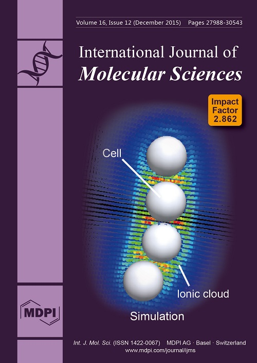
Blood vessels are ubiquitous and one of the most important organs of all vertebrate animals, since they are essential for the supply of oxygen and nutrition for the whole body through the networks. During the network formation of vascular endothelial cells, a concentration gradient of the vascular endothelial growth factor (VEGF) is considered to have a dominant effect on the morphologies of networks. On the other hand, we found that chain-like clusters are formed in absence of VEGF, and in vitro experiments revealed these clusters act as primary networks. The numerical simulations show these chain-like clusters are stabilized by the surface charges of cells, and the concentration of ions surrounding them may also be involved in this process. Image provided by Shunto Arai. View this article
  • Issues are regarded as officially published after their release is announced to the table of contents alert mailing list.
  • You may sign up for e-mail alerts to receive table of contents of newly released issues.
  • PDF is the official format for papers published in both, html and pdf forms. To view the papers in pdf format, click on the "PDF Full-text" link, and use the free Adobe Reader to open them.
Order results
Result details
Section
Select all
Export citation of selected articles as:
13 pages, 1872 KiB  
Article
Application of Green Tea Catechin for Inducing the Osteogenic Differentiation of Human Dedifferentiated Fat Cells in Vitro
by Koji Kaida 1, Yoshitomo Honda 2,*, Yoshiya Hashimoto 3, Masahiro Tanaka 4 and Shunsuke Baba 1
1 Department of Oral Implantology, Osaka Dental University, Osaka 573-1121, Japan
2 Institute of Dental Research, Osaka Dental University, Osaka 573-1121, Japan
3 Department of Biomaterials, Osaka Dental University, Osaka 573-1121, Japan
4 Department of Fixed Prosthodontics and Occlusion, Osaka Dental University, Osaka 573-1121, Japan
Int. J. Mol. Sci. 2015, 16(12), 27988-28000; https://doi.org/10.3390/ijms161226081 - 25 Nov 2015
Cited by 23 | Viewed by 6969
Abstract
Despite advances in stem cell biology, there are few effective techniques to promote the osteogenic differentiation of human primary dedifferentiated fat (DFAT) cells. We attempted to investigate whether epigallocatechin-3-gallate (EGCG), the main component of green tea catechin, facilitates early osteogenic differentiation and mineralization [...] Read more.
Despite advances in stem cell biology, there are few effective techniques to promote the osteogenic differentiation of human primary dedifferentiated fat (DFAT) cells. We attempted to investigate whether epigallocatechin-3-gallate (EGCG), the main component of green tea catechin, facilitates early osteogenic differentiation and mineralization on DFAT cells in vitro. DFAT cells were treated with EGCG (1.25–10 μM) in osteogenic medium (OM) with or without 100 nM dexamethasone (Dex) for 12 days (hereafter two osteogenic media were designated as OM(Dex) and OM). Supplementation of 1.25 μM EGCG to both the media effectively increased the mRNA expression of collagen 1 (COL1A1) and runt-related transcription factor 2 (RUNX2) and also increased proliferation and mineralization. Compared to OM(Dex) with EGCG, OM with EGCG induced earlier expression for COL1A1 and RUNX2 at day 1 and higher mineralization level at day 12. OM(Dex) with 10 μM EGCG remarkably hampered the proliferation of the DFAT cells. These results suggest that OM(without Dex) with EGCG might be a preferable medium to promote proliferation and to induce osteoblast differentiation of DFAT cells. Our findings provide an insight for the combinatory use of EGCG and DFAT cells for bone regeneration and stem cell-based therapy. Full article
(This article belongs to the Special Issue Phenolics and Polyphenolics 2015)
Show Figures

Graphical abstract

13 pages, 6900 KiB  
Article
Adjuvant Immune Enhancement of Subunit Vaccine Encoding pSCPI of Streptococcus iniae in Channel Catfish (Ictalurus punctatus)
by Jie Jiang 1, Zonglin Zheng 2, Kaiyu Wang 1,3,*, Jun Wang 1, Yang He 1, Erlong Wang 1, Defang Chen 4, Ping Ouyang 1,2, Yi Geng 1,2 and Xiaoli Huang 4
1 Department of Basic Veterinary, College of Veterinary Medicine, Sichuan Agricultural University, Wenjiang District Huimin Road No. 211, Chengdu 611130, China
2 Department of Aquaculture, Rongchang Campus, Southwest University, Chongqing 402460, China
3 Key Laboratory of Animal Disease and Human Health of Sichuan Province, Sichuan Agricultural University, Wenjiang District Huimin Road No. 211, Chengdu 611130, China
4 Department of Aquaculture, Sichuan Agricultural University, Wenjiang District Huimin Road No. 211, Chengdu 611130, China
Int. J. Mol. Sci. 2015, 16(12), 28001-28013; https://doi.org/10.3390/ijms161226082 - 25 Nov 2015
Cited by 20 | Viewed by 6805
Abstract
Channel catfish (Ictalurus punctatus) is an important agricultural fish that has been plagued by Streptococcus iniae (S. iniae) infections in recent years, some of them severe. C5a peptidase is an important virulent factor of S. iniae. In this [...] Read more.
Channel catfish (Ictalurus punctatus) is an important agricultural fish that has been plagued by Streptococcus iniae (S. iniae) infections in recent years, some of them severe. C5a peptidase is an important virulent factor of S. iniae. In this study, the subunit vaccine containing the truncated part of C5a peptidase (pSCPI) was mixed with aluminum hydroxide gel (AH), propolis adjuvant (PA), and Freund’s Incomplete Adjuvant (FIA). The immunogenicity of the pSCPI was detected by Western-blot in vitro. The relative percent survival (RPS), lysozyme activity, antibody titers, and the expression of the related immune genes were monitored in vivo to evaluate the immune effects of the three different adjuvants. The results showed that pSCPI exerted moderate immune protection (RPS = 46.43%), whereas each of the three adjuvants improved the immune protection of pSCPI. The immunoprotection of pSCPI + AH, pSCPI + PA, and pSCPI + FIA was characterized by RPS values of 67.86%, 75.00% and, 85.71%, respectively. Further, each of the three different adjuvanted pSCPIs stimulated higher levels of lysozyme activity and antibody titers than the unadjuvanted pSCPI and/or PBS buffer. In addition, pSCPI + FIA and pSCPI + PA induced expression of the related immune genes under investigation, which was substantially higher than the levels stimulated by PBS. pSCPI + AH significantly stimulated the induction of MHC II β, CD4-L2, and IFN-γ, while it induced slightly higher production of TNF-α and even led to a decrease in the levels of IL-1β, MHC I α, and CD8 α. Therefore, we conclude that compared with the other two adjuvants, FIA combined with pSCPI is a more promising candidate adjuvant against S. iniae in channel catfish. Full article
(This article belongs to the Special Issue Fish Molecular Biology)
Show Figures

Figure 1

8 pages, 456 KiB  
Review
Does Lysosomial Acid Lipase Reduction Play a Role in Adult Non-Alcoholic Fatty Liver Disease?
by Francesco Baratta 1,†, Daniele Pastori 1,†, Licia Polimeni 1, Giulia Tozzi 2, Francesco Violi 3, Francesco Angelico 4,*,‡ and Maria Del Ben 3,‡
1 Department of Internal Medicine and Medical Specialities and Department of Anatomical, Histological, Forensic Medicine and Orthopedics Sciences-Sapienza University, Rome 00185, Italy
2 Unit for Neuromuscular and Neurodegenerative Diseases, Children’s Hospital and Research Institute “Bambino Gesù”, Rome 00165, Italy
3 Department of Internal Medicine and Medical Specialities, Sapienza University, Rome 00185, Italy
4 Department of Public Health and Infectious Diseases, Sapienza University, Policlinico Umberto I, I Clinica Medica, Viale del Policlinico 155, Rome 00161, Italy
These authors contributed equally to this work.
Joint senior authors.
Int. J. Mol. Sci. 2015, 16(12), 28014-28021; https://doi.org/10.3390/ijms161226085 - 25 Nov 2015
Cited by 21 | Viewed by 10027
Abstract
Lysosomal Acid Lipase (LAL) is a key enzyme involved in lipid metabolism, responsible for hydrolysing the cholesteryl esters and triglycerides. Wolman Disease represents the early onset phenotype of LAL deficiency rapidly leading to death. Cholesterol Ester Storage Disease is a late onset phenotype [...] Read more.
Lysosomal Acid Lipase (LAL) is a key enzyme involved in lipid metabolism, responsible for hydrolysing the cholesteryl esters and triglycerides. Wolman Disease represents the early onset phenotype of LAL deficiency rapidly leading to death. Cholesterol Ester Storage Disease is a late onset phenotype that occurs with fatty liver, elevated aminotransferase levels, hepatomegaly and dyslipidaemia, the latter characterized by elevated LDL-C and low HDL-C. The natural history and the clinical manifestations of the LAL deficiency in adults are not well defined, and the diagnosis is often incidental. LAL deficiency has been suggested as an under-recognized cause of dyslipidaemia and fatty liver. Therefore, LAL activity may be reduced also in non-obese patients presenting non-alcoholic fatty liver disease (NAFLD), unexplained persistently elevated liver transaminases or with elevation in LDL cholesterol. In these patients, it could be indicated to test LAL activity. So far, very few studies have been performed to assess LAL activity in representative samples of normal subjects or patients with NAFLD. Moreover, no large study has been carried out in adult subjects with NAFLD or cryptogenic cirrhosis. Full article
(This article belongs to the Special Issue Non-Alcoholic Fatty Liver Disease Research 2016)
Show Figures

Figure 1

16 pages, 9238 KiB  
Article
Genotypic Variation under Fe Deficiency Results in Rapid Changes in Protein Expressions and Genes Involved in Fe Metabolism and Antioxidant Mechanisms in Tomato Seedlings (Solanum lycopersicum L.)
by Sowbiya Muneer 1 and Byoung Ryong Jeong 1,2,3,*
1 Division of Applied Life Science (BK21 Plus), Graduate School, Gyeongsang National University, Jinju 660-701, Korea
2 Institute of Agriculture & Life Science, Gyeongsang National University, Jinju 660-701, Korea
3 Research Institute of Life Science, Gyeongsang National University, Jinju 660-701, Korea
Int. J. Mol. Sci. 2015, 16(12), 28022-28037; https://doi.org/10.3390/ijms161226086 - 25 Nov 2015
Cited by 2 | Viewed by 6679
Abstract
To investigate Fe deficiency tolerance in tomato cultivars, quantification of proteins and genes involved in Fe metabolism and antioxidant mechanisms were performed in “Roggusanmaru” and “Super Doterang”. Fe deficiency (Moderate, low and –Fe) significantly decreased the biomass, total, and apoplastic Fe concentration of [...] Read more.
To investigate Fe deficiency tolerance in tomato cultivars, quantification of proteins and genes involved in Fe metabolism and antioxidant mechanisms were performed in “Roggusanmaru” and “Super Doterang”. Fe deficiency (Moderate, low and –Fe) significantly decreased the biomass, total, and apoplastic Fe concentration of “Roggusanmaru”, while a slight variation was observed in “Super Doterang” cultivar. The quantity of important photosynthetic pigments such as total chlorophyll and carotenoid contents significantly decreased in “Roggusanmaru” than “Super Doterang” cultivar. The total protein profile in leaves and roots determines that “Super Doterang” exhibited an optimal tolerance to Fe deficiency compared to “Roggusanmaru” cultivar. A reduction in expression of PSI (photosystem I), PSII (photosystem II) super-complexes and related thylakoid protein contents were detected in “Roggusanmaru” than “Super Doterang” cultivar. Moreover, the relative gene expression of SlPSI and SlPSII were well maintained in “Super Doterang” than “Roggusanmaru” cultivar. The relative expression of genes involved in Fe-transport (SlIRT1 and SlIRT2) and Fe(III) chelates reductase oxidase (SlFRO1) were relatively reduced in “Roggusanmaru”, while increased in “Super Doterang” cultivar under Fe deficient conditions. The H+-ATPase relative gene expression (SlAHA1) in roots were maintained in “Super Doterang” compared to “Roggusanmaru”. Furthermore, the gene expressions involved in antioxidant defense mechanisms (SlSOD, SlAPX and SlCAT) in leaves and roots showed that these genes were highly increased in “Super Doterang”, whereas decreased in “Roggusanmaru” cultivar under Fe deficiency. The present study suggested that “Super Doterang” is better tomato cultivar than “Roggusanmaru” for calcareous soils. Full article
(This article belongs to the Special Issue Plant Molecular Biology)
Show Figures

Figure 1

12 pages, 871 KiB  
Article
A Multiple Interaction Analysis Reveals ADRB3 as a Potential Candidate for Gallbladder Cancer Predisposition via a Complex Interaction with Other Candidate Gene Variations
by Rajani Rai 1,2,*,†, Jong Joo Kim 1,†, Sanjeev Misra 3, Ashok Kumar 4 and Balraj Mittal 2,*
1 School of Biotechnology, Yeungnam University, Gyeongsan, Gyeongbuk 712-749, Korea
2 Department of Genetics, Sanjay Gandhi Post Graduate Institute of Medical Sciences (SGPGIMS), Lucknow-226014, India
3 Department of Surgical Oncology, King George’s Medical University (KGMU), Lucknow-226003, India
4 Department of Surgical Gastroenterology, Sanjay Gandhi Post Graduate Institute of Medical Sciences (SGPGIMS), Lucknow-226014, India
These authors contributed equally to this work.
Int. J. Mol. Sci. 2015, 16(12), 28038-28049; https://doi.org/10.3390/ijms161226077 - 25 Nov 2015
Cited by 14 | Viewed by 6284
Abstract
Gallbladder cancer is the most common and a highly aggressive biliary tract malignancy with a dismal outcome. The pathogenesis of the disease is multifactorial, comprising the combined effect of multiple genetic variations of mild consequence along with numerous dietary and environmental risk factors. [...] Read more.
Gallbladder cancer is the most common and a highly aggressive biliary tract malignancy with a dismal outcome. The pathogenesis of the disease is multifactorial, comprising the combined effect of multiple genetic variations of mild consequence along with numerous dietary and environmental risk factors. Previously, we demonstrated the association of several candidate gene variations with GBC risk. In this study, we aimed to identify the combination of gene variants and their possible interactions contributing towards genetic susceptibility of GBC. Here, we performed Multifactor-Dimensionality Reduction (MDR) and Classification and Regression Tree Analysis (CRT) to investigate the gene–gene interactions and the combined effect of 14 SNPs in nine genes (DR4 (rs20576, rs6557634); FAS (rs2234767); FASL (rs763110); DCC (rs2229080, rs4078288, rs7504990, rs714); PSCA (rs2294008, rs2978974); ADRA2A (rs1801253); ADRB1 (rs1800544); ADRB3 (rs4994); CYP17 (rs2486758)) involved in various signaling pathways. Genotyping was accomplished by PCR-RFLP or Taqman allelic discrimination assays. SPSS software version 16.0 and MDR software version 2.0 were used for all the statistical analysis. Single locus investigation demonstrated significant association of DR4 (rs20576, rs6557634), DCC (rs714, rs2229080, rs4078288) and ADRB3 (rs4994) polymorphisms with GBC risk. MDR analysis revealed ADRB3 (rs4994) to be crucial candidate in GBC susceptibility that may act either alone (p < 0.0001, CVC = 10/10) or in combination with DCC (rs714 and rs2229080, p < 0.0001, CVC = 9/10). Our CRT results are in agreement with the above findings. Further, in-silico results of studied SNPs advocated their role in splicing, transcriptional and/or protein coding regulation. Overall, our result suggested complex interactions amongst the studied SNPs and ADRB3 rs4994 as candidate influencing GBC susceptibility. Full article
(This article belongs to the Special Issue Human Single Nucleotide Polymorphisms and Disease Diagnostics)
Show Figures

Figure 1

13 pages, 1747 KiB  
Article
Identification of Pathogenicity-Related Genes in Biofilm-Defective Acidovorax citrulli by Transposon Tn5 Mutagenesis
by Jinyan Luo 1, Wen Qiu 2, Lei Chen 1, Syed Ishtiaq Anjum 2,3, Menghao Yu 2, Changlin Shan 2,4, Mehmoona Ilyas 5, Bin Li 2,*, Yanli Wang 6 and Guochang Sun 6,*
1 Department of Plant Quarantine, Shanghai Extension and Service Center of Agriculture Technology, Shanghai 201103, China
2 State Key Laboratory of Rice Biology, Institute of Biotechnology, Zhejiang University, Hangzhou 310058, China
3 Department of Zoology, Kohat University of Science and Technology, Kohat 26000, Pakistan
4 Department of Plant Quarantine, Zhoushan Entry-Exit Inspections and Quarantine Bureau, Hangzhou 310012, China
5 Department of Biotechnology, University of Sargodha, Sargodha 40100, Pakistan
6 State Key Laboratory Breeding Base for Zhejiang Sustainable Plant Pest and Disease Control, Key Laboratory of Detection for Pesticide Residues, Ministry of Agriculture, Zhejiang Academy of Agricultural Sciences, Hangzhou 310021, China
Int. J. Mol. Sci. 2015, 16(12), 28050-28062; https://doi.org/10.3390/ijms161226076 - 25 Nov 2015
Cited by 13 | Viewed by 7084
Abstract
Biofilm formation is important for virulence of a large number of plant pathogenic bacteria. Indeed, some virulence genes have been found to be involved in the formation of biofilm in bacterial fruit blotch pathogen Acidovorax citrulli. However, some virulent strains of A. [...] Read more.
Biofilm formation is important for virulence of a large number of plant pathogenic bacteria. Indeed, some virulence genes have been found to be involved in the formation of biofilm in bacterial fruit blotch pathogen Acidovorax citrulli. However, some virulent strains of A. citrulli were unable to format biofilm, indicating the complexity between biofilm formation and virulence. In this study, virulence-related genes were identified in the biofilm-defective strain A1 of A. citrulli by using Tn5 insertion, pathogenicity test, and high-efficiency thermal asymmetric interlaced PCR (hiTAIL-PCR). Results from this study indicated that 22 out of the obtained 301 mutants significantly decreased the virulence of strain A1 compared to the wild-type. Furthermore, sequence analysis indicated that the obtained 22 mutants were due to the insertion of Tn5 into eight genes, including Aave 4244 (cation diffusion facilitator family transporter), Aave 4286 (hypothetical protein), Aave 4189 (alpha/beta hydrolase fold), Aave 1911 (IMP dehydrogenase/GMP reductase domain), Aave 4383 (bacterial export proteins, family 1), Aave 4256 (Hsp70 protein), Aave 0003 (histidine kinase, DNA gyrase B, and HSP90-like ATPase), and Aave 2428 (pyridoxal-phosphate dependent enzyme). Furthermore, the growth of mutant Aave 2428 was unaffected and even increased by the change in incubation temperature, NaCl concentration and the pH of the LB broth, indicating that this gene may be directly involved in the bacterial virulence. Overall, the determination of the eight pathogenicity-related genes in strain A1 will be helpful to elucidate the pathogenesis of biofilm-defective A. citrulli. Full article
(This article belongs to the Special Issue Plant Microbe Interaction)
Show Figures

Graphical abstract

14 pages, 377 KiB  
Review
MicroRNAs: Clinical Relevance in Colorectal Cancer
by Joe Thomas 1, Masahisa Ohtsuka 1, Martin Pichler 1,2 and Hui Ling 1,*
1 Department of Experimental Therapeutics, The University of Texas MD Anderson Cancer Center, Houston, TX 77054, USA
2 Division of Oncology, Medical University of Graz, 8010 Graz, Austria
Int. J. Mol. Sci. 2015, 16(12), 28063-28076; https://doi.org/10.3390/ijms161226080 - 25 Nov 2015
Cited by 115 | Viewed by 9602
Abstract
Colorectal cancer is one of the most common cancer diagnoses and causes of mortality worldwide. MicroRNAs are a class of small, non-coding regulatory RNAs that have shown strong associations with colorectal cancer. Through the repression of target messenger RNAs, microRNAs modulate many cellular [...] Read more.
Colorectal cancer is one of the most common cancer diagnoses and causes of mortality worldwide. MicroRNAs are a class of small, non-coding regulatory RNAs that have shown strong associations with colorectal cancer. Through the repression of target messenger RNAs, microRNAs modulate many cellular pathways, such as those involved in cell proliferation, apoptosis, and differentiation. The utilization of microRNAs has shown significant promise in the diagnosis and prognosis of colorectal cancer, owing to their unique expression profile associations with cancer types and malignancies. Moreover, microRNA therapeutics with mimics or antagonists show great promise in preclinical studies, which encourages further development of their clinical use for colorectal cancer patients. The unique ability of microRNAs to affect multiple downstream pathways represents a novel approach for cancer therapy. Although still early in its development, we believe that microRNAs can be used in the near future as biomarkers and therapeutic targets for colorectal cancer. Full article
(This article belongs to the Collection Regulation by Non-coding RNAs)
Show Figures

Figure 1

10 pages, 1807 KiB  
Article
Critical Role of Endoplasmic Reticulum Stress in Cognitive Impairment Induced by Microcystin-LR
by Fei Cai 1,†, Jue Liu 2,†, Cairong Li 3,† and Jianghua Wang 4,*
1 Department of Pharmacology, Hubei University of Science and Technology, Xianning 437100, China
2 Department of Pharmacy, The Central Hospital of Wuhan, Tongji Medical College, Huazhong University of Science and Technology, Wuhan 430014, China
3 Hubei Province Key Laboratory on Cardiovascular, Cerebrovascular, and Metabolic Disorders, Hubei University of Science and Technology, Xianning 437100, China
4 Fisheries College, Huazhong Agricultural University, Wuhan 430070, China
These authors contributed equally to this work.
Int. J. Mol. Sci. 2015, 16(12), 28077-28086; https://doi.org/10.3390/ijms161226083 - 25 Nov 2015
Cited by 31 | Viewed by 6379
Abstract
Recent studies showed that cyanobacteria-derived microcystin-leucine-arginine (MCLR) can cause hippocampal pathological damage and trigger cognitive impairment; but the underlying mechanisms have not been well understood. The objective of the present study was to investigate the mechanism of MCLR-induced cognitive deficit; with a focus [...] Read more.
Recent studies showed that cyanobacteria-derived microcystin-leucine-arginine (MCLR) can cause hippocampal pathological damage and trigger cognitive impairment; but the underlying mechanisms have not been well understood. The objective of the present study was to investigate the mechanism of MCLR-induced cognitive deficit; with a focus on endoplasmic reticulum (ER) stress. The Morris water maze test and electrophysiological study demonstrated that MCLR caused spatial memory injury in male Wistar rats; which could be inhibited by ER stress blocker; tauroursodeoxycholic acid (TUDCA). Meanwhile; real-time polymerase chain reaction (real-time PCR) and immunohistochemistry demonstrated that the expression level of the 78-kDa glucose-regulated protein (GRP78); C/EBP homologous protein (CHOP) and caspase 12 were significantly up-regulated. These effects were rescued by co-administration of TUDCA. In agreement with this; we also observed that treatment of rats with TUDCA blocked the alterations in ER ultrastructure and apoptotic cell death in CA1 neurons from rats exposed to MCLR. Taken together; the present results suggested that ER stress plays an important role in potential memory impairments in rats treated with MCLR; and amelioration of ER stress may serve as a novel strategy to alleviate damaged cognitive function triggered by MCLR. Full article
(This article belongs to the Special Issue Modulators of Endoplasmic Reticulum Stress)
Show Figures

Graphical abstract

11 pages, 3924 KiB  
Article
Transcriptome Profiling of Louisiana iris Root and Identification of Genes Involved in Lead-Stress Response
by Songqing Tian 1,2,†, Chunsun Gu 1,†, Liangqin Liu 3,†, Xudong Zhu 1,2, Yanhai Zhao 1 and Suzhen Huang 1,*
1 Institute of Botany, Jiangsu Province and Chinese Academy of Science, Nanjing 210014, China
2 Suzhou Polytechnical Institute of Agriculture, Suzhou 215008, China
3 College of Horticulture, Nanjing Agricultural University, Nanjing 210014, China
These authors contributed equally to this work.
Int. J. Mol. Sci. 2015, 16(12), 28087-28097; https://doi.org/10.3390/ijms161226084 - 25 Nov 2015
Cited by 26 | Viewed by 7294
Abstract
Louisiana iris is tolerant to and accumulates the heavy metal lead (Pb). However, there is limited knowledge of the molecular mechanisms behind this feature. We describe the transcriptome of Louisiana iris using Illumina sequencing technology. The root transcriptome of Louisiana iris under control [...] Read more.
Louisiana iris is tolerant to and accumulates the heavy metal lead (Pb). However, there is limited knowledge of the molecular mechanisms behind this feature. We describe the transcriptome of Louisiana iris using Illumina sequencing technology. The root transcriptome of Louisiana iris under control and Pb-stress conditions was sequenced. Overall, 525,498 transcripts representing 313,958 unigenes were assembled using the clean raw reads. Among them, 43,015 unigenes were annotated and their functions classified using the euKaryotic Orthologous Groups (KOG) database. They were divided into 25 molecular families. In the Gene Ontology (GO) database, 50,174 unigenes were categorized into three GO trees (molecular function, cellular component and biological process). After analysis of differentially expressed genes, some Pb-stress-related genes were selected, including biosynthesis genes of chelating compounds, metal transporters, transcription factors and antioxidant-related genes. This study not only lays a foundation for further studies on differential genes under Pb stress, but also facilitates the molecular breeding of Louisiana iris. Full article
Show Figures

Graphical abstract

10 pages, 571 KiB  
Review
Dermal Contributions to Human Interfollicular Epidermal Architecture and Self-Renewal
by Kynan T. Lawlor and Pritinder Kaur *
Hudson Institute of Medical Research, Clayton, VIC 3168, Australia
Int. J. Mol. Sci. 2015, 16(12), 28098-28107; https://doi.org/10.3390/ijms161226078 - 25 Nov 2015
Cited by 50 | Viewed by 10681
Abstract
The human interfollicular epidermis is renewed throughout life by populations of proliferating basal keratinocytes. Though interfollicular keratinocyte stem cells have been identified, it is not known how self-renewal in this compartment is spatially organized. At the epidermal-dermal junction, keratinocytes sit atop a heterogeneous [...] Read more.
The human interfollicular epidermis is renewed throughout life by populations of proliferating basal keratinocytes. Though interfollicular keratinocyte stem cells have been identified, it is not known how self-renewal in this compartment is spatially organized. At the epidermal-dermal junction, keratinocytes sit atop a heterogeneous mix of dermal cells that may regulate keratinocyte self-renewal by influencing local tissue architecture and signalling microenvironments. Focusing on the rete ridges and complementary dermal papillae in human skin, we review the identity and organisation of abundant dermal cells types and present evidence for interactions between the dermal microenvironment and the interfollicular keratinocytes. Full article
(This article belongs to the Special Issue Molecular Research of Epidermal Stem Cells 2015)
Show Figures

Graphical abstract

15 pages, 18003 KiB  
Article
High SPARC Expression Starting from Dysplasia, Associated with Breast Carcinoma, Is Predictive for Bone Metastasis without Enhancement of Plasma Levels
by Paola Maroni 1, Paola Bendinelli 2, Daniele Morelli 3, Lorenzo Drago 1,2, Alessandro Luzzati 1, Giuseppe Perrucchini 1, Chiara Bonini 3, Emanuela Matteucci 2 and Maria Alfonsina Desiderio 2,*
1 Istituto Ortopedico Galeazzi, Scientific Institute for Research, Hospitalization and Helth Care (IRCCS), 20161 Milano, Italy
2 Dipartimento di Scienze Biomediche per la Salute, Molecular Pathology Laboratory, Università degli Studi di Milano, 20133 Milano, Italy
3 Department of Pathology and Laboratory Medicine, Fondazione IRCCS Istituto Nazionale dei Tumori, 20133 Milano, Italy
Int. J. Mol. Sci. 2015, 16(12), 28108-28122; https://doi.org/10.3390/ijms161225997 - 26 Nov 2015
Cited by 13 | Viewed by 6125
Abstract
In order to become established in the skeleton, metastatic cells disseminating from the breast carcinoma need to acquire organ-specific traits. There are no effective predictors for who will develop bone metastasis to guide long-term predictive therapy. Our purpose was to individuate events critical [...] Read more.
In order to become established in the skeleton, metastatic cells disseminating from the breast carcinoma need to acquire organ-specific traits. There are no effective predictors for who will develop bone metastasis to guide long-term predictive therapy. Our purpose was to individuate events critical for bone colonization to make a molecular classification of breast carcinoma useful for bone-metastasis outcome. In dysplasia adjacent to carcinoma and in pair-matched specimens of bone metastasis we examined SPARC expression and localization as well as Endothelin 1/ETAR signals by immunohistochemistry, and the evaluation of plasma levels of SPARC by ELISA was also performed. In patients with breast carcinoma metastasizing to bone, SPARC and Endothelin 1/ETAR axis were highly expressed from dysplasia until bone metastasis, but the SPARC plasma level was as low as that of normal women, in contrast to patients that never develop bone metastasis, suggesting that circulating SPARC was counter adhesive. Altogether, the early identification of SPARC/Endothelin 1/ETAR in dysplastic lesions would be important to devise therapies preventing metastasis engraftment, since often carcinoma cells spread to distant organs at the time or even before patients present with cancer. Full article
(This article belongs to the Special Issue Molecular Classification of Human Cancer: Diagnosis and Treatment)
Show Figures

Figure 1

3 pages, 140 KiB  
Book Review
Molecular Biology of the Cell, Sixth Edition; ISBN: 9780815344643; and Molecular Biology of the Cell, Sixth Edition, The Problems Book; ISBN 9780815344537
by Stephen Bustin
Postgraduate Medical Institute, Anglia Ruskin University, Chelmsford CM1 1SQ, UK
Int. J. Mol. Sci. 2015, 16(12), 28123-28125; https://doi.org/10.3390/ijms161226074 - 26 Nov 2015
Cited by 17 | Viewed by 22449
Abstract
The latest edition maintains the excellence and appeal of the previous editions. Its clear text and outstanding illustrations, together with the complementary problems book make this an essential companion for students and lecturers alike. Full article
(This article belongs to the Section Biochemistry)
20 pages, 1091 KiB  
Review
Chronic Hepatitis B with Spontaneous Severe Acute Exacerbation
by Wei-Lun Tsai 1,2,*, Wei-Chi Sun 1,2 and Jin-Shiung Cheng 1,2
1 Division of Gastroenterology, Department of Internal Medicine, Kaohsiung Veterans General Hospital, Kaohsiung 813, Taiwan
2 School of Medicine, National Yang Ming University, Taipei 100, Taiwan
Int. J. Mol. Sci. 2015, 16(12), 28126-28145; https://doi.org/10.3390/ijms161226087 - 26 Nov 2015
Cited by 20 | Viewed by 7331
Abstract
Chronic hepatitis B virus (HBV) infection is a major global health problem with an estimated 400 million HBV carriers worldwide. In the natural history of chronic hepatitis B (CHB), spontaneous acute exacerbation (AE) is not uncommon, with a cumulative incidence of 10%–30% every [...] Read more.
Chronic hepatitis B virus (HBV) infection is a major global health problem with an estimated 400 million HBV carriers worldwide. In the natural history of chronic hepatitis B (CHB), spontaneous acute exacerbation (AE) is not uncommon, with a cumulative incidence of 10%–30% every year. While exacerbations can be mild, some patients may develop hepatic decompensation and even die. The underlying pathogenesis is possibly related to the activation of cytotoxic T lymphocyte-mediated immune response against HBV. An upsurge of serum HBV DNA usually precedes the rise of alanine aminotransferase (ALT) and bilirubin. Whether antiviral treatment can benefit CHB with severe AE remains controversial, but early nucleos(t)ide analogues treatment seemed to be associated with an improved outcome. There has been no randomized study that compared the effects of different nucleos(t)ide analogues (NA) in the setting of CHB with severe AE. However, potent NAs with good resistance profiles are recommended. In this review, we summarized current knowledge regarding the natural history, pathogenetic mechanisms, and therapeutic options of CHB with severe AE. Full article
(This article belongs to the Special Issue Viral Hepatitis Research)
Show Figures

Figure 1

10 pages, 9030 KiB  
Article
The Formation of pH-Sensitive Wormlike Micelles in Ionic Liquids Driven by the Binding Ability of Anthranilic Acid
by Qing You 1,*, Yan Zhang 1,*, Huan Wang 1, Hongfu Fan 1, Jianping Guo 1 and Ming Li 2
1 School of Energy Resources, China University of Geosciences, Beijing 100083, China
2 Sinopec Shengli Oilfield Research Institute of Petroleum Engineering, Dongying 257000, China
Int. J. Mol. Sci. 2015, 16(12), 28146-28155; https://doi.org/10.3390/ijms161226096 - 26 Nov 2015
Cited by 16 | Viewed by 6878
Abstract
Wormlike micelles are typically formed by mixing cationic and anionic surfactants because of attractive interactions in oppositely charged head-groups. The structural transitions of wormlike micelles triggered by pH in ionic liquids composed of N-alkyl-N-methylpyrrolidinium bromide-based ILs (ionic liquids) and anthranilic [...] Read more.
Wormlike micelles are typically formed by mixing cationic and anionic surfactants because of attractive interactions in oppositely charged head-groups. The structural transitions of wormlike micelles triggered by pH in ionic liquids composed of N-alkyl-N-methylpyrrolidinium bromide-based ILs (ionic liquids) and anthranilic acid were investigated. These structures were found responsible for the variations in flow properties identified by rheology and dynamic light scattering, and account for the structures observed with cryogenic transmission electron microscopy (Cryo-TEM). High-viscosity, shear-thinning behavior, and Maxwell-type dynamic rheology shown by the system at certain pH values suggested that spherical micelles grow into entangled wormlike micelles. Light scattering profiles also supported the notion of pH-sensitive microstructural transitions in the solution. Cryo-TEM images confirmed the presence of spherical micelles in the low-viscosity sample and entangled wormlike micelles in the peak viscosity sample. Nuclear magnetic resonance spectroscopy analysis revealed that the pH sensitivity of ionic liquid systems originated from the pH-dependent binding ability of anthranilic acid to the cationic headgroup of ionic liquids. Full article
(This article belongs to the Special Issue Ionic Liquids 2016 and Selected Papers from ILMAT III)
Show Figures

Graphical abstract

13 pages, 3779 KiB  
Article
miR-218 Inhibits Erythroid Differentiation and Alters Iron Metabolism by Targeting ALAS2 in K562 Cells
by Yanming Li 1,2,†, Shuge Liu 1,2,†, Hongying Sun 1,†, Yadong Yang 1, Heyuan Qi 1,2, Nan Ding 1,2, Jiawen Zheng 1,2, Xunong Dong 1,2, Hongzhu Qu 1, Zhaojun Zhang 1 and Xiangdong Fang 1,*
1 CAS Key Laboratory of Genome Sciences and Information, Beijing Institute of Genomics, Chinese Academy of Sciences, Beijing 100101, China
2 College of Life Sciences, University of Chinese Academy of Sciences, Beijing 100049, China
These authors contributed equally to this work.
Int. J. Mol. Sci. 2015, 16(12), 28156-28168; https://doi.org/10.3390/ijms161226088 - 26 Nov 2015
Cited by 14 | Viewed by 7113
Abstract
microRNAs (miRNAs) are involved in a variety of biological processes. The regulatory function and potential role of miRNAs targeting the mRNA of the 5′-aminolevulinate synthase 2 (ALAS2) in erythropoiesis were investigated in order to identify miRNAs which play a role in [...] Read more.
microRNAs (miRNAs) are involved in a variety of biological processes. The regulatory function and potential role of miRNAs targeting the mRNA of the 5′-aminolevulinate synthase 2 (ALAS2) in erythropoiesis were investigated in order to identify miRNAs which play a role in erythroid iron metabolism and differentiation. Firstly, the role of ALAS2 in erythroid differentiation and iron metabolism in human erythroid leukemia cells (K562) was confirmed by ALAS2 knockdown. Through a series of screening strategies and experimental validations, it was identified that hsa-miR-218 (miR-218) targets and represses the expression of ALAS2 by binding to the 3′-untranslated region (UTR). Overexpression of miR-218 repressed erythroid differentiation and altered iron metabolism in K562 cells similar to that seen in the ALAS2 knockdown in K562 cells. In addition to iron metabolism and erythroid differentiation, miR-218 was found to be responsible for a reduction in K562 cell growth. Taken together, our results show that miR-218 inhibits erythroid differentiation and alters iron metabolism by targeting ALAS2 in K562 cells. Full article
(This article belongs to the Special Issue MicroRNA Regulation)
Show Figures

Graphical abstract

11 pages, 1473 KiB  
Article
Linalool Induces Cell Cycle Arrest and Apoptosis in Leukemia Cells and Cervical Cancer Cells through CDKIs
by Mei-Yin Chang 1,*, Den-En Shieh 2, Chung-Chi Chen 1, Ching-Sheng Yeh 3 and Huei-Ping Dong 4
1 Department of Medical Laboratory Science and Biotechnology, School of Medical and Health Sciences, Fooyin University, Ta-Liao District, Kaohsiung 83102, Taiwan
2 Department of Biotechnology, Collage of Pharmacy and Health Care, Tajen University, Yanpu Township, Pingtung County 90741, Taiwan
3 Department of Nutrition and Health Science, School of Medical and Health Sciences, Fooyin University, Ta-Liao District, Kaohsiung 83102, Taiwan
4 Department of Physical Therapy, School of Medical and Health Sciences, Fooyin University, Ta-Liao District, Kaohsiung 83102, Taiwan
Int. J. Mol. Sci. 2015, 16(12), 28169-28179; https://doi.org/10.3390/ijms161226089 - 26 Nov 2015
Cited by 85 | Viewed by 8239
Abstract
Plantaginaceae, a popular traditional Chinese medicine, has long been used for treating various diseases from common cold to cancer. Linalool is one of the biologically active compounds that can be isolated from Plantaginaceae. Most of the commonly used cytotoxic anticancer drugs [...] Read more.
Plantaginaceae, a popular traditional Chinese medicine, has long been used for treating various diseases from common cold to cancer. Linalool is one of the biologically active compounds that can be isolated from Plantaginaceae. Most of the commonly used cytotoxic anticancer drugs have been shown to induce apoptosis in susceptible tumor cells. However, the signaling pathway for apoptosis remains undefined. In this study, the cytotoxic effect of linalool on human cancer cell lines was investigated. Water-soluble tetrazolium salts (WST-1) based colorimetric cellular cytotoxicity assay, was used to test the cytotoxic ability of linalool against U937 and HeLa cells, and flow cytometry (FCM) and genechip analysis were used to investigate the possible mechanism of apoptosis. These results demonstrated that linalool exhibited a good cytotoxic effect on U937 and HeLa cells, with the IC50 value of 2.59 and 11.02 μM, respectively, compared with 5-FU with values of 4.86 and 12.31 μM, respectively. After treating U937 cells with linalool for 6 h, we found an increased sub-G1 peak and a dose-dependent phenomenon, whereby these cells were arrested at the G0/G1 phase. Furthermore, by using genechip analysis, we observed that linalool can promote p53, p21, p27, p16, and p18 gene expression. Therefore, this study verified that linalool can arrest the cell cycle of U937 cells at the G0/G1 phase and can arrest the cell cycle of HeLa cells at the G2/M phase. Its mechanism facilitates the expression of the cyclin-dependent kinases inhibitors (CDKIs) p53, p21, p27, p16, and p18, as well as the non-expression of cyclin-dependent kinases (CDKs) activity. Full article
(This article belongs to the Section Molecular Toxicology)
Show Figures

Graphical abstract

14 pages, 3244 KiB  
Article
Evodiamine Attenuates PDGF-BB-Induced Migration of Rat Vascular Smooth Muscle Cells through Activating PPARγ
by Xie Ge, Siyu Chen, Mei Liu, Tingming Liang and Chang Liu *
Jiangsu Key Laboratory for Molecular and Medical Biotechnology and College of Life Sciences, Nanjing Normal University, Nanjing 210023, China
Int. J. Mol. Sci. 2015, 16(12), 28180-28193; https://doi.org/10.3390/ijms161226093 - 26 Nov 2015
Cited by 34 | Viewed by 6973
Abstract
The uncontrolled migration of vascular smooth muscle cells (VSMCs) into the intima is a critical process in the development of atherosclerosis. Evodiamine, an indole alkaloid extracted from the Chinese medicine evodia, has been shown to inhibit tumor cell invasion and protect the cardiovascular [...] Read more.
The uncontrolled migration of vascular smooth muscle cells (VSMCs) into the intima is a critical process in the development of atherosclerosis. Evodiamine, an indole alkaloid extracted from the Chinese medicine evodia, has been shown to inhibit tumor cell invasion and protect the cardiovascular system, but its effects on VSMCs remain unknown. In the present study, we investigated the inhibitory effects of evodiamine on the platelet-derived growth factor-BB (PDGF-BB)-induced VSMC migration using wound healing and transwell assays, and assessed its role in decreasing the protein levels of matrix metalloproteinases and cell adhesion molecules. More importantly, we found that evodiamine activated the expression and nuclear translocation of peroxisome proliferator-activated receptor γ (PPARγ). Inhibition of PPARγ activity by using its antagonist T0070907 and its specific siRNA oligonucleotides significantly attenuated the inhibitory effects of evodiamine on VSMC migration. Taken together, our results indicate a promising anti-atherogenic effect of evodiamine through attenuation of VSMC migration by activating PPARγ. Full article
(This article belongs to the Section Biochemistry)
Show Figures

Graphical abstract

24 pages, 1051 KiB  
Review
Old Things New View: Ascorbic Acid Protects the Brain in Neurodegenerative Disorders
by Adriana Covarrubias-Pinto 1,2, Aníbal Ignacio Acuña 1,2, Felipe Andrés Beltrán 1,2, Leandro Torres-Díaz 1,2 and Maite Aintzane Castro 1,2,*
1 Instituto de Bioquímica y Microbiología, Facultad de Ciencias, Universidad Austral de Chile, Valdivia 5090000, Chile
2 Center for Interdisciplinary Studies on the Nervous system (CISNe), Universidad Austral de Chile, Casilla 547, Valdivia 5090000, Chile
Int. J. Mol. Sci. 2015, 16(12), 28194-28217; https://doi.org/10.3390/ijms161226095 - 27 Nov 2015
Cited by 118 | Viewed by 13444
Abstract
Ascorbic acid is a key antioxidant of the Central Nervous System (CNS). Under brain activity, ascorbic acid is released from glial reservoirs to the synaptic cleft, where it is taken up by neurons. In neurons, ascorbic acid scavenges reactive oxygen species (ROS) generated [...] Read more.
Ascorbic acid is a key antioxidant of the Central Nervous System (CNS). Under brain activity, ascorbic acid is released from glial reservoirs to the synaptic cleft, where it is taken up by neurons. In neurons, ascorbic acid scavenges reactive oxygen species (ROS) generated during synaptic activity and neuronal metabolism where it is then oxidized to dehydroascorbic acid and released into the extracellular space, where it can be recycled by astrocytes. Other intrinsic properties of ascorbic acid, beyond acting as an antioxidant, are important in its role as a key molecule of the CNS. Ascorbic acid can switch neuronal metabolism from glucose consumption to uptake and use of lactate as a metabolic substrate to sustain synaptic activity. Multiple evidence links oxidative stress with neurodegeneration, positioning redox imbalance and ROS as a cause of neurodegeneration. In this review, we focus on ascorbic acid homeostasis, its functions, how it is used by neurons and recycled to ensure antioxidant supply during synaptic activity and how this antioxidant is dysregulated in neurodegenerative disorders. Full article
(This article belongs to the Special Issue Antioxidant 2.0——Redox Modulation by Food and Drugs)
Show Figures

Figure 1

12 pages, 693 KiB  
Review
Therapeutic Targets for Neurodevelopmental Disorders Emerging from Animal Models with Perinatal Immune Activation
by Daisuke Ibi 1,2 and Kiyofumi Yamada 2,*
1 Department of Chemical Pharmacology, Faculty of Pharmaceutical Sciences, Meijo University, 150 Yagotoyama, Tenpaku-ku, Nagoya 468-8503, Japan
2 Department of Neuropsychopharmacology and Hospital Pharmacy, Nagoya University Graduate School of Medicine, Nagoya 466-8560, Japan
Int. J. Mol. Sci. 2015, 16(12), 28218-28229; https://doi.org/10.3390/ijms161226092 - 27 Nov 2015
Cited by 21 | Viewed by 7635
Abstract
Increasing epidemiological evidence indicates that perinatal infection with various viral pathogens enhances the risk for several psychiatric disorders. The pathophysiological significance of astrocyte interactions with neurons and/or gut microbiomes has been reported in neurodevelopmental disorders triggered by pre- and postnatal immune insults. Recent [...] Read more.
Increasing epidemiological evidence indicates that perinatal infection with various viral pathogens enhances the risk for several psychiatric disorders. The pathophysiological significance of astrocyte interactions with neurons and/or gut microbiomes has been reported in neurodevelopmental disorders triggered by pre- and postnatal immune insults. Recent studies with the maternal immune activation or neonatal polyriboinosinic polyribocytidylic acid models of neurodevelopmental disorders have identified various candidate molecules that could be responsible for brain dysfunction. Here, we review the functions of several candidate molecules in neurodevelopment and brain function and discuss their potential as therapeutic targets for psychiatric disorders. Full article
Show Figures

Figure 1

12 pages, 366 KiB  
Review
Therapeutic Potential of Cell Penetrating Peptides (CPPs) and Cationic Polymers for Chronic Hepatitis B
by Bénédicte Ndeboko 1,2, Guy Joseph Lemamy 2, Peter. E Nielsen 3 and Lucyna Cova 1,*
1 Institut National de la Sante et Recherche Medicale (INSERM) U1052, Cancer Research Center of Lyon (CRCL), Lyon 69003, France
2 Département de Biologie Cellulaire and Moléculaire-Génétique, Faculté de Médecine, Université des Sciences de la Santé, Libreville 241, Gabon
3 Department of Cellular and Molecular Medicine, Departement of Drug Design and Pharmacology, Faculty of Health and Medical Sciences, the Panum Institute, University of Copenhagen, Copenhagen DK 2200N, Denmark
Int. J. Mol. Sci. 2015, 16(12), 28230-28241; https://doi.org/10.3390/ijms161226094 - 27 Nov 2015
Cited by 23 | Viewed by 6170
Abstract
Chronic hepatitis B virus (HBV) infection remains a major health problem worldwide. Because current anti-HBV treatments are only virostatic, there is an urgent need for development of alternative antiviral approaches. In this context, cell-penetrating peptides (CPPs) and cationic polymers, such as chitosan (CS), [...] Read more.
Chronic hepatitis B virus (HBV) infection remains a major health problem worldwide. Because current anti-HBV treatments are only virostatic, there is an urgent need for development of alternative antiviral approaches. In this context, cell-penetrating peptides (CPPs) and cationic polymers, such as chitosan (CS), appear of particular interest as nonviral vectors due to their capacity to facilitate cellular delivery of bioactive cargoes including peptide nucleic acids (PNAs) or DNA vaccines. We have investigated the ability of a PNA conjugated to different CPPs to inhibit the replication of duck hepatitis B virus (DHBV), a reference model for human HBV infection. The in vivo administration of PNA-CPP conjugates to neonatal ducklings showed that they reached the liver and inhibited DHBV replication. Interestingly, our results indicated also that a modified CPP (CatLip) alone, in the absence of its PNA cargo, was able to drastically inhibit late stages of DHBV replication. In the mouse model, conjugation of HBV DNA vaccine to modified CS (Man-CS-Phe) improved cellular and humoral responses to plasmid-encoded antigen. Moreover, other systems for gene delivery were investigated including CPP-modified CS and cationic nanoparticles. The results showed that these nonviral vectors considerably increased plasmid DNA uptake and expression. Collectively promising results obtained in preclinical studies suggest the usefulness of these safe delivery systems for the development of novel therapeutics against chronic hepatitis B. Full article
(This article belongs to the Special Issue Cell-Penetrating Peptides)
Show Figures

Figure 1

13 pages, 4081 KiB  
Article
ITSN2L Interacts with and Negatively Regulates RABEP1
by Xiaoxu Yang 1,†, Feng Yan 1,†, Zhicheng He 1,†, Shan Liu 1, Yeqing Cheng 1, Ke Wei 1, Shiquan Gan 1, Jing Yuan 1, Shang Wang 1, Ye Xiao 1, Kaiqun Ren 1, Ning Liu 1, Xiang Hu 1, Xiaofeng Ding 1, Xingwang Hu 1,2,* and Shuanglin Xiang 1,*
1 Key Laboratory of Protein Chemistry and Developmental Biology of State Ministry of Education, College of Life Sciences, Hunan Normal University, Changsha 410081, China
2 Department of Dermatology, Xiangya Hospital, Central South University, Changsha 410081, China
These authors contributed equally to this work.
Int. J. Mol. Sci. 2015, 16(12), 28242-28254; https://doi.org/10.3390/ijms161226091 - 27 Nov 2015
Cited by 4 | Viewed by 5729
Abstract
Intersectin-2Long (ITSN2L) is a multi-domain protein participating in endocytosis and exocytosis. In this study, RABEP1 was identified as a novel ITSN2L interacting protein using a yeast two-hybrid screen from a human brain cDNA library and this interaction, specifically involving the ITSN2L CC domain [...] Read more.
Intersectin-2Long (ITSN2L) is a multi-domain protein participating in endocytosis and exocytosis. In this study, RABEP1 was identified as a novel ITSN2L interacting protein using a yeast two-hybrid screen from a human brain cDNA library and this interaction, specifically involving the ITSN2L CC domain and RABEP1 CC3 regions, was further confirmed by in vitro GST (glutathione-S-transferase) pull-down and in vivo co-immunoprecipitation assays. Corroboratively, we observed that these two proteins co-localize in the cytoplasm of mammalian cells. Furthermore, over-expression of ITSN2L promotes RABEP1 degradation and represses RABEP1-enhanced endosome aggregation, indicating that ITSN2L acts as a negative regulator of RABEP1. Finally, we showed that ITSN2L and RABEP1 play opposite roles in regulating endocytosis. Taken together, our results indicate that ITSN2L interacts with RABEP1 and stimulates its degradation in regulation of endocytosis. Full article
(This article belongs to the Section Biochemistry)
Show Figures

Figure 1

15 pages, 2365 KiB  
Article
Polar Glycosylated and Lateral Non-Glycosylated Flagella from Aeromonas hydrophila Strain AH-1 (Serotype O11)
by Kelly M. Fulton 1,†, Elena Mendoza-Barberá 2,†, Susan M. Twine 1, Juan M. Tomás 2,* and Susana Merino 2
1 National Research Council, 100 Sussex Drive, Ottawa, ON K1A0R1, Canada
2 Departamento de Microbiología, Facultad de Biología, Universidad de Barcelona, Diagonal 645, Barcelona 08071, Spain
These authors contributed equally to this work.
Int. J. Mol. Sci. 2015, 16(12), 28255-28269; https://doi.org/10.3390/ijms161226097 - 27 Nov 2015
Cited by 12 | Viewed by 6523
Abstract
Polar and but not lateral flagellin proteins from Aeromonas hydrophila strain AH-1 (serotype O11) were found to be glycosylated. Top-down mass spectrometry studies of purified polar flagellins suggested the presence of a 403 Da glycan of mass. Bottom-up mass spectrometry studies showed the [...] Read more.
Polar and but not lateral flagellin proteins from Aeromonas hydrophila strain AH-1 (serotype O11) were found to be glycosylated. Top-down mass spectrometry studies of purified polar flagellins suggested the presence of a 403 Da glycan of mass. Bottom-up mass spectrometry studies showed the polar flagellin peptides to be modified with 403 Da glycans in O-linkage. The MS fragmentation pattern of this putative glycan was similar to that of pseudaminic acid derivative. Mutants lacking the biosynthesis of pseudaminic acid (pseB and pseI homologues) were unable to produce polar flagella but no changes were observed in lateral flagella by post-transcriptional regulation of the flagellin. Complementation was achieved by reintroduction of the wild-type pseB and pseI. We compared two pathogenic features (adhesion to eukaryotic cells and biofilm production) between the wild-type strain and two kinds of mutants: mutants lacking polar flagella glycosylation and lacking the O11-antigen lipopolysaccharide (LPS) but with unaltered polar flagella glycosylation. Results suggest that polar flagella glycosylation is extremely important for A. hydrophila AH-1 adhesion to Hep-2 cells and biofilm formation. In addition, we show the importance of the polar flagella glycosylation for immune stimulation of IL-8 production via toll-“like” receptor 5 (TLR5). Full article
(This article belongs to the Special Issue Glycosylation and Glycoproteins)
Show Figures

Graphical abstract

15 pages, 3640 KiB  
Article
Characterization of Aspartate Kinase from Corynebacterium pekinense and the Critical Site of Arg169
by Weihong Min 1,2,*, Huiying Li 1,2, Hongmei Li 1,2, Chunlei Liu 1,2 and Jingsheng Liu 1,2
1 College of Food Science and Engineering, Jilin Agricultural University, Changchun 130118, China
2 National Engineering Laboratory of Wheat and Corn Deep Processing, Changchun 130118, China
Int. J. Mol. Sci. 2015, 16(12), 28270-28284; https://doi.org/10.3390/ijms161226098 - 27 Nov 2015
Cited by 10 | Viewed by 6478
Abstract
Aspartate kinase (AK) is the key enzyme in the biosynthesis of aspartate-derived amino acids. Recombinant AK was efficiently purified and systematically characterized through analysis under optimal conditions combined with steady-state kinetics study. Homogeneous AK was predicted as a decamer with a molecular weight [...] Read more.
Aspartate kinase (AK) is the key enzyme in the biosynthesis of aspartate-derived amino acids. Recombinant AK was efficiently purified and systematically characterized through analysis under optimal conditions combined with steady-state kinetics study. Homogeneous AK was predicted as a decamer with a molecular weight of ~48 kDa and a half-life of 4.5 h. The enzymatic activity was enhanced by ethanol and Ni2+. Moreover, steady-state kinetic study confirmed that AK is an allosteric enzyme, and its activity was inhibited by allosteric inhibitors, such as Lys, Met, and Thr. Theoretical results indicated the binding mode of AK and showed that Arg169 is an important residue in substrate binding, catalytic domain, and inhibitor binding. The values of the kinetic parameter Vmax of R169 mutants, namely, R169Y, R169P, R169D, and R169H AK, with l-aspartate as the substrate, were 4.71-, 2.25-, 2.57-, and 2.13-fold higher, respectively, than that of the wild-type AK. Furthermore, experimental and theoretical data showed that Arg169 formed a hydrogen bond with Glu92, which functions as the entrance gate. This study provides a basis to develop new enzymes and elucidate the corresponding amino acid production. Full article
(This article belongs to the Special Issue Protein Engineering)
Show Figures

Graphical abstract

11 pages, 1318 KiB  
Communication
Metagenomic Analysis of Upwelling-Affected Brazilian Coastal Seawater Reveals Sequence Domains of Type I PKS and Modular NRPS
by Rafael R. C. Cuadrat 1,2,3, Juliano C. Cury 4 and Alberto M. R. Dávila 1,*
1 Computational and Systems Biology Laboratory, Oswaldo Cruz Institute, Fiocruz, Avenida Brasil 4365, Rio de Janeiro CEP 21040-360, Brazil
2 Leibniz-Institute of Freshwater Ecology and Inland Fisheries, Alte Fischerhuette 2, OT Neuglobsow, Stechlin 16775, Germany
3 Berlin Center for Genomics in Biodiversity Research, Königin-Luise-Straße 6-8, Berlin 14195, Germany
4 Molecular Microbiology Laboratory, Federal University of São João del-Rei, Sete Lagoas Campus Rua Sétimo Moreira Martins 188, Itapoã II, Sete Lagoas CEP 35702-031, Brazil
Int. J. Mol. Sci. 2015, 16(12), 28285-28295; https://doi.org/10.3390/ijms161226101 - 27 Nov 2015
Cited by 5 | Viewed by 6739
Abstract
Marine environments harbor a wide range of microorganisms from the three domains of life. These microorganisms have great potential to enable discovery of new enzymes and bioactive compounds for industrial use. However, only ~1% of microorganisms from the environment can currently be identified [...] Read more.
Marine environments harbor a wide range of microorganisms from the three domains of life. These microorganisms have great potential to enable discovery of new enzymes and bioactive compounds for industrial use. However, only ~1% of microorganisms from the environment can currently be identified through cultured isolates, limiting the discovery of new compounds. To overcome this limitation, a metagenomics approach has been widely adopted for biodiversity studies on samples from marine environments. In this study, we screened metagenomes in order to estimate the potential for new natural compound synthesis mediated by diversity in the Polyketide Synthase (PKS) and Nonribosomal Peptide Synthetase (NRPS) genes. The samples were collected from the Praia dos Anjos (Angel’s Beach) surface water—Arraial do Cabo (Rio de Janeiro state, Brazil), an environment affected by upwelling. In order to evaluate the potential for screening natural products in Arraial do Cabo samples, we used KS (keto-synthase) and C (condensation) domains (from PKS and NRPS, respectively) to build Hidden Markov Models (HMM) models. From both samples, a total of 84 KS and 46 C novel domain sequences were obtained, showing the potential of this environment for the discovery of new genes of biotechnological interest. These domains were classified by phylogenetic analysis and this was the first study conducted to screen PKS and NRPS genes in an upwelling affected sample Full article
(This article belongs to the Special Issue Microbial Genomics and Metabolomics)
Show Figures

Graphical abstract

15 pages, 1257 KiB  
Article
The Importance of Caveolin-1 as Key-Regulator of Three-Dimensional Growth in Thyroid Cancer Cells Cultured under Real and Simulated Microgravity Conditions
by Stefan Riwaldt 1, Johann Bauer 2,*, Jessica Pietsch 1, Markus Braun 3, Jürgen Segerer 4, Achim Schwarzwälder 4, Thomas J. Corydon 5, Manfred Infanger 1 and Daniela Grimm 1,5
1 Plastic, Aesthetic and Hand Surgery, Otto-von-Guericke University Clinic, Leipziger Str. 44, 39120 Magdeburg, Germany
2 Max Planck Institute for Biochemistry, Am Klopferspitz 18, 82152 Martinsried, Germany
3 Institute for Molecular Physiology and Biotechnology of Plants (IMBIO), Gravitational Biology Group, University of Bonn, Karlrobert-Kreiten-Str. 13, 53115 Bonn, Germany
4 Airbus Defense and Space GmbH (ADS), Claude-Dornier-Straße, 88090 Immenstaad, Germany
5 Institute of Biomedicine, Aarhus University, Wilhelm Meyers Allé 4, 8000 Aarhus C, Denmark
Int. J. Mol. Sci. 2015, 16(12), 28296-28310; https://doi.org/10.3390/ijms161226108 - 30 Nov 2015
Cited by 41 | Viewed by 7921
Abstract
We recently demonstrated that the CAV1 gene was down-regulated, when poorly differentiated thyroid FTC-133 cancer cells formed spheroids under simulated microgravity conditions. Here, we present evidence that the caveolin-1 protein is involved in the inhibition of spheroid formation, when confluent monolayers are exposed [...] Read more.
We recently demonstrated that the CAV1 gene was down-regulated, when poorly differentiated thyroid FTC-133 cancer cells formed spheroids under simulated microgravity conditions. Here, we present evidence that the caveolin-1 protein is involved in the inhibition of spheroid formation, when confluent monolayers are exposed to microgravity. The evidence is based on proteins detected in cells and their supernatants of the recent spaceflight experiment: “NanoRacks-CellBox-Thyroid Cancer”. The culture supernatant had been collected in a special container adjacent to the flight hardware incubation chamber and stored at low temperature until it was analyzed by Multi-Analyte Profiling (MAP) technology, while the cells remaining in the incubation chamber were fixed by RNAlater and examined by mass spectrometry. The soluble proteins identified by MAP were investigated in regard to their mutual interactions and their influence on proteins, which were associated with the cells secreting the soluble proteins and had been identified in a preceding study. A Pathway Studio v.11 analysis of the soluble and cell-associated proteins together with protein kinase C alpha (PRKCA) suggests that caveolin-1 is involved, when plasminogen enriched in the extracellular space is not activated and the vascular cellular adhesion molecule (VCAM-1) mediated cell–cell adhesion is simultaneously strengthened and activated PRKCA is recruited in caveolae, while the thyroid cancer cells do not form spheroids. Full article
(This article belongs to the Special Issue Advances in Molecular Oncology)
Show Figures

Graphical abstract

9 pages, 3308 KiB  
Article
RpoN Regulates Virulence Factors of Pseudomonas aeruginosa via Modulating the PqsR Quorum Sensing Regulator
by Zhao Cai 1,2,†, Yang Liu 1,†, Yicai Chen 1, Joey Kuok Hoong Yam 1,2, Su Chuen Chew 1,2, Song Lin Chua 1, Ke Wang 3, Michael Givskov 1,4 and Liang Yang 1,5,*
1 Singapore Centre for Environmental Life Sciences Engineering (SCELSE), Nanyang Technological University, Singapore 637551
2 Interdisciplinary Graduate School, Nanyang Technological University, Singapore 637551
3 Department of Respiratory Disease, First Affiliated Hospital of Guangxi Medical University, Nanning 530000, China
4 Costerton Biofilm Center, Department of International Health, Immunology and Microbiology, University of Copenhagen, 2200 København N, Denmark
5 School of Biological Sciences, Nanyang Technological University, Singapore 637551
These authors contributed equally to this work.
Int. J. Mol. Sci. 2015, 16(12), 28311-28319; https://doi.org/10.3390/ijms161226103 - 30 Nov 2015
Cited by 44 | Viewed by 9867
Abstract
The alternative sigma factor RpoN regulates many cell functions, such as motility, quorum sensing, and virulence in the opportunistic pathogen Pseudomonas aeruginosa (P. aeruginosa). P. aeruginosa often evolves rpoN-negative variants during the chronic infection in cystic fibrosis patients. It is [...] Read more.
The alternative sigma factor RpoN regulates many cell functions, such as motility, quorum sensing, and virulence in the opportunistic pathogen Pseudomonas aeruginosa (P. aeruginosa). P. aeruginosa often evolves rpoN-negative variants during the chronic infection in cystic fibrosis patients. It is unclear how RpoN interacts with other regulatory mechanisms to control virulence of P. aeruginosa. In this study, we show that RpoN modulates the function of PqsR, a quorum sensing receptor regulating production of virulence factors including the phenazine pyocyanin. The ∆rpoN mutant is able to synthesize 4-quinolone signal molecule HHQ but unable to activate PqsR and Pseudomonas quinolone signal (pqs) quorum sensing. The ∆rpoN mutant produces minimal level of pyocyanin and is unable to produce the anti-staphylococcal agents. Providing pqsR in trans in the ∆rpoN mutant restores its pqs quorum sensing and virulence factor production to the wild-type level. Our study provides evidence that RpoN has a regulatory effect on P. aeruginosa virulence through modulating the function of the PqsR quorum sensing regulator. Full article
(This article belongs to the Section Biochemistry)
Show Figures

Graphical abstract

14 pages, 1296 KiB  
Article
Gene Expression Profiles of Main Olfactory Epithelium in Adenylyl Cyclase 3 Knockout Mice
by Zhenshan Wang 1,*, Yanfen Zhou 1, Yingtao Luo 1, Jing Zhang 1, Yunpeng Zhai 1, Dong Yang 1, Zhe Zhang 1, Yongchao Li 2, Daniel R. Storm 3 and Runlin Z. Ma 2
1 College of Life Science, Hebei University, Baoding 071002, China
2 Institute of Genetics and Developmental Biology, Chinese Academy of Sciences, Beijing 100101, China
3 Department of Pharmacology, University of Washington, Seattle, WA 98195, USA
Int. J. Mol. Sci. 2015, 16(12), 28320-28333; https://doi.org/10.3390/ijms161226107 - 30 Nov 2015
Cited by 8 | Viewed by 6549
Abstract
Adenylyl Cyclase 3 (AC3) plays an important role in the olfactory sensation-signaling pathway in mice. AC3 deficiency leads to defects in olfaction. However, it is still unknown whether AC3 deficiency affects gene expression or olfactory signal transduction pathways within the main [...] Read more.
Adenylyl Cyclase 3 (AC3) plays an important role in the olfactory sensation-signaling pathway in mice. AC3 deficiency leads to defects in olfaction. However, it is still unknown whether AC3 deficiency affects gene expression or olfactory signal transduction pathways within the main olfactory epithelium (MOE). In this study, gene microarrays were used to screen differentially expressed genes in MOE from AC3 knockout (AC3−/−) and wild-type (AC3+/+) mice. The differentially expressed genes identified were subjected to bioinformatic analysis and verified by qRT-PCR. Gene expression in the MOE from AC3−/− mice was significantly altered, compared to AC3+/+ mice. Of the 41266 gene probes, 3379 had greater than 2-fold fold change in expression levels between AC3−/− and AC3+/+ mice, accounting for 8% of the total gene probes. Of these genes, 1391 were up regulated, and 1988 were down regulated, including 425 olfactory receptor genes, 99 genes that are specifically expressed in the immature olfactory neurons, 305 genes that are specifically expressed in the mature olfactory neurons, and 155 genes that are involved in epigenetic regulation. Quantitative RT-PCR verification of the differentially expressed epigenetic regulation related genes, olfactory receptors, ion transporter related genes, neuron development and differentiation related genes, lipid metabolism and membrane protein transport etc. related genes showed that P75NTR, Hinfp, Gadd45b, and Tet3 were significantly up-regulated, while Olfr370, Olfr1414, Olfr1208, Golf, Faim2, Tsg101, Mapk10, Actl6b, H2BE, ATF5, Kirrrel2, OMP, Drd2 etc. were significantly down-regulated. In summary, AC3 may play a role in proximal olfactory signaling and play a role in the regulation of differentially expressed genes in mouse MOE. Full article
(This article belongs to the Section Biochemistry)
Show Figures

Figure 1

13 pages, 5864 KiB  
Article
Carbon Ion Irradiated Neural Injury Induced the Peripheral Immune Effects in Vitro or in Vivo
by Runhong Lei 1, Tuo Zhao 1, Qiang Li 2, Xiao Wang 3, Hong Ma 1,* and Yulin Deng 1,*
1 School of Life Science, Beijing Institute of Technology, Beijing 100081, China
2 Department of Space Radiobiology, Key Laboratory of Heavy Ion Radiation Biology and Medicine, Institute of Modern Physics, Chinese Academy of Sciences, Lanzhou 730000, China
3 Department of Nuclear Physics, China Institute of Atomic Energy, Beijing 102413, China
Int. J. Mol. Sci. 2015, 16(12), 28334-28346; https://doi.org/10.3390/ijms161226109 - 30 Nov 2015
Cited by 22 | Viewed by 6712
Abstract
Carbon ion radiation is a promising treatment for brain cancer; however, the immune system involved long-term systemic effects evoke a concern of complementary and alternative therapies in clinical treatment. To clarify radiotherapy caused fundamental changes in peripheral immune system, examinations were performed based [...] Read more.
Carbon ion radiation is a promising treatment for brain cancer; however, the immune system involved long-term systemic effects evoke a concern of complementary and alternative therapies in clinical treatment. To clarify radiotherapy caused fundamental changes in peripheral immune system, examinations were performed based on established models in vitro and in vivo. We found that brain-localized carbon ion radiation of neural cells induced complex changes in the peripheral blood, thymus, and spleen at one, two, and three months after its application. Atrophy, apoptosis, and abnormal T-cell distributions were observed in rats receiving a single high dose of radiation. Radiation downregulated the expression of proteins involved in T-cell development at the transcriptional level and increased the proportion of CD3+CD4CD8+ T-cells in the thymus and the proportion of CD3+CD4+CD8 T-cells in the spleen. These data show that brain irradiation severely affects the peripheral immune system, even at relatively long times after irradiation. In addition, they provide valuable information that will implement the design of biological-based strategies that will aid brain cancer patients suffering from the long-term side effects of radiation. Full article
(This article belongs to the Collection Radiation Toxicity in Cells)
Show Figures

Figure 1

30 pages, 652 KiB  
Review
miRNAs and Other Epigenetic Changes as Biomarkers in Triple Negative Breast Cancer
by Andrea Mathe 1,2, Rodney J. Scott 1,2,3 and Kelly A. Avery-Kiejda 1,2,*
1 Centre for Information Based Medicine, Hunter Medical Research Institute, Newcastle, NSW 2305, Australia
2 Priority Research Centre for Cancer, School of Biomedical Sciences and Pharmacy, Faculty of Health, University of Newcastle, Newcastle, NSW 2308, Australia
3 Hunter Area Pathology Service, Pathology North, John Hunter Hospital, Newcastle, NSW 2305, Australia
Int. J. Mol. Sci. 2015, 16(12), 28347-28376; https://doi.org/10.3390/ijms161226090 - 30 Nov 2015
Cited by 61 | Viewed by 11073
Abstract
Triple negative breast cancer (TNBC) is characterised by the lack of receptors for estrogen (ER), progesterone (PR), and human epidermal growth factor 2 (HER2). Since it cannot be treated by current endocrine therapies which target these receptors and due to its aggressive nature, [...] Read more.
Triple negative breast cancer (TNBC) is characterised by the lack of receptors for estrogen (ER), progesterone (PR), and human epidermal growth factor 2 (HER2). Since it cannot be treated by current endocrine therapies which target these receptors and due to its aggressive nature, it has one of the worst prognoses of all breast cancer subtypes. The only treatments remain chemo- and/or radio-therapy and surgery and because of this, novel biomarkers or treatment targets are urgently required to improve disease outcomes. MicroRNAs represent an attractive candidate for targeted therapies against TNBC, due to their natural ability to act as antisense interactors and regulators of entire gene sets involved in malignancy and their superiority over mRNA profiling to accurately classify disease. Here we review the current knowledge regarding miRNAs as biomarkers in TNBC and their potential use as therapeutic targets in this disease. Further, we review other epigenetic changes and interactions of these changes with microRNAs in this breast cancer subtype, which may lead to the discovery of new treatment targets for TNBC. Full article
(This article belongs to the Special Issue MicroRNA in Various Disease States as Biomarkers)
Show Figures

Figure 1

9 pages, 1060 KiB  
Article
Myricetin Attenuates Depressant-Like Behavior in Mice Subjected to Repeated Restraint Stress
by Zegang Ma *, Guilin Wang, Lin Cui and Qimin Wang
Department of Physiology, Medical College of Qingdao University, Shandong Provincial Collaborative Innovation Center for Neurodegenerative Disorders, Qingdao 266071, China
Int. J. Mol. Sci. 2015, 16(12), 28377-28385; https://doi.org/10.3390/ijms161226102 - 30 Nov 2015
Cited by 56 | Viewed by 7377
Abstract
Increasing evidence has shown that oxidative stress may be implicated in chronic stress-induced depression. Several flavonoids with anti-oxidative effects have been proved to be anti-depressive. Myricetin is a well-defined flavonoid with the anti-oxidative, anti-inflammatory, anti-apoptotic, and neuroprotective properties. The aim of the present [...] Read more.
Increasing evidence has shown that oxidative stress may be implicated in chronic stress-induced depression. Several flavonoids with anti-oxidative effects have been proved to be anti-depressive. Myricetin is a well-defined flavonoid with the anti-oxidative, anti-inflammatory, anti-apoptotic, and neuroprotective properties. The aim of the present study is to investigate the possible effects of chronic administration of myricetin on depressant-like behaviors in mice subjected to repeated restraint (4 h/day) for 21 days. Our results showed that myricetin administration specifically reduced the immobility time in mice exposed to chronic stress, as tested in both forced swimming test and tail suspension test. Myricetin treatment improved activities of glutathione peroxidase (GSH-PX) in the hippocampus of stressed mice. In addition, myricetin treatment decreased plasma corticosterone levels of those mice subjected to repeated restraint stress. The effects of myricetin on the brain-derived neurotrophic factor (BDNF) levels in hippocampus were also investigated. The results revealed that myricetin normalized the decreased BDNF levels in mice subjected to repeated restraint stress. These findings provided more evidence that chronic administration of myricetin improves helpless behaviors. The protective effects of myricetin might be partially mediated by an influence on BDNF levels and might be attributed to myricetin-mediated anti-oxidative stress in the hippocampus. Full article
(This article belongs to the Special Issue Neuroprotective Strategies 2022)
Show Figures

Figure 1

15 pages, 1353 KiB  
Article
Neuropeptide Trefoil Factor 3 Reverses Depressive-Like Behaviors by Activation of BDNF-ERK-CREB Signaling in Olfactory Bulbectomized Rats
by Jiali Li 1,2,3, Yixiao Luo 1, Ruoxi Zhang 1,2,3, Haishui Shi 1, Weili Zhu 1,2,3,* and Jie Shi 1,2,3,4,5,*
1 National Institute on Drug Dependence, Peking University, Beijing 100191, China
2 Department of Pharmacology, School of Basic Medical Sciences, Peking University Health Science Center, Beijing 100191, China
3 Beijing Key Laboratory on Drug Dependence Research, Beijing 100191, China
4 The State Key Laboratory of Natural and Biomimetic Drugs, Beijing 100191, China
5 Key Laboratory for Neuroscience of the Ministry of Education and Ministry of Public Healthy, Beijing 100191, China
Int. J. Mol. Sci. 2015, 16(12), 28386-28400; https://doi.org/10.3390/ijms161226105 - 30 Nov 2015
Cited by 29 | Viewed by 8178
Abstract
The trefoil factors (TFFs) are a family of three polypeptides, among which TFF1 and TFF3 are widely distributed in the central nervous system. Our previous study indicated that TFF3 was a potential rapid-onset antidepressant as it reversed the depressive-like behaviors induced by acute [...] Read more.
The trefoil factors (TFFs) are a family of three polypeptides, among which TFF1 and TFF3 are widely distributed in the central nervous system. Our previous study indicated that TFF3 was a potential rapid-onset antidepressant as it reversed the depressive-like behaviors induced by acute or chronic mild stress. In order to further identify the antidepressant-like effect of TFF3, we applied an olfactory bulbectomy (OB), a classic animal model of depression, in the present study. To elucidate the mechanism underlying the antidepressant-like activity of TFF3, we tested the role of brain-derived neurotrophic factor (BDNF)-extracellular signal-related kinase (ERK)-cyclic adenosine monophosphate response element binding protein (CREB) signaling in the hippocampus in the process. Chronic systemic administration of TFF3 (0.1 mg/kg, i.p.) for seven days not only produced a significant antidepressant-like efficacy in the OB paradigm, but also restored the expression of BDNF, pERK, and pCREB in the hippocampal CA3. Inhibition of BDNF or extracellular signal-related kinase (ERK) signaling in CA3 blocked the antidepressant-like activity of TFF3 in OB rats. Our findings further confirmed the therapeutic effect of TFF3 against depression and suggested that the normalization of the BDNF-ERK-CREB pathway was involved in the behavioral response of TFF3 for the treatment of depression. Full article
(This article belongs to the Special Issue Antipsychotics)
Show Figures

Graphical abstract

17 pages, 561 KiB  
Review
Cytidine 5′-Diphosphocholine (Citicoline) in Glaucoma: Rationale of Its Use, Current Evidence and Future Perspectives
by Gloria Roberti 1,*, Lucia Tanga 1, Manuele Michelessi 1,*, Luciano Quaranta 2, Vincenzo Parisi 1, Gianluca Manni 3 and Francesco Oddone 1
1 IRCCS-Fondazione GB Bietti, Via Livenza, 3, 00198 Rome, Italy
2 DSMC, Università degli studi di Brescia, USVD “Centro per lo studio del Glaucoma” P.le Spedali Civili, 1, 25123 Brescia, Italy
3 DSCMT, Università di Roma Tor Vergata, Viale Oxford 81, 00133 Rome, Italy
Int. J. Mol. Sci. 2015, 16(12), 28401-28417; https://doi.org/10.3390/ijms161226099 - 30 Nov 2015
Cited by 42 | Viewed by 13122
Abstract
Cytidine 5′-diphosphocholine or citicoline is an endogenous compound that acts in the biosynthetic pathway of phospholipids of cell membranes, particularly phosphatidylcholine, and it is able to increase neurotrasmitters levels in the central nervous system. Citicoline has shown positive effects in Parkinson’s disease and [...] Read more.
Cytidine 5′-diphosphocholine or citicoline is an endogenous compound that acts in the biosynthetic pathway of phospholipids of cell membranes, particularly phosphatidylcholine, and it is able to increase neurotrasmitters levels in the central nervous system. Citicoline has shown positive effects in Parkinson’s disease and Alzheimer’s disease, as well as in amblyopia. Glaucoma is a neurodegenerative disease currently considered a disease involving ocular and visual brain structures. Neuroprotection has been proposed as a valid therapeutic option for those patients progressing despite a well-controlled intraocular pressure, the main risk factor for the progression of the disease. The aim of this review is to critically summarize the current evidence about the effect of citicoline in glaucoma. Full article
(This article belongs to the Special Issue Neuroprotective Strategies 2022)
Show Figures

Graphical abstract

11 pages, 204 KiB  
Review
Thrombophilia and Pregnancy Complications
by Louise E. Simcox 1,2,*, Laura Ormesher 2, Clare Tower 1,2 and Ian A. Greer 3
1 Maternal and Fetal Health Research Centre, Institute of Human Development, Faculty of Medical and Human Sciences, University of Manchester, Hathersage Road, Manchester M13 9WL, UK
2 St. Mary’s Hospital, Central Manchester University Hospitals NHS Foundation Trust, Manchester Academic Health Science Centre, Manchester M13 9WL, UK
3 The University of Manchester, Oxford Road, Manchester M13 9PL, UK
Int. J. Mol. Sci. 2015, 16(12), 28418-28428; https://doi.org/10.3390/ijms161226104 - 30 Nov 2015
Cited by 119 | Viewed by 14574
Abstract
There is a paucity of strong evidence associated with adverse pregnancy outcomes and thrombophilia in pregnancy. These problems include both early (recurrent miscarriage) and late placental vascular-mediated problems (fetal loss, pre-eclampsia, placental abruption and intra-uterine growth restriction). Due to poor quality case-control and [...] Read more.
There is a paucity of strong evidence associated with adverse pregnancy outcomes and thrombophilia in pregnancy. These problems include both early (recurrent miscarriage) and late placental vascular-mediated problems (fetal loss, pre-eclampsia, placental abruption and intra-uterine growth restriction). Due to poor quality case-control and cohort study designs, there is often an increase in the relative risk of these complications associated with thrombophilia, particularly recurrent early pregnancy loss, late fetal loss and pre-eclampsia, but the absolute risk remains very small. It appears that low-molecular weight heparin has other benefits on the placental vascular system besides its anticoagulant properties. Its use is in the context of antiphospholipid syndrome and recurrent pregnancy loss and also in women with implantation failure to improve live birth rates. There is currently no role for low-molecular weight heparin to prevent late placental-mediated complications in patients with inherited thrombophilia and this may be due to small patient numbers in the studies involved in summarising the evidence. There is potential for low-molecular weight heparin to improve pregnancy outcomes in women with prior severe vascular complications of pregnancy such as early-onset intra-uterine growth restriction and pre-eclampsia but further high quality randomised controlled trials are required to answer this question. Full article
(This article belongs to the Special Issue Prediction, Diagnostics and Prevention of Pregnancy Complications)
20 pages, 2119 KiB  
Review
Maize-Pathogen Interactions: An Ongoing Combat from a Proteomics Perspective
by Olga Pechanova 1 and Tibor Pechan 2,*
1 Mississippi State Chemical Laboratory, Mississippi State University, Mississippi State, MS 39762, USA
2 Institute for Genomics, Biocomputing and Biotechnology, Mississippi State University, Mississippi State, MS 39762, USA
Int. J. Mol. Sci. 2015, 16(12), 28429-28448; https://doi.org/10.3390/ijms161226106 - 30 Nov 2015
Cited by 66 | Viewed by 12463
Abstract
Maize (Zea mays L.) is a host to numerous pathogenic species that impose serious diseases to its ear and foliage, negatively affecting the yield and the quality of the maize crop. A considerable amount of research has been carried out to elucidate [...] Read more.
Maize (Zea mays L.) is a host to numerous pathogenic species that impose serious diseases to its ear and foliage, negatively affecting the yield and the quality of the maize crop. A considerable amount of research has been carried out to elucidate mechanisms of maize-pathogen interactions with a major goal to identify defense-associated proteins. In this review, we summarize interactions of maize with its agriculturally important pathogens that were assessed at the proteome level. Employing differential analyses, such as the comparison of pathogen-resistant and susceptible maize varieties, as well as changes in maize proteomes after pathogen challenge, numerous proteins were identified as possible candidates in maize resistance. We describe findings of various research groups that used mainly mass spectrometry-based, high through-put proteomic tools to investigate maize interactions with fungal pathogens Aspergillus flavus, Fusarium spp., and Curvularia lunata, and viral agents Rice Black-streaked Dwarf Virus and Sugarcane Mosaic Virus. Full article
(This article belongs to the Special Issue Plant Proteomic Research)
Show Figures

Figure 1

23 pages, 18610 KiB  
Article
Characterization of Morphological and Cellular Events Underlying Oral Regeneration in the Sea Anemone, Nematostella vectensis
by Aldine R. Amiel, Hereroa T. Johnston, Karine Nedoncelle, Jacob F. Warner, Solène Ferreira and Eric Röttinger *
Institute for Research on Cancer and Aging, Université de Nice Sophia-Antipolis UMR 7284, INSERM U1081, CNRS UMR 7284, Nice 06107 Cedex 02, France
Int. J. Mol. Sci. 2015, 16(12), 28449-28471; https://doi.org/10.3390/ijms161226100 - 1 Dec 2015
Cited by 52 | Viewed by 11054
Abstract
Cnidarians, the extant sister group to bilateria, are well known for their impressive regenerative capacity. The sea anemone Nematostella vectensis is a well-established system for the study of development and evolution that is receiving increased attention for its regenerative capacity. Nematostella is able [...] Read more.
Cnidarians, the extant sister group to bilateria, are well known for their impressive regenerative capacity. The sea anemone Nematostella vectensis is a well-established system for the study of development and evolution that is receiving increased attention for its regenerative capacity. Nematostella is able to regrow missing body parts within five to six days after its bisection, yet studies describing the morphological, cellular, and molecular events underlying this process are sparse and very heterogeneous in their experimental approaches. In this study, we lay down the basic framework to study oral regeneration in Nematostella vectensis. Using various imaging and staining techniques we characterize in detail the morphological, cellular, and global molecular events that define specific landmarks of this process. Furthermore, we describe in vivo assays to evaluate wound healing success and the initiation of pharynx reformation. Using our described landmarks for regeneration and in vivo assays, we analyze the effects of perturbing either transcription or cellular proliferation on the regenerative process. Interestingly, neither one of these experimental perturbations has major effects on wound closure, although they slightly delay or partially block it. We further show that while the inhibition of transcription blocks regeneration in a very early step, inhibiting cellular proliferation only affects later events such as pharynx reformation and tentacle elongation. Full article
(This article belongs to the Special Issue Molecular and Cellular Basis of Regeneration and Tissue Repair)
Show Figures

Graphical abstract

14 pages, 1436 KiB  
Article
Molecular Evolution of the TET Gene Family in Mammals
by Hiromichi Akahori 1, Stéphane Guindon 2, Sumio Yoshizaki 1 and Yoshinori Muto 1,3,*
1 United Graduate School of Drug Discovery and Medical Information Sciences, Gifu University, 1-1 Yanagido, Gifu 501-1194, Japan
2 Department of Statistics, the University of Auckland, Auckland 1010, New Zealand
3 Department of Functional Bioscience, Gifu University School of Medicine, 1-1 Yanagido, Gifu 501-1193, Japan
Int. J. Mol. Sci. 2015, 16(12), 28472-28485; https://doi.org/10.3390/ijms161226110 - 1 Dec 2015
Cited by 20 | Viewed by 10045
Abstract
Ten-eleven translocation (TET) proteins, a family of Fe2+- and 2-oxoglutarate-dependent dioxygenases, are involved in DNA demethylation. They also help regulate various cellular functions. Three TET paralogs have been identified (TET1, TET2, and TET3) in humans. This study focuses on the evolution [...] Read more.
Ten-eleven translocation (TET) proteins, a family of Fe2+- and 2-oxoglutarate-dependent dioxygenases, are involved in DNA demethylation. They also help regulate various cellular functions. Three TET paralogs have been identified (TET1, TET2, and TET3) in humans. This study focuses on the evolution of mammalian TET genes. Distinct patterns in TET1 and TET2 vs. TET3 were revealed by codon-based tests of positive selection. Results indicate that TET1 and TET2 genes have experienced positive selection more frequently than TET3 gene, and that the majority of codon sites evolved under strong negative selection. These findings imply that the selective pressure on TET3 may have been relaxed in several lineages during the course of evolution. Our analysis of convergent amino acid substitutions also supports the different evolutionary dynamics among TET gene subfamily members. All of the five amino acid sites that are inferred to have evolved under positive selection in the catalytic domain of TET2 are localized at the protein’s outer surface. The adaptive changes of these positively selected amino acid sites could be associated with dynamic interactions between other TET-interacting proteins, and positive selection thus appears to shift the regulatory scheme of TET enzyme function. Full article
(This article belongs to the Section Biochemistry)
Show Figures

Graphical abstract

12 pages, 948 KiB  
Article
Simultaneous Analysis of SEPT9 Promoter Methylation Status, Micronuclei Frequency, and Folate-Related Gene Polymorphisms: The Potential for a Novel Blood-Based Colorectal Cancer Biomarker
by Gloria Ravegnini 1,†, Juan Manuel Zolezzi Moraga 1,2,†, Francesca Maffei 3, Muriel Musti 4, Corrado Zenesini 5, Vittorio Simeon 6, Giulia Sammarini 1, Davide Festi 7, Patrizia Hrelia 1,‡ and Sabrina Angelini 1,*,‡
1 Department of Pharmacy and Biotechnology, University of Bologna, via Irnerio 48, 40126 Bologna, Italy
2 Laboratorio de Biología Celular y Molecular, Departamento de Biología, Facultad de Ciencias, Universidad de Tarapacá, Arica 1000007, Chile
3 Department for Life Quality Studies, University of Bologna, Corso d’Augusto 237, 47921 Rimini, Italy
4 Unit of Epidemiology, Health Promotion and Risk Communication, Department of Public Health, Bologna Local Health Authority, Via del Seminario1, 40068 San lazzaro di Savena, Italy
5 Unit of Epidemiology and Biostatistics, IRCCS, ISNB, Via Altura 3, 40139 Bologna, Italy
6 Laboratory of Pre-Clinical and Translational Research Reference Cancer Center of Basilicata, Scientific Institute of Hospitalization and Treatment, Rionero in Vulture, 85028 Potenza, Italy
7 Department of Clinical Medicine, University of Bologna, Via Massarenti 9, 40138 Bologna, Italy
These authors contributed equally to this work.
Both authors jointly directed this work.
Int. J. Mol. Sci. 2015, 16(12), 28486-28497; https://doi.org/10.3390/ijms161226113 - 1 Dec 2015
Cited by 22 | Viewed by 7449
Abstract
One challenge in colorectal cancer (CRC) is identifying novel biomarkers to be introduced in screening programs. The present study investigated the promoter methylation status of the SEPT9 gene in peripheral blood samples of subjects’ positive fecal occult blood test (FOBT). In order to [...] Read more.
One challenge in colorectal cancer (CRC) is identifying novel biomarkers to be introduced in screening programs. The present study investigated the promoter methylation status of the SEPT9 gene in peripheral blood samples of subjects’ positive fecal occult blood test (FOBT). In order to add new insights, we investigated the association between SEPT9 promoter methylation and micronuclei frequency, and polymorphisms in the folate-related pathway genes. SEPT9 promoter methylation, micronuclei frequency, and genotypes were evaluated on 74 individuals’ FOBT positive. Individuals were subjected to a colonoscopy that provided written informed consent for study participation. SEPT9 promoter methylation status was significantly lower in the CRC group than controls (p = 0.0006). In contrast, the CaCo2 cell-line, analyzed as a tissue specific model of colon adenocarcinoma, showed a significantly higher percentage of SEPT9 promoter methylation compared to the CRC group (p < 0.0001). Linear regression analysis showed an inverse correlation between micronuclei frequency and the decrease in the methylation levels of SEPT9 promoter region among CRC patients (β = −0.926, p = 0.0001). With regard to genotype analysis, we showed the involvement of the DHFR polymorphism (rs70991108) in SEPT9 promoter methylation level in CRC patients only. In particular, the presence of at least one 19 bp del allele significantly correlates with decreased SEPT9 promoter methylation, compared to the 19 bp ins/ins genotype (p = 0.007). While remaining aware of the strengths and limitations of the study, this represents the first evidence of a novel approach for the early detection of CRC, using SEPT9 promoter methylation, micronuclei frequency and genotypes, with the potential to improve CRC risk assessment. Full article
(This article belongs to the Special Issue Advances in Molecular Oncology)
Show Figures

Graphical abstract

12 pages, 3916 KiB  
Article
Cloning and Expression Analysis of Vvlcc3, a Novel and Functional Laccase Gene Possibly Involved in Stipe Elongation
by Yuanping Lu 1, Guangmei Wu 2, Lingdan Lian 1, Lixian Guo 1, Wei Wang 1, Zhiyun Yang 1, Juan Miao 1, Bingzhi Chen 1 and Baogui Xie 1,*
1 Mycological Research Center, College of Life Sciences, Fujian Agriculture and Forestry University, Fuzhou 350002, China
2 College of Horticulture Sciences, Fujian Agriculture and Forestry University, Fuzhou 350002, China
Int. J. Mol. Sci. 2015, 16(12), 28498-28509; https://doi.org/10.3390/ijms161226111 - 1 Dec 2015
Cited by 16 | Viewed by 6733
Abstract
Volvariella volvacea, usually harvested in its egg stage, is one of the most popular mushrooms in Asia. The rapid transition from the egg stage to elongation stage, during which the stipe stretches to almost full length leads to the opening of the [...] Read more.
Volvariella volvacea, usually harvested in its egg stage, is one of the most popular mushrooms in Asia. The rapid transition from the egg stage to elongation stage, during which the stipe stretches to almost full length leads to the opening of the cap and rupture of the universal veil, and is considered to be one of the main factors that negatively impacts the yield and value of V. volvacea. Stipe elongation is a common phenomenon in mushrooms; however, the mechanisms, genes and regulation involved in stipe elongation are still poorly understood. In order to study the genes related to the stipe elongation, we analyzed the transcription of laccase genes in stipe tissue of V. volvacea, as some laccases have been suggested to be involved in stipe elongation in Flammulina velutipes. Based on transcription patterns, the expression of Vvlcc3 was found to be the highest among the 11 laccase genes. Moreover, phylogenetic analysis showed that VvLCC3 has a high degree of identity with other basidiomycete laccases. Therefore, we selected and cloned a laccase gene, named Vvlcc3, a cDNA from V. volvacea, and expressed the cDNA in Pichia pastoris. The presence of the laccase signature L1-L4 on the deduced protein sequence indicates that the gene encodes a laccase. Phylogenetic analysis showed that VvLCC3 clusters with Coprinopsis cinerea laccases. The ability to catalyze ABTS (2,2’-Azino-bis(3-ethylbenzothiazoline-6-sulfonic acid) oxidation proved that the product of the Vvlcc3 gene was a functional laccase. We also found that the expression of the Vvlcc3 gene in V. volvacea increased during button stage to the elongation stage; it reached its peak in the elongation stage, and then decreased in the maturation stage, which was similar to the trend in the expression of Fv-lac3 and Fv-lac5 in F. velutipes stipe tissue. The similar trend in expression level of these laccase genes of F. velutipes suggested that this gene could be involved in stipe elongation in V. volvacea. Full article
(This article belongs to the Special Issue Microbial Genomics and Metabolomics)
Show Figures

Graphical abstract

13 pages, 6271 KiB  
Article
DAG/PKCδ and IP3/Ca2+/CaMK IIβ Operate in Parallel to Each Other in PLCγ1-Driven Cell Proliferation and Migration of Human Gastric Adenocarcinoma Cells, through Akt/mTOR/S6 Pathway
by Lianzhi Dai 1, Luhua Zhuang 1, Bingchang Zhang 1, Fen Wang 1, Xiaolei Chen 2, Chun Xia 2,* and Bing Zhang 1,*
1 Medical School, Xiamen University, Fujian 361102, China
2 Zhongshan Hospital, Xiamen University, Fujian 361004, China
Int. J. Mol. Sci. 2015, 16(12), 28510-28522; https://doi.org/10.3390/ijms161226116 - 1 Dec 2015
Cited by 27 | Viewed by 8263
Abstract
Phosphoinositide specific phospholipase Cγ (PLCγ) activates diacylglycerol (DAG)/protein kinase C (PKC) and inositol 1,4,5-trisphosphate (IP3)/Ca2+/calmodulin-dependent protein kinase II (CaMK II) axes to regulate import events in some cancer cells, including gastric adenocarcinoma cells. However, whether DAG/PKCδ and IP3/Ca2+/CaMK IIβ [...] Read more.
Phosphoinositide specific phospholipase Cγ (PLCγ) activates diacylglycerol (DAG)/protein kinase C (PKC) and inositol 1,4,5-trisphosphate (IP3)/Ca2+/calmodulin-dependent protein kinase II (CaMK II) axes to regulate import events in some cancer cells, including gastric adenocarcinoma cells. However, whether DAG/PKCδ and IP3/Ca2+/CaMK IIβ axes are simultaneously involved in PLCγ1-driven cell proliferation and migration of human gastric adenocarcinoma cells and the underlying mechanism are not elucidated. Here, we investigated the role of DAG/PKCδ or CaMK IIβ in PLCγ1-driven cell proliferation and migration of human gastric adenocarcinoma cells, using the BGC-823 cell line. The results indicated that the inhibition of PKCδ and CaMK IIβ could block cell proliferation and migration of BGC-823 cells as well as the effect of inhibiting PLCγ1, including the decrease of cell viability, the increase of apoptotic index, the down-regulation of matrix metalloproteinase (MMP) 9 expression level, and the decrease of cell migration rate. Both DAG/PKCδ and CaMK IIβ triggered protein kinase B (Akt)/mammalian target of rapamycin (mTOR)/S6 pathway to regulate protein synthesis. The data indicate that DAG/PKCδ and IP3/Ca2+/CaMK IIβ operate in parallel to each other in PLCγ1-driven cell proliferation and migration of human gastric adenocarcinoma cells through Akt/mTOR/S6 pathway, with important implication for validating PLCγ1 as a molecular biomarker in early gastric cancer diagnosis and disease surveillance. Full article
(This article belongs to the Special Issue Molecular Classification of Human Cancer: Diagnosis and Treatment)
Show Figures

Graphical abstract

11 pages, 3363 KiB  
Article
Effects of PMMA and Cross-Linked Dextran Filler for Soft Tissue Augmentation in Rats
by Jung-Bo Huh 1,*,†, Joo-Hyun Kim 1,†, Soyun Kim 2, So-Hyoun Lee 1, Kyung Mi Shim 3, Se Eun Kim 3, Seong Soo Kang 3,* and Chang-Mo Jeong 1,*
1 Department of Prosthodontics, Dental Research Institute, School of Dentistry, Pusan National University, YangSan 676-870, Korea
2 School of Dentistry, Pusan National University, YangSan 676-870, Korea
3 Department of Veterinary Surgery, College of Veterinary Medicine, Chonnam National University, Gwangju 500-757, Korea
These authors contributed equally to this work.
Int. J. Mol. Sci. 2015, 16(12), 28523-28533; https://doi.org/10.3390/ijms161226112 - 1 Dec 2015
Cited by 7 | Viewed by 9468
Abstract
This study was conducted for evaluation of the ability to maintain efficacy and biocompatibility of cross-linked dextran in hydroxypropyl methylcellulose (DiHM) and cross-linked dextran mixed with PMMA in hydroxypropyl methylcellulose (PDiHM), compared with hyaluronic acid (HA) filler. Saline and HA solution was administered [...] Read more.
This study was conducted for evaluation of the ability to maintain efficacy and biocompatibility of cross-linked dextran in hydroxypropyl methylcellulose (DiHM) and cross-linked dextran mixed with PMMA in hydroxypropyl methylcellulose (PDiHM), compared with hyaluronic acid (HA) filler. Saline and HA solution was administered in the negative and positive control groups, and DiHM and PDiHM were administered in the test groups (n = 10 in each group). The site of cranial subcutaneous injection was the mid-point of the interpupillary line, and the site of intraoral submucosal injection was the ridge crest 2 mm below the cervical line of the mandibular left incisor. Before and immediately after filler injection, intraoral photos and lateral cephalometric radiographs were taken for analysis and comparison of the effect of the filler on the injection sites. The filler injected areas were converted into sequential size changes (%) of the baseline. Histomorphologic examination was performed after 12 weeks. The smallest value in the filler injected area was observed during the experimental period in the normal saline group (p < 0.001), which was almost absorbed at 4 weeks (7.19% ± 12.72%). The HA group exhibited a steady decrease in sequential size and showed a lower value than the DiHM and PDiHM groups (saline < HA < DHiM, PDHiM, p < 0.001). DiHM and PDiHM tended to increase for the first 4 weeks and later decreased until 12 weeks. In this study on DiHM and PDiHM, there was no histological abnormality in cranial skin and oral mucosa. DiHM and PDiHM filler materials with injection system provide an excellent alternative surgical method for use in oral and craniofacial fields. Full article
(This article belongs to the Special Issue Biomaterials for Tissue Engineering)
Show Figures

Graphical abstract

15 pages, 4749 KiB  
Article
Repositioning of Thiourea-Containing Drugs as Tyrosinase Inhibitors
by Joonhyeok Choi and Jun-Goo Jee *
Research Institute of Pharmaceutical Sciences, College of Pharmacy, Kyungpook National University, 80 Daehak-ro, Buk-gu, Daegu 702-701, Korea
Int. J. Mol. Sci. 2015, 16(12), 28534-28548; https://doi.org/10.3390/ijms161226114 - 2 Dec 2015
Cited by 36 | Viewed by 10405
Abstract
Tyrosinase catalyzes two distinct sequential reactions in melanin biosynthesis: The hydroxylation of tyrosine to dihydroxyphenylalanine (DOPA) and the oxidation of DOPA to dopaquinone. Developing functional modulators of tyrosinase is important for therapeutic and cosmetic purposes. Given the abundance of thiourea moiety in known [...] Read more.
Tyrosinase catalyzes two distinct sequential reactions in melanin biosynthesis: The hydroxylation of tyrosine to dihydroxyphenylalanine (DOPA) and the oxidation of DOPA to dopaquinone. Developing functional modulators of tyrosinase is important for therapeutic and cosmetic purposes. Given the abundance of thiourea moiety in known tyrosinase inhibitors, we studied other thiourea-containing drugs as potential tyrosinase inhibitors. The thiourea-containing drugs in clinical use were retrieved and tested for their ability to inhibit tyrosinase. We observed that methimazole, thiouracil, methylthiouracil, propylthiouracil, ambazone, and thioacetazone inhibited mushroom tyrosinase. Except for methimazole, there was limited information regarding the activity of other drugs against tyrosinase. Both thioacetazone and ambazone significantly inhibited tyrosinase, with IC50 of 14 and 15 μM, respectively. Ambazone decreased melanin content without causing cellular toxicity at 20 μM in B16F10 cells. The activity of ambazone was stronger than that of kojic acid both in enzyme and melanin content assays. Kinetics of enzyme inhibition assigned the thiourea-containg drugs as non-competitive inhibitors. The complex models by docking simulation suggested that the intermolecular hydrogen bond via the nitrogen of thiourea and the contacts via thione were equally important for interacting with tyrosinase. These data were consistent with the results of enzyme assays with the analogues of thiourea. Full article
Show Figures

Graphical abstract

17 pages, 932 KiB  
Review
Plants as Factories for Human Pharmaceuticals: Applications and Challenges
by Jian Yao 1, Yunqi Weng 1, Alexia Dickey 2 and Kevin Yueju Wang 2,*
1 Emergency Department, The Affiliated Hospital of Qingdao University, Qingdao 266003, China
2 Department of Natural Sciences Northeastern State University at Broken Arrow, Broken Arrow, OK 74014, USA
Int. J. Mol. Sci. 2015, 16(12), 28549-28565; https://doi.org/10.3390/ijms161226122 - 2 Dec 2015
Cited by 212 | Viewed by 21645
Abstract
Plant molecular farming (PMF), defined as the practice of using plants to produce human therapeutic proteins, has received worldwide interest. PMF has grown and advanced considerably over the past two decades. A number of therapeutic proteins have been produced in plants, some of [...] Read more.
Plant molecular farming (PMF), defined as the practice of using plants to produce human therapeutic proteins, has received worldwide interest. PMF has grown and advanced considerably over the past two decades. A number of therapeutic proteins have been produced in plants, some of which have been through pre-clinical or clinical trials and are close to commercialization. Plants have the potential to mass-produce pharmaceutical products with less cost than traditional methods. Tobacco-derived antibodies have been tested and used to combat the Ebola outbreak in Africa. Genetically engineered immunoadhesin (DPP4-Fc) produced in green plants has been shown to be able to bind to MERS-CoV (Middle East Respiratory Syndrome), preventing the virus from infecting lung cells. Biosafety concerns (such as pollen contamination and immunogenicity of plant-specific glycans) and costly downstream extraction and purification requirements, however, have hampered PMF production from moving from the laboratory to industrial application. In this review, the challenges and opportunities of PMF are discussed. Topics addressed include; transformation and expression systems, plant bioreactors, safety concerns, and various opportunities to produce topical applications and health supplements. Full article
(This article belongs to the Special Issue Plant-Derived Pharmaceuticals by Molecular Farming 2016)
Show Figures

Graphical abstract

16 pages, 1325 KiB  
Review
Parallel Reaction Monitoring: A Targeted Experiment Performed Using High Resolution and High Mass Accuracy Mass Spectrometry
by Navin Rauniyar 1,2
1 W.M. Keck Foundation Biotechnology Resource Laboratory, School of Medicine, Yale University, 300 George Street, New Haven, CT 06511, USA
2 Department of Molecular Biophysics & Biochemistry, Yale University, New Haven, CT 06520, USA
Int. J. Mol. Sci. 2015, 16(12), 28566-28581; https://doi.org/10.3390/ijms161226120 - 2 Dec 2015
Cited by 281 | Viewed by 21611
Abstract
The parallel reaction monitoring (PRM) assay has emerged as an alternative method of targeted quantification. The PRM assay is performed in a high resolution and high mass accuracy mode on a mass spectrometer. This review presents the features that make PRM a highly [...] Read more.
The parallel reaction monitoring (PRM) assay has emerged as an alternative method of targeted quantification. The PRM assay is performed in a high resolution and high mass accuracy mode on a mass spectrometer. This review presents the features that make PRM a highly specific and selective method for targeted quantification using quadrupole-Orbitrap hybrid instruments. In addition, this review discusses the label-based and label-free methods of quantification that can be performed with the targeted approach. Full article
(This article belongs to the Special Issue Fourier Transform Mass Spectrometry in Molecular Sciences)
Show Figures

Figure 1

16 pages, 3404 KiB  
Article
Transcriptomic Response to Nitric Oxide Treatment in Larix olgensis Henry
by Xiaoqing Hu, Jingli Yang and Chenghao Li *
1 State Key Laboratory of Tree Genetics and Breeding, Northeast Forestry University, 26 Hexing Road, Harbin 150040, China
These authors contributed equally to this work.
Int. J. Mol. Sci. 2015, 16(12), 28582-28597; https://doi.org/10.3390/ijms161226117 - 2 Dec 2015
Cited by 21 | Viewed by 6016
Abstract
Larix olgensis Henry is an important coniferous species found in plantation forests in northeastern China, but it is vulnerable to pathogens. Nitric oxide (NO) is an important molecule involved in plant resistance to pathogens. To study the regulatory role of NO at the [...] Read more.
Larix olgensis Henry is an important coniferous species found in plantation forests in northeastern China, but it is vulnerable to pathogens. Nitric oxide (NO) is an important molecule involved in plant resistance to pathogens. To study the regulatory role of NO at the transcriptional level, we characterized the transcriptomic response of L. olgensis seedlings to sodium nitroprusside (SNP, NO donor) using Illumina sequencing and de novo transcriptome assembly. A significant number of putative metabolic pathways and functions associated with the unique sequences were identified. Genes related to plant pathogen infection (FLS2, WRKY33, MAPKKK, and PR1) were upregulated with SNP treatment. This report describes the potential contribution of NO to disease resistance in L. olgensis as induced by biotic stress. Our results provide a substantial contribution to the genomic and transcriptomic resources for L. olgensis, as well as expanding our understanding of the involvement of NO in defense responses at the transcriptional level. Full article
(This article belongs to the Special Issue Plant Microbe Interaction)
Show Figures

Figure 1

16 pages, 1801 KiB  
Article
New Whitening Constituents from Taiwan-Native Pyracantha koidzumii: Structures and Tyrosinase Inhibitory Analysis in Human Epidermal Melanocytes
by Rong-Dih Lin 1, Mei-Chuan Chen 2,3, Yan-Ling Liu 2, Yi-Tzu Lin 2,3, Mei-Kuang Lu 4, Feng-Lin Hsu 2 and Mei-Hsien Lee 2,3,5,*
1 Department of Internal Medicine, Heping Branch, Taipei City Hospital, Taipei 100, Taiwan
2 Graduate Institute of Pharmacognosy, College of Pharmacy, Taipei Medical University, Taipei 110, Taiwan
3 Ph.D. Program for the Clinical Drug Discovery from Botanical Herbs, College of Pharmacy, Taipei Medical University, Taipei 110, Taiwan
4 National Research Institute of Chinese Medicine, Ministry of Health and Welfare, Taipei 112, Taiwan
5 Center for Reproductive Medicine & Sciences, Taipei Medical University Hospital, Taipei 110, Taiwan
Int. J. Mol. Sci. 2015, 16(12), 28598-28613; https://doi.org/10.3390/ijms161226115 - 2 Dec 2015
Cited by 23 | Viewed by 6465
Abstract
Nontoxic natural products useful in skin care cosmetics are of considerable interest. Tyrosinase is a rate-limiting enzyme for which its inhibitor is useful in developing whitening cosmetics. Pyracantha koidzumii (Hayata) Rehder is an endemic species in Taiwan that exhibits tyrosinase-inhibitory activity. To find [...] Read more.
Nontoxic natural products useful in skin care cosmetics are of considerable interest. Tyrosinase is a rate-limiting enzyme for which its inhibitor is useful in developing whitening cosmetics. Pyracantha koidzumii (Hayata) Rehder is an endemic species in Taiwan that exhibits tyrosinase-inhibitory activity. To find new active natural compounds from P. koidzumii, we performed bioguided isolation and studied the related activity in human epidermal melanocytes. In total, 13 compounds were identified from P. koidzumii in the present study, including two new compounds, 3,6-dihydroxy-2,4-dimethoxy-dibenzofuran (9) and 3,4-dihydroxy-5-methoxybiphenyl-2ʹ-O-β-d-glucopyranoside (13), as well as 11 known compounds. The new compound 13 exhibited maximum potency in inhibiting cellular tyrosinase activity, the protein expression of cellular tyrosinase and tyrosinase-related protein-2, as well as the mRNA expression of Paired box 3 and microphthalmia-associated transcription factor in a concentration-dependent manner. In the enzyme kinetic assay, the new compound 13 acted as an uncompetitive mixed-type inhibitor against the substrate l-3,4-dihydroxyphenylalanine and had a Km value against this substrate of 0.262 mM, as calculated using the Lineweaver–Burk plots. Taken together, our findings show compound 13 exhibits tyrosinase inhibition in human melanocytes and compound 13 may be a potential candidate for use in cosmetics. Full article
(This article belongs to the Section Biochemistry)
Show Figures

Graphical abstract

21 pages, 1373 KiB  
Review
Induced Pluripotency and Gene Editing in Disease Modelling: Perspectives and Challenges
by Yu Fen Samantha Seah 1,†, Chadi A. EL Farran 1,2,†, Tushar Warrier 1,2, Jian Xu 2,3 and Yuin-Han Loh 1,2,*
1 Epigenetics and Cell Fates Laboratory, A*STAR Institute of Molecular and Cell Biology, 61 Biopolis Drive Proteos, Singapore 138673, Singapore
2 Department of Biological Sciences, National University of Singapore, Singapore 117543
3 NUS Centre for BioImaging Sciences, National University of Singapore, Singapore 117543
These authors contributed equally to this work.
Int. J. Mol. Sci. 2015, 16(12), 28614-28634; https://doi.org/10.3390/ijms161226119 - 2 Dec 2015
Cited by 20 | Viewed by 9324
Abstract
Embryonic stem cells (ESCs) are chiefly characterized by their ability to self-renew and to differentiate into any cell type derived from the three main germ layers. It was demonstrated that somatic cells could be reprogrammed to form induced pluripotent stem cells (iPSCs) via [...] Read more.
Embryonic stem cells (ESCs) are chiefly characterized by their ability to self-renew and to differentiate into any cell type derived from the three main germ layers. It was demonstrated that somatic cells could be reprogrammed to form induced pluripotent stem cells (iPSCs) via various strategies. Gene editing is a technique that can be used to make targeted changes in the genome, and the efficiency of this process has been significantly enhanced by recent advancements. The use of engineered endonucleases, such as homing endonucleases, zinc finger nucleases (ZFNs), transcription activator-like effector nucleases (TALENs) and Cas9 of the CRISPR system, has significantly enhanced the efficiency of gene editing. The combination of somatic cell reprogramming with gene editing enables us to model human diseases in vitro, in a manner considered superior to animal disease models. In this review, we discuss the various strategies of reprogramming and gene targeting with an emphasis on the current advancements and challenges of using these techniques to model human diseases. Full article
(This article belongs to the Section Biochemistry)
Show Figures

Figure 1

12 pages, 2310 KiB  
Article
Overexpression of X-Box Binding Protein 1 (XBP1) Correlates to Poor Prognosis and Up-Regulation of PI3K/mTOR in Human Osteosarcoma
by Jielai Yang 1,†, Dongdong Cheng 1,†, Shumin Zhou 2, Bin Zhu 1, Tu Hu 1 and Qingcheng Yang 1,*
1 Department of Orthopedics, Shanghai Jiao Tong University Affiliated Sixth People’s Hospital, No. 600, Yishan Road, Shanghai 200233, China
2 Institute of Orthopaedics, Shanghai Jiao Tong University Affiliated Sixth People’s Hospital, No. 600, Yishan Road, Shanghai 200233, China
These authors contributed equally to this work.
Int. J. Mol. Sci. 2015, 16(12), 28635-28646; https://doi.org/10.3390/ijms161226123 - 2 Dec 2015
Cited by 40 | Viewed by 8085
Abstract
Increasing evidence demonstrates that dysregulation of XBP1 function contributes to tumorigenesis in some cancers. However, little is known about the role of XBP1 in the progression of osteosarcoma (OS). The expression of XBP1 in OS samples was measured by quantitative RT-PCR and Western [...] Read more.
Increasing evidence demonstrates that dysregulation of XBP1 function contributes to tumorigenesis in some cancers. However, little is known about the role of XBP1 in the progression of osteosarcoma (OS). The expression of XBP1 in OS samples was measured by quantitative RT-PCR and Western blotting assays. Cell cycle analysis and cell counting kit 8 (CCK8) assays were performed to determine the effects of XBP1 expression on cells growth capacity. Cell apoptosis coassay was applied to determine cell survival. The expression of genes affected by XBP1 was examined by quantitative RT-RCR and validated by Western blotting assays. XBP1 was overexpressed in OS clinical samples compared with corresponding non-cancerous tissues. Overexpression of XBP1 was significantly associated with advanced clinical stages, high degree of malignancy and low tumor necrosis rate. Furthermore, hypoxia activated XBP1, and silencing XBP1 significantly enhanced OS cell apoptosis. Knock-down of XBP1 resulted in inhibition of OS growth. Most importantly, knockdown of XBP1 led to down-regulation of PIK3R3 and mTOR. Taken together, XBP1 is up-regulated and has a pro-tumor effect in OS with activation of PI3K/mTOR signaling. Thus, targeting XBP1 may provide a new potential therapeutic method for OS. Full article
(This article belongs to the Special Issue Molecular Classification of Human Cancer: Diagnosis and Treatment)
Show Figures

Figure 1

10 pages, 1986 KiB  
Article
Cloning of the Major Capsid Protein (MCP) of Grouper Iridovirus of Taiwan (TGIV) and Preliminary Evaluation of a Recombinant MCP Vaccine against TGIV
by Hsin-I Liu 1, Pinwen Peter Chiou 1,2, Hong-Yi Gong 1,2 and Hsin-Yiu Chou 1,2,*
1 Department of Aquaculture, National Taiwan Ocean University, Keelung 20224, Taiwan
2 Center of Excellence for the Oceans, National Taiwan Ocean University, No. 2, Pei-Ning Rd., Keelung 20224, Taiwan
Int. J. Mol. Sci. 2015, 16(12), 28647-28656; https://doi.org/10.3390/ijms161226118 - 2 Dec 2015
Cited by 35 | Viewed by 7603
Abstract
Fish iridoviruses cause systemic diseases with high mortality in various species of wild and farm-raised fish, resulting in severe economic losses. In 1998, we isolated a new epizootic iridovirus in cultured grouper (Epinephelus sp.) in Taiwan, thus named as grouper iridovirus of [...] Read more.
Fish iridoviruses cause systemic diseases with high mortality in various species of wild and farm-raised fish, resulting in severe economic losses. In 1998, we isolated a new epizootic iridovirus in cultured grouper (Epinephelus sp.) in Taiwan, thus named as grouper iridovirus of Taiwan (TGIV). We report here the cloning of TGIV major capsid protein (MCP). Phylogenetic analysis of the iridoviral MCPs confirmed the classification of TGIV into the Megalocytivirus genus. Recombinant TGIV MCP and GIV MCP were then generated to produce polyclonal antibodies. Western blot analysis revealed that the two antisera were species-specific, indicating no common epitope shared by the MCPs of the two viruses. We further assayed the potency of a subunit vaccine containing recombinant TGIV MCP. The vaccine effectively protected grouper from TGIV infection. The result demonstrated that MCP is a suitable antigen for anti-TGIV vaccines. Full article
(This article belongs to the Special Issue Fish Molecular Biology)
Show Figures

Graphical abstract

12 pages, 4175 KiB  
Article
Mutations of Glucose-6-Phosphate Dehydrogenase Durham, Santa-Maria and A+ Variants Are Associated with Loss Functional and Structural Stability of the Protein
by Saúl Gómez-Manzo 1,*, Jaime Marcial-Quino 2,*, America Vanoye-Carlo 3, Sergio Enríquez-Flores 1, Ignacio De la Mora-De la Mora 1, Abigail González-Valdez 4, Itzhel García-Torres 1, Víctor Martínez-Rosas 1, Edgar Sierra-Palacios 5, Fernando Lazcano-Pérez 6, Eduardo Rodríguez-Bustamante 6 and Roberto Arreguin-Espinosa 6
1 Laboratorio de Bioquímica Genética, Instituto Nacional de Pediatría, Mexico D.F. 04530, Mexico
2 CONACYT Research Fellow—Instituto Nacional de Pediatría, Mexico D.F. 04530, Mexico
3 Laboratorio de Neurociencias, Instituto Nacional de Pediatría, Mexico D.F. 04530, Mexico
4 Departamento de Biología Molecular y Biotecnología, Instituto de Investigaciones Biomédicas, Universidad Nacional Autónoma de Mexico, Mexico D.F. 04510, Mexico
5 Colegio de Ciencias y Humanidades, Plantel Casa Libertad, UACM, Mexico D.F.09620, Mexico
6 Departamento de Química de Biomacromoléculas, Instituto de Química, Universidad Nacional Autónoma de Mexico, México, D.F. 04510, Mexico
Int. J. Mol. Sci. 2015, 16(12), 28657-28668; https://doi.org/10.3390/ijms161226124 - 2 Dec 2015
Cited by 32 | Viewed by 9692
Abstract
Glucose-6-phosphate dehydrogenase (G6PD) deficiency is the most common enzymopathy in the world. More than 160 mutations causing the disease have been identified, but only 10% of these variants have been studied at biochemical and biophysical levels. In this study we report on the [...] Read more.
Glucose-6-phosphate dehydrogenase (G6PD) deficiency is the most common enzymopathy in the world. More than 160 mutations causing the disease have been identified, but only 10% of these variants have been studied at biochemical and biophysical levels. In this study we report on the functional and structural characterization of three naturally occurring variants corresponding to different classes of disease severity: Class I G6PD Durham, Class II G6PD Santa Maria, and Class III G6PD A+. The results showed that the G6PD Durham (severe deficiency), and the G6PD Santa Maria and A+ (less severe deficiency) (Class I, II and III, respectively) affect the catalytic efficiency of these enzymes, are more sensitive to temperature denaturing, and affect the stability of the overall protein when compared to the wild type WT-G6PD. In the variants, the exposure of more and buried hydrophobic pockets was induced and monitored with 8-Anilinonaphthalene-1-sulfonic acid (ANS) fluorescence, directly affecting the compaction of structure at different levels and probably reducing the stability of the protein. The degree of functional and structural perturbation by each variant correlates with the clinical severity reported in different patients. Full article
Show Figures

Graphical abstract

14 pages, 6084 KiB  
Article
gga-miR-101-3p Plays a Key Role in Mycoplasma gallisepticum (HS Strain) Infection of Chicken
by Jiao Chen 1, Zaiwei Wang 1, Dingren Bi 2, Yue Hou 1, Yabo Zhao 1, Jianjun Sun 3,* and Xiuli Peng 1,2,*
1 Key Laboratory of Agricultural Animal Genetics, Breeding and Reproduction, Ministry of Education, Huazhong Agricultural University, Wuhan 430070, China
2 China National Key Laboratory of Agricultural Microbiology, Huazhong Agricultural University, Wuhan 430070, China
3 Department of Biological Sciences and Border Biomedical Research Center, University of Texas at El Paso, El Paso, TX 79968, USA
Int. J. Mol. Sci. 2015, 16(12), 28669-28682; https://doi.org/10.3390/ijms161226121 - 2 Dec 2015
Cited by 23 | Viewed by 6581
Abstract
Mycoplasma gallisepticum (MG), one of the most pathogenic Mycoplasma, has caused tremendous economic loss in the poultry industry. Recently, increasing evidence has suggested that micro ribonucleic acids (miRNAs) are involved in microbial pathogenesis. However, little is known about potential roles of miRNAs [...] Read more.
Mycoplasma gallisepticum (MG), one of the most pathogenic Mycoplasma, has caused tremendous economic loss in the poultry industry. Recently, increasing evidence has suggested that micro ribonucleic acids (miRNAs) are involved in microbial pathogenesis. However, little is known about potential roles of miRNAs in MG infection of chicken. In the present study, using miRNA Solexa sequencing we have found that gga-miR-101-3p was up-regulated in the lungs of MG-infected chicken embryos. Moreover, gga-miR-101-3p regulated expression of the host enhancer of zeste homolog 2 (EZH2) through binding to the 3’ un-translated region (3’-UTR) of EZH2 gene. Over-expression of gga-miR-101-3p significantly inhibited EZH2 expression and hence inhibited proliferation of chicken embryonic fibroblast (DF-1 cells) by blocking the G1-to-S phase transition. Similar results were obtained in MG-infected chicken embryos and DF-1 cells, where gga-miR-101-3p was significantly up-regulated, while EZH2 was significantly down-regulated. This study reveals that gga-miR-101-3p plays an important role in MG infection through regulation of EZH2 expression and provides a new insight into the mechanisms of MG pathogenesis. Full article
(This article belongs to the Special Issue MicroRNA Regulation)
Show Figures

Figure 1

22 pages, 4188 KiB  
Article
Genome-Wide Identification and Analysis of the VQ Motif-Containing Protein Family in Chinese Cabbage (Brassica rapa L. ssp. Pekinensis)
by Gaoyuan Zhang 1,†, Fengde Wang 2,†, Jingjuan Li 2, Qian Ding 2, Yihui Zhang 2, Huayin Li 2, Jiannong Zhang 1,* and Jianwei Gao 2,*
1 College of Horticulture, Gansu Agricultural University, Lanzhou 730070, China
2 Shandong Key Laboratory of Greenhouse Vegetable Biology, Shandong Branch of National Vegetable Improvement Center, Institute of Vegetables and Flowers, Shandong Academy of Agricultural Sciences, Jinan 250100, China
These authors contributed equally to this work.
Int. J. Mol. Sci. 2015, 16(12), 28683-28704; https://doi.org/10.3390/ijms161226127 - 2 Dec 2015
Cited by 55 | Viewed by 8749
Abstract
Previous studies have showed that the VQ motif–containing proteins in Arabidopsis thaliana and Oryza sativa play an important role in plant growth, development, and stress responses. However, little is known about the functions of the VQ genes in Brassica rapa (Chinese cabbage). In [...] Read more.
Previous studies have showed that the VQ motif–containing proteins in Arabidopsis thaliana and Oryza sativa play an important role in plant growth, development, and stress responses. However, little is known about the functions of the VQ genes in Brassica rapa (Chinese cabbage). In this study, we performed genome-wide identification, characterization, and expression analysis of the VQ genes in Chinese cabbage, especially under adverse environment. We identified 57 VQ genes and classified them into seven subgroups (I–VII), which were dispersedly distributed on chromosomes 1 to 10. The expansion of these genes mainly contributed to segmental and tandem duplication. Fifty-four VQ genes contained no introns and 50 VQ proteins were less than 300 amino acids in length. Quantitative real-time PCR showed that the VQ genes were differentially expressed in various tissues and during different abiotic stresses and plant hormone treatments. This study provides a comprehensive overview of Chinese cabbage VQ genes and will benefit the molecular breeding for resistance to stresses and disease, as well as further studies on the biological functions of the VQ proteins. Full article
(This article belongs to the Special Issue Plant Innate Immunity)
Show Figures

Graphical abstract

41 pages, 1224 KiB  
Review
Current Status of Herbal Medicines in Chronic Liver Disease Therapy: The Biological Effects, Molecular Targets and Future Prospects
by Ming Hong 1, Sha Li 1, Hor Yue Tan 1, Ning Wang 1, Sai-Wah Tsao 2 and Yibin Feng 1,*
1 School of Chinese Medicine, Li Ka Shing Faculty of Medicine, The University of Hong Kong, Hong Kong, China
2 Department of Anatomy, Li Ka Shing Faculty of Medicine, The University of Hong Kong, Hong Kong, China
Int. J. Mol. Sci. 2015, 16(12), 28705-28745; https://doi.org/10.3390/ijms161226126 - 2 Dec 2015
Cited by 142 | Viewed by 28057
Abstract
Chronic liver dysfunction or injury is a serious health problem worldwide. Chronic liver disease involves a wide range of liver pathologies that include fatty liver, hepatitis, fibrosis, cirrhosis, and hepatocellular carcinoma. The efficiency of current synthetic agents in treating chronic liver disease is [...] Read more.
Chronic liver dysfunction or injury is a serious health problem worldwide. Chronic liver disease involves a wide range of liver pathologies that include fatty liver, hepatitis, fibrosis, cirrhosis, and hepatocellular carcinoma. The efficiency of current synthetic agents in treating chronic liver disease is not satisfactory and they have undesirable side effects. Thereby, numerous medicinal herbs and phytochemicals have been investigated as complementary and alternative treatments for chronic liver diseases. Since some herbal products have already been used for the management of liver diseases in some countries or regions, a systematic review on these herbal medicines for chronic liver disease is urgently needed. Herein, we conducted a review describing the potential role, pharmacological studies and molecular mechanisms of several commonly used medicinal herbs and phytochemicals for chronic liver diseases treatment. Their potential toxicity and side effects were also discussed. Several herbal formulae and their biological effects in chronic liver disease treatment as well as the underlying molecular mechanisms are also summarized in this paper. This review article is a comprehensive and systematic analysis of our current knowledge of the conventional medicinal herbs and phytochemicals in treating chronic liver diseases and on the potential pitfalls which need to be addressed in future study. Full article
(This article belongs to the Special Issue Molecular Mechanisms of Human Liver Diseases)
Show Figures

Graphical abstract

19 pages, 904 KiB  
Article
Omics-Based Comparative Transcriptional Profiling of Two Contrasting Rice Genotypes during Early Infestation by Small Brown Planthopper
by Weilin Zhang 1, Ling Yang 1,†, Mei Li 2,†, Bojun Ma 1, Chengqi Yan 3,* and Jianping Chen 3,*
1 College of Chemistry and Life Sciences, Zhejiang Normal University, Jinhua 321004, China
2 Analysis Center of Agrobiology and Environmental Sciences, Zhejiang University, Hangzhou 310058, China
3 State Key Laboratory Breeding Base for Zhejiang Sustainable Pest and Disease Control, Ministry of China Key Laboratory of Biotechnology in Plant Protection, Institute of Virology and Biotechnology, Zhejiang Academy of Agricultural Sciences, Hangzhou 310021, China
These authors contributed equally to this work.
Int. J. Mol. Sci. 2015, 16(12), 28746-28764; https://doi.org/10.3390/ijms161226128 - 3 Dec 2015
Cited by 13 | Viewed by 6690
Abstract
The small brown planthopper (SBPH) is one of the destructive pests of rice. Although different biochemical pathways that are involved in rice responding to planthopper infestation have been documented, it is unclear which individual metabolic pathways are responsive to planthopper infestation. In this [...] Read more.
The small brown planthopper (SBPH) is one of the destructive pests of rice. Although different biochemical pathways that are involved in rice responding to planthopper infestation have been documented, it is unclear which individual metabolic pathways are responsive to planthopper infestation. In this study, an omics-based comparative transcriptional profiling of two contrasting rice genotypes, an SBPH-resistant and an SBPH-susceptible rice line, was assessed for rice individual metabolic pathways responsive to SBPH infestation. When exposed to SBPH, 166 metabolic pathways were differentially regulated; of these, more than one-third of metabolic pathways displayed similar change patterns between these two contrasting rice genotypes; the difference of change pattern between these two contrasting rice genotypes mostly lies in biosynthetic pathways and the obvious difference of change pattern lies in energy metabolism pathways. Combining the Pathway Tools Omics Viewer with the web tool Venn, 21 and 6 metabolic pathways which potentially associated with SBPH resistance and susceptibility, respectively were identified. This study presents an omics-based comparative transcriptional profiling of SBPH-resistant and SBPH-susceptible rice plants during early infestation by SBPH, which will be very informative in studying rice-insect interaction. The results will provide insight into how rice plants respond to early infestation by SBPH from the biochemical pathways perspective. Full article
(This article belongs to the Special Issue Molecular Research in Plant Secondary Metabolism 2015)
Show Figures

Figure 1

18 pages, 680 KiB  
Article
Next-Generation Sequencing Workflow for NSCLC Critical Samples Using a Targeted Sequencing Approach by Ion Torrent PGM™ Platform
by Irene Vanni 1,*,†, Simona Coco 1,†, Anna Truini 1,2, Marta Rusmini 3, Maria Giovanna Dal Bello 1, Angela Alama 1, Barbara Banelli 4, Marco Mora 5, Erika Rijavec 1, Giulia Barletta 1, Carlo Genova 1, Federica Biello 1, Claudia Maggioni 1 and Francesco Grossi 1
1 Lung Cancer Unit, IRCCS AOU San Martino-IST National Cancer Research Institute, L. go R. Benzi 10, 16132 Genoa, Italy
2 Department of Internal Medicine and Medical Specialties (DIMI), University of Genoa, IRCCS AOU San Martino-IST National Cancer Research Institute, L. go R. Benzi 10, 16132 Genoa, Italy
3 Laboratory of Molecular Genetics, IRCCS, Giannina Gaslini Institute, L. go G. Gaslini 5, 16148 Genoa, Italy
4 Laboratory of Tumor Epigenetics, IRCCS AOU San Martino-IST National Cancer Research Institute, L. go R. Benzi 10, 16132 Genoa, Italy
5 Department of Pathology, IRCCS AOU San Martino-IST National Cancer Research Institute, L. go R. Benzi 10, 16132 Genoa, Italy
These authors contributed equally to this work.
Int. J. Mol. Sci. 2015, 16(12), 28765-28782; https://doi.org/10.3390/ijms161226129 - 3 Dec 2015
Cited by 36 | Viewed by 12816
Abstract
Next-generation sequencing (NGS) is a cost-effective technology capable of screening several genes simultaneously; however, its application in a clinical context requires an established workflow to acquire reliable sequencing results. Here, we report an optimized NGS workflow analyzing 22 lung cancer-related genes to sequence [...] Read more.
Next-generation sequencing (NGS) is a cost-effective technology capable of screening several genes simultaneously; however, its application in a clinical context requires an established workflow to acquire reliable sequencing results. Here, we report an optimized NGS workflow analyzing 22 lung cancer-related genes to sequence critical samples such as DNA from formalin-fixed paraffin-embedded (FFPE) blocks and circulating free DNA (cfDNA). Snap frozen and matched FFPE gDNA from 12 non-small cell lung cancer (NSCLC) patients, whose gDNA fragmentation status was previously evaluated using a multiplex PCR-based quality control, were successfully sequenced with Ion Torrent PGM™. The robust bioinformatic pipeline allowed us to correctly call both Single Nucleotide Variants (SNVs) and indels with a detection limit of 5%, achieving 100% specificity and 96% sensitivity. This workflow was also validated in 13 FFPE NSCLC biopsies. Furthermore, a specific protocol for low input gDNA capable of producing good sequencing data with high coverage, high uniformity, and a low error rate was also optimized. In conclusion, we demonstrate the feasibility of obtaining gDNA from FFPE samples suitable for NGS by performing appropriate quality controls. The optimized workflow, capable of screening low input gDNA, highlights NGS as a potential tool in the detection, disease monitoring, and treatment of NSCLC. Full article
(This article belongs to the Special Issue Molecular Classification of Human Cancer: Diagnosis and Treatment)
Show Figures

Graphical abstract

17 pages, 3763 KiB  
Article
Diversity and Inheritance of Intergenic Spacer Sequences of 45S Ribosomal DNA among Accessions of Brassica oleracea L. var. capitata
by Kiwoung Yang 1,†, Arif Hasan Khan Robin 1,†, Go-Eun Yi 1, Jonghoon Lee 2, Mi-Young Chung 3, Tae-Jin Yang 2,* and Ill-Sup Nou 1,*
1 Department of Horticulture, Sunchon National University, Suncheon 540-950, Korea
2 Department of Plant Science, Seoul National University, Seoul 151-742, Korea
3 Department of Agricultural Education, Sunchon National University, Suncheon 540-950, Korea
These authors contributed equally to this work.
Int. J. Mol. Sci. 2015, 16(12), 28783-28799; https://doi.org/10.3390/ijms161226125 - 3 Dec 2015
Cited by 12 | Viewed by 10914
Abstract
Ribosomal DNA (rDNA) of plants is present in high copy number and shows variation between and within species in the length of the intergenic spacer (IGS). The 45S rDNA of flowering plants includes the 5.8S, 18S and 25S rDNA genes, the internal transcribed [...] Read more.
Ribosomal DNA (rDNA) of plants is present in high copy number and shows variation between and within species in the length of the intergenic spacer (IGS). The 45S rDNA of flowering plants includes the 5.8S, 18S and 25S rDNA genes, the internal transcribed spacer (ITS1 and ITS2), and the intergenic spacer 45S-IGS (25S-18S). This study identified six different types of 45S-IGS, A to F, which at 363 bp, 1121 bp, 1717 bp, 1969 bp, 2036 bp and 2111 bp in length, respectively, were much shorter than the reported reference IGS sequences in B. oleracea var. alboglabra. The shortest two IGS types, A and B, lacked the transcription initiation site, non-transcribed spacer, and external transcribed spacer. Functional behavior of those two IGS types in relation to rRNA synthesis is a subject of further investigation. The other four IGSs had subtle variations in the transcription termination site, guanine-cytosine (GC) content, and number of tandem repeats, but the external transcribed spacers of these four IGSs were quite similar in length. The 45S IGSs were found to follow Mendelian inheritance in a population of 15 F1s and their 30 inbred parental lines, which suggests that these sequences could be useful for development of new breeding tools. In addition, this study represents the first report of intra-specific (within subspecies) variation of the 45S IGS in B. oleracea. Full article
(This article belongs to the Special Issue Plant Molecular Biology)
Show Figures

Graphical abstract

12 pages, 2725 KiB  
Article
Possible Mechanisms of Di(2-ethylhexyl) Phthalate-Induced MMP-2 and MMP-9 Expression in A7r5 Rat Vascular Smooth Muscle Cells
by Mei-Fen Shih 1, Kuang-Hung Pan 2 and Jong Yuh Cherng 2,*
1 Department of Pharmacy, Chia-Nan University of Pharmacy and Science, Tainan 717, Taiwan
2 Department of Biochemistry and Chemistry, National Chung Cheng University, Chia-Yi 621, Taiwan
Int. J. Mol. Sci. 2015, 16(12), 28800-28811; https://doi.org/10.3390/ijms161226131 - 4 Dec 2015
Cited by 38 | Viewed by 8730
Abstract
Proliferation and migration of vascular smooth muscle cells (VSMC) are important in the development and/or progression of many cardiovascular diseases, including atherosclerosis. Evidence shows that matrix metalloproteinase (MMP)-2 and MMP-9 are related to the pathogenesis of atherosclerosis. The expressions of MMP-2 and MMP-9 [...] Read more.
Proliferation and migration of vascular smooth muscle cells (VSMC) are important in the development and/or progression of many cardiovascular diseases, including atherosclerosis. Evidence shows that matrix metalloproteinase (MMP)-2 and MMP-9 are related to the pathogenesis of atherosclerosis. The expressions of MMP-2 and MMP-9 in atherosclerosis are regulated via various pathways, such as p38 mitogen activated protein kinase (MAPK), extracellular signal regulated kinase 1 and 2 (ERK1/2), Akt, and nuclear factor kappa (NF-κB). Di(2-ethylhexyl) phthalate (DEHP) has been shown to induce atherosclerosis by increasing tumor necrosis factor (TNF)-α, interleukin (IL)-6, and intercellular adhesion molecule (ICAM) productions. However, whether DEHP poses any effects on MMP-2 or MMP-9 expression in VSMC has not yet been answered. In our studies, rat aorta VSMC was treated with DEHP (between 2 and 17.5 ppm) and p38 MAPK, ERK1/2, Akt, NF-κB, and MMP-2 and MMP-9 proteins and activities were measured. Results showed that the presence of DEHP can induce higher MMP-2 and MMP-9 expression than the controls. Similar results on MMP-regulating proteins, i.e., p38 MAPK, ERK1/2, Akt, and NF-κB, were also observed. In summary, our current results have showed that DEHP can be a potent inducer of atherosclerosis by increasing MMP-2 and MMP-9 expression at least through the regulations of p38 MAPK, ERK1/2, Akt, and NF-κB. Full article
Show Figures

Figure 1

29 pages, 2547 KiB  
Review
Discovery and Current Status of Evaluation System of Bioavailability and Related Pharmaceutical Technologies for Traditional Chinese Medicines—Flos Lonicerae Japonicae—Fructus Forsythiae Herb Couples as an Example
by Wei Zhou 1,2,3,4, Baochang Cai 1,5, Jinjun Shan 4, Shouchuan Wang 4 and Liuqing Di 1,2,3,4,*
1 College of pharmacy, Nanjing University of Chinese Medicine, Nanjing 210023, China
2 Jiangsu Engineering Research Center for Efficient Delivery System of TCM, Nanjing 210023, China
3 Nanjing Engineering Research Center for Industrialization of Chinese Medicine Pellets, Nanjing 210023, China
4 Jiangsu Key Laboratory of Pediatric Respiratory Disease, Institute of Pediatrics, Nanjing University of Chinese Medicine, Nanjing 210023, China
5 Nanjing Haichang Chinese Medicine Group Co., Ltd., Nanjing 210023, China
Int. J. Mol. Sci. 2015, 16(12), 28812-28840; https://doi.org/10.3390/ijms161226132 - 4 Dec 2015
Cited by 12 | Viewed by 8388
Abstract
Traditional Chinese medicines (TCMs) have attracted extensive interest throughout the world due to their long history of health protection and disease control, and the internalization of TCM preparations or patented drugs has been considered a wind vane in the process of TCM modernization. [...] Read more.
Traditional Chinese medicines (TCMs) have attracted extensive interest throughout the world due to their long history of health protection and disease control, and the internalization of TCM preparations or patented drugs has been considered a wind vane in the process of TCM modernization. However, multi-target effects, caused by multiple components in TCMs, hinder not only the construction of the quality evaluation system (bioavailability), but also the application of pharmaceutical technologies, which results in the poor efficacy in clinical practice. This review describes the methods in the literature as well as in our thoughts about how to identify the marker components, establish the evaluation system of bioavailability, and improve the bioavailability in TCM preparations. We expect that the current study will be positive and informative. Full article
(This article belongs to the Section Biochemistry)
Show Figures

Graphical abstract

29 pages, 5511 KiB  
Review
The Historical Development of Immunoendocrine Concepts of Psychiatric Disorders and Their Therapy
by Holger Steinberg 1,*, Kenneth C. Kirkby 2 and Hubertus Himmerich 3,4
1 Archives for the History of Psychiatry in Leipzig, Department of Psychiatry, University of Leipzig, Leipzig 04103, Germany
2 Department of Mental Health, University of Tasmania, Hobart TAS 7005, Australia
3 Department of Psychiatry, University of Leipzig, Leipzig 04103, Germany
4 Department of Psychological Medicine, King’s College London, London SE5 8AF, UK
Int. J. Mol. Sci. 2015, 16(12), 28841-28869; https://doi.org/10.3390/ijms161226136 - 4 Dec 2015
Cited by 12 | Viewed by 9544
Abstract
Relationships between the central nervous, immune and endocrine systems are a focus of psychiatric research, particularly in depression and schizophrenia. The field has long antecedents. Observed phenomena attributable to these relationships date back to the Neolithic era. Immunoendocrine theories in the broadest sense [...] Read more.
Relationships between the central nervous, immune and endocrine systems are a focus of psychiatric research, particularly in depression and schizophrenia. The field has long antecedents. Observed phenomena attributable to these relationships date back to the Neolithic era. Immunoendocrine theories in the broadest sense are recorded in antiquity. In the 19th century, Kraepelin and Wagner-Jauregg reported pioneering clinical observations in psychiatric patients. Von Basedow, Addison and Cushing described psychiatric symptoms in patients suffering from endocrine diseases. The 20th century opened with the identification of hormones, the first, adrenaline, chemically isolated independently by Aldrich und Takamine in 1901. Berson and Yalow developed the radioimmunoassay (RIA) technique in 1959 making it possible to measure levels of hormones and cytokines. These developments have enabled great strides in psychoimmunoendocrinology. Contemporary research is investigating diagnostic and therapeutic applications of these concepts, for example by identifying biomarkers within the endocrine and immune systems and by synthesizing and testing drugs that modulate these systems and show antidepressant or antipsychotic properties. Full article
Show Figures

Figure 1

16 pages, 1711 KiB  
Article
Angiotensin-I Converting Enzyme (ACE) Inhibitory and Anti-Oxidant Activities of Sea Cucumber (Actinopyga lecanora) Hydrolysates
by Raheleh Ghanbari 1, Mohammad Zarei 1,2, Afshin Ebrahimpour 1, Azizah Abdul-Hamid 1, Amin Ismail 3 and Nazamid Saari 1,*
1 Faculty of Food Science and Technology, Universiti Putra Malaysia, 43400 UPM Serdang, Selangor, Malaysia
2 Department of Food Science and Technology, College of Agriculture and Natural Resources, Islamic Azad University, Sanandaj 66131, Iran
3 Faculty of Medicine and Health Sciences, Universiti Putra Malaysia, 43400 UPM Serdang, Selangor, Malaysia
Int. J. Mol. Sci. 2015, 16(12), 28870-28885; https://doi.org/10.3390/ijms161226140 - 4 Dec 2015
Cited by 98 | Viewed by 8832
Abstract
In recent years, food protein-derived hydrolysates have received considerable attention because of their numerous health benefits. Amongst the hydrolysates, those with anti-hypertensive and anti-oxidative activities are receiving special attention as both activities can play significant roles in preventing cardiovascular diseases. The present study [...] Read more.
In recent years, food protein-derived hydrolysates have received considerable attention because of their numerous health benefits. Amongst the hydrolysates, those with anti-hypertensive and anti-oxidative activities are receiving special attention as both activities can play significant roles in preventing cardiovascular diseases. The present study investigated the angiotensin-I converting enzyme (ACE) inhibitory and anti-oxidative activities of Actinopyga lecanora (A. lecanora) hydrolysates, which had been prepared by alcalase, papain, bromelain, flavourzyme, pepsin, and trypsin under their optimum conditions. The alcalase hydrolysate showed the highest ACE inhibitory activity (69.8%) after 8 h of hydrolysis while the highest anti-oxidative activities measured by 2,2-diphenyl 1-1-picrylhydrazyl radical scavenging (DPPH) (56.00%) and ferrous ion-chelating (FIC) (59.00%) methods were exhibited after 24 h and 8 h of hydrolysis, respectively. The ACE-inhibitory and anti-oxidative activities displayed dose-dependent trends, and increased with increasing protein hydrolysate concentrations. Moreover, strong positive correlations between angiotensin-I converting enzyme (ACE) inhibitory and anti-oxidative activities were also observed. This study indicates that A. lecanora hydrolysate can be exploited as a source of functional food owing to its anti-oxidant as well as anti-hypertension functions. Full article
Show Figures

Figure 1

26 pages, 267 KiB  
Review
An Updated Review on the Genetics of Primary Open Angle Glaucoma
by Khaled Abu-Amero 1,2, Altaf A. Kondkar 1 and Kakarla V. Chalam 2,*
1 Glaucoma Research Chair, Department of Ophthalmology, College of Medicine, King Saud University, Riyadh 11424, Saudi Arabia
2 Department of Ophthalmology, University of Florida College of Medicine, 580, W, 8th Street, Tower-2, Jacksonville, FL 32209, USA
Int. J. Mol. Sci. 2015, 16(12), 28886-28911; https://doi.org/10.3390/ijms161226135 - 4 Dec 2015
Cited by 101 | Viewed by 9458
Abstract
Epidemiological studies suggest that by 2020 the prevalence of primary open angle glaucoma (POAG) is estimated to increase to 76.0 million, and to 111.8 million by 2040 globally due to the population aging. The prevalence of POAG is the highest among those of [...] Read more.
Epidemiological studies suggest that by 2020 the prevalence of primary open angle glaucoma (POAG) is estimated to increase to 76.0 million, and to 111.8 million by 2040 globally due to the population aging. The prevalence of POAG is the highest among those of African descent, followed by Asians, and the lowest in Europeans. POAG is a genetically complex trait with a substantial fraction exhibiting a significant heritability. Less than 10% of POAG cases in the general population are caused by specific gene mutations and the remaining cases are polygenic. Quantitative traits related to POAG pathogenesis such as intra-ocular pressure (IOP), vertical cup/disc ratio (VCDR), optic disc area, and central corneal thickness (CCT) are highly heritable, and likely to be influenced at least in part by genes and show substantial variation in human populations. Recent genome-wide association studies (GWAS) have identified several single nucleotide polymorphisms (SNPs) at different loci including CAV1/CAV2, TMCO1, CDKN2B-AS1, CDC7-TGFBR3, SIX1/SIX6, GAS7 and ATOH7 to be associated with POAG and its related quantitative traits (endophenotypes). The chapter provides a brief overview on the different GWAS and SNP association studies and their correlation with various clinical parameters important for POAG in the population worldwide, including the Middle East. Full article
(This article belongs to the Special Issue Human Single Nucleotide Polymorphisms and Disease Diagnostics)
19 pages, 973 KiB  
Review
Cell Penetrating Peptide Conjugated Chitosan for Enhanced Delivery of Nucleic Acid
by Buddhadev Layek 1,*, Lindsey Lipp 2 and Jagdish Singh 2,*
1 Department of Pharmaceutics, College of Pharmacy, University of Minnesota, 308 Harvard Street S.E., Minneapolis, MN 55455, USA
2 Department of Pharmaceutical Sciences, School of Pharmacy, North Dakota State University, Fargo, ND 58105, USA
Int. J. Mol. Sci. 2015, 16(12), 28912-28930; https://doi.org/10.3390/ijms161226142 - 4 Dec 2015
Cited by 76 | Viewed by 17452
Abstract
Gene therapy is an emerging therapeutic strategy for the cure or treatment of a spectrum of genetic disorders. Nevertheless, advances in gene therapy are immensely reliant upon design of an efficient gene carrier that can deliver genetic cargoes into the desired cell populations. [...] Read more.
Gene therapy is an emerging therapeutic strategy for the cure or treatment of a spectrum of genetic disorders. Nevertheless, advances in gene therapy are immensely reliant upon design of an efficient gene carrier that can deliver genetic cargoes into the desired cell populations. Among various nonviral gene delivery systems, chitosan-based carriers have gained increasing attention because of their high cationic charge density, excellent biocompatibility, nearly nonexistent cytotoxicity, negligible immune response, and ideal ability to undergo chemical conjugation. However, a major shortcoming of chitosan-based carriers is their poor cellular uptake, leading to inadequate transfection efficiency. The intrinsic feature of cell penetrating peptides (CPPs) for transporting diverse cargoes into multiple cell and tissue types in a safe manner suggests that they can be conjugated to chitosan for improving its transfection efficiency. In this review, we briefly discuss CPPs and their classification, and also the major mechanisms contributing to the cellular uptake of CPPs and cargo conjugates. We also discuss immense improvements for the delivery of nucleic acids using CPP-conjugated chitosan-based carriers with special emphasis on plasmid DNA and small interfering RNA. Full article
(This article belongs to the Special Issue Cell-Penetrating Peptides)
Show Figures

Graphical abstract

12 pages, 2083 KiB  
Article
Protective Role of Proton-Sensing TDAG8 in Lipopolysaccharide-Induced Acute Lung Injury
by Hiroaki Tsurumaki 1,2,*, Chihiro Mogi 1, Haruka Aoki-Saito 1, Masayuki Tobo 1, Yosuke Kamide 2, Masakiyo Yatomi 2, Koichi Sato 1, Kunio Dobashi 3, Tamotsu Ishizuka 4, Takeshi Hisada 2,*, Masanobu Yamada 2 and Fumikazu Okajima 1,*
1 Laboratory of Signal Transduction, Institute for Molecular and Cellular Regulation, Gunma University, Maebashi 371-8512, Japan
2 Department of Medicine and Molecular Science, Gunma University Graduate School of Medicine, Maebashi 371-8511, Japan
3 Gunma University Graduate School of Health Sciences, Maebashi 371-8514, Japan
4 Third Department of Internal Medicine, Faculty of Medical Sciences, University of Fukui, Fukui 910-1193, Japan
Int. J. Mol. Sci. 2015, 16(12), 28931-28942; https://doi.org/10.3390/ijms161226145 - 4 Dec 2015
Cited by 24 | Viewed by 7524
Abstract
Acute lung injury is characterized by the infiltration of neutrophils into lungs and the subsequent impairment of lung function. Here we explored the role of TDAG8 in lung injury induced by lipopolysaccharide (LPS) administrated intratracheally. In this model, cytokines and chemokines released from [...] Read more.
Acute lung injury is characterized by the infiltration of neutrophils into lungs and the subsequent impairment of lung function. Here we explored the role of TDAG8 in lung injury induced by lipopolysaccharide (LPS) administrated intratracheally. In this model, cytokines and chemokines released from resident macrophages are shown to cause neutrophilic inflammation in the lungs. We found that LPS treatment increased TDAG8 expression in the lungs and confirmed its expression in resident macrophages in bronchoalveolar lavage (BAL) fluids. LPS administration remarkably increased neutrophil accumulation without appreciable change in the resident macrophages, which was associated with increased penetration of blood proteins into BAL fluids, interstitial accumulation of inflammatory cells, and damage of the alveolar architecture. The LPS-induced neutrophil accumulation and the associated lung damage were enhanced in TDAG8-deficient mice as compared with those in wild-type mice. LPS also increased several mRNA and protein expressions of inflammatory cytokines and chemokines in the lungs or BAL fluids. Among these inflammatory mediators, mRNA and protein expression of KC (also known as CXCL1), a chemokine of neutrophils, were significantly enhanced by TDAG8 deficiency. We conclude that TDAG8 is a negative regulator for lung neutrophilic inflammation and injury, in part, through the inhibition of chemokine production. Full article
(This article belongs to the Collection G Protein-Coupled Receptor Signaling and Regulation)
Show Figures

Graphical abstract

36 pages, 1755 KiB  
Review
Non-Coding RNAs in Castration-Resistant Prostate Cancer: Regulation of Androgen Receptor Signaling and Cancer Metabolism
by Jing-Wen Shih 1,2,†, Ling-Yu Wang 3,†, Chiu-Lien Hung 3, Hsing-Jien Kung 1,3,4 and Chia-Ling Hsieh 2,*
1 Integrated Translational Lab, The Center of Translational Medicine, Taipei Medical University, Taipei 11031, Taiwan
2 The Ph.D. Program for Translational Medicine, College of Medical Science and Technology, Taipei Medical University, Taipei 11031, Taiwan
3 Department of Biochemistry and Molecular Medicine, Comprehensive Cancer Center, University of California at Davis, Sacramento, CA 95817, USA
4 Institute of Molecular and Genomic Medicine, National Health Research Institutes, Zhunan, Miaoli County 35053, Taiwan
These authors contributed equally to this work.
Int. J. Mol. Sci. 2015, 16(12), 28943-28978; https://doi.org/10.3390/ijms161226138 - 4 Dec 2015
Cited by 33 | Viewed by 11442
Abstract
Hormone-refractory prostate cancer frequently relapses from therapy and inevitably progresses to a bone-metastatic status with no cure. Understanding of the molecular mechanisms conferring resistance to androgen deprivation therapy has the potential to lead to the discovery of novel therapeutic targets for type of [...] Read more.
Hormone-refractory prostate cancer frequently relapses from therapy and inevitably progresses to a bone-metastatic status with no cure. Understanding of the molecular mechanisms conferring resistance to androgen deprivation therapy has the potential to lead to the discovery of novel therapeutic targets for type of prostate cancer with poor prognosis. Progression to castration-resistant prostate cancer (CRPC) is characterized by aberrant androgen receptor (AR) expression and persistent AR signaling activity. Alterations in metabolic activity regulated by oncogenic pathways, such as c-Myc, were found to promote prostate cancer growth during the development of CRPC. Non-coding RNAs represent a diverse family of regulatory transcripts that drive tumorigenesis of prostate cancer and various other cancers by their hyperactivity or diminished function. A number of studies have examined differentially expressed non-coding RNAs in each stage of prostate cancer. Herein, we highlight the emerging impacts of microRNAs and long non-coding RNAs linked to reactivation of the AR signaling axis and reprogramming of the cellular metabolism in prostate cancer. The translational implications of non-coding RNA research for developing new biomarkers and therapeutic strategies for CRPC are also discussed. Full article
(This article belongs to the Collection Regulation by Non-coding RNAs)
Show Figures

Figure 1

19 pages, 482 KiB  
Review
Advances in Intracranial Pressure Monitoring and Its Significance in Managing Traumatic Brain Injury
by Usmah Kawoos *, Richard M. McCarron, Charles R. Auker and Mikulas Chavko
Department of NeuroTrauma, Naval Medical Research Center, Silver Spring, MD 20910, USA
Int. J. Mol. Sci. 2015, 16(12), 28979-28997; https://doi.org/10.3390/ijms161226146 - 4 Dec 2015
Cited by 106 | Viewed by 15878
Abstract
Intracranial pressure (ICP) measurements are essential in evaluation and treatment of neurological disorders such as subarachnoid and intracerebral hemorrhage, ischemic stroke, hydrocephalus, meningitis/encephalitis, and traumatic brain injury (TBI). The techniques of ICP monitoring have evolved from invasive to non-invasive—with both limitations and advantages. [...] Read more.
Intracranial pressure (ICP) measurements are essential in evaluation and treatment of neurological disorders such as subarachnoid and intracerebral hemorrhage, ischemic stroke, hydrocephalus, meningitis/encephalitis, and traumatic brain injury (TBI). The techniques of ICP monitoring have evolved from invasive to non-invasive—with both limitations and advantages. Some limitations of the invasive methods include short-term monitoring, risk of infection, restricted mobility of the subject, etc. The invasiveness of a method limits the frequency of ICP evaluation in neurological conditions like hydrocephalus, thus hampering the long-term care of patients with compromised ICP. Thus, there has been substantial interest in developing noninvasive techniques for assessment of ICP. Several approaches were reported, although none seem to provide a complete solution due to inaccuracy. ICP measurements are fundamental for immediate care of TBI patients in the acute stages of severe TBI injury. In severe TBI, elevated ICP is associated with mortality or poor clinical outcome. ICP monitoring in conjunction with other neurological monitoring can aid in understanding the pathophysiology of brain damage. This review article presents: (a) the significance of ICP monitoring; (b) ICP monitoring methods (invasive and non-invasive); and (c) the role of ICP monitoring in the management of brain damage, especially TBI. Full article
(This article belongs to the Special Issue Neurological Injuries’ Monitoring, Tracking and Treatment)
Show Figures

Figure 1

13 pages, 1513 KiB  
Article
Reduced Dendritic Cells Expressing CD200R1 in Children with Inflammatory Bowel Disease: Correlation with Th17 and Regulatory T Cells
by Mohamed F. Elshal 1,2,3,*, Alia M. Aldahlawi 2,4,5, Omar I. Saadah 2,6 and J. Philip McCoy 2,7
1 Biochemistry Department, Faculty of Sciences, King Abdulaziz University, Jeddah 21589, Saudi Arabia
2 Inflammatory Bowel Disease Research Group, King Abdulaziz University, Jeddah 21589, Saudi Arabia
3 Molecular Biology Department, Genetic Engineering and Biotechnology Research Institute, Sadat City University, Sadat City 32897, Egypt
4 Biological Sciences Department, Faculty of Sciences, King Abdulaziz University, Jeddah 21589, Saudi Arabia
5 Immunology Unit, King Fahd Medical Research Center, King Abdulaziz University, Jeddah 21589, Saudi Arabia
6 Department of Pediatrics, Faculty of Medicine, King Abdulaziz University, Jeddah 21589, Saudi Arabia
7 Flow Cytometry Core Facility, National Heart, Lung, and Blood Institute, National Institutes of Health, Bethesda, MD 20892, USA
Int. J. Mol. Sci. 2015, 16(12), 28998-29010; https://doi.org/10.3390/ijms161226143 - 4 Dec 2015
Cited by 33 | Viewed by 8134
Abstract
Loss of tolerance of the adaptive immune system towards indigenous flora contributes to the development of inflammatory bowel diseases (IBD). Defects in dendritic cell (DC)-mediated innate and adoptive immune responses are conceivable. The aim of this study was to investigate the expression of [...] Read more.
Loss of tolerance of the adaptive immune system towards indigenous flora contributes to the development of inflammatory bowel diseases (IBD). Defects in dendritic cell (DC)-mediated innate and adoptive immune responses are conceivable. The aim of this study was to investigate the expression of the inhibitory molecules CD200R1 and their ligand CD200 on DCs, to clarify the role of the DCs in the pathogenesis of IBD. Thirty-seven pediatric IBD patients (23 with Crohn’s disease (CD) and 14 with ulcerative colitis (UC)) with mean age 13.25 ± 2.9 years were included. Fourteen age-matched healthy pediatric volunteers (five males and nine females) served as a control group (HC). The percentage of CD11c+ myeloid dendritic cells (mDCs) and CD123+ plasmacytoid DCs (pDCs) expressing CD200R1 and CD200 were evaluated in peripheral blood using flow cytometry and were correlated with routine biochemical, serological markers, serum levels of cytokines and with the percentages of circulating regulatory T cells (Treg) and CD4+ producing IL-17 (Th17). IBD patients showed a significant decrease in the percentage of pDCs and mDCs expressing CD200R1 compared to that of HC. Patients with UC showed increased expressions of the CD200 molecule on pDCs as compared to HC. DCs expressing CD200R1 were found to be correlated positively with Treg and negatively with TH17 and erythrocyte sedimentation rate (ESR). Our findings suggest that IBD is associated with dysregulation in the CD200R1/CD200 axis and that the decrease in DCs expressing CD200R1 may contribute to the imbalance of Th17 and Treg cells and in the pathogenesis of IBD. Full article
(This article belongs to the Section Biochemistry)
Show Figures

Graphical abstract

18 pages, 584 KiB  
Review
The Role of the Neuroprotective Factor Npas4 in Cerebral Ischemia
by Fong Chan Choy 1, Thomas S. Klarić 1, Simon A. Koblar 2 and Martin D. Lewis 1,2,3,*
1 School of Biological Sciences, The University of Adelaide, Adelaide, SA 5005, Australia
2 School of Medicine, The University of Adelaide, Adelaide, SA 5005, Australia
3 South Australian Health & Medical Research Institute, North Terrace, Adelaide, SA 5005, Australia
Int. J. Mol. Sci. 2015, 16(12), 29011-29028; https://doi.org/10.3390/ijms161226144 - 4 Dec 2015
Cited by 41 | Viewed by 9367
Abstract
Stroke is one of the leading causes of death and adult disability in the world. Although many molecules have been documented to have a neuroprotective effect, the majority of these molecules failed to improve the neurological outcomes for patients with brain ischemia. It [...] Read more.
Stroke is one of the leading causes of death and adult disability in the world. Although many molecules have been documented to have a neuroprotective effect, the majority of these molecules failed to improve the neurological outcomes for patients with brain ischemia. It has been proposed that neuroprotection alone may, in fact, not be adequate for improving the prognosis of ischemic stroke. Neuroprotectants that can regulate other processes which occur in the brain during ischemia could potentially be targets for the development of effective therapeutic interventions in stroke. Neuronal Per-Arnt-Sim domain protein 4 (Npas4) is an activity-dependent transcription factor whose expression is induced in various brain insults, including cerebral ischemia. It has been shown that Npas4 plays an important role in protecting neurons against many types of neurodegenerative insult. Recently, it was demonstrated that Npas4 indeed has a neuroprotective role in ischemic stroke and that Npas4 might be involved in modulating the cell death pathway and inflammatory response. In this review, we summarize the current knowledge of the roles that Npas4 may play in neuroinflammation and ischemia. Understanding how ischemic lesion size in stroke may be reduced through modulation of Npas4-dependent apoptotic and inflammatory pathways could lead to the development of new stroke therapies. Full article
(This article belongs to the Special Issue The Immune System and Inflammation in Cerebral Ischemia)
Show Figures

Figure 1

18 pages, 1370 KiB  
Review
Innate Immunity and Inflammation Post-Stroke: An α7-Nicotinic Agonist Perspective
by Silke Neumann 1,2, Nicholas J. Shields 1, Thomas Balle 3, Mary Chebib 3 and Andrew N. Clarkson 2,3,*
1 Department of Pathology, University of Otago, Dunedin 9054, New Zealand
2 Department of Anatomy, Brain Health Research Centre and Brain Research New Zealand, University of Otago, P.O. Box 913, Dunedin 9054, New Zealand
3 Faculty of Pharmacy, The University of Sydney, Sydney, NSW 2006, Australia
Int. J. Mol. Sci. 2015, 16(12), 29029-29046; https://doi.org/10.3390/ijms161226141 - 4 Dec 2015
Cited by 51 | Viewed by 12523
Abstract
Stroke is one of the leading causes of death and long-term disability, with limited treatment options available. Inflammation contributes to damage tissue in the central nervous system across a broad range of neuropathologies, including Alzheimer’s disease, pain, Schizophrenia, and stroke. While the immune [...] Read more.
Stroke is one of the leading causes of death and long-term disability, with limited treatment options available. Inflammation contributes to damage tissue in the central nervous system across a broad range of neuropathologies, including Alzheimer’s disease, pain, Schizophrenia, and stroke. While the immune system plays an important role in contributing to brain damage produced by ischemia, the damaged brain, in turn, can exert a powerful immune-suppressive effect that promotes infections and threatens the survival of stroke patients. Recently the cholinergic anti-inflammatory pathway, in particular its modulation using α7-nicotinic acetylcholine receptor (α7-nAChR) ligands, has shown potential as a strategy to dampen the inflammatory response and facilitate functional recovery in stroke patients. Here we discuss the current literature on stroke-induced inflammation and the effects of α7-nAChR modulators on innate immune cells. Full article
(This article belongs to the Special Issue The Immune System and Inflammation in Cerebral Ischemia)
Show Figures

Figure 1

13 pages, 5592 KiB  
Article
De Novo Transcriptome Sequencing Analysis of cDNA Library and Large-Scale Unigene Assembly in Japanese Red Pine (Pinus densiflora)
by Le Liu, Shijie Zhang and Chunlan Lian *
Asian Natural Environmental Science Center, The University of Tokyo, Midori-cho 1-1-8, Nishi Tokyo, Tokyo 188-0002, Japan
Int. J. Mol. Sci. 2015, 16(12), 29047-29059; https://doi.org/10.3390/ijms161226139 - 4 Dec 2015
Cited by 14 | Viewed by 6416
Abstract
Japanese red pine (Pinus densiflora) is extensively cultivated in Japan, Korea, China, and Russia and is harvested for timber, pulpwood, garden, and paper markets. However, genetic information and molecular markers were very scarce for this species. In this study, over 51 [...] Read more.
Japanese red pine (Pinus densiflora) is extensively cultivated in Japan, Korea, China, and Russia and is harvested for timber, pulpwood, garden, and paper markets. However, genetic information and molecular markers were very scarce for this species. In this study, over 51 million sequencing clean reads from P. densiflora mRNA were produced using Illumina paired-end sequencing technology. It yielded 83,913 unigenes with a mean length of 751 bp, of which 54,530 (64.98%) unigenes showed similarity to sequences in the NCBI database. Among which the best matches in the NCBI Nr database were Picea sitchensis (41.60%), Amborella trichopoda (9.83%), and Pinus taeda (4.15%). A total of 1953 putative microsatellites were identified in 1784 unigenes using MISA (MicroSAtellite) software, of which the tri-nucleotide repeats were most abundant (50.18%) and 629 EST-SSR (expressed sequence tag- simple sequence repeats) primer pairs were successfully designed. Among 20 EST-SSR primer pairs randomly chosen, 17 markers yielded amplification products of the expected size in P. densiflora. Our results will provide a valuable resource for gene-function analysis, germplasm identification, molecular marker-assisted breeding and resistance-related gene(s) mapping for pine for P. densiflora. Full article
(This article belongs to the Special Issue Plant Molecular Biology)
Show Figures

Graphical abstract

9 pages, 730 KiB  
Review
Use of Inotropic Agents in Treatment of Systolic Heart Failure
by Sohaib Tariq and Wilbert S. Aronow *
Cardiology Division, Department of Medicine, Westchester Medical Center, New York Medical College, Valhalla, New York, NY 10595, USA
Int. J. Mol. Sci. 2015, 16(12), 29060-29068; https://doi.org/10.3390/ijms161226147 - 4 Dec 2015
Cited by 93 | Viewed by 24950
Abstract
The most common use of inotropes is among hospitalized patients with acute decompensated heart failure, with reduced left ventricular ejection fraction and with signs of end-organ dysfunction in the setting of a low cardiac output. Inotropes can be used in patients with severe [...] Read more.
The most common use of inotropes is among hospitalized patients with acute decompensated heart failure, with reduced left ventricular ejection fraction and with signs of end-organ dysfunction in the setting of a low cardiac output. Inotropes can be used in patients with severe systolic heart failure awaiting heart transplant to maintain hemodynamic stability or as a bridge to decision. In cases where patients are unable to be weaned off inotropes, these agents can be used until a definite or escalated supportive therapy is planned, which can include coronary revascularization or mechanical circulatory support (intra-aortic balloon pump, extracorporeal membrane oxygenation, impella, left ventricular assist device, etc.). Use of inotropic drugs is associated with risks and adverse events. This review will discuss the use of the inotropes digoxin, dopamine, dobutamine, norepinephrine, milrinone, levosimendan, and omecamtiv mecarbil. Long-term inotropic therapy should be offered in selected patients. A detailed conversation with the patient and family shall be held, including a discussion on the risks and benefits of use of inotropes. Chronic heart failure patients awaiting heart transplants are candidates for intravenous inotropic support until the donor heart becomes available. This helps to maintain hemodynamic stability and keep the fluid status and pulmonary pressures optimized prior to the surgery. On the other hand, in patients with severe heart failure who are not candidates for advanced heart failure therapies, such as transplant and mechanical circulatory support, inotropic agents can be used for palliative therapy. Inotropes can help reduce frequency of hospitalizations and improve symptoms in these patients. Full article
(This article belongs to the Special Issue Improvement of Cardiac Function in Heart Failure)
Show Figures

Figure 1

24 pages, 1007 KiB  
Review
Mast Cell-Mediated Mechanisms of Nociception
by Anupam Aich, Lawrence B. Afrin and Kalpna Gupta *
Vascular Biology Center, Division of Hematology, Oncology and Transplantation, Department of Medicine, University of Minnesota, Minneapolis, MN 55455, USA
Int. J. Mol. Sci. 2015, 16(12), 29069-29092; https://doi.org/10.3390/ijms161226151 - 4 Dec 2015
Cited by 118 | Viewed by 22141
Abstract
Mast cells are tissue-resident immune cells that release immuno-modulators, chemo-attractants, vasoactive compounds, neuropeptides and growth factors in response to allergens and pathogens constituting a first line of host defense. The neuroimmune interface of immune cells modulating synaptic responses has been of increasing interest, [...] Read more.
Mast cells are tissue-resident immune cells that release immuno-modulators, chemo-attractants, vasoactive compounds, neuropeptides and growth factors in response to allergens and pathogens constituting a first line of host defense. The neuroimmune interface of immune cells modulating synaptic responses has been of increasing interest, and mast cells have been proposed as key players in orchestrating inflammation-associated pain pathobiology due to their proximity to both vasculature and nerve fibers. Molecular underpinnings of mast cell-mediated pain can be disease-specific. Understanding such mechanisms is critical for developing disease-specific targeted therapeutics to improve analgesic outcomes. We review molecular mechanisms that may contribute to nociception in a disease-specific manner. Full article
(This article belongs to the Special Issue Molecular and Cellular Mechanisms of Pain)
Show Figures

Figure 1

10 pages, 1750 KiB  
Article
Preparation of a Near-Infrared Ray Absorption Film from N-Phenylthiocarbamoyl Chitosan Derivative
by Shouko Nishida, Masaya Shibano, Hiroshi Kamitakahara and Toshiyuki Takano *
Division of Forest and Biomaterials Science, Graduate School of Agriculture, Kyoto University, Kyoto 606-8502, Japan
Int. J. Mol. Sci. 2015, 16(12), 29093-29102; https://doi.org/10.3390/ijms161226153 - 4 Dec 2015
Cited by 2 | Viewed by 5656
Abstract
We recently observed that the decanoylation of N-phenylthiocarbamoyl chitosan (2) with a mixture of decanoic anhydride and pyridine at 60 °C for 24 h afforded N,N-(decanoyl)phenythiocarbamoyl-/2-isothiocynato chitosan decanoate (3b) rather than the expected product N [...] Read more.
We recently observed that the decanoylation of N-phenylthiocarbamoyl chitosan (2) with a mixture of decanoic anhydride and pyridine at 60 °C for 24 h afforded N,N-(decanoyl)phenythiocarbamoyl-/2-isothiocynato chitosan decanoate (3b) rather than the expected product N,N-(decanoyl)phenylthiocarbamoyl chitosan decanoate (3a). This result suggested that some of the N,N-(decanoyl)phenylthiocarmbamoyl groups had been converted to isothiocyanate groups during the decanoylation process. The subsequent reaction of compound 3b with aniline gave N,N-(decanoyl)phenylthiocarbamoyl/N-phenylthiocarbamoyl chitosan decanoate (4) in high yield. A solution of compound 4 in CHCl3 was then added to a solution of copper decanoate (5) in the same solvent, and the resulting mixture was cast onto a glass plate to give a cast film. The film was annealed at 200 °C in an oven to give a greenish film, which showed good near-infrared absorption characteristic in the range of 800–2200 nm. Full article
(This article belongs to the Special Issue Chitins 2015)
Show Figures

Graphical abstract

17 pages, 871 KiB  
Review
Tumor-Associated CSF MicroRNAs for the Prediction and Evaluation of CNS Malignancies
by Tarek Shalaby and Michael A. Grotzer *
Department of Oncology, University Children’s Hospital, CH-8032 Zurich, Switzerland
Int. J. Mol. Sci. 2015, 16(12), 29103-29119; https://doi.org/10.3390/ijms161226150 - 7 Dec 2015
Cited by 37 | Viewed by 7540
Abstract
Cerebrospinal fluid (CSF) is a readily reachable body fluid that is reflective of the underlying pathological state of the central nervous system (CNS). Hence it has been targeted for biomarker discovery for a variety of neurological disorders. CSF is also the major route [...] Read more.
Cerebrospinal fluid (CSF) is a readily reachable body fluid that is reflective of the underlying pathological state of the central nervous system (CNS). Hence it has been targeted for biomarker discovery for a variety of neurological disorders. CSF is also the major route for seeding metastases of CNS malignancies and its analysis could be informative for diagnosis and risk stratification of brain cancers. Recently, modern high-throughput, microRNAs (miRNAs) measuring technology has enabled sensitive detection of distinct miRNAs that are bio-chemicallystable in the CSF and can distinguish between different types of CNS cancers. Owing to the fact that a CSF specimen can be obtained with relative ease, analysis of CSF miRNAs could be a promising contribution to clinical practice. In this review, we examine the current scientific knowledge on tumor associated CSF miRNAs that could guide diagnosis of different brain cancer types, or could be helpful in predicting disease progression and therapy response. Finally, we highlight their potential applications clinically as biomarkers and discuss limitations. Full article
(This article belongs to the Collection Regulation by Non-coding RNAs)
Show Figures

Graphical abstract

14 pages, 634 KiB  
Review
Phenolic Phytoalexins in Rice: Biological Functions and Biosynthesis
by Man-Ho Cho 1,* and Sang-Won Lee 1,2,*
1 Graduate School of Biotechnology, Kyung Hee University, Yongin 17104, Korea
2 Department of Plant Molecular Systems Biotechnology & Crop Biotech Institute, Kyung Hee University, Yongin 17104, Korea
Int. J. Mol. Sci. 2015, 16(12), 29120-29133; https://doi.org/10.3390/ijms161226152 - 7 Dec 2015
Cited by 105 | Viewed by 11158
Abstract
Phytoalexins are inducible secondary metabolites possessing antimicrobial activity against phytopathogens. Rice produces a wide array of phytoalexins in response to pathogen attacks and environmental stresses. With few exceptions, most phytoalexins identified in rice are diterpenoid compounds. Until very recently, flavonoid sakuranetin was the [...] Read more.
Phytoalexins are inducible secondary metabolites possessing antimicrobial activity against phytopathogens. Rice produces a wide array of phytoalexins in response to pathogen attacks and environmental stresses. With few exceptions, most phytoalexins identified in rice are diterpenoid compounds. Until very recently, flavonoid sakuranetin was the only known phenolic phytoalexin in rice. However, recent studies have shown that phenylamides are involved in defense against pathogen attacks in rice. Phenylamides are amine-conjugated phenolic acids that are induced by pathogen infections and abiotic stresses including ultra violet (UV) radiation in rice. Stress-induced phenylamides, such as N-trans-cinnamoyltryptamine, N-p-coumaroylserotonin and N-cinnamoyltyramine, have been reported to possess antimicrobial activities against rice bacterial and fungal pathogens, an indication of their direct inhibitory roles against invading pathogens. This finding suggests that phenylamides act as phytoalexins in rice and belong to phenolic phytoalexins along with sakuranetin. Phenylamides also have been implicated in cell wall reinforcement for disease resistance and allelopathy of rice. Synthesis of phenolic phytoalexins is stimulated by phytopathogen attacks and abiotic challenges including UV radiation. Accumulating evidence has demonstrated that biosynthetic pathways including the shikimate, phenylpropanoid and arylmonoamine pathways are coordinately activated for phenolic phytoalexin synthesis, and related genes are induced by biotic and abiotic stresses in rice. Full article
(This article belongs to the Special Issue Molecular Research in Plant Secondary Metabolism 2015)
Show Figures

Figure 1

14 pages, 2575 KiB  
Article
Allyl Isothiocyanate Inhibits Actin-Dependent Intracellular Transport in Arabidopsis thaliana
by Bjørnar Sporsheim, Anders Øverby *,† and Atle Magnar Bones *
1 Department of Biology, the Norwegian University of Science and Technology, Høgskoleringen 5, N-7491 Trondheim, Norway
These authors contributed equally to this work.
Int. J. Mol. Sci. 2015, 16(12), 29134-29147; https://doi.org/10.3390/ijms161226154 - 7 Dec 2015
Cited by 12 | Viewed by 7051
Abstract
Volatile allyl isothiocyanate (AITC) derives from the biodegradation of the glucosinolate sinigrin and has been associated with growth inhibition in several plants, including the model plant Arabidopsis thaliana. However, the underlying cellular mechanisms of this feature remain scarcely investigated in plants. In [...] Read more.
Volatile allyl isothiocyanate (AITC) derives from the biodegradation of the glucosinolate sinigrin and has been associated with growth inhibition in several plants, including the model plant Arabidopsis thaliana. However, the underlying cellular mechanisms of this feature remain scarcely investigated in plants. In this study, we present evidence of an AITC-induced inhibition of actin-dependent intracellular transport in A. thaliana. A transgenic line of A. thaliana expressing yellow fluorescent protein (YFP)-tagged actin filaments was used to show attenuation of actin filament movement by AITC. This appeared gradually in a time- and dose-dependent manner and resulted in actin filaments appearing close to static. Further, we employed four transgenic lines with YFP-fusion proteins labeling the Golgi apparatus, endoplasmic reticulum (ER), vacuoles and peroxisomes to demonstrate an AITC-induced inhibition of actin-dependent intracellular transport of or, in these structures, consistent with the decline in actin filament movement. Furthermore, the morphologies of actin filaments, ER and vacuoles appeared aberrant following AITC-exposure. However, AITC-treated seedlings of all transgenic lines tested displayed morphologies and intracellular movements similar to that of the corresponding untreated and control-treated plants, following overnight incubation in an AITC-absent environment, indicating that AITC-induced decline in actin-related movements is a reversible process. These findings provide novel insights into the cellular events in plant cells following exposure to AITC, which may further expose clues to the physiological significance of the glucosinolate-myrosinase system. Full article
(This article belongs to the Special Issue Plant Molecular Biology)
Show Figures

Figure 1

13 pages, 6069 KiB  
Article
Primary Phenomenon in the Network Formation of Endothelial Cells: Effect of Charge
by Shunto Arai
Department of Applied Physics, Graduate School of Engineering, The University of Tokyo, Hongo 7-3-1, Bunkyo-ku, Tokyo 113-8656, Japan
Int. J. Mol. Sci. 2015, 16(12), 29148-29160; https://doi.org/10.3390/ijms161226149 - 7 Dec 2015
Cited by 3 | Viewed by 5885
Abstract
Blood vessels are essential organs that are involved in the supply of nutrients and oxygen and play an important role in regulating the body’s internal environment, including pH, body temperature, and water homeostasis. Many studies have examined the formation of networks of endothelial [...] Read more.
Blood vessels are essential organs that are involved in the supply of nutrients and oxygen and play an important role in regulating the body’s internal environment, including pH, body temperature, and water homeostasis. Many studies have examined the formation of networks of endothelial cells. The results of these studies have revealed that vascular endothelial growth factor (VEGF) affects the interactions of these cells and modulates the network structure. Though almost all previous simulation studies have assumed that the chemoattractant VEGF is present before network formation, vascular endothelial cells secrete VEGF only after the cells bind to the substrate. This suggests VEGF is not essential for vasculogenesis especially at the early stage. Using a simple experiment, we find chain-like structures which last quite longer than it is expected, unless the energetically stable cluster should be compact. Using a purely physical model and simulation, we find that the hydrodynamic interaction retard the compaction of clusters and that the chains are stabilized through the effects of charge. The charge at the surface of the cells affect the interparticle potential, and the resulting repulsive forces prevent the chains from folding. The ions surrounding the cells may also be involved in this process. Full article
(This article belongs to the Special Issue Solution Chemical Kinetics)
Show Figures

Figure 1

18 pages, 1799 KiB  
Article
Antioxidant and ACE Inhibitory Bioactive Peptides Purified from Egg Yolk Proteins
by Marwa Yousr and Nazlin Howell *
Faculty of Health and Medical Sciences, the University of Surrey, Guildford, Surrey GU2 7XH, UK
Int. J. Mol. Sci. 2015, 16(12), 29161-29178; https://doi.org/10.3390/ijms161226155 - 7 Dec 2015
Cited by 76 | Viewed by 9646
Abstract
Protein by-products from the extraction of lecithin from egg yolk can be converted into value-added products, such as bioactive hydrolysates and peptides that have potential health enhancing antioxidant, and antihypertensive properties. In this study, the antioxidant and angiotensin converting enzyme (ACE) inhibitory activities [...] Read more.
Protein by-products from the extraction of lecithin from egg yolk can be converted into value-added products, such as bioactive hydrolysates and peptides that have potential health enhancing antioxidant, and antihypertensive properties. In this study, the antioxidant and angiotensin converting enzyme (ACE) inhibitory activities of peptides isolated and purified from egg yolk protein were investigated. Defatted egg yolk was hydrolyzed using pepsin and pancreatin and sequentially fractionated by ultrafiltration, followed by gel filtration to produce egg yolk gel filtration fractions (EYGF). Of these, two fractions, EYGF-23 and EYGF-33, effectively inhibited the peroxides and thiobarbituric acid reactive substance (TBARS) in an oxidizing linoleic acid model system. The antioxidant mechanism involved superoxide anion and hydroxyl radicals scavenging and ferrous chelation. The presence of hydrophobic amino acids such as tyrosine (Y) and tryptophan (W), in sequences identified by LC-MS as WYGPD (EYGF-23) and KLSDW (EYGF-33), contributed to the antioxidant activity and were not significantly different from the synthetic BHA antioxidant. A third fraction (EYGF-56) was also purified from egg yolk protein by gel filtration and exhibited high ACE inhibitory activity (69%) and IC50 value (3.35 mg/mL). The SDNRNQGY peptide (10 mg/mL) had ACE inhibitory activity, which was not significantly different from that of the positive control captopril (0.5 mg/mL). In addition, YPSPV in (EYGF-33) (10 mg/mL) had higher ACE inhibitory activity compared with captopril. These findings indicated a substantial potential for producing valuable peptides with antioxidant and ACE inhibitory activity from egg yolk. Full article
(This article belongs to the Section Biochemistry)
Show Figures

Figure 1

28 pages, 5138 KiB  
Review
Bioinformatics Mining and Modeling Methods for the Identification of Disease Mechanisms in Neurodegenerative Disorders
by Martin Hofmann-Apitius 1,2,*, Gordon Ball 3,4, Stephan Gebel 5, Shweta Bagewadi 1, Bernard De Bono 6,7, Reinhard Schneider 5, Matt Page 8, Alpha Tom Kodamullil 2, Erfan Younesi 1, Christian Ebeling 1, Jesper Tegnér 3,4 and Luc Canard 9
1 Department of Bioinformatics, Fraunhofer Institute for Algorithms and Scientific Computing (SCAI), Institutszentrum Birlinghoven, Sankt Augustin D-53754, Germany
2 Rheinische Friedrich-Wilhelms-Universitaet Bonn, University of Bonn, Bonn 53113, Germany
3 Unit of Computational Medicine, Center for Molecular Medicine, Department of Medicine, and Unit of Clinical Epidemiology, Karolinska University Hospital, Stockholm SE-171 77, Sweden
4 Science for Life Laboratories, Karolinska Institutet, Stockholm SE-171 77, Sweden
5 Luxembourg Centre for Systems Biomedicine, University of Luxembourg, 7, avenue des Hauts-Fourneaux, Esch-sur-Alzette L-4362, Luxembourg
6 Institute of Health Informatics, University College London, London NW1 2DA, UK
7 Auckland Bioengineering Institute, University of Auckland, Symmonds Street, Auckland 1142, New Zealand
8 Translational Bioinformatics, UCB Pharma, 216 Bath Rd, Slough SL1 3WE, UK
9 Translational Science Unit, SANOFI Recherche & Développement, 1 Avenue Pierre Brossolette, Chilly-Mazarin Cedex 91385, France
Int. J. Mol. Sci. 2015, 16(12), 29179-29206; https://doi.org/10.3390/ijms161226148 - 7 Dec 2015
Cited by 46 | Viewed by 11319
Abstract
Since the decoding of the Human Genome, techniques from bioinformatics, statistics, and machine learning have been instrumental in uncovering patterns in increasing amounts and types of different data produced by technical profiling technologies applied to clinical samples, animal models, and cellular systems. Yet, [...] Read more.
Since the decoding of the Human Genome, techniques from bioinformatics, statistics, and machine learning have been instrumental in uncovering patterns in increasing amounts and types of different data produced by technical profiling technologies applied to clinical samples, animal models, and cellular systems. Yet, progress on unravelling biological mechanisms, causally driving diseases, has been limited, in part due to the inherent complexity of biological systems. Whereas we have witnessed progress in the areas of cancer, cardiovascular and metabolic diseases, the area of neurodegenerative diseases has proved to be very challenging. This is in part because the aetiology of neurodegenerative diseases such as Alzheimer´s disease or Parkinson´s disease is unknown, rendering it very difficult to discern early causal events. Here we describe a panel of bioinformatics and modeling approaches that have recently been developed to identify candidate mechanisms of neurodegenerative diseases based on publicly available data and knowledge. We identify two complementary strategies—data mining techniques using genetic data as a starting point to be further enriched using other data-types, or alternatively to encode prior knowledge about disease mechanisms in a model based framework supporting reasoning and enrichment analysis. Our review illustrates the challenges entailed in integrating heterogeneous, multiscale and multimodal information in the area of neurology in general and neurodegeneration in particular. We conclude, that progress would be accelerated by increasing efforts on performing systematic collection of multiple data-types over time from each individual suffering from neurodegenerative disease. The work presented here has been driven by project AETIONOMY; a project funded in the course of the Innovative Medicines Initiative (IMI); which is a public-private partnership of the European Federation of Pharmaceutical Industry Associations (EFPIA) and the European Commission (EC). Full article
(This article belongs to the Special Issue Mechanisms of Neurodegeneration)
Show Figures

Figure 1

12 pages, 3031 KiB  
Article
The Dipeptidyl Peptidase-4 Inhibitor Teneligliptin Attenuates Hepatic Lipogenesis via AMPK Activation in Non-Alcoholic Fatty Liver Disease Model Mice
by Takayasu Ideta 1, Yohei Shirakami 1,2,*, Tsuneyuki Miyazaki 1, Takahiro Kochi 1, Hiroyasu Sakai 1, Hisataka Moriwaki 1 and Masahito Shimizu 1
1 Department of Gastroenterology, Internal Medicine, Gifu University Graduate School of Medicine, 1-1 Yanagido, Gifu 501-1194, Japan
2 Informative Clinical Medicine, Gifu University Graduate School of Medicine, 1-1 Yanagido, Gifu 501-1194, Japan
Int. J. Mol. Sci. 2015, 16(12), 29207-29218; https://doi.org/10.3390/ijms161226156 - 8 Dec 2015
Cited by 50 | Viewed by 8791
Abstract
Non-alcoholic fatty liver disease (NAFLD), which is strongly associated with metabolic syndrome, is increasingly a major cause of hepatic disorder. Dipeptidyl peptidase (DPP)-4 inhibitors, anti-diabetic agents, are expected to be effective for the treatment of NAFLD. In the present study, we established a [...] Read more.
Non-alcoholic fatty liver disease (NAFLD), which is strongly associated with metabolic syndrome, is increasingly a major cause of hepatic disorder. Dipeptidyl peptidase (DPP)-4 inhibitors, anti-diabetic agents, are expected to be effective for the treatment of NAFLD. In the present study, we established a novel NAFLD model mouse using monosodium glutamate (MSG) and a high-fat diet (HFD) and investigated the effects of a DPP-4 inhibitor, teneligliptin, on the progression of NAFLD. Male MSG/HFD-treated mice were divided into two groups, one of which received teneligliptin in drinking water. Administration of MSG and HFD caused mice to develop severe fatty changes in the liver, but teneligliptin treatment improved hepatic steatosis and inflammation, as evaluated by the NAFLD activity score. Serum alanine aminotransferase and intrahepatic triglyceride levels were significantly decreased in teneligliptin-treated mice (p < 0.05). Hepatic mRNA levels of the genes involved in de novo lipogenesis were significantly downregulated by teneligliptin (p < 0.05). Moreover, teneligliptin increased hepatic expression levels of phosphorylated AMP-activated protein kinase (AMPK) protein. These findings suggest that teneligliptin attenuates lipogenesis in the liver by activating AMPK and downregulating the expression of genes involved in lipogenesis. DPP-4 inhibitors may be effective for the treatment of NAFLD and may be able to prevent its progression to non-alcoholic steatohepatitis. Full article
(This article belongs to the Special Issue Non-Alcoholic Fatty Liver Disease Research 2016)
Show Figures

Figure 1

7 pages, 427 KiB  
Brief Report
Oncogenic MicroRNAs Characterization in Clear Cell Renal Cell Carcinoma
by Vincenzo Petrozza 1, Antonio Carbone 2, Teresa Bellissimo 3, Natale Porta 1, Giovanni Palleschi 2, Antonio Luigi Pastore 2, Angelina Di Carlo 4, Carlo Della Rocca 1 and Francesco Fazi 3,*
1 Pathology Unit, ICOT, Department of Medico-Surgical Sciences and Biotechnologies, Sapienza University of Rome, Latina 04100, Italy
2 Urology Unit, ICOT, Department of Medico Surgical Sciences and Biotechnologies, Sapienza University of Rome, Latina 04100, Italy
3 Department of Anatomical, Histological, Forensic & Orthopaedic Sciences, Section of Histology & Medical Embryology, Sapienza University of Rome, Rome 00161, Italy
4 Department of Medico-Surgical Sciences and Biotechnologies, Sapienza University of Rome, Latina 04100, Italy
Int. J. Mol. Sci. 2015, 16(12), 29219-29225; https://doi.org/10.3390/ijms161226160 - 8 Dec 2015
Cited by 36 | Viewed by 5318
Abstract
A key challenge for the improvement of clear cell renal cell carcinoma (ccRCC) management could derive from a deeper characterization of the biology of these neoplasms that could greatly improve the diagnosis, prognosis and treatment choice. The aim of this study was to [...] Read more.
A key challenge for the improvement of clear cell renal cell carcinoma (ccRCC) management could derive from a deeper characterization of the biology of these neoplasms that could greatly improve the diagnosis, prognosis and treatment choice. The aim of this study was to identify specific miRNAs that are deregulated in tumor vs. normal kidney tissues and that could impact on the biology of ccRCC. To this end we selected four miRNAs (miR-21-5p, miR-210-3p, miR-185-5p and miR-221-3p) and their expression has been evaluated in a retrospective cohort of formalin-fixed paraffin-embedded (FFPE) tissues from 20 ccRCC patients who underwent surgical nephrectomy resection. miR-21-5p and miR-210-3p resulted the most significantly up-regulated miRNAs in this patient cohort, highlighting these onco-miRNAs as possible relevant players involved in ccRCC tumorigenesis. Thus, this study reports the identification of specific oncogenic miRNAs that are altered in ccRCC tissues and suggests that they might be useful biomarkers in ccRCC management. Full article
(This article belongs to the Special Issue MicroRNA in Various Disease States as Biomarkers)
Show Figures

Figure 1

10 pages, 2609 KiB  
Article
Hydrolysis of Oligosaccharides by a Thermostable α-Galactosidase from Termitomyces eurrhizus
by Weiwei Zhang 1,†, Fang Du 2,†, Li Wang 1, Liyan Zhao 3, Hexiang Wang 1,* and Tzi Bun Ng 4,*
1 State Key Laboratory for Agrobiotechnology and Department of Microbiology, China Agricultural University, Beijing 100193, China
2 Shaanxi Microbiology Research Institute, No. 76 Xiying Road, Xi’an 710043, China
3 College of Food Science and Technology, Nanjing Agricultural University, Weigang, Nanjing 210095, China
4 School of Biomedical Sciences, Faculty of Medicine, The Chinese University of Hong Kong, Shatin, New Territories, Hong Kong, China
These authors contributed equally to this work.
Int. J. Mol. Sci. 2015, 16(12), 29226-29235; https://doi.org/10.3390/ijms161226159 - 8 Dec 2015
Cited by 15 | Viewed by 7668
Abstract
The genus of Termitomyces purchased from the market has been identified as Termitomyces eurrhizus using the Internal Transcribed Spacer (ITS) method. An α-galactosidase from T. eurrhizus (TEG), a monomeric protein with a molecular mass of 72 kDa, was purified 146 fold by employing [...] Read more.
The genus of Termitomyces purchased from the market has been identified as Termitomyces eurrhizus using the Internal Transcribed Spacer (ITS) method. An α-galactosidase from T. eurrhizus (TEG), a monomeric protein with a molecular mass of 72 kDa, was purified 146 fold by employing ion exchange chromatography and gel filtration. The optimum pH and temperature was 5.0 and 60 °C, respectively. TEG was stable over pH 2–6, and also exhibited good thermostablility, retaining 100% of the original activity after incubation at 60 °C for 2 h. Inhibition of the enzyme activity by N-bromosuccinimide (NBS) constituted evidence for an essential role of tryptophan in the catalytic action of the isolated enzyme. Besides 4-nitro-phenyl α-d-galactophyranoside (pNPGal), natural substrates could also be effectively hydrolyzed by TEG. Results of thin-layer chromatography (TLC) revealed complete enzymatic hydrolysis of raffinose and stachyose to galactose at 50 °C within 6 h. These properties of TEG advocate its utilization for elevating the nutritional value of soymilk. Full article
(This article belongs to the Section Biochemistry)
Show Figures

Figure 1

14 pages, 1031 KiB  
Article
Intravenous Single-Dose Toxicity of Redaporfin-Based Photodynamic Therapy in Rodents
by Luis B. Rocha 1,2, Fábio Schaberle 1, Janusz M. Dąbrowski 3, Sérgio Simões 2 and Luis G. Arnaut 4,*
1 Luzitin SA, S. Martinho do Bispo, Coimbra 3045-016, Portugal
2 Bluepharma—Indústria Farmacêutica SA, S. Martinho do Bispo, Coimbra 3045-016, Portugal
3 Faculty of Chemistry, Jagiellonian University, Krakow 30-060, Poland
4 Chemistry Department, University of Coimbra, Coimbra 3004-535, Portugal
Int. J. Mol. Sci. 2015, 16(12), 29236-29249; https://doi.org/10.3390/ijms161226162 - 8 Dec 2015
Cited by 29 | Viewed by 10454
Abstract
We assessed the tolerability and safety in rodents of a single intravenous (i.v.) dose of redaporfin, a novel photosensitizer for Photodynamic Therapy (PDT) of cancer. Two approaches were used to evaluate acute toxicity: (i) a dose escalation study in BALB/c mice to evaluate [...] Read more.
We assessed the tolerability and safety in rodents of a single intravenous (i.v.) dose of redaporfin, a novel photosensitizer for Photodynamic Therapy (PDT) of cancer. Two approaches were used to evaluate acute toxicity: (i) a dose escalation study in BALB/c mice to evaluate the maximum tolerated dose of redaporfin; and (ii) a safety toxicology study in Wistar rats, of a single dose of redaporfin, with or without illumination, to evaluate possible signs of systemic toxicity. Redaporfin formulation was well tolerated by mice, with no signs of adverse reactions up to 75 mg/kg. In rats, there were no relevant changes, except for a significant, but transient, increase in the blood serum markers for hepatic function and muscle integrity, and also on neutrophil counts, observed after the application of light. The overall results showed that redaporfin-PDT is very well tolerated. No abnormalities were observed, including reactions at the injection site or skin phototoxicity, although the animals were maintained in normal indoor lighting. Redaporfin also showed a high efficacy in the treatment of male BALB/c mice with subcutaneously implanted colon (CT26) tumours. Vascular-PDT with 1.5 mg/kg redaporfin and a light dose of 74 J/cm2 led to the complete tumour regression in 83% of the mice. Full article
(This article belongs to the Special Issue Advances in Photodynamic Therapy)
Show Figures

Graphical abstract

15 pages, 966 KiB  
Review
Vitamin E Content and Composition in Tomato Fruits: Beneficial Roles and Bio-Fortification
by Assunta Raiola 1, Gian Carlo Tenore 2, Amalia Barone 1, Luigi Frusciante 1 and Maria Manuela Rigano 1,*
1 Department of Agricultural Sciences, University of Naples Federico II, Via Università 100, Portici (Naples) 80055, Italy
2 Department of Pharmacy, University of Naples Federico II, Via D. Montesano 49, Naples 80131, Italy
Int. J. Mol. Sci. 2015, 16(12), 29250-29264; https://doi.org/10.3390/ijms161226163 - 8 Dec 2015
Cited by 67 | Viewed by 9761
Abstract
Several epidemiological studies have demonstrated that high vitamin E intakes are related to a reduced risk of non-communicable diseases, while other dietary antioxidants are not, suggesting that vitamin E exerts specific healthy functions in addition to its antioxidant role. In this regard, tomato [...] Read more.
Several epidemiological studies have demonstrated that high vitamin E intakes are related to a reduced risk of non-communicable diseases, while other dietary antioxidants are not, suggesting that vitamin E exerts specific healthy functions in addition to its antioxidant role. In this regard, tomato (Solanum lycopersicum), one of the most consumed vegetables of the whole world population, is an important source of both tocopherols and tocotrienols. However, vitamin E content may strongly depend on several biotic and abiotic factors. In this review we will debate the elements affecting the synthesis of tocopherols and tocotrienols in tomato fruit, such as environmental conditions, genotype, fruit maturity level, and the impact of classical processing methods, such as pasteurization and lyophilization on the amount of these compounds. In addition we will analyze the specific vitamin E mechanisms of action in humans and the consequent functional effects derived from its dietary intake. Finally, we will examine the currently available molecular techniques used to increase the content of vitamin E in tomato fruit, starting from the identification of genetic determinants and quantitative trait loci that control the accumulation of these metabolites. Full article
(This article belongs to the Special Issue Tocopherols and Tocotrienols: Metabolism and Properties)
Show Figures

Figure 1

13 pages, 1104 KiB  
Article
Monosodium Urate Crystal-Induced Chondrocyte Death via Autophagic Process
by Hyun Sook Hwang 1,2, Chung Mi Yang 1,2, Su Jin Park 1,2 and Hyun Ah Kim 1,2,*
1 Division of rheumatology, Department of Internal Medicine, Hallym University Sacred Heart Hospital, 896, Pyongchon, Anyang, Kyunggi 431-070, Korea
2 Institute for Skeletal Aging, Hallym University, Chunchon 200-702, Korea
Int. J. Mol. Sci. 2015, 16(12), 29265-29277; https://doi.org/10.3390/ijms161226164 - 8 Dec 2015
Cited by 37 | Viewed by 8160
Abstract
Monosodium urate (MSU) crystals, which are highly precipitated in the joint cartilage, increase the production of cartilage-degrading enzymes and pro-inflammatory mediators in cartilage, thereby leading to gouty inflammation and joint damage. In this study, we investigated the effect of MSU crystals on the [...] Read more.
Monosodium urate (MSU) crystals, which are highly precipitated in the joint cartilage, increase the production of cartilage-degrading enzymes and pro-inflammatory mediators in cartilage, thereby leading to gouty inflammation and joint damage. In this study, we investigated the effect of MSU crystals on the viability of human articular chondrocytes and the mechanism of MSU crystal-induced chondrocyte death. MSU crystals significantly decreased the viability of primary chondrocytes in a time- and dose-dependent manner. DNA fragmentation was observed in a culture medium of MSU crystal-treated chondrocytes, but not in cell lysates. MSU crystals did not activate caspase-3, a marker of apoptosis, compared with actinomycin D and TNF-α-treated cells. MSU crystals did not directly affect the expression of endoplasmic reticulum (ER) stress markers at the mRNA and protein levels. However, MSU crystals significantly increased the LC3-II level in a time-dependent manner, indicating autophagy activation. Moreover, MSU crystal-induced autophagy and subsequent chondrocyte death were significantly inhibited by 3-methyladenine, a blocker of autophagosomes formation. MSU crystals activated autophagy via inhibition of phosporylation of the Akt/mTOR signaling pathway. These results demonstrate that MSU crystals may cause the death of chondrocytes through the activation of the autophagic process rather than apoptosis or ER stress. Full article
(This article belongs to the Special Issue Apoptotic Chondrocytes and Osteoarthritis)
Show Figures

Graphical abstract

27 pages, 7108 KiB  
Review
Mass Spectrometry-Based N-Glycomics of Colorectal Cancer
by Manveen K. Sethi 1 and Susan Fanayan 2,*
1 Department of Chemistry and Biomolecular Sciences, Macquarie University, North Ryde, NSW 2109, Australia
2 Department of Biomedical Sciences, Macquarie University, North Ryde, NSW 2109, Australia
Int. J. Mol. Sci. 2015, 16(12), 29278-29304; https://doi.org/10.3390/ijms161226165 - 9 Dec 2015
Cited by 28 | Viewed by 12031
Abstract
Colorectal cancer (CRC) is one of the most prevalent cancers worldwide. An increased molecular understanding of the CRC pathology is warranted to gain insights into the underlying molecular and cellular mechanisms of the disease. Altered protein glycosylation patterns are associated with most diseases [...] Read more.
Colorectal cancer (CRC) is one of the most prevalent cancers worldwide. An increased molecular understanding of the CRC pathology is warranted to gain insights into the underlying molecular and cellular mechanisms of the disease. Altered protein glycosylation patterns are associated with most diseases including malignant transformation. Recent advances in mass spectrometry and bioinformatics have accelerated glycomics research and present a new paradigm for cancer biomarker discovery. Mass spectrometry (MS)-based glycoproteomics and glycomics, therefore, hold considerable promise to improve the discovery of novel biomarkers with utility in disease diagnosis and therapy. This review focuses on the emerging field of glycomics to present a comprehensive review of advances in technologies and their application in studies aimed at discovering novel glycan-based biomarkers. We will also discuss some of the challenges associated with using glycans as biomarkers. Full article
(This article belongs to the Special Issue Glycosylation and Glycoproteins)
Show Figures

Figure 1

10 pages, 1849 KiB  
Article
ADP-Ribosylation Factor 1 Regulates Proliferation, Migration, and Fusion in Early Stage of Osteoclast Differentiation
by Min Jae Kim 1, Hyunsoo Kim 1, Seoung Hoon Lee 2,3, Dong Ryun Gu 3, Soo Young Lee 4, Kyunghee Lee 1,* and Daewon Jeong 1,*
1 Department of Microbiology, Laboratory of Bone Metabolism and Control, Yeungnam University College of Medicine, Daegu 705-717, Korea
2 Department of Oral Microbiology and Immunology, College of Dentistry, Wonkwang University, Iksan 570-749, Korea
3 Center for Metabolic Function Regulation (CMFR), Wonkwang University School of Medicine, Iksan 570-749, Korea
4 Department of Life Science and Research Center for Cellular Homeostasis, Ewha Womans University, Seoul 120-750, Korea
Int. J. Mol. Sci. 2015, 16(12), 29305-29314; https://doi.org/10.3390/ijms161226168 - 9 Dec 2015
Cited by 7 | Viewed by 6132
Abstract
Small G-protein adenosine diphosphate (ADP)-ribosylation factors (ARFs) regulate a variety of cellular functions, including actin cytoskeleton remodeling, plasma membrane reorganization, and vesicular transport. Here, we propose the functional roles of ARF1 in multiple stages of osteoclast differentiation. ARF1 was upregulated during receptor activator [...] Read more.
Small G-protein adenosine diphosphate (ADP)-ribosylation factors (ARFs) regulate a variety of cellular functions, including actin cytoskeleton remodeling, plasma membrane reorganization, and vesicular transport. Here, we propose the functional roles of ARF1 in multiple stages of osteoclast differentiation. ARF1 was upregulated during receptor activator of nuclear factor kappa-B ligand (RANKL)-induced osteoclast differentiation and transiently activated in an initial stage of their differentiation. Differentiation of ARF1-deficient osteoclast precursors into mature osteoclasts temporarily increased in pre-maturation stage of osteoclasts followed by reduced formation of mature osteoclasts, indicating that ARF1 regulates the osteoclastogenic process. ARF1 deficiency resulted in reduced osteoclast precursor proliferation and migration as well as increasing cell-cell fusion. In addition, ARF1 silencing downregulated c-Jun N-terminal kinase (JNK), Akt, osteopontin, and macrophage colony-stimulating factor (M-CSF)-receptor c-Fms as well as upregulating several fusion-related genes including CD44, CD47, E-cadherin, and meltrin-α. Collectively, we showed that ARF1 stimulated proliferation and migration of osteoclast precursors while suppressing their fusion, suggesting that ARF1 may be a plausible inter-player that mediates the transition to osteoclast fusion at multiple steps during osteoclast differentiation Full article
(This article belongs to the Special Issue Molecular Machinery of Cell Growth Regulation)
Show Figures

Figure 1

14 pages, 822 KiB  
Review
The Autonomous Glycosylation of Large DNA Viruses
by Francesco Piacente, Matteo Gaglianone, Maria Elena Laugieri and Michela G. Tonetti *
Department of Experimental Medicine and Center of Excellence for Biomedical Research, University of Genova, Viale Benedetto XV, 1, 16132 Genova, Italy
Int. J. Mol. Sci. 2015, 16(12), 29315-29328; https://doi.org/10.3390/ijms161226169 - 9 Dec 2015
Cited by 30 | Viewed by 7147
Abstract
Glycosylation of surface molecules is a key feature of several eukaryotic viruses, which use the host endoplasmic reticulum/Golgi apparatus to add carbohydrates to their nascent glycoproteins. In recent years, a newly discovered group of eukaryotic viruses, belonging to the Nucleo-Cytoplasmic Large DNA Virus [...] Read more.
Glycosylation of surface molecules is a key feature of several eukaryotic viruses, which use the host endoplasmic reticulum/Golgi apparatus to add carbohydrates to their nascent glycoproteins. In recent years, a newly discovered group of eukaryotic viruses, belonging to the Nucleo-Cytoplasmic Large DNA Virus (NCLDV) group, was shown to have several features that are typical of cellular organisms, including the presence of components of the glycosylation machinery. Starting from initial observations with the chlorovirus PBCV-1, enzymes for glycan biosynthesis have been later identified in other viruses; in particular in members of the Mimiviridae family. They include both the glycosyltransferases and other carbohydrate-modifying enzymes and the pathways for the biosynthesis of the rare monosaccharides that are found in the viral glycan structures. These findings, together with genome analysis of the newly-identified giant DNA viruses, indicate that the presence of glycogenes is widespread in several NCLDV families. The identification of autonomous viral glycosylation machinery leads to many questions about the origin of these pathways, the mechanisms of glycan production, and eventually their function in the viral replication cycle. The scope of this review is to highlight some of the recent results that have been obtained on the glycosylation systems of the large DNA viruses, with a special focus on the enzymes involved in nucleotide-sugar production. Full article
(This article belongs to the Special Issue Glycosylation and Glycoproteins)
Show Figures

Figure 1

16 pages, 1828 KiB  
Article
Intravenous Administration of Cilostazol Nanoparticles Ameliorates Acute Ischemic Stroke in a Cerebral Ischemia/Reperfusion-Induced Injury Model
by Noriaki Nagai, Chiaki Yoshioka, Yoshimasa Ito *, Yoshinori Funakami, Hiroyuki Nishikawa and Atsufumi Kawabata
Faculty of Pharmacy, Kinki University, 3-4-1 Kowakae, Higashi-Osaka, Osaka 577-8502, Japan
Int. J. Mol. Sci. 2015, 16(12), 29329-29344; https://doi.org/10.3390/ijms161226166 - 9 Dec 2015
Cited by 37 | Viewed by 7666
Abstract
It was reported that cilostazol (CLZ) suppressed disruption of the microvasculature in ischemic areas. In this study, we have designed novel injection formulations containing CLZ nanoparticles using 0.5% methylcellulose, 0.2% docusate sodium salt, and mill methods (CLZnano dispersion; particle size 81 ± [...] Read more.
It was reported that cilostazol (CLZ) suppressed disruption of the microvasculature in ischemic areas. In this study, we have designed novel injection formulations containing CLZ nanoparticles using 0.5% methylcellulose, 0.2% docusate sodium salt, and mill methods (CLZnano dispersion; particle size 81 ± 59 nm, mean ± S.D.), and investigated their toxicity and usefulness in a cerebral ischemia/reperfusion-induced injury model (MCAO/reperfusion mice). The pharmacokinetics of injections of CLZnano dispersions is similar to that of CLZ solutions prepared with 2-hydroxypropyl-β-cyclodextrin, and no changes in the rate of hemolysis of rabbit red blood cells, a model of cell injury, were observed with CLZnano dispersions. In addition, the intravenous injection of 0.6 mg/kg CLZnano dispersions does not affect the blood pressure and blood flow, and the 0.6 mg/kg CLZnano dispersions ameliorate neurological deficits and ischemic stroke in MCAO/reperfusion mice. It is possible that the CLZnano dispersions will provide effective therapy for ischemic stroke patients, and that injection preparations of lipophilic drugs containing drug nanoparticles expand their therapeutic usage. Full article
(This article belongs to the Special Issue Bioactive Nanoparticles)
Show Figures

Graphical abstract

12 pages, 2532 KiB  
Article
The Safety Evaluation of Salvianolic Acid B and Ginsenoside Rg1 Combination on Mice
by Qun Zhao 1,2, Min Yang 1, Yanping Deng 1, Haitao Yu 1, Linlin Wang 1, Fukang Teng 1,2, Kenka Cho 3, Hongmei Ma 4, Peng Wu 1,2, Xue Li 1, Wanying Wu 1, Xuan Liu 1, Feng Xu 2,*, Baohong Jiang 1,* and De-An Guo 1
1 Shanghai Institute of Materia Medica, Chinese Academy of Sciences, Haike Road #501, Shanghai 201203, China
2 Shenyang Pharmaceutical University, Wenhua Road #103, Shenyang 110016, China
3 Takarazuka University of Medical and Health Care, Hanayashiki-Midorigaoka, Takarazuka-City 6660162, Japan
4 East China University of Science and Technology Shanghai, Meilong Road 130, Shanghai 200237, China
Int. J. Mol. Sci. 2015, 16(12), 29345-29356; https://doi.org/10.3390/ijms161226176 - 9 Dec 2015
Cited by 27 | Viewed by 6596
Abstract
Our previous study indicated that the combination of salvianolic acid B (SalB) and ginsenoside Rg1 (Rg1), the main components of Salvia miltiorrhizae and Panax notoginseng, improves myocardium structure and ventricular function in rats with ischemia/reperfusion injury. The present study aimed to determine [...] Read more.
Our previous study indicated that the combination of salvianolic acid B (SalB) and ginsenoside Rg1 (Rg1), the main components of Salvia miltiorrhizae and Panax notoginseng, improves myocardium structure and ventricular function in rats with ischemia/reperfusion injury. The present study aimed to determine the safety of the combined SalB and Rg1 (SalB-Rg1) in mice. The safety of SalB-Rg1 was evaluated through acute toxicity and repeated-dose toxicity. In the acute toxicity study, the up and down procedure was carried out firstly, and then, the Bliss method was applied. In the toxicity study for seven-day repeated treatment of SalB-Rg1, forty Kunming mice were randomly divided into four groups. The intravenous median lethal dose (LD50) of the SalB-Rg1 combination was 1747 mg/kg using the Bliss method. For both the acute toxicity study and the seven-day repeated toxicity study, SalB-Rg1 did not induce significant abnormality on brain, heart, kidney, liver and lung structure at any dose based on H&E stain. There were no significant changes related to the SalB-Rg1 toxicity detected on biochemical parameters for two kinds of toxicity studies. The LD50 in mice was 1747 mg/kg, which was more than one hundred times higher than the effective dose. Both studies of acute toxicity and seven-day repeated dose toxicity indicated the safety of the SalB-Rg1 combination. Full article
Show Figures

Figure 1

13 pages, 2182 KiB  
Review
IL-18 and Cutaneous Inflammatory Diseases
by Ji Hyun Lee 1, Dae Ho Cho 2 and Hyun Jeong Park 3,*
1 Department of Dermatology, Seoul St Mary’s Hospital, College of Medicine, the Catholic University of Korea, Seoul 137-701, Korea
2 Department of Life Science, Sookmyung Women’s University, Seoul 140-742, Korea
3 Department of Dermatology, Yeouido St. Mary’s Hospital, College of Medicine, the Catholic University of Korea, 62 Yeouido-dong, Yeongdeungpo-gu, Seoul 150-713, Korea
Int. J. Mol. Sci. 2015, 16(12), 29357-29369; https://doi.org/10.3390/ijms161226172 - 9 Dec 2015
Cited by 79 | Viewed by 13029
Abstract
Interleukin (IL)-18, an IL-1 family cytokine, is a pleiotropic immune regulator. IL-18 plays a strong proinflammatory role by inducing interferon (IFN)-γ. Previous studies have implicated IL-18 in the pathogenesis of various diseases. However, it is not well understood biologic activities of IL-18 in [...] Read more.
Interleukin (IL)-18, an IL-1 family cytokine, is a pleiotropic immune regulator. IL-18 plays a strong proinflammatory role by inducing interferon (IFN)-γ. Previous studies have implicated IL-18 in the pathogenesis of various diseases. However, it is not well understood biologic activities of IL-18 in the diverse skin diseases. Here, we have reviewed the expression and function of IL-18 in skin diseases including inflammatory diseases. This article provides an evidence-based understanding of the role of IL-18 in skin diseases and its relationship with disease activities. Full article
(This article belongs to the Special Issue Inflammatory Skin Conditions)
Show Figures

Figure 1

13 pages, 2294 KiB  
Article
Fulvic Acid Attenuates Resistin-Induced Adhesion of HCT-116 Colorectal Cancer Cells to Endothelial Cells
by Wen-Shih Huang 1,2, Jen-Tsung Yang 3, Chien-Chang Lu 1,4, Shun-Fu Chang 5, Cheng-Nan Chen 6, Yu-Ping Su 7 and Ko-Chao Lee 4,*
1 Graduate Institute of Clinical Medical Sciences, College of Medicine, Chang Gung University, Taoyuan 333, Taiwan
2 Division of Colon and Rectal Surgery, Department of Surgery, Chang Gung Memorial Hospital, Chiayi 613, Taiwan
3 Department of Neurosurgery, Chang Gung Memorial Hospital at Chiayi, Chang-Gung University College of Medicine, Chiayi 613, Taiwan
4 Division of Colorectal Surgery, Department of Surgery, Chang Gung Memorial Hospital-Kaohsiung Medical Center, Chang Gung University College of Medicine, Kaohsiung 833, Taiwan
5 Department of Medical Research and Development, Chang Gung Memorial Hospital Chiayi Branch, Chiayi 613, Taiwan
6 Department of Biochemical Science and Technology, National Chiayi University, Chiayi 600, Taiwan
7 Department of Orthopaedics and Traumatology, Taipei Veterans General Hospital & School of Medicine, National Yang-Ming University, Taipei 112, Taiwan
Int. J. Mol. Sci. 2015, 16(12), 29370-29382; https://doi.org/10.3390/ijms161226174 - 9 Dec 2015
Cited by 21 | Viewed by 7247
Abstract
A high level of serum resistin has recently been found in patients with a number of cancers, including colorectal cancer (CRC). Hence, resistin may play a role in CRC development. Fulvic acid (FA), a class of humic substances, possesses pharmacological properties. However, the [...] Read more.
A high level of serum resistin has recently been found in patients with a number of cancers, including colorectal cancer (CRC). Hence, resistin may play a role in CRC development. Fulvic acid (FA), a class of humic substances, possesses pharmacological properties. However, the effect of FA on cancer pathophysiology remains unclear. The aim of this study was to investigate the effect of resistin on the endothelial adhesion of CRC and to determine whether FA elicits an antagonistic mechanism to neutralize this resistin effect. Human HCT-116 (p53-negative) and SW-48 (p53-positive) CRC cells and human umbilical vein endothelial cells (HUVECs) were used in the experiments. Treatment of both HCT-116 and SW-48 cells with resistin increases the adhesion of both cells to HUVECs. This result indicated that p53 may not regulate this resistin effect. A mechanistic study in HCT-116 cells further showed that this resistin effect occurs via the activation of NF-κB and the expression of intercellular adhesion molecule-1 (ICAM-1) and vascular cell adhesion molecule-1 (VCAM-1). Co-treating cells with both FA and resistin revealed that FA significantly attenuated the resistin-increased NF-κB activation and ICAM-1/VCAM-1 expression and the consequent adhesion of HCT-116 cells to HUVECs. These results demonstrate the role of resistin in promoting HCT-116 cell adhesion to HUVECs and indicate that FA might be a potential candidate for the inhibition of the endothelial adhesion of CRC in response to resistin. Full article
(This article belongs to the Special Issue Molecular Classification of Human Cancer: Diagnosis and Treatment)
Show Figures

Figure 1

15 pages, 4007 KiB  
Article
Domain Motions and Functionally-Key Residues of l-Alanine Dehydrogenase Revealed by an Elastic Network Model
by Xing-Yuan Li, Jing-Chao Zhang *, Yan-Ying Zhu and Ji-Guo Su *
College of Science, Yanshan University, Qinhuangdao 066004, China
Int. J. Mol. Sci. 2015, 16(12), 29383-29397; https://doi.org/10.3390/ijms161226170 - 9 Dec 2015
Cited by 10 | Viewed by 6271
Abstract
Mycobacterium tuberculosis l-alanine dehydrogenase (l-MtAlaDH) plays an important role in catalyzing l-alanine to ammonia and pyruvate, which has been considered to be a potential target for tuberculosis treatment. In the present work, the functional domain motions encoded in the structure of [...] Read more.
Mycobacterium tuberculosis l-alanine dehydrogenase (l-MtAlaDH) plays an important role in catalyzing l-alanine to ammonia and pyruvate, which has been considered to be a potential target for tuberculosis treatment. In the present work, the functional domain motions encoded in the structure of l-MtAlaDH were investigated by using the Gaussian network model (GNM) and the anisotropy network model (ANM). The slowest modes for the open-apo and closed-holo structures of the enzyme show that the domain motions have a common hinge axis centered in residues Met133 and Met301. Accompanying the conformational transition, both the 1,4-dihydronicotinamide adenine dinucleotide (NAD)-binding domain (NBD) and the substrate-binding domain (SBD) move in a highly coupled way. The first three slowest modes of ANM exhibit the open-closed, rotation and twist motions of l-MtAlaDH, respectively. The calculation of the fast modes reveals the residues responsible for the stability of the protein, and some of them are involved in the interaction with the ligand. Then, the functionally-important residues relevant to the binding of the ligand were identified by using a thermodynamic method. Our computational results are consistent with the experimental data, which will help us to understand the physical mechanism for the function of l-MtAlaDH. Full article
Show Figures

Figure 1

19 pages, 11313 KiB  
Article
Silica Nanoparticles Induce Oxidative Stress and Autophagy but Not Apoptosis in the MRC-5 Cell Line
by Sorina Nicoleta Petrache Voicu 1,2, Diana Dinu 1, Cornelia Sima 3, Anca Hermenean 2,4, Aurel Ardelean 2, Elena Codrici 5, Miruna Silvia Stan 1, Otilia Zărnescu 1 and Anca Dinischiotu 1,*
1 Department of Biochemistry and Molecular Biology, Faculty of Biology, University of Bucharest, 91–95 Splaiul Independentei, Bucharest 050095, Romania
2 Department of Experimental and Applied Biology, Institute of Life Sciences, Vasile Goldis Western University of Arad, 86 Rebreanu, Arad 310414, Romania
3 Laser Department, National Institute of Laser, Plasma and Radiation Physics, 409 Atomistilor, Bucharest-Magurele 077125, Romania
4 Department of Histology, Faculty of Medicine, Pharmacy and Dentistry, Vasile Goldis Western University of Arad, 1 Feleacului, Arad 310396, Romania
5 Biochemistry Proteomics Department, Victor Babes National Institute of Pathology, 99-101 Splaiul Independentei, Bucharest 050096, Romania
Int. J. Mol. Sci. 2015, 16(12), 29398-29416; https://doi.org/10.3390/ijms161226171 - 10 Dec 2015
Cited by 96 | Viewed by 9033
Abstract
This study evaluated the in vitro effects of 62.5 µg/mL silica nanoparticles (SiO2 NPs) on MRC-5 human lung fibroblast cells for 24, 48 and 72 h. The nanoparticles’ morphology, composition, and structure were investigated using high resolution transmission electron microscopy, selected area [...] Read more.
This study evaluated the in vitro effects of 62.5 µg/mL silica nanoparticles (SiO2 NPs) on MRC-5 human lung fibroblast cells for 24, 48 and 72 h. The nanoparticles’ morphology, composition, and structure were investigated using high resolution transmission electron microscopy, selected area electron diffraction and X-ray diffraction. Our study showed a decreased cell viability and the induction of cellular oxidative stress as evidenced by an increased level of reactive oxygen species (ROS), carbonyl groups, and advanced oxidation protein products after 24, 48, and 72 h, as well as a decreased concentration of glutathione (GSH) and protein sulfhydryl groups. The protein expression of Hsp27, Hsp60, and Hsp90 decreased at all time intervals, while the level of protein Hsp70 remained unchanged during the exposure. Similarly, the expression of p53, MDM2 and Bcl-2 was significantly decreased for all time intervals, while the expression of Bax, a marker for apoptosis, was insignificantly downregulated. These results correlated with the increase of pro-caspase 3 expression. The role of autophagy in cellular response to SiO2 NPs was demonstrated by a fluorescence-labeled method and by an increased level of LC3-II/LC3-I ratio. Taken together, our data suggested that SiO2 NPs induced ROS-mediated autophagy in MRC-5 cells as a possible mechanism of cell survival. Full article
(This article belongs to the Special Issue Bioactive Nanoparticles)
Show Figures

Figure 1

19 pages, 3535 KiB  
Article
Exposure to Iron Oxide Nanoparticles Coated with Phospholipid-Based Polymeric Micelles Induces Biochemical and Histopathological Pulmonary Changes in Mice
by Mihaela Radu (Balas) 1,2, Ioana Mihaela Din (Popescu) 1, Anca Hermenean 2,3,*, Otilia Ludmila Cinteză 4, Radu Burlacu 5, Aurel Ardelean 2 and Anca Dinischiotu 1,*
1 Department of Biochemistry and Molecular Biology, Faculty of Biology, University of Bucharest, 91-95 Splaiul Independentei, Bucharest 050095, Romania
2 Department of Experimental and Applied Biology, Institute of Life Sciences, Vasile Goldis Western University of Arad, 86 Rebreanu, Arad 310414, Romania
3 Department of Histology, Faculty of Medicine, Vasile Goldis Western University of Arad, 1 Feleacului, Arad 310396, Romania
4 Department of Physical Chemistry, Faculty of Chemistry, University of Bucharest, 4-12 Regina Elisabeta Blvd, Bucharest 030018, Romania
5 Department of Mathematics, University of Agriculture Sciences and Veterinary Medicine, 59 Marasti, Bucharest 011464, Romania
Int. J. Mol. Sci. 2015, 16(12), 29417-29435; https://doi.org/10.3390/ijms161226173 - 10 Dec 2015
Cited by 22 | Viewed by 6878
Abstract
The biochemical and histopathological changes induced by the exposure to iron oxide nanoparticles coated with phospholipid-based polymeric micelles (IONPs-PM) in CD-1 mice lungs were analyzed. After 2, 3, 7 and 14 days following the intravenous injection of IONPs-PM (5 and 15 mg Fe/kg [...] Read more.
The biochemical and histopathological changes induced by the exposure to iron oxide nanoparticles coated with phospholipid-based polymeric micelles (IONPs-PM) in CD-1 mice lungs were analyzed. After 2, 3, 7 and 14 days following the intravenous injection of IONPs-PM (5 and 15 mg Fe/kg bw), lactate dehydrogenase (LDH) activity, oxidative stress parameters and the expression of Bax, Bcl-2, caspase-3 and TNF-α were evaluated in lung tissue. An increase of catalase (CAT) and glutathione reductase (GR) activities on the second day followed by a decrease on the seventh day, as well as a decline of lactate dehydrogenase (LDH), superoxide dismutase (SOD) and glutathione peroxidase (GPx) activity on the third and seventh day were observed in treated groups vs. controls. However, all these enzymatic activities almost fully recovered on the 14th day. The reduced glutathione (GSH) and protein thiols levels decreased significantly in nanoparticles-treated groups and remained diminished during the entire experimental period; by contrast malondialdehyde (MDA) and protein carbonyls increased between the 3rd and 14th day of treatment vs. control. Relevant histopathological modifications were highlighted using Hematoxylin and Eosin (H&E) staining. In addition, major changes in the expression of apoptosis markers were observed in the first week, more pronounced for the higher dose. The injected IONPs-PM generated a dose-dependent decrease of the mouse lung capacity, which counteracted oxidative stress, thus creating circumstances for morphopathological lesions and oxidation processes. Full article
(This article belongs to the Special Issue Magnetic Nanoparticles 2015)
Show Figures

Graphical abstract

10 pages, 2663 KiB  
Article
miR-29a Participated in Nacre Formation and Immune Response by Targeting Y2R in Pinctada martensii
by Rongrong Tian 1, Zhe Zheng 1, Ronglian Huang 1, Yu Jiao 1,* and Xiaodong Du 1,2,*
1 Fishery College, Guangdong Ocean University, Zhanjiang 524025, China
2 Guangdong Technology Research Center for Pearl Aquaculture and Process, Guangdong Ocean University, Zhanjiang 524025, China
Int. J. Mol. Sci. 2015, 16(12), 29436-29445; https://doi.org/10.3390/ijms161226182 - 10 Dec 2015
Cited by 31 | Viewed by 5486
Abstract
miR-29a is a conserved miRNA that participates in bone formation and immune response in vertebrates. miR-29a of Pinctada martensii (Pm-miR-29a) was identified in the previous research though deep sequencing. In this report, the precise sequence of mature Pm-miR-29a was validated using miRNA rapid [...] Read more.
miR-29a is a conserved miRNA that participates in bone formation and immune response in vertebrates. miR-29a of Pinctada martensii (Pm-miR-29a) was identified in the previous research though deep sequencing. In this report, the precise sequence of mature Pm-miR-29a was validated using miRNA rapid amplification of cDNA ends (miR-RACE) technology. The precursor sequence of Pm-miR-29a was predicted to have 87 bp. Stem loop qRT-PCR analysis showed that Pm-miR-29a was easily detected in all the tissues, although expressions in the mantle and gill were low. The microstructure showed the disrupted growth of the nacre after Pm-miR-29a over-expression, which was induced by mimic injection into P. martensii. Results of the target analysis indicated that neuropeptide Y receptor type 2 (Y2R) was the potential target of Pm-miR-29a. Meanwhile, Pm-miR-29a mimics could obviously inhibit the relative luciferase activity of the reporter containing 3′ UTR (Untranslated Regions) of the Y2R gene. Furthermore, the expression of Y2R was downregulated whereas expressions of interleukin 17 (IL-17) and nuclear factor κB (NF-κB) were upregulated after Pm-miR-29a over-expression in the mantle and gill, thereby suggesting that Pm-miR-29a could activate the immune response of the pearl oyster. Results showed that Pm-miR-29a was involved in nacre formation and immune response by regulating Y2R in pearl oyster P. martensii. Full article
(This article belongs to the Special Issue MicroRNA Regulation)
Show Figures

Graphical abstract

8 pages, 1559 KiB  
Communication
New Insights to Clathrin and Adaptor Protein 2 for the Design and Development of Therapeutic Strategies
by Ebbe Toftgaard Poulsen 1, Agnete Larsen 2, Alen Zollo 2, Arne L. Jørgensen 2, Kristian W. Sanggaard 1, Jan J. Enghild 1 and Carmela Matrone 2,*
1 Department of Molecular Biology and Genetics, Aarhus University, Gustav Wieds Vej, 10C, Aarhus 8000, Denmark
2 Institute of Biomedicine, Aarhus University, Bartholins Alle’, 6, Aarhus 8000, Denmark
Int. J. Mol. Sci. 2015, 16(12), 29446-29453; https://doi.org/10.3390/ijms161226181 - 10 Dec 2015
Cited by 20 | Viewed by 5742
Abstract
The Amyloid Precursor Protein (APP) has been extensively studied for its role as the precursor of the β-amyloid protein (Aβ) in Alzheimer’s disease (AD). However, our understanding of the normal function of APP is still patchy. Emerging evidence indicates that a dysfunction in [...] Read more.
The Amyloid Precursor Protein (APP) has been extensively studied for its role as the precursor of the β-amyloid protein (Aβ) in Alzheimer’s disease (AD). However, our understanding of the normal function of APP is still patchy. Emerging evidence indicates that a dysfunction in APP trafficking and degradation can be responsible for neuronal deficits and progressive degeneration in humans. We recently reported that the Y682 mutation in the 682YENPTY687 domain of APP affects its binding to specific adaptor proteins and leads to its anomalous trafficking, to defects in the autophagy machinery and to neuronal degeneration. In order to identify adaptors that influence APP function, we performed pull-down experiments followed by quantitative mass spectrometry (MS) on hippocampal tissue extracts of three month-old mice incubated with either the 682YENPTY687 peptide, its mutated form, 682GENPTY687 or its phosphorylated form, 682pYENPTY687. Our experiments resulted in the identification of two proteins involved in APP internalization and trafficking: Clathrin heavy chain (hc) and its Adaptor Protein 2 (AP-2). Overall our results consolidate and refine the importance of Y682 in APP normal functions from an animal model of premature aging and dementia. Additionally, they open the perspective to consider Clathrin hc and AP-2 as potential targets for the design and development of new therapeutic strategies. Full article
(This article belongs to the Special Issue Amyloid-beta and Neurological Diseases)
Show Figures

Figure 1

13 pages, 4711 KiB  
Article
Fingolimod (FTY720-P) Does Not Stabilize the Blood–Brain Barrier under Inflammatory Conditions in an in Vitro Model
by Michael K. Schuhmann 1, Stefan Bittner 2, Sven G. Meuth 3,4, Christoph Kleinschnitz 1 and Felix Fluri 1,*
1 Department of Neurology, University of Würzburg, Würzburg 97080, Germany
2 Department of Neurology, University Medical Center of the Johannes Gutenberg-University Mainz, Mainz 55131, Germany
3 Department of Neurology, University of Münster, Münster 48149, Germany
4 Department of Physiology I-Neuropathophysiology, University of Münster, Münster 48149, Germany
Int. J. Mol. Sci. 2015, 16(12), 29454-29466; https://doi.org/10.3390/ijms161226177 - 10 Dec 2015
Cited by 13 | Viewed by 7836
Abstract
Breakdown of the blood-brain barrier (BBB) is an early hallmark of multiple sclerosis (MS), a progressive inflammatory disease of the central nervous system. Cell adhesion in the BBB is modulated by sphingosine-1-phosphate (S1P), a signaling protein, via S1P receptors (S1P1). Fingolimod [...] Read more.
Breakdown of the blood-brain barrier (BBB) is an early hallmark of multiple sclerosis (MS), a progressive inflammatory disease of the central nervous system. Cell adhesion in the BBB is modulated by sphingosine-1-phosphate (S1P), a signaling protein, via S1P receptors (S1P1). Fingolimod phosphate (FTY720-P) a functional S1P1 antagonist has been shown to improve the relapse rate in relapsing-remitting MS by preventing the egress of lymphocytes from lymph nodes. However, its role in modulating BBB permeability—in particular, on the tight junction proteins occludin, claudin 5 and ZO-1—has not been well elucidated to date. In the present study, FTY720-P did not change the transendothelial electrical resistance in a rat brain microvascular endothelial cell (RBMEC) culture exposed to inflammatory conditions and thus did not decrease endothelial barrier permeability. In contrast, occludin was reduced in RBMEC culture after adding FTY720-P. Additionally, FTY720-P did not alter the amount of endothelial matrix metalloproteinase (MMP)-9 and MMP-2 in RBMEC cultures. Taken together, our observations support the assumption that S1P1 plays a dual role in vascular permeability, depending on its ligand. Thus, S1P1 provides a mechanistic basis for FTY720-P-associated disruption of endothelial barriers—such as the blood-retinal barrier—which might result in macular edema. Full article
(This article belongs to the Special Issue Advances in Multiple Sclerosis)
Show Figures

Graphical abstract

15 pages, 2305 KiB  
Article
Protein Arginine Methyltransferase 6 Involved in Germ Cell Viability during Spermatogenesis and Down-Regulated by the Androgen Receptor
by Manling Luo 1,†, Yuchi Li 1,2,†, Huan Guo 2,3, Shouren Lin 2, Jianbo Chen 2,4, Qian Ma 2, Yanli Gu 2, Zhimao Jiang 2 and Yaoting Gui 2,*
1 Department of Physiology, Shantou University Medical College, Shantou 515041, China
2 Guangdong and Shenzhen Key Laboratory of Male Reproductive Medicine and Genetics, Institute of Urology, Peking University Shenzhen Hospital, Shenzhen PKU-HKUST Medical Center, Shenzhen 518036, China
3 Department of Surgery, Guangzhou Medical University, Guangzhou 510182, China
4 Department of Surgery, Anhui Medical University, Hefei 230032, China
These authors contributed equally to this work.
Int. J. Mol. Sci. 2015, 16(12), 29467-29481; https://doi.org/10.3390/ijms161226186 - 10 Dec 2015
Cited by 20 | Viewed by 6847
Abstract
Androgens and the androgen receptor (AR) are of great importance to spermatogenesis and male fertility. AR knockout (ARKO) mice display a complete insensitivity to androgens and male infertility; however, the exact molecular mechanism for this effect remains unclear. In this study, we found [...] Read more.
Androgens and the androgen receptor (AR) are of great importance to spermatogenesis and male fertility. AR knockout (ARKO) mice display a complete insensitivity to androgens and male infertility; however, the exact molecular mechanism for this effect remains unclear. In this study, we found that the expression levels of Prmt6 mRNA and protein were significantly up-regulated in the testes of ARKO mice compared to wild type (WT) mice. PRMT6 was principally localized to the nucleus of spermatogonia and spermatocytes by immunofluorescence staining. Furthermore, luciferase assay data showed that AR together with testosterone treatment suppressed Prmt6 transcription via binding to the androgen-responsive element (ARE) of the Prmt6 promoter. Moreover, knockdown of Prmt6 suppressed germ cells migration and promoted apoptosis. In addition, both of these cellular activities could not be enhanced by testosterone treatment. Taken together, these data indicate that PRMT6, which was down-regulated by AR and influenced cell migration and apoptosis of germ cells, could play a potentially important role in spermatogenesis. Full article
(This article belongs to the Special Issue Molecular Machinery of Cell Growth Regulation)
Show Figures

Graphical abstract

14 pages, 2417 KiB  
Article
De Novo Transcriptome Sequencing of Oryza officinalis Wall ex Watt to Identify Disease-Resistance Genes
by Bin He 1, Yinghong Gu 1, Xiang Tao 2, Xiaojie Cheng 1, Changhe Wei 1, Jian Fu 3, Zaiquan Cheng 3,* and Yizheng Zhang 1,*
1 Key Laboratory of Bio-Resources and Eco-Environment, Ministry of Education, Sichuan Key Laboratory of Molecular Biology and Biotechnology, College of Life Sciences, Sichuan University, Chengdu 610064, China
2 Chengdu Institute of Biology, Chinese Academy of Sciences, Chengdu 610041, China
3 Biotechnology & Genetic Resources Institute, Yunnan Academy of Agricultural Sciences, Kunming 650223, China
Int. J. Mol. Sci. 2015, 16(12), 29482-29495; https://doi.org/10.3390/ijms161226178 - 10 Dec 2015
Cited by 14 | Viewed by 6346
Abstract
Oryza officinalis Wall ex Watt is one of the most important wild relatives of cultivated rice and exhibits high resistance to many diseases. It has been used as a source of genes for introgression into cultivated rice. However, there are limited genomic resources [...] Read more.
Oryza officinalis Wall ex Watt is one of the most important wild relatives of cultivated rice and exhibits high resistance to many diseases. It has been used as a source of genes for introgression into cultivated rice. However, there are limited genomic resources and little genetic information publicly reported for this species. To better understand the pathways and factors involved in disease resistance and accelerating the process of rice breeding, we carried out a de novo transcriptome sequencing of O. officinalis. In this research, 137,229 contigs were obtained ranging from 200 to 19,214 bp with an N50 of 2331 bp through de novo assembly of leaves, stems and roots in O. officinalis using an Illumina HiSeq 2000 platform. Based on sequence similarity searches against a non-redundant protein database, a total of 88,249 contigs were annotated with gene descriptions and 75,589 transcripts were further assigned to GO terms. Candidate genes for plant–pathogen interaction and plant hormones regulation pathways involved in disease-resistance were identified. Further analyses of gene expression profiles showed that the majority of genes related to disease resistance were all expressed in the three tissues. In addition, there are two kinds of rice bacterial blight-resistant genes in O. officinalis, including two Xa1 genes and three Xa26 genes. All 2 Xa1 genes showed the highest expression level in stem, whereas one of Xa26 was expressed dominantly in leaf and other 2 Xa26 genes displayed low expression level in all three tissues. This transcriptomic database provides an opportunity for identifying the genes involved in disease-resistance and will provide a basis for studying functional genomics of O. officinalis and genetic improvement of cultivated rice in the future. Full article
(This article belongs to the Special Issue Plant Molecular Biology)
Show Figures

Graphical abstract

12 pages, 3707 KiB  
Article
Calycosin Suppresses RANKL-Mediated Osteoclastogenesis through Inhibition of MAPKs and NF-κB
by Gui-Hua Quan 1,†, Hongbing Wang 2,†, Jinjin Cao 3, Yuxin Zhang 3, Donglin Wu 4, Qisheng Peng 3, Ning Liu 1,* and Wan-Chun Sun 3,*
1 Key Laboratory for Molecular and Chemical Genetics of Critical Human Diseases of Jilin Province, Jilin University Bethune Second Hospital, Changchun 130041, China
2 School of Life Sciences and Technology, Tongji University, Shanghai 200092, China
3 Key laboratory of Zoonosis, Ministry of Education, Institute of Zoonosis, Jilin University, Changchun 130062, China
4 Department of Virus Disease Prevention and Control, Jilin Provincial Center for Disease Control and Prevention, Changchun 130062, China
These authors contributed equally to this work.
Int. J. Mol. Sci. 2015, 16(12), 29496-29507; https://doi.org/10.3390/ijms161226179 - 10 Dec 2015
Cited by 55 | Viewed by 6680
Abstract
Calycosin, an isoflavonoid phytoestrogen, isolated from Radix Astragali, was reported to possess anti-tumor, anti-inflammation, and osteogenic properties, but its impact on osteoclast differentiation remains unclear. In this study, we examined the effects of calycosin on osteoclastogenesis induced by RANKL. The results showed [...] Read more.
Calycosin, an isoflavonoid phytoestrogen, isolated from Radix Astragali, was reported to possess anti-tumor, anti-inflammation, and osteogenic properties, but its impact on osteoclast differentiation remains unclear. In this study, we examined the effects of calycosin on osteoclastogenesis induced by RANKL. The results showed that calycosin significantly inhibited RANKL-induced osteoclast formation from primary bone marrow macrophages (BMMs). Calycosin also dose-dependently suppressed the formation of bone resorption pits by mature osteoclasts. In addition, the expression of osteoclatogenesis-related genes, including cathepsin K (CtsK), tartrate-resistant acid phosphatase (TRAP), and MMP-9, was significantly inhibited by calycosin. Furthermore, the results indicated that calycosin down-regulated the expression levels of NFATc1 and c-Fos through suppressing the activation of NF-κB and MAPKs. Our results indicate that calycosin has an inhibitory role in the bone loss by preventing osteoclast formation, as well as its bone resorptive activity. Therefore, calycosin may be useful as a therapeutic reagent for bone loss-associated diseases. Full article
(This article belongs to the Special Issue The Mechanism of Action of Food Components in Disease Prevention)
Show Figures

Graphical abstract

14 pages, 392 KiB  
Review
Proteomic Investigations into Hemodialysis Therapy
by Mario Bonomini 1,*, Vittorio Sirolli 1, Luisa Pieroni 2,3, Paolo Felaco 1, Luigi Amoroso 1 and Andrea Urbani 2,3
1 Nephrology and Dialysis Institute, Department of Medicine, G. d’Annunzio University, Chieti-Pescara, SS. Annunziata Hospital, Via dei Vestini, 66013 Chieti, Italy
2 Proteomics and Metabonomics Laboratory, Istituto di Ricovero e Cura a Carattere Scientifico (IRCCS) S. Lucia Foundation, 00179 Rome, Italy
3 Department of Surgery and Experimental Medicine, Tor Vergata University, 00134 Rome, Italy
Int. J. Mol. Sci. 2015, 16(12), 29508-29521; https://doi.org/10.3390/ijms161226189 - 10 Dec 2015
Cited by 20 | Viewed by 6789
Abstract
The retention of a number of solutes that may cause adverse biochemical/biological effects, called uremic toxins, characterizes uremic syndrome. Uremia therapy is based on renal replacement therapy, hemodialysis being the most commonly used modality. The membrane contained in the hemodialyzer represents the ultimate [...] Read more.
The retention of a number of solutes that may cause adverse biochemical/biological effects, called uremic toxins, characterizes uremic syndrome. Uremia therapy is based on renal replacement therapy, hemodialysis being the most commonly used modality. The membrane contained in the hemodialyzer represents the ultimate determinant of the success and quality of hemodialysis therapy. Membrane’s performance can be evaluated in terms of removal efficiency for unwanted solutes and excess fluid, and minimization of negative interactions between the membrane material and blood components that define the membrane’s bio(in)compatibility. Given the high concentration of plasma proteins and the complexity of structural functional relationships of this class of molecules, the performance of a membrane is highly influenced by its interaction with the plasma protein repertoire. Proteomic investigations have been increasingly applied to describe the protein uremic milieu, to compare the blood purification efficiency of different dialyzer membranes or different extracorporeal techniques, and to evaluate the adsorption of plasma proteins onto hemodialysis membranes. In this article, we aim to highlight investigations in the hemodialysis setting making use of recent developments in proteomic technologies. Examples are presented of why proteomics may be helpful to nephrology and may possibly affect future directions in renal research. Full article
(This article belongs to the Special Issue Advances in Proteomic Research)
Show Figures

Graphical abstract

20 pages, 2717 KiB  
Article
Antioxidant, Anti-Tyrosinase and Anti-Inflammatory Activities of Oil Production Residues from Camellia tenuifloria
by Shu-Yuan Chiou 1,†, Choi-Lan Ha 2,†, Pei-Shan Wu 3, Chiu-Ling Yeh 3, Ying-Shan Su 2, Man-Po Li 3 and Ming-Jiuan Wu 3,*
1 Crop Environment Section, Hualien District Agricultural Research and Extension Station, Hualien 973, Taiwan
2 Department of Health and Nutrition, Chia-Nan University of Pharmacy and Science, Tainan 717, Taiwan
3 Department of Biotechnology, Chia-Nan University of Pharmacy and Science, Tainan 717, Taiwan
These authors contributed equally to this work.
Int. J. Mol. Sci. 2015, 16(12), 29522-29541; https://doi.org/10.3390/ijms161226184 - 10 Dec 2015
Cited by 9 | Viewed by 6755
Abstract
Camellia tenuifloria is an indigenous Camellia species used for the production of camellia oil in Taiwan. This study investigated for the first time the potential antioxidant, anti-tyrosinase and anti-inflammatory activities of oil production byproducts, specifically those of the fruit shell, seed shell, and [...] Read more.
Camellia tenuifloria is an indigenous Camellia species used for the production of camellia oil in Taiwan. This study investigated for the first time the potential antioxidant, anti-tyrosinase and anti-inflammatory activities of oil production byproducts, specifically those of the fruit shell, seed shell, and seed pomace from C. tenuifloria. It was found that the crude ethanol extract of the seed shell had the strongest DPPH scavenging and mushroom tyrosinase inhibitory activities, followed by the fruit shell, while seed pomace was the weakest. The IC50 values of crude extracts and fractions on monophenolase were smaller than diphenolase. The phenolic-rich methanol fraction of seed shell (SM) reduced nitric oxide (NO) production, and inducible nitric oxide synthase (iNOS) expression in lipopolysaccharide (LPS)-stimulated RAW 264.7 cells. It also repressed the expression of IL-1β, and secretion of prostaglandin E2 (PGE2) and IL-6 in response to LPS. SM strongly stimulated heme oxygenase 1 (HO-1) expression and addition of zinc protoporphyrin (ZnPP), a HO-1 competitive inhibitor, reversed the inhibition of NO production, indicating the involvement of HO-1 in its anti-inflammatory activity. The effects observed in this study provide evidence for the reuse of residues from C. tenuifloria in the food additive, medicine and cosmetic industries. Full article
(This article belongs to the Special Issue The Mechanism of Action of Food Components in Disease Prevention)
Show Figures

Graphical abstract

12 pages, 2129 KiB  
Article
Spatiotemporal Expression of p63 in Mouse Epidermal Commitment
by Qian Zhao 1,†, Shuang Liu 2,†, Huishan Zhang 2, Na Li 2,3, Xinyue Wang 2, Yujing Cao 2, Lina Ning 2, Enkui Duan 2,* and Guoliang Xia 1,*
1 State Key Laboratory of Agrobiotechnology, College of Biological Sciences, China Agricultural University, Beijing 100193, China
2 State Key Laboratory of Stem Cell and Reproductive Biology, Institute of Zoology, Chinese Academy of Sciences, Beijing 100101, China
3 University of Chinese Academy of Sciences, Beijing 100049, China
These authors contributed equally to this work.
Int. J. Mol. Sci. 2015, 16(12), 29542-29553; https://doi.org/10.3390/ijms161226185 - 10 Dec 2015
Cited by 4 | Viewed by 7142
Abstract
The embryonic surface ectoderm is a simple flat epithelium consisting of cells that express the cytokeratins K8/K18. Before stratification, K5/K14 expression substitutes K8/K18 expression, marking the event called epidermal commitment. Previous studies show that the transcription factor p63 plays an essential role in [...] Read more.
The embryonic surface ectoderm is a simple flat epithelium consisting of cells that express the cytokeratins K8/K18. Before stratification, K5/K14 expression substitutes K8/K18 expression, marking the event called epidermal commitment. Previous studies show that the transcription factor p63 plays an essential role in epidermal commitment. However, detailed expression information of p63 during early epidermal development in mice is still unclear. We systematically studied the expression pattern of p63 in mouse epidermal commitment, together with K8 and K5. We show that p63 expression could be detected as early as E8.5 in mouse embryos preceding epidermal commitment. p63 expression first appears near the newly formed somites and the posterior part of the embryo, further expanding to the whole embryonic surface with particular enrichment in the first branchial arches and the limb buds. ΔNp63 is the major class of isoforms expressed in this period. Relative expression intensity of p63 depends on the embryonic position. In summary, there is a sequential and regular expression pattern of K8, p63 and K5 in mouse epidermal commitment. Our study not only contributes to understanding the early events during epidermal development but also provides a basal tool to study the function of p63 in mammals. Full article
(This article belongs to the Special Issue Molecular Research of Epidermal Stem Cells 2015)
Show Figures

Figure 1

20 pages, 1177 KiB  
Review
Overnutrition Determines LPS Regulation of Mycotoxin Induced Neurotoxicity in Neurodegenerative Diseases
by Ian James Martins 1,2,3
1 Centre of Excellence in Alzheimer’s Disease Research and Care, School of Medical Sciences, Edith Cowan University, 270 Joondalup Drive, Joondalup 6027, Australia
2 School of Psychiatry and Clinical Neurosciences, The University of Western Australia, Nedlands 6009, Australia
3 McCusker Alzheimer’s Research Foundation, Hollywood Medical Centre, 85 Monash Avenue, Suite 22, Nedlands 6009, Australia
Int. J. Mol. Sci. 2015, 16(12), 29554-29573; https://doi.org/10.3390/ijms161226190 - 10 Dec 2015
Cited by 30 | Viewed by 11106
Abstract
Chronic neurodegenerative diseases are now associated with obesity and diabetes and linked to the developing and developed world. Interests in healthy diets have escalated that may prevent neurodegenerative diseases such as Parkinson’s and Alzheimer’s disease. The global metabolic syndrome involves lipoprotein abnormalities and [...] Read more.
Chronic neurodegenerative diseases are now associated with obesity and diabetes and linked to the developing and developed world. Interests in healthy diets have escalated that may prevent neurodegenerative diseases such as Parkinson’s and Alzheimer’s disease. The global metabolic syndrome involves lipoprotein abnormalities and insulin resistance and is the major disorder for induction of neurological disease. The effects of bacterial lipopolysaccharides (LPS) on dyslipidemia and NAFLD indicate that the clearance and metabolism of fungal mycotoxins are linked to hypercholesterolemia and amyloid beta oligomers. LPS and mycotoxins are associated with membrane lipid disturbances with effects on cholesterol interacting proteins, lipoprotein metabolism, and membrane apo E/amyloid beta interactions relevant to hypercholesterolemia with close connections to neurological diseases. The influence of diet on mycotoxin metabolism has accelerated with the close association between mycotoxin contamination from agricultural products such as apple juice, grains, alcohol, and coffee. Cholesterol efflux in lipoproteins and membrane cholesterol are determined by LPS with involvement of mycotoxin on amyloid beta metabolism. Nutritional interventions such as diets low in fat/carbohydrate/cholesterol have become of interest with relevance to low absorption of lipophilic LPS and mycotoxin into lipoproteins with rapid metabolism of mycotoxin to the liver with the prevention of neurodegeneration. Full article
(This article belongs to the Special Issue Molecular Research in Neurotoxicology)
Show Figures

Figure 1

9 pages, 1239 KiB  
Article
Synthesis and Evaluation of Neuroprotective Selenoflavanones
by Yong-Sung Choi 1, Dong-Myung Kim 2, Yoon-Jung Kim 1, Sai Yang 1, Kyung-Tae Lee 3, Jong Hoon Ryu 3 and Jin-Hyun Jeong 1,*
1 College of Pharmacy, Yonsei Institute of Pharmaceutical Sciences, Yonsei University, 85 Songdogwahak-ro, Yeonsu-gu, Incheon 21983, Korea
2 Office of Research Affairs, Yonsei University, 50 Yonsei-ro, Seodaemun-gu, Seoul 03720, Korea
3 College of Pharmacy, Kyung Hee University, 26 Kyungheedae-ro, Dongdaemun-gu, Seoul 02453, Korea
Int. J. Mol. Sci. 2015, 16(12), 29574-29582; https://doi.org/10.3390/ijms161226188 - 10 Dec 2015
Cited by 17 | Viewed by 5417
Abstract
The physicochemical properties and antioxidant activity of a molecule could be improved by the substitution of an oxygen atom in a molecule with selenium. We synthesized selenoflavanones and flavanones to evaluate their neuroprotective effects. The selenoflavanones showed improved physicochemical properties, suggestive of the [...] Read more.
The physicochemical properties and antioxidant activity of a molecule could be improved by the substitution of an oxygen atom in a molecule with selenium. We synthesized selenoflavanones and flavanones to evaluate their neuroprotective effects. The selenoflavanones showed improved physicochemical properties, suggestive of the ability to pass through the blood-brain barrier (BBB). They showed in vitro antioxidant effects against hydrogen peroxide, and did not result in severe cytotoxicity. Moreover, infarction volumes in a transient ischemia mouse model were significantly reduced by the selenoflavanone treatments. Full article
(This article belongs to the Special Issue Neuroprotective Strategies 2022)
Show Figures

Graphical abstract

9 pages, 398 KiB  
Review
Modulators of Macrophage Polarization Influence Healing of the Infarcted Myocardium
by Ellis N. Ter Horst 1,2,3,4,*, Nazanin Hakimzadeh 5, Anja M. Van der Laan 2, Paul A. J. Krijnen 1,4, Hans W. M. Niessen 1,4,6 and Jan J. Piek 2
1 Department of Pathology, VU University Medical Center, De Boelelaan 1117, Amsterdam 1081 HV, The Netherlands
2 Department of Cardiology, Academic Medical Center, University of Amsterdam, Meibergdreef 9, Amsterdam 1105 AZ, The Netherlands
3 Interuniversity Cardiology Institute of the Netherlands, Netherlands Heart Institute, Moreelsepark 1, Utrecht 3511 EP, The Netherlands
4 Institute for Cardiovascular Research, VU University Medical Center, van der Boechorstraat 7, Amsterdam 1081 BT, The Netherlands
5 Department of Biomedical Engineering and Physics, Academic Medical Center, University of Amsterdam, Meibergdreef 9, Amsterdam 1105 AZ, The Netherlands
6 Department of Cardiac Surgery, VU University Medical Center, de Boelelaan 1117, Amsterdam 1081 HV, The Netherlands
Int. J. Mol. Sci. 2015, 16(12), 29583-29591; https://doi.org/10.3390/ijms161226187 - 10 Dec 2015
Cited by 59 | Viewed by 8777
Abstract
To diminish heart failure development after acute myocardial infarction (AMI), several preclinical studies have focused on influencing the inflammatory processes in the healing response post-AMI. The initial purpose of this healing response is to clear cell debris of the injured cardiac tissue and [...] Read more.
To diminish heart failure development after acute myocardial infarction (AMI), several preclinical studies have focused on influencing the inflammatory processes in the healing response post-AMI. The initial purpose of this healing response is to clear cell debris of the injured cardiac tissue and to eventually resolve inflammation and support scar tissue formation. This is a well-balanced reaction. However, excess inflammation can lead to infarct expansion, adverse ventricular remodeling and thereby propagate heart failure development. Different macrophage subtypes are centrally involved in both the promotion and resolution phase of inflammation. Modulation of macrophage subset polarization has been described to greatly affect the quality and outcome of healing after AMI. Therefore, it is of great interest to reveal the process of macrophage polarization to support the development of therapeutic targets. The current review summarizes (pre)clinical studies that demonstrate essential molecules involved in macrophage polarization that can be modulated and influence cardiac healing after AMI. Full article
(This article belongs to the Special Issue Improvement of Cardiac Function in Heart Failure)
Show Figures

Graphical abstract

39 pages, 604 KiB  
Review
Heavy Metals and Human Health: Mechanistic Insight into Toxicity and Counter Defense System of Antioxidants
by Arif Tasleem Jan 1,*,†, Mudsser Azam 2,†, Kehkashan Siddiqui 2, Arif Ali 2, Inho Choi 1,* and Qazi Mohd. Rizwanul Haq 2
1 School of Biotechnology, Yeungnam University, Gyeongsan 712-749, Korea
2 Department of Biosciences, Jamia Millia Islamia, New Delhi 110025, India
These authors contributed equally to this work.
Int. J. Mol. Sci. 2015, 16(12), 29592-29630; https://doi.org/10.3390/ijms161226183 - 10 Dec 2015
Cited by 967 | Viewed by 35782
Abstract
Heavy metals, which have widespread environmental distribution and originate from natural and anthropogenic sources, are common environmental pollutants. In recent decades, their contamination has increased dramatically because of continuous discharge in sewage and untreated industrial effluents. Because they are non-degradable, they persist in [...] Read more.
Heavy metals, which have widespread environmental distribution and originate from natural and anthropogenic sources, are common environmental pollutants. In recent decades, their contamination has increased dramatically because of continuous discharge in sewage and untreated industrial effluents. Because they are non-degradable, they persist in the environment; accordingly, they have received a great deal of attention owing to their potential health and environmental risks. Although the toxic effects of metals depend on the forms and routes of exposure, interruptions of intracellular homeostasis include damage to lipids, proteins, enzymes and DNA via the production of free radicals. Following exposure to heavy metals, their metabolism and subsequent excretion from the body depends on the presence of antioxidants (glutathione, α-tocopherol, ascorbate, etc.) associated with the quenching of free radicals by suspending the activity of enzymes (catalase, peroxidase, and superoxide dismutase). Therefore, this review was written to provide a deep understanding of the mechanisms involved in eliciting their toxicity in order to highlight the necessity for development of strategies to decrease exposure to these metals, as well as to identify substances that contribute significantly to overcome their hazardous effects within the body of living organisms. Full article
(This article belongs to the Section Biochemistry)
Show Figures

Graphical abstract

12 pages, 3822 KiB  
Article
Molecular Characterization and Biological Effects of a C-Type Lectin-Like Receptor in Large Yellow Croaker (Larimichthys crocea)
by Jingqun Ao 1,2, Yang Ding 1,2, Yuanyuan Chen 1,2, Yinnan Mu 1,2 and Xinhua Chen 1,2,3,*
1 Key Laboratory of Marine Biogenetic Resources, Third Institute of Oceanography, State Oceanic Administration, Xiamen 361005, China
2 Fujian Collaborative Innovation Center for Exploitation and Utilization of Marine Biological Resources, Key Laboratory of Marine Genetic Resources of Fujian Province, Xiamen 361005, China
3 Laboratory for Marine Biology and Biotechnology, Qingdao National Laboratory for Marine Science and Technology, Qingdao 266071, China
Int. J. Mol. Sci. 2015, 16(12), 29631-29642; https://doi.org/10.3390/ijms161226175 - 10 Dec 2015
Cited by 19 | Viewed by 5434
Abstract
The C-type lectin-like receptors (CTLRs) play important roles in innate immunity as one type of pattern recognition receptors. Here, we cloned and characterized a C-type lectin-like receptor (LycCTLR) from large yellow croaker Larimichthys crocea. The full-length cDNA of LycCTLR is 880 nucleotides [...] Read more.
The C-type lectin-like receptors (CTLRs) play important roles in innate immunity as one type of pattern recognition receptors. Here, we cloned and characterized a C-type lectin-like receptor (LycCTLR) from large yellow croaker Larimichthys crocea. The full-length cDNA of LycCTLR is 880 nucleotides long, encoding a protein of 215 amino acids. The deduced LycCTLR contains a C-terminal C-type lectin-like domain (CTLD), an N-terminal cytoplasmic tail, and a transmembrane region. The CTLD of LycCTLR possesses six highly conserved cysteine residues (C1–C6), a conserved WI/MGL motif, and two sugar binding motifs, EPD (Glu-Pro-Asp) and WYD (Trp-Tyr-Asp). Ca2+ binding site 1 and 2 were also found in the CTLD. The LycCTLR gene consists of five exons and four introns, showing the same genomic organization as tilapia (Oreochromis niloticus) and guppy (Poecilia retitculata) CTLRs. LycCTLR was constitutively expressed in various tissues tested, and its transcripts significantly increased in the head kidney and spleen after stimulation with inactivated trivalent bacterial vaccine. Recombinant LycCTLR (rLycCTLR) protein produced in Escherichia coli BL21 exhibited not only the hemagglutinating activity and a preference for galactose, but also the agglutinating activity against two food-borne pathogenic bacteria E. coli and Bacillus cereus in a Ca2+-dependent manner. These results indicate that LycCTLR is a potential galactose-binding C-type lectin that may play a role in the antibacterial immunity in fish. Full article
(This article belongs to the Special Issue Fish Molecular Biology)
Show Figures

Figure 1

11 pages, 390 KiB  
Article
Association of the rs1346044 Polymorphism of the Werner Syndrome Gene RECQL2 with Increased Risk and Premature Onset of Breast Cancer
by Karin Zins 1, Barbara Frech 2, Eva Taubenschuss 2, Christian Schneeberger 2, Dietmar Abraham 1,3 and Martin Schreiber 2,3,*
1 Laboratory for Molecular Cellular Biology, Center for Anatomy and Cell Biology, Medical University of Vienna, A-1090 Vienna, Austria
2 Department of Obstetrics and Gynecology, Medical University of Vienna, Waehringer Guertel 18-20, A-1090 Vienna, Austria
3 Comprehensive Cancer Center (CCC), Medical University of Vienna, A-1090 Vienna, Austria
Int. J. Mol. Sci. 2015, 16(12), 29643-29653; https://doi.org/10.3390/ijms161226192 - 10 Dec 2015
Cited by 17 | Viewed by 6052
Abstract
Like other RECQ helicases, WRN/RECQL2 plays a crucial role in DNA replication and the maintenance of genome stability. Inactivating mutations in RECQL2 lead to Werner syndrome, a rare autosomal disease associated with premature aging and an increased susceptibility to multiple cancer types. We [...] Read more.
Like other RECQ helicases, WRN/RECQL2 plays a crucial role in DNA replication and the maintenance of genome stability. Inactivating mutations in RECQL2 lead to Werner syndrome, a rare autosomal disease associated with premature aging and an increased susceptibility to multiple cancer types. We analyzed the association of two coding single-nucleotide polymorphisms in WRN, Cys1367Arg (rs1346044), and Arg834Cys (rs3087425), with the risk, age at onset, and clinical subclasses of breast cancer in a hospital-based case-control study of an Austrian population of 272 breast cancer patients and 254 controls. Here we report that the rare homozygous CC genotype of rs1346044 was associated with an approximately two-fold elevated breast cancer risk. Moreover, patients with the CC genotype exhibited a significantly increased risk of developing breast cancer under the age of 55 in both recessive and log-additive genetic models. CC patients developed breast cancer at a mean age of 55.2 ± 13.3 years and TT patients at 60.2 ± 14.7 years. Consistently, the risk of breast cancer was increased in pre-menopausal patients in the recessive model. These findings suggest that the CC genotype of WRN rs1346044 may contribute to an increased risk and a premature onset of breast cancer. Full article
(This article belongs to the Special Issue Human Single Nucleotide Polymorphisms and Disease Diagnostics)
Show Figures

Figure 1

9 pages, 979 KiB  
Article
Histone Methylation Marks on Circulating Nucleosomes as Novel Blood-Based Biomarker in Colorectal Cancer
by Ugur Gezer 1,*, Ebru E. Yörüker 1, Metin Keskin 2, Cemil Burak Kulle 2, Yoganiranjana Dharuman 3 and Stefan Holdenrieder 3
1 Department of Basic Oncology, Oncology Institute, Istanbul University, Istanbul 34093, Turkey
2 Department of General Surgery, Istanbul Medical Faculty, Istanbul University, Istanbul 34093, Turkey
3 Institute of Clinical Chemistry and Clinical Pharmacology, University of Bonn, Bonn D-53127, Germany
Int. J. Mol. Sci. 2015, 16(12), 29654-29662; https://doi.org/10.3390/ijms161226180 - 11 Dec 2015
Cited by 69 | Viewed by 8121
Abstract
Circulating nucleic acids (CNAs) are under investigation as a liquid biopsy in cancer as potential non-invasive biomarkers, as stable structure in circulation nucleosomes could be valuable sources for detection of cancer-specific alterations in histone modifications. Our interest is in histone methylation marks with [...] Read more.
Circulating nucleic acids (CNAs) are under investigation as a liquid biopsy in cancer as potential non-invasive biomarkers, as stable structure in circulation nucleosomes could be valuable sources for detection of cancer-specific alterations in histone modifications. Our interest is in histone methylation marks with a focus on colorectal cancer, one of the leading cancers respective the incidence and mortality. Our previous work included the analysis of trimethylations of lysine 9 on histone 3 (H3K9me3) and of lysine 20 on histone 4 (H4K20me3) by chromatin immuno- precipitation-related PCR in circulating nucleosomes. Here we asked whether global immunologic measurement of histone marks in circulation could be a suitable approach to show their potential as biomarkers. In addition to H3K9me3 and H4K20me3 we also measured H3K27me3 in plasma samples from CRC patients (n = 63) and cancer free individuals (n = 40) by ELISA-based methylation assays. Our results show that of three marks, the amounts of H3K27me3 (p = 0.04) and H4K20me3 (p < 0.001) were significantly lower in CRC patients than in healthy controls. For H3K9me3 similar amounts were measured in both groups. Areas under the curve (AUC) in receiver operating characteristic (ROC) curves indicating the power of CRC detection were 0.620 for H3K27me3, 0.715 for H4K20me3 and 0.769 for the combination of both markers. In conclusion, findings of this preliminary study reveal the potential of blood-based detection of CRC by quantification of histone methylation marks and the additive effect of the marker combination. Full article
(This article belongs to the Special Issue Advances in Molecular Oncology)
Show Figures

Figure 1

10 pages, 1951 KiB  
Article
Comparative Studies of 5S rDNA Profiles and Cyt b Sequences in two Onychostoma Species (Cyprinidae)
by Chiao-Chuan Han 1,2, Tsair-Bor Yen 3, Nian-Cih Chen 4 and Mei-Chen Tseng 4,*
1 National Museum of Marine Biology and Aquarium, Pingtung 944, Taiwan
2 Graduate Institute of Marine Biology, National Dong Hwa University, Hualien 974, Taiwan
3 Department of Tropical Agriculture and International Cooperation, National Pingtung University of Science and Technology, Pingtung 912, Taiwan
4 Institute of Aquaculture, National Pingtung University of Science and Technology, Pingtung 912, Taiwan
Int. J. Mol. Sci. 2015, 16(12), 29663-29672; https://doi.org/10.3390/ijms161226193 - 11 Dec 2015
Cited by 8 | Viewed by 5212
Abstract
Onychostoma barbatulum and O. alticorpus, two primarily freshwater cyprinid fish, have similar morphological characters and partially overlapping ecological habitats. In order to explore the genetic differences between these two species, chromosomal characteristics and genetic variations were examined by fluorescence in situ hybridization [...] Read more.
Onychostoma barbatulum and O. alticorpus, two primarily freshwater cyprinid fish, have similar morphological characters and partially overlapping ecological habitats. In order to explore the genetic differences between these two species, chromosomal characteristics and genetic variations were examined by fluorescence in situ hybridization (FISH) of 5S rDNA and cytochrome (Cyt) b gene analysis. Ten specimens of O. barbatulum and O. alticorpus were collected from the Nanzihsian Stream in southern Taiwan. FISH revealed that the 5S rDNA loci of O. barbatulum and O. alticorpus were found at a pericentromeric and subtelomeric position, respectively, in a pair of submetacentric chromosomes. Cyt b genes were amplified and sequenced from five individuals of each species. Intraspecific genetic distances ranged from 0.001–0.004 in O. barbatulum and from 0.001–0.006 in O. alticorpus. Genetic distances between these two species ranged from 0.132–0.142. The phylogenetic tree showed these two species are not sister species. In conclusion, FISH cytogenetic information and Cyt b gene analyses indicated that these two species have significantly different genetic characteristics; nevertheless, their morphological similarities may be due to environmental adaptation. Full article
(This article belongs to the Special Issue Fish Molecular Biology)
Show Figures

Figure 1

9 pages, 1167 KiB  
Communication
One-Electron Reduction of Penicillins in Relation to the Oxidative Stress Phenomenon
by László Szabó 1,2,*, Tünde Tóth 2, Erzsébet Takács 1,3 and László Wojnárovits 1
1 Institute for Energy Security and Environmental Safety, Centre for Energy Research, Hungarian Academy of Sciences, Budapest H-1121, Hungary
2 Department of Organic Chemistry and Technology, Budapest University of Technology and Economics, Budapest H-1111, Hungary
3 Óbuda University, Sándor Rejtő Faculty of Light Industry and Environmental Engineering, Budapest H-1034, Hungary
Int. J. Mol. Sci. 2015, 16(12), 29673-29681; https://doi.org/10.3390/ijms161226130 - 11 Dec 2015
Cited by 7 | Viewed by 6240
Abstract
Certain bactericidal antibiotics target mitochondrial components and, due to the leakage of electrons from the electron transport chain, one-electron reduction might occur that can lead to intermediates passing the electron to suitable acceptors. This study aimed at investigating the one-electron reduction mechanism of [...] Read more.
Certain bactericidal antibiotics target mitochondrial components and, due to the leakage of electrons from the electron transport chain, one-electron reduction might occur that can lead to intermediates passing the electron to suitable acceptors. This study aimed at investigating the one-electron reduction mechanism of selected penicillin derivatives using pulse radiolysis techniques. Penicillins can accommodate the electron on each of their carbonyl carbon. Ketyl radicals are thus produced, which are reducing agents with possibility to interact with suitable biomolecules. A detailed mechanism of the reduction is reported. Full article
(This article belongs to the Special Issue Solution Chemical Kinetics)
Show Figures

Graphical abstract

35 pages, 7105 KiB  
Review
Recent Advances in Lipase-Mediated Preparation of Pharmaceuticals and Their Intermediates
by Ana Caroline Lustosa de Melo Carvalho 1, Thiago De Sousa Fonseca 1, Marcos Carlos de Mattos 1,*, Maria Da Conceição Ferreira de Oliveira 1, Telma Leda Gomes de Lemos 1, Francesco Molinari 2, Diego Romano 2 and Immacolata Serra 2
1 Laboratório de Biotecnologia e Síntese Orgânica (LABS), Department of Organic and Inorganic Chemistry, Federal University of Ceará, Campus do Pici, Postal box 6044, 60455-970 Fortaleza, Ceará, Brazil
2 Department of Food, Environmental and Nutritional Sciences (DEFENS), University of Milan, via Mangiagalli 25, 20133 Milan, Italy
Int. J. Mol. Sci. 2015, 16(12), 29682-29716; https://doi.org/10.3390/ijms161226191 - 11 Dec 2015
Cited by 129 | Viewed by 13772
Abstract
Biocatalysis offers an alternative approach to conventional chemical processes for the production of single-isomer chiral drugs. Lipases are one of the most used enzymes in the synthesis of enantiomerically pure intermediates. The use of this type of enzyme is mainly due to the [...] Read more.
Biocatalysis offers an alternative approach to conventional chemical processes for the production of single-isomer chiral drugs. Lipases are one of the most used enzymes in the synthesis of enantiomerically pure intermediates. The use of this type of enzyme is mainly due to the characteristics of their regio-, chemo- and enantioselectivity in the resolution process of racemates, without the use of cofactors. Moreover, this class of enzymes has generally excellent stability in the presence of organic solvents, facilitating the solubility of the organic substrate to be modified. Further improvements and new applications have been achieved in the syntheses of biologically active compounds catalyzed by lipases. This review critically reports and discusses examples from recent literature (2007 to mid-2015), concerning the synthesis of enantiomerically pure active pharmaceutical ingredients (APIs) and their intermediates in which the key step involves the action of a lipase. Full article
(This article belongs to the Special Issue Molecular Biocatalysis)
Show Figures

Graphical abstract

3 pages, 145 KiB  
Editorial
Regulation of Plant Mineral Nutrition: Transport, Sensing and Signaling
by Hatem Rouached 1,* and Lam-Son Phan Tran 2,*
1 Biochimie et Physiologie Moléculaire des Plantes Research Unit, Montpellier SupAgro, 2, Place Pierre Viala 34060 Montpellier Cedex 2, France
2 Signaling Pathway Research Unit, RIKEN Center for Sustainable Resource Science, 1-7-22 Suehiro-cho, Tsurumi-ku, Yokohama, 230-0045, Japan
Int. J. Mol. Sci. 2015, 16(12), 29717-29719; https://doi.org/10.3390/ijms161226198 - 11 Dec 2015
Cited by 8 | Viewed by 5841
Abstract
Limitation in crop yield productivity significantly contributes to the pressing problem of food security and malnutrition worldwide. [...] Full article
12 pages, 3293 KiB  
Article
The Intrinsic Dynamics and Unfolding Process of an Antibody Fab Fragment Revealed by Elastic Network Model
by Ji-Guo Su 1,*, Xiao Zhang 1, Xiao-Ming Han 1, Shu-Xin Zhao 1 and Chun-Hua Li 2,*
1 College of Science, Yanshan University, Qinhuangdao 066004, China
2 College of Life Science and Bioengineering, Beijing University of Technology, Beijing 100024, China
Int. J. Mol. Sci. 2015, 16(12), 29720-29731; https://doi.org/10.3390/ijms161226197 - 11 Dec 2015
Cited by 5 | Viewed by 6559
Abstract
Antibodies have been increasingly used as pharmaceuticals in clinical treatment. Thermal stability and unfolding process are important properties that must be considered in antibody design. In this paper, the structure-encoded dynamical properties and the unfolding process of the Fab fragment of the phosphocholine-binding [...] Read more.
Antibodies have been increasingly used as pharmaceuticals in clinical treatment. Thermal stability and unfolding process are important properties that must be considered in antibody design. In this paper, the structure-encoded dynamical properties and the unfolding process of the Fab fragment of the phosphocholine-binding antibody McPC603 are investigated by use of the normal mode analysis of Gaussian network model (GNM). Firstly, the temperature factors for the residues of the protein were calculated with GNM and then compared with the experimental measurements. A good result was obtained, which provides the validity for the use of GNM to study the dynamical properties of the protein. Then, with this approach, the mean-square fluctuation (MSF) of the residues, as well as the MSF in the internal distance (MSFID) between all pairwise residues, was calculated to investigate the mobility and flexibility of the protein, respectively. It is found that the mobility and flexibility of the constant regions are higher than those of the variable regions, and the six complementarity-determining regions (CDRs) in the variable regions also exhibit relative large mobility and flexibility. The large amplitude motions of the CDRs are considered to be associated with the immune function of the antibody. In addition, the unfolding process of the protein was simulated by iterative use of the GNM. In our method, only the topology of protein native structure is taken into account, and the protein unfolding process is simulated through breaking the native contacts one by one according to the MSFID values between the residues. It is found that the flexible regions tend to unfold earlier. The sequence of the unfolding events obtained by our method is consistent with the hydrogen-deuterium exchange experimental results. Our studies imply that the unfolding behavior of the Fab fragment of antibody McPc603 is largely determined by the intrinsic dynamics of the protein. Full article
(This article belongs to the Special Issue Proteins and Protein-Ligand Interactions)
Show Figures

Graphical abstract

12 pages, 1393 KiB  
Review
The Role of Sulforaphane in Epigenetic Mechanisms, Including Interdependence between Histone Modification and DNA Methylation
by Agnieszka Kaufman-Szymczyk 1,*,†, Grzegorz Majewski 2,†, Katarzyna Lubecka-Pietruszewska 1 and Krystyna Fabianowska-Majewska 1
1 Department of Biomedical Chemistry, Faculty of Health Sciences, Medical University of Lodz, 6/8 Mazowiecka St., 92-215 Lodz, Poland
2 Faculty of Public Health, University of Social Sciences in Lodz, 9 Sienkiewicza St., 90-113 Lodz, Poland
These authors contributed equally to this work.
Int. J. Mol. Sci. 2015, 16(12), 29732-29743; https://doi.org/10.3390/ijms161226195 - 12 Dec 2015
Cited by 82 | Viewed by 10528
Abstract
Carcinogenesis as well as cancer progression result from genetic and epigenetic changes of the genome that leads to dysregulation of transcriptional activity of genes. Epigenetic mechanisms in cancer cells comprise (i) post-translation histone modification (i.e., deacetylation and methylation); (ii) DNA global [...] Read more.
Carcinogenesis as well as cancer progression result from genetic and epigenetic changes of the genome that leads to dysregulation of transcriptional activity of genes. Epigenetic mechanisms in cancer cells comprise (i) post-translation histone modification (i.e., deacetylation and methylation); (ii) DNA global hypomethylation; (iii) promoter hypermethylation of tumour suppressor genes and genes important for cell cycle regulation, cell differentiation and apoptosis; and (iv) posttranscriptional regulation of gene expression by noncoding microRNA. These epigenetic aberrations can be readily reversible and responsive to both synthetic agents and natural components of diet. A source of one of such diet components are cruciferous vegetables, which contain high levels of a number of glucosinolates and deliver, after enzymatic hydrolysis, sulforaphane and other bioactive isothiocyanates, that are involved in effective up-regulation of transcriptional activity of certain genes and also in restoration of active chromatin structure. Thus a consumption of cruciferous vegetables, treated as a source of isothiocyanates, seems to be potentially useful as an effective cancer preventive factor or as a source of nutrients improving efficacy of standard chemotherapies. In this review an attempt is made to elucidate the role of sulforaphane in regulation of gene promoter activity through a direct down-regulation of histone deacetylase activity and alteration of gene promoter methylation in indirect ways, but the sulforaphane influence on non-coding micro-RNA will not be a subject of this review. Full article
(This article belongs to the Special Issue Nutrigenetics and Nutrigenomics)
Show Figures

Figure 1

28 pages, 4724 KiB  
Article
Computer-Aided Design of Orally Bioavailable Pyrrolidine Carboxamide Inhibitors of Enoyl-Acyl Carrier Protein Reductase of Mycobacterium tuberculosis with Favorable Pharmacokinetic Profiles
by Affiba Florance Kouassi 1, Mawa Kone 2,3, Melalie Keita 1,3, Akori Esmel 1, Eugene Megnassan 1,2,3,*, Yao Thomas N’Guessan 2, Vladimir Frecer 3,4,5 and Stanislav Miertus 3,5,6
1 Laboratoire de Physique Fondamentale et Appliquée, University of Abobo Adjamé—Nangui Abrogoua, Autoroute d’Abobo, Abidjan 02, Cote D’Ivoire
2 Laboratoire de Chimie Organique et des Substances Naturelles, University of Cocody—Felix Houphouët-Boigny, Avenue de l’Université, Abidjan 22, Cote D’Ivoire
3 International Centre for Science and High Technology, UNIDO, Area Science Park, Trieste I-34012, Italy
4 Faculty of Pharmacy, Comenius University in Bratislava, Bratislava SK-83232, Slovakia
5 International Centre for Applied Research and Sustainable Technology, Bratislava SK-84104, Slovakia
6 Faculty of Natural Sciences, University of SS. Cyril and Methodius, Trnava SK-91701, Slovakia
Int. J. Mol. Sci. 2015, 16(12), 29744-29771; https://doi.org/10.3390/ijms161226196 - 12 Dec 2015
Cited by 10 | Viewed by 6820
Abstract
We have carried out a computational structure-based design of new potent pyrrolidine carboxamide (PCAMs) inhibitors of enoyl-acyl carrier protein reductase (InhA) of Mycobacterium tuberculosis (MTb). Three-dimensional (3D) models of InhA-PCAMx complexes were prepared by in situ modification of the crystal structure [...] Read more.
We have carried out a computational structure-based design of new potent pyrrolidine carboxamide (PCAMs) inhibitors of enoyl-acyl carrier protein reductase (InhA) of Mycobacterium tuberculosis (MTb). Three-dimensional (3D) models of InhA-PCAMx complexes were prepared by in situ modification of the crystal structure of InhA-PCAM1 (Protein Data Bank (PDB) entry code: 4U0J), the reference compound of a training set of 20 PCAMs with known experimental inhibitory potencies (IC50exp). First, we built a gas phase quantitative structure-activity relationships (QSAR) model, linearly correlating the computed enthalpy of the InhA-PCAM complex formation and the IC50exp. Further, taking into account the solvent effect and loss of inhibitor entropy upon enzyme binding led to a QSAR model with a superior linear correlation between computed Gibbs free energies (ΔΔGcom) of InhA-PCAM complex formation and IC50exp (pIC50exp = −0.1552·ΔΔGcom + 5.0448, R2 = 0.94), which was further validated with a 3D-QSAR pharmacophore model generation (PH4). Structural information from the models guided us in designing of a virtual combinatorial library (VL) of more than 17 million PCAMs. The VL was adsorption, distribution, metabolism and excretion (ADME) focused and reduced down to 1.6 million drug like orally bioavailable analogues and PH4 in silico screened to identify new potent PCAMs with predicted IC50pre reaching up to 5 nM. Combining molecular modeling and PH4 in silico screening of the VL resulted in the proposed novel potent antituberculotic agent candidates with favorable pharmacokinetic profiles. Full article
(This article belongs to the Section Physical Chemistry, Theoretical and Computational Chemistry)
Show Figures

Graphical abstract

15 pages, 9553 KiB  
Article
Microtubule-Associated Protein SBgLR Facilitates Storage Protein Deposition and Its Expression Leads to Lysine Content Increase in Transgenic Maize Endosperm
by Chen Liu 1,2, Shixue Li 1, Jing Yue 1, Wenhan Xiao 1, Qian Zhao 1, Dengyun Zhu 1 and Jingjuan Yu 1,*
1 State Key Laboratory for Agro-Biotechnology, College of Biological Sciences, China Agricultural University, No. 2 Yuanmingyuan West Road, Beijing 100193, China
2 Key Laboratory of Pollution Ecology and Environmental Engineering, Institute of Applied Ecology, Chinese Academy of Sciences, No. 72 Wenhua Road, Shenyang 110016, China
Int. J. Mol. Sci. 2015, 16(12), 29772-29786; https://doi.org/10.3390/ijms161226199 - 12 Dec 2015
Cited by 8 | Viewed by 7435
Abstract
Maize (Zea mays) seed is deficient in protein and lysine content. Many studies have been made to improve the nutritional quality of maize seeds. Previously, we reported the role of a natural lysine-rich protein gene SBgLR in increasing protein and lysine [...] Read more.
Maize (Zea mays) seed is deficient in protein and lysine content. Many studies have been made to improve the nutritional quality of maize seeds. Previously, we reported the role of a natural lysine-rich protein gene SBgLR in increasing protein and lysine content. However, how the SBgLR improves lysine and protein content remains unclear. Here, the reasons and possible mechanism for SBgLR in protein and lysine improvement have been analyzed and discussed. Through seed-specific expression of SBgLR, we obtained transgenic maize with the simultaneously increased lysine and protein contents. High-protein and high-lysine characters were stably inherited across generations. The expression of SBgLR in maize kernels increased the accumulation of both zeins and non-zein proteins. Transmission electron microscopy showed that the number of protein bodies (PBs) was increased obviously in SBgLR transgenic immature endosperms with the morphology and structure of PBs unchanged. The proteinaceous matrix was more abundant in transgenic mature endosperms under scanning electron microscopy. The stabilities of zein and lysine-rich non-zein genes were also increased in transgenic endosperms. Finally, the potential application of SBgLR in maize nutrient improvement was evaluated. This study shows that a cytoskeleton-associated protein has potential applicable value in crop nutrient improving, and provided a feasible strategy for improvement of maize grain quality. Full article
(This article belongs to the Special Issue Plant Proteomic Research)
Show Figures

Figure 1

10 pages, 5068 KiB  
Article
Identification of Novel Chromosomal Aberrations Induced by 60Co-γ-Irradiation in Wheat-Dasypyrum villosum Lines
by Jie Zhang 1,2, Yun Jiang 1, Yuanlin Guo 1, Guangrong Li 2, Zujun Yang 2, Delin Xu 3 and Pu Xuan 1,4,*
1 Institute of Biotechnology and Nuclear Technology Research, Sichuan Academy of Agricultural Sciences, Chengdu 610061, Sichuan, China
2 School of Life Science and Technology, University of Electronic Science and Technology of China, Chengdu 610054, China
3 Department of Cell Biology and Genetics, Zunyi Medical University, Zunyi 563000, Guizhou, China
4 Institute of Agro-Products Processing Science and Technology, Sichuan Academy of Agricultural Sciences, Chengdu 610066, Sichuan, China
Int. J. Mol. Sci. 2015, 16(12), 29787-29796; https://doi.org/10.3390/ijms161226134 - 14 Dec 2015
Cited by 13 | Viewed by 5305
Abstract
Mutations induced by radiation are widely used for developing new varieties of plants. To better understand the frequency and pattern of irradiation-induced chromosomal rearrangements, we irradiated the dry seeds of Chinese Spring (CS)-Dasypyrum villosum nullisomic-tetrasomic (6A/6D) addition (6V) line (2n = [...] Read more.
Mutations induced by radiation are widely used for developing new varieties of plants. To better understand the frequency and pattern of irradiation-induced chromosomal rearrangements, we irradiated the dry seeds of Chinese Spring (CS)-Dasypyrum villosum nullisomic-tetrasomic (6A/6D) addition (6V) line (2n = 44), WD14, with 60Co-γ-rays at dosages of 100, 200, and 300 Gy. The M0 and M1 generations were analyzed using Feulgen staining and non-denaturing fluorescence in situ hybridization (ND-FISH) by using oligonucleotide probes. Abnormal mitotic behavior and chromosomes with structural changes were observed in the M0 plants. In all, 39 M1 plants had structurally changed chromosomes, with the B genome showing the highest frequency of aberrations and tendency to recombine with chromosomes of the D genome. In addition, 19 M1 plants showed a variation in chromosome number. The frequency of chromosome loss was considerably higher for 6D than for the alien chromosome 6V, indicating that 6D is less stable after irradiation. Our findings suggested that the newly obtained γ-induced genetic materials might be beneficial for future wheat breeding programs and functional gene analyses. Full article
(This article belongs to the Section Molecular Toxicology)
Show Figures

Graphical abstract

18 pages, 2322 KiB  
Review
The Mechanisms of Virulence Regulation by Small Noncoding RNAs in Low GC Gram-Positive Pathogens
by Stephanie Pitman and Kyu Hong Cho *
Department of Biology, Indiana State University, Terre Haute, IN 47809, USA
Int. J. Mol. Sci. 2015, 16(12), 29797-29814; https://doi.org/10.3390/ijms161226194 - 14 Dec 2015
Cited by 15 | Viewed by 8426
Abstract
The discovery of small noncoding regulatory RNAs (sRNAs) in bacteria has grown tremendously recently, giving new insights into gene regulation. The implementation of computational analysis and RNA sequencing has provided new tools to discover and analyze potential sRNAs. Small regulatory RNAs that act [...] Read more.
The discovery of small noncoding regulatory RNAs (sRNAs) in bacteria has grown tremendously recently, giving new insights into gene regulation. The implementation of computational analysis and RNA sequencing has provided new tools to discover and analyze potential sRNAs. Small regulatory RNAs that act by base-pairing to target mRNAs have been found to be ubiquitous and are the most abundant class of post-transcriptional regulators in bacteria. The majority of sRNA studies has been limited to E. coli and other gram-negative bacteria. However, examples of sRNAs in gram-positive bacteria are still plentiful although the detailed gene regulation mechanisms behind them are not as well understood. Strict virulence control is critical for a pathogen’s survival and many sRNAs have been found to be involved in that process. This review outlines the targets and currently known mechanisms of trans-acting sRNAs involved in virulence regulation in various gram-positive pathogens. In addition, their shared characteristics such as CU interaction motifs, the role of Hfq, and involvement in two-component regulators, riboswitches, quorum sensing, or toxin/antitoxin systems are described. Full article
(This article belongs to the Collection Regulation by Non-coding RNAs)
Show Figures

Figure 1

14 pages, 2689 KiB  
Review
Transducer of ERBB2.1 (TOB1) as a Tumor Suppressor: A Mechanistic Perspective
by Hun Seok Lee 1,†, Juthika Kundu 1,†, Ryong Nam Kim 1,2 and Young Kee Shin 1,2,3,*
1 Research Institute of Pharmaceutical Science, Department of Pharmacy, College of Pharmacy, Seoul National University, Seoul 08826, Korea
2 Tumor Microenvironment Global Core Research Center, Seoul National University, Seoul 08826, Korea
3 The Center for Anti-cancer Companion Diagnostics, School of Biological Science, Institutes of Entrepreneurial BioConvergence, Seoul National University, Seoul 08826, Korea
These authors contributed equally to this work.
Int. J. Mol. Sci. 2015, 16(12), 29815-29828; https://doi.org/10.3390/ijms161226203 - 15 Dec 2015
Cited by 28 | Viewed by 8064
Abstract
Transducer of ERBB2.1 (TOB1) is a tumor-suppressor protein, which functions as a negative regulator of the receptor tyrosine-kinase ERBB2. As most of the other tumor suppressor proteins, TOB1 is inactivated in many human cancers. Homozygous deletion of TOB1 in mice is reported to [...] Read more.
Transducer of ERBB2.1 (TOB1) is a tumor-suppressor protein, which functions as a negative regulator of the receptor tyrosine-kinase ERBB2. As most of the other tumor suppressor proteins, TOB1 is inactivated in many human cancers. Homozygous deletion of TOB1 in mice is reported to be responsible for cancer development in the lung, liver, and lymph node, whereas the ectopic overexpression of TOB1 shows anti-proliferation, and a decrease in the migration and invasion abilities on cancer cells. Biochemical studies revealed that the anti-proliferative activity of TOB1 involves mRNA deadenylation and is associated with the reduction of both cyclin D1 and cyclin-dependent kinase (CDK) expressions and the induction of CDK inhibitors. Moreover, TOB1 interacts with an oncogenic signaling mediator, β-catenin, and inhibits β-catenin-regulated gene transcription. TOB1 antagonizes the v-akt murine thymoma viral oncogene (AKT) signaling and induces cancer cell apoptosis by activating BCL2-associated X (BAX) protein and inhibiting the BCL-2 and BCL-XL expressions. The tumor-specific overexpression of TOB1 results in the activation of other tumor suppressor proteins, such as mothers against decapentaplegic homolog 4 (SMAD4) and phosphatase and tensin homolog-10 (PTEN), and blocks tumor progression. TOB1-overexpressing cancer cells have limited potential of growing as xenograft tumors in nude mice upon subcutaneous implantation. This review addresses the molecular basis of TOB1 tumor suppressor function with special emphasis on its regulation of intracellular signaling pathways. Full article
(This article belongs to the Collection Programmed Cell Death and Apoptosis)
Show Figures

Graphical abstract

14 pages, 523 KiB  
Review
Proteins and Their Interacting Partners: An Introduction to Protein–Ligand Binding Site Prediction Methods
by Daniel Barry Roche 1,2,*, Danielle Allison Brackenridge 3 and Liam James McGuffin 3
1 Institut de Biologie Computationnelle, LIRMM, CNRS, Université de Montpellier, Montpellier 34095, France
2 Centre de Recherche de Biochimie Macromoléculaire, CNRS-UMR 5237, Montpellier 34293, France
3 School of Biological Sciences, University of Reading, Reading RG6 6AS, UK
Int. J. Mol. Sci. 2015, 16(12), 29829-29842; https://doi.org/10.3390/ijms161226202 - 15 Dec 2015
Cited by 76 | Viewed by 14956
Abstract
Elucidating the biological and biochemical roles of proteins, and subsequently determining their interacting partners, can be difficult and time consuming using in vitro and/or in vivo methods, and consequently the majority of newly sequenced proteins will have unknown structures and functions. However, in [...] Read more.
Elucidating the biological and biochemical roles of proteins, and subsequently determining their interacting partners, can be difficult and time consuming using in vitro and/or in vivo methods, and consequently the majority of newly sequenced proteins will have unknown structures and functions. However, in silico methods for predicting protein–ligand binding sites and protein biochemical functions offer an alternative practical solution. The characterisation of protein–ligand binding sites is essential for investigating new functional roles, which can impact the major biological research spheres of health, food, and energy security. In this review we discuss the role in silico methods play in 3D modelling of protein–ligand binding sites, along with their role in predicting biochemical functionality. In addition, we describe in detail some of the key alternative in silico prediction approaches that are available, as well as discussing the Critical Assessment of Techniques for Protein Structure Prediction (CASP) and the Continuous Automated Model EvaluatiOn (CAMEO) projects, and their impact on developments in the field. Furthermore, we discuss the importance of protein function prediction methods for tackling 21st century problems. Full article
(This article belongs to the Special Issue Proteins and Protein-Ligand Interactions)
Show Figures

Graphical abstract

13 pages, 3481 KiB  
Article
Development and Characterization of Novel Films Based on Sulfonamide-Chitosan Derivatives for Potential Wound Dressing
by Oana Maria Dragostin 1, Sangram Keshari Samal 2, Florentina Lupascu 1, Andreea Pânzariu 1, Peter Dubruel 3, Dan Lupascu 1, Cristina Tuchilus 4, Cornelia Vasile 5 and Lenuta Profire 1,*
1 Department of Pharmaceutical Chemistry, Faculty of Pharmacy, “Grigore T. Popa” University of Medicine and Pharmacy Iasi, 16 University Street, Iasi 700115, Romania
2 Laboratory of General Biochemistry and Physical Pharmacy, Centre for Nano- and Biophotonics, Ghent University, Ottergemsesteenweg 460, Ghent 9000, Belgium
3 Polymer Chemistry & Biomaterials Research Group, Ghent University, Krijgslaan 281, S4-Bis, Ghent 9000, Belgium
4 Department of Microbiology, Faculty of Pharmacy, “Grigore T. Popa” University of Medicine and Pharmacy Iasi, 16 University Street, Iasi 700115, Romania
5 Department of Physical Chemistry of Polymers, “Petru Poni” Institute of Macromolecular Chemistry, 41A Grigore Ghica Voda Alley, Iasi 700487, Romania
Int. J. Mol. Sci. 2015, 16(12), 29843-29855; https://doi.org/10.3390/ijms161226204 - 15 Dec 2015
Cited by 20 | Viewed by 6915
Abstract
The objective of this study was to develop new films based on chitosan functionalized with sulfonamide drugs (sulfametoxydiazine, sulfadiazine, sulfadimetho-xine, sulfamethoxazol, sulfamerazine, sulfizoxazol) in order to enhance the biological effects of chitosan. The morphology and physical properties of functionalized chitosan films as well [...] Read more.
The objective of this study was to develop new films based on chitosan functionalized with sulfonamide drugs (sulfametoxydiazine, sulfadiazine, sulfadimetho-xine, sulfamethoxazol, sulfamerazine, sulfizoxazol) in order to enhance the biological effects of chitosan. The morphology and physical properties of functionalized chitosan films as well the antioxidant effects of sulfonamide-chitosan derivatives were investigated. The chitosan-derivative films showed a rough surface and hydrophilic properties, which are very important features for their use as a wound dressing. The film based on chitosan-sulfisoxazol (CS-S6) showed the highest swelling ratio (197%) and the highest biodegradation rate (63.04%) in comparison to chitosan film for which the swelling ratio was 190% and biodegradation rate was only 10%. Referring to the antioxidant effects the most active was chitosan-sulfamerazine (CS-S5) which was 8.3 times more active than chitosan related to DPPH (1,1-diphenyl-2-picrylhydrazyl) radical scavenging ability. This compound showed also a good ferric reducing power and improved total antioxidant capacity. Full article
(This article belongs to the Special Issue Chitins 2015)
Show Figures

Graphical abstract

19 pages, 2220 KiB  
Review
Allostatic Load and Preterm Birth
by David M. Olson 1,*, Emily M. Severson 1,†, Barbara S. E. Verstraeten 1,†, Jane W. Y. Ng 1, J. Keiko McCreary 2 and Gerlinde A. S. Metz 2
1 Departments of Obstetrics and Gynecology, Pediatrics and Physiology, University of Alberta, Edmonton, AB T6G 2S2, Canada
2 Canadian Centre for Behavioural Neuroscience, University of Lethbridge, Lethbridge, AB T1K 3M4, Canada
These authors contributed equally to this work.
Int. J. Mol. Sci. 2015, 16(12), 29856-29874; https://doi.org/10.3390/ijms161226209 - 15 Dec 2015
Cited by 93 | Viewed by 22079
Abstract
Preterm birth is a universal health problem that is one of the largest unmet medical needs contributing to the global burden of disease. Adding to its complexity is that there are no means to predict who is at risk when pregnancy begins or [...] Read more.
Preterm birth is a universal health problem that is one of the largest unmet medical needs contributing to the global burden of disease. Adding to its complexity is that there are no means to predict who is at risk when pregnancy begins or when women will actually deliver. Until these problems are addressed, there will be no interventions to reduce the risk because those who should be treated will not be known. Considerable evidence now exists that chronic life, generational or accumulated stress is a risk factor for preterm delivery in animal models and in women. This wear and tear on the body and mind is called allostatic load. This review explores the evidence that chronic stress contributes to preterm birth and other adverse pregnancy outcomes in animal and human studies. It explores how allostatic load can be used to, firstly, model stress and preterm birth in animal models and, secondly, how it can be used to develop a predictive model to assess relative risk among women in early pregnancy. Once care providers know who is in the highest risk group, interventions can be developed and applied to mitigate their risk. Full article
(This article belongs to the Special Issue Prediction, Diagnostics and Prevention of Pregnancy Complications)
Show Figures

Graphical abstract

14 pages, 1439 KiB  
Article
Quantification and Application of Potential Epigenetic Markers in Maternal Plasma of Pregnancies with Hypertensive Disorders
by Hyun Jin Kim 1,†, Shin Young Kim 1,†, Ji Hyae Lim 1, Dong Wook Kwak 2, So Yeon Park 1 and Hyun Mee Ryu 1,2,*
1 Laboratory of Medical Genetics, Medical Research Institute, Cheil General Hospital and Women’s Healthcare Center, Seoul 100-380, Korea
2 Department of Obstetrics and Gynecology, Cheil General Hospital and Women’s Healthcare Center, Dankook University College of Medicine, Seoul 100-380, Korea
These authors contributed equally to this work.
Int. J. Mol. Sci. 2015, 16(12), 29875-29888; https://doi.org/10.3390/ijms161226201 - 15 Dec 2015
Cited by 14 | Viewed by 4852
Abstract
The aim of this study was to evaluate quantitative aberrations of novel fetal-specific epigenetic markers in maternal plasma of pregnancies with hypertensive disorders. We compared the concentrations of DSCR3, RASSF1A, and SRY as cell-free fetal DNA markers in 188 normal pregnancies, 16 pregnancies [...] Read more.
The aim of this study was to evaluate quantitative aberrations of novel fetal-specific epigenetic markers in maternal plasma of pregnancies with hypertensive disorders. We compared the concentrations of DSCR3, RASSF1A, and SRY as cell-free fetal DNA markers in 188 normal pregnancies, 16 pregnancies with early-onset preeclampsia (EO-PE), 47 pregnancies with late-onset preeclampsia (LO-PE), and 29 pregnancies with gestational hypertension (GH). The concentrations of all markers were significantly correlated with gestational age (p < 0.001 for all). Strong positive correlations were also observed between DSCR3 and SRY (r = 0.471, p < 0.001), as well as between RASSF1A and SRY (r = 0.326, p = 0.015) and between DSCR3 and RASSF1A (r = 0.673, p < 0.001). The concentrations of DSCR3 and RASSF1A in the EO-PE were significantly higher at 24–32 weeks and onwards (p < 0.05 for both). In the LO-PE, DSCR3 and RASSF1A concentrations were significantly higher only at 33–41 weeks compared with the controls. The concentrations of all markers in the GH group were not significantly different from those in the control group. This study is the first demonstration that DSCR3 is a novel epigenetic marker that can be an alternative to the RASSF1A for the prediction of EO-PE. Full article
(This article belongs to the Section Molecular Pathology, Diagnostics, and Therapeutics)
Show Figures

Figure 1

11 pages, 993 KiB  
Article
Elicitation of Diosgenin Production in Trigonella foenum-graecum (Fenugreek) Seedlings by Methyl Jasmonate
by Spandan Chaudhary 1, Surendra K. Chikara 1, Mahesh C. Sharma 2, Abhinav Chaudhary 1, Bakhtiyar Alam Syed 3, Pooja S. Chaudhary 1, Aditya Mehta 1, Maulik Patel 1, Arpita Ghosh 1 and Marcello Iriti 4,*
1 Department of Medical Genetics, Xcelris Labs Limited, Old Premchandnagar Road, Opp. Satyagrah Chhavani, Bodakdev, Ahmedabad-380015, Gujarat, India
2 Department of Biotechnology, Kadi Sarva Vishwavidyalaya, II Floor, KBIPER Building, Kadi Campus, Sector-23, Gandhinagar-382023, Gujarat, India
3 Department of Biotechnology, Hemchandracharya North Gujarat University, Patan-384265, Gujarat, India
4 Department of Agricultural and Environmental Sciences, Milan State University, via G. Celoria 2, Milan 20133, Italy
Int. J. Mol. Sci. 2015, 16(12), 29889-29899; https://doi.org/10.3390/ijms161226208 - 15 Dec 2015
Cited by 61 | Viewed by 9669
Abstract
The effects of methyl jasmonate (MeJA), an elicitor of plant defense mechanisms, on the biosynthesis of diosgenin, a steroidal saponin, were investigated in six fenugreek (Trigonella foenum-graecum) varieties (Gujarat Methi-2, Kasuri-1, Kasuri-2, Pusa Early Branching, Rajasthan Methi and Maharashtra Methi-5). Treatment [...] Read more.
The effects of methyl jasmonate (MeJA), an elicitor of plant defense mechanisms, on the biosynthesis of diosgenin, a steroidal saponin, were investigated in six fenugreek (Trigonella foenum-graecum) varieties (Gujarat Methi-2, Kasuri-1, Kasuri-2, Pusa Early Branching, Rajasthan Methi and Maharashtra Methi-5). Treatment with 0.01% MeJA increased diosgenin levels, in 12 days old seedlings, from 0.5%–0.9% to 1.1%–1.8%. In addition, MeJA upregulated the expression of two pivotal genes of the mevalonate pathway, the metabolic route leading to diosgenin: 3-hydroxy-3-methylglutaryl-CoA reductase (HMG) and sterol-3-β-glucosyl transferase (STRL). In particular, MeJA increased the expression of HMG and STRL genes by 3.2- and 22.2-fold, respectively, in the Gujarat Methi-2 variety, and by 25.4- and 28.4-fold, respectively, in the Kasuri-2 variety. Therefore, MeJA may be considered a promising elicitor for diosgenin production by fenugreek plants. Full article
(This article belongs to the Special Issue Molecular Research in Plant Secondary Metabolism 2015)
Show Figures

Graphical abstract

11 pages, 2985 KiB  
Article
Effect of Guibi-Tang, a Traditional Herbal Formula, on Retinal Neovascularization in a Mouse Model of Proliferative Retinopathy
by Yun Mi Lee, Yu-Ri Lee, Chan-Sik Kim, Kyuhyung Jo, Eunjin Sohn, Jin Sook Kim and Junghyun Kim *
KM Convergence Research Division, Korea Institute of Oriental Medicine, Daejeon 34054, Korea
Int. J. Mol. Sci. 2015, 16(12), 29900-29910; https://doi.org/10.3390/ijms161226211 - 16 Dec 2015
Cited by 10 | Viewed by 6586
Abstract
Ocular pathologic angiogenesis is an important causative risk factor of blindness in retinopathy of prematurity, proliferative diabetic retinopathy, and neovascular macular degeneration. Guibi-tang (GBT) is a frequently used oriental herbal formula in East Asian countries, and is also called Qui-pi-tang in Chinese and [...] Read more.
Ocular pathologic angiogenesis is an important causative risk factor of blindness in retinopathy of prematurity, proliferative diabetic retinopathy, and neovascular macular degeneration. Guibi-tang (GBT) is a frequently used oriental herbal formula in East Asian countries, and is also called Qui-pi-tang in Chinese and Kihi-To in Japanese. In the present study, we investigated the preventive effect of GBT on retinal pathogenic neovascularization in a mouse model of oxygen-induced retinopathy (OIR). C57BL/6 mice were exposed to 75% hyperoxia for five days on postnatal day 7 (P7). The mice were then exposed to room air from P12 to P17 to induce ischemic proliferative retinopathy. GBT (50 or 100 mg/kg/day) was intraperitoneally administered daily for five days (from P12 to P16). On P17, Retinal neovascularization was measured on P17, and the expression levels of 55 angiogenesis-related factors were analyzed using protein arrays. GBT significantly decreased retinal pathogenic angiogenesis in OIR mice, and protein arrays revealed that GBT decreased PAI-1 protein expression levels. Quantitative real-time PCR revealed that GBT reduced vascular endothelial growth factor (VEGF), fibroblast growth factor 2 (FGF2), and plasminogen activator inhibitor 1 (PAI-1) mRNA levels in OIR mice. GBT promotes potent inhibitory activity for retinal neovascularization by decreasing VEGF, FGF2, and PAI-1 levels. Full article
(This article belongs to the Section Biochemistry)
Show Figures

Graphical abstract

12 pages, 1220 KiB  
Article
Downregulation of de Novo Fatty Acid Synthesis in Subcutaneous Adipose Tissue of Moderately Obese Women
by Esther Guiu-Jurado 1,†, Teresa Auguet 1,2,†, Alba Berlanga 1, Gemma Aragonès 1, Carmen Aguilar 1, Fàtima Sabench 3, Sandra Armengol 1, José Antonio Porras 2, Andreu Martí 2, Rosa Jorba 4, Mercè Hernández 3, Daniel Del Castillo 3 and Cristóbal Richart 1,2,*
1 Grup de Recerca GEMMAIR (AGAUR)—Medicina Aplicada, Departament de Medicina i Cirurgia, Universitat Rovira i Virgili (URV), Institut d’Investigació Pere Virgili (IISPV), Mallafré Guasch, 4, 43007 Tarragona, Spain
2 Servei Medicina Interna, Hospital Universitari Joan XXIII Tarragona, Mallafré Guasch, 4, 43007 Tarragona, Spain
3 Servei de Cirurgia, Hospital Sant Joan de Reus, Departament de Medicina i Cirurgia, Universitat Rovira i Virgili (URV), Institut d’Investigació Pere Virgili (IISPV), Avinguda Doctor Josep Laporte, 2, 43204 Reus, Spain
4 Servei de Cirurgia, Hospital Universitari Joan XXIII Tarragona, Mallafré Guasch, 4, 43007 Tarragona, Spain
These authors contributed equally to this work.
Int. J. Mol. Sci. 2015, 16(12), 29911-29922; https://doi.org/10.3390/ijms161226206 - 16 Dec 2015
Cited by 17 | Viewed by 6752
Abstract
The purpose of this work was to evaluate the expression of fatty acid metabolism-related genes in human adipose tissue from moderately obese women. We used qRT-PCR and Western Blot to analyze visceral (VAT) and subcutaneous (SAT) adipose tissue mRNA expression involved in de [...] Read more.
The purpose of this work was to evaluate the expression of fatty acid metabolism-related genes in human adipose tissue from moderately obese women. We used qRT-PCR and Western Blot to analyze visceral (VAT) and subcutaneous (SAT) adipose tissue mRNA expression involved in de novo fatty acid synthesis (ACC1, FAS), fatty acid oxidation (PPARα, PPARδ) and inflammation (IL6, TNFα), in normal weight control women (BMI < 25 kg/m2, n = 35) and moderately obese women (BMI 30–38 kg/m2, n = 55). In SAT, ACC1, FAS and PPARα mRNA expression were significantly decreased in moderately obese women compared to controls. The downregulation reported in SAT was more pronounced when BMI increased. In VAT, lipogenic-related genes and PPARα were similar in both groups. Only PPARδ gene expression was significantly increased in moderately obese women. As far as inflammation is concerned, TNFα and IL6 were significantly increased in moderate obesity in both tissues. Our results indicate that there is a progressive downregulation in lipogenesis in SAT as BMI increases, which suggests that SAT decreases the synthesis of fatty acid de novo during the development of obesity, whereas in VAT lipogenesis remains active regardless of the degree of obesity. Full article
(This article belongs to the Special Issue Molecular Research on Obesity and Diabetes)
Show Figures

Graphical abstract

13 pages, 3862 KiB  
Article
DNA Double Strand Break Response and Limited Repair Capacity in Mouse Elongated Spermatids
by Emad A. Ahmed 1,2,*, Harry Scherthan 3 and Dirk G. De Rooij 4,*
1 Laboratory of Immunology and Molecular Physiology, Department of Zoology, Faculty of Science, Assiut University, Assiut 71516, Egypt
2 Department of Molecular Biology and Biotechnology, University of Sheffield, Sheffield S10 2TN, UK
3 Institute für Radiobiologie der Bundeswehr in Verb. mit der University, Ulm, Neuherbergstr, 11, Munich D-80937, Germany
4 Reproductive Biology Group, Division of Developmental Biology, Department of Biology, Faculty of Science, Utrecht University, Utrecht 3584CM, The Netherlands
Int. J. Mol. Sci. 2015, 16(12), 29923-29935; https://doi.org/10.3390/ijms161226214 - 16 Dec 2015
Cited by 39 | Viewed by 7760
Abstract
Spermatids are extremely sensitive to genotoxic exposures since during spermiogenesis only error-prone non homologous end joining (NHEJ) repair pathways are available. Hence, genomic damage may accumulate in sperm and be transmitted to the zygote. Indirect, delayed DNA fragmentation and lesions associated with apoptotic-like [...] Read more.
Spermatids are extremely sensitive to genotoxic exposures since during spermiogenesis only error-prone non homologous end joining (NHEJ) repair pathways are available. Hence, genomic damage may accumulate in sperm and be transmitted to the zygote. Indirect, delayed DNA fragmentation and lesions associated with apoptotic-like processes have been observed during spermatid elongation, 27 days after irradiation. The proliferating spermatogonia and early meiotic prophase cells have been suggested to retain a memory of a radiation insult leading later to this delayed fragmentation. Here, we used meiotic spread preparations to localize phosphorylate histone H2 variant (γ-H2AX) foci marking DNA double strand breaks (DSBs) in elongated spermatids. This technique enabled us to determine the background level of DSB foci in elongated spermatids of RAD54/RAD54B double knockout (dko) mice, severe combined immunodeficiency SCID mice, and poly adenosine diphosphate (ADP)-ribose polymerase 1 (PARP1) inhibitor (DPQ)-treated mice to compare them with the appropriate wild type controls. The repair kinetics data and the protein expression patterns observed indicate that the conventional NHEJ repair pathway is not available for elongated spermatids to repair the programmed and the IR-induced DSBs, reflecting the limited repair capacity of these cells. However, although elongated spermatids express the proteins of the alternative NHEJ, PARP1-inhibition had no effect on the repair kinetics after IR, suggesting that DNA damage may be passed onto sperm. Finally, our genetic mutant analysis suggests that an incomplete or defective meiotic recombinational repair of Spo11-induced DSBs may lead to a carry-over of the DSB damage or induce a delayed nuclear fragmentation during the sensitive programmed chromatin remodeling occurring in elongated spermatids. Full article
(This article belongs to the Special Issue Molecular Machinery of Cell Growth Regulation)
Show Figures

Graphical abstract

12 pages, 1547 KiB  
Article
Tudor-SN Regulates Milk Synthesis and Proliferation of Bovine Mammary Epithelial Cells
by Jinxia Ao, Chengjie Wei, Yu Si, Chaochao Luo, Wei Lv, Ye Lin, Yingjun Cui and Xuejun Gao *
The Key Laboratory of Dairy Science of Education Ministry, Northeast Agricultural University, Harbin 150030, China
Int. J. Mol. Sci. 2015, 16(12), 29936-29947; https://doi.org/10.3390/ijms161226212 - 16 Dec 2015
Cited by 26 | Viewed by 5517
Abstract
Tudor staphylococcal nuclease (Tudor-SN) is a highly conserved and ubiquitously expressed multifunctional protein, related to multiple and diverse cell type- and species-specific cellular processes. Studies have shown that Tudor-SN is mainly expressed in secretory cells, however knowledge of its role is limited. In [...] Read more.
Tudor staphylococcal nuclease (Tudor-SN) is a highly conserved and ubiquitously expressed multifunctional protein, related to multiple and diverse cell type- and species-specific cellular processes. Studies have shown that Tudor-SN is mainly expressed in secretory cells, however knowledge of its role is limited. In our previous work, we found that the protein level of Tudor-SN was upregulated in the nucleus of bovine mammary epithelial cells (BMEC). In this study, we assessed the role of Tudor-SN in milk synthesis and cell proliferation of BMEC. We exploited gene overexpression and silencing methods, and found that Tudor-SN positively regulates milk synthesis and proliferation via Stat5a activation. Both amino acids (methionine) and estrogen triggered NFκB1 to bind to the gene promoters of Tudor-SN and Stat5a, and this enhanced the protein level and nuclear localization of Tudor-SN and p-Stat5a. Taken together, these results suggest the key role of Tudor-SN in the transcriptional regulation of milk synthesis and proliferation of BMEC under the stimulation of amino acids and hormones. Full article
(This article belongs to the Section Biochemistry)
Show Figures

Figure 1

23 pages, 7810 KiB  
Article
Transcriptome Characterization for Non-Model Endangered Lycaenids, Protantigius superans and Spindasis takanosis, Using Illumina HiSeq 2500 Sequencing
by Bharat Bhusan Patnaik 1,2,†, Hee-Ju Hwang 1,†, Se Won Kang 1, So Young Park 1, Tae Hun Wang 1, Eun Bi Park 1, Jong Min Chung 1, Dae Kwon Song 1, Changmu Kim 3, Soonok Kim 3, Jae Bong Lee 4, Heon Cheon Jeong 5, Hong Seog Park 6, Yeon Soo Han 7 and Yong Seok Lee 1,*
1 Department of Life Science and Biotechnology, College of Natural Sciences, Soonchunhyang University, 22 Soonchunhyangro, Shinchang-myeon, Asan, Chungcheongnam-do 31538, Korea
2 Trident School of Biotech Sciences, Trident Academy of Creative Technology (TACT), Chandaka Industrial Estate, Chandrasekharpur, Bhubaneswar, Odisha 751024, India
3 National Institute of Biological Resources, 42, Hwangyeong-ro, Seo-gu, Incheon 22689, Korea
4 Korea Zoonosis Research Institute (KOZRI), Chonbuk National University, 820-120 Hana-ro, Iksan, Jeollabuk-do 54528, Korea
5 Hampyeong County Insect Institute, Hampyeong County Agricultural Technology Center, 90, Hakgyohwasan-gil, Hakgyo-myeon, Hampyeong-gun, Jeollanan-do 57158, Korea
6 Research Institute, GnC BIO Co., LTD. 621-6 Banseok-dong, Yuseong-gu, Daejeon 34069, Korea
7 College of Agriculture and Life Science, Chonnam National University 77 Yongbong-ro, Buk-gu, Gwangju 61186, Korea
These authors contributed equally to this work.
Int. J. Mol. Sci. 2015, 16(12), 29948-29970; https://doi.org/10.3390/ijms161226213 - 16 Dec 2015
Cited by 16 | Viewed by 6732
Abstract
The Lycaenidae butterflies, Protantigius superans and Spindasis takanosis, are endangered insects in Korea known for their symbiotic association with ants. However, necessary genomic and transcriptomics data are lacking in these species, limiting conservation efforts. In this study, the P. superans and S. [...] Read more.
The Lycaenidae butterflies, Protantigius superans and Spindasis takanosis, are endangered insects in Korea known for their symbiotic association with ants. However, necessary genomic and transcriptomics data are lacking in these species, limiting conservation efforts. In this study, the P. superans and S. takanosis transcriptomes were deciphered using Illumina HiSeq 2500 sequencing. The P. superans and S. takanosis transcriptome data included a total of 254,340,693 and 245,110,582 clean reads assembled into 159,074 and 170,449 contigs and 107,950 and 121,140 unigenes, respectively. BLASTX hits (E-value of 1.0 × 10⁻⁵) against the known protein databases annotated a total of 46,754 and 51,908 transcripts for P. superans and S. takanosis. Approximately 41.25% and 38.68% of the unigenes for P. superans and S. takanosis found homologous sequences in Protostome DB (PANM-DB). BLAST2GO analysis confirmed 18,611 unigenes representing Gene Ontology (GO) terms and a total of 5259 unigenes assigned to 116 pathways for P. superans. For S. takanosis, a total of 6697 unigenes were assigned to 119 pathways using the Kyoto Encyclopedia of Genes and Genomes (KEGG) pathway database. Additionally, 382,164 and 390,516 Simple Sequence Repeats (SSRs) were compiled from the unigenes of P. superans and S. takanosis, respectively. This is the first report to record new genes and their utilization for conservation of lycaenid species population and as a reference information for closely related species. Full article
(This article belongs to the Section Biochemistry)
Show Figures

Graphical abstract

9 pages, 1672 KiB  
Communication
Stem Cell Replacement Improves Expression of SMP30 in db/db Mice
by Ming Li 1,†, Kequan Guo 2,†, Shigeru Taketani 3, Yasushi Adachi 4 and Susumu Ikehara 1,*
1 Department of Stem Cell Disorders, Kansai Medical University, Hirakata City, Osaka 5731010, Japan
2 Department of Cardiac Surgery, Beijing Anzhen Hospital affiliated to Capital Medical University, Beijing 100029, China
3 Department of Microbiology, Kansai Medical University, Hirakata City, Osaka 5731010, Japan
4 Division of Clinical Pathology, Toyooka Hospital, Hyogo 6688501, Japan
These authors contributed equally to this work.
Int. J. Mol. Sci. 2015, 16(12), 29971-29979; https://doi.org/10.3390/ijms161226217 - 16 Dec 2015
Cited by 3 | Viewed by 5891
Abstract
We have previously reported that replacing bone marrow stem cells may improve hyperglycemia and oxidative stress in db/db mice, a type 2 diabetic mouse model. Senescence marker protein 30 (SMP30) is an antioxidant protein that decreases with aging. However, it has not been [...] Read more.
We have previously reported that replacing bone marrow stem cells may improve hyperglycemia and oxidative stress in db/db mice, a type 2 diabetic mouse model. Senescence marker protein 30 (SMP30) is an antioxidant protein that decreases with aging. However, it has not been clear whether SMP30 decreases in the livers of obese mice, and whether stem cell replacement would improve SMP30 expression in the liver. Bone marrow stem cells of db/db mice were replaced with the bone marrow stem cells of C57BL/6 mice. Plasma cytokine and insulin levels were measured, and glycogen content, expression of SMP30, and fibrosis in the liver were assessed. Our results showed that stem cell replacement increased the expression of SMP30 in the liver, resulting from decreased plasma inflammation cytokines and hyperinsulinemia in db/db mice. This is the first report that stem cell replacement increased the expression of SMP30 in the liver, and may help prevent fibrosis in the liver of db/db mice. Full article
(This article belongs to the Special Issue Stem Cell Activation in Adult Organism)
Show Figures

Figure 1

16 pages, 4652 KiB  
Article
Dietary Compound Kaempferol Inhibits Airway Thickening Induced by Allergic Reaction in a Bovine Serum Albumin-Induced Model of Asthma
by Daekeun Shin, Sin-Hye Park, Yean-Jung Choi, Yun-Ho Kim, Lucia Dwi Antika, Nurina Umy Habibah, Min-Kyung Kang and Young-Hee Kang *
1 Department of Food and Nutrition, Hallym University, Chuncheon 200-702, Korea
These authors contributed equally to this work.
Int. J. Mol. Sci. 2015, 16(12), 29980-29995; https://doi.org/10.3390/ijms161226218 - 16 Dec 2015
Cited by 42 | Viewed by 8052
Abstract
Asthma is characterized by aberrant airways including epithelial thickening, goblet cell hyperplasia, and smooth muscle hypertrophy within the airway wall. The current study examined whether kaempferol inhibited mast cell degranulation and prostaglandin (PG) release leading to the development of aberrant airways, using an [...] Read more.
Asthma is characterized by aberrant airways including epithelial thickening, goblet cell hyperplasia, and smooth muscle hypertrophy within the airway wall. The current study examined whether kaempferol inhibited mast cell degranulation and prostaglandin (PG) release leading to the development of aberrant airways, using an in vitro model of dinitrophenylated bovine serum albumin (DNP-BSA)-sensitized rat basophilic leukemia (RBL-2H3) mast cells and an in vivo model of BSA-challenged asthmatic mice. Nontoxic kaempferol at 10–20 μM suppressed β-hexosaminidase release and cyclooxygenase 2 (COX2)-mediated production of prostaglandin D2 (PGD2) and prostaglandin F2α (PGF2α) in sensitized mast cells. Oral administration of ≤20 mg/kg kaempferol blocked bovine serum albumin (BSA) inhalation-induced epithelial cell excrescence and smooth muscle hypertrophy by attenuating the induction of COX2 and the formation of PGD2 and PGF2α, together with reducing the anti-α-smooth muscle actin (α-SMA) expression in mouse airways. Kaempferol deterred the antigen-induced mast cell activation of cytosolic phospholipase A2 (cPLA2) responsive to protein kinase Cμ (PKCμ) and extracellular signal-regulated kinase (ERK). Furthermore, the antigen-challenged activation of Syk-phospholipase Cγ (PLCγ) pathway was dampened in kaempferol-supplemented mast cells. These results demonstrated that kaempferol inhibited airway wall thickening through disturbing Syk-PLCγ signaling and PKCμ-ERK-cPLA2-COX2 signaling in antigen-exposed mast cells. Thus, kaempferol may be a potent anti-allergic compound targeting allergic asthma typical of airway hyperplasia and hypertrophy. Full article
(This article belongs to the Special Issue Phenolics and Polyphenolics 2015)
Show Figures

Graphical abstract

19 pages, 8520 KiB  
Article
Histone H2AX Is Involved in FoxO3a-Mediated Transcriptional Responses to Ionizing Radiation to Maintain Genome Stability
by Stephane Tarrade 1,†,‡, Tanya Bhardwaj 1,†,§, Matthew Flegal 1, Lindsey Bertrand 1, Ilya Velegzhaninov 2, Alexey Moskalev 2,3,4 and Dmitry Klokov 1,*
1 Canadian Nuclear Laboratories, Stn 51, Chalk River, ON K0J 1P0, Canada
2 Institute of Biology, Komi Science Center of RAS, 28b Kommunisticheskaya St, Syktyvkar 167982, Russia
3 Department of Ecology, Syktyvkar State University, Syktyvkar 167001, Russia
4 Laboratory of Genetics of Aging and Longevity, Moscow Institute of Physics and Technology, Dolgoprudny, Moscow Region 141700, Russia
These authors contributed equally to this work.
Present address: Department of Animal Molecular Genetics, University of Limoges, 123, avenue Albert Thomas, Limoges Cedex 87060, France
§ Present address: 48 Wolridge Court, Brampton, ON L6Y 3V7, Canada
Int. J. Mol. Sci. 2015, 16(12), 29996-30014; https://doi.org/10.3390/ijms161226216 - 16 Dec 2015
Cited by 13 | Viewed by 8972
Abstract
Histone H2AX plays a crucial role in molecular and cellular responses to DNA damage and in the maintenance of genome stability. It is downstream of ataxia telangiectasia mutated (ATM) damage signaling pathway and there is an emerging role of the transcription factor FoxO3a, [...] Read more.
Histone H2AX plays a crucial role in molecular and cellular responses to DNA damage and in the maintenance of genome stability. It is downstream of ataxia telangiectasia mutated (ATM) damage signaling pathway and there is an emerging role of the transcription factor FoxO3a, a regulator of a variety of other pathways, in activating this signaling. We asked whether H2AX may feedback to FoxO3a to affect respective FoxO3a-dependent pathways. We used a genetically matched pair of mouse embryonic fibroblast H2AX+/+ and H2AX/ cell lines to carry out comprehensive time-course and dose-response experiments and to show that the expression of several FoxO3a-regulated genes was altered in H2AX/ compared to H2AX+/+ cells at both basal and irradiated conditions. Hspa1b and Gadd45a were down-regulated four- to five-fold and Ddit3, Cdkn1a and Sod2 were up-regulated 2–3-fold in H2AX/ cells. Using the luciferase reporter assay, we directly demonstrated that transcriptional activity of FoxoO3a was reduced in H2AX/ cells. FoxO3a localization within the nuclear phospho-ATM (Ser1981) foci in irradiated cells was affected by the H2AX status, as well as its posttranslational modification (phospho-Thr32). These differences were associated with genomic instability and radiosensitivity in H2AX/ cells. Finally, knockdown of H2AX in H2AX+/+ cells resulted in FoxO3a-dependent gene expression patterns and increased radiosensitivity that partially mimicked those found in H2AX/ cells. Taken together, our data suggest a role for FoxO3a in the maintenance of genome integrity in response to DNA damage that is mediated by H2AX via yet unknown mechanisms. Full article
(This article belongs to the Collection Radiation Toxicity in Cells)
Show Figures

Graphical abstract

19 pages, 699 KiB  
Review
Mesenchymal Stem Cell-Mediated Effects of Tumor Support or Suppression
by Ki-Jong Rhee 1,†, Jong In Lee 2,† and Young Woo Eom 3,*
1 Department of Biomedical Laboratory Science, College of Health Sciences, Yonsei University, 1 Yonseidae-gil, Wonju 26493, Korea
2 Department of Hematology-Oncology, Wonju College of Medicine, Yonsei University, 20 Ilsan-ro, Wonju 26426, Korea
3 Cell Therapy and Tissue Engineering Center, Wonju College of Medicine, Yonsei University, 20 Ilsan-ro, Wonju 26426, Korea
These authors contributed equally to this work.
Int. J. Mol. Sci. 2015, 16(12), 30015-30033; https://doi.org/10.3390/ijms161226215 - 16 Dec 2015
Cited by 179 | Viewed by 11171
Abstract
Mesenchymal stem cells (MSCs) can exhibit a marked tropism towards site of tumors. Many studies have reported that tumor progression and metastasis increase by MSCs. In contrast, other studies have shown that MSCs suppress growth of tumors. MSCs contribute to tumor growth promotion [...] Read more.
Mesenchymal stem cells (MSCs) can exhibit a marked tropism towards site of tumors. Many studies have reported that tumor progression and metastasis increase by MSCs. In contrast, other studies have shown that MSCs suppress growth of tumors. MSCs contribute to tumor growth promotion by several mechanisms: (1) transition to tumor-associated fibroblasts; (2) suppression of immune response; (3) promotion of angiogenesis; (4) stimulation of epithelial-mesenchymal transition (EMT); (5) contribution to the tumor microenvironment; (6) inhibition of tumor cell apoptosis; and (7) promotion of tumor metastasis. In contrast to the tumor-promoting properties, MSCs inhibit tumor growth by increasing inflammatory infiltration, inhibiting angiogenesis, suppressing Wnt signaling and AKT signaling, and inducing cell cycle arrest and apoptosis. In this review, we will discuss potential mechanisms by which MSC mediates tumor support or suppression and then the possible tumor-specific therapeutic strategies using MSCs as delivery vehicles, based on their homing potential to tumors. Full article
(This article belongs to the Special Issue Advances in Molecular Oncology)
Show Figures

Graphical abstract

12 pages, 1469 KiB  
Article
A Combined Metabolomic and Proteomic Analysis of Gestational Diabetes Mellitus
by Joanna Hajduk 1,†, Agnieszka Klupczynska 1,†, Paweł Dereziński 1, Jan Matysiak 1, Piotr Kokot 2, Dorota M. Nowak 3, Marzena Gajęcka 3,4, Ewa Nowak-Markwitz 5 and Zenon J. Kokot 1,*
1 Department of Inorganic and Analytical Chemistry, Poznan University of Medical Sciences, 6 Grunwaldzka Street, Poznań 60-780, Poland
2 Obstetrics and Gynecology Ward, District Hospital in Mielec, 22a Żeromskiego Street, Mielec 39-300, Poland
3 Departmentof Genetics and Pharmaceutical Microbiology, Poznan University of Medical Sciences, Święcickiego 4 Street, Poznań 60-781, Poland
4 Institute of Human Genetics, Polish Academy of Sciences, 32 Strzeszyńska Street, Poznań 60-479, Poland
5 Gynecologic Oncology Department, Poznan University of Medical Sciences, Polna 33 Street, Poznań 60-535, Poland
These authors contributed equally to this work.
Int. J. Mol. Sci. 2015, 16(12), 30034-30045; https://doi.org/10.3390/ijms161226133 - 16 Dec 2015
Cited by 24 | Viewed by 8212
Abstract
The aim of this pilot study was to apply a novel combined metabolomic and proteomic approach in analysis of gestational diabetes mellitus. The investigation was performed with plasma samples derived from pregnant women with diagnosed gestational diabetes mellitus (n = 18) and [...] Read more.
The aim of this pilot study was to apply a novel combined metabolomic and proteomic approach in analysis of gestational diabetes mellitus. The investigation was performed with plasma samples derived from pregnant women with diagnosed gestational diabetes mellitus (n = 18) and a matched control group (n = 13). The mass spectrometry-based analyses allowed to determine 42 free amino acids and low molecular-weight peptide profiles. Different expressions of several peptides and altered amino acid profiles were observed in the analyzed groups. The combination of proteomic and metabolomic data allowed obtaining the model with a high discriminatory power, where amino acids ethanolamine, l-citrulline, l-asparagine, and peptide ions with m/z 1488.59; 4111.89 and 2913.15 had the highest contribution to the model. The sensitivity (94.44%) and specificity (84.62%), as well as the total group membership classification value (90.32%) calculated from the post hoc classification matrix of a joint model were the highest when compared with a single analysis of either amino acid levels or peptide ion intensities. The obtained results indicated a high potential of integration of proteomic and metabolomics analysis regardless the sample size. This promising approach together with clinical evaluation of the subjects can also be used in the study of other diseases. Full article
(This article belongs to the Special Issue Advances in Proteomic Research)
Show Figures

Graphical abstract

15 pages, 3455 KiB  
Article
Biosynthesis of Essential Polyunsaturated Fatty Acids in Wheat Triggered by Expression of Artificial Gene
by Daniel Mihálik 1,2, Lenka Klčová 1,3, Katarína Ondreičková 1, Martina Hudcovicová 1, Marcela Gubišová 1, Tatiana Klempová 4, Milan Čertík 4, János Pauk 5 and Ján Kraic 1,2,*
1 Research Institute of Plant Production, National Agricultural and Food Center, 921 68 Piešťany, Slovakia
2 Department of Biotechnology, Faculty of Natural Sciences, University of SS, Cyril and Methodius in Trnava, 917 01 Trnava, Slovakia
3 Department of Botany and Genetics, Faculty of Natural Sciences, Constantine the Philosopher University in Nitra, 949 74 Nitra, Slovakia
4 Faculty of Chemical and Food Technology, Slovak University of Technology, 812 37 Bratislava, Slovakia
5 Cereal Research Non-profit Ltd., Szeged, Alsó kikötö sor 9, H-6726 Szeged, Hungary
Int. J. Mol. Sci. 2015, 16(12), 30046-30060; https://doi.org/10.3390/ijms161226137 - 16 Dec 2015
Cited by 12 | Viewed by 7058
Abstract
The artificial gene D6D encoding the enzyme ∆6desaturase was designed and synthesized using the sequence of the same gene from the fungus Thamnidium elegans. The original start codon was replaced by the signal sequence derived from the wheat gene for [...] Read more.
The artificial gene D6D encoding the enzyme ∆6desaturase was designed and synthesized using the sequence of the same gene from the fungus Thamnidium elegans. The original start codon was replaced by the signal sequence derived from the wheat gene for high-molecular-weight glutenin subunit and the codon usage was completely changed for optimal expression in wheat. Synthesized artificial D6D gene was delivered into plants of the spring wheat line CY-45 and the gene itself, as well as transcribed D6D mRNA were confirmed in plants of T0 and T1 generations. The desired product of the wheat genetic modification by artificial D6D gene was the γ-linolenic acid. Its presence was confirmed in mature grains of transgenic wheat plants in the amount 0.04%–0.32% (v/v) of the total amount of fatty acids. Both newly synthesized γ-linolenic acid and stearidonic acid have been detected also in leaves, stems, roots, awns, paleas, rachillas, and immature grains of the T1 generation as well as in immature and mature grains of the T2 generation. Contents of γ-linolenic acid and stearidonic acid varied in range 0%–1.40% (v/v) and 0%–1.53% (v/v) from the total amount of fatty acids, respectively. This approach has opened the pathway of desaturation of fatty acids and production of essential polyunsaturated fatty acids in wheat. Full article
(This article belongs to the Special Issue Metabolomics in the Plant Sciences)
Show Figures

Graphical abstract

14 pages, 2231 KiB  
Article
Biochemical Characterization of An Arginine-Specific Alkaline Trypsin from Bacillus licheniformis
by Jin-Song Gong, Wei Li, Dan-Dan Zhang, Min-Feng Xie, Biao Yang, Rong-Xian Zhang, Heng Li, Zhen-Ming Lu, Zheng-Hong Xu and Jin-Song Shi *
1 School of Pharmaceutical Science, Jiangnan University, Wuxi 214122, China
These authors contributed equally to this work.
Int. J. Mol. Sci. 2015, 16(12), 30061-30074; https://doi.org/10.3390/ijms161226200 - 17 Dec 2015
Cited by 24 | Viewed by 11815
Abstract
In the present study, we isolated a trypsin-producing strain DMN6 from the leather waste and identified it as Bacillus licheniformis through a two-step screening strategy. The trypsin activity was increased up to 140 from 20 U/mL through culture optimization. The enzyme was purified [...] Read more.
In the present study, we isolated a trypsin-producing strain DMN6 from the leather waste and identified it as Bacillus licheniformis through a two-step screening strategy. The trypsin activity was increased up to 140 from 20 U/mL through culture optimization. The enzyme was purified to electrophoretic homogeneity with a molecular mass of 44 kDa by sodium dodecyl sulfate-polyacrylamide gel electrophoresis and the specific activity of purified enzyme is 350 U/mg with Nα-Benzoyl-l-arginine ethylester as the substrate. The optimum temperature and pH for the trypsin are 65 °C and pH 9.0, respectively. Also, the enzyme can be significantly activated by Ba2+. This enzyme is relatively stable in alkaline environment and displays excellent activity at low temperatures. It could retain over 95% of enzyme activity after 180 min of incubation at 45 °C. The distinguished activity under low temperature and prominent stability enhance its catalytic potential. In the current work, the open reading frame was obtained with a length of 1371 nucleotides that encoded a protein of 456 amino acids. These data would warrant the B. licheniformis trypsin as a promising candidate for catalytic application in collagen preparation and leather bating through further protein engineering. Full article
(This article belongs to the Section Biochemistry)
Show Figures

Graphical abstract

16 pages, 2015 KiB  
Review
Date (Phoenix dactylifera) Polyphenolics and Other Bioactive Compounds: A Traditional Islamic Remedy’s Potential in Prevention of Cell Damage, Cancer Therapeutics and Beyond
by Bibi R. Yasin 1, Hassan A. N. El-Fawal 2 and Shaker A. Mousa 1,*
1 The Pharmaceutical Research Institute, Albany College of Pharmacy and Health Sciences, Rensselaer, NY 12144, USA
2 Neurotoxicology Laboratory, Albany College of Pharmacy and Health Sciences, Albany, NY 12208, USA
Int. J. Mol. Sci. 2015, 16(12), 30075-30090; https://doi.org/10.3390/ijms161226210 - 17 Dec 2015
Cited by 81 | Viewed by 12046
Abstract
This review analyzes current studies of the therapeutic effects of Phoenix dactylifera, or date palm fruit, on the physiologic system. Specifically, we sought to summarize the effects of its application in preventing cell damage, improving cancer therapeutics and reducing damage caused by [...] Read more.
This review analyzes current studies of the therapeutic effects of Phoenix dactylifera, or date palm fruit, on the physiologic system. Specifically, we sought to summarize the effects of its application in preventing cell damage, improving cancer therapeutics and reducing damage caused by conventional chemotherapy. Phoenix dactylifera exhibits potent anti-oxidative properties both in vitro and in vivo. This allows the fruit to prevent depletion of intrinsic protection from oxidative cell damage and assist these defense systems in reducing cell damage. Macroscopically, this mechanism may be relevant to the prevention of various adverse drug events common to chemotherapy including hepatotoxicity, nephrotoxicity, gastrotoxicity, and peripheral neuropathy. While such effects have only been studied in small animal systems, research suggests a potential application to more complex mammalian systems and perhaps a solution to some problems of chemotherapy in hepato-compromised and nephro-compromised patients. Full article
(This article belongs to the Special Issue Advances in Molecular Research of Functional and Nutraceutical Food)
12 pages, 3742 KiB  
Article
The Complete Mitochondrial Genome of Mindarus keteleerifoliae (Insecta: Hemiptera: Aphididae) and Comparison with Other Aphididae Insects
by Yuan Wang 1,2, Jing Chen 1, Li-Yun Jiang 1 and Ge-Xia Qiao 1,*
1 Key Laboratory of Zoological Systematics and Evolution, Institute of Zoology, Chinese Academy of Sciences, Beijing 100101, China
2 College of Life Sciences, University of Chinese Academy of Sciences, Beijing 100049, China
Int. J. Mol. Sci. 2015, 16(12), 30091-30102; https://doi.org/10.3390/ijms161226219 - 17 Dec 2015
Cited by 14 | Viewed by 6125
Abstract
The mitogenome of Mindarus keteleerifoliae Zhang (Hemiptera: Aphididae) is a 15,199 bp circular molecule. The gene order and orientation of M. keteleerifoliae is similarly arranged to that of the ancestral insect of other aphid mitogenomes, and, a tRNA isomerism event maybe identified in [...] Read more.
The mitogenome of Mindarus keteleerifoliae Zhang (Hemiptera: Aphididae) is a 15,199 bp circular molecule. The gene order and orientation of M. keteleerifoliae is similarly arranged to that of the ancestral insect of other aphid mitogenomes, and, a tRNA isomerism event maybe identified in the mitogenome of M. keteleerifoliae. The tRNA-Trp gene is coded in the J-strand and the same sequence in the N-strand codes for the tRNA-Ser gene. A similar phenomenon was also found in the mitogenome of Eriosoma lanigerum. However, whether tRNA isomers in aphids exist requires further study. Phylogenetic analyses, using all available protein-coding genes, support Mindarinae as the basal position of Aphididae. Two tribes of Aphidinae were recovered with high statistical significance. Characteristics of the M. keteleerifoliae mitogenome revealed distinct mitogenome structures and provided abundant phylogenetic signals, thus advancing our understanding of insect mitogenomic architecture and evolution. But, because only eight complete aphid mitogenomes, including M. keteleerifoliae, were published, future studies with larger taxon sampling sizes are necessary. Full article
(This article belongs to the Section Biochemistry)
Show Figures

Graphical abstract

2 pages, 931 KiB  
Correction
Correction: Xiong, Z., et al. Different Roles of GRP78 on Cell Proliferation and Apoptosis in Cartilage Development. Int. J. Mol. Sci. 2015, 16, 21153–21176
by Zhangyuan Xiong 1, Rong Jiang 2, Xiangzhu Li 1, Yanna Liu 1 and Fengjin Guo 1,*
1 Department of Cell Biology and Genetics, Core Facility of Development Biology, Chongqing Medical University, Chongqing 400016, China
2 Laboratory of Stem Cells and Tissue Engineering, Chongqing Medical University, Chongqing 400016, China
Int. J. Mol. Sci. 2015, 16(12), 30103-30104; https://doi.org/10.3390/ijms161226222 - 17 Dec 2015
Viewed by 4207
Abstract
The authors wish to replace Figure 4A on Page 21161 of their paper published in IJMS [1]. [...] Full article
(This article belongs to the Section Biochemistry)
Show Figures

Figure 4

12 pages, 864 KiB  
Article
MicroRNA (miRNA) Signaling in the Human CNS in Sporadic Alzheimer’s Disease (AD)-Novel and Unique Pathological Features
by Yuhai Zhao 1,2, Aileen I. Pogue 3 and Walter J. Lukiw 1,3,4,5,*
1 Louisiana State University Neuroscience Center, LSU Health Sciences Center, New Orleans, LA 70112, USA
2 Department of Anatomy and Cell Biology, Louisiana State University Health Sciences Center, New Orleans, LA 70112, USA
3 Alchem Biotek, Toronto, ON M5S 1A8, Canada
4 Department of Ophthalmology, LSU Neuroscience Center, Louisiana State University Health Sciences Center, New Orleans, LA 70112, USA
5 Department of Neurology, LSU Neuroscience Center, Louisiana State University Health Sciences Center, New Orleans, LA 70112, USA
Int. J. Mol. Sci. 2015, 16(12), 30105-30116; https://doi.org/10.3390/ijms161226223 - 17 Dec 2015
Cited by 54 | Viewed by 9612
Abstract
Of the approximately ~2.65 × 10³ mature microRNAs (miRNAs) so far identified in Homo sapiens, only a surprisingly small but select subset—about 35–40—are highly abundant in the human central nervous system (CNS). This fact alone underscores the extremely high selection pressure [...] Read more.
Of the approximately ~2.65 × 10³ mature microRNAs (miRNAs) so far identified in Homo sapiens, only a surprisingly small but select subset—about 35–40—are highly abundant in the human central nervous system (CNS). This fact alone underscores the extremely high selection pressure for the human CNS to utilize only specific ribonucleotide sequences contained within these single-stranded non-coding RNAs (ncRNAs) for productive miRNA–mRNA interactions and the down-regulation of gene expression. In this article we will: (i) consolidate some of our still evolving ideas concerning the role of miRNAs in the CNS in normal aging and in health, and in sporadic Alzheimer’s disease (AD) and related forms of chronic neurodegeneration; and (ii) highlight certain aspects of the most current work in this research field, with particular emphasis on the findings from our lab of a small pathogenic family of six inducible, pro-inflammatory, NF-κB-regulated miRNAs including miRNA-7, miRNA-9, miRNA-34a, miRNA-125b, miRNA-146a and miRNA-155. This group of six CNS-abundant miRNAs significantly up-regulated in sporadic AD are emerging as what appear to be key mechanistic contributors to the sporadic AD process and can explain much of the neuropathology of this common, age-related inflammatory neurodegeneration of the human CNS. Full article
(This article belongs to the Special Issue MicroRNA Regulation)
Show Figures

Graphical abstract

16 pages, 3126 KiB  
Article
Exogenous Spermidine Alleviates Low Temperature Injury in Mung Bean (Vigna radiata L.) Seedlings by Modulating Ascorbate-Glutathione and Glyoxalase Pathway
by Kamrun Nahar 1,2, Mirza Hasanuzzaman 3,*, Md. Mahabub Alam 1 and Masayuki Fujita 1,*
1 Laboratory of Plant Stress Responses, Department of Applied Biological Science, Faculty of Agriculture, Kagawa University, Miki-cho, Kita-gun, Kagawa 761-0795, Japan
2 Department of Agricultural Botany, Faculty of Agriculture, Sher-e-Bangla Agricultural University, Sher-e-Bangla Nagar, Dhaka-1207, Bangladesh
3 Department of Agronomy, Faculty of Agriculture, Sher-e-Bangla Agricultural University, Dhaka-1207, Bangladesh
Int. J. Mol. Sci. 2015, 16(12), 30117-30132; https://doi.org/10.3390/ijms161226220 - 17 Dec 2015
Cited by 91 | Viewed by 8505
Abstract
The role of exogenous spermidine (Spd) in alleviating low temperature (LT) stress in mung bean (Vigna radiata L. cv. BARI Mung-3) seedlings has been investigated. Low temperature stress modulated the non-enzymatic and enzymatic components of ascorbate-glutathione (AsA-GSH) cycle, increased H2O [...] Read more.
The role of exogenous spermidine (Spd) in alleviating low temperature (LT) stress in mung bean (Vigna radiata L. cv. BARI Mung-3) seedlings has been investigated. Low temperature stress modulated the non-enzymatic and enzymatic components of ascorbate-glutathione (AsA-GSH) cycle, increased H2O2 content and lipid peroxidation, which indicate oxidative damage of seedlings. Low temperature reduced the leaf relative water content (RWC) and destroyed leaf chlorophyll, which inhibited seedlings growth. Exogenous pretreatment of Spd in LT-affected seedlings significantly increased the contents of non-enzymatic antioxidants of AsA-GSH cycle, which include AsA and GSH. Exogenous Spd decreased dehydroascorbate (DHA), increased AsA/DHA ratio, decreased glutathione disulfide (GSSG) and increased GSH/GSSG ratio under LT stress. Activities of AsA-GSH cycle enzymes such as ascorbate peroxidase (APX), monodehydroascorbate reductase (MDHAR), dehydroascorbate reductase (DHAR) and glutathione reductase (GR) increased after Spd pretreatment in LT affected seedlings. Thus, the oxidative stress was reduced. Protective effects of Spd are also reflected from reduction of methylglyoxal (MG) toxicity by improving glyoxalase cycle components, and by maintaining osmoregulation, water status and improved seedlings growth. The present study reveals the vital roles of AsA-GSH and glyoxalase cycle in alleviating LT injury. Full article
(This article belongs to the Special Issue Plant Molecular Biology)
Show Figures

Figure 1

11 pages, 3738 KiB  
Article
Selective Analysis of Sulfur-Containing Species in a Heavy Crude Oil by Deuterium Labeling Reactions and Ultrahigh Resolution Mass Spectrometry
by Xuxiao Wang and Wolfgang Schrader *
Max-Planck-Institut für Kohlenforschung, Kaiser-Wilhelm-Platz 1, D-45470 Mülheim an der Ruhr, Germany
Int. J. Mol. Sci. 2015, 16(12), 30133-30143; https://doi.org/10.3390/ijms161226205 - 17 Dec 2015
Cited by 25 | Viewed by 7323
Abstract
A heavy crude oil has been treated with deuterated alkylating reagents (CD3I and C2D5I) and directly analyzed without any prior fractionation and chromatographic separation by high-field Orbitrap Fourier Transform Mass Spectrometry (FTMS) and Fourier Transform Ion Cyclotron [...] Read more.
A heavy crude oil has been treated with deuterated alkylating reagents (CD3I and C2D5I) and directly analyzed without any prior fractionation and chromatographic separation by high-field Orbitrap Fourier Transform Mass Spectrometry (FTMS) and Fourier Transform Ion Cyclotron Resonance Mass Spectrometry (FT-ICR MS) using electrospray ionization (ESI). The reaction of a polycyclic aromatic sulfur heterocycles (PASHs) dibenzothiophene (DBT), in the presence of silver tetrafluoroborate (AgBF4) with ethyl iodide (C2H5I) in anhydrous dichloroethane (DCE) was optimized as a sample reaction to study heavy crude oil mixtures, and the reaction yield was monitored and determined by proton nuclear magnetic resonance spectroscopy (1H-NMR). The obtained conditions were then applied to a mixture of standard aromatic CH-, N-, O- and S-containing compounds and then a heavy crude oil, and only sulfur-containing compounds were selectively alkylated. The deuterium labeled alkylating reagents, iodomethane-d3 (CD3I) and iodoethane-d5 (C2D5I), were employed to the alkylation of heavy crude oil to selectively differentiate the tagged sulfur species from the original crude oil. Full article
(This article belongs to the Special Issue Fourier Transform Mass Spectrometry in Molecular Sciences)
Show Figures

Figure 1

20 pages, 7250 KiB  
Review
Alterations of Dopamine D2 Receptors and Related Receptor-Interacting Proteins in Schizophrenia: The Pivotal Position of Dopamine Supersensitivity Psychosis in Treatment-Resistant Schizophrenia
by Yasunori Oda 1, Nobuhisa Kanahara 2,* and Masaomi Iyo 1
1 Department of Psychiatry, Chiba University Graduate School of Medicine, 1-8-1 Inohana, Chuou-ku, Chiba 260-8670, Japan
2 Division of Medical Treatment and Rehabilitation, Chiba University Center for Forensic Mental Health, 1-8-1 Inohana, Chuou-ku, Chiba 260-8670, Japan
Int. J. Mol. Sci. 2015, 16(12), 30144-30163; https://doi.org/10.3390/ijms161226228 - 17 Dec 2015
Cited by 49 | Viewed by 12077
Abstract
Although the dopamine D2 receptor (DRD2) has been a main target of antipsychotic pharmacotherapy for the treatment of schizophrenia, the standard treatment does not offer sufficient relief of symptoms to 20%–30% of patients suffering from this disorder. Moreover, over 80% of patients experience [...] Read more.
Although the dopamine D2 receptor (DRD2) has been a main target of antipsychotic pharmacotherapy for the treatment of schizophrenia, the standard treatment does not offer sufficient relief of symptoms to 20%–30% of patients suffering from this disorder. Moreover, over 80% of patients experience relapsed psychotic episodes within five years following treatment initiation. These data strongly suggest that the continuous blockade of DRD2 by antipsychotic(s) could eventually fail to control the psychosis in some point during long-term treatment, even if such treatment has successfully provided symptomatic improvement for the first-episode psychosis, or stability for the subsequent chronic stage. Dopamine supersensitivity psychosis (DSP) is historically known as a by-product of antipsychotic treatment in the manner of tardive dyskinesia or transient rebound psychosis. Numerous data in psychopharmacological studies suggest that the up-regulation of DRD2, caused by antipsychotic(s), is likely the mechanism underlying the development of the dopamine supersensitivity state. However, regardless of evolving notions of dopamine signaling, particularly dopamine release, signal transduction, and receptor recycling, most of this research has been conducted and discussed from the standpoint of disease etiology or action mechanism of the antipsychotic, not of DSP. Hence, the mechanism of the DRD2 up-regulation or mechanism evoking clinical DSP, both of which are caused by pharmacotherapy, remains unknown. Once patients experience a DSP episode, they become increasingly difficult to treat. Light was recently shed on a new aspect of DSP as a treatment-resistant factor. Clarification of the detailed mechanism of DSP is therefore crucial, and a preventive treatment strategy for DSP or treatment-resistant schizophrenia is urgently needed. Full article
Show Figures

Graphical abstract

17 pages, 1106 KiB  
Review
Plant Adaptation to Multiple Stresses during Submergence and Following Desubmergence
by Bishal Gole Tamang 1 and Takeshi Fukao 1,2,3,*
1 Department of Crop and Soil Environmental Sciences, Virginia Tech, Blacksburg, VA 24061, USA
2 Translational Plant Sciences Program, Virginia Tech, Blacksburg, VA 24061, USA
3 Fralin Life Science Institute, Virginia Tech, Blacksburg, VA 24061, USA
Int. J. Mol. Sci. 2015, 16(12), 30164-30180; https://doi.org/10.3390/ijms161226226 - 17 Dec 2015
Cited by 116 | Viewed by 13809
Abstract
Plants require water for growth and development, but excessive water negatively affects their productivity and viability. Flash floods occasionally result in complete submergence of plants in agricultural and natural ecosystems. When immersed in water, plants encounter multiple stresses including low oxygen, low light, [...] Read more.
Plants require water for growth and development, but excessive water negatively affects their productivity and viability. Flash floods occasionally result in complete submergence of plants in agricultural and natural ecosystems. When immersed in water, plants encounter multiple stresses including low oxygen, low light, nutrient deficiency, and high risk of infection. As floodwaters subside, submerged plants are abruptly exposed to higher oxygen concentration and greater light intensity, which can induce post-submergence injury caused by oxidative stress, high light, and dehydration. Recent studies have emphasized the significance of multiple stress tolerance in the survival of submergence and prompt recovery following desubmergence. A mechanistic understanding of acclimation responses to submergence at molecular and physiological levels can contribute to the deciphering of the regulatory networks governing tolerance to other environmental stresses that occur simultaneously or sequentially in the natural progress of a flood event. Full article
(This article belongs to the Special Issue Plant Molecular Biology)
Show Figures

Graphical abstract

9 pages, 971 KiB  
Article
Randomized Controlled Trial of Darbepoetin α Versus Continuous Erythropoietin Receptor Activator Injected Subcutaneously Once Every Four Weeks in Patients with Chronic Kidney Disease at the Pre-Dialysis Stage
by Tetsuya Furukawa, Kazuyoshi Okada *, Masanori Abe, Ritsukou Tei, Osamu Oikawa, Noriaki Maruyama and Takashi Maruyama
Division of Nephrology, Hypertension and Endocrinology, Department of Medicine, Nihon University School of Medicine, 30-1 Oyaguchi-kamichou, Itabashi-ku, Tokyo 173-8610, Japan
Int. J. Mol. Sci. 2015, 16(12), 30181-30189; https://doi.org/10.3390/ijms161226229 - 18 Dec 2015
Cited by 9 | Viewed by 7147
Abstract
Continuous erythropoietin receptor activator (CERA) seems to maintain a stable hemoglobin (Hb) level because its half-life is longer than darbepoetin α (DA). Twenty chronic kidney disease (CKD) patients at the pre-dialysis stage who had been administered DA for over 24 weeks were randomly [...] Read more.
Continuous erythropoietin receptor activator (CERA) seems to maintain a stable hemoglobin (Hb) level because its half-life is longer than darbepoetin α (DA). Twenty chronic kidney disease (CKD) patients at the pre-dialysis stage who had been administered DA for over 24 weeks were randomly assigned to receive subcutaneous CERA or DA once every four weeks during 48 weeks. In both groups, the rate of achievement of target Hb level changed from 70% to 100% in weeks 0 to 48, with no significant difference between the groups. Compared with week 0, the Hb level was significantly increased from week 24 in the DA group and from week 8 in the CERA group. In addition, the reticulocyte count was significantly increased from week 4 in the CERA group compared with the DA group. There was no significant difference in the levels of estimated glomerular filtration rate and iron status between both groups. Because of the small number of patients in this study, only limited conclusions can be drawn. However, the results suggest that subcutaneous administration of DA or CERA once every four weeks to predialysis patients has similar effects on achievement of target Hb levels. Full article
(This article belongs to the Special Issue Advances in Chronic Kidney Disease)
Show Figures

Graphical abstract

14 pages, 1691 KiB  
Article
Comparative Transcriptome Analysis of Genes Involved in GA-GID1-DELLA Regulatory Module in Symbiotic and Asymbiotic Seed Germination of Anoectochilus roxburghii (Wall.) Lindl. (Orchidaceae)
by Si-Si Liu, Juan Chen, Shu-Chao Li, Xu Zeng, Zhi-Xia Meng * and Shun-Xing Guo *
Institute of Medicinal Plant Development, Chinese Academy of Medical Sciences & Peking Union Medical College, Beijing 100193, China
Int. J. Mol. Sci. 2015, 16(12), 30190-30203; https://doi.org/10.3390/ijms161226224 - 18 Dec 2015
Cited by 55 | Viewed by 10611
Abstract
Anoectochilus roxburghii (Wall.) Lindl. (Orchidaceae) is an endangered medicinal plant in China, also called “King Medicine”. Due to lacking of sufficient nutrients in dust-like seeds, orchid species depend on mycorrhizal fungi for seed germination in the wild. As part of a conservation plan [...] Read more.
Anoectochilus roxburghii (Wall.) Lindl. (Orchidaceae) is an endangered medicinal plant in China, also called “King Medicine”. Due to lacking of sufficient nutrients in dust-like seeds, orchid species depend on mycorrhizal fungi for seed germination in the wild. As part of a conservation plan for the species, research on seed germination is necessary. However, the molecular mechanism of seed germination and underlying orchid-fungus interactions during symbiotic germination are poorly understood. In this study, Illumina HiSeq 4000 transcriptome sequencing was performed to generate a substantial sequence dataset of germinating A. roxburghii seed. A mean of 44,214,845 clean reads were obtained from each sample. 173,781 unigenes with a mean length of 653 nt were obtained. A total of 51,514 (29.64%) sequences were annotated, among these, 49 unigenes encoding proteins involved in GA-GID1-DELLA regulatory module, including 31 unigenes involved in GA metabolism pathway, 5 unigenes encoding GID1, 11 unigenes for DELLA and 2 unigenes for GID2. A total of 11,881 genes showed significant differential expression in the symbiotic germinating seed sample compared with the asymbiotic germinating seed sample, of which six were involved in the GA-GID1-DELLA regulatory module, and suggested that they might be induced or suppressed by fungi. These results will help us understand better the molecular mechanism of orchid seed germination and orchid-fungus symbiosis. Full article
(This article belongs to the Special Issue Plant Molecular Biology)
Show Figures

Graphical abstract

19 pages, 4753 KiB  
Article
MicroRNA-Target Network Inference and Local Network Enrichment Analysis Identify Two microRNA Clusters with Distinct Functions in Head and Neck Squamous Cell Carcinoma
by Steffen Sass 1,†, Adriana Pitea 1,2,†, Kristian Unger 2,3, Julia Hess 2,3, Nikola S. Mueller 1,* and Fabian J. Theis 1,4,*
1 Institute of Computational Biology, Helmholtz Zentrum München, Ingolstädter Landstr. 1, 85764 Neuherberg, Germany
2 Research Unit Radiation Cytogenetics, Helmholtz Zentrum München, Ingolstädter Landstr. 1, 85764 Neuherberg, Germany
3 Clinical Cooperation Group “Personalized Radiotherapy in Head and Neck Cancer”, Helmholtz Zentrum München, Ingolstädter Landstr. 1, 85764 Neuherberg, Germany
4 Department of Mathematics, Technical University of Munich, Boltzmannstr. 3, 85748 Garching, Germany
These authors contributed equally to this work.
Int. J. Mol. Sci. 2015, 16(12), 30204-30222; https://doi.org/10.3390/ijms161226230 - 18 Dec 2015
Cited by 11 | Viewed by 9063
Abstract
MicroRNAs represent ~22 nt long endogenous small RNA molecules that have been experimentally shown to regulate gene expression post-transcriptionally. One main interest in miRNA research is the investigation of their functional roles, which can typically be accomplished by identification of mi-/mRNA interactions and [...] Read more.
MicroRNAs represent ~22 nt long endogenous small RNA molecules that have been experimentally shown to regulate gene expression post-transcriptionally. One main interest in miRNA research is the investigation of their functional roles, which can typically be accomplished by identification of mi-/mRNA interactions and functional annotation of target gene sets. We here present a novel method “miRlastic”, which infers miRNA-target interactions using transcriptomic data as well as prior knowledge and performs functional annotation of target genes by exploiting the local structure of the inferred network. For the network inference, we applied linear regression modeling with elastic net regularization on matched microRNA and messenger RNA expression profiling data to perform feature selection on prior knowledge from sequence-based target prediction resources. The novelty of miRlastic inference originates in predicting data-driven intra-transcriptome regulatory relationships through feature selection. With synthetic data, we showed that miRlastic outperformed commonly used methods and was suitable even for low sample sizes. To gain insight into the functional role of miRNAs and to determine joint functional properties of miRNA clusters, we introduced a local enrichment analysis procedure. The principle of this procedure lies in identifying regions of high functional similarity by evaluating the shortest paths between genes in the network. We can finally assign functional roles to the miRNAs by taking their regulatory relationships into account. We thoroughly evaluated miRlastic on a cohort of head and neck cancer (HNSCC) patients provided by The Cancer Genome Atlas. We inferred an mi-/mRNA regulatory network for human papilloma virus (HPV)-associated miRNAs in HNSCC. The resulting network best enriched for experimentally validated miRNA-target interaction, when compared to common methods. Finally, the local enrichment step identified two functional clusters of miRNAs that were predicted to mediate HPV-associated dysregulation in HNSCC. Our novel approach was able to characterize distinct pathway regulations from matched miRNA and mRNA data. An R package of miRlastic was made available through: http://icb.helmholtz-muenchen.de/mirlastic. Full article
(This article belongs to the Collection Regulation by Non-coding RNAs)
Show Figures

Graphical abstract

28 pages, 6948 KiB  
Review
Applications of Micro-Fourier Transform Infrared Spectroscopy (FTIR) in the Geological Sciences—A Review
by Yanyan Chen 1,*, Caineng Zou 1, Maria Mastalerz 2, Suyun Hu 1, Carley Gasaway 3 and Xiaowan Tao 1
1 PetroChina Research Institute of Petroleum Exploration & Development, Beijing 100083, China
2 Indiana Geological Survey, Indiana University, Bloomington, IN 47405-2208, USA
3 Department of Geological Sciences, Indiana University, Bloomington, IN 47405-1405, USA
Int. J. Mol. Sci. 2015, 16(12), 30223-30250; https://doi.org/10.3390/ijms161226227 - 18 Dec 2015
Cited by 338 | Viewed by 33104
Abstract
Fourier transform infrared spectroscopy (FTIR) can provide crucial information on the molecular structure of organic and inorganic components and has been used extensively for chemical characterization of geological samples in the past few decades. In this paper, recent applications of FTIR in the [...] Read more.
Fourier transform infrared spectroscopy (FTIR) can provide crucial information on the molecular structure of organic and inorganic components and has been used extensively for chemical characterization of geological samples in the past few decades. In this paper, recent applications of FTIR in the geological sciences are reviewed. Particularly, its use in the characterization of geochemistry and thermal maturation of organic matter in coal and shale is addressed. These investigations demonstrate that the employment of high-resolution micro-FTIR imaging enables visualization and mapping of the distributions of organic matter and minerals on a micrometer scale in geological samples, and promotes an advanced understanding of heterogeneity of organic rich coal and shale. Additionally, micro-FTIR is particularly suitable for in situ, non-destructive characterization of minute microfossils, small fluid and melt inclusions within crystals, and volatiles in glasses and minerals. This technique can also assist in the chemotaxonomic classification of macrofossils such as plant fossils. These features, barely accessible with other analytical techniques, may provide fundamental information on paleoclimate, depositional environment, and the evolution of geological (e.g., volcanic and magmatic) systems. Full article
(This article belongs to the Section Biochemistry)
Show Figures

Graphical abstract

18 pages, 6164 KiB  
Article
Short- and Long-Term Effects of Prenatal Exposure to Iron Oxide Nanoparticles: Influence of Surface Charge and Dose on Developmental and Reproductive Toxicity
by Kristin R. Di Bona 1,*, Yaolin Xu 2, Marquita Gray 1, Douglas Fair 1, Hunter Hayles 1, Luckie Milad 1, Alex Montes 1, Jennifer Sherwood 2, Yuping Bao 2 and Jane F. Rasco 1
1 Department of Biological Sciences, the University of Alabama, Tuscaloosa, AL 35487, USA
2 Department of Chemical and Biological Engineering, the University of Alabama, Tuscaloosa, AL 35487, USA
Int. J. Mol. Sci. 2015, 16(12), 30251-30268; https://doi.org/10.3390/ijms161226231 - 18 Dec 2015
Cited by 44 | Viewed by 7014
Abstract
Iron oxide nanoparticles (NPs) are commonly utilized for biomedical, industrial, and commercial applications due to their unique properties and potential biocompatibility. However, little is known about how exposure to iron oxide NPs may affect susceptible populations such as pregnant women and developing fetuses. [...] Read more.
Iron oxide nanoparticles (NPs) are commonly utilized for biomedical, industrial, and commercial applications due to their unique properties and potential biocompatibility. However, little is known about how exposure to iron oxide NPs may affect susceptible populations such as pregnant women and developing fetuses. To examine the influence of NP surface-charge and dose on the developmental toxicity of iron oxide NPs, Crl:CD1(ICR) (CD-1) mice were exposed to a single, low (10 mg/kg) or high (100 mg/kg) dose of positively-charged polyethyleneimine-Fe2O3-NPs (PEI-NPs), or negatively-charged poly(acrylic acid)-Fe2O3-NPs (PAA-NPs) during critical windows of organogenesis (gestation day (GD) 8, 9, or 10). A low dose of NPs, regardless of charge, did not induce toxicity. However, a high exposure led to charge-dependent fetal loss as well as morphological alterations of the uteri (both charges) and testes (positive only) of surviving offspring. Positively-charged PEI-NPs given later in organogenesis resulted in a combination of short-term fetal loss (42%) and long-term alterations in reproduction, including increased fetal loss for second generation matings (mice exposed in utero). Alternatively, negatively-charged PAA-NPs induced fetal loss (22%) earlier in organogenesis to a lesser degree than PEI-NPs with only mild alterations in offspring uterine histology observed in the long-term. Full article
(This article belongs to the Special Issue Developmental and Reproductive Toxicity of Iron Oxide Nanoparticles)
Show Figures

Graphical abstract

40 pages, 2540 KiB  
Review
Oxidative Stress and Inflammation in Hepatic Diseases: Therapeutic Possibilities of N-Acetylcysteine
by Kívia Queiroz De Andrade 1, Fabiana Andréa Moura 1,2, John Marques Dos Santos 3, Orlando Roberto Pimentel De Araújo 3, Juliana Célia De Farias Santos 2 and Marília Oliveira Fonseca Goulart 3,*
1 Pós Graduação em Ciências da Saúde (PPGCS), Campus A. C. Simões, Tabuleiro dos Martins, 57072-970 Maceió, AL, Brazil
2 Faculdade de Nutrição/Universidade Federal de Alagoas (FANUT/UFAL), Campus A. C. Simões, Tabuleiro dos Martins, 57072-970 Maceió, AL, Brazil
3 Instituto de Química e Biotecnologia (IQB), Universidade Federal de Alagoas (UFAL), Campus A. C. Simões, Tabuleiro dos Martins, 57072-970 Maceió, AL, Brazil
Int. J. Mol. Sci. 2015, 16(12), 30269-30308; https://doi.org/10.3390/ijms161226225 - 18 Dec 2015
Cited by 205 | Viewed by 24037
Abstract
Liver disease is highly prevalent in the world. Oxidative stress (OS) and inflammation are the most important pathogenetic events in liver diseases, regardless the different etiology and natural course. N-acetyl-l-cysteine (the active form) (NAC) is being studied in diseases characterized [...] Read more.
Liver disease is highly prevalent in the world. Oxidative stress (OS) and inflammation are the most important pathogenetic events in liver diseases, regardless the different etiology and natural course. N-acetyl-l-cysteine (the active form) (NAC) is being studied in diseases characterized by increased OS or decreased glutathione (GSH) level. NAC acts mainly on the supply of cysteine for GSH synthesis. The objective of this review is to examine experimental and clinical studies that evaluate the antioxidant and anti-inflammatory roles of NAC in attenuating markers of inflammation and OS in hepatic damage. The results related to the supplementation of NAC in any form of administration and type of study are satisfactory in 85.5% (n = 59) of the cases evaluated (n = 69, 100%). Within this percentage, the dosage of NAC utilized in studies in vivo varied from 0.204 up to 2 g/kg/day. A standard experimental design of protection and treatment as well as the choice of the route of administration, with a broader evaluation of OS and inflammation markers in the serum or other biological matrixes, in animal models, are necessary. Clinical studies are urgently required, to have a clear view, so that, the professionals can be sure about the effectiveness and safety of NAC prescription. Full article
(This article belongs to the Special Issue Antioxidant 2.0——Redox Modulation by Food and Drugs)
Show Figures

Graphical abstract

12 pages, 1395 KiB  
Review
Human Anti-Oxidation Protein A1M—A Potential Kidney Protection Agent in Peptide Receptor Radionuclide Therapy
by Jonas Ahlstedt 1,*, Thuy A. Tran 2, Sven-Erik Strand 3, Magnus Gram 1 and Bo Åkerström 1
1 Section for Infection Medicine, Department of Clinical Sciences in Lund, Lund University, Lund 221 84, Sweden
2 Lund University Bioimaging Center, Lund University, Lund 221 84, Sweden
3 Section of Medical Radiation Physics, Department of Clinical Sciences in Lund, Lund University, Lund 221 84, Sweden
Int. J. Mol. Sci. 2015, 16(12), 30309-30320; https://doi.org/10.3390/ijms161226234 - 18 Dec 2015
Cited by 11 | Viewed by 7670
Abstract
Peptide receptor radionuclide therapy (PRRT) has been in clinical use for 15 years to treat metastatic neuroendocrine tumors. PRRT is limited by reabsorption and retention of the administered radiolabeled somatostatin analogues in the proximal tubule. Consequently, it is essential to develop and employ [...] Read more.
Peptide receptor radionuclide therapy (PRRT) has been in clinical use for 15 years to treat metastatic neuroendocrine tumors. PRRT is limited by reabsorption and retention of the administered radiolabeled somatostatin analogues in the proximal tubule. Consequently, it is essential to develop and employ methods to protect the kidneys during PRRT. Today, infusion of positively charged amino acids is the standard method of kidney protection. Other methods, such as administration of amifostine, are still under evaluation and show promising results. α1-microglobulin (A1M) is a reductase and radical scavenging protein ubiquitously present in plasma and extravascular tissue. Human A1M has antioxidation properties and has been shown to prevent radiation-induced in vitro cell damage and protect non-irradiated surrounding cells. It has recently been shown in mice that exogenously infused A1M and the somatostatin analogue octreotide are co-localized in proximal tubules of the kidney after intravenous infusion. In this review we describe the current situation of kidney protection during PRRT, discuss the necessity and implications of more precise dosimetry and present A1M as a new, potential candidate for renal protection during PRRT and related targeted radionuclide therapies. Full article
(This article belongs to the Collection Radiation Toxicity in Cells)
Show Figures

Figure 1

21 pages, 1713 KiB  
Review
FLIP the Switch: Regulation of Apoptosis and Necroptosis by cFLIP
by Yuichi Tsuchiya *, Osamu Nakabayashi and Hiroyasu Nakano
Department of Biochemistry, Toho University School of Medicine, Tokyo 143-8540, Japan
Int. J. Mol. Sci. 2015, 16(12), 30321-30341; https://doi.org/10.3390/ijms161226232 - 18 Dec 2015
Cited by 129 | Viewed by 16276
Abstract
cFLIP (cellular FLICE-like inhibitory protein) is structurally related to caspase-8 but lacks proteolytic activity due to multiple amino acid substitutions of catalytically important residues. cFLIP protein is evolutionarily conserved and expressed as three functionally different isoforms in humans (cFLIPL, cFLIPS [...] Read more.
cFLIP (cellular FLICE-like inhibitory protein) is structurally related to caspase-8 but lacks proteolytic activity due to multiple amino acid substitutions of catalytically important residues. cFLIP protein is evolutionarily conserved and expressed as three functionally different isoforms in humans (cFLIPL, cFLIPS, and cFLIPR). cFLIP controls not only the classical death receptor-mediated extrinsic apoptosis pathway, but also the non-conventional pattern recognition receptor-dependent apoptotic pathway. In addition, cFLIP regulates the formation of the death receptor-independent apoptotic platform named the ripoptosome. Moreover, recent studies have revealed that cFLIP is also involved in a non-apoptotic cell death pathway known as programmed necrosis or necroptosis. These functions of cFLIP are strictly controlled in an isoform-, concentration- and tissue-specific manner, and the ubiquitin-proteasome system plays an important role in regulating the stability of cFLIP. In this review, we summarize the current scientific findings from biochemical analyses, cell biological studies, mathematical modeling, and gene-manipulated mice models to illustrate the critical role of cFLIP as a switch to determine the destiny of cells among survival, apoptosis, and necroptosis. Full article
(This article belongs to the Collection Programmed Cell Death and Apoptosis)
Show Figures

Figure 1

0 pages, 147 KiB  
Retraction
RETRACTED: Ma et al. Hybrid Endovascular Repair in Aortic Arch Pathologies: A Retrospective Study. Int. J. Mol. Sci. 2010, 11, 4687–4696
by Mark L. Richter 1,* and International Journal of Molecular Sciences Editorial Office 2,*
1 Department of Molecular Biosciences, the University of Kansas, Lawrence, KS 66045, USA
2 MDPI AG, Klybeckstrasse 64, 4057 Basel, Switzerland
Int. J. Mol. Sci. 2015, 16(12), 30342; https://doi.org/10.3390/ijms161226233 - 18 Dec 2015
Viewed by 4346
Abstract
We have been made aware that the figures and experimental data reported in the title paper [...] Full article
19 pages, 2841 KiB  
Article
Protein Sub-Nuclear Localization Based on Effective Fusion Representations and Dimension Reduction Algorithm LDA
by Shunfang Wang * and Shuhui Liu
School of Information Science and Engineering, Yunnan University, Kunming 650504, China
Int. J. Mol. Sci. 2015, 16(12), 30343-30361; https://doi.org/10.3390/ijms161226237 - 19 Dec 2015
Cited by 37 | Viewed by 5483
Abstract
An effective representation of a protein sequence plays a crucial role in protein sub-nuclear localization. The existing representations, such as dipeptide composition (DipC), pseudo-amino acid composition (PseAAC) and position specific scoring matrix (PSSM), are insufficient to represent protein sequence due to their single [...] Read more.
An effective representation of a protein sequence plays a crucial role in protein sub-nuclear localization. The existing representations, such as dipeptide composition (DipC), pseudo-amino acid composition (PseAAC) and position specific scoring matrix (PSSM), are insufficient to represent protein sequence due to their single perspectives. Thus, this paper proposes two fusion feature representations of DipPSSM and PseAAPSSM to integrate PSSM with DipC and PseAAC, respectively. When constructing each fusion representation, we introduce the balance factors to value the importance of its components. The optimal values of the balance factors are sought by genetic algorithm. Due to the high dimensionality of the proposed representations, linear discriminant analysis (LDA) is used to find its important low dimensional structure, which is essential for classification and location prediction. The numerical experiments on two public datasets with KNN classifier and cross-validation tests showed that in terms of the common indexes of sensitivity, specificity, accuracy and MCC, the proposed fusing representations outperform the traditional representations in protein sub-nuclear localization, and the representation treated by LDA outperforms the untreated one. Full article
(This article belongs to the Section Biochemistry)
Show Figures

Graphical abstract

20 pages, 2502 KiB  
Review
Tissue Regeneration in the Chronically Inflamed Tumor Environment: Implications for Cell Fusion Driven Tumor Progression and Therapy Resistant Tumor Hybrid Cells
by Thomas Dittmar * and Kurt S. Zänker
Institute of Immunology & Experimental Oncology, Center for Biomedical Education and Research (ZBAF), University of Witten/Herdecke, Witten 58453, Germany
Int. J. Mol. Sci. 2015, 16(12), 30362-30381; https://doi.org/10.3390/ijms161226240 - 19 Dec 2015
Cited by 29 | Viewed by 9605
Abstract
The biological phenomenon of cell fusion in a cancer context is still a matter of controversial debates. Even though a plethora of in vitro and in vivo data have been published in the past decades the ultimate proof that tumor hybrid cells could [...] Read more.
The biological phenomenon of cell fusion in a cancer context is still a matter of controversial debates. Even though a plethora of in vitro and in vivo data have been published in the past decades the ultimate proof that tumor hybrid cells could originate in (human) cancers and could contribute to the progression of the disease is still missing, suggesting that the cell fusion hypothesis is rather fiction than fact. However, is the lack of this ultimate proof a valid argument against this hypothesis, particularly if one has to consider that appropriate markers do not (yet) exist, thus making it virtually impossible to identify a human tumor cell clearly as a tumor hybrid cell. In the present review, we will summarize the evidence supporting the cell fusion in cancer concept. Moreover, we will refine the cell fusion hypothesis by providing evidence that cell fusion is a potent inducer of aneuploidy, genomic instability and, most likely, even chromothripsis, suggesting that cell fusion, like mutations and aneuploidy, might be an inducer of a mutator phenotype. Finally, we will show that “accidental” tissue repair processes during cancer therapy could lead to the origin of therapy resistant cancer hybrid stem cells. Full article
(This article belongs to the Special Issue Stem Cell Activation in Adult Organism)
Show Figures

Graphical abstract

23 pages, 1858 KiB  
Article
Effects of Heat Stress on Metabolite Accumulation and Composition, and Nutritional Properties of Durum Wheat Grain
by Anna Maria De Leonardis 1,2, Mariagiovanna Fragasso 1, Romina Beleggia 1, Donatella Bianca Maria Ficco 1, Pasquale De Vita 1 and Anna Maria Mastrangelo 1,*
1 Cereal Research Centre, Council for Agricultural Research and Economics, Foggia 71122, Italy
2 Department of the Sciences of Agriculture, Food and Environment, University of Foggia, S.S. 673 Km 25,200, Foggia 71122, Italy
Int. J. Mol. Sci. 2015, 16(12), 30382-30404; https://doi.org/10.3390/ijms161226241 - 19 Dec 2015
Cited by 52 | Viewed by 8460
Abstract
Durum wheat (Triticum turgidum (L.) subsp. turgidum (L.) convar. durum (Desf.)) is momentous for human nutrition, and environmental stresses can strongly limit the expression of yield potential and affect the qualitative characteristics of the grain. The aim of this study was to [...] Read more.
Durum wheat (Triticum turgidum (L.) subsp. turgidum (L.) convar. durum (Desf.)) is momentous for human nutrition, and environmental stresses can strongly limit the expression of yield potential and affect the qualitative characteristics of the grain. The aim of this study was to determine how heat stress (five days at 37 °C) applied five days after flowering affects the nutritional composition, antioxidant capacity and metabolic profile of the grain of two durum wheat genotypes: “Primadur”, an elite cultivar with high yellow index, and “T1303”, an anthocyanin-rich purple cultivar. Qualitative traits and metabolite evaluation (by gas chromatography linked to mass spectrometry) were carried out on immature (14 days after flowering) and mature seeds. The effects of heat stress were genotype-dependent. Although some metabolites (e.g., sucrose, glycerol) increased in response to heat stress in both genotypes, clear differences were observed. Following the heat stress, there was a general increase in most of the analyzed metabolites in “Primadur”, with a general decrease in “T1303”. Heat shock applied early during seed development produced changes that were observed in immature seeds and also long-term effects that changed the qualitative and quantitative parameters of the mature grain. Therefore, short heat-stress treatments can affect the nutritional value of grain of different genotypes of durum wheat in different ways. Full article
(This article belongs to the Special Issue Metabolomics in the Plant Sciences)
Show Figures

Figure 1

17 pages, 2943 KiB  
Article
Combination Treatment with Sublethal Ionizing Radiation and the Proteasome Inhibitor, Bortezomib, Enhances Death-Receptor Mediated Apoptosis and Anti-Tumor Immune Attack
by Ercan Cacan 1, Alexander M. Spring 2, Anita Kumari 2, Susanna F. Greer 3 and Charlie Garnett-Benson 2,*
1 Department of Molecular Biology and Genetics, Gaziosmanpasa University, 60250 Tokat, Turkey
2 Department of Biology, Georgia State University, 161 Jesse Hill Jr. Dr, Atlanta, GA 30303, USA
3 Department of Clinical Research and Immunology, American Cancer Society, 250 Williams St, Atlanta, GA 30303, USA
Int. J. Mol. Sci. 2015, 16(12), 30405-30421; https://doi.org/10.3390/ijms161226238 - 21 Dec 2015
Cited by 19 | Viewed by 6101
Abstract
Sub-lethal doses of radiation can modulate gene expression, making tumor cells more susceptible to T-cell-mediated immune attack. Proteasome inhibitors demonstrate broad anti-tumor activity in clinical and pre-clinical cancer models. Here, we use a combination treatment of proteasome inhibition and irradiation to further induce [...] Read more.
Sub-lethal doses of radiation can modulate gene expression, making tumor cells more susceptible to T-cell-mediated immune attack. Proteasome inhibitors demonstrate broad anti-tumor activity in clinical and pre-clinical cancer models. Here, we use a combination treatment of proteasome inhibition and irradiation to further induce immunomodulation of tumor cells that could enhance tumor-specific immune responses. We investigate the effects of the 26S proteasome inhibitor, bortezomib, alone or in combination with radiotherapy, on the expression of immunogenic genes in normal colon and colorectal cancer cell lines. We examined cells for changes in the expression of several death receptors (DR4, DR5 and Fas) commonly used by T cells for killing of target cells. Our results indicate that the combination treatment resulted in increased cell surface expression of death receptors by increasing their transcript levels. The combination treatment further increases the sensitivity of carcinoma cells to apoptosis through FAS and TRAIL receptors but does not change the sensitivity of normal non-malignant epithelial cells. Furthermore, the combination treatment significantly enhances tumor cell killing by tumor specific CD8+ T cells. This study suggests that combining radiotherapy and proteasome inhibition may simultaneously enhance tumor immunogenicity and the induction of antitumor immunity by enhancing tumor-specific T-cell activity. Full article
(This article belongs to the Collection Radiation Toxicity in Cells)
Show Figures

Graphical abstract

16 pages, 1413 KiB  
Article
Effects of Ethanol on the Expression Level of Various BDNF mRNA Isoforms and Their Encoded Protein in the Hippocampus of Adult and Embryonic Rats
by Shahla Shojaei 1, Saeid Ghavami 2,3, Mohammad Reza Panjehshahin 4 and Ali Akbar Owji 5,*
1 Department of Biochemistry and Recombinant Protein Laboratory, School of Medicine, Shiraz University of Medical Sciences, Shiraz 713484579, Iran
2 Department of Human Anatomy and Cell Science, Faculty of Health Sciences College of Medicine, University of Manitoba, Winnipeg, MB R3E 0J9, Canada
3 Health Research Policy Centre, Shiraz University of Medical Sciences, Shiraz 713484579, Iran
4 Department of Pharmacology, School of Medicine, Shiraz University of Medical Sciences, Shiraz 713484579, Iran
5 Research Center for Psychiatry and Behavioral Sciences, Shiraz University of Medical Sciences, Shiraz 713484579, Iran
Int. J. Mol. Sci. 2015, 16(12), 30422-30437; https://doi.org/10.3390/ijms161226242 - 21 Dec 2015
Cited by 12 | Viewed by 5962
Abstract
We aimed to compare the effects of oral ethanol (Eth) alone or combined with the phytoestrogen resveratrol (Rsv) on the expression of various brain-derived neurotrophic factor (BDNF) transcripts and the encoded protein pro-BDNF in the hippocampus of pregnant and embryonic rats. A low [...] Read more.
We aimed to compare the effects of oral ethanol (Eth) alone or combined with the phytoestrogen resveratrol (Rsv) on the expression of various brain-derived neurotrophic factor (BDNF) transcripts and the encoded protein pro-BDNF in the hippocampus of pregnant and embryonic rats. A low (0.25 g/kg body weight (BW)/day) dose of Eth produced an increase in the expression of BDNF exons I, III and IV and a decrease in that of the exon IX in embryos, but failed to affect BDNF transcript and pro-BDNF protein expression in adults. However, co-administration of Eth 0.25 g/kg·BW/day and Rsv led to increased expression of BDNF exons I, III and IV and to a small but significant increase in the level of pro-BDNF protein in maternal rats. A high (2.5 g/kg·BW/day) dose of Eth increased the expression of BDNF exons III and IV in embryos, but it decreased the expression of exon IX containing BDNF mRNAs in the maternal rats. While the high dose of Eth alone reduced the level of pro-BDNF in adults, it failed to change the levels of pro-BDNF in embryos. Eth differentially affects the expression pattern of BDNF transcripts and levels of pro-BDNF in the hippocampus of both adult and embryonic rats. Full article
(This article belongs to the Special Issue Mechanisms of Neurodegeneration)
Show Figures

Figure 1

20 pages, 7359 KiB  
Article
Cloning of the Lycopene β-cyclase Gene in Nicotiana tabacum and Its Overexpression Confers Salt and Drought Tolerance
by Yanmei Shi 1,2,†, Jinggong Guo 3,†, Wei Zhang 4,†, Lifeng Jin 2, Pingping Liu 2, Xia Chen 2, Feng Li 2, Pan Wei 2, Zefeng Li 2, Wenzheng Li 5, Chunyang Wei 6, Qingxia Zheng 2, Qiansi Chen 2, Jianfeng Zhang 2, Fucheng Lin 2, Lingbo Qu 1, John Hugh Snyder 2 and Ran Wang 2,*
1 College of Chemistry and Molecular Engineering, Zhengzhou University, Zhengzhou 450001, Henan, China
2 National Tobacco Gene Research Center, Zhengzhou Tobacco Research Institute, Zhengzhou 450001, Henan, China
3 Institute of Plant Stress Biology, State Key Laboratory of Cotton Biology, School of Life Sciences, Henan University, Kaifeng 475004, Henan, China
4 China National Tobacco Quality Supervision & Test Centre, Zhengzhou 450001, Henan, China
5 Yunnan Academy of Tobacco Agricultural Sciences, Kunming 650031, Yunnan, China
6 Staff Training Academy of CNTC, Zhengzhou 450008, Henan, China
These authors contributed equally to this work.
Int. J. Mol. Sci. 2015, 16(12), 30438-30457; https://doi.org/10.3390/ijms161226243 - 21 Dec 2015
Cited by 44 | Viewed by 7839
Abstract
Carotenoids are important pigments in plants that play crucial roles in plant growth and in plant responses to environmental stress. Lycopene β cyclase (β-LCY) functions at the branch point of the carotenoid biosynthesis pathway, catalyzing the cyclization of lycopene. Here, a β-LCY gene [...] Read more.
Carotenoids are important pigments in plants that play crucial roles in plant growth and in plant responses to environmental stress. Lycopene β cyclase (β-LCY) functions at the branch point of the carotenoid biosynthesis pathway, catalyzing the cyclization of lycopene. Here, a β-LCY gene from Nicotiana tabacum, designated as Ntβ-LCY1, was cloned and functionally characterized. Robust expression of Ntβ-LCY1 was found in leaves, and Ntβ-LCY1 expression was obviously induced by salt, drought, and exogenous abscisic acid treatments. Strong accumulation of carotenoids and expression of carotenoid biosynthesis genes resulted from Ntβ-LCY1 overexpression. Additionally, compared to wild-type plants, transgenic plants with overexpression showed enhanced tolerance to salt and drought stress with higher abscisic acid levels and lower levels of malondialdehyde and reactive oxygen species. Conversely, transgenic RNA interference plants had a clear albino phenotype in leaves, and some plants did not survive beyond the early developmental stages. The suppression of Ntβ-LCY1 expression led to lower expression levels of genes in the carotenoid biosynthesis pathway and to reduced accumulation of carotenoids, chlorophyll, and abscisic acid. These results indicate that Ntβ-LCY1 is not only a likely cyclization enzyme involved in carotenoid accumulation but also confers salt and drought stress tolerance in Nicotiana tabacum. Full article
(This article belongs to the Special Issue Plant Molecular Biology)
Show Figures

Graphical abstract

12 pages, 861 KiB  
Review
Understanding Melanocyte Stem Cells for Disease Modeling and Regenerative Medicine Applications
by Amber N. Mull, Ashwini Zolekar and Yu-Chieh Wang *
1 Department of Pharmaceutical Sciences, University of North Texas Health Science Center, 3500 Camp Bowie Boulevard, Fort Worth, TX 76107, USA
These authors contributed equally to this work.
Int. J. Mol. Sci. 2015, 16(12), 30458-30469; https://doi.org/10.3390/ijms161226207 - 21 Dec 2015
Cited by 29 | Viewed by 15451
Abstract
Melanocytes in the skin play an indispensable role in the pigmentation of skin and its appendages. It is well known that the embryonic origin of melanocytes is neural crest cells. In adult skin, functional melanocytes are continuously repopulated by the differentiation of melanocyte [...] Read more.
Melanocytes in the skin play an indispensable role in the pigmentation of skin and its appendages. It is well known that the embryonic origin of melanocytes is neural crest cells. In adult skin, functional melanocytes are continuously repopulated by the differentiation of melanocyte stem cells (McSCs) residing in the epidermis of the skin. Many preceding studies have led to significant discoveries regarding the cellular and molecular characteristics of this unique stem cell population. The alteration of McSCs has been also implicated in several skin abnormalities and disease conditions. To date, our knowledge of McSCs largely comes from studying the stem cell niche of mouse hair follicles. Suggested by several anatomical differences between mouse and human skin, there could be distinct features associated with mouse and human McSCs as well as their niches in the skin. Recent advances in human pluripotent stem cell (hPSC) research have provided us with useful tools to potentially acquire a substantial amount of human McSCs and functional melanocytes for research and regenerative medicine applications. This review highlights recent studies and progress involved in understanding the development of cutaneous melanocytes and the regulation of McSCs. Full article
(This article belongs to the Special Issue Molecular Research of Epidermal Stem Cells 2015)
Show Figures

Graphical abstract

13 pages, 2011 KiB  
Case Report
Brain Recovery after a Plane Crash: Treatment with Growth Hormone (GH) and Neurorehabilitation: A Case Report
by Jesús Devesa 1,2,*, Gustavo Díaz-Getino 1, Pablo Rey 1, José García-Cancela 1, Iria Loures 1, Sonia Nogueiras 1, Alba Hurtado de Mendoza 1, Lucía Salgado 1, Mónica González 1, Tamara Pablos 1 and Pablo Devesa 1
1 Scientific Direction Medical Centre Foltra, Teo 15886, Spain
2 Department of Physiology, School of Medicine, University of Santiago de Compostela, Santiago de Compostela 15710, Spain
Int. J. Mol. Sci. 2015, 16(12), 30470-30482; https://doi.org/10.3390/ijms161226244 - 21 Dec 2015
Cited by 31 | Viewed by 8799
Abstract
The aim of this study is to describe the results obtained after growth hormone (GH) treatment and neurorehabilitation in a young man that suffered a very grave traumatic brain injury (TBI) after a plane crash. Methods: Fifteen months after the accident, the patient [...] Read more.
The aim of this study is to describe the results obtained after growth hormone (GH) treatment and neurorehabilitation in a young man that suffered a very grave traumatic brain injury (TBI) after a plane crash. Methods: Fifteen months after the accident, the patient was treated with GH, 1 mg/day, at three-month intervals, followed by one-month resting, together with daily neurorehabilitation. Blood analysis at admission showed that no pituitary deficits existed. At admission, the patient presented: spastic tetraplegia, dysarthria, dysphagia, very severe cognitive deficits and joint deformities. Computerized tomography scanners (CT-Scans) revealed the practical loss of the right brain hemisphere and important injuries in the left one. Clinical and blood analysis assessments were performed every three months for three years. Feet surgery was needed because of irreducible equinovarus. Results: Clinical and kinesitherapy assessments revealed a prompt improvement in cognitive functions, dysarthria and dysphagia disappeared and three years later the patient was able to live a practically normal life, walking alone and coming back to his studies. No adverse effects were observed during and after GH administration. Conclusions: These results, together with previous results from our group, indicate that GH treatment is safe and effective for helping neurorehabilitation in TBI patients, once the acute phase is resolved, regardless of whether or not they have GH-deficiency (GHD). Full article
(This article belongs to the Special Issue Neuroprotective Strategies 2015)
Show Figures

Figure 1

Previous Issue
Next Issue
Back to TopTop