
Oncogenic MicroRNAs Characterization in Clear Cell Renal Cell Carcinoma

 , , ,
, , ,  ,
,
Abstract
:1. Introduction
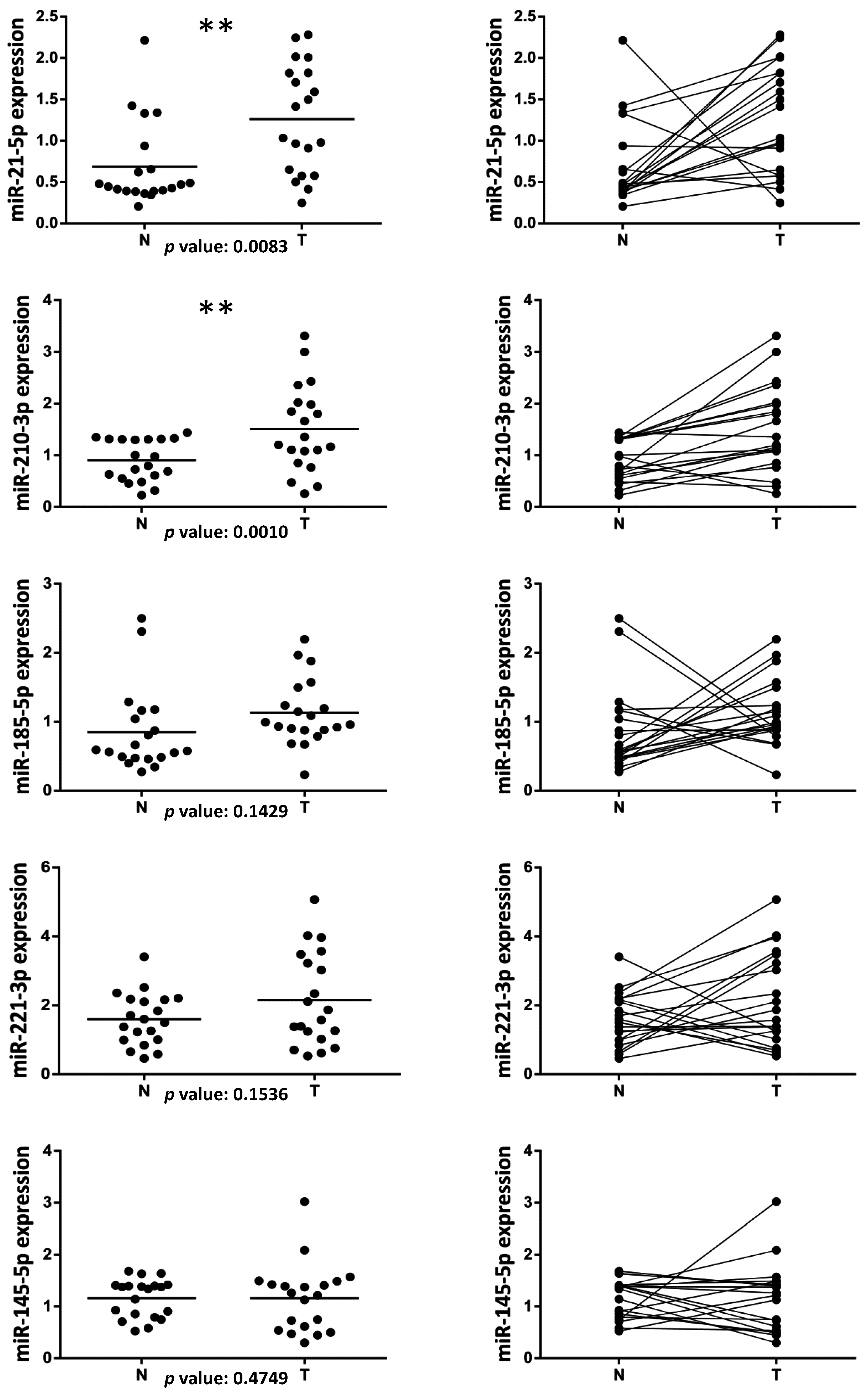
2. Results
3. Discussion
4. Patients and Methods
4.1. Patients
4.2. RNA Extraction and MicroRNA Expression Analysis

| Gender/Age | BMI | Clavien | Hyperten. | Smok. Habit | Tumor Size | Nodal Status | Metast. | TNM Stage | Histology | Tumour Cells (%) | Grade | Surgery |
|---|---|---|---|---|---|---|---|---|---|---|---|---|
| M/65 | 33.2 | II | No | No | T2a | Nx | Mx | II | ccRCC | 87 | G2 | O. Rad. Neph |
| F/65 | 22.3 | I | No | Yes | T1a | Nx | Mx | I | ccRCC | 89 | G2/3 | L. Rad. Neph |
| M/61 | 25.9 | I | No | Former-15 yrs | T2 | N0 | Mx | II | ccRCC | 90 | G3 | O. Rad. Neph |
| M/68 | 29.9 | I | Yes | Former-30 yrs | T3a | Nx | Mx | III | ccRCC | 84 | G3 | O. Rad. Neph |
| M/82 | 21.9 | I | No | No | T3a | Nx | Mx | III | ccRCC | 86 | G3 | L. Rad. Neph |
| M/84 | 24.6 | I | No | No | T2a | Nx | Mx | II | ccRCC | 87 | G2 | O. Rad. Neph |
| M/59 | 28.4 | I | Yes | Yes | T1a | Nx | Mx | I | ccRCC | 88 | G2 | L. Part. Neph |
| M/83 | 30.7 | II | No | No | T1a | N0 | Mx | I | ccRCC | 90 | G2 | L. Rad. Neph |
| M/69 | 28.3 | I | Yes | No | T2b | Nx | Mx | II | ccRCC | 90 | G2 | L. Rad. Neph |
| M/55 | 23.14 | I | Yes | No | T2b | Nx | Mx | II | ccRCC | 90 | G2 | L. Rad. Neph |
| F/72 | 25.59 | I | No | Yes | T1b | Nx | Mx | I | ccRCC | 88 | G2 | L. Rad. Neph |
| M/65 | 40.3 | II | No | Yes | T1a | Nx | Mx | I | ccRCC | 87 | G1 | L. Part. Neph |
| F/59 | 31.4 | I | Yes | Former-15 yrs | T1a | Nx | Mx | I | ccRCC | 86 | G1 | L. Part. Neph |
| M/63 | 26.5 | II | No | Yes | T1b | N0 | Mx | I | ccRCC | 85 | G2 | L. Rad. Neph |
| F/87 | 22.3 | I | Yes | No | T3a | N0 | Mx | III | ccRCC | 88 | G3 | L. Rad. Neph |
| F/56 | 30.2 | II | Yes | Yes | T1a | Nx | Mx | I | ccRCC | 89 | G2 | L. Rad. Neph |
| M/64 | 24.3 | I | No | No | T3a | Nx | Mx | III | ccRCC | 88 | G3 | L. Rad. Neph |
| M/82 | 26.65 | I | Yes | No | T3a | Nx | Mx | III | ccRCC | 90 | G3 | L. Rad. Neph |
| M/77 | 29.46 | I | Yes | Yes | T1a | Nx | Mx | I | ccRCC | 88 | G1 | L. Part. Neph |
| M/66 | 24.71 | I | No | Former-25 yrs | T1a | Nx | Mx | I | ccRCC | 89 | G2 | L. Rad. Neph |
Acknowledgments
Author Contributions
Conflicts of Interest
References
- Siegel, R.; Naishadham, D.; Jemal, A. Cancer statistics, 2013. CA: Cancer J. Clin. 2013, 63, 11–30. [Google Scholar] [CrossRef] [PubMed]
- Jonasch, E.; Futreal, P.A.; Davis, I.J.; Bailey, S.T.; Kim, W.Y.; Brugarolas, J.; Giaccia, A.J.; Kurban, G.; Pause, A.; Frydman, J.; et al. State of the science: An update on renal cell carcinoma. Mol. Cancer Res.: MCR 2012, 10, 859–880. [Google Scholar] [CrossRef] [PubMed]
- Escudier, B. Advanced renal cell carcinoma: Current and emerging management strategies. Drugs 2007, 67, 1257–1264. [Google Scholar] [CrossRef] [PubMed]
- Calin, G.A.; Croce, C.M. MicroRNA signatures in human cancers. Nat. Rev. Cancer 2006, 6, 857–866. [Google Scholar] [CrossRef] [PubMed]
- Fatica, A.; Fazi, F. MicroRNA-regulated pathways in hematological malignancies: How to avoid cells playing out of tune. Int. J. Mol. Sci. 2013, 14, 20930–20953. [Google Scholar] [CrossRef] [PubMed]
- Bartel, D.P. MicroRNAs: Genomics, biogenesis, mechanism, and function. Cell 2004, 116, 281–297. [Google Scholar] [CrossRef]
- Fazi, F.; Nervi, C. MicroRNA: Basic mechanisms and transcriptional regulatory networks for cell fate determination. Cardiovasc. Res. 2008, 79, 553–561. [Google Scholar] [CrossRef] [PubMed]
- Blandino, G.; Fazi, F.; Donzelli, S.; Kedmi, M.; Sas-Chen, A.; Muti, P.; Strano, S.; Yarden, Y. Tumor suppressor microRNAs: A novel non-coding alliance against cancer. FEBS Lett. 2014, 588, 2639–2652. [Google Scholar] [CrossRef] [PubMed]
- Papagiannakopoulos, T.; Kosik, K.S. MicroRNAs: Regulators of oncogenesis and stemness. BMC Med. 2008, 6. [Google Scholar] [CrossRef] [PubMed]
- Cancer Genome Atlas Research Network. Comprehensive molecular characterization of clear cell renal cell carcinoma. Nature 2013, 499, 43–49. [Google Scholar]
- Christinat, Y.; Krek, W. Integrated genomic analysis identifies subclasses and prognosis signatures of kidney cancer. Oncotarget 2015, 6, 10521–10531. [Google Scholar] [CrossRef] [PubMed]
- Osanto, S.; Qin, Y.; Buermans, H.P.; Berkers, J.; Lerut, E.; Goeman, J.J.; van Poppel, H. Genome-wide microRNA expression analysis of clear cell renal cell carcinoma by next generation deep sequencing. PLoS ONE 2012, 7, e38298. [Google Scholar] [CrossRef] [PubMed]
- Gowrishankar, B.; Ibragimova, I.; Zhou, Y.; Slifker, M.J.; Devarajan, K.; Al-Saleem, T.; Uzzo, R.G.; Cairns, P. MicroRNA expression signatures of stage, grade, and progression in clear cell RCC. Cancer Biol. Ther. 2014, 15, 329–341. [Google Scholar] [CrossRef] [PubMed]
- Ge, Y.Z.; Wu, R.; Xin, H.; Zhu, M.; Lu, T.Z.; Liu, H.; Xu, Z.; Yu, P.; Zhao, Y.C.; Li, M.H.; et al. A tumor-specific microRNA signature predicts survival in clear cell renal cell carcinoma. J. Cancer Res. Clin. Oncol. 2015, 141, 1291–1299. [Google Scholar] [CrossRef] [PubMed]
- Wach, S.; Nolte, E.; Theil, A.; Stohr, C.; T Trau, T.; Hartmann, A.; Ekici, A.; Keck, B.; Taubert, H.; Wullich, B. MicroRNA profiles classify papillary renal cell carcinoma subtypes. Br. J. Cancer 2013, 109, 714–722. [Google Scholar] [CrossRef] [PubMed]
- Heinzelmann, J.; Unrein, A.; Wickmann, U.; Baumgart, S.; Stapf, M.; Szendroi, A.; Grimm, M.O.; Gajda, M.R.; Wunderlich, H.; Junker, K. MicroRNAs with prognostic potential for metastasis in clear cell renal cell carcinoma: A comparison of primary tumors and distant metastases. Ann. Surg. Oncol. 2014, 21, 1046–1054. [Google Scholar] [CrossRef] [PubMed]
- Wotschofsky, Z.; Liep, J.; Meyer, H.A.; Jung, M.; Wagner, I.; Disch, A.C.; Schaser, K.D.; Melcher, I.; Kilic, E.; Busch, J.; et al. Identification of metastamirs as metastasis-associated microRNAs in clear cell renal cell carcinomas. Int. J. Biol. Sci. 2012, 8, 1363–1374. [Google Scholar] [CrossRef] [PubMed]
- Wu, X.; Weng, L.; Li, X.; Guo, C.; Pal, S.K.; Jin, J.M.; Li, Y.; Nelson, R.A.; Mu, B.; Onami, S.H.; et al. Identification of a 4-microRNA signature for clear cell renal cell carcinoma metastasis and prognosis. PLoS ONE 2012, 7, e35661. [Google Scholar] [CrossRef] [PubMed]
- Xiao, H.; Zeng, J.; Li, H.; Chen, K.; Yu, G.; Hu, J.; Tang, K.; Zhou, H.; Huang, Q.; Li, A.; et al. miR-1 downregulation correlates with poor survival in clear cell renal cell carcinoma where it interferes with cell cycle regulation and metastasis. Oncotarget 2015, 6, 13201–13215. [Google Scholar] [CrossRef] [PubMed]
- Tang, K.; Xu, H. Prognostic value of meta-signature miRNAs in renal cell carcinoma: An integrated miRNA expression profiling analysis. Sci. Rep. 2015, 5. [Google Scholar] [CrossRef] [PubMed]
- Samaan, S.; Khella, H.W.; Girgis, A.; Scorilas, A.; Lianidou, E.; Gabril, M.; Krylov, S.N.; Jewett, M.; Bjarnason, G.A.; El-said, H.; et al. miR-210 is a prognostic marker in clear cell renal cell carcinoma. J. Mol. Diagn. 2015, 17, 136–144. [Google Scholar] [CrossRef] [PubMed]
- Redova, M.; Poprach, A.; Besse, A.; Iliev, R.; Nekvindova, J.; Lakomy, R.; Radova, L.; Svoboda, M.; Dolezel, J.; Vyzula, R.; et al. mir-210 expression in tumor tissue and in vitro effects of its silencing in renal cell carcinoma. Tumour Biol. 2013, 34, 481–491. [Google Scholar] [CrossRef] [PubMed]
- Liu, H.; Brannon, A.R.; Reddy, A.R.; Alexe, G.; Seiler, M.W.; Arreola, A.; Oza, J.H.; Yao, M.; Juan, D.; Liou, L.S.; et al. Identifying mRNA targets of microRNA dysregulated in cancer: With application to clear cell renal cell carcinoma. BMC Syst. Biol. 2010, 4, 51. [Google Scholar] [CrossRef] [PubMed]
- Cao, J.; Liu, J.; Xu, R.; Zhu, X.; Liu, L.; Zhao, X. MicroRNA-21 stimulates epithelial-to-mesenchymal transition and tumorigenesis in clear cell renal cells. Mol. Med. Rep. 2015. [Google Scholar] [CrossRef] [PubMed]
- Lu, G.J.; Dong, Y.Q.; Zhang, Q.M.; Di, W.Y.; Jiao, L.Y.; Gao, Q.Z.; Zhang, C.G. miRNA-221 promotes proliferation, migration and invasion by targeting TIMP2 in renal cell carcinoma. Int. J. Clin. Exp. Pathol. 2015, 8, 5224–5229. [Google Scholar] [PubMed]
- Cheng, T.; Wang, L.; Li, Y.; Huang, C.; Zeng, L.; Yang, J. Differential microRNA expression in renal cell carcinoma. Oncol. Lett. 2013, 6, 769–776. [Google Scholar] [PubMed]
- Dindo, D.; Demartines, N.; Clavien, P.A. Classification of surgical complications: A new proposal with evaluation in a cohort of 6336 patients and results of a survey. Ann. Surg. 2004, 240, 205–213. [Google Scholar] [CrossRef] [PubMed]
© 2015 by the authors; licensee MDPI, Basel, Switzerland. This article is an open access article distributed under the terms and conditions of the Creative Commons by Attribution (CC-BY) license (http://creativecommons.org/licenses/by/4.0/).
Share and Cite
Petrozza, V.; Carbone, A.; Bellissimo, T.; Porta, N.; Palleschi, G.; Pastore, A.L.; Di Carlo, A.; Della Rocca, C.; Fazi, F. Oncogenic MicroRNAs Characterization in Clear Cell Renal Cell Carcinoma. Int. J. Mol. Sci. 2015, 16, 29219-29225. https://doi.org/10.3390/ijms161226160
Petrozza V, Carbone A, Bellissimo T, Porta N, Palleschi G, Pastore AL, Di Carlo A, Della Rocca C, Fazi F. Oncogenic MicroRNAs Characterization in Clear Cell Renal Cell Carcinoma. International Journal of Molecular Sciences. 2015; 16(12):29219-29225. https://doi.org/10.3390/ijms161226160
Chicago/Turabian StylePetrozza, Vincenzo, Antonio Carbone, Teresa Bellissimo, Natale Porta, Giovanni Palleschi, Antonio Luigi Pastore, Angelina Di Carlo, Carlo Della Rocca, and Francesco Fazi. 2015. "Oncogenic MicroRNAs Characterization in Clear Cell Renal Cell Carcinoma" International Journal of Molecular Sciences 16, no. 12: 29219-29225. https://doi.org/10.3390/ijms161226160
APA StylePetrozza, V., Carbone, A., Bellissimo, T., Porta, N., Palleschi, G., Pastore, A. L., Di Carlo, A., Della Rocca, C., & Fazi, F. (2015). Oncogenic MicroRNAs Characterization in Clear Cell Renal Cell Carcinoma. International Journal of Molecular Sciences, 16(12), 29219-29225. https://doi.org/10.3390/ijms161226160