Next Issue
Volume 16, November
Previous Issue
Volume 16, September
 
 
ijms-logo

Journal Browser

Journal Browser

Int. J. Mol. Sci., Volume 16, Issue 10 (October 2015) – 145 articles , Pages 23127-25933

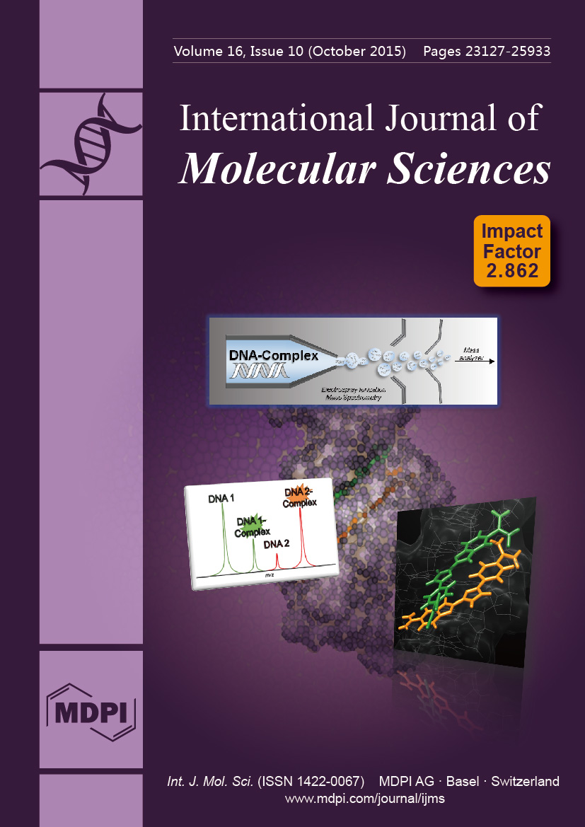
Cover Story: ESI mass spectrometry studies have made enormous contributions with macromolecules and their complexes. Nucleic acids are attractive therapeutic targets for compounds to modulate genetic processes and we have prepared several compound classes to target DNA. Screening for ligand–DNA complexes can prove daunting but a competitive mass spectrometric binding assay can help rapidly identify compounds with DNA sequence specificity. This powerful assay provides stoichiometry, binding mode and relative binding affinity and helps form rationally designed compounds with therapeutic potential. View this article.
  • Issues are regarded as officially published after their release is announced to the table of contents alert mailing list.
  • You may sign up for e-mail alerts to receive table of contents of newly released issues.
  • PDF is the official format for papers published in both, html and pdf forms. To view the papers in pdf format, click on the "PDF Full-text" link, and use the free Adobe Reader to open them.
Order results
Result details
Section
Select all
Export citation of selected articles as:
16 pages, 1030 KiB  
Article
Combining Protein and Strain Engineering for the Production of Glyco-Engineered Horseradish Peroxidase C1A in Pichia pastoris
by Simona Capone 1, Lejla Ćorajević 1, Günther Bonifert 1, Patrick Murth 1, Daniel Maresch 2, Friedrich Altmann 2, Christoph Herwig 1 and Oliver Spadiut 1,*
1 Institute of Chemical Engineering, Research Area Biochemical Engineering, Vienna University of Technology, Gumpendorfer Strasse 1a, 1060 Vienna, Austria
2 Department of Chemistry, University of Natural Resources and Life Sciences, 1180 Vienna, Austria
Int. J. Mol. Sci. 2015, 16(10), 23127-23142; https://doi.org/10.3390/ijms161023127 - 24 Sep 2015
Cited by 10 | Viewed by 6518
Abstract
Horseradish peroxidase (HRP), conjugated to antibodies and lectins, is widely used in medical diagnostics. Since recombinant production of the enzyme is difficult, HRP isolated from plant is used for these applications. Production in the yeast Pichia pastoris (P. pastoris), the most [...] Read more.
Horseradish peroxidase (HRP), conjugated to antibodies and lectins, is widely used in medical diagnostics. Since recombinant production of the enzyme is difficult, HRP isolated from plant is used for these applications. Production in the yeast Pichia pastoris (P. pastoris), the most promising recombinant production platform to date, causes hyperglycosylation of HRP, which in turn complicates conjugation to antibodies and lectins. In this study we combined protein and strain engineering to obtain an active and stable HRP variant with reduced surface glycosylation. We combined four mutations, each being beneficial for either catalytic activity or thermal stability, and expressed this enzyme variant as well as the unmutated wildtype enzyme in both a P. pastoris benchmark strain and a strain where the native α-1,6-mannosyltransferase (OCH1) was knocked out. Considering productivity in the bioreactor as well as enzyme activity and thermal stability, the mutated HRP variant produced in the P. pastoris benchmark strain turned out to be interesting for medical diagnostics. This variant shows considerable catalytic activity and thermal stability and is less glycosylated, which might allow more controlled and efficient conjugation to antibodies and lectins. Full article
(This article belongs to the Special Issue Protein Engineering)
Show Figures

Figure 1

22 pages, 1517 KiB  
Review
Applications of Engineered DNA-Binding Molecules Such as TAL Proteins and the CRISPR/Cas System in Biology Research
by Toshitsugu Fujita and Hodaka Fujii *
Chromatin Biochemistry Research Group, Combined Program on Microbiology and Immunology, Research Institute for Microbial Diseases, Osaka University, Suita, Osaka 565-0871, Japan
Int. J. Mol. Sci. 2015, 16(10), 23143-23164; https://doi.org/10.3390/ijms161023143 - 24 Sep 2015
Cited by 10 | Viewed by 8663
Abstract
Engineered DNA-binding molecules such as transcription activator-like effector (TAL or TALE) proteins and the clustered regularly interspaced short palindromic repeats (CRISPR) and CRISPR-associated proteins (Cas) (CRISPR/Cas) system have been used extensively for genome editing in cells of various types and species. The sequence-specific [...] Read more.
Engineered DNA-binding molecules such as transcription activator-like effector (TAL or TALE) proteins and the clustered regularly interspaced short palindromic repeats (CRISPR) and CRISPR-associated proteins (Cas) (CRISPR/Cas) system have been used extensively for genome editing in cells of various types and species. The sequence-specific DNA-binding activities of these engineered DNA-binding molecules can also be utilized for other purposes, such as transcriptional activation, transcriptional repression, chromatin modification, visualization of genomic regions, and isolation of chromatin in a locus-specific manner. In this review, we describe applications of these engineered DNA-binding molecules for biological purposes other than genome editing. Full article
(This article belongs to the Special Issue Protein Engineering)
Show Figures

Graphical abstract

12 pages, 1508 KiB  
Article
Development of Ferromagnetic Superspins in Bare Cu Nanoparticles by Electronic Charge Redistribution
by Erdembayalag Batsaikhan, Yen-Cheng Chen, Chi-Hung Lee, Hsiao-Chi Li and Wen-Hsien Li *
Department of Physics, National Central University, Jhongli 32001, Taiwan
Int. J. Mol. Sci. 2015, 16(10), 23165-23176; https://doi.org/10.3390/ijms161023165 - 24 Sep 2015
Cited by 5 | Viewed by 5013
Abstract
We report on the results of investigating the ferromagnetic properties of bare Cu nanoparticles. Three sets of bare Cu nanoparticle assemblies with mean particle diameters of 6.6, 8.1, and 11.1 nm were fabricated, employing the gas condensation method. Curie-Weiss paramagnetic responses to a [...] Read more.
We report on the results of investigating the ferromagnetic properties of bare Cu nanoparticles. Three sets of bare Cu nanoparticle assemblies with mean particle diameters of 6.6, 8.1, and 11.1 nm were fabricated, employing the gas condensation method. Curie-Weiss paramagnetic responses to a weak driving magnetic field were detected, showing the appearance of particle superspins that overcomes the diamagnetic responses from the inner core. The isothermal magnetization displays a Langevin field profile together with magnetic hysteresis appearing even at 300 K, demonstrating the existence of ferromagnetic superspins in the Cu nanoparticles. Shifting of a noticeable amount of electronic charge from being distributed near the lattice sites in bulk form toward their neighboring ions in nanoparticles was found. The extended 3d and 4s band mixture are the main sources for the development of localized 3d holes for the development of ferromagnetic particle superspins in Cu nanoparticles. Full article
(This article belongs to the Special Issue Magnetic Nanoparticles 2015)
Show Figures

Graphical abstract

18 pages, 744 KiB  
Review
Regulatory Proteolysis in Arabidopsis-Pathogen Interactions
by Miklós Pogány *, Tamás Dankó, Evelin Kámán-Tóth, Ildikó Schwarczinger and Zoltán Bozsó
Plant Protection Institute, Centre for Agricultural Research, Hungarian Academy of Sciences, Herman Ottó út 15, 1022 Budapest, Hungary
Int. J. Mol. Sci. 2015, 16(10), 23177-23194; https://doi.org/10.3390/ijms161023177 - 24 Sep 2015
Cited by 20 | Viewed by 7750
Abstract
Approximately two and a half percent of protein coding genes in Arabidopsis encode enzymes with known or putative proteolytic activity. Proteases possess not only common housekeeping functions by recycling nonfunctional proteins. By irreversibly cleaving other proteins, they regulate crucial developmental processes and control [...] Read more.
Approximately two and a half percent of protein coding genes in Arabidopsis encode enzymes with known or putative proteolytic activity. Proteases possess not only common housekeeping functions by recycling nonfunctional proteins. By irreversibly cleaving other proteins, they regulate crucial developmental processes and control responses to environmental changes. Regulatory proteolysis is also indispensable in interactions between plants and their microbial pathogens. Proteolytic cleavage is simultaneously used both by plant cells, to recognize and inactivate invading pathogens, and by microbes, to overcome the immune system of the plant and successfully colonize host cells. In this review, we present available results on the group of proteases in the model plant Arabidopsis thaliana whose functions in microbial pathogenesis were confirmed. Pathogen-derived proteolytic factors are also discussed when they are involved in the cleavage of host metabolites. Considering the wealth of review papers available in the field of the ubiquitin-26S proteasome system results on the ubiquitin cascade are not presented. Arabidopsis and its pathogens are conferred with abundant sets of proteases. This review compiles a list of those that are apparently involved in an interaction between the plant and its pathogens, also presenting their molecular partners when available. Full article
(This article belongs to the Special Issue Plant Microbe Interaction)
Show Figures

Graphical abstract

15 pages, 1084 KiB  
Article
Early and Degressive Putamen Atrophy in Multiple Sclerosis
by Julia Krämer *, Sven G. Meuth, Jan-Gerd Tenberge, Patrick Schiffler, Heinz Wiendl and Michael Deppe *
Department of Neurology, Westfälische Wilhelms University, Albert-Schweitzer-Campus 1, Gebäude A1, 48149 Münster, Germany
Int. J. Mol. Sci. 2015, 16(10), 23195-23209; https://doi.org/10.3390/ijms161023195 - 25 Sep 2015
Cited by 24 | Viewed by 6372
Abstract
Putamen atrophy and its long-term progress during disease course were recently shown in patients with multiple sclerosis (MS). Here we investigated retrospectively the time point of atrophy onset in patients with relapsing-remitting MS (RRMS). 68 patients with RRMS and 26 healthy controls (HC) [...] Read more.
Putamen atrophy and its long-term progress during disease course were recently shown in patients with multiple sclerosis (MS). Here we investigated retrospectively the time point of atrophy onset in patients with relapsing-remitting MS (RRMS). 68 patients with RRMS and 26 healthy controls (HC) were admitted to 3T MRI in a cross-sectional study. We quantitatively analyzed the putamen volume of individual patients in relation to disease duration by correcting for age and intracranial volume (ICV). Patient’s relative putamen volume (RPV), expressed in percent of ICV, was significantly reduced compared to HC. Based on the correlation between RPV and age, we computed the age-corrected RPV deviation (ΔRPV) from HC. Patients showed significantly negative ΔRPV. Interestingly, the age-corrected ΔRPV depended logarithmically on disease duration: Directly after first symptom manifestation, patients already showed a reduced RPV followed by a further degressive volumetric decline. This means that atrophy progression was stronger in the first than in later years of disease. Putamen atrophy starts directly after initial symptom manifestation or even years before, and progresses in a degressive manner. Due to its important role in neurological functions, early detection of putamen atrophy seems necessary. High-resolution structural MRI allows monitoring of disease course. Full article
(This article belongs to the Special Issue Advances in Multiple Sclerosis)
Show Figures

Figure 1

17 pages, 1362 KiB  
Article
Improved Monitoring of Semi-Continuous Anaerobic Digestion of Sugarcane Waste: Effects of Increasing Organic Loading Rate on Methanogenic Community Dynamics
by Athaydes Francisco Leite 1, Leandro Janke 2, Zuopeng Lv 1, Hauke Harms 1, Hans-Hermann Richnow 3 and Marcell Nikolausz 1,*
1 Department of Environmental Microbiology, Helmholtz Centre for Environmental Research-UFZ, Permoserstrasse 15, 04318 Leipzig, Germany
2 Department of Biochemical Conversion, Deutsches Biomasseforschungszentrum Gemeinnützige GmbH, Torgauerstrasse 116, 04347 Leipzig, Germany
3 Department of Isotope Biogeochemistry, Helmholtz Centre for Environmental Research-UFZ, Permoserstrasse 15, 04318 Leipzig, Germany
Int. J. Mol. Sci. 2015, 16(10), 23210-23226; https://doi.org/10.3390/ijms161023210 - 25 Sep 2015
Cited by 32 | Viewed by 6676
Abstract
The anaerobic digestion of filter cake and its co-digestion with bagasse, and the effect of gradual increase of the organic loading rate (OLR) from start-up to overload were investigated. Understanding the influence of environmental and technical parameters on the development of particular methanogenic [...] Read more.
The anaerobic digestion of filter cake and its co-digestion with bagasse, and the effect of gradual increase of the organic loading rate (OLR) from start-up to overload were investigated. Understanding the influence of environmental and technical parameters on the development of particular methanogenic pathway in the biogas process was an important aim for the prediction and prevention of process failure. The rapid accumulation of volatile organic acids at high OLR of 3.0 to 4.0 gvs·L1·day1 indicated strong process inhibition. Methanogenic community dynamics of the reactors was monitored by stable isotope composition of biogas and molecular biological analysis. A potential shift toward the aceticlastic methanogenesis was observed along with the OLR increase under stable reactor operating conditions. Reactor overloading and process failure were indicated by the tendency to return to a predominance of hydrogenotrophic methanogenesis with rising abundances of the orders Methanobacteriales and Methanomicrobiales and drop of the genus Methanosarcina abundance. Full article
(This article belongs to the Special Issue Bioprocess Engineering)
Show Figures

Figure 1

23 pages, 746 KiB  
Review
Focus on Pivotal Role of Dietary Intake (Diet and Supplement) and Blood Levels of Tocopherols and Tocotrienols in Obtaining Successful Aging
by Mariangela Rondanelli 1, Milena Anna Faliva 1, Gabriella Peroni 1, Francesca Moncaglieri 1, Vittoria Infantino 1, Maurizio Naso 2 and Simone Perna 1,*
1 Department of Public Health, Experimental and Forensic Medicine, School of Medicine, Endocrinology and Nutrition Unit, University of Pavia, Azienda di Servizi alla Persona di Pavia, Pavia 27100, Italy
2 Faculty of Medicine and Surgery, Department of Clinical Sciences, University of Milano, Milan 20100, Italy
Int. J. Mol. Sci. 2015, 16(10), 23227-23249; https://doi.org/10.3390/ijms161023227 - 25 Sep 2015
Cited by 30 | Viewed by 7234
Abstract
Numerous specific age-related morbidities have been correlated with low intake and serum levels of tocopherols and tocotrienols. We performed a review in order to evaluate the extant evidence regarding: (1) the association between intake and serum levels of tocopherols and tocotrienols and age-related [...] Read more.
Numerous specific age-related morbidities have been correlated with low intake and serum levels of tocopherols and tocotrienols. We performed a review in order to evaluate the extant evidence regarding: (1) the association between intake and serum levels of tocopherols and tocotrienols and age-related pathologies (osteoporosis, sarcopenia and cognitive impairment); and (2) the optimum diet therapy or supplementation with tocopherols and tocotrienols for the treatment of these abnormalities. This review included 51 eligible studies. The recent literature underlines that, given the detrimental effect of low intake and serum levels of tocopherols and tocotrienols on bone, muscle mass, and cognitive function, a change in the lifestyle must be the cornerstone in the prevention of these specific age-related pathologies related to vitamin E-deficient status. The optimum diet therapy in the elderly for avoiding vitamin E deficiency and its negative correlates, such as high inflammation and oxidation, must aim at achieving specific nutritional goals. These goals must be reached through: accession of the elderly subjects to specific personalized dietary programs aimed at achieving and/or maintaining body weight (avoid malnutrition); increase their intake of food rich in vitamin E, such as derivatives of oily seeds (in particular wheat germ oil), olive oil, hazelnuts, walnuts, almonds, and cereals rich in vitamin E (such as specific rice cultivar rich in tocotrienols) or take vitamin E supplements. In this case, vitamin E can be correctly used in a personalized way either for the outcome from the pathology or to achieve healthy aging and longevity without any adverse effects. Full article
(This article belongs to the Special Issue Tocopherols and Tocotrienols: Metabolism and Properties)
Show Figures

Figure 1

9 pages, 967 KiB  
Article
Secreted Frizzled-Related Protein Promotes Bone Regeneration by Human Bone Marrow-Derived Mesenchymal Stem Cells
by Wataru Katagiri *, Masashi Osugi, Takamasa Kawai and Hideharu Hibi
Department of Oral and Maxillofacial Surgery, Nagoya University Graduate School of Medicine, 65 Tsuruma-cho, Showa-ku, Nagoya 466-8550, Japan
Int. J. Mol. Sci. 2015, 16(10), 23250-23258; https://doi.org/10.3390/ijms161023250 - 25 Sep 2015
Cited by 10 | Viewed by 8204
Abstract
Secreted frizzled-related protein (sFRP)-3 is a negative regulator of Wnt signaling in human mesenchymal stem cells (hMSCs). The present study investigated the effects sFRP-3 on osteogenic differentiation by assessing osteogenic gene expression in hMSCs in vitro and by examining bone regeneration in a [...] Read more.
Secreted frizzled-related protein (sFRP)-3 is a negative regulator of Wnt signaling in human mesenchymal stem cells (hMSCs). The present study investigated the effects sFRP-3 on osteogenic differentiation by assessing osteogenic gene expression in hMSCs in vitro and by examining bone regeneration in a rat bone defect model. sFRP-3 treatment induced osteogenic differentiation in hMSCs as determined by alkaline phosphatase, collagen type I, osteocalcin, and Runt-related transcription factor 2 gene expression. hMSCs with or without sFRP-3 were implanted into a rat calvarial bone defect; a radiographic analysis by micro-computed tomography and histological analysis 4 and 8 weeks after implantation showed greater bone regeneration in the sFRP(+) than in the sFRP(−) group. These results suggest that modulation of Wnt signaling contributes to osteogenic differentiation in hMSCs. Specifically, sFRP-3 induces osteoblastic differentiation of cultured MSCs and bone regeneration in a calvarial bone defect, suggesting that it can be a useful agent for the treatment of bone defects. Full article
(This article belongs to the Special Issue Stem Cell Activation in Adult Organism)
Show Figures

Figure 1

20 pages, 1058 KiB  
Review
Topical PDT in the Treatment of Benign Skin Diseases: Principles and New Applications
by Miri Kim, Haw Young Jung and Hyun Jeong Park *
Department of Dermatology, Yeouido St. Mary's Hospital, The Catholic University of Korea, Seoul 150-713, Korea
Int. J. Mol. Sci. 2015, 16(10), 23259-23278; https://doi.org/10.3390/ijms161023259 - 25 Sep 2015
Cited by 106 | Viewed by 13609
Abstract
Photodynamic therapy (PDT) uses a photosensitizer, light energy, and molecular oxygen to cause cell damage. Cells exposed to the photosensitizer are susceptible to destruction upon light absorption because excitation of the photosensitizing agents leads to the production of reactive oxygen species and, subsequently, [...] Read more.
Photodynamic therapy (PDT) uses a photosensitizer, light energy, and molecular oxygen to cause cell damage. Cells exposed to the photosensitizer are susceptible to destruction upon light absorption because excitation of the photosensitizing agents leads to the production of reactive oxygen species and, subsequently, direct cytotoxicity. Using the intrinsic cellular heme biosynthetic pathway, topical PDT selectively targets abnormal cells, while preserving normal surrounding tissues. This selective cytotoxic effect is the basis for the use of PDT in antitumor treatment. Clinically, PDT is a widely used therapeutic regimen for oncologic skin conditions such as actinic keratosis, squamous cell carcinoma in situ, and basal cell carcinoma. PDT has been shown, under certain circumstances, to stimulate the immune system and produce antibacterial, and/or regenerative effects while protecting cell viability. Thus, it may be useful for treating benign skin conditions. An increasing number of studies support the idea that PDT may be effective for treating acne vulgaris and several other inflammatory/infective skin diseases, including psoriasis, rosacea, viral warts, and aging-related changes. This review provides an overview of the clinical investigations of PDT and discusses each of the essential aspects of the sequence: its mechanism of action, common photosensitizers, light sources, and clinical applications in dermatology. Of the numerous clinical trials of PDT in dermatology, this review focuses on those studies that have reported remarkable therapeutic benefits following topical PDT for benign skin conditions such as acne vulgaris, viral warts, and photorejuvenation without causing severe side effects. Full article
(This article belongs to the Special Issue Advances in Photodynamic Therapy)
Show Figures

Graphical abstract

21 pages, 3218 KiB  
Article
Liver Toxicity of Cadmium Telluride Quantum Dots (CdTe QDs) Due to Oxidative Stress in Vitro and in Vivo
by Ting Zhang 1,2,†, Yuanyuan Hu 1,†, Meng Tang 1,2,*, Lu Kong 1, Jiali Ying 1, Tianshu Wu 1,2, Yuying Xue 1 and Yuepu Pu 1,2,*
1 Key Laboratory of Environmental Medicine Engineering, Ministry of Education, School of Public Health, Southeast University, Nanjing 210009, China
2 Jiangsu Key Laboratory for Biomaterials and Devices, Southeast University, Nanjing 210009, China
These authors contributed equally to this work.
Int. J. Mol. Sci. 2015, 16(10), 23279-23299; https://doi.org/10.3390/ijms161023279 - 25 Sep 2015
Cited by 93 | Viewed by 9034
Abstract
With the applications of quantum dots (QDs) expanding, many studies have described the potential adverse effects of QDs, yet little attention has been paid to potential toxicity of QDs in the liver. The aim of this study was to investigate the effects of [...] Read more.
With the applications of quantum dots (QDs) expanding, many studies have described the potential adverse effects of QDs, yet little attention has been paid to potential toxicity of QDs in the liver. The aim of this study was to investigate the effects of cadmium telluride (CdTe) QDs in mice and murine hepatoma cells alpha mouse liver 12 (AML 12). CdTe QDs administration significantly increased the level of lipid peroxides marker malondialdehyde (MDA) in the livers of treated mice. Furthermore, CdTe QDs caused cytotoxicity in AML 12 cells in a dose- and time-dependent manner, which was likely mediated through the generation of reactive oxygen species (ROS) and the induction of apoptosis. An increase in ROS generation with a concomitant increase in the gene expression of the tumor suppressor gene p53, the pro-apoptotic gene Bcl-2 and a decrease in the anti-apoptosis gene Bax, suggested that a mitochondria mediated pathway was involved in CdTe QDs’ induced apoptosis. Finally, we showed that NF-E2-related factor 2 (Nrf2) deficiency blocked induced oxidative stress to protect cells from injury induced by CdTe QDs. These findings provide insights into the regulatory mechanisms involved in the activation of Nrf2 signaling that confers protection against CdTe QDs-induced apoptosis in hepatocytes. Full article
(This article belongs to the Special Issue Bioactive Nanoparticles)
Show Figures

Graphical abstract

18 pages, 698 KiB  
Review
Usefulness of Photodynamic Therapy as a Possible Therapeutic Alternative in the Treatment of Basal Cell Carcinoma
by Paola Savoia 1,*, Tommaso Deboli 1, Alberto Previgliano 1 and Paolo Broganelli 2
1 Department of Medical Sciences, University of Turin, Torino 10126, Italy
2 Città della Salute e della Scienza, Turin 10126, Italy
Int. J. Mol. Sci. 2015, 16(10), 23300-23317; https://doi.org/10.3390/ijms161023300 - 28 Sep 2015
Cited by 32 | Viewed by 6427
Abstract
Basal cell carcinoma (BCC) is the most common cancer in individuals with fair skin type (I–II) and steadily increasing in incidence (70% of skin malignancy). It is locally invasive but metastasis is usually very rare, with an estimated incidence of 0.0028%–0.55%. Conventional therapy [...] Read more.
Basal cell carcinoma (BCC) is the most common cancer in individuals with fair skin type (I–II) and steadily increasing in incidence (70% of skin malignancy). It is locally invasive but metastasis is usually very rare, with an estimated incidence of 0.0028%–0.55%. Conventional therapy is surgery, especially for the H region of the face and infiltrative lesions; in case of inoperable tumors, radiotherapy is a valid option. Recently, topical photodynamic therapy (PDT) has become an effective treatment in the management of superficial and small nodular BCC. PDT is a minimally invasive procedure that involves the administration of a photo-sensibilizing agent followed by irradiation at a pre-defined wavelength; this determines the creation of reactive oxygen species that specifically destroy target cells. The only major side effect is pain, reported by some patients during the irradiation. The high cure rate and excellent cosmetic outcome requires considering this possibility for the management of patients with both sporadic and hereditary BCC. In this article, an extensive review of the recent literature was made, in order to clarify the role of PDT as a possible alternative therapeutic option in the treatment of BCC. Full article
(This article belongs to the Special Issue Advances in Photodynamic Therapy)
19 pages, 5971 KiB  
Article
Evaluating Osteogenic Potential of Ligamentum Flavum Cells Cultivated in Photoresponsive Hydrogel that Incorporates Bone Morphogenetic Protein-2 for Spinal Fusion
by Chih-Wei Chiang 1,2,†, Wei-Chuan Chen 1,†, Hsia-Wei Liu 3, I-Chun Wang 4,5,*,‡ and Chih-Hwa Chen 1,6,‡
1 Bone and Joint Research Center, Department of Orthopedics and Traumatology, Taipei Medical University Hospital, School of Medicine, College of Medicine, Taipei Medical University, Taipei 110, Taiwan
2 Graduate Institute of Biomedical Electronics and Bioinformatics, National Taiwan University, Taipei 106, Taiwan
3 Department of Life Science, College of Science and Engineering, Fu Jen Catholic University, New Taipei City 242, Taiwan
4 Department of Orthopedic Surgery, Chang Gung Memorial Hospital, Keelung 204, Taiwan
5 College of Medicine, Chang Gung University, Taoyuan 333, Taiwan
6 Graduate Institute of Biomedical Materials and Tissue Engineering, College of Biomedical Engineering, Taipei Medical University, Taipei 110, Taiwan
These authors contributed equally to this work.
Int. J. Mol. Sci. 2015, 16(10), 23318-23336; https://doi.org/10.3390/ijms161023318 - 28 Sep 2015
Cited by 8 | Viewed by 6791
Abstract
Regenerative medicine is increasingly important in clinical practice. Ligamentum flava (LF) are typically removed during spine-related surgeries. LF may be a source of cells for spinal fusion that is conducted using tissue engineering techniques. In this investigation, LF cells of rabbits were isolated [...] Read more.
Regenerative medicine is increasingly important in clinical practice. Ligamentum flava (LF) are typically removed during spine-related surgeries. LF may be a source of cells for spinal fusion that is conducted using tissue engineering techniques. In this investigation, LF cells of rabbits were isolated and then characterized by flow cytometry, morphological observation, and immunofluorescence staining. The LF cells were also cultivated in polyethylene (glycol) diacrylate (PEGDA) hydrogels that incorporated bone morphogenetic protein-2 (BMP-2) growth factor, to evaluate their proliferation and secretion of ECM and differentiation in vitro. The experimental results thus obtained that the proliferation, ECM secretion, and differentiation of the PEGDA-BMP-2 group exceeded those of the PEGDA group during the period of cultivation. The mineralization and histological staining results differed similarly. A nude mice model was utilized to prove that LF cells on hydrogels could undergo osteogenic differentiation in vivo. These experimental results also revealed that the PEGDA-BMP-2 group had better osteogenic effects than the PEGDA group following a 12 weeks after transplantation. According to all of these experimental results, LF cells are a source of cells for spinal fusion and PEGDA-BMP-2 hydrogel is a candidate biomaterial for spinal fusion by tissue engineering. Full article
(This article belongs to the Special Issue Biomaterials for Tissue Engineering)
Show Figures

Graphical abstract

18 pages, 2262 KiB  
Review
Dangerous Liaisons: Caspase-11 and Reactive Oxygen Species Crosstalk in Pathogen Elimination
by JoAnn Simone Roberts 1,* and Ӧzlem Yilmaz 2,3
1 Department of Oral Biology, University of Florida, Gainesville, FL 32610, USA
2 Department of Periodontology, University of Florida, P.O. Box 100434, Gainesville, FL 32610, USA
3 Emerging Pathogens Institute, University of Florida, P.O. Box 100434, Gainesville, FL 32610, USA
Int. J. Mol. Sci. 2015, 16(10), 23337-23354; https://doi.org/10.3390/ijms161023337 - 28 Sep 2015
Cited by 21 | Viewed by 8892
Abstract
Recently, the focus of murine caspase-11 and human orthologs caspase-4, -5 research has been on their novel function to induce noncanonical inflammasome activation in direct response to Gram-negative bacterial infection. On the other hand, a new role in anti-bacterial autophagy has been attributed [...] Read more.
Recently, the focus of murine caspase-11 and human orthologs caspase-4, -5 research has been on their novel function to induce noncanonical inflammasome activation in direct response to Gram-negative bacterial infection. On the other hand, a new role in anti-bacterial autophagy has been attributed to caspase-11, -4 and -5, which currently stands largely unexplored. In this review, we connect lately emerged evidence that suggests these caspases have a key role in anti-bacterial autophagy and discuss the growing implications of a danger molecule—extracellular ATP—and NADPH oxidase-mediated ROS generation as novel inducers of human caspase-4, -5 signaling during infection. We also highlight the adeptness of persistent pathogens like Porphyromonas gingivalis, a Gram-negative anaerobe and successful colonizer of oral mucosa, to potentially interfere with the activated caspase-4 pathway and autophagy. While, the ability of caspase-4, -5 to promote autophagolysosomal fusion is not well understood, the abundance of caspase-4 in skin and other mucosal epithelial cells implies an important role for caspase-4 in mucosal defense, supporting the view that caspase-4, -5 may play a non-redundant part in innate immunity. Thus, this review will join the currently disconnected cutting-edge research thereby proposing a working model for regulation of caspase-4, -5 in pathogen elimination via cellular-trafficking. Full article
(This article belongs to the Section Biochemistry)
Show Figures

Figure 1

14 pages, 1299 KiB  
Article
Polyphosphate during the Regreening of Chlorella vulgaris under Nitrogen Deficiency
by Fei-Fei Chu 1,2, Xiao-Fei Shen 1, Paul K. S. Lam 1,3 and Raymond J. Zeng 1,4,*
1 Advanced Laboratory for Environmental Research and Technology, USTC-CityU, Suzhou 215123, China
2 College of Standardization, China Jiliang University, Hangzhou 310018, China
3 State Key Laboratory in Marine Pollution, Department of Biology and Chemistry, City University of Hong Kong, Kowloon 999077, Hong Kong
4 CAS Key Laboratory of Urban Pollutant Conversion, Department of Chemistry, University of Science and Technology of China, Hefei 230026, China
Int. J. Mol. Sci. 2015, 16(10), 23355-23368; https://doi.org/10.3390/ijms161023355 - 28 Sep 2015
Cited by 17 | Viewed by 6165
Abstract
Polyphosphate (Poly-P) accumulation has been reported in Chlorella vulgaris under nitrogen deficiency conditions with sufficient P supply, and the process has been demonstrated to have great impact on lipid productivity. In this article, the utilization of polyphosphates and the regreening process under N [...] Read more.
Polyphosphate (Poly-P) accumulation has been reported in Chlorella vulgaris under nitrogen deficiency conditions with sufficient P supply, and the process has been demonstrated to have great impact on lipid productivity. In this article, the utilization of polyphosphates and the regreening process under N resupplying conditions, especially for lipid production reviving, were investigated. This regreening process was completed within approximately 3–5 days. Polyphosphates were first degraded within 3 days in the regreening process, with and without an external P supply, and the degradation preceded the assimilation of phosphate in the media with an external P offering. Nitrate assimilation was markedly influenced by the starvation of P after polyphosphates were exhausted in the medium without external phosphates, and then the reviving process of biomass and lipid production was strictly impeded. It is, thus, reasonable to assume that simultaneous provision of external N and P is essential for overall biodiesel production revival during the regreening process. Full article
(This article belongs to the Special Issue Biofuel)
Show Figures

Graphical abstract

13 pages, 1873 KiB  
Article
Alkyl Chain Growth on a Transition Metal Center: How Does Iron Compare to Ruthenium and Osmium?
by Mala A. Sainna and Sam P. De Visser *
Manchester Institute of Biotechnology and School of Chemical Engineering and Analytical Science, the University of Manchester, 131 Princess Street, Manchester M1 7DN, UK
Int. J. Mol. Sci. 2015, 16(10), 23369-23381; https://doi.org/10.3390/ijms161023369 - 28 Sep 2015
Viewed by 5422
Abstract
Industrial Fischer-Tropsch processes involve the synthesis of hydrocarbons usually on metal surface catalysts. On the other hand, very few homogeneous catalysts are known to perform a Fischer-Tropsch style of reaction. In recent work, we established the catalytic properties of a diruthenium-platinum carbene complex, [...] Read more.
Industrial Fischer-Tropsch processes involve the synthesis of hydrocarbons usually on metal surface catalysts. On the other hand, very few homogeneous catalysts are known to perform a Fischer-Tropsch style of reaction. In recent work, we established the catalytic properties of a diruthenium-platinum carbene complex, [(CpRu)22-H) (μ2-NHCH3)(μ3-C)PtCH3(P(CH3)3)2](CO)n+ with n = 0, 2 and Cp = η5-C5(CH3)5, and showed it to react efficiently by initial hydrogen atom transfer followed by methyl transfer to form an alkyl chain on the Ru-center. In particular, the catalytic efficiency was shown to increase after the addition of two CO molecules. As such, this system could be viewed as a potential homogeneous Fischer-Tropsch catalyst. Herein, we have engineered the catalytic center of the catalyst and investigated the reactivity of trimetal carbene complexes of the same type using iron, ruthenium and osmium at the central metal scaffold. The work shows that the reactivity should increase from diosmium to diruthenium to diiron; however, a non-linear trend is observed due to multiple factors contributing to the individual barrier heights. We identified all individual components of these reaction steps in detail and established the difference in reactivity of the various complexes. Full article
(This article belongs to the Section Physical Chemistry, Theoretical and Computational Chemistry)
Show Figures

Graphical abstract

8 pages, 1219 KiB  
Article
Combination of MiR-378 and MiR-210 Serum Levels Enables Sensitive Detection of Renal Cell Carcinoma
by Michal Fedorko 1,†, Michal Stanik 2,†, Robert Iliev 3,4, Martina Redova-Lojova 4, Tana Machackova 4, Marek Svoboda 3, Dalibor Pacik 1, Jan Dolezel 2 and Ondrej Slaby 3,4,*
1 Department of Urology, University Hospital Brno and Masaryk University Brno, Brno 62500, Czech Republic
2 Department of Urologic Oncology, Masaryk Memorial Cancer Institute, Brno 65653, Czech Republic
3 Department of Comprehensive Cancer Care, Masaryk Memorial Cancer Institute, Brno 65653, Czech Republic
4 Central European Institute of Technology, Masaryk University, Brno 62500, Czech Republic
These authors contributed equally to this work.
Int. J. Mol. Sci. 2015, 16(10), 23382-23389; https://doi.org/10.3390/ijms161023382 - 29 Sep 2015
Cited by 83 | Viewed by 6760
Abstract
Serum microRNAs are emerging as a clinically useful tool for early and non-invasive detection of various cancer types including renal cell carcinoma (RCC). Based on our previous results, we performed the study to analyze circulating serum miR-378 and miR-210 in patients with various [...] Read more.
Serum microRNAs are emerging as a clinically useful tool for early and non-invasive detection of various cancer types including renal cell carcinoma (RCC). Based on our previous results, we performed the study to analyze circulating serum miR-378 and miR-210 in patients with various histological subtypes of RCC. RNA was purified from blood serum samples of 195 RCC patients and 100 healthy controls. The levels of miR-378 and miR-210 in serum were determined absolutely using quantitative real-time PCR. Pre- and postoperative levels of both microRNAs were compared in 20 RCC patients. Significantly increased serum levels of both miR-378 and miR-210 enabled to clearly distinguish RCC patients and healthy controls with 80% sensitivity and 78% specificity if analyzed in combination (p < 0.0001), and their levels significantly decreased in the time period of three months after radical nephrectomy (p < 0.0001). Increased level of miR-378 positively correlates with disease-free survival (p = 0.036) and clinical stage (p = 0.0476). The analysis of serum miR-378 and miR-210 proved their potential to serve as powerful non-invasive diagnostic and prognostic biomarkers in RCC. Full article
(This article belongs to the Collection Regulation by Non-coding RNAs)
Show Figures

Figure 1

15 pages, 1333 KiB  
Article
Heavy Metal Induced Antibiotic Resistance in Bacterium LSJC7
by Songcan Chen 1,†, Xiaomin Li 1,†, Guoxin Sun 1,*, Yingjiao Zhang 2, Jianqiang Su 2 and Jun Ye 2,*
1 State Key Lab of Urban and Regional Ecology, Research Center for Eco-Environmental Sciences, Chinese Academy of Sciences, Beijing 100085, China
2 Key Lab of Urban Environment and Health, Institute of Urban Environment, Chinese Academy of Sciences, Xiamen 361021, China
These authors contributed equally to this work.
Int. J. Mol. Sci. 2015, 16(10), 23390-23404; https://doi.org/10.3390/ijms161023390 - 29 Sep 2015
Cited by 116 | Viewed by 10482
Abstract
Co-contamination of antibiotics and heavy metals prevails in the environment, and may play an important role in disseminating bacterial antibiotic resistance, but the selective effects of heavy metals on bacterial antibiotic resistance is largely unclear. To investigate this, the effects of heavy metals [...] Read more.
Co-contamination of antibiotics and heavy metals prevails in the environment, and may play an important role in disseminating bacterial antibiotic resistance, but the selective effects of heavy metals on bacterial antibiotic resistance is largely unclear. To investigate this, the effects of heavy metals on antibiotic resistance were studied in a genome-sequenced bacterium, LSJC7. The results showed that the presence of arsenate, copper, and zinc were implicated in fortifying the resistance of LSJC7 towards tetracycline. The concentrations of heavy metals required to induce antibiotic resistance, i.e., the minimum heavy metal concentrations (MHCs), were far below (up to 64-fold) the minimum inhibition concentrations (MIC) of LSJC7. This finding indicates that the relatively low heavy metal levels in polluted environments and in treated humans and animals might be sufficient to induce bacterial antibiotic resistance. In addition, heavy metal induced antibiotic resistance was also observed for a combination of arsenate and chloramphenicol in LSJC7, and copper/zinc and tetracycline in antibiotic susceptible strain Escherichia coli DH5α. Overall, this study implies that heavy metal induced antibiotic resistance might be ubiquitous among various microbial species and suggests that it might play a role in the emergence and spread of antibiotic resistance in metal and antibiotic co-contaminated environments. Full article
(This article belongs to the Section Molecular Toxicology)
Show Figures

Figure 1

20 pages, 4224 KiB  
Article
Nimbolide Induces ROS-Regulated Apoptosis and Inhibits Cell Migration in Osteosarcoma
by Ju-Fang Liu 1, Chun-Han Hou 2, Feng-Ling Lin 3, Ya-Ting Tsao 2 and Sheng-Mou Hou 4,*
1 Central Laboratory, Shin-Kong Wu Ho-Su Memorial Hospital, Taipei 11101, Taiwan
2 Department of Orthopedic Surgery, National Taiwan University Hospital, Taipei 10617, Taiwan
3 Department of Dermatology, Sijhih Cathay General Hospital, Taipei 22174, Taiwan
4 Department of Orthopedic Surgery, Shin-Kong Wu Ho-Su Memorial Hospital, No. 95, Wenchang Road, Shilin District, Taipei 11101, Taiwan
Int. J. Mol. Sci. 2015, 16(10), 23405-23424; https://doi.org/10.3390/ijms161023405 - 29 Sep 2015
Cited by 42 | Viewed by 7808
Abstract
Osteosarcoma (OS) is a primary malignant tumor of bone and is most prevalent in children and adolescents. OS is frequently associated with pulmonary metastasis, which is the main cause of OS-related mortality. OS has a poor prognosis and is often unresponsive to conventional [...] Read more.
Osteosarcoma (OS) is a primary malignant tumor of bone and is most prevalent in children and adolescents. OS is frequently associated with pulmonary metastasis, which is the main cause of OS-related mortality. OS has a poor prognosis and is often unresponsive to conventional chemotherapy. In this study, we determined that Nimbolide, a novel anti-cancer therapy, acts by modulating multiple mechanisms in osteosarcoma cells. Nimbolide induces apoptosis by increasing endoplasmic reticulum (ER) stress, mitochondrial dysfunction, accumulation of reactive oxygen species (ROS), and finally, caspase activation. We also determined that Nimbolide inhibits cell migration, which is crucial for metastasis, by reducing the expression of integrin αvβ5. In addition, our results demonstrate that integrin αvβ5 expression is modulated by the PI3K/Akt and NF-κB signaling cascade. Nimbolide has potential as an anti-tumor drug given its multifunctional effects in OS. Collectively, these results help us to understand the mechanisms of action of Nimbolide and will aid in the development of effective therapies for OS. Full article
(This article belongs to the Special Issue Advances in Molecular Oncology)
Show Figures

Graphical abstract

21 pages, 2614 KiB  
Article
Sunflower Oil but Not Fish Oil Resembles Positive Effects of Virgin Olive Oil on Aged Pancreas after Life-Long Coenzyme Q Addition
by Adrián González-Alonso 1, César L. Ramírez-Tortosa 2, Alfonso Varela-López 1, Enrique Roche 3, María I. Arribas 3, M. Carmen Ramírez-Tortosa 4, Francesca Giampieri 5, Julio J. Ochoa 1 and José L. Quiles 1,*
1 Department of Physiology, Institute of Nutrition and Food Technology "Jose Mataix", University of Granada, Biomedical Research Center, lab. 250, Avda. Conocimiento s/n, 18100 Armilla, Granada, Spain
2 Department of Pathology, Complejo Hospitalario de Jaén, 23007 Jaén, Spain
3 Bioengineering Institute, University Miguel Hernandez and CIBEROBN (CB 12/03/30038, Fisiopatología de la Obesidad y la Nutrición, Instituto de Salud Carlos III), 03202 Elche, Spain
4 Department of Biochemistry and Molecular Biology II, Institute of Nutrition and Food Technology "Jose Mataix", University of Granada, 18071 Granada, Spain
5 Dip. Scienze Cliniche Specialistiche ed Odontostomatologiche, Università Politecnica delle Marche, 60121 Ancona, Italy
Int. J. Mol. Sci. 2015, 16(10), 23425-23445; https://doi.org/10.3390/ijms161023425 - 29 Sep 2015
Cited by 12 | Viewed by 9288
Abstract
An adequate pancreatic structure is necessary for optimal organ function. Structural changes are critical in the development of age-related pancreatic disorders. In this context, it has been reported that different pancreatic compartments from rats were affected according to the fat composition consumed. Since [...] Read more.
An adequate pancreatic structure is necessary for optimal organ function. Structural changes are critical in the development of age-related pancreatic disorders. In this context, it has been reported that different pancreatic compartments from rats were affected according to the fat composition consumed. Since there is a close relationship between mitochondria, oxidative stress and aging, an experimental approach has been developed to gain more insight into this process in the pancreas. A low dosage of coenzyme Q was administered life-long in rats in order to try to prevent pancreatic aging-related alterations associated to some dietary fat sources. According to that, three groups of rats were fed normocaloric diets containing Coenzyme Q (CoQ) for two years, where virgin olive, sunflower, or fish oil was included as unique fat source. Pancreatic samples for microscopy and blood samples were collected at the moment of euthanasia. The main finding is that CoQ supplementation gives different results according to fat used in diet. When sunflower oil was the main fat in the diet, CoQ supplementation seems to improve endocrine pancreas structure and in particular β-cell mass resembling positive effects of virgin olive oil. Conversely, CoQ intake does not seem to improve the structural alterations of exocrine compartment previously observed in fish oil fed rats. Therefore CoQ may improve pancreatic alterations associated to the chronic intake of some dietary fat sources. Full article
Show Figures

Figure 1

17 pages, 3395 KiB  
Review
An Overview of Predictors for Intrinsically Disordered Proteins over 2010–2014
by Jianzong Li 1,†, Yu Feng 1,†, Xiaoyun Wang 1, Jing Li 1,2, Wen Liu 1, Li Rong 1 and Jinku Bao 1,2,3,*
1 College of Life Sciences & Key Laboratory of Ministry of Education for Bio-Resources and Bio-Environment, Sichuan University, Chengdu 610064, China
2 State Key Laboratory of Biotherapy/Collaborative Innovation Center for Biotherapy, West China Hospital, Sichuan University, Chengdu 610041, China
3 State Key Laboratory of Oral Diseases, West China College of Stomatology, Sichuan University, Chengdu 610041, China
These authors contributed equally to this work.
Int. J. Mol. Sci. 2015, 16(10), 23446-23462; https://doi.org/10.3390/ijms161023446 - 29 Sep 2015
Cited by 40 | Viewed by 6762
Abstract
The sequence-structure-function paradigm of proteins has been changed by the occurrence of intrinsically disordered proteins (IDPs). Benefiting from the structural disorder, IDPs are of particular importance in biological processes like regulation and signaling. IDPs are associated with human diseases, including cancer, cardiovascular disease, [...] Read more.
The sequence-structure-function paradigm of proteins has been changed by the occurrence of intrinsically disordered proteins (IDPs). Benefiting from the structural disorder, IDPs are of particular importance in biological processes like regulation and signaling. IDPs are associated with human diseases, including cancer, cardiovascular disease, neurodegenerative diseases, amyloidoses, and several other maladies. IDPs attract a high level of interest and a substantial effort has been made to develop experimental and computational methods. So far, more than 70 prediction tools have been developed since 1997, within which 17 predictors were created in the last five years. Here, we presented an overview of IDPs predictors developed during 2010–2014. We analyzed the algorithms used for IDPs prediction by these tools and we also discussed the basic concept of various prediction methods for IDPs. The comparison of prediction performance among these tools is discussed as well. Full article
Show Figures

Figure 1

19 pages, 1055 KiB  
Article
Meta-Analysis of Multiple Sclerosis Microarray Data Reveals Dysregulation in RNA Splicing Regulatory Genes
by Elvezia Maria Paraboschi 1, Giulia Cardamone 1, Valeria Rimoldi 2,3, Donato Gemmati 4, Marta Spreafico 5, Stefano Duga 2,3, Giulia Soldà 2,3,* and Rosanna Asselta 2,3
1 Dipartimento di Biotecnologie Mediche e Medicina Traslazionale, Università degli Studi di Milano, Via Viotti 3/5, Milan 20133, Italy
2 Department of Biomedical Sciences, Humanitas University, Via Manzoni 113, Rozzano, Milan 20089, Italy
3 Humanitas Clinical and Research Center, Via Manzoni 56, Rozzano, Milan 20089, Italy
4 Center Haemostasis & Thrombosis, Department of Medical Sciences, Corso Giovecca 203, University of Ferrara, Ferrara 44121, Italy
5 Department of Transfusion Medicine and Hematology, Azienda Ospedaliera della Provincia di Lecco, Alessandro Manzoni Hospital, Via dell'Eremo 9/11, Lecco 23900, Italy
Int. J. Mol. Sci. 2015, 16(10), 23463-23481; https://doi.org/10.3390/ijms161023463 - 30 Sep 2015
Cited by 21 | Viewed by 7008
Abstract
Abnormalities in RNA metabolism and alternative splicing (AS) are emerging as important players in complex disease phenotypes. In particular, accumulating evidence suggests the existence of pathogenic links between multiple sclerosis (MS) and altered AS, including functional studies showing that an imbalance in alternatively-spliced [...] Read more.
Abnormalities in RNA metabolism and alternative splicing (AS) are emerging as important players in complex disease phenotypes. In particular, accumulating evidence suggests the existence of pathogenic links between multiple sclerosis (MS) and altered AS, including functional studies showing that an imbalance in alternatively-spliced isoforms may contribute to disease etiology. Here, we tested whether the altered expression of AS-related genes represents a MS-specific signature. A comprehensive comparative analysis of gene expression profiles of publicly-available microarray datasets (190 MS cases, 182 controls), followed by gene-ontology enrichment analysis, highlighted a significant enrichment for differentially-expressed genes involved in RNA metabolism/AS. In detail, a total of 17 genes were found to be differentially expressed in MS in multiple datasets, with CELF1 being dysregulated in five out of seven studies. We confirmed CELF1 downregulation in MS (p = 0.0015) by real-time RT-PCRs on RNA extracted from blood cells of 30 cases and 30 controls. As a proof of concept, we experimentally verified the unbalance in alternatively-spliced isoforms in MS of the NFAT5 gene, a putative CELF1 target. In conclusion, for the first time we provide evidence of a consistent dysregulation of splicing-related genes in MS and we discuss its possible implications in modulating specific AS events in MS susceptibility genes. Full article
(This article belongs to the Special Issue Pre-mRNA Splicing 2015)
Show Figures

Figure 1

35 pages, 1693 KiB  
Review
Mutagenic Effects of Iron Oxide Nanoparticles on Biological Cells
by Niluka M. Dissanayake, Kelley M. Current and Sherine O. Obare *
1 Department of Chemistry, Western Michigan University, Kalamazoo, MI 49008, USA
These authors contributed equally to this work.
Int. J. Mol. Sci. 2015, 16(10), 23482-23516; https://doi.org/10.3390/ijms161023482 - 30 Sep 2015
Cited by 66 | Viewed by 9605
Abstract
In recent years, there has been an increased interest in the design and use of iron oxide materials with nanoscale dimensions for magnetic, catalytic, biomedical, and electronic applications. The increased manufacture and use of iron oxide nanoparticles (IONPs) in consumer products as well [...] Read more.
In recent years, there has been an increased interest in the design and use of iron oxide materials with nanoscale dimensions for magnetic, catalytic, biomedical, and electronic applications. The increased manufacture and use of iron oxide nanoparticles (IONPs) in consumer products as well as industrial processes is expected to lead to the unintentional release of IONPs into the environment. The impact of IONPs on the environment and on biological species is not well understood but remains a concern due to the increased chemical reactivity of nanoparticles relative to their bulk counterparts. This review article describes the impact of IONPs on cellular genetic components. The mutagenic impact of IONPs may damage an organism’s ability to develop or reproduce. To date, there has been experimental evidence of IONPs having mutagenic interactions on human cell lines including lymphoblastoids, fibroblasts, microvascular endothelial cells, bone marrow cells, lung epithelial cells, alveolar type II like epithelial cells, bronchial fibroblasts, skin epithelial cells, hepatocytes, cerebral endothelial cells, fibrosarcoma cells, breast carcinoma cells, lung carcinoma cells, and cervix carcinoma cells. Other cell lines including the Chinese hamster ovary cells, mouse fibroblast cells, murine fibroblast cells, Mytilus galloprovincialis sperm cells, mice lung cells, murine alveolar macrophages, mice hepatic and renal tissue cells, and vero cells have also shown mutagenic effects upon exposure to IONPs. We further show the influence of IONPs on microorganisms in the presence and absence of dissolved organic carbon. The results shed light on the OPEN ACCESS Int. J. Mol. Sci. 2015, 16 23483 transformations IONPs undergo in the environment and the nature of the potential mutagenic impact on biological cells. Full article
(This article belongs to the Special Issue Developmental and Reproductive Toxicity of Iron Oxide Nanoparticles)
Show Figures

Graphical abstract

28 pages, 7900 KiB  
Article
Systematic Analysis of the Maize PHD-Finger Gene Family Reveals a Subfamily Involved in Abiotic Stress Response
by Qianqian Wang, Jinyang Liu, Yu Wang, Yang Zhao, Haiyang Jiang * and Beijiu Cheng *
1 Key Laboratory of Crop Biology of Anhui Province, Anhui Agricultural University, No. 130, Changjiang West Road, Hefei 230036, China
These authors contributed equally to this work.
Int. J. Mol. Sci. 2015, 16(10), 23517-23544; https://doi.org/10.3390/ijms161023517 - 30 Sep 2015
Cited by 42 | Viewed by 8109
Abstract
Plant homeodomain (PHD)-finger proteins were found universally in eukaryotes and known as key players in regulating transcription and chromatin structure. Many PHD-finger proteins have been well studied on structure and function in animals. Whereas, only a few of plant PHD-finger factors had been [...] Read more.
Plant homeodomain (PHD)-finger proteins were found universally in eukaryotes and known as key players in regulating transcription and chromatin structure. Many PHD-finger proteins have been well studied on structure and function in animals. Whereas, only a few of plant PHD-finger factors had been characterized, and majority of PHD-finger proteins were functionally unclear. In this study, a complete comprehensive analysis of maize PHD family is presented. Sixty-seven PHD-finger genes in maize were identified and further divided into ten groups according to phylogenetic analysis that was supported by motif and intron/exon analysis. These genes were unevenly distributed on ten chromosomes and contained 12 segmental duplication events, suggesting that segmental duplications were the major contributors in expansion of the maize PHD family. The paralogous genes mainly experienced purifying selection with restrictive functional divergence after the duplication events on the basis of the Ka/Ks ratio. Gene digital expression analysis showed that the PHD family had a wide expression profile in maize development. In addition, 15 potential stress response genes were detected by promoter cis-element and expression analysis. Two proteins ZmPHD14 and ZmPHD19 were located in the nucleus. These results provided a solid base for future functional genome study of the PHD-finger family in maize and afforded important clues for characterizing and cloning potentially important candidates in response to abiotic stresses. Full article
(This article belongs to the Special Issue Plant Molecular Biology)
Show Figures

Figure 1

11 pages, 1019 KiB  
Article
A Large-Scale Analysis of the Relationship of Synonymous SNPs Changing MicroRNA Regulation with Functionality and Disease
by Yuchen Wang 1,2, Chengxiang Qiu 1,2 and Qinghua Cui 1,2,3,*
1 Department of Biomedical Informatics, School of Basic Medical Sciences, Peking University, Beijing 100191, China
2 Center for Noncoding RNA Medicine, Peking University Health Science Center, Beijing 100191, China
3 MOE Key Lab of Molecular Cardiovascular Science, Peking University, Beijing 100191, China
Int. J. Mol. Sci. 2015, 16(10), 23545-23555; https://doi.org/10.3390/ijms161023545 - 30 Sep 2015
Cited by 30 | Viewed by 5687
Abstract
Historically, owing to not changing amino acid composition of protein sequences, synonymous mutations are commonly assumed to be neutral during evolution and therefore have no effect on the phenotype and disease. Here, based on observations from large-scale analysis of genomic data, we predicted [...] Read more.
Historically, owing to not changing amino acid composition of protein sequences, synonymous mutations are commonly assumed to be neutral during evolution and therefore have no effect on the phenotype and disease. Here, based on observations from large-scale analysis of genomic data, we predicted the putative synonymous SNPs that could result in functional consequences and disease risk through changing the microRNA-mediated gene regulation. We found that nearly half of the synonymous SNPs could affect protein expression by changing microRNA regulation in human genome and these SNPs significantly prefer to be associated with human diseases and traits. The synonymous SNPs changing microRNA-mediated gene regulation tend to be more under recent positive selection, prefer to affect gene expression, and implicate in human disease. We conclude that the miRNA-mediated regulation changes could be a potential mechanism for the contributions of synonymous SNPs to protein functions and disease risks. Full article
(This article belongs to the Collection Regulation by Non-coding RNAs)
Show Figures

Graphical abstract

16 pages, 4973 KiB  
Review
Systemically Administered, Target Organ-Specific Therapies for Regenerative Medicine
by Tero A. H. Järvinen 1,2,*, Ulrike May 1 and Stuart Prince 1
1 School of Medicine, University of Tampere, 33520 Tampere, Finland
2 Department of Orthopedics & Traumatology, Tampere University Hospital, 33520 Tampere, Finland
Int. J. Mol. Sci. 2015, 16(10), 23556-23571; https://doi.org/10.3390/ijms161023556 - 30 Sep 2015
Cited by 9 | Viewed by 11140
Abstract
Growth factors and other agents that could potentially enhance tissue regeneration have been identified, but their therapeutic value in clinical medicine has been limited for reasons such as difficulty to maintain bioactivity of locally applied therapeutics in the protease-rich environment of regenerating tissues. [...] Read more.
Growth factors and other agents that could potentially enhance tissue regeneration have been identified, but their therapeutic value in clinical medicine has been limited for reasons such as difficulty to maintain bioactivity of locally applied therapeutics in the protease-rich environment of regenerating tissues. Although human diseases are treated with systemically administered drugs in general, all current efforts aimed at enhancing tissue repair with biological drugs have been based on their local application. The systemic administration of growth factors has been ruled out due to concerns about their safety. These concerns are warranted. In addition, only a small proportion of systemically administered drugs reach their intended target. Selective delivery of the drug to the target tissue and use of functional protein domains capable of penetrating cells and tissues could alleviate these problems in certain circumstances. We will present in this review a novel approach utilizing unique molecular fingerprints (“Zip/postal codes”) in the vasculature of regenerating tissues that allows target organ-specific delivery of systemically administered therapeutic molecules by affinity-based physical targeting (using peptides or antibodies as an “address tag”) to injured tissues undergoing repair. The desired outcome of targeted therapies is increased local accumulation and lower systemic concentration of the therapeutic payload. We believe that the physical targeting of systemically administered therapeutic molecules could be rapidly adapted in the field of regenerative medicine. Full article
(This article belongs to the Special Issue Molecular and Cellular Basis of Regeneration and Tissue Repair)
Show Figures

Graphical abstract

15 pages, 1932 KiB  
Article
Identification and Validation of Aspartic Acid Semialdehyde Dehydrogenase as a New Anti-Mycobacterium Tuberculosis Target
by Jianzhou Meng 1, Yanhui Yang 2, Chunling Xiao 1,*, Yan Guan 1, Xueqin Hao 1, Qi Deng 1 and Zhongyang Lu 1
1 Institute of Medicinal Biotechnology, Chinese Academy of Medical Sciences and Peking Union Medical College, Beijing 100050, China
2 The School of Basic Medicine, Ningxia Medical University, Yinchuan 750004, China
Int. J. Mol. Sci. 2015, 16(10), 23572-23586; https://doi.org/10.3390/ijms161023572 - 30 Sep 2015
Cited by 8 | Viewed by 5899
Abstract
Aspartic acid semialdehyde dehydrogenase (ASADH) lies at the first branch point in the essential aspartic acid biosynthetic pathway that is found in bacteria and plants but is absent from animals. Mutations in the asadh gene encoding ASADH produce an inactive enzyme, which is [...] Read more.
Aspartic acid semialdehyde dehydrogenase (ASADH) lies at the first branch point in the essential aspartic acid biosynthetic pathway that is found in bacteria and plants but is absent from animals. Mutations in the asadh gene encoding ASADH produce an inactive enzyme, which is lethal. Therefore, in this study, we investigated the hypothesis that ASADH represents a new anti-Mycobacterium tuberculosis (MTB) target. An asadh promoter-replacement mutant MTB, designated MTB::asadh, in which asadh gene expression is regulated by pristinamycin, was constructed to investigate the physiological functions of ASADH in the host bacteria. Bacterial growth was evaluated by monitoring OD600 and ASADH expression was analyzed by Western blotting. The results showed that the growth and survival of MTB::asadh was completely inhibited in the absence of the inducer pristinamycin. Furthermore, the growth of the mutant was rigorously dependent on the presence of the inducer in the medium. The starved mutant exhibited a marked reduction (approximately 80%) in the cell wall materials compared to the wild-type, in addition to obvious morphological differences that were apparent in scanning electron microscopy studies; however, with the addition of pristinamycin, the cell wall contents and morphology similar to those of the wild-type strain were recovered. The starved mutant also exhibited almost no pathogenicity in an in vitro model of infection using mouse macrophage J774A.1 cells. The mutant showed a concentration-dependent recovery of pathogenicity with the addition of the inducer. These findings implicate ASADH as a promising target for the development of novel anti-MTB drugs. Full article
(This article belongs to the Section Biochemistry)
Show Figures

Figure 1

17 pages, 1449 KiB  
Article
Streamlining the Pipeline for Generation of Recombinant Affinity Reagents by Integrating the Affinity Maturation Step
by Renhua Huang 1,†, Kevin T. Gorman 1, Chris R. Vinci 1,‡, Elena Dobrovetsky 2, Susanne Gräslund 2 and Brian K. Kay 1,*
1 Department of Biological Sciences, University of Illinois at Chicago, 900 S. Ashland Ave., Chicago, IL 60607, USA
2 Structural Genomics Consortium, University of Toronto, 101 College St., Toronto, ON M5G1L7, Canada
Current address: Meso Scale Diagnostics, 1601 Research Blvd., Rockville, MD 20850, USA.
Current address: Integrated DNA Technologies, 8180 McCormick Blvd., Skokie, IL 60076, USA.
Int. J. Mol. Sci. 2015, 16(10), 23587-23603; https://doi.org/10.3390/ijms161023587 - 30 Sep 2015
Cited by 11 | Viewed by 7741
Abstract
Often when generating recombinant affinity reagents to a target, one singles out an individual binder, constructs a secondary library of variants, and affinity selects a tighter or more specific binder. To enhance the throughput of this general approach, we have developed a more [...] Read more.
Often when generating recombinant affinity reagents to a target, one singles out an individual binder, constructs a secondary library of variants, and affinity selects a tighter or more specific binder. To enhance the throughput of this general approach, we have developed a more integrated strategy where the “affinity maturation” step is part of the phage-display pipeline, rather than a follow-on process. In our new schema, we perform two rounds of affinity selection, followed by error-prone PCR on the pools of recovered clones, generation of secondary libraries, and three additional rounds of affinity selection, under conditions of off-rate competition. We demonstrate the utility of this approach by generating low nanomolar fibronectin type III (FN3) monobodies to five human proteins: ubiquitin-conjugating enzyme E2 R1 (CDC34), COP9 signalosome complex subunit 5 (COPS5), mitogen-activated protein kinase kinase 5 (MAP2K5), Splicing factor 3A subunit 1 (SF3A1) and ubiquitin carboxyl-terminal hydrolase 11 (USP11). The affinities of the resulting monobodies are typically in the single-digit nanomolar range. We demonstrate the utility of two binders by pulling down the targets from a spiked lysate of HeLa cells. This integrated approach should be applicable to directed evolution of any phage-displayed affinity reagent scaffold. Full article
(This article belongs to the Special Issue Protein Engineering)
Show Figures

Figure 1

11 pages, 1367 KiB  
Review
Genome Editing Using Mammalian Haploid Cells
by Takuro Horii and Izuho Hatada *
Laboratory of Genome Science, Biosignal Genome Resource Center, Institute for Molecular and Cellular Regulation, Gunma University, 3-39-15 Showa-machi, Maebashi, Gunma 371-8512, Japan
Int. J. Mol. Sci. 2015, 16(10), 23604-23614; https://doi.org/10.3390/ijms161023604 - 1 Oct 2015
Cited by 16 | Viewed by 16836
Abstract
Haploid cells are useful for studying gene functions because disruption of a single allele can cause loss-of-function phenotypes. Recent success in generating haploid embryonic stem cells (ESCs) in mice, rats, and monkeys provides a new platform for simple genetic manipulation of the mammalian [...] Read more.
Haploid cells are useful for studying gene functions because disruption of a single allele can cause loss-of-function phenotypes. Recent success in generating haploid embryonic stem cells (ESCs) in mice, rats, and monkeys provides a new platform for simple genetic manipulation of the mammalian genome. Use of haploid ESCs enhances the genome-editing potential of the CRISPR/Cas system. For example, CRISPR/Cas was used in haploid ESCs to generate multiple knockouts and large deletions at high efficiency. In addition, genome-wide screening is facilitated by haploid cell lines containing gene knockout libraries. Full article
(This article belongs to the Special Issue Genome Editing)
Show Figures

Figure 1

15 pages, 1059 KiB  
Article
Insulin-Like Growth Factor-2 Is Induced Following 5-Aminolevulinic Acid-Mediated Photodynamic Therapy in SW620 Human Colon Cancer Cell Line
by Marta Woźniak, Kamila Duś-Szachniewicz and Piotr Ziółkowski *
Department of Pathology, Wroclaw Medical University, Wrocław 50-368, Poland
Int. J. Mol. Sci. 2015, 16(10), 23615-23629; https://doi.org/10.3390/ijms161023615 - 2 Oct 2015
Cited by 13 | Viewed by 6365
Abstract
The IGF system is a family of polypeptide growth factors, which plays a significant role in the development and growth of many cells. Dysregulation of insulin-like growth factors and their pathway components has been connected with essential tumor properties, such as tumor cell [...] Read more.
The IGF system is a family of polypeptide growth factors, which plays a significant role in the development and growth of many cells. Dysregulation of insulin-like growth factors and their pathway components has been connected with essential tumor properties, such as tumor cell proliferation, antiapoptotic properties, invasive behavior and chemotherapy resistance. However, the effects of photodynamic therapy (PDT), one of the cancer treatment methods for the regulation of the IGF signaling pathway, are still unclear. The aim of this study was to investigate the expression of IGF-2 after 5-aminolevulinic acid (5-ALA)-mediated-PDT in SW620 human colorectal cancer cells with evaluation of cell proliferation and apoptosis and to determine the effects of PDT on the IGF-2 receptor (IGF-2R), IGF-2 binding protein-1 (IGF-2BP-1) and the proapoptotic protein, BAX. Cells were treated with 5-aminolevulinic acid and its methyl ester. Changes of the expression and concentration of IGF-2 before and after treatment were assayed by immunocytochemistry, Western blot and ELISA. We found that IGF-2 was significantly overexpressed in the SW620 cell line, while its receptor and binding protein-1 were not significantly changed. Within this study, we would like to suggest that IGF-2 contributes to the effects of PDT and that its expression will influence post-PDT efficacy. Full article
(This article belongs to the Special Issue Advances in Photodynamic Therapy)
Show Figures

Graphical abstract

21 pages, 1634 KiB  
Article
Iron Oxide and Titanium Dioxide Nanoparticle Effects on Plant Performance and Root Associated Microbes
by David J. Burke 1,*, Nicole Pietrasiak 1, Shu F. Situ 2, Eric C. Abenojar 2, Mya Porche 2, Pawel Kraj 2,3, Yutthana Lakliang 2 and Anna Cristina S. Samia 2,*
1 Holden Arboretum, Kirtland, OH 44094, USA
2 Department of Chemistry, Case Western Reserve University, Cleveland, OH 44106, USA
3 Department of Chemistry, Mercer University, Macon, GA 31207, USA
Int. J. Mol. Sci. 2015, 16(10), 23630-23650; https://doi.org/10.3390/ijms161023630 - 5 Oct 2015
Cited by 133 | Viewed by 13338
Abstract
In this study, we investigated the effect of positively and negatively charged Fe3O4 and TiO2 nanoparticles (NPs) on the growth of soybean plants (Glycine max.) and their root associated soil microbes. Soybean plants were grown in a [...] Read more.
In this study, we investigated the effect of positively and negatively charged Fe3O4 and TiO2 nanoparticles (NPs) on the growth of soybean plants (Glycine max.) and their root associated soil microbes. Soybean plants were grown in a greenhouse for six weeks after application of different amounts of NPs, and plant growth and nutrient content were examined. Roots were analyzed for colonization by arbuscular mycorrhizal (AM) fungi and nodule-forming nitrogen fixing bacteria using DNA-based techniques. We found that plant growth was significantly lower with the application of TiO2 as compared to Fe3O4 NPs. The leaf carbon was also marginally significant lower in plants treated with TiO2 NPs; however, leaf phosphorus was reduced in plants treated with Fe3O4. We found no effects of NP type, concentration, or charge on the community structure of either rhizobia or AM fungi colonizing plant roots. However, the charge of the Fe3O4 NPs affected both colonization of the root system by rhizobia as well as leaf phosphorus content. Our results indicate that the type of NP can affect plant growth and nutrient content in an agriculturally important crop species, and that the charge of these particles influences the colonization of the root system by nitrogen-fixing bacteria. Full article
(This article belongs to the Special Issue Developmental and Reproductive Toxicity of Iron Oxide Nanoparticles)
Show Figures

Graphical abstract

17 pages, 1506 KiB  
Review
Long Non-Coding RNAs as Master Regulators in Cardiovascular Diseases
by Krystal Archer 1, Zuzana Broskova 2, Ahmed S. Bayoumi 2, Jian-peng Teoh 2, Alec Davila 1, Yaoliang Tang 1,2, Huabo Su 2 and Il-man Kim 2,3,*
1 Department of Medicine, Medical College of Georgia, Georgia Regents University, Augusta, GA 30912, USA
2 Vascular Biology Center, Medical College of Georgia, Georgia Regents University, Augusta, GA 30912, USA
3 Department of Biochemistry and Molecular Biology, Medical College of Georgia, Georgia Regents University, Augusta, GA 30912, USA
Int. J. Mol. Sci. 2015, 16(10), 23651-23667; https://doi.org/10.3390/ijms161023651 - 5 Oct 2015
Cited by 147 | Viewed by 12227
Abstract
Cardiovascular disease is the leading cause of death in the United States, accounting for nearly one in every seven deaths. Over the last decade, various targeted therapeutics have been introduced, but there has been no corresponding improvement in patient survival. Since the mortality [...] Read more.
Cardiovascular disease is the leading cause of death in the United States, accounting for nearly one in every seven deaths. Over the last decade, various targeted therapeutics have been introduced, but there has been no corresponding improvement in patient survival. Since the mortality rate of cardiovascular disease has not been significantly decreased, efforts have been made to understand the link between heart disease and novel therapeutic targets such as non-coding RNAs. Among multiple non-coding RNAs, long non-coding RNA (lncRNA) has emerged as a novel therapeutic in cardiovascular medicine. LncRNAs are endogenous RNAs that contain over 200 nucleotides and regulate gene expression. Recent studies suggest critical roles of lncRNAs in modulating the initiation and progression of cardiovascular diseases. For example, aberrant lncRNA expression has been associated with the pathogenesis of ischemic heart failure. In this article, we present a synopsis of recent discoveries that link the roles and molecular interactions of lncRNAs to cardiovascular diseases. Moreover, we describe the prevalence of circulating lncRNAs and assess their potential utilities as biomarkers for diagnosis and prognosis of heart disease. Full article
(This article belongs to the Collection Regulation by Non-coding RNAs)
Show Figures

Graphical abstract

15 pages, 1856 KiB  
Article
MicroRNA-302b Enhances the Sensitivity of Hepatocellular Carcinoma Cell Lines to 5-FU via Targeting Mcl-1 and DPYD
by Donghui Cai, Kang He, Su'e Chang, Dongdong Tong and Chen Huang *
Department of Genetics and Molecular Biology, Xi'an Jiaotong University College of Medicine, Xi'an 710061, China
Int. J. Mol. Sci. 2015, 16(10), 23668-23682; https://doi.org/10.3390/ijms161023668 - 6 Oct 2015
Cited by 36 | Viewed by 6446
Abstract
MiR-302b is a member of miR-302-367 cluster. The miR-302-367 cluster played important roles in maintaining pluripotency in human embryonic stem cells (hESCs) and has been proved to be capable of suppressing cell growth in several types of cancer cell lines including Hepatocellular Carcinoma [...] Read more.
MiR-302b is a member of miR-302-367 cluster. The miR-302-367 cluster played important roles in maintaining pluripotency in human embryonic stem cells (hESCs) and has been proved to be capable of suppressing cell growth in several types of cancer cell lines including Hepatocellular Carcinoma (HCC) Cell lines. However, the role that miR-302b plays in the 5-Fluorouracil (5-FU) sensitivity of HCC has not been known. This study showed that miR-302b could enhance the sensitivity to 5-FU in HCC cell lines and verified its two putative targeted genes responsible for its 5-FU sensitivity. Full article
(This article belongs to the Special Issue MicroRNA Regulation)
Show Figures

Graphical abstract

12 pages, 665 KiB  
Review
Interferon Response in Hepatitis C Virus (HCV) Infection: Lessons from Cell Culture Systems of HCV Infection
by Pil Soo Sung 1, Eui-Cheol Shin 1,* and Seung Kew Yoon 2,*
1 Laboratory of Immunology and Infectious Diseases, Graduate School of Medical Science and Engineering, KAIST, Daejeon 34141, Korea
2 Department of Internal Medicine, College of Medicine, The Catholic University of Korea, Seoul 06591, Korea
Int. J. Mol. Sci. 2015, 16(10), 23683-23694; https://doi.org/10.3390/ijms161023683 - 7 Oct 2015
Cited by 7 | Viewed by 6009
Abstract
Hepatitis C virus (HCV) is a positive-stranded RNA virus that infects approximately 130–170 million people worldwide. In 2005, the first HCV infection system in cell culture was established using clone JFH-1, which was isolated from a Japanese patient with fulminant HCV infection. JFH-1 [...] Read more.
Hepatitis C virus (HCV) is a positive-stranded RNA virus that infects approximately 130–170 million people worldwide. In 2005, the first HCV infection system in cell culture was established using clone JFH-1, which was isolated from a Japanese patient with fulminant HCV infection. JFH-1 replicates efficiently in hepatoma cells and infectious virion particles are released into the culture supernatant. The development of cell culture-derived HCV (HCVcc) systems has allowed us to understand how hosts respond to HCV infection and how HCV evades host responses. Although the mechanisms underlying the different outcomes of HCV infection are not fully understood, innate immune responses seem to have a critical impact on the outcome of HCV infection, as demonstrated by the prognostic value of IFN-λ gene polymorphisms among patients with chronic HCV infection. Herein, we review recent research on interferon response in HCV infection, particularly studies using HCVcc infection systems. Full article
(This article belongs to the Special Issue Viral Hepatitis Research)
Show Figures

Graphical abstract

28 pages, 7131 KiB  
Article
Aqueous Molecular Dynamics Simulations of the M. tuberculosis Enoyl-ACP Reductase-NADH System and Its Complex with a Substrate Mimic or Diphenyl Ethers Inhibitors
by Camilo Henrique da Silva Lima 1,2,*, Ricardo Bicca De Alencastro 1, Carlos Roland Kaiser 1, Marcus Vinícius Nora De Souza 1,2, Carlos Rangel Rodrigues 3 and Magaly Girão Albuquerque 1,*
1 Graduate Program in Chemistry, Institute of Chemistry (Instituto de Química), Federal University of Rio de Janeiro (Universidade Federal do Rio de Janeiro, UFRJ), 21949-900 Rio de Janeiro, RJ, Brazil
2 Oswaldo Cruz Foundation (Fundação Osvaldo Cruz, FioCruz), Institute of Pharmaceutical Technology (Instituto de Tecnologia em Fármacos, FarManguinhos), 21041-250 Rio de Janeiro, RJ, Brazil
3 Graduate Program in Pharmaceutical Sciences, College of Pharmacy (Faculdade de Farmácia), Federal University of Rio de Janeiro, 21949-900 Rio de Janeiro, RJ, Brazil
Int. J. Mol. Sci. 2015, 16(10), 23695-23722; https://doi.org/10.3390/ijms161023695 - 7 Oct 2015
Cited by 18 | Viewed by 7568
Abstract
Molecular dynamics (MD) simulations of 12 aqueous systems of the NADH-dependent enoyl-ACP reductase from Mycobacterium tuberculosis (InhA) were carried out for up to 20–40 ns using the GROMACS 4.5 package. Simulations of the holoenzyme, holoenzyme-substrate, and 10 holoenzyme-inhibitor complexes were conducted in order [...] Read more.
Molecular dynamics (MD) simulations of 12 aqueous systems of the NADH-dependent enoyl-ACP reductase from Mycobacterium tuberculosis (InhA) were carried out for up to 20–40 ns using the GROMACS 4.5 package. Simulations of the holoenzyme, holoenzyme-substrate, and 10 holoenzyme-inhibitor complexes were conducted in order to gain more insight about the secondary structure motifs of the InhA substrate-binding pocket. We monitored the lifetime of the main intermolecular interactions: hydrogen bonds and hydrophobic contacts. Our MD simulations demonstrate the importance of evaluating the conformational changes that occur close to the active site of the enzyme-cofactor complex before and after binding of the ligand and the influence of the water molecules. Moreover, the protein-inhibitor total steric (ELJ) and electrostatic (EC) interaction energies, related to Gly96 and Tyr158, are able to explain 80% of the biological response variance according to the best linear equation, pKi = 7.772 − 0.1885 × Gly96 + 0.0517 × Tyr158 (R2 = 0.80; n = 10), where interactions with Gly96, mainly electrostatic, increase the biological response, while those with Tyr158 decrease. These results will help to understand the structure-activity relationships and to design new and more potent anti-TB drugs. Full article
(This article belongs to the Special Issue Solution Chemical Kinetics)
Show Figures

Graphical abstract

22 pages, 2268 KiB  
Article
Dwell-Time Distribution, Long Pausing and Arrest of Single-Ribosome Translation through the mRNA Duplex
by Ping Xie
Key Laboratory of Soft Matter Physics and Beijing National Laboratory for Condensed Matter Physics, Institute of Physics, Chinese Academy of Sciences, Beijing 100190, China
Int. J. Mol. Sci. 2015, 16(10), 23723-23744; https://doi.org/10.3390/ijms161023723 - 9 Oct 2015
Cited by 10 | Viewed by 5904
Abstract
Proteins in the cell are synthesized by a ribosome translating the genetic information encoded on the single-stranded messenger RNA (mRNA). It has been shown that the ribosome can also translate through the duplex region of the mRNA by unwinding the duplex. Here, based [...] Read more.
Proteins in the cell are synthesized by a ribosome translating the genetic information encoded on the single-stranded messenger RNA (mRNA). It has been shown that the ribosome can also translate through the duplex region of the mRNA by unwinding the duplex. Here, based on our proposed model of the ribosome translation through the mRNA duplex we study theoretically the distribution of dwell times of the ribosome translation through the mRNA duplex under the effect of a pulling force externally applied to the ends of the mRNA to unzip the duplex. We provide quantitative explanations of the available single molecule experimental data on the distribution of dwell times with both short and long durations, on rescuing of the long paused ribosomes by raising the pulling force to unzip the duplex, on translational arrests induced by the mRNA duplex and Shine-Dalgarno(SD)-like sequence in the mRNA. The functional consequences of the pauses or arrests caused by the mRNA duplex and the SD sequence are discussed and compared with those obtained from other types of pausing, such as those induced by “hungry” codons or interactions of specific sequences in the nascent chain with the ribosomal exit tunnel. Full article
(This article belongs to the Section Biochemistry)
Show Figures

Graphical abstract

15 pages, 727 KiB  
Article
Association of the C-Reactive Protein Gene (CRP) rs1205 C>T Polymorphism with Aortic Valve Calcification in Patients with Aortic Stenosis
by Ewa Wypasek 1,2,*,†, Daniel P. Potaczek 2,3,† and Anetta Undas 1,2
1 Institute of Cardiology, School of Medicine, Jagiellonian University, 31-202 Cracow, Poland
2 John Paul II Hospital, 31-202 Cracow, Poland
3 Institute of Laboratory Medicine, Philipps-Universität Marburg, 35043 Marburg, Germany
These authors contributed equally to this work.
Int. J. Mol. Sci. 2015, 16(10), 23745-23759; https://doi.org/10.3390/ijms161023745 - 9 Oct 2015
Cited by 17 | Viewed by 6828
Abstract
Elevation in C-reactive protein (CRP) levels have been shown in patients with aortic valve stenosis (AS). Minor allele of the CRP gene (CRP) rs1205 C>T polymorphism has been associated with lower plasma CRP concentrations in cohorts of healthy and atherosclerotic patients. [...] Read more.
Elevation in C-reactive protein (CRP) levels have been shown in patients with aortic valve stenosis (AS). Minor allele of the CRP gene (CRP) rs1205 C>T polymorphism has been associated with lower plasma CRP concentrations in cohorts of healthy and atherosclerotic patients. Considering the existing similarities between atherosclerosis and AS, we examined the effect of CRP rs1205 C>T polymorphism on the AS severity. Three hundred consecutive Caucasian patients diagnosed with AS were genotyped for the rs1205 C>T polymorphism using the TaqMan assay. Severity of the AS was assessed using transthoracic echocardiography. The degree of calcification was analyzed semi-quantitatively. Carriers of the rs1205 T allele were characterized by elevated serum CRP levels (2.53 (1.51–3.96) vs. 1.68 (0.98–2.90) mg/L, p < 0.001) and a higher proportion of the severe aortic valve calcification (70.4% vs. 55.1%, p = 0.01) compared with major homozygotes. The effect of CRP rs1205 polymorphism on CRP levels is opposite in AS-affected than in unaffected subjects, suggesting existence of a disease-specific molecular regulatory mechanism. Furthermore, rs1205 variant allele predisposes to larger aortic valve calcification, potentially being a novel genetic risk marker of disease progression. Full article
(This article belongs to the Special Issue Human Single Nucleotide Polymorphisms and Disease Diagnostics)
Show Figures

Figure 1

24 pages, 780 KiB  
Review
Bioactive Compounds Found in Brazilian Cerrado Fruits
by Elisa Flávia Luiz Cardoso Bailão 1, Ivano Alessandro Devilla 1, Edemilson Cardoso Da Conceição 2 and Leonardo Luiz Borges 3,*
1 Henrique Santillo, Universidade Estadual de Goiás, Anápolis, Goiás CEP 75132-903, Brasil
2 Laboratório de Pesquisa em Produtos Naturais, Faculdade de Farmácia, Universidade Federal de Goiás, Goiânia, Goiás CEP 74605-170, Brasil
3 Escola de Ciências Médicas, Farmacêuticas e Biomédicas, Pontifícia Universidade Católica de Goiás, Goiânia, Goiás CEP 74605-010, Brasil
Int. J. Mol. Sci. 2015, 16(10), 23760-23783; https://doi.org/10.3390/ijms161023760 - 9 Oct 2015
Cited by 121 | Viewed by 15668
Abstract
Functional foods include any natural product that presents health-promoting effects, thereby reducing the risk of chronic diseases. Cerrado fruits are considered a source of bioactive substances, mainly phenolic compounds, making them important functional foods. Despite this, the losses of natural vegetation in the [...] Read more.
Functional foods include any natural product that presents health-promoting effects, thereby reducing the risk of chronic diseases. Cerrado fruits are considered a source of bioactive substances, mainly phenolic compounds, making them important functional foods. Despite this, the losses of natural vegetation in the Cerrado are progressive. Hence, the knowledge propagation about the importance of the species found in Cerrado could contribute to the preservation of this biome. This review provides information about Cerrado fruits and highlights the structures and pharmacologic potential of functional compounds found in these fruits. Compounds detected in Caryocar brasiliense Camb. (pequi), Dipteryx alata Vog. (baru), Eugenia dysenterica DC. (cagaita), Eugenia uniflora L. (pitanga), Genipa americana L. (jenipapo), Hancornia speciosa Gomes (mangaba), Mauritia flexuosa L.f. (buriti), Myrciaria cauliflora (DC) Berg (jabuticaba), Psidium guajava L. (goiaba), Psidium spp. (araçá), Solanum lycocarpum St. Hill (lobeira), Spondias mombin L. (cajá), Annona crassiflora Mart. (araticum), among others are reported here. Full article
Show Figures

Graphical abstract

39 pages, 2176 KiB  
Review
Progress and Challenges in Developing Aptamer-Functionalized Targeted Drug Delivery Systems
by Feng Jiang 1,2,†, Biao Liu 1,2,†, Jun Lu 1,2,†, Fangfei Li 1,2,†, Defang Li 1,2, Chao Liang 1,2, Lei Dang 1,2, Jin Liu 1,2, Bing He 1,2, Shaikh Atik Badshah 1,2, Cheng Lu 1,2, Xiaojuan He 1,2, Baosheng Guo 1,2, Xiao-Bing Zhang 3,4,*, Weihong Tan 3,4,*, Aiping Lu 1,2,* and Ge Zhang 1,2,*
1 Institute for Advancing Translational Medicine in Bone & Joint Diseases, School of Chinese Medicine, Hong Kong Baptist University, Hong Kong, China
2 Hong Kong Baptist University Branch of State Key Laboratory of Chemo/Biosensing and Chemometrics of Hunan University, Hong Kong, China
3 Molecular Science and Biomedicine Laboratory, State Key Laboratory of Chemo/Bio-Sensing and Chemometrics, College of Chemistry and Chemical Engineering, Hunan University, Changsha 410000, China
4 College of Biology, Hunan University, Changsha 410000, China
These authors contributed equally to this work.
Int. J. Mol. Sci. 2015, 16(10), 23784-23822; https://doi.org/10.3390/ijms161023784 - 9 Oct 2015
Cited by 85 | Viewed by 12733
Abstract
Aptamers, which can be screened via systematic evolution of ligands by exponential enrichment (SELEX), are superior ligands for molecular recognition due to their high selectivity and affinity. The interest in the use of aptamers as ligands for targeted drug delivery has been increasing [...] Read more.
Aptamers, which can be screened via systematic evolution of ligands by exponential enrichment (SELEX), are superior ligands for molecular recognition due to their high selectivity and affinity. The interest in the use of aptamers as ligands for targeted drug delivery has been increasing due to their unique advantages. Based on their different compositions and preparation methods, aptamer-functionalized targeted drug delivery systems can be divided into two main categories: aptamer-small molecule conjugated systems and aptamer-nanomaterial conjugated systems. In this review, we not only summarize recent progress in aptamer selection and the application of aptamers in these targeted drug delivery systems but also discuss the advantages, challenges and new perspectives associated with these delivery systems. Full article
Show Figures

Graphical abstract

27 pages, 2179 KiB  
Article
RETRACTED: Shikonin Inhibits the Migration and Invasion of Human Glioblastoma Cells by Targeting Phosphorylated β-Catenin and Phosphorylated PI3K/Akt: A Potential Mechanism for the Anti-Glioma Efficacy of a Traditional Chinese Herbal Medicine
by Feng-Ying Zhang 1,2, Yi Hu 3, Zhong-You Que 3, Ping Wang 1,2, Yun-Hui Liu 3, Zhen-Hua Wang 4 and Yi-Xue Xue 1,2,*
1 Department of Neurobiology, College of Basic Medicine, China Medical University, Shenyang 110122, China
2 Institute of Pathology and Pathophysiology, China Medical University, Shenyang 110122, China
3 Department of Neurosurgery, Shengjing Hospital of China Medical University, Shenyang 110004, China
4 Department of Physiology, College of Basic Medicine, China Medical University, Shenyang 110122, China
Int. J. Mol. Sci. 2015, 16(10), 23823-23848; https://doi.org/10.3390/ijms161023823 - 9 Oct 2015
Cited by 89 | Viewed by 8934 | Retraction
Abstract
Shikonin is an anthraquinone derivative extracted from the root of lithospermum. Shikonin is traditionally used in the treatment of inflammatory and infectious diseases such as hepatitis. Shikonin also inhibits proliferation and induces apoptosis in various tumors. However, the effect of shikonin on gliomas [...] Read more.
Shikonin is an anthraquinone derivative extracted from the root of lithospermum. Shikonin is traditionally used in the treatment of inflammatory and infectious diseases such as hepatitis. Shikonin also inhibits proliferation and induces apoptosis in various tumors. However, the effect of shikonin on gliomas has not been fully elucidated. In the present study, we aimed to investigate the effects of shikonin on the migration and invasion of human glioblastoma cells as well as the underlying mechanisms. U87 and U251 human glioblastoma cells were treated with shikonin at 2.5, 5, and 7.5 μmol/L and cell viability, migration and invasiveness were assessed with CCK8, scratch wound healing, in vitro Transwell migration, and invasion assays. The expression and activity of matrix metalloproteinase-2 (MMP-2) and matrix metalloproteinase-9 (MMP-9) and the expression of phosphorylated β-catenin (p-β-catenin) and phosphorylated PI3K/Akt were also checked. Results showed that shikonin significantly inhibited the cell proliferation, migration, invasion, and expression of MMP-2 and MMP-9 in U87 and U251 cells. The expression of p-β-catenin showed contrary trends in two cell lines. It was significantly inhibited in U87 cells and promoted in U251 cells. Results in this work indicated that shikonin displayed an inhibitory effect on the migration and invasion of glioma cells by inhibiting the expression and activity of MMP-2 and -9. In addition, shikonin also inhibited the expression of p-PI3K and p-Akt to attenuate cell migration and invasion and MMP-2 and MMP-9 expression in both cell lines, which could be reversed by the PI3K/Akt pathway agonist, insulin-like growth factor-1 (IGF-1). Full article
(This article belongs to the Section Biochemistry)
Show Figures

Graphical abstract

18 pages, 3189 KiB  
Article
Homologous Recombination-Independent Large Gene Cassette Knock-in in CHO Cells Using TALEN and MMEJ-Directed Donor Plasmids
by Tetsushi Sakuma 1,*, Mitsumasa Takenaga 1, Yoshinori Kawabe 2, Takahiro Nakamura 3, Masamichi Kamihira 2 and Takashi Yamamoto 1,*
1 Department of Mathematical and Life Sciences, Graduate School of Science, Hiroshima University, Hiroshima 739-8526, Japan
2 Department of Chemical Engineering, Faculty of Engineering, Kyushu University, Fukuoka 819-0395, Japan
3 Faculty of Agriculture, Kyushu University, Fukuoka 812-8581, Japan
Int. J. Mol. Sci. 2015, 16(10), 23849-23866; https://doi.org/10.3390/ijms161023849 - 9 Oct 2015
Cited by 70 | Viewed by 12016
Abstract
Gene knock-in techniques have rapidly evolved in recent years, along with the development and maturation of genome editing technology using programmable nucleases. We recently reported a novel strategy for microhomology-mediated end-joining-dependent integration of donor DNA by using TALEN or CRISPR/Cas9 and optimized targeting [...] Read more.
Gene knock-in techniques have rapidly evolved in recent years, along with the development and maturation of genome editing technology using programmable nucleases. We recently reported a novel strategy for microhomology-mediated end-joining-dependent integration of donor DNA by using TALEN or CRISPR/Cas9 and optimized targeting vectors, named PITCh (Precise Integration into Target Chromosome) vectors. Here we describe TALEN and PITCh vector-mediated integration of long gene cassettes, including a single-chain Fv-Fc (scFv-Fc) gene, in Chinese hamster ovary (CHO) cells, with comparison of targeting and cloning efficiency among several donor design and culture conditions. We achieved 9.6-kb whole plasmid integration and 7.6-kb backbone-free integration into a defined genomic locus in CHO cells. Furthermore, we confirmed the reasonable productivity of recombinant scFv-Fc protein of the knock-in cells. Using our protocol, the knock-in cell clones could be obtained by a single transfection and a single limiting dilution using a 96-well plate, without constructing targeting vectors containing long homology arms. Thus, the study described herein provides a highly practical strategy for gene knock-in of large DNA in CHO cells, which accelerates high-throughput generation of cell lines stably producing any desired biopharmaceuticals, including huge antibody proteins. Full article
(This article belongs to the Special Issue Genome Editing)
Show Figures

Graphical abstract

14 pages, 1464 KiB  
Article
Effect of Relative Arrangement of Cationic and Lipophilic Moieties on Hemolytic and Antibacterial Activities of PEGylated Polyacrylates
by Ashish Punia 1,2, Kevin Lee 3, Edward He 3, Sumit Mukherjee 4,†, Andrew Mancuso 4,†, Probal Banerjee 5 and Nan-Loh Yang 1,2,*
1 Department of Chemistry and Center for Engineered Polymeric Materials, College of Staten Island, Staten Island, New York, NY 10016, USA
2 Ph. D. Program in Chemistry at the Graduate Center of the City University of New York, New York, NY 10016, USA
3 Department of Chemistry, Macaulay Honors College, New York, NY 10016, USA
4 Ph.D. Program in Biochemistry at the Graduate Center of the City University of New York, New York, NY 10016, USA
5 Department of Chemistry and Center for Developmental Neuroscience, College of Staten Island, Staten Island, New York, NY 10016, USA
These authors contributed equally to this work.
Int. J. Mol. Sci. 2015, 16(10), 23867-23880; https://doi.org/10.3390/ijms161023867 - 9 Oct 2015
Cited by 17 | Viewed by 6251
Abstract
Synthetic amphiphilic polymers have been established as potentially efficient agents to combat widespread deadly infections involving antibiotic resistant superbugs. Incorporation of poly(ethylene glycol) (PEG) side chains into amphiphilic copolymers can reduce their hemolytic activity while maintaining high antibacterial activity. Our study found that [...] Read more.
Synthetic amphiphilic polymers have been established as potentially efficient agents to combat widespread deadly infections involving antibiotic resistant superbugs. Incorporation of poly(ethylene glycol) (PEG) side chains into amphiphilic copolymers can reduce their hemolytic activity while maintaining high antibacterial activity. Our study found that the incorporation of PEG has substantially different effects on the hemolytic and antibacterial activities of copolymers depending on structural variations in the positions of cationic centers relative to hydrophobic groups. The PEG side chains dramatically reduced the hemolytic activities in copolymers with hydrophobic hexyl and cationic groups on the same repeating unit. However, in case of terpolymers with cationic and lipophilic groups placed on separate repeating units, the presence of PEG has significantly lower effect on hemolytic activities of these copolymers. PEGylated terpolymers displayed substantially lower activity against Staphylococcus aureus (S. aureus) than Escherichia coli (E. coli) suggesting the deterring effect of S. aureus’ peptidoglycan cell wall against the penetration of PEGylated polymers. Time-kill studies confirmed the bactericidal activity of these copolymers and a 5 log reduction in E. coli colony forming units was observed within 2 h of polymer treatment. Full article
(This article belongs to the Special Issue Antimicrobial Polymers 2016)
Show Figures

Graphical abstract

24 pages, 769 KiB  
Review
Constituents and Pharmacological Activities of Myrcia (Myrtaceae): A Review of an Aromatic and Medicinal Group of Plants
by Márcia Moraes Cascaes 1, Giselle Maria Skelding Pinheiro Guilhon 1,*, Eloisa Helena de Aguiar Andrade 1, Maria Das Graças Bichara Zoghbi 2 and Lourivaldo Da Silva Santos 1
1 Programa de Pós-graduação em Química, Universidade Federal do Pará, Belém 66075-110, PA, Brazil
2 Museu Paraense Emílio Goeldi, Belém 66040-170, PA, Brazil
Int. J. Mol. Sci. 2015, 16(10), 23881-23904; https://doi.org/10.3390/ijms161023881 - 9 Oct 2015
Cited by 82 | Viewed by 13593
Abstract
Myrcia is one of the largest genera of the economically important family Myrtaceae. Some of the species are used in folk medicine, such as a group known as “pedra-hume-caá” or “pedra-ume-caá” or “insulina vegetal” (insulin plant) that it is used for the treatment [...] Read more.
Myrcia is one of the largest genera of the economically important family Myrtaceae. Some of the species are used in folk medicine, such as a group known as “pedra-hume-caá” or “pedra-ume-caá” or “insulina vegetal” (insulin plant) that it is used for the treatment of diabetes. The species are an important source of essential oils, and most of the chemical studies on Myrcia describe the chemical composition of the essential oils, in which mono- and sesquiterpenes are predominant. The non-volatile compounds isolated from Myrcia are usually flavonoids, tannins, acetophenone derivatives and triterpenes. Anti-inflammatory, antinociceptive, antioxidant, antimicrobial activities have been described to Myrcia essential oils, while hypoglycemic, anti-hemorrhagic and antioxidant activities were attributed to the extracts. Flavonoid glucosides and acetophenone derivatives showed aldose reductase and α-glucosidase inhibition, and could explain the traditional use of Myrcia species to treat diabetes. Antimicrobial and anti-inflammatory are some of the activities observed for other isolated compounds from Myrcia. Full article
(This article belongs to the Special Issue Molecular Research in Plant Secondary Metabolism 2015)
Show Figures

Graphical abstract

24 pages, 733 KiB  
Review
Cardiovascular Complications of Pregnancy
by Maria Carolina Gongora * and Nanette K. Wenger
Department of Medicine, Division of Cardiology, Emory University School of Medicine, Atlanta, GA 30322, USA
Int. J. Mol. Sci. 2015, 16(10), 23905-23928; https://doi.org/10.3390/ijms161023905 - 9 Oct 2015
Cited by 85 | Viewed by 13574
Abstract
Pregnancy causes significant metabolic and hemodynamic changes in a woman’s physiology to allow for fetal growth. The inability to adapt to these changes might result in the development of hypertensive disorders of pregnancy (hypertension, preeclampsia or eclampsia), gestational diabetes and preterm birth. Contrary [...] Read more.
Pregnancy causes significant metabolic and hemodynamic changes in a woman’s physiology to allow for fetal growth. The inability to adapt to these changes might result in the development of hypertensive disorders of pregnancy (hypertension, preeclampsia or eclampsia), gestational diabetes and preterm birth. Contrary to previous beliefs these complications are not limited to the pregnancy period and may leave permanent vascular and metabolic damage. There is in addition, a direct association between these disorders and increased risk of future cardiovascular disease (CVD, including hypertension, ischemic heart disease, heart failure and stroke) and diabetes mellitus. Despite abundant evidence of this association, women who present with these complications of pregnancy do not receive adequate postpartum follow up and counseling regarding their increased risk of future CVD. The postpartum period in these women represents a unique opportunity to intervene with lifestyle modifications designed to reduce the development of premature cardiovascular complications. In some cases it allows early diagnosis and treatment of chronic hypertension or diabetes mellitus. The awareness of this relationship is growing in the medical community, especially among obstetricians and primary care physicians, who play a pivotal role in detecting these complications and assuring appropriate follow up. Full article
(This article belongs to the Special Issue Prediction, Diagnostics and Prevention of Pregnancy Complications)
Show Figures

Graphical abstract

41 pages, 848 KiB  
Review
Effect of Metals, Metalloids and Metallic Nanoparticles on Microalgae Growth and Industrial Product Biosynthesis: A Review
by Krystian Miazek 1,*, Waldemar Iwanek 2, Claire Remacle 3, Aurore Richel 4 and Dorothee Goffin 5
1 AgricultureIsLife Platform, University of Liege-Gembloux Agro-Bio Tech, Passage des Déportés 2, Gembloux B-5030, Belgium
2 Faculty of Mathematics and Natural Sciences, the Jan Kochanowski University in Kielce, Swietokrzyska 15, Kielce 25-406, Poland
3 Genetics and Physiology of Microalgae, Institute of Botany, University of Liege, B22, 27, Bld du Rectorat, Liège B-4000, Belgium
4 Unit of Biological and Industrial Chemistry, University of Liege-Gembloux Agro-Bio Tech, Passage des Déportés 2, Gembloux B-5030, Belgium
5 Cellule Innovation et Créativité, University of Liege-Gembloux Agro-Bio Tech, Passage des Déportés 2, Gembloux B-5030, Belgium
Int. J. Mol. Sci. 2015, 16(10), 23929-23969; https://doi.org/10.3390/ijms161023929 - 9 Oct 2015
Cited by 203 | Viewed by 20484
Abstract
Microalgae are a source of numerous compounds that can be used in many branches of industry. Synthesis of such compounds in microalgal cells can be amplified under stress conditions. Exposure to various metals can be one of methods applied to induce cell stress [...] Read more.
Microalgae are a source of numerous compounds that can be used in many branches of industry. Synthesis of such compounds in microalgal cells can be amplified under stress conditions. Exposure to various metals can be one of methods applied to induce cell stress and synthesis of target products in microalgae cultures. In this review, the potential of producing diverse biocompounds (pigments, lipids, exopolymers, peptides, phytohormones, arsenoorganics, nanoparticles) from microalgae cultures upon exposure to various metals, is evaluated. Additionally, different methods to alter microalgae response towards metals and metal stress are described. Finally, possibilities to sustain high growth rates and productivity of microalgal cultures in the presence of metals are discussed. Full article
(This article belongs to the Special Issue Microalgal Biotechnology)
Show Figures

Figure 1

24 pages, 741 KiB  
Review
The Role of Pathogen-Secreted Proteins in Fungal Vascular Wilt Diseases
by Mara De Sain and Martijn Rep *
Molecular Plant Pathology, Swammerdam Institute for Life Sciences, University of Amsterdam, Amsterdam 1098XH, The Netherlands
Int. J. Mol. Sci. 2015, 16(10), 23970-23993; https://doi.org/10.3390/ijms161023970 - 9 Oct 2015
Cited by 116 | Viewed by 10570
Abstract
A limited number of fungi can cause wilting disease in plants through colonization of the vascular system, the most well-known being Verticillium dahliae and Fusarium oxysporum. Like all pathogenic microorganisms, vascular wilt fungi secrete proteins during host colonization. Whole-genome sequencing and proteomics [...] Read more.
A limited number of fungi can cause wilting disease in plants through colonization of the vascular system, the most well-known being Verticillium dahliae and Fusarium oxysporum. Like all pathogenic microorganisms, vascular wilt fungi secrete proteins during host colonization. Whole-genome sequencing and proteomics screens have identified many of these proteins, including small, usually cysteine-rich proteins, necrosis-inducing proteins and enzymes. Gene deletion experiments have provided evidence that some of these proteins are required for pathogenicity, while the role of other secreted proteins remains enigmatic. On the other hand, the plant immune system can recognize some secreted proteins or their actions, resulting in disease resistance. We give an overview of proteins currently known to be secreted by vascular wilt fungi and discuss their role in pathogenicity and plant immunity. Full article
(This article belongs to the Special Issue Plant Microbe Interaction)
17 pages, 1754 KiB  
Article
Increased Histone Deacetylase Activity Involved in the Suppressed Invasion of Cancer Cells Survived from ALA-Mediated Photodynamic Treatment
by Pei-Tzu Li 1, Yi-Jane Tsai 1, Ming-Jen Lee 2,* and Chin-Tin Chen 1,*
1 Department of Biochemical Science and Technology, National Taiwan University, Taipei 106, Taiwan
2 Department of Neurology, National Taiwan University Hospital, 7, Chung-Shan South Road, Taipei 100, Taiwan
Int. J. Mol. Sci. 2015, 16(10), 23994-24010; https://doi.org/10.3390/ijms161023994 - 10 Oct 2015
Cited by 15 | Viewed by 8412
Abstract
Previously, we have found that cancer cells survived from 5-Aminolevulinic acid-mediated photodynamic therapy (ALA-PDT) have abnormal mitochondrial function and suppressed cellular invasiveness. Here we report that both the mRNA expression level and enzymatic activity of histone deacetylase (HDAC) were elevated in the PDT-derived [...] Read more.
Previously, we have found that cancer cells survived from 5-Aminolevulinic acid-mediated photodynamic therapy (ALA-PDT) have abnormal mitochondrial function and suppressed cellular invasiveness. Here we report that both the mRNA expression level and enzymatic activity of histone deacetylase (HDAC) were elevated in the PDT-derived variants with dysfunctional mitochondria. The activated HDAC deacetylated histone H3 and further resulted in the reduced migration and invasion, which correlated with the reduced expression of the invasion-related genes, matrix metalloproteinase 9 (MMP9), paternally expressed gene 1 (PEG1), and miR-355, the intronic miRNA. Using chromatin immunoprecipitation, we further demonstrate the reduced amount of acetylated histone H3 on the promoter regions of MMP9 and PEG1, supporting the down-regulation of these two genes in PDT-derived variants. These results indicate that HDAC activation induced by mitochondrial dysfunction could modulate the cellular invasiveness and its related gene expression. This argument was further verified in the 51-10 cybrid cells with the 4977 bp mtDNA deletion and A375 ρ0 cells with depleted mitochondria. These results indicate that mitochondrial dysfunction might suppress tumor invasion through modulating histone acetylation. Full article
(This article belongs to the Special Issue Advances in Photodynamic Therapy)
Show Figures

Graphical abstract

21 pages, 7109 KiB  
Article
Ferulic Acid Exerts Anti-Angiogenic and Anti-Tumor Activity by Targeting Fibroblast Growth Factor Receptor 1-Mediated Angiogenesis
by Guang-Wei Yang, Jin-Song Jiang and Wei-Qin Lu *
Department of Vascular Surgery, Zhejiang Provincial People’s Hospital, Hangzhou 310003, China
Int. J. Mol. Sci. 2015, 16(10), 24011-24031; https://doi.org/10.3390/ijms161024011 - 12 Oct 2015
Cited by 119 | Viewed by 10550
Abstract
Most anti-angiogenic therapies currently being evaluated target the vascular endothelial growth factor (VEGF) pathway; however, the tumor vasculature can acquire resistance to VEGF-targeted therapy by shifting to other angiogenesis mechanisms. Therefore, other therapeutic agents that block non-VEGF angiogenic pathways need to be evaluated. [...] Read more.
Most anti-angiogenic therapies currently being evaluated target the vascular endothelial growth factor (VEGF) pathway; however, the tumor vasculature can acquire resistance to VEGF-targeted therapy by shifting to other angiogenesis mechanisms. Therefore, other therapeutic agents that block non-VEGF angiogenic pathways need to be evaluated. Here, we identified ferulic acid as a novel fibroblast growth factor receptor 1 (FGFR1) inhibitor and a novel agent with potential anti-angiogenic and anti-cancer activities. Ferulic acid demonstrated inhibition of endothelial cell proliferation, migration and tube formation in response to basic fibroblast growth factor 1 (FGF1). In ex vivo and in vivo angiogenesis assays, ferulic acid suppressed FGF1-induced microvessel sprouting of rat aortic rings and angiogenesis. To understand the underlying molecular basis, we examined the effects of ferulic acid on different molecular components and found that ferulic acid suppressed FGF1-triggered activation of FGFR1 and phosphatidyl inositol 3-kinase (PI3K)-protein kinase B (Akt) signaling. Moreover, ferulic acid directly inhibited proliferation and blocked the PI3K-Akt pathway in melanoma cell. In vivo, using a melanoma xenograft model, ferulic acid showed growth-inhibitory activity associated with inhibition of angiogenesis. Taken together, our results indicate that ferulic acid targets the FGFR1-mediated PI3K-Akt signaling pathway, leading to the suppression of melanoma growth and angiogenesis. Full article
(This article belongs to the Section Biochemistry)
Show Figures

Figure 1

16 pages, 3009 KiB  
Article
MiR-30a Inhibits the Epithelial—Mesenchymal Transition of Podocytes through Downregulation of NFATc3
by Rui Peng 1,†, Li Zhou 1,†, Yuru Zhou 1,2, Ya Zhao 3, Qianyin Li 1, Dongsheng Ni 1,2, Yanxia Hu 1,2, Yaoshui Long 1,2, Jianing Liu 1,2, Zhongshi Lyu 1, Zhaomin Mao 1, Yue Yuan 1, Liyuan Huang 1, Hui Zhao 4, Ge Li 5 and Qin Zhou 1,*
1 The Divsion of Molecular Nephrology and the Creative Training Center for Undergraduates, the M.O.E. Key Laboratory of Laboratory Medical Diagnostics, the College of Laboratory Medicine, Chongqing Medical University, Chongqing 400016, China
2 The College of Laboratory Medicine, Chongqing Medical University, Chongqing 400016, China
3 The First Hospital of Xi’an, Xi’an 710002, China
4 Key Laboratory for Regenerative Medicine, Ministry of Education, School of Biomedical Sciences, Faculty of Medicine, The Chinese University of Hong Kong, Hong Kong, China
5 Core Facility of Experiments Training, Chongqing Medical University, Chongqing 400016, China
These authors contributed equally to this work.
Int. J. Mol. Sci. 2015, 16(10), 24032-24047; https://doi.org/10.3390/ijms161024032 - 12 Oct 2015
Cited by 29 | Viewed by 7617
Abstract
MicroRNAs (miRNAs) possess an important regulating effect among numerous renal diseases, while their functions in the process of epithelial-to-mesenchymal transition (EMT) after podocyte injury remain unclear. The purpose of our study is to identify the potential functions of miR-30a in EMT of podocytes [...] Read more.
MicroRNAs (miRNAs) possess an important regulating effect among numerous renal diseases, while their functions in the process of epithelial-to-mesenchymal transition (EMT) after podocyte injury remain unclear. The purpose of our study is to identify the potential functions of miR-30a in EMT of podocytes and explore the underlying mechanisms of miR-30a in the impaired podocytes. The results revealed that downregulation of miR-30a in podocyte injury animal models and patients, highly induced the mesenchymal markers of EMT including Collagen I, Fibronectin and Snail. Furthermore, overexpression of miR-30a enhances epithelial markers (E-cadherin) but diminished mesenchymal markers (Collagen I, Fibronectin and Snail) in podocytes. In addition, we established miR-30a target NFATc3, an important transcription factor of Non-canonical Wnt signaling pathway. More importantly, our findings demonstrated that the augmentation of miR-30a level in podocytes inhibits the nuclear translocation of NFATc3 to protect cytoskeleton disorder or rearrangement. In summary, we uncovered the protective function of miR30a targeting NFATc3 in the regulation of podocyte injury response to EMT. Full article
(This article belongs to the Special Issue MicroRNA Regulation)
Show Figures

Graphical abstract

11 pages, 638 KiB  
Review
The Potential of Adaptive Design in Animal Studies
by Arshad Majid 1,*, Ok-Nam Bae 2, Jessica Redgrave 1, Dawn Teare 3, Ali Ali 1 and Daniel Zemke 1
1 Sheffield Institute for Translational Neuroscience, University of Sheffield, Sheffield S10 2HQ, UK
2 College of Pharmacy Institute of Pharmaceutical Science and Technology, Hanyang University, Ansan 426-791, Korea
3 School of Health and Related Research, University of Sheffield, Sheffield S10 2HQ, UK
Int. J. Mol. Sci. 2015, 16(10), 24048-24058; https://doi.org/10.3390/ijms161024048 - 12 Oct 2015
Cited by 6 | Viewed by 5545
Abstract
Clinical trials are the backbone of medical research, and are often the last step in the development of new therapies for use in patients. Prior to human testing, however, preclinical studies using animal subjects are usually performed in order to provide initial data [...] Read more.
Clinical trials are the backbone of medical research, and are often the last step in the development of new therapies for use in patients. Prior to human testing, however, preclinical studies using animal subjects are usually performed in order to provide initial data on the safety and effectiveness of prospective treatments. These studies can be costly and time consuming, and may also raise concerns about the ethical treatment of animals when potentially harmful procedures are involved. Adaptive design is a process by which the methods used in a study may be altered while it is being conducted in response to preliminary data or other new information. Adaptive design has been shown to be useful in reducing the time and costs associated with clinical trials, and may provide similar benefits in preclinical animal studies. The purpose of this review is to summarize various aspects of adaptive design and evaluate its potential for use in preclinical research. Full article
(This article belongs to the Special Issue Neuroprotective Strategies 2022)
22 pages, 1044 KiB  
Article
New Peptide-Conjugated Chlorin-Type Photosensitizer Targeting Neuropilin-1 for Anti-Vascular Targeted Photodynamic Therapy
by Ezatul Ezleen Kamarulzaman 1,2,3, Amirah Mohd Gazzali 1,2,3, Samir Acherar 1,2, Céline Frochot 4,5, Muriel Barberi-Heyob 6,7, Cédric Boura 6,7, Patrick Chaimbault 8,9, Estelle Sibille 10, Habibah A. Wahab 3 and Régis Vanderesse 1,2,*
1 LCPM UMR 7375, CNRS, ENSIC, 1 rue Grandville, BP 20451-54001 Nancy Cedex, France
2 LCPM, UMR 7375, Université de Lorraine, ENSIC, 1 rue Grandville, BP 20451-54001 Nancy Cedex, France
3 School of Pharmaceutical Sciences, Universiti Sains Malaysia, 11800 Penang, Malaysia
4 LRGP, UMR 7274, CNRS, ENSIC, 1 rue Grandville, BP 20451-54001 Nancy Cedex, France
5 LRGP, UMR 7274, Université de Lorraine, ENSIC, 1 rue Grandville, BP 20451-54001 Nancy Cedex, France
6 CRAN, UMR 7039, Université de Lorraine, Campus Sciences, BP 70239-54506 Vandoeuvre Cedex, France
7 CRAN, UMR 7039, CNRS, Campus Sciences, BP 70239-54506 Vandoeuvre Cedex, France
8 SRSMC, UMR 7565 ICPM, Université de Lorraine, 1 boulevard Arago, 57078 Metz Cedex 3, France
9 SRSMC, UMR 7565 ICPM, CNRS, 1 boulevard Arago, 57078 Metz Cedex 3, France
10 LCP-A2MC, EA 4632, ICPM, 1 boulevard Arago, 57078 Metz Cedex 3, France
Int. J. Mol. Sci. 2015, 16(10), 24059-24080; https://doi.org/10.3390/ijms161024059 - 12 Oct 2015
Cited by 32 | Viewed by 7781
Abstract
Photodynamic therapy (PDT) is a cancer treatment modality that requires three components, namely light, dioxygen and a photosensitizing agent. After light excitation, the photosensitizer (PS) in its excited state transfers its energy to oxygen, which leads to photooxidation reactions. In order to improve [...] Read more.
Photodynamic therapy (PDT) is a cancer treatment modality that requires three components, namely light, dioxygen and a photosensitizing agent. After light excitation, the photosensitizer (PS) in its excited state transfers its energy to oxygen, which leads to photooxidation reactions. In order to improve the selectivity of the treatment, research has focused on the design of PS covalently attached to a tumor-targeting moiety. In this paper, we describe the synthesis and the physico-chemical and photophysical properties of six new peptide-conjugated photosensitizers designed for targeting the neuropilin-1 (NRP-1) receptor. We chose a TPC (5-(4-carboxyphenyl)-10,15, 20-triphenyl chlorine as photosensitizer, coupled via three different spacers (aminohexanoic acid, 1-amino-3,6-dioxaoctanoic acid, and 1-amino-9-aza-3,6,12,15-tetraoxa-10-on-heptadecanoic acid) to two different peptides (DKPPR and TKPRR). The affinity towards the NRP-1 receptor of the conjugated chlorins was evaluated along with in vitro and in vivo stability levels. The tissue concentration of the TPC-conjugates in animal model shows good distribution, especially for the DKPPR conjugates. The novel peptide–PS conjugates proposed in this study were proven to have potential to be further developed as future NRP-1 targeting photodynamic therapy agent. Full article
(This article belongs to the Special Issue Advances in Photodynamic Therapy)
Show Figures

Graphical abstract

13 pages, 1134 KiB  
Article
Identification of Specific Variations in a Non-Motile Strain of Cyanobacterium Synechocystis sp. PCC 6803 Originated from ATCC 27184 by Whole Genome Resequencing
by Qinglong Ding, Gu Chen *, Yuling Wang and Dong Wei
College of Light Industry and Food Sciences, South China University of Technology, 381 Wushan Road, 510641 Guangzhou, China
Int. J. Mol. Sci. 2015, 16(10), 24081-24093; https://doi.org/10.3390/ijms161024081 - 12 Oct 2015
Cited by 9 | Viewed by 5778
Abstract
Cyanobacterium Synechocystis sp. PCC 6803 is a widely used model organism in basic research and biofuel biotechnology application. Here, we report the genomic sequence of chromosome and seven plasmids of a glucose-tolerant, non-motile strain originated from ATCC 27184, GT-G, in use at Guangzhou. [...] Read more.
Cyanobacterium Synechocystis sp. PCC 6803 is a widely used model organism in basic research and biofuel biotechnology application. Here, we report the genomic sequence of chromosome and seven plasmids of a glucose-tolerant, non-motile strain originated from ATCC 27184, GT-G, in use at Guangzhou. Through high-throughput genome re-sequencing and verification by Sanger sequencing, eight novel variants were identified in its chromosome and plasmids. The eight novel variants, especially the five non-silent mutations might have interesting effects on the phenotype of GT-G strains, for example the truncated Sll1895 and Slr0322 protein. These resequencing data provide background information for further research and application based on the GT-G strain and also provide evidence to study the evolution and divergence of Synechocystis 6803 globally. Full article
(This article belongs to the Special Issue Microbial Genomics and Metabolomics)
Show Figures

Figure 1

17 pages, 794 KiB  
Review
Cancer Microenvironment: What Can We Learn from the Stem Cell Niche
by Lukas Lacina 1,2, Jan Plzak 3, Ondrej Kodet 1,2, Pavol Szabo 1, Martin Chovanec 3, Barbora Dvorankova 1 and Karel Smetana Jr. 1,*
1 Institute of Anatomy, 1st Faculty of Medicine, Charles University, U Nemocnice 3, 12800 Prague 2, Czech Republic
2 Department of Dermatology and Venereology, 1st Faculty of Medicine and General University Hospital, Charles University, U Nemocnice 2, 12808 Prague 2, Czech Republic
3 Department of Otorhinolaryngology and Head and Neck Surgery, 1st Faculty of Medicine and University Hospital Motol, Charles University, V Úvalu 84, 15006 Prague 5, Czech Republic
Int. J. Mol. Sci. 2015, 16(10), 24094-24110; https://doi.org/10.3390/ijms161024094 - 12 Oct 2015
Cited by 58 | Viewed by 8977
Abstract
Epidermal stem cells (ESCs) are crucial for maintenance and self- renewal of skin epithelium and also for regular hair cycling. Their role in wound healing is also indispensable. ESCs reside in a defined outer root sheath portion of hair follicle—also known as the [...] Read more.
Epidermal stem cells (ESCs) are crucial for maintenance and self- renewal of skin epithelium and also for regular hair cycling. Their role in wound healing is also indispensable. ESCs reside in a defined outer root sheath portion of hair follicle—also known as the bulge region. ECS are also found between basal cells of the interfollicular epidermis or mucous membranes. The non-epithelial elements such as mesenchymal stem cell-like elements of dermis or surrounding adipose tissue can also contribute to this niche formation. Cancer stem cells (CSCs) participate in formation of common epithelial malignant diseases such as basal cell or squamous cell carcinoma. In this review article, we focus on the role of cancer microenvironment with emphasis on the effect of cancer-associated fibroblasts (CAFs). This model reflects various biological aspects of interaction between cancer cell and CAFs with multiple parallels to interaction of normal epidermal stem cells and their niche. The complexity of intercellular interactions within tumor stroma is depicted on example of malignant melanoma, where keratinocytes also contribute the microenvironmental landscape during early phase of tumor progression. Interactions seen in normal bulge region can therefore be an important source of information for proper understanding to melanoma. The therapeutic consequences of targeting of microenvironment in anticancer therapy and for improved wound healing are included to article. Full article
(This article belongs to the Special Issue Molecular Research of Epidermal Stem Cells 2015)
Show Figures

Figure 1

16 pages, 1270 KiB  
Review
Adjunctive Application of Antimicrobial Photodynamic Therapy in Nonsurgical Periodontal Treatment: A Review of Literature
by Takeshi Kikuchi 1,*, Makio Mogi 2, Iichiro Okabe 1, Kosuke Okada 1, Hisashi Goto 1, Yasuyuki Sasaki 1, Takeki Fujimura 1, Mitsuo Fukuda 1 and Akio Mitani 1,*
1 Department of Periodontology, School of Dentistry, Aichi Gakuin University, Nagoya, Aichi 464-8651, Japan
2 Department of Medicinal Biochemistry, School of Pharmacy, Aichi Gakuin University, Nagoya, Aichi 464-8650, Japan
Int. J. Mol. Sci. 2015, 16(10), 24111-24126; https://doi.org/10.3390/ijms161024111 - 13 Oct 2015
Cited by 72 | Viewed by 13253
Abstract
Periodontal disease is caused by dental plaque biofilms, and the removal of these biofilms from the root surface of teeth plays a central part in its treatment. The conventional treatment for periodontal disease fails to remove periodontal infection in a subset of cases, [...] Read more.
Periodontal disease is caused by dental plaque biofilms, and the removal of these biofilms from the root surface of teeth plays a central part in its treatment. The conventional treatment for periodontal disease fails to remove periodontal infection in a subset of cases, such as those with complicated root morphology. Adjunctive antimicrobial photodynamic therapy (aPDT) has been proposed as an additional treatment for this infectious disease. Many periodontal pathogenic bacteria are susceptible to low-power lasers in the presence of dyes, such as methylene blue, toluidine blue O, malachite green, and indocyanine green. aPDT uses these light-activated photosensitizer that is incorporated selectively by bacteria and absorbs a low-power laser/light with an appropriate wavelength to induce singlet oxygen and free radicals, which are toxic to bacteria. While this technique has been evaluated by many clinical studies, some systematic reviews and meta-analyses have reported controversial results about the benefits of aPDT for periodontal treatment. In the light of these previous reports, the aim of this review is to provide comprehensive information about aPDT and help extend knowledge of advanced laser therapy. Full article
(This article belongs to the Special Issue Advances in Photodynamic Therapy)
Show Figures

Graphical abstract

12 pages, 5536 KiB  
Article
Differential Effects of Mycobacterium bovis BCG on Macrophages and Dendritic Cells from Murine Spleen
by Zhengzhong Xu, Chuang Meng, Bin Qiang, Hongyan Gu, Lin Sun, Yuelan Yin, Zhiming Pan, Xiang Chen * and Xinan Jiao *
Jiangsu Key Laboratory of Zoonosis/Jiangsu Co-Innovation Center for Prevention and Control of Important Animal Infectious Diseases and Zoonoses, Yangzhou University, Yangzhou 225009, China
Int. J. Mol. Sci. 2015, 16(10), 24127-24138; https://doi.org/10.3390/ijms161024127 - 13 Oct 2015
Cited by 8 | Viewed by 4924
Abstract
Macrophages (MΦ) and dendritic cells (DCs) are both pivotal antigen presenting cells capable of inducing specific cellular responses to inhaled mycobacteria, and thus, they may be important in the initiation of early immune responses to mycobacterial infection. In this study, we evaluated and [...] Read more.
Macrophages (MΦ) and dendritic cells (DCs) are both pivotal antigen presenting cells capable of inducing specific cellular responses to inhaled mycobacteria, and thus, they may be important in the initiation of early immune responses to mycobacterial infection. In this study, we evaluated and compared the roles of murine splenic DCs and MΦs in immunity against Mycobacterium bovis Bacillus Calmette-Guérin (M.bovis BCG). The number of internalized rBCG-GFP observed was obviously greater in murine splenic MΦs compared with DCs, and the intracellular reactive oxygen species (ROS), inducible nitric oxide synthase (iNOS) and nitric oxide (NO) levels in MΦs were all higher than in DCs. DCs have a stronger capacity for presenting Ag85A peptide to specific T hybridoma and when the murine splenic MΦs were infected with BCG and rBCG::Ag85A, low level of antigen presenting activity was detected. These data suggest that murine splenic MΦs participate in mycobacteria uptake, killing and inducing inflammatory response, whereas the murine splenic DCs are primarily involved in specific antigen presentation and T cell activation. Full article
(This article belongs to the Section Biochemistry)
Show Figures

Figure 1

20 pages, 1216 KiB  
Article
Improved Chemotherapeutic Activity by Morus alba Fruits through Immune Response of Toll-Like Receptor 4
by Bo Yoon Chang 1, Seon Beom Kim 2, Mi Kyeong Lee 2, Hyun Park 3 and Sung Yeon Kim 1,*
1 Institute of Pharmaceutical Research and Development, College of Pharmacy, Wonkwang University, Iksan, Jeonbuk 570-749, Korea
2 College of Pharmacy, Chungbuk National University, Cheongju 361-763, Korea
3 Institute of Zoonosis Research Center and Department Infection Biology, College of Medicine, Wonkwang University, Iksan, Jeonbuk 570-749, Korea
Int. J. Mol. Sci. 2015, 16(10), 24139-24158; https://doi.org/10.3390/ijms161024139 - 13 Oct 2015
Cited by 32 | Viewed by 7499
Abstract
Morus alba L. fruits have long been used in traditional medicine by many cultures. Their medicinal attributes include cardiovascular, hepatoprotective, neuroprotective and immunomodulatory actions. However, their mechanism of macrophage activation and anti-cancer effects remain unclear. The present study investigated the molecular mechanisms of [...] Read more.
Morus alba L. fruits have long been used in traditional medicine by many cultures. Their medicinal attributes include cardiovascular, hepatoprotective, neuroprotective and immunomodulatory actions. However, their mechanism of macrophage activation and anti-cancer effects remain unclear. The present study investigated the molecular mechanisms of immune stimulation and improved chemotherapeutic effect of M. alba L. fruit extract (MFE). MFE stimulated the production of cytokines, nitric oxide (NO) and tumor necrosis factor-α (TNF-α) and tumoricidal properties of macrophages. MFE activated macrophages through the mitogen-activated protein kinase (MAPKinase) and nuclear factor-κB (NF-κB) signaling pathways downstream from toll-like receptor (TLR) 4. MFE was shown to exhibit cytotoxicity of CT26 cells via the activated macrophages, even though MFE did not directly affect CT26 cells. In a xenograft mouse model, MFE significantly enhanced anti-cancer activity combined with 5-fluorouracil and markedly promoted splenocyte proliferation, natural killer (NK) cell activity, cytotoxic T lymphocyte (CTL) activity and IFN-γ production. Immunoglobulin G (IgG) antibody levels were significantly increased. These results indicate the indirect anti-cancer activity of MFE through improved immune response mediated by TLR4 signaling. M. alba L. fruit extract might be a potential anti-tumor immunomodulatory candidate chemotherapy agent. Full article
Show Figures

Graphical abstract

15 pages, 1483 KiB  
Article
Effects of Pulsed Electric Fields (PEF) on Vitamin C and Its Antioxidant Properties
by Zhi-Hong Zhang 1, Xin-An Zeng 1,*, Charles S. Brennan 1,2,*, Margaret Brennan 2, Zhong Han 1 and Xia-Yu Xiong 1
1 College of Light Industry and Food Science, South China University of Technology, Guangzhou 510640, China
2 Centre for Food Research and Innovation, Department of Wine, Food and Molecular Biosciences, Lincoln University, Lincoln 85084, New Zealand
Int. J. Mol. Sci. 2015, 16(10), 24159-24173; https://doi.org/10.3390/ijms161024159 - 13 Oct 2015
Cited by 52 | Viewed by 7434
Abstract
In this study, pulsed electric fields (PEF) treatments and their effects on the structure of vitamin C (VIT-C) were estimated by fluorescence and Fourier transform infrared (FT-IR) spectroscopy, the relative content of VIT-C was measured by HPLC and the antioxidant properties of treated [...] Read more.
In this study, pulsed electric fields (PEF) treatments and their effects on the structure of vitamin C (VIT-C) were estimated by fluorescence and Fourier transform infrared (FT-IR) spectroscopy, the relative content of VIT-C was measured by HPLC and the antioxidant properties of treated VIT-C by DPPH radical scavenging as well as reducing power tests. The fluorescence intensity of treated VIT-C increased slightly compared to the untreated VIT-C. Moreover, the effect of PEF on the structure of VIT-C was observed using the FT-IR spectra. These phenomena indicated that the PEF affected the conformation of VIT-C, which promoted the VIT-C isomer transformed enol-form into keto-form. In addition, the PEF treatments did not suffer the damage to VIT-C and could slow down the oxidation process in involving of experimental conditions by HPLC. The antioxidant properties of the treated VIT-C were enhanced, which was proved by radical scavenging and also the reducing power tests. Full article
(This article belongs to the Section Biochemistry)
Show Figures

Graphical abstract

20 pages, 1998 KiB  
Article
Developmental and Reproductive Effects of Iron Oxide Nanoparticles in Arabidopsis thaliana
by Sergey Bombin 1, Mitchell LeFebvre 1, Jennifer Sherwood 2, Yaolin Xu 2, Yuping Bao 2,* and Katrina M. Ramonell 1,*
1 Department of Biological Sciences, University of Alabama, Box 870344, Tuscaloosa, AL 35487, USA
2 Department of Chemical and Biological Engineering, University of Alabama, Box 870203, Tuscaloosa, AL 35487, USA
Int. J. Mol. Sci. 2015, 16(10), 24174-24193; https://doi.org/10.3390/ijms161024174 - 13 Oct 2015
Cited by 65 | Viewed by 8013
Abstract
Increasing use of iron oxide nanoparticles in medicine and environmental remediation has led to concerns regarding exposure of these nanoparticles to the public. However, limited studies are available to evaluate their effects on the environment, in particular on plants and food crops. Here, [...] Read more.
Increasing use of iron oxide nanoparticles in medicine and environmental remediation has led to concerns regarding exposure of these nanoparticles to the public. However, limited studies are available to evaluate their effects on the environment, in particular on plants and food crops. Here, we investigated the effects of positive (PC) and negative (NC) charged iron oxide (Fe2O3) nanoparticles (IONPs) on the physiology and reproductive capacity of Arabidopsis thaliana at concentrations of 3 and 25 mg/L. The 3 mg/L treated plants did not show evident effects on seeding and root length. However, the 25 mg/L treatment resulted in reduced seedling (positive-20% and negative-3.6%) and root (positive-48% and negative-negligible) length. Interestingly, treatment with polyethylenimine (PEI; IONP-PC coating) also resulted in reduced root length (39%) but no change was observed with polyacrylic acid (PAA; IONP-NC coating) treatment alone. However, treatment with IONPs at 3 mg/L did lead to an almost 5% increase in aborted pollen, a 2%–6% reduction in pollen viability and up to an 11% reduction in seed yield depending on the number of treatments. Interestingly, the treated plants did not show any observable phenotypic changes in overall size or general plant structure, indicating that environmental nanoparticle contamination could go dangerously unnoticed. Full article
(This article belongs to the Special Issue Developmental and Reproductive Toxicity of Iron Oxide Nanoparticles)
Show Figures

Graphical abstract

25 pages, 1076 KiB  
Review
Cytokines and MicroRNAs as Candidate Biomarkers for Systemic Lupus Erythematosus
by Barbara Stypińska * and Agnieszka Paradowska-Gorycka
Department of Biochemistry and Molecular Biology, National Institute of Geriatrics, Rheumatology and Rehabilitation, Warsaw 02-637, Poland
Int. J. Mol. Sci. 2015, 16(10), 24194-24218; https://doi.org/10.3390/ijms161024194 - 13 Oct 2015
Cited by 42 | Viewed by 7696
Abstract
Systemic lupus erythematosus (SLE) is a systemic autoimmune disease, with varied course and symptoms. Its etiology is very complex and not clearly understood. There is growing evidence of the important role of cytokines in SLE pathogenesis, as well as their utility as biomarkers [...] Read more.
Systemic lupus erythematosus (SLE) is a systemic autoimmune disease, with varied course and symptoms. Its etiology is very complex and not clearly understood. There is growing evidence of the important role of cytokines in SLE pathogenesis, as well as their utility as biomarkers and targets in new therapies. Other potential new SLE biomarkers are microRNAs. Recently, over one hundred different microRNAs have been demonstrated to have a significant impact on the immune system. Various alterations in these microRNAs, associated with disease pathogenesis, have been described. They influence the signaling pathways and functions of immune response cells. Here, we aim to review the emerging new data on SLE etiology and pathogenesis. Full article
(This article belongs to the Special Issue Mechanism of Action and Applications of Cytokines in Immunotherapy)
Show Figures

Figure 1

24 pages, 2829 KiB  
Article
Inhibitory Effect of Dried Pomegranate Concentration Powder on Melanogenesis in B16F10 Melanoma Cells; Involvement of p38 and PKA Signaling Pathways
by Su Jin Kang 1,2,†, Beom Rak Choi 3,†, Eun Kyoung Lee 1,2, Seung Hee Kim 3, Hae Yeon Yi 3, Hye Rim Park 3, Chang Hyun Song 1,4, Young Joon Lee 1,2,* and Sae Kwang Ku 1,4,*
1 The Medical Research Center for Globalization of Herbal Medicine, Daegu Haany University, Gyeongsan 712-715, Korea
2 Department of Preventive Medicine, College of Korean Medicine, Deagu Haany University, Gyeongsan 712-715, Korea
3 Research Institute, Health-Love Co., Ltd., Anyang 431-060, Korea
4 Department of Histology and Anatomy, College of Korean Medicine, Daegu Haany University, Gyeongsan 712-715, Korea
These authors contributed equally to this work.
Int. J. Mol. Sci. 2015, 16(10), 24219-24242; https://doi.org/10.3390/ijms161024219 - 13 Oct 2015
Cited by 43 | Viewed by 13339
Abstract
Plants rich in antioxidant substances may be useful for preventing skin aging. Pomegranates, containing flavonoids and other polyphenolic compounds, are widely consumed due to their beneficial properties. We examined the underlying mechanisms of dried pomegranate concentrate powder (PCP) on melanin synthesis in B16F10 [...] Read more.
Plants rich in antioxidant substances may be useful for preventing skin aging. Pomegranates, containing flavonoids and other polyphenolic compounds, are widely consumed due to their beneficial properties. We examined the underlying mechanisms of dried pomegranate concentrate powder (PCP) on melanin synthesis in B16F10 melanoma cells. The antioxidant effects of PCP were determined by measuring free radical scavenging capacity and transcript levels of antioxidant enzymes. To explore the inhibitory effects of PCP on melanin synthesis, we measured tyrosinase activity and melanin content in α-melanocyte stimulating hormone (α-MSH)-stimulated B16F10 cells. In addition, the levels of tyrosinase-related protein-1 (TRP-1), TRP-2, tyrosinase, and microphthalmia-associated transcription factor (MITF) expression were determined by Western blotting. Changes in the phosphorylation status of protein kinase A (PKA), cAMP response element-binding protein (CREB), mitogen-activated protein kinases (MAPKs), phosphatidylinositol 3-kinase (PI3K), serine/threonine kinase Akt, and glycogen kinase 3β (GSK3β) were also examined. The free radical scavenging activity of PCP increased in a dose-dependent manner. In PCP-treated B16F10 cells, transcript levels of glutathione peroxidase-1 (GPx-1) were increased compared with α-MSH-stimulated cells. In addition, PCP led to the down-regulation of phospho-p38, phospho-PKA, phospho-CREB, phospho-GSK3β, MITF, and TRP-1 compared with α-MSH-stimulated B16F10 cells. We believe this effect may be associated with PCP activity, which leads to the inhibition of melanin production and tyrosinase activity. These results suggest that PCP decreases tyrosinase activity and melanin production via inactivation of the p38 and PKA signaling pathways, and subsequently decreases phosphorylation of CREB, MITF, and melanogenic enzymes. These observations provided new insights on the molecular mechanisms of the skin-whitening property of PCP. Full article
(This article belongs to the Section Biochemistry)
Show Figures

Graphical abstract

33 pages, 2752 KiB  
Review
The Role of MicroRNAs as Predictors of Response to Tamoxifen Treatment in Breast Cancer Patients
by Nina G. Egeland 1,2,*,†, Siri Lunde 3,*,†, Kristin Jonsdottir 1, Tone H. Lende 3,4, Deirdre Cronin-Fenton 5, Bjørnar Gilje 6, Emiel A. M. Janssen 1,2 and Håvard Søiland 3,4
1 Department of Pathology, Stavanger University Hospital, Gerd Ragna Bloch Thorsens Gate 8, 4011 Stavanger, Norway
2 Department of Mathematics and Natural Sciences, University of Stavanger, 4036 Stavanger, Norway
3 Department of Breast and Endocrine Surgery, Stavanger University Hospital, 4011 Stavanger, Norway
4 Department of Clinical Science, University of Bergen, Postboks 7804, 5020 Bergen, Norway
5 Department of Clinical Epidemiology, Aarhus University, Science Center Skejby, Olof Palmes Allé 43, Aarhus N, 8200 Aarhus, Denmark
6 Department of Haematology and Oncology, Stavanger University Hospital, Gerd Ragna Bloch Thorsens Gate 8, 4011 Stavanger, Norway
These authors contributed equally to this work.
Int. J. Mol. Sci. 2015, 16(10), 24243-24275; https://doi.org/10.3390/ijms161024243 - 14 Oct 2015
Cited by 59 | Viewed by 11790
Abstract
Endocrine therapy is a key treatment strategy to control or eradicate hormone-responsive breast cancer. However, resistance to endocrine therapy leads to breast cancer relapse. The recent extension of adjuvant tamoxifen treatment up to 10 years actualizes the need for identifying biological markers that [...] Read more.
Endocrine therapy is a key treatment strategy to control or eradicate hormone-responsive breast cancer. However, resistance to endocrine therapy leads to breast cancer relapse. The recent extension of adjuvant tamoxifen treatment up to 10 years actualizes the need for identifying biological markers that may be used to monitor predictors of treatment response. MicroRNAs are promising biomarkers that may fill the gap between preclinical knowledge and clinical observations regarding endocrine resistance. MicroRNAs regulate gene expression by posttranscriptional repression or degradation of mRNA, most often leading to gene silencing. MicroRNAs have been identified directly in the primary tumor, but also in the circulation of breast cancer patients. The few available studies investigating microRNA in patients suggest that seven microRNAs (miR-10a, miR-26, miR-30c, miR-126a, miR-210, miR-342 and miR-519a) play a role in tamoxifen resistance. Ingenuity Pathway Analysis (IPA) reveals that these seven microRNAs interact more readily with estrogen receptor (ER)-independent pathways than ER-related signaling pathways. Some of these pathways are targetable (e.g., PIK3CA), suggesting that microRNAs as biomarkers of endocrine resistance may have clinical value. Validation of the role of these candidate microRNAs in large prospective studies is warranted. Full article
(This article belongs to the Collection Regulation by Non-coding RNAs)
Show Figures

Graphical abstract

19 pages, 2994 KiB  
Article
Metabolic and Physiological Responses of Shiraz and Cabernet Sauvignon (Vitis vinifera L.) to Near Optimal Temperatures of 25 and 35 °C
by Uri Hochberg 1,2,3, Albert Batushansky 2,3, Asfaw Degu 2,3, Shimon Rachmilevitch 3 and Aaron Fait 3,*
1 Department of Agricultural and Environmental Sciences, University of Udine, via delle Scienze 208, 33100 Udine, Italy
2 Albert Katz International School, Ben Gurion University of the Negev, 84990 Sede Boqer, Israel
3 The French Associates Institute for Agriculture and Biotechnology of Drylands (FAAB), the Jacob Blaustein Institute for Desert Research, Ben Gurion University of the Negev, 84990 Sede Boqer, Israel
Int. J. Mol. Sci. 2015, 16(10), 24276-24294; https://doi.org/10.3390/ijms161024276 - 14 Oct 2015
Cited by 55 | Viewed by 6859
Abstract
Shiraz and Cabernet Sauvignon (Cs) grapevines were grown at near optimal temperatures (25 or 35 °C). Gas exchange, fluorescence, metabolic profiling and correlation based network analysis were used to characterize leaf physiology. When grown at 25 °C, the growth rate and photosynthesis of [...] Read more.
Shiraz and Cabernet Sauvignon (Cs) grapevines were grown at near optimal temperatures (25 or 35 °C). Gas exchange, fluorescence, metabolic profiling and correlation based network analysis were used to characterize leaf physiology. When grown at 25 °C, the growth rate and photosynthesis of both cultivars were similar. At 35 °C Shiraz showed increased respiration, non-photochemical quenching and reductions of photosynthesis and growth. In contrast, Cs maintained relatively stable photosynthetic activity and growth regardless of the condition. In both cultivars, growth at 35 °C resulted in accumulations of secondary sugars (raffinose, fucose and ribulose) and reduction of primary sugars concentration (glucose, fructose and sucrose), more noticeably in Shiraz than Cs. In spite of similar patterns of metabolic changes in response to growth at 35 °C, significant differences in important leaf antioxidants and antioxidant precursors (DHA/ascorbate, quinates, cathechins) characterized the cultivar response. Correlation analysis reinforced Shiraz sensitivity to the 35 °C, showing higher number of newly formed edges at 35 °C and higher modularity in Shiraz as compared to Cs. The results suggest that the optimal growth temperatures of grapevines are cultivar dependent, and allow a first insight into the variability of the metabolic responses of grapevines under varied temperatures. Full article
(This article belongs to the Special Issue Metabolomics in the Plant Sciences)
Show Figures

Graphical abstract

7 pages, 694 KiB  
Article
Mutational Spectrum Analysis of Neurodegenerative Diseases and Its Pathogenic Implication
by Liang Shen and Hong-Fang Ji *
Shandong Provincial Research Center for Bioinformatic Engineering and Technique, Shandong University of Technology, Zibo 255049, China
Int. J. Mol. Sci. 2015, 16(10), 24295-24301; https://doi.org/10.3390/ijms161024295 - 14 Oct 2015
Cited by 3 | Viewed by 5290
Abstract
One of the most conspicuous features of neurodegenerative diseases (NDs) is the occurrence of dramatic conformation change of individual proteins. We performed a mutational spectrum analysis of disease-causing missense mutations in seven types of NDs at nucleotide and amino acid levels, and compared [...] Read more.
One of the most conspicuous features of neurodegenerative diseases (NDs) is the occurrence of dramatic conformation change of individual proteins. We performed a mutational spectrum analysis of disease-causing missense mutations in seven types of NDs at nucleotide and amino acid levels, and compared the results with those of non-NDs. The main findings included: (i) The higher mutation ratio of G:C→T:A transversion to G:C→A:T transition was observed in NDs than in non-NDs, interpreting the excessive guanine-specific oxidative DNA damage in NDs; (ii) glycine and proline had highest mutability in NDs than in non-NDs, which favor the protein conformation change in NDs; (iii) surprisingly low mutation frequency of arginine was observed in NDs. These findings help to understand how mutations may cause NDs. Full article
(This article belongs to the Special Issue Mechanisms of Neurodegeneration)
Show Figures

Figure 1

17 pages, 3117 KiB  
Article
Identification and Functional Analysis of MicroRNAs in Mice following Focal Cerebral Ischemia Injury
by Cuiying Liu 1,2,3,†, Lei Zhao 4,†, Song Han 2, Junfa Li 2,* and Dongguo Li 1,2,*
1 Institute of Biomedical Engineering, Capital Medical University, Beijing 100069, China
2 Department of Neurobiology and Center of Stroke, Beijing Institute for Brain Disorders, Capital Medical University, Beijing 100069, China
3 China-America Institute of Neuroscience, Beijing Luhe Hospital, Capital Medical University, Beijing 101100, China
4 Department of Anesthesiology Xuan Wu Hospital, Capital Medical University, Beijing 100053, China
These authors contributed equally to this work.
Int. J. Mol. Sci. 2015, 16(10), 24302-24318; https://doi.org/10.3390/ijms161024302 - 14 Oct 2015
Cited by 25 | Viewed by 8422
Abstract
Numerous studies have demonstrated that genes, RNAs, and proteins are involved in the occurrence and development of stroke. In addition, previous studies concluded that microRNAs (miRNAs or miRs) are closely related to the pathological process of ischemic and hypoxic disease. Therefore, the aims [...] Read more.
Numerous studies have demonstrated that genes, RNAs, and proteins are involved in the occurrence and development of stroke. In addition, previous studies concluded that microRNAs (miRNAs or miRs) are closely related to the pathological process of ischemic and hypoxic disease. Therefore, the aims of this study were to quantify the altered expression levels of miRNAs in the infarct region 6 h after middle cerebral artery occlusion (MCAO)-induced focal cerebral ischemia in mice using a large-scale miRNAs microarray. Firstly, MCAO-induced cerebral ischemic injuries were investigated by observing the changes of neurological deficits, infarct volume and edema ratio. One hundred and eighteen differentially expressed miRNAs were identified in the infarct region of mice following the MCAOs compared with sham group (p < 0.05 was considered as significant). Among these 118 significantly expressed microRNAs, we found that 12 miRNAs were up-regulated with fold changes lager than two, and 18 miRNAs were down-regulated with fold changes less than 0.5 in the infarct region of mice following the 6 h MCAOs, compared with the sham group. Then, these 30 miRNAs with expression in fold change larger than two or less than 0.5 was predicted, and the functions of the target genes of 30 miRNAs were analyzed using a bioinformatics method. Finally, the miRNA-gene network was established and the functional miRNA-mRNA pairs were identified, which provided insight into the roles of the specific miRNAs that regulated specified genes in the ischemic injuries. The miRNAs identified in this study may represent effective therapeutic targets for stroke, and further study of the role of these targets may increase our understanding of the mechanisms underlying ischemic injuries. Full article
(This article belongs to the Special Issue MicroRNA Regulation)
Show Figures

Figure 1

13 pages, 2709 KiB  
Article
G Protein-Coupled Receptor 87 (GPR87) Promotes Cell Proliferation in Human Bladder Cancer Cells
by Xia Zhang 1,*, Dage Liu 2, Yushi Hayashida 1, Homare Okazoe 1, Takeshi Hashimoto 3, Nobufumi Ueda 1, Mikio Sugimoto 1 and Yoshiyuki Kakehi 1,*
1 Department of Urology, Kagawa University Faculty of Medicine, 1750-1 Ikenobe, Miki-cho, Kita-gun, Kagawa 761-0793, Japan
2 Department of General Thoracic Surgery, Kagawa University Faculty of Medicine, 1750-1 Ikenobe, Miki-cho, Kita-gun, Kagawa 761-0793, Japan
3 Department of Cardiovascular Physiology, Kagawa University Faculty of Medicine, 1750-1 Ikenobe, Miki-cho, Kita-gun, Kagawa 761-0793, Japan
Int. J. Mol. Sci. 2015, 16(10), 24319-24331; https://doi.org/10.3390/ijms161024319 - 14 Oct 2015
Cited by 26 | Viewed by 8413
Abstract
G protein-coupled receptor 87 (GPR87) is a newly deorphanized member of the cell surface molecule G protein-coupled receptor family. GPR signaling was shown to play a role in promotion of cell growth and survival, metastasis, and drug resistance. The overexpression of GPR87 has [...] Read more.
G protein-coupled receptor 87 (GPR87) is a newly deorphanized member of the cell surface molecule G protein-coupled receptor family. GPR signaling was shown to play a role in promotion of cell growth and survival, metastasis, and drug resistance. The overexpression of GPR87 has also been reported in many malignant tumors including bladder cancer. The aim of the present study is to examine the effect of silencing GPR87 expression with a replication-deficient recombinant adenoviral vector expressing short hairpin RNA targeting GPR87 (Ad-shGPR87) and to explore the underlying molecular mechanisms in bladder cancer cells. Six GPR87-expressing human bladder cancer cells, HT1197, HT1376, J82, RT112, TCCSUP and UMUC3, were used. Infection with Ad-shGPR87 effectively downregulated the GPR87 expression, and significantly reduced the percentage of viable cells in 4 of 6 cell lines as detected by an MTT assay. Significant inhibition on cell proliferation with Ad-shGPR87 was observed in the wild-type p53 bladder cancer cell lines (HT1197, RT112, TCCSUP and UMUC3), but not in the mutant p53 cells (HT1376 and J82). As represented by a wild-type p53 RT112 cell, Ad-shGPR87 infection significantly enhanced p53 and p21 expression and caused caspase-dependent apoptosis. Furthermore, the treatment with Ad-shGPR87 exerted a significant antitumor effect against the GPR87-expressing RT112 xenografts. GPR87 appeared to be a promising target for gene therapy, and Ad-shGPR87 had strong antitumor effects, specifically anti-proliferative and pro-apoptotic effects, against GPR87-expressing human bladder cancer cells. Full article
(This article belongs to the Collection G Protein-Coupled Receptor Signaling and Regulation)
Show Figures

Graphical abstract

21 pages, 4294 KiB  
Article
Quantitative Proteomics Analysis of Herbaceous Peony in Response to Paclobutrazol Inhibition of Lateral Branching
by Daqiu Zhao, Saijie Gong, Zhaojun Hao, Jiasong Meng and Jun Tao *
Jiangsu Key Laboratory of Crop Genetics and Physiology, College of Horticulture and Plant Protection, Yangzhou University, Yangzhou 225009, China
Int. J. Mol. Sci. 2015, 16(10), 24332-24352; https://doi.org/10.3390/ijms161024332 - 14 Oct 2015
Cited by 13 | Viewed by 7325
Abstract
Herbaceous peony (Paeonia lactiflora Pall.) is an emerging high-grade cut flower worldwide, which is usually used in wedding bouquets and known as the “wedding flower”. However, abundant lateral branches appear frequently in some excellent cultivars, and a lack of a method to [...] Read more.
Herbaceous peony (Paeonia lactiflora Pall.) is an emerging high-grade cut flower worldwide, which is usually used in wedding bouquets and known as the “wedding flower”. However, abundant lateral branches appear frequently in some excellent cultivars, and a lack of a method to remove Paeonia lactiflora lateral branches other than inefficient artificial methods is an obstacle for improving the quality of its cut flowers. In this study, paclobutrazol (PBZ) application was found to inhibit the growth of lateral branches in Paeonia lactiflora for the first time, including 96.82% decreased lateral bud number per branch, 77.79% and 42.31% decreased length and diameter of lateral branches, respectively, declined cell wall materials and changed microstructures. Subsequently, isobaric tag for relative and absolute quantitation (iTRAQ) technology was used for quantitative proteomics analysis of lateral branches under PBZ application and control. The results indicated that 178 differentially expressed proteins (DEPs) successfully obtained, 98 DEPs were up-regulated and 80 DEPs were down-regulated. Thereafter, 34 candidate DEPs associated with the inhibited growth of lateral branches were screened according to their function and classification. These PBZ-stress responsive candidate DEPs were involved in eight biological processes, which played a very important role in the growth and development of lateral branches together with the response to PBZ stress. These results provide a better understanding of the molecular theoretical basis for removing Paeonia lactiflora lateral branches using PBZ application. Full article
(This article belongs to the Special Issue Plant Proteomic Research)
Show Figures

Graphical abstract

16 pages, 1275 KiB  
Review
Molecular Pathways Regulating Macrovascular Pathology and Vascular Smooth Muscle Cells Phenotype in Type 2 Diabetes
by Sara Casella, Alessandra Bielli, Alessandro Mauriello and Augusto Orlandi *
Department of Biomedicine and Prevention, Institute of Anatomic Pathology, Tor Vergata University of Rome, Rome 00133, Italy
Int. J. Mol. Sci. 2015, 16(10), 24353-24368; https://doi.org/10.3390/ijms161024353 - 14 Oct 2015
Cited by 38 | Viewed by 8263
Abstract
Type 2 diabetes mellitus (T2DM) is a disease reaching a pandemic proportion in developed countries and a major risk factor for almost all cardiovascular diseases and their adverse clinical manifestations. T2DM leads to several macrovascular and microvascular alterations that influence the progression of [...] Read more.
Type 2 diabetes mellitus (T2DM) is a disease reaching a pandemic proportion in developed countries and a major risk factor for almost all cardiovascular diseases and their adverse clinical manifestations. T2DM leads to several macrovascular and microvascular alterations that influence the progression of cardiovascular diseases. Vascular smooth muscle cells (VSMCs) are fundamental players in macrovascular alterations of T2DM patients. VSMCs display phenotypic and functional alterations that reflect an altered intracellular biomolecular scenario of great vessels of T2DM patients. Hyperglycemia itself and through intraparietal accumulation of advanced glycation-end products (AGEs) activate different pathways, in particular nuclear factor-κB and MAPKs, while insulin and insulin growth-factor receptors (IGFR) are implicated in the activation of Akt and extracellular-signal-regulated kinases (ERK) 1/2. Nuclear factor-κB is also responsible of increased susceptibility of VSMCs to pro-apoptotic stimuli. Down-regulation of insulin growth-factor 1 receptors (IGFR-1R) activity in diabetic vessels also influences negatively miR-133a levels, so increasing apoptotic susceptibility of VSMCs. Alterations of those bimolecular pathways and related genes associate to the prevalence of a synthetic phenotype of VSMCs induces extracellular matrix alterations of great vessels. A better knowledge of those biomolecular pathways and related genes in VSMCs will help to understand the mechanisms leading to macrovascular alterations in T2DM patients and to suggest new targeted therapies. Full article
(This article belongs to the Special Issue Molecular Research on Obesity and Diabetes)
Show Figures

Figure 1

18 pages, 2344 KiB  
Article
Expression of Vitamin D Receptor (VDR) Positively Correlates with Survival of Urothelial Bladder Cancer Patients
by Wojciech Jóźwicki 1,2,*, Anna A. Brożyna 1,2, Jerzy Siekiera 3 and Andrzej T. Slominski 4,5
1 Department of Tumour Pathology and Pathomorphology, Nicolaus Copernicus University Collegium Medicum in Bydgoszcz, Bydgoszcz 85-796, Poland
2 Department of Tumour Pathology and Pathomorphology, Oncology Centre–Prof. Franciszek Łukaszczyk Memorial Hospital, Bydgoszcz 85-796, Poland
3 Department of Urology, Oncology Centre–Prof. Franciszek Łukaszczyk Memorial Hospital, Bydgoszcz 85-796, Poland
4 Departments of Dermatology and Pathology, University of Alabama at Birmingham, Birmingham, AL 35294, USA
5 Department of Veteran Affairs (VA) Medical Center, Birmingham, AL 35294, USA
Int. J. Mol. Sci. 2015, 16(10), 24369-24386; https://doi.org/10.3390/ijms161024369 - 15 Oct 2015
Cited by 33 | Viewed by 6710
Abstract
Vitamin D3 shows tumoristatic and anticancer effects by acting through the vitamin D receptor (VDR), while hydroxylation of 25-hydroxyvitamin D3 at position 1α by CYP27B1 is an essential step in its activation. The expression of both the VDR and CYP27B1 has been found [...] Read more.
Vitamin D3 shows tumoristatic and anticancer effects by acting through the vitamin D receptor (VDR), while hydroxylation of 25-hydroxyvitamin D3 at position 1α by CYP27B1 is an essential step in its activation. The expression of both the VDR and CYP27B1 has been found in many normal and cancer tissues, but there is a lack of information about its expression in human bladder cancers. The aim of the present research was to examine whether the expression of the VDR and CYP27B1 in bladder cancer was related to the prognostic markers and disease outcome. We analyzed VDR and CYP27B1 in samples of tumor and normal tissues obtained from 71 urinary bladder cancer patients. The highest VDR immunostaining was found in normal epithelium and was significantly lower in bladder cancer cells (p < 0.001 with Mann–Whitney U test). VDR expression was lowest in more advanced (pT2b–pT4) (p = 0.005 with Mann–Whitney U test) and metastasizing cancers (p < 0.05 and p = 0.004 with Mann–Whitney U test for nuclear and cytoplasmic VDR immunostaining, respectively). The lack of cytoplasmic and nuclear VDR was also related to shorter overall survival (for cytoplasmic VDR immunolocalization 13.3 vs. 55.3 months of survival, HR = 1.92, p = 0.04 and for nuclear VDR immunostaining 13.5 vs. 55.3 months of survival, HR = 2.47, p = 0.002 with Mantel-Cox test). In cases with the lack of high cytoplasmic VDR staining the non-classic differentiations (NDs) was observed in higher percentage of tumor area. CYP27B1 expression was lower in cancer cells than in normal epithelial cells (p = 0.03 with Mann–Whitney U test), but its expression did not correlate with tumor stage (pT), metastasizing, grade, mitotic activity or overall survival. In conclusion, expression of the VDR and CYP27B1 are deregulated in urothelial bladder cancers. Although our results showing a relationship between the decreased VDR expression and prognostic markers and survival time indicate potential usefulness of VDR as a new indicator of a poorer prognosis, further studies are needed in different patient cohorts by independent groups to validate this hypothesis. We also suggest that vitamin D-based therapies may represent an adjuvant strategy in treatment for bladder cancers expressing VDR. Full article
Show Figures

Graphical abstract

16 pages, 919 KiB  
Article
Comparison of Metabolite Concentrations in the Left Dorsolateral Prefrontal Cortex, the Left Frontal White Matter, and the Left Hippocampus in Patients in Stable Schizophrenia Treated with Antipsychotics with or without Antidepressants. 1H-NMR Spectroscopy Study
by Dominik Strzelecki 1,*, Piotr Grzelak 2, Michał Podgórski 2, Olga Kałużyńska 1, Ludomir Stefańczyk 2, Magdalena Kotlicka-Antczak 1 and Agnieszka Gmitrowicz 3
1 Department of Affective and Psychotic Disorders, Medical University of Łódź, Central Clinical Hospital, Łódź 92-213, Poland
2 Department of Radiology-Diagnostic Imaging, Medical University of Łódź, Barlicki University Hospital No. 1, Łódź 90-153, Poland
3 Department of Adolescent Psychiatry, Medical University of Łódź, Central Clinical Hospital, Łódź 92-213, Poland
Int. J. Mol. Sci. 2015, 16(10), 24387-24402; https://doi.org/10.3390/ijms161024387 - 15 Oct 2015
Cited by 1 | Viewed by 5366
Abstract
Managing affective, negative, and cognitive symptoms remains the most difficult therapeutic problem in stable phase of schizophrenia. Efforts include administration of antidepressants. Drugs effects on brain metabolic parameters can be evaluated by means of proton nuclear magnetic resonance (1H-NMR) spectroscopy. We [...] Read more.
Managing affective, negative, and cognitive symptoms remains the most difficult therapeutic problem in stable phase of schizophrenia. Efforts include administration of antidepressants. Drugs effects on brain metabolic parameters can be evaluated by means of proton nuclear magnetic resonance (1H-NMR) spectroscopy. We compared spectroscopic parameters in the left prefrontal cortex (DLPFC), the left frontal white matter (WM) and the left hippocampus and assessed the relationship between treatment and the spectroscopic parameters in both groups. We recruited 25 patients diagnosed with schizophrenia (DSM-IV-TR), with dominant negative symptoms and in stable clinical condition, who were treated with antipsychotic and antidepressive medication for minimum of three months. A group of 25 patients with schizophrenia, who were taking antipsychotic drugs but not antidepressants, was matched. We compared metabolic parameters (N-acetylaspartate (NAA), myo-inositol (mI), glutamatergic parameters (Glx), choline (Cho), and creatine (Cr)) between the two groups. All patients were also assessed with the Positive and Negative Syndrome Scale (PANSS) and the Calgary Depression Scale for Schizophrenia (CDSS). In patients receiving antidepressants we observed significantly higher NAA/Cr and NAA/Cho ratios within the DLPFC, as well as significantly higher mI/Cr within the frontal WM. Moreover, we noted significantly lower values of parameters associated with the glutamatergic transmission—Glx/Cr and Glx/Cho in the hippocampus. Doses of antipsychotic drugs in the group treated with antidepressants were also significantly lower in the patients showing similar severity of psychopathology. Full article
(This article belongs to the Special Issue Antipsychotics)
Show Figures

Figure 1

14 pages, 806 KiB  
Article
Microanalysis, Pharmacokinetics and Tissue Distribution of Polysaccharide-Protein Complexes from Longan Pulp in Mice
by Ting Min 1, Jie Sun 1, Yang Yi 1,2,*, Hong-Xun Wang 1,2, Fei Hang 3, You-Wei Ai 1 and Li-Mei Wang 1,2
1 College of Food Science & Engineering, Wuhan Polytechnic University, Wuhan 430023, China
2 Hubei Collaborative Innovation Center for Processing of Agricultural Products, Wuhan 430023, China
3 Sericultural & Agri-food Research Institute, Guangdong Academy of Agricultural Sciences, Key Laboratory of Functional Foods, Ministry of Agriculture, Guangzhou 510610, China
Int. J. Mol. Sci. 2015, 16(10), 24403-24416; https://doi.org/10.3390/ijms161024403 - 15 Oct 2015
Cited by 18 | Viewed by 6076
Abstract
A high performance size exclusion-fluorescence detection (HPSEC-FD) method combined with fluorescein isothiocyanate (FITC) prelabeling was established for the microanalysis of polysaccharide–protein complexes from longan pulp (LPP). FITC-labeled LPP (LPPF) was fractionated by gel filtration chromatography. The weight-average molecular weight and FITC substitution degree [...] Read more.
A high performance size exclusion-fluorescence detection (HPSEC-FD) method combined with fluorescein isothiocyanate (FITC) prelabeling was established for the microanalysis of polysaccharide–protein complexes from longan pulp (LPP). FITC-labeled LPP (LPPF) was fractionated by gel filtration chromatography. The weight-average molecular weight and FITC substitution degree of LPPF were 39.01 kDa and 0.20%, respectively. The HPSEC-FD calibration curves linear over the range of 1–200 µg/mL in mouse plasma, spleen and lung samples with correlation coefficients greater than 0.995. The inter-day and intra-day precisions of the method were not more than 6.9%, and the relative recovery ranged from 93.7% to 106.4%. The concentration–time curve of LPPF in plasma following intravenous (i.v.) administration at 40 mg/kg body weight well fitted to a two-compartment model. LPPF rapidly eliminated from plasma according to the short half-lives (t1/2α = 2.23 min, t1/2β = 39.11 min) and mean retention times (MRT0–t = 1.15 h, MRT0–∞ = 1.39 h). After administration over 5 to 360 min, the concentration of LPPF in spleen homogenate decreased from 7.41 to 3.68 µg/mL; the concentration in lung homogenate decreased from 9.08 to 3.40 µg/mL. On the other hand, the increasing concentration of LPPF fraction with low molecular weight in heart homogenate was observed. Full article
(This article belongs to the Section Biochemistry)
Show Figures

Graphical abstract

34 pages, 1748 KiB  
Review
In Vitro/In Vivo Toxicity Evaluation and Quantification of Iron Oxide Nanoparticles
by Ujwal S. Patil 1,*, Shiva Adireddy 2, Ashvin Jaiswal 3, Sree Mandava 4, Benjamin R. Lee 4 and Douglas B. Chrisey 2
1 Department of Chemistry, University of New Orleans, 2000 Lakeshore Drive, New Orleans, LA 70148, USA
2 Department of Physics and Engineering Physics, Tulane University, 5050 Percival Stern Hall, New Orleans, LA 70118, USA
3 Department of Immunology, the University of Texas MD Anderson Cancer Center, 7455 Fannin Street, Houston, TX 77054, USA
4 Department of Urology, Tulane University School of Medicine, 1430 Tulane avenue, SL-42, New Orleans, LA 70112, USA
Int. J. Mol. Sci. 2015, 16(10), 24417-24450; https://doi.org/10.3390/ijms161024417 - 15 Oct 2015
Cited by 177 | Viewed by 11999
Abstract
Increasing biomedical applications of iron oxide nanoparticles (IONPs) in academic and commercial settings have alarmed the scientific community about the safety and assessment of toxicity profiles of IONPs. The great amount of diversity found in the cytotoxic measurements of IONPs points toward the [...] Read more.
Increasing biomedical applications of iron oxide nanoparticles (IONPs) in academic and commercial settings have alarmed the scientific community about the safety and assessment of toxicity profiles of IONPs. The great amount of diversity found in the cytotoxic measurements of IONPs points toward the necessity of careful characterization and quantification of IONPs. The present document discusses the major developments related to in vitro and in vivo toxicity assessment of IONPs and its relationship with the physicochemical parameters of IONPs. Major discussion is included on the current spectrophotometric and imaging based techniques used for quantifying, and studying the clearance and biodistribution of IONPs. Several invasive and non-invasive quantification techniques along with the pitfalls are discussed in detail. Finally, critical guidelines are provided to optimize the design of IONPs to minimize the toxicity. Full article
(This article belongs to the Special Issue Developmental and Reproductive Toxicity of Iron Oxide Nanoparticles)
Show Figures

Graphical abstract

24 pages, 1754 KiB  
Article
Studies on [5,6]-Fused Bicyclic Scaffolds Derivatives as Potent Dual B-RafV600E/KDR Inhibitors Using Docking and 3D-QSAR Approaches
by Hai-Chun Liu 1, San-Zhi Tang 1, Shuai Lu 1, Ting Ran 1, Jian Wang 1, Yan-Min Zhang 1, An-Yang Xu 1, Tao Lu 1,2,* and Ya-Dong Chen 1,*
1 School of Science, China Pharmaceutical University, Nanjing 211169, China
2 State Key Laboratory of Natural Medicines, China Pharmaceutical University, Nanjing 211169, China
Int. J. Mol. Sci. 2015, 16(10), 24451-24474; https://doi.org/10.3390/ijms161024451 - 15 Oct 2015
Cited by 13 | Viewed by 5213
Abstract
Research and development of multi-target inhibitors has attracted increasing attention as anticancer therapeutics. B-RafV600E synergistically works with vascular endothelial growth factor receptor 2 (KDR) to promote the occurrence and progression of cancers, and the development of dual-target drugs simultaneously against these two [...] Read more.
Research and development of multi-target inhibitors has attracted increasing attention as anticancer therapeutics. B-RafV600E synergistically works with vascular endothelial growth factor receptor 2 (KDR) to promote the occurrence and progression of cancers, and the development of dual-target drugs simultaneously against these two kinds of kinase may offer a better treatment advantage. In this paper, docking and three-dimensional quantitative structure activity relationship (3D-QSAR) studies were performed on a series of dual B-Raf/KDR inhibitors with a novel hinge-binding group, [5,6]-fused bicyclic scaffold. Docking studies revealed optimal binding conformations of these compounds interacting with both B-Raf and KDR. Based on these conformations, comparative molecular field analysis (CoMFA) and comparative molecular similarity indices analysis (CoMSIA) 3D-QSAR models were constructed, and the best CoMFA (q2 = 0.542, r2 = 0.989 for B-Raf; q2 = 0.768, r2 = 0.991 for KDR) and CoMSIA models (q2 = 0.519, r2 = 0.992 for B-Raf; q2 = 0.849, r2 = 0.993 for KDR) were generated. Further external validations confirmed their predictability, yielding satisfactory correlation coefficients (r2pred = 0.764 (CoMFA), r2pred = 0.841 (CoMSIA) for B-Raf, r2pred = 0.912 (CoMFA), r2pred = 0.846 (CoMSIA) for KDR, respectively). Through graphical analysis and comparison on docking results and 3D-QSAR contour maps, key amino acids that affect the ligand-receptor interactions were identified and structural features influencing the activities were discussed. New potent derivatives were designed, and subjected to preliminary pharmacological evaluation. The study may offer useful references for the modification and development of novel dual B-Raf/KDR inhibitors. Full article
(This article belongs to the Section Physical Chemistry, Theoretical and Computational Chemistry)
Show Figures

Graphical abstract

15 pages, 1893 KiB  
Article
Adding Sarcosine to Antipsychotic Treatment in Patients with Stable Schizophrenia Changes the Concentrations of Neuronal and Glial Metabolites in the Left Dorsolateral Prefrontal Cortex
by Dominik Strzelecki 1,*, Michał Podgórski 2, Olga Kałużyńska 1, Ludomir Stefańczyk 2, Magdalena Kotlicka-Antczak 1, Agnieszka Gmitrowicz 3 and Piotr Grzelak 2
1 Department of Affective and Psychotic Disorders, Medical University of Łódź, Central Clinical Hospital, Łódź 92-213, Poland
2 Department of Radiology-Diagnostic Imaging, Medical University of Łódź, Barlicki University Hospital No. 1, Łódź 90-153, Poland
3 Department of Adolescent Psychiatry, Medical University of Łódź, Central Clinical Hospital, Łódź 92-213, Poland
Int. J. Mol. Sci. 2015, 16(10), 24475-24489; https://doi.org/10.3390/ijms161024475 - 15 Oct 2015
Cited by 25 | Viewed by 7138
Abstract
The glutamatergic system is a key point in pathogenesis of schizophrenia. Sarcosine (N-methylglycine) is an exogenous amino acid that acts as a glycine transporter inhibitor. It modulates glutamatergic transmission by increasing glycine concentration around NMDA (N-methyl-d-aspartate) receptors. In patients [...] Read more.
The glutamatergic system is a key point in pathogenesis of schizophrenia. Sarcosine (N-methylglycine) is an exogenous amino acid that acts as a glycine transporter inhibitor. It modulates glutamatergic transmission by increasing glycine concentration around NMDA (N-methyl-d-aspartate) receptors. In patients with schizophrenia, the function of the glutamatergic system in the prefrontal cortex is impaired, which may promote negative and cognitive symptoms. Proton nuclear magnetic resonance (1H-NMR) spectroscopy is a non-invasive imaging method enabling the evaluation of brain metabolite concentration, which can be applied to assess pharmacologically induced changes. The aim of the study was to evaluate the influence of a six-month course of sarcosine therapy on the concentration of metabolites (NAA, N-acetylaspartate; Glx, complex of glutamate, glutamine and γ-aminobutyric acid (GABA); mI, myo-inositol; Cr, creatine; Cho, choline) in the left dorso-lateral prefrontal cortex (DLPFC) in patients with stable schizophrenia. Fifty patients with schizophrenia, treated with constant antipsychotics doses, in stable clinical condition were randomly assigned to administration of sarcosine (25 patients) or placebo (25 patients) for six months. Metabolite concentrations in DLPFC were assessed with 1.5 Tesla 1H-NMR spectroscopy. Clinical symptoms were evaluated with the Positive and Negative Syndrome Scale (PANSS). The first spectroscopy revealed no differences in metabolite concentrations between groups. After six months, NAA/Cho, mI/Cr and mI/Cho ratios in the left DLPFC were significantly higher in the sarcosine than the placebo group. In the sarcosine group, NAA/Cr, NAA/Cho, mI/Cr, mI/Cho ratios also significantly increased compared to baseline values. In the placebo group, only the NAA/Cr ratio increased. The addition of sarcosine to antipsychotic therapy for six months increased markers of neurons viability (NAA) and neurogilal activity (mI) with simultaneous improvement of clinical symptoms. Sarcosine, two grams administered daily, seems to be an effective adjuvant in the pharmacotherapy of schizophrenia. Full article
Show Figures

Graphical abstract

16 pages, 1258 KiB  
Article
Enhanced Bio-Ethanol Production from Industrial Potato Waste by Statistical Medium Optimization
by Gulten Izmirlioglu 1 and Ali Demirci 1,2,*
1 Department of Agricultural and Biological Engineering, the Pennsylvania State University, University Park, PA 16802, USA
2 The Huck Institutes of Life Sciences, the Pennsylvania State University, University Park, PA 16802, USA
Int. J. Mol. Sci. 2015, 16(10), 24490-24505; https://doi.org/10.3390/ijms161024490 - 15 Oct 2015
Cited by 44 | Viewed by 9145
Abstract
Industrial wastes are of great interest as a substrate in production of value-added products to reduce cost, while managing the waste economically and environmentally. Bio-ethanol production from industrial wastes has gained attention because of its abundance, availability, and rich carbon and nitrogen content. [...] Read more.
Industrial wastes are of great interest as a substrate in production of value-added products to reduce cost, while managing the waste economically and environmentally. Bio-ethanol production from industrial wastes has gained attention because of its abundance, availability, and rich carbon and nitrogen content. In this study, industrial potato waste was used as a carbon source and a medium was optimized for ethanol production by using statistical designs. The effect of various medium components on ethanol production was evaluated. Yeast extract, malt extract, and MgSO4·7H2O showed significantly positive effects, whereas KH2PO4 and CaCl2·2H2O had a significantly negative effect (p-value < 0.05). Using response surface methodology, a medium consisting of 40.4 g/L (dry basis) industrial waste potato, 50 g/L malt extract, and 4.84 g/L MgSO4·7H2O was found optimal and yielded 24.6 g/L ethanol at 30 °C, 150 rpm, and 48 h of fermentation. In conclusion, this study demonstrated that industrial potato waste can be used effectively to enhance bioethanol production. Full article
(This article belongs to the Special Issue Bioprocess Engineering)
Show Figures

Graphical abstract

26 pages, 2878 KiB  
Review
May the Best Molecule Win: Competition ESI Mass Spectrometry
by Sarah Laughlin and W. David Wilson *
Department of Chemistry, Georgia State University, Atlanta, GA 30303, USA
Int. J. Mol. Sci. 2015, 16(10), 24506-24531; https://doi.org/10.3390/ijms161024506 - 15 Oct 2015
Cited by 14 | Viewed by 8257
Abstract
Electrospray ionization mass spectrometry has become invaluable in the characterization of macromolecular biological systems such as nucleic acids and proteins. Recent advances in the field of mass spectrometry and the soft conditions characteristic of electrospray ionization allow for the investigation of non-covalent interactions [...] Read more.
Electrospray ionization mass spectrometry has become invaluable in the characterization of macromolecular biological systems such as nucleic acids and proteins. Recent advances in the field of mass spectrometry and the soft conditions characteristic of electrospray ionization allow for the investigation of non-covalent interactions among large biomolecules and ligands. Modulation of genetic processes through the use of small molecule inhibitors with the DNA minor groove is gaining attention as a potential therapeutic approach. In this review, we discuss the development of a competition method using electrospray ionization mass spectrometry to probe the interactions of multiple DNA sequences with libraries of minor groove binding molecules. Such an approach acts as a high-throughput screening method to determine important information including the stoichiometry, binding mode, cooperativity, and relative binding affinity. In addition to small molecule-DNA complexes, we highlight other applications in which competition mass spectrometry has been used. A competitive approach to simultaneously investigate complex interactions promises to be a powerful tool in the discovery of small molecule inhibitors with high specificity and for specific, important DNA sequences. Full article
(This article belongs to the Special Issue Low Molecular Weight DNA and RNA Binding Agents)
Show Figures

Graphical abstract

23 pages, 1379 KiB  
Review
Small RNAs in Plant Responses to Abiotic Stresses: Regulatory Roles and Study Methods
by Yee-Shan Ku 1,†, Johanna Wing-Hang Wong 1,†, Zeta Mui 1,†, Xuan Liu 2,†, Jerome Ho-Lam Hui 1, Ting-Fung Chan 1 and Hon-Ming Lam 1,*
1 Center for Soybean Research of State Key Laboratory of Agrobiotechnology and School of Life Sciences, The Chinese University of Hong Kong, Shatin, Hong Kong
2 Department of Computer Science, The University of Hong Kong, Pokfulam, Hong Kong
These authors contributed equally to this work.
Int. J. Mol. Sci. 2015, 16(10), 24532-24554; https://doi.org/10.3390/ijms161024532 - 15 Oct 2015
Cited by 38 | Viewed by 8552
Abstract
To survive under abiotic stresses in the environment, plants trigger a reprogramming of gene expression, by transcriptional regulation or translational regulation, to turn on protective mechanisms. The current focus of research on how plants cope with abiotic stresses has transitioned from transcriptomic analyses [...] Read more.
To survive under abiotic stresses in the environment, plants trigger a reprogramming of gene expression, by transcriptional regulation or translational regulation, to turn on protective mechanisms. The current focus of research on how plants cope with abiotic stresses has transitioned from transcriptomic analyses to small RNA investigations. In this review, we have summarized and evaluated the current methodologies used in the identification and validation of small RNAs and their targets, in the context of plant responses to abiotic stresses. Full article
(This article belongs to the Special Issue Plant Molecular Biology)
Show Figures

Graphical abstract

19 pages, 4237 KiB  
Article
The Mixture of Salvianolic Acids from Salvia miltiorrhiza and Total Flavonoids from Anemarrhena asphodeloides Attenuate Sulfur Mustard-Induced Injury
by Jianzhong Li 1,*, Linlin Chen 1, Hongyuan Wu 1, Yiming Lu 1, Zhenlin Hu 1, Bin Lu 1, Liming Zhang 2, Yifeng Chai 1 and Junping Zhang 1,*
1 School of Pharmacy, Second Military Medical University, Shanghai 200433, China
2 Department of Chemical Defence Medicine, Faculty of Navy Medicine, Second Military Medical University, Shanghai 200433, China
Int. J. Mol. Sci. 2015, 16(10), 24555-24573; https://doi.org/10.3390/ijms161024555 - 15 Oct 2015
Cited by 15 | Viewed by 5875
Abstract
Sulfur mustard (SM) is a vesicating chemical warfare agent used in numerous military conflicts and remains a potential chemical threat to the present day. Exposure to SM causes the depletion of cellular antioxidant thiols, mainly glutathione (GSH), which may lead to a series [...] Read more.
Sulfur mustard (SM) is a vesicating chemical warfare agent used in numerous military conflicts and remains a potential chemical threat to the present day. Exposure to SM causes the depletion of cellular antioxidant thiols, mainly glutathione (GSH), which may lead to a series of SM-associated toxic responses. MSTF is the mixture of salvianolic acids (SA) of Salvia miltiorrhiza and total flavonoids (TFA) of Anemarrhena asphodeloides. SA is the main water-soluble phenolic compound in Salvia miltiorrhiza. TFA mainly includes mangiferin, isomangiferin and neomangiferin. SA and TFA possess diverse activities, including antioxidant and anti-inflammation activities. In this study, we mainly investigated the therapeutic effects of MSTF on SM toxicity in Sprague Dawley rats. Treatment with MSTF 1 h after subcutaneous injection with 3.5 mg/kg (equivalent to 0.7 LD50) SM significantly increased the survival levels of rats and attenuated the SM-induced morphological changes in the testis, small intestine and liver tissues. Treatment with MSTF at doses of 60 and 120 mg/kg caused a significant (p < 0.05) reversal in SM-induced GSH depletion. Gene expression profiles revealed that treatment with MSTF had a dramatic effect on gene expression changes caused by SM. Treatment with MSTF prevented SM-induced differential expression of 93.8% (973 genes) of 1037 genes. Pathway enrichment analysis indicated that these genes were mainly involved in a total of 36 pathways, such as the MAPK signaling pathway, pathways in cancer, antigen processing and presentation. These data suggest that MSTF attenuates SM-induced injury by increasing GSH and targeting multiple pathways, including the MAPK signaling pathway, as well as antigen processing and presentation. These results suggest that MSTF has the potential to be used as a potential therapeutic agent against SM injuries. Full article
(This article belongs to the Section Biochemistry)
Show Figures

Graphical abstract

14 pages, 2712 KiB  
Article
Knockdown of PKM2 Suppresses Tumor Growth and Invasion in Lung Adenocarcinoma
by Hong Sun 1, Anyou Zhu 1, Lunjun Zhang 1, Jie Zhang 2, Zhengrong Zhong 1 and Fengchao Wang 1,*
1 Department of Clinical Laboratory Science, the First Affiliated Hospital of Bengbu Medical College, Bengbu 233004, China
2 Department of Pathology, Shanghai Chest Hospital, Shanghai Jiaotong University, Shanghai 200030, China
Int. J. Mol. Sci. 2015, 16(10), 24574-24587; https://doi.org/10.3390/ijms161024574 - 15 Oct 2015
Cited by 52 | Viewed by 6505
Abstract
Accumulating evidence shows that activity of the pyruvate kinase M2 (PKM2) isoform is closely related to tumorigenesis. In this study, we investigated the relationship betweenPKM2 expression, tumor invasion, and the prognosis of patients with lung adenocarcinoma. We retrospectively analyzed 65 cases of [...] Read more.
Accumulating evidence shows that activity of the pyruvate kinase M2 (PKM2) isoform is closely related to tumorigenesis. In this study, we investigated the relationship betweenPKM2 expression, tumor invasion, and the prognosis of patients with lung adenocarcinoma. We retrospectively analyzed 65 cases of patients with lung adenocarcinoma who were divided into low and a high expression groups based on PKM2immunohistochemical staining. High PKM2 expression was significantly associated with reduced patient survival. We used small interfering RNA (siRNA) technology to investigate the effect of targeted PKM2-knockout on tumor growth at the cellular level. In vitro, siRNA-mediated PKM2-knockdown significantly inhibited the proliferation, glucose uptake (25%), ATP generation (20%) and fatty acid synthesis of A549 cells, while the mitochondrial respiratory capacity of the cells increased (13%).Western blotting analysis showed that PKM2-knockout significantly inhibited the expression of the glucose transporter GLUT1 and ATP citrate lyase, which is critical for fatty acid synthesis. Further Western blotting analysis showed that PKM2-knockdown inhibited the expression of matrix metalloproteinase 2 (MMP-2) and vascular endothelial growth factor (VEGF), which are important in degradation of the extracellular matrix and angiogenesis, respectively. These observations show that PKM2 activates both glycolysis and lipid synthesis, thereby regulating cell proliferation and invasion. This information is important in elucidating the mechanisms by which PKM2 influences the growth and metastasis of lung adenocarcinoma at the cellular and molecular level, thereby providing the basic data required for the development of PKM2-targeted gene therapy. Full article
(This article belongs to the Special Issue Molecular Machinery of Cell Growth Regulation)
Show Figures

Graphical abstract

12 pages, 1965 KiB  
Communication
Poly-γ-glutamate-based Materials for Multiple Infection Prophylaxis Possessing Versatile Coating Performance
by Makoto Ashiuchi 1,2,*, Yuichi Hakumai 1, Shigeo Shibatani 3, Hirofumi Hakuba 3, Nogiho Oka 2, Hisato Kobayashi 3 and Keizo Yoneda 3
1 Graduate School of Integrated Arts and Sciences, Kochi University, Kochi 783-8502, Japan
2 Faculty of Agriculture, Kochi University, Nankoku, Kochi 783-8502, Japan
3 Research Center, Toyobo Co., Otsu, Shiga 520-0292, Japan
Int. J. Mol. Sci. 2015, 16(10), 24588-24599; https://doi.org/10.3390/ijms161024588 - 15 Oct 2015
Cited by 7 | Viewed by 5880
Abstract
Poly-γ-glutamate (PGA) possesses a nylon-like backbone and polyacrylate-like carboxyl groups, and shows an extraordinary solubility in water. In this study, the effective synthesis and structural analysis of some water-insoluble PGA ion-complexes (PGAICs) using cationic surfactants, hexadecylpyridinium (HDP), dodecylpyridinium, benzalkonium and benzetonium, were examined. [...] Read more.
Poly-γ-glutamate (PGA) possesses a nylon-like backbone and polyacrylate-like carboxyl groups, and shows an extraordinary solubility in water. In this study, the effective synthesis and structural analysis of some water-insoluble PGA ion-complexes (PGAICs) using cationic surfactants, hexadecylpyridinium (HDP), dodecylpyridinium, benzalkonium and benzetonium, were examined. We demonstrated their spontaneous coating performance to the surfaces of different materials (i.e., plastics, metals, and ceramics) as potent anti-staphylococcal and anti-Candida agents. The tests against Staphylococcus aureus revealed that, regardless of a variety of materials, PGAICs maintained surface antimicrobial activity, even after the water-soaking treatment, whereas those against Candida albicans indicated that, among PGAICs, PGA/HDP complex is most useful as an anti-fungal agent because of its coating stability. Moreover, the log reduction values against Influenza A and B viruses of PGA/HDP-coated surfaces were estimated to be 5.4 and 3.2, respectively, suggesting that it can be dramatically suppressed the infection of influenza. This is to our knowledge the first observation of PGA-based antiviral coatings. Full article
(This article belongs to the Special Issue Antimicrobial Polymers 2016)
Show Figures

Graphical abstract

14 pages, 712 KiB  
Article
No Effect of Omega-3 Fatty Acid Supplementation on Cognition and Mood in Individuals with Cognitive Impairment and Probable Alzheimer’s Disease: A Randomised Controlled Trial
by Michelle A. Phillips 1,*, Caroline E. Childs 2, Philip C. Calder 2,3 and Peter J. Rogers 1
1 School of Experimental Psychology, University of Bristol, Bristol BS8 1TU, UK
2 Human Development and Health Academic Unit, Faculty of Medicine, University of Southampton, Southampton SO16 6YD, UK
3 NIHR Southampton Biomedical Research Centre, University Hospital Southampton NHS Foundation Trust and University of Southampton, Southampton SO16 6YD, UK
Int. J. Mol. Sci. 2015, 16(10), 24600-24613; https://doi.org/10.3390/ijms161024600 - 16 Oct 2015
Cited by 117 | Viewed by 12951
Abstract
Findings from epidemiological and observational studies have indicated that diets high in omega-3 polyunsaturated fatty acids (PUFAs) such as docosahexaenoic acid (DHA) and eicosapentaenoic acid (EPA) may reduce the risk of cognitive decline and Alzheimer’s disease (AD). To determine if increasing intake of [...] Read more.
Findings from epidemiological and observational studies have indicated that diets high in omega-3 polyunsaturated fatty acids (PUFAs) such as docosahexaenoic acid (DHA) and eicosapentaenoic acid (EPA) may reduce the risk of cognitive decline and Alzheimer’s disease (AD). To determine if increasing intake of DHA and EPA through supplementation is beneficial to cognition and mood in individuals with cognitive impairment no dementia (CIND) or Alzheimer’s disease (AD) a four month, randomised, double-blind, placebo controlled study was conducted. Fifty-seven participants with CIND and nineteen with AD were randomised to receive either omega-3 PUFAs (600 mg EPA and 625 mg DHA per day) or placebo (olive oil) over a four month period. Elevating depleted levels of EPA and DHA through supplementation in individuals with CIND or AD was found to have negligible beneficial effect on their cognition or mood. These findings confirm an overall negligible benefit of omega-3 PUFA supplementation for those with cognitive impairment and dementia. More intervention studies need to be undertaken with longer study durations and larger sample sizes. It may prove fruitful to examine effects of different doses as well as effects in other dementia subtypes. Full article
(This article belongs to the Special Issue Omega-3 Fatty Acids in Health and Diseases)
Show Figures

Figure 1

15 pages, 2528 KiB  
Article
Structural Analysis of Crystalline R(+)-α-Lipoic Acid-α-cyclodextrin Complex Based on Microscopic and Spectroscopic Studies
by Naoko Ikuta 1, Takatsugu Endo 2, Shota Hosomi 2, Keita Setou 3, Shiori Tanaka 3, Noriko Ogawa 3, Hiromitsu Yamamoto 3, Tomoyuki Mizukami 2, Shoji Arai 2, Masayuki Okuno 2, Kenji Takahashi 2, Keiji Terao 1,4 and Seiichi Matsugo 2,*
1 Graduate School of Medicine, Kobe University, Kobe 650-0017, Japan
2 College of Science and Engineering, Kanazawa University, Kanazawa 920-1192, Japan
3 Department of Pharmaceutical Engineering, School of Pharmacy, Aichi Gakuin University, Nagoya 464-8650, Japan
4 CycloChem Bio Co., Ltd., Kobe 650-0047, Japan
Int. J. Mol. Sci. 2015, 16(10), 24614-24628; https://doi.org/10.3390/ijms161024614 - 16 Oct 2015
Cited by 12 | Viewed by 7700
Abstract
R(+)-α-lipoic acid (RALA) is a naturally-occurring substance, and its protein-bound form plays significant role in the energy metabolism in the mitochondria. RALA is vulnerable to a variety of physical stimuli, including heat and UV light, which prompted us to study the stability [...] Read more.
R(+)-α-lipoic acid (RALA) is a naturally-occurring substance, and its protein-bound form plays significant role in the energy metabolism in the mitochondria. RALA is vulnerable to a variety of physical stimuli, including heat and UV light, which prompted us to study the stability of its complexes with cyclodextrins (CDs). In this study, we have prepared and purified a crystalline RALA-αCD complex and evaluated its properties in the solid state. The results of 1H NMR and PXRD analyses indicated that the crystalline RALA-αCD complex is a channel type complex with a molar ratio of 2:3 (RALA:α-CD). Attenuated total reflection/Fourier transform infrared analysis of the complex showed the shift of the C=O stretching vibration of RALA due to the formation of the RALA-αCD complex. Raman spectroscopic analysis revealed the significant weakness of the S–S and C–S stretching vibrations of RALA in the RALA-αCD complex implying that the dithiolane ring of RALA is almost enclosed in glucose ring of α-CD. Extent of this effect was dependent on the direction of the excitation laser to the hexagonal morphology of the crystal. Solid-state NMR analysis allowed for the chemical shift of the C=O peak to be precisely determined. These results suggested that RALA was positioned in the α-CD cavity with its 1,2-dithiolane ring orientated perpendicular to the plane of the α-CD ring. Full article
(This article belongs to the Special Issue Bioactive Nanoparticles)
Show Figures

Graphical abstract

27 pages, 1513 KiB  
Review
Genetics Underlying Atypical Parkinsonism and Related Neurodegenerative Disorders
by Sonja W. Scholz 1,2,* and Jose Bras 3
1 Neurodegenerative Diseases Research Unit, Laboratory of Neurogenetics, National Institute of Neurological Disorders and Stroke, National Institutes of Health, 35 Convent Drive, Bethesda, MD 20892, USA
2 Department of Neurology, Johns Hopkins University School of Medicine, 1800 Orleans Street, Baltimore, MD 21287, USA
3 Department of Molecular Neuroscience, University College London, Institute of Neurology, Queen Square House, London WC1N 3BG, UK
Int. J. Mol. Sci. 2015, 16(10), 24629-24655; https://doi.org/10.3390/ijms161024629 - 16 Oct 2015
Cited by 21 | Viewed by 20324
Abstract
Atypical parkinsonism syndromes, such as dementia with Lewy bodies, multiple system atrophy, progressive supranuclear palsy and corticobasal degeneration, are neurodegenerative diseases with complex clinical and pathological features. Heterogeneity in clinical presentations, possible secondary determinants as well as mimic syndromes pose a major challenge [...] Read more.
Atypical parkinsonism syndromes, such as dementia with Lewy bodies, multiple system atrophy, progressive supranuclear palsy and corticobasal degeneration, are neurodegenerative diseases with complex clinical and pathological features. Heterogeneity in clinical presentations, possible secondary determinants as well as mimic syndromes pose a major challenge to accurately diagnose patients suffering from these devastating conditions. Over the last two decades, significant advancements in genomic technologies have provided us with increasing insights into the molecular pathogenesis of atypical parkinsonism and their intriguing relationships to related neurodegenerative diseases, fueling new hopes to incorporate molecular knowledge into our diagnostic, prognostic and therapeutic approaches towards managing these conditions. In this review article, we summarize the current understanding of genetic mechanisms implicated in atypical parkinsonism syndromes. We further highlight mimic syndromes relevant to differential considerations and possible future directions. Full article
(This article belongs to the Special Issue Mechanisms of Neurodegeneration)
Show Figures

Figure 1

17 pages, 2118 KiB  
Article
Staphylococcus aureus and MRSA Growth and Biofilm Formation after Treatment with Antibiotics and SeNPs
by Kristyna Cihalova 1,2, Dagmar Chudobova 1,2, Petr Michalek 1,2, Amitava Moulick 1,2, Roman Guran 1,2, Pavel Kopel 1,2, Vojtech Adam 1,2,3 and Rene Kizek 1,2,*
1 Department of Chemistry and Biochemistry, Mendel University in Brno, Zemedelska 1, CZ-613 00 Brno, Czech Republic
2 Central European Institute of Technology, Brno University of Technology, Technicka 3058/10, CZ-616 00 Brno, Czech Republic
3 Department of Microelectronics, Faculty of Electrical Engineering and Communication, Brno University of Technology, Technicka 3058/10, CZ-616 00 Brno, Czech Republic
Int. J. Mol. Sci. 2015, 16(10), 24656-24672; https://doi.org/10.3390/ijms161024656 - 16 Oct 2015
Cited by 75 | Viewed by 11680
Abstract
Methicillin-resistant Staphylococcus aureus (MRSA) is a dangerous pathogen resistant to β-lactam antibiotics. Due to its resistance, it is difficult to manage the infections caused by this strain. We examined this issue in terms of observation of the growth properties and ability to form [...] Read more.
Methicillin-resistant Staphylococcus aureus (MRSA) is a dangerous pathogen resistant to β-lactam antibiotics. Due to its resistance, it is difficult to manage the infections caused by this strain. We examined this issue in terms of observation of the growth properties and ability to form biofilms in sensitive S. aureus and MRSA after the application of antibiotics (ATBs)—ampicillin, oxacillin and penicillin—and complexes of selenium nanoparticles (SeNPs) with these ATBs. The results suggest the strong inhibition effect of SeNPs in complexes with conventional ATBs. Using the impedance method, a higher disruption of biofilms was observed after the application of ATB complexes with SeNPs compared to the group exposed to ATBs without SeNPs. The biofilm formation was intensely inhibited (up to 99% ± 7% for S. aureus and up to 94% ± 4% for MRSA) after application of SeNPs in comparison with bacteria without antibacterial compounds whereas ATBs without SeNPs inhibited S. aureus up to 79% ± 5% and MRSA up to 16% ± 2% only. The obtained results provide a basis for the use of SeNPs as a tool for the treatment of bacterial infections, which can be complicated because of increasing resistance of bacteria to conventional ATB drugs. Full article
(This article belongs to the Special Issue Bioactive Nanoparticles)
Show Figures

Graphical abstract

34 pages, 771 KiB  
Review
Bioactive Compounds and Antioxidant Activity in Different Types of Berries
by Sona Skrovankova 1,*, Daniela Sumczynski 1, Jiri Mlcek 1, Tunde Jurikova 2 and Jiri Sochor 3
1 Department of Food Analysis and Chemistry, Faculty of Technology, Tomas Bata University in Zlin, nam. T.G. Masaryka 5555, CZ-760 01 Zlin, Czech Republic
2 Institut for Teacher Training, Faculty of Central European Studies, Constantine the Philosopher University in Nitra, Drazovska 4, Nitra SK-949 74, Slovakia
3 Department of Viticulture and Enology, Faculty of Horticulture, Mendel University in Brno, Valticka 337, CZ-691 44 Lednice, Czech Republic
Int. J. Mol. Sci. 2015, 16(10), 24673-24706; https://doi.org/10.3390/ijms161024673 - 16 Oct 2015
Cited by 773 | Viewed by 45040
Abstract
Berries, especially members of several families, such as Rosaceae (strawberry, raspberry, blackberry), and Ericaceae (blueberry, cranberry), belong to the best dietary sources of bioactive compounds (BAC). They have delicious taste and flavor, have economic importance, and because of the antioxidant properties of BAC, [...] Read more.
Berries, especially members of several families, such as Rosaceae (strawberry, raspberry, blackberry), and Ericaceae (blueberry, cranberry), belong to the best dietary sources of bioactive compounds (BAC). They have delicious taste and flavor, have economic importance, and because of the antioxidant properties of BAC, they are of great interest also for nutritionists and food technologists due to the opportunity to use BAC as functional foods ingredients. The bioactive compounds in berries contain mainly phenolic compounds (phenolic acids, flavonoids, such as anthocyanins and flavonols, and tannins) and ascorbic acid. These compounds, either individually or combined, are responsible for various health benefits of berries, such as prevention of inflammation disorders, cardiovascular diseases, or protective effects to lower the risk of various cancers. In this review bioactive compounds of commonly consumed berries are described, as well as the factors influencing their antioxidant capacity and their health benefits. Full article
11 pages, 1376 KiB  
Article
Surface-Deacetylated Chitin Nano-Fiber/Hyaluronic Acid Composites as Potential Antioxidative Compounds for Use in Extended-Release Matrix Tablets
by Makoto Anraku 1,2,†, Ryo Tabuchi 1,†, Shinsuke Ifuku 3, Takako Ishiguro 1, Daisuke Iohara 1 and Fumitoshi Hirayama 1,2,*
1 Faculty of Pharmaceutical Sciences, Sojo University, 4-22-1 Ikeda, Nishi-Ku, Kumamoto 860-0082, Japan
2 DDS Research Institute, Sojo University, 4-22-1 Ikeda, Nishi-Ku, Kumamoto 860-0082, Japan
3 Graduate School of Engineering, Tottori University, 4-101 Koyama-cho Minami, Tottori 680-8552, Japan
These authors contributed equally to this work.
Int. J. Mol. Sci. 2015, 16(10), 24707-24717; https://doi.org/10.3390/ijms161024707 - 16 Oct 2015
Cited by 14 | Viewed by 6257
Abstract
In this study, we examined a possible use of a surface-deacetylated chitin nano-fiber (SDCH-NF) and hyaluronic acid (HA) interpolymer complex (IPC) tablet as a potential antioxidative compound in extended-release matrix tablets. The antioxidant properties of untreated chitin (UCH), SDCH-NF, and HA were examined [...] Read more.
In this study, we examined a possible use of a surface-deacetylated chitin nano-fiber (SDCH-NF) and hyaluronic acid (HA) interpolymer complex (IPC) tablet as a potential antioxidative compound in extended-release matrix tablets. The antioxidant properties of untreated chitin (UCH), SDCH-NF, and HA were examined using N-centered radicals derived from 1,1′-diphenyl-2-picrylhydrazyl (DPPH) and 2,2′-azinobis (3-ethylbenzothiazoline-6-sulfonic acid) (ABTS). SDCH-NF and HA had acceptable scavenging abilities and were relatively efficient radical scavengers, but UCH was much less effective. The results suggest that SDCH-NF and HA could serve as scavengers of compounds related to the development of oxidative stress. An SDCH-NF/HA IPC tablet was prepared and evaluated as an extended-release tablet matrix using famotidine (FMT) as a model drug. The release of FMT from the IPC tablet (DCF-NF:HA = 1:1) was slower than that from a SDCH-NF only tablet. Turbidity measurements and X-ray diffraction (XRD) data also indicated that the optimum complexation ratio for IPC between SDCH-NF/HA is 1/1, resulting in a good relationship between turbidity or XRD of the complex and the release ratio of FMT. These results suggest that an SDCH-NF/HA tablet has the potential for use in an extended-release IPC tablet with a high antioxidant activity. Full article
(This article belongs to the Special Issue Chitins 2015)
Show Figures

Graphical abstract

14 pages, 4217 KiB  
Article
Biological Screening of Newly Synthesized BIAN N-Heterocyclic Gold Carbene Complexes in Zebrafish Embryos
by Muhammad Farooq 1,*, Nael Abu Taha 1, Rachel R. Butorac 2, Daniel Anthony Evans 2, Ahmed A. Elzatahry 3, Elsayed Ahmed Elsayed 1,4, Mohammad A. M. Wadaan 1, Salem S. Al-Deyab 5 and Alan H. Cowley 2
1 Bioproducts Research Chair, Department of Zoology, College of Science, King Saud University, Riyadh 11451, Saudi Arabia
2 Department of Chemistry, the University of Texas at Austin, Austin, TX 78712, USA
3 Materials Science and Technology Program, College of Arts and Sciences, Qatar University, PO Box 2713, Doha, Qatar
4 Natural and Microbial Products Department, National Research Centre, Dokki, Cairo 12311, Egypt
5 Petrochemical Research Chair, Department of Chemistry, King Saud University, Riyadh 11451, Saudi Arabia
Int. J. Mol. Sci. 2015, 16(10), 24718-24731; https://doi.org/10.3390/ijms161024718 - 16 Oct 2015
Cited by 16 | Viewed by 6614
Abstract
N-Heterocyclic carbene (NHC) metal complexes possess diverse biological activities but have yet to be extensively explored as potential chemotherapeutic agents. We have previously reported the synthesis of a new class of NHC metal complexes N-heterocyclic with acetate [IPr(BIAN)AuOAc] and chloride [IPr(BIAN)AuCl] [...] Read more.
N-Heterocyclic carbene (NHC) metal complexes possess diverse biological activities but have yet to be extensively explored as potential chemotherapeutic agents. We have previously reported the synthesis of a new class of NHC metal complexes N-heterocyclic with acetate [IPr(BIAN)AuOAc] and chloride [IPr(BIAN)AuCl] ligands. In the experiments reported herein, the zebrafish embryos were exposed to serial dilutions of each of these complexes for 10–12 h. One hundred percent mortality was observed at concentrations ≥50 µM. At sub-lethal concentrations (10–30 µM), both compounds influenced zebrafish embryonic development. However, quite diverse categories of abnormalities were found in exposed embryos with each compound. Severe brain deformation and notochord degeneration were evident in the case of [IPr(BIAN)AuOAc]. The zebrafish embryos treated with [IPr(BIAN)AuCl] exhibited stunted growth and consequently had smaller body sizes. A depletion of 30%–40% glutathione was detected in the treated embryos, which could account for one of the possible mechanism of neurotoxicity. The fact that these compounds are capable of both affecting the growth and also compromising antioxidant systems by elevating intracellular ROS production implies that they could play an important role as a new breed of therapeutic molecules. Full article
(This article belongs to the Special Issue Fish Molecular Biology)
Show Figures

Graphical abstract

19 pages, 2010 KiB  
Article
CRISPR/Cas9-Mediated Rapid Generation of Multiple Mouse Lines Identified Ccdc63 as Essential for Spermiogenesis
by Samantha A. M. Young 1,2,†, Haruhiko Miyata 2,†, Yuhkoh Satouh 2, Hirotaka Kato 2,3, Kaori Nozawa 2,3, Ayako Isotani 4, R. John Aitken 1, Mark A. Baker 1 and Masahito Ikawa 2,3,4,*
1 School of Environmental and Life Science, University of Newcastle, Callaghan, New South Wales 2308, Australia
2 Research Institute for Microbial Diseases, Osaka University, Suita, Osaka 565-0871, Japan
3 Graduate School of Medicine, Osaka University, Suita, Osaka 565-0871, Japan
4 Immunology Frontier Research Center, Osaka University, Suita, Osaka 565-0871, Japan
These authors contributed equally to this work.
Int. J. Mol. Sci. 2015, 16(10), 24732-24750; https://doi.org/10.3390/ijms161024732 - 16 Oct 2015
Cited by 42 | Viewed by 9066
Abstract
Spermatozoa are flagellated cells whose role in fertilization is dependent on their ability to move towards an oocyte. The structure of the sperm flagella is highly conserved across species, and much of what is known about this structure is derived from studies utilizing [...] Read more.
Spermatozoa are flagellated cells whose role in fertilization is dependent on their ability to move towards an oocyte. The structure of the sperm flagella is highly conserved across species, and much of what is known about this structure is derived from studies utilizing animal models. One group of proteins essential for the movement of the flagella are the dyneins. Using the advanced technology of CRISPR/Cas9 we have targeted three dynein group members; Dnaic1, Wdr63 and Ccdc63 in mice. All three of these genes are expressed strongly in the testis. We generated mice with amino acid substitutions in Dnaic1 to analyze two specific phosphorylation events at S124 and S127, and generated simple knockouts of Wdr63 and Ccdc63. We found that the targeted phosphorylation sites in Dnaic1 were not essential for male fertility. Similarly, Wdr63 was not essential for male fertility; however, Ccdc63 removal resulted in sterile male mice due to shortened flagella. This study demonstrates the versatility of the CRISPR/Cas9 system to generate animal models of a highly complex system by introducing point mutations and simple knockouts in a fast and efficient manner. Full article
(This article belongs to the Special Issue Genome Editing)
Show Figures

Graphical abstract

21 pages, 607 KiB  
Review
Minimizing off-Target Mutagenesis Risks Caused by Programmable Nucleases
by Kentaro Ishida 1,†, Peter Gee 1,† and Akitsu Hotta 1,2,*
1 Center for iPS Cell Research and Application (CiRA), Kyoto University, 53 Kawahara-cho, Shogoin, Sakyo-ku, Kyoto 606-8507, Japan
2 Institute for Integrated Cell Material Sciences (iCeMS), Kyoto University, Yoshida Ushinomiya-cho, Sakyo-ku, Kyoto 606-8507, Japan
These authors contributed equally to this work.
Int. J. Mol. Sci. 2015, 16(10), 24751-24771; https://doi.org/10.3390/ijms161024751 - 16 Oct 2015
Cited by 31 | Viewed by 10664
Abstract
Programmable nucleases, such as zinc finger nucleases (ZFNs), transcription activator like effector nucleases (TALENs), and clustered regularly interspersed short palindromic repeats associated protein-9 (CRISPR-Cas9), hold tremendous potential for applications in the clinical setting to treat genetic diseases or prevent infectious diseases. However, because [...] Read more.
Programmable nucleases, such as zinc finger nucleases (ZFNs), transcription activator like effector nucleases (TALENs), and clustered regularly interspersed short palindromic repeats associated protein-9 (CRISPR-Cas9), hold tremendous potential for applications in the clinical setting to treat genetic diseases or prevent infectious diseases. However, because the accuracy of DNA recognition by these nucleases is not always perfect, off-target mutagenesis may result in undesirable adverse events in treated patients such as cellular toxicity or tumorigenesis. Therefore, designing nucleases and analyzing their activity must be carefully evaluated to minimize off-target mutagenesis. Furthermore, rigorous genomic testing will be important to ensure the integrity of nuclease modified cells. In this review, we provide an overview of available nuclease designing platforms, nuclease engineering approaches to minimize off-target activity, and methods to evaluate both on- and off-target cleavage of CRISPR-Cas9. Full article
(This article belongs to the Special Issue Genome Editing)
Show Figures

Graphical abstract

19 pages, 1752 KiB  
Article
Elevated STAT3 Signaling-Mediated Upregulation of MMP-2/9 Confers Enhanced Invasion Ability in Multidrug-Resistant Breast Cancer Cells
by Fei Zhang 1,2,3, Zhiyong Wang 1,2,3, Yanling Fan 1,2,3, Qiao Xu 1,2,3, Wei Ji 1,2,3, Ran Tian 1,2,3 and Ruifang Niu 1,2,3,*
1 Public Laboratory, Tianjin Medical University Cancer Institute and Hospital, National Clinical Research Center for Cancer, Tianjin 300060, China
2 Key Laboratory of Breast Cancer Prevention and Therapy, Tianjin Medical University, Ministry of Education, Tianjin 300060, China
3 Key Laboratory of Cancer Prevention and Therapy, Tianjin 300060, China
Int. J. Mol. Sci. 2015, 16(10), 24772-24790; https://doi.org/10.3390/ijms161024772 - 16 Oct 2015
Cited by 58 | Viewed by 11364
Abstract
The development of multidrug resistance greatly impedes effective cancer therapy. Recent advances in cancer research have demonstrated that acquisition of multidrug resistance by cancer cells is usually accompanied by enhanced cell invasiveness. Several lines of evidence indicated that cross activation of other signaling [...] Read more.
The development of multidrug resistance greatly impedes effective cancer therapy. Recent advances in cancer research have demonstrated that acquisition of multidrug resistance by cancer cells is usually accompanied by enhanced cell invasiveness. Several lines of evidence indicated that cross activation of other signaling pathways during development of drug resistance may increase invasive potential of multidrug-resistant (MDR) cancer cells. However, the accurate mechanism of this process is largely undefined. In this study, to better understand the associated molecular pathways responsible for cancer progression induced by drug resistance, a MDR human breast cancer cell line SK-BR-3/EPR with P-glycoprotein overexpression was established using stepwise long-term exposure to increasing concentration of epirubicin. The SK-BR-3/EPR cell line exhibited decreased cell proliferative activity, but enhanced cell invasive capacity. We showed that the expression of metastasis-related matrix metalloproteinase (MMP)-2/9 was elevated in SK-BR-3/EPR cells. Moreover, SK-BR-3/EPR cells showed elevated activation of STAT3. Activation of STAT3 signaling is responsible for enhanced invasiveness of SK-BR-3/EPR cells through upregulation of MMP-2/9. STAT3 is a well-known oncogene and is frequently implicated in tumorigenesis and chemotherapeutic resistance. Our findings augment insight into the mechanism underlying the functional association between MDR and cancer invasiveness. Full article
(This article belongs to the Special Issue Molecular Classification of Human Cancer: Diagnosis and Treatment)
Show Figures

Graphical abstract

29 pages, 4746 KiB  
Article
Stress Sensitivity Is Associated with Differential Accumulation of Reactive Oxygen and Nitrogen Species in Maize Genotypes with Contrasting Levels of Drought Tolerance
by Liming Yang 1,2,3,†, Jake C. Fountain 2,†, Hui Wang 2, Xinzhi Ni 4, Pingsheng Ji 2, Robert D. Lee 5, Robert C. Kemerait 2, Brian T. Scully 6 and Baozhu Guo 1,*
1 United States Department of Agriculture, Agricultural Research Service (USDA-ARS), Crop Protection and Management Research Unit, Tifton, GA 31793, USA
2 Department of Plant Pathology, University of Georgia, Tifton, GA 31793, USA
3 School of Life Sciences, Huaiyin Normal University, Huaian 223300, China
4 United States Department of Agriculture, Agricultural Research Service (USDA-ARS), Crop Genetics and Breeding Research Unit, Tifton, GA 31793, USA
5 Department of Crop and Soil Sciences, University of Georgia, Tifton, GA 31793, USA
6 United States Department of Agriculture, Agricultural Research Service (USDA-ARS), U.S. Horticultural Research Laboratory, Fort Pierce, FL 34945, USA
These authors contributed equally to this work.
Int. J. Mol. Sci. 2015, 16(10), 24791-24819; https://doi.org/10.3390/ijms161024791 - 19 Oct 2015
Cited by 46 | Viewed by 8387
Abstract
Drought stress decreases crop growth, yield, and can further exacerbate pre-harvest aflatoxin contamination. Tolerance and adaptation to drought stress is an important trait of agricultural crops like maize. However, maize genotypes with contrasting drought tolerances have been shown to possess both common and [...] Read more.
Drought stress decreases crop growth, yield, and can further exacerbate pre-harvest aflatoxin contamination. Tolerance and adaptation to drought stress is an important trait of agricultural crops like maize. However, maize genotypes with contrasting drought tolerances have been shown to possess both common and genotype-specific adaptations to cope with drought stress. In this research, the physiological and metabolic response patterns in the leaves of maize seedlings subjected to drought stress were investigated using six maize genotypes including: A638, B73, Grace-E5, Lo964, Lo1016, and Va35. During drought treatments, drought-sensitive maize seedlings displayed more severe symptoms such as chlorosis and wilting, exhibited significant decreases in photosynthetic parameters, and accumulated significantly more reactive oxygen species (ROS) and reactive nitrogen species (RNS) than tolerant genotypes. Sensitive genotypes also showed rapid increases in enzyme activities involved in ROS and RNS metabolism. However, the measured antioxidant enzyme activities were higher in the tolerant genotypes than in the sensitive genotypes in which increased rapidly following drought stress. The results suggest that drought stress causes differential responses to oxidative and nitrosative stress in maize genotypes with tolerant genotypes with slower reaction and less ROS and RNS production than sensitive ones. These differential patterns may be utilized as potential biological markers for use in marker assisted breeding. Full article
(This article belongs to the Special Issue Molecular Research in Plant Secondary Metabolism 2015)
Show Figures

Graphical abstract

19 pages, 5312 KiB  
Article
PARP Inhibitor PJ34 Suppresses Osteogenic Differentiation in Mouse Mesenchymal Stem Cells by Modulating BMP-2 Signaling Pathway
by Yuta Kishi 1, Hisako Fujihara 1,2,*, Koji Kawaguchi 1, Hiroyuki Yamada 1, Ryoko Nakayama 3, Nanami Yamamoto 1, Yuko Fujihara 4, Yoshiki Hamada 1, Kazuhito Satomura 5 and Mitsuko Masutani 6,7
1 Department of Oral and Maxillofacial Surgery, School of Dental Medicine, Tsurumi University 2-1-3 Tsurumi, Tsurumi-ku, Yokohama, Kanagawa 230-8501, Japan
2 Department of Oral Hygiene, Tsurumi Junior College 2-1-3 Tsurumi, Tsurumi-ku, Yokohama, Kanagawa 230-8501, Japan
3 Department of Pathology, School of Dental Medicine, Tsurumi University 2-1-3 Tsurumi, Tsurumi-ku, Yokohama, Kanagawa 230-8501, Japan
4 Department of Oral and Maxillofacial Surgery, Dentistry and Orthodontics, The University of Tokyo Hospital 7-3-1 Hongo, Bunkyo-ku, Tokyo 113-8655, Japan
5 Department of Oral Medicine and Stomatology, School of Dental Medicine, Tsurumi University 2-1-3 Tsurumi, Tsurumi-ku, Yokohama, Kanagawa 230-8501, Japan
6 Department of Frontier Life Science, Graduate School of Biochemical Science, Nagasaki University 1-7-1 Sakamoto, Nagasaki 852-8588, Japan
7 Division of Chemotherapy and Clinical Cancer Research, National Cancer Center Research Institute 5-1-1 Tsukiji, Chuo-ku, Tokyo 104-0045, Japan
Int. J. Mol. Sci. 2015, 16(10), 24820-24838; https://doi.org/10.3390/ijms161024820 - 19 Oct 2015
Cited by 23 | Viewed by 8414
Abstract
Poly(ADP-ribosyl)ation is known to be involved in a variety of cellular processes, such as DNA repair, cell death, telomere regulation, genomic stability and cell differentiation by poly(ADP-ribose) polymerase (PARP). While PARP inhibitors are presently under clinical investigation for cancer therapy, little is known [...] Read more.
Poly(ADP-ribosyl)ation is known to be involved in a variety of cellular processes, such as DNA repair, cell death, telomere regulation, genomic stability and cell differentiation by poly(ADP-ribose) polymerase (PARP). While PARP inhibitors are presently under clinical investigation for cancer therapy, little is known about their side effects. However, PARP involvement in mesenchymal stem cell (MSC) differentiation potentiates MSC-related side effects arising from PARP inhibition. In this study, effects of PARP inhibitors on MSCs were examined. MSCs demonstrated suppressed osteogenic differentiation after 1 µM PJ34 treatment without cytotoxicity, while differentiation of MSCs into chondrocytes or adipocytes was unaffected. PJ34 suppressed mRNA induction of osteogenic markers, such as Runx2, Osterix, Bone Morphogenetic Protein-2, Osteocalcin, bone sialoprotein, and Osteopontin, and protein levels of Bone Morphogenetic Protein-2, Osterix and Osteocalcin. PJ34 treatment also inhibited transcription factor regulators such as Smad1, Smad4, Smad5 and Smad8. Extracellular mineralized matrix formation was also diminished. These results strongly suggest that PARP inhibitors are capable of suppressing osteogenic differentiation and poly(ADP-ribosyl)ation may play a physiological role in this process through regulation of BMP-2 signaling. Therefore, PARP inhibition may potentially attenuate osteogenic metabolism, implicating cautious use of PARP inhibitors for cancer treatments and monitoring of patient bone metabolism levels. Full article
(This article belongs to the Section Biochemistry)
Show Figures

Graphical abstract

34 pages, 1178 KiB  
Review
Metabolomics to Decipher the Chemical Defense of Cereals against Fusarium graminearum and Deoxynivalenol Accumulation
by Léa Gauthier 1,2, Vessela Atanasova-Penichon 2, Sylvain Chéreau 2 and Florence Richard-Forget 2,*
1 Euralis, Domaine de Sandreau, 6 chemin de Panedeautes, Mondonville CS 60224, 31705 Blagnac Cedex, France
2 INRA, UR1264 MycSA, 71 avenue Edouard Bourleaux, CS20032, 33882 Villenave d’Ornon Cedex, France
Int. J. Mol. Sci. 2015, 16(10), 24839-24872; https://doi.org/10.3390/ijms161024839 - 20 Oct 2015
Cited by 90 | Viewed by 11495
Abstract
Fusarium graminearum is the causal agent of Fusarium head blight (FHB) and Gibberella ear rot (GER), two devastating diseases of wheat, barley, and maize. Furthermore, F. graminearum species can produce type B trichothecene mycotoxins that accumulate in grains. Use of FHB and GER [...] Read more.
Fusarium graminearum is the causal agent of Fusarium head blight (FHB) and Gibberella ear rot (GER), two devastating diseases of wheat, barley, and maize. Furthermore, F. graminearum species can produce type B trichothecene mycotoxins that accumulate in grains. Use of FHB and GER resistant cultivars is one of the most promising strategies to reduce damage induced by F. graminearum. Combined with genetic approaches, metabolomic ones can provide powerful opportunities for plant breeding through the identification of resistant biomarker metabolites which have the advantage of integrating the genetic background and the influence of the environment. In the past decade, several metabolomics attempts have been made to decipher the chemical defense that cereals employ to counteract F. graminearum. By covering the major classes of metabolites that have been highlighted and addressing their potential role, this review demonstrates the complex and integrated network of events that cereals can orchestrate to resist to F. graminearum. Full article
(This article belongs to the Special Issue Plant Microbe Interaction)
Show Figures

Graphical abstract

22 pages, 4366 KiB  
Article
Antiproliferative Activity and in Vivo Toxicity of Double-Point Modified Analogs of 1,25-Dihydroxyergocalciferol
by Justyna Trynda 1,†, Eliza Turlej 1,†, Magdalena Milczarek 1, Anita Pietraszek 2, Michał Chodyński 2, Andrzej Kutner 2 and Joanna Wietrzyk 1,3,*
1 Ludwik Hirszfeld Institute of Immunology and Experimental Therapy, Polish Academy of Sciences, 12 Weigla, 53-114 Wrocław, Poland
2 Pharmaceutical Research Institute, 8 Rydygiera, 01-793 Warsaw, Poland
3 Institute of Chemistry, Environmental Protection and Biotechnology, Jan Długosz University, 13/15 Armii Krajowej Ave., 42-200 Częstochowa, Poland
These authors contributed equally to this study.
Int. J. Mol. Sci. 2015, 16(10), 24873-24894; https://doi.org/10.3390/ijms161024873 - 20 Oct 2015
Cited by 24 | Viewed by 5907
Abstract
Analogs of 1,25-dihydroxyergocalciferol, modified in the side-chain and in the A-ring, were tested for their antiproliferative activity against a series of human cancer cell lines in vitro and in vivo toxicity. The proliferation inhibition caused by the analogs was higher than that of [...] Read more.
Analogs of 1,25-dihydroxyergocalciferol, modified in the side-chain and in the A-ring, were tested for their antiproliferative activity against a series of human cancer cell lines in vitro and in vivo toxicity. The proliferation inhibition caused by the analogs was higher than that of the parent compounds, while the toxicity, measured as the serum calcium level, was lower. All analogs were able to induce, in HL-60 and MV4-11 leukemic cells, G0/G1 cell cycle arrest and differentiation expressed as morphological signs typical for monocytes. The analogs also induced the expression of CD11b and/or CD14 cell-differentiation markers. The most potent analogs, PRI-5105, PRI-5106, PRI-5201 and PRI-5202, were also able to induce vitamin D receptor (VDR) protein expression, mainly in the cytoplasmic fraction of HL-60 or MV4-11 cells. The most active analogs were the 19-nor ones with an extended and rigidified side-chain (PRI-5201 and PRI-5202), as in the former analogs PRI-1906 and PRI-1907. Epimerization at C-24 (PRI-5101) or introduction of an additional hydroxyl at C-23 (PRI-5104) reduced the toxicity of the analog with retained antiproliferative activity. Full article
Show Figures

Graphical abstract

23 pages, 1265 KiB  
Review
MicroRNAs Regulate Mitochondrial Function in Cerebral Ischemia-Reperfusion Injury
by Yue Hu 1,†, Hao Deng 2,†, Shixin Xu 2,* and Junping Zhang 2,*
1 Graduate School, Tianjin University of Traditional Chinese Medicine, 312 An Shan Xi Road, Nan Kai District, Tianjin 300193, China
2 Medical Experiment Center, First Teaching Hospital of Tianjin University of Traditional Chinese Medicine, 314 An Shan Xi Road, Nan Kai District, Tianjin 300193, China
These authors contributed equally to this work.
Int. J. Mol. Sci. 2015, 16(10), 24895-24917; https://doi.org/10.3390/ijms161024895 - 20 Oct 2015
Cited by 82 | Viewed by 10474
Abstract
Cerebral ischemia-reperfusion injury involves multiple independently fatal terminal pathways in the mitochondria. These pathways include the reactive oxygen species (ROS) generation caused by changes in mitochondrial membrane potential and calcium overload, resulting in apoptosis via cytochrome c (Cyt c) release. In addition, numerous [...] Read more.
Cerebral ischemia-reperfusion injury involves multiple independently fatal terminal pathways in the mitochondria. These pathways include the reactive oxygen species (ROS) generation caused by changes in mitochondrial membrane potential and calcium overload, resulting in apoptosis via cytochrome c (Cyt c) release. In addition, numerous microRNAs are associated with the overall process. In this review, we first briefly summarize the mitochondrial changes in cerebral ischemia-reperfusion and then describe the possible molecular mechanism of miRNA-regulated mitochondrial function, which likely includes oxidative stress and energy metabolism, as well as apoptosis. On the basis of the preceding analysis, we conclude that studies of microRNAs that regulate mitochondrial function will expedite the development of treatments for cerebral ischemia-reperfusion injury. Full article
(This article belongs to the Special Issue MicroRNA Regulation)
Show Figures

Graphical abstract

28 pages, 1831 KiB  
Review
High-Throughput Screening in Protein Engineering: Recent Advances and Future Perspectives
by Magdalena Wójcik, Aline Telzerow, Wim J. Quax * and Ykelien L. Boersma *
Groningen Research Institute of Pharmacy, Department of Pharmaceutical Biology, University of Groningen, A. Deusinglaan 1, 9717 AV Groningen, The Netherlands
Int. J. Mol. Sci. 2015, 16(10), 24918-24945; https://doi.org/10.3390/ijms161024918 - 20 Oct 2015
Cited by 40 | Viewed by 10805
Abstract
Over the last three decades, protein engineering has established itself as an important tool for the development of enzymes and (therapeutic) proteins with improved characteristics. New mutagenesis techniques and computational design tools have greatly aided in the advancement of protein engineering. Yet, one [...] Read more.
Over the last three decades, protein engineering has established itself as an important tool for the development of enzymes and (therapeutic) proteins with improved characteristics. New mutagenesis techniques and computational design tools have greatly aided in the advancement of protein engineering. Yet, one of the pivotal components to further advance protein engineering strategies is the high-throughput screening of variants. Compartmentalization is one of the key features allowing miniaturization and acceleration of screening. This review focuses on novel screening technologies applied in protein engineering, highlighting flow cytometry- and microfluidics-based platforms. Full article
(This article belongs to the Special Issue Protein Engineering)
Show Figures

Graphical abstract

19 pages, 8749 KiB  
Article
The Osteogenesis Effect and Underlying Mechanisms of Local Delivery of gAPN in Extraction Sockets of Beagle Dogs
by Hongcheng Hu 1,2,†, Yinfei Pu 1,2,†, Songhe Lu 1,2,†, Kuo Zhang 3, Yuan Guo 1,2, Hui Lu 1,2, Deli Li 1,2, Xuefen Li 2, Zichen Li 4, Yuwei Wu 1,2,* and Zhihui Tang 1,2,*
1 2nd Dental Center, Peking University School and Hospital of Stomatology, Beijing 100101, China
2 National Engineering Laboratory for Digital and Material Technology of Stomatology, Peking University School and Hospital of Stomatology, Beijing 100081, China
3 Department of Laboratory Animal Science, Peking University Health Science Center, Beijing 100191, China
4 Department of Polymer Science & Engineering College of Chemistry & Molecular Engineering, Peking University, Beijing 100871, China
These authors contributed equally to this work.
Int. J. Mol. Sci. 2015, 16(10), 24946-24964; https://doi.org/10.3390/ijms161024946 - 20 Oct 2015
Cited by 13 | Viewed by 7605
Abstract
A plastic and biodegradable bone substitute consists of poly (l-lactic-co-glycolic) acid and 30 wt % β-tricalcium phosphate has been previously fabricated, but its osteogenic capability required further improvement. We investigated the use of globular adiponectin (gAPN) as an anabolic agent for [...] Read more.
A plastic and biodegradable bone substitute consists of poly (l-lactic-co-glycolic) acid and 30 wt % β-tricalcium phosphate has been previously fabricated, but its osteogenic capability required further improvement. We investigated the use of globular adiponectin (gAPN) as an anabolic agent for tissue-engineered bone using this scaffold. A qualitative analysis of the bone regeneration process was carried out using μCT and histological analysis 12 weeks after implantation. CBCT (Cone Beam Computed Tomography) superimposition was used to characterise the effect of the different treatments on bone formation. In this study, we also explored adiponectin’s (APN) influence on primary cultured human jaw bone marrow mesenchymal stem cells gene expressions involved in the osteogenesis. We found that composite scaffolds loaded with gAPN or bone morphogenetic protein 2 (BMP2) exhibited significantly increased bone formation and mineralisation following 12 weeks in the extraction sockets of beagle dogs, as well as enhanced expression of osteogenic markers. In vitro investigation revealed that APN also promoted osteoblast differentiation of primary cultured human jaw bone marrow mesenchymal stem cells (h-JBMMSCs), accompanied by increased activity of alkaline phosphatase, greater mineralisation, and production of the osteoblast-differentiated genes osteocalcin, bone sialoprotein and collagen type I, which was reversed by APPL1 siRNA. Therefore, the composite scaffold loaded with APN exhibited superior activity for guided bone regeneration compared with blank control or Bio-Oss® (a commercially available product). The composite scaffold with APN has significant potential for clinical applications in bone tissue engineering. Full article
Show Figures

Graphical abstract

18 pages, 4626 KiB  
Article
miR-223 Inhibits Lipid Deposition and Inflammation by Suppressing Toll-Like Receptor 4 Signaling in Macrophages
by Jun Wang *, Xiaojun Bai, Qiang Song, Fenling Fan, Zhi Hu, Gesheng Cheng and Yushun Zhang *
Department of Cardiology, the First Affiliated Hospital of Medical College, Xi’an Jiaotong University, Xi’an 710004, China
Int. J. Mol. Sci. 2015, 16(10), 24965-24982; https://doi.org/10.3390/ijms161024965 - 20 Oct 2015
Cited by 96 | Viewed by 6647
Abstract
Atherosclerosis and its complications rank as the leading cause of death with the hallmarks of lipid deposition and inflammatory response. MicroRNAs (miRNAs) have recently garnered increasing interests in cardiovascular disease. In this study, we investigated the function of miR-223 and the underlying mechanism [...] Read more.
Atherosclerosis and its complications rank as the leading cause of death with the hallmarks of lipid deposition and inflammatory response. MicroRNAs (miRNAs) have recently garnered increasing interests in cardiovascular disease. In this study, we investigated the function of miR-223 and the underlying mechanism in atherosclerosis. In the atherosclerotic ApoE−/ mice models, an obvious increase of miR-223 was observed in aortic atherosclerotic lesions. In lipopolysaccharide (LPS) activated macrophages, its expression was decreased. The miR-223 overexpression significantly attenuated macrophage foam cell formation, lipid accumulation and pro-inflammatory cytokine production, which were reversed by anti-miR-223 inhibitor transfection. Mechanism assay corroborated that miR-223 negatively regulated the activation of the toll-like receptor 4 (TLR4)-nuclear factor-κB (NF-κB) pathway. Pretreatment with a specific inhibitor of NF-κB (pyrrolidinedithiocarbamate, PDTC) strikingly abrogated miR-223 silence-induced lipid deposition and inflammatory cytokine production. Furthermore, PI3K/AKT was activated by miR-223 up-regulation. Pretreatment with PI3K/AKT inhibitor LY294002 strikingly ameliorated the inhibitory effects of miR-223 on the activation of TLR4 and p65, concomitant with the increase in lipid deposition and inflammatory cytokine production. Together, these data indicate that miR-223 up-regulation might abrogate the development of atherosclerosis by blocking TLR4 signaling through activation of the PI3K/AKT pathway, and provides a promising therapeutic avenue for the treatment of atherosclerosis. Full article
(This article belongs to the Section Biochemistry)
Show Figures

Figure 1

19 pages, 2044 KiB  
Article
(−)-Epicatechin-3-O-β-d-allopyranoside from Davallia formosana, Prevents Diabetes and Hyperlipidemia by Regulation of Glucose Transporter 4 and AMP-Activated Protein Kinase Phosphorylation in High-Fat-Fed Mice
by Chun-Ching Shih 1,*, Jin-Bin Wu 2, Jia-Ying Jian 1, Cheng-Hsiu Lin 3 and Hui-Ya Ho 4
1 Graduate Institute of Pharmaceutical Science and Technology, College of Health Science, Central Taiwan University of Science and Technology, Taichung City 40601, Taiwan
2 Graduate Institute of Pharmaceutical Chemistry, China Medical University, Taichung City 40402, Taiwan
3 Department of Internal Medicine, Fong-Yuan Hospital, Department of Health, Executive Yuan, Fong-Yuan District, Taichung City 42055, Taiwan
4 Jen Li Biotech Company Ltd., Taiping District, Taichung City 41143, Taiwan
Int. J. Mol. Sci. 2015, 16(10), 24983-25001; https://doi.org/10.3390/ijms161024983 - 20 Oct 2015
Cited by 18 | Viewed by 6332
Abstract
The purpose of this experiment was to determine the antidiabetic and lipid-lowering effects of (−)-epicatechin-3-O-β-d-allopyranoside (BB) from the roots and stems of Davallia formosana in mice. Animal treatment was induced by high-fat diet (HFD) or low-fat diet (control diet, [...] Read more.
The purpose of this experiment was to determine the antidiabetic and lipid-lowering effects of (−)-epicatechin-3-O-β-d-allopyranoside (BB) from the roots and stems of Davallia formosana in mice. Animal treatment was induced by high-fat diet (HFD) or low-fat diet (control diet, CD). After eight weeks of HFD or CD exposure, the HFD mice were treating with BB or rosiglitazone (Rosi) or fenofibrate (Feno) or water through gavage for another four weeks. However, at 12 weeks, the HFD-fed group had enhanced blood levels of glucose, triglyceride (TG), and insulin. BB treatment significantly decreased blood glucose, TG, and insulin levels. Moreover, visceral fat weights were enhanced in HFD-fed mice, accompanied by increased blood leptin concentrations and decreased adiponectin levels, which were reversed by treatment with BB. Muscular membrane protein levels of glucose transporter 4 (GLUT4) were reduced in HFD-fed mice and significantly enhanced upon administration of BB, Rosi, and Feno. Moreover, BB treatment markedly increased hepatic and skeletal muscular expression levels of phosphorylation of AMP-activated (adenosine monophosphate) protein kinase (phospho-AMPK). BB also decreased hepatic mRNA levels of phosphenolpyruvate carboxykinase (PEPCK), which are associated with a decrease in hepatic glucose production. BB-exerted hypotriglyceridemic activity may be partly associated with increased mRNA levels of peroxisome proliferator activated receptor α (PPARα), and with reduced hepatic glycerol-3-phosphate acyltransferase (GPAT) mRNA levels in the liver, which decreased triacylglycerol synthesis. Nevertheless, we demonstrated BB was a useful approach for the management of type 2 diabetes and dyslipidemia in this animal model. Full article
(This article belongs to the Section Biochemistry)
Show Figures

Graphical abstract

12 pages, 1049 KiB  
Article
Folic Acid Inhibits Amyloid β-Peptide Production through Modulating DNA Methyltransferase Activity in N2a-APP Cells
by Wen Li 1, Mingyue Jiang 1, Shijing Zhao 1, Huan Liu 1, Xumei Zhang 1, John X. Wilson 2 and Guowei Huang 1,*
1 Department of Nutrition and Food Science, School of Public Health, Tianjin Medical University, 22 Qixiangtai Road, Heping District, Tianjin 300070, China
2 Department of Exercise and Nutrition Sciences, School of Public Health and Health Professions, University at Buffalo, Buffalo, NY 14214, USA
Int. J. Mol. Sci. 2015, 16(10), 25002-25013; https://doi.org/10.3390/ijms161025002 - 20 Oct 2015
Cited by 31 | Viewed by 6938
Abstract
Alzheimer’s disease (AD) is a common neurodegenerative disease resulting in progressive dementia, and is a principal cause of dementia among older adults. Folate acts through one-carbon metabolism to support the methylation of multiple substrates. We hypothesized that folic acid supplementation modulates DNA methyltransferase [...] Read more.
Alzheimer’s disease (AD) is a common neurodegenerative disease resulting in progressive dementia, and is a principal cause of dementia among older adults. Folate acts through one-carbon metabolism to support the methylation of multiple substrates. We hypothesized that folic acid supplementation modulates DNA methyltransferase (DNMT) activity and may alter amyloid β-peptide (Aβ) production in AD. Mouse Neuro-2a cells expressing human APP695 were incubated with folic acid (2.8–40 μmol/L), and with or without zebularine (the DNMT inhibitor). DNMT activity, cell viability, Aβ and DNMTs expression were then examined. The results showed that folic acid stimulated DNMT gene and protein expression, and DNMT activity. Furthermore, folic acid decreased Aβ protein production, whereas inhibition of DNMT activity by zebularine increased Aβ production. The results indicate that folic acid induces methylation potential-dependent DNMT enzymes, thereby attenuating Aβ production. Full article
(This article belongs to the Special Issue Amyloid-beta and Neurological Diseases)
Show Figures

Figure 1

17 pages, 1909 KiB  
Article
MyoD Is a Novel Activator of Porcine FIT1 Gene by Interacting with the Canonical E-Box Element during Myogenesis
by Chi Yan 1, Xiaoliang Xia 1, Junxian He 2, Zhuqing Ren 1, Dequan Xu 1, Yuanzhu Xiong 1,* and Bo Zuo 1,*
1 Key Laboratory of Swine Genetics and Breeding of the Ministry of Agriculture, College of Animal Sciences & Technology, Huazhong Agricultural University, Wuhan 430070, China
2 Yuguan Agricultural Inc., Shuining 629208, China
Int. J. Mol. Sci. 2015, 16(10), 25014-25030; https://doi.org/10.3390/ijms161025014 - 20 Oct 2015
Cited by 6 | Viewed by 5997
Abstract
Fat-induced transcript 1 (FIT1/FITM1) gene is a member of the conserved gene family important for triglyceride-rich lipid droplet accumulation. FIT1 gene displays a similar muscle-specific expression across pigs, mice, and humans. Thus pigs can act as a useful model of many human diseases [...] Read more.
Fat-induced transcript 1 (FIT1/FITM1) gene is a member of the conserved gene family important for triglyceride-rich lipid droplet accumulation. FIT1 gene displays a similar muscle-specific expression across pigs, mice, and humans. Thus pigs can act as a useful model of many human diseases resulting from misexpression of FIT1 gene. Triglyceride content in skeletal muscle plays a key role in pork meat quality and flavors. An insertion/deletion mutation in porcine FIT1 coding region shows a high correlation with a series of fat traits. To gain better knowledge of the potential role of FIT1 gene in human diseases and the correlations with pork meat quality, our attention is given to the region upstream of the porcine FIT1 coding sequence. We cloned ~1 kb of the 5′-flanking region of porcine FIT1 gene to define the role of this sequence in modulating the myogenic expression. A canonical E-box element that activated porcine FIT1 promoter activity during myogenesis was identified. Further analysis demonstrated that promoter activity was induced by overexpression of MyoD1, which bound to this canonical E-box during C2C12 differentiation. This is the first evidence that FIT1 as the direct novel target of MyoD is involved in muscle development. Full article
(This article belongs to the Special Issue Post-Transcriptional Gene Regulation by Ribonucleoprotein Complexes)
Show Figures

Graphical abstract

19 pages, 1856 KiB  
Article
Comparative Mitogenomics of the Genus Odontobutis (Perciformes: Gobioidei: Odontobutidae) Revealed Conserved Gene Rearrangement and High Sequence Variations
by Zhihong Ma 1, Xuefen Yang 1,2, Miklos Bercsenyi 3, Junjie Wu 1, Yongyao Yu 1, Kaijian Wei 1,2, Qixue Fan 1,2 and Ruibin Yang 1,2,*
1 Key Lab of Freshwater Animal Breeding Certificated by Ministry of Agriculture, College of Fisheries, Huazhong Agricultural University, Wuhan 430070, China
2 Freshwater Aquaculture Collaborative Innovation Center of Hubei Province, Wuhan 430070, China
3 Georgikon Faculty, University of Pannonia, Keszthely 8360, Hungary
Int. J. Mol. Sci. 2015, 16(10), 25031-25049; https://doi.org/10.3390/ijms161025031 - 20 Oct 2015
Cited by 21 | Viewed by 6837
Abstract
To understand the molecular evolution of mitochondrial genomes (mitogenomes) in the genus Odontobutis, the mitogenome of Odontobutis yaluensis was sequenced and compared with those of another four Odontobutis species. Our results displayed similar mitogenome features among species in genome organization, base composition, [...] Read more.
To understand the molecular evolution of mitochondrial genomes (mitogenomes) in the genus Odontobutis, the mitogenome of Odontobutis yaluensis was sequenced and compared with those of another four Odontobutis species. Our results displayed similar mitogenome features among species in genome organization, base composition, codon usage, and gene rearrangement. The identical gene rearrangement of trnS-trnL-trnH tRNA cluster observed in mitogenomes of these five closely related freshwater sleepers suggests that this unique gene order is conserved within Odontobutis. Additionally, the present gene order and the positions of associated intergenic spacers of these Odontobutis mitogenomes indicate that this unusual gene rearrangement results from tandem duplication and random loss of large-scale gene regions. Moreover, these mitogenomes exhibit a high level of sequence variation, mainly due to the differences of corresponding intergenic sequences in gene rearrangement regions and the heterogeneity of tandem repeats in the control regions. Phylogenetic analyses support Odontobutis species with shared gene rearrangement forming a monophyletic group, and the interspecific phylogenetic relationships are associated with structural differences among their mitogenomes. The present study contributes to understanding the evolutionary patterns of Odontobutidae species. Full article
(This article belongs to the Special Issue Fish Molecular Biology)
Show Figures

Graphical abstract

17 pages, 7882 KiB  
Article
Abnormal Paraplegin Expression in Swollen Neurites, τ- and α-Synuclein Pathology in a Case of Hereditary Spastic Paraplegia SPG7 with an Ala510Val Mutation
by Dietmar R. Thal 1,2,*, Stephan Züchner 3,4, Stephan Gierer 5, Claudia Schulte 6,7, Ludger Schöls 6,7, Rebecca Schüle 3,6,7,† and Matthis Synofzik 5,6,*,†
1 Laboratory of Neuropathology—Institute of Pathology, Center of Clinical Research, University of Ulm, Helmholtzstraße 8/1, D-89081 Ulm, Germany
2 Department of Neuroscience, Katholieke Universiteit Leuven, B-3000 Leuven, Belgium
3 Dr. John T. Macdonald Foundation Department of Human Genetics University of Miami Miller School of Medicine, Miami, FL 33136, USA
4 John P. Hussman Institute for Human Genomics, University of Miami Miller School of Medicine, Miami, FL 33136, USA
5 Outpatient Praxis for Neurology, D-89407 Dillingen, Germany
6 Department of Neurodegenerative Diseases, Hertie-Institute for Clinical Brain Research, Hoppe-Seyler-Strasse 3, University of Tübingen, 72077 Tübingen, Germany
7 German Research Center for Neurodegenerative Diseases (DZNE), University of Tübingen, 72076 Tuebingen, Germany
These authors contributed equally to this work.
Int. J. Mol. Sci. 2015, 16(10), 25050-25066; https://doi.org/10.3390/ijms161025050 - 21 Oct 2015
Cited by 19 | Viewed by 7966
Abstract
Mutations in the SPG7 gene are the most frequent cause of autosomal recessive hereditary spastic paraplegias and spastic ataxias. Ala510Val is the most common SPG7 mutation, with a frequency of up to 1% in the general population. Here we report the clinical, genetic, [...] Read more.
Mutations in the SPG7 gene are the most frequent cause of autosomal recessive hereditary spastic paraplegias and spastic ataxias. Ala510Val is the most common SPG7 mutation, with a frequency of up to 1% in the general population. Here we report the clinical, genetic, and neuropathological findings in a homozygous Ala510Val SPG7 case with spastic ataxia. Neuron loss with associated gliosis was found in the inferior olivary nucleus, the dentate nucleus of the cerebellum, the substantia nigra and the basal nucleus of Meynert. Neurofilament and/or paraplegin accumulation was observed in swollen neurites in the cerebellar and cerebral cortex. This case also showed subcortical τ-pathology in an unique distribution pattern largely restricted to the brainstem. α-synuclein containing Lewy bodies (LBs) were observed in the brainstem and the cortex, compatible with a limbic pattern of Braak LB-Disease stage 4. Taken together, this case shows that the spectrum of pathologies in SPG7 can include neuron loss of the dentate nucleus and the inferior olivary nucleus as well as neuritic pathology. The progressive supranuclear palsy-like brainstem predominant pattern of τ pathology and α-synuclein containing Lewy bodies in our SPG7 cases may be either coincidental or related to SPG7 in addition to neuron loss and neuritic pathology. Full article
(This article belongs to the Special Issue Mechanisms of Neurodegeneration)
Show Figures

Figure 1

13 pages, 816 KiB  
Article
Intra-Genomic Internal Transcribed Spacer Region Sequence Heterogeneity and Molecular Diagnosis in Clinical Microbiology
by Ying Zhao 1,2, Chi-Ching Tsang 2, Meng Xiao 1, Jingwei Cheng 1,3, Yingchun Xu 1, Susanna K. P. Lau 2,4,5,6,* and Patrick C. Y. Woo 2,4,5,6,*
1 Department of Clinical Laboratory, Peking Union Medical College Hospital, Chinese Academy of Medical Sciences, Beijing 100730, China
2 Department of Microbiology, The University of Hong Kong, Hong Kong
3 Graduate School, Peking Union Medical College, Chinese Academy of Medical Sciences, Beijing, China
4 State Key Laboratory of Emerging Infectious Diseases, The University of Hong Kong, Hong Kong
5 Research Centre of Infection and Immunology, The University of Hong Kong, Hong Kong
6 Carol Yu Centre for Infection, The University of Hong Kong, Hong Kong
Int. J. Mol. Sci. 2015, 16(10), 25067-25079; https://doi.org/10.3390/ijms161025067 - 22 Oct 2015
Cited by 29 | Viewed by 7724
Abstract
Internal transcribed spacer region (ITS) sequencing is the most extensively used technology for accurate molecular identification of fungal pathogens in clinical microbiology laboratories. Intra-genomic ITS sequence heterogeneity, which makes fungal identification based on direct sequencing of PCR products difficult, has rarely been reported [...] Read more.
Internal transcribed spacer region (ITS) sequencing is the most extensively used technology for accurate molecular identification of fungal pathogens in clinical microbiology laboratories. Intra-genomic ITS sequence heterogeneity, which makes fungal identification based on direct sequencing of PCR products difficult, has rarely been reported in pathogenic fungi. During the process of performing ITS sequencing on 71 yeast strains isolated from various clinical specimens, direct sequencing of the PCR products showed ambiguous sequences in six of them. After cloning the PCR products into plasmids for sequencing, interpretable sequencing electropherograms could be obtained. For each of the six isolates, 10–49 clones were selected for sequencing and two to seven intra-genomic ITS copies were detected. The identities of these six isolates were confirmed to be Candida glabrata (n = 2), Pichia (Candida) norvegensis (n = 2), Candida tropicalis (n = 1) and Saccharomyces cerevisiae (n = 1). Multiple sequence alignment revealed that one to four intra-genomic ITS polymorphic sites were present in the six isolates, and all these polymorphic sites were located in the ITS1 and/or ITS2 regions. We report and describe the first evidence of intra-genomic ITS sequence heterogeneity in four different pathogenic yeasts, which occurred exclusively in the ITS1 and ITS2 spacer regions for the six isolates in this study. Full article
(This article belongs to the Special Issue Microbial Genomics and Metabolomics)
Show Figures

Figure 1

16 pages, 1138 KiB  
Article
Cloning, Expression, and Characterization of a Thermophilic Endoglucanase, AcCel12B from Acidothermus cellulolyticus 11B
by Junling Wang 1,2, Gui Gao 1, Yuwei Li 1,3, Liangzhen Yang 1, Yanli Liang 1, Hanyong Jin 1, Weiwei Han 1, Yan Feng 1 and Zuoming Zhang 1,*
1 Key Laboratory for Molecular Enzymology & Engineering of the Ministry of Education, School of Life Science, Jilin University, Changchun 130012, China
2 Department of Biotechnology, Jilin Agricultural Science and Technology College, Jilin 132101, China
3 State Key Laboratory of Supramolecular Structure and Materials, College of Chemistry, Jilin University, Changchun 130012, China
Int. J. Mol. Sci. 2015, 16(10), 25080-25095; https://doi.org/10.3390/ijms161025080 - 22 Oct 2015
Cited by 35 | Viewed by 7156
Abstract
The gene ABK52392 from the thermophilic bacterium Acidothermus cellulolyticus 11B was predicted to be endoglucanase and classified into glycoside hydrolase family 12. ABK52392 encodes a protein containing a catalytic domain and a carbohydrate binding module. ABK52392 was cloned and functionally expressed in Escherichia [...] Read more.
The gene ABK52392 from the thermophilic bacterium Acidothermus cellulolyticus 11B was predicted to be endoglucanase and classified into glycoside hydrolase family 12. ABK52392 encodes a protein containing a catalytic domain and a carbohydrate binding module. ABK52392 was cloned and functionally expressed in Escherichia coli. After purification by Ni-NTA agarose affinity chromatography and Q-Sepharose® Fast Flow chromatography, the properties of the recombinant protein (AcCel12B) were characterized. AcCel12B exhibited optimal activity at pH 4.5 and 75 °C. The half-lives of AcCel12B at 60 and 70 °C were about 90 and 2 h, respectively, under acidic conditions. The specific hydrolytic activities of AcCel12B at 70 °C and pH 4.5 for sodium carboxymethylcellulose (CMC) and regenerated amorphous cellulose (RAC) were 118.3 and 104.0 U·mg−1, respectively. The Km and Vmax of AcCel12B for CMC were 25.47 mg·mL−1 and 131.75 U·mg−1, respectively. The time course of hydrolysis for RAC was investigated by measuring reducing ends in the soluble and insoluble phases. The total hydrolysis rate rapidly decreased after the early stage of incubation and the generation of insoluble reducing ends decreased earlier than that of soluble reducing ends. High thermostability of the cellulase indicates its potential commercial significance and it could be exploited for industrial application in the future. Full article
(This article belongs to the Special Issue Molecular Biocatalysis)
Show Figures

Figure 1

12 pages, 1095 KiB  
Article
Silkworm Thorn Stem Extract Targets RSK2 and Suppresses Solar UV-Induced Cyclooxygenase-2 Expression
by Jong-Eun Kim 1,2,3 and Ki Won Lee 1,3,*
1 WCU Biomodulation Major, Department of Agricultural Biotechnology and Center for Food and Bioconvergence, Seoul National University, Seoul 151-921, Korea
2 The Hormel Institute, University of Minnesota, Austin, MN 55912, USA
3 Advanced Institutes of Convergence Technology, Seoul National University, Suwon 443-270, Korea
Int. J. Mol. Sci. 2015, 16(10), 25096-25107; https://doi.org/10.3390/ijms161025096 - 22 Oct 2015
Cited by 5 | Viewed by 6031
Abstract
Excessive exposure to solar UV (sUV) is associated with numerous human skin disorders, such as carcinogenesis, skin photoaging and skin inflammation. Silkworm Thorn (Cudraniatricuspidata, SW) is a plant belonging to the Moraceae family and widely present throughout Korea, China, and Japan. [...] Read more.
Excessive exposure to solar UV (sUV) is associated with numerous human skin disorders, such as carcinogenesis, skin photoaging and skin inflammation. Silkworm Thorn (Cudraniatricuspidata, SW) is a plant belonging to the Moraceae family and widely present throughout Korea, China, and Japan. Most parts of the tree (including the fruit, leaf, stem, root, and bark) is consumable as a functional food or tea. In this study, we found that SW extract (SWE) inhibited the elevated expression of sUV-induced cyclooxygenase (COX)-2 levels in both HaCaT and JB6 cells. Levels of nuclear factor-κB and activator protein-1, two crucial transcription factors involved in COX-2 expression, were elevated by sUV treatment. Treatment with SWE abolished this activation. SWE also inhibited sUV-induced histone H3 phosphorylation. However, sUV-induced phosphorylation of Akt, c-Jun N-terminal kinase and p38 kinase remained unchanged in the presence of SWE. SWE inhibited RSK2 activity, and pull-down assays using SWE-Sepharose beads revealed that SWE binds directly with RSK2 in an ATP-competitive manner. These results suggest a potential for SWE to be developed as a cosmeceutical material and functional food constituent for the promotion of skin health. Full article
(This article belongs to the Section Biochemistry)
Show Figures

Graphical abstract

13 pages, 6555 KiB  
Article
The Safety and Anti-Tumor Effects of Ozonated Water in Vivo
by Kohei Kuroda 1, Kazuo Azuma 1, Takuro Mori 1, Kinya Kawamoto 1, Yusuke Murahata 1, Takeshi Tsuka 1, Tomohiro Osaki 1, Norihiko Ito 1, Tomohiro Imagawa 1, Fumio Itoh 2 and Yoshiharu Okamoto 1,*
1 Department of Clinical Medicine, Joint School of Veterinary Medicine, Faculty of Agriculture, Tottori University, Tottori 680-8553, Japan
2 Department of Technical Development, Sakuragawa Pump Co., Ltd., Osaka 567-0005, Japan
Int. J. Mol. Sci. 2015, 16(10), 25108-25120; https://doi.org/10.3390/ijms161025108 - 22 Oct 2015
Cited by 21 | Viewed by 9499
Abstract
Ozonated water is easier to handle than ozone gas. However, there have been no previous reports on the biological effects of ozonated water. We conducted a study on the safety of ozonated water and its anti-tumor effects using a tumor-bearing mouse model and [...] Read more.
Ozonated water is easier to handle than ozone gas. However, there have been no previous reports on the biological effects of ozonated water. We conducted a study on the safety of ozonated water and its anti-tumor effects using a tumor-bearing mouse model and normal controls. Local administration of ozonated water (208 mM) was not associated with any detrimental effects in normal tissues. On the other hand, local administration of ozonated water (20.8, 41.6, 104, or 208 mM) directly into the tumor tissue induced necrosis and inhibited proliferation of tumor cells. There was no significant difference in the number of terminal deoxynucleotidyl transferase-mediated deoxyuridine triphosphate-biotin nick-end labeling (TUNEL)-positive cells following administration of ozonated water. The size of the necrotic areas was dependent on the concentration of ozonated water. These results indicate that ozonated water does not affect normal tissue and damages only the tumor tissue by selectively inducing necrosis. There is a possibility that it exerts through the production of reaction oxygen species (ROS). In addition, the induction of necrosis rather than apoptosis is very useful in tumor immunity. Based on these results, we believe that administration of ozonated water is a safe and potentially simple adjunct or alternative to existing antineoplastic treatments. Full article
(This article belongs to the Section Biochemistry)
Show Figures

Figure 1

20 pages, 1533 KiB  
Article
Comparative Proteomic Analysis of Gossypium thurberi in Response to Verticillium dahliae Inoculation
by Weiping Fang 1,*,†, Deyi Xie 1,†, Heqin Zhu 2,†, Wu Li 1, Zhenzhen Xu 2, Lirong Yang 3, Zhifang Li 2, Li Sun 4, Jinxia Wang 5, Lihong Nie 1, Zhongjie Tang 1, Shuping Lv 1, Fu’an Zhao 1, Yao Sun 1, Yuanming Zhao 1, Jianan Hou 1 and Xiaojie Yang 1,*
1 Economic Crop Research Institute, Henan Academy of Agricultural Sciences, Zhengzhou 450002, China
2 State Key Laboratory of Cotton Biology, Institute of Cotton Research, Chinese Academy of Agricultural Sciences, Anyang 455000, China
3 Plant Protection Research Institute, Henan Academy of Agricultural Sciences, Zhengzhou 450002, China
4 Department of Biological Sciences, Texas Tech University, Lubbock, TX 79409, USA
5 Department of Crop Biotechnology, Agronomy College, Henan Agricultural University, Zhengzhou 450002, China
These authors contributed equally to this work.
Int. J. Mol. Sci. 2015, 16(10), 25121-25140; https://doi.org/10.3390/ijms161025121 - 22 Oct 2015
Cited by 14 | Viewed by 6351
Abstract
Verticillium wilt is threatening cotton productivity globally. This disease is caused by soil-borne Verticillium dahliae which directly infects cotton roots, and exclusively colonizes and occludes xylem vessels, finally resulting in necrosis, defoliation, and most severely, plant death. For the first time, iTRAQ (isobaric [...] Read more.
Verticillium wilt is threatening cotton productivity globally. This disease is caused by soil-borne Verticillium dahliae which directly infects cotton roots, and exclusively colonizes and occludes xylem vessels, finally resulting in necrosis, defoliation, and most severely, plant death. For the first time, iTRAQ (isobaric tags for relative and absolute quantification) was applied to screen the differentially expressed proteins of Gossypium thurberi inoculated with V. dahliae. A total of 6533 proteins were identified from the roots of G. thurberi after inoculation with V. dahliae, and 396 showed up- and 279 down-regulated in comparison to a mock-inoculated roots. Of these identified proteins, the main functional groups were those involved in cell wall organization and reinforcement, disease-resistant chemicals of secondary metabolism, phytohormone signaling, pathogenesis-related proteins, and disease-resistant proteins. Physiological and biochemical analysis showed that peroxidase activity, which promotes the biosynthesis and accumulation of lignin, was induced early in the hypocotyl after inoculation with V. dahliae. Similarly, salicylic acid also accumulated significantly in hypocotyl of the seedlings after inoculation. These findings provide an important knowledge of the molecular events and regulatory networks occurring during G. thurberi-V. dahliae interaction, which may provide a foundation for breeding disease-resistance in cotton. Full article
(This article belongs to the Special Issue Plant Microbe Interaction)
Show Figures

Figure 1

13 pages, 1517 KiB  
Article
Detection and Identification of Probiotic Lactobacillus plantarum Strains by Multiplex PCR Using RAPD-Derived Primers
by Alex Galanis 1,*, Yiannis Kourkoutas 1, Chrysoula C. Tassou 2 and Nikos Chorianopoulos 2,*
1 Department of Molecular Biology and Genetics, Democritus University of Thrace, Alexandroupolis 68100, Greece
2 Institute of Technology of Agricultural Products, Hellenic Agricultural Organization-DEMETER, Sof. Venizelou 1, Lycovrissi, Attiki 14123, Greece
Int. J. Mol. Sci. 2015, 16(10), 25141-25153; https://doi.org/10.3390/ijms161025141 - 22 Oct 2015
Cited by 31 | Viewed by 8713
Abstract
Lactobacillus plantarum 2035 and Lactobacillus plantarum ACA-DC 2640 are two lactic acid bacteria (LAB) strains that have been isolated from Feta cheese. Both display significant potential for the production of novel probiotic food products. The aim of the present study was the development [...] Read more.
Lactobacillus plantarum 2035 and Lactobacillus plantarum ACA-DC 2640 are two lactic acid bacteria (LAB) strains that have been isolated from Feta cheese. Both display significant potential for the production of novel probiotic food products. The aim of the present study was the development of an accurate and efficient method for the molecular detection and identification of the above strains in a single reaction. A multiplex PCR assay was designed for each strain, based on specific primers derived from Random Amplified Polymorphic DNA (RAPD) Sequenced Characterized Amplified Region (SCAR) analysis. The specificity of the assay was tested with a total of 23 different LAB strains, for L. plantarum 2035 and L. plantarum ACA-DC 2640. The multiplex PCR assay was also successfully applied for the detection of the above cultures in yogurt samples prepared in our lab. The proposed methodology may be applied for monitoring the presence of these strains in food products, thus evaluating their probiotic character. Moreover, our strategy may be adapted for other novel LAB strains with probiotic potential, thus providing a powerful tool for molecular discrimination that could be invaluable to the food industry. Full article
(This article belongs to the Section Biochemistry)
Show Figures

Figure 1

14 pages, 835 KiB  
Review
The Role of Autophagy in Lupus Nephritis
by Linlin Wang and Helen Ka Wai Law *
Department of Health Technology and Informatics, Faculty of Health and Social Sciences, The Hong Kong Polytechnic University, Hunghom, Hong Kong, China
Int. J. Mol. Sci. 2015, 16(10), 25154-25167; https://doi.org/10.3390/ijms161025154 - 22 Oct 2015
Cited by 49 | Viewed by 9150
Abstract
Systemic lupus erythematosus (SLE) is a multifactorial autoimmune disease characterized by the generation of immune responses to self-antigens. Lupus nephritis is one of the most common and severe complications in SLE patients. Though the pathogenesis of lupus nephritis has been studied extensively, unresolved [...] Read more.
Systemic lupus erythematosus (SLE) is a multifactorial autoimmune disease characterized by the generation of immune responses to self-antigens. Lupus nephritis is one of the most common and severe complications in SLE patients. Though the pathogenesis of lupus nephritis has been studied extensively, unresolved questions are still left and new therapeutic methods are needed for disease control. Autophagy is a conserved catabolic process through which cytoplasmic constituents can be degraded in lysosome and reused. Autophagy plays vital roles in maintaining cell homeostasis and is involved in the pathogenesis of many diseases. In particular, autophagy can affect almost all parts of the immune system and is involved in autoimmune diseases. Based on genetic analysis, cell biology, and mechanism studies of the classic and innovative therapeutic drugs, there are growing lines of evidence suggesting the relationship between autophagy and lupus nephritis. In the present review, we summarize the recent publications investigating the relationship between autophagy and lupus nephritis and provide a new perspective towards the pathogenesis of lupus nephritis. Full article
Show Figures

Graphical abstract

31 pages, 1543 KiB  
Article
Relevant Aspects of Nutritional and Dietary Interventions in Non-Alcoholic Fatty Liver Disease
by Maria Catalina Hernandez-Rodas 1, Rodrigo Valenzuela 1,* and Luis A. Videla 2
1 Department of Nutrition, Faculty of Medicine, University of Chile, Santiago 8380453, Chile
2 Molecular and Clinical Pharmacology Program, Institute of Biomedical Sciences, Faculty of Medicine, University of Chile, Santiago 8380453, Chile
Int. J. Mol. Sci. 2015, 16(10), 25168-25198; https://doi.org/10.3390/ijms161025168 - 23 Oct 2015
Cited by 114 | Viewed by 11909
Abstract
Non-alcoholic fatty liver disease (NAFLD) is the main cause of liver disease worldwide. NAFLD is linked to circumstances such as type 2 diabetes, insulin resistance, obesity, hyperlipidemia, and hypertension. Since the obesity figures and related comorbidities are increasing, NAFLD has turned into a [...] Read more.
Non-alcoholic fatty liver disease (NAFLD) is the main cause of liver disease worldwide. NAFLD is linked to circumstances such as type 2 diabetes, insulin resistance, obesity, hyperlipidemia, and hypertension. Since the obesity figures and related comorbidities are increasing, NAFLD has turned into a liver problem that has become progressively more common. Currently, there is no effective drug therapy for NAFLD; therefore, interventions in lifestyles remain the first line of treatment. Bearing in mind that adherence rates to this type of treatment are poor, great efforts are currently focused on finding novel therapeutic agents for the prevention in the development of hepatic steatosis and its progression to nonalcoholic steatohepatitis and cirrhosis. This review presents a compilation of the scientific evidence found in the last years showing the results of interventions in lifestyle, diet, and behavioral therapies and research results in human, animal and cell models. Possible therapeutic agents ranging from supplementation with vitamins, amino acids, prebiotics, probiotics, symbiotics, polyunsaturated fatty acids and polyphenols to interventions with medicinal plants are analyzed. Full article
(This article belongs to the Special Issue Non-Alcoholic Fatty Liver Disease Research 2016)
Show Figures

Figure 1

15 pages, 2124 KiB  
Article
miR-134 Modulates the Proliferation of Human Cardiomyocyte Progenitor Cells by Targeting Meis2
by Ya-Han Wu 1,2,3,4,†, Hong Zhao 5,†, Li-Ping Zhou 1,4,†, Chun-Xia Zhao 1,3,4,†, Yu-Fei Wu 1,4, Li-Xiao Zhen 1,4, Jun Li 1,2,3, Dong-Xia Ge 1,2,3, Liang Xu 1,2,3, Li Lin 1,2,3, Yi Liu 1,2,3, Dan-Dan Liang 1,2,3 and Yi-Han Chen 1,2,3,4,6,*
1 Key Laboratory of Arrhythmias of the Ministry of Education of China, East Hospital, Tongji University School of Medicine, Shanghai 200120, China
2 Research Center for Translational Medicine, East Hospital, Tongji University School of Medicine, Shanghai 200120, China
3 Institute of Medical Genetics, Tongji University, Shanghai 200092, China
4 Department of Cardiology, East Hospital, Tongji University School of Medicine, Shanghai 200120, China
5 Department of Pediatrics, Tongji Hospital, Tongji University, Shanghai 200120, China
6 Department of Pathology and Pathophysiology, Tongji University School of Medicine, Shanghai 200092, China
These authors contributed equally to this work.
Int. J. Mol. Sci. 2015, 16(10), 25199-25213; https://doi.org/10.3390/ijms161025199 - 23 Oct 2015
Cited by 26 | Viewed by 7022
Abstract
Cardiomyocyte progenitor cells play essential roles in early heart development, which requires highly controlled cellular organization. microRNAs (miRs) are involved in various cell behaviors by post-transcriptional regulation of target genes. However, the roles of miRNAs in human cardiomyocyte progenitor cells (hCMPCs) remain to [...] Read more.
Cardiomyocyte progenitor cells play essential roles in early heart development, which requires highly controlled cellular organization. microRNAs (miRs) are involved in various cell behaviors by post-transcriptional regulation of target genes. However, the roles of miRNAs in human cardiomyocyte progenitor cells (hCMPCs) remain to be elucidated. Our previous study showed that miR-134 was significantly downregulated in heart tissue suffering from congenital heart disease, underlying the potential role of miR-134 in cardiogenesis. In the present work, we showed that the upregulation of miR-134 reduced the proliferation of hCMPCs, as determined by EdU assay and Ki-67 immunostaining, while the inhibition of miR-134 exhibited an opposite effect. Both up- and downregulation of miR-134 expression altered the transcriptional level of cell-cycle genes. We identified Meis2 as the target of miR-134 in the regulation of hCMPC proliferation through bioinformatic prediction, luciferase reporter assay and western blot. The over-expression of Meis2 mitigated the effect of miR-134 on hCMPC proliferation. Moreover, miR-134 did not change the degree of hCMPC differentiation into cardiomyocytes in our model, suggesting that miR-134 is not required in this process. These findings reveal an essential role for miR-134 in cardiomyocyte progenitor cell biology and provide new insights into the physiology and pathology of cardiogenesis. Full article
(This article belongs to the Special Issue Improvement of Cardiac Function in Heart Failure)
Show Figures

Figure 1

20 pages, 4290 KiB  
Article
Graphene Functionalized with Arginine Decreases the Development of Glioblastoma Multiforme Tumor in a Gene-Dependent Manner
by Ewa Sawosz 1, Sławomir Jaworski 1, Marta Kutwin 1, Krishna Prasad Vadalasetty 2, Marta Grodzik 1, Mateusz Wierzbicki 1, Natalia Kurantowicz 1, Barbara Strojny 1, Anna Hotowy 1, Ludwika Lipińska 3, Joanna Jagiełło 3 and André Chwalibog 2,*
1 Department of Animal Nutrition and Biotechnology, Warsaw University of Life Sciences, Warsaw 02-787, Poland
2 Department of Veterinary Clinical and Animal Sciences, University of Copenhagen, Frederiksberg 1870, Denmark
3 Institute of Electronic Materials Technology, Warsaw 02-787, Poland
Int. J. Mol. Sci. 2015, 16(10), 25214-25233; https://doi.org/10.3390/ijms161025214 - 23 Oct 2015
Cited by 38 | Viewed by 7922
Abstract
Our previous studies revealed that graphene had anticancer properties in experiments in vitro with glioblastoma multiforme (GBM) cells and in tumors cultured in vivo. We hypothesized that the addition of arginine or proline to graphene solutions might counteract graphene agglomeration and increase the [...] Read more.
Our previous studies revealed that graphene had anticancer properties in experiments in vitro with glioblastoma multiforme (GBM) cells and in tumors cultured in vivo. We hypothesized that the addition of arginine or proline to graphene solutions might counteract graphene agglomeration and increase the activity of graphene. Experiments were performed in vitro with GBM U87 cells and in vivo with GBM tumors cultured on chicken embryo chorioallantoic membranes. The measurements included cell morphology, mortality, viability, tumor morphology, histology, and gene expression. The cells and tumors were treated with reduced graphene oxide (rGO) and rGO functionalized with arginine (rGO + Arg) or proline (rGO + Pro). The results confirmed the anticancer effect of graphene on GBM cells and tumor tissue. After functionalization with amino acids, nanoparticles were distributed more specifically, and the flakes of graphene were less agglomerated. The molecule of rGO + Arg did not increase the expression of TP53 in comparison to rGO, but did not increase the expression of MDM2 or the MDM2/TP53 ratio in the tumor, suggesting that arginine may block MDM2 expression. The expression of NQO1, known to be a strong protector of p53 protein in tumor tissue, was greatly increased. The results indicate that the complex of rGO + Arg has potential in GBM therapy. Full article
(This article belongs to the Special Issue Bioactive Nanoparticles)
Show Figures

Graphical abstract

30 pages, 890 KiB  
Review
Diabetic Cardiovascular Disease Induced by Oxidative Stress
by Yosuke Kayama 1,2, Uwe Raaz 1,2, Ann Jagger 1,2, Matti Adam 1,2, Isabel N. Schellinger 1,2, Masaya Sakamoto 3, Hirofumi Suzuki 3, Kensuke Toyama 1,2, Joshua M. Spin 1,2 and Philip S. Tsao 1,2,*
1 Division of Cardiovascular Medicine, Stanford University School of Medicine, Stanford University, Stanford, CA 94305, USA
2 VA Palo Alto Health Care System, Palo Alto, CA 94304, USA
3 Division of Diabetes, Metabolism and Endocrinology, Department of Internal Medicine, Jikei University School of Medicine, 3-25-8 Nishi-Shinbashi, Minatoku, Tokyo 105-0003, Japan
Int. J. Mol. Sci. 2015, 16(10), 25234-25263; https://doi.org/10.3390/ijms161025234 - 23 Oct 2015
Cited by 362 | Viewed by 23536
Abstract
Cardiovascular disease (CVD) is the leading cause of morbidity and mortality among patients with diabetes mellitus (DM). DM can lead to multiple cardiovascular complications, including coronary artery disease (CAD), cardiac hypertrophy, and heart failure (HF). HF represents one of the most common causes [...] Read more.
Cardiovascular disease (CVD) is the leading cause of morbidity and mortality among patients with diabetes mellitus (DM). DM can lead to multiple cardiovascular complications, including coronary artery disease (CAD), cardiac hypertrophy, and heart failure (HF). HF represents one of the most common causes of death in patients with DM and results from DM-induced CAD and diabetic cardiomyopathy. Oxidative stress is closely associated with the pathogenesis of DM and results from overproduction of reactive oxygen species (ROS). ROS overproduction is associated with hyperglycemia and metabolic disorders, such as impaired antioxidant function in conjunction with impaired antioxidant activity. Long-term exposure to oxidative stress in DM induces chronic inflammation and fibrosis in a range of tissues, leading to formation and progression of disease states in these tissues. Indeed, markers for oxidative stress are overexpressed in patients with DM, suggesting that increased ROS may be primarily responsible for the development of diabetic complications. Therefore, an understanding of the pathophysiological mechanisms mediated by oxidative stress is crucial to the prevention and treatment of diabetes-induced CVD. The current review focuses on the relationship between diabetes-induced CVD and oxidative stress, while highlighting the latest insights into this relationship from findings on diabetic heart and vascular disease. Full article
(This article belongs to the Special Issue Oxidative Stress in Cardiovascular Disease 2015)
Show Figures

Graphical abstract

21 pages, 2251 KiB  
Article
Increased Uptake of Chelated Copper Ions by Lolium perenne Attributed to Amplified Membrane and Endodermal Damage
by Anthea Johnson and Naresh Singhal *
Department of Civil and Environmental Engineering, University of Auckland, Auckland 1142, New Zealand
Int. J. Mol. Sci. 2015, 16(10), 25264-25284; https://doi.org/10.3390/ijms161025264 - 23 Oct 2015
Cited by 7 | Viewed by 6948
Abstract
The contributions of mechanisms by which chelators influence metal translocation to plant shoot tissues are analyzed using a combination of numerical modelling and physical experiments. The model distinguishes between apoplastic and symplastic pathways of water and solute movement. It also includes the barrier [...] Read more.
The contributions of mechanisms by which chelators influence metal translocation to plant shoot tissues are analyzed using a combination of numerical modelling and physical experiments. The model distinguishes between apoplastic and symplastic pathways of water and solute movement. It also includes the barrier effects of the endodermis and plasma membrane. Simulations are used to assess transport pathways for free and chelated metals, identifying mechanisms involved in chelate-enhanced phytoextraction. Hypothesized transport mechanisms and parameters specific to amendment treatments are estimated, with simulated results compared to experimental data. Parameter values for each amendment treatment are estimated based on literature and experimental values, and used for model calibration and simulation of amendment influences on solute transport pathways and mechanisms. Modeling indicates that chelation alters the pathways for Cu transport. For free ions, Cu transport to leaf tissue can be described using purely apoplastic or transcellular pathways. For strong chelators (ethylenediaminetetraacetic acid (EDTA) and diethylenetriaminepentaacetic acid (DTPA)), transport by the purely apoplastic pathway is insufficient to represent measured Cu transport to leaf tissue. Consistent with experimental observations, increased membrane permeability is required for simulating translocation in EDTA and DTPA treatments. Increasing the membrane permeability is key to enhancing phytoextraction efficiency. Full article
Show Figures

Graphical abstract

38 pages, 2324 KiB  
Article
Integrated Bioinformatics, Environmental Epidemiologic and Genomic Approaches to Identify Environmental and Molecular Links between Endometriosis and Breast Cancer
by Deodutta Roy 1,*, Marisa Morgan 1, Changwon Yoo 2, Alok Deoraj 1, Sandhya Roy 3, Vijay Kumar Yadav 4, Mohannad Garoub 1, Hamza Assaggaf 1 and Mayur Doke 1
1 Department of Environmental & Occupational Health, Florida International University, Miami, FL 33199, USA
2 Department of Biostatistics, Robert Stempel College of Public Health and Social Work, Florida International University, Miami, FL 33199, USA
3 BMSNF Inc., Aventura, FL 33180, USA
4 Department of Physics, GKPD College, Karpoorigram, Samastipur 848129, India
Int. J. Mol. Sci. 2015, 16(10), 25285-25322; https://doi.org/10.3390/ijms161025285 - 23 Oct 2015
Cited by 53 | Viewed by 9960
Abstract
We present a combined environmental epidemiologic, genomic, and bioinformatics approach to identify: exposure of environmental chemicals with estrogenic activity; epidemiologic association between endocrine disrupting chemical (EDC) and health effects, such as, breast cancer or endometriosis; and gene-EDC interactions and disease associations. Human exposure [...] Read more.
We present a combined environmental epidemiologic, genomic, and bioinformatics approach to identify: exposure of environmental chemicals with estrogenic activity; epidemiologic association between endocrine disrupting chemical (EDC) and health effects, such as, breast cancer or endometriosis; and gene-EDC interactions and disease associations. Human exposure measurement and modeling confirmed estrogenic activity of three selected class of environmental chemicals, polychlorinated biphenyls (PCBs), bisphenols (BPs), and phthalates. Meta-analysis showed that PCBs exposure, not Bisphenol A (BPA) and phthalates, increased the summary odds ratio for breast cancer and endometriosis. Bioinformatics analysis of gene-EDC interactions and disease associations identified several hundred genes that were altered by exposure to PCBs, phthalate or BPA. EDCs-modified genes in breast neoplasms and endometriosis are part of steroid hormone signaling and inflammation pathways. All three EDCs–PCB 153, phthalates, and BPA influenced five common genes—CYP19A1, EGFR, ESR2, FOS, and IGF1—in breast cancer as well as in endometriosis. These genes are environmentally and estrogen responsive, altered in human breast and uterine tumors and endometriosis lesions, and part of Mitogen Activated Protein Kinase (MAPK) signaling pathways in cancer. Our findings suggest that breast cancer and endometriosis share some common environmental and molecular risk factors. Full article
Show Figures

Figure 1

15 pages, 753 KiB  
Review
Membranotropic Cell Penetrating Peptides: The Outstanding Journey
by Annarita Falanga 1, Massimiliano Galdiero 2,3 and Stefania Galdiero 1,2,*
1 Department of Pharmacy, University of Naples “Federico II”, Via Mezzocannone 16, 80134 Naples, Italy
2 CiRPEB, University of Naples “Federico II”, Via Mezzocannone 16, 80134 Naples, Italy
3 Department of Experimental Medicine, II University of Naples, Via De Crecchio 7, 80138 Naples, Italy
Int. J. Mol. Sci. 2015, 16(10), 25323-25337; https://doi.org/10.3390/ijms161025323 - 23 Oct 2015
Cited by 52 | Viewed by 6745
Abstract
The membrane bilayer delimits the interior of individual cells and provides them with the ability to survive and function properly. However, the crossing of cellular membranes constitutes the principal impediment to gaining entry into cells, and the potential therapeutic application of many drugs [...] Read more.
The membrane bilayer delimits the interior of individual cells and provides them with the ability to survive and function properly. However, the crossing of cellular membranes constitutes the principal impediment to gaining entry into cells, and the potential therapeutic application of many drugs is predominantly dependent on the development of delivery tools that should take the drug to target cells selectively and efficiently with only minimal toxicity. Cell-penetrating peptides are short and basic peptides are widely used due to their ability to deliver a cargo across the membrane both in vitro and in vivo. It is widely accepted that their uptake mechanism involves mainly the endocytic pathway, the drug is catched inside endosomes and lysosomes, and only a small quantity is able to reach the intracellular target. In this wide-ranging scenario, a fascinating novel hypothesis is that membranotropic peptides that efficiently cross biological membranes, promote lipid-membrane reorganizing processes and cause a local and temporary destabilization and reorganization of the membrane bilayer, may also be able to enter cells circumventing the endosomal entrapment; in particular, by either favoring the escape from the endosome or by direct translocation. This review summarizes current data on membranotropic peptides for drug delivery. Full article
(This article belongs to the Special Issue Cell-Penetrating Peptides)
Show Figures

Figure 1

15 pages, 767 KiB  
Article
A Parallel Biological Optimization Algorithm to Solve the Unbalanced Assignment Problem Based on DNA Molecular Computing
by Zhaocai Wang 1, Jun Pu 2, Liling Cao 3 and Jian Tan 4,*
1 College of Information, Shanghai Ocean University, Shanghai 201306, China
2 Center for Finance and Accounting Research of University of International Business and Economics, Beijing 100029, China
3 College of Engineering Science and Technology, Shanghai Ocean University, Shanghai 201306, China
4 Key Laboratory of Digital Earth Science, Institute of Remote Sensing and Digital Earth, Chinese Academy of Sciences, Beijing 100094, China
Int. J. Mol. Sci. 2015, 16(10), 25338-25352; https://doi.org/10.3390/ijms161025338 - 23 Oct 2015
Cited by 19 | Viewed by 4713
Abstract
The unbalanced assignment problem (UAP) is to optimally resolve the problem of assigning n jobs to m individuals (m < n), such that minimum cost or maximum profit obtained. It is a vitally important Non-deterministic Polynomial (NP) complete problem in operation [...] Read more.
The unbalanced assignment problem (UAP) is to optimally resolve the problem of assigning n jobs to m individuals (m < n), such that minimum cost or maximum profit obtained. It is a vitally important Non-deterministic Polynomial (NP) complete problem in operation management and applied mathematics, having numerous real life applications. In this paper, we present a new parallel DNA algorithm for solving the unbalanced assignment problem using DNA molecular operations. We reasonably design flexible-length DNA strands representing different jobs and individuals, take appropriate steps, and get the solutions of the UAP in the proper length range and O(mn) time. We extend the application of DNA molecular operations and simultaneity to simplify the complexity of the computation. Full article
(This article belongs to the Section Physical Chemistry, Theoretical and Computational Chemistry)
24 pages, 2536 KiB  
Article
Structure-Functional Study of Tyrosine and Methionine Dipeptides: An Approach to Antioxidant Activity Prediction
by Anna Torkova 1, Olga Koroleva 1,*, Ekaterina Khrameeva 2, Tatyana Fedorova 1 and Mikhail Tsentalovich 1
1 Bach Institute of Biochemistry of the Russian Academy of Sciences, Leninsky Prospekt, 33, bld 2, Moscow 119071, Russian
2 Department of Bioengineering and Bioinformatics, Moscow State University, GSP-1, Leninskie Hills, bld 73, Moscow 119234, Russian
Int. J. Mol. Sci. 2015, 16(10), 25353-25376; https://doi.org/10.3390/ijms161025353 - 23 Oct 2015
Cited by 42 | Viewed by 6566
Abstract
Quantum chemical methods allow screening and prediction of peptide antioxidant activity on the basis of known experimental data. It can be used to design the selective proteolysis of protein sources in order to obtain products with antioxidant activity. Molecular geometry and electronic descriptors [...] Read more.
Quantum chemical methods allow screening and prediction of peptide antioxidant activity on the basis of known experimental data. It can be used to design the selective proteolysis of protein sources in order to obtain products with antioxidant activity. Molecular geometry and electronic descriptors of redox-active amino acids, as well as tyrosine and methionine-containing dipeptides, were studied by Density Functional Theory method. The calculated data was used to reveal several descriptors responsible for the antioxidant capacities of the model compounds based on their experimentally obtained antioxidant capacities against ABTS (2,2′-Azino-bis-(3-ethyl-benzothiazoline-6-sulfonate)) and peroxyl radical. A formula to predict antioxidant activity of peptides was proposed. Full article
(This article belongs to the Section Physical Chemistry, Theoretical and Computational Chemistry)
Show Figures

Figure 1

15 pages, 1299 KiB  
Article
MicroRNA-214 and MicroRNA-126 Are Potential Biomarkers for Malignant Endothelial Proliferative Diseases
by Kazuki Heishima 1,2,†, Takashi Mori 1,2,*,†, Yukie Ichikawa 1,†, Hiroki Sakai 2,3, Yuki Kuranaga 4, Takayuki Nakagawa 5, Yuiko Tanaka 5, Yasuhiko Okamura 6, Mikio Masuzawa 7, Nobuhiko Sugito 4, Mami Murakami 1, Nami Yamada 4, Yukihiro Akao 4 and Kohji Maruo 1,2
1 Department of Veterinary Clinical Oncology, Faculty of Applied Biological Sciences, Gifu University, Gifu 501-1193, Japan
2 United Graduate School of Veterinary Sciences, Gifu University, Gifu 501-1193, Japan
3 Laboratory of Veterinary Pathology, Department of Veterinary Medicine, Faculty of Applied Biological Sciences, Gifu University, Gifu 501-1193, Japan
4 United Graduate School of Drug Discovery and Medical Information Sciences, Gifu 501-1193, Japan
5 Laboratory of Veterinary Surgery, Graduate School of Agricultural and Life Sciences, The University of Tokyo, Bunkyo-ku, Tokyo 113-8657, Japan
6 Cooperative Department of Veterinary Medicine, Faculty of Agriculture, Iwate University, Morioka, Iwate 020-8550, Japan
7 Department of Molecular Diagnostics, School of Allied Health Sciences, Kitasato University, Sagamihara, Kanagawa 252-0373, Japan
These authors contributed equally to this work.
Int. J. Mol. Sci. 2015, 16(10), 25377-25391; https://doi.org/10.3390/ijms161025377 - 23 Oct 2015
Cited by 39 | Viewed by 7496
Abstract
Malignant endothelial proliferative diseases including human angiosarcoma (AS) and canine hemangiosarcoma (HSA) are serious diseases with a grave prognosis. Establishing liquid biopsy-based biomarkers for screening has definite clinical utility; however, plasma miRNAs up- or down-regulated in these sarcomas have been unclear. For identifying [...] Read more.
Malignant endothelial proliferative diseases including human angiosarcoma (AS) and canine hemangiosarcoma (HSA) are serious diseases with a grave prognosis. Establishing liquid biopsy-based biomarkers for screening has definite clinical utility; however, plasma miRNAs up- or down-regulated in these sarcomas have been unclear. For identifying possible diagnostic plasma miRNAs for these sarcomas, we investigated whether plasma miR-214 and miR-126, which miRNAs play important roles in angiogenesis and tumorigenesis, were elevated in malignant endothelial proliferative diseases. For this investigation, human angiosarcoma and canine hemangiosarcoma cell lines and clinical plasma samples of canine hemangiosarcoma were examined by performing miRNA qRT-PCR. We report here that human angiosarcoma and canine hemangiosarcoma cell lines over-secreted miR-214 and miR-126 via microvesicles; in addition, their levels in the plasma samples from canines with hemangiosarcoma were increased. Moreover, the surgical resection of primary tumors decreased the levels of plasma miR-214 and miR-126. Our findings suggest that these malignant endothelial proliferative diseases over-secreted miR-214 and miR-126, thus suggesting that these miRNAs have potential as diagnostic biomarkers for malignant endothelial proliferative diseases in canine and possible in human angiosarcoma. Full article
(This article belongs to the Special Issue MicroRNA in Various Disease States as Biomarkers)
Show Figures

Figure 1

41 pages, 1894 KiB  
Review
Changes in Regenerative Capacity through Lifespan
by Maximina H. Yun
Institute of Structural and Molecular Biology, Division of Biosciences, University College London, Gower Street, London WC1E 6BT, UK
Int. J. Mol. Sci. 2015, 16(10), 25392-25432; https://doi.org/10.3390/ijms161025392 - 23 Oct 2015
Cited by 144 | Viewed by 19378
Abstract
Most organisms experience changes in regenerative abilities through their lifespan. During aging, numerous tissues exhibit a progressive decline in homeostasis and regeneration that results in tissue degeneration, malfunction and pathology. The mechanisms responsible for this decay are both cell intrinsic, such as cellular [...] Read more.
Most organisms experience changes in regenerative abilities through their lifespan. During aging, numerous tissues exhibit a progressive decline in homeostasis and regeneration that results in tissue degeneration, malfunction and pathology. The mechanisms responsible for this decay are both cell intrinsic, such as cellular senescence, as well as cell-extrinsic, such as changes in the regenerative environment. Understanding how these mechanisms impact on regenerative processes is essential to devise therapeutic approaches to improve tissue regeneration and extend healthspan. This review offers an overview of how regenerative abilities change through lifespan in various organisms, the factors that underlie such changes and the avenues for therapeutic intervention. It focuses on established models of mammalian regeneration as well as on models in which regenerative abilities do not decline with age, as these can deliver valuable insights for our understanding of the interplay between regeneration and aging. Full article
(This article belongs to the Special Issue Molecular and Cellular Basis of Regeneration and Tissue Repair)
Show Figures

Graphical abstract

17 pages, 909 KiB  
Article
Characterization of Peripheral Immune Cell Subsets in Patients with Acute and Chronic Cerebrovascular Disease: A Case-Control Study
by Peter Kraft 1,*, Christiane Drechsler 2, Michael K. Schuhmann 1, Ignaz Gunreben 1 and Christoph Kleinschnitz 1
1 Department of Neurology, University Hospital Würzburg, 97080 Würzburg, Germany
2 Department of Internal Medicine, University Hospital Würzburg, 97080 Würzburg, Germany
Int. J. Mol. Sci. 2015, 16(10), 25433-25449; https://doi.org/10.3390/ijms161025433 - 23 Oct 2015
Cited by 7 | Viewed by 5964
Abstract
Immune cells (IC) play a crucial role in murine stroke pathophysiology. However, data are limited on the role of these cells in ischemic stroke in humans. We therefore aimed to characterize and compare peripheral IC subsets in patients with acute ischemic stroke/transient ischemic [...] Read more.
Immune cells (IC) play a crucial role in murine stroke pathophysiology. However, data are limited on the role of these cells in ischemic stroke in humans. We therefore aimed to characterize and compare peripheral IC subsets in patients with acute ischemic stroke/transient ischemic attack (AIS/TIA), chronic cerebrovascular disease (CCD) and healthy volunteers (HV). We conducted a case-control study of patients with AIS/TIA (n = 116) or CCD (n = 117), and HV (n = 104) who were enrolled at the University Hospital Würzburg from 2010 to 2013. We determined the expression and quantity of IC subsets in the three study groups and performed correlation analyses with demographic and clinical parameters. The quantity of several IC subsets differed between the AIS/TIA, CCD, and HV groups. Several clinical and demographic variables independently predicted the quantity of IC subsets in patients with AIS/TIA. No significant changes in the quantity of IC subsets occurred within the first three days after AIS/TIA. Overall, these findings strengthen the evidence for a pathophysiologic role of IC in human ischemic stroke and the potential use of IC-based biomarkers for the prediction of stroke risk. A comprehensive description of IC kinetics is crucial to enable the design of targeted treatment strategies. Full article
(This article belongs to the Special Issue The Immune System and Inflammation in Cerebral Ischemia)
Show Figures

Figure 1

16 pages, 2310 KiB  
Article
TP53inp1 Gene Is Implicated in Early Radiation Response in Human Fibroblast Cells
by Nikolett Sándor 1,2, Boglárka Schilling-Tóth 1, Enikő Kis 1, Lili Fodor 3, Fruzsina Mucsányi 3, Géza Sáfrány 1 and Hargita Hegyesi 1,4,*
1 Division of Molecular Radiobiology, National Public Health Center—National Research Directorate for Radiobiology and Radiohygiene, Anna 5, Budapest 1221, Hungary
2 Doctoral School of Pathological Sciences, Semmelweis University, Üllői 26, Budapest 1089, Hungary
3 Department of Genetics, Cell and Immunobiology, Semmelweis University, Nagyvárad tér 4, Budapest 1089, Hungary
4 Department of Morphology and Physiology, College of Health Care, Semmelweis University, Vas 17, Budapest 1089, Hungary
Int. J. Mol. Sci. 2015, 16(10), 25450-25465; https://doi.org/10.3390/ijms161025450 - 23 Oct 2015
Cited by 13 | Viewed by 8089
Abstract
Tumor protein 53-induced nuclear protein-1 (TP53inp1) is expressed by activation via p53 and p73. The purpose of our study was to investigate the role of TP53inp1 in response of fibroblasts to ionizing radiation. γ-Ray radiation dose-dependently induces the expression of TP53inp1 [...] Read more.
Tumor protein 53-induced nuclear protein-1 (TP53inp1) is expressed by activation via p53 and p73. The purpose of our study was to investigate the role of TP53inp1 in response of fibroblasts to ionizing radiation. γ-Ray radiation dose-dependently induces the expression of TP53inp1 in human immortalized fibroblast (F11hT) cells. Stable silencing of TP53inp1 was done via lentiviral transfection of shRNA in F11hT cells. After irradiation the clonogenic survival of TP53inp1 knockdown (F11hT-shTP) cells was compared to cells transfected with non-targeting (NT) shRNA. Radiation-induced senescence was measured by SA-β-Gal staining and autophagy was detected by Acridine Orange dye and microtubule-associated protein-1 light chain 3 (LC3B) immunostaining. The expression of TP53inp1, GDF-15, and CDKN1A and alterations in radiation induced mitochondrial DNA deletions were evaluated by qPCR. TP53inp1 was required for radiation (IR) induced maximal elevation of CDKN1A and GDF-15 expressions. Mitochondrial DNA deletions were increased and autophagy was deregulated following irradiation in the absence of TP53inp1. Finally, we showed that silencing of TP53inp1 enhances the radiation sensitivity of fibroblast cells. These data suggest functional roles for TP53inp1 in radiation-induced autophagy and survival. Taken together, we suppose that silencing of TP53inp1 leads radiation induced autophagy impairment and induces accumulation of damaged mitochondria in primary human fibroblasts. Full article
(This article belongs to the Collection Radiation Toxicity in Cells)
Show Figures

Graphical abstract

10 pages, 1030 KiB  
Article
Photodynamic Therapy (PDT) with Chemotherapy for Advanced Lung Cancer with Airway Stenosis
by Masakazu Kimura *, Kuniharu Miyajima, Masakazu Kojika, Takafumi Kono and Harubumi Kato
Department of Thoracic Surgery, Niizashiki Central General Hospital, 1-7-2 Touhoku, Niiza, Saitama 352-0001, Japan
Int. J. Mol. Sci. 2015, 16(10), 25466-25475; https://doi.org/10.3390/ijms161025466 - 23 Oct 2015
Cited by 73 | Viewed by 9293
Abstract
Intractable advanced lung cancer can be treated palliatively with photodynamic therapy (PDT) combined with chemotherapy to remove central and peripheral (lobar or segmental bronchi) bronchial stenosis and obstruction. We present data for 12 (eight men, four women) consecutive patients with 13 advanced non-small [...] Read more.
Intractable advanced lung cancer can be treated palliatively with photodynamic therapy (PDT) combined with chemotherapy to remove central and peripheral (lobar or segmental bronchi) bronchial stenosis and obstruction. We present data for 12 (eight men, four women) consecutive patients with 13 advanced non-small cell lung carcinomas in whom curative operations were contraindicated, who underwent PDT combined with chemotherapy for local control of the intraluminal lesions. The mean age was 73.3 years (range, 58–80 years), and the stages of cancer were IIA–IV. The median stenosis rates before treatment, one week post-treatment, and one month post-treatment were 60% (range, 30%–100%), 15% (range, 15%–99%), and 15% (range 15%–60%), respectively. The mean and median survival times were 9.3 and 5.9 months, respectively. The overall 1-year survival rate was 30.0%. No PDT-related morbidity or mortality occurred. In this single-institution study, all patients experienced improved symptoms and quality of life at one week after treatment; furthermore, an objective response was evidenced by the substantial increase in the openings of the bronchial lumen and prevention of obstructive pneumonia. Therefore, PDT with chemotherapy was useful and safe for the treatment of bronchial obstruction. Full article
(This article belongs to the Special Issue Advances in Photodynamic Therapy)
Show Figures

Graphical abstract

26 pages, 736 KiB  
Review
Stem Cells in Skin Regeneration, Wound Healing, and Their Clinical Applications
by Nkemcho Ojeh 1, Irena Pastar 2, Marjana Tomic-Canic 2 and Olivera Stojadinovic 2,*
1 Faculty of Medical Sciences, the University of the West Indies, Cave Hill Campus, P.O. Box 64, Bridgetown BB 11000, St. Michael, Barbados
2 Wound Healing and Regenerative Medicine Research Program, Department of Dermatology and Cutaneous Surgery, University of Miami Miller Medical School, 1600 NW 10th Avenue, RMSB, Room 2023-A, Miami, FL 33136, USA
Int. J. Mol. Sci. 2015, 16(10), 25476-25501; https://doi.org/10.3390/ijms161025476 - 23 Oct 2015
Cited by 245 | Viewed by 21783
Abstract
The skin is the largest organ of the body and has an array of functions. Skin compartments, epidermis, and hair follicles house stem cells that are indispensable for skin homeostasis and regeneration. These stem cells also contribute to wound repair, resulting in restoration [...] Read more.
The skin is the largest organ of the body and has an array of functions. Skin compartments, epidermis, and hair follicles house stem cells that are indispensable for skin homeostasis and regeneration. These stem cells also contribute to wound repair, resulting in restoration of tissue integrity and function of damaged tissue. Unsuccessful wound healing processes often lead to non-healing wounds. Chronic wounds are caused by depletion of stem cells and a variety of other cellular and molecular mechanisms, many of which are still poorly understood. Current chronic wound therapies are limited, so the search to develop better therapeutic strategies is ongoing. Adult stem cells are gaining recognition as potential candidates for numerous skin pathologies. In this review, we will discuss epidermal and other stem cells present in the skin, and highlight some of the therapeutic applications of epidermal stem cells and other adult stem cells as tools for cell/scaffold-based therapies for non-healing wounds and other skin disorders. We will also discuss emerging concepts and offer some perspectives on how skin tissue-engineered products can be optimized to provide efficacious therapy in cutaneous repair and regeneration. Full article
(This article belongs to the Special Issue Molecular Research of Epidermal Stem Cells 2015)
14 pages, 1268 KiB  
Article
The Fab Fragment of a Humanized Anti-Toll Like Receptor 4 (TLR4) Monoclonal Antibody Reduces the Lipopolysaccharide Response via TLR4 in Mouse Macrophage
by Binggang Cai 1,†, Maorong Wang 1,2,*,†, Xuhui Zhu 3,†, Jing Xu 2, Wenkai Zheng 4, Yiqing Zhang 4, Feng Zheng 3, Zhenqing Feng 4 and Jin Zhu 3,4,*
1 Department of Infectious Disease, Anhui Medical University Affiliated with Bayi Clinical College, Hefei 230000, China
2 Institute of Liver Disease, Nanjing Jingdu Hospital, Nanjing 210002, China
3 Department of Microbiology, Huadong Medical Institute of Biotechnology, Nanjing 210002, China
4 Department of Pathology, Key Laboratory of Antibody Technique of the Ministry of Health, NJMU, Nanjing 210029, China
These authors contributed equally to this work.
Int. J. Mol. Sci. 2015, 16(10), 25502-25515; https://doi.org/10.3390/ijms161025502 - 23 Oct 2015
Cited by 20 | Viewed by 7210
Abstract
Lipopolysaccharides (LPS) can induce acute inflammation, sepsis, or chronic inflammatory disorders through the Toll receptor 4 (TLR4) signaling pathway. The TLR4/MD2 (myeloid differentiation protein 2) complex plays a major role in the immune response to LPS. However, there is not a good method [...] Read more.
Lipopolysaccharides (LPS) can induce acute inflammation, sepsis, or chronic inflammatory disorders through the Toll receptor 4 (TLR4) signaling pathway. The TLR4/MD2 (myeloid differentiation protein 2) complex plays a major role in the immune response to LPS. However, there is not a good method to suppress the immune response induced by LPS via this complex in macrophages. In this article, we aimed to evaluate the effects of humanized anti-TLR4 monoclonal antibodies on LPS-induced responses in mouse macrophages. The peritoneal macrophages of mice were incubated with anti-TLR4 monoclonal antibodies and stimulated with LPS. The expression levels of cytokines were analyzed by quantitative polymerase chain reaction and enzyme-linked immunosorbent assays. Additionally, activation of various signaling pathways was evaluated by Western blotting. The results showed that the humanized anti-TLR4 monoclonal antibody blocked the inflammatory cytokines expression at both the mRNA and protein level. We also found that the Fab fragment significantly inhibited the nuclear factor kappaB signaling pathway by reducing the phosphorylation of the inhibitor of kappaBalpha and decreasing the translocation of p65, resulting in the suppression of p38, extracellular signal-regulated kinase 1/2, c-Jun N-terminal kinase 1/2, and IFN-β regulatory factor 3 phosphorylation. Therefore, our study showed that this humanized anti-TLR4 monoclonal antibody could effectively protect against LPS-induced responses by blocking the TLR4 signaling pathway in mouse peritoneal macrophages. Full article
(This article belongs to the Section Molecular Recognition)
Show Figures

Graphical abstract

20 pages, 6512 KiB  
Article
Molecular Cloning and Characterization of DXS and DXR Genes in the Terpenoid Biosynthetic Pathway of Tripterygium wilfordii
by Yuru Tong 1, Ping Su 1, Yujun Zhao 1, Meng Zhang 1, Xiujuan Wang 1, Yujia Liu 1, Xianan Zhang 1, Wei Gao 1,* and Luqi Huang 2,*
1 School of Traditional Chinese Medicine, Capital Medical University, Beijing 100069, China
2 National Resource Center for Chinese Materia Medica, China Academy of Chinese Medical Sciences, Beijing 100700, China
Int. J. Mol. Sci. 2015, 16(10), 25516-25535; https://doi.org/10.3390/ijms161025516 - 23 Oct 2015
Cited by 64 | Viewed by 9022
Abstract
1-Deoxy-d-xylulose-5-phosphate synthase (DXS) and 1-deoxy-d-xylulose-5-phosphate reductoisomerase (DXR) genes are the key enzyme genes of terpenoid biosynthesis but still unknown in Tripterygium wilfordii Hook. f. Here, three full-length cDNA encoding DXS1, DXS2 and DXR were cloned from suspension cells of T. wilfordii with ORF [...] Read more.
1-Deoxy-d-xylulose-5-phosphate synthase (DXS) and 1-deoxy-d-xylulose-5-phosphate reductoisomerase (DXR) genes are the key enzyme genes of terpenoid biosynthesis but still unknown in Tripterygium wilfordii Hook. f. Here, three full-length cDNA encoding DXS1, DXS2 and DXR were cloned from suspension cells of T. wilfordii with ORF sizes of 2154 bp (TwDXS1, GenBank accession no.KM879187), 2148 bp (TwDXS2, GenBank accession no.KM879186), 1410 bp (TwDXR, GenBank accession no.KM879185). And, the TwDXS1, TwDXS2 and TwDXR were characterized by color complementation in lycopene accumulating strains of Escherichia coli, which indicated that they encoded functional proteins and promoted lycopene pathway flux. TwDXS1 and TwDXS2 are constitutively expressed in the roots, stems and leaves and the expression level showed an order of roots > stems > leaves. After the suspension cells were induced by methyl jasmonate, the mRNA expression level of TwDXS1, TwDXS2, and TwDXR increased, and triptophenolide was rapidly accumulated to 149.52 µg·g−1, a 5.88-fold increase compared with the control. So the TwDXS1, TwDXS2, and TwDXR could be important genes involved in terpenoid biosynthesis in Tripterygium wilfordii Hook. f. Full article
(This article belongs to the Special Issue Molecular Research in Plant Secondary Metabolism 2015)
Show Figures

Figure 1

16 pages, 1106 KiB  
Review
Cell Death Conversion under Hypoxic Condition in Tumor Development and Therapy
by Yu Qiu, Peng Li and Chunyan Ji *
Department of Hematology, Qilu Hospital, Shandong University, Jinan 250012, China
Int. J. Mol. Sci. 2015, 16(10), 25536-25551; https://doi.org/10.3390/ijms161025536 - 23 Oct 2015
Cited by 38 | Viewed by 10385
Abstract
Hypoxia, which is common during tumor progression, plays important roles in tumor biology. Failure in cell death in response to hypoxia contributes to progression and metastasis of tumors. On the one hand, the metabolic and oxidative stress following hypoxia could lead to cell [...] Read more.
Hypoxia, which is common during tumor progression, plays important roles in tumor biology. Failure in cell death in response to hypoxia contributes to progression and metastasis of tumors. On the one hand, the metabolic and oxidative stress following hypoxia could lead to cell death by triggering signal cascades, like LKB1/AMPK, PI3K/AKT/mTOR, and altering the levels of effective components, such as the Bcl-2 family, Atg and p62. On the other hand, hypoxia-induced autophagy can serve as a mechanism to turn over nutrients, so as to mitigate the adverse condition and then avoid cell death potentially. Due to the effective role of hypoxia, this review focuses on the crosstalk in cell death under hypoxia in tumor progression. Additionally, the illumination of cell death in hypoxia could shed light on the clinical applications of cell death targeted therapy. Full article
(This article belongs to the Section Biochemistry)
Show Figures

Figure 1

8 pages, 671 KiB  
Article
Non-Alcoholic Steatohepatitis (NASH): Risk Factors in Morbidly Obese Patients
by Alexandre Losekann 1, Antonio C. Weston 2, Angelo A. De Mattos 1, Cristiane V. Tovo 1, Luis A. De Carli 2, Marilia B. Espindola 2, Sergio R. Pioner 2 and Gabriela P. Coral 1,*
1 Post-Graduation Program, Hepatology at Universidade Federal de Ciências da Saúde de Porto Alegre (UFCSPA), Porto Alegre 90.050-170, Brasil
2 Centro de Tratamento da Obesidade (CTO), Hospital Santa Casa de Misericórdia de Porto Alegre, Porto Alegre 92.010-300, Brasil
Int. J. Mol. Sci. 2015, 16(10), 25552-25559; https://doi.org/10.3390/ijms161025552 - 23 Oct 2015
Cited by 25 | Viewed by 6803
Abstract
The aim was to investigate the prevalence of non-alcoholic steatohepatitis (NASH) and risk factors for hepatic fibrosis in morbidly obese patients submitted to bariatric surgery. This retrospective study recruited all patients submitted to bariatric surgery from January 2007 to December 2012 at a [...] Read more.
The aim was to investigate the prevalence of non-alcoholic steatohepatitis (NASH) and risk factors for hepatic fibrosis in morbidly obese patients submitted to bariatric surgery. This retrospective study recruited all patients submitted to bariatric surgery from January 2007 to December 2012 at a reference attendance center of Southern Brazil. Clinical and biochemical data were studied as a function of the histological findings of liver biopsies done during the surgery. Steatosis was present in 226 (90.4%) and NASH in 176 (70.4%) cases. The diagnosis of cirrhosis was established in four cases (1.6%) and fibrosis in 108 (43.2%). Risk factors associated with NASH at multivariate analysis were alanine aminotransferase (ALT) >1.5 times the upper limit of normal (ULN); glucose ≥ 126 mg/dL and triglycerides ≥ 150 mg/dL. All patients with ALT ≥1.5 times the ULN had NASH. When the presence of fibrosis was analyzed, ALT > 1.5 times the ULN and triglycerides ≥ 150 mg/dL were risk factors, furthermore, there was an increase of 1% in the prevalence of fibrosis for each year of age increase. Not only steatosis, but NASH is a frequent finding in MO patients. In the present study, ALT ≥ 1.5 times the ULN identifies all patients with NASH, this finding needs to be further validated in other studies. Moreover, the presence of fibrosis was associated with ALT, triglycerides and age, identifying a subset of patients with more severe disease. Full article
(This article belongs to the Special Issue Non-Alcoholic Fatty Liver Disease Research 2016)
16 pages, 1607 KiB  
Article
Novel Electrokinetic Microfluidic Detector for Evaluating Effectiveness of Microalgae Disinfection in Ship Ballast Water
by Myint Myint Maw 1, Junsheng Wang 2,*, Fabo Li 2, Jinhu Jiang 2, Younan Song 2 and Xinxiang Pan 1
1 College of Marine Engineering, Dalian Maritime University, Dalian 116026, China
2 College of Information and Science Technology, Dalian Maritime University, Dalian 116026, China
Int. J. Mol. Sci. 2015, 16(10), 25560-25575; https://doi.org/10.3390/ijms161025560 - 26 Oct 2015
Cited by 15 | Viewed by 6205
Abstract
Ship ballast water treatment methods face many technical challenges. The effectiveness of every treatment method usually is evaluated by using large scale equipment and a large volume of samples, which involves time-consuming, laborious, and complex operations. This paper reports the development of a [...] Read more.
Ship ballast water treatment methods face many technical challenges. The effectiveness of every treatment method usually is evaluated by using large scale equipment and a large volume of samples, which involves time-consuming, laborious, and complex operations. This paper reports the development of a novel, simple and fast platform of methodology in evaluating the efficiency and the best parameters for ballast water treatment systems, particularly in chemical disinfection. In this study, a microfluidic chip with six sample wells and a waste well was designed, where sample transportation was controlled by electrokinetic flow. The performance of this microfluidic platform was evaluated by detecting the disinfection of Dunaliella salina (D. salina) algae in ballast water treated by sodium hypochlorite (NaClO) solution. Light-induced chlorophyll fluorescence (LICF) intensity was used to determine the viability of microalgae cells in the system, which can be operated automatically with the dimension of the detector as small as 50 mm × 24 mm × 5 mm. The 40 µL volume of sample solution was used for each treatment condition test and the validity of detection can be accomplished within about five min. The results show that the viability of microalgae cells under different treatment conditions can be determined accurately and further optimal treatment conditions including concentrations of NaClO and treatment time can also be obtained. These results can provide accurate evaluation and optimal parameters for ballast water treatment methods. Full article
(This article belongs to the Special Issue Microalgal Biotechnology)
Show Figures

Graphical abstract

29 pages, 2824 KiB  
Review
The Role of Plant–Microbe Interactions and Their Exploitation for Phytoremediation of Air Pollutants
by Nele Weyens 1,*, Sofie Thijs 1, Robert Popek 2, Nele Witters 1, Arkadiusz Przybysz 2, Jordan Espenshade 1, Helena Gawronska 2, Jaco Vangronsveld 1 and Stanislaw W. Gawronski 2
1 Centre for Environmental Sciences, Hasselt University, Agoralaan building D, Diepenbeek 3590, Belgium
2 Faculty of Horticulture, Biotechnology and Landscape Architecture, Warsaw University of Life Sciences, Nowoursynowska 159, Warsaw 02-766, Poland
Int. J. Mol. Sci. 2015, 16(10), 25576-25604; https://doi.org/10.3390/ijms161025576 - 26 Oct 2015
Cited by 155 | Viewed by 26189
Abstract
Since air pollution has been linked to a plethora of human health problems, strategies to improve air quality are indispensable. Despite the complexity in composition of air pollution, phytoremediation was shown to be effective in cleaning air. Plants are known to scavenge significant [...] Read more.
Since air pollution has been linked to a plethora of human health problems, strategies to improve air quality are indispensable. Despite the complexity in composition of air pollution, phytoremediation was shown to be effective in cleaning air. Plants are known to scavenge significant amounts of air pollutants on their aboveground plant parts. Leaf fall and runoff lead to transfer of (part of) the adsorbed pollutants to the soil and rhizosphere below. After uptake in the roots and leaves, plants can metabolize, sequestrate and/or excrete air pollutants. In addition, plant-associated microorganisms play an important role by degrading, detoxifying or sequestrating the pollutants and by promoting plant growth. In this review, an overview of the available knowledge about the role and potential of plant–microbe interactions to improve indoor and outdoor air quality is provided. Most importantly, common air pollutants (particulate matter, volatile organic compounds and inorganic air pollutants) and their toxicity are described. For each of these pollutant types, a concise overview of the specific contributions of the plant and its microbiome is presented. To conclude, the state of the art and its related future challenges are presented. Full article
(This article belongs to the Special Issue Plant Microbe Interaction)
Show Figures

Graphical abstract

36 pages, 1002 KiB  
Review
Perturbation of Brain Oscillations after Ischemic Stroke: A Potential Biomarker for Post-Stroke Function and Therapy
by Gratianne Rabiller 1,2,3,4, Ji-Wei He 1,2, Yasuo Nishijima 1,2,5, Aaron Wong 1,2,6 and Jialing Liu 1,2,*
1 Department of Neurological Surgery, University of California at San Francisco and Department of Veterans Affairs Medical Center, 1700 Owens Street, San Francisco, CA 94158, USA
2 UCSF and SFVAMC, San Francisco, CA 94158, USA
3 Univ. de Bordeaux, Institut des Maladies Neurodégénératives, UMR 5293, Bordeaux 33000, France
4 CNRS, Institut des Maladies Neurodégénératives, UMR 5293, Bordeaux 33000, France
5 Department of Neurosurgery, Tohoku University Graduate School of Medicine 1-1 Seiryo-machi, Aoba-ku, Sendai 980-8574, Japan
6 Rice University, 6100 Main St, Houston, TX 77005, USA
Int. J. Mol. Sci. 2015, 16(10), 25605-25640; https://doi.org/10.3390/ijms161025605 - 26 Oct 2015
Cited by 94 | Viewed by 11070
Abstract
Brain waves resonate from the generators of electrical current and propagate across brain regions with oscillation frequencies ranging from 0.05 to 500 Hz. The commonly observed oscillatory waves recorded by an electroencephalogram (EEG) in normal adult humans can be grouped into five main [...] Read more.
Brain waves resonate from the generators of electrical current and propagate across brain regions with oscillation frequencies ranging from 0.05 to 500 Hz. The commonly observed oscillatory waves recorded by an electroencephalogram (EEG) in normal adult humans can be grouped into five main categories according to the frequency and amplitude, namely δ (1–4 Hz, 20–200 μV), θ (4–8 Hz, 10 μV), α (8–12 Hz, 20–200 μV), β (12–30 Hz, 5–10 μV), and γ (30–80 Hz, low amplitude). Emerging evidence from experimental and human studies suggests that groups of function and behavior seem to be specifically associated with the presence of each oscillation band, although the complex relationship between oscillation frequency and function, as well as the interaction between brain oscillations, are far from clear. Changes of brain oscillation patterns have long been implicated in the diseases of the central nervous system including ischemic stroke, in which the reduction of cerebral blood flow as well as the progression of tissue damage have direct spatiotemporal effects on the power of several oscillatory bands and their interactions. This review summarizes the current knowledge in behavior and function associated with each brain oscillation, and also in the specific changes in brain electrical activities that correspond to the molecular events and functional alterations observed after experimental and human stroke. We provide the basis of the generations of brain oscillations and potential cellular and molecular mechanisms underlying stroke-induced perturbation. We will also discuss the implications of using brain oscillation patterns as biomarkers for the prediction of stroke outcome and therapeutic efficacy. Full article
(This article belongs to the Special Issue Neurological Injuries’ Monitoring, Tracking and Treatment)
Show Figures

Figure 1

16 pages, 790 KiB  
Article
Theoretical Mechanistic and Kinetic Studies on Homogeneous Gas-Phase Formation of Polychlorinated Naphthalene from 2-Chlorophenol as Forerunner
by Fei Xu *, Ruiming Zhang, Yunfeng Li, Qingzhu Zhang and Wenxing Wang
Environment Research Institute, Shandong University, Jinan 250100, China
Int. J. Mol. Sci. 2015, 16(10), 25641-25656; https://doi.org/10.3390/ijms161025641 - 26 Oct 2015
Cited by 9 | Viewed by 6296
Abstract
Polychlorinated naphthalenes (PCNs) are dioxins-like compounds and are formed along with polychlorinated dibenzo-p-dioxins (PCDDs) and polychlorinated dibenzofurans (PCDFs) in thermal and combustion procedures. Chlorophenols (CPs) are the most important forerunners of PCNs. A comprehensive comprehension of PCN formation procedure from CPs [...] Read more.
Polychlorinated naphthalenes (PCNs) are dioxins-like compounds and are formed along with polychlorinated dibenzo-p-dioxins (PCDDs) and polychlorinated dibenzofurans (PCDFs) in thermal and combustion procedures. Chlorophenols (CPs) are the most important forerunners of PCNs. A comprehensive comprehension of PCN formation procedure from CPs is a precondition for reducing the discharge of PCNs. Experiments on the formation of PCNs from CPs have been hindered by PCN toxicity and short of precise detection methods for active intermediate radicals. In this work, PCN formation mechanism in gas-phase condition from 2-chlorophenol (2-CP) as forerunner was studied by quantum chemistry calculations. Numbers of energetically advantaged formation routes were proposed. The rate constants of key elementary steps were calculated over 600–1200 K using canonical variational transition-state theory (CVT) with small curvature tunneling contribution (SCT) method. This study illustrates formation of PCNs with one chlorine atom loss from 2-CP is preferred over that without chlorine atom loss. In comparison with formation of PCDFs from 2-CP, PCN products are less chlorinated and have lower formation potential. Full article
(This article belongs to the Section Physical Chemistry, Theoretical and Computational Chemistry)
Show Figures

Graphical abstract

21 pages, 1856 KiB  
Article
De Novo Sequencing and Analysis of the Safflower Transcriptome to Discover Putative Genes Associated with Safflor Yellow in Carthamus tinctorius L.
by Xiuming Liu 1,2, Yuanyuan Dong 1, Na Yao 1, Yu Zhang 2, Nan Wang 1, Xiyan Cui 2, Xiaowei Li 1, Yanfang Wang 1, Fawei Wang 1, Jing Yang 1, Lili Guan 1, Linna Du 1, Haiyan Li 1,2 and Xiaokun Li 1,*
1 Ministry of Education Engineering Research Center of Bioreactor and Pharmaceutical Development, Jilin Agricultural University, Changchun 130118, Jilin, China
2 College of Life Sciences, Jilin Agricultural University, Changchun 130118, Jilin, China
Int. J. Mol. Sci. 2015, 16(10), 25657-25677; https://doi.org/10.3390/ijms161025657 - 26 Oct 2015
Cited by 28 | Viewed by 8577
Abstract
Safflower (Carthamus tinctorius L.), an important traditional Chinese medicine, is cultured widely for its pharmacological effects, but little is known regarding the genes related to the metabolic regulation of the safflower’s yellow pigment. To investigate genes related to safflor yellow biosynthesis, 454 [...] Read more.
Safflower (Carthamus tinctorius L.), an important traditional Chinese medicine, is cultured widely for its pharmacological effects, but little is known regarding the genes related to the metabolic regulation of the safflower’s yellow pigment. To investigate genes related to safflor yellow biosynthesis, 454 pyrosequencing of flower RNA at different developmental stages was performed, generating large databases.In this study, we analyzed 454 sequencing data from different flowering stages in safflower. In total, 1,151,324 raw reads and 1,140,594 clean reads were produced, which were assembled into 51,591 unigenes with an average length of 679 bp and a maximum length of 5109 bp. Among the unigenes, 40,139 were in the early group, 39,768 were obtained from the full group and 28,316 were detected in both samples. With the threshold of “log2 ratio ≥ 1”, there were 34,464 differentially expressed genes, of which 18,043 were up-regulated and 16,421 were down-regulated in the early flower library. Based on the annotations of the unigenes, 281 pathways were predicted. We selected 12 putative genes and analyzed their expression levels using quantitative real time-PCR. The results were consistent with the 454 sequencing results. In addition, the expression of chalcone synthase, chalcone isomerase and anthocyanidin synthase, which are involved in safflor yellow biosynthesis and safflower yellow pigment (SYP) content, were analyzed in different flowering periods, indicating that their expression levels were related to SYP synthesis. Moreover, to further confirm the results of the 454 pyrosequencing, full-length cDNA of chalcone isomerase (CHI) and anthocyanidin synthase (ANS) were cloned from safflower petal by RACE (Rapid-amplification of cDNA ends) method according to fragment of the transcriptome. Full article
(This article belongs to the Special Issue Plant Molecular Biology)
Show Figures

Figure 1

13 pages, 2151 KiB  
Article
The Effect of Interferon-γ and Zoledronate Treatment on Alpha-Tricalcium Phosphate/Collagen Sponge-Mediated Bone-Tissue Engineering
by Peiqi Li 1, Yoshiya Hashimoto 2,*, Yoshitomo Honda 3,*, Yoshiyuki Arima 1 and Naoyuki Matsumoto 1
1 Department of Orthodontics, Graduate School of Dentistry, Osaka Dental University, 8-1 Kuzuha Hanazonocho, Hirakata, Osaka 573-1121, Japan
2 Department of Biomaterials, Osaka Dental University, 8-1 Kuzuha Hanazonocho, Hirakata, Osaka 573-1121, Japan
3 Institute of Dental Research, Osaka Dental University, 8-1 Kuzuha Hanazonocho, Hirakata, Osaka 573-1121, Japan
Int. J. Mol. Sci. 2015, 16(10), 25678-25690; https://doi.org/10.3390/ijms161025678 - 26 Oct 2015
Cited by 10 | Viewed by 6657
Abstract
Inflammatory responses are frequently associated with the expression of inflammatory cytokines and severe osteoclastogenesis, which significantly affect the efficacy of biomaterials. Recent findings have suggested that interferon (IFN)-γ and zoledronate (Zol) are effective inhibitors of osteoclastogenesis. However, little is known regarding the utility [...] Read more.
Inflammatory responses are frequently associated with the expression of inflammatory cytokines and severe osteoclastogenesis, which significantly affect the efficacy of biomaterials. Recent findings have suggested that interferon (IFN)-γ and zoledronate (Zol) are effective inhibitors of osteoclastogenesis. However, little is known regarding the utility of IFN-γ and Zol in bone tissue engineering. In this study, we generated rat models by generating critically sized defects in calvarias implanted with an alpha-tricalcium phosphate/collagen sponge (α-TCP/CS). At four weeks post-implantation, the rats were divided into IFN-γ, Zol, and control (no treatment) groups. Compared with the control group, the IFN-γ and Zol groups showed remarkable attenuation of severe osteoclastogenesis, leading to a significant enhancement in bone mass. Histomorphometric data and mRNA expression patterns in IFN-γ and Zol-injected rats reflected high bone-turnover with increased bone formation, a reduction in osteoclast numbers, and tumor necrosis factor-α expression. Our results demonstrated that the administration of IFN-γ and Zol enhanced bone regeneration of α-TCP/CS implants by enhancing bone formation, while hampering excess bone resorption. Full article
(This article belongs to the Special Issue Biomaterials for Tissue Engineering)
Show Figures

Graphical abstract

20 pages, 2418 KiB  
Article
ClRTL1 Encodes a Chinese Fir RNase III–Like Protein Involved in Regulating Shoot Branching
by Xia Li 1,2,†, Qian Su 1,†, Renhua Zheng 3, Guangxin Liu 1, Ye Lu 1, Liming Bian 1, Jinhui Chen 1,* and Jisen Shi 1,*
1 Key Laboratory of Forest Genetics and Biotechnology of the Ministry of Education of China, Nanjing Forestry University, Nanjing 210037, China
2 School of the Environment and Safety Engineering, Jiangsu University, Zhenjiang 212013, China
3 Southern Mountain Timber Forest Cultivation Lab, Fujian Academy of Forestry, Ministry of Forestry, Fuzhou 350012, China
These authors contributed equally to this work.
Int. J. Mol. Sci. 2015, 16(10), 25691-25710; https://doi.org/10.3390/ijms161025691 - 26 Oct 2015
Cited by 4 | Viewed by 6923
Abstract
Identification of genes controlling shoot branching is crucial for improving plant architecture and increasing crop yield or biomass. A branching mutant of Chinese fir named “Dugansha” (Cunninghamia lanceolata var. dugan.) has been isolated in our laboratory. We chose the cDNA-AFLP technique [...] Read more.
Identification of genes controlling shoot branching is crucial for improving plant architecture and increasing crop yield or biomass. A branching mutant of Chinese fir named “Dugansha” (Cunninghamia lanceolata var. dugan.) has been isolated in our laboratory. We chose the cDNA-AFLP technique and an effective strategy to screen genes that potentially regulate shoot branching in Chinese fir using this mutant. An RNase III-like1 cDNA fragment named ClRTL1 was identified as a potential positive regulator. To investigate the function of ClRTL1 in regulating shoot branching, we cloned the full-length cDNA sequence from C. lanceolata (Lamb.) Hook, deduced its secondary structure and function, and overexpressed the coding sequence in Arabidopsis. The ClRTL1 cDNA is 1045 bp and comprises an open reading frame of 705 bp. It encodes a protein of 235 amino acids. The deduced secondary structure of the ClRTL1 indicates that it is a mini-RNase III-like protein. The expression analysis and phenotypes of 35S: ClRTL1 in A. thaliana implies that ClRTL1 plays a role in promoting shoot branching in Chinese fir. Full article
(This article belongs to the Special Issue Molecular Research in Plant Secondary Metabolism 2015)
Show Figures

Graphical abstract

33 pages, 743 KiB  
Review
Epidemiology of Hepatitis E Virus in European Countries
by Daniele Lapa, Maria Rosaria Capobianchi and Anna Rosa Garbuglia *
Laboratory of Virology, “Lazzaro Spallanzani” National Institute for Infectious Diseases, Via Portuense 292, Rome 00149, Italy
Int. J. Mol. Sci. 2015, 16(10), 25711-25743; https://doi.org/10.3390/ijms161025711 - 27 Oct 2015
Cited by 98 | Viewed by 9464
Abstract
Over the last decade the seroprevalence of immunoglobulin (IgG) anti hepatitis E virus (HEV) has been increasing in European countries and shows significant variability among different geographical areas. In this review, we describe the serological data concerning the general population and risk groups [...] Read more.
Over the last decade the seroprevalence of immunoglobulin (IgG) anti hepatitis E virus (HEV) has been increasing in European countries and shows significant variability among different geographical areas. In this review, we describe the serological data concerning the general population and risk groups in different European countries. Anti-HEV antibody prevalence ranged from 1.3% (blood donors in Italy) to 52% (blood donors in France). Various studies performed on risk groups in Denmark, Moldova and Sweden revealed that swine farmers have a high seroprevalence of HEV IgG (range 13%–51.1%), confirming that pigs represent an important risk factor in HEV infection in humans. Subtypes 3e,f are the main genotypes detected in the European population. Sporadic cases of autochthonous genotype 4 have been described in Spain, France, and Italy. Although most HEV infections are subclinical, in immune-suppressed and transplant patients they could provoke chronic infection. Fulminant hepatitis has rarely been observed and it was related to genotype 3. Interferon and ribavirin treatment was seen to represent the most promising therapy. Full article
(This article belongs to the Special Issue Viral Hepatitis Research)
15 pages, 5751 KiB  
Article
JNK-Bcl-2/Bcl-xL-Bax/Bak Pathway Mediates the Crosstalk between Matrine-Induced Autophagy and Apoptosis via Interplay with Beclin 1
by Jiong Yang 1 and Shukun Yao 1,2,*
1 Peking University China-Japan Friendship School of Clinical Medicine, Beijing 100029, China
2 Department of Gastroenterology, China-Japan Friendship Hospital, Ministry of Health, Beijing 100029, China
Int. J. Mol. Sci. 2015, 16(10), 25744-25758; https://doi.org/10.3390/ijms161025744 - 27 Oct 2015
Cited by 106 | Viewed by 11045
Abstract
Autophagy is associated with drug resistance which has been a threat in chemotherapy of hepatocellular carcinoma (HCC). The interconnected molecular regulators between autophagy and apoptosis serve as switching points critical to the ultimate outcome of the cell. Our study was performed to investigate [...] Read more.
Autophagy is associated with drug resistance which has been a threat in chemotherapy of hepatocellular carcinoma (HCC). The interconnected molecular regulators between autophagy and apoptosis serve as switching points critical to the ultimate outcome of the cell. Our study was performed to investigate the crosstalk between autophagy and apoptosis in HCC after the treatment of matrine. Flow cytometry and TUNEL (terminal dexynucleotidyl transferase (TdT)-mediated dUTP nick end labeling) assay were used to detect apoptosis in vitro and in vivo, respectively. Bax oligomerization and Cytochrome c release assay were performed. Immunoprecipitation and siRNA transfection were used to detect the interplay between Bcl-2/Bcl-xL,Bax, and Beclin 1. Our results showed that: (1) matrine not only activated caspase and PARP (poly ADP-ribose polymerase) cleavage, but also triggered autophagy as shown by the increased levels of LC3II, Beclin 1, and PI3KC3, and the decreased level of p62; (2) matrine treatment promoted the JNK-Bcl-2/ Bcl-xL-Bax/Bak pathway; (3) Bax was oligomerized, the mitochondrial membrane potential altered, and Cytochrome c was released subsequently; (4) Bax interacts with Beclin 1 and inhibits autophagy, which may be a new crosstalk point; and (5) finally, we showed that matrine suppressed the growth of a MHCC97L xenograft in vivo for the first time. In conclusion, the JNK-Bcl-2/Bcl-xL-Bax/Bak pathway mediates the crosstalk between matrine-induced autophagy and apoptosis via interplay with Beclin 1. Full article
(This article belongs to the Section Biochemistry)
Show Figures

Graphical abstract

14 pages, 2436 KiB  
Article
Identification and Analysis of Regulatory Elements in Porcine Bone Morphogenetic Protein 15 Gene Promoter
by Qianhui Wan, Yaxian Wang and Huayan Wang *
1 Department of Animal Biotechnology, College of Veterinary Medicine, Northwest A&F University, Xi’an 712100, Shaanxi, China
These authors contributed equally to this work.
Int. J. Mol. Sci. 2015, 16(10), 25759-25772; https://doi.org/10.3390/ijms161025759 - 27 Oct 2015
Cited by 6 | Viewed by 5397
Abstract
Bone morphogenetic protein 15 (BMP15) is secreted by the mammalian oocytes and is indispensable for ovarian follicular development, ovulation, and fertility. To determine the regulation mechanism of BMP15 gene, the regulatory sequence of porcine BMP15 was investigated in this study. The cloned BMP15 [...] Read more.
Bone morphogenetic protein 15 (BMP15) is secreted by the mammalian oocytes and is indispensable for ovarian follicular development, ovulation, and fertility. To determine the regulation mechanism of BMP15 gene, the regulatory sequence of porcine BMP15 was investigated in this study. The cloned BMP15 promoter retains the cell-type specificity, and is activated in cells derived from ovarian tissue. The luciferase assays in combination with a series of deletion of BMP15 promoter sequence show that the −427 to −376 bp region of BMP15 promoter is the primary regulatory element, in which there are a number of transcription factor binding sites, including LIM homeobox 8 (LHX8), newborn ovary homeobox gene (NOBOX), and paired-like homeodomain transcription factor 1 (PITX1). Determination of tissue-specific expression reveals that LHX8, but not PITX1 and NOBOX, is exclusively expressed in pig ovary tissue and is translocated into the cell nuclei. Overexpression of LHX8 in Chinese hamster ovary (CHO) cells could significantly promote BMP15 promoter activation. This study confirms a key regulatory element that is located in the proximal region of BMP15 promoter and is regulated by the LHX8 factor. Full article
(This article belongs to the Section Biochemistry)
Show Figures

Graphical abstract

15 pages, 1270 KiB  
Article
Rare Titin (TTN) Variants in Diseases Associated with Sudden Cardiac Death
by Oscar Campuzano 1,2,†, Olallo Sanchez-Molero 1,†, Irene Mademont-Soler 1, Helena Riuró 1, Catarina Allegue 1, Monica Coll 1, Alexandra Pérez-Serra 1, Jesus Mates 1, Ferran Picó 1, Anna Iglesias 1 and Ramon Brugada 1,2,3,*
1 Cardiovascular Genetics Center, University of Girona-IDIBGI, Girona 17190, Spain
2 Medical Science Department, School of Medicine, University of Girona, Girona 17071, Spain
3 Cardiovascular Genetics Clinic, Hospital Josep Trueta, Girona 17007, Spain
These authors contributed equally to this work.
Int. J. Mol. Sci. 2015, 16(10), 25773-25787; https://doi.org/10.3390/ijms161025773 - 27 Oct 2015
Cited by 18 | Viewed by 10992
Abstract
A leading cause of death in western countries is sudden cardiac death, and can be associated with genetic disease. Next-generation sequencing has allowed thorough analysis of genes associated with this entity, including, most recently, titin. We aimed to identify potentially pathogenic genetic variants [...] Read more.
A leading cause of death in western countries is sudden cardiac death, and can be associated with genetic disease. Next-generation sequencing has allowed thorough analysis of genes associated with this entity, including, most recently, titin. We aimed to identify potentially pathogenic genetic variants in titin. A total of 1126 samples were analyzed using a custom sequencing panel including major genes related to sudden cardiac death. Our cohort was divided into three groups: 432 cases from patients with cardiomyopathies, 130 cases from patients with channelopathies, and 564 post-mortem samples from individuals showing anatomical healthy hearts and non-conclusive causes of death after comprehensive autopsy. None of the patients included had definite pathogenic variants in the genes analyzed by our custom cardio-panel. Retrospective analysis comparing the in-house database and available public databases also was performed. We identified 554 rare variants in titin, 282 of which were novel. Seven were previously reported as pathogenic. Of these 554 variants, 493 were missense variants, 233 of which were novel. Of all variants identified, 399 were unique and 155 were identified at least twice. No definite pathogenic variants were identified in any of genes analyzed. We identified rare, mostly novel, titin variants that seem to play a potentially pathogenic role in sudden cardiac death. Additional studies should be performed to clarify the role of these variants in sudden cardiac death. Full article
(This article belongs to the Special Issue Human Single Nucleotide Polymorphisms and Disease Diagnostics)
Show Figures

Figure 1

29 pages, 1057 KiB  
Review
Hormonal Regulation of Response to Oxidative Stress in Insects—An Update
by Dalibor Kodrík 1,2,*, Andrea Bednářová 1,3, Milada Zemanová 1,2 and Natraj Krishnan 3
1 Institute of Entomology, Biology Centre, Academy of Sciences, Branišovská 31, 370 05 České Budějovice, Czech Republic
2 Faculty of Science, University of South Bohemia, Branišovská 31, 370 05 České Budějovice, Czech Republic
3 Department of Biochemistry, Molecular Biology, Entomology and Plant Pathology, Mississippi State University, Mississippi State, MS 39762, USA
Int. J. Mol. Sci. 2015, 16(10), 25788-25816; https://doi.org/10.3390/ijms161025788 - 27 Oct 2015
Cited by 135 | Viewed by 12457
Abstract
Insects, like other organisms, must deal with a wide variety of potentially challenging environmental factors during the course of their life. An important example of such a challenge is the phenomenon of oxidative stress. This review summarizes the current knowledge on the role [...] Read more.
Insects, like other organisms, must deal with a wide variety of potentially challenging environmental factors during the course of their life. An important example of such a challenge is the phenomenon of oxidative stress. This review summarizes the current knowledge on the role of adipokinetic hormones (AKH) as principal stress responsive hormones in insects involved in activation of anti-oxidative stress response pathways. Emphasis is placed on an analysis of oxidative stress experimentally induced by various stressors and monitored by suitable biomarkers, and on detailed characterization of AKH’s role in the anti-stress reactions. These reactions are characterized by a significant increase of AKH levels in the insect body, and by effective reversal of the markers—disturbed by the stressors—after co-application of the stressor with AKH. A plausible mechanism of AKH action in the anti-oxidative stress response is discussed as well: this probably involves simultaneous employment of both protein kinase C and cyclic adenosine 3′,5′-monophosphate pathways in the presence of extra and intra-cellular Ca2+ stores, with the possible involvement of the FoxO transcription factors. The role of other insect hormones in the anti-oxidative defense reactions is also discussed. Full article
(This article belongs to the Section Biochemistry)
Show Figures

Graphical abstract

14 pages, 1072 KiB  
Article
Interleukins 6 and 15 Levels Are Higher in Subcutaneous Adipose Tissue, but Obesity Is Associated with Their Increased Content in Visceral Fat Depots
by Marta Izabela Jonas 1,†, Alina Kurylowicz 1,*,†, Zbigniew Bartoszewicz 1, Wojciech Lisik 2, Maurycy Jonas 2, Zbigniew Wierzbicki 2, Andrzej Chmura 2, Piotr Pruszczyk 3 and Monika Puzianowska-Kuznicka 1,4,*
1 Department of Human Epigenetics, Mossakowski Medical Research Centre, Polish Academy of Sciences, 5 Pawinskiego Street, 02-106 Warsaw, Poland
2 Department of General and Transplantation Surgery, Medical University of Warsaw, 59 Nowogrodzka Street, 02-005 Warsaw, Poland
3 Department of Internal Medicine and Cardiology, Medical University of Warsaw, Lindleya 4, 02-005 Warsaw, Poland
4 Department of Geriatrics and Gerontology, Medical Centre of Postgraduate Education, 99 Marymoncka Street, 01-813 Warsaw, Poland
These authors contributed equally to this work.
Int. J. Mol. Sci. 2015, 16(10), 25817-25830; https://doi.org/10.3390/ijms161025817 - 28 Oct 2015
Cited by 50 | Viewed by 8728
Abstract
Excess adiposity is associated with chronic inflammation, which takes part in the development of obesity-related complications. The aim of this study was to establish whether subcutaneous (SAT) or visceral (VAT) adipose tissue plays a major role in synthesis of pro-inflammatory cytokines. Concentrations of [...] Read more.
Excess adiposity is associated with chronic inflammation, which takes part in the development of obesity-related complications. The aim of this study was to establish whether subcutaneous (SAT) or visceral (VAT) adipose tissue plays a major role in synthesis of pro-inflammatory cytokines. Concentrations of interleukins (IL): 1β, 6, 8 and 15 were measured at the protein level by an ELISA-based method and on the mRNA level by real-time PCR in VAT and SAT samples obtained from 49 obese (BMI > 40 kg/m2) and 16 normal-weight (BMI 20–24.9 kg/m2) controls. IL-6 and IL-15 protein concentrations were higher in SAT than in VAT for both obese (p = 0.003 and p < 0.0001, respectively) and control individuals (p = 0.004 and p = 0.001, respectively), while for IL-1β this was observed only in obese subjects (p = 0.047). What characterized obese individuals was the higher expression of IL-6 and IL-15 at the protein level in VAT compared to normal-weight controls (p = 0.047 and p = 0.016, respectively). Additionally, obese individuals with metabolic syndrome had higher IL-1β levels in VAT than did obese individuals without this syndrome (p = 0.003). In conclusion, concentrations of some pro-inflammatory cytokines were higher in SAT than in VAT, but it was the increased pro-inflammatory activity of VAT that was associated with obesity and metabolic syndrome. Full article
(This article belongs to the Special Issue Molecular Research on Obesity and Diabetes)
Show Figures

Graphical abstract

34 pages, 2342 KiB  
Review
Site-Specific PEGylation of Therapeutic Proteins
by Jonathan K. Dozier 1 and Mark D. Distefano 1,2,*
1 Department of Chemistry, University of Minnesota, Minneapolis, MN 55455, USA
2 Department of Medicinal Chemistry, University of Minnesota, Minneapolis, MN 55455, USA
Int. J. Mol. Sci. 2015, 16(10), 25831-25864; https://doi.org/10.3390/ijms161025831 - 28 Oct 2015
Cited by 246 | Viewed by 19556
Abstract
The use of proteins as therapeutics has a long history and is becoming ever more common in modern medicine. While the number of protein-based drugs is growing every year, significant problems still remain with their use. Among these problems are rapid degradation and [...] Read more.
The use of proteins as therapeutics has a long history and is becoming ever more common in modern medicine. While the number of protein-based drugs is growing every year, significant problems still remain with their use. Among these problems are rapid degradation and excretion from patients, thus requiring frequent dosing, which in turn increases the chances for an immunological response as well as increasing the cost of therapy. One of the main strategies to alleviate these problems is to link a polyethylene glycol (PEG) group to the protein of interest. This process, called PEGylation, has grown dramatically in recent years resulting in several approved drugs. Installing a single PEG chain at a defined site in a protein is challenging. Recently, there is has been considerable research into various methods for the site-specific PEGylation of proteins. This review seeks to summarize that work and provide background and context for how site-specific PEGylation is performed. After introducing the topic of site-specific PEGylation, recent developments using chemical methods are described. That is followed by a more extensive discussion of bioorthogonal reactions and enzymatic labeling. Full article
(This article belongs to the Special Issue Protein Engineering)
Show Figures

Graphical abstract

16 pages, 980 KiB  
Review
Aminolevulinic Acid-Based Tumor Detection and Therapy: Molecular Mechanisms and Strategies for Enhancement
by Xue Yang, Pratheeba Palasuberniam, Daniel Kraus and Bin Chen *
Department of Pharmaceutical Sciences, Philadelphia College of Pharmacy, University of the Sciences, Philadelphia, PA 19104, USA
Int. J. Mol. Sci. 2015, 16(10), 25865-25880; https://doi.org/10.3390/ijms161025865 - 28 Oct 2015
Cited by 143 | Viewed by 9669
Abstract
Aminolevulinic acid (ALA) is the first metabolite in the heme biosynthesis pathway in humans. In addition to the end product heme, this pathway also produces other porphyrin metabolites. Protoporphyrin (PpIX) is one heme precursor porphyrin with good fluorescence and photosensitizing activity. Because tumors [...] Read more.
Aminolevulinic acid (ALA) is the first metabolite in the heme biosynthesis pathway in humans. In addition to the end product heme, this pathway also produces other porphyrin metabolites. Protoporphyrin (PpIX) is one heme precursor porphyrin with good fluorescence and photosensitizing activity. Because tumors and other proliferating cells tend to exhibit a higher level of PpIX than normal cells after ALA incubation, ALA has been used as a prodrug to enable PpIX fluorescence detection and photodynamic therapy (PDT) of lesion tissues. Extensive studies have been carried out in the past twenty years to explore why some tumors exhibit elevated ALA-mediated PpIX and how to enhance PpIX levels to achieve better tumor detection and treatment. Here we would like to summarize previous research in order to stimulate future studies on these important topics. In this review, we focus on summarizing tumor-associated alterations in heme biosynthesis enzymes, mitochondrial functions and porphyrin transporters that contribute to ALA-PpIX increase in tumors. Mechanism-based therapeutic strategies for enhancing ALA-based modalities including iron chelators, differentiation agents and PpIX transporter inhibitors are also discussed. Full article
(This article belongs to the Special Issue Advances in Photodynamic Therapy)
Show Figures

Graphical abstract

16 pages, 5964 KiB  
Article
Heat Killed Lactobacillus reuteri GMNL-263 Reduces Fibrosis Effects on the Liver and Heart in High Fat Diet-Hamsters via TGF-β Suppression
by Wei-Jen Ting 1, Wei-Wen Kuo 2, Dennis Jine-Yuan Hsieh 3, Yu-Lan Yeh 4,5, Cecilia-Hsuan Day 6, Ya-Hui Chen 7, Ray-Jade Chen 8, Viswanadha Vijaya Padma 9, Yi-Hsing Chen 7,* and Chih-Yang Huang 1,10,11,*
1 Graduate Institute of Basic Medical Science, China Medical University, Taichung 40402, Taiwan
2 Department of Biological Science and Technology, China Medical University, Taichung 40402, Taiwan
3 School of Medical Laboratory and Biotechnology, Chung Shan Medical University, Taichung 40201, Taiwan
4 Department of Pathology, Changhua Christian Hospital, Changhua 50006, Taiwan
5 Jen-Teh Junior College of Medicine, Nursing and Management, Miaoli 35664, Taiwan
6 Department of Nursing, Mei Ho University, Pingtung 91201, Taiwan
7 Research and Development Department, GenMont Biotech Incorporation, Tainan 74144, Taiwan
8 Department of Surgery, School of Medicine, College of Medicine, Taipei Medical University, Taipei 11031, Taiwan
9 Department of Biotechnology, Bharathiar University, Coimbatore 641046, India
10 Graduate Institute of Chinese Medical Science, China Medical University, Taichung 40402, Taiwan
11 Department of Health and Nutrition Biotechnology, Asia University, Taichung 41354, Taiwan
add Show full affiliation list remove Hide full affiliation list
Int. J. Mol. Sci. 2015, 16(10), 25881-25896; https://doi.org/10.3390/ijms161025881 - 28 Oct 2015
Cited by 49 | Viewed by 8878
Abstract
Obesity is one of the major risk factors for nonalcoholic fatty liver disease (NAFLD), and NAFLD is highly associated with an increased risk of cardiovascular disease (CVD). Scholars have suggested that certain probiotics may significantly impact cardiovascular health, particularly certain Lactobacillus species, such [...] Read more.
Obesity is one of the major risk factors for nonalcoholic fatty liver disease (NAFLD), and NAFLD is highly associated with an increased risk of cardiovascular disease (CVD). Scholars have suggested that certain probiotics may significantly impact cardiovascular health, particularly certain Lactobacillus species, such as Lactobacillus reuteri GMNL-263 (Lr263) probiotics, which have been shown to reduce obesity and arteriosclerosis in vivo. In the present study, we examined the potential of heat-killed bacteria to attenuate high fat diet (HFD)-induced hepatic and cardiac damages and the possible underlying mechanism of the positive effects of heat-killed Lr263 oral supplements. Heat-killed Lr263 treatments (625 and 3125 mg/kg-hamster/day) were provided as a daily supplement by oral gavage to HFD-fed hamsters for eight weeks. The results show that heat-killed Lr263 treatments reduce fatty liver syndrome. Moreover, heat-killed Lactobacillus reuteri GMNL-263 supplementation in HFD hamsters also reduced fibrosis in the liver and heart by reducing transforming growth factor β (TGF-β) expression levels. In conclusion, heat-killed Lr263 can reduce lipid metabolic stress in HFD hamsters and decrease the risk of fatty liver and cardiovascular disease. Full article
(This article belongs to the Section Biochemistry)
Show Figures

Graphical abstract

15 pages, 1183 KiB  
Article
Identification of Molecular Fingerprints in Human Heat Pain Thresholds by Use of an Interactive Mixture Model R Toolbox (AdaptGauss)
by Alfred Ultsch 1, Michael C. Thrun 1, Onno Hansen-Goos 2 and Jörn Lötsch 2,3,*
1 DataBionics Research Group, University of Marburg, Hans-Meerwein-Straße, Marburg 35032, Germany
2 Institute of Clinical Pharmacology, Goethe-University, Theodor-Stern-Kai 7, Frankfurt am Main 60590, Germany
3 Fraunhofer Institute for Molecular Biology and Applied Ecology IME, Project Group Translational Medicine and Pharmacology TMP, Theodor-Stern-Kai 7, Frankfurt am Main 60590, Germany
Int. J. Mol. Sci. 2015, 16(10), 25897-25911; https://doi.org/10.3390/ijms161025897 - 28 Oct 2015
Cited by 45 | Viewed by 9765
Abstract
Biomedical data obtained during cell experiments, laboratory animal research, or human studies often display a complex distribution. Statistical identification of subgroups in research data poses an analytical challenge. Here were introduce an interactive R-based bioinformatics tool, called “AdaptGauss”. It enables a valid identification [...] Read more.
Biomedical data obtained during cell experiments, laboratory animal research, or human studies often display a complex distribution. Statistical identification of subgroups in research data poses an analytical challenge. Here were introduce an interactive R-based bioinformatics tool, called “AdaptGauss”. It enables a valid identification of a biologically-meaningful multimodal structure in the data by fitting a Gaussian mixture model (GMM) to the data. The interface allows a supervised selection of the number of subgroups. This enables the expectation maximization (EM) algorithm to adapt more complex GMM than usually observed with a noninteractive approach. Interactively fitting a GMM to heat pain threshold data acquired from human volunteers revealed a distribution pattern with four Gaussian modes located at temperatures of 32.3, 37.2, 41.4, and 45.4 °C. Noninteractive fitting was unable to identify a meaningful data structure. Obtained results are compatible with known activity temperatures of different TRP ion channels suggesting the mechanistic contribution of different heat sensors to the perception of thermal pain. Thus, sophisticated analysis of the modal structure of biomedical data provides a basis for the mechanistic interpretation of the observations. As it may reflect the involvement of different TRP thermosensory ion channels, the analysis provides a starting point for hypothesis-driven laboratory experiments. Full article
(This article belongs to the Special Issue Molecular and Cellular Mechanisms of Pain)
Show Figures

Graphical abstract

Previous Issue
Next Issue
Back to TopTop