Next Issue
Volume 15, December
Previous Issue
Volume 15, October
 
 
ijms-logo

Journal Browser

Journal Browser

Int. J. Mol. Sci., Volume 15, Issue 11 (November 2014) – 130 articles , Pages 19330-21602

  • Issues are regarded as officially published after their release is announced to the table of contents alert mailing list.
  • You may sign up for e-mail alerts to receive table of contents of newly released issues.
  • PDF is the official format for papers published in both, html and pdf forms. To view the papers in pdf format, click on the "PDF Full-text" link, and use the free Adobe Reader to open them.
Cover Story:
Order results
Result details
Section
Select all
Export citation of selected articles as:
12 pages, 740 KiB  
Article
Genetic Variations rs11892031 and rs401681 Are Associated with Bladder Cancer Risk in a Chinese Population
by Yu Zhang 1,†, Yan Sun 2,†, Tao Chen 1,†, Hailong Hu 1,2, Wanqin Xie 2, Zhihui Qiao 1, Na Ding 2, Linguo Xie 1, Sheng Li 1, Wenlong Wang 1, Chen Xing 1, Yihan Wang 1, Yunkai Qie 1 and Changli Wu 1,2,*
1 Department of Urology, the Second Hospital of Tianjin Medical University, Pingjiang Road 23, Hexi District, Tianjin 300211, China
2 Tianjin Key Laboratory of Urology, Tianjin Institute of Urology, the Second Hospital of Tianjin Medical University, Tianjin 300211, China
These authors contributed equally to this work.
Int. J. Mol. Sci. 2014, 15(11), 19330-19341; https://doi.org/10.3390/ijms151119330 - 24 Oct 2014
Cited by 17 | Viewed by 6415
Abstract
Genome-wide association studies (GWAS) have identified a number of genetic variants associated with risk of bladder cancer in populations of European descent. Here, we assessed association of two of these variants, rs11892031 (2q37.1 region) and rs401681 (5p15.33 region) in a Chinese case-control study, [...] Read more.
Genome-wide association studies (GWAS) have identified a number of genetic variants associated with risk of bladder cancer in populations of European descent. Here, we assessed association of two of these variants, rs11892031 (2q37.1 region) and rs401681 (5p15.33 region) in a Chinese case-control study, which included 367 bladder cancer cases and 420 controls. We found that the AC genotype of rs11892031 was associated with remarkably decreased risk of bladder cancer (adjusted odds ratio (OR), 0.27; 95% confidence interval (CI), 0.09–0.81; p = 0.019), compared with the AA genotype of rs11892031; and that CT/CC genotypes of rs401681 were associated with significantly increased risk of bladder cancer (adjusted OR, 1.79; 95% CI, 1.10–2.91; p = 0.02), compared with the TT genotype of rs401681. We further conducted stratification analysis to examine the correlation between single nucleotide polymorphism (SNP) rs11892031/rs401681 and tumor grade/stage. Results showed that heterogeneity in ORs of tumor categories was not significant for either rs11892031 or rs401681 (p > 0.05), indicating that the two SNPs seemingly do not associate with tumor grade and stage of bladder cancer in our study population. The present study suggests that the SNPs rs11892031 and rs401681 are associated with bladder cancer risk in a Chinese population. Future analyses will be conducted with more participants recruited in a case-control study. Full article
(This article belongs to the Special Issue Human Single Nucleotide Polymorphisms and Disease Diagnostics)
13 pages, 763 KiB  
Article
Processed Aloe vera Gel Ameliorates Cyclophosphamide-Induced Immunotoxicity
by Sun-A Im 1,†, Ki-Hyang Kim 1,†, Hee-Suk Kim 1, Ki-Hwa Lee 1, Eunju Shin 2, Seon-Gil Do 2, Tae Hyung Jo 2, Young In Park 3 and Chong-Kil Lee 1,*
1 College of Pharmacy, Chungbuk National University, Cheongju 361-763, Korea
2 Univera Inc., Seoul 133-120, Korea
3 College of Pharmacy, Korea University, Sejong 339-700, Korea
These authors contributed equally to this work.
Int. J. Mol. Sci. 2014, 15(11), 19342-19354; https://doi.org/10.3390/ijms151119342 - 24 Oct 2014
Cited by 30 | Viewed by 7862
Abstract
The effects of processed Aloe vera gel (PAG) on cyclophosphamide (CP)-induced immunotoxicity were examined in mice. Intraperitoneal injection of CP significantly reduced the total number of lymphocytes and erythrocytes in the blood. Oral administration of PAG quickly restored CP-induced lymphopenia and erythropenia in [...] Read more.
The effects of processed Aloe vera gel (PAG) on cyclophosphamide (CP)-induced immunotoxicity were examined in mice. Intraperitoneal injection of CP significantly reduced the total number of lymphocytes and erythrocytes in the blood. Oral administration of PAG quickly restored CP-induced lymphopenia and erythropenia in a dose-dependent manner. The reversal of CP-induced hematotoxicity by PAG was mediated by the functional preservation of Peyer’s patch cells. Peyer’s patch cells isolated from CP-treated mice, which were administered PAG, produced higher levels of T helper 1 cytokines and colony-stimulating factors (CSF) in response to concanavalin A stimulation as compared with those isolated from CP-treated control mice. PAG-derived polysaccharides directly activated Peyer’s patch cells isolated from normal mice to produce cytokines including interleukin (IL)-6, IL-12, interferon-γ, granulocyte-CSF, and granulocyte-macrophage-CSF. The cytokines produced by polysaccharide-stimulated Peyer’s patch cells had potent proliferation-inducing activity on mouse bone marrow cells. In addition, oral administration of PAG restored IgA secretion in the intestine after CP treatment. These results indicated that PAG could be an effective immunomodulator and that it could prevent CP-induced immunotoxic side effects. Full article
(This article belongs to the Section Biochemistry)
Show Figures

Figure 1

14 pages, 1909 KiB  
Article
Emodin Ameliorates LPS-Induced Acute Lung Injury, Involving the Inactivation of NF-κB in Mice
by Min Xiao 1,†, Tao Zhu 1,†, Wei Zhang 2,†, Tao Wang 1, Yong-Chun Shen 1, Qiong-Fang Wan 1 and Fu-Qiang Wen 1,*
1 Division of Pulmonary Diseases, State Key Laboratory of Biotherapy of China, Department of Respiratory Medicine, West China Hospital of Sichuan University, Chengdu 610041, China
2 Respiratory Medicine, the First Affiliated Hospital of Chengdu Medical College, Chengdu 610500, China
These authors contributed equally to this work.
Int. J. Mol. Sci. 2014, 15(11), 19355-19368; https://doi.org/10.3390/ijms151119355 - 24 Oct 2014
Cited by 89 | Viewed by 9991
Abstract
Acute lung injury (ALI) and its severe manifestation of acute respiratory distress syndrome (ARDS) are well-known illnesses. Uncontrolled and self-amplified pulmonary inflammation lies at the center of the pathology of this disease. Emodin, the bio-active coxund of herb Radix rhizoma Rhei, shows potent [...] Read more.
Acute lung injury (ALI) and its severe manifestation of acute respiratory distress syndrome (ARDS) are well-known illnesses. Uncontrolled and self-amplified pulmonary inflammation lies at the center of the pathology of this disease. Emodin, the bio-active coxund of herb Radix rhizoma Rhei, shows potent anti-inflammatory properties through inactivation of nuclear factor-κB (NF-κB). The aim of this study was to evaluate the effect of emodin on lipopolysaccharide (LPS)-induced ALI in mice, and its potential bio-mechanism. In our study, BALB/c mice were stimulated with LPS to induce ALI. After 72 h of LPS stimulation, pulmonary pathological changes, lung injury scores, pulmonary edema, myeloperoxidase (MPO) activity, total cells, neutrophils, macrophages, TNF-α, IL-6 and IL-1β in bronchoalveolar lavage fluid (BALF), and MCP-1 and E-selectin expression were notably attenuated by emodin in mice. Meanwhile, our data also revealed that emodin significantly inhibited the LPS-enhanced the phosphorylation of NF-κB p65 and NF-κB p65 DNA binding activity in lung. Our data indicates that emodin potently inhibits LPS-induced pulmonary inflammation, pulmonary edema and MCP-1 and E-selectin expression, and that these effects were very likely mediated by inactivation of NF-κB in mice. These results suggest a therapeutic potential of emodin as an anti-inflammatory agent for ALI/ARDS treatment. Full article
(This article belongs to the Special Issue Signal Transduction of Tissue Repair)
Show Figures

Figure 1

20 pages, 703 KiB  
Review
Improved Release and Metabolism of Flavonoids by Steered Fermentation Processes: A Review
by Nguyen Thai Huynh 1,2,3,4, John Van Camp 2, Guy Smagghe 3 and Katleen Raes 1,*
1 Department of Industrial Biological Sciences, Faculty of Bioscience Engineering, Ghent University—Campus Kortrijk, Graaf Karel de Goedelaan 5, 8500 Kortrijk, Belgium
2 Department of Food Safety and Food Quality, Faculty of Bioscience Engineering, Ghent University, Coupure Links 653, 9000 Ghent, Belgium
3 Department of Crop Protection, Faculty of Bioscience Engineering, Ghent University, Coupure Links 653, 9000 Ghent, Belgium
4 Faculty of Food Technology, Ho Chi Minh City University of Food Industry, 140 Le Trong Tan, Tay Thanh Ward, Tan Phu District, 700000 Ho Chi Minh City, Vietnam
Int. J. Mol. Sci. 2014, 15(11), 19369-19388; https://doi.org/10.3390/ijms151119369 - 24 Oct 2014
Cited by 210 | Viewed by 12634
Abstract
This paper provides an overview on steered fermentation processes to release phenolic compounds from plant-based matrices, as well as on their potential application to convert phenolic compounds into unique metabolites. The ability of fermentation to improve the yield and to change the profile [...] Read more.
This paper provides an overview on steered fermentation processes to release phenolic compounds from plant-based matrices, as well as on their potential application to convert phenolic compounds into unique metabolites. The ability of fermentation to improve the yield and to change the profile of phenolic compounds is mainly due to the release of bound phenolic compounds, as a consequence of the degradation of the cell wall structure by microbial enzymes produced during fermentation. Moreover, the microbial metabolism of phenolic compounds results in a large array of new metabolites through different bioconversion pathways such as glycosylation, deglycosylation, ring cleavage, methylation, glucuronidation and sulfate conjugation, depending on the microbial strains and substrates used. A whole range of metabolites is produced, however metabolic pathways related to the formation and bioactivities, and often quantification of the metabolites are highly underinvestigated. This strategy could have potential to produce extracts with a high-added value from plant-based matrices. Full article
(This article belongs to the Special Issue Bioactive Phenolics and Polyphenols)
Show Figures

Figure 1

5 pages, 626 KiB  
Editorial
Symbiotic Nitrogen Fixation in Legume Nodules: Metabolism and Regulatory Mechanisms
by Saad Sulieman 1,2 and Lam-Son Phan Tran 1,*
1 Signaling Pathway Research Unit, RIKEN Center for Sustainable Resource Science (CSRS), 1-7-22, Suehiro-cho, Tsurumi, Yokohama 230-0045, Japan
2 Department of Agronomy, Faculty of Agriculture, University of Khartoum, Shambat, Khartoum North 13314, Sudan
Int. J. Mol. Sci. 2014, 15(11), 19389-19393; https://doi.org/10.3390/ijms151119389 - 24 Oct 2014
Cited by 30 | Viewed by 7834
Abstract
The special issue “Symbiotic Nitrogen Fixation in Legume Nodules: Metabolism and Regulatory Mechanisms” aims to investigate the physiological and biochemical advances in the symbiotic process with an emphasis on nodule establishment, development and functioning. The original research articles included in this issue provide [...] Read more.
The special issue “Symbiotic Nitrogen Fixation in Legume Nodules: Metabolism and Regulatory Mechanisms” aims to investigate the physiological and biochemical advances in the symbiotic process with an emphasis on nodule establishment, development and functioning. The original research articles included in this issue provide important information regarding novel aspects of nodule metabolism and various regulatory pathways, which could have important future implications. This issue also included one review article that highlights the importance of using legume trees in the production of renewable biofuels. Full article
12 pages, 1205 KiB  
Article
Catalpol Suppresses Proliferation and Facilitates Apoptosis of OVCAR-3 Ovarian Cancer Cells through Upregulating MicroRNA-200 and Downregulating MMP-2 Expression
by Na Gao 1, Jian-Xin Tian 1, Yu-Hong Shang 1, Dan-Yi Zhao 2 and Tao Wu 2,*
1 Department of Obstetrics and Gynecology, First Affiliated Hospital of Dalian Medical University, Dalian 116011, China
2 Department of Oncology, Second Affiliated Hospital of Dalian Medical University, Dalian 116027, China
Int. J. Mol. Sci. 2014, 15(11), 19394-19405; https://doi.org/10.3390/ijms151119394 - 24 Oct 2014
Cited by 35 | Viewed by 6147
Abstract
Catalpol is expected to possess diverse pharmacological actions including anti-cancer, anti-inflammatory and hypoglycemic properties. Matrix metalloproteinase-2 (MMP-2) is closely related to the pathogenesis of ovarian cancer. In addition, microRNA-200 (miR-200) can modulate phenotype, proliferation, infiltration and transfer of various tumors. Here, OVCAR-3 cells [...] Read more.
Catalpol is expected to possess diverse pharmacological actions including anti-cancer, anti-inflammatory and hypoglycemic properties. Matrix metalloproteinase-2 (MMP-2) is closely related to the pathogenesis of ovarian cancer. In addition, microRNA-200 (miR-200) can modulate phenotype, proliferation, infiltration and transfer of various tumors. Here, OVCAR-3 cells were employed to investigate whether the effect of catalpol (25, 50 and 100 μg/mL) promoted apoptosis of ovarian cancer cells and to explore the potential mechanisms. Our results demonstrate that catalpol could remarkably reduce the proliferation and accelerate the apoptosis of OVCAR-3 cells. Interestingly, our findings show that catalpol treatment significantly decreased the MMP-2 protein level and increased the miR-200 expression level in OVCAR-3 cells. Further, microRNA-200 was shown to regulate the protein expression of MMP-2 in OVCAR-3 cells. It is concluded that catalpol suppressed cellular proliferation and accelerated apoptosis in OVCAR-3 ovarian cancer cells via promoting microRNA-200 expression levels and restraining MMP-2 signaling. Full article
(This article belongs to the Section Biochemistry)
Show Figures

Figure 1

11 pages, 743 KiB  
Article
Association Study Identifying a New Susceptibility Gene (AUTS2) for Schizophrenia
by Bao Zhang, Yue-Hong Xu, Shu-Guang Wei, Hong-Bo Zhang, Dong-Ke Fu, Zu-Fei Feng, Fang-Lin Guan, Yong-Sheng Zhu and Sheng-Bin Li *
1 College of Forensic Science, Health Science Center, Xi'an Jiaotong University, Xi'an 710061, China
These authors contributed equally to this work.
Int. J. Mol. Sci. 2014, 15(11), 19406-19416; https://doi.org/10.3390/ijms151119406 - 24 Oct 2014
Cited by 33 | Viewed by 6593
Abstract
Schizophrenia (SCZ) is a severe and debilitating mental disorder, and the specific genetic factors that underlie the risk for SCZ remain elusive. The autism susceptibility candidate 2 (AUTS2) gene has been reported to be associated with autism, suicide, alcohol consumption, and [...] Read more.
Schizophrenia (SCZ) is a severe and debilitating mental disorder, and the specific genetic factors that underlie the risk for SCZ remain elusive. The autism susceptibility candidate 2 (AUTS2) gene has been reported to be associated with autism, suicide, alcohol consumption, and heroin dependence. We hypothesized that AUTS2 might be associated with SCZ. In the present study, three polymorphisms (rs6943555, rs7459368, and rs9886351) in the AUTS2 gene were genotyped in 410 patients with SCZ and 435 controls using polymerase chain reaction-restriction fragment length polymorphism (PCR-RFLP) and forced PCR-RFLP methods. We detected an association between SCZ and the rs6943555 genotype distribution (odds ratio (OR) = 1.363, 95% confidence interval (CI): 0.848–2.191, p = 0.001). The association remained significant after adjusting for gender, and a significant effect (p = 0.001) was observed among the females. In the present study, rs6943555 was determined to be associated with female SCZ. Our results confirm previous reports which have suggested that rs6943555 might elucidate the pathogenesis of schizophrenia and play an important role in its etiology. Full article
(This article belongs to the Section Biochemistry)
Show Figures

Figure 1

27 pages, 13418 KiB  
Article
Obligatory Role of Intraluminal O2 in Acute Endothelin-1 and Angiotensin II Signaling to Mediate Endothelial Dysfunction and MAPK Activation in Guinea-Pig Hearts
by Emilia Wojtera, Anna Konior, Natalia Fedoryszak-Kuśka and Andrzej Beręsewicz *
Department of Clinical Physiology, Postgraduate Medical School, Warsaw 01-813, Poland
Int. J. Mol. Sci. 2014, 15(11), 19417-19443; https://doi.org/10.3390/ijms151119417 - 27 Oct 2014
Cited by 8 | Viewed by 6572
Abstract
We hypothesized that, due to a cross-talk between cytoplasmic O2-sources and intraluminally expressed xanthine oxidase (XO), intraluminal O2 is instrumental in mediating intraluminal (endothelial dysfunction) and cytosolic (p38 and ERK1/2 MAPKs phosphorylation) manifestations of vascular oxidative stress induced [...] Read more.
We hypothesized that, due to a cross-talk between cytoplasmic O2-sources and intraluminally expressed xanthine oxidase (XO), intraluminal O2 is instrumental in mediating intraluminal (endothelial dysfunction) and cytosolic (p38 and ERK1/2 MAPKs phosphorylation) manifestations of vascular oxidative stress induced by endothelin-1 (ET-1) and angiotensin II (AT-II). Isolated guinea-pig hearts were subjected to 10-min agonist perfusion causing a burst of an intraluminal O2. ET-1 antagonist, tezosentan, attenuated AT-II-mediated O2, indicating its partial ET-1 mediation. ET-1 and Ang-T (AT-II + tezosentan) triggered intraluminal O2, endothelial dysfunction, MAPKs and p47phox phosphorylation, and NADPH oxidase (Nox) and XO activation. These effects were: (i) prevented by blocking PKC (chelerythrine), Nox (apocynin), mitochondrial ATP-dependent K+ channel (5-HD), complex II (TTFA), and XO (allopurinol); (ii) mimicked by the activation of Nox (NADH); and mitochondria (diazoxide, 3-NPA) and (iii) the effects by NADH were prevented by 5-HD, TTFA and chelerythrine, and those by diazoxide and 3-NPA by apocynin and chelerythrine, suggesting that the agonists coactivate Nox and mitochondria, which further amplify their activity via PKC. The effects by ET-1, Ang-T, NADH, diazoxide, and 3-NPA were opposed by blocking intraluminal O2 (SOD) and XO, and were mimicked by XO activation (hypoxanthine). Apocynin, TTFA, chelerythrine, and SOD opposed the effects by hypoxanthine. In conclusion, oxidative stress by agonists involves cellular inside-out and outside-in signaling in which Nox-mitochondria-PKC system and XO mutually maintain their activities via the intraluminal O2. Full article
(This article belongs to the Special Issue Oxidative Stress in Cardiovascular Disease 2015)
Show Figures

Figure 1

14 pages, 6056 KiB  
Article
EAAC1 Gene Deletion Increases Neuronal Death and Blood Brain Barrier Disruption after Transient Cerebral Ischemia in Female Mice
by Bo Young Choi 1, Jin Hee Kim 1, Hyun Jung Kim 1, Bo Eun Lee 1, In Yeol Kim 1, Min Sohn 2 and Sang Won Suh 1,*
1 Department of Physiology, Hallym University, College of Medicine, Chuncheon 200-702, Korea
2 Department of Nursing, Inha University, Incheon 402-751, Korea
Int. J. Mol. Sci. 2014, 15(11), 19444-19457; https://doi.org/10.3390/ijms151119444 - 27 Oct 2014
Cited by 17 | Viewed by 6490
Abstract
EAAC1 is important in modulating brain ischemic tolerance. Mice lacking EAAC1 exhibit increased susceptibility to neuronal oxidative stress in mice after transient cerebral ischemia. EAAC1 was first described as a glutamate transporter but later recognized to also function as a cysteine transporter in [...] Read more.
EAAC1 is important in modulating brain ischemic tolerance. Mice lacking EAAC1 exhibit increased susceptibility to neuronal oxidative stress in mice after transient cerebral ischemia. EAAC1 was first described as a glutamate transporter but later recognized to also function as a cysteine transporter in neurons. EAAC1-mediated transport of cysteine into neurons contributes to neuronal antioxidant function by providing cysteine substrates for glutathione synthesis. Here we evaluated the effects of EAAC1 gene deletion on hippocampal blood vessel disorganization after transient cerebral ischemia. EAAC1−/− female mice subjected to transient cerebral ischemia by common carotid artery occlusion for 30 min exhibited twice as much hippocampal neuronal death compared to wild-type female mice as well as increased reduction of neuronal glutathione, blood–brain barrier (BBB) disruption and vessel disorganization. Pre-treatment of N-acetyl cysteine, a membrane-permeant cysteine prodrug, increased basal glutathione levels in the EAAC1−/− female mice and reduced ischemic neuronal death, BBB disruption and vessel disorganization. These findings suggest that cysteine uptake by EAAC1 is important for neuronal antioxidant function under ischemic conditions. Full article
(This article belongs to the Special Issue Neuroprotective Strategies 2014)
Show Figures

Figure 1

14 pages, 761 KiB  
Article
Mixed Pro- and Anti-Oxidative Effects of Pomegranate Polyphenols in Cultured Cells
by Francesca Danesi 1,*, Paul A. Kroon 2, Shikha Saha 2, Dario De Biase 3, Luigi Filippo D'Antuono 1 and Alessandra Bordoni 1
1 Department of Agri-Food Sciences and Technologies, University of Bologna, Piazza Goidanich, 60-47521 Cesena (FC), Italy
2 Food & Health Programme, Institute of Food Research, Norwich Research Park, Norwich NR4 7UA, UK
3 Department of Experimental, Diagnostic and Specialty Medicine, University of Bologna, Via Altura, 3-40139 Bologna (BO), Italy
Int. J. Mol. Sci. 2014, 15(11), 19458-19471; https://doi.org/10.3390/ijms151119458 - 27 Oct 2014
Cited by 26 | Viewed by 9204
Abstract
In recent years, the number of scientific papers concerning pomegranate (Punica granatum L.) and its health properties has increased greatly, and there is great potential for the use of bioactive-rich pomegranate extracts as ingredients in functional foods and nutraceuticals. To translate this [...] Read more.
In recent years, the number of scientific papers concerning pomegranate (Punica granatum L.) and its health properties has increased greatly, and there is great potential for the use of bioactive-rich pomegranate extracts as ingredients in functional foods and nutraceuticals. To translate this potential into effective strategies it is essential to further elucidate the mechanisms of the reported bioactivity. In this study HepG2 cells were supplemented with a pomegranate fruit extract or with the corresponding amount of pure punicalagin, and then subjected to an exogenous oxidative stress. Overall, upon the oxidative stress the gene expression and activity of the main antioxidant enzymes appeared reduced in supplemented cells, which were more prone to the detrimental effects than unsupplemented ones. No differences were detected between cells supplemented with the pomegranate juice or the pure punicalagin. Although further studies are needed due to the gaps existing between in vitro and in vivo studies, our results suggest caution in the administration of high concentrations of nutraceutical molecules, particularly when they are administered in concentrated form. Full article
(This article belongs to the Special Issue Bioactive Phenolics and Polyphenols)
Show Figures

Graphical abstract

15 pages, 1521 KiB  
Short Note
Expression Analysis of Immune Related Genes Identified from the Coelomocytes of Sea Cucumber (Apostichopus japonicus) in Response to LPS Challenge
by Ying Dong, Hongjuan Sun, Zunchun Zhou *, Aifu Yang, Zhong Chen, Xiaoyan Guan, Shan Gao, Bai Wang, Bei Jiang and Jingwei Jiang
1 Liaoning Key Lab of Marine Fishery Molecular Biology, Liaoning Ocean and Fisheries Science Research Institute, Dalian 116023, China
These authors contributed equally to this work.
Int. J. Mol. Sci. 2014, 15(11), 19472-19486; https://doi.org/10.3390/ijms151119472 - 27 Oct 2014
Cited by 26 | Viewed by 7168
Abstract
The sea cucumber (Apostichopus japonicus) occupies a basal position during the evolution of deuterostomes and is also an important aquaculture species. In order to identify more immune effectors, transcriptome sequencing of A. japonicus coelomocytes in response to lipopolysaccharide (LPS) challenge was [...] Read more.
The sea cucumber (Apostichopus japonicus) occupies a basal position during the evolution of deuterostomes and is also an important aquaculture species. In order to identify more immune effectors, transcriptome sequencing of A. japonicus coelomocytes in response to lipopolysaccharide (LPS) challenge was performed using the Illumina HiSeq™ 2000 platform. One hundred and seven differentially expressed genes were selected and divided into four functional categories including pathogen recognition (25 genes), reorganization of cytoskeleton (27 genes), inflammation (41 genes) and apoptosis (14 genes). They were analyzed to elucidate the mechanisms of host-pathogen interactions and downstream signaling transduction. Quantitative real-time polymerase chain reactions (qRT-PCRs) of 10 representative genes validated the accuracy and reliability of RNA sequencing results with the correlation coefficients from 0.88 to 0.98 and p-value <0.05. Expression analysis of immune-related genes after LPS challenge will be useful in understanding the immune response mechanisms of A. japonicus against pathogen invasion and developing strategies for resistant markers selection. Full article
(This article belongs to the Section Biochemistry)
Show Figures

Figure 1

12 pages, 3948 KiB  
Article
Hyperhomocysteinemia Accelerates Collagen Accumulation in the Adventitia of Balloon-Injured Rat Carotid Arteries via Angiotensin II Type 1 Receptor
by Dan Yao and Ning-Ling Sun *
Department of Cardiology, Peking University People's Hospital, No. 11 Xizhimen South Street, Xicheng District of Beijing, Beijing 100044, China
Int. J. Mol. Sci. 2014, 15(11), 19487-19498; https://doi.org/10.3390/ijms151119487 - 27 Oct 2014
Cited by 20 | Viewed by 5495
Abstract
Recent studies suggest that hyperhomocysteinemia (HHcy) increases collagen type I accumulation in rat vascular adventitia after balloon injury and that Angiotensin II (Ang II) induces collagen synthesis in vascular adventitial fibroblasts. Reports also indicate that Ang II type1 receptor (AT1R) activation, [...] Read more.
Recent studies suggest that hyperhomocysteinemia (HHcy) increases collagen type I accumulation in rat vascular adventitia after balloon injury and that Angiotensin II (Ang II) induces collagen synthesis in vascular adventitial fibroblasts. Reports also indicate that Ang II type1 receptor (AT1R) activation, mediated by homocysteine (Hcy) may contribute to collagen type 1 expression in mouse aortic endothelial cells. However, little is known about the possible mechanisms behind the relationship between Hcy and AT1R in adventitial remodeling. Thus, we investigated whether HHcy induces collagen accumulation via activation of AT1R in the adventitia. Male Sprague-Dawley (SD) rats were randomly divided into a control group and a 1% l-methionine-induced HHcy group. Balloon injury was performed after 12 experimental weeks and animals were sacrificed at 7, 14, and 28 days after injury. Collagen deposition and AT1R expression was measured with Western blot. Serum Hcy, adventitial collagen, and AT1R levels were higher in the HHcy group compared with the control group. Hcy time-dependently induced collagen type 1 and AT1R expression, with the highest induction observed at 48 h. Also, we observed that the AT1R blocker, valsartan, attenuated collagen type 1 and AT1R expression. HHcy exacerbates adventitial remodeling after balloon injury, and the underling mechanisms may be related to AT1R activity. Full article
Show Figures

Figure 1

17 pages, 3391 KiB  
Article
Over-Expression of SlSHN1 Gene Improves Drought Tolerance by Increasing Cuticular Wax Accumulation in Tomato
by Ayed M. Al-Abdallat 1,2,*, Hmoud S. Al-Debei 1, Jamal Y. Ayad 1 and Shireen Hasan 2
1 Department of Horticulture and Crop Science, Faculty of Agriculture, the University of Jordan, Amman 11942, Jordan
2 Hamdi Mango Center for Scientific Research, the University of Jordan, Amman 11942, Jordan
Int. J. Mol. Sci. 2014, 15(11), 19499-19515; https://doi.org/10.3390/ijms151119499 - 27 Oct 2014
Cited by 80 | Viewed by 8993
Abstract
Increasing cuticular wax accumulation in plants has been associated with improving drought tolerance in plants. In this study, a cDNA clone encoding the SlSHN1 transcription factor, the closest ortholog to WIN/SHN1 gene in Arabidopsis, was isolated from tomato plant. Expression analysis of [...] Read more.
Increasing cuticular wax accumulation in plants has been associated with improving drought tolerance in plants. In this study, a cDNA clone encoding the SlSHN1 transcription factor, the closest ortholog to WIN/SHN1 gene in Arabidopsis, was isolated from tomato plant. Expression analysis of SlSHN1 indicated that it is induced in response to drought conditions. The over-expression of SlSHN1 in tomato under the control of the constitutive CaMV 35S promoter produced plants that showed mild growth retardation phenotype with shiny and dark green leaves. Scanning electron microscopy showed that the over-expression of SlSHN1 in tomato resulted in higher cuticular wax deposition on leaf epidermial tissue when compared to non-transformed plants. Expression analysis in transgenic lines over-expressing SlSHN1 indicated that several wax-related synthesis genes were induced. Transgenic tomato plants over-expressing SlSHN1 showed higher drought tolerance when compared with wild type plants; this was reflected in delayed wilting of transgenic lines, improved water status and reduced water loss rate when compared with wild type plants. In conclusion, the SlSHN1 gene can modulate wax accumulation and could be utilized to enhance drought tolerance in tomato plant. Full article
(This article belongs to the Section Biochemistry)
Show Figures

Figure 1

19 pages, 1663 KiB  
Article
X-ray-Induced Changes in the Expression of Inflammation-Related Genes in Human Peripheral Blood
by Ping Wang 1,†, Fei Guo 2,†, Lin Han 1, Xi'ai Wang 1, Jie Li 1, Yan Guo 2 and Yumin LÜ 1,2,*
1 Department of Toxicology, Henan Institute of Occupational Medicine, Zhengzhou 450052, China
2 College of Public Health, Zhengzhou University, Zhengzhou 450001, China
These authors contributed equally to this work.
Int. J. Mol. Sci. 2014, 15(11), 19516-19534; https://doi.org/10.3390/ijms151119516 - 27 Oct 2014
Cited by 13 | Viewed by 5712
Abstract
Using quantitative real-time polymerase chain reaction (PCR) array, we explored and compared the expression changes of inflammation-related genes in human peripheral blood irradiated with 0.5, 3, and 10 Gy doses of X-rays 24 h after exposure. Results indicated that the expression of 62 [...] Read more.
Using quantitative real-time polymerase chain reaction (PCR) array, we explored and compared the expression changes of inflammation-related genes in human peripheral blood irradiated with 0.5, 3, and 10 Gy doses of X-rays 24 h after exposure. Results indicated that the expression of 62 out of 84 genes was significantly altered after X-ray radiation. Among these 62 genes, 35 (such as TNFSF4) are known to be associated with radiation response, but others are novel. At a low radiation dose (0.5 Gy), 9 genes were up-regulated and 19 were down-regulated. With further increased dose to 3 Gy, 8 unique genes were up-regulated and 19 genes were down-regulated. We also identified 48 different genes that were differentially expressed significantly after 10 Gy of irradiation, and among these transcripts, up-regulated genes accounted for only one-third (16 genes) of the total. Of the 62 genes, 31 were significantly altered only at a specific dose, and a total of 10 genes were significantly expressed at all 3 doses. The dose- and time-dependent expression of CCL2 was confirmed by quantitative real-time reverse-transcription PCR. A number of candidate genes reported herein may be useful molecular biomarkers of radiation exposure in human peripheral blood. Full article
(This article belongs to the Section Biochemistry)
Show Figures

Figure 1

17 pages, 2663 KiB  
Article
Long-Term Spinal Ventral Root Reimplantation, but not Bone Marrow Mononuclear Cell Treatment, Positively Influences Ultrastructural Synapse Recovery and Motor Axonal Regrowth
by Roberta Barbizan 1,†, Mateus V. Castro 1,†, Rui Seabra Ferreira, Jr. 2, Benedito Barraviera 2 and Alexandre L. R. Oliveira 1,*
1 Department of Structural and Functional Biology, University of Campinas (UNICAMP), PO Box 6109, Campinas 13083-970, São Paulo, Brazil
2 Center for the Study of Venoms and Venomous Animals (CEVAP), São Paulo State University (UNESP), Botucatu 18610-307, São Paulo, Brazil
These authors contributed equally to this work.
Int. J. Mol. Sci. 2014, 15(11), 19535-19551; https://doi.org/10.3390/ijms151119535 - 28 Oct 2014
Cited by 20 | Viewed by 6805
Abstract
We recently proposed a new surgical approach to treat ventral root avulsion, resulting in motoneuron protection. The present work combined such a surgical approach with bone marrow mononuclear cells (MC) therapy. Therefore, MC were added to the site of reimplantation. Female Lewis rats [...] Read more.
We recently proposed a new surgical approach to treat ventral root avulsion, resulting in motoneuron protection. The present work combined such a surgical approach with bone marrow mononuclear cells (MC) therapy. Therefore, MC were added to the site of reimplantation. Female Lewis rats (seven weeks old) were subjected to unilateral ventral root avulsion (VRA) at L4, L5 and L6 levels and divided into the following groups (n = 5 for each group): Avulsion, sealant reimplanted roots and sealant reimplanted roots plus MC. After four weeks and 12 weeks post-surgery, the lumbar intumescences were processed by transmission electron microscopy, to analyze synaptic inputs to the repaired α motoneurons. Also, the ipsi and contralateral sciatic nerves were processed for axon counting and morphometry. The ultrastructural results indicated a significant preservation of inhibitory pre-synaptic boutons in the groups repaired with sealant alone and associated with MC therapy. Moreover, the average number of axons was higher in treated groups when compared to avulsion only. Complementary to the fiber counting, the morphometric analysis of axonal diameter and “g” ratio demonstrated that root reimplantation improved the motor component recovery. In conclusion, the data herein demonstrate that root reimplantation at the lesion site may be considered a therapeutic approach, following proximal lesions in the interface of central nervous system (CNS) and peripheral nervous system (PNS), and that MC therapy does not further improve the regenerative recovery, up to 12 weeks post lesion. Full article
(This article belongs to the Special Issue Neuroprotective Strategies 2014)
Show Figures

Figure 1

10 pages, 921 KiB  
Article
Antimicrobial Activity of Isothiocyanates from Cruciferous Plants against Methicillin-Resistant Staphylococcus aureus (MRSA)
by Carla Dias 1,†, Alfredo Aires 2,*,† and Maria José Saavedra 1
1 Animal and Veterinary Research Centre, CECAV, University of Trás-os-Montes and Alto Douro, UTAD, Quinta de Prados, Vila Real 5000801, Portugal
2 Centre for the Research and Technology of Agro-Environmental and Biological Sciences, CITAB, University of Trás-os-Montes and Alto Douro, UTAD, Quinta de Prados, Vila Real 5000801, Portugal
These authors contributed equally to this work.
Int. J. Mol. Sci. 2014, 15(11), 19552-19561; https://doi.org/10.3390/ijms151119552 - 28 Oct 2014
Cited by 67 | Viewed by 8553
Abstract
Purified isothiocyanates from cruciferous plants (Brassicacea, Syn. Cruciferae) plants were evaluated against 15 isolates of methicillin-resistant S. aureus isolated from diabetic foot-ulcer patients aiming the study of the potential usage of allyl-isothiocyanate, benzyl-isothiocyanate and 2-phenylethyl-isothiocyanate against this important bacteria. Disc diffusion and minimum [...] Read more.
Purified isothiocyanates from cruciferous plants (Brassicacea, Syn. Cruciferae) plants were evaluated against 15 isolates of methicillin-resistant S. aureus isolated from diabetic foot-ulcer patients aiming the study of the potential usage of allyl-isothiocyanate, benzyl-isothiocyanate and 2-phenylethyl-isothiocyanate against this important bacteria. Disc diffusion and minimum inhibitory concentration methods were used to access the antimicrobial activity. The index (Ia) and rate (Ra) of the antibacterial activity for each compound were calculated. The results showed a highly dose-dependent compound and chemical structure antibacterial effectiveness. The results showed a strong relation between the chemical structure of isothiocyanates and its antibacterial effectiveness. The benzyl-isothiocyanate was the most effective with a minimum inhibitory concentration varying between 2.9 and 110 µg·mL−1 with an antibacterial activity rate up to 87%. Moreover, their antibacterial activity was mainly bactericidal. This study provides scientific evidence that isothiocyanates have an interesting biological value and must be considered as an important tool to be used against MRSA. Full article
(This article belongs to the Section Biochemistry)
Show Figures

Graphical abstract

72 pages, 992 KiB  
Review
Competing Intramolecular vs. Intermolecular Hydrogen Bonds in Solution
by Peter I. Nagy
Center for Drug Design and Development, the University of Toledo, Toledo, OH 43606-3390, USA
Int. J. Mol. Sci. 2014, 15(11), 19562-19633; https://doi.org/10.3390/ijms151119562 - 28 Oct 2014
Cited by 153 | Viewed by 18580
Abstract
A hydrogen bond for a local-minimum-energy structure can be identified according to the definition of the International Union of Pure and Applied Chemistry (IUPAC recommendation 2011) or by finding a special bond critical point on the density map of the structure in the [...] Read more.
A hydrogen bond for a local-minimum-energy structure can be identified according to the definition of the International Union of Pure and Applied Chemistry (IUPAC recommendation 2011) or by finding a special bond critical point on the density map of the structure in the framework of the atoms-in-molecules theory. Nonetheless, a given structural conformation may be simply favored by electrostatic interactions. The present review surveys the in-solution competition of the conformations with intramolecular vs. intermolecular hydrogen bonds for different types of small organic molecules. In their most stable gas-phase structure, an intramolecular hydrogen bond is possible. In a protic solution, the intramolecular hydrogen bond may disrupt in favor of two solute-solvent intermolecular hydrogen bonds. The balance of the increased internal energy and the stabilizing effect of the solute-solvent interactions regulates the new conformer composition in the liquid phase. The review additionally considers the solvent effects on the stability of simple dimeric systems as revealed from molecular dynamics simulations or on the basis of the calculated potential of mean force curves. Finally, studies of the solvent effects on the type of the intermolecular hydrogen bond (neutral or ionic) in acid-base complexes have been surveyed. Full article
(This article belongs to the Special Issue Chemical Bond and Bonding 2015)
Show Figures

Figure 1

16 pages, 3572 KiB  
Article
OCT4 Expression and Vasculogenic Mimicry Formation Positively Correlate with Poor Prognosis in Human Breast Cancer
by Tieju Liu 1,3,†, Baocun Sun 1,2,3,*,†, Xiulan Zhao 1,3,†, Yanlei Li 1, Qiang Gu 1,3, Xueyi Dong 1 and Fang Liu 1
1 Department of Pathology, Tianjin Medical University, Tianjin 300070, China
2 Department of Pathology, Tianjin Cancer Hospital, Tianjin Medical University, Tianjin 300060, China
3 Department of Pathology, General Hospital of Tianjin Medical University, Tianjin 300052, China
These authors contributed equally to this work.
Int. J. Mol. Sci. 2014, 15(11), 19634-19649; https://doi.org/10.3390/ijms151119634 - 28 Oct 2014
Cited by 49 | Viewed by 6339
Abstract
To evaluate the prognostic value of OCT4 expression and vasculogenic mimicry (VM) in human breast cancer, we examined OCT4 expression and VM formation using immunohistochemistry and CD31/PAS (periodic acid-schiff) double staining on 90 breast cancer specimens. All patients were followed up for five–149 [...] Read more.
To evaluate the prognostic value of OCT4 expression and vasculogenic mimicry (VM) in human breast cancer, we examined OCT4 expression and VM formation using immunohistochemistry and CD31/PAS (periodic acid-schiff) double staining on 90 breast cancer specimens. All patients were followed up for five–149 months following surgery. Survival curves were generated using Kaplan-Meier method. Multivariate analysis was performed using Cox regression model to assess the prognostic values. Results showed positive correlation between OCT4 expression and VM formation (p < 0.05). Both OCT4 expression and VM were also positively correlated with lymph node metastasis, higher histological grade, and Nottingham prognostic index (p < 0.05). Patients with OCT4 expression or VM formation exhibited poorer overall survival (OS) and disease-free survival (DFS) than OCT4-negative or VM-negative patients (p < 0.05). OCT4-positive/VM-positive patients also had the worst OS and DFS (p < 0.05). In multivariate survival analysis, VM, Nottingham prognostic index (NPI), and Her2 were independent prognostic factors related to OS and OCT4-positive/VM-positive patients, whereas NPI and Her2 were independent predictors of DFS. These results suggest that a combined OCT4 expression/VM could improve the prognostic judgment for breast cancer patients. Full article
(This article belongs to the Section Biochemistry)
Show Figures

Figure 1

12 pages, 3192 KiB  
Article
Endothelial Semaphorin 7A Promotes Inflammation in Seawater Aspiration-Induced Acute Lung Injury
by Minlong Zhang 1,†, Li Wang 1,†, Mingqing Dong 2,†, Zhichao Li 2,* and Faguang Jin 1,*
1 Department of Respiration, Tangdu Hospital, Fourth Military Medical University, Xi'an 710038, China
2 Department of Pathology and Pathophysiology, Fourth Military Medical University, Xi'an 710032, China
These authors contributed equally to this work.
Int. J. Mol. Sci. 2014, 15(11), 19650-19661; https://doi.org/10.3390/ijms151119650 - 28 Oct 2014
Cited by 24 | Viewed by 5967
Abstract
Inflammation is involved in the pathogenesis of seawater aspiration-induced acute lung injury (ALI). Although several studies have shown that Semaphorin 7A (SEMA7A) promotes inflammation, there are limited reports regarding immunological function of SEMA7A in seawater aspiration-induced ALI. Therefore, we investigated the role of [...] Read more.
Inflammation is involved in the pathogenesis of seawater aspiration-induced acute lung injury (ALI). Although several studies have shown that Semaphorin 7A (SEMA7A) promotes inflammation, there are limited reports regarding immunological function of SEMA7A in seawater aspiration-induced ALI. Therefore, we investigated the role of SEMA7A during seawater aspiration-induced ALI. Male Sprague–Dawley rats were underwent seawater instillation. Then, lung samples were collected at an indicated time for analysis. In addition, rat pulmonary microvascular endothelial cells (RPMVECs) were cultured and then stimulated with 25% seawater for indicated time point. After these treatments, cells samples were collected for analysis. In vivo, seawater instillation induced lung histopathologic changes, pro-inflammation cytokines release and increased expression of SEMA7A. In vitro, seawater stimulation led to pro-inflammation cytokine release, cytoskeleton remodeling and increased monolayer permeability in pulmonary microvascular endothelial cells. In addition, knockdown of hypoxia-inducible factor (HIF)-1α inhibited the seawater induced increase expression of SEMA7A. Meanwhile, knockdown of SEMA7A by specific siRNA inhibited the seawater induced aberrant inflammation, endothelial cytoskeleton remodeling and endothelial permeability. These results suggest that SEMA7A is critical in the development of lung inflammation and pulmonary edema in seawater aspiration-induced ALI, and may be a therapeutic target for this disease. Full article
(This article belongs to the Special Issue Signal Transduction of Tissue Repair)
Show Figures

Figure 1

20 pages, 2878 KiB  
Article
Spectral Analysis of Two Coupled Diatomic Rotor Molecules
by Horace T. Crogman 1,*,† and William G. Harter 2,†
1 The Institute for Effective Thinking, Riverside, CA 95340, USA
2 Department of Physics, University of Arkansas, Fayetteville, AR 72701, USA
These authors contributed equally to this work.
Int. J. Mol. Sci. 2014, 15(11), 19662-19681; https://doi.org/10.3390/ijms151119662 - 28 Oct 2014
Cited by 1 | Viewed by 5114
Abstract
In a previous article the theory of frame transformation relation between Body Oriented Angular (BOA) states and Lab Weakly Coupled states (LWC) was developed to investigate simple rotor–rotor interactions. By analyzing the quantum spectrum for two coupled diatomic molecules and comparing it with [...] Read more.
In a previous article the theory of frame transformation relation between Body Oriented Angular (BOA) states and Lab Weakly Coupled states (LWC) was developed to investigate simple rotor–rotor interactions. By analyzing the quantum spectrum for two coupled diatomic molecules and comparing it with spectrum and probability distribution of simple models, evidence was found that, as we move from a LWC state to a strongly coupled state, a single rotor emerges in the strong limit. In the low coupling, the spectrum was quadratic which indicates the degree of floppiness in the rotor–rotor system. However in the high coupling behavior it was found that the spectrum was linear which corresponds to a rotor deep in a well. Full article
(This article belongs to the Section Physical Chemistry, Theoretical and Computational Chemistry)
Show Figures

Figure 1

18 pages, 720 KiB  
Article
Polymorphism of the DNA Base Excision Repair Genes in Keratoconus
by Katarzyna A. Wojcik 1, Ewelina Synowiec 1, Katarzyna Sobierajczyk 1, Justyna Izdebska 2, Janusz Blasiak 1, Jerzy Szaflik 2 and Jacek P. Szaflik 2,*
1 Department of Molecular Genetics, University of Lodz, Pomorska 141/143, 90-236 Lodz, Poland
2 Department of Ophthalmology, Medical University of Warsaw, SPKSO Ophthalmic Hospital, Sierakowskiego 13, 03-709 Warsaw, Poland
Int. J. Mol. Sci. 2014, 15(11), 19682-19699; https://doi.org/10.3390/ijms151119682 - 29 Oct 2014
Cited by 12 | Viewed by 5723
Abstract
Keratoconus (KC) is a degenerative corneal disorder for which the exact pathogenesis is not yet known. Oxidative stress is reported to be associated with this disease. The stress may damage corneal biomolecules, including DNA, and such damage is primarily removed by base excision [...] Read more.
Keratoconus (KC) is a degenerative corneal disorder for which the exact pathogenesis is not yet known. Oxidative stress is reported to be associated with this disease. The stress may damage corneal biomolecules, including DNA, and such damage is primarily removed by base excision repair (BER). Variation in genes encoding BER components may influence the effectiveness of corneal cells to cope with oxidative stress. In the present work we genotyped 5 polymorphisms of 4 BER genes in 284 patients and 353 controls. The A/A genotype of the c.–1370T>A polymorphism of the DNA polymerase γ (POLG) gene was associated with increased occurrence of KC, while the A/T genotype was associated with decreased occurrence of KC. The A/G genotype and the A allele of the c.1196A>G polymorphism of the X-ray repair cross-complementing group 1 (XRCC1) were associated with increased, and the G/G genotype and the G allele, with decreased KC occurrence. Also, the C/T and T as well as C/C genotypes and alleles of the c.580C>T polymorphism of the same gene displayed relationship with KC occurrence. Neither the g.46438521G>C polymorphism of the Nei endonuclease VIII-like 1 (NEIL1) nor the c.2285T>C polymorphism of the poly(ADP-ribose) polymerase-1 (PARP-1) was associated with KC. In conclusion, the variability of the XRCC1 and POLG genes may play a role in KC pathogenesis and determine the risk of this disease. Full article
(This article belongs to the Special Issue DNA Damage and Repair in Degenerative Diseases 2014)
29 pages, 1409 KiB  
Review
Cell-Surface Receptors Transactivation Mediated by G Protein-Coupled Receptors
by Fabio Cattaneo 1,†, Germano Guerra 2,†, Melania Parisi 1, Marta De Marinis 1, Domenico Tafuri 3, Mariapia Cinelli 4 and Rosario Ammendola 1,*
1 Department of Molecular Medicine and Medical Biotechnology, School of Medicine, University of Naples Federico II, Naples 80131, Italy
2 Department of Medicine and Health Sciences, University of Molise, Campobasso 86100, Italy
3 Department of Sport Science and Wellness, University of Naples Parthenope, Naples 80133, Italy
4 Department of Public Health, School of Medicine, University of Naples Federico II, Naples 80131, Italy
These authors contributed equally to this work.
Int. J. Mol. Sci. 2014, 15(11), 19700-19728; https://doi.org/10.3390/ijms151119700 - 29 Oct 2014
Cited by 156 | Viewed by 20473
Abstract
G protein-coupled receptors (GPCRs) are seven transmembrane-spanning proteins belonging to a large family of cell-surface receptors involved in many intracellular signaling cascades. Despite GPCRs lack intrinsic tyrosine kinase activity, tyrosine phosphorylation of a tyrosine kinase receptor (RTK) occurs in response to binding of [...] Read more.
G protein-coupled receptors (GPCRs) are seven transmembrane-spanning proteins belonging to a large family of cell-surface receptors involved in many intracellular signaling cascades. Despite GPCRs lack intrinsic tyrosine kinase activity, tyrosine phosphorylation of a tyrosine kinase receptor (RTK) occurs in response to binding of specific agonists of several such receptors, triggering intracellular mitogenic cascades. This suggests that the notion that GPCRs are associated with the regulation of post-mitotic cell functions is no longer believable. Crosstalk between GPCR and RTK may occur by different molecular mechanism such as the activation of metalloproteases, which can induce the metalloprotease-dependent release of RTK ligands, or in a ligand-independent manner involving membrane associated non-receptor tyrosine kinases, such as c-Src. Reactive oxygen species (ROS) are also implicated as signaling intermediates in RTKs transactivation. Intracellular concentration of ROS increases transiently in cells stimulated with GPCR agonists and their deliberated and regulated generation is mainly catalyzed by enzymes that belong to nicotinamide adenine dinucleotide phosphate (NADPH) oxidase family. Oxidation and/or reduction of cysteine sulfhydryl groups of phosphatases tightly controls the activity of RTKs and ROS-mediated inhibition of cellular phosphatases results in an equilibrium shift from the non-phosphorylated to the phosphorylated state of RTKs. Many GPCR agonists activate phospholipase C, which catalyze the hydrolysis of phosphatidylinositol 4,5-bis-phosphate to produce inositol 1,4,5-triphosphate and diacylglicerol. The consequent mobilization of Ca2+ from endoplasmic reticulum leads to the activation of protein kinase C (PKC) isoforms. PKCα mediates feedback inhibition of RTK transactivation during GPCR stimulation. Recent data have expanded the coverage of transactivation to include Serine/Threonine kinase receptors and Toll-like receptors. Herein, we discuss the main mechanisms of GPCR-mediated cell-surface receptors transactivation and the pathways involved in intracellular responses induced by GPCR agonists. These studies may suggest the design of novel strategies for therapeutic interventions. Full article
(This article belongs to the Special Issue Signalling Molecules and Signal Transduction in Cells 2014)
Show Figures

Figure 1

12 pages, 3314 KiB  
Article
Overexpression of MicroRNA-30b Improves Adenovirus-Mediated p53 Cancer Gene Therapy for Laryngeal Carcinoma
by Liang Li 1,4 and Binquan Wang 1,2,3,*
1 Department of Otolaryngology, Head and Neck Surgery, the First Hospital, Shanxi Medical University, 85 South Jiefang Road, Taiyuan 030001, China
2 Shanxi Key Laboratory of Otorhinolaryngology Head and Neck Cancer, Taiyuan 030001, China
3 Key Institute and Laboratory of Otolaryngology Affiliated with Shanxi Province, Taiyuan 030001, China
4 Department of Otolaryngology, Head and Neck Surgery, the Second Affiliated Hospital of Nanjing Medical University, 121 Jiangjiayuan Road, Nanjing 210011, China
Int. J. Mol. Sci. 2014, 15(11), 19729-19740; https://doi.org/10.3390/ijms151119729 - 29 Oct 2014
Cited by 20 | Viewed by 5765
Abstract
MicroRNAs play important roles in laryngeal carcinoma and other cancers. However, the expression of microRNAs in paracancerous tissue has been studied less. Here, using laser capture microdissection (LCM), we detected the expression of microRNAs in paracancerous tissues. Among all down-regulated microRNAs in the [...] Read more.
MicroRNAs play important roles in laryngeal carcinoma and other cancers. However, the expression of microRNAs in paracancerous tissue has been studied less. Here, using laser capture microdissection (LCM), we detected the expression of microRNAs in paracancerous tissues. Among all down-regulated microRNAs in the center area of tumor tissues, only miR-30b expression was significantly reduced in paracancerous tissues compared to surgical margins. Therefore, to further investigate the effect of miR-30b on laryngeal carcinoma, we stably overexpressed miR-30b in laryngeal carcinoma cell line HEp-2 cells. It was found that although there was no significant difference in cell viability between miR-30b overexpressed cells and control HEp-2 cells, p53 expression was obviously enhanced in miR-30b overexpressed cells. Whether miR-30b could improve the anti-tumor effect of adenovirus-p53 (Ad-p53) in laryngeal carcinoma and other cancer cell lines was also evaluated. It was found that in miR-30b overexpressed HEp-2 cells, p53-mediated tumor cell apoptosis was obviously increased both in vitro and in vivo. MDM2-p53 interaction might be involved in miR-30b-mediated anti-tumor effect. Together, results suggested that miR-30b could modulate p53 pathway and enhance p53 gene therapy-induced apoptosis in laryngeal carcinoma, which could provide a novel microRNA target in tumor therapy. Full article
(This article belongs to the Section Biochemistry)
Show Figures

Figure 1

19 pages, 2107 KiB  
Article
Characterization of CaHsp70-1, a Pepper Heat-Shock Protein Gene in Response to Heat Stress and Some Regulation Exogenous Substances in Capsicum annuum L.
by Meng Guo 1, Yu-Fei Zhai 1, Jin-Ping Lu 1, Lin Chai 1, Wei-Guo Chai 2, Zhen-Hui Gong 1,* and Ming-Hui Lu 1,*
1 College of Horticulture, Northwest A&F University, Yangling 712100, China
2 Institute of Vegetables, Hangzhou Academy of Agricultural Sciences, Hangzhou 310024, China
Int. J. Mol. Sci. 2014, 15(11), 19741-19759; https://doi.org/10.3390/ijms151119741 - 29 Oct 2014
Cited by 64 | Viewed by 8212
Abstract
Pepper (Capsicum annuum L.) is sensitive to heat stress (HS). Heat shock proteins 70 (Hsp70s) play a crucial role in protecting plant cells against HS and control varies characters in different plants. However, CaHsp70-1 gene was not well characterized in pepper. In [...] Read more.
Pepper (Capsicum annuum L.) is sensitive to heat stress (HS). Heat shock proteins 70 (Hsp70s) play a crucial role in protecting plant cells against HS and control varies characters in different plants. However, CaHsp70-1 gene was not well characterized in pepper. In this study, CaHsp70-1 was cloned from the pepper thermotolerant line R9, which encoded a protein of 652 amino acids, with a molecular weight of 71.54 kDa and an isoelectric point of 5.20. CaHsp70-1 belongs to the cytosolic Hsp70 subgroup, and best matched with tomato SlHsp70. CaHsp70-1 was highly induced in root, stem, leaf and flower in R9 with HS treatment (40 °C for 2 h). In both thermosensitive line B6 and thermotolerant line R9, CaHsp70-1 significantly increased after 0.5 h of HS (40 °C), and maintained in a higher level after 4 h HS. The expression of CaHsp70-1 induced by CaCl2, H2O2 and putrescine (Put) under HS were difference between B6 and R9 lines. The different expression patterns may be related to the differences in promoters of CaHsp70-1 from the two lines. These results suggest that CaHsp70-1 as a member of cytosolic Hsp70 subgroup, may be involved in HS defense response via a signal transduction pathway contained Ca2+, H2O2 and Put. Full article
(This article belongs to the Section Biochemistry)
Show Figures

Figure 1

17 pages, 449 KiB  
Review
Recently Discovered Adipokines and Cardio-Metabolic Comorbidities in Childhood Obesity
by Gloria Maria Barraco 1, Rosa Luciano 2, Michela Semeraro 1, Pedro L. Prieto-Hontoria 3 and Melania Manco 1,*
1 Research Unit for Multi-Factorial Diseases, Obesity and Diabetes, Scientific Directorate, Bambino Gesù Children Hospital, Rome 00146, Italy
2 Department of Laboratory Medicine, Bambino Gesù Children Hospital, Rome 00146, Italy
3 Faculty of Health and Physical Activity Science, University (SEK), Santiago de Chile, Chile
Int. J. Mol. Sci. 2014, 15(11), 19760-19776; https://doi.org/10.3390/ijms151119760 - 29 Oct 2014
Cited by 29 | Viewed by 8582
Abstract
White adipose tissue (WAT) asset, in terms of cell number, fat storage capacity and endocrine function, is largely determined in early stages of life and is pivotal for shaping the WAT pro-inflammatory behavior. WAT derived adipokines have been shown to play a main [...] Read more.
White adipose tissue (WAT) asset, in terms of cell number, fat storage capacity and endocrine function, is largely determined in early stages of life and is pivotal for shaping the WAT pro-inflammatory behavior. WAT derived adipokines have been shown to play a main role in several cardio-metabolic abnormalities of obesity. This review focuses on the most recently identified adipokines, namely adipocyte-fatty acid-binding protein, chemerin, fibroblast growth factor-21, lipocalin-2, omentin-1 and vaspin; their role in the pathogenesis of obesity and associated cardio-metabolic abnormalities; and on their adaptive response to body weight change. Evidence consistently suggests a pathogenic role for A-FABP, chemerin and FGF-21. Nevertheless, large population studies are needed to verify whether they can be useful to predict the risk of cardio-metabolic abnormalities in adulthood and/or monitor the clinical response to therapeutic interventions. Full article
(This article belongs to the Section Biochemistry)
Show Figures

Graphical abstract

14 pages, 1829 KiB  
Article
Betulinic Acid Derivatives NVX-207 and B10 for Treatment of Glioblastoma—An in Vitro Study of Cytotoxicity and Radiosensitization
by Matthias Bache 1,*, Stephan Bernhardt 1,2, Sarina Passin 1, Henri Wichmann 1,3, Anja Hein 1, Martin P Zschornak 1, Matthias Kappler 3, Helge Taubert 4, Reinhard Paschke 5 and Dirk Vordermark 1
1 Department of Radiotherapy, Martin Luther University Halle–Wittenberg, Ernst Grube Straße 40, D-06120 Halle, Germany
2 Division of Molecular Genome Analysis, German Cancer Research Center (DKFZ), Im Neuenheimer Feld 580, D-69120 Heidelberg, Germany
3 Department of Oral and Maxillofacial Plastic Surgery, Martin Luther University Halle–Wittenberg, Ernst Grube Straße 40, D-06120 Halle, Germany
4 Clinic of Urology, Friedrich Alexander University Hospital Erlangen, Hartmann Str. 14, D-91054 Erlangen, Germany
5 Biozentrum, Martin Luther Universität Halle–Wittenberg, Weinbergweg 22, D-06120 Halle, Germany
Int. J. Mol. Sci. 2014, 15(11), 19777-19790; https://doi.org/10.3390/ijms151119777 - 30 Oct 2014
Cited by 34 | Viewed by 8821
Abstract
Betulinic acid (BA), a pentacyclic triterpene, represents a new therapeutic substance that has potential benefits for treating glioblastoma. Recently, new strategies for producing BA derivatives with improved properties have evolved. However, few studies have examined the combination of BA or BA derivatives using [...] Read more.
Betulinic acid (BA), a pentacyclic triterpene, represents a new therapeutic substance that has potential benefits for treating glioblastoma. Recently, new strategies for producing BA derivatives with improved properties have evolved. However, few studies have examined the combination of BA or BA derivatives using radiotherapy. The effects of two BA derivatives, NVX-207 and B10, on cellular and radiobiological behavior were analyzed using glioblastoma cell lines (U251MG, U343MG and LN229). Based on IC50 values under normoxic conditions, we detected a 1.3–2.9-fold higher cytotoxicity of the BA derivatives B10 and NVX-207, respectively, compared to BA. Incubation using both BA derivatives led to decreased cell migration, cleavage of PARP and decreased protein expression levels of Survivin. Weak radiation sensitivity enhancement was observed in U251MG cells after treatment with both BA derivatives. The enhancement factors at an irradiation dose of 6 Gy after treatment with 5 µM NVX-207 and 5 µM B10 were 1.32 (p = 0.029) and 1.55 (p = 0.002), respectively. In contrast to BA, neither NVX-207 nor B10 had additional effects under hypoxic conditions. Our results suggest that the BA derivatives NVX-207 and B10 improve the effects of radiotherapy on human malignant glioma cells, particularly under normoxic conditions. Full article
(This article belongs to the Collection Radiation Toxicity in Cells)
Show Figures

Figure 1

25 pages, 1468 KiB  
Review
Hypoxic Signaling During Tissue Repair and Regenerative Medicine
by Tessa D. Nauta 1,2, Victor W. M. Van Hinsbergh 1 and Pieter Koolwijk 1,*
1 Department of Physiology, Institute for Cardiovascular Research, VU University Medical Center Amsterdam, Van der Boechorststraat 7, Amsterdam 1081 BT, The Netherlands
2 Skin Nederland BV, De Boelelaan 1117, Amsterdam 1007 MB, The Netherlands
Int. J. Mol. Sci. 2014, 15(11), 19791-19815; https://doi.org/10.3390/ijms151119791 - 31 Oct 2014
Cited by 86 | Viewed by 15187
Abstract
In patients with chronic wounds, autologous tissue repair is often not sufficient to heal the wound. These patients might benefit from regenerative medicine or the implantation of a tissue-engineered scaffold. Both wound healing and tissue engineering is dependent on the formation of a [...] Read more.
In patients with chronic wounds, autologous tissue repair is often not sufficient to heal the wound. These patients might benefit from regenerative medicine or the implantation of a tissue-engineered scaffold. Both wound healing and tissue engineering is dependent on the formation of a microvascular network. This process is highly regulated by hypoxia and the transcription factors hypoxia-inducible factors-1α (HIF-1α) and -2α (HIF-2α). Even though much is known about the function of HIF-1α in wound healing, knowledge about the function of HIF-2α in wound healing is lacking. This review focuses on the function of HIF-1α and HIF-2α in microvascular network formation, wound healing, and therapy strategies. Full article
(This article belongs to the Special Issue Signal Transduction of Tissue Repair)
Show Figures

Figure 1

16 pages, 3051 KiB  
Article
Evaluating the Role of PTH in Promotion of Chondrosarcoma Cell Proliferation and Invasion by Inhibiting Primary Cilia Expression
by Wei Xiang 1, Ting Jiang 2, Fengjing Guo 1, Tao Xu 2, Chen Gong 3, Peng Cheng 1, Libo Zhao 1, Weiting Cheng 4 and Kai Xu 1,*
1 Department of Orthopedics, Tongji Hospital, Tongji Medical College, Huazhong University of Science and Technology, Wuhan 430030, China
2 Department of Rehabilitation, Tongji Hospital, Tongji Medical College, Huazhong University of Science and Technology, Wuhan 430030, China
3 Department of Oncology, Tongji Hospital, Tongji Medical College, Huazhong University of Science and Technology, Wuhan 430030, China
4 Department of Oncology, Wuhan Integrated Traditional Chinese Medicine and Western Medicine Hospital, Wuhan No1. Hospital, Wuhan 430030, China
Int. J. Mol. Sci. 2014, 15(11), 19816-19831; https://doi.org/10.3390/ijms151119816 - 31 Oct 2014
Cited by 12 | Viewed by 6717
Abstract
Chondrosarcoma is characterized by secretion of a cartilage-like matrix, with high proliferation ability and metastatic potential. Previous studies have shown that parathyroid hormone-related protein (PTHrP) has a close relationship with various tumor types. The objectives of this study were to research the function [...] Read more.
Chondrosarcoma is characterized by secretion of a cartilage-like matrix, with high proliferation ability and metastatic potential. Previous studies have shown that parathyroid hormone-related protein (PTHrP) has a close relationship with various tumor types. The objectives of this study were to research the function played by PTHrP in human chondrosarcoma, especially targeting cell proliferation and invasion, and to search for the potential interaction between PTHrP and primary cilia in tumorigenesis. Surgical resection tissues and the human chondrosarcoma cell line SW1353 were used in the scientific research. Cells were stimulated with an optimum concentration of recombinant PTH (1-84), and siRNA was used to interfere with internal PTHrP. Cell proliferation and invasion assays were applied, including MTS-8 cell proliferation assay, Western blot, RT-PCR, Transwell invasion assay, and immunohistochemistry and immunofluorescence assays. A high level of PTHrP expression was found in human chondrosarcoma tissues, and recombinant PTH exhibited positive promotion in tumor cell proliferation and invasion. In the meantime, PTHrP could inhibit the assembly of primary cilia and regulate downstream gene expression. These findings indicate that PTHrP can regulate tumor cell proliferation and invasion ability, possibly through suppression of primary cilia assembly. Thus, restricting PTHrP over-expression is a feasible potential therapeutic method for chondrosarcoma. Full article
(This article belongs to the Special Issue Advances in Molecular Oncology 2014)
Show Figures

Figure 1

15 pages, 1571 KiB  
Article
Nuclear Lipid Microdomain as Resting Place of Dexamethasone to Impair Cell Proliferation
by Samuela Cataldi 1,†, Michela Codini 2,†, Giacomo Cascianelli 1, Sabina Tringali 3, Anna Rita Tringali 3, Andrea Lazzarini 1, Alessandro Floridi 1, Elisa Bartoccini 1, Mercedes Garcia-Gil 4, Remo Lazzarini 1, Francesco Saverio Ambesi-Impiombato 5, Francesco Curcio 5, Tommaso Beccari 2 and Elisabetta Albi 1,*
1 Laboratory of Nuclear Lipid BioPathology, Crabion, 06074 Perugia, Italy
2 Department of Pharmaceutical Science, University of Perugia, 06100 Perugia, Italy
3 Laboratory of Clinical Pathology, 96011 Augusta-Siracusa, Italy
4 Department of Biology, University of Pisa, 56127 Pisa, Italy
5 Department of Clinical and Biological Sciences, University of Udine, 33100 Udine, Italy
These authors contributed equally to this work.
Int. J. Mol. Sci. 2014, 15(11), 19832-19846; https://doi.org/10.3390/ijms151119832 - 31 Oct 2014
Cited by 13 | Viewed by 5962
Abstract
The action of dexamethasone is initiated by, and strictly dependent upon, the interaction of the drug with its receptor followed by its translocation into the nucleus where modulates gene expression. Where the drug localizes at the intranuclear level is not yet known. We [...] Read more.
The action of dexamethasone is initiated by, and strictly dependent upon, the interaction of the drug with its receptor followed by its translocation into the nucleus where modulates gene expression. Where the drug localizes at the intranuclear level is not yet known. We aimed to study the localization of the drug in nuclear lipid microdomains rich in sphingomyelin content that anchor active chromatin and act as platform for transcription modulation. The study was performed in non-Hodgkin’s T cell human lymphoblastic lymphoma (SUP-T1 cell line). We found that when dexamethasone enters into the nucleus it localizes in nuclear lipid microdomains where influences sphingomyelin metabolism. This is followed after 24 h by a cell cycle block accompanied by the up-regulation of cyclin-dependent kinase inhibitor 1A (CDKN1A), cyclin-dependent kinase inhibitor 1B (CDKN1B), growth arrest and DNA-damage 45A (GADD45A), and glyceraldehyde 3-phosphate dehydrogenase (GAPDH) genes and by the reduction of signal transducer and activator of transcription 3 (STAT3) and phospho signal transducer and activator of transcription 3 (phoshoSTAT3) proteins. After 48 h some cells show morphological changes characteristic of apoptosis while the number of the cells that undergo cell division and express B-cell lymphoma-2 (Bcl-2) is very low. We suggest that the integrity of nuclear lipid microdomains is important for the response to glucocorticoids of cancer cells. Full article
(This article belongs to the Special Issue Bioactive Lipids and Lipidomics)
Show Figures

Graphical abstract

23 pages, 2180 KiB  
Article
Differential Proteomics Analysis of Bacillus amyloliquefaciens and Its Genome-Shuffled Mutant for Improving Surfactin Production
by Junfeng Zhao 1,2, Lin Cao 1, Chong Zhang 1, Lei Zhong 1, Jing Lu 1 and Zhaoxin Lu 1,*
1 College of Food Science and Technology, Nanjing Agricultural University, Nanjing 210095, Jiangsu, China
2 College of Food Science and Engineering, Henan University of Science and Technology, Tianjing Road, Luoyang 471003, China
Int. J. Mol. Sci. 2014, 15(11), 19847-19869; https://doi.org/10.3390/ijms151119847 - 31 Oct 2014
Cited by 14 | Viewed by 6776
Abstract
Genome shuffling technology was used as a novel whole-genome engineering approach to rapidly improve the antimicrobial lipopeptide yield of Bacillus amyloliquefaciens. Comparative proteomic analysis of the parental ES-2-4 and genome-shuffled FMB38 strains was conducted to examine the differentially expressed proteins. The proteome [...] Read more.
Genome shuffling technology was used as a novel whole-genome engineering approach to rapidly improve the antimicrobial lipopeptide yield of Bacillus amyloliquefaciens. Comparative proteomic analysis of the parental ES-2-4 and genome-shuffled FMB38 strains was conducted to examine the differentially expressed proteins. The proteome was separated by 2-DE (two dimensional electrophoresis) and analyzed by MS (mass spectrum). In the shuffled strain FMB38, 51 differentially expressed protein spots with higher than two-fold spot density were detected by gel image comparison. Forty-six protein spots were detectable by silver staining and further MS analysis. The results demonstrated that among the 46 protein spots expressed particularly induced in the genome-shuffled mutant, 15 were related to metabolism, five to DNA replication, recombination and repair, six to translation and post-translational modifications, one to cell secretion and signal transduction mechanisms, three to surfactin synthesis, two to energy production and conversion, and 14 to others. All these indicated that the metabolic capability of the mutant was improved by the genome shuffling. The study will enable future detailed investigation of gene expression and function linked with surfactin synthesis. The results of proteome analysis may provide information for metabolic engineering of Bacillus amyloliquefaciens for overproduction of surfactin. Full article
(This article belongs to the Special Issue Advances in Proteomic Research)
Show Figures

Graphical abstract

7 pages, 661 KiB  
Communication
CA15.3 Serum Concentrations in Older Women with Infiltrating Ductal Carcinomas of the Breast
by Álvaro Ruibal 1,2,3, Pablo Aguiar 1,2,*, María Carmen Del Río 4, María Elena Padín-Iruegas 5, José Ignacio Arias 6 and Michel Herranz 1,2
1 Molecular Imaging Group, F. Medicine, University of Santiago Compostela, R/de San Francisco, s/n, Santiago de Compostela 15782, Spain
2 Nuclear Medicine Department, University Hospital Santiago Compostela (CHUS), R/Choupana, s/n, Santiago de Compostela 15706, Spain
3 Fundación Tejerina, C/José Abascal, 40, Madrid 28003, Spain
4 General Lab, H. Virgen Xunqueira, P/Pepe Sánchez, 7, Cee, A Coruña 15270, Spain
5 Anatomía Humana Area, University of Vigo, Lagoas-Marcosende, s/n, Vigo 36310, Spain
6 Surgery Department, Hospital of Monte Naranco, Av Fernández Vega, 107, Oviedo 33012, Spain
Int. J. Mol. Sci. 2014, 15(11), 19870-19876; https://doi.org/10.3390/ijms151119870 - 31 Oct 2014
Cited by 2 | Viewed by 4849
Abstract
Breast cancer is currently becoming a disease of the elderly. We have studied the relation between CA 15.3 serum concentrations and clinical-pathological parameters in 69 women with IDC aged over 70 years (76.3 ± 4.2; range: 71–88; median 76). A group of 205 [...] Read more.
Breast cancer is currently becoming a disease of the elderly. We have studied the relation between CA 15.3 serum concentrations and clinical-pathological parameters in 69 women with IDC aged over 70 years (76.3 ± 4.2; range: 71–88; median 76). A group of 205 women with the same tumor but aged <70 years (62.8 ± 4.0; range: 55–70; median 63) was also considered for comparison. Tumor size, axillary lymph node involvement, distant metastasis and histological grade were taken account. Serum CA 15.3 was determined by luminescence assay. CA 15.3 serum concentrations ranged between 6 and 85 U/mL (median 22.9 U/mL), and were higher only in patients with greater (qualitative and quantitative; p: 0.041) tumor size. Our results show that in women with IDCs, and aged over 70 years, serum CA 15.3 serum concentrations are associated exclusively with a greater tumor size, being these findings different to those described in women with the same subtype of tumor considered as a whole or with lower age. Full article
(This article belongs to the Section Molecular Pathology, Diagnostics, and Therapeutics)
21 pages, 2425 KiB  
Article
Comparative Proteomic Analysis of Labellum and Inner Lateral Petals in Cymbidium ensifolium Flowers
by Xiaobai Li 1,*, Weiwei Xu 2, Moytri Roy Chowdhury 3 and Feng Jin 4
1 Institute of Horticulture, Zhejiang Academy of Agricultural Sciences, Hangzhou 310021, China
2 Cixi Agricultural Technology Extension Center, Cixi, Ningbo 315300, China
3 Department of Horticulture, Washington State University, Pullman, WA 99164, USA
4 College of Life Sciences, Hubei University, Wuhan 430062, China
Int. J. Mol. Sci. 2014, 15(11), 19877-19897; https://doi.org/10.3390/ijms151119877 - 31 Oct 2014
Cited by 19 | Viewed by 6936
Abstract
The labellum in orchids shares homology with the inner lateral petals of the flower. The labellum is a modified petal and often distinguished from other petals and sepals due to its large size and irregular shape. Herein, we combined two-dimensional gel electrophoresis (2-DE) [...] Read more.
The labellum in orchids shares homology with the inner lateral petals of the flower. The labellum is a modified petal and often distinguished from other petals and sepals due to its large size and irregular shape. Herein, we combined two-dimensional gel electrophoresis (2-DE) and matrix assisted laser desorption/ionization time of flight/time of flight (MALDI-TOF/TOF) approaches to identify the differentially expressed proteome between labellum and inner lateral petal in one of Orchid species (C. ensifolium). A total of 30 protein spots were identified, which showed more than a two-fold significant difference (p < 0.05) in their expression. Compared with C. ensifolium transcriptome (sequenced in house), 21 proteins matched the translated nucleotide. The proteins identified were classified into 48 categories according to gene ontology (GO). Additionally, these proteins were involved in 18 pathways and 9 possible protein-protein interactions. Serine carboxypeptidase and beta-glucosidase were involved in the phenylpropanoid pathway, which could regulate biosynthesis of floral scent components. Malate dehydrogenase (maeB) and triosephosphate isomerase (TPI) in carbon fixation pathway could regulate the energy metabolism. Xyloglucan endotransglucosylase/hydrolase (XET/XTH) could promote cell wall formation and aid the petal’s morphogenesis. The identification of such differentially expressed proteins provides new targets for future studies; these will assess the proteins’ physiological roles and significance in labellum and inner lateral petals. Full article
(This article belongs to the Special Issue Advances in Proteomic Research)
Show Figures

Figure 1

26 pages, 875 KiB  
Article
Mutation Scanning in a Single and a Stacked Genetically Modified (GM) Event by Real-Time PCR and High Resolution Melting (HRM) Analysis
by Sina-Elisabeth Ben Ali 1,2,†, Zita Erika Madi 1,†, Rupert Hochegger 1, David Quist 3, Bernhard Prewein 1, Alexander G. Haslberger 2 and Christian Brandes 1,*
1 Austrian Agency for Health and Food Safety, Spargelfeldstrasse 191, 1220 Vienna, Austria
2 Department of Nutritional Sciences, University of Vienna, Althanstraße 14, 1090 Vienna, Austria
3 Centre for Biosafety–GenØk, PB 6418 Science Park, 9294 Tromsoe, Norway
These authors contributed equally to this work.
Int. J. Mol. Sci. 2014, 15(11), 19898-19923; https://doi.org/10.3390/ijms151119898 - 31 Oct 2014
Cited by 12 | Viewed by 8932
Abstract
Genetic mutations must be avoided during the production and use of seeds. In the European Union (EU), Directive 2001/18/EC requires any DNA construct introduced via transformation to be stable. Establishing genetic stability is critical for the approval of genetically modified organisms (GMOs). In [...] Read more.
Genetic mutations must be avoided during the production and use of seeds. In the European Union (EU), Directive 2001/18/EC requires any DNA construct introduced via transformation to be stable. Establishing genetic stability is critical for the approval of genetically modified organisms (GMOs). In this study, genetic stability of two GMOs was examined using high resolution melting (HRM) analysis and real-time polymerase chain reaction (PCR) employing Scorpion primers for amplification. The genetic variability of the transgenic insert and that of the flanking regions in a single oilseed rape variety (GT73) and a stacked maize (MON88017 × MON810) was studied. The GT73 and the 5' region of MON810 showed no instabilities in the examined regions. However; two out of 100 analyzed samples carried a heterozygous point mutation in the 3' region of MON810 in the stacked variety. These results were verified by direct sequencing of the amplified PCR products as well as by sequencing of cloned PCR fragments. The occurrence of the mutation suggests that the 5' region is more suitable than the 3' region for the quantification of MON810. The identification of the single nucleotide polymorphism (SNP) in a stacked event is in contrast to the results of earlier studies of the same MON810 region in a single event where no DNA polymorphism was found. Full article
(This article belongs to the Special Issue Detection and Safety Assessment of Genetically Modified Organisms)
Show Figures

Figure 1

14 pages, 4893 KiB  
Article
MWCNTs-Reinforced Epoxidized Linseed Oil Plasticized Polylactic Acid Nanocomposite and Its Electroactive Shape Memory Behaviour
by Javed Alam 1,*, Manawwer Alam 2, Mohan Raja 3, Zainularifeen Abduljaleel 4 and Lawrence Arockiasamy Dass 1
1 King Abdullah Institute for Nanotechnology, King Saud University, P.O. Box 2455, Riyadh 11451, Saudi Arabia
2 Research Center, College of Science, King Saud University, P.O. Box 2454, Riyadh 11451, Saudi Arabia
3 Amity Institute of Nanotechnology, Amity University, Sector 125, Noida 201303, U.P., India
4 Department of Medical Genetics, Faculty of Medicine, Umm Al Qura University, P.O. Box 715, Makkah Al Mukarramah 21421, Saudi Arabia
Int. J. Mol. Sci. 2014, 15(11), 19924-19937; https://doi.org/10.3390/ijms151119924 - 31 Oct 2014
Cited by 59 | Viewed by 9023
Abstract
A novel electroactive shape memory polymer nanocomposite of epoxidized linseed oil plasticized polylactic acid and multi-walled carbon nanotubes (MWCNTs) was prepared by a combination of solution blending, solvent cast technique, and hydraulic hot press moulding. In this study, polylactic acid (PLA) was first [...] Read more.
A novel electroactive shape memory polymer nanocomposite of epoxidized linseed oil plasticized polylactic acid and multi-walled carbon nanotubes (MWCNTs) was prepared by a combination of solution blending, solvent cast technique, and hydraulic hot press moulding. In this study, polylactic acid (PLA) was first plasticized by epoxidized linseed oil (ELO) in order to overcome the major limitations of PLA, such as high brittleness, low toughness, and low tensile elongation. Then, MWCNTs were incorporated into the ELO plasticized PLA matrix at three different loadings (2, 3 and 5 wt. %), with the aim of making the resulting nanocomposites electrically conductive. The addition of ELO decreased glass transition temperature, and increased the elongation and thermal degradability of PLA, as shown in the results of differential scanning calorimetry (DSC), tensile test, and thermo gravimetric analysis (TGA). Scanning electron microscopy (SEM) and atomic force microscopy (AFM) were used to observe surface morphology, topography, and the dispersion of MWCNTs in the nanocomposite. Finally, the electroactive-shape memory effect (electroactive-SME) in the resulting nanocomposite was investigated by a fold-deploy “U”-shape bending test. As per the results, the addition of both ELO and MWCNTs to PLA matrix seemed to enhance its overall properties with a great deal of potential in improved shape memory. The 3 wt. % MWCNTs-reinforced nanocomposite system, which showed 95% shape recovery within 45 s at 40 DC voltage, is expected to be used as a preferential polymeric nanocomposite material in various actuators, sensors and deployable devices. Full article
(This article belongs to the Special Issue Advances in Anisotropic and Smart Materials)
Show Figures

Figure 1

14 pages, 1552 KiB  
Review
Advances in the Function and Regulation of Hydrogenase in the Cyanobacterium Synechocystis PCC6803
by Corinne Cassier-Chauvat, Théo Veaudor and Franck Chauvat *
UMR8221, CEA, CNRS, University of Paris XI, Institute of Biology and Technology Saclay, Laboratory of Biology and Biotechnology of Cyanobacteria, CEA-Saclay, Gif sur Yvette 91 191, France
Int. J. Mol. Sci. 2014, 15(11), 19938-19951; https://doi.org/10.3390/ijms151119938 - 31 Oct 2014
Cited by 17 | Viewed by 8327
Abstract
In order to use cyanobacteria for the biological production of hydrogen, it is important to thoroughly study the function and the regulation of the hydrogen-production machine in order to better understand its role in the global cell metabolism and identify bottlenecks limiting H [...] Read more.
In order to use cyanobacteria for the biological production of hydrogen, it is important to thoroughly study the function and the regulation of the hydrogen-production machine in order to better understand its role in the global cell metabolism and identify bottlenecks limiting H2 production. Most of the recent advances in our understanding of the bidirectional [Ni-Fe] hydrogenase (Hox) came from investigations performed in the widely-used model cyanobacterium Synechocystis PCC6803 where Hox is the sole enzyme capable of combining electrons with protons to produce H2 under specific conditions. Recent findings suggested that the Hox enzyme can receive electrons from not only NAD(P)H as usually shown, but also, or even preferentially, from ferredoxin. Furthermore, plasmid-encoded functions and glutathionylation (the formation of a mixed-disulfide between the cysteines residues of a protein and the cysteine residue of glutathione) are proposed as possible new players in the function and regulation of hydrogen production. Full article
(This article belongs to the Special Issue Photosynthesis and Biological Hydrogen Production)
Show Figures

Graphical abstract

10 pages, 1126 KiB  
Article
Identification of Protein Markers in Patients Infected with Plasmodium knowlesi, Plasmodium falciparum and Plasmodium vivax
by Alan Kang-Wai Mu 1, Ping Chong Bee 2, Yee Ling Lau 3 and Yeng Chen 1,4,*
1 Department of Oral Biology and Biomedical Sciences, Faculty of Dentistry, University of Malaya, Kuala Lumpur 50603, Malaysia
2 Department of Medicine, Faculty of Medicine, University of Malaya, Kuala Lumpur 50603, Malaysia
3 Department of Parasitology, Faculty of Medicine, University of Malaya, Kuala Lumpur 50603, Malaysia
4 Oral Cancer Research and Coordinating Centre, Faculty of Dentistry, University of Malaya, Kuala Lumpur 50603, Malaysia
Int. J. Mol. Sci. 2014, 15(11), 19952-19961; https://doi.org/10.3390/ijms151119952 - 3 Nov 2014
Cited by 11 | Viewed by 5919
Abstract
Malaria is caused by parasitic protozoans of the genus Plasmodium and is one of the most prevalent infectious diseases in tropical and subtropical regions. For this reason, effective and practical diagnostic methods are urgently needed to control the spread of malaria. The aim [...] Read more.
Malaria is caused by parasitic protozoans of the genus Plasmodium and is one of the most prevalent infectious diseases in tropical and subtropical regions. For this reason, effective and practical diagnostic methods are urgently needed to control the spread of malaria. The aim of the current study was to identify a panel of new malarial markers, which could be used to diagnose patients infected with various Plasmodium species, including P. knowlesi, P. vivax and P. falciparum. Sera from malaria-infected patients were pooled and compared to control sera obtained from healthy individuals using the isobaric tags for relative and absolute quantitation (iTRAQ) technique. Mass spectrometry was used to identify serum proteins and quantify their relative abundance. We found that the levels of several proteins were increased in pooled serum from infected patients, including cell adhesion molecule-4 and C-reactive protein. In contrast, the serum concentration of haptoglobin was reduced in malaria-infected individuals, which we verified by western blot assay. Therefore, these proteins might represent infectious markers of malaria, which could be used to develop novel diagnostic tools for detecting P. knowlesi, P. vivax and P. falciparum. However, these potential malarial markers will need to be validated in a larger population of infected individuals. Full article
(This article belongs to the Section Molecular Pathology, Diagnostics, and Therapeutics)
Show Figures

Figure 1

9 pages, 977 KiB  
Communication
Predictive Value of Decoy Receptor 3 in Postoperative Nosocomial Bacterial Meningitis
by Yong-Juan Liu 1, Li-Hua Shao 2, Qian Wang 3, Jian Zhang 3, Rui-Ping Ma 4, Hai-Hong Liu 1, Xiao-Meng Dong 1 and Li-Xian Ma 1,*
1 Department of Infectious Diseases, Qilu Hospital of Shandong University, Wenhua Xi Road 107, Jinan 250012, China
2 Department of Laboratory Sciences, School of Public Health of Shandong University, Wenhua Xi Road 44, Jinan 250012, China
3 Department of Clinical Laboratory, Qilu Hospital of Shandong University, Wenhua Xi Road 107, Jinan 250012, China
4 Department of Digestive System Diseases, Shandong Provincial Qianfoshan Hospital of Shandong University, Jingshi Road 16766, Jinan 250014, China
Int. J. Mol. Sci. 2014, 15(11), 19962-19970; https://doi.org/10.3390/ijms151119962 - 3 Nov 2014
Cited by 2 | Viewed by 5083
Abstract
Nosocomial bacterial meningitis requires timely treatment, but what is difficult is the prompt and accurate diagnosis of this disease. The aim of this study was to assess the potential role of decoy receptor 3 (DcR3) levels in the differentiation of bacterial meningitis from [...] Read more.
Nosocomial bacterial meningitis requires timely treatment, but what is difficult is the prompt and accurate diagnosis of this disease. The aim of this study was to assess the potential role of decoy receptor 3 (DcR3) levels in the differentiation of bacterial meningitis from non-bacterial meningitis. A total of 123 patients were recruited in this study, among them 80 patients being with bacterial meningitis and 43 patients with non-bacterial meningitis. Bacterial meningitis was confirmed by bacterial culture of cerebrospinal fluid (CSF) culture and enzyme-linked immunosorbent assay (ELISA) was used to detect the level of DcR3 in CSF. CSF levels of DcR3 were statistically significant between patients with bacterial meningitis and those with non-bacterial meningitis (p < 0.001). A total of 48.75% of patients with bacterial meningitis received antibiotic >24 h before CSF sampling, which was much higher than that of non-bacterial meningitis. CSF leucocyte count yielded the highest diagnostic value, with an area under the receiver operating characteristic curve (ROC) of 0.928, followed by DcR3. At a critical value of 0.201 ng/mL for DcR3, the sensitivity and specificity were 78.75% and 81.40% respectively. DcR3 in CSF may be a valuable predictor for differentiating patients with bacterial meningitis from those with non-bacterial meningitis. Further studies are needed for the validation of this study. Full article
(This article belongs to the Section Molecular Pathology, Diagnostics, and Therapeutics)
Show Figures

Figure 1

16 pages, 6825 KiB  
Article
Ultrasensitive Imaging of Ca2+ Dynamics in Pancreatic Acinar Cells of Yellow Cameleon-Nano Transgenic Mice
by Yusuke Oshima 1,2,3,*, Takeshi Imamura 1,2,3, Atsuko Shintani 4, Hiroko Kajiura-Kobayashi 4, Terumasa Hibi 5, Takeharu Nagai 6, Shigenori Nonaka 4 and Tomomi Nemoto 5,*
1 Molecular Medicine for Pathogenesis, Graduate School of Medicine, Ehime University, Toon City, Ehime 791-0295, Japan
2 Division of Bio-Imaging, Proteo-Science Center, Ehime University, Toon City, Ehime 791-0295, Japan
3 Translational Research Center, Ehime University Hospital, Toon City, Ehime 791-0295, Japan
4 Laboratory for Spatiotemporal Regulations, National Institute for Basic Biology, Okazaki, Aichi 444-8585, Japan
5 Laboratory of Molecular and Cellular Biophysics, Research Institute for Electronic Science, Hokkaido University, Sapporo, Hokkaido 001-0020, Japan
6 The Institute of Scientific and Industrial Research, Osaka University, Ibaraki, Osaka 567-0047, Japan
Int. J. Mol. Sci. 2014, 15(11), 19971-19986; https://doi.org/10.3390/ijms151119971 - 3 Nov 2014
Cited by 8 | Viewed by 6998
Abstract
Yellow Cameleons are genetically encoded Ca2+ indicators in which cyan and yellow fluorescent proteins and calmodulin work together as a fluorescence (Förster) resonance energy transfer Ca2+-sensor probe. To achieve ultrasensitive Ca2+ imaging for low resting Ca2+ or small [...] Read more.
Yellow Cameleons are genetically encoded Ca2+ indicators in which cyan and yellow fluorescent proteins and calmodulin work together as a fluorescence (Förster) resonance energy transfer Ca2+-sensor probe. To achieve ultrasensitive Ca2+ imaging for low resting Ca2+ or small Ca2+ transients in various organs, we generated a transgenic mouse line expressing the highest-sensitive genetically encoded Ca2+ indicator (Yellow Cameleon-Nano 15) in the whole body. We then focused on the mechanism of exocytotic events mediated by intracellular Ca2+ signaling in acinar cells of the mice with an agonist and observed them by two-photon excitation microscopy. In the results, two-photon excitation imaging of Yellow Cameleon-Nano 15 successfully visualized intracellular Ca2+ concentration under stimulation with the agonist at nanomolar levels. This is the first demonstration for application of genetically encoded Ca2+ indicators to pancreatic acinar cells. We also simultaneously observed exocytotic events and an intracellular Ca2+ concentration under in vivo condition. Yellow Cameleon-Nano 15 mice are healthy and no significant deteriorative effect was observed on physiological response regarding the pancreatic acinar cells. The dynamic range of 165% was calculated from Rmax and Rmin values under in vivo condition. The mice will be useful for ultrasensitive Ca2+ imaging in vivo. Full article
(This article belongs to the Special Issue Laser Application in Life Sciences)
Show Figures

Figure 1

17 pages, 5107 KiB  
Article
ZmSOC1, a MADS-Box Transcription Factor from Zea mays, Promotes Flowering in Arabidopsis
by Suzhou Zhao 1,2, Yanzhong Luo 1, Zhanlu Zhang 3, Miaoyun Xu 1, Weibu Wang 3, Yangmin Zhao 3, Lan Zhang 1, Yunliu Fan 1 and Lei Wang 1,2,*
1 Biotechnology Research Institute/The National Key Facility for Crop Gene Resources and Genetic Improvement, Chinese Academy of Agricultural Sciences, Beijing 100081, China
2 School of Life Science and Engineering, Southwest University of Science and Technology, Mianyang 621010, China
3 Shenzhen Nongke Group CO., LTD, Shenzhen 518040, China
Int. J. Mol. Sci. 2014, 15(11), 19987-20003; https://doi.org/10.3390/ijms151119987 - 3 Nov 2014
Cited by 36 | Viewed by 7445
Abstract
Zea mays is an economically important crop, but its molecular mechanism of flowering remains largely uncharacterized. The gene, SUPPRESSOR OF OVEREXPRESSION OF CONSTANS 1 (SOC1), integrates multiple flowering signals to regulate floral transition in Arabidopsis. In this study, ZmSOC1 was [...] Read more.
Zea mays is an economically important crop, but its molecular mechanism of flowering remains largely uncharacterized. The gene, SUPPRESSOR OF OVEREXPRESSION OF CONSTANS 1 (SOC1), integrates multiple flowering signals to regulate floral transition in Arabidopsis. In this study, ZmSOC1 was isolated from Zea mays. Sequence alignment and phylogenetic analysis demonstrated that the ZmSOC1 protein contained a highly conserved MADS domain and a typical SOC1 motif. ZmSOC1 protein was localized in the nucleus in protoplasts and showed no transcriptional activation activity in yeast cells. ZmSOC1 was highly expressed in maize reproductive organs, including filaments, ear and endosperm, but expression was very low in embryos; on the other hand, the abiotic stresses could repress ZmSOC1 expression. Overexpression of ZmSOC1 resulted in early flowering in Arabidopsis through increasing the expression of AtLFY and AtAP1. Overall, these results suggest that ZmSOC1 is a flowering promoter in Arabidopsis. Full article
(This article belongs to the Section Biochemistry)
Show Figures

Figure 1

18 pages, 6783 KiB  
Article
Validation of Bmi1 as a Therapeutic Target of Hepatocellular Carcinoma in Mice
by Shibo Qi 1,†, Bin Li 1,†, Tan Yang 1, Yong Liu 1, Shanshan Cao 1, Xingxing He 2, Peng Zhang 3, Lei Li 1 and Chuanrui Xu 1,*
1 School of Pharmacy, Tongji Medical College, Huazhong University of Science and Technology, Wuhan 430030, China
2 Department of Hepatology, Tongji Medical College, Huazhong University of Science and Technology, Wuhan 430030, China
3 Department of Oncology of Tongji Hospital, Tongji Medical College, Huazhong University of Science and Technology, Wuhan 430030, China
These authors contributed equally to this work.
Int. J. Mol. Sci. 2014, 15(11), 20004-20021; https://doi.org/10.3390/ijms151120004 - 3 Nov 2014
Cited by 10 | Viewed by 7650
Abstract
Bmi1 is a member of the polycomb group family of proteins, and it drives the carcinogenesis of various cancers and governs the self-renewal of multiple types of stem cells. Our previous studies have revealed that Bmi1 acts as an oncogene in hepatic carcinogenesis [...] Read more.
Bmi1 is a member of the polycomb group family of proteins, and it drives the carcinogenesis of various cancers and governs the self-renewal of multiple types of stem cells. Our previous studies have revealed that Bmi1 acts as an oncogene in hepatic carcinogenesis in an INK4a/ARF locus independent manner. However, whether Bmi1 can be used as a potential target for hepatocellular carcinoma treatment has not been fully confirmed yet. Here, we show that perturbation of Bmi1 expression by using short hairpin RNA can inhibit the tumorigenicity and tumor growth of hepatocellular carcinoma cells both in vitro and in vivo. Importantly, Bmi1 knockdown can block the tumor growth, both in the initiating stages and the fast growing stages. Cellular biology analysis revealed that Bmi1 knockdown induces cell cycle arrest and apoptosis. Our findings verify Bmi1 as a qualified treatment target for hepatocellular carcinoma (HCC) and support Bmi1 targeting treatment with chemotherapeutic agents. Full article
(This article belongs to the Special Issue Advances in Molecular Oncology 2014)
Show Figures

Figure 1

23 pages, 5691 KiB  
Article
Effects of Bofu-Tsusho-San on Diabetes and Hyperlipidemia Associated with AMP-Activated Protein Kinase and Glucose Transporter 4 in High-Fat-Fed Mice
by Cheng-Hsiu Lin 1, Yueh-Hsiung Kuo 2,3 and Chun-Ching Shih 4,*
1 Department of Internal Medicine, Feng-Yuan Hospital, Ministry of Health and Welfare, Fengyuan District, Taichung City 42055, Taiwan
2 Department of Chinese Pharmaceutical Sciences and Chinese Medicine Resources, China Medical University, Taichung City 40402, Taiwan
3 Department of Biotechnology, Asia University, Taichung City 41354, Taiwan
4 Graduate Institute of Pharmaceutical Science and Technology, College of Health Science, Central Taiwan University of Science and Technology, No. 666, Buzih Road, Beitun District, Taichung City 40601, Taiwan
Int. J. Mol. Sci. 2014, 15(11), 20022-20044; https://doi.org/10.3390/ijms151120022 - 4 Nov 2014
Cited by 25 | Viewed by 7481
Abstract
This study was undertaken to examine the effect and mechanism of Bofu-tsusho-san formula (BO) on hyperglycemia and hyperlipidemia and in mice fed with a high-fat (HF) diet. The C57BL/6J mice were received control/HF diet for 12 weeks, and oral administration of BO (at [...] Read more.
This study was undertaken to examine the effect and mechanism of Bofu-tsusho-san formula (BO) on hyperglycemia and hyperlipidemia and in mice fed with a high-fat (HF) diet. The C57BL/6J mice were received control/HF diet for 12 weeks, and oral administration of BO (at three doses) or rosiglitazone (Rosi) or vehicle for the last 4 weeks. Blood, skeletal muscle and tissues were examined by means of measuring glycaemia and dyslipidaemia-associated events. BO treatment effectively prevented HF diet-induced increases in the levels of triglyceride (TG), free fatty acid (FFA) and leptin (p < 0.01, p < 0.01, p < 0.01, respectively). BO treatment exhibited reduced both visceral fat mass and hepatic triacylglycerol content; moreover, BO treatment displayed significantly decreased both the average area of the cut of adipocytes and ballooning of hepatocytes. BO treatment exerted increased the protein contents of glucose transporter 4 (GLUT4) in skeletal muscle, and caused lowered blood glucose levels. BO treatment displayed increased levels of phosphorylated AMP-activated protein kinase (AMPK) in both skeletal muscle and liver tissue. Furthermore, BO reduced the hepatic expression of glucose-6-phosphatase (G6Pase) and phosphenolpyruvate carboxykinase (PEPCK) and glucose production. Therefore, it is possible that the activation of AMPK by BO leads to diminished gluconeogenesis in liver tissue. BO increased hepatic expressions of peroxisome proliferator-activated receptor α (PPARα), whereas down-regulating decreasing expressions of fatty acid synthesis, including sterol regulatory element binding protein 1c (SREBP1c) and fatty acid synthase (FAS), resulting in a decrease in circulating triglycerides. This study originally provides the evidence that amelioration of dyslipidemic and diabetic state by BO in HF-fed mice occurred by regulation of GLUT4, SREBP1c, FAS, PPARα, adiponectin and AMPK phosphorylation. Full article
(This article belongs to the Section Biochemistry)
Show Figures

Graphical abstract

9 pages, 790 KiB  
Article
Cytotoxic Sesterterpenoids Isolated from the Marine Sponge Scalarispongia sp.
by Yeon-Ju Lee 1,*, Jeong-Woo Lee 1, Dong-Geun Lee 1, Hyi-Seung Lee 1, Jong Soon Kang 2 and Jieun Yun 2
1 Marine Natural Product Chemistry Laboratory, Korea Institute of Ocean Science and Technology, Ansan 426-744, Korea
2 Bio-Evaluation Center, Korea Research Institute of Bioscience and Biotechnology, Cheongwon 323-833, Korea
Int. J. Mol. Sci. 2014, 15(11), 20045-20053; https://doi.org/10.3390/ijms151120045 - 4 Nov 2014
Cited by 10 | Viewed by 5559
Abstract
Eight scalarane sesterterpenoids, including four new compounds, were isolated from the marine sponge Scalarispongia sp. The structures of the new compounds were elucidated by 2D-NMR and HRMS analyses. All of the isolated compounds, with the exception of 16-O-deacetyl-12,16-epi-scalarolbutanolide, showed [...] Read more.
Eight scalarane sesterterpenoids, including four new compounds, were isolated from the marine sponge Scalarispongia sp. The structures of the new compounds were elucidated by 2D-NMR and HRMS analyses. All of the isolated compounds, with the exception of 16-O-deacetyl-12,16-epi-scalarolbutanolide, showed significant in vitro cytotoxicity (GI50 values down to 5.2 μM) against six human cancer cell lines. Full article
(This article belongs to the Section Biochemistry)
Show Figures

Figure 1

0 pages, 2360 KiB  
Article
RETRACTED: Dinitrosopiperazine-Mediated Phosphorylated-Proteins Are Involved in Nasopharyngeal Carcinoma Metastasis
by Gongjun Tan 1,2,†, Xiaowei Tang 3,†, Damao Huang 2,†, Yuejin Li 1,†, Na Liu 2, Zhengke Peng 1, Zhenlin Zhang 1, Chaojun Duan 2, Jinping Lu 1, Guangrong Yan 4 and Faqing Tang 1,*
1 Medical Research Center and Clinical Laboratory, Zhuhai Hospital of Jinan University, 79 Kangning Road, Zhuhai 519000, China
2 Clinical Laboratory and Medical Research Center, Xiangya Hospital, Central South University, 87 Xiangya Road, Changsha 410008, China
3 Metallurgical Science and Engineering, Central South University, 21 Lushan South Road, Changsha 410083, China
4 Institute of Life and Health Engineering, National Engineering and Research Center for Genetic Medicine, Jinan University, 601 Huangpu Road West, Guangzhou 510632, China
These authors contributed equally to this work.
Int. J. Mol. Sci. 2014, 15(11), 20054-20071; https://doi.org/10.3390/ijms151120054 - 4 Nov 2014
Cited by 7 | Viewed by 6215 | Retraction
Abstract
N,N'-dinitrosopiperazine (DNP) with organ specificity for nasopharyngeal epithelium, is involved in nasopharyngeal carcinoma (NPC) metastasis, though its mechanism is unclear. To reveal the pathogenesis of DNP-induced metastasis, immunoprecipitation was used to identify DNP-mediated phosphoproteins. DNP-mediated NPC cell line [...] Read more.
N,N'-dinitrosopiperazine (DNP) with organ specificity for nasopharyngeal epithelium, is involved in nasopharyngeal carcinoma (NPC) metastasis, though its mechanism is unclear. To reveal the pathogenesis of DNP-induced metastasis, immunoprecipitation was used to identify DNP-mediated phosphoproteins. DNP-mediated NPC cell line (6-10B) motility and invasion was confirmed. Twenty-six phosphoproteins were increased at least 1.5-fold following DNP exposure. Changes in the expression levels of selected phosphoproteins were verified by Western-blotting analysis. DNP treatment altered the phosphorylation of ezrin (threonine 567), vimentin (serine 55), stathmin (serine 25) and STAT3 (serine 727). Furthermore, it was shown that DNP-dependent metastasis is mediated in part through ezrin at threonine 567, as DNP-mediated metastasis was decreased when threonine 567 of ezrin was mutated. Strikingly, NPC metastatic tumors exhibited a higher expression of phosphorylated-ezrin at threonine 567 than the primary tumors. These findings provide novel insight into DNP-induced NPC metastasis and may contribute to a better understanding of the metastatic mechanisms of NPC tumors. Full article
(This article belongs to the Special Issue Signalling Molecules and Signal Transduction in Cells 2014)
Show Figures

Figure 1

7 pages, 688 KiB  
Editorial
Molecular Science for Drug Development and Biomedicine
by Wei-Zhu Zhong 1,* and Shu-Feng Zhou 2
1 Gordon Life Science Institute, Belmont, MA 02478, USA
2 Department of Pharmaceutical Sciences, College of Pharmacy, University of South Florida, Tampa, FL 33620, USA
Int. J. Mol. Sci. 2014, 15(11), 20072-20078; https://doi.org/10.3390/ijms151120072 - 4 Nov 2014
Cited by 86 | Viewed by 8275
Abstract
With the avalanche of biological sequences generated in the postgenomic age, molecular science is facing an unprecedented challenge, i.e., how to timely utilize the huge amount of data to benefit human beings. Stimulated by such a challenge, a rapid development has taken [...] Read more.
With the avalanche of biological sequences generated in the postgenomic age, molecular science is facing an unprecedented challenge, i.e., how to timely utilize the huge amount of data to benefit human beings. Stimulated by such a challenge, a rapid development has taken place in molecular science, particularly in the areas associated with drug development and biomedicine, both experimental and theoretical. The current thematic issue was launched with the focus on the topic of “Molecular Science for Drug Development and Biomedicine”, in hopes to further stimulate more useful techniques and findings from various approaches of molecular science for drug development and biomedicine.[...] Full article
(This article belongs to the Special Issue Molecular Science for Drug Development and Biomedicine)
22 pages, 1170 KiB  
Article
Role of NADPH Oxidase and Xanthine Oxidase in Mediating Inducible VT/VF and Triggered Activity in a Canine Model of Myocardial Ischemia
by James B. Martins *, Ashok K. Chaudhary, Shuxia Jiang, Michael Kwofie, Prescott Mackie and Francis J. Miller
Division of Cardiovascular Diseases, Departments of Internal Medicine, University of Iowa and Veterans Affairs Medical Center, Iowa City, Iowa 52242, IA, USA
Int. J. Mol. Sci. 2014, 15(11), 20079-20100; https://doi.org/10.3390/ijms151120079 - 4 Nov 2014
Cited by 6 | Viewed by 5464
Abstract
Background: Ventricular tachycardia or fibrillation (VT/VF) of focal origin due to triggered activity (TA) from delayed afterdepolarizations (DADs) is reproducibly inducible after anterior coronary artery occlusion. Both VT/VF and TA can be blocked by reducing reactive oxygen species (ROS). We tested the hypothesis [...] Read more.
Background: Ventricular tachycardia or fibrillation (VT/VF) of focal origin due to triggered activity (TA) from delayed afterdepolarizations (DADs) is reproducibly inducible after anterior coronary artery occlusion. Both VT/VF and TA can be blocked by reducing reactive oxygen species (ROS). We tested the hypothesis that inhibition of NADPH oxidase and xanthine oxidase would block VT/VF. Methods: 69 dogs received apocynin (APO), 4 mg/kg intraveneously (IV), oxypurinol (OXY), 4 mg/kg IV, or both APO and OXY (BOTH) agents, or saline 3 h after coronary occlusion. Endocardium from ischemic sites (3-D mapping) was sampled for Rac1 (GTP-binding protein in membrane NADPH oxidase) activation or standard microelectrode techniques. Results (mean ± SE, * p < 0.05): VT/VF originating from ischemic zones was blocked by APO in 6/10 *, OXY in 4/9 *, BOTH in 5/8 * or saline in 1/27; 11/16 VT/VFs blocked were focal. In isolated myocardium, TA was blocked by APO (10−6 M) or OXY (10−8 M). Rac1 levels in ischemic endocardium were decreased by APO or OXY. Conclusion: APO and OXY suppressed focal VT/VF due to DADs, but the combination of the drugs was not more effective than either alone. Both drugs inhibited ischemic Rac1 with inhibition by OXY suggesting ROS-induced ROS. The inability to totally prevent VT/VF suggests that other mechanisms also contribute to ischemic VT. Full article
(This article belongs to the Special Issue Oxidative Stress in Cardiovascular Disease 2015)
Show Figures

Figure 1

16 pages, 3496 KiB  
Article
Overexpression of the CaTIP1-1 Pepper Gene in Tobacco Enhances Resistance to Osmotic Stresses
by Yan-Xu Yin 1,†, Shu-Bin Wang 2,†, Huai-Juan Xiao 1, Huai-Xia Zhang 1, Zhen Zhang 1, Hua Jing 1, Ying-Li Zhang 1,3, Ru-Gang Chen 1 and Zhen-Hui Gong 1,*
1 College of Horticulture, Northwest A&F University, Yangling, Shaanxi 712100, China
2 Institute of Vegetable Crops, Jiangsu Academy of Agricultural Sciences, Nanjing, Jiangsu 210014, China
3 College of Food and Biological Engineering, Xuchang University, Xuchang, Henan 461000, China
These authors contributed equally to this work.
Int. J. Mol. Sci. 2014, 15(11), 20101-20116; https://doi.org/10.3390/ijms151120101 - 4 Nov 2014
Cited by 18 | Viewed by 6999
Abstract
Both the gene expression and activity of water channel protein can control transmembrane water movement. We have reported the overexpression of CaTIP1-1, which caused a decrease in chilling tolerance in transgenic plants by increasing the size of the stomatal pore. CaTIP1-1 expression [...] Read more.
Both the gene expression and activity of water channel protein can control transmembrane water movement. We have reported the overexpression of CaTIP1-1, which caused a decrease in chilling tolerance in transgenic plants by increasing the size of the stomatal pore. CaTIP1-1 expression was strongly induced by salt and mannitol stresses in pepper (Capsicum annuum). However, its biochemical and physiological functions are still unknown in transgenic tobacco. In this study, transient expression of CaTIP1-1-GFP in tobacco suspension cells revealed that the protein was localized in the tonoplast. CaTIP1-1 overexpressed in radicle exhibited vigorous growth under high salt and mannitol treatments more than wild-type plants. The overexpression of CaTIP1-1 pepper gene in tobacco enhanced the antioxidant enzyme activities and increased transcription levels of reactive oxygen species-related gene expression under osmotic stresses. Moreover, the viability of transgenic tobacco cells was higher than the wild-type after exposure to stress. The pepper plants with silenced CaTIP1-1 in P70 decreased tolerance to salt and osmotic stresses using the detached leaf method. We concluded that the CaTIP1-1 gene plays an important role in response to osmotic stresses in tobacco. Full article
(This article belongs to the Section Biochemistry)
Show Figures

Figure 1

17 pages, 9335 KiB  
Article
Cartilage Oligomeric Matrix Protein Gene Multilayers Inhibit Osteogenic Differentiation and Promote Chondrogenic Differentiation of Mesenchymal Stem Cells
by Peng Guo, Zhong-Li Shi, An Liu, Tiao Lin, Fang-Gang Bi, Ming-Min Shi and Shi-Gui Yan *
1 Department of Orthopaedic Surgery, the Second Affiliated Hospital, Medical College of Zhejiang University, NO. 88 Jiefang Road, Hangzhou 310009, China
These authors contributed equally to this work.
Int. J. Mol. Sci. 2014, 15(11), 20117-20133; https://doi.org/10.3390/ijms151120117 - 5 Nov 2014
Cited by 8 | Viewed by 7594
Abstract
There are still many challenges to acquire the optimal integration of biomedical materials with the surrounding tissues. Gene coatings on the surface of biomaterials may offer an effective approach to solve the problem. In order to investigate the gene multilayers mediated differentiation of [...] Read more.
There are still many challenges to acquire the optimal integration of biomedical materials with the surrounding tissues. Gene coatings on the surface of biomaterials may offer an effective approach to solve the problem. In order to investigate the gene multilayers mediated differentiation of mesenchymal stem cells (MSCs), gene functionalized films of hyaluronic acid (HA) and lipid-DNA complex (LDc) encoding cartilage oligomeric matrix protein (COMP) were constructed in this study via the layer-by-layer self-assembly technique. Characterizations of the HA/DNA multilayered films indicated the successful build-up process. Cells could be directly transfected by gene films and a higher expression could be obtained with the increasing bilayer number. The multilayered films were stable for a long period and DNA could be easily released in an enzymatic condition. Real-time polymerase chain reaction (RT-PCR) assay presented significantly higher (p < 0.01) COMP expression of MSCs cultured with HA/COMP multilayered films. Compared with control groups, the osteogenic gene expression levels of MSCs with HA/COMP multilayered films were down-regulated while the chondrogenic gene expression levels were up-regulated. Similarly, the alkaline phosphatase (ALP) staining and Alizarin red S staining of MSCs with HA/COMP films were weakened while the alcian blue staining was enhanced. These results demonstrated that HA/COMP multilayered films could inhibit osteogenic differentiation and promote chondrogenic differentiation of MSCs, which might provide new insight for physiological ligament-bone healing. Full article
(This article belongs to the Special Issue Biomimetic and Functional Materials)
Show Figures

Graphical abstract

24 pages, 816 KiB  
Article
MicroRNAs Associated with the Efficacy of Photodynamic Therapy in Biliary Tract Cancer Cell Lines
by Andrej Wagner 1,*, Christian Mayr 1, Doris Bach 1, Romana Illig 2, Kristjan Plaetzer 3, Frieder Berr 1, Martin Pichler 4,5, Daniel Neureiter 2 and Tobias Kiesslich 1,6
1 Department of Internal Medicine I, Paracelsus Medical University/Salzburger Landeskliniken (SALK), Muellner Hauptstrasse 48, Salzburg 5020, Austria
2 Institute of Pathology, Paracelsus Medical University/Salzburger Landeskliniken (SALK), Salzburg 5020, Austria
3 Laboratory of Photodynamic Inactivation of Microorganisms, Department of Materials Science and Physics, University of Salzburg, Salzburg 5020, Austria
4 Division of Oncology, Medical University Graz, Graz 8036, Austria
5 Department of Experimental Therapeutics, the University of Texas MD Anderson Cancer Center, Houston, TX 77054, USA
6 Institute of Physiology and Pathophysiology, Paracelsus Medical University, Salzburg 5020, Austria
Int. J. Mol. Sci. 2014, 15(11), 20134-20157; https://doi.org/10.3390/ijms151120134 - 5 Nov 2014
Cited by 19 | Viewed by 7898
Abstract
Photodynamic therapy (PDT) is a palliative treatment option for unresectable hilar biliary tract cancer (BTC) showing a considerable benefit for survival and quality of life with few side effects. Currently, factors determining the cellular response of BTC cells towards PDT are unknown. Due [...] Read more.
Photodynamic therapy (PDT) is a palliative treatment option for unresectable hilar biliary tract cancer (BTC) showing a considerable benefit for survival and quality of life with few side effects. Currently, factors determining the cellular response of BTC cells towards PDT are unknown. Due to their multifaceted nature, microRNAs (miRs) are a promising analyte to investigate the cellular mechanisms following PDT. For two photosensitizers, Photofrin® and Foscan®, the phototoxicity was investigated in eight BTC cell lines. Each cell line (untreated) was profiled for expression of n = 754 miRs using TaqMan® Array Human MicroRNA Cards. Statistical analysis and bioinformatic tools were used to identify miRs associated with PDT efficiency and their putative targets, respectively. Twenty miRs correlated significantly with either high or low PDT efficiency. PDT was particularly effective in cells with high levels of clustered miRs 25-93*-106b and (in case of miR-106b) a phenotype characterized by high expression of the mesenchymal marker vimentin and high proliferation (cyclinD1 and Ki67 expression). Insensitivity towards PDT was associated with high miR-200 family expression and (for miR-cluster 200a/b-429) expression of differentiation markers Ck19 and Ck8/18. Predicted and validated downstream targets indicate plausible involvement of miRs 20a*, 25, 93*, 130a, 141, 200a, 200c and 203 in response mechanisms to PDT, suggesting that targeting these miRs could improve susceptibility to PDT in insensitive cell lines. Taken together, the miRNome pattern may provide a novel tool for predicting the efficiency of PDT and—following appropriate functional verification—may subsequently allow for optimization of the PDT protocol. Full article
(This article belongs to the Collection Regulation by Non-coding RNAs)
Show Figures

Figure 1

11 pages, 2080 KiB  
Article
A Microdeletion of Chromosome 9q33.3 Encompasses the Entire LMX1B Gene in a Chinese Family with Nail Patella Syndrome
by Shujuan Jiang 1, Jiubin Zhang 2, Dan Huang 1, Yuanyuan Zhang 1, Xiaoliang Liu 1, Yinzhao Wang 3, Rong He 1,* and Yanyan Zhao 1,*
1 Clinical Genetics, the Affiliated Shengjing Hospital, China Medical University, Shenyang 110004, Liaoning, China
2 Orthopedics Department, the First Affiliated Hospital, China Medical University, Shenyang 110001, Liaoning, China
3 NO. 31 Middle School in Shenyang of Liaoning Province, Shenyang 110021, Liaoning, China
Int. J. Mol. Sci. 2014, 15(11), 20158-20168; https://doi.org/10.3390/ijms151120158 - 5 Nov 2014
Cited by 5 | Viewed by 7155
Abstract
Nail patella syndrome (NPS) is an autosomal dominant disorder characterized by nail malformations, patellar apoplasia, or patellar hypoplasia. Mutations within the LMX1B gene are found in 85% of families with NPS; thus, this gene has been characterized as the causative gene of NPS. [...] Read more.
Nail patella syndrome (NPS) is an autosomal dominant disorder characterized by nail malformations, patellar apoplasia, or patellar hypoplasia. Mutations within the LMX1B gene are found in 85% of families with NPS; thus, this gene has been characterized as the causative gene of NPS. In this study, we identified a heterozygous microdeletion of the entire LMX1B gene using multiplex ligation-dependent probe amplification (MLPA) in a Chinese family with NPS. The determination of the deletion breakpoints by Illumina genome-wide DNA analysis beadchip showed that the deletion was located in chromosome 9q33.3 and spanned about 0.66 Mb in size. This heterozygous deletion provides strong evidence for haploinsufficiency as the pathogenic mechanism of NPS. Full article
(This article belongs to the Section Biochemistry)
Show Figures

Graphical abstract

40 pages, 820 KiB  
Review
Current Experience in Testing Mitochondrial Nutrients in Disorders Featuring Oxidative Stress and Mitochondrial Dysfunction: Rational Design of Chemoprevention Trials
by Giovanni Pagano 1,*, Annarita Aiello Talamanca 1, Giuseppe Castello 1, Mario D. Cordero 2, Marco D'Ischia 3, Maria Nicola Gadaleta 4, Federico V. Pallardó 5, Sandra Petrović 6, Luca Tiano 7 and Adriana Zatterale 8
1 Istituto Nazionale Tumori Fondazione G. Pascale—Cancer Research Center at Mercogliano (CROM)—IRCCS, Naples I-80131, Italy
2 Research Laboratory, Dental School, Universidad de Sevilla, Sevilla 41009, Spain
3 Department of Chemical Sciences, University of Naples "Federico II", Naples I-80126, Italy
4 National Research Council, Institute of Biomembranes and Bioenergetics, Bari I-70126, Italy
5 CIBERER (Centro de Investigación Biomédica en Red de Enfermedades Raras), University of Valencia—INCLIVA, Valencia 46010, Spain
6 Vinca" Institute of Nuclear Sciences, University of Belgrade, Belgrade 11001, Serbia
7 Biochemistry Unit, Department of Clinical and Dental Sciences, Polytechnical University of Marche, Ancona I-60131, Italy
8 Genetics Unit, Azienda Sanitaria Locale (ASL) Napoli 1 Centro, Naples I-80136, Italy
Int. J. Mol. Sci. 2014, 15(11), 20169-20208; https://doi.org/10.3390/ijms151120169 - 5 Nov 2014
Cited by 25 | Viewed by 9604
Abstract
An extensive number of pathologies are associated with mitochondrial dysfunction (MDF) and oxidative stress (OS). Thus, mitochondrial cofactors termed “mitochondrial nutrients” (MN), such as α-lipoic acid (ALA), Coenzyme Q10 (CoQ10), and l-carnitine (CARN) (or its derivatives) have been tested in a number of [...] Read more.
An extensive number of pathologies are associated with mitochondrial dysfunction (MDF) and oxidative stress (OS). Thus, mitochondrial cofactors termed “mitochondrial nutrients” (MN), such as α-lipoic acid (ALA), Coenzyme Q10 (CoQ10), and l-carnitine (CARN) (or its derivatives) have been tested in a number of clinical trials, and this review is focused on the use of MN-based clinical trials. The papers reporting on MN-based clinical trials were retrieved in MedLine up to July 2014, and evaluated for the following endpoints: (a) treated diseases; (b) dosages, number of enrolled patients and duration of treatment; (c) trial success for each MN or MN combinations as reported by authors. The reports satisfying the above endpoints included total numbers of trials and frequencies of randomized, controlled studies, i.e., 81 trials testing ALA, 107 reports testing CoQ10, and 74 reports testing CARN, while only 7 reports were retrieved testing double MN associations, while no report was found testing a triple MN combination. A total of 28 reports tested MN associations with “classical” antioxidants, such as antioxidant nutrients or drugs. Combinations of MN showed better outcomes than individual MN, suggesting forthcoming clinical studies. The criteria in study design and monitoring MN-based clinical trials are discussed. Full article
(This article belongs to the Special Issue Mitochondrial Dysfunction in Ageing and Diseases)
Show Figures

Figure 1

31 pages, 6701 KiB  
Review
Microenvironment of Tumor-Draining Lymph Nodes: Opportunities for Liposome-Based Targeted Therapy
by Siddarth Chandrasekaran and Michael R. King *
Department of Biomedical Engineering, Cornell University, Ithaca, NY 14853, USA
Int. J. Mol. Sci. 2014, 15(11), 20209-20239; https://doi.org/10.3390/ijms151120209 - 5 Nov 2014
Cited by 67 | Viewed by 17989
Abstract
The World Health Organization (WHO) recently reported that the total number of global cancer cases in 2013 reached 14 million, a 10% rise since 2008, while the total number of cancer deaths reached 8.2 million, a 5.2% increase since 2008. Metastasis is the [...] Read more.
The World Health Organization (WHO) recently reported that the total number of global cancer cases in 2013 reached 14 million, a 10% rise since 2008, while the total number of cancer deaths reached 8.2 million, a 5.2% increase since 2008. Metastasis is the major cause of death from cancer, accounting for 90% of all cancer related deaths. Tumor-draining lymph nodes (TDLN), the sentinel nodes, are the first organs of metastasis in several types of cancers. The extent of metastasis in the TDLN is often used in disease staging and prognosis evaluation in cancer patients. Here, we describe the microenvironment of the TDLN and review the recent literature on liposome-based therapies directed to immune cells within the TDLN with the intent to target cancer cells. Full article
Show Figures

Graphical abstract

14 pages, 4212 KiB  
Article
Pressure Overload-Induced Cardiac Hypertrophy Response Requires Janus Kinase 2-Histone Deacetylase 2 Signaling
by Huang Ying 1,†, Mao-Chun Xu 2,†, Jing-Hua Tan 1, Jing-Hua Shen 1, Hao Wang 3,* and Dai-Fu Zhang 1,*
1 Department of Cardiology, Shanghai Pu Dong New Area People's Hospital, Shanghai 200120, China
2 Department of Cardiology, Huashan Hospital of Fudan University, Shanghai 200040, China
3 Fudan University Shanghai Medical College Centre of Medical Experiments, Shanghai 200040, China
These authors contributed equally to this work.
Int. J. Mol. Sci. 2014, 15(11), 20240-20253; https://doi.org/10.3390/ijms151120240 - 5 Nov 2014
Cited by 7 | Viewed by 6597
Abstract
Pressure overload induces cardiac hypertrophy through activation of Janus kinase 2 (Jak2), however, the underlying mechanisms remain largely unknown. In the current study, we tested whether histone deacetylase 2 (HDAC2) was involved in the process. We found that angiotensin II (Ang-II)-induced re-expression of [...] Read more.
Pressure overload induces cardiac hypertrophy through activation of Janus kinase 2 (Jak2), however, the underlying mechanisms remain largely unknown. In the current study, we tested whether histone deacetylase 2 (HDAC2) was involved in the process. We found that angiotensin II (Ang-II)-induced re-expression of fetal genes (Atrial natriuretic peptide (ANP) and brain natriuretic peptide (BNP)) in cultured cardiomyocytes was prevented by the Jak2 inhibitor AG-490 and HDAC2 inhibitor Trichostatin-A (TSA), or by Jak2/HDAC2 siRNA knockdown. On the other hand, myocardial cells with Jak2 or HDAC2 over-expression were hyper-sensitive to Ang-II. In vivo, pressure overload by transverse aorta binding (AB) induced a significant cardiac hypertrophic response as well as re-expression of ANP and BNP in mice heart, which were markedly reduced by AG-490 and TSA. Significantly, AG-490, the Jak2 inhibitor, largely suppressed pressure overload-/Ang-II-induced HDAC2 nuclear exportation in vivo and in vitro. Meanwhile, TSA or HDAC2 siRNA knockdown reduced Ang-II-induced ANP/BNP expression in Jak2 over-expressed H9c2 cardiomyocytes. Together, these results suggest that HDAC2 might be a downstream effector of Jak2 to mediate cardiac hypertrophic response by pressure overload or Ang-II. Full article
(This article belongs to the Section Biochemistry)
Show Figures

Figure 1

12 pages, 1429 KiB  
Article
Formation and Yield of Multi-Walled Carbon Nanotubes Synthesized via Chemical Vapour Deposition Routes Using Different Metal-Based Catalysts of FeCoNiAl, CoNiAl and FeNiAl-LDH
by Mohd Zobir Hussein 1,*, Adila Mohamad Jaafar 1,2,†, Asmah Hj. Yahaya 2,†, Mas Jaffri Masarudin 3,† and Zulkarnain Zainal 2,†
1 Advanced Material and Nanotechnology Laboratory, Institute of Advanced Technology (ITMA), Universiti Putra Malaysia, 43400 UPM Serdang, Selangor, Malaysia
2 Department of Chemistry, Faculty of Science, Universiti Putra Malaysia, 43400 UPM Serdang, Selangor, Malaysia
3 Department of Cell and Molecular Biology, Faculty of Biotechnology and Biomolecular Sciences, Universiti Putra Malaysia, 43400 UPM Serdang, Selangor, Malaysia
These authors contributed equally to this work.
Int. J. Mol. Sci. 2014, 15(11), 20254-20265; https://doi.org/10.3390/ijms151120254 - 5 Nov 2014
Cited by 12 | Viewed by 6840
Abstract
Multi-walled carbon nanotubes (MWCNTs) were prepared via chemical vapor deposition (CVD) using a series of different catalysts, derived from FeCoNiAl, CoNiAl and FeNiAl layered double hydroxides (LDHs). Catalyst-active particles were obtained by calcination of LDHs at 800 °C for 5 h. Nitrogen and [...] Read more.
Multi-walled carbon nanotubes (MWCNTs) were prepared via chemical vapor deposition (CVD) using a series of different catalysts, derived from FeCoNiAl, CoNiAl and FeNiAl layered double hydroxides (LDHs). Catalyst-active particles were obtained by calcination of LDHs at 800 °C for 5 h. Nitrogen and hexane were used as the carrier gas and carbon source respectively, for preparation of MWCNTs using CVD methods at 800 °C. MWCNTs were allowed to grow for 30 min on the catalyst spread on an alumina boat in a quartz tube. The materials were subsequently characterized through X-ray diffraction, Fourier transform infrared spectroscopy, surface area analysis, field emission scanning electron microscopy and transmission electron microscopy. It was determined that size and yield of MWCNTs varied depending on the type of LDH catalyst precursor that is used during synthesis. MWCNTs obtained using CoNiAl-LDH as the catalyst precursor showed smaller diameter and higher yield compared to FeCoNiAl and FeNiAl LDHs. Full article
(This article belongs to the Section Materials Science)
Show Figures

Figure 1

24 pages, 428 KiB  
Review
Hunting the Needle in the Haystack: A Guide to Obtain Biologically Meaningful MicroRNA Targets
by Michael Karbiener, Christina Glantschnig and Marcel Scheideler *
1 RNA Biology Group, Institute of Molecular Biotechnology, Graz University of Technology, Petersgasse 14, 8010 Graz, Austria
These authors contributed equally to this work.
Int. J. Mol. Sci. 2014, 15(11), 20266-20289; https://doi.org/10.3390/ijms151120266 - 6 Nov 2014
Cited by 19 | Viewed by 7109
Abstract
MicroRNAs (miRNAs) are endogenous small non-coding RNAs of ~23 nucleotides in length that form up a novel class of regulatory determinants, with a large set of target mRNAs postulated for every single miRNA. Thousands of miRNAs have been discovered so far, with hundreds [...] Read more.
MicroRNAs (miRNAs) are endogenous small non-coding RNAs of ~23 nucleotides in length that form up a novel class of regulatory determinants, with a large set of target mRNAs postulated for every single miRNA. Thousands of miRNAs have been discovered so far, with hundreds of them shown to govern biological processes with impact on disease. However, very little is known about how they specifically interfere with biological pathways and disease mechanisms. To investigate this interaction, the hunt for direct miRNA targets that mediate the miRNA effects—the “needle in the haystack”—is an essential step. In this review we provide a comprehensive workflow of successfully applied methods starting from the identification of putative miRNA-target pairs, followed by validation of direct miRNA–mRNA interactions, and finally presenting methods that dissect the impact of particular miRNA-target pairs on a biological process or disease. This guide allows the way to be paved for obtaining biologically meaningful miRNA targets. Full article
(This article belongs to the Collection Regulation by Non-coding RNAs)
Show Figures

Graphical abstract

16 pages, 1015 KiB  
Article
Nrf2 and Redox Status in Prediabetic and Diabetic Patients
by Angélica S. Jiménez-Osorio 1, Alejandra Picazo 2, Susana González-Reyes 1, Diana Barrera-Oviedo 2, Martha E. Rodríguez-Arellano 3 and José Pedraza-Chaverri 1,*
1 Faculty of Chemistry, Department of Biology, National Autonomous University of Mexico (UNAM), University City 04510 DF, Mexico
2 Faculty of Medicine, Department of Pharmacology, National Autonomous University of Mexico (UNAM), University City 04510 DF, Mexico
3 Research Department, Hospital Regional "Lic. Adolfo López Mateos", ISSSTE, Av. Universidad 1321, Florida 01030 DF, Mexico
Int. J. Mol. Sci. 2014, 15(11), 20290-20305; https://doi.org/10.3390/ijms151120290 - 6 Nov 2014
Cited by 89 | Viewed by 7263
Abstract
The redox status associated with nuclear factor erythroid 2-related factor-2 (Nrf2) was evaluated in prediabetic and diabetic subjects. Total antioxidant status (TAS) in plasma and erythrocytes, glutathione (GSH) and malondialdehyde (MDA) content and activity of antioxidant enzymes were measured as redox status markers [...] Read more.
The redox status associated with nuclear factor erythroid 2-related factor-2 (Nrf2) was evaluated in prediabetic and diabetic subjects. Total antioxidant status (TAS) in plasma and erythrocytes, glutathione (GSH) and malondialdehyde (MDA) content and activity of antioxidant enzymes were measured as redox status markers in 259 controls, 111 prediabetics and 186 diabetic type 2 subjects. Nrf2 was measured in nuclear extract fractions from peripheral blood mononuclear cells (PBMC). Nrf2 levels were lower in prediabetic and diabetic patients. TAS, GSH and activity of glutamate cysteine ligase were lower in diabetic subjects. An increase of MDA and superoxide dismutase activity was found in diabetic subjects. These results suggest that low levels of Nrf2 are involved in the development of oxidative stress and redox status disbalance in diabetic patients. Full article
(This article belongs to the Section Biochemistry)
Show Figures

Figure 1

15 pages, 1879 KiB  
Article
Post-Transcriptional Up-Regulation of PDGF-C by HuR in Advanced and Stressed Breast Cancer
by Nian-An Luo 1,2,†, Ya-Qi Qu 1,†, Guo-Dong Yang 3, Tao Wang 3, Ren-Li Li 1, Lin-Tao Jia 3,* and Rui Dong 1,*
1 Department of General Surgery, Tangdu Hospital, Fourth Military Medical University, Xi'an 710032, China
2 Department of General Surgery, 10 Hospital of People's Liberation Army (PLA), Wuwei 733000, China
3 Department of Biochemistry and Molecular Biology, Fourth Military Medical University, Xi'an 710032, China
These authors contributed equally to this work.
Int. J. Mol. Sci. 2014, 15(11), 20306-20320; https://doi.org/10.3390/ijms151120306 - 6 Nov 2014
Cited by 15 | Viewed by 6033
Abstract
Breast cancer is a heterogeneous disease characterized by multiple genetic alterations leading to the activation of growth factor signaling pathways that promote cell proliferation. Platelet-derived growth factor-C (PDGF-C) is overexpressed in various malignancies; however, the involvement of PDGF-C in breast cancers and the [...] Read more.
Breast cancer is a heterogeneous disease characterized by multiple genetic alterations leading to the activation of growth factor signaling pathways that promote cell proliferation. Platelet-derived growth factor-C (PDGF-C) is overexpressed in various malignancies; however, the involvement of PDGF-C in breast cancers and the mechanisms underlying PDGF-C deregulation remain unclear. Here, we show that PDGF-C is overexpressed in clinical breast cancers and correlates with poor prognosis. PDGF-C up-regulation was mediated by the human embryonic lethal abnormal vision-like protein HuR, which stabilizes the PDGF-C transcript by binding to two predicted AU-rich elements (AREs) in the 3'-untranslated region (3'-UTR). HuR is up-regulated in hydrogen peroxide-treated or ultraviolet-irradiated breast cancer cells. Clinically, HuR levels are correlated with PDGF-C expression and histological grade or pathological tumor-node-metastasis (pTNM) stage. Our findings reveal a novel mechanism underlying HuR-mediated breast cancer progression, and suggest that HuR and PDGF-C are potential molecular candidates for targeted therapy of breast cancers. Full article
(This article belongs to the Section Biochemistry)
Show Figures

Figure 1

18 pages, 2799 KiB  
Review
Structural Biology of DNA (6-4) Photoproducts Formed by Ultraviolet Radiation and Interactions with Their Binding Proteins
by Hideshi Yokoyama 1,* and Ryuta Mizutani 2
1 School of Pharmaceutical Sciences, University of Shizuoka, 52-1 Yada, Suruga-ku, Shizuoka 422-8526, Japan
2 Department of Applied Biochemistry, School of Engineering, Tokai University, Kanagawa 259-1292, Japan
Int. J. Mol. Sci. 2014, 15(11), 20321-20338; https://doi.org/10.3390/ijms151120321 - 6 Nov 2014
Cited by 48 | Viewed by 8668
Abstract
Exposure to the ultraviolet component of sunlight causes DNA damage, which subsequently leads to mutations, cellular transformation, and cell death. DNA photoproducts with (6-4) pyrimidine-pyrimidone adducts are more mutagenic than cyclobutane pyrimidine dimers. These lesions must be repaired because of the high mutagenic [...] Read more.
Exposure to the ultraviolet component of sunlight causes DNA damage, which subsequently leads to mutations, cellular transformation, and cell death. DNA photoproducts with (6-4) pyrimidine-pyrimidone adducts are more mutagenic than cyclobutane pyrimidine dimers. These lesions must be repaired because of the high mutagenic potential of (6-4) photoproducts. We here reviewed the structures of (6-4) photoproducts, particularly the detailed structures of the (6-4) lesion and (6-4) lesion-containing double-stranded DNA. We also focused on interactions with their binding proteins such as antibody Fabs, (6-4) photolyase, and nucleotide excision repair protein. The (6-4) photoproducts that bound to these proteins had common structural features: The 5'-side thymine and 3'-side pyrimidone bases of the T(6-4)T segment were in half-chair and planar conformations, respectively, and both bases were positioned nearly perpendicularly to each other. Interactions with binding proteins showed that the DNA helices flanking the T(6-4)T segment were largely kinked, and the flipped-out T(6-4)T segment was recognized by these proteins. These proteins had distinctive binding-site structures that were appropriate for their functions. Full article
(This article belongs to the Section Biochemistry)
Show Figures

Figure 1

16 pages, 3443 KiB  
Article
α, ω-Cholesterol-Functionalized Low Molecular Weight Polyethylene Glycol as a Novel Modifier of Cationic Liposomes for Gene Delivery
by Cui-Cui Ma 1,†, Zhi-Yao He 1,†, Shan Xia 1,2, Ke Ren 3, Li-Wei Hui 1, Han-Xiao Qin 1, Ming-Hai Tang 1, Jun Zeng 1,* and Xiang-Rong Song 1,*
1 Department of Anesthesiology, State Key Laboratory of Biotherapy, West China Hospital, Sichuan University, Chengdu 610041, Sichuan, China
2 Central Laboratory, Science Education Department, Chengdu Normal University, Chengdu 610041, Sichuan, China
3 Department of Pharmaceutical Sciences, University of Nebraska Medical Center, Omaha, NE 68198, USA
These authors contributed equally to this work.
Int. J. Mol. Sci. 2014, 15(11), 20339-20354; https://doi.org/10.3390/ijms151120339 - 6 Nov 2014
Cited by 12 | Viewed by 6333
Abstract
Here, three novel cholesterol (Ch)/low molecular weight polyethylene glycol (PEG) conjugates, termed α, ω-cholesterol-functionalized PEG (Ch2-PEGn), were successfully synthesized using three kinds of PEG with different average molecular weight (PEG600, PEG1000 and PEG2000). The [...] Read more.
Here, three novel cholesterol (Ch)/low molecular weight polyethylene glycol (PEG) conjugates, termed α, ω-cholesterol-functionalized PEG (Ch2-PEGn), were successfully synthesized using three kinds of PEG with different average molecular weight (PEG600, PEG1000 and PEG2000). The purpose of the study was to investigate the potential application of novel cationic liposomes (Ch2-PEGn-CLs) containing Ch2-PEGn in gene delivery. The introduction of Ch2-PEGn affected both the particle size and zeta potential of cationic liposomes. Ch2-PEG2000 effectively compressed liposomal particles and Ch2-PEG2000-CLs were of the smallest size. Ch2-PEG1000 and Ch2-PEG2000 significantly decreased zeta potentials of Ch2-PEGn-CLs, while Ch2-PEG600 did not alter the zeta potential due to the short PEG chain. Moreover, the in vitro gene transfection efficiencies mediated by different Ch2-PEGn-CLs also differed, in which Ch2-PEG600-CLs achieved the strongest GFP expression than Ch2-PEG1000-CLs and Ch2-PEG2000-CLs in SKOV-3 cells. The gene delivery efficacy of Ch2-PEGn-CLs was further examined by addition of a targeting moiety (folate ligand) in both folate-receptor (FR) overexpressing SKOV-3 cells and A549 cells with low expression of FR. For Ch2-PEG1000-CLs and Ch2-PEG2000-CLs, higher molar ratios of folate ligand resulted in enhanced transfection efficacies, but Ch2-PEG600-CLs had no similar in contrast. Additionally, MTT assay proved the reduced cytotoxicities of cationic liposomes after modification by Ch2-PEGn. These findings provide important insights into the effects of Ch2-PEGn on cationic liposomes for delivering genes, which would be beneficial for the development of Ch2-PEGn-CLs-based gene delivery system. Full article
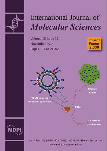
(This article belongs to the Section Materials Science)
Show Figures

Graphical abstract

10 pages, 960 KiB  
Article
Circulating microRNA-19a as a Potential Novel Biomarker for Diagnosis of Acute Myocardial Infarction
by Jianfeng Zhong 1,†, Yuan He 2,†, Wenjiang Chen 1, Xiaorong Shui 3, Can Chen 1,2,* and Wei Lei 1,2,*
1 Department of Cardiovascular Medicine, Affiliated Hospital of Guangdong Medical College, Zhanjiang 524000, China
2 Laboratory of Cardiovascular Diseases, Guangdong Medical College, Zhanjiang 524001, China
3 Laboratory of Vascular Surgery, Guangdong Medical College, Zhanjiang 524001, China
These authors contributed equally to this work.
Int. J. Mol. Sci. 2014, 15(11), 20355-20364; https://doi.org/10.3390/ijms151120355 - 6 Nov 2014
Cited by 68 | Viewed by 6358
Abstract
Acute myocardial infarction (AMI) is a serious cardiovascular disease. Investigating new susceptibility genes for effective methods of early diagnosis of AMI is important. In the current study, peripheral blood miR-19a levels were detected by real-time polymerase chain reaction. Significant differences and logistic correlation [...] Read more.
Acute myocardial infarction (AMI) is a serious cardiovascular disease. Investigating new susceptibility genes for effective methods of early diagnosis of AMI is important. In the current study, peripheral blood miR-19a levels were detected by real-time polymerase chain reaction. Significant differences and logistic correlation analyses were carried out by grouping of disease types and stratification of risk factors. Receiver-operator characteristic curve analysis was used to compare the current common clinical biochemical markers and evaluate the sensitivity and specificity of miR-19a for diagnosing AMI. Circulating miR-19a expression in the AMI group was higher than that in controls. The diagnostic effect of circulating miR-19a levels was superior to current clinical biochemical indices, such as CK, CK-MB, MYO, hs-TnI, and BNP. Our results show that there is a close association of circulating miR-19a levels with susceptibility to AMI. Circulating miR-19a levels could be a candidate diagnostic biomarker for AMI. Full article
(This article belongs to the Section Molecular Pathology, Diagnostics, and Therapeutics)
Show Figures

Figure 1

17 pages, 1707 KiB  
Article
Interactions of Borneol with DPPC Phospholipid Membranes: A Molecular Dynamics Simulation Study
by Qianqian Yin 1,†, Xinyuan Shi 2,*,†, Haiou Ding 3, Xingxing Dai 2, Guang Wan 1 and Yanjiang Qiao 2,*
1 School of Traditional Chinese Medicine, Capital Medical University, No. 10 of Xitoutiao Outside Youanmen, Fengtai District, Beijing 100069, China
2 Key Laboratory of TCM-Information Engineer, Beijing University of Chinese Medicine, No. 6 of Zhonghuan South Road, Wangjing, Chaoyang District, Beijing 100102, China
3 Civil Aviation General Hospital, No. 1 of Chaowai Takai A, Beijing 100123, China
These authors contributed equally to this work.
Int. J. Mol. Sci. 2014, 15(11), 20365-20381; https://doi.org/10.3390/ijms151120365 - 6 Nov 2014
Cited by 21 | Viewed by 8536
Abstract
Borneol, known as a “guide” drug in traditional Chinese medicine, is widely used as a natural penetration enhancer in modern clinical applications. Despite a large number of experimental studies on borneol’s penetration enhancing effect, the molecular basis of its action on bio-membranes is [...] Read more.
Borneol, known as a “guide” drug in traditional Chinese medicine, is widely used as a natural penetration enhancer in modern clinical applications. Despite a large number of experimental studies on borneol’s penetration enhancing effect, the molecular basis of its action on bio-membranes is still unclear. We carried out a series of coarse-grained molecular dynamics simulations with the borneol concentration ranging from 3.31% to 54.59% (v/v, lipid-free basis) to study the interactions of borneol with aDPPC(1,2-dipalmitoylsn-glycero-3-phosphatidylcholine) bilayer membrane, and the temperature effects were also considered. At concentrations below 21.89%, borneol’s presence only caused DPPC bilayer thinning and an increase in fluidity; A rise in temperature could promote the diffusing progress of borneol. When the concentration was 21.89% or above, inverted micelle-like structures were formed within the bilayer interior, which led to increased bilayer thickness, and an optimum temperature was found for the interaction of borneol with the DPPC bilayer membrane. These findings revealed that the choice of optimal concentration and temperature is critical for a given application in which borneol is used as a penetration enhancer. Our results not only clarify some molecular basis for borneol’s penetration enhancing effects, but also provide some guidance for the development and applications of new preparations containing borneol. Full article
(This article belongs to the Special Issue Bioactive Lipids and Lipidomics)
Show Figures

Figure 1

21 pages, 1475 KiB  
Article
Pine Bark and Green Tea Concentrated Extracts: Antioxidant Activity and Comprehensive Characterization of Bioactive Compounds by HPLC–ESI-QTOF-MS
by María De la Luz Cádiz-Gurrea 1,2, Salvador Fernández-Arroyo 3 and Antonio Segura-Carretero 1,2,*
1 Department of Analytical Chemistry, University of Granada, c/Fuentenueva s/n, 18071 Granada, Spain
2 Research and Development of Functional Food Centre (CIDAF), PTS Granada, Avda. Del Conocimiento s/n, Edificio BioRegion, 18016 Granada, Spain
3 Biomedical Research Centre, University Hospital of Sant Joan, IISPV, Rovira i Virgili University, C/Sant Joan s/n, 43201 Reus (Tarragona), Spain
Int. J. Mol. Sci. 2014, 15(11), 20382-20402; https://doi.org/10.3390/ijms151120382 - 6 Nov 2014
Cited by 69 | Viewed by 11196
Abstract
The consumption of polyphenols has frequently been associated with low incidence of degenerative diseases. Most of these natural antioxidants come from fruits, vegetables, spices, grains and herbs. For this reason, there has been increasing interest in identifying plant extract compounds. Polymeric tannins and [...] Read more.
The consumption of polyphenols has frequently been associated with low incidence of degenerative diseases. Most of these natural antioxidants come from fruits, vegetables, spices, grains and herbs. For this reason, there has been increasing interest in identifying plant extract compounds. Polymeric tannins and monomeric flavonoids, such as catechin and epicatechin, in pine bark and green tea extracts could be responsible for the higher antioxidant activities of these extracts. The aim of the present study was to characterize the phenolic compounds in pine bark and green tea concentrated extracts using high-performance liquid chromatography coupled to electrospray ionization mass spectrometry (HPLC–ESI-QTOF-MS). A total of 37 and 35 compounds from pine bark and green tea extracts, respectively, were identified as belonging to various structural classes, mainly flavan-3-ol and its derivatives (including procyanidins). The antioxidant capacity of both extracts was evaluated by three complementary antioxidant activity methods: Trolox equivalent antioxidant capacity (TEAC), ferric reducing antioxidant power (FRAP) and oxygen radical absorbance capacity (ORAC). Higher antioxidant activity values by each method were obtained. In addition, total polyphenol and flavan-3-ol contents, which were determined by Folin–Ciocalteu and vanillin assays, respectively, exhibited higher amounts of gallic acid and (+)-catechin equivalents. Full article
(This article belongs to the Special Issue Bioactive Phenolics and Polyphenols)
Show Figures

Figure 1

10 pages, 473 KiB  
Article
The Discovery of Aurora Kinase Inhibitor by Multi-Docking-Based Virtual Screening
by Jun-Tae Kim 1,†, Seo Hee Jung 1,†, Sun Young Kang 2, Chung-Kyu Ryu 3 and Nam Sook Kang 1,*
1 Graduate School of New Drug Discovery and Development, Chungnam National University, Daehakno 99, Yuseong-gu, Daejeon 305-764, Korea
2 AccelrysKorea, Korea BioPark Bldg C-dong 602, Sampyeong-dong, Bundang-gu, Seongnami-si, Gyeonggi-do 463-400, Korea
3 College of Pharmacy & Graduate School of Pharmaceutical Sciences, Ewha Womans University, 52, Ewhayeodae-gil, Seodaemun-gu, Seoul 120-75, Korea
These authors contributed equally to this work.
Int. J. Mol. Sci. 2014, 15(11), 20403-20412; https://doi.org/10.3390/ijms151120403 - 6 Nov 2014
Cited by 4 | Viewed by 7118
Abstract
We report the discovery of aurora kinase inhibitor using the fragment-based virtual screening by multi-docking strategy. Among a number of fragments collected from eMololecules, we found four fragment molecules showing potent activity (>50% at 100 μM) against aurora kinase. Based on the explored [...] Read more.
We report the discovery of aurora kinase inhibitor using the fragment-based virtual screening by multi-docking strategy. Among a number of fragments collected from eMololecules, we found four fragment molecules showing potent activity (>50% at 100 μM) against aurora kinase. Based on the explored fragment scaffold, we selected two compounds in our synthesized library and validated the biological activity against Aurora kinase. Full article
Show Figures

Graphical abstract

21 pages, 4521 KiB  
Article
An Uncleaved Signal Peptide Directs the Malus xiaojinensis Iron Transporter Protein Mx IRT1 into the ER for the PM Secretory Pathway
by Peng Zhang 1, Song Tan 1, James O. Berry 2, Peng Li 3, Na Ren 1, Shuang Li 1, Guang Yang 1, Wei-Bing Wang 1, Xiao-Ting Qi 1 and Li-Ping Yin 1,*
1 College of Life Science, Capital Normal University, Beijing 100048, China
2 Department of Biological Sciences, State University of New York, Buffalo, NY 14260, USA
3 School of Life Sciences, Tsinghua University, Beijing 100083, China
Int. J. Mol. Sci. 2014, 15(11), 20413-20433; https://doi.org/10.3390/ijms151120413 - 7 Nov 2014
Cited by 14 | Viewed by 10898
Abstract
Malus xiaojinensis iron-regulated transporter 1 (Mx IRT1) is a highly effective inducible iron transporter in the iron efficient plant Malus xiaojinensis. As a multi-pass integral plasma membrane (PM) protein, Mx IRT1 is predicted to consist of eight transmembrane domains, with a putative [...] Read more.
Malus xiaojinensis iron-regulated transporter 1 (Mx IRT1) is a highly effective inducible iron transporter in the iron efficient plant Malus xiaojinensis. As a multi-pass integral plasma membrane (PM) protein, Mx IRT1 is predicted to consist of eight transmembrane domains, with a putative N-terminal signal peptide (SP) of 1–29 amino acids. To explore the role of the putative SP, constructs expressing Mx IRT1 (with an intact SP) and Mx DsIRT1 (with a deleted SP) were prepared for expression in Arabidopsis and in yeast. Mx IRT1 could rescue the iron-deficiency phenotype of an Arabidopsis irt1 mutant, and complement the iron-limited growth defect of the yeast mutant DEY 1453 (fet3fet4). Furthermore, fluorescence analysis indicated that a chimeric Mx IRT1-eGFP (enhanced Green Fluorescent Protein) construct was translocated into the ER (Endoplasmic reticulum) for the PM sorting pathway. In contrast, the SP-deleted Mx DsIRT1 could not rescue either of the mutant phenotypes, nor direct transport of the GFP signal into the ER. Interestingly, immunoblot analysis indicated that the SP was not cleaved from the mature protein following transport into the ER. Taken together, data presented here provides strong evidence that an uncleaved SP determines ER-targeting of Mx IRT1 during the initial sorting stage, thereby enabling the subsequent transport and integration of this protein into the PM for its crucial role in iron uptake. Full article
Show Figures

Figure 1

15 pages, 1408 KiB  
Review
Long Non-Coding RNAs: Critical Players in Hepatocellular Carcinoma
by Jin Sun 1, Beibei Bie 1, Shu Zhang 1,2, Jun Yang 1,3 and Zongfang Li 1,2,*
1 National-Local Joint Engineering Research Center of Biodiagnostics and Biotherapy, Xi'an Jiaotong University, Xi'an 710004, China
2 Department of General Surgery, the Second Affiliated Hospital, School of Medicine, Xi'an Jiaotong University, Xi'an 710004, China
3 Department of Pathology, the Second Affiliated Hospital, School of Medicine, Xi'an Jiaotong University, Xi'an 710004, China
Int. J. Mol. Sci. 2014, 15(11), 20434-20448; https://doi.org/10.3390/ijms151120434 - 7 Nov 2014
Cited by 44 | Viewed by 7254
Abstract
Hepatocellular carcinoma (HCC) is a complex disease with multiple underlying pathogenic mechanisms caused by a variety of etiologic factors. Emerging evidence showed that long non-coding RNAs (lncRNAs), with size larger than 200 nucleotides (nt), play important roles in various types of cancer development [...] Read more.
Hepatocellular carcinoma (HCC) is a complex disease with multiple underlying pathogenic mechanisms caused by a variety of etiologic factors. Emerging evidence showed that long non-coding RNAs (lncRNAs), with size larger than 200 nucleotides (nt), play important roles in various types of cancer development and progression. In recent years, some dysregulated lncRNAs in HCC have been revealed and roles for several of them in HCC have been characterized. All these findings point to the potential of lncRNAs as prospective novel therapeutic targets in HCC. In this review, we summarize known dysregulated lncRNAs in HCC, and review potential biological roles and underlying molecular mechanisms of lncRNAs in HCC. Additionally, we discussed prospects of lncRNAs as potential biomarker and therapeutic target for HCC. In conclusion, this paper will help us gain better understanding of molecular mechanisms by which lncRNAs perform their function in HCC and also provide general strategies and directions for future research. Full article
(This article belongs to the Collection Regulation by Non-coding RNAs)
Show Figures

Figure 1

20 pages, 2089 KiB  
Article
The Effects of NAD+ on Apoptotic Neuronal Death and Mitochondrial Biogenesis and Function after Glutamate Excitotoxicity
by Xiaowan Wang, Hailong Li and Shinghua Ding *
Dalton Cardiovascular Research Center, Department of Bioengineering, University of Missouri, Columbia, MO 65211, USA
Int. J. Mol. Sci. 2014, 15(11), 20449-20468; https://doi.org/10.3390/ijms151120449 - 7 Nov 2014
Cited by 35 | Viewed by 9729
Abstract
NAD+ is an essential co-enzyme for cellular energy metabolism and is also involved as a substrate for many cellular enzymatic reactions. It has been shown that NAD+ has a beneficial effect on neuronal survival and brain injury in in vitro and [...] Read more.
NAD+ is an essential co-enzyme for cellular energy metabolism and is also involved as a substrate for many cellular enzymatic reactions. It has been shown that NAD+ has a beneficial effect on neuronal survival and brain injury in in vitro and in vivo ischemic models. However, the effect of NAD+ on mitochondrial biogenesis and function in ischemia has not been well investigated. In the present study, we used an in vitro glutamate excitotoxicity model of primary cultured cortical neurons to study the effect of NAD+ on apoptotic neuronal death and mitochondrial biogenesis and function. Our results show that supplementation of NAD+ could effectively reduce apoptotic neuronal death, and apoptotic inducing factor translocation after neurons were challenged with excitotoxic glutamate stimulation. Using different approaches including confocal imaging, mitochondrial DNA measurement and Western blot analysis of PGC-1 and NRF-1, we also found that NAD+ could significantly attenuate glutamate-induced mitochondrial fragmentation and the impairment of mitochondrial biogenesis. Furthermore, NAD+ treatment effectively inhibited mitochondrial membrane potential depolarization and NADH redistribution after excitotoxic glutamate stimulation. Taken together, our results demonstrated that NAD+ is capable of inhibiting apoptotic neuronal death after glutamate excitotoxicity via preserving mitochondrial biogenesis and integrity. Our findings provide insights into potential neuroprotective strategies in ischemic stroke. Full article
(This article belongs to the Special Issue Neuroprotective Strategies 2014)
Show Figures

Figure 1

17 pages, 3606 KiB  
Article
Spectroscopic Studies of R(+)-α-Lipoic Acid—Cyclodextrin Complexes
by Naoko Ikuta 1,2, Akira Tanaka 2, Ayako Otsubo 2, Noriko Ogawa 3, Hiromitsu Yamamoto 3, Tomoyuki Mizukami 2, Shoji Arai 2, Masayuki Okuno 2, Keiji Terao 1,4 and Seiichi Matsugo 2,*
1 Graduate School of Medicine, Kobe University, Kobe 650-0017, Japan
2 College of Science and Engineering, Kanazawa University, Kanazawa 920-1192, Japan
3 Department of Pharmaceutical Engineering, School of Pharmacy, Aichi Gakuin University, Nagoya 464-8650, Japan
4 CycloChem Bio Co., Ltd., Kobe 650-0047, Japan
Int. J. Mol. Sci. 2014, 15(11), 20469-20485; https://doi.org/10.3390/ijms151120469 - 7 Nov 2014
Cited by 35 | Viewed by 9049
Abstract
α-Lipoic acid (ALA) has a chiral center at the C6 position, and exists as two enantiomers, R(+)-ALA (RALA) and S(−)-ALA (SALA). RALA is naturally occurring, and is a cofactor for mitochondrial enzymes, therefore playing a major role in energy metabolism. However, [...] Read more.
α-Lipoic acid (ALA) has a chiral center at the C6 position, and exists as two enantiomers, R(+)-ALA (RALA) and S(−)-ALA (SALA). RALA is naturally occurring, and is a cofactor for mitochondrial enzymes, therefore playing a major role in energy metabolism. However, RALA cannot be used for pharmaceuticals or nutraceuticals because it readily polymerizes via a 1,2-dithiolane ring-opening when exposed to light or heat. So, it is highly desired to find out the method to stabilize RALA. The purpose of this study is to provide the spectroscopic information of stabilized RALA and SALA through complexation with cyclodextrins (CDs), α-CD, β-CD and γ-CD and to examine the physical characteristics of the resultant complexes in the solid state. The RALA-CD structures were elucidated based on the micro fourier transform infrared (FT-IR) and Raman analyses. The FT-IR results showed that the C=O stretching vibration of RALA appeared at 1717 cm−1 and then shifted on formation of the RALA-CD complexes. The Raman spectra showed that the S–S and C–S stretching vibrations for RALA at 511 cm−1 (S–S), 631 cm−1 (C–S) and 675 cm−1 (C–S) drastically weakened and almost disappeared upon complexation with CDs. Several peaks indicative of O–H vibrations also shifted or changed in intensity. These results indicate that RALA and CDs form host-guest complexes by interacting with one another. Full article
(This article belongs to the Special Issue Bioactive Nanoparticles 2014)
Show Figures

Figure 1

14 pages, 4460 KiB  
Article
MicroRNA-130b Promotes Cell Aggressiveness by Inhibiting Peroxisome Proliferator-Activated Receptor Gamma in Human Hepatocellular Carcinoma
by Kangsheng Tu *,†, Xin Zheng, Changwei Dou, Chao Li, Wei Yang, Yingmin Yao and Qingguang Liu *
1 Department of Hepatobiliary Surgery, the First Affiliated Hospital of Xi'an Jiaotong University, Xi'an 710061, China
These authors contributed equally to this work.
Int. J. Mol. Sci. 2014, 15(11), 20486-20499; https://doi.org/10.3390/ijms151120486 - 7 Nov 2014
Cited by 69 | Viewed by 8669
Abstract
MircroRNA-130b (miR-130b) is proposed as a novel tumor-related miRNA and has been found to be significantly dysregulated in tumors. In this study, the expression level of miR-130b was found to be obviously higher in hepatocellular carcinoma (HCC) tissues than that in nontumor tissues. [...] Read more.
MircroRNA-130b (miR-130b) is proposed as a novel tumor-related miRNA and has been found to be significantly dysregulated in tumors. In this study, the expression level of miR-130b was found to be obviously higher in hepatocellular carcinoma (HCC) tissues than that in nontumor tissues. Further, miR-130b was expressed at significantly higher levels in aggressive and recurrent tumor tissues. Clinical analysis indicated that high-expression of miR-130b was prominently correlated with venous infiltration, high Edmondson-Steiner grading and advanced tumor-node-metastasis (TNM) tumor stage in HCC. Elevated miR-130b expression was observed in all HCC cell lines (HepG2, SMMC-7721, Huh7, Hep3B and MHCC97H) as compared with that in a nontransformed hepatic cell line (LO2). Furthermore, an inverse correlation between miR-130b and E-cadherin and a positive correlation between miR-130b and Vimentin were observed in HCC tissues. Down-regulation of miR-130b expression reduced invasion and migration in both Hep3B and MHCC97H cells. Peroxisome proliferator-activated receptor gamma (PPAR-γ) was inversely correlated with miR-130b expression in HCC tissues. In addition, down-regulation of miR-130b restored PPAR-γ expression and subsequently suppressed epithelial-mesenchymal transition (EMT) in HCC cells. We identified PPARγ as a direct target of miR-130b in HCC in vitro. Notably, PPAR-γ knockdown abolished down-regulation of miR-130b-inhibited EMT in MHCC97H cells. In conclusion, miR-130b may promote HCC cell migration and invasion by inhibiting PPAR-γ and subsequently inducing EMT. Full article
(This article belongs to the Special Issue Advances in Molecular Oncology 2014)
Show Figures

Figure 1

18 pages, 2711 KiB  
Article
Multiple Mechanisms Mediate Resistance to Sorafenib in Urothelial Cancer
by Judith Knievel 1, Wolfgang A. Schulz 1, Annemarie Greife 1, Christiane Hader 1, Tobias Lübke 2,3, Ingo Schmitz 2,3, Peter Albers 1 and Günter Niegisch 1,*
1 Department of Urology, Heinrich-Heine-University, Moorenstr. 5, Düsseldorf D-40225, Germany
2 Helmholtz-Zentrum für Infektionsforschung, Inhoffenstr. 7, Braunschweig D-38124, Germany
3 Institute for Molecular and Clinical Immunology, Otto-von-Guericke-University, Leipzigerstr. 44, Magdeburg D-39120, Germany
Int. J. Mol. Sci. 2014, 15(11), 20500-20517; https://doi.org/10.3390/ijms151120500 - 7 Nov 2014
Cited by 23 | Viewed by 8904
Abstract
Genetic and epigenetic changes in the mitogen activated protein kinase (MAPK) signaling render urothelial cancer a potential target for tyrosine kinase inhibitor (TKI) treatment. However, clinical trials of several TKIs failed to prove efficacy. In this context, we investigated changes in MAPK signaling [...] Read more.
Genetic and epigenetic changes in the mitogen activated protein kinase (MAPK) signaling render urothelial cancer a potential target for tyrosine kinase inhibitor (TKI) treatment. However, clinical trials of several TKIs failed to prove efficacy. In this context, we investigated changes in MAPK signaling activity, downstream apoptotic regulators and changes in cell cycle distribution in different urothelial cancer cell lines (UCCs) upon treatment with the multikinase inhibitor sorafenib. None of the classical sorafenib targets (vascular endothelial growth factor receptor 1/-receptor 2, VEGFR1/-R2; platelet-derived growth factor receptor α/-receptor β, PDGFR-α/-β; c-KIT) was expressed at significant levels leaving RAF proteins as its likely molecular target. Low sorafenib concentrations paradoxically increased cell viability, whereas higher concentrations induced G1 arrest and eventually apoptosis. MAPK signaling remained partly active after sorafenib treatment, especially in T24 cells with an oncogenic HRAS mutation. AKT phosphorylation was increased, suggesting compensatory activation of the phosphatidylinositol-3-kinase (PI3K) pathway. Sorafenib regularly down regulated the anti-apoptotic myeloid cell leukemia 1 (Mcl-1) protein, but combinatorial treatment with ABT-737 targeting other B-cell lymphoma 2 (Bcl-2) family proteins did not result in synergistic effects. In summary, efficacy of sorafenib in urothelial cancer cell lines appears hampered by limited effects on MAPK signaling, crosstalk with further cancer pathways and an anti-apoptotic state of UCCs. These observations may account for the lack of efficacy of sorafenib in clinical trials and should be considered more broadly in the development of signaling pathway inhibitors for drug therapy in urothelial carcinoma. Full article
(This article belongs to the Section Biochemistry)
Show Figures

Figure 1

20 pages, 2318 KiB  
Review
Gliadin Peptides as Triggers of the Proliferative and Stress/Innate Immune Response of the Celiac Small Intestinal Mucosa
by Maria Vittoria Barone 1,2,*, Riccardo Troncone 1,2 and Salvatore Auricchio 2
1 Department of Translational Medical Science (Section of Pediatrics), University of Naples Federico II, Via S. Pansini 5, Naples 80131, Italy
2 European Laboratory for the Investigation of Food Induced Diseases (ELFID), University of Naples Federico II, Via S. Pansini 5, Naples 80131, Italy
Int. J. Mol. Sci. 2014, 15(11), 20518-20537; https://doi.org/10.3390/ijms151120518 - 7 Nov 2014
Cited by 88 | Viewed by 12717
Abstract
Celiac disease (CD) is a frequent inflammatory intestinal disease, with a genetic background, caused by gliadin-containing food. Undigested gliadin peptides induce innate and adaptive T cell-mediated immune responses. The major mediator of the stress and innate immune response to gliadin peptides (i.e. [...] Read more.
Celiac disease (CD) is a frequent inflammatory intestinal disease, with a genetic background, caused by gliadin-containing food. Undigested gliadin peptides induce innate and adaptive T cell-mediated immune responses. The major mediator of the stress and innate immune response to gliadin peptides (i.e., peptide 31–43, P31–43) is the cytokine interleukin-15 (IL-15). The role of epithelial growth factor (EGF) as a mediator of enterocyte proliferation and the innate immune response has been described. In this paper, we review the most recent literature on the mechanisms responsible for triggering the up-regulation of these mediators in CD by gliadin peptides. We will discuss the role of P31–43 in enterocyte proliferation, structural changes and the innate immune response in CD mucosa in cooperation with EGF and IL-15, and the mechanism of up-regulation of these mediators related to vesicular trafficking. We will also review the literature that focuses on constitutive alterations of the structure, signalling/proliferation and stress/innate immunity pathways of CD cells. Finally, we will discuss how these pathways can be triggered by gliadin peptide P31–43 in controls, mimicking the celiac cellular phenotype. Full article
(This article belongs to the Special Issue Bioactive Proteins and Peptides Derived from Food)
Show Figures

Figure 1

17 pages, 2434 KiB  
Article
Intracellular Signaling Transduction Pathways Triggered by a Well-Known Anti-GHR Monoclonal Antibody, Mab263, in Vitro and in Vivo
by Hainan Lan 1, Wei Li 2,†, Honglong Jiang 1,†, Yanhong Yang 1 and Xin Zheng 1,*
1 College of Animal Science and Technology, Jilin Agricultural University, Xincheng Street 2888, Changchun 130118, China
2 State Key Laboratory of Animal Nutrition, China Agricultural University, Beijing 100193, China
These authors contributed equally to this work.
Int. J. Mol. Sci. 2014, 15(11), 20538-20554; https://doi.org/10.3390/ijms151120538 - 10 Nov 2014
Cited by 7 | Viewed by 5474
Abstract
A series of studies have reported that monoclonal antibody 263 (Mab263), a monoclonal antibody against the growth hormone receptor (GHR), acts as an agonist in vitro and in vivo. However, the intracellular signaling pathways triggered by Mab263 have not yet been delineated. [...] Read more.
A series of studies have reported that monoclonal antibody 263 (Mab263), a monoclonal antibody against the growth hormone receptor (GHR), acts as an agonist in vitro and in vivo. However, the intracellular signaling pathways triggered by Mab263 have not yet been delineated. Therefore, we examined the intracellular signaling pathways induced by Mab263 in vivo and in vitro in the present study. The results show that this antibody activated janus kinase 2 (JAK2), signal transducer and activator of transcription 3 (STAT3), STAT1 and extracellular signal-regulated kinase 1/2 (ERK1/2), but not STAT5. The phosphorylation kinetics of JAK2, STAT3/1 and ERK1/2 induced by Mab263 were subsequently analyzed in dose-response and time course experiments. Our observations indicate that Mab263 induced different intracellular signaling pathways than GH, which indicates that Mab263 is a signal-specific molecule and that Mab263 may be a valuable biological reagent to study the mechanism(s) of GHR-mediated intracellular signaling pathways. Full article
(This article belongs to the Section Biochemistry)
Show Figures

Figure 1

17 pages, 1538 KiB  
Article
Depletion of C3orf1/TIMMDC1 Inhibits Migration and Proliferation in 95D Lung Carcinoma Cells
by Huiling Wu 1,2, Wenbing Wang 3 and Huaxi Xu 3,*
1 School of Food and Biological Engineering, Jiangsu University, Zhenjiang 212013, China
2 Institute of Life Sciences, Jiangsu University, Zhenjiang 212013, China
3 School of Medical Science and Laboratory Medicine, Jiangsu University, Zhenjiang 212013, China
Int. J. Mol. Sci. 2014, 15(11), 20555-20571; https://doi.org/10.3390/ijms151120555 - 10 Nov 2014
Cited by 12 | Viewed by 6088
Abstract
In our previous study, we identified an association of high expression of c3orf1, also known as TIMMDC1 (translocase of inner mitochondrial membrane domain-containing protein 1), with metastatic characteristics in lung carcinoma cells. To investigate the preliminary function and mechanism of this mitochondrial [...] Read more.
In our previous study, we identified an association of high expression of c3orf1, also known as TIMMDC1 (translocase of inner mitochondrial membrane domain-containing protein 1), with metastatic characteristics in lung carcinoma cells. To investigate the preliminary function and mechanism of this mitochondrial protein, we depleted C3orf1 expression by introducing siRNA into 95D lung carcinoma cells. We demonstrated that C3orf1 depletion significantly suppressed 95D cell growth and migration. We confirmed C3orf1 localization in the inner mitochondrial membrane and showed that mitochondrial viability, membrane potential, and ATPase activity were remarkably reduced upon depletion of C3orf1. Microarray data indicated that genes involved in regulation of cell death, migration, and cell-cycle arrest were significantly altered after C3orf1 depletion for 48 h. The expression of genes involved in focal adhesion, ECM-receptor interaction, and p53-signaling pathways were notably altered. Furthermore, cell-cycle arrest genes such as CCNG2 and PTEN as well as genes involved in cell migration inhibition, such as TIMP3 and COL3A1, were upregulated after C3orf1 depletion in 95D cells. Concurrently, expression of the migration-promoting gene NUPR1 was markedly reduced, as confirmed by real-time PCR. We conclude that C3orf1 is critical for mitochondrial function, migration, and proliferation in 95D lung carcinoma cells. Depletion of C3orf1 inhibited cell migration and cell proliferation in association with upregulation of genes involved in cell-cycle arrest and cell migration inhibition. These results suggest that C3orf1 (TIMMDC1) may be a viable treatment target for lung carcinoma, and that further study of the role of this protein in lung carcinoma pathogenesis is justified. Full article
(This article belongs to the Special Issue Molecular Machinery of Cell Growth Regulation)
Show Figures

Figure 1

13 pages, 660 KiB  
Article
A Functional Monomer Is Not Enough: Principal Component Analysis of the Influence of Template Complexation in Pre-Polymerization Mixtures on Imprinted Polymer Recognition and Morphology
by Kerstin Golker 1, Björn C. G. Karlsson 1, Annika M. Rosengren 1 and Ian A. Nicholls 1,2,*
1 Bioorganic & Biophysical Chemistry Laboratory, Linnaeus University Centre for Biomaterials Chemistry, Linnaeus University, SE-391 82 Kalmar, Sweden
2 Department of Chemistry—BMC, Uppsala University, SE-751 23 Uppsala, Sweden
Int. J. Mol. Sci. 2014, 15(11), 20572-20584; https://doi.org/10.3390/ijms151120572 - 10 Nov 2014
Cited by 29 | Viewed by 6888
Abstract
In this report, principal component analysis (PCA) has been used to explore the influence of template complexation in the pre-polymerization phase on template molecularly imprinted polymer (MIP) recognition and polymer morphology. A series of 16 bupivacaine MIPs were studied. The ethylene glycol dimethacrylate [...] Read more.
In this report, principal component analysis (PCA) has been used to explore the influence of template complexation in the pre-polymerization phase on template molecularly imprinted polymer (MIP) recognition and polymer morphology. A series of 16 bupivacaine MIPs were studied. The ethylene glycol dimethacrylate (EGDMA)-crosslinked polymers had either methacrylic acid (MAA) or methyl methacrylate (MMA) as the functional monomer, and the stoichiometry between template, functional monomer and crosslinker was varied. The polymers were characterized using radioligand equilibrium binding experiments, gas sorption measurements, swelling studies and data extracted from molecular dynamics (MD) simulations of all-component pre-polymerization mixtures. The molar fraction of the functional monomer in the MAA-polymers contributed to describing both the binding, surface area and pore volume. Interestingly, weak positive correlations between the swelling behavior and the rebinding characteristics of the MAA-MIPs were exposed. Polymers prepared with MMA as a functional monomer and a polymer prepared with only EGDMA were found to share the same characteristics, such as poor rebinding capacities, as well as similar surface area and pore volume, independent of the molar fraction MMA used in synthesis. The use of PCA for interpreting relationships between MD-derived descriptions of events in the pre-polymerization mixture, recognition properties and morphologies of the corresponding polymers illustrates the potential of PCA as a tool for better understanding these complex materials and for their rational design. Full article
(This article belongs to the Section Molecular Recognition)
Show Figures

Figure 1

22 pages, 1159 KiB  
Article
Rosmarinus Officinalis Leaves as a Natural Source of Bioactive Compounds
by Isabel Borrás-Linares 1,2, Zorica Stojanović 3, Rosa Quirantes-Piné 1,2, David Arráez-Román 1,2,*, Jaroslava Švarc-Gajić 3, Alberto Fernández-Gutiérrez 1,2 and Antonio Segura-Carretero 1,2
1 Department of Analytical Chemistry, University of Granada, c/Fuentenueva s/n, 18071 Granada, Spain
2 Research and Development of Functional Food Centre (CIDAF), PTS Granada, Avda. Del Conocimiento s/n., Edificio BioRegion, 18016 Granada, Spain
3 Faculty of Technology, University of Novi Sad, Bulevar Cara Lazara 1, 21000 Novi Sad, Serbia
Int. J. Mol. Sci. 2014, 15(11), 20585-20606; https://doi.org/10.3390/ijms151120585 - 10 Nov 2014
Cited by 209 | Viewed by 15202
Abstract
In an extensive search for bioactive compounds from plant sources, the composition of different extracts of rosemary leaves collected from different geographical zones of Serbia was studied. The qualitative and quantitative characterization of 20 rosemary (Rosmarinus officinalis) samples, obtained by microwave-assisted [...] Read more.
In an extensive search for bioactive compounds from plant sources, the composition of different extracts of rosemary leaves collected from different geographical zones of Serbia was studied. The qualitative and quantitative characterization of 20 rosemary (Rosmarinus officinalis) samples, obtained by microwave-assisted extraction (MAE), was determined by high performance liquid chromatography coupled to electrospray quadrupole-time of flight mass spectrometry (HPLC–ESI-QTOF-MS). The high mass accuracy and true isotopic pattern in both MS and MS/MS spectra provided by the QTOF-MS analyzer enabled the characterization of a wide range of phenolic compounds in the extracts, including flavonoids, phenolic diterpenes and abietan-type triterpenoids, among others. According to the data compiled, rosemary samples from Sokobanja presented the highest levels in flavonoids and other compounds such as carnosol, rosmaridiphenol, rosmadial, rosmarinic acid, and carnosic acid. On the other hand, higher contents in triterpenes were found in the extracts of rosemary from Gložan (Vojvodina). Full article
(This article belongs to the Special Issue Bioactive Phenolics and Polyphenols)
Show Figures

Figure 1

31 pages, 878 KiB  
Review
Molecular Mechanisms Underlying the Effects of Statins in the Central Nervous System
by Amelia J. McFarland 1,2, Shailendra Anoopkumar-Dukie 1,2, Devinder S. Arora 1,2, Gary D. Grant 1,2, Catherine M. McDermott 3, Anthony V. Perkins 2,4 and Andrew K. Davey 1,2,*
1 School of Pharmacy, Griffith University, Queensland 4222, Australia
2 Griffith Health Institute, Griffith University, Queensland 4222, Australia
3 Department of Biomedical Science, Bond University, Queensland 4226, Australia
4 School of Medical Sciences, Griffith University, Queensland, 4222, Australia
Int. J. Mol. Sci. 2014, 15(11), 20607-20637; https://doi.org/10.3390/ijms151120607 - 10 Nov 2014
Cited by 159 | Viewed by 19265
Abstract
3-Hydroxy-3-methylglutaryl coenzyme A reductase inhibitors, commonly referred to as statins, are widely used in the treatment of dyslipidaemia, in addition to providing primary and secondary prevention against cardiovascular disease and stroke. Statins’ effects on the central nervous system (CNS), particularly on cognition and [...] Read more.
3-Hydroxy-3-methylglutaryl coenzyme A reductase inhibitors, commonly referred to as statins, are widely used in the treatment of dyslipidaemia, in addition to providing primary and secondary prevention against cardiovascular disease and stroke. Statins’ effects on the central nervous system (CNS), particularly on cognition and neurological disorders such as stroke and multiple sclerosis, have received increasing attention in recent years, both within the scientific community and in the media. Current understanding of statins’ effects is limited by a lack of mechanism-based studies, as well as the assumption that all statins have the same pharmacological effect in the central nervous system. This review aims to provide an updated discussion on the molecular mechanisms contributing to statins’ possible effects on cognitive function, neurodegenerative disease, and various neurological disorders such as stroke, epilepsy, depression and CNS cancers. Additionally, the pharmacokinetic differences between statins and how these may result in statin-specific neurological effects are also discussed. Full article
(This article belongs to the Special Issue Molecular Research in Neurotoxicology)
Show Figures

Figure 1

18 pages, 572 KiB  
Article
Analysis of Human TAAR8 and Murine Taar8b Mediated Signaling Pathways and Expression Profile
by Jessica Mühlhaus 1,†, Juliane Dinter 1,†, Daniela Nürnberg 1,†, Maren Rehders 2, Maren Depke 3, Janine Golchert 3, Georg Homuth 3, Chun-Xia Yi 4, Silke Morin 4, Josef Köhrle 5, Klaudia Brix 2, Matthias Tschöp 4,6, Gunnar Kleinau 1 and Heike Biebermann 1,*
1 Institut für Experimentelle Pädiatrische Endokrinologie, Charité-Universitätsmedizin, Campus Virchow-Klinikum, Augustenburger Platz 1, 13353 Berlin, Germany
2 School of Engineering and Science, Research Center MOLIFE—Molecular Life Science, Jacobs University Bremen, Campus Ring 1, 28759 Bremen, Germany
3 Interfaculty Institute for Genetics and Functional Genomics, University Medicine and Ernst-Moritz-Arndt-University Greifswald, Fr iedrich-Ludwig-Jahn-Str. 15a, 17487 Greifswald, Germany
4 Helmholtz Zentrum München, German Research Center for Environmental Health, Institute for Diabetes and Obesity, Business Campus Garching, Parkring 13, 85748 Garching, Germany
5 Institut für Experimentelle Endokrinologie, Charité-Universitätsmedizin Campus Virchow-Klinikum, Augustenburger Platz 1, 13353 Berlin, Germany
6 Division of Metabolic Diseases, School of Medicine, Technische Universität München, Schneckenburgerstraße 8, 81675 München, Germany
These authors contributed equally to this work.
Int. J. Mol. Sci. 2014, 15(11), 20638-20655; https://doi.org/10.3390/ijms151120638 - 10 Nov 2014
Cited by 25 | Viewed by 10364
Abstract
The thyroid hormone derivative 3-iodothyronamine (3-T1AM) exerts metabolic effects in vivo that contradict known effects of thyroid hormones. 3-T1AM acts as a trace amine-associated receptor 1 (TAAR1) agonist and activates Gs signaling in vitro. Interestingly, 3-T1 [...] Read more.
The thyroid hormone derivative 3-iodothyronamine (3-T1AM) exerts metabolic effects in vivo that contradict known effects of thyroid hormones. 3-T1AM acts as a trace amine-associated receptor 1 (TAAR1) agonist and activates Gs signaling in vitro. Interestingly, 3-T1AM-meditated in vivo effects persist in Taar1 knockout-mice indicating that further targets of 3-T1AM might exist. Here, we investigated another member of the TAAR family, the only scarcely studied mouse and human trace-amine-associated receptor 8 (Taar8b, TAAR8). By RT-qPCR and locked-nucleic-acid (LNA) in situ hybridization, Taar8b expression in different mouse tissues was analyzed. Functionally, we characterized TAAR8 and Taar8b with regard to cell surface expression and signaling via different G-protein-mediated pathways. Cell surface expression was verified by ELISA, and cAMP accumulation was quantified by AlphaScreen for detection of Gs and/or Gi/o signaling. Activation of G-proteins Gq/11 and G12/13 was analyzed by reporter gene assays. Expression analyses revealed at most marginal Taar8b expression and no gender differences for almost all analyzed tissues. In heart, LNA-in situ hybridization demonstrated the absence of Taar8b expression. We could not identify 3-T1AM as a ligand for TAAR8 and Taar8b, but both receptors were characterized by a basal Gi/o signaling activity, a so far unknown signaling pathway for TAARs. Full article
(This article belongs to the Collection G Protein-Coupled Receptor Signaling and Regulation)
Show Figures

Figure 1

12 pages, 715 KiB  
Review
Potential Roles of BMP9 in Liver Fibrosis
by Jianjun Bi and Shengfang Ge *
Department of Ophthalmology, Ninth People's Hospital, Shanghai Jiao Tong University School of Medicine, Shanghai 200011, China
Int. J. Mol. Sci. 2014, 15(11), 20656-20667; https://doi.org/10.3390/ijms151120656 - 11 Nov 2014
Cited by 42 | Viewed by 10067
Abstract
Liver fibrosis is a common phenomenon that is associated with several pathologies and is characterized by excessive extracellular matrix deposition that leads to progressive liver dysfunction. Bone morphogenetic protein 9 (BMP9) is the most recently discovered member of the BMP family. BMP9 bound [...] Read more.
Liver fibrosis is a common phenomenon that is associated with several pathologies and is characterized by excessive extracellular matrix deposition that leads to progressive liver dysfunction. Bone morphogenetic protein 9 (BMP9) is the most recently discovered member of the BMP family. BMP9 bound with high affinity to activin receptor-like kinase 1 (ALK1) and endoglin in non-parenchymal liver cells. In addition, BMP9 activated Smad1/Smad5/Smad8 and induced the expression of the target genes inhibitor of differentiation 1 (Id1), hepcidin, Snail and the co-receptor endoglin in liver cells. Although the role of BMP9 in liver fibrosis is currently poorly understood, the presence of BMP9-activated proteins and its target genes have been reported to be associated with liver fibrosis development. This review summarizes the indirect connection between BMP9 and liver fibrosis, with a focus on the BMP9 signaling pathway members ALK1, endoglin, Id1, hepcidin and Snail. The observations on the role of BMP9 in regulating liver fibrosis may help in understanding the pathology mechanisms of liver disease. Furthermore, BMP9 could be served as a potent biomarker and the target of potential therapeutic drugs to treat hepatocytes fibrosis. Full article
(This article belongs to the Special Issue Molecular Mechanisms of Human Liver Diseases)
Show Figures

Figure 1

18 pages, 689 KiB  
Article
Characterization of Flavonoid Glycosides from Fenugreek (Trigonella foenum-graecum) Crude Seeds by HPLC–DAD–ESI/MS Analysis
by Zakia Benayad 1,*, Carmen Gómez-Cordovés 1 and Nour Eddine Es-Safi 2,*
1 Institute of Food science, Technology and Nutrition of the Spanish National Research Council (ICTAN–CSIC), Juan de la Cierva 3, Madrid 28006, Spain
2 Team of Organic Chemistry and Physico-Chemical Studies, Ecole Normale Superieure, Mohammed V University of Rabat, P.O. Box 5118, 10200 Rabat, Morocco
Int. J. Mol. Sci. 2014, 15(11), 20668-20685; https://doi.org/10.3390/ijms151120668 - 11 Nov 2014
Cited by 154 | Viewed by 13397
Abstract
Fenugreek (Trigonella foenum-graecum) is a medicinal plant which is widely used for its pharmacological properties. In this study the phenolic composition of fenugreek crude seeds originating from Morocco has been investigated. Extraction was performed from defatted seeds by a hydromethanolic solution [...] Read more.
Fenugreek (Trigonella foenum-graecum) is a medicinal plant which is widely used for its pharmacological properties. In this study the phenolic composition of fenugreek crude seeds originating from Morocco has been investigated. Extraction was performed from defatted seeds by a hydromethanolic solution using an Accelerated Solvent Extractor. HPLC technique coupled to negative ion electrospray ionization mass spectrometry and diode array detection was employed to identify the polyphenol in the obtained extract. The obtained results allowed the detection of 32 phenolic compounds among which various flavonoid glycosides and phenolic acids have been tentatively identified on the basis of their UV and MS spectra, and comparisons with standards when available, as well as with literature data. A systematic study of the obtained MS spectra and the observed fragmentation showed that most of the identified compounds were acylated and non-acylated flavonoids with apigenin, luteolin and kaempferol as aglycons. Hydroxycinnamic acids mostly dominated by caffeic acid derivatives were also detected. The quantitative analysis of the identified compounds showed that the phenolic composition of the studied crude fenugreek seeds was predominantly acylated and non-acylated flavone derivatives with apigenin as the main aglycon. Full article
(This article belongs to the Special Issue Bioactive Phenolics and Polyphenols)
Show Figures

Graphical abstract

20 pages, 837 KiB  
Article
Berteroin Present in Cruciferous Vegetables Exerts Potent Anti-Inflammatory Properties in Murine Macrophages and Mouse Skin
by Yoo Jin Jung 1, Jae In Jung 1, Han Jin Cho 2, Myung-Sook Choi 3, Mi-Kyung Sung 4, Rina Yu 5, Young-Hee Kang 1 and Jung Han Yoon Park 1,6,*
1 Department of Food Science and Nutrition, Hallym University, Chuncheon 200-702, Korea
2 WCU Biomodulation Major, Department of Agricultural Biotechnology and Center for Food and Bioconvergence, Seoul National University, Seoul 151-921, Korea
3 Center for Food and Nutritional Genomics Research and Department of Food Science and Nutrition, Kyungpook National University, Daegu 702-701, Korea
4 Department of Food and Nutrition, Sookmyung Women's University, Seoul 140-742, Korea
5 Department of Food Science and Nutrition, University of Ulsan, Ulsan 680-749, Korea
6 Advanced Institutes of Convergence Technology, Seoul National University, Suwon, Gyonggi-do 443-270, Korea
Int. J. Mol. Sci. 2014, 15(11), 20686-20705; https://doi.org/10.3390/ijms151120686 - 11 Nov 2014
Cited by 21 | Viewed by 8496
Abstract
Berteroin (5-methylthiopentyl isothiocyanate) is a sulforaphane analog present in cruciferous vegetables, including Chinese cabbage, rucola salad leaves, and mustard oil. We examined whether berteroin exerts anti-inflammatory activities using lipopolysaccharide (LPS)-stimulated Raw 264.7 macrophages and 12-O-tetradecanoylphorbol-13-acetate (TPA)-induced mouse skin inflammation models. Berteroin decreased LPS-induced [...] Read more.
Berteroin (5-methylthiopentyl isothiocyanate) is a sulforaphane analog present in cruciferous vegetables, including Chinese cabbage, rucola salad leaves, and mustard oil. We examined whether berteroin exerts anti-inflammatory activities using lipopolysaccharide (LPS)-stimulated Raw 264.7 macrophages and 12-O-tetradecanoylphorbol-13-acetate (TPA)-induced mouse skin inflammation models. Berteroin decreased LPS-induced release of inflammatory mediators and pro-inflammatory cytokines in Raw 264.7 macrophages. Berteroin inhibited LPS-induced degradation of inhibitor of κBα (IκBα) and nuclear factor-κB p65 translocation to the nucleus and DNA binding activity. Furthermore, berteroin suppressed degradation of IL-1 receptor-associated kinase and phosphorylation of transforming growth factor β activated kinase-1. Berteroin also inhibited LPS-induced phosphorylation of p38 MAPK, ERK1/2, and AKT. In the mouse ear, berteroin effectively suppressed TPA-induced edema formation and down-regulated iNOS and COX-2 expression as well as phosphorylation of AKT and ERK1/2. These results demonstrate that berteroin exhibits potent anti-inflammatory properties and suggest that berteroin can be developed as a skin anti-inflammatory agent. Full article
(This article belongs to the Section Biochemistry)
Show Figures

Figure 1

17 pages, 1282 KiB  
Article
Caveolin-1 Limits the Contribution of BKCa Channel to MCF-7 Breast Cancer Cell Proliferation and Invasion
by Cheng Du 1,2,†, Li Chen 3,†, Haijun Zhang 3, Zhongchao Wang 3, Wenchao Liu 1,*, Xiaodong Xie 2,* and Manjiang Xie 3,*
1 Department of Oncology, Xijing Hospital, the Fourth Military Medical University, Xi'an 710032, China
2 Department of Oncology, General Hospital of Shenyang Military Area Command, Shenyang 110840, China
3 Key Laboratory of Aerospace Medicine, Ministry of Education, the Fourth Military Medical University, Xi'an 710032, China
These authors contributed equally to this work.
Int. J. Mol. Sci. 2014, 15(11), 20706-20722; https://doi.org/10.3390/ijms151120706 - 12 Nov 2014
Cited by 26 | Viewed by 6827
Abstract
Increasing evidence suggests that caveolin-1 and large conductance Ca2+-activated potassium (BKCa) channels are implicated in the carcinogenesis processes, including cell proliferation and invasion. These two proteins have been proven to interact with each other in vascular endothelial and smooth muscle cells [...] Read more.
Increasing evidence suggests that caveolin-1 and large conductance Ca2+-activated potassium (BKCa) channels are implicated in the carcinogenesis processes, including cell proliferation and invasion. These two proteins have been proven to interact with each other in vascular endothelial and smooth muscle cells and modulate vascular contractility. In this study, we investigated the probable interaction between caveolin-1 and BKCa in MCF-7 breast cancer cells. We identified that caveolin-1 and BKCa were co-localized and could be reciprocally co-immunoprecipitated in human breast cancer MCF-7 cells. siRNA mediated caveolin-1 knockdown resulted in activation and increased surface expression of BKCa channel, and subsequently promoted the proliferation and invasiveness of breast cancer cells. These effects were attenuated in the presence of BKCa-siRNA. Conversely, up-regulated caveolin-1 suppressed function and surface expression of BKCa channel and exerted negative effects on breast cancer cell proliferation and invasion. Similarly, these opposing effects were abrogated by BKCa up-regulation. Collectively, our findings suggest that BKCa is a critical target for suppression by caveolin-1 in suppressing proliferation and invasion of breast cancer cells. The functional complex of caveolin-1 and BKCa in the membrane microdomain may be served as a potential therapeutic target in breast cancer. Full article
(This article belongs to the Special Issue Proteins and Protein-Ligand Interactions)
Show Figures

Figure 1

13 pages, 572 KiB  
Article
Synthesis and Biological Evaluation of 2-Thioxopyrimidin-4(1H)-one Derivatives as Potential Non-Nucleoside HIV-1 Reverse Transcriptase Inhibitors
by Nagy M. Khalifa 1,2,* and Mohamed A. Al-Omar 1
1 Pharmaceutical Chemistry Department, Drug Exploration & Development Chair, College of Pharmacy, King Saud University, Riyadh 11451, Saudia Arabia
2 Therapeutical Chemistry Department, Pharmaceutical and Drug Industries Division, National Research Centre, Dokki 12622, Cairo, Egypt
Int. J. Mol. Sci. 2014, 15(11), 20723-20735; https://doi.org/10.3390/ijms151120723 - 12 Nov 2014
Cited by 4 | Viewed by 4927
Abstract
A series of new 5-allyl-6-benzylpyrimidin-4(3H)-ones bearing different substituents at the C-2 position of the pyrimidine core have been synthesized and evaluated for their in vitro activities against human immunodeficiency virus type 1 (HIV-1) in the human T-lymphotropic type (MT-4 cell cultures). [...] Read more.
A series of new 5-allyl-6-benzylpyrimidin-4(3H)-ones bearing different substituents at the C-2 position of the pyrimidine core have been synthesized and evaluated for their in vitro activities against human immunodeficiency virus type 1 (HIV-1) in the human T-lymphotropic type (MT-4 cell cultures). The majority of the title compounds showed moderate to good activities against HIV-1. Amongst them, 5-allyl-6-benzyl-2-(3-hydroxypropylthio)pyrimidin-4(3H)-one analogue 11c exhibited the most potent anti-HIV-1 activity (IC50 0.32 µM). The biological testing results clearly indicated that the substitution at C-2 position of the pyrimidine ring could increase the anti-HIV-1 reverse transcriptase (RT) activity. Full article
(This article belongs to the Section Biochemistry)
Show Figures

Figure 1

17 pages, 2083 KiB  
Article
Structures of the Inducer-Binding Domain of Pentachlorophenol-Degrading Gene Regulator PcpR from Sphingobium chlorophenolicum
by Robert P. Hayes 1, Timothy W. Moural 1, Kevin M. Lewis 1, David Onofrei 2, Luying Xun 2,* and ChulHee Kang 1,*
1 Department of Chemistry, Washington State University, Pullman, WA 99164-4630, USA
2 School of Molecular Biosciences, Washington State University, Pullman, WA 99164-4660, USA
Int. J. Mol. Sci. 2014, 15(11), 20736-20752; https://doi.org/10.3390/ijms151120736 - 12 Nov 2014
Cited by 17 | Viewed by 6914
Abstract
PcpR is a LysR-type transcription factor from Sphingobium chlorophenolicum L-1 that is responsible for the activation of several genes involved in polychlorophenol degradation. PcpR responds to several polychlorophenols in vivo. Here, we report the crystal structures of the inducer-binding domain of PcpR [...] Read more.
PcpR is a LysR-type transcription factor from Sphingobium chlorophenolicum L-1 that is responsible for the activation of several genes involved in polychlorophenol degradation. PcpR responds to several polychlorophenols in vivo. Here, we report the crystal structures of the inducer-binding domain of PcpR in the apo-form and binary complexes with pentachlorophenol (PCP) and 2,4,6-trichlorophenol (2,4,6-TCP). Both X-ray crystal structures and isothermal titration calorimetry data indicated the association of two PCP molecules per PcpR, but only one 2,4,6-TCP molecule. The hydrophobic nature and hydrogen bonds of one binding cavity allowed the tight association of both PCP (Kd = 110 nM) and 2,4,6-TCP (Kd = 22.8 nM). However, the other cavity was unique to PCP with much weaker affinity (Kd = 70 μM) and thus its significance was not clear. Neither phenol nor benzoic acid displayed any significant affinity to PcpR, indicating a role of chlorine substitution in ligand specificity. When PcpR is compared with TcpR, a LysR-type regulator controlling the expression of 2,4,6-trichlorophenol degradation in Cupriavidus necator JMP134, most of the residues constituting the two inducer-binding cavities of PcpR are different, except for their general hydrophobic nature. The finding concurs that PcpR uses various polychlorophenols as long as it includes 2,4,6-trichlorophenol, as inducers; whereas TcpR is only responsive to 2,4,6-trichlorophenol. Full article
(This article belongs to the Section Biochemistry)
Show Figures

Figure 1

17 pages, 756 KiB  
Review
Current Models of Mammalian Target of Rapamycin Complex 1 (mTORC1) Activation by Growth Factors and Amino Acids
by Xu Zheng 1,2,†, Yan Liang 1,†, Qiburi He 1, Ruiyuan Yao 1, Wenlei Bao 1, Lili Bao 1,3, Yanfeng Wang 1,* and Zhigang Wang 1,*
1 College of Life Sciences, Inner Mongolia University, Hohhot 010021, China
2 Department of Clinical Laboratory, Hulunbeir Municipal People's Hospital, Hailar 021008, China
3 College of Basic Medical Science, Inner Mongolia Medical University, Hohhot 010110, China
These authors contributed equally to this work.
Int. J. Mol. Sci. 2014, 15(11), 20753-20769; https://doi.org/10.3390/ijms151120753 - 13 Nov 2014
Cited by 44 | Viewed by 12848
Abstract
Mammalian target of rapamycin (mTOR), which is now referred to as mechanistic target of rapamycin, integrates many signals, including those from growth factors, energy status, stress, and amino acids, to regulate cell growth and proliferation, protein synthesis, protein degradation, and other physiological and [...] Read more.
Mammalian target of rapamycin (mTOR), which is now referred to as mechanistic target of rapamycin, integrates many signals, including those from growth factors, energy status, stress, and amino acids, to regulate cell growth and proliferation, protein synthesis, protein degradation, and other physiological and biochemical processes. The mTOR-Rheb-TSC-TBC complex co-localizes to the lysosome and the phosphorylation of TSC-TBC effects the dissociation of the complex from the lysosome and activates Rheb. GTP-bound Rheb potentiates the catalytic activity of mTORC1. Under conditions with growth factors and amino acids, v-ATPase, Ragulator, Rag GTPase, Rheb, hVps34, PLD1, and PA have important but disparate effects on mTORC1 activation. In this review, we introduce five models of mTORC1 activation by growth factors and amino acids to provide a comprehensive theoretical foundation for future research. Full article
(This article belongs to the Section Biochemistry)
Show Figures

Figure 1

19 pages, 1287 KiB  
Article
Analysis of Protein–Protein Interactions in MCF-7 and MDA-MB-231 Cell Lines Using Phthalic Acid Chemical
by Shih-Shin Liang 1,4,*, Tsu-Nai Wang 2,5 and Eing-Mei Tsai 3,4,*
1 Department of Biotechnology, College of Life Science, Kaohsiung Medical University, Kaohsiung 80708, Taiwan
2 Department of Public Health, College of Health Science, Kaohsiung Medical University, Kaohsiung 80708, Taiwan
3 Graduate Institute of Medicine, College of Medicine, Kaohsiung Medical University, Kaohsiung 80708, Taiwan
4 Center for Resources, Research and Development, Kaohsiung Medical University, Kaohsiung 80708, Taiwan
5 Center of Excellence for Environmental Medicine, Kaohsiung Medical University, Kaohsiung 80708, Taiwan
Int. J. Mol. Sci. 2014, 15(11), 20770-20788; https://doi.org/10.3390/ijms151120770 - 13 Nov 2014
Cited by 4 | Viewed by 7610
Abstract
Phthalates are a class of plasticizers that have been characterized as endocrine disrupters, and are associated with genital diseases, cardiotoxicity, hepatotoxicity, and nephrotoxicity in the GeneOntology gene/protein database. In this study, we synthesized phthalic acid chemical probes and demonstrated differing protein–protein interactions between [...] Read more.
Phthalates are a class of plasticizers that have been characterized as endocrine disrupters, and are associated with genital diseases, cardiotoxicity, hepatotoxicity, and nephrotoxicity in the GeneOntology gene/protein database. In this study, we synthesized phthalic acid chemical probes and demonstrated differing protein–protein interactions between MCF-7 cells and MDA-MB-231 breast cancer cell lines. Phthalic acid chemical probes were synthesized using silicon dioxide particle carriers, which were modified using the silanized linker 3-aminopropyl triethoxyslane (APTES). Incubation with cell lysates from breast cancer cell lines revealed interactions between phthalic acid and cellular proteins in MCF-7 and MDA-MB-231 cells. Subsequent proteomics analyses indicated 22 phthalic acid-binding proteins in both cell types, including heat shock cognate 71-kDa protein, ATP synthase subunit beta, and heat shock protein HSP 90-beta. In addition, 21 MCF-7-specific and 32 MDA-MB-231 specific phthalic acid-binding proteins were identified, including related proteasome proteins, heat shock 70-kDa protein, and NADPH dehydrogenase and ribosomal correlated proteins, ras-related proteins, and members of the heat shock protein family, respectively. Full article
(This article belongs to the Special Issue Proteins and Protein-Ligand Interactions)
Show Figures

Figure 1

11 pages, 723 KiB  
Article
Distinct Expression Profiles of Three Melatonin Receptors during Early Development and Metamorphosis in the Flatfish Solea senegalensis
by Olivier Lan-Chow-Wing 1,†, Francesca Confente 1, Patricia Herrera-Pérez 1,5, Esther Isorna 2, Olvido Chereguini 3, Maria Del Carmen Rendón 1, Jack Falcón 4 and José A. Muñoz-Cueto 1,5,*
1 Department of Biology, Faculty of Marine and Environmental Sciences, University of Cádiz, Marine Campus of International Excellence (CEIMAR), Agrifood Campus of International Excellence (ceiA3), E-11510 Puerto Real, Spain
2 Department of Physiology (Animal Physiology II), Faculty of Biology, Complutense University of Madrid, E-28040 Madrid, Spain
3 IEO, Spanish Institute of Oceanography, Santander Oceanographic Centre, Promontorio de San Martín, s/n, P.O. Box 240, E-39080 Santander, Spain
4 Aragó Laboratory—UMR7628 (CNRS and UPMC) and GDR2821 (CNRS/Ifremer), F-66651 Banyuls/Mer, France
5 INMAR-CACYTMAR Research Institutes, Puerto Real University Campus, E-11510 Puerto Real, Spain
Present address: Department of Fish Physiology and Biotechnology, Institute of Aquaculture of Torre de la Sal, Spanish National Research Council (CSIC), Torre de la Sal, Ribera de Cabanes, E-12595 Castellón, Spain
Int. J. Mol. Sci. 2014, 15(11), 20789-20799; https://doi.org/10.3390/ijms151120789 - 13 Nov 2014
Cited by 5 | Viewed by 5985
Abstract
Melatonin actions are mediated through G protein-coupled transmembrane receptors. Recently, mt1, mt2, and mel1c melatonin receptors were cloned in the Senegalese sole. Here, their day-night and developmental expressions were analyzed by quantitative PCR. These results revealed distinct expression patterns of each [...] Read more.
Melatonin actions are mediated through G protein-coupled transmembrane receptors. Recently, mt1, mt2, and mel1c melatonin receptors were cloned in the Senegalese sole. Here, their day-night and developmental expressions were analyzed by quantitative PCR. These results revealed distinct expression patterns of each receptor through development. mel1c transcripts were more abundant in unfertilized ovulated oocytes and declined during embryonic development. mt1 and mt2 expression was higher at the earliest stages (2–6 days post-fertilization), decreasing before (mt2) or during (mt1) metamorphosis. Only mt1 and mel1c expression exhibited day-night variations, with higher nocturnal mRNA levels. These results suggest different roles and transcriptional regulation of these melatonin receptors during flatfish development and metamorphosis. Full article
(This article belongs to the Special Issue Advances in the Research of Melatonin 2014)
Show Figures

Figure 1

33 pages, 2572 KiB  
Review
Antimicrobial Activity of Chitosan Derivatives Containing N-Quaternized Moieties in Its Backbone: A Review
by Alessandro F. Martins 1,2,*, Suelen P. Facchi 2, Heveline D. M. Follmann 1, Antonio G. B. Pereira 1,2,*, Adley F. Rubira 1 and Edvani C. Muniz 1,*
1 Department of Chemistry, Universidade Estadual de Maringá (UEM), Av. Colombo, 5790, Maringá-PR 87020-900, Brazil
2 Universidade Tecnológica Federal do Paraná (UTFPR)–Estrada para Boa Esperança, Dois Vizinhos, Paraná 85660-000, Brazil
Int. J. Mol. Sci. 2014, 15(11), 20800-20832; https://doi.org/10.3390/ijms151120800 - 13 Nov 2014
Cited by 250 | Viewed by 17988
Abstract
Chitosan, which is derived from a deacetylation reaction of chitin, has attractive antimicrobial activity. However, chitosan applications as a biocide are only effective in acidic medium due to its low solubility in neutral and basic conditions. Also, the positive charges carried by the [...] Read more.
Chitosan, which is derived from a deacetylation reaction of chitin, has attractive antimicrobial activity. However, chitosan applications as a biocide are only effective in acidic medium due to its low solubility in neutral and basic conditions. Also, the positive charges carried by the protonated amine groups of chitosan (in acidic conditions) that are the driving force for its solubilization are also associated with its antimicrobial activity. Therefore, chemical modifications of chitosan are required to enhance its solubility and broaden the spectrum of its applications, including as biocide. Quaternization on the nitrogen atom of chitosan is the most used route to render water-soluble chitosan-derivatives, especially at physiological pH conditions. Recent reports in the literature demonstrate that such chitosan-derivatives present excellent antimicrobial activity due to permanent positive charge on nitrogen atoms side-bonded to the polymer backbone. This review presents some relevant work regarding the use of quaternized chitosan-derivatives obtained by different synthetic paths in applications as antimicrobial agents. Full article
(This article belongs to the Special Issue Antimicrobial Polymers)
Show Figures

Figure 1

13 pages, 585 KiB  
Article
Genome-Wide Transcriptome Profiles of Rice Hybrids and Their Parents
by Zhiguo E 1, Shanshan Huang 2, Yuping Zhang 1, Lei Ge 3 and Lei Wang 1,*
1 China National Rice Research Institute, Hangzhou 310006, China
2 ReaderBio Company, Ltd., Beijing 100193, China
3 Nantong Agricultural College, Nantong 226007, China
Int. J. Mol. Sci. 2014, 15(11), 20833-20845; https://doi.org/10.3390/ijms151120833 - 13 Nov 2014
Cited by 11 | Viewed by 7186
Abstract
Heterosis is a widely studied phenomenon in several plant species. However, its genetic basis still remains to be elucidated. In this study, we used RNA-seq data from two rice genotypes and their reciprocal hybrids, and used a combination of transcriptome profiling and allele-specific [...] Read more.
Heterosis is a widely studied phenomenon in several plant species. However, its genetic basis still remains to be elucidated. In this study, we used RNA-seq data from two rice genotypes and their reciprocal hybrids, and used a combination of transcriptome profiling and allele-specific expression analysis to identify genes that are differentially expressed in the hybrids and their parents or expressed in an allele-specific manner. The differentially expressed genes (DEGs) were identified by a pairwise comparison of the four genotypes. Detailed annotation of DEGs suggested that these genes showed enrichment in some gene ontology categories, and they tend to have tissue-specific expression patterns compared to all genes. A total of 1033 (10.24%) of 10,195 genes with informative single nucleotide polymorphism (SNPs) were identified as ASE genes. These allele-specific expessed (ASE) genes showed a broader expression breadth suggesting that they function in diverse developmental stages. Among 1033 ASE genes, we also identified 45 ASE transcription factors belonging to 17 transcription factor families. These ASE transcription factors may act in trans to regulate gene expression in filial 1 (F1) hybrids. Our analyses provide a comprehensive transcriptome profile of rice hybrids and their parents, and would be a useful resource for the rice research community. Full article
(This article belongs to the Section Biochemistry)
Show Figures

Figure 1

13 pages, 657 KiB  
Article
Peptide Array on Cellulose Support—A Screening Tool to Identify Peptides with Dipeptidyl-Peptidase IV Inhibitory Activity within the Sequence of α-Lactalbumin
by Isabelle M. E. Lacroix and Eunice C. Y. Li-Chan *
Faculty of Land and Food Systems, Food, Nutrition and Health Program, the University of British Columbia, 2205 East Mall, Vancouver, BC V6T 1Z4, Canada
Int. J. Mol. Sci. 2014, 15(11), 20846-20858; https://doi.org/10.3390/ijms151120846 - 13 Nov 2014
Cited by 19 | Viewed by 8523
Abstract
The inhibition of the enzyme dipeptidyl-peptidase IV (DPP-IV) is an effective pharmacotherapeutic approach for the management of type 2 diabetes. Recent findings have suggested that dietary proteins, including bovine α-lactalbumin, could be precursors of peptides able to inhibit DPP-IV. However, information on the [...] Read more.
The inhibition of the enzyme dipeptidyl-peptidase IV (DPP-IV) is an effective pharmacotherapeutic approach for the management of type 2 diabetes. Recent findings have suggested that dietary proteins, including bovine α-lactalbumin, could be precursors of peptides able to inhibit DPP-IV. However, information on the location of active peptide sequences within the proteins is far from being comprehensive. Moreover, the traditional approach to identify bioactive peptides from foods can be tedious and long. Therefore, the objective of this study was to use peptide arrays to screen α-lactalbumin-derived peptides for their interaction with DPP-IV. Deca-peptides spanning the entire α-lactalbumin sequence, with a frame shift of 1 amino acid between successive sequences, were synthesized on cellulose membranes using “SPOT” technology, and their binding to and inhibition of DPP-IV was studied. Among the 114 α-lactalbumin-derived decamers investigated, the peptides 60WCKDDQNPHS69Ki = 76 µM), 105LAHKALCSEK114 (Ki = 217 µM) and 110LCSEKLDQWL119 (Ki = 217 µM) were among the strongest DPP-IV inhibitors. While the SPOT- and traditionally-synthesized peptides showed consistent trends in DPP-IV inhibitory activity, the cellulose-bound peptides’ binding behavior was not correlated to their ability to inhibit the enzyme. This research showed, for the first time, that peptide arrays are useful screening tools to identify DPP-IV inhibitory peptides from dietary proteins. Full article
(This article belongs to the Special Issue Bioactive Proteins and Peptides Derived from Food)
Show Figures

Graphical abstract

17 pages, 1808 KiB  
Article
RNase P RNA from the Recently Evolved Plastid of Paulinella and from Algae
by Pilar Bernal-Bayard, Leonor Puerto-Galán and Agustín Vioque *
Instituto de Bioquímica Vegetal y Fotosíntesis, Universidad de Sevilla and CSIC, Américo Vespucio 49, 41092 Sevilla, Spain
Int. J. Mol. Sci. 2014, 15(11), 20859-20875; https://doi.org/10.3390/ijms151120859 - 13 Nov 2014
Cited by 2 | Viewed by 6884
Abstract
The RNase P RNA catalytic subunit (RPR) encoded in some plastids has been found to be functionally defective. The amoeba Paulinella chromatophora contains an organelle (chromatophore) that is derived from the recent endosymbiotic acquisition of a cyanobacterium, and therefore represents a model of [...] Read more.
The RNase P RNA catalytic subunit (RPR) encoded in some plastids has been found to be functionally defective. The amoeba Paulinella chromatophora contains an organelle (chromatophore) that is derived from the recent endosymbiotic acquisition of a cyanobacterium, and therefore represents a model of the early steps in the acquisition of plastids. In contrast with plastid RPRs the chromatophore RPR retains functionality similar to the cyanobacterial enzyme. The chromatophore RPR sequence deviates from consensus at some positions but those changes allow optimal activity compared with mutated chromatophore RPR with the consensus sequence. We have analyzed additional RPR sequences identifiable in plastids and have found that it is present in all red algae and in several prasinophyte green algae. We have assayed in vitro a subset of the plastid RPRs not previously analyzed and confirm that these organelle RPRs lack RNase P activity in vitro. Full article
(This article belongs to the Special Issue Functions of Transfer RNAs)
Show Figures

Figure 1

24 pages, 1293 KiB  
Article
Palmitic Acid-Induced Neuron Cell Cycle G2/M Arrest and Endoplasmic Reticular Stress through Protein Palmitoylation in SH-SY5Y Human Neuroblastoma Cells
by Yung-Hsuan Hsiao 1, Ching-I Lin 2, Hsiang Liao 1, Yue-Hua Chen 1 and Shyh-Hsiang Lin 1,*
1 Department of School of Nutrition and Health Sciences, Taipei Medical University, 250 Wu-Hsing Street, Taipei 110, Taiwan
2 Department of Nutrition and Health Sciences, Kainan University, No.1 Kainan Road, Luzhu Shiang, Taoyuan 338, Taiwan
Int. J. Mol. Sci. 2014, 15(11), 20876-20899; https://doi.org/10.3390/ijms151120876 - 13 Nov 2014
Cited by 48 | Viewed by 11091
Abstract
Obesity-related neurodegenerative diseases are associated with elevated saturated fatty acids (SFAs) in the brain. An increase in SFAs, especially palmitic acid (PA), triggers neuron cell apoptosis, causing cognitive function to deteriorate. In the present study, we focused on the specific mechanism by which [...] Read more.
Obesity-related neurodegenerative diseases are associated with elevated saturated fatty acids (SFAs) in the brain. An increase in SFAs, especially palmitic acid (PA), triggers neuron cell apoptosis, causing cognitive function to deteriorate. In the present study, we focused on the specific mechanism by which PA triggers SH-SY5Y neuron cell apoptosis. We found that PA induces significant neuron cell cycle arrest in the G2/M phase in SH-SY5Y cells. Our data further showed that G2/M arrest is involved in elevation of endoplasmic reticular (ER) stress according to an increase in p-eukaryotic translation inhibition factor 2α, an ER stress marker. Chronic exposure to PA also accelerates beta-amyloid accumulation, a pathological characteristic of Alzheimer’s disease. Interestingly, SFA-induced ER stress, G2/M arrest and cell apoptosis were reversed by treatment with 2-bromopalmitate, a protein palmitoylation inhibitor. These findings suggest that protein palmitoylation plays a crucial role in SFA-induced neuron cell cycle G2/M arrest, ER stress and apoptosis; this provides a novel strategy for preventing SFA-induced neuron cell dysfunction. Full article
(This article belongs to the Section Biochemistry)
Show Figures

Figure 1

13 pages, 666 KiB  
Article
3,4,5-Trichloroaniline Nephrotoxicity in Vitro: Potential Role of Free Radicals and Renal Biotransformation
by Christopher Racine 1, Dakota Ward 1, Dianne K. Anestis 1, Travis Ferguson 1, Deborah Preston 2 and Gary O. Rankin 1,*
1 Department of Pharmacology, Physiology and Toxicology, Joan C. Edwards School of Medicine, Marshall University, Huntington, WV 25755, USA
2 Department of Pediatrics, Joan C. Edwards School of Medicine, Marshall University, Huntington, WV 25755, USA
Int. J. Mol. Sci. 2014, 15(11), 20900-20912; https://doi.org/10.3390/ijms151120900 - 13 Nov 2014
Cited by 7 | Viewed by 7121
Abstract
Chloroanilines are widely used in the manufacture of drugs, pesticides and industrial intermediates. Among the trichloroanilines, 3,4,5-trichloroaniline (TCA) is the most potent nephrotoxicant in vivo. The purpose of this study was to examine the nephrotoxic potential of TCA in vitro and to [...] Read more.
Chloroanilines are widely used in the manufacture of drugs, pesticides and industrial intermediates. Among the trichloroanilines, 3,4,5-trichloroaniline (TCA) is the most potent nephrotoxicant in vivo. The purpose of this study was to examine the nephrotoxic potential of TCA in vitro and to determine if renal biotransformation and/or free radicals contributed to TCA cytotoxicity using isolated renal cortical cells (IRCC) from male Fischer 344 rats as the animal model. IRCC (~4 million cells/mL; 3 mL) were incubated with TCA (0, 0.1, 0.25, 0.5 or 1.0 mM) for 60–120 min. In some experiments, IRCC were pretreated with an antioxidant or a cytochrome P450 (CYP), flavin monooxygenase (FMO), cyclooxygenase or peroxidase inhibitor prior to incubation with dimethyl sulfoxide (control) or TCA (0.5 mM) for 120 min. At 60 min, TCA did not induce cytotoxicity, but induced cytotoxicity as early as 90 min with 0.5 mM or higher TCA and at 120 min with 0.1 mM or higher TCA, as evidenced by increased lactate dehydrogenase (LDH) release. Pretreatment with the CYP inhibitor piperonyl butoxide, the cyclooxygenase inhibitor indomethacin or the peroxidase inhibitor mercaptosuccinate attenuated TCA cytotoxicity, while pretreatment with FMO inhibitors or the CYP inhibitor metyrapone had no effect on TCA nephrotoxicity. Pretreatment with an antioxidant (α-tocopherol, glutathione, ascorbate or N-acetyl-l-cysteine) also reduced or completely blocked TCA cytotoxicity. These results indicate that TCA is directly nephrotoxic to IRCC in a time and concentration dependent manner. Bioactivation of TCA to toxic metabolites by CYP, cyclooxygenase and/or peroxidase contributes to the mechanism of TCA nephrotoxicity. Lastly, free radicals play a role in TCA cytotoxicity, although the exact nature of the origin of these radicals remains to be determined. Full article
(This article belongs to the Special Issue Renal Toxicology—Epidemiology and Mechanisms)
Show Figures

Figure 1

14 pages, 829 KiB  
Article
Chrysin Protects against Focal Cerebral Ischemia/Reperfusion Injury in Mice through Attenuation of Oxidative Stress and Inflammation
by Yang Yao 1, Li Chen 1, Jinting Xiao 1, Chunyang Wang 1, Wei Jiang 1, Rongxin Zhang 2 and Junwei Hao 1,*
1 Department of Neurology and Tianjin Neurological Institute, Tianjin Medical University General Hospital, Tianjin 300052, China
2 Laboratory of Immunology and Inflammation, Research Center of Basic Medical Science, Tianjin Medical University, Tianjin 300070, China
Int. J. Mol. Sci. 2014, 15(11), 20913-20926; https://doi.org/10.3390/ijms151120913 - 13 Nov 2014
Cited by 127 | Viewed by 9980
Abstract
Inflammation and oxidative stress play an important part in the pathogenesis of focal cerebral ischemia/reperfusion (I/R) injury, resulting in neuronal death. The signaling pathways involved and the underlying mechanisms of these events are not fully understood. Chrysin, which is a naturally occurring flavonoid, [...] Read more.
Inflammation and oxidative stress play an important part in the pathogenesis of focal cerebral ischemia/reperfusion (I/R) injury, resulting in neuronal death. The signaling pathways involved and the underlying mechanisms of these events are not fully understood. Chrysin, which is a naturally occurring flavonoid, exhibits various biological activities. In this study, we investigated the neuroprotective properties of chrysin in a mouse model of middle cerebral artery occlusion (MCAO). To this end, male C57/BL6 mice were pretreated with chrysin once a day for seven days and were then subjected to 1 h of middle cerebral artery occlusion followed by reperfusion for 24 h. Our data show that chrysin successfully decreased neurological deficit scores and infarct volumes, compared with the vehicle group. The increases in glial cell numbers and proinflammatory cytokine secretion usually caused by ischemia/reperfusion were significantly ameliorated by chrysin pretreatment. Moreover, chrysin also inhibited the MCAO-induced up-regulation of nuclear factor-kappa B (NF-κB), cyclooxygenase-2 (COX-2), and inducible nitric oxide synthase (iNOS), compared with the vehicle. These results suggest that chrysin could be a potential prophylactic agent for cerebral ischemia/reperfusion (I/R) injury mediated by its anti-inflammatory and anti-oxidative effects. Full article
(This article belongs to the Special Issue Neuroprotective Strategies 2014)
Show Figures

Figure 1

21 pages, 527 KiB  
Article
3D QSAR Studies, Pharmacophore Modeling and Virtual Screening on a Series of Steroidal Aromatase Inhibitors
by Huiding Xie 1,2,*, Kaixiong Qiu 2 and Xiaoguang Xie 1,*
1 Department of Chemistry, Yunnan University, Kunming 650091, Yunnan, China
2 Department of Chemistry, School of Pharmaceutical Science & Yunnan Key Laboratory of Pharmacology for Natural Products, Kunming Medical University, Kunming 650500, Yunnan, China
Int. J. Mol. Sci. 2014, 15(11), 20927-20947; https://doi.org/10.3390/ijms151120927 - 14 Nov 2014
Cited by 23 | Viewed by 7310 | Correction
Abstract
Aromatase inhibitors are the most important targets in treatment of estrogen-dependent cancers. In order to search for potent steroidal aromatase inhibitors (SAIs) with lower side effects and overcome cellular resistance, comparative molecular field analysis (CoMFA) and comparative molecular similarity indices analysis (CoMSIA) were [...] Read more.
Aromatase inhibitors are the most important targets in treatment of estrogen-dependent cancers. In order to search for potent steroidal aromatase inhibitors (SAIs) with lower side effects and overcome cellular resistance, comparative molecular field analysis (CoMFA) and comparative molecular similarity indices analysis (CoMSIA) were performed on a series of SAIs to build 3D QSAR models. The reliable and predictive CoMFA and CoMSIA models were obtained with statistical results (CoMFA: q2 = 0.636, r2ncv = 0.988, r2pred = 0.658; CoMSIA: q2 = 0.843, r2ncv = 0.989, r2pred = 0.601). This 3D QSAR approach provides significant insights that can be used to develop novel and potent SAIs. In addition, Genetic algorithm with linear assignment of hypermolecular alignment of database (GALAHAD) was used to derive 3D pharmacophore models. The selected pharmacophore model contains two acceptor atoms and four hydrophobic centers, which was used as a 3D query for virtual screening against NCI2000 database. Six hit compounds were obtained and their biological activities were further predicted by the CoMFA and CoMSIA models, which are expected to design potent and novel SAIs. Full article
(This article belongs to the Special Issue Chemical Bond and Bonding 2015)
Show Figures

Graphical abstract

20 pages, 832 KiB  
Review
Stem Cells: The Pursuit of Genomic Stability
by Saranya P. Wyles 1,2, Emma B. Brandt 1,3 and Timothy J. Nelson 1,2,3,4,*
1 Center for Regenerative Medicine, Mayo Clinic, Rochester, MN 55901, USA
2 Center for Clinical and Translational Sciences, Mayo Clinic, Rochester, MN 55901, USA
3 Department of Molecular Pharmacology and Experimental Therapeutics, Mayo Clinic, Rochester, MN 55901, USA
4 Division of General Internal Medicine, Mayo Clinic, Rochester, MN 55901, USA
Int. J. Mol. Sci. 2014, 15(11), 20948-20967; https://doi.org/10.3390/ijms151120948 - 14 Nov 2014
Cited by 25 | Viewed by 8674
Abstract
Stem cells harbor significant potential for regenerative medicine as well as basic and clinical translational research. Prior to harnessing their reparative nature for degenerative diseases, concerns regarding their genetic integrity and mutation acquisition need to be addressed. Here we review pluripotent and multipotent [...] Read more.
Stem cells harbor significant potential for regenerative medicine as well as basic and clinical translational research. Prior to harnessing their reparative nature for degenerative diseases, concerns regarding their genetic integrity and mutation acquisition need to be addressed. Here we review pluripotent and multipotent stem cell response to DNA damage including differences in DNA repair kinetics, specific repair pathways (homologous recombination vs. non-homologous end joining), and apoptotic sensitivity. We also describe DNA damage and repair strategies during reprogramming and discuss potential genotoxic agents that can reduce the inherent risk for teratoma formation and mutation accumulation. Ensuring genomic stability in stem cell lines is required to achieve the quality control standards for safe clinical application. Full article
(This article belongs to the Section Biochemistry)
Show Figures

Figure 1

14 pages, 1203 KiB  
Article
Association of the miR-149 Rs2292832 Polymorphism with Papillary Thyroid Cancer Risk and Clinicopathologic Characteristics in a Chinese Population
by Wen-Jun Wei 1,2,†, Zhong-Wu Lu 1,2,†, Duan-Shu Li 1,2, Yu Wang 1,2, Yong-Xue Zhu 1,2, Zhuo-Ying Wang 1,2, Yi Wu 1,2, Yu-Long Wang 1,2,* and Qing-Hai Ji 1,2,*
1 Department of Head & Neck Surgery, Cancer Hospital, Fudan University, Shanghai 200032, China
2 Department of Oncology, Shanghai Medical College, Fudan University, Shanghai 200032, China
These authors contributed equally to this work.
Int. J. Mol. Sci. 2014, 15(11), 20968-20981; https://doi.org/10.3390/ijms151120968 - 14 Nov 2014
Cited by 37 | Viewed by 7194
Abstract
(1) Background: The genetic predisposition to papillary thyroid cancer (PTC) is far from clearly elucidated. Rs2292832 is a genetic polymorphism that located in the precursor of mir-149 and has been studied in diverse cancers. Thus far, the role of rs2292832 in PTC tumorigenesis [...] Read more.
(1) Background: The genetic predisposition to papillary thyroid cancer (PTC) is far from clearly elucidated. Rs2292832 is a genetic polymorphism that located in the precursor of mir-149 and has been studied in diverse cancers. Thus far, the role of rs2292832 in PTC tumorigenesis and progression was unclear; (2) Method: Rs2292832 was genotyped in 838 PTCs, 495 patients with thyroid benign tumors (BNs) and 1006 controls in a Chinese Han population. Clinicopathological data was collected and compared. The expression level of mature mir-149 was examined in 55 normal thyroid tissue samples; (3) Results: The CC genotype of rs2292832 was significantly associated with an increased risk of PTC compared with TT homozygote (OR = 1.60, 95% CI: 1.72–2.20, p = 0.003) and TT/TC combined genotype (OR = 1.54, 95% CI: 1.14–2.09, p = 0.005). Rs2292832 is an independent risk factor correlated with tumor invasion (p = 0.006) and higher T stage in PTC patients (p = 0.007), but uncorrelated with short-term disease persistence of PTC. PTC subjects carrying CC genotype have lower mir-149-5p expression than those with TC genotype (p = 0.002). Twelve predicted target genes have been identified by collaboratively using computational tools; (4) Conclusion: Rs2292832 was possibly involved in the susceptibility and local progression of PTC in Chinese patients, by altering the expression level of mir-149-5p and its target genes. Full article
(This article belongs to the Section Molecular Toxicology)
Show Figures

Figure 1

15 pages, 1062 KiB  
Article
Isolation and Multiple Differentiation Potential Assessment of Human Gingival Mesenchymal Stem Cells
by Yuan Gao, Guizhi Zhao, Dongxia Li, Xin Chen, Jianliang Pang and Jie Ke *
Department of Stomatology, Air Force General Hospital, People's Liberation Army (PLA), Beijing 100142, China
Int. J. Mol. Sci. 2014, 15(11), 20982-20996; https://doi.org/10.3390/ijms151120982 - 14 Nov 2014
Cited by 63 | Viewed by 8202
Abstract
The aim of this study was to isolate human mesenchymal stem cells (MSCs) from the gingiva (GMSCs) and confirm their multiple differentiation potentials, including the odontogenic lineage. GMSCs, periodontal ligament stem cells (PDLSCs) and dermal stem cells (DSCs) cultures were analyzed for cell [...] Read more.
The aim of this study was to isolate human mesenchymal stem cells (MSCs) from the gingiva (GMSCs) and confirm their multiple differentiation potentials, including the odontogenic lineage. GMSCs, periodontal ligament stem cells (PDLSCs) and dermal stem cells (DSCs) cultures were analyzed for cell shape, cell cycle, colony-forming unit-fibroblast (CFU-F) and stem cell markers. Cells were then induced for osteogenic and adipogenic differentiation and analyzed for differentiation markers (alkaline phosphatase (ALP) activity, mineralization nodule formation and Runx2, ALP, osteocalcin (OCN) and collagen I expressions for the osteogenic differentiation, and lipid vacuole formation and PPARγ-2 expression for the adipogenic differentiation). Besides, the odontogenic differentiation potential of GMSCs induced with embryonic tooth germ cell-conditioned medium (ETGC-CM) was observed. GMSCs, PDLSCs and DSCs were all stromal origin. PDLSCs showed much higher osteogenic differentiation ability but lower adipogenic differentiation potential than DSCs. GMSCs showed the medial osteogenic and adipogenic differentiation potentials between those of PDLSCs and DSCs. GMSCs were capable of expressing the odontogenic genes after ETGC-CM induction. This study provides evidence that GMSCs can be used in tissue engineering/regeneration protocols as an approachable stem cell source. Full article
(This article belongs to the Section Biochemistry)
Show Figures

Figure 1

14 pages, 521 KiB  
Review
Exploring the Role of Paraoxonases in the Pathogenesis of Coronary Artery Disease: A Systematic Review
by David Abelló 1,†, Elena Sancho 1,†, Jordi Camps 1,2,* and Jorge Joven 1,2,*
1 Department of Medicine and Surgery, Faculty of Medicine and Health Sciences, Rovira i Virgili University, C. Sant Llorenç, s/n, Reus, Catalonia 43201, Spain
2 Biomedical Research Unit, Hospital Universitari de Sant Joan, Institut d'Investigació Sanitària Pere Virgili, C. Sant Joan, s/n, Reus, Catalonia 43201, Spain
These authors contributed equally to this work.
Int. J. Mol. Sci. 2014, 15(11), 20997-21010; https://doi.org/10.3390/ijms151120997 - 14 Nov 2014
Cited by 37 | Viewed by 14974
Abstract
Paraoxonases (PON) are three enzymes (PON1, PON2 and PON3) that play a role in the organism’s antioxidant system; alterations in which are associated with diseases involving oxidative stress. In this review, we summarize the evidence of PON related to the pathogenesis of coronary [...] Read more.
Paraoxonases (PON) are three enzymes (PON1, PON2 and PON3) that play a role in the organism’s antioxidant system; alterations in which are associated with diseases involving oxidative stress. In this review, we summarize the evidence of PON related to the pathogenesis of coronary artery disease (CAD) and atherosclerosis. We searched three electronic databases (PubMed, Scopus and Cochrane Database) with no date limit. All of the articles selected investigated PON enzymatic activity and/or PON gene polymorphisms. The selection focused on PON in relation to atherosclerosis, CAD and myocardial infarction. The exclusion criteria were a sample size <100 patients, non-human studies, editorials and systematic reviews without restrictions on the country of origin. With these criteria, we identified thirty-five prospective studies published between 1986 and 2014 with a total of 28,164 participants. The relationship between PON gene polymorphisms and CAD was not conclusive, but most studies support the concept that alterations in PON1 enzymatic activity levels do influence atheroma formation. Conversely, relationships between PON2 and PON3 vs. CAD have not been extensively investigated. Our review of the current data concludes that the bases of paraoxonases involvement in atherosclerosis are poorly understood and that this issue requires future comprehensive, multi-centered studies. Full article
(This article belongs to the Special Issue Oxidative Stress in Cardiovascular Disease 2015)
Show Figures

Figure 1

17 pages, 2010 KiB  
Article
WISP1 Polymorphisms Contribute to Platinum-Based Chemotherapy Toxicity in Lung Cancer Patients
by Juan Chen 1,2, Jiye Yin 1,2, Xiangping Li 1,2, Ying Wang 3, Yi Zheng 1,2, Chenyue Qian 1,2, Ling Xiao 1,2, Ting Zou 1,2, Zhan Wang 4, Junyan Liu 5, Wei Zhang 1,2, Honghao Zhou 1,2 and Zhaoqian Liu 1,2,*
1 Department of Clinical Pharmacology, Xiangya Hospital, Central South University, Changsha 410008, China
2 Institute of Clinical Pharmacology, Central South University, Hunan Key Laboratory of Pharmacogenetics, Changsha 410078, China
3 The Affiliated Cancer Hospital of XiangYa School of Medicine, Central South University, Changsha 410014, China
4 Department of Oncology, Xiangya Hospital, Central South University, Changsha 410008, China
5 Xiangya School of Medicine, Central South University, Changsha 410008, China
Int. J. Mol. Sci. 2014, 15(11), 21011-21027; https://doi.org/10.3390/ijms151121011 - 14 Nov 2014
Cited by 28 | Viewed by 5994
Abstract
Platinum-based chemotherapy toxicity is always one of the serious problems from which lung cancer patients suffer. The genetic polymorphism of WISP1 was revealed to be associated with susceptibility and platinum-based chemotherapy response in our previous studies. In this study, we aimed to investigate [...] Read more.
Platinum-based chemotherapy toxicity is always one of the serious problems from which lung cancer patients suffer. The genetic polymorphism of WISP1 was revealed to be associated with susceptibility and platinum-based chemotherapy response in our previous studies. In this study, we aimed to investigate the relationship of WISP1 genetic polymorphisms with platinum-based chemotherapy toxicity in lung cancer patients. A total of 412 lung cancer patients were enrolled in this study, and 28 polymorphisms of the WISP1 gene were genotyped by SequenomMassARRAY. We found that WISP1 polymorphisms (rs2929965, rs2929969, rs2929970, rs2929973 and rs754958) were related to the overall chemotherapy toxicity of lung cancer in subgroup analyses. Rs16904853, rs2929970, rs2977549 and rs2977551 (p = 0.021, 0.028, 0.024, 0.048, respectively) polymorphisms were significantly associated with hematologic toxicity. Rs2929946, rs2929970, rs2977519, rs2977536, rs3739262 and rs754958 (p = 0.031, 0.046, 0.029, 0.016, 0.042, 0.035, respectively) polymorphisms were significantly associated with the gastrointestinal toxicity of lung cancer. Genotypes of WISP1 may be novel and useful biomarkers for predicting platinum-based chemotherapy toxicity in lung cancer patients. Full article
(This article belongs to the Special Issue Emerging Classes of Biomarkers for Molecular Diagnostics)
Show Figures

Figure 1

17 pages, 593 KiB  
Article
Time-Course Changes of Steroidogenic Gene Expression and Steroidogenesis of Rat Leydig Cells after Acute Immobilization Stress
by Han Lin 1,†, Kai-ming Yuan 1,†, Hong-yu Zhou 2, Tiao Bu 1, Huina Su 1, Shiwen Liu 1, Qiqi Zhu 1,2, Yiyan Wang 1, Yuanyuan Hu 1, Yuanyuan Shan 3, Qing-quan Lian 2,*, Xiao-yun Wu 4,* and Ren-shan Ge 1,3,*
1 Department of Anesthesiology of the Second Affiliated Hospital
2 Department of Pharmacology of School of Pharmacy, Wenzhou Medical University, Wenzhou 325035, Zhejiang, China
3 Research Academy of Reproductive Biomedicine, Wenzhou Medical University, Wenzhou 325000, Zhejiang, China
4 Huzhou Maternity & Child Care Hospital, Huzhou 313000, Zhejiang, China
These authors contributed equally to this work
Int. J. Mol. Sci. 2014, 15(11), 21028-21044; https://doi.org/10.3390/ijms151121028 - 14 Nov 2014
Cited by 36 | Viewed by 7569
Abstract
Leydig cells secrete testosterone, which is essential for male fertility and reproductive health. Stress increases the secretion of glucocorticoid (corticosterone, CORT; in rats), which decreases circulating testosterone levels in part through a direct action by binding to the glucocorticoid receptors (NR3C1) in Leydig [...] Read more.
Leydig cells secrete testosterone, which is essential for male fertility and reproductive health. Stress increases the secretion of glucocorticoid (corticosterone, CORT; in rats), which decreases circulating testosterone levels in part through a direct action by binding to the glucocorticoid receptors (NR3C1) in Leydig cells. The intratesticular CORT level is dependent on oxidative inactivation of glucocorticoid by 11β-hydroxysteroid dehydrogenase 1 (HSD11B1) in Leydig cells. In the present study, we investigated the time-course changes of steroidogenic gene expression levels after acute immobilization stress in rats. The plasma CORT levels were significantly increased 0.5, 1, 3 and 6 h after immobilization stress, while plasma testosterone levels were significantly reduced 3 and 6 h, after stress and luteinizing hormone (LH) did not change. Immobilization stress caused the down-regulation of Scarb1, Star and Cyp17a1 expression levels in the rat testis starting at the first hour of stress, ahead of the significant decreases of plasma testosterone levels. Other mRNA levels, including Cyp11a1, Hsd3b1 and Hsd17b3, began to decline after 3 h. Hsd11b1 and Nos2 mRNA levels did not change during the course of stress. Administration of glucocorticoid antagonist RU486 significantly restored plasma testosterone levels. In conclusion, Scarb1, Star and Cyp17a1 expression levels are more sensitive to acute stress, and acute immobilization stress causes the decline of the steroidogenic pathway via elevating the levels of glucocorticoid, which binds to NR3C1 in Leydig cells to inhibit steroidogenic gene expression. Full article
(This article belongs to the Section Molecular Toxicology)
Show Figures

Figure 1

24 pages, 1206 KiB  
Review
Interferons and Their Receptors in Birds: A Comparison of Gene Structure, Phylogenetic Analysis, and Cross Modulation
by Hao Zhou 1, Shun Chen 1,2,3,*, Mingshu Wang 1,2,3 and Anchun Cheng 1,2,3,*
1 Institute of Preventive Veterinary Medicine, Sichuan Agricultural University, Chengdu 611130, China
2 Avian Disease Research Center, College of Veterinary Medicine of Sichuan Agricultural University, 46 Xinkang Road, Ya'an 625014, China
3 Key Laboratory of Animal Disease and Human Health of Sichuan Province, Sichuan Agricultural University, Chengdu 611130, China
Int. J. Mol. Sci. 2014, 15(11), 21045-21068; https://doi.org/10.3390/ijms151121045 - 14 Nov 2014
Cited by 33 | Viewed by 8694
Abstract
Interferon may be thought of as a key, with the interferon receptor as the signal lock: Crosstalk between them maintains their balance during viral infection. In this review, the protein structure of avian interferon and the interferon receptor are discussed, indicating remarkable similarity [...] Read more.
Interferon may be thought of as a key, with the interferon receptor as the signal lock: Crosstalk between them maintains their balance during viral infection. In this review, the protein structure of avian interferon and the interferon receptor are discussed, indicating remarkable similarity between different species. However, the structures of the interferon receptors are more sophisticated than those of the interferons, suggesting that the interferon receptor is a more complicated signal lock system and has considerable diversity in subtypes or structures. Preliminary evolutionary analysis showed that the subunits of the interferon receptor formed a distinct clade, and the orthologs may be derived from the same ancestor. Furthermore, the development of interferons and interferon receptors in birds may be related to an animal’s age and the maintenance of a balanced state. In addition, the equilibrium between interferon and its receptor during pathological and physiological states revealed that the virus and the host influence this equilibrium. Birds could represent an important model for studies on interferon’s antiviral activities and may provide the basis for new antiviral strategies. Full article
(This article belongs to the Section Molecular Recognition)
Show Figures

Figure 1

11 pages, 1399 KiB  
Article
Inhibition of Histone Deacetylase by Butyrate Protects Rat Liver from Ischemic Reperfusion Injury
by Jie Sun 1, Qiujv Wu 2, Huiling Sun 2 and Yingli Qiao 3,*
1 Department of Endocrinology, Liaocheng People's Hospital, Liaocheng 252000, China
2 Department of Gastroenterology, Liaocheng People's Hospital, Liaocheng 252000, China
3 Department of General Surgery, Liaocheng People's Hospital, Liaocheng 252000, China
Int. J. Mol. Sci. 2014, 15(11), 21069-21079; https://doi.org/10.3390/ijms151121069 - 14 Nov 2014
Cited by 37 | Viewed by 6771
Abstract
We showed previously that pretreatment of butyrate, which is an endogenous histone deacetylase (HDAC) inhibitor normally fermented from undigested fiber by intestinal microflora, seriously alleviated ischemia reperfusion (I/R)-induced liver injury by inhibiting the nuclear factor κB (NF-κB) pathway. The goal of this study [...] Read more.
We showed previously that pretreatment of butyrate, which is an endogenous histone deacetylase (HDAC) inhibitor normally fermented from undigested fiber by intestinal microflora, seriously alleviated ischemia reperfusion (I/R)-induced liver injury by inhibiting the nuclear factor κB (NF-κB) pathway. The goal of this study was to investigate the effect of butyrate administrated at the onset of ischemia for HDAC inhibition in hepatic I/R injury. Sprague Dawley rats were subjected to warm ischemia for 60 min followed by 6 and 24 h of reperfusion. Butyrate was administrated at the onset of ischemia. Liver injury was evaluated by serum levels of aminotransferase, inflammatory factors, and histopathology. The levels of acetylated histone H3 and expression of heat shock protein (Hsp) 70 were measured by Western blot. After reperfusion, the levels of acetylated histone H3 significantly decreased. Butyrate treatment markedly prevented the reduction of acetylated histone H3 and upregulated the expression of Hsp70, thereby reducing liver injury. Our study demonstrated that I/R resulted in marked reduction of histone acetylation; butyrate exerted a great hepatoprotective effect through HDAC inhibition and Hsp70 induction. Full article
(This article belongs to the Special Issue Molecular Mechanisms of Human Liver Diseases)
Show Figures

Figure 1

10 pages, 1256 KiB  
Article
Boric Ester-Type Molten Salt via Dehydrocoupling Reaction
by Noriyoshi Matsumi 1,*, Yoshiyuki Toyota 1, Prerna Joshi 1, Puhup Puneet 1, Raman Vedarajan 1 and Toshihiro Takekawa 2
1 School of Materials Science, Japan Advanced Institute of Science and Technology (JAIST), 1-1 Asahidai, Nomi, Ishikawa 923-1292, Japan
2 Advanced Materials Laboratory, Nissan Motor Co., Ltd., 1 Natsushima-cho, Yokosuka-shi, Kanagawa 237-8523, Japan
Int. J. Mol. Sci. 2014, 15(11), 21080-21089; https://doi.org/10.3390/ijms151121080 - 14 Nov 2014
Cited by 1 | Viewed by 7668
Abstract
Novel boric ester-type molten salt was prepared using 1-(2-hydroxyethyl)-3-methylimidazolium chloride as a key starting material. After an ion exchange reaction of 1-(2-hydroxyethyl)-3-methylimidazolium chloride with lithium (bis-(trifluoromethanesulfonyl) imide) (LiNTf2), the resulting 1-(2-hydroxyethyl)-3-methylimidazolium NTf2 was reacted with 9-borabicyclo[3.3.1]nonane (9-BBN) to [...] Read more.
Novel boric ester-type molten salt was prepared using 1-(2-hydroxyethyl)-3-methylimidazolium chloride as a key starting material. After an ion exchange reaction of 1-(2-hydroxyethyl)-3-methylimidazolium chloride with lithium (bis-(trifluoromethanesulfonyl) imide) (LiNTf2), the resulting 1-(2-hydroxyethyl)-3-methylimidazolium NTf2 was reacted with 9-borabicyclo[3.3.1]nonane (9-BBN) to give the desired boric ester-type molten salt in a moderate yield. The structure of the boric ester-type molten salt was supported by 1H-, 13C-, 11B- and 19F-NMR spectra. In the presence of two different kinds of lithium salts, the matrices showed an ionic conductivity in the range of 1.1 × 10⁻⁴–1.6 × 10⁻⁵ S cm−1 at 51 °C. This was higher than other organoboron molten salts ever reported. Full article
(This article belongs to the Special Issue Green Chemistry and the Biorefinery)
Show Figures

Figure 1

15 pages, 1401 KiB  
Article
Effects of Melatonin on the Proliferation and Apoptosis of Sheep Granulosa Cells under Thermal Stress
by Yao Fu 1,2,†, Chang-Jiu He 1,†, Peng-Yun Ji 1, Zhi-Yong Zhuo 1, Xiu-Zhi Tian 1, Feng Wang 1, Dun-Xian Tan 4 and Guo-Shi Liu 1,3,*
1 Key Laboratory of Animal Genetics and Breeding of the Ministry of Agriculture, National Engineering Laboratory for Animal Breeding, Beijing Key Laboratory for Animal Genetic Improvement, National Key Laboratory of Animal Nutrition, College of Animal Science and Technology, China Agricultural University, Beijing 100193, China
2 College of Animal Science, Jilin University, Changchun 130062, China
3 College of Animal Science, Xinjiang Agricultural University, Wulumuqi 830052, China
4 Department of Cellular & Structural Biology, The UT Health Science Center, San Antonio, TX 78229, USA
These authors contributed equally to this work.
Int. J. Mol. Sci. 2014, 15(11), 21090-21104; https://doi.org/10.3390/ijms151121090 - 14 Nov 2014
Cited by 59 | Viewed by 8009
Abstract
The cross-talk between oocyte and somatic cells plays a crucial role in the regulation of follicular development and oocyte maturation. As a result, granulosa cell apoptosis causes follicular atresia. In this study, sheep granulosa cells were cultured under thermal stress to induce apoptosis, [...] Read more.
The cross-talk between oocyte and somatic cells plays a crucial role in the regulation of follicular development and oocyte maturation. As a result, granulosa cell apoptosis causes follicular atresia. In this study, sheep granulosa cells were cultured under thermal stress to induce apoptosis, and melatonin (MT) was examined to evaluate its potential effects on heat-induced granulosa cell injury. The results demonstrated that the Colony Forming Efficiency (CFE) of granulosa cells was significantly decreased (heat 19.70% ± 1.29% vs. control 26.96% ± 1.81%, p < 0.05) and the apoptosis rate was significantly increased (heat 56.16% ± 13.95% vs. control 22.80% ± 12.16%, p < 0.05) in granulosa cells with thermal stress compared with the control group. Melatonin (10−7 M) remarkably reduced the negative effects caused by thermal stress in the granulosa cells. This reduction was indicated by the improved CFE and decreased apoptotic rate of these cells. The beneficial effects of melatonin on thermal stressed granulosa cells were not inhibited by its membrane receptor antagonist luzindole. A mechanistic exploration indicated that melatonin (10−7 M) down-regulated p53 and up-regulated Bcl-2 and LHR gene expression of granulosa cells under thermal stress. This study provides evidence for the molecular mechanisms of the protective effects of melatonin on granulosa cells during thermal stress. Full article
(This article belongs to the Special Issue Advances in the Research of Melatonin 2014)
Show Figures

Figure 1

15 pages, 6877 KiB  
Article
Growth Suppression of Colorectal Cancer by Plant-Derived Multiple mAb CO17-1A × BR55 via Inhibition of ERK1/2 Phosphorylation
by Dong Hoon Kwak 1, Ghislain Moussavou 2, Ju Hyoung Lee 2, Sung Youn Heo 2, Kisung Ko 3, Kyung-A Hwang 4, Seung-Joo Jekal 5 and Young-Kug Choo 1,2,*
1 Institute of Glycoscience, Wonkwang University, Iksan, Jeonbuk 570-749, Korea
2 Department of Biological Science, College of Natural Sciences, Wonkwang University, Iksan, Jeonbuk 570-749, Korea
3 Department of Medicine, Medical Research Institute, College of Medicine Chung-Ang University, Heukseok-ro 84, Seoul 156-756, Korea
4 Department of Agrofood Resources, National Academy of Agricultural Science, RDA, Suwon 441-853, Korea
5 Department of Clinical Laboratory Science, Wonkwang Health Science University, Iksan 570-750, Korea
Int. J. Mol. Sci. 2014, 15(11), 21105-21119; https://doi.org/10.3390/ijms151121105 - 14 Nov 2014
Cited by 1 | Viewed by 7228
Abstract
We have generated the transgenic Tabaco plants expressing multiple monoclonal antibody (mAb) CO7-1A × BR55 by cross-pollinating with mAb CO17-1A and mAb BR55. We have demonstrated the anti-cancer effect of plant-derived multiple mAb CO17-1A × BR55. We find that co-treatment of colorectal mAbs [...] Read more.
We have generated the transgenic Tabaco plants expressing multiple monoclonal antibody (mAb) CO7-1A × BR55 by cross-pollinating with mAb CO17-1A and mAb BR55. We have demonstrated the anti-cancer effect of plant-derived multiple mAb CO17-1A × BR55. We find that co-treatment of colorectal mAbs (anti-epithelial cellular adhesion molecule (EpCAM), plant-derived monoclonal antibody (mAbP) CO17-1A and mAbP CO17-1A × BR55) with RAW264.7 cells significantly inhibited the cell growth in SW620 cancer cells. In particular, multi mAbP CO17-1A × BR55 significantly and efficiently suppressed the growth of SW620 cancer cells compared to another mAbs. Apoptotic death-positive cells were significantly increased in the mAbP CO17-1A × BR55-treated. The mAbP CO17-1A × BR55 treatment significantly decreased the expression of B-Cell lymphoma-2 (BCl-2), but the expression of Bcl-2-associated X protein (Bax), and cleaved caspase-3 were markedly increased. In vivo, the mAbP CO17-1A × BR55 significantly and efficiently inhibited the growth of colon tumors compared to another mAbs. The apoptotic cell death and inhibition of pro-apoptotic proteins expression were highest by treatment with mAbP CO17-1A × BR55. In addition, the mAbP CO17-1A × BR55 significantly inhibited the extracellular signal-regulated kinase 1 and 2 (ERK1/2) phosphorylation in cancer cells and tumors. Therefore, this study results suggest that multiple mAbP CO17-1A × BR55 has a significant effect on apoptosis-mediated anticancer by suppression of ERK1/2 phosphorylation in colon cancer compared to another mAbs. In light of these results, further clinical investigation should be conducted on mAbP CO17-1A × BR55 to determine its possible chemopreventive and/or therapeutic efficacy against human colon cancer. Full article
(This article belongs to the Special Issue Advances in Molecular Oncology 2014)
Show Figures

Figure 1

16 pages, 693 KiB  
Review
Bioactive Peptides in Cereals and Legumes: Agronomical, Biochemical and Clinical Aspects
by Marco Malaguti 1, Giovanni Dinelli 2, Emanuela Leoncini 1, Valeria Bregola 2, Sara Bosi 2, Arrigo F. G. Cicero 3,*,† and Silvana Hrelia 1,†
1 Department for Life Quality Studies, Alma Mater Studiorum University of Bologna, Corso D'Augusto 237, 47921 Rimini, Italy
2 Department of Agricultural Sciences, Alma Mater Studiorum University of Bologna, Viale Fanin 44, 40127 Bologna, Italy
3 Department of Medicine and Surgery, Alma Mater Studiorum University of Bologna, Via Albertoni 15, 40138 Bologna, Italy
These authors contributed equally to this work.
Int. J. Mol. Sci. 2014, 15(11), 21120-21135; https://doi.org/10.3390/ijms151121120 - 14 Nov 2014
Cited by 160 | Viewed by 12199
Abstract
Cereals and legumes are key components of a healthy and balanced diet. Accordingly, many national nutritional guidelines emphasize their health promoting properties by placing them at the base of nutritional food pyramids. This concept is further validated by the observed correlation between a [...] Read more.
Cereals and legumes are key components of a healthy and balanced diet. Accordingly, many national nutritional guidelines emphasize their health promoting properties by placing them at the base of nutritional food pyramids. This concept is further validated by the observed correlation between a lower risk and occurrence of chronic diseases and the adherence to dietary patterns, like the Mediterranean diet, in which cereal grains, legumes and derived products represent a staple food. In the search for a dietary approach to control/prevent chronic degenerative diseases, protein derived bioactive peptides may represent one such source of health-enhancing components. These peptides may already be present in foods as natural components or may derive from hydrolysis by chemical or enzymatic treatments (digestion, hydrolysis or fermentation). Many reports are present in the literature regarding the bioactivity of peptides in vitro and a wide range of activities has been described, including antimicrobial properties, blood pressure-lowering (ACE inhibitory) effects, cholesterol-lowering ability, antithrombotic and antioxidant activities, enhancement of mineral absorption/bioavailability, cyto- or immunomodulatory effects, and opioid-like activities. However it is difficult to translate these observed effects to human. In fact, the active peptide may be degraded during digestion, or may not be absorbed or reach the target tissues at a concentration necessary to exert its function. This review will focus on bioactive peptides identified in cereals and legumes, from an agronomical and biochemical point of view, including considerations about requirements for the design of appropriate clinical trials necessary for the assessment of their nutraceutical effect in vivo. Full article
(This article belongs to the Special Issue Bioactive Proteins and Peptides Derived from Food)
19 pages, 5161 KiB  
Article
The eTOX Data-Sharing Project to Advance in Silico Drug-Induced Toxicity Prediction
by Montserrat Cases 1,3, Katharine Briggs 2, Thomas Steger-Hartmann 3, François Pognan 4, Philippe Marc 4, Thomas Kleinöder 5, Christof H. Schwab 5, Manuel Pastor 1, Jörg Wichard 3 and Ferran Sanz 1,*
1 Research Programme on Biomedical Informatics (GRIB), Hospital del Mar Medical Research Institute (IMIM), Department of Experimental and Health Sciences, Universitat Pompeu Fabra, C/Dr. Aiguader 88, Barcelona E-08003, Spain
2 Lhasa Limited, Granary Wharf House, 2 Canal Wharf, Leeds LS11 5PS, UK
3 Investigational Toxicology, Bayer HealthCare, Müllerstraße 178, Berlin D-13353, Germany
4 PreClinical Safety, Novartis Institute for Biomedical Research, Klybeckstrasse 141, Basel CH-4057, Switzerland
5 Molecular Networks GmbH, Medical Valley Center, Henke strasse 91, Erlangen 91052, Germany
Int. J. Mol. Sci. 2014, 15(11), 21136-21154; https://doi.org/10.3390/ijms151121136 - 14 Nov 2014
Cited by 56 | Viewed by 12268
Abstract
The high-quality in vivo preclinical safety data produced by the pharmaceutical industry during drug development, which follows numerous strict guidelines, are mostly not available in the public domain. These safety data are sometimes published as a condensed summary for the few compounds that [...] Read more.
The high-quality in vivo preclinical safety data produced by the pharmaceutical industry during drug development, which follows numerous strict guidelines, are mostly not available in the public domain. These safety data are sometimes published as a condensed summary for the few compounds that reach the market, but the majority of studies are never made public and are often difficult to access in an automated way, even sometimes within the owning company itself. It is evident from many academic and industrial examples, that useful data mining and model development requires large and representative data sets and careful curation of the collected data. In 2010, under the auspices of the Innovative Medicines Initiative, the eTOX project started with the objective of extracting and sharing preclinical study data from paper or pdf archives of toxicology departments of the 13 participating pharmaceutical companies and using such data for establishing a detailed, well-curated database, which could then serve as source for read-across approaches (early assessment of the potential toxicity of a drug candidate by comparison of similar structure and/or effects) and training of predictive models. The paper describes the efforts undertaken to allow effective data sharing intellectual property (IP) protection and set up of adequate controlled vocabularies) and to establish the database (currently with over 4000 studies contributed by the pharma companies corresponding to more than 1400 compounds). In addition, the status of predictive models building and some specific features of the eTOX predictive system (eTOXsys) are presented as decision support knowledge-based tools for drug development process at an early stage. Full article
Show Figures

Figure 1

24 pages, 8291 KiB  
Article
Cobalt Alleviates GA-Induced Programmed Cell Death in Wheat Aleurone Layers via the Regulation of H2O2 Production and Heme Oxygenase-1 Expression
by Mingzhu Wu 1,2, Jiale Li 1, Fangquan Wang 1, Feng Li 2, Jun Yang 2 and Wenbiao Shen 1,*
1 College of Life Sciences, Laboratory Center of Life Sciences, Nanjing Agricultural University, Nanjing 210095, China
2 China Tobacco Gene Research Center, Zhengzhou Tobacco Research Institute of China National Tobacco Corporation, Zhengzhou 450001, China
Int. J. Mol. Sci. 2014, 15(11), 21155-21178; https://doi.org/10.3390/ijms151121155 - 14 Nov 2014
Cited by 7 | Viewed by 6492
Abstract
Heme oxygenase-1 (HO-1) and hydrogen peroxide (H2O2) are key signaling molecules that are produced in response to various environmental stimuli. Here, we demonstrate that cobalt is able to delay gibberellic acid (GA)-induced programmed cell death (PCD) in wheat aleurone [...] Read more.
Heme oxygenase-1 (HO-1) and hydrogen peroxide (H2O2) are key signaling molecules that are produced in response to various environmental stimuli. Here, we demonstrate that cobalt is able to delay gibberellic acid (GA)-induced programmed cell death (PCD) in wheat aleurone layers. A similar response was observed when samples were pretreated with carbon monoxide (CO) or bilirubin (BR), two end-products of HO catalysis. We further observed that increased HO-1 expression played a role in the cobalt-induced alleviation of PCD. The application of HO-1-specific inhibitor, zinc protoporphyrin-IX (ZnPPIX), substantially prevented the increases of HO-1 activity and the alleviation of PCD triggered by cobalt. The stimulation of HO-1 expression, and alleviation of PCD might be caused by the initial H2O2 production induced by cobalt. qRT-PCR and enzymatic assays revealed that cobalt-induced gene expression and the corresponding activities of superoxide dismutase (SOD), catalase (CAT) and ascorbate peroxidase (APX), three enzymes that metabolize reactive oxygen species, were consistent with the H2O2 accumulation during GA treatment. These cobalt responses were differentially blocked by co-treatment with ZnPPIX. We therefore suggest that HO-1 functions in the cobalt-triggered alleviation of PCD in wheat aleurone layers, which is also dependent on the enhancement of the activities of antioxidant enzymes. Full article
(This article belongs to the Section Biochemistry)
Show Figures

Figure 1

23 pages, 4151 KiB  
Article
The Stability of G6PD Is Affected by Mutations with Different Clinical Phenotypes
by Saúl Gómez-Manzo 1,*, Jessica Terrón-Hernández 1, Ignacio De la Mora-De la Mora 1, Abigail González-Valdez 2, Jaime Marcial-Quino 3, Itzhel García-Torres 1, America Vanoye-Carlo 1, Gabriel López-Velázquez 1, Gloria Hernández-Alcántara 4, Jesús Oria-Hernández 1, Horacio Reyes-Vivas 1 and Sergio Enríquez-Flores 1,*
1 Laboratorio de Bioquímica Genética, Instituto Nacional de Pediatría, México D.F. 04530, Mexico
2 Departamento de Biología Molecular y Biotecnología, Instituto de Investigaciones Biomédicas, Universidad Nacional Autónoma de México, México D.F. 04510, Mexico
3 CONACyT, Instituto Nacional de Pediatría, México D.F. 04530, Mexico
4 Departamento de Bioquímica, Facultad de Medicina, Universidad Nacional Autónoma de México, México D.F. 04510, Mexico
Int. J. Mol. Sci. 2014, 15(11), 21179-21201; https://doi.org/10.3390/ijms151121179 - 17 Nov 2014
Cited by 64 | Viewed by 8384
Abstract
Glucose-6-phosphate dehydrogenase (G6PD) deficiency is the most common enzyme deficiency worldwide, causing a wide spectrum of conditions with severity classified from the mildest (Class IV) to the most severe (Class I). To correlate mutation sites in the G6PD with the resulting phenotypes, we [...] Read more.
Glucose-6-phosphate dehydrogenase (G6PD) deficiency is the most common enzyme deficiency worldwide, causing a wide spectrum of conditions with severity classified from the mildest (Class IV) to the most severe (Class I). To correlate mutation sites in the G6PD with the resulting phenotypes, we studied four naturally occurring G6PD variants: Yucatan, Nashville, Valladolid and Mexico City. For this purpose, we developed a successful over-expression method that constitutes an easier and more precise method for obtaining and characterizing these enzymes. The kcat (catalytic constant) of all the studied variants was lower than in the wild-type. The structural rigidity might be the cause and the most evident consequence of the mutations is their impact on protein stability and folding, as can be observed from the protein yield, the T50 (temperature where 50% of its original activity is retained) values, and differences on hydrophobic regions. The mutations corresponding to more severe phenotypes are related to the structural NADP+ region. This was clearly observed for the Classes III and II variants, which became more thermostable with increasing NADP+, whereas the Class I variants remained thermolabile. The mutations produce repulsive electric charges that, in the case of the Yucatan variant, promote increased disorder of the C-terminus and consequently affect the binding of NADP+, leading to enzyme instability. Full article
(This article belongs to the Section Biochemistry)
Show Figures

Figure 1

13 pages, 1910 KiB  
Article
Plasma Levels of Homocysteine and Cysteine Increased in Pediatric NAFLD and Strongly Correlated with Severity of Liver Damage
by Anna Pastore 1,*,†, Anna Alisi 2,*,†, Gianna Di Giovamberardino 1, Annalisa Crudele 2, Sara Ceccarelli 2, Nadia Panera 2,3, Carlo Dionisi-Vici 1 and Valerio Nobili 2,3
1 Metabolomics and Proteomics Unit, "Bambino Gesù" Children's Hospital, IRCCS, Rome 00165, Italy
2 Liver Research Unit, "Bambino Gesù" Children's Hospital, IRCCS, Rome 00165, Italy
3 Hepato-Metabolic Disease Unit, "Bambino Gesù" Children's Hospital, IRCCS, Rome 00165, Italy
These authors equally contributed to this work.
Int. J. Mol. Sci. 2014, 15(11), 21202-21214; https://doi.org/10.3390/ijms151121202 - 17 Nov 2014
Cited by 99 | Viewed by 11327
Abstract
Non-alcoholic fatty liver disease (NAFLD) is a spectrum of metabolic abnormalities ranging from simple triglyceride accumulation in the hepatocytes to hepatic steatosis with inflammation, ballooning and fibrosis. It has been demonstrated that the pathogenesis of NAFLD involves increased oxidative stress, with consumption of [...] Read more.
Non-alcoholic fatty liver disease (NAFLD) is a spectrum of metabolic abnormalities ranging from simple triglyceride accumulation in the hepatocytes to hepatic steatosis with inflammation, ballooning and fibrosis. It has been demonstrated that the pathogenesis of NAFLD involves increased oxidative stress, with consumption of the major cellular antioxidant, glutathione (GSH). Liver has a fundamental role in sulfur compound metabolism, although the data reported on plasma thiols status in NAFLD are conflicting. We recruited 63 NAFLD patients, and we analyzed all plasma thiols, such as homocysteine (Hcy), cysteine (Cys), cysteinylglycine (CysGly) and GSH, by high-performance liquid chromatography (HPLC) with fluorescence detection. Hcy, Cys and CysGly plasma levels increased in NAFLD patients (p < 0.0001); whereas GSH levels were decreased in NAFLD patients when compared to controls (p < 0.0001). On the contrary, patients with steatohepatitis exhibited lower levels of Hcy and Cys than subjects without. Furthermore, a positive correlation was found between Hcy and Cys and the presence of fibrosis in children with NAFLD. Taken together, these data demonstrated a defective hepatic sulfur metabolism in children with NAFLD, and that high levels of Hcy and Cys probably correlates with a pattern of more severe histological liver damage, due to mechanisms that require further studies. Full article
(This article belongs to the Special Issue Molecular Mechanisms of Human Liver Diseases)
Show Figures

Graphical abstract

14 pages, 7257 KiB  
Article
Molecular Characterization of the BMP7 Gene and Its Potential Role in Shell Formation in Pinctada martensii
by Fang Yan, Shaojie Luo, Yu Jiao *, Yuewen Deng *, Xiaodong Du, Ronglian Huang, Qingheng Wang and Weiyao Chen
Fishery College, Guangdong Ocean University, 40 East Jiefang Road, Xiashan District, Zhanjiang 524025, Guangdong, China
Int. J. Mol. Sci. 2014, 15(11), 21215-21228; https://doi.org/10.3390/ijms151121215 - 17 Nov 2014
Cited by 31 | Viewed by 6772
Abstract
Bone morphogenetic protein 7 (BMP7), also called osteogenetic protein-1, can induce bone formation. In this study, the obtained full-length cDNA of BMP7 from Pinctada martensii (Pm-BMP7) was 2972 bp, including a 5'-untranslated region (UTR) of 294 bp, an open reading fragment of 1290 [...] Read more.
Bone morphogenetic protein 7 (BMP7), also called osteogenetic protein-1, can induce bone formation. In this study, the obtained full-length cDNA of BMP7 from Pinctada martensii (Pm-BMP7) was 2972 bp, including a 5'-untranslated region (UTR) of 294 bp, an open reading fragment of 1290 bp encoding a 429 amino acid polypeptide and a 3'-UTR of 1388 bp. The deduced protein sequence of Pm-BMP7 contained a signal peptide, a pro-domain and a mature peptide. The mature peptide consisted of 135 amino acids and included a transforming growth factor β family domain with six shared cysteine residues. The protein sequence of Pm-BMP7 showed 66% identity with that from Crassostrea gigas. Two unigenes encoding Pm-BMPRI (Pm-BMP receptor I) and Pm-BMPRII were obtained from the transcriptome database of P. martensii. Tissue expression analysis demonstrated Pm-BMP7 and Pm-BMPRI were highly expressed in the mantle (shell formation related-tissue), while Pm-BMPRII was highly expressed in the foot. After inhibiting Pm-BMP7 expression using RNA interference (RNAi) technology, Pm-BMP7 mRNA was significantly down-regulated (p < 0.05) in the mantle pallium (nacre formation related-tissue) and the mantle edge (prismatic layer formation related-tissue). The microstructure, observed using a scanning electron microscope, indicated a disordered growth status in the nacre and obvious holes in the prismatic layer in the dsRNA-Pm-BMP7 injected-group. These results suggest that Pm-BMP7 plays a crucial role in the nacre and prismatic layer formation process of the shell. Full article
(This article belongs to the Section Biochemistry)
Show Figures

Figure 1

24 pages, 1171 KiB  
Review
From Plasminogen to Plasmin: Role of Plasminogen Receptors in Human Cancer
by Miroslava Didiasova 1, Lukasz Wujak 1, Malgorzata Wygrecka 1,2,† and Dariusz Zakrzewicz 1,*,†
1 Department of Biochemistry, Faculty of Medicine, University of Giessen Lung Center, Friedrichstrasse 24, 35392 Giessen, Germany
2 The German Center for Lung Research, 35392 Giessen, Germany
These authors contributed equally to this work.
Int. J. Mol. Sci. 2014, 15(11), 21229-21252; https://doi.org/10.3390/ijms151121229 - 17 Nov 2014
Cited by 66 | Viewed by 14289
Abstract
Cell surface-associated proteolysis mediated by plasmin (PLA) is an essential feature of wound healing, angiogenesis and cell invasion, processes that are dysregulated in cancer development, progression and systemic spread. The generation of PLA, initiated by the binding of its precursor plasminogen (PLG) to [...] Read more.
Cell surface-associated proteolysis mediated by plasmin (PLA) is an essential feature of wound healing, angiogenesis and cell invasion, processes that are dysregulated in cancer development, progression and systemic spread. The generation of PLA, initiated by the binding of its precursor plasminogen (PLG) to the cell surface, is regulated by an array of activators, inhibitors and receptors. In this review, we will highlight the importance of the best-characterized components of the PLG/PLA cascade in the pathogenesis of cancer focusing on the role of the cell surface-PLG receptors (PLG-R). PLG-R overexpression has been associated with poor prognosis of cancer patients and resistance to chemotherapy. We will also discuss recent findings on the molecular mechanisms regulating cell surface expression and distribution of PLG-R. Full article
(This article belongs to the Section Biochemistry)
Show Figures

Figure 1

17 pages, 1127 KiB  
Article
Nickel Nanoparticles Exposure and Reproductive Toxicity in Healthy Adult Rats
by Lu Kong 1, Meng Tang 1,*, Ting Zhang 1, Dayong Wang 2, Ke Hu 1, Weiqi Lu 1, Chao Wei 1, Geyu Liang 1 and Yuepu Pu 1,*
1 Key Laboratory of Environmental Medicine Engineering, Ministry of Education, School of Public Health, Southeast University, Nanjing 210009, China
2 Key Laboratory of Developmental Genes and Human Disease in Ministry of Education, Medical School of Southeast University, Nanjing 210009, China
Int. J. Mol. Sci. 2014, 15(11), 21253-21269; https://doi.org/10.3390/ijms151121253 - 17 Nov 2014
Cited by 166 | Viewed by 15116
Abstract
Nickel is associated with reproductive toxicity. However, the reproductive toxicity of nickel nanoparticles (Ni NPs) is unclear. Our goal was to determine the association between nickel nanoparticle exposure and reproductive toxicity. According to the one-generation reproductive toxicity standard, rats were exposed to nickel [...] Read more.
Nickel is associated with reproductive toxicity. However, the reproductive toxicity of nickel nanoparticles (Ni NPs) is unclear. Our goal was to determine the association between nickel nanoparticle exposure and reproductive toxicity. According to the one-generation reproductive toxicity standard, rats were exposed to nickel nanoparticles by gavage and we selected indicators including sex hormone levels, sperm motility, histopathology, and reproductive outcome etc. Experimental results showed nickel nanoparticles increased follicle stimulating hormone (FSH) and luteinizing hormone (LH), and lowered etradiol (E2) serum levels at a dose of 15 and 45 mg/kg in female rats. Ovarian lymphocytosis, vascular dilatation and congestion, inflammatory cell infiltration, and increase in apoptotic cells were found in ovary tissues in exposure groups. For male rats, the weights decreased gradually, the ratio of epididymis weight over body weight increased, the motility of rat sperm changed, and the levels of FSH and testosterone (T) diminished. Pathological results showed the shedding of epithelial cells of raw seminiferous tubule, disordered arrangement of cells in the tube, and the appearance of cell apoptosis and death in the exposure group. At the same time, Ni NPs resulted in a change of the reproductive index and the offspring development of rats. Further research is needed to elucidate exposure to human populations and mechanism of actions. Full article
(This article belongs to the Special Issue Bioactive Nanoparticles 2014)
Show Figures

Figure 1

16 pages, 1128 KiB  
Review
Acquiring Chondrocyte Phenotype from Human Mesenchymal Stem Cells under Inflammatory Conditions
by Masahiro Kondo 1,2, Kunihiro Yamaoka 1,3 and Yoshiya Tanaka 1,*
1 The First Department of Internal Medicine, University of Occupational and Environmental Health, 1-1 Iseigaoka, Yahatanishi-ku, Kitakyushu, Fukuoka 807-8555, Japan
2 Pharmacology Research Laboratories I, Research Division, Mitsubishi Tanabe Pharma Corporation, 1000 Kamoshida-cho, Aoba-ku, Yokohama, Kanagawa 227-0033, Japan
3 Division of Rheumatology, Department of Internal Medicine, School of Medicine, Keio University, 35 Shinanomachi, Shinjuku-ku, Tokyo 160-8582, Japan
Int. J. Mol. Sci. 2014, 15(11), 21270-21285; https://doi.org/10.3390/ijms151121270 - 17 Nov 2014
Cited by 26 | Viewed by 13293
Abstract
An inflammatory milieu breaks down the cartilage matrix and induces chondrocyte apoptosis, resulting in cartilage destruction in patients with cartilage degenerative diseases, such as rheumatoid arthritis or osteoarthritis. Because of the limited regenerative ability of chondrocytes, defects in cartilage are irreversible and difficult [...] Read more.
An inflammatory milieu breaks down the cartilage matrix and induces chondrocyte apoptosis, resulting in cartilage destruction in patients with cartilage degenerative diseases, such as rheumatoid arthritis or osteoarthritis. Because of the limited regenerative ability of chondrocytes, defects in cartilage are irreversible and difficult to repair. Mesenchymal stem cells (MSCs) are expected to be a new tool for cartilage repair because they are present in the cartilage and are able to differentiate into multiple lineages of cells, including chondrocytes. Although clinical trials using MSCs for patients with cartilage defects have already begun, its efficacy and repair mechanisms remain unknown. A PubMed search conducted in October 2014 using the following medical subject headings (MeSH) terms: mesenchymal stromal cells, chondrogenesis, and cytokines resulted in 204 articles. The titles and abstracts were screened and nine articles relevant to “inflammatory” cytokines and “human” MSCs were identified. Herein, we review the cell biology and mechanisms of chondrocyte phenotype acquisition from human MSCs in an inflammatory milieu and discuss the clinical potential of MSCs for cartilage repair. Full article
(This article belongs to the Special Issue The Chondrocyte Phenotype in Cartilage Biology)
Show Figures

Figure 1

13 pages, 2197 KiB  
Article
Enhanced Levels of Interleukin-8 Are Associated with Hepatitis B Virus Infection and Resistance to Interferon-Alpha Therapy
by Kai Yang, Shi-He Guan *, Hao Zhang, Ying Pan, Yuan-Yuan Wu, Ai-Hua Wang and Bei-Bei Sun
Department of Laboratory Medicine, Second Affiliated Hospital of Anhui Medical University, Hefei 230601, China
Int. J. Mol. Sci. 2014, 15(11), 21286-21298; https://doi.org/10.3390/ijms151121286 - 17 Nov 2014
Cited by 21 | Viewed by 6669
Abstract
The objective of this study was to analyze the expression levels of IL-8 in serum and liver tissues from patients with chronic hepatitis B (CHB) infection and to investigate whether IL-8 may antagonize interferon-alpha (IFN-α) antiviral activity against HBV. IL-8 expression in serum [...] Read more.
The objective of this study was to analyze the expression levels of IL-8 in serum and liver tissues from patients with chronic hepatitis B (CHB) infection and to investigate whether IL-8 may antagonize interferon-alpha (IFN-α) antiviral activity against HBV. IL-8 expression in serum was determined by enzyme linked immunosorbent assay (ELISA), and fluorescence-based quantitative real-time PCR (RT-qPCR) was used to measure IL-8 mRNA in peripheral blood mononuclear cells (PBMCs) in patients with CHB. IL-8 protein expression was detected in liver biopsy tissues by immunohistochemistry. In addition, the differences in serum IL-8 and PBMCs mRNA levels were also observed in patients with different anti-viral responses to IFN-α. Compared to normal controls, serum IL-8 protein and mRNA levels were increased in CHB patients, IL-8 levels were positively correlated with the severity of liver inflammation/fibrosis. Moreover, serum IL-8 protein and mRNA levels were positively correlated with serum alanine aminotransferase (ALT) level and negatively correlated with serum prealbumin (PA) level. IL-8 expression was mainly located in portal area of liver tissues and was increased with the severity of liver inflammation and fibrosis stage. The expression serum and mRNA levels of IL-8 in the CHB patients with a complete response to IFN-α are significantly lower than that of the patients with non-response to IFN-α treatment. It is suggested that IL-8 might play important roles in the pathogenesis of CHB. Moreover, interferon resistance may be related to the up-regulation of IL-8 expression in the patients did not respond to IFN-α treatment. Full article
(This article belongs to the Section Molecular Pathology, Diagnostics, and Therapeutics)
Show Figures

Figure 1

15 pages, 3635 KiB  
Article
Dexamethasone Improves Heat Stroke-Induced Multiorgan Dysfunction and Damage in Rats
by Chia-Chyuan Liu 1, Mei-Fen Shih 2, Yi-Szu Wen 3, Ying-Hsiu Lai 4 and Tsai-Hsiu Yang 5,*
1 Department and Institute of Cosmetic Science, Chia-Nan University of Pharmacy and Science, Tainan 717, Taiwan
2 Department of Pharmacy, Chia-Nan University of Pharmacy and Science, Tainan 717, Taiwan
3 Department of Emergency medicine, Taipei Veterans General Hospital, Taipei 112, Taiwan
4 Department of Medical Research and Education, Taipei Veterans General Hospital, Taipei 112, Taiwan
5 Department of Health and Nutrition, Chia-Nan University of Pharmacy and Science, Tainan 717, Taiwan
Int. J. Mol. Sci. 2014, 15(11), 21299-21313; https://doi.org/10.3390/ijms151121299 - 18 Nov 2014
Cited by 17 | Viewed by 7047
Abstract
Dexamethasone (DXM) is known as an immunosuppressive drug used for inflammation control. In the present study, we attempted to examine whether DXM administration could attenuate the hypercoagulable state and the overproduction of pro-inflammatory cytokines, improve arterial hypotension, cerebral ischemia and damage, and vital [...] Read more.
Dexamethasone (DXM) is known as an immunosuppressive drug used for inflammation control. In the present study, we attempted to examine whether DXM administration could attenuate the hypercoagulable state and the overproduction of pro-inflammatory cytokines, improve arterial hypotension, cerebral ischemia and damage, and vital organ failure in a rat model of heat stroke. The results indicated that all the rats suffering from heat stroke showed high serum levels of tumor necrosis factor-α (TNF-α) and interleukin-1β (IL-1β), accompanied with increased prothrombin time, activated partial thromboplastin time and D-D dimer, and decreased protein C. During the induction period of heat stroke, plasma levels of blood urea nitrogen (BUN), creatinine, glutamic oxaloacetic transaminase (SGOT), glutamic pyruvic transaminase (SGPT), and alkaline phosphatase (ALP), were consistently increased. High striatal levels of glycerol, glutamate, and lactate/pyruvate were simultaneously detected. On the contrary, the mean arterial pressure, plasma levels of interleukin-10 (IL-10), and local cerebral blood flow at the striatum were all decreased. Importantly, intravenous administration of DXM substantially ameliorated the circulatory dysfunction, systematic inflammation, hypercoagulable state, cerebral ischemia and damage during the induction period of heat stroke. These findings demonstrated that DXM may be an alternative therapy that can ameliorate heat stroke victims by attenuating activated coagulation, systemic inflammation, and vital organ ischemia/injury during heat stroke. Full article
(This article belongs to the Special Issue Neuroprotective Strategies 2014)
Show Figures

Figure 1

17 pages, 4642 KiB  
Article
Inhibition of p53 deSUMOylation Exacerbates Puromycin Aminonucleoside-Induced Apoptosis in Podocytes
by Lingyu Wang 1, Jingwei Zhu 1, Ming Fang 1, Tuaner Zhang 1, Hua Xie 1, Nan Wang 1, Nan Shen 1, Hui Guo 1, Bo Fu 2 and Hongli Lin 1,*
1 Department of Nephrology, Liaoning Translational Medicine Center of Nephrology, the First Affiliated Hospital of Dalian Medical University, Dalian 116011, China
2 Department of Nephrology, State Key Laboratory of Kidney Disease, PLA General Hospital, Beijing 100853, China
Int. J. Mol. Sci. 2014, 15(11), 21314-21330; https://doi.org/10.3390/ijms151121314 - 18 Nov 2014
Cited by 21 | Viewed by 7388
Abstract
Apoptosis is a major cause of reduced podocyte numbers, which leads to proteinuria and/or glomerulosclerosis. Emerging evidence has indicated that deSUMOylation, a dynamic post-translational modification that reverses SUMOylation, is involved in the apoptosis of Burkitt’s lymphoma cells and cardiomyocytes; however, the impact of [...] Read more.
Apoptosis is a major cause of reduced podocyte numbers, which leads to proteinuria and/or glomerulosclerosis. Emerging evidence has indicated that deSUMOylation, a dynamic post-translational modification that reverses SUMOylation, is involved in the apoptosis of Burkitt’s lymphoma cells and cardiomyocytes; however, the impact of deSUMOylation on podocyte apoptosis remains unexplored. The p53 protein plays a major role in the pathogenesis of podocyte apoptosis, and p53 can be SUMOylated. Therefore, in the present study, we evaluated the effect of p53 deSUMOylation, which is regulated by sentrin/SUMO-specific protease 1 (SENP1), on podocyte apoptosis. Our results showed that SENP1 deficiency significantly increases puromycin aminonucleoside (PAN)-induced podocyte apoptosis. Moreover, SENP1 knockdown results in the accumulation of SUMOylated p53 protein and the increased expression of the p53 target pro-apoptotic genes, BAX, Noxa and PUMA, in podocytes during PAN stimulation. Thus, SENP1 may be essential for preventing podocyte apoptosis, at least partly through regulating the functions of p53 protein via deSUMOylation. The regulation of deSUMOylation may provide a novel strategy for the treatment of glomerular disorders that involve podocyte apoptosis. Full article
(This article belongs to the Collection Programmed Cell Death and Apoptosis)
Show Figures

Figure 1

17 pages, 2122 KiB  
Article
Se-Methylselenocysteine Inhibits Apoptosis Induced by Clusterin Knockdown in Neuroblastoma N2a and SH-SY5Y Cell Lines
by Chao Wang 1,2,3,†, Zhenyu Zeng 2,†, Qiong Liu 2,*, Renli Zhang 4 and Jiazuan Ni 1,2,*
1 Changchun Institute of Applied Chemistry, Chinese Academy of Sciences, Changchun 130022, China
2 Shenzhen Key Laboratory of Marine Biotechnology and Ecology, College of Life Sciences, Shenzhen University, Shenzhen 518060, China
3 University of Chinese Academy of Sciences, Chinese Academy of Sciences, Beijing 100049, China
4 Microorganism Examination Division, Shenzhen Center for Disease Control and Prevention, Shenzhen 518055, China
These authors contributed equally to this work.
Int. J. Mol. Sci. 2014, 15(11), 21331-21347; https://doi.org/10.3390/ijms151121331 - 18 Nov 2014
Cited by 6 | Viewed by 7754
Abstract
Apoptosis, as a programmed cell death process, is essential for the maintenance of tissue function in organisms. Alteration of this process is linked to many diseases. Over-expression of clusterin (Clu) can antagonize apoptosis in various cells. Selenium (Se) is an essential trace element [...] Read more.
Apoptosis, as a programmed cell death process, is essential for the maintenance of tissue function in organisms. Alteration of this process is linked to many diseases. Over-expression of clusterin (Clu) can antagonize apoptosis in various cells. Selenium (Se) is an essential trace element for human health. Its biological function is also associated with cell apoptosis. To explore the function of Clu and the impact of Se in the process of apoptosis, several short-hairpin RNAs (shRNA) were designed for the construction of two sets of recombinant plasmids: one set for plasmid-transfection of mouse neuroblastoma N2a cells (N2a cells); and the other set for lentiviral infection of human neuroblastoma SH-SY5Y cells (SH-SY5Y cells). These shRNAs specifically and efficiently interfered with the intracellular expression of Clu at both the mRNA and protein levels. The Clu-knockdown cells showed apoptosis-related features, including down-regulation of antioxidative capacity and the Bcl-2/Bax ratio and up-regulation of caspase-8 activity. Se-methylselenocysteine (MSC) at an optimum concentration of 1 μM could reverse the alteration in antioxidative capacity, Bcl2/Bax ratio and caspase-8 activity caused by Clu-knockdown, thus inhibiting apoptosis and maintaining cell viability. The results hereby imply the potentiality of Clu and Se in neuroprotection. Full article
(This article belongs to the Special Issue RNA Interference)
Show Figures

Graphical abstract

18 pages, 8401 KiB  
Article
Multigenerational Study of Chemically Induced Cytotoxicity and Proliferation in Cultures of Human Proximal Tubular Cells
by Lawrence H. Lash *, David A. Putt and Bavneet Benipal
1 Department of Pharmacology, Wayne State University School of Medicine, 540 East Canfield Avenue, Detroit, MI 48201, USA
Present address: Detroit R&D, 2727 Second Avenue, Detroit, MI 48201, USA
Present address: Center for Lung Biology and Baromedicine, Institute for Environmental Medicine, University of Pennsylvania Medical Center, 1 John Morgan Building, 3620 Hamilton Walk, Philadelphia, PA 19104, USA.
Int. J. Mol. Sci. 2014, 15(11), 21348-21365; https://doi.org/10.3390/ijms151121348 - 18 Nov 2014
Cited by 4 | Viewed by 5443
Abstract
Primary cultures of human proximal tubular (hPT) cells are a useful experimental model to study transport, metabolism, cytotoxicity, and effects on gene expression of a diverse array of drugs and environmental chemicals because they are derived directly from the in vivo human kidney. [...] Read more.
Primary cultures of human proximal tubular (hPT) cells are a useful experimental model to study transport, metabolism, cytotoxicity, and effects on gene expression of a diverse array of drugs and environmental chemicals because they are derived directly from the in vivo human kidney. To extend the model to investigate longer-term processes, primary cultures (P0) were passaged for up to four generations (P1–P4). hPT cells retained epithelial morphology and stained positively for cytokeratins through P4, although cell growth and proliferation successively slowed with each passage. Necrotic cell death due to the model oxidants tert-butyl hydroperoxide (tBH) and methyl vinyl ketone (MVK) increased with increasing passage number, whereas that due to the selective nephrotoxicant S-(1,2-dichlorovinyl)-l-cysteine (DCVC) was modest and did not change with passage number. Mitochondrial activity was lower in P2–P4 cells than in either P0 or P1 cells. P1 and P2 cells were most sensitive to DCVC-induced apoptosis. DCVC also increased cell proliferation most prominently in P1 and P2 cells. Modest differences with respect to passage number and response to DCVC exposure were observed in expression of three key proteins (Hsp27, GADD153, p53) involved in stress response. Hence, although there are some modest differences in function with passage, these results support the use of multiple generations of hPT cells as an experimental model. Full article
(This article belongs to the Special Issue Renal Toxicology—Epidemiology and Mechanisms)
Show Figures

Figure 1

15 pages, 1140 KiB  
Review
Small Molecule Membrane Transporters in the Mammalian Podocyte: A Pathogenic and Therapeutic Target
by Cristina Zennaro 1,†, Mary Artero 2,†, Vittorio Di Maso 2,† and Michele Carraro 1,*
1 Department of Medical, Surgery and Health Sciences, University of Trieste, Trieste 34149, Italy
2 Nephrology and Dialysis Unit, Cattinara Hospital, Trieste 34149, Italy
These authors contributed equally to this work.
Int. J. Mol. Sci. 2014, 15(11), 21366-21380; https://doi.org/10.3390/ijms151121366 - 18 Nov 2014
Cited by 5 | Viewed by 7096
Abstract
The intriguingly complex glomerular podocyte has been a recent object of intense study. Researchers have sought to understand its role in the pathogenesis of common proteinuric diseases such as minimal change disease and focal segmental glomerular sclerosis. In particular, considerable effort has been [...] Read more.
The intriguingly complex glomerular podocyte has been a recent object of intense study. Researchers have sought to understand its role in the pathogenesis of common proteinuric diseases such as minimal change disease and focal segmental glomerular sclerosis. In particular, considerable effort has been directed towards the anatomic and functional barrier to macromolecular filtration provided by the secondary foot processes, but little attention has been paid to the potential of podocytes to handle plasma proteins beyond the specialization of the slit diaphragm. Renal membrane transporters in the proximal tubule have been extensively studied for decades, particularly in relation to drug metabolism and elimination. Recently, uptake and efflux transporters for small organic molecules have also been found in the glomerular podocyte, and we and others have found that these transporters can engage not only common pharmaceuticals but also injurious endogenous and exogenous agents. We have also found that the activity of podocyte transporters can be manipulated to inhibit pathogen uptake and efflux. It is conceivable that podocyte transporters may play a role in disease pathogenesis and may be a target for future drug development. Full article
(This article belongs to the Section Biochemistry)
Show Figures

Figure 1

20 pages, 9564 KiB  
Article
Structure–Biological Function Relationship Extended to Mitotic Arrest-Deficient 2-Like Protein Mad2 Native and Mutants-New Opportunity for Genetic Disorder Control
by Speranta Avram 1,†, Adina Milac 1,2, Maria Mernea 1,*, Dan Mihailescu 1, Mihai V. Putz 3,† and Catalin Buiu 1,4
1 Department of Anatomy, Animal Physiology and Biophysics, Faculty of Biology, University of Bucharest, 91-95 Spl. Independentei, Bucharest 050095, Romania
2 Department of Bioinformatics and Structural Biochemistry, Institute of Biochemistry of the Romanian Academy Bucharest (IBAR), 296 Spl. Independentei, Bucharest 060031, Romania
3 Department of Biology-Chemistry, Faculty of Chemistry, Biology, Geography, West University of Timisoara, 16th Pestalozzi Str., Timisoara 300115, Romania
4 Department of Automatic Control and Systems Engineering, Politehnica University of Bucharest, 313 Spl. Independentei, Bucharest 060042, Romania
These authors contributed equally to this work.
Int. J. Mol. Sci. 2014, 15(11), 21381-21400; https://doi.org/10.3390/ijms151121381 - 18 Nov 2014
Cited by 4 | Viewed by 6368
Abstract
Overexpression of mitotic arrest-deficient proteins Mad1 and Mad2, two components of spindle assembly checkpoint, is a risk factor for chromosomal instability (CIN) and a trigger of many genetic disorders. Mad2 transition from inactive open (O-Mad2) to active closed (C-Mad2) conformations or Mad2 binding [...] Read more.
Overexpression of mitotic arrest-deficient proteins Mad1 and Mad2, two components of spindle assembly checkpoint, is a risk factor for chromosomal instability (CIN) and a trigger of many genetic disorders. Mad2 transition from inactive open (O-Mad2) to active closed (C-Mad2) conformations or Mad2 binding to specific partners (cell-division cycle protein 20 (Cdc20) or Mad1) were targets of previous pharmacogenomics studies. Here, Mad2 binding to Cdc20 and the interconversion rate from open to closed Mad2 were predicted and the molecular features with a critical contribution to these processes were determined by extending the quantitative structure-activity relationship (QSAR) method to large-size proteins such as Mad2. QSAR models were built based on available published data on 23 Mad2 mutants inducing CIN-related functional changes. The most relevant descriptors identified for predicting Mad2 native and mutants action mechanism and their involvement in genetic disorders are the steric (van der Waals area and solvent accessible area and their subdivided) and energetic van der Waals energy descriptors. The reliability of our QSAR models is indicated by significant values of statistical coefficients: Cross-validated correlation q2 (0.53–0.65) and fitted correlation r2 (0.82–0.90). Moreover, based on established QSAR equations, we rationally design and analyze nine de novo Mad2 mutants as possible promoters of CIN. Full article
(This article belongs to the Special Issue Chemical Bond and Bonding 2015)
Show Figures

Graphical abstract

18 pages, 879 KiB  
Article
Nicotinic Acid Increases Adiponectin Secretion from Differentiated Bovine Preadipocytes through G-Protein Coupled Receptor Signaling
by Christina Kopp, Afshin Hosseini, Shiva P. Singh, Petra Regenhard, Hamed Khalilvandi-Behroozyar §, Helga Sauerwein and Manfred Mielenz *,||
1 Institute of Animal Science, Physiology & Hygiene Unit, University of Bonn, 53115 Bonn, Germany
Present address: Division of Physiology, Reproduction and Shelter Management, Central Institute for Research on Goats, Makhdoom, Farah-281 122 Mathura, India.
Present address: Lohmann Animal Health GmbH, 27472 Cuxhaven, Germany.
§ Present address: Department of Animal Science, Urmia University, 51818-57561 Urmia, Iran.
|| Present address: Leibniz Institute for Farm Animal Biology (FBN), Institute of Nutritional Physiology "Oskar Kellner", Wilhelm-Stahl-Allee 2, 18196 Dummerstorf, Germany.
Int. J. Mol. Sci. 2014, 15(11), 21401-21418; https://doi.org/10.3390/ijms151121401 - 18 Nov 2014
Cited by 35 | Viewed by 6972
Abstract
The transition period in dairy cows (3 weeks prepartum until 3 weeks postpartum) is associated with substantial mobilization of energy stores, which is often associated with metabolic diseases. Nicotinic acid (NA) is an antilipolytic and lipid-lowering compound used to treat dyslipidaemia in humans, [...] Read more.
The transition period in dairy cows (3 weeks prepartum until 3 weeks postpartum) is associated with substantial mobilization of energy stores, which is often associated with metabolic diseases. Nicotinic acid (NA) is an antilipolytic and lipid-lowering compound used to treat dyslipidaemia in humans, and it also reduces non-esterified fatty acids in cattle. In mice the G-protein coupled receptor 109A (GPR109A) ligand NA positively affects the secretion of adiponectin, an important modulator of glucose and fat metabolism. In cattle, the corresponding data linking NA to adiponectin are missing. Our objective was to examine the effects of NA on adiponectin and AMPK protein abundance and the expression of mRNAs of related genes such as chemerin, an adipokine that enhances adiponectin secretion in vitro. Differentiated bovine adipocytes were incubated with pertussis toxin (PTX) to verify the involvement of GPR signaling, and treated with 10 or 15 µM NA for 12 or 24 h. NA increased adiponectin concentrations (p ≤ 0.001) and the mRNA abundances of GPR109A (p ≤ 0.05) and chemerin (p ≤ 0.01). Pre-incubation with PTX reduced the adiponectin response to NA (p ≤ 0.001). The NA-stimulated secretion of adiponectin and the mRNA expression of chemerin in the bovine adipocytes were suggestive of GPR signaling-dependent improved insulin sensitivity and/or adipocyte metabolism in dairy cows. Full article
(This article belongs to the Section Biochemistry)
Show Figures

Figure 1

14 pages, 1212 KiB  
Article
The Daidzein Metabolite, 6,7,4'-Trihydroxyisoflavone, Is a Novel Inhibitor of PKCα in Suppressing Solar UV-Induced Matrix Metalloproteinase 1
by Tae-Gyu Lim 1,2,3,†, Jong-Eun Kim 1,2,3,†, Sung-Young Lee 2, Jun Seong Park 4, Myung Hun Yeom 4, Hanyong Chen 2, Ann M. Bode 2, Zigang Dong 2,* and Ki Won Lee 1,3,5,*
1 World Class University Biomodulation Major, Department of Agricultural Biotechnology and Center for Food and Bioconvergence, Seoul National University, Seoul 151-742, Korea
2 The Hormel Institute, University of Minnesota, Austin, MN 55912, USA
3 Advanced Institutes of Convergence Technology, Seoul National University, Suwon 443-270, Korea
4 Skin Research Institute, Amorepacific Corporation R&D Center, Yongin 446-829, Korea
5 Research Institute of Bio Food Industry, Institute of Green Bio Science and Technology, Seoul National University, Pyeongchang 232-916, Korea
These authors contributed equally to this work.
Int. J. Mol. Sci. 2014, 15(11), 21419-21432; https://doi.org/10.3390/ijms151121419 - 19 Nov 2014
Cited by 27 | Viewed by 9326
Abstract
Soy isoflavone is an attractive source of functional cosmetic materials with anti-wrinkle, whitening and skin hydration effects. After consumption, the majority of soy isoflavones are converted to their metabolites in the human gastrointestinal tract. To understand the physiological impact of soy isoflavone on [...] Read more.
Soy isoflavone is an attractive source of functional cosmetic materials with anti-wrinkle, whitening and skin hydration effects. After consumption, the majority of soy isoflavones are converted to their metabolites in the human gastrointestinal tract. To understand the physiological impact of soy isoflavone on the human body, it is necessary to evaluate and address the biological function of its metabolites. In this study, we investigated the effect of 6,7,4'-trihydroxyisoflavone (6,7,4'-THIF), a major metabolite of daidzein, against solar UV (sUV)-induced matrix metalloproteinases (MMPs) in normal human dermal fibroblasts. MMPs play a critical role in the degradation of collagen in skin, thereby accelerating the aging process of skin. The mitogen-activated protein/extracellular signal-regulated kinase (MEK)/extracellular signal-regulated kinase (ERK), mitogen-activated protein kinase (MKK)3/6/p38 and MKK4/c-Jun N-terminal kinases (JNK) signaling pathways are known to modulate MMP-1 function, and their activation by sUV was significantly reduced by 6,7,4'-THIF pretreatment. Our results also indicated that the enzyme activity of protein kinase C (PKC)α, an upstream regulator of MKKs signaling, is suppressed by 6,7,4'-THIF using the in vitro kinase assay. Furthermore, the direct interaction between 6,7,4'-THIF and endogenous PKCα was confirmed using the pull-down assay. Not only sUV-induced MMP-1 expression, but also sUV-induced signaling pathway activation were decreased in PKCα knockdown cells. Overall, we elucidated the inhibitory effect of 6,7,4'-THIF on sUV-induced MMPs and suggest PKCα as its direct molecular target. Full article
(This article belongs to the Collection Radiation Toxicity in Cells)
Show Figures

Figure 1

22 pages, 4991 KiB  
Article
Epidermal Growth Factor Receptor Transactivation Is Required for Mitogen-Activated Protein Kinase Activation by Muscarinic Acetylcholine Receptors in HaCaT Keratinocytes
by Wymke Ockenga, Sina Kühne, Simone Bocksberger, Antje Banning and Ritva Tikkanen *
Institute of Biochemistry, Medical Faculty, University of Giessen, Friedrichstrasse 24, D-35392 Giessen, Germany
Int. J. Mol. Sci. 2014, 15(11), 21433-21454; https://doi.org/10.3390/ijms151121433 - 21 Nov 2014
Cited by 20 | Viewed by 9901
Abstract
Non-neuronal acetylcholine plays a substantial role in the human skin by influencing adhesion, migration, proliferation and differentiation of keratinocytes. These processes are regulated by the Mitogen-Activated Protein (MAP) kinase cascade. Here we show that in HaCaT keratinocytes all five muscarinic receptor subtypes are [...] Read more.
Non-neuronal acetylcholine plays a substantial role in the human skin by influencing adhesion, migration, proliferation and differentiation of keratinocytes. These processes are regulated by the Mitogen-Activated Protein (MAP) kinase cascade. Here we show that in HaCaT keratinocytes all five muscarinic receptor subtypes are expressed, but M1 and M3 are the subtypes involved in mitogenic signaling. Stimulation with the cholinergic agonist carbachol leads to activation of the MAP kinase extracellular signal regulated kinase, together with the protein kinase Akt. The activation is fully dependent on the transactivation of the epidermal growth factor receptor (EGFR), which even appears to be the sole pathway for the muscarinic receptors to facilitate MAP kinase activation in HaCaT cells. The transactivation pathway involves a triple-membrane-passing process, based on activation of matrix metalloproteases, and extracellular ligand release; whereas phosphatidylinositol 3-kinase, Src family kinases or protein kinase C do not appear to be involved in MAP kinase activation. Furthermore, phosphorylation, ubiquitination and endocytosis of the EGF receptor after cholinergic transactivation are different from that induced by a direct stimulation with EGF, suggesting that ligands other than EGF itself mediate the cholinergic transactivation. Full article
(This article belongs to the Collection G Protein-Coupled Receptor Signaling and Regulation)
Show Figures

Figure 1

13 pages, 694 KiB  
Article
Reactivation of Hepatitis B Virus in Hematopoietic Stem Cell Transplant Recipients in Japan: Efficacy of Nucleos(t)ide Analogues for Prevention and Treatment
by Shingo Nakamoto 1,2,†, Tatsuo Kanda 2,*,†, Chiaki Nakaseko 3,†, Emiko Sakaida 3, Chikako Ohwada 3, Masahiro Takeuchi 3, Yusuke Takeda 3, Naoya Mimura 3,4, Tohru Iseki 3,4, Shuang Wu 2, Makoto Arai 2, Fumio Imazeki 2, Kengo Saito 1, Hiroshi Shirasawa 1 and Osamu Yokosuka 2
1 Department of Molecular Virology, Graduate School of Medicine, Chiba University, 1-8-1 Inohana, Chuo-ku, Chiba 260-8677, Japan
2 Department of Gastroenterology and Nephrology, Graduate School of Medicine, Chiba University, 1-8-1 Inohana, Chuo-ku, Chiba 260-8677, Japan
3 Department of Hematology, Chiba University Hospital, 1-8-1 Inohana, Chuo-ku, Chiba 260-8670, Japan
4 Division of Transfusion Medicine and Cell Therapy, Chiba University Hospital, 1-8-1 Inohana, Chuo-ku, Chiba 260-8670, Japan
These authors contributed equally to this work.
Int. J. Mol. Sci. 2014, 15(11), 21455-21467; https://doi.org/10.3390/ijms151121455 - 21 Nov 2014
Cited by 37 | Viewed by 7681
Abstract
We retrospectively reviewed 413 recipients with hematologic malignancies who underwent hematopoietic stem cell transplantation (HSCT) between June 1986 and March 2013. Recipients with antibody to hepatitis B core antigen (anti-HBc) and/or to hepatitis B surface antigen (anti-HBs) were regarded as experiencing previous hepatitis [...] Read more.
We retrospectively reviewed 413 recipients with hematologic malignancies who underwent hematopoietic stem cell transplantation (HSCT) between June 1986 and March 2013. Recipients with antibody to hepatitis B core antigen (anti-HBc) and/or to hepatitis B surface antigen (anti-HBs) were regarded as experiencing previous hepatitis B virus (HBV) infection. Clinical data of these recipients were reviewed from medical records. We defined ≥1 log IU/mL increase in serum HBV DNA from nadir as HBV reactivation in hepatitis B surface antigen (HBsAg)-positive recipients, and also defined ≥1 log IU/mL increase or re-appearance of HBV DNA and/or HBsAg as HBV reactivation in HBsAg-negative recipients. In 5 HBsAg-positive recipients, 2 recipients initially not administered with nucleos(t)ide analogues (NUCs) experienced HBV reactivation, but finally all 5 were successfully controlled with NUCs. HBV reactivation was observed in 11 (2.7%) of 408 HBsAg-negative recipients; 8 of these were treated with NUCs, and fortunately none developed acute liver failure. In 5 (6.0%) of 83 anti-HBc and/or anti-HBs-positive recipients, HBV reactivation occurred. None of 157 (0%) recipients without HBsAg, anti-HBs or anti-HBc experienced HBV reactivation. In HSCT recipients, HBV reactivation is a common event in HBsAg-positive recipients, or in HBsAg-negative recipients with anti-HBc and/or anti-HBs. Further attention should be paid to HSCT recipients with previous exposure to HBV. Full article
(This article belongs to the Special Issue Viral Hepatitis Research)
Show Figures

Figure 1

8 pages, 632 KiB  
Editorial
Editorial: Biodegradable Materials
by Carl Schaschke 1,* and Jean-Luc Audic 2,*
1 School of Science, Engineering and Technology, Abertay University, Bell Street, Dundee, Scotland DD1 1HG, UK
2 Ecole Nationale Supérieure de Chimie de Rennes, CNRS, UMR 6226, 11 Allée de Beaulieu, CS 50837, 35708 Rennes Cedex 7, France
Int. J. Mol. Sci. 2014, 15(11), 21468-21475; https://doi.org/10.3390/ijms151121468 - 21 Nov 2014
Cited by 16 | Viewed by 7924
Abstract
This Special Issue “Biodegradable Materials” features research and review papers concerning recent advances on the development, synthesis, testing and characterisation of biomaterials. These biomaterials, derived from natural and renewable sources, offer a potential alternative to existing non-biodegradable materials with application to the food [...] Read more.
This Special Issue “Biodegradable Materials” features research and review papers concerning recent advances on the development, synthesis, testing and characterisation of biomaterials. These biomaterials, derived from natural and renewable sources, offer a potential alternative to existing non-biodegradable materials with application to the food and biomedical industries amongst many others. In this Special Issue, the work is expanded to include the combined use of fillers that can enhance the properties of biomaterials prepared as films. The future application of these biomaterials could have an impact not only at the economic level, but also for the improvement of the environment. Full article
(This article belongs to the Special Issue Biodegradable Materials)
16 pages, 1107 KiB  
Article
Assessing the Accuracy of Quantitative Molecular Microbial Profiling
by Denise M. O'Sullivan 1,*, Thomas Laver 2,†, Sasithon Temisak 1,†, Nicholas Redshaw 1, Kathryn A. Harris 3, Carole A. Foy 1, David J. Studholme 2 and Jim F. Huggett 1
1 Molecular Biology, LGC Ltd., Queens Road, Teddington TW11 0LY, UK
2 Biosciences, University of Exeter, Geoffrey Pope Building, Stocker Road, Exeter EX4 4QD, UK
3 Department of Microbiology, Virology and Infection Control, Great Ormond Street Hospital for Children NHS Trust, Great Ormond Street, London WC1N 3JH, UK
These authors contributed equally to this work.
Int. J. Mol. Sci. 2014, 15(11), 21476-21491; https://doi.org/10.3390/ijms151121476 - 21 Nov 2014
Cited by 17 | Viewed by 11063
Abstract
The application of high-throughput sequencing in profiling microbial communities is providing an unprecedented ability to investigate microbiomes. Such studies typically apply one of two methods: amplicon sequencing using PCR to target a conserved orthologous sequence (typically the 16S ribosomal RNA gene) or whole [...] Read more.
The application of high-throughput sequencing in profiling microbial communities is providing an unprecedented ability to investigate microbiomes. Such studies typically apply one of two methods: amplicon sequencing using PCR to target a conserved orthologous sequence (typically the 16S ribosomal RNA gene) or whole (meta)genome sequencing (WGS). Both methods have been used to catalog the microbial taxa present in a sample and quantify their respective abundances. However, a comparison of the inherent precision or bias of the different sequencing approaches has not been performed. We previously developed a metagenomic control material (MCM) to investigate error when performing different sequencing strategies. Amplicon sequencing using four different primer strategies and two 16S rRNA regions was examined (Roche 454 Junior) and compared to WGS (Illumina HiSeq). All sequencing methods generally performed comparably and in good agreement with organism specific digital PCR (dPCR); WGS notably demonstrated very high precision. Where discrepancies between relative abundances occurred they tended to differ by less than twofold. Our findings suggest that when alternative sequencing approaches are used for microbial molecular profiling they can perform with good reproducibility, but care should be taken when comparing small differences between distinct methods. This work provides a foundation for future work comparing relative differences between samples and the impact of extraction methods. We also highlight the value of control materials when conducting microbial profiling studies to benchmark methods and set appropriate thresholds. Full article
(This article belongs to the Special Issue Metagenomics: a Powerful Lens Viewing the Microbial World)
Show Figures

Figure 1

13 pages, 4149 KiB  
Article
Hepatoma-Derived Growth Factor Upregulation Is Correlated with Prognostic Factors of Early-Stage Cervical Adenocarcinoma
by Ching-Chou Tsai 1, Shun-Chen Huang 2,†, Ming Hong Tai 3,†, Chan-Chao Chang Chien 1, Chao-Cheng Huang 2 and Yi-Chiang Hsu 4,5,*
1 Department of Obstetrics and Gynecology, Chang Gung Memorial Hospital, Kaohsiung and College of Medicine, Chang Gung University, Kaohsiung 83301, Taiwan
2 Department of Pathology, Chang Gung Memorial Hospital, Kaohsiung and College of Medicine, Chang Gung University, Kaohsiung 83301, Taiwan
3 Department of Biological Sciences, National Sun Yat-Sen University, Kaohsiung 80424, Taiwan
4 Graduate Institute of Medical Science, College of Health Sciences, Chang Jung Christian University, Tainan 71101, Taiwan
5 Innovative Research Center of Medicine, College of Health Sciences, Chang Jung Christian University, Tainan 71101, Taiwan
These authors contributed equally to this work.
Int. J. Mol. Sci. 2014, 15(11), 21492-21504; https://doi.org/10.3390/ijms151121492 - 21 Nov 2014
Cited by 19 | Viewed by 5823
Abstract
Hepatoma-derived growth factor (HDGF) is a unique nuclear/growth factor that plays an important role in the progression of different types of cancer. A total of 63 patients with early-stage cervical adenocarcinoma (Cx) were enrolled in this retrospective study. The expression of HDGF was [...] Read more.
Hepatoma-derived growth factor (HDGF) is a unique nuclear/growth factor that plays an important role in the progression of different types of cancer. A total of 63 patients with early-stage cervical adenocarcinoma (Cx) were enrolled in this retrospective study. The expression of HDGF was significantly increased compared with adjacent non-tumor tissue samples (p < 0.001). Moreover, elevated nuclear HDGF levels were correlated with lymph-vascular space invasion (LVSI; p < 0.05), lymph node metastasis (LNM; p < 0.001), recurrence (p < 0.001) and advanced grade (AG; p < 0.001). The growth of cervical cancer cells (Hela cells) was enhanced by HDGF treatment. The HDGF mRNA and protein level were significantly higher in malignant cervical cancer cells compared with primary ones. By adenovirus gene delivery, HDGF overexpression enhanced, whereas HDGF knockdown perturbed the tumorigenic behaviors of cervical cancer cells. HDGF overexpression is common in early-stage cervical adenocarcinoma and is involved in the carcinogenesis of cervical adenocarcinoma. Cytoplasmic HDGF expression is strongly correlated with pelvic lymph node metastasis and recurrence, indicating that HDGF may serve as a novel prognostic marker for patients with Cx. Full article
(This article belongs to the Section Biochemistry)
Show Figures

Figure 1

33 pages, 1462 KiB  
Review
Therapeutic Phytogenic Compounds for Obesity and Diabetes
by Hee Soong Jung 1,†, Yun Lim 1,† and Eun-Kyoung Kim 1,2,*
1 Department of Brain Science, Daegu Gyeongbuk Institute of Science & Technology, 333, Techno Jungang-daero, Hyeonpung-myeon, Dalseong-gun, Daegu 711-873, Korea
2 Neurometabolomics Research Center, Daegu Gyeongbuk Institute of Science & Technology, 333, Techno Jungang-daero, Hyeonpung-myeon, Dalseong-gun, Daegu 711-873, Korea
These authors contributed equally to this work.
Int. J. Mol. Sci. 2014, 15(11), 21505-21537; https://doi.org/10.3390/ijms151121505 - 21 Nov 2014
Cited by 29 | Viewed by 12000
Abstract
Natural compounds have been used to develop drugs for many decades. Vast diversities and minimum side effects make natural compounds a good source for drug development. However, the composition and concentrations of natural compounds can vary. Despite this inconsistency, half of the Food [...] Read more.
Natural compounds have been used to develop drugs for many decades. Vast diversities and minimum side effects make natural compounds a good source for drug development. However, the composition and concentrations of natural compounds can vary. Despite this inconsistency, half of the Food and Drug Administration (FDA)-approved pharmaceuticals are natural compounds or their derivatives. Therefore, it is essential to continuously investigate natural compounds as sources of new pharmaceuticals. This review provides comprehensive information and analysis on natural compounds from plants (phytogenic compounds) that may serve as anti-obesity and/or anti-diabetes therapeutics. Our growing understanding and further exploration of the mechanisms of action of the phytogenic compounds may afford opportunities for development of therapeutic interventions in metabolic diseases. Full article
(This article belongs to the Special Issue Bioactive Phytochemicals in Functional Foods for Cancer Prevention)
Show Figures

Graphical abstract

16 pages, 1857 KiB  
Article
Some Characteristics of Free Cell Population in the Airways of Rats after Intratracheal Instillation of Copper-Containing Nano-Scale Particles
by Larisa I. Privalova 1, Boris A. Katsnelson 1,*, Nadezhda V. Loginova 1, Vladimir B. Gurvich 1, Vladimir Y. Shur 2, Yakov B. Beikin 3, Marina P. Sutunkova 1, Ilzira A. Minigalieva 1, Ekaterina V. Shishkina 2, Svetlana V. Pichugova 3, Ludmila G. Tulakina 3 and Svetlana V. Beljayeva 3
1 The Medical Research Center for Prophylaxis and Health Protection in Industrial Workers, 30 Popov Str., Ekaterinburg 620014, Russia
2 The Institute of Natural Sciences, the Ural Federal University, Ekaterinburg 620000, Russia
3 The City Clinical Diagnostics Centre, 28 Dekabristov Str., Ekaterinburg 620142, Russia
Int. J. Mol. Sci. 2014, 15(11), 21538-21553; https://doi.org/10.3390/ijms151121538 - 24 Nov 2014
Cited by 18 | Viewed by 6543
Abstract
We used stable water suspensions of copper oxide particles with mean diameter 20 nm and of particles containing copper oxide and element copper with mean diameter 340 nm to assess the pulmonary phagocytosis response of rats to a single intratracheal instillation of these [...] Read more.
We used stable water suspensions of copper oxide particles with mean diameter 20 nm and of particles containing copper oxide and element copper with mean diameter 340 nm to assess the pulmonary phagocytosis response of rats to a single intratracheal instillation of these suspensions using optical, transmission electron, and semi-contact atomic force microscopy and biochemical indices measured in the bronchoalveolar lavage fluid. Although both nano and submicron ultrafine particles were adversely bioactive, the former were found to be more toxic for lungs as compared with the latter while evoking more pronounced defense recruitment of alveolar macrophages and especially of neutrophil leukocytes and more active phagocytosis. Based on our results and literature data, we consider both copper solubilization and direct contact with cellular organelles (mainly, mitochondria) of persistent particles internalized by phagocytes as probable mechanisms of their cytotoxicity. Full article
(This article belongs to the Special Issue Bioactive Nanoparticles 2014)
Show Figures

Figure 1

33 pages, 1422 KiB  
Review
MicroRNA Signatures as Biomarkers and Therapeutic Target for CNS Embryonal Tumors: The Pros and the Cons
by Tarek Shalaby, Giulio Fiaschetti, Martin Baumgartner and Michael A. Grotzer *
Department of Oncology, University Children's Hospital of Zurich, Steinwiesstrasse 75, Zurich 8032, Switzerland
Int. J. Mol. Sci. 2014, 15(11), 21554-21586; https://doi.org/10.3390/ijms151121554 - 24 Nov 2014
Cited by 32 | Viewed by 8778
Abstract
Embryonal tumors of the central nervous system represent a heterogeneous group of childhood cancers with an unknown pathogenesis; diagnosis, on the basis of histological appearance alone, is controversial and patients’ response to therapy is difficult to predict. They encompass medulloblastoma, atypical teratoid/rhabdoid tumors [...] Read more.
Embryonal tumors of the central nervous system represent a heterogeneous group of childhood cancers with an unknown pathogenesis; diagnosis, on the basis of histological appearance alone, is controversial and patients’ response to therapy is difficult to predict. They encompass medulloblastoma, atypical teratoid/rhabdoid tumors and a group of primitive neuroectodermal tumors. All are aggressive tumors with the tendency to disseminate throughout the central nervous system. The large amount of genomic and molecular data generated over the last 5–10 years encourages optimism that new molecular targets will soon improve outcomes. Recent neurobiological studies have uncovered the key role of microRNAs (miRNAs) in embryonal tumors biology and their potential use as biomarkers is increasingly being recognized and investigated. However the successful use of microRNAs as reliable biomarkers for the detection and management of pediatric brain tumors represents a substantial challenge. This review debates the importance of miRNAs in the biology of central nervous systemembryonal tumors focusing on medulloblastoma and atypical teratoid/rhabdoid tumors and highlights the advantages as well as the limitations of their prospective application as biomarkers and candidates for molecular therapeutic targets. Full article
(This article belongs to the Collection Regulation by Non-coding RNAs)
Show Figures

Figure 1

Previous Issue
Next Issue
Back to TopTop