


Int. J. Mol. Sci. 2026, 27(9), 4129; https://doi.org/10.3390/ijms27094129 - 5 May 2026
Viewed by 720
Abstract
►
Show Figures
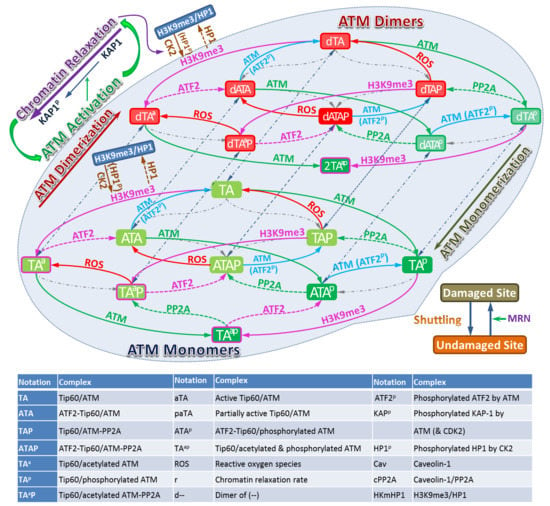
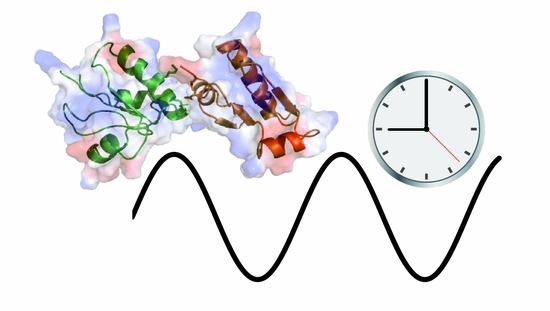
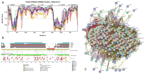
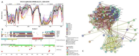
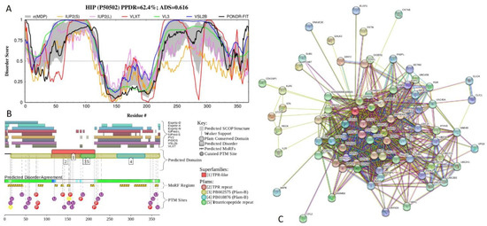
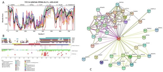
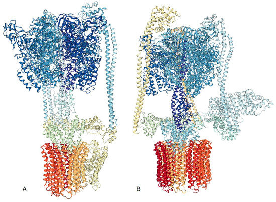
Big Bang theories are connected to gravity by force of attraction. Forced lengthening, like eccentric contractions, instigate proprioception as a result of working against gravity. Piezo2, as the principal mechanosensory ion channel responsible for proprioception, is theorized to fine-modulate these anti-gravitational contractions in
[...] Read more.
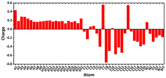
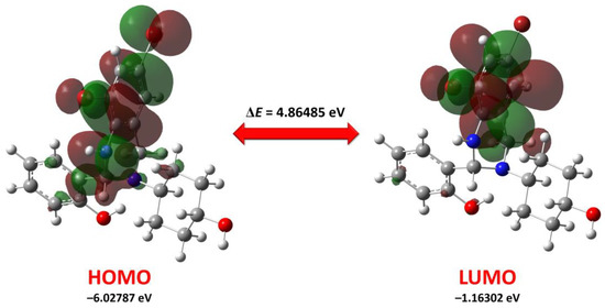
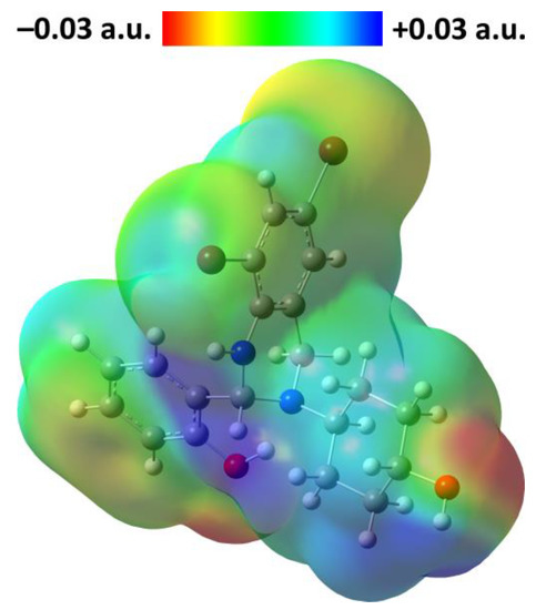
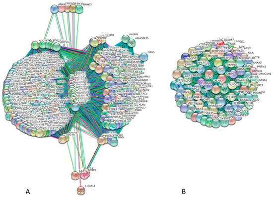
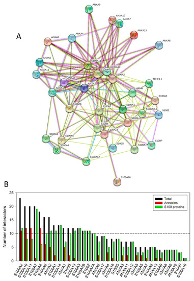
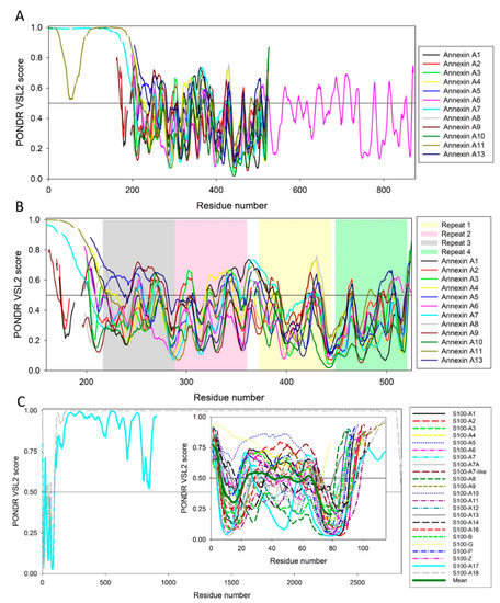
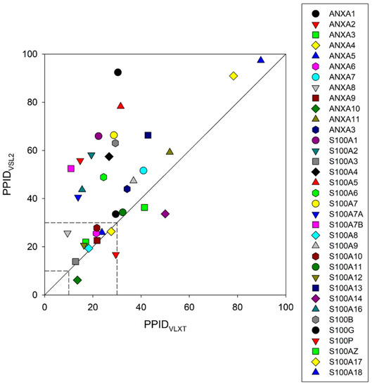
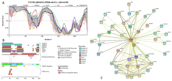
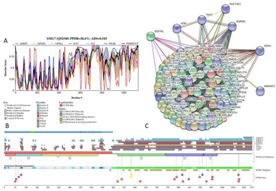
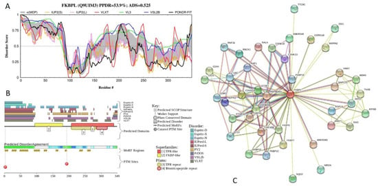
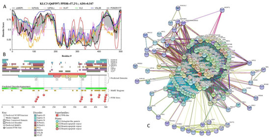
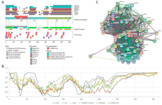
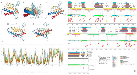
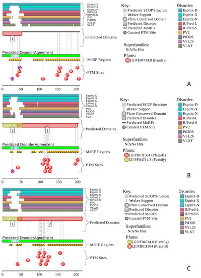
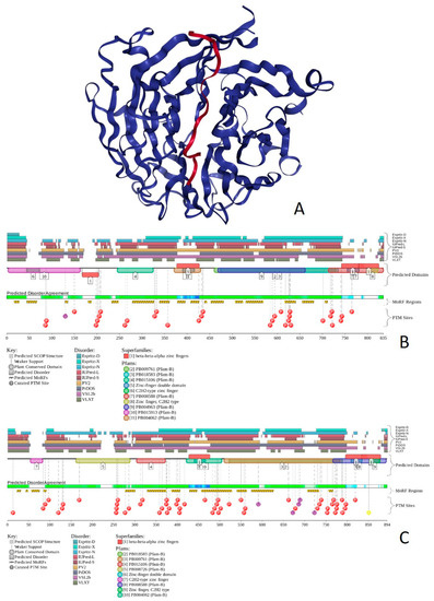
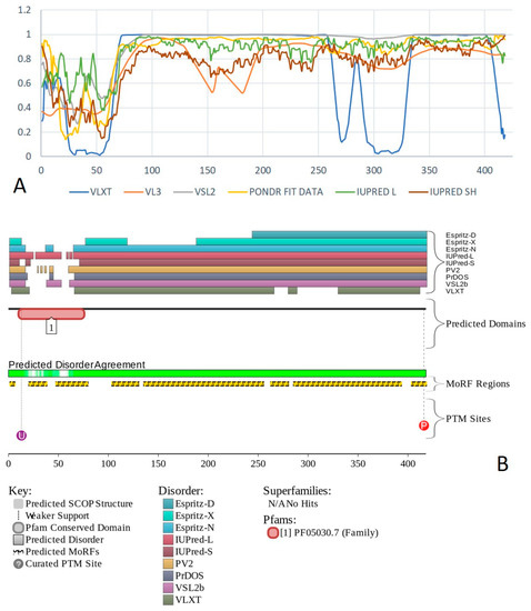
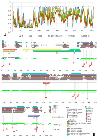
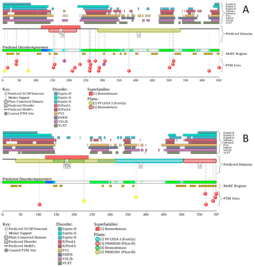
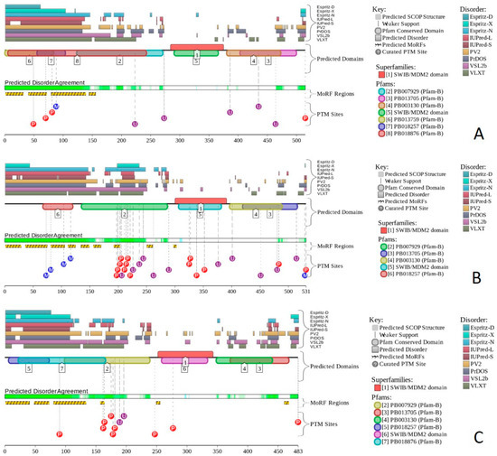
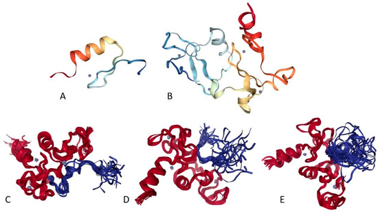
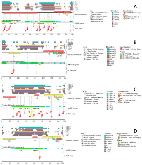
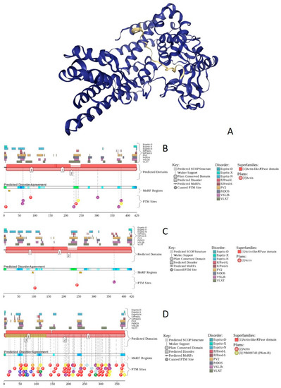
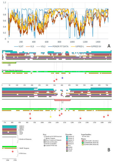
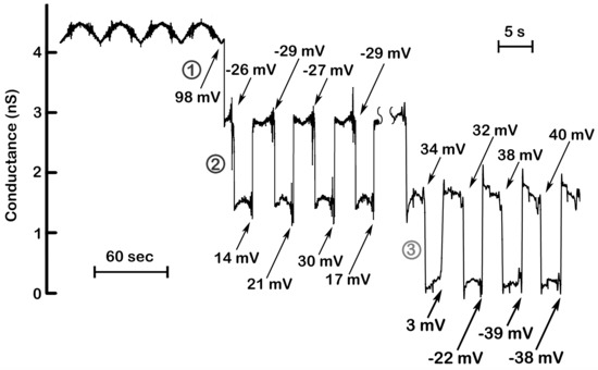
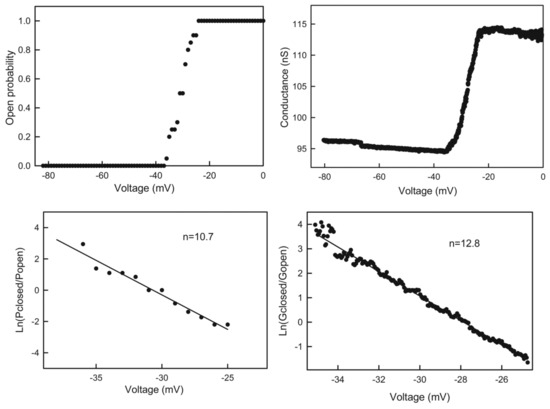
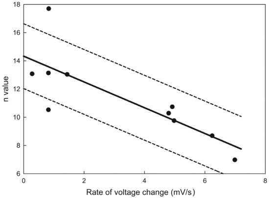
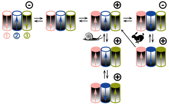
Big Bang theories are connected to gravity by force of attraction. Forced lengthening, like eccentric contractions, instigate proprioception as a result of working against gravity. Piezo2, as the principal mechanosensory ion channel responsible for proprioception, is theorized to fine-modulate these anti-gravitational contractions in order to provide system-wide ultrafast postural control. This mechanism may instantaneously emit energy and force through Piezo2 in order to offset gravity by anti-gravity entropic-spring-like stochastic mechanics and it is suggested to be propagated by quantum tunneling of protons (and electrons). However, a Piezo2-initiated wormhole-like mechanism with the contribution of cryptochromes should be considered as part of this ultrafast long-distance non-synaptic neurotransmission, although the quantum gravity concept is short of being unequivocally proven to be unified with quantum theory. The impairment of this theoretical ultrafast signaling is analogous to a Big Bang-like mechanism within a given compartment, or acquired Piezo2 channelopathy, leading to the principal gateway to pathophysiology.
Full article

Figure 1


