Topical Collection "Marine Compounds and Cancer"

A topical collection in Marine Drugs (ISSN 1660-3397). This collection belongs to the section "Marine Pharmacology".

Viewed by 587548
Printed Edition Available!
A printed edition of this Special Issue is available here.

Editors

Tumor and Breast Center ZeTuP St. Gallen, Rorschacherstr. 150, CH-9006 St. Gallen, Switzerland
Interests: medical oncology; drug resistance; marine anti-cancer compounds; drug development; tumor biology; proteomics
Special Issues, Collections and Topics in MDPI journals
1. Laboratory of Experimental Oncology, Department of Oncology, Hematology and Bone Marrow Transplantation with Section Pneumology, Hubertus Wald-Tumorzentrum, University Medical Center Hamburg-Eppendorf, Hamburg, Germany
2. Martini-Klinik, Prostate Cancer Center, University Hospital Hamburg-Eppendorf, Hamburg, Germany
Interests: marine natural compounds; secondary metabolites; anticancer activity; mechanism of action; autophagy; drug combinational studies
Special Issues, Collections and Topics in MDPI journals

Topical Collection Information

Dear Colleagues,

In 2019, the scientific and medical community celebrated the 50th anniversary of the introduction of the very first marine drug, Cytarabine (aka Ara-C, Cytosar-U®), into the clinics. In 1969, it was approved by the Food and Drug Administration (FDA) for the treatment of leukemia. Nowadays, the list of approved marine-derived anticancer drugs consists of seven medications. Many more are in all phases of clinical testing, and a plethora of substances have already been examined for in vitro and in vivo activity.

Increasingly precise research tools allow the dissection of the molecular mode of action of these cytotoxic substances, thereby uncovering the specific drug targets in cancer cells. This development will blur the edges between targeted and untargeted therapy, and will hopefully lead to a more directed use of cancer medicine based on a molecular rationale of activity in the future.

This Topical Collection will cover the whole scope from agents with cancer-preventive activity, to novel and previously characterized compounds with anti-cancer activity, both in vitro and in vivo, and the latest status of clinical development from drug trials. Notably, compounds possessing pro-carcinogenic activity or mediating cancer cell survival are also within the scope of this Topical Collection. In addition, special focus will be placed on current shortfalls and possible strategies to overcome obstacles in the area of marine anti-cancer drug development.

Dr. Sergey A. Dyshlovoy
Dr. Friedemann Honecker
Collection Editors

Manuscript Submission Information

Manuscripts should be submitted online at www.mdpi.com by registering and logging in to this website. Once you are registered, click here to go to the submission form. Manuscripts can be submitted until the deadline. All submissions that pass pre-check are peer-reviewed. Accepted papers will be published continuously in the journal (as soon as accepted) and will be listed together on the collection website. Research articles, review articles as well as short communications are invited. For planned papers, a title and short abstract (about 100 words) can be sent to the Editorial Office for announcement on this website.

Submitted manuscripts should not have been published previously, nor be under consideration for publication elsewhere (except conference proceedings papers). All manuscripts are thoroughly refereed through a single-blind peer-review process. A guide for authors and other relevant information for submission of manuscripts is available on the Instructions for Authors page. Marine Drugs is an international peer-reviewed open access monthly journal published by MDPI.

Please visit the Instructions for Authors page before submitting a manuscript. The Article Processing Charge (APC) for publication in this open access journal is 2900 CHF (Swiss Francs). Submitted papers should be well formatted and use good English. Authors may use MDPI's English editing service prior to publication or during author revisions.

Keywords

  • cancer
  • marine natural compounds and their derivatives
  • marine toxins
  • drug discovery
  • cancer-preventive activity
  • molecular effects
  • molecular targets
  • drug resistance
  • drug combination
  • toxicology
  • xenograft models

Published Papers (96 papers)

2023

Jump to: 2022, 2021, 2020, 2019, 2018, 2017, 2015, 2014, 2013

13 pages, 2939 KiB  
Article
A Novel Anticancer Peptide Derived from Bryopsis plumosa Regulates Proliferation and Invasion in Non-Small Cell Lung Cancer Cells
Mar. Drugs 2023, 21(12), 607; https://doi.org/10.3390/md21120607 - 24 Nov 2023
Viewed by 247
Abstract
The discovery of new highly effective anticancer drugs with few side effects is a challenge for drug development research. Natural or synthetic anticancer peptides (ACPs) represent a new generation of anticancer agents with high selectivity and specificity. The rapid emergence of chemoradiation-resistant lung [...] Read more.
The discovery of new highly effective anticancer drugs with few side effects is a challenge for drug development research. Natural or synthetic anticancer peptides (ACPs) represent a new generation of anticancer agents with high selectivity and specificity. The rapid emergence of chemoradiation-resistant lung cancer has necessitated the discovery of novel anticancer agents as alternatives to conventional therapeutics. In this study, we synthesized a peptide containing 22 amino acids and characterized it as a novel ACP (MP06) derived from green sea algae, Bryopsis plumosa. Using the ACP database, MP06 was predicted to possess an alpha-helical secondary structure and functionality. The anti-proliferative and apoptotic effects of the MP06, determined using the cytotoxicity assay and Annexin V/propidium iodide staining kit, were significantly higher in non-small-cell lung cancer (NSCLC) cells than in non-cancerous lung cells. We confirmed that MP06 suppressed cellular migration and invasion and inhibited the expression of N-cadherin and vimentin, the markers of epithelial–mesenchymal transition. Moreover, MP06 effectively reduced the metastasis of tumor xenografts in zebrafish embryos. In conclusion, we suggest considering MP06 as a novel candidate for the development of new anticancer drugs functioning via the ERK signaling pathway. Full article
Show Figures

Figure 1

18 pages, 2581 KiB  
Article
A New Mild Method for Synthesis of Marine Alkaloid Fascaplysin and Its Therapeutically Promising Derivatives
Mar. Drugs 2023, 21(8), 424; https://doi.org/10.3390/md21080424 - 25 Jul 2023
Viewed by 956
Abstract
Fascaplysin is a marine alkaloid which is considered to be a lead drug candidate due to its diverse and potent biological activity. As an anticancer agent, fascaplysin holds a great potential due to the multiple targets affected by this alkaloid in cancer cells, [...] Read more.
Fascaplysin is a marine alkaloid which is considered to be a lead drug candidate due to its diverse and potent biological activity. As an anticancer agent, fascaplysin holds a great potential due to the multiple targets affected by this alkaloid in cancer cells, including inhibition of cyclin-dependent kinase 4 (CDK4) and induction of intrinsic apoptosis. At the same time, the studies on structural optimization are hampered by its rather high toxicity, mainly caused by DNA intercalation. In addition, the number of methods for the syntheses of its derivatives is limited. In the current study, we report a new two-step method of synthesis of fascaplysin derivatives based on low temperature UV quaternization for the synthesis of thermolabile 9-benzyloxyfascaplysin and 6-tert-butylfascaplysin. 9-Benzyloxyfascaplysin was used as the starting compound to obtain 9-hydroxyfascaplysin. However, the latter was found to be chemically highly unstable. 6-tert-Butylfascaplysin revealed a significant decrease in DNA intercalation when compared to fascaplysin, while cytotoxicity was only slightly reduced. Therefore, the impact of DNA intercalation for the cytotoxic effects of fascaplysin and its derivatives needs to be questioned. Full article
Show Figures

Graphical abstract

13 pages, 3856 KiB  
Article
RIP1 Mediates Manzamine-A-Induced Secretory Autophagy in Breast Cancer
Mar. Drugs 2023, 21(3), 151; https://doi.org/10.3390/md21030151 - 25 Feb 2023
Viewed by 1209
Abstract
Cancer-derived small extracellular vesicles (sEVs) serve as critical mediators of cell-to-cell communication. Manzamine A (MA), a unique marine-derived alkaloid with various bioactivities, exerts anticancer effects against several kinds of tumors, but it remains unclear whether it has the same activity against breast cancer. [...] Read more.
Cancer-derived small extracellular vesicles (sEVs) serve as critical mediators of cell-to-cell communication. Manzamine A (MA), a unique marine-derived alkaloid with various bioactivities, exerts anticancer effects against several kinds of tumors, but it remains unclear whether it has the same activity against breast cancer. Here, we proved that MA inhibits MDA-MB-231 and MCF-7 cell proliferation, migration, and invasion in a time- and dose-dependent manner. In addition, MA promotes autophagosome formation but suppresses autophagosome degradation in breast cancer cells. Importantly, we also found that MA stimulates sEVs secretion and increases autophagy-related protein accumulation in secreted sEVs, further potentiated by autophagy inhibitor chloroquine (CQ). Mechanistically, MA decreases the expression level of RIP1, the key upstream regulator of the autophagic pathway, and reduces the acidity of lysosome. Overexpression of RIP1 activated AKT/mTOR signaling, thus attenuating MA-induced autophagy and the corresponding secretion of autophagy-associated sEVs. Collectively, these data suggested that MA is a potential inhibitor of autophagy by preventing autophagosome turnover, and RIP1 mediates MA-induced secretory autophagy, which may be efficacious for breast cancer treatment. Full article
Show Figures

Graphical abstract

11 pages, 1934 KiB  
Article
New Marine Fungal Deoxy-14,15-Dehydroisoaustamide Resensitizes Prostate Cancer Cells to Enzalutamide
Mar. Drugs 2023, 21(1), 54; https://doi.org/10.3390/md21010054 - 14 Jan 2023
Cited by 1 | Viewed by 2414
Abstract
Marine fungi serve as a valuable source for new bioactive molecules bearing various biological activities. In this study, we report on the isolation of a new indole diketopiperazine alkaloid deoxy-14,15-dehydroisoaustamide (1) from the marine-derived fungus Penicillium dimorphosporum KMM 4689 associated with [...] Read more.
Marine fungi serve as a valuable source for new bioactive molecules bearing various biological activities. In this study, we report on the isolation of a new indole diketopiperazine alkaloid deoxy-14,15-dehydroisoaustamide (1) from the marine-derived fungus Penicillium dimorphosporum KMM 4689 associated with a soft coral. The structure of this metabolite, including its absolute configuration, was determined by HR-MS, 1D and 2D NMR as well as CD data. Compound 1 is a very first deoxyisoaustamide alkaloid possessing two double bonds in the proline ring. The isolated compound was noncytotoxic to a panel of human normal and cancer cell lines up to 100 µM. At the same time, compound 1 resensitized prostate cancer 22Rv1 cells to androgen receptor (AR) blocker enzalutamide. The mechanism of this phenomenon was identified as specific drug-induced degradation of androgen receptor transcription variant V7 (AR-V7), which also resulted in general suppression of AR signaling. Our data suggest that the isolated alkaloid is a promising candidate for combinational therapy of castration resistant prostate cancer, including drug-resistant subtypes. Full article
Show Figures

Figure 1

2022

Jump to: 2023, 2021, 2020, 2019, 2018, 2017, 2015, 2014, 2013

4 pages, 583 KiB  
Editorial
Marine Compounds and Cancer: Updates 2022
Mar. Drugs 2022, 20(12), 759; https://doi.org/10.3390/md20120759 - 01 Dec 2022
Cited by 3 | Viewed by 1415
Abstract
The field of marine bioactive compounds (marine drugs) has evolved significantly in recent years [...] Full article
Show Figures

Figure 1

15 pages, 1909 KiB  
Article
κ- and λ-Carrageenans from Marine Alga Chondrus armatus Exhibit Anticancer In Vitro Activity in Human Gastrointestinal Cancers Models
Mar. Drugs 2022, 20(12), 741; https://doi.org/10.3390/md20120741 - 25 Nov 2022
Cited by 6 | Viewed by 1305
Abstract
The carrageenans isolated from red algae demonstrated a variety of activities from antiviral and immunomodulatory to antitumor. The diverse structure and sulfation profile of carrageenans provide a great landscape for drug development. In this study, we isolated, purified and structurally characterized κo- and [...] Read more.
The carrageenans isolated from red algae demonstrated a variety of activities from antiviral and immunomodulatory to antitumor. The diverse structure and sulfation profile of carrageenans provide a great landscape for drug development. In this study, we isolated, purified and structurally characterized κo- and λo- oligosaccharides from the marine algae Chondrus armatus. We further examined the tumor suppressive activity of both carrageenans in gastrointestinal cancer models. Thus, using MTT assay, we could demonstrate a pronounced antiproliferative effect of the carrageenans in KYSE-30 and FLO-1 as well as HCT-116 and RKO cell lines with IC50 184~405 μg/mL, while both compounds were less active in non-cancer epithelial cells RPE-1. This effect was stipulated by the inhibition of cell cycle progression in the cancer cells. Specifically, flow cytometry revealed an S phase delay in FLO-1 and HCT-116 cells under κo-carrageenan treatment, while KYSE-30 demonstrated a pronounced G2/M cell cycle delay. In line with this, western blotting revealed a reduction of cell cycle markers CDK2 and E2F2. Interestingly, κo-carrageenan inhibited cell cycle progression of RKO cells in G1 phase. Finally, isolated κo- and λo- carrageenans induced apoptosis on adenocarcinomas, specifically with high apoptosis induction in RKO cells. Overall, our data underline the potential of κo- and λo- carrageenans for colon and esophageal carcinoma drug development. Full article
Show Figures

Figure 1

18 pages, 5567 KiB  
Article
Cytotoxic N-Methylpretrichodermamide B Reveals Anticancer Activity and Inhibits P-Glycoprotein in Drug-Resistant Prostate Cancer Cells
Mar. Drugs 2022, 20(10), 597; https://doi.org/10.3390/md20100597 - 23 Sep 2022
Cited by 3 | Viewed by 1435
Abstract
N-methylpretrichodermamide B (NB) is a biologically active epidithiodiketopiperazine isolated from several strains of the algae-derived fungus Penicillium sp. Recently, we reported the first data on its activity in human cancer cells lines in vitro. Here, we investigated the activity, selectivity, and mechanism of [...] Read more.
N-methylpretrichodermamide B (NB) is a biologically active epidithiodiketopiperazine isolated from several strains of the algae-derived fungus Penicillium sp. Recently, we reported the first data on its activity in human cancer cells lines in vitro. Here, we investigated the activity, selectivity, and mechanism of action of NB in human prostate cancer cell lines, including drug-resistant subtypes. NB did not reveal cross-resistance to docetaxel in the PC3-DR cell line model and was highly active in hormone-independent 22Rv1 cells. NB-induced cell death was stipulated by externalization of phosphatidylserine and activation of caspase-3. Moreover, inhibition of caspase activity by z-VAD(OMe)-fmk did not affect NB cytotoxicity, suggesting a caspase-independent cell death induced by NB. The compound has a moderate p-glycoprotein (p-gp) substrate-like affinity and can simultaneously inhibit p-gp at nanomolar concentrations. Therefore, NB resensitized p-gp-overexpressing PC3-DR cells to docetaxel. A kinome profiling of the NB-treated cells revealed, among other things, an induction of mitogen-activated protein kinases JNK1/2 and p38. Further functional analysis confirmed an activation of both kinases and indicated a prosurvival role of this biological event in the cellular response to the treatment. Overall, NB holds promising anticancer potential and further structure–activity relationship studies and structural optimization are needed in order to improve its biological properties. Full article
Show Figures

Figure 1

9 pages, 607 KiB  
Article
Salmon Protein Hydrolysate Potentiates the Growth Inhibitory Effect of Bicalutamide on Human Prostate Cancer Cell Lines LNCaP and PC3 by Modulating Iron Homeostasis
Mar. Drugs 2022, 20(4), 228; https://doi.org/10.3390/md20040228 - 28 Mar 2022
Cited by 3 | Viewed by 2111
Abstract
Prostate cancer is a common cause of cancer death in men. In advanced stages of prostate cancer, androgen deprivation therapy (ADT) is initiated. Despite ADT, prostate cancers invariably progress to become androgen independent. A growing body of evidence implicates iron dysmetabolism in prostate [...] Read more.
Prostate cancer is a common cause of cancer death in men. In advanced stages of prostate cancer, androgen deprivation therapy (ADT) is initiated. Despite ADT, prostate cancers invariably progress to become androgen independent. A growing body of evidence implicates iron dysmetabolism in prostate cancer progression. A bioactive peptide-rich salmon protein hydrolysate (SPH) has previously been demonstrated to modulate iron homeostatic mechanisms. In the present study, the anticancer effect of SPH and bicalutamide co-treatment on LNCaP and PC3 prostate cancer cell proliferation was investigated. Our results found that SPH potentiates the anti-proliferative effect of bicalutamide in a dose-dependent manner for both cell lines. In the presence of 160 µg/mL SPH, co-treatment with 1.0 µM bicalutamide decreased LNCaP cells’ relative colony survival from 25% (1.0 µM bicalutamide monotreatment) to 2% after culturing for 12 days. For PC3 cells, the relative colony survival diminished from 52% (10.0 µM bicalutamide) to 32% at an SPH concentration of 160 µg/mL. Gene expression profiling, employing quantitative real-time PCR, revealed that the inhibitory effects were related to significant FTH1 up-regulation with a concomitant TFRC down-regulation. In conclusion, our results provide in vitro evidence that SPH potentiates the growth inhibitory effect of bicalutamide on prostate cancer cells by modulating iron homeostasis mechanisms. Full article
Show Figures

Figure 1

2021

Jump to: 2023, 2022, 2020, 2019, 2018, 2017, 2015, 2014, 2013

13 pages, 3681 KiB  
Article
Actinomycin V Induces Apoptosis Associated with Mitochondrial and PI3K/AKT Pathways in Human CRC Cells
Mar. Drugs 2021, 19(11), 599; https://doi.org/10.3390/md19110599 - 22 Oct 2021
Cited by 8 | Viewed by 1938
Abstract
Actinomycin (Act) V, an analogue of Act D, presented stronger antitumor activity and less hepatorenal toxicity than Act D in our previous studies, which is worthy of further investigation. We hereby report that Act V induces apoptosis via mitochondrial and PI3K/AKT pathways in [...] Read more.
Actinomycin (Act) V, an analogue of Act D, presented stronger antitumor activity and less hepatorenal toxicity than Act D in our previous studies, which is worthy of further investigation. We hereby report that Act V induces apoptosis via mitochondrial and PI3K/AKT pathways in colorectal cancer (CRC) cells. Act V-induced apoptosis was characterized by mitochondrial dysfunction, with loss of mitochondria membrane potential (MMP) and cytochrome c release, which then activated cleaved caspase-9, cleaved caspase-3, and cleaved PARP, revealing that it was related to the mitochondrial pathway, and the apoptotic trendency can be reversed by caspase inhibitor Z-VAD-FMK. Furthermore, we proved that Act V significantly inhibited PI3K/AKT signalling in HCT-116 cells using cell experiments in vitro, and it also presented a potential targeted PI3Kα inhibition using computer docking models. Further elucidation revealed that it exhibited a 28-fold greater potency than the PI3K inhibitor LY294002 on PI3K inhibition efficacy. Taken together, Act V, as a superior potential replacement of Act D, is a potential candidate for inhibiting the PI3K/AKT pathway and is worthy of more pre-clinical studies in the therapy of CRC. Full article
Show Figures

Graphical abstract

18 pages, 1726 KiB  
Review
Recent Updates on Marine Cancer-Preventive Compounds
Mar. Drugs 2021, 19(10), 558; https://doi.org/10.3390/md19100558 - 29 Sep 2021
Cited by 17 | Viewed by 2233
Abstract
The natural compounds derived from marine organisms often exhibit unique chemical structures and potent biological activities. Cancer-preventive activity is one of the rather new activities that has emerged and been extensively studied over the last decades. This review summarizes the recent updates on [...] Read more.
The natural compounds derived from marine organisms often exhibit unique chemical structures and potent biological activities. Cancer-preventive activity is one of the rather new activities that has emerged and been extensively studied over the last decades. This review summarizes the recent updates on the marine chemopreventive compounds covering the relevant literature published in 2013–2021 and following the previous comprehensive review by Stonik and Fedorov (Marine Drugs 2014, 12, 636–671). In the current article, only the molecules having an effect on malignant transformation (or related pathway and molecules), cancer stem cells, or carcinogen-induced in vivo tumor development were considered to be “true” cancer-preventive compounds and were, therefore, reviewed. Additionally, particular attention has been given to the molecular mechanisms of chemoprevention, executed by the reported marine compounds. Full article
Show Figures

Figure 1

17 pages, 2663 KiB  
Article
Cytotoxic Marine Alkaloid 3,10-Dibromofascaplysin Induces Apoptosis and Synergizes with Cytarabine Resulting in Leukemia Cell Death
Mar. Drugs 2021, 19(9), 489; https://doi.org/10.3390/md19090489 - 27 Aug 2021
Cited by 5 | Viewed by 2386
Abstract
Myeloid leukemia is a hematologic neoplasia characterized by a clonal proliferation of hematopoietic stem cell progenitors. Patient prognosis varies depending on the subtype of leukemia as well as eligibility for intensive treatment regimens and allogeneic stem cell transplantation. Although significant progress has been [...] Read more.
Myeloid leukemia is a hematologic neoplasia characterized by a clonal proliferation of hematopoietic stem cell progenitors. Patient prognosis varies depending on the subtype of leukemia as well as eligibility for intensive treatment regimens and allogeneic stem cell transplantation. Although significant progress has been made in the therapy of patients including novel targeted treatment approaches, there is still an urgent need to optimize treatment outcome. The most common therapy is based on the use of chemotherapeutics cytarabine and anthrayclines. Here, we studied the effect of the recently synthesized marine alkaloid 3,10-dibromofascaplysin (DBF) in myeloid leukemia cells. Unsubstituted fascaplysin was early found to affect cell cycle via inhibiting CDK4/6, thus we compared the activity of DBF and other brominated derivatives with known CDK4/6 inhibitor palbociclib, which was earlier shown to be a promising candidate to treat leukemia. Unexpectedly, the effect DBF on cell cycle differs from palbociclib. In fact, DBF induced leukemic cells apoptosis and decreased the expression of genes responsible for cancer cell survival. Simultaneously, DBF was found to activate the E2F1 transcription factor. Using bioinformatical approaches we evaluated the possible molecular mechanisms, which may be associated with DBF-induced activation of E2F1. Finally, we found that DBF synergistically increase the cytotoxic effect of cytarabine in different myeloid leukemia cell lines. In conclusion, DBF is a promising drug candidate, which may be used in combinational therapeutics approaches to reduce leukemia cell growth. Full article
Show Figures

Figure 1

17 pages, 3825 KiB  
Article
PNSA, a Novel C-Terminal Inhibitor of HSP90, Reverses Epithelial–Mesenchymal Transition and Suppresses Metastasis of Breast Cancer Cells In Vitro
Mar. Drugs 2021, 19(2), 117; https://doi.org/10.3390/md19020117 - 20 Feb 2021
Cited by 6 | Viewed by 2838
Abstract
Metastasis accounts for the vast majority of deaths in breast cancer, and novel and effective treatments to inhibit cancer metastasis remain urgently developed. The expression level of heat shock protein 90 (HSP90) in invasive breast cancer tissue is higher than in adjacent non-cancerous [...] Read more.
Metastasis accounts for the vast majority of deaths in breast cancer, and novel and effective treatments to inhibit cancer metastasis remain urgently developed. The expression level of heat shock protein 90 (HSP90) in invasive breast cancer tissue is higher than in adjacent non-cancerous tissue. In the present study, we investigated the inhibitory effect of penisuloxazin A (PNSA), a novel C- terminal inhibitor of HSP90, on metastasis of breast cancer cells and related mechanism in vitro. We found that PNSA obviously affected adhesion, migration, and invasion of triple-negative breast cancer (TNBC) MDA-MB-231 cells and Trastuzumab-resistant JIMT-1 cells. Furthermore, PNSA was capable of reversing epithelial–mesenchymal transformation (EMT) of MDA-MB-231 cells with change of cell morphology. PNSA increases E-cadherin expression followed by decreasing amounts of N-cadherin, vimentin, and matrix metalloproteinases9 (MMP9) and proteolytic activity of matrix metalloproteinases2 (MMP2) and MMP9. Comparatively, the N-terminal inhibitor of HSP90 17-allyl-17-demethoxygeldanamycin (17-AAG) had no effect on EMT of MDA-MB-231 cells. PNSA was uncovered to reduce the stability of epidermal growth factor receptor (EGFR) and fibroblast growth factor receptor (FGFR) proteins and thereby inhibiting their downstream signaling transductions by inhibition of HSP90. In addition, PNSA reduced the expression of programmed cell death-ligand 1 (PD-L1) to promote natural killer (NK) cells to kill breast cancer cells with a dose far less than that of cytotoxicity to NK cell itself, implying the potential of PNSA to enhance immune surveillance against metastasis in vivo. All these results indicate that PNSA is a promising anti-metastasis agent worthy of being studied in the future. Full article
Show Figures

Figure 1

17 pages, 6034 KiB  
Article
Marine Seagrass Extract of Thalassia testudinum Suppresses Colorectal Tumor Growth, Motility and Angiogenesis by Autophagic Stress and Immunogenic Cell Death Pathways
Mar. Drugs 2021, 19(2), 52; https://doi.org/10.3390/md19020052 - 22 Jan 2021
Cited by 11 | Viewed by 2961
Abstract
Marine plants have become an inexhaustible reservoir of new phytopharmaceuticals for cancer treatment. We demonstrate in vitro/in vivo antitumor efficacy of a standardized polyphenol extract from the marine angiosperm Thalassia testudinum (TTE) in colon tumor cell lines (RKO, SW480, and CT26) and a [...] Read more.
Marine plants have become an inexhaustible reservoir of new phytopharmaceuticals for cancer treatment. We demonstrate in vitro/in vivo antitumor efficacy of a standardized polyphenol extract from the marine angiosperm Thalassia testudinum (TTE) in colon tumor cell lines (RKO, SW480, and CT26) and a syngeneic allograft murine colorectal cancer model. MTT assays revealed a dose-dependent decrease of cell viability of RKO, CT26, and SW480 cells upon TTE treatment with IC50 values of, respectively, 175, 115, and 60 μg/mL. Furthermore, TTE significantly prevented basal and bFGF-induced angiogenesis in the chicken chorioallantoic membrane angiogenesis assay. In addition, TTE suppressed bFGF-induced migration of endothelial cells in a wound closure assay. Finally, TTE treatment abrogated CT26 colorectal cancer growth and increased overall organism survival in a syngeneic murine allograft model. Corresponding transcriptome profiling and pathway analysis allowed for the identification of the mechanism of action for the antitumor effects of TTE. In line with our in vitro/in vivo results, TTE treatment triggers ATF4-P53-NFκB specific gene expression and autophagy stress pathways. This results in suppression of colon cancer cell growth, cell motility, and angiogenesis pathways in vitro and in addition promotes antitumor immunogenic cell death in vivo. Full article
Show Figures

Graphical abstract

12 pages, 2306 KiB  
Article
In Vitro Chemopreventive Potential of Phlorotannins-Rich Extract from Brown Algae by Inhibition of Benzo[a]pyrene-Induced P2X7 Activation and Toxic Effects
Mar. Drugs 2021, 19(1), 34; https://doi.org/10.3390/md19010034 - 14 Jan 2021
Cited by 11 | Viewed by 2080
Abstract
Phlorotannins are polyphenols occurring exclusively in some species of brown algae, known for numerous biological activities, e.g., antioxidant, antiproliferative, antidiabetic, and antiallergic properties. Their effects on the response of human lung cells to benzo[a]pyrene (B[a]P) has not been characterized. Our objective was to [...] Read more.
Phlorotannins are polyphenols occurring exclusively in some species of brown algae, known for numerous biological activities, e.g., antioxidant, antiproliferative, antidiabetic, and antiallergic properties. Their effects on the response of human lung cells to benzo[a]pyrene (B[a]P) has not been characterized. Our objective was to in vitro evaluate the effects of a phlorotannin-rich extract obtained from the brown algae Ascophyllum nodosum and Fucus vesiculosus on B[a]P cytotoxic effects. The A549 cell line was incubated with B[a]P for 48 and 72 h in the presence or absence of the brown algae extract. Cytochrome P450 activity, activation of P2X7 receptor, F-actin disorganization, and loss of E-cadherin expression were assessed using microplate cytometry and fluorescence microscopy. Relative to control, incubation with the brown algae extract was associated with lower B[a]P-induced CYP1 activity, lower P2X7 receptor activation, and lower reactive oxygen species production. The brown algae extract inhibited the alterations of F-actin arrangement and the downregulation of E-cadherin expression. We identified a phlorotannins-rich extract that could be deeper investigated as a cancer chemopreventive agent to block B[a]P-mediated carcinogenesis. Full article
Show Figures

Figure 1

2020

Jump to: 2023, 2022, 2021, 2019, 2018, 2017, 2015, 2014, 2013

6 pages, 206 KiB  
Editorial
Marine Compounds and Cancer: Updates 2020
Mar. Drugs 2020, 18(12), 643; https://doi.org/10.3390/md18120643 - 15 Dec 2020
Cited by 24 | Viewed by 2970
Abstract
By the end of the year 2020, there are nine marine-derived anticancer drugs available on the market, and the field is currently growing exponentially [...] Full article
17 pages, 3335 KiB  
Article
Efficacy and Mechanism of Action of Marine Alkaloid 3,10-Dibromofascaplysin in Drug-Resistant Prostate Cancer Cells
Mar. Drugs 2020, 18(12), 609; https://doi.org/10.3390/md18120609 - 01 Dec 2020
Cited by 13 | Viewed by 6061
Abstract
Efficacy and mechanism of action of marine alkaloid 3,10-dibromofascaplysin (DBF) were investigated in human prostate cancer (PCa) cells harboring different levels of drug resistance. Anticancer activity was observed across all cell lines examined without signs of cross-resistance to androgen receptor targeting agents (ARTA) [...] Read more.
Efficacy and mechanism of action of marine alkaloid 3,10-dibromofascaplysin (DBF) were investigated in human prostate cancer (PCa) cells harboring different levels of drug resistance. Anticancer activity was observed across all cell lines examined without signs of cross-resistance to androgen receptor targeting agents (ARTA) or taxane based chemotherapy. Kinome analysis followed by functional investigation identified JNK1/2 to be one of the molecular targets of DBF in 22Rv1 cells. In contrast, no activation of p38 and ERK1/2 MAPKs was observed. Inhibition of the drug-induced JNK1/2 activation or of the basal p38 activity resulted in increased cytotoxicity of DBF, whereas an active ERK1/2 was identified to be important for anticancer activity of the alkaloid. Synergistic effects of DBF were observed in combination with PARP-inhibitor olaparib most likely due to the induction of ROS production by the marine alkaloid. In addition, DBF intensified effects of platinum-based drugs cisplatin and carboplatin, and taxane derivatives docetaxel and cabazitaxel. Finally, DBF inhibited AR-signaling and resensitized AR-V7-positive 22Rv1 prostate cancer cells to enzalutamide, presumably due to AR-V7 down-regulation. These findings propose DBF to be a promising novel drug candidate for the treatment of human PCa regardless of resistance to standard therapy. Full article
Show Figures

Figure 1

15 pages, 1114 KiB  
Article
Interaction of Thalassia testudinum Metabolites with Cytochrome P450 Enzymes and Its Effects on Benzo(a)pyrene-Induced Mutagenicity
Mar. Drugs 2020, 18(11), 566; https://doi.org/10.3390/md18110566 - 19 Nov 2020
Cited by 5 | Viewed by 2110
Abstract
The aim of the present work was to evaluate the effects of Thalassia testudinum hydroethanolic extract, its polyphenolic fraction and thalassiolin B on the activity of phase I metabolizing enzymes as well as their antimutagenic effects. Spectrofluorometric techniques were used to evaluate the [...] Read more.
The aim of the present work was to evaluate the effects of Thalassia testudinum hydroethanolic extract, its polyphenolic fraction and thalassiolin B on the activity of phase I metabolizing enzymes as well as their antimutagenic effects. Spectrofluorometric techniques were used to evaluate the effect of tested products on rat and human CYP1A and CYP2B activity. The antimutagenic effect of tested products was evaluated in benzo[a]pyrene (BP)-induced mutagenicity assay by an Ames test. Finally, the antimutagenic effect of Thalassia testudinum (100 mg/kg) was assessed in BP-induced mutagenesis in mice. The tested products significantly (p < 0.05) inhibit rat CYP1A1 activity, acting as mixed-type inhibitors of rat CYP1A1 (Ki = 54.16 ± 9.09 μg/mL, 5.96 ± 1.55 μg/mL and 3.05 ± 0.89 μg/mL, respectively). Inhibition of human CYP1A1 was also observed (Ki = 197.1 ± 63.40 μg/mL and 203.10 ± 17.29 μg/mL for the polyphenolic fraction and for thalassiolin B, respectively). In addition, the evaluated products significantly inhibit (p < 0.05) BP-induced mutagenicity in vitro. Furthermore, oral doses of Thalassia testudinum (100 mg/kg) significantly reduced (p < 0.05) the BP-induced micronuclei and oxidative damage, together with an increase of reduced glutathione, in mice. In summary, Thalassia testudinum metabolites exhibit antigenotoxic activity mediated, at least, by the inhibition of CYP1A1-mediated BP biotransformation, arresting the oxidative and mutagenic damage. Thus, the metabolites of T. testudinum may represent a potential source of chemopreventive compounds for the adjuvant therapy of cancer. Full article
Show Figures

Figure 1

12 pages, 1255 KiB  
Article
Gracilosulfates A–G, Monosulfated Polyoxygenated Steroids from the Marine Sponge Haliclona gracilis
Mar. Drugs 2020, 18(9), 454; https://doi.org/10.3390/md18090454 - 30 Aug 2020
Cited by 13 | Viewed by 2620
Abstract
Seven new polyoxygenated steroids belonging to a new structural group of sponge steroids, gracilosulfates A–G (17), possessing 3β-O-sulfonato, 5β,6β epoxy (or 5(6)-dehydro), and 4β,23-dihydroxy substitution patterns as a common structural [...] Read more.
Seven new polyoxygenated steroids belonging to a new structural group of sponge steroids, gracilosulfates A–G (17), possessing 3β-O-sulfonato, 5β,6β epoxy (or 5(6)-dehydro), and 4β,23-dihydroxy substitution patterns as a common structural motif, were isolated from the marine sponge Haliclona gracilis. Their structures were determined by NMR and MS methods. The compounds 1, 2, 4, 6, and 7 inhibited the expression of prostate-specific antigen (PSA) in 22Rv1 tumor cells. Full article
Show Figures

Graphical abstract

18 pages, 8509 KiB  
Article
Antiproliferative Activity of Mycalin A and Its Analogues on Human Skin Melanoma and Human Cervical Cancer Cells
Mar. Drugs 2020, 18(8), 402; https://doi.org/10.3390/md18080402 - 29 Jul 2020
Cited by 4 | Viewed by 1977
Abstract
Mycalin A, a polybrominated C15 acetogenin isolated from the encrusting sponge Mycale rotalis, displays an antiproliferative activity on human melanoma (A375) and cervical adenocarcinoma (HeLa) cells and induces cell death by an apoptotic mechanism. Various analogues and degraded derivatives of the [...] Read more.
Mycalin A, a polybrominated C15 acetogenin isolated from the encrusting sponge Mycale rotalis, displays an antiproliferative activity on human melanoma (A375) and cervical adenocarcinoma (HeLa) cells and induces cell death by an apoptotic mechanism. Various analogues and degraded derivatives of the natural substance have been prepared. A modification of the left-hand part of the molecule generates the most active substances. A structurally simplified lactone derivative of mycalin A, lacking the C1–C3 side chain, is the most active among the synthesized compounds exhibiting a strong cytotoxicity on both A375 and HeLa cells but not but not on human dermal fibroblast (HDF) used as healthy cells. Further evidence on a recently discovered chlorochromateperiodate-catalyzed process, used to oxidise mycalin A, have been collected. Full article
Show Figures

Graphical abstract

13 pages, 2123 KiB  
Article
12-Deacetyl-12-epi-Scalaradial, a Scalarane Sesterterpenoid from a Marine Sponge Hippospongia sp., Induces HeLa Cells Apoptosis via MAPK/ERK Pathway and Modulates Nuclear Receptor Nur77
Mar. Drugs 2020, 18(7), 375; https://doi.org/10.3390/md18070375 - 21 Jul 2020
Cited by 17 | Viewed by 3197
Abstract
12-Deacetyl-12-epi-scalaradial, a scalarane sesterterpenoid from a marine sponge Hippospongia sp, has been reported to possess cytotoxic activity on HepG2, MCF-7, and HCT-116 cells. However, there is no research to indicate that 12-deacetyl-12-epi-scalaradial exhibited anticancer effect on cervical cancer [...] Read more.
12-Deacetyl-12-epi-scalaradial, a scalarane sesterterpenoid from a marine sponge Hippospongia sp, has been reported to possess cytotoxic activity on HepG2, MCF-7, and HCT-116 cells. However, there is no research to indicate that 12-deacetyl-12-epi-scalaradial exhibited anticancer effect on cervical cancer HeLa cells. 
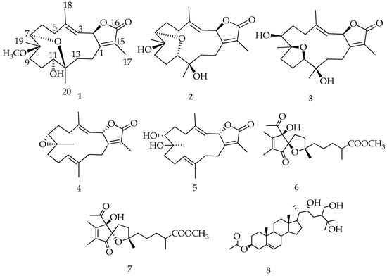
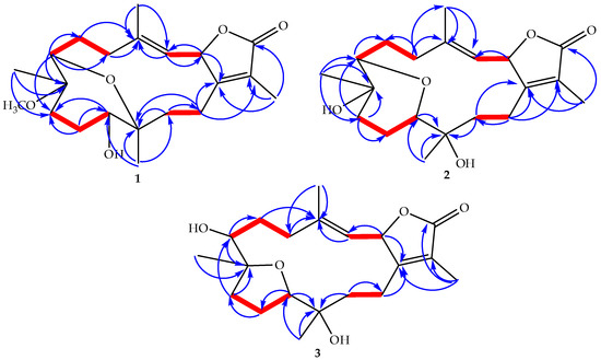
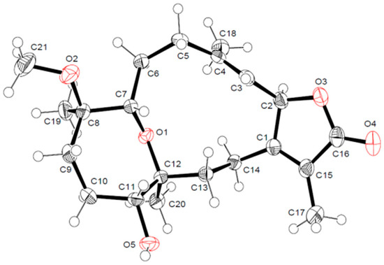
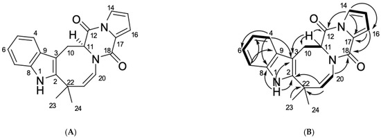
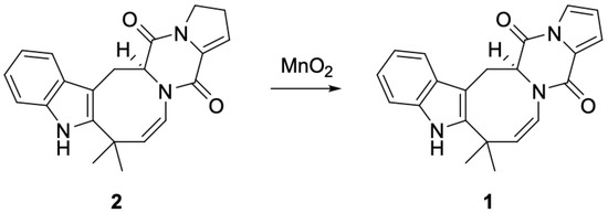
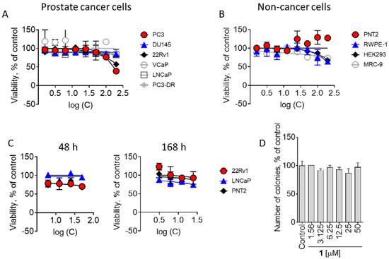
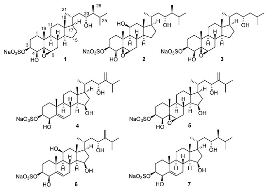
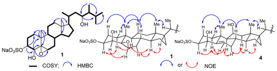
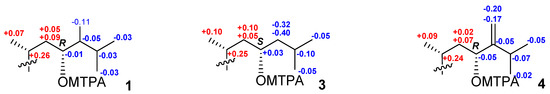
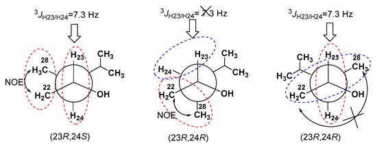
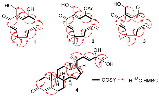
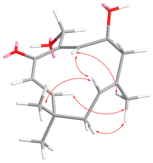
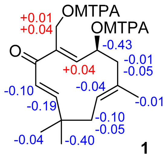
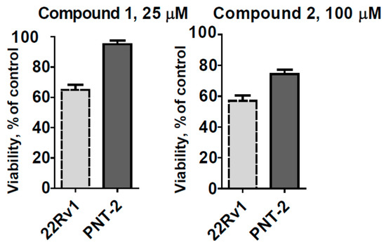
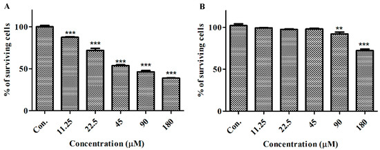
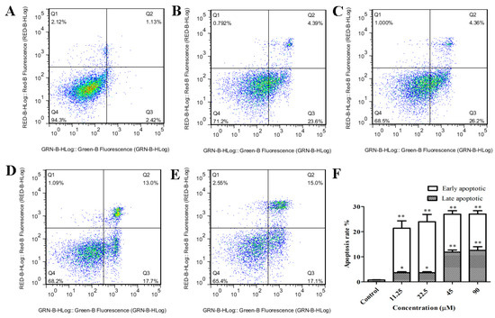
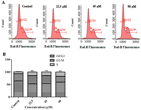
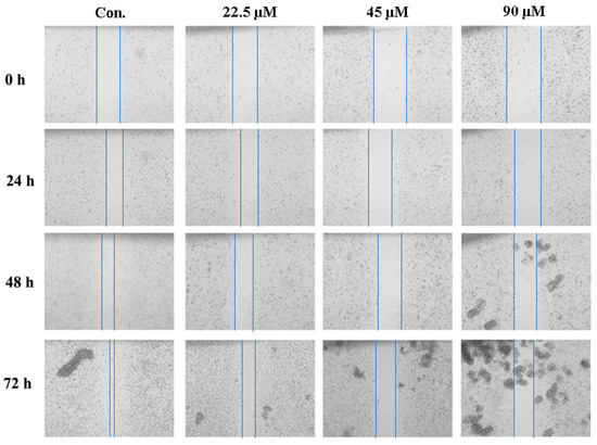
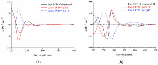
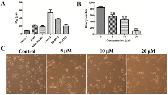
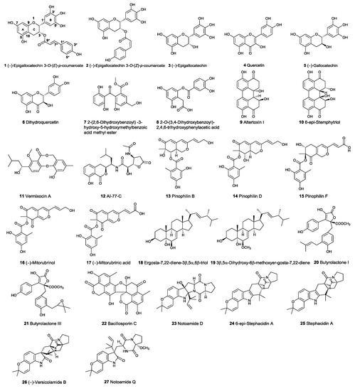
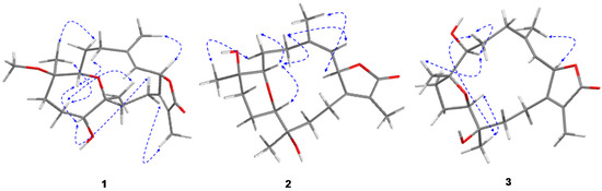
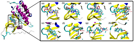
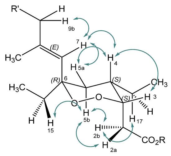
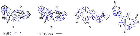
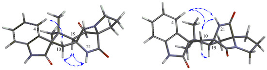
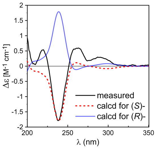
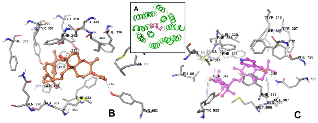
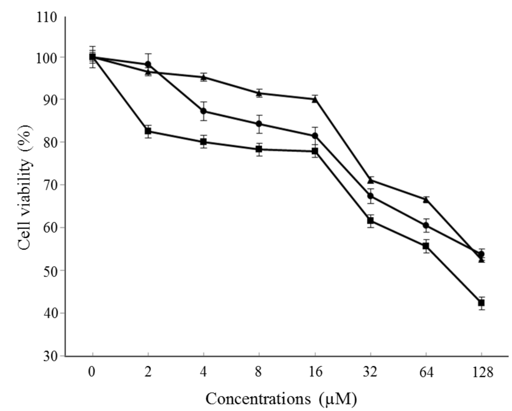
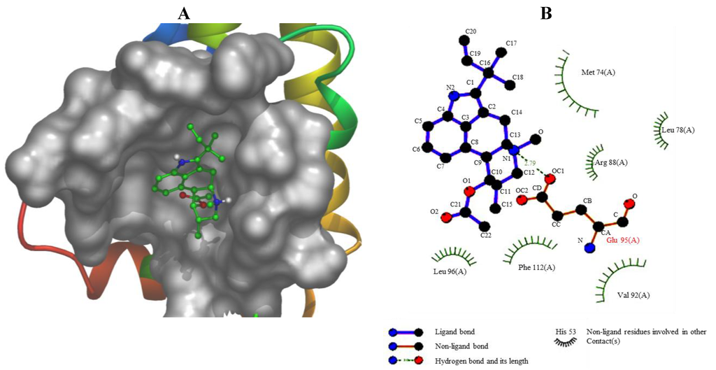
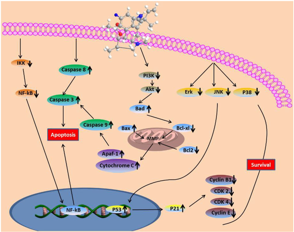
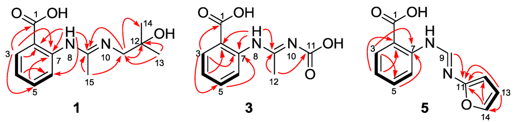
The aim of this study was to investigate the anticancer activity of 12-deacetyl-12-epi-scalaradial against HeLa cells and to explore the mechanism. The results from a methylthiazolyldiphenyl-tetrazolium (MTT) assay suggested that 12-deacetyl-12-epi-scalaradial suppressed the proliferation of HeLa cells and flow cytometry analysis showed 12-deacetyl-12-epi-scalaradial could induce the apoptosis of HeLa cells in dose- and time-dependent manner. Western blotting analysis demonstrated that 12-deacetyl-12-epi-scalaradial triggered apoptosis via mediating the extrinsic pathway and was found to suppress MAPK/ERK pathway which was associate with cancer cell death. Nur77, a critical number of orphan nuclear receptors, plays diverse roles in tumor development as a transcription factor and has been considered as a promising anticancer drug target. The dual-luciferase reporter assays suggested that 12-deacetyl-12-epi-scalaradial could selectively enhance the trans-activation activity of Nur77. Furthermore, Western blotting analysis and fluorescence quenching showed that 12-deacetyl-12-epi-scalaradial could induce the phosphorylation of Nur77 and interact with the ligand-binding domain (LBD) of Nur77. Our research confirmed 12-deacetyl-12-epi-scalaradial as a potential agent for cervical cancer therapy and provided a view that 12-deacetyl-12-epi-scalaradial may be a modulator of Nur77. Full article
Show Figures

Figure 1

11 pages, 1420 KiB  
Article
Leptogorgins A–C, Humulane Sesquiterpenoids from the Vietnamese Gorgonian Leptogorgia sp.
Mar. Drugs 2020, 18(6), 310; https://doi.org/10.3390/md18060310 - 13 Jun 2020
Cited by 6 | Viewed by 2216
Abstract
Leptogorgins A–C (13), new humulane sesquiterpenoids, and leptogorgoid A (4), a new dihydroxyketosteroid, were isolated from the gorgonian Leptogorgia sp. collected from the South China Sea. The structures were established using MS and NMR data. The absolute [...] Read more.
Leptogorgins A–C (13), new humulane sesquiterpenoids, and leptogorgoid A (4), a new dihydroxyketosteroid, were isolated from the gorgonian Leptogorgia sp. collected from the South China Sea. The structures were established using MS and NMR data. The absolute configuration of 1 was confirmed by a modification of Mosher’s method. Configurations of double bonds followed from NMR data, including NOE correlations. This is the first report of humulane-type sesquiterpenoids from marine invertebrates. Sesquiterpenoids leptogorgins A (1) and B (2) exhibited a moderate cytotoxicity and some selectivity against human drug-resistant prostate cancer cells 22Rv1. Full article
Show Figures

Figure 1

14 pages, 3288 KiB  
Article
αO-Conotoxin GeXIVA Inhibits the Growth of Breast Cancer Cells via Interaction with α9 Nicotine Acetylcholine Receptors
Mar. Drugs 2020, 18(4), 195; https://doi.org/10.3390/md18040195 - 07 Apr 2020
Cited by 19 | Viewed by 3031
Abstract
The α9-containing nicotinic acetylcholine receptor (nAChR) is increasingly emerging as a new tumor target owing to its high expression specificity in breast cancer. αO-Conotoxin GeXIVA is a potent antagonist of α9α10 nAChR. Nevertheless, the anti-tumor effect of GeXIVA on breast cancer cells remains [...] Read more.
The α9-containing nicotinic acetylcholine receptor (nAChR) is increasingly emerging as a new tumor target owing to its high expression specificity in breast cancer. αO-Conotoxin GeXIVA is a potent antagonist of α9α10 nAChR. Nevertheless, the anti-tumor effect of GeXIVA on breast cancer cells remains unclear. Cell Counting Kit-8 assay was used to study the cell viability of breast cancer MDA-MD-157 cells and human normal breast epithelial cells, which were exposed to different doses of GeXIVA. Flow cytometry was adopted to detect the cell cycle arrest and apoptosis of GeXIVA in breast cancer cells. Migration ability was analyzed by wound healing assay. Western blot (WB), quantitative real-time PCR (QRT-PCR) and flow cytometry were used to determine expression of α9-nAChR. Stable MDA-MB-157 breast cancer cell line, with the α9-nAChR subunit knocked out (KO), was established using the CRISPR/Cas9 technique. GeXIVA was able to significantly inhibit the proliferation and promote apoptosis of breast cancer MDA-MB-157 cells. Furthermore, the proliferation of breast cancer MDA-MB-157 cells was inhibited by GeXIVA, which caused cell cycle arrest through downregulating α9-nAChR. GeXIVA could suppress MDA-MB-157 cell migration as well. This demonstrates that GeXIVA induced a downregulation of α9-nAChR expression, and the growth of MDA-MB-157 α9-nAChR KO cell line was inhibited as well, due to α9-nAChR deletion. GeXIVA inhibits the growth of breast cancer cell MDA-MB-157 cells in vitro and may occur in a mechanism abolishing α9-nAChR. Full article
Show Figures

Figure 1

2019

Jump to: 2023, 2022, 2021, 2020, 2018, 2017, 2015, 2014, 2013

4 pages, 173 KiB  
Editorial
Marine Compounds and Cancer: The First Two Decades of XXI Century
Mar. Drugs 2020, 18(1), 20; https://doi.org/10.3390/md18010020 - 26 Dec 2019
Cited by 40 | Viewed by 7178
Abstract
In 2019, the scientific and medical community celebrated the 50th anniversary of the introduction of the very first marine-derived drug, Cytarabine, into clinics [...] Full article
21 pages, 3426 KiB  
Review
Design and Synthesis of Anti-Cancer Chimera Molecules Based on Marine Natural Products
Mar. Drugs 2019, 17(9), 500; https://doi.org/10.3390/md17090500 - 27 Aug 2019
Cited by 10 | Viewed by 4124
Abstract
In this paper, the chemical conjugation of marine natural products with other bioactive molecules for developing an advanced anti-cancer agent is described. Structural complexity and the extraordinary biological features of marine natural products have led to tremendous research in isolation, structural elucidation, synthesis, [...] Read more.
In this paper, the chemical conjugation of marine natural products with other bioactive molecules for developing an advanced anti-cancer agent is described. Structural complexity and the extraordinary biological features of marine natural products have led to tremendous research in isolation, structural elucidation, synthesis, and pharmacological evaluation. In addition, this basic scientific achievement has made it possible to hybridize two or more biologically important skeletons into a single compound. The hybridization strategy has been used to identify further opportunities to overcome certain limitations, such as structural complexity, scarcity problems, poor solubility, severe toxicity, and weak potency of marine natural products for advanced development in drug discovery. Further, well-designed marine chimera molecules can function as a platform for target discovery or degradation. In this review, the design, synthesis, and biological evaluation of recent marine chimera molecules are presented. Full article
Show Figures

Figure 1

24 pages, 3555 KiB  
Article
Selective Suppression of Cell Growth and Programmed Cell Death-Ligand 1 Expression in HT1080 Fibrosarcoma Cells by Low Molecular Weight Fucoidan Extract
Mar. Drugs 2019, 17(7), 421; https://doi.org/10.3390/md17070421 - 19 Jul 2019
Cited by 13 | Viewed by 4203
Abstract
Low molecular weight fucoidan extract (LMF), prepared by an abalone glycosidase digestion of a crude fucoidan extracted from Cladosiphon novae-caledoniae Kylin, exhibits various biological activities, including anticancer effect. Various cancers express programmed cell death-ligand 1 (PD-L1), which is known to play a significant [...] Read more.
Low molecular weight fucoidan extract (LMF), prepared by an abalone glycosidase digestion of a crude fucoidan extracted from Cladosiphon novae-caledoniae Kylin, exhibits various biological activities, including anticancer effect. Various cancers express programmed cell death-ligand 1 (PD-L1), which is known to play a significant role in evasion of the host immune surveillance system. PD-L1 is also expressed in many types of normal cells for self-protection. Previous research has revealed that selective inhibition of PD-L1 expressed in cancer cells is critical for successful cancer eradication. In the present study, we analyzed whether LMF could regulate PD-L1 expression in HT1080 fibrosarcoma cells. Our results demonstrated that LMF suppressed PD-L1/PD-L2 expression and the growth of HT1080 cancer cells and had no effect on the growth of normal TIG-1 cells. Thus, LMF differentially regulates PD-L1 expression in normal and cancer cells and could serve as an alternative complementary agent for treatment of cancers with high PD-L1 expression. Full article
Show Figures

Graphical abstract

12 pages, 4342 KiB  
Article
Actinomycin V Inhibits Migration and Invasion via Suppressing Snail/Slug-Mediated Epithelial-Mesenchymal Transition Progression in Human Breast Cancer MDA-MB-231 Cells In Vitro
Mar. Drugs 2019, 17(5), 305; https://doi.org/10.3390/md17050305 - 24 May 2019
Cited by 21 | Viewed by 3915
Abstract
Actinomycin V, an analog of actinomycin D produced by the marine-derived actinomycete Streptomyces sp., possessing a 4-ketoproline instead of a 4-proline in actinomycin D. In this study, the involvement of snail/slug-mediated epithelial-mesenchymal transition (EMT) in the anti-migration and -invasion actions of actinomycin V [...] Read more.
Actinomycin V, an analog of actinomycin D produced by the marine-derived actinomycete Streptomyces sp., possessing a 4-ketoproline instead of a 4-proline in actinomycin D. In this study, the involvement of snail/slug-mediated epithelial-mesenchymal transition (EMT) in the anti-migration and -invasion actions of actinomycin V was investigated in human breast cancer MDA-MB-231 cells in vitro. Cell proliferation effect was evaluated by 3-(4,5-Dimethylthiazol)-2,5-diphenyltetrazolium bromide (MTT) assay. Wound-healing and Transwell assay were performed to investigate the anti-migration and -invasion effects of actinomycin V. Western blotting was used to detect the expression levels of E-cadherin, N-cadherin, vimentin, snail, slug, zinc finger E-box binding homeobox 1 (ZEB1), and twist proteins and the mRNA levels were detected by rt-PCR. Actinomycin V showed stronger cytotoxic activity than that of actinomycin D. Actinomycin V up-regulated both of the protein and mRNA expression levels of E-cadherin and down-regulated that of N-cadherin and vimentin in the same cells. In this connection, actinomycin V decreased the snail and slug protein expression, and consequently inhibited cells EMT procession. Our results suggest that actinomycin V inhibits EMT-mediated migration and invasion via decreasing snail and slug expression, which exhibits therapeutic potential for the treatment of breast cancer and further toxicity investigation in vivo is needed. Full article
Show Figures

Figure 1

9 pages, 2431 KiB  
Article
Divergolides T–W with Apoptosis-Inducing Activity from the Mangrove-Derived Actinomycete Streptomyces sp. KFD18
Mar. Drugs 2019, 17(4), 219; https://doi.org/10.3390/md17040219 - 11 Apr 2019
Cited by 12 | Viewed by 3014
Abstract
Four new ansamycins, named divergolides T–W (14), along with two known analogs were isolated from the fermentation broth of the mangrove-derived actinomycete Streptomyces sp. KFD18. The structures of the compounds, including the absolute configurations of their stereogenic carbons, were [...] Read more.
Four new ansamycins, named divergolides T–W (14), along with two known analogs were isolated from the fermentation broth of the mangrove-derived actinomycete Streptomyces sp. KFD18. The structures of the compounds, including the absolute configurations of their stereogenic carbons, were determined by spectroscopic data and single-crystal X-ray diffraction analysis. Compounds 14 showed cytotoxic activity against the human gastric cancer cell line SGC-7901, the human leukemic cell line K562, the HeLa cell line, and the human lung carcinoma cell line A549, with 1 being the most active while compounds 5 and 6 were inactive against all the tested cell lines. Compounds 1 and 3 showed very potent and specific cytotoxic activities (IC50 2.8 and 4.7 µM, respectively) against the SGC-7901 cells. Further, the apoptosis-inducing effect of 1 and 3 against SGC-7901 cells was demonstrated by two kinds of staining methods for the first time. Full article
Show Figures

Figure 1

11 pages, 3681 KiB  
Article
Discovery of Natural Dimeric Naphthopyrones as Potential Cytotoxic Agents through ROS-Mediated Apoptotic Pathway
Mar. Drugs 2019, 17(4), 207; https://doi.org/10.3390/md17040207 - 02 Apr 2019
Cited by 14 | Viewed by 2883
Abstract
A study on the secondary metabolites of Aspergillus sp. XNM-4, which was derived from marine algae Leathesia nana (Chordariaceae), led to the identification of one previously undescribed (1) and seventeen known compounds (218). Their planar structures [...] Read more.
A study on the secondary metabolites of Aspergillus sp. XNM-4, which was derived from marine algae Leathesia nana (Chordariaceae), led to the identification of one previously undescribed (1) and seventeen known compounds (218). Their planar structures were established by extensive spectroscopic analyses, while the stereochemical assignments were defined by electronic circular dichroism (ECD) calculations. The biological activities of the compounds were assessed on five human cancer cell lines (PANC-1, A549, MDA-MB-231, Caco-2, and SK-OV-3), and one human normal cell line (HL-7702) using an MTT [3-(4,5-dimethyl-2-thiazolyl)-2,5-diphenyl tetrazolium bromide] assay. Among them, the dimeric naphthopyrones 7, 10 and 12 exhibited potent cytotoxicity. Further mechanism studies showed that 12 induced apoptosis, arrested the cell cycle at the G0/G1 phase in the PANC-1 cells, caused morphological changes and generated ROS; and it induces PANC-1 cells apoptosis via ROS-mediated PI3K/Akt signaling pathway. Full article
Show Figures

Figure 1

15 pages, 3662 KiB  
Article
λ-Carrageenan Oligosaccharides of Distinct Anti-Heparanase and Anticoagulant Activities Inhibit MDA-MB-231 Breast Cancer Cell Migration
Mar. Drugs 2019, 17(3), 140; https://doi.org/10.3390/md17030140 - 27 Feb 2019
Cited by 36 | Viewed by 5972
Abstract
In tumor development, the degradation of heparan sulfate (HS) by heparanase (HPSE) is associated with cell-surface and extracellular matrix remodeling as well as the release of HS-bound signaling molecules, allowing cancer cell migration, invasion and angiogenesis. Because of their structural similarity with HS, [...] Read more.
In tumor development, the degradation of heparan sulfate (HS) by heparanase (HPSE) is associated with cell-surface and extracellular matrix remodeling as well as the release of HS-bound signaling molecules, allowing cancer cell migration, invasion and angiogenesis. Because of their structural similarity with HS, sulfated polysaccharides are considered a promising source of molecules to control these activities. In this study, we used a depolymerisation method for producing λ-carrageenan oligosaccharides (λ-CO), with progressive desulfation over time. These were then used to investigate the influence of polymeric chain length and degree of sulfation (DS) on their anti-HPSE activity. The effects of these two features on λ-CO anticoagulant properties were also investigated to eliminate a potential limitation on the use of a candidate λ-CO as a chemotherapeutic agent. HPSE inhibition was mainly related to the DS of λ-CO, however this correlation was not complete. On the other hand, both chain length and DS modulated λ-CO activity for factor Xa and thrombin IIa inhibition, two enzymes that are involved in the coagulation cascade, and different mechanisms of inhibition were observed. A λ-carrageenan oligosaccharide of 5.9 KDa was identified as a suitable anticancer candidate because it displayed one of the lowest anticoagulant properties among the λ-CO produced, while showing a remarkable inhibitory effect on MDA-MB-231 breast cancer cell migration. Full article
Show Figures

Graphical abstract

11 pages, 2764 KiB  
Article
Tetracenomycin X Exerts Antitumour Activity in Lung Cancer Cells through the Downregulation of Cyclin D1
Mar. Drugs 2019, 17(1), 63; https://doi.org/10.3390/md17010063 - 18 Jan 2019
Cited by 13 | Viewed by 3420
Abstract
Tetracenomycin X (Tcm X) has been reported to have antitumour activity in various cancers, but there have not been any studies on its activity with respect to lung cancer to date. Therefore, this study aims to investigate the anti-lung cancer activity of Tcm [...] Read more.
Tetracenomycin X (Tcm X) has been reported to have antitumour activity in various cancers, but there have not been any studies on its activity with respect to lung cancer to date. Therefore, this study aims to investigate the anti-lung cancer activity of Tcm X. In this study, we found that tetracenomycin X showed antitumour activity in vivo and selectively inhibited the proliferation of lung cancer cells without influencing lung fibroblasts. In addition, apoptosis and autophagy did not contribute to the antitumour activity. Tetracenomycin X exerts antitumour activity through cell cycle arrest induced by the downregulation of cyclin D1. To explore the specific mechanism, we found that tetracenomycin X directly induced cyclin D1 proteasomal degradation and indirectly downregulated cyclin D1 via the activation of p38 and c-JUN proteins. All these findings were explored for the first time, which indicated that tetracenomycin X may be a powerful antimitotic class of anticancer drug candidates for the treatment of lung cancer in the future. Full article
Show Figures

Graphical abstract

10 pages, 2799 KiB  
Article
Fragment-Based Structural Optimization of a Natural Product Itampolin A as a p38α Inhibitor for Lung Cancer
Mar. Drugs 2019, 17(1), 53; https://doi.org/10.3390/md17010053 - 12 Jan 2019
Cited by 9 | Viewed by 3246
Abstract
Marine animals and plants provide abundant secondary metabolites with antitumor activity. Itampolin A is a brominated natural tyrosine secondary metabolite that is isolated from the sponge Iotrochota purpurea. Recently, we have achieved the first total synthesis of this brominated tyrosine secondary metabolite, [...] Read more.
Marine animals and plants provide abundant secondary metabolites with antitumor activity. Itampolin A is a brominated natural tyrosine secondary metabolite that is isolated from the sponge Iotrochota purpurea. Recently, we have achieved the first total synthesis of this brominated tyrosine secondary metabolite, which was found to be a potent p38α inhibitor exhibiting anticancer effects. A fragment-based drug design (FBDD) was carried out to optimize itampolin A. Forty-five brominated tyrosine derivatives were synthesized with interesting biological activities. Then, a QSAR study was carried out to explore the structural determinants responsible for the activity of brominated tyrosine skeleton p38α inhibitors. The lead compound was optimized by a FBDD method, then three series of brominated tyrosine derivatives were synthesized and evaluated for their inhibitory activities against p38α and tumor cells. Compound 6o (IC50 = 0.66 μM) exhibited significant antitumor activity against non-small cell lung A549 cells (A549). This also demonstrated the feasibility of the FBDD method of structural optimization. Full article
Show Figures

Figure 1

2018

Jump to: 2023, 2022, 2021, 2020, 2019, 2017, 2015, 2014, 2013

10 pages, 2318 KiB  
Article
Nile Tilapia Derived TP4 Shows Broad Cytotoxicity toward to Non-Small-Cell Lung Cancer Cells
Mar. Drugs 2018, 16(12), 506; https://doi.org/10.3390/md16120506 - 13 Dec 2018
Cited by 14 | Viewed by 2969
Abstract
Non-small cell lung cancer (NSCLC) is among the leading causes of human mortality due to a lack of effective treatments. Conventional chemotherapies affect healthy cells and cause multidrug resistance, while tumors may eventually develop resistance to less-toxic targeted therapies. Thus, the need to [...] Read more.
Non-small cell lung cancer (NSCLC) is among the leading causes of human mortality due to a lack of effective treatments. Conventional chemotherapies affect healthy cells and cause multidrug resistance, while tumors may eventually develop resistance to less-toxic targeted therapies. Thus, the need to develop novel therapies for NSCLC is urgent. Here, we show that Nile tilapia-derived Tilapia piscidin (TP) 4 is cytotoxic to a panel of NSCLC cells with different genetic profiles. We observed that TP4 triggers NSCLC cell death through the necrosis and combining TP4 with potent Epidermal growth factor receptor (EGFR)- tyrosine kinase inhibitors (TKI)s, Erlotinib, and Gefitinib, improved drug responses in EGFR-mutated NSCLC cells, but not in EGFR-wild-type NSCLC cells. This work provides novel insights into potential NSCLC treatments, which may utilize antimicrobial peptide TP4 as monotherapy or in combination with EGFR-TKIs. Full article
Show Figures

Figure 1

17 pages, 6312 KiB  
Article
The Anti-Angiogenic Activity of a Cystatin F Homologue from the Buccal Glands of Lampetra morii
Mar. Drugs 2018, 16(12), 477; https://doi.org/10.3390/md16120477 - 29 Nov 2018
Cited by 3 | Viewed by 2734
Abstract
Cystatins are a family of cysteine protease inhibitors which are associated with a variety of physiological and pathological processes in vivo. In the present study, the cDNA sequence of a cystatin F homologue called Lm-cystatin F was cloned from the buccal glands of [...] Read more.
Cystatins are a family of cysteine protease inhibitors which are associated with a variety of physiological and pathological processes in vivo. In the present study, the cDNA sequence of a cystatin F homologue called Lm-cystatin F was cloned from the buccal glands of Lampetra morii. Although Lm-cystatin F shares a lower homology with cystatin superfamily members, it is also composed of a signal peptide and three highly conserved motifs, including the G in the N-terminal, QXVXG, as well as the PW in the C-terminal of the sequence. After sequence optimization and recombination, the recombinant protein was expressed as a soluble protein in Escherichia coli with a molecular weight of 19.85 kDa. Through affinity chromatography and mass spectrometry analysis, the purified protein was identified as a recombinant Lm-cystatin F (rLm-cystatin F). Additionally, rLm-cystatin F could inhibit the activity of papain. Based on MTT assay, rLm-cystatin F inhibited the proliferation of human umbilical vein endothelial cells (HUVECs) dose dependently with an IC50 of 5 μM. In vitro studies show that rLm-cystatin F suppressed the adhesion, migration, invasion, and tube formation of HUVECs, suggesting that rLm-cystatin F possesses anti-angiogenic activity, which provides information on the feeding mechanisms of Lampetra morii and insights into the application of rLm-cystatin F as a potential drug in the future. Full article
Show Figures

Figure 1

12 pages, 2794 KiB  
Article
Anticancer Activity of Fascaplysin against Lung Cancer Cell and Small Cell Lung Cancer Circulating Tumor Cell Lines
Mar. Drugs 2018, 16(10), 383; https://doi.org/10.3390/md16100383 - 14 Oct 2018
Cited by 27 | Viewed by 4996
Abstract
Lung cancer is a leading cause of tumor-associated mortality. Fascaplysin, a bis-indole of a marine sponge, exhibit broad anticancer activity as specific CDK4 inhibitor among several other mechanisms, and is investigated as a drug to overcome chemoresistance after the failure of targeted agents [...] Read more.
Lung cancer is a leading cause of tumor-associated mortality. Fascaplysin, a bis-indole of a marine sponge, exhibit broad anticancer activity as specific CDK4 inhibitor among several other mechanisms, and is investigated as a drug to overcome chemoresistance after the failure of targeted agents or immunotherapy. The cytotoxic activity of fascaplysin was studied using lung cancer cell lines, primary Non-Small Cell Lung Cancer (NSCLC) and Small Cell Lung Cancer (SCLC) cells, as well as SCLC circulating tumor cell lines (CTCs). This compound exhibited high activity against SCLC cell lines (mean IC50 0.89 µM), as well as SCLC CTCs as single cells and in the form of tumorospheres (mean IC50 0.57 µM). NSCLC lines showed a mean IC50 of 1.15 µM for fascaplysin. Analysis of signal transduction mediators point to an ATM-triggered signaling cascade provoked by drug-induced DNA damage. Fascaplysin reveals at least an additive cytotoxic effect with cisplatin, which is the mainstay of lung cancer chemotherapy. In conclusion, fascaplysin shows high activity against lung cancer cell lines and spheroids of SCLC CTCs which are linked to the dismal prognosis of this tumor type. Derivatives of fascaplysin may constitute valuable new agents for the treatment of lung cancer. Full article
Show Figures

Figure 1

15 pages, 4289 KiB  
Article
Manzamine A Exerts Anticancer Activity against Human Colorectal Cancer Cells
Mar. Drugs 2018, 16(8), 252; https://doi.org/10.3390/md16080252 - 29 Jul 2018
Cited by 30 | Viewed by 4211
Abstract
Marine sponges are known to produce numerous bioactive secondary metabolites as defense strategies to avoid predation. Manzamine A is a sponge-derived β-carboline-fused pentacyclic alkaloid with various bioactivities, including recently reported anticancer activity on pancreatic cancer. However, its cytotoxicity and mode of action against [...] Read more.
Marine sponges are known to produce numerous bioactive secondary metabolites as defense strategies to avoid predation. Manzamine A is a sponge-derived β-carboline-fused pentacyclic alkaloid with various bioactivities, including recently reported anticancer activity on pancreatic cancer. However, its cytotoxicity and mode of action against other tumors remain unclear. In this study, we exhibit that manzamine A reduced cell proliferation in several colorectal cancer (CRC) cell lines. To further investigate the manzamine A triggered molecular regulation, we analyzed the gene expression with microarray and revealed that pathways including cell cycle, DNA repair, mRNA metabolism, and apoptosis were dysregulated. We verified that manzamine A induced cell cycle arrest at G0/G1 phase via inhibition of cyclin-dependent kinases by p53/p21/p27 and triggered a caspase-dependent apoptotic cell death through mitochondrial membrane potential depletion. Additionally, we performed bioinformatics analysis and demonstrated that manzamine A abolished epithelial–mesenchymal transition process. Several mesenchymal transcriptional factors, such as Snail, Slug, and Twist were suppressed and epithelial marker E-cadherin was induced simultaneously in HCT116 cells by manzamine A, leading to the epithelial-like phenotype and suppression of migration. These findings suggest that manzamine A may serve as a starting point for the development of an anticancer drug for the treatment of metastatic CRC. Full article
Show Figures

Figure 1

30 pages, 2323 KiB  
Review
Review of Chromatographic Bioanalytical Assays for the Quantitative Determination of Marine-Derived Drugs for Cancer Treatment
Mar. Drugs 2018, 16(7), 246; https://doi.org/10.3390/md16070246 - 23 Jul 2018
Cited by 16 | Viewed by 4820
Abstract
The discovery of marine-derived compounds for the treatment of cancer has seen a vast increase over the last few decades. Bioanalytical assays are pivotal for the quantification of drug levels in various matrices to construct pharmacokinetic profiles and to link drug concentrations to [...] Read more.
The discovery of marine-derived compounds for the treatment of cancer has seen a vast increase over the last few decades. Bioanalytical assays are pivotal for the quantification of drug levels in various matrices to construct pharmacokinetic profiles and to link drug concentrations to clinical outcomes. This review outlines the different analytical methods that have been described for marine-derived drugs in cancer treatment hitherto. It focuses on the major parts of the bioanalytical technology, including sample type, sample pre-treatment, separation, detection, and quantification. Full article
Show Figures

Figure 1

15 pages, 3417 KiB  
Article
The In Vitro Anti-Tumor Activity of Phycocyanin against Non-Small Cell Lung Cancer Cells
Mar. Drugs 2018, 16(6), 178; https://doi.org/10.3390/md16060178 - 23 May 2018
Cited by 59 | Viewed by 6010
Abstract
Phycocyanin, a type of functional food colorant, is shown to have a potent anti-cancer property. Non-small cell lung cancer (NSCLC) is one of the most aggressive form of cancers with few effective therapeutic options. Previous studies have demonstrated that phycocyanin exerts a growth [...] Read more.
Phycocyanin, a type of functional food colorant, is shown to have a potent anti-cancer property. Non-small cell lung cancer (NSCLC) is one of the most aggressive form of cancers with few effective therapeutic options. Previous studies have demonstrated that phycocyanin exerts a growth inhibitory effect on NSCLC A549 cells. However, its biological function and underlying regulatory mechanism on other cells still remain unknown. Here, we investigated the in vitro function of phycocyanin on three typical NSCLC cell lines, NCI-H1299, NCI-H460, and LTEP-A2, for the first time. The results showed that phycocyanin could significantly induce apoptosis, cell cycle arrest, as well as suppress cell migration, proliferation, and the colony formation ability of NSCLC cells through regulating multiple key genes. Strikingly, phycocyanin was discovered to affect the cell phenotype through regulating the NF-κB signaling of NSCLC cells. Our findings demonstrated the anti-neoplastic function of phycocyanin and provided valuable information for the regulation of phycocyanin in NSCLC cells. Full article
Show Figures

Graphical abstract

15 pages, 5823 KiB  
Article
Anti-Tumorigenic and Anti-Metastatic Activity of the Sponge-Derived Marine Drugs Aeroplysinin-1 and Isofistularin-3 against Pheochromocytoma In Vitro
Mar. Drugs 2018, 16(5), 172; https://doi.org/10.3390/md16050172 - 20 May 2018
Cited by 39 | Viewed by 4784
Abstract
Over 10% of pheochromocytoma and paraganglioma (PPGL) patients have malignant disease at their first presentation in the clinic. Development of malignancy and the underlying molecular pathways in PPGLs are poorly understood and efficient treatment strategies are missing. Marine sponges provide a natural source [...] Read more.
Over 10% of pheochromocytoma and paraganglioma (PPGL) patients have malignant disease at their first presentation in the clinic. Development of malignancy and the underlying molecular pathways in PPGLs are poorly understood and efficient treatment strategies are missing. Marine sponges provide a natural source of promising anti-tumorigenic and anti-metastatic agents. We evaluate the anti-tumorigenic and anti-metastatic potential of Aeroplysinin-1 and Isofistularin-3, two secondary metabolites isolated from the marine sponge Aplysina aerophoba, on pheochromocytoma cells. Aeroplysinin-1 diminished the number of proliferating cells and reduced spheroid growth significantly. Beside these anti-tumorigenic activity, Aeroplysinin-1 decreased the migration ability of the cells significantly (p = 0.01), whereas, the invasion capacity was not affected. Aeroplysinin-1 diminished the high adhesion capacity of the MTT cells to collagen (p < 0.001) and, furthermore, reduced the ability to form spheroids significantly. Western Blot and qRT-PCR analysis showed a downregulation of integrin β1 that might explain the lower adhesion and migration capacity after Aeroplysinin-1 treatment. Isofistularin-3 showed only a negligible influence on proliferative and pro-metastatic cell properties. These in vitro investigations show promise for the application of the sponge-derived marine drug, Aeroplysinin-1 as anti-tumorigenic and anti-metastatic agent against PPGLs for the first time. Full article
Show Figures

Graphical abstract

17 pages, 5379 KiB  
Review
Marine Microalgae with Anti-Cancer Properties
Mar. Drugs 2018, 16(5), 165; https://doi.org/10.3390/md16050165 - 15 May 2018
Cited by 172 | Viewed by 15013
Abstract
Cancer is the leading cause of death globally and finding new therapeutic agents for cancer treatment remains a major challenge in the pursuit for a cure. This paper presents an overview on microalgae with anti-cancer activities. Microalgae are eukaryotic unicellular plants that contribute [...] Read more.
Cancer is the leading cause of death globally and finding new therapeutic agents for cancer treatment remains a major challenge in the pursuit for a cure. This paper presents an overview on microalgae with anti-cancer activities. Microalgae are eukaryotic unicellular plants that contribute up to 40% of global primary productivity. They are excellent sources of pigments, lipids, carotenoids, omega-3 fatty acids, polysaccharides, vitamins and other fine chemicals, and there is an increasing demand for their use as nutraceuticals and food supplements. Some microalgae are also reported as having anti-cancer activity. In this review, we report the microalgal species that have shown anti-cancer properties, the cancer cell lines affected by algae and the concentrations of compounds/extracts tested to induce arrest of cell growth. We also report the mediums used for growing microalgae that showed anti-cancer activity and compare the bioactivity of these microalgae with marine anticancer drugs already on the market and in phase III clinical trials. Finally, we discuss why some microalgae can be promising sources of anti-cancer compounds for future development. Full article
Show Figures

Figure 1

15 pages, 10580 KiB  
Review
Investigation of the Anti-Prostate Cancer Properties of Marine-Derived Compounds
Mar. Drugs 2018, 16(5), 160; https://doi.org/10.3390/md16050160 - 12 May 2018
Cited by 32 | Viewed by 5922
Abstract
This review focuses on marine compounds with anti-prostate cancer properties. Marine species are unique and have great potential for the discovery of anticancer drugs. Marine sources are taxonomically diverse and include bacteria, cyanobacteria, fungi, algae, and mangroves. Marine-derived compounds, including nucleotides, amides, quinones, [...] Read more.
This review focuses on marine compounds with anti-prostate cancer properties. Marine species are unique and have great potential for the discovery of anticancer drugs. Marine sources are taxonomically diverse and include bacteria, cyanobacteria, fungi, algae, and mangroves. Marine-derived compounds, including nucleotides, amides, quinones, polyethers, and peptides are biologically active compounds isolated from marine organisms such as sponges, ascidians, gorgonians, soft corals, and bryozoans, including those mentioned above. Several compound classes such as macrolides and alkaloids include drugs with anti-cancer mechanisms, such as antioxidants, anti-angiogenics, antiproliferatives, and apoptosis-inducing drugs. Despite the diversity of marine species, most marine-derived bioactive compounds have not yet been evaluated. Our objective is to explore marine compounds to identify new treatment strategies for prostate cancer. This review discusses chemically and pharmacologically diverse marine natural compounds and their sources in the context of prostate cancer drug treatment. Full article
14 pages, 12488 KiB  
Article
A Low Molecular Weight Protein from the Sea Anemone Anemonia viridis with an Anti-Angiogenic Activity
Mar. Drugs 2018, 16(4), 134; https://doi.org/10.3390/md16040134 - 19 Apr 2018
Cited by 8 | Viewed by 4471
Abstract
Sea anemones are a remarkable source of active principles due to a decentralized venom system. New blood vessel growth or angiogenesis is a very promising target against cancer, but the few available antiangiogenic compounds have limited efficacy. In this study, a protein fraction, [...] Read more.
Sea anemones are a remarkable source of active principles due to a decentralized venom system. New blood vessel growth or angiogenesis is a very promising target against cancer, but the few available antiangiogenic compounds have limited efficacy. In this study, a protein fraction, purified from tentacles of Anemonia viridis, was able to limit endothelial cells proliferation and angiogenesis at low concentration (14 nM). Protein sequences were determined with Edman degradation and mass spectrometry in source decay and revealed homologies with Blood Depressing Substance (BDS) sea anemones. The presence of a two-turn alpha helix observed with circular dichroism and a trypsin activity inhibition suggested that the active principle could be a Kunitz-type inhibitor, which may interact with an integrin due to an Arginine Glycin Aspartate (RGD) motif. Molecular modeling showed that this RGD motif was well exposed to solvent. This active principle could improve antiangiogenic therapy from existing antiangiogenic compounds binding on the Vascular Endothelial Growth Factor (VEGF). Full article
Show Figures

Figure 1

10 pages, 247 KiB  
Review
A Potential Adjuvant Agent of Chemotherapy: Sepia Ink Polysaccharides
Mar. Drugs 2018, 16(4), 106; https://doi.org/10.3390/md16040106 - 28 Mar 2018
Cited by 33 | Viewed by 4682
Abstract
Sepia ink polysaccharide (SIP) isolated from squid and cuttlefish ink is a kind of acid mucopolysaccharide that has been identified in three types of primary structures from squid (Illex argentinus and Ommastrephes bartrami), cuttlefish Sepiella maindroni, and cuttlefish Sepia esculenta ink. [...] Read more.
Sepia ink polysaccharide (SIP) isolated from squid and cuttlefish ink is a kind of acid mucopolysaccharide that has been identified in three types of primary structures from squid (Illex argentinus and Ommastrephes bartrami), cuttlefish Sepiella maindroni, and cuttlefish Sepia esculenta ink. Although SIP has been proved to be multifaceted, most of the reported evidence has illuminated its chemopreventive and antineoplastic activities. As a natural product playing a role in cancer treatment, SIP may be used as chemotherapeutic ancillary agent or functional food. Based on the current findings on SIP, we have summarized four topics in this review, including: chemopreventive, antineoplastic, chemosensitive, and procoagulant and anticoagulant activities, which are correlative closely with the actions of anticancer agents on cancer patients, such as anticancer, toxicity and thrombogenesis, with the latter two actions being common causes of death in cancer cases exposed to chemotherapeutic agents. Full article
12 pages, 12896 KiB  
Article
Effects of the Combination of Gliotoxin and Adriamycin on the Adriamycin-Resistant Non-Small-Cell Lung Cancer A549 Cell Line
Mar. Drugs 2018, 16(4), 105; https://doi.org/10.3390/md16040105 - 27 Mar 2018
Cited by 11 | Viewed by 5863
Abstract
Acquired drug resistance constitutes an enormous hurdle in cancer treatment, and the search for effective compounds against resistant cancer is still advancing. Marine organisms are a promising natural resource for the discovery and development of anticancer agents. In this study, we examined whether [...] Read more.
Acquired drug resistance constitutes an enormous hurdle in cancer treatment, and the search for effective compounds against resistant cancer is still advancing. Marine organisms are a promising natural resource for the discovery and development of anticancer agents. In this study, we examined whether gliotoxin (GTX), a secondary metabolite isolated from marine-derived Aspergillus fumigatus, inhibits the growth of adriamycin (ADR)-resistant non-small-cell lung cancer (NSCLC) cell lines A549/ADR. We investigated the effects of GTX on A549/ADR cell viability with the 3-(4,5-dimethylthiazol-2-yl)-2,5-diphenyltetrazolium bromide (MTT) assay and the induction of apoptosis in A549/ADR cells treated with GTX via fluorescence-activated cell sorting analysis, Hoechst staining, annexin V/propidium iodide staining, tetraethylbenzimidazolylcarbocyanine iodide (JC-1) staining, and western blotting. We found that GTX induced apoptosis in A549/ADR cells through the mitochondria-dependent pathway by disrupting mitochondrial membrane potential and activating p53, thereby increasing the expression levels of p21, p53 upregulated modulator of apoptosis (PUMA), Bax, cleaved poly (ADP-ribose) polymerase (PARP), and cleaved caspase-9. More importantly, we discovered that GTX works in conjunction with ADR to exert combinational effects on A549/ADR cells. In conclusion, our results suggest that GTX may have promising effects on ADR-resistant NSCLC cells by inducing mitochondria-dependent apoptosis and through the combined effects of sequential treatment with ADR. Full article
Show Figures

Figure 1

16 pages, 1856 KiB  
Article
Antiproliferative Activity of Glycosaminoglycan-Like Polysaccharides Derived from Marine Molluscs
Mar. Drugs 2018, 16(2), 63; https://doi.org/10.3390/md16020063 - 15 Feb 2018
Cited by 17 | Viewed by 5372
Abstract
Despite the increasing availability of new classes of cancer treatment, such as immune- and targeted therapies, there remains a need for the development of new antiproliferative/cytotoxic drugs with improved pharmacological profiles that can also overcome drug resistant forms of cancer. In this study, [...] Read more.
Despite the increasing availability of new classes of cancer treatment, such as immune- and targeted therapies, there remains a need for the development of new antiproliferative/cytotoxic drugs with improved pharmacological profiles that can also overcome drug resistant forms of cancer. In this study, we have identified, and characterised, a novel marine polysaccharide with the potential to be developed as an anticancer agent. Sulphated polysaccharides isolated from the common cockle (Cerastoderma edule) were shown to have antiproliferative activity on chronic myelogenous leukaemia and relapsed acute lymphoblastic leukaemia cell lines. Disaccharide and monosaccharide analysis of these marine polysaccharides confirmed the presence of glycosaminoglycan-like structures that were enriched in ion-exchange purified fractions containing antiproliferative activity. The antiproliferative activity of these glycosaminoglycan-like marine polysaccharides was shown to be susceptible to heparinase but not chondrotinase ABC digestion. This pattern of enzymatic and antiproliferative activity has not previously been seen, with either marine or mammalian glycosaminoglycans. As such, our findings suggest we have identified a new type of marine derived heparan sulphate/heparin-like polysaccharide with potent anticancer properties. Full article
Show Figures

Figure 1

13 pages, 5209 KiB  
Article
A Novel Bromophenol Derivative BOS-102 Induces Cell Cycle Arrest and Apoptosis in Human A549 Lung Cancer Cells via ROS-Mediated PI3K/Akt and the MAPK Signaling Pathway
Mar. Drugs 2018, 16(2), 43; https://doi.org/10.3390/md16020043 - 25 Jan 2018
Cited by 40 | Viewed by 5557
Abstract
Bromophenol is a type of natural marine product. It has excellent biological activities, especially anticancer activities. In our study of searching for potent anticancer drugs, a novel bromophenol derivative containing indolin-2-one moiety, 3-(4-(3-([1,4′-bipiperidin]-1′-yl)propoxy)-3-bromo-5-methoxybenzylidene)-N-(4-bromophenyl)-2-oxoindoline-5-sulfonamide (BOS-102) was synthesized, which showed excellent [...] Read more.
Bromophenol is a type of natural marine product. It has excellent biological activities, especially anticancer activities. In our study of searching for potent anticancer drugs, a novel bromophenol derivative containing indolin-2-one moiety, 3-(4-(3-([1,4′-bipiperidin]-1′-yl)propoxy)-3-bromo-5-methoxybenzylidene)-N-(4-bromophenyl)-2-oxoindoline-5-sulfonamide (BOS-102) was synthesized, which showed excellent anticancer activities on human lung cancer cell lines. A study of the mechanisms indicated that BOS-102 could significantly block cell proliferation in human A549 lung cancer cells and effectively induce G0/G1 cell cycle arrest via targeting cyclin D1 and cyclin-dependent kinase 4 (CDK4). BOS-102 could also induce apoptosis, including activating caspase-3 and poly (ADP-ribose) polymerase (PARP), increasing the Bax/Bcl-2 ratio, enhancing reactive oxygen species (ROS) generation, decreasing mitochondrial membrane potential (MMP, ΔΨm), and leading cytochrome c release from mitochondria. Further research revealed that BOS-102 deactivated the PI3K/Akt pathway and activated the mitogen-activated protein kinase (MAPK) signaling pathway resulting in apoptosis and cell cycle arrest, which indicated that BOS-102 has the potential to develop into an anticancer drug. Full article
Show Figures

Figure 1

3 pages, 176 KiB  
Editorial
Marine Compounds and Cancer: 2017 Updates
Mar. Drugs 2018, 16(2), 41; https://doi.org/10.3390/md16020041 - 24 Jan 2018
Cited by 40 | Viewed by 5368
Abstract
By the end of 2017, there were seven marine-derived pharmaceutical substances that have been approved by the FDA for clinical use as drugs[...] Full article
17 pages, 581 KiB  
Review
Sponges: A Reservoir of Genes Implicated in Human Cancer
Mar. Drugs 2018, 16(1), 20; https://doi.org/10.3390/md16010020 - 10 Jan 2018
Cited by 14 | Viewed by 5997
Abstract
Recently, it was shown that the majority of genes linked to human diseases, such as cancer genes, evolved in two major evolutionary transitions—the emergence of unicellular organisms and the transition to multicellularity. Therefore, it has been widely accepted that the majority of disease-related [...] Read more.
Recently, it was shown that the majority of genes linked to human diseases, such as cancer genes, evolved in two major evolutionary transitions—the emergence of unicellular organisms and the transition to multicellularity. Therefore, it has been widely accepted that the majority of disease-related genes has already been present in species distantly related to humans. An original way of studying human diseases relies on analyzing genes and proteins that cause a certain disease using model organisms that belong to the evolutionary level at which these genes have emerged. This kind of approach is supported by the simplicity of the genome/proteome, body plan, and physiology of such model organisms. It has been established for quite some time that sponges are an ideal model system for such studies, having a vast variety of genes known to be engaged in sophisticated processes and signalling pathways associated with higher animals. Sponges are considered to be the simplest multicellular animals and have changed little during evolution. Therefore, they provide an insight into the metazoan ancestor genome/proteome features. This review compiles current knowledge of cancer-related genes/proteins in marine sponges. Full article
Show Figures

Figure 1

2017

Jump to: 2023, 2022, 2021, 2020, 2019, 2018, 2015, 2014, 2013

325 pages, 1561 KiB  
Review
Pleiotropic Role of Puupehenones in Biomedical Research
Mar. Drugs 2017, 15(10), 325; https://doi.org/10.3390/md15100325 - 21 Oct 2017
Cited by 11 | Viewed by 3865
Abstract
Marine sponges represent a vast source of metabolites with very interesting potential biomedical applications. Puupehenones are sesquiterpene quinones isolated from sponges of the orders Verongida and Dictyoceratida. This family of chemical compounds is composed of a high number of metabolites, including puupehenone, the [...] Read more.
Marine sponges represent a vast source of metabolites with very interesting potential biomedical applications. Puupehenones are sesquiterpene quinones isolated from sponges of the orders Verongida and Dictyoceratida. This family of chemical compounds is composed of a high number of metabolites, including puupehenone, the most characteristic compound of the family. Chemical synthesis of puupehenone has been reached by different routes, and the special chemical reactivity of this molecule has allowed the synthesis of many puupehenone-derived compounds. The biological activities of puupehenones are very diverse, including antiangiogenic, antitumoral, antioxidant, antimicrobial, immunomodulatory and antiatherosclerotic effects. Despite the very important roles described for puupehenones concerning different pathologies, the exact mechanism of action of these compounds and the putative therapeutic effects in vivo remain to be elucidated. This review offers an updated and global view about the biology of puupehenones and their therapeutic possibilities in human diseases such as cancer. Full article
Show Figures

Figure 1

271 pages, 1002 KiB  
Article
Paulomycin G, a New Natural Product with Cytotoxic Activity against Tumor Cell Lines Produced by Deep-Sea Sediment Derived Micromonospora matsumotoense M-412 from the Avilés Canyon in the Cantabrian Sea
Mar. Drugs 2017, 15(9), 271; https://doi.org/10.3390/md15090271 - 28 Aug 2017
Cited by 40 | Viewed by 5535
Abstract
The present article describes a structurally novel natural product of the paulomycin family, designated as paulomycin G (1), obtained from the marine strain Micromonospora matsumotoense M-412, isolated from Cantabrian Sea sediments collected at 2000 m depth during an oceanographic expedition to [...] Read more.
The present article describes a structurally novel natural product of the paulomycin family, designated as paulomycin G (1), obtained from the marine strain Micromonospora matsumotoense M-412, isolated from Cantabrian Sea sediments collected at 2000 m depth during an oceanographic expedition to the submarine Avilés Canyon. Paulomycin G is structurally unique since—to our knowledge—it is the first member of the paulomycin family of antibiotics lacking the paulomycose moiety. It is also the smallest bioactive paulomycin reported. Its structure was determined using HRMS and 1D and 2D NMR spectroscopy. This novel natural product displays strong cytotoxic activities against different human tumour cell lines, such as pancreatic adenocarcinoma (MiaPaca_2), breast adenocarcinoma (MCF-7), and hepatocellular carcinoma (HepG2). The compound did not show any significant bioactivity when tested against a panel of bacterial and fungal pathogens. Full article
Show Figures

Graphical abstract

262 pages, 2439 KiB  
Article
The Marine Natural Product Pseudopterosin Blocks Cytokine Release of Triple-Negative Breast Cancer and Monocytic Leukemia Cells by Inhibiting NF-κB Signaling
Mar. Drugs 2017, 15(9), 262; https://doi.org/10.3390/md15090262 - 23 Aug 2017
Cited by 23 | Viewed by 6927
Abstract
Pseudopterosins are a group of marine diterpene glycosides which possess an array of biological activities including anti-inflammatory effects. However, despite the striking in vivo anti-inflammatory potential, the underlying in vitro molecular mode of action remains elusive. To date, few studies have examined pseudopterosin [...] Read more.
Pseudopterosins are a group of marine diterpene glycosides which possess an array of biological activities including anti-inflammatory effects. However, despite the striking in vivo anti-inflammatory potential, the underlying in vitro molecular mode of action remains elusive. To date, few studies have examined pseudopterosin effects on cancer cells. However, to our knowledge, no studies have explored their ability to block cytokine release in breast cancer cells and the respective bidirectional communication with associated immune cells. The present work demonstrates that pseudopterosins have the ability to block the key inflammatory signaling pathway nuclear factor κB (NF-κB) by inhibiting the phosphorylation of p65 and IκB (nuclear factor of kappa light polypeptide gene enhancer in B-cells inhibitor) in leukemia and in breast cancer cells, respectively. Blockade of NF-κB leads to subsequent reduction of the production of the pro-inflammatory cytokines interleukin-6 (IL-6), tumor necrosis factor alpha (TNFα) and monocyte chemotactic protein 1 (MCP-1). Furthermore, pseudopterosin treatment reduces cytokine expression induced by conditioned media in both cell lines investigated. Interestingly, the presence of pseudopterosins induces a nuclear translocation of the glucocorticoid receptor. When knocking down the glucocorticoid receptor, the natural product loses the ability to block cytokine expression. Thus, we hypothesize that pseudopterosins inhibit NF-κB through activation of the glucocorticoid receptor in triple negative breast cancer. Full article
Show Figures

Figure 1

215 pages, 3772 KiB  
Article
Selenium-Containing Polysaccharide-Protein Complex in Se-Enriched Ulva fasciata Induces Mitochondria-Mediated Apoptosis in A549 Human Lung Cancer Cells
Mar. Drugs 2017, 15(7), 215; https://doi.org/10.3390/md15070215 - 16 Jul 2017
Cited by 34 | Viewed by 4231
Abstract
The role of selenium (Se) and Ulva fasciata as potent cancer chemopreventive and chemotherapeutic agents has been supported by epidemiological, preclinical, and clinical studies. In this study, Se-containing polysaccharide-protein complex (Se-PPC), a novel organoselenium compound, a Se-containing polysaccharide-protein complex in Se-enriched Ulva fasciata [...] Read more.
The role of selenium (Se) and Ulva fasciata as potent cancer chemopreventive and chemotherapeutic agents has been supported by epidemiological, preclinical, and clinical studies. In this study, Se-containing polysaccharide-protein complex (Se-PPC), a novel organoselenium compound, a Se-containing polysaccharide-protein complex in Se-enriched Ulva fasciata, is a potent anti-proliferative agent against human lung cancer A549 cells. Se-PPC markedly inhibited the growth of cancer cells via induction of apoptosis which was accompanied by the formation of apoptotic bodies, an increase in the population of apoptotic sub-G1 phase cells, upregulation of p53, and activation of caspase-3 in A549 cells. Further investigation on intracellular mechanisms indicated that cytochrome C was released from mitochondria into cytosol in A549 cells after Se-PPC treatment. Se-PPC induced depletion of mitochondrial membrane potential (ΔΨm) in A549 cells through regulating the expression of anti-apoptotic (Bcl-2, Bcl-XL) and pro-apoptotic (Bax, Bid) proteins, resulting in disruption of the activation of caspase-9. This is the first report to demonstrate the cytotoxic effect of Se-PPC on human cancer cells and to provide a possible mechanism for this activity. Thus, Se-PPC is a promising novel organoselenium compound with potential to treat human cancers. Full article
Show Figures

Figure 1

217 pages, 3181 KiB  
Article
Discovery of DNA Topoisomerase I Inhibitors with Low-Cytotoxicity Based on Virtual Screening from Natural Products
Mar. Drugs 2017, 15(7), 217; https://doi.org/10.3390/md15070217 - 09 Jul 2017
Cited by 26 | Viewed by 4458
Abstract
Currently, DNA topoisomerase I (Topo I) inhibitors constitute a family of antitumor agents with demonstrated clinical effects on human malignancies. However, the clinical uses of these agents have been greatly limited due to their severe toxic effects. Therefore, it is urgent to find [...] Read more.
Currently, DNA topoisomerase I (Topo I) inhibitors constitute a family of antitumor agents with demonstrated clinical effects on human malignancies. However, the clinical uses of these agents have been greatly limited due to their severe toxic effects. Therefore, it is urgent to find and develop novel low toxic Topo I inhibitors. In recent years, during our ongoing research on natural antitumor products, a collection of low cytotoxic or non-cytotoxic compounds with various structures were identified from marine invertebrates, plants, and their symbiotic microorganisms. In the present study, new Topo I inhibitors were discovered from low cytotoxic and non-cytotoxic natural products by virtual screening with docking simulations in combination with bioassay test. In total, eight potent Topo I inhibitors were found from 138 low cytotoxic or non-cytotoxic compounds from coral-derived fungi and plants. All of these Topo I inhibitors demonstrated activities against Topo I-mediated relaxation of supercoiled DNA at the concentrations of 5–100 µM. Notably, the flavonoids showed higher Topo I inhibitory activities than other compounds. These newly discovered Topo I inhibitors exhibited structurally diverse and could be considered as a good starting point for the development of new antitumor lead compounds. Full article
Show Figures

Graphical abstract

193 pages, 3632 KiB  
Article
Fucoidan Does Not Exert Anti-Tumorigenic Effects on Uveal Melanoma Cell Lines
Mar. Drugs 2017, 15(7), 193; https://doi.org/10.3390/md15070193 - 22 Jun 2017
Cited by 25 | Viewed by 4352
Abstract
Background. The polysaccharide fucoidan is widely investigated as an anti-cancer agent. Here, we tested the effect of fucoidan on uveal melanoma cell lines. Methods. The effect of 100 µM fucoidan was investigated on five cell lines (92.1, Mel270 OMM1, OMM2.3, OMM2.5) and of [...] Read more.
Background. The polysaccharide fucoidan is widely investigated as an anti-cancer agent. Here, we tested the effect of fucoidan on uveal melanoma cell lines. Methods. The effect of 100 µM fucoidan was investigated on five cell lines (92.1, Mel270 OMM1, OMM2.3, OMM2.5) and of 1 µg/mL–1 mg/mL fucoidan in two cell lines (OMM1, OMM2.3). Cell proliferation and viability were investigated with a WST-1 assay, migration in a wound healing (scratch) assay. Vascular Endothelial Growth Factor (VEGF) was measured in ELISA. Angiogenesis was evaluated in co-cultures with endothelial cells. Cell toxicity was induced by hydrogen-peroxide. Protein expression (Akt, ERK1/2, Bcl-2, Bax) was investigated in Western blot. Results. Fucoidan increased proliferation in two and reduced it in one cell line. Migration was reduced in three cell lines. The effect of fucoidan on VEGF was cell type and concentration dependent. In endothelial co-culture with 92.1, fucoidan significantly increased tubular structures. Moreover, fucoidan significantly protected all tested uveal melanoma cell lines from hydrogen-peroxide induced cell death. Under oxidative stress, fucoidan did not alter the expression of Bcl-2, Bax or ERK1/2, while inducing Akt expression in 92.1 cells but not in any other cell line. Conclusion. Fucoidan did not show anti-tumorigenic effects but displayed protective and pro-angiogenic properties, rendering fucoidan unsuitable as a potential new drug for the treatment of uveal melanoma. Full article
Show Figures

Figure 1

192 pages, 5084 KiB  
Article
Cembrene Diterpenoids with Ether Linkages from Sarcophyton ehrenbergi: An Anti-Proliferation and Molecular-Docking Assessment
Mar. Drugs 2017, 15(6), 192; https://doi.org/10.3390/md15060192 - 21 Jun 2017
Cited by 39 | Viewed by 5010
Abstract
Three new cembrene diterpenoids, sarcoehrenbergilid A–C (13), along with four known diterpenoids, sarcophine (4), (+)-7α,8β-dihydroxydeepoxysarcophine (5), sinulolide A (6), and sinulolide B (7), and one steroid, sardisterol (8), were [...] Read more.
Three new cembrene diterpenoids, sarcoehrenbergilid A–C (13), along with four known diterpenoids, sarcophine (4), (+)-7α,8β-dihydroxydeepoxysarcophine (5), sinulolide A (6), and sinulolide B (7), and one steroid, sardisterol (8), were isolated and characterized from a solvent extract of the Red Sea soft coral Sarcophyton ehrenbergi. Chemical structures were elucidated by NMR and MS analyses with absolute stereochemistry determined by X-ray analysis. Since these isolated cembrene diterpenes contained 10 or more carbons in a large flexible ring, conformer stabilities were examined based on density functional theory calculations. Anti-proliferative activities for 18 were evaluated against three human tumor cell lines of different origins including the: lung (A549), colon (Caco-2), and liver (HepG2). Sardisterol (8) was the most potent of the metabolites isolated with an IC50 of 27.3 µM against the A549 cell line. Since an elevated human-cancer occurrence is associated with an aberrant receptor function for the epidermal growth factor receptor (EGFR), molecular docking studies were used to examine preferential metabolite interactions/binding and probe the mode-of-action for metabolite-anti tumor activity. Full article
Show Figures

Figure 1

63 pages, 592 KiB  
Article
Cytotoxicity of Endoperoxides from the Caribbean Sponge Plakortis halichondrioides towards Sensitive and Multidrug-Resistant Leukemia Cells: Acids vs. Esters Activity Evaluation
Mar. Drugs 2017, 15(3), 63; https://doi.org/10.3390/md15030063 - 03 Mar 2017
Cited by 11 | Viewed by 4997
Abstract
The 6-epimer of the plakortide H acid (1), along with the endoperoxides plakortide E (2), plakortin (3), and dihydroplakortin (4) have been isolated from a sample of the Caribbean sponge Plakortis halichondrioides. To perform [...] Read more.
The 6-epimer of the plakortide H acid (1), along with the endoperoxides plakortide E (2), plakortin (3), and dihydroplakortin (4) have been isolated from a sample of the Caribbean sponge Plakortis halichondrioides. To perform a comparative study on the cytotoxicity towards the drug-sensitive leukemia CCRF-CEM cell line and its multi-drug resistant subline CEM/ADR5000, the acid of plakortin, namely plakortic acid (5), as well as the esters plakortide E methyl ester (6) and 6-epi-plakortide H (7) were synthesized by hydrolysis and Steglich esterification, respectively. The data obtained showed that the acids (1, 2, 5) exhibited potent cytotoxicity towards both cell lines, whereas the esters showed no activity (6, 7) or weaker activity (3, 4) compared to their corresponding acids. Plakortic acid (5) was the most promising derivative with half maximal inhibitory concentration (IC50) values of ca. 0.20 µM for both cell lines. Full article
Show Figures

Figure 1

43 pages, 1657 KiB  
Article
Brevianamides and Mycophenolic Acid Derivatives from the Deep-Sea-Derived Fungus Penicillium brevicompactum DFFSCS025
Mar. Drugs 2017, 15(2), 43; https://doi.org/10.3390/md15020043 - 17 Feb 2017
Cited by 32 | Viewed by 6391
Abstract
Four new compounds (14), including two brevianamides and two mycochromenic acid derivatives along with six known compounds were isolated from the deep-sea-derived fungus Penicillium brevicompactum DFFSCS025. Their structures were elucidated by spectroscopic analysis. Moreover, the absolute configurations of 1 [...] Read more.
Four new compounds (14), including two brevianamides and two mycochromenic acid derivatives along with six known compounds were isolated from the deep-sea-derived fungus Penicillium brevicompactum DFFSCS025. Their structures were elucidated by spectroscopic analysis. Moreover, the absolute configurations of 1 and 2 were determined by quantum chemical calculations of the electronic circular dichroism (ECD) spectra. Compound 9 showed moderate cytotoxicity against human colon cancer HCT116 cell line with IC50 value of 15.6 μM. In addition, 3 and 5 had significant antifouling activity against Bugula neritina larval settlement with EC50 values of 13.7 and 22.6 μM, respectively. The NMR data of 6, 8, and 9 were assigned for the first time. Full article
Show Figures

Figure 1

2015

Jump to: 2023, 2022, 2021, 2020, 2019, 2018, 2017, 2014, 2013

9 pages, 234 KiB  
Editorial
Marine Compounds and Cancer: Where Do We Stand?
Mar. Drugs 2015, 13(9), 5657-5665; https://doi.org/10.3390/md13095657 - 02 Sep 2015
Cited by 37 | Viewed by 5808
Abstract
In Western countries, cancer is among the most frequent causes of death. Despite striking advances in cancer therapy, there is still an urgent need for new drugs in oncology. Current development favors so called “targeted agents” or drugs that target the immune system, [...] Read more.
In Western countries, cancer is among the most frequent causes of death. Despite striking advances in cancer therapy, there is still an urgent need for new drugs in oncology. Current development favors so called “targeted agents” or drugs that target the immune system, i.e., therapeutic antibodies that enhance or facilitate an immune response against tumor cells (also referred to as “checkpoint inhibitors”). However, until recently, roughly 60% of drugs used in hematology and oncology were originally derived from natural sources, and one third of the top-selling agents are either natural agents or derivatives [1]. There is justified hope for the discovery and development of new anticancer agents from the marine environment. Historically, this habitat has proven to be a rich source of potent natural compounds such as alkaloids, steroids, terpenes, macrolides, peptides, and polyketides, among others. Interestingly, marine agents and cancer treatment have had a special relationship from the beginning. One of the first marine-derived compounds, discovered in 1945 that was later developed into a clinically used drug, was spongothymidine [2–4], which was the lead compound for the discovery of cytarabine [5]. Until today, cytarabine remains one of the most widely used agents in the treatment of acute myeloid leukemia and relapsed aggressive lymphomas. [...] Full article
18 pages, 1169 KiB  
Article
Functional and Structural Characterization of FAU Gene/Protein from Marine Sponge Suberites domuncula
Mar. Drugs 2015, 13(7), 4179-4196; https://doi.org/10.3390/md13074179 - 07 Jul 2015
Cited by 9 | Viewed by 6100