Next Issue
Volume 10, November
Previous Issue
Volume 10, September


Diagnostics, Volume 10, Issue 10 (October 2020) – 123 articles

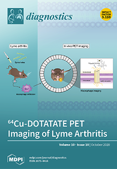
Cover Story (view full-size image): This is the first study to present data on somatostatin receptor imaging of infectious lesions in Lyme borreliosis (LB). LB caused by the Borrelia burgdorferi sensu lato complex is the most prevalent vector-borne infection in Europe and the U.S. New methods to monitor disease activity in patients with LB would be of paramount interest to practice precision medicine. As macrophages play a key role in the inflammatory response in Lyme arthritis, macrophage imaging using 64Cu-DOTATATE was used for the detection of disease activity in a murine model of Lyme arthritis. We suggest that PET imaging targeting the immune response of LB can become a valuable diagnostic tool for the detection of Borrelia infection. View this paper
  • Issues are regarded as officially published after their release is announced to the table of contents alert mailing list.
  • You may sign up for e-mail alerts to receive table of contents of newly released issues.
  • PDF is the official format for papers published in both, html and pdf forms. To view the papers in pdf format, click on the "PDF Full-text" link, and use the free Adobe Reader to open them.
Order results
Result details
Section
Select all
Export citation of selected articles as:
11 pages, 1593 KiB  
Article
Kappa Index versus CSF Oligoclonal Bands in Predicting Multiple Sclerosis and Infectious/Inflammatory CNS Disorders
by Diana Ferraro, Roberta Bedin, Patrizia Natali, Diego Franciotta, Krzysztof Smolik, Mario Santangelo, Paolo Immovilli, Valentina Camera, Francesca Vitetta, Matteo Gastaldi, Tommaso Trenti, Stefano Meletti and Patrizia Sola
Diagnostics 2020, 10(10), 856; https://doi.org/10.3390/diagnostics10100856 - 21 Oct 2020
Cited by 24 | Viewed by 4207
Abstract
Background: Cerebrospinal fluid (CSF) kappa free light chains (KFLC) are gaining increasing interest as markers of intrathecal immunoglobulin synthesis. The main aim of this study was to assess the diagnostic accuracy (AUC) of the kappa index (CSF/serum KFLC divided by the CSF/serum albumin [...] Read more.
Background: Cerebrospinal fluid (CSF) kappa free light chains (KFLC) are gaining increasing interest as markers of intrathecal immunoglobulin synthesis. The main aim of this study was to assess the diagnostic accuracy (AUC) of the kappa index (CSF/serum KFLC divided by the CSF/serum albumin ratio) compared to CSF oligoclonal IgG bands (OCB) in predicting Multiple Sclerosis (MS) or a central nervous system infectious/inflammatory disorder (CNSID). Methods: We enrolled patients who underwent a diagnostic spinal tap throughout two years. KFLC levels were determined using a Freelite assay (Binding Site) and the turbidimetric Optilite analyzer. Results: Of 540 included patients, 223 had a CNSID, and 84 had MS. The kappa index was more sensitive (0.89 versus 0.85) and less specific (0.84 versus 0.89), with the same AUC (0.87) as OCB for MS diagnosis (optimal cut-off: 6.2). Adding patients with a single CSF IgG band to the OCB-positive group slightly increased the AUC (0.88). Likewise, the kappa index (cut-off: 3.9) was more sensitive (0.67 versus 0.50) and less specific (0.81 versus 0.97), with the same AUC (0.74) as OCB, for a CNSID diagnosis. Conclusion: The kappa index and CSF OCB have comparable diagnostic accuracies for a MS or CNSID diagnosis and supply the clinician with useful, complementary information. Full article
Show Figures

Figure 1

11 pages, 2028 KiB  
Article
Feasibility of a Noninvasive Operability Assessment in Chronic Thromboembolic Pulmonary Hypertension under Real-World Practice
by Adriana Rodriguez Chaverri, Yolanda Revilla Ostolaza, Maria Jesus Lopez-Gude, María Teresa Velazquez, Ines Ponz de Antonio, Sergio Alonso Charterina, Agustin Albarran Gonzalez-Trevilla, Marta Perez Nunez, Jose Luis Perez Vela, Rafael Morales Ruiz, Juan F. Delgado Jimenez, Fernando Arribas Ynsaurriaga, Jose Maria Cortina and Pilar Escribano Subias
Diagnostics 2020, 10(10), 855; https://doi.org/10.3390/diagnostics10100855 - 21 Oct 2020
Cited by 6 | Viewed by 2099
Abstract
This study aimed to evaluate the feasibility of a noninvasive operability assessment of chronic thromboembolic pulmonary hypertension (CTEPH) based on multidetector computed tomographic angiography (MCTA). Up to 176 patients were evaluated from January 2016 to April 2018. Throughout the first phase, the initial [...] Read more.
This study aimed to evaluate the feasibility of a noninvasive operability assessment of chronic thromboembolic pulmonary hypertension (CTEPH) based on multidetector computed tomographic angiography (MCTA). Up to 176 patients were evaluated from January 2016 to April 2018. Throughout the first phase, the initial surgical decision was made based on MCTA with further analysis of pulmonary angiography (PA) in order to evaluate in which cases the initial decision was not modified by PA. During the second phase, PA was limited to patients judged inoperable based on MCTA or those whose assessment was not possible. Patients deemed operable (50%) based on MCTA along the first phase had been adequately classified, as PA did not modify the initial decision in all but one patient. Comparable results were obtained throughout the implementation phase. Regarding operated patients, the decision of operability was based solely on MCTA in 94% of those with level I disease, in 75% with level II, and 54% with level III. This approach enabled shorter periods of time to complete surgical assessment and the avoidance of PA-related morbidity. Baseline parameters, postoperative measures, and survival rates at 1 year after surgery were comparable in both phases. Noninvasive operability assessment is feasible in a subset of CTEPH patients and optimizes surgical candidacy evaluation. Full article
(This article belongs to the Special Issue Pulmonary Hypertension: Diagnosis and Management)
Show Figures

Graphical abstract

14 pages, 2073 KiB  
Article
Effect of Mutated ids Overexpression on IDS Enzyme Activity and Developmental Phenotypes in Zebrafish Embryos: A Valuable Index for Assessing Critical Point-Mutations Associated with Mucopolysaccharidosis Type II Occurrence in Humans
by Cheng-Yung Lin, Hsiang-Yu Lin, Chih-Kuang Chuang, Po-Hsiang Zhang, Ru-Yi Tu, Shuan-Pei Lin and Huai-Jen Tsai
Diagnostics 2020, 10(10), 854; https://doi.org/10.3390/diagnostics10100854 - 21 Oct 2020
Cited by 4 | Viewed by 2447
Abstract
Mucopolysaccharidosis type II (MPS II) is an X-linked disorder resulting from a deficiency in iduronate 2-sulfatase (IDS), which is reported to be caused by gene mutations in the iduronate 2-sulfatase (IDS) gene. Many IDS mutation sites have not yet had their causal relationship [...] Read more.
Mucopolysaccharidosis type II (MPS II) is an X-linked disorder resulting from a deficiency in iduronate 2-sulfatase (IDS), which is reported to be caused by gene mutations in the iduronate 2-sulfatase (IDS) gene. Many IDS mutation sites have not yet had their causal relationship with MPS II characterized. We employed a gain-of-function strategy whereby we microinjected different mutated zebrafish ids (z-ids) mRNAs corresponded to human IDS gene into zebrafish embryos, and then measured their total IDS enzymatic activity and observed the occurrence of defective phenotypes during embryonic development. We examined three known mutation sites for human IDS genes (h-IDS) associated with MPS II symptoms, including h-IDS-P86L, -S333L and -R468W, which corresponded to z-ids-P80L, -S327L and -R454W. When these three mutated z-ids mRNAs were overexpressed in zebrafish embryos, the IDS enzymatic activity of the total proteins extracted from the injected embryos was not increased compared with the endogenous IDS of the untreated embryos, which suggests that the IDS enzymatic activity of these three mutated z-ids was totally lost, as expected. Additionally, we observed defective phenotypes in these injected embryos, resulting from the failed IDS enzyme breakdown, which, in turn, has a dominant negative effect on the endogenous wild-type IDS function. These phenotypes were similar to the clinical symptoms observed in MPS II pathogenesis. We further studied six uncharacterized IDS mutation sites as identified by the Taiwanese MPS newborn screening programs. We propose a novel IDS enzyme activity assay combined with phenotypic observation in zebrafish embryos, as an alternative platform for quickly providing a valuable index for preliminarily assessment of any identified IDS point mutation gene that has not yet been characterized, in the context of its role in MPS II development. Full article
(This article belongs to the Special Issue Mucopolysaccharidoses: Diagnosis, Treatment, and Management 2.0)
Show Figures

Figure 1

11 pages, 2229 KiB  
Article
Improved Diagnostic Accuracy of Bone Metastasis Detection by Water-HAP Associated to Non-Contrast CT
by Yoshinobu Ishiwata, Yojiro Hieda, Soichiro Kaki, Shinjiro Aso, Keiichi Horie, Yusuke Kobayashi, Motoki Nakamura, Kazuhiko Yamada, Tsuneo Yamashiro and Daisuke Utsunomiya
Diagnostics 2020, 10(10), 853; https://doi.org/10.3390/diagnostics10100853 - 20 Oct 2020
Cited by 10 | Viewed by 2835
Abstract
We examined whether water-hydroxyapatite (HAP) images improve the diagnostic accuracy of bone metastasis compared with non-contrast CT alone. We retrospectively evaluated dual-energy computed tomography (DECT) images of 83 cancer patients (bone metastasis, 31; without bone metastasis, 52) from May 2018 to June 2019. [...] Read more.
We examined whether water-hydroxyapatite (HAP) images improve the diagnostic accuracy of bone metastasis compared with non-contrast CT alone. We retrospectively evaluated dual-energy computed tomography (DECT) images of 83 cancer patients (bone metastasis, 31; without bone metastasis, 52) from May 2018 to June 2019. Initially, two evaluators examined for bone metastasis on conventional CT images. In the second session, both CT and CT images plus water-HAP images on DECT. The confidence of bone metastasis was scored from 1 (benign) to 5 (malignant). The sensitivity, specificity, positive predictive values, and negative predictive values for both modalities were calculated based on true positive and negative findings. The intra-observer area under curve (AUC) for detecting bone metastasis was compared by receiver operating characteristic analysis. Kappa coefficient calculated the inter-observer agreement. In conventional CT images, sensitivity, specificity, positive predictive value, and negative predictive value of raters 1 and 2 for the identification of bone metastases were 0.742 and 0.710, 0.981 and 0.981, 0.958 and 0.957, and 0.864 and 0.850, respectively. In water-HAP, they were 1.00 and 1.00, 0.981 and 1.00, 0.969 and 1.00, and 1.00 and 1.00, respectively. In CT, AUCs were 0.861 and 0.845 in each observer. On water-HAP images, AUCs were 0.990 and 1.00. Kappa coefficient was 0.964 for CT and 0.976 for water-HAP images. The combination of CT and water-HAP images significantly increased diagnostic accuracy for detecting bone metastasis. Water-HAP images on DECT may enable accurate initial staging, reduced radiation exposure, and cost. Full article
(This article belongs to the Section Medical Imaging and Theranostics)
Show Figures

Figure 1

14 pages, 1935 KiB  
Review
Atherosclerosis Imaging with 18F-Sodium Fluoride PET
by Poul F. Høilund-Carlsen, Reza Piri, Caius Constantinescu, Kasper Karmark Iversen, Thomas J. Werner, Michael Sturek, Abass Alavi and Oke Gerke
Diagnostics 2020, 10(10), 852; https://doi.org/10.3390/diagnostics10100852 - 20 Oct 2020
Cited by 17 | Viewed by 3442
Abstract
The evidence on atherosclerosis imaging with 18F-sodium-fluoride (NaF) positron emission tomography (PET) is hotly debated because of the different patient characteristics, methodology, vascular beds, etc. in reported studies. This review is a continuation of a previous review on this topic, which covered [...] Read more.
The evidence on atherosclerosis imaging with 18F-sodium-fluoride (NaF) positron emission tomography (PET) is hotly debated because of the different patient characteristics, methodology, vascular beds, etc. in reported studies. This review is a continuation of a previous review on this topic, which covered the period 2010–2018. The purpose was to examine whether some of the most important questions that the previous review had left open had been elucidated by the most recent literature. Using principles of a systematic review, we ended analyzing 25 articles dealing with the carotids, coronary arteries, aorta, femoral, intracranial, renal, and penile arteries. The knowledge thus far can be summarized as follows: by targeting active arterial microcalcification, NaF uptake is considered a marker of early stage atherosclerosis, is age-dependent, and consistently associated with cardiovascular risk. Longitudinal studies on NaF uptake, conducted in the abdominal aorta only, showed unchanged uptake in postmenopausal women for nearly four years and varying uptake in prostate cancer patients over 1.5 years, despite constant or increasing calcium volume detected by computed tomography (CT). Thus, uncertainty remains about the transition from active arterial wall calcification marked by increased NaF uptake to less active or consolidated calcification detected by CT. The question of whether early-phase atherosclerosis and calcification can be modified remains also unanswered due to lack of intervention studies. Full article
(This article belongs to the Section Medical Imaging and Theranostics)
Show Figures

Figure 1

5 pages, 22609 KiB  
Interesting Images
18F-Fluorocholine PET/CT, Tc-99m-MIBI and TC-99m-MDP SPECT/CT in Tertiary Hyperparathyroidism with Renal Osteodystrophy
by Cristina Ferrari, Valentina Lavelli, Giulia Santo, Maria Teresa Frugis, Francesca Iuele, Giuseppe Rubini and Angela Sardaro
Diagnostics 2020, 10(10), 851; https://doi.org/10.3390/diagnostics10100851 - 20 Oct 2020
Cited by 4 | Viewed by 2694
Abstract
Tertiary hyperparathyroidism (HPT) is a metabolic disorder characterized by the semi-autonomous hypersecretion of parathyroid hormone (PTH), leading to hypercalcemia. It can be the end result of persistent secondary hyperparathyroidism and is most commonly observed in patients with long-standing chronic kidney disease (CKD) and [...] Read more.
Tertiary hyperparathyroidism (HPT) is a metabolic disorder characterized by the semi-autonomous hypersecretion of parathyroid hormone (PTH), leading to hypercalcemia. It can be the end result of persistent secondary hyperparathyroidism and is most commonly observed in patients with long-standing chronic kidney disease (CKD) and often after renal transplantation. Untreated HPT can lead to progressive bone disease, fibrocystic osteitis, and soft-tissue calcifications, along with other severe complications. In the 2009 Kidney Disease Improving Global Outcomes (KDIGO) guidelines, CKD-Mineral and Bone Disorder (CKD-MBD) is used to describe the broader clinical syndrome encompassing mineral, bone, and calcific cardiovascular abnormalities that develop as a complication of CKD. We report a 62-year-old female with a severe HPT evolved from advanced chronic kidney disease (stage 5D, KDIGO). Patient was evaluated with multimodality nuclear medicine functional imaging to assess hyperfunctioning parathyroid glands and bone lesions. Tc-99m-methoxyisobutylisonitrile (MIBI) dual-phase scintigraphy, Tc-99m-methylenediphosphonate (MDP) bone scan and 18F-Fluorocholine positron emission tomography/computed tomography (18F-FCH PET/CT) were performed before surgery. Full article
(This article belongs to the Section Medical Imaging and Theranostics)
Show Figures

Figure 1

14 pages, 1698 KiB  
Article
Discrepancies in Assessing Diastolic Function in Pre-Clinical Heart Failure Using Different Algorithms—A Primary Care Study
by Martina Setti, Giovanni Benfari, Donato Mele, Andrea Rossi, Piercarlo Ballo, Maurizio Galderisi, Michael Henein and Stefano Nistri
Diagnostics 2020, 10(10), 850; https://doi.org/10.3390/diagnostics10100850 - 20 Oct 2020
Cited by 8 | Viewed by 3618
Abstract
Background: Current guidelines on diastolic function (DF) by the American Society of Echocardiography and the European Association of Cardiovascular Imaging (ASE/EACVI) have been disputed and two alternative algorithms have been proposed by Johansen et al. and Oh et al. We sought (a) to [...] Read more.
Background: Current guidelines on diastolic function (DF) by the American Society of Echocardiography and the European Association of Cardiovascular Imaging (ASE/EACVI) have been disputed and two alternative algorithms have been proposed by Johansen et al. and Oh et al. We sought (a) to assess the concordance of ASE/EACVI guidelines on DF using these proposed alternative approaches and (b) to evaluate the prevalence of indeterminate diastolic dysfunction (DD) by each method, exploring means for reducing their number. Methods: We retrospectively analyzed the echocardiographic reports of 1158 outpatients including subjects at risk of heart failure without (n = 644) or with (n = 241) structural heart disease, and 273 healthy individuals. Concordance was calculated using the k coefficient and overall proportion of DD reclassification rate. The effectiveness of pulmonary vein flow (PVF), Valsalva maneuver, and left atrial volume index/late diastolic a’-ratio (LAVi/a’) over indeterminate grading was assessed. Results: The DD reclassification rate was 30.1% (k = 0.35) for ASE/EACVI and OH, 36.5% (k = 0.27) for ASE/EACVI and JOHANSEN and 31.1% (k = 0.37) for OH and JOHANSEN (p < 0.0001 for all comparisons). DF could not be graded only by ASE/EACVI and OH in 9% and 11% patients, respectively. The majority of patients could be reclassified using PVF or Valsalva maneuver or LAVi/a’, with the latter being the single most effective parameter. Conclusion: Inconsistencies between updated guidelines and independent approaches to assess and grade DF impede their interchangeable clinical use. The inconclusive diagnoses can be reconciled by conventional echocardiography in most patients, and LAVi/a’ emerges as a simple and effective approach to this aim. Full article
(This article belongs to the Section Medical Imaging and Theranostics)
Show Figures

Graphical abstract

25 pages, 975 KiB  
Review
Post-Mortem Investigations for the Diagnosis of Sepsis: A Review of Literature
by Chiara Stassi, Cristina Mondello, Gennaro Baldino and Elvira Ventura Spagnolo
Diagnostics 2020, 10(10), 849; https://doi.org/10.3390/diagnostics10100849 - 20 Oct 2020
Cited by 21 | Viewed by 7743
Abstract
To date, sepsis is still one of the most important causes of death due to the difficulties concerning the achievement of a correct diagnosis. As well as in a clinical context, also in a medico-legal setting the diagnosis of sepsis can reveal challenging [...] Read more.
To date, sepsis is still one of the most important causes of death due to the difficulties concerning the achievement of a correct diagnosis. As well as in a clinical context, also in a medico-legal setting the diagnosis of sepsis can reveal challenging due to the unspecificity of the signs detected during autopsies, especially when no ante-mortem clinical data, laboratory, and cultural results are available. Thus, a systematic review of literature was performed to provide an overview of the main available and updated forensic tools for the post-mortem diagnosis of sepsis. Moreover, the aim of this review was to evaluate whether a marker or a combination of markers exist, specific enough to allow a correct and definite post-mortem diagnosis. The review was conducted searching in PubMed and Scopus databases, and using variable combinations of the keywords “post mortem sepsis diagnosis”, “macroscopic signs”, “morphology”, “histology”, “immunohistochemical markers”, “biochemical markers”, and “forensic microbiology”. The article selection was carried out following specific inclusion and exclusion criteria. A total of 44 works was identified, providing data on morphological aspects of the organs examined, histological findings, immunohistochemical and biochemical markers, and cultural assays. The review findings suggested that the post-mortem diagnosis of sepsis can be achieved by a combination of data obtained from macroscopic and microscopic analysis and microbial investigations, associated with the increased levels of at least two of three biochemical and/or immunohistochemical markers evaluated simultaneously on blood samples. Full article
(This article belongs to the Section Diagnostic Microbiology and Infectious Disease)
Show Figures

Figure 1

15 pages, 9590 KiB  
Review
Differential Diagnosis of Endometriosis by Ultrasound: A Rising Challenge
by Marco Scioscia, Bruna A. Virgilio, Antonio Simone Laganà, Tommaso Bernardini, Nicola Fattizzi, Manuela Neri and Stefano Guerriero
Diagnostics 2020, 10(10), 848; https://doi.org/10.3390/diagnostics10100848 - 20 Oct 2020
Cited by 23 | Viewed by 9322
Abstract
Ultrasound is an effective tool to detect and characterize endometriosis lesions. Variances in endometriosis lesions’ appearance and distorted anatomy secondary to adhesions and fibrosis present as major difficulties during the complete sonographic evaluation of pelvic endometriosis. Currently, differential diagnosis of endometriosis to distinguish [...] Read more.
Ultrasound is an effective tool to detect and characterize endometriosis lesions. Variances in endometriosis lesions’ appearance and distorted anatomy secondary to adhesions and fibrosis present as major difficulties during the complete sonographic evaluation of pelvic endometriosis. Currently, differential diagnosis of endometriosis to distinguish it from other diseases represents the hardest challenge and affects subsequent treatment. Several gynecological and non-gynecological conditions can mimic deep-infiltrating endometriosis. For example, abdominopelvic endometriosis may present as atypical lesions by ultrasound. Here, we present an overview of benign and malignant diseases that may resemble endometriosis of the internal genitalia, bowels, bladder, ureter, peritoneum, retroperitoneum, as well as less common locations. An accurate diagnosis of endometriosis has significant clinical impact and is important for appropriate treatment. Full article
(This article belongs to the Special Issue Diagnosis of Endometriosis: Biomarkers and Clinical Methods)
Show Figures

Figure 1

9 pages, 4052 KiB  
Case Report
Persistent Left Superior Vena Cava with Absent Right Superior Vena Cava and Discrete Subaortic Stenosis Diagnosed in a Patient with Sick Sinus Syndrome: A Case Report and Brief Review of the Literature
by Irina Demșa, Daniela Crișu, Cristian Mihai Ștefan Haba, Andreea Maria Ursaru, Vlad-Adrian Afrăsânie, Irina Iuliana Costache, Antoniu Octavian Petriș and Dan Nicolae Tesloianu
Diagnostics 2020, 10(10), 847; https://doi.org/10.3390/diagnostics10100847 - 19 Oct 2020
Cited by 10 | Viewed by 11098
Abstract
A persistent left superior vena cava (PLSVC) is the most frequent anomaly of the venous drainage system. While both a right and left superior vena cava (SVC) are usually present, a unique, left-sided SVC, also known as an isolated PLSVC, accounts for only [...] Read more.
A persistent left superior vena cava (PLSVC) is the most frequent anomaly of the venous drainage system. While both a right and left superior vena cava (SVC) are usually present, a unique, left-sided SVC, also known as an isolated PLSVC, accounts for only 10–20% of cases. It is frequently associated with arrhythmias and other congenital cardiac anomalies. Though it is usually an asymptomatic condition, it may pose significant problems whenever central venous access is needed. We report a case of an isolated PLSVC that was diagnosed incidentally during pacemaker implantation for sinus node dysfunction. The venous anomaly was associated with subvalvular aortic stenosis determined by a subaortic membrane; this particular association of congenital cardiovascular anomalies is a rare finding, with only a few cases reported in the literature. We aim to highlight the clinical and practical implications of this condition, as well as to discuss the embryonic development and diagnostic methods of this congenital defect. Full article
(This article belongs to the Special Issue Imaging Cardiac Arrhythmia/Sudden Cardiac Death)
Show Figures

Figure 1

11 pages, 2199 KiB  
Article
Hybrid Operating Room System for the Treatment of Thoracic and Abdominal Aortic Aneurysms: Evaluation of the Radiation Dose Received by Patients
by Yoshihiro Haga, Koichi Chida, Masahiro Sota, Yuji Kaga, Mitsuya Abe, Yohei Inaba, Masatoshi Suzuki, Taiichiro Meguro and Masayuki Zuguchi
Diagnostics 2020, 10(10), 846; https://doi.org/10.3390/diagnostics10100846 - 19 Oct 2020
Cited by 15 | Viewed by 2681
Abstract
In recent years, endovascular treatment of aortic aneurysms has attracted considerable attention as a promising alternative to traditional surgery. Hybrid operating room systems (HORSs) are increasingly being used to perform endovascular procedures. The clinical benefits of endovascular treatments using HORSs are very clear, [...] Read more.
In recent years, endovascular treatment of aortic aneurysms has attracted considerable attention as a promising alternative to traditional surgery. Hybrid operating room systems (HORSs) are increasingly being used to perform endovascular procedures. The clinical benefits of endovascular treatments using HORSs are very clear, and these procedures are increasing in number. In procedures such as thoracic endovascular aortic repair (TEVAR) and endovascular aortic repair (EVAR), wires and catheters are used to deliver and deploy the stent graft in the thoracic/abdominal aorta under fluoroscopic control, including DSA. Thus, the radiation dose to the patient is an important issue. We determined radiation dose indicators (the dose–area product (DAP) and air karma (AK) parameters) associated with endovascular treatments (EVAR and TEVAR) using a HORS. As a result, the mean ± standard deviation (SD) DAPs of TEVAR and EVAR were 323.7 ± 161.0 and 371.3 ± 186.0 Gy × cm2, respectively. The mean ± SD AKs of TEVAR and EVAR were 0.92 ± 0.44 and 1.11 ± 0.54 Gy, respectively. The mean ± SD fluoroscopy times of TEVAR and EVAR were 13.4 ± 7.1 and 23.2 ± 11.7 min, respectively. Patient radiation dose results in this study of endovascular treatments using HORSs showed no deterministic radiation effects, such as skin injuries. However, radiation exposure during TEVAR and EVAR cannot be ignored. The radiation dose should be evaluated in HORSs during endovascular treatments. Reducing/optimizing the radiation dose to the patient in HORSs is important. Full article
(This article belongs to the Special Issue Assessment of Radiation Dose in X-ray and CT Exams)
Show Figures

Figure 1

12 pages, 2038 KiB  
Article
Ultrasensitive Label-Free Detection of Unamplified Multidrug-Resistance Bacteria Genes with a Bimodal Waveguide Interferometric Biosensor
by Jesús Maldonado, Ana Belén González-Guerrero, Adrián Fernández-Gavela, Juan José González-López and Laura M. Lechuga
Diagnostics 2020, 10(10), 845; https://doi.org/10.3390/diagnostics10100845 - 19 Oct 2020
Cited by 15 | Viewed by 3503
Abstract
Infections by multidrug-resistant bacteria are becoming a major healthcare emergence with millions of reported cases every year and an increasing incidence of deaths. An advanced diagnostic platform able to directly detect and identify antimicrobial resistance in a faster way than conventional techniques could [...] Read more.
Infections by multidrug-resistant bacteria are becoming a major healthcare emergence with millions of reported cases every year and an increasing incidence of deaths. An advanced diagnostic platform able to directly detect and identify antimicrobial resistance in a faster way than conventional techniques could help in the adoption of early and accurate therapeutic interventions, limiting the actual negative impact on patient outcomes. With this objective, we have developed a new biosensor methodology using an ultrasensitive nanophotonic bimodal waveguide interferometer (BiMW), which allows a rapid and direct detection, without amplification, of two prevalent and clinically relevant Gram-negative antimicrobial resistance encoding sequences: the extended-spectrum betalactamase-encoding gene blaCTX-M-15 and the carbapenemase-encoding gene blaNDM-5 We demonstrate the extreme sensitivity and specificity of our biosensor methodology for the detection of both gene sequences. Our results show that the BiMW biosensor can be employed as an ultrasensitive (attomolar level) and specific diagnostic tool for rapidly (less than 30 min) identifying drug resistance. The BiMW nanobiosensor holds great promise as a powerful tool for the control and management of healthcare-associated infections by multidrug-resistant bacteria. Full article
(This article belongs to the Collection Diagnostic Sensors)
Show Figures

Graphical abstract

11 pages, 8527 KiB  
Article
Integrating Finite Element Death Technique and Bone Remodeling Theory to Predict Screw Loosening Affected by Radiation Treatment after Mandibular Reconstruction Surgery
by Le-Jung Wu, Kai-Hung Hsieh and Chun-Li Lin
Diagnostics 2020, 10(10), 844; https://doi.org/10.3390/diagnostics10100844 - 19 Oct 2020
Cited by 4 | Viewed by 2905
Abstract
This study developed a numerical simulation to understand bone mechanical behavior and micro-crack propagation around a fixation screw with severe mandibular defects. A mandible finite element (FE) model was constructed in a rabbit with a right unilateral body defect. The reconstruction implant was [...] Read more.
This study developed a numerical simulation to understand bone mechanical behavior and micro-crack propagation around a fixation screw with severe mandibular defects. A mandible finite element (FE) model was constructed in a rabbit with a right unilateral body defect. The reconstruction implant was designed to be fixed using six screws distributed on the distal and mesial sides. The element death technique provided in FE analysis was combined with bone remodeling theory to simulate bone necrosis around the fixation screw in which the strain value reached the overload threshold. A total of 20 iterations were performed to observe the micro-crack propagation pattern for each screw according to the high strain locations occurring in each result from consecutive iterations. A parallel in vivo animal study was performed to validate the FE simulation by placing specific metal 3D printing reconstruction implants in rabbits to compare the differences in bone remodeling caused by radiation treatment after surgery. The results showed that strain values of the surrounding distal bone fixation screws were much larger than those at the mesial side. With the increase in the number of iteration analyses, the micro-crack prorogation trend for the distal fixation screws can be represented by the number and element death locations during the iteration analysis process. The corresponding micro-movement began to increase gradually and induced screw loosening after iteration calculation. The strained bone results showed that relatively high bone loss (damage) existed around the distal fixation screws under radiation treatment. This study concluded that the FE simulation developed in this study can provide a better predictive diagnosis method for understanding fixation screw loosening and advanced implant development before surgery. Full article
Show Figures

Figure 1

34 pages, 1435 KiB  
Review
Proteomics of Extracellular Vesicles: Update on Their Composition, Biological Roles and Potential Use as Diagnostic Tools in Atherosclerotic Cardiovascular Diseases
by Alice Mallia, Erica Gianazza, Beatrice Zoanni, Maura Brioschi, Silvia Stella Barbieri and Cristina Banfi
Diagnostics 2020, 10(10), 843; https://doi.org/10.3390/diagnostics10100843 - 19 Oct 2020
Cited by 28 | Viewed by 4826
Abstract
Extracellular vesicles (EVs) are lipid-bound vesicles released from cells under physiological and pathological conditions. Basing on biogenesis, dimension, content and route of secretion, they can be classified into exosomes, microvesicles (MVs) and apoptotic bodies. EVs have a key role as bioactive mediators in [...] Read more.
Extracellular vesicles (EVs) are lipid-bound vesicles released from cells under physiological and pathological conditions. Basing on biogenesis, dimension, content and route of secretion, they can be classified into exosomes, microvesicles (MVs) and apoptotic bodies. EVs have a key role as bioactive mediators in intercellular communication, but they are also involved in other physiological processes like immune response, blood coagulation, and tissue repair. The interest in studying EVs has increased over the years due to their involvement in several diseases, such as cardiovascular diseases (CVDs), and their potential role as biomarkers in diagnosis, therapy, and in drug delivery system development. Nowadays, the improvement of mass spectrometry (MS)-based techniques allows the characterization of the EV protein composition to deeply understand their role in several diseases. In this review, a critical overview is provided on the EV’s origin and physical properties, as well as their emerging functional role in both physiological and disease conditions, focusing attention on the role of exosomes in CVDs. The most important cardiac exosome proteomic studies will be discussed giving a qualitative and quantitative characterization of the exosomal proteins that could be used in future as new potential diagnostic markers or targets for specific therapies. Full article
Show Figures

Graphical abstract

11 pages, 1272 KiB  
Article
The Appropriate Opportunity for Evaluating Liver Fibrosis by Using the FIB-4 Index in Patients with Nonalcoholic Fatty Liver Disease in Japan
by Yuya Seko, Kota Yano, Aya Takahashi, Shinya Okishio, Seita Kataoka, Keiichiroh Okuda, Atsushi Umemura, Kanji Yamaguchi, Michihisa Moriguchi, Saiyu Tanaka and Yoshito Itoh
Diagnostics 2020, 10(10), 842; https://doi.org/10.3390/diagnostics10100842 - 19 Oct 2020
Cited by 3 | Viewed by 2021
Abstract
In patients with nonalcoholic fatty liver disease (NAFLD), liver fibrosis is the predictive factor for liver-related events and prognosis. This retrospective study aimed to evaluate longitudinal changes in the FIB-4 index and to determine a strategy for diagnosing and following patients with NAFLD [...] Read more.
In patients with nonalcoholic fatty liver disease (NAFLD), liver fibrosis is the predictive factor for liver-related events and prognosis. This retrospective study aimed to evaluate longitudinal changes in the FIB-4 index and to determine a strategy for diagnosing and following patients with NAFLD using this index. We analyzed the FIB-4 index at baseline and after 1 and 5 years in 272 consecutive patients with biopsy-proven NAFLD. Of these, 52 patients underwent serial biopsies. The change in the FIB-4 index was correlated with changes in the fibrosis stage among these patients (p = 0.048). The median FIB-4 index was 1.64 at baseline, 1.45 at 1 year, and 1.74 at 5 years. The negative predictive value for advanced fibrosis at a low cutoff point was 90.4/90.1 at baseline/1 year. Its specificity at a high cutoff point increased from 65.0% at baseline to 82.3% at 1 year. Multivariate analysis identified the FIB-4 index at 1 year as a predictive factor for a FIB-4 index > 2.67 at 5 years. A FIB-4 index < 1.30 was acceptable for excluding advanced fibrosis at baseline. In contrast, to evaluate and predict advanced liver fibrosis with the FIB-4 index at a high cutoff point, we should use the index at 1 year after appropriate therapy. Full article
(This article belongs to the Special Issue Fatty Liver Disease: Diagnostic, Predictive and Prognostic Markers)
Show Figures

Figure 1

24 pages, 2459 KiB  
Review
Detection of Bacterial and Viral Pathogens Using Photonic Point-of-Care Devices
by Peuli Nath, Alamgir Kabir, Somaiyeh Khoubafarin Doust, Zachary Joseph Kreais and Aniruddha Ray
Diagnostics 2020, 10(10), 841; https://doi.org/10.3390/diagnostics10100841 - 19 Oct 2020
Cited by 33 | Viewed by 8290
Abstract
Infectious diseases caused by bacteria and viruses are highly contagious and can easily be transmitted via air, water, body fluids, etc. Throughout human civilization, there have been several pandemic outbreaks, such as the Plague, Spanish Flu, Swine-Flu, and, recently, COVID-19, amongst many others. [...] Read more.
Infectious diseases caused by bacteria and viruses are highly contagious and can easily be transmitted via air, water, body fluids, etc. Throughout human civilization, there have been several pandemic outbreaks, such as the Plague, Spanish Flu, Swine-Flu, and, recently, COVID-19, amongst many others. Early diagnosis not only increases the chance of quick recovery but also helps prevent the spread of infections. Conventional diagnostic techniques can provide reliable results but have several drawbacks, including costly devices, lengthy wait time, and requirement of trained professionals to operate the devices, making them inaccessible in low-resource settings. Thus, a significant effort has been directed towards point-of-care (POC) devices that enable rapid diagnosis of bacterial and viral infections. A majority of the POC devices are based on plasmonics and/or microfluidics-based platforms integrated with mobile readers and imaging systems. These techniques have been shown to provide rapid, sensitive detection of pathogens. The advantages of POC devices include low-cost, rapid results, and portability, which enables on-site testing anywhere across the globe. Here we aim to review the recent advances in novel POC technologies in detecting bacteria and viruses that led to a breakthrough in the modern healthcare industry. Full article
(This article belongs to the Special Issue Mobile Diagnosis 2.0)
Show Figures

Figure 1

10 pages, 2201 KiB  
Article
Characterization of Uterine Motion in Early Gestation Using MRI-Based Motion Tracking
by Thomas Martin, Carla Janzen, Xinzhou Li, Irish Del Rosario, Teresa Chanlaw, Sarah Choi, Tess Armstrong, Rinat Masamed, Holden H. Wu, Sherin U. Devaskar and Kyunghyun Sung
Diagnostics 2020, 10(10), 840; https://doi.org/10.3390/diagnostics10100840 - 19 Oct 2020
Cited by 7 | Viewed by 2268
Abstract
Magnetic resonance imaging (MRI) is a promising non-invasive imaging technique that can be safely used to study placental development and function. However, studies of the human placenta performed by MRI are limited by uterine motion and motion in the uterus during MRI remains [...] Read more.
Magnetic resonance imaging (MRI) is a promising non-invasive imaging technique that can be safely used to study placental development and function. However, studies of the human placenta performed by MRI are limited by uterine motion and motion in the uterus during MRI remains one of the major limiting factors. Here, we aimed to investigate the characterization of uterine activity during MRI in the second trimester of pregnancy using MRI-based motion tracking. In total, 46 pregnant women were scanned twice (first scan between 14 and 18 weeks and second scan between 19 and 24 weeks), and 20 pregnant subjects underwent a single MRI between 14 and 18 weeks GA, resulting in 112 MRI scans. An MRI-based algorithm was used to track uterine motion in the superior-inferior and left-right directions. Uterine contraction and maternal motion cases were separated by the experts, and unpaired Wilcoxon tests were performed within the groups of gestational age (GA), fetal sex, and placental location in terms of the overall intensity measures of the uterine activity. In total, 22.3% of cases had uterine contraction during MRI, which increased from 18.6% at 14–18 weeks to 26.4% at 19–24 weeks GA. The dominant direction of the uterine contraction and maternal motion was the superior to the inferior direction during early gestation. Full article
(This article belongs to the Special Issue Advanced Techniques in Body Magnetic Resonance Imaging)
Show Figures

Figure 1

12 pages, 783 KiB  
Article
Diagnostic Accuracy of Biomarkers for Early-Onset Neonatal Bacterial Infections: Evaluation of Serum Procalcitonin Reference Curves
by Hidetoshi Go, Nobuhiko Nagano, Daichi Katayama, Takuya Akimoto, Takayuki Imaizumi, Ryoji Aoki, Midori Hijikata, Ayako Seimiya, Ryota Kato, Aya Okahashi and Ichiro Morioka
Diagnostics 2020, 10(10), 839; https://doi.org/10.3390/diagnostics10100839 - 18 Oct 2020
Cited by 7 | Viewed by 2641 | Correction
Abstract
To date, no clinical studies have compared the accuracy of serum procalcitonin (PCT) reference curves. We aimed to validate the diagnostic accuracy of previously reported serum PCT reference curves and to determine which biomarkers among a cut-off value over the 95th percentile in [...] Read more.
To date, no clinical studies have compared the accuracy of serum procalcitonin (PCT) reference curves. We aimed to validate the diagnostic accuracy of previously reported serum PCT reference curves and to determine which biomarkers among a cut-off value over the 95th percentile in the serum PCT reference curve, white blood cell (WBC) count, and C-reactive protein (CRP) and immunoglobulin M (IgM) levels, have the highest diagnostic accuracy for early-onset neonatal bacterial infections. This retrospective cohort study assessed 16 preterm and 23 term infants with suspected bacterial infections within 72 h after birth. Each infant group was divided into two subgroups: confirmed- and non-infection. The diagnostic accuracy was determined using the Youden index. The reference curves by Fukuzumi et al. in preterm and term infants had the highest Youden indexes: 1.000 and 0.324, respectively. Among preterm infants, the Youden index for PCT was 1.000. Among term infants, the Youden index for a combination of PCT, CRP, and WBC and/or IgM was 1.000. In conclusion, a serum PCT level over the 95th percentile on the reference curve for preterm infants and a combination of PCT and CRP levels with WBC count and/or IgM levels for term infants provided sufficient diagnostic accuracy. Full article
(This article belongs to the Special Issue Diagnosis and Management of Sepsis)
Show Figures

Figure 1

28 pages, 1879 KiB  
Review
Diagnostic Accuracy of Oral Fluids Biomarker Profile to Determine the Current and Future Status of Periodontal and Peri-Implant Diseases
by Sarhang S. Gul, Ali A. Abdulkareem, Aram M. Sha and Andrew Rawlinson
Diagnostics 2020, 10(10), 838; https://doi.org/10.3390/diagnostics10100838 - 18 Oct 2020
Cited by 47 | Viewed by 5536
Abstract
Severe periodontitis is ranked as the sixth most prevalent disease affecting humankind, with an estimated 740 million people affected worldwide. The diagnosis of periodontal diseases mainly relies upon assessment of conventional clinical parameters. However, these parameters reflect past, rather than current, clinical status [...] Read more.
Severe periodontitis is ranked as the sixth most prevalent disease affecting humankind, with an estimated 740 million people affected worldwide. The diagnosis of periodontal diseases mainly relies upon assessment of conventional clinical parameters. However, these parameters reflect past, rather than current, clinical status or future disease progression and, likely, outcome of periodontal treatment. Specific and sensitive biomarkers for periodontal diseases have been examined widely to address these issues and some biomarkers have been translated as point-of-care (PoC) tests. The aim of this review was to provide an update on PoC tests for use in the diagnosis and management of periodontal diseases. Among the PoC tests developed so far, active matrix metalloproteinase-8 has shown promising results in terms of diagnostic and prognostic values. However, further studies are required to increase the sensitivity and specificity via combining more than one biomarker and merging these test kits with periodontal risk assessment tools. Furthermore, the validity of these test kits needs to be investigated by applying the results in further independent studies and the impact on these test kits’, together with the results of risk factors for periodontal diseases, such as diabetes and smoking, also needs to be examined. Full article
(This article belongs to the Special Issue Dental Peri-Implant Point-of-Care Tests)
Show Figures

Figure 1

15 pages, 828 KiB  
Article
Managing Deviating EQA Results: A Survey to Assess the Corrective and Preventive Actions of Medical Laboratories Testing for Oncological Biomarkers
by Cleo Keppens, Ed Schuuring and Elisabeth MC Dequeker
Diagnostics 2020, 10(10), 837; https://doi.org/10.3390/diagnostics10100837 - 18 Oct 2020
Cited by 3 | Viewed by 2702
Abstract
Laboratories testing predictive biomarkers in lung and colorectal cancer are advised to participate in external quality assessment (EQA) schemes. This study aimed to investigate which corrective actions were taken by laboratories if predetermined performance criteria were not met, to ultimately improve current test [...] Read more.
Laboratories testing predictive biomarkers in lung and colorectal cancer are advised to participate in external quality assessment (EQA) schemes. This study aimed to investigate which corrective actions were taken by laboratories if predetermined performance criteria were not met, to ultimately improve current test practices. EQA participants from the European Society of Pathology between 2014 and 2018 for lung and colorectal cancer were contacted, if they had at least one analysis error or test failure in the provided cases, to complete a survey. For 72.4% of 514 deviating EQA results, an appropriate action was performed, most often including staff training (15.2%) and protocol revisions (14.6%). Main assigned persons were the molecular biologist (40.0%) and pathologist (46.5%). A change in test method or the use of complex techniques, such as next-generation sequencing, required more training and the involvement of dedicated personnel to reduce future test failures. The majority of participants adhered to ISO 15189 and implemented suitable actions by designated staff, not limited to accredited laboratories. However, for 27.6% of cases (by 20 laboratories) no corrective action was taken, especially for pre-analytic problems and complex techniques. The surveys were feasible to request information on results follow-up and further recommendations were provided. Full article
Show Figures

Figure 1

13 pages, 478 KiB  
Article
Accuracy of the Zika IgM Antibody Capture Enzyme-Linked Immunosorbent Assay from the Centers for Disease Control and Prevention (CDC Zika MAC-ELISA) for Diagnosis of Zika Virus Infection
by Moyra Machado Portilho, Laise de Moraes, Mariana Kikuti, Leile Camila Jacob Nascimento, Mitermayer Galvão Reis, Viviane Sampaio Boaventura, Ricardo Khouri and Guilherme Sousa Ribeiro
Diagnostics 2020, 10(10), 835; https://doi.org/10.3390/diagnostics10100835 - 18 Oct 2020
Cited by 3 | Viewed by 3506
Abstract
Serological diagnosis of Zika virus (ZIKV) infection is challenging because of antigenic cross-reactivity with dengue virus (DENV). This study evaluated the accuracy of the Zika IgM antibody capture enzyme-linked immunosorbent assay (CDC Zika IgM MAC-ELISA) in differentiating between ZIKV and DENV infections. To [...] Read more.
Serological diagnosis of Zika virus (ZIKV) infection is challenging because of antigenic cross-reactivity with dengue virus (DENV). This study evaluated the accuracy of the Zika IgM antibody capture enzyme-linked immunosorbent assay (CDC Zika IgM MAC-ELISA) in differentiating between ZIKV and DENV infections. To determine sensitivity, we used acute- and convalescent-phase sera from 21 patients with RT-PCR-confirmed ZIKV infection. To determine specificity, we used acute- and convalescent-phase sera from 60 RT-PCR-confirmed dengue cases and sera from 23 blood donors. During the acute-phase of the illness, the assay presented a sensitivity of 12.5% (2/16) for samples collected 0–4 days post symptoms onset (DPSO), and of 75.0% (3/4) for samples collected 5–9 DPSO. During the convalescent-phase of the illness, the test sensitivity was 90.9% (10/11), 100% (2/2), and 0% (0/2) for samples obtained 12–102, 258–260, and 722–727 DPSO, respectively. Specificity for acute- and convalescent-phase samples from RT-PCR-confirmed dengue cases was 100% and 93.2%, respectively. Specificity for blood donor samples was 100%. The assay is an accurate method for Zika serological diagnosis and proved to be reliable for use during surveillance and outbreak investigations in settings where ZIKV and DENV cocirculate. Full article
(This article belongs to the Collection Diagnostic Virology)
Show Figures

Graphical abstract

13 pages, 2210 KiB  
Article
Initial and Delayed Metabolic Activity of Palatine Tonsils Measured with the PET/CT-Dedicated Parameters
by Agata Pietrzak, Andrzej Marszalek, Malgorzata Paterska, Pawel Golusinski, Julitta Narozna and Witold Cholewinski
Diagnostics 2020, 10(10), 836; https://doi.org/10.3390/diagnostics10100836 - 17 Oct 2020
Cited by 4 | Viewed by 3473
Abstract
One of the most critical elements in the palatine tonsils (PT) patients’ management is to distinguish chronic tonsillitis and malignant tumor. The single-time-point (STP) 2-deoxy-2-[18 F]fluoro-D-glucose positron emission tomography/computed tomography (18 F-FDG PET/CT) examination offers the most significant sensitivity and specificity [...] Read more.
One of the most critical elements in the palatine tonsils (PT) patients’ management is to distinguish chronic tonsillitis and malignant tumor. The single-time-point (STP) 2-deoxy-2-[18 F]fluoro-D-glucose positron emission tomography/computed tomography (18 F-FDG PET/CT) examination offers the most significant sensitivity and specificity in the head and neck (H&N) region evaluation among commonly used methods of imaging. However, introducing dual-time-point (DTP) scanning might improve the specificity and sensitivity of the technique, limited by the 18 F-FDG non-tumor-specific patterns, especially when comparing different metabolic parameters. The study aims to compare several surrogates of the maximal standardized uptake value (SUVmax), obtained in 36 subjects, divided into confirmed by pathologic study PT cancer and tonsillitis in patients who underwent DTP 18 F-FDG PET/CT scanning. In this study, we observed the increased sensitivity and the specificity of the DTP 18 F-FDG PET/CT when compared with the standard PET/CT protocol. It could be concluded that DTP 18 F-FDG PET/CT improves the PT cancer and chronic tonsillitis differential diagnosis. Full article
(This article belongs to the Special Issue Biomarkers of Oral Cancer)
Show Figures

Figure 1

15 pages, 1104 KiB  
Article
Rehabilitation Improves Mitochondrial Energetics in Progressive Multiple Sclerosis: The Significant Role of Robot-Assisted Gait Training and of the Personalized Intensity
by Fabio Manfredini, Sofia Straudi, Nicola Lamberti, Simone Patergnani, Veronica Tisato, Paola Secchiero, Francesco Bernardi, Nicole Ziliotto, Giovanna Marchetti, Nino Basaglia, Massimo Bonora and Paolo Pinton
Diagnostics 2020, 10(10), 834; https://doi.org/10.3390/diagnostics10100834 - 17 Oct 2020
Cited by 15 | Viewed by 3541
Abstract
Abnormal levels of pyruvate and lactate were reported in multiple sclerosis (MS). We studied the response of markers of mitochondrial function to rehabilitation in relation to type, intensity and endurance performance in severely disabled MS patients. Forty-six progressive MS patients were randomized to [...] Read more.
Abnormal levels of pyruvate and lactate were reported in multiple sclerosis (MS). We studied the response of markers of mitochondrial function to rehabilitation in relation to type, intensity and endurance performance in severely disabled MS patients. Forty-six progressive MS patients were randomized to receive 12 walking sessions of robot-assisted gait training (RAGT, n = 23) or conventional overground therapy (CT, n = 23). Ten healthy subjects were also studied. Blood samples were collected to determine lactate, pyruvate, and glutathione levels and lactate/pyruvate ratio pre–post rehabilitation. In vivo muscle metabolism and endurance walking capacity were assessed by resting muscle oxygen consumption (rmVO2) using near-infrared spectroscopy and by six-minute walking distance (6MWD), respectively. The levels of mitochondrial biomarkers and rmVO2, altered at baseline with respect to healthy subjects, improved after rehabilitation in the whole population. In the two groups, an enhanced response was observed after RAGT compared to CT for lactate (p = 0.012), glutathione (<0.001), lactate/pyruvate ratio (p = 0.08) and rmVO2 (p = 0.07). Metabolic biomarkers and 6MWD improvements were exclusively correlated with a training speed markedly below individual gait speed. In severely disabled MS patients, rehabilitation rebalanced altered serum metabolic and muscle parameters, with RAGT being more effective than CT. A determinable slow training speed was associated with better metabolic and functional recovery. Trial Registration: ClinicalTrials.gov NCT02421731. Full article
(This article belongs to the Special Issue Biomarkers of Vascular Diseases)
Show Figures

Figure 1

22 pages, 3629 KiB  
Article
A Pilot Study of the Reliability and Agreement of Heart Rate, Respiratory Rate and Short-Term Heart Rate Variability in Elite Modern Pentathlon Athletes
by Bartosz Hoffmann, Andrew A. Flatt, Luiz Eduardo Virgilio Silva, Marcel Młyńczak, Rafał Baranowski, Ewelina Dziedzic, Bożena Werner and Jakub S. Gąsior
Diagnostics 2020, 10(10), 833; https://doi.org/10.3390/diagnostics10100833 - 16 Oct 2020
Cited by 27 | Viewed by 4517
Abstract
Research on reliability of heart rate variability (HRV) parameters in athletes has received increasing attention. The aims of this study were to examine the inter-day reliability of short-term (5 min) and ultra-short-term (1 min) heart rate (HR), respiratory rate (RespRate) and HRV parameters, [...] Read more.
Research on reliability of heart rate variability (HRV) parameters in athletes has received increasing attention. The aims of this study were to examine the inter-day reliability of short-term (5 min) and ultra-short-term (1 min) heart rate (HR), respiratory rate (RespRate) and HRV parameters, agreement between short-term and ultra-short-term parameters, and association between differences in HR, RespRate and HRV parameters in elite modern pentathletes. Electrocardiographic recordings were performed in stable measurement conditions with a week interval between tests. Relative reliability was evaluated by intra-class correlation coefficients, absolute reliability was evaluated by within-subject coefficient of variation, and agreement was evaluated using Bland–Altman (BA) plot with limits of agreement and defined a priori maximum acceptable difference. Short-term HR, RespRate, log transformed (ln) root mean square of successive normal-to-normal interval differences (lnRMSSD), ln high frequency (lnHF) and SD2/SD1 HRV indices and ultra-short-term HR, RespRate and lnRMSSD presented acceptable, satisfactory inter-day reliability. Although there were no significant differences between short-term and ultra-short-term HR, RespRate and lnRMSSD, no parameter showed acceptable differences with BA plots. Differences in time-domain and non-linear HRV parameters were more correlated with differences in HR than with differences in RespRate. Inverse results were observed for frequency-domain parameters. Short-term HR, RespRate, lnRMSSD, lnHF, and SD2/SD1 and ultra-short-term HR, RespRate and lnRMSSD could be used as reliable parameters in endurance athletes. However, practitioners should interpret changes in HRV parameters with regard to concomitant differences in HR and RespRate and caution should be taken before considering 5 min and 1 min parameters as interchangeable. Full article
(This article belongs to the Special Issue Diagnostic Challenges in Sports Cardiology)
Show Figures

Figure 1

10 pages, 554 KiB  
Guidelines
Guideline for Urine Culture and Biochemical Identification of Bacterial Urinary Pathogens in Low-Resource Settings
by Nabil Karah, Rayane Rafei, Wael Elamin, Anan Ghazy, Aula Abbara, Monzer Hamze and Bernt Eric Uhlin
Diagnostics 2020, 10(10), 832; https://doi.org/10.3390/diagnostics10100832 - 16 Oct 2020
Cited by 32 | Viewed by 65649
Abstract
Medical diagnosis in low-resource settings is confronted by the lack of suitable guidelines, protocols and checklists. Online-accessible procedural documents are difficult to find, might be mistranslated or interpreted and usually do not address the needs of developing countries. Urinalysis, one of the most [...] Read more.
Medical diagnosis in low-resource settings is confronted by the lack of suitable guidelines, protocols and checklists. Online-accessible procedural documents are difficult to find, might be mistranslated or interpreted and usually do not address the needs of developing countries. Urinalysis, one of the most frequently performed diagnostic examinations worldwide, involves a series of tests aiming to detect particular disorders, such as urinary tract infections, kidney disease and diabetes. In this guideline, we present an alternative approach for clinical laboratories with limited resources to identify common bacterial uropathogens. We propose dividing the identification plan into two levels. The implicated pathogen will first be assigned into a bacterial group, basic identification, against which a suitable panel of antimicrobial agents shall be selected for the antimicrobial susceptibility testing (AST). Characterization of the pathogen to the genus or species level, advanced identification, will then be performed to ensure correct reading of the AST results and determine the epidemiology of clinically significant pathogens. Most of the proposed steps in our guideline are tailored to meet the needs of clinical laboratories in low-resource settings. Such guidelines are needed to strengthen the capacity of regional pathology laboratories and to enhance international initiatives on antimicrobial resistance and health equity. Full article
(This article belongs to the Special Issue Urinary Tract Infection: Diagnosis, Treatment and Management)
Show Figures

Figure 1

14 pages, 8024 KiB  
Review
Postnatal Outcome and Associated Anomalies of Prenatally Diagnosed Right Aortic Arch with Concomitant Right Ductal Arch: A Systematic Review and Meta-Analysis
by Paolo Ivo Cavoretto, Alexandros Sotiriadis, Serena Girardelli, Silvia Spinillo, Massimo Candiani, Silvia Amodeo, Antonio Farina and Vlasta Fesslova
Diagnostics 2020, 10(10), 831; https://doi.org/10.3390/diagnostics10100831 - 15 Oct 2020
Cited by 12 | Viewed by 3130
Abstract
Right aortic arch presents a reported incidence of 0.1% of the general population; the aim of our study was to evaluate the risk of associated intracardiac (ICA), extracardiac (ECA), or chromosomal abnormalities in fetuses with right aortic arch (RAA) and concomitant right ductal [...] Read more.
Right aortic arch presents a reported incidence of 0.1% of the general population; the aim of our study was to evaluate the risk of associated intracardiac (ICA), extracardiac (ECA), or chromosomal abnormalities in fetuses with right aortic arch (RAA) and concomitant right ductal arch (RDA). A systematic review of the literature selected 18 studies including 60 cases of RAA/RDA. A meta-analysis with a random effect model calculated for each outcome the pooled crude proportion of associated abnormal outcomes in cases of RAA/RDA and the pooled proportions and odds ratios in RAA with LDA or RDA. Quality assessment of the included studies was achieved using the NIH quality assessment tool for case series studies. RAA/RDA presents risk of associated conotruncal CHDs of about 30% and risk of 22q11 microdeletion in the region of 1%. Two-thirds of 22q11 microdeletions had concomitant thymic hypoplasia and no other chromosomal defects were described. Risks for ICA, ECA, 22q11 microdeletion, and aberrant left subclavian artery are not substantially different in RAA with right or left arterial duct. RAA increases the risk of associated cardiac defects regardless of laterality of the ductal arch. In isolated RDA/RAA cases, absolute risks of extracardiac associated problems or surgery are rather low, we would therefore recommend reassurance, particularly when the thymus and karyotype are normal. Full article
(This article belongs to the Special Issue Fetal Medicine)
Show Figures

Figure 1

11 pages, 277 KiB  
Review
Rapid Detection of Methicillin-Resistant Staphylococcus aureus Directly from Blood for the Diagnosis of Bloodstream Infections: A Mini-Review
by Anna Rita Buonomini, Elisabetta Riva, Giovanni Di Bonaventura and Giovanni Gherardi
Diagnostics 2020, 10(10), 830; https://doi.org/10.3390/diagnostics10100830 - 15 Oct 2020
Cited by 10 | Viewed by 3153
Abstract
Staphylococcus aureus represents a major human pathogen able to cause a number of infections, especially bloodstream infections (BSI). Clinical use of methicillin has led to the emergence of methicillin-resistant S. aureus (MRSA) and MRSA-BSI have been reported to be associated with high morbidity [...] Read more.
Staphylococcus aureus represents a major human pathogen able to cause a number of infections, especially bloodstream infections (BSI). Clinical use of methicillin has led to the emergence of methicillin-resistant S. aureus (MRSA) and MRSA-BSI have been reported to be associated with high morbidity and mortality. Clinical diagnosis of BSI is based on the results from blood culture that, although considered the gold standard method, is time-consuming. For this reason, rapid diagnostic tests to identify the presence of methicillin-susceptible S. aureus (MSSA) and MRSA isolates directly in blood cultures are being used with increasing frequency to rapidly commence targeted antimicrobial therapy, also in the light of antimicrobial stewardship efforts. Here, we review and report the most common rapid non-molecular and molecular methods currently available to detect the presence of MRSA directly from blood. Full article
(This article belongs to the Section Diagnostic Microbiology and Infectious Disease)
9 pages, 2038 KiB  
Article
Diagnostic Value of C-Reactive Protein in Discrimination between Uncomplicated and Complicated Parapneumonic Effusion
by Yana Kogan, Edmond Sabo and Majed Odeh
Diagnostics 2020, 10(10), 829; https://doi.org/10.3390/diagnostics10100829 - 15 Oct 2020
Cited by 6 | Viewed by 2186
Abstract
Objectives: The role of serum C-reactive protein (CRPs) and pleural fluid CRP (CRPpf) in discriminating uncomplicated parapneumonic effusion (UCPPE) from complicated parapneumonic effusion (CPPE) is yet to be validated since most of the previous studies were on small cohorts and with variable results. [...] Read more.
Objectives: The role of serum C-reactive protein (CRPs) and pleural fluid CRP (CRPpf) in discriminating uncomplicated parapneumonic effusion (UCPPE) from complicated parapneumonic effusion (CPPE) is yet to be validated since most of the previous studies were on small cohorts and with variable results. The role of CRPs and CRPpf gradient (CRPg) and of their ratio (CRPr) in this discrimination has not been previously reported. The study aims to assess the diagnostic efficacy of CRPs, CRPpf, CRPr, and CRPg in discriminating UCPPE from CPPE in a relatively large cohort. Methods: The study population included 146 patients with PPE, 86 with UCPPE and 60 with CPPE. Levels of CRPs and CRPpf were measured, and the CRPg and CRPr were calculated. The values are presented as mean ± SD. Results: Mean levels of CRPs, CRPpf, CRPg, and CRPr of the UCPPE group were 145.3 ± 67.6 mg/L, 58.5 ± 38.5 mg/L, 86.8 ± 37.3 mg/L, and 0.39 ± 0.11, respectively, and for the CPPE group were 302.2 ± 75.6 mg/L, 112 ± 65 mg/L, 188.3 ± 62.3 mg/L, and 0.36 ± 0.19, respectively. Levels of CRPs, CRPpf, and CRPg were significantly higher in the CPPE than in the UCPPE group (p < 0.0001). No significant difference was found between the two groups for levels of CRPr (p = 0.26). The best cut-off value calculated by the receiver operating characteristic (ROC) analysis for discriminating UCPPE from CPPE was for CRPs, 211.5 mg/L with area under the curve (AUC) = 94% and p < 0.0001, for CRPpf, 90.5 mg/L with AUC = 76.3% and p < 0.0001, and for CRPg, 142 mg/L with AUC = 91% and p < 0.0001. Conclusions: CRPs, CRPpf, and CRPg are strong markers for discrimination between UCPPE and CPPE, while CRPr has no role in this discrimination. Full article
(This article belongs to the Section Pathology and Molecular Diagnostics)
Show Figures

Figure 1

13 pages, 3719 KiB  
Article
Usefulness of Modified CEUS LI-RADS for the Diagnosis of Hepatocellular Carcinoma Using Sonazoid
by Katsutoshi Sugimoto, Tatsuya Kakegawa, Hiroshi Takahashi, Yusuke Tomita, Masakazu Abe, Yu Yoshimasu, Hirohito Takeuchi, Yoshitaka Kasai and Takao Itoi
Diagnostics 2020, 10(10), 828; https://doi.org/10.3390/diagnostics10100828 - 15 Oct 2020
Cited by 33 | Viewed by 2912
Abstract
The Contrast-Enhanced Ultrasound Liver Imaging Reporting and Data System (CEUS LI-RADS) was introduced for classifying suspected hepatocellular carcinoma (HCC). However, it cannot be applied to Sonazoid. We assessed the diagnostic usefulness of a modified CEUS LI-RADS for HCC and non-HCC malignancies based on [...] Read more.
The Contrast-Enhanced Ultrasound Liver Imaging Reporting and Data System (CEUS LI-RADS) was introduced for classifying suspected hepatocellular carcinoma (HCC). However, it cannot be applied to Sonazoid. We assessed the diagnostic usefulness of a modified CEUS LI-RADS for HCC and non-HCC malignancies based on sensitivity, specificity, positive predictive value (PPV), and negative predictive value (NPV). Patients with chronic liver disease at risk for HCC were evaluated retrospectively. Nodules ≥1 cm with arterial phase hyperenhancement, no early washout (within 60 s), and contrast defects in the Kupffer phase were classified as LR-5. Nodules showing early washout, contrast defects in the Kupffer phase, and/or rim enhancement were classified as LR-M. A total of 104 nodules in 104 patients (median age: 70.0 years; interquartile range: 54.5–78.0 years; 74 men) were evaluated. The 48 (46.2%) LR-5 lesions included 45 HCCs, 2 high-flow hemangiomas, and 1 adrenal rest tumor. The PPV of LR-5 for HCC was 93.8% (95% confidence interval (CI): 82.8–98.7%). The 22 (21.2%) LR-M lesions included 16 non-HCC malignancies and 6 HCCs. The PPV of LR-M for non-HCC malignancies, including six intrahepatic cholangiocarcinomas, was 100% (95% CI: 69.8–100%). In conclusion, in the modified CEUS LI-RADS for Sonazoid, LR-5 and LR-M are good predictors of HCC and non-HCC malignancies, respectively. Full article
(This article belongs to the Special Issue Abdominal Ultrasound and Treatment of Hepatocellular Carcinoma)
Show Figures

Figure 1

31 pages, 14940 KiB  
Article
In Vivo Endoscopic Optical Coherence Tomography of the Healthy Human Oral Mucosa: Qualitative and Quantitative Image Analysis
by Marius Albrecht, Christian Schnabel, Juliane Mueller, Jonas Golde, Edmund Koch and Julia Walther
Diagnostics 2020, 10(10), 827; https://doi.org/10.3390/diagnostics10100827 - 15 Oct 2020
Cited by 16 | Viewed by 4986
Abstract
To date, there is still a lack of reliable imaging modalities to improve the quality of consultation, diagnostic and medical examinations of the oral mucosa in dentistry. Even though, optical technologies have become an important element for the detection and treatment of different [...] Read more.
To date, there is still a lack of reliable imaging modalities to improve the quality of consultation, diagnostic and medical examinations of the oral mucosa in dentistry. Even though, optical technologies have become an important element for the detection and treatment of different diseases of soft tissue, for the case of oral screenings the evidence of the benefit in comparison to conventional histopathology is mostly still pending. One promising optical technology for oral diagnostics is optical coherence tomography (OCT). To prove the potential of OCT, even the amount of freely accessible OCT data is not sufficient to describe the variance of healthy human oral soft tissue in vivo. In order to remedy this deficiency, the present study provides in vivo OCT cross sections of the human oral mucosa of the anterior and posterior oral cavity as well as the oropharynx of 47 adult volunteers. A collection of representative OCT cross sections forms the basis for a randomized blinded image analysis by means of seven criteria to assess the main features of the superficial layers of the human oral mucosa and to determine its correlation to regional features known from hematoxylin and eosin (HE) stained histology. Full article
(This article belongs to the Section Medical Imaging and Theranostics)
Show Figures

Figure 1

Previous Issue
Next Issue
Back to TopTop