Characterization of Uterine Motion in Early Gestation Using MRI-Based Motion Tracking

 ,
, 
Abstract
:1. Introduction
2. Materials and Methods
2.1. Study Population
2.2. MRI Techniques and Reconstruction
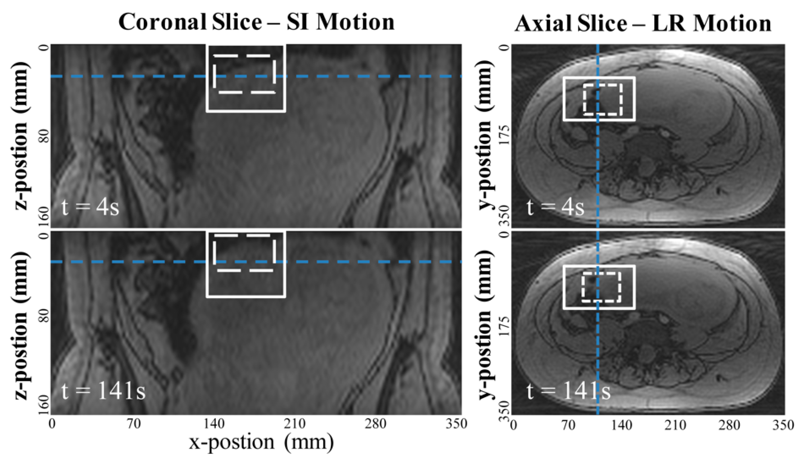
2.3. Image-Based Template-Matching Motion Tracking
2.4. Uterine Contraction and Maternal Motion
2.5. Statistical Analysis
3. Results
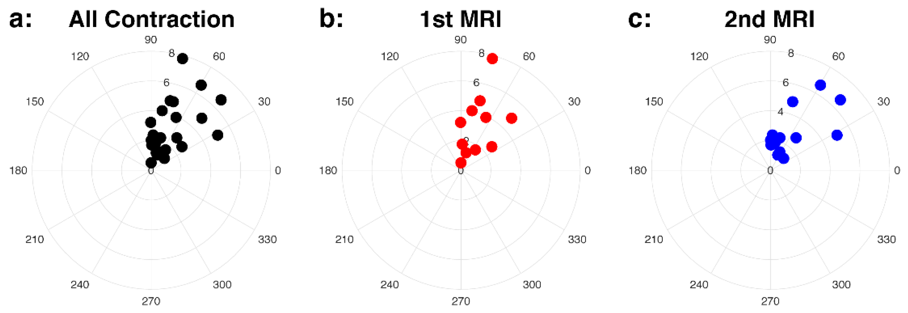
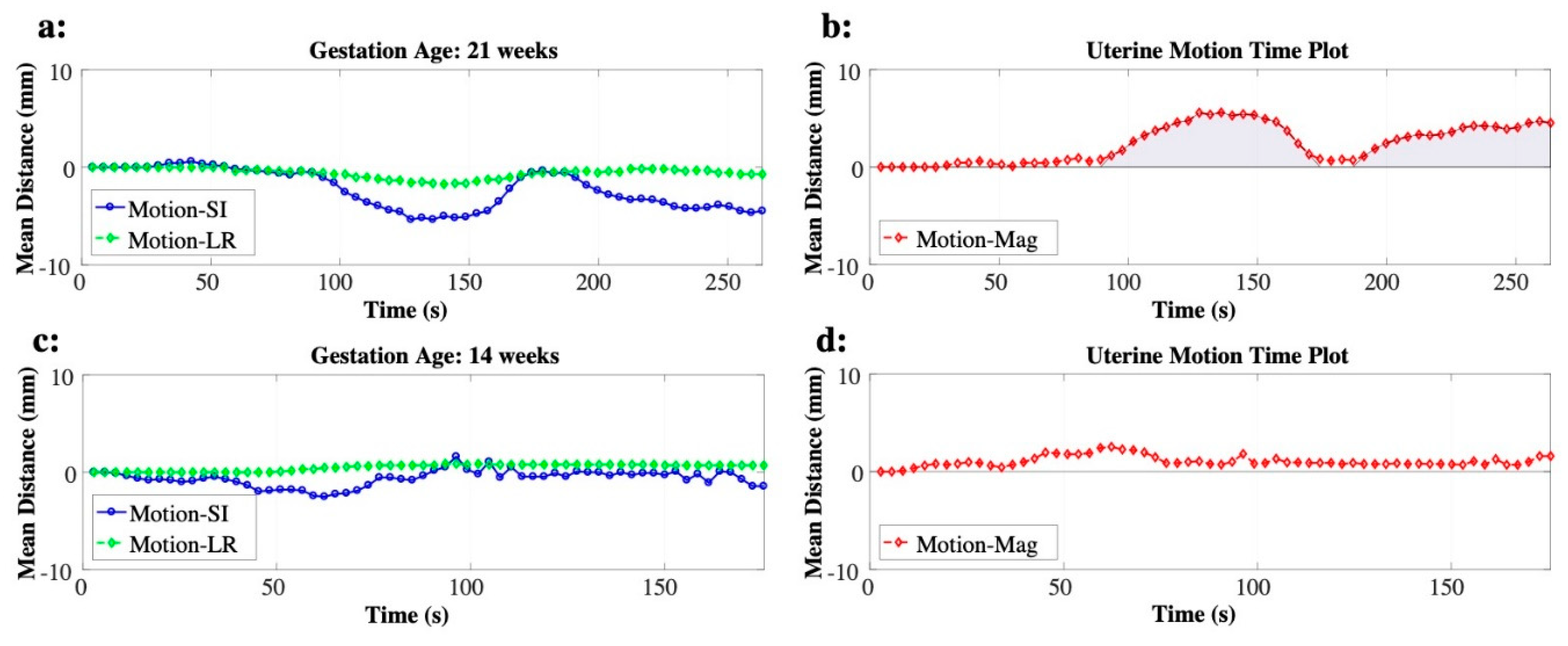
3.1. Uterine Contraction
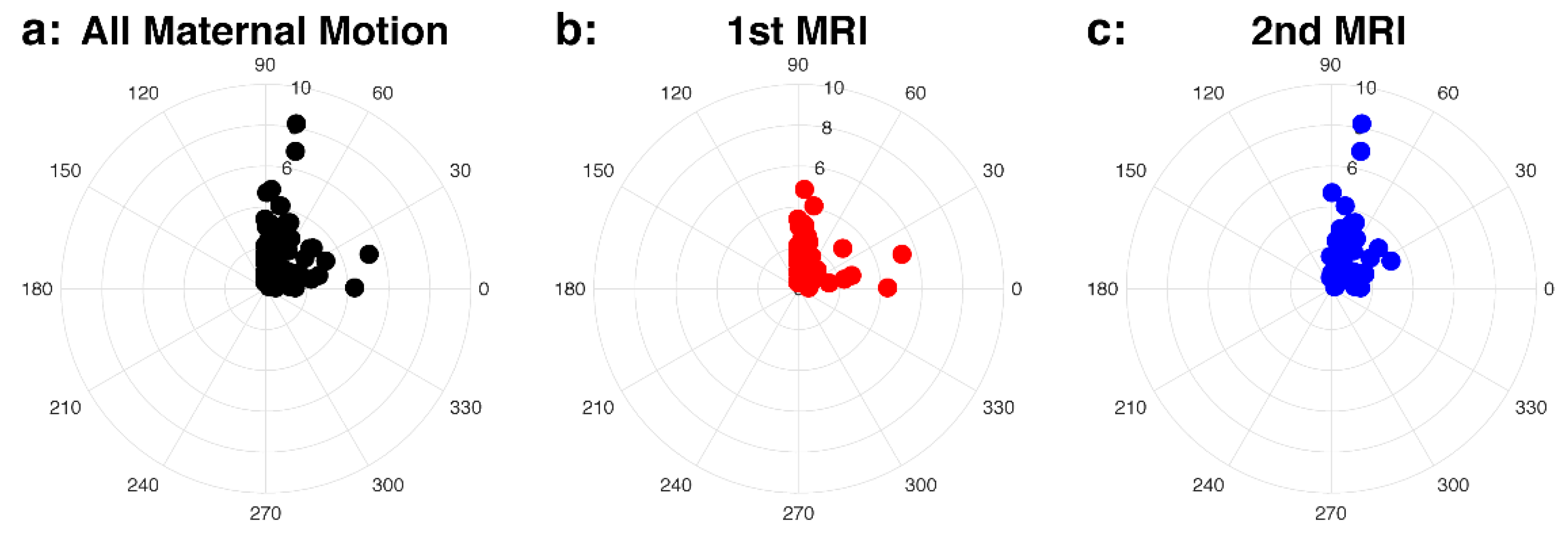
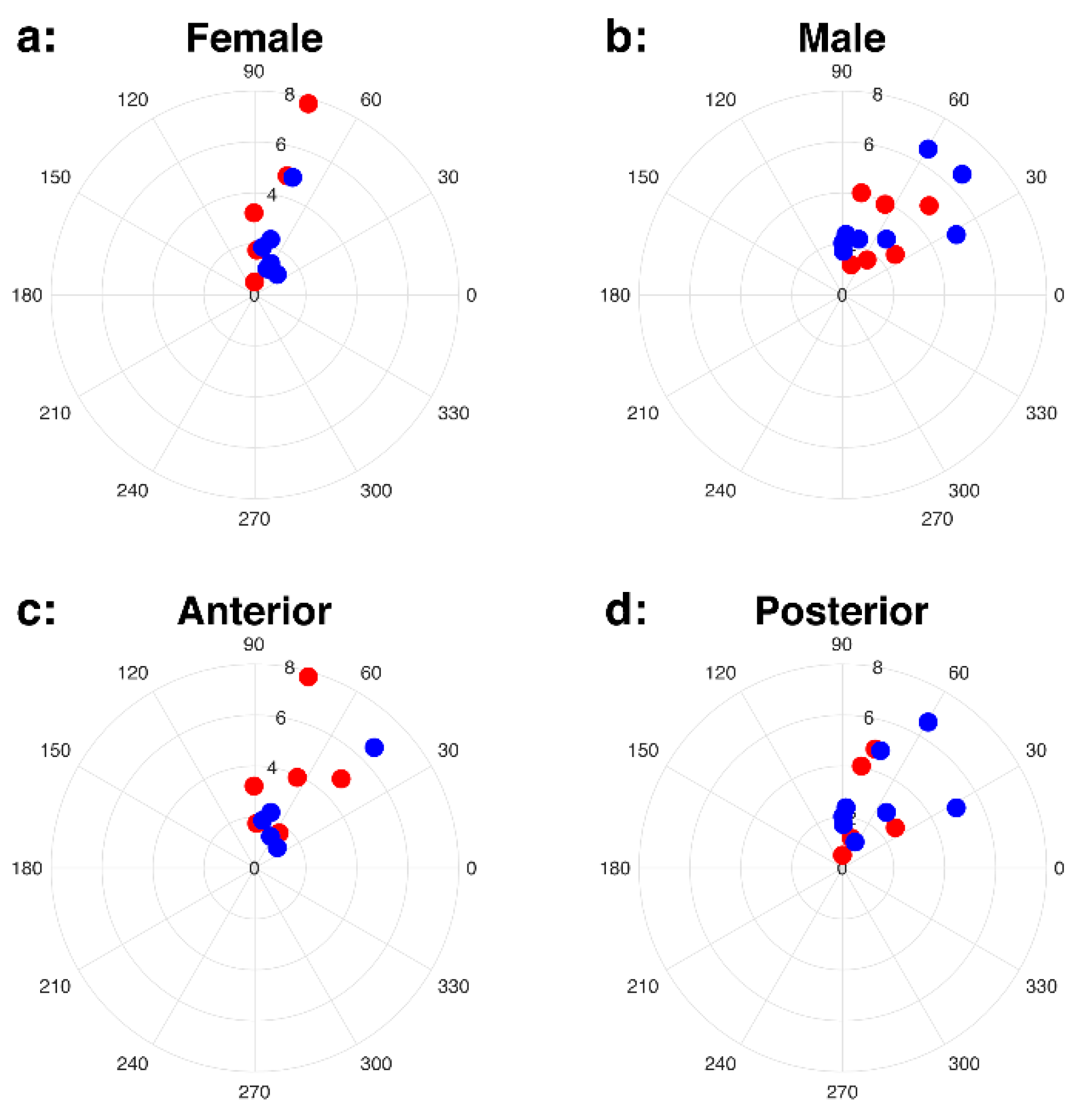
3.2. Maternal Motion
4. Discussion
5. Conclusions
Supplementary Materials
Author Contributions
Funding
Conflicts of Interest
References
- Derwig, I.E.; Akolekar, R.; Zelaya, F.; Gowland, P.; Barker, G.J.; Nicolaides, K.H. Association of placental volume measured by MRI and birth weight percentile. J. Magn. Reson. Imaging 2011, 34, 1125–1130. [Google Scholar] [CrossRef] [PubMed]
- Thomson, A.M.; Billewicz, W.Z.; Hytten, F.E. The weight of the placenta in relation to birthweight. BJOG Int. J. Obstet. Gynaecol. 1969, 76, 865–872. [Google Scholar] [CrossRef] [PubMed]
- Damodaram, M.; Story, L.; Eixarch, E.; Patel, A.; McGuinness, A.; Allsop, J.; Wyatt-Ashmead, J.; Kumar, S.; Rutherford, M. Placental MRI in Intrauterine Fetal Growth Restriction. Placenta 2010, 31, 491–498. [Google Scholar] [CrossRef] [PubMed]
- Li, S.; Hoskins, P.; Anderson, T.; McDicken, W. Measurement of mean velocity during pulsatile flow using time-averaged maximum frequency of doppler ultrasound waveforms. Ultrasound Med. Boil. 1993, 19, 105–113. [Google Scholar] [CrossRef]
- Angtuaco, T.L.; Shah, H.R.; Mattison, D.R.; Quirk, J.G. MR imaging in high-risk obstetric patients: A valuable complement to US. Radiographics 1992, 12, 91–109. [Google Scholar] [CrossRef] [PubMed] [Green Version]
- Chien, P.F.; Arnott, N.; Gordon, A.; Owen, P.; Khan, K.S. How useful is uterine artery Doppler flow velocimetry in the prediction of pre-eclampsia, intrauterine growth retardation and perinatal death? An overview. BJOG Int. J. Obstet. Gynaecol. 2000, 107, 196–208. [Google Scholar] [CrossRef] [PubMed] [Green Version]
- Wang, J.; Bi, X.; Avants, B.B.; Meng, T.; Zuehlsdorff, S.; Detre, J.A. Estimation of perfusion and arterial transit time in myocardium using free-breathing myocardial arterial spin labeling with navigator-echo. Magn. Reson. Med. 2010, 64, 1289–1295. [Google Scholar] [CrossRef] [PubMed] [Green Version]
- Shao, X.; Liu, D.; Martin, T.; Chanlaw, T.; Devaskar, S.U.; Janzen, C.; Murphy, A.M.; Margolis, D.; Sung, K.; Wang, D.J. Measuring human placental blood flow with multidelay 3D GRASE pseudocontinuous arterial spin labeling at 3T. J. Magn. Reson. Imaging 2017, 47, 1667–1676. [Google Scholar] [CrossRef] [PubMed]
- Sørensen, A.; Sinding, M.; Peters, D.A.; Petersen, A.; Frøkjær, J.B.; Christiansen, O.B.; Uldbjerg, N. Placental oxygen transport estimated by the hyperoxic placental BOLD MRI response. Physiol. Rep. 2015, 3, e12582. [Google Scholar] [CrossRef] [PubMed]
- Liu, D.; Shao, X.; Danyalov, A.; Chanlaw, T.; Masamed, R.; Wang, D.J.; Janzen, C.; Devaskar, S.U.; Sung, K. Human Placenta Blood Flow During Early Gestation with Pseudocontinuous Arterial Spin Labeling MRI. J. Magn. Reson. Imaging 2019, 51, 1247–1257. [Google Scholar] [CrossRef] [PubMed]
- Coakley, F.V.; Glenn, O.A.; Qayyum, A.; Barkovich, A.J.; Goldstein, R.; Filly, R.A. Fetal MRI: A Developing Technique for the Developing Patient. Am. J. Roentgenol. 2004, 182, 243–252. [Google Scholar] [CrossRef] [PubMed]
- Prayer, D.; Brugger, P.C.; Prayer, L. Fetal MRI: Techniques and protocols. Pediatr. Radiol. 2004, 34, 685–693. [Google Scholar] [CrossRef] [PubMed]
- Dickinson, J.E.; Godfrey, M.; Evans, S.F. Antenatal patterns of uterine activity in low-risk women: A longitudinal study. Aust. N. Z. J. Obstet. Gynaecol. 1997, 37, 149–152. [Google Scholar] [CrossRef] [PubMed]
- Sletten, J.; Kiserud, T.; Kessler, J. Effect of uterine contractions on fetal heart rate in pregnancy: A prospective observational study. Acta Obstet. Gynecol. Scand. 2016, 95, 1129–1135. [Google Scholar] [CrossRef] [PubMed]
- Guttmacher, A.E.; Maddox, Y.T.; Spong, C.Y. The Human Placenta Project: Placental structure, development, and function in real time. Placenta 2014, 35, 303–304. [Google Scholar] [CrossRef] [PubMed] [Green Version]
- Wu, H.H.; Gurney, P.T.; Hu, B.S.; Nishimura, D.G.; McConnell, M.V. Free-breathing multiphase whole-heart coronary MR angiography using image-based navigators and three-dimensional cones imaging. Magn. Reson. Med. 2012, 69, 1083–1093. [Google Scholar] [CrossRef] [PubMed] [Green Version]
- Song, H.K.; Dougherty, L. Dynamic MRI with projection reconstruction and KWIC processing for simultaneous high spatial and temporal resolution. Magn. Reson. Med. 2004, 52, 815–824. [Google Scholar] [CrossRef] [PubMed]
- Zia, S. Placental location and pregnancy outcome. J. Turk. Gynecol. Assoc. 2013, 14, 190–193. [Google Scholar] [CrossRef] [PubMed]
- Armstrong, T.; Liu, D.; Martin, T.; Masamed, R.; Janzen, C.; Wong, C.; Chanlaw, T.; Devaskar, S.U.; Sung, K.; Wu, H.H. 3D R2* mapping of the placenta during early gestation using free-breathing multiecho stack-of-radial MRI at 3T. J. Magn. Reson. Imaging 2018, 49, 291–303. [Google Scholar] [CrossRef] [PubMed] [Green Version]


| Imaging Parameters | 3-D Multi-Echo GA Radial GRE |
|---|---|
| Number of Echoes | 6–12 |
| TE1, ms | 1.23 |
| ΔTE, ms | 1.23 |
| TR, ms | 8.85–15.9 |
| In-plane Resolution, mm × mm | 1.7–2.2 × 1.7–2.2 |
| FOV, mm | 350–380 |
| Slice Thickness, mm | 3.5–5 |
| Flip Angle, degrees | 5 |
| Scan Time, min: sec | 3:18–6:06 |
| Groups | Uterine Contraction | Number of Contraction/Totals (%) | Maternal Motion | |||
|---|---|---|---|---|---|---|
| GA, Weeks | p-Value | GA, Weeks | p-Value | |||
| All Cases | 19.0 ± 2.6 | 25/112 (22.3) | 17.8 ± 2.6 | |||
| Longitudinal | 1st MRI | 16.5 ± 1.0 | <0.001 ** | 11/59 (18.6) | 15.8 ± 1.0 | <0.001 ** |
| 2nd MRI | 20.9 ± 1.4 | 14/53 (26.4) | 20.4 ± 1.2 | |||
| Fetal Sex | Female | 18.8 ± 2.4 | 1 | 11/50 (22.0) | 18.0 ± 2.6 | 0.57 |
| Male | 19.1 ± 2.8 | 14/62 (22.6) | 17.7 ± 2.5 | |||
| Placental Position | Anterior | 18.9 ± 2.3 | 0.87 | 12/60 (20.0) | 18.0 ± 2.6 | 0.51 |
| Posterior | 19.0 ± 2.9 | 13/52 (25.0) | 17.7 ± 2.5 | |||
| Groups | AUCnorm, mm | p-Value | Umax, mm | p-Value | θ, Degrees | Number of SI Dominant/Totals (%) | |
|---|---|---|---|---|---|---|---|
| All Cases | 3.1 ± 2.0 | 7.8 ± 5.5 | 66.7 ± 18.1 | 21/25 (84) | |||
| Longitudinal | 1st MRI | 3.3 ± 2.1 | 0.76 | 6.7 ± 4.3 | 0.46 | 69.3 ± 17.7 | 10/11 (90.9) |
| 2nd MRI | 3.0 ± 1.9 | 8.7 ± 6.3 | 64.7 ± 18.9 | 11/14 (78.6) | |||
| Fetal Sex | Female | 2.8 ± 2.2 | 0.20 | 7.4 ± 4.2 | 0.94 | 72.6 ± 14.4 | 10/11 (90.9) |
| Male | 3.4 ± 1.8 | 8.1 ± 6.4 | 62.0 ± 19.9 | 11/14 (78.6) | |||
| Placental Position | Anterior | 3.2 ± 2.2 | 0.98 | 8.0 ± 4.8 | 0.61 | 65.4 ± 16.7 | 10/12 (83.3) |
| Posterior | 3.0 ± 1.9 | 7.6 ± 6.2 | 67.9 ± 19.9 | 11/13 (84.6) | |||
| Groups | Umax, mm | p-Value | θ, Degrees | Number of SI Dominant/Totals (%) | |
|---|---|---|---|---|---|
| All Cases | 2.1 ± 2.1 | 60.0 ± 29.9 | 59/87 (67.8) | ||
| Longitudinal | 1st MRI | 1.8 ± 1.2 | 0.48 | 65.7 ± 29.9 | 35/48 (72.9) |
| 2nd MRI | 2.4 ± 2.8 | 52.9 ± 28.7 | 24/39 (61.5) | ||
| Fetal Sex | Female | 2.2 ± 2.7 | 0.77 | 61.2 ± 29.2 | 27/39 (69.2) |
| Male | 2.0 ± 1.4 | 59.0 ± 30.7 | 32/48 (66.7) | ||
| Placental Position | Anterior | 2.0 ± 1.5 | 0.50 | 58.0 ± 30.3 | 30/48 (62.5) |
| Posterior | 2.1 ± 2.6 | 62.3 ± 29.5 | 29/39 (74.3) | ||
Publisher’s Note: MDPI stays neutral with regard to jurisdictional claims in published maps and institutional affiliations. |
© 2020 by the authors. Licensee MDPI, Basel, Switzerland. This article is an open access article distributed under the terms and conditions of the Creative Commons Attribution (CC BY) license (http://creativecommons.org/licenses/by/4.0/).
Share and Cite
Martin, T.; Janzen, C.; Li, X.; Del Rosario, I.; Chanlaw, T.; Choi, S.; Armstrong, T.; Masamed, R.; Wu, H.H.; Devaskar, S.U.; et al. Characterization of Uterine Motion in Early Gestation Using MRI-Based Motion Tracking. Diagnostics 2020, 10, 840. https://doi.org/10.3390/diagnostics10100840
Martin T, Janzen C, Li X, Del Rosario I, Chanlaw T, Choi S, Armstrong T, Masamed R, Wu HH, Devaskar SU, et al. Characterization of Uterine Motion in Early Gestation Using MRI-Based Motion Tracking. Diagnostics. 2020; 10(10):840. https://doi.org/10.3390/diagnostics10100840
Chicago/Turabian StyleMartin, Thomas, Carla Janzen, Xinzhou Li, Irish Del Rosario, Teresa Chanlaw, Sarah Choi, Tess Armstrong, Rinat Masamed, Holden H. Wu, Sherin U. Devaskar, and et al. 2020. "Characterization of Uterine Motion in Early Gestation Using MRI-Based Motion Tracking" Diagnostics 10, no. 10: 840. https://doi.org/10.3390/diagnostics10100840
APA StyleMartin, T., Janzen, C., Li, X., Del Rosario, I., Chanlaw, T., Choi, S., Armstrong, T., Masamed, R., Wu, H. H., Devaskar, S. U., & Sung, K. (2020). Characterization of Uterine Motion in Early Gestation Using MRI-Based Motion Tracking. Diagnostics, 10(10), 840. https://doi.org/10.3390/diagnostics10100840