Next Issue
Volume 10, July
Previous Issue
Volume 10, May


Viruses, Volume 10, Issue 6 (June 2018) – 61 articles

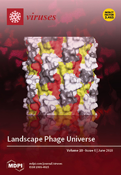
Cover Story (view full-size image): Filamentous phages have attracted the attention of bioengineers as structurally flexible bio-nanomaterials with a wide spectrum of potential applications. Phage landscape, which is formed by a genetically predetermined combination of thousands of functional organic groups, can be redesigned using genetic engineering methods. Furthermore, phage landscape can be randomly diversified to form multibillion clone landscape phage libraries, which can be screened for desired properties. The paper describes the history of the landscape phage invention, and gives some examples of their application. View Paper here.
  • Issues are regarded as officially published after their release is announced to the table of contents alert mailing list.
  • You may sign up for e-mail alerts to receive table of contents of newly released issues.
  • PDF is the official format for papers published in both, html and pdf forms. To view the papers in pdf format, click on the "PDF Full-text" link, and use the free Adobe Reader to open them.
Order results
Result details
Section
Select all
Export citation of selected articles as:

Editorial

Jump to: Research, Review, Other

3 pages, 181 KiB  
Editorial
Special Issue “Mathematical Modeling of Viral Infections”
by John M. Murray 1,2,* and Ruy M. Ribeiro 3,4,*
1 School of Mathematics and Statistics, UNSW Australia, Sydney 2052, Australia
2 Cancer Research Division, Cancer Council NSW, Woolloomooloo NSW 2011, Australia
3 Theoretical Biology and Biophysics, Los Alamos National Laboratory, Los Alamos, NM 87545, USA
4 Laboratorio de Biomatematica, Faculdade de Medicina da Universidade de Lisboa, 1649-028 Lisboa, Portugal
Viruses 2018, 10(6), 303; https://doi.org/10.3390/v10060303 - 4 Jun 2018
Cited by 3 | Viewed by 3860
Abstract
How an infection will progress in the body is dependent on myriad factors: the rate of spread of the agent, the immune response, what treatment may be applied[...] Full article
(This article belongs to the Special Issue Mathematical Modeling of Viral Infections)

Research

Jump to: Editorial, Review, Other

8 pages, 1625 KiB  
Article
Human Metapneumovirus Small Hydrophobic Protein Inhibits Interferon Induction in Plasmacytoid Dendritic Cells
by Xiaoyong Bao 1,*, Deepthi Kolli 2, Dana Esham 3, Thangam S. Velayutham 4 and Antonella Casola 1,5,6,*
1 Department of Pediatrics, The University of Texas Medical Branch, Galveston, TX 77555, USA
2 Atara Biotherapeutics, Oak Park, CA 94080, USA
3 Adena Health System, Chillicothe, OH 45601, USA
4 Department of Pathology, The University of Texas Medical Branch, Galveston, TX 77555, USA
5 Department of Microbiology & Immunology, The University of Texas Medical Branch, Galveston, TX 77555, USA
6 Sealy Center for Vaccine Development, The University of Texas Medical Branch, Galveston, TX 77555, USA
Viruses 2018, 10(6), 278; https://doi.org/10.3390/v10060278 - 23 May 2018
Cited by 12 | Viewed by 3028
Abstract
Human metapneumovirus (hMPV), a leading cause of respiratory tract infections in infants, encodes a small hydrophobic (SH) protein of unknown function. Here we show that infection of plasmacytoid dendritic cells (pDCs) with a recombinant virus lacking SH expression (rhMPV-ΔSH) enhanced the secretion of [...] Read more.
Human metapneumovirus (hMPV), a leading cause of respiratory tract infections in infants, encodes a small hydrophobic (SH) protein of unknown function. Here we show that infection of plasmacytoid dendritic cells (pDCs) with a recombinant virus lacking SH expression (rhMPV-ΔSH) enhanced the secretion of type I interferons (IFNs), which required TLR7 and MyD88 expression. HMPV SH protein inhibited TLR7/MyD88/TRAF6 signaling leading to IFN gene transcription, identifying a novel mechanism by which paramyxovirus SH proteins modulate innate immune responses. Full article
Show Figures

Figure 1

12 pages, 1780 KiB  
Article
Application of CRISPR/Cas9 Gene Editing System on MDV-1 Genome for the Study of Gene Function
by Yaoyao Zhang 1,†, Na Tang 1,2,†, Yashar Sadigh 1, Susan Baigent 1, Zhiqiang Shen 2, Venugopal Nair 1,* and Yongxiu Yao 1,*
1 The Pirbright Institute & UK-China Centre of Excellence for Research on Avian Diseases, Pirbright, Ash Road, Guildford, Surrey GU24 0NF, UK
2 Binzhou Animal Science and Veterinary Medicine Academy & UK-China Centre of Excellence for Research on Avian Diseases, Binzhou 256600, Shandong, China
These authors contributed equally to this work.
Viruses 2018, 10(6), 279; https://doi.org/10.3390/v10060279 - 24 May 2018
Cited by 24 | Viewed by 6273
Abstract
Marek’s disease virus (MDV) is a member of alphaherpesviruses associated with Marek’s disease, a highly contagious neoplastic disease in chickens. Complete sequencing of the viral genome and recombineering techniques using infectious bacterial artificial chromosome (BAC) clones of Marek’s disease virus genome have identified [...] Read more.
Marek’s disease virus (MDV) is a member of alphaherpesviruses associated with Marek’s disease, a highly contagious neoplastic disease in chickens. Complete sequencing of the viral genome and recombineering techniques using infectious bacterial artificial chromosome (BAC) clones of Marek’s disease virus genome have identified major genes that are associated with pathogenicity. Recent advances in CRISPR/Cas9-based gene editing have given opportunities for precise editing of the viral genome for identifying pathogenic determinants. Here we describe the application of CRISPR/Cas9 gene editing approaches to delete the Meq and pp38 genes from the CVI988 vaccine strain of MDV. This powerful technology will speed up the MDV gene function studies significantly, leading to a better understanding of the molecular mechanisms of MDV pathogenesis. Full article
(This article belongs to the Section Animal Viruses)
Show Figures

Figure 1

16 pages, 3829 KiB  
Article
Multiple, Independent T Cell Lymphomas Arising in an Experimentally FIV-Infected Cat during the Terminal Stage of Infection
by Brian G. Murphy 1,*, Christina Eckstrand 2, Diego Castillo 1, Andre Poon 1, Molly Liepnieks 1, Kristy Harmon 1 and Peter Moore 1
1 Department of Pathology, Microbiology and Immunology, School of Veterinary Medicine, University of California, Davis, CA 95616-5270, USA
2 Department of Veterinary Microbiology and Pathology, College of Veterinary Medicine, Washington State University, Pullman, WA 99163, USA
Viruses 2018, 10(6), 280; https://doi.org/10.3390/v10060280 - 24 May 2018
Cited by 11 | Viewed by 3866
Abstract
Our laboratory has serially reported on the virologic and immunopathologic features of a cohort of experimental feline immunodeficiency virus (FIV)-infected cats for more than eight years. At 8.09 years post infection (PI), one of these animals entered the terminal stage of infection, characterized [...] Read more.
Our laboratory has serially reported on the virologic and immunopathologic features of a cohort of experimental feline immunodeficiency virus (FIV)-infected cats for more than eight years. At 8.09 years post infection (PI), one of these animals entered the terminal stage of infection, characterized by undulating hyperthermia, progressive anorexia, weight loss, and pancytopenia; the animal was not responsive to therapeutic interventions, necessitating euthanasia six weeks later (8.20 years PI). Subsequent analyses indicated that neoplastic lymphocytes infiltrated multiple cervical lymph nodes and a band-like region of the mucosal lamina propria within a segment of the intestine. Immunohistochemistry and T cell clonality testing determined that the nodal and intestinal lesions were independently arising from CD3 T cell lymphomas. In-situ RNA hybridization studies indicated that diffuse neoplastic lymphocytes from the cervical lymph node contained abundant viral nucleic acid, while viral nucleic acid was not detectable in lymphocytes from the intestinal lymphoma lesion. The proviral long terminal repeat (LTR) was amplified and sequenced from multiple anatomic sites, and a common clone containing a single nucleotide polymorphism was determined to be defective in response to phorbol myristate acetate (PMA)-mediated promoter activation in a reporter gene assay. This assay revealed a previously unidentified PMA response element within the FIV U3 region 3’ to the TATA box. The possible implications of these results on FIV-lymphoma pathogenesis are discussed. Full article
(This article belongs to the Special Issue Nonprimate Lentivirus)
Show Figures

Figure 1

16 pages, 1056 KiB  
Article
Yeast Derived LysA2 Can Control Bacterial Contamination in Ethanol Fermentation
by Jun-Seob Kim 1,2, M. Angela Daum 1,2, Yong-Su Jin 1,2 and Michael J. Miller 1,2,*
1 Department of Food Science and Human Nutrition, University of Illinois, 905 S. Goodwin Ave., Urbana, IL 61801, USA
2 Carl R. Woese Institute for Genomic Biology, University of Illinois at Urbana-Champaign, 1206 W. Gregory Dr., Urbana, IL 61801, USA
Viruses 2018, 10(6), 281; https://doi.org/10.3390/v10060281 - 24 May 2018
Cited by 15 | Viewed by 5047
Abstract
Contamination of fuel-ethanol fermentations continues to be a significant problem for the corn and sugarcane-based ethanol industries. In particular, members of the Lactobacillaceae family are the primary bacteria of concern. Currently, antibiotics and acid washing are two major means of controlling contaminants. However, [...] Read more.
Contamination of fuel-ethanol fermentations continues to be a significant problem for the corn and sugarcane-based ethanol industries. In particular, members of the Lactobacillaceae family are the primary bacteria of concern. Currently, antibiotics and acid washing are two major means of controlling contaminants. However, antibiotic use could lead to increased antibiotic resistance, and the acid wash step stresses the fermenting yeast and has limited effectiveness. Bacteriophage endolysins such as LysA2 are lytic enzymes with the potential to contribute as antimicrobials to the fuel ethanol industries. Our goal was to evaluate the potential of yeast-derived LysA2 as a means of controlling Lactobacillaceae contamination. LysA2 intracellularly produced by Pichia pastoris showed activity comparable to Escherichia coli produced LysA2. Lactic Acid Bacteria (LAB) with the A4α peptidoglycan chemotype (L-Lys-D-Asp crosslinkage) were the most sensitive to LysA2, though a few from that chemotype were insensitive. Pichia-expressed LysA2, both secreted and intracellularly produced, successfully improved ethanol productivity and yields in glucose (YPD60) and sucrose-based (sugarcane juice) ethanol fermentations in the presence of a LysA2 susceptible LAB contaminant. LysA2 secreting Sacharomyces cerevisiae did not notably improve production in sugarcane juice, but it did control bacterial contamination during fermentation in YPD60. Secretion of LysA2 by the fermenting yeast, or adding it in purified form, are promising alternative tools to control LAB contamination during ethanol fermentation. Endolysins with much broader lytic spectrums than LysA2 could supplement or replace the currently used antibiotics or the acidic wash. Full article
(This article belongs to the Special Issue Phage Lytic Enzymes and Their Applications)
Show Figures

Figure 1

12 pages, 1278 KiB  
Article
K15 Protein of Kaposi’s Sarcoma Herpesviruses Increases Endothelial Cell Proliferation and Migration through Store-Operated Calcium Entry
by Wei Chen 1,2, Changqing Xu 1, Liuqing Wang 2, Bing Shen 3 and Linding Wang 1,*
1 Department of Microbiology, Anhui Medical University, Hefei 230032, China
2 Department of Clinical Laboratory, the Third People’s Hospital of Hefei, Hefei 230051, China
3 Department of Physiology, Anhui Medical University, Hefei 230032, China
Viruses 2018, 10(6), 282; https://doi.org/10.3390/v10060282 - 24 May 2018
Cited by 4 | Viewed by 5021
Abstract
Kaposi’s sarcoma (KS) is a tumor of the vascular endothelium that is caused by Kaposi’s sarcoma-associated herpesvirus (KSHV). K15 of KSHV is a specific gene encoding a transmembrane protein. Two highly different forms of K15, the predominant (K15P) and minor (K15M) have been [...] Read more.
Kaposi’s sarcoma (KS) is a tumor of the vascular endothelium that is caused by Kaposi’s sarcoma-associated herpesvirus (KSHV). K15 of KSHV is a specific gene encoding a transmembrane protein. Two highly different forms of K15, the predominant (K15P) and minor (K15M) have been identified in different KSHV strains. In genomic locations and protein topology, two K15 alleles resemble the latent membrane protein (LMP) 1 and LMP2A of Epstein–Barr virus. Both K15 proteins have motifs similar to those found in LMP1 and LMP2A. K15 therefore seems to be a hybrid of a distant evolutionary relative of LMP1 and LMP2A. Ca2+ is a second messenger and participates in numerous activities in cells, like proliferation, migration and metastasis. It has been found previously that LMP1 increased Ca2+ influx through store-operated calcium channels and blockade of LMP1 reduced store-operated Ca2+ entry (SOCE). LMP2A has similar activity. So we sought to determine whether K15 had similar activity. We showed that K15P induced Ca2+ influx and enhanced expression of Orail1, which is a vital protein in SOCE, and overexpression of K15P improved cell motility. Mutant K15P did not show these activities in HEK-293T and EA.hy 926 cells. Our results showed that K15P increased cell proliferation and migration though SOCE and established a novel mechanism for the development of KS and KSHV-associated diseases. Full article
(This article belongs to the Section Animal Viruses)
Show Figures

Figure 1

19 pages, 2245 KiB  
Article
Saracatinib Inhibits Middle East Respiratory Syndrome-Coronavirus Replication In Vitro
by Jin Soo Shin 1,2,†, Eunhye Jung 1,†, Meehyein Kim 1,3, Ralph S. Baric 4 and Yun Young Go 1,3,*
1 Virus Research Group, Korea Research Institute of Chemical Technology, Daejeon 34114, Korea
2 Department of Biological Sciences, Korea Advanced Institute of Science and Technology, Daejeon 34141, Korea
3 Department of Medicinal Chemistry and Pharmacology, University of Science and Technology, Daejeon 34114, Korea
4 Department of Epidemiology, University of North Carolina at Chapel Hill, Chapel Hill, NC 27599, USA
The authors contributed equally to this article.
Viruses 2018, 10(6), 283; https://doi.org/10.3390/v10060283 - 24 May 2018
Cited by 69 | Viewed by 6478
Abstract
The Middle East respiratory syndrome-coronavirus (MERS-CoV), first identified in Saudi Arabia, is an emerging zoonotic pathogen that causes severe acute respiratory illness in humans with a high fatality rate. Since its emergence, MERS-CoV continues to spread to countries outside of the Arabian Peninsula [...] Read more.
The Middle East respiratory syndrome-coronavirus (MERS-CoV), first identified in Saudi Arabia, is an emerging zoonotic pathogen that causes severe acute respiratory illness in humans with a high fatality rate. Since its emergence, MERS-CoV continues to spread to countries outside of the Arabian Peninsula and gives rise to sporadic human infections following the entry of infected individuals to other countries, which can precipitate outbreaks similar to the one that occurred in South Korea in 2015. Current therapeutics against MERS-CoV infection have primarily been adapted from previous drugs used for the treatment of severe acute respiratory syndrome. In search of new potential drug candidates, we screened a library composed of 2334 clinically approved drugs and pharmacologically active compounds. The drug saracatinib, a potent inhibitor of Src-family of tyrosine kinases (SFK), was identified as an inhibitor of MERS-CoV replication in vitro. Our results suggest that saracatinib potently inhibits MERS-CoV at the early stages of the viral life cycle in Huh-7 cells, possibly through the suppression of SFK signaling pathways. Furthermore, saracatinib exhibited a synergistic effect with gemcitabine, an anticancer drug with antiviral activity against several RNA viruses. These data indicate that saracatinib alone or in combination with gemcitabine can provide a new therapeutic option for the treatment of MERS-CoV infection. Full article
(This article belongs to the Section Viral Immunology, Vaccines, and Antivirals)
Show Figures

Graphical abstract

12 pages, 3294 KiB  
Article
The Auxiliary Role of the Amidase Domain in Cell Wall Binding and Exolytic Activity of Staphylococcal Phage Endolysins
by Bokyung Son 1,2,†, Minsuk Kong 1,2,†,‡ and Sangryeol Ryu 1,2,*
1 Laboratory of Molecular Food Microbiology, Department of Food and Animal Biotechnology, Seoul National University, Seoul 08826, Korea
2 Department of Agricultural Biotechnology and Center for Agricultural Biomaterials, Seoul National University, Seoul 08826, Korea
These authors contributed equally to this work.
Current address: Laboratory of Molecular Biology, National Cancer Institute, National Institutes of Health, Bethesda, MD 20892, USA.
Viruses 2018, 10(6), 284; https://doi.org/10.3390/v10060284 - 25 May 2018
Cited by 34 | Viewed by 4699
Abstract
In response to increasing concern over antibiotic-resistant Staphylococcus aureus, the development of novel antimicrobials has been called for, with bacteriophage endolysins having received considerable attention as alternatives to antibiotics. Most staphylococcal phage endolysins have a modular structure consisting of an N-terminal cysteine, [...] Read more.
In response to increasing concern over antibiotic-resistant Staphylococcus aureus, the development of novel antimicrobials has been called for, with bacteriophage endolysins having received considerable attention as alternatives to antibiotics. Most staphylococcal phage endolysins have a modular structure consisting of an N-terminal cysteine, histidine-dependent amidohydrolases/peptidase domain (CHAP), a central amidase domain, and a C-terminal cell wall binding domain (CBD). Despite extensive studies using truncated staphylococcal endolysins, the precise function of the amidase domain has not been determined. Here, a functional analysis of each domain of two S. aureus phage endolysins (LysSA12 and LysSA97) revealed that the CHAP domain conferred the main catalytic activity, while the central amidase domain showed no enzymatic activity in degrading the intact S. aureus cell wall. However, the amidase-lacking endolysins had reduced hydrolytic activity compared to the full-length endolysins. Comparison of the binding affinities of fusion proteins consisting of the green fluorescent protein (GFP) with CBD and GFP with the amidase domain and CBD revealed that the major function of the amidase domain was to enhance the binding affinity of CBD, resulting in higher lytic activity of endolysin. These results suggest an auxiliary binding role of the amidase domain of staphylococcal endolysins, which can be useful information for designing effective antimicrobial and diagnostic agents against S. aureus. Full article
(This article belongs to the Special Issue Phage Lytic Enzymes and Their Applications)
Show Figures

Figure 1

12 pages, 1832 KiB  
Article
Post-Exposure Protection in Mice against Sudan Virus by a Two Antibody Cocktail
by Jeffrey W. Froude 1,*,†, Andrew S. Herbert 1, Thibaut Pelat 2, Sebastian Miethe 3, Samantha E. Zak 1, Jennifer M. Brannan 1, Russell R. Bakken 1, Alexander R. Steiner 4, Gang Yin 4, Trevor J. Hallam 4, Aaron K. Sato 4,‡, Michael Hust 3, Philippe Thullier 2 and John M. Dye 1,*
1 US Army Medical Research Institute for Infectious Disease (USAMRIID), Fort Detrick, MD 21702, USA
2 Unite de Biotechnologie des Anticorps, Institut de Recherche Biomedicale des Armees (IRBA-CRSSA), La Tronche 38516, France
3 Institut für Biochemie, Biotechnologie und Bioinformatik, Technische Universität Braunschweig, Braunschweig 38106, Germany
4 Sutro Biopharma Inc., South San Francisco, CA 94080, USA
Currently working at Vaccines and Therapeutics Division of the Defense Threat Reduction Agency (DTRA), Fort Belvoir, VA 22060, USA.
Currently working at LakePharma Inc., South San Francisco, CA 24080, USA.
Viruses 2018, 10(6), 286; https://doi.org/10.3390/v10060286 - 26 May 2018
Cited by 18 | Viewed by 7379
Abstract
Sudan virus (SUDV) and Ebola viruses (EBOV) are both members of the Ebolavirus genus and have been sources of epidemics and outbreaks for several decades. We present here the generation and characterization of cross-reactive antibodies to both SUDV and EBOV, which were produced [...] Read more.
Sudan virus (SUDV) and Ebola viruses (EBOV) are both members of the Ebolavirus genus and have been sources of epidemics and outbreaks for several decades. We present here the generation and characterization of cross-reactive antibodies to both SUDV and EBOV, which were produced in a cell-free system and protective against SUDV in mice. A non-human primate, cynomolgus macaque, was immunized with viral-replicon particles expressing the glycoprotein of SUDV-Boniface (8A). Two separate antibody fragment phage display libraries were constructed after four immunogen injections. Both libraries were screened first against the SUDV and a second library was cross-selected against EBOV-Kikwit. Sequencing of 288 selected clones from the two distinct libraries identified 58 clones with distinct VH and VL sequences. Many of these clones were cross-reactive to EBOV and SUDV and able to neutralize SUDV. Three of these recombinant antibodies (X10B1, X10F3, and X10H2) were produced in the scFv-Fc format utilizing a cell-free production system. Mice that were challenged with SUDV-Boniface receiving 100µg of the X10B1/X10H2 scFv-Fc combination 6 and 48-h post-exposure demonstrated partial protection individually and complete protection as a combination. The data herein suggests these antibodies may be promising candidates for further therapeutic development. Full article
(This article belongs to the Collection Advances in Ebolavirus, Marburgvirus, and Cuevavirus Research)
Show Figures

Figure 1

12 pages, 1644 KiB  
Article
Histone Modulation Blocks Treg-Induced Foxp3 Binding to the IL-2 Promoter of Virus-Specific CD8+ T Cells from Feline Immunodeficiency Virus-Infected Cats
by Mukta Nag 1,2, Yan Wang 2, Kristina De Paris 2 and Jonathan E. Fogle 1,*
1 Department of Population Health and Pathobiology, College of Veterinary Medicine, North Carolina State University, 1060 William Moore Drive, Raleigh, NC 27607, USA
2 Department of Microbiology and Immunology, University of North Carolina at Chapel Hill, Chapel Hill, NC 27599, USA
Viruses 2018, 10(6), 287; https://doi.org/10.3390/v10060287 - 27 May 2018
Viewed by 4443
Abstract
CD8+ T cells are critical for controlling HIV infection. During the chronic phase of lentiviral infection, CD8+ T cells lose their proliferative capacity and exhibit impaired antiviral function. This loss of CD8+ T cell function is due, in part, to [...] Read more.
CD8+ T cells are critical for controlling HIV infection. During the chronic phase of lentiviral infection, CD8+ T cells lose their proliferative capacity and exhibit impaired antiviral function. This loss of CD8+ T cell function is due, in part, to CD4+CD25+ T regulatory (Treg) cell-mediated suppression. Our research group has demonstrated that lentivirus-activated CD4+CD25+ Treg cells induce the repressive transcription factor forkhead box P3 (Foxp3) in autologous CD8+ T cells following co-culture. We have recently reported that Treg-induced Foxp3 binds the interleukin-2 (IL-2), interferon-γ (IFN- γ), and tumor necrosis factor-α (TNF-α) promoters in virus-specific CD8+ T cells. These data suggest an important role of Foxp3-mediated CD8+ T cell dysfunction in lentiviral infection. To elucidate the mechanism of this suppression, we previously reported that decreased methylation facilitates Foxp3 binding in mitogen-activated CD8+ T cells from feline immunodeficiency virus (FIV)-infected cats. We demonstrated the reduced binding of Foxp3 to the IL-2 promoter by increasing methylation of CD8+ T cells. In the studies presented here, we ask if another form of epigenetic modulation might alleviate Foxp3-mediated suppression in CD8+ T cells. We hypothesized that decreasing histone acetylation in virus-specific CD8+ T cells would decrease Treg-induced Foxp3 binding to the IL-2 promoter. Indeed, using anacardic acid (AA), a known histone acetyl transferase (HAT) inhibitor, we demonstrate a reduction in Foxp3 binding to the IL-2 promoter in virus-specific CD8+ T cells co-cultured with autologous Treg cells. These data identify a novel mechanism of Foxp3-mediated CD8+ T cell dysfunction during lentiviral infection. Full article
(This article belongs to the Special Issue Nonprimate Lentivirus)
Show Figures

Figure 1

15 pages, 1813 KiB  
Article
In Vitro Studies of Lipopolysaccharide-Mediated DNA Release of Podovirus HK620
by Nina K. Broeker 1, Franziska Kiele 1, Sherwood R. Casjens 2, Eddie B. Gilcrease 2, Anja Thalhammer 1, Joachim Koetz 3 and Stefanie Barbirz 1,*
1 Physikalische Biochemie, Universität Potsdam, Karl-Liebknecht-Str. 24-25, 14476 Golm, Germany
2 Division of Microbiology and Immunology, Department of Pathology, University of Utah School of Medicine, Salt Lake City, UT 84112, USA
3 Kolloidchemie, Universität Potsdam, Karl-Liebknecht-Str.24-25, Haus 25, 14476 Golm, Germany
Viruses 2018, 10(6), 289; https://doi.org/10.3390/v10060289 - 29 May 2018
Cited by 20 | Viewed by 6128
Abstract
Gram-negative bacteria protect themselves with an outermost layer containing lipopolysaccharide (LPS). O-antigen-specific bacteriophages use tailspike proteins (TSP) to recognize and cleave the O-polysaccharide part of LPS. However, O-antigen composition and structure can be highly variable depending on the environmental conditions. It is important [...] Read more.
Gram-negative bacteria protect themselves with an outermost layer containing lipopolysaccharide (LPS). O-antigen-specific bacteriophages use tailspike proteins (TSP) to recognize and cleave the O-polysaccharide part of LPS. However, O-antigen composition and structure can be highly variable depending on the environmental conditions. It is important to understand how these changes may influence the early steps of the bacteriophage infection cycle because they can be linked to changes in host range or the occurrence of phage resistance. In this work, we have analyzed how LPS preparations in vitro trigger particle opening and DNA ejection from the E. coli podovirus HK620. Fluorescence-based monitoring of DNA release showed that HK620 phage particles in vitro ejected their genome at velocities comparable to those found for other podoviruses. Moreover, we found that HK620 irreversibly adsorbed to the LPS receptor via its TSP at restrictive low temperatures, without opening the particle but could eject its DNA at permissive temperatures. DNA ejection was solely stimulated by LPS, however, the composition of the O-antigen dictated whether the LPS receptor could start the DNA release from E. coli phage HK620 in vitro. This finding can be significant when optimizing bacteriophage mixtures for therapy, where in natural environments O-antigen structures may rapidly change. Full article
(This article belongs to the Special Issue Phage-Host Interactions)
Show Figures

Figure 1

13 pages, 3357 KiB  
Article
Effects of a Chimeric Lysin against Planktonic and Sessile Enterococcus faecalis Hint at Potential Application in Endodontic Therapy
by Wuyou Li 1,†, Hang Yang 2,*,†, Yujing Gong 3, Shujuan Wang 2, Yuhong Li 1,* and Hongping Wei 2,*
1 The State Key Laboratory Breeding Base of Basic Science of Stomatology (Hubei-MOST) & Key Laboratory of Oral Biomedicine, Ministry of Education, School of Stomatology, Wuhan University, Wuhan 430079, China
2 Key Laboratory of Special Pathogens and Biosafety, Center for Emerging Infectious Diseases, Wuhan Institute of Virology, Chinese Academy of Sciences, Wuhan 430071, China
3 State Key Laboratory of Agricultural Microbiology, College of Life Science and Technology, Huazhong Agricultural University, Wuhan 430070, China
These authors contributed equally to this work.
Viruses 2018, 10(6), 290; https://doi.org/10.3390/v10060290 - 29 May 2018
Cited by 16 | Viewed by 4553
Abstract
Enterococcus faecalis is a commensal opportunistic pathogen found in the intestine, mouth, and vaginal tract of humans. As an invasive pathogen in the oral cavity, E. faecalis is one of the leading causes of periapical endodontic lesions. However, due to the strong biofilm-forming [...] Read more.
Enterococcus faecalis is a commensal opportunistic pathogen found in the intestine, mouth, and vaginal tract of humans. As an invasive pathogen in the oral cavity, E. faecalis is one of the leading causes of periapical endodontic lesions. However, due to the strong biofilm-forming capacity and tolerance of E. faecalis to conventional antibiotics and treatments, limited therapeutic options are available. In the present study, we investigated the activity of ClyR, a chimeric lysin with extended streptococcal lytic spectrum, against planktonic and sessile E. faecalis cells in vitro and in an ex vivo dental model. Our results showed that ClyR has robust and rapid lytic activity against multiple E. faecalis strains, killing >90% planktonic cells within 1 min at a concentration of 50 μg/mL. The biochemical experiments combined with microscopy analysis revealed that ClyR degrades E. faecalis biofilm with high efficacy in a dose-dependent manner, reducing the survival rate to <40% within biofilms after treatment with 50 μg/mL ClyR for 1 h. In the ex vivo dental model, ClyR showed a significant biofilm removal efficacy, killing >90% viable bacteria within biofilms at a low dose of 50 μg/mL, which is much better than ampicillin and similar to calcium hydroxide, the extensively used routine intracanal medicament in the treatment of endodontics and dental traumatology. The robust activity of ClyR against both planktonic and sessile E. faecalis suggests the potential of ClyR in treating endodontic infections caused by E. faecalis. Full article
(This article belongs to the Special Issue Phage Lytic Enzymes and Their Applications)
Show Figures

Figure 1

15 pages, 3493 KiB  
Article
A Novel Deltaflexivirus that Infects the Plant Fungal Pathogen, Sclerotinia sclerotiorum, Can Be Transmitted Among Host Vegetative Incompatible Strains
by Muhammad Rizwan Hamid 1,2, Jiatao Xie 1,2, Songsong Wu 1,2, Shahzeen Kanwal Maria 1,2, Dan Zheng 1,2, Abdoulaye Assane Hamidou 1,2, Qihua Wang 1,2, Jiasen Cheng 1,2, Yanping Fu 1,2 and Daohong Jiang 1,2,*
1 State Key Laboratory of Agriculture Microbiology, Huazhong Agricultural University, Wuhan 430070, China
2 Provincial Key Laboratory of Plant Pathology of Hubei Province, College of Plant Science and Technology, Huazhong Agricultural University, Wuhan 430070, China
Viruses 2018, 10(6), 295; https://doi.org/10.3390/v10060295 - 31 May 2018
Cited by 39 | Viewed by 6241
Abstract
Various mycoviruses have been isolated from Sclerotinia sclerotiorum. Here, we identified a viral RNA sequence contig, representing a novel virus, Sclerotinia sclerotiorum deltaflexivirus 2 (SsDFV2), from an RNA_Seq database. We found that SsDFV2 was harbored in the hypovirulent strain, 228, which grew [...] Read more.
Various mycoviruses have been isolated from Sclerotinia sclerotiorum. Here, we identified a viral RNA sequence contig, representing a novel virus, Sclerotinia sclerotiorum deltaflexivirus 2 (SsDFV2), from an RNA_Seq database. We found that SsDFV2 was harbored in the hypovirulent strain, 228, which grew slowly on potato dextrose agar, produced a few sclerotia, and could not induce typical lesions on detached rapeseed (Brassica napus) leaves. Strain 228 was also infected by Botrytis porri RNA Virus 1 (BpRV1), a virus originally isolated from Botrytis porri. The genome of SsDFV2 comprised 6711 nucleotides, excluding the poly (A) tail, and contained a single large predicted open reading frame encoding a putative viral RNA replicase. Phylogenetic analysis demonstrated that SsDFV2 is closely related to viruses in the family Deltaflexiviridae; however, it also differs significantly from members of this family, suggesting that it may represent a new species. Further we determined that SsDFV2 could be efficiently transmitted to host vegetative incompatible individuals by dual culture. To our best knowledge, this is the first report that a (+) ssRNA mycovirus can overcome the transmission limitations of the vegetative incompatibility system, a phenomenon that may facilitate the potential use of mycoviruses for the control of crop fungal diseases. Full article
(This article belongs to the Section Viruses of Plants, Fungi and Protozoa)
Show Figures

Figure 1

19 pages, 1801 KiB  
Article
Burkholderia cenocepacia Prophages—Prevalence, Chromosome Location and Major Genes Involved
by Bartosz Roszniowski 1, Siobhán McClean 2 and Zuzanna Drulis-Kawa 1,*
1 Institute of Genetics and Microbiology, University of Wroclaw, 51-148 Wroclaw, Poland
2 School of Biomolecular and Biomedical Science, University College Dublin, Belfield, Dublin 4, Ireland
Viruses 2018, 10(6), 297; https://doi.org/10.3390/v10060297 - 31 May 2018
Cited by 16 | Viewed by 5056
Abstract
Burkholderia cenocepacia, is a Gram-negative opportunistic pathogen that belongs to Burkholderia cepacia complex (BCC) group. BCC representatives carry various pathogenicity factors and can infect humans and plants. Phages as bacterial viruses play a significant role in biodiversity and ecological balance in the [...] Read more.
Burkholderia cenocepacia, is a Gram-negative opportunistic pathogen that belongs to Burkholderia cepacia complex (BCC) group. BCC representatives carry various pathogenicity factors and can infect humans and plants. Phages as bacterial viruses play a significant role in biodiversity and ecological balance in the environment. Specifically, horizontal gene transfer (HGT) and lysogenic conversion (temperate phages) influence microbial diversification and fitness. In this study, we describe the prevalence and gene content of prophages in 16 fully sequenced B. cenocepacia genomes stored in NCBI database. The analysis was conducted in silico by manual and automatic approaches. Sixty-three potential prophage regions were found and classified as intact, incomplete, questionable, and artifacts. The regions were investigated for the presence of known virulence factors, resulting in the location of sixteen potential pathogenicity mechanisms, including toxin–antitoxin systems (TA), Major Facilitator Superfamily (MFS) transporters and responsible for drug resistance. Investigation of the region’s closest neighborhood highlighted three groups of genes with the highest occurrence—tRNA-Arg, dehydrogenase family proteins, and ABC transporter substrate-binding proteins. Searches for antiphage systems such as BacteRiophage EXclusion (BREX) and Clustered Regularly Interspaced Short Palindromic Repeats (CRISPR) in the analyzed strains suggested 10 sequence sets of CRISPR elements. Our results suggest that intact B. cenocepacia prophages may provide an evolutionary advantage to the bacterium, while domesticated prophages may help to maintain important genes. Full article
(This article belongs to the Special Issue Phage-Host Interactions)
Show Figures

Figure 1

10 pages, 2229 KiB  
Article
Analysis of 19 Highly Conserved Vibrio cholerae Bacteriophages Isolated from Environmental and Patient Sources Over a Twelve-Year Period
by Angus Angermeyer 1, Moon Moon Das 2, Durg Vijai Singh 2 and Kimberley D. Seed 1,3,*
1 Department of Plant and Microbial Biology, University of California Berkeley, Berkeley, CA 94720, USA
2 Department of Infectious Disease Biology, Institute of Life Sciences, Nalco Square, Bhubaneswar 751023, India
3 Chan Zuckerberg Biohub, San Francisco, CA 94158, USA
Viruses 2018, 10(6), 299; https://doi.org/10.3390/v10060299 - 1 Jun 2018
Cited by 20 | Viewed by 5174
Abstract
The Vibrio cholerae biotype “El Tor” is responsible for all of the current epidemic and endemic cholera outbreaks worldwide. These outbreaks are clonal, and it is hypothesized that they originate from the coastal areas near the Bay of Bengal, where the lytic bacteriophage [...] Read more.
The Vibrio cholerae biotype “El Tor” is responsible for all of the current epidemic and endemic cholera outbreaks worldwide. These outbreaks are clonal, and it is hypothesized that they originate from the coastal areas near the Bay of Bengal, where the lytic bacteriophage ICP1 (International Centre for Diarrhoeal Disease Research, Bangladesh cholera phage 1) specifically preys upon these pathogenic outbreak strains. ICP1 has also been the dominant bacteriophage found in cholera patient stools since 2001. However, little is known about the genomic differences between the ICP1 strains that have been collected over time. Here, we elucidate the pan-genome and the phylogeny of the ICP1 strains by aligning, annotating, and analyzing the genomes of 19 distinct isolates that were collected between 2001 and 2012. Our results reveal that the ICP1 isolates are highly conserved and possess a large core-genome as well as a smaller, somewhat flexible accessory-genome. Despite its overall conservation, ICP1 strains have managed to acquire a number of unknown genes, as well as a CRISPR-Cas system which is known to be critical for its ongoing struggle for co-evolutionary dominance over its host. This study describes a foundation on which to construct future molecular and bioinformatic studies of these V. cholerae-associated bacteriophages. Full article
(This article belongs to the Special Issue Bacteriophage Genomes and Genomics: News from the Wild)
Show Figures

Figure 1

7 pages, 1749 KiB  
Communication
Virological Sampling of Inaccessible Wildlife with Drones
by Jemma L. Geoghegan 1,*,†, Vanessa Pirotta 1,†, Erin Harvey 2,†, Alastair Smith 3, Jan P. Buchmann 2, Martin Ostrowski 4, John-Sebastian Eden 2,5, Robert Harcourt 1 and Edward C. Holmes 2
1 Department of Biological Sciences, Macquarie University, Sydney, NSW 2109, Australia
2 Marie Bashir Institute for Infectious Diseases and Biosecurity, Charles Perkins Centre, School of Life and Environmental Sciences and Sydney Medical School, The University of Sydney, Sydney, NSW 2006, Australia
3 Heliguy Scientific Pty Ltd., Sydney, NSW 2204, Australia
4 Department of Molecular Sciences, Macquarie University, Sydney, NSW 2109, Australia
5 Westmead Institute for Medical Research, Centre for Virus Research, Westmead, NSW 2145, Australia
The authors contributed equally to this paper.
Viruses 2018, 10(6), 300; https://doi.org/10.3390/v10060300 - 2 Jun 2018
Cited by 53 | Viewed by 21535
Abstract
There is growing interest in characterizing the viromes of diverse mammalian species, particularly in the context of disease emergence. However, little is known about virome diversity in aquatic mammals, in part due to difficulties in sampling. We characterized the virome of the exhaled [...] Read more.
There is growing interest in characterizing the viromes of diverse mammalian species, particularly in the context of disease emergence. However, little is known about virome diversity in aquatic mammals, in part due to difficulties in sampling. We characterized the virome of the exhaled breath (or blow) of the Eastern Australian humpback whale (Megaptera novaeangliae). To achieve an unbiased survey of virome diversity, a meta-transcriptomic analysis was performed on 19 pooled whale blow samples collected via a purpose-built Unmanned Aerial Vehicle (UAV, or drone) approximately 3 km off the coast of Sydney, Australia during the 2017 winter annual northward migration from Antarctica to northern Australia. To our knowledge, this is the first time that UAVs have been used to sample viruses. Despite the relatively small number of animals surveyed in this initial study, we identified six novel virus species from five viral families. This work demonstrates the potential of UAVs in studies of virus disease, diversity, and evolution. Full article
(This article belongs to the Section Animal Viruses)
Show Figures

Figure 1

19 pages, 4479 KiB  
Article
Inhibition of Hepatitis E Virus Spread by the Natural Compound Silvestrol
by Mirco Glitscher 1, Kiyoshi Himmelsbach 1, Kathrin Woytinek 1, Reimar Johne 2, Andreas Reuter 3, Jelena Spiric 3, Luisa Schwaben 3, Arnold Grünweller 4 and Eberhard Hildt 1,5,*
1 Department of Virology, Paul-Ehrlich-Institut, 63225 Langen, Germany
2 Federal Institute for Risk Assessment, 10589 Berlin, Germany
3 Department of Allergology, Paul-Ehrlich-Institut, 63225 Langen, Germany
4 Institute of Pharmaceutical Chemistry, Philipps-Universität Marburg, 35037 Marburg, Germany
5 German Center for Infection Research (DZIF), 38124 Braunschweig, Germany
Viruses 2018, 10(6), 301; https://doi.org/10.3390/v10060301 - 2 Jun 2018
Cited by 50 | Viewed by 6511
Abstract
Every year, there are about 20 Mio hepatitis E virus (HEV) infections and 60,000 deaths that are associated with HEV worldwide. At the present, there exists no specific therapy for HEV. The natural compound silvestrol has a potent antiviral effect against the (−)-strand [...] Read more.
Every year, there are about 20 Mio hepatitis E virus (HEV) infections and 60,000 deaths that are associated with HEV worldwide. At the present, there exists no specific therapy for HEV. The natural compound silvestrol has a potent antiviral effect against the (−)-strand RNA-virus Ebola virus, and also against the (+)-strand RNA viruses Corona-, Picorna-, and Zika virus. The inhibitory effect on virus spread is due to an inhibition of the DEAD-box RNA helicase eIF4A, which is required to unwind structured 5′-untranslated regions (UTRs). This leads to an impaired translation of viral RNA. The HEV (+)-strand RNA genome contains a 5′-capped, short 5′-UTR. This study aims to analyze the impact of silvestrol on the HEV life cycle. Persistently infected A549 cells were instrumental. This study identifies silvestrol as a potent inhibitor of the release of HEV infectious viral particles. This goes along with a strongly reduced HEV capsid protein translation, retention of viral RNA inside the cytoplasm, and without major cytotoxic effects. Interestingly, in parallel silvestrol affects the activity of the antiviral major vault protein (MVP) by translocation from the cytoplasm to the perinuclear membrane. These data further characterize the complex antiviral activity of silvestrol and show silvestrol’s broad spectrum of function, since HEV is a virus without complex secondary structures in its genome, but it is still affected. Full article
(This article belongs to the Section Viral Immunology, Vaccines, and Antivirals)
Show Figures

Figure 1

15 pages, 2923 KiB  
Article
Preclinical Testing of an Oncolytic Parvovirus in Ewing Sarcoma: Protoparvovirus H-1 Induces Apoptosis and Lytic Infection In Vitro but Fails to Improve Survival In Vivo
by Jeannine Lacroix 1,2,*, Zoltán Kis 1,3, Rafael Josupeit 1, Franziska Schlund 1, Alexandra Stroh-Dege 1, Monika Frank-Stöhr 4, Barbara Leuchs 1, Jörg R. Schlehofer 1, Jean Rommelaere 1 and Christiane Dinsart 1
1 Division of Tumor Virology, Program Infection, Inflammation and Cancer, German Cancer Research Center (DKFZ), Im Neuenheimer Feld 242, Heidelberg 69120, Germany
2 Present address: Department of Pediatrics, Karlsruhe Municipal Hospital, Moltkestraße 90, 76133 Karlsruhe, Germany
3 Faculty of Engineering, Department of Chemical Engineering, Imperial College London, South Kensington Campus, London SW7 2AZ, UK
4 Division of Viral Transformation Mechanisms, Program Infection, Inflammation and Cancer, German Cancer Research Center (DKFZ), Im Neuenheimer Feld 242, 69120 Heidelberg, Germany
Viruses 2018, 10(6), 302; https://doi.org/10.3390/v10060302 - 3 Jun 2018
Cited by 12 | Viewed by 4544
Abstract
About 70% of all Ewing sarcoma (EWS) patients are diagnosed under the age of 20 years. Over the last decades little progress has been made towards finding effective treatment approaches for primarily metastasized or refractory Ewing sarcoma in young patients. Here, in the [...] Read more.
About 70% of all Ewing sarcoma (EWS) patients are diagnosed under the age of 20 years. Over the last decades little progress has been made towards finding effective treatment approaches for primarily metastasized or refractory Ewing sarcoma in young patients. Here, in the context of the search for novel therapeutic options, the potential of oncolytic protoparvovirus H-1 (H-1PV) to treat Ewing sarcoma was evaluated, its safety having been proven previously tested in adult cancer patients and its oncolytic efficacy demonstrated on osteosarcoma cell cultures. The effects of viral infection were tested in vitro on four human Ewing sarcoma cell lines. Notably evaluated were effects of the virus on the cell cycle and its replication efficiency. Within 24 h after infection, the synthesis of viral proteins was induced. Efficient H-1PV replication was confirmed in all four Ewing sarcoma cell lines. The cytotoxicity of the virus was determined on the basis of cytopathic effects, cell viability, and cell lysis. These in vitro experiments revealed efficient killing of Ewing sarcoma cells by H-1PV at a multiplicity of infection between 0.1 and 5 plaque forming units (PFU)/cell. In two of the four tested cell lines, significant induction of apoptosis by H-1PV was observed. H-1PV thus meets all the in vitro criteria for a virus to be oncolytic towards Ewing sarcoma. In the first xenograft experiments, however, although an antiproliferative effect of intratumoral H-1PV injection was observed, no significant improvement of animal survival was noted. Future projects aiming to validate parvovirotherapy for the treatment of pediatric Ewing sarcoma should focus on combinatorial treatments and will require the use of patient-derived xenografts and immunocompetent syngeneic animal models. Full article
(This article belongs to the Special Issue Protoparvoviruses: Friends or Foes?)
Show Figures

Figure 1

16 pages, 3371 KiB  
Article
Tumor Necrosis Factor Receptor-Associated Factor 5 Interacts with the NS3 Protein and Promotes Classical Swine Fever Virus Replication
by Huifang Lv 1,2,†, Wang Dong 1,3,†, Kangkang Guo 1, Mingxing Jin 1, Xiaomeng Li 4, Cunfa Li 2 and Yanming Zhang 1,*
1 College of Veterinary Medicine, Northwest A&F University, Yangling 712100, China
2 College of Pharmaceutical Engineering, Henan University of Animal Husbandry and Economy, Zhengzhou 450046, China
3 College of Veterinary Medicine, Henan University of Animal Husbandry and Economy, Zhengzhou 450046, China
4 Ningbo Entry-Exit Inspection and Quarantine Bureau, Ningbo 315000, China
These authors contributed equally to this work.
Viruses 2018, 10(6), 305; https://doi.org/10.3390/v10060305 - 5 Jun 2018
Cited by 9 | Viewed by 3123
Abstract
Classical swine fever, caused by classical swine fever virus (CSFV), is a highly contagious and high-mortality viral disease, causing huge economic losses in the swine industry worldwide. CSFV non-structural protein 3 (NS3), a multifunctional protein, plays crucial roles in viral replication. However, how [...] Read more.
Classical swine fever, caused by classical swine fever virus (CSFV), is a highly contagious and high-mortality viral disease, causing huge economic losses in the swine industry worldwide. CSFV non-structural protein 3 (NS3), a multifunctional protein, plays crucial roles in viral replication. However, how NS3 exactly exerts these functions is currently unknown. Here, we identified tumor necrosis factor receptor-associated factor 5 (TRAF5) as a novel binding partner of the NS3 protein via yeast two-hybrid, co-immunoprecipitation and glutathione S-transferase pull-down assays. Furthermore, we observed that TRAF5 promoted CSFV replication in porcine alveolar macrophages (PAMs). Additionally, CSFV infection or NS3 expression upregulated TRAF5 expression, implying that CSFV may exploit TRAF5 via NS3 for better growth. Moreover, CSFV infection and TRAF5 expression activated p38 mitogen activated protein kinase (MAPK) activity, and inhibition of p38 MAPK activation by the SB203580 inhibitor suppressed CSFV replication. Notably, TRAF5 overexpression did not promote CSFV replication following inhibition of p38 MAPK activation. Our findings reveal that TRAF5 promotes CSFV replication via p38 MAPK activation. This work provides a novel insight into the role of TRAF5 in CSFV replication capacity. Full article
(This article belongs to the Section Animal Viruses)
Show Figures

Figure 1

19 pages, 3865 KiB  
Article
1-Benzyl-3-cetyl-2-methylimidazolium Iodide (NH125) Is a Broad-Spectrum Inhibitor of Virus Entry with Lysosomotropic Features
by Sarah Moeschler 1,2, Samira Locher 1 and Gert Zimmer 1,*
1 Institut für Virologie und Immunologie (IVI), Abteilung Virologie, CH-3147 Mittelhäusern, Switzerland
2 Graduate School for Cellular and Biomedical Sciences, University of Bern, CH-3012 Bern, Switzerland
Viruses 2018, 10(6), 306; https://doi.org/10.3390/v10060306 - 5 Jun 2018
Cited by 6 | Viewed by 5123
Abstract
Cellular kinases are crucial for the transcription/replication of many negative-strand RNA viruses and might serve as targets for antiviral therapy. In this study, a library comprising 80 kinase inhibitors was screened for antiviral activity against vesicular stomatitis virus (VSV), a prototype member of [...] Read more.
Cellular kinases are crucial for the transcription/replication of many negative-strand RNA viruses and might serve as targets for antiviral therapy. In this study, a library comprising 80 kinase inhibitors was screened for antiviral activity against vesicular stomatitis virus (VSV), a prototype member of the family Rhabdoviridae. 1-Benzyl-3-cetyl-2-methylimidazolium iodide (NH125), an inhibitor of eukaryotic elongation factor 2 (eEF2) kinase, significantly inhibited entry of single-cycle VSV encoding a luciferase reporter. Treatment of virus particles had only minimal effect on virus entry, indicating that the compound primarily acts on the host cell rather than on the virus. Accordingly, resistant mutant viruses were not detected when the virus was passaged in the presence of the drug. Unexpectedly, NH125 led to enhanced, rather than reduced, phosphorylation of eEF2, however, it did not significantly affect cellular protein synthesis. In contrast, NH125 revealed lysosomotropic features and showed structural similarity with N-dodecylimidazole, a known lysosomotropic agent. Related alkylated imidazolium compounds also exhibited antiviral activity, which was critically dependent on the length of the alkyl group. Apart from VSV, NH125 inhibited infection by VSV pseudotypes containing the envelope glycoproteins of viruses that are known to enter cells in a pH-dependent manner, i.e. avian influenza virus (H5N1), Ebola virus, and Lassa virus. In conclusion, we identified an alkylated imidazolium compound which inhibited entry of several viruses not because of the previously postulated inhibition of eEF2 kinase but most likely because of its lysosomotropic properties. Full article
(This article belongs to the Section Viral Immunology, Vaccines, and Antivirals)
Show Figures

Figure 1

22 pages, 5869 KiB  
Article
The E. coli Global Regulator DksA Reduces Transcription during T4 Infection
by Jennifer Patterson-West 1, Tamara D. James 1,2, Llorenç Fernández-Coll 2, James R. Iben 3, Kyung Moon 1, Leslie Knipling 1, Michael Cashel 2 and Deborah M. Hinton 1,*
1 Gene Expression and Regulation Section, Laboratory of Cell and Molecular Biology, National Institute of Diabetes and Digestive and Kidney Diseases, National Institutes of Health, Bethesda, MD 20814, USA
2 Section on Microbial Regulation, Eunice Kennedy Shriver National Institute of Child Health and Human Development, National Institutes of Health, Bethesda, MD 20814, USA
3 Molecular Genomics Core, Eunice Kennedy Shriver National Institute of Child Health and Human Development, National Institutes of Health, Bethesda, MD 20814, USA
Viruses 2018, 10(6), 308; https://doi.org/10.3390/v10060308 - 6 Jun 2018
Cited by 10 | Viewed by 5348
Abstract
Bacteriophage T4 relies on host RNA polymerase to transcribe three promoter classes: early (Pe, requires no viral factors), middle (Pm, requires early proteins MotA and AsiA), and late (Pl, requires middle proteins gp55, gp33, and gp45). Using primer extension, RNA-seq, RT-qPCR, single bursts, [...] Read more.
Bacteriophage T4 relies on host RNA polymerase to transcribe three promoter classes: early (Pe, requires no viral factors), middle (Pm, requires early proteins MotA and AsiA), and late (Pl, requires middle proteins gp55, gp33, and gp45). Using primer extension, RNA-seq, RT-qPCR, single bursts, and a semi-automated method to document plaque size, we investigated how deletion of DksA or ppGpp, two E. coli global transcription regulators, affects T4 infection. Both ppGpp0 and ΔdksA increase T4 wild type (wt) plaque size. However, ppGpp0 does not significantly alter burst size or latent period, and only modestly affects T4 transcript abundance, while ΔdksA increases burst size (2-fold) without affecting latent period and increases the levels of several Pe transcripts at 5 min post-infection. In a T4motAam infection, ΔdksA increases plaque size and shortens latent period, and the levels of specific middle RNAs increase due to more transcription from Pe’s that extend into these middle genes. We conclude that DksA lowers T4 early gene expression. Consequently, ΔdksA results in a more productive wt infection and ameliorates the poor expression of middle genes in a T4motAam infection. As DksA does not inhibit Pe transcription in vitro, regulation may be indirect or perhaps requires additional factors. Full article
(This article belongs to the Special Issue Phage-Host Interactions)
Show Figures

Figure 1

13 pages, 2065 KiB  
Article
Structure of an Acinetobacter Broad-Range Prophage Endolysin Reveals a C-Terminal α-Helix with the Proposed Role in Activity against Live Bacterial Cells
by Nina N. Sykilinda 1, Alena Y. Nikolaeva 2, Mikhail M. Shneider 1, Dmitry V. Mishkin 3, Artem A. Patutin 4, Vladimir O. Popov 2,5, Konstantin M. Boyko 2,5, Natalia L. Klyachko 3 and Konstantin A. Miroshnikov 1,*
1 Shemyakin—Ovchinnikov Institute of Bioorganic Chemistry, Russian Academy of Sciences, Moscow 117997, Russia
2 National Research Center “Kurchatov Institute”, Moscow 123182, Russia
3 Department of Chemistry, Lomonosov Moscow State University, Moscow 119991, Russia
4 Skryabin Moscow State Academy of Veterinary Medicine and Biotechnology, Moscow 109472, Russia
5 Bach Institute of Biochemistry, Research Center of Biotechnology of the Russian Academy of Sciences, Moscow 119071, Russia
Viruses 2018, 10(6), 309; https://doi.org/10.3390/v10060309 - 6 Jun 2018
Cited by 30 | Viewed by 5181
Abstract
Proteins that include enzymatic domain degrading the bacterial cell wall and a domain providing transport through the bacterial outer membrane are considered as prospective compounds to combat pathogenic Gram-negative bacteria. This paper presents an isolation and study of an enzyme of this class [...] Read more.
Proteins that include enzymatic domain degrading the bacterial cell wall and a domain providing transport through the bacterial outer membrane are considered as prospective compounds to combat pathogenic Gram-negative bacteria. This paper presents an isolation and study of an enzyme of this class naturally encoded in the prophage region of Acinetobacter baumannii AB 5075 genome. Recombinant protein expressed in E. coli exhibits an antimicrobial activity with respect to live cultures of Gram-negative bacteria reducing the population of viable bacteria by 1.5–2 log colony forming units (CFU)/mL. However the protein becomes rapidly inactivated and enables the bacteria to restore the population. AcLys structure determined by X-ray crystallography reveals a predominantly α—helical fold similar to bacteriophage P22 lysozyme. The С-terminal part of AcLys polypeptide chains forms an α—helix enriched by Lys and Arg residues exposed outside of the protein globule. Presumably this type of structure of the C-terminal α—helix has evolved evolutionally enabling the endolysin to pass the inner membrane during the host lysis or, potentially, to penetrate the outer membrane of the Gram-negative bacteria. Full article
(This article belongs to the Special Issue Phage Lytic Enzymes and Their Applications)
Show Figures

Figure 1

19 pages, 2919 KiB  
Article
Comparison of Porcine Airway and Intestinal Epithelial Cell Lines for the Susceptibility and Expression of Pattern Recognition Receptors upon Influenza Virus Infection
by Milton Thomas 1, Max Pierson 1, Tirth Uprety 1, Laihua Zhu 1, Zhiguang Ran 1, Chithra C. Sreenivasan 1, Dan Wang 1,2, Ben Hause 3, David H. Francis 4, Feng Li 1,2 and Radhey S. Kaushik 1,*
1 Department of Biology and Microbiology, South Dakota State University, Brookings, SD 57007, USA
2 BioSNTR, Brookings, SD 57007, USA
3 Cambridge Technologies, Oxford Street Worthington, MN 56187, USA
4 Department of Veterinary and Biomedical Sciences, South Dakota State University, Brookings, SD 57007, USA
Viruses 2018, 10(6), 312; https://doi.org/10.3390/v10060312 - 7 Jun 2018
Cited by 13 | Viewed by 5395
Abstract
Influenza viruses infect the epithelial cells of the swine respiratory tract. Cell lines derived from the respiratory tract of pigs could serve as an excellent in vitro model for studying the pathogenesis of influenza viruses. In this study, we examined the replication of [...] Read more.
Influenza viruses infect the epithelial cells of the swine respiratory tract. Cell lines derived from the respiratory tract of pigs could serve as an excellent in vitro model for studying the pathogenesis of influenza viruses. In this study, we examined the replication of influenza viruses in the MK1-OSU cell line, which was clonally derived from pig airway epithelium. MK1-OSU cells expressed both cytokeratin and vimentin proteins and displayed several sugar moieties on the cell membrane. These cells also expressed both Sial2-3Gal and Sial2-6Gal receptors and were susceptible to swine influenza A, but not to human B and C viruses. Interestingly, these cells were also permissive to infection by influenza D virus that utilized 9-O-acetylated glycans. To study the differences in the expression of pattern recognition receptors (PRRs) upon influenza virus infection in the respiratory and digestive tract, we compared the protein expression of various PRRs in MK1-OSU cells with that in the SD-PJEC cell line, a clonally derived cell line from the porcine jejunal epithelium. Toll-like receptor 7 (TLR-7) and melanoma differentiation-associated protein 5 (MDA5) receptors showed decreased expression in influenza A infected MK1-OSU cells, while only TLR-7 expression decreased in SD-PJEC cells. Further research is warranted to study the mechanism behind the virus-mediated suppression of these proteins. Overall, this study shows that the porcine respiratory epithelial cell line, MK1-OSU, could serve as an in-vitro model for studying the pathogenesis and innate immune responses to porcine influenza viruses. Full article
(This article belongs to the Section Animal Viruses)
Show Figures

Figure 1

18 pages, 2089 KiB  
Article
The Odd “RB” Phage—Identification of Arabinosylation as a New Epigenetic Modification of DNA in T4-Like Phage RB69
by Julie A. Thomas 1,2,*, Jared Orwenyo 3,4, Lai-Xi Wang 3,4 and Lindsay W. Black 1,*
1 Department of Biochemistry and Molecular Biology, University of Maryland School of Medicine, 108 N. Greene St., Baltimore, MD 21201, USA
2 Gosnell School of Life Sciences, Rochester Institute of Technology, 85 Lomb Memorial Drive, Rochester, NY 14623, USA
3 Institute of Human Virology, University of Maryland School of Medicine, 725 West Lombard Street, Baltimore, MD 21201, USA
4 Department of Chemistry and Biochemistry, University of Maryland, 8051 Regents Drive, College Park, MD 20742, USA
Viruses 2018, 10(6), 313; https://doi.org/10.3390/v10060313 - 8 Jun 2018
Cited by 19 | Viewed by 5781
Abstract
In bacteriophages related to T4, hydroxymethylcytosine (hmC) is incorporated into the genomic DNA during DNA replication and is then further modified to glucosyl-hmC by phage-encoded glucosyltransferases. Previous studies have shown that RB69 shares a core set of genes with T4 and relatives. However, [...] Read more.
In bacteriophages related to T4, hydroxymethylcytosine (hmC) is incorporated into the genomic DNA during DNA replication and is then further modified to glucosyl-hmC by phage-encoded glucosyltransferases. Previous studies have shown that RB69 shares a core set of genes with T4 and relatives. However, unlike the other “RB” phages, RB69 is unable to recombine its DNA with T4 or with the other “RB” isolates. In addition, despite having homologs to the T4 enzymes used to synthesize hmC, RB69 has no identified homolog to known glucosyltransferase genes. In this study we sought to understand the basis for RB69’s behavior using high-pH anion exchange chromatography (HPAEC) and mass spectrometry. Our analyses identified a novel phage epigenetic DNA sugar modification in RB69 DNA, which we have designated arabinosyl-hmC (ara-hmC). We sought a putative glucosyltranserase responsible for this novel modification and determined that RB69 also has a novel transferase gene, ORF003c, that is likely responsible for the arabinosyl-specific modification. We propose that ara-hmC was responsible for RB69 being unable to participate in genetic exchange with other hmC-containing T-even phages, and for its described incipient speciation. The RB69 ara-hmC also likely protects its DNA from some anti-phage type-IV restriction endonucleases. Several T4-related phages, such as E. coli phage JS09 and Shigella phage Shf125875 have homologs to RB69 ORF003c, suggesting the ara-hmC modification may be relatively common in T4-related phages, highlighting the importance of further work to understand the role of this modification and the biochemical pathway responsible for its production. Full article
(This article belongs to the Special Issue Phage-Host Interactions)
Show Figures

Figure 1

17 pages, 3658 KiB  
Article
Understanding Oxidative Stress in Aedes during Chikungunya and Dengue Virus Infections Using Integromics Analysis
by Jatin Shrinet 1, Neel Sarovar Bhavesh 2,* and Sujatha Sunil 1,*
1 Vector Borne Diseases, International Centre for Genetic Engineering and Biotechnology (ICGEB), Aruna Asaf Ali Marg, New Delhi 110067, India
2 Transcriptional Regulation, International Centre for Genetic Engineering and Biotechnology (ICGEB), Aruna Asaf Ali Marg, New Delhi 110067, India
Viruses 2018, 10(6), 314; https://doi.org/10.3390/v10060314 - 9 Jun 2018
Cited by 18 | Viewed by 4951
Abstract
Arboviral infection causes dysregulation of cascade of events involving numerous biomolecules affecting fitness of mosquito to combat virus. In response of the viral infection mosquito’s defense mechanism get initiated. Oxidative stress is among the first host responses triggered by the vector. Significant number [...] Read more.
Arboviral infection causes dysregulation of cascade of events involving numerous biomolecules affecting fitness of mosquito to combat virus. In response of the viral infection mosquito’s defense mechanism get initiated. Oxidative stress is among the first host responses triggered by the vector. Significant number of information is available showing changes in the transcripts and/or proteins upon Chikungunya virus and Dengue virus mono-infections and as co-infections. In the present study, we collected different -omics data available in the public database along with the data generated in our laboratory related to mono-infections or co-infections of these viruses. We analyzed the data and classified them into their respective pathways to study the role of oxidative stress in combating arboviral infection in Aedes mosquito. The analysis revealed that the oxidative stress related pathways functions in harmonized manner. Full article
Show Figures

Figure 1

15 pages, 2473 KiB  
Article
Genetic Characterization and Phylogenetic Analysis of Small Ruminant Lentiviruses Detected in Spanish Assaf Sheep with Different Mammary Lesions
by Elena Gayo 1, Vincenzo Cuteri 2, Laura Polledo 3, Giacomo Rossi 2, Juan F. García Marín 1 and Silvia Preziuso 2,*
1 Pathological Anatomy Section, Animal Health Department, School of Veterinary Medicine, University of Leon, via Profesor Pedro Carmenes s/n Campus de Vegazana, 24071 León, Spain
2 School of Biosciences and Veterinary Medicine, University of Camerino, Via Circonvallazione 93/95, 62024 Matelica (MC), Italy
3 Micros Veterinaria, INDEGSAL, via Profesor Pedro Carmenes s/n Campus de Vegazana, 24071 León, Spain
Viruses 2018, 10(6), 315; https://doi.org/10.3390/v10060315 - 9 Jun 2018
Cited by 12 | Viewed by 3807
Abstract
Small Ruminant Lentiviruses (SRLVs) are widespread in many countries and cause economically relevant, slow, and persistent diseases in sheep and goats. Monitoring the genetic diversity of SRLVs is useful to improve the diagnostic tools used in the eradication programs. In this study, SRLVs [...] Read more.
Small Ruminant Lentiviruses (SRLVs) are widespread in many countries and cause economically relevant, slow, and persistent diseases in sheep and goats. Monitoring the genetic diversity of SRLVs is useful to improve the diagnostic tools used in the eradication programs. In this study, SRLVs detected in Spanish Assaf sheep with different grades of lymphoproliferative mastitis were sequenced. Genetic characterization showed that most samples belonged to type A and were closer to Spanish SRLV isolates previously classified as A2/A3. Four samples belonged to subtype B2 and showed higher homology with Italian B2 strains than with Spanish B2 isolates. Amino acid sequences of immuno-dominant epitopes in the gag region were very conserved while more alterations were found in the LTR sequences. No significant correlations were found between grades of mastitis and alterations in the sequences although samples with similar histological features were phylogenetically closer to each other. Broader genetic characterization surveys in samples with different grades of SRLV-lesions are required for evaluating potential correlations between SRLV sequences and the severity of diseases. Full article
(This article belongs to the Special Issue Nonprimate Lentivirus)
Show Figures

Figure 1

12 pages, 1546 KiB  
Article
Antiviral Effects of Clinically-Relevant Interferon-α and Ribavirin Regimens against Dengue Virus in the Hollow Fiber Infection Model (HFIM)
by Camilly P. Pires de Mello 1, George L. Drusano 1, Jaime L. Rodriquez 1, Ajeet Kaushik 2 and Ashley N. Brown 1,*
1 Institute for Therapeutic Innovation, Department of Medicine, College of Medicine, University of Florida, Orlando, FL 32827, USA
2 Center for Personalized Nanomedicine, Institute of Neuroimmune Pharmacology, Department of Immunology, Herbert Wertheim College of Medicine, Florida International University, Miami, FL 33199, USA
Viruses 2018, 10(6), 317; https://doi.org/10.3390/v10060317 - 9 Jun 2018
Cited by 23 | Viewed by 4699
Abstract
Dengue virus (DENV) is the most prevalent mosquito-borne viral illness in humans. Currently, there are no therapeutic agents available to prevent or treat DENV infections. Our objective was to fill this unmet medical need by evaluating the antiviral activity of interferon-α (IFN) and [...] Read more.
Dengue virus (DENV) is the most prevalent mosquito-borne viral illness in humans. Currently, there are no therapeutic agents available to prevent or treat DENV infections. Our objective was to fill this unmet medical need by evaluating the antiviral activity of interferon-α (IFN) and ribavirin (RBV) as a combination therapy against DENV. DENV-infected Vero and Huh-7 cells were exposed to RBV and/or IFN, and the viral burden was quantified over time by plaque assay. Drug-drug interactions for antiviral effect were determined by fitting a mathematical model to the data. We then assessed clinically-relevant exposures of IFN plus RBV using the hollow fiber infection model (HFIM) system. RBV monotherapy was only effective against DENV at toxic concentrations in Vero and Huh-7 cells. IFN, as a single agent, did inhibit DENV replication at physiological concentrations and viral suppression was substantial in Huh-7 cells (Half maximal effective concentration (EC50) = 58.34 IU/mL). As a combination therapy, RBV plus IFN was additive for viral suppression in both cell lines; however, enhancement of antiviral activity at clinically-achievable concentrations was observed only in Huh-7 cells. Finally, clinical exposures of RBV plus IFN suppressed DENV replication by 99% even when treatment was initiated 24 h post-infection in the HFIM. Further evaluation revealed that the antiviral effectiveness of the combination regimen against DENV is mostly attributed to activity associated with IFN. These findings suggest that IFN is a potential therapeutic strategy for the treatment of DENV. Full article
(This article belongs to the Section Viral Immunology, Vaccines, and Antivirals)
Show Figures

Figure 1

12 pages, 704 KiB  
Communication
Small RNA NGS Revealed the Presence of Cherry Virus A and Little Cherry Virus 1 on Apricots in Hungary
by Dániel Baráth 1, Nikoletta Jaksa-Czotter 1, János Molnár 2, Tünde Varga 1, Júlia Balássy 1, Luca Krisztina Szabó 3, Zoltán Kirilla 3, Gábor E. Tusnády 4, Éva Preininger 3 and Éva Várallyay 1,*
1 Agricultural Biotechnology Institute, NARIC, 2100 Gödöllő, Hungary
2 Department of Biotechnology, Nanophagetherapy Center, Enviroinvest Corporation, 7632 Pécs, Hungary
3 Fruitculture Research Institute, NARIC, 1223 Budapest, Hungary
4 Institute of Enzymology, Research Center of Natural Sciences, HAS, 1117 Budapest, Hungary
Viruses 2018, 10(6), 318; https://doi.org/10.3390/v10060318 - 11 Jun 2018
Cited by 15 | Viewed by 5730
Abstract
Fruit trees, such as apricot trees, are constantly exposed to the attack of viruses. As they are propagated in a vegetative way, this risk is present not only in the field, where they remain for decades, but also during their propagation. Metagenomic diagnostic [...] Read more.
Fruit trees, such as apricot trees, are constantly exposed to the attack of viruses. As they are propagated in a vegetative way, this risk is present not only in the field, where they remain for decades, but also during their propagation. Metagenomic diagnostic methods, based on next generation sequencing (NGS), offer unique possibilities to reveal all the present pathogens in the investigated sample. Using NGS of small RNAs, a special field of these techniques, we tested leaf samples of different varieties of apricot originating from an isolator house or open field stock nursery. As a result, we identified Cherry virus A (CVA) and little cherry virus 1 (LChV-1) for the first time in Hungary. The NGS results were validated by RT-PCR and also by Northern blot in the case of CVA. Cloned and Sanger sequenced viral-specific PCR products enabled us to investigate their phylogenetic relationships. However, since these pathogens have not been described in our country before, their role in symptom development and modification during co-infection with other viruses requires further investigation. Full article
(This article belongs to the Special Issue Fruit Tree Viruses and Viroids)
Show Figures

Figure 1

9 pages, 514 KiB  
Article
The First Isolation and Whole Genome Sequencing of Murray Valley Encephalitis Virus from Cerebrospinal Fluid of a Patient with Encephalitis
by Jessica S. Russell 1, Leon Caly 1, Renata Kostecki 1, Sarah L. McGuinness 2,†, Glen Carter 3,4, Dieter Bulach 3,4, Torsten Seemann 3,4, Tim P. Stinear 3, Rob Baird 5, Mike Catton 1 and Julian Druce 1,*
1 Victorian Infectious Diseases Reference Laboratory at the Peter Doherty Institute for Infection and Immunity, 792 Elizabeth Street, Melbourne, VIC 3000, Australia
2 Department of Infectious Diseases, Royal Darwin Hospital, 105 Rocklands Drive, Tiwi, Darwin, NT 0810, Australia
3 Doherty Applied Microbial Genomics, Department of Microbiology and Immunology, University of Melbourne, Doherty Institute for Infection and Immunity, 792 Elizabeth Street, Melbourne, VIC 3000, Australia
4 The Microbiological Diagnostic Unit Public Health Laboratory, Department of Microbiology and Immunology, University of Melbourne, Doherty Institute for Infection and Immunity, 792 Elizabeth Street, Melbourne, VIC 3000, Australia
5 Territory Pathology, Royal Darwin Hospital, 105 Rocklands Drive, Tiwi, Darwin, NT 0810, Australia
Present address: McGuinness SL, School of Public Health and Preventive Medicine, Monash University, Melbourne, VIC 3004, Australia.
Viruses 2018, 10(6), 319; https://doi.org/10.3390/v10060319 - 11 Jun 2018
Cited by 12 | Viewed by 5114
Abstract
Murray Valley Encephalitis virus (MVEV) is a mosquito-borne Flavivirus. Clinical presentation is rare but severe, with a case fatality rate of 15–30%. Here we report a case of MVEV from the cerebrospinal fluid (CSF) of a patient in the Northern Territory in Australia. [...] Read more.
Murray Valley Encephalitis virus (MVEV) is a mosquito-borne Flavivirus. Clinical presentation is rare but severe, with a case fatality rate of 15–30%. Here we report a case of MVEV from the cerebrospinal fluid (CSF) of a patient in the Northern Territory in Australia. Initial diagnosis was performed using both MVEV-specific real-time, and Pan-Flavivirus conventional, Polymerase Chain Reaction (PCR), with confirmation by Sanger sequencing. Subsequent isolation, the first from CSF, was conducted in Vero cells and the observed cytopathic effect was confirmed by increasing viral titre in the real-time PCR. Isolation allowed for full genome sequencing using the Scriptseq V2 RNASeq library preparation kit. A consensus genome for VIDRL-MVE was generated and phylogenetic analysis identified it as Genotype 2. This is the first reported isolation, and full genome sequencing of MVEV from CSF. It is also the first time Genotype 2 has been identified in humans. As such, this case has significant implications for public health surveillance, epidemiology, and the understanding of MVEV evolution. Full article
(This article belongs to the Special Issue Emerging Viruses)
Show Figures

Figure 1

14 pages, 2522 KiB  
Article
The HPV E2 Transcriptional Transactivation Protein Stimulates Cellular DNA Polymerase Epsilon
by Michaelle Chojnacki and Thomas Melendy *
Departments of Microbiology & Immunology and Biochemistry, Jacobs School of Medicine & Biomedical Sciences, University at Buffalo, Buffalo, NY 14214, USA
Viruses 2018, 10(6), 321; https://doi.org/10.3390/v10060321 - 12 Jun 2018
Cited by 9 | Viewed by 3796
Abstract
The papillomavirus (PV) protein E2 is one of only two proteins required for viral DNA replication. E2 is the viral transcriptional regulator/activation protein as well as the initiator of viral DNA replication. E2 is known to interact with various cellular DNA replication proteins, [...] Read more.
The papillomavirus (PV) protein E2 is one of only two proteins required for viral DNA replication. E2 is the viral transcriptional regulator/activation protein as well as the initiator of viral DNA replication. E2 is known to interact with various cellular DNA replication proteins, including the PV E1 protein, the cellular ssDNA binding complex (RPA), and topoisomerase I. Recently, we observed that cellular DNA polymerase ε (pol ε) interacts with the PV helicase protein, E1. E1 stimulates its activity with a very high degree of specificity, implicating pol ε in PV DNA replication. In this paper, we evaluated whether E2 also shows a functional interaction with pol ε. We found that E2 stimulates the DNA synthesis activity of pol ε, independently of pol ε’ s processivity factors, RFC, PCNA, and RPA, or E1. This appears to be specific for pol ε, as cellular DNA polymerase δ is unaffected by E1. However, unlike other known stimulatory factors of pol ε, E2 does not affect the processivity of pol ε. The domains of E2 were analyzed individually and in combination for their ability to stimulate pol ε. Both the transactivation and hinge domains were found to be important for this stimulation, while the E2 DNA-binding domain was dispensable. These findings support a role for E2 beyond E1 recruitment in viral DNA replication, demonstrate a novel functional interaction in PV DNA replication, and further implicate cellular pol ε in PV DNA replication. Full article
(This article belongs to the Section Animal Viruses)
Show Figures

Figure 1

11 pages, 1408 KiB  
Article
Self-Assembled Nanoporous Biofilms from Functionalized Nanofibrous M13 Bacteriophage
by Vasanthan Devaraj 1, Jiye Han 2,3, Chuntae Kim 2,3, Yong-Cheol Kang 4 and Jin-Woo Oh 1,2,3,5,*
1 Research Center for Energy Convergence and Technology Division, Pusan National University, Busan 46241, Korea
2 Department of Nano Fusion Technology, Pusan National University, Busan 46241, Korea
3 BK21 Plus Nanoconvergence Technology Division, Pusan National University, Busan 46241, Korea
4 Department of Chemistry, Pukyong National University, Busan 48513, Korea
5 Department of Nanoenergy Engineering, Pusan National University, Busan 46241, Korea
Viruses 2018, 10(6), 322; https://doi.org/10.3390/v10060322 - 12 Jun 2018
Cited by 16 | Viewed by 5589
Abstract
Highly periodic and uniform nanostructures, based on a genetically engineered M13 bacteriophage, displayed unique properties at the nanoscale that have the potential for a variety of applications. In this work, we report a multilayer biofilm with self-assembled nanoporous surfaces involving a nanofiber-like genetically [...] Read more.
Highly periodic and uniform nanostructures, based on a genetically engineered M13 bacteriophage, displayed unique properties at the nanoscale that have the potential for a variety of applications. In this work, we report a multilayer biofilm with self-assembled nanoporous surfaces involving a nanofiber-like genetically engineered 4E-type M13 bacteriophage, which was fabricated using a simple pulling method. The nanoporous surfaces were effectively formed by using the networking-like structural layers of the M13 bacteriophage during self-assembly. Therefore, an external template was not required. The actual M13 bacteriophage-based fabricated multilayered biofilm with porous nanostructures agreed well with experimental and simulation results. Pores formed in the final layer had a diameter of about 150–500 nm and a depth of about 15–30 nm. We outline a filter application for this multilayered biofilm that enables selected ions to be extracted from a sodium chloride solution. Here, we describe a simple, environmentally friendly, and inexpensive fabrication approach with large-scale production potential. The technique and the multi-layered biofilms produced may be applied to sensor, filter, plasmonics, and bio-mimetic fields. Full article
(This article belongs to the Special Issue Biotechnological Applications of Phage and Phage-Derived Proteins)
Show Figures

Figure 1

11 pages, 1374 KiB  
Article
A Simple and Robust Approach for Evaluation of Antivirals Using a Recombinant Influenza Virus Expressing Gaussia Luciferase
by Ping Li 1, Qinghua Cui 1,2, Lin Wang 1, Xiujuan Zhao 1, Yingying Zhang 2,3, Balaji Manicassamy 4, Yong Yang 2,5, Lijun Rong 6 and Ruikun Du 1,2,*
1 College of Pharmacy, Shandong University of Traditional Chinese Medicine, Jinan 250355, China
2 Shandong Provincial Collaborative Innovation Center for Antiviral Traditional Chinese Medicine, Jinan 250355, China
3 College of Traditional Chinese medicine, Shandong University of Traditional Chinese Medicine, Jinan 250355, China
4 Department of Microbiology, University of Chicago, Chicago, IL 60637, USA
5 Scientific Research Centre, College of Medicine, Shandong University of Traditional Chinese Medicine, Jinan 250355, China
6 Department of Microbiology and Immunology, College of Medicine, University of Illinois at Chicago, Chicago, IL 60612, USA
Viruses 2018, 10(6), 325; https://doi.org/10.3390/v10060325 - 13 Jun 2018
Cited by 17 | Viewed by 4732
Abstract
Influenza A virus (IAV) causes seasonal epidemics and occasional but devastating pandemics, which are major public health concerns. Because the effectiveness of seasonal vaccines is highly variable and the currently available drugs are limited in their efficacy because of the emergence of drug [...] Read more.
Influenza A virus (IAV) causes seasonal epidemics and occasional but devastating pandemics, which are major public health concerns. Because the effectiveness of seasonal vaccines is highly variable and the currently available drugs are limited in their efficacy because of the emergence of drug resistance, there is an urgent need to develop novel antivirals. In this study, we characterized a recombinant IAV-carrying Gaussia luciferase (Gluc) gene and determined its potential as a tool for evaluating therapeutics. We demonstrated that this recombinant IAV is replication-competent in tissue culture and pathogenic in mice, although it is slightly attenuated compared to the parental virus. Luciferase expression correlated well with virus propagation both in vitro and in vivo, providing a simple measure for viral replication in tissue culture and in mouse lungs. To demonstrate the utility of this virus, ribavirin and oseltamivir phosphate were used to treat the IAV-infected cells and mice, and we observed the dose-dependent inhibition of viral replication by a luciferase assay. Moreover, the decreased luciferase expression in the infected lungs could predict the protective efficacy of antiviral interventions as early as day 2 post virus challenge. In summary, this study provides a new and quantitative approach to evaluate antivirals against IAV. Full article
(This article belongs to the Section Animal Viruses)
Show Figures

Figure 1

11 pages, 2337 KiB  
Article
NS1 Antigenemia and Viraemia Load: Potential Markers of Progression to Dengue Fatal Outcome?
by Priscila Conrado Guerra Nunes 1,2, Rita Maria Ribeiro Nogueira 2, Manoela Heringer 1, Thaís Chouin-Carneiro 1,3, Cintia Damasceno dos Santos Rodrigues 2, Ana Maria Bispo De Filippis 2, Monique Da Rocha Queiroz Lima 1,† and Flávia Barreto Dos Santos 1,*,†
1 Viral Immunology Laboratory (LIV), Oswaldo Cruz Institute—FIOCRUZ, Avenida Brasil, 4365. Manguinhos, Rio de Janeiro 21040-360, Brazil
2 Flavivirus Laboratory (LABFLA), Oswaldo Cruz Institute—FIOCRUZ, Avenida Brasil, 4365. Manguinhos, Rio de Janeiro 21040-360, Brazil
3 Hematozoa Transmittors Mosquitoes Laboratory, Oswaldo Cruz Institute, Rio de Janeiro 21040-360, Brazil
Those authors contributed equally to the work.
Viruses 2018, 10(6), 326; https://doi.org/10.3390/v10060326 - 14 Jun 2018
Cited by 27 | Viewed by 5186
Abstract
Dengue is a worldwide problem characterized by a multifactorial pathogenesis. Considering the viral components, it is known that high viremia or high levels of the secreted nonstructural protein 1 (NS1) may be associated with a more severe disease. We aimed to characterize the [...] Read more.
Dengue is a worldwide problem characterized by a multifactorial pathogenesis. Considering the viral components, it is known that high viremia or high levels of the secreted nonstructural protein 1 (NS1) may be associated with a more severe disease. We aimed to characterize the NS1 antigenemia and viremia in dengue fatal and non-fatal cases, as potential markers of progression to a fatal outcome. NS1 antigenemia and viremia were determined in Brazilian dengue fatal cases (n = 40) and non-fatal cases (n = 40), representative of the four dengue virus (DENV) serotypes. Overall, the fatal cases presented higher NS1 levels and viremia. Moreover, the fatal cases from secondary infections showed significantly higher NS1 levels than the non-fatal ones. Here, irrespective of the disease outcome, DENV-1 cases presented higher NS1 levels than the other serotypes. However, DENV-2 and DENV-4 fatal cases had higher NS1 antigenemia than the non-fatal cases with the same serotype. The viremia in the fatal cases was higher than in the non-fatal ones, with DENV-3 and DENV-4 presenting higher viral loads. Viral components, such as NS1 and viral RNA, may be factors influencing the disease outcome. However, the host immune status, comorbidities, and access to adequate medical support cannot be ruled out as interfering in the disease outcome. Full article
(This article belongs to the Special Issue 6th Pan-American Dengue Research Network Meeting)
Show Figures

Figure 1

16 pages, 41413 KiB  
Article
Whole Genome Analysis of Two Novel Type 2 Porcine Reproductive and Respiratory Syndrome Viruses with Complex Genome Recombination between Lineage 8, 3, and 1 Strains Identified in Southwestern China
by Long Zhou 1,†, Runmin Kang 2,†, Yi Zhang 3,†, Mengdie Ding 3, Bo Xie 4, Yiming Tian 1, Xuan Wu 1, Lei Zuo 1, Xin Yang 1 and Hongning Wang 1,*
1 School of Life Science, Sichuan University, Animal Disease Prevention and Food Safety Key Laboratory of Sichuan Province, Key Laboratory of Bio-Resources and Eco-Environment, Ministry of Education, 29# Wangjiang Road, Chengdu 610064, China
2 Sichuan Animal Science Academy, Sichuan Provincial Key laboratory of Animal Breeding and Genetics, Chengdu 610066, China
3 Sichuan Provincial Center for Animal Disease Control and Prevention, Wuhou District, Chengdu 610041, China
4 Chengdu Chia Tai Agro-industry & Food Co., Ltd., Animal Healthy Disease Service, Gongping Town, Wenjiang District, Chengdu 610081, China
These authors contributed equally to this work.
Viruses 2018, 10(6), 328; https://doi.org/10.3390/v10060328 - 15 Jun 2018
Cited by 40 | Viewed by 4947
Abstract
Recombination among porcine reproductive and respiratory syndrome viruses (PRRSVs) is thought to contribute to the emergence of new PRRSV variants. In this study, two newly emerged PRRSV strains, designated SCcd16 and SCya17, are isolated from lung tissues of piglets in Southwestern China. Genome [...] Read more.
Recombination among porcine reproductive and respiratory syndrome viruses (PRRSVs) is thought to contribute to the emergence of new PRRSV variants. In this study, two newly emerged PRRSV strains, designated SCcd16 and SCya17, are isolated from lung tissues of piglets in Southwestern China. Genome comparative analysis reveals that SCcd16/SCya17 exhibit 93.1%/93.2%, 86.9%/87.0%, 85.3%/85.7%, and 83.6%/82.0% nucleotide similarity to PRRSVs JXA1, VR-2332, QYYZ and NADC30, respectively. They only exhibit 44.8%/45.1% sequence identity with LV (PRRSV-1), indicating that both emergent strains belong to the PRRSV-2 genotype. Genomic sequence alignment shows that SCcd16 and SCya17 have the same discontinuous 30-amino acid (aa) deletion in Nsp2 of the highly pathogenic Chinese PRRSV strain JXA1, when compared to strain VR-2332. Notably, SCya17 shows a unique 5-nt deletion in its 3’-UTR. Phylogenetic analysis shows that both of the isolates are classified in the QYYZ-like lineage based on ORF5 genotyping, whereas they appear to constitute an inter-lineage between JXA1-like and QYYZ-like lineages based on their genomic sequences. Furthermore, recombination analyses reveal that the two newly emerged PRRSV isolates share the same novel recombination pattern. They have both likely originated from multiple recombination events between lineage 8 (JXA1-like), lineage 1 (NADC30-like), and lineage 3 (QYYZ-like) strains that have circulated in China recently. The genomic data from SCcd16 and SCya17 indicate that there is on going evolution of PRRSV field strains through genetic recombination, leading to outbreaks in the pig populations in Southwestern China. Full article
(This article belongs to the Special Issue Emerging Viruses)
Show Figures

Figure 1

16 pages, 3294 KiB  
Article
Host Long Noncoding RNA lncRNA-PAAN Regulates the Replication of Influenza A Virus
by Jing Wang 1, Yujia Wang 1, Rui Zhou 1, Jianyuan Zhao 1, Yongxin Zhang 1, Dongrong Yi 1, Quanjie Li 1, Jinming Zhou 1, Fei Guo 2, Chen Liang 3, Xiaoyu Li 1 and Shan Cen 1,*
1 Institute of Medicinal Biotechnology, Chinese Academy of Medical Sciences and Peking Union Medical School, Beijing 100050, China
2 Institute of Pathogen Biology, Chinese Academy of Medical Sciences and Peking Union Medical School, Beijing 100730, China
3 Lady Davis Institute for Medical Research, Jewish General Hospital, McGill University, Montreal, QC H3T 1E2, Canada
Viruses 2018, 10(6), 330; https://doi.org/10.3390/v10060330 - 16 Jun 2018
Cited by 50 | Viewed by 6277
Abstract
The productive infection of influenza A virus (IAV) depends on host factors. However, the involvement of long non-coding RNAs (lncRNAs) in IAV infection remains largely uninvestigated. In this work, we have discovered a human lncRNA, named lncRNA-PAAN (PA-associated noncoding RNA) that enhances IAV [...] Read more.
The productive infection of influenza A virus (IAV) depends on host factors. However, the involvement of long non-coding RNAs (lncRNAs) in IAV infection remains largely uninvestigated. In this work, we have discovered a human lncRNA, named lncRNA-PAAN (PA-associated noncoding RNA) that enhances IAV replication. The level of lncRNA-PAAN increases upon infection of IAV, but not other viruses, nor interferon treatment, suggesting specific up-regulation of lncRNA-PAAN expression by IAV. Silencing lncRNA-PAAN significantly decreases IAV replication through impairing the activity of viral RNA-dependent RNA polymerase (RdRp). This function of lncRNA-PAAN is a result of its association with viral PA protein, a key component of IAV RNA polymerase complex. Consequently, depletion of lncRNA-PAAN prevents the formation of functional RdRp. Together, these results suggest that lncRNA-PAAN promotes the assembly of viral RNA polymerase, thus warranting efficient viral RNA synthesis. Elucidating the functions of lncRNAs in IAV infection is expected to advance our understanding of IAV pathogenesis and open new avenues to the development of novel anti-IAV therapeutics. Full article
(This article belongs to the Special Issue What’s New with Flu?)
Show Figures

Figure 1

14 pages, 1976 KiB  
Article
Pseudomonas PB1-Like Phages: Whole Genomes from Metagenomes Offer Insight into an Abundant Group of Bacteriophages
by Siobhan C. Watkins 1,†, Emily Sible 1,‡ and Catherine Putonti 1,2,3,*
1 Department of Biology, Loyola University Chicago, Chicago, IL 60660, USA
2 Department of Computer Science, Loyola University Chicago, Chicago, IL 60660, USA
3 Bioinformatics Program, Loyola University Chicago, Chicago, IL 60660, USA
Current Address: Biology Department, New Mexico Institute of Mining and Technology, Socorro, NM 87801, USA.
Current Address: Biology Department, City University of New York, New York, NY 10031, USA.
Viruses 2018, 10(6), 331; https://doi.org/10.3390/v10060331 - 16 Jun 2018
Cited by 11 | Viewed by 5957
Abstract
Despite the abundance, ubiquity and impact of environmental viruses, their inherent genomic plasticity and extreme diversity pose significant challenges for the examination of bacteriophages on Earth. Viral metagenomic studies have offered insight into broader aspects of phage ecology and repeatedly uncover genes to [...] Read more.
Despite the abundance, ubiquity and impact of environmental viruses, their inherent genomic plasticity and extreme diversity pose significant challenges for the examination of bacteriophages on Earth. Viral metagenomic studies have offered insight into broader aspects of phage ecology and repeatedly uncover genes to which we are currently unable to assign function. A combined effort of phage isolation and metagenomic survey of Chicago’s nearshore waters of Lake Michigan revealed the presence of Pbunaviruses, relatives of the Pseudomonas phage PB1. This prompted our expansive investigation of PB1-like phages. Genomic signatures of PB1-like phages and Pbunaviruses were identified, permitting the unambiguous distinction between the presence/absence of these phages in soils, freshwater and wastewater samples, as well as publicly available viral metagenomic datasets. This bioinformatic analysis led to the de novo assembly of nine novel PB1-like phage genomes from a metagenomic survey of samples collected from Lake Michigan. While this study finds that Pbunaviruses are abundant in various environments of Northern Illinois, genomic variation also exists to a considerable extent within individual communities. Full article
(This article belongs to the Special Issue Bacteriophage Genomes and Genomics: News from the Wild)
Show Figures

Figure 1

19 pages, 2632 KiB  
Article
Transcriptomic Analysis of the Campylobacter jejuni Response to T4-Like Phage NCTC 12673 Infection
by Jessica C. Sacher 1,*, Annika Flint 2, James Butcher 2, Bob Blasdel 3, Hayley M. Reynolds 4, Rob Lavigne 3, Alain Stintzi 2 and Christine M. Szymanski 1,4
1 Department of Biological Sciences, University of Alberta, Edmonton, AB T6G 2E9, Canada
2 Ottawa Institute of Systems Biology, Department of Biochemistry, Microbiology and Immunology, University of Ottawa, Ottawa, ON K1H 8M5, Canada
3 Laboratory of Gene Technology, Department of Biosystems, KU Leuven, Leuven 3001, Belgium
4 Department of Microbiology and Complex Carbohydrate Research Center, University of Georgia, Athens, GA 30602, USA
Viruses 2018, 10(6), 332; https://doi.org/10.3390/v10060332 - 16 Jun 2018
Cited by 40 | Viewed by 6586
Abstract
Campylobacter jejuni is a frequent foodborne pathogen of humans. As C. jejuni infections commonly arise from contaminated poultry, phage treatments have been proposed to reduce the C. jejuni load on farms to prevent human infections. While a prior report documented the transcriptome of [...] Read more.
Campylobacter jejuni is a frequent foodborne pathogen of humans. As C. jejuni infections commonly arise from contaminated poultry, phage treatments have been proposed to reduce the C. jejuni load on farms to prevent human infections. While a prior report documented the transcriptome of C. jejuni phages during the carrier state life cycle, transcriptomic analysis of a lytic C. jejuni phage infection has not been reported. We used RNA-sequencing to profile the infection of C. jejuni NCTC 11168 by the lytic T4-like myovirus NCTC 12673. Interestingly, we found that the most highly upregulated host genes upon infection make up an uncharacterized operon (cj0423–cj0425), which includes genes with similarity to T4 superinfection exclusion and antitoxin genes. Other significantly upregulated genes include those involved in oxidative stress defense and the Campylobactermultidrug efflux pump (CmeABC). We found that phage infectivity is altered by mutagenesis of the oxidative stress defense genes catalase (katA), alkyl-hydroxyperoxidase (ahpC), and superoxide dismutase (sodB), and by mutagenesis of the efflux pump genes cmeA and cmeB. This suggests a role for these gene products in phage infection. Together, our results shed light on the phage-host dynamics of an important foodborne pathogen during lytic infection by a T4-like phage. Full article
(This article belongs to the Special Issue Phage-Host Interactions)
Show Figures

Figure 1

20 pages, 3604 KiB  
Article
Biochemical and Functional Characterization of Mouse Mammary Tumor Virus Full-Length Pr77Gag Expressed in Prokaryotic and Eukaryotic Cells
by Akhil Chameettachal 1, Vineeta Narayana Pillai 1, Lizna Mohamed Ali 1, Fathima Nuzra Nagoor Pitchai 1, Mustafa Taleb Ardah 2, Farah Mustafa 2, Roland Marquet 3 and Tahir Aziz Rizvi 1,*
1 Department of Microbiology & Immunology, College of Medicine and Health Sciences (CMHS), United Arab Emirates University (UAEU), Al Ain 20000, United Arab Emirates (UAE)
2 Department of Biochemistry, College of Medicine and Health Sciences (CMHS), United Arab Emirates University (UAEU), Al Ain 20000, United Arab Emirates (UAE)
3 Centre National de la Recherche Scientifique (CNRS), Architecture et Réactivité de l’ARN, UPR 9002, Université de Strasbourg, 67084 Strasbourg, France
Viruses 2018, 10(6), 334; https://doi.org/10.3390/v10060334 - 18 Jun 2018
Cited by 9 | Viewed by 5544
Abstract
The mouse mammary tumor virus (MMTV) Pr77Gag polypeptide is an essential retroviral structural protein without which infectious viral particles cannot be formed. This process requires specific recognition and packaging of dimerized genomic RNA (gRNA) by Gag during virus assembly. Most of the [...] Read more.
The mouse mammary tumor virus (MMTV) Pr77Gag polypeptide is an essential retroviral structural protein without which infectious viral particles cannot be formed. This process requires specific recognition and packaging of dimerized genomic RNA (gRNA) by Gag during virus assembly. Most of the previous work on retroviral assembly has used either the nucleocapsid portion of Gag, or other truncated Gag derivatives—not the natural substrate for virus assembly. In order to understand the molecular mechanism of MMTV gRNA packaging process, we expressed and purified full-length recombinant Pr77Gag-His6-tag fusion protein from soluble fractions of bacterial cultures. We show that the purified Pr77Gag-His6-tag protein retained the ability to assemble virus-like particles (VLPs) in vitro with morphologically similar immature intracellular particles. The recombinant proteins (with and without His6-tag) could both be expressed in prokaryotic and eukaryotic cells and had the ability to form VLPs in vivo. Most importantly, the recombinant Pr77Gag-His6-tag fusion proteins capable of making VLPs in eukaryotic cells were competent for packaging sub-genomic MMTV RNAs. The successful expression and purification of a biologically active, full-length MMTV Pr77Gag should lay down the foundation towards performing RNA–protein interaction(s), especially for structure-function studies and towards understanding molecular intricacies during MMTV gRNA packaging and assembly processes. Full article
(This article belongs to the Section Animal Viruses)
Show Figures

Figure 1

19 pages, 7882 KiB  
Article
Identification and Characterization of Type IV Pili as the Cellular Receptor of Broad Host Range Stenotrophomonas maltophilia Bacteriophages DLP1 and DLP2
by Jaclyn G. McCutcheon, Danielle L. Peters and Jonathan J. Dennis *
CW405 Biological Sciences Building, 11455 Saskatchewan Dr. NW, Department of Biological Sciences, University of Alberta, Edmonton, AB T6G 2E9, Canada
Viruses 2018, 10(6), 338; https://doi.org/10.3390/v10060338 - 20 Jun 2018
Cited by 42 | Viewed by 6188
Abstract
Bacteriophages DLP1 and DLP2 are capable of infecting both Stenotrophomonas maltophilia and Pseudomonas aeruginosa strains, two highly antibiotic resistant bacterial pathogens, which is unusual for phages that typically exhibit extremely limited host range. To explain their unusual cross-order infectivity and differences in host [...] Read more.
Bacteriophages DLP1 and DLP2 are capable of infecting both Stenotrophomonas maltophilia and Pseudomonas aeruginosa strains, two highly antibiotic resistant bacterial pathogens, which is unusual for phages that typically exhibit extremely limited host range. To explain their unusual cross-order infectivity and differences in host range, we have identified the type IV pilus as the primary receptor for attachment. Screening of a P. aeruginosa PA01 mutant library, a host that is susceptible to DLP1 but not DLP2, identified DLP1-resistant mutants with disruptions in pilus structural and regulatory components. Subsequent complementation of the disrupted pilin subunit genes in PA01 restored DLP1 infection. Clean deletion of the major pilin subunit, pilA, in S. maltophilia strains D1585 and 280 prevented phage binding and lysis by both DLP1 and DLP2, and complementation restored infection by both. Transmission electron microscopy shows a clear interaction between DLP1 and pili of both D1585 and PA01. These results support the identity of the type IV pilus as the receptor for DLP1 and DLP2 infection across their broad host ranges. This research further characterizes DLP1 and DLP2 as potential “anti-virulence” phage therapy candidates for the treatment of multidrug resistant bacteria from multiple genera. Full article
(This article belongs to the Special Issue Phage-Host Interactions)
Show Figures

Figure 1

Review

Jump to: Editorial, Research, Other

23 pages, 2464 KiB  
Review
Hepatitis E in High-Income Countries: What Do We Know? And What Are the Knowledge Gaps?
by Lisandru Capai 1,*, Rémi Charrel 2 and Alessandra Falchi 1,*
1 EA7310 BIOSCOPE, Laboratoire de Virologie, Université de Corse-Inserm, 20250 Corte, France
2 Unité des Virus Emergents (UVE), Aix-Marseille Université, IRD 190, INSERM 1207, IHU Méditerranée Infection, 13005 Marseille, France
Viruses 2018, 10(6), 285; https://doi.org/10.3390/v10060285 - 25 May 2018
Cited by 34 | Viewed by 8001
Abstract
Hepatitis E virus (HEV) is a positive-strand RNA virus transmitted by the fecal–oral route. HEV genotypes 1 and 2 infect only humans and cause mainly waterborne outbreaks. HEV genotypes 3 and 4 are widely represented in the animal kingdom, and are mainly transmitted [...] Read more.
Hepatitis E virus (HEV) is a positive-strand RNA virus transmitted by the fecal–oral route. HEV genotypes 1 and 2 infect only humans and cause mainly waterborne outbreaks. HEV genotypes 3 and 4 are widely represented in the animal kingdom, and are mainly transmitted as a zoonosis. For the past 20 years, HEV infection has been considered an imported disease in developed countries, but now there is evidence that HEV is an underrecognized pathogen in high-income countries, and that the incidence of confirmed cases has been steadily increasing over the last decade. In this review, we describe current knowledge about the molecular biology of HEV, its clinical features, its main routes of transmission, and possible therapeutic strategies in developed countries. Full article
Show Figures

Figure 1

28 pages, 369 KiB  
Review
Phage Therapy: What Have We Learned?
by Andrzej Górski 1,2,3,*, Ryszard Międzybrodzki 1,2,3, Małgorzata Łobocka 4,5, Aleksandra Głowacka-Rutkowska 4, Agnieszka Bednarek 4, Jan Borysowski 3, Ewa Jończyk-Matysiak 1, Marzanna Łusiak-Szelachowska 1, Beata Weber-Dąbrowska 1,2, Natalia Bagińska 1, Sławomir Letkiewicz 2,6, Krystyna Dąbrowska 1,7 and Jacques Scheres 8,†
1 Bacteriophage Laboratory, Ludwik Hirszfeld Institute of Immunology and Experimental Therapy, Polish Academy of Sciences, Rudolfa Weigla Street 12, 53-114 Wroclaw, Poland
2 Phage Therapy Unit, Ludwik Hirszfeld Institute of Immunology and Experimental Therapy, Polish Academy of Sciences, Rudolfa Weigla Street 12, 53-114 Wroclaw, Poland
3 Department of Clinical Immunology, Transplantation Institute, Medical University of Warsaw, Nowogrodzka Street 59, 02-006 Warsaw, Poland
4 Institute of Biochemistry and Biophysics, Polish Academy of Sciences, Pawińskiego Street 5 A, 02-106 Warsaw, Poland
5 Autonomous Department of Microbial Biology, Faculty of Agriculture and Biology, Warsaw University of Life Sciences, Nowoursynowska Street 159, 02-776 Warsaw, Poland
6 Medical Sciences Institute, Katowice School of Economics, Harcerzy Września Street 3, 40-659 Katowice, Poland
7 Research and Development Center, Regional Specialized Hospital, Kamieńskiego 73a, 51-124 Wrocław, Poland
8 National Institute of Public Health NIZP, Chocimska Street 24, 00-971 Warsaw, Poland
Current Address: Department of Medical Microbiology, University Medical Centre Groningen, Hanzeplein 1, 9713 GZ Groningen, The Netherlands
Viruses 2018, 10(6), 288; https://doi.org/10.3390/v10060288 - 28 May 2018
Cited by 103 | Viewed by 14685
Abstract
In this article we explain how current events in the field of phage therapy may positively influence its future development. We discuss the shift in position of the authorities, academia, media, non-governmental organizations, regulatory agencies, patients, and doctors which could enable further advances [...] Read more.
In this article we explain how current events in the field of phage therapy may positively influence its future development. We discuss the shift in position of the authorities, academia, media, non-governmental organizations, regulatory agencies, patients, and doctors which could enable further advances in the research and application of the therapy. In addition, we discuss methods to obtain optimal phage preparations and suggest the potential of novel applications of phage therapy extending beyond its anti-bacterial action. Full article
(This article belongs to the Special Issue Hurdles for Phage Therapy (PT) to Become a Reality)
Show Figures

Graphical abstract

12 pages, 570 KiB  
Review
Potential Application of the CRISPR/Cas9 System against Herpesvirus Infections
by Yuan-Chuan Chen 1,2,3, Jingxue Sheng 2, Phong Trang 2 and Fenyong Liu 1,2,*
1 College of Life Sciences and Technology, Jinan University, Guangzhou 510632, China
2 Program in Comparative Biochemistry, University of California, Berkeley, CA 94720, USA
3 National Applied Research Laboratories, Taipei 10636, Taiwan
Viruses 2018, 10(6), 291; https://doi.org/10.3390/v10060291 - 29 May 2018
Cited by 58 | Viewed by 10796
Abstract
The CRISPR/Cas9 system has been applied in the genome editing and disruption of latent infections for herpesviruses such as the herpes simplex virus, Epstein–Barr virus, cytomegalovirus, and Kaposi’s sarcoma-associated herpesvirus. CRISPR/Cas9-directed mutagenesis can introduce similar types of mutations to the viral genome as [...] Read more.
The CRISPR/Cas9 system has been applied in the genome editing and disruption of latent infections for herpesviruses such as the herpes simplex virus, Epstein–Barr virus, cytomegalovirus, and Kaposi’s sarcoma-associated herpesvirus. CRISPR/Cas9-directed mutagenesis can introduce similar types of mutations to the viral genome as can bacterial artificial chromosome recombination engineering, which maintains and reconstitutes the viral genome successfully. The cleavage mediated by CRISPR/Cas9 enables the manipulation of disease-associated viral strains with unprecedented efficiency and precision. Additionally, current therapies for herpesvirus productive and latent infections are limited in efficacy and cannot eradicate viruses. CRISPR/Cas9 is potentially adapted for antiviral treatment by specifically targeting viral genomes during latent infections. This review, which focuses on recently published progress, suggests that the CRISPR/Cas9 system is not only a useful tool for basic virology research, but also a promising strategy for the control and prevention of herpesvirus latent infections. Full article
(This article belongs to the Special Issue Applications of CRISPR Technology in Virology 2018)
Show Figures

Figure 1

18 pages, 813 KiB  
Review
Phage-Derived Peptidoglycan Degrading Enzymes: Challenges and Future Prospects for In Vivo Therapy
by Hugo Oliveira 1, Carlos São-José 2 and Joana Azeredo 1,*
1 CEB—Centre of Biological Engineering, LIBRO—Laboratório de Investigação em Biofilmes Rosário Oliveira, University of Minho, 4710-057 Braga, Portugal
2 Research Institute for Medicines (iMed.ULisboa), Faculty of Pharmacy, Universidade de Lisboa, Av. Prof. Gama Pinto, 1649-003 Lisboa, Portugal
Viruses 2018, 10(6), 292; https://doi.org/10.3390/v10060292 - 29 May 2018
Cited by 129 | Viewed by 9120
Abstract
Peptidoglycan degrading enzymes are of increasing interest as antibacterial agents, especially against multi-drug resistant pathogens. Herein we present a review about the biological features of virion-associated lysins and endolysins, phage-derived enzymes that have naturally evolved to compromise the bacterial peptidoglycan from without and [...] Read more.
Peptidoglycan degrading enzymes are of increasing interest as antibacterial agents, especially against multi-drug resistant pathogens. Herein we present a review about the biological features of virion-associated lysins and endolysins, phage-derived enzymes that have naturally evolved to compromise the bacterial peptidoglycan from without and from within, respectively. These natural features may determine the adaptability of the enzymes to kill bacteria in different environments. Endolysins are by far the most studied group of peptidoglycan-degrading enzymes, with several studies showing that they can exhibit potent antibacterial activity under specific conditions. However, the lytic activity of most endolysins seems to be significantly reduced when tested against actively growing bacteria, something that may be related to fact that these enzymes are naturally designed to degrade the peptidoglycan from within dead cells. This may negatively impact the efficacy of the endolysin in treating some infections in vivo. Here, we present a critical view of the methods commonly used to evaluate in vitro and in vivo the antibacterial performance of PG-degrading enzymes, focusing on the major hurdles concerning in vitro-to-in vivo translation. Full article
(This article belongs to the Special Issue Phage Lytic Enzymes and Their Applications)
Show Figures

Figure 1

22 pages, 1576 KiB  
Review
AIDS Clinical Research in Spain—Large HIV Population, Geniality of Doctors, and Missing Opportunities
by Vicente Soriano 1,2,*, José M. Ramos 3, Pablo Barreiro 1 and Jose V. Fernandez-Montero 4
1 Infectious Diseases Unit, La Paz University Hospital, 28046 Madrid, Spain
2 UNIR Health Sciences School, 28040 Madrid, Spain
3 Department of Internal Medicine, General University Hospital, 03010 Alicante, Spain
4 Department of Infectious Diseases, University Hospital Crosshouse, Kilmarnock KA2 0BE, UK
Viruses 2018, 10(6), 293; https://doi.org/10.3390/v10060293 - 30 May 2018
Cited by 13 | Viewed by 6384
Abstract
The first cases of AIDS in Spain were reported in 1982. Since then over 85,000 persons with AIDS have been cumulated, with 60,000 deaths. Current estimates for people living with HIV are of 145,000, of whom 20% are unaware of it. This explains [...] Read more.
The first cases of AIDS in Spain were reported in 1982. Since then over 85,000 persons with AIDS have been cumulated, with 60,000 deaths. Current estimates for people living with HIV are of 145,000, of whom 20% are unaware of it. This explains the still high rate of late HIV presenters. Although the HIV epidemic in Spain was originally driven mostly by injection drug users, since the year 2000 men having sex with men (MSM) account for most new incident HIV cases. Currently, MSM represent over 80% of new yearly HIV diagnoses. In the 80s, a subset of young doctors and nurses working at Internal Medicine hospital wards became deeply engaged in attending HIV-infected persons. Before the introduction of antiretrovirals in the earlier 1990s, diagnosis and treatment of opportunistic infections was their major task. A new wave of infectious diseases specialists was born. Following the wide introduction of triple combination therapy in the late 1990s, drug side effects and antiretroviral resistance led to built a core of highly devoted HIV specialists across the country. Since then, HIV medicine has improved and currently is largely conducted by multidisciplinary teams of health care providers working at hospital-based outclinics, where HIV-positive persons are generally seen every six months. Antiretroviral therapy is currently prescribed to roughly 75,000 persons, almost all attended at clinics belonging to the government health public system. Overall, the impact of HIV/AIDS publications by Spanish teams is the third most important in Europe. HIV research in Spain has classically been funded mostly by national and European public agencies along with pharma companies. Chronologically, some of the major contributions of Spanish HIV research are being in the field of tuberculosis, toxoplasmosis, leishmaniasis, HIV variants including HIV-2, drug resistance, pharmacology, antiretroviral drug-related toxicities, coinfection with viral hepatitis, design and participation in clinical trials with antiretrovirals, immunopathogenesis, ageing, and vaccine development. Full article
(This article belongs to the Special Issue Homage to Mark Wainberg)
Show Figures

Figure 1

32 pages, 523 KiB  
Review
The Interplay of Viral and Host Factors in Chikungunya Virus Infection: Targets for Antiviral Strategies
by Kai Zhi Wong 1 and Justin Jang Hann Chu 1,2,*
1 Laboratory of Molecular RNA Virology & Antiviral Strategies, Department of Microbiology & Immunology, Yong Loo Lin School of Medicine, National University Health System, 5 Science Drive 2, National University of Singapore, Singapore 117597, Singapore
2 Institute of Molecular & Cell Biology, Agency for Science, Technology & Research (A*STAR), 61 Biopolis Drive, Proteos #06-05, Singapore 138673, Singapore
Viruses 2018, 10(6), 294; https://doi.org/10.3390/v10060294 - 30 May 2018
Cited by 45 | Viewed by 6754
Abstract
Chikungunya virus (CHIKV) has re-emerged as one of the many medically important arboviruses that have spread rampantly across the world in the past decade. Infected patients come down with acute fever and rashes, and a portion of them suffer from both acute and [...] Read more.
Chikungunya virus (CHIKV) has re-emerged as one of the many medically important arboviruses that have spread rampantly across the world in the past decade. Infected patients come down with acute fever and rashes, and a portion of them suffer from both acute and chronic arthralgia. Currently, there are no targeted therapeutics against this debilitating virus. One approach to develop potential therapeutics is by understanding the viral-host interactions. However, to date, there has been limited research undertaken in this area. In this review, we attempt to briefly describe and update the functions of the different CHIKV proteins and their respective interacting host partners. In addition, we also survey the literature for other reported host factors and pathways involved during CHIKV infection. There is a pressing need for an in-depth understanding of the interaction between the host environment and CHIKV in order to generate potential therapeutics. Full article
(This article belongs to the Special Issue Advances in Alphavirus Research)
23 pages, 393 KiB  
Review
Baculovirus Surface Display of Immunogenic Proteins for Vaccine Development
by Balraj Premanand, Poh Zhong Wee and Mookkan Prabakaran *
Temasek Life Sciences Laboratory, 1 Research Link, National University of Singapore, Singapore 117604, Singapore
Viruses 2018, 10(6), 298; https://doi.org/10.3390/v10060298 - 31 May 2018
Cited by 30 | Viewed by 7345
Abstract
Vaccination is an efficient way to prevent the occurrence of many infectious diseases in humans. To date, several viral vectors have been utilized for the generation of vaccines. Among them, baculovirus—categorized as a nonhuman viral vector—has been used in wider applications. Its versatile [...] Read more.
Vaccination is an efficient way to prevent the occurrence of many infectious diseases in humans. To date, several viral vectors have been utilized for the generation of vaccines. Among them, baculovirus—categorized as a nonhuman viral vector—has been used in wider applications. Its versatile features, like large cloning capacity, nonreplicative nature in mammalian cells, and broad tissue tropism, hold it at an excellent position among vaccine vectors. In addition to ease and safety during swift production, recent key improvements to existing baculovirus vectors (such as inclusion of hybrid promoters, immunostimulatory elements, etc.) have led to significant improvements in immunogenicity and efficacy of surface-displayed antigens. Furthermore, some promising preclinical results have been reported that mirror the scope and practicality of baculovirus as a vaccine vector for human applications in the near future. Herein, this review provides an overview of the induced immune responses by baculovirus surface-displayed vaccines against influenza and other infectious diseases in animal models, and highlights the strategies applied to enhance the protective immune responses against the displayed antigens. Full article
(This article belongs to the Special Issue Baculovirus Advances and Applications)
17 pages, 15198 KiB  
Review
Landscape Phage: Evolution from Phage Display to Nanobiotechnology
by Valery A. Petrenko
Department of Pathobiology, Auburn University, Auburn, AL 36849-5519, USA
Viruses 2018, 10(6), 311; https://doi.org/10.3390/v10060311 - 7 Jun 2018
Cited by 46 | Viewed by 8931
Abstract
The development of phage engineering technology has led to the construction of a novel type of phage display library—a collection of nanofiber materials with diverse molecular landscapes accommodated on the surface of phage particles. These new nanomaterials, called the “landscape phage”, serve as [...] Read more.
The development of phage engineering technology has led to the construction of a novel type of phage display library—a collection of nanofiber materials with diverse molecular landscapes accommodated on the surface of phage particles. These new nanomaterials, called the “landscape phage”, serve as a huge resource of diagnostic/detection probes and versatile construction materials for the preparation of phage-functionalized biosensors and phage-targeted nanomedicines. Landscape-phage-derived probes interact with biological threat agents and generate detectable signals as a part of robust and inexpensive molecular recognition interfaces introduced in mobile detection devices. The use of landscape-phage-based interfaces may greatly improve the sensitivity, selectivity, robustness, and longevity of these devices. In another area of bioengineering, landscape-phage technology has facilitated the development and testing of targeted nanomedicines. The development of high-throughput phage selection methods resulted in the discovery of a variety of cancer cell-associated phages and phage proteins demonstrating natural proficiency to self-assemble into various drug- and gene-targeting nanovehicles. The application of this new “phage-programmed-nanomedicines” concept led to the development of a number of cancer cell-targeting nanomedicine platforms, which demonstrated anticancer efficacy in both in vitro and in vivo experiments. This review was prepared to attract the attention of chemical scientists and bioengineers seeking to develop functionalized nanomaterials and use them in different areas of bioscience, medicine, and engineering. Full article
(This article belongs to the Special Issue Biotechnological Applications of Phage and Phage-Derived Proteins)
Show Figures

Graphical abstract

17 pages, 283 KiB  
Review
Non-Primate Lentiviral Vectors and Their Applications in Gene Therapy for Ocular Disorders
by Vincenzo Cavalieri 1,2,*, Elena Baiamonte 3 and Melania Lo Iacono 3
1 Department of Biological, Chemical and Pharmaceutical Sciences and Technologies (STEBICEF), University of Palermo, Viale delle Scienze Edificio 16, 90128 Palermo, Italy
2 Advanced Technologies Network (ATeN) Center, University of Palermo, Viale delle Scienze Edificio 18, 90128 Palermo, Italy
3 Campus of Haematology Franco e Piera Cutino, Villa Sofia-Cervello Hospital, 90146 Palermo, Italy
Viruses 2018, 10(6), 316; https://doi.org/10.3390/v10060316 - 9 Jun 2018
Cited by 25 | Viewed by 5119
Abstract
Lentiviruses have a number of molecular features in common, starting with the ability to integrate their genetic material into the genome of non-dividing infected cells. A peculiar property of non-primate lentiviruses consists in their incapability to infect and induce diseases in humans, thus [...] Read more.
Lentiviruses have a number of molecular features in common, starting with the ability to integrate their genetic material into the genome of non-dividing infected cells. A peculiar property of non-primate lentiviruses consists in their incapability to infect and induce diseases in humans, thus providing the main rationale for deriving biologically safe lentiviral vectors for gene therapy applications. In this review, we first give an overview of non-primate lentiviruses, highlighting their common and distinctive molecular characteristics together with key concepts in the molecular biology of lentiviruses. We next examine the bioengineering strategies leading to the conversion of lentiviruses into recombinant lentiviral vectors, discussing their potential clinical applications in ophthalmological research. Finally, we highlight the invaluable role of animal organisms, including the emerging zebrafish model, in ocular gene therapy based on non-primate lentiviral vectors and in ophthalmology research and vision science in general. Full article
(This article belongs to the Special Issue Nonprimate Lentivirus)
21 pages, 11936 KiB  
Review
Changes in the EV-A71 Genome through Recombination and Spontaneous Mutations: Impact on Virulence
by Madiiha Bibi Mandary and Chit Laa Poh *
Centre for Virus and Vaccine Research, School of Science and Technology, Sunway University, Kuala Lumpur, Selangor 47500, Malaysia
Viruses 2018, 10(6), 320; https://doi.org/10.3390/v10060320 - 12 Jun 2018
Cited by 25 | Viewed by 6951
Abstract
Enterovirus 71 (EV-A71) is a major etiological agent of hand, foot and mouth disease (HFMD) that mainly affects young children less than five years old. The onset of severe HFMD is due to neurological complications bringing about acute flaccid paralysis and pulmonary oedema. [...] Read more.
Enterovirus 71 (EV-A71) is a major etiological agent of hand, foot and mouth disease (HFMD) that mainly affects young children less than five years old. The onset of severe HFMD is due to neurological complications bringing about acute flaccid paralysis and pulmonary oedema. In this review, we address how genetic events such as recombination and spontaneous mutations could change the genomic organization of EV-A71, leading to an impact on viral virulence. An understanding of the recombination mechanism of the poliovirus and non-polio enteroviruses will provide further evidence of the emergence of novel strains responsible for fatal HFMD outbreaks. We aim to see if the virulence of EV-A71 is contributed solely by the presence of fatal strains or is due to the co-operation of quasispecies within a viral population. The phenomenon of quasispecies within the poliovirus is discussed to reflect viral fitness, virulence and its implications for EV-A71. Ultimately, this review gives an insight into the evolution patterns of EV-A71 by looking into its recombination history and how spontaneous mutations would affect its virulence. Full article
(This article belongs to the Special Issue Viral Recombination: Ecology, Evolution and Pathogenesis)
Show Figures

Figure 1

11 pages, 488 KiB  
Review
The Diversity of Bacterial Lifestyles Hampers Bacteriophage Tenacity
by Marta Lourenço 1,2, Luisa De Sordi 1 and Laurent Debarbieux 1,*
1 Department of Microbiology, Institut Pasteur, F-75015 Paris, France
2 Collège Doctoral, Sorbonne Université, F-75005 Paris, France
Viruses 2018, 10(6), 327; https://doi.org/10.3390/v10060327 - 15 Jun 2018
Cited by 28 | Viewed by 6545
Abstract
Phage therapy is based on a simple concept: the use of a virus (bacteriophage) that is capable of killing specific pathogenic bacteria to treat bacterial infections. Since the pioneering work of Félix d’Herelle, bacteriophages (phages) isolated in vitro have been shown to be [...] Read more.
Phage therapy is based on a simple concept: the use of a virus (bacteriophage) that is capable of killing specific pathogenic bacteria to treat bacterial infections. Since the pioneering work of Félix d’Herelle, bacteriophages (phages) isolated in vitro have been shown to be of therapeutic value. Over decades of study, a large number of rather complex mechanisms that are used by phages to hijack bacterial resources and to produce their progeny have been deciphered. While these mechanisms have been identified and have been studied under optimal conditions in vitro, much less is known about the requirements for successful viral infections in relevant natural conditions. This is particularly true in the context of phage therapy. Here, we highlight the parameters affecting phage replication in both in vitro and in vivo environments, focusing, in particular, on the mammalian digestive tract. We propose avenues for increasing the knowledge-guided implementation of phages as therapeutic tools. Full article
(This article belongs to the Special Issue Hurdles for Phage Therapy (PT) to Become a Reality)
Show Figures

Figure 1

18 pages, 1070 KiB  
Review
Viral Determinants of Virulence in Tick-Borne Flaviviruses
by Eliza M. Kellman 1, Danielle K. Offerdahl 1, Wessam Melik 2 and Marshall E. Bloom 1,*
1 Laboratory of Virology, Rocky Mountain Laboratories, NIAID, NIH, Hamilton, MT 59840, USA
2 School of Medical Sciences, Orebro University, SE-703 62 Örebro, Sweden
Viruses 2018, 10(6), 329; https://doi.org/10.3390/v10060329 - 16 Jun 2018
Cited by 36 | Viewed by 5912
Abstract
Tick-borne flaviviruses have a global distribution and cause significant human disease, including encephalitis and hemorrhagic fever, and often result in neurologic sequelae. There are two distinct properties that determine the neuropathogenesis of a virus. The ability to invade the central nervous system (CNS) [...] Read more.
Tick-borne flaviviruses have a global distribution and cause significant human disease, including encephalitis and hemorrhagic fever, and often result in neurologic sequelae. There are two distinct properties that determine the neuropathogenesis of a virus. The ability to invade the central nervous system (CNS) is referred to as the neuroinvasiveness of the agent, while the ability to infect and damage cells within the CNS is referred to as its neurovirulence. Examination of laboratory variants, cDNA clones, natural isolates with varying pathogenicity, and virally encoded immune evasion strategies have contributed extensively to our understanding of these properties. Here we will review the major viral determinants of virulence that contribute to pathogenesis and influence both neuroinvasiveness and neurovirulence properties of tick-borne flaviviruses, focusing particularly on the envelope protein (E), nonstructural protein 5 (NS5), and the 3′ untranslated region (UTR). Full article
(This article belongs to the Special Issue Biology and Treatment of Tick-Borne Viral Pathogens)
Show Figures

Figure 1

13 pages, 922 KiB  
Review
Neutralizing Antibody-Based Prevention of Cell-Associated HIV-1 Infection
by Matthew S Parsons 1,*, Roger Le Grand 2 and Stephen J Kent 1,3,4
1 Department of Microbiology and Immunology, Peter Doherty Institute for Infection and Immunity, The University of Melbourne, Melbourne, Victoria 3000, Australia
2 CEA, Université Paris Sud 11, INSERM u1184, Immunology of Viral Infections and Autoimmune Diseases, IDMIT Department, IBFJ, Fontenay-aux-Roses 92265, France
3 Melbourne Sexual Health Clinic and Infectious Disease Department, Alfred Hospital, Monash University Central Clinical School, Melbourne, Victoria 3053, Australia
4 ARC Centre of Excellence in Convergent Bio-Nano Science and Technology, The University of Melbourne, Parkville, Victoria 3052, Australia
Viruses 2018, 10(6), 333; https://doi.org/10.3390/v10060333 - 18 Jun 2018
Cited by 8 | Viewed by 5051
Abstract
Improved vaccine-mediated protection against HIV-1 requires a thorough understanding of the mode of HIV-1 transmission and how various immune responses control transmission. Cell-associated HIV-1 is infectious and contributes to HIV-1 transmission in humans. Non-human primate models of cell-associated SIV infection demonstrate that cell-associated [...] Read more.
Improved vaccine-mediated protection against HIV-1 requires a thorough understanding of the mode of HIV-1 transmission and how various immune responses control transmission. Cell-associated HIV-1 is infectious and contributes to HIV-1 transmission in humans. Non-human primate models of cell-associated SIV infection demonstrate that cell-associated SIV is more infectious than cell-free SIV. In a recently described chimeric simian–human immunodeficiency virus (SHIV) macaque model, it was demonstrated that an occult infection with cell-associated SHIV can be established that evades passive protection with a broadly neutralizing antibody (bnAb). Indeed, considerable in vitro data shows that bnAbs have less efficacy against cell-associated HIV-1 than cell-free HIV-1. Optimizing the protective capacity of immune responses such as bnAbs against cell-associated infections may be needed to maximize their protective efficacy. Full article
(This article belongs to the Special Issue HIV Vaccines)
Show Figures

Figure 1

11 pages, 790 KiB  
Review
Phage Genetic Engineering Using CRISPR–Cas Systems
by Asma Hatoum-Aslan
Department of Biological Sciences, University of Alabama, Tuscaloosa, AL 35487, USA
Viruses 2018, 10(6), 335; https://doi.org/10.3390/v10060335 - 19 Jun 2018
Cited by 59 | Viewed by 11764
Abstract
Since their discovery over a decade ago, the class of prokaryotic immune systems known as CRISPR–Cas have afforded a suite of genetic tools that have revolutionized research in model organisms spanning all domains of life. CRISPR-mediated tools have also emerged for the natural [...] Read more.
Since their discovery over a decade ago, the class of prokaryotic immune systems known as CRISPR–Cas have afforded a suite of genetic tools that have revolutionized research in model organisms spanning all domains of life. CRISPR-mediated tools have also emerged for the natural targets of CRISPR–Cas immunity, the viruses that specifically infect bacteria, or phages. Despite their status as the most abundant biological entities on the planet, the majority of phage genes have unassigned functions. This reality underscores the need for robust genetic tools to study them. Recent reports have demonstrated that CRISPR–Cas systems, specifically the three major types (I, II, and III), can be harnessed to genetically engineer phages that infect diverse hosts. Here, the mechanisms of each of these systems, specific strategies used, and phage editing efficacies will be reviewed. Due to the relatively wide distribution of CRISPR–Cas systems across bacteria and archaea, it is anticipated that these immune systems will provide generally applicable tools that will advance the mechanistic understanding of prokaryotic viruses and accelerate the development of novel technologies based on these ubiquitous organisms. Full article
(This article belongs to the Special Issue Applications of CRISPR Technology in Virology 2018)
Show Figures

Figure 1

19 pages, 693 KiB  
Review
Harnessing T Follicular Helper Cell Responses for HIV Vaccine Development
by Julia Niessl 1,2 and Daniel E. Kaufmann 1,2,*
1 Centre de Recherche du Centre Hospitalier de l’Université de Montréal (CRCHUM) and University of Montreal, Montreal, QC H2X 0A9, Canada
2 Scripps Center for HIV/AIDS Vaccine Immunology and Immunogen Discovery (CHAVI-ID), La Jolla, CA 92037, USA
Viruses 2018, 10(6), 336; https://doi.org/10.3390/v10060336 - 19 Jun 2018
Cited by 6 | Viewed by 8440
Abstract
Passive administration of broadly neutralizing antibodies (bNAbs) capable of recognizing a broad range of viral strains to non-human primates has led to protection from infection with chimeric SIV/HIV virus (SHIV). This data suggests that generating protective antibody responses could be an effective strategy [...] Read more.
Passive administration of broadly neutralizing antibodies (bNAbs) capable of recognizing a broad range of viral strains to non-human primates has led to protection from infection with chimeric SIV/HIV virus (SHIV). This data suggests that generating protective antibody responses could be an effective strategy for an HIV vaccine. However, classic vaccine approaches have failed so far to induce such protective antibodies in HIV vaccine trials. HIV-specific bNAbs identified in natural infection show high levels of somatic hypermutations, demonstrating that they underwent extensive affinity maturation. It is likely that to gain ability to recognize diverse viral strains, vaccine-induced humoral responses will also require complex, iterative maturation. T follicular helper cells (Tfh) are a specialized CD4+ T cell subset that provides help to B cells in the germinal center for the generation of high-affinity and long-lasting humoral responses. It is therefore probable that the quality and quantity of Tfh responses upon vaccination will impact development of bNAbs. Here, we review studies that advanced our understanding of Tfh differentiation, function and regulation. We discuss correlates of Tfh responses and bNAb development in natural HIV infection. Finally, we highlight recent strategies to optimize Tfh responses upon vaccination and their impact on prophylactic HIV vaccine research. Full article
(This article belongs to the Special Issue HIV Vaccines)
Show Figures

Figure 1

22 pages, 856 KiB  
Review
Visualizing Viral Infection In Vivo by Multi-Photon Intravital Microscopy
by Xaver Sewald
Max von Pettenkofer Institute & Gene Center, Virology, National Reference Center for Retroviruses, Faculty of Medicine, LMU München, 80336 Munich, Germany
Viruses 2018, 10(6), 337; https://doi.org/10.3390/v10060337 - 20 Jun 2018
Cited by 14 | Viewed by 6858
Abstract
Viral pathogens have adapted to the host organism to exploit the cellular machinery for virus replication and to modulate the host cells for efficient systemic dissemination and immune evasion. Much of our knowledge of the effects that virus infections have on cells originates [...] Read more.
Viral pathogens have adapted to the host organism to exploit the cellular machinery for virus replication and to modulate the host cells for efficient systemic dissemination and immune evasion. Much of our knowledge of the effects that virus infections have on cells originates from in vitro imaging studies using experimental culture systems consisting of cell lines and primary cells. Recently, intravital microscopy using multi-photon excitation of fluorophores has been applied to observe virus dissemination and pathogenesis in real-time under physiological conditions in living organisms. Critical steps during viral infection and pathogenesis could be studied by direct visualization of fluorescent virus particles, virus-infected cells, and the immune response to viral infection. In this review, I summarize the latest research on in vivo studies of viral infections using multi-photon intravital microscopy (MP-IVM). Initially, the underlying principle of multi-photon microscopy is introduced and experimental challenges during microsurgical animal preparation and fluorescent labeling strategies for intravital imaging are discussed. I will further highlight recent studies that combine MP-IVM with optogenetic tools and transcriptional analysis as a powerful approach to extend the significance of in vivo imaging studies of viral pathogens. Full article
(This article belongs to the Special Issue Application of Advanced Imaging to the Study of Virus Replication)
Show Figures

Graphical abstract

Other

8 pages, 335 KiB  
Brief Report
Absence of A3Z3-Related Hypermutations in the env and vif Proviral Genes in FIV Naturally Infected Cats
by Lucía Cano-Ortiz 1,*, Dennis Maletich Junqueira 2, Juliana Comerlato 1, André Zani 1, Cristina Santos Costa 1, Paulo Michel Roehe 1 and Ana Cláudia Franco 1
1 Virology Laboratory, Institute of Basic Health Sciences, Federal University of Rio Grande do Sul, Rua Sarmento Leite 500, Porto Alegre, RS CEP 90150-070, Brazil
2 Health Science Department, UniRitter Laureate International Universities, Rua Orfanotrófio, 555, Alto Teresópolis, Porto Alegre, RS CEP 90840-440C, Brazil
Viruses 2018, 10(6), 296; https://doi.org/10.3390/v10060296 - 31 May 2018
Viewed by 3210
Abstract
Apolipoprotein B mRNA-editing enzyme catalytic polypeptide-like 3 (APOBEC3; A3) proteins comprise an important family of restriction factors that produce hypermutations on proviral DNA and are able to limit virus replication. Vif, an accessory protein present in almost all lentiviruses, counteracts the antiviral A3 [...] Read more.
Apolipoprotein B mRNA-editing enzyme catalytic polypeptide-like 3 (APOBEC3; A3) proteins comprise an important family of restriction factors that produce hypermutations on proviral DNA and are able to limit virus replication. Vif, an accessory protein present in almost all lentiviruses, counteracts the antiviral A3 activity. Seven haplotypes of APOBEC3Z3 (A3Z3) were described in domestic cats (hap I–VII), and in-vitro studies have demonstrated that these proteins reduce infectivity of vif-defective feline immunodeficiency virus (FIV). Moreover, hap V is resistant to vif-mediated degradation. However, studies on the effect of A3Z3 in FIV-infected cats have not been developed. Here, the correlation between APOBEC A3Z3 haplotypes in domestic cats and the frequency of hypermutations in the FIV vif and env genes were assessed in a retrospective cohort study with 30 blood samples collected between 2012 and 2016 from naturally FIV-infected cats in Brazil. The vif and env sequences were analyzed and displayed low or undetectable levels of hypermutations, and could not be associated with any specific A3Z3 haplotype. Full article
(This article belongs to the Special Issue Nonprimate Lentivirus)
Show Figures

Figure A1

2 pages, 518 KiB  
Correction
Correction: Won-Kyung Cho; et al. Epimedium Koreanum Nakai Displays Broad Spectrum of Antiviral Activity In Vitro and In Vivo by Inducing Cellular Antiviral State. Viruses 2015, 7, 352–377
by Won-Kyung Cho 1,†, Prasanna Weeratunga 2,†, Byeong-Hoon Lee 2, Jun-Seol Park 2, Chul-Joong Kim 2, Jin Yeul Ma 1,* and Jong-Soo Lee 2,*
1 Korean Medicine (KM) Based Herbal Drug Development Group, Korea Institute of Oriental Medicine, Daejeon 305-764, Korea
2 College of Veterinary Medicine, Chungnam National University, 220 Gung-Dong, Yuseong-Gu, Daejeon 305-764, Korea
The authors contributed equally to this article.
Viruses 2018, 10(6), 304; https://doi.org/10.3390/v10060304 - 4 Jun 2018
Cited by 1 | Viewed by 2501
Abstract
The authors wish to make the following change to their paper [1].[...] Full article
Show Figures

Figure 1

15 pages, 1806 KiB  
Opinion
Nanomedicine and Phage Capsids
by Philip Serwer * and Elena T. Wright
Department of Biochemistry and Structural Biology, The University of Texas Health Science Center, San Antonio, TX 78229-3900, USA
Viruses 2018, 10(6), 307; https://doi.org/10.3390/v10060307 - 6 Jun 2018
Cited by 11 | Viewed by 5322
Abstract
Studies of phage capsids have at least three potential interfaces with nanomedicine. First, investigation of phage capsid states potentially will provide therapies targeted to similar states of pathogenic viruses. Recently detected, altered radius-states of phage T3 capsids include those probably related to intermediate [...] Read more.
Studies of phage capsids have at least three potential interfaces with nanomedicine. First, investigation of phage capsid states potentially will provide therapies targeted to similar states of pathogenic viruses. Recently detected, altered radius-states of phage T3 capsids include those probably related to intermediate states of DNA injection and DNA packaging (dynamic states). We discuss and test the idea that some T3 dynamic states include extensive α-sheet in subunits of the capsid’s shell. Second, dynamic states of pathogenic viral capsids are possible targets of innate immune systems. Specifically, α-sheet-rich innate immune proteins would interfere with dynamic viral states via inter-α-sheet co-assembly. A possible cause of neurodegenerative diseases is excessive activity of these innate immune proteins. Third, some phage capsids appear to have characteristics useful for improved drug delivery vehicles (DDVs). These characteristics include stability, uniformity and a gate-like sub-structure. Gating by DDVs is needed for (1) drug-loading only with gate opened; (2) closed gate-DDV migration through circulatory systems (no drug leakage-generated toxicity); and (3) drug release only at targets. A gate-like sub-structure is the connector ring of double-stranded DNA phage capsids. Targeting to tumors of phage capsid-DDVs can possibly be achieved via the enhanced permeability and retention effect. Full article
(This article belongs to the Special Issue Biotechnological Applications of Phage and Phage-Derived Proteins)
Show Figures

Figure 1

10 pages, 3985 KiB  
Essay
Development of Phage Lysins as Novel Therapeutics: A Historical Perspective
by Vincent A. Fischetti
Laboratory of Bacterial Pathogenesis and Immunology, The Rockefeller University, 1230 York Avenue, New York, NY 10065, USA
Viruses 2018, 10(6), 310; https://doi.org/10.3390/v10060310 - 7 Jun 2018
Cited by 132 | Viewed by 10607
Abstract
Bacteriophage lysins and related bacteriolytic enzymes are now considered among the top antibiotic alternatives for solving the mounting resistance problem. Over the past 17 years, lysins have been widely developed against Gram-positive and recently Gram-negative pathogens, and successfully tested in a variety of [...] Read more.
Bacteriophage lysins and related bacteriolytic enzymes are now considered among the top antibiotic alternatives for solving the mounting resistance problem. Over the past 17 years, lysins have been widely developed against Gram-positive and recently Gram-negative pathogens, and successfully tested in a variety of animal models to demonstrate their efficacy. A lysin (CF-301) directed to methicillin resistant Staphylococcus aureus (MRSA) has effectively completed phase 1 human clinical trials, showing safety in this novel therapeutic class. To validate efficacy, CF-301 is currently the first lysin to enter phase 2 human trials to treat hospitalized patients with MRSA bacteremia or endocarditis. If successful, it could be the defining moment leading to the acceptance of lysins as an alternative to small molecule antibiotics. This article is a detailed account of events leading to the first therapeutic use and ultimate development of phage-encoded lysins as novel anti-infectives. Full article
(This article belongs to the Special Issue Phage Lytic Enzymes and Their Applications)
Show Figures

Figure 1

8 pages, 449 KiB  
Comment
Phage Therapy Faces Evolutionary Challenges
by Clara Torres-Barceló
University of Reunion Island, UMR Plant populations and bio-agressors in tropical environment (PVBMT), Saint-Pierre 97410, Reunion, France
Viruses 2018, 10(6), 323; https://doi.org/10.3390/v10060323 - 12 Jun 2018
Cited by 85 | Viewed by 9517
Abstract
Antibiotic resistance evolution in bacteria indicates that one of the challenges faced by phage therapy is that, sooner or later, bacteria will evolve resistance to phages. Evidently, this is the case of every known antimicrobial therapy, but here this is also part of [...] Read more.
Antibiotic resistance evolution in bacteria indicates that one of the challenges faced by phage therapy is that, sooner or later, bacteria will evolve resistance to phages. Evidently, this is the case of every known antimicrobial therapy, but here this is also part of a ubiquitous natural process of co-evolution between phages and bacteria. Fundamental evolutionary studies hold some clues that are crucial to limit the problematic process of bacterial resistance during phage applications. First, I discuss here the importance of defining evolutionary and ecological factors influencing bacterial resistance and phage counter-defense mechanisms. Then, I comment on the interest of determining the co-evolutionary dynamics between phages and bacteria that may allow for selecting the conditions that will increase the probability of therapeutic success. I go on to suggest the varied strategies that may ensure the long-term success of phage therapy, including analysis of internal phage parameters and personalized treatments. In practical terms, these types of approaches will define evolutionary criteria regarding how to develop, and when to apply, therapeutic phage cocktails. Integrating this perspective in antimicrobial treatments, such as phage therapy, is among the necessary steps to expand its use in the near future, and to ensure its durability and success. Full article
(This article belongs to the Special Issue Hurdles for Phage Therapy (PT) to Become a Reality)
Show Figures

Figure 1

10 pages, 1557 KiB  
Brief Report
Production of Bacteriophages by Listeria Cells Entrapped in Organic Polymers
by Brigitte Roy 1,2,3, Cécile Philippe 1,3, Martin J. Loessner 4, Jacques Goulet 2 and Sylvain Moineau 1,3,*
1 Département de Biochimie, de Microbiologie et de Bio-Informatique, Faculté des Sciences et de Génie, Université Laval, Québec, QC G1V OA6, Canada
2 Département des Sciences des Aliments, Faculté des Sciences de L’agriculture et de L’alimentation, Université Laval, Québec, QC G1V OA6, Canada
3 Félix d’Hérelle Reference Center for Bacterial Viruses and GREB, Faculté de Médecine Dentaire, Université Laval, Québec, QC G1V OA6, Canada
4 ETH Zurich, Institute of Food, Nutrition and Health, Schmelzbergstrasse, 7CH-8092 Zürich, Switzerland
Viruses 2018, 10(6), 324; https://doi.org/10.3390/v10060324 - 13 Jun 2018
Cited by 8 | Viewed by 5406
Abstract
Applications for bacteriophages as antimicrobial agents are increasing. The industrial use of these bacterial viruses requires the production of large amounts of suitable strictly lytic phages, particularly for food and agricultural applications. This work describes a new approach for phage production. Phages H387 [...] Read more.
Applications for bacteriophages as antimicrobial agents are increasing. The industrial use of these bacterial viruses requires the production of large amounts of suitable strictly lytic phages, particularly for food and agricultural applications. This work describes a new approach for phage production. Phages H387 (Siphoviridae) and A511 (Myoviridae) were propagated separately using Listeria ivanovii host cells immobilised in alginate beads. The same batch of alginate beads could be used for four successive and efficient phage productions. This technique enables the production of large volumes of high-titer phage lysates in continuous or semi-continuous (fed-batch) cultures. Full article
(This article belongs to the Special Issue Hurdles for Phage Therapy (PT) to Become a Reality)
Show Figures

Figure 1

Previous Issue
Next Issue
Back to TopTop