


Diagnostics, Volume 15, Issue 22 (November-2 2025) – 117 articles
Cover Story (view full-size image):
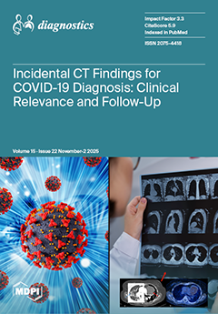
This study analyzes the prevalence and clinical relevance of incidental findings from 683 chest CT scans performed for suspected COVID-19 over nearly two years. Using the COVID-19 Pneumonia Imaging Classification and a structured relevance scale, we evaluated how these findings were identified, categorized, and clinically managed. Incidental findings occurred in 94 patients (13.8%), with women affected more frequently. Although most findings were of limited clinical significance, several required further diagnostics and work up. This study highlights the need for consistent follow-up and standardized management pathways to ensure that clinically significant incidental findings are appropriately addressed. View this paper
- Issues are regarded as officially published after their release is announced to the table of contents alert mailing list.
- You may sign up for e-mail alerts to receive table of contents of newly released issues.
- PDF is the official format for papers published in both, html and pdf forms. To view the papers in pdf format, click on the "PDF Full-text" link, and use the free Adobe Reader to open them.
Previous Issue
Next Issue