


Prosthesis 2026, 8(4), 42; https://doi.org/10.3390/prosthesis8040042 - 21 Apr 2026
Viewed by 273
Abstract
►
Show Figures
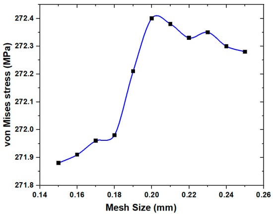
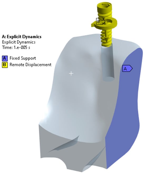
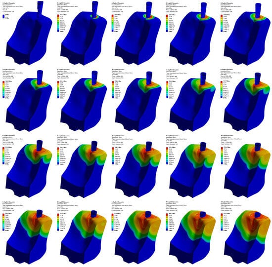
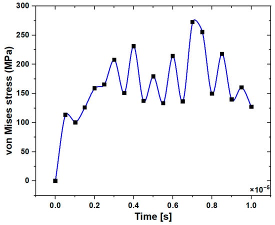
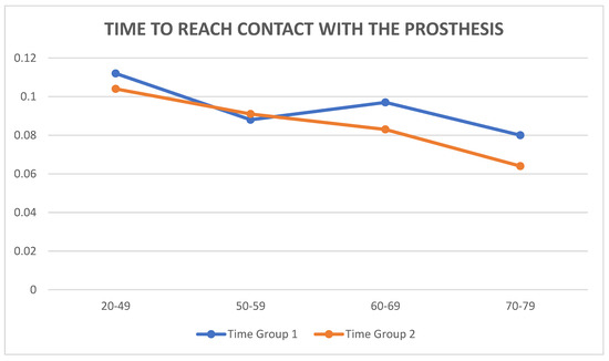
Background and aim: The influence of fabrication techniques, cement type, and cyclic loading on the marginal adaptation of lithium disilicate crowns remains a clinical concern that may affect their long-term performance. This study aimed to evaluate the effects of cyclic loading and
[...] Read more.
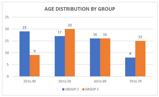
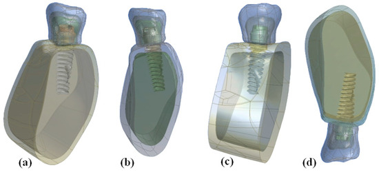
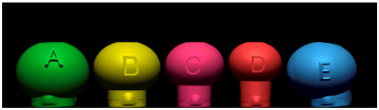
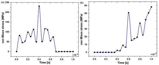
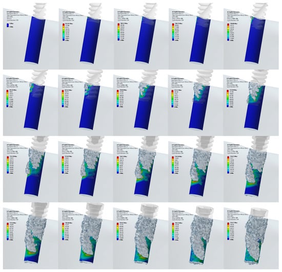
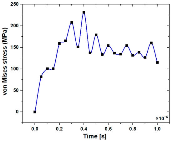
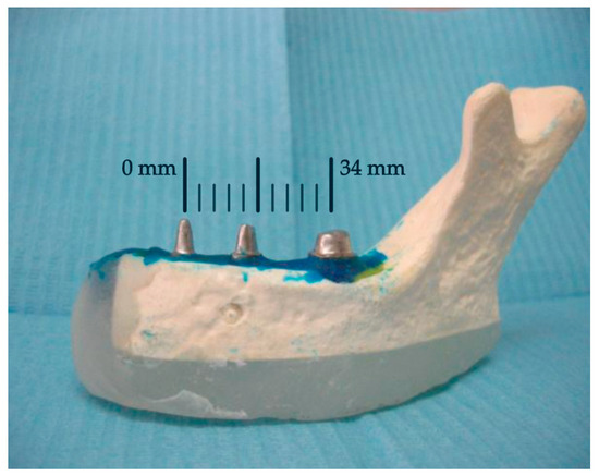
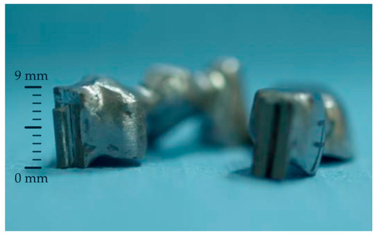
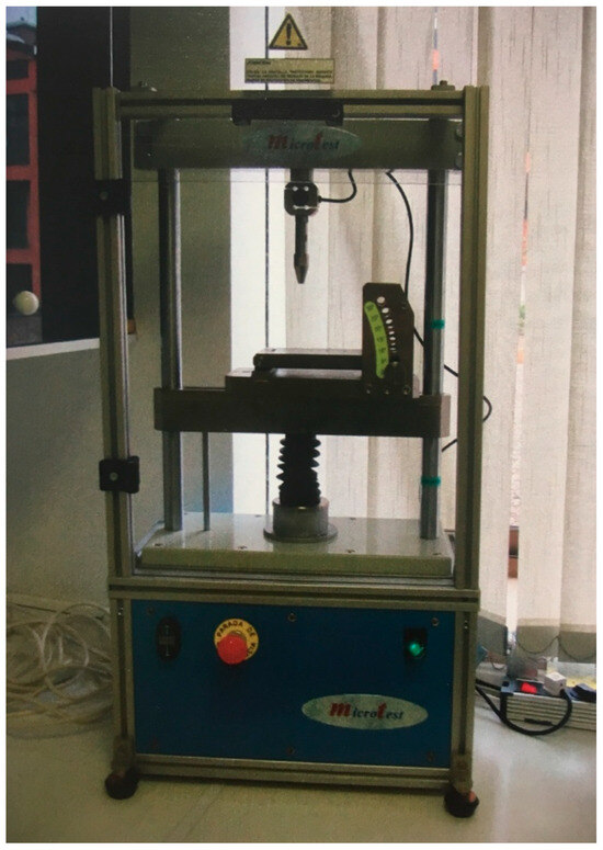
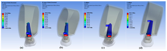
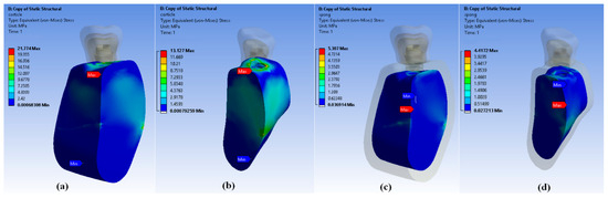
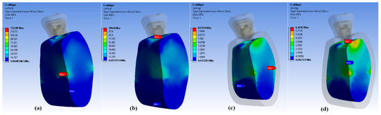
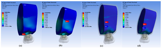
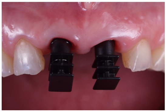
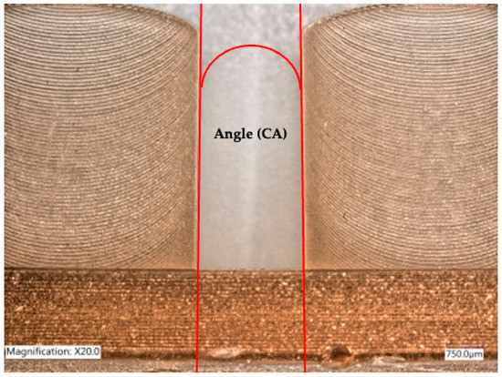
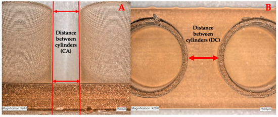
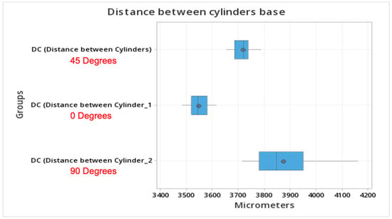
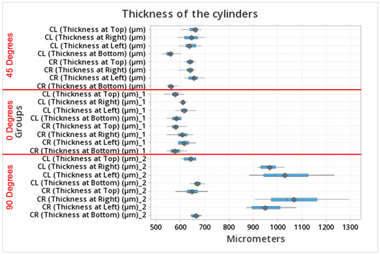
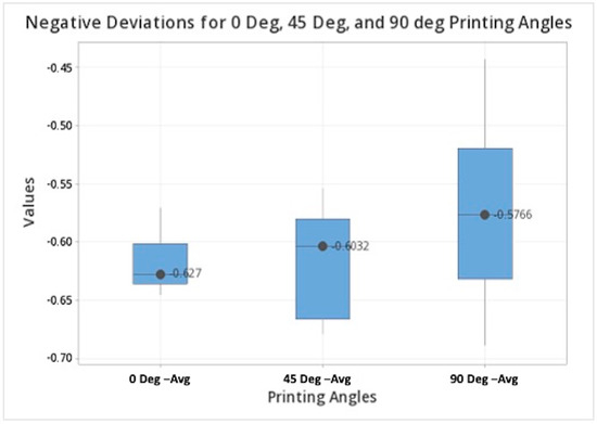
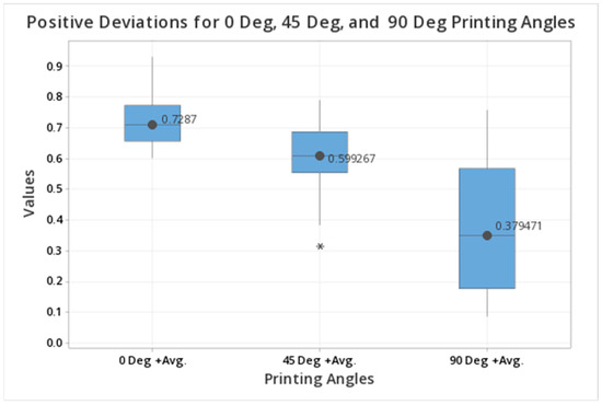
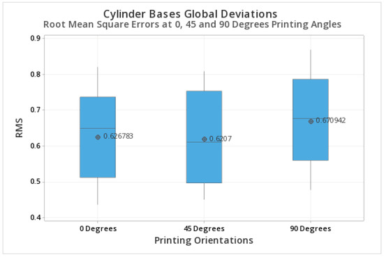
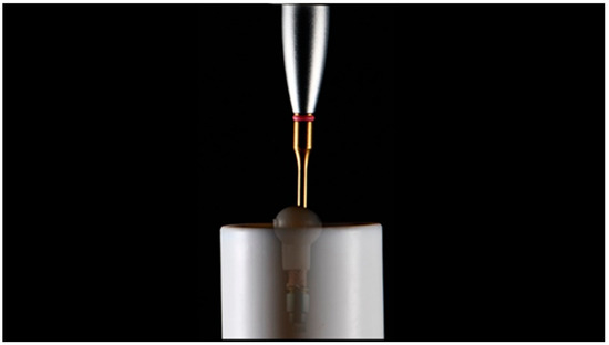
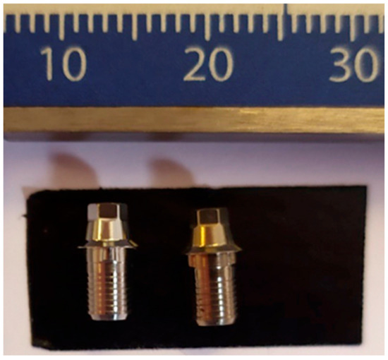
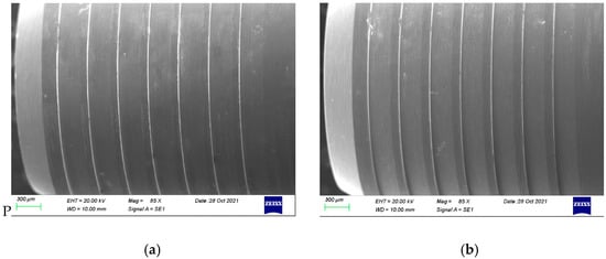
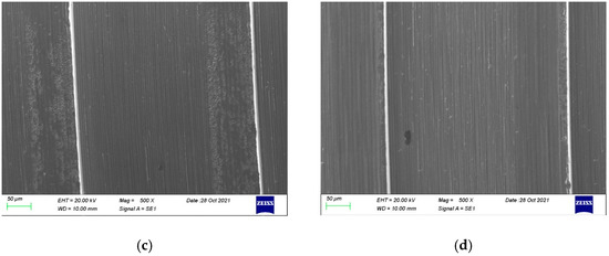
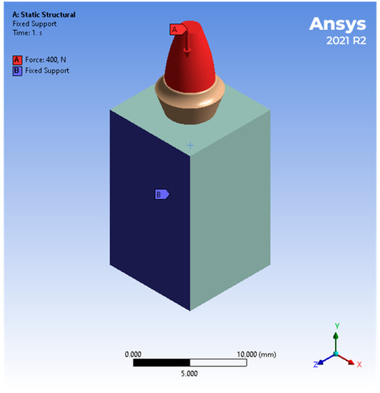
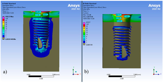
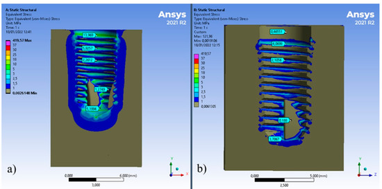
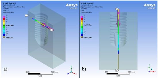
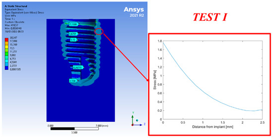
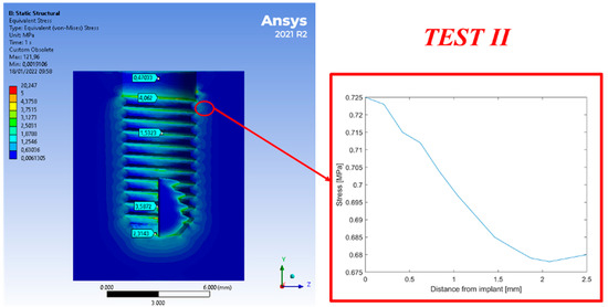
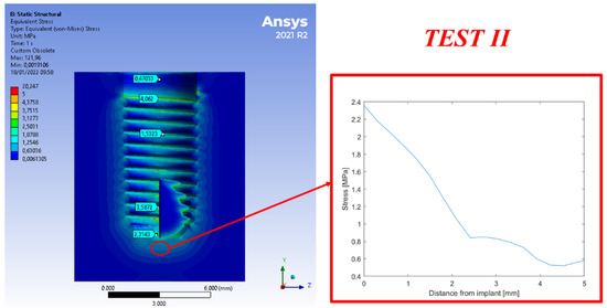
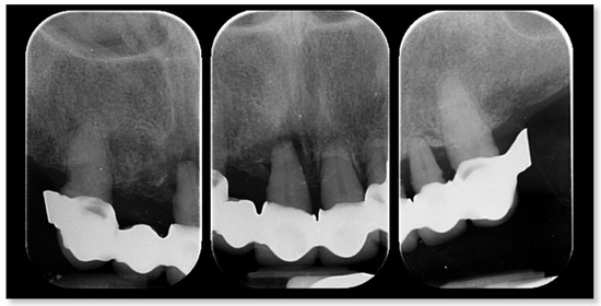
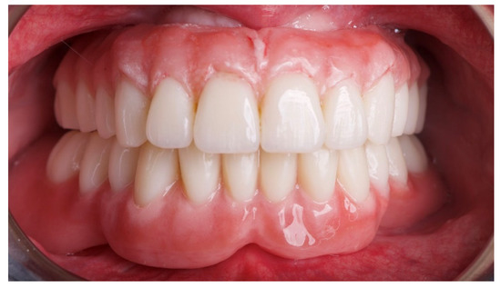
Background and aim: The influence of fabrication techniques, cement type, and cyclic loading on the marginal adaptation of lithium disilicate crowns remains a clinical concern that may affect their long-term performance. This study aimed to evaluate the effects of cyclic loading and cement type on the marginal fit of milled and pressed lithium disilicate crowns. Methods: Twenty lithium disilicate crowns were fabricated and divided into two groups based on the manufacturing technique: milled and pressed (n = 10 each). Each group was further subdivided according to the cement type: resin or resin-modified glass ionomer (n = 5 per group). Crowns were cemented on standardized epoxy resin dies, and the marginal gap was measured using a stereomicroscope before and after cyclic loading. Cyclic loading was performed at 50 N for 37,000 cycles. Data were statistically analyzed using a three-way ANOVA (α = 0.05). Results: Milled crowns showed marginal gaps ranging from 52 to 57 µm, whereas the pressed crowns exhibited smaller gaps ranging from 39 to 47 µm. Neither the cement type nor the cyclic loading produced a significant difference in the marginal gap values (p > 0.05). Conclusions: Pressed lithium disilicate crowns exhibited superior marginal adaptation compared with the milled crowns. Neither the type of cement nor the cyclic loading had a significant effect on the marginal gap. Both fabrication techniques yielded clinically acceptable fits (<100 µm).
Full article

Figure 1


