Article

Clinical, Radiological, and Aesthetic Outcomes after Placement of a Bioactive-Surfaced Implant with Immediate or Delayed Loading in the Anterior Maxilla: 1-Year Retrospective Follow-Up Study

1
Department of Oral and Maxillo-Facial Sciences, “Sapienza” University of Rome, Via Caserta 6, 00161 Rome, Italy
2
Department of Periodontology, School of Post Graduate Dentistry, Rambam Health Care Campus, Haifa P.O. Box 9602, Israel
3
Department of Neuroscience, Reproductive Sciences and Dentistry, University of Naples Federico II, 80131 Naples, Italy
4
Department of Oral and Maxillofacial Surgery and Periodontology, Faculty of Dentistry of Ribeirão Preto, University of São Paulo, São Paulo 14040-904, Brazil
5
Department of Oral Biology, Faculty of Dentistry of Ribeirão Preto, University of São Paulo, São Paulo 14040-904, Brazil
6
Department of Surgical Sciences (DISC), University of Genoa, Largo Benzi 10, 16132 Genova, Italy
*
Author to whom correspondence should be addressed.
Prosthesis 2023, 5(3), 610-621; https://doi.org/10.3390/prosthesis5030043
Submission received: 3 June 2023 / Revised: 4 July 2023 / Accepted: 5 July 2023 / Published: 7 July 2023
(This article belongs to the Collection Oral Implantology: Current Aspects and Future Perspectives)

Abstract

:
Background: Dental implants have become the standard for replacing missing teeth. However, patients’ demands for shorter treatment times and the desire for aesthetics in their results can complicate the rehabilitation process, particularly when poor-quality bone is involved. In order to address these challenges, new methods of treating implant surfaces have been introduced. These methods aim to make the implants superhydrophilic and bioactive, enhancing their functionality and interactions with the surrounding tissues. Aim: The aim of the study was to retrospectively examine the efficacy of a superhydrophilic and bioactive implant for treatment of the edentulous maxillary anterior area. The study also aimed to evaluate whether this improved implant surface, by enhancing the osseointegration processes, could serve as a factor in speeding up the loading protocols. Materials and Methods: For this retrospective study, a total of 13 implants were included: 6 delayed restored implants and 7 immediately loaded implants placed in the anterior maxillary area. Clinical, radiographic, and esthetic outcomes were assessed. Baseline measurements of the insertion torque value (ITV) and stability implant quotient (ISQ) were recorded for all implants. In the early-loaded group, these measurements were also taken 30 days (t30) and 45 days (t45) after the placement of the implant to monitor their changes over time. Marginal bone loss (MBL) was calculated according to the changes in marginal bone level on intraoral X-rays taken at two different time points: at baseline and one year after loading. To evaluate the esthetic results, the pink esthetic score (PES) and the white esthetic score (WES) were assessed. For this evaluation, intraoral photographs were taken one year after implant loading. Results: A total of 7 implants were immediately restored, with a mean ITV of 32.29 ± 9.01 Ncm and a mean ISQ of 72.71 ± 2.81. These implants were placed in a bone environment with a mean density of 410.00 ± 194.42 HU. On the other hand, 6 implants with delayed loading had a mean ITV of 28.50 ± 3.27 Ncm, an ISQ of 67.92 ± 8.43, and a mean bone density of 607.50 ± 140.83 HU. The mean PES and WES after 1 year were, respectively, 8.71 ± 1.89 and 8.57 ± 0.79 for immediate and 8.33 ± 1.36 and 9.17 ± 1.33 for delayed-loaded implants. At 12 months after loading, the immediately loaded group had a MBL of 0.29 ± 0.29 mm, while the delayed-loaded group had a MBL of 0.33 ± 0.25. No statistically significant differences between the two treatment groups were found for any of the evaluated outcomes. Conclusions: Despite the limitations of this study, the obtained results may support the use of a superhydrophilic and bioactive implant surface for implant-prosthetic rehabilitation in critical loading protocols with satisfactory esthetic results.

1. Introduction

The loss of a tooth triggers a series of physiological reactions. Initially, the extraction wound undergoes a healing process, followed by remodeling of the periodontal structures that rely on the functional presence of the tooth [1,2]. As observed by Chappius and colleagues, the remodeling of hard and soft tissues following the extraction of a tooth in the anterior maxilla has a significant impact on the aesthetic result of implant-prosthetic rehabilitation [3].
Furthermore, a tooth can be missing for congenital reasons, and there may be a dental abnormality, such as an agenesis. Bilateral or unilateral agenesis of the maxillary lateral incisors has a high prevalence rate [4]. The agenesis of an anterior tooth presents a significant risk of compromising the smile, particularly in adolescents. These cases often require extensive and multidisciplinary treatment approaches that can be long and exhausting.
Patients who have lost an anterior tooth or have agenesis in the second sextant often seek more than just functional restoration. They generally desire rehabilitation treatments that not only restore function, but also offer aesthetically pleasing results. In addition, these patients often prefer anticipated loading protocols, which expedite the process and enable faster restoration of their smile and oral function.
Over the years, irrespective of the cause of tooth loss, implantology has become a significantly reliable solution for replacing natural teeth. This reliability has been achieved through extensive research efforts, which have resulted in the development of sophisticated and perfected instruments, software, surgical techniques, and loading protocols. These advancements have contributed to the success and effectiveness of dental implant procedures, offering patients improved outcomes and greater satisfaction [5,6,7,8].
When a clinician plans to place an anterior implant, it is crucial to consider not only the primary objective of osseointegration, but also factors such as the patient’s desire for early prosthetic loading and optimal esthetic outcomes. These rehabilitations have a profound impact on the patient’s quality of life. It is, therefore, important to use all the techniques and materials available to help to achieve the best possible result in terms of function, aesthetics, and patient satisfaction.
The concept of osseointegration was introduced by Branemark in the 1980s to define the direct structural and functional connection that occurs between the bone and the fixture without the interposition of a connective tissue layer between the two surfaces [9,10].
Osseointegration involves the apposition of new bone around the implant’s surface. It is a complex and delicate process that is influenced by various factors, primarily by the quality and quantity of bone present at the fixture recipient bed. Indeed, the characteristics of the implant fixture surface, including its topography, chemical composition, surface energy, and wettability, have been extensively studied for their ability to modulate and enhance the osseointegration process. These surface characteristics play a crucial role in promoting the interaction between the implant and the surrounding bone tissue. Surface topography, such as roughness and micro- and nano-scale features, can influence cellular responses and promote better bone integration. The chemical composition and surface energy of the implant surface also affect the bioactivity and biocompatibility of the implant, influencing cellular adhesion and tissue integration. Furthermore, wettability and surface energy have an impact on the initial stability and osseointegration of the implant. It is, therefore, important to optimize these surface characteristics in order to promote successful osseointegration and improve the long-term outcomes of implant treatments [11,12,13,14].
Over time, in fact, numerous surface treatments have been introduced for dental implants, such as sandblasting and acid etching [15,16].
Modifications to implant surfaces involve creating roughened textures with the intention of increasing bone-to-implant contact. These modifications are designed to enhance the osteophilic (bone-loving) properties and hydrophilicity (water affinity) properties of the surfaces. Studies have shown that these surface modifications have a positive impact on the osseointegration rate compared to smooth surfaces.
The increased surface roughness provides a larger attachment surface for bone cells and promotes better integration between the implant and surrounding bone tissue. The improved osteophilic and hydrophilic properties of modified surfaces further facilitate the biological response and the osseointegration process. Consequently, surface modifications have shown promising results in enhancing the success and speed of osseointegration compared to implants with smooth surfaces [15,17]. The topography and chemical characteristics of implant surfaces strongly influence their wettability. Based on the contact angle, a distinction can be made between hydrophobic surfaces with a CA between 90 and 150 degrees, hydrophilic surfaces with a CA < 90 degrees, or superhydrophilic surfaces with a CA of 0 degrees [18].
Hydrophilic and superhydrophilic surfaces have better interactions with biological fluids, leading to enhanced protein adsorption. These surfaces also promote the initial attachment, proliferation, and differentiation of osteoblast-like cells. Research, such as the work by Hotchiss, has demonstrated that these surfaces can modify macrophage activation.
The hydrophilic nature of these surfaces allows them to attract and absorb water molecules, which facilitates the adsorption of proteins from the surrounding biological fluids onto the implant surface. This protein layer acts as a bridge between the implant and the surrounding cells, promoting cellular attachment and subsequent cell functions.
Additionally, hydrophilic and superhydrophilic surfaces have been shown to positively influence the behavior of osteoblast-like cells. These surfaces provide a favorable environment for cell attachment, proliferation, and differentiation, ultimately contributing to improved osseointegration.
Furthermore, studies have indicated that the characteristics of superhydrophilic surfaces can alter macrophage activation. Macrophages play a crucial role in the immune response and healing process. Modulating their activation through surface properties can influence the tissue response and the subsequent integration of the implant. [19,20,21,22,23].
This study was conducted in order to analyze the biological response of tissues following implant prosthetic rehabilitation in the anterior maxilla with immediate or delayed loading using an implant with a superhydrophilic surface activated by salts and dry technology. We also intended to study whether this type of surface, with encouraging results both in vitro and in animals, may be the key to improving and catalyzing the osseointegration process, and, consequently, accelerating loading protocols while guaranteeing biologically and aesthetically acceptable results.
The aim of this study was to analyze the clinical and radiographical response of hard and soft tissues after implant-prosthetic rehabilitation in the anterior maxilla using immediate or delayed loading protocols. The study focused on the use of implants with superhydrophilic surfaces activated by salts and dry technology. The goal was to achieve biologically and aesthetically acceptable outcomes while accelerating loading protocols for implant treatment in the anterior maxilla.

2. Materials and Methods

2.1. Study Design

This is a retrospective analysis of a series of cases treated with superhydrophilic bioactive implants inserted into the anterior maxilla according to two different loading protocols: immediate loading (Group A) and delayed loading (Group B).
Eleven systemically healthy patients (ASA 1 or ASA 2) were consecutively enrolled in a private dental clinic in Rome, where an expert surgeon (LC) performed all of the surgeries. A total of 13 consecutive implants were placed between December 2021 and March 2022, and were considered afterward for statistical analysis.

2.2. Participants

In this retrospective analysis, cases of implant-prosthetic rehabilitation in patients presenting at least one edentulous area in the maxillary anterior area were considered to be suitable for inclusion. Specifically, periodontally and systemically healthy adults (age > 18 years) and non-smokers were included. The exclusion criteria consisted of patients under the age of 18, smokers, patients with untreated periodontitis, patients with a history of implant failure at the surgical site, and individuals receiving bisphosphonate treatment or suffering from systemic diseases likely to interfere with the implant rehabilitation process. These exclusion criteria were implemented to ensure a more homogeneous study population and to minimize confounding factors that could affect the outcomes of the study.

2.3. Preoperative Procedure

Photographs and periapical X-rays were taken and cone beam computed tomography (CBCT) and intraoral scanning were used during screening visits. Dedicated software (RealGUIDE 5.0 software, 3Diemme, Cantù, Italy) was used to digitally plan the implant’s placement and to provide information about the bone environment at the surgical site. The virtual implant plan was sent to the dental lab for the creation of a resin 3D-printed surgical template, which was used to perform all surgical procedures.
To ensure optimal oral health and minimize the risk of complications, each patient underwent professional tooth cleaning prior to the surgery. In addition, they received home oral hygiene instructions to maintain proper oral hygiene until the day of the surgery. The goal was to achieve a full mouth bleeding score and full mouth plaque score of less than 20% to ensure a healthy oral environment for the surgical procedure.

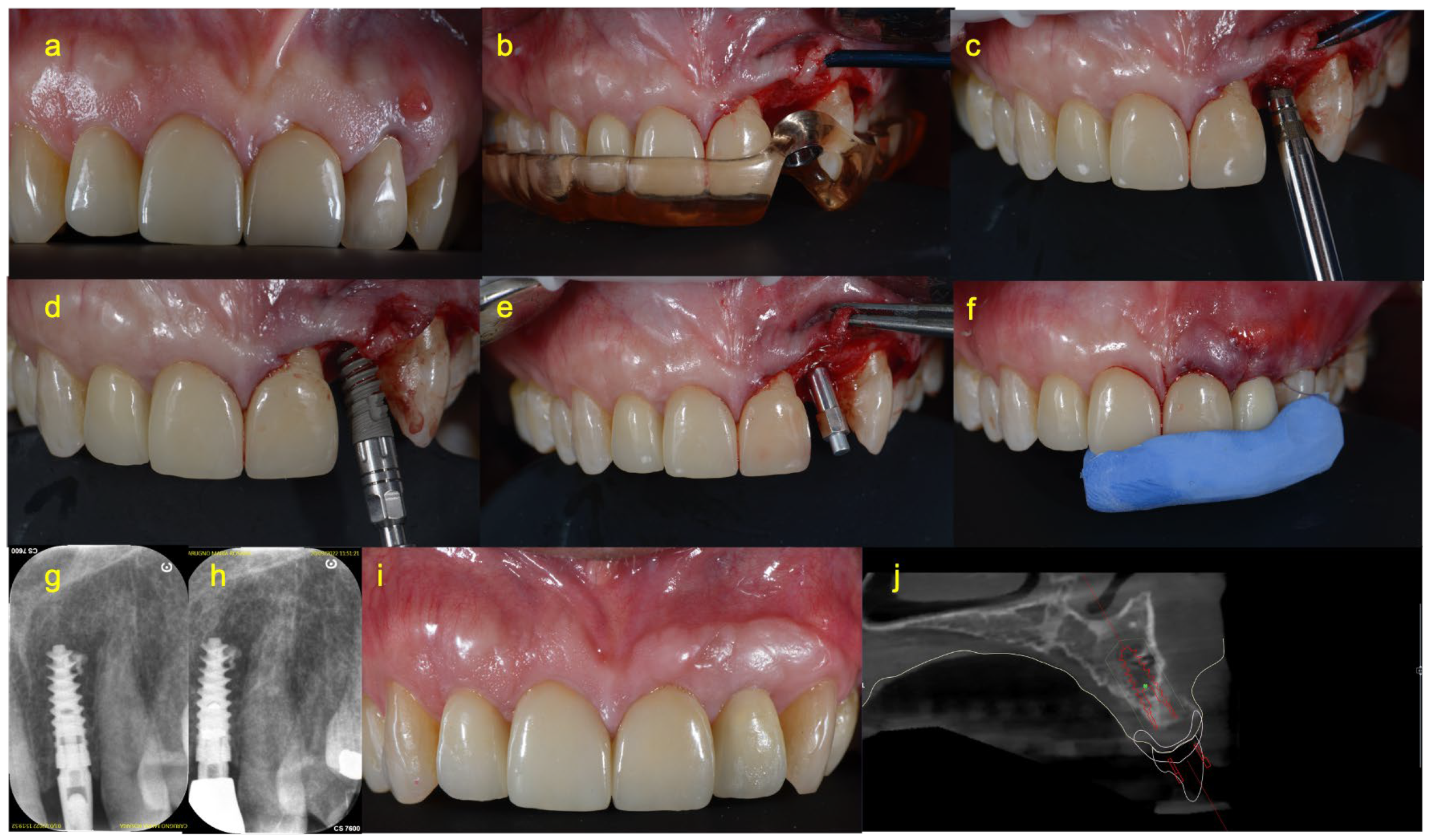
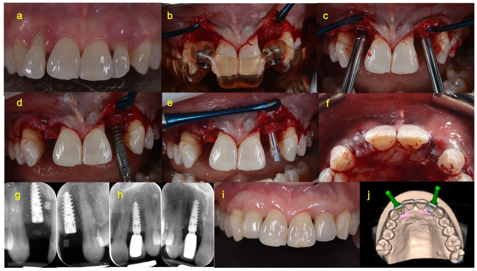
2.4. Surgical Procedure

The same surgical procedure was applied to all patients. After the administration of local anesthesia (articaine 4% with adrenaline 1:100.000), a flap was raised with a minimally invasive approach. The surgical guide was inserted immediately after the exposure of the alveolar ridge, and the preparation of the implant site was carried out with the hybrid funnel technique described previously [24], using a specific sequence of drills and osteotomes (Figure 1b,c and Figure 2b,c).
The surgical guide was then removed, and the super-hydrophilic bioactive implant (Multineo NH CS, Alpha Bio Tec, Petah Tikva, Israel) was inserted into the surgical site. The insertion torque value (ITV) and primary stability (implant stability quotient—ISQ) were recorded at the time of the implant’s placement. Intraoral digital radiography was performed using the parallel long-cone technique with Rinn X-ray holders to check whether the implant placement was correct (Figure 1e,g and Figure 2e,g).
The subsequent prosthetic procedures differed between the two treatment groups.

2.5. Prosthetic Procedure of Immediate Loading Group (Group A)

In the immediate loading group, on the day of implant surgery, after measuring primary stability, patients underwent an immediate restoration without occlusal loading using the one abutment at one time (OAOT) protocol [25,26] and a PMMA crown (Figure 1f).
At 90 days, the implant was functionally loaded with a zirconia crown. The definitive restoration was cemented with interim cement (Temp-Bond; Kerr Corp, Orange, CA, 92867 USA) (Figure 1i). An intraoral periapical X-ray was taken using the parallel long-cone technique.

2.6. Prosthetic Procedure of Delayed Loading Group (Group B)

In the delayed loading group, on the day of implant surgery, after measuring primary stability, the cover screw was inserted and the flap was sutured. A temporary aesthetic restoration (Maryland bridge) was provisionally cemented to the neighboring teeth.
Next, 30 days after the placement of the implant (T30), the Maryland bridge was removed, a small incision was made with a 15c blade, and the implant was exposed. A SmartPeg was screwed to the fixture, and the implant’s stability was checked. A scan body was then placed in order to create a digital impression for the fabrication of the provisional restoration. Finally, the Maryland bridge was cemented once again.
At 45 days (T45), the Maryland bridge was removed, the implant’s stability was assessed using a SmartPeg, and the implant was functionally loaded with a PMMA prosthetic crown cemented with interim cement (Temp-Bond; Kerr Corp).
At 90 days, a zirconia crown was cemented with interim cement (Temp-Bond; Kerr Corp) and an intraoral periapical X-ray was performed (Figure 2i).

2.7. Postoperative Management

Antibiotic therapy was prescribed to all patients. Specifically, they were instructed to begin taking amoxicillin/clavulanic acid (875 mg/125 mg) 12 h prior to the surgery. The antibiotic regimen was to be continued for four days, with doses taken three times a day [27].
In addition to antibiotics, pain management was addressed by prescribing ibuprofen (600 mg) twice a day for a duration of three days.
Patients were instructed to avoid brushing the surgical area or chewing for 2 weeks. Professional mechanical tooth cleaning was scheduled every 3/4 months.

2.8. Clinical and Radiographical Measurements

The ISQ was calculated by resonance frequency analysis (RFA) using an Osstell device (W&H Osstell ISQ module, W&H srl, Austria), after the insertion of a sterile disposable device called a Smart Peg (Osstell AB, Stampgatan 14,41101 Goteborg, Sweden) on the implant head. Two ISQ measurements were taken, one bucco-palatal and one mesio-distal. The mean value of the two measurements was considered for the analysis. The ISQ value was collected at the time of implant insertion (T0) in the immediate loading group, and at T0, T30, and T45 in the delayed loading group.
The ITV (Ncm) value was recorded at T0 using a surgical micromotor (Implantmed SI-1010; W&H srl, Austria) during the placement of the implant.
Marginal bone loss (MBL) was evaluated using specific software (CS 2200, Carestream, Rochester, NY, USA) and digital periapical radiographs taken both at baseline and 12 months after loading, as well as by measuring changes in the bone level between the two points in time. The MBL was calculated as the mean value of two measurements taken on the mesial and distal sides of each implant.
Bone density was assessed from preoperative CBCT in Hounsfield units (HU) using RealGUIDE 5.0 software (3Diemme, Cantù, Italy) in order to determine the bone environment of the healed site after tooth extraction.
The pink esthetic score (PES) and white esthetic scores (WES) were calculated according to the variables described by Belser et al. [28] to evaluate the aesthetic results of the peri-implant soft tissues and the visible parts of the implant restorations. Scores from 0 to 2 for each of the 5 WES parameters (tooth form, tooth volume/outline, color, surface texture, and translucency) and the 5 PES parameters (mesial papilla, distal papilla, facial mucosal curvature, facial mucosal level, and root convexity/soft tissue color and texture) were assigned by a single operator (PP) observing an intraoral photograph taken with a Nikon D800 with a 105 mm macro lens. (Figure 1i and Figure 2i).

2.9. Statistical Methods

All statistical analyzes were performed using SPSS software, version 25.0. Microsoft Excel (Microsoft Corporation, Washington, DC, USA) was used for data collection. The descriptive variables are presented as means and standard deviations (SDs).
Differences between the immediately restored implant group and the delayed restored implant group were analyzed using the Mann–Whitney U Test. The results were considered significant at a p-value < 0.05.

3. Results

Thirteen single-implant prosthetic rehabilitations in the maxillary anterior area, performed on eleven patients (nine female and two male), were considered for this retrospective study. The demographic factors are summarized in Table 1. Of the 11 patients considered, 5 lost their teeth due to endodontic complications, 2 due to periodontal complications, and 4 due to agenesis of the upper lateral incisors. In total, 7 implants were immediately loaded, and 6 implants were loaded 45 days after the placement of the implant.
Implants with diameters of 3.3 mm, 3.75 mm, or 10 mm in length were placed in a bone site prepared according to a specific osteotomy technique. In group A, the implants were inserted with a mean ITV of 32.29 ± 9.01 Ncm and bone density with a mean value of 410.00 ± 194.42 HU. The ISQ was recorded at t0 with a mean value of 72.71 ± 2.81.
In group B, the mean ITV recorded during implant placement was 28.50 ± 3.27 Ncm, and the mean bone density evaluated using the HU scale was 607.50 ± 140.83. The ISQ was recorded at three different time points—at T0, T30, and T45—and the mean values were 67.92 ± 8.43, 68.42 ± 9.00, and 68.42 ± 6.47, respectively.
No statistically significant differences were found between the baseline ITV and ISQ of the two groups treated with different loading protocols.
Minimal changes in MBL were observed radiographically, with no statistically significant differences between the two treatment groups; a mean MBL of 0.33 ± 0.25 mm in the delayed group and 0.29 ± 0.29 mm in the immediate group were detected.
The PES and WES scores were obtained by adding up the scores given to the five variables of each of the two indices. The mean values of PES and WES for delayed-loading implant rehabilitations were 8.33 ± 1.36 and 9.17 ± 1.33. Despite the absence of statistically significant differences, the immediately loaded implants showed slightly higher PES scores, with a mean value of 8.71 ± 1.89, and lower WES scores, with a mean value of 8.57 ± 0.79 (Table 2).

4. Discussion

Currently, implant-supported rehabilitation represents a predictable treatment protocol in terms of its success rate. However, when it comes to rehabilitating the anterior area, clinicians face additional challenges due to the significant esthetic implications involved. The anterior region is highly visible during smiling and talking, and any esthetic discrepancies can have a profound impact on the patient’s self-confidence and satisfaction with the treatment outcome.
In the present study, two different approaches approaches were used in the esthetic area: immediate loading and early loading. At the end of the one-year follow-up, no significant differences were noted, either in terms of marginal bone level changes or esthetic outcomes.
The literature suggests that immediate nonfunctional loading is considered an ideal protocol for shaping soft tissues in implant-supported rehabilitations, particularly in the anterior area.
According to Weigl et al. this procedure may produce promising results, although a strict patient selection process is required [29]. On the other hand, according to Buser et al., traditional staged approaches, which often involve bone regeneration and soft tissue management, may result in a safer achievement of the esthetic outcomes [30]. This may come from the fact that staged approaches are meant to allow for both soft and hard tissue healing, leaving more room for tissue management in the second stage.
However, the present study confirms that, from a clinical point of view, no differences could be perceived. The results of the present studies are in line with the findings reported by Puisys et al. and the Lithuanian group, indicating that there is no significant esthetic difference between immediate implant insertion with non-functional restoration and staged approaches. These studies suggest that both immediate and staged protocols can provide favorable esthetic outcomes in implant-supported rehabilitation [31].
Implant rehabilitation for the treatment of agenesis of the lateral incisors after an orthodontic space opening was evaluated by the authors as the most suitable choice regarding the profile of the hard and soft tissues. The canines and first premolars were preserved from changes in the coronal morphology. The use of a bioactive surface allowed these patients and for all the others included in the study to experience quick rehabilitation, with the definitive restoration inserted 90 days after implant placement. Furthermore, the choice of narrow implants (3.3 mm in diameter) was made to increase the distance from the residual teeth, thus avoiding ridge augmentation and reabsorption of interdental bone peaks and favoring complete papilla formation.
To better explain the absence of significant differences in terms of MBL, the adopted prosthetic protocol can be suggested. Indeed, one abutment at one time was adopted in both groups. In fact, this prosthetic approach was proven to minimize the microdamages at the supracrestal connective component when compared to traditional work-flow. This might minimize the marginal bone loss and increase the soft tissue response.
The hybrid funnel technique, described by Canullo et al. and used for the preparation of the implant site, was developed with the aim of minimizing the stress on the cortical bone by preparing the crestal area of the same diameter as the neck of the implant, therefore avoiding mismatching [24].
At the same time, in the present study, a bioactive surface was adopted to verify whether a super-hydrophilic surface could be advantageous for accelerating osseointegration and shortening loading times in both clinical scenarios.
In fact, immediate non-functional loading can be a risky procedure, especially in low quality/quantity bone environments.
Over the last two decades, the role of implant surfaces in the osseointegration process has been the subject of ongoing debate and research [16,31,32,33]. The surface energy of an implant correlates with its wettability, or hydrophilicity, which refers to the ability of the surface to interact with liquids and cells. Various methods have been used to increase the surface energy of implants [34].
The activation of implant surfaces by increasing surface energy has shown biological advantages in terms of cell adhesion. This activation leads to qualitative and quantitative improvements in cell adhesion [35]. Qualitatively, there is an increase in the number of adhered cells, while quantitatively, the arrangement of cells is more spread out rather than flat. This stronger cell adhesion promotes better osseointegration and enhances the overall success of the implant [36].
These findings suggest that modifying implant surfaces to increase their surface energy and hydrophilicity can positively influence the biological responses, leading to improved osseointegration and, ultimately, better clinical outcomes for patients [37].
Titanium is known for its excellent bioactivity at the time of production, meaning it has favorable interactions with biological tissues and promotes osseointegration. However, the bioactivity of titanium surfaces tends to decrease over time, especially during storage [38]. This decrease in bioactivity can impact the rate of osseointegration and overall implant success.
The development of bioactive surfaces aims to address this issue by creating surfaces that maintain their bioactivity even during the storage phase. These surfaces are designed to enhance the secondary stability of implants and to accelerate peri-implant bone formation. They promote a more favorable biological response, leading to faster and more robust osseointegration.
Research conducted by Romero-Ruiz et al. [39] has confirmed the enhanced biological response and accelerated peri-implant bone formation associated with these bioactive surfaces. This suggests that the use of these surfaces can be beneficial in promoting successful implant osseointegration and improving overall treatment outcomes.
By using bioactive surfaces that maintain their stability and bioactivity over time, clinicians can enhance the osseointegration process and achieve better long-term results in implant-prosthetic rehabilitation.
Superhydrophilic surfaces may positively influence implant osseointegration, even in post-extraction cases. In fact, according to Clauser et al., there is clear evidence of the association between bone grafting and early implant failure [40]. In the present study, in fact, no failure was registered after the follow-up time.
The main limitation of the present study is its small sample size. In fact, in order to draw clinically relevant conclusions confirming the outcomes of the present study, a larger number of patients is needed, along with the presence of a control group that allows for comparison between two different implant surfaces. Another limitation may be represented by the study design; a prospective controlled trial, even better if randomized, may suggest stronger conclusions compared to a retrospective design.

5. Conclusions

Beyond the limitations of this study and on the basis of the slight MBL which was observed and the PES and WES values which were obtained, this retrospective analysis showed encouraging clinical, radiographical, and esthetic results following the use of a super hydrophilic and bioactive implant surface in the cases of both immediate loading and early loading.

Author Contributions

Conceptualization, R.I. and F.B.; methodology, R.I. and Y.M.; formal analysis, R.I. and G.M.; investigation, M.M.; resources, B.V.F. and G.E.P.; data curation, R.I.; writing—original draft preparation, R.I; writing—review and editing, R.I., F.B. and Y.M.; visualization, G.M.; supervision, F.B. All authors have read and agreed to the published version of the manuscript.

Funding

This research received no external funding.

Institutional Review Board Statement

Not applicable.

Informed Consent Statement

Informed consent was obtained from all subjects involved in the study.

Data Availability Statement

Data are available from corresponding author at reasonable request.

Conflicts of Interest

The authors declare no conflict of interest.

References

  1. Amler, M.H. The time sequence of tissue regeneration in human extraction wounds. Oral Surg. Oral Med. Oral Pathol. 1969, 27, 309–318. [Google Scholar] [CrossRef] [PubMed]
  2. Cardaropoli, G.; Araújo, M.; Lindhe, J. Dynamics of bone tissue formation in tooth extraction sites. An experimental study in dogs. J. Clin. Periodontol. 2003, 30, 809–818. [Google Scholar] [CrossRef]
  3. Chappuis, V.; Araújo, M.G.; Buser, D. Clinical relevance of dimensional bone and soft tissue alterations post-extraction in esthetic sites. Periodontol. 2000 2017, 73, 73–83. [Google Scholar] [CrossRef]
  4. Polder, B.J.; Van’t Hof, M.A.; Van der Linden, F.P.; Kuijpers-Jagtman, A.M. A meta-analysis of the prevalence of dental agenesis of permanent teeth. Community Dent. Oral Epidemiol. 2004, 32, 217–226. [Google Scholar] [CrossRef]
  5. Canullo, L.; Peñarrocha, D.; Peñarrocha, M.; Rocio, A.G.; Penarrocha-Diago, M. Piezoelectric vs. conventional drilling in implant site preparation: Pilot controlled randomized clinical trial with crossover design. Clin. Oral Implant. Res. 2014, 25, 1336–1343. [Google Scholar] [CrossRef] [PubMed]
  6. Jorba-García, A.; González-Barnadas, A.; Camps-Font, O.; Figueiredo, R.; Valmaseda-Castellón, E. Accuracy assessment of dynamic computer-aided implant placement: A systematic review and meta-analysis. Clin. Oral Investig. 2021, 25, 2479–2494. [Google Scholar] [CrossRef] [PubMed]
  7. Viña-Almunia, J.; Maestre-Ferrín, L.; Alegre-Domingo, T.; Peñarrocha-Diago, M. Survival of implants placed with the osteotome technique: An update. Med. Oral Patol. Oral Cir. Bucal 2012, 17, e765–e768. [Google Scholar] [CrossRef] [Green Version]
  8. Smeets, R.; Stadlinger, B.; Schwarz, F.; Beck-Broichsitter, B.; Jung, O.; Precht, C.; Kloss, F.; Gröbe, A.; Heiland, M.; Ebker, T. Impact of Dental Implant Surface Modifications on Osseointegration. Biomed. Res. Int. 2016, 2016, 6285620. [Google Scholar] [CrossRef] [Green Version]
  9. Brånemark, P.I.; Hansson, B.O.; Adell, R.; Breine, U.; Lindström, J.; Hallén, O.; Ohman, A. Osseointegrated implants in the treatment of the edentulous jaw. Experience from a 10-year period. Scand. J. Plast. Reconstr. Surg. Suppl. 1977, 16, 1–132. [Google Scholar]
  10. Albrektsson, T.; Brånemark, P.I.; Hansson, H.A.; Lindström, J. Osseointegrated titanium implants: Requirements for ensuring a long-lasting, direct bone-to-implant anchorage in man. Acta Orthop. Scand. 1981, 52, 155–170. [Google Scholar] [CrossRef] [Green Version]
  11. Wieland, M.; Sittig, C.; Brunette, D.M.; Textor, M.; Spencer, N.D. Measurement and evaluation of the chemical composition and topography of titanium implant surfaces. In Bone Engineering; Davies, J.E., Ed.; Em Squared: Toronto, ON, Canada, 2000; pp. 163–182. [Google Scholar]
  12. Buser, D.; Schenk, R.K.; Steinemann, S.; Fiorellini, J.P.; Fox, C.H.; Stich, H. Influence of surface characteristics on bone integration of titanium implants. A histomorphometric study in miniature pigs. J. Biomed. Mater. Res. 1991, 25, 889–902. [Google Scholar] [CrossRef] [PubMed]
  13. Rupp, F.; Liang, L.; Geis-Gerstorfer, J.; Scheideler, L.; Hüttig, F. Surface characteristics of dental implants: A review. Dent. Mater. 2018, 34, 40–57. [Google Scholar] [CrossRef] [PubMed]
  14. Gittens, R.A.; Olivares-Navarrete, R.; Cheng, A.; Anderson, D.; McLachlan, T.; Stephan, I.; Geis-Gerstorfer, J.; Sandhage, K.H.; Fedorov, A.G.; Rupp, F.; et al. The roles of titanium surface micro/nanotopography and wettability on the differential response of human osteoblast lineage cells. Acta Biomater. 2013, 9, 6268–6277. [Google Scholar] [CrossRef] [Green Version]
  15. Le Guéhennec, L.; Soueidan, A.; Layrolle, P.; Amouriq, Y. Surface treatments of titanium dental implants for rapid osseointegration. Dent. Mater. 2007, 23, 844–854. [Google Scholar] [CrossRef]
  16. Chambrone, L.; Shibli, J.A.; Mercúrio, C.E.; Cardoso, B.; Preshaw, P.M. Efficacy of standard (SLA) and modified sandblasted and acid-etched (SLA ctive) dental implants in promoting immediate and/or early occlusal loading protocols: A systematic review of prospective studies. Clin. Oral Implant. Res. 2015, 26, 359–370. [Google Scholar] [CrossRef]
  17. Kim, H.-Y.; Kim, S.-C. Bone cutting capacity and osseointegration of surface-treated orthodontic mini-implants. Korean J. Orthod. 2016, 46, 386–394. [Google Scholar] [CrossRef] [PubMed]
  18. Rupp, F.; Gittens, R.A.; Scheideler, L.; Marmur, A.; Boyan, B.D.; Schwartz, Z.; Geis-Gerstorfer, J. A review on the wettability of dental implant surfaces I: Theoretical and experimental aspects. Acta Biomater. 2014, 10, 2894–2906. [Google Scholar] [CrossRef] [PubMed] [Green Version]
  19. Yamamura, K.; Miura, T.; Kou, I.; Muramatsu, T.; Furusawa, M.; Yoshinari, M. Influence of various superhydrophilic treatments of titanium on the initial attachment, proliferation, and differentiation of osteoblast-like cells. Dent. Mater. J. 2015, 34, 120–127. [Google Scholar] [CrossRef] [Green Version]
  20. Lang, N.P.; Salvi, G.E.; Huynh-Ba, G.; Ivanovski, S.; Donos, N.; Bosshardt, D.D. Early osseointegration to hydrophilic and hydrophobic implant surfaces in humans. Clin. Oral Implant. Res. 2011, 22, 349–356. [Google Scholar] [CrossRef]
  21. Moses, O.; Bengazi, F.; Ferri, M.; Gianfreda, F.; Urbizo Velez, J.; Botticelli, D.; Canullo, L. Bioactivated Implant Surfaces Placed in Healed Sites or Extraction Sockets: A Preliminary Experimental Study in Dogs. Int. J. Oral Maxillofac. Implant. 2022, 37, 963–970. [Google Scholar] [CrossRef]
  22. Gianfreda, F.; Raffone, C.; Antonacci, D.; Mussano, F.; Genova, T.; Chinigò, G.; Canullo, L.; Bollero, P. Early Biological Response of an Ultra-Hydrophilic Implant Surface Activated by Salts and Dry Technology: An In-Vitro Study. Appl. Sci. 2021, 11, 6120. [Google Scholar] [CrossRef]
  23. Hotchkiss, K.M.; Reddy, G.B.; Hyzy, S.L.; Schwartz, Z.; Boyan, B.D.; Olivares-Navarrete, R. Titanium surface characteristics, including topography and wettability, alter macrophage activation. Acta Biomater. 2016, 31, 425–434. [Google Scholar] [CrossRef] [Green Version]
  24. Canullo, L.; Iacono, R.; Pires Godoy, E.; Punzo, A.; Cavicchia, A.; Gianfreda, F.; Bollero, P. Hybrid Funnel Technique: A Novel Approach for Implant Site Preparation: A Pilot Study. Dent. J. 2022, 10, 157. [Google Scholar] [CrossRef]
  25. Canullo, L.; Omori, Y.; Amari, Y.; Iannello, G.; Pesce, P. Five-year cohort prospective study on single implants in the esthetic area restored using one-abutment/one-time prosthetic approach. Clin. Implant. Dent. Relat. Res. 2018, 20, 668–673. [Google Scholar] [CrossRef] [PubMed]
  26. Canullo, L.; Pesce, P.; Tronchi, M.; Fiorellini, J.; Amari, Y.; Penarrocha, D. Marginal soft tissue stability around conical abutments inserted with the one abutment-one time protocol after 5 years of prosthetic loading. Clin. Implant. Dent. Relat. Res. 2018, 20, 976–982. [Google Scholar] [CrossRef]
  27. Canullo, L.; Troiano, G.; Sbricoli, L.; Guazzo, R.; Laino, L.; Caiazzo, A.; Pesce, P. The Use of Antibiotics in Implant Therapy: A Systematic Review and Meta-Analysis with Trial Sequential Analysis on Early Implant Failure. Int. J. Oral Maxillofac. Implant. 2020, 35, 485–494. [Google Scholar] [CrossRef] [PubMed]
  28. Belser, U.C.; Grütter, L.; Vailati, F.; Bornstein, M.M.; Weber, H.P.; Buser, D. Outcome evaluation of early placed maxillary anterior single-tooth implants using objective esthetic criteria: A cross-sectional, retrospective study in 45 patients with a 2-to 4-year follow-up using pink and white esthetic scores. J. Periodontol. 2009, 80, 140–151. [Google Scholar] [CrossRef]
  29. Weigl, P.; Strangio, A. The impact of immediately placed and restored single-tooth implants on hard and soft tissues in the anterior maxilla. Eur. J. Oral Implantol. 2016, 9 (Suppl. S1), S89–S106. [Google Scholar]
  30. Buser, D.; Chappuis, V.; Belser, U.C.; Chen, S. Implant placement post extraction in esthetic single tooth sites: When immediate, when early, when late? Periodontology 2000 2017, 73, 84–102. [Google Scholar] [CrossRef]
  31. Puisys, A.; Auzbikaviciute, V.; Vindasiute-Narbute, E.; Pranskunas, M.; Razukevicus, D.; Linkevicius, T. Immediate implant placement vs. early implant treatment in the esthetic area. A 1-year randomized clinical trial. Clin. Oral Implant. Res. 2022, 33, 634–655. [Google Scholar] [CrossRef]
  32. Pera, F.; Menini, M.; Alovisi, M.; Crupi, A.; Ambrogio, G.; Asero, S.; Marchetti, C.; Canepa, C.; Merlini, L.; Pesce, P.; et al. Can Abutment with Novel Superlattice CrN/NbN Coatings Influence Peri-Implant Tissue Health and Implant Survival Rate Compared to Machined Abutment? 6-Month Results from a Multi-Center Split-Mouth Randomized Control Trial. Materials 2022, 16, 246. [Google Scholar] [CrossRef] [PubMed]
  33. Alovisi, M.; Carossa, M.; Mandras, N.; Roana, J.; Costalonga, M.; Cavallo, L.; Pira, E.; Putzu, M.G.; Bosio, D.; Roato, I.; et al. Disinfection and Biocompatibility of Titanium Surfaces Treated with Glycine Powder Airflow and Triple Antibiotic Mixture: An In Vitro Study. Materials 2022, 15, 4850. [Google Scholar] [CrossRef] [PubMed]
  34. Pesce, P.; Menini, M.; Santori, G.; Giovanni, E.; Bagnasco, F.; Canullo, L. Photo and Plasma Activation of Dental Implant Titanium Surfaces. A Systematic Review with Meta-Analysis of Pre-Clinical Studies. J. Clin. Med. 2020, 9, 2817. [Google Scholar] [CrossRef] [PubMed]
  35. Oates, T.W.; Valderrama, P.; Bischof, M.; Nedir, R.; Jones, A.; Simpson, J.; Toutenburg, H.; Cochran, D.L. Enhanced implant stability with a chemically modified SLA surface: A randomized pilot study. Int. J. Oral Maxillofac. Implant. 2007, 22, 755–760. [Google Scholar]
  36. Pesce, P.; Del Fabbro, M.; Menini, M.; De Giovanni, E.; Annunziata, M.; Khijmatgar, S.; Canullo, L. Effects of abutment materials on peri-implant soft tissue health and stability: A network meta-analysis. J. Prosthodont. Res. 2023; Epub ahead of print. [Google Scholar] [CrossRef]
  37. Pesce, P.; Menini, M.; Tommasato, G.; Patini, R.; Canullo, L. Influence of modified titanium abutment surface on peri-implant soft tissue behaviour: A systematic review of histological findings. Int. J. Oral Implantol. 2019, 12, 419–429. [Google Scholar]
  38. Att, W.; Hori, N.; Takeuchi, M.; Ouyang, J.; Yang, Y.; Anpo, M.; Ogawa, T. Time-dependent degradation of titanium osteoconductivity: An implication of biological aging of implant materials. Biomaterials 2009, 30, 5352–5363. [Google Scholar] [CrossRef]
  39. Romero-Ruiz, M.M.; Gil, F.; Ríos-Santos, J.V.; Lázaro-Calvo, P.; Ríos-Carrasco, B.; Herrero-Climent, M. Influence of a Novel Surface of Bioactive Implants on Osseointegration: AComparative and Histomorfometric Correlation and Implant Stability Study in Minipigs. Int. J. Mol. Sci. 2019, 20, 2307. [Google Scholar] [CrossRef] [Green Version]
  40. Clauser, T.; Lin, G.H.; Lee, E.; Del Fabbro, M.; Wang, H.L.; Testori, T. Risk of early implant failure in grafted and non-grafted sites: A systematic review and meta-analysis. Int. J. Oral Implantol. 2022, 15, 31–41. [Google Scholar]
Figure 1. A case of a patient treated with an immediately loaded implant. (a): The photograph was taken at the time of the first visit. Tooth 22 was extracted due to endodontic complications about 5 months before the implant surgery. (b): A 3D-printed implant surgical guide was used for implant site preparation according to the hybrid funnel technique. (c) A specific osteotome was used to complete the implant site preparation according to the hybrid funnel technique. (d): An implant with a bioactive and super-hydrophilic surface was placed. (e): The implant stability quotient was measured after implant placement. (f): A definitive abutment and a temporary crown were inserted during the implant surgery. (g): A periapical X-ray was taken at baseline. (h): A periapical X-ray was taken at the 1-year follow-up visit. (i): This photograph was taken 12 months after the insertion of definitive restoration, and was used to calculate the white esthetic score and pink esthetic score. (j) Virtual implant planning.
Prosthesis 05 00043 g001
Figure 2. A case of a patient treated with an early-loaded implant. (a): A digital photograph of the second sextant was taken at the first visit. Teeth 12 and 22 were extracted 5 months before implant surgery. (b): Implant site preparation using drills and a surgical template—step 1 of the hybrid funnel technique. (c): Implant site preparation using osteotomes—step 2 of the hybrid funnel technique. (d): Insertion of an implant with a super-hydrophilic surface. (e): Measurement of implant stability by screwing the SmartPeg onto the implant fixture. (f): Flaps were sutured over the submerged implants. (g): Baseline periapical radiographs. (h): Intraoral periapical X-ray taken at the 1-year follow-up visit. (i): This clinical photograph was taken 12 months after the insertion of definitive restoration (90 days after implant placement), and was used to calculate the white esthetic score and pink esthetic score. (j): Virtual implant planning.
Prosthesis 05 00043 g002
Table 1. Demographic Parameters.
Demographic ParametersImmediately Loaded Group
n Implant = 7
Delayed-Loaded Group
n Implant = 6
p Value
Age (mean ± SD)37 ± 24.8658.60 ± 8.68p = 0.329 a
Sex (Female/male)5 females (83.33%)/1 male (16.67%)4 females (80%)/1 male (20%)
Implant diameter (3.3/3.8 mm)3/42/4
Bone density (HU)
(mean ± SD)
410.00 ± 194.415607.50 ± 140.83p = 0.051 a
a Mann–Whitney test.
Table 2. Clinical Measurements.
Clinical MeasurementsImmediately Loaded Group
n Implant = 7
Early-Loaded Group
n Implant = 6
p Value
ITV Ncm (mean ± SD)32.29 ± 9.0128.50 ± 3.27p = 0.445 a
ISQ T0 (mean ± SD)72.71 ± 2.8167.92 ± 8.43p = 0.234 a
MBL mm (mean ± SD)0.29 ± 0.290.33 ± 0.25p = 0.836 a
WES (mean ± SD)8.57 ± 0.799.17 ± 1.33p = 0.366 a
PES (mean ± SD)8.71 ± 1.898.33 ± 1.36p = 0.455 a
a Mann–Whitney test.
Disclaimer/Publisher’s Note: The statements, opinions and data contained in all publications are solely those of the individual author(s) and contributor(s) and not of MDPI and/or the editor(s). MDPI and/or the editor(s) disclaim responsibility for any injury to people or property resulting from any ideas, methods, instructions or products referred to in the content.

