Article

Single Crown Restorations Supported by One-Piece Zirconia Dental Implants: Case Series with a Mean Follow-Up of 58 Months

1
Department of Biomedical, Surgical and Dental Sciences, Dental Clinic, School of Dentistry, University of Milan, 20142 Milan, Italy
2
Private Practice, Via Del Futurismo, 8, 20138 Milano, Italy
*
Author to whom correspondence should be addressed.
Prosthesis 2023, 5(4), 1060-1074; https://doi.org/10.3390/prosthesis5040074
Submission received: 12 September 2023 / Revised: 8 October 2023 / Accepted: 11 October 2023 / Published: 19 October 2023
(This article belongs to the Collection Oral Implantology: Current Aspects and Future Perspectives)

Abstract

:
The main aim of this case series was to report the clinical and radiographic outcomes of 22 one-piece zirconia dental implants positioned in 19 patients to restore single edentulisms and followed up for at least 2 years. The mean observation period was 58.18 months. At the last follow-up visit, no issues, such as foreign body sensation, discomfort or pain, were reported by the patients. No implant showed signs of infection with suppuration or implant mobility. Marginal bone levels (MBL) were assessed through standardized dental radiographs and a dedicated software. The mean distance between the implant head and the first detectable bone to implant contact was calculated at the mesial and distal aspect of each implant. The mean MBL at baseline was 1.82 ± 0.63 mm while the mean MBL at the last follow-up visit was 2.57 ± 0.72 mm. The results obtained in the present case series over a mean follow-up period of 58.18 months (range 27–96) showed that one-piece zirconia dental implants could be an alternative option to support single crowns in patients requiring metal-free restorations. Nevertheless, further better-controlled research featuring an adequate study design and longer follow-up is needed in order to clarify advantages and limitations which are related to this treatment modality.

1. Introduction

At the time of writing, titanium implants are considered the gold standard in oral implantology due to their biological and mechanical features and proven survival rate of over 90% in the medium to long term [1]. Nevertheless, several drawbacks have been associated with titanium as material for dental implant production. On the one hand, poor aesthetic integration due to greyish color, especially in cases of localized bone deficiency or thin gingival phenotype, has been reported [2]. On the other hand, albeit seldomly, emerging literature has shed light on material hypersensitivities and titanium allergy [3,4]. In this regard, the possible role of titanium particles as a triggering factor in peri-implantitis development has recently been raised. In the field of orthopedics, material disaggregation with the release of metal ions has been associated with the aseptic failures of implants and arthroplasties [5]. Although numerous studies have demonstrated that remnants of titanium may elicit inflammation within the peri-implant tissues, solid scientific evidence of this phenomenon and its clinical sequelae are still lacking [6]. In this context, ceramic dental implants have been proposed in order to overcome aesthetic issues or to satisfy the demand for “metal-free” restorations [7]. To be more specific, the ceramic zirconium dioxide (ZrO2, or zirconia) has several advantages, such as the following: less mucosal discoloration, better soft tissue integration and mimetism with the neighboring natural teeth [8,9], significantly lower bacteria adhesion than with titanium surfaces [10,11,12], reduced inflammatory response when compared to titanium implants [13,14], faster attachment of the junctional epithelium [14] and significantly increased micro-circulation in peri-implant soft tissue than with titanium [15]. Furthermore, in the minipig experimental model, it has been demonstrated that zirconia implants presented a better collagen fiber organization and epithelial attachment, thus favoring ceramic implants over titanium [16]. Due to the aforementioned reasons, dental implants composed of yttria-stabilized zirconia (Y-TZP) have been introduced as an adjunctive option aiming to improve conventional titanium implants [17]. At present, zirconia dental implants are available as monocomponent solutions or in two pieces. Monocomponent, or one-piece, zirconia implants, are characterized by transmucosal abutment in the same implant body so as to avoid any microgap and micromovement at the implant–abutment interface [18]. Consequently, bacterial percolation within the dental implant components may be prevented [19]. In addition to lower plaque adhesion and better response to soft tissues, animal studies have demonstrated that zirconia is non-inferior to titanium in terms of osseointegration [20,21,22], direct contact between vital bone and the implant fixture (BIC), reverse torque removal [23,24] and mechanical resistance [25,26]. In spite of promising results obtained in pre-clinical and animal studies, only few clinical investigations with a follow-up longer than two years have been published so far [27,28,29]. Moreover, different survival and success rates have been reported for zirconia implants [30,31], which have been associated with differences in terms of micro and macro design, timing of placement and loading, and their prosthetic reconstruction [30]. By and large, there can be observed a lack in scientific evidence, especially regarding the clinical indications of zirconia dental implants [32,33]. It has to be highlighted that the zirconia implant systems tested so far showed higher early failure rates compared with titanium implants. Sound results on long-term outcomes are scarce. The mechanical fracture of the material and implant failures are critical issues that have an impact on usability and acceptance in every-day clinical practice. Another disadvantage of zirconia dental implants is represented by the aging process, often known as low-temperature degradation. To summarize, within the oral environment, the slow transformation of the tetragonal phase into the monoclinic one is slow, so that the material’s strength, toughness, hardness and density can be reduced. Aging is the result of compressive stresses and microcracking over time [19]. Further shortcomings of zirconia implants are directly related to the restorability and the prosthetic workflow. First of all, zirconia dental implants are, nowadays, recommended only for single crown restorations or three-unit fixed dental prostheses, thus limiting their clinical usage. Misalignment and implant malpositioning are difficult to be managed, abutment modifications are not contemplated in one-piece zirconia implants, and most implant systems can only be restored using cemented solutions. Although there is space for further technical progress, future improvements should deal with limitations in the material, especially regarding two-piece systems with screwable abutments. Moreover, most clinical studies have evaluated zirconia implants based on less than 24 months of observation. The main scope of the present series of cases was to report the clinical and radiographic outcomes of single crown rehabilitations supported by one-piece zirconia dental implants. All cases were characterized by at least 2 years of observation after the cementation of the final prosthesis in healthy patients without impaired systemic conditions. The null hypothesis of the present study is that the success rate of modern zirconia implants may decrease after 2 years of function.

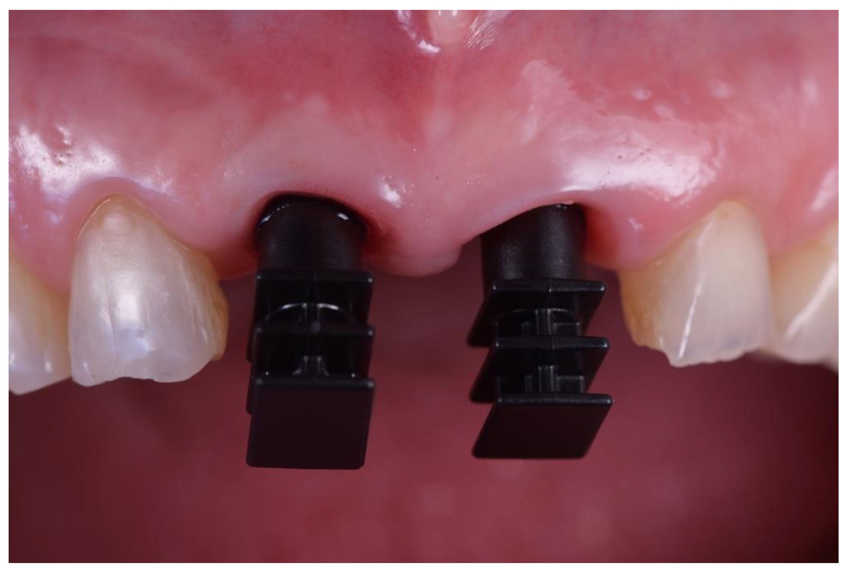
2. Materials and Methods

This study was conducted in full accordance with the most recent World Medical Declaration of Helsinki. Dental records of patients fulfilling eligibility criteria were screened. All patients were carefully informed regarding ceramic implant placement. They agreed to participate during regular visits in order to evaluate the implants under investigation both clinically and radiographically. Included subjects were asked to sign informed consent form before being involved in the study. Operative procedures were performed between July 2015 and January 2021. All of the 19 subjects participating in this study were affected by partial edentulism at one or more sites. No restrictions were applied regarding dental arch (e.g., upper or lower) and site (e.g., anterior or posterior). The study enrolled patients treated using one-piece zirconia implants who were observed for at least 2 years after cementation of final single crown prostheses.

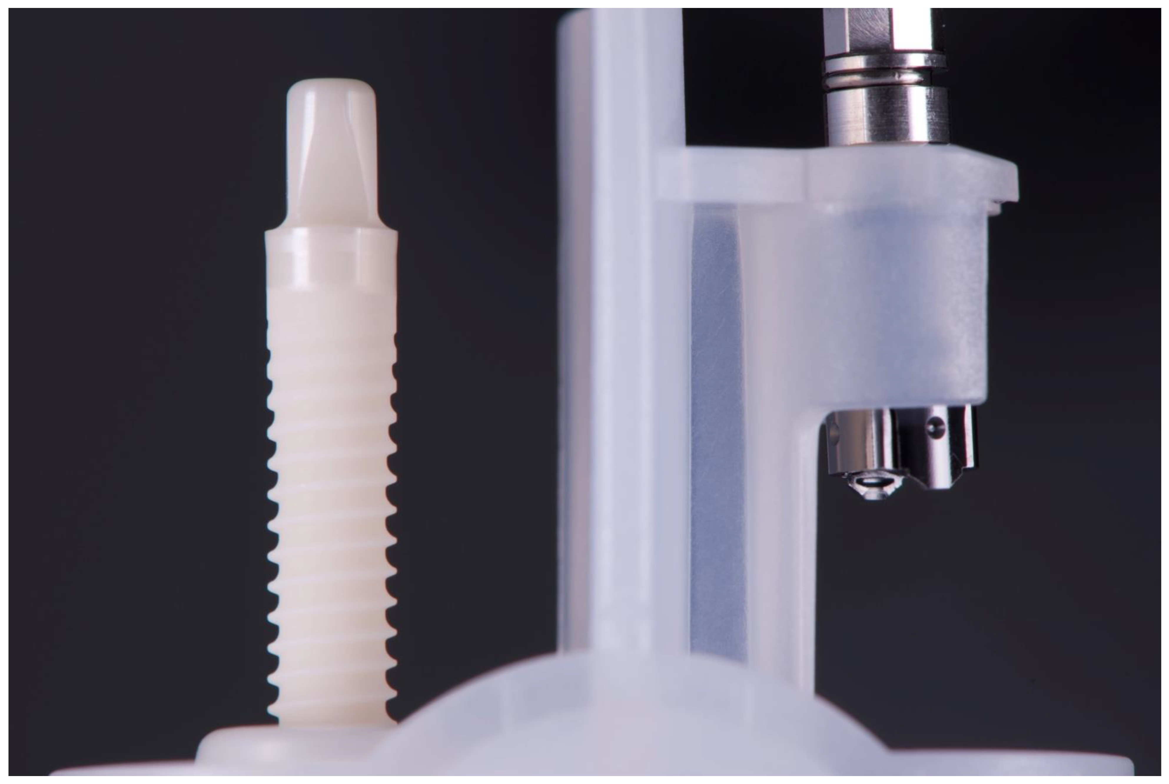
Implant Characteristics
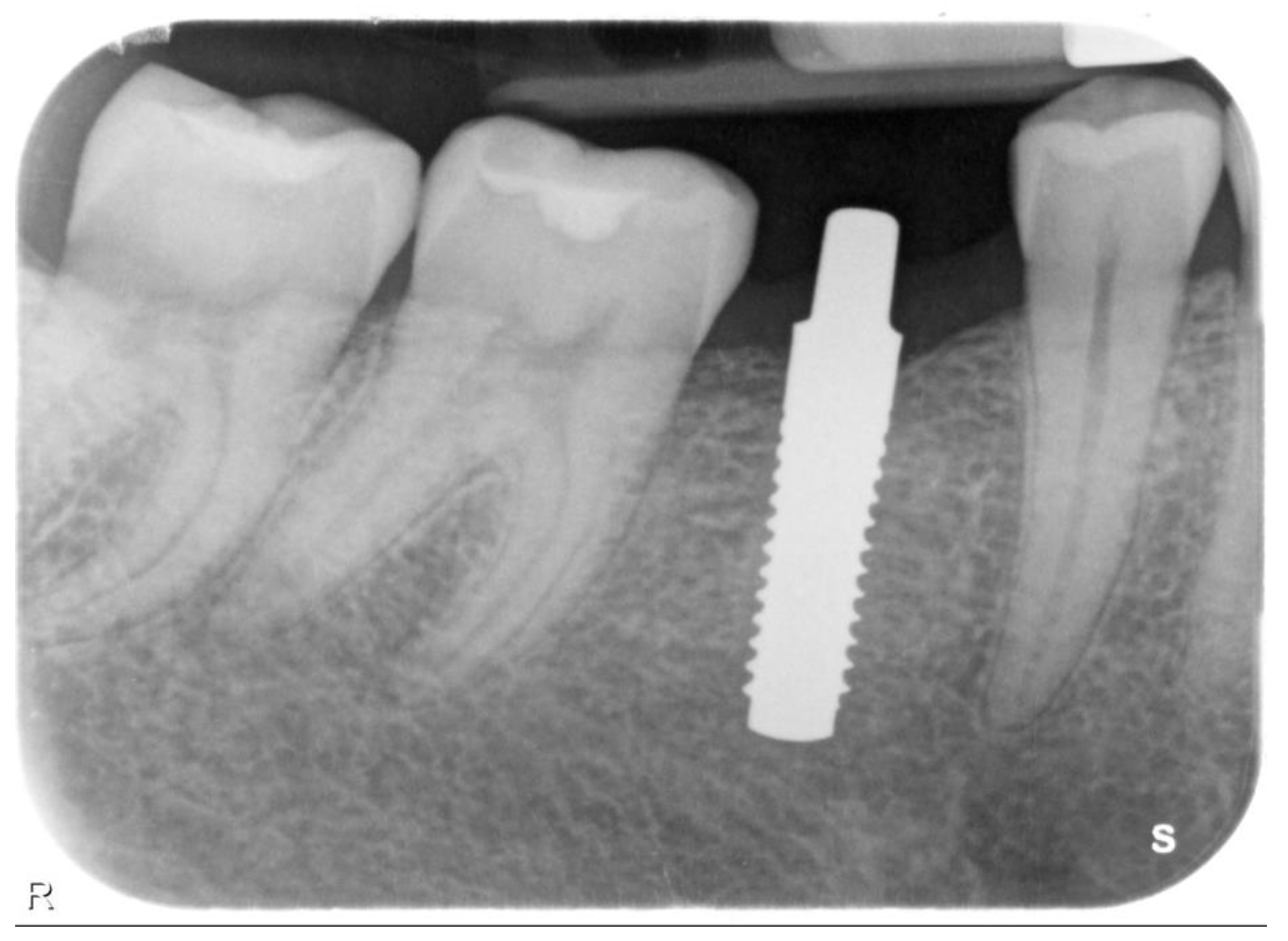
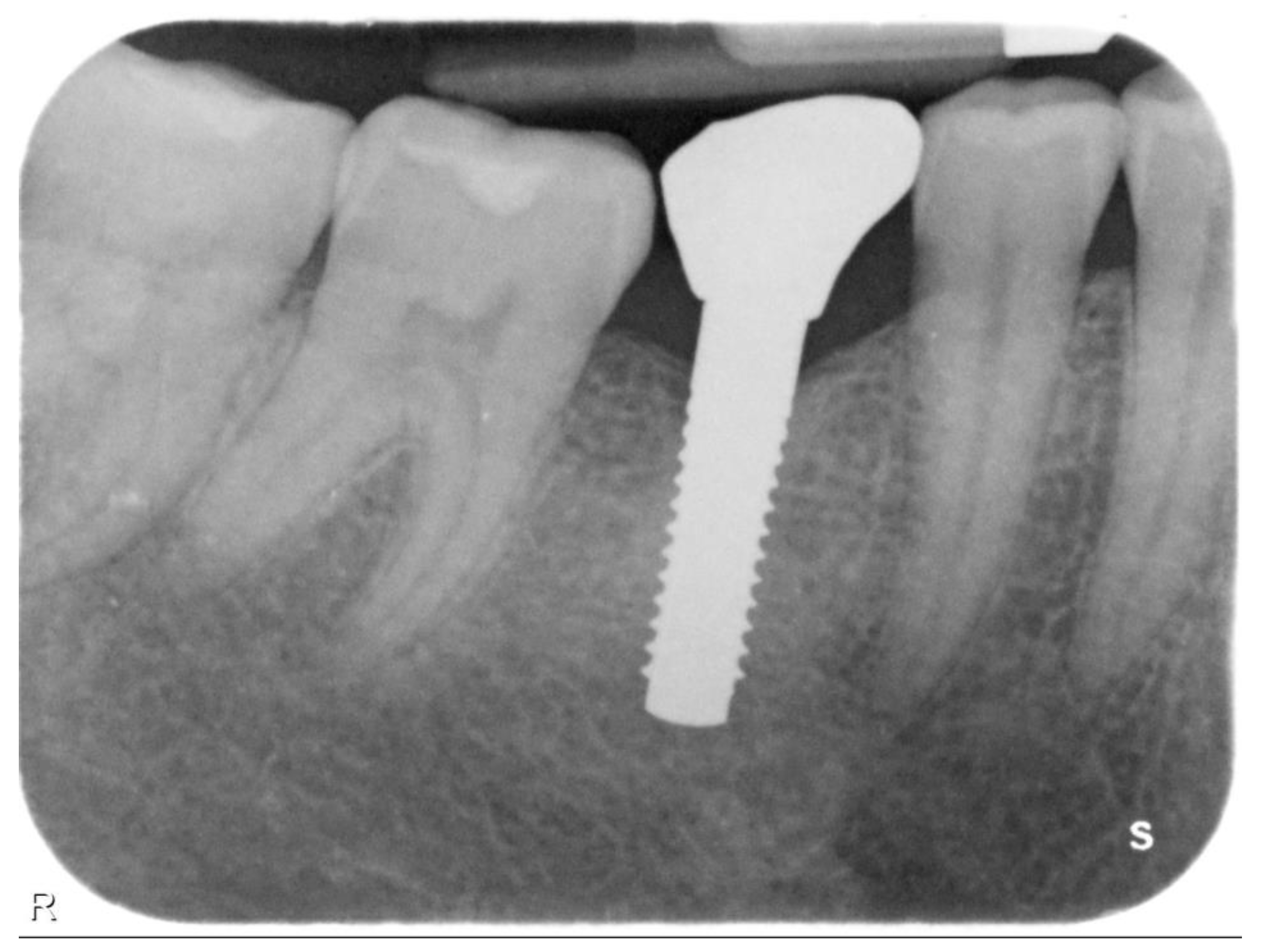
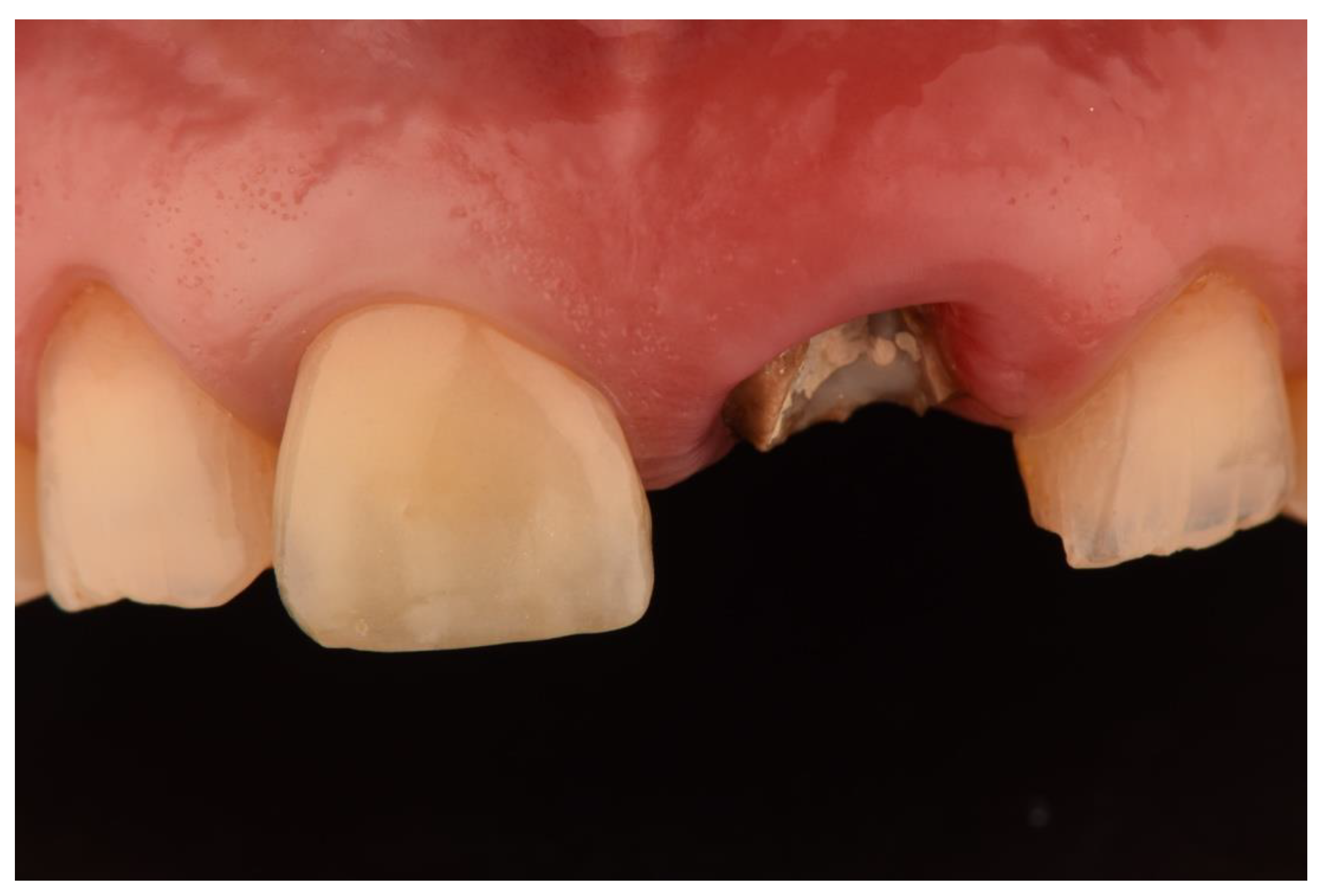
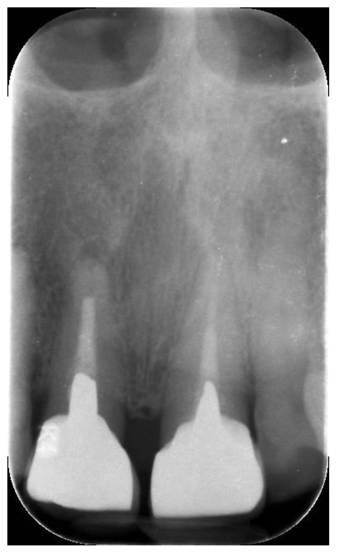
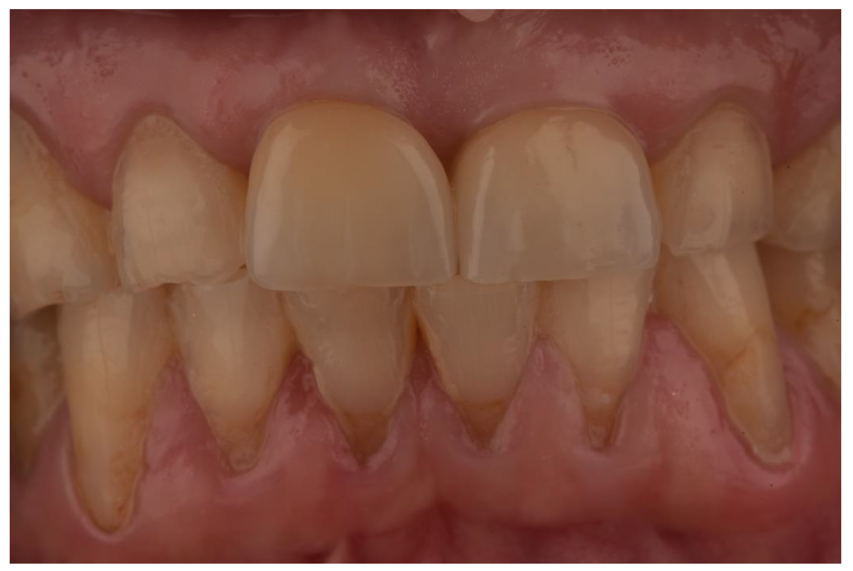
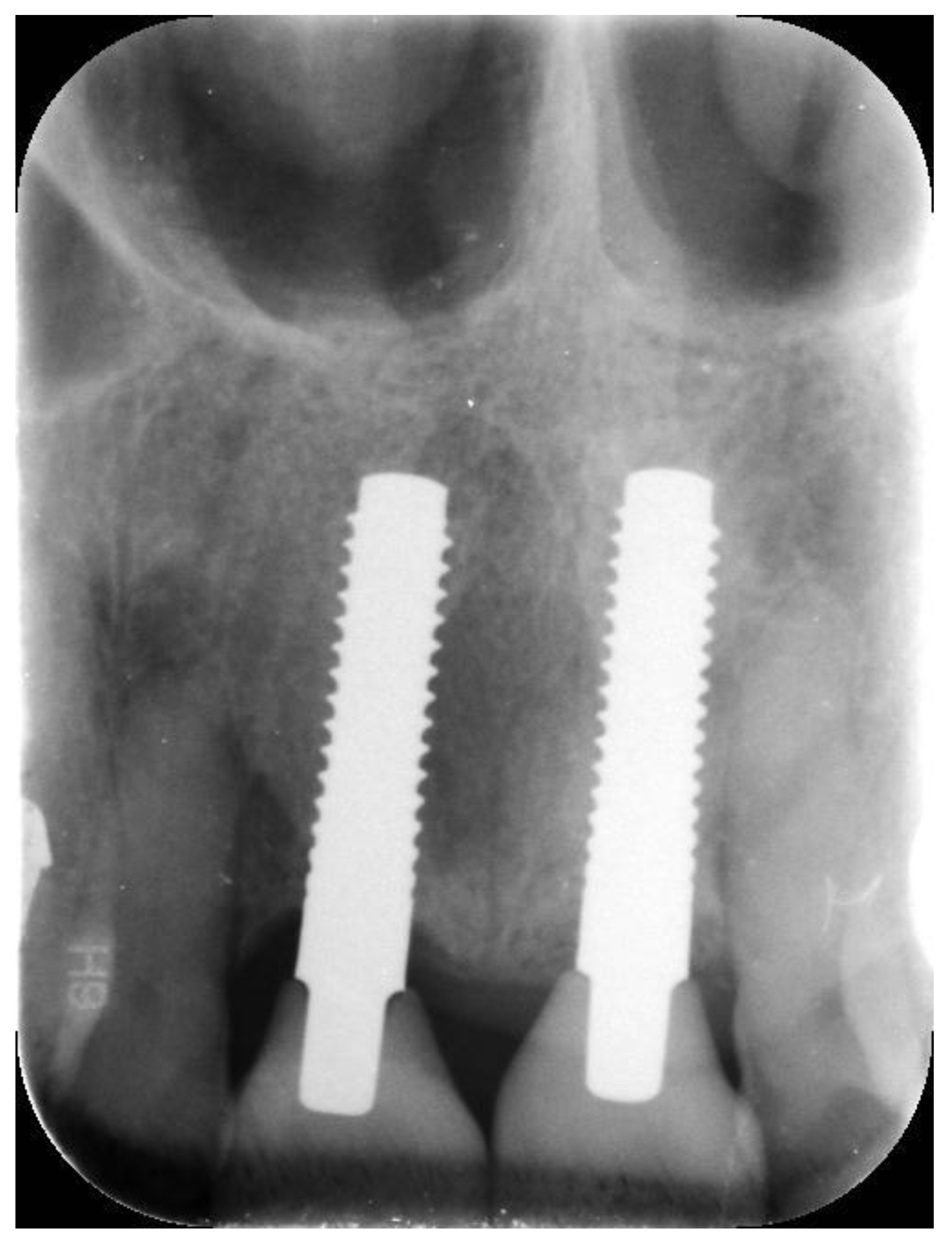
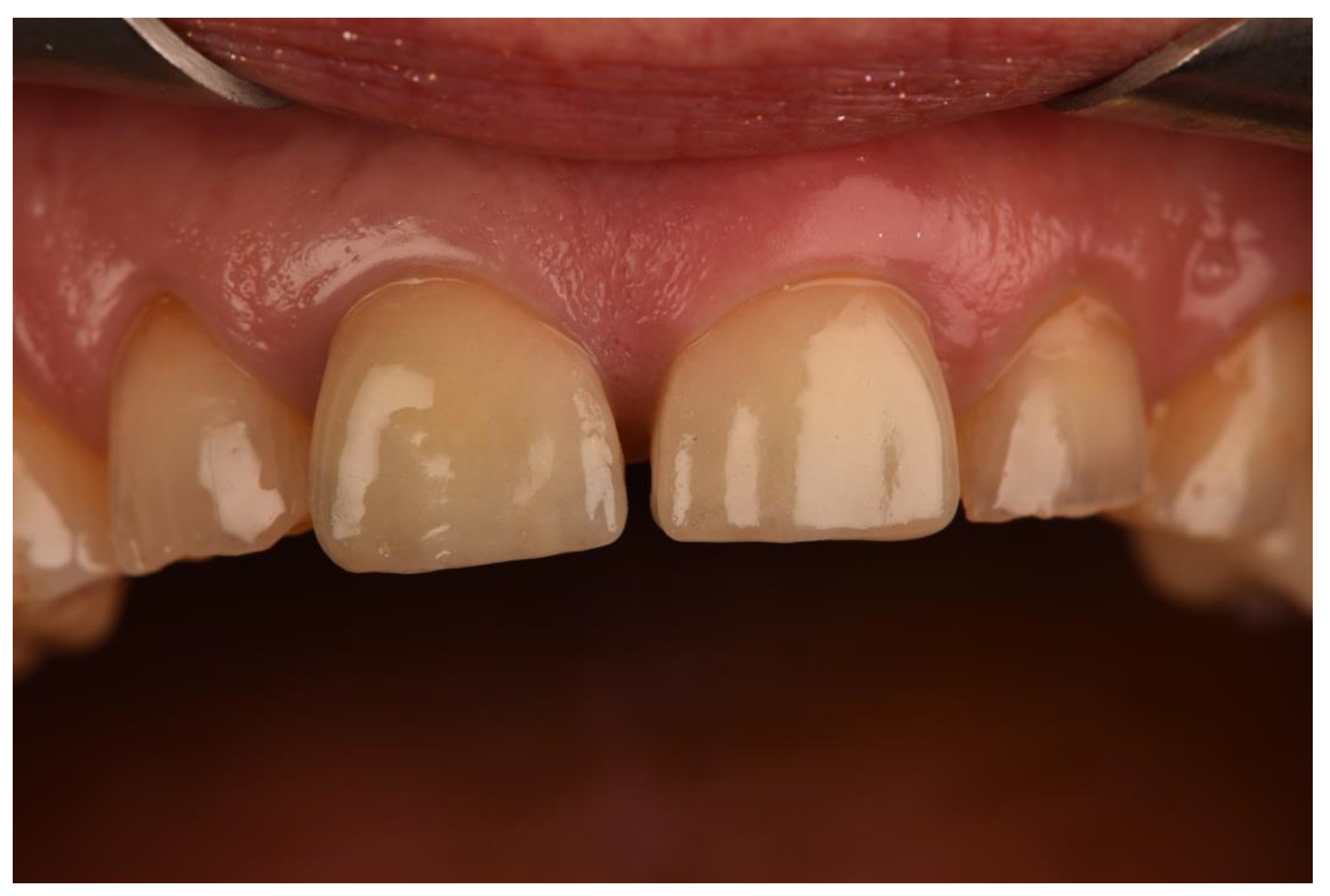
The Straumann® PURE Ceramic Implant Monotype (Straumann Holding AG, Basel, Switzerland) system was employed. Monotype is a ceramic dental implant made using zirconium dioxide in its tetragonal phase, which is then stabilized through yttrium (designated Y-TZP). The fixture and abutment are not separated, as this is a one-piece implant system. Therefore implant–abutment connection is not present (Figure 1). The implant design is cylindrical with a tapered effect thread. The thread pitch amounts to 0.8 mm. The intraosseous portion of the implant features an acid-etched microrough surface (1.3 μm, ZLA® surface topography, Institut Straumann AG, Basel, Switzerland). No grinding of any part of the implant or implant abutment is performed in order to reduce the risk of micro-cracks in the material. The transmucosal portion consists of a 1.8 mm machined neck (0.5 μm). The abutment component is characterized by a four-cornered shaft design.
Inclusion criteria: healthy patients older than 18 years of age, signed informed consent, bone width and height measuring at least 6 and 10 mm, respectively. Minor guided bone regeneration (GBR) procedures to improve peri-implant tissue conditions were accepted. They were performed, only when needed, in concomitance with implant placement. Such procedures were carried out using a xenograft (Bio-Oss, Geistlich Pharma AG, Wolhusen, Switzerland) covered by a collagen membrane (Bio-Gide, Geistlich Pharma AG, Wolhusen, Switzerland). Exclusion criteria: pregnancy, heavy smokers (>10 cigarettes a day), kidney or liver diseases, uncontrolled or poorly controlled diabetes, bisphosphonates intake, history of radiotherapy of the head and neck, current therapy with immunosuppressants or steroids, patients affected by primary or secondary immunodeficiency, connective tissue disorders, untreated periodontitis, autoimmune diseases, oral parafunctions, refractory infections of the oral cavity. Extensive alveolar ridge deficiencies requiring two-stage guided bone regeneration were considered an exclusion criterium. Other dental implant systems and designs (e.g., titanium dental implants, two-piece zirconia implants consisting of separate fixture and abutment) and monopiece zirconia implants with insufficient follow-up (<2 years) were excluded. Presurgical planning of each patient was performed using cone beam computed tomography (CBCT) (SCANORA, KaVo, Biberach, Germany). Implant insertion was performed as immediate post-extraction implant or following a delayed approach (at least 3 months of healing after dental extraction). In the latter case, after performing local anesthesia, a mucoperiosteal flap without vertical releasing incisions was raised. Implant site preparation was performed according to manufacturer’s instructions. The bone drilling sequence started with a round bur 2.3 mm or 3.1 mm in diameter in order to mark the implantation site. Then, stainless steel twist drills 2.2 mm and 2.8 mm in diameter were used to determine the implant axis. Hence, aligner dedicated pins were employed in order to verify the preparation depth, the angulation and restorability. This sequence was used to place zirconia implants 3.3 mm in diameter. Further twist drill 3.5 mm in diameter was applied to widen the implant bed preparation in case 4.1 mm diameter implant had to be positioned. All implants were placed freehand and no aid was obtained by partial or fully guided surgery. The insertion depth of the implants was determined by the endosseous portion. The 1.8 mm transmucosal collar was used as landmark and was not forced apically during implant placement so as to avoid mechanical stress at the crestal zone. Hence, the machined neck and implant head were positioned epicrestally according to the surgical and restorative protocol. Primary stability was evaluated at the time of implant placement by a dedicated counter-torque device. Immediate loading was performed for implants showing values higher than 30 Ncm. Zirconia implants ranged between 10 and 14 mm in length and 3.3 to 4.1 mm in diameter, respectively. Minor GBR procedures were performed only when needed. Therefore, flaps were sutured around the transmucosal portion of the implant body using separated single stitches. An intraoral dental radiograph with apico-pericapical projection was obtained at the end of the surgical phase (Figure 2).
After concluding implant placement, impression copings were positioned and polyether impressions were made (Impregum Soft, 3M Espe, Seefeld, Germany). Then, a resin-temporary crown was applied in order to cover the implant abutment and condition the shape of surrounding tissues. The provisional restoration was placed free from centric and eccentric contacts. Three months after surgery, final impressions were performed using polyether material (Impregum Soft, 3M Espe, Seefeld, Germany). Hence, veneered zirconia single crowns were applied by an expert therapist (R.S.) using resin-modified glass ionomer cement (RelyX Luting Plus Cement, Minneapolis, MN, USA). Retraction cords were then used in order to control the cementation line. Therefore, cement remnants were carefully removed by means of a sharp instrument. Clinical and radiological evaluations were performed from then on once a year during scheduled follow-up visits (Figure 3).
Clinical evaluation was carried out following Buser’s criteria, namely (1) self-reported complaints or issues in the treated zone; (2) refractory infections or suppuration of the surroundings peri-implant tissues; and (3) loosening or mobility of the implant body. All x-rays were obtained with the same projection with customized film holder mounted on a Rinn-type positioner (Dentsply RINN, York, PA, USA) and using paralleling technique. A single device with the same exposure setting was used at different time points (Vistascan, Durr Dental, Bietigheim-Bissingen, Germany; 75 kV, 9 mA, 0.22–0.25 s). Radiographs were stored on a personal computer. Radiographic images were analyzed with a dedicated software program (Image J, National Institute of Health, Bethesda, Rockville, MA, USA). Each x-ray was standardized through known measures, such as the implant diameter or length, in order to prevent any possible distortion. Marginal bone level (MBL) was measured as the distance between the implant head and the first detectable bone-to-implant contact. Linear measurements were retrieved at both implant sides, namely mesial and distal. A mean value per implant was then obtained. Numeric values expressed in millimeters were calculated to the nearest 0.1 mm. All measurements were performed using a single calibrated investigator (A.P.). Intraexaminer calibration was performed two times based on 20 images not related to the study sample, which were taken on different days. Hence, Buser criteria were applied to determine the success or failure of each included zirconia implant. Illustrative case depicting surgical and prosthetic procedures is reported from Figure 4, Figure 5, Figure 6, Figure 7, Figure 8, Figure 9, Figure 10 and Figure 11 (Figure 4, Figure 5, Figure 6, Figure 7, Figure 8, Figure 9, Figure 10 and Figure 11).

3. Results

Overall, 22 zirconia dental implants (12 in women and 10 in men) inserted in 19 generally healthy subjects were included in the study. No patients suffered from bruxism or reported diurnal parafunctional activities. The mean patient age was 49 years (range 31–69). Nine implants (37.5%) were 3.3 mm in diameter and thirteen (62.5%) were 4.1 mm in diameter. Eleven implants were placed in the maxilla (50%) and eleven in the mandible (50%). Four implants were placed to restore missing maxillary central incisors; three implants were positioned in place of maxillary lateral incisors; one implant replaced a maxillary canine; four implants were inserted to replace maxillary premolars or molars; one implant was employed to restore a mandibular central incisor; and nine implants were located in the mandibular premolar or molar position. Twelve implants presented an abutment length of 6 mm, while 11 implants were characterized by 4 mm abutments. Fourteen zirconia implants were inserted at the time of tooth extraction (immediate implant placement), while eight implants were positioned in healed ridges. Twelve implants underwent conventional loading (12 weeks after implant placement), while ten zirconia implants were immediately loaded (up to 1 week after implant placement). Guided bone regeneration was carried out at the same time as implant insertion in six implants (25%). The mean follow-up was 58.18 months (range: 27–96). According to Buser criteria, all implants were deemed to be successful. No pain, discomfort, foreign body sensation or dysesthesia were reported by the patients. No implant showed mobility or suppuration upon palpation at the last observation visit. The average marginal bone level (MBL), namely the radiographical distance between the shoulder of the transmucosal portion of the implant and the first detectable bone-to-implant contact, was 1.82 ± 0.63 mm at the time of implant placement, while it was 2.57 ± 0.72 mm at the last follow-up visit (range 0.36–4.19 mm). The peculiarities of the aforementioned outcomes are summarized in Table 1.

4. Discussion

According to the success criteria proposed by Buser et al., the null hypothesis of the present study was rejected. Indeed, no reduction in zirconia implant success rate was observed over a medium-term observation period. In agreement with the EAO position paper, monopiece zirconia dental implants can be recommended for fixed rehabilitations of single or partial edentulisms, up to three elements [19]. Ceramic implants are emerging as an adjunctive suitable option due to their high biocompatibility and mechanical resistance, and the fact that they are associated with a more natural color and better soft tissue integration [32]. Furthermore, zirconia implants have shown lower plaque accumulation and seem to be less prone to mucositis [34,35]. Nevertheless, lower survival rates have been observed for zirconia implants when compared to traditional titanium ones [33]. The survival and success rate of zirconia dental implants are still a matter of debate, and no solid scientific conclusions are available at the time of writing. On the other hand, a recent meta-analysis showed survival and success rates higher than 90% for zirconia implants after a mean observation period of 42.37 months [36]. However, most included studies presented short-term data, and medium-term follow-ups have been infrequently reported [37,38,39,40]. Due to their whiter color, zirconia implants have been advocated in the aesthetic zone, especially in case of thin gingival phenotype [32]. Satisfactory outcomes from clinical and radiological viewpoints have been observed in the present case series. No implant showed inadequate bone levels, progressive bone loss or thread exposure. In parallel, neither signs of infection associated with suppuration nor patient complaints were observed. These results are in full accordance with the existing literature. Indeed, several authors have reported a scarce tendency for bleeding and plaque accumulation associated with stable peri-implant hard and soft tissues in the short–medium term (up to three years) post loading [41,42]. Furthermore, this result is in line with previous authors who suggested a faster and more efficient epithelial sealing around zirconia when compared to titanium [43]. Zirconia implants have been associated with a lower tendency for plaque accumulation and, as a consequence, for peri-implant inflammation [44]. Whether this tendency would mean a lower incidence of mucositis and peri-implantitis when compared to conventional titanium implants should be better clarified in future studies. Despite these positive findings, the results observed in this case series should be interpreted cautiously as a consequence of the small sample size. Furthermore, it has to be highlighted that all inserted zirconia implants pertained to generally healthy patients undergoing regular professional mechanical plaque removal and with adequate bone volumes. As a consequence of limited sample size and study design, direct comparisons of baseline and follow-up measurements related to clinical and radiological data were deemed to be unfeasible. All implants were characterized by a distinct microdesign, consisting of a rough surface of the implant body followed by a 1.8 mm smooth surface of the transmucosal collar. This peculiar feature may have an impact regarding marginal bone level measurements as usually the implant shoulder is placed in the supracrestal position. Nevertheless, only slight changes in marginal bone levels were observed over time, thus suggesting the acceptable stability of the marginal bone. Monopiece implants have been associated with lower marginal bone loss as compared to two-piece implants due to the fact that microgap and micromovements at the interface between implant and abutment, which may lead to bacterial percolation, are not present [18]. Rodriguez et al. reported similar clinical and radiological outcomes when assessing 24 one- and two-piece zirconia dental implants in 12 consecutive patients. In their study, one two-piece zirconia implant inserted in the posterior area was lost before delivering the definitive rehabilitation, while one monopiece zirconia implant underwent implant failure in the aesthetic zone after immediate provisional protocol [45]. The protection of marginal bone loss related to monopiece implants has mainly been found to be associated with the absence of the implant–abutment interface. In fact, this is a critical zone where several components’ interfaces are very close to the surrounding tissues. The presence of a microgap, albeit minimal, can represent a niche that is colonizable by peri-implant pathogens [46]. At the same time, the prolonged exposure to bacteria and bacterial products may trigger soft tissue inflammation and, consequently, peri-implant bone resorption. The monopiece design features a transmucosal collar, which provides prosthetic versatility, as it can be used in single, multiple or even full-arch rehabilitations [47]. The primary scope of this observational investigation was to evaluate marginal bone level changes between the baseline and the last follow-up visit, thus providing data after a mean observational period of 58.18 months. Regarding monopiece ceramic dental implants, Borgonovo et al. reported a 1.2 mm marginal bone loss after four years. Conversely, Balmer et al. observed marginal bone level changes amounting to 0.72 mm from implant placement to the last follow-up visit, which was carried out three years after the delivery of definitive restoration [27,28,29,30,31,32,33,34,35,36,37,38,39,40,41]. Interestingly, Elnayef et al., in their systematic review and meta-analysis, reported 0.14 mm more marginal bone loss when using zirconia when compared to titanium dental implants [36]. Nonetheless, a recent systematic review published by Esteves Fernandes et al. in 2022 reported that the marginal bone loss of titanium and zirconia implants was similar after years of functional loading. Nevertheless, due to the fact that most studies are characterized by an inadequate follow-up period and number of zirconia implants evaluated, solid conclusions cannot be drawn [48]. The hard and soft tissue outcomes could be conditioned by additional GBR procedures, generally producing better results. Several shortcomings have been associated with one-piece implants. In fact, abutment modification is not achievable, and cement margins are determined by manufacturers. This could lead to challenging clinical conditions, especially in the aesthetic zone. Hence, the results presented in this case series should be interpreted with caution. Future research characterized by adequate study design (e.g., randomized controlled clinical trials) is advocated to investigate the potential advantages and disadvantages of zirconia dental implants over a medium- to long-term follow-up.

5. Conclusions

In the present case series, zirconia dental implants showed a 100% success rate. All prosthetically restored monocomponent ceramic implants were in situ at the last follow-up visit. Good clinical outcomes and acceptable marginal bone levels were observed after a mean observation period of 58.18 months (range: 27–96). The main limitations of this study are the small sample size associated with patient selection. Indeed, the inclusion criteria were only patients with good general health and oral hygiene. As a consequence of the observational study design, a comparison between baseline and follow-up parameters was judged to be inappropriate. Moreover, guided bone regeneration was performed in some implants, while others did not receive this procedure. Despite the promising results, further research is warranted so as to assess the medium- to long-term performance of zirconia implants in interventional studies featuring adequate methodology, sample size and follow-up.

Author Contributions

Conceptualization, A.P., R.S. and E.R.; methodology, A.P.; software, A.P. and D.L.; validation, R.S. and E.R.; formal analysis, E.R.; investigation, A.P.; resources, R.S.; data curation, A.P.; writing—original draft preparation, A.P.; writing—review and editing, A.P. and E.R.; visualization, R.S.; supervision, E.R.; project administration, R.S. All authors have read and agreed to the published version of the manuscript.

Funding

This research received no external funding.

Institutional Review Board Statement

The study was conducted in accordance with the Declaration of Helsinki. Ethical review and approval were waived for this study due to the observational and non-interventional nature of the study design. Only conventional and routine procedures were carried out.

Informed Consent Statement

Informed consent was obtained from all subjects involved in the study. Written informed consent has been obtained from the patient(s) to publish this paper.

Data Availability Statement

All data presented in the manuscript are available from the corresponding author ([email protected]).

Acknowledgments

The authors want to thank the dental technician Dario Viera for providing valuable provisional and definitive restorations.

Conflicts of Interest

The authors declare no conflict of interest.

References

  1. Buser, D.; Janner, S.F.; Wittneben, J.G.; Bragger, U.; Ramseier, C.A.; Salvi, G.E. 10-year survival and success rates of 511 titanium implants with a sandblasted and acid-etched surface: A retrospective study in 303 partially edentulous patients. Clin. Implant. Dent. Relat. Res. 2012, 14, 839–851. [Google Scholar] [CrossRef]
  2. Thoma, D.S.; Ioannidis, A.; Cathomen, E.; Hämmerle, C.H.; Hüsler, J.; Jung, R.E. Discoloration of the peri-implant mucosa caused by zirconia and titanium implants. Int. J. Periodontics Restor. Dent. 2016, 36, 39–45. [Google Scholar] [CrossRef] [PubMed]
  3. Sicilia, A.; Cuesta, S.; Coma, G.; Arregui, I.; Guisasola, C.; Ruiz, E.; Maestro, A. Titanium allergy in dental implant patients: A clinical study on 1500 consecutive patients. Clin. Oral Implant. Res. 2008, 19, 823–835. [Google Scholar] [CrossRef] [PubMed]
  4. Hosoki, M.; Nishigawa, K.; Miyamoto, Y.; Ohe, G.; Matsuka, Y. Allergic contact dermatitis caused by titanium screws and dental implants. J. Prosthodont. Res. 2016, 60, 213–219. [Google Scholar] [CrossRef]
  5. Fretwurst, T.; Nelson, K.; Tarnow, D.P.; Wang, H.L.; Giannobile, W.V. Is Metal Particle Release Associated with Peri-implant Bone Destruction? An Emerging Concept. J. Dent. Res. 2018, 97, 259–265. [Google Scholar] [CrossRef]
  6. Mombelli, A.; Hashim, D.; Cionca, N. What is the impact of titanium particles and biocorrosion on implant survival and complications? A critical review. Clin. Oral Implant. Res. 2018, 29 (Suppl. S18), 37–53. [Google Scholar] [CrossRef]
  7. Benic, G.I.; Thoma, D.S.; Sanz-Martin, I.; Munoz, F.; Hämmerle, C.H.; Cantalapiedra, A.; Cantalapiedra, A.; Fischer, J.; Jung, R.E. Guided bone regeneration at zirconia and titanium dental implants: A pilot histological investigation. Clin. Oral Implant. Res. 2017, 28, 1592–1599. [Google Scholar] [CrossRef]
  8. Jung, R.E.; Sailer, I.; Hämmerle, C.H.; Attin, T.; Schmidlin, P. In vitro color changes of soft tissues caused by restorative materials. Int. J. Periodontics Restor. Dent. 2007, 27, 251–257. [Google Scholar]
  9. Cosgarea, R.; Gasparik, C.; Dudea, D.; Culic, B.; Dannewitz, B.; Sculean, A. Peri-implant soft tissue colour around titanium and zirconia abutments: A prospective randomized controlled clinical study. Clin. Oral Implant. Res. 2015, 26, 537–544. [Google Scholar] [CrossRef]
  10. Scarano, A.; Piattelli, M.; Caputi, S.; Favero, G.A.; Piattelli, A. Bacterial adhesion on commercially pure titanium and zirconium oxide disks: An in vivo human study. J. Periodontol. 2004, 75, 292–296. [Google Scholar] [CrossRef]
  11. Nascimento, C.D.; Pita, M.S.; Fernandes, F.H.N.C.; Pedrazzi, V.; de Albuquerque Junior, R.F.; Ribeiro, R.F. Bacterial adhesion on the titanium and zirconia abutment surfaces. Clin. Oral Implant. Res. 2014, 25, 337–343. [Google Scholar] [CrossRef]
  12. Roehling, S.; Woelfler, H.; Hicklin, S.; Kniha, H.; Gahlert, M. A retrospective clinical study with regard to survival and success rates of zirconia implants up to and after 7 years of loading. Clin. Implant. Dent. Relat. Res. 2016, 18, 545–558. [Google Scholar] [CrossRef]
  13. Degidi, M.; Artese, L.; Scarano, A.; Perrotti, V.; Gehrke, P.; Piattelli, A. Inflammatory infiltrate, microvessel density, nitric oxide synthase expression, vascular endothelial growth factor expression, and proliferative activity in peri-implant soft tissues around titanium and zirconium oxide healing caps. J. Periodontol. 2006, 77, 73–80. [Google Scholar] [CrossRef] [PubMed]
  14. Welander, M.; Abrahamsson, I.; Berglundh, T. The mucosal barrier at implant abutments of different materials. Clin. Oral Implant. Res. 2008, 19, 635–641. [Google Scholar] [CrossRef]
  15. Kajiwara, N.; Masaki, C.; Mukaibo, T.; Kondo, Y.; Nakamoto, T.; Hosokawa, R. Soft tissue biological response to zirconia and metal implant abutments compared with natural tooth: Microcirculation monitoring as a novel bioindicator. Implant. Dent. 2015, 24, 37–41. [Google Scholar] [CrossRef]
  16. Liñares, A.; Grize, L.; Muñoz, F.; Pippenger, B.E.; Dard, M.; Domken, O.; Blanco-Carrión, J. Histological assessment of hard and soft tissues surrounding a novel ceramic implant: A pilot study in the minipig. J. Clin. Periodontol. 2016, 43, 538–546. [Google Scholar] [CrossRef] [PubMed]
  17. Hasim, D.; Cionca, N.; Courvoisier, D.S.; Mombelli, A. A systematic review of the clinical survival of zirconia implants. Clin. Oral Investig. 2016, 20, 1403–1417. [Google Scholar] [CrossRef]
  18. Vohra, F.; Al-Kheraif, A.A.; Ab Ghani, S.M.; Abu Hassan, M.I.; Alnassar, T.; Javed, F. Crestal bone loss and periimplant inflammatory parameters around zirconia implants: A systematic review. J. Prosthet. Dent. 2015, 114, 351–357. [Google Scholar] [CrossRef] [PubMed]
  19. Balmer, M.; Payer, M.; Kohal, R.J.; Spies, B.C. EAO Position Paper: Current Level of Evidence Regarding Zirconia Implants in Clinical Trials. Int. J. Prosthodont. 2022, 35, 560–566. [Google Scholar] [CrossRef]
  20. Gahlert, M.; Röhling, S.; Wieland, M.; Eichhorn, S.; Küchenhoff, H.; Kniha, H. A comparison study of the osseointegration of zirconia and titanium dental implants. A biomechanical evaluation in the maxilla of pigs. Clin. Implant. Dent. Relat. Res. 2010, 12, 297–305. [Google Scholar] [CrossRef]
  21. Depprich, R.; Zipprich, H.; Ommerborn, M.; Naujoks, C.; Wiesmann, H.-P.; Kiattavorncharoen, S.; Lauer, H.-C.; Meyer, U.; Kübler, N.R.; Handschel, J. Osseointegration of zirconia implants compared with titanium: An in vivo study. Head Face Med. 2008, 11, 30. [Google Scholar] [CrossRef] [PubMed]
  22. Scarano, A.; Di Carlo, F.; Quaranta, M.; Piatelli, A. Bone response to zirconia ceramic implants: An experimental study in rabbits. J. Oral Implantol. 2003, 29, 8–12. [Google Scholar] [CrossRef] [PubMed]
  23. Koch, F.P.; Weng, D.; Kramer, S.; Biesterfeld, S.; Jahn-Eimermacher, A.; Wagner, W. Osseointegration of one-piece zirconia implants compared with a titanium implant of identical design: A histomorphometric study in the dog. Clin. Oral Implant. Res. 2010, 21, 350–356. [Google Scholar] [CrossRef]
  24. Mihatovic, I.; Goluvobic, V.; Becker, J.; Schwarz, F. Bone tissue response to experimentalzirconia implants. Clin. Oral Investig. 2017, 21, 523–532. [Google Scholar] [CrossRef] [PubMed]
  25. Sanon, C.; Chevalier, J.; Douillard, T.; Cattani-Lorente, M.; Scherrer, S.S.; Gremillard, L. A new testing protocol for zirconia dental implants. Dent. Mater. 2015, 31, 15–25. [Google Scholar] [CrossRef] [PubMed]
  26. Monzavi, M.; Zhang, F.; Meille, S.; Douillard, T.; Adrien, J.; Noumbissi, S.; Nowzari, H.; Chevalier, J. Influence of artificial aging on mechanical properties of commercially and non-commercially available zirconia dental implants. J. Mech. Behav. Biomed. Mater. 2020, 101, 103423. [Google Scholar] [CrossRef]
  27. Balmer, M.; Spies, B.C.; Vach, K.; Ralf-Joachim, K.; Hämmerle, C.H.F.; Jung, R.E. Three-year analysis of zirconia implants used for single-tooth replacement and three unit fixed dental prostheses: A prospective multicenter study. Clin. Oral Implant. Res. 2018, 29, 290–299. [Google Scholar] [CrossRef]
  28. Oliva, J.; Oliva, X. 15-Year Post-Market Clinical Follow-up Study of 1828 Ceramic (Zirconia) Implants in Humans. Int. J. Oral Maxillofac. Implant. 2023, 38, 357–366. [Google Scholar] [CrossRef]
  29. Kiechle, S.; Liebermann, A.; Mast, G.; Heitzer, M.; Möhlhenrich, S.C.; Hölzle, F.; Kniha, H.; Kniha, K. Evaluation of one-piece zirconia dental implants: An 8-year follow-up study. Clin. Oral Investig. 2023, 27, 3415–3421. [Google Scholar] [CrossRef]
  30. Cionca, N.; Müller, N.; Mombelli, A. Two-piece zirconia implants supporting all-ceramic crowns: A prospective clinical study. Clin. Oral Implant. Res. 2015, 26, 413–418. [Google Scholar] [CrossRef]
  31. Borgonovo, A.E.; Censi, R.; Vavassori, V.; Arnaboldi, O.; Maiorana, C.; Re, D. Zirconia implants in esthetic areas: 4-year follow-up evaluation study. Int. J. Dent. 2015, 2015, 415029. [Google Scholar] [CrossRef]
  32. Afrashtehfar, K.I.; Del Fabbro, M. Clinical performance of zirconia implants: A meta-review. J. Prosthet. Dent. 2020, 123, 419–426. [Google Scholar] [CrossRef] [PubMed]
  33. Roehling, S.; Schelegel, K.; Woelfler, H.; Gahlert, M. Zirconia compared to titanium dental implants in preclinical studies-a systematic review and meta-analysis. Clin. Oral Implant. Res. 2019, 30, 365–395. [Google Scholar] [CrossRef] [PubMed]
  34. Andreiotelli, M.; Wenz, H.J.; Kohal, R.J. Are ceramic implants a viable alternative to titanium implants? A systematic literature review. Clin. Oral Implant. Res. 2009, 20 (Suppl. S4), 32–47. [Google Scholar] [CrossRef]
  35. Kohal, R.J.; Knauf, M.; Larsson, B.; Sahlin, H.; Butz, F. One-piece zirconia oral implants: One-year results from a prospective cohort study. 1. Single tooth replacement. J. Clin. Periodontol. 2012, 39, 590–597. [Google Scholar] [CrossRef] [PubMed]
  36. Elnayef, B.; Lazaro, A.; Suarez-Lopez Del Amo, F.; Galindo-Moreno, P.; Wang, H.L.; Gargallo-Albiol, J.; Hernandez-Alfaro, F. Zirconia implants as an alternative to titanium: A systematic review and meta-analysis. Int. J. Oral Maxillofac. Implant. 2017, 32, e125–e134. [Google Scholar] [CrossRef]
  37. Brüll, F.; van Winkelhoff, J.V.; Cune, M.S. Zirconia dental implants: A clinical, radiographic, and microbiologic evaluation up to 3 years. Int. Oral Maxillofac. Implant. 2014, 29, 914–920. [Google Scholar]
  38. Osman, R.B.; Morgaine, K.C.; Duncan, W.; Swain, M.V.; Ma, S. Patients’ perspectives on zirconia and titanium implants with a novel distribution supporting maxillary and mandibular overdentures: A qualitative study. Clin. Oral Implant. Res. 2014, 25, 587–597. [Google Scholar] [CrossRef]
  39. Pieralli, S.; Kohal, R.J.; Jung, R.E.; Vach, K.; Spies, B. Clinical outcomes of zirconia dental implants; a systematic review. J. Dent. Dent. 2017, 9, 38–46. [Google Scholar] [CrossRef]
  40. Cacaci, C.; Cantner, F.; Mücke, T.; Randelzhofer, P.; Hajto, J.; Beuer, F. Clinical performance of screw-retained and cemented implant-supported zirconia single crowns: 36-month results. Clin. Oral Investig. 2017, 21, 1953–1959. [Google Scholar] [CrossRef]
  41. Borgonovo, A.E.; Fabbri, A.; Vavassori, V.; Censi, R.; Maiorana, C. Multiple teeth replacement with endosseous one-piece yttrium-stabilized zirconia dental implants. Med. Oral Patol. Oral Cir. Bucal 2012, 17, e981–e987. [Google Scholar] [CrossRef] [PubMed]
  42. Vilor-Fernández, V.; García-de-la-Fuente, A.M.; Marichalar-Mendia, X.; Estefanía-Fresco, R.; Aguirre-Zorzano, L.A. Single tooth restoration in the maxillary esthetic zone using a one-piece ceramic implant with 1 year of follow-up: Case series. Int. J. Implant. Dent. 2021, 7, 26. [Google Scholar] [CrossRef] [PubMed]
  43. Glauser, R.; Sailer, I.; Wohlwend, A.; Studer, S.; Schibli, M.; Scharer, P. Experimental zirconia abutments for implant-supported single-tooth restorations in esthetically demanding regions: 4-year results of a prospective clinical study. Int. J. Prosthodont. 2004, 17, 285–290. [Google Scholar] [PubMed]
  44. Rimondini, L.; Cerroni, L.; Carrassi, A.; Torricelli, P. Bacterial colonization of zirconia ceramic surfaces: An in vitro and in vivo study. Int. J. Oral Maxillofac. Implant. 2002, 17, 793–798. [Google Scholar]
  45. Rodriguez, A.E.; Monzavi, M.; Yokoyama, C.L.; Nowzari, H. Zirconia dental implants: A clinical and radiographic evaluation. J. Esthet. Restor. Dent. 2018, 30, 538–544. [Google Scholar] [CrossRef]
  46. Pera, F.; Pesce, P.; Menini, M.; Fanelli, F.; Kim, B.C.; Zhurakivska, K.; Mayer, Y.; Isola, G.; Cianciotta, G.; Crupi, A.; et al. Immediate loading full-arch rehabilitation using transmucosal tissue-level implants with different variables associated: A one-year observational study. Minerva Dent. Oral Sci. 2023. ahead of print. [Google Scholar] [CrossRef]
  47. Carossa, M.; Alovisi, M.; Crupi, A.; Ambrogio, G.; Pera, F. Full-Arch Rehabilitation Using Trans-Mucosal Tissue-Level Implants with and without Implant-Abutment Units: A Case Report. Dent. J. 2022, 10, 116. [Google Scholar] [CrossRef]
  48. Fernandes, P.R.E.; Otero, A.I.P.; Fernandes, J.C.H.; Nassani, L.M.; Castilho, R.M.; de Oliveira Fernandes, G.V. Clinical Performance Comparing Titanium and Titanium-Zirconium or Zirconia Dental Implants: A Systematic Review of Randomized Controlled Trials. Dent. J. 2022, 10, 83. [Google Scholar] [CrossRef]
Figure 1. Characteristics of the ceramic one-piece implant system (Straumann® PURE Ceramic Implant Monotype, Straumann Holding AG, Basel, Switzerland).
Prosthesis 05 00074 g001
Figure 2. Intraoral radiograph taken at the end of the surgical phase, apico-coronal projection, implant number 7 (see Table 1).
Prosthesis 05 00074 g002
Figure 3. Intraoral radiograph taken at the last follow-up visit, 73 months after definitive prosthesis delivery. Apico-coronal projection, implant number 7 (see Table 1).
Prosthesis 05 00074 g003
Figure 4. Frontal view of the maxillary failing teeth.
Prosthesis 05 00074 g004
Figure 5. Frontal projection of the failing teeth before extraction.
Prosthesis 05 00074 g005
Figure 6. Dental radiograph with apico-periapical projection showing endo-restorative status before dental extraction.
Prosthesis 05 00074 g006
Figure 7. Occlusal-palatal view.
Prosthesis 05 00074 g007
Figure 8. Frontal view after positioning impression copings.
Prosthesis 05 00074 g008
Figure 9. Lateral view of cemented single crowns.
Prosthesis 05 00074 g009
Figure 10. Clinical photograph after definitive prostheses delivery.
Prosthesis 05 00074 g010
Figure 11. Intraoral radiograph with apico-periapical projection taken 38 months after implant positioning.
Prosthesis 05 00074 g011
Table 1. Main features of the included studies’ legend.
NFollow-Up (Months)AgeGenderImplant PositionImplant Diameter (mm)Implant Length (mm)GBRPlacement Timing
(IP/DP)
Loading Timing
(IL/CL)
Mean MBL (mm)
(T0–T1)
19642F114.112NIPIL1.9–3.48
29659F214.114YIPIL1.41–1.70
39441M134.112NIPIL2.11–3.2
49250M123.312NDPIL2.35–3.3
58335M143.312NDPIL1.57–1.92
68837F154.112NIPIL2.73–2.74
77343F463.312NDPCL1.8–1.87
87346F464.112NDPCL1.35–1.69
96237F464.110NDPCL1.94–2.06
105947M124.112YIPCL0.36–2.03
114848M444.112NIPCL2.75–2.86
124848M454.112NIPCL2.34–3.24
134848M464.112NIPCL3.5–4.19
144231M464.110NDPCL1.23–2.03
154234M343.312NIPCL1.68–2.32
164165F243.312YIPIL2.1–2.82
174064F213.314YIPIL2.19–3.45
183865F114.114NIPIL1.48–2.17
193354F263.310NIPCL1.32–1.66
203063F313.314NIPIL1.72–1.86
212752M223.314YIPCL1.49–2.53
222769F364.112YDPCL1.72–3.57
N = implant number, follow-up (months), age, gender, implant position, implant diameter (mm), implant length (mm); GBR = guided bone regeneration; placement timing: IP = immediate placement, DP = delayed placement; loading timing: IL = immediate loading, CL = conventional loading; mean marginal bone level (mm): T0 = MBL calculated on the x-rays taken immediately after implant positioning, T1 = MBL calculated on the x-rays taken during the last visit).
Disclaimer/Publisher’s Note: The statements, opinions and data contained in all publications are solely those of the individual author(s) and contributor(s) and not of MDPI and/or the editor(s). MDPI and/or the editor(s) disclaim responsibility for any injury to people or property resulting from any ideas, methods, instructions or products referred to in the content.

