1. Introduction
The advent of computer-aided design systems (CAD-CAMs) has had a huge impact on the daily practice of all dentists [
1].
The digital workflow involves the acquisition of physical data, which are then transformed into digital data to enable the computer-aided design and manufacturing (CAD-CAM) of a prosthesis [
2]. The transformation of physical data into digital data can occur either by digitizing models obtained from traditional impressions in the laboratory, using bench scanners operated by dental technicians, or directly in the dental office, using an intraoral scanner (IOS) [
3].
Passivity is fundamental for any type of implant prosthesis, and even more so for a screw-retained prosthesis, where tensile forces and overloads can also be generated during the screw-retained phase, causing tension at the implant–abutment–prosthesis interface. The impression is therefore the key moment for transferring the correct implant position to the master model and, consequently, for the accuracy and passivity of the prosthetic structure [
4].
Until recently, conventional dental impressions were the most commonly used method for full-arch cases. However, even conventional workflows can introduce various errors. Factors such as the type of materials used; the number of implants; the implant’s/implants’ positions, depth, and angulation can all impact precision and accuracy. The accumulation of these errors may ultimately compromise the passive fit of an implant prosthesis [
5].
Intraoral scanning (that is, so-called “digital impressions”) has accelerated the data-acquisition process and eliminated many inconveniences present in conventional impressions, thus reducing patient discomfort and improving the predictability of prosthesis design and production procedures over time [
6]. To achieve high-precision prosthetic rehabilitation, it is crucial that the digitized physical data accurately replicate what is scanned and maintain this precision throughout the entire manufacturing process of the prosthetic device [
7,
8].
In partial and full-arch implant prosthetic rehabilitations, intraoral scans have achieved an accuracy comparable to that of traditional impressions [
9,
10]. Several in vitro and clinical studies have shown that the discrepancy between analogue and digital impressions on patients treated with full-arch rehabilitation is low, reaching below the tolerability value reported in the literature (150 microns) [
11,
12].
There are various stages within both the digital and analogue workflows where errors can be introduced, potentially compromising the passivity of the framework. In the analogue method, rigid materials were traditionally used during impression-taking to ensure the immobility of implants and the accuracy of the impression in full-arch rehabilitations [
13].
In the digital workflow, however, errors may occur during the scanning phase, as well as during the image-design and -matching phases [
14].
Several factors influence the precision and accuracy of intraoral scans [
15]. The type of IOS used plays a significant role in accuracy; in fact, some studies suggest that not all scanners are suitable for capturing full-arch implant-supported prostheses [
16]. Scan bodies can also impact accuracy, as their shape and position may affect the precision of the impression, even though they are generally easy to acquire with the scanner [
17,
18,
19,
20,
21]. Another factor contributing to minor deviations in accuracy is the matching of the virtual scan body from the CAD software library to the virtual model.
If the original scan is not performed properly, this can lead to a mismatch of the scan body and an error in the position of the analogue in the virtual model. The inter-implant distance significantly impacts the accuracy of impressions; in fact, an increase in distance tends to amplify errors, reducing both precision and accuracy [
22].
The location of the scan also plays a role in digital-impression accuracy. Scanning an edentulous jaw is often challenging due to the reduced presence of landmarks. Additionally, in cases involving osteoplasty or flap-less surgery, flap repositioning and bleeding can further obscure landmarks, complicating the scanning process. Numerous in vitro studies have shown that impressions obtained with intraoral and analogue scanners demonstrate similar and overlapping levels of accuracy. However, these studies do not account for the fact that impressions taken in the oral cavity are subject to various factors that can influence accuracy and precision, which cannot be fully replicated or studied in vitro [
23].
Few studies have, in fact, clinically analyzed whether digital impressions have the same predictability and accuracy as traditional impressions [
23].
A study by Pera et al. [
10] aimed to analyze the discrepancy between plaster impressions and impressions with full scans in full-arch rehabilitations on healed tissue and found that the discrepancies were not clinically relevant.
Capparé et al. [
24] conducted a study to evaluate the accuracy and precision of prostheses obtained from traditional and digital impressions in patients undergoing full-arch rehabilitation. The study revealed that digital impressions offer clinically usable accuracy that is comparable to that of traditional impressions.
Few studies have analyzed the accuracy of impressions taken just after implant surgery.
A study by De Angelis et al. [
25] aimed to evaluate the accuracy of prostheses obtained through digital impressions immediately after surgery. This study found that digital impressions, compared to traditional impressions, offer similar accuracy and are clinically usable. However, in this study, no flap was performed, and no dislocation occurred, so the impact of soft tissue mobility could not be assessed.
To the authors’ knowledge, there is currently no study that has been performed that has evaluated the accuracy of post-surgical impressions in patients rehabilitated with immediately loaded full-arch prostheses. The authors hypothesize that the opening of a large flap, performing osteoplasty, and suturing the flap may increase flap mobility and reduce the reliability of reference stitches, potentially leading to greater errors and distortions in the impressions.
The aim of this clinical study was to compare the accuracy of implant digital impressions taken immediately after surgery with those obtained from healed tissue in patients rehabilitated with a full-arch implant-supported prosthesis. The null hypothesis tested was that there is no significant difference between post-surgical impressions and impressions taken on healed tissue.
2. Materials and Methods
Between September 2023 and March 2024, 8 patients were recruited and treated at the Division of Prosthodontics and Implantology (Department of Surgical Sciences, DISC) of the University of Genoa (Center 1), and 11 patients were recruited and tested at the Division of Prosthodontics of the C.I.R. Dental School (Department of Surgical Sciences) of the University of Turin (Center 2).
All 19 patients had a compromised residual dentition (6 women; 13 men) and were treated with full-arch screw-retained fixed prostheses supported by 4–5 immediately loaded implants in the lower jaw (8 patients) or upper jaw (11 patients).
All subjects treated in this study, which was approved by the Scientific Ethics Committee of the University of Genoa (protocol approval number: 2023/03), were carefully informed about the study design and aims and signed an informed-consent form before the start of the study. The study was conducted in accordance with the Declaration of Helsinki.
All patients were recruited if they had complete impairme nt of residual teeth at least in one arch and were requiring an immediate loading rehabilitation of the upper or lower jaw and had adequate bone volume for the placement of 4-to-6 implants without the need for regenerative procedures.
All patients had to be in good general health; have no systemic conditions and not be under pharmacological therapies that do not allow for oral surgery; and not have a degree of bone atrophy requiring regenerative methods or the use of zygomatic implants [
26,
27].
The Columbus Bridge Protocol (CBP) was applied, which is a surgical and prosthetic protocol for full-arch immediate loading rehabilitation. This protocol allows for the restoration of aesthetics, phonetics, and function in patients with severely compromised teeth within 48–72 h [
28].
Following the extraction of compromised dentition, the protocol involves the insertion of 4–6 immediate implants with a minimum length of 10 mm into native bone, achieving high insertion torque. Implants are positioned vertically in the anterior regions of the jaws and, if necessary, inclined in the posterior regions to avoid anatomical structures and facilitate the distal placement of the implant head. Endosseous titanium implants with a tapered apex were used, Shard (Mech & Human, Grisignano di Zocco, Italy) and Syra (Sweden & Martina, Due Carrare PD, Italy). Multiunit abutments (MUAs) were used when needed to correct implant inclination.
The fixed prosthesis was delivered and screwed onto the implants within 48 h after surgery. It featured a rigid framework without distal extensions and was veneered with composite resin material. The classic protocol involves taking an impression immediately after the surgical phase using the plaster pick-up technique to achieve maximum precision for the metal framework [
26].
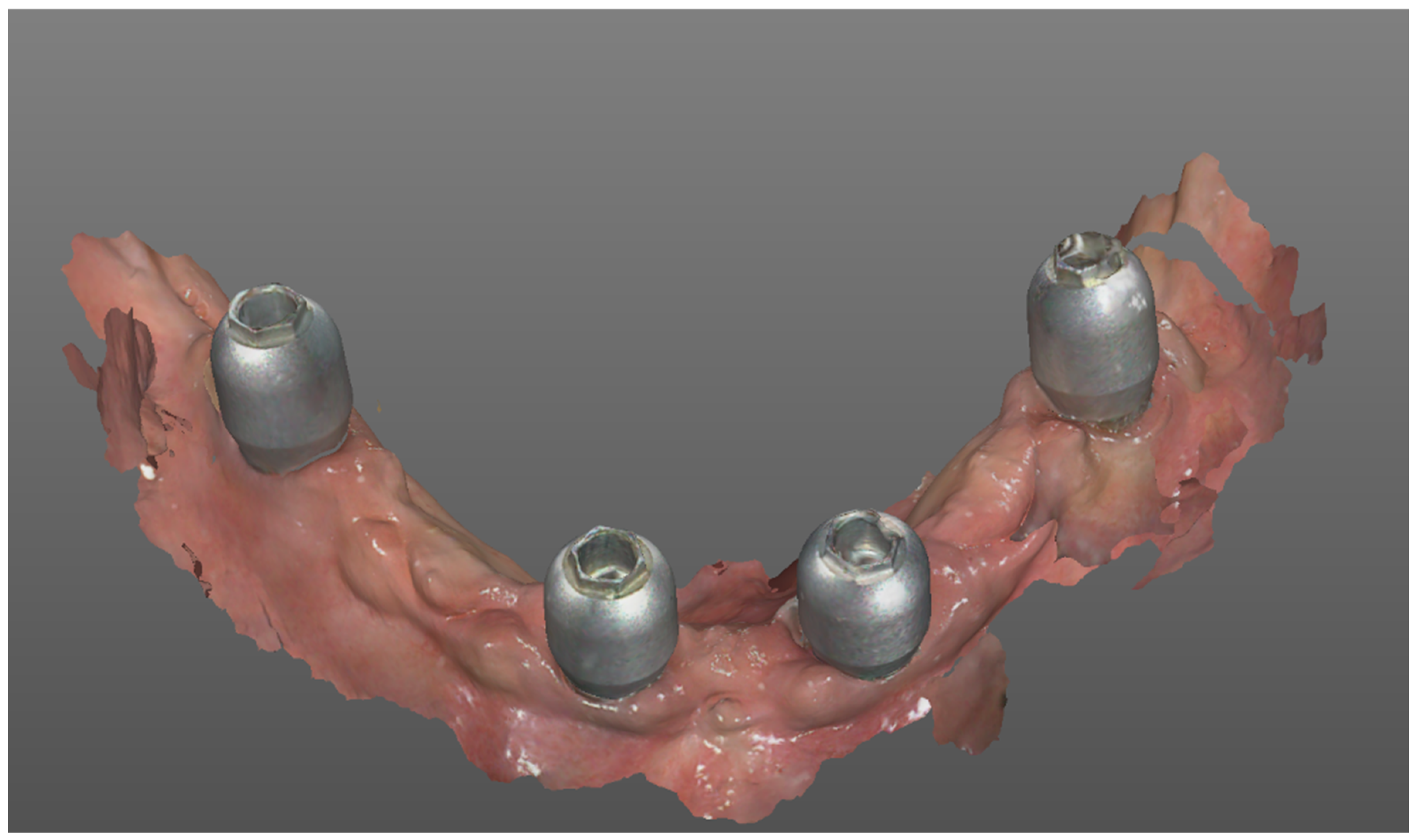
In this study, the post-surgical impression was made using a scanner with a speed of 15 frames per second (Mach2 Intraoral Scanner, Shining 3D, distributed by Euromax Monaco) (
Figure 1).
Scan bodies from the same manufacturer as the inserted implants were placed on the implants/MUAs, and an impression of the entire arch was taken immediately after suturing. No powder was used to opacify the elements to be scanned, and the output file was in “open” STL format.
In 71 implants, the scan bodies were screwed onto the MUA, while in 8 implants, the scan bodies were screwed directly onto the implant heads.
On Shard metal implants, Toothless®® Scanbodies (Mech & Human, Grisignano di Zocco, Italy) were used. They had a rounded shape with a hexagon on the top that is useful to increase the reference points for the best-fitting operation and had two different types of connection depending on whether they had to be screwed directly onto the implant head or onto the MUA.
On eight implants in two patients, scan bodies were used that were screwed directly onto the implant head (Sweden & Martina, Due Carrare PD, Italy), which have a flag shape with two points on the surface.
Two trained and calibrated clinicians, one per each center, with more than two years of experience with the IOS herein described (F.P. at the Center 2 and F.B. at Center 1) performed all the intraoral scans.
The two operators performed the scans using an “S” scan path; the IOS tip followed the entire arch with a smooth movement, starting from the most distal implant in the first quadrant to the contralateral implant, zigzagging from the vestibular to the palatal side, and vice versa. For each patient, an analogic post-surgical plaster pick-up impression using transfers was also acquired that served as a control. An aluminum test bar obtained from the digital impression was then initially tested on the analogic model obtained from the plaster impression. Based on the test bar’s fit, the scenarios were classified as follows:
Clinically acceptable: Good fit of the bar on the analogic model; the delivered full-arch prosthesis was completely realized from digital impression.
Not-satisfying fit: The operators were able to screw the test bar on the model, but the fit was not evaluated as good; the final prosthesis delivered to the patients was produced on the analogic model.
Not-passive result: It was not possible to screw the test bar on the analogic model; the final prosthesis delivered to the patients was produced on the analogic model.
After four months, the provisional prosthesis was removed, and a new digital impression was taken with the same IOS and the same scanning pattern (
Figure 2).
Before the intraoral scan, the stability of the implants/MUAs was carefully checked.
The two STL files obtained from the immediate and delayed intraoral scans were sent to a digital analysis laboratory that is experienced in digital rehabilitation to perform the superimposition and analysis.
In the upper arch, all implants in the scans were numbered from one to six (Implant 1, Implant 2, Implant 3, etc.), starting with the most distal implant in the right hemiarch and numbering them in ascending order throughout the arch to the most distal contralateral implant, while in the lower hemiarch, all implants were numbered in ascending order, starting with the most distal implant on the right to the most distal implant in the contralateral hemiarch.
2.1. Outcome Measures
Deviations were assessed using the Hausdorff distance metric, which evaluates the extent of deviation between two sets by measuring the maximum distance between them. Minimum, maximum, and mean distances between surfaces, as well as RMS values (which measure the root mean square deviation between two sets of points), were determined.
The parametric software GOM Inspection Pro (pro version, Carl Zeiss GOM Metrology GmbH, Braunschweig, Germany) was used for this analysis. This software applies basic parametric concepts to the STL files obtained from the mouth scans. Points to be measured were isolated by determining subsets, eliminating mucosal flanges, and isolating scan bodies (Scanbody Toothless). For each Toothless subset, a point of origin was determined and centered as the zero point of the Cartesian axes. By superimposing the two scans using a best-fit logic, discrepancies between the Toothless subsets were measured, which were evidenced by differences in the position of the Cartesian axes’ origin (
Figure 3).
From the three Cartesian axes, the variations in coordinates (X, Y, and Z) were expressed in millimeters, defining the most common direction of deviation between the different scans of the same edentulous arch.
2.2. Statistical Analysis
Confidence intervals at 5% were computed for the overall implant types and by group (Centers 2 and 1).
Then, the results of Centers 2 and 1 were compared to investigate possible statistical differences between the groups. The independent-sample t-test was calculated between Centers 2 and 1 (Satterthwaite method for unequal variances was considered).
Furthermore, the mandible and maxilla groups were compared by means of the independent-sample t-test (the Satterthwaite method for unequal variances was considered).
A regression model was applied to each implant type, considering the implant value as the dependent variable, and time as the independent variable.
For all the analyses, a p < 0.05 was considered statistically significant, and SAS 9.4 was used for the computation.
3. Results
Eleven patients were treated in the upper jaw and eight in the lower jaw. No patients dropped out, and all of them received both the immediate and the delayed intraoral scan. No implants failed, and all the implants were clinically stable and functional when the delayed scan was taken.
Seventeen dental arches were rehabilitated with four implants, one with five implants and one with six implants, for a total of seventy-nine dental implants. All the patients treated at the University of Genoa were rehabilitated with four implants only. Following the test-bar fit, 17 cases were classified as clinically acceptable, and the prosthesis was manufactured following a full digital workflow. In regard to the other five cases, four were classified as having a not-satisfying fit, and one was classified as having a not-passive result, and the prosthesis was manufactured following an analogic workflow.
The digital analysis showed an average discrepancy between the analyzed points of 0.1905 mm.
After evaluating the individual centers, we noted an average discrepancy of 0.1303 mm for the Center 2 and 0.279 mm for Center 1.
From the analysis on the three Cartesian axes, the greatest discrepancy was 0.019 mm on the y-axis.
After analyzing each RMS for each implant individually, a greater discrepancy was found for the implants positioned in the posterior areas, more precisely, 0.2326 mm for Implant 1 and 0.2124 for Implant 4.
In the anterior areas, Implants 2 and 3 differed by 0.1777 and 0.1555 mm, respectively.
The following table shows the average values of the discrepancies (distances) between the measured points, both as an average across all implants and each individual implant (
Table 1).
The scans performed at Center 2 had a smaller variance with respect to the prosthesis of Center 1, presenting a mean value of 130 μm and 279 μm, respectively.
The independent-sample t-test between Center 2 and Center 1 groups was computed (Satterthwaite method for unequal variances has been considered). Only Implant 2 is significantly different among the two centers (alpha at 5% was considered).
No statistically significant difference was found when comparing treated patients in regard to the upper and lower jaws. The accuracy of the scans increased over the study period. With the increasing time spent scanning and, thus, increased operator experience, the number of scanning errors decreased.
This means that, as the time that an operator spends scanning increases, the value of the discrepancy between the implants decreases (
p-value at 6%;
Figure 4).
The implants placed in the posterior areas were those with the greatest discrepancy between the two scans.
Implant 5 and Implant 6 were only available for two dental arches and one dental arch, respectively, all treated at the University of Turin.
With the increase in the number of implants, the discrepancy between the scans decreases.
The two clinicians who performed the intraoral scans reported, anecdotally, that scans on unhealed tissue were more difficult to perform, as the presence of bleeding and non-keratinized tissue and mobility made scanning more complicated, especially in the distal mandibular areas. Furthermore, the two clinicians also anecdotally reported that, even if no statistically significant differences were highlighted between upper and lower jaws, they experienced more clinical difficulties in acquiring lower-jaw scans.
4. Discussion
To the best of the authors’ knowledge, this is the first study to analyze the discrepancy between digital impression on surgical and healed sites in patients rehabilitated with implant full-arch immediate-loading rehabilitations.
The null hypothesis was partially rejected. The discrepancy between the two impressions was around the value of 200 μm, a discrepancy value defined as acceptable in the literature from a clinical point of view, more precisely with an average value of 190 μm [
29].
Furthermore, despite the fact that the same surgical and scanning protocols were applied, the scans performed at Center 1 showed a greater discrepancy than those performed for the Center 2 group (average: 0.279 mm versus 0.130 mm, respectively).
In 100% of the cases, the scans performed in Center 2 showed a value of less than 150 μm in the anterior regions (Implants 2–3), while in the posterior regions (Implants 1–4), the values were greater than 150 μm but less than 200 μm in 50% of the cases. On the other hand, the scans performed at Center 1 in the anterior regions (Implants 2–3) showed a value of less than 150 μm in 12.5% of the cases for Implant 2 and 25% of the cases for Implant 3; in the posterior regions (Implants 1–4), the value was less than 150 μm in 25% of the cases for Implant 1 and in 50% of the cases for Implant 4. This result is of particular interest because it shows a possible influence of the operator on the final accuracy of the impression. It is well known that digital impressions of edentulous areas may be challenging due to the absence of reference points, and this is also true for digital impressions on implant full-arch rehabilitations, where the areas between the implants are only represented by edentulous spaces. Therefore, in comparison to digital impressions on natural teeth, it can be hypothesized that, in the case of implant full-arch rehabilitation, the experience of the clinicians in using the IOS may play an important role. In the present study, both of the clinicians who performed the scans had more than two years of previous experience with the used IOS and were trained and calibrated for the research purpose. However, when comparing the two centers, a difference in the discrepancies recorded between them was determined. The fact that this type of digital impression may be dependent on the clinician was also highlighted when comparing the accuracy of the impressions recorded in relation to the time. The results of the study showed that the last digital impressions acquired were more accurate than the first ones. It is true that the sample population was small (20 patients), but it also means that 40 implant full-arch (20 after the surgery and 20 after the healing) digital impressions were acquired. Even if the clinicians were already experienced, this may mean that systemically performing this type of impression over time may improve the operators’ skills in acquiring this challenging type of digital scan. However, this result must be confirmed in further clinical studies with a larger sample size.
In some impressions, the discrepancy value between the two scans was greater than 200 microns, which is considered the maximum value for prosthetic misfit, but this value is theoretical [
29,
30].
In fact, the main limitation of this study is the fact that the accuracy and precision of the prosthetic artefacts obtained from the two scans were not evaluated; such evalutaions would help evaluate the clinical reliability of the scans as well. The discrepancy between the two scans was evaluated only digitally, while the in vivo clinical evaluation of the prosthesis was only partially evaluated with the aluminum test bar, but no fit check was performed directly in the mouth.
Scans in patients undergoing full-arch rehabilitation are challenging due to the reduced number of reference points. This limitation can result in point clouds with deficiencies in the digital files, which may lead to improper stitching of the images [
31,
32,
33].
Other factors, such as saliva and blood, can also lead to incongruous stitching of the stitch clouds, resulting in a reduction in the accuracy of the impression [
34].
If reference points are missing, images are stitched with composition errors, including an inaccurate and noisy mesh or key parts of the scan possibly being identified as redundant points and cut by the post-processing algorithm [
31,
32,
33,
35].
The properties of the scanned area are also important factors that can influence the density of the point cloud. It is known that shiny, rough, undercut, and sharp surfaces make scanning difficult, and that saliva creates reflective surfaces [
32,
33,
36,
37].
A study by Gimenez et al. [
38] evaluated scanning in edentulous areas, showing how the lack of reference points and the width and inhomogeneity of the tissues could lead to distortion and incorrect stitching of the images. There is also an increase in the difficulty of scanning as the inter-implant distance increases.
This is particularly evident in the posterior sectors of the lower jaws, where scanning is more difficult due to the small space and poorly keratinized mucosa, thus requiring more images to be stitched together and making scanning more error-prone [
38,
39,
40].
In this study, as reported in the results, the greatest discrepancies were found in the posterior areas on patients treated in the lower jaws, maybe due to the reduced reference points and the reduced amount of keratinized gingiva.
It has been proposed that adding external landmarks could facilitate scanning and increase the accuracy of scans. A study by Campana et al. [
41] showed that adding scan templates around scan bodies can increase scanning accuracy, especially in areas that are more difficult for the IOS to scan [
42].
Maintaining a few stable teeth in strategic areas during implant placement could be another trick used in order to increase the reference points and facilitate impression matching by the technician [
43]. This could be valid if it is not necessary to perform extensive osteotomies to normalize the bone ridges of post-extraction sites. In fact, in these cases, it is necessary to remove all of the compromised residual elements before implant placement. To avoid the need for post-surgical scanning, an alternative approach could be to perform full-arch rehabilitations using guided surgery and then cement temporary prostheses in the mouth based on a pre-prepared digital design [
35].
The use of a prosthesis glued directly into the mouth can be a valid alternative that allows the patient to be rehabilitated immediately after surgery. However, this can be performed with prostheses without metal frameworks when the prosthetic space is large enough to ensure the rigidity of the prostheses and immobility of the newly inserted implants. During the luting phase, the space between the prosthetic cylinder and the prosthesis should be large enough to avoid possible inaccuracies and difficulties in inserting the prosthetic framework. This may result in reduced rigidity of the cemented prosthetic restoration if the amount of cement between the prosthetic cylinder and the prosthesis is excessive [
44,
45].
One of the limitations of this study was that we performed a comparison of two digital scans without comparing them with a master model obtained from traditional impressions. Indeed, the test bar was performed only in the post-surgical scenario. It is not possible to know which of the two impressions was more similar to the clinical reality of the mouth, so it is not possible to determine which scans were more precise. However, the authors believe that the scans performed on healed tissue were comparable in accuracy to those performed using traditional methods and were likely more accurate than the scans taken immediately after surgery. This is because post-surgical sutured tissue is prone to bleeding and flap mobility, which can introduce errors and reduce the accuracy and precision of the scans. An additional limitation, as previously stated, is the small sample size of the study. Further clinical studies with larger populations are required to confirm the findings of the present study. Furthermore, in the present study, in 71 implants, the scan bodies were screwed onto the MUA, while in 8 implants, the scan bodies were screwed directly onto the implant heads. Even if no differences were highlighted, the numbers were largely imbalanced, and further studies are required to investigate whether performing scanning on MUA or directly on the implant head may have an effect in the final accuracy of the full-arch scan.
In a previous study by the same team of authors [
10], applying the same clinical protocol, the same IOS, and the same scanning pattern, traditional and digital impressions taken on osseointegrated implants with healed tissues were compared, and a statistically significant discrepancy was found between the digital scans and the digital models obtained from plaster impressions. In addition, a mean discrepancy of 110 μm was found between the cast derived from the digital impression and the analogic cast. However, the Sheffield test and radiographic examination showed the excellent passivity of the milled metal frameworks obtained through both the digital and the analogic workflows, suggesting that both methods were clinically acceptable [
10].
This, however, was not performed because of the possible discomfort that could have been caused to the patient, especially during surgery.
These values are interesting because they show how the accuracy of a scan improves with time and scanner usage and is operator-dependent.
When analyzing the values individually, we can see that the scans obtained from the Center 2 have a small discrepancy of around 150 microns, and most of them are less than 200 microns.
From a clinical point of view, these scans could be used to obtain a framework with sufficient and adequate passivity, as reported in the literature. Frameworks obtained from scans with a discrepancy of more than 150 or 200 microns, as reported by Center 1, could not be used from a clinical point of view, as they would not allow for sufficient passive frameworks to avoid biological and prosthetic complications.
It is therefore difficult to determine whether post-surgical intraoral scanning can always be predictable in immediate load rehabilitations, as there are numerous factors that could influence the accuracy of the scan.
Increasing the reference points during post-surgical scanning would reduce possible scanning errors and possible errors due to a reduced point cloud and gaps caused by a lack of adequate reference points [
24].
This is just one of the factors that can influence the results of a post-surgical scan on patients treated with full-arch rehabilitations. In fact, scan results are influenced by so many other factors that can affect the success of the rehabilitation, including the scanner used, the number of implants needed, the intruding distance, the presence of keratinized tissue, the presence of saliva, and the light and temperature of the environment.
So, when clinicians decide to take a scan in a post-surgical full-arch rehabilitation, they should take all of these factors into account and assess the practicability of the scan on a case-by-case basis.
To conclude, implant-supported rehabilitations have, today, achieved high levels of long-term survival and success rates [
46]. Novel implant designs [
47], as well as digital technologies [
48], aim to further simplify the process and minimize the onset of complications [
49]. However, as shown in the present study, despite the increasingly widespread use of digital technologies, research on the topic is still ongoing, and further studies are required in order to validate the procedures for any clinical scenario.
5. Conclusions
Despite the limits of the study, overlapping implant digital scans of post-surgical and healed tissues revealed discrepancies that could be attributed to the more challenging conditions present in the post-surgical scenario. Based on this result, clinicians should be aware that immediate post-surgical intraoral digital scans for implant-supported full-arch rehabilitations may result in a higher risk of imprecision. Furthermore, according to the results of the study, the accuracy of the digital impression on implant full-arch rehabilitations seems to be influenced by the clinician’s skills. Further studies with larger sample size are required to confirm the results.


 ,
, 


