Systematic Review

Systematic Review of In Vitro Studies on Distortion Generated by Intraoral Scanning Systems for Oral Rehabilitations with More Than Three Implants

1
Department of Prosthodontics, Faculty of Dentistry, University of Medicine and Pharmacy “Carol Davila”, 010221 Bucharest, Romania
2
Department of Orthodontics, Faculty of Dentistry, University of Medicine and Pharmacy “Carol Davila”, 010221 Bucharest, Romania
3
Department of Prosthetics Technology and Dental Materials, Faculty of Dentistry, University of Medicine and Pharmacy “Carol Davila”, 010221 Bucharest, Romania
*
Authors to whom correspondence should be addressed.
Prosthesis 2023, 5(4), 1139-1152; https://doi.org/10.3390/prosthesis5040080
Submission received: 29 September 2023 / Revised: 24 October 2023 / Accepted: 6 November 2023 / Published: 8 November 2023
(This article belongs to the Collection Oral Implantology: Current Aspects and Future Perspectives)

Abstract

:
(1) Background: This systematic review intends to answer the following Patient–Intervention–Comparison–Outcome (PICO) question: Do digital impression systems generate significant errors during scanning in extensive implant restorative treatments? (2) Methods: Following the PRISMA protocol and according to predefined inclusion criteria, two trained investigators searched for relevant articles in the PubMed database and related sources using a standard keyword sequence. The investigators were responsible for selecting studies and performing quality analysis. (3) Results: From 78 titles, only 9 studies were selected. An analysis of registration distortion variations was conducted for each potential influencing factor in terms of accuracy: interimplant distance, implant angulation, scanner type, and scanning body type. The results showed repeatable differences in accuracy between types of scanning technologies and techniques, and a positive correlation between interimplant distance and the amplitude of deviations detected in comparative analysis, with the highest error levels in total edentulous arch recording. There was no consensus on the error level owing to implant angulation, and statistically significant differences were found between the types of scan bodies used. (4) Conclusions: Digital impression systems generate significant errors during scanning in extensive implant restorative treatments, influenced by scanning technology, interimplant distance, and scanning body type.

1. Introduction

The introduction of digital alternatives to traditional options was first applied in the restorative prosthetic field and later extended to orthodontics, dentofacial orthopedics, and cosmetic dentistry. The use of intraoral digital scanning systems represents the primary stage in the computer-aided design/computer-aided manufacturing (CAD/CAM) technological process, with reducing distortion in this step being mandatory to ensure the accuracy of the entire workflow. The question remains as to whether optical impression is a reliable method for generating accurate results, regardless of the clinical situation being recorded. To optimize productivity and efficiency, optical scanning technology has been continuously developed and updated since its introduction. However, there are limitations owing to the limited number of clinical research studies that validate the accuracy through in vitro research [1,2,3]. Currently, the applicability of intraoral scanning systems is not universally accepted, and registration errors are questionable in the context of a fully edentulous prosthetic field [4]. Possible sources of errors include shadows, retentive areas, oral cavity humidity, limited space that impedes access [5,6,7], difficulty in identifying preparation margins, especially in cases of deep placement or bleeding during impression, and higher acquisition and maintenance costs [8].
A major factor that generates distortions is the intraoral scanner software algorithm, which creates the final virtual representation using a process of successive sequence alignment [9]. This function is imprecise when certain retentions are not completely identified, when preparation margins are subgingivally and deeply placed, and when shadows interfere with the propagation of light, which occurs when there is limited mouth opening and improper projection of the light beam emitted by the intraoral scanning device. Additionally, the scanning strategy significantly affects the correct and complete overlapping and alignment phenomenon because gaps generated in the overall composition will lead to final dimensional deviations. Generally, each manufacturer recommends an optimal registration strategy according to the characteristic imaging acquisition principle of its product [9].
At present, the accuracy level of intraoral scanning technologies is considered similar to that obtained by conventional impressions, at least in the case of small-scale restorations fitted to a partially edentulous arch [1,7,10,11,12,13,14,15,16,17]. A higher distortion level is recorded in relation to the transarch records during the large-scale restorations aggregated on implants [1,7,12,13,15,18,19,20]. In totally edentulous patients treated by total fixed implant restorations, there must be total passivity to ensure the long-term success of the final restoration, which can be achieved by ensuring the maximum level of accuracy at each restorative stage.
The ISO 5725 [21] definition of accuracy incorporates two dimensions, trueness and precision, with each representing a separate identity. They are measured separately but together describe the general concept of accuracy. Trueness refers to the level of closeness of the arithmetic mean of the values obtained from a large number of tested recordings to the actual value of the measured quantity or the considered value. Precision refers to the level of closeness of the obtained values, practically within the group of results obtained through tests or measurements, mainly characterizing the reliability of the equipment used rather than variations dependent on the operator. In the research, the precision of intraoral scanning equipment is generally expressed by the mean values of the standard deviation that is recorded [22].
The optical impression stage is a primary stage in the implant–prosthetic restoration process. Therefore, the requirements for accuracy and precision parameters are increased because the potential level of error generated in this stage is added to potential subsequent errors, which will have a cumulative effect on the final restoration. Therefore, a specific investigation of the performance of instruments used in this stage is of great clinical importance.

2. Materials and Methods

2.1. Systematic Analysis Protocol

2.1.1. Generating the PICO Question

The systematic analysis was initiated by defining the Problem–Intervention–Comparison–Outcome (PICO) question as follows: Does digital impression systems generate significant errors during scanning in extensive implant restorative treatments?
P—Standardized models containing analogs and scanning bodies from various implant systems. I—Digital impressions taken using different intraoral scanning systems. C—Comparative analysis of the files obtained between different intraoral scanning systems or digital impressions versus conventional methods using specialized analysis software (GeoMag). O—The presence of statistically significant differences.

2.1.2. Search Methods

The search for relevant articles was performed in the Pubmed, Scopus, Google Scholar databases, and a reference list of included papers and similar systematic reviews was hand-searched. We used the following keywords: accuracy, digital impression, dental abutment, trueness, optical impression, dental implant, precision, virtual scan body, intraoral scanner, intraoral scanning, following the string ((accuracy) OR (trueness) OR (precision)) AND ((digital impression) OR (virtual impression) OR (intraoral scan) OR (optical impression)) AND ((dental abutment) OR (dental implant)) NOT ((position) OR (placement) OR (implant position)).

2.1.3. Establishing Inclusion and Exclusion Criteria

Inclusion criteria: publication date within the last 5 years (2018–2022); data regarding the evaluation of accuracy and precision parameters in the context of digitalization, large-scale prosthetic restorations (prosthetic restoration aggregated on more than 3 implants), full-arch impression/scanning; the existence of a control group; more than 10 measurements (full arch digital impression) per scanning system to ensure high statistical power; the existence of the “Control File” (the control file can be constructed in various ways, such as the extraoral scanning of a master model and exporting a test file: it will be a standardized model/digital file whose parameters are known with certainty and cannot be challenged, required for a precise and valid comparative analysis); a detailed description of the implementation methodology; experienced operators and studies published in peer-reviewed specialized dental journals in English. The exclusion criteria were as follows: extraoral scanning of a model obtained through a conventional impression, a process subject to unquantifiable and unobjectifiable errors; hemiarch/segmental impression; insufficiently described methodology of the study; surface investigation including the adaptation of the final prosthetic restorations, resulting in the error analysis being imprecise during a multistage process; case reports, review papers, book chapters, conference papers, letters, and commentaries.

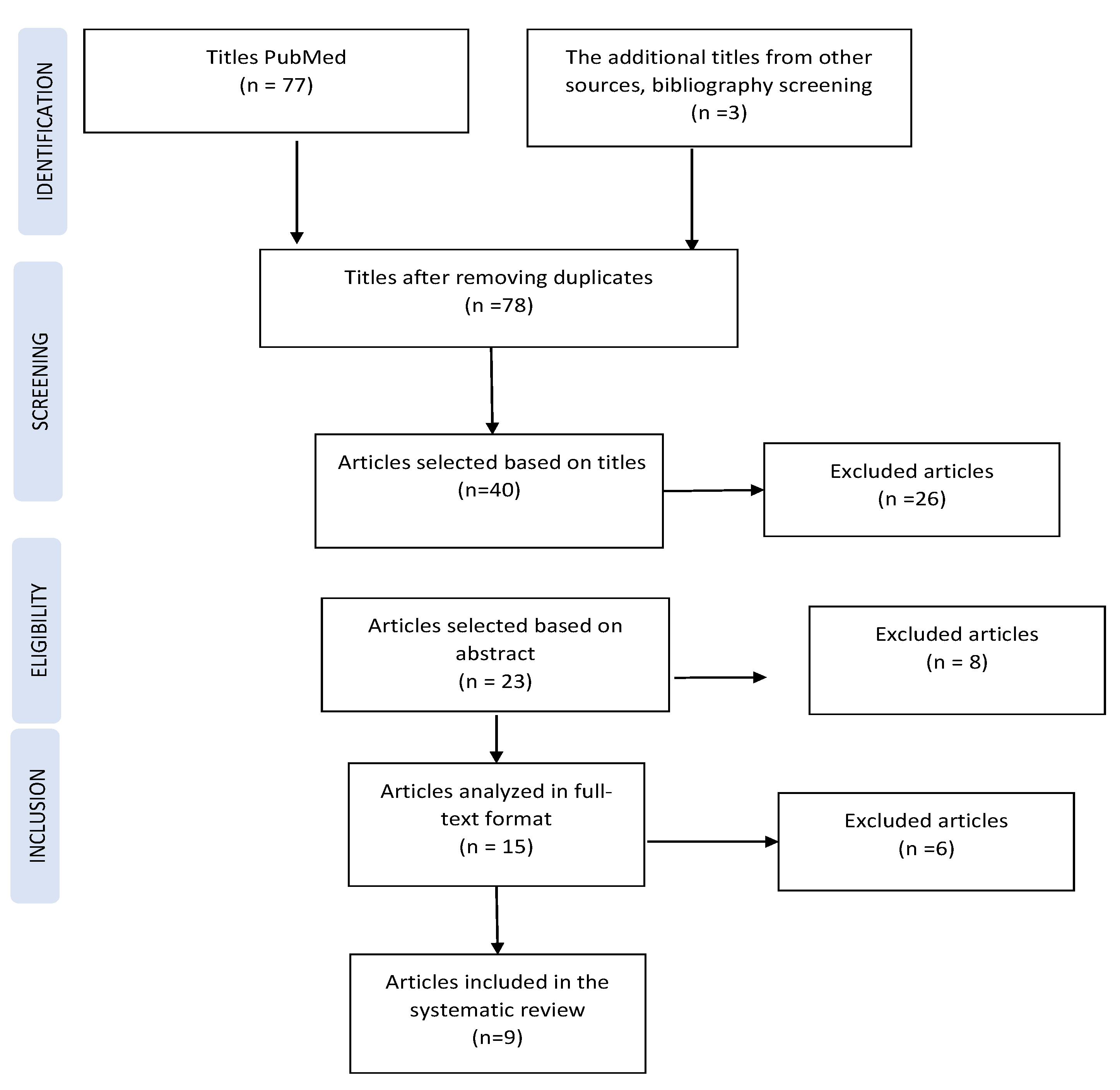
2.1.4. Selecting Studies for the Systematic Analysis

Two trained investigators were responsible for selecting studies for the systematic review, data extraction, and analysis process. The selection process involved four main stages, which included identifying titles of interest, screening them in databases, verifying their eligibility, and then including them in the systematic analysis based on the established inclusion and exclusion criteria. After identifying 78 titles, 40 were selected for abstract analysis, of which 23 abstracts were chosen. After reading them, only 15 abstracts met the inclusion criteria and were retained (Figure 1).

2.1.5. Qualitative Analysis

Studies included in the systematic analysis were of the in vitro type, which reduced the risk of subjectivity and the interpretability of conclusions. The control file was obtained using a Coordinate Measuring Machine (CMM), which returned accuracy data variable in the range from 0.7 [24] to 4.2 [25] μm or using an extraoral scanner with an accuracy ranging from 5 [26] to 11 [27] μm or even 20 μm [28] (D250, 3Shape). The final data were obtained through comparative analysis performed by dedicated engineering software for dimensional inspection (Geomagic or Rapidform XOR2) with a tolerance level selected by the operator ranging from 0.0014 to 6 μm [29]. The only factor prone to error in the research methodology was the human factor, which had the potential to influence data in three ways: the operator’s experience in using the scanning technology used in each study—an insignificant factor in this analysis because it excluded studies with records made by inexperienced operators; study design; scanning strategy, which was analyzed by applying a questionnaire adapted from the British Medical Journal (BMJ) [30]. Does the study address a clearly formulated question? Does the study use methods described in detail that are valid to answer the stated question? Do the generated valid results justifiably lead to the obtained conclusions? Does the adopted strategy agree with the recommendations of the scanning equipment manufacturer? Were any variations made to the recommended standard strategy provided by the manufacturer? If so, was the manufacturer consulted regarding the modifications made? Did the strategy generate scan gaps that required a rescan?
Because each of the selected studies is focused on the correlation between a factor with potential influence on accuracy, the analysis of registration distortion variations is performed by category—for each potential influencing factor based on accuracy: interimplant distance, implant angulation, scanner type, and scanning body type. Based on the inclusion criteria, each study model involves obtaining a “control” model (Master Model) against which the amplitude of discrepancy is comparatively evaluated through the mean of three-dimensional distortion parameters. The comparative analysis further requires differentiation based on one of the two computerized measurement and analysis techniques of Standard Tessellation Language (STL) files: best-fit alignment (BFA) overlap and linear distances (LD).

3. Results

3.1. Systematic Review

Following the search and selection stages of the studies based on inclusion and exclusion criteria, nine in vitro studies were selected for systematic analysis. The results are presented based on the analysis criteria.

3.1.1. The Impact of the Registration Technique/Scanning System on Scanning Error (Table 1)

A heterogeneity can be observed in the research methodology, although the research topic is the same. To contextualize the results and facilitate their interpretation, a brief description of the research method for each study is presented below.
Table 1. Correlation between impression material/technique/scan technology and accuracy.
Study (Reference)Scanning TechnologyConventional Impression Material and TechniqueStatistically Significant Differences
1.Rech-Ortega [25]True DefinitionPolyether—direct techniqueYes/No depending on the recorded distance
2. Moura [31]Dental Wings 3 SeriesSynthetic elastomer—direct and indirect techniqueNo
3. Kim K.R. [24]Trios 3ShapeSilicone addition—direct techniqueYes
4. Bohner L. [32]inEos BlueSilicone additionYes—cusp
No—occlusal fossae
5. Alikhasi M. [33]Trios 3ShapeSilicone addition—direct and indirect techniqueNo
6. Papaspyridakos P. [34]Trios 3ShapePolyether—technique with bonded and sectioned transfer copings, and bonded and unsectioned copingsNo
7. Mizumoto R.M. [27]Trios 3Shape
Without modifications
Interconnecting scanning bodies with dental floss
Glass spheres on the alveolar mucosa
Markings with pressure-indicating paste

-Yes
8. Vandeweghe S. [26]Lava COS
3M True Definition
Cerec Omnicam
3Shape Trios
-Yes
9. Flugge T. [28]iTero
Trios 3Shape
3M True Definition
-Yes
The study conducted by Rech-Ortega C. et al. [25] aims to investigate the variations in accuracy in recording a model that presents six angle analogs of 0° angulation and to compare the variation module in measuring three types of distances within the same model: adjacent distances (between neighboring implants 1–2, 2–3, 3–4, 4–5, 5–6), distances between interspersed analogs (1–4, 3–6), and the distance between the most distally positioned analogs (1–6). Statistically significant differences were identified when measuring the smallest distance between adjacent analogs in all analogs except for the distance between 1 and 2, where the difference between the control model and the one recorded by conventional impression did not confer statistical significance (p = 0.146), indicating superior accuracy in the case of conventional impression, with average values of variations of 20 µm for the elastomeric technique compared to 37 µm for the digital technique (p < 0.001). In recording the distance between interspersed analogs (1–4, 3–6), statistically significant differences occur at the moment of removing the reference point, because each scanning technique starts from the same point, which adds a layer of variability alongside the recorded interimplant distance parameter. Superior accuracy is recorded in the case of the distance between implants 1–4 for both techniques, with the mean variations being higher in the case of conventional impression (39 µm) compared to digital impression (21 µm). Finally, in the case of the analogs placed furthest away (1–6), both techniques used generate inaccuracies, with distortion variations ranging from 68 to 118 µm, with the higher amplitude belonging to the digital recording technique.
The research of Moura R.V. et al. [31] did not identify any statistically significant differences (p = 0.1099) between the compared groups: direct digitization (Dental Wings 3 series), conventional impression with synthetic elastomer using indirect and direct techniques, and impression using the two methods of the conventional technique followed by digitalization for comparison. No differences in accuracy were identified, even between the two variations in conventional impression, in contrast to the results recurrently confirmed in the literature regarding the superiority of the direct technique with bonded and sectioned abutments.
The study conducted by Kim K.R. [24] identified statistically significant differences in accuracy between the two compared groups (digital Trios 3Shape and conventional with silicone addition) only at the level of the implant in the right first molar (p = 0.529), which was also used as the starting point of the scanning strategy. The study separately evaluated the two accuracy parameters (defined as the difference in linear and angular distortions between the control master model and the test models from the two groups) and precision (defined as the difference in coordinates of 10 specimens belonging to the same group). Both parameters are evaluated as being superior in the case of conventional impression registration, a conclusion that contradicts the results presented in the literature, a contrast attributed to the fact that the researchers’ methodology evaluates the three-dimensional deviations of the virtually reconstructed implant replicas rather than the analysis of deviations of scanning bodies, which is frequently used in the cited literature.
In his research, Bohner L. et al. [32] analyzed deviations in terms of registered surfaces and identified the appearance of statistically significant differences at the level of vestibular and lingual cusps, confirming the inferiority of digital impression techniques (inEos Blue, Sirona) compared to conventional ones (addition silicone). Alikhasi M. [33] in his article concluded that digital impression techniques generate minimal deviation values for both types of implant connections that were studied (internal and external connections, with no statistically significant differences between the two types of connections), regardless of the implant angle (in the study, implants were inserted at the angles of 0° and 45°). Another study, conducted by Papaspyridakos P. et al. [34], did not identify statistically significant differences between digital impression techniques (Trios 3Shape) and conventional impressions (polyether), with the bonded and sectioned copings of ranked at the top compared to other analyzed techniques, with both returning higher accuracy results than conventional impressions with unbonded copings. In the study by Mizumoto and Yilmaz [27], the researchers formulated a more elaborate hypothesis, namely, evaluating five different types of scanning bodies in combination with four different scanning techniques using a single intraoral scanning system (Trios 3Shape). Regarding the enrichment of the defined points on the scanned oral mucosa texture, the three techniques did not generate statistically significant differences compared to when recording the field without any texture modification. On the contrary, it is considered that linear deviations were amplified by the ligature of dental floss between the scanning bodies because the recognizable software surface of the scanning body was altered by the superimposition of foreign elements such as dental floss. The identified linear deviations were related to the type of scanning body used and will be discussed later when discussing this parameter.
Studies that aim to exclusively compare different intraoral scanning techniques and their possible hierarchy based on accuracy and precision parameters have also been included in the analysis. Thus, Vandeweghe S. et al. [26] viewed the techniques offered by Trios 3Shape and 3M TrueDef as being equal because no statistically significant differences in terms of accuracy (p = 0.262) and precision (p = 0.119) were identified between the two techniques. The authors emphasize the importance of the clinician’s expertise in obtaining high-quality scans, regardless of the technique used. The authors hierarchize the scanning systems for each evaluated parameter as follows:
  • Accuracy: 3Shape (28 µm) > True Def (35 µm) > Cerec (61 µm) > Lava (112 µm);
  • Precision: True Def (30 µm) > 3Shape (33 µm) > Cerec (59 µm) > Lava (66 µm).
Regardless of the technique used, the maximum value discrepancies were recorded at the ends of the scanned field, specifically when registering implants in the area of molars 36 and 46. Another study, which aimed to analyze accuracy using only digital means of registration, was conducted by Flügge T.V. [28]. The analysis was performed from the perspective of several interimplant distances, with superior accuracy identified at distances of 6 mm, 11 mm (adjacent analogs), and 18 mm (intercalated analogs), which are the distances at which the differences between the control model (obtained with a laboratory scanner with a 20 μm accuracy, D250, 3Shape) are not statistically significant, and are thus closer to real dimensional values. In this case, superior accuracy was identified for the Trios and True Definition systems, which generate similar results without statistically significant differences. The conclusion of the study is centered around accuracy (represented by the standard deviation variation), which was inversely proportional to the recorded interimplant distance, compared to the constant SD recorded in the control model (obtained through laboratory extraoral scanning technology) in which there are no fluctuations; thus, the technology’s performance is independent of the amplitude of the recorded interimplant distance.

3.1.2. The Impact of Interimplant Distance on Scanning Error (Table 2)

Overall, the analysis of the nine articles included in the systematic review suggested that intraoral scanning systems can provide accurate and precise digital impressions for clinical use. The accuracy and precision of the different scanning systems varied, with some systems performing better than others in certain parameters.
Table 2. Correlations between interimplant distance and accuracy.
StudyImplant NumberScanning TechnologyInterimplantar Distance (mm)Statistically Significant Differences
1. Rech-Ortega [25]6True Definition1–2/5–6: ~11
2–3/–5: ~14
3–4: ~10
1–4/3–6: ~32yes
1–6: ~40
2. Kim K.R. [24]6Trios 3Shape-yes
3. Flugge T. [28]5Trios 3Shape
True Definition
iTero
35–36: 6.6
33–35/45–47:11
33–36: 18
35–45: 40
36–47: 50
yes
The method of superimposition (best fit alignment) and positioning of the test model against the master reference model can generate errors because the position chosen before the software comparison will have the lowest global number of deviations [35]. When considering the cumulative effect of errors in the total arch registration and the final impression recorded with an intraoral scanner, a variable degree of error can be expected, with the proportion increasing from the origin point of the registration to its final point. When analyzing the variations in the recording device, the precision and reproducibility of the software algorithm are essential. Comparative analysis techniques that measure defined linear distances between two easily identifiable points are considered a preferable alternative.
Rech-Ortega and Fernández-Estevan [25] identified different performances of compared technologies that are correlated with the recorded distance variation between implants. Based on their conclusions, the authors provide clinical practice recommendations: for restorations anchored on less than three implants, the recommended impression technique is conventional with polyether. For prosthetic restorations anchored on four implants, superior accuracy is achieved by digital techniques. For restorations anchored on more than four implants, both techniques generated variable results with statistically significant differences from the control model, but the deviation amplitude did not exceed the clinically acceptable limit of 150 μm. In this case, the authors recommend creating an intermediate piece to verify passivity before designing the final prosthetic piece. Judging by the precision factor of each technology used, namely the fluctuations in the proportional standard deviation with the recorded distance, the authors conclude that the error factor is correlated with the amplitude of the recorded distance. This conclusion is supported by other results in the literature [36,37]. The authors support their conclusion by recording maximum deviations at the level of the distance between the furthest positioned implants (1–6, 118 μm) and the distance between 3 and 6 (109 μm).
The conclusions of the study conducted by Kim and Seo [24] indicate the superiority of conventional silicone addition impressions, but their analysis is based on the centroid deviation of the digital reconstruction of the scanning body. The applicability of these conclusions is questionable in practice because the future prosthetic reconstruction relies on implant abutments and has no direct connection to the intermediate scanning piece.
Flugge T.V. [28] identified statistically significant differences between the types of technologies included in his research. A preliminary analysis shows that the extraoral scanning system D250 3Shape consistently performs well in terms of precision, regardless of the interimplant distance factor, with a constant standard deviation value. This performance is attributed to the acquisition technique that is used, which involves laser plane projection onto the surface of the scanned model. Other systems, such as iTero, achieve lower precision values regardless of the recorded interimplant distance. The authors of the study identified statistically significant differences between this system and Trios or True Definition for the 11 mm distance (equivalent to the absence of three teeth). Regarding distances between 6 and 18 mm, the True Definition technology exhibits superior precision. For the maximum distances investigated, with a transarcadic direction of 40 and 50 mm, statistically significant differences were identified between two groups: iTero and True Definition, as well as iTero and D250.

3.1.3. The Impact of Implant Angulation on Scanning Error (Table 3)

In the study by Moura R.V. [31], after conducting ANOVA variance tests involving two factors of variation, no statistically significant differences were found between the test models and the control model, indicating a higher level of reliability for each recording method. The authors concluded that implant angulation variation does not significantly impact the obtained recordings (Dental Wings 3 Series, direct and indirect conventional impressions), which confirms similar results reported in the literature [35,36,37].
Table 3. Correlations between angulation and accuracy.
Study (Reference)Number of ImplantsImplants’ CharacteristicsStatistically Significant Differences
PositionAngulationDirection
1. Moura R.V. [32]617
23
15°
 
Mesial
Distal
No
15, 12, 25, 27-
2. Alikhasi M. [34]413, 23-No (digital)
Yes (conventional)
15, 2545°Distal
3. Papaspyridakos P. [35]545, 3515°DistalNo
43, 33, 41–31
The same conclusion is achieved in the study conducted by Alikhasi M. [33]; however, the study is only applicable to digital impressions (Trios 3Shape) that generate superior accuracy results irrespective of implant angulation variability or implant connection type, without significant differences compared to the control model. However, the conclusion does not apply to conventional impressions, where differences are identified compared to the control model, suggesting the superiority of accuracy parameters generated by digital impressions for both 0° and 45° angulated implants, with a difference in direct impressions for non-angulated implants compared to angulated ones, and with a reversal of the difference in indirect impressions, which is favorable for angulated implants compared to nonangulated ones.
The study conducted by Papaspyridakos P. [34] concludes that there is no significant disturbance in accuracy parameters for angular variabilities up to 15° for the Trios 3Shape technology. This conclusion is supported by previous studies, which also show consistent results of up to 30° when using a different technology, LAVA Cos [35].

3.1.4. The Impact of the Scan Body Type on Scanning Error

Regarding the impact of scan body type on scanning error, only one study [27] included in the systematic analysis compared five different types of scan bodies designed by different manufacturers, successively connected to the same maxillary model, and registered with the same scanning technology (Trios 3Shape). The authors proposed a dual hypothesis. First, the scanning body type does not affect the accuracy of the recording. Second, changes to the scanned surface will not introduce variations in the accuracy parameters. Changes to the mucosal surface refer to various additional elements to the interimplant surface in cases of total edentulism, which are considered difficult to record using digital technology because the number of recognizable or identifiable elements is very low [36]. The inadequacy of orientation elements for scanning technology requires an adaptation of the software algorithm, which, in the absence of sufficient intraoral orientation, autonomously corrects these deficient interpretations by cutting out areas labeled as “redundant”, resulting in an incorrect joining of successive acquisitions [20]. The study’s results only managed to refute one of the hypotheses, confirming that different strategies used to enrich the number of usable points in three-dimensional reconstruction do not produce differences compared to the control technique, which represents the study model registration without any modification to the mucosal area. Additionally, the maximum deviation values were identified for the technique of using dental floss anchored on scan bodies. The justification for this observation is that the floss overlay on certain areas of the scan body interferes with its unique identifiable points in the digitization process. Statistically significant differences were identified between the types of scan bodies used, with authors hierarchically separating the most favorable geometric shape for recording accurate implant positions.

3.2. Statistical Analysis in the Studies

In studies that met the inclusion criteria for systematic analysis, SPSS software versions 20, 21.0, 22.0, 23.0, and 24.0 were predominantly used. One study used Minitab and Statistix 9.1, another used SAS 9.3, and two studies did not specify the data-processing method. ICC was mentioned only in four studies, with the study conducted by Mizumoto et al. [27] reporting a raw data correlation factor of 0.999, and that of Rech-Ortega [25] reporting the value of 0.00027–0.00385%, with values below 1%, ensuring the internal validity of the comparative analysis method and ensuring safe reproducibility of the obtained data. In terms of the statistical tests, ANOVA was applied in 7 out of 12 studies, with a p-value < 0.05 established in all of them. The remaining statistical tests were applied unevenly, with HSD tests being applied in seven studies, with correction used in only two of them (Bonferroni, Mann–Whitney).

3.3. Evaluation of Study Design from a Qualitative Perspective (Table 4 and Table 5)

The study design evaluation (Table 4) shows that a clear question is asked in six articles [24,27,31,32,33,34]; the methods are described in detail in only five studies [24,27,31,33,34]; however, for all selected studies, valid results generated lead to the conclusions presented in a justified manner. The qualitative characteristics of the adopted scanning strategy analysis (Table 5) point out that three studies [25,26,33] do not mention whether this is in accordance with the recommendations of the scanning equipment manufacturer; only four [24,27,28,34] studies were performed without modification of the standard strategy recommended by the manufacturer, and none of the papers mention whether the strategy generates scan gaps that required a rescan.
Table 4. The qualitative characteristics of the included studies.
Study (Reference)Does the Study Address a Clearly Formulated Question?Does the Study Use Methods That Are Described in Detail and Valid to Answer the Issued Question?Do the Valid Results Generated Lead to the Conclusions Presented in a Justified Manner?
1. Rech-Ortega C. [25]vaguesuperficial yes
2. Moura R.V. [32]yesyesyes
3. Kim K.R. [24]yesyesyes
4. Bohner L. [33]yessuperficial yes
5. Alikhasi M. [34]yesyesyes
6. Papaspyridakos P. [35]yesyesyes
7. Mizumoto R.M. [27]yesyesyes
8. Vandeweghe S. [26]vague superficial yes
9. Flugge T. [28]Nosuperficial yes
Table 5. The qualitative characteristics of the adopted scanning strategy.
Study (Reference) Is the Adopted Strategy in Accordance with the Recommendations of the Scanning Equipment Manufacturer?Have Any Modifications Been Made to the Standard Strategy Recommended by the Manufacturer? If So, Was the Manufacturer Consulted Regarding the Modifications Made?Did the Strategy Generate Scan Gaps that Required a Rescan?
1. Rech-Ortega [25]Is not describedDoes not mentionDoes not mention
2. Moura R.V. [32]Extraoral scanningDoes not applyDoes not mention
3. Kim K.R. [24]YesWithout modificationsDoes not mention
4. Bohner L. [33]Extraoral scanningDoes not applyDoes not mention
5. Alikhasi M. [34]NoDoes not mentionDoes not mention
6. Papaspyridakos P. [35]YesWithout modificationsDoes not mention
7.Mizumoto R.M. [27]YesWithout modificationsDoes not mention
8.Vandeweghe S. [26]Is not describedDoes not mentionDoes not mention
9. Flugge T. [28]YesWithout modificationsDoes not mention

4. Discussion

To achieve an adequate level of passivity in the prosthetic restoration at the end of the clinical–technical stages, the maximum tolerated value of mismatch between the prosthetic restoration and the supporting substructure is 120 µm [37], and there is no standardized value accepted by consensus for the impression-making stage. Therefore, our analysis will rank different types of intraoral scanning devices in relation to the amplitude of deviations returned by them during recording.
A higher predictability of the final prosthetic restoration guarantees clinical success in many cases. However, in the literature, there are few reports of completely digital implant–prosthetic restorations [38] because the progress of qualitative clinical studies seems to be slower compared to the technological progress.
The methodology of the chosen topic was heterogeneous. Different software comparison tools were highlighted for the obtained records (measuring linear distances or comparisons through three-dimensional overlap) and for the level of tolerance of each comparative software. Different possibilities were also recorded for obtaining the control model (manual measurement, computerized measurement using dedicated devices, and extraoral scanning with laboratory devices of superior accuracy), the selected reference point for comparative overlay (calibrated element added additionally to the study model or uncalibrated elements already belonging to the prosthetic field), or statistical calculations.
The factors that improved uniformity in the selection of studies included in our analysis were as follows: respecting the scanning strategy recommended by the technology manufacturer; obtaining a control file with maximum accuracy against which subsequent comparative analysis is performed; performing measurements by an experienced operator with the clear understanding of the scanning technology to minimize the incidence of false-positive errors; using an optimal number of measurements to generate the statistical power of the conclusions drawn from each study. These factors were considered relevant to ensure the validity of this study and presented a reduced degree of variability between the studies included in the analysis.
The review identified repeatable differences in the accuracy between different types of scanning technologies, which is attributed to the different imaging acquisition principles of each technology, with the highest error levels being found in total edentulous arch recording. There is no consensus on the maximum acceptable error level for implant angulation, with some authors suggesting 15° and others recommending even 30°. Statistically significant differences were identified between the types of scan bodies used.
The generated conclusions are discussed from different aspects. In a study conducted by Rech-Ortega C. [25], the researchers identified specific variations depending on the measured distance, identifying relevant clinical correlations between certain impression techniques and the magnitude of the proposed prosthetic restorations. Thus, the distance between neighboring analogs (1–2) indicates a higher-accuracy registration in the case of conventional impression compared to the larger distances recorded within the same model, which is why the authors of the study recommend using the direct conventional impression technique with polyether for implant restorations anchored on a maximum of three implant posts. Furthermore, the recordings indicate a reduction in the accuracy parameter as the distance from the origin point of the scanning strategy increases. When recording the distance between the intercalated implant posts (1–4), the digital technique returns satisfactory results, with minimal recorded variations compared to the same distance in the case of implants 3–6, which are further away from the origin point. In this case, the variations are more uneven and statistically significant in amplitude compared to the control file. Thus, the authors of the study recommend digital impression for implant restorations anchored on four posts. In the case of large-scale restorations (1–6), both impression techniques generate deviations with statistical significance, but the distortion values still fall within clinically acceptable limits (68–118 µm).
The study by Moura R.V. [31] does not identify any statistically significant difference between conventional impression with synthetic elastomer and the digital technique with the Dental Wings 3 Series system, indicating a favorable virtual reconstruction for prosthetic restorations under conditions of passivity. Similarly, another study [34] places the impression technique with the intraoral scanner on the same level as the conventional impression technique in terms of the accuracy parameter, but with the advantage of reducing processing time and the possibility of capturing additional information.
Significant differences were found compared to the control model in terms of the accuracy and precision of different digital scanning technologies. An analysis based on the scanning body shape emphasizes that the most favorable shape for accurate recording is one with the minimal height and simplified geometry, without retentive or complex features.
The main limitations of the included studies come from their in vitro execution, which eliminates biological factors present in the clinical scenario such as the presence of saliva and intraoral fluid dynamics, changes in light reflection caused by saliva, mucosal surface mobility, and possible loss of reference points. Additionally, the intraoral and perioral muscles may hinder access and visibility. Some authors suggest that error amplitude may double in clinical conditions compared to in vitro conditions [1]. Additional data on the performance of digital scanning technologies are needed, particularly in a clinical context. A promising direction for future research may be the development of standardized clinical study methodologies for evaluating the accuracy and precision parameters of optical scanning systems.
The absence of modifying clinical factors in the in vitro context precludes the extension of error assessments from these studies to intraoral clinical scenarios and only allows for the assumption that errors detected in vitro will inevitably be amplified in vivo.

5. Conclusions

Intraoral scanning systems generate distortion in extensive implant restorations, depending on the registration technique/scanning system, interimplant distance, implant angulation, and the scan body type. These factors need to be monitored in clinical practice.

Author Contributions

Conceptualization, L.O. and I.L.; methodology L.O. and T.C.; formal analysis, A.M.; investigation, S.R.; resources I.L. and T.C.; data curation, A.M.; writing—original draft preparation, L.O. and A.M.; writing—review and editing, S.R. All authors have read and agreed to the published version of the manuscript.

Funding

This research received no external funding.

Institutional Review Board Statement

Not applicable.

Informed Consent Statement

Not applicable.

Data Availability Statement

The data reported in this study are available in the literature.

Conflicts of Interest

The authors declare no conflict of interest.

References

  1. Flügge, T.V.; Schlager, S.; Nelson, K.; Nahles, S.; Metzger, M.C. Precision of intraoral digital dental impressions with iTero and extraoral digitization with the iTero and a model scanner. Am. J. Orthod. Dentofac. Orthop. 2013, 144, 471–478. [Google Scholar] [CrossRef]
  2. Wiranto, M.G.; Engelbrecht, W.P.; Nolthenius, H.E.T.; van der Meer, W.J.; Ren, Y. Validity, reliability, and reproducibility of linear measurements on digital models obtained from intraoral and cone-beam computed tomography scans of alginate impressions. Am. J. Orthod. Dentofac. Orthop. 2013, 143, 140–147. [Google Scholar] [CrossRef]
  3. Grünheid, T.; McCarthy, S.D.; Larson, B.E. Clinical use of a direct chairside oral scanner: An assessment of accuracy, time, and patient acceptance. Am. J. Orthod. Dentofac. Orthop. 2014, 146, 673–682. [Google Scholar] [CrossRef]
  4. Driscoll, C.D. Clinical Applications of Digital Dental Technology; Wiley: Hoboken, NJ, USA, 2015; ISBN 9781118655795. [Google Scholar] [CrossRef]
  5. Mehl, A.; Ender, A.; Mörmann, W.; Attin, T. Accuracy testing of a new intraoral 3D camera. Int. J. Comput. Dent. 2009, 12, 11–28. [Google Scholar] [CrossRef]
  6. Rudolph, H.; Salmen, H.; Moldan, M.; Kuhn, K.; Sichwardt, V.; Wöstmann, B.; Luthardt, R.G. Accuracy of intraoral and extraoral digital data acquisition for dental restorations. J. Appl. Oral Sci. 2016, 24, 85–94. [Google Scholar] [CrossRef]
  7. Ender, A.; Attin, T.; Mehl, A. In vivo precision of conventional and digital methods of obtaining complete-arch dental impressions. J. Prosthet. Dent. 2016, 115, 313–320. [Google Scholar] [CrossRef]
  8. Mangano, F.; Gandolfi, A.; Luongo, G.; Logozzo, S. Intraoral scanners in dentistry: A review of the current literature. BMC Oral Health 2017, 17, 149. [Google Scholar] [CrossRef]
  9. Richert, R.; Goujat, A.; Venet, L.; Viguie, G.; Viennot, S.; Robinson, P.; Farges, J.-C.; Fages, M.; Ducret, M. Intraoral Scanner Technologies: A Review to Make a Successful Impression. J. Healthc. Eng. 2017, 2017, 8427595. [Google Scholar] [CrossRef]
  10. Schepke, U.; Meijer, H.J.A.; Kerdijk, W.; Cune, M.S. Digital versus analog complete-arch impressions for single-unit premolar implant crowns: Operating time and patient preference. J. Prosthet. Dent. 2015, 114, 403–406.e1. [Google Scholar] [CrossRef]
  11. Joda, T.; Braegger, U. Time-Efficiency Analysis Comparing Digital and Conventional Workflows for Implant Crowns: A Prospective Clinical Crossover Trial. Int. J. Oral Maxillofac. Implant. 2015, 30, 1047–1053. [Google Scholar] [CrossRef]
  12. Ender, A.; Zimmermann, M.; Attin, T.; Mehl, A. In vivo precision of conventional and digital methods for obtaining quadrant dental impressions. Clin. Oral Investig. 2016, 20, 1495–1504. [Google Scholar] [CrossRef] [PubMed]
  13. Ender, A.; Mehl, A. Accuracy of complete-arch dental impressions: A new method of measuring trueness and precision. J. Prosthet. Dent. 2013, 109, 121–128. [Google Scholar] [CrossRef] [PubMed]
  14. Zhang, F.; Suh, K.-J.; Lee, K.-M. Validity of intraoral scans compared with plaster models: An in-vivo comparison of dental measurements and 3D surface analysis. PLoS ONE 2016, 11, e0157713. [Google Scholar] [CrossRef] [PubMed]
  15. Muallah, J.; Wesemann, C.; Nowak, R.; Robben, J.; Mah, J.; Pospiech, P.; Bumann, A. Accuracy of full-arch scans using intraoral and extraoral scanners: An in vitro study using a new method of evaluation Genauigkeit von Ganzkieferscans mit intraoralen und extraoralen Scannern: Eine In-vitro-Untersuchung. Int. J. Comput. Dent. 2017, 20, 151–164. [Google Scholar] [PubMed]
  16. Wesemann, C.; Muallah, J.; Mah, J.; Bumann, A. Accuracy and efficiency of full-arch digitalization and 3D printing: A comparison between desktop model scanners, an intraoral scanner, a CBCT model scan, and stereolithographic 3D printing. Quintessence Int. 2017, 48, 41–50. [Google Scholar] [CrossRef]
  17. Seelbach, P.; Brueckel, C.; Wöstmann, B. Accuracy of digital and conventional impression techniques and workflow. Clin. Oral Investig. 2013, 17, 1759–1764. [Google Scholar] [CrossRef]
  18. Güth, J.-F.; Keul, C.; Stimmelmayr, M.; Beuer, F.; Edelhoff, D. Accuracy of digital models obtained by direct and indirect data capturing. Clin. Oral Investig. 2013, 17, 1201–1208. [Google Scholar] [CrossRef]
  19. Su, T.-S.; Sun, J. Comparison of repeatability between intraoral digital scanner and extraoral digital scanner: An in-vitro study. J. Prosthodont. Res. 2015, 59, 236–242. [Google Scholar] [CrossRef]
  20. Rhee, Y.-K.; Huh, Y.-H.; Cho, L.-R.; Park, C.-J. Comparison of intraoral scanning and conventional impression techniques using 3-dimensional superimposition. J. Adv. Prosthodont. 2015, 7, 460–467. [Google Scholar] [CrossRef]
  21. ISO 5725-1; Accuracy (Trueness and Precision) of Measurement Methods and Results–Part 1: General Principles and Definitions. 1998. Available online: https://www.iso.org/obp/ui/#iso:std:iso:5725:-1:ed-1:v1:en (accessed on 23 March 2021).
  22. Menditto, A.; Patriarca, M.; Magnusson, B. Understanding the meaning of accuracy, trueness and precision. Accredit. Qual. Assur. 2007, 12, 45–47. [Google Scholar] [CrossRef]
  23. Moher, D.; Liberati, M.; Tetzlaff, J.; Altman, D.G.; PRISMA Group. Preferred Reporting Items for Systematic Reviews and Meta-Analyses: The Prisma Statement. PLoS Med. 2009, 6, e1000097. [Google Scholar] [CrossRef] [PubMed]
  24. Kim, K.R.; Seo, K.-Y.; Kim, S. Conventional open-tray impression versus intraoral digital scan for implant-level complete-arch impression. J. Prosthet. Dent. 2019, 122, 543–549. [Google Scholar] [CrossRef] [PubMed]
  25. Rech-Ortega, C.; Fernandez-Estevan, L.; Sola-Ruiz, M.; Agustin-Panadero, R.; Labaig-Rueda, C. Comparative in vitro study of the accuracy of impression techniques for dental implants: Direct technique with an elastomeric impression material versus intraoral scanner. Med. Oral Patol. Oral Cir. Bucal. 2019, 24, e89–e95. [Google Scholar] [CrossRef]
  26. Vandeweghe, S.; Vervack, V.; Dierens, M.; De Bruyn, H. Accuracy of digital impressions of multiple dental implants: An in vitro study. Clin. Oral Implant. Res. 2017, 28, 648–653. [Google Scholar] [CrossRef]
  27. Mizumoto, R.M.; Yilmaz, B.; McGlumphy, E.A.J.; Seidt, J.; Johnston, W.M. Accuracy of different digital scanning techniques and scan bodies for complete-arch implant-supported prostheses. J. Prosthet. Dent. 2020, 123, 96–104. [Google Scholar] [CrossRef]
  28. Flügge, T.V.; Att, W.; Metzger, M.C.; Nelson, K. Precision of Dental Implant Digitization Using Intraoral Scanners. Int. J. Prosthodont. 2016, 29, 277–283. [Google Scholar] [CrossRef] [PubMed]
  29. Tan, M.Y.; Yee, S.H.X.; Wong, K.M.; Tan, Y.H.; Tan, K.B.C. Comparison of Three-Dimensional Accuracy of Digital and Conventional Implant Impressions: Effect of Interimplant Distance in an Edentulous Arch. Int. J. Oral Maxillofac. Implant. 2019, 34, 366–380. [Google Scholar] [CrossRef]
  30. Greenhalgh, T.; Taylor, R. How to read a paper: Papers that go beyond numbers (qualitative research). BMJ 1997, 315, 740–743. [Google Scholar] [CrossRef]
  31. Moura, R.V.; Kojima, A.N.; Saraceni, C.H.C.; Bassolli, L.; Balducci, I.; Özcan, M.; Mesquita, A.M.M. Evaluation of the Accuracy of Conventional and Digital Impression Techniques for Implant Restorations. J. Prosthodont. 2019, 28, e530–e535. [Google Scholar] [CrossRef]
  32. Bohner, L.; Hanisch, M.; Canto, G.D.L.; Mukai, E.; Sesma, N.; Neto, P.T. Accuracy of Casts Fabricated by Digital and Conventional Implant Impressions. J. Oral Implant. 2019, 45, 94–99. [Google Scholar] [CrossRef]
  33. Alikhasi, M.; Siadat, H.; Nasirpour, A.; Hasanzade, M. Three-Dimensional Accuracy of Digital Impression versus Conventional Method: Effect of Implant Angulation and Connection Type. Int. J. Dent. 2018, 2018, 3761750. [Google Scholar] [CrossRef] [PubMed]
  34. Papaspyridakos, P.; Gallucci, G.O.; Chen, C.-J.; Hanssen, S.; Naert, I.; Vandenberghe, B. Digital versus conventional implant impressions for edentulous patients: Accuracy outcomes. Clin. Oral Implant. Res. 2016, 27, 465–472. [Google Scholar] [CrossRef] [PubMed]
  35. Conrad, H.J.; Pesun, I.J.; DeLong, R.; Hodges, J.S. Accuracy of two impression techniques with angulated implants. J. Prosthet. Dent. 2007, 97, 349–356. [Google Scholar] [CrossRef] [PubMed]
  36. Andriessen, F.S.; Rijkens, D.R.; van der Meer, W.J.; Wismeijer, D.W. Applicability and accuracy of an intraoral scanner for scanning multiple implants in edentulous mandibles: A pilot study. J. Prosthet. Dent. 2014, 111, 186–194. [Google Scholar] [CrossRef] [PubMed]
  37. McLean, J.W.; von Fraunhofer, J.A. The estimation of cement film thickness by an in vivo technique. Br. Dent. J. 1971, 131, 107–111. [Google Scholar] [CrossRef]
  38. Joda, T.; Zarone, F.; Ferrari, M. The complete digital workflow in fixed prosthodontics: A systematic review. BMC Oral Health 2017, 17, 124. [Google Scholar] [CrossRef]
Figure 1. Research strategy PRISMA protocol [23].
Prosthesis 05 00080 g001
Disclaimer/Publisher’s Note: The statements, opinions and data contained in all publications are solely those of the individual author(s) and contributor(s) and not of MDPI and/or the editor(s). MDPI and/or the editor(s) disclaim responsibility for any injury to people or property resulting from any ideas, methods, instructions or products referred to in the content.

