Article

Determinants of Temperature Development during Dental Implant Surgery

Department of Prosthodontics, Saarland University, 66424 Homburg, Germany
*
Author to whom correspondence should be addressed.
Prosthesis 2024, 6(3), 657-669; https://doi.org/10.3390/prosthesis6030046
Submission received: 19 April 2024 / Revised: 6 June 2024 / Accepted: 7 June 2024 / Published: 12 June 2024
(This article belongs to the Collection Oral Implantology: Current Aspects and Future Perspectives)

Abstract

:
Mechanical and thermal trauma during implant surgery may be reasons for initial peri-implant bone loss. Temperature development during drilling and implant insertion were quantified in this series of in vitro and animal experiments. Polyurethane foam material mimicking different classes of alveolar bone was used as a model material for simulating implant surgery. Using thermocouples, temperature development was determined in the model material at depths of 3 mm and 10 mm during site preparation and implant insertion. Additionally, an infrared camera allowed for measuring drill temperatures both in vitro and as part of an animal trial using an intraoral minipig model. Drill diameter and repeated usage of drills did not have a major effect on temperature generation. The addition of a diamond-like carbon coating, bone density, predrilling, and irrigation heavily affected intraosseous temperatures. In vivo, applying regular drill protocols, an intraosseous temperature rise of approximately 3 °K was determined. Implant geometry as well as the amount of undersizing of an osteotomy governed heat generation during implant insertion. Drill protocols and the amount of undersizing of an implant osteotomy constitute parameters by which clinicians can limit trauma during implant surgery.

1. Introduction

Marginal bone level change continues to be one of the most relevant criterion for implant success with an initial loss of 1.0 mm and an annual loss of 0.2 mm after the first year being widely accepted [1,2,3]. Other than peri-implantitis defects, this bone loss is currently understood as an adaptive process, which initially is related to surgical trauma [4] comprising both mechanical and thermal stresses during osteotomy preparation and implant insertion [5,6,7].
While there seems to be consensus that surgical interventions cause cell death to some extent [8], preservation of bone viability is critical for osseointegration [9]. However, it is difficult to link biologic performance to one specific factor, i.e., temperature development vs. mechanical stress [10]. Based on an animal trial, it was concluded that the influence of drill speed and irrigation would be minimal in terms of the temperature of the cortical bone, primary and secondary implant stability, and osseointegration [11]. Given the widespread use of drilling as a technique for implant site preparation as well as the advanced devices available today, it has been questioned whether or not heat above the critical temperature for bone necrosis [12] can be generated at all, if accepted protocols are followed.
For conventional drilling, the following procedural parameters have been described [5] to affect temperature development: rotational speed, proceeding speed, contact pressure, drilling motion pattern, bone density [8,13,14], drill depth [15], and irrigation [16,17]. Variables related directly to drill design also seem to play a role with the major parameters being the number of drill blades, drill design (tapered vs. straight) [18], drill fatigue [7,19], drill material [20], and its heat capacity and thermal conductivity [21].
In an attempt to reduce surgery times, abbreviated drilling protocols have been advocated, for instance, using multistepped drills for single-stage implant site preparation [22]. In an animal trial, a novel drill design led to lower osteotomy temperature values and shorter drill times but also improved osseointegration of dental implants [5]. In this context, low-speed drilling without irrigation has been shown to result in greater quantity and more beneficial cellular and histomorphologic properties of harvested bone with even greater osteotomy precision [23].
Applying diamond-like carbon (DLC) coatings on drills has been claimed to optimize existing surgical approaches. DLC coatings have already been shown to bear superior tribological and mechanical properties [24] leading to improved wear properties [25] as well as reduced friction between mechanical components [26].
With the goal of shortening overall treatment times, clinicians have been trying to insert implants with maximum primary stability in order to limit the risk of excessive micromotion at the implant–bone interface during healing in immediate loading cases [27]. Undersizing of an osteotomy as well as using a tapered implant resulting in bone compression have been described as effective approaches for reaching high insertion torque values [28]. This effect of bone compression was verified in finite element simulations showing a clear trend towards greater stress levels in bone with increasing levels of under preparation of the osteotomy [29,30]. Temperature rise during implant insertion has also been shown in an animal trial [6] and seems to be a phenomenon based on friction between bone and the implant body [17,31]. Several authors [10] have claimed that bone damage during implant insertion will cause cracks to varying extents [32] which leads to bone resorption [33] followed by new bone formation during the healing phase [34] which, however, will take longer as compared to areas which had not been damaged [35].
Ideally, a dental implant should allow for both bone compression, for achieving primary stability, and room for new bone formation [34]. Two approaches are currently provided by the industry which include the omission of threads in the cervical region and non-round implant cross-sections. From a clinical perspective, the omission of cervical threads did not enhance esthetic performance as compared to a more traditional, round implant design [36]. Similarly, no major advantage with respect to marginal bone level change was described for implants with a tri-oval [37] or a triangular [38] design.
In this context, a novel implant design has recently been developed which is characterized by a narrow core diameter with sharp threads in the apical part intended for proper engagement, a middle part with a bulky core and shallow, condensing threads intended for compressing trabecular bone, and a cervical part with a narrow core and sharp threads for cutting the cortical plate [39]. This design has been shown to be advantageous with respect to stress development and primary stability in advanced indications such as immediate implant placement and sinus lifting [40].
It was the goal of this combined in vitro and animal trial to determine the temperature development of commercially available conventional and DLC-coated drills under various parameters as well as to compare an existing, tapered bone level implant with a novel implant design with respect to heat generation during insertion, primary implant stability, and insertion torque.

2. Materials and Methods

The experimental design closely followed previous studies in this field [16,31,41]. For the first part of this investigation, bone surrogate materials [14,16,42] with varying densities and structure were employed (Solid rigid Polyurethane Foam, Sawbones Europe AB, Malmö, Sweden). Into theses perfectly squared blocks, perpendicular osteotomies were drilled using burs from a specific implant system (AlfaGate, Kfar Qara, Israel) and a surgical motor set at 800RPM (Mastersurg, KaVo, Biberach, Germany) with the contra-angle mounted in a modified drill press allowing for exact alignment. A weight of 1 kg mounted on the handle of the drill press was used for standardizing the vertical velocity. The surgical kit of the implant system offered both conventional drills and DLC-coated drills. Each drilling procedure was repeated five times [14]. Prior to creating an implant osteotomy, two holes were drilled with a diameter of 1 mm (3 mm depth) and 1.5 mm (10 mm depth) at a distance of 0.5 mm from the future osteotomy wall (Figure 1a). Two thermocoupling elements were inserted into these holes at 3 mm and 10 mm depth for recording maximum temperature [5,31] prior to the drilling process and when the osteotomy was finished (Figure 1b). In addition, an infrared camera was employed in selected experimental groups for determining the temperature of drills immediately after removal from an osteotomy. Table 1 provides an overview of the experimental groups.
Statistical analysis of these data was based on temperature differences at the bottom and at the top of the osteotomy. Shapiro–Wilk tests were employed for testing normality of distribution of measurement values, Levene’s test was used for testing equality of variances, and two-sample t tests with p value adjustment for multiple comparisons (Holm method) were employed for pairwise comparisons between experimental groups. The level of significance was set at α = 0.05 for all statistical operations carried out.
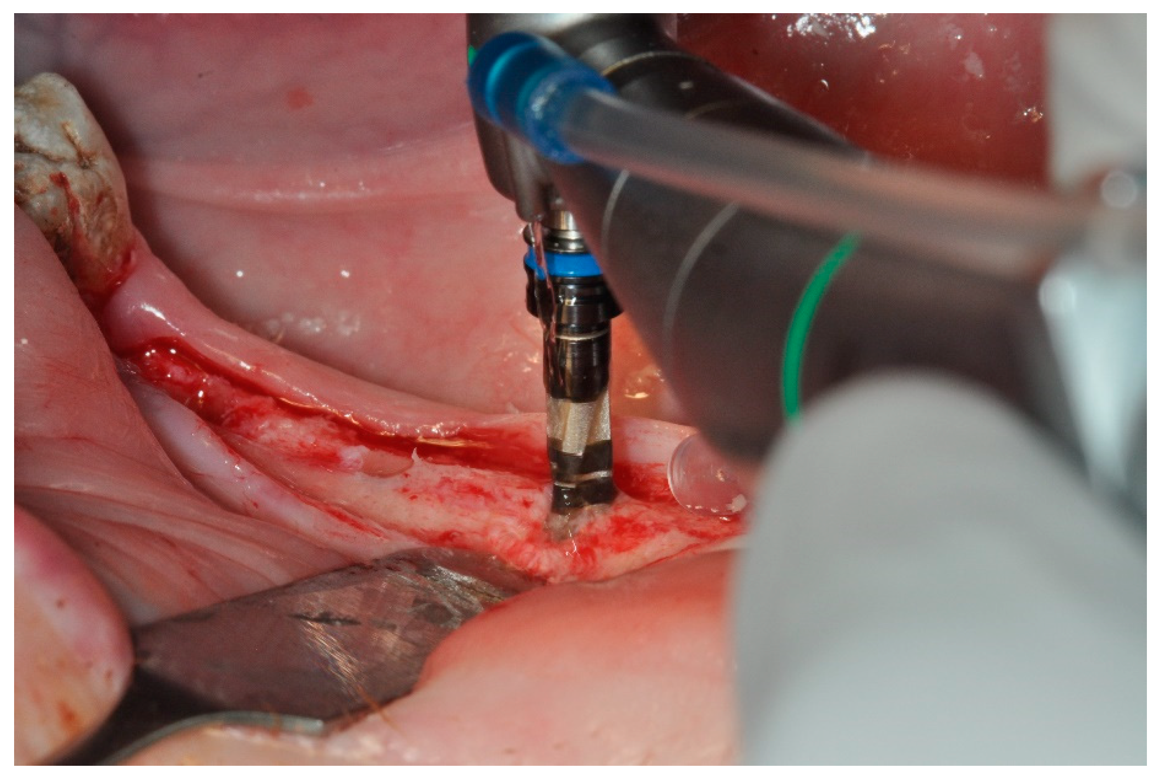
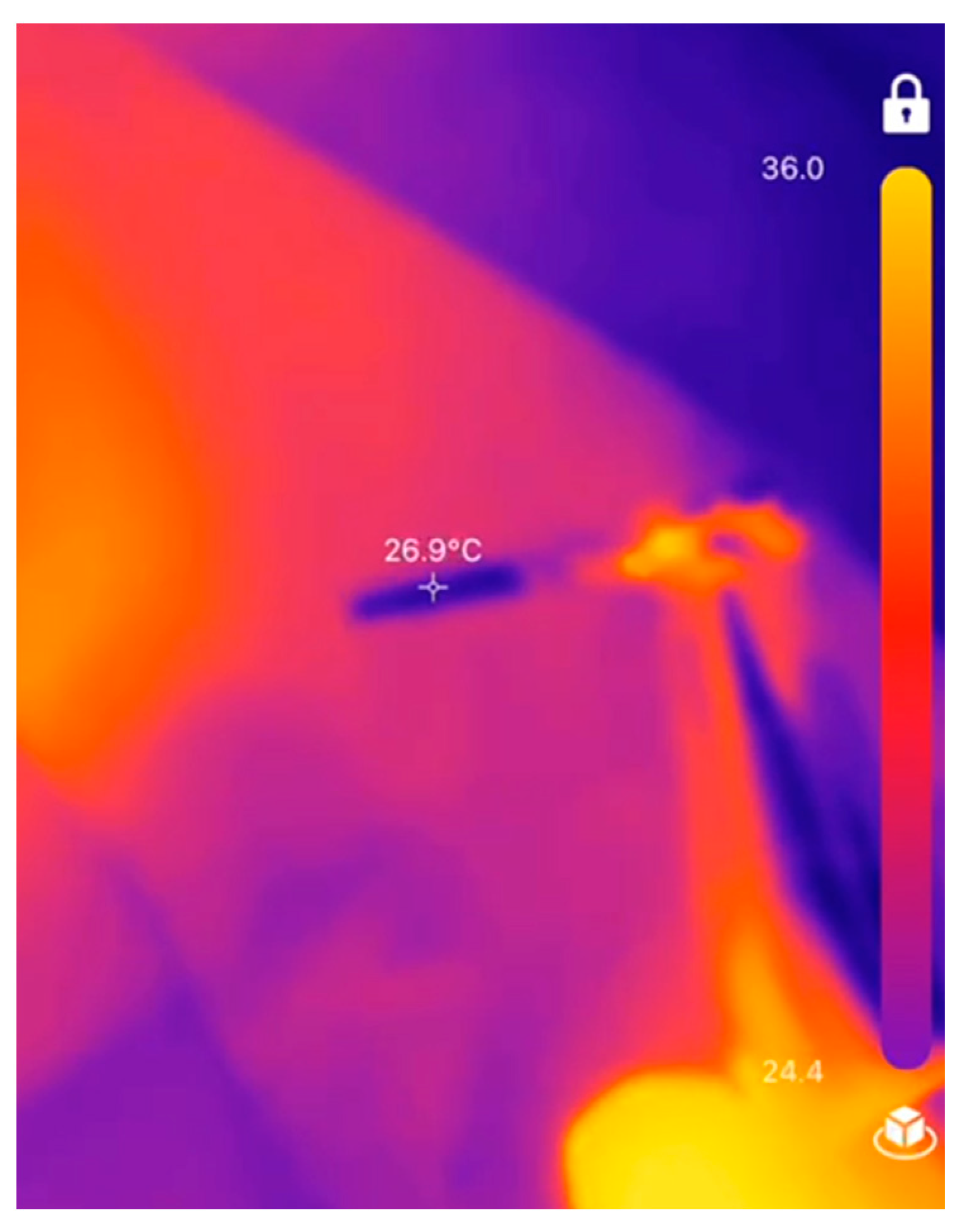
In the second part of this study (Figure 2), temperature measurements were carried out as part of an animal trial using an intraoral minipig model (Saarland—Landesamt für Verbraucherschutz; Versuch: 07/2023). As part of the regular surgical protocol, the IR camera was used for temperature determination of the drills immediately following implant site preparation with 2.8 mm drills (Figure 3). In this part of this study, the DLC drills mentioned above were used in addition to single-use drills for Straumann BLT implants (STMN; Straumann AG, Basel, Switzerland).
For determining intraosseous temperature development in minipigs based on IR camera measurements, a linear regression model with intercept a and slope b was set up using the in vitro (polyurethane foam) measured IR temperature and the difference between the intraosseous temperatures determined at the beginning and at the end of the drilling procedure for top and bottom (Equation (1)). The regression parameters a and b were then applied for estimating the intraosseous temperature in minipigs (Equation (2)).
Equation (1). Linear regression model based on in vitro measurements.
DIFF = a + b * IR + resDIFF: temperature change between start and end of drilling procedure
a: Intercept
b: Slope
IR: temperature measured with IR camera
res: regression residuals
Equation (2). Linear regression model for estimating the intraosseous temperature in minipigs.
DIFFminipig = a + b * IRminipigDIFFminipig: estimated temperature change between start and end of drilling procedure
a: Intercept
b: Slope
IRminipig: temperature measured with IR camera
In the third part of this study, intraosseous temperature development as a consequence of implant insertion was determined using the same in vitro setup described for part one of this study (Figure 1). Bone level implants (n = 5 per group) were inserted in bone surrogate material [14,16,42] consisting of a layer of softer polyurethane foam mimicking trabecular bone covered by a 3 mm layer of very dense material simulating a cortical plate (Solid rigid Polyurethane Foam 10 pcf, Solid Rigid Polyurethane Foam 40 pcf, Sawbones Europe AB, Malmö, Sweden). A conventional, tapered implant (MAX, AlfaGate, Kfar Qara, Israel) with a diameter of 4.2 mm and 11 mm in length served as control while a novel implant characterized by a simultaneous shift in core diameter and thread geometry [39,40] was used in the test group. This implant measured 4.3 mm in diameter and 10 mm in length (MT, AlfaGate). For both groups, perpendicular osteotomies were created using burs with 2.0, 2.8, 3.2, and 3.7 mm in diameter and a surgical motor set at 800RPM (iChiropro, BienAir, Biel, Switzerland) with the contra-angle mounted in a drill press. Maximum temperature was recorded [5,31] prior to the implant insertion process and when an implant was fully seated. Implant insertion was conducted at a velocity of 35 RPM using the surgical motor described, which also allowed recording torque values at a sampling rate of 1/200 ms. Primary implant stability was measured using resonance frequency analysis (Osstell AB, Gothenburg, Sweden) taking two measurements per implant perpendicular to each other.
Statistical analysis was based on differences between start and end temperature, maximum insertion torque, and mean values of implant stability measurements. Assuming normal distribution of measurement values, two-sample t-tests were used for statistical comparisons, which are robust against violations of their requirements if samples of equal size are compared. The level of significance was set at α = 0.05.

3. Results

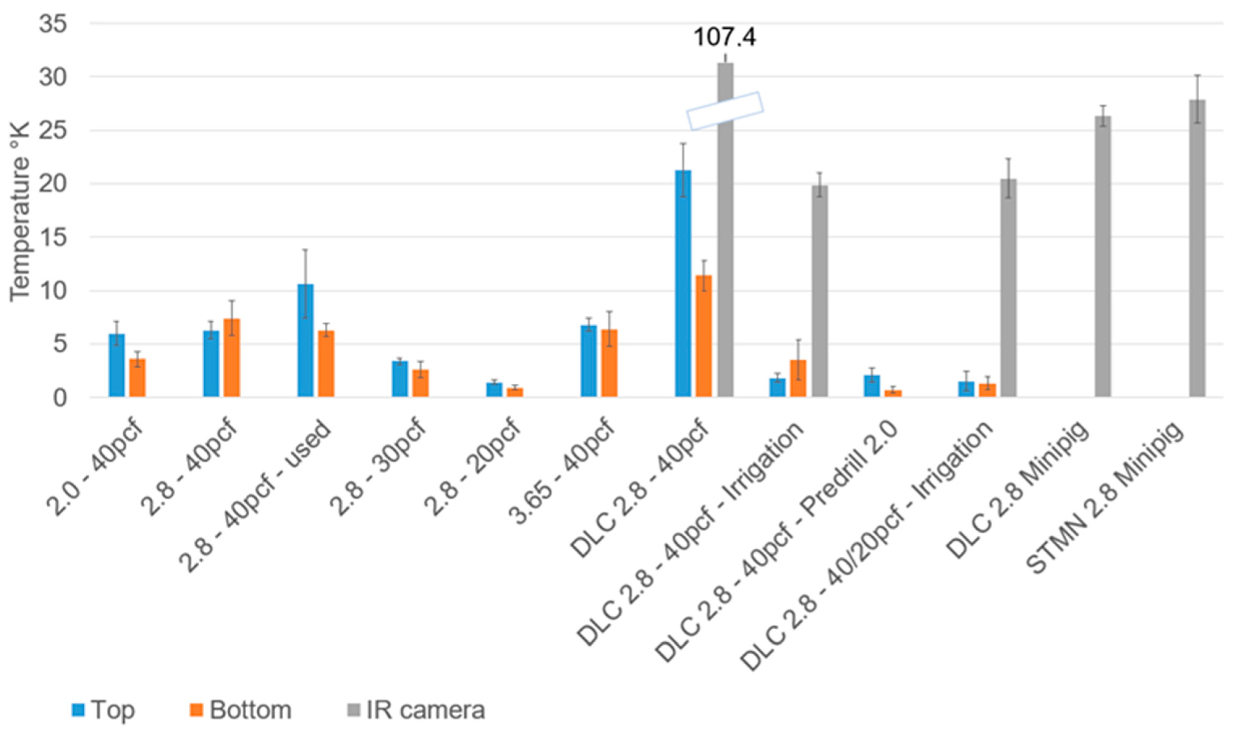
The mean temperature changes recorded in the first two parts of this study are shown in Figure 4. In vitro, temperature differences could be assumed to be normally distributed as Shapiro–Wilk tests indicated only two significant p-values (2.8–30 pcf: p = 0.04; DLC2.8–40 pcf—Irrigation: p = 0.05) in measurements made at the bottom in a total of 20 groups of measurement values. Levene’s test for equality of variances showed p = 0.076 and p = 0.032 for temperature differences calculated at the bottom and at the top of the osteotomies. As a consequence of significantly unequal variances, pairwise comparisons based on two-sample t tests and subsequent p value adjustment for multiple comparisons (Holm method) were conducted (Table 2a,b).
Increasing the drill diameter from 2.0 mm to 2.8 mm and 3.65 mm showed a slight trend towards greater temperature development at the bottom of the osteotomies but none of the comparisons between new drills used in 40 pcf bone were statistically significant (p > 0.05; Table 2). Using a drill 25 times for osteotomy preparation led to increased temperature development at the top part and greater standard deviations, but the difference compared to new drills did not reach statistical significance (p = 1.000; Table 2). Increasing bone density by 10 pcf led to a significant increase in temperature development both at the top and bottom parts of the osteotomies with the exception of only one comparison (30 pcf vs. 20 pcf Bottom p = 0.135; Table 2). DLC coating of drills led to a pronounced increase in temperature development both in the bottom part (p = 0.066) and in the top part (p = 0.009) with the latter reaching statistical significance (Table 2). Adding irrigation during drilling with DLC burrs led to a significant reduction in temperature development (bottom p = 0.004; top p = 0.000) reaching comparable values as seen in conventional drills of the same diameter without irrigation (bottom p = 0.135; top p = 0.321). With irrigation, no significant effect of bone quality on temperature development was observed (bottom p = 0.591; top p = 1.000) when comparing solid bone 40 pcf with layered bone 40/20 pcf (Table 2). Predrilling significantly reduced temperature development in both parts of the osteotomy (bottom p = 0.000; top p = 0.000) which was comparable to the effect of adding irrigation, with the top part of the osteotomy even showing a significantly smaller temperature increase (p = 0.032; Table 2).
The measurement values obtained in minipigs using the IR camera were not normally distributed in the case of STMN burrs (p = 0.001), while normal distribution was shown in DLC burrs (p = 0.3) using Shapiro–Wilk tests. The Wilcoxon test revealed a significant difference between both groups of drills (p = 0.02). The regression parameters for temperature development in the bottom part of the osteotomy were a = 0.3473 and b = 0.1023, while in the top portion, values of a = −2.858 and b = 0.225 were determined. Using these regression parameters, the intraosseous temperature changes provided in Table 3 were calculated for DLC and STMN burrs which were in the range of 3 °K. Since the results of statistical tests are invariant against scale transformations as evident here with the regression model, the above p-values apply also to the calculated intraosseous temperature differences.
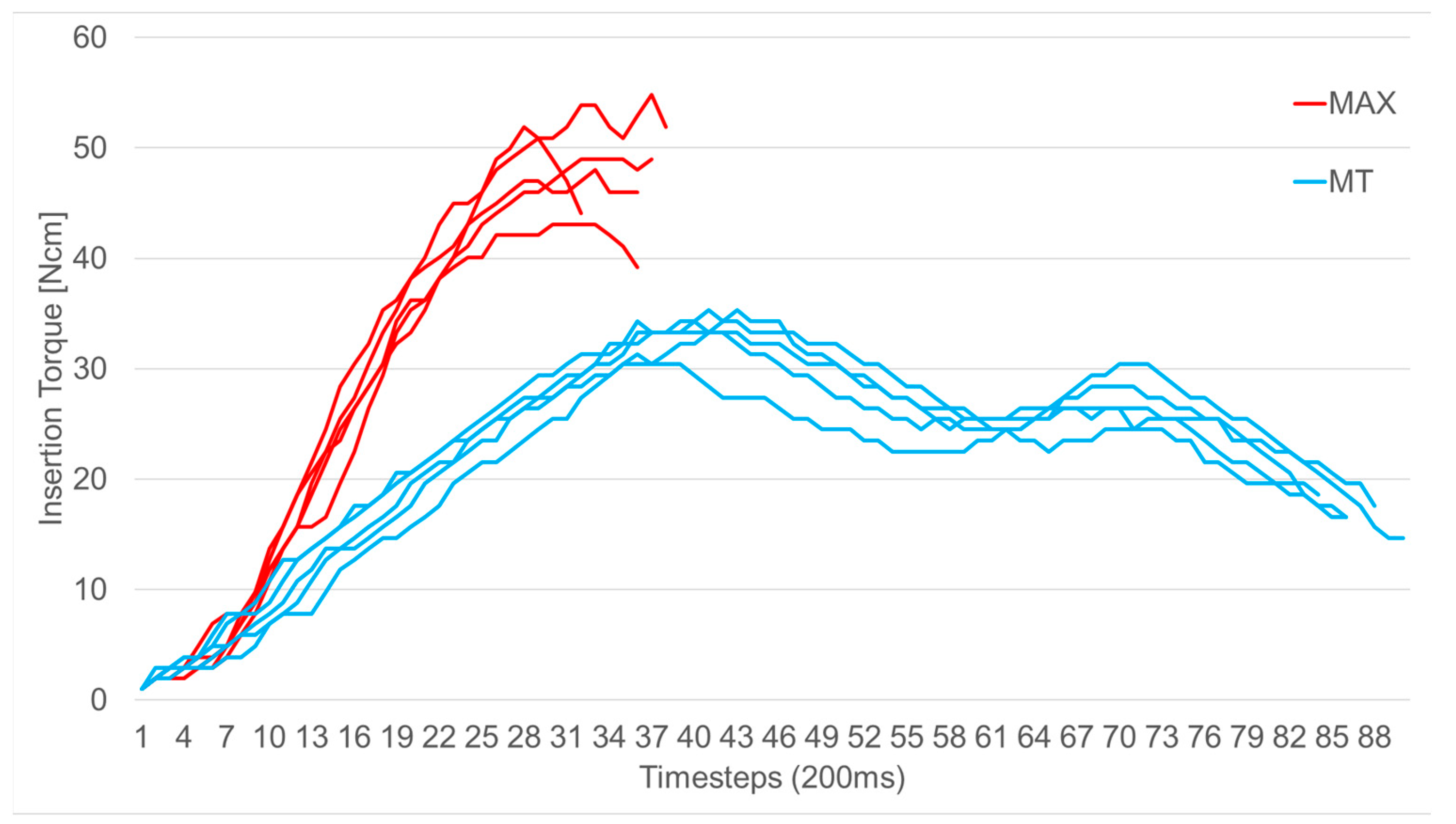
Both implants tested in the third part of this study showed characteristic torque–time curves with the MAX implant indicating a continuous rise in insertion torque with insertion depth and MT implant with a maximum value coinciding with approximately 50% insertion depth (Figure 5). Insertion of the MAX implant took approximately 37 time steps equaling 7.4 s while the insertion of the MT implant took approximately 86 time steps equaling 17.2 s.
Mean maximum insertion torque (Table 4) for MAX implants was 49.36 Ncm while MT implants required only 33.90 Ncm revealing a statistically significant difference (p < 0.01). Primary stability as determined with resonance frequency analysis (Table 4) did not differ significantly between the two groups of implants (MAX implant: 71.50; MT implant 68.40; p = 0.4). Inserting an MT implant caused a temperature rise (Table 4) in the top part of the osteotomy of 7.58 °K and in the bottom part of 7.50 °K, while for MAX implants, 3.80 °K and 4.48 °K were recorded. Both differences were statistically significant (p < 0.01 for top part and p = 0.02 for bottom part).
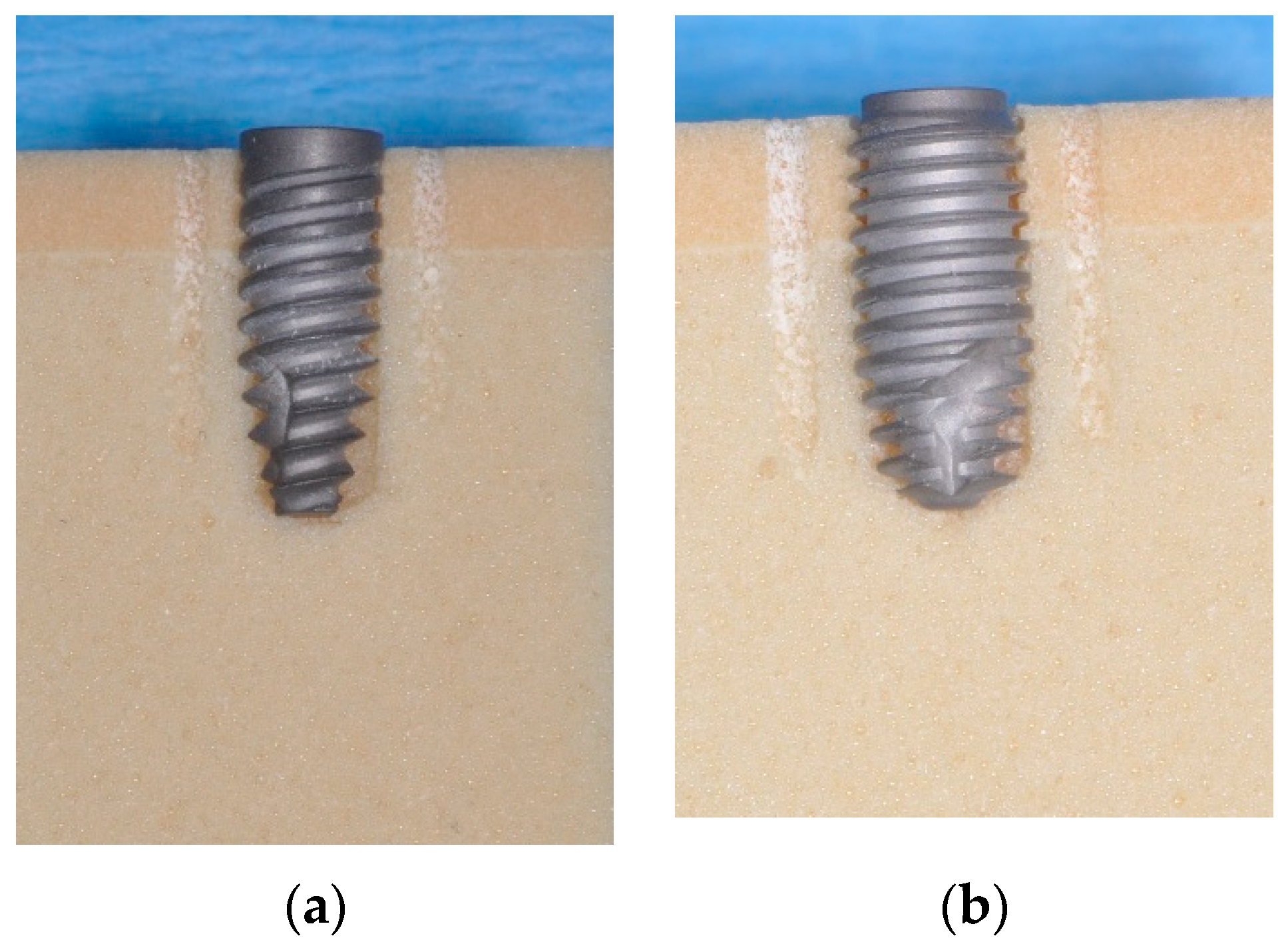
Cross-sections of the osteotomies (Figure 6) showed that MAX implants were in contact with the bony walls only in the cervical area while MT implants showed bone implant contact over the whole length of the implants.

4. Discussion

This series of in vitro and animal experiments was aimed at evaluating common factors affecting intraosseous temperature rise during implant surgery. In the first part, using well defined polyurethane foam as bone surrogate material, drill diameter had only a minor effect on temperature development and seemed to be more relevant in the bottom part of an osteotomy. This is in contrast with previous studies describing higher temperatures with smaller diameter drills [21,42]. The conventional drills that were used appear to be of good quality and from the perspective of temperature development can be safely reused. This seems at least partially contradictive to other authors describing drill fatigue [7,19] as a variable for temperature rise. Wear of the drills after creating 25 drill holes obviously differed among specimen as indicated by higher standard deviations in this group. It also has to be kept in mind that for the comparisons described here, no predrilling of sites had been performed as would be the case in clinical reality. As expected, increasing bone density significantly affected temperature development during drilling which is in line with previous reports [8,13,14] also stressing the risk of overheating cortical bone during implant site preparation.
In vitro, DLC-coated burrs led to much higher intrabony temperatures which had not been expected as DLC coatings had previously been found to reduce friction between mechanical components [24,25,26] which constitutes a relevant determinant for temperature increase [21]. The measurements on DLC drills in 40 pcf bone had even been redone after collecting an initial data set and could be verified. External irrigation as well as predrilling led to a marked reduction in heat generation which is consistent with previous studies [21,42,43]. DLC coatings are applied with the goal of achieving sharp and wear-resistant instruments which may constitute one reason why the bone quality present in a specific situation may have less influence on heat generation.
In the animal model, a temperature rise of approximately 3 °K was calculated for both drill types and both regions of the osteotomies. These sites had been predrilled and were created under irrigation. Having seen in vitro temperature increases ranging from 0.7 °K to 2.1 °K, the comparability and consistency of thermocoupling and IR measurements seems proven while the comparability of the raw data measured in vitro and in vivo implies the validity of the in vitro setup. The somewhat higher drill temperatures observed in vivo may have also been due to very dense bone being present in minipig mandibles. Given that DLC drills showed a significantly lower temperature increase as compared to STMN drills, which have been widely used in clinical application, the use of DLC drills should also be safely possible. An additional animal trial is currently being conducted aimed at evaluating peri-implant bone levels after osseointegration as a result of surgical trauma, i.e., heat generation combined with mechanical stress.
The third part of this study was aimed at comparing intraosseous heat generation [6] during the insertion process of two different implant types. As expected, the conical implant produced less heat in a cylindrically shaped osteotomy as compared to a mostly parallel-walled novel implant type. As pointed out previously, temperature rise during implant insertion can be understood as a phenomenon based on friction between bone and the implant [17,31], with the osteotomy shape and size being the decisive factor [6]. This seems consistent with the findings presented here, and it also has to be kept in mind that identical osteotomy diameters had been created for both implant types despite them showing a diameter of 4.2 mm (MAX, conical implant) and 4.3 mm (MT, parallel-walled implant), respectively. With undersizing of an osteotomy relative to the diameter of an implant resulting in bone compression and consequently high insertion torque values [28], these results demand for an adapted drill protocol to be developed for the novel implant type in order to avoid potential impairment of osseointegration [5,6,7]. Nevertheless, even under the protocol applied here, the widely accepted threshold for bone regeneration of 47° for 1 min [18,44] had not been surpassed.
The observed slower insertion process of the MT implant is due to a comparably small thread pitch, which on the one hand requires more surgical time but allows the bone to respond to inevitable deformation during implant insertion [45]. Furthermore, the slower insertion process facilitates precise positioning of the implant–abutment interface in the vertical dimension. Verifying previous reports on the novel implant design [39,40], significantly lower mean maximum insertion torque values were recorded as compared to the conical implant. With the parallel-walled implant also deriving primary stability from interacting with trabecular bone [46], an identical level of primary stability was shown.
While trying to mimic clinical reality, the following limitations of this study have to be considered. While polyurethane foam material has been widely applied as bone surrogate material and has been accepted for biomechanical research, it cannot mimic the properties of human alveolar bone [47,48,49,50]. As such, the measurement values recorded have to be interpreted on a relative scale. Comparable studies used techniques ranging from infrared cameras [42] to thermocouples [14], fiber optic thermometers [51], and infrared thermography [12] for measuring temperature development during implant installation.
Other than in clinical reality, where intermittent drilling is usually done, constant pressure was applied to the drill in the in vitro part of this study which may have led to higher temperatures [15,21] indicating a worst-case scenario.
Several factors seem to govern initial peri-implant bone loss, of which temperature development and mechanical stress can be controlled by the implant surgeon. Based on the findings of all study parts, it appears that the single steps of drill protocols as well as the final osteotomy diameters relative to the implant body are relevant factors in this context.

Author Contributions

Conceptualization, M.K. and T.G.-K.; methodology, C.E. and K.S.; software, K.S.; validation, C.E.; formal analysis, M.K.; investigation, K.S. and C.E.; resources, T.G.-K.; data curation, K.S. and C.E.; writing—original draft preparation, M.K.; writing—review and editing, T.G.-K.; visualization, K.S.; supervision, T.G.-K.; project administration, M.K.; funding acquisition, M.K. and T.G.-K. All authors have read and agreed to the published version of the manuscript.

Funding

This study was supported by AlfaGate, Bonn, Germany through materials donations that are kindly acknowledged.

Institutional Review Board Statement

Not applicable.

Informed Consent Statement

Not applicable.

Data Availability Statement

Original data are available from the corresponding author upon request.

Acknowledgments

The experiments presented were carried out by Kirsten Sekura and Carolin Erbel in partial fulfillment of the requirements for the degree Dr. med. dent. at Saarland University, Homburg, Germany. The authors wish to thank Friedrich Graef, Department of Mathematics, Friedrich-Alexander-Universität Erlangen-Nürnberg for statistical data analysis.

Conflicts of Interest

M.K. discloses a conflict of interest as he acts as a consultant to AlfaGate.

References

  1. Albrektsson, T.; Zarb, G.; Worthington, P.; Eriksson, A.R. The long-term efficacy of currently used dental implants: A review and proposed criteria of success. Int. J. Oral Maxillofac. Implants 1986, 1, 11–25. [Google Scholar] [PubMed]
  2. Buch, R.S.; Weibrich, G.; Wagner, W. Criteria of success in implantology. Mund. Kiefer Gesichtschir. 2003, 7, 42–46. [Google Scholar] [CrossRef] [PubMed]
  3. Roos, J.; Sennerby, L.; Lekholm, U.; Jemt, T.; Gröndahl, K.; Albrektsson, T. A qualitative and quantitative method for evaluating implant success: A 5-year retrospective analysis of the Brånemark implant. Int. J. Oral Maxillofac. Implants 1997, 12, 504–514. [Google Scholar]
  4. Albrektsson, T.; Chrcanovic, B.; Östman, P.O.; Sennerby, L. Initial and long-term crestal bone responses to modern dental implants. Periodontology 2017, 73, 41–50. [Google Scholar] [CrossRef] [PubMed]
  5. Heuzeroth, R.; Pippenger, B.E.; Sandgren, R.; Bellón, B.; Kühl, S. Thermal exposure of implant osteotomies and its impact on osseointegration-A preclinical in vivo study. Clin. Oral Implants Res. 2021, 32, 672–683. [Google Scholar] [CrossRef] [PubMed]
  6. Stocchero, M.; Jinno, Y.; Toia, M.; Ahmad, M.; Papia, E.; Yamaguchi, S.; Becktor, J.P. Intraosseous Temperature Change during Installation of Dental Implants with Two Different Surfaces and Different Drilling Protocols: An In Vivo Study in Sheep. J. Clin. Med. 2019, 8, 1198. [Google Scholar] [CrossRef] [PubMed]
  7. Mishra, S.K.; Chowdhary, R. Heat generated by dental implant drills during osteotomy-a review: Heat generated by dental implant drills. J. Indian Prosthodont. Soc. 2014, 14, 131–143. [Google Scholar] [CrossRef] [PubMed]
  8. Aghvami, M.; Brunski, J.B.; Serdar Tulu, U.; Chen, C.H.; Helms, J.A. A Thermal and Biological Analysis of Bone Drilling. J. Biomech. Eng. 2018, 140, 1010101–1010108. [Google Scholar] [CrossRef] [PubMed]
  9. Chen, C.H.; Coyac, B.R.; Arioka, M.; Leahy, B.; Tulu, U.S.; Aghvami, M.; Holst, S.; Hoffmann, W.; Quarry, A.; Bahat, O.; et al. A Novel Osteotomy Preparation Technique to Preserve Implant Site Viability and Enhance Osteogenesis. J. Clin. Med. 2019, 8, 170. [Google Scholar] [CrossRef]
  10. Ikar, M.; Grobecker-Karl, T.; Karl, M.; Steiner, C. Mechanical stress during implant surgery and its effects on marginal bone: A literature review. Quintessence Int. 2020, 51, 142–150. [Google Scholar]
  11. Ozcan, M.; Salimov, F.; Temmerman, A.; Turer, O.U.; Alkaya, B.; Haytac, M.C. The Evaluation of Different Osteotomy Drilling Speed Protocols on Cortical Bone Temperature, Implant Stability and Bone Healing: An Experimental Study in an Animal Model. J. Oral Implantol. 2022, 48, 3–8. [Google Scholar] [CrossRef] [PubMed]
  12. Raj, R.; Manju, V.; Kumar-Gopal, V.; Eswar, M. Analysis of factors determining thermal changes at osteotomy site in dental implant placement—An in-vitro study. J. Clin. Exp. Dent. 2021, 13, e234–e239. [Google Scholar] [CrossRef] [PubMed]
  13. Eriksson, A.R.; Albrektsson, T.; Albrektsson, B. Heat caused by drilling cortical bone: Temperature measured in vivo in patients and animals. Acta Orthop. Scand. 1984, 55, 629–631. [Google Scholar] [CrossRef] [PubMed]
  14. Yamaba, T.; Suganami, T.; Ikebe, K.; Sogo, M.; Maeda, Y.; Wada, M. The Evaluation of the Heat Generated by the Implant Osteotomy Preparation Using a Modified Method of the Measuring Temperature. Int. J. Oral Maxillofac. Implants 2015, 30, 820–826. [Google Scholar] [CrossRef] [PubMed]
  15. Katic, Z.; Jukic, T.; Stubljar, D. Effects of Osteotomy Lengths on the Temperature Rise of the Crestal Bone During Implant Site Preparation. Implant. Dent. 2018, 27, 213–220. [Google Scholar] [CrossRef] [PubMed]
  16. Gehrke, S.A.; Bettach, R.; Cayron, B.; Boukhris, G.; Dedavid, B.A.; Frutos, J.C.P. Development of a New Drill Design to Improve the Temperature Control during the Osteotomy for Dental Implants: A Comparative In Vitro Analysis. Biology 2020, 9, 208. [Google Scholar] [CrossRef] [PubMed]
  17. Orgev, A.; Gonzaga, L.; Martin, W.; Morton, D.; Lin, W.S. Addition of an irrigation channel to a surgical template to facilitate cooling during implant osteotomy. J. Prosthet. Dent. 2021, 126, 164–166. [Google Scholar] [CrossRef] [PubMed]
  18. Soldatos, N.; Nelson-Rabe, L.; Palanker, N.; Angelov, N.; Romanos, G.; Weltman, R. Temperature Changes during Implant Osteotomy Preparations in Fresh Human Cadaver Tibiae, Comparing Straight with Tapered Drills. Materials 2022, 15, 2369. [Google Scholar] [CrossRef] [PubMed]
  19. Karaca, F.; Aksakal, B.; Kom, M. Influence of orthopaedic drilling parameters on temperature and histopathology of bovine tibia: An in vitro study. Med. Eng. Phys. 2011, 33, 1221–1227. [Google Scholar] [CrossRef]
  20. Tur, D.; Giannis, K.; Unger, E.; Mittlböck, M.; Rausch-Fan, X.; Strbac, G.D. Thermal effects of various drill materials during implant site preparation-Ceramic vs. stainless steel drills: A comparative in vitro study in a standardised bovine bone model. Clin. Oral Implants Res. 2021, 32, 154–166. [Google Scholar] [CrossRef]
  21. Strbac, G.D.; Giannis, K.; Unger, E.; Mittlböck, M.; Vasak, C.; Watzek, G.; Zechner, W. Drilling- and withdrawing-related thermal changes during implant site osteotomies. Clin. Implant. Dent. Relat. Res. 2015, 17, 32–43. [Google Scholar] [CrossRef] [PubMed]
  22. Abboud, M.; Rugova, S.; Orentlicher, G. Immediate Loading: Are Implant. Surface and Thread Design More Important Than Osteotomy Preparation? Compend. Contin. Educ. Dent. 2020, 41, 384–386. [Google Scholar] [PubMed]
  23. Bernabeu-Mira, J.C.; Soto-Peñaloza, D.; Peñarrocha-Diago, M.; Camacho-Alonso, F.; Rivas-Ballester, R.; Peñarrocha-Oltra, D. Low-speed drilling without irrigation versus conventional drilling for dental implant osteotomy preparation: A systematic review. Clin. Oral Investig. 2021, 25, 4251–4267. [Google Scholar] [CrossRef] [PubMed]
  24. Roy, R.K.; Lee, K.R. Biomedical applications of diamond-like carbon coatings: A review. J. Biomed. Mater. Res. B Appl. Biomater. 2007, 83, 72–84. [Google Scholar] [CrossRef] [PubMed]
  25. Batista Mendes, G.C.; Padovan, L.E.; Ribeiro-Júnior, P.D.; Sartori, E.M.; Valgas, L.; Claudino, M. Influence of implant drill materials on wear, deformation, and roughness after repeated drilling and sterilization. Implant. Dent. 2014, 23, 188–194. [Google Scholar] [CrossRef] [PubMed]
  26. Akaike, S.; Hayakawa, T.; Kobayashi, D.; Aono, Y.; Hirata, A.; Hiratsuka, M.; Nakamura, Y. Reduction in static friction by deposition of a homogeneous diamond-like carbon (DLC) coating on orthodontic brackets. Dent. Mater. J. 2015, 34, 888–895. [Google Scholar] [CrossRef]
  27. Raghavendra, S.; Wood, M.C.; Taylor, T.D. Early wound healing around endosseous implants: A review of the literature. Int. J. Oral Maxillofac. Implants 2005, 20, 425–431. [Google Scholar] [PubMed]
  28. Khayat, P.G.; Arnal, H.M.; Tourbah, B.I.; Sennerby, L. Clinical outcome of dental implants placed with high insertion torques (up to 176 Ncm). Clin. Implant. Dent. Relat. Res. 2013, 15, 227–233. [Google Scholar] [CrossRef]
  29. Frisardi, G.; Barone, S.; Razionale, A.V.; Paoli, A.; Frisardi, F.; Tullio, A.; Lumbau, A.; Chessa, G. Biomechanics of the press-fit phenomenon in dental implantology: An image-based finite element analysis. Head. Face Med. 2012, 8, 18. [Google Scholar] [CrossRef]
  30. Guan, H.; van Staden, R.C.; Johnson, N.W.; Loo, Y.C. Dynamic modelling and simulation of dental implant insertion process—A finite element study. Finite Elem. Anal. Des. 2011, 47, 886–897. [Google Scholar] [CrossRef]
  31. Flanagan, D. Heat generated during seating of dental implant fixtures. J. Oral Implantol. 2014, 40, 174–181. [Google Scholar] [CrossRef] [PubMed]
  32. Bartold, P.M.; Kuliwaba, J.S.; Lee, V.; Shah, S.; Marino, V.; Fazzalari, N.L. Influence of surface roughness and shape on microdamage of the osseous surface adjacent to titanium dental implants. Clin. Oral Implants Res. 2011, 22, 613–618. [Google Scholar] [CrossRef]
  33. Tabassum, A.; Meijer, G.J.; Walboomers, X.F.; Jansen, J.A. Biological limits of the undersized surgical technique: A study in goats. Clin. Oral Implants Res. 2011, 22, 129–134. [Google Scholar] [CrossRef]
  34. Coelho, P.G.; Marin, C.; Teixeira, H.S.; Campos, F.E.; Gomes, J.B.; Guastaldi, F.; Anchieta, R.B.; Silveira, L.; Bonfante, E.A. Biomechanical evaluation of undersized drilling on implant biomechanical stability at early implantation times. J. Oral Maxillofac. Surg. 2013, 71, 69–75. [Google Scholar] [CrossRef] [PubMed]
  35. Berglundh, T.; Abrahamsson, I.; Lang, N.P.; Lindhe, J. De novo alveolar bone formation adjacent to endosseous implants. Clin. Oral Implants Res. 2003, 14, 251–262. [Google Scholar] [CrossRef]
  36. Parvini, P.; Trimpou, G.; Begic, A.; Cafferata, E.A.; Petsos, H.; Müller, K.M.; Schwarz, F.; Eickholz, P.; Obreja, K. Esthetic and clinical outcomes after immediate placement and restoration: Comparison of two implant systems in the anterior maxilla-A cross-sectional study. Clin. Implant. Dent. Relat. Res. 2023, 25, 252–260. [Google Scholar] [CrossRef]
  37. Fabbri, G.; Staas, T.; Urban, I. A Retrospective Observational Study Assessing the Clinical Outcomes of a Novel Implant System with Low-Speed Site Preparation Protocol and Tri-Oval Implant Geometry. J. Clin. Med. 2022, 11, 4859. [Google Scholar] [CrossRef]
  38. Hurtgen, A.; Seidel, L.; Manni, L.L.; Liegeois, L.; Lecloux, G.; Lambert, F. Clinical and radiographic assessment of circular versus triangular cross-section neck implants in the posterior maxilla: Five-year follow-up of a randomized controlled trial. Clin. Oral Implants Res. 2023, 34, 698–706. [Google Scholar] [CrossRef] [PubMed]
  39. Schulz, A.; Klär, V.; Grobecker-Karl, T.; Karl, M. Biomechanical rationale for a novel implant design reducing stress on buccal bone. Appl. Sci. 2023, 13, 666. [Google Scholar] [CrossRef]
  40. Klär, V.; Zimmerer, R.; Schulz, A.; Lethaus, B. Biomechanical performance of a novel implant design in simulated extraction sites and sinuslift procedures. Appl. Sci. 2023, 13, 7541. [Google Scholar] [CrossRef]
  41. Parvizi, S.; Cameron, A.; Tadakamadla, S.; Figueredo, C.M.S.; Reher, P. A Novel Irrigation System to Reduce Heat Generation during Guided Implantology: An In Vitro Study. J. Clin. Med. 2023, 12, 3944. [Google Scholar] [CrossRef]
  42. Kuster, M.; Mukaddam, K.; Zitzmann, N.U.; Filippi, A.; Kühl, S. Influence of a Novel Drill Design on Heat Generation During Conventional and Guided Implant Osteotomy. Int. J. Oral Maxillofac. Implants 2021, 36, e31–e41. [Google Scholar] [CrossRef]
  43. Strbac, G.D.; Unger, E.; Donner, R.; Bijak, M.; Watzek, G.; Zechner, W. Thermal effects of a combined irrigation method during implant site drilling: A standardized in vitro study using a bovine rib model. Clin. Oral Implants Res. 2014, 25, 665–674. [Google Scholar] [CrossRef] [PubMed]
  44. Eriksson, R.A.; Albrektsson, T. The effect of heat on bone regeneration: An experimental study in the rabbit using the bone growth chamber. J. Oral Maxillofac. Surg. 1984, 42, 705–711. [Google Scholar] [CrossRef]
  45. Pérez-Pevida, E.; Brizuela-Velasco, A.; Chávarri-Prado, D.; Diéguez-Pereira, M.; Jiménez-Garrudo, A.; Montalbán-Vadillo, O.; Viteri-Agustín, I.; Estrada-Martínez, A.; Santamaría-Arrieta, G. Influence of time on primary stability of dental implants placed with osteotomes due to the elastic properties of peri-implant bone. J. Dent. Sci. 2019, 14, 358–364. [Google Scholar] [CrossRef]
  46. Wang, R.; Eppell, S.J.; Nguyen, C.; Morris, N. Relative Contribution of Trabecular and Cortical Bone to Primary Implant Stability: An In Vitro Model Study. J. Oral Implantol. 2016, 42, 145–152. [Google Scholar] [CrossRef]
  47. Di Stefano, D.A.; Arosio, P.; Gastaldi, G.; Gherlone, E. The insertion torque-depth curve integral as a measure of implant primary stability: An in vitro study on polyurethane foam blocks. J. Prosthet. Dent. 2018, 120, 706–714. [Google Scholar] [CrossRef] [PubMed]
  48. Sierra-Rebolledo, A.; Allais-Leon, M.; Maurette-O’Brien, P.; Gay-Escoda, C. Primary Apical Stability of Tapered Implants Through Reduction of Final Drilling Dimensions in Different Bone Density Models: A Biomechanical Study. Implant. Dent. 2016, 25, 775–782. [Google Scholar] [CrossRef] [PubMed]
  49. Elias, C.N.; Rocha, F.A.; Nascimento, A.L.; Coelho, P.G. Influence of implant shape, surface morphology, surgical technique and bone quality on the primary stability of dental implants. J. Mech. Behav. Biomed. Mater. 2012, 16, 169–180. [Google Scholar] [CrossRef]
  50. Ueno, D.; Nakamura, K.; Kojima, K.; Toyoshima, T.; Tanaka, H.; Ueda, K.; Koyano, K.; Kodama, T. A stepwise under-prepared osteotomy technique improves primary stability in shallow-placed implants: A preliminary study for simultaneous vertical ridge augmentation. Odontology 2018, 106, 187–193. [Google Scholar] [CrossRef]
  51. Lamazza, L.; Lollobrigida, M.; Vozza, I.; Palmieri, L.; Stacchi, C.; Lombardi, T.; De Biase, A. Piezoelectric Implant Site Preparation: Influence of Handpiece Movements on Temperature Elevation. Materials 2020, 13, 4072. [Google Scholar] [CrossRef] [PubMed]
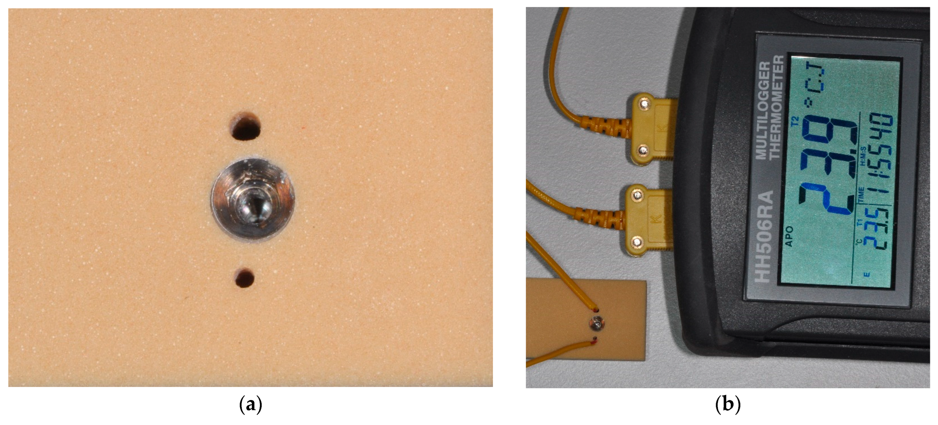
Figure 1. Model situation with polyurethane foam as the bone surrogate material showing the spatial relationship of the implant osteotomy and the drill holes for temperature measurements (a). Thermocouples were inserted into the drill holes at 3 mm and 10 mm depth; the distance to the implant surface measures 0.5 mm (b).
Prosthesis 06 00046 g001
Figure 2. Intraoral situation from the animal experiment with a DLC-coated drill shown during osteotomy preparation.
Prosthesis 06 00046 g002
Figure 3. Measurement taken with IR camera showing the drill in focus while kept in the contra-angle handpiece and positioned outside the oral cavity.
Prosthesis 06 00046 g003
Figure 4. Temperature rise during drilling with conventional drills and DLC-coated drills in bone surrogate materials differing in density and composition and mandibular bone of minipigs, respectively. In addition to intrabony measurements using two sensors at 3 mm (Top) and 10 mm (Bottom) depths in the vicinity of an osteotomy, an infrared camera was used for determining temperature of drills immediately after removal from the drill hole. For comparison, single use drills for Straumann BLT implants have been used in the animal trial. Five measurements per group were made in vitro while in the animal trial a total of nine measurements were made with Straumann drills and 22 measurements for DLC drills, respectively. Note: IR camera measurements in vitro for DLC 2.8—40 pcf resulted in a mean of 107.4 °K (+/−4.4 °K).
Prosthesis 06 00046 g004
Figure 5. Torque–time curves recorded during the insertion of both implant types with MAX implants showing a continuous increase in torque development while MT implants show a maximum when the middle part of the implant passed the cortical plate. Note: X-axis represents the sampling rate of the surgical motor with one measurement value every 200 ms.
Prosthesis 06 00046 g005
Figure 6. Cross-sections of bone surrogate material with implant osteotomies and drill holes for temperature measurements. The conventional tapered implant only touches the simulated cortical bone plate (a) while the novel implant shows contact in all areas with the exception of the apical most portion (b).
Prosthesis 06 00046 g006
Table 1. Experimental groups established in the first part of this study.
DrillBoneIrrigationIR Camera
2.040--
2.840--
3.6540--
2.8–25 times used40--
2.830--
2.820--
DLC 2.840-yes
DLC 2.840yesyes
DLC 2.8—Predrill DLC 2.040--
DLC 2.820 and 40 (3 mm)yesyes
DLC 2.8Minipigyesyes
STMN 2.8Minipigyesyes
Table 2. Results of pairwise comparisons based on two-sample t tests and subsequent p value adjustment for multiple comparisons (Holm method) for temperature differences measured at the bottom (a) and top (b) (α = 0.05; significant differences are marked with *).
2.8–40 pcf2.8–40 pcf–Used2.8–30 pcf2.8–20 pcf3.65–40 pcfDLC 2.8–40 pcfDLC 2.8–40 pcf–IrrigationDLC 2.8–40 pcf–Predrill 2.0DLC 2.8–40/20 pcf–Irrigation
(a)
2.0–40 pcf0.0760.007 *0.6520.018 *0.2130.000 *1.0000.007 *0.013 *
2.8–40 pcf 1.0000.029 *0.022 *1.0000.0660.1350.018 *0.016 *
2.8–40 pcf–Used 0.004 *0.000 *1.0000.013 *0.3570.000 *0.000 *
2.8–30 pcf 0.1350.0710.000 *1.0000.0710.267
2.8–20 pcf 0.035 *0.004 *0.4290.6521.000
3.65–40 pcf 0.024 *0.3590.029 *0.029 *
DLC 2.8–40 pcf 0.004*0.000 *0.000 *
DLC 2.8–40 pcf–Irrigation 0.3590.591
DLC 2.8–40 pcf–Predrill 2.0 0.652
(b)
2.0–40 pcf 1.0001.0000.016 *0.011 *1.0000.006 *0.4510.006 *0.004 *
2.8–40 pcf 1.0000.004 *0.004 *1.0000.009 *0.3210.000 *0.000 *
2.8–40 pcf–Used 0.0680.046 *0.4721.0000.0680.044 *0.046 *
2.8–30 pcf 0.000 *0.1980.004 *1.0000.000 *0.011 *
2.8–20 pcf 0.044 *0.004 *0.7590.029 *1.000
3.65–40 pcf 0.018 *0.1980.000 *0.000 *
DLC 2.8–40 pcf 0.000 *0.000 *0.000 *
DLC 2.8–40 pcf–Irrigation 0.032 *1.000
DLC 2.8–40 pcf–Predrill 2.0 0.808
Table 3. Calculated intraosseous temperature changes in minipig bone for Straumann and DLC burrs.
StraumannDLC
MEANSDMEANSD
Top3.4180.50613.0710.2161
Bottom3.2020.23023.0450.0983
Table 4. Descriptive statistics for all measurement parameters recorded during implant insertion in the third part of this study and results of two-sample t-tests for comparisons between the two implant types expressed as p-values. Significant differences (p < 0.05) are marked with *.
MAX ImplantMT ImplantComparison MAX vs. MT
MEANSDMEANSDp-Value
Temperature top3.800.657.580.64<0.01 *
Temperature bottom4.481.927.500.160.02 *
Torque49.364.3933.901.52<0.01 *
Osstell71.507.5768.402.750.4
Disclaimer/Publisher’s Note: The statements, opinions and data contained in all publications are solely those of the individual author(s) and contributor(s) and not of MDPI and/or the editor(s). MDPI and/or the editor(s) disclaim responsibility for any injury to people or property resulting from any ideas, methods, instructions or products referred to in the content.