Next Issue
Volume 15, January-2
Previous Issue
Volume 14, December-2


Cells, Volume 15, Issue 1 (January-1 2026) – 92 articles

Cover Story (view full-size image): 
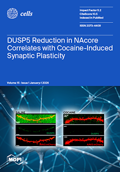
Drug overdose remains a leading cause of preventable death in the United States, underscoring the urgent need to identify the neurobiological mechanisms that drive substance use disorders. Addiction is characterized by persistent changes in brain circuits that govern reward, motivation, and aversion, with the nucleus accumbens core (NAcore) serving as a critical locus for these adaptations. Chronic exposure to drugs of abuse produces long-lasting structural and functional synaptic modifications within the NAcore that promote compulsive drug seeking and relapse. However, the intracellular signaling mechanisms that constrain or exacerbate these maladaptive synaptic changes remain incompletely understood. View this paper
  • Issues are regarded as officially published after their release is announced to the table of contents alert mailing list.
  • You may sign up for e-mail alerts to receive table of contents of newly released issues.
  • PDF is the official format for papers published in both, html and pdf forms. To view the papers in pdf format, click on the "PDF Full-text" link, and use the free Adobe Reader to open them.
Order results
Result details
Section
Select all
Export citation of selected articles as:
13 pages, 1392 KB  
Review
MYO5B Deficiency-Associated Cholestasis and the Role of the Bile Salt Export Pump
by Zhe Zhou and Sven C. D. van IJzendoorn
Cells 2026, 15(1), 92; https://doi.org/10.3390/cells15010092 - 5 Jan 2026
Viewed by 309
Abstract
Variants of the MYO5B gene, which encodes the molecular motor protein myosin-Vb, have gained prominence as a causative factor in familial intrahepatic cholestasis (FIC). Understanding the disease mechanism is pivotal for therapy development and clinical decision-making. The prevailing theory for the mechanism underlying [...] Read more.
Variants of the MYO5B gene, which encodes the molecular motor protein myosin-Vb, have gained prominence as a causative factor in familial intrahepatic cholestasis (FIC). Understanding the disease mechanism is pivotal for therapy development and clinical decision-making. The prevailing theory for the mechanism underlying MYO5B-associated cholestasis implicates faulty trafficking of the ABCB11-encoded bile salt export pump (BSEP) in hepatocytes due to dysfunctional myosin-Vb. This is supported by cell and mouse studies. However, while BSEP localization was abnormal in some patients’ liver biopsies, BSEP appeared normally localized in others, raising questions with regard to the role of BSEP in MYO5B-associated FIC. We present a focused systematic narrative review of all cases of MYO5B variant-associated isolated FIC reported in the MEDLINE database. We assembled a comprehensive patient dataset and assessed clinical features of MYO5B-associated FIC, their relationship with MYO5B genotype, the clinical value and significance of BSEP abnormalities, and the relationship of MYO5B-associated FIC to ABCB11 variant-associated FIC. Our review revealed that aberrant BSEP localization correlated with the absence of one MYO5B allele carrying a truncating nonsense or frameshift variant. Notably, biochemical and clinical parameters including treatment outcome were indistinguishable between patients presenting with normal and aberrant BSEP localization. Further, myosin-Vb and BSEP deficiency-associated FIC patient cohorts showed distinct biochemical and clinical phenotypes, indicating different underlying mechanisms. This suggests that whether or not BSEP localization was abnormal depended on the MYO5B genotype without a predictable effect on clinical parameters and treatment response. Treatment decisions should be guided by clinical parameters rather than by genotype or immunohistochemistry findings. Full article
(This article belongs to the Special Issue Cellular and Molecular Mechanisms of Liver Diseases)
Show Figures

Graphical abstract

27 pages, 2345 KB  
Review
Limbal Epithelial Stem Cells in Review: Immune and Lymphangiogenic Privilege and Their Clinical Relevance
by Berbang Meshko, Thomas Volatier, Claus Cursiefen and Maria Notara
Cells 2026, 15(1), 91; https://doi.org/10.3390/cells15010091 - 5 Jan 2026
Viewed by 631
Abstract
The cornea maintains transparency by preserving immune and (lymph)angiogenic privilege through active suppression of inflammation and vascular invasion, a process centrally regulated by limbal epithelial stem cells (LESCs) located at the corneoscleral junction. Beyond renewing the corneal epithelium, LESCs maintain immune and vascular [...] Read more.
The cornea maintains transparency by preserving immune and (lymph)angiogenic privilege through active suppression of inflammation and vascular invasion, a process centrally regulated by limbal epithelial stem cells (LESCs) located at the corneoscleral junction. Beyond renewing the corneal epithelium, LESCs maintain immune and vascular balance via extracellular matrix interactions and paracrine signalling, exerting predominantly anti-inflammatory and anti-(lymph)angiogenic effects in vivo. Disruption of the limbal niche by trauma, UV exposure, or genetic disorders such as aniridia leads to limbal stem cell deficiency (LSCD), chronic inflammation, loss of corneal avascularity, and vision loss. The identification of ABCB5 as a key LESC marker has clarified functional limbal subsets, highlighting ABCB5+ epithelial cells as mediators of repair, remodelling, and immune suppression, and positioning them as promising therapeutic targets for treatments that restore both epithelial integrity and corneal immune privilege. Full article
Show Figures

Figure 1

32 pages, 4732 KB  
Article
Distinct Regulation of the ARF and TAp73 Tumor Suppressor Genes by the Transcription Factor E2F1 Enables Discrimination of Cancer Cells from Normal Growing Cells
by Yaxuan Zhou, Rinka Nakajima, Mashiro Shirasawa, Mariana Fikriyanti, Ako Watanabe, Caiwei Yang, Ritsuko Iwanaga, Andrew P. Bradford, Kenta Kurayoshi, Keigo Araki and Kiyoshi Ohtani
Cells 2026, 15(1), 90; https://doi.org/10.3390/cells15010090 - 5 Jan 2026
Viewed by 289
Abstract
Discrimination of cancer cells from normal growing cells is crucial to specifically target cancer cells. The transcription factor E2F1 is the principal target of the tumor suppressor pRB. E2F1 activated by growth stimulation activates cell cycle-related genes and facilitates cell proliferation. E2F1 activated [...] Read more.
Discrimination of cancer cells from normal growing cells is crucial to specifically target cancer cells. The transcription factor E2F1 is the principal target of the tumor suppressor pRB. E2F1 activated by growth stimulation activates cell cycle-related genes and facilitates cell proliferation. E2F1 activated by loss of pRB control, such as forced inactivation of pRB, activates tumor suppressor genes such as ARF and TAp73 and induces apoptosis. We show here that these genes are specifically activated by exogenously expressed E2F1 or forced inactivation of pRB but not by growth stimulation in epithelial cells. This observation indicates that E2F1 activity induced by forced inactivation of pRB contains distinct E2F1 activity that activates these tumor suppressor genes. Cancer cells survive with concomitant dysfunction of apoptosis-inducing pathways, suggesting the presence of distinct E2F1 activity specifically in cancer cells. We determined the presence of distinct E2F1 activity using E2F responsive elements of the ARF and TAp73 genes by reporter assay. All 33 cancer cell lines tested possessed distinct E2F1 activity, but five normal growing cell lines did not. These results indicate that distinct E2F1 activity is a unique characteristic of cancer cells that facilitates discrimination from normal growing cells to specifically target cancer cells. Full article
(This article belongs to the Special Issue Cancer Cell Signaling, Autophagy and Tumorigenesis)
Show Figures

Figure 1

26 pages, 5121 KB  
Article
The Role of Aldosterone in Vascular Permeability in Diabetes
by Michal Aleksiejczuk, Natalia Bielicka, Magdalena Bruzgo-Grzybko, Izabela Suwda Kalita, Adam Jan Olichwier, Paulina Mierzejewska, Joanna Stelmaszewska, Janusz Dzieciol, Ewa Chabielska and Anna Gromotowicz-Poplawska
Cells 2026, 15(1), 89; https://doi.org/10.3390/cells15010089 - 5 Jan 2026
Viewed by 336
Abstract
More than 30% of diabetic patients develop dermatopathies linked to inflammation and increased vascular permeability. Considering the role of the renin–angiotensin–aldosterone system (RAAS) in diabetic complications, this study examined whether aldosterone (ALDO) and the mineralocorticoid receptor (MR) contribute to diabetes-related skin microangiopathy. Vascular [...] Read more.
More than 30% of diabetic patients develop dermatopathies linked to inflammation and increased vascular permeability. Considering the role of the renin–angiotensin–aldosterone system (RAAS) in diabetic complications, this study examined whether aldosterone (ALDO) and the mineralocorticoid receptor (MR) contribute to diabetes-related skin microangiopathy. Vascular permeability was measured in normoglycemic rats and insulin-dependent (streptozotocin-induced) diabetic rats. The expression of MR, 11β-hydroxysteroid dehydrogenase type 2 (HSD11β2), vascular endothelial growth factor (VEGF), von Willebrand factor (vWF), and the tight junction protein ZO-1 was determined by PCR and immunohistochemistry. Diabetic rats received the MR antagonist eplerenone (EPL, 100 mg/kg) for 10 days. Additionally, the effects of ALDO and EPL on endothelial permeability were evaluated in human dermal microvascular endothelial cells (HMEC-1) using a Transwell system. Diabetic rats showed skin atrophy, collagen damage, elevated ALDO levels, reduced MR and HSD11β2 expression, and increased vascular permeability, along with upregulation of VEGF and vWF. EPL markedly reduced these abnormalities. In vitro, ALDO increased endothelial permeability under hyperglycemia, and EPL counteracted this effect. These findings indicate that activation of the ALDO/MR pathway promotes skin vascular permeability in diabetes through VEGF- and vWF-dependent mechanisms. MR blockade limits these changes, suggesting therapeutic potential in preventing diabetes-associated skin complications. Full article
(This article belongs to the Special Issue Cellular and Molecular Mechanisms of Vascular-Related Diseases)
Show Figures

Figure 1

21 pages, 3242 KB  
Article
Photobiomodulation Activates Coordinated Signaling Networks to Modulate Inflammation, Adaptive Stress, and Tissue Healing via Redox-Mediated NFκB–TGF-β1–ATF-4 Axis
by Sasikumar Ponnusamy, Mahmud Amin, Amruta Bhat, Sarah Garczynski, Saeed Ur Rahman, Sailee Rasam, Sharaschandra Reddy Govindool, Imran Khan and Praveen Arany
Cells 2026, 15(1), 88; https://doi.org/10.3390/cells15010088 - 5 Jan 2026
Viewed by 814
Abstract
Photobiomodulation (PBM) therapy has been effectively used to relieve pain and inflammation and promote tissue healing and regeneration in a broad range of ailments. Prior work has focused on intracellular mitochondrial cytochrome c oxidase, while extracellular latent TGF-β1 activation had been noted. This [...] Read more.
Photobiomodulation (PBM) therapy has been effectively used to relieve pain and inflammation and promote tissue healing and regeneration in a broad range of ailments. Prior work has focused on intracellular mitochondrial cytochrome c oxidase, while extracellular latent TGF-β1 activation had been noted. This work investigated the role of PBM-generated redox signaling and integration in normal oral keratinocytes, using Western blots and pathway-specific small molecule inhibitors. We observed that PBM primarily generates ROS intracellularly within mitochondria, which then diffuse extracellularly to activate latent TGF-β1. This activation triggers ATF-4 expression through both canonical (Smad3) and non-canonical (p38, ERK) TGF-β signaling pathways. We observed a critical role for NFκB as an essential integrator, coordinating these responses as evidenced by the loss of ATF-4 expression following NFκB inhibition (BAY II) after both PBM and TGF-β1 treatments. Proteomic pathway analysis revealed that PBM downregulates inflammatory and apoptotic pathways while activating stress-adaptive responses in the NFκB pathway. A core set of PBM-induced redox, NFκB, and TGF-β signaling targets was identified. These findings suggest that optimal PBM treatment responses require a coordinated action of multiple signaling pathways that optimize cellular adaptation to stress and promote tissue repair rather than protracted inflammation and cell death. Full article
Show Figures

Figure 1

24 pages, 4157 KB  
Article
Caffeine Mitigates Adenosine-Mediated Angiogenic Properties of Choroidal Endothelial Cells Through Antagonism of A1 Adenosine Receptor and PI3K-AKT Axis
by SunYoung Park, Yong-Seok Song, Xuan Feng, Christine M. Sorenson and Nader Sheibani
Cells 2026, 15(1), 87; https://doi.org/10.3390/cells15010087 - 5 Jan 2026
Viewed by 490
Abstract
Aging reduces the tissue regenerative capacity, promotes chronic inflammation, and contributes to neurodegenerative diseases, including age-related macular degeneration (AMD). AMD is a leading cause of vision loss in older adults and manifests as dry (atrophic) or wet (neovascular) disease. Although dry AMD is [...] Read more.
Aging reduces the tissue regenerative capacity, promotes chronic inflammation, and contributes to neurodegenerative diseases, including age-related macular degeneration (AMD). AMD is a leading cause of vision loss in older adults and manifests as dry (atrophic) or wet (neovascular) disease. Although dry AMD is more prevalent, neovascular AMD (nAMD) causes the most severe vision impairment and remains a major public health burden. Oxidative stress-mediated inflammation and dysfunction of retinal pigment epithelium (RPE) cells and choriocapillaris drive early AMD. Neovascular AMD is marked by pathologic choroidal neovascularization (CNV), driven largely by dysregulated VEGF signaling. Anti-VEGF therapies are the current standard of care for nAMD but require frequent intravitreal injections, carry procedure-related risks, and are ineffective in a substantial subset of patients, underscoring the need for new therapeutic approaches. Caffeine, a widely consumed and well-tolerated adenosine receptor antagonist, has emerging relevance in vascular regulation and inflammatory signaling. Extracellular ATP and its metabolites, including adenosine, accumulate under stress and act through purinergic receptors to influence angioinflammatory processes. We recently showed that systemic caffeine administration suppressed CNV in vivo, an effect partly reproduced by the adenosine receptor A2A antagonist Istradefylline. Here, we investigated the cell-autonomous effects of caffeine on mouse choroidal endothelial cells, focusing on its role as an adenosine receptor antagonist and its potential to inhibit pathological neovascularization. Full article
(This article belongs to the Special Issue Cellular and Molecular Mechanisms of Vascular-Related Diseases)
Show Figures

Figure 1

14 pages, 1247 KB  
Article
Evidence for Quasi-High-LET Biological Effects in Clinical Proton Beams That Suppress c-NHEJ and Enhance HR and Alt-EJ
by Emil Mladenov, Mina Pressler, Veronika Mladenova, Aashish Soni, Fanghua Li, Feline Heinzelmann, Johannes Niklas Esser, Razan Hessenow, Eleni Gkika, Verena Jendrossek, Beate Timmermann, Martin Stuschke and George Iliakis
Cells 2026, 15(1), 86; https://doi.org/10.3390/cells15010086 - 4 Jan 2026
Viewed by 448
Abstract
Protons are conventionally regarded as a low-linear energy transfer (low-LET) radiation modality with a relative biological effectiveness (RBE) of 1.1, suggesting direct mechanistic similarity to X-rays in the underpinning biological effects. However, exposure to spread-out Bragg peak (SOBP) protons reveals instructive deviations from [...] Read more.
Protons are conventionally regarded as a low-linear energy transfer (low-LET) radiation modality with a relative biological effectiveness (RBE) of 1.1, suggesting direct mechanistic similarity to X-rays in the underpinning biological effects. However, exposure to spread-out Bragg peak (SOBP) protons reveals instructive deviations from this assumption. Indeed, proton beams have a maximum LET of ~5 keV/µm but display reduced reliance on classical non-homologous end joining (c-NHEJ) as well as an increased dependence on homologous recombination (HR) and alternative end joining (alt-EJ). These features are well described in cells exposed to high-LET radiation and typically manifest between 100 and 150 keV/µm. We hypothesized that this apparent discrepancy reflects biological consequences of proton-beam properties that remain uncharacterized. In the present study, we outline exploratory experiments aiming at uncovering such mechanisms. We begin by investigating for both entrance and SOBP protons the dose-dependent engagement of HR we recently showed for X-rays. Consistent with our previous findings with X-rays, HR engagement after exposure to both types of proton beams declined with dose, from ~80% at 0.2 Gy to less than 20% at higher doses. RAD51/γH2AX foci ratios, reflecting HR engagement, were modestly higher following proton irradiation, in line with increased HR utilization. G2-checkpoint activation, previously linked to HR, was also stronger after exposure to protons, as was DNA end resection. Moreover, the formation of structural chromosomal abnormalities (SCAs) was higher for SOBP than entrance protons and X-rays. Collectively, our results suggest quasi-high-LET characteristics for proton beams and raise the question as to the physical proton properties that underpin them. We discuss that the commonly employed definition of LET may be insufficient for this purpose. Full article
Show Figures

Figure 1

23 pages, 2292 KB  
Article
Potential Proteins Associated with Canine Epididymal Sperm Motility
by Marzena Mogielnicka-Brzozowska, Aleksandra Wiktoria Cichowska-Likszo, Pawel Likszo, Leyland Fraser, Weronika Popielarczyk, Julia Pieklik, Maja Kamińska and Gaia Cecilia Luvoni
Cells 2026, 15(1), 85; https://doi.org/10.3390/cells15010085 - 4 Jan 2026
Viewed by 458
Abstract
The maturation and motility of epididymal sperm (ES) cells are largely driven by changes in protein expression. This study aimed to analyze the proteomic profile of canine (Canis lupus familiaris) ES across groups characterized by different progressive motility (PMOT) values to [...] Read more.
The maturation and motility of epididymal sperm (ES) cells are largely driven by changes in protein expression. This study aimed to analyze the proteomic profile of canine (Canis lupus familiaris) ES across groups characterized by different progressive motility (PMOT) values to identify motility-related sperm proteins (MRSPs). ES were obtained from the epididymal semen of 19 dogs. The motility and movement parameters of ejaculated sperm (ES) were evaluated using computer-assisted semen analysis (CASA). Samples were classified into two groups: good sperm motility (GSM), defined as PMOT% ≥ 55%, and poor sperm motility (PSM), defined as PMOT < 55%. Principal component analysis (PCA) of the first two components could explain 88.1% of the total variance between the GSM and PSM groups. Protein profiling of ES was performed using NanoUPLC-Q-TOF/MS. Significant statistical differences were demonstrated between the GSM and PSM groups for the TMOT (p = 0.039) and PMOT (p < 0.001). For five common proteins, their abundance was estimated to be higher in the GSM group than in the PSM group: ACTB (p = 0.2732), CRISP2 (p = 0.1558), LTF (p = 0.2661) and significantly higher: ce10 (p = 0.009) and NPC2 (p < 0.0044). These findings may be used to develop diagnostic MRSP-based tests related to ES quality in assisted reproduction techniques in dogs. Full article
(This article belongs to the Special Issue Advances in Reproductive Biology: Cellular and Molecular Mechanisms)
Show Figures

Graphical abstract

17 pages, 4614 KB  
Article
SOX11 Is Regulated by EGFR-STAT3 and Promotes Epithelial–Mesenchymal Transition in Head and Neck Squamous Cell Carcinoma
by Jiayi Peng, Li Cui, Mian Guo, Yi Liu, Wanqi Jia, Kaori Misuno, Jeremy Barrett, Diana Messadi, Shun-Fa Yang and Shen Hu
Cells 2026, 15(1), 84; https://doi.org/10.3390/cells15010084 - 4 Jan 2026
Viewed by 498
Abstract
The transcription factor SOX11 is implicated in tumor progression across multiple types of cancers, including head and neck squamous cell carcinoma (HNSCC). However, its mechanistic role in HNSCC remains elusive. In this study, we found that the expression of SOX11 was induced by [...] Read more.
The transcription factor SOX11 is implicated in tumor progression across multiple types of cancers, including head and neck squamous cell carcinoma (HNSCC). However, its mechanistic role in HNSCC remains elusive. In this study, we found that the expression of SOX11 was induced by epidermal growth factor (EGF) but suppressed by an epidermal growth factor receptor (EGFR) inhibitor in HNSCC cells. The signal transducer and activator of transcription 3 (STAT3) bound to the Sox11 gene promoter and transcriptionally upregulated the expression of Sox11 in HNSCC cells. Meanwhile, analyses of The Cancer Genome Atlas (TCGA) gene expression datasets indicated that Sox11 gene expression was significantly overexpressed in HNSCC versus adjacent normal tissues and correlated with those of most epithelial–mesenchymal transition transcription factors (EMT-TFs) and marker genes. Knockdown of SOX11 significantly downregulated the expression of EMT-related genes, including EMT-TFs, vimentin, fibronectin, and N-cadherin, but significantly upregulated E-cadherin and vice versa when SOX11 was overexpressed. Collectively, our studies demonstrated that SOX11 was regulated by EGF-EGFR-STAT3 signals, promoting EMT in HNSCC. Full article
(This article belongs to the Section Cell Microenvironment)
Show Figures

Figure 1

34 pages, 2799 KB  
Review
MicroRNAs in Prostate Cancer Liquid Biopsies: Early Detection, Prognosis, and Treatment Monitoring
by Seyyed Mohammad Yaghoubi, Erfan Zare, Sina Jafari Dargahlou, Maryam Jafari, Mahdiye Azimi, Maedeh Khoshnazar, Solmaz Shirjang and Behzad Mansoori
Cells 2026, 15(1), 83; https://doi.org/10.3390/cells15010083 - 4 Jan 2026
Cited by 1 | Viewed by 543
Abstract
Prostate cancer (PCa) is a common malignancy in men worldwide, with incidence projected to rise in the coming years. Traditional screening and diagnostic methods, such as prostate-specific antigen (PSA) testing and biopsy, face limitations in specificity and invasiveness. Circulating microRNAs (miRNAs) have emerged [...] Read more.
Prostate cancer (PCa) is a common malignancy in men worldwide, with incidence projected to rise in the coming years. Traditional screening and diagnostic methods, such as prostate-specific antigen (PSA) testing and biopsy, face limitations in specificity and invasiveness. Circulating microRNAs (miRNAs) have emerged as stable, non-invasive biomarkers obtainable via liquid biopsies (blood, urine, semen) that could transform PCa management. These small regulatory RNAs reflect underlying tumor biology and are detectable at early disease stages, enabling improved early detection when used alongside or in place of PSA. Distinct miRNA expression patterns correlate with tumor aggressiveness. For example, miR-141 and miR-375 are elevated in metastatic cases, whereas let-7 family members and miR-326 are upregulated in aggressive disease, highlighting their prognostic value. Moreover, dynamic changes in reported miRNAs during therapy provide real-time insights into treatment response. In androgen-deprivation therapy (ADT), oncogenic miRNAs, such as miR-21 and miR-125b, increase upon resistance, whereas a decline in tumor-suppressive miRNAs, such as miR-23b/-27b, flags the transition to castration-resistant PCa (CRPC). Similarly, baseline levels of miRNAs (e.g., miR-200b/c, miR-20a) can predict chemotherapy outcomes. Integrating multi-miRNA panels has demonstrated superior accuracy for risk stratification and monitoring, paving the way for personalized treatment. Although promising, clinical implementation of miRNA-based assays requires further validation, standardization of protocols, and large-scale prospective studies. Harnessing circulating miRNAs could usher in a new era of precision oncology for PCa, improving early diagnosis, prognostication, and real-time therapeutic guidance. Full article
(This article belongs to the Special Issue Therapeutic Targeting of MicroRNAs in Human Cancer)
Show Figures

Figure 1

25 pages, 680 KB  
Review
The Oral Microbiota: Implications in Mucosal Health and Systemic Disease—Crosstalk with Gut and Brain
by Vincenzo Miranda, Kamilia Laarej and Carlo Cavaliere
Cells 2026, 15(1), 82; https://doi.org/10.3390/cells15010082 - 4 Jan 2026
Viewed by 874
Abstract
During the last ten years, the scientific community has increasingly acquired greater knowledge of the importance of oral microbiota, in general, for the physical condition of humans. Not only oral diseases, related to oral dysbiosis, are examined, but also several systemic inflammatory degenerative [...] Read more.
During the last ten years, the scientific community has increasingly acquired greater knowledge of the importance of oral microbiota, in general, for the physical condition of humans. Not only oral diseases, related to oral dysbiosis, are examined, but also several systemic inflammatory degenerative diseases induced by this condition. This narrative review aims to shed light on the communication mechanisms between the oral cavity and different mucosal compartments, and to explain how the changes in microorganisms may alter their balance, leading to disease. Many potential pathogenic bacteria can induce oral dysbiosis, among them Porphyromonas gingivalis and Fusobacterium nucleatum are the most explored; however, other bacterial species such as Tannerella forsythia, Treponema denticola, Aggregatibacter actinomycetemcomitans and Filifactor alocis are able to give rise to local and systemic diseases through the release of toxins. The two-way communication system between the gastrointestinal tract and the central nervous system, known as the gut–brain axis, is strongly influenced by the gut microbiota and can ultimately be studied even more broadly and in depth if we consider the influence of the oral microbiota on this axis. Taste receptors’ activity also has a significant role, being able to affect a subject’s food choice by interacting with the microbiota. Qualitative and quantitative alterations in microorganisms existing in the main mucosal compartments may easily lead the host to develop systemic degenerative inflammatory diseases. Full article
Show Figures

Figure 1

28 pages, 2830 KB  
Review
Human Genome Safe Harbor Sites: A Comprehensive Review of Criteria, Discovery, Features, and Applications
by Amer Ahmed, Daria Di Molfetta, Giorgia Natalia Iaconisi, Antonello Caponio, Ansu Singh, Aasia Bibi, Vincenza Dolce, Luigi Palmieri, Vincenzo Coppola and Giuseppe Fiermonte
Cells 2026, 15(1), 81; https://doi.org/10.3390/cells15010081 - 4 Jan 2026
Viewed by 645
Abstract
The stable and safe integration of exogenous DNA into the genome is crucial to both genetic engineering and gene therapy. Traditional transgenesis approaches, such as those using retroviral vectors, result in random genomic integration, posing the risk of insertional mutagenesis and transcriptional dysregulation. [...] Read more.
The stable and safe integration of exogenous DNA into the genome is crucial to both genetic engineering and gene therapy. Traditional transgenesis approaches, such as those using retroviral vectors, result in random genomic integration, posing the risk of insertional mutagenesis and transcriptional dysregulation. Safe harbor sites (SHSs), genomic loci that support reliable transgene expression without compromising endogenous gene function, genomic integrity, or cellular physiology, have been identified and characterized across various model organisms. Well-established SHSs such as AAVS1, ROSA26, and CLYBL are routinely utilized for targeted transgene integration in human cells. Recent advances in genome architecture, gene regulation, and genome editing technologies are driving the discovery of novel SHSs for precise and safe genetic modification. This review aims to provide a comprehensive overview of SHSs and their applications that will guide investigators in the choice of SHS, especially when complementary sites are needed for more than one transgene integration. First, it outlines safety and functional criteria that qualify a genomic site as a safe harbor site. It then discusses the two primary strategies for identifying SHSs: i) traditional lentiviral-based random transgenesis, and ii) modern genome-wide in silico screening followed by CRISPR-based validation. This review also provides an updated catalogue of currently known SHSs in the human genome, detailing their characteristics, uses, and limitations. Additionally, it discusses the diverse applications of SHSs in basic research, gene therapy, CAR T cell-based therapy, and biotechnological production systems. Finally, it concludes by highlighting challenges in identifying universally applicable SHSs and outlines future directions for their refinement and validation across biological systems. Full article
(This article belongs to the Special Issue CRISPR-Based Genome Editing in Translational Research—Third Edition)
Show Figures

Figure 1

19 pages, 3327 KB  
Article
Ovulation-Derived Fibronectin Promotes Peritoneal Seeding of High-Grade Serous Carcinoma Precursor Cells via Integrin β1 Signaling
by Che-Fang Hsu, Liang-Yuan Wang, Vaishnavi Seenan, Pao-Chu Chen and Tang-Yuan Chu
Cells 2026, 15(1), 80; https://doi.org/10.3390/cells15010080 - 4 Jan 2026
Viewed by 473
Abstract
High-grade serous ovarian carcinoma (HGSC) is predominantly diagnosed at advanced stages with extensive peritoneal metastasis. A pivotal early event in HGSC development is the peritoneal seeding of tumor cells originating from the fallopian tube epithelial (FTE) precursor lesions. Ovulation releases follicular fluid (FF), [...] Read more.
High-grade serous ovarian carcinoma (HGSC) is predominantly diagnosed at advanced stages with extensive peritoneal metastasis. A pivotal early event in HGSC development is the peritoneal seeding of tumor cells originating from the fallopian tube epithelial (FTE) precursor lesions. Ovulation releases follicular fluid (FF), which is known to contain oncogenic factors that promote FTE cell transformation. However, the specific mechanisms and factors within FF that drive the early metastatic seeding of precancerous FTE cells remain poorly defined. We investigated the role of FF in the peritoneal dissemination of FTE-derived cells, and the abundance of fibronectin (FN) as a potential key mediator. Functional assays were performed using FN-depleted FF to assess its impact on migration, invasion, anchorage-independent growth, and peritoneal attachment. The role of the fibronectin receptor, integrin β1 (ITGB1), and the signaling pathways were evaluated via knockdown studies. In vivo xenograft models were used to quantify peritoneal seeding, and mechanistic studies elucidated the involved signaling pathways. We identified FN as a critical component of FF, present at high concentrations (~210 µg/mL), that potently drives FTE cell migration, invasion, and peritoneal seeding. Depletion of FN from FF abrogated the majority of these pro-metastatic activities in vitro and led to a dramatic 82% reduction in peritoneal tumor seeding in vivo. Knockdown of ITGB1 similarly impaired seeding. Mechanistically, FF-derived FN activates the ITGB1/FAK-SRC signaling pathway to promote tumor cell motility and colonization. Our study establishes FF-fibronectin as an important regulator of the early peritoneal seeding of HGSC precursor cells. These findings reveal a direct link between ovulation and HGSC development, suggesting that targeting the FN-ITGB1 signaling axis may offer a novel preventive strategy for high-risk individuals. Full article
(This article belongs to the Special Issue Genomics and Cellular Mechanisms in Ovarian Cancer)
Show Figures

Figure 1

14 pages, 2000 KB  
Article
Virtual Screening–Guided Discovery of a Selective TRPV1 Pentapeptide Inhibitor with Topical Anti-Allergic Efficacy
by Lulu Liu, Wenqian Hou, Qinyi He, Fuchu Yuan, Changrun Guo, Ruxia Liu, Biao Huang, Atikan Wubulikasimu and Mingqiang Rong
Cells 2026, 15(1), 79; https://doi.org/10.3390/cells15010079 - 3 Jan 2026
Viewed by 650
Abstract
Transient receptor potential vanilloid 1 (TRPV1) channels are critical mediators of cutaneous allergic inflammation, contributing to pruritus, erythema, and hypersensitivity in allergic skin disorders. Despite their therapeutic potential, clinically available TRPV1 inhibitors remain limited, leaving effective treatment options lacking. Here, we focused on [...] Read more.
Transient receptor potential vanilloid 1 (TRPV1) channels are critical mediators of cutaneous allergic inflammation, contributing to pruritus, erythema, and hypersensitivity in allergic skin disorders. Despite their therapeutic potential, clinically available TRPV1 inhibitors remain limited, leaving effective treatment options lacking. Here, we focused on a self-constructed virtual pentapeptide library and identified a highly selective TRPV1 inhibitor that demonstrated pronounced anti-allergic effects in human skin assays. Through structure-based virtual screening of approximately 200,000 peptide conformations, five candidate pentapeptides, especially P5 (DQKNC), exhibited the inhibition. Electrophysiological recordings showed that P5 inhibited TRPV1 currents with nanomolar potency, while exhibiting negligible effects on major cardiac and neuronal ion channels, highlighting its favorable selectivity and safety profile. In capsaicin-induced human skin hypersensitivity tests, topical P5 significantly reduced burning pain, erythema, and pruritus, with simultaneous application providing the most robust relief. These findings reveal a short peptide with strong TRPV1 selectivity and demonstrable efficacy in alleviating skin inflammation and allergic responses, supporting the notion that rationally designed pentapeptides may represent promising topical therapeutics for allergic skin disorders. Full article
Show Figures

Figure 1

46 pages, 1962 KB  
Review
Neurogenesis and Neuroinflammation in Dialogue: Mapping Gaps, Modulating Microglia, Rewiring Aging
by Masaru Tanaka
Cells 2026, 15(1), 78; https://doi.org/10.3390/cells15010078 - 3 Jan 2026
Viewed by 766
Abstract
Background: Aging brains are shaped by a persistent dialogue between declining neurogenesis and rising neuroinflammation. Neural stem cells progressively lose regenerative capacity, while microglia and astrocytes shift toward maladaptive states that erode synaptic plasticity and cognition. This convergence defines inflammaging, a slow yet [...] Read more.
Background: Aging brains are shaped by a persistent dialogue between declining neurogenesis and rising neuroinflammation. Neural stem cells progressively lose regenerative capacity, while microglia and astrocytes shift toward maladaptive states that erode synaptic plasticity and cognition. This convergence defines inflammaging, a slow yet relentless process that undermines resilience. However, the field remains hampered by critical gaps: incomplete mapping of microglial heterogeneity, poorly understood epigenetic scars from inflammasome signaling, lack of longitudinal data, unclear niche-specific immune mechanisms, and uncertain cross-species relevance. This review addresses these pressing barriers, aiming to transform fragmented insights into actionable strategies. Summary: I chart how neurogenesis and neuroinflammation operate in continuous dialogue, identify five major knowledge gaps, and evaluate strategies to reprogram this interaction. Approaches include longitudinal imaging, niche-focused immunomodulation, glial subtype reprogramming, brain-penetrant inflammasome inhibitors, and CRISPR-based epigenetic editing. Each strategy is mapped against translational potential, short-term feasibility, and long-term vision, with emphasis on how mechanistic precision can guide clinical innovation. Conclusions: Here I highlight that neurogenic potential is not entirely lost with age but may be preserved or restored by tuning immune and epigenetic environments. This review proposes a roadmap for reshaping the aging brain’s fate, offering mechanistically grounded strategies to delay cognitive decline. Beyond neurology, the work underscores a broader principle: by integrating cellular plasticity with immune modulation, science edges closer to re-engineering resilience across the lifespan. Full article
(This article belongs to the Special Issue Advanced Research in Neurogenesis and Neuroinflammation)
Show Figures

Figure 1

22 pages, 1081 KB  
Review
Insulin Growth Factor Binding Protein-6 and the Liver
by Anna Rita Daniela Coda, Sławomir Kasperczyk, Michał Dobrakowski, Aleksandra Kasperczyk, Maria Incoronata Trecca, Arcangelo Liso, Gaetano Serviddio and Francesco Bellanti
Cells 2026, 15(1), 77; https://doi.org/10.3390/cells15010077 - 2 Jan 2026
Viewed by 703
Abstract
The insulin-like growth factor (IGF) axis orchestrates hepatic development, regeneration, and metabolism, yet the roles of individual IGF-binding proteins (IGFBPs) remain incompletely defined. IGFBP-6, a high-affinity, IGF-II-preferring binding protein, has emerged as a context-dependent modulator of IGF bioavailability and cell signaling with additional [...] Read more.
The insulin-like growth factor (IGF) axis orchestrates hepatic development, regeneration, and metabolism, yet the roles of individual IGF-binding proteins (IGFBPs) remain incompletely defined. IGFBP-6, a high-affinity, IGF-II-preferring binding protein, has emerged as a context-dependent modulator of IGF bioavailability and cell signaling with additional IGF-independent actions. This review synthesizes current evidence on IGFBP-6 in liver biology and disease. We first outline hepatic expression, regulation, and post-translational processing of IGFBP-6 across development, homeostasis, and injury, and summarize its effects on canonical IGF-II/IGF1R signaling and downstream phosphatidylinositol 3-kinase—protein kinase B (PI3K–AKT) and rat sarcoma—mitogen-activated protein kinase (RAS–MAPK) pathways. We then evaluate experimental and clinical data linking IGFBP-6 to steatotic liver disease, inflammation, and fibrogenesis, including putative roles in hepatocyte stress responses, stellate cell activation, and extracellular matrix remodeling. Finally, we examine IGFBP-6 in primary liver cancers—hepatocellular carcinoma and cholangiocarcinoma—highlighting evidence for tumor-suppressive versus pro-migratory activities, potential crosstalk with hypoxia, Wnt/β-catenin and TGF-β signaling, and interactions with the tumor immune microenvironment. Across conditions, we assess the translational potential of IGFBP-6 as a circulating or tissue biomarker, its utility for patient stratification, and prospects for therapeutic targeting—either by modulating IGF-II sequestration or exploiting IGF-independent mechanisms. We conclude by identifying key knowledge gaps, methodological limitations, and priorities for future studies, including standardized measurement, cell-type-resolved profiling, and in vivo perturbation in clinically relevant models. Collectively, the review positions IGFBP-6 as a nuanced regulator of liver pathophysiology and a promising, yet underexplored, lever for diagnosis and therapy. Full article
Show Figures

Figure 1

15 pages, 842 KB  
Review
Neuroimmune Regulation by TRPM2 Channels
by Xuming Zhang and Mitali Malhotra
Cells 2026, 15(1), 76; https://doi.org/10.3390/cells15010076 - 1 Jan 2026
Viewed by 544
Abstract
Mutual interaction between the nervous and immune systems underpins many pathophysiological processes. Transient Receptor Potential Melastatin 2 (TRPM2) channels are abundantly expressed in both systems, acting as a critical interface of neuroimmune interaction. TRPM2 channels in immune cells participate in innate immunity and [...] Read more.
Mutual interaction between the nervous and immune systems underpins many pathophysiological processes. Transient Receptor Potential Melastatin 2 (TRPM2) channels are abundantly expressed in both systems, acting as a critical interface of neuroimmune interaction. TRPM2 channels in immune cells participate in innate immunity and immune inflammation by acting as an oxidative stress and metabolic sensor. TRPM2 in neurons functions not only as an oxidative sensor but also a temperature sensor and a pain transducer critical to neuronal death, temperature sensing, thermoregulation, and chronic pain. Cooperation between immune and neuronal TRPM2 influences the outcome of neuroimmune interaction and many diseases such as infection, inflammation, ischemic stroke, pain, and neurodegenerative diseases. Improved understanding of neuronal and immune TRPM2 interaction is essential for therapeutic interventions for the treatment of diseases mediated by TRPM2 channels. Full article
(This article belongs to the Special Issue Transient Receptor Potential (TRP) Channels and Health and Disease)
Show Figures

Figure 1

22 pages, 932 KB  
Review
Absorption of Energy in Excess, Photoinhibition, Transpiration, and Foliar Heat Emission Feedback Loops During Global Warming
by Roshanak Zarrin Ghalami, Maria Duszyn and Stanisław Karpiński
Cells 2026, 15(1), 75; https://doi.org/10.3390/cells15010075 - 1 Jan 2026
Viewed by 555
Abstract
Global warming is increasingly constraining plant productivity by altering the photosynthetic energy balance and leaf thermoregulation. Under high light and elevated temperatures, absorption of energy in excess (AEE) by photosystem II disrupts photosynthetic electron transport, oxygen evolution, and CO2 assimilation, often accompanied [...] Read more.
Global warming is increasingly constraining plant productivity by altering the photosynthetic energy balance and leaf thermoregulation. Under high light and elevated temperatures, absorption of energy in excess (AEE) by photosystem II disrupts photosynthetic electron transport, oxygen evolution, and CO2 assimilation, often accompanied by reduced foliar transpiration. These conditions promote photoinhibition, as reflected by a decrease in maximal photosynthetic efficiency (Fv/Fm), an increase in non-photochemical quenching (NPQ), and photooxidative stress associated with enhanced reactive oxygen species (ROS) production. In addition to environmental heat stress, AEE influences foliar temperature through internal energy partitioning, including regulated dissipation of AEE as heat and changes in transpirational cooling. The relative contributions of NPQ, photochemistry, and transpiration to leaf temperature regulation are strongly context dependent and vary with light intensity, temperature changes, and water availability. Under global warming, rising background temperatures and increased vapor pressure deficit may constrain transpirational cooling and alter the balance between non-photochemical and photochemical energy dissipation and usage, respectively. In this review, we synthesize current knowledge on AEE handling, photoinhibition, NPQ and other quenching processes, and on transpiration cooling, and discuss a conceptual framework in which sustained imbalance among these processes under global warming conditions could amplify foliar heat stress and increase the risk of cellular damage. Rather than proposing new physiological mechanisms, this work integrates existing evidence across molecular, leaf, and ecosystem scales to highlight potential feedbacks relevant to plant performance under future climate prediction scenarios. Full article
(This article belongs to the Special Issue Plant Stress and Acclimation Responses During Global Warming)
Show Figures

Figure 1

49 pages, 2444 KB  
Review
Genetic Determinants of Wound Healing: Monogenic Disorders and Polygenic Influence
by Stephanie M. Mueller, Nalani Miller, Jasleen Gill, LaYow C. Yu, Michael Drake Pike and Dennis P. Orgill
Cells 2026, 15(1), 74; https://doi.org/10.3390/cells15010074 - 1 Jan 2026
Viewed by 1189
Abstract
(1) Background: Wound healing is a highly coordinated process encompassing hemostasis, inflammation, angiogenesis, keratinocyte migration, collagen deposition, and extracellular matrix remodeling. Successful repair also requires adequate nutrient and oxygen delivery through a well-developed vascular supply. Disruption of these processes can occur through aberrations [...] Read more.
(1) Background: Wound healing is a highly coordinated process encompassing hemostasis, inflammation, angiogenesis, keratinocyte migration, collagen deposition, and extracellular matrix remodeling. Successful repair also requires adequate nutrient and oxygen delivery through a well-developed vascular supply. Disruption of these processes can occur through aberrations in diverse biological pathways, including extracellular matrix organization, cellular adhesions, angiogenesis, and immune regulation. (2) Methods: We reviewed mechanisms of impaired tissue repair in monogenic disorders by focusing on three categories—connective tissue, hematological/immunological, and aging-related disorders—to illustrate how single-gene defects disrupt inflammation, cellular proliferation, and matrix remodeling. Additionally, we reviewed various polygenic disorders—chronic kidney disease, diabetes mellitus, hypertension, and obesity—to contrast complex multifactorial pathologies with single-gene defects. (3) Results: This review establishes that genetic impediments, despite their distinct etiologies, monogenic and polygenic disorders share critical downstream failures in the wound healing cascade. While monogenic diseases illustrate direct causal links between specific protein deficits and repair failure, polygenic diseases demonstrate how multifactorial stressors overwhelm the body’s regenerative capacity. (4) Conclusions: This review synthesizes current evidence on both monogenic diseases and polygenic contributions to impaired wound healing. These findings highlight that genetic susceptibility is a decisive factor in the ability to restore tissue homeostasis. This underscores the profound impact of genetic background on the efficacy of hemostasis, inflammation, and remodeling. Full article
(This article belongs to the Special Issue Cellular and Molecular Mechanisms of Wound Repair)
Show Figures

Figure 1

23 pages, 1297 KB  
Review
Preclinical PET and SPECT Imaging in Small Animals: Technologies, Challenges and Translational Impact
by Magdalena Bruzgo-Grzybko, Izabela Suwda Kalita, Adam Jan Olichwier, Natalia Bielicka, Ewa Chabielska and Anna Gromotowicz-Poplawska
Cells 2026, 15(1), 73; https://doi.org/10.3390/cells15010073 - 31 Dec 2025
Viewed by 594
Abstract
Molecular imaging in preclinical research using PET and SPECT has become a key component of contemporary biomedicine, enabling noninvasive, quantitative, and longitudinal assessment of biological processes in vivo. Rapid technological progress, including advances in detector design, readout electronics, reconstruction algorithms, and multimodal integration, [...] Read more.
Molecular imaging in preclinical research using PET and SPECT has become a key component of contemporary biomedicine, enabling noninvasive, quantitative, and longitudinal assessment of biological processes in vivo. Rapid technological progress, including advances in detector design, readout electronics, reconstruction algorithms, and multimodal integration, has substantially improved spatial resolution, sensitivity, and quantitative accuracy, thereby enhancing the translational value of animal models. PET and SPECT enable precise characterization of metabolic, molecular, and functional alterations across a wide range of diseases including cancer, cardiovascular disorders, neurodegeneration, and inflammation. Radiopharmaceuticals targeting diverse biological pathways, combined with PET and SPECT systems, allow comprehensive and physiologically relevant evaluation of disease mechanisms and therapeutic responses. Despite these significant advances, important challenges remain, including limitations in quantitative precision, partial-volume effects and inter-laboratory variability in experimental protocols. An additional limitation is the lack of globally standardized quality-control and calibration procedures tailored to preclinical imaging systems. Emerging multimodal imaging platforms and high-fidelity disease models, such as genetically engineered rodents, large animals, and zebrafish, continue to enhance reproducibility, biological relevance, and translational potential. This review summarizes the development, capabilities, and limitations of preclinical PET and SPECT imaging, highlighting their expanding role in advancing molecular diagnostics, radiopharmaceutical development, and translational medicine in both preclinical studies and early-phase clinical research. Full article
Show Figures

Figure 1

48 pages, 1103 KB  
Review
Inflammatory Mechanisms in Acute Coronary Syndromes: From Pathophysiology to Therapeutic Targets
by Daniel Miron Brie, Cristian Mornoș, Ovidiu Adam, Alexandru Tîrziu, Roxana Popescu and Alina Diduța Brie
Cells 2026, 15(1), 72; https://doi.org/10.3390/cells15010072 - 31 Dec 2025
Viewed by 695
Abstract
Inflammation plays a pivotal role in the pathogenesis of acute coronary syndromes (ACS), contributing to plaque instability, thrombosis, and myocardial injury. This review aims to comprehensively examine the inflammatory mechanisms underlying ACS and evaluate current and emerging anti-inflammatory therapeutic strategies. We conducted a [...] Read more.
Inflammation plays a pivotal role in the pathogenesis of acute coronary syndromes (ACS), contributing to plaque instability, thrombosis, and myocardial injury. This review aims to comprehensively examine the inflammatory mechanisms underlying ACS and evaluate current and emerging anti-inflammatory therapeutic strategies. We conducted a comprehensive literature review examining the role of inflammatory pathways in ACS pathophysiology, including innate and adaptive immune responses, key inflammatory mediators, and cellular mechanisms. We analyzed current evidence for anti-inflammatory therapies and their clinical outcomes in ACS management. Inflammatory processes in ACS involve complex interactions between innate immune cells (neutrophils, macrophages, monocytes) and adaptive immune cells (T lymphocytes, B cells). Key mechanisms include neutrophil extracellular trap (NET) formation, macrophage polarization, T cell subset imbalances (Th1/Th17 predominance with regulatory T cell dysfunction), and complement activation. Inflammatory biomarkers such as C-reactive protein, interleukin-6, and NET-specific markers demonstrate prognostic value. Anti-inflammatory therapies including colchicine, canakinumab (IL-1β inhibition), and methotrexate have shown cardiovascular benefits in clinical trials. Emerging targets include NET inhibition, T cell modulation, and precision inflammatory profiling approaches. Inflammation represents a critical therapeutic target in ACS beyond traditional risk factor modification. While colchicine and IL-1β inhibition have demonstrated clinical efficacy, future strategies should focus on precision medicine approaches targeting specific inflammatory pathways based on individual patient profiles. Integration of anti-inflammatory therapy with lipid management and antithrombotic strategies offers promise for improving ACS outcomes through comprehensive targeting of the multifactorial pathophysiology underlying coronary artery disease. Full article
Show Figures

Figure 1

21 pages, 567 KB  
Review
If Plan A Does Not Work: The CD47 Ectodomain as a Target for Immune Tolerance
by Enrique Montero and Jeffrey S. Isenberg
Cells 2026, 15(1), 71; https://doi.org/10.3390/cells15010071 - 31 Dec 2025
Viewed by 536
Abstract
Cell surface immune checkpoint receptors are objects for therapeutic intervention to stimulate immune cell attack of cancers. Interference between the checking ectodomain (ECD) and the natural ligand lowers constitutive restraints exerted on immune cells. This approach assumes that immune cells can do more, [...] Read more.
Cell surface immune checkpoint receptors are objects for therapeutic intervention to stimulate immune cell attack of cancers. Interference between the checking ectodomain (ECD) and the natural ligand lowers constitutive restraints exerted on immune cells. This approach assumes that immune cells can do more, that a checkpoint blocker will make immune cells more effective at killing cancer cells, and that checkpoint molecules might have limited physiological roles. These assumptions may be warranted, as in the case of checkpoint-blockers towards the programmed death-ligand 1 (PD-L1) ECD, where clinical outcomes are consistently good. However, this does not appear to be the case for the universally expressed CD47 ECD. Much effort has been directed at engineering molecules that bind to the CD47 ECD to increase T cell and macrophage killing of cancers. But a wealth of clinical data do not indicate strong signals, improved killing, or meaningful survival advantages. This suggests that the CD47 ECD is a subpar target for cancer therapy. Consideration of reasons accounting for the modest benefits realized by molecules that bind to the CD47 ECD in cancer, also known as Plan A, is provided. This is followed by thoughts on what might be done, known as plan B, to identify advantages within the CD47 ECD for modulating tolerance in autoimmune diseases. Full article
Show Figures

Graphical abstract

43 pages, 7705 KB  
Review
From CAR-T Cells to Exosome-Based Immunotherapy: Exploring the Frontiers of Cell-Free Targeted Cancer Therapeutics
by Alexandru Tîrziu, Florina Maria Bojin, Oana Isabella Gavriliuc, Cosmin Ioan Faur and Virgil Păunescu
Cells 2026, 15(1), 70; https://doi.org/10.3390/cells15010070 - 31 Dec 2025
Viewed by 952
Abstract
Chimeric antigen receptor (CAR) cell therapies have revolutionized cancer immunotherapy by enabling targeted and potent antitumor immune responses. However, clinical challenges such as limited efficacy in solid tumors, severe toxicities including cytokine release syndrome (CRS), and manufacturing complexities restrict their broader use. Recently, [...] Read more.
Chimeric antigen receptor (CAR) cell therapies have revolutionized cancer immunotherapy by enabling targeted and potent antitumor immune responses. However, clinical challenges such as limited efficacy in solid tumors, severe toxicities including cytokine release syndrome (CRS), and manufacturing complexities restrict their broader use. Recently, CAR cell-derived exosomes (CAR-Exos) have emerged as promising cell-free therapeutic alternatives that retain the key antitumor functionalities of their parent cells while potentially overcoming the limitations of live cellular therapies. These nanoscale vesicles can deliver bioactive CAR molecules, cytotoxic proteins, and immunomodulatory cargo, enabling targeted tumor cell killing with reduced systemic toxicity and offering “off-the-shelf” applicability. This review comprehensively explores the biology, engineering, and therapeutic potential of CAR-Exos derived from T cells, natural killer (NK) cells, and other immune effectors. We discuss advances in isolation, characterization, and cargo profiling techniques, as well as preclinical and early clinical data supporting their application. Further, we address translational challenges including large-scale production, biodistribution, and immune evasion in tumor microenvironments. Combining cellular and exosomal CAR platforms holds promise to enhance efficacy and safety in cancer treatment, representing a frontier in targeted immunotherapy. Full article
Show Figures

Figure 1

17 pages, 2182 KB  
Review
MAIT Cells in Liver Disease
by Adiba I. Azad, Florencia Gutierrez and Gregory J. Gores
Cells 2026, 15(1), 69; https://doi.org/10.3390/cells15010069 - 31 Dec 2025
Viewed by 463
Abstract
Mucosal-associated invariant T (MAIT) cells are abundant innate-like T lymphocytes in the human liver which can provide antimicrobial defense, amplify inflammatory processes and mediate tissue repair and fibrosis depending on microenvironmental cues. Chronic liver diseases of diverse etiologies, including viral hepatitis, metabolic dysfunction-associated [...] Read more.
Mucosal-associated invariant T (MAIT) cells are abundant innate-like T lymphocytes in the human liver which can provide antimicrobial defense, amplify inflammatory processes and mediate tissue repair and fibrosis depending on microenvironmental cues. Chronic liver diseases of diverse etiologies, including viral hepatitis, metabolic dysfunction-associated steatotic liver disease, alcohol-associated liver disease, biliary tract disease, autoimmune hepatitis and hepatocellular carcinoma are accompanied by numerical and functional adjustments in the MAIT cell population. In this review, we integrate existing data on MAIT cell markers and functions in diverse liver diseases, comparing how these cells are similarly or differentially shaped by distinct pathogenic contexts. Finally, we propose a spatially anchored conceptual and technical framework to study MAIT cell biology in liver disease. Full article
Show Figures

Figure 1

14 pages, 7613 KB  
Article
Proteasomal Degradation of Mutant Huntingtin Exon1 Regulates Autophagy
by Austin Folger, Chuan Chen, Phasin Gonzalez, Sophia L. Owutey and Yanchang Wang
Cells 2026, 15(1), 68; https://doi.org/10.3390/cells15010068 - 30 Dec 2025
Viewed by 430
Abstract
Accumulation of misfolded proteins is implicated in neurodegenerative diseases. One of these is Huntington’s disease, which is caused by an expansion of trinucleotide (CAG) repeats in exon 1 of huntingtin gene (HTT). This expansion results in the production of mutant huntingtin [...] Read more.
Accumulation of misfolded proteins is implicated in neurodegenerative diseases. One of these is Huntington’s disease, which is caused by an expansion of trinucleotide (CAG) repeats in exon 1 of huntingtin gene (HTT). This expansion results in the production of mutant huntingtin exon1 protein (mHttEx1) containing polyglutamine tracks that is prone to cytotoxic aggregation. These mHttEx1 aggregates range from small soluble aggregates to large insoluble inclusion bodies. The mechanisms to clear mHttEx1 aggregates include ubiquitin-dependent proteasomal degradation and autophagy. For the proteasomal degradation of mHttEx1, ubiquitinated protein is first recognized by the Cdc48 complex for extraction and unfolding. For autophagy, mHttEx1 inclusion bodies are engulfed by an autophagosome, which fuses with the vacuole/lysosome and delivers cargo for vacuolar degradation. We name this autophagy IBophagy. In this study, we further show that the ubiquitination of mHttEx1 by the E3 ligase San1, its extraction and unfolding by the Cdc48 complex, and subsequent proteasomal degradation are all essential steps for mHttEx1 IBophagy in budding yeast, revealing a new layer of autophagy regulation and mHttEx1 cytotoxicity. Full article
(This article belongs to the Section Autophagy)
Show Figures

Graphical abstract

32 pages, 3098 KB  
Article
Multiomic Analyses Reveal Brainstem Metabolic Changes in a Mouse Model of Dravet Syndrome
by Ashwini Sri Hari, Alexandria M. Chan, Audrey Scholl, Aidan Mulligan, Janint Camacho, Ireland Rose Kearns, Gustavo Vasquez Opazo, Jenna Cheminant, Teresa Musci, Min-Jee Goh, Alessandro Venosa, Philip J. Moos, Martin Golkowski and Cameron S. Metcalf
Cells 2026, 15(1), 67; https://doi.org/10.3390/cells15010067 - 30 Dec 2025
Viewed by 630
Abstract
Dravet Syndrome (DS) is a severe genetic epileptic encephalopathy caused by mutations in the SCN1A gene that encodes the voltage-gated sodium channel (NaV1.1) subunit alpha. DS is characterized by intractable seizures, progressive developmental delay, cognitive impairment, and high mortality due to [...] Read more.
Dravet Syndrome (DS) is a severe genetic epileptic encephalopathy caused by mutations in the SCN1A gene that encodes the voltage-gated sodium channel (NaV1.1) subunit alpha. DS is characterized by intractable seizures, progressive developmental delay, cognitive impairment, and high mortality due to sudden unexpected death in epilepsy (SUDEP). SUDEP is mediated by respiratory dysfunction, but the exact molecular underpinnings are unclear. Though hippocampal metabolic alterations have been reported in DS mice, such changes in brain regions controlling breathing have not been studied. We used Scn1aA1783V/WT DS mice to study temporal alterations in the brain metabolome, including analysis of brainstem and forebrain regions. Glycolytic and pentose phosphate pathway intermediates were significantly elevated in the brainstem of DS mice during the period of enhanced susceptibility to mortality (post-natal days P20–30). In older P40–P50 mice, mitochondrial aconitate and the antioxidant glutathione were significantly elevated in the brainstem. Single-nuclei RNA sequencing (snRNA seq) and proteomic analyses revealed alterations in genes associated with neurotransmission, cellular respiration, and protein translation, as well as reorganization of protein kinase-mediated pathways that are specific to the brainstem. These findings suggest that there are widespread metabolic changes in the brainstem of DS mice. Full article
Show Figures

Figure 1

13 pages, 2390 KB  
Article
Microglial Expression of Serotonin Receptors Reveals Parallel Regulation of 5-HT2b and BDNF in the Rat Hippocampus
by Andrei Turkin, Maria Sidorova, Ekaterina Kurilova, Natalia Alenina, Oksana Tuchina and Friederike Klempin
Cells 2026, 15(1), 66; https://doi.org/10.3390/cells15010066 - 30 Dec 2025
Viewed by 450
Abstract
Growing evidence suggests that psychiatric disorders are characterized by a prolonged inflammatory state, which may influence the efficacy of compounds targeting serotonin. Serotonin is a key signaling molecule in neuroplasticity of the adult hippocampus and involved in antidepressant action. Recent in vitro studies [...] Read more.
Growing evidence suggests that psychiatric disorders are characterized by a prolonged inflammatory state, which may influence the efficacy of compounds targeting serotonin. Serotonin is a key signaling molecule in neuroplasticity of the adult hippocampus and involved in antidepressant action. Recent in vitro studies indicate the neurotransmitter may also facilitate the response to inflammation and potentially modulate microglial function towards neuroprotection. Using Tph2−/− rats depleted of brain serotonin, we examined microglial expression of various serotonin receptors (5-HTRs) in vivo in both the hippocampus and prefrontal cortex and assessed mRNA levels of cytokines and brain-derived neurotrophic factor (BDNF). We observed age-dependent and region-specific gene expression of 5-HTRs on sorted microglia, paralleling changes in BDNF signaling, especially with 5-HT2b. Notably, both 5-HT2b and BDNF expression in the hippocampus was significantly upregulated in the absence of brain serotonin. Our data indicate distinct roles of 5-HTR subtypes in early network formation (5-HT1b, 5-HT5b) and in the response to endogenous changes (5-HT2b, 5-HT5a). Understanding serotonin–microglia interplay could offer therapeutic insights into the maintenance of mood via brain–immune cell interactions. Full article
(This article belongs to the Special Issue Advanced Research in Neurogenesis and Neuroinflammation)
Show Figures

Figure 1

12 pages, 2827 KB  
Communication
Acute Cold Exposure Cell-Autonomously Reduces mTORC1 Signaling and Protein Synthesis Independent of AMPK
by Benjamin Y. Sung, Eliza J. Ford, Daniel J. Foster, Kyler J. Fullmer, Cosette Cromwell, Benoit Viollet and David M. Thomson
Cells 2026, 15(1), 65; https://doi.org/10.3390/cells15010065 - 30 Dec 2025
Viewed by 864
Abstract
Cryotherapy is a commonly used strategy for skeletal muscle recovery, although the efficacy of its use has been controversial. Therefore, more research is needed to understand under what circumstances it should be used. This study aimed to examine the cell-autonomous effects of acute [...] Read more.
Cryotherapy is a commonly used strategy for skeletal muscle recovery, although the efficacy of its use has been controversial. Therefore, more research is needed to understand under what circumstances it should be used. This study aimed to examine the cell-autonomous effects of acute cold exposure on primary mouse myoblasts, focusing on metabolic signaling through the AMPK/mTORC1 pathway. In it, we hypothesized that cold exposure (COLD) would impair myoblast proliferation, differentiation, and protein synthesis in an AMPK-dependent manner. Wild-type (WT) and AMPK double-knockout (dKO) myoblast cultures were treated at 37 °C or 26 °C to evaluate AMPK-dependent effects. As expected, 30 min of cold exposure activated AMPK and decreased mTORC1 activity and protein synthesis; however, mTORC1 and protein synthesis were downregulated independently of AMPK activation. Additionally, cold exposure suppressed proliferation 6 h post-treatment in WT, but not dKO, myoblasts. On the other hand, in differentiated WT and dKO cells, cold treatment did not influence myotube size, although dKO myotubes exhibited decreased fusion index and increased size compared to WT. These findings offer new insights into the cell-autonomous metabolic effects of cryotherapy in skeletal muscle and indicate that while COLD-induced AMPK activation contributes to impaired myoblast proliferation, AMPK is not necessary for the COLD-induced inhibition of the mTORC1 pathway and protein synthesis. Full article
(This article belongs to the Special Issue AMPK: From Mechanisms to New Therapies)
Show Figures

Figure 1

3 pages, 9290 KB  
Correction
Correction: Li et al. The Role of Endothelial L-PGDS in the Pro-Angiogenic and Anti-Inflammatory Effects of Low-Dose Alcohol Consumption. Cells 2024, 13, 2007
by Jiyu Li, Chun Li, Utsab Subedi, Pushpa Subedi, Manikandan Panchatcharam and Hong Sun
Cells 2026, 15(1), 64; https://doi.org/10.3390/cells15010064 - 30 Dec 2025
Viewed by 210
Abstract
In the original publication [...] Full article
Show Figures

Figure 1

24 pages, 1810 KB  
Review
Protein Kinase A Signaling in Cortisol Production and Adrenal Cushing’s Syndrome
by Abhishek Kumar, Abhimanyu Sharma and Mitchell H. Omar
Cells 2026, 15(1), 63; https://doi.org/10.3390/cells15010063 - 29 Dec 2025
Viewed by 669
Abstract
The adenosine 3′,5′-cyclic monophosphate–protein kinase A (cAMP-PKA) signaling pathway is highly utilized in human physiology. It is a crucial component of development and is vital to cellular function in nearly all tissues. Indeed, genetic mutations to cAMP-PKA machinery are found in many pathologies, [...] Read more.
The adenosine 3′,5′-cyclic monophosphate–protein kinase A (cAMP-PKA) signaling pathway is highly utilized in human physiology. It is a crucial component of development and is vital to cellular function in nearly all tissues. Indeed, genetic mutations to cAMP-PKA machinery are found in many pathologies, including multiple cancers, cardiac myxoma, neurodevelopmental disorders, and hypercortisolism. Cyclic AMP and PKA were first identified as vital components in cortisol synthesis over 50 years ago, yet the cellular mechanisms connecting PKA to cortisol production are still not well understood. This article will review evidence for PKA’s roles in adrenal gland zona fasciculata steroidogenesis and consider recent studies of the stress hormone disease adrenal Cushing’s syndrome to synthesize a current model for cAMP-PKA actions in cortisol production. Full article
Show Figures

Figure 1

Previous Issue
Back to TopTop