Skip to Content
You are currently on the new version of our website. Access the old version .
  • 6.0
    Impact Factor
  • 11.4
    CiteScore
  • 15 days
    Submission to First Decision
  • 3 days
    Acceptance to Publication

Cells

Cells is an international, peer-reviewed, open access journal on cell biology, molecular biology, and biophysics, published semimonthly online by MDPI. The Nordic Autophagy Society (NAS), the Spanish Society of Hematology and Hemotherapy (SEHH), the International Cell Death Society (ICDS), the Spanish Group for Hematopoietic Transplantation and Cell Therapy (GETH-TC), and the Epigenetics Society (ES) are affiliated with Cells, and their members receive discounts on the article processing charges.

Get Alerted

Add your email address to receive forthcoming issues of this journal.

All Articles (21,792)

  • Article
  • Open Access

(1) Background: Epithelial–mesenchymal transition (EMT) facilitates tumor cell migration and invasion. Autophagy contributes to tumor cell survival by maintaining metabolic homeostasis. This study aimed to investigate how indocyanine green-mediated photodynamic therapy (ICG–PDT) affects these two important processes in oral squamous cell carcinoma (OSCC). (2) Methods: Transforming growth factor-β1 (TGF-β1) was used to induce EMT. The effects of ICG–PDT on the EMT-associated phenotypes were evaluated using functional assays. Reactive oxygen species (ROS) generation, autophagic processes, and signaling pathways were analyzed using fluorescence staining, Western blotting, and pharmacological modulation. (3) Results: ICG-PDT attenuated TGF-β1-induced EMT-associated phenotypes. ICG–PDT induced ROS generation and caused LC3B-II and p62 accumulation, indicating modulation of the autophagic process. Pharmacological modulation of autophagy confirmed that modulation of the autophagic process may contribute to EMT inhibition. ICG–PDT can also influence the Akt/β-catenin pathway. (4) Conclusions: ICG-PDT attenuates TGF-β1-induced EMT-associated phenotypes while modulating autophagy-related processes in OSCC cells. This study provides preliminary evidence on the relationship between PDT and EMT, suggesting that targeting autophagic pathways may contribute to improving the efficacy of PDT in tumor progression.

Cells

12 November 2026

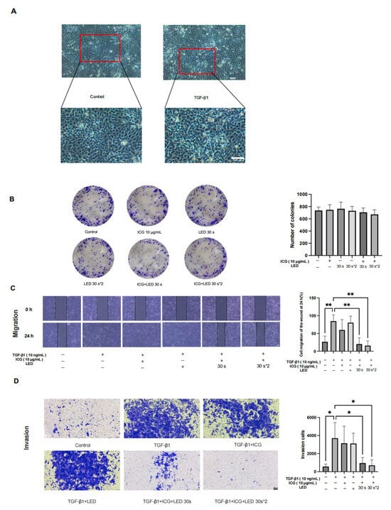
ICG–PDT inhibits the migration and invasion of SAS cells. (A) SAS cells were treated with or without (control) 10 ng/mL TGF-β1 for 24 h. Cell morphological changes associated with EMT are shown in the image. (magnification, ×40). (B) The effect of LED, ICG and ICG-PDT on long-term proliferation and viability was detected by a colony formation assay. (C) The effect of ICG-PDT on migration in SAS cells was detected by a wound healing assay after 24 h (magnification, ×40). The black lines mark the boundaries of the scratch area at each time point. (D) Transwell Matrigel invasion assays in TGF-β1-treated-SAS cells treated with or without PDT. Cells were stained with crystal violet. (magnification, ×40). Statistical significance was determined by one-way ANOVA. Data are presented as mean ± SD (n = 3). * p < 0.05, ** p < 0.01  vs. control group.
  • Review
  • Open Access

Natural ingredient-based cosmeceuticals represent an expanding area at the interface of cosmetics; these include polyphenols, phenolic acids, flavonoids, carotenoids, fatty acids, polysaccharides, and essential oil constituents. They have attracted increasing attention because of their antioxidant, anti-inflammatory, antimicrobial, photoprotective, and skin barrier-supporting properties. These compounds may contribute to the prevention of premature skin aging by reducing oxidative stress, modulating inflammatory signaling, inhibiting extracellular matrix degradation, and supporting epidermal homeostasis. In parallel, the emerging concept of microbiome-friendly skincare has created new opportunities for natural ingredients that act as prebiotics, postbiotics, or microbial balance supporting agents. Before clinical translation, cell-based experimental systems employing keratinocytes, dermal fibroblasts, melanocytes, and reconstructed skin models can be used to validate the relevant mechanisms of action at the cellular level. However, the translation of natural bioactivities into effective cosmetic products remains limited by poor chemical stability, low skin penetration, variability in extract composition, formulation incompatibility, and insufficient clinical validation. Advanced formulation technologies, including liposomes, nanoemulsions, solid lipid nanoparticles, hydrogels, and biopolymer-based delivery systems, may improve the stability, bioavailability, and controlled release of natural actives. This review summarizes major classes of plant-derived cosmeceutical ingredients, their mechanisms of action in skin aging, photoaging, barrier repair, and microbiome modulation, and discusses extraction, characterization, formulation, safety, and regulatory considerations. Future development should prioritize standardized phytochemical profiling, mechanism-based efficacy testing, microbiome-aware formulation design, sustainable sourcing, and well-controlled human studies.

Cells

12 November 2026

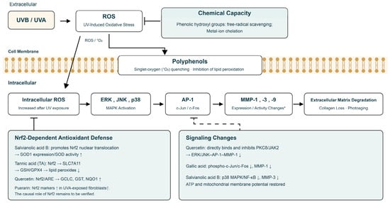
Effects of plant-derived bioactive compounds on UV-induced oxidative stress and photoaging. Chemical scavenging and cellular signaling regulation distinguished by extracellular, membrane, and intracellular locations. Arrows: UV-related damage cascade. Solid T-bars: observed inhibition or decreased markers. The dashed T-bar summarizes compound-associated signaling changes linked to reduced AP-1 signaling; it does not imply direct binding to AP-1. MMP endpoints vary among experiments. The cell membrane is schematic. * MMP endpoints vary among experiments; correlated changes do not establish direct compound–target binding. Created by the authors.
  • Review
  • Open Access

SIRT1 in Cardiometabolic Pathophysiology: A Narrative Review

  • Valentina Ventimiglia,
  • Francesca Megiorni and
  • Stefania Mariani
  • + 4 authors

Sirtuins (SIRTs) are nicotinic adenine dinucleotide (NAD)+-dependent deacetylases regulating critical signaling pathways in prokaryotes and eukaryotes. Recent in vivo and in vitro studies have successfully demonstrated the involvement of SIRT1 in key pathways for the biological function of the cardiovascular system, including cellular metabolism, senescence, oxidative stress, apoptosis and DNA damage. Based on SIRT1 pathophysiology activity, emerging evidence has stimulated a significant evolution in eventually preventing and treating cardiovascular disease (CVDs) by its modulation. SIRT1 is characterized by a protective role against inflammation, vascular aging, arrhythmias and the development of atherosclerotic plaques. Mechanistically, SIRT1 exerts vasculoprotective effects through deacetylation of key transcription factors leading to reduced inflammation, enhanced antioxidant defense, improved endothelial function, and correct vascular remodeling. This review provides a comprehensive overview of SIRT1-controlled molecular signaling pathways in heart pathophysiology, with particular emphasis on atherosclerosis, hypertension, dyslipidemia, atrial fibrillation, cardiac ischemia and ischemic/reperfusion injury. A deeper understanding of the epigenetic mechanisms by which SIRT1 intervenes in signaling pathways responsible for the onset and development of CVDs is important for speeding up the achievement of selective and effective pharmacological modulation of SIRT1 for future applications in the prevention and treatment of CVDs.

Cells

12 November 2026

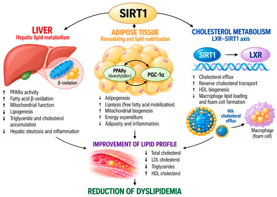
Schematic representation of SIRT1-mediated regulation of lipid homeostasis. SIRT1 coordinates hepatic fatty acid oxidation and lipid handling, adipose-tissue remodeling and energy expenditure, and LXR-dependent cholesterol efflux and reverse cholesterol transport.
  • Review
  • Open Access

Cancer is increasingly understood as a tissue- and system-level disease shaped by dynamic interactions among malignant cells, stromal elements, the vasculature, the immune system, and the nervous system. In this context, tumors are not passive recipients of neural input, but active participants in the recruitment, remodeling, and exploitation of neural circuits that support tumor initiation, progression, metastasis, immune evasion, and resistance to therapy. Emerging work in cancer neuroscience has therefore shifted the field from a neuron-centric perspective toward an integrated model of the tumor microenvironment in which malignant cells help establish pathogenic neural niches. This review adopts a tumor-centric perspective to examine how cancer cells drive neoneurogenesis, engage in perineural invasion, and co-opt neural signaling and neuro-immune crosstalk to promote disease progression. It first distinguishes tumor-induced nerve growth and remodeling from perineural invasion as related but mechanistically distinct processes, then considers how tumor cell plasticity and neural mimicry enable malignant cells to acquire neuronal-like traits and exploit neural pathways. The review further explores how tumor-driven innervation shapes angiogenesis, immune modulation, therapeutic resistance, and minimal residual disease while also assessing emerging strategies designed to therapeutically target the tumor–nerve axis. By integrating these themes, the review highlights innervation not as a secondary feature of cancer, but as an active and potentially actionable dimension of tumor biology.

Cells

12 November 2026

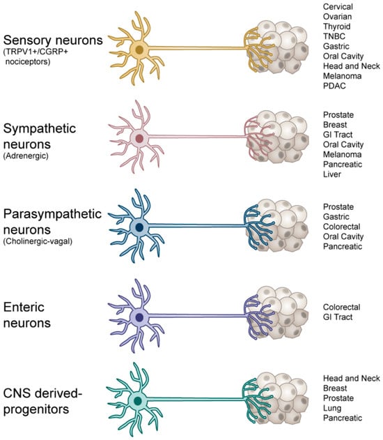
Neuronal subtypes reported to innervate peripheral tumors.

Highly Accessed Articles

News & Conferences

Latest Issues

Open for Submission

Journal Sections

XFacebookLinkedIn
Cells - ISSN 2073-4409