Next Issue
Volume 15, December
Previous Issue
Volume 15, October


Biomolecules, Volume 15, Issue 11 (November 2025) – 149 articles

Cover Story (view full-size image): 
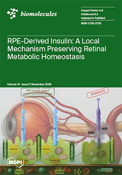
Recent evidence shows that the retinal pigment epithelium (RPE) contributes to retinal homeostasis by producing insulin locally. Under physiological conditions, daily phagocytosis of photoreceptor outer segments stimulates Ins2 expression and insulin release, supporting glucose uptake, oxidative metabolism, and photoreceptor renewal. During fasting, this local production remains active, providing protection when systemic insulin decreases. In diabetic or oxidative stress conditions, RPE insulin synthesis declines, reactive oxygen species rise, and metabolic coupling with photoreceptors is disrupted. As a result, outer segment turnover becomes defective and retinal dysfunction accelerates. These findings suggest that beyond pancreatic secretion, RPE-derived insulin may play a key role in regulating retinal metabolism and resilience. View this paper
  • Issues are regarded as officially published after their release is announced to the table of contents alert mailing list.
  • You may sign up for e-mail alerts to receive table of contents of newly released issues.
  • PDF is the official format for papers published in both, html and pdf forms. To view the papers in pdf format, click on the "PDF Full-text" link, and use the free Adobe Reader to open them.
Order results
Result details
Section
Select all
Export citation of selected articles as:
22 pages, 3965 KB  
Review
Sequence Determinants of G-Quadruplex Thermostability: Aligning Evidence from High-Precision Biophysics and High-Throughput Genomics
by Ke Xiao, Jiye Fu, Rongxin Zhang and Jing Tu
Biomolecules 2025, 15(11), 1632; https://doi.org/10.3390/biom15111632 - 20 Nov 2025
Viewed by 610
Abstract
G-quadruplexes (G4s) are non-canonical nucleic acid structures that function as key regulatory elements in crucial cellular processes. Their biological functions are intrinsically linked to thermostability, which is governed by specific sequence features. This review systematically synthesizes evidence from high-precision biophysical studies and high-throughput [...] Read more.
G-quadruplexes (G4s) are non-canonical nucleic acid structures that function as key regulatory elements in crucial cellular processes. Their biological functions are intrinsically linked to thermostability, which is governed by specific sequence features. This review systematically synthesizes evidence from high-precision biophysical studies and high-throughput genomic assays to delineate the sequence determinants of G4 thermostability. Analyses align the trends derived from both methodological paradigms and establish that stability emerges from a complex interplay among three structural elements: the G-tract core, whose length and integrity generally govern stability despite notable exceptions such as the anomalous stability of short G-tracts with 1-nt loops and the stabilization induced by large, structured bulges; the loops, which exhibit a consistent inverse relationship between length and stability across methods, though with context-dependent compositional effects and methodological disparities; and the flanking sequences, whose composition modulates stability and can bias topological outcomes. By integrating findings across scales, this work provides a unified conceptual framework connecting biophysical measurements with genomic observations—a critical step toward computationally predicting G4 stability, topology, and function directly from sequence, thereby advancing the understanding of their roles in health and disease. Full article
(This article belongs to the Section Molecular Biophysics: Structure, Dynamics, and Function)
Show Figures

Figure 1

37 pages, 48276 KB  
Article
Comparative Ultrasonic Bath and Probe Extraction of Piperine from Piper nigrum L. Using Natural Deep Eutectic Solvents: RSM Optimization, Characterization, and In Vitro Bioactivity
by Abdullah Mohammed Ayedh Al Adhreai, Johnson Retnaraj Samuel Selvan Christyraj, Beryl Vedha Yesudhason, Yolin Angel Poomany Arul Soundara Rajan and Maharshi Bhaswant
Biomolecules 2025, 15(11), 1631; https://doi.org/10.3390/biom15111631 - 20 Nov 2025
Viewed by 1061
Abstract
Background: Piper nigrum L. (PNL) is a rich source of piperine, a bioactive alkaloid with pharmaceutical, cosmetic, nutritional supplement, and agricultural applications, yet efficient and sustainable extraction methods remain underexplored. Methods: This study compared ultrasonic bath extraction (UBE) and ultrasonic probe extraction (UPE) [...] Read more.
Background: Piper nigrum L. (PNL) is a rich source of piperine, a bioactive alkaloid with pharmaceutical, cosmetic, nutritional supplement, and agricultural applications, yet efficient and sustainable extraction methods remain underexplored. Methods: This study compared ultrasonic bath extraction (UBE) and ultrasonic probe extraction (UPE) using natural deep eutectic solvents (NADES) for isolating piperine from PNL fruits. Six NADES formulations were screened, with NADES-5 (choline chloride:glycerin:urea, 1:1:1) showing superior performance. Response surface methodology with a Box–Behnken design optimized extraction parameters, including liquid-to-solid ratio, extraction time, temperature, and water content, for both UBE and UPE. Results: Optimized UPE consistently outperformed UBE, yielding 49.97 mg/g of piperine versus 25.67 mg/g under identical NADES conditions. Comprehensive characterization using TLC, HPTLC, UV, FTIR, Raman, HPLC, NMR, XRD, SEM, and EDX confirmed the successful isolation and structural integrity of piperine, with samples obtained via UPE exhibiting higher purity (98.7% vs. 95.2%) and enhanced crystallinity. In vitro cytotoxicity assays demonstrated that piperine extracted by UPE showed stronger activity against C2C12 myoblasts (IC50: 24.3 μg/mL vs. 40.6 μg/mL) and greater anticancer effects in MCF-7 and HT-29 cells compared to piperine extracted by UBE. Antioxidant evaluation via DPPH, ABTS, FRAP, and TAC assays, along with intracellular reactive oxygen and nitrogen species suppression in THP-1 and RAW 264.7 macrophages, further confirmed the superior biological potential of the UPE-derived piperine sample. Conclusions: These findings indicate that UPE using NADES is a sustainable approach for high-yield piperine extraction with enhanced purity and bioactivity, supporting its potential for pharmaceutical applications. Full article
(This article belongs to the Section Natural and Bio-derived Molecules)
Show Figures

Figure 1

18 pages, 3834 KB  
Article
The miR-125a-5p/IRF4 Axis Mediates Sodium Arsenite-Induced M2 Macrophage Polarization
by Yan Yu, Fan Yao, Suyuan Tong, Mingzheng Li, Qilong Liao, Fei Wang and Shuhua Xi
Biomolecules 2025, 15(11), 1630; https://doi.org/10.3390/biom15111630 - 20 Nov 2025
Cited by 1 | Viewed by 650
Abstract
Arsenic, a ubiquitous metalloid, is commonly found in surface waters; as well as serious human health issues, it also induces systemic diseases and carcinogenesis upon chronic exposure. To better understand how arsenic potentially alters the immune system, it is important to study its [...] Read more.
Arsenic, a ubiquitous metalloid, is commonly found in surface waters; as well as serious human health issues, it also induces systemic diseases and carcinogenesis upon chronic exposure. To better understand how arsenic potentially alters the immune system, it is important to study its effects on macrophage polarization. Micro-RNA plays an epigenetic regulatory role in organisms. The miR-125 family regulates macrophage polarization and tumorigenesis, yet its role in arsenic-induced macrophage polarization remains unexplored. This study investigated the mechanism of sodium arsenite (NaAsO2)-driven macrophage polarization via miR-125a-5p. In vivo, rats exposed to 10 or 50 mg/L NaAsO2 for 12 weeks exhibited elevated M2 markers (CD206, Arg1) and reduced M1 markers (iNOS, IL-1β, TNF-α) in liver and bladder tissues. In vitro, THP-1-derived macrophages treated with NaAsO2 (2–8 μM) for 48 h showed dose-dependent M2 polarization, marked by upregulated CD206, Arg1, and IL-10. Flow cytometry results show that the proportion of M2/M1-type cells has increased significantly. Notably, NaAsO2 suppressed miR-125a-5p expression and elevated interferon regulatory factor 4 (IRF4), a predicted target of miR-125a-5p. Overexpression of miR-125a-5p reversed NaAsO2-induced M2 polarization by inhibiting IRF4, thereby reducing M2 markers and restoring M1-associated proteins. These findings reveal that NaAsO2 promotes M2 macrophage polarization through the miR-125a-5p/IRF4 axis, highlighting a novel epigenetic mechanism in arsenic-associated tumor microenvironments and immune dysfunction. This study provides critical insights into targeting miR-125a-5p as a therapeutic strategy. Full article
(This article belongs to the Section Biological Factors)
Show Figures

Graphical abstract

27 pages, 1534 KB  
Review
Microglia-Mediated Phagocytosis in Alzheimer’s Disease: Mechanisms, Heterogeneity, and Therapeutic Insights
by Halimatu Hassan, Charlotte Rawlinson, Yu-Long Lan, Stuart Jenkins and Ruoli Chen
Biomolecules 2025, 15(11), 1629; https://doi.org/10.3390/biom15111629 - 20 Nov 2025
Viewed by 2525
Abstract
Microglia are the resident immune cells of the CNS, maintaining brain homeostasis partially through phagocytosis. In Alzheimer’s disease (AD), microglial phagocytosis is significantly impaired, contributing to the accumulation of pathological aggregates. Microglial phenotypes are dynamic and can shift depending on the disease stage [...] Read more.
Microglia are the resident immune cells of the CNS, maintaining brain homeostasis partially through phagocytosis. In Alzheimer’s disease (AD), microglial phagocytosis is significantly impaired, contributing to the accumulation of pathological aggregates. Microglial phenotypes are dynamic and can shift depending on the disease stage and local environment. While some subpopulations retain or enhance phagocytic activity, especially under inflammatory conditions, others lose their capacity to clear toxic debris effectively. This variability underscores the need for a more nuanced understanding of microglial regulation and function. This paper explores the dual role of microglial phagocytosis in AD and discusses the emerging insights into microglial heterogeneity and how phenotypic shifts affect phagocytic capacity throughout disease progression. A comprehensive understanding of microglial phagocytosis and its dysregulation in AD is essential for designing targeted treatments. Modulating microglial activity to enhance their protective roles without triggering harmful inflammation represents a promising direction for therapeutic intervention in AD. Full article
(This article belongs to the Special Issue Pathogenesis and Targeted Therapy of Neurodegenerative Diseases)
Show Figures

Figure 1

29 pages, 1350 KB  
Review
Protein Engineering and Drug Discovery: Importance, Methodologies, Challenges, and Prospects
by Ahmed Mohammed, Nasir A. Ibrahim and Nosiba S. Basher
Biomolecules 2025, 15(11), 1628; https://doi.org/10.3390/biom15111628 - 20 Nov 2025
Viewed by 1822
Abstract
Protein engineering is a rapidly evolving field that plays a critical role in transforming drug discovery and development. This innovative field harnesses the unique structural and functional properties of engineered proteins, such as monoclonal antibodies, nanobodies, therapeutic enzymes, and cytokines, to address complex [...] Read more.
Protein engineering is a rapidly evolving field that plays a critical role in transforming drug discovery and development. This innovative field harnesses the unique structural and functional properties of engineered proteins, such as monoclonal antibodies, nanobodies, therapeutic enzymes, and cytokines, to address complex diseases more effectively than traditional small-molecule drugs. These biologics not only enhance therapeutic specificity but also minimize adverse effects, marking a significant advancement in patient care. However, the journey of protein engineering is not without challenges. Issues related to protein folding, stability, and potential immunogenicity pose significant complications. Additionally, navigating the complex regulatory landscape can delay the transition from laboratory to clinical application. Addressing these hurdles requires the integration of cutting-edge technologies, including phage and yeast display technology, CRISPR, and advanced computational modeling, which enhance the predictability and efficiency of protein design. In this review, we explore the multifaceted impact of protein engineering on modern medicine, highlighting its potential to transform treatment paradigms, methodologies, challenges, and the successful development and approval of recombinant protein-based therapies. By navigating the complexities and leveraging technological advancements, the field is poised to unlock new therapeutic possibilities, ultimately improving patient outcomes and transforming healthcare. Full article
(This article belongs to the Section Molecular Medicine)
Show Figures

Figure 1

16 pages, 3496 KB  
Article
FTO Suppresses Dental Pulp Stem Cell Senescence by Destabilizing NOLC1 mRNA
by Bingrong Li, Mi Xu, Junjun Huang and Rong Jia
Biomolecules 2025, 15(11), 1627; https://doi.org/10.3390/biom15111627 - 19 Nov 2025
Viewed by 566
Abstract
Cellular senescence is an intricate process that severely restricts stem cell function. The N6-methyladenosine (m6A) eraser, fat mass and obesity-associated (FTO) protein control several aspects of stem cell fate, including differentiation, self-renewal, and senescence. However, the role of FTO in dental [...] Read more.
Cellular senescence is an intricate process that severely restricts stem cell function. The N6-methyladenosine (m6A) eraser, fat mass and obesity-associated (FTO) protein control several aspects of stem cell fate, including differentiation, self-renewal, and senescence. However, the role of FTO in dental pulp stem cell (DPSC) senescence has not yet been elucidated. This study aimed to explore the role of FTO in DPSC senescence. FTO expression decreases during DPSC senescence. FTO depletion inhibited DPSC proliferation, accelerated senescence, and increased reactive oxygen species (ROS) levels. FTO overexpression reduced DPSC senescence, enhanced proliferation, and decreased ROS accumulation. RNA sequencing demonstrated that FTO knockdown inhibited ribosomal RNA precursor (pre-rRNA) biogenesis. We found nucleolar and coiled-body phosphoprotein 1 (NOLC1) as a novel target of FTO. NOLC1 was upregulated after FTO knockdown and promoted DPSC senescence. Mechanistically, FTO downregulation increased the m6A modifications of NOLC1 mRNA, increasing the stability of the NOLC1 mRNA. NOLC1 upregulation inhibits the transcription of pre-rRNA, causing nucleolar stress and p53 accumulation. In addition, NOLC1 knockdown partially rescued FTO deficiency-induced DPSC senescence. Our findings identified the significant role of the FTO/NOLC1/p53 axis in DPSC senescence and provide new insights to prevent the aging of DPSCs, which is beneficial for the application of DPSCs in regenerative medicine and stem cell therapy. Full article
(This article belongs to the Section Cellular Biochemistry)
Show Figures

Figure 1

20 pages, 5117 KB  
Article
Evaluating the Topological Features of Monomeric and Trimeric TRAF2-C: A Multi-Disciplinary Approach
by Fulvio Erba, Daniela Russo, Velia Minicozzi, Luisa Di Paola, Sylvain Prevost, Anastasia De Luca, Giampiero Mei and Almerinda Di Venere
Biomolecules 2025, 15(11), 1626; https://doi.org/10.3390/biom15111626 - 19 Nov 2025
Viewed by 399
Abstract
This study investigates the structural dynamics of the TRAF2 C-terminal domain (TRAF2-C), a key adaptor protein in TNF receptor signaling. TRAF2 usually forms trimers, but its ability to dissociate into monomers is critical for regulating apoptosis, inflammation, and cell survival. Using Fluorescence Fluctuation [...] Read more.
This study investigates the structural dynamics of the TRAF2 C-terminal domain (TRAF2-C), a key adaptor protein in TNF receptor signaling. TRAF2 usually forms trimers, but its ability to dissociate into monomers is critical for regulating apoptosis, inflammation, and cell survival. Using Fluorescence Fluctuation Spectroscopy, dynamic light scattering, circular dichroism, and Small Angle Neutron Scattering, we analyzed TRAF2-C over a wide concentration range. At nanomolar levels, the protein dissociates easily, with trimers representing only a minor fraction, while micromolar concentrations strongly favor trimerization. Dissociation also reduces α-helical content without disrupting the overall fold. Molecular dynamics simulations and protein contact network analysis support this analysis, identifying interfacial residues and hydrogen bonds as key factors stabilizing oligomers and enabling dynamic asymmetry. Overall, these findings highlight TRAF2-C’s capacity to switch between monomeric and trimeric states as a crucial regulatory mechanism, offering insights into TRAF-mediated signaling and potential therapeutic strategies. Full article
(This article belongs to the Special Issue Role of TRAF in Regulating Inflammation and Cell Survival)
Show Figures

Figure 1

28 pages, 2510 KB  
Review
Function and Mechanism of Small Nucleolar RNAs (snoRNAs) and Their Host Genes (SNHGs) in Malignant Tumors
by Jiaji Yu, Yingjie Shao and Wendong Gu
Biomolecules 2025, 15(11), 1625; https://doi.org/10.3390/biom15111625 - 19 Nov 2025
Viewed by 892
Abstract
Small nucleolar RNAs (snoRNAs) and their host genes (SNHGs) are non-coding RNAs that are integral to tumorigenesis and progression. snoRNAs contribute to tumor progression primarily through RNA modification and engagement in intracellular signaling, and by serving as precursors for small nucleolar RNA-derived RNAs [...] Read more.
Small nucleolar RNAs (snoRNAs) and their host genes (SNHGs) are non-coding RNAs that are integral to tumorigenesis and progression. snoRNAs contribute to tumor progression primarily through RNA modification and engagement in intracellular signaling, and by serving as precursors for small nucleolar RNA-derived RNAs (sdRNAs) that exert microRNA (miRNA)-like or epigenetic regulatory functions. SNHGs modulate key tumor cell behaviors—including proliferation, metastasis, and resistance to therapy—through competing endogenous RNA (ceRNA)-mediated interactions and epigenetic mechanisms. Their combined influence significantly impacts patient prognosis. Across diverse malignancies such as neurologic, bone, and head and neck cancers, snoRNAs and SNHGs exhibit cancer-specific regulatory dynamics; for instance, in glioblastoma, snoRNAs and their derived fragments (sdRNAs) contribute to intratumoral heterogeneity by mediating both metabolic reprogramming and epigenetic remodeling, while their mediated modulation of cellular proliferation and metastatic potential is evident in breast cancer. Concurrently, several snoRNAs and SNHGs have emerged as potential diagnostic and prognostic biomarkers, as well as therapeutic targets. Preclinical interventions targeting select snoRNAs or SNHGs have demonstrated promising therapeutic outcomes. This study reviews current insights into the oncogenic functions and signaling networks associated with dysregulated snoRNAs and SNHGs in malignancies, while highlighting novel avenues for future investigation in this domain. Full article
(This article belongs to the Section Molecular Genetics)
Show Figures

Figure 1

33 pages, 3761 KB  
Review
Alternative Splicing Dysregulation in Retinitis Pigmentosa: Pathogenic Mechanisms and Therapeutic Opportunities
by Yuxin Jiang, Xuyu Liu, Jie Fu, Yican Wu, Shanshan Yu and Kai Yao
Biomolecules 2025, 15(11), 1624; https://doi.org/10.3390/biom15111624 - 19 Nov 2025
Viewed by 1194
Abstract
Retinitis pigmentosa (RP) represents a genetically heterogeneous group of inherited retinal dystrophies characterized by progressive photoreceptor degeneration and irreversible vision loss. Among the diverse pathogenic mechanisms, dysregulation of alternative splicing has emerged as a pivotal driver, particularly in RP cases caused by mutations [...] Read more.
Retinitis pigmentosa (RP) represents a genetically heterogeneous group of inherited retinal dystrophies characterized by progressive photoreceptor degeneration and irreversible vision loss. Among the diverse pathogenic mechanisms, dysregulation of alternative splicing has emerged as a pivotal driver, particularly in RP cases caused by mutations in splicing factors or cis-regulatory elements. Alternative splicing governs transcript diversity and fine-tunes gene expression, with more than 95% of human multi-exon genes undergoing this process. Disruption of precise splicing patterns in the retina—an organ with exceptionally high transcriptional complexity—leads to widespread mis-splicing of photoreceptor-specific genes, triggering retinal dysfunction and cell death. This review synthesizes current understanding of alternative splicing-related mechanisms in RP, integrating molecular insights from splicing-factor mutations, retina-specific splice isoforms, and their downstream cellular consequences. We also evaluate therapeutic strategies targeting splicing dysregulation, including antisense oligonucleotides (ASOs), modified U1 snRNA, spliceosome-mediated RNA trans-splicing (SMaRT), and genome editing, emphasizing translational potential and clinical challenges. Finally, we highlight key research gaps and propose future directions for splicing-centered precision medicine in RP. Full article
(This article belongs to the Section Molecular Genetics)
Show Figures

Graphical abstract

16 pages, 6116 KB  
Article
Mitochondrial Imaging and Transcriptome Analysis of Bone Mesenchymal Stem Cells During Osteogenesis Under Different Culture Conditions
by Qicheng Li, Tianze Sun, Shiyan Liu, Lu Zhang and Yuhui Kou
Biomolecules 2025, 15(11), 1623; https://doi.org/10.3390/biom15111623 - 19 Nov 2025
Viewed by 801
Abstract
Bone mesenchymal stem cells (BMSCs) are multipotent progenitors with significant potential for bone tissue engineering and regenerative medicine. This study compared the mitochondrial imaging and transcriptome of BMSCs under two-dimensional (2D) and three-dimensional (3D) culture conditions during osteogenesis. 2D BMSCs were induced toward [...] Read more.
Bone mesenchymal stem cells (BMSCs) are multipotent progenitors with significant potential for bone tissue engineering and regenerative medicine. This study compared the mitochondrial imaging and transcriptome of BMSCs under two-dimensional (2D) and three-dimensional (3D) culture conditions during osteogenesis. 2D BMSCs were induced toward osteogenesis for 7, 14, and 21 days, while 3D BMSCs were induced for 21 days. Osteogenic mineralization was assessed by Alizarin Red S (ARS) staining, and whole-transcriptome sequencing (RNA-Seq) was performed to elucidate gene expression profiles. Furthermore, mitochondrial morphology in live cells was monitored at 0, 7, 14, and 21 days of 2D osteogenic differentiation to observe the mitochondrial changes. High-Sensitivity Structured Illumination Microscopy (HIS-SIM) imaging showed that mitochondrial morphology in BMSCs underwent a shift toward elongated and interconnected networks over time. The transcriptional profile showed that genes associated with skeletal morphogenesis, bone development, and extracellular matrix organization were significantly upregulated in 3D culture systems. These findings indicate that 3D culture is associated with a transcriptional profile enriched in pathways commonly observed during in vivo osteogenesis, which can inform scaffold-based bone-regeneration strategies. Full article
(This article belongs to the Section Molecular Medicine)
Show Figures

Figure 1

16 pages, 1477 KB  
Systematic Review
Divergent Risks of Hematologic Malignancies Associated with GLP-1 Receptor Agonists and SGLT2 Inhibitors: Preliminary Findings from a Pilot Network Meta-Analysis
by Pao-Yen Lin, Bing-Yan Zeng, Chih-Wei Hsu, Mein-Woei Suen, Chao-Ming Hung, Brendon Stubbs, Yen-Wen Chen, Tien-Yu Chen, Wei-Te Lei, Jiann-Jy Chen, Bing-Syuan Zeng, Kuan-Pin Su, Chih-Sung Liang and Ping-Tao Tseng
Biomolecules 2025, 15(11), 1622; https://doi.org/10.3390/biom15111622 - 19 Nov 2025
Viewed by 1049
Abstract
Background: Although glucagon-like peptide-1 (GLP-1) receptor agonists and sodium–glucose cotransporter 2 (SGLT2) inhibitors have gained attention for their broad therapeutic effects, their influence on hematologic malignancy remains underexplored. Given the high mortality associated with hematologic cancers, clarifying the impact of these agents on [...] Read more.
Background: Although glucagon-like peptide-1 (GLP-1) receptor agonists and sodium–glucose cotransporter 2 (SGLT2) inhibitors have gained attention for their broad therapeutic effects, their influence on hematologic malignancy remains underexplored. Given the high mortality associated with hematologic cancers, clarifying the impact of these agents on such malignancies is essential. Objectives: This pilot network meta-analysis (NMA) aimed to assess the comparative risk of hematologic malignancies—including lymphoma, leukemia, and myeloma—associated with various GLP-1 receptor agonists and SGLT2 inhibitors. Methods: Following Cochrane-recommended confirmatory methods, we systematically searched multiple databases for randomized controlled trials (RCTs) published through 4 December 2024. The primary outcome was the incidence of overall hematologic malignancies. A frequentist random-effects NMA via the netmeta package was conducted, with additional validation through Bayesian NMA for solely sensitivity analyses. Results: Fifty-five RCTs (n = 200,606) were analyzed. Dulaglutide showed a significantly higher risk of overall hematologic malignancy [odds ratio (OR) = 2.18, 95% confidence interval (95%CI) = 1.14–4.19). In contrast, tirzepatide was linked to a significantly reduced risk (OR = 0.14, 95%CI = 0.03–0.60), especially for lymphoma. No statistically significant associations were identified for SGLT2 inhibitors (i.e., 95%CI across 1.0). Conclusions: Our preliminary findings reveal distinct and agent-specific effects of GLP-1 receptor agonists on hematologic malignancy risk. While dulaglutide may elevate the risk, tirzepatide appears protective, particularly against lymphoma. These results call for further long-term mechanistic studies to clarify causality and underlying pathways. Full article
Show Figures

Figure 1

22 pages, 1098 KB  
Review
Circulating microRNAs and Plasma Gelsolin as Biomarkers of Sepsis: Molecular Insights and Prospects for Precision Medicine
by Mircea Stoian, Leonard Azamfirei, Sergio Rares Bandila, Adina Stoian, Dragoș-Florin Babă and Claudia Bănescu
Biomolecules 2025, 15(11), 1621; https://doi.org/10.3390/biom15111621 - 18 Nov 2025
Cited by 1 | Viewed by 956
Abstract
Sepsis is a major medical emergency, characterized by a dysfunctional immune response to infection, which often progresses to multiple organ failure and death. Early diagnosis and prognostic evaluation present significant challenges due to limitations in the specificity and sensitivity of traditional biomarkers. This [...] Read more.
Sepsis is a major medical emergency, characterized by a dysfunctional immune response to infection, which often progresses to multiple organ failure and death. Early diagnosis and prognostic evaluation present significant challenges due to limitations in the specificity and sensitivity of traditional biomarkers. This narrative review summarizes recent evidence on the potential of circulating microRNAs (miRNAs) such as miR-150, miR-146a, miR-223, miR-155, miR-122, and miR-4772-5p and plasma gelsolin (pGSN) as diagnostic and prognostic markers in sepsis. We discuss mechanisms involved and their potential for integration with artificial intelligence (AI) in personalized medicine. PubMed, Embase, and Web of Science databases were searched for relevant literature. Original research, systematic reviews, and meta-analyses focused on the diagnostic or prognostic value of circulating miRNAs or pGSN in sepsis were included; opinion papers and case reports were excluded. Altered expression of certain circulating microRNAs correlates with disease severity and mortality. Among circulating microRNAs (miRNAs), miR-122 and miR-150 have become the most consistently validated biomarkers in clinical studies, associated with sepsis severity and death rates. Additionally, other miRNAs such as miR-146a, miR-155, and miR-223 play roles in modulating immune and endothelial responses, highlighting the complex regulation of sepsis pathophysiology. Low pGSN concentrations at admission are associated with severe sepsis and acute respiratory distress syndrome, and serve as an independent predictor of mortality. Preclinical studies suggest that supplementation with exogenous pGSN could increase survival. AI algorithms show promising results for early sepsis detection and optimization of therapeutic decisions. However, combining circulating miRNAs and plasma gelsolin (pGSN) into AI-based models is still an exploratory idea that needs prospective validation, assay standardization, and multicenter studies before it can be used clinically. Full article
(This article belongs to the Special Issue Mechanisms of RNA Regulation in Inflammation)
Show Figures

Figure 1

15 pages, 1581 KB  
Article
Structural and Functional Characterization of LIMCH1 and Its Agmatinase-like Region: A Case of Catalysis in a Highly Disordered Protein
by María-Belén Reyes, Allison Fuentes, Diego Bustamante, Fernando Retamal, Ignacia Lillo, Cristián Villegas, Juan-Pablo Carrasco, Martin Pereira-Silva, Marcell Gatica, Juan Román, Maximiliano Figueroa, Yamil Neira, José Martínez-Oyanedel, Víctor Castro-Fernández and Elena Uribe
Biomolecules 2025, 15(11), 1620; https://doi.org/10.3390/biom15111620 - 18 Nov 2025
Viewed by 507
Abstract
Agmatine is a biogenic amine that functions as a neurotransmitter and exhibits anticonvulsant, antineurotoxic, and antidepressant properties. It can be metabolized into putrescine and urea by canonical agmatinases or by the agmatinase-like protein (ALP), which corresponds to the C-terminal region of the LIMCH1 [...] Read more.
Agmatine is a biogenic amine that functions as a neurotransmitter and exhibits anticonvulsant, antineurotoxic, and antidepressant properties. It can be metabolized into putrescine and urea by canonical agmatinases or by the agmatinase-like protein (ALP), which corresponds to the C-terminal region of the LIMCH1 protein. The amino acid sequence of ALP/LIMCH1 diverges significantly from that of canonical agmatinases and lacks the conserved residues typically required for coordination with Mn2+, an essential cofactor for ureohydrolase activity. The three-dimensional structure of ALP/LIMCH1 remains unresolved, and predictive artificial intelligence algorithms such as AlphaFold have failed to model it reliably. As a result, the configuration of its active site and the identity of potential metal-coordinating ligands remain elusive. In this study, we purified recombinant full-length rat LIMCH1 (119.5 kDa) and a truncated ALP variant, ΔLIM-ALP (51 kDa), and analyzed their secondary structures using circular dichroism spectroscopy. Our results indicate that both proteins differ markedly from known ureohydrolases, exhibiting a high proportion of disordered regions (~60%) and β-structures (~30%). In contrast, Escherichia coli agmatinase displays a well-defined α/β/α sandwich fold. Despite these structural differences, ALP/LIMCH1 remain the only known mammalian proteins exhibiting agmatinase activity. To gain insight into the putative active site of ALP, we proposed candidate Mn2+-binding residues and generated single-point mutants (N213A, Q215A, D217A, E288A, K290A). Although these mutations did not significantly alter Mn2+ binding or its overall content in the protein samples, four mutants exhibited a decreased Km for agmatine and a reduced Vmax when normalized to protein concentration. Full article
(This article belongs to the Section Molecular Biophysics: Structure, Dynamics, and Function)
Show Figures

Figure 1

15 pages, 3238 KB  
Article
Mechanistic and Kinetic Insights into the Acylation Reaction of Hepatitis C Virus NS3/NS4A Serine Protease with NS4B/5A Substrate
by José Ángel Martínez-González, Nuria Salazar-Sanchez, María Larriva-Hormigos, Rodrigo Martínez and Miguel González
Biomolecules 2025, 15(11), 1619; https://doi.org/10.3390/biom15111619 - 18 Nov 2025
Viewed by 491
Abstract
Reaction mechanisms and rate constants of the acylation reaction of the hepatitis C virus (HCV) NS3/NS4A serine protease with the NS4B/5A natural substrate were studied using SCC-DFTB/MM (self-consistent charge density functional tight binding/molecular mechanics) and EA-VTST/MT (ensemble-averaged variational transition state theory/multidimensional tunneling) methods, [...] Read more.
Reaction mechanisms and rate constants of the acylation reaction of the hepatitis C virus (HCV) NS3/NS4A serine protease with the NS4B/5A natural substrate were studied using SCC-DFTB/MM (self-consistent charge density functional tight binding/molecular mechanics) and EA-VTST/MT (ensemble-averaged variational transition state theory/multidimensional tunneling) methods, considering the isotope effect (H/D). This reaction is crucial in the HCV life cycle. The reaction follows an essentially concerted mechanism. Although two elementary steps are involved, no intermediate step has been found between them. Thus, the proposed general two-step serine protease acylation mechanism, which includes a tetrahedral intermediate, does not occur here. This finding aligns with our studies on another natural substrate (NS5A/5B), indicating a greater variety in mechanism than previously expected. Tunneling and recrossing play an intermediate role; the activation free energy barriers are in good agreement with the experimental value, and the kinetic isotope effect (k(H)/k(D)) is somewhat larger than one (1.3). The rate constant value is not reproduced due to the exponential dependence of the rate constant on the activation free energy. Full article
(This article belongs to the Section Chemical Biology)
Show Figures

Figure 1

20 pages, 6149 KB  
Article
Multi-Omics Analysis Reveals 1-Propanol-Induced Pentadecanoic Acid Biosynthesis in Yarrowia lipolytica
by Jiahe Cong, Xin Hu, Dongsheng Lu, Sam C. Kollie, Ahmed A. Elolimy, Juan J. Loor, Zhendong Yang, Mingxun Li, Yongjiang Mao, Zhangping Yang and Huimin Zhang
Biomolecules 2025, 15(11), 1618; https://doi.org/10.3390/biom15111618 - 18 Nov 2025
Viewed by 722
Abstract
Pentadecanoic acid (C15:0) is an odd-chain fatty acid (OCFA) with significant health benefits, mainly produced by microbial fermentation. To improve C15:0 production, this study compared the effects of different alcohols on C15:0 production in Yarrowia lipolytica CICC1778, identified 1-propanol as the most effective [...] Read more.
Pentadecanoic acid (C15:0) is an odd-chain fatty acid (OCFA) with significant health benefits, mainly produced by microbial fermentation. To improve C15:0 production, this study compared the effects of different alcohols on C15:0 production in Yarrowia lipolytica CICC1778, identified 1-propanol as the most effective precursor, assessed its optimal concentration, and employed transcriptomic and metabolomic analyses to elucidate the regulatory mechanisms. The results showed that supplementation with 0.5% 1-propanol resulted in a total lipid production of 1.54 g/L in Y. lipolytica CICC1778, showing no differences compared with the negative control (NC) group, while C15:0 production increased to 76.68 mg/L, representing a 794.7% increase compared with the NC group. Integrated omics analysis showed that propionylcarnitine was positively correlated with ADH2, ADH1, ACADSB, ALDH6A1, and CAT2; O-methylmalonylcarnitine was positively correlated with IVD, MCCC2, ACADSB, and ALDH6A1; and (R)-leucic acid and 2-hydroxy-3-methylbutyric acid were positively correlated with IVD, BAT2, MCCC2, and ACADSB and ALDH6A1 and BAT2, respectively. All of these DEGs and DEMs were upregulated in the alcohol-treated (ALC; supplementation with 0.5% 1-propanol) group. Taken together, supplementation with 0.5% 1-propanol was an effective strategy for enhancing C15:0 production in Y. lipolytica CICC1778; 1-propanol underwent dehydrogenation-oxidation and promoted branched-chain amino acid degradation to expand the propionyl-CoA pool, thereby facilitating C15:0 synthesis. Full article
(This article belongs to the Section Lipids)
Show Figures

Figure 1

19 pages, 1525 KB  
Article
Screening and Validation of Functional Residues of the Antimicrobial Peptide PpRcys1
by Ming Tao, Zixun Fei, Aobo Sun, Guangming Yu, Huaiyuan Ye, Huishao Shi, Wei Zhang and Junjian Wang
Biomolecules 2025, 15(11), 1617; https://doi.org/10.3390/biom15111617 - 18 Nov 2025
Viewed by 547
Abstract
The excessive use of conventional antibiotics in aquaculture has created significant challenges, making it essential to explore and develop effective alternatives. Antimicrobial peptides (AMPs) have gained attention as potential therapeutic agents owing to their wide-ranging antibacterial effects and their ability to address pathogens [...] Read more.
The excessive use of conventional antibiotics in aquaculture has created significant challenges, making it essential to explore and develop effective alternatives. Antimicrobial peptides (AMPs) have gained attention as potential therapeutic agents owing to their wide-ranging antibacterial effects and their ability to address pathogens resistant to conventional drugs. PpRcys1 is an antimicrobial peptide that mainly targets bacterial cell membranes, exhibiting a minimum inhibitory concentration of 8–32 μM. Its antibacterial activity should be further optimized. Before such optimization, however, it is crucial to identify the key amino acid residues that determine its functional activity. In this study, molecular dynamics simulations indicated that arginine 40 (ARG40), lysine 55 (LYS55), lysine 90 (LYS90), and lysine 93 (LYS93) play critical roles in the interaction between PpRcys1 and bacterial membranes. To investigate this further, these residues were mutated to serine, producing the mutant peptide PpRcys1_RMRK. Compared with PpRcys1, the mutant peptide PpRcys1_RMRK showed a significant reduction in antibacterial activity. Results from molecular dynamics simulations, Western blot, and ELISA demonstrated a marked decrease in its ability to bind to bacterial cell membranes. Membrane permeation assays, cell membrane depolarization experiments, and scanning electron microscopy revealed that PpRcys1 could not compromise the integrity of the bacterial membrane after losing ARG40, LYS55, LYS90 and LYS93. These findings highlight the critical roles of ARG40, LYS55, LYS90, and LYS93 in sustaining the antibacterial activity of PpRcys1. This study provides important initial insights into the structure–activity relationship of PpRcys1 and establishes a theoretical foundation for its future optimization. Full article
(This article belongs to the Special Issue Recent Molecular Research on Protein Structure and Function)
Show Figures

Figure 1

12 pages, 1203 KB  
Review
Amylase Binding to Oral Streptococci: A Key Interaction for Human Oral Microbial Ecology, Adaptation and Fitness
by Amarpreet Sabharwal, Elaine M. Haase and Frank A. Scannapieco
Biomolecules 2025, 15(11), 1616; https://doi.org/10.3390/biom15111616 - 18 Nov 2025
Viewed by 602
Abstract
The interaction between human salivary alpha-amylase (HSAmy) and amylase-binding oral streptococci (ABS) helps determine the bacteria that colonize the oral cavity by establishing dental biofilms. Streptococci are important pioneer species of the oral cavity and influence oral health as well as common diseases [...] Read more.
The interaction between human salivary alpha-amylase (HSAmy) and amylase-binding oral streptococci (ABS) helps determine the bacteria that colonize the oral cavity by establishing dental biofilms. Streptococci are important pioneer species of the oral cavity and influence oral health as well as common diseases such as dental caries. Various oral streptococcal species express distinct amylase-binding proteins, among which amylase-binding protein A (AbpA), encoded by the abpA gene in Streptococcus gordonii and several other species, which is the most extensively studied. Amylase binding facilitates microbial adhesion to host surfaces and biofilm formation and enables bacteria to harness the host’s amylase enzymatic activity at their cell surface, enhancing their capacity to metabolize dietary starch for nutritional gain. Additionally, amylase binding may also influence bacterial cell division and stress tolerance by engaging novel bacterial signaling pathways. From an evolutionary perspective, both Neanderthals and modern humans exhibit functional adaptations in nutrient metabolism, including selection for salivary amylase-binding oral streptococci, highlighting the importance of microbial co-adaptation in response to host diet. Further research is warranted to elucidate the broader roles of amylase binding to bacteria in host-bacterial signaling, bacterial cell division and fitness and the evolutionary trajectory of the oral microbiome. Full article
(This article belongs to the Special Issue Digestive Enzymes in Health and Disease)
Show Figures

Figure 1

27 pages, 5073 KB  
Article
Activity of Serpins in Context to Hydrophobic Interaction
by Irena Roterman, Katarzyna Stapor, Grzegorz Zemanek, Dawid Dulak and Leszek Konieczny
Biomolecules 2025, 15(11), 1615; https://doi.org/10.3390/biom15111615 - 18 Nov 2025
Viewed by 496
Abstract
The activity of serpins uses a specific mechanism or process. This process comprises several steps and is related to significant structural changes that involve significant displacement of chain fragments and whole molecules of protease. An important role is played by a segment of [...] Read more.
The activity of serpins uses a specific mechanism or process. This process comprises several steps and is related to significant structural changes that involve significant displacement of chain fragments and whole molecules of protease. An important role is played by a segment of the serpin chain called the Reactive Central Loop (RCL), which interacts with the protease by inhibiting its activity. For the covalent binding of the protease to serpin, the movement of the protease molecule is an effect of splicing the RCL segment into beta-sheet A of serpin. There are structural forms—native, latent, Michaelis complex (non-covalent enzyme-inhibitor complex prior to RCL cleavage), covalent serpin–protease complex, and cleaved—associated with serpin activity. In this work, all these structural forms are discussed using the fuzzy oil drop (FOD-M) model, where the assessment criterion of structuring is based on identifying the type of hydrophobicity distribution. The analysis reveals the specificity of the inhibition mechanism, including the specific action of the RCL. The structural changes involved in this process have been shown to preserve the distribution of hydrophobicity in the form preferred by the aqueous environment in which serpins are active. The disorder (according to FOD-M model) in two complexes (Michaelis and covalent) is hypothetically treated as code for degradation factors. The applied model assesses the function-related structures using the hydrophobicity distribution as the criterion in contrast to many publications based on energetic aspects of serpin activity. Structural changes appear appropriate for water environments—the environment of serpin activity. Full article
(This article belongs to the Special Issue Protein Biophysics)
Show Figures

Figure 1

14 pages, 5073 KB  
Article
Citronellol Reduces Sepsis-Induced Renal Inflammation via AP-1/NF-κB/TNF-α Pathway
by Huda Rashid Atiyah, Sarmed H. Kathem and Surya M. Nauli
Biomolecules 2025, 15(11), 1614; https://doi.org/10.3390/biom15111614 - 17 Nov 2025
Viewed by 630
Abstract
Sepsis is characterized by the over-production of pro-inflammatory cytokines. Cecal ligation and puncture (CLP) is a well-accepted model for recreating sepsis-induced renal injury in mice. The current study investigates how citronellol, a naturally occurring substance with a variety of biological characteristics, can prevent [...] Read more.
Sepsis is characterized by the over-production of pro-inflammatory cytokines. Cecal ligation and puncture (CLP) is a well-accepted model for recreating sepsis-induced renal injury in mice. The current study investigates how citronellol, a naturally occurring substance with a variety of biological characteristics, can prevent acute kidney inflammation brought on by CLP. In the CLP mouse model, citronellol was administered orally at doses of 50 and 100 mg/kg. Serum levels of creatinine and urea were used as markers of renal function, and the Murine Sepsis Score (MSS) was used to assess the severity of sepsis. According to our findings, CLP caused a decline in renal function, as shown by higher serum urea and creatinine levels in comparison to control mice. Nevertheless, administering citronellol as pretreatment at doses of 50 and 100 mg/kg alleviated the deterioration in renal functions. Citronellol decreased levels of serum urea and creatinine. Citronellol demonstrated an anti-inflammatory effect by reducing pro-inflammatory cytokines (TNF-α, NF-κB, AP-1) and KIM-1. Overall, our study suggests that citronellol holds a promise as a potential therapeutic agent for mitigating kidney inflammation. Full article
(This article belongs to the Section Natural and Bio-derived Molecules)
Show Figures

Figure 1

39 pages, 2165 KB  
Review
Antimicrobial Peptides for Skin Wound Healing
by Yifan Wu, Tingting Liu, Lili Jin, Chuyuan Wang and Dianbao Zhang
Biomolecules 2025, 15(11), 1613; https://doi.org/10.3390/biom15111613 - 17 Nov 2025
Cited by 1 | Viewed by 2897
Abstract
Skin wound healing is a highly regulated biological process that requires the coordinated activity of multiple cell types. However, this process can be significantly impaired by factors such as metabolic diseases and infections, posing ongoing challenges for current treatment strategies. As a critical [...] Read more.
Skin wound healing is a highly regulated biological process that requires the coordinated activity of multiple cell types. However, this process can be significantly impaired by factors such as metabolic diseases and infections, posing ongoing challenges for current treatment strategies. As a critical defense mechanism for cells and organisms against external threats, antimicrobial peptides (AMPs) hold great potential to enhance both the rate and quality of healing in both acute and chronic wounds. AMPs play a crucial role in promoting skin wound healing through mechanisms such as keratinocyte migration and proliferation, collagen synthesis and tissue remodeling, promotion of angiogenesis, immunomodulatory effects and broad-spectrum antimicrobial activity. Moreover, structural modifications and optimized delivery systems have further enhanced the stability and efficacy of AMPs. This paper explores the mechanisms by which AMPs aid in the healing of damaged skin and reviews the types of AMPs in clinical trials, providing a foundation for their development and clinical application. Full article
(This article belongs to the Special Issue Advances in Antimicrobial Peptides (AMPs))
Show Figures

Figure 1

23 pages, 3479 KB  
Article
Altered Short Non-Coding RNA Landscape in the Hippocampus of a Mouse Model of CDKL5 Deficiency Disorder
by Bilal El-Mansoury, Adrian Hayes, Samuel Egan, Jordan Higgins, Stephen B. Keane, Elena Langa, Erva Ghani, Morten T. Venø, Mona Heiland, David C. Henshall and Omar Mamad
Biomolecules 2025, 15(11), 1612; https://doi.org/10.3390/biom15111612 - 17 Nov 2025
Viewed by 975
Abstract
CDKL5 deficiency disorder (CDD) is a rare developmental epileptic encephalopathy (DEE) caused by mutations in cyclin-dependent kinase-like 5 (CDKL5). The clinical manifestations include early and severe epilepsy, intellectual disability, motor abnormalities, and cortical visual impairments. The pathophysiological mechanisms underlying CDD are [...] Read more.
CDKL5 deficiency disorder (CDD) is a rare developmental epileptic encephalopathy (DEE) caused by mutations in cyclin-dependent kinase-like 5 (CDKL5). The clinical manifestations include early and severe epilepsy, intellectual disability, motor abnormalities, and cortical visual impairments. The pathophysiological mechanisms underlying CDD are not fully understood, and current treatments are limited to symptomatic management and do not target the underlying cause. Characterizing the downstream molecular pathways that are disrupted by CDKL5 deficiency may provide a more complete understanding of the underlying molecular mechanisms and yield therapeutic strategies. Previous studies have focused on mapping the differential expression of protein-coding genes and post-translational modifications of CDKL5 targets, but the role of non-coding RNAs (ncRNAs) in CDD is unknown. Here we performed small RNA sequencing to define the short non-coding RNA landscape in the hippocampus of mice in the Cdkl5 exon 6 deletion mouse model (12-week-old heterozygous mice). Our findings catalog extensive bi-directional alterations in the expression of multiple ncRNA species including microRNAs, tRNAs, piwi-RNAs, snoRNAs, and snRNAs. We further validated two dysregulated miRNAs, namely, miRNA-200c-3p and miRNA-384-3p, in CDD mice. The findings reveal that the loss of this single gene has an extensive impact on the non-coding transcriptional landscape in CDD. Such dysregulated ncRNAs may hold potential as biomarkers and could provide valuable insights into underlying disease mechanisms. Full article
(This article belongs to the Section Biological Factors)
Show Figures

Figure 1

20 pages, 3718 KB  
Article
Modulation of Spliceosomal Proteins hnRNPH1 and H2 Increases Melanoma Cell Pro-Inflammatory Signaling In Vitro
by Maab Sultan, Shuai Ma, Juan Diez, Sadeeshkumar Velayutham, Yousef Al-Harbi, Jun Yong Choi, Keiran S. M. Smalley, Lubov Nathanson, Vladimir Beljanski and Dmitriy Minond
Biomolecules 2025, 15(11), 1611; https://doi.org/10.3390/biom15111611 - 17 Nov 2025
Viewed by 592
Abstract
Melanoma is the most aggressive and deadliest form of skin cancer, and the current treatments of melanoma have many limitations, which necessitate discovering new compounds and targets for melanoma. Two probes, 2155-14 and 2155-18, were identified to induce apoptotic cell death, autophagy, and [...] Read more.
Melanoma is the most aggressive and deadliest form of skin cancer, and the current treatments of melanoma have many limitations, which necessitate discovering new compounds and targets for melanoma. Two probes, 2155-14 and 2155-18, were identified to induce apoptotic cell death, autophagy, and immune signaling modulation through hnRNPH1/H2-dependent mechanisms. RNA sequencing following the siRNA-mediated knockdown of hnRNPH2 in melanoma cells revealed an enrichment of immune-related signaling pathways. The present study investigated the effect of genetic and pharmacologic downregulation of hnRNPH1/H2 on melanoma immunogenicity in vitro. Our results indicated that treating melanoma cell lines with 2155-14 and 2155-18 led to hnRNPH1/H2 downregulation, whereas hnRNPH2 siRNA treatment led to only hnRNPH2 downregulation. Both types of treatment resulted in a significant upregulation of pro-inflammatory pathways and simultaneous downregulation of anti-inflammatory pathways. These findings provide the first insight into the role of hnRNPH1/H2 as critical drivers of melanoma immunogenicity and suggest their potential as novel therapeutic targets for enhancing melanoma treatment outcomes. This study underscores the impact of post-transcriptional regulation on the immune environment in melanoma and in cancer in general. Full article
(This article belongs to the Section Molecular Medicine)
Show Figures

Figure 1

18 pages, 7696 KB  
Article
Interactive Role of the DHPR β1a SH3 Domain in Skeletal Muscle Excitation–Contraction Coupling
by Yamuna Karunasekara, Shouvik Aditya, Nicole C. Norris, Jean Cappello, Angela F. Dulhunty, Philip G. Board, Jose M. Eltit, Claudio F. Perez and Marco G. Casarotto
Biomolecules 2025, 15(11), 1610; https://doi.org/10.3390/biom15111610 - 17 Nov 2025
Viewed by 779
Abstract
Excitation–contraction (EC) coupling in skeletal muscle requires a physical interaction between the voltage-gated calcium channel, dihydropyridine receptor (DHPR), and the ryanodine receptor (RyR1) Ca2+ release channel. Although the exact mode of communication that links these two membrane proteins remains to be fully [...] Read more.
Excitation–contraction (EC) coupling in skeletal muscle requires a physical interaction between the voltage-gated calcium channel, dihydropyridine receptor (DHPR), and the ryanodine receptor (RyR1) Ca2+ release channel. Although the exact mode of communication that links these two membrane proteins remains to be fully resolved, both the α1s and β1a subunits of DHPR are two of a select number of critical proteins involved in this process. A detailed in vitro interaction study of these two proteins reveals that their association occurs between the β1a SH3 domain and the polyproline motifs located in a critical region of the α1s II-III loop. We demonstrate that subtle changes in the composition of the β1a SH3 domain influences the ability of β proteins to bind to II-III loop proteins and investigate the effect of these changes on EC skeletal coupling. Furthermore, investigation into the composition of the II-III loop shows that previously identified amino acids demonstrated to be important in EC coupling are implicated in in vitro binding. In summary, we ascribe a role for the DHPR β1a which involves the engagement of its SH3 domain with the α1s II-III loop and propose a scenario whereby this interaction may facilitate skeletal muscle EC coupling. Full article
(This article belongs to the Special Issue The Role of Calcium Signaling in Cardiac and Skeletal Muscle)
Show Figures

Figure 1

44 pages, 3780 KB  
Review
Lactobacilli-Derived Microbe-Associated Molecular Patterns (MAMPs) in Host Immune Modulation
by Salvatore Furnari, Ruben Ciantia, Adriana Garozzo, Pio Maria Furneri and Virginia Fuochi
Biomolecules 2025, 15(11), 1609; https://doi.org/10.3390/biom15111609 - 17 Nov 2025
Viewed by 1628
Abstract
Although traditionally sidelined by live probiotic effects, Lactobacilli-derived Microbe-Associated Molecular Patterns (MAMPs) are emerging as potent modulators of innate and adaptive immune responses, capable of acting independently of bacterial viability. However, the underlying mechanisms remain incompletely understood. These MAMPs, such as peptidoglycan (PGN), [...] Read more.
Although traditionally sidelined by live probiotic effects, Lactobacilli-derived Microbe-Associated Molecular Patterns (MAMPs) are emerging as potent modulators of innate and adaptive immune responses, capable of acting independently of bacterial viability. However, the underlying mechanisms remain incompletely understood. These MAMPs, such as peptidoglycan (PGN), lipoteichoic acid (LTA), and exopolysaccharides (EPSs), interact with pattern recognition receptors (PRRs) like Toll-like receptors (TLRs), initiating immune-signaling cascades that regulate cytokine production and inflammation. Lactobacilli-derived MAMPs exhibit dual immunomodulatory effects: they can enhance pro-inflammatory responses, e.g., interleukin-1β (IL-1β), IL-6, and tumor necrosis factor alpha (TNF-α) under inflammatory contexts, while enhancing regulatory pathways via IL-10 and regulatory T-cell (Tregs) induction in anti-inflammatory settings. Importantly, these immunomodulatory properties persist in the absence of bacterial viability, making MAMPs promising candidates for postbiotic therapies. This opens new avenues for MAMP-based strategies to target inflammation, overcoming the risks associated with live bacterial administration. This review examines the therapeutic relevance of non-viable MAMPs, particularly in inflammatory diseases where they have demonstrated benefits in reducing tissue damage, enhancing gut barrier function, and alleviating disease symptoms. Additionally, we discuss regulatory and translational challenges hindering their clinical implementation, highlighting the need for standardized characterization, a clear safety framework, and strain-specific profiling. Given their ability to fine-tune immune responses, MAMPs represent an emerging strategy for innovative treatments aimed at restoring immune balance and reinforcing host–microbe interactions. Full article
(This article belongs to the Section Biomacromolecules: Proteins, Nucleic Acids and Carbohydrates)
Show Figures

Figure 1

13 pages, 6821 KB  
Article
Rapid In Situ Coating of Covered Stents with Highly Tough, Biocompatible Membrane for Emergency Coronary Artery Perforation
by Yuan Ji, Mingyue Fan, Bing Li, Guolin Gao and Zaixing Jiang
Biomolecules 2025, 15(11), 1608; https://doi.org/10.3390/biom15111608 - 17 Nov 2025
Viewed by 541
Abstract
Covered stents have made a significant contribution to managing coronary artery perforation (CAP). Biocompatibility and toughness are critical properties for the covering membrane of covered stents. The mismatch between covered stents and patient coronary arteries in the clinic restricts the application of covered [...] Read more.
Covered stents have made a significant contribution to managing coronary artery perforation (CAP). Biocompatibility and toughness are critical properties for the covering membrane of covered stents. The mismatch between covered stents and patient coronary arteries in the clinic restricts the application of covered stents for emergency CAP. The ability to rapidly in situ coating of the stent at the rescue scene has so far been elusive, especially for small-diameter coronary artery covered stents. Here, we investigate a rapid coating technology of covered stents with polyvinylidene fluoride (PVDF)/dibutyl phthalate (DBP) covering membrane for CAP. The highly tough membrane and the short coating timeframe make it possible to prepare the covered stent suitable for patients with emergency CAP. In vitro cell assays demonstrated the excellent biocompatibility of the covering membrane. Moreover, in vivo evaluation in a rabbit model demonstrated successful delivery of the covered stent through the sheath system and effective sealing of vascular perforation. Full article
(This article belongs to the Special Issue Applications of Biomaterials in Medicine and Healthcare)
Show Figures

Figure 1

17 pages, 609 KB  
Review
RhoA/Rho-Kinase Signaling in Vascular Smooth Muscle and Endothelium: Mechanistic Insights and Translational Implications in Hypertension
by Stephanie Randar, Diana L. Silva-Velasco, Fernanda Priviero and R. Clinton Webb
Biomolecules 2025, 15(11), 1607; https://doi.org/10.3390/biom15111607 - 16 Nov 2025
Viewed by 1513
Abstract
The small GTPase RhoA and its downstream effector Rho-kinase (ROCK) have emerged as pivotal regulators of vascular smooth muscle cell (VSMC) contraction, endothelial function, and vascular remodeling. Activation of the RhoA/ROCK pathway enhances calcium (Ca2+) sensitivity by inhibiting myosin light chain [...] Read more.
The small GTPase RhoA and its downstream effector Rho-kinase (ROCK) have emerged as pivotal regulators of vascular smooth muscle cell (VSMC) contraction, endothelial function, and vascular remodeling. Activation of the RhoA/ROCK pathway enhances calcium (Ca2+) sensitivity by inhibiting myosin light chain phosphatase (MLCP), thereby promoting sustained vascular tone independent of intracellular Ca2+ levels. In endothelial cells (ECs), RhoA/ROCK signaling contributes to nitric oxide (NO) dysregulation, oxidative stress, cytoskeletal reorganization, and inflammatory activation. Cumulative evidence implicates this pathway in the development and progression of hypertension and other cardiovascular diseases, where maladaptive vascular remodeling, VSMC proliferation, and endothelial dysfunction drive increased vascular resistance. Translational studies have identified ROCK inhibitors and indirect modulators such as statins as promising therapeutic strategies. This review integrates recent mechanistic insights into RhoA/ROCK regulation of vascular function with clinical and translational perspectives on targeting this pathway in hypertension. Full article
Show Figures

Figure 1

18 pages, 1880 KB  
Article
Evaluations of Quinone/Hydroquinone Couples Acting as Two Hydrogen Atoms Antioxidants, Radical Quenchers, and Hydrogen Atom Abstractors
by Xiaotang Chen, Jun-Ke Wang, Xiao-Qing Zhu and Guang-Bin Shen
Biomolecules 2025, 15(11), 1606; https://doi.org/10.3390/biom15111606 - 15 Nov 2025
Cited by 1 | Viewed by 649
Abstract
Quinone/hydroquinone couples play a crucial role in a variety of biochemical processes and chemical syntheses. Extending from our previous work, a practical dataset including the thermodynamic driving forces of 12 chemical processes for 118 quinone/hydroquinone couples accepting or releasing two hydrogen atoms in [...] Read more.
Quinone/hydroquinone couples play a crucial role in a variety of biochemical processes and chemical syntheses. Extending from our previous work, a practical dataset including the thermodynamic driving forces of 12 chemical processes for 118 quinone/hydroquinone couples accepting or releasing two hydrogen atoms in DMSO is established. The dataset serves as a foundation for assessing and discussing the thermodynamic capabilities of hydroquinones acting as two-hydrogen-atoms antioxidants or radical quenchers, quinones and semiquinone radicals acting as hydrogen atoms abstractors, and quinone/hydroquinone couples acting as dehydrogenation and hydrogenation reagents. The fundamental thermodynamic knowledge is expected to further promote the broader application of quinone/hydroquinone couples in the field of chemical antioxidation and redox reactions. Full article
(This article belongs to the Section Lipids)
Show Figures

Scheme 1

24 pages, 4208 KB  
Article
Nanoparticle-Based Delivery of Resveratrol Suppresses Ehrlich Ascites Carcinoma and Protects Testicular Function via Antioxidant, Anti-Angiogenic, Anti-Inflammatory, and Pro-Apoptotic Mechanisms
by M. Alfawaz, Ekramy M. Elmorsy, Ahmad Najem Alshammari, Marwa Nagy Emam, Islam Ibrahim Hegab, Aly A. M. Shaalan, Manal S. Fawzy and Lina Abdelhady Mohammed
Biomolecules 2025, 15(11), 1605; https://doi.org/10.3390/biom15111605 - 15 Nov 2025
Cited by 1 | Viewed by 889
Abstract
This study, for the first time, evaluated the therapeutic potential of resveratrol-loaded phytosome nanoparticles (RES-PNPs) against Ehrlich ascites carcinoma (EAC) and associated testicular dysfunction, compared with free resveratrol (RES). Ninety male Swiss albino mice were divided into six groups, (1) control; (2) RES [...] Read more.
This study, for the first time, evaluated the therapeutic potential of resveratrol-loaded phytosome nanoparticles (RES-PNPs) against Ehrlich ascites carcinoma (EAC) and associated testicular dysfunction, compared with free resveratrol (RES). Ninety male Swiss albino mice were divided into six groups, (1) control; (2) RES (10 mg/kg/day, orally); (3) RES-PNPs (10 mg/kg/day, orally); (4) EAC, induced by intraperitoneal injection of 2.5 × 10⁶ cells; (5) EAC + RES; and (6) EAC + RES-PNPs, treated for 20 days post-tumor inoculation. Tumor growth parameters, reproductive function, antioxidant enzyme activities, inflammatory mediators, apoptotic markers, and histopathological features were assessed. Additionally, in silico docking was performed to identify molecular targets mediating RES effects. RES-PNPs markedly reduced tumor volume, ascitic cell viability, and body weight gain while significantly prolonging survival compared with free RES. Molecular assays revealed enhanced pro-apoptotic signaling (increased Bax and Caspase-3, decreased Bcl-2), suppression of vascular endothelial growth factor (VEGF), and inhibition of COX-2 with reduced TNF-α, IFN-γ, and IL-1β levels. RES-PNPs also restored semen quality, normalized reproductive hormones, elevated antioxidant enzyme activities, and reduced lipid and protein oxidation, corroborated by notable testicular histological protection. In conclusion, Resveratrol-loaded phytosome nanoparticles provide superior anti-tumor, antioxidant, anti-inflammatory, and pro-apoptotic benefits compared with free RES. These findings highlight RES-PNPs as a potent and stable nanoformulation for effective EAC suppression and preservation of male reproductive integrity. Full article
(This article belongs to the Special Issue The Role of Nanoparticles in Tumor Treatment)
Show Figures

Figure 1

20 pages, 3608 KB  
Article
Toll-like Receptor 7 Deficiency Attenuates Platelet Dysfunction in Sepsis
by Rashida Mohamed-Hinds, Arijit Dutta, Chanhee Park, Xiaomei Yang, Lin Zou, Wei Chao and Brittney Williams
Biomolecules 2025, 15(11), 1604; https://doi.org/10.3390/biom15111604 - 15 Nov 2025
Viewed by 656
Abstract
Sepsis is a clinical syndrome caused by abnormal host response to infection. Thrombocytopenia and platelet dysfunction are common findings in sepsis and associated with worse outcomes. The innate immune single-stranded RNA sensor, Toll-like Receptor-7 (TLR7), plays a key role in thrombocytopenia in sepsis. [...] Read more.
Sepsis is a clinical syndrome caused by abnormal host response to infection. Thrombocytopenia and platelet dysfunction are common findings in sepsis and associated with worse outcomes. The innate immune single-stranded RNA sensor, Toll-like Receptor-7 (TLR7), plays a key role in thrombocytopenia in sepsis. This study investigated whether TLR7 signaling also contributes to platelet dysfunction in sepsis, and whether the bioactivity of downstream inflammatory mediators, specifically extracellular vesicles (EVs), is impacted by the TLR7 signaling pathway. Sepsis was induced in wild-type (WT) and TLR7-deficient (TLR7−/−) mice by cecal ligation and puncture. Blood was collected at twenty-four hours for platelet and plasma isolation, and platelet function was assessed using aggregation, adhesion, and calcium flux assays. EVs were isolated from plasma and used in vitro to evaluate their impact on platelet–leukocyte aggregate (PLA) formation. We found that septic platelets are highly activated and more adhesive, yet show markedly impaired aggregation and reduced calcium signaling, indicating functional exhaustion despite activation. Notably, mice lacking TLR7 maintained stronger platelet aggregation, enhanced adhesion, and preserved calcium release in the septic state compared to wild-type controls, suggesting a protective effect of TLR7 deficiency. Plasma EVs increased in abundance and size during sepsis and promoted clot and PLA formation in vitro. Notably, EV-mediated platelet activation was reduced with EVs derived from TLR7-deficient mice. Our results demonstrate that while sepsis drives persistent platelet activation and dysfunction, TLR7 deficiency preserves platelet function and modulates the pathogenic activity of EV-mediated platelet activation, highlighting TLR7 as a key regulator and potential therapeutic target in sepsis-induced platelet dysfunction. Full article
Show Figures

Figure 1

14 pages, 5161 KB  
Article
The Synaptic and Intrinsic Cellular Mechanisms of Persistent Firing in Neurogliaform Cells
by Shiyuan Chen, Xiaoshan Chen, Jianwen Zhou, Jinzhao Wang, Kaiyuan Li, Wenyuan Xie, Cheng Long and Gangyi Wu
Biomolecules 2025, 15(11), 1603; https://doi.org/10.3390/biom15111603 - 15 Nov 2025
Cited by 1 | Viewed by 708
Abstract
While persistent firing in glutamatergic neurons has been well-characterized, the intrinsic and synaptic mechanisms driving this phenomenon in neurogliaform cells (NGFCs), a subtype of GABAergic interneurons, remain unclear. This study investigates the mechanisms underlying persistent firing in hippocampal NGFCs. Whole-cell current-clamp recordings were [...] Read more.
While persistent firing in glutamatergic neurons has been well-characterized, the intrinsic and synaptic mechanisms driving this phenomenon in neurogliaform cells (NGFCs), a subtype of GABAergic interneurons, remain unclear. This study investigates the mechanisms underlying persistent firing in hippocampal NGFCs. Whole-cell current-clamp recordings were performed on acute brain slices from C57BL/6J mice to examine the electrophysiological properties of NGFCs in the hippocampal stratum lacunosum-moleculare (SLM). Pharmacological interventions, including T-type calcium channel blocker ML218 and 5-hydroxytryptamine (5-HT) receptor antagonist olanzapine, were used to dissect the mechanisms of persistent firing. Biocytin labeling and confocal microscopy were employed to confirm neuronal morphology and location. The study revealed that persistent firing in NGFCs is induced by a long-lasting delayed afterdepolarization (L-ADP), which depends on T-type calcium channels (intrinsic mechanism) and is modulated by 5-HT receptors (synaptic mechanism). Persistent firing was observed in 62.96% of SLM neurons and was abolished by ML218 or olanzapine. The findings bridge a gap in understanding how inhibitory interneurons contribute to memory processes. The dual-mechanism framework (T-type channels and 5-HT receptors) aligns with prior work on glutamatergic systems but highlights unique features of GABAergic persistent firing. These insights advance the understanding of inhibitory circuit dynamics and their potential role in cognitive functions, paving the way for further research into interneuron-specific memory encoding. Full article
Show Figures

Figure 1

Previous Issue
Next Issue
Back to TopTop