Article

Interactive Role of the DHPR β1a SH3 Domain in Skeletal Muscle Excitation–Contraction Coupling

1
John Curtin School of Medical Research, Australian National University, Canberra, ACT 2601, Australia
2
The Research School of Biology, Australian National University, Canberra, ACT 2601, Australia
3
Department of Physiology and Biophysics, School of Medicine, Virginia Commonwealth University, Richmond, VA 23298, USA
4
Department of Anaesthesiology, Brigham and Women’s Hospital, Harvard Medical School, Boston, MA 02115, USA
*
Author to whom correspondence should be addressed.
These authors contributed equally to this work.
Biomolecules 2025, 15(11), 1610; https://doi.org/10.3390/biom15111610
Submission received: 26 September 2025 / Revised: 3 November 2025 / Accepted: 12 November 2025 / Published: 17 November 2025
(This article belongs to the Special Issue The Role of Calcium Signaling in Cardiac and Skeletal Muscle)

Abstract

Excitation–contraction (EC) coupling in skeletal muscle requires a physical interaction between the voltage-gated calcium channel, dihydropyridine receptor (DHPR), and the ryanodine receptor (RyR1) Ca2+ release channel. Although the exact mode of communication that links these two membrane proteins remains to be fully resolved, both the α1s and β1a subunits of DHPR are two of a select number of critical proteins involved in this process. A detailed in vitro interaction study of these two proteins reveals that their association occurs between the β1a SH3 domain and the polyproline motifs located in a critical region of the α1s II-III loop. We demonstrate that subtle changes in the composition of the β1a SH3 domain influences the ability of β proteins to bind to II-III loop proteins and investigate the effect of these changes on EC skeletal coupling. Furthermore, investigation into the composition of the II-III loop shows that previously identified amino acids demonstrated to be important in EC coupling are implicated in in vitro binding. In summary, we ascribe a role for the DHPR β1a which involves the engagement of its SH3 domain with the α1s II-III loop and propose a scenario whereby this interaction may facilitate skeletal muscle EC coupling.

1. Introduction

Dihydropyridine receptors (DHPRs) are multi-subunit voltage-gated calcium channels that play an essential role in muscle contraction. In skeletal muscle they are located in the surface membrane and in transverse (t-) tubule invaginations, where they facilitate the conversion of an action potential signal into muscle contraction. This involves a physical interaction transmitted between the DHPR and the ryanodine receptor calcium release channel (RyR1) located in the sarcoplasmic reticulum (SR), enabling the RyR1 channel to release stored Ca2+, which subsequently initiates a cascade of events leading to muscle contraction [1]. What is unknown and somewhat controversial is the mechanism by which the transmission of the signal from DHPR to RyR1 occurs.
It has been established that key features of skeletal muscle EC coupling can be reconstituted in tsA201 cells through the incorporation of five core proteins including RyR1, the α1 and β1a subunits of DHPR, STAC3, and junctophilin (JP) [2] (see Figure 1). RyR1, the α1 and β1a DHPR subunits, and STAC3 are well-known for their involvement in skeletal muscle voltage-induced Ca2+ release [3,4,5], while JP1&2 promotes the formation of endoplasmic reticulum–plasma membrane junctions [6,7].
DHPR alpha (α1s) is the largest of the five subunits that makes up the DHPR complex and acts as the voltage sensor. It consists of four transmembrane repeats that are connected by cytoplasmic loops, one of which, the II-III loop, is crucial for skeletal muscle EC coupling to take place [8]. Within this loop lies a critical region (C region, residues 720–765) in which key residues have been identified as key determinants driving skeletal muscle EC coupling [8,9,10] (Figure 1a). A widely held view is that the C region of the II-III loop must somehow communicate or promote communication with the RyR1 for it to open and conduct Ca2+ [1]; however, conjecture remains as to whether direct binding must take place or an alternative pathway is involved. This question was partially addressed in a crystallographic and binding study that showed an in vitro interaction between the C region of the II-III loop and the SH3 domains of STAC3 [11]. However, the specific region of the II-III loop C region reported to interact with STAC3 (residues 747–760) [11] did not correspond to an upstream region (residues 739–744) within the II-III loop C region previously determined to be important for EC coupling [12]. This begs the question as to whether the II-III loop C region may engage with multiple binding partners.
In addition to the α1s subunit, the DHPR beta subunit, β1a, has also been shown to be a central player in skeletal muscle EC coupling. As with other β-subunit isoforms, β1a plays a dual role as a chaperone by targeting the α1-subunit to the membrane and also as a modulator of the DHPR α1-subunit [13,14]. The cytoplasmic β-subunit is made up of a core Src-homology 3 (SH3)-guanylate kinase (GK) module, with the SH3 domain split by a HOOK (Figure 1b) [15,16,17]. The core SH3 and GK domains are highly conserved among all four genes that encode β-subunits, with the N- and C-termini and the HOOK region showing the greatest sequence diversity [18]. In an attempt to map the roles of the individual modules, domain swapping experiments involving different isoforms have been reported. In mouse myotubes and zebra fish, the C-terminal domain of β1a was identified as a common determinant for EC coupling in skeletal muscle [19,20,21], while in zebra fish, the SH3 domain was noted as important in voltage sensing [22]. As there are potential SH3-binding motifs in the critical II-III loop region, and because of the involvement of the α1s II-III loop and β1a in EC coupling, we have investigated the possible interaction of the β1a subunit with the α1s II-III cytoplasmic loop and its polyproline rich elements.
The core structures of all four different β-subunit isoforms (β1a, β2a, β3, and β4) have been solved by X-ray crystallography, and all isoforms have very similar structures that resemble a family of membrane-associated guanylate kinase (MAGUK) proteins [15,16,17,23]. The α1 and β DHPR subunits bind through a conserved, high-affinity interaction (~3–50 nM) between the alpha interaction domain (AID) in the I-II loop of the α1-subunit and an AID-binding pocket (ABP) in the GK domain of the β-subunit [17,23,24,25]. In addition to this high-affinity interaction, other lower affinity β-subunit interaction sites are also purported to exist [18]. What remains to be determined is whether there are other interaction partners and the role that they may play in skeletal muscle EC coupling. The binding sites of canonical SH3 have been extensively studied [26]; however, the unique structural architecture of MAGUK SH3 domains [18] raises the possibility of a non-canonical mode of binding.
We have utilized fluorescence quenching experiments to characterize the interaction between the α1s DHPR II-III loop and β1a subunit elements in vitro. The strategy involved the identification of interacting segments/domains within these proteins and probing their role through substitution with comparable sequences derived from cardiac isoforms, i.e., α1c and β2a. We explored a series of α1s/ α1c DHPR II–III loop peptides and selected β1a2a chimeras with the aim of identifying and consolidating the specific regions that confer skeletal isoform-specific characteristics. Myotube electrophysiology techniques have been employed to examine skeletal muscle EC coupling using selected chimeras. Taken together with other previously identified interactions, our data provides valuable, unexplored insights into the roles the DHPR β1a subunit and II-III loop play in skeletal muscle EC coupling.

2. Materials and Methods

The mouse β1a and β2a subunits (NCBI reference sequences—NM_031173 and NM_023116), the β1a-SH3 domain (β1a residues V101-P272), and the α1s II-III loop (NCBI reference sequence—X05921, residues E666-L791) were amplified by PCR. Each PCR product was cloned in-frame downstream of a polyhistidine-tagged ubiquitin sequence in the plasmid pHUE [27]. The plasmids were transferred into Escherichia coli BL21, and expression of fusion protein was induced by addition of 0.1 mM IPTG (isopropyl β-D-1thiogalactoside) to the culture medium. The His-tagged proteins were purified by chromatography on Ni-agarose. Ubiquitin was removed from the N-terminal end of the recombinant proteins by digestion with a His-tagged ubiquitin-dependant protease [27]. The recombinant β-subunit proteins and the II-III loop were further purified by preparative electrophoresis using a Bio-Rad model 491 prep cell. The final samples were dialysed into a buffer containing 50 mM sodium phosphate pH 8 and 300 mM NaCl (buffer A) and stored at −70 °C.
The β1a core construct (β1a-core) (residues 68–185 and 252–462) was purchased from DNA 2.0, Newark, USA. The gene was inserted into pJ411kanR containing a pUC origin of replication, a T7 promoter, and an N-terminal hexahistidine tag containing an HRV 3C protease site. The protein was expressed in BL21 (DE3). Bacteria were cultured in 2 × YT supplemented with yeast nitrogen base without amino acids (1.34 g∙L−1) and kanamycin (50 µg∙mL−1). Cells were cultured to an optical density (OD600 nm) of~0.3 at 37 °C and the temperature reduced to 16 °C. Protein expression was induced with 0.2 mM IPTG at an OD600 nm~0.6 for 16 h. The cell pellet was resuspended in 50 mM sodium phosphate at pH 7.0, 500 mM sodium chloride, and 30 mM imidazole. Cells were chemically lysed with the addition of lysozyme (1 µg∙mL−1), DNaseI (1 µg∙mL−1), and RNaseH (1 µg∙mL−1). Cells were physically lysed with the French Pressure Cell. Cleared lysate was applied to HisTrap (GE Healthcare) column equilibrated in buffer A and washed with buffer A. Then the column was washed with buffer A containing 1.5 M sodium chloride before being returned to buffer A. Finally, the protein was eluted with a gradient of imidazole. The eluate was simultaneously dialysed into buffer A, and the tag was cleaved overnight with HRV 3C protease (Pierce Scientific, Rockford, IL, USA) at 4 °C. The eluate was reapplied to the HisTrap column and the flowthrough dialysed into 20 mM Tris, 200 NaCl at pH 7.0. The protein was concentrated to ~8 mg/mL.
The β1a SH3 protein (residues V101-P272) was purified by IMAC (immobilized metal affinity chromatography) under denaturing conditions (6M guanidinium HCl) using Ni-NTA resin. The purity of the protein was assessed by SDS page gels (Supplementary Figure S1). The purified protein was then refolded by dialysing against buffer A, and the folded structure was checked by circular dichroism (Supplementary Figure S2). The purified and refolded protein was stored (−70 °C) at concentrations of ~1μM. It was not possible to directly assess the aggregation state of the β1a SH3 protein construct at 1μM as the SEC UV peak absorbance was below detectable levels. However, the fact that the fluorescence binding results for the full-length β1a and SH3 domain construct are virtually the same for all experiments (see Table 1) suggests that the mode of binding is similar and the SH3 domain construct is most likely in a monomeric state.

2.1. Peptide Synthesis

Peptides were synthesized by the Biomolecular Resource Facility of the John Curtin School of Medical Research (Australian National University, Canberra, Australia) using an Applied Biosystems 430A peptide synthesizer and purified by reverse phase high-performance liquid chromatography (RP-HPLC) on a Jupiter 300 C4 column. Peptides were eluted using a linear gradient from buffer A (deionised water and 0.1% trifluroacetic acid, TFA) and buffer B (acetonitrile and 0.1% TFA). Purified peptide fractions were identified by mass spectroscopy using an AB MDS Sciex 4800 MALDI-TOF-TOF Mass Analyzer. The peptide sequences were derived from NCBI reference sequence NP_001095190.1 (skeletal) and NP_001129994.1 (cardiac).

2.2. Fluorescence Quenching Experiments

Equilibrium dissociation constants for β1a and β2a subunit and β1a-SH3 domain binding to the α1s II-III loop and its peptides were determined with a Perkin-Elmer LS 50B6 Spectrofluorimeter. Changes in the intrinsic fluorescence of the tryptophan residues of the β1a subunit or the β1a-SH3 domain was monitored while titrating the α1s II-III loop or its peptides. Excitation was at 280 nm, and fluorescence emission was monitored at 340 nm. Data was corrected for dilution effects and for intrinsic fluorescence of the II-III loop or its peptides and fitted with nonlinear regression using GraphPad Prism software (6.0), as previously described [28].

2.3. Circular Dichroism (CD)

CD data was aquired on a Chirascan™ Circular Dichroism Spectrometer (Applied Photophysics Ltd., Leatherhead, UK). Samples were prepared at a concentration of <10 μM in 10 mM NaF buffer, pH 8.0. Spectra were measured at 20 °C over the range of 180–320 nm at a scan rate of 1 nm/s. For all measurements, a cell with 0.1 path length was used. CD spectra were corrected for buffer contributions, and an average of three scans was subjected to a smoothing function using the proprietary software of the manufacturer.

2.4. cDNA Constructs and Virus Packaging for Myotube Studies

Cloning and expression of the full-length cDNA of mouse DHPR β1a subunit (GenBank NM_031173) into the bicistronic retroviral vector pC-MMP-MCS-IRES-Puro were described previously [29]. Insertion of the mouse β2a RT loop (GeneBank, NM_199247.2) was introduced into the homologous loop of the β1a mouse gene using Gibson assembly (New England Biolabs) and double-stranded DNA fragments (gBlocks, Integrated DNA Technologies). The presence of substitution and proper reading frame of all selected clones were confirmed by sequencing before use. Virus packaging was performed with a set of three packaging vectors, as described elsewhere [29].

2.5. Isolation of Primary Myotubes, Cell Culture, and Calcium Imaging

All animal procedures were carried out in accordance with the guidelines of Harvard Medical Area Standing Committee on Animals under experimental protocol number 2589, approved on 01 Sept 2014. Primary myoblasts from DHPRβ1a-null mouse skeletal muscles were isolated from the hindlimb of neonatal mice, as described elsewhere [30,31]. Stably transduced cell lines were then obtained by infection of primary β-null myoblasts with virion particles carrying the β1a genes at a multiplicity of infection (MOI) of 0.5 and then selected with 1.6 g/mL Puromycin for 2 weeks, as described previously [32]. Cells expressing mouse Wtβ1a or β1a-loop chimera subunit were then grown and differentiated in 96-well plates, as described previously [33]. Calcium imaging was performed 5–6 days after differentiation in myotubes loaded with 5μM Fura-2-AM or 2μM Fluo-4 (Molecular Probes) in imaging buffer (125 mM NaCl, 5 mM KCl, 2 mM CaCl2, 1.2 mM MgSO4, 6 mM glucose, and 25 mM HEPES-Tris, pH 7.4). Cell membrane depolarization was performed by 5s of perfusion with 5–7 volumes of imaging buffer containing increasing concentrations of KCl. To preserve osmolarity of the depolarization buffer, the increased K concentration was compensated with an equivalent reduction in total NaCl concentration. Sarcoplasmic reticulum Ca2+ content of Fura-2-AM-loaded cultured myotubes was estimated from the peak fluorescent amplitude of the Ca2+ transient induced by 40 mM caffeine stimulation in the presence of 1μM thapsigargin (Tg) and in the absence of extracellular Ca2+ [34,35]. Extracellular Ca2+ entry at rest was estimated from the rate of dye quench by Mn2+ entry in myotubes loaded with 5μM Fura-2-AM in the presence and in the absence of 10μM KB-R7943 (glycerol, 2-(2-(4-(4-nitrobenzyloxy) phenyl)ethyl)-isothiourea methane sulfonate), as described previously [35,36]. Cells were then imaged at 360 nm (5 nm) with an intensified 10-bit digital CCD camera (XR-Mega-10, Stanford Photonics, Stanford, CA, USA) using aDG4 multiwavelength light source. Fura-2 images were acquired with a 40 objective (Olympus, UApo/340), and fluorescent emission at 510 nm was then captured from regions of interest within each myotube at 33 frames/s using Piper-controlled acquisition software (Stanford Photonics) and is expressed as the ratio of signal collected at alternating 340 nm/380 nm excitation wavelengths. For Mn2+ quench studies, images were acquired at 510 nm as the average of 5 individual recordings captured at 6 frames/s. Fluo-4 fluorescent images were acquired using a 60 objective, and emission at 510 nm was then collected from regions of interest from individual cells excited at 490 nm and is expressed as arbitrary fluorescent units.

2.6. L-Type Ca2+ Current Determinations in Cultured Myotubes

The macroscopic Ca2+ current was determined using the same equipment and protocols previously described (PMID: 29212769). The Sylgard-coated patch pipettes had a resistance of ~2 MΩ when filled with the pipette solution. The internal solution consisted of 140 mM Cs-Aspartate, 5 mM MgCl2, 10 mM Cs-EGTA, and 10 mM HEPES titrated with CsOH to pH 7.2. The composition of the external bath solution was 145 mM TEA-Cl, 10 mM CaCl2, and 10 mM HEPES titrated with TEA(OH) to pH 7.4. Whole-cell parameters (mean ± SE) were as follows: Cm (pF) = 68.7 ± 5.26 and 74.8 ± 5.94; Rm (GΩ) 2.67 ± 0.320 and 2.09 ± 0.217; and Ra (MΩ) = 5.6 ± 0.28 and 5.8 ± 0.27 for Wt-β1a (N = 22) and β1a-RT-loop (N = 21) myotubes, respectively.
The steady-state voltage dependence of the Ca2+ currents was fitted to the following equation:
I (Ca) = Gmax (V − Vr)/{1 + exp[(V1/2 − V)/k]}, where Gmax is maximal conductance, V corresponds to the test potential, V1/2 is the potential at which G = 1/2 Gmax, k represents a slope parameter, and Vr is the reversal potential. These parameters are included in Table 2.

3. Results

The DHPR II-III loop (residues 666–790, NCBI reference sequence NP_001095190), full-length β1a1a-FL), β2a (NCBI reference sequences—NM_031173 and NM_023116), β1a and β2a mutants and chimeras, β1a SH3 domain (β1a-SH3, residues 101–272), and the β1a core (β1a-core, residues 68–185 and 252–462) were all expressed and purified to homogeneity (Supplementary Figures S1 and S2).
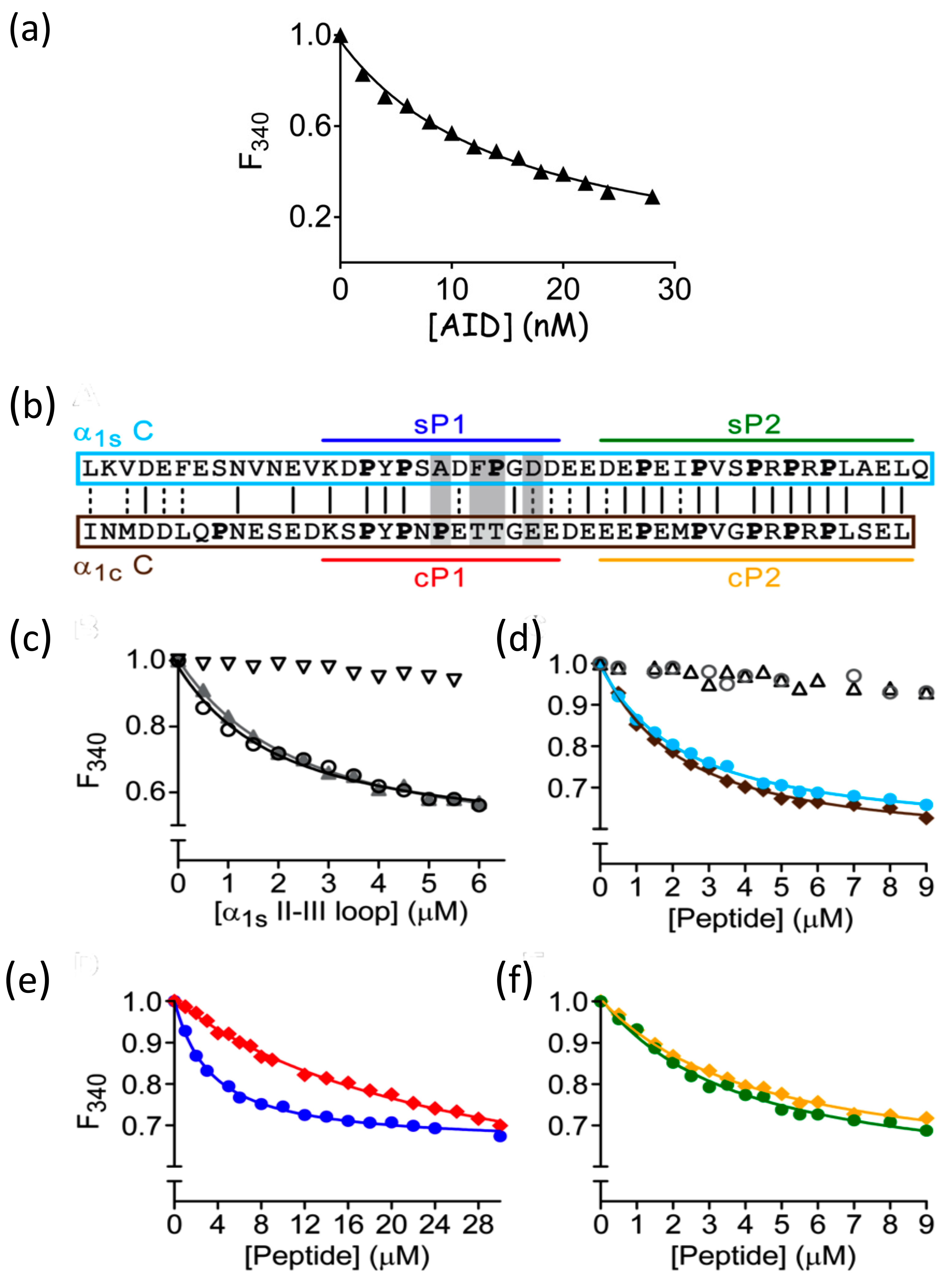
The most characterized interaction between DHPR β subunits and the DHPR α1 I-II loop to date involves the AID region binding to the DHPR β subunit with nanomolar affinity [24,25,37]. Because full-length DHPR β subunits are prone to aggregation [37], the majority of these previous studies have utilized truncated or modified β subunit constructs. In the case of the full-length DHPR β subunit, this makes some of the more common binding techniques such as Isothermal Titration Calorimetry (ITC) impracticable. However, because of the intrinsic fluorescence properties of full-length β1a and β2a, their interactions with α1s-subunit fragments can be measured using sub-micromolar concentrations of the β subunits, thereby minimizing protein aggregation. Fluorescence binding (or fluorescence quenching) is a highly sensitive technique that has been used extensively in our studies and by others to probe protein–protein interactions [28,38,39]. The affinity of AID peptide and full-length β1a was measured, with fluorescence emission monitored at 340 nm. After correction of the data for dilution effects and intrinsic fluorescence of the AID peptide, a titration curve was generated (Figure 2a) and fitted using an exponential-based equation [28] to yield a dissociation constant (Kd) of 15.2 ± 1.8 nM, which falls within the previous range of results using truncated versions of the DHPR β subunit [24,25] and indicates that full-length β1a is correctly folded.
Therefore, a series of fluorescence binding experiments were performed with the β1a to examine its potential binding affinity with the DHPR α1s II-III loop and its peptide segments. The results are displayed in Figure 2 and summarized in Table 1. Significantly, a Kd value of 2.5 ± 0.4 μM was observed between β1a and the DHPR II-III loop. A comparable affinity (2.6 ± 0.2 μM) was observed for the II-III loop binding to the β1a SH3 domain (β1a-SH3, Figure 2c and Table 1), indicating that the II-III loop binds β1a primarily through its SH3 domain. The observed micromolar binding constants are typical for polyproline–SH3-mediated interactions, which have dissociation constants in the micromolar to millimolar range [26].
To pinpoint the exact regions of the II-III loop that bind to β1a, the proline-rich C region (720–765) of the loop (α1s C) was synthesized and its ability to bind to β1a and β1a-SH3 measured (Figure 2d). Because the II-III loop is an intrinsically disordered protein [28], the structural integrity of the C region was likely to be minimally disrupted by its excision from the loop. Binding affinities of 4.9 ± 0.6 and 3.6 ± 0.6 μM were found for β1a and β1a-SH3, respectively (Table 1), suggesting that α1s C is responsible for binding to the SH3 domain of β1a. To exclude non-specific binding events, extensive control experiments were performed with a scrambled C-region peptide and a mutant C peptide, where all of the proline residues were substituted for alanine residues (Figure 2d). In both cases, no measurable binding was detected, highlighting the overall importance of the presence and the location of the proline residues.
The amino acids that make up the critical region of the skeletal II-III loop contains a PxxDY motif (residues 733–745) as well as two adjacent PXXP motifs (residues 748–764, Figure 2b), all of which are potential candidates for SH3 domain recognition [26,40]. Therefore, two shorter peptides spanning these regions were synthesized and denoted as peptides sP1 and sP2, respectively (Figure 2b), and tested against β1a and β1a-SH3. Both sP1 and sP2 peptides bind to β1a and β1a-SH3 in the low micromolar range (3–5 μM, Table 1), demonstrating that one or both of these regions contribute to the C-region–β1a SH3 interaction.
In previous dysgenic (α1s-null) mouse myotube studies, the sP1 region was identified as being of critical importance in EC coupling [8,10]. Substituting residues A739, F741, P742, and D744 with their cardiac equivalent amino acids markedly altered the skeletal voltage-dependent Ca2+ release profiles [12]. These residues are contained in the sP1 peptide, prompting us to explore the binding of the cardiac homologue of the sP1 peptide (cP1) to the β1a SH3 domain. The affinity of the cP1 peptide for β1a and β1a-SH3 was reduced compared to sP1 (4 to 7-fold, Figure 2e, and Table 1). We also found that, of the four variant residues, P742 and D744 in sP1 were independently responsible for the reduction in binding (Table 1). In contrast, the cardiac homologue of sP2 and cP2 displayed a similar affinity to its skeletal counterpart (Figure 2f). Notably, in myotube experiments, the substitution of cP2 on a skeletal II-III loop background did not impact EC coupling [12]. This result is in line with the high sequence similarity of the skeletal and cardiac P2 region compared to that observed in the P1 region (Figure 2b) and is therefore suggesting that the SP1 region of the skeletal II-III loop is most likely responsible for the molecular recognition of the β1a subunit.
In light of the interaction profiles associated with the β1a subunit, it was of interest to examine whether these α1s polyproline-rich peptides could bind to the cardiac β2a isoform. Figure 3a shows that no measurable binding was detected between β2a and α1s C. A comparison of the SH3 domain sequences for each of the four β isoforms reveals that, despite good overall sequence conservation, there is significant variation in the β1a RT loop relative to the other isoforms [37]. Therefore, a β1a chimera was designed with the β1a RT loop replaced with the β2a RT loop. Accurate fitting of a binding curve was not possible; however, it is clear there is weak, residual binding of this chimeric protein to α1s C (Figure 3a).
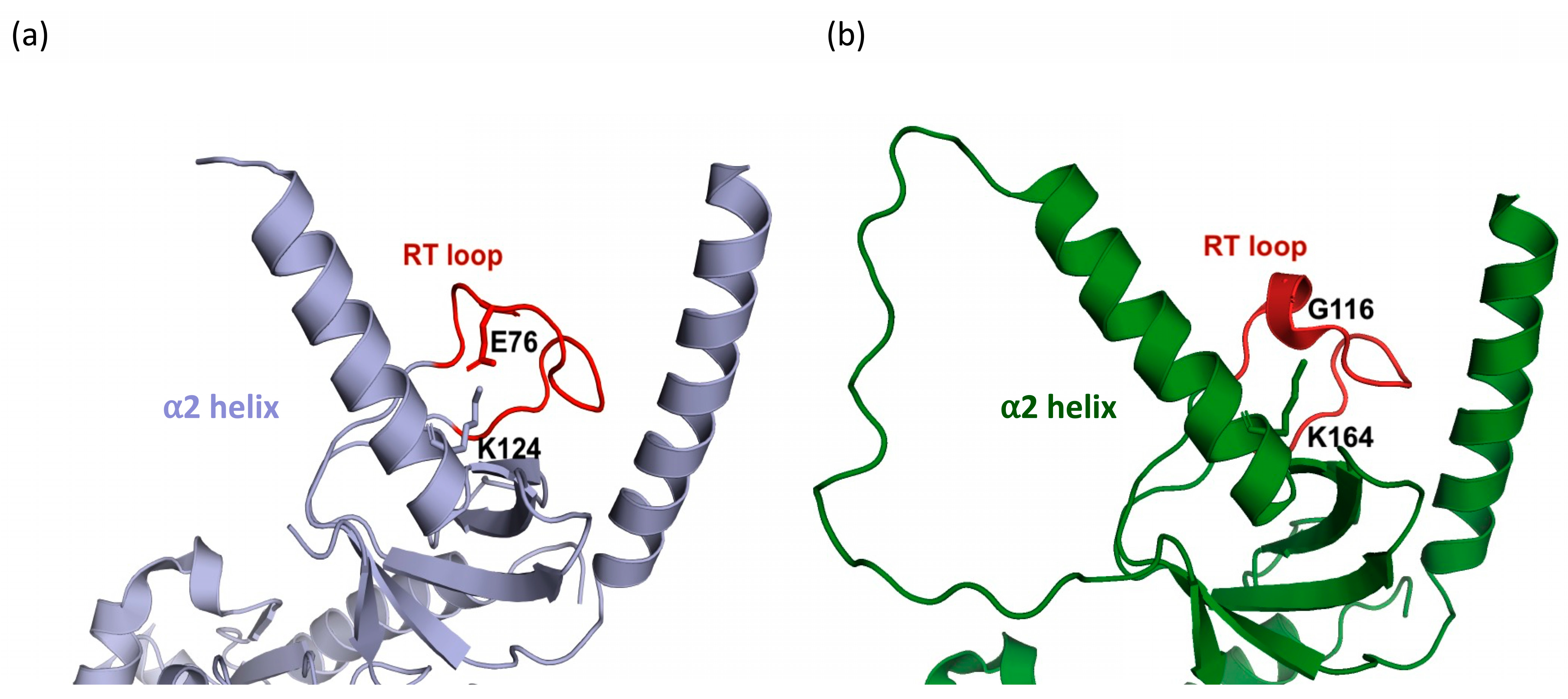
In three of the β-subunit structures, β2a, β3, and β4, the α2 helix and RT loop are linked via a salt bridge [15,16,17] (Figure 4a). The presence of this salt bridge stabilizes these two structural elements so as to position them over and occlude the α1s C binding site region. The participating positively charged residue in the α2 helix is conserved in all of these three β subunit isoforms. However, in β1a, the equivalent negatively charged residue in the RT loop is replaced by a glycine and cannot form an equivalent salt bridge (Figure 4b). Therefore, the residues P115 and G116 present in the RT loop of β1a were reintroduced into the β2a protein as well as into the weakly binding chimeric β1a protein (β1a background with β2a RT loop). In both instances, binding of the α1s C peptide was enhanced with affinities of ~ 8 and 10 μM (Figure 3b, Table 3).

Calcium Release and L-Type Channel Function

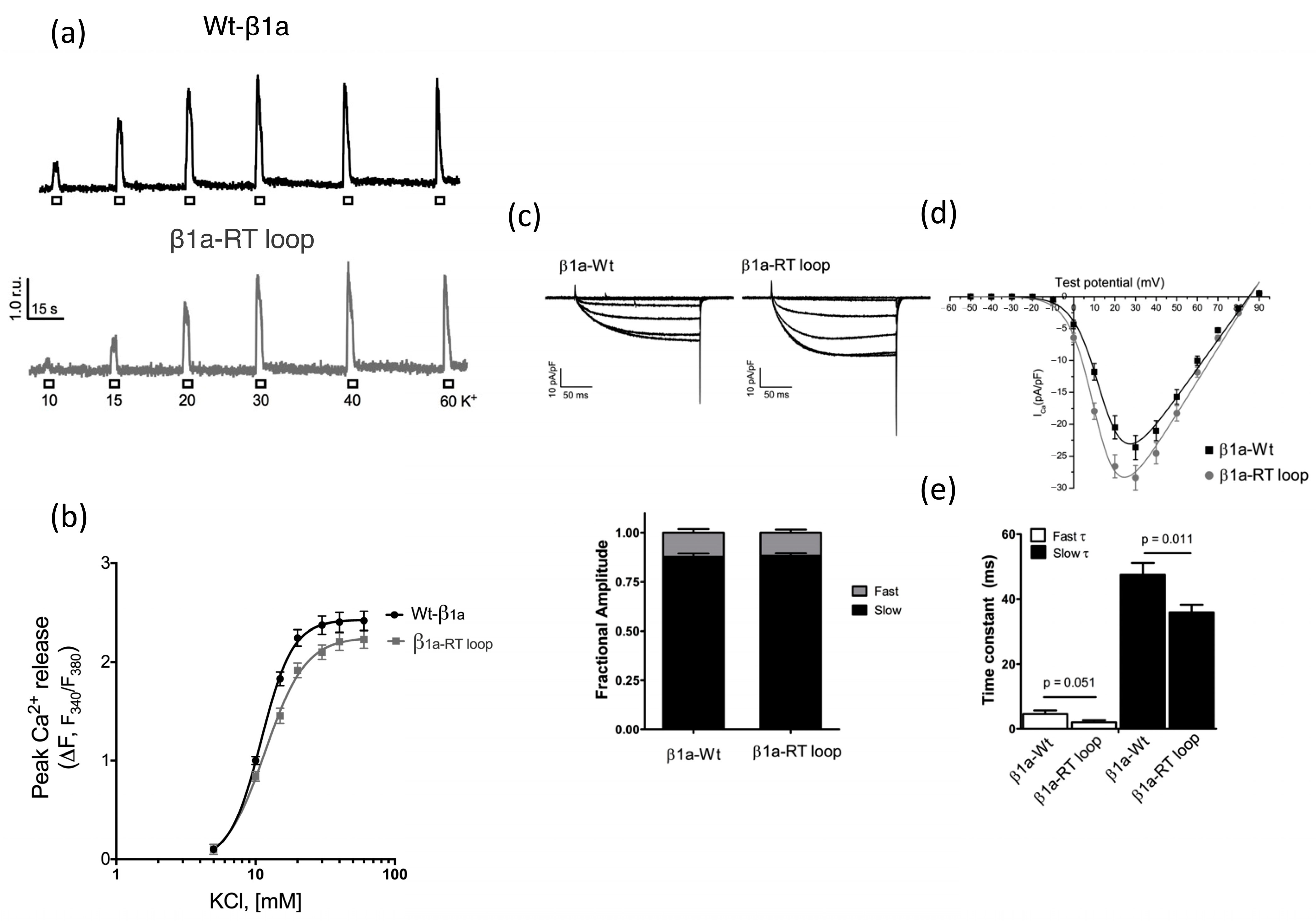
The effect of swapping the β1a RT loop for its equivalent counterpart, β2a, was analyzed using depolarization-induced Ca2+ release and whole-cell patch clamp experiments in cultured myotubes. Figure 5a,b, show representative KCl dose–response curves of Fura-2-AM-loaded for myotubes expressing either WT- or the β1a-RT-loop construct. Investigations of these phenotypes indicate that there is a modest but discernible reduction in maximal peak Ca2+ amplitude and a shift in the EC50 value from 6.68 ± 0.45 mM (WT) to 9.42 ± 0.53 mM for the RT loop. The level of SR calcium load of cultured cells was also compared and showed that the RT-loop mutation did not affect the SR calcium content.
Whole-cell patch-clamp techniques were employed to examine the effects of the β1a RT-loop mutation on DHPR channel function. Comparison of Ca2+ current recording shows that WT-β1a and β1a-RT-loop-expressing myotubes display characteristically slow-activating Ca2+ currents (Figure 5c). Further analysis revealed β1a-RT-loop-expressing myotubes displayed Ca2+ currents that were higher than WT-β1a myotubes in peak amplitude magnitude of L-type conductance (Gmax) (Figure 5c,d). In addition, average voltage dependence of the channel activation (V1/2) in β1a RT-loop-expressing myotubes was shifted by ~4 mV to more negative potential (Figure 5d and Table 2, p = 0.0051). Further analysis of the current kinetics of WT-β1a and β1a-RT-loop-expressing myotubes were studied by fitting currents to a biexponential curve and the corresponding slow and fast values at 30 mV plotted as means SE (Figure 5e). A key point of difference between the two myotubes is that the channels activated more quickly in the β1a-RT-loop-expressing myotubes.

4. Discussion

In contrast to other DHPR β-subunit isoforms, β1a is expressed only in skeletal muscle, where it is an exclusive partner of DHPR α1s. It is essential in the functional assembly of skeletal muscle triads and is required to form DHPR tetrads [41]. It has been long recognized that the I-II loop of the DHPR α-subunit is capable of a high-affinity interaction with all beta isoforms [24,25], but it has been flagged that other secondary binding sites may also exist for the SH3 and GK subunits [42]. While the β1a sequence of the N and C-termini and the hook regions are quite distinct from other β isoforms, the overall SH3 domain sequence is conserved; however, within this domain, the RT-loop region shows the greatest sequence variability [37]. This led us to speculate that the properties of this loop may confer different binding properties for the various beta isoforms. In this study, we probed the in vitro binding of the polyproline-rich DHPR II-III loop of the α1s subunit with β1a.
By performing in vitro fluorescence binding experiments, we have measured affinities between the β1a SH3 domain and two polyproline regions located on the α1s II-III loop. One of these regions (sP1) has been previously identified in mouse myotube studies as a central component associated with EC coupling [12]. We showed that the equivalent cardiac segment (cP1) binds with lower affinity to β1a-FL compared to its skeletal counterpart (sP1) and that the equivalent cardiac single point mutations, P742 and D744, in sP1 resulted in reduced binding affinities, consistent with the myotube data showing that these residues affected orthograde coupling [12]. It is noteworthy that human mutations involving the residue P742 (P741Q/S) were reported to cause congenital myopathy [43], indicating a physiological impact for this residue.
The second region of interaction involves two contiguous PXXP motifs that are present in both skeletal and cardiac isoforms of the II-III loop (sP2 and cP2). In myotube studies, interchanging sP2 and cP2 did not impact orthograde coupling [12]. The fact that the α1c region of the II-III loop cannot substitute for α1s in reconstituting skeletal muscle EC coupling [9] may indicate that both regions (sP1 and sP2) are required for skeletal muscle EC coupling. Indeed, the sP2 region has been shown to be the primary binding region for the SH3 domains of STAC3 [11], suggesting that the critical C region of the II-III loop may have multiple interaction sites. Interestingly, the fact that, under in vitro conditions, the binding of sP2 and sP1 to β1a both occur highlights the promiscuous nature of polyproline elements binding to SH3 domains—a feature not uncommon for the SH3 binding domain [44]. The precise sequence of binding events leading up to and during EC coupling remains to be determined; however, one can speculate that, in an in vivo setting, there is potential for the polyproline motifs in the II-III loop C region to interact with multiple partners.
Another question that is addressed in this study is whether the composition of the β1a SH3 domain plays an isoform-specific role in the in vitro interaction of the II-III loop. One notable difference between the β1a and RT loops of other isoforms is the presence of Gly116 (β1a—numbering), which is substituted in the other isoforms by a conserved Glu or Asp residue. In some of the X-ray crystal structures complexed to the AID peptide, these acidic acid residues participate in the formation of a salt bridge to a conserved basic residue located in the α2-helix and most likely contributes to the stabilization of the occluded polyproline binding site [37]. The absence of this salt bridge in the β1a isoform is likely to increase the dynamic mobility of the RT loop and the α2 helix and enhance its ability to bind to the polyproline motifs. Indeed, binding to α1s C was increased to 8–10 μM when the His-Glu residues in β2a and the β1a2a RT-loop chimera were replaced by the corresponding Pro-Gly residues in β1a. This enhancement in binding is not unprecedented, with a previous report showing that the introduction of a glycine residue within an SH3 domain RT loop enhances its flexibility and increases the domain’s affinity to its polyproline binding partner [45].
There are multiple factors that may influence the accessibility of the β1a SH3 binding site. For instance, there are some very obvious differences between the constructs used for crystallization and other studies. The beta constructs used for crystallization have been heavily modified to promote crystal formation relative to full length β1a. Notably, two of the truncated regions in the crystal structure constructs (N-terminal and hook) are directly adjacent to the α1 and α2 helices, which may impact their conformation and SH3 domain binding. Indeed, varying the composition of the hook region of a MAGUK protein has been shown to alter the binding properties of the GK domain via the SH3 domain [46]. It was concluded from this that the SH3 modulates the GK domain allosterically to control its function.
Despite numerous attempts to crystallize a truncated version of β1a by itself, crystallization was only possible in the presence of a CaV AID peptide [37], whereas AID was absent for full-length β1a fluorescence binding experiments. The potential for allosteric changes elicited by AID binding has been flagged in our previous study, demonstrated by a major increase in the melting temperature of β1a arising from the addition of AID [37]. This increase in melting temperature was affected by the substitution of the β1a SH3 RT loop with that of β2a, suggesting that conformational changes at one end of β1a can be transmitted throughout the molecule. Moreover, the binding affinity of the AID peptide region was shown to increase by a factor of 2 to 3-fold [37] when the β2a SH3 domain was substituted with that of β1a. What can be concluded from this series of observations is that, despite the fact that AID binding occurs exclusively through contact with the GK domain of β1a, modification or binding with the SH3 domain can allosterically influence the nature of this interaction.
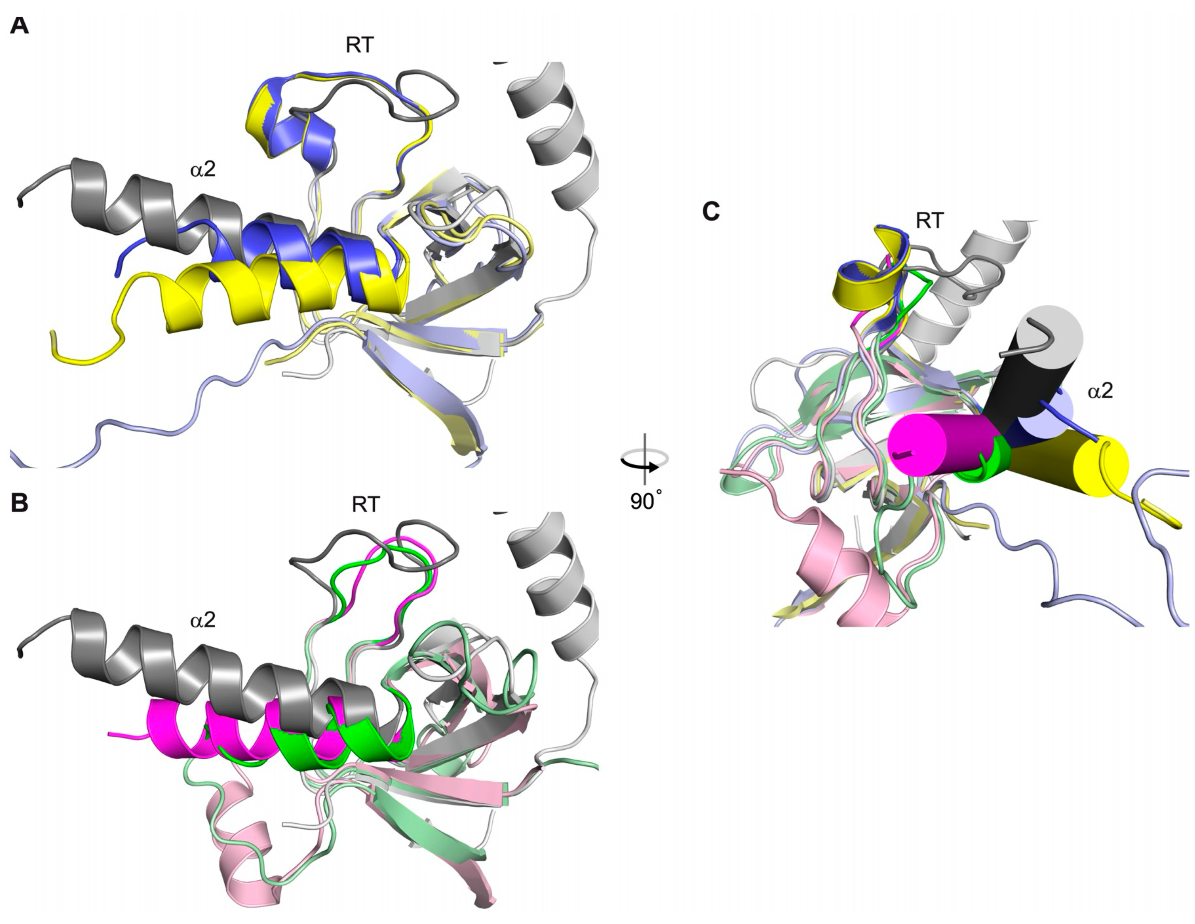
Comparison of DHPR β1a with other members of the MAGUK family, PSD-95, and ZO-1 reveals that their SH3 binding sites are capable of binding PXXP motif-containing proteins [47,48,49]. In the crystal structures of PSD-95 and ZO-1 so far determined [50,51,52], non-covalent interactions (observed in the β-isoforms) connecting the RT loop and the α2 helix are absent, and, importantly, the RT loop does not occlude either SH3 binding site. Also, the position α2 helix of PSD-95 and ZO-1 is highly variable compared to the beta structures, indicating that this helix is mobile (Figure 6). Therefore, the SH3 domains of PSD-95 and ZO-1, which are functionally and structurally similar to β1a SH3, also support the alternative positioning of both the RT loop and α2 helix, indicating that the polyproline motif binding site can become exposed for this family of proteins.

4.1. Functional Roles of the β1a SH3 Domain in Skeletal Muscle EC Coupling

The in vivo roles of the β1a SH3 domain have been previously explored by swapping this domain with that of other isomers. One of the early DHPR beta myotube studies [21] reported that simultaneously interchanging the N-terminal, hook region, and SH3 domain of β1a with those of β2a resulted in no discernible difference in skeletal-like EC coupling. In another study carried out in zebra fish [22], SH3 domain substitution (β1a to β3) resulted in a substantial reduction in charge movement and calcium release (>50%), while a comparable study involving substitution of the β4 SH3 domain resulted in no detectable effect on charge movement or calcium release [20]. In this study, we have shown in myotubes that on a β1a background, substitution of the β1a SH3 RT loop with that of β2a resulted in a subtle but detectable change in the calcium release profile as well as an effect upon the DHPR channel function. For these two sets of experiments, these changes included a decrease in maximal Ca2+ release, while an increase was observed in Gmax for the Ca2+ current experiments. From these results, the data suggest that swapping the β1a SH3 RT-loop segment has only minor effects for both orthograde and retrograde signalling. Although it is difficult to reconcile these results with our in vitro affinity measurements, one cannot discount the possibility that, under physiological conditions, the salt bridge involving the β2a RT loop may be weakened, enabling the SH3 domain binding site to engage with the II-III loop.
The DHPR II-III loop residues (Ala739, Phe741, Pro742, and Asp 744) have been previously identified as being important in orthograde and retrograde coupling [12]. Our binding results show that a segment peptide comprising these II-III loop residues (SP1) interacts with the β1a SH3 domain and that binding can be affected individually or collectively by altering these residues. While this series of observations does not definitively prove that the II-III loop and the β1a SH3 domain participates in in vivo interactions, they do show that subtle variations in the composition of the SH3 domain plays a role in skeletal muscle EC coupling. A study employing a functionalised nanobody targeting β1 (nb.E8) was used to explore its effect on voltage-gated calcium channels [53]. The binding site for this nanobody was located at the SH3 domain of β1, with the structural complex revealing direct interactions with the RT loop and α2 helix. Addition of the nb.E8 antibody to whole cells was found to inhibit the DHPR current amplitude and modulate gating properties, supporting the proposition that the disruption of endogenous modulators (i.e., II-III loop) at this region may impact EC coupling.

4.2. Structural Relationship Between β1a and the DHPR II-III Loop

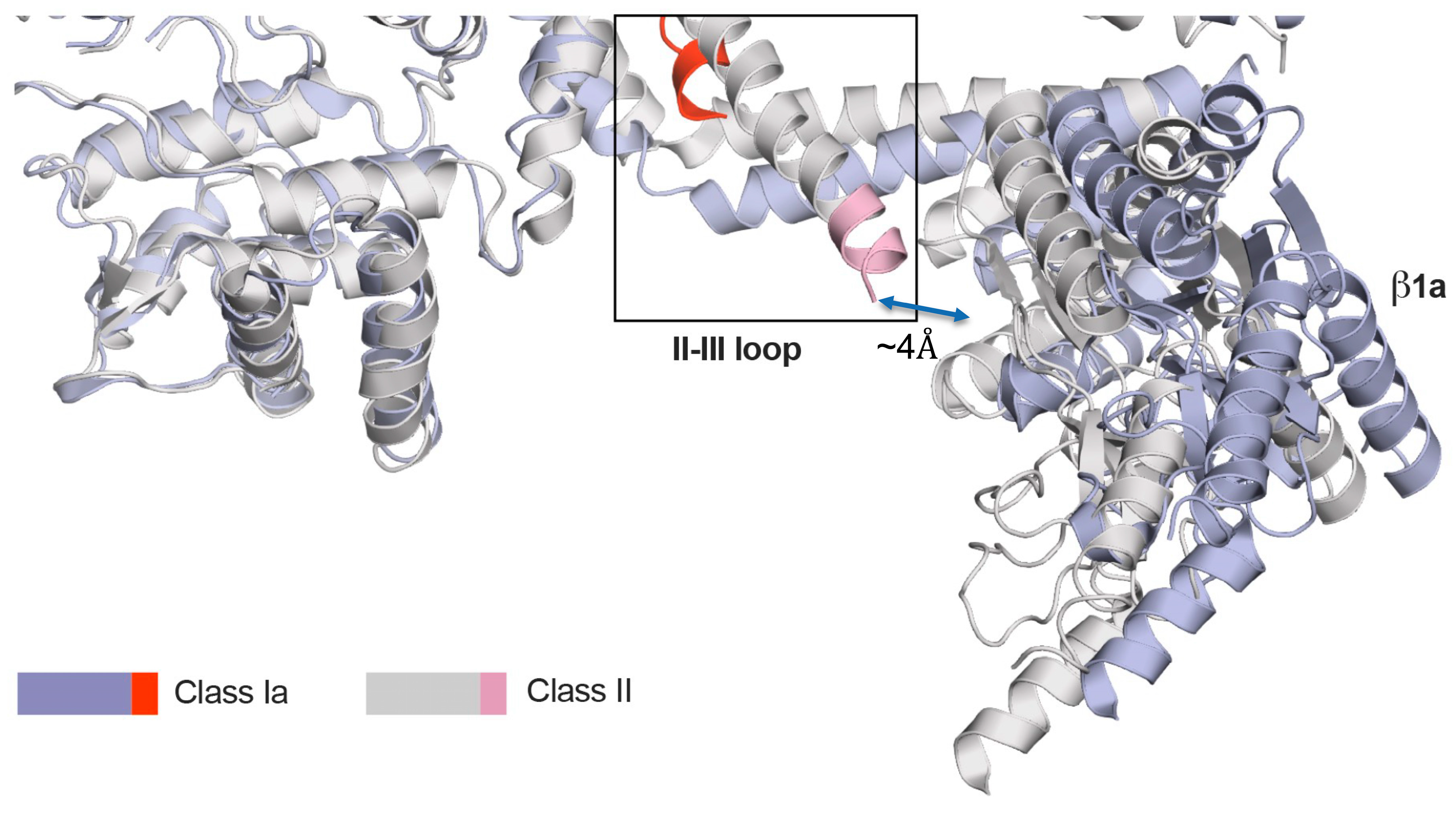
Due to the disordered nature of the II-III loop, no definitive structural evidence demonstrating the direct interaction of the β1a SH3 domain with the II-III loop is available. However, it has been noted through a FRET study that, within the triad environment, the presence of β1a modulates the α1s II-III conformation [54]. Other indirect support of this association can be found in the cryo-em structure of CaV1.1 (skeletal DHPR), where the N-terminal II-III loop helix was noted to be adjacent (<4Å) to the α9 helix of the β1a GK domain [55] (Figure 7), thus placing the β1a SH3 domain and C region of the II-III loop in close enough proximity to interact. Lastly, a recently published article on the role of STAC3 SH3 domains revealed that, even though its interaction with the C region of the II-III loop is very important in supporting skeletal muscle EC coupling, the deletion of these domains proved to not be essential [56]. This contrasts with the deletion or substitution of the C region of the DHPR II-III loop, which has been determined to be absolutely critical for skeletal muscle EC coupling [8,10,12]. The fact that these reciprocal experiments designed to both abolish the II-III loop/STAC3 interaction resulted in different skeletal muscle EC coupling effects lends weight to the argument there may be another binding partner of the DHPR II-III loop C region that at least supports EC coupling.

5. Conclusions

Our results indicate that the SH3 domain of β1a recognizes polyproline elements located in the cytoplasmic α1s DHPR II-III loop, one of which has been demonstrated to be important for skeletal muscle EC coupling. In vitro affinity experiments using a combination of skeletal and cardiac beta and alpha subunit isoforms reveal that the interaction is influenced by the amino acid composition of the various isoforms. The findings presented here offer a unique insight into the potential signalling pathway that may contribute to skeletal muscle EC coupling. Whereas the binding partner of the α1s II-III loop was previously ascribed to STAC3, we show that the SH3 domain β1a subunit may form part of the communication network between the DHPR voltage-sensing subunit, leading to RyR1 activation.

Supplementary Materials

The following supporting information can be downloaded at: https://www.mdpi.com/article/10.3390/biom15111610/s1, Figure S1: Purified recombinant proteins used for binding experiments; Figure S2: Circular dichroism (CD) spectra of β1a subunits—(A) CD spectrum of the purified and refolded full length β1a protein. The spectrum shows a maximum at 191 nm followed by minima at 209 and 226 nm. (B) CD spectrum of the purified and refolded full length SH3 domain b1a protein (101–272).

Author Contributions

Conceptualization, M.G.C.; Methodology, M.G.C., P.G.B., J.M.E. and C.F.P.; Software, M.G.C. and Y.K.; Validation, M.G.C. and Y.K.; Formal Analysis, Y.K., S.A., J.M.E. and C.F.P.; Investigation, Y.K., S.A., J.C., J.M.E. and C.F.P.; Resources, M.G.C., A.F.D., P.G.B., J.M.E. and C.F.P., Data Curation, M.G.C., N.C.N., S.A., Y.K., J.M.E. and C.F.P.; Writing—Original Draft Preparation, M.G.C. and Y.K.; Writing—Review and Editing, M.G.C., A.F.D., P.G.B., J.M.E. and C.F.P.; Visualization, M.G.C., N.C.N., S.A. and Y.K.; Supervision, M.G.C., A.F.D. and P.G.B.; Project Administration, M.G.C. and A.F.D.; Funding Acquisition, M.G.C., A.F.D. and P.G.B. All authors have read and agreed to the published version of the manuscript.

Funding

This work was supported by Project Grant APP1126201 to MGC, ADF & PGB from the National Health and Medical Research Council of Australia.

Institutional Review Board Statement

All animal procedures were carried out in accordance with the guidelines of Harvard Medical Area Standing Committee on Animals under, experimental protocol numbers 2589, approved on 1 September 2014.

Informed Consent Statement

Not applicable.

Data Availability Statement

The original contributions presented in this study are included in the article/Supplementary Materials. Further inquiries can be directed to the corresponding author.

Acknowledgments

Open access publishing facilitated by Australian National University—Australian National University agreement via the Council of Australian University Librarians.

Conflicts of Interest

The authors declare no conflicts of interest.

References

  1. Beam, K.G.; Bannister, R.A. Looking for Answers to Ec Coupling’s Persistent Questions. J. Gen. Physiol. 2010, 136, 7–12. [Google Scholar] [CrossRef] [PubMed]
  2. Perni, S.; Lavorato, M.; Beam, K.G. De Novo Reconstitution Reveals the Proteins Required for Skeletal Muscle Voltage-Induced Ca(2+) Release. Proc. Natl. Acad. Sci. USA 2017, 114, 13822–13827. [Google Scholar] [CrossRef]
  3. Beurg, M.; Ahern, C.A.; Vallejo, P.; Conklin, M.W.; Powers, P.A.; Gregg, R.G.; Coronado, R. Involvement of the Carboxy-Terminus Region of the Dihydropyridine Receptor Beta1a Subunit in Excitation-Contraction Coupling of Skeletal Muscle. Biophys. J. 1999, 77, 2953–2967. [Google Scholar] [CrossRef]
  4. Horstick, E.J.; Linsley, J.W.; Dowling, J.J.; Hauser, M.A.; McDonald, K.K.; Ashley-Koch, A.; Saint-Amant, L.; Satish, A.; Cui, W.W.; Zhou, W.; et al. Stac3 Is a Component of the Excitation-Contraction Coupling Machinery and Mutated in Native American Myopathy. Nat. Commun. 2013, 4, 1952. [Google Scholar] [CrossRef]
  5. Schredelseker, J.; Di Biase, V.; Obermair, G.J.; Felder, E.T.; Flucher, B.E.; Franzini-Armstrong, C.; Grabner, M. The Beta 1a Subunit Is Essential for the Assembly of Dihydropyridine-Receptor Arrays in Skeletal Muscle. Proc. Natl. Acad. Sci. USA 2005, 102, 17219–17224. [Google Scholar] [CrossRef] [PubMed]
  6. Perni, S.; Beam, K. Junctophilins 1, 2, and 3 All Support Voltage-Induced Ca2+ Release Despite Considerable Divergence. J. Gen. Physiol. 2022, 154, e202113024. [Google Scholar] [CrossRef] [PubMed]
  7. Yang, Z.F.; Panwar, P.; McFarlane, C.R.; Tuinte, W.E.; Campiglio, M.; Van Petegem, F. Structures of the Junctophilin/Voltage-Gated Calcium Channel Interface Reveal Hot Spot for Cardiomyopathy Mutations. Proc. Natl. Acad. Sci. USA 2022, 119, e2120416119. [Google Scholar] [CrossRef]
  8. Nakai, J.; Tanabe, T.; Konno, T.; Adams, B.; Beam, K.G. Localization in the II-III Loop of the Dihydropyridine Receptor of a Sequence Critical for Excitation-Contraction Coupling. J. Biol. Chem. 1998, 273, 24983–24986. [Google Scholar] [CrossRef]
  9. Grabner, M.; Dirksen, R.T.; Suda, N.; Beam, K.G. The II-III Loop of the Skeletal Muscle Dihydropyridine Receptor Is Responsible for the Bi-Directional Coupling with the Ryanodine Receptor. J. Biol. Chem. 1999, 274, 21913–21919. [Google Scholar] [CrossRef]
  10. Wilkens, C.M.; Kasielke, N.; Flucher, B.E.; Beam, K.G.; Grabner, M. Excitation-Contraction Coupling Is Unaffected by Drastic Alteration of the Sequence Surrounding Residues L720-L764 of the Alpha 1s II-III Loop. Proc. Natl. Acad. Sci. USA 2001, 98, 5892–5897. [Google Scholar] [CrossRef]
  11. Yuen, S.M.W.K.; Campiglio, M.; Tung, C.-C.; Flucher, B.E.; Van Petegem, F. Structural Insights into Binding of Stac Proteins to Voltage-Gated Calcium Channels. Proc. Natl. Acad. Sci. USA 2017, 114, E9520–E9528. [Google Scholar]
  12. Kugler, G.; Weiss, R.G.; Flucher, B.E.; Grabner, M. Structural Requirements of the Dihydropyridine Receptor Alpha1s II-III Loop for Skeletal-Type Excitation-Contraction Coupling. J. Biol. Chem. 2004, 279, 4721–4728. [Google Scholar] [CrossRef]
  13. Bichet, D.; Lecomte, C.; Sabatier, J.M.; Felix, R.; De Waard, M. Reversibility of the Ca(2+) Channel Alpha(1)-Beta Subunit Interaction. Biochem. Biophys. Res. Commun. 2000, 277, 729–735. [Google Scholar] [CrossRef]
  14. Gerster, U.; Neuhuber, B.; Groschner, K.; Striessnig, J.; Flucher, B.E. Current Modulation and Membrane Targeting of the Calcium Channel Alpha1c Subunit Are Independent Functions of the Beta Subunit. J. Physiol. 1999, 517 Pt 2, 353–368. [Google Scholar] [CrossRef]
  15. Chen, Y.H.; Li, M.H.; Zhang, Y.; He, L.L.; Yamada, Y.; Fitzmaurice, A.; Yang, J. Structural Basis of the Alpha1-Beta Subunit Interaction of Voltage-Gated Ca2+ Channels. Nature 2004, 429, 675–680. [Google Scholar] [CrossRef] [PubMed]
  16. Opatowsky, Y.; Chen, C.-C.; Campbell, K.P.; Hirsch, J.A. Structural Analysis of the Voltage-Dependent Calcium Channel Beta Subunit Functional Core and Its Complex with the Alpha 1 Interaction Domain. Neuron 2004, 42, 387–399. [Google Scholar] [CrossRef]
  17. Van Petegem, F.; Clark, K.A.; Chatelain, F.C.; Minor, D.L., Jr. Structure of a Complex between a Voltage-Gated Calcium Channel Beta-Subunit and an Alpha-Subunit Domain. Nature 2004, 429, 671–675. [Google Scholar] [CrossRef]
  18. Buraei, Z.; Yang, J. The Beta-Subunit Subunit of Voltage-Gated Ca2+ Channels. Physiol. Rev. 2010, 90, 1461–1506. [Google Scholar] [CrossRef] [PubMed]
  19. Cheng, W.; Altafaj, X.; Ronjat, M.; Coronado, R. Interaction between the Dihydropyridine Receptor Ca2+ Channel Beta-Subunit and Ryanodine Receptor Type 1 Strengthens Excitation-Contraction Coupling. Proc. Natl. Acad. Sci. USA 2005, 102, 19225–19230. [Google Scholar] [CrossRef] [PubMed]
  20. Dayal, A.; Perni, S.; Franzini-Armstrong, C.; Beam, K.G.; Grabner, M. The Distal C Terminus of the Dihydropyridine Receptor Beta(1a) Subunit Is Essential for Tetrad Formation in Skeletal Muscle. Proc. Natl. Acad. Sci. USA 2022, 119, e2201136119. [Google Scholar] [CrossRef]
  21. Sheridan, D.C.; Cheng, W.; Ahern, C.A.; Mortenson, L.; Alsammarae, D.; Vallejo, P.; Coronado, R. Truncation of the Carboxyl Terminus of the Dihydropyridine Receptor Beta1a Subunit Promotes Ca2+ Dependent Excitation-Contraction Coupling in Skeletal Myotubes. Biophys. J. 2003, 84, 220–237. [Google Scholar] [CrossRef]
  22. Dayal, A.; Bhat, V.; Franzini-Armstrong, C.; Grabner, M. Domain Cooperativity in the Beta1a Subunit Is Essential for Dihydropyridine Receptor Voltage Sensing in Skeletal Muscle. Proc. Natl. Acad. Sci. USA 2013, 110, 7488–7493. [Google Scholar] [CrossRef] [PubMed]
  23. Dolphin, A.C. Beta Subunits of Voltage-Gated Calcium Channels. J. Bioenerg. Biomembr. 2003, 35, 599–620. [Google Scholar] [CrossRef]
  24. Richards, M.W.; Butcher, A.J.; Dolphin, A.C. Ca2+ Channel Beta-Subunits: Structural Insights Aid Our Understanding. Trends Pharmacol. Sci. 2004, 25, 626–632. [Google Scholar] [CrossRef]
  25. Van Petegem, F.; Duderstadt, K.E.; Clark, K.A.; Wang, M.; Minor, D.L. Alanine-Scanning Mutagenesis Defines a Conserved Energetic Hotspot in the Cavalpha1 Aid-Cavbeta Interaction Site That Is Critical for Channel Modulation. Structure 2008, 16, 280–294. [Google Scholar] [CrossRef] [PubMed]
  26. Ball, L.J.; Kühne, R.; Schneider-Mergener, J.; Oschkinat, H. Recognition of Proline-Rich Motifs by Protein-Protein-Interaction Domains. Angew. Chem. Int. Ed. Engl. 2005, 44, 2852–2869. [Google Scholar] [CrossRef] [PubMed]
  27. Catanzariti, A.; Soboleva, T.A.; Jans, D.A.; Board, P.G.; Baker, R.T. An Efficient System for High-Level Expression and Easy Purification of Authentic Recombinant Proteins. Protein Sci. 2004, 13, 1331–1339. [Google Scholar] [CrossRef]
  28. Cui, Y.; Tae, H.S.; Norris, N.C.; Karunasekara, Y.; Pouliquin, P.; Board, P.G.; Dulhunty, A.F.; Casarotto, M.G. A Dihydropyridine Receptor Alpha1s Loop Region Critical for Skeletal Muscle Contraction Is Intrinsically Unstructured and Binds to a Spry Domain of the Type 1 Ryanodine Receptor. Int. J. Biochem. Cell Biol. 2009, 41, 677–686. [Google Scholar] [CrossRef]
  29. Eltit, J.M.; Franzini-Armstrong, C.; Perez, C.F. Amino Acid Residues 489–503 of Dihydropyridine Receptor (Dhpr) Beta1a Subunit Are Critical for Structural Communication between the Skeletal Muscle Dhpr Complex and Type 1 Ryanodine Receptor. J. Biol. Chem. 2014, 289, 36116–36124. [Google Scholar] [CrossRef]
  30. Beurg, M.; Sukhareva, M.; Strube, C.; Powers, P.; Gregg, R.; Coronado, R. Recovery of Ca2+ Current, Charge Movements, and Ca2+ Transients in Myotubes Deficient in Dihydropyridine Receptor Beta 1 Subunit Transfected with Beta 1 Cdna. Biophys. J. 1997, 73, 807–818. [Google Scholar] [CrossRef]
  31. Gregg, R.G.; Messing, A.; Strube, C.; Beurg, M.; Moss, R.; Behan, M.; Powers, P.A. Absence of the Beta Subunit (Cchb1) of the Skeletal Muscle Dihydropyridine Receptor Alters Expression of the Alpha 1 Subunit and Eliminates Excitation-Contraction Coupling. Proc. Natl. Acad. Sci. USA 1996, 93, 13961–13966. [Google Scholar] [CrossRef]
  32. Perez, C.F.; Eltit, J.M.; Lopez, J.R.; Bodnár, D.; Dulhunty, A.F.; Aditya, S.; Casarotto, M.G. Functional and Structural Characterization of a Novel Malignant Hyperthermia-Susceptible Variant of DHPR-Beta(1a) Subunit (Cacnb1). Am. J. Physiol. Cell Physiol. 2018, 314, C323–C333. [Google Scholar] [CrossRef] [PubMed]
  33. Eltit, J.M.; Li, H.; Ward, C.W.; Molinski, T.; Pessah, I.N.; Allen, P.D.; Lopez, J.R. Orthograde Dihydropyridine Receptor Signal Regulates Ryanodine Receptor Passive Leak. Proc. Natl. Acad. Sci. USA 2011, 108, 7046–7051. [Google Scholar] [CrossRef]
  34. Chen, Y.; Xue, S.; Zou, J.; Lopez, J.R.; Yang, J.J.; Perez, C.F. Myoplasmic Resting Ca2+ Regulation by Ryanodine Receptors Is under the Control of a Novel Ca2+-Binding Region of the Receptor. Biochem. J. 2014, 460, 261–271. [Google Scholar] [CrossRef]
  35. Eltit, J.M.; Szpyt, J.; Li, H.; Allen, P.D.; Perez, C.F. Reduced Gain of Excitation-Contraction Coupling in Triadin-Null Myotubes Is Mediated by the Disruption of Fkbp12/Ryr1 Interaction. Cell Calcium 2011, 49, 128–135. [Google Scholar] [CrossRef] [PubMed]
  36. Eltit, J.M.; Feng, W.; Lopez, J.R.; Padilla, I.T.; Pessah, I.N.; Molinski, T.F.; Fruen, B.R.; Allen, P.D.; Perez, C.F. Ablation of Skeletal Muscle Triadin Impairs Fkbp12/Ryr1 Channel Interactions Essential for Maintaining Resting Cytoplasmic Ca2+. J. Biol. Chem. 2010, 285, 38453–38462. [Google Scholar] [CrossRef] [PubMed]
  37. Norris, N.C.; Joseph, S.; Aditya, S.; Karunasekara, Y.; Board, P.G.; Dulhunty, A.F.; Casarotto, M.G. Structural and Biophysical Analyses of the Skeletal Dihydropyridine Receptor Beta Subunit Beta(1a) Reveal Critical Roles of Domain Interactions for Stability. J. Biol. Chem. 2017, 292, 8401–8411. [Google Scholar] [CrossRef]
  38. Fan, J.Y.; Rangasamy, D.; Luger, K.; Tremethick, D.J. H2a.Z Alters the Nucleosome Surface to Promote Hp1alpha-Mediated Chromatin Fiber Folding. Mol. Cell 2004, 16, 655–661. [Google Scholar] [CrossRef]
  39. Tae, H.; Norris, N.C.; Cui, Y.; Karunasekara, Y.; Board, P.G.; Dulhunty, A.F.; Casarotto, M.G. Molecular Recognition of the Disordered Dihydropyridine Receptor Ii-Iii Loop by a Conserved Spry Domain of the Type 1 Ryanodine Receptor. Clin. Exp. Pharmacol. Physiol. 2009, 36, 346–349. [Google Scholar] [CrossRef]
  40. Aitio, O.; Hellman, M.; Kesti, T.; Kleino, I.; Samuilova, O.; Pääkkönen, K.; Tossavainen, H.; Saksela, K.; Permi, P. Structural Basis of Pxxdy Motif Recognition in SH3 Binding. J. Mol. Biol. 2008, 382, 167–178. [Google Scholar] [CrossRef]
  41. Schredelseker, J.; Dayal, A.; Schwerte, T.; Franzini-Armstrong, C.; Grabner, M. Proper Restoration of Excitation-Contraction Coupling in the Dihydropyridine Receptor Beta1-Null Zebrafish Relaxed Is an Exclusive Function of the Beta1a Subunit. J. Biol. Chem. 2009, 284, 1242–1251. [Google Scholar] [CrossRef]
  42. Campiglio, M.; Di Biase, V.; Tuluc, P.; Flucher, B.E. Stable Incorporation Versus Dynamic Exchange of Beta Subunits in a Native Ca2+ Channel Complex. J. Cell Sci. 2013, 126, 2092–2101. [Google Scholar]
  43. Schartner, V.; Romero, N.B.; Donkervoort, S.; Treves, S.; Munot, P.; Pierson, T.M.; Dabaj, I.; Malfatti, E.; Zaharieva, I.T.; Zorzato, F.; et al. Dihydropyridine Receptor (Dhpr, Cacna1s) Congenital Myopathy. Acta Neuropathol. 2017, 133, 517–533. [Google Scholar] [CrossRef]
  44. Agrawal, V.; Kishan, K.R. Promiscuous Binding Nature of SH3 Domains to Their Target Proteins. Protein Pept. Lett. 2002, 9, 185–193. [Google Scholar] [CrossRef] [PubMed]
  45. Bauer, F.; Sticht, H. A Proline to Glycine Mutation in the Lck SH3-Domain Affects Conformational Sampling and Increases Ligand Binding Affinity. FEBS Lett. 2007, 581, 1555–1560. [Google Scholar] [CrossRef]
  46. Marcette, J.; Hood, I.V.; Johnston, C.A.; Doe, C.Q.; Prehoda, K.E. Allosteric Control of Regulated Scaffolding in Membrane-Associated Guanylate Kinases. Biochemistry 2009, 48, 10014–10019. [Google Scholar] [CrossRef]
  47. GGarcia, E.P.; Mehta, S.; Blair, L.A.; Wells, D.G.; Shang, J.; Fukushima, T.; Fallon, J.R.; Garner, C.C.; Marshall, J. Sap90 Binds and Clusters Kainate Receptors Causing Incomplete Desensitization. Neuron 1998, 21, 727–739. [Google Scholar] [CrossRef] [PubMed]
  48. Maximov, A.; Südhof, T.C.; Bezprozvanny, I. Association of Neuronal Calcium Channels with Modular Adaptor Proteins. J. Biol. Chem. 1999, 274, 24453–24456. [Google Scholar] [CrossRef]
  49. Ooshio, T.; Kobayashi, R.; Ikeda, W.; Miyata, M.; Fukumoto, Y.; Matsuzawa, N.; Ogita, H.; Takai, Y. Involvement of the Interaction of Afadin with Zo-1 in the Formation of Tight Junctions in Madin-Darby Canine Kidney Cells. J. Biol. Chem. 2010, 285, 5003–5012. [Google Scholar] [CrossRef] [PubMed]
  50. Lye, M.F.; Fanning, A.S.; Su, Y.; Anderson, J.M.; Lavie, A. Insights into Regulated Ligand Binding Sites from the Structure of Zo-1 Src Homology 3-Guanylate Kinase Module. J. Biol. Chem. 2010, 285, 13907–13917. [Google Scholar] [CrossRef]
  51. McGee, A.W.; Dakoji, S.R.; Olsen, O.; Bredt, D.S.; Lim, W.A.; Prehoda, K.E. Structure of the SH3-Guanylate Kinase Module from Psd-95 Suggests a Mechanism for Regulated Assembly of Maguk Scaffolding Proteins. Mol. Cell 2001, 8, 1291–1301. [Google Scholar] [CrossRef] [PubMed]
  52. Tavares, G.A.; Panepucci, E.H.; Brunger, A.T. Structural Characterization of the Intramolecular Interaction between the SH3 and Guanylate Kinase Domains of Psd-95. Mol. Cell 2001, 8, 1313–1325. [Google Scholar] [CrossRef]
  53. Morgenstern, T.J.; Nirwan, N.; Hernández-Ochoa, E.O.; Bibollet, H.; Choudhury, P.; Laloudakis, Y.D.; Colecraft, H.M. Selective Posttranslational Inhibition of Ca(V)Beta(1)-Associated Voltage-Dependent Calcium Channels with a Functionalized Nanobody. Nat. Commun. 2022, 13, 7556. [Google Scholar] [CrossRef] [PubMed]
  54. Mohana, M.; Perez, C.F.; Fessenden, J.D. Fluorescence Resonance Energy Transfer-Based Structural Analysis of the Dihydropyridine Receptor Alpha1s Subunit Reveals Conformational Differences Induced by Binding of the Beta1a Subunit. J. Biol. Chem. 2016, 291, 13762–13770. [Google Scholar] [CrossRef] [PubMed]
  55. Wu, J.; Yan, Z.; Li, Z.; Qian, X.; Lu, S.; Dong, M.; Yan, N. Structure of the Voltage-Gated Calcium Channel Ca(V)1.1 at 3.6 a Resolution. Nature 2016, 537, 191–196. [Google Scholar] [CrossRef]
  56. Tuinte, W.E.; Török, E.; Tuluc, P.; Fattori, F.; D’Amico, A.; Campiglio, M. Stac3 Binding to Cav1.1 II-III Loop Is Nonessential but Critically Supports Skeletal Muscle Excitation-Contraction Coupling. JCI Insight 2025, 10, e191053. [Google Scholar] [CrossRef]
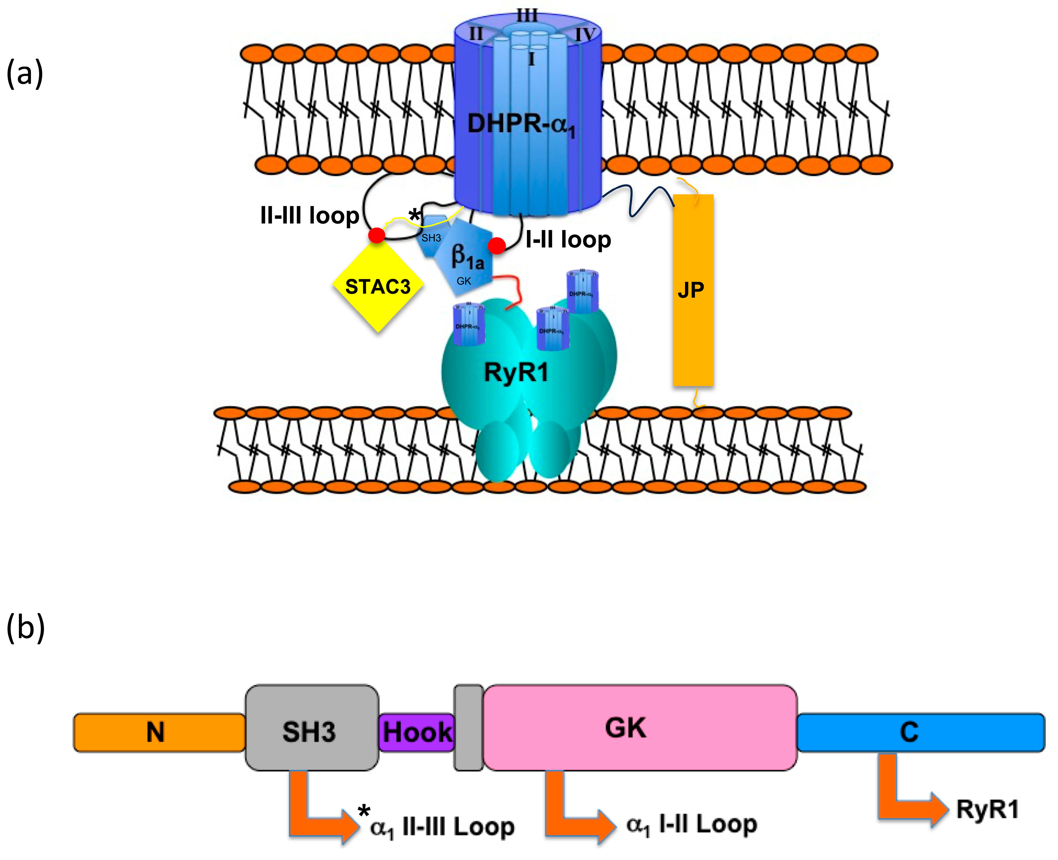
Figure 1. DHPR tetrad/RyR1 tetramer schematic representation depicting key elements involved in skeletal muscle EC coupling: DHPR, β1a, RyR1, STAC3, and junctophilin 2 (JP2). (a) Diagram expansion outlining the known interactions of DHPR I-II between the α1s and β1a subunit GK domain and between the DHPR II-III loop and STAC3 SH3 domain (depicted as red dots). The asterisk denotes the proposed interaction site between the α1s II-III loop and β1a SH3 domain. The red line depicts an interaction between the β1a C-terminal tail and RyR1(location not yet established). (b) The domain architecture of β1a illustrating a split SH3 domain (grey) and a guanylate kinase (GK) domain (pink). The arrows denote known and proposed (*) binding with interaction partners.
Biomolecules 15 01610 g001
Figure 2. Fluorescence binding measurements for DHPR β1a. (a) Fluorescence binding curve for β1a-FL and DHPR α1s I-II loop (AID) (▲). The relative fluorescence of β1a-FL at 340 nm (F340) is plotted as a function of AID peptide concentration. The concentration of β1a-FL in the cuvette was 50 nM. (b) Sequence alignment between the skeletal (α1s) and cardiac (α1c) II-III loop C region. Two polyproline SH3 binding regions are highlighted, sP1/sP2 and cP1/cP2, corresponding to DHPR α1s and α1c, respectively. The solid lines indicate conserved residues and dotted lines amino acid types with similar properties. The grey shading denotes residues that have been previously mutated and identified as important with respect to EC coupling [12]. Relative fluorescence of β1a-FL and/or its β1a-SH3 at 340 nm (F340) are plotted as a function of II-III loop peptide concentration. (c) Binding curves between β1a-FL (▲) and β1a-SH3 (○) and the DHPR II-III loop. BSA was used as the control experiment (∇). (d) Binding curves of the α1s C (●)(residues 720–765) and α1c C (◆)(residues 851–895) II-III loop C-region peptides with the β1a-FL. Control binding experiments included the scrambled skeletal C region (∆) and proline to alanine mutant (○) peptides, respectively. (e) Binding curves of the sP1(●) and cP1 (◆) peptides with the β1a-FL. (f) Binding curves of the sP2 (●) and cP2(◆) peptides with β1a-FL. The coloured bars above in panel 2a denote the location of peptide segment within the II-III loop C region.
Biomolecules 15 01610 g002
Figure 3. Fluorescence binding measurements for DHPR β2a, β1a, and β2a1a chimeras to α1s C peptide. (a) Binding curves for β2a (◇) and the β1a2a RT-loop chimera (□) with peptide α1s C. Binding between β1a-FL and α1s C is shown for comparison (○). (b) Curves for β2a (□) and the β1a2a RT-loop chimera (∇), where β2a RT-loop residues (H&E) have been mutated to β1a residues (P&G), binding to peptide α1s C.
Biomolecules 15 01610 g003
Figure 4. Interactions of β2a and β1a SH3 RT loops. (a) The X-ray crystal structure of the SH3 domain of rabbit β2a (mauve, PDB code1t3l). The α2 helix and the RT loop interact through a salt bridge involving the side chains of E76 and K124. (b) The X-ray crystal structure of the SH3 domain of rabbit β1a (green, PDB code 4zw2), showing the absence of a salt bridge due to the existence of an RT-loop glycine residue (G116).
Biomolecules 15 01610 g004
Figure 5. Ca2+ release and L-type Ca2+ current patch clamping measurements. (a) Depolarization-induced Ca2+ release of β1a subunit Wt and RT-loop constructs. Representative fluorescent traces of K-dose responses from Wt- and RT-loop-expressing myotubes. (b) Average peak fluorescent amplitude of KCl-induced Ca2+ release transients observed in Fura-2 loaded myotubes expressing Wt (black) and RT loop (grey). Data are expressed as mean SE and were fitted to a sigmoidal dose–response curve. (c) Representative traces of the L-type Ca2+ current in β-null myocytes expressing the β1a-Wt or β1a-RT-loop constructs. (d) Comparison of current –voltage relationship (I-V), myocytes where depolarized from a holding potential of −50 mV, and 200 ms pulses were applied from −40 mV to +90 mV in 10 mV increments. (e) Current kinetics were determined by fitting the current recorded at +30 mV (maximal current density) to a biexponential equation; fractional amplitude of the fast and slow components of the current were identical between cell types, but the onset of the current was faster for the β1a-RT-loop-expressing myotubes. Data is presented as mean ± SE of n = 22 (Wt-β1a) and n = 21 (β1a-RT loop) recordings of at least 3 independent days.
Biomolecules 15 01610 g005
Figure 6. Structural comparison of MAGUK SH3 domains. The SH3 domain of β2a (grey, pdb code 1t3l) was aligned with the SH3 domains of either (A) two PSD-95 structures (yellow and blue, pdb code 1jxo and 1kjw, respectively) or (B) ZO-1(green, pdb code3lh5) and ZO-3 (magenta, pbd code 3kfv). In (A,B) for all structures, the α2 helix and RT loop are dark coloured, the rest of the SH3 domain is light. (C) SH3 β2a domain aligned with those of PSD-95, ZO-1, and ZO-3, with the structures rotated 90° with respect to (A,B). The helix is shown as a cylinder. Colouring is the same as for (A,B).
Biomolecules 15 01610 g006
Figure 7. Cryo-EM structures of Cav1.1 reveals two conformational states. Overlay of two conformational states class Ia and class II, 5GJV, and 5GJW, respectively. Superposition of the DHPR II-III loop (red and light red) shows the relocation and extension of helix IIS6 (boxed). The II-III loop in conformation II is in close proximity (<4Å) to the α9 helix of the β1a GK domain.
Biomolecules 15 01610 g007
Table 1. Binding affinities of selected skeletal and cardiac DHPR II-III loop peptides to β1a and the β1a SH3 domain as measured by tryptophan fluorescence quenching.
DHPR II-III Fragmentβ1a Subunit (μM)β1a–SH3 Domain (Residues 101–272) (μM)
α1s DHPR II-III Loop2.5 ± 0.42.6 ± 0.2
α1s II–III loop C region (α1s C)4.9 ± 0.63.6 ± 0.6
α1c II–III loop C region (α1c C)3.5 ± 0.42.5 ± 0.2
Scrambled α1s CNo bindingNo binding
Mutated α1s C All P⟶ANo bindingNo binding
α1s C binding peptide 1 (sP1)3.6 ± 0.22.6 ± 0.4
sP1 A⟶P mutant4.5 ± 0.32.3 ± 0.2
sP1 F⟶T mutant4.6 ± 0.44.6 ± 0.3
sP1 P⟶T mutant22 ± 219 ± 2
sP1 D⟶E mutant15 ± 217 ± 1
α1c C binding peptide 1 (cP1)14 ± 219 ± 4
α1s C binding peptide 2 (sP2)5.4 ± 0.93.8 ± 0.2
α1c C binding peptide 2 (cP2)4.6 ± 0.33.6 ± 0.3
α1s I-II Loop0.0152 ± 0.0018No binding
Table 2. Calcium release and L-type channel parameters of myotubes expressing β1a wild-type and RT-loop modifications.
Gmax (pS/pF)V1/2, mVVr, mVk, mV
Wt-β1a (n = 22)453 ± 33.014.7 ± 0.8884.8 ± 0.356.0 ± 0.17
β1a-RT loop (n = 21)521 ± 36.611.2 ± 0.77 **85.6 ± 0.485.8 ± 0.23
** p = 0.0051 vs. Wt-β1a (t-test).
Table 3. Binding affinities of various β isoforms and chimeras to α1s C.
β-Constructβ1aβ2aβ1a2a RT LoopΒ2a: HE to PG Mutantβ1a2a RT Loop: HE to PG Mutant
Kd (μM)4.9 ± 0.6No bindingWeak binding10.4 ± 0.78.7 ± 0.6
Disclaimer/Publisher’s Note: The statements, opinions and data contained in all publications are solely those of the individual author(s) and contributor(s) and not of MDPI and/or the editor(s). MDPI and/or the editor(s) disclaim responsibility for any injury to people or property resulting from any ideas, methods, instructions or products referred to in the content.