You are currently viewing a new version of our website. To view the old version click .
Biomolecules
  • Review
  • Open Access

16 November 2025

RhoA/Rho-Kinase Signaling in Vascular Smooth Muscle and Endothelium: Mechanistic Insights and Translational Implications in Hypertension

,
,
and
1
Cardiovascular Translational Research Center, School of Medicine, University of South Carolina, 6311 Garners Ferry Road, Building 1, Columbia, SC 29209, USA
2
Department of Biomedical Engineering Molinaroli, Swearingen Engineering Center, College of Engineering and Computing, University of South Carolina, 301 Main Street, Room 3A47, Columbia, SC 29208, USA
3
Department of Cell Biology and Anatomy, School of Medicine, University of South Carolina, 6311 Garners Ferry Road, Building 1, Columbia, SC 29209, USA
*
Authors to whom correspondence should be addressed.
Biomolecules2025, 15(11), 1607;https://doi.org/10.3390/biom15111607 
(registering DOI)
This article belongs to the Special Issue Calcium Signaling and Transport in Health and Disease: Recent Developments and New Insights

Abstract

The small GTPase RhoA and its downstream effector Rho-kinase (ROCK) have emerged as pivotal regulators of vascular smooth muscle cell (VSMC) contraction, endothelial function, and vascular remodeling. Activation of the RhoA/ROCK pathway enhances calcium (Ca2+) sensitivity by inhibiting myosin light chain phosphatase (MLCP), thereby promoting sustained vascular tone independent of intracellular Ca2+ levels. In endothelial cells (ECs), RhoA/ROCK signaling contributes to nitric oxide (NO) dysregulation, oxidative stress, cytoskeletal reorganization, and inflammatory activation. Cumulative evidence implicates this pathway in the development and progression of hypertension and other cardiovascular diseases, where maladaptive vascular remodeling, VSMC proliferation, and endothelial dysfunction drive increased vascular resistance. Translational studies have identified ROCK inhibitors and indirect modulators such as statins as promising therapeutic strategies. This review integrates recent mechanistic insights into RhoA/ROCK regulation of vascular function with clinical and translational perspectives on targeting this pathway in hypertension.

1. Introduction

Hypertension is a leading risk factor for cardiovascular morbidity and mortality, characterized by increased vascular resistance driven by complex interactions among vascular smooth muscle cells (VSMCs), endothelial cells (ECs), and extracellular matrix remodeling [,]. Among the multiple signaling networks implicated, the RhoA/Rho-kinase (ROCK) pathway has garnered particular attention for its dual role in regulating vascular tone and driving vascular pathology [,,].
Originally identified as a central regulator of sensitization in smooth muscle contraction, RhoA/ROCK signaling has since been implicated in diverse cellular functions, including cytoskeletal dynamics, proliferation, migration, oxidative stress regulation, and inflammatory signaling [,]. Therefore, these mechanisms contribute not only to acute vasoconstriction but also to chronic vascular remodeling, a hallmark of hypertensive pathology.
This review synthesizes mechanistic and translational insights into RhoA/ROCK signaling in VSMCs and ECs, emphasizing its contribution to hypertension and cardiovascular disease, and evaluates current and emerging therapeutic strategies targeting this pathway.

2. Mechanistic Pathways of RhoA/ROCK Signaling

2.1. Signal Initiation and Ca2+ Sensitization

RhoA (Ras homology family member) is a small GTPase highly expressed in VSMCs and is crucial for controlling a range of cellular processes, including contraction [], proliferation, migration [], dedifferentiation [] and phenotypic switching []. Most of RhoA’s actions are carried out through its key interactions, particularly with Rho-associated protein kinase (ROCK). The ROCK family consists of two isoforms, ROCK1 (also known as ROKβ or p160ROCK) and ROCK2 (also referred to as ROKα), which share 65% overall sequence identity and 92% identity within the kinase domain. Both kinases feature an N-terminal catalytic kinase domain, followed by a central coiled-coil region that includes a Rho-binding domain (RBD), and a C-terminal pleckstrin-homology domain containing an internal cysteine-rich region [,,]. In both humans and mice, ROCK1 and ROCK2 are expressed ubiquitously across tissues []. ROCK sustains contraction primarily by inhibiting myosin light chain phosphatase (MLCP) through phosphorylation of its myosin phosphatase target subunit 1 (MYPT1) and CPI-17, a PKC-potentiated phosphatase inhibitor, thereby sustaining myosin light chain (MLC) phosphorylation and force generation even at constant Ca2+ concentrations [,,,]. In this sense, upon agonist binding to G protein–coupled receptors (e.g., Angiotensin II [AngII], endothelin-1 [ET-1], thrombin), phospholipase C (PLC) activation leads to hydrolysis of phosphatidylinositol 4,5-bisphosphate (PIP2) into inositol 1,4,5-trisphosphate (IP3) and diacylglycerol (DAG). IP3 mobilizes Ca2+ from the sarcoplasmic reticulum, while DAG activates protein kinase C (PKC).
RhoA activation is controlled by guanine nucleotide exchange factors (GEFs) that catalyze GDP→GTP exchange; GTPase activating proteins (GAPs) that accelerate GTP hydrolysis; and guanine nucleotide dissociation inhibitors (GDIs), which sequester Rho proteins in the cytosol. Agonists (Ang II, endothelin 1, thrombin, GPCR ligands), mechanical cues (pressure, stretch, shear), and integrin/matrix signals recruit specific GEFs (e.g., p115RhoGEF, LARG) to activate RhoA locally, thereby generating spatially confined contractile responses [,]. Notably, intracellular Ca2+ elevations are coupled to RhoA activation via PYK2-mediated phosphorylation of PDZ-RhoGEF, which enables Ca2+ signals (for example, those triggered by ionophore or receptor activation) to drive downstream RhoA/ROCK responses in VSMCs. Indeed, knockdown of PDZ-RhoGEF or PYK2 markedly suppresses RhoA activation elicited by elevated [Ca2+]ᵢ induced by the ionophore A23187 []. Recent studies further highlight the role of Pyk2, a Ca2+-sensitive non-receptor tyrosine kinase, in coupling membrane depolarization to RhoA activation through regulation of RhoGEFs and RhoGAPs. In rat caudal arterial smooth muscle, Pyk2-mediated RhoA activation facilitates depolarization-induced contraction, emphasizing the importance of upstream Rho regulators in integrating Ca2+ signals with RhoA/ROCK activation [].
Moreover, pharmacological ROCK inhibition attenuates agonist-induced cytosolic Ca2+ responses in conductance arteries, such as the aorta and large mesenteric vessels. Interestingly, this effect is less pronounced in small resistance arteries, suggesting vessel-specific modulation of Ca2+ signaling by ROCK []. Beyond voltage-dependent Ca2+ influx, RhoA/ROCK signaling also facilitates non–voltage-gated Ca2+ entry pathways, including store-operated and receptor-operated Ca2+ influx. This effect may result from ROCK-dependent cytoskeletal reorganization, which enhances coupling between second messenger systems and Ca2+ channels []. Collectively, these findings support a bidirectional mechanism in which Ca2+ signals trigger RhoA/ROCK activation through GEF phosphorylation, while RhoA/ROCK activity, in turn, enhances both Ca2+ sensitivity and, in certain contexts, Ca2+ entry. This reciprocal regulation likely contributes to the sustained vasoconstriction and vascular tone observed in hypertensive states.
Recent optogenetic studies reinforce this concept by demonstrating that light-activated RhoA can directly induce intracellular Ca2+ transients. In HeLa and RPE1 cells, RhoA triggers Ca2+ signaling through ROCK–myosin II–dependent activation of mechanosensitive channels, whereas in other epithelial systems such as MDCK cells, it can also signal via the RhoA–PLCε–IP3 pathway leading to ER Ca2+ release. Such transients activate NFAT nuclear translocation, linking RhoA activity to transcriptional programs and highlighting its role as an integrator of mechanical, Ca2+, and gene-regulatory signals [].

2.2. Myosin Light Chain Phosphorylation and VSMC Contraction

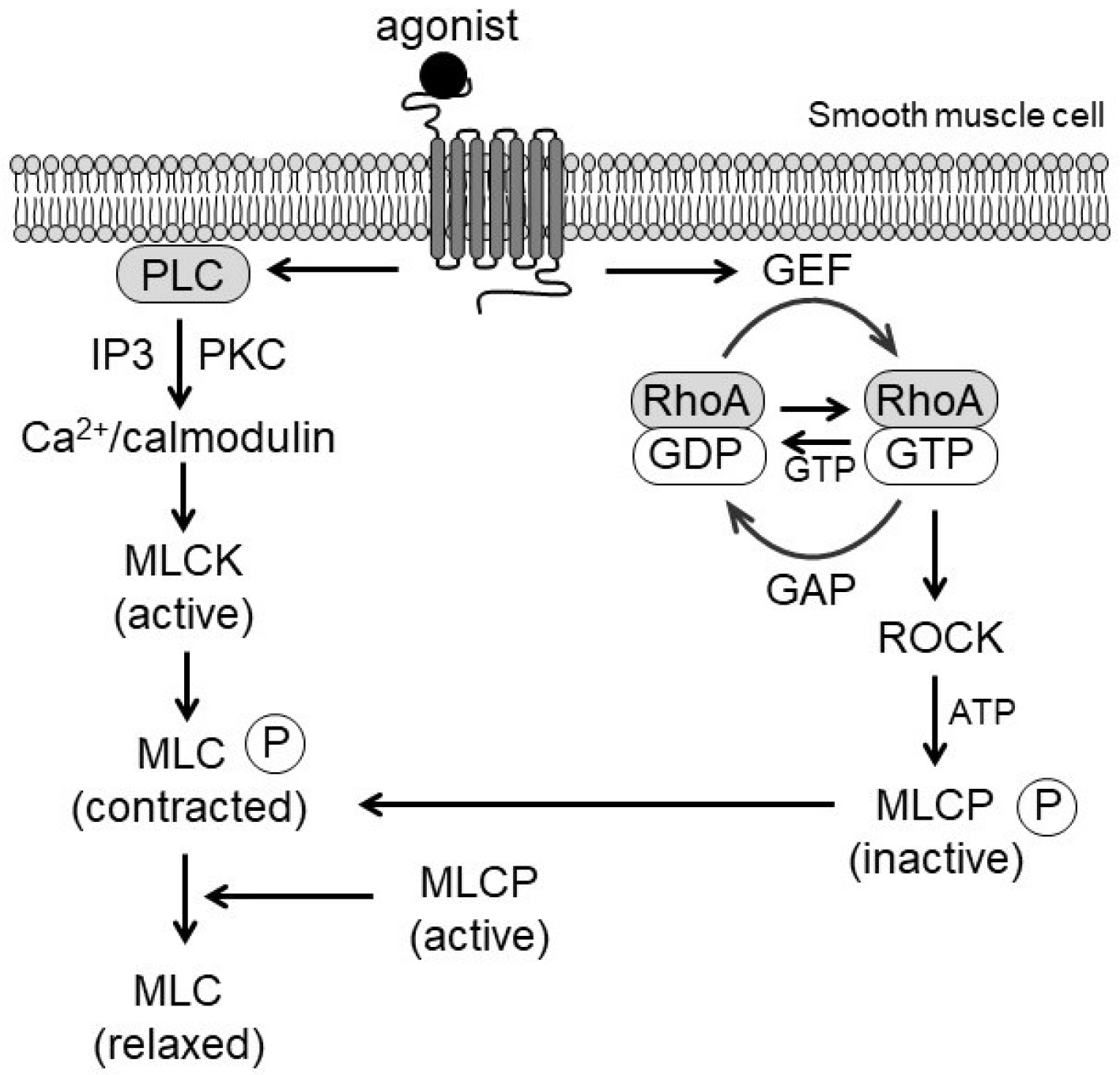
Phosphorylation of the regulatory MLC by myosin light chain kinase (MLCK) activates myosin ATPase, enabling actin–myosin cross-bridge cycling. The level of MLC phosphorylation reflects the balance between MLCK and MLCP activities. In this context, RhoA/ROCK enhances MLC phosphorylation primarily through MLCP inhibition and, under certain conditions, via direct phosphorylation of MLC []. Furthermore, monophosphorylation at Ser19 and diphosphorylation at Ser19/Thr18 augment actin-activated ATPase activity, strengthening vascular tone []. In proliferative or synthetic VSMCs, MLC diphosphorylation is increased, linking phenotypic modulation to vascular remodeling []. Collectively, these mechanisms highlight the dual role of RhoA/ROCK in both acute contraction and long-term structural adaptation (Figure 1).
Figure 1. Schematic representation of Ca2+/calmodulin and RhoA/ROCK signaling in vascular smooth muscle contraction. Agonist binding to G protein-coupled receptors (GPCRs) activates phospholipase C (PLC) and guanine nucleotide exchange factors (GEFs), leading to inositol trisphosphate (IP3)–mediated Ca2+ release, myosin light chain kinase (MLCK) activation, and phosphorylation of myosin light chain (MLC). Concurrently, RhoA activation stimulates Rho-kinase (ROCK), which inhibits myosin phosphatase (MLCP) through phosphorylation of its myosin-binding subunit, enhancing Ca2+ sensitization and sustaining vascular contraction.

2.3. Endothelial Mechanisms in RhoA/ROCK Signaling

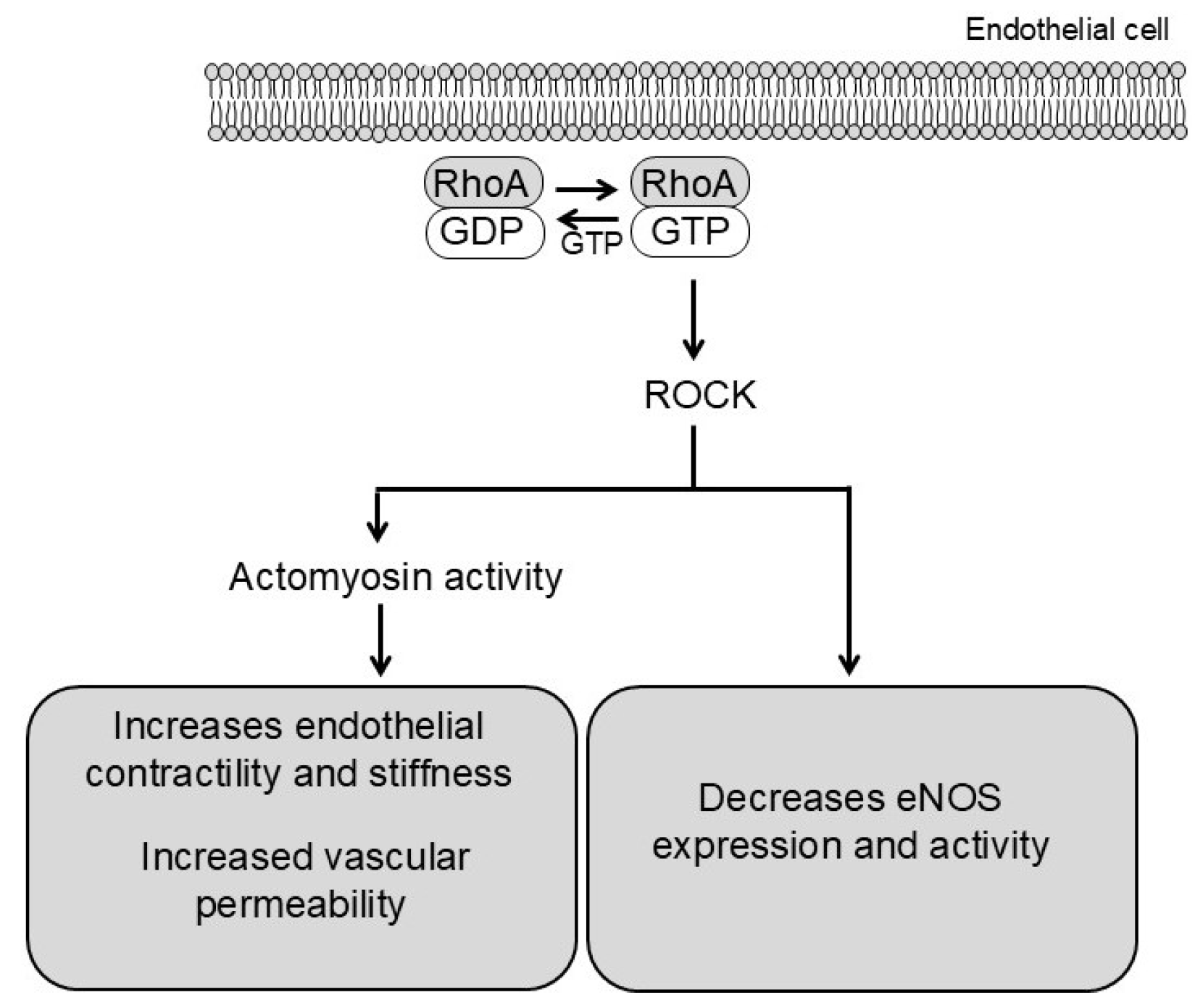
In endothelial cells, ROCK activation reduces endothelial nitric oxide synthase (eNOS) expression and activity while increasing endothelial contractility through actomyosin tension. Consequently, cell–cell junctions are disrupted and permeability rises []. These changes facilitate leukocyte infiltration and trigger pro-inflammatory signaling, establishing a mechanistic link between endothelial dysfunction and vascular remodeling (Figure 2).
Figure 2. Schematic representation of ROCK signaling in endothelial cells. ROCK exerts two related effects: (1) it inhibits eNOS activity via intracellular signaling pathways, reducing nitric oxide (NO) production and increasing vascular tone; and (2) it directly enhances actomyosin contractility, increasing endothelial contractility (modulation of cell–cell junctions) and permeability.
In addition, ROCK modulates endothelial-to-mesenchymal transition (EndMT) and interacts with transcriptional coactivators, such as YAP (Yes, associated protein) and TAZ (Transcriptional coactivator with PDZ-binding motif), contributing to fibrosis and arterial stiffening. These mechanisms contribute to reduced vasodilatory capacity and heightened vascular inflammation in hypertension. Notably, AngII preferentially activates the ROCK2 isoform in endothelial cells, leading to loss of endothelium-dependent relaxation. Isoform-selective inhibition restores endothelial function in both male and female mice, highlighting the therapeutic potential of targeting ROCK2 [].

3. Crosstalk with Oxidative Stress and Redox Signaling

3.1. Reactive Oxygen Species (ROS)

ROS such as superoxide anion (O2•−) and hydrogen peroxide (H2O2) are implicated as key regulators of VSMCs function. ROS are produced as byproducts of normal cell metabolism and other enzymatic reactions, particularly via nicotinamide adenine dinucleotide phosphate (NADPH) oxidases (NOX), xanthine oxidase, and the mitochondrial electron transport chain. Major intracellular sources include mitochondria, endoplasmic reticulum, and peroxisomes [,]. In addition, exogenous factors such as smoking, pollution, xenobiotics, and radiation further enhance ROS production []. At physiological levels, ROS regulate proliferation, differentiation, and apoptosis. However, elevated ROS induces oxidative stress, causing lipid peroxidation, protein oxidation, DNA damage and activation of inflammatory pathways []. ROS-induced inflammation results in pathological developments including neurodegenerative, autoimmune, respiratory, and vascular disorders []. In the vasculature, elevated ROS levels disrupt cellular homeostasis and promote chronic inflammation, contributing to the pathogenesis of vascular diseases. In VSMCs, oxidative stress impacts Ca2+ handling and enhances contractility, which leads to the development of hypertension and vascular remodeling.

3.2. Impact on Ca2+ Handling and Ca2+ Channels

ROS directly and indirectly modulate Ca2+ handling in VSMCs by affecting Ca2+ channels, pumps, and intracellular Ca2+ stores. Given that intracellular Ca2+ is the key second messenger mediating VSMCs contractility and vascular tone, these effects have profound physiological consequences [,]. For example, ROS regulate a variety of Ca2+ channels in the plasma membrane, including voltage-dependent, receptor-induced, and store-mediated. ROS modifies the redox-sensitive cysteine residues on the channel α1-subunit of L-type voltage-gated Ca2+ channels, which causes changes in channel trafficking, and ultimately, alterations in VSMC function []. The L-type voltage-gated channels in VSMCs, Cav 1.2 channels, are specifically stimulated by ROS produced by NADPH oxidase localized near the plasma membrane. The effect of ROS on these channels was mediated through protein kinase C alpha (PKCα) signaling. H2O2 has been shown to increase the Ca2+ influx through L-type voltage-gated channels in VSMCs. The enhanced stimulation of Cav 1.2 channels and increased influx of Ca2+ causes sustained vasoconstriction []. Oxidative stress and ROS can impact Ca2+ handling in VSMCs by influencing the expression and function of ion channels, such as transient receptor potential melastatin 2 (TRPM2) which is a ROS sensor and regulator of Ca2+ and Na+ transport. During hypertensive states, ROS production can be enhanced, which increases the activation of TRPM2 channels on VSMCs via the Poly [ADP-ribose] polymerase 1 (PARP1) protein []. This leads to increased Ca2+ influx through a Ca2+/Na+ exchanger, leading to increased intracellular Ca2+ and VSMC contraction. An example of store operated Ca2+ entry is the Orai1 channel on plasma membranes in VSMCs. In physiological conditions, the Orai pathway increases Ca2+ influx when Ca2+ stores in the endoplasmic reticulum are depleted. The sensor protein, STIM1, activates Orai1 channels, allowing more Ca2+ to enter the cell. In hypertensive conditions, high ROS levels enhance the Orai1 channel function and there is excessive Ca2+ entry into the cell. This leads to vascular dysfunction and sustained vasoconstriction [].

3.3. Ca2+ Microdomains

Ca2+ microdomains in VSMCs are small, localized regions of elevated Ca2+ concentrations formed near specific signaling receptors and ion channels on the plasma membrane or endoplasmic reticulum []. These microdomains are organized via caveolin scaffolding proteins within caveolae, which cluster receptors and channels in defined areas. In this sense, the localized high Ca2+ concentrations enable precise activation of downstream signaling pathways without significantly altering global intracellular Ca2+, ensuring tightly controlled vascular contraction and relaxation []. Importantly, Ca2+ microdomains play a crucial role in balancing vascular tone and are often involved in pathological conditions. In hypertensive conditions, Ang II activates NADPH oxidase, leading to excess ROS in the body. Consequently, ROS alter the Ca2+ microdomains by over activating certain receptors and reorganizing the receptor expression in the caveolae. For example, Cav1.2 channels can be upregulated within microdomains, enhancing Ca2+ influx and contributing to sustained vasoconstriction, which maintains hypertension []. Moreover, hyperglycemia-induced ROS in VSMCs disrupt caveolar signaling, which not only affects Ca2+ microdomains organization but also leads to dysregulated expression of adipokines such as adiponectin and leptin. Although adiponectin is primarily secreted by adipocytes, its synthesis in VSMCs appears to be a compensatory response mediated by ROS and NADPH oxidase type 4 (Nox4) within caveolae, while downregulation of adiponectin receptors may limit protective effects. These caveola-dependent changes in both Ca2+ signaling and adipokine expression further exacerbate vascular dysfunction under metabolic stress [].
Beyond Ca2+ handling, localized Ca2+ signals within caveolae can activate RhoA via Ca2+/calmodulin-dependent pathways and GEFs, which subsequently stimulate ROCK to enhance Ca2+ sensitization of contractile responses. Therefore, dysregulation of Ca2+ microdomains under oxidative stress may potentiate RhoA/ROCK activation, further contributing to vascular hypercontractility in hypertension [].

3.4. Redox Sensitive Proteins

Redox sensitive proteins serve as a critical bridge between ROS and Ca2+ signaling in VSMCs, influencing vascular tone, contractility, and cellular function. Several key proteins, including PARP1, protein tyrosine phosphatases (PTP), protein kinases, and transcription factors, are involved in these redox-regulated pathways. Specifically, PARP1 is activated by oxidative DNA damage in VSMCs and catalyzes the synthesis of ADP-ribose polymers, which are broken down into free ADP ribose (ADPR). ADPR directly binds to the C-terminal Nudix-type motif 9 homologous (NUDT9-H) domain of TRPM2 channels, inducing a conformational change that opens the channel and facilitates Ca2+ and Na+ influx []. Consequently, VSMC contractility increases, particularly in hypertensive states where ROS levels are elevated. In physiological conditions, PTPs act to balance the protein tyrosine kinases (PTKs) by dephosphorylating tyrosine residues on proteins []. However, excessive ROS oxidizes cysteine residues within PTPs, leading to their inactivation. Therefore, unchecked phosphorylation of signaling proteins leads to increased Ca2+ influx and heightened vascular tone []. Two examples of redox sensitive protein kinases include Ca2+/calmodulin-dependent protein kinase II (CaMKII) and phosphatidylinositol 3-kinase (PI3K) proteins. CaMKII is directly oxidized by ROS through the methionine residue, leading to sustained kinase activity without sustained stimulation. This prolonged kinase activity causes vascular stiffening and VSMC apoptosis []. PI3K, which normally regulates VSMC phenotype, including contraction and differentiation [], shows increased membrane recruitment and catalytic activity upon H2O2 stimulation. This enhances ROS accumulation and activates the AKT signaling pathway, ultimately altering VSMCs. The combined results lead to alterations in VSMC phenotype []. In addition, under oxidative stress, specific methionine residues (Met281/282) within CaMKII’s regulatory domain undergo oxidation, leading to sustained kinase activation independent of Ca2+/calmodulin binding []. This persistent activation promotes VSMC apoptosis, hypertrophic signaling, and vascular stiffening. In cardiac tissue, oxidized CaMKII has been implicated in arrhythmogenesis, myocardial injury, and maladaptive remodeling, linking redox imbalance to Ca2+-driven vascular and cardiac pathology []. ROS also stimulates a variety of transcription factors with protective effects over VSMCs to combat the damage done through the previously mentioned pathways. The redox-sensitive transcription factor, nuclear factor erythroid 2-related factor 2 (Nrf2) is activated by oxidative stress and functions to increase the transcription of antioxidants, which protect against oxidative stress []. In contrast, chronic high levels of ROS shifts the balance toward harmful transcription factors, such as nuclear factor-kappa B (NF-kB), which promotes secretion of pro-inflammatory cytokines, leads to further production of ROS, and ultimately causes vascular damage []. Collectively, redox-sensitive proteins integrate oxidative signals with Ca2+ dynamics and downstream transcriptional programs, establishing a feedback loop that amplifies vascular dysfunction under hypertensive conditions.
Collectively, these redox-sensitive proteins not only amplify oxidative stress and Ca2+ disturbances but also converge on RhoA/ROCK activation. For instance, oxidation-driven activation of PTPs enhances RhoA activity through sustained phosphorylation of RhoA-associated kinases, while oxidized CAMKII can indirectly promote cytoskeletal remodeling via ROCK signaling [,]. This integration of redox and RhoA/ROCK pathways represents a central mechanism linking oxidative stress to vascular stiffness and remodeling.

4. RhoA/ROCK in Hypertension and Vascular Diseases

4.1. Increased Contractility and Peripheral Vascular Resistance

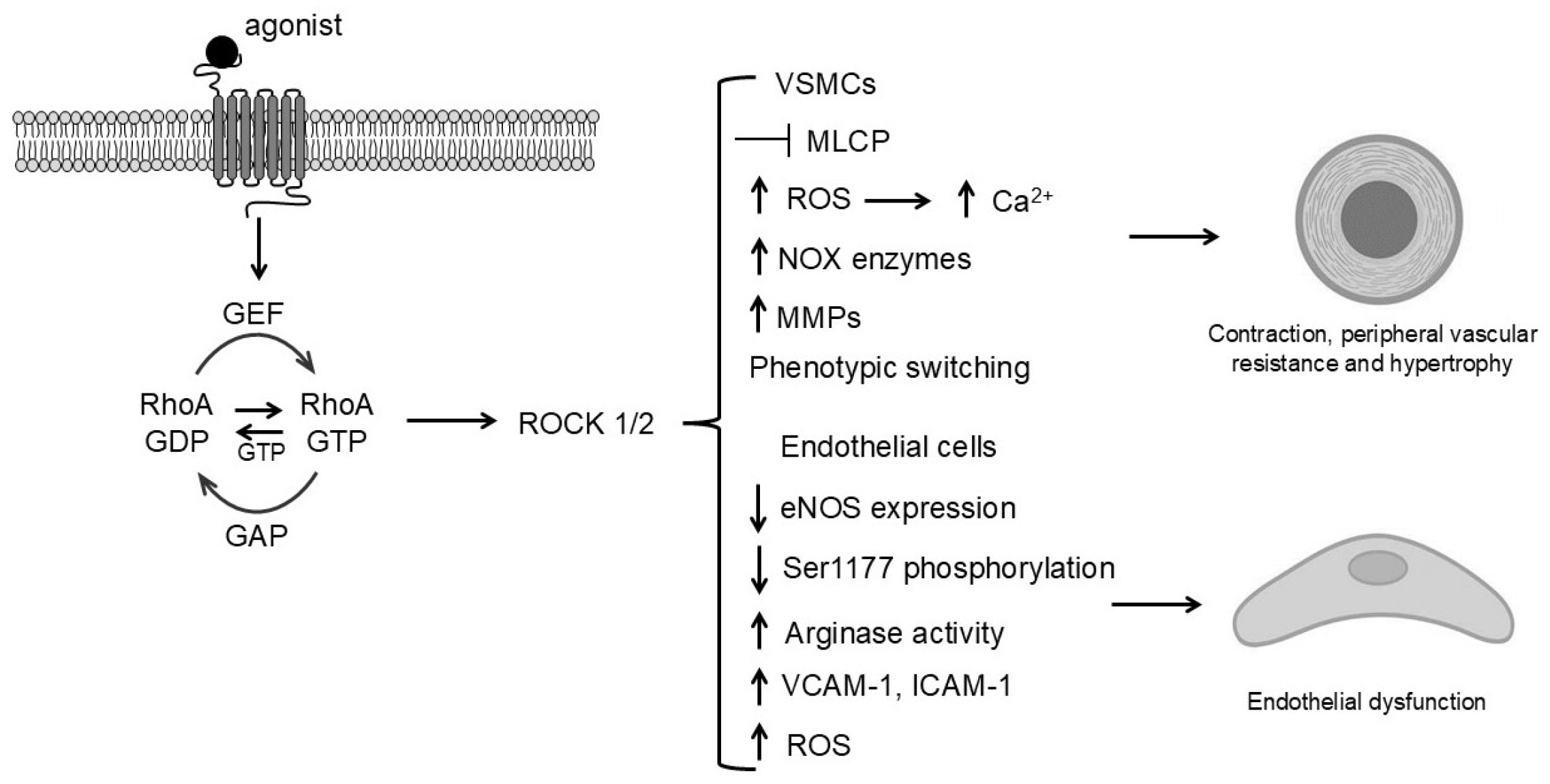
In hypertension, increased contractility of VSMC leads to sustained peripheral vascular resistance and vascular dysfunction [] (Figure 3). This heightened contractility results from both elevated intracellular Ca2+ levels and Ca2+ sensitization mechanisms. In particular, activation of the RhoA/ROCK pathway decreases MLCP activity, thereby sustaining vasoconstriction and elevating blood pressure even in the presence of lower Ca2+ concentrations. Additionally, an imbalance in redox signaling, often characterized by excessive ROS production from NOX enzymes, contributes to increased VSMC contractility and elevated peripheral vascular resistance. Elevated ROS levels during hypertensive states activate multiple signaling molecules through oxidation of specific protein residues. Consequently, kinases such as PKC and ROCK are hyperactivated resulting in heightened Ca2+ sensitivity and sustained vasoconstriction of blood vessels. In addition, ROS alter the function of Ca2+ channels and pumps, such as the L-type Ca2+ channels, enhancing influx of Ca2+ into VSMCs. Moreover, RhoA/ROCK signaling amplifies endothelium- and endoperoxide-dependent contractile responses, which are hallmarks of hypertensive vascular dysfunction []. Overall, ROS increases VSMC contractile capabilities and leads to prolonged hypertension by enhancing peripheral vascular resistance.
Figure 3. Mechanisms of RhoA/ROCK-mediated vascular cell proliferation and remodeling during hypertension. Agonist binding to G protein-coupled receptors (GPCRs) leading to ROCK1/2 activation in VSMCs and endothelial cells. In VSMCs, ROCK enhances MLC phosphorylation, cytoskeletal remodeling, and phenotypic switching, resulting in proliferation, migration, and extracellular matrix deposition. In endothelial cells, ROCK decreases eNOS expression and increases permeability and inflammation. Collectively, these mechanisms drive vascular wall thickening, stiffness, and reduced lumen diameter, contributing to elevated peripheral resistance and hypertension.
Consistent with these mechanisms, animal studies have demonstrated elevated ROCK activity primarily in VSMCs across multiple hypertensive models, including spontaneously hypertensive rats (SHRs), Ang II–infused, deoxycorticosterone acetate (DOCA)-salt, and Dahl salt-sensitive rats []. Pharmacological inhibition of ROCK in VSMCs with agents such as Y-27632 or fasudil significantly reduces vascular tone, prevents medial thickening, and attenuates the rise in blood pressure [,]. These findings highlight the mechanistic role of RhoA/ROCK signaling in maintaining VSMCs contractility and structural remodeling in hypertension.
Supporting this, rat models of apatinib-induced hypertension exhibit upregulation of RhoA and ROCK2 in the mid-aorta, accompanied by reduced MLCP activity and increased ET-1 and collagen I expression. Administration of the ROCK inhibitor Y-27632 significantly reduced both blood pressure and vascular remodeling, confirming the critical role of ROCK2 in contractile dysfunction []. Furthermore, genetic studies provide further evidence for the role of RhoA/ROCK signaling in vascular contractility. In Dahl salt-sensitive rats, loss of the Rho GEF Arhgef11 attenuates ROCK activation, decreased vascular contractility, and protection against hypertension-induced renal injury. This highlights Arhgef11 as an upstream regulator of the RhoA/ROCK pathway in hypertension [].

4.2. RhoA/ROCK-Induced Endothelial Dysfunction in Hypertension

Endothelial dysfunction is a characteristic of vascular disease and is often mediated by oxidative stress and Ca2+ dysregulation. Vascular endothelium, a monolayer of cells lining the lumen, regulates both permeability and vascular tone. In this context, ROS impair NO production by oxidizing and inactivating eNOS, thereby reducing vasodilation and increasing vascular tone [] (Figure 3). Additionally, abnormal Ca2+ release from intracellular stores and enhanced entry into endothelial cells promote pathological reactions. Increased cytosolic Ca2+ levels can over activate enzymes promoting oxidative stress and inflammatory signals. In addition, high Ca2+ levels contribute to vascular calcification, which can harden VSMCs and further contribute to vascular dysfunction, atherosclerosis and morbidity. Calcification involves stiffening of vessel walls which limits the vasodilation capabilities, increasing the peripheral vascular resistance [].

4.3. RhoA/ROCK-Mediated Suppression of NO Bioavailability and Vascular Remodeling

RhoA/ROCK signaling contributes to endothelial dysfunction and vascular remodeling through multiple, interconnected mechanisms. ROCK downregulates eNOS transcription, destabilizes eNOS mRNA, decreases Ser1177 phosphorylation, and enhances oxidative stress, leading to eNOS uncoupling [,]. In addition, increased arginase activity further reduces L-arginine availability, collectively these effects impair endothelium-dependent relaxation. Supporting this, exposure of endothelial cells to high-salt conditions activates RhoA/ROCK signaling and suppresses eNOS expression through the upregulation of asymmetric dimethylarginine (ADMA), an endogenous eNOS inhibitor. This highlights dietary salt as an upstream modulator of the RhoA/ROCK–NO axis in vascular dysfunction [].
Moreover, ROCK-mediated EC contraction which is referring to actomyosin-driven-cytoskeletal tension that modulates cell–cell junctions [], increases permeability and promotes leukocyte adhesion/transmigration. This is reinforced by activation of NF-κB–dependent adhesion molecules (VCAM-1, ICAM-1) and chemokines, linking endothelial ROCK activity to vascular inflammation [].
In parallel, vascular remodeling occurs through alterations in vessel thickness, lumen diameter, and elasticity within the wall []. In VSMCs, ROS-driven phenotypic modulation, growth, death, migration, and extracellular matrix reorganization underlie these structural changes. Excessive ROS contributes to vascular wall thickening, lumen narrowing, and reduced elasticity. In addition, matrix malloproteinases (MMPs) involved in extracellular matrix remodeling are influenced by ROS, leading to disorganized remodeling, increased collagen deposition, and vessel stiffening []. The phenotypic changes caused by ROS are associated with altered cell growth and apoptosis, leading to hyperplasia and hypertrophy of VSMCs. At lower levels, ROS induces VSMC growth. However, at high levels, ROS dampen protective properties of VSMC, causing unnecessary apoptosis, contributing to atherosclerosis and atypical vascular remodeling []. Importantly, ROS in VSMCs not only drive these structural changes but also directly modulate RhoA/ROCK signaling. Oxidative stress activates RhoA through redox-sensitive GEFs such as p115RhoGEF [], enhancing ROCK-mediated phosphorylation of MLC and promoting VSMCs contraction and stiffness []. Thus, ROS-induced remodeling is closely linked to RhoA/ROCK-dependent cytoskeletal reorganization.
Notably, studies in normotensive rats with elevated angiotensin-converting enzyme levels revealed increased ROCK activation in the aortic wall, alongside upregulation of profibrotic and inflammatory genes such as monocyte chemoattractant protein-1, transforming growth factor (TGF)-beta(1) (TGF-β1), plasminogen activator inhibitor-1 (PAI-1), and monocyte chemoattractant protein-1 (MCP-1). This was accompanied by enhanced NADPH oxidase activity and superoxide production, all of which were normalized by the ROCK inhibitor fasudil, highlighting the role of RhoA/ROCK in early vascular remodeling even before the onset of hypertension [].
Finally, by augmenting Ca2+ sensitivity, RhoA/ROCK sustains vasoconstriction independently of changes in intracellular Ca2+ concentration, a phenomenon observed in hypertensive resistance vessels []. In contrast, RhoA/ROCK signaling in the cardiomyocytes promotes hypertrophic gene expression via activation of serum response factor (SRF) and extracellular signal-regulated kinase (ERK). The RhoA/ROCK signaling in the cardiomyocytes leads to maladaptive outcomes such as biventricular hypertrophy and myocardial stiffening as a result of hypertensive stress [,].

5. Inflammation and Immune Activation

ROS play a pivotal role in promoting vascular inflammation in endothelial cells and VSMCs and activating immune cells, thereby influencing vascular function and accelerating tissue damage. Inflammation not only results from vascular dysfunction but also perpetuates it, creating a self-sustaining pathological loop. Elevated ROS levels activate redox-sensitive transcription factors such as NF-kB which induces the expression of pro-inflammatory cytokines, chemokines, and adhesion molecules []. This, in turn, promotes the recruitment and activation of immune cells at the endothelial level, facilitating their adhesion and subsequent transmigration into the surrounding connective tissue, where they amplify local oxidative stress and inflammation. The infiltration of immune cells, particularly macrophages and neutrophils, further amplifies oxidative stress by generating additional ROS through respiratory bursts, aggravating endothelial injury []. Moreover, elevated intracellular Ca2+ concentrations can activate inflammasomes, such as NOD-, LRR- and pyrin domain-containing protein 3 (inflammasome) (NLRP3), in immune cells leading to the release of pro-inflammatory cytokines that exacerbate vascular inflammation []. Activation of NLRP3 in VSMCs promotes a phenotypic switch from contractile to synthetic and proliferative states via NF-κB signaling and increased IL-1β and IL-18 secretion, driving vascular hypertrophy, stiffness, and elevated peripheral resistance. In endothelial cells, NLRP3 activation reduces eNOS activity and NO bioavailability, further promoting endothelial dysfunction [,]. Experimental evidence supports these effects, as NLRP3 activation in VSMCs and ECs has been shown to induce vascular inflammation remodeling, and impaired vasodilation in cardiovascular diseases [,,]. Moreover, under oxidative stress, NLRP3- mediated inflammation enhances the canonical RhoA/ROCK signaling pathway [], amplifying Ca2+ sensitization, cytoskeletal reorganization, and vascular contraction. In addition, continuous ROS production in VSMCs and endothelial cells maintains a chronic inflammatory state, establishing a vicious cycle that contributes to progressive vascular damage, characteristic of hypertension and atherosclerosis [].

6. Human Evidence

Evidence from human studies corroborates the preclinical findings. In mononuclear cells from patients with essential hypertension, p63RhoGEF mRNA and protein levels, as well as phospho-myosin phosphatase target protein-1 (MYPT-1), a marker of Rho kinase activity, were significantly increased compared with healthy individuals, indicating enhanced RhoA/ROCK signaling in vivo []. Acute fasudil infusion improves forearm blood flow and reduces arterial stiffness in humans [] supporting the concept that ROCK inhibition can restore endothelial function. However, long-term clinical trials evaluating chronic blood pressure-lowering effects remain limited.
Further clinical evidence shows that cigarette smokers display significantly increased leukocyte ROCK activity, which correlates with higher systolic blood pressure and impaired flow-mediated dilation (FMD) []. This association suggests that activated leukocytes may contribute to endothelial dysfunction through increased adhesion to the vascular wall, potentially promoting oxidative stress and inflammatory signaling, and subsequent endothelial injury. Thus, leukocyte ROCK activation may reflect systemic oxidative stress and is associated with endothelial dysfunction in smokers []. Consistent with these findings, young adults with metabolic syndrome exhibit increased RhoA/ROCK activity along with elevated markers of endothelial dysfunction, even before the onset of overt hypertension []. This clinical evidence highlights the early involvement of RhoA/ROCK in vascular pathology and its potential as a therapeutic target in at-risk populations.

7. Therapeutic Targeting

7.1. No Pan-ROCK Inhibitors Are in Clinical Use for the Treatment of Hypertension

Systemic ROCK inhibition can cause severe hypotension, headache, edema, and off-target effects (gastrointestinal, urinary, etc.), reflecting broad ROCK distribution []. Additionally, the lack of isoform selectivity (ROCK1 vs. ROCK2) complicates therapeutic precision []. As a result, clinical development for ROCK inhibitors has shifted towards targeted and local applications to modulate VSMCs and endothelial cell function. For instance, fasudil is used for cerebral vasospasm following subarachnoid hemorrhage associated with hypertension, while topical formulations of netarsudil (Rhopressa) and ripasudil (Glanatec) are employed for glaucoma and ocular hypertension [].

7.2. Emerging Therapeutic Strategies

Many of the emerging strategies for disrupting RhoA/Rho-kinase signaling in the vasculature are highly experimental and caution is needed to address off-target effects and potential adverse events associated with pan-ROCK inhibition. Promising next-generation approaches for the treatment of hypertension include isoform-selective inhibitors, such as KD025, which exhibits antifibrotic properties in preclinical studies using animal model of hypertension [], nanoparticle vascular delivery [], and targeting upstream activators (specific RhoGEFs) [].
Additionally, recent work has identified the TRPV4–RhoA–RhoGDI1 axis as a key regulatory pathway in vascular tone. Activation of TRPV4 promotes RhoA inactivation through RhoGDI1, and pharmacologic targeting of this axis reduces vascular contractility and lowers blood pressure in hypertensive models. These findings suggest that enhancing endogenous inhibitory mechanisms of RhoA may represent a complementary therapeutic approach [].
The RhoA/ROCK signaling axis plays a central role in VSMCs, calcium sensitization, arterial constriction and structural remodeling in hypertension []. However, ROCK1 and ROCK2 are ubiquitously expressed in multiple tissues, including heart, skeletal muscle, kidney, lung, and immune cells and participate in a broad range of cellular functions such as cytoskeletal organization, cell motility, proliferation/apoptosis, matrix remodeling, and endothelial barrier regulation [,]. Thus, the widespread tissue distribution and pleiotropic roles of ROCK1/2 represent a major challenge for the development of vascular-specific therapeutic interventions, as systemic inhibition may produce unwanted effects in non-vascular tissues. To overcome this, recent strategies have focused on isoform-selective inhibition [] and on tissue/cell-type-specific targeting, with preclinical studies demonstrating favorable effects on vascular remodeling and blood pressure with reduced non-vascular toxicity [,]. These developments suggest that while vascular specificity of ROCK targeting remains an obstacle, tailored approaches with isoform specificity or targeted delivery hold promise to enhance therapeutic efficacy and safety in hypertension.

8. Future Directions

Future research on RhoA/ROCK inhibitors in hypertension focus on improving therapeutic precision, identifying specific disease mechanisms, and optimizing clinical use []. Research is moving beyond pan-inhibition to define isoform-specific roles (ROCK1 vs. ROCK2) within VSMCs and ECs to develop more targeted therapies and avoid systemic side effects. Mapping the upstream Rho GEFs and GAPs activated in hypertensive phenotypes is crucial for understanding disease-specific activation of the pathway [,]. Simultaneously, the development of reliable biomarkers, such as phospho-MYPT1 or leukocyte ROCK activity, will allow for better patient stratification and monitoring of treatment response in clinical practice [,]. To enhance efficacy and reduce monotherapy risks, combination therapies are being explored, particularly combining ROCK inhibitors with established treatments like RAAS blockade or antioxidants []. Ultimately, conducting robust, mechanism-based clinical trials is necessary to assess the long-term safety and efficacy of chronic ROCK inhibition and solidify its role in managing hypertension. In summary:
  • Define isoform-specific contributions (ROCK1 vs. ROCK2) in VSMCs vs. ECs.
  • Map upstream RhoGEFs and GAPs activated in hypertensive phenotypes.
  • Develop biomarkers (phospho-MYPT1, leukocyte ROCK activity) for patient stratification.
  • Explore combination therapies (ROCK inhibition + RAAS blockade or antioxidants).
  • Conduct mechanism-based clinical trials assessing chronic ROCK inhibition in hypertension.

9. Conclusions

The RhoA/ROCK pathway represents a central regulator of vascular tone, endothelial function, and remodeling. Mechanistic insights into Ca2+ sensitization, MLCP inhibition, oxidative stress amplification, and endothelial dysfunction underscore its role in hypertension pathogenesis. Translational studies highlight ROCK inhibitors and statins as promising therapeutic approaches, though broader clinical application requires careful balancing of efficacy and safety. Precision targeting RhoA/ROCK signaling offers a compelling strategy for future antihypertensive therapies and vascular protection.

Author Contributions

S.R.: writing—original draft, writing—review and editing, investigation, supervision. D.L.S.-V.: writing—review and editing and investigation. F.P.: writing—review and editing and investigation. R.C.W.: writing—review and editing, design, figures and investigation. All authors have read and agreed to the published version of the manuscript.

Funding

The authors of this review were funded by the National Institutes of Health (RO1 DK132948), the Airforce Office of Scientific Research, U.S. Airforce, Department of Defense, the Society for the Study of Women’s Sexual Health, the Sexual Medicine Society of North America, and the Institute for Cardiovascular Disease Research from University South Carolina Office of the Vice President for Research.

Insitutional Review Board Statement

Not applicable.

Data Availability Statement

No new data were created or analyzed in this study.

Acknowledgments

During the preparation of this manuscript, the authors used Grammarly (Grammarly Inc., Sao Francisco, CA, USA) and ChatGPT 5.0 (OpenAI, Sao Francisco, CA, USA) for the purposes of language polishing, including grammar and clarity improvements. The authors have reviewed and edited the output and take full responsibility for the content of this publication.

Conflicts of Interest

The authors declare no conflicts of interest.

Abbreviations

The following abbreviations are used in this manuscript:
ADMAAsymmetric dimethylarginine
ADPRADP-ribose
Ang IIAngiotensin II
ATPAdenosine triphosphate
Ca2+Calcium ion
CaMKIICa2+/calmodulin-dependent protein kinase II
CPI-17PKC-potentiated inhibitory protein for heterotrimeric myosin phosphatase of 17 kDa
DAGDiacylglycerol
DOCADeoxycorticosterone acetate
ECEndothelial cell
eNOSEndothelial nitric oxide synthase
EREndoplasmic reticulum
ET-1Endothelin-1
FMDFlow-mediated dilation
GAPGTPase activating protein
GDPGuanosine diphosphate
GDIGuanine nucleotide dissociation inhibitor
GEFGuanine nucleotide exchange factor
GPCRG protein–coupled receptor
GTPGuanosine triphosphate
H2O2Hydrogen peroxide
ICAM-1Intercellular adhesion molecule 1
IP3Inositol 1,4,5-trisphosphate
LARGLeukemia-associated Rho guanine nucleotide exchange factor
L-ArgL-arginine
MLCMyosin light chain
MLCKMyosin light chain kinase
MLCPMyosin light chain phosphatase
MCP-1Monocyte chemoattractant protein-1
MMPMatrix metalloproteinase
MYPT1Myosin phosphatase target subunit 1
NADPHNicotinamide adenine dinucleotide phosphate
NF-κBNuclear factor kappa-light-chain-enhancer of activated B cells
NFATNuclear factor of activated T cells
NONitric oxide
NOXNADPH oxidase
NLRP3NOD-, LRR- and pyrin domain-containing protein 3 (inflammasome)
Nrf2Nuclear factor erythroid 2–related factor 2
NUDT9-HNudix-type motif 9 homologous domain
O2•−Superoxide anion
Orai1Calcium release-activated calcium channel protein 1
PAI-1Plasminogen activator inhibitor-1
PARP1Poly(ADP-ribose) polymerase 1
PDZ-RhoGEFPDZ domain-containing Rho guanine nucleotide exchange factor
PI3KPhosphatidylinositol 3-kinase
PIP2Phosphatidylinositol 4,5-bisphosphate
PKCProtein kinase C
PKCαProtein kinase C alpha
PLCPhospholipase C
PTKProtein tyrosine kinase
PTPProtein tyrosine phosphatase
PYK2Proline-rich tyrosine kinase 2
RAASRenin–angiotensin–aldosterone system
RBDRho-binding domain
ROCKRho-associated coiled-coil-containing protein kinase (Rho-kinase)
ROSReactive oxygen species
SHRSpontaneously hypertensive rat
STIM1Stromal interaction molecule 1
TGF-β1Transforming growth factor beta 1
TRPM2Transient receptor potential melastatin 2
TRPV4Transient receptor potential vanilloid 4
VCAM-1Vascular cell adhesion molecule 1
VSMCVascular smooth muscle cell

References

  1. Durante, A.; Mazzapicchi, A.; Baiardo Redaelli, M. Systemic and Cardiac Microvascular Dysfunction in Hypertension. Int. J. Mol. Sci. 2024, 25, 13294. [Google Scholar] [CrossRef] [PubMed]
  2. Zeng, X.; Yang, Y. Molecular Mechanisms Underlying Vascular Remodeling in Hypertension. Rev. Cardiovasc. Med. 2024, 25, 72. [Google Scholar] [CrossRef]
  3. Chitaley, K.; Weber, D.; Webb, R.C. RhoA/Rho-kinase, vascular changes, and hypertension. Curr. Hypertens. Rep. 2001, 3, 139–144. [Google Scholar] [CrossRef] [PubMed]
  4. Yang, Q.; Hori, M. Characterization of Contractile Machinery of Vascular Smooth Muscles in Hypertension. Life 2021, 11, 702. [Google Scholar] [CrossRef]
  5. Zicha, J.; Vaneckova, I. Altered Balance Between Vasoconstrictor and Vasodilator Systems in Experimental Hypertension. Physiol. Res. 2024, 73, 901–928. [Google Scholar] [CrossRef]
  6. Loirand, G.; Guerin, P.; Pacaud, P. Rho kinases in cardiovascular physiology and pathophysiology. Circ. Res. 2006, 98, 322–334. [Google Scholar] [CrossRef]
  7. Seccia, T.M.; Rigato, M.; Ravarotto, V.; Calo, L.A. ROCK (RhoA/Rho Kinase) in Cardiovascular-Renal Pathophysiology: A Review of New Advancements. J. Clin. Med. 2020, 9, 1328. [Google Scholar] [CrossRef]
  8. Sward, K.; Mita, M.; Wilson, D.P.; Deng, J.T.; Susnjar, M.; Walsh, M.P. The role of RhoA and Rho-associated kinase in vascular smooth muscle contraction. Curr. Hypertens. Rep. 2003, 5, 66–72. [Google Scholar] [CrossRef]
  9. Liu, X.; Chen, D.; Liu, G. Overexpression of RhoA promotes the proliferation and migration of cervical cancer cells. Biosci. Biotechnol. Biochem. 2014, 78, 1895–1901. [Google Scholar] [CrossRef]
  10. Qi, Y.; Liang, X.; Dai, F.; Guan, H.; Sun, J.; Yao, W. RhoA/ROCK Pathway Activation is Regulated by AT1 Receptor and Participates in Smooth Muscle Migration and Dedifferentiation via Promoting Actin Cytoskeleton Polymerization. Int. J. Mol. Sci. 2020, 21, 5398. [Google Scholar] [CrossRef] [PubMed]
  11. Sawma, T.; Shaito, A.; Najm, N.; Sidani, M.; Orekhov, A.; El-Yazbi, A.F.; Iratni, R.; Eid, A.H. Role of RhoA and Rho-associated kinase in phenotypic switching of vascular smooth muscle cells: Implications for vascular function. Atherosclerosis 2022, 358, 12–28. [Google Scholar] [CrossRef] [PubMed]
  12. Ishizaki, T.; Maekawa, M.; Fujisawa, K.; Okawa, K.; Iwamatsu, A.; Fujita, A.; Watanabe, N.; Saito, Y.; Kakizuka, A.; Morii, N.; et al. The small GTP-binding protein Rho binds to and activates a 160 kDa Ser/Thr protein kinase homologous to myotonic dystrophy kinase. EMBO J. 1996, 15, 1885–1893. [Google Scholar] [CrossRef] [PubMed]
  13. Matsui, T.; Amano, M.; Yamamoto, T.; Chihara, K.; Nakafuku, M.; Ito, M.; Nakano, T.; Okawa, K.; Iwamatsu, A.; Kaibuchi, K. Rho-associated kinase, a novel serine/threonine kinase, as a putative target for small GTP binding protein Rho. EMBO J. 1996, 15, 2208–2216. [Google Scholar] [CrossRef] [PubMed]
  14. Nakagawa, O.; Fujisawa, K.; Ishizaki, T.; Saito, Y.; Nakao, K.; Narumiya, S. ROCK-I and ROCK-II, two isoforms of Rho-associated coiled-coil forming protein serine/threonine kinase in mice. FEBS Lett. 1996, 392, 189–193. [Google Scholar] [CrossRef]
  15. Fukata, Y.; Kaibuchi, K.; Amano, M. Rho-Rho-kinase pathway in smooth muscle contraction and cytoskeletal reorganization of non-muscle cells. Trends Pharmacol. Sci. 2001, 22, 32–39. [Google Scholar] [CrossRef]
  16. Hartshorne, D.J.; Ito, M.; Erdodi, F. Myosin light chain phosphatase: Subunit composition, interactions and regulation. J. Muscle Res. Cell Motil. 1998, 19, 325–341. [Google Scholar] [CrossRef]
  17. Kimura, K.; Ito, M.; Amano, M.; Chihara, K.; Fukata, Y.; Nakafuku, M.; Yamamori, B.; Feng, J.; Nakano, T.; Okawa, K.; et al. Regulation of myosin phosphatase by Rho and Rho-associated kinase (Rho-kinase). Science 1996, 273, 245–248. [Google Scholar] [CrossRef]
  18. Somlyo, A.P.; Somlyo, A.V. Ca2+ sensitivity of smooth muscle and nonmuscle myosin II: Modulated by G proteins, kinases, and myosin phosphatase. Physiol. Rev. 2003, 83, 1325–1358. [Google Scholar] [CrossRef]
  19. Ying, Z.; Giachini, F.R.; Tostes, R.C.; Webb, R.C. PYK2/PDZ-RhoGEF links Ca2+ signaling to RhoA. Arter. Thromb. Vasc. Biol. 2009, 29, 1657–1663. [Google Scholar] [CrossRef]
  20. Aida, K.; Mita, M.; Ishii-Nozawa, R. Role of RhoGEFs or RhoGAPs in Pyk2-Mediated RhoA Activation in Depolarization-Induced Contraction of Rat Caudal Arterial Smooth Muscle. Int. J. Mol. Sci. 2025, 26, 8676. [Google Scholar] [CrossRef]
  21. Martinsen, A.; Yerna, X.; Rath, G.; Gomez, E.L.; Dessy, C.; Morel, N. Different effect of Rho kinase inhibition on calcium signaling in rat isolated large and small arteries. J. Vasc. Res. 2012, 49, 522–533. [Google Scholar] [CrossRef]
  22. Martinsen, A.; Dessy, C.; Morel, N. Regulation of calcium channels in smooth muscle: New insights into the role of myosin light chain kinase. Channels 2014, 8, 402–413. [Google Scholar] [CrossRef] [PubMed]
  23. Inaba, H.; Miao, Q.; Nakata, T. Optogenetic control of small GTPases reveals RhoA mediates intracellular calcium signaling. J. Biol. Chem. 2021, 296, 100290. [Google Scholar] [CrossRef]
  24. Amano, M.; Fukata, Y.; Kaibuchi, K. Regulation and functions of Rho-associated kinase. Exp. Cell Res. 2000, 261, 44–51. [Google Scholar] [CrossRef]
  25. Ikebe, M.; Hartshorne, D.J. Phosphorylation of smooth muscle myosin at two distinct sites by myosin light chain kinase. J. Biol. Chem. 1985, 260, 10027–10031. [Google Scholar] [CrossRef]
  26. Koyama, H.; Raines, E.W.; Bornfeldt, K.E.; Roberts, J.M.; Ross, R. Fibrillar collagen inhibits arterial smooth muscle proliferation through regulation of Cdk2 inhibitors. Cell 1996, 87, 1069–1078. [Google Scholar] [CrossRef]
  27. Yao, L.; Romero, M.J.; Toque, H.A.; Yang, G.; Caldwell, R.B.; Caldwell, R.W. The role of RhoA/Rho kinase pathway in endothelial dysfunction. J. Cardiovasc. Dis. Res. 2010, 1, 165–170. [Google Scholar] [CrossRef] [PubMed]
  28. Kinzenbaw, D.A.; Langmack, L.; Faraci, F.M. Angiotensin II-induced endothelial dysfunction: Impact of sex, genetic background, and rho kinase. Physiol. Rep. 2022, 10, e15336. [Google Scholar] [CrossRef]
  29. de Almeida, A.; de Oliveira, J.; da Silva Pontes, L.V.; de Souza Junior, J.F.; Goncalves, T.A.F.; Dantas, S.H.; de Almeida Feitosa, M.S.; Silva, A.O.; de Medeiros, I.A. ROS: Basic Concepts, Sources, Cellular Signaling, and its Implications in Aging Pathways. Oxid. Med. Cell. Longev. 2022, 2022, 1225578. [Google Scholar] [CrossRef] [PubMed]
  30. Jitca, G.; Osz, B.E.; Tero-Vescan, A.; Miklos, A.P.; Rusz, C.M.; Batrinu, M.G.; Vari, C.E. Positive Aspects of Oxidative Stress at Different Levels of the Human Body: A Review. Antioxidants 2022, 11, 572. [Google Scholar] [CrossRef]
  31. Sharifi-Rad, M.; Anil Kumar, N.V.; Zucca, P.; Varoni, E.M.; Dini, L.; Panzarini, E.; Rajkovic, J.; Tsouh Fokou, P.V.; Azzini, E.; Peluso, I.; et al. Lifestyle, Oxidative Stress, and Antioxidants: Back and Forth in the Pathophysiology of Chronic Diseases. Front. Physiol. 2020, 11, 694. [Google Scholar] [CrossRef]
  32. Afzal, S.; Abdul Manap, A.S.; Attiq, A.; Albokhadaim, I.; Kandeel, M.; Alhojaily, S.M. From imbalance to impairment: The central role of reactive oxygen species in oxidative stress-induced disorders and therapeutic exploration. Front. Pharmacol. 2023, 14, 1269581. [Google Scholar] [CrossRef]
  33. Checa, J.; Aran, J.M. Reactive Oxygen Species: Drivers of Physiological and Pathological Processes. J. Inflamm. Res. 2020, 13, 1057–1073. [Google Scholar] [CrossRef]
  34. Amberg, G.C.; Earley, S.; Glapa, S.A. Local regulation of arterial L-type calcium channels by reactive oxygen species. Circ. Res. 2010, 107, 1002–1010. [Google Scholar] [CrossRef] [PubMed]
  35. AlAhmad, M.; Shitaw, E.E.; Sivaprasadarao, A. A TRPM2-Driven Signalling Cycle Orchestrates Abnormal Inter-Organelle Crosstalk in Cardiovascular and Metabolic Diseases. Biomolecules 2025, 15, 1193. [Google Scholar] [CrossRef] [PubMed]
  36. Giachini, F.R.; Chiao, C.W.; Carneiro, F.S.; Lima, V.V.; Carneiro, Z.N.; Dorrance, A.M.; Tostes, R.C.; Webb, R.C. Increased activation of stromal interaction molecule-1/Orai-1 in aorta from hypertensive rats: A novel insight into vascular dysfunction. Hypertension 2009, 53, 409–416. [Google Scholar] [CrossRef]
  37. Parekh, A.B. Ca2+ microdomains near plasma membrane Ca2+ channels: Impact on cell function. J. Physiol. 2008, 586, 3043–3054. [Google Scholar] [CrossRef] [PubMed]
  38. Billaud, M.; Lohman, A.W.; Johnstone, S.R.; Biwer, L.A.; Mutchler, S.; Isakson, B.E. Regulation of cellular communication by signaling microdomains in the blood vessel wall. Pharmacol. Rev. 2014, 66, 513–569. [Google Scholar] [CrossRef]
  39. Suzuki, Y. Ca2+ microdomains in vascular smooth muscle cells: Roles in vascular tone regulation and hypertension. J. Pharmacol. Sci. 2025, 158, 59–67. [Google Scholar] [CrossRef]
  40. El Atab, O.; Ghantous, C.M.; El-Zein, N.; Farhat, R.; Agouni, A.; Korashy, H.M.; Djouhri, L.; Kamareddine, L.; Zibara, K.; Zeidan, A. Involvement of caveolae in hyperglycemia-induced changes in adiponectin and leptin expressions in vascular smooth muscle cells. Eur. J. Pharmacol. 2022, 919, 174701. [Google Scholar] [CrossRef]
  41. Webb, R.C. Smooth muscle contraction and relaxation. Adv. Physiol. Educ. 2003, 27, 201–206. [Google Scholar] [CrossRef]
  42. Sumoza-Toledo, A.; Penner, R. TRPM2: A multifunctional ion channel for calcium signalling. J. Physiol. 2011, 589, 1515–1525. [Google Scholar] [CrossRef] [PubMed]
  43. Welsh, C.L.; Madan, L.K. Protein Tyrosine Phosphatase regulation by Reactive Oxygen Species. Adv. Cancer Res. 2024, 162, 45–74. [Google Scholar] [CrossRef] [PubMed]
  44. Trebak, M.; Ginnan, R.; Singer, H.A.; Jourd’heuil, D. Interplay Between calcium and reactive oxygen/nitrogen species: An essential paradigm for vascular smooth muscle signaling. Antioxid. Redox Signal. 2010, 12, 657–674. [Google Scholar] [CrossRef]
  45. Roberts-Craig, F.T.; Worthington, L.P.; O’Hara, S.P.; Erickson, J.R.; Heather, A.K.; Ashley, Z. CaMKII Splice Variants in Vascular Smooth Muscle Cells: The Next Step or Redundancy? Int. J. Mol. Sci. 2022, 23, 7916. [Google Scholar] [CrossRef] [PubMed]
  46. Yu, Q.; Li, W.; Jin, R.; Yu, S.; Xie, D.; Zheng, X.; Zhong, W.; Cheng, X.; Hu, S.; Li, M.; et al. PI3Kγ (Phosphoinositide 3-Kinase γ) Regulates Vascular Smooth Muscle Cell Phenotypic Modulation and Neointimal Formation Through CREB (Cyclic AMP-Response Element Binding Protein)/YAP (Yes-Associated Protein) Signaling. Arter. Thromb. Vasc. Biol. 2019, 39, e91–e105. [Google Scholar] [CrossRef]
  47. Byon, C.H.; Heath, J.M.; Chen, Y. Redox signaling in cardiovascular pathophysiology: A focus on hydrogen peroxide and vascular smooth muscle cells. Redox Biol. 2016, 9, 244–253. [Google Scholar] [CrossRef]
  48. Erickson, J.R.; Joiner, M.L.; Guan, X.; Kutschke, W.; Yang, J.; Oddis, C.V.; Bartlett, R.K.; Lowe, J.S.; O’Donnell, S.E.; Aykin-Burns, N.; et al. A dynamic pathway for calcium-independent activation of CaMKII by methionine oxidation. Cell 2008, 133, 462–474. [Google Scholar] [CrossRef]
  49. Luczak, E.D.; Anderson, M.E. CaMKII oxidative activation and the pathogenesis of cardiac disease. J. Mol. Cell. Cardiol. 2014, 73, 112–116. [Google Scholar] [CrossRef]
  50. Ashino, T.; Yamamoto, M.; Yoshida, T.; Numazawa, S. Redox-sensitive transcription factor Nrf2 regulates vascular smooth muscle cell migration and neointimal hyperplasia. Arter. Thromb. Vasc. Biol. 2013, 33, 760–768. [Google Scholar] [CrossRef]
  51. Ginnan, R.; Guikema, B.J.; Halligan, K.E.; Singer, H.A.; Jourd’heuil, D. Regulation of smooth muscle by inducible nitric oxide synthase and NADPH oxidase in vascular proliferative diseases. Free Radic. Biol. Med. 2008, 44, 1232–1245. [Google Scholar] [CrossRef]
  52. Meng, T.C.; Fukada, T.; Tonks, N.K. Reversible oxidation and inactivation of protein tyrosine phosphatases in vivo. Mol. Cell 2002, 9, 387–399. [Google Scholar] [CrossRef]
  53. Touyz, R.M.; Alves-Lopes, R.; Rios, F.J.; Camargo, L.L.; Anagnostopoulou, A.; Arner, A.; Montezano, A.C. Vascular smooth muscle contraction in hypertension. Cardiovasc. Res. 2018, 114, 529–539. [Google Scholar] [CrossRef] [PubMed]
  54. Denniss, S.G.; Jeffery, A.J.; Rush, J.W. RhoA-Rho kinase signaling mediates endothelium- and endoperoxide-dependent contractile activities characteristic of hypertensive vascular dysfunction. Am. J. Physiol. Heart Circ. Physiol. 2010, 298, H1391–H1405. [Google Scholar] [CrossRef]
  55. Wirth, A. Rho kinase and hypertension. Biochim. Biophys. Acta 2010, 1802, 1276–1284. [Google Scholar] [CrossRef]
  56. Seko, T.; Ito, M.; Kureishi, Y.; Okamoto, R.; Moriki, N.; Onishi, K.; Isaka, N.; Hartshorne, D.J.; Nakano, T. Activation of RhoA and inhibition of myosin phosphatase as important components in hypertension in vascular smooth muscle. Circ. Res. 2003, 92, 411–418. [Google Scholar] [CrossRef]
  57. Uehata, M.; Ishizaki, T.; Satoh, H.; Ono, T.; Kawahara, T.; Morishita, T.; Tamakawa, H.; Yamagami, K.; Inui, J.; Maekawa, M.; et al. Calcium sensitization of smooth muscle mediated by a Rho-associated protein kinase in hypertension. Nature 1997, 389, 990–994. [Google Scholar] [CrossRef]
  58. Li, C.; Ma, L.; Wang, Q.; Shao, X.; Guo, L.; Chen, J.; Wang, W.; Yu, J. Rho kinase inhibition ameliorates vascular remodeling and blood pressure elevations in a rat model of apatinib-induced hypertension. J. Hypertens. 2022, 40, 675–684. [Google Scholar] [CrossRef]
  59. Johnson, A.C.; Wu, W.; Attipoe, E.M.; Sasser, J.M.; Taylor, E.B.; Showmaker, K.C.; Kyle, P.B.; Lindsey, M.L.; Garrett, M.R. Loss of Arhgef11 in the Dahl Salt-Sensitive Rat Protects Against Hypertension-Induced Renal Injury. Hypertension 2020, 75, 1012–1024. [Google Scholar] [CrossRef] [PubMed]
  60. Toth, A.; Balogh, E.; Jeney, V. Regulation of Vascular Calcification by Reactive Oxygen Species. Antioxidants 2020, 9, 963. [Google Scholar] [CrossRef] [PubMed]
  61. Ming, X.F.; Barandier, C.; Viswambharan, H.; Kwak, B.R.; Mach, F.; Mazzolai, L.; Hayoz, D.; Ruffieux, J.; Rusconi, S.; Montani, J.P.; et al. Thrombin stimulates human endothelial arginase enzymatic activity via RhoA/ROCK pathway: Implications for atherosclerotic endothelial dysfunction. Circulation 2004, 110, 3708–3714. [Google Scholar] [CrossRef]
  62. Takemoto, M.; Sun, J.; Hiroki, J.; Shimokawa, H.; Liao, J.K. Rho-kinase mediates hypoxia-induced downregulation of endothelial nitric oxide synthase. Circulation 2002, 106, 57–62. [Google Scholar] [CrossRef]
  63. Cao, Y.; Fang, Y.; Mu, J.; Liu, X. High salt medium activates RhoA/ROCK and downregulates eNOS expression via the upregulation of ADMA. Mol. Med. Rep. 2016, 14, 606–612. [Google Scholar] [CrossRef]
  64. Gavard, J.; Gutkind, J.S. Protein kinase C-related kinase and ROCK are required for thrombin-induced endothelial cell permeability downstream from Gα12/13 and Gα11/q. J. Biol. Chem. 2008, 283, 29888–29896. [Google Scholar] [CrossRef] [PubMed]
  65. Gimbrone, M.A., Jr.; Garcia-Cardena, G. Endothelial Cell Dysfunction and the Pathobiology of Atherosclerosis. Circ. Res. 2016, 118, 620–636. [Google Scholar] [CrossRef]
  66. Renna, N.F.; de Las Heras, N.; Miatello, R.M. Pathophysiology of vascular remodeling in hypertension. Int. J. Hypertens. 2013, 2013, 808353. [Google Scholar] [CrossRef]
  67. Xu, S.; Touyz, R.M. Reactive oxygen species and vascular remodelling in hypertension: Still alive. Can. J. Cardiol. 2006, 22, 947–951. [Google Scholar] [CrossRef]
  68. Staiculescu, M.C.; Foote, C.; Meininger, G.A.; Martinez-Lemus, L.A. The role of reactive oxygen species in microvascular remodeling. Int. J. Mol. Sci. 2014, 15, 23792–23835. [Google Scholar] [CrossRef]
  69. Chen, Z.; Guo, L.; Hadas, J.; Gutowski, S.; Sprang, S.R.; Sternweis, P.C. Activation of p115-RhoGEF requires direct association of Gα13 and the Dbl homology domain. J. Biol. Chem. 2012, 287, 25490–25500. [Google Scholar] [CrossRef] [PubMed]
  70. Eckenstaler, R.; Hauke, M.; Benndorf, R.A. A current overview of RhoA, RhoB, and RhoC functions in vascular biology and pathology. Biochem. Pharmacol. 2022, 206, 115321. [Google Scholar] [CrossRef] [PubMed]
  71. Rivera, P.; Ocaranza, M.P.; Lavandero, S.; Jalil, J.E. Rho kinase activation and gene expression related to vascular remodeling in normotensive rats with high angiotensin I converting enzyme levels. Hypertension 2007, 50, 792–798. [Google Scholar] [CrossRef]
  72. Leguina-Ruzzi, A.; Pereira, J.; Pereira-Flores, K.; Valderas, J.P.; Mezzano, D.; Velarde, V.; Saez, C.G. Increased RhoA/Rho-Kinase Activity and Markers of Endothelial Dysfunction in Young Adult Subjects with Metabolic Syndrome. Metab. Syndr. Relat. Disord. 2015, 13, 373–380. [Google Scholar] [CrossRef]
  73. Dai, Y.; Luo, W.; Chang, J. Rho kinase signaling and cardiac physiology. Curr. Opin. Physiol. 2018, 1, 14–20. [Google Scholar] [CrossRef] [PubMed]
  74. Okamoto, R.; Li, Y.; Noma, K.; Hiroi, Y.; Liu, P.Y.; Taniguchi, M.; Ito, M.; Liao, J.K. FHL2 prevents cardiac hypertrophy in mice with cardiac-specific deletion of ROCK2. FASEB J. 2013, 27, 1439–1449. [Google Scholar] [CrossRef] [PubMed]
  75. Hong, Y.; Boiti, A.; Vallone, D.; Foulkes, N.S. Reactive Oxygen Species Signaling and Oxidative Stress: Transcriptional Regulation and Evolution. Antioxidants 2024, 13, 312. [Google Scholar] [CrossRef]
  76. Tran, N.; Mills, E.L. Redox regulation of macrophages. Redox Biol. 2024, 72, 103123. [Google Scholar] [CrossRef]
  77. Rossol, M.; Pierer, M.; Raulien, N.; Quandt, D.; Meusch, U.; Rothe, K.; Schubert, K.; Schoneberg, T.; Schaefer, M.; Krugel, U.; et al. Extracellular Ca2+ is a danger signal activating the NLRP3 inflammasome through G protein-coupled calcium sensing receptors. Nat. Commun. 2012, 3, 1329. [Google Scholar] [CrossRef] [PubMed]
  78. Krishnan, S.M.; Ling, Y.H.; Huuskes, B.M.; Ferens, D.M.; Saini, N.; Chan, C.T.; Diep, H.; Kett, M.M.; Samuel, C.S.; Kemp-Harper, B.K.; et al. Pharmacological inhibition of the NLRP3 inflammasome reduces blood pressure, renal damage, and dysfunction in salt-sensitive hypertension. Cardiovasc. Res. 2019, 115, 776–787. [Google Scholar] [CrossRef]
  79. Zheng, Y.; Xu, L.; Dong, N.; Li, F. NLRP3 inflammasome: The rising star in cardiovascular diseases. Front. Cardiovasc. Med. 2022, 9, 927061. [Google Scholar] [CrossRef]
  80. Bai, B.; Yang, Y.; Wang, Q.; Li, M.; Tian, C.; Liu, Y.; Aung, L.H.H.; Li, P.F.; Yu, T.; Chu, X.M. NLRP3 inflammasome in endothelial dysfunction. Cell Death Dis. 2020, 11, 776. [Google Scholar] [CrossRef]
  81. Burger, F.; Baptista, D.; Roth, A.; da Silva, R.F.; Montecucco, F.; Mach, F.; Brandt, K.J.; Miteva, K. NLRP3 Inflammasome Activation Controls Vascular Smooth Muscle Cells Phenotypic Switch in Atherosclerosis. Int. J. Mol. Sci. 2021, 23, 340. [Google Scholar] [CrossRef] [PubMed]
  82. Sun, J.; Zhu, Q.; Yu, X.; Liang, X.; Guan, H.; Zhao, H.; Yao, W. RhoGDI3 at the trans-Golgi network participates in NLRP3 inflammasome activation, VSMC phenotypic modulation, and neointima formation. Atherosclerosis 2023, 387, 117391. [Google Scholar] [CrossRef]
  83. Nunes, K.P.; Webb, R.C. New insights into RhoA/Rho-kinase signaling: A key regulator of vascular contraction. Small GTPases 2021, 12, 458–469. [Google Scholar] [CrossRef]
  84. Batty, M.; Bennett, M.R.; Yu, E. The Role of Oxidative Stress in Atherosclerosis. Cells 2022, 11, 3843. [Google Scholar] [CrossRef]
  85. Calo, L.A.; Davis, P.A.; Pagnin, E.; Dal Maso, L.; Maiolino, G.; Seccia, T.M.; Pessina, A.C.; Rossi, G.P. Increased level of p63RhoGEF and RhoA/Rho kinase activity in hypertensive patients. J. Hypertens. 2014, 32, 331–338. [Google Scholar] [CrossRef] [PubMed]
  86. Nohria, A.; Grunert, M.E.; Rikitake, Y.; Noma, K.; Prsic, A.; Ganz, P.; Liao, J.K.; Creager, M.A. Rho kinase inhibition improves endothelial function in human subjects with coronary artery disease. Circ. Res. 2006, 99, 1426–1432. [Google Scholar] [CrossRef]
  87. Hidaka, T.; Hata, T.; Soga, J.; Fujii, Y.; Idei, N.; Fujimura, N.; Kihara, Y.; Noma, K.; Liao, J.K.; Higashi, Y. Increased leukocyte rho kinase (ROCK) activity and endothelial dysfunction in cigarette smokers. Hypertens. Res. 2010, 33, 354–359. [Google Scholar] [CrossRef]
  88. Hahmann, C.; Schroeter, T. Rho-kinase inhibitors as therapeutics: From pan inhibition to isoform selectivity. Cell. Mol. Life Sci. 2010, 67, 171–177. [Google Scholar] [CrossRef]
  89. Cheng, Z.; Erhardt, S.; Wang, J. Rho/ROCK Pathway as a Therapeutic Target in Multiple Diseases. Int. J. Drug Discov. Pharmacol. 2025, 4, 100018. [Google Scholar] [CrossRef]
  90. Liu, L.C.; Chen, Y.H.; Lu, D.W. The Application of Rho Kinase Inhibitors in the Management of Glaucoma. Int. J. Mol. Sci. 2024, 25, 5576. [Google Scholar] [CrossRef]
  91. Fehrenbach, D.J.; Nguyen, B.; Alexander, M.R.; Madhur, M.S. Modulating T Cell Phenotype and Function to Treat Hypertension. Kidney360 2023, 4, e534–e543. [Google Scholar] [CrossRef]
  92. Althaf Umar, K.P.; Anagha, R.A.; Sreeja, C.N.; Kanthlal, S.K. Nanoparticle-based approaches for vascular inflammation in managing hypertension: Advancing molecular mechanisms and treatment strategies. Drug Deliv. Transl. Res. 2025, 1–31. [Google Scholar] [CrossRef]
  93. Loirand, G.; Pacaud, P. Involvement of Rho GTPases and their regulators in the pathogenesis of hypertension. Small GTPases 2014, 5, e983866. [Google Scholar] [CrossRef]
  94. Wang, J.; Yuan, Z.; Yu, N.; Jiao, Q.; Zhou, H.; Liao, W.; Shan, J.; Ruan, S.; Zhao, Y.; Mo, Y.; et al. Inactivation of RhoA for Hypertension Treatment Through the TRPV4-RhoA-RhoGDI1 Axis. Circulation 2025, 152, 519–536. [Google Scholar] [CrossRef]
  95. Dong, M.; Yan, B.P.; Liao, J.K.; Lam, Y.Y.; Yip, G.W.; Yu, C.M. Rho-kinase inhibition: A novel therapeutic target for the treatment of cardiovascular diseases. Drug Discov. Today 2010, 15, 622–629. [Google Scholar] [CrossRef]
  96. Hartmann, S.; Ridley, A.J.; Lutz, S. The Function of Rho-Associated Kinases ROCK1 and ROCK2 in the Pathogenesis of Cardiovascular Disease. Front. Pharmacol. 2015, 6, 276. [Google Scholar] [CrossRef]
  97. Fayed, H.S.; Bakleh, M.Z.; Ashraf, J.V.; Howarth, A.; Ebner, D.; Al Haj Zen, A. Selective ROCK Inhibitor Enhances Blood Flow Recovery After Hindlimb Ischemia. Int. J. Mol. Sci. 2023, 24, 14410. [Google Scholar] [CrossRef] [PubMed]
  98. Schmidt, A.; Hall, A. Guanine nucleotide exchange factors for Rho GTPases: Turning on the switch. Genes Dev. 2002, 16, 1587–1609. [Google Scholar] [CrossRef] [PubMed]
  99. Cheng, C.I.; Lin, Y.C.; Tsai, T.H.; Lin, H.S.; Liou, C.W.; Chang, W.N.; Lu, C.H.; Yuen, C.M.; Yip, H.K. The prognostic values of leukocyte Rho kinase activity in acute ischemic stroke. Biomed. Res. Int. 2014, 2014, 214587. [Google Scholar] [CrossRef]
  100. Liu, P.Y.; Chen, J.H.; Lin, L.J.; Liao, J.K. Increased Rho kinase activity in a Taiwanese population with metabolic syndrome. J. Am. Coll. Cardiol. 2007, 49, 1619–1624. [Google Scholar] [CrossRef] [PubMed]
  101. Thomas, N.M.; Nagrale, P. Rho Kinase Inhibitors as a Neuroprotective Pharmacological Intervention for the Treatment of Glaucoma. Cureus 2022, 14, e28445. [Google Scholar] [CrossRef] [PubMed]
Disclaimer/Publisher’s Note: The statements, opinions and data contained in all publications are solely those of the individual author(s) and contributor(s) and not of MDPI and/or the editor(s). MDPI and/or the editor(s) disclaim responsibility for any injury to people or property resulting from any ideas, methods, instructions or products referred to in the content.

Article Metrics

Citations

Article Access Statistics

Multiple requests from the same IP address are counted as one view.