


Biosensors, Volume 15, Issue 7 (July 2025) – 74 articles
Cover Story (view full-size image):
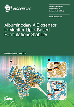
Albuminodan is a fluorescent probe consisting of bovine serum albumin labeled with acrylodan designed for monitoring the degradation of lipid nanoparticles. This biosensor can detect phospholipids, fatty acids, and lysophospholipids simultaneously when released from the formulation into the aqueous phase during spontaneous and phospholipase A₂-promoted hydrolysis. The characterization of the degradation process of lipid nanoparticle formulations was achieved by analyzing fluorescence, ligand-dependent spectral changes upon ligand binding. View this paper
- Issues are regarded as officially published after their release is announced to the table of contents alert mailing list.
- You may sign up for e-mail alerts to receive table of contents of newly released issues.
- PDF is the official format for papers published in both, html and pdf forms. To view the papers in pdf format, click on the "PDF Full-text" link, and use the free Adobe Reader to open them.
Previous Issue
Next Issue