Next Issue
Volume 16, February
Previous Issue
Volume 15, December
 
 
ijms-logo

Journal Browser

Journal Browser

Int. J. Mol. Sci., Volume 16, Issue 1 (January 2015) – 130 articles , Pages 1-2268

  • Issues are regarded as officially published after their release is announced to the table of contents alert mailing list.
  • You may sign up for e-mail alerts to receive table of contents of newly released issues.
  • PDF is the official format for papers published in both, html and pdf forms. To view the papers in pdf format, click on the "PDF Full-text" link, and use the free Adobe Reader to open them.
Cover Story:
Order results
Result details
Section
Select all
Export citation of selected articles as:
18 pages, 4930 KiB  
Review
Formins: Linking Cytoskeleton and Endomembranes in Plant Cells
by Fatima Cvrčková 1,*, Denisa Oulehlová 1,2 and Viktor Žárský 1,2
1 Department of Experimental Plant Biology, Faculty of Sciences, Charles University, Viničná 5, 128 43 Prague 2, Czech Republic
2 Institute of Experimental Botany, Academy of Sciences of the Czech Republic, Rozvojová 263, 165 02 Prague 6, Czech Republic
Int. J. Mol. Sci. 2015, 16(1), 1-18; https://doi.org/10.3390/ijms16010001 - 23 Dec 2014
Cited by 20 | Viewed by 11011
Abstract
The cytoskeleton plays a central part in spatial organization of the plant cytoplasm, including the endomebrane system. However, the mechanisms involved are so far only partially understood. Formins (FH2 proteins), a family of evolutionarily conserved proteins sharing the FH2 domain whose dimer can [...] Read more.
The cytoskeleton plays a central part in spatial organization of the plant cytoplasm, including the endomebrane system. However, the mechanisms involved are so far only partially understood. Formins (FH2 proteins), a family of evolutionarily conserved proteins sharing the FH2 domain whose dimer can nucleate actin, mediate the co-ordination between actin and microtubule cytoskeletons in multiple eukaryotic lineages including plants. Moreover, some plant formins contain transmembrane domains and participate in anchoring cytoskeletal structures to the plasmalemma, and possibly to other membranes. Direct or indirect membrane association is well documented even for some fungal and metazoan formins lacking membrane insertion motifs, and FH2 proteins have been shown to associate with endomembranes and modulate their dynamics in both fungi and metazoans. Here we summarize the available evidence suggesting that formins participate in membrane trafficking and endomembrane, especially ER, organization also in plants. We propose that, despite some methodological pitfalls inherent to in vivo studies based on (over)expression of truncated and/or tagged proteins, formins are beginning to emerge as candidates for the so far somewhat elusive link between the plant cytoskeleton and the endomembrane system. Full article
(This article belongs to the Special Issue Plant Cell Compartmentation and Volume Control)
Show Figures

Graphical abstract

21 pages, 723 KiB  
Article
Plasma Levels of Dimethylarginines in Preterm Very Low Birth Weight Neonates: Its Relation with Perinatal Factors and Short-Term Outcome
by Rob M. Moonen 1,2, Maurice J. Huizing 2, Giacomo Cavallaro 3, Gema E. González-Luis 4, Pilar Bas-Suárez 4, Jaap A. Bakker 5,6 and Eduardo Villamor 2,*
1 Department of Pediatrics, Atrium Medical Center Parkstad, Heerlen 6401CX, The Netherlands
2 Department of Pediatrics, Maastricht University Medical Center (MUMC+), Cardiovascular Research Institute Maastricht (CARIM), Maastricht 6202AZ, The Netherlands
3 Neonatal Intensive Care Unit, Department of Clinical Sciences and Community Health, Fondazione IRCCS Cà Granda Ospedale Maggiore Policlinico, Università degli Studi di Milano, Milan 20122, Italy
4 Department of Pediatrics, Hospital Universitario Materno-Infantil de Canarias, Las Palmas de Gran, Canaria 35016, Spain
5 Department of Clinical Genetics, Maastricht University Medical Center (MUMC+), Maastricht 6202AZ, The Netherlands
6 Department of Clinical Chemistry and Laboratory Medicine, Leiden University Medical Center, Leiden 2333ZA, The Netherlands
Int. J. Mol. Sci. 2015, 16(1), 19-39; https://doi.org/10.3390/ijms16010019 - 23 Dec 2014
Cited by 13 | Viewed by 6841
Abstract
Endogenously produced inhibitors of nitric oxide (NO) synthase, in particular asymmetric dimethylarginine (ADMA), are currently considered of importance in various disease states characterized by reduced NO availability. We investigated the association between plasma levels of ADMA, symmetric dimethylarginine (SDMA), l-arginine, and citrulline and [...] Read more.
Endogenously produced inhibitors of nitric oxide (NO) synthase, in particular asymmetric dimethylarginine (ADMA), are currently considered of importance in various disease states characterized by reduced NO availability. We investigated the association between plasma levels of ADMA, symmetric dimethylarginine (SDMA), l-arginine, and citrulline and perinatal factors and outcome in 130 preterm (gestational age ≤30 weeks) very low birth weight (VLBW, <1500 g) infants. Plasma samples were collected 6–12 h after birth. We did not find significant correlations between ADMA, SDMA, l-arginine, and citrulline levels and gestational age or birth weight. However, the arginine:ADMA ratio (AAR, a better indicator of NO availability than either arginine or ADMA separately) was positively correlated with gestational age. ADMA and arginine levels were not significantly different between males and females but males showed a negative correlation between ADMA levels and gestational age. Perinatal factors such as preeclampsia, chrorioamnionitis, prolonged rupture of membranes, or form of delivery did not significantly alter dimethylarginine levels or AAR. In contrast, the AAR was significantly reduced in the infants with respiratory distress, mechanical ventilation, and systemic hypotension Therefore, our data suggest that altered NO availability may play a role in the respiratory and cardiovascular adaptation in preterm VLBW infants. Full article
(This article belongs to the Section Biochemistry)
9 pages, 877 KiB  
Review
The Role of Autophagy as a Mechanism of Toxicity Induced by Multi-Walled Carbon Nanotubes in Human Lung Cells
by Tamotsu Tsukahara 1, Yoshikaszu Matsuda 2 and Hisao Haniu 3,*
1 Department of Molecular Pharmacology and Neuroscience, Nagasaki University Graduate School of Biomedical Sciences, 1-14 Bunkyo-machi, Nagasaki 852-8521, Japan
2 Clinical Pharmacology Educational Center, Nihon Pharmaceutical University, Ina-machi, Saitama 362-0806, Japan
3 Institute for Biomedical Sciences, Interdisciplinary Cluster for Cutting Edge Research, Shinshu University, 3-1-1 Asahi, Matsumoto, Nagano 390-8621, Japan
Int. J. Mol. Sci. 2015, 16(1), 40-48; https://doi.org/10.3390/ijms16010040 - 23 Dec 2014
Cited by 30 | Viewed by 7633
Abstract
Carbon nanotubes (CNTs) are promising nanomaterials having unique physical and chemical properties, with applications in a variety of fields. In this review, we briefly summarize the intrinsic properties of highly purified multi-walled CNTs (MWCNTs, HTT2800) and their potential hazardous effects on intracellular and [...] Read more.
Carbon nanotubes (CNTs) are promising nanomaterials having unique physical and chemical properties, with applications in a variety of fields. In this review, we briefly summarize the intrinsic properties of highly purified multi-walled CNTs (MWCNTs, HTT2800) and their potential hazardous effects on intracellular and extracellular pathways, which alter cellular signaling and impact major cell functions such as differentiation, reactive oxygen species (ROS) production, apoptosis, and autophagy. A recent study suggested that the induction of autophagy by CNTs causes nanotoxicity. Autophagy was recently recognized as a critical cell death pathway, and autophagosome accumulation has been found to be associated with exposure to CNTs. Although autophagy is considered as a cytoprotective process, it is often observed in association with cell death, and the relationship between autophagy and cell death remains unclear. Our recent study suggests that the levels of autophagy-related genes (LC3B) and autophagosome formation are clearly up-regulated, along with an increase in numbers of autophagosome vacuoles. This review highlights the importance of autophagy as an emerging mechanism of CNT toxicity. Full article
(This article belongs to the Special Issue Nanotoxicology and Lung Diseases)
Show Figures

Figure 1

19 pages, 12334 KiB  
Article
The Regulation of Nitric Oxide Synthase Isoform Expression in Mouse and Human Fallopian Tubes: Potential Insights for Ectopic Pregnancy
by Junting Hu 1,2,†, Shulan Ma 1,3,†, Sien Zou 4, Xin Li 4, Peng Cui 1, Birgitta Weijdegård 2, Gencheng Wu 1, Ruijin Shao 2, Håkan Billig 2,* and Yi Feng 1,2,*
1 Department of Integrative Medicine and Neurobiology, State Key Lab of Medical Neurobiology, Shanghai Medical College and Institute of Acupuncture Research (WHO Collaborating Center for Traditional Medicine), Institute of Brain Science, Fudan University, Shanghai 200032, China
2 Department of Physiology and Endocrinology, Institute of Neuroscience and Physiology, the Sahlgrenska Academy at University of Gothenburg, 40530 Gothenburg, Sweden
3 Training Center of Medical Experiments, Fudan University, Shanghai 200032, China
4 Department of Gynecology, Obstetrics and Gynecology Hospital of Fudan University, Shanghai 200011, China
These authors contributed equally to this work.
Int. J. Mol. Sci. 2015, 16(1), 49-67; https://doi.org/10.3390/ijms16010049 - 23 Dec 2014
Cited by 14 | Viewed by 7765
Abstract
Nitric oxide (NO) is highly unstable and has a half-life of seconds in buffer solutions. It is synthesized by NO-synthase (NOS), which has been found to exist in the following three isoforms: neuro nitric oxide synthase (nNOS), inducible nitric oxide synthase (iNOS), and [...] Read more.
Nitric oxide (NO) is highly unstable and has a half-life of seconds in buffer solutions. It is synthesized by NO-synthase (NOS), which has been found to exist in the following three isoforms: neuro nitric oxide synthase (nNOS), inducible nitric oxide synthase (iNOS), and endothelial nitric oxide synthase (eNOS). NOS activity is localized in the reproductive tracts of many species, although direct evidence for NOS isoforms in the Fallopian tubes of mice is still lacking. In the present study, we investigated the expression and regulation of NOS isoforms in the mouse and human Fallopian tubes during the estrous and menstrual cycles, respectively. We also measured isoform expression in humans with ectopic pregnancy and in mice treated with lipopolysaccharide (LPS). Our results confirmed the presence of different NOS isoforms in the mouse and human Fallopian tubes during different stages of the estrous and menstrual cycles and showed that iNOS expression increased in the Fallopian tubes of women with ectopic pregnancy and in LPS-treated mice. Elevated iNOS activity might influence ovulation, cilia beats, contractility, and embryo transportation in such a manner as to increase the risk of ectopic pregnancy. This study has provided morphological and molecular evidence that NOS isoforms are present and active in the human and mouse Fallopian tubes and suggests that iNOS might play an important role in both the reproductive cycle and infection-induced ectopic pregnancies. Full article
(This article belongs to the Section Biochemistry)
Show Figures

Figure 1

23 pages, 2272 KiB  
Review
Exposure to Non-Extreme Solar UV Daylight: Spectral Characterization, Effects on Skin and Photoprotection
by Claire Marionnet 1, Caroline Tricaud 2 and Françoise Bernerd 1,*
1 Oréal Research and Innovation, 1 avenue Eugène Schueller, 93601 Aulnay-sous-Bois, France
2 Oréal Research and Innovation, 188-200 rue Paul Hochart, 94550 Chevilly-Larue, France
Int. J. Mol. Sci. 2015, 16(1), 68-90; https://doi.org/10.3390/ijms16010068 - 23 Dec 2014
Cited by 137 | Viewed by 14457
Abstract
The link between chronic sun exposure of human skin and harmful clinical consequences such as photo-aging and skin cancers is now indisputable. These effects are mostly due to ultraviolet (UV) rays (UVA, 320–400 nm and UVB, 280–320 nm). The UVA/UVB ratio can vary [...] Read more.
The link between chronic sun exposure of human skin and harmful clinical consequences such as photo-aging and skin cancers is now indisputable. These effects are mostly due to ultraviolet (UV) rays (UVA, 320–400 nm and UVB, 280–320 nm). The UVA/UVB ratio can vary with latitude, season, hour, meteorology and ozone layer, leading to different exposure conditions. Zenithal sun exposure (for example on a beach around noon under a clear sky) can rapidly induce visible and well-characterized clinical consequences such as sunburn, predominantly induced by UVB. However, a limited part of the global population is exposed daily to such intense irradiance and until recently little attention has been paid to solar exposure that does not induce any short term clinical impact. This paper will review different studies on non-extreme daily UV exposures with: (1) the characterization and the definition of the standard UV daylight and its simulation in the laboratory; (2) description of the biological and clinical effects of such UV exposure in an in vitro reconstructed human skin model and in human skin in vivo, emphasizing the contribution of UVA rays and (3) analysis of photoprotection approaches dedicated to prevent the harmful impact of such UV exposure. Full article
(This article belongs to the Collection Radiation Toxicity in Cells)
Show Figures

Figure 1

23 pages, 3004 KiB  
Article
Transfer RNA Methyltransferases from Thermoplasma acidophilum, a Thermoacidophilic Archaeon
by Takuya Kawamura, Ryou Anraku, Takahiro Hasegawa, Chie Tomikawa and Hiroyuki Hori *
1 Department of Materials Science and Biotechnology, Graduate School of Science and Engineering, Ehime Univsersity, Bunkyo 3, Matsuyama, Ehime 790-8577, Japan
These authors contributed equally to this work.
Int. J. Mol. Sci. 2015, 16(1), 91-113; https://doi.org/10.3390/ijms16010091 - 23 Dec 2014
Cited by 17 | Viewed by 7224
Abstract
We investigated tRNA methyltransferase activities in crude cell extracts from the thermoacidophilic archaeon Thermoplasma acidophilum. We analyzed the modified nucleosides in native initiator and elongator tRNAMet, predicted the candidate genes for the tRNA methyltransferases on the basis of the tRNA [...] Read more.
We investigated tRNA methyltransferase activities in crude cell extracts from the thermoacidophilic archaeon Thermoplasma acidophilum. We analyzed the modified nucleosides in native initiator and elongator tRNAMet, predicted the candidate genes for the tRNA methyltransferases on the basis of the tRNAMet and tRNALeu sequences, and characterized Trm5, Trm1 and Trm56 by purifying recombinant proteins. We found that the Ta0997, Ta0931, and Ta0836 genes of T. acidophilum encode Trm1, Trm56 and Trm5, respectively. Initiator tRNAMet from T. acidophilum strain HO-62 contained G+, m1I, and m22G, which were not reported previously in this tRNA, and the m2G26 and m22G26 were formed by Trm1. In the case of elongator tRNAMet, our analysis showed that the previously unidentified G modification at position 26 was a mixture of m2G and m22G, and that they were also generated by Trm1. Furthermore, purified Trm1 and Trm56 could methylate the precursor of elongator tRNAMet, which has an intron at the canonical position. However, the speed of methyl-transfer by Trm56 to the precursor RNA was considerably slower than that to the mature transcript, which suggests that Trm56 acts mainly on the transcript after the intron has been removed. Moreover, cellular arrangements of the tRNA methyltransferases in T. acidophilum are discussed. Full article
(This article belongs to the Special Issue Functions of Transfer RNAs)
Show Figures

Figure 1

21 pages, 771 KiB  
Review
Transmembrane Signal Transduction in Oocyte Maturation and Fertilization: Focusing on Xenopus laevis as a Model Animal
by Ken-ichi Sato
Laboratory of Cell Signaling and Development, Department of Molecular Biosciences, Faculty of Life Sciences, Kyoto Sangyo University, Kamigamo-motoyama, Kita-ku, Kyoto 603-8555, Japan
Int. J. Mol. Sci. 2015, 16(1), 114-134; https://doi.org/10.3390/ijms16010114 - 23 Dec 2014
Cited by 11 | Viewed by 10706
Abstract
Fertilization is a cell biological phenomenon of crucial importance for the birth of new life in a variety of multicellular and sexual reproduction species such as algae, animal and plants. Fertilization involves a sequence of events, in which the female gamete “egg” and [...] Read more.
Fertilization is a cell biological phenomenon of crucial importance for the birth of new life in a variety of multicellular and sexual reproduction species such as algae, animal and plants. Fertilization involves a sequence of events, in which the female gamete “egg” and the male gamete “spermatozoon (sperm)” develop, acquire their functions, meet and fuse with each other, to initiate embryonic and zygotic development. Here, it will be briefly reviewed how oocyte cytoplasmic components are orchestrated to undergo hormone-induced oocyte maturation and sperm-induced activation of development. I then review how sperm-egg membrane interaction/fusion and activation of development in the fertilized egg are accomplished and regulated through egg coat- or egg plasma membrane-associated components, highlighting recent findings and future directions in the studies using Xenopus laevis as a model experimental animal. Full article
(This article belongs to the Special Issue Cellular and Molecular Mechanisms of Sperm-Egg Interaction)
Show Figures

Figure 1

13 pages, 1203 KiB  
Article
Quantification of Drug Transport Function across the MultipleResistance-Associated Protein 2 (Mrp2) in Rat Livers
by Pierre Bonnaventure 1 and Catherine M. Pastor 1,2,*
1 Universitaires de Genève, Geneva 1205, Switzerland
2 Laboratory of Imaging Biomarkers, UMR1149 INSERM-Université Paris Diderot, Sorbonne Paris Cité, Paris 70519, France
Int. J. Mol. Sci. 2015, 16(1), 135-147; https://doi.org/10.3390/ijms16010135 - 24 Dec 2014
Cited by 7 | Viewed by 5824
Abstract
To understand the transport function of drugs across the canalicular membrane of hepatocytes, it would be important to measure concentrations in hepatocytes and bile. However, these concentration gradients are rarely provided. The aim of the study is then to measure these concentrations and [...] Read more.
To understand the transport function of drugs across the canalicular membrane of hepatocytes, it would be important to measure concentrations in hepatocytes and bile. However, these concentration gradients are rarely provided. The aim of the study is then to measure these concentrations and define parameters to quantify the canalicular transport of drugs through the multiple resistance associated-protein 2 (Mrp2) in entire rat livers. Besides drug bile excretion rates, we measured additional parameters to better define transport function across Mrp2: (1) Concentration gradients between hepatocyte and bile concentrations over time; and (2) a unique parameter (canalicular concentration ratio) that represents the slope of the non-linear regression curve between hepatocyte and bile concentrations. This information was obtained in isolated rat livers perfused with gadobenate dimeglumine (BOPTA) and mebrofenin (MEB), two hepatobiliary drugs used in clinical liver imaging. Interestingly, despite different transport characteristics including excretion rates into bile and hepatocyte clearance into bile, BOPTA and MEB have a similar canalicular concentration ratio. In contrast, the ratio was null when BOPTA was not excreted in bile in hepatocytes lacking Mrp2. The canalicular concentration ratio is more informative than bile excretion rates because it is independent of time, bile flows, and concentrations perfused in portal veins. It would be interesting to apply such information in human liver imaging where hepatobiliary compounds are increasingly investigated. Full article
(This article belongs to the Section Biochemistry)
Show Figures

Graphical abstract

11 pages, 2374 KiB  
Article
Systemic Delivery of Protein Nanocages Bearing CTT Peptides for Enhanced Imaging of MMP-2 Expression in Metastatic Tumor Models
by Takahito Kawano 1,2, Masaharu Murata 1,2,3,*, Jing Shu Piao 2,3, Sayoko Narahara 1,2, Nobuhito Hamano 2,3, Jeong-Hun Kang 4 and Makoto Hashizume 1,2,3
1 Innovation Center for Medical Redox Navigation, Kyushu University, 3-1-1 Maidashi, Higashi-ku, Fukuoka 812-8582, Japan
2 Department of Advanced Medical Initiatives, Kyushu University, 3-1-1 Maidashi, Higashi-ku, Fukuoka 812-8582, Japan
3 Faculty of Medical Sciences and Center for Advanced Medical Innovation, Kyushu University, 3-1-1 Maidashi, Higashi-ku, Fukuoka 812-8582, Japan
4 Division of Biopharmaceutics and Pharmacokinetics, Department of Biomedical Engineering, National Cerebral and Cardiovascular Center Research Institute, 5-7-1 Fujishirodai, Suita, Osaka 565-8565, Japan
Int. J. Mol. Sci. 2015, 16(1), 148-158; https://doi.org/10.3390/ijms16010148 - 24 Dec 2014
Cited by 21 | Viewed by 7161
Abstract
Matrix metalloproteinase 2 (MMP-2) in metastatic cancer tissue, which is associated with a poor prognosis, is a potential target for tumor imaging in vivo. Here, we describe a metastatic cancer cell-targeted protein nanocage. An MMP-2-binding peptide, termed CTT peptide (CTTHWGFTLC), was conjugated [...] Read more.
Matrix metalloproteinase 2 (MMP-2) in metastatic cancer tissue, which is associated with a poor prognosis, is a potential target for tumor imaging in vivo. Here, we describe a metastatic cancer cell-targeted protein nanocage. An MMP-2-binding peptide, termed CTT peptide (CTTHWGFTLC), was conjugated to the surface of a naturally occurring heat shock protein nanocage by genetic modification. The engineered protein nanocages showed a binding affinity for MMP-2 and selective uptake in cancer cells that highly expressed MMP-2 in vitro. In near-infrared fluorescence imaging, the nanocages showed specific and significant accumulation in tumor tissue after intravenous injection in vivo. These protein nanocages conjugated with CTT peptide could be potentially applied to a noninvasive near-infrared fluorescence detection method for imaging gelatinase activity in metastatic tumors in vivo. Full article
(This article belongs to the Special Issue Bioactive Nanoparticles 2014)
Show Figures

Figure 1

19 pages, 1416 KiB  
Article
Upregulation of TLRs and IL-6 as a Marker in Human Colorectal Cancer
by Chien-Chang Lu 1,2,*, Hsing-Chun Kuo 3,4,5, Feng-Sheng Wang 6, Ming-Huey Jou 6, Ko-Chao Lee 1 and Jiin-Haur Chuang 7,*
1 Division of Colorectal Surgery, Department of Surgery, Chang Gung Memorial Hospital-Kaohsiung Medical Center, Chang Gung University College of Medicine, Kaohsiung 833, Taiwan
2 Graduate Institute of Clinical Medical Sciences, College of Medicine, Chang Gung University, Taoyuan 330, Taiwan
3 Department of Nursing, Chang Gung University of Science and Technology, Chiayi 61363, Taiwan
4 Chronic Diseases and Health Promotion Research Center, Chang Gung University of Science and Technology, Chiayi 61363, Taiwan
5 Research Center for Industry of Human Ecology, Chang Gung University of Science and Technology, Taoyuan 333, Taiwan
6 Department of Medical Research, Chang Gung Memorial Hospital-Kaohsiung Medical Center, Chang Gung University College of Medicine, Kaohsiung 833, Taiwan
7 Department of Surgery, Chang Gung Memorial Hospital-Kaohsiung Medical Center, Chang Gung University College of Medicine, Kaohsiung 833, Taiwan
Int. J. Mol. Sci. 2015, 16(1), 159-177; https://doi.org/10.3390/ijms16010159 - 24 Dec 2014
Cited by 68 | Viewed by 8366
Abstract
Toll-like receptors (TLRs) not only form an important part of the innate immune system but also serve to activate the adaptive immune system in response to cancer. Real-time PCR; immunohistochemical stain and Western blotting analyses were performed to clarify molecular alterations in colorectal [...] Read more.
Toll-like receptors (TLRs) not only form an important part of the innate immune system but also serve to activate the adaptive immune system in response to cancer. Real-time PCR; immunohistochemical stain and Western blotting analyses were performed to clarify molecular alterations in colorectal cancer (CRC) patients. We identified Toll-like receptor 1 (TLR1), TLR2, TLR4 and TLR8 gene expression levels and downstream gene, i.e., interleukin-6 (IL-6), IL-8, interferon-α (IFN-α) and myeloid differentiation primary-response protein-88 (MyD88), expression levels in CRC patients and in cancer cell lines. CRC tissues have higher TLR1, TLR2, TLR4, TLR8, IL-6 and IL-8 gene expression levels than do the normal colon mucosa (p < 0.05). TLR2 expression varied in different cell types (mucosa and lymphocytes). There was no difference in the MyD88 and IFN-α gene expression levels between cancerous and normal colon mucosa. CRC patients had higher levels of IL-6 (p = 0.002) and IL-8 (p = 0.038) expression than healthy volunteers did; and higher IL-6 and IL-8 expression was also found to signify a higher risk of recurrence. CL075 (3M002) treatments can reduce the production of IL-8 in different cancer cell lines. The signaling pathway of TLRs in cancer tissue is different from that in normal cells; and is MyD88-independent. Higher expression levels of TLR1, TLR2, TLR 4 and TLR 8 mRNA were related to upregulation inflammatory cytokines IL-6 and IL-8 gene expression in tissue and to the upregulation of IL-6 in blood. The concentration of IL-6 in serum can be used as an indicator of the possibility of CRC recurrence. Treatment with 3M002 can reduce IL-6 production in vitro and may prevent CRC recurrence. Our findings provide evidence that TLR1, TLR2, TLR4 and TLR8 gene expression induce downstream IL-6 and IL-8 gene expression; detection of these expression levels can serve as a CRC marker. Full article
(This article belongs to the Section Biochemistry)
Show Figures

Figure 1

15 pages, 2239 KiB  
Article
Increased ARPP-19 Expression Is Associated with Hepatocellular Carcinoma
by Haiyan Song 1,†, Jielu Pan 1,†, Yang Liu 1, Hongzhu Wen 1, Lei Wang 1, Jiefeng Cui 2, Yinkun Liu 2, Bing Hu 3, Zemin Yao 4 and Guang Ji 1,*
1 Institute of Digestive Diseases, Longhua Hospital, Shanghai University of Traditional Chinese Medicine, Shanghai 200032, China
2 Liver Cancer Institute of Zhongshan Hospital, Fudan University, Shanghai 200032, China
3 Department of Oncology and Institute of Traditional Chinese Medicine in Oncology, Longhua Hospital, Shanghai University of Traditional Chinese Medicine, Shanghai 200032, China
4 Department of Biochemistry, Microbiology and Immunology, Ottawa Institute of Systems Biology, University of Ottawa, Ottawa, ON K1H 8M5, Canada
These authors contributed equally to this work.
Int. J. Mol. Sci. 2015, 16(1), 178-192; https://doi.org/10.3390/ijms16010178 - 24 Dec 2014
Cited by 17 | Viewed by 8042
Abstract
The cAMP-regulated phosphoprotein 19 (ARPP-19) plays a key role in cell mitotic G2/M transition. Expression of ARPP-19 was increased in human hepatocellular carcinoma (HCC) compared to adjacent non-tumorous liver tissues in 36 paired liver samples, and the level of ARPP-19 in HCC tissues [...] Read more.
The cAMP-regulated phosphoprotein 19 (ARPP-19) plays a key role in cell mitotic G2/M transition. Expression of ARPP-19 was increased in human hepatocellular carcinoma (HCC) compared to adjacent non-tumorous liver tissues in 36 paired liver samples, and the level of ARPP-19 in HCC tissues was positively correlated with the tumor size. To determine the interrelationship between ARPP-19 expression and HCC, we silenced ARPP-19 expression in the human hepatocarcinoma HepG2 and SMMC-7721 cells using lentivirus encoding ARPP-19 siRNA. HepG2 and SMMC-7721 cells with ARPP-19 knockdown displayed lowered cell growth rate, retarded colony formation and increased arrest at the G2/M phase transition. Silencing ARPP-19 in HCC cells resulted in decreased protein levels of phospho-(Ser) CDKs substrates and increased levels of inactivated cyclin division cycle 2 (Cdc2). Therefore, ARPP-19 may play a role in HCC pathogenesis through regulating cell proliferation. Full article
(This article belongs to the Special Issue Molecular Mechanisms of Human Liver Diseases)
Show Figures

Figure 1

25 pages, 1609 KiB  
Review
Oxidative Stress and Its Significant Roles in Neurodegenerative Diseases and Cancer
by Raynoo Thanan 1,2, Shinji Oikawa 3, Yusuke Hiraku 3, Shiho Ohnishi 4, Ning Ma 5, Somchai Pinlaor 2,6, Puangrat Yongvanit 1,2, Shosuke Kawanishi 4 and Mariko Murata 3,*
1 Department of Biochemistry, Faculty of Medicine, Khon Kaen University, Khon Kaen 40002, Thailand
2 Liver Fluke and Cholangiocarcinoma Research Center, Faculty of Medicine, Khon Kaen University, Khon Kaen 40002, Thailand
3 Department of Environmental and Molecular Medicine, Mie University Graduate School of Medicine, 2-174 Edobashi, Tsu, Mie 514-8507, Japan
4 Faculty of Pharmaceutical Sciences, Suzuka University of Medical Science, Suzuka, Mie 513-8670, Japan
5 Faculty of Nursing Science, Suzuka University of Medical Science, Suzuka, Mie 513-8670, Japan
6 Department of Parasitology, Faculty of Medicine, Khon Kaen University, Khon Kaen 40002, Thailand
Int. J. Mol. Sci. 2015, 16(1), 193-217; https://doi.org/10.3390/ijms16010193 - 24 Dec 2014
Cited by 389 | Viewed by 26185
Abstract
Reactive oxygen and nitrogen species have been implicated in diverse pathophysiological conditions, including inflammation, neurodegenerative diseases and cancer. Accumulating evidence indicates that oxidative damage to biomolecules including lipids, proteins and DNA, contributes to these diseases. Previous studies suggest roles of lipid peroxidation and [...] Read more.
Reactive oxygen and nitrogen species have been implicated in diverse pathophysiological conditions, including inflammation, neurodegenerative diseases and cancer. Accumulating evidence indicates that oxidative damage to biomolecules including lipids, proteins and DNA, contributes to these diseases. Previous studies suggest roles of lipid peroxidation and oxysterols in the development of neurodegenerative diseases and inflammation-related cancer. Our recent studies identifying and characterizing carbonylated proteins reveal oxidative damage to heat shock proteins in neurodegenerative disease models and inflammation-related cancer, suggesting dysfunction in their antioxidative properties. In neurodegenerative diseases, DNA damage may not only play a role in the induction of apoptosis, but also may inhibit cellular division via telomere shortening. Immunohistochemical analyses showed co-localization of oxidative/nitrative DNA lesions and stemness markers in the cells of inflammation-related cancers. Here, we review oxidative stress and its significant roles in neurodegenerative diseases and cancer. Full article
(This article belongs to the Special Issue DNA Damage and Repair in Degenerative Diseases 2014)
Show Figures

Graphical abstract

12 pages, 675 KiB  
Review
Compartmentalization Role of A-Kinase Anchoring Proteins (AKAPs) in Mediating Protein Kinase A (PKA) Signaling and Cardiomyocyte Hypertrophy
by Abeer Rababa'h 1,*, Sonal Singh 2, Santosh V. Suryavanshi 2, Salah Eldien Altarabsheh 3, Salil V. Deo 4 and Bradley K. McConnell 2,*
1 Department of Clinical Pharmacy, Jordan University of Science and Technology, Irbid 22110, Jordan
2 Department of Pharmacological and Pharmaceutical Sciences, University of Houston, Texas Medical Center, Houston, TX 77204, USA
3 Department of Cardiac Surgery, Queen Alia Heart Institute, Amman 11953, Jordan
4 Department of Cardiovascular Surgery, Case Western Reserve University, Cleveland, OH 44106, USA
Int. J. Mol. Sci. 2015, 16(1), 218-229; https://doi.org/10.3390/ijms16010218 - 24 Dec 2014
Cited by 36 | Viewed by 9276
Abstract
The Beta-adrenergic receptors (β-ARs) stimulation enhances contractility through protein kinase-A (PKA) substrate phosphorylation. This PKA signaling is conferred in part by PKA binding to A-kinase anchoring proteins (AKAPs). AKAPs coordinate multi-protein signaling networks that are targeted to specific intracellular locations, resulting in the [...] Read more.
The Beta-adrenergic receptors (β-ARs) stimulation enhances contractility through protein kinase-A (PKA) substrate phosphorylation. This PKA signaling is conferred in part by PKA binding to A-kinase anchoring proteins (AKAPs). AKAPs coordinate multi-protein signaling networks that are targeted to specific intracellular locations, resulting in the localization of enzyme activity and transmitting intracellular actions of neurotransmitters and hormones to its target substrates. In particular, mAKAP (muscle-selective AKAP) has been shown to be present on the nuclear envelope of cardiomyocytes with various proteins including: PKA-regulatory subunit (RIIα), phosphodiesterase-4D3, protein phosphatase-2A, and ryanodine receptor (RyR2). Therefore, through the coordination of spatial-temporal signaling of proteins and enzymes, mAKAP controls cyclic-adenosine monophosphate (cAMP) levels very tightly and functions as a regulator of PKA-mediated substrate phosphorylation leading to changes in calcium availability and myofilament calcium sensitivity. The goal of this review is to elucidate the critical compartmentalization role of mAKAP in mediating PKA signaling and regulating cardiomyocyte hypertrophy by acting as a scaffolding protein. Based on our literature search and studying the structure–function relationship between AKAP scaffolding protein and its binding partners, we propose possible explanations for the mechanism by which mAKAP promotes cardiac hypertrophy. Full article
(This article belongs to the Special Issue Signalling Molecules and Signal Transduction in Cells 2014)
26 pages, 1060 KiB  
Review
The Potential of Liposomes with Carbonic Anhydrase IX to Deliver Anticancer Ingredients to Cancer Cells in Vivo
by Huei Leng Helena Ng 1, Aiping Lu 1,2,†, Ge Lin 3,†, Ling Qin 4,† and Zhijun Yang 1,2,*
1 School of Chinese Medicine, Hong Kong Baptist University, 7 Baptist University Road, Kowloon Tong, Kowloon, Hong Kong 999077, China
2 Changshu Research Institute, Hong Kong Baptist University, Changshu Economic and Technological Development (CETD) Zone, Changshu 215500, China
3 School of Biomedical Sciences, Chinese University of Hong Kong, Area 39, CUHK, Shatin, NT, Hong Kong 999077, China
4 Musculoskeletal Research Laboratory, Department of Orthopaedics and Traumatology, The Chinese University of Hong Kong, Prince of Wales Hospital, Shatin, NT, Hong Kong 999077, China
These authors contributed equally to this work.
Int. J. Mol. Sci. 2015, 16(1), 230-255; https://doi.org/10.3390/ijms16010230 - 24 Dec 2014
Cited by 14 | Viewed by 9687
Abstract
Drug delivery nanocarriers, especially targeted drug delivery by liposomes are emerging as a class of therapeutics for cancer. Early research results suggest that liposomal therapeutics enhanced efficacy, while simultaneously reducing side effects, owing to properties such as more targeted localization in tumors and [...] Read more.
Drug delivery nanocarriers, especially targeted drug delivery by liposomes are emerging as a class of therapeutics for cancer. Early research results suggest that liposomal therapeutics enhanced efficacy, while simultaneously reducing side effects, owing to properties such as more targeted localization in tumors and active cellular uptake. Here, we highlight the features of immunoliposomes that distinguish them from previous anticancer therapies, and describe how these features provide the potential for therapeutic effects that are not achievable with other modalities. While a large number of studies has been published, the emphasis here is placed on the carbonic anhydrase IX (CA-IX) and the conjugated liposomes that are likely to open a new chapter on drug delivery system by using immunoliposomes to deliver anticancer ingredients to cancer cells in vivo. Full article
(This article belongs to the Special Issue Bioactive Nanoparticles 2014)
Show Figures

Figure 1

28 pages, 1088 KiB  
Review
Molecular Targets of Antihypertensive Peptides: Understanding the Mechanisms of Action Based on the Pathophysiology of Hypertension
by Kaustav Majumder 1,2 and Jianping Wu 1,2,*
1 Department of Agricultural, Food and Nutritional Science, Faculty of Agricultural, Life and Environmental Sciences, University of Alberta, Edmonton, AB T6G 2P5, Canada
2 Cardiovascular Research Centre, Faculty of Medicine & Dentistry, University of Alberta, Edmonton, AB T6G 2S2, Canada
Int. J. Mol. Sci. 2015, 16(1), 256-283; https://doi.org/10.3390/ijms16010256 - 24 Dec 2014
Cited by 146 | Viewed by 18115
Abstract
There is growing interest in using functional foods or nutraceuticals for the prevention and treatment of hypertension or high blood pressure. Although numerous preventive and therapeutic pharmacological interventions are available on the market, unfortunately, many patients still suffer from poorly controlled hypertension. Furthermore, [...] Read more.
There is growing interest in using functional foods or nutraceuticals for the prevention and treatment of hypertension or high blood pressure. Although numerous preventive and therapeutic pharmacological interventions are available on the market, unfortunately, many patients still suffer from poorly controlled hypertension. Furthermore, most pharmacological drugs, such as inhibitors of angiotensin-I converting enzyme (ACE), are often associated with significant adverse effects. Many bioactive food compounds have been characterized over the past decades that may contribute to the management of hypertension; for example, bioactive peptides derived from various food proteins with antihypertensive properties have gained a great deal of attention. Some of these peptides have exhibited potent in vivo antihypertensive activity in both animal models and human clinical trials. This review provides an overview about the complex pathophysiology of hypertension and demonstrates the potential roles of food derived bioactive peptides as viable interventions targeting specific pathways involved in this disease process. This review offers a comprehensive guide for understanding and utilizing the molecular mechanisms of antihypertensive actions of food protein derived peptides. Full article
(This article belongs to the Special Issue Bioactive Proteins and Peptides Derived from Food)
Show Figures

Figure 1

23 pages, 1019 KiB  
Review
The Anticancer Drug Ellipticine Activated with Cytochrome P450 Mediates DNA Damage Determining Its Pharmacological Efficiencies: Studies with Rats, Hepatic Cytochrome P450 Reductase Null (HRN™) Mice and Pure Enzymes
by Marie Stiborová 1,*, Věra Černá 1, Michaela Moserová 1, Iveta Mrízová 1, Volker M. Arlt 2 and Eva Frei 3
1 Department of Biochemistry, Faculty of Science, Charles University, Hlavova 2030, CZ-12843 Prague 2, Czech Republic
2 Analytical and Environmental Sciences Division, MRC-PHE Centre for Environmental & Health, King’s College London, 150 Stamford Street, London SE1 9NH, UK
3 Division of Preventive Oncology, National Center for Tumor Diseases, German Cancer Research Center (DKFZ), Im Neuenheimer Feld 280, 69120 Heidelberg, Germany
Int. J. Mol. Sci. 2015, 16(1), 284-306; https://doi.org/10.3390/ijms16010284 - 25 Dec 2014
Cited by 60 | Viewed by 8680
Abstract
Ellipticine is a DNA-damaging agent acting as a prodrug whose pharmacological efficiencies and genotoxic side effects are dictated by activation with cytochrome P450 (CYP). Over the last decade we have gained extensive experience in using pure enzymes and various animal models that helped [...] Read more.
Ellipticine is a DNA-damaging agent acting as a prodrug whose pharmacological efficiencies and genotoxic side effects are dictated by activation with cytochrome P450 (CYP). Over the last decade we have gained extensive experience in using pure enzymes and various animal models that helped to identify CYPs metabolizing ellipticine. In this review we focus on comparison between the in vitro and in vivo studies and show a necessity of both approaches to obtain valid information on CYP enzymes contributing to ellipticine metabolism. Discrepancies were found between the CYP enzymes activating ellipticine to 13-hydroxy- and 12-hydroxyellipticine generating covalent DNA adducts and those detoxifying this drug to 9-hydroxy- and 7-hydroellipticine in vitro and in vivo. In vivo, formation of ellipticine-DNA adducts is dependent not only on expression levels of CYP3A, catalyzing ellipticine activation in vitro, but also on those of CYP1A that oxidize ellipticine in vitro mainly to the detoxification products. The finding showing that cytochrome b5 alters the ratio of ellipticine metabolites generated by CYP1A1/2 and 3A4 explained this paradox. Whereas the detoxification of ellipticine by CYP1A and 3A is either decreased or not changed by cytochrome b5, activation leading to ellipticine-DNA adducts increased considerably. We show that (I) the pharmacological effects of ellipticine mediated by covalent ellipticine-derived DNA adducts are dictated by expression levels of CYP1A, 3A and cytochrome b5, and its own potency to induce these enzymes in tumor tissues, (II) animal models, where levels of CYPs are either knocked out or induced are appropriate to identify CYPs metabolizing ellipticine in vivo, and (III) extrapolation from in vitro data to the situation in vivo is not always possible, confirming the need for these animal models. Full article
(This article belongs to the Special Issue DNA Damage and Repair in Degenerative Diseases 2014)
Show Figures

Figure 1

14 pages, 3310 KiB  
Article
USP22 Promotes NSCLC Tumorigenesis via MDMX Up-Regulation and Subsequent p53 Inhibition
by Fangbao Ding 1,*,†, Chunrong Bao 1,†, Yue Tian 2,†, Haibo Xiao 1, Mingsong Wang 1, Xiao Xie 1, Fengqing Hu 1 and Ju Mei 1
1 Department of Cardiothoracic Surgery, Xinhua Hospital, School of Medicine, Shanghai Jiaotong University, Shanghai 200092, China
2 Institute of Orthopaedics, Chinese PLA General Hospital, Beijing 100853, China
These authors contributed equally to this work.
Int. J. Mol. Sci. 2015, 16(1), 307-320; https://doi.org/10.3390/ijms16010307 - 25 Dec 2014
Cited by 32 | Viewed by 6763
Abstract
Increasing evidence suggests that ubiquitin-specific protease 22 (USP22) has great clinicopathologic significance in oncology. In this study, we investigated the role of USP22 in human NSCLC tumorigenesis along with the underlying mechanisms of action. First, we determined the expression of USP22 in human [...] Read more.
Increasing evidence suggests that ubiquitin-specific protease 22 (USP22) has great clinicopathologic significance in oncology. In this study, we investigated the role of USP22 in human NSCLC tumorigenesis along with the underlying mechanisms of action. First, we determined the expression of USP22 in human NSCLC, as well as normal tissues and cell lines. We then studied the effects of USP22 silencing by shRNA on NSCLC cell growth in vitro and tumorigenesis in vivo, along with the effect on the p53 pathway. We found that USP22 is overexpressed in human NSCLC tissues and cell lines. USP22 silencing by shRNA inhibits proliferation, induces apoptosis and arrests cells at the G0/G1 phases in NSCLC cells and curbs human NSCLC tumor growth in a mouse xenograft model. Additionally, USP22 silencing downregulates MDMX protein expression and activates the p53 pathway. Our co-immunoprecipitation analysis shows that USP22 interacts with MDMX in NSCLC cells. Furthermore, MDMX silencing leads to growth arrest and apoptosis in NSCLC cells, and over-expression of MDMX reverses the USP22 silencing-induced effects. Taken together, our results suggest that USP22 promotes NSCLC tumorigenesis in vitro and in vivo through MDMX upregulation and subsequent p53 inhibition. USP22 may represent a novel target for NSCLC treatment. Full article
(This article belongs to the Section Biochemistry)
Show Figures

Figure 1

29 pages, 1161 KiB  
Review
tRNAs as Antibiotic Targets
by Shaileja Chopra and John Reader *
Department of Cell Biology and Physiology, the University of North Carolina at Chapel Hill, 536 Taylor Hall, 109 Mason Farm Road, Chapel Hill, NC 27599-7545, USA
Int. J. Mol. Sci. 2015, 16(1), 321-349; https://doi.org/10.3390/ijms16010321 - 25 Dec 2014
Cited by 31 | Viewed by 19026
Abstract
Transfer RNAs (tRNAs) are central players in the protein translation machinery and as such are prominent targets for a large number of natural and synthetic antibiotics. This review focuses on the role of tRNAs in bacterial antibiosis. We will discuss examples of antibiotics [...] Read more.
Transfer RNAs (tRNAs) are central players in the protein translation machinery and as such are prominent targets for a large number of natural and synthetic antibiotics. This review focuses on the role of tRNAs in bacterial antibiosis. We will discuss examples of antibiotics that target multiple stages in tRNA biology from tRNA biogenesis and modification, mature tRNAs, aminoacylation of tRNA as well as prevention of proper tRNA function by small molecules binding to the ribosome. Finally, the role of deacylated tRNAs in the bacterial “stringent response” mechanism that can lead to bacteria displaying antibiotic persistence phenotypes will be discussed. Full article
(This article belongs to the Special Issue Functions of Transfer RNAs)
Show Figures

Figure 1

13 pages, 1072 KiB  
Article
Stable Hydrogen Production from Ethanol through Steam Reforming Reaction over Nickel-Containing Smectite-Derived Catalyst
by Hiroshi Yoshida, Ryohei Yamaoka and Masahiko Arai *
Division of Chemical Process Engineering, Faculty of Engineering, Hokkaido University, Sapporo 060-8628, Japan
Int. J. Mol. Sci. 2015, 16(1), 350-362; https://doi.org/10.3390/ijms16010350 - 25 Dec 2014
Cited by 10 | Viewed by 5990
Abstract
Hydrogen production through steam reforming of ethanol was investigated with conventional supported nickel catalysts and a Ni-containing smectite-derived catalyst. The former is initially active, but significant catalyst deactivation occurs during the reaction due to carbon deposition. Side reactions of the decomposition of CO [...] Read more.
Hydrogen production through steam reforming of ethanol was investigated with conventional supported nickel catalysts and a Ni-containing smectite-derived catalyst. The former is initially active, but significant catalyst deactivation occurs during the reaction due to carbon deposition. Side reactions of the decomposition of CO and CH4 are the main reason for the catalyst deactivation, and these reactions can relatively be suppressed by the use of the Ni-containing smectite. The Ni-containing smectite-derived catalyst contains, after H2 reduction, stable and active Ni nanocrystallites, and as a result, it shows a stable and high catalytic performance for the steam reforming of ethanol, producing H2. Full article
(This article belongs to the Section Materials Science)
Show Figures

Graphical abstract

15 pages, 10295 KiB  
Article
High Levels of KAP1 Expression Are Associated with Aggressive Clinical Features in Ovarian Cancer
by Yanfen Cui 1,†, Shaobin Yang 1,†, Xin Fu 2, Jingwen Feng 1, Shilei Xu 1 and Guoguang Ying 1,*
1 Laboratory of Cancer Cell Biology, National Clinical Research Center for Cancer, Tianjin Key Laboratory of Cancer Prevention and Therapy, Tianjin Medical University Cancer Institute and Hospital, Tianjin 300060, China
2 Department of Gynecology Cancer, National Clinical Research Center for Cancer, Tianjin Key Laboratory of Cancer Prevention and Therapy, Tianjin Medical University Cancer Institute and Hospital, Tianjin 300060, China
These authors contributed equally to this work.
Int. J. Mol. Sci. 2015, 16(1), 363-377; https://doi.org/10.3390/ijms16010363 - 26 Dec 2014
Cited by 49 | Viewed by 7776
Abstract
KAP1 is an universal corepressor for Kruppel-associated box zinc finger proteins in both normal and tumor cells. In this study, the biological function and clinical significance of KAP1 expression in ovarian cancer were investigated. Immunohistological staining of KAP1 was evaluated in 111 patients [...] Read more.
KAP1 is an universal corepressor for Kruppel-associated box zinc finger proteins in both normal and tumor cells. In this study, the biological function and clinical significance of KAP1 expression in ovarian cancer were investigated. Immunohistological staining of KAP1 was evaluated in 111 patients with ovarian epithelial cancer, 15 with ovarian borderline tumor, and 20 normal ovarian tissue. The correlations of KAP1 expression with clinicopathological features were studied. Kaplan-Meier analysis and Cox proportional hazard modeling were used to assess overall survival to analyze the effect of KAP1 expression on the prognosis of ovarian cancer. The positive rates of KAP1 were significantly higher in ovarian epithelial cancer (55.7%) and borderline tumor (20.0%) than in normal ovarian tissue (5.0%) (all p < 0.01). KAP1 expression correlated significantly with clinical stage (χ2 = 14.57, p < 0.0001), pathological grade (χ2 = 6.06, p = 0.048) and metastases (χ2 =10.38, p = 0.001). Patients with high KAP 1 levels showed poor survival (p < 0.0001). Multivariate analysis showed that KAP1 high expression was an independent predictor for ovarian cancer patients (hazard ratio = 0.463; 95% confidence interval = 0.230–0.9318, p = 0.031). Functionally, depletion of KAP1 by siRNA inhibited ovarian cancer cell proliferation, cell migration. KAP1 expression correlated with aggressive clinical features in ovarian cancer. High KAP1 expression was a prognostic factor of ovarian cancer. Full article
(This article belongs to the Special Issue Advances in Molecular Oncology 2014)
Show Figures

Figure 1

23 pages, 850 KiB  
Review
Oxidative Stress in Obesity: A Critical Component in Human Diseases
by Lucia Marseglia 1,*, Sara Manti 2, Gabriella D’Angelo 1, Antonio Nicotera 3, Eleonora Parisi 3, Gabriella Di Rosa 3, Eloisa Gitto 1 and Teresa Arrigo 2
1 Neonatal and Pediatric Intensive Care Unit, Department of Pediatrics, University of Messina, Via Consolare Valeria 1, 98125 Messina, Italy
2 Unit of Paediatric Genetics and Immunology, Department of Paediatrics, University of Messina, Via Consolare Valeria 1, 98125 Messina, Italy
3 Unit of Child Neurology and Psychiatry, Department of Pediatrics, University of Messina, Via Consolare Valeria 1, 98125 Messina, Italy
Int. J. Mol. Sci. 2015, 16(1), 378-400; https://doi.org/10.3390/ijms16010378 - 26 Dec 2014
Cited by 771 | Viewed by 25866
Abstract
Obesity, a social problem worldwide, is characterized by an increase in body weight that results in excessive fat accumulation. Obesity is a major cause of morbidity and mortality and leads to several diseases, including metabolic syndrome, diabetes mellitus, cardiovascular, fatty liver diseases, and [...] Read more.
Obesity, a social problem worldwide, is characterized by an increase in body weight that results in excessive fat accumulation. Obesity is a major cause of morbidity and mortality and leads to several diseases, including metabolic syndrome, diabetes mellitus, cardiovascular, fatty liver diseases, and cancer. Growing evidence allows us to understand the critical role of adipose tissue in controlling the physic-pathological mechanisms of obesity and related comorbidities. Recently, adipose tissue, especially in the visceral compartment, has been considered not only as a simple energy depository tissue, but also as an active endocrine organ releasing a variety of biologically active molecules known as adipocytokines or adipokines. Based on the complex interplay between adipokines, obesity is also characterized by chronic low grade inflammation with permanently increased oxidative stress (OS). Over-expression of oxidative stress damages cellular structures together with under-production of anti-oxidant mechanisms, leading to the development of obesity-related complications. The aim of this review is to summarize what is known in the relationship between OS in obesity and obesity-related diseases. Full article
(This article belongs to the Section Biochemistry)
Show Figures

Figure 1

19 pages, 922 KiB  
Review
Lipoprotein-Associated Oxidative Stress: A New Twist to the Postprandial Hypothesis
by Ngoc-Anh Le
Fellow, American Heart Association, Laboratory Director, Biomarker Core Laboratory, Atlanta Research and Education Foundation, Atlanta VA Medical Center, Decatur, GA 30033, USA
Int. J. Mol. Sci. 2015, 16(1), 401-419; https://doi.org/10.3390/ijms16010401 - 26 Dec 2014
Cited by 48 | Viewed by 9568
Abstract
Oxidative stress is recognized as one of the primary processes underlying the initiation and progression of atherosclerotic vascular disease. Under physiological conditions, the balance between reactive oxygen species (ROS) generation and ROS scavenging is tightly controlled. As part of normal cellular metabolism, regulated [...] Read more.
Oxidative stress is recognized as one of the primary processes underlying the initiation and progression of atherosclerotic vascular disease. Under physiological conditions, the balance between reactive oxygen species (ROS) generation and ROS scavenging is tightly controlled. As part of normal cellular metabolism, regulated oxidative stress is responsible for a variety of cellular responses. Excess generation of ROS that could not be compensated by antioxidant system has been suggested to be responsible for a number of pathological conditions. Due to their short biological half-lives, direct measurement of ROS is not available and surrogate measures are commonly used. Plasma lipoproteins, by virtue of their close interactions with endothelial cells in the vasculature and the susceptibility of their surface lipids to oxidative modification, are perfect biological sensors of oxidative stress in the arterial wall. In particular, with each consumed meal, triglyceride-rich lipoproteins, secreted by the intestine into the circulation, are responsible for the delivery of 20–40 grams of fat to the peripheral tissues. This flux of dietary lipids is accompanied by concomitant increases in glucose, insulin and other meal-associated metabolites. The contribution of postprandial lipemia to the pathogenesis of atherosclerosis has been previously suggested by several lines of investigation. We have extended this hypothesis by demonstrating the acute generation of oxidative epitopes on plasma lipoproteins as well as transient changes in the oxidative susceptibility of plasma lipoproteins. Full article
(This article belongs to the Special Issue Oxidative Stress in Cardiovascular Disease 2015)
Show Figures

Figure 1

19 pages, 4155 KiB  
Article
Effects of Different Sera Conditions on Olfactory Ensheathing Cells in Vitro
by Meng Lu 1, Jun Dong 1, Teng Lu 1, Hongjun Lv 2, Pinglin Yang 1, Zhijian Cheng 1, Jin Li 1, Baobao Liang 3, Junkui Xu 4, Haopeng Li 1 and Xijing He 1,*
1 Second Department of Orthopedics, Second Affiliated Hospital of Xi’an Jiaotong University, 157 West Five Road, Xincheng District, Xi’an 710004, Shaanxi, China
2 Department of Endocrinology, First Affiliated Hospital of Xi’an Jiaotong University, Yanta West Road, No. 277, Xi’an 710061, China
3 Department of Plastic Surgery, Second Affiliated Hospital of Xi’an Jiaotong University, West Five Road, No. 157, Xi’an 710004, China
4 Department of Orthopedics, Xi’an Honghui hospital of Xi’an Jiaotong University, Nanguo Road, No. 76, Xi’an 710054, China
Int. J. Mol. Sci. 2015, 16(1), 420-438; https://doi.org/10.3390/ijms16010420 - 26 Dec 2014
Cited by 3 | Viewed by 6371
Abstract
Transplantation of olfactory ensheathing cells (OEC) is a promising therapy in spinal cord injury (SCI) treatment. However, the therapeutic efficacy of this method is unstable due to unknown reasons. Considering the alterations in the culture environment that occur during OEC preparation for transplantation, [...] Read more.
Transplantation of olfactory ensheathing cells (OEC) is a promising therapy in spinal cord injury (SCI) treatment. However, the therapeutic efficacy of this method is unstable due to unknown reasons. Considering the alterations in the culture environment that occur during OEC preparation for transplantation, we hypothesize that these changes may cause variations in the curative effects of this method. In this study, we compared OEC cultured in medium containing different types and concentrations of serum. After purification and passage, the OEC were cultured for 7 days in different media containing 5%, 10%, 15% or 20% fetal bovine serum (FBS) or rat serum (RS), or the cells were cultured in FBS-containing medium first, followed by medium containing RS. In another group, the OEC were first cultured in 10% FBS for 3 days and then cultured with rat spinal cord explants with 10% RS for another 4 days. An MTT assay and P75 neurotrophin receptor immunofluorescence staining were used to examine cell viability and OEC numbers, respectively. The concentration of neurotrophin-3 (NT-3), which is secreted by OEC into the culture supernatant, was detected using the enzyme-linked immunosorbent assay (ELISA). RT-PCR was applied to investigate the NT-3 gene expression in OEC according to different groups. Compared with FBS, RS reduced OEC proliferation in relation to OEC counts (χ2 = 166.279, df = 1, p < 0.01), the optical density (OD) value in the MTT assay (χ2 = 34.730, df = 1, p < 0.01), and NT-3 concentration in the supernatant (χ2 = 242.997, df = 1, p < 0.01). OEC cultured with spinal cord explants secreted less NT-3 than OEC cultured alone (F = 9.611, df = 5.139, p < 0.01). Meanwhile, the order of application of different sera was not influential. There was statistically significant difference in NT-3 gene expression among different groups when the serum concentration was 15% (χ2 = 64.347, df = 1, p < 0.01). In conclusion, different serum conditions may be responsible for the variations in OEC proliferation and function. Full article
(This article belongs to the Special Issue Neuroprotective Strategies 2014)
Show Figures

Graphical abstract

13 pages, 2334 KiB  
Article
Connexin 43 Suppresses Tumor Angiogenesis by Down-Regulation of Vascular Endothelial Growth Factor via Hypoxic-Induced Factor-1α
by Wei-Kuang Wang 1, Man-Chin Chen 2, Hon-Fai Leong 3, Yu-Liang Kuo 4,5, Chun-Yu Kuo 2 and Che-Hsin Lee 2,3,*
1 Department of Environmental Engineering and Science, Feng Chia University, Taichung 40407, Taiwan
2 Graduate Institute of Basic Medical Science, School of Medicine, China Medical University, Taichung 40404, Taiwan
3 Department of Microbiology, School of Medicine, China Medical University, Taichung 40404, Taiwan
4 School of Medical Imaging and Radiological Sciences, Chung Shan Medical University, Taichung 40402, Taiwan
5 Department of Medical Imaging, Chung Shan Medical University Hospital, Taichung 40402, Taiwan
Int. J. Mol. Sci. 2015, 16(1), 439-451; https://doi.org/10.3390/ijms16010439 - 26 Dec 2014
Cited by 54 | Viewed by 7731
Abstract
Previous work showed that connexin 43 (Cx43) reduced the expression of hypoxic-induced factor-1α (HIF-1α) in astrocytes. HIF-1α is a master transcription factor for angiogenesis in tumor. Angiogenesis is essential for tumor progression. Here, we investigated the role of Cx43 in vascular endothelial growth [...] Read more.
Previous work showed that connexin 43 (Cx43) reduced the expression of hypoxic-induced factor-1α (HIF-1α) in astrocytes. HIF-1α is a master transcription factor for angiogenesis in tumor. Angiogenesis is essential for tumor progression. Here, we investigated the role of Cx43 in vascular endothelial growth factor (VEGF) production and angiogenesis in murine tumor. In the study, mouse B16F10 and 4T1 cells were overexpressed or knockdown with Cx43. The expression profiles as well as activity of the treated cells were examined. Furthermore, reduced Cx43 expression in B16F10 and 4T1 cells causes increased expression of VEGF and enhanced the proliferation of endothelial cells. On the contrary, the expression of VEGF and the proliferation of endothelial were increased in the conditioned medium of Cx43-knockdown tumor cells. We subcutaneously transplanted Cx43-overexpressing B16F10 cells into mice to evaluate the roles of Cx43 in the tumor angiogenesis. Both tumor size and the number of vessels growing in the tumor were markedly decreased compare with control group. Our findings suggest that Cx43 inhibited tumor growth by reducing angiogenesis. Full article
(This article belongs to the Special Issue Advances in Molecular Oncology 2014)
Show Figures

Figure 1

24 pages, 631 KiB  
Review
Alternative RNA Structure-Coupled Gene Regulations in Tumorigenesis
by Feng-Chi Chen 1,2,3
1 Institute of Population Health Sciences, National Health Research Institutes, Miaoli County 350, Taiwan
2 Department of Biological Science and Technology, National Chiao-Tung University, Hsinchu 300, Taiwan
3 Department of Dentistry, China Medical University, Taichung 404, Taiwan 
Int. J. Mol. Sci. 2015, 16(1), 452-475; https://doi.org/10.3390/ijms16010452 - 29 Dec 2014
Cited by 4 | Viewed by 7879
Abstract
Alternative RNA structures (ARSs), or alternative transcript isoforms, are critical for regulating cellular phenotypes in humans. In addition to generating functionally diverse protein isoforms from a single gene, ARS can alter the sequence contents of 5'/3' untranslated regions (UTRs) and intronic regions, thus [...] Read more.
Alternative RNA structures (ARSs), or alternative transcript isoforms, are critical for regulating cellular phenotypes in humans. In addition to generating functionally diverse protein isoforms from a single gene, ARS can alter the sequence contents of 5'/3' untranslated regions (UTRs) and intronic regions, thus also affecting the regulatory effects of these regions. ARS may introduce premature stop codon(s) into a transcript, and render the transcript susceptible to nonsense-mediated decay, which in turn can influence the overall gene expression level. Meanwhile, ARS can regulate the presence/absence of upstream open reading frames and microRNA targeting sites in 5'UTRs and 3'UTRs, respectively, thus affecting translational efficiencies and protein expression levels. Furthermore, since ARS may alter exon-intron structures, it can influence the biogenesis of intronic microRNAs and indirectly affect the expression of the target genes of these microRNAs. The connections between ARS and multiple regulatory mechanisms underline the importance of ARS in determining cell fate. Accumulating evidence indicates that ARS-coupled regulations play important roles in tumorigenesis. Here I will review our current knowledge in this field, and discuss potential future directions. Full article
(This article belongs to the Special Issue Pre-mRNA Splicing)
Show Figures

Figure 1

20 pages, 1005 KiB  
Article
The Effects of Endogenous Non-Peptide Molecule Isatin and Hydrogen Peroxide on Proteomic Profiling of Rat Brain Amyloid-β Binding Proteins: Relevance to Alzheimer’s Disease?
by Alexei E. Medvedev 1,2,*, Olga A. Buneeva 1, Arthur T. Kopylov 1, Oksana V. Gnedenko 1, Marina V. Medvedeva 3, Sergey A. Kozin 2, Alexis S. Ivanov 1, Victor G. Zgoda 1 and Alexander A. Makarov 2
1 Department of Proteomic Research and Mass Spectrometry, Institute of Biomedical Chemistry, 10 Pogodinskaya Street, Moscow 119121, Russia
2 Engelhardt Institute of Molecular Biology, Russian Academy of Sciences, Moscow 119991, Russia
3 School of Biology, Lomonosov Moscow State University, Moscow 119191, Russia
Int. J. Mol. Sci. 2015, 16(1), 476-495; https://doi.org/10.3390/ijms16010476 - 29 Dec 2014
Cited by 31 | Viewed by 7697
Abstract
The amyloid-β peptide is considered as a key player in the development and progression of Alzheimer’s disease (AD). Although good evidence exists that amyloid-β accumulates inside cells, intracellular brain amyloid-binding proteins remain poorly characterized. Proteomic profiling of rat brain homogenates, performed in this [...] Read more.
The amyloid-β peptide is considered as a key player in the development and progression of Alzheimer’s disease (AD). Although good evidence exists that amyloid-β accumulates inside cells, intracellular brain amyloid-binding proteins remain poorly characterized. Proteomic profiling of rat brain homogenates, performed in this study, resulted in identification of 89 individual intracellular amyloid-binding proteins, and approximately 25% of them were proteins that we had previously identified as specifically binding to isatin, an endogenous neuroprotector molecule. A significant proportion of the amyloid-binding proteins (more than 30%) are differentially expressed or altered/oxidatively modified in AD patients. Incubation of brain homogenates with 70 µM hydrogen peroxide significantly influenced the profile of amyloid-β binding proteins and 0.1 mM isatin decreased the number of identified amyloid-β binding proteins both in control and hydrogen peroxide treated brain homogenates. The effects of hydrogen peroxide and isatin have been confirmed in optical biosensor experiments with purified glyceraldehyde-3-phosphate dehydrogenase, one of the known crucial amyloid-β binding proteins (also identified in this study). Data obtained suggest that isatin protects crucial intracellular protein targets against amyloid binding, and possibly favors intracellular degradation of this protein via preventing formation of amyloid-β oligomers described in the literature for some isatin derivatives. Full article
(This article belongs to the Section Biochemistry)
Show Figures

Graphical abstract

14 pages, 1346 KiB  
Article
Transcriptional Responses of a Bicarbonate-Tolerant Monocot, Puccinellia tenuiflora, and a Related Bicarbonate-Sensitive Species, Poa annua, to NaHCO3 Stress
by Shio Kobayashi 1, Hina Satone 2,†, Engkong Tan 2,†, Hiroyuki Kurokochi 1,†, Shuichi Asakawa 2, Shenkui Liu 3 and Tetsuo Takano 1,*
1 Asian Natural Environmental Science Center (ANESC), the University of Tokyo, Nishitokyo-shi, Tokyo 188-0002, Japan
2 Graduate School of Agricultural and Life Sciences, the University of Tokyo, Bunkyo-ku, Tokyo 113-8657, Japan
3 Alkali Soil Natural Environmental Science Center (ASNESC), Northeast Forestry University, Harbin Hexing Road, Harbin 150040, China
These authors contributed equally to this work.
Int. J. Mol. Sci. 2015, 16(1), 496-509; https://doi.org/10.3390/ijms16010496 - 29 Dec 2014
Cited by 22 | Viewed by 6595
Abstract
Puccinellia tenuiflora is an alkaline salt-tolerant monocot found in saline-alkali soil in China. To identify the genes which are determining the higher tolerance of P. tenuiflora compared to bicarbonate sensitive species, we examined the responses of P. tenuiflora and a related bicarbonate-sensitive Poeae [...] Read more.
Puccinellia tenuiflora is an alkaline salt-tolerant monocot found in saline-alkali soil in China. To identify the genes which are determining the higher tolerance of P. tenuiflora compared to bicarbonate sensitive species, we examined the responses of P. tenuiflora and a related bicarbonate-sensitive Poeae plant, Poa annua, to two days of 20 mM NaHCO3 stress by RNA-seq analysis. We obtained 28 and 38 million reads for P. tenuiflora and P. annua, respectively. For each species, the reads of both unstressed and stressed samples were combined for de novo assembly of contigs. We obtained 77,329 contigs for P. tenuiflora and 115,335 contigs for P. annua. NaHCO3 stress resulted in greater than two-fold absolute expression value changes in 157 of the P. tenuiflora contigs and 1090 of P. annua contigs. Homologs of the genes involved in Fe acquisition, which are important for the survival of plants under alkaline stress, were up-regulated in P. tenuiflora and down-regulated in P. annua. The smaller number of the genes differentially regulated in P. tenuiflora suggests that the genes regulating bicarbonate tolerance are constitutively expressed in P. tenuiflora. Full article
(This article belongs to the Section Biochemistry)
Show Figures

Figure 1

11 pages, 930 KiB  
Article
Elucidating Pharmacological Mechanisms of Natural Medicines by Biclustering Analysis of the Gene Expression Profile: A Case Study on Curcumin and Si-Wu-Tang
by Yuan Quan 1, Bin Li 1, You-Min Sun 2 and Hong-Yu Zhang 1,*
1 Agricultural Bioinformatics Key Laboratory of Hubei Province, College of Informatics, Huazhong Agricultural University, Wuhan 430070, China
2 School of Municipal and Environmental Engineering, Shandong Jianzhu University, Jinan 250101, China
Int. J. Mol. Sci. 2015, 16(1), 510-520; https://doi.org/10.3390/ijms16010510 - 29 Dec 2014
Cited by 11 | Viewed by 7382
Abstract
Natural medicines have attracted wide attention in recent years. It is of great significance to clarify the pharmacological mechanisms of natural medicines. In prior studies, we established a method for elucidating pharmacological mechanisms of natural products contained in connectivity map (cMap), in terms [...] Read more.
Natural medicines have attracted wide attention in recent years. It is of great significance to clarify the pharmacological mechanisms of natural medicines. In prior studies, we established a method for elucidating pharmacological mechanisms of natural products contained in connectivity map (cMap), in terms of module profiles of gene expression in chemical treatments. In this study, we explore whether this methodology is applicable to dissecting the pharmacological mechanisms of natural medicines beyond the agents contained in cMap. First, the gene expression profiles of curcumin (a typical isolated natural medicine) and Si-Wu-Tang (a classic traditional Chinese medicine formula) treatments were merged with those of cMap-derived 1309 agents, respectively. Then, a biclustering analysis was performed using FABIA method to identify gene modules. The biological functions of gene modules provide preliminary insights into pharmacological mechanisms of both natural medicines. The module profile can be characterized by a binary vector, which allowed us to compare the expression profiles of natural medicines with those of cMap-derived agents. Accordingly, we predicted a series of pharmacological effects for curcumin and Si-Wu-Tang by the indications of cMap-covered drugs. Most predictions were supported by experimental observations, suggesting the potential use of this method in natural medicine dissection. Full article
Show Figures

Figure 1

14 pages, 681 KiB  
Article
Transcriptome-Wide Analysis of SAMe Superfamily to Novelty Phosphoethanolamine N-Methyltransferase Copy in Lonicera japonica
by Yuan Yuan 1, Linjie Qi 2, Jun Yu 3, Xumin Wang 3,* and Luqi Huang 1,*
1 State Key Laboratory of Dao-di Herbs, National Resource Center for Chinese Materia Medica, Academy of Chinese Medical Sciences, Beijing 100700, China
2 School of Biology and Pharmaceutical Engineering, Wuhan Polytechnic University, Wuhan 430023, China
3 CAS Key Laboratory of Genome Sciences and Information, Beijing 100029, China
Int. J. Mol. Sci. 2015, 16(1), 521-534; https://doi.org/10.3390/ijms16010521 - 29 Dec 2014
Cited by 10 | Viewed by 5378
Abstract
The S-adenosyl-l-methionine-dependent methyltransferase superfamily plays important roles in plant development. The buds of Lonicera japonica are used as Chinese medical material and foods; chinese people began domesticating L. japonica thousands of years ago. Compared to the wild species, L. japonica var. [...] Read more.
The S-adenosyl-l-methionine-dependent methyltransferase superfamily plays important roles in plant development. The buds of Lonicera japonica are used as Chinese medical material and foods; chinese people began domesticating L. japonica thousands of years ago. Compared to the wild species, L. japonica var. chinensis, L. japonica gives a higher yield of buds, a fact closely related to positive selection over the long cultivation period of the species. Genome duplications, which are always detected in the domestic species, are the source of the multifaceted roles of the functional gene. In this paper, we investigated the evolution of the SAMe genes in L. japonica and L. japonica var. chinensis and further analyzed the roles of the duplicated genes among special groups. The SAMe protein sequences were subdivided into three clusters and several subgroups. The difference in transcriptional levels of the duplicated genes showed that seven SAMe genes could be related to the differences between the wild and the domesticated varieties. The sequence diversity of seven SAMe genes was also analyzed, and the results showed that different gene expression levels between the varieties could not be related to amino acid variation. The transcriptional level of duplicated PEAMT could be regulated through the SAM–SAH cycle. Full article
(This article belongs to the Section Biochemistry)
12 pages, 1430 KiB  
Article
Diagnostic Potential of Differentially Expressed Homer1, IL-1β, and TNF-α in Coronary Artery Disease
by Xuan Jing, Shan-Shan Chen, Wei Jing, Qian Tan, Ming-Xia Yu and Jian-Cheng Tu *
Department of Clinical Laboratory Medicine and Center for Gene Diagnosis, Zhongnan Hospital of Wuhan University, Wuhan 430071, China
Int. J. Mol. Sci. 2015, 16(1), 535-546; https://doi.org/10.3390/ijms16010535 - 29 Dec 2014
Cited by 28 | Viewed by 6041
Abstract
Increasing evidences suggest that inflammation plays an important role in the pathogenesis of coronary artery disease (CAD). Numerous inflammatory cytokines and related genes mediate adverse cardiovascular events in patients with CAD, such as interleukin-1β (IL-1β), tumor necrosis factor-α (TNF-α), and Homer in the [...] Read more.
Increasing evidences suggest that inflammation plays an important role in the pathogenesis of coronary artery disease (CAD). Numerous inflammatory cytokines and related genes mediate adverse cardiovascular events in patients with CAD, such as interleukin-1β (IL-1β), tumor necrosis factor-α (TNF-α), and Homer in the present study. The study was carried out on 163 CAD patients at different stages and 68 controls. The gene expression of Homer1, Homer2, Homer3, IL-1β, and TNF-α in the peripheral blood leukocytes were measured by real-time polymerase chain reaction. The mRNA levels of Homer1, IL-1β, and TNF-α in CAD patients were significantly higher than those in the control group, but not Homer2 and Homer3. However, there was no considerable difference in the mRNA levels of Homer1, IL-1β, and TNF-α among AMI, UAP, and SAP three subgroups of CAD. The receiver operating characteristic (ROC) curves showed that Homer1 had a better diagnostic value for UAP patients compared with IL-1β and TNF-α. Like IL-1β and TNF-α, Homer1 may also be an important participant of atherosclerotic plaque development and eventually rupture. The results of the present study may provide an important basis for diagnosing CAD patients, and provide new therapeutic targets for CAD. Full article
(This article belongs to the Section Biochemistry)
Show Figures

Graphical abstract

17 pages, 2298 KiB  
Article
Cyclodextrin-Complexed Ocimum basilicum Leaves Essential Oil Increases Fos Protein Expression in the Central Nervous System and Produce an Antihyperalgesic Effect in Animal Models for Fibromyalgia
by Simone S. Nascimento 1,†, Adriano A. S. Araújo 2,†, Renan G. Brito 1, Mairim R. Serafini 2, Paula P. Menezes 2, Josimari M. DeSantana 3, Waldecy Lucca Júnior 4, Pericles B. Alves 5, Arie F. Blank 6, Rita C. M. Oliveira 7, Aldeidia P. Oliveira 7, Ricardo L. C. Albuquerque-Júnior 8, Jackson R. G. S. Almeida 9 and Lucindo J. Quintans-Júnior 1,*
1 Laboratory of Pre-Clinical Pharmacology (LAPEC), Department of Physiology, Federal University of Sergipe, Av. Tancredo Neves, S/N, Rosa Elza, CEP: 49.000-100, São Cristóvão, Sergipe 49.100-000, Brazil
2 Department of Pharmacy, Federal University of Sergipe, São Cristóvão, Sergipe 49.100-000, Brazil
3 Department of Physical Therapy, Federal University of Sergipe, Aracaju, Sergipe 49.060-108, Brazil
4 Department of Morphology, Federal University of Sergipe, São Cristóvão, Sergipe 49.100-000, Brazil
5 Department of Chemistry, Federal University of Sergipe, São Cristóvão, Sergipe 49.100-000, Brazil
6 Department of Agronomic Engineering, Federal University of Sergipe, São Cristóvão, Sergipe 49.100-000, Brazil
7 Medicinal Plants Research Center, Federal University of Piauí, Teresina, Piauí 64.049-550, Brazil
8 Institute of Technology and Research, University Tiradentes, Aracaju, Sergipe 49.032-490, Brazil
9 Department of Pharmacy, Federal University of San Francisco Valley, Petrolina, Pernambuco 56.304-917, Brazil
These authors contributed equally to this work.
Int. J. Mol. Sci. 2015, 16(1), 547-563; https://doi.org/10.3390/ijms16010547 - 29 Dec 2014
Cited by 48 | Viewed by 15457
Abstract
O. basilicum leaves produce essential oils (LEO) rich in monoterpenes. The short half-life and water insolubility are limitations for LEO medical uses. β-Cyclodextrin (β-CD) has been employed to improve the pharmacological properties of LEO. We assessed the antihyperalgesic profile of LEO, isolated or [...] Read more.
O. basilicum leaves produce essential oils (LEO) rich in monoterpenes. The short half-life and water insolubility are limitations for LEO medical uses. β-Cyclodextrin (β-CD) has been employed to improve the pharmacological properties of LEO. We assessed the antihyperalgesic profile of LEO, isolated or complexed in β-CD (LEO/β-CD), on an animal model for fibromyalgia. Behavioral tests: mice were treated every day with either LEO/β-CD (25, 50 or 100 mg/kg, p.o.), LEO (25 mg/kg, p.o.), tramadol (TRM 4 mg/kg, i.p.) or vehicle (saline), and 60 min after treatment behavioral parameters were assessed. Therefore, mice were evaluated for mechanical hyperalgesia (von Frey), motor coordination (Rota-rod) and muscle strength (Grip Strength Metter) in a mice fibromyalgia model. After 27 days, we evaluated the central nervous system (CNS) pathways involved in the effect induced by experimental drugs through immunofluorescence protocol to Fos protein. The differential scanning analysis (DSC), thermogravimetry/derivate thermogravimetry (TG/DTG) and infrared absorption spectroscopy (FTIR) curves indicated that the products prepared were able to incorporate the LEO efficiently. Oral treatment with LEO or LEO-βCD, at all doses tested, produced a significant reduction of mechanical hyperalgesia and we were able to significantly increase Fos protein expression. Together, our results provide evidence that LEO, isolated or complexed with β-CD, produces analgesic effects on chronic non-inflammatory pain as fibromyalgia. Full article
(This article belongs to the Section Biochemistry)
Show Figures

Figure 1

33 pages, 2597 KiB  
Review
Polyester-Based (Bio)degradable Polymers as Environmentally Friendly Materials for Sustainable Development
by Joanna Rydz 1,2,*, Wanda Sikorska 2, Mariya Kyulavska 1 and Darinka Christova 1
1 Bulgarian Academy of Sciences, Institute of Polymers, Acad. Georgi Bonchev St., Bl. 103A, Sofia 1113, Bulgaria
2 Polish Academy of Sciences, Centre of Polymer and Carbon Materials, 34 M. Curie-Sklodowska St., Zabrze 41-800, Poland
Int. J. Mol. Sci. 2015, 16(1), 564-596; https://doi.org/10.3390/ijms16010564 - 29 Dec 2014
Cited by 258 | Viewed by 25313
Abstract
This review focuses on the polyesters such as polylactide and polyhydroxyalkonoates, as well as polyamides produced from renewable resources, which are currently among the most promising (bio)degradable polymers. Synthetic pathways, favourable properties and utilisation (most important applications) of these attractive polymer families are [...] Read more.
This review focuses on the polyesters such as polylactide and polyhydroxyalkonoates, as well as polyamides produced from renewable resources, which are currently among the most promising (bio)degradable polymers. Synthetic pathways, favourable properties and utilisation (most important applications) of these attractive polymer families are outlined. Environmental impact and in particular (bio)degradation of aliphatic polyesters, polyamides and related copolymer structures are described in view of the potential applications in various fields. Full article
(This article belongs to the Special Issue Biodegradable Materials)
Show Figures

Figure 1

31 pages, 2017 KiB  
Review
Accelerating in Situ Endothelialisation of Cardiovascular Bypass Grafts
by Ee Teng Goh 1,2, Eleanor Wong 1,2, Yasmin Farhatnia 1, Aaron Tan 1,2,3 and Alexander M. Seifalian 1,4,*
1 Centre for Nanotechnology & Regenerative Medicine, Research Department of Nanotechnology, UCL Division of Surgery & Interventional Science, University College London (UCL), London NW3 2QG, UK
2 UCL Medical School, University College London (UCL), London WC1E 6DE, UK
3 Biomaterials & Advanced Drug Delivery Laboratory, Stanford University School of Medicine, Stanford University, Palo Alto, CA 94304, USA
4 University Department of Surgery, Royal Free London NHS Foundation Trust Hospital, London NW3 2QG, UK
Int. J. Mol. Sci. 2015, 16(1), 597-627; https://doi.org/10.3390/ijms16010597 - 29 Dec 2014
Cited by 52 | Viewed by 8811
Abstract
The patency of synthetic cardiovascular grafts in the long run is synonymous with their ability to inhibit the processes of intimal hyperplasia, thrombosis and calcification. In the human body, the endothelium of blood vessels exhibits characteristics that inhibit such processes. As such it [...] Read more.
The patency of synthetic cardiovascular grafts in the long run is synonymous with their ability to inhibit the processes of intimal hyperplasia, thrombosis and calcification. In the human body, the endothelium of blood vessels exhibits characteristics that inhibit such processes. As such it is not surprising that research in tissue engineering is directed towards replicating the functionality of the natural endothelium in cardiovascular grafts. This can be done either by seeding the endothelium within the lumen of the grafts prior to implantation or by designing the graft such that in situ endothelialisation takes place after implantation. Due to certain difficulties identified with in vitro endothelialisation, in situ endothelialisation, which will be the focus of this article, has garnered interest in the last years. To promote in situ endothelialisation, the following aspects can be taken into account: (1) Endothelial progenital cell mobilization, adhesion and proliferation; (2) Regulating differentiation of progenitor cells to mature endothelium; (3) Preventing thrombogenesis and inflammation during endothelialisation. This article aims to review and compile recent developments to promote the in situ endothelialisation of cardiovascular grafts and subsequently improve their patency, which can also have widespread implications in the field of tissue engineering. Full article
(This article belongs to the Special Issue Artificial Organs)
Show Figures

Graphical abstract

17 pages, 3637 KiB  
Article
Asymmetric Synthesis and Evaluation of Danshensu-Cysteine Conjugates as Novel Potential Anti-Apoptotic Drug Candidates
by Li-Long Pan 1,†, Jie Wang 2,†, Yao-Ling Jia 2, Hong-Ming Zheng 1, Yang Wang 2,* and Yi-Zhun Zhu 1,*
1 Shanghai Key Laboratory of Bioactive Small Molecules, Department of Pharmacology, School of Pharmacy, Fudan University, Shanghai 201203, China
2 Department of Medicinal Chemistry, School of Pharmacy, Fudan University, Shanghai 201203, China
These authors contributed equally to this work.
Int. J. Mol. Sci. 2015, 16(1), 628-644; https://doi.org/10.3390/ijms16010628 - 29 Dec 2014
Cited by 13 | Viewed by 6031
Abstract
We have previously reported that the danshensu-cysteine conjugate N-((R)-3-benzylthio-1-methoxy-1-oxo-2-propanyl)-2-acetoxy-3-(3,4-diacetoxyphenyl) propanamide (DSC) is a potent anti-oxidative and anti-apoptotic agent. Herein, we further design and asymmetrically synthesize two diastereoisomers of DSC and explore their potential bioactivities. Our results show that DSC and [...] Read more.
We have previously reported that the danshensu-cysteine conjugate N-((R)-3-benzylthio-1-methoxy-1-oxo-2-propanyl)-2-acetoxy-3-(3,4-diacetoxyphenyl) propanamide (DSC) is a potent anti-oxidative and anti-apoptotic agent. Herein, we further design and asymmetrically synthesize two diastereoisomers of DSC and explore their potential bioactivities. Our results show that DSC and its two diastereoisomers exert similar protective effects in hydrogen peroxide (H2O2)-induced cellular injury in SH-SY5Y cells, as evidenced by the increase of cell viability, superoxide dismutase (SOD), and reduced glutathione (GSH) activity, and glutathione peroxidase (GPx) expression, and the decrease of cellular morphological changes and nuclear condensation, lactate dehydrogenase (LDH) release, and malondialdehyde (MDA) production. In H2O2-stimulated human umbilical vein endothelial cells (HUVEC), DSC concentration-dependently attenuates H2O2-induced cell death, LDH release, mitochondrial membrane potential collapse, and modulates the expression of apoptosis-related proteins (Bcl-2, Bax, caspase-3, and caspase-9). Our results provide strong evidence that DSC and its two diastereoisomers have similar anti-oxidative activity and that DSC exerts significant vascular-protective effects, at least in part, through inhibition of apoptosis and modulation of endogenous antioxidant enzymes. Full article
(This article belongs to the Section Biochemistry)
Show Figures

Figure 1

15 pages, 2691 KiB  
Article
Morin, a Flavonoid from Moraceae, Induces Apoptosis by Induction of BAD Protein in Human Leukemic Cells
by Cheol Park 1, Won Sup Lee 2,*, Se-Il Go 2, Arulkumar Nagappan 2, Min Ho Han 3, Su Hyun Hong 3, Gon Sup Kim 4, Gi Young Kim 5, Taeg Kyu Kwon 6, Chung Ho Ryu 7, Sung Chul Shin 8 and Yung Hyun Choi 3,9,*
1 Department of Molecular Biology, College of Natural Sciences, Dongeui University, Busan 614-714, Korea
2 Department of Internal Medicine, Institute of Health Sciences, Gyeongsang National University School of Medicine, Jinju 660-702, Korea
3 Department of Biochemistry, Dongeui University College of Oriental Medicine, Busan 614-052, Korea
4 School of Veterinary Medicine, Research Institute of Life Science, Gyeongsang National University, Jinju 660-701, Korea
5 Laboratory of Immunobiology, Department of Marine Life Sciences, Jeju National University, Jeju 690-756, Korea
6 Department of Immunology, School of Medicine, Keimyung University, Daegu 704-701, Korea
7 Division of Applied Life Science (BK 21 Program), Research Institute of Life Science, Gyeongsang National University, Jinju 660-701, Korea
8 Department of Chemistry, Research Institute of Life Science, Gyeongsang National University, Jinju 660-701, Korea
9 Anti-Aging Research Center & Blue-Bio Industry RIC, Dongeui University, Busan 614-714, Korea
Int. J. Mol. Sci. 2015, 16(1), 645-659; https://doi.org/10.3390/ijms16010645 - 30 Dec 2014
Cited by 58 | Viewed by 8378
Abstract
Evidence suggests that phytochemicals can safely modulate cancer cell biology and induce apoptosis. Here, we investigated the anti-cancer activity of morin, a flavone originally isolated from members of the Moraceae family in human leukemic cells, focusing on apoptosis. An anti-cancer effect of morin [...] Read more.
Evidence suggests that phytochemicals can safely modulate cancer cell biology and induce apoptosis. Here, we investigated the anti-cancer activity of morin, a flavone originally isolated from members of the Moraceae family in human leukemic cells, focusing on apoptosis. An anti-cancer effect of morin was screened with several human leukemic cell lines. U937 cells were most sensitive to morin, where it induced caspase-dependent apoptosis in a dose-dependent manner. It also induced loss of MMP (ΔΨm) along with cytochrome c release, down-regulated Bcl-2 protein, and up-regulated BAX proteins. The apoptotic activity of morin was significantly attenuated by Bcl-2 augmentation. In conclusion, morin induced caspase-dependent apoptosis through an intrinsic pathway by upregulating BAD proteins. In addition, Bcl-2 protein expression is also important in morin-induced apoptosis of U937 cells. This study provides evidence that morin might have anticancer properties in human leukemic cells. Full article
(This article belongs to the Special Issue Bioactive Phytochemicals in Functional Foods for Cancer Prevention)
Show Figures

Figure 1

17 pages, 2290 KiB  
Article
Synergistic Effect of Bolus Exposure to Zinc Oxide Nanoparticles on Bleomycin-Induced Secretion of Pro-Fibrotic Cytokines without Lasting Fibrotic Changes in Murine Lungs
by Wenting Wu 1, Gaku Ichihara 2,*, Naozumi Hashimoto 3, Yoshinori Hasegawa 3, Yasuhiko Hayashi 4, Saeko Tada-Oikawa 5, Yuka Suzuki 5, Jie Chang 1, Masashi Kato 1, Corina N. D'Alessandro-Gabazza 6, Esteban C. Gabazza 6 and Sahoko Ichihara 5
1 Department of Occupational and Environmental Health, Nagoya University Graduate School of Medicine, Nagoya 466-8550, Japan
2 Department of Occupational and Environmental Health, Tokyo University of Science, Noda 278-8510, Japan
3 Department of Respiratory Medicine, Nagoya University Graduate School of Medicine, Nagoya 466-8550, Japan
4 Department of Electrical and Electronic Engineering, Okayama University, Okayama 700-8530, Japan
5 Graduate School of Regional Innovation Studies, Mie University, Tsu 514-8507, Japan
6 Department of Immunology, Mie University School of Medicine, Tsu 514-8507, Japan
Int. J. Mol. Sci. 2015, 16(1), 660-676; https://doi.org/10.3390/ijms16010660 - 30 Dec 2014
Cited by 14 | Viewed by 7729
Abstract
Zinc oxide (ZnO) nanoparticles are widely used in various products, and the safety evaluation of this manufactured material is important. The present study investigated the inflammatory and fibrotic effects of pulmonary exposure to ZnO nanoparticles in a mouse model of pulmonary fibrosis. Pulmonary [...] Read more.
Zinc oxide (ZnO) nanoparticles are widely used in various products, and the safety evaluation of this manufactured material is important. The present study investigated the inflammatory and fibrotic effects of pulmonary exposure to ZnO nanoparticles in a mouse model of pulmonary fibrosis. Pulmonary fibrosis was induced by constant subcutaneous infusion of bleomycin (BLM). Female C57BL/6Jcl mice were divided into BLM-treated and non-treated groups. In each treatment group, 0, 10, 20 or 30 µg of ZnO nanoparticles were delivered into the lungs through pharyngeal aspiration. Bronchoalveolar lavage fluid (BALF) and the lungs were sampled at Day 10 or 14 after administration. Pulmonary exposure by a single bolus of ZnO nanoparticles resulted in severe, but transient inflammatory infiltration and thickening of the alveolar septa in the lungs, along with the increase of total and differential cell counts in BLAF. The BALF level of interleukin (IL)-1β and transforming growth factor (TGF)-β was increased at Day 10 and 14, respectively. At Day 10, the synergistic effect of BLM and ZnO exposure was detected on IL-1β and monocyte chemotactic protein (MCP)-1 in BALF. The present study demonstrated the synergistic effect of pulmonary exposure to ZnO nanoparticles and subcutaneous infusion of BLM on the secretion of pro-fibrotic cytokines in the lungs. Full article
(This article belongs to the Special Issue Bioactive Nanoparticles 2014)
Show Figures

Figure 1

14 pages, 6810 KiB  
Article
Reduced 5-Methylcytosine Level as a Potential Progression Predictor in Patients with T1 or Non-Invasive Urothelial Carcinoma
by Chi-Jung Chung 1,2, Chao-Hsiang Chang 3,4, Chih-Pin Chuu 5,6,7, Chi-Rei Yang 3, Yi-Huei Chang 3, Chi-Ping Huang 3, Wen-Chi Chen 3, Mu-Chi Chung 8 and Han Chang 9,*
1 Department of Health Risk Management, College of Public Health, China Medical University, Taichung 40402, Taiwan
2 Department of Medical Research, China Medical University Hospital, Taichung 40402, Taiwan
3 Department of Urology, China Medical University and Hospital, Taichung 40402, Taiwan
4 Department of Medicine, College of Medicine, China Medical University and Hospital, Taichung 40402, Taiwan
5 Institute of Cellular and System Medicine, National Health Research Institutes, Miaoli 35053, Taiwan
6 Translational Center for Glandular Malignancies, National Health Research Institutes, Miaoli 35053, Taiwan
7 Graduate Program for Aging, China Medical University, Taichung 40402, Taiwan
8 Division of Nephrology, Department of Internal Medicine, Taichung Veterans General Hospital, Taichung 40705, Taiwan
9 Department of Pathology, College of Medicine, China Medical University and Hospital, Taichung 40402, Taiwan
Int. J. Mol. Sci. 2015, 16(1), 677-690; https://doi.org/10.3390/ijms16010677 - 30 Dec 2014
Cited by 9 | Viewed by 5929
Abstract
This study aims to elucidate the level of DNA methylation in urothelial carcinomas (UCs) using 5-methylcytosine (5-MeC) immunohistochemistry (IHC). We examined the relationship among 5-MeC levels, DNA (cytosine-5)-methyltransferase 1 (DNMT1) immunostaining levels, and clinicopathologic features. Tissue samples included 23 normal urothelia and 150 [...] Read more.
This study aims to elucidate the level of DNA methylation in urothelial carcinomas (UCs) using 5-methylcytosine (5-MeC) immunohistochemistry (IHC). We examined the relationship among 5-MeC levels, DNA (cytosine-5)-methyltransferase 1 (DNMT1) immunostaining levels, and clinicopathologic features. Tissue samples included 23 normal urothelia and 150 urothelial neoplasia, which comprised 40 non-invasive and 110 invasive UCs. The levels of 5-MeC and DNMT1 were assessed based on their immunoreactivities and then divided into low and high levels. In addition, we collected information on clinical variables, pathologic features, and recurrent status from patient questionnaires and medical records. Chi-square test and multivariate logistic regression model were used for analyses. Results showed that 5-MeC levels were positively associated with DNMT1 levels in UC (p = 0.0288). Both 5-MeC and DNMT1 were low in approximately 50% (76/150) of UC. The percentage of low 5-MeC levels was higher in invasive UC (65/110; 59%) than in normal urothelia (2/23; 13%) and non-invasive UC (18/40; 45%). Clinical factors were independently associated with low 5-MeC levels after adjusting for age and sex, including cancer stages II–IV, presence of UC in situ, and marked inflammation. Low 5-MeC levels in stage I invasive UC were not significantly different from those of non-invasive tumors (p = 0.8478). Low DNMT1 levels were only associated with UC with squamous differentiation (p = 0.0365). Neither 5-MeC nor DNMT1 levels were associated with UC recurrence. In conclusion, a low 5-MeC level could predict the progression of UC invasion into muscle. Full article
(This article belongs to the Special Issue Emerging Classes of Biomarkers for Molecular Diagnostics)
Show Figures

Figure 1

13 pages, 4042 KiB  
Article
Zinc-α-2-Glycoprotein: A Candidate Biomarker for Colon Cancer Diagnosis in Chinese Population
by Yingming Xue 1, Fudong Yu 1, Dongwang Yan 1, Feifei Cui 1, Huamei Tang 2, Xiaoliang Wang 1, Jian Chen 1, Huijun Lu 2, Senlin Zhao 1 and Zhihai Peng 1,*
1 Department of General Surgery, Shanghai Jiao Tong University Affiliated First People's Hospital, Shanghai 200000, China
2 Department of Pathology, Shanghai Jiao Tong University Affiliated First People's Hospital, Shanghai 200000, China
Int. J. Mol. Sci. 2015, 16(1), 691-703; https://doi.org/10.3390/ijms16010691 - 30 Dec 2014
Cited by 31 | Viewed by 7776
Abstract
Zinc-α-2-glycoprotein (AZGP1) is a 41-kDa secreted glycoprotein, which has been detected in several malignancies. The diagnostic value of AZGP1 in serum of prostate and breast cancer patients has been reported. Analyzing “The Cancer Genome Atlas” data, we found that in colon cancer AZGP1 [...] Read more.
Zinc-α-2-glycoprotein (AZGP1) is a 41-kDa secreted glycoprotein, which has been detected in several malignancies. The diagnostic value of AZGP1 in serum of prostate and breast cancer patients has been reported. Analyzing “The Cancer Genome Atlas” data, we found that in colon cancer AZGP1 gene expression was upregulated at transcriptional level. We hypothesized that AZGP1 could be used as a diagnostic marker of colon cancer. First, we confirmed AZGP1 expression was higher in a set of 28 tumor tissues than in normal colonic mucosa tissues by real-time quantitative PCR and western blot in a Chinese population. We verified that serum concentration of AZGP1 was higher in 120 colon cancer patients compared with 40 healthy controls by ELISA (p < 0.001). Then receiver-operating characteristic (ROC) curve analysis was used to evaluate the predictive diagnostic value of AZGP1 in serum. The area under the curve (AUC) of AZGP1 was 0.742 (p < 0.001, 95% confidence interval (CI) = 0.656–0.827) in between the AUC of carcinoembryonic antigen (CEA) and the AUC of CA19-9, suggesting that predictive diagnostic value of AZGP1 is between CEA and Carbohydrate 19-9 (CA19-9). The combination of AZGP1 with traditional serum biomarkers, CEA and CA19-9, could result in better diagnostic results. To further validate the diagnostic value of AZGP1, a tissue microarray containing 190 samples of primary colon cancer tissue paired with normal colonic tissue was analysed and the result showed that AZGP1 was significantly upregulated in 68.4% (130 of 190) of the primary cancer lesions. In contrast, there was a weakly positive staining in 29.5% (56 of 190) of the normal colonic tissue samples (p < 0.001). Leave-one-out cross-validation was performed on the serum data, and showed that the diagnostic value of AZGP1 had 63.3% sensitivity and 65.0% specificity. Combination of AZGP1, CEA and CA19-9 had improved diagnosis value accuracy with 74.2% sensitivity and 72.5% specificity. These results suggest that AZGP1 is a useful diagnostic biomarker in tissues and serum from a Chinese population. Full article
(This article belongs to the Special Issue Emerging Classes of Biomarkers for Molecular Diagnostics)
Show Figures

Figure 1

20 pages, 1192 KiB  
Article
Association between ADIPOQ +45T>G Polymorphism and Type 2 Diabetes: A Systematic Review and Meta-Analysis
by Yaofu Fan, Kun Wang, Shuhang Xu, Guofang Chen, Hongjie Di, Meng Cao and Chao Liu *
1 Endocrine and Diabetes Center, Jiangsu Province Hospital on Integration of Chinese and Western Medicine, Nanjing University of Chinese Medicine, Nanjing 210028, China
These authors contributed equally to this work.
Int. J. Mol. Sci. 2015, 16(1), 704-723; https://doi.org/10.3390/ijms16010704 - 30 Dec 2014
Cited by 17 | Viewed by 8590
Abstract
Recently, a number of studies have reported the association between the single nucleotide polymorphisms (SNPs) +45T>G polymorphism in the adiponectin (ADIPOQ) gene and type 2 diabetes mellitus (T2DM) risk, though the results are inconsistent. In order to obtain a more precise estimation of [...] Read more.
Recently, a number of studies have reported the association between the single nucleotide polymorphisms (SNPs) +45T>G polymorphism in the adiponectin (ADIPOQ) gene and type 2 diabetes mellitus (T2DM) risk, though the results are inconsistent. In order to obtain a more precise estimation of the relationship, a meta-analysis was performed. In this current study, the Medline, Embase, Pubmed, ISI Web of Knowledge, Ovid, Science Citation Index Expanded Database, Wanfang Database, and China National Knowledge Infrastructure were searched for eligible studies. Odds ratios (ORs) with 95% confidence intervals (CIs) were used to estimate the strength of association. Forty-five publications were included in the final meta-analysis with 9986 T2DM patients and 16,222 controls for ADIPOQ +45T>G polymorphism according to our inclusion and exclusion criteria. The +45T>G polymorphism was associated with an overall significantly increased risk of T2DM (G vs. T: OR = 1.18, 95% CI = 1.06–1.32; The dominant model: OR = 1.18, 95% CI = 1.03–1.33; The recessive model: OR = 1.47, 95% CI = 1.20–1.78; The homozygous model: OR = 1.62, 95% CI = 1.25–2.09; Except the heterozygous model: OR = 1.11, 95% CI = 0.98–1.24). Subgroup analysis revealed a significant association between the +45T>G polymorphism and T2D in an Asian population. Thus, this meta-analysis indicates that the G allele of the ADIPOQ +45T>G polymorphisms associated with a significantly increased risk of T2DM in the Asian population. Full article
(This article belongs to the Special Issue Human Single Nucleotide Polymorphisms and Disease Diagnostics)
Show Figures

Figure 1

12 pages, 2840 KiB  
Review
Chlamydia pneumoniae and Oxidative Stress in Cardiovascular Disease: State of the Art and Prevention Strategies
by Marisa Di Pietro, Simone Filardo, Fiorenzo De Santis, Paola Mastromarino and Rosa Sessa *
Department of Public Health and Infectious Diseases, "Sapienza" University, Rome 00185, Italy
Int. J. Mol. Sci. 2015, 16(1), 724-735; https://doi.org/10.3390/ijms16010724 - 30 Dec 2014
Cited by 25 | Viewed by 10100
Abstract
Chlamydia pneumoniae, a pathogenic bacteria responsible for respiratory tract infections, is known as the most implicated infectious agent in atherosclerotic cardiovascular diseases (CVDs). Accumulating evidence suggests that C. pneumoniae-induced oxidative stress may play a critical role in the pathogenesis of CVDs. [...] Read more.
Chlamydia pneumoniae, a pathogenic bacteria responsible for respiratory tract infections, is known as the most implicated infectious agent in atherosclerotic cardiovascular diseases (CVDs). Accumulating evidence suggests that C. pneumoniae-induced oxidative stress may play a critical role in the pathogenesis of CVDs. Indeed, the overproduction of reactive oxygen species (ROS) within macrophages, endothelial cells, platelets and vascular smooth muscle cells (VSMCs) after C. pneumoniae exposure, has been shown to cause low density lipoprotein oxidation, foam cell formation, endothelial dysfunction, platelet adhesion and aggregation, and VSMC proliferation and migration, all responsible for the typical pathological changes of atherosclerotic plaque. The aim of this review is to improve our insight into C. pneumoniae-induced oxidative stress in order to suggest potential strategies for CVD prevention. Several antioxidants, acting on multi-enzymatic targets related to ROS production induced by C. pneumoniae, have been discussed. A future strategy for the prevention of C. pneumoniae-associated CVDs will be to target chlamydial HSP60, involved in oxidative stress. Full article
(This article belongs to the Special Issue Oxidative Stress in Cardiovascular Disease 2015)
Show Figures

Graphical abstract

11 pages, 743 KiB  
Article
Bioactive Chemical Constituents from the Brown Alga Homoeostrichus formosana
by Hui-Yu Fang 1,†, Uvarani Chokkalingam 1,†, Shu-Fen Chiou 1, Tsong-Long Hwang 2,3, Shu-Li Chen 4, Wei-Lung Wang 5 and Jyh-Horng Sheu 1,4,6,*
1 Department of Marine Biotechnology and Resources, National Sun Yat-sen University, Kaohsiung 80424, Taiwan
2 Graduate Institute of Natural Products, Chang Gung University, Taoyuan 33302, Taiwan
3 Chinese Herbal Medicine Research Team, Healthy Aging Research Center, Chang Gung University, Taoyuan 33302, Taiwan
4 Graduate Institute of Natural Products, Kaohsiung Medical University, Kaohsiung 80708, Taiwan
5 Department of Biology, National Changhua University of Education, Changhua 50007, Taiwan
6 Department of Medical Research, China Medical University Hospital, China Medical University, Taichung 40402, Taiwan
These authors contributed equally to this work.
Int. J. Mol. Sci. 2015, 16(1), 736-746; https://doi.org/10.3390/ijms16010736 - 30 Dec 2014
Cited by 14 | Viewed by 5847
Abstract
A new chromene derivative, 2-(4',8'-dimethylnona-3'E,7'-dienyl)-8-hydroxy-2,6-dimethyl-2H-chromene (1) together with four known natural products, methylfarnesylquinone (2), isololiolide (3), pheophytin a (4), and β-carotene (5) were isolated from the brown alga Homoeostrichus [...] Read more.
A new chromene derivative, 2-(4',8'-dimethylnona-3'E,7'-dienyl)-8-hydroxy-2,6-dimethyl-2H-chromene (1) together with four known natural products, methylfarnesylquinone (2), isololiolide (3), pheophytin a (4), and β-carotene (5) were isolated from the brown alga Homoeostrichus formosana. The structure of 1 was determined by extensive 1D and 2D spectroscopic analyses. Acetylation of 1 yielded the monoacetylated derivative 2-(4',8'-dimethylnona-3'E,7'-dienyl)-8-acetyl-2,6-dimethyl-2H-chromene (6). Compounds 16 exhibited various levels of cytotoxic, antibacterial, and anti-inflammatory activities. Compound 2 was found to display potent in vitro anti-inflammatory activity by inhibiting the generation of superoxide anion (IC50 0.22 ± 0.03 μg/mL) and elastase release (IC50 0.48 ± 0.11 μg/mL) in FMLP/CB-induced human neutrophils. Full article
(This article belongs to the Section Biochemistry)
Show Figures

Graphical abstract

11 pages, 677 KiB  
Article
Gene Mutation Analysis in EGFR Wild Type NSCLC Responsive to Erlotinib: Are There Features to Guide Patient Selection?
by Paola Ulivi 1,*, Angelo Delmonte 2, Elisa Chiadini 1, Daniele Calistri 1, Maximilian Papi 3, Marita Mariotti 2, Alberto Verlicchi 4, Angela Ragazzini 5, Laura Capelli 1, Alessandro Gamboni 6, Maurizio Puccetti 7, Alessandra Dubini 8, Marco Angelo Burgio 2, Claudia Casanova 4, Lucio Crinò 9, Dino Amadori 2 and Claudio Dazzi 4
1 Biosciences Laboratory, Istituto Scientifico Romagnolo per lo Studio e la Cura dei Tumori (IRST) IRCCS, Meldola 47014, Italy
2 Department of Medical Oncology, Istituto Scientifico Romagnolo per lo Studio e la Cura dei Tumori (IRST) IRCCS, Meldola 47014, Italy
3 Department of Oncology, Per gli Infermi Hospital, Rimini 47900, Italy
4 Department of Medical Oncology, Santa Maria delle Croci Hospital, Ravenna 48121, Italy
5 Unit of Biostatistics and Clinical Trials, IRST IRCCS, Meldola 47014, Italy
6 Oncology Unit, Degli Infermi Hospital, Faenza 48018, Italy
7 Pathology Unit, Santa Maria delle Croci Hospital, Ravenna 48121, Italy
8 Pathology Unit, Morgagni-Pierantoni Hospital, Forlì 47121, Italy
9 Division of Medical Oncology, Santa Maria della Misericordia Hospital, Perugia 06156, Italy
Int. J. Mol. Sci. 2015, 16(1), 747-757; https://doi.org/10.3390/ijms16010747 - 31 Dec 2014
Cited by 32 | Viewed by 7730
Abstract
Tyrosine kinase inhibitors (TKIs) are very efficacious in non-small-cell lung cancer (NSCLC) patients harboring activating Epidermal Growth Factor Receptor (EGFR) mutations. However, about 10% of EGFR wild type (wt) patients respond to TKI, with unknown molecular mechanisms of sensitivity. We considered [...] Read more.
Tyrosine kinase inhibitors (TKIs) are very efficacious in non-small-cell lung cancer (NSCLC) patients harboring activating Epidermal Growth Factor Receptor (EGFR) mutations. However, about 10% of EGFR wild type (wt) patients respond to TKI, with unknown molecular mechanisms of sensitivity. We considered a case series of 34 EGFR wt NSCLC patients responsive to erlotinib after at least one line of therapy. Responsive patients were matched with an equal number of non-responsive EGFR wt patients. A panel of 26 genes, for a total of 214 somatic mutations, was analyzed by MassARRAY® System (Sequenom, San Diego, CA, USA). A 15% KRAS mutation was observed in both groups, with a prevalence of G12C in non-responders (80% vs. 40% in responders). NOTCH1, p53 and EGFR-resistance-related mutations were found more frequently in non-responders, whereas EGFR-sensitizing mutations and alterations in genes involved in proliferation pathways were more frequent in responders. In conclusion, our findings indicate that p53, NOTCH1 and exon 20 EGFR mutations seem to be related to TKI resistance. KRAS mutations do not appear to influence the TKI response, although G12C mutation is more frequent in non-responders. Finally, the use of highly sensitive methodologies could lead to the identification of under-represented EGFR mutations potentially associated with TKI sensitivity. Full article
30 pages, 6385 KiB  
Article
Motor, Visual and Emotional Deficits in Mice after Closed-Head Mild Traumatic Brain Injury Are Alleviated by the Novel CB2 Inverse Agonist SMM-189
by Anton Reiner 1,2,*, Scott A. Heldt 1, Chaela S. Presley 3, Natalie H. Guley 1, Andrea J. Elberger 1, Yunping Deng 1, Lauren D'Surney 1, Joshua T. Rogers 1, Jessica Ferrell 1, Wei Bu 1, Nobel Del Mar 1, Marcia G. Honig 1, Steven N. Gurley 3 and Bob M. Moore II 3
1 Department of Anatomy and Neurobiology, the University of Tennessee Health Science Center, Memphis, TN 38163, USA
2 Department of Ophthalmology, the University of Tennessee Health Science Center, Memphis, TN 38163, USA
3 Department of Pharmaceutical Sciences, the University of Tennessee Health Science Center, Memphis, TN 38163, USA
Int. J. Mol. Sci. 2015, 16(1), 758-787; https://doi.org/10.3390/ijms16010758 - 31 Dec 2014
Cited by 68 | Viewed by 11383
Abstract
We have developed a focal blast model of closed-head mild traumatic brain injury (TBI) in mice. As true for individuals that have experienced mild TBI, mice subjected to 50–60 psi blast show motor, visual and emotional deficits, diffuse axonal injury and microglial activation, [...] Read more.
We have developed a focal blast model of closed-head mild traumatic brain injury (TBI) in mice. As true for individuals that have experienced mild TBI, mice subjected to 50–60 psi blast show motor, visual and emotional deficits, diffuse axonal injury and microglial activation, but no overt neuron loss. Because microglial activation can worsen brain damage after a concussive event and because microglia can be modulated by their cannabinoid type 2 receptors (CB2), we evaluated the effectiveness of the novel CB2 receptor inverse agonist SMM-189 in altering microglial activation and mitigating deficits after mild TBI. In vitro analysis indicated that SMM-189 converted human microglia from the pro-inflammatory M1 phenotype to the pro-healing M2 phenotype. Studies in mice showed that daily administration of SMM-189 for two weeks beginning shortly after blast greatly reduced the motor, visual, and emotional deficits otherwise evident after 50–60 psi blasts, and prevented brain injury that may contribute to these deficits. Our results suggest that treatment with the CB2 inverse agonist SMM-189 after a mild TBI event can reduce its adverse consequences by beneficially modulating microglial activation. These findings recommend further evaluation of CB2 inverse agonists as a novel therapeutic approach for treating mild TBI. Full article
(This article belongs to the Special Issue Neuroprotective Strategies 2014)
Show Figures

Graphical abstract

17 pages, 558 KiB  
Technical Note
Improvement of the Fluorescence Intensity during a Flow Cytometric Analysis for Rice Protoplasts by Localization of a Green Fluorescent Protein into Chloroplasts
by Min Kyoung You 1,2,†, Sun-Hyung Lim 1,†, Min-Jin Kim 1, Ye Sol Jeong 1,2, Mi-Gi Lee 3 and Sun-Hwa Ha 2,*
1 National Academy of Agricultural Science, Rural Development Administration, Jeonju 560-500, Korea
2 Graduate School of Biotechnology and Crop Biotech Institute, Kyung Hee University, Yongin 446-701, Korea
3 Gyeonggi Bio-Center, Gyeonggi Institute of Science & Technology Promotion, Suwon 443-270, Korea
These authors contributed equally to this work.
Int. J. Mol. Sci. 2015, 16(1), 788-804; https://doi.org/10.3390/ijms16010788 - 31 Dec 2014
Cited by 15 | Viewed by 7984
Abstract
Protoplasts have been a useful unicellular system for various molecular biological analyses based on transient expression and single cell analysis using fluorescence-activated cell sorting (FACS), widely used as a powerful method in functional genomics. Despite the versatility of these methods, some limits based [...] Read more.
Protoplasts have been a useful unicellular system for various molecular biological analyses based on transient expression and single cell analysis using fluorescence-activated cell sorting (FACS), widely used as a powerful method in functional genomics. Despite the versatility of these methods, some limits based on low fluorescence intensity of a flow cytometric analysis (FCA) using protoplasts have been reported. In this study, the chloroplast targeting of fluorescent proteins (FPs) led to an eight-fold increase in fluorescence intensity and a 4.5-fold increase of transfection ratio from 14.7% to 65.7% as compared with their targeting into the cytoplasm. Moreover, the plot data of FCA shows that 83.3% of the K-sGFP population is under the threshold level, regarded as a non-transgenic population with background signals, while 65.7% of the K-sGFP population is spread on overall intervals. To investigate the reason underlying this finding, mRNA/protein levels and transfection efficiency were analyzed, and results suggest that mRNA/protein levels and transfection ratio are not much different between K-sGFP and KR-sGFP. From those results, we hypothesized that the difference of fluorescence intensity is not only derived from cellular events such as molecular level or transfection efficiency. Taken together, we suggest that the translocation of FPs into chloroplasts contributes to the improvement of fluorescence intensity in FCA and, apparently, plays an important role in minimizing the loss of the transfected population. Our study could be usefully applicable for highly sensitive FACS and FCA-investigations of green tissue. Full article
(This article belongs to the Section Biochemistry)
Show Figures

Graphical abstract

18 pages, 1090 KiB  
Article
Nutritional Composition and Antioxidant Capacity in Edible Flowers: Characterisation of Phenolic Compounds by HPLC-DAD-ESI/MSn
by Inmaculada Navarro-González, Rocío González-Barrio, Verónica García-Valverde, Ana Belén Bautista-Ortín and María Jesús Periago *
1 Department of Food Technology, Food Science and Nutrition, Faculty of Veterinary Sciences, Regional Campus of International Excellence "Campus Mare Nostrum", University of Murcia, Espinardo 30071 (Murcia), Spain
These authors contributed equally to this work.
Int. J. Mol. Sci. 2015, 16(1), 805-822; https://doi.org/10.3390/ijms16010805 - 31 Dec 2014
Cited by 150 | Viewed by 14386
Abstract
Edible flowers are commonly used in human nutrition and their consumption has increased in recent years. The aim of this study was to ascertain the nutritional composition and the content and profile of phenolic compounds of three edible flowers, monks cress (Tropaeolum [...] Read more.
Edible flowers are commonly used in human nutrition and their consumption has increased in recent years. The aim of this study was to ascertain the nutritional composition and the content and profile of phenolic compounds of three edible flowers, monks cress (Tropaeolum majus), marigold (Tagetes erecta) and paracress (Spilanthes oleracea), and to determine the relationship between the presence of phenolic compounds and the antioxidant capacity. Proximate composition, total dietary fibre (TDF) and minerals were analysed according to official methods: total phenolic compounds (TPC) were determined with Folin-Ciocalteu’s reagent, whereas antioxidant capacity was evaluated using Trolox Equivalent Antioxidant Capacity (TEAC) and Oxygen Radical Absorbance Capacity (ORAC) assays. In addition, phenolic compounds were characterised by HPLC-DAD-MSn. In relation to the nutritional value, the edible flowers had a composition similar to that of other plant foods, with a high water and TDF content, low protein content and very low proportion of total fat—showing significant differences among samples. The levels of TPC compounds and the antioxidant capacity were significantly higher in T. erecta, followed by S. oleracea and T. majus. Thirty-nine different phenolic compounds were tentatively identified, with flavonols being the major compounds detected in all samples, followed by anthocyanins and hydroxycynnamic acid derivatives. In T. erecta small proportions of gallotannin and ellagic acid were also identified. Full article
Show Figures

Graphical abstract

17 pages, 1201 KiB  
Review
Pathogenesis of Target Organ Damage in Hypertension: Role of Mitochondrial Oxidative Stress
by Speranza Rubattu 1,2,*, Beniamino Pagliaro 1, Giorgia Pierelli 1, Caterina Santolamazza 1, Sara Di Castro 2, Silvia Mennuni 1 and Massimo Volpe 1,2
1 Department of Clinical and Molecular Medicine, School of Medicine and Psychology, University Sapienza of Rome, Ospedale S. Andrea, Rome 00189, Italy
2 Istituto di Ricovero e Cura a Carattere Scientifico (IRCCS) Neuromed, Pozzilli 86077, Italy
Int. J. Mol. Sci. 2015, 16(1), 823-839; https://doi.org/10.3390/ijms16010823 - 31 Dec 2014
Cited by 106 | Viewed by 14270
Abstract
Hypertension causes target organ damage (TOD) that involves vasculature, heart, brain and kidneys. Complex biochemical, hormonal and hemodynamic mechanisms are involved in the pathogenesis of TOD. Common to all these processes is an increased bioavailability of reactive oxygen species (ROS). Both in vitro [...] Read more.
Hypertension causes target organ damage (TOD) that involves vasculature, heart, brain and kidneys. Complex biochemical, hormonal and hemodynamic mechanisms are involved in the pathogenesis of TOD. Common to all these processes is an increased bioavailability of reactive oxygen species (ROS). Both in vitro and in vivo studies explored the role of mitochondrial oxidative stress as a mechanism involved in the pathogenesis of TOD in hypertension, especially focusing on atherosclerosis, heart disease, renal failure, cerebrovascular disease. Both dysfunction of mitochondrial proteins, such as uncoupling protein-2 (UCP2), superoxide dismutase (SOD) 2, peroxisome proliferator-activated receptor γ coactivator 1-α (PGC-1α), calcium channels, and the interaction between mitochondria and other sources of ROS, such as NADPH oxidase, play an important role in the development of endothelial dysfunction, cardiac hypertrophy, renal and cerebral damage in hypertension. Commonly used anti-hypertensive drugs have shown protective effects against mitochondrial-dependent oxidative stress. Notably, few mitochondrial proteins can be considered therapeutic targets on their own. In fact, antioxidant therapies specifically targeted at mitochondria represent promising strategies to reduce mitochondrial dysfunction and related hypertensive TOD. In the present article, we discuss the role of mitochondrial oxidative stress as a contributing factor to hypertensive TOD development. We also provide an overview of mitochondria-based treatment strategies that may reveal useful to prevent TOD and reduce its progression. Full article
(This article belongs to the Special Issue Oxidative Stress in Cardiovascular Disease 2015)
Show Figures

Figure 1

17 pages, 2994 KiB  
Article
Identification of Retinopathy of Prematurity Related miRNAs in Hyperoxia-Induced Neonatal Rats by Deep Sequencing
by Ruibin Zhao, Lijuan Qian and Li Jiang *
Department of Pediatrics, Zhongda Hospital, Southeast University, Nanjing 210096, China
Int. J. Mol. Sci. 2015, 16(1), 840-856; https://doi.org/10.3390/ijms16010840 - 31 Dec 2014
Cited by 15 | Viewed by 5891
Abstract
Retinopathy of prematurity (ROP) remains a major problem for many preterm infants. MicroRNAs (miRNAs) are a class of small noncoding RNAs that regulate gene expression at the posttranscriptional level and have been studied in many diseases. To understand the roles of miRNAs in [...] Read more.
Retinopathy of prematurity (ROP) remains a major problem for many preterm infants. MicroRNAs (miRNAs) are a class of small noncoding RNAs that regulate gene expression at the posttranscriptional level and have been studied in many diseases. To understand the roles of miRNAs in ROP model rats, we constructed two small RNA libraries from the plasma of hyperoxia-induced rats and normal controls. Sequencing data revealed that 44 down-regulated microRNAs and 22 up-regulated microRNAs from the hyperoxia-induced rats were identified by deep sequencing technology. Some of the differentially expressed miRNAs were confirmed by quantitative reverse transcription-PCR (qRT-PCR). A total of 594 target genes of the differentially expressed microRNAs were identified using a bioinformatics approach. Functional annotation analysis indicated that a number of pathways might be involved in angiogenesis, cell proliferation and cell differentiation, which might be involved in the genesis and development of ROP. The elevated expression level of the vascular endothelial growth factor (VEGF) protein in the hyperoxia-induced neonatal rats was also confirmed by enzyme linked immunosorbent assay (ELISA). This study provides some insights into the molecular mechanisms that underlie ROP development, thereby aiding the diagnosis and treatment of this disease. Full article
(This article belongs to the Section Biochemistry)
Show Figures

Figure 1

14 pages, 1228 KiB  
Article
Exploring the Arabidopsis Proteome: Influence of Protein Solubilization Buffers on Proteome Coverage
by Claudius Marondedze 1, Aloysius Wong 1, Arnoud Groen 2, Natalia Serrano 1, Boris Jankovic 1, Kathryn Lilley 2, Christoph Gehring 1 and Ludivine Thomas 1,*
1 Biological and Environmental Sciences and Engineering Division, 4700 King Abdullah University of Science and Technology, Thuwal 23955-6900, Saudi Arabia
2 Cambridge Centre for Proteomics, Cambridge System Biology, University of Cambridge, Department of Biochemistry, Tennis Court Road, Cambridge CB2 1QR, UK
Int. J. Mol. Sci. 2015, 16(1), 857-870; https://doi.org/10.3390/ijms16010857 - 31 Dec 2014
Cited by 9 | Viewed by 7695
Abstract
The study of proteomes provides new insights into stimulus-specific responses of protein synthesis and turnover, and the role of post-translational modifications at the systems level. Due to the diverse chemical nature of proteins and shortcomings in the analytical techniques used in their study, [...] Read more.
The study of proteomes provides new insights into stimulus-specific responses of protein synthesis and turnover, and the role of post-translational modifications at the systems level. Due to the diverse chemical nature of proteins and shortcomings in the analytical techniques used in their study, only a partial display of the proteome is achieved in any study, and this holds particularly true for plant proteomes. Here we show that different solubilization and separation methods have profound effects on the resulting proteome. In particular, we observed that the type of detergents employed in the solubilization buffer preferentially enriches proteins in different functional categories. These include proteins with a role in signaling, transport, response to temperature stimuli and metabolism. This data may offer a functional bias on comparative analysis studies. In order to obtain a broader coverage, we propose a two-step solubilization protocol with first a detergent-free buffer and then a second step utilizing a combination of two detergents to solubilize proteins. Full article
(This article belongs to the Special Issue Advances in Proteomic Research)
Show Figures

Figure 1

16 pages, 1242 KiB  
Review
Responses to Oxidative and Heavy Metal Stresses in Cyanobacteria: Recent Advances
by Corinne Cassier-Chauvat and Franck Chauvat *
UMR8221, CEA, CNRS, Université Paris-Sud, Institut de Biologie et Technologie Saclay, Laboratoire de Biologie et Biotechnologie des Cyanobactéries, CEA-Saclay, Gif sur Yvette 91190, France
Int. J. Mol. Sci. 2015, 16(1), 871-886; https://doi.org/10.3390/ijms16010871 - 31 Dec 2014
Cited by 93 | Viewed by 10857
Abstract
Cyanobacteria, the only known prokaryotes that perform oxygen-evolving photosynthesis, are receiving strong attention in basic and applied research. In using solar energy, water, CO2 and mineral salts to produce a large amount of biomass for the food chain, cyanobacteria constitute the first [...] Read more.
Cyanobacteria, the only known prokaryotes that perform oxygen-evolving photosynthesis, are receiving strong attention in basic and applied research. In using solar energy, water, CO2 and mineral salts to produce a large amount of biomass for the food chain, cyanobacteria constitute the first biological barrier against the entry of toxics into the food chain. In addition, cyanobacteria have the potential for the solar-driven carbon-neutral production of biofuels. However, cyanobacteria are often challenged by toxic reactive oxygen species generated under intense illumination, i.e., when their production of photosynthetic electrons exceeds what they need for the assimilation of inorganic nutrients. Furthermore, in requiring high amounts of various metals for growth, cyanobacteria are also frequently affected by drastic changes in metal availabilities. They are often challenged by heavy metals, which are increasingly spread out in the environment through human activities, and constitute persistent pollutants because they cannot be degraded. Consequently, it is important to analyze the protection against oxidative and metal stresses in cyanobacteria because these ancient organisms have developed most of these processes, a large number of which have been conserved during evolution. This review summarizes what is known regarding these mechanisms, emphasizing on their crosstalk. Full article
Show Figures

Figure 1

20 pages, 766 KiB  
Review
Cytokine-Modulating Strategies and Newer Cytokine Targets for Arthritis Therapy
by Shivaprasad H. Venkatesha, Steven Dudics, Bodhraj Acharya and Kamal D. Moudgil *
1 Department of Microbiology and Immunology, University of Maryland School of Medicine, 685 W. Baltimore Street, HSF-1, Suite 380, Baltimore, MD 21201, USA
These authors contributed equally to this work.
Int. J. Mol. Sci. 2015, 16(1), 887-906; https://doi.org/10.3390/ijms16010887 - 31 Dec 2014
Cited by 93 | Viewed by 14237
Abstract
Cytokines are the key mediators of inflammation in the course of autoimmune arthritis and other immune-mediated diseases. Uncontrolled production of the pro-inflammatory cytokines such as interferon-γ (IFN-γ), tumor necrosis factor α (TNFα), interleukin-6 (IL-6), and IL-17 can promote autoimmune pathology, whereas anti-inflammatory cytokines [...] Read more.
Cytokines are the key mediators of inflammation in the course of autoimmune arthritis and other immune-mediated diseases. Uncontrolled production of the pro-inflammatory cytokines such as interferon-γ (IFN-γ), tumor necrosis factor α (TNFα), interleukin-6 (IL-6), and IL-17 can promote autoimmune pathology, whereas anti-inflammatory cytokines including IL-4, IL-10, and IL-27 can help control inflammation and tissue damage. The pro-inflammatory cytokines are the prime targets of the strategies to control rheumatoid arthritis (RA). For example, the neutralization of TNFα, either by engineered anti-cytokine antibodies or by soluble cytokine receptors as decoys, has proven successful in the treatment of RA. The activity of pro-inflammatory cytokines can also be downregulated either by using specific siRNA to inhibit the expression of a particular cytokine or by using small molecule inhibitors of cytokine signaling. Furthermore, the use of anti-inflammatory cytokines or cytokine antagonists delivered via gene therapy has proven to be an effective approach to regulate autoimmunity. Unexpectedly, under certain conditions, TNFα, IFN-γ, and few other cytokines can display anti-inflammatory activities. Increasing awareness of this phenomenon might help develop appropriate regimens to harness or avoid this effect. Furthermore, the relatively newer cytokines such as IL-32, IL-34 and IL-35 are being investigated for their potential role in the pathogenesis and treatment of arthritis. Full article
(This article belongs to the Special Issue Mechanism of Action and Applications of Cytokines in Immunotherapy)
Show Figures

Graphical abstract

17 pages, 2747 KiB  
Article
Development and Application of a Label-Free Fluorescence Method for Determining the Composition of Gold Nanoparticle–Protein Conjugates
by Dmitriy V. Sotnikov, Anatoly V. Zherdev and Boris B. Dzantiev *
A.N. Bach Institute of Biochemistry, Russian Academy of Sciences, Leninsky Prospect 33, Moscow 119071, Russia
Int. J. Mol. Sci. 2015, 16(1), 907-923; https://doi.org/10.3390/ijms16010907 - 31 Dec 2014
Cited by 29 | Viewed by 12621
Abstract
A method was developed for determining the composition of the conjugates between gold nanoparticles and proteins based on the intrinsic fluorescence of unbound protein molecules. The fluorescence was evaluated after separation of the conjugates from the reaction mixture by centrifugation. Gold nanoparticles obtained [...] Read more.
A method was developed for determining the composition of the conjugates between gold nanoparticles and proteins based on the intrinsic fluorescence of unbound protein molecules. The fluorescence was evaluated after separation of the conjugates from the reaction mixture by centrifugation. Gold nanoparticles obtained using the citrate technique (average diameter 24 nm) were conjugated at pH 5.4 with the following four proteins: human immunoglobulin G (IgG), bovine serum albumin (BSA), recombinant streptococcal protein G (protein G), and Kunitz-type soybean trypsin inhibitor (STI). The compositions of these conjugates were determined using the developed method. The conjugate compositions were dependent on the concentration of the added protein, and in all cases reached saturation. The equilibrium dissociation constants of the gold nanoparticle conjugates with IgG, BSA, protein G, STI in the initial section of the concentration dependence curve were 4, 6, 10, and 15 nM, respectively. Close to saturation, the corresponding values were 25, 76, 175, and 100 nM, respectively. The maximal binding capacities of a single gold nanoparticle for IgG, BSA, Protein G, and STI were 52, 90, 500, and 550, respectively, which agrees well with the hypothesis of monolayer immobilization. Full article
(This article belongs to the Special Issue Bioactive Nanoparticles 2014)
Show Figures

Figure 1

26 pages, 1065 KiB  
Review
Lipid Metabolism, Apoptosis and Cancer Therapy
by Chunfa Huang * and Carl Freter
Division of Hematology/Oncology, Department of Internal Medicine, School of Medicine and Cancer Center, Saint Louis University, 3655 Vista Avenue, Saint Louis, MO 63110, USA
Int. J. Mol. Sci. 2015, 16(1), 924-949; https://doi.org/10.3390/ijms16010924 - 2 Jan 2015
Cited by 367 | Viewed by 27667
Abstract
Lipid metabolism is regulated by multiple signaling pathways, and generates a variety of bioactive lipid molecules. These bioactive lipid molecules known as signaling molecules, such as fatty acid, eicosanoids, diacylglycerol, phosphatidic acid, lysophophatidic acid, ceramide, sphingosine, sphingosine-1-phosphate, phosphatidylinositol-3 phosphate, and cholesterol, are involved [...] Read more.
Lipid metabolism is regulated by multiple signaling pathways, and generates a variety of bioactive lipid molecules. These bioactive lipid molecules known as signaling molecules, such as fatty acid, eicosanoids, diacylglycerol, phosphatidic acid, lysophophatidic acid, ceramide, sphingosine, sphingosine-1-phosphate, phosphatidylinositol-3 phosphate, and cholesterol, are involved in the activation or regulation of different signaling pathways. Lipid metabolism participates in the regulation of many cellular processes such as cell growth, proliferation, differentiation, survival, apoptosis, inflammation, motility, membrane homeostasis, chemotherapy response, and drug resistance. Bioactive lipid molecules promote apoptosis via the intrinsic pathway by modulating mitochondrial membrane permeability and activating different enzymes including caspases. In this review, we discuss recent data in the fields of lipid metabolism, lipid-mediated apoptosis, and cancer therapy. In conclusion, understanding the underlying molecular mechanism of lipid metabolism and the function of different lipid molecules could provide the basis for cancer cell death rationale, discover novel and potential targets, and develop new anticancer drugs for cancer therapy. Full article
(This article belongs to the Collection Programmed Cell Death and Apoptosis)
Show Figures

Figure 1

16 pages, 717 KiB  
Review
Diverse Roles of SIRT1 in Cancer Biology and Lipid Metabolism
by Glenn E. Simmons, Jr. 1, Wendy M. Pruitt 2 and Kevin Pruitt 2,*
1 Department of Molecular Genetics, University of Texas Southwestern Medical Center, Dallas, TX 75390, USA
2 Department of Immunology and Molecular Microbiology, Texas Tech University Health Sciences Center, 3601 4th Street, STOP 6591, Lubbock, TX 79430-6591, USA
Int. J. Mol. Sci. 2015, 16(1), 950-965; https://doi.org/10.3390/ijms16010950 - 5 Jan 2015
Cited by 102 | Viewed by 16348
Abstract
SIRT1, an NAD+-dependent deacetylase, has been described in the literature as a major player in the regulation of cellular stress responses. Its expression has been shown to be altered in cancer cells, and it targets both histone and non-histone proteins for [...] Read more.
SIRT1, an NAD+-dependent deacetylase, has been described in the literature as a major player in the regulation of cellular stress responses. Its expression has been shown to be altered in cancer cells, and it targets both histone and non-histone proteins for deacetylation and thereby alters metabolic programs in response to diverse physiological stress. Interestingly, many of the metabolic pathways that are influenced by SIRT1 are also altered in tumor development. Not only does SIRT1 have the potential to regulate oncogenic factors, it also orchestrates many aspects of metabolism and lipid regulation and recent reports are beginning to connect these areas. SIRT1 influences pathways that provide an alternative means of deriving energy (such as fatty acid oxidation and gluconeogenesis) when a cell encounters nutritive stress, and can therefore lead to altered lipid metabolism in various pathophysiological contexts. This review helps to show the various connections between SIRT1 and major pathways in cellular metabolism and the consequence of SIRT1 deregulation on carcinogenesis and lipid metabolism. Full article
(This article belongs to the Special Issue Advances in Molecular Oncology 2014)
Show Figures

Figure 1

24 pages, 1177 KiB  
Review
Potential Relationship between Inadequate Response to DNA Damage and Development of Myelodysplastic Syndrome
by Ting Zhou 1,2, Peishuai Chen 1,3, Jian Gu 3, Alexander J. R. Bishop 1,2,4,5, Linda M. Scott 6, Paul Hasty 4,5,7 and Vivienne I. Rebel 1,2,4,5,*
1 Greehey Children's Cancer Research Center, University of Texas Health Science Center San Antonio (UTHSCSA), 8403 Floyd Curl Drive, San Antonio, TX 78229, USA
2 Department of Cellular and Structural Biology, UTHSCSA, 7703 Floyd Curl Drive, San Antonio, TX 78229, USA
3 Department of Hematology, Northern Jiangsu People's Hospital, Yangzhou 225001, China
4 The Cancer Therapy Research Center, UTHSCSA, 7979 Wurzbach Road, San Antonio, TX 78229, USA
5 Barshop Institute for Longevity and Aging Studies, UTHSCSA, Texas Research Park Campus, 15355 Lambda Drive, San Antonio, TX 78245, USA
6 The University of Queensland Diamantina Institute, Translational Research Institute, 37 Kent Street, Woolloongabba, QLD 4102, Australia
7 Department of Molecular Medicine, UTHSCSA, 7703 Floyd Curl Drive, San Antonio, TX 78229, USA
Int. J. Mol. Sci. 2015, 16(1), 966-989; https://doi.org/10.3390/ijms16010966 - 5 Jan 2015
Cited by 27 | Viewed by 8837
Abstract
Hematopoietic stem cells (HSCs) are responsible for the continuous regeneration of all types of blood cells, including themselves. To ensure the functional and genomic integrity of blood tissue, a network of regulatory pathways tightly controls the proliferative status of HSCs. Nevertheless, normal HSC [...] Read more.
Hematopoietic stem cells (HSCs) are responsible for the continuous regeneration of all types of blood cells, including themselves. To ensure the functional and genomic integrity of blood tissue, a network of regulatory pathways tightly controls the proliferative status of HSCs. Nevertheless, normal HSC aging is associated with a noticeable decline in regenerative potential and possible changes in other functions. Myelodysplastic syndrome (MDS) is an age-associated hematopoietic malignancy, characterized by abnormal blood cell maturation and a high propensity for leukemic transformation. It is furthermore thought to originate in a HSC and to be associated with the accrual of multiple genetic and epigenetic aberrations. This raises the question whether MDS is, in part, related to an inability to adequately cope with DNA damage. Here we discuss the various components of the cellular response to DNA damage. For each component, we evaluate related studies that may shed light on a potential relationship between MDS development and aberrant DNA damage response/repair. Full article
(This article belongs to the Special Issue DNA Damage and Repair in Degenerative Diseases 2014)
Show Figures

Figure 1

18 pages, 1649 KiB  
Article
Revealing the Supramolecular Nature of Side-Chain Terpyridine-Functionalized Polymer Networks
by Jérémy Brassinne, Florian D. Jochum, Charles-André Fustin * and Jean-François Gohy *
1 Institute of Condensed Matter and Nanosciences (IMCN), Bio- and Soft Matter (BSMA) Division, Université catholique de Louvain (UCL), Place L. Pasteur 1, Louvain-la-Neuve B-1348, Belgium
These authors contributed equally to this work.
Int. J. Mol. Sci. 2015, 16(1), 990-1007; https://doi.org/10.3390/ijms16010990 - 5 Jan 2015
Cited by 20 | Viewed by 8062
Abstract
Nowadays, finely controlling the mechanical properties of polymeric materials is possible by incorporating supramolecular motifs into their architecture. In this context, the synthesis of a side-chain terpyridine-functionalized poly(2-(dimethylamino)ethyl methacrylate) is reported via reversible addition-fragmentation chain transfer polymerization. By addition of transition metal ions, [...] Read more.
Nowadays, finely controlling the mechanical properties of polymeric materials is possible by incorporating supramolecular motifs into their architecture. In this context, the synthesis of a side-chain terpyridine-functionalized poly(2-(dimethylamino)ethyl methacrylate) is reported via reversible addition-fragmentation chain transfer polymerization. By addition of transition metal ions, concentrated aqueous solutions of this polymer turn into metallo-supramolecular hydrogels whose dynamic mechanical properties are investigated by rotational rheometry. Hence, the possibility for the material to relax mechanical constrains via dissociation of transient cross-links is brought into light. In addition, the complex phenomena occurring under large oscillatory shear are interpreted in the context of transient networks. Full article
(This article belongs to the Special Issue Supramolecular Polymers and Their Assemblies)
Show Figures

Graphical abstract

22 pages, 1299 KiB  
Article
Elucidating Mechanisms of Toxicity Using Phenotypic Data from Primary Human Cell Systems—A Chemical Biology Approach for Thrombosis-Related Side Effects
by Ellen L. Berg *, Mark A. Polokoff, Alison O'Mahony, Dat Nguyen and Xitong Li
BioSeek, a Division of DiscoveRx Corp., 310 Utah Ave., Suite 100, South San Francisco, CA 94080, USA
Int. J. Mol. Sci. 2015, 16(1), 1008-1029; https://doi.org/10.3390/ijms16011008 - 5 Jan 2015
Cited by 20 | Viewed by 9602
Abstract
Here we describe a chemical biology approach for elucidating potential toxicity mechanisms for thrombosis-related side effects. This work takes advantage of a large chemical biology data set comprising the effects of known, well-characterized reference agents on the cell surface levels of tissue factor [...] Read more.
Here we describe a chemical biology approach for elucidating potential toxicity mechanisms for thrombosis-related side effects. This work takes advantage of a large chemical biology data set comprising the effects of known, well-characterized reference agents on the cell surface levels of tissue factor (TF) in a primary human endothelial cell-based model of vascular inflammation, the BioMAP® 3C system. In previous work with the Environmental Protection Agency (EPA) for the ToxCast™ program, aryl hydrocarbon receptor (AhR) agonists and estrogen receptor (ER) antagonists were found to share an usual activity, that of increasing TF levels in this system. Since human exposure to compounds in both chemical classes is associated with increased incidence of thrombosis-related side effects, we expanded this analysis with a large number of well-characterized reference compounds in order to better understand the underlying mechanisms. As a result, mechanisms for increasing (AhR, histamine H1 receptor, histone deacetylase or HDAC, hsp90, nuclear factor kappa B or NFκB, MEK, oncostatin M receptor, Jak kinase, and p38 MAPK) and decreasing (vacuolar ATPase or V-ATPase) and mTOR) TF expression levels were uncovered. These data identify the nutrient, lipid, bacterial, and hypoxia sensing functions of autophagy as potential key regulatory points controlling cell surface TF levels in endothelial cells and support the mechanistic hypothesis that these functions are associated with thrombosis-related side effects in vivo. Full article
Show Figures

Graphical abstract

13 pages, 374 KiB  
Article
Melatonin and Female Hormone Secretion in Postmenopausal Overweight Women
by Ewa Walecka-Kapica *, Jan Chojnacki, Agnieszka Stępień, Patrycja Wachowska-Kelly, Grażyna Klupińska and Cezary Chojnacki
Department of Clinical Nutrition and Gastroenterological Diagnostics, Medical University, 1 Haller's Square, 90-647 Lodz, Poland
Int. J. Mol. Sci. 2015, 16(1), 1030-1042; https://doi.org/10.3390/ijms16011030 - 5 Jan 2015
Cited by 24 | Viewed by 8439
Abstract
Estrogen deficiency is considered to be the main cause of increased appetite and increased weight in postmenopausal women. In this period, reduced secretion of melatonin (MEL) was also observed. The aim of the study was to evaluate the secretion of melatonin, 17-β estradiol [...] Read more.
Estrogen deficiency is considered to be the main cause of increased appetite and increased weight in postmenopausal women. In this period, reduced secretion of melatonin (MEL) was also observed. The aim of the study was to evaluate the secretion of melatonin, 17-β estradiol and follicle-stimulating hormone (FSH) in relation to body mass index (BMI) in pre- and postmenopausal women. The study included 90 women divided into three equal groups: group I (control)—women without menstrual disorders, group II—postmenopausal women without change in appetite and body weight, group III—postmenopausal women experiencing increased appetite and weight gain. In each patient, serum melatonin, 17-β-estradiol, FSH and urine a 6-sulfatoxymelatonin (aMT6s) were determined. Compared to the control group, the level of melatonin and estradiol was statistically lower. The FSH level was higher than in the groups of postmenopausal women. No significant correlation was found in all groups between the level of melatonin and the levels of estradiol and FSH. A negative correlation was found between aMT6s excretion and BMI, and a positive correlation between the level of FSH and BMI, mainly in overweight women. The obtained results indicate a significant effect of melatonin deficiency on the process of weight gain in postmenopausal women and justify its use in treatment of these disorders. Full article
(This article belongs to the Special Issue Advances in the Research of Melatonin 2014)
Show Figures

Figure 1

8 pages, 667 KiB  
Article
Hydrostatic Pressure Influences HIF-2 Alpha Expression in Chondrocytes
by Hiroaki Inoue 1, Yuji Arai 1,*, Tsunao Kishida 2, Ryu Terauchi 1, Kuniaki Honjo 1, Shuji Nakagawa 1, Shinji Tsuchida 1, Tomohiro Matsuki 1, Keiichirou Ueshima 1, Hiroyoshi Fujiwara 1, Osam Mazda 2 and Toshikazu Kubo 1
1 Department of Orthopaedics, Graduate School of Medical Science, Kyoto Prefectural University of Medicine, Kawaramachi-Hirokoji, Kamigyo-ku, Kyoto 602-8566, Japan
2 Department of Immunology, Graduate School of Medical Science, Kyoto Prefectural University of Medicine, Kawaramachi-Hirokoji, Kamigyo-ku, Kyoto 602-8566, Japan
Int. J. Mol. Sci. 2015, 16(1), 1043-1050; https://doi.org/10.3390/ijms16011043 - 5 Jan 2015
Cited by 17 | Viewed by 5597
Abstract
Hypoxia-inducible factor (HIF)-2α is considered to play a major role in the progression of osteoarthritis. Recently, it was reported that pressure amplitude influences HIF-2α expression in murine endothelial cells. We examined whether hydrostatic pressure is involved in expression of HIF-2α in articular chondrocytes. [...] Read more.
Hypoxia-inducible factor (HIF)-2α is considered to play a major role in the progression of osteoarthritis. Recently, it was reported that pressure amplitude influences HIF-2α expression in murine endothelial cells. We examined whether hydrostatic pressure is involved in expression of HIF-2α in articular chondrocytes. Chondrocytes were cultured and stimulated by inflammation or hydrostatic pressure of 0, 5, 10, or 50 MPa. After stimulation, heat shock protein (HSP) 70, HIF-2α, nuclear factor kappa B (NF-κB), matrix metalloproteinase (MMP)-13, MMP-3, and vascular endothelial growth factor (VEGF) gene expression were evaluated. The levels of all gene expression were increased by inflammatory stress. When chondrocytes were exposed to a hydrostatic pressure of 5 MPa, HIF-2α, MMP-13, and MMP-3 gene expression increased significantly although those of HSP70 and NF-κB were not significantly different from the control group. In contrast, HIF-2α gene expression did not increase under a hydrostatic pressure of 50 MPa although HSP70 and NF-κB expression increased significantly compared to control. We considered that hydrostatic pressure of 5 MPa could regulate HIF-2α independent of NF-κB, because the level of HIF-2α gene expression increased significantly without upregulation of NF-κB expression at 5 MPa. Hydrostatic pressure may influence cartilage degeneration, inducing MMP-13 and MMP-3 expression through HIF-2α. Full article
(This article belongs to the Section Molecular Toxicology)
Show Figures

Figure 1

15 pages, 2004 KiB  
Article
Hypermethylation of the 16q23.1 Tumor Suppressor Gene ADAMTS18 in Clear Cell Renal Cell Carcinoma
by Ben Xu 1,†, Lian Zhang 1,†, Cheng Luo 1, Yan Qi 2, Yun Cui 1, Jian-Ming Ying 3, Qian Zhang 1,* and Jie Jin 1,*
1 Department of Urology, Peking University First Hospital and Institute of Urology, Peking University, National Urological Cancer Center, 8 Xishiku Street, Xicheng District, Beijing 100034, China
2 Department of Urology, Affiliated Hospital of Taishan Medical University, Tai'an 271000, China
3 Department of Pathology, Cancer Hospital & Cancer Institute, PUMC, Chinese Academy of Medical Sciences, Beijing 100021, China
These authors contributed equally to this work.
Int. J. Mol. Sci. 2015, 16(1), 1051-1065; https://doi.org/10.3390/ijms16011051 - 5 Jan 2015
Cited by 24 | Viewed by 7130
Abstract
To identify tumor suppressor genes (TSGs) silenced by hypermethylation and discover new epigenetic biomarkers for early cancer detection. ADAMTS18, located at 16q23.1, has been reported to be a critical TSG in multiple primary tumors; however, this has not yet been verified in [...] Read more.
To identify tumor suppressor genes (TSGs) silenced by hypermethylation and discover new epigenetic biomarkers for early cancer detection. ADAMTS18, located at 16q23.1, has been reported to be a critical TSG in multiple primary tumors; however, this has not yet been verified in clear cell renal cell carcinoma (ccRCC). We explored epigenetic alterations in this gene in ccRCC and analyzed possible clinicopathological associations. We examined ADAMTS18 gene expression and methylation by semi-quantitative reverse transcription PCR (RT-PCR) and methylation-specific polymerase chain reaction (MSP) in 5 ccRCC-derived cell lines before and after treatment with 5-aza-2'-deoxycytidine (5-AzaC). MSP was further performed for 101 ccRCC primary tumors and 20 adjacent normal tissues. Some cell lines and specimens were examined by subsequent bisulfite genomic sequencing (BGS) and real-time PCR. Further, we analyzed the relationship between the ADAMTS18 gene methylation and clinicopathological features, including short-term disease-free survival (DFS), in patients with ccRCC. ADAMTS18 down-regulation and hypermethylation were detected in the ccRCC-derived cell lines using RT-PCR and MSP. Treatment with 5-AzaC reversed the hypermethylation of the ADAMTS18 gene and restored its expression. Hypermethylation was further detected in 44 of 101 (43.6%) primary tumors and 3 of 20 (15.0%) adjacent normal tissues. However, a significant difference between both groups was observed (p = 0.02). BGS analysis and real-time PCR were subsequently performed to confirm the results of RT-PCR and MSP. Furthermore, the methylation status of ADAMTS18 was not significantly associated with gender, age, location, tumor diameter, pathological stage, nuclear grade or short-term DFS in patients with ccRCC (p > 0.05). The ADAMTS18 gene is often down-regulated by hypermethylation in ccRCC-derived cell lines and primary tumors, indicating its critical role as a TSG in ccRCC. We conclude that ADAMTS18 gene hypermethylation may be involved in the tumorigenesis of ccRCC and may serve as a novel biomarker for this disease. Full article
(This article belongs to the Special Issue Emerging Classes of Biomarkers for Molecular Diagnostics)
Show Figures

Figure 1

30 pages, 2712 KiB  
Review
Ca2+-Dependent Regulations and Signaling in Skeletal Muscle: From Electro-Mechanical Coupling to Adaptation
by Sebastian Gehlert *, Wilhelm Bloch and Frank Suhr *
Institute of Cardiovascular Research and Sport Medicine, Department of Molecular and Cellular Sport Medcine, German Sport University Cologne, Am Sportpark Müngersdorf 6, Cologne 50933, Germany
Int. J. Mol. Sci. 2015, 16(1), 1066-1095; https://doi.org/10.3390/ijms16011066 - 5 Jan 2015
Cited by 140 | Viewed by 19322
Abstract
Calcium (Ca2+) plays a pivotal role in almost all cellular processes and ensures the functionality of an organism. In skeletal muscle fibers, Ca2+ is critically involved in the innervation of skeletal muscle fibers that results in the exertion of an [...] Read more.
Calcium (Ca2+) plays a pivotal role in almost all cellular processes and ensures the functionality of an organism. In skeletal muscle fibers, Ca2+ is critically involved in the innervation of skeletal muscle fibers that results in the exertion of an action potential along the muscle fiber membrane, the prerequisite for skeletal muscle contraction. Furthermore and among others, Ca2+ regulates also intracellular processes, such as myosin-actin cross bridging, protein synthesis, protein degradation and fiber type shifting by the control of Ca2+-sensitive proteases and transcription factors, as well as mitochondrial adaptations, plasticity and respiration. These data highlight the overwhelming significance of Ca2+ ions for the integrity of skeletal muscle tissue. In this review, we address the major functions of Ca2+ ions in adult muscle but also highlight recent findings of critical Ca2+-dependent mechanisms essential for skeletal muscle-regulation and maintenance. Full article
(This article belongs to the Special Issue Signalling Molecules and Signal Transduction in Cells 2014)
Show Figures

Graphical abstract

15 pages, 2716 KiB  
Article
Cloud Computing-Based TagSNP Selection Algorithm for Human Genome Data
by Che-Lun Hung 1,*, Wen-Pei Chen 2, Guan-Jie Hua 3, Huiru Zheng 4, Suh-Jen Jane Tsai 2 and Yaw-Ling Lin 2,5,*
1 Department of Computer Science and Communication Engineering, Providence University, Taichung 43301, Taiwan
2 Department of Applied Chemistry, Providence University, Taiwan 43301, Taiwan
3 Department of Computer Science, National Tsing Hua University, Hsinchu 30013, Taiwan
4 School of Computing and Mathematics, University of Ulster, Newtownabbey BT37 0QB, UK
5 Department of Computer Science and Information Engineering, Providence University, Taichung 43301, Taiwan
Int. J. Mol. Sci. 2015, 16(1), 1096-1110; https://doi.org/10.3390/ijms16011096 - 5 Jan 2015
Cited by 8 | Viewed by 7103
Abstract
Single nucleotide polymorphisms (SNPs) play a fundamental role in human genetic variation and are used in medical diagnostics, phylogeny construction, and drug design. They provide the highest-resolution genetic fingerprint for identifying disease associations and human features. Haplotypes are regions of linked genetic variants [...] Read more.
Single nucleotide polymorphisms (SNPs) play a fundamental role in human genetic variation and are used in medical diagnostics, phylogeny construction, and drug design. They provide the highest-resolution genetic fingerprint for identifying disease associations and human features. Haplotypes are regions of linked genetic variants that are closely spaced on the genome and tend to be inherited together. Genetics research has revealed SNPs within certain haplotype blocks that introduce few distinct common haplotypes into most of the population. Haplotype block structures are used in association-based methods to map disease genes. In this paper, we propose an efficient algorithm for identifying haplotype blocks in the genome. In chromosomal haplotype data retrieved from the HapMap project website, the proposed algorithm identified longer haplotype blocks than an existing algorithm. To enhance its performance, we extended the proposed algorithm into a parallel algorithm that copies data in parallel via the Hadoop MapReduce framework. The proposed MapReduce-paralleled combinatorial algorithm performed well on real-world data obtained from the HapMap dataset; the improvement in computational efficiency was proportional to the number of processors used. Full article
(This article belongs to the Special Issue Human Single Nucleotide Polymorphisms and Disease Diagnostics)
Show Figures

Figure 1

20 pages, 722 KiB  
Review
Agomelatine beyond Borders: Current Evidences of Its Efficacy in Disorders Other than Major Depression
by Domenico De Berardis 1,2,*, Michele Fornaro 3, Nicola Serroni 2, Daniela Campanella 2, Gabriella Rapini 2, Luigi Olivieri 1, Venkataramanujam Srinivasan 4,†, Felice Iasevoli 5, Carmine Tomasetti 5, Andrea De Bartolomeis 5, Alessandro Valchera 6, Giampaolo Perna 7,8,9, Monica Mazza 10, Marco Di Nicola 11, Giovanni Martinotti 2 and Massimo Di Giannantonio 2
1 National Health Service, Department of Mental Health, Psychiatric Service of Diagnosis and Treatment, Hospital "G. Mazzini", ASL 4, 64100 Teramo, Italy
2 Department of Neuroscience and Imaging, University "G. D'Annunzio", 66013 Chieti, Italy
3 Department of "Scienze della Formazione", University of Catania, 95121 Catania, Italy
4 Sri Sathya Sai Medical Educational and Research Foundation, Medical Sciences Research Study Center, Prasanthi Nilayam, 40-Kovai Thirunagar Coimbatore-641014, 641014 Tamilnadu, India
5 Laboratory of Molecular Psychiatry and Psychopharmacotherapeutics, Section of Psychiatry, Department of Neuroscience, University School of Medicine "Federico II", 80131 Naples, Italy
6 Hermanas Hospitalarias, FoRiPsi, Villa S. Giuseppe Hospital, 63100 Ascoli Piceno, Italy
7 Hermanas Hospitalarias, FoRiPsi, Department of Clinical Neurosciences, Villa San Benedetto Menni, Albese con Cassano, 22032 Como, Italy
8 Department of Psychiatry and Behavioral Sciences, Leonard Miller School of Medicine, University of Miami, Miami, FL 33124, USA
9 Department of Psychiatry and Neuropsychology, University of Maastricht, 6200 MD Maastricht, The Netherlands
10 Department of Life, Health and Environmental Sciences, University of L'Aquila, 67100 L'Aquila, Italy
11 Institute of Psychiatry and Psychology, Catholic University of Sacred Heart, 00168 Rome, Italy
Deceased.
add Show full affiliation list remove Hide full affiliation list
Int. J. Mol. Sci. 2015, 16(1), 1111-1130; https://doi.org/10.3390/ijms16011111 - 5 Jan 2015
Cited by 67 | Viewed by 15893
Abstract
Agomelatine, a melatonergic antidepressant with a rapid onset of action, is one of the most recent drugs in the antidepressant category. Agomelatine’s antidepressant actions are attributed to its sleep-promoting and chronobiotic actions mediated by MT1 and MT2 receptors present in the suprachiasmatic nucleus, [...] Read more.
Agomelatine, a melatonergic antidepressant with a rapid onset of action, is one of the most recent drugs in the antidepressant category. Agomelatine’s antidepressant actions are attributed to its sleep-promoting and chronobiotic actions mediated by MT1 and MT2 receptors present in the suprachiasmatic nucleus, as well as to its effects on the blockade of 5-HT2c receptors. Blockade of 5-HT2c receptors causes release of both noradrenaline and dopamine at the fronto-cortical dopaminergic and noradrenergic pathways. The combined actions of agomelatine on MT1/MT2 and 5-HT2c receptors facilitate the resynchronization of altered circadian rhythms and abnormal sleep patterns. Agomelatine appeared to be effective in treating major depression. Moreover, evidence exists that points out a possible efficacy of such drug in the treatment of bipolar depression, anxiety disorders, alcohol dependence, migraines etc. Thus, the aim of this narrative review was to elucidate current evidences on the role of agomelatine in disorders other than major depression. Full article
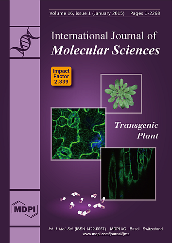
(This article belongs to the Special Issue Advances in the Research of Melatonin 2014)
12 pages, 1855 KiB  
Article
Pulicaria glutinosa Extract: A Toolbox to Synthesize Highly Reduced Graphene Oxide-Silver Nanocomposites
by Abdulhadi H. Al-Marri 1, Mujeeb Khan 1, Merajuddin Khan 1, Syed F. Adil 1, Abdulrahman Al-Warthan 1, Hamad Z. Alkhathlan 1, Wolfgang Tremel 2, Joselito P. Labis 3, Mohammed Rafiq H. Siddiqui 1,* and Muhammad N. Tahir 2,*
1 Department of Chemistry, College of Science, King Saud University, P.O. Box 2455, Riyadh 11451, Saudi Arabia
2 Institute of Inorganic and Analytical Chemistry, Johannes Gutenberg-University of Mainz, Mainz 55122, Germany
3 King Abdullah Institute for Nanotechnology, King Saud University, Riyadh 11451, Saudi Arabia
Int. J. Mol. Sci. 2015, 16(1), 1131-1142; https://doi.org/10.3390/ijms16011131 - 5 Jan 2015
Cited by 58 | Viewed by 11727
Abstract
A green, one-step approach for the preparation of graphene/Ag nanocomposites (PE-HRG-Ag) via simultaneous reduction of both graphene oxide (GRO) and silver ions using Pulicaria glutinosa plant extract (PE) as reducing agent is reported. The plant extract functionalizes the surfaces of highly reduced graphene [...] Read more.
A green, one-step approach for the preparation of graphene/Ag nanocomposites (PE-HRG-Ag) via simultaneous reduction of both graphene oxide (GRO) and silver ions using Pulicaria glutinosa plant extract (PE) as reducing agent is reported. The plant extract functionalizes the surfaces of highly reduced graphene oxide (HRG) which helps in conjugating the Ag NPs to HRG. Increasing amounts of Ag precursor enhanced the density of Ag nanoparticles (NPs) on HRG. The preparation of PE-HRG-Ag nanocomposite is monitored by using ultraviolet–visible (UV-Vis) spectroscopy, powder X-ray diffraction (XRD), and energy dispersive X-ray (EDX). The as-prepared PE-HRG-Ag nanocomposities display excellent surface-enhanced Raman scattering (SERS) activity, and significantly increased the intensities of the Raman signal of graphene. Full article
(This article belongs to the Special Issue Bioactive Nanoparticles 2014)
Show Figures

Graphical abstract

17 pages, 800 KiB  
Article
Factor XIII B Subunit Polymorphisms and the Risk of Coronary Artery Disease
by Zoltán A. Mezei 1, Zsuzsanna Bereczky 1, Éva Katona 1, Réka Gindele 1, Emília Balogh 2, Szilvia Fiatal 3, László Balogh 2, István Czuriga 2, Róza Ádány 3, István Édes 2 and László Muszbek 1,4,*
1 Division of Clinical Laboratory Science, Department of Laboratory Medicine, Faculty of Medicine, University of Debrecen, 98 Nagyerdei Krt., Debrecen H-4032, Hungary
2 Department of Cardiology, Faculty of Medicine, University of Debrecen, 98 Nagyerdei Krt., Debrecen H-4032, Hungary
3 Department of Preventive Medicine, Faculty of Public Health, University of Debrecen, 98 Nagyerdei Krt., Debrecen H-4032, Hungary
4 Vascular Biology, Thrombosis and Hemostasis Research Group of the Hungarian Academy of Sciences, University of Debrecen, 98 Nagyerdei Krt., Debrecen H-4032, Hungary
Int. J. Mol. Sci. 2015, 16(1), 1143-1159; https://doi.org/10.3390/ijms16011143 - 6 Jan 2015
Cited by 17 | Viewed by 7709 | Correction
Abstract
The aim of the case-control study was to explore the effect of coagulation factor XIII (FXIII) B subunit (FXIII-B) polymorphisms on the risk of coronary artery disease, and on FXIII levels. In the study, 687 patients admitted for coronary angiography to investigate suspected [...] Read more.
The aim of the case-control study was to explore the effect of coagulation factor XIII (FXIII) B subunit (FXIII-B) polymorphisms on the risk of coronary artery disease, and on FXIII levels. In the study, 687 patients admitted for coronary angiography to investigate suspected coronary artery disease and 994 individuals representing the Hungarian population were enrolled. The patients were classified according to the presence of significant coronary atherosclerosis (CAS) and history of myocardial infarction (MI). The F13B gene was genotyped for p.His95Arg and for intron K nt29756 C>G polymorphisms; the latter results in the replacement of 10 C-terminal amino acids by 25 novel amino acids. The p.His95Arg polymorphism did not influence the risk of CAS or MI. The FXIII-B intron K nt29756 G allele provided significant protection against CAS and MI in patients with a fibrinogen level in the upper tertile. However, this effect prevailed only in the presence of the FXIII-A Leu34 allele, and a synergism between the two polymorphisms was revealed. Carriers of the intron K nt29756 G allele had significantly lower FXIII levels, and FXIII levels in the lower tertile provided significant protection against MI. It is suggested that the protective effect of the combined polymorphisms is related to decreased FXIII levels. Full article
(This article belongs to the Special Issue Human Single Nucleotide Polymorphisms and Disease Diagnostics)
Show Figures

Figure 1

19 pages, 915 KiB  
Article
Excited States and Photodebromination of Selected Polybrominated Diphenyl Ethers: Computational and Quantitative Structure—Property Relationship Studies
by Jin Luo 1, Jiwei Hu 1,*, Xionghui Wei 2, Lingyun Li 1 and Xianfei Huang 1
1 Guizhou Provincial Key Laboratory for Information System of Mountainous Areas and Protection of Ecological Environment, Guizhou Normal University, Guiyang 550001, China
2 Department of Applied Chemistry, College of Chemistry and Molecular Engineering, Peking University, Beijing 100871, China
Int. J. Mol. Sci. 2015, 16(1), 1160-1178; https://doi.org/10.3390/ijms16011160 - 6 Jan 2015
Cited by 11 | Viewed by 7452
Abstract
This paper presents a density functional theory (DFT)/time-dependent DFT (TD-DFT) study on the lowest lying singlet and triplet excited states of 20 selected polybrominateddiphenyl ether (PBDE) congeners, with the solvation effect included in the calculations using the polarized continuum model (PCM). The results [...] Read more.
This paper presents a density functional theory (DFT)/time-dependent DFT (TD-DFT) study on the lowest lying singlet and triplet excited states of 20 selected polybrominateddiphenyl ether (PBDE) congeners, with the solvation effect included in the calculations using the polarized continuum model (PCM). The results obtained showed that for most of the brominated diphenyl ether (BDE) congeners, the lowest singlet excited state was initiated by the electron transfer from HOMO to LUMO, involving a π–σ* excitation. In triplet excited states, structure of the BDE congeners differed notably from that of the BDE ground states with one of the specific C–Br bonds bending off the aromatic plane. In addition, the partial least squares regression (PLSR), principal component analysis-multiple linear regression analysis (PCA-MLR), and back propagation artificial neural network (BP-ANN) approaches were employed for a quantitative structure-property relationship (QSPR) study. Based on the previously reported kinetic data for the debromination by ultraviolet (UV) and sunlight, obtained QSPR models exhibited a reasonable evaluation of the photodebromination reactivity even when the BDE congeners had same degree of bromination, albeit different patterns of bromination. Full article
(This article belongs to the Special Issue Chemical Bond and Bonding 2015)
Show Figures

Figure 1

13 pages, 1540 KiB  
Article
Trans-Splicing Improvement by the Combined Application of Antisense Strategies
by Ulrich Koller *,†, Stefan Hainzl *,†, Thomas Kocher, Clemens Hüttner, Alfred Klausegger, Christina Gruber, Elisabeth Mayr, Verena Wally, Johann W. Bauer and Eva M. Murauer
1 Department of Dermatology and EB House Austria, Paracelsus Medical University, Salzburg 5020, Austria
These authors contributed equally to this work.
Authors with joint senior authorship.
Int. J. Mol. Sci. 2015, 16(1), 1179-1191; https://doi.org/10.3390/ijms16011179 - 6 Jan 2015
Cited by 18 | Viewed by 8982
Abstract
Spliceosome-mediated RNA trans-splicing has become an emergent tool for the repair of mutated pre-mRNAs in the treatment of genetic diseases. RNA trans-splicing molecules (RTMs) are designed to induce a specific trans-splicing reaction via a binding domain for a respective target [...] Read more.
Spliceosome-mediated RNA trans-splicing has become an emergent tool for the repair of mutated pre-mRNAs in the treatment of genetic diseases. RNA trans-splicing molecules (RTMs) are designed to induce a specific trans-splicing reaction via a binding domain for a respective target pre-mRNA region. A previously established reporter-based screening system allows us to analyze the impact of various factors on the RTM trans-splicing efficiency in vitro. Using this system, we are further able to investigate the potential of antisense RNAs (AS RNAs), presuming to improve the trans-splicing efficiency of a selected RTM, specific for intron 102 of COL7A1. Mutations in the COL7A1 gene underlie the dystrophic subtype of the skin blistering disease epidermolysis bullosa (DEB). We have shown that co-transfections of the RTM and a selected AS RNA, interfering with competitive splicing elements on a COL7A1-minigene (COL7A1-MG), lead to a significant increase of the RNA trans-splicing efficiency. Thereby, accurate trans-splicing between the RTM and the COL7A1-MG is represented by the restoration of full-length green fluorescent protein GFP on mRNA and protein level. This mechanism can be crucial for the improvement of an RTM-mediated correction, especially in cases where a high trans-splicing efficiency is required. Full article
(This article belongs to the Special Issue Pre-mRNA Splicing)
Show Figures

Figure 1

17 pages, 863 KiB  
Article
Telomerase Reverse Transcriptase Regulates microRNAs
by Timo Lassmann 1,†, Yoshiko Maida 2,†, Yasuhiro Tomaru 1, Mami Yasukawa 2, Yoshinari Ando 1, Miki Kojima 1, Vivi Kasim 2, Christophe Simon 1, Carsten O. Daub 1, Piero Carninci 1, Yoshihide Hayashizaki 1 and Kenkichi Masutomi 2,3,*
1 RIKEN Omics Science Center, RIKEN Yokohama Institute, 1-7-22 Suehiro-cho, Tsurumi-ku, Yokohama 230-0045, Japan
2 Cancer Stem Cell Project, National Cancer Center Research Institute, 5-1-1 Tsukiji, Chuo-ku, Tokyo 104-0045, Japan
3 Precursory Research for Embryonic Science and Technology (PREST), Japan Science and Technology Agency, 4-1-8 Honcho Kawaguchi, Saitama 332-0012, Japan
These authors contributed equally to this work.
Int. J. Mol. Sci. 2015, 16(1), 1192-1208; https://doi.org/10.3390/ijms16011192 - 6 Jan 2015
Cited by 23 | Viewed by 8292
Abstract
MicroRNAs are small non-coding RNAs that inhibit the translation of target mRNAs. In humans, most microRNAs are transcribed by RNA polymerase II as long primary transcripts and processed by sequential cleavage of the two RNase III enzymes, DROSHA and DICER, into precursor and [...] Read more.
MicroRNAs are small non-coding RNAs that inhibit the translation of target mRNAs. In humans, most microRNAs are transcribed by RNA polymerase II as long primary transcripts and processed by sequential cleavage of the two RNase III enzymes, DROSHA and DICER, into precursor and mature microRNAs, respectively. Although the fundamental functions of microRNAs in RNA silencing have been gradually uncovered, less is known about the regulatory mechanisms of microRNA expression. Here, we report that telomerase reverse transcriptase (TERT) extensively affects the expression levels of mature microRNAs. Deep sequencing-based screens of short RNA populations revealed that the suppression of TERT resulted in the downregulation of microRNAs expressed in THP-1 cells and HeLa cells. Primary and precursor microRNA levels were also reduced under the suppression of TERT. Similar results were obtained with the suppression of either BRG1 (also called SMARCA4) or nucleostemin, which are proteins interacting with TERT and functioning beyond telomeres. These results suggest that TERT regulates microRNAs at the very early phases in their biogenesis, presumably through non-telomerase mechanism(s). Full article
(This article belongs to the Collection Regulation by Non-coding RNAs)
Show Figures

Figure 1

12 pages, 664 KiB  
Review
Analgesic, Anxiolytic and Anaesthetic Effects of Melatonin: New Potential Uses in Pediatrics
by Lucia Marseglia 1,*, Gabriella D'Angelo 1, Sara Manti 2, Salvatore Aversa 1, Teresa Arrigo 2, Russel J. Reiter 3 and Eloisa Gitto 1
1 Neonatal and Paediatric Intensive Care Unit, Department of Paediatrics, University of Messina, Via Consolare Valeria, Messina 98125, Italy
2 Unit of Paediatric Genetics and Immunology, Department of Paediatrics, University of Messina, Via Consolare Valeria 1, Messina 98125, Italy
3 Department of Cellular and Structural Biology, University of Texas Health Science Center at San Antonio, San Antonio, TX 40729, USA
Int. J. Mol. Sci. 2015, 16(1), 1209-1220; https://doi.org/10.3390/ijms16011209 - 6 Jan 2015
Cited by 80 | Viewed by 11990
Abstract
Exogenous melatonin is used in a number of situations, first and foremost in the treatment of sleep disorders and jet leg. However, the hypnotic, antinociceptive, and anticonvulsant properties of melatonin endow this neurohormone with the profile of a drug that modulates effects of [...] Read more.
Exogenous melatonin is used in a number of situations, first and foremost in the treatment of sleep disorders and jet leg. However, the hypnotic, antinociceptive, and anticonvulsant properties of melatonin endow this neurohormone with the profile of a drug that modulates effects of anesthetic agents, supporting its potential use at different stages during anesthetic procedures, in both adults and children. In light of these properties, melatonin has been administered to children undergoing diagnostic procedures requiring sedation or general anesthesia, such as magnetic resonance imaging, auditory brainstem response tests and electroencephalogram. Controversial data support the use of melatonin as anxiolytic and antinociceptive agents in pediatric patients undergoing surgery. The aim of this review was to evaluate available evidence relating to efficacy and safety of melatonin as an analgesic and as a sedative agent in children. Melatonin and its analogs may have a role in antinociceptive therapies and as an alternative to midazolam in premedication of adults and children, although its effectiveness is still controversial and available data are clearly incomplete. Full article
(This article belongs to the Special Issue Antioxidant 2.0——Redox Modulation by Food and Drugs)
11 pages, 1094 KiB  
Article
Human Adipose Tissue Conditioned Media from Lean Subjects Is Protective against H2O2 Induced Neurotoxicity in Human SH-SY5Y Neuronal Cells
by Zhongxiao Wan 1, Dorrian Mah 1, Svetlana Simtchouk 1, Andreas Kluftinger 2 and Jonathan P. Little 1,*
1 School of Health and Exercise Sciences, University of British Columbia Okanagan, Kelowna, BC V1V 1V7, Canada
2 Department of Surgery, University of British Columbia; General, Laparoscopic, Endocrine and Gastric Band Surgery, 203-3040 Tutt St., Kelowna, BC V1Y 2H5, Canada
Int. J. Mol. Sci. 2015, 16(1), 1221-1231; https://doi.org/10.3390/ijms16011221 - 6 Jan 2015
Cited by 13 | Viewed by 7300
Abstract
Adipose tissue secretes numerous hormone-like factors, which are known as adipokines. Adipokine receptors have been identified in the central nervous system but the potential role of adipokine signaling in neuroprotection is unclear. The aim of this study is to determine (1) Whether adipokines [...] Read more.
Adipose tissue secretes numerous hormone-like factors, which are known as adipokines. Adipokine receptors have been identified in the central nervous system but the potential role of adipokine signaling in neuroprotection is unclear. The aim of this study is to determine (1) Whether adipokines secreted from cultured adipose tissue of lean humans is protective against oxidative stress-induced neurotoxicity in human SH-SY5Y neuronal cells; and (2) To explore potential signaling pathways involved in these processes. Adipose tissue conditioned media (ATCM) from healthy lean subjects completely prevented H2O2 induced neurotoxicity, while this effect is lost after heating ATCM. ATCM activated the phosphorylation of ERK1/2, JNK and Akt at serine 308 in SH-SY5Y cells. PD98059 (25 µM), SP600125 (5 µM) and LY29400 (20 µM) partially blocked the protective effects of ATCM against H2O2 induced neurotoxicity. Findings demonstrate that heat-sensitive factors secreted from human adipose tissue of lean subjects are protective against H2O2 induced neurotoxicity and ERK1/2, JNK, and PI3K signaling pathways are involved in these processes. In conclusion, this study demonstrates preliminary but encouraging data to further support that adipose tissue secreted factors from lean human subjects might possess neuroprotective properties and unravel the specific roles of ERK1/2, JNK and PI3K in these processes. Full article
(This article belongs to the Special Issue Molecular Research in Neurotoxicology)
Show Figures

Figure 1

20 pages, 2728 KiB  
Article
Anti-Inflammatory and Analgesic Effects of Pyeongwisan on LPS-Stimulated Murine Macrophages and Mouse Models of Acetic Acid-Induced Writhing Response and Xylene-Induced Ear Edema
by You-Chang Oh, Yun Hee Jeong, Won-Kyung Cho, Jeong-Ho Ha, Min Jung Gu and Jin Yeul Ma *
Korean Medicine (KM)-Based Herbal Drug Development Group, Korea Institute of Oriental Medicine, 461-24, Jeonmin-dong, Yuseong, Daejeon 305-811, Korea
Int. J. Mol. Sci. 2015, 16(1), 1232-1251; https://doi.org/10.3390/ijms16011232 - 6 Jan 2015
Cited by 37 | Viewed by 8602
Abstract
Pyeongwisan (PW) is an herbal medication used in traditional East Asian medicine to treat anorexia, abdominal distension, borborygmus and diarrhea caused by gastric catarrh, atony and dilatation. However, its effects on inflammation-related diseases are unknown. In this study, we investigated the biological effects [...] Read more.
Pyeongwisan (PW) is an herbal medication used in traditional East Asian medicine to treat anorexia, abdominal distension, borborygmus and diarrhea caused by gastric catarrh, atony and dilatation. However, its effects on inflammation-related diseases are unknown. In this study, we investigated the biological effects of PW on lipopolysaccharide (LPS)-mediated inflammation in macrophages and on local inflammation in vivo. We investigated the biological effects of PW on the production of inflammatory mediators, pro-inflammatory cytokines and related products as well as the activation of nuclear factor kappa B (NF-κB) and mitogen-activated protein kinases (MAPKs) in LPS-stimulated macrophages. Additionally, we evaluated the analgesic effect on the acetic acid-induced writhing response and the inhibitory activity on xylene-induced ear edema in mice. PW showed anti-inflammatory effects by inhibiting the production of nitric oxide (NO), tumor necrosis factor-α (TNF-α) and interleukin-6 (IL-6) and interleukin-1β (IL-1β). In addition, PW strongly suppressed inducible nitric oxide synthase (iNOS), a NO synthesis enzyme, induced heme oxygenase-1 (HO-1) expression and inhibited NF-κB activation and MAPK phosphorylation. Also, PW suppressed TNF-α, IL-6 and IL-1β cytokine production in LPS-stimulated peritoneal macrophage cells. Furthermore, PW showed an analgesic effect on the writhing response and an inhibitory effect on mice ear edema. We demonstrated the anti-inflammatory effects and inhibitory mechanism in macrophages as well as inhibitory activity of PW in vivo for the first time. Our results suggest the potential value of PW as an inflammatory therapeutic agent developed from a natural substance. Full article
(This article belongs to the Section Biochemistry)
Show Figures

Graphical abstract

14 pages, 3394 KiB  
Article
Mechanisms of Hyperhomocysteinemia Induced Skeletal Muscle Myopathy after Ischemia in the CBS/+ Mouse Model
by Sudhakar Veeranki * and Suresh C. Tyagi
Department of Physiology & Biophysics, University of Louisville, Louisville, KY 40202, USA
Int. J. Mol. Sci. 2015, 16(1), 1252-1265; https://doi.org/10.3390/ijms16011252 - 6 Jan 2015
Cited by 21 | Viewed by 7134
Abstract
Although hyperhomocysteinemia (HHcy) elicits lower than normal body weights and skeletal muscle weakness, the mechanisms remain unclear. Despite the fact that HHcy-mediated enhancement in ROS and consequent damage to regulators of different cellular processes is relatively well established in other organs, the nature [...] Read more.
Although hyperhomocysteinemia (HHcy) elicits lower than normal body weights and skeletal muscle weakness, the mechanisms remain unclear. Despite the fact that HHcy-mediated enhancement in ROS and consequent damage to regulators of different cellular processes is relatively well established in other organs, the nature of such events is unknown in skeletal muscles. Previously, we reported that HHcy attenuation of PGC-1α and HIF-1α levels enhanced the likelihood of muscle atrophy and declined function after ischemia. In the current study, we examined muscle levels of homocysteine (Hcy) metabolizing enzymes, anti-oxidant capacity and focused on protein modifications that might compromise PGC-1α function during ischemic angiogenesis. Although skeletal muscles express the key enzyme (MTHFR) that participates in re-methylation of Hcy into methionine, lack of trans-sulfuration enzymes (CBS and CSE) make skeletal muscles more susceptible to the HHcy-induced myopathy. Our study indicates that elevated Hcy levels in the CBS−/+ mouse skeletal muscles caused diminished anti-oxidant capacity and contributed to enhanced total protein as well as PGC-1α specific nitrotyrosylation after ischemia. Furthermore, in the presence of NO donor SNP, either homocysteine (Hcy) or its cyclized version, Hcy thiolactone, not only increased PGC-1α specific protein nitrotyrosylation but also reduced its association with PPARγ in C2C12 cells. Altogether these results suggest that HHcy exerts its myopathic effects via reduction of the PGC-1/PPARγ axis after ischemia. Full article
(This article belongs to the Section Biochemistry)
Show Figures

Figure 1

27 pages, 5626 KiB  
Article
Molecular Targeting of the Oncoprotein PLK1 in Pediatric Acute Myeloid Leukemia: RO3280, a Novel PLK1 Inhibitor, Induces Apoptosis in Leukemia Cells
by Na-Na Wang 1,†, Zhi-Heng Li 1,†, He Zhao 1,†, Yan-Fang Tao 1, Li-Xiao Xu 1, Jun Lu 1, Lan Cao 1, Xiao-Juan Du 2, Li-Chao Sun 3, Wen-Li Zhao 1, Pei-Fang Xiao 1, Fang Fang 1, Guang-Hao Su 1, Yan-Hong Li 1, Gang Li 1, Yi-Ping Li 1, Yun-Yun Xu 1, Hui-Ting Zhou 1, Yi Wu 1, Mei-Fang Jin 1, Lin Liu 1, Jian Ni 4, Jian Wang 1, Shao-Yan Hu 1, Xue-Ming Zhu 1, Xing Feng 1,* and Jian Pan 1,*add Show full author list remove Hide full author list
1 Department of Hematology and Oncology, Children's Hospital of Soochow University, Suzhou 215003, China
2 Department of Gastroenterology, the 5th Hospital of Chinese People's Liberation Army (PLA), Yinchuan 750000, China
3 Department of Cell and Molecular Biology, Cancer Institute (Hospital), Chinese Academy of Medical Sciences, Peking Union Medical College, Beijing 100021, China
4 Translational Research Center, Second Hospital, The Second Clinical School, Nanjing Medical University, Nanjing 210000, China
These authors contributed equally to this work.
Int. J. Mol. Sci. 2015, 16(1), 1266-1292; https://doi.org/10.3390/ijms16011266 - 7 Jan 2015
Cited by 24 | Viewed by 9495
Abstract
Polo-like kinase 1 (PLK1) is highly expressed in many cancers and therefore a biomarker of transformation and potential target for the development of cancer-specific small molecule drugs. RO3280 was recently identified as a novel PLK1 inhibitor; however its therapeutic effects in leukemia treatment [...] Read more.
Polo-like kinase 1 (PLK1) is highly expressed in many cancers and therefore a biomarker of transformation and potential target for the development of cancer-specific small molecule drugs. RO3280 was recently identified as a novel PLK1 inhibitor; however its therapeutic effects in leukemia treatment are still unknown. We found that the PLK1 protein was highly expressed in leukemia cell lines as well as 73.3% (11/15) of pediatric acute myeloid leukemia (AML) samples. PLK1 mRNA expression was significantly higher in AML samples compared with control samples (82.95 ± 110.28 vs. 6.36 ± 6.35; p < 0.001). Kaplan-Meier survival analysis revealed that shorter survival time correlated with high tumor PLK1 expression (p = 0.002). The 50% inhibitory concentration (IC50) of RO3280 for acute leukemia cells was between 74 and 797 nM. The IC50 of RO3280 in primary acute lymphocytic leukemia (ALL) and AML cells was between 35.49 and 110.76 nM and 52.80 and 147.50 nM, respectively. RO3280 induced apoptosis and cell cycle disorder in leukemia cells. RO3280 treatment regulated several apoptosis-associated genes. The regulation of DCC, CDKN1A, BTK, and SOCS2 was verified by western blot. These results provide insights into the potential use of RO3280 for AML therapy; however, the underlying mechanisms remain to be determined. Full article
Show Figures

Figure 1

19 pages, 3051 KiB  
Article
The Oxidative Fermentation of Ethanol in Gluconacetobacter diazotrophicus Is a Two-Step Pathway Catalyzed by a Single Enzyme: Alcohol-Aldehyde Dehydrogenase (ADHa)
by Saúl Gómez-Manzo 1,*, José E. Escamilla 2,†, Abigail González-Valdez 3, Gabriel López-Velázquez 1,4, América Vanoye-Carlo 1, Jaime Marcial-Quino 5, Ignacio De la Mora-de la Mora 1, Itzhel Garcia-Torres 1, Sergio Enríquez-Flores 1, Martha Lucinda Contreras-Zentella 2, Roberto Arreguín-Espinosa 6, Peter M. H. Kroneck 7 and Martha Elena Sosa-Torres 8,*
1 Laboratorio de Bioquímica-Genética, Instituto Nacional de Pediatría, S.S. Mexico City 04530, Mexico
2 Departamento de Bioquímica y Biología Estructural, Instituto de Fisiología Celular, Universidad Nacional Autónoma de México, Mexico City 04510, Mexico
3 Departamento de Biología Molecular y Biotecnología, Instituto de Investigaciones Biomédicas, Universidad Nacional Autónoma de México, Mexico City 04510, Mexico
4 Coordinación de Investigación, Facultad de Medicina, Universidad La Salle, Mexico City 14000, Mexico
5 CONACyT, Comisionado a Instituto Nacional de Pediatría, S.S. Mexico City 03940, Mexico
6 Departamento de Química de Biomacromoléculas, Instituto de Química, Universidad Nacional Autónoma de México, Circuito Exterior s/n, Ciudad Universitaria, Mexico City 04510, Mexico
7 Fachbereich Biologie, Universität Konstanz, 78457 Konstanz, Germany
8 Departamento de Química Inorgánica y Nuclear, Facultad de Química, Universidad Nacional Autónoma de México, Mexico City 04510, Mexico
This work is dedicated to Prof. Jos
Int. J. Mol. Sci. 2015, 16(1), 1293-1311; https://doi.org/10.3390/ijms16011293 - 7 Jan 2015
Cited by 18 | Viewed by 10203
Abstract
Gluconacetobacter diazotrophicus is a N2-fixing bacterium endophyte from sugar cane. The oxidation of ethanol to acetic acid of this organism takes place in the periplasmic space, and this reaction is catalyzed by two membrane-bound enzymes complexes: the alcohol dehydrogenase (ADH) and [...] Read more.
Gluconacetobacter diazotrophicus is a N2-fixing bacterium endophyte from sugar cane. The oxidation of ethanol to acetic acid of this organism takes place in the periplasmic space, and this reaction is catalyzed by two membrane-bound enzymes complexes: the alcohol dehydrogenase (ADH) and the aldehyde dehydrogenase (ALDH). We present strong evidence showing that the well-known membrane-bound Alcohol dehydrogenase (ADHa) of Ga. diazotrophicus is indeed a double function enzyme, which is able to use primary alcohols (C2–C6) and its respective aldehydes as alternate substrates. Moreover, the enzyme utilizes ethanol as a substrate in a reaction mechanism where this is subjected to a two-step oxidation process to produce acetic acid without releasing the acetaldehyde intermediary to the media. Moreover, we propose a mechanism that, under physiological conditions, might permit a massive conversion of ethanol to acetic acid, as usually occurs in the acetic acid bacteria, but without the transient accumulation of the highly toxic acetaldehyde. Full article
(This article belongs to the Section Biochemistry)
Show Figures

Figure 1

24 pages, 728 KiB  
Article
Whole Exome Sequencing in Females with Autism Implicates Novel and Candidate Genes
by Merlin G. Butler 1,*, Syed K. Rafi 1,†, Waheeda Hossain 1,†, Dietrich A. Stephan 2,† and Ann M. Manzardo 1,†
1 Departments of Psychiatry & Behavioral Sciences, University of Kansas Medical Center, 3901 Rainbow Boulevard, MS 4015, Kansas City, KS 66160, USA
2 Department of Human Genetics, University of Pittsburgh, Pittsburgh, PA 15260, USA
These authors contributed equally to this work.
Int. J. Mol. Sci. 2015, 16(1), 1312-1335; https://doi.org/10.3390/ijms16011312 - 7 Jan 2015
Cited by 71 | Viewed by 13948
Abstract
Classical autism or autistic disorder belongs to a group of genetically heterogeneous conditions known as Autism Spectrum Disorders (ASD). Heritability is estimated as high as 90% for ASD with a recently reported compilation of 629 clinically relevant candidate and known genes. We chose [...] Read more.
Classical autism or autistic disorder belongs to a group of genetically heterogeneous conditions known as Autism Spectrum Disorders (ASD). Heritability is estimated as high as 90% for ASD with a recently reported compilation of 629 clinically relevant candidate and known genes. We chose to undertake a descriptive next generation whole exome sequencing case study of 30 well-characterized Caucasian females with autism (average age, 7.7 ± 2.6 years; age range, 5 to 16 years) from multiplex families. Genomic DNA was used for whole exome sequencing via paired-end next generation sequencing approach and X chromosome inactivation status. The list of putative disease causing genes was developed from primary selection criteria using machine learning-derived classification score and other predictive parameters (GERP2, PolyPhen2, and SIFT). We narrowed the variant list to 10 to 20 genes and screened for biological significance including neural development, function and known neurological disorders. Seventy-eight genes identified met selection criteria ranging from 1 to 9 filtered variants per female. Five females presented with functional variants of X-linked genes (IL1RAPL1, PIR, GABRQ, GPRASP2, SYTL4) with cadherin, protocadherin and ankyrin repeat gene families most commonly altered (e.g., CDH6, FAT2, PCDH8, CTNNA3, ANKRD11). Other genes related to neurogenesis and neuronal migration (e.g., SEMA3F, MIDN), were also identified. Full article
Show Figures

Graphical abstract

39 pages, 876 KiB  
Editorial
Acknowledgement to Reviewers of the International Journal of Molecular Sciences in 2014
by IJMS Editorial Office
IJMS Editorial Office, MDPI AG, Klybeckstrasse 64, CH-4057 Basel, Switzerland
Int. J. Mol. Sci. 2015, 16(1), 1336-1374; https://doi.org/10.3390/ijms16011336 - 7 Jan 2015
Viewed by 8813
Abstract
The editors of the International Journal of Molecular Sciences would like to express their sincere gratitude to the following reviewers for assessing manuscripts in 2014:[...] Full article
10 pages, 669 KiB  
Article
No Association between HMOX1 and Risk of Colorectal Cancer and No Interaction with Diet and Lifestyle Factors in a Prospective Danish Case-Cohort Study
by Vibeke Andersen 1,2,*, Tine Iskov Kopp 3,4, Anne Tjønneland 4 and Ulla Vogel 5
1 Organ Center, Hospital of Southern Jutland, 6200 Aabenraa, Denmark
2 Institute of Regional Health Research, University of Southern Denmark, 5000 Odense, Denmark
3 National Food Institute, 2860 Soborg, Denmark
4 Danish Cancer Society Research Center, 2100 Copenhagen, Denmark
5 National Research Centre for the Working Environment, 2100 Copenhagen, Denmark
Int. J. Mol. Sci. 2015, 16(1), 1375-1384; https://doi.org/10.3390/ijms16011375 - 7 Jan 2015
Cited by 7 | Viewed by 6470
Abstract
Red meat is a risk factor for colorectal cancer (CRC). We wanted to evaluate whether a functional polymorphism in the HMOX1 gene encoding heme oxygenase modifies risk of CRC or interacts with diet or lifestyle factors because this would identify heme or heme [...] Read more.
Red meat is a risk factor for colorectal cancer (CRC). We wanted to evaluate whether a functional polymorphism in the HMOX1 gene encoding heme oxygenase modifies risk of CRC or interacts with diet or lifestyle factors because this would identify heme or heme iron as a risk factor of CRC. The HMOX1 A-413T (rs2071746) was assessed in relation to risk of colorectal cancer (CRC) and interactions with diet (red meat, fish, fiber, cereals, fruit and vegetables) and lifestyle (use of non-steroidal anti-inflammatory drug and smoking status) were assessed in a case-cohort study of 928 CRC cases and a comparison group of 1726 randomly selected participants from a prospective study of 57,053 persons. No association between HMOX1 A-413T and CRC risk was found (TT vs. AA + TA; IRR = 1.15, 95% CI: 0.98–1.36, p = 0.10 for the adjusted estimate). No interactions were found between diet or lifestyle and HMOX1 A-413T. HMOX1 A-413T was not associated with CRC risk and no interactions with diet or lifestyle were identified in this large, prospective cohort with high meat intake. The results reproduced the previous findings from the same cohort and did not support a link between heme or heme iron and colorectal cancer. These results should be sought and replicated in other well-characterized cohorts with high meat intake. Full article
(This article belongs to the Special Issue Gene-Nutrient Interactions)
10 pages, 786 KiB  
Communication
Profiling of Ubiquitination Pathway Genes in Peripheral Cells from Patients with Frontotemporal Dementia due to C9ORF72 and GRN Mutations
by Maria Serpente *, Chiara Fenoglio, Sara M. G. Cioffi, Rossana Bonsi, Andrea Arighi, Giorgio G. Fumagalli, Laura Ghezzi, Elio Scarpini and Daniela Galimberti
Neurology Unit, Department of Pathophysiology and Transplantation, "Dino Ferrari" Center, University of Milan, Fondazione Ca' Granda, IRCCS Ospedale Maggiore Policlinico, Milan 20122, Italy
Int. J. Mol. Sci. 2015, 16(1), 1385-1394; https://doi.org/10.3390/ijms16011385 - 8 Jan 2015
Cited by 14 | Viewed by 5818
Abstract
We analysed the expression levels of 84 key genes involved in the regulated degradation of cellular protein by the ubiquitin-proteasome system in peripheral cells from patients with frontotemporal dementia (FTD) due to C9ORF72 and GRN mutations, as compared with sporadic FTD and age-matched [...] Read more.
We analysed the expression levels of 84 key genes involved in the regulated degradation of cellular protein by the ubiquitin-proteasome system in peripheral cells from patients with frontotemporal dementia (FTD) due to C9ORF72 and GRN mutations, as compared with sporadic FTD and age-matched controls. A SABiosciences PCR array was used to investigate the transcription profile in a discovery population consisting of six patients each in C9ORF72, GRN, sporadic FTD and age-matched control groups. A generalized down-regulation of gene expression compared with controls was observed in C9ORF72 expansion carriers and sporadic FTD patients. In particular, in both groups, four genes, UBE2I, UBE2Q1, UBE2E1 and UBE2N, were down-regulated at a statistically significant (p < 0.05) level. All of them encode for members of the E2 ubiquitin-conjugating enzyme family. In GRN mutation carriers, no statistically significant deregulation of ubiquitination pathway genes was observed, except for the UBE2Z gene, which displays E2 ubiquitin conjugating enzyme activity, and was found to be statistically significant up-regulated (p = 0.006). These preliminary results suggest that the proteasomal degradation pathway plays a role in the pathogenesis of FTD associated with TDP-43 pathology, although different proteins are altered in carriers of GRN mutations as compared with carriers of the C9ORF72 expansion. Full article
(This article belongs to the Section Biochemistry)
Show Figures

Figure 1

11 pages, 1385 KiB  
Review
Long Non-Coding RNAs in Cancer and Development: Where Do We Go from Here?
by Monika Haemmerle 1,2 and Tony Gutschner 3,*
1 Department of Gynecologic Oncology and Reproductive Medicine, the University of Texas MD Anderson Cancer Center, Houston, TX 77054, USA
2 Institute of Pathology, University Hospital Heidelberg, Heidelberg 69120, Germany
3 Department of Genomic Medicine, the University of Texas MD Anderson Cancer Center, Houston, TX 77054, USA
Int. J. Mol. Sci. 2015, 16(1), 1395-1405; https://doi.org/10.3390/ijms16011395 - 8 Jan 2015
Cited by 75 | Viewed by 9306
Abstract
Recent genome-wide expression profiling studies have uncovered a huge amount of novel, long non-protein-coding RNA transcripts (lncRNA). In general, these transcripts possess a low, but tissue-specific expression, and their nucleotide sequences are often poorly conserved. However, several studies showed that lncRNAs can have [...] Read more.
Recent genome-wide expression profiling studies have uncovered a huge amount of novel, long non-protein-coding RNA transcripts (lncRNA). In general, these transcripts possess a low, but tissue-specific expression, and their nucleotide sequences are often poorly conserved. However, several studies showed that lncRNAs can have important roles for normal tissue development and regulate cellular pluripotency as well as differentiation. Moreover, lncRNAs are implicated in the control of multiple molecular pathways leading to gene expression changes and thus, ultimately modulate cell proliferation, migration and apoptosis. Consequently, deregulation of lncRNA expression contributes to carcinogenesis and is associated with human diseases, e.g., neurodegenerative disorders like Alzheimer’s Disease. Here, we will focus on some major challenges of lncRNA research, especially loss-of-function studies. We will delineate strategies for lncRNA gene targeting in vivo, and we will briefly discuss important consideration and pitfalls when investigating lncRNA functions in knockout animal models. Finally, we will highlight future opportunities for lncRNAs research by applying the concept of cross-species comparison, which might contribute to novel disease biomarker discovery and might identify lncRNAs as potential therapeutic targets. Full article
(This article belongs to the Collection Regulation by Non-coding RNAs)
Show Figures

Figure 1

23 pages, 12506 KiB  
Review
Structure and Function of SET and MYND Domain-Containing Proteins
by Nicholas Spellmon 1, Joshua Holcomb 1, Laura Trescott 1, Nualpun Sirinupong 2 and Zhe Yang 1,*
1 Department of Biochemistry and Molecular Biology, Wayne State University School of Medicine, 540 East Canfield Street, Detroit, MI 48201, USA
2 Nutraceuticals and Functional Food Research and Development Center, Prince of Songkla University, Hat-Yai, Songkhla 90112, Thailand
Int. J. Mol. Sci. 2015, 16(1), 1406-1428; https://doi.org/10.3390/ijms16011406 - 8 Jan 2015
Cited by 121 | Viewed by 11613
Abstract
SET (Suppressor of variegation, Enhancer of Zeste, Trithorax) and MYND (Myeloid-Nervy-DEAF1) domain-containing proteins (SMYD) have been found to methylate a variety of histone and non-histone targets which contribute to their various roles in cell regulation including chromatin remodeling, transcription, signal transduction, and cell [...] Read more.
SET (Suppressor of variegation, Enhancer of Zeste, Trithorax) and MYND (Myeloid-Nervy-DEAF1) domain-containing proteins (SMYD) have been found to methylate a variety of histone and non-histone targets which contribute to their various roles in cell regulation including chromatin remodeling, transcription, signal transduction, and cell cycle control. During early development, SMYD proteins are believed to act as an epigenetic regulator for myogenesis and cardiomyocyte differentiation as they are abundantly expressed in cardiac and skeletal muscle. SMYD proteins are also of therapeutic interest due to the growing list of carcinomas and cardiovascular diseases linked to SMYD overexpression or dysfunction making them a putative target for drug intervention. This review will examine the biological relevance and gather all of the current structural data of SMYD proteins. Full article
(This article belongs to the Special Issue Protein Crystallography in Molecular Biology 2015)
Show Figures

Graphical abstract

19 pages, 886 KiB  
Review
HCN Channels—Modulators of Cardiac and Neuronal Excitability
by Stefan Herrmann *, Sabine Schnorr and Andreas Ludwig *
Institut für Experimentelle und Klinische Pharmakologie und Toxikologie, Friedrich-Alexander-Universität Erlangen-Nürnberg, 91054 Erlangen, Germany
Int. J. Mol. Sci. 2015, 16(1), 1429-1447; https://doi.org/10.3390/ijms16011429 - 8 Jan 2015
Cited by 44 | Viewed by 10410
Abstract
Hyperpolarization-activated cyclic nucleotide-gated (HCN) channels comprise a family of cation channels activated by hyperpolarized membrane potentials and stimulated by intracellular cyclic nucleotides. The four members of this family, HCN1–4, show distinct biophysical properties which are most evident in the kinetics of activation and [...] Read more.
Hyperpolarization-activated cyclic nucleotide-gated (HCN) channels comprise a family of cation channels activated by hyperpolarized membrane potentials and stimulated by intracellular cyclic nucleotides. The four members of this family, HCN1–4, show distinct biophysical properties which are most evident in the kinetics of activation and deactivation, the sensitivity towards cyclic nucleotides and the modulation by tyrosine phosphorylation. The four isoforms are differentially expressed in various excitable tissues. This review will mainly focus on recent insights into the functional role of the channels apart from their classic role as pacemakers. The importance of HCN channels in the cardiac ventricle and ventricular hypertrophy will be discussed. In addition, their functional significance in the peripheral nervous system and nociception will be examined. The data, which are mainly derived from studies using transgenic mice, suggest that HCN channels contribute significantly to cellular excitability in these tissues. Remarkably, the impact of the channels is clearly more pronounced in pathophysiological states including ventricular hypertrophy as well as neural inflammation and neuropathy suggesting that HCN channels may constitute promising drug targets in the treatment of these conditions. This perspective as well as the current therapeutic use of HCN blockers will also be addressed. Full article
(This article belongs to the Special Issue Signalling Molecules and Signal Transduction in Cells 2014)
Show Figures

Figure 1

18 pages, 4241 KiB  
Article
MicroRNA Expression Profiling of Lactating Mammary Gland in Divergent Phenotype Swine Breeds
by Jing Peng, Jun-Sheng Zhao, Yi-Fei Shen, Hai-Guang Mao and Ning-Ying Xu *
College of Animal Sciences, Zhejiang University, Hangzhou 310058, China
Int. J. Mol. Sci. 2015, 16(1), 1448-1465; https://doi.org/10.3390/ijms16011448 - 8 Jan 2015
Cited by 28 | Viewed by 7092
Abstract
MicroRNA (miRNA) plays a key role in development and specific biological processes, such as cell proliferation, differentiation, and apoptosis. Extensive studies of mammary miRNAs have been performed in different species and tissues. However, little is known about porcine mammary gland miRNAs. In this [...] Read more.
MicroRNA (miRNA) plays a key role in development and specific biological processes, such as cell proliferation, differentiation, and apoptosis. Extensive studies of mammary miRNAs have been performed in different species and tissues. However, little is known about porcine mammary gland miRNAs. In this study, we report the identification and characterization of miRNAs in the lactating mammary gland in two distinct pig breeds, Jinhua and Yorkshire. Many miRNAs were detected as significantly differentially expressed between the two libraries. Among the differentially expressed miRNAs, many are known to be related to mammary gland development and lactation by interacting with putative target genes in previous studies. These findings suggest that miRNA expression patterns may contribute significantly to target mRNA regulation and influence mammary gland development and peak lactation performance. The data we obtained provide useful information about the roles of miRNAs in the biological processes of lactation and the mechanisms of target gene expression and regulation. Full article
(This article belongs to the Collection Regulation by Non-coding RNAs)
Show Figures

Graphical abstract

16 pages, 2121 KiB  
Article
Prediction of Mature MicroRNA and Piwi-Interacting RNA without a Genome Reference or Precursors
by Mark S. Menor, Kyungim Baek * and Guylaine Poisson *
Department of Information and Computer Sciences, University of Hawaii at Mānoa, 1680 East-West Road, Honolulu, HI 96822, USA
Int. J. Mol. Sci. 2015, 16(1), 1466-1481; https://doi.org/10.3390/ijms16011466 - 8 Jan 2015
Cited by 10 | Viewed by 6557
Abstract
The discovery of novel microRNA (miRNA) and piwi-interacting RNA (piRNA) is an important task for the understanding of many biological processes. Most of the available miRNA and piRNA identification methods are dependent on the availability of the organism’s genome sequence and the quality [...] Read more.
The discovery of novel microRNA (miRNA) and piwi-interacting RNA (piRNA) is an important task for the understanding of many biological processes. Most of the available miRNA and piRNA identification methods are dependent on the availability of the organism’s genome sequence and the quality of its annotation. Therefore, an efficient prediction method based solely on the short RNA reads and requiring no genomic information is highly desirable. In this study, we propose an approach that relies primarily on the nucleotide composition of the read and does not require reference genomes of related species for prediction. Using an empirical Bayesian kernel method and the error correcting output codes framework, compact models suitable for large-scale analyses are built on databases of known mature miRNAs and piRNAs. We found that the usage of an L1-based Gaussian kernel can double the true positive rate compared to the standard L2-based Gaussian kernel. Our approach can increase the true positive rate by at most 60% compared to the existing piRNA predictor based on the analysis of a hold-out test set. Using experimental data, we also show that our approach can detect about an order of magnitude or more known miRNAs than the mature miRNA predictor, miRPlex. Full article
(This article belongs to the Collection Regulation by Non-coding RNAs)
Show Figures

Figure 1

2 pages, 614 KiB  
Correction
Correction: Kang, K.A.; et al., Myricetin Protects Cells against Oxidative Stress-Induced Apoptosis via Regulation of PI3K/Akt and MAPK Signaling Pathways. Int. J. Mol. Sci. 2010, 11, 4348–4360
by Kyoung Ah Kang 1, Zhi Hong Wang 1, Rui Zhang 1, Mei Jing Piao 1, Ki Cheon Kim 1, Sam Sik Kang 2, Young Woo Kim 3, Jongsung Lee 3, Deokhoon Park 3 and Jin Won Hyun 1,*
1 School of Medicine and Applied Radiological Science Research Institute, Jeju National University, Jeju 690-756, Korea
2 College of Pharmacy, Seoul National University, Seoul 110-460, Korea
3 Biospectrum Life Science Institute, Gunpo 435-833, Korea
Int. J. Mol. Sci. 2015, 16(1), 1482-1483; https://doi.org/10.3390/ijms16011482 - 8 Jan 2015
Cited by 6 | Viewed by 5173
Abstract
The authors want to change Figure 1 of the paper published in IJMS [1]. In Figure 1, 5-position of OH was at 6-position. Therefore, Figure 1 is revised as follows. The authors would like to apologize for any inconvenience caused to the readers [...] Read more.
The authors want to change Figure 1 of the paper published in IJMS [1]. In Figure 1, 5-position of OH was at 6-position. Therefore, Figure 1 is revised as follows. The authors would like to apologize for any inconvenience caused to the readers by this change.[...] Full article
Show Figures

Figure 1

11 pages, 871 KiB  
Review
Cadmium Transporters in the Kidney and Cadmium-Induced Nephrotoxicity
by Hong Yang and Yan Shu *
Department of Pharmaceutical Sciences, School of Pharmacy, University of Maryland at Baltimore, Baltimore, MD 21201, USA
Int. J. Mol. Sci. 2015, 16(1), 1484-1494; https://doi.org/10.3390/ijms16011484 - 9 Jan 2015
Cited by 194 | Viewed by 13126
Abstract
Among the organs in which the environmental pollutant cadmium causes toxicity, the kidney has gained the most attention in recent years. Numerous studies have sought to unravel the exact pathways by which cadmium enters the renal epithelial cells and the mechanisms by which [...] Read more.
Among the organs in which the environmental pollutant cadmium causes toxicity, the kidney has gained the most attention in recent years. Numerous studies have sought to unravel the exact pathways by which cadmium enters the renal epithelial cells and the mechanisms by which it causes toxicity in the kidney. The purpose of this review is to present the progress made on the mechanisms of cadmium transport in the kidney and the role of transporter proteins in cadmium-induced nephrotoxicity. Full article
(This article belongs to the Special Issue Renal Toxicology—Epidemiology and Mechanisms)
Show Figures

Graphical abstract

14 pages, 3656 KiB  
Article
Hair Dyes Resorcinol and Lawsone Reduce Production of Melanin in Melanoma Cells by Tyrosinase Activity Inhibition and Decreasing Tyrosinase and Microphthalmia-Associated Transcription Factor (MITF) Expression
by Shu-Mei Lee 1,*, Yi-Shyan Chen 2, Chih-Chien Lin 2 and Kuan-Hung Chen 2
1 Department of Cosmetic Science and Management, Mackay Medicine, Nursing and Management College, 92 Shengjing Road, Beitou, Taipei 11260, Taiwan
2 Department of Cosmetic Science, Providence University, 200, Sec. 7, Taiwan Boulevard, Shalu Dist., Taichung 43301, Taiwan
Int. J. Mol. Sci. 2015, 16(1), 1495-1508; https://doi.org/10.3390/ijms16011495 - 9 Jan 2015
Cited by 22 | Viewed by 9617
Abstract
Hair coloring products are one of the most important cosmetics for modern people; there are three major types of hair dyes, including the temporary, semi-permanent and permanent hair dyes. The selected hair dyes (such as ammonium persulfate, sodium persulfate, resorcinol and lawsone) are [...] Read more.
Hair coloring products are one of the most important cosmetics for modern people; there are three major types of hair dyes, including the temporary, semi-permanent and permanent hair dyes. The selected hair dyes (such as ammonium persulfate, sodium persulfate, resorcinol and lawsone) are the important components for hair coloring products. Therefore, we analyzed the effects of these compounds on melanogenesis in B16-F10 melanoma cells. The results proved that hair dyes resorcinol and lawsone can reduce the production of melanin. The results also confirmed that resorcinol and lawsone inhibit mushroom and cellular tyrosinase activities in vitro. Resorcinol and lawsone can also downregulate the protein levels of tyrosinase and microphthalmia-associated transcription factor (MITF) in B16-F10 cells. Thus, we suggest that frequent use of hair dyes may have the risk of reducing natural melanin production in hair follicles. Moreover, resorcinol and lawsone may also be used as hypopigmenting agents to food, agricultural and cosmetic industry in the future. Full article
Show Figures

Figure 1

17 pages, 1032 KiB  
Review
Membrane Trafficking in the Yeast Saccharomyces cerevisiae Model
by Serge Feyder, Johan-Owen De Craene, Séverine Bär, Dimitri L. Bertazzi and Sylvie Friant *
Department of Molecular and Cellular Genetics, UMR7156, Université de Strasbourg and CNRS, 21 rue Descartes, Strasbourg 67084, France
Int. J. Mol. Sci. 2015, 16(1), 1509-1525; https://doi.org/10.3390/ijms16011509 - 9 Jan 2015
Cited by 110 | Viewed by 17035
Abstract
The yeast Saccharomyces cerevisiae is one of the best characterized eukaryotic models. The secretory pathway was the first trafficking pathway clearly understood mainly thanks to the work done in the laboratory of Randy Schekman in the 1980s. They have isolated yeast sec mutants [...] Read more.
The yeast Saccharomyces cerevisiae is one of the best characterized eukaryotic models. The secretory pathway was the first trafficking pathway clearly understood mainly thanks to the work done in the laboratory of Randy Schekman in the 1980s. They have isolated yeast sec mutants unable to secrete an extracellular enzyme and these SEC genes were identified as encoding key effectors of the secretory machinery. For this work, the 2013 Nobel Prize in Physiology and Medicine has been awarded to Randy Schekman; the prize is shared with James Rothman and Thomas Südhof. Here, we present the different trafficking pathways of yeast S. cerevisiae. At the Golgi apparatus newly synthesized proteins are sorted between those transported to the plasma membrane (PM), or the external medium, via the exocytosis or secretory pathway (SEC), and those targeted to the vacuole either through endosomes (vacuolar protein sorting or VPS pathway) or directly (alkaline phosphatase or ALP pathway). Plasma membrane proteins can be internalized by endocytosis (END) and transported to endosomes where they are sorted between those targeted for vacuolar degradation and those redirected to the Golgi (recycling or RCY pathway). Studies in yeast S. cerevisiae allowed the identification of most of the known effectors, protein complexes, and trafficking pathways in eukaryotic cells, and most of them are conserved among eukaryotes. Full article
Show Figures

Graphical abstract

18 pages, 3710 KiB  
Article
Relationship between Expression of Onco-Related miRNAs and the Endoscopic Appearance of Colorectal Tumors
by Yoshihito Nakagawa 1,*, Yukihiro Akao 2, Kohei Taniguchi 2, Akemi Kamatani 1, Tomomitsu Tahara 1, Toshiaki Kamano 1, Naoko Nakano 1, Naruomi Komura 1, Hirokazu Ikuno 1, Takafumi Ohmori 1, Yasutaka Jodai 1, Masahiro Miyata 1, Mistuo Nagasaka 1, Tomoyuki Shibata 1, Naoki Ohmiya 1 and Ichiro Hirata 1
1 Department of Gastroenterology, School of Medicine, Fujita Health University, Kutsukake-cho, Aichi 470-1192, Japan
2 The United Graduate School of Drug Discovery and Medical Information Sciences, Gifu University, Gifu 501-1193, Japan
Int. J. Mol. Sci. 2015, 16(1), 1526-1543; https://doi.org/10.3390/ijms16011526 - 9 Jan 2015
Cited by 20 | Viewed by 10700
Abstract
Accumulating data indicates that certain microRNAs (miRNAs or miRs) are differently expressed in samples of tumors and paired non-tumorous samples taken from the same patients with colorectal tumors. We examined the expression of onco-related miRNAs in 131 sporadic exophytic adenomas or early cancers [...] Read more.
Accumulating data indicates that certain microRNAs (miRNAs or miRs) are differently expressed in samples of tumors and paired non-tumorous samples taken from the same patients with colorectal tumors. We examined the expression of onco-related miRNAs in 131 sporadic exophytic adenomas or early cancers and in 52 sporadic flat elevated adenomas or early cancers to clarify the relationship between the expression of the miRNAs and the endoscopic morphological appearance of the colorectal tumors. The expression levels of miR-143, -145, and -34a were significantly reduced in most of the exophytic tumors compared with those in the flat elevated ones. In type 2 cancers, the miRNA expression profile was very similar to that of the exophytic tumors. The expression levels of miR-7 and -21 were significantly up-regulated in some flat elevated adenomas compared with those in exophytic adenomas. In contrast, in most of the miR-143 and -145 down-regulated cases of the adenoma-carcinoma sequence and in some of the de novo types of carcinoma, the up-regulation of oncogenic miR-7 and/or -21 contributed to the triggering mechanism leading to the carcinogenetic process. These findings indicated that the expression of onco-related miRNA was associated with the morphological appearance of colorectal tumors. Full article
(This article belongs to the Section Biochemistry)
Show Figures

Figure 1

18 pages, 1176 KiB  
Article
Defective Autophagosome Formation in p53-Null Colorectal Cancer Reinforces Crocin-Induced Apoptosis
by Amr Amin 1,2,*,†, Khuloud Bajbouj 1,3,†, Adrian Koch 4, Muktheshwar Gandesiri 4 and Regine Schneider-Stock 4,*
1 Department of Biology, College of Science, United Arab Emirates University, Al-Ain 15551, United Arab Emirates
2 Zoology Department, Faculty of Science, Cairo University, Cairo 12613, Egypt
3 University of Hawaii Cancer Center, University of Hawaii, Honolulu, HI 96822, USA
4 Experimental Tumor Pathology, Institute of Pathology, University of Erlangen, Erlangen 91054, Germany
These authors contributed equally to this work.
Int. J. Mol. Sci. 2015, 16(1), 1544-1561; https://doi.org/10.3390/ijms16011544 - 9 Jan 2015
Cited by 68 | Viewed by 8064
Abstract
Crocin, a bioactive molecule of saffron, inhibited proliferation of both HCT116 wild-type and HCT116 p53−/− cell lines at a concentration of 10 mM. Flow cytometric analysis of cell cycle distribution revealed that there was an accumulation of HCT116 wild-type cells in G1 [...] Read more.
Crocin, a bioactive molecule of saffron, inhibited proliferation of both HCT116 wild-type and HCT116 p53−/− cell lines at a concentration of 10 mM. Flow cytometric analysis of cell cycle distribution revealed that there was an accumulation of HCT116 wild-type cells in G1 (55.9%, 56.1%) compared to the control (30.4%) after 24 and 48 h of crocin treatment, respectively. However, crocin induced only mild G2 arrest in HCT116 p53−/− after 24 h. Crocin induced inefficient autophagy in HCT116 p53−/− cells, where crocin induced the formation of LC3-II, which was combined with a decrease in the protein levels of Beclin 1 and Atg7 and no clear p62 degradation. Autophagosome formation was not detected in HCT116 p53−/− after crocin treatment predicting a nonfunctional autophagosome formation. There was a significant increase of p62 after treating the cells with Bafilomycin A1 (Baf) and crocin compared to crocin exposure alone. Annexin V staining showed that Baf-pretreatment enhanced the induction of apoptosis in HCT116 wild-type cells. Baf-exposed HCT116 p53−/− cells did not, however, show any enhancement of apoptosis induction despite an increase in the DNA damage-sensor accumulation, γH2AX indicating that crocin induced an autophagy-independent classical programmed cell death. Full article
(This article belongs to the Section Biochemistry)
Show Figures

Figure 1

14 pages, 1597 KiB  
Article
Rapid and Sensitive Identification of the Herbal Tea Ingredient Taraxacum formosanum Using Loop-Mediated Isothermal Amplification
by Guan-Hua Lai 1,†, Jung Chao 2,†, Ming-Kuem Lin 3, Wen-Te Chang 3, Wen-Huang Peng 3, Fang-Chun Sun 4, Meng-Shiunn Lee 5 and Meng-Shiou Lee 3,*
1 Graduate Institute of Biotechnology, National Chung Hsing University, Taichung 40402, Taiwan
2 Department & Institute of Pharmacology, National Yang-Ming University, Taipei 11221, Taiwan
3 Department of Chinese Pharmaceutical Science and Chinese Medicine Resources, China Medical University, Taichung 40402, Taiwan
4 Department of Bioresources, Da-Yeh University, Changhua 51591, Taiwan
5 Management Center, Department of Medical Research and Development, Show Chwan Health Care Sysytem, Changhua 51951, Taiwan
These authors contributed equally to this work.
Int. J. Mol. Sci. 2015, 16(1), 1562-1575; https://doi.org/10.3390/ijms16011562 - 9 Jan 2015
Cited by 36 | Viewed by 7130
Abstract
Taraxacum formosanum (TF) is a medicinal plant used as an important component of health drinks in Taiwan. In this study, a rapid, sensitive and specific loop-mediated isothermal amplification (LAMP) assay for authenticating TF was established. A set of four specific LAMP primers was [...] Read more.
Taraxacum formosanum (TF) is a medicinal plant used as an important component of health drinks in Taiwan. In this study, a rapid, sensitive and specific loop-mediated isothermal amplification (LAMP) assay for authenticating TF was established. A set of four specific LAMP primers was designed based on the nucleotide sequence of the internal transcribed spacer 2 (ITS2) nuclear ribosomal DNA (nrDNA) of TF. LAMP amplicons were successfully amplified and detected when purified genomic DNA of TF was added in the LAMP reaction under isothermal condition (65 °C) within 45 min. These specific LAMP primers have high specificity and can accurately discriminate Taraxacum formosanum from other adulterant plants; 1 pg of genomic DNA was determined to be the detection limit of the LAMP assay. In conclusion, using this novel approach, TF and its misused plant samples obtained from herbal tea markets were easily identified and discriminated by LAMP assay for quality control. Full article
(This article belongs to the Section Biochemistry)
Show Figures

Figure 1

14 pages, 3031 KiB  
Article
Ponicidin Induces Apoptosis via JAK2 and STAT3 Signaling Pathways in Gastric Carcinoma
by Yuan-Fei Liu, Yun-Min Lu, Guo-Qiang Qu, Yuan Liu, Wei-Xiong Chen, Xiao-Hong Liao and Wu-Ming Kong *
Department of Gastroenterology, Shanghai Jiao Tong University Affiliated Sixth People's Hospital, Shanghai 200233, China
Int. J. Mol. Sci. 2015, 16(1), 1576-1589; https://doi.org/10.3390/ijms16011576 - 12 Jan 2015
Cited by 28 | Viewed by 6721
Abstract
Ponicidin has a variety of biological effects such as immunoregulatory and anti-inflammatory functions as well as anti-viral functions especially in the upper respiratory tract infection. This study was aimed to elucidate the antitumor effect of ponicidin in gastric carcinoma MKN28 cells and the [...] Read more.
Ponicidin has a variety of biological effects such as immunoregulatory and anti-inflammatory functions as well as anti-viral functions especially in the upper respiratory tract infection. This study was aimed to elucidate the antitumor effect of ponicidin in gastric carcinoma MKN28 cells and the possible molecular mechanism involved. Cell viability was measured by the Cell Count Kit-8 (CCK8). Cell apoptosis was assessed by flow cytometry as well as cell cycle and reactive oxygen species (ROS) analysis. Western blot analysis was used to detect the active form of caspase-3 as well as Bax and B-cell lymphoma-2 (Bcl-2) expressions after cells were treated with different concentrations of ponicidin. The results revealed that ponicidin could inhibit the growth of MKN28 cells significantly in both a time- and dose-dependent manner. The cell cycle was blocked and ROS generation was increased after the cells were treated with ponicidin. Bcl-2 expression was down-regulated remarkably while Bax expression and the active form of caspase-3 were increased after apoptosis occurred. We therefore conclude that ponicidin exhibited significant growth inhibition of gastric carcinoma cell line MKN28 and induced apoptosis of MKN28 cells via the signaling pathway regulated by Janus kinase 2 (JAK2) and signal transducers and activators of transcription 3 (STAT3). Ponicidin may serve as a potential therapeutic agent for gastric carcinoma. Full article
(This article belongs to the Section Biochemistry)
Show Figures

Figure 1

23 pages, 2599 KiB  
Article
TiO2 Immobilized on Manihot Carbon: Optimal Preparation and Evaluation of Its Activity in the Decomposition of Indigo Carmine
by Cynthia M. Antonio-Cisneros 1,2, Martín M. Dávila-Jiménez 3, María P. Elizalde-González 1,* and Esmeralda García-Díaz 1
1 Centro de Química -ICUAP, Universidad Autónoma de Puebla Ciudad Universitaria, Edif. H103, Puebla 72570, Mexico
2 Instituto de Agroingeniería, Universidad del Papaloapan, Campus Loma Bonita, Oaxaca 68400, Mexico
3 Facultad de Ciencias Químicas, Universidad Autónoma de Puebla, Ciudad Universitaria, Edif. H105, Puebla 72570, Mexico
Int. J. Mol. Sci. 2015, 16(1), 1590-1612; https://doi.org/10.3390/ijms16011590 - 12 Jan 2015
Cited by 9 | Viewed by 5946
Abstract
Applications of carbon-TiO2 materials have attracted attention in nanotechnology due to their synergic effects. We report the immobilization of TiO2 on carbon prepared from residues of the plant Manihot, commercial TiO2 and glycerol. The objective was to obtain a moderate [...] Read more.
Applications of carbon-TiO2 materials have attracted attention in nanotechnology due to their synergic effects. We report the immobilization of TiO2 on carbon prepared from residues of the plant Manihot, commercial TiO2 and glycerol. The objective was to obtain a moderate loading of the anatase phase by preserving the carbonaceous external surface and micropores of the composite. Two preparation methods were compared, including mixing dry precursors and immobilization using a glycerol slurry. The evaluation of the micropore blocking was performed using nitrogen adsorption isotherms. The results indicated that it was possible to use Manihot residues and glycerol to prepare an anatase-containing material with a basic surface and a significant SBET value. The activities of the prepared materials were tested in a decomposition assay of indigo carmine. The TiO2/carbon eliminated nearly 100% of the dye under UV irradiation using the optimal conditions found by a Taguchi L4 orthogonal array considering the specific surface, temperature and initial concentration. The reaction was monitored by UV-Vis spectrophotometry and LC-ESI-(Qq)-TOF-MS, enabling the identification of some intermediates. No isatin-5-sulfonic acid was detected after a 60 min photocatalytic reaction, and three sulfonated aromatic amines, including 4-amino-3-hydroxybenzenesulfonic acid, 2-(2-amino-5-sulfophenyl)-2-oxoacetic acid and 2-amino-5-sulfobenzoic acid, were present in the reaction mixture. Full article
(This article belongs to the Section Materials Science)
Show Figures

Graphical abstract

14 pages, 1096 KiB  
Article
Acetic Acid Can Catalyze Succinimide Formation from Aspartic Acid Residues by a Concerted Bond Reorganization Mechanism: A Computational Study
by Ohgi Takahashi *, Ryota Kirikoshi and Noriyoshi Manabe
Faculty of Pharmaceutical Sciences, Tohoku Pharmaceutical University, 4-4-1 Komatsushima, Aoba-ku, Sendai 981-8558, Japan
Int. J. Mol. Sci. 2015, 16(1), 1613-1626; https://doi.org/10.3390/ijms16011613 - 12 Jan 2015
Cited by 17 | Viewed by 10288
Abstract
Succinimide formation from aspartic acid (Asp) residues is a concern in the formulation of protein drugs. Based on density functional theory calculations using Ace-Asp-Nme (Ace = acetyl, Nme = NHMe) as a model compound, we propose the possibility that acetic acid (AA), which [...] Read more.
Succinimide formation from aspartic acid (Asp) residues is a concern in the formulation of protein drugs. Based on density functional theory calculations using Ace-Asp-Nme (Ace = acetyl, Nme = NHMe) as a model compound, we propose the possibility that acetic acid (AA), which is often used in protein drug formulation for mildly acidic buffer solutions, catalyzes the succinimide formation from Asp residues by acting as a proton-transfer mediator. The proposed mechanism comprises two steps: cyclization (intramolecular addition) to form a gem-diol tetrahedral intermediate and dehydration of the intermediate. Both steps are catalyzed by an AA molecule, and the first step was predicted to be rate-determining. The cyclization results from a bond formation between the amide nitrogen on the C-terminal side and the side-chain carboxyl carbon, which is part of an extensive bond reorganization (formation and breaking of single bonds and the interchange of single and double bonds) occurring concertedly in a cyclic structure formed by the amide NH bond, the AA molecule and the side-chain C=O group and involving a double proton transfer. The second step also involves an AA-mediated bond reorganization. Carboxylic acids other than AA are also expected to catalyze the succinimide formation by a similar mechanism. Full article
(This article belongs to the Special Issue Chemical Bond and Bonding 2015)
Show Figures

Figure 1

17 pages, 1429 KiB  
Article
Caution Is Required in Interpretation of Mutations in the Voltage Sensing Domain of Voltage Gated Channels as Evidence for Gating Mechanisms
by Alisher M. Kariev and Michael E. Green *
Department of Chemistry, City College of New York, 160 Convent Avenue, New York, NY 10031, USA
Int. J. Mol. Sci. 2015, 16(1), 1627-1643; https://doi.org/10.3390/ijms16011627 - 12 Jan 2015
Cited by 6 | Viewed by 4952
Abstract
The gating mechanism of voltage sensitive ion channels is generally considered to be the motion of the S4 transmembrane segment of the voltage sensing domains (VSD). The primary supporting evidence came from R→C mutations on the S4 transmembrane segment of the VSD, followed [...] Read more.
The gating mechanism of voltage sensitive ion channels is generally considered to be the motion of the S4 transmembrane segment of the voltage sensing domains (VSD). The primary supporting evidence came from R→C mutations on the S4 transmembrane segment of the VSD, followed by reaction with a methanethiosulfonate (MTS) reagent. The cys side chain is –SH (reactive form –S); the arginine side chain is much larger, leaving space big enough to accommodate the MTS sulfonate head group. The cavity created by the mutation has space for up to seven more water molecules than were present in wild type, which could be displaced irreversibly by the MTS reagent. Our quantum calculations show there is major reorientation of three aromatic residues that face into the cavity in response to proton displacement within the VSD. Two phenylalanines reorient sufficiently to shield/unshield the cysteine from the intracellular and extracellular ends, depending on the proton positions, and a tyrosine forms a hydrogen bond to the cysteine sulfur with its side chain –OH. These could produce the results of the experiments that have been interpreted as evidence for physical motion of the S4 segment, without physical motion of the S4 backbone. The computations strongly suggest that the interpretation of cysteine substitution reaction experiments be re-examined in the light of these considerations. Full article
(This article belongs to the Section Biochemistry)
Show Figures

Graphical abstract

13 pages, 2640 KiB  
Article
Protection of Retina by Mini-αA in NaIO3-Induced Retinal Pigment Epithelium Degeneration Mice
by Jinglin Zhang, Xiujuan Zhao, Yu Cai, Yonghao Li, Xiling Yu and Lin Lu *
State Key Laboratory of Ophthalmology, Zhongshan Ophthalmic Center, Sun Yat-sen University, Guangzhou 510060, China
Int. J. Mol. Sci. 2015, 16(1), 1644-1656; https://doi.org/10.3390/ijms16011644 - 12 Jan 2015
Cited by 27 | Viewed by 7544
Abstract
Background: Studies have shown that mini-αA can protect retinal pigment epithelium (RPE) cells from apoptosis. However, no in vivo study concerning the anti-apoptotic function of mini-αA has been conducted yet. Methods: MTT assay, HE staining and TUNEL assay were used to assess levels [...] Read more.
Background: Studies have shown that mini-αA can protect retinal pigment epithelium (RPE) cells from apoptosis. However, no in vivo study concerning the anti-apoptotic function of mini-αA has been conducted yet. Methods: MTT assay, HE staining and TUNEL assay were used to assess levels of cells, and an animal model was established to examine the protective effects of mini-αA against NaIO3-induced RPE cell apoptosis. Western blot analysis and RT-qPCR were performed to explore the possible mechanism of mini-αA’s protective function against NaIO3-induced RPE cell apoptosis. Results: Results from in vivo and animal experiments showed that mini-αA antagonized NaIO3-induced RPE cell apoptosis. Further investigation into how mini-αA provided protection against NaIO3-induced RPE cell apoptosis showed that mini-αA reduced NaIO3-induced RPE cell apoptosis and autophagy. In addition, unfolded protein response was also involved in the protective effects of mini-αA against NaIO3-induced RPE cell apoptosis. Conclusions: mini-αA can antagonize RPE cell apoptosis induced by NaIO3. A possible mechanism is by inhibition of apoptosis by repressing autophagy and endoplasmic reticulum stress. Full article
(This article belongs to the Section Biochemistry)
Show Figures

Graphical abstract

20 pages, 1333 KiB  
Article
Activation of the Ubiquitin Proteasome Pathway by Silk Fibroin Modified Chitosan Nanoparticles in Hepatic Cancer Cells
by Ming-Hui Yang 1,2,†, Tze-Wen Chung 3,†, Yi-Shan Lu 4, Yi-Ling Chen 5, Wan-Chi Tsai 6,7, Shiang-Bin Jong 4, Shyng-Shiou Yuan 1,2,8,9, Pao-Chi Liao 10, Po-Chiao Lin 11 and Yu-Chang Tyan 2,4,12,13,*
1 Department of Medical Research, Kaohsiung Medical University Hospital, Kaohsiung 807, Taiwan
2 Translational Research Center, Kaohsiung Medical University Hospital, Kaohsiung 807, Taiwan
3 Department of Biomedical Engineering, National Yang-Ming University, Taipei 112, Taiwan
4 Department of Medical Imaging and Radiological Sciences, Kaohsiung Medical University, Kaohsiung 807, Taiwan
5 Department of Nuclear Medicine, Kaohsiung Medical University Hospital, Kaohsiung 807, Taiwan
6 Department of Medical Laboratory Science and Biotechnology, Kaohsiung Medical University, Kaohsiung 807, Taiwan
7 Department of Laboratory Medicine, Kaohsiung Medical University Hospital, Kaohsiung 807, Taiwan
8 Department of Obstetrics and Gynecology, Kaohsiung Medical University Hospital, Kaohsiung 807, Taiwan
9 School of Medicine, College of Medicine, Kaohsiung Medical University, Kaohsiung 807, Taiwan
10 Department of Environmental and Occupational Health, National Cheng Kung University, Tainan 701, Taiwan
11 Department of Chemistry, National Sun Yat-sen University, Kaohsiung 804, Taiwan
12 National Sun Yat-sen University–Kaohsiung Medical University Joint Research Center, Kaohsiung 804, Taiwan
13 Institute of Medical Science and Technology, National Sun Yat-sen University, Kaohsiung 804, Taiwan
These authors contributed equally to this work.
add Show full affiliation list remove Hide full affiliation list
Int. J. Mol. Sci. 2015, 16(1), 1657-1676; https://doi.org/10.3390/ijms16011657 - 12 Jan 2015
Cited by 11 | Viewed by 9129
Abstract
Silk fibroin (SF) is a protein with bulky hydrophobic domains and can be easily purified as sericin-free silk-based biomaterial. Silk fibroin modified chitosan nanoparticle (SF-CSNP), a biocompatible material, has been widely used as a potential drug delivery system. Our current investigation studied the [...] Read more.
Silk fibroin (SF) is a protein with bulky hydrophobic domains and can be easily purified as sericin-free silk-based biomaterial. Silk fibroin modified chitosan nanoparticle (SF-CSNP), a biocompatible material, has been widely used as a potential drug delivery system. Our current investigation studied the bio-effects of the SF-CSNP uptake by liver cells. In this experiment, the characterizations of SF-CSNPs were measured by particle size analysis and protein assay. The average size of the SF-CSNP was 311.9 ± 10.7 nm, and the average zeta potential was +13.33 ± 0.3 mV. The SF coating on the SF-CSNP was 6.27 ± 0.17 μg/mL. Moreover, using proteomic approaches, several proteins involved in the ubiquitin proteasome pathway were identified by analysis of differential protein expressions of HepG2 cell uptake the SF-CSNP. Our experimental results have demonstrated that the SF-CSNP may be involved in liver cancer cell survival and proliferation. Full article
(This article belongs to the Special Issue Advances in Proteomic Research)
Show Figures

Figure 1

14 pages, 991 KiB  
Article
The Potential of Minor Ginsenosides Isolated from the Leaves of Panax ginseng as Inhibitors of Melanogenesis
by Dae-Young Lee 1,†, Byeong-Ju Cha 2,†, Young-Seob Lee 1, Geum-Soog Kim 1, Hyung-Jun Noh 1, Seung-Yu Kim 1, Hee Cheol Kang 3, Jin Hee Kim 4,* and Nam-In Baek 2,*
1 Department of Herbal Crop Research, National Institute of Horticultural and Herbal Science, RDA, Eumseong 369-837, Korea
2 Graduate School of Oriental Medicine Biotechnology, Kyung Hee University, Yongin 446-706, Korea
3 Research & Development Center, GFC Co., Ltd., Suwon 443-813, Korea
4 College of Herbal Bio-industry, Daegu Haany University, Gyeongsan 712-715, Korea
These authors contribute equally to this work.
Int. J. Mol. Sci. 2015, 16(1), 1677-1690; https://doi.org/10.3390/ijms16011677 - 13 Jan 2015
Cited by 29 | Viewed by 7642
Abstract
Three minor ginsenosides, namely, ginsenoside Rh6 (1), vina-ginsenoside R4 (2) and vina-ginsenoside R13 (3), were isolated from the leaves of hydroponic Panax ginseng. The chemical structures were determined based on spectroscopic methods, including fast atom bombardment [...] Read more.
Three minor ginsenosides, namely, ginsenoside Rh6 (1), vina-ginsenoside R4 (2) and vina-ginsenoside R13 (3), were isolated from the leaves of hydroponic Panax ginseng. The chemical structures were determined based on spectroscopic methods, including fast atom bombardment mass spectroscopy (FAB-MS), 1D-nuclear magnetic resonance (NMR), 2D-NMR, and, infrared (IR) spectroscopy. The melanogenic inhibitory activity of compounds 1, 2 and 3 was 23.9%, 27.8% and 35.2%, respectively, at a concentration of 80 µM. Likewise, the three compounds showed inhibitory activity on body pigmentation on a zebrafish model, which is commonly used as a model for biomedical or cosmetic research. These results from in vitro and in vivo systems suggest that the three aforementioned compounds isolated from Panax ginseng may have potential as new skin whitening compounds. Full article
Show Figures

Figure 1

20 pages, 962 KiB  
Review
Mechanisms and Αpplications of Ιnterleukins in Cancer Immunotherapy
by Doxakis Anestakis 1,2, Savvas Petanidis 3, Spyridon Kalyvas 4, Christiane M. Nday 3, Olga Tsave 3, Efrosini Kioseoglou 3 and Athanasios Salifoglou 3,*
1 Laboratory of General Biology, Medical School, Aristotle University of Thessaloniki, Thessaloniki 54124, Greece
2 Laboratory of Forensic Medicine and Toxicology, Medical School, Aristotle University of Thessaloniki, Thessaloniki 54124, Greece
3 Department of Chemical Engineering, Aristotle University of Thessaloniki, Thessaloniki 54124, Greece
4 Department of Internal Medicine, General Hospital of Halkidiki, Poligiros 63100, Greece
Int. J. Mol. Sci. 2015, 16(1), 1691-1710; https://doi.org/10.3390/ijms16011691 - 13 Jan 2015
Cited by 68 | Viewed by 17571
Abstract
Over the past years, advances in cancer immunotherapy have resulted in innovative and novel approaches in molecular cancer diagnostics and cancer therapeutic procedures. However, due to tumor heterogeneity and inter-tumoral discrepancy in tumor immunity, the clinical benefits are quite restricted. The goal of [...] Read more.
Over the past years, advances in cancer immunotherapy have resulted in innovative and novel approaches in molecular cancer diagnostics and cancer therapeutic procedures. However, due to tumor heterogeneity and inter-tumoral discrepancy in tumor immunity, the clinical benefits are quite restricted. The goal of this review is to evaluate the major cytokines-interleukins involved in cancer immunotherapy and project their basic biochemical and clinical applications. Emphasis will be given to new cytokines in pre-clinical development, and potential directions for future investigation using cytokines. Furthermore, current interleukin-based approaches and clinical trial data from combination cancer immunotherapies will also be discussed. It appears that continuously increasing comprehension of cytokine-induced effects, cancer stemness, immunoediting, immune-surveillance as well as understanding of molecular interactions emerging in the tumor microenvironment and involving microRNAs, autophagy, epithelial-mesenchymal transition (EMT), inflammation, and DNA methylation processes may hold much promise in improving anti-tumor immunity. To this end, the emerging in-depth knowledge supports further studies on optimal synergistic combinations and additional adjuvant therapies to realize the full potential of cytokines as immunotherapeutic agents. Full article
(This article belongs to the Special Issue Mechanism of Action and Applications of Cytokines in Immunotherapy)
Show Figures

Graphical abstract

17 pages, 4888 KiB  
Article
Synthesis and Degradation of Schiff Bases Containing Heterocyclic Pharmacophore
by Ionuţ Ledeţi 1, Anda Alexa 2, Vasile Bercean 3, Gabriela Vlase 4, Titus Vlase 4, Lenuţa-Maria Şuta 1 and Adriana Fuliaş 1,*
1 Faculty of Pharmacy, Victor Babeş University of Medicine and Pharmacy, 2 Eftimie Murgu, Timisoara 300041, Romania
2 Faculty of Medicine, Victor Babeş University of Medicine and Pharmacy, 2 Eftimie Murgu, Timisoara 300041, Romania
3 Faculty of Industrial Chemistry and Environmental Engineering, Politehnica University of Timişoara, 6 Carol Telbisz, Timisoara 300001, Romania
4 Research Centre for Thermal Analysis in Environmental Problems, West University of Timişoara, Timisoara 300115, Romania
Int. J. Mol. Sci. 2015, 16(1), 1711-1727; https://doi.org/10.3390/ijms16011711 - 13 Jan 2015
Cited by 29 | Viewed by 6561
Abstract
This paper reports on the synthesis and characterization of two Schiff bases bearing 1,2,4-triazolic moieties, namely 4H-4-(2-hydroxy-benzylidene-amino)-5-benzyl-3-mercapto-1,2,4-triazole and 4H-4-(4-nitro-benzylidene-amino)-5-benzyl-3-mercapto-1,2,4-triazole using thin layer chromatography, melting interval, elemental analysis, spectroscopy and thermal stability studies. Full article
(This article belongs to the Section Materials Science)
Show Figures

Figure 1

8 pages, 1504 KiB  
Article
The Cluster [Re6Se8I6]3− Induces Low Hemolysis of Human Erythrocytes in Vitro: Protective Effect of Albumin
by Edgardo Rojas-Mancilla 1, Alexis Oyarce 2, Viviana Verdugo 2, Zhiping Zheng 3 and Rodrigo Ramírez-Tagle 4,*
1 Universidad Bernardo O' Higgins, Departamento de Ciencias Químicas y Biológicas, General Gana 1780, Santiago 8370854, Chile
2 Universidad Bernardo O'Higgins, Escuela de Tecnología Médica, General Gana 1780, Santiago 8370854, Chile
3 Department of Chemistry, the University of Arizona, Tucson, AZ 85721, USA
4 Universidad Bernardo O' Higgins, Laboratorio de Bionanotecnología, General Gana 1780, Santiago 8370854, Chile
Int. J. Mol. Sci. 2015, 16(1), 1728-1735; https://doi.org/10.3390/ijms16011728 - 13 Jan 2015
Cited by 13 | Viewed by 6522
Abstract
The cluster Re6Se8I63− has been shown to induce preferential cell death of a hepatic carcinoma cell line, thus becoming a promising anti-cancer drug. Whether this cluster induces acute hemolysis or if it interacts with albumin remains unclear. [...] Read more.
The cluster Re6Se8I63− has been shown to induce preferential cell death of a hepatic carcinoma cell line, thus becoming a promising anti-cancer drug. Whether this cluster induces acute hemolysis or if it interacts with albumin remains unclear. The effect of acute exposure of human red blood cells to different concentrations of the cluster with and without albumin is described. Red blood cells from healthy donors were isolated, diluted at 1% hematocrit and exposed to the cluster (25–150 µM) at 37 °C, under agitation. Hemolysis and morphology were analyzed at 1 and 24 h. The potential protection of 0.1% albumin was also evaluated. Exposition to therapeutic doses of the cluster did not induce acute hemolysis. Similar results were observed following 24 h of exposition, and albumin slightly reduced hemolysis levels. Furthermore, the cluster induced alteration in the morphology of red blood cells, and this was prevented by albumin. Together, these results indicate that the cluster Re6Se8I63− is not a hemolytic component and induces moderate morphological alterations of red blood cells at high doses, which are prevented by co-incubation with albumin. In conclusion, the cluster Re6Se8I63− could be intravenously administered in animals at therapeutic doses for in vivo studies. Full article
(This article belongs to the Section Biochemistry)
Show Figures

Graphical abstract

19 pages, 1904 KiB  
Article
Apigenin-7-Glycoside Prevents LPS-Induced Acute Lung Injury via Downregulation of Oxidative Enzyme Expression and Protein Activation through Inhibition of MAPK Phosphorylation
by Kun-Cheng Li 1,†, Yu-Ling Ho 2,†, Wen-Tsong Hsieh 3, Shyh-Shyun Huang 4, Yuan-Shiun Chang 1,5 and Guan-Jhong Huang 1,*
1 Department of Chinese Pharmaceutical Sciences and Chinese Medicine Resources, College of Pharmacy, China Medical University, Taichung 404, Taiwan
2 Department of Nursing, Hungkuang University, Taichung 433, Taiwan
3 Department of Pharmacology, School of Medicine, China Medical University, Taichung 404, Taiwan
4 School of Pharmacy, China Medical University, Taichung 404, Taiwan
5 Chinese Crude Drug Pharmacy, China Medical University Hospital, Taichung 404, Taiwan
These authors contributed equally to this work.
Int. J. Mol. Sci. 2015, 16(1), 1736-1754; https://doi.org/10.3390/ijms16011736 - 13 Jan 2015
Cited by 55 | Viewed by 7503
Abstract
Apigenin-7-glycoside (AP7Glu) with multiple biological activities is a flavonoid that is currently prescribed to treat inflammatory diseases such as upper respiratory infections. Recently, several studies have shown that its anti-inflammatory activities have been strongly linked to the inhibition of secretion of pro-inflammatory proteins, [...] Read more.
Apigenin-7-glycoside (AP7Glu) with multiple biological activities is a flavonoid that is currently prescribed to treat inflammatory diseases such as upper respiratory infections. Recently, several studies have shown that its anti-inflammatory activities have been strongly linked to the inhibition of secretion of pro-inflammatory proteins, such as inducible nitric oxide synthase (iNOs) and cyclooxygenase-2 (COX-2) induced through phosphorylation nuclear factor-κB (NF-κB) and mitogen-activated protein kinases (MAPK) pathways. Additionally, inflammation, which can decrease the activities of antioxidative enzymes (AOEs) is also observed in these studies. At the same time, flavonoids are reported to promote the activities of heme oxygenase-1 (HO-1) decreased by LPS. The purpose of this study was to assess these theories in a series of experiments on the suppressive effects of AP7Glu based on LPS-induced nitric oxide production in RAW264.7 macrophages in vitro and acute lung injury in mice in vivo. After six hours of lipopolysaccharide (LPS) stimulation, pulmonary pathological, myeloperoxidase (MPO) activity, total polymorphonuclear leukocytes (PMN) cells, cytokines in bronchoalveolar lavage fluid (BALF) and AOEs, are all affected and changed. Meanwhile, our data revealed that AP7Glu not only did significantly inhibit the LPS-enhanced inflammatory activity in lung, but also exhibited anti-inflammatory effect through the MAPK and inhibitor NF-κB (IκB) pathways. Full article
(This article belongs to the Section Biochemistry)
Show Figures

Graphical abstract

17 pages, 6491 KiB  
Article
Novel Transcription Factor Variants through RNA-Sequencing: The Importance of Being “Alternative”
by Margherita Scarpato 1,†, Antonio Federico 1,†, Alfredo Ciccodicola 1,2 and Valerio Costa 1,*
1 Institute of Genetics and Biophysics "Adriano Buzzati-Traverso", National Research Council, 80131 Naples, Italy
2 Department of Science and Technology, University of Naples "Parthenope", 80143 Naples, Italy
These authors contributed equally to this work.
Int. J. Mol. Sci. 2015, 16(1), 1755-1771; https://doi.org/10.3390/ijms16011755 - 13 Jan 2015
Cited by 10 | Viewed by 8231
Abstract
Alternative splicing is a pervasive mechanism of RNA maturation in higher eukaryotes, which increases proteomic diversity and biological complexity. It has a key regulatory role in several physiological and pathological states. The diffusion of Next Generation Sequencing, particularly of RNA-Sequencing, has exponentially empowered [...] Read more.
Alternative splicing is a pervasive mechanism of RNA maturation in higher eukaryotes, which increases proteomic diversity and biological complexity. It has a key regulatory role in several physiological and pathological states. The diffusion of Next Generation Sequencing, particularly of RNA-Sequencing, has exponentially empowered the identification of novel transcripts revealing that more than 95% of human genes undergo alternative splicing. The highest rate of alternative splicing occurs in transcription factors encoding genes, mostly in Krüppel-associated box domains of zinc finger proteins. Since these molecules are responsible for gene expression, alternative splicing is a crucial mechanism to “regulate the regulators”. Indeed, different transcription factors isoforms may have different or even opposite functions. In this work, through a targeted re-analysis of our previously published RNA-Sequencing datasets, we identified nine novel transcripts in seven transcription factors genes. In silico analysis, combined with RT-PCR, cloning and Sanger sequencing, allowed us to experimentally validate these new variants. Through computational approaches we also predicted their novel structural and functional properties. Our findings indicate that alternative splicing is a major determinant of transcription factor diversity, confirming that accurate analysis of RNA-Sequencing data can reliably lead to the identification of novel transcripts, with potentially new functions. Full article
(This article belongs to the Special Issue Pre-mRNA Splicing)
Show Figures

Figure 1

19 pages, 1916 KiB  
Review
Mechanisms and Implications of Dual-Acting Methotrexate in Folate-Targeted Nanotherapeutic Delivery
by Pamela T. Wong and Seok Ki Choi *
Department of Internal Medicine, Michigan Nanotechnology Institute for Medicine and Biological Sciences, University of Michigan Medical School, Ann Arbor, MI 48109, USA
Int. J. Mol. Sci. 2015, 16(1), 1772-1790; https://doi.org/10.3390/ijms16011772 - 13 Jan 2015
Cited by 80 | Viewed by 13250
Abstract
The rational design of a nanoplatform in drug delivery plays a crucial role in determining its targeting specificity and efficacy in vivo. A conventional approach relies on the surface conjugation of a nanometer-sized particle with two functionally distinct types of molecules, one [...] Read more.
The rational design of a nanoplatform in drug delivery plays a crucial role in determining its targeting specificity and efficacy in vivo. A conventional approach relies on the surface conjugation of a nanometer-sized particle with two functionally distinct types of molecules, one as a targeting ligand, and the other as a therapeutic agent to be delivered to the diseased cell. However, an alternative simplified approach can be used, in which a single type of molecule displaying dual function as both a targeting ligand and therapeutic agent is conjugated to the nanoparticle. In this review, we evaluate the validity of this new strategy by using methotrexate, which displays multifunctional mechanisms of action. Methotrexate binds to the folate receptor, a surface biomarker frequently overexpressed in tumor cells, and also inhibits dihydrofolate reductase, an enzyme critical for cell survival and division. Thus we describe a series of fifth generation poly(amido amine) dendrimers conjugated with methotrexate, and discuss several lines of evidence supporting the efficacy of this new platform strategy based on surface plasmon resonance spectroscopy, enzyme activity assays, and cell-based studies with folate receptor (+) KB cancer cells. Full article
(This article belongs to the Special Issue Bioactive Nanoparticles 2014)
Show Figures

Graphical abstract

15 pages, 1358 KiB  
Review
Cellular Disulfide Bond Formation in Bioactive Peptides and Proteins
by Nitin A. Patil 1,2, Julien Tailhades 1, Richard Anthony Hughes 3, Frances Separovic 2, John D. Wade 1,2,4 and Mohammed Akhter Hossain 1,2,4,*
1 Florey Institute of Neuroscience and Mental Health, the University of Melbourne, Victoria 3010, Australia
2 School of Chemistry, the University of Melbourne, Victoria 3010, Australia
3 Department of Pharmacology and Therapeutics, the University of Melbourne, Victoria 3010, Australia
4 Florey Departments of Neuroscience and Mental Health, the University of Melbourne, Victoria 3010, Australia
Int. J. Mol. Sci. 2015, 16(1), 1791-1805; https://doi.org/10.3390/ijms16011791 - 14 Jan 2015
Cited by 59 | Viewed by 23998
Abstract
Bioactive peptides play important roles in metabolic regulation and modulation and many are used as therapeutics. These peptides often possess disulfide bonds, which are important for their structure, function and stability. A systematic network of enzymes—a disulfide bond generating enzyme, a disulfide bond [...] Read more.
Bioactive peptides play important roles in metabolic regulation and modulation and many are used as therapeutics. These peptides often possess disulfide bonds, which are important for their structure, function and stability. A systematic network of enzymes—a disulfide bond generating enzyme, a disulfide bond donor enzyme and a redox cofactor—that function inside the cell dictates the formation and maintenance of disulfide bonds. The main pathways that catalyze disulfide bond formation in peptides and proteins in prokaryotes and eukaryotes are remarkably similar and share several mechanistic features. This review summarizes the formation of disulfide bonds in peptides and proteins by cellular and recombinant machinery. Full article
(This article belongs to the Special Issue Bioactive Carbohydrates and Peptides)
Show Figures

Graphical abstract

15 pages, 1626 KiB  
Article
Specificity Protein 1 Regulates Gene Expression Related to Fatty Acid Metabolism in Goat Mammary Epithelial Cells
by Jiangjiang Zhu 1,†, Yuting Sun 1,†, Jun Luo 1,*, Min Wu 1, Jianhua Li 1 and Yanhong Cao 2
1 Shaanxi Key Laboratory of Molecular Biology for Agriculture, College of Animal Science and Technology, Northwest A&F University, Yangling 712100, China
2 Guangxi Institute of Animal Husbandry, Guangxi Zhuang Autonomous Region, Nanning 530000, China
These authors contributed equally to this paper.
Int. J. Mol. Sci. 2015, 16(1), 1806-1820; https://doi.org/10.3390/ijms16011806 - 14 Jan 2015
Cited by 62 | Viewed by 7296
Abstract
Specificity protein 1 (SP1) is a ubiquitous transcription factor that plays an important role in controlling gene expression. Although important in mediating the function of various hormones, the role of SP1 in regulating milk fat formation remains unknown. To investigate the sequence and [...] Read more.
Specificity protein 1 (SP1) is a ubiquitous transcription factor that plays an important role in controlling gene expression. Although important in mediating the function of various hormones, the role of SP1 in regulating milk fat formation remains unknown. To investigate the sequence and expression information, as well as its role in modulating lipid metabolism, we cloned SP1 gene from mammary gland of Xinong Saanen dairy goat. The full-length cDNA of the SP1 gene is 4376 bp including 103 bp of 5'UTR, 2358 bp of ORF (HM_236311) and 1915 bp of 3'UTR, which is predicted to encode a 786 amino acids polypeptide. Phylogenetic tree analysis showed that goat SP1 has the closest relationship with sheep, followed by bovines (bos taurus, odobenus and ceratotherium), pig, primates (pongo, gorilla, macaca and papio) and murine (rattus and mus), while the furthest relationship was with canis and otolemur. Expression was predominant in the lungs, small intestine, muscle, spleen, mammary gland and subcutaneous fat. There were no significant expression level differences between the mammary gland tissues collected at lactation and dry-off period. Overexpression of SP1 in goat mammary epithelial cells (GMECs) led to higher mRNA expression level of peroxisome proliferator-activated receptor-γ (PPARγ) and lower liver X receptor α (LXRα) mRNA level, both of which were crucial in regulating fatty acid metabolism, and correspondingly altered the expression of their downstream genes in GMECs. These results were further enhanced by the silencing of SP1. These findings suggest that SP1 may play an important role in fatty acid metabolism. Full article
(This article belongs to the Section Biochemistry)
Show Figures

Graphical abstract

19 pages, 1721 KiB  
Article
Non-Classical Gluconeogenesis-Dependent Glucose Metabolism in Rhipicephalus microplus Embryonic Cell Line BME26
by Renato Martins Da Silva 1,2, Bárbara Della Noce 1, Camila Fernanda Waltero 1,3, Evenilton Pessoa Costa 1, Leonardo Araujo De Abreu 1,3, Naftaly Wang'ombe Githaka 2, Jorge Moraes 3, Helga Fernandes Gomes 3, Satoru Konnai 2, Itabajara Da Silva Vaz, Jr. 4, Kazuhiko Ohashi 2 and Carlos Logullo 1,*
1 Laboratory of Chemistry and Function of Proteins and Peptides, Animal Experimentation Unit, UENF, Av. Alberto Lamego, 2000, Horto, CEP 28013-602 Campos dos Goytacazes, RJ, Brazil
2 Laboratory of Infectious Diseases, Graduate School of Veterinary Medicine, Hokkaido University, Kita 18, Kita-ku Sapporo 060-0818, Japan
3 Laboratory of Biochemistry Hatisaburo Masuda, Institute of Medical Biochemistry, Federal University of Rio de Janeiro, NUPEM-UFRJ/Macaé, Campus Macaé, Avenida São José do Barreto, São José do Barreto, CEP 27965-045 Macaé, RJ, Brazil
4 Center of Biotechnology, Federal University of Rio Grande do Sul, C.P. 15005, Av. Bento Gonçalves 9500, Prédio 43421, Campos do Vale, CEP 91501-970 Porto Alegre, RS, Brazil
Int. J. Mol. Sci. 2015, 16(1), 1821-1839; https://doi.org/10.3390/ijms16011821 - 14 Jan 2015
Cited by 19 | Viewed by 7213
Abstract
In this work we evaluated several genes involved in gluconeogenesis, glycolysis and glycogen metabolism, the major pathways for carbohydrate catabolism and anabolism, in the BME26 Rhipicephalus microplus embryonic cell line. Genetic and catalytic control of the genes and enzymes associated with these pathways [...] Read more.
In this work we evaluated several genes involved in gluconeogenesis, glycolysis and glycogen metabolism, the major pathways for carbohydrate catabolism and anabolism, in the BME26 Rhipicephalus microplus embryonic cell line. Genetic and catalytic control of the genes and enzymes associated with these pathways are modulated by alterations in energy resource availability (primarily glucose). BME26 cells in media were investigated using three different glucose concentrations, and changes in the transcription levels of target genes in response to carbohydrate utilization were assessed. The results indicate that several genes, such as glycogen synthase (GS), glycogen synthase kinase 3 (GSK3), phosphoenolpyruvate carboxykinase (PEPCK), and glucose-6 phosphatase (GP) displayed mutual regulation in response to glucose treatment. Surprisingly, the transcription of gluconeogenic enzymes was found to increase alongside that of glycolytic enzymes, especially pyruvate kinase, with high glucose treatment. In addition, RNAi data from this study revealed that the transcription of gluconeogenic genes in BME26 cells is controlled by GSK-3. Collectively, these results improve our understanding of how glucose metabolism is regulated at the genetic level in tick cells. Full article
(This article belongs to the Section Biochemistry)
Show Figures

Figure 1

15 pages, 837 KiB  
Article
Capability of Utilizing CYP3A5 Polymorphisms to Predict Therapeutic Dosage of Tacrolimus at Early Stage Post-Renal Transplantation
by Takenori Niioka 1, Hideaki Kagaya 1, Mitsuru Saito 2, Takamitsu Inoue 2, Kazuyuki Numakura 2, Tomonori Habuchi 2, Shigeru Satoh 3 and Masatomo Miura 1,*
1 Department of Pharmacy, Akita University Hospital, 1-1-1 Hondo, Akita 010-8543, Japan
2 Department of Urology, Akita University School of Medicine, Akita 010-8543, Japan
3 Center for Kidney Disease and Transplantation, Akita University Hospital, Akita 010-8543, Japan
Int. J. Mol. Sci. 2015, 16(1), 1840-1854; https://doi.org/10.3390/ijms16011840 - 14 Jan 2015
Cited by 16 | Viewed by 7013
Abstract
While CYP3A5 polymorphisms are used to predict the initial dosage of tacrolimus therapy, the predictive capability of genetic information for dosing at early stage post-renal transplantation is unknown. We investigated the influence of polymorphisms over time. An initial oral dose of modified-release once-daily [...] Read more.
While CYP3A5 polymorphisms are used to predict the initial dosage of tacrolimus therapy, the predictive capability of genetic information for dosing at early stage post-renal transplantation is unknown. We investigated the influence of polymorphisms over time. An initial oral dose of modified-release once-daily tacrolimus formulation (0.20 mg/kg) was administered to 50 Japanese renal transplant patients every 24 h. Stepwise multiple linear regression analysis for tacrolimus dosing was performed each week to determine the effect of patient clinical characteristics. The dose-adjusted trough concentration was approximately 70% higher for patients with the CYP3A5*3/*3 than patients with the CYP3A5*1 allele before the second pre-transplantation tacrolimus dose (0.97 (0.78–1.17) vs. 0.59 (0.45–0.87) ng/mL/mg; p < 0.001). The contribution of genetic factors (CYP3A5*1 or *3) for tacrolimus dosing showed increased variation from Day 14 to Day 28 after transplantation: 7.2%, 18.4% and 19.5% on Days 14, 21 and 28, respectively. The influence of CYP3A5 polymorphisms on the tacrolimus maintenance dosage became evident after Day 14 post-transplantation, although the tacrolimus dosage was determined based only on patient body weight for the first three days after surgery. Tacrolimus dosage starting with the initial administration should be individualized using the CYP3A5 genotype information. Full article
(This article belongs to the Section Molecular Pathology, Diagnostics, and Therapeutics)
Show Figures

Figure 1

18 pages, 2696 KiB  
Article
Structural Diversity of the Microbial Surfactin Derivatives from Selective Esterification Approach
by Chuanshi Shao, Lin Liu, Hongze Gang, Shizhong Yang and Bozhong Mu *
State Key Laboratory of Bioreactor Engineering and Institute of Applied Chemistry, East China University of Science and Technology, Shanghai 200237, China
Int. J. Mol. Sci. 2015, 16(1), 1855-1872; https://doi.org/10.3390/ijms16011855 - 15 Jan 2015
Cited by 31 | Viewed by 7847
Abstract
Surfactin originated from genus Bacillus is composed of a heptapeptide moiety bonded to the carboxyl and hydroxyl groups of a β-hydroxy fatty acid and it can be chemically modified to prepare the derivatives with different structures, owing to the existence of two free [...] Read more.
Surfactin originated from genus Bacillus is composed of a heptapeptide moiety bonded to the carboxyl and hydroxyl groups of a β-hydroxy fatty acid and it can be chemically modified to prepare the derivatives with different structures, owing to the existence of two free carboxyl groups in its peptide loop. This article presents the chemical modification of surfactin esterified with three different alcohols, and nine novel surfactin derivatives have been separated from products by the high performance liquid chromatography (HPLC). The novel derivatives, identified with Fourier transform infrared spectroscopy (FT-IR) and electrospray ionization mass spectrometry (ESI-MS), are the mono-hexyl-surfactin C14 ester, mono-hexyl-surfactin C15 ester, mono-2-methoxy-ethyl-surfactin C14 ester, di-hexyl-surfactin C14 ester, di-hexyl-surfactin ester C15, di-2-methoxy-ethyl-surfactin ester C14, di-2-methoxy-ethyl-surfactin ester C15, di-6-hydoxyl-hexyl-surfactin C14 ester and, di-6-hydoxyl-hexyl-surfactin C15 ester. The reaction conditions for esterification were optimized and the dependence of yields on different alcohols and catalysts were discussed. This study shows that esterification is one of the most efficient ways of chemical modification for surfactin and it can be used to prepare more derivatives to meet the needs of study in biological and interfacial activities. Full article
(This article belongs to the Section Green Chemistry)
Show Figures

Figure 1

21 pages, 1327 KiB  
Review
Surveillance and Cleavage of Eukaryotic tRNAs
by Cyrille Megel 1,†, Geoffrey Morelle 1,2,†, Stéphanie Lalande 1, Anne-Marie Duchêne 1, Ian Small 2 and Laurence Maréchal-Drouard 1,*
1 Institut de Biologie Moléculaire des Plantes, CNRS, Associated with University of Strasbourg, 12 rue du Général Zimmer, F-67084 Strasbourg Cedex, France
2 Australian Research Council Centre of Excellence in Plant Energy Biology, the University of Western Australia, Crawley WA6009, Australia
These authors contributed equally to this work.
Int. J. Mol. Sci. 2015, 16(1), 1873-1893; https://doi.org/10.3390/ijms16011873 - 15 Jan 2015
Cited by 60 | Viewed by 10380
Abstract
Beyond their central role in protein synthesis, transfer RNAs (tRNAs) have many other crucial functions. This includes various roles in the regulation of gene expression, stress responses, metabolic processes and priming reverse transcription. In the RNA world, tRNAs are, with ribosomal RNAs, among [...] Read more.
Beyond their central role in protein synthesis, transfer RNAs (tRNAs) have many other crucial functions. This includes various roles in the regulation of gene expression, stress responses, metabolic processes and priming reverse transcription. In the RNA world, tRNAs are, with ribosomal RNAs, among the most stable molecules. Nevertheless, they are not eternal. As key elements of cell function, tRNAs need to be continuously quality-controlled. Two tRNA surveillance pathways have been identified. They act on hypo-modified or mis-processed pre-tRNAs and on mature tRNAs lacking modifications. A short overview of these two pathways will be presented here. Furthermore, while the exoribonucleases acting in these pathways ultimately lead to complete tRNA degradation, numerous tRNA-derived fragments (tRFs) are present within a cell. These cleavage products of tRNAs now potentially emerge as a new class of small non-coding RNAs (sncRNAs) and are suspected to have important regulatory functions. The tRFs are evolutionarily widespread and created by cleavage at different positions by various endonucleases. Here, we review our present knowledge on the biogenesis and function of tRFs in various organisms. Full article
(This article belongs to the Special Issue Functions of Transfer RNAs)
Show Figures

Graphical abstract

13 pages, 2232 KiB  
Article
Isolation and Expression of NAC Genes during Persimmon Fruit Postharvest Astringency Removal
by Ting Min 1,2, Miao-Miao Wang 1, Hongxun Wang 2, Xiaofen Liu 1, Fang Fang 1, Donald Grierson 1,3, Xue-Ren Yin 1,* and Kun-Song Chen 1
1 Laboratory of Fruit Quality Biology/The State Agriculture Ministry Laboratory of Horticultural Plant Growth, Development and Quality Improvement, Zhejiang University, Zijingang Campus, Hangzhou 310058, China
2 College of Food Science and Engineering, Wuhan Polytechnic University, Changqing Campus, Wuhan 430023, China
3 Plant & Crop Sciences Division, School of Biosciences, University of Nottingham, Sutton Bonington Campus, Loughborough LE12 5RD, UK
Int. J. Mol. Sci. 2015, 16(1), 1894-1906; https://doi.org/10.3390/ijms16011894 - 15 Jan 2015
Cited by 26 | Viewed by 7365
Abstract
NAC genes have been characterized in numerous plants, where they are involved in responses to biotic and abiotic stress, including low oxygen stress. High concentration of CO2 is one of the most effective treatments to remove astringency of persimmon fruit owing to [...] Read more.
NAC genes have been characterized in numerous plants, where they are involved in responses to biotic and abiotic stress, including low oxygen stress. High concentration of CO2 is one of the most effective treatments to remove astringency of persimmon fruit owing to the action of the accumulated anoxia metabolite acetaldehyde. In model plants, NAC genes have been identified as being responsive to low oxygen. However, the possible relationship between NAC transcription factors and persimmon astringency removal remains unexplored. In the present research, treatment with a high concentration of CO2 (95%) effectively removed astringency of “Mopan” persimmon fruit by causing decreases in soluble tannin. Acetaldehyde content increased in response to CO2 treatment concomitantly with astringency removal. Using RNA-seq and Rapid amplification of cDNA ends (RACE), six DkNAC genes were isolated and studied. Transcriptional analysis indicated DkNAC genes responded differentially to CO2 treatment; DkNAC1, DkNAC3, DkNAC5 and DkNAC6 were transiently up-regulated, DkNAC2 was abundantly expressed 3 days after treatment, while the DkNAC4 was suppressed during astringency removal. It is proposed that DkNAC1/3/5/6 could be important candidates as regulators of persimmon astringency removal and the roles of other member are also discussed. Full article
Show Figures

Figure 1

21 pages, 6414 KiB  
Article
Melatonin Stimulates Dendrite Formation and Complexity in the Hilar Zone of the Rat Hippocampus: Participation of the Ca++/Calmodulin Complex
by Aline Domínguez-Alonso 1,2, Marcela Valdés-Tovar 1, Héctor Solís-Chagoyán 1 and Gloria Benítez-King 1,*
1 Laboratorio de Neurofarmacología, Subdirección de Investigaciones Clínicas, Instituto Nacional de Psiquiatría Ramón de la Fuente Muñiz, Calzada México-Xochimilco No. 101, Col. San Lorenzo-Huipulco, CP 14370 Tlalpan, DF, Mexico
2 Posgrado en Ciencias Biológicas, Universidad Nacional Autónoma de México, Av. Ciudad Universitaria 3000, CP 04360 Coyoacán, DF, Mexico
Int. J. Mol. Sci. 2015, 16(1), 1907-1927; https://doi.org/10.3390/ijms16011907 - 16 Jan 2015
Cited by 45 | Viewed by 7187
Abstract
Melatonin (MEL), the main product synthesized by the pineal gland, stimulates early and late stages of neurodevelopment in the adult brain. MEL increases dendrite length, thickness and complexity in the hilar and mossy neurons of hippocampus. Dendrite formation involves activation of Ca2+ [...] Read more.
Melatonin (MEL), the main product synthesized by the pineal gland, stimulates early and late stages of neurodevelopment in the adult brain. MEL increases dendrite length, thickness and complexity in the hilar and mossy neurons of hippocampus. Dendrite formation involves activation of Ca2+/Calmodulin (CaM)-dependent kinase II (CaMKII) by CaM. Previous work showed that MEL increased the synthesis and translocation of CaM, suggesting that MEL activates CaM-dependent enzymes by this pathway. In this work we investigated whether MEL stimulates dendrite formation by CaMKII activation in organotypic cultures from adult rat hippocampus. We found that the CaMKII inhibitor, KN-62, abolished the MEL stimulatory effects on dendritogenesis and that MEL increased the relative amount of CaM in the soluble fraction of hippocampal slices. Also, PKC inhibition abolished dendritogenesis, while luzindole, an antagonist of MEL receptors (MT1/2), partially blocked the effects of MEL. Moreover, autophosphorylation of CaMKII and PKC was increased in presence of MEL, as well as phosphorylation of ERK1/2. Our results indicate that MEL stimulates dendrite formation through CaMKII and the translocation of CaM to the soluble fraction. Dendritogenesis elicited by MEL also required PKC activation, and signaling through MT1/2 receptors was partially involved. Data strongly suggest that MEL could repair the loss of hippocampal dendrites that occur in neuropsychiatric disorders by increasing CaM levels and activation of CaMKII. Full article
(This article belongs to the Special Issue Advances in the Research of Melatonin 2014)
Show Figures

Figure 1

21 pages, 3366 KiB  
Article
BRD4 Inhibitor Inhibits Colorectal Cancer Growth and Metastasis
by Yuan Hu 1,2,†, Jieqiong Zhou 1,†, Fei Ye 2, Huabao Xiong 3, Liang Peng 3, Zihan Zheng 4, Feihong Xu 3, Miao Cui 2, Chengguo Wei 5, Xinying Wang 1, Zhongqiu Wang 1, Hongfa Zhu 2, Peng Lee 6, Mingming Zhou 7, Bo Jiang 1,* and David Y. Zhang 2,*
1 Guangdong Provincial Key Laboratory of Gastroenterology, Department of Gastroenterology, Nanfang Hospital, Southern Medical University, 1838 North Guangzhou Road, Guangzhou 510515, China
2 Department of Pathology, Icahn School of Medicine at Mount Sinai, New York, NY 10029, USA
3 Department of Medicine, Immunology Institute, Icahn School of Medicine at Mount Sinai, New York, NY 10029, USA
4 College of Arts and Sciences, University of North Carolina at Chapel Hill, Chapel Hill, NC 27514, USA
5 Departments of Medicine Bioinformatics Core, Icahn School of Medicine at Mount Sinai, New York, NY 10029, USA
6 Departments of Pathology, Urology, NYU Cancer Institute, New York Harbor Healthcare System, New York University, School of Medicine, New York, NY 10010, USA
7 Department of Structural and Chemical Biology, Icahn School of Medicine at Mount Sinai, New York, NY 10029, USA
These authors contributed equally to this work.
Int. J. Mol. Sci. 2015, 16(1), 1928-1948; https://doi.org/10.3390/ijms16011928 - 16 Jan 2015
Cited by 86 | Viewed by 12012
Abstract
Post-translational modifications have been identified to be of great importance in cancers and lysine acetylation, which can attract the multifunctional transcription factor BRD4, has been identified as a potential therapeutic target. In this paper, we identify that BRD4 has an important role in [...] Read more.
Post-translational modifications have been identified to be of great importance in cancers and lysine acetylation, which can attract the multifunctional transcription factor BRD4, has been identified as a potential therapeutic target. In this paper, we identify that BRD4 has an important role in colorectal cancer; and that its inhibition substantially wipes out tumor cells. Treatment with inhibitor MS417 potently affects cancer cells, although such effects were not always outright necrosis or apoptosis. We report that BRD4 inhibition also limits distal metastasis by regulating several key proteins in the progression of epithelial-to-mesenchymal transition (EMT). This effect of BRD4 inhibitor is demonstrated via liver metastasis in animal model as well as migration and invasion experiments in vitro. Together, our results demonstrate a new application of BRD4 inhibitor that may be of clinical use by virtue of its ability to limit metastasis while also being tumorcidal. Full article
(This article belongs to the Section Biochemistry)
Show Figures

Figure 1

31 pages, 883 KiB  
Review
Ligand-Induced Dynamics of Neurotrophin Receptors Investigated by Single-Molecule Imaging Approaches
by Laura Marchetti 1,2, Stefano Luin 1, Fulvio Bonsignore 1, Teresa De Nadai 2, Fabio Beltram 1 and Antonino Cattaneo 2,*
1 National Enterprise for nanoScience and nanoTechnology (NEST) Laboratory, Scuola Normale Superiore and Istituto Nanoscienze-CNR, Piazza San Silvestro 12, Pisa I-56127, Italy
2 Biology Laboratory (BioSNS), Scuola Normale Superiore and Istituto di Neuroscienze-CNR, via Moruzzi 1, Pisa I-56100, Italy
Int. J. Mol. Sci. 2015, 16(1), 1949-1979; https://doi.org/10.3390/ijms16011949 - 16 Jan 2015
Cited by 15 | Viewed by 8594
Abstract
Neurotrophins are secreted proteins that regulate neuronal development and survival, as well as maintenance and plasticity of the adult nervous system. The biological activity of neurotrophins stems from their binding to two membrane receptor types, the tropomyosin receptor kinase and the p75 neurotrophin [...] Read more.
Neurotrophins are secreted proteins that regulate neuronal development and survival, as well as maintenance and plasticity of the adult nervous system. The biological activity of neurotrophins stems from their binding to two membrane receptor types, the tropomyosin receptor kinase and the p75 neurotrophin receptors (NRs). The intracellular signalling cascades thereby activated have been extensively investigated. Nevertheless, a comprehensive description of the ligand-induced nanoscale details of NRs dynamics and interactions spanning from the initial lateral movements triggered at the plasma membrane to the internalization and transport processes is still missing. Recent advances in high spatio-temporal resolution imaging techniques have yielded new insight on the dynamics of NRs upon ligand binding. Here we discuss requirements, potential and practical implementation of these novel approaches for the study of neurotrophin trafficking and signalling, in the framework of current knowledge available also for other ligand-receptor systems. We shall especially highlight the correlation between the receptor dynamics activated by different neurotrophins and the respective signalling outcome, as recently revealed by single-molecule tracking of NRs in living neuronal cells. Full article
Show Figures

Figure 1

21 pages, 1131 KiB  
Review
Medullary Thymic Epithelial Cells and Central Tolerance in Autoimmune Hepatitis Development: Novel Perspective from a New Mouse Model
by Konstantina Alexandropoulos *, Anthony J. Bonito, Erica G. Weinstein and Olivier Herbin
Department of Medicine and Clinical Immunology, the Mount Sinai School of Medicine, One Gustave L. Levy Place, Box 1089, New York, NY 10029, USA
Int. J. Mol. Sci. 2015, 16(1), 1980-2000; https://doi.org/10.3390/ijms16011980 - 16 Jan 2015
Cited by 13 | Viewed by 15393
Abstract
Autoimmune hepatitis (AIH) is an immune-mediated disorder that affects the liver parenchyma. Diagnosis usually occurs at the later stages of the disease, complicating efforts towards understanding the causes of disease development. While animal models are useful for studying the etiology of autoimmune disorders, [...] Read more.
Autoimmune hepatitis (AIH) is an immune-mediated disorder that affects the liver parenchyma. Diagnosis usually occurs at the later stages of the disease, complicating efforts towards understanding the causes of disease development. While animal models are useful for studying the etiology of autoimmune disorders, most of the existing animal models of AIH do not recapitulate the chronic course of the human condition. In addition, approaches to mimic AIH-associated liver inflammation have instead led to liver tolerance, consistent with the high tolerogenic capacity of the liver. Recently, we described a new mouse model that exhibited spontaneous and chronic liver inflammation that recapitulated the known histopathological and immunological parameters of AIH. The approach involved liver-extrinsic genetic engineering that interfered with the induction of T-cell tolerance in the thymus, the very process thought to inhibit AIH induction by liver-specific expression of exogenous antigens. The mutation led to depletion of specialized thymic epithelial cells that present self-antigens and eliminate autoreactive T-cells before they exit the thymus. Based on our findings, which are summarized below, we believe that this mouse model represents a relevant experimental tool towards elucidating the cellular and molecular aspects of AIH development and developing novel therapeutic strategies for treating this disease. Full article
(This article belongs to the Special Issue Molecular Mechanisms of Human Liver Diseases)
Show Figures

Figure 1

19 pages, 1499 KiB  
Article
Meshless Method with Operator Splitting Technique for Transient Nonlinear Bioheat Transfer in Two-Dimensional Skin Tissues
by Ze-Wei Zhang 1, Hui Wang 2,* and Qing-Hua Qin 1
1 Research School of Engineering, Australian National University, Acton, ACT 2601, Australia
2 Institute of Scientific and Engineering Computation, Henan University of Technology, Zhengzhou 450001, China
Int. J. Mol. Sci. 2015, 16(1), 2001-2019; https://doi.org/10.3390/ijms16012001 - 16 Jan 2015
Cited by 11 | Viewed by 6704
Abstract
A meshless numerical scheme combining the operator splitting method (OSM), the radial basis function (RBF) interpolation, and the method of fundamental solutions (MFS) is developed for solving transient nonlinear bioheat problems in two-dimensional (2D) skin tissues. In the numerical scheme, the nonlinearity caused [...] Read more.
A meshless numerical scheme combining the operator splitting method (OSM), the radial basis function (RBF) interpolation, and the method of fundamental solutions (MFS) is developed for solving transient nonlinear bioheat problems in two-dimensional (2D) skin tissues. In the numerical scheme, the nonlinearity caused by linear and exponential relationships of temperature-dependent blood perfusion rate (TDBPR) is taken into consideration. In the analysis, the OSM is used first to separate the Laplacian operator and the nonlinear source term, and then the second-order time-stepping schemes are employed for approximating two splitting operators to convert the original governing equation into a linear nonhomogeneous Helmholtz-type governing equation (NHGE) at each time step. Subsequently, the RBF interpolation and the MFS involving the fundamental solution of the Laplace equation are respectively employed to obtain approximated particular and homogeneous solutions of the nonhomogeneous Helmholtz-type governing equation. Finally, the full fields consisting of the particular and homogeneous solutions are enforced to fit the NHGE at interpolation points and the boundary conditions at boundary collocations for determining unknowns at each time step. The proposed method is verified by comparison of other methods. Furthermore, the sensitivity of the coefficients in the cases of a linear and an exponential relationship of TDBPR is investigated to reveal their bioheat effect on the skin tissue. Full article
(This article belongs to the Special Issue Advances in Anisotropic and Smart Materials)
Show Figures

Figure 1

14 pages, 1578 KiB  
Article
Designed Surface Residue Substitutions in [NiFe] Hydrogenase that Improve Electron Transfer Characteristics
by Isaac T. Yonemoto, Hamilton O. Smith and Philip D. Weyman *
J. Craig Venter Institute, Synthetic Biology and Bioenergy Group, 4120 Capricorn Lane, La Jolla, CA 92037, USA
Int. J. Mol. Sci. 2015, 16(1), 2020-2033; https://doi.org/10.3390/ijms16012020 - 16 Jan 2015
Cited by 4 | Viewed by 7258
Abstract
Photobiological hydrogen production is an attractive, carbon-neutral means to convert solar energy to hydrogen. We build on previous research improving the Alteromonas macleodii “Deep Ecotype” [NiFe] hydrogenase, and report progress towards creating an artificial electron transfer pathway to supply the hydrogenase with electrons [...] Read more.
Photobiological hydrogen production is an attractive, carbon-neutral means to convert solar energy to hydrogen. We build on previous research improving the Alteromonas macleodii “Deep Ecotype” [NiFe] hydrogenase, and report progress towards creating an artificial electron transfer pathway to supply the hydrogenase with electrons necessary for hydrogen production. Ferredoxin is the first soluble electron transfer mediator to receive high-energy electrons from photosystem I, and bears an electron with sufficient potential to efficiently reduce protons. Thus, we engineered a hydrogenase-ferredoxin fusion that also contained several other modifications. In addition to the C-terminal ferredoxin fusion, we truncated the C-terminus of the hydrogenase small subunit, identified as the available terminus closer to the electron transfer region. We also neutralized an anionic patch surrounding the interface Fe-S cluster to improve transfer kinetics with the negatively charged ferredoxin. Initial screening showed the enzyme tolerated both truncation and charge neutralization on the small subunit ferredoxin-binding face. While the enzyme activity was relatively unchanged using the substrate methyl viologen, we observed a marked improvement from both the ferredoxin fusion and surface modification using only dithionite as an electron donor. Combining ferredoxin fusion and surface charge modification showed progressively improved activity in an in vitro assay with purified enzyme. Full article
(This article belongs to the Special Issue Photosynthesis and Biological Hydrogen Production)
Show Figures

Figure 1

18 pages, 1996 KiB  
Article
Efficient Synthesis of a Maghemite/Gold Hybrid Nanoparticle System as a Magnetic Carrier for the Transport of Platinum-Based Metallotherapeutics
by Pavel Štarha 1, David Smola 1, Jiří Tuček 2 and Zdeněk Trávníček 1,*
1 Regional Centre of Advanced Technologies and Materials, Department of Inorganic Chemistry, Faculty of Science, Palacký University, 17. listopadu 12, Olomouc CZ-77146, Czech Republic
2 Regional Centre of Advanced Technologies and Materials, Department of Experimental Physics, Faculty of Science, Palacký University, 17. listopadu 12, Olomouc CZ-77146, Czech Republic
Int. J. Mol. Sci. 2015, 16(1), 2034-2051; https://doi.org/10.3390/ijms16012034 - 16 Jan 2015
Cited by 18 | Viewed by 9431
Abstract
The preparation and thorough characterization of a hybrid magnetic carrier system for the possible transport of activated platinum-based anticancer drugs, as demonstrated for cisplatin (cis-[Pt(NH3)2Cl2], CDDP), are described. The final functionalized mag/Au–LA–CDDP* system consists of [...] Read more.
The preparation and thorough characterization of a hybrid magnetic carrier system for the possible transport of activated platinum-based anticancer drugs, as demonstrated for cisplatin (cis-[Pt(NH3)2Cl2], CDDP), are described. The final functionalized mag/Au–LA–CDDP* system consists of maghemite/gold nanoparticles (mag/Au) coated by lipoic acid (HLA; LA stands for deprotonated form of lipoic acid) and functionalized by activated cisplatin in the form of cis-[Pt(NH3)2(H2O)2]2+ (CDDP*). The relevant techniques (XPS, EDS, ICP-MS) proved the incorporation of the platinum-containing species on the surface of the studied hybrid system. HRTEM, TEM and SEM images showed the nanoparticles as spherical with an average size of 12 nm, while their superparamagnetic feature was proven by 57Fe Mössbauer spectroscopy. In the case of mag/Au, mag/Au–HLA and mag/Au–LA–CDDP*, weaker magnetic interactions among the Fe3+ centers of maghemite, as compared to maghemite nanoparticles (mag), were detected, which can be associated with the non-covalent coating of the maghemite surface by gold. The pH and time-dependent stability of the mag/Au–LA–CDDP* system in different media, represented by acetate (pH 5.0), phosphate (pH 7.0) and carbonate (pH 9.0) buffers and connected with the release of the platinum-containing species, showed the ability of CDDP* to be released from the functionalized nanosystem. Full article
(This article belongs to the Special Issue Bioactive Nanoparticles 2014)
Show Figures

Graphical abstract

14 pages, 3588 KiB  
Article
Isolation and Characterization of Six AP2/ERF Transcription Factor Genes in Chrysanthemum nankingense
by Chunyan Gao 1,2, Peiling Li 1, Aiping Song 1, Haibin Wang 1, Yinjie Wang 1, Liping Ren 1, Xiangyu Qi 1, Fadi Chen 1, Jiafu Jiang 1 and Sumei Chen 1,2,*
1 College of Horticulture, Nanjing Agricultural University, Nanjing 210095, China
2 Jiangsu Province Engineering Lab for Modern Facility Agriculture Technology & Equipment, Nanjing 210095, China
Int. J. Mol. Sci. 2015, 16(1), 2052-2065; https://doi.org/10.3390/ijms16012052 - 19 Jan 2015
Cited by 24 | Viewed by 7386
Abstract
The AP2/ERF family of plant transcription factors (TFs) regulate a variety of developmental and physiological processes. Here, we report the isolation of six AP2/ERF TF family genes from Chrysanthemum nankingense. On the basis of sequence similarity, one of these belonged to the Ethylene [...] Read more.
The AP2/ERF family of plant transcription factors (TFs) regulate a variety of developmental and physiological processes. Here, we report the isolation of six AP2/ERF TF family genes from Chrysanthemum nankingense. On the basis of sequence similarity, one of these belonged to the Ethylene Responsive Factor (ERF) subfamily and the other five to the Dehydration Responsive Element Binding protein (DREB) subfamily. A transient expression experiment showed that all six AP2/ERF proteins localized to the nucleus. A yeast-one hybrid assay demonstrated that CnDREB1-1, 1-2 and 1-3 all function as transactivators, while CnERF1, CnDREB3-1 and 3-2 have no transcriptional activation ability. The transcription response of the six TFs in response to wounding, salinity and low temperature stress and treatment with abscisic acid (ABA), salicylic acid (SA) and jasmonic acid (JA) showed that CnERF1 was up-regulated by wounding and low temperature stress but suppressed by salinity stress. The transcription of CnDREB1-1, 1-2 and 1-3 was down-regulated by ABA and JA to varying degrees. CnDREB3-1 and 3-2 was moderately increased or decreased by wounding and SA treatment, suppressed by salinity stress and JA treatment, and enhanced by low temperature stress and ABA treatment. Full article
(This article belongs to the Special Issue Plant Molecular Biology)
Show Figures

Figure 1

12 pages, 1252 KiB  
Article
Variation and Genetic Structure in Platanus mexicana (Platanaceae) along Riparian Altitudinal Gradient
by Dulce M. Galván-Hernández 1,*, J. Armando Lozada-García 2,*, Norma Flores-Estévez 1,†, Jorge Galindo-González 1,† and S. Mario Vázquez-Torres 3,†
1 Instituto de Biotecnología y Ecología Aplicada (INBIOTECA), Universidad Veracruzana, Av. de las Culturas Veracruzanas No. 101 Col. Emiliano Zapata, Xalapa 91090, Mexico
2 Facultad de Biología, Universidad Veracruzana, Zona Universitaria, Circuito Gonzalo Aguirre Beltrán s/n, Xalapa 91090, Mexico
3 Centro de Investigaciones Tropicales, Universidad Veracruzana, Ex Hacienda Lucas Martín, Privada de Araucarias s/n, Col. Periodistas, Xalapa 91019, Mexico
These authors contributed equally to this work.
Int. J. Mol. Sci. 2015, 16(1), 2066-2077; https://doi.org/10.3390/ijms16012066 - 19 Jan 2015
Cited by 6 | Viewed by 6416
Abstract
Platanus mexicana is a dominant arboreal species of riparian ecosystems. These ecosystems are associated with altitudinal gradients that can generate genetic differences in the species, especially in the extremes of the distribution. However, studies on the altitudinal effect on genetic variation to riparian [...] Read more.
Platanus mexicana is a dominant arboreal species of riparian ecosystems. These ecosystems are associated with altitudinal gradients that can generate genetic differences in the species, especially in the extremes of the distribution. However, studies on the altitudinal effect on genetic variation to riparian species are scarce. In Mexico, the population of P. mexicana along the Colipa River (Veracruz State) grows below its reported minimum altitude range, possibly the lowest where this tree grows. This suggests that altitude might be an important factor in population genetics differentiation. We examined the genetic variation and population structuring at four sites with different altitudes (70, 200, 600 and 1700 m a.s.l.) using ten inter-simple sequence repeats (ISSR) markers. The highest value for Shannon index and Nei’s gene diversity was obtained at 1700 m a.s.l. (He = 0.27, Ne = 1.47, I = 0.42) and polymorphism reached the top value at the middle altitude (% p = 88.57). Analysis of molecular variance (AMOVA) and STRUCTURE analysis indicated intrapopulation genetic differentiation. The arithmetic average (UPGMA) dendrogram identified 70 m a.s.l. as the most genetically distant site. The genetic structuring resulted from limited gene flow and genetic drift. This is the first report of genetic variation in populations of P. mexicana in Mexico. This research highlights its importance as a dominant species, and its ecological and evolutionary implications in altitudinal gradients of riparian ecosystems. Full article
(This article belongs to the Special Issue Plant Molecular Biology)
Show Figures

Figure 1

21 pages, 2373 KiB  
Article
Insecticide-Mediated Up-Regulation of Cytochrome P450 Genes in the Red Flour Beetle (Tribolium castaneum)
by Xiao Liang 1,2, Da Xiao 2, Yanping He 2, Jianxiu Yao 2, Guonian Zhu 1 and Kun Yan Zhu 2,*
1 Institute of Pesticide and Environmental Toxicology, Zhejiang University, Hangzhou 310029, China
2 Department of Entomology, 123 Waters Hall, Kansas State University, Manhattan, KS 66506, USA
Int. J. Mol. Sci. 2015, 16(1), 2078-2098; https://doi.org/10.3390/ijms16012078 - 19 Jan 2015
Cited by 56 | Viewed by 7557
Abstract
Some cytochrome P450 (CYP) genes are known for their rapid up-regulation in response to insecticide exposures in insects. To date, however, limited information is available with respect to the relationships among the insecticide type, insecticide concentration, exposure duration and the up-regulated CYP genes. [...] Read more.
Some cytochrome P450 (CYP) genes are known for their rapid up-regulation in response to insecticide exposures in insects. To date, however, limited information is available with respect to the relationships among the insecticide type, insecticide concentration, exposure duration and the up-regulated CYP genes. In this study, we examined the transcriptional response of eight selected CYP genes, including CYP4G7, CYP4Q4, CYP4BR3, CYP12H1, CYP6BK11, CYP9D4, CYP9Z5 and CYP345A1, to each of four insecticides in the red flour beetle, Tribolium castaneum. Reverse transcription quantitative PCR (RT-qPCR) revealed that CYP4G7 and CYP345A1 can be significantly up-regulated by cypermethrin (1.97- and 2.06-fold, respectively), permethrin (2.00- and 2.03-fold) and lambda-cyhalothrin (1.73- and 1.81-fold), whereas CYP4BR3 and CYP345A1 can be significantly up-regulated by imidacloprid (1.99- and 1.83-fold) when 20-day larvae were exposed to each of these insecticides at the concentration of LC20 for 24 h. Our studies also showed that similar levels of up-regulation can be achieved for CYP4G7, CYP4BR3 and CYP345A1 by cypermethrin, permethrin, lambda-cyhalothrin or imidacloprid with approximately one fourth of LC20 in 6 h. Our study demonstrated that up-regulation of these CYP genes was rapid and only required low concentrations of insecticides, and the up-regulation not only depended on the CYP genes but also the type of insecticides. Our results along with those from previous studies also indicated that there were no specific patterns for predicting the up-regulation of specific CYP gene families based on the insecticide classification. Full article
(This article belongs to the Section Molecular Toxicology)
Show Figures

Graphical abstract

18 pages, 1416 KiB  
Review
Antimicrobial Polymers with Metal Nanoparticles
by Humberto Palza
Departamento de Ingeniería Química y Biotecnología, Facultad de Ciencias Físicas y Matemáticas, Universidad de Chile, Beauchef 850, Santiago 8320000, Chile
Int. J. Mol. Sci. 2015, 16(1), 2099-2116; https://doi.org/10.3390/ijms16012099 - 19 Jan 2015
Cited by 621 | Viewed by 31889
Abstract
Metals, such as copper and silver, can be extremely toxic to bacteria at exceptionally low concentrations. Because of this biocidal activity, metals have been widely used as antimicrobial agents in a multitude of applications related with agriculture, healthcare, and the industry in general. [...] Read more.
Metals, such as copper and silver, can be extremely toxic to bacteria at exceptionally low concentrations. Because of this biocidal activity, metals have been widely used as antimicrobial agents in a multitude of applications related with agriculture, healthcare, and the industry in general. Unlike other antimicrobial agents, metals are stable under conditions currently found in the industry allowing their use as additives. Today these metal based additives are found as: particles, ions absorbed/exchanged in different carriers, salts, hybrid structures, etc. One recent route to further extend the antimicrobial applications of these metals is by their incorporation as nanoparticles into polymer matrices. These polymer/metal nanocomposites can be prepared by several routes such as in situ synthesis of the nanoparticle within a hydrogel or direct addition of the metal nanofiller into a thermoplastic matrix. The objective of the present review is to show examples of polymer/metal composites designed to have antimicrobial activities, with a special focus on copper and silver metal nanoparticles and their mechanisms. Full article
(This article belongs to the Special Issue Antimicrobial Polymers)
Show Figures

Graphical abstract

13 pages, 1230 KiB  
Article
Prevention of Osteoporosis by Oral Administration of Phytate-Removed and Deamidated Soybean β-Conglycinin
by Makoto Akao 1, Ryusuke Abe 1, Noriko Sato 1, Atsuko Hasegawa-Tanigome 2, Hitoshi Kumagai 3 and Hitomi Kumagai 1,*
1 Department of Chemistry and Life Science, Nihon University, 1866 Kameino, Fujisawa-shi, Kanagawa 252-0880, Japan
2 Department of Food Business, Nihon University, 1866 Kameino, Fujisawa-shi, Kanagawa 252-0880, Japan
3 Department of Food Science and Nutrition, Kyoritsu Women's University, 2-2-1 Hitotsubashi, Chiyoda-ku, Tokyo 101-8437, Japan
Int. J. Mol. Sci. 2015, 16(1), 2117-2129; https://doi.org/10.3390/ijms16012117 - 19 Jan 2015
Cited by 8 | Viewed by 7256
Abstract
Phytate-removed and deamidated soybean β-conglycinin (PrDS) prepared by ion-exchange resins was supplemented to be 4% in the diet administered to ovariectomized rats to investigate its preventive effect on osteoporosis. The apparent calcium absorption rate decreased following ovariectomy and was not replenished by oral [...] Read more.
Phytate-removed and deamidated soybean β-conglycinin (PrDS) prepared by ion-exchange resins was supplemented to be 4% in the diet administered to ovariectomized rats to investigate its preventive effect on osteoporosis. The apparent calcium absorption rate decreased following ovariectomy and was not replenished by oral administration of phytate-removed soybean β-conglycinin (PrS) or casein. On the other hand, administration of PrDS restored the calcium absorption rate to the same level as the sham group. Markers of bone resorption, such as serum parathyroid hormone (PTH) and urinary deoxypyridinoline (DPD), increased, and the bone mineral density and breaking stress decreased following ovariectomy. However, PrDS supplementation suppressed the changes caused by the decrease in calcium absorption from the small intestine. Therefore, PrDS supplementation shows promise for the prevention of postmenopausal osteoporosis. Full article
(This article belongs to the Special Issue Bioactive Proteins and Peptides Derived from Food)
Show Figures

Graphical abstract

15 pages, 3184 KiB  
Article
Running Exercise Alleviates Pain and Promotes Cell Proliferation in a Rat Model of Intervertebral Disc Degeneration
by Shuo Luan, Qing Wan, Haijie Luo, Xiao Li, Songjian Ke, Caina Lin, Yuanyuan Wu *, Shaoling Wu and Chao Ma *
1 Department of Rehabilitation Medicine, Sun Yat-sen Memorial Hospital, Sun Yat-sen University, Guangzhou 510120, China
These authors contributed equally to this work.
Int. J. Mol. Sci. 2015, 16(1), 2130-2144; https://doi.org/10.3390/ijms16012130 - 19 Jan 2015
Cited by 29 | Viewed by 8643
Abstract
Chronic low back pain accompanied by intervertebral disk degeneration is a common musculoskeletal disorder. Physical exercise, which is clinically recommended by international guidelines, has proven to be effective for degenerative disc disease (DDD) patients. However, the mechanism underlying the analgesic effects of physical [...] Read more.
Chronic low back pain accompanied by intervertebral disk degeneration is a common musculoskeletal disorder. Physical exercise, which is clinically recommended by international guidelines, has proven to be effective for degenerative disc disease (DDD) patients. However, the mechanism underlying the analgesic effects of physical exercise on DDD remains largely unclear. The results of the present study showed that mechanical withdrawal thresholds of bilateral hindpaw were significantly decreased beginning on day three after intradiscal complete Freund’s adjuvant (CFA) injection and daily running exercise remarkably reduced allodynia in the CFA exercise group beginning at day 28 compared to the spontaneous recovery group (controls). The hindpaw withdrawal thresholds of the exercise group returned nearly to baseline at the end of experiment, but severe pain persisted in the control group. Histological examinations performed on day 70 revealed that running exercise restored the degenerative discs and increased the cell densities of the annulus fibrosus (AF) and nucleus pulposus (NP). Furthermore, immunofluorescence labeling revealed significantly higher numbers of 5-bromo-2-deoxyuridine (BrdU)-positive cells in the exercise group on days 28, 42, 56 and 70, which indicated more rapid proliferation compared to the control at the corresponding time points. Taken together, these results suggest that running exercise might alleviate the mechanical allodynia induced by intradiscal CFA injection via disc repair and cell proliferation, which provides new evidence for future clinical use. Full article
(This article belongs to the Special Issue Molecular and Cellular Basis of Regeneration and Tissue Repair)
Show Figures

Figure 1

17 pages, 1518 KiB  
Article
Unravelling Genes and Pathways Implicated in Working Memory of Schizophrenia in Han Chinese
by Hongyan Ren 1, Chengcheng Zhang 1, Chaohua Huang 1, Na Li 1, Mingli Li 1, Yinfei Li 1, Wei Deng 1, Xiaohong Ma 1,2, Bo Xiang 2, Qiang Wang 1,* and Tao Li 1,*
1 Mental Health Center, West China Hospital, Sichuan University, 28 Dian Xin Nan Road, Chengdu 610041, China
2 State Key Laboratory of Biotherapy, Psychiatric Laboratory, West China Hospital, Sichuan University, 1 Ke Yuan 4 Road, Hi-Tech Developmental Zone, Chengdu 610041, China
Int. J. Mol. Sci. 2015, 16(1), 2145-2161; https://doi.org/10.3390/ijms16012145 - 20 Jan 2015
Cited by 7 | Viewed by 6530
Abstract
Working memory deficit is the core neurocognitive disorder in schizophrenia patients. To identify the factors underlying working memory deficit in schizophrenia patients and to explore the implication of possible genes in the working memory using genome-wide association study (GWAS) of schizophrenia, computerized delay-matching-to-sample [...] Read more.
Working memory deficit is the core neurocognitive disorder in schizophrenia patients. To identify the factors underlying working memory deficit in schizophrenia patients and to explore the implication of possible genes in the working memory using genome-wide association study (GWAS) of schizophrenia, computerized delay-matching-to-sample (DMS) and whole genome genotyping data were obtained from 100 first-episode, treatment-naïve patients with schizophrenia and 140 healthy controls from the Mental Health Centre of the West China Hospital, Sichuan University. A composite score, delay-matching-to-sample total correct numbers (DMS-TC), was found to be significantly different between the patients and control. On associating quantitative DMS-TC with interactive variables of groups × genotype, one SNP (rs1411832), located downstream of YWHAZP5 in chromosome 10, was found to be associated with the working memory deficit in schizophrenia patients with lowest p-value (p = 2.02 × 10⁻⁷). ConsensusPathDB identified that genes with SNPs for which p values below the threshold of 5 × 10⁻⁵ were significantly enriched in GO:0007155 (cell adhesion, p < 0.001). This study indicates that working memory, as an endophenotype of schizophrenia, could improve the efficacy of GWAS in schizophrenia. However, further study is required to replicate the results from our study. Full article
(This article belongs to the Section Biochemistry)
Show Figures

Figure 1

12 pages, 3874 KiB  
Article
Molecular Characterization of a New Wheat-Thinopyrum intermedium Translocation Line with Resistance to Powdery Mildew and Stripe Rust
by Haixian Zhan 1,2,†, Xiaojun Zhang 2,†, Guangrong Li 1,†, Zhihui Pan 1, Jin Hu 2, Xin Li 2, Linyi Qiao 2, Juqing Jia 3, Huijuan Guo 2, Zhijian Chang 2,4 and Zujun Yang 1,*
1 School of Life Science and Technology, University of Electronic Science and Technology of China, Chengdu 610054, China
2 Crop Science Institute, Shanxi Academy of Agricultural Sciences, Taiyuan 030031, China
3 College of agronomy, Shanxi Agricultural University, Taigu 030801, China
4 Key Lab of Crop Gene Resources and Germplasm Enhancement on Loess Plateau, Ministry of Agriculture, Taiyuan 030031, China
These authors contributed equally to this work.
Int. J. Mol. Sci. 2015, 16(1), 2162-2173; https://doi.org/10.3390/ijms16012162 - 20 Jan 2015
Cited by 24 | Viewed by 8155
Abstract
A new wheat-Thinopyrum translocation line CH13-21 was selected from the progenies derived from a cross between wheat-Th. intermedium partial amphiploid TAI7047 and wheat line Mianyang11. CH13-21 was characterized by using genomic in situ hybridization (GISH), multicolor-GISH (mc-GISH), multicolor-fluorescence in situ hybridization [...] Read more.
A new wheat-Thinopyrum translocation line CH13-21 was selected from the progenies derived from a cross between wheat-Th. intermedium partial amphiploid TAI7047 and wheat line Mianyang11. CH13-21 was characterized by using genomic in situ hybridization (GISH), multicolor-GISH (mc-GISH), multicolor-fluorescence in situ hybridization (mc-FISH) and chromosome-specific molecular markers. When inoculated with stripe rust and powdery mildew isolates, CH13-21 displayed novel resistance to powdery mildew and stripe rust which inherited from its Thinopyrum parent. The chromosomal counting analyses indicated that CH13-21 has 42 chromosomes, with normal bivalent pairing at metaphase I of meiosis. GISH probed by Th. intermedium genomic DNA showed that CH13-21 contained a pair of wheat-Th. intermedium translocated chromosomes. Sequential mc-FISH analyses probed by pSc119.2 and pAs1 clearly revealed that chromosome arm 6BS of CH13-21 was replaced by Thinopyrum chromatin in the translocation chromosome. The molecular markers analysis further confirmed that the introduced Th. intermedium chromatin in CH13-21 belonged to the long arm of homoeologous group 6 chromosome. Therefore, CH13-21 was a new T6BS.6Ai#1L compensating Robertsonian translocation line. It concludes that CH13-21 is a new genetic resource for wheat breeding programs providing novel variation for disease resistances. Full article
(This article belongs to the Special Issue Plant Molecular Biology)
Show Figures

Figure 1

13 pages, 2734 KiB  
Article
Transgenic Plants as Low-Cost Platform for Chemotherapeutic Drugs Screening
by Daniele Vergara 1,2,*, Stefania De Domenico 3, Michele Maffia 1,2, Gabriella Piro 1 and Gian-Pietro Di Sansebastiano 1
1 Department of Biological and Environmental Sciences and Technologies (DiSTeBA), University of Salento, Campus ECOTEKNE, S.P. 6, Lecce-Monteroni, Lecce 73100, Italy
2 Laboratory of Clinical Proteomics, Vito Fazzi Hospital, Lecce 73100, Italy
3 Institute of Science of Food Production, National Research Council (CNR), Lecce 73100, Italy
Int. J. Mol. Sci. 2015, 16(1), 2174-2186; https://doi.org/10.3390/ijms16012174 - 20 Jan 2015
Cited by 9 | Viewed by 7294
Abstract
In this work we explored the possibility of using genetically modified Arabidopsis thaliana plants as a rapid and low-cost screening tool for evaluating human anticancer drugs action and efficacy. Here, four different inhibitors with a validated anticancer effect in humans and distinct mechanism [...] Read more.
In this work we explored the possibility of using genetically modified Arabidopsis thaliana plants as a rapid and low-cost screening tool for evaluating human anticancer drugs action and efficacy. Here, four different inhibitors with a validated anticancer effect in humans and distinct mechanism of action were screened in the plant model for their ability to interfere with the cytoskeletal and endomembrane networks. We used plants expressing a green fluorescent protein (GFP) tagged microtubule-protein (TUA6-GFP), and three soluble GFPs differently sorted to reside in the endoplasmic reticulum (GFPKDEL) or to accumulate in the vacuole through a COPII dependent (AleuGFP) or independent (GFPChi) mechanism. Our results demonstrated that drugs tested alone or in combination differentially influenced the monitored cellular processes including cytoskeletal organization and endomembrane trafficking. In conclusion, we demonstrated that A. thaliana plants are sensitive to the action of human chemotherapeutics and can be used for preliminary screening of drugs efficacy. The cost-effective subcellular imaging in plant cell may contribute to better clarify drugs subcellular targets and their anticancer effects. Full article
(This article belongs to the Special Issue Plant Cell Compartmentation and Volume Control)
Show Figures

Graphical abstract

17 pages, 1467 KiB  
Review
Experimental Confirmation of a Whole Set of tRNA Molecules in Two Archaeal Species
by Yoh-ichi Watanabe 1 and Yutaka Kawarabayasi 2,3,*
1 University of Tokyo, Graduate School of Medicine, Hongo 7-3-1, Bunkyo-ku, Tokyo 113-0033, Japan
2 Kyushu University, Faculty of Agriculture, Hakozaki 6-10-1, Fukuoka 812-8581, Japan
3 National Institute of Advanced Industrial Science and Technology (AIST), Nakoji 3-11-46, Amagasaki, Hyogo 661-0974, Japan
Int. J. Mol. Sci. 2015, 16(1), 2187-2203; https://doi.org/10.3390/ijms16012187 - 20 Jan 2015
Cited by 2 | Viewed by 5861
Abstract
Based on the genomic sequences for most archaeal species, only one tRNA gene (isodecoder) is predicted for each triplet codon. This observation promotes analysis of a whole set of tRNA molecules and actual splicing patterns of interrupted tRNA in one organism. The entire [...] Read more.
Based on the genomic sequences for most archaeal species, only one tRNA gene (isodecoder) is predicted for each triplet codon. This observation promotes analysis of a whole set of tRNA molecules and actual splicing patterns of interrupted tRNA in one organism. The entire genomic sequences of two Creanarchaeota, Aeropyrum pernix and Sulfolobus tokodaii, were determined approximately 15 years ago. In these genome datasets, 47 and 46 tRNA genes were detected, respectively. Among them, 14 and 24 genes, respectively, were predicted to be interrupted tRNA genes. To confirm the actual transcription of these predicted tRNA genes and identify the actual splicing patterns of the predicted interrupted tRNA molecules, RNA samples were prepared from each archaeal species and used to synthesize cDNA molecules with tRNA sequence-specific primers. Comparison of the nucleotide sequences of cDNA clones representing unspliced and spliced forms of interrupted tRNA molecules indicated that some introns were located at positions other than one base 3' from anticodon region and that bulge-helix-bulge structures were detected around the actual splicing sites in each interrupted tRNA molecule. Whole-set analyses of tRNA molecules revealed that the archaeal tRNA splicing mechanism may be essential for efficient splicing of all tRNAs produced from interrupted tRNA genes in these archaea. Full article
(This article belongs to the Special Issue Functions of Transfer RNAs)
Show Figures

Figure 1

16 pages, 1450 KiB  
Article
Identification and Analysis of Differentially-Expressed microRNAs in Japanese Encephalitis Virus-Infected PK-15 Cells with Deep Sequencing
by Yuhan Cai 1,†, Ling Zhu 1,2,†, Yuanchen Zhou 1,†, Xiao Liu 1, Xiaowan Liu 1, Xinqiong Li 1, Qiaoli Lang 1, Xiaogai Qiao 1 and Zhiwen Xu 1,2,*
1 Animal Biotechnology Center, College of Veterinary Medicine, Sichuan Agricultural University, Ya'an 625014, China
2 Key Laboratory of Animal Disease and Human Health, College of Veterinary Medicine, Sichuan Agricultural University, Ya'an 625014, China
These authors contributed equally to this work.
Int. J. Mol. Sci. 2015, 16(1), 2204-2219; https://doi.org/10.3390/ijms16012204 - 20 Jan 2015
Cited by 21 | Viewed by 7774
Abstract
Japanese encephalitis virus (JEV), a mosquito-borne Flavivirus, causes acute viral encephalitis with high morbidity and mortality in humans and animals. MicroRNAs (miRNAs) are small noncoding RNAs that are important modulators of the intricate host-pathogen interaction networks. However, our knowledge of the changes [...] Read more.
Japanese encephalitis virus (JEV), a mosquito-borne Flavivirus, causes acute viral encephalitis with high morbidity and mortality in humans and animals. MicroRNAs (miRNAs) are small noncoding RNAs that are important modulators of the intricate host-pathogen interaction networks. However, our knowledge of the changes that occur in miRNAs in host cells after JEV infection is still limited. To understand the molecular pathogenesis of JEV at the level of posttranscriptional regulation, we used Illumina deep sequencing to sequence two small RNA libraries prepared from PK-15 cells before and after JEV infection. We identified 522 and 427 miRNAs in the infected and uninfected cells, respectively. Overall, 132 miRNAs were expressed significantly differently after challenge with JEV: 78 were upregulated and 54 downregulated. The sequencing results for selected miRNAs were confirmed with RT-qPCR. GO analysis of the host target genes revealed that these dysregulated miRNAs are involved in complex cellular pathways, including the metabolic pathway, inflammatory response and immune response. To our knowledge, this is the first report of the comparative expression of miRNAs in PK-15 cells after JEV infection. Our findings will underpin further studies of miRNAs’ roles in JEV replication and identify potential candidates for antiviral therapies against JEV. Full article
(This article belongs to the Section Biochemistry)
Show Figures

Figure 1

19 pages, 1770 KiB  
Article
De Novo Assembly and Characterization of Narrow-Ridged Finless Porpoise Renal Transcriptome and Identification of Candidate Genes Involved in Osmoregulation
by Rui Ruan 1,2, Ai-Huan Guo 1,2, Yu-Jiang Hao 1, Jin-Song Zheng 1,* and Ding Wang 1,*
1 The Key Laboratory of Aquatic Biodiversity and Conservation of Chinese Academy of Sciences, Institute of Hydrobiology of Chinese Academy of Sciences, Wuhan 430072, China
2 University of Chinese Academy of Sciences, Beijing 100049, China
Int. J. Mol. Sci. 2015, 16(1), 2220-2238; https://doi.org/10.3390/ijms16012220 - 20 Jan 2015
Cited by 19 | Viewed by 8446
Abstract
During the evolutionary transition from land to water, cetaceans have undergone numerous critical challenges, with osmoregulation being the major one. Two subspecies of the narrow-ridged finless porpoise (Neophocaena asiaeorientalis), the freshwater Yangtze finless porpoise (N. a. asiaeorientalis, NAA) and [...] Read more.
During the evolutionary transition from land to water, cetaceans have undergone numerous critical challenges, with osmoregulation being the major one. Two subspecies of the narrow-ridged finless porpoise (Neophocaena asiaeorientalis), the freshwater Yangtze finless porpoise (N. a. asiaeorientalis, NAA) and the marine East Asian finless porpoise (N. a. sunameri, NAS), provide excellent subjects to understand the genetic basis of osmoregulatory divergence between freshwater and marine mammals. The kidney plays an important and well-established role in osmoregulation in marine mammals and thus, herein, we utilized RNA-seq to characterize the renal transcriptome and preliminarily analyze the divergence between the NAA and the NAS. Approximately 48.98 million clean reads from NAS and 49.40 million clean reads from NAA were obtained by RNA-Seq. And 73,449 (NAS) and 68,073 (NAA) unigenes were assembled. Among these annotations, 22,231 (NAS) and 21,849 (NAA) unigenes were annotated against the NCBI nr protein database. The ion channel complex GO term and four pathways were detected as relevant to osmoregulation by GO and KEGG pathway classification of these annotated unigenes. Although the endangered status of the study species prevented analysis of biological replicates, we identified nine differentially expressed genes (DEGs) that may be vital in the osmoregulation of the narrow-ridged finless porpoise and worthwhile for future studies. Of these DEGs, the differential expression and distribution of the aquaporin-2 (AQP2) in the collecting duct were verified using immunohistochemical experiments. Together, this work is the first report of renal transcriptome sequencing in cetaceans, and it will provide a valuable resource for future molecular genetics studies on cetacean osmoregulation. Full article
(This article belongs to the Section Biochemistry)
Show Figures

Figure 1

13 pages, 1430 KiB  
Article
Mechanical and Anticorrosive Properties of Graphene/Epoxy Resin Composites Coating Prepared by in-Situ Method
by Zhiyi Zhang 1, Wenhui Zhang 1, Diansen Li 2, Youyi Sun 1,*, Zhuo Wang 1, Chunling Hou 1, Lu Chen 3, Yang Cao 1 and Yaqing Liu 1,*
1 Research Center for Engineering Technology of Polymeric Composites of Shanxi Province, North University of China, Taiyuan 030051, China
2 Key Laboratory of Bio-Inspired Smart Interfacial Science and Technology of Ministry of Education, Beijing Key Laboratory of Bio-Inspired Energy Materials and Devices, School of Chemistry and Environment, Beijing University of Aeronautics and Astronautics, Beijing 100191, China
3 College of Resources and Environmental Science, Nanjing Agricultural University, Nanjing 210095, China
Int. J. Mol. Sci. 2015, 16(1), 2239-2251; https://doi.org/10.3390/ijms16012239 - 20 Jan 2015
Cited by 157 | Viewed by 14953
Abstract
The graphene nanosheets-based epoxy resin coating (0, 0.1, 0.4 and 0.7 wt %) was prepared by a situ-synthesis method. The effect of polyvinylpyrrolidone/reduced graphene oxide (PVP-rGO) on mechanical and thermal properties of epoxy resin coating was investigated using nanoindentation technique and thermogravimetric analysis, [...] Read more.
The graphene nanosheets-based epoxy resin coating (0, 0.1, 0.4 and 0.7 wt %) was prepared by a situ-synthesis method. The effect of polyvinylpyrrolidone/reduced graphene oxide (PVP-rGO) on mechanical and thermal properties of epoxy resin coating was investigated using nanoindentation technique and thermogravimetric analysis, respectively. A significant enhancement (ca. 213% and 73 °C) in the Young modulus and thermal stability of epoxy resin coating was obtained at a loading of 0.7 wt %, respectively. Furthermore, the erosion resistance of graphene nanosheets-based epoxy resin coating was investigated by electrochemical measurement. The results showed also that the Rrcco (ca. 0.3 mm/year) of graphene nanosheets-based epoxy resin coating was far lower than neat epoxy resin (1.3 mm/year). Thus, this approach provides a novel route for improving erosion resistance and mechanical-thermal stability of polymers coating, which is expected to be used in mechanical-thermal-corrosion coupling environments. Full article
(This article belongs to the Section Materials Science)
Show Figures

Figure 1

Previous Issue
Next Issue
Back to TopTop