


-
Human Mutant Dynactin Causes Aberrant Mitochondria, Motor Neuron Death, and ALS -
RPE-Derived Insulin: A Local Mechanism Preserving Retinal Metabolic Homeostasis -
Kisspeptin-10 Effects on the Morphology of Intestinal and Islet Cells in Mice -
Neurofilament Light Chain as a Serum Biomarker in Anorexia Nervosa -
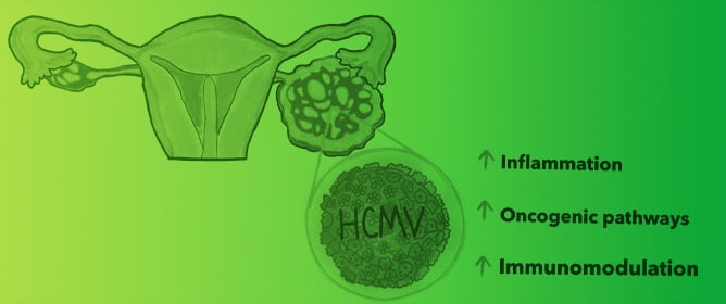
HCMV as an Oncomodulatory Virus in Ovarian Cancer Progression
Journal Description
Biomolecules
Biomolecules
is an international, peer-reviewed, open access journal on structures and functions of bioactive and biogenic substances, molecular mechanisms with biological and medical implications as well as biomaterials and their applications, published monthly online by MDPI.
- Open Access— free for readers, with article processing charges (APC) paid by authors or their institutions.
- High Visibility: indexed within Scopus, SCIE (Web of Science), PubMed, MEDLINE, PMC, Embase, CAPlus / SciFinder, and other databases.
- Journal Rank: JCR - Q1 (Biochemistry and Molecular Biology) / CiteScore - Q1 (Biochemistry)
- Rapid Publication: manuscripts are peer-reviewed and a first decision is provided to authors approximately 16.6 days after submission; acceptance to publication is undertaken in 3.3 days (median values for papers published in this journal in the first half of 2026).
- Recognition of Reviewers: reviewers who provide timely, thorough peer-review reports receive vouchers entitling them to a discount on the APC of their next publication in any MDPI journal, in appreciation of the work done.
- Sections: published in 15 topical sections.
- Testimonials: See what our editors and authors say about Biomolecules.
- Companion journal: Receptors.
Impact Factor:
5.6 (2025);
5-Year Impact Factor:
6.2 (2025)
Latest Articles
Advances in Imaging of Plant Ca2+ Signaling
Biomolecules 2026, 16(8), 1193; https://doi.org/10.3390/biom16081193 (registering DOI) - 15 Aug 2026
Abstract
Calcium ions (Ca2+) function as ubiquitous second messengers that translate environmental and developmental cues into spatially and temporally defined cellular responses in plants. This review summarizes the cellular architecture and molecular mechanisms that generate, shape, and terminate Ca2+ signals, with
[...] Read more.
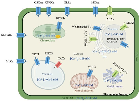
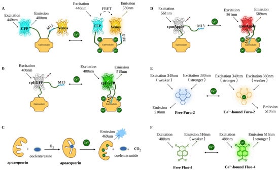
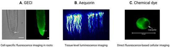
Calcium ions (Ca2+) function as ubiquitous second messengers that translate environmental and developmental cues into spatially and temporally defined cellular responses in plants. This review summarizes the cellular architecture and molecular mechanisms that generate, shape, and terminate Ca2+ signals, with emphasis on plasma-membrane channels, intracellular stores, pumps, exchangers, and organelle-associated transport systems. We also examine the development of live Ca2+ indicators, from chemical dyes and aequorin to ratiometric and single-fluorophore genetically encoded calcium indicators, and discuss principles for selecting sensors for different tissues and subcellular compartments. Recent studies have applied these tools to abiotic stress, plant immunity, polar growth, development, symbiosis, and systemic signaling. Accurate quantitative imaging nevertheless requires careful matching of sensor properties to the target cellular environment and rigorous control of motion, spectral interference, and analytical procedures. Combining improved indicators with advanced microscopy, genetic validation, and standardized data analysis should help connect distinct Ca2+ signatures with their molecular origins and physiological roles.
Full article
(This article belongs to the Section Molecular Biology)
►
Show Figures
Open AccessArticle
DNA Methylation Dynamics in Eisenia andrei Regeneration: The Effects of Time, Region, and a Hypomethylating Agent
by
Chayeen Brotzki da Costa, Péter Németh and Péter Engelmann
Biomolecules 2026, 16(8), 1192; https://doi.org/10.3390/biom16081192 - 14 Aug 2026
Abstract
The impact of epigenetic mechanisms on molecular and cellular processes of regeneration remains a less-investigated field, especially concerning earthworm segment restoration. To evaluate distinct methylated cytosines under different conditions, earthworm segments were collected: decitabine-treated and controls; anterior and posterior amputation; intact and regenerated
[...] Read more.
The impact of epigenetic mechanisms on molecular and cellular processes of regeneration remains a less-investigated field, especially concerning earthworm segment restoration. To evaluate distinct methylated cytosines under different conditions, earthworm segments were collected: decitabine-treated and controls; anterior and posterior amputation; intact and regenerated (2- and 4-week). 5-methylcytosine (5mC) and 5-hydroxymethylcytosine (5hmC) distribution in tissues was assessed by immunohistochemistry (IHC), and in genomic DNA by dot blot. DNA-methyltransferase (DNMT) and ten-eleven translocation (TET) dioxygenase activity was determined by immuno-based colorimetry. DNMT1 and TET gene expressions were verified with real-time PCR. Decitabine treatment reduced 5mC levels in most tissues, persisting in the anterior coelomic cavity (intact and blastemas). 5hmC remained elevated in most tissues, remarkably in 2-week blastemas. In the same period, DNMT and TET presented the highest activity in anterior treated samples compared to other treated periods. Concurrently, DNMT1 gene expression increased prominently in anterior segments, while TET showed the highest expression in both anterior and posterior 2-week treated groups. Thus far, our observations indicate that, despite decitabine’s effect on the DNA methylation machinery of the earthworm model, the region of amputation and the regeneration period also interfere with activity and expression of epigenetic enzymes, affecting 5mC and 5hmC distribution.
Full article
(This article belongs to the Special Issue Wound Repair and Regeneration: From Molecular and Cellular Mechanisms to New Approaches)
Open AccessReview
Mechanobiology of Matricellular Proteins in Bladder Cancer: A Narrative Review and Bioinformatics Analysis
by
Alim Turgaliyev, Roman Konovalov, Anton Borissenko and Dieter Riethmacher
Biomolecules 2026, 16(8), 1191; https://doi.org/10.3390/biom16081191 - 14 Aug 2026
Abstract
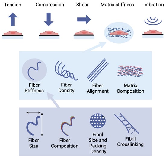
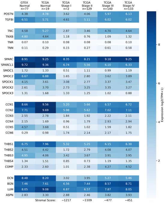
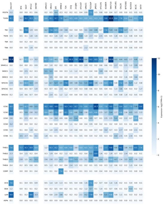
The extracellular matrix (ECM) in cancer differs from healthy tissue in structure, composition, and mechanical properties. Matricellular proteins (MCPs) play important roles in shaping ECM architecture during tissue remodeling. This narrative review, combined with a bioinformatics analysis, examines six major MCP families—Fasciclins, Tenascins,
[...] Read more.
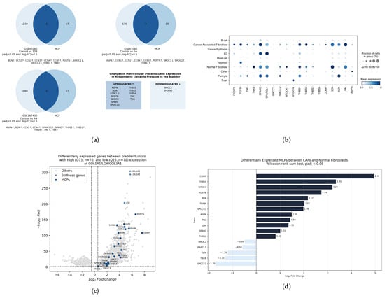
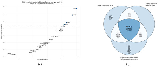
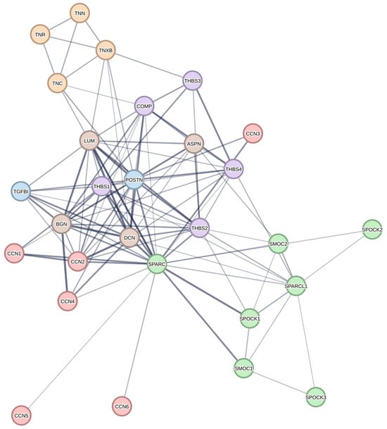
The extracellular matrix (ECM) in cancer differs from healthy tissue in structure, composition, and mechanical properties. Matricellular proteins (MCPs) play important roles in shaping ECM architecture during tissue remodeling. This narrative review, combined with a bioinformatics analysis, examines six major MCP families—Fasciclins, Tenascins, Thrombospondins, Small Leucine-Rich Proteoglycans, the SPARC family, and the CCN family—through a mechanobiological lens in bladder cancer. It summarizes current knowledge on the mechanical regulation of MCP expression, their effects on matrix stiffness, and their contributions to bladder cancer progression. Analyses of public datasets reveal that stromal cells are the predominant source of MCPs in the tumor microenvironment. Furthermore, mechanical upregulation and involvement in the formation of stiff ECM highlight MCPs as important players in a mechanotransduction feedback loop. While most MCPs exert pro-tumorigenic effects on bladder cancer cells, several display context-dependent or anti-tumorigenic activities. Existing studies have primarily focused on the isolated effects of MCPs on bladder cancer cell lines in two-dimensional systems or simple subcutaneous xenograft models. Both approaches fail to capture the context-dependent nature of MCPs and their involvement in ECM formation. These findings underscore the need for future studies to investigate the complex effects of MCPs on bladder cancer progression.
Full article
(This article belongs to the Special Issue Extracellular Matrix Components and Glycobiology in Cancer: From Mechanisms to Therapeutic Targeting)
►▼
Show Figures

Figure 1

Open AccessArticle
Macrophage-Based Transcriptional Assays for the Comparative Assessment of the Anti-Inflammatory Paracrine Activity of Canine Adipose-Derived Mesenchymal Stromal Cells
by
Andrea Exnerová, Sabina Seidlová, Věra Daňková, Vojtěch Pavlík and Kristina Nešporová
Biomolecules 2026, 16(8), 1190; https://doi.org/10.3390/biom16081190 - 14 Aug 2026
Abstract
Therapies based on mesenchymal stromal cells (MSCs) have high potential in the field of regenerative medicine due mainly to their immunomodulatory properties. However, their clinical translation is hampered by a lack of sufficiently standardised potency tests. Since macrophages constitute key mediators of the
[...] Read more.
Therapies based on mesenchymal stromal cells (MSCs) have high potential in the field of regenerative medicine due mainly to their immunomodulatory properties. However, their clinical translation is hampered by a lack of sufficiently standardised potency tests. Since macrophages constitute key mediators of the effects of MSCs, macrophage-based assays potentially provide a relevant in vitro tool for the evaluation of the activity of MSC products. This study involved the coculturing of canine adipose-derived mesenchymal stromal cells (ASCs) with macrophages derived from human THP-1 and U937 monocyte cell lines, murine RAW264.7 macrophages and primary human macrophages. The M2 polarisation was assessed following stimulation with IL-4/IL-13 in THP-1 and U937 macrophages. The mRNA expression of the pro- and anti-inflammatory markers was analysed using qPCR. The ASC transwell coculture altered the LPS-induced inflammatory mRNA expression in a strongly model- and marker-dependent manner. The U937-derived macrophages exhibited the most consistent suppression of the tested inflammatory transcripts and the RAW264.7 cells provided a practical readout for selected inflammatory markers, whereas the THP-1 macrophages evinced the suppression of TNFA but not IL1B or PTGS2 under the selected stimulation conditions. IL-4/IL-13 induced moderate but statistically non-significant changes in IL10 and TGFB1 in the U937-derived macrophages but no reproducible response in the THP-1-derived macrophages. In a subsequent U937 coculture experiment, ASC-derived paracrine factors altered selected M2-associated transcripts at specific time points. The results thus provided support for macrophage-based transcriptional readouts as an early-stage tool for comparing responder macrophage models and detecting the selected anti-inflammatory paracrine effects of canine ASCs; the U937 cells were found to be particularly suitable for the study of inflammatory polarisation and the RAW264.7 cells for the purpose of standardised screening.
Full article
(This article belongs to the Section Biological Factors)
Open AccessReview
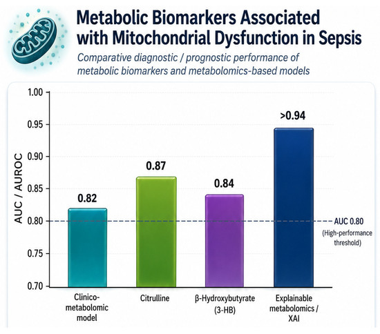
Systems Bioengineering of Septic Shock Metabolism: Citrulline, β-Hydroxybutyrate and Plasma Biomarker-Based Phenotyping
by
Leonard Azamfirei, Vlad Dimitrie Cehan, Alina Roxana Cehan, Mihai Claudiu Pui and Alexandra Lazar
Biomolecules 2026, 16(8), 1189; https://doi.org/10.3390/biom16081189 - 14 Aug 2026
Abstract
Background: Although advances in critical care have improved short-term outcomes, sepsis survivors continue to face substantial chronic morbidity and impaired long-term survival. Conventional threshold-based tools such as the Sequential Organ Failure Assessment (SOFA) and Modified Early Warning Score (MEWS) show moderate and variable
[...] Read more.
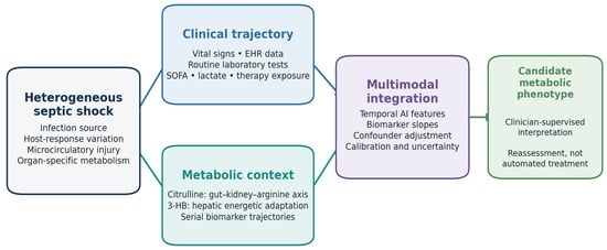
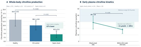
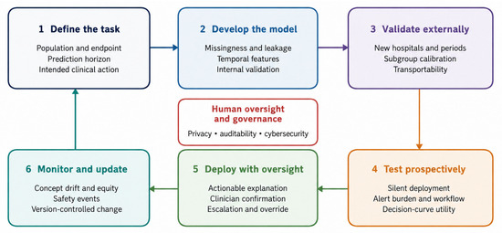
Background: Although advances in critical care have improved short-term outcomes, sepsis survivors continue to face substantial chronic morbidity and impaired long-term survival. Conventional threshold-based tools such as the Sequential Organ Failure Assessment (SOFA) and Modified Early Warning Score (MEWS) show moderate and variable discrimination across cohorts. Reported areas under the receiver operating characteristic curve (AUROCs) must therefore be interpreted in relation to the population, prediction horizon, and outcome used in each study rather than as direct head-to-head comparisons. Objectives: This review evaluates how artificial intelligence (AI) could be linked with dynamic plasma metabolites, particularly citrulline and β-hydroxybutyrate (3-HB), to support biologically informed sepsis phenotyping, while critically examining mechanistic evidence, clinical limitations, and translational readiness. Data Synthesis: Machine-learning and natural language processing architectures have shown promising discrimination in many early-detection studies, with pooled AUROCs near 0.87 and reported prediction windows extending to 48 h. However, performance estimates vary with cohort composition, outcome definition, and validation design, and they should not be ranked against unrelated biomarker studies. Human sepsis studies generally associate low or persistently low citrulline with impaired intestinal function and organ injury, but no sepsis-specific decision cutoff has been externally validated. For 3-HB, an AUROC of 0.8429 for septic liver injury was derived from a cohort of 57 patients and has not been shown to add value beyond routine liver tests or illness-severity measures. Murine experiments provide mechanistic hypotheses for ketone-mediated organ protection, but model-specific and sometimes opposing nutritional effects limit direct translation. These metabolites are therefore best considered candidate longitudinal features for multimodal phenotyping rather than stand-alone clinical triggers. Conclusions: Biologically informed algorithmic surveillance is a promising direction, but clinical implementation requires prospective serial sampling, explicit adjustment for renal, hepatic and nutritional confounders, head-to-head comparison with routine markers, and external validation of calibration and clinical utility. Until these requirements are met, citrulline and 3-HB should support research phenotyping rather than direct treatment selection.
Full article
(This article belongs to the Topic Biomarker Development and Application, 2nd Edition)
►▼
Show Figures

Figure 1
Open AccessReview
Oxidation of Uroporphyrinogens During Heme Synthesis: Role of Iron, Susceptibility and Consequences
by
Andrew G. Smith
Biomolecules 2026, 16(8), 1188; https://doi.org/10.3390/biom16081188 - 14 Aug 2026
Abstract
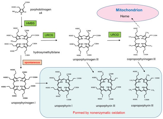
In the biosynthesis of heme the tetrapyrrole hydroxymethylbilane is converted enzymatically to uroporphyrinogen III whereas conversion to uroporphyrinogen I occurs spontaneously. Both are substrates for uroporphyrinogen decarboxylase (UROD) but only the III isomer is a precursor of heme. These porphyrinogens are easily oxidised
[...] Read more.
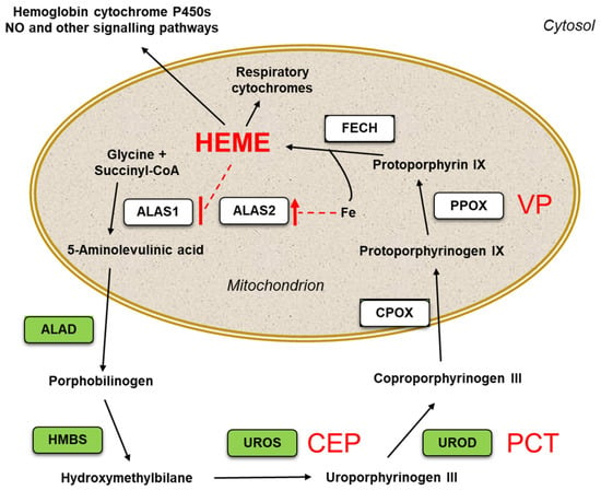
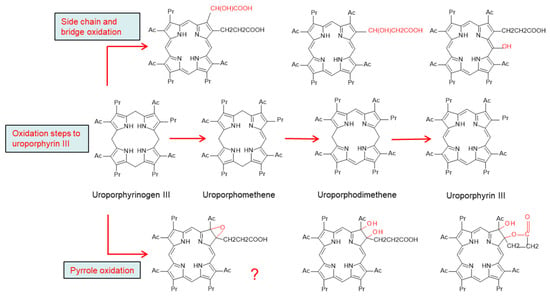
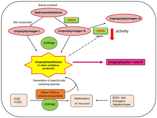
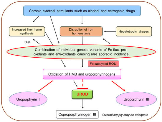
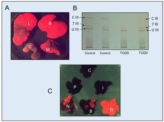
In the biosynthesis of heme the tetrapyrrole hydroxymethylbilane is converted enzymatically to uroporphyrinogen III whereas conversion to uroporphyrinogen I occurs spontaneously. Both are substrates for uroporphyrinogen decarboxylase (UROD) but only the III isomer is a precursor of heme. These porphyrinogens are easily oxidised to the respective uroporphyrins and trace amounts occur in the urine of healthy humans and animals. Large quantities of uroporphyrins I and III, as well as other oxidation products, occur in the liver and urine of patients with some porphyrias and after poisoning of people and animals by chemicals, such as hexachlorobenzene (HCB) and 2,3,7,8-tetrachorodibenzo-p-dioxin (TCDD). In the acquired disorder sporadic porphyria cutanea tarda (sPCT) and chemical-induced porphyria, hepatic UROD is inhibited, ostensibly by a partially oxidised uroporphyrinogen. The processes leading to oxidation of the uroporphyrinogens are interactions of a variety of external, internal and genetic factors. In some experimental systems, cytochrome P450 1A2 is implicated in the oxidation of uroporphyrinogens and uroporphyria and many patients and in vivo studies demonstrate the influence of iron. The article reviews the present state of knowledge of uroporphyrinogen oxidation, susceptibility and outcomes, and illustrates areas that require further exploration to explain fully the mechanisms of sPCT and the related uroporphyria caused by chemicals of toxic concern.
Full article
(This article belongs to the Special Issue Advances in Porphyria and Liver Disease Research)
►▼
Show Figures

Figure 1

Open AccessFeature PaperArticle
Genetic Diversity and Runs of Homozygosity in Three Masu Salmon (Oncorhynchus masou) Populations Based on Whole-Genome Resequencing Data
by
Song Bai, Chenfan Geng, Wei Wang, Xiaoyu Yan, Tian Dong, Hailiang Song and Hongxia Hu
Biomolecules 2026, 16(8), 1187; https://doi.org/10.3390/biom16081187 - 14 Aug 2026
Abstract
Masu salmon (Oncorhynchus masou) is an ecologically and economically important cold-water salmonid in East Asia that exhibits diverse life-history forms. To compare population-level genomic variation and patterns of homozygosity among fish from different sources, we analyzed whole-genome resequencing data from 465
[...] Read more.
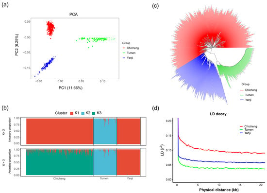
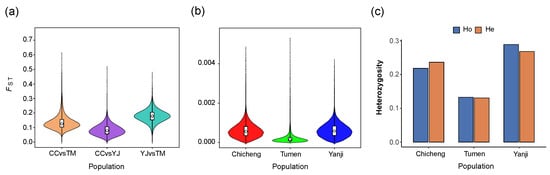
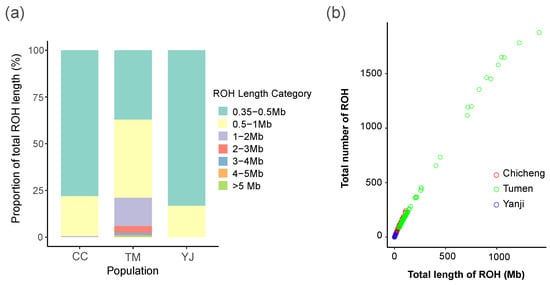
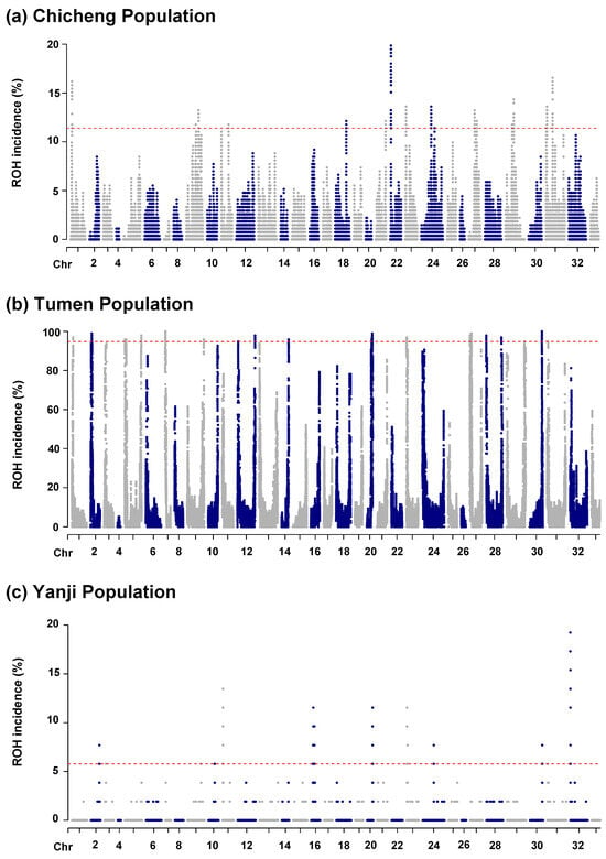
Masu salmon (Oncorhynchus masou) is an ecologically and economically important cold-water salmonid in East Asia that exhibits diverse life-history forms. To compare population-level genomic variation and patterns of homozygosity among fish from different sources, we analyzed whole-genome resequencing data from 465 individuals representing one field-collected Tumen River population (TM) and two landlocked cultured populations from Chicheng (CC) and Yanji (YJ). After quality control, 6,220,980 high-quality SNPs were retained. Population-specific filtering identified 5,589,828, 3,545,209, and 4,975,751 polymorphic SNPs in CC, TM, and YJ, respectively; although SNP numbers differed, approximately 91% of variants in each population were located in intronic or intergenic regions. Principal component analysis, ADMIXTURE, and distance-based neighbor-joining analysis clearly distinguished the three populations, with CC and YJ showing the closest genetic relationship. Pairwise was lowest between CC and YJ and highest between TM and YJ. CC exhibited the highest linkage disequilibrium, whereas TM showed the fastest LD decay and the lowest nucleotide diversity and heterozygosity. Runs of homozygosity (ROH) burden was highest in TM, intermediate in CC, and lowest in YJ. TM had the highest number of ROHs, cumulative ROH length, and , and ROHs longer than 5 Mb were detected only in this population. CC had an intermediate ROH burden dominated by short segments, whereas YJ had the lowest ROH-based genomic inbreeding. The high and heterogeneous ROH burden in TM indicates elevated genome-wide homozygosity among the sampled fish but does not, by itself, demonstrate recent inbreeding throughout the population. Candidate ROH islands and their annotated genes showed limited overlap among populations. Candidate genes in TM were primarily associated with ion regulation, neural processes, and energy metabolism, whereas those in CC and YJ shared broad functional categories involving development, muscle organization, nutrient transport, and neural regulation but differed in most specific genes. These regions and genes should be regarded as exploratory, hypothesis-generating candidates rather than evidence of selection or causality. Overall, this study reveals distinct population genomic characteristics and ROH patterns among masu salmon populations of different origins and provides a basis for future germplasm conservation and genetic management.
Full article
(This article belongs to the Special Issue Vertebrate Comparative Genomics)
►▼
Show Figures

Figure 1

Open AccessArticle
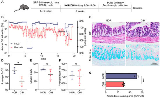
Chronic Intermittent Hypoxia Disrupts Intestinal Homeostasis Through Gut Microbiota Remodeling and Microbiota-Metabolite Interactions
by
Yuying He, Jun Gao, Qiang Li, Chuxi Zhang, Mingrui Zhai and Yuehua Liu
Biomolecules 2026, 16(8), 1186; https://doi.org/10.3390/biom16081186 - 14 Aug 2026
Abstract
Obstructive sleep apnea (OSA) is characterized by chronic intermittent hypoxia (CIH), which contributes to systemic metabolic disorders. However, the mechanisms underlying CIH-induced intestinal dysfunction remain unclear. In this study, we investigated the effects of CIH on intestinal barrier integrity, gut microbiota, and host
[...] Read more.
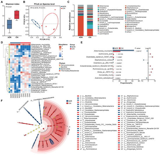
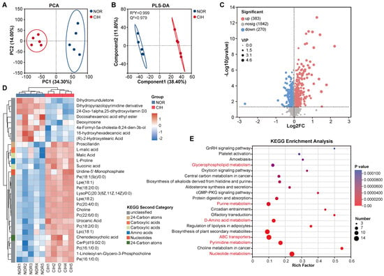
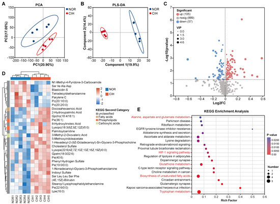
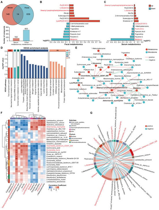
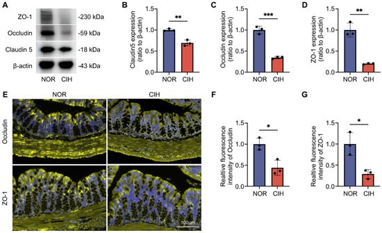
Obstructive sleep apnea (OSA) is characterized by chronic intermittent hypoxia (CIH), which contributes to systemic metabolic disorders. However, the mechanisms underlying CIH-induced intestinal dysfunction remain unclear. In this study, we investigated the effects of CIH on intestinal barrier integrity, gut microbiota, and host metabolism using a multi-omics approach. Male C57BL/6J mice were exposed to six weeks of CIH or normoxia. Colonic barrier integrity was assessed by histological and molecular analyses. Gut microbiota was profiled by full-length 16S rRNA gene sequencing. Untargeted metabolomics was performed on fecal and serum samples, followed by integrated microbiome–metabolome analysis. CIH markedly impaired colonic barrier integrity, as evidenced by disrupted crypt architecture, reduced goblet cell abundance, and decreased expression of ZO-1, Occludin, and Claudin-5. CIH also induced gut microbial dysbiosis, characterized by depletion of the beneficial mucin-associated bacterium Akkermansia muciniphila and enrichment of several anaerobic taxa. Metabolomic analysis revealed opposite alterations of PC (20:2/0:0) and LysoPE (20:5/0:0) between feces and serum, whereas melatonin was consistently decreased in both compartments. Integrated multi-omics analysis further revealed close associations between microbial dysbiosis and metabolic remodeling. Collectively, these findings demonstrate that CIH disrupts intestinal homeostasis through coordinated alterations in barrier integrity, gut microbiota composition, and host metabolism, providing new insights into the intestinal mechanisms underlying OSA-associated systemic dysfunction.
Full article
(This article belongs to the Special Issue Gut Microbiome and Related Diseases in Animals)
►▼
Show Figures

Figure 1
Open AccessReview
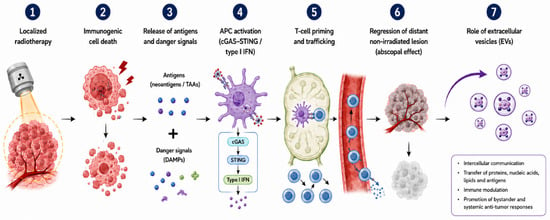
Revisiting the Abscopal Effect in the Era of Immuno-Radiotherapy: Mechanisms, Challenges, and Clinical Perspectives
by
Enrico Rosa, Maria Vaccaro, Bruno Fionda, Valentina Lancellotta, Gabriele Ciasca, Pierpaolo Dragonetti, Lucia Di Maio, Fabio Marazzi, Francesco Marampon, Monica Mangoni, Maria Antonietta Gambacorta, Marco De Spirito and Luca Tagliaferri
Biomolecules 2026, 16(8), 1185; https://doi.org/10.3390/biom16081185 - 14 Aug 2026
Abstract
Background: The abscopal effect refers to the clinical response of non-irradiated tumor lesions following localized radiotherapy (RT). Once regarded as a rare phenomenon, it has gained renewed interest in the era of immunotherapy, as RT may promote systemic anti-tumor immune responses. This narrative
[...] Read more.
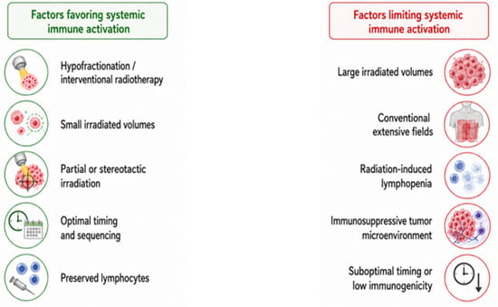
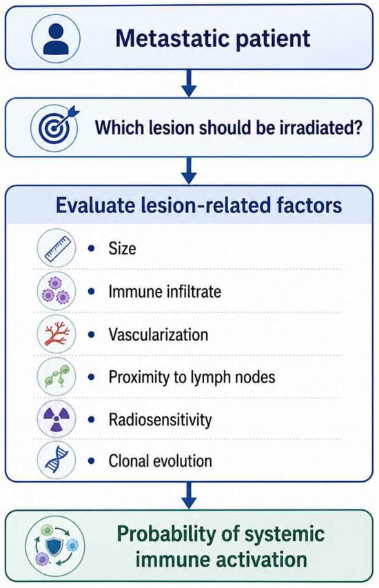
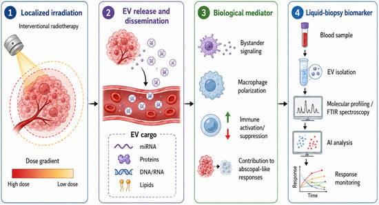
Background: The abscopal effect refers to the clinical response of non-irradiated tumor lesions following localized radiotherapy (RT). Once regarded as a rare phenomenon, it has gained renewed interest in the era of immunotherapy, as RT may promote systemic anti-tumor immune responses. This narrative review summarizes current biological, radiobiological, and clinical evidence on the abscopal effect and highlights translational gaps limiting its reproducibility. Methods: Preclinical, translational, and clinical evidence was qualitatively analyzed across seven domains: RT, immunology, and clinical oncology. A structured qualitative gap analysis was used to identify disconnections between biological mechanisms, RT parameters, biomarkers, and clinical outcomes. Results: Current evidence supports the biological plausibility of the abscopal effect through immunogenic cell death, antigen and damage-associated molecular patterns (DAMP) release, activation of the cyclic GMP–AMP synthase–stimulator of interferon genes (cGAS-STING) pathway, dendritic-cell priming, and T-cell-mediated responses. However, clinical results remain heterogeneous. Dose, fractionation, irradiated volume, timing, lymphocyte preservation, and interventional RT (modern brachytherapy, IRT) may influence systemic immune activation. Emerging biomarkers, particularly extracellular vesicles (EVs), may help connect radiation-induced biological stress with immune modulation and clinical response. Conclusions: The main barrier to clinical translation is the fragmentation of evidence across RT, immunology, and clinical oncology. Integrated translational frameworks combining dosimetry, immune monitoring, EVs-based biomarkers, imaging, and clinical endpoints may improve the interpretation and reproducibility of abscopal responses in immuno-RT.
Full article
(This article belongs to the Special Issue Advances in Cellular and Molecular Mechanisms in Immuno-Oncology and Onco-Hematology: 2nd Edition)
►▼
Show Figures

Figure 1
Open AccessArticle
The Antioxidant and Antibacterial Properties of Honeycomb-Structured Polylactic Acid/Astaxanthin@ZIF-8 Bio-Composite Film Applied in Beef Preservation
by
Sheng Liu, Shuran Xing, Feifei Wang, He Zhu, Xiaoyun Fu, Yang Yu and Litao Wang
Biomolecules 2026, 16(8), 1184; https://doi.org/10.3390/biom16081184 - 13 Aug 2026
Abstract
Food spoilage is mainly driven by lipid oxidation and microbial proliferation. To counteract these challenges, multifunctional protective films have emerged as effective solutions for suppressing both reactions. In this work, biodegradable polylactic acid (PLA) was selected as the polymer matrix to fabricate composite
[...] Read more.
Food spoilage is mainly driven by lipid oxidation and microbial proliferation. To counteract these challenges, multifunctional protective films have emerged as effective solutions for suppressing both reactions. In this work, biodegradable polylactic acid (PLA) was selected as the polymer matrix to fabricate composite films. Colloidal ZIF-8 nanoparticles loaded with the natural antioxidant astaxanthin (AST) were mixed with PLA. By precisely controlling temperature and humidity during the solution-casting process, a honeycomb-structured PLA/AST@ZIF-8 composite film was successfully prepared. The structural and physico-chemical properties of the as-prepared film were systematically characterized by FE-SEM, FTIR, Raman, XRD, and TGA. The results confirm the formation of a well-defined honeycomb morphology, with AST uniformly dispersed throughout the PLA matrix. Importantly, films with AST content above 2.0% exhibit weak antibacterial properties. Practical application tests for preserving beef tenderloin demonstrate that the film provides excellent antioxidant and antibacterial effects. On the seventh day, the water loss, pH, ΔE and total volatile basic nitrogen (TVB-N) values of beef samples treated with the PLA/ZIF-8/2.5%AST film remained within acceptable thresholds, effectively extending the shelf life of fresh meat. Safety assessments on overall migration (OM) and Zn2+-specific migration confirm that the PLA/AST@ZIF-8 composite film fully complies with EU Regulation No. 10/2011. These results demonstrate the great application prospects of the composite film for high-performance food preservation.
Full article
(This article belongs to the Section Bio-Engineered Materials)
Open AccessReview
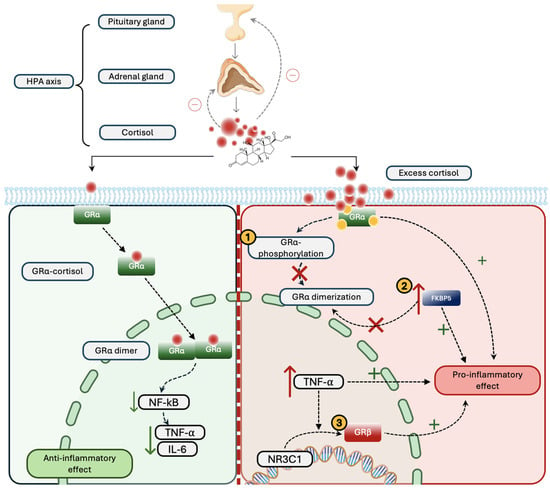
Biomolecular Pathways Linking Preoperative Chronic Stress to Postoperative Cardiovascular Dysfunction in Noncardiac Surgery
by
Predrag Jancic, Ivana Kovac, Halil Emre Demirtas, Nebojsa Nick Knezevic and Graham Trevor Lubinsky
Biomolecules 2026, 16(8), 1183; https://doi.org/10.3390/biom16081183 - 13 Aug 2026
Abstract
Postoperative cardiovascular complications remain a leading cause of morbidity and mortality after noncardiac surgery, yet current risk models do not incorporate psychosocial stress. With over 300 million noncardiac surgeries performed annually worldwide and a substantial burden of perioperative cardiovascular complications, preoperative chronic stress
[...] Read more.
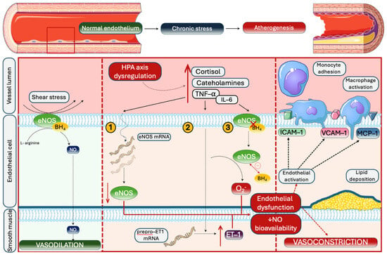
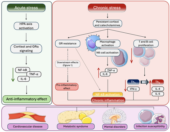
Postoperative cardiovascular complications remain a leading cause of morbidity and mortality after noncardiac surgery, yet current risk models do not incorporate psychosocial stress. With over 300 million noncardiac surgeries performed annually worldwide and a substantial burden of perioperative cardiovascular complications, preoperative chronic stress is increasingly recognized as a potentially modifiable risk factor. Chronic stress produces HPA axis dysregulation, glucocorticoid resistance, sympathetic activation, inflammation, endothelial dysfunction, and hypercoagulability. These pathways overlap with the mechanisms underlying perioperative myocardial injury, arrhythmogenesis, and venous thromboembolism. Prospective data demonstrated that preoperative psychological distress independently predicted 30-day cardiovascular complications and 1-year mortality after noncardiac surgery. Allostatic load studies in noncardiac surgery patients showed that high preoperative physiological burden was associated with up to twofold increases in postoperative mortality and elevated rates of myocardial infarction and venous thromboembolism. Epidemiological evidence further supports that anxiety and depression independently increase cardiovascular risk. Converging evidence suggests that preoperative psychological distress is a relevant perioperative cardiovascular risk factor. However, no randomized controlled trial has evaluated whether targeted preoperative stress reduction can decrease postoperative cardiovascular events, representing a critical gap warranting prospective investigation.
Full article
(This article belongs to the Section Biological Factors)
►▼
Show Figures

Figure 1

Open AccessArticle
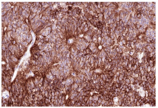
Integrin αvβ6 Expression in the Human Pituitary Gland and Pituitary Neuroendocrine Tumors: Immunohistochemical Characterization with Potential Relevance to αvβ6 PET/CT Pituitary Uptake and Theranostic Implications
by
Muin Tuffaha, Wael Hananeh, Ehab Shiban and Michael Starke
Biomolecules 2026, 16(8), 1182; https://doi.org/10.3390/biom16081182 - 13 Aug 2026
Abstract
Integrins are heterodimeric transmembrane receptors that mediate bidirectional signaling and regulate cell–cell and cell–extracellular matrix interactions. Integrin αvβ6 is an epithelial-associated integrin that has emerged as a promising molecular target for PET/CT imaging using integrin αvβ6-directed radiotracers such as 68Ga-Trivehexin, and, most
[...] Read more.
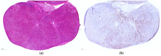
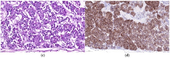
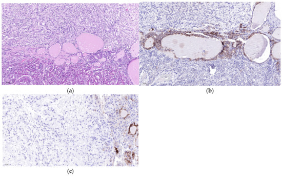
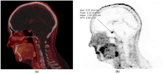
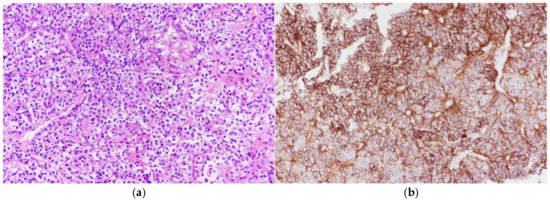
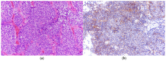
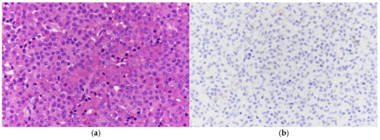
Integrins are heterodimeric transmembrane receptors that mediate bidirectional signaling and regulate cell–cell and cell–extracellular matrix interactions. Integrin αvβ6 is an epithelial-associated integrin that has emerged as a promising molecular target for PET/CT imaging using integrin αvβ6-directed radiotracers such as 68Ga-Trivehexin, and, most recently, for antibody–drug conjugate therapy in epithelial malignancies. Unexpected physiological and incidental uptake within the pituitary gland has been reported in integrin αvβ6-targeted PET studies, including uptake in morphologically normal pituitary glands and pituitary neuroendocrine tumors (PitNETs). However, the histological basis of integrin αvβ6 expression in the human pituitary gland remains poorly understood. The aim of this study is to characterize the immunohistochemical expression of integrin αvβ6 in normal human pituitary tissue and PitNETs and to evaluate its potential implications for integrin αvβ6-targeted imaging and theranostic applications. Five complete adult pituitary glands obtained at autopsy and 28 PitNETs were examined by immunohistochemistry for integrin αvβ6. Staining distribution, intensity, and cellular localization were assessed in the adenohypophysis, neurohypophysis, and Rathke’s cleft remnants. PitNETs were classified according to transcription factor expression (PIT1, TPIT, and SF1). Among the 28 PitNETs, 17 were SF1-lineage (60.7%), three were PIT1-lineage (10.7), two were TPIT-lineage (7.1%), three lacked a dominant transcription factor (10.7%), and three showed plurilineage expression (10.7%). Integrin αvβ6 expression was evaluated semiquantitatively according to staining intensity and the percentage of positive tumor cells. In normal pituitary glands, integrin αvβ6 immunoreactivity was predominantly membranous and localized to larger adenohypophyseal cells irrespective of transcription factor lineage or hormone phenotype. Strong expression was also observed in the epithelial lining cells of Rathke’s cleft remnants, whereas the neurohypophysis lacked detectable integrin αvβ6 expression. Among the 28 PitNETs, integrin αvβ6 expression was detected in 20 cases (71.4%). Positive tumors demonstrated variable staining intensity and extent, ranging from 20% to 100% positive tumor cells. By lineage, integrin αvβ6 expression was detected in 13 of 17 SF1-lineage tumors (76.5%), one of three PIT1-lineage tumors (33.3%), and zero of two TPIT-lineage tumors (0%). Additionally, all three tumors lacking a dominant transcription factor (100%) and all three plurilineage tumors (100%) demonstrated integrin αvβ6 expression. Eleven integrin αvβ6-positive tumors showed expression in ≥50% of tumor cells, and six exhibited strong or diffuse immunoreactivity. Integrin αvβ6 expression in adenohypophyseal cells and Rathke’s cleft remnants provides a histological explanation for physiological pituitary uptake observed on αvβ6-targeted PET/CT imaging. The high prevalence of integrin αvβ6 expression in PitNETs, particularly in a subset demonstrating strong and diffuse immunoreactivity, suggests potential applicability of integrin αvβ6-targeted molecular imaging and theranostic approaches, including both radioligand- and antibody-based strategies. However, these applications remain investigational and require further validation in preclinical and clinical studies. At the same time, physiological integrin αvβ6 expression in normal anterior pituitary tissue may limit imaging specificity and should be considered when developing integrin αvβ6-targeted radioligand therapies. Further clinicopathological and imaging correlation studies are warranted to define the diagnostic and therapeutic role of integrin αvβ6-targeted approaches in PitNETs.
Full article
(This article belongs to the Special Issue Preclinical: Drug, Model and Imaging Development)
►▼
Show Figures

Figure 1

Open AccessReview
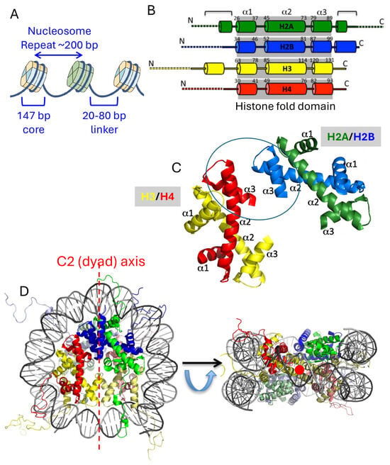
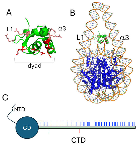
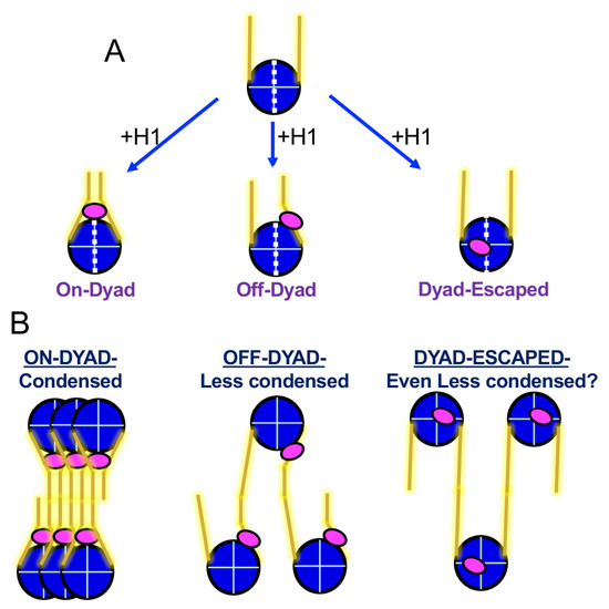
Linker Histones: The Multiple Binding Modes of the Enigmatic 5th Histone
by
Nicholas R. Rugelis, Ashok Kumar and Jeffrey J. Hayes
Biomolecules 2026, 16(8), 1181; https://doi.org/10.3390/biom16081181 - 13 Aug 2026
Abstract
Chromatin structure is dynamic and regulated by many factors, including enzymes that chemically modify histones and DNA, chromatin remodeling complexes that physically manipulate nucleosomes and chromatin, and non-enzymatic proteins that bind to DNA or nucleosomes to create specialized regions in chromatin. This multifactorial
[...] Read more.
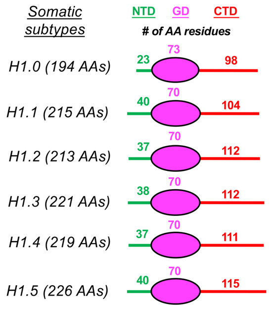
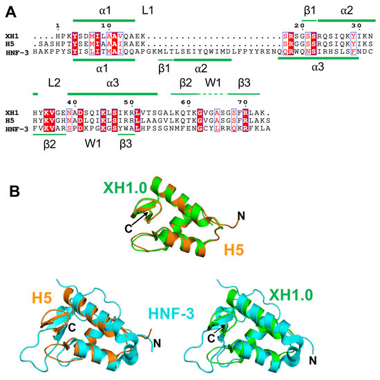
Chromatin structure is dynamic and regulated by many factors, including enzymes that chemically modify histones and DNA, chromatin remodeling complexes that physically manipulate nucleosomes and chromatin, and non-enzymatic proteins that bind to DNA or nucleosomes to create specialized regions in chromatin. This multifactorial regulation stems from the need for fine-tuned control, which is key in processes including DNA repair, replication, and gene expression. Linker histones are a family of proteins structurally distinct from the core histones that provide a poorly understood layer of regulation in chromatin. In this review, we introduce the basics of chromatin structure, what is known about how linker histones (H1s) bind to nucleosomes and influence chromatin, and how the individual domains within H1s contribute to these activities. We especially focus on recent studies describing canonical and alternative H1-nucleosome binding and their potential roles in chromatin.
Full article
(This article belongs to the Special Issue Recent Advances in Chromatin and Chromosome Molecular Research)
►▼
Show Figures

Figure 1

Open AccessReview
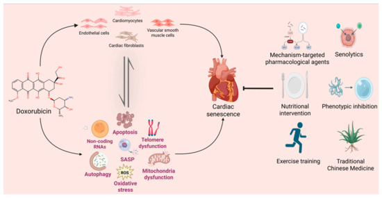
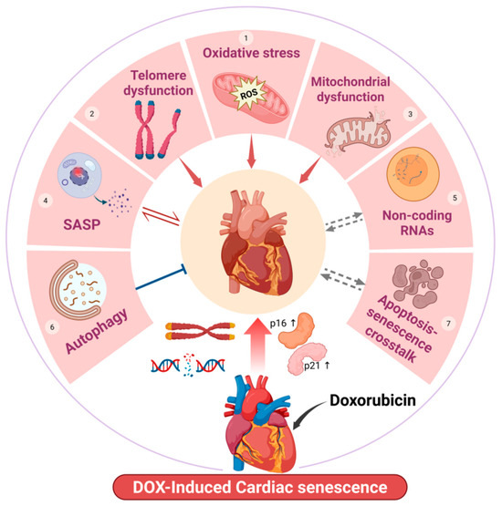
Mechanisms of Doxorubicin-Induced Cardiac Senescence and Potential Therapeutic Strategies
by
Yanli Bai, Wen Yang, Zirong Wang, Qianqian Yang, Zhongping Zhang, Jialong Liu, Yafang Qi and Dongling Liu
Biomolecules 2026, 16(8), 1180; https://doi.org/10.3390/biom16081180 - 12 Aug 2026
Abstract
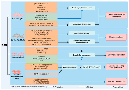
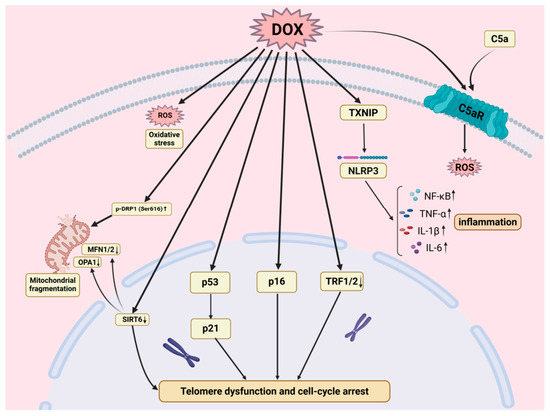
Doxorubicin (DOX) is a widely used anthracycline chemotherapeutic agent; however, its clinical application is limited by dose-dependent cardiotoxicity, which can result in progressive cardiac dysfunction and heart failure. Increasing evidence indicates that DOX-induced cardiotoxicity is closely associated with premature cardiac senescence, a pathological
[...] Read more.
Doxorubicin (DOX) is a widely used anthracycline chemotherapeutic agent; however, its clinical application is limited by dose-dependent cardiotoxicity, which can result in progressive cardiac dysfunction and heart failure. Increasing evidence indicates that DOX-induced cardiotoxicity is closely associated with premature cardiac senescence, a pathological process distinct from physiological cardiac aging. DOX induces senescence-associated alterations in multiple cardiac cell populations, disrupting cardiac homeostasis and contributing to pathological remodeling. In this review, we summarize current advances in DOX-induced cardiac senescence, focusing on the contributions of different cardiac cell types, the underlying molecular mechanisms, and emerging therapeutic strategies. We further discuss the challenges and future perspectives for developing effective interventions that alleviate cardiac senescence while preserving the anticancer efficacy of DOX. Understanding the mechanisms driving DOX-induced cardiac senescence may provide new opportunities to develop effective cardioprotective strategies and improve long-term cardiac outcomes after chemotherapy.
Full article
(This article belongs to the Section Cellular Biochemistry)
►▼
Show Figures

Figure 1
Open AccessReview
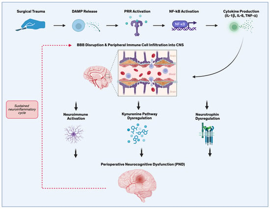
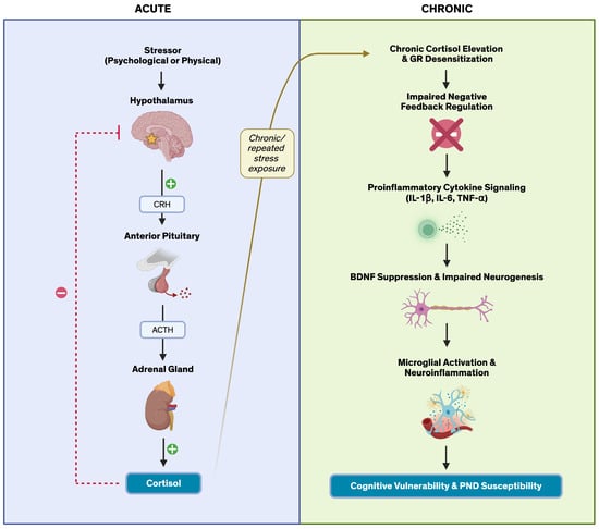
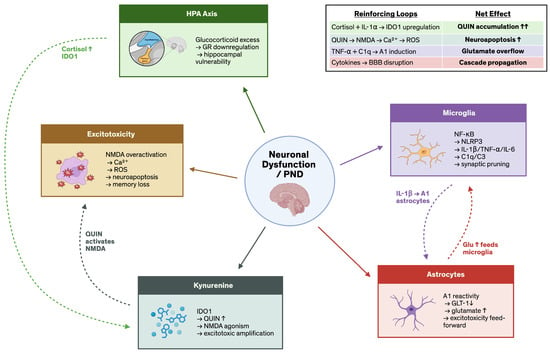
Molecular and Cellular Mechanisms Linking Mood Disorders, HPA Axis Dysregulation, and Neurocognitive Inflammation to Perioperative Neurocognitive Disorders
by
Alyson Sato, Nicole Chang, Nebojsa Nick Knezevic and Chanannait Paisansathan
Biomolecules 2026, 16(8), 1179; https://doi.org/10.3390/biom16081179 - 12 Aug 2026
Abstract
Perioperative neurocognitive disorders (PND) encompass a spectrum of cognitive impairments occurring across the surgical period and are associated with significant morbidity, delayed recovery, and reduced quality of life. Although established risk factors include advanced age, cardiovascular disease, and preexisting cognitive impairment, the contribution
[...] Read more.
Perioperative neurocognitive disorders (PND) encompass a spectrum of cognitive impairments occurring across the surgical period and are associated with significant morbidity, delayed recovery, and reduced quality of life. Although established risk factors include advanced age, cardiovascular disease, and preexisting cognitive impairment, the contribution of mood disorders to PND susceptibility remains incompletely understood. This review systematically examines the neurobiological overlap between mood disorders, particularly major depressive disorder (MDD) and bipolar disorder, and PND, with emphasis on shared biomolecular mechanisms. We identify convergent pathophysiologic pathways including hypothalamic–pituitary–adrenal (HPA) axis dysregulation, chronic neuroinflammation, NF-κB-mediated cytokine signaling, microglial priming, tryptophan–kynurenine pathway dysregulation, and brain-derived neurotrophic factor (BDNF) suppression. These mechanisms collectively suggest that patients with preexisting mood disorders may enter surgery in a biologically sensitized neuroimmune state, lowering the threshold for exaggerated neuroinflammatory responses and postoperative cognitive dysfunction. Recognition of mood disorders as modifiable perioperative vulnerability states may inform preoperative risk stratification, guide anesthetic and analgesic management, and support the development of targeted interventions to reduce postoperative cognitive complications and improve surgical outcomes.
Full article
(This article belongs to the Section Molecular Medicine)
►▼
Show Figures

Figure 1
Open AccessArticle
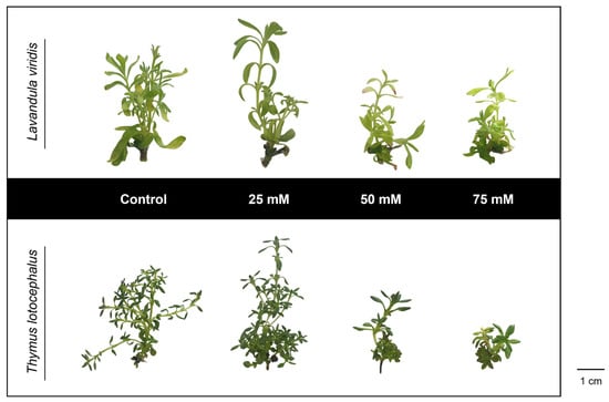
Salinity-Induced Modulation of Phenolic Compounds and Antioxidant Activity in Lavandula viridis and Thymus lotocephalus
by
Inês Mansinhos, Sandra Gonçalves, Raquel Rodríguez-Solana, Gema Pereira-Caro and Anabela Romano
Biomolecules 2026, 16(8), 1178; https://doi.org/10.3390/biom16081178 - 12 Aug 2026
Abstract
Mediterranean Lamiaceae species are important sources of bioactive compounds with potential applications in food, pharmaceutical, and cosmetic industries. In the present study, controlled salinity was evaluated as a biotechnological elicitation strategy to enhance the accumulation of bioactive metabolites in two species, Lavandula viridis
[...] Read more.
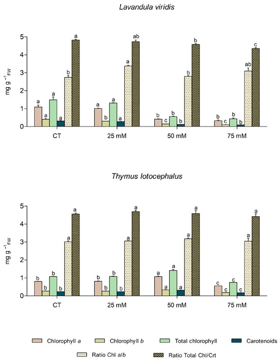
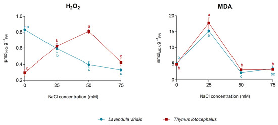
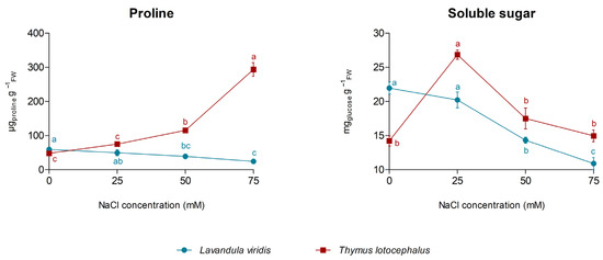
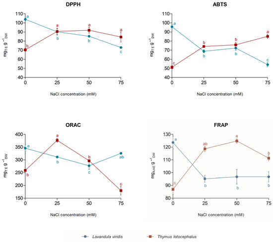
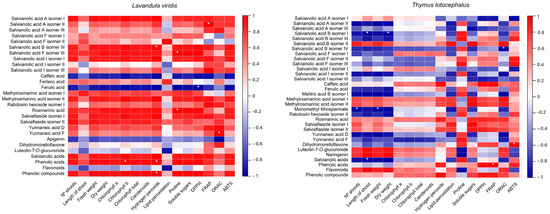
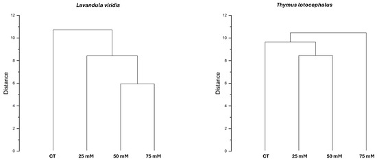
Mediterranean Lamiaceae species are important sources of bioactive compounds with potential applications in food, pharmaceutical, and cosmetic industries. In the present study, controlled salinity was evaluated as a biotechnological elicitation strategy to enhance the accumulation of bioactive metabolites in two species, Lavandula viridis and Thymus lotocephalus, cultured in vitro. Morphological, biochemical and metabolic responses of shoots grown for seven weeks in culture medium containing four NaCl concentrations (0, 25, 50, or 75 mM) were assessed. Mild salinity (25 mM NaCl) stimulated shoot growth in both species, whereas higher salt concentrations progressively reduced growth. Salinity induced oxidative stress, as indicated by elevated malondialdehyde (MDA) levels at 25 mM NaCl in both species, while hydrogen peroxide (H2O2) exhibited contrasting patterns, decreasing with increasing salinity in L. viridis but peaking at 50 mM NaCl in T. lotocephalus. Salt stress caused reductions in photosynthetic pigments, phenolic compounds, antioxidant capacity (DPPH, FRAP, ABTS, and ORAC), and overall metabolic performance in L. viridis, indicating limited tolerance to salinity. Conversely, T. lotocephalus displayed greater metabolic plasticity, characterized by increased levels of phenolic compounds, particularly rosmarinic acid and its derivatives, and higher antioxidant activity of the extracts, as well as the accumulation of proline and soluble sugars. These findings demonstrate marked species-specific differences in salinity responses and identify controlled salinity as an effective elicitation strategy to sustainably produce high-value bioactive compounds in T. lotocephalus.
Full article
(This article belongs to the Special Issue Plant Secondary Metabolism Engineering and Bioactive Compounds)
►▼
Show Figures

Figure 1
Open AccessArticle
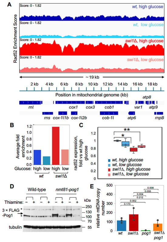
Evolutionarily Conserved but Mechanistically Distinct Mitochondrial Responses to Loss of Timeless/Swi1
by
Kalisse I. Horne, Joshua Chang Mell, Chiaki Noguchi, Sri Havya Jana, Shriya Pinisetty, Rhea Masand, Christian Sell and Eishi Noguchi
Biomolecules 2026, 16(8), 1177; https://doi.org/10.3390/biom16081177 - 12 Aug 2026
Abstract
Timeless and its fission yeast ortholog Swi1 are evolutionarily conserved components of the replication fork protection complex that ensures faithful DNA replication and genome stability. While their nuclear roles are well-characterized, their roles in mitochondrial genome maintenance remain unknown. Here, we demonstrate a
[...] Read more.
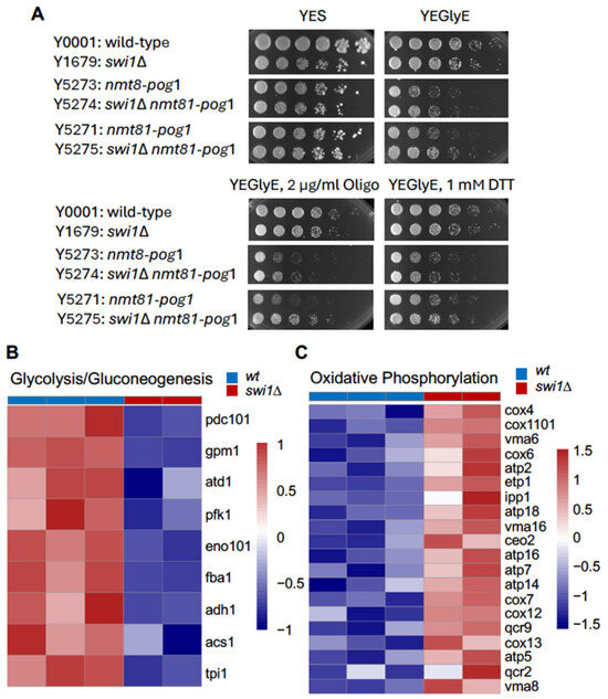
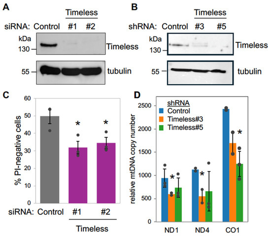
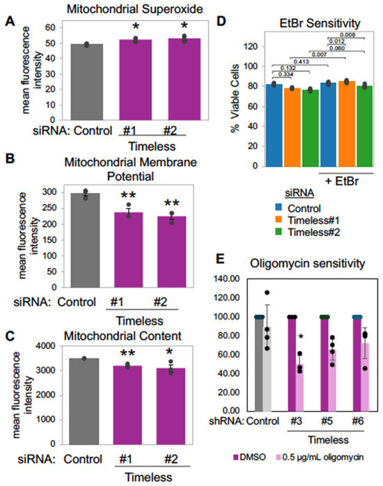
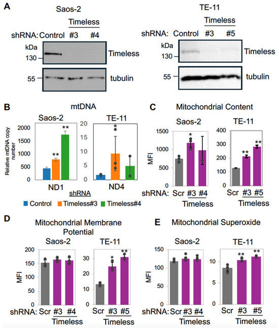
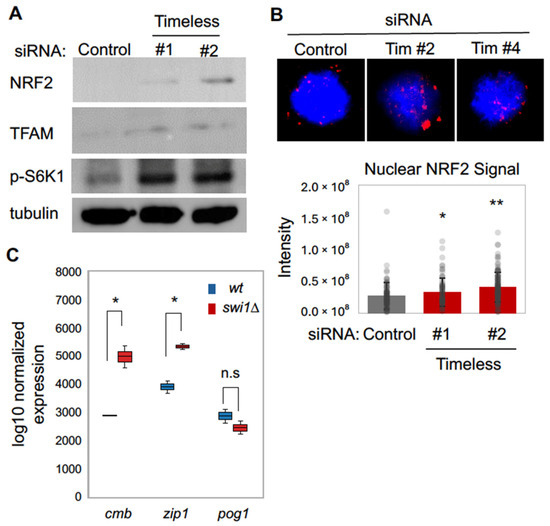
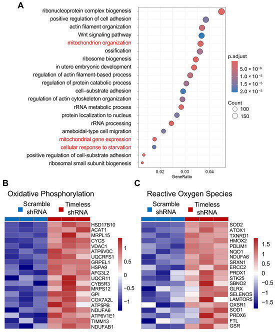
Timeless and its fission yeast ortholog Swi1 are evolutionarily conserved components of the replication fork protection complex that ensures faithful DNA replication and genome stability. While their nuclear roles are well-characterized, their roles in mitochondrial genome maintenance remain unknown. Here, we demonstrate a previously unrecognized connection between Timeless/Swi1 and mitochondrial homeostasis. In fission yeast, swi1 deletion increased association of the DNA repair protein Rad52 with mitochondrial DNA sequences across the mitochondrial genome, suggesting altered mitochondrial genome maintenance. Unexpectedly, swi1∆ cells showed an increased mtDNA copy number and improved growth under respiratory conditions, suggesting activation of compensatory mechanisms that promote mitochondrial genome maintenance. The loss of Swi1 also partially rescued the growth defect under respiratory conditions and mtDNA loss associated with depletion of mitochondrial DNA polymerase γ, linking Swi1 to pathways regulating mitochondrial replication under stress. Consistent with these phenotypes, transcriptomic and pathway enrichment analyses revealed transcriptional changes indicative of reduced glycolysis and enhanced oxidative phosphorylation, suggesting a shift toward respiratory metabolism. In human cells, Timeless depletion elicited distinct mitochondrial responses depending on the cell type. While Timeless-depleted TE-11 and Saos-2 cells elicited mitochondrial phenotypes comparable to those observed in fission yeast, Timeless depletion in U-2 OS cells led to reduced mtDNA copy number, elevated mitochondrial reactive oxygen species, and decreased mitochondrial membrane potential and mass, consistent with mitochondrial dysfunction. Despite these phenotypic differences, both fission yeast and human cells exhibited elevated levels of orthologs of the mitochondrial transcription factor A (TFAM) and the oxidative stress regulator NRF2, suggesting the conserved activation of compensatory mitochondrial and antioxidant pathways. Together, these findings identify an evolutionarily conserved connection between Timeless/Swi1 and mitochondrial homeostasis and reveal distinct adaptive responses to mitochondrial stress.
Full article
(This article belongs to the Special Issue Advances in Molecular Therapy Targeting DNA Damage and Repair Systems)
►▼
Show Figures

Figure 1
Open AccessReview
RNA Modifications as Molecular Regulators of Alveolar Epithelial Injury and Aberrant Repair in Pulmonary Fibrosis
by
Qi Huang, Shuguang Wang, Yuman Huang, Shibo Xiao, Ruohan Xia and Xianwang Wang
Biomolecules 2026, 16(8), 1176; https://doi.org/10.3390/biom16081176 - 12 Aug 2026
Abstract
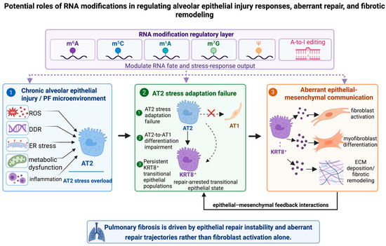
Pulmonary fibrosis is a progressive interstitial lung disease characterized by persistent alveolar epithelial injury, aberrant repair, and excessive extracellular matrix deposition. Increasing evidence indicates that disease progression is closely associated with alveolar type II (AT2) cell dysfunction, impaired AT2-to-AT1 differentiation, and the persistence
[...] Read more.
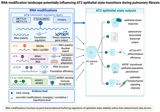
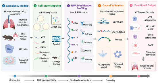
Pulmonary fibrosis is a progressive interstitial lung disease characterized by persistent alveolar epithelial injury, aberrant repair, and excessive extracellular matrix deposition. Increasing evidence indicates that disease progression is closely associated with alveolar type II (AT2) cell dysfunction, impaired AT2-to-AT1 differentiation, and the persistence of transitional epithelial populations, including KRT8+ intermediate populations. Because the formation and resolution of these transitional epithelial populations require dynamic regulation of stress-responsive transcripts and differentiation-associated RNA programs, they provide a biologically relevant context for investigating RNA modification-mediated post-transcriptional regulation. RNA modifications have emerged as post-transcriptional regulatory layers that modulate RNA stability, processing, translation efficiency, and stress-response gene expression, thereby influencing epithelial stress adaptation and repair-related state transitions. Among these modifications, N6-methyladenosine (m6A) is the best-characterized layer, with evidence linking it to epithelial injury responses, senescence-associated transcript remodeling, and differentiation impairment. In contrast, non-m6A modifications, including m5C, m1A, m7G, pseudouridine (Ψ), and A-to-I RNA editing, remain emerging regulatory layers with limited AT2 cell-specific functional validation. This review summarizes current evidence connecting RNA modifications with alveolar epithelial injury, transitional-state persistence, epithelial–mesenchymal communication, and fibrotic remodeling. Rather than interpreting RNA modifications as isolated pathogenic drivers, we highlight their context-dependent roles in RNA fate control, epithelial stress adaptation, and aberrant repair in pulmonary fibrosis.
Full article
(This article belongs to the Special Issue Feature Papers in "Molecular Biology" Section 2026)
►▼
Show Figures

Figure 1
Open AccessReview
Polycomb Repressive Complex 2 (PRC2): A Context-Dependent Epigenetic Regulator of Brain Aging
by
Maria A. Katsianou, Eleni-Kyriaki Vetsika, Mariam Markouli, Christina Piperi and Antonios N. Gargalionis
Biomolecules 2026, 16(8), 1175; https://doi.org/10.3390/biom16081175 - 12 Aug 2026
Abstract
Brain aging is characterized by extensive and dynamic epigenetic changes that reshape chromatin architecture and influence gene regulatory networks, leading to deregulation of transcriptional programs essential for neuronal survival, synaptic plasticity, and cognitive function. Emerging studies highlight the pivotal regulatory role of the
[...] Read more.
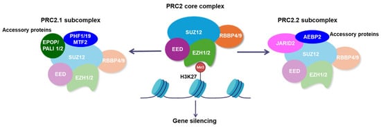
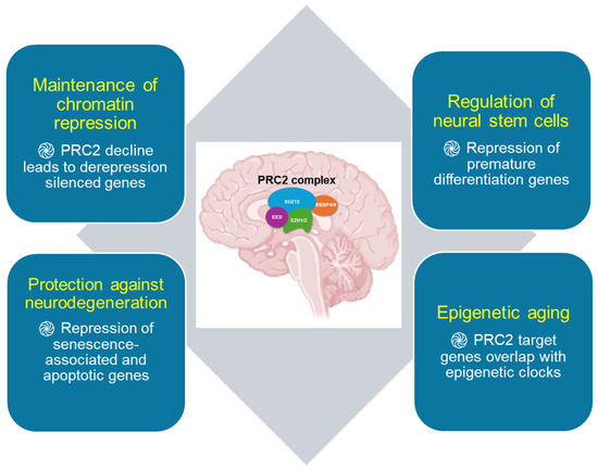
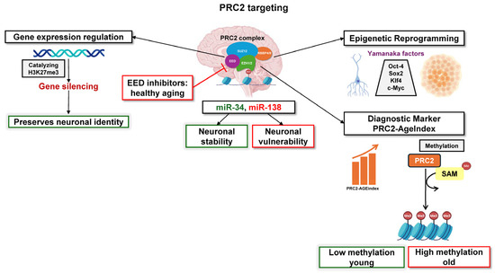
Brain aging is characterized by extensive and dynamic epigenetic changes that reshape chromatin architecture and influence gene regulatory networks, leading to deregulation of transcriptional programs essential for neuronal survival, synaptic plasticity, and cognitive function. Emerging studies highlight the pivotal regulatory role of the Polycomb Repressive Complex 2 (PRC2) in brain aging, functioning mainly as an epigenetic silencer through the establishment of the H3K27me3 histone mark at gene promoters and enhancers, leading to transcriptional repression. Altered distribution and decreased activity of PRC2 during aging disrupts the balance between gene activation and repression, affecting pathways involved in neurogenesis, synaptic plasticity and stress response. Further interplay with other epigenetic regulators such as histone deacetylase 2 (HDAC2), DNA methyltransferases and non-coding RNAs forms an extensive network that contributes to cognitive decline, increasing vulnerability for neurodegeneration. In this review, we describe structural and functional aspects of PRC2, with emphasis on the potential of this epigenetic context-dependent regulator to integrate developmental, environmental and aging-related signals, to shape the transcriptional landscape of aging brain. We also discuss the newly developed PRC2-AgeIndex, which serves as a prognostic biomarker for age-related neurodegeneration and monitoring treatment response, as well as current PRC2-targeting options for healthy brain aging.
Full article
(This article belongs to the Section Molecular Medicine)
►▼
Show Figures

Figure 1
Open AccessArticle
INO80E Suppresses Oxidized LDL-Induced Endothelial Apoptosis Through HDAC1-Mediated Stabilization of YY1
by
Tingting Liu, Quanye Luo, Shihong Yang, Dongmei Yang, Xuzhen Lv, Liyan Zhao and Qinhui Tuo
Biomolecules 2026, 16(8), 1174; https://doi.org/10.3390/biom16081174 - 12 Aug 2026
Abstract
Objectives: Endothelial apoptosis is a central event in atherosclerotic vascular injury, yet the contribution of the INO80 chromatin-remodeling subunit INO80E to this process remains unclear. This study examined whether INO80E is altered during atherosclerosis (AS)-associated endothelial injury and explored its functional role in
[...] Read more.
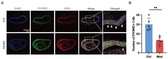
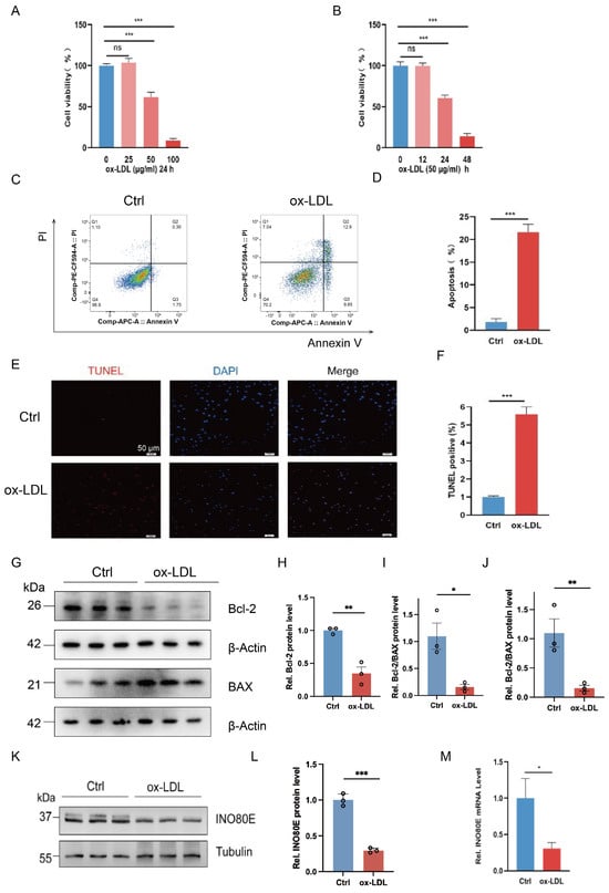
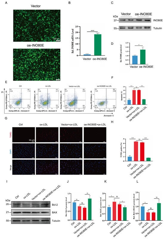
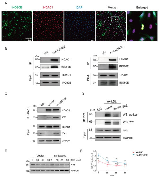
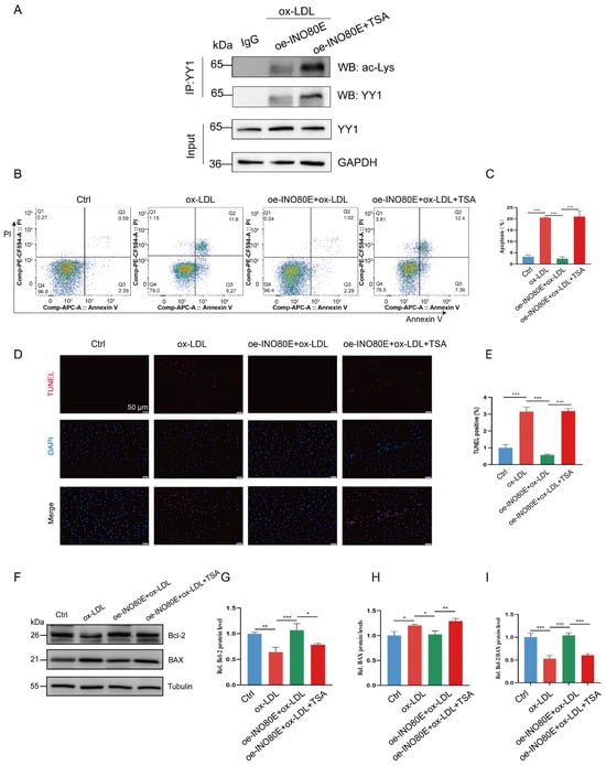
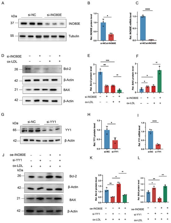
Objectives: Endothelial apoptosis is a central event in atherosclerotic vascular injury, yet the contribution of the INO80 chromatin-remodeling subunit INO80E to this process remains unclear. This study examined whether INO80E is altered during atherosclerosis (AS)-associated endothelial injury and explored its functional role in ox-LDL-triggered apoptosis. Methods: Atherosclerotic mouse models were used to examine INO80E expression in vascular tissues, and ox-LDL-treated HUVECs were applied as an in vitro model of endothelial injury. The functional role of INO80E was evaluated using lentiviral overexpression and siRNA-mediated knockdown approaches, while apoptosis was measured by flow cytometry and TUNEL staining. Mechanistic experiments included co-immunoprecipitation, immunofluorescence, YY1 acetylation analysis, and pharmacological inhibition with the pan-HDAC inhibitor trichostatin A (TSA). In addition, YY1-silencing rescue experiments were performed in INO80E-overexpressing cells to determine whether YY1 contributes to the protective effect of INO80E against ox-LDL-induced endothelial apoptosis. Results: INO80E expression was reduced in the endothelial layer of ApoE−/− aortas and in ox-LDL-treated HUVECs. INO80E overexpression attenuated ox-LDL-induced apoptosis and increased the Bcl-2/BAX ratio, whereas INO80E knockdown produced the opposite effect. INO80E colocalized with HDAC1, increased the HDAC1-YY1 association, decreased YY1 acetylation, and prolonged YY1 protein stability. TSA treatment weakened the anti-apoptotic phenotype associated with INO80E overexpression, and rescue experiments showed that YY1 knockdown partially reversed the INO80E overexpression-associated regulation of Bcl-2 and BAX. Conclusions: These findings suggest that INO80E protects endothelial cells from ox-LDL-induced apoptosis, at least in part by promoting HDAC-associated YY1 deacetylation and stabilization. The INO80E-HDAC1-YY1 pathway may represent a candidate protective mechanism in AS that requires further validation.
Full article
(This article belongs to the Section Molecular Medicine)
►▼
Show Figures

Figure 1
Journal Menu
► ▼ Journal Menu-
- Biomolecules Home
- Aims & Scope
- Editorial Board
- Reviewer Board
- Topical Advisory Panel
- Early Career Editorial Board
- Instructions for Authors
- Special Issues
- Topics
- Sections & Collections
- Article Processing Charge
- Indexing & Archiving
- Editor’s Choice Articles
- Most Cited & Viewed
- Journal Statistics
- Journal History
- Journal Awards
- Conferences
- Editorial Office
Journal Browser
► ▼ Journal BrowserHighly Accessed Articles
Latest Books
E-Mail Alert
News
Topics
Topic in
Biomolecules, Chemistry, IJMS, Molecules, Pharmaceuticals
Enzymes and Enzyme Inhibitors in Drug Research
Topic Editors: Athina Geronikaki, Cosimo D. Altomare, Maria Stefania SinicropiDeadline: 11 September 2026
Topic in
Biomedicines, IJMS, Sci. Pharm., Molecules, Future Pharmacology, Biomolecules
Natural Products and Drug Discovery—2nd Edition
Topic Editors: Sonia Piacente, Marta MenegazziDeadline: 30 September 2026
Topic in
Biomolecules, IJMS, Cancers, Proteomes
Extracellular Vesicles: Isolation, Characterization, Function, Application and Utility
Topic Editors: Suresh Mathivanan, Sai Vara Prasad ChittiDeadline: 30 October 2026
Topic in
Biomedicines, Future Pharmacology, Pharmacy, IJMS, Biomolecules, Genes
Prospects of Multi-Target Agonists in Metabolic and Epigenetic Medicine
Topic Editors: Riham Abouleisa, Yanming LiDeadline: 30 November 2026
Conferences
Special Issues
Special Issue in
Biomolecules
Novel Evidence About Hydrogen Sulfide (H2S)-Donor Molecules as Innovative Therapeutical Tools 2025
Guest Editor: Alma MartelliDeadline: 20 August 2026
Special Issue in
Biomolecules
Advancing Next-Generation Agricultural Research with Multi-Omics Approaches
Guest Editors: Rui Li, Xinmin LiDeadline: 20 August 2026
Special Issue in
Biomolecules
TRP Channels in Cardiovascular and Inflammatory Disease, 2nd Edition
Guest Editor: Susan D. BrainDeadline: 20 August 2026
Special Issue in
Biomolecules
Molecular and Cellular Mechanisms in Anti-Thrombosis
Guest Editors: Joen-Rong Sheu, Thanasekaran JayakumarDeadline: 20 August 2026
Topical Collections
Topical Collection in
Biomolecules
Feature Papers in Bioinformatics and Systems Biology Section
Collection Editor: Lukasz Kurgan
Topical Collection in
Biomolecules
Cellular and Molecular Mechanisms Governing Stress Response in Plants
Collection Editors: Giovanna Serino, Daisuke Todaka
Topical Collection in
Biomolecules
Molecular Mechanisms of Obesity, Diabetes, Inflammation and Aging
Collection Editors: Yuxiang Sun, Susanne Talcott
Topical Collection in
Biomolecules
Feature Papers in Molecular Biomarkers
Collection Editor: Pietro Scicchitano