


Biomolecules, Volume 15, Issue 8 (August 2025) – 154 articles
Cover Story (view full-size image):
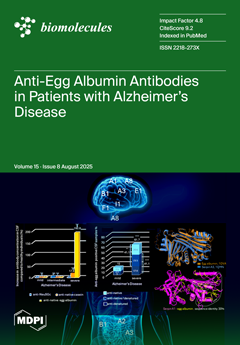
An increased concentration of antibodies against egg albumin has been found in patients with Alzheimer’s Disease (AD). While high levels of anti-native egg albumin antibodies were found in the serum of patients at all stages, with higher levels in mild disease, antibodies against the denatured form exhibited an increase with severity. An increased positivity of antibodies against both protein forms was also present in the CSF of severely ill patients. The sequence/structural similarity of egg albumin with human serpins, involved in processes which exhibit impairment in AD, may support egg albumin’s implication in disease development via molecular mimicry; indicative serpins are A1, A2, A3, A8, B1, E1, F1 and I1. More experiments are needed to elucidate this assumption as well as the effect of egg albumin deprivation in the disease’s progress. View this paper
- Issues are regarded as officially published after their release is announced to the table of contents alert mailing list.
- You may sign up for e-mail alerts to receive table of contents of newly released issues.
- PDF is the official format for papers published in both, html and pdf forms. To view the papers in pdf format, click on the "PDF Full-text" link, and use the free Adobe Reader to open them.
Previous Issue
Next Issue