Next Issue
Volume 16, October
Previous Issue
Volume 16, August


J. Funct. Biomater., Volume 16, Issue 9 (September 2025) – 51 articles

Cover Story (view full-size image): 
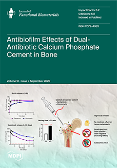
This study reports the development of a novel injectable calcium phosphate cement (CPC) co-loaded with gentamicin and vancomycin for the localized treatment of polymicrobial bone infections. The dual incorporation of antibiotics preserved the cement’s handling properties, maintained high porosity, and ensured biocompatibility with human osteoblasts. A rapid burst release followed by sustained antibiotic delivery provided broad-spectrum antibacterial effects against both Gram-positive and Gram-negative pathogens, including resistant strains. Importantly, the combination of gentamicin and vancomycin demonstrated superior antibiofilm activity compared to single-antibiotic formulations, highlighting this CPC as a promising strategy for managing osteomyelitis. View this paper
  • Issues are regarded as officially published after their release is announced to the table of contents alert mailing list.
  • You may sign up for e-mail alerts to receive table of contents of newly released issues.
  • PDF is the official format for papers published in both, html and pdf forms. To view the papers in pdf format, click on the "PDF Full-text" link, and use the free Adobe Reader to open them.
Order results
Result details
Section
Select all
Export citation of selected articles as:
10 pages, 9228 KB  
Article
Comparative Evaluation of Shear Bond Strength Between Monolithic Zirconia and Three Different Core Build-Up Materials
by Ayben Şentürk, Bora Akat, Fehmi Gönüldaş, Onur Alp Halaçlı, Selin Kartal and Mehmet Ali Kılıçarslan
J. Funct. Biomater. 2025, 16(9), 355; https://doi.org/10.3390/jfb16090355 - 21 Sep 2025
Viewed by 225
Abstract
This in vitro study aimed to evaluate and compare the shear bond strength (SBS) between monolithic zirconia (MZ) and different core build-up materials. Sixty cylindrical MZ specimens were fabricated and divided into three groups (n = 20) based on the type of [...] Read more.
This in vitro study aimed to evaluate and compare the shear bond strength (SBS) between monolithic zirconia (MZ) and different core build-up materials. Sixty cylindrical MZ specimens were fabricated and divided into three groups (n = 20) based on the type of core build-up material: nanohybrid composite resin (NHCR), glass ionomer cement (GIC), and zirconia-reinforced glass ionomer cement (zirconomer). All specimens were subjected to airborne-particle abrasion with aluminum oxide and bonded using a self-adhesive dual-cure resin cement. After 24 h of storage in distilled water at 37 °C, SBS testing was performed using a universal testing machine. Failure modes were examined under a stereomicroscope and classified as adhesive, cohesive, or mixed. The NHCR group exhibited the highest SBS values (48.32 ± 12.49 MPa), followed by the Zirkonomer group (14.19 ± 3.66 MPa), and the GIC group (10.37 ± 4.21 MPa). The SBS of NHCR was significantly higher than that of both Zirconomer and GIC (p < 0.001). Although no significant difference was found between Zirconomer and GIC, Zirconomer demonstrated higher mean bond strength. Within the limitations of this study, NHCR showed the highest bond strength to monolithic zirconia. Zirconomer performed better than conventional GIC; however, further investigations involving different surface treatments and long-term clinical conditions are recommended to enhance its bonding efficacy. Full article
(This article belongs to the Special Issue Biomaterials Applied in Dental Sciences)
Show Figures

Figure 1

18 pages, 7576 KB  
Review
Clinical Efficacy of Clear Aligners in Class II Malocclusion: From Pediatric to Adult Cases–A Narrative Review
by Gianna Dipalma, Grazia Marinelli, Francesco Inchingolo, Marialuisa Longo, Maral Di Giulio Cesare, Sharon Di Serio, Andrea Palermo, Massimo Del Fabbro, Alessio Danilo Inchingolo and Angelo Michele Inchingolo
J. Funct. Biomater. 2025, 16(9), 354; https://doi.org/10.3390/jfb16090354 - 19 Sep 2025
Viewed by 260
Abstract
Background: Class II malocclusion is one of the most common and challenging orthodontic problems, often requiring complex, lengthy treatment and sometimes involving extractions or surgery. While conventional fixed appliances have been the gold standard, the increasing demand for aesthetic and comfortable treatment alternatives [...] Read more.
Background: Class II malocclusion is one of the most common and challenging orthodontic problems, often requiring complex, lengthy treatment and sometimes involving extractions or surgery. While conventional fixed appliances have been the gold standard, the increasing demand for aesthetic and comfortable treatment alternatives has made clear aligners a prevalent choice. Understanding the specific biomechanics, limitations, and successful clinical strategies for using aligners—especially in managing vertical dimension and achieving skeletal correction (mandibular advancement)—is crucial for expanding non-invasive treatment options and improving outcomes for a broad range of Class II patients. Objective: The objective of this review is to examine the effectiveness and clinical approaches of clear aligners in Class II correction across different age groups, with particular attention to vertical control, mandibular advancement methods, and the predictability of tooth movements in both growing and fully mature patients. Materials and Methods: This review narratively discusses the most relevant clinical findings and practical strategies for managing Class II malocclusions with clear aligners. Particular attention is given to the integration of auxiliary devices, such as elastics, attachments, and temporary anchorage devices (TADs), which can enhance biomechanical control. Results: The combination of aligners with mini-implants and attachments resulted in a consequent decrease in excessive overjet, improvement in facial profile, and long-term stability supported by fixed retention. In growing patients, correction benefited from mandibular advancement protocols and control of molar extrusion, allowing for preservation of the mandibular plane angle. Movement predictability showed higher reliability in anterior torque movements, whereas maxillary incisor intrusion remained less predictable. Conclusions: Clear aligners, especially when supported by auxiliary device, such as mini-implants and attachments, offer a reliable and aesthetic alternative to conventional orthodontic treatment for Class II malocclusions. However, certain tooth movements may still be less predictable, highlighting the need for careful planning, individualized biomechanics, and ongoing technological improvements. Full article
(This article belongs to the Special Issue Feature Papers in Dental Biomaterials (2nd Edition))
Show Figures

Figure 1

32 pages, 40932 KB  
Review
Engineering Metal-Organic Frameworks for Enhanced Antimicrobial Efficacy: Synthesis Methodologies, Mechanistic Perspectives, and Versatile Applications
by Zaixiang Zheng, Junnan Cui, Shutong Wu, Zhimin Cao and Pan Cao
J. Funct. Biomater. 2025, 16(9), 353; https://doi.org/10.3390/jfb16090353 - 19 Sep 2025
Viewed by 454
Abstract
Bacterial contamination and the escalating crisis of antibiotic resistance represent pressing global public health threats, with approximately 4.95 million deaths linked to antimicrobial resistance (AMR) in 2019 and projections estimating up to 10 million annual fatalities by 2050. As third-generation antimicrobial materials, metal–organic [...] Read more.
Bacterial contamination and the escalating crisis of antibiotic resistance represent pressing global public health threats, with approximately 4.95 million deaths linked to antimicrobial resistance (AMR) in 2019 and projections estimating up to 10 million annual fatalities by 2050. As third-generation antimicrobial materials, metal–organic frameworks (MOFs) have emerged as promising alternatives to conventional agents, leveraging their unique attributes such as high specific surface areas, tunable porosity, and controlled metal ion release kinetics. This review provides a systematic analysis of the foundational principles and core antibacterial mechanisms of MOFs, which include the sustained release of metal ions (e.g., Ag+, Cu2+, Zn2+), the generation of reactive oxygen species (ROS), and synergistic effects with encapsulated functional molecules. We highlight how these mechanisms underpin their efficacy across a range of applications. Rather than offering an exhaustive list of synthesis methods and metal compositions, this review focuses on clarifying structure–function relationships that enable MOF-based materials to outperform conventional antimicrobials. Their potential is particularly evident in several key areas: wound dressings and medical coatings that enhance tissue regeneration and prevent infections; targeted nanotherapeutics against drug-resistant bacteria; and functional coatings for food preservation and water disinfection. Despite existing challenges, including gaps in clinical translation, limited efficacy in complex multi-species infections, and incomplete mechanistic understanding, MOFs hold significant promise to revolutionize antimicrobial therapy. Through interdisciplinary optimization and advancements in translational research, MOFs are poised to drive a paradigm shift from “passive defense” to “active ecological regulation”, offering a critical solution to mitigate the global AMR crisis. Full article
(This article belongs to the Section Antibacterial Biomaterials)
Show Figures

Figure 1

36 pages, 2272 KB  
Review
Bio-Functional Nanomaterials for Enhanced Lung Cancer Therapy: The Synergistic Roles of Vitamins D and K
by Andreea Crintea, Camelia Munteanu, Tamás Ilyés, Ciprian N. Silaghi and Alexandra M. Crăciun
J. Funct. Biomater. 2025, 16(9), 352; https://doi.org/10.3390/jfb16090352 - 19 Sep 2025
Viewed by 365
Abstract
Lung cancer remains a leading cause of cancer-related mortality worldwide, requiring the development of innovative and effective therapeutic strategies. Bio-functional nanomaterials, due to their unique physicochemical properties, offer a versatile platform for targeted drug delivery, controlled release, and multimodal therapies, thereby enhancing efficacy [...] Read more.
Lung cancer remains a leading cause of cancer-related mortality worldwide, requiring the development of innovative and effective therapeutic strategies. Bio-functional nanomaterials, due to their unique physicochemical properties, offer a versatile platform for targeted drug delivery, controlled release, and multimodal therapies, thereby enhancing efficacy and reducing the systemic toxicity of conventional treatments. Independently, both vitamin D and vitamin K have demonstrated significant anti-cancer properties, including inhibition of proliferation, induction of apoptosis, modulation of angiogenesis, and attenuation of metastatic potential in various cancer cell lines and in vivo models. However, their clinical application is often limited by poor bioavailability, rapid metabolism, and potential for off-target effects. Specifically, by enhancing the solubility, stability, and targeted accumulation of fat-soluble vitamins D and K within tumoral tissues for improved lung cancer therapy, this review emphasizes the novel and cooperative role of bio-functional nanomaterials in overcoming these limitations. Future studies should focus on the logical development of sophisticated nanomaterial carriers for optimal co-delivery plans and thorough in vivo validation, aiming to convert these encouraging preclinical results into successful clinical treatments for patients with lung cancer. Full article
Show Figures

Figure 1

18 pages, 540 KB  
Review
Bionanomaterials or Nanobiomaterials: Differences in Definitions and Applications
by Bogdan Walkowiak, Małgorzata Siatkowska and Piotr Komorowski
J. Funct. Biomater. 2025, 16(9), 351; https://doi.org/10.3390/jfb16090351 - 18 Sep 2025
Viewed by 383
Abstract
Since the turn of the century, we have witnessed an extremely intensive development of biotechnology and nanotechnology, which, in terms of intensity can only be compared to the development of information technology and the resulting emergence of artificial intelligence. In the present review, [...] Read more.
Since the turn of the century, we have witnessed an extremely intensive development of biotechnology and nanotechnology, which, in terms of intensity can only be compared to the development of information technology and the resulting emergence of artificial intelligence. In the present review, we deliberately omit the development of information technology and artificial intelligence. Instead, our interest is focused on bionanomaterials and nanobiomaterials, their production and applications, and, in particular, the different meanings of these terms. We adopted an analysis of the literature published between January 2000 and May 2025, available in PubMed. The database was searched for selected areas: types (origin, structure, and function), manufacturing methods (chemical, physicochemical, and biological), and applications (medicine/pharmacy, textile technology, cosmetology, and agriculture/environment). Our findings revealed a significant increase in the number of publications for both terms, with nanobiomaterials predominating. The authors of the publications included in PubMed clearly outline the separation of meanings of both concepts, despite the lack of normative regulations in this regard. Nanoparticles are the most commonly represented type in the use of both terms, and drug delivery is a dominant application. However, it is worth noting the lack of nanobiomaterials in the agricultural/environmental application categories. Despite the enormous similarity between the terms “nanobiomaterials” and “bionanomaterials,” both in terms of nomenclature and application, there is a significant difference resulting from the manufacturing technologies and applications used. The term “nanobiomaterials” should be assigned only to biomaterials, in accordance with the definition of a biomaterial, regardless of their manufacturing technology, while the term “bionanomaterials” should be applied to all products of bionanotechnology, excluding products used as biomaterials. Full article
Show Figures

Figure 1

22 pages, 4981 KB  
Article
Data-Driven Design and Additive Manufacturing of Patient-Specific Lattice Titanium Scaffolds for Mandibular Bone Reconstruction
by Nail Beisekenov, Bagdat Azamatov, Marzhan Sadenova, Dmitriy Dogadkin, Daniyar Kaliyev, Sergey Rudenko and Boris Syrnev
J. Funct. Biomater. 2025, 16(9), 350; https://doi.org/10.3390/jfb16090350 - 18 Sep 2025
Viewed by 391
Abstract
The reconstruction of segmental bone defects requires patient-specific scaffolds that combine mechanical safety, biological functionality, and rapid manufacturing. We converted CT-derived mandibular geometry into a functionally graded Ti-6Al-4V lattice and optimised porosity, screw layout, and strut thickness through a cyber-physical loop that joins [...] Read more.
The reconstruction of segmental bone defects requires patient-specific scaffolds that combine mechanical safety, biological functionality, and rapid manufacturing. We converted CT-derived mandibular geometry into a functionally graded Ti-6Al-4V lattice and optimised porosity, screw layout, and strut thickness through a cyber-physical loop that joins high-fidelity FEM, millisecond ANN, and a BN for uncertainty quantification. Fifteen candidate scaffolds were fabricated by direct metal laser sintering and hot isostatic pressing and were mechanically tested. FEM predicted stress and stiffness with 98% accuracy; the ANN reproduced these outputs with 94% fidelity while evaluating 10,000 designs in real time, and the BN limited failure probability to <3% under worst-case loads. The selected 55–65% porosity design reduced titanium use by 15%, shortened development time by 25% and raised multi-objective optimisation efficiency by 20% relative to a solid-plate baseline, while resisting a 600 N bite with a peak von Mises stress of 225 MPa and micromotion < 150 µm. Integrating physics-based simulation, AI speed, and probabilistic rigour yields a validated, additively manufactured scaffold that meets surgical timelines and biomechanical requirements, offering a transferable blueprint for functional scaffolds in bone and joint surgery. Full article
(This article belongs to the Special Issue Functional Scaffolds for Bone and Joint Surgery)
Show Figures

Figure 1

42 pages, 11753 KB  
Review
Integrating Additive and Traditional Manufacturing for Multiscale Bone Tissue Engineering Scaffolds
by Yixuan Zhu, Haotian Gao, Qingchen Qiao, Yafei Yuan, Dongyu Fang, Yuxing Bai and Qingsong Jiang
J. Funct. Biomater. 2025, 16(9), 349; https://doi.org/10.3390/jfb16090349 - 16 Sep 2025
Viewed by 418
Abstract
Additive manufacturing (AM) has emerged as a cutting-edge technology for fabricating biomimetic scaffolds with controllable architectures and compositional diversity, showing great promise in the fields of bone tissue engineering (BTE) and regenerative medicine. However, due to limitations in printing resolution and single-process capabilities, [...] Read more.
Additive manufacturing (AM) has emerged as a cutting-edge technology for fabricating biomimetic scaffolds with controllable architectures and compositional diversity, showing great promise in the fields of bone tissue engineering (BTE) and regenerative medicine. However, due to limitations in printing resolution and single-process capabilities, AM alone struggles to replicate the complex multiscale hierarchical structures inherent in native bone. Traditional fabrication techniques provide valuable complementary strategies to address these limitations. This review systematically summarizes recent advances in the construction of heterogeneous scaffolds from a multiscale design perspective, encompassing macro-, meso-, and microscale approaches. Emphasis is placed on the integration of major AM techniques—such as extrusion-based and light-based printing—with conventional methods including freeze-drying, gas foaming, and electrospinning. Particular attention is given to emerging in situ fabrication strategies, such as in situ foaming and mineralization, which enable spatially resolved and functionally graded architectures. Furthermore, this review explores pathways for constructing multiscale-integrated scaffolds and examines the current challenges and opportunities in clinical translation. Collectively, this work provides a comprehensive framework to guide the development of next-generation bone tissue scaffolds with enhanced biological performance and translational potential. Full article
Show Figures

Figure 1

16 pages, 939 KB  
Article
Original Locking Rod System Designed for Diaphyseal Fractures of Long Bones
by Liviu-Coriolan Misca, Cristian Constantin Croicu, Adrian Emil Lazarescu, Mihai-Alexandru Sandesc, Jenel Marian Patrascu, Jr., Sorin Florescu and Jenel Marian Patrascu, Sr.
J. Funct. Biomater. 2025, 16(9), 348; https://doi.org/10.3390/jfb16090348 - 15 Sep 2025
Viewed by 321
Abstract
Introduction: Intramedullary nailing is widely used for long bone fractures. Traditional systems are reliable, but they present some complications regarding lack of modularity or possible growth plate damage. Methods: A novel locking rod–screw system featuring a central rod and a grooved [...] Read more.
Introduction: Intramedullary nailing is widely used for long bone fractures. Traditional systems are reliable, but they present some complications regarding lack of modularity or possible growth plate damage. Methods: A novel locking rod–screw system featuring a central rod and a grooved screw with a secondary interlocking mechanism was developed and tested. Mechanical testing followed ASTM F543 and ISO 6475 standards using a 3.0 mm steel alloy prototype. Results: The system withstood mechanical testing >200,000 cycles at loads up to 200 N with no rates of failure or loosening, significantly outperforming other implants of the same size (3.0 mm TENS). Conclusions: The proposed implant demonstrates superior biomechanical performance in vitro, enabled by its unique hollow screw and secondary locking configuration. This modular and minimally invasive system shows promise for use in cases of smaller long bones, personalized paediatric fractures, and all types of diaphyseal fractures, but does warrant in vivo validation. Full article
(This article belongs to the Section Bone Biomaterials)
Show Figures

Figure 1

13 pages, 3100 KB  
Article
Biocompatibility Evaluation of Porcine-Derived Collagen Sheets for Clinical Applications: In Vitro Cytotoxicity, In Vivo Sensitization, and Intracutaneous Reactivity Studies
by Tae-Hoon Koo, Jason K. Lee, Shawn P. Grogan and Darryl D. D'Lima
J. Funct. Biomater. 2025, 16(9), 347; https://doi.org/10.3390/jfb16090347 - 15 Sep 2025
Viewed by 459
Abstract
Biocompatibility evaluation of medical devices is essential for ensuring safety, with ISO 10993 series being the standard. However, these tests can be time-consuming and resource-intensive. This study assessed the early-stage biocompatibility of a collagen matrix derived from porcine dermis using three selective ISO [...] Read more.
Biocompatibility evaluation of medical devices is essential for ensuring safety, with ISO 10993 series being the standard. However, these tests can be time-consuming and resource-intensive. This study assessed the early-stage biocompatibility of a collagen matrix derived from porcine dermis using three selective ISO tests: in vitro cytotoxicity, in vivo sensitization, and irritation. Collagen was hydrolyzed, purified from miniature pig skin, and then processed into porous sheets via lyophilization. Extracts were prepared using both polar and non-polar extraction vehicles for biological testing. Cytotoxicity testing with mouse fibroblast cells showed no significant cytotoxic effects, with cell morphology and viability comparable to controls. Sensitization testing in guinea pigs, involving intradermal and topical exposure, revealed no allergic responses. Irritation testing in rabbits showed no signs of irritation. The cytotoxicity test took 3 days, the sensitization test 28 days, and the irritation test 7 days, all of which proved suitable for early biocompatibility screening. These results confirmed that the collagen matrix is non-cytotoxic, non-sensitizing, and non-irritant for a month. The use of these three tests enables early identification of unsafe materials, reducing time, cost, and animal use before advancing to more complex ISO 10993 studies. Therefore, they are appropriate and necessary for early feasibility decisions in medical device development. Full article
(This article belongs to the Special Issue Biocompatible Research of Materials in Biomedical Applications)
Show Figures

Figure 1

16 pages, 3705 KB  
Article
Anti-Swelling Antibacterial Hydrogels Based on Electrostatic Repulsion and Hydrophobic Interactions for Human Motion Sensing
by Zexing Deng, Litong Shen, Qiwei Cheng, Ying Li, Tianming Du and Xin Zhao
J. Funct. Biomater. 2025, 16(9), 346; https://doi.org/10.3390/jfb16090346 - 14 Sep 2025
Viewed by 486
Abstract
The development of high-performance sensing materials is critical for advancing bioelectronics. Conductive hydrogels, with their unique flexibility, are promising candidates for biomedical sensors. However, traditional conductive hydrogels often suffer from excessive swelling and undesirable antibacterial activity, limiting their practical use. To overcome these [...] Read more.
The development of high-performance sensing materials is critical for advancing bioelectronics. Conductive hydrogels, with their unique flexibility, are promising candidates for biomedical sensors. However, traditional conductive hydrogels often suffer from excessive swelling and undesirable antibacterial activity, limiting their practical use. To overcome these challenges, anti-swelling, antibacterial, and ionically conductive hydrogels were built through free radical polymerization. The preparation was conducted using a monomer mixture comprising acrylic acid (AA), the antibacterial zwitterionic compound [2-(methacryloyloxy)ethyl]dimethyl-(3-sulfopropyl)ammonium hydroxide (SBMA), and the hydrophobic monomer lauryl methacrylate (LMA). The protonation of SBMA by AA enables electrostatic repulsion, thereby imparting anti-swelling properties to the hydrogel. The introduction of hydrophobic LMA components further enhances the anti-swelling and mechanical performance of hydrogel. The resulting hydrogel exhibits excellent anti-swelling property with a swelling ratio of 59.36% after 120 h and good mechanical performance with a tensile strength of 158 kPa, an elongation at break of 176%, and a compressive strength of 0.37 MPa at 80% strain. In addition, hydrogels possess superior sensing performance for strain sensing with a gauge factor of 1.315 within 40–60% of strain, 330 ms of response time, and 177 ms of recovery time. Furthermore, the hydrogel is capable of monitoring human motion and physiological signals. These attributes make it highly suitable for wearable sensors and biomedical monitoring applications. Full article
Show Figures

Figure 1

18 pages, 3059 KB  
Article
Clinical and Radiological Evaluation of Flap and Flapless Procedures with Biomaterials in Alveolar Ridge Preservation
by Ewa Dolińska, Ewa Duraj, Marcin Bernaczyk, Magdalena Sulewska and Małgorzata Pietruska
J. Funct. Biomater. 2025, 16(9), 345; https://doi.org/10.3390/jfb16090345 - 14 Sep 2025
Viewed by 415
Abstract
Although ridge preservation procedures have been shown to prevent post-extraction bone loss, the effectiveness of using a flap or flapless surgical approach remains unclear. The aim of the study was to compare the mentioned above alveolar ridge preservation procedures in the esthetic region [...] Read more.
Although ridge preservation procedures have been shown to prevent post-extraction bone loss, the effectiveness of using a flap or flapless surgical approach remains unclear. The aim of the study was to compare the mentioned above alveolar ridge preservation procedures in the esthetic region of maxilla. Twenty-nine patients were randomly assigned to receive flap (n = 14) or flapless (n = 15) alveolar ridge preservation procedure. Sockets were grafted with alloplastic biomaterial, then covered with a collagen membrane in both groups. Clinical examinations were performed over a 6-month observation period and radiological (CBCT) examination was conducted before and 6 months after treatment. For both after flap and flapless procedures, there was a reduction in interdental papillae height and keratinized tissue width, increase in buccal soft tissues thickness with a decrease in radiological buccal bone plate width, decrease in radiological buccal and lingual plate height (significantly for the flapless group) and radiological alveolar process width reduction (significantly in flapless group at the height of 5 mm and 7 mm from the bottom of the socket). A decrease in the radiological buccal bone plate width was observed, where the further measuring point was from the bottom of the alveolus. In the mucoperiosteal flap preparation, group buccal bone plate width reduction at the height 3 mm, 5 mm and 7 mm was significant and in the flapless group a significant decrease was observed from 5 to 9 mm from the bottom of the socket. Despite ridge preservation, there is soft tissue thickening and a decrease in bone height and width regardless of the flap/flapless method used. Full article
(This article belongs to the Special Issue Functional Biomaterial for Bone Regeneration (2nd Edition))
Show Figures

Figure 1

19 pages, 5379 KB  
Article
Antibacterial Activity of a Trace-Cu-Modified Mg Alloy in Simulated Intestinal Fluid
by Baiyun Zhong, Zemeng Wei, Yi Yao, Lixun Jiang, Manli Zhou, Jinping Li, Weidong Liu, Xin Li and Ming-Chun Zhao
J. Funct. Biomater. 2025, 16(9), 344; https://doi.org/10.3390/jfb16090344 - 12 Sep 2025
Viewed by 415
Abstract
Mg alloys hold promise for biodegradable gastrointestinal implants, but most evaluations rely on simplified media like Hank’s solution, which lacks organic components and fails to replicate the acidic-to-alkaline transition of intestinal fluid, risking underestimation of biodegradation rates and clinical relevance. This work investigated [...] Read more.
Mg alloys hold promise for biodegradable gastrointestinal implants, but most evaluations rely on simplified media like Hank’s solution, which lacks organic components and fails to replicate the acidic-to-alkaline transition of intestinal fluid, risking underestimation of biodegradation rates and clinical relevance. This work investigated a trace-Cu-modified Mg alloy (Mg-0.05Cu) in simulated intestinal fluid (SIF) versus Hank’s solution. Microstructural analysis confirmed Mg2Cu intermetallic phases as Cu reservoirs. Electrochemical and immersion tests revealed significantly accelerated biodegradation in SIF, due to its disruption of protective layer formation, sustaining loose biodegradation products. The biodegradation rate of the trace-Cu-modified Mg alloy in SIF was consistent with reported values for Mg alloys in similar media, as was that in Hank’s solution. Remarkably, Mg-0.05Cu exhibited potent antibacterial activity against E. coli, achieving 99.3% eradication within 12 h and 100% elimination by 24–48 h, alongside excellent cytocompatibility with L929 cells (>95% viability). This efficacy arose from the synergistic Cu2+ release and high-pH microenvironment. These findings demonstrate that trace Cu alloying in high-purity Mg balances rapid antibacterial action with controlled biodegradation in a physiologically relevant SIF. This positions Mg-0.05Cu as a highly promising candidate for practical applications, such as biodegradable intestinal stents, anti-adhesion barriers, anastomosis rings, and anti-obesity devices, where rapid infection control and predictable degradation are critical for clinical success. This work underscores the importance of using biomimetic media for evaluating gastrointestinal implants and establishes Mg-0.05Cu as a promising strategy for developing infection-resistant biodegradable devices. Full article
(This article belongs to the Special Issue Antimicrobial Biomaterials for Medical Applications)
Show Figures

Figure 1

27 pages, 11472 KB  
Article
Electrophoretic Coatings for Orthodontic Implants: Evaluation of Surface Properties, Adhesion, and Antibacterial Activity in Simulated Implantation Trials
by Maria Biegun-Żurowska, Karolina Klesiewicz, Katarzyna Matysiak, Marcin Gajek, Alicja Rapacz-Kmita and Magdalena Ziąbka
J. Funct. Biomater. 2025, 16(9), 343; https://doi.org/10.3390/jfb16090343 - 12 Sep 2025
Viewed by 513
Abstract
In this study, the properties of electrophoretically deposited (EPD) coatings on orthodontic implants made from Ti-6Al-4V alloy were evaluated during simulated implantation trials on animal bones. Three types of chitosan-based coatings were prepared using EPD: titanium nitride microparticles (TiNPs), titanium nitride nanoparticles (TiNNPs), [...] Read more.
In this study, the properties of electrophoretically deposited (EPD) coatings on orthodontic implants made from Ti-6Al-4V alloy were evaluated during simulated implantation trials on animal bones. Three types of chitosan-based coatings were prepared using EPD: titanium nitride microparticles (TiNPs), titanium nitride nanoparticles (TiNNPs), and boron nitride particles (BNPs). Each of these coatings was also modified by adding a polylactic acid (PLA) layer using a dip-coating technique to compare their properties with and without this additional layer. The coatings were analysed using optical microscopy, confocal microscopy, and scanning electron microscopy (SEM) with elemental analysis. Surface roughness measurements of the coated implants were also conducted to highlight differences that could significantly influence the type and strength of the bone-implant interface, directly affecting the stability of the implant as an anchorage unit. Eventually, to evaluate the antibacterial properties of the EPD coatings, their antibacterial activity against both Gram-positive and Gram-negative bacteria strains was tested. Scanning electron observations confirmed the homogenous distribution of micro- and nanoparticles in all coatings. The highest surface roughness values were observed in layers containing titanium nitride nanoparticles (TiNNPs) and chitosan. The presence of an additional dip-coating PLA layer improved the adhesion, and its effect on the surface roughness depended on the particle size. While the antibacterial properties of the coatings show promising results, achieving optimal adhesion of the coatings to implants remains a challenge that requires further development. Full article
Show Figures

Graphical abstract

12 pages, 1140 KB  
Review
Influence of Design Parameters on Implant Abutment Performance: A Scoping Review
by Vladimir Prpic, Petar Kosec, Stanko Skec and Amir Catic
J. Funct. Biomater. 2025, 16(9), 342; https://doi.org/10.3390/jfb16090342 - 11 Sep 2025
Viewed by 599
Abstract
Implant abutments serve as the critical interface between dental implants and prosthodontic restorations, playing a central role in the functional and aesthetic success of implant-supported rehabilitations. With the development of CAD/CAM technologies, dental implantology has experienced a significant transformation. The latest and still [...] Read more.
Implant abutments serve as the critical interface between dental implants and prosthodontic restorations, playing a central role in the functional and aesthetic success of implant-supported rehabilitations. With the development of CAD/CAM technologies, dental implantology has experienced a significant transformation. The latest and still unexplored field of science includes an advanced algorithm-driven process known as generative design. Generally, generative design is used to investigate a broad spectrum of design alternatives that satisfy predetermined criteria established by a designer. Prior to the application of generative design, it is essential to define the key parameters that influence the optimization of the designed object’s configuration, such as a hybrid implant abutment. A bibliographic search was performed using PubMed and Scopus databases to identify relevant studies published up to 1 July 2025. Studies that investigated transmucosal and prosthodontic height of implant abutments were selected for inclusion. Only 13 studies met inclusion criteria and were further analyzed. Included studies showed the importance of transmucosal and prosthodontic height of implant abutments in order to reduce possible complications. The review highlights the importance of optimizing the transmucosal and transgingival heights of implant abutments. For predictable biological and mechanical outcomes, both dimensions should be at least 2 mm. The obtained parameter values will be used to create the initial parametric model, which will then be utilized in the generative design process of hybrid implant abutments. Full article
(This article belongs to the Special Issue The Development and Future of Dental Implants)
Show Figures

Figure 1

48 pages, 1146 KB  
Systematic Review
Types of Bone Substitutes and Their Application in Regenerative Medicine: A Systematic Review
by Nikoleta Ivanova, Stoyan Ivanov, Stefan Peev and Tsanka Dikova
J. Funct. Biomater. 2025, 16(9), 341; https://doi.org/10.3390/jfb16090341 - 9 Sep 2025
Viewed by 810
Abstract
Background: The growing demand for effective methods of bone tissue regeneration highlights the relevance of studying modern bone substitutes and their applications in regenerative medicine. The aim of this work was to conduct a comprehensive analysis of the biological, mechanical, and clinical characteristics [...] Read more.
Background: The growing demand for effective methods of bone tissue regeneration highlights the relevance of studying modern bone substitutes and their applications in regenerative medicine. The aim of this work was to conduct a comprehensive analysis of the biological, mechanical, and clinical characteristics of various types of bone substitutes to determine their potential in regenerative medicine. Methods: The study was performed as a systematic literature review in accordance with PRISMA guidelines, analyzing 68 high-quality scientific sources from 2019 to May 2025, using the PubMed, Scopus, Web of Science, and Google Scholar databases. Results: It was established that autogenous grafts exhibit the highest osteogenic properties due to the presence of growth factors BMP-2, BMP-7, and concentrated growth factors; however, their use is limited by donor site morbidity in 20–30% of patients and the requirement to treat 6% of fractures complicated by non-union. Allogeneic and xenogeneic substitutes provide structural support for large defects but require intensive processing in accordance with European Directives 2004/23/EC and 2006/86/EC to minimize the risk of infection transmission. Synthetic substitutes based on calcium phosphate ceramics with pore sizes ranging from 23 to 210 micrometres demonstrate excellent biocompatibility and controlled degradation, with β-tricalcium phosphate exhibiting optimal characteristics for long-term applications compared to calcium sulphate. Conclusions: The findings of the study highlight the necessity of a personalized approach in selecting bone substitutes, considering the specific requirements of medical specialities, and support the development of hybrid biomaterials to combine structural strength with biological activity. Full article
Show Figures

Figure 1

16 pages, 2649 KB  
Article
Intraoperative Biologization of β-TCP and PCL-TCP by Autologous Proteins
by Andrea Sowislok, Gerrit Gruber, Farnusch Kaschani, Markus Kaiser, Eleftherios Papaeleftheriou and Marcus Jäger
J. Funct. Biomater. 2025, 16(9), 340; https://doi.org/10.3390/jfb16090340 - 9 Sep 2025
Viewed by 582
Abstract
Protein adsorption on orthopedic biomaterials during the initial intraoperative contact critically influences biological responses and osseointegration. Osteoconductive grafts such as β-tricalcium phosphate (β-TCP) and poly (ε-caprolactone)-β-TCP (PCL-TCP) can be functionally activated by exposure to autologous tissue. However, the composition and relevance of the [...] Read more.
Protein adsorption on orthopedic biomaterials during the initial intraoperative contact critically influences biological responses and osseointegration. Osteoconductive grafts such as β-tricalcium phosphate (β-TCP) and poly (ε-caprolactone)-β-TCP (PCL-TCP) can be functionally activated by exposure to autologous tissue. However, the composition and relevance of the resulting protein layer still remain unclear. In this study involving 10 patients undergoing primary total hip arthroplasty, β-TCP and PCL-TCP samples were incubated both in the femoral medullary cavity and within a surgical tissue collector harvesting autologous tissue (blood, bone fragments, muscle, and fat). Surface morphology was assessed microscopically, and protein adsorption was characterized via high-resolution LC-MS/MS with subsequent bioinformatics and statistical analysis. Both materials adsorbed over 2000 different autologous proteins. β-TCP showed higher overall protein concentrations, while PCL-TCP demonstrated greater proteomic diversity and incubation method-dependent shifts in protein profiles, influenced by surface roughness and wettability. Samples incubated in the tissue collector exhibited less protein variability and smaller material-specific differences compared to incubation in the femoral cavity, particularly for PCL-TCP. Predominant proteins were linked to immune regulation, stress response, and protein metabolism. These findings emphasize the impact of material properties and incubation environment on protein adsorption, with ex vivo incubation leading to more consistent protein adsorption patterns. Full article
(This article belongs to the Special Issue Functional Scaffolds for Bone and Joint Surgery)
Show Figures

Figure 1

17 pages, 3394 KB  
Article
Impact of 45S5-Bioactive Glass on Chondrocytes in Knee Osteoarthritis—In Vitro Study Exploring Cellular Responses
by Max Marinescu, Sébastien Hagmann, Jörg Fellenberg, Elena Tripel, Simone Gantz, Ravikumar Mayakrishnan, Aldo R. Boccaccini, Tobias Renkawitz, Babak Moradi, Fabian Westhauser and Hadrian Platzer
J. Funct. Biomater. 2025, 16(9), 339; https://doi.org/10.3390/jfb16090339 - 9 Sep 2025
Viewed by 584
Abstract
Osteoarthritis (OA), the most common joint disease, is marked by cartilage degradation and chronic inflammation. While 45S5-bioactive glass (45S5-BG) is well-established in bone regeneration and has been suggested to exert immunomodulatory effects, its impact on OA chondrocytes remains largely unexplored. Therefore, this in [...] Read more.
Osteoarthritis (OA), the most common joint disease, is marked by cartilage degradation and chronic inflammation. While 45S5-bioactive glass (45S5-BG) is well-established in bone regeneration and has been suggested to exert immunomodulatory effects, its impact on OA chondrocytes remains largely unexplored. Therefore, this in vitro study investigated the effects of 45S5-BG microparticles (0.125 mg/mL) on chondrocytes derived from OA patients, evaluating its therapeutic potential in OA. Chondrocytes were cultured with or without 45S5-BG for 1 and 7 days. Gene expression of cartilage markers, cytokines, matrix metalloproteinases (MMPs), and toll-like receptors (TLRs) was analyzed by qPCR. Protein levels were assessed by ELISA. 45S5-BG stimulation significantly altered chondrocyte activity, inducing upregulation of IL-6, IL-1β, TNF-α, MMP-1/-3/-13, and TLR4. Expression of ACAN and COL2A1 was reduced, while COL10A1—a marker of chondrocyte hypertrophy—was significantly increased at day 1. These findings show a catabolic and pro-inflammatory shift in chondrocyte phenotype upon 45S5-BG exposure, showing no therapeutic benefit of 45S5-BG on OA chondrocytes. However, considering the pronounced effects on chondrocyte activity and the well-established bioactivity and biocompatibility of 45S5-BG, our findings suggest that modified BG formulations could be developed to enhance chondroprotective and anti-inflammatory properties, warranting further investigation in co-culture and in vivo models. Full article
(This article belongs to the Special Issue Bioactive Glass in Tissue Engineering Applications)
Show Figures

Figure 1

28 pages, 5311 KB  
Review
Modified Polysaccharides: Potential Biomaterials for Bioprinting
by Tao Jiang, Yun Yang, Zening Lin, Yang Hong and Zirong Luo
J. Funct. Biomater. 2025, 16(9), 338; https://doi.org/10.3390/jfb16090338 - 9 Sep 2025
Viewed by 655
Abstract
Polysaccharides have emerged as promising biomaterials for 3D bioprinting due to their inherent biocompatibility, biodegradability, and structural diversity. However, their limited mechanical strength, insufficient bioactivity, and suboptimal printability hinder their direct application in fabricating complex tissue constructs. This review systematically summarizes universal modification [...] Read more.
Polysaccharides have emerged as promising biomaterials for 3D bioprinting due to their inherent biocompatibility, biodegradability, and structural diversity. However, their limited mechanical strength, insufficient bioactivity, and suboptimal printability hinder their direct application in fabricating complex tissue constructs. This review systematically summarizes universal modification strategies to address these challenges by tailoring polysaccharides’ physicochemical and biological properties. We first analyse the fundamental requirements of bioprinting materials, emphasising on the critical role of shear-thinning behaviours, post-printing structural fidelity, and cell-instructive functions. Subsequently, we highlight the advantages and limitations of representative polysaccharides, including chitosan, alginate, and hyaluronic acid. Chemical functionalisation, physical reinforcement, and biological hybridisation are proposed as versatile approaches to synergistically enhance printability, mechanical robustness, and bioactivity to tackle the limitations. Furthermore, dynamic crosslinking mechanisms enabling self-healing and stimuli-responsive behaviours are discussed as emerging solutions for constructing biomimetic architectures. Finally, we outline future directions in balancing material processability with cellular viability and scaling up modified polysaccharides for clinical translation. This review aims to provide a design blueprint for engineering polysaccharide-based bioinks toward next-generation regenerative medicine. Full article
Show Figures

Figure 1

13 pages, 8569 KB  
Article
Comparative Evaluation of the Bone Regenerative Potential of a Novel Calcium Silicate-Modified Calcium Carbonate Graft Material: Histological and Micro-Computed Tomography Assessment Using a Rat Calvarial Defect Model
by Masataka Nakayama, Yu Kataoka, Naoki Kitamura, Chie Watanabe, Satoko Kujiraoka, Kikue Yamaguchi, Yuma Seki, Toshitake Furusawa, Hidero Unuma and Motohiro Munakata
J. Funct. Biomater. 2025, 16(9), 337; https://doi.org/10.3390/jfb16090337 - 9 Sep 2025
Viewed by 532
Abstract
In the present study, we evaluated the usefulness of a porous sintered calcium carbonate body with CaSiO3 by comparing its osteogenic capacity with that of calcium carbonate without CaSiO3 and that of β-tricalcium phosphate (TP). A cranial defect model of eight-week-old [...] Read more.
In the present study, we evaluated the usefulness of a porous sintered calcium carbonate body with CaSiO3 by comparing its osteogenic capacity with that of calcium carbonate without CaSiO3 and that of β-tricalcium phosphate (TP). A cranial defect model of eight-week-old male Wistar rats was divided into three groups: calcium carbonate (CC), calcium carbonate-CaSiO3 composite (CC+CS), and TP. Micro-computed tomography (CT) and histological analysis were performed at four and eight weeks postoperatively. Upon quantitative evaluation of newly formed bone volume by radiography, the CC+CS group demonstrated the highest value at eight weeks postoperatively and exhibited significantly more new bone formations than the CC group (p < 0.05). Upon histological evaluation, the CC+CS group demonstrated significantly higher new bone formation than the CC group (p < 0.05). Furthermore, in terms of residual graft material ratio, at eight weeks postoperatively, the amount of residual graft material in the CC+CS group was significantly higher than that in the TP group (p < 0.05). Therefore, the addition of CaSiO3 enhances the functional regulation of calcium carbonate-based artificial bone and can be incorporated in bone graft materials. Full article
(This article belongs to the Section Biomaterials and Devices for Healthcare Applications)
Show Figures

Figure 1

22 pages, 1268 KB  
Systematic Review
Propolis as a Natural Remedy in Reducing Dental Plaque and Gingival Inflammation: A Systematic Review and Meta-Analysis
by Magdalena Sycińska-Dziarnowska, Liliana Szyszka-Sommerfeld, Monika Bugajska, Magdalena Ziąbka, Izabela Szućko-Kociuba, Gianrico Spagnuolo, Krzysztof Woźniak and Hyo-Sang Park
J. Funct. Biomater. 2025, 16(9), 336; https://doi.org/10.3390/jfb16090336 - 8 Sep 2025
Viewed by 892
Abstract
Dental plaque, if not regularly removed through proper oral hygiene, can lead to tooth decay, gingivitis, and more severe periodontal disease. Effective plaque removal is essential in preventing gingivitis, the precursor to periodontitis. Propolis, a bee product known for its antibacterial, anti-inflammatory, and [...] Read more.
Dental plaque, if not regularly removed through proper oral hygiene, can lead to tooth decay, gingivitis, and more severe periodontal disease. Effective plaque removal is essential in preventing gingivitis, the precursor to periodontitis. Propolis, a bee product known for its antibacterial, anti-inflammatory, and antioxidant properties, has shown potential in dental applications. This systematic review and meta-analysis was conducted to evaluate the efficacy of propolis-containing mouthwashes and toothpastes in reducing dental plaque and gingival inflammation. Materials and Methods: The study protocol was registered in PROSPERO (CRD42023467573), and the review was conducted in accordance with PRISMA guidelines. A comprehensive search of PubMed, PubMed Central, Embase, Scopus, and Web of Science was performed up to 10 May 2025 to identify randomized controlled trials and observational studies assessing propolis-based mouthwashes or toothpastes. Data synthesis used random-effects meta-analysis due to anticipated heterogeneity among studies. Results: Seven randomized controlled trials were included in the meta-analysis, evaluating the efficacy of propolis alcohol-free mouthwash on plaque index (PI) and gingival index (GI). For PI, the pooled standardized mean difference (SMD) was 1.74 (95% CI: 0.19–3.29; p = 0.036), with low between-study heterogeneity (I2 = 13.7%). For GI, the pooled SMD was 2.19 (95% CI: 1.10–3.29; p = 0.005), with no observed heterogeneity (I2 = 0.0%). Propolis mouthwashes demonstrated large effect sizes, significantly reducing plaque accumulation and gingival inflammation compared to baseline. Conclusions: The evidence supports the potential of propolis-containing mouthwashes and toothpastes in managing dental plaque and gingival health. Propolis-based oral care products could be a valuable addition to preventive strategies in dental hygiene, offering an alternative for reducing dental plaque and gingival inflammation. Full article
(This article belongs to the Special Issue Dental Biomaterials in Implantology and Orthodontics)
Show Figures

Figure 1

12 pages, 1949 KB  
Article
Comparison of Commercial and Experimental Fibre-Reinforced Composites in Restoring Endodontically Treated Teeth with Minimal Coronal Dentine: An In Vitro Study
by Amre R Atmeh, Faisal Masaud, Luba AlMuhaish, Abdulkarim Alanazi, Hadeel Almutiri, Saqib Ali, Hassan Almoqhawi and Abdul Samad Khan
J. Funct. Biomater. 2025, 16(9), 335; https://doi.org/10.3390/jfb16090335 - 8 Sep 2025
Viewed by 670
Abstract
Aim: To compare the fracture resistance of teeth with varying degrees of residual coronal dentine after restoration using two fibre-reinforced composite core materials. Materials and Methods: Seventy extracted human lower premolars were divided into four groups: sound (control), one missing proximal wall (Cl-II), [...] Read more.
Aim: To compare the fracture resistance of teeth with varying degrees of residual coronal dentine after restoration using two fibre-reinforced composite core materials. Materials and Methods: Seventy extracted human lower premolars were divided into four groups: sound (control), one missing proximal wall (Cl-II), two missing proximal walls (MOD), and endocrown (EC). Subgroups were restored with either a short fibre-reinforced flowable composite (EverX Flow) or an experimental fibre-reinforced composite. Except for the control, teeth underwent endodontic treatment and were restored accordingly. Fracture resistance was tested using a universal testing machine. Statistical analysis compared fracture resistance across groups. Results: Teeth in EC exhibited the highest fracture resistance (1153.43 ± 332.52 N), comparable to sound teeth (1114.03 ± 185.58 N) and not significantly different from the experimental composite group (1006.89 ± 200.51 N) (p = 0.304). Cl-II restorations with EverX had significantly lower strength (652.48 ± 314.04 N) compared to MOD (773.02 ± 261.18 N) and EC (p < 0.05). The experimental composite showed a similar trend, with MOD having the lowest strength (408.6 ± 168.85 N). Significant differences were noted between materials in the MOD group (p = 0.009). Scanning electron microscopy revealed distinct fracture patterns. Conclusions: Endocrowns using direct fibre-reinforced composites provided protection for endodontically treated teeth with higher fracture resistance compared to teeth with MOD and Cl-II cavities. This gives direct composite endocrowns a potential for high-stress applications, though design and material selection remain critical. Full article
(This article belongs to the Special Issue Advanced Dental Restorative Composite Materials)
Show Figures

Figure 1

25 pages, 7887 KB  
Article
Sustainable Thermal Post-Processing of PLA 3D Prints: Increased Dimensional Precision and Autoclave Compatibility
by Florina Chiscop, Carmen-Cristiana Cazacu, Dragos-Alexandru Cazacu and Costel Emil Cotet
J. Funct. Biomater. 2025, 16(9), 334; https://doi.org/10.3390/jfb16090334 - 8 Sep 2025
Viewed by 649
Abstract
This study investigates the thermal properties and sterilization efficacy of polylactic acid (PLA) components fabricated via fused deposition modeling (FDM), focusing on PLA’s compatibility with autoclave sterilization protocols. While PLA is extensively recognized for its biobased and biodegradable characteristics, its limited thermal stability [...] Read more.
This study investigates the thermal properties and sterilization efficacy of polylactic acid (PLA) components fabricated via fused deposition modeling (FDM), focusing on PLA’s compatibility with autoclave sterilization protocols. While PLA is extensively recognized for its biobased and biodegradable characteristics, its limited thermal stability has traditionally restricted its application in high-temperature sterilization settings, such as in medical contexts. In our research, we examined three distinct specimen geometries—cylindrical, rectangular, and curved—subjecting them to thermal post-processing through constrained annealing, employing salt or silicone as the embedding medium. Following this process, we exposed the specimens to elevated temperatures, simulating typical sterilization conditions. The outcomes indicated that the annealed PLA specimens exhibited dimensional stability at temperatures exceeding 170 °C, thereby demonstrating their viability for steam sterilization procedures. To translate these findings into practical applications, we selected a small, complex geometrically relevant component, the Easy Bone Collector (EBC) shell, for autoclave testing at 134 °C. Post-sterilization, the part successfully retained its shape and functionality, indicating that, with appropriate thermal conditioning, PLA can be effectively utilized to manufacture cost-efficient, autoclavable components suitable for medical use. These results reveal a promising and sustainable approach to producing reusable, sterilization-compatible PLA devices, particularly in low-volume or single-use applications where biodegradability is advantageous. Full article
(This article belongs to the Section Synthesis of Biomaterials via Advanced Technologies)
Show Figures

Figure 1

16 pages, 6323 KB  
Article
Finite Element Analysis of Trabecular-Surfaced Implants and Implant Angulation in Different Mandibular Arch Forms
by Ahmet İlter Atay, Bahattin Alper Gültekin and Serdar Yalçın
J. Funct. Biomater. 2025, 16(9), 333; https://doi.org/10.3390/jfb16090333 - 8 Sep 2025
Viewed by 595
Abstract
Finite element analysis is commonly used to evaluate implant biomechanics, yet limited data exist on arch form and trabecular-surfaced implants. This study aimed to investigate the biomechanical impact of a designed trabecular surface compared with a standard implant surface in full-arch, four-implant-supported restorations, [...] Read more.
Finite element analysis is commonly used to evaluate implant biomechanics, yet limited data exist on arch form and trabecular-surfaced implants. This study aimed to investigate the biomechanical impact of a designed trabecular surface compared with a standard implant surface in full-arch, four-implant-supported restorations, using two mandibular arch forms and four placement configurations. Finite element analyses were conducted under a 250-N oblique load applied at 30° to the posterior segment. The prosthesis was modeled as a titanium–acrylic hybrid structure. Stress distribution was evaluated in cortical and cancellous bones, implants, and prosthetic frameworks. Implants with a trabecular surface demonstrated lower stress concentrations in both bone and implant structures. The von Mises stress at the neck of the posterior implant decreased from 383.3 MPa (standard implant, hyperbolic arch, configuration 1) to 194.9 MPa (trabecular-surfaced implant, U-shaped arch, configuration 4). Similarly, the average maximum principal tensile stress in cortical bone reduced from 44.32 to 40.99 MPa with the trabecular design. Among placement strategies, Configuration 3 (all implants tilted distally) yielded the highest bone stress, whereas Configurations 2 and 4 provided more favorable load distribution. Stress concentrations were also higher in hyperbolic arches, whereas U-shaped arches exhibited a more uniform distribution. These findings emphasized the biomechanical advantage of the designed trabecular surface in reducing stress across bone and implant components, indicating that trabecular titanium may represent a more reliable and cost-effective alternative for clinical applications, potentially enhancing long-term stability. Independently, the arch form and placement strategy also significantly influenced load distribution. Despite assumptions such as isotropic, homogeneous, and linearly elastic material properties, and the use of a single oblique loading condition, this study offers valuable biomechanical insights such as the stress-reducing effect of the trabecular surface, the influence of three-dimensional arch anatomy on stress concentration sites, and the necessity of selecting implant configurations according to arch forms, which may inform future full-arch implant rehabilitations. Full article
(This article belongs to the Section Dental Biomaterials)
Show Figures

Figure 1

36 pages, 2482 KB  
Review
Micro- and Nanoplastics and the Oral Cavity: Implications for Oral and Systemic Health, Dental Practice, and the Environment—A Narrative Review
by Federica Di Spirito, Veronica Folliero, Maria Pia Di Palo, Giuseppina De Benedetto, Leonardo Aulisio, Stefano Martina, Luca Rinaldi and Gianluigi Franci
J. Funct. Biomater. 2025, 16(9), 332; https://doi.org/10.3390/jfb16090332 - 6 Sep 2025
Viewed by 754
Abstract
Background: Micro- and nanoplastics (MNPs) have emerged as increasing environmental and public health concerns. Dentistry contributes to this exposure through polymer-based materials and personal oral care products. This review summarizes the current evidence on the sources, release mechanisms, physicochemical properties, and toxicological and [...] Read more.
Background: Micro- and nanoplastics (MNPs) have emerged as increasing environmental and public health concerns. Dentistry contributes to this exposure through polymer-based materials and personal oral care products. This review summarizes the current evidence on the sources, release mechanisms, physicochemical properties, and toxicological and biological effects of MNPs derived from dental sources and oral care products, as well as the synergistic effects of MNP oral exposure with environmental exposure. Methods: An electronic search was performed across the PubMed/MEDLINE, Scopus, and Web of Science databases to identify studies investigating the source, release mechanisms, physico/chemical properties, and toxicological/biological impact of MNPs related to dental materials, oral care products, and the synergic effects of MNPs oral and environmental exposure. Results: MNPs are released in the dental setting from resin-based composites, clear aligners, and prosthetic and impression materials through degradation, wear, and handling processes. Home-use products like toothpastes, toothbrushes, floss, and mouthwashes contribute to chronic oral exposure. Evidence from in vitro, in vivo, and human biomonitoring studies supports the biological activity and systemic distribution of MNPs. Despite this, clinical awareness remains limited, and regulatory oversight insufficient. Conclusions: Dentistry is both a source and vector of MNP exposure. Encouraging the use of safer, MNP-free materials, and raising awareness among dental professionals, may support more responsible and health-conscious practices. Further research and alignment with global policy strategies could help guide future innovation and risk mitigation in the dental field. Full article
(This article belongs to the Special Issue Biomaterials in Dentistry: Current Status and Advances)
Show Figures

Figure 1

13 pages, 1078 KB  
Article
Polydatin Modulates Inflammatory Cytokine Expression in Lipoteichoic Acid-Stimulated Human Dental-Pulp Stem Cells
by Rawan Al-Ateeq, Mona Elsafadi, Manikandan Muthurangan and Solaiman Al-Hadlaq
J. Funct. Biomater. 2025, 16(9), 331; https://doi.org/10.3390/jfb16090331 - 5 Sep 2025
Viewed by 505
Abstract
Gram-positive bacteria are responsible for initiating dental caries. In this process, lipoteichoic acid (LTA), which is expressed on Gram-positive bacteria cell walls, binds to the dental pulp cells, triggering an immune response, followed by inflammation and eventually pulp necrosis. Polydatin is a polyphenolic [...] Read more.
Gram-positive bacteria are responsible for initiating dental caries. In this process, lipoteichoic acid (LTA), which is expressed on Gram-positive bacteria cell walls, binds to the dental pulp cells, triggering an immune response, followed by inflammation and eventually pulp necrosis. Polydatin is a polyphenolic compound that has been shown to modulate inflammatory mediators in a manner favorable to healing. The purpose of this study was to assess levels of expression of the most prevalent cytokines in the inflamed pulp after polydatin treatment of LTA-stimulated human dental-pulp stem cells (hDPSCs). LTA-stimulated hDPSCs were treated with polydatin in three different concentrations (0.01 µM, 0.1 µM, and 1 µM). Interleukin-6 (IL-6), interleukin-8 (IL-8), interleukin-10 (IL-10), and tumor necrosis factor-α (TNF-α) levels were measured using reverse transcription–quantitative polymerase chain reaction (RT-qPCR) and enzyme-linked immunosorbent assay (ELISA) were quantified. Treatment with all concentrations of polydatin significantly decreased IL-6 and TNF-α levels as evaluated by ELISA and RT-qPCR, respectively. In addition, a significant reduction was observed in IL-8 levels of mRNA and in ELISA, with 0.01 µM and with 1 µM of polydatin in RT-qPCR. On the other hand, IL-10 levels increased with all of the concentrations. In conclusion, polydatin treatment of LTA-stimulated hDPSCs modulated inflammatory cytokine production by suppressing IL-6, IL-8, and TNF-α levels while elevating IL-10 levels. Full article
(This article belongs to the Section Dental Biomaterials)
Show Figures

Figure 1

16 pages, 11359 KB  
Article
Fracture Resistance of 3-Unit Zirconia Fixed Dental Prostheses Differing in Wall Thickness Fabricated by Either 3D-Printing or Milling
by Stefan Rues, Jannis Crocoll, Sebastian Hetzler, Johannes Rossipal, Peter Rammelsberg and Andreas Zenthöfer
J. Funct. Biomater. 2025, 16(9), 330; https://doi.org/10.3390/jfb16090330 - 5 Sep 2025
Viewed by 637
Abstract
Background: To evaluate the fracture resistance of 3D-printed 3-unit fixed dental prostheses (FDPs) made from tetragonal zirconia polycrystal (3Y-TZP). Methods: Based on a maxillary typodont model with a missing first molar and neighboring teeth with full crown preparations, FDPs differing in wall thickness [...] Read more.
Background: To evaluate the fracture resistance of 3D-printed 3-unit fixed dental prostheses (FDPs) made from tetragonal zirconia polycrystal (3Y-TZP). Methods: Based on a maxillary typodont model with a missing first molar and neighboring teeth with full crown preparations, FDPs differing in wall thickness (d = 0.6 mm / d = 0.8 mm / d = 1.0 mm) were designed. For all test groups, 12 samples were fabricated from 3Y-TZP by either 3D-printing or milling. For 3D-printing, pontic designs were modified by basal slots to enable regular firing times. After luting on CoCr dies, samples underwent artificial aging. Loads tilted by 30° were applied on the mesio-buccal cusp of the pontic, and fracture resistance Fu was assessed. Welch ANOVA and Dunnett-T3 tests were used for statistical evaluation. Results: Significant differences in Fu were identified (Welch ANOVA, p < 0.001). For milled FDPs, fracture originated from connector areas, and Fu increased with increasing wall thickness (d = 0.6 mm: 1536 ± 131 N, d = 0.8 mm: 2226 ± 145 N, d = 1.0 mm: 2686 ± 127 N, significant differences but for the comparison d = 0.8 mm vs. d = 1.0 mm). For 3D-printed FDPs, the loaded cusp fractured, and Fu did not change with FDP wall thicknesses (p > 0.779, Fu = 1110 ± 26 N for all PZ FDPs). Milled FDPs showed significantly higher Fu when compared to 3D-printed FDPs with identical wall thickness. Conclusions: Although 3D-printed zirconia FDPs still show lower fracture resistance values than their milled counterparts, all tested FDP configurations clearly exceed the clinical reference thresholds and can therefore be recommended for clinical use. Full article
(This article belongs to the Section Dental Biomaterials)
Show Figures

Figure 1

19 pages, 5577 KB  
Article
Comparative Analysis of the Effects of Acidic and Alkaline Beverages on the Optical Properties, Surface Topography, and Bacterial Activity of Zirconia Materials
by Nasser M. Alahmari
J. Funct. Biomater. 2025, 16(9), 329; https://doi.org/10.3390/jfb16090329 - 5 Sep 2025
Viewed by 521
Abstract
This study aimed to evaluate how acidic and alkaline staining solutions affect the optical properties (mean color change, ΔE*), geometric characteristics (surface roughness, Ra), and bacterial adhesion of zirconia Ceramill Zolid PS computer-aided design/computer-aided manufacture (CAD/CAM) material after 21 days of immersion. Ninety-six [...] Read more.
This study aimed to evaluate how acidic and alkaline staining solutions affect the optical properties (mean color change, ΔE*), geometric characteristics (surface roughness, Ra), and bacterial adhesion of zirconia Ceramill Zolid PS computer-aided design/computer-aided manufacture (CAD/CAM) material after 21 days of immersion. Ninety-six zirconia CAD/CAM Ceramill Zolid multilayer PS specimens were prepared and allocated to eight groups based on the pH values of the immersion solutions; the acidic solutions included Mirinda Citrus, CodeRed, yerba mate tea, Saudi coffee, and Nescafe (A–E), and the alkaline solutions included artificial saliva, DZRT (tobacco-free nicotine pouches), and smokeless tobacco (F–H). The specimens were immersed for 21 days at 37 °C, with the solutions replaced every 12 h to ensure consistency. Color changes were measured using a VITA Easyshade V spectrophotometer, and Ra was evaluated via white-light interferometric microscopy. The bacterial adhesion of Streptococcus mutans was quantified by counting colony-forming units (CFUs, CFU/mm2). Statistical analyses included the Shapiro–Wilk test for normality, one-way ANOVA with Tukey’s HSD post hoc test for group comparisons, and paired t-tests, with significance set at <0.05. The recorded pH values of the staining materials ranged from acidic (Mirinda Citrus: 3.23) to alkaline (smokeless tobacco: 8.54). Smokeless tobacco caused the most unacceptable mean color change (ΔE* = 6.84), followed by DZRT (ΔE* = 6.46), whereas artificial saliva produced the least discoloration (ΔE* = 2.15), with statistically significant differences among the solutions (p < 0.001). The Ra measurements varied significantly (p < 0.001), with Nescafe demonstrating the lowest value (0.486 µm) and DZRT the highest (0.748 µm). S. mutans adhesion was the highest for CodeRed (546.75 CFU) and the lowest for smokeless tobacco (283.92 CFU), demonstrating significant variation across groups (ANOVA, p < 0.001). The acidic and alkaline solutions significantly altered the optical properties, Ra, and bacterial adhesion of zirconia Ceramill Zolid PS CAD/CAM, with acidic solutions leading to higher bacterial adhesion. Full article
(This article belongs to the Section Dental Biomaterials)
Show Figures

Figure 1

29 pages, 2415 KB  
Review
Recent Advances in 3D Bioprinting of Porous Scaffolds for Tissue Engineering: A Narrative and Critical Review
by David Picado-Tejero, Laura Mendoza-Cerezo, Jesús M. Rodríguez-Rego, Juan P. Carrasco-Amador and Alfonso C. Marcos-Romero
J. Funct. Biomater. 2025, 16(9), 328; https://doi.org/10.3390/jfb16090328 - 4 Sep 2025
Viewed by 1131
Abstract
3D bioprinting has emerged as a key tool in tissue engineering by facilitating the creation of customized scaffolds with properties tailored to specific needs. Among the design parameters, porosity stands out as a determining factor, as it directly influences critical mechanical and biological [...] Read more.
3D bioprinting has emerged as a key tool in tissue engineering by facilitating the creation of customized scaffolds with properties tailored to specific needs. Among the design parameters, porosity stands out as a determining factor, as it directly influences critical mechanical and biological properties such as nutrient diffusion, cell adhesion and structural integrity. This review comprehensively analyses the state of the art in scaffold design, emphasizing how porosity-related parameters such as pore size, geometry, distribution and interconnectivity affect cellular behavior and mechanical performance. It also addresses advances in manufacturing methods, such as additive manufacturing and computer-aided design (CAD), which allow the development of scaffolds with hierarchical structures and controlled porosity. In addition, the use of computational modelling, in particular finite element analysis (FEA), as an essential predictive tool to optimize the design of scaffolds under physiological conditions is highlighted. This narrative review analyzed 112 core articles retrieved primarily from Scopus (2014–2025) to provide a comprehensive and up-to-date synthesis. Despite recent progress, significant challenges persist, including the lack of standardized methodologies for characterizing and comparing porosity parameters across different studies. This review identifies these gaps and suggests future research directions, such as the development of unified characterization and classification systems and the enhancement of nanoscale resolution in bioprinting technologies. By integrating structural design with biological functionality, this review underscores the transformative potential of porosity research applied to 3D bioprinting, positioning it as a key strategy to meet current clinical needs in tissue engineering. Full article
(This article belongs to the Special Issue Bio-Additive Manufacturing in Materials Science)
Show Figures

Figure 1

30 pages, 7652 KB  
Article
Advancing Scaffold Architecture for Bone Tissue Engineering: A Comparative Study of 3D-Printed β-TCP Constructs in Dynamic Culture with pBMSC
by Yannick M. Sillmann, Ana M. P. Baggio, Pascal Eber, Benjamin R. Freedman, Cynthia Liu, Youssef Jounaidi, Alexander Schramm, Frank Wilde and Fernando P. S. Guastaldi
J. Funct. Biomater. 2025, 16(9), 327; https://doi.org/10.3390/jfb16090327 - 4 Sep 2025
Viewed by 787
Abstract
Scaffold architecture is a key determinant of cell behavior and tissue regeneration in bone tissue engineering, yet the influence of pore size under dynamic culture conditions remains incompletely understood. This study aimed to evaluate the effects of scaffold pore size on osteogenic differentiation [...] Read more.
Scaffold architecture is a key determinant of cell behavior and tissue regeneration in bone tissue engineering, yet the influence of pore size under dynamic culture conditions remains incompletely understood. This study aimed to evaluate the effects of scaffold pore size on osteogenic differentiation of porcine bone marrow-derived mesenchymal stem cells (pBMSCs) cultured in a rotational oxygen-permeable bioreactor system (ROBS). Three-dimensionally (3D) printed beta-tricalcium phosphate (β-TCP) scaffolds with pore sizes of 500 µm and 1000 µm were seeded with pBMSC and cultured for 7 and 14 days under dynamic perfusion conditions. Gene expression analysis revealed significantly higher levels of osteogenic markers (Runx2, BMP-2, ALP, Osx, Col1A1) in the 1000 µm group, particularly at the early time point, with the later-stage marker Osteocalcin (Ocl) rising faster and higher in the 1000 µm group, after a lower expression at 7 days. ALP activity assays corroborated these findings. Despite having lower mechanical strength, the 1000 µm scaffolds supported a homogeneous cell distribution and high viability across all regions. These results suggest that larger pore sizes enhance early osteogenic commitment by improving nutrient transport and fluid flow in dynamic culture. These findings also support the use of larger-pore scaffolds in bioreactor-based preconditioning strategies and underscore the clinical importance of promoting early osteogenic differentiation to reduce in vitro culture time, an essential consideration for the timely preparation of implantable grafts in bone tissue engineering. Full article
Show Figures

Figure 1

19 pages, 2901 KB  
Article
Salivary Fistula as a Complication After the ORIF of a Mandibular Condylar Process Fracture: A Single-Centre Retrospective Study
by Paulina Agier, Marcin Kozakiewicz and Piotr Szymor
J. Funct. Biomater. 2025, 16(9), 326; https://doi.org/10.3390/jfb16090326 - 4 Sep 2025
Viewed by 508
Abstract
Surgical management of condylar process fractures is associated with postoperative complications, the most common being transient facial nerve palsy. Less frequent but noteworthy is the development of salivary fistulas, which, although rare, constitute a clinically relevant condition. This research aimed to investigate factors [...] Read more.
Surgical management of condylar process fractures is associated with postoperative complications, the most common being transient facial nerve palsy. Less frequent but noteworthy is the development of salivary fistulas, which, although rare, constitute a clinically relevant condition. This research aimed to investigate factors impacting salivary fistula formation and treatment in patients surgically treated for mandibular condylar process fracture. This study included 395 patients who underwent open rigid internal fixation (ORIF). Salivary fistula occurred in 5.8% of those treated. Multiple factors were assessed as potential contributors to post-operative fistula formation, but only gender demonstrated a statistically significant association as an independent risk factor (p < 0.05). The longer the surgical procedure, the sooner a fistula will appear in the postoperative follow-up period. Moderately elevated white blood cell and C-reactive protein levels were associated with faster resolution of salivary fistula. Treatment duration was longer for patients with a low body mass index. The most effective treatment method was disinfecting the fistula, applying a pressure dressing, and adhering to a tasteless diet (p < 0.05); both chemical cauterization and plastic surgery proved to be less effective. When a fistula occurs, it can be successfully resolved in a relatively short period of time (median 10 days); in most cases, conservative methods are sufficient. As this is a pioneering study, further research is necessary to validate the results. Full article
(This article belongs to the Section Dental Biomaterials)
Show Figures

Figure 1

Previous Issue
Next Issue
Back to TopTop