- Review

A Guide to Patients with Acute Transient Vestibular Symptoms in the Emergency Department
- Alexander A. Tarnutzer,
- Arlindo C. Lima Neto and
- Diego Kaski
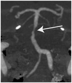
Background: As many as two-thirds of acutely dizzy patients presenting to the emergency department report intermittent symptoms only, consistent with a transient acute vestibular syndrome (single episode lasting < 24 h) or an episodic vestibular syndrome (recurrent episodes, each lasting < 24 h). A broad differential diagnosis, presentation outside the episode(s), and difficulties in describing the symptoms make the diagnostic workup of transient vestibular symptoms (TVS) challenging. Methods: This critical review provides an overview on the management of TVS, focusing on structured history-taking and bedside testing, but also reviewing the role of neuroimaging and eye movement recordings. Results: Asking about timing and triggers helps clarify the temporal evolution of symptoms (transient vs. persistent) and the circumstances of occurrence (spontaneous vs. triggered). Such structured history-taking narrows down the differential diagnosis and ensures appropriate bedside tests. On clinical examination, identifying focal neurologic signs, (subtle) ocular motor findings, and gait imbalance is essential. While positional testing should be applied in all patients with TVS, HINTS+ has not been validated in those patients without nystagmus or with transient (already resolved) symptoms. For neuroimaging, MRI with diffusion-weighted imaging is preferred, but early false-negative findings in up to 20% (to even 50% for small brainstem strokes) of cases must be considered. Quantitative eye movement recordings may be especially valuable in rural areas, supporting telemedicine consults. Conclusions: Special emphasis should be put on distinguishing vertebrobasilar transient ischemic attack from vestibular migraine and cardiac arrhythmia. In the case of triggered TVS, immediate and sustained response to liberation maneuvers strongly supports benign paroxysmal positional vertigo over vestibular migraine or structural central positional nystagmus. Treatment strategies in TVS strongly depend on the underlying cause, ranging from hyperacute stroke treatment to migraine prophylaxis to liberation maneuvers.
17 July 2026