
Topical Advisory Panel applications are now closed. Please contact the Editorial Office with any queries.
Journal Description
Antioxidants
Antioxidants
is an international, peer-reviewed, open access journal related to the science and technology of antioxidants, published monthly online by MDPI. The International Coenzyme Q10 Association (ICQ10A), Israel Society for Oxygen and Free Radical Research (ISOFRR) and European Academy for Molecular Hydrogen Research (EAMHR) are affiliated with Antioxidants and their members receive discounts on the article processing charge.
- Open Access— free for readers, with article processing charges (APC) paid by authors or their institutions.
- High Visibility: indexed within Scopus, SCIE (Web of Science), PubMed, PMC, FSTA, PubAg, CAPlus / SciFinder, and other databases.
- Journal Rank: JCR - Q1 (Chemistry, Medicinal) / CiteScore - Q1 (Clinical Biochemistry)
- Rapid Publication: manuscripts are peer-reviewed and a first decision is provided to authors approximately 18.7 days after submission; acceptance to publication is undertaken in 2.9 days (median values for papers published in this journal in the first half of 2026).
- Recognition of Reviewers: reviewers who provide timely, thorough peer-review reports receive vouchers entitling them to a discount on the APC of their next publication in any MDPI journal, in appreciation of the work done.
- Testimonials: See what our editors and authors say about Antioxidants.
- Companion journal: Oxygen.
Impact Factor:
8.2 (2025);
5-Year Impact Factor:
8.5 (2025)
Latest Articles
Lute-Gen® Alleviates Dry Eye Disease and Modulates Nrf2/HO-1, TLR4/NF-κB/MAPK Signaling, and Aquaporin-Mediated Tear Homeostasis
Antioxidants 2026, 15(7), 872; https://doi.org/10.3390/antiox15070872 (registering DOI) - 13 Jul 2026
Abstract
Dry eye disease (DED) is a multifactorial ocular surface disorder characterized by inflammation, oxidative stress, tear film instability, and secretory dysfunction. This study investigated the protective effects of Lute-gen®, a lutein and zeaxanthin-based formulation, in both in vitro and in vivo
[...] Read more.
Dry eye disease (DED) is a multifactorial ocular surface disorder characterized by inflammation, oxidative stress, tear film instability, and secretory dysfunction. This study investigated the protective effects of Lute-gen®, a lutein and zeaxanthin-based formulation, in both in vitro and in vivo DED models. Human corneal epithelial (HCE-T) cells were stimulated with TNF-α, while dry eye was induced in female Sprague–Dawley rats using subcutaneous scopolamine (SCP) administration. In HCE-T cells, Lute-gen® showed no cytotoxicity, restored cell viability, reduced intracellular ROS, and was associated with increased expression of antioxidant-related markers (Nrf2, HO-1, SOD, CAT, and GPx), reduced expression of inflammatory mediators (TLR4/MyD88/NF-κB/NLRP3), and increased expression of AQP3 and AQP5. In SCP-induced rats, Lute-gen® significantly improved tear secretion and reduced corneal fluorescein staining. Histopathological analyses revealed restoration of conjunctival goblet cells, mucin staining, corneal epithelial integrity, acinar area and cell density, and lacrimal gland architecture, with reduced inflammatory infiltration. Immunofluorescence further demonstrated reduced TLR4 and MMP9 immunoreactivity and decreased CD68+ inflammatory cell infiltration. Molecular analyses showed reduced expression of inflammatory cytokines and NF-κB/MAPK/MMP signaling-related inflammatory mediators, together with restoration of AQP1, AQP3, and AQP5 expression in corneal tissues. Collectively, these findings suggest that Lute-gen® treatment was associated with improvements in dry eye-related pathological changes, including restoration of antioxidant-related markers, attenuation of inflammatory responses, restoration of aquaporin expression, and preservation of ocular surface and lacrimal gland integrity. These preclinical findings support further mechanistic investigations and the future clinical evaluation of Lute-gen® as a potential nutritional intervention for dry eye disease.
Full article
(This article belongs to the Special Issue The Role of Phytochemical Antioxidants in the Prevention and Management of Metabolic Disorders)
Open AccessArticle
Capsicum annuum Regulates Tumor Growth Through Modulation of TLR4/PI3K Signaling in a Lewis Lung Carcinoma Mouse Model
by
Hye Ji Choi, Hyo Lim Lee, Yeong Hyeon Ju, Yu Mi Heo, Hwa Rang Na, Chae Eun Yoon, Young Hee Son, Do-Yoon Kim, Yu-Jin Kim, Hui-Seok Jeong, Seung-Hwan Park, Hyun-Jin Kim and Ho Jin Heo
Antioxidants 2026, 15(7), 871; https://doi.org/10.3390/antiox15070871 (registering DOI) - 13 Jul 2026
Abstract
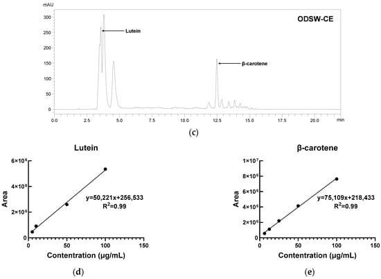
Since lung cancer remains a leading cause of cancer-related mortality, effective adjunct strategies against this disease are needed. This study evaluated an extract prepared from organically cultivated Capsicum annuum treated with deep-seawater-derived ion minerals during cultivation (ODSW-CE) in Lewis lung carcinoma (LLC1) cells
[...] Read more.
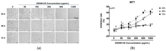
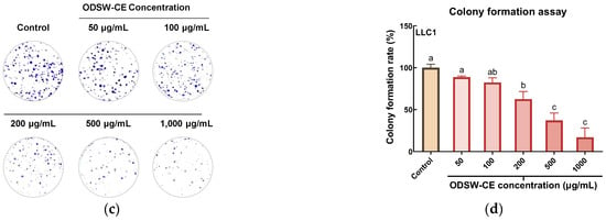
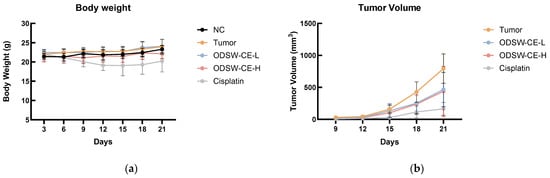
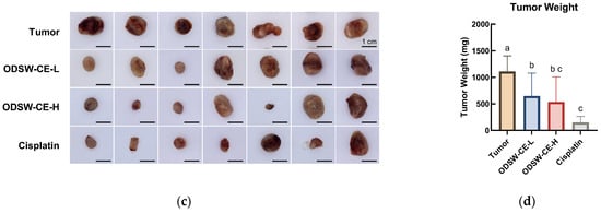
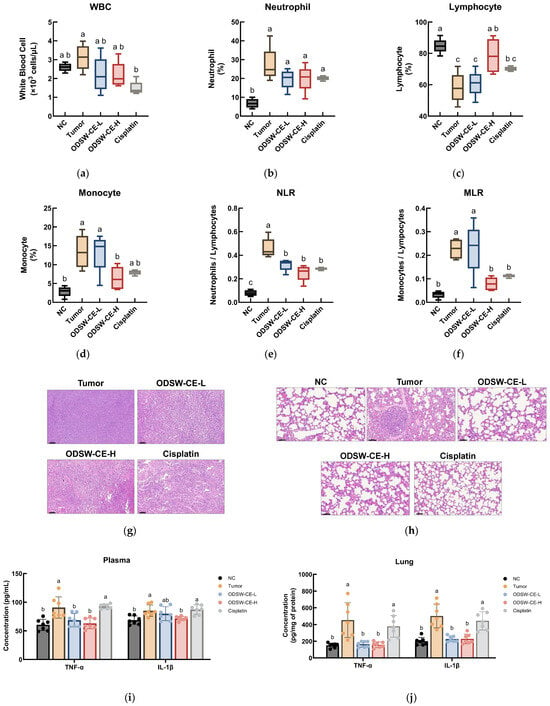
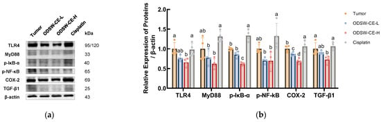
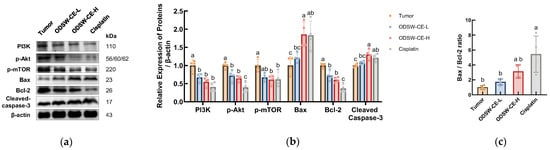
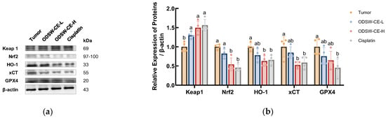
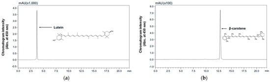
Since lung cancer remains a leading cause of cancer-related mortality, effective adjunct strategies against this disease are needed. This study evaluated an extract prepared from organically cultivated Capsicum annuum treated with deep-seawater-derived ion minerals during cultivation (ODSW-CE) in Lewis lung carcinoma (LLC1) cells and LLC1 tumor-bearing mice. Targeted HPLC-DAD analysis identified and quantified lutein and β-carotene as representative carotenoids. ODSW-CE reduced MTT-derived cell viability and colony-forming ability in vitro and suppressed tumor growth in vivo. In tumor-bearing mice, ODSW-CE was associated with lower hematological inflammatory indices, reduced TNF-α and IL-1β levels in plasma and lung tissue, and attenuated histopathological alterations in tumor and lung tissues. These changes were accompanied by reduced expression of proteins associated with TLR4/MyD88/NF-κB signaling. ODSW-CE also modulated survival and apoptosis-related proteins, decreasing PI3K, p-Akt, and p-mTOR expression, increasing Bax and cleaved caspase-3 expression, and decreasing Bcl-2 expression. Additionally, in tumor tissue, Keap1 expression increased, whereas Nrf2, xCT, and GPX4 expression decreased. Overall, ODSW-CE suppressed LLC1 tumor growth and was associated with coordinated modulation of tumor-associated inflammatory signaling, survival pathways, apoptosis-related proteins, and redox defense.
Full article
(This article belongs to the Special Issue Anti-Cancer Potential of Plant-Based Antioxidants—2nd Edition)
►▼
Show Figures

Figure 1
Open AccessArticle
Extraction Strategy and C18 Solid-Phase Fractionation Shape Phenolic Profiles, Antioxidant Capacity, and Cancer Cell Antiproliferative Activity of Selected Medicinal Plants
by
Domantas Armonavičius, Audrius Maruška, Kristina Bimbiraitė-Survilienė, Mantas Stankevičius, Baltramiejus Jakštys, Tomas Drevinskas, Ugnė Gabrytė, Elżbieta Skrzydlewska, Ona Ragažinskienė, Vilma Kaškonienė, Saulius Šatkauskas, Inga Pečiulienė and Arvydas Kanopka
Antioxidants 2026, 15(7), 870; https://doi.org/10.3390/antiox15070870 (registering DOI) - 13 Jul 2026
Abstract
Medicinal plants are a rich source of biologically active compounds, including phenolic acids, flavonoids, ellagitannins and other secondary metabolites. However, the contribution of specific groups of phenolic compounds to antiproliferative activity remains insufficiently clarified. This study extends our previous crude-extract screening by evaluating
[...] Read more.
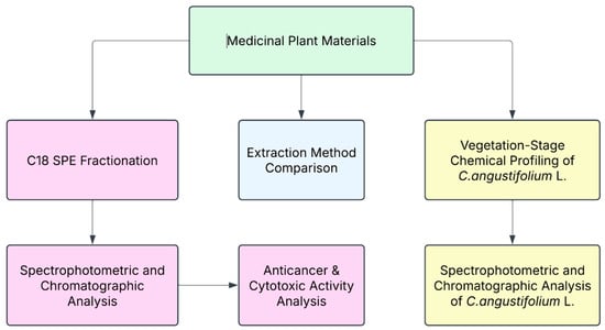
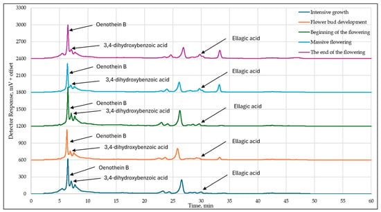
Medicinal plants are a rich source of biologically active compounds, including phenolic acids, flavonoids, ellagitannins and other secondary metabolites. However, the contribution of specific groups of phenolic compounds to antiproliferative activity remains insufficiently clarified. This study extends our previous crude-extract screening by evaluating whether C18 solid-phase extraction (SPE) fractions with different phenolic profiles are associated with different antiproliferative responses. In parallel, extraction strategies were compared to assess method-dependent changes in phenolic recovery and antioxidant capacity, and an additional single-species vegetation-stage analysis of Chamaenerion angustifolium L. Holub was performed to evaluate harvest-stage effects. Phytochemical characterisation was performed using spectrophotometric assays and high-performance liquid chromatography (HPLC) analyses. Among the tested extraction methods, 75% (v/v) methanol in water was the most effective conventional solvent, and ultrasound-assisted extraction yielded the highest overall TPC (total phenolic content), TFC (total flavonoid content), and RSA (radical scavenging activity) values. Vegetation stage analysis of C. angustifolium L. Holub revealed significant variation in phenolic content and antioxidant activity, with the highest levels observed at the beginning of the flowering. Antiproliferative activity was assessed against five cancer cell lines (4T1, A549, Caki-1, HCT116 and MCF7), while HEK-293 cells were used as an immortalised non-cancerous reference model for general cytotoxicity evaluation. Linear mixed-model analysis confirmed a significant incubation-time effect in all tested cancer cell lines, with IC50 values generally decreasing after prolonged exposure. Statistically significant F2-F3 differences were plant-dependent. The 30% (v/v) methanol in water fraction (F2), enriched in oenothein B in C. angustifolium L., showed stronger antiproliferative activity, whereas the 60% (v/v) methanol in water fraction (F3) showed stronger activity in Quercus robur L., Juglans nigra L., Juglans regia L., and Solidago canadensis L. These findings indicate that antiproliferative activity was associated with the qualitative and quantitative composition of the selected phenolic-rich SPE fractions rather than with a single universal fraction effect. All tested fractions exhibited lower cytotoxicity toward HEK-293 cells under the applied conditions; however, claims of selectivity should be confirmed using additional normal or primary cell models. Overall, the findings clarify the role of extraction strategy, harvest stage and targeted fractionation in linking phenolic composition with biological activity.
Full article
(This article belongs to the Special Issue Advances in Analysis of Antioxidant-Rich Medicinal Plants, Functional Foods and Food Supplements)
►▼
Show Figures

Figure 1
Open AccessArticle
Anthraquinone Derivative Rufigallol Protects Against Ethanol-Induced Gastric Damage via Modulation of PGE2, NO, and Inflammatory Pathways: In Vivo and In Vitro Study
by
Tariq G. Alsahli, Khushhaal, Sami I. Alzarea, Hesham A. M. Gomaa and Muhammad Afzal
Antioxidants 2026, 15(7), 869; https://doi.org/10.3390/antiox15070869 (registering DOI) - 12 Jul 2026
Abstract
Excessive ethanol consumption is a cause of gastric ulceration. This study aimed to identify the gastroprotective action of rufigallol, an anthraquinone, against ethanol-induced gastric ulcers and assess its antioxidant and anti-inflammatory properties. Healthy rats were grouped into five groups (n = 8);
[...] Read more.
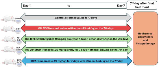
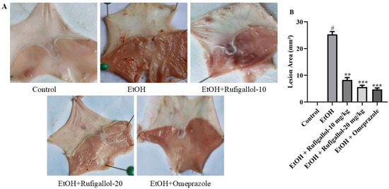
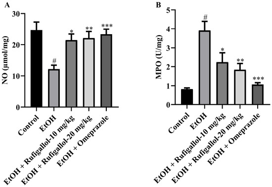
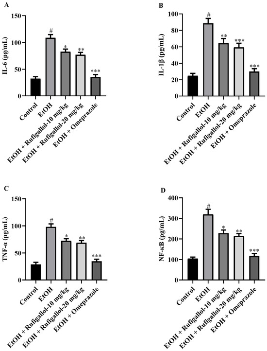
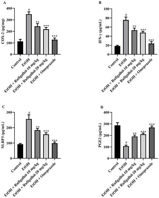
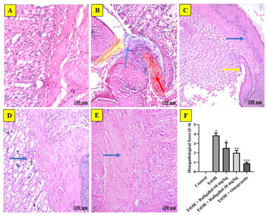
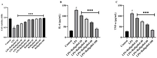
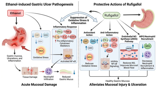
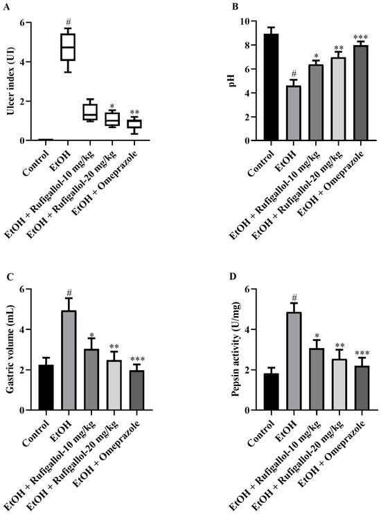
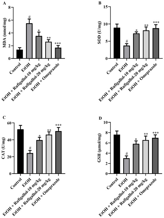
Excessive ethanol consumption is a cause of gastric ulceration. This study aimed to identify the gastroprotective action of rufigallol, an anthraquinone, against ethanol-induced gastric ulcers and assess its antioxidant and anti-inflammatory properties. Healthy rats were grouped into five groups (n = 8); the control group received sterile saline orally; the ethanol group received ethanol (5 mL/kg) to generate gastric ulcers on the last day of the experiment; the pretreated rufigallol group was administered 10 or 20 mg/kg rufigallol orally for a week before gastric ulcer initiation; and the drug control group received omeprazole (20 mg/kg) for a week with ethanol treatment. The results showed that oral rugalallol significantly reduced gastric ulcers, as indicated by decreased gastric juice volume and increased preventive percentage, gastric pH value, and pepsin activity. Histopathology confirmed a reduction in the gastric ulcer index following rufigallol treatment. Rufigallol pre-treatment significantly increased antioxidant levels (CAT, SOD, and GSH) and decreased MDA levels compared to the ethanol group. Furthermore, rufigallol treatment decreased MPO, pro-inflammatory cytokines, and mediator levels. It also reduced COX-2, IFN-γ, and NLRP3 expression and restored NO and PGE2 levels. In vitro experiments using HT-29 and HaCaT cell lines confirmed that rufigallol reduced cytokine production and exhibited anti-inflammatory activity in response to lipopolysaccharide stimulation. These results suggested that rufigallol administration may provide gastroprotective benefits against ethanol-induced gastric ulcers, potentially by reducing oxidative stress and gastric inflammation.
Full article
(This article belongs to the Special Issue Bioactive Compounds from Natural Sources with Antioxidant and Anti-Inflammatory Potential)
►▼
Show Figures

Figure 1
Open AccessArticle
Computational Study of the Peroxyl Radical Scavenging Ability of Phenolic Antioxidants
by
Ainsley Barisoff, Max Walton-Raaby, Paula Jofily and Nelaine Mora-Diez
Antioxidants 2026, 15(7), 868; https://doi.org/10.3390/antiox15070868 - 11 Jul 2026
Abstract
The physiological implications of chronic oxidative stress motivate research on antioxidant activity: the cellular defence and repair mechanism for oxidative damage. Through evaluation of thermodynamic and kinetic quantities of hydroperoxyl (•OOH) and methylperoxyl (•OOCH3) radical scavenging reactions,
[...] Read more.
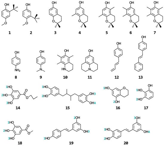
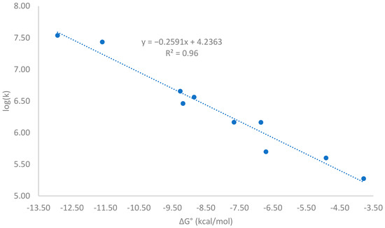
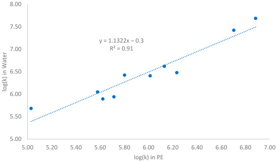
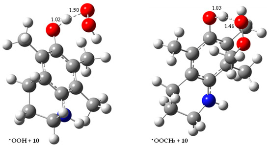
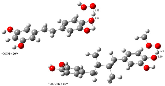
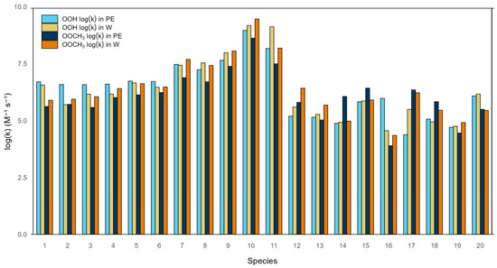
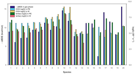
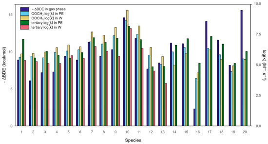
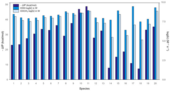
The physiological implications of chronic oxidative stress motivate research on antioxidant activity: the cellular defence and repair mechanism for oxidative damage. Through evaluation of thermodynamic and kinetic quantities of hydroperoxyl (•OOH) and methylperoxyl (•OOCH3) radical scavenging reactions, the primary antioxidant activity of twenty phenolic compounds, previously investigated for tertiary antioxidant activity, is evaluated at the M06-2X(SMD)/6-31++G(d,p) level of theory in water and pentyl ethanoate. The formal-hydrogen atom transfer (f-HAT) and single electron transfer (SET) mechanisms are considered. In aqueous environments, SET proves to be the dominant mechanism for most phenols studied, as nearly half of the phenols produced rate constants within the diffusion limit, implicating biochemical relevance. Only three of the phenols also exhibited significant activity via f-HAT. The phenols show greater SET scavenging of •OOH than •OOCH3, but the more efficient f-HAT target is variable. Through comparison with their tertiary activity, we show that thermodynamics are a better predictor for antioxidant activity when the radical is a less complex species (i.e., •OOH versus protein radical); however, several inconsistencies in the Bell–Evans–Polanyi principle still appear between and within solvents and phenolic compounds. This work examines the differences that appear when studying primary versus tertiary antioxidant activity and highlights the importance of using kinetic strategies to investigate antioxidant activity.
Full article
(This article belongs to the Section Natural and Synthetic Antioxidants)
►▼
Show Figures

Figure 1
Open AccessArticle
Disulfiram Alleviates Metabolic Dysfunction-Associated Steatohepatitis in Mice via Inhibiting Aurora Kinase A and Restoring Autophagy
by
Zixiong Zhou, Xi Zeng, Yuqi Guo, Zhengyi Tan, Xin Zhang, Xuyang Liu, Shuyu Zheng, Wenwen Liu, Haiyan Wang and Jing Qi
Antioxidants 2026, 15(7), 867; https://doi.org/10.3390/antiox15070867 - 11 Jul 2026
Abstract
Metabolic dysfunction-associated steatohepatitis (MASH) is a severe, progressive liver disease lacking effective therapies. Disulfiram (DSF), an FDA-approved medication for alcohol dependence, exhibits diverse biological activities beyond its primary indication. This study aimed to evaluate whether DSF holds intervention promise for MASH and to
[...] Read more.
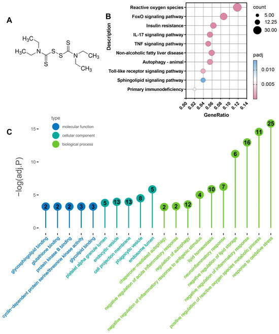
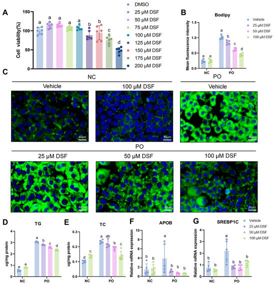
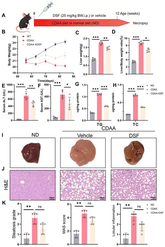
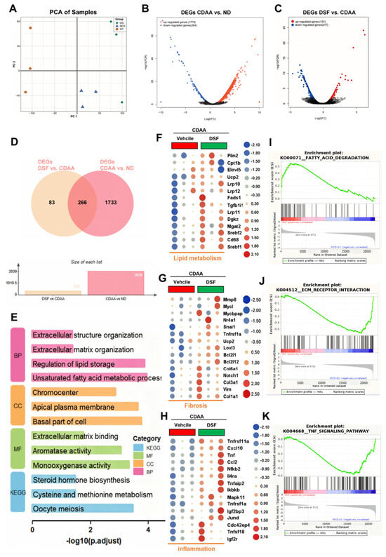
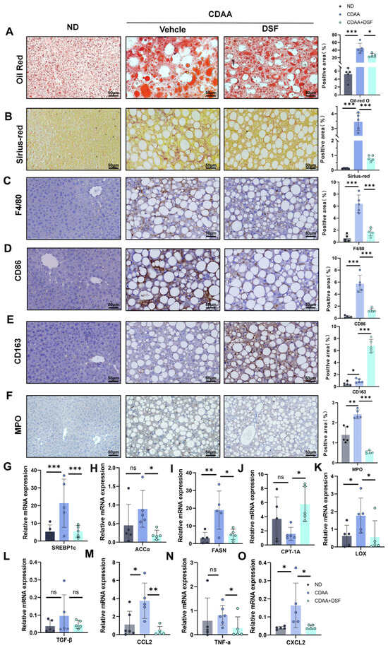
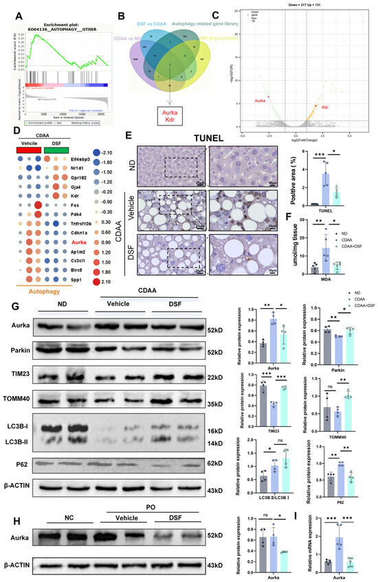
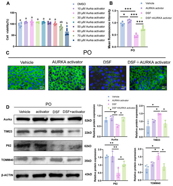
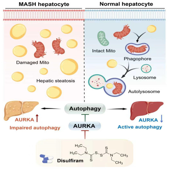
Metabolic dysfunction-associated steatohepatitis (MASH) is a severe, progressive liver disease lacking effective therapies. Disulfiram (DSF), an FDA-approved medication for alcohol dependence, exhibits diverse biological activities beyond its primary indication. This study aimed to evaluate whether DSF holds intervention promise for MASH and to unravel the underlying molecular mechanism. The efficacy of DSF was assessed in a mouse model of MASH induced by a choline-deficient, L-amino acid-defined diet, as well as in hepatocytes exposed to free fatty acids (FFAs) to trigger lipotoxicity. RNA-seq analysis combined with bioinformatic approaches was performed to identify key pathways and hub genes. Mechanistic validation was carried out using Western blotting and qPCR. Computational predictions suggested that DSF may influence insulin resistance, inflammation, autophagy-related markers, and lipid metabolism. In FFAs-treated hepatocytes, DSF administration dose-dependently reduced lipid accumulation and lipotoxicity. Consistently, in MASH mice, DSF administration significantly lowered elevated serum ALT (35%) and AST (40%) levels and the absolute hepatic triglyceride content (reduced from 1 to 0.5 μg/mg protein), and markedly attenuated hepatic steatosis, inflammation, fibrosis, and oxidative stress. Of note, RNA-seq analysis revealed that DSF modulated autophagy-related pathways and identified Aurora kinase A (AURKA) as a central downregulated hub gene. Mechanistically, DSF suppressed AURKA expression, which in turn led to changes in autophagy-related markers. These changes in autophagy-related markers were functionally coupled to a reduction in lipotoxicity. Collectively, DSF alleviates MASH by inhibiting AURKA, thereby relieving AURKA-mediated suppression of autophagy-related markers, which was associated with diminishing lipotoxicity, and ultimately achieving broad suppression of disease progression. Thus, DSF represents a promising hepatoprotective candidate for the intervention of MASH.
Full article
(This article belongs to the Special Issue Lipotoxicity in Metabolic Dysfunction-Associated Steatotic Liver Disease (MASLD) and Its Therapeutic Control)
►▼
Show Figures

Figure 1
Open AccessArticle
Anti-Aging Potential of Biogenic Selenium Nanoparticles and Selenium/Polysaccharides Nanoconjugate Biosynthesized by Bacillus subtilis Isolated from Selenium-Rich Soil
by
Yiming Luo, Chengbin Feng, Mengze Liu, Pengfei Zhao and Danfei Huang
Antioxidants 2026, 15(7), 866; https://doi.org/10.3390/antiox15070866 - 11 Jul 2026
Abstract
Selenium nanoparticles (SeNPs), which are nanoscale particles of elemental selenium (Se0), combine the biological activity of selenium with the distinctive properties inherent in nanomaterials, thereby making SeNPs a more promising candidate for advancing selenium-based resources. In this work, several strains of
[...] Read more.
Selenium nanoparticles (SeNPs), which are nanoscale particles of elemental selenium (Se0), combine the biological activity of selenium with the distinctive properties inherent in nanomaterials, thereby making SeNPs a more promising candidate for advancing selenium-based resources. In this work, several strains of Bacillus with sodium selenite-reducing ability were isolated from selenium-rich soils. Bacillus subtilis ESNS-2 with high selenium tolerance was selected as the target strain. Under culture conditions supplemented with 5 mmol/L sodium selenite, this strain exhibited a reduction efficiency of 75.4 ± 0.6% over 24 h. The produced bioSeNPs were purified, decorated using polysaccharides from the seeds of Plantago asiatica L. (PLP), and subsequently systematically characterized using various means. The results revealed that the prepared PLP-bioSeNPs were regularly spherical elemental selenium particles, with an average particle size of 96.9 ± 1.1 nm, a PDI of 0.108 ± 0.003, and a zeta potential of −19.7 ± 0.4 mV. Characterization confirmed that they exhibited excellent dispersibility and stability. In vitro antioxidant assays, both bioSeNPs and the PLP-bioSeNP complex demonstrated pronounced dose-dependent scavenging activity against DPPH• and ABTS+• radicals. In a D-galactose-induced aging mouse model, both bioSeNPs and PLP-bioSeNPs alleviated D-galactose-induced hepatic and cerebral damage, as well as associated behavioral deficits, through the modulation of oxidative stress balance and suppression of inflammation. This study successfully accomplished the efficient production of SeNPs utilizing Bacillus subtilis ESNS-2. The modification of PLP provided innovative strategies for the development of macromolecular drugs, with a specific emphasis on enhancing stability.
Full article
(This article belongs to the Section Health Outcomes of Antioxidants and Oxidative Stress)
Open AccessReview
Hydroxysafflor Yellow A for Diabetic Retinopathy: A Critical Review of Retinal Neurovascular Mechanisms and Systemic-to-Ocular Pharmacokinetic Barriers
by
Jiaqi Liu, Wenjing Liu, Lu Li, Qianqian Zhang, Jun Zhang and Wenjie Yan
Antioxidants 2026, 15(7), 865; https://doi.org/10.3390/antiox15070865 - 10 Jul 2026
Abstract
►▼
Show Figures
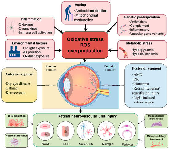
Oxidative stress contributes to retinal neurovascular injury through inflammation, mitochondrial dysfunction, blood–retinal barrier (BRB) disruption, microcirculatory impairment, and regulated cell death. Hydroxysafflor yellow A (HSYA), a water-soluble quinochalcone C-glycoside derived from safflower (Carthamus tinctorius L.), modulates oxidative and inflammatory signaling, apoptosis, mitochondrial
[...] Read more.
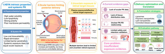
Oxidative stress contributes to retinal neurovascular injury through inflammation, mitochondrial dysfunction, blood–retinal barrier (BRB) disruption, microcirculatory impairment, and regulated cell death. Hydroxysafflor yellow A (HSYA), a water-soluble quinochalcone C-glycoside derived from safflower (Carthamus tinctorius L.), modulates oxidative and inflammatory signaling, apoptosis, mitochondrial injury, endothelial barrier dysfunction, and neurovascular damage in experimental ischemic, inflammatory, and metabolic disorders. This review critically evaluates the direct ocular evidence for HSYA in diabetic retinopathy and examines the systemic-to-ocular pharmacokinetic and delivery barriers that constrain its ophthalmic translation. Current ocular evidence is limited and concentrated mainly in DR models, in which HSYA attenuates oxidative stress, inflammation, BRB disruption, and apoptosis, potentially through Nrf2/HO-1 signaling. Evidence in retinal photic injury is limited, whereas the proposed relevance of HSYA to retinal ischemia–reperfusion injury, glaucoma, and AMD remains largely hypothesis-generating. The principal translational challenge is whether HSYA can achieve pharmacologically relevant exposure in ocular target tissues. Future studies should integrate dose, plasma and ocular exposure, target engagement, retinal structure, local safety, and visual function in disease-specific models. Accordingly, evidence from non-DR models is discussed primarily to define mechanistic hypotheses and experimental priorities rather than to establish ophthalmic efficacy.
Full article

Figure 1
Open AccessReview
Biology of HDL: From Structural Heterogeneity to Dysfunctional Remodeling in Cardiovascular Disease and Comorbidities
by
Yihang Cai, Kehan Li, Huibo Ma, Jianqiang Wu and Yuehong Zheng
Antioxidants 2026, 15(7), 864; https://doi.org/10.3390/antiox15070864 - 10 Jul 2026
Abstract
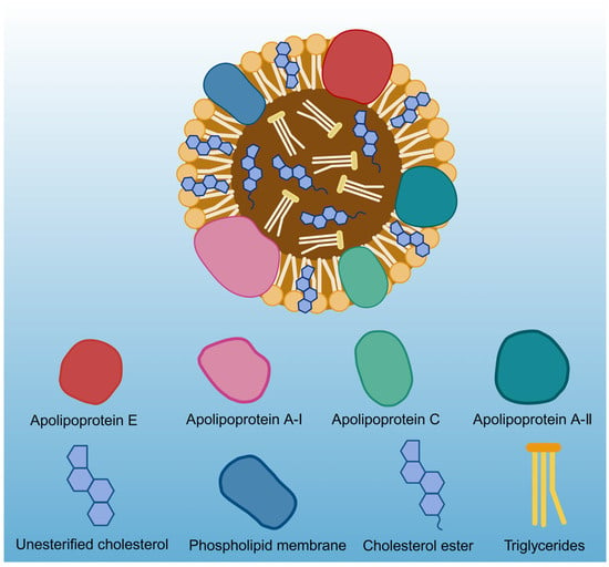
The pathogenesis of cardiovascular diseases (CVDs) is intimately linked to cholesterol dysregulation. While high-density lipoprotein cholesterol (HDL-C) is classically considered cardioprotective, contemporary epidemiological evidence reveals a noncausal, often U-shaped, relationship with CVD risk. Static measurements of HDL-C obscure the structural and functional heterogeneity
[...] Read more.
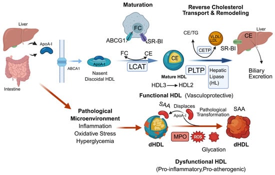
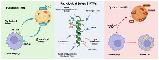
The pathogenesis of cardiovascular diseases (CVDs) is intimately linked to cholesterol dysregulation. While high-density lipoprotein cholesterol (HDL-C) is classically considered cardioprotective, contemporary epidemiological evidence reveals a noncausal, often U-shaped, relationship with CVD risk. Static measurements of HDL-C obscure the structural and functional heterogeneity of circulating HDL particles. Under pathological stress, HDL undergoes extensive structural remodeling into dysfunctional HDL, thereby losing its vasculoprotective properties and instead mediating proatherogenic and proinflammatory responses. This review critically evaluates the biogenesis, maturation, and metabolic trajectory of HDL. By integrating recent advancements in proteomics and lipidomics, we map the intricate compositional shifts within HDL subpopulations and clarify the regulatory roles of HDL-associated microRNAs in intercellular communication. We investigate the specific drivers of HDL dysfunction, which is often exacerbated by comorbidities such as diabetes and chronic kidney disease. Furthermore, we outline the methodological transition from automated homogeneous HDL-C quantification to multidimensional profiling. Shifting the clinical focus from HDL quantity to functional quality resolves the HDL-C paradox, helping to drive the development of precision lipidology and targeted therapies to reverse HDL dysfunction in CVDs.
Full article
(This article belongs to the Section Aberrant Oxidation of Biomolecules)
►▼
Show Figures

Figure 1
Open AccessReview
Mechanistic Insights into Phytocompounds for Vitiligo Therapy: Current Evidence and Future Opportunities
by
Rethabile Banda-Lesole, Ipeleng Kopano Rosinah Kgosiemang and Tshepiso Jan Makhafola
Antioxidants 2026, 15(7), 863; https://doi.org/10.3390/antiox15070863 - 10 Jul 2026
Abstract
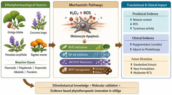
Vitiligo is a multifactorial depigmentation disorder involving complex interactions among oxidative stress, immune dysregulation, inflammatory signaling, and programmed cell death pathways, which act as a central driver of melanocyte dysfunction and loss, interacting with immune-mediated cytotoxicity and intrinsic cellular susceptibility. Excessive reactive oxygen
[...] Read more.
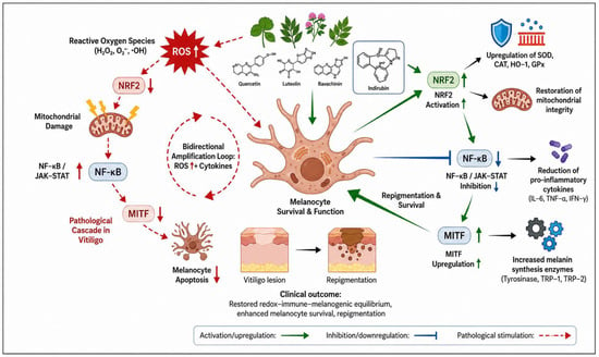
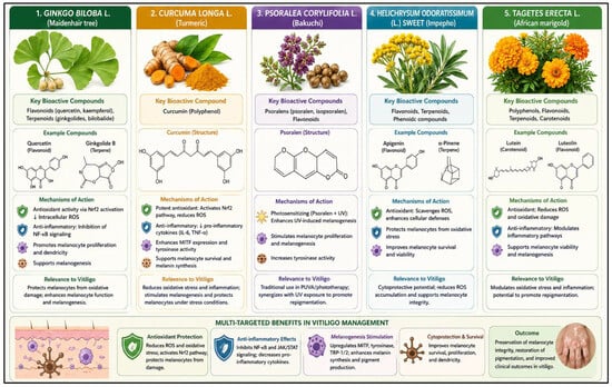
Vitiligo is a multifactorial depigmentation disorder involving complex interactions among oxidative stress, immune dysregulation, inflammatory signaling, and programmed cell death pathways, which act as a central driver of melanocyte dysfunction and loss, interacting with immune-mediated cytotoxicity and intrinsic cellular susceptibility. Excessive reactive oxygen species (ROS) disrupt mitochondrial integrity, impair redox homeostasis, suppress microphthalmia-associated transcription factor (MITF)-dependent melanogenesis, and induce melanocyte apoptosis. Concomitant dysfunction of the nuclear factor erythroid 2-related factor 2 (NRF2)/antioxidant response element (ARE) axis further exacerbates oxidative injury by limiting endogenous antioxidant capacity. Current therapeutic approaches, including corticosteroids, phototherapy, and targeted immunomodulators, achieve partial repigmentation but do not adequately resolve melanocyte-intrinsic redox imbalance. This structured narrative review comprehensively integrates mechanistic and translational evidence to define phytocompounds as redox-active, multi-target modulators in vitiligo. Plant-derived polyphenols, flavonoids, terpenoids, and related metabolites are shown to attenuate ROS accumulation, preserve mitochondrial function, activate NRF2-dependent antioxidant signaling, and restore MITF-mediated expression of tyrosinase and associated melanogenic enzymes. Furthermore, coordinated modulation of MAPK, PI3K/Akt, and JAK/STAT pathways highlights their capacity to regulate immune–oxidative crosstalk and promote melanocyte survival. Despite promising preclinical and emerging clinical evidence of repigmentation efficacy, translational progress remains limited by poor phytochemical standardization, insufficient transcriptional and proteomic validation, suboptimal stability and dermal bioavailability, and a lack of rigorously designed clinical trials. Collectively, this review provides a mechanistic framework linking redox dysregulation to melanocyte failure and positions phytocompounds as rational candidates for adjunctive or stand-alone antioxidant-based therapies, while defining critical priorities for clinical translation.
Full article
(This article belongs to the Special Issue Natural Antioxidants: Multiple Mechanisms for Skin Protection and Skin Disease)
►▼
Show Figures

Figure 1
Open AccessArticle
Gene- and Isoform-Level Responses to Extreme Acidic pH Stress in an Emerging Marine Invertebrate Model Organism Litoditis marina
by
Beining Xue, Pengchi Zhang, Hanwen Yang and Liusuo Zhang
Antioxidants 2026, 15(7), 862; https://doi.org/10.3390/antiox15070862 - 9 Jul 2026
Abstract
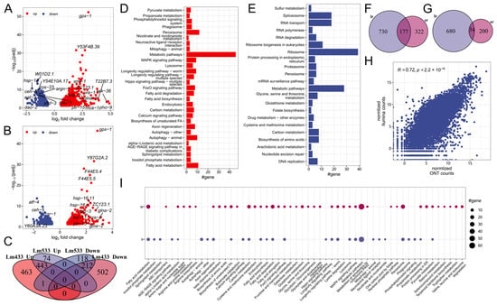
Ocean acidification poses a critical threat to marine invertebrate survival and diversification, yet the post-transcriptional regulatory mechanisms underlying acid stress responses remain poorly understood. Here, we employed Oxford Nanopore long-read RNA sequencing to systematically characterize transcriptional and post-transcriptional responses to acidic pH stresses
[...] Read more.
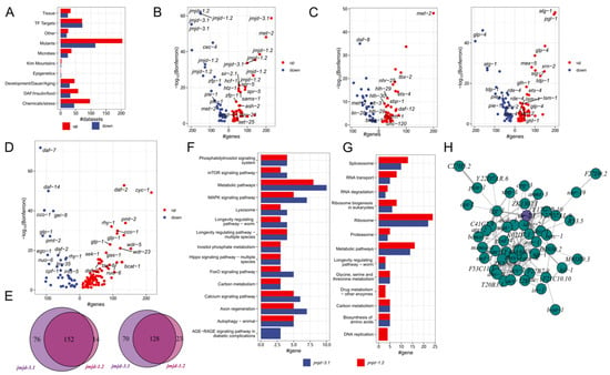
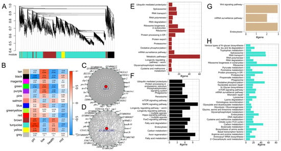
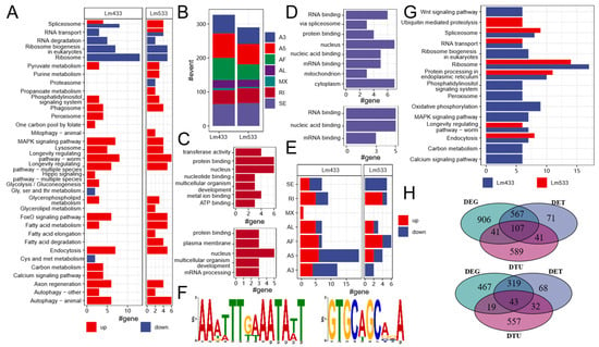
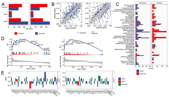
Ocean acidification poses a critical threat to marine invertebrate survival and diversification, yet the post-transcriptional regulatory mechanisms underlying acid stress responses remain poorly understood. Here, we employed Oxford Nanopore long-read RNA sequencing to systematically characterize transcriptional and post-transcriptional responses to acidic pH stresses in the marine nematode Litoditis marina. Our analysis revealed 912 upregulated and 728 downregulated genes enriched in autophagy, fatty acid metabolism, and peroxisome activation, alongside 327 differential alternative splicing events and 1512 transcripts with significant usage changes under severe acidic pH stress. By integrating weighted gene co-expression network analysis with databases such as WormExp2, we found that acidic pH stress response exhibited resemblance to oxidative stress response. Specifically, we identified genes involved in the oxidative stress response, such as gpx-1, cyp-13A11, trxr-1, cyc-1, and key regulators, including hlh-30/TFEB. Genes such as gpx-1, trxr-1, and cyp-23A1 might protect L. marina from oxidative stress under acidic pH. Moreover, several ferroptosis-related genes, such as gpx-5, fat-2, and smf-1, might render L. marina vulnerable to acidic pH stress. Among the genes with splicing changes, we identified oxidative stress responding genes such as sod-4, prx-2, coq-2, coq-3, prx-10, ctl-2, gst-7, trx-1, mdt-15, and fat-2. Additionally, we discovered the preference for proximal 3′ UTR under acidic pH stress. Genes related to ferroptosis, including cyp-23A1, C07E3.9/PLA2G1B, and C53D5.5/GGT1, exhibited differential 3′ UTR usage under acidic pH stress. Our findings shift the focus from traditional gene-centric analyses to capturing the full breadth of post-transcriptional diversity, providing novel insights into post-transcriptional gene regulation in marine metazoans under environmental stress, as well as revealing that alleviating oxidative stress might increase resistance to acid pH stress.
Full article
(This article belongs to the Special Issue Antioxidants and Aquaculture: A Synergistic Approach for Sustainable Aquatic Production—2nd Edition)
►▼
Show Figures

Figure 1
Open AccessArticle
Urinary TBARS as a Non-Invasive Proxy of Plasma Lipid Peroxidation in Essential Hypertension: A Translational Study on Vascular Oxidative–Inflammatory Burden
by
Antón Cruces-Sande, Néstor Vázquez-Agra, Óscar Seoane-Casqueiro, Emma López-Prado, Estefanía Méndez-Álvarez, Ramón Soto-Otero, Antonio Pose-Reino and Álvaro Hermida-Ameijeiras
Antioxidants 2026, 15(7), 861; https://doi.org/10.3390/antiox15070861 - 9 Jul 2026
Abstract
Background/Objectives: Lipid peroxidation is a relevant oxidative–inflammatory mechanism in essential hypertension and cardiovascular disease. Plasma thiobarbituric acid reactive substances (TBARS), commonly reported as malondialdehyde-equivalent values, provide an operational index of lipid peroxidation-related aldehydic reactivity, but blood-based assessment is limited by venipuncture and preanalytical
[...] Read more.
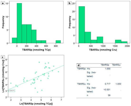
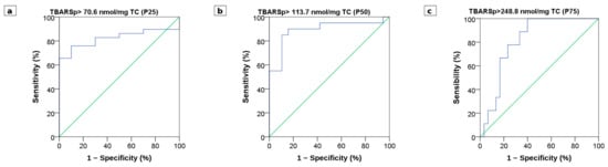
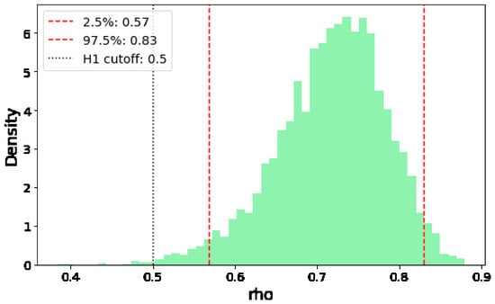
Background/Objectives: Lipid peroxidation is a relevant oxidative–inflammatory mechanism in essential hypertension and cardiovascular disease. Plasma thiobarbituric acid reactive substances (TBARS), commonly reported as malondialdehyde-equivalent values, provide an operational index of lipid peroxidation-related aldehydic reactivity, but blood-based assessment is limited by venipuncture and preanalytical handling requirements. Urine is an attractive non-invasive matrix for redox biomarker development, although whether urinary TBARS reflect plasma lipid peroxidation in hypertensive patients remains insufficiently characterized. This study aimed to evaluate whether matrix-specific normalization—total cholesterol for plasma TBARS and creatinine for urinary TBARS—reveals a measurable intra-individual relationship between these matrices in essential hypertension. Methods: In this paired observational study, plasma and urine samples were obtained from 39 treated patients with essential hypertension under standardized fasting conditions. TBARS were quantified using a colorimetric thiobarbituric acid reaction assay. Plasma TBARS were normalized to total cholesterol and expressed as TBARSp, while urinary TBARS were normalized to creatinine and expressed as TBARSu. Associations were assessed using Spearman’s rank correlation, exploratory receiver operating characteristic (ROC) analyses based on internally derived plasma TBARS percentile thresholds, and Bayesian bootstrap inference. Results: Cholesterol-normalized plasma TBARS and creatinine-normalized urinary TBARS showed a moderate-to-strong positive monotonic association (Spearman’s ρ = 0.717, p < 0.001). Bayesian bootstrap analysis supported this relationship, with a 95% credible interval of 0.57–0.83 and a Bayes factor > 300 for ρ ≥ 0.5. Urinary TBARS showed exploratory within-cohort discriminatory capacity for identifying elevated plasma TBARS using internally derived thresholds, with an AUC of 0.892 for the median-based classification. Conclusions: Creatinine-normalized urinary TBARS showed a moderate-to-strong association with cholesterol-normalized plasma TBARS in treated essential hypertension. These findings provide hypothesis-generating paired-sample evidence that urinary TBARS may serve as a low-burden, non-invasive proxy of plasma lipid peroxidation-related redox alterations. Further validation in larger and clinically diverse cohorts, ideally including more specific lipid peroxidation markers and renal-function-aware analyses, is required to define their translational and clinical utility.
Full article
(This article belongs to the Special Issue Redox Biomarkers in Inflammatory Diseases)
►▼
Show Figures

Figure 1
Open AccessReview
Ferroptosis Resistance: Redundant Antioxidant Networks Are a Barrier to Cancer Therapy
by
Birandra K. Sinha
Antioxidants 2026, 15(7), 860; https://doi.org/10.3390/antiox15070860 - 9 Jul 2026
Abstract
Ferroptosis is an iron-dependent, lipid peroxidation-driven form of regulated cell death that has emerged as a promising strategy for targeting therapy-resistant cancers. However, both intrinsic and acquired resistance to ferroptosis-inducing agents (FINs) limit their clinical efficacy. Here, we propose an integrated framework in
[...] Read more.
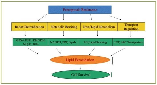
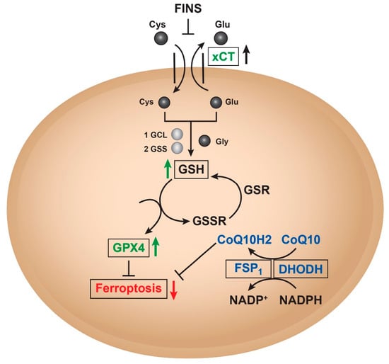
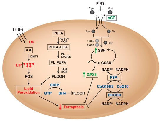
Ferroptosis is an iron-dependent, lipid peroxidation-driven form of regulated cell death that has emerged as a promising strategy for targeting therapy-resistant cancers. However, both intrinsic and acquired resistance to ferroptosis-inducing agents (FINs) limit their clinical efficacy. Here, we propose an integrated framework in which ferroptosis resistance arises from coordinated redox, metabolic, lipid, iron, and transport adaptations that collectively suppress lipid peroxidation and promote tumor survival. Central to this network is the cysteine–glutathione–GPX4 axis, supported by parallel GPX4-independent systems including FSP1–CoQ10, DHODH–CoQ10, GCH1–BH4, and NQO1–NADPH pathways. These antioxidant systems are reinforced by NRF2-driven transcriptional programs, iron sequestration mechanisms, lipid remodeling that reduces polyunsaturated fatty acid availability, and ATP-binding cassette (ABC) transporters that regulate drug and glutathione flux. Tumor heterogeneity further enhances ferroptosis resistance by generating metabolically distinct cellular subpopulations that differ in their susceptibility to lipid peroxidation. We discuss emerging therapeutic strategies designed to overcome these coordinated defense mechanisms, including simultaneous targeting of GPX4 and FSP1, metabolic reprogramming, iron-directed therapies, and nanoparticle-based delivery systems. Collectively, these observations support a systems-level model in which durable ferroptosis-based cancer therapy will require disruption of multiple interconnected resistance mechanisms rather than inhibition of a single molecular target.
Full article
(This article belongs to the Section Antioxidant Enzyme Systems)
►▼
Show Figures

Graphical abstract
Open AccessArticle
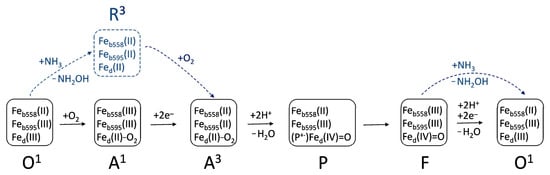
Effect of Ammonia on Escherichia coli Growth and Aerobic Respiration: The Role of Cytochrome bd-II
by
Francesca Giordano, Martina Roberta Nastasi, Vitaliy B. Borisov and Elena Forte
Antioxidants 2026, 15(7), 859; https://doi.org/10.3390/antiox15070859 - 9 Jul 2026
Abstract
Bacterial terminal oxidases are essential for growth and may provide protection against environmental stressors. Bacteria often have to cope with ammonia, which, although an essential nutrient, is toxic at high concentrations. Here, we studied the influence of ammonia on the cell growth of
[...] Read more.
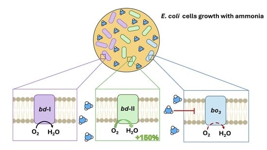
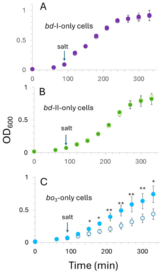
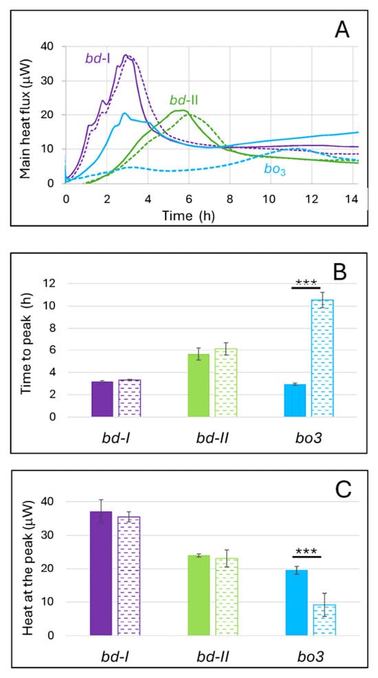
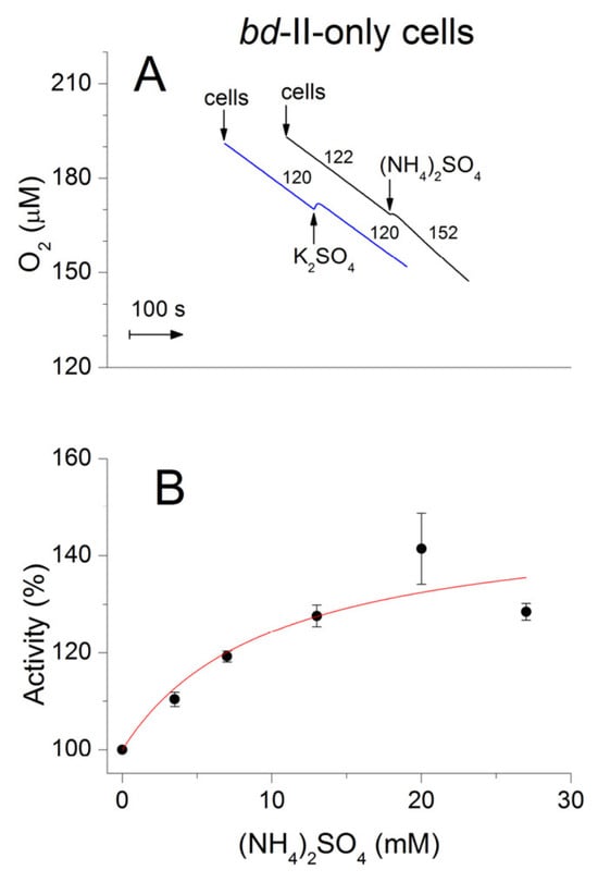
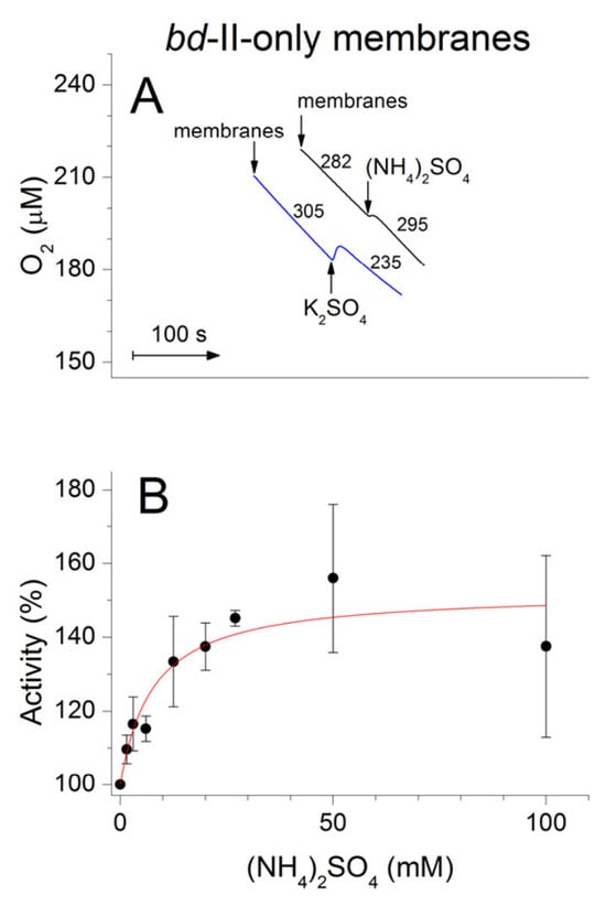
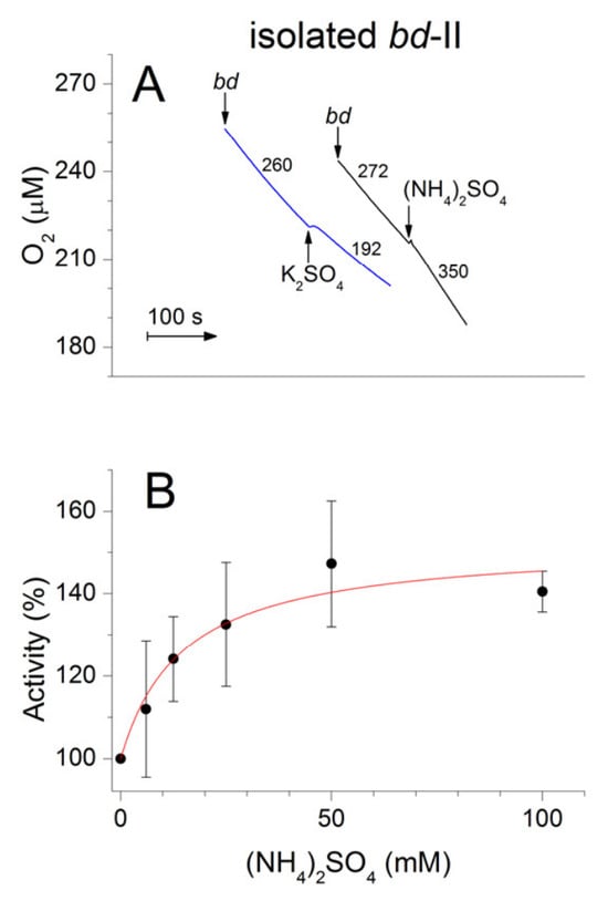
Bacterial terminal oxidases are essential for growth and may provide protection against environmental stressors. Bacteria often have to cope with ammonia, which, although an essential nutrient, is toxic at high concentrations. Here, we studied the influence of ammonia on the cell growth of three different Escherichia coli respiratory mutants, each possessing a single terminal oxidase: cytochrome bd-I, cytochrome bd-II, or cytochrome bo3. We also investigated the effect of ammonia on O2 consumption in cytochrome bd-II-only cells and membranes, as well as in the isolated bd-II enzyme. Using microcalorimetry, spectrophotometry and high-resolution respirometry, the following new results were obtained: (i) At pH 8.3, the addition of ammonia to both bd-I-only and bd-II-only cell cultures has virtually no effect on growth. In contrast, the growth of bo3-only cells is significantly impaired. (ii) The addition of ammonia to bd-II-only intact cells at pH 8.3 not only fails to inhibit their respiration but also accelerates O2 consumption. The same is observed with bd-II-only isolated membranes and detergent-solubilized bd-II enzyme. The maximum increase in cytochrome bd-II O2 consumption rate is approximately 150%. Physiological aspects of the findings are discussed, and molecular mechanisms for ammonia-induced acceleration of O2 consumption by cytochrome bd-II are suggested.
Full article
(This article belongs to the Section ROS, RNS and RSS)
►▼
Show Figures

Graphical abstract
Open AccessArticle
N-Acetylcysteine Attenuates Oxidative Stress and Preserves Red Blood Cell Quality During Whole Blood Storage
by
Sonia Eligini, Lisa Brocca, Alice Mallia, Arianna Valeriano, Erica Gianazza and Cristina Banfi
Antioxidants 2026, 15(7), 858; https://doi.org/10.3390/antiox15070858 - 8 Jul 2026
Abstract
Whole blood (WB) storage induces biochemical and biomechanical alterations that may compromise red blood cell (RBC) quality. Since oxidative stress is a major driver of storage lesions, we investigated whether N-acetylcysteine (NAC) could attenuate these changes during refrigerated storage. WB from healthy donors
[...] Read more.
Whole blood (WB) storage induces biochemical and biomechanical alterations that may compromise red blood cell (RBC) quality. Since oxidative stress is a major driver of storage lesions, we investigated whether N-acetylcysteine (NAC) could attenuate these changes during refrigerated storage. WB from healthy donors was stored at 4 °C for 42 days with or without NAC, added either once at baseline or every 10 days. Plasma albumin proteoforms were assessed by liquid chromatography–mass spectrometry, free hemoglobin species by spectrophotometry, plasma proteomic changes by proximity extension assay, and RBC hemorheological properties by LORRCA analysis. Storage decreased reduced albumin (HSA-SH) and increased oxidized albumin (HSA-Cys), indicating plasma oxidation. Free oxyhemoglobin, deoxyhemoglobin, and methemoglobin increased, consistent with hemoglobin oxidation and hemolysis. Storage also induced plasma proteomic alterations and impaired RBC osmotic and deformability parameters. NAC preserved albumin redox status, limited free hemoglobin accumulation, and attenuated storage-induced proteomic changes. Moreover, NAC partially preserved RBC osmotic and rheological properties, particularly parameters related to osmotic fragility and hydration. No clear advantage of 20 mM over 10 mM NAC was observed. Overall, NAC attenuated oxidative and functional alterations associated with refrigerated whole blood storage, supporting further investigation of antioxidant supplementation as a strategy to mitigate storage lesions under ex vivo conditions.
Full article
(This article belongs to the Special Issue Advanced Bioanalytical Techniques in Antioxidant and Oxidative Stress Research)
Open AccessArticle
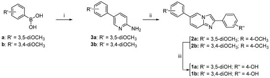
Polyphenolic Imidazopyridines as Multifunctional Modulators of Oxidative Stress, Metal Dyshomeostasis, and β1-42 Amyloid Aggregation in an In Vitro Model of Alzheimer’s Disease
by
Lidia Ciccone, Giovanni Petrarolo, Ilaria D’Agostino, Fabio Scianò, Bianca Laura Bernardoni, Manuela Leri, Jihyae Ann, Susanna Nencetti, Jeewoo Lee, Monica Bucciantini and Concettina La Motta
Antioxidants 2026, 15(7), 857; https://doi.org/10.3390/antiox15070857 - 8 Jul 2026
Abstract
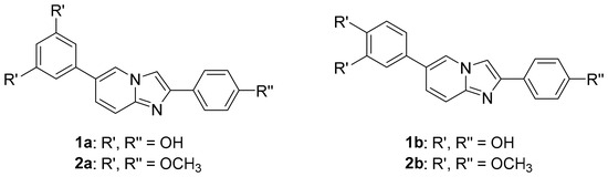
Alzheimer’s disease (AD) involves oxidative stress, metal dyshomeostasis, and toxic oligomers of the amyloid-β peptide (Aβ1-42), calling for multifunctional agents. We investigated a panel of imidazo[1,2-a]pyridines bearing catechol or resorcinol motifs previously designed as SIRT1-activating agents. Their antioxidant profile
[...] Read more.
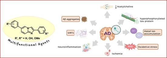
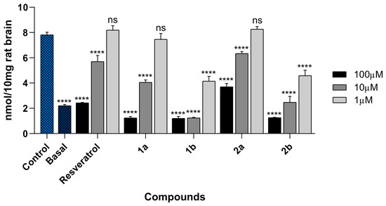
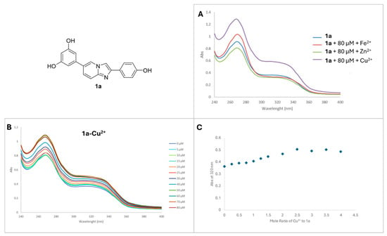
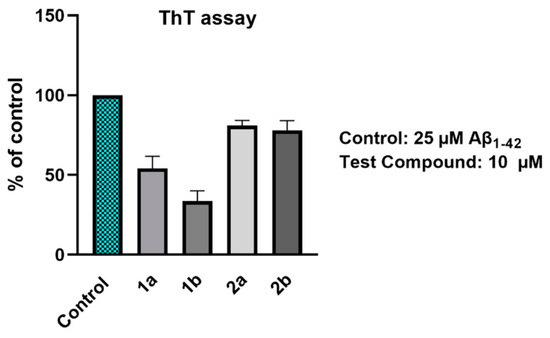
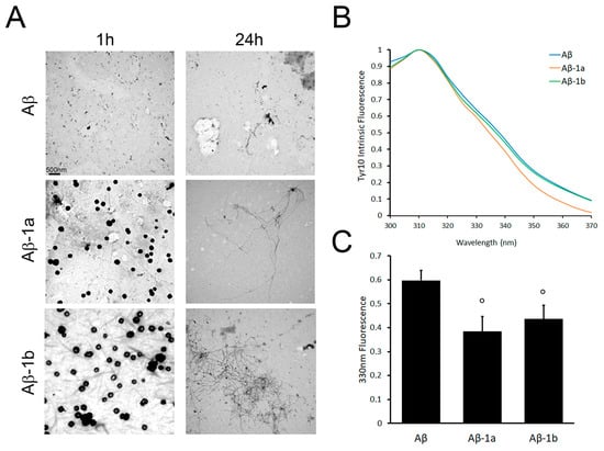
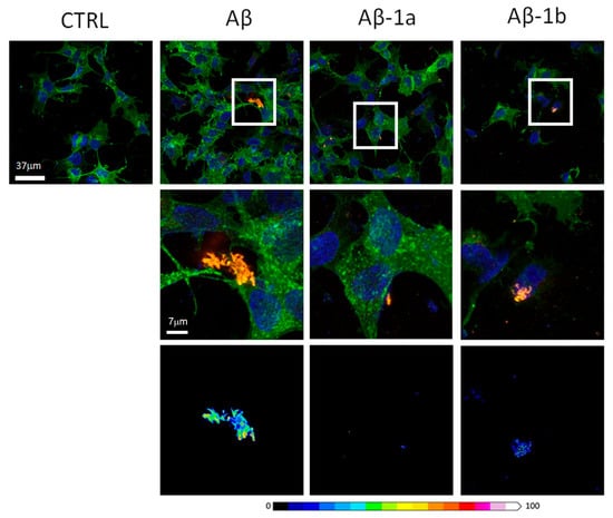
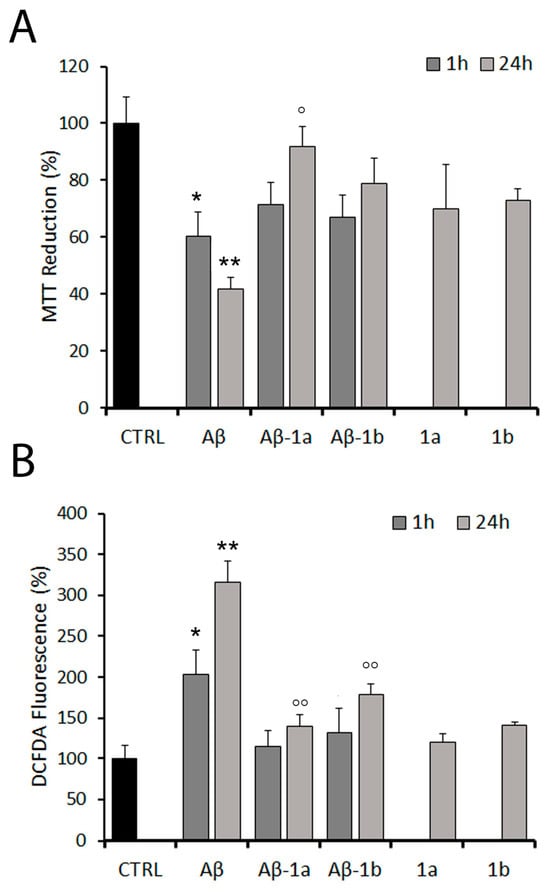
Alzheimer’s disease (AD) involves oxidative stress, metal dyshomeostasis, and toxic oligomers of the amyloid-β peptide (Aβ1-42), calling for multifunctional agents. We investigated a panel of imidazo[1,2-a]pyridines bearing catechol or resorcinol motifs previously designed as SIRT1-activating agents. Their antioxidant profile was evaluated using in vitro DPPH and ABTS assays, which revealed promising radical scavenging activities, and TBARS assays on rat brain homogenates showing inhibition of lipid peroxidation, strictly dependent on the phenolic pattern. UV–Vis studies revealed metal-binding properties, particularly Cu2+ and Fe2+ interactions. In Aβ1-42 aggregation assays, the most active derivatives appeared to promote fibril maturation and the growth of large, ThT-low aggregates with distinct morphological features observed by TEM. Notably, Aβ1-42 aggregates generated in the presence of these compounds exhibited reduced cytotoxicity, preserved cell viability, and induced lower ROS levels in RA-differentiated SH-SY5Y cells compared to aggregates formed in their absence. Imaging and FRET analyses further indicated reduced formation of membrane-binding toxic species. Overall, our data suggest that polyphenolic imidazo[1,2-a]pyridines can remodel Aβ1-42 aggregation, redirecting it toward structurally distinct and less toxic assemblies, while also counteracting oxidative and metal-associated damage. These findings highlight their potential as multifunctional agents capable of addressing several pathological hallmarks of AD.
Full article
(This article belongs to the Special Issue Oxidative Stress as a Therapeutic Target of Alzheimer’s Disease—2nd Edition)
►▼
Show Figures

Graphical abstract
Open AccessArticle
Dihydromyricetin Alleviates Fermented Rapeseed Meal-Induced Intestinal Injury in Chinese Soft-Shelled Turtle (Pelodiscus sinensis): Insights from Growth, Antioxidant, Inflammatory, Transcriptomic and Metabolomic Assessments
by
Wenshu Liu, Lun Chen, Wencai Liu, Jingjing Lu, Yuzhu Wang, Xiaoze Guo, Lingya Li, Xu Han, Chuang Mei, Siming Li and Zirui Wang
Antioxidants 2026, 15(7), 856; https://doi.org/10.3390/antiox15070856 - 8 Jul 2026
Abstract
►▼
Show Figures
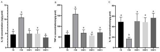
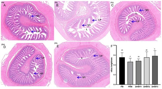
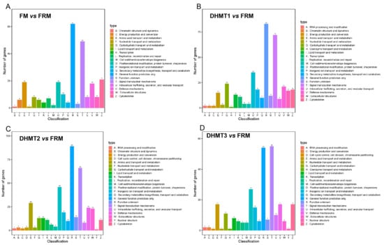
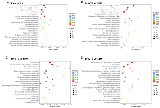
This study was designed to assess the protective efficacy of dihydromyricetin (DHM) against intestinal injury in Chinese soft-shelled turtles (Pelodiscus sinensis) caused by partial replacement of fishmeal (FM) with fermented rapeseed meal (FRM), and to uncover the molecular mechanisms involved. Turtles
[...] Read more.
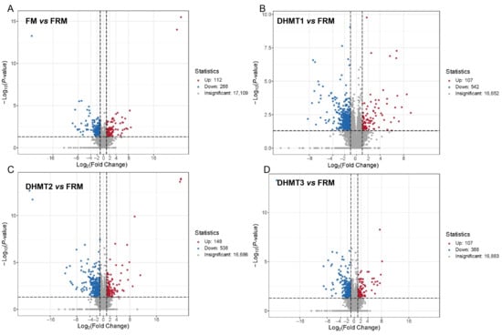
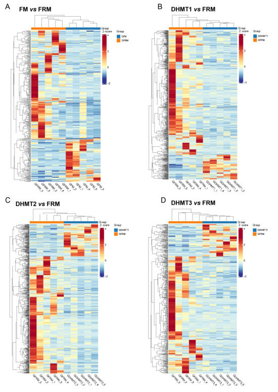
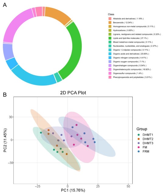
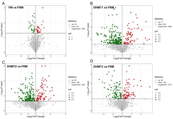
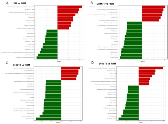
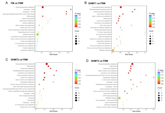
This study was designed to assess the protective efficacy of dihydromyricetin (DHM) against intestinal injury in Chinese soft-shelled turtles (Pelodiscus sinensis) caused by partial replacement of fishmeal (FM) with fermented rapeseed meal (FRM), and to uncover the molecular mechanisms involved. Turtles were fed an FM-based control diet, an FRM-substituted diet, or FRM diets supplemented with DHM at 0.5‰ (DHMT1), 1.0‰ (DHMT2) or 2.0‰ (DHMT3) for 8 weeks. Growth performance, digestive enzyme activities, antioxidant parameters, inflammatory cytokines, and intestinal histomorphology were assessed, and intestinal transcriptomics and metabolomics were also performed. FRM replacement significantly improved growth performance; however, this beneficial effect was accompanied by notable intestinal mucosal oxidative damage, as evidenced by decreased villus height, increased lumen space, and lamina propria edema, along with elevated pro-inflammatory cytokines (IL-1β and TNF-α) and MDA content, alongside reduced GSH and CAT activities. DHM supplementation dose-dependently ameliorated these adverse effects by restoring mucosal integrity, digestive enzyme activities, redox homeostasis, and inflammatory balance, ultimately improving growth performance. Transcriptomic KEGG analysis revealed that DHM enriched pathways related to glycerophospholipid metabolism, glutathione metabolism, drug metabolis-cytochrome P450, and polyunsaturated fatty acid metabolism. Metabolomics further confirmed dose-dependent remodeling of phospholipids and bile acids. Integrated omics demonstrated that DHM likely regulates detoxification, anti-inflammatory, membrane repair, and antioxidant pathways. In conclusion, DHM demonstrates a protective effect against FRM-induced intestinal mucosal oxidative damage in P. sinensis, which may be mediated by a synergistic combination of enhanced detoxification, anti-inflammatory modulation, restoration of phospholipid membrane integrity, and reinforcement of antioxidant defenses.
Full article

Figure 1
Open AccessArticle
Safflower Extract Ameliorates Cisplatin-Induced Acute Kidney Injury by Regulating Microbiota-Metabolic-Redox Nexus and PI3K–Akt/Nrf2 Pathway
by
Yue Chang, Yanzhuo Song, Naveed Ahmad, Chao Song, Yuhang Chu, Yuru Zhang, Lufei Feng, Wei Wei, Min Zhang and Xiuming Liu
Antioxidants 2026, 15(7), 855; https://doi.org/10.3390/antiox15070855 - 7 Jul 2026
Abstract
Cisplatin-induced acute kidney injury (AKI) remains a dose-limiting complication in cancer chemotherapy with restricted preventive measures. Carthamus tinctorius L. (safflower) is known to exhibit effective antioxidant and anti-inflammatory properties; however its potential in renoprotective mechanisms remains poorly understood. The present study utilized a
[...] Read more.
Cisplatin-induced acute kidney injury (AKI) remains a dose-limiting complication in cancer chemotherapy with restricted preventive measures. Carthamus tinctorius L. (safflower) is known to exhibit effective antioxidant and anti-inflammatory properties; however its potential in renoprotective mechanisms remains poorly understood. The present study utilized a cisplatin-induced AKI mouse model to evaluate the renoprotective potential of CT (Carthamus tinctorius) extract. Integrated multi-omics along with in silico and in vivo approaches were used to elucidate the underlying mechanisms of action. The results initially demonstrated a rich phytochemical profile of CT extract characterized by abundant polysaccharides and flavonoids, with Hydroxysafflor Yellow A as a dominant bioactive constituent. In a cisplatin-induced acute kidney injury (AKI) mouse model, CT extract noticeably ameliorated the abnormalities of renal injury, as suggested by improved histopathology, reduced serum creatinine and BUN levels, and regulation of redox homeostasis. Metabolically, CT extract partially reversed AKI-associated disturbances by affecting 21 key metabolites, likely associated with histidine and alanine-aspartate-glutamate biosynthesis, and modulating amino acid and energy metabolism pathways. Concurrently, CT extract improved gut microbial homeostasis, increasing microbial diversity, normalizing the Firmicutes/Bacteroidota ratio, suppressing pathogens, and enriching beneficial Ligilactobacillus. Network pharmacology and molecular docking identified AKT1, RELA, MAPK, and TP53 as central targets of core compounds (rutin and kaempferol derivatives), apparently targeting the PI3K-AKT and RELA (NF-kappaB) hubs. These results suggested that the renoprotective effects of CT extract are associated with transcriptional upregulation of the PI3K/Akt/Nrf2 pathway-related genes, increased expression of antioxidant genes (Ho-1, Sod1), and reduced expression of pro-inflammatory mediators (RelA, Cdk2) in the cisplatin-induced AKI mouse model.
Full article
(This article belongs to the Special Issue Bioactive Compounds from Natural Sources with Antioxidant and Anti-Inflammatory Potential)
Open AccessArticle
Promoter Hypermethylation Is Associated with Reduced Nrf2 and Antioxidant Enzyme Expression in Mandibular Condylar Cartilage in Mice
by
Hisano Ujiie, Hiroyuki Kanzaki, Mao Katayama, Tomomi Ida, Syunnosuke Tohyama, Miho Shimoyama, Yuta Katsumata, Chihiro Arai, Misao Ishikawa and Hiroshi Tomonari
Antioxidants 2026, 15(7), 854; https://doi.org/10.3390/antiox15070854 - 6 Jul 2026
Abstract
Mandibular condylar cartilage (MCC) exhibits greater susceptibility to mechanical stress-induced degeneration than tibial articular cartilage (TAC). This study investigated whether differential epigenetic regulation of nuclear factor erythroid 2-related factor 2 (Nrf2), a master regulator of antioxidant responses, is associated with distinct antioxidant capacities
[...] Read more.
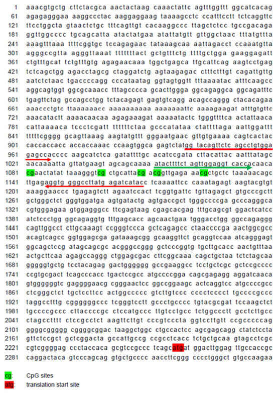
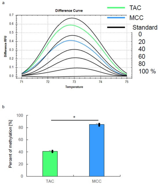
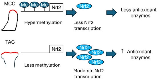
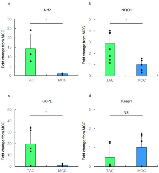
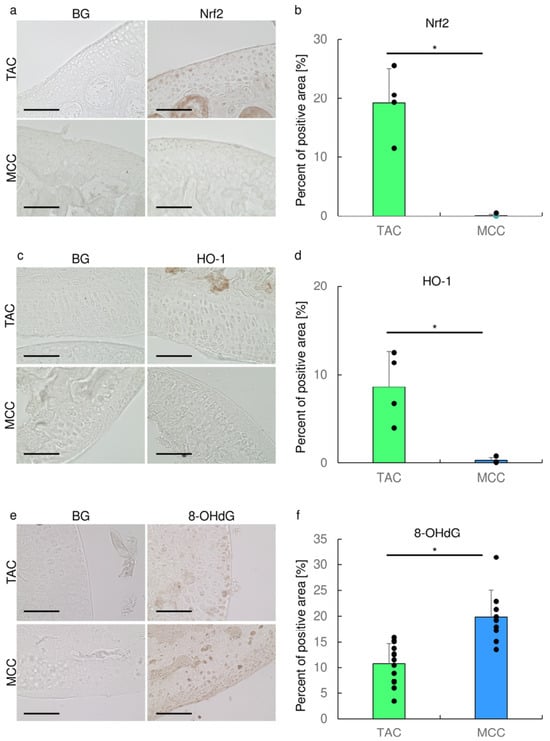
Mandibular condylar cartilage (MCC) exhibits greater susceptibility to mechanical stress-induced degeneration than tibial articular cartilage (TAC). This study investigated whether differential epigenetic regulation of nuclear factor erythroid 2-related factor 2 (Nrf2), a master regulator of antioxidant responses, is associated with distinct antioxidant capacities between these cartilage types. Cartilage tissues from 5-week-old male ICR mice (n = 16 for gene analyses, n = 8 for protein analyses) were obtained using laser microdissection. Gene and protein expression was analyzed by microarray, real-time RT-PCR, and immunohistochemistry. DNA methylation of the Nrf2 promoter was evaluated using pyrosequencing and high-resolution melting analysis. Nrf2 expression in MCC was approximately 1/10 that in TAC at mRNA level and only 5% at protein level. Downstream antioxidant enzymes (NQO1, G6PD, HO-1) showed significantly reduced expression in MCC. Oxidative DNA damage marker 8-OHdG was significantly elevated in MCC compared to TAC (20.0% vs. 10.7%, p < 0.05). The Nrf2 promoter region showed higher DNA methylation levels in MCC, confirmed by high-resolution melting analysis. Higher Nrf2 promoter methylation in MCC is associated with reduced antioxidant capacity and elevated oxidative damage. This epigenetic–antioxidant relationship may contribute to MCC’s vulnerability to mechanical stress-induced degeneration and represents a potential therapeutic target for temporomandibular joint disorders.
Full article
(This article belongs to the Special Issue Oxidative Stress in Osteoclast Metabolism: Implications for Bone Disease Treatment)
►▼
Show Figures

Figure 1
Open AccessReview
Redox Regulation of Plant–Root-Knot Nematode Interactions: From ROS-Mediated Immunity to Sustainable Resistance
by
Jung-Wook Yang, Ho Soo Kim and Yun-Hee Kim
Antioxidants 2026, 15(7), 853; https://doi.org/10.3390/antiox15070853 - 6 Jul 2026
Abstract
Root-knot nematodes (RKNs; Meloidogyne spp.) are among the most destructive plant parasites, causing severe yield losses in diverse crops. Reactive oxygen species (ROS), particularly superoxide radicals (O2•−) and hydrogen peroxide (H2O2), are central regulators of
[...] Read more.
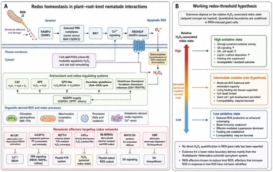
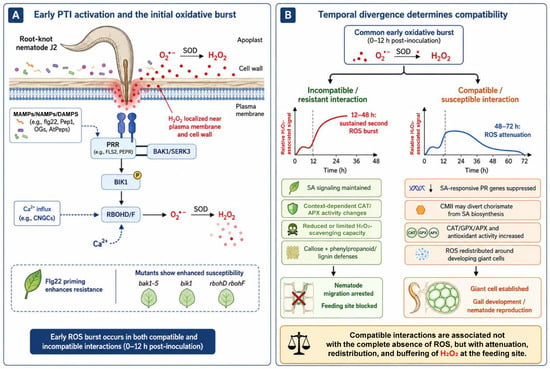
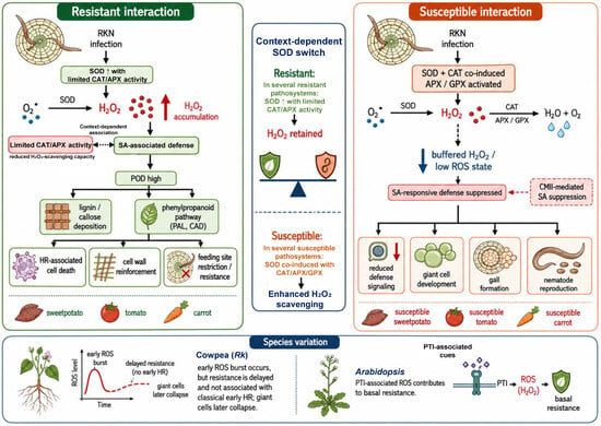
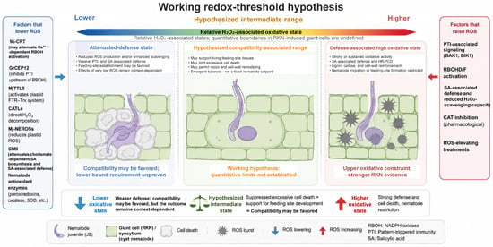
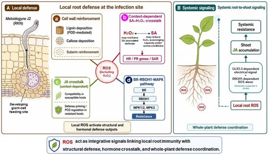
Root-knot nematodes (RKNs; Meloidogyne spp.) are among the most destructive plant parasites, causing severe yield losses in diverse crops. Reactive oxygen species (ROS), particularly superoxide radicals (O2•−) and hydrogen peroxide (H2O2), are central regulators of plant–RKN interactions. This review synthesizes current molecular, biochemical, genetic, transcriptomic, and translational evidence showing that the outcome of infection is determined by the spatiotemporal regulation of H2O2 rather than by ROS abundance alone. In resistant interactions, nematode perception activates PTI-associated signaling through selected cell-surface receptor complexes, including some BAK1/SERK3-associated pathways, together with BIK1, Ca2+ signaling, and RBOHD/F, generating a sustained oxidative activity associated with salicylic acid-dependent immune signaling and reduced H2O2-scavenging capacity and coupled to hypersensitive response, lignin and callose deposition, and feeding site restriction. In susceptible interactions, RKNs deploy ROS-targeting effectors such as Mi-CRT, MjTTL5, CATLe, Mj-NEROSs, and CMII to suppress ROS production, enhance antioxidant scavenging, or weaken SA-dependent defense. Evidence from a cyst-nematode system suggests that RBOH-derived ROS can restrict excessive cell death around syncytia; whether an analogous lower-redox requirement exists in RKN-induced giant cells remains unresolved. Finally, redox-based strategies, including CRISPR/Cas editing, host-induced gene silencing, chemical priming, and biocontrol, are discussed as promising approaches for durable and sustainable nematode resistance.
Full article
(This article belongs to the Special Issue Advances in Plant Redox Biology Research)
►▼
Show Figures

Figure 1
Journal Menu
► ▼ Journal Menu-
- Antioxidants Home
- Aims & Scope
- Editorial Board
- Reviewer Board
- Topical Advisory Panel
- Early Career Editorial Board
- Instructions for Authors
- Special Issues
- Topics
- Sections & Collections
- Article Processing Charge
- Indexing & Archiving
- Editor’s Choice Articles
- Most Cited & Viewed
- Journal Statistics
- Journal History
- Journal Awards
- Society Collaborations
- Conferences
- Editorial Office
Journal Browser
► ▼ Journal BrowserHighly Accessed Articles
Latest Books
E-Mail Alert
News
Topics
Topic in
Antioxidants, Biomedicines, IJMS, Life, Oxygen, Cosmetics, Nutraceuticals
Oxidative Stress and Inflammation, 3rd Edition
Topic Editors: Maria Letizia Manca, Amparo Nacher, Matteo Perra, Ines Castangia, Mohamad AllawDeadline: 31 July 2026
Topic in
Animals, Antioxidants, Fishes, Biology
Immunology and Disease Prevention and Control in Aquatic Animals
Topic Editors: Hongliang Zuo, Jiejie Sun, Linwei YangDeadline: 31 August 2026
Topic in
Animals, Antioxidants, Metabolites, Pets, Veterinary Sciences
Research on Companion Animal Nutrition
Topic Editors: Baichuan Deng, Lian Li, Yun JiDeadline: 26 October 2026
Topic in
Antioxidants, Biomedicines, Biomolecules, Cells, IJMS, Metabolites, Nutrients, Sci
Muscle Aging and Sarcopenia: Mechanisms, Metabolic Regulation, and Intervention Strategies
Topic Editors: Hélio José Coelho Júnior, Mariam El Assar, Javier AnguloDeadline: 15 December 2026
Conferences
Special Issues
Special Issue in
Antioxidants
Hydrophilic and Lipophilic Antioxidants in Foods and Food Processing
Guest Editor: Volker BöhmDeadline: 15 July 2026
Special Issue in
Antioxidants
Phytochemical Analysis and Evaluation of Antioxidant Properties in Medicinal Plants
Guest Editors: Magdalena Jeszka-Skowron, Agnieszka Zgoła-Grześkowiak, Tomasz Grześkowiak, Robert FrankowskiDeadline: 20 July 2026
Special Issue in
Antioxidants
Glutathione and Health: From Development to Disease
Guest Editors: Ying Chen, Vasilis K. VasiliouDeadline: 20 July 2026
Special Issue in
Antioxidants
The Interaction Between Gut Microbiota and Host Oxidative Stress
Guest Editor: Jie YinDeadline: 20 July 2026
Topical Collections
Topical Collection in
Antioxidants
Advances in Antioxidant Ingredients from Natural Products
Collection Editors: Carla Susana Correia Pereira, Lillian Barros