

Oxidative Stress and NLRP3 Inflammasome as Markers of Cardiac Injury Following Cardiopulmonary Bypass: Potential Implications for Patients with Preoperative Heart Failure with Reduced Ejection Fraction

 ,
,  ,
,  , , , and
Abstract
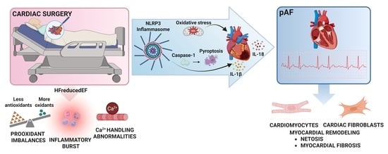
1. Introduction
2. Materials and Methods
2.1. Subjects and Clinical Follow-Up
2.2. Post-Operative AF Detection
2.3. Samples and Biopsies
2.4. Pre-Operative Echocardiographic Images
2.5. Biochemical Parameters
2.5.1. Determination of Oxidative Stress-Related Markers
Antioxidant Status
Oxidative Stress Markers
2.5.2. Immunoblot Analysis
2.5.3. RT-PCR NLRP3, IL 1-β and IL-18
2.5.4. Enzyme-Linked Immunosorbent Assay (ELISA)
2.5.5. proBNP and Troponin Levels in Plasma Samples
2.5.6. Immunolabeling and Fluorescent Microscopy
Quantitative Immunofluorescent Measurements
2.5.7. Statistical Analysis
3. Results
3.1. Clinical and Perioperative Characteristics
3.2. Antioxidants and Oxidative Stress Markers in Plasma and Atrial Tissue
3.3. Association Between LVEF and Oxidative Stress Markers
3.4. NLRP3 Inflammasome in Atrial Tissue and Pericardial Fluid
3.5. Atrial Fibrillation and Oxidative Stress Markers
3.6. Immunohistochemistry and Quantification of ROS, Nitrotyrosine, and NLRP3 in Atrial Biopsies
4. Discussion
Limitations of the Study
5. Conclusions
Supplementary Materials
Author Contributions
Funding
Institutional Review Board Statement
Informed Consent Statement
Data Availability Statement
Acknowledgments
Conflicts of Interest
Abbreviations
| ARE | antioxidants response elements |
| CPB | cardiopulmonary bypass |
| HFrEF | heart failure with reduced ejection fraction |
| HFpEF | heart failure with preserved ejection fraction |
| FRAP | ferric reducing ability of plasma |
| GSH | glutathione |
| IL-1 | interleukin-1 |
| IL-1β | interleukin-1 beta |
| IL-18 | interleukin-18 |
| Nrf2 | Nuclear factor erythroid 2-related factor 2 |
| NOO- | peroxynitrite |
| NLRP3 | NOD-, LRR- and pyrin domain-containing protein 3 |
| NADPH | nicotinamide adenine dinucleotide phosphate |
| 3-NT | 3-Nitrotyrosine |
| OS | oxidative stress |
| •OH | hydroxyl radical |
| •O2− | superoxide radical |
| pAF | postoperative atrial fibrillation |
| ROS | reactive oxygen species |
| TBARS | thiobarbituric acid reactive substances |
References
- Lazzarin, T.; Martins, D.; Ballarin, R.S.; Monte, M.G.; Minicucci, M.F.; Polegato, B.F.; Zornoff, L. The Role of Omega-3 in Attenuating Cardiac Remodeling and Heart Failure through the Oxidative Stress and Inflammation Pathways. Antioxidants 2023, 12, 2067. [Google Scholar] [CrossRef] [PubMed]
- Omote, K.; Verbrugge, F.H.; Borlaug, B.A. Heart Failure with Preserved Ejection Fraction: Mechanisms and Treatment Strategies. Annu. Rev. Med. 2022, 73, 321–337. [Google Scholar] [CrossRef] [PubMed]
- Langlois, P.L.; Hardy, G.; Manzanares, W. Omega-3 polyunsaturated fatty acids in cardiac surgery patients: An updated systematic review and meta-analysis. Clin. Nutr. 2017, 36, 737–746. [Google Scholar] [CrossRef] [PubMed]
- Chen, S.; Acou, W.J.; Kiuchi, M.G.; Meyer, C.; Sommer, P.; Martinek, M.; Schratter, A.; Andrea, B.R.; Ling, Z.; Liu, S.; et al. Association of Preoperative Renin-Angiotensin System Inhibitors with Prevention of Postoperative Atrial Fibrillation and Adverse Events: A Systematic Review and Meta-analysis. JAMA Netw. Open 2017, 5, e194934. [Google Scholar] [CrossRef] [PubMed]
- Hogue, C.W., Jr.; Creswell, L.L.; Gutterman, D.D.; Fleisher, L.A. Epidemiology, mechanisms, and risks: American College of Chest Physicians guidelines for the prevention and management of postoperative atrial fibrillation after cardiac surgery. Chest 2005, 128, 9S–16S. [Google Scholar] [CrossRef] [PubMed]
- Dobrev, D.; Aguilar, M.; Heijman, J.; Guichard, J.-B.; Nattel, S. Postoperative atrial fibrillation: Mechanisms, manifestations and management. Nat. Rev. Cardiol. 2019, 16, 417–436. [Google Scholar] [CrossRef] [PubMed]
- Goulden, C.; Hagana, A.; Ulucay, E.; Zaman, E.; Ahmed, A.; Harky, A. Optimising risk factors for atrial fibrillation post-cardiac surgery. Perfusion 2022, 37, 675–683. [Google Scholar] [CrossRef] [PubMed]
- Andrade, J.; Khairy, P.; Dobrev, D.; Nattel, S. The clinical profile and pathophysiology of atrial fibrillation: Relationships among clinical features, epidemiology, and mechanisms. Circ. Res. 2014, 114, 1453–1468. [Google Scholar] [CrossRef] [PubMed]
- Harada, M.; Van Wagoner, D.R.; Nattel, S. Role of inflammation in atrial fibrillation pathophysiology and management. Circ. J. 2015, 79, 495–502. [Google Scholar] [CrossRef] [PubMed]
- Castillo, R.L.; Farías, J.G.; Sandoval, C.; González-Candia, A.; Figueroa, E.; Quezada, M.; Cruz, G.; Llanos, P.; Jorquera, G.; Kostin, S.; et al. Role of NLRP3 Inflammasome in Heart Failure Patients Undergoing Cardiac Surgery as a Potential Determinant of Postoperative Atrial Fibrillation and Remodeling: Is SGLT2 Cotransporter Inhibition an Alternative for Cardioprotection? Antioxidants 2024, 11, 1388. [Google Scholar] [CrossRef] [PubMed]
- Smorodinova, N.; Bláha, M.; Melenovský, V.; Rozsívalová, K.; Přidal, J.; Ďurišová, M.; Pirk, J.; Kautzner, J.; Kučera, T. Analysis of immune cell populations in atrial myocardium of patients with atrial fibrillation or sinus rhythm. PLoS ONE 2017, 12, e0172691. [Google Scholar] [CrossRef] [PubMed]
- Rodrigo, R.; Korantzopoulos, P.; Cereceda, M.; Asenjo, R.; Zamorano, J.; Villalabeitia, E.; Baeza, C.; Aguayo, R.; Castillo, R.; Carrasco, R.; et al. A randomized controlled trial to prevent postoperative atrial fibrillation by antioxidant reinforcement. J. Am. Coll. Cardiol. 2013, 62, 1457–1465. [Google Scholar] [CrossRef] [PubMed]
- Gnoni, A.; Ballini, A.; Trentadue, R.; Taurin, F.; Santacroce, L.; Ferrara, P.; Massaro, F.; Brienza, N.; Massari, A.M.; Sardaro, N.; et al. Induction of mitochondrial dysfunction in patients under cardiopulmonary by-pass: Preliminary results. Eur. Rev. Med. Pharmacol. Sci. 2019, 23, 8115–8123. [Google Scholar] [CrossRef] [PubMed]
- Castillo, R.L.; Rodrigo, R.; Pérez, F.; Cereceda, M.; Asenjo, R.; Zamorano, J.; Navarrete, R.; Villalabeitia, E.; Sanz, J.; Baeza, C.; et al. Antioxidant therapy reduces oxidative and inflammatory tissue damage in patients subjected to cardiac surgery with extracorporeal circulation. Basic. Clin. Pharmacol. Toxicol. 2011, 108, 256–262. [Google Scholar] [CrossRef] [PubMed]
- Liu, C.; Huang, Z.H.; Huang, S.H.; Jou, T. Endocytosis of peroxiredoxin 1 links sterile inflammation to immunoparalysis in pediatric patients following cardiopulmonary bypass. Redox Biol. 2021, 46, 102086. [Google Scholar] [CrossRef] [PubMed]
- Korantzopoulos, P.; Letsas, K.; Fragakis, N.; Tse, G.; Liu, T. Oxidative stress and atrial fibrillation: An update. Free Radic. Res. 2018, 52, 1199–1209. [Google Scholar] [CrossRef] [PubMed]
- Sánchez, F.J.; Gonzalez, V.A.; Farrando, M.; Jayat, A.O.; Segovia-Roldan, M.; García-Mendívil, L.; Ordovás, L.; Prado, N.J.; Pueyo, E.; Diez, E.R. Atrial Dyssynchrony Measured by Strain Echocardiography as a Marker of Proarrhythmic Remodeling and Oxidative Stress in Cardiac Surgery Patients. Oxid. Med. Cell Longev. 2020, 2020, 8895078. [Google Scholar] [CrossRef] [PubMed]
- Colombo, P.; Castagna, F.; Onat, D.; Wong, K.Y.; Harxhi, A.; Hayashi, Y.; Friedman, R.A.; Pinsino, A.; Ladanyi, L.; Mebazaa, A.; et al. Experimentally Induced Peripheral Venous Congestion Exacerbates Inflammation, Oxidative Stress, and Neurohormonal and Endothelial Cell Activation in Patients with Systolic Heart Failure. J. Card. Fail. 2024, 30, 580–591. [Google Scholar] [CrossRef] [PubMed]
- Mathis, M.R.; Duggal, N.L.; Janda, J.M.; Fennema, J.L.; Yang, B.; Pagani, F.D.; Maile, M.D.; Hofer, R.E.; Jewell, E.S.; Engoren, M.C. Reduced Echocardiographic Inotropy Index after Cardiopulmonary Bypass Is Associated with Complications After Cardiac Surgery: An Institutional Outcomes Study. J. Cardiothorac. Vasc. Anesth. 2021, 35, 2732–2742. [Google Scholar] [CrossRef] [PubMed]
- Nakae, M.; Kainuma, S.; Toda, K.; Miyagawa, S.; Yoshikawa, Y.; Hata, H.; Yoshioka, D.; Kawamura, T.; Kawamura, A.; Kashiyama, N.; et al. Incidence, determinants and clinical impact of left ventricular function recovery after surgical treatments for ischaemic cardiomyopathy. Eur. J. Cardiothorac. Surg. 2021, 60, 689–696. [Google Scholar] [CrossRef] [PubMed]
- Kislitsina, O.N.; Cox, J.L.; Shah, L.J.; Malaisrie, S.C.; Kruse, J.; Liu, M.; Andrei, A.; McCarthy, P.M. Preoperative left atrial strain abnormalities are associated with the development of postoperative atrial fibrillation following isolated coronary artery bypass surgery. J. Thorac. Cardiovasc. Surg. 2020, 164, 917–924. [Google Scholar] [CrossRef] [PubMed]
- Vasan, R.S.; Urbina, E.M.; Jin, J.; Xanthakis, V. Prognostic Significance of Echocardiographic Measures of Cardiac Remodeling in the Community. Curr. Cardiol. Rep. 2021, 23, 86. [Google Scholar] [CrossRef] [PubMed]
- van Wezenbeek, J.; Canada, J.M.; Ravindra, K.; Carbone, S.; Trankle, C.R.; Kadariya, D.; Buckley, L.F.; Del Buono, M.; Billingsley, H.; Viscusi, M.; et al. Reactive Protein and N-Terminal Pro-brain Natriuretic Peptide Levels Correlate with Impaired Cardiorespiratory Fitness in Patients with Heart Failure Across a Wide Range of Ejection Fraction. Front. Cardiovasc. Med. 2018, 5, 178. [Google Scholar] [CrossRef] [PubMed]
- Kostin, S.; Krizanic, F.; Kelesidis, T.; Pagonas, N. The role of NETosis in heart failure. Heart Fail. Rev. 2024, 29, 1097–1106. [Google Scholar] [CrossRef] [PubMed]
- Hidayet, S.; Yağmur, Y.; Bayramoğlu, A.; Taşolar, M.H.; Kurtoğlu, E.; Özyalın, F. Prediction of postoperative atrial fibrillation with left atrial mechanical functions and NT-pro ANP levels after coronary artery bypass surgery: A three-dimensional echocardiography study. Echocardiography 2018, 35, 661–666. [Google Scholar] [CrossRef] [PubMed]
- Thavendiranathan, P.; Negishi, T.; Somerset, E.; Negishi, K.; Penicka, M.; Lemieux, J.; Aakhus, S.; Miyazaki, S.; Shirazi, M.; Galderisi, M.; et al. Strain-Guided Management of Potentially Cardiotoxic Cancer Therapy. J. Am. Coll. Cardiol. 2021, 77, 392–401. [Google Scholar] [CrossRef] [PubMed]
- Benzie, I.F.; Strain, J.J. The ferric reducing ability of plasma (FRAP) as a measure of ‘antioxidant power’: The FRAP assay. Anal. Biochem. 1996, 239, 70–76. [Google Scholar] [CrossRef] [PubMed]
- Ohkawa, H.; Ohishi, N.; Yagi, K. Assay for lipid peroxides in animal tissues by thiobarbituric acid reaction. Anal. Biochem. 1979, 95, 351–358. [Google Scholar] [CrossRef] [PubMed]
- Szymczyk, G.; Bełtowski, J.; Marciniak, A.; Kotarski, J. Serum isoprostanes levels in patients after abdominal hysterectomy. Rocz. Akad. Med. Bialymst. 2005, 50, 322–324. [Google Scholar]
- Gutiérrez-Camacho, L.R.; Kormanovski, A.; Castillo-Hernández, M.; Guevara-Balcázar, G.; Lara-Padilla, E. Alterations in glutathione, nitric oxide and 3-nitrotyrosine levels following exercise and/or hyperbaric oxygen treatment in mice with diet-induced diabetes. Biomed. Rep. 2020, 12, 222–232. [Google Scholar] [CrossRef] [PubMed]
- Manfrere, K.; Torrealba, M.P.; Ferreira, F.M.; Alho de Sousa, E.S.; Miyashiro, D.; Teixeira, F.M.; Custódio, R.W.; Nakaya, H.; Ramos, Y.A.; Sotto, M.N.; et al. Imbalanced IL-1B and IL-18 Expression in Sézary Syndrome. Int. J. Mol. Sci. 2023, 24, 4674. [Google Scholar] [CrossRef] [PubMed]
- Pfaffl, M.W. A new mathematical model for relative quantification in real-time RT-PCR. Nucleic Acids Res. 2001, 29, e45. [Google Scholar] [CrossRef] [PubMed]
- Atas, E.; Kismet, E.; Kesik, V.; Karaoglu, B.; Aydemir, G.; Korkmazer, N.; Demirkaya, E.; Karslioglu, Y.; Yurttutan, N.; Unay, B.; et al. Cardiac troponin-I, brain natriuretic peptide and endothelin-1 levels in a rat model of doxorubicin-induced cardiac injury. J. Cancer Res. Ther. 2015, 11, 882–886. [Google Scholar] [CrossRef] [PubMed]
- Kostin, S.; Richter, M.; Krizanic, F.; Sasko, B.; Kelesidis, T.; Pagonas, N. NETosis Is an Important Component of Chronic Myocardial Inflammation in Patients With Heart Failure. Circ. Heart Fail. 2025, 18, e012231. [Google Scholar] [CrossRef] [PubMed]
- Cabrera-Fuentes, H.A.; Ruiz-Meana, M.; Simsekyilmaz, S.; Kostin, S.; Inserte, J.; Saffarzadeh, M.; Galuska, S.P.; Vijayan, V.; Barba, I.; Barreto, G.; et al. RNase1 prevents the damaging interplay between extracellular RNA and tumour necrosis factor-α in cardiac ischaemia/reperfusion injury. Thromb. Haemost. 2014, 112, 1110–1119. [Google Scholar] [CrossRef] [PubMed]
- Abassi, Z.A.; Barac, Y.D.; Kostin, S.; Roguin, A.; Ovcharenko, E.; Awad, H.; Blank, A.; Bar-Am, O.; Amit, T.; Schaper, J.; et al. TVP1022 attenuates cardiac remodeling and kidney dysfunction in experimental volume overload-induced congestive heart failure. Circ. Heart Fail. 2011, 4, 463–473. [Google Scholar] [CrossRef] [PubMed]
- Bricker-Anthony, C.; Rex, T.S. Neurodegeneration and Vision Loss after Mild Blunt Trauma in the C57Bl/6 and DBA/2J Mouse. PLoS ONE 2015, 10, e0131921. [Google Scholar] [CrossRef] [PubMed]
- Noubiap, J.J.; Sanders, P.; Nattel, S.; Lau, D.H. Biomarkers in Atrial Fibrillation: Pathogenesis and Clinical Implications. Card. Electrophysiol. Clin. 2021, 13, 221–233. [Google Scholar] [CrossRef] [PubMed]
- Simonato, M.; Baritussio, A.; Carnielli, V.P.; Vedovelli, L.; Falasco, G.; Salvagno, M.; Padalino, M.; Cogo, P. Influence of the type of congenital heart defects on epithelial lining fluid composition in infants undergoing cardiac surgery with cardiopulmonary bypass. Pediatr. Res. 2018, 83, 791–797. [Google Scholar] [CrossRef] [PubMed]
- Wu, M.Y.; Yiang, G.T.; Liao, W.T.; Tsai, A.P.; Cheng, Y.; Cheng, P.; Li, C.; Li, C. Current Mechanistic Concepts in Ischemia and Reperfusion Injury. Cell Physiol. Biochem. 2018, 46, 1650–1667. [Google Scholar] [CrossRef] [PubMed]
- Farías, J.G.; Herrera, E.A.; Carrasco-Pozo, C.; Sotomayor-Zárate, R.; Cruz, G.; Morales, P.; Castillo, R.L. Pharmacological models and approaches for pathophysiological conditions associated with hypoxia and oxidative stress. Pharmacol. Ther. 2016, 158, 1–23. [Google Scholar] [CrossRef] [PubMed]
- Bai, J.; Lyden, P.D. Revisiting cerebral postischemic reperfusion injury: New insights in understanding reperfusion failure, hemorrhage, and edema. Int. J. Stroke 2015, 10, 143–152. [Google Scholar] [CrossRef] [PubMed]
- Himmetoglu, S.; Dincer, Y.; Bozcali, E.; Vural, V.A.; Akcay, T. Oxidative DNA damage and antioxidant defense after reperfusion in acute myocardial infarction. J. Investig. Med. 2009, 57, 595–599. [Google Scholar] [CrossRef] [PubMed]
- Schipper, D.A.; Marsh, K.M.; Ferng, A.S.; Duncker, D.J.; Laman, J.D.; Khalpey, Z. The Critical Role of Bioenergetics in Donor Cardiac Allograft Preservation. J. Cardiovasc. Transl. Res. 2016, 9, 176–183. [Google Scholar] [CrossRef] [PubMed]
- Nakamura, K.; Miura, D.; Kusano, K.F.; Fujimoto, Y.; Sumita-Yoshikawa, W.; Fuke, S.; Nishii, N.; Nagase, S.; Hata, Y.; Morita, H.; et al. 4-Hydroxy-2-nonenal induces calcium overload via the generation of reactive oxygen species in isolated rat cardiac myocytes. J. Card. Fail. 2009, 15, 709–716. [Google Scholar] [CrossRef] [PubMed]
- Yu, Y.; Yan, Y.; Niu, F.; Wang, Y.; Chen, X.; Su, G.; Liu, Y.; Zhao, X.; Qian, L.; Liu, P.; et al. Ferroptosis: A cell death connecting oxidative stress, inflammation and cardiovascular diseases. Cell Death Discov. 2021, 7, 193. [Google Scholar] [CrossRef] [PubMed]
- Duez, H.; Pourcet, B. Nuclear Receptors in the Control of the NLRP3 Inflammasome Pathway. Front. Endocrinol. 2021, 12, 630536. [Google Scholar] [CrossRef] [PubMed]
- Gunata, M.; Parlakpinar, H. A review of myocardial ischaemia/reperfusion injury: Pathophysiology, experimental models, biomarkers, genetics and pharmacological treatment. Cell Biochem. Funct. 2021, 39, 190–217. [Google Scholar] [CrossRef] [PubMed]
- Mora-Ruíz, M.D.; Blanco-Favela, F.; Rueda, A.K.; Legorreta-Haquet, M.V.; Chávez-Sánchez, L. Role of interleukin-17 in acute myocardial infarction. Mol. Immunol. 2019, 107, 71–78. [Google Scholar] [CrossRef] [PubMed]
- Baci, D.; Bosi, A.; Parisi, L.; Buono, G.; Mortara, L.; Ambrosio, G.; Bruno, A. Innate Immunity Effector Cells as Inflammatory Drivers of Cardiac Fibrosis. Int. J. Mol. Sci. 2020, 28, 7165. [Google Scholar] [CrossRef] [PubMed]
- Nattel, S.; Heijman, J.; Zhou, L.; Dobrev, D. Molecular Basis of Atrial Fibrillation Pathophysiology and Therapy: A Translational Perspective. Circ. Res. 2020, 127, 51–72. [Google Scholar] [CrossRef] [PubMed]
- Kobara, M.; Naseratun, N.; Toba, H.; Nakata, T. Preconditioning with Short-term Dietary Restriction Attenuates Cardiac Oxidative Stress and Hypertrophy Induced by Chronic Pressure Overload. Nutrients 2021, 13, 737. [Google Scholar] [CrossRef] [PubMed]
- Radovits, T.; Korkmaz, S.; Mátyás, C.; Oláh, A.; Németh, T.B.; Páli, S.; Hirschberg, K.; Zubarevich, A.; Gwanmesia, P.N.; Li, S.; et al. An altered pattern of myocardial histopathological and molecular changes underlies the different characteristics of type-1 and type-2 diabetic cardiac dysfunction. J. Diabetes Res. 2015, 2015, 728741. [Google Scholar] [CrossRef] [PubMed]
- Sultan, A.; Singh, J.; Howarth, F.C. Mechanisms underlying electro-mechanical dysfunction in the Zucker diabetic fatty rat heart: A model of obesity and type 2 diabetes. Heart Fail. Rev. 2020, 25, 873–886. [Google Scholar] [CrossRef] [PubMed]
- Castillo, R.L.; Arias, C.A.; Farías, J.G. Omega 3 chronic supplementation attenuates myocardial ischaemia-reperfusion injury through reinforcement of antioxidant defense system in rats. Cell Biochem. Funct. 2014, 32, 274–281. [Google Scholar] [CrossRef] [PubMed]
- Oliveira, M.S.; Tanaka, L.Y.; Antonio, E.L.; Brandizzi, L.I.; Serra, A.J.; Dos Santos, L.; Krieger, J.E.; Laurindo, F.R.; Tucci, P. Hyperbaric oxygenation improves redox control and reduces mortality in the acute phase of myocardial infarction in a rat model. Mol. Med. Rep. 2020, 21, 1431–1438. [Google Scholar] [CrossRef] [PubMed]
- Xu, Y.; Guo, W.; Zeng, D.; Fang, Y.; Wang, R.; Guo, D.; Qi, B.; Xue, Y.; Xue, F.; Jin, Z.; et al. Inhibiting miR-205 Alleviates Cardiac Ischemia/Reperfusion Injury by Regulating Oxidative Stress, Mitochondrial Function, and Apoptosis. Oxid. Med. Cell Longev. 2021, 2021, 9986506. [Google Scholar] [CrossRef] [PubMed]
- Ndrepepa, G. Myeloperoxidase—A bridge linking inflammation and oxidative stress with cardiovascular disease. Clin. Chim. Acta 2019, 493, 36–51. [Google Scholar] [CrossRef] [PubMed]
- Farías, J.G.; Molina, V.M.; Carrasco, R.; Zepeda, A.B.; Figueroa, E.; Letelier, P.; Castillo, R.L. Antioxidant Therapeutic Strategies for Cardiovascular Conditions Associated with Oxidative Stress. Nutrients 2017, 9, 966. [Google Scholar] [CrossRef] [PubMed]
- Chen, M.; Li, X.; Mu, G. Myocardial protective and anti-inflammatory effects of dexmedetomidine in patients undergoing cardiovascular surgery with cardiopulmonary bypass: A systematic review and meta-analysis. J. Anesth. 2022, 36, 5–16. [Google Scholar] [CrossRef] [PubMed]
- Antonic, M.; Lipovec, R.; Gregorcic, F.; Juric, P.; Kosir, G. Perioperative ascorbic acid supplementation does not reduce the incidence of postoperative atrial fibrillation in on-pump coronary artery bypass graft patients. J. Cardiol. 2017, 69, 98–102. [Google Scholar] [CrossRef] [PubMed]
- Del Campo, A.; Perez, G.; Castro, P.F.; Parra, V.; Verdejo, H.E. Mitochondrial function, dynamics and quality control in the pathophysiology of HFpEF. Biochim. Biophys. Acta Mol. Basis Dis. 2021, 1867, 166208. [Google Scholar] [CrossRef] [PubMed]
- Bayes-Genis, A.; Bisbal, F.; Núñez, J.; Santas, E.; Lupón, J.; Rossignol, P.; Paulus, W. Transitioning from Preclinical to Clinical Heart Failure with Preserved Ejection Fraction: A Mechanistic Approach. J. Clin. Med. 2020, 9, 1110. [Google Scholar] [CrossRef] [PubMed]
- Petersen, F.; Rodrigo, R.; Richter, M.; Kostin, S. The effects of polyunsaturated fatty acids and antioxidant vitamins on atrial oxidative stress, nitrotyrosine residues, and connexins following extracorporeal circulation in patients undergoing cardiac surgery. Mol. Cell Biochem. 2017, 433, 27–40. [Google Scholar] [CrossRef] [PubMed]
- Matata, B.M.; Elahi, M.M. In Situ Oxidative Stress and Atrial Cell Deaths in Patients with Valve Disease. Cardiovasc. Hematol. Disord. Drug Targets 2019, 19, 79–87. [Google Scholar] [CrossRef] [PubMed]
- Abuelgasim, E.; Shah, S.; Abuelgasim, B. Clinical overview of diabetes mellitus as a risk factor for cardiovascular death. Rev. Cardiovasc. Med. 2021, 22, 301–314. [Google Scholar] [CrossRef] [PubMed]
- Kim, H.M.; Cho, G.; Hwang, C.; Choi, H.; Park, J.-B.; Yoon, Y.E.; Kim, H. Myocardial Strain in Prediction of Outcomes After Surgery for Severe Mitral Regurgitation. JACC Cardiovasc. Imaging 2018, 11, 1235–1244. [Google Scholar] [CrossRef] [PubMed]
- Ahmed, M.I.; Gladden, J.D.; Litovsky, S.H.; Lloyd, S.G.; Gupta, H.; Inusah, S.; Denney Jr, T.; Powell, P.; McGiffin, D.C.; Dell’Italia, L.J. Increased oxidative stress and cardiomyocyte myofibrillar degeneration in patients with chronic isolated mitral regurgitation and ejection fraction >60%. J. Am. Coll. Cardiol. 2010, 55, 671–679. [Google Scholar] [CrossRef] [PubMed]
- Dhalla, N.S.; Elimban, V.; Bartekova, M.; Adameova, A. Involvement of Oxidative Stress in the Development of Subcellular Defects and Heart Disease. Biomedicines 2022, 10, 393. [Google Scholar] [CrossRef] [PubMed]
- Abbate, A.; Toldo, S.; Marchetti, C.; Kron, J.; Van Tassell, B.W.; Dinarello, C. Interleukin-1 and the Inflammasome as Therapeutic Targets in Cardiovascular Disease. Circ. Res. 2020, 126, 1260–1280. [Google Scholar] [CrossRef] [PubMed]
- Potere, N.; Bonaventura, A.; Abbate, A. Novel Therapeutics and Upcoming Clinical Trials Targeting Inflammation in Cardiovascular Diseases. Arterioscler. Thromb. Vasc. Biol. 2024, 44, 2371–2395. [Google Scholar] [CrossRef] [PubMed]
- Ridker, P.M.; Everett, B.M.; Thuren, T.; MacFadyen, J.G.; Chang, W.H.; Ballantyne, C.; Fonseca, F.; Nicolau, J.; Koenig, W.; Anker, S.D.; et al. Antiinflammatory therapy with canakinumab for atherosclerotic disease. N. Engl. J. Med. 2017, 377, 1119–1131. [Google Scholar] [CrossRef] [PubMed]
- Van Tassell, B.W.; Michael, J.; Appleton, L.D.; Roberts, C.S.; Kontos, M.C.; Abouzaki, N.; Melchior, R.; Mueller, G.; Garnett, J.; Canada, J.; et al. Rationale and design of the Virginia Commonwealth University-Anakinra Remodeling Trial-3 (VCU-ART3): A randomized, placebo-controlled, double-blinded, multicenter study. Clin. Cardiol. 2018, 41, 1004–1008. [Google Scholar] [CrossRef] [PubMed]
- Yao, C.; Veleva, T.; Scott, L., Jr.; Cao, S.; Li, L.; Chen, G.; Jeyabal, P.; Pan, X.; Alsina, K.M.; Abu-Taha, I.; et al. Enhanced Cardiomyocyte NLRP3 Inflammasome Signaling Promotes Atrial Fibrillation. Circulation 2018, 138, 2227–2242. [Google Scholar] [CrossRef] [PubMed]
- Xing, Y.; Yan, L.; Li, X.; Xu, Z.; Wu, X.; Gao, H.; Chen, Y.; Ma, X.; Liu, J.; Zhang, J. The relationship between atrial fibrillation and NLRP3 inflammasome: A gut microbiota perspective. Front. Immunol. 2023, 14, 1273524. [Google Scholar] [CrossRef] [PubMed]
- Zhang, B.; Hou, J.; Liu, J.; He, J.; Gao, Y.; Li, G.; Ma, T.; Lv, X.; Dong, L.; Yang, W.; et al. Hydrogen decreases susceptibility to AngII-induced atrial fibrillation and atrial fibrosis via the NOX4/ROS/NLRP3 and TGF-β1/Smad2/3 signaling pathways. PLoS ONE 2025, 20, e0310852. [Google Scholar] [CrossRef] [PubMed]
- Narendran, S.; Pereira, F.; Ambati, J. NLRP3 Inflammasome Inhibition: A Potential Therapeutic Strategy to Attenuate Postinfarction Adverse Cardiac Remodeling. JACC Basic Transl. Sci. 2020, 5, 1225–1227. [Google Scholar] [CrossRef] [PubMed]
- Wei, Z.; Fei, Y.; Wang, Q.; Hou, J.; Cai, X.; Yang, Y.; Chen, T.; Xu, Q.; Wang, Y.; Li, Y. Loss of Camk2n1 aggravates cardiac remodeling and malignant ventricular arrhythmia after myocardial infarction in mice via NLRP3 inflammasome activation. Free Radic. Biol. Med. 2021, 167, 243–257. [Google Scholar] [CrossRef] [PubMed]
- Cheng, X.; Zhao, H.; Wen, X.; Li, G.; Guo, S.; Zhang, D. NLRP3-inflammasome inhibition by MCC950 attenuates cardiac and pulmonary artery remodelling in heart failure with preserved ejection fraction. Life Sci. 2023, 333, 122185. [Google Scholar] [CrossRef] [PubMed]
- Sadeghi, M.; Khosrawi, S.; Heshmat-Ghahdarijani, K.; Gheisari, Y.; Roohafza, H.; Mansoorian, M.; Hoseini, S. Effect of melatonin on heart failure: Design for a double-blinded randomized clinical trial. ESC Heart Fail. 2020, 7, 3142–3150. [Google Scholar] [CrossRef] [PubMed]
- Hoseini, S.G.; Heshmat-Ghahdarijani, K.; Khosrawi, S.; Garakyaraghi, M.; Shafie, D.; Roohafza, H.; Mansourian, M.; Azizi, E.; Gheisari, Y.; Sadeghi, M. Effect of melatonin supplementation on endothelial function in heart failure with reduced ejection fraction: A randomized, double-blinded clinical trial. Clin. Cardiol. 2021, 44, 1263–1271. [Google Scholar] [CrossRef] [PubMed]
- Vashi, R.; Patel, B.M. NRF2 in Cardiovascular Diseases: A Ray of Hope. J. Cardiovasc. Transl. Res. 2021, 14, 573–586. [Google Scholar] [CrossRef] [PubMed]
- Quiles, J.M.; Pepin, M.E.; Sunny, S.; Shela, S.B.; Challa, A.K.; Dalley, B.; Hoidal, J.R.; Pogwizd, S.M.; Wende, A.R.; Rajasekaran, N.S. Identification of Nrf2-responsive microRNA networks as putative mediators of myocardial reductive stress. Sci. Rep. 2021, 11, 11977. [Google Scholar] [CrossRef] [PubMed]
- Sairam, T.; Patel, A.N.; Subrahmanian, M.; Gopalan, R.; Pogwizd, S.M.; Ramalingam, S.; Sankaran, R.; Rajasekaran, N.S. Evidence for a hyper-reductive redox in a sub-set of heart failure patients. J. Transl. Med. 2018, 16, 130. [Google Scholar] [CrossRef] [PubMed]
- Carnes, C.A.; Janssen, P.M.; Ruehr, M.L.; Nakayama, H.; Nakayama, T.; Haase, H.; Bauer, J.A.; Chung, M.K.; Fearon, I.M.; Gillinov, A.M.; et al. Atrial glutathione content, calcium current, and contractility. J. Biol. Chem. 2007, 282, 28063–28073. [Google Scholar] [CrossRef] [PubMed]
- Rennison, J.H.; Li, L.; Lin, C.R.; Lovano, B.S.; Castel, L.; Wass, S.Y.; Cantlay, C.C.; McHale, M.; Gillinov, A.M.; Mehra, R.; et al. Atrial fibrillation rhythm is associated with marked changes in metabolic and myofibrillar protein expression in left atrial appendage. Pflugers Arch. 2021, 473, 461–475. [Google Scholar] [CrossRef] [PubMed]
- Carnes, C.A.; Chung, M.K.; Nakayama, T.; Nakayama, H.; Baliga, R.S.; Piao, S.; Kanderian, A.; Pavia, S.; Hamlin, R.L.; McCarthy, P.M.; et al. Ascorbate attenuates atrial pacing-induced peroxynitrite formation and electrical remodeling and decreases the incidence of postoperative atrial fibrillation. Circ. Res. 2001, 89, E32–E38. [Google Scholar] [CrossRef] [PubMed]
- Antoniades, C.; Demosthenous, M.; Reilly, S.; Margaritis, M.; Zhang, M.-H.; Antonopoulos, A.; Marinou, K.; Nahar, K.; Jayaram, R.; Tousoulis, D.; et al. Myocardial redox state predicts in-hospital clinical outcome after cardiac surgery effects of short-term pre-operative statin treatment. J. Am. Coll. Cardiol. 2012, 59, 60–70. [Google Scholar] [CrossRef] [PubMed]
- Winter, J.L.; Castro, P.F.; Quintana, J.C.; Altamirano, R.; Enriquez, A.; Verdejo, H.E.; Jalil, J.E.; Mellado, R.; Concepción, R.; Sepúlveda, P.; et al. Effects of trimetazidine in nonischemic heart failure: A randomized study. J. Card. Fail. 2014, 20, 149–154. [Google Scholar] [CrossRef] [PubMed]
- Ocaranza, M.P.; Moya, J.; Jalil, J.E.; Lavandero, S.; Kalergis, A.M.; Molina, C.; Gabrielli, L.; Godoy, I.; Córdova, S.; Castro, P.; et al. Rho-kinase pathway activation and apoptosis in circulating leucocytes in patients with heart failure with reduced ejection fraction. J. Cell Mol. Med. 2020, 24, 1413–1427. [Google Scholar] [CrossRef] [PubMed]
- Ternacle, J.; Berry, M.; Alonso, E.; Kloeckner, M.; Couetil, J.-P.; Randé, J.-L.; Gueret, P.; Monin, J.-L.; Lim, P. Incremental value of global longitudinal strain for predicting early outcome after cardiac surgery. Eur. Heart J. Cardiovasc. Imaging 2013, 14, 77–84. [Google Scholar] [CrossRef] [PubMed]
- Ikeda, M.; Niinami, H.; Morita, K.; Saito, S.; Yoshitake, A. Long-term results following off-pump coronary-artery bypass grafting in left ventricular dysfunction. Heart Vessels 2024, 39, 571–581. [Google Scholar] [CrossRef] [PubMed]
- Erturk, O.; Keles, N.; Karaagac, A.; Arslanhan, A.S.; Pocan, Y.K.; Yesilkaya, M.I.; Bozkurt, B.; Aydogan, H.; Kaplan, M. Comparison of early postoperative left ventricular function with 3d ef and strain measurements according to graft selection. J. Cardiothorac. Surg. 2024, 19, 615. [Google Scholar] [CrossRef] [PubMed]
- Silva, R.R.M.; Hueb, W.; Lima, E.G.; Rezende, P.C.; Soares, P.R.; Ramires, J.A.; Filho, R.K. Long-term analysis of ventricular function in patients with symptomatic coronary disease who underwent on-pump or off-pump coronary artery bypass grafting. J. Cardiothorac. Surg. 2022, 17, 326. [Google Scholar] [CrossRef] [PubMed]
- Downey, M.C.; Hooks, M.; Gravely, A.; Naksuk, N.; Buelt-Gebhardt, M.; Carlson, S.; Tholakanahalli, V.; Adabag, S. Perioperative changes in left ventricular systolic function following surgical revascularization. PLoS ONE 2022, 17, e0277454. [Google Scholar] [CrossRef] [PubMed]
- Olsen, F.J.; Lindberg, S.; Pedersen, S.; Iversen, A.; Davidovski, F.S.; Galatius, S.; Fritz-Hansen, T.; Gislason, G.H.; Søgaard, P.; Møgelvang, R.; et al. Global longitudinal strain predicts cardiovascular events after coronary artery bypass grafting. Heart 2021, 107, 814–821. [Google Scholar] [CrossRef] [PubMed]
- Wakefield, B.J.; Artis, A.S.; Alfirevic, A.; Sale, S.; Duncan, A.E. Post-cardiopulmonary bypass longitudinal strain provides higher prognostic ability than baseline strain or change in strain. Ann. Card. Anaesth. 2022, 25, 505–513. [Google Scholar] [CrossRef] [PubMed]


| Clinical Parameters | rLVEF (n = 25) | pLVEF (n = 27) | p-Value |
|---|---|---|---|
| Age | 57.6 (56–61) | 64.7 (58–67) | 0.63 |
| Sex (M/F) | 15/10 | 18/9 | 0.47 |
| BMI | 29.8 (25–33) | 31.3 (26–35) | 0.45 |
| Comorbidities | |||
| Essential hypertension | 12 | 14 | 0.66 |
| D. Mellitus | 13 | 15 | 0.78 |
| Chronic pulmonary disease | 12 | 11 | 0.88 |
| Hypercholesterolemia | 12 | 12 | 0.75 |
| Smoking history | 11 | 12 | 1.00 |
| Pharmacotherapy | |||
| Aspirin | 12 | 14 | 0.85 |
| Statins | 21 | 21 | 0.71 |
| Diuretics | 1 | -- | -- |
| Beta-blockers | 14 | 16 | 0.69 |
| Nitrates | 12 | 11 | 0.77 |
| ACEI/ARB | 14 | 16 | 0.78 |
| Sulfonylureas | 11 | 13 | 0.49 |
| Biguanides | 13 | 14 | 0.33 |
| Gliflozins | 7 | 8 | 0.78 |
| M. injury markers | |||
| hs-cTn (ng/L) | 13.25 ± 7.4 | 7.17 ± 2.8 | 0.46 |
| BNP (ng/mL) | 145 ± 27.6 | 98 ± 16.8 | 0.77 |
| Echocardiographical parameters | |||
| LVEF (%) | 35.3 ± 7.1 | 55.7 ± 8.4 * | 0.04 |
| LVGS (%) | −21% | −18.8 | 0.66 |
| LA volumen index (mL/m2) | 17.4 ± 2.3 * | 12.5 ± 1.5 | 0.04 |
| LV end-systolic | |||
| dimension (mm) | 30.1 ± 1.8 | 29.3 ± 1.9 | 0.07 |
| LV end-diastolic | 41.1 ± 1.3 | 43.7 ± 2.1 | 0.68 |
| dimension (mm) | |||
| LV mass index (g/m2) | 116.3 ± 7.8 * | 85.7 ± 2.5 | 0.03 |
| E/A ratio | 1.6 ± 0.05 | 1.4 ± 0.04 | 0.22 |
| E/e’ ratio | 6.0 ± 0.95 | 6.3 ± 0.77 | 0.45 |
Disclaimer/Publisher’s Note: The statements, opinions and data contained in all publications are solely those of the individual author(s) and contributor(s) and not of MDPI and/or the editor(s). MDPI and/or the editor(s) disclaim responsibility for any injury to people or property resulting from any ideas, methods, instructions or products referred to in the content. |
© 2025 by the authors. Licensee MDPI, Basel, Switzerland. This article is an open access article distributed under the terms and conditions of the Creative Commons Attribution (CC BY) license (https://creativecommons.org/licenses/by/4.0/).
Share and Cite
Castillo, R.L.; Carrasco, R.A.; Gonzaléz-Candia, A.; Figueroa, E.G.; Paz, A.A.; Candia, A.A.; Kostin, S.; Pagonas, N.; Arias, P.V.; Herrera, E.A.; et al. Oxidative Stress and NLRP3 Inflammasome as Markers of Cardiac Injury Following Cardiopulmonary Bypass: Potential Implications for Patients with Preoperative Heart Failure with Reduced Ejection Fraction. Antioxidants 2025, 14, 1311. https://doi.org/10.3390/antiox14111311
Castillo RL, Carrasco RA, Gonzaléz-Candia A, Figueroa EG, Paz AA, Candia AA, Kostin S, Pagonas N, Arias PV, Herrera EA, et al. Oxidative Stress and NLRP3 Inflammasome as Markers of Cardiac Injury Following Cardiopulmonary Bypass: Potential Implications for Patients with Preoperative Heart Failure with Reduced Ejection Fraction. Antioxidants. 2025; 14(11):1311. https://doi.org/10.3390/antiox14111311
Chicago/Turabian StyleCastillo, Rodrigo L., Rodrigo A. Carrasco, Alejandro Gonzaléz-Candia, Esteban G. Figueroa, Adolfo A. Paz, Alejandro A. Candia, Sawa Kostin, Nikolaos Pagonas, Pamela V. Arias, Emilio A. Herrera, and et al. 2025. "Oxidative Stress and NLRP3 Inflammasome as Markers of Cardiac Injury Following Cardiopulmonary Bypass: Potential Implications for Patients with Preoperative Heart Failure with Reduced Ejection Fraction" Antioxidants 14, no. 11: 1311. https://doi.org/10.3390/antiox14111311
APA StyleCastillo, R. L., Carrasco, R. A., Gonzaléz-Candia, A., Figueroa, E. G., Paz, A. A., Candia, A. A., Kostin, S., Pagonas, N., Arias, P. V., Herrera, E. A., Pérez, R. A., & Iturra, S. (2025). Oxidative Stress and NLRP3 Inflammasome as Markers of Cardiac Injury Following Cardiopulmonary Bypass: Potential Implications for Patients with Preoperative Heart Failure with Reduced Ejection Fraction. Antioxidants, 14(11), 1311. https://doi.org/10.3390/antiox14111311