


Biomolecules, Volume 15, Issue 5 (May 2025) – 143 articles
Cover Story (view full-size image):
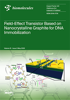
Owing to global health challenges posed by the recent COVID-19 pandemic and ESKAPE organisms, lab-on-a-chip and point-of-care devices are becoming essential tools for maintaining public health. Novel nanocrystalline graphite (NCG)-based field effect transistors with DNA probes as receptors become indispensable tools in modern approaches for disease control and prevention. As the first of its kind, we develop an experimental approach that focuses on understanding the interaction and influence of DNA samples with nanocrystalline graphite, a novel 3D nanocarbon material. Our experimental results suggest that the DNA nucleobase contributes contrastingly to the detection sensitivity of NCG-FETs. These differences in interaction, observed through changes in the Dirac point, transconductance, and Raman spectra, underline the ability of NCG-FETs to differentiate between nucleobases. View this paper
- Issues are regarded as officially published after their release is announced to the table of contents alert mailing list.
- You may sign up for e-mail alerts to receive table of contents of newly released issues.
- PDF is the official format for papers published in both, html and pdf forms. To view the papers in pdf format, click on the "PDF Full-text" link, and use the free Adobe Reader to open them.
Previous Issue
Next Issue