


Biosensors, Volume 15, Issue 5 (May 2025) – 69 articles
Cover Story (view full-size image):
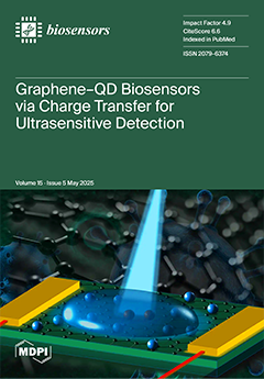
We present a graphene–quantum dot (QD) hybrid biosensor that achieves femtomolar sensitivity through a charge transfer-based quenching and recovery mechanism. Using single-layer graphene field-effect transistors (SLG-FETs) and time-resolved photoluminescence (TRPL), we demonstrate that photoluminescence quenching in QD–graphene hybrids results from static charge transfer rather than energy transfer. A simple analytical quantum mechanical model supports this interpretation. Electrical and optical signals show correlated responses to analyte concentration, enabling dual-mode detection. We validated the platform for biotin–streptavidin and IgG–anti-IgG interactions, achieving limits of detection down to 0.1 fM. These results establish a robust framework for next-generation, high-specificity biosensors. View this paper
- Issues are regarded as officially published after their release is announced to the table of contents alert mailing list.
- You may sign up for e-mail alerts to receive table of contents of newly released issues.
- PDF is the official format for papers published in both, html and pdf forms. To view the papers in pdf format, click on the "PDF Full-text" link, and use the free Adobe Reader to open them.
Previous Issue
Next Issue