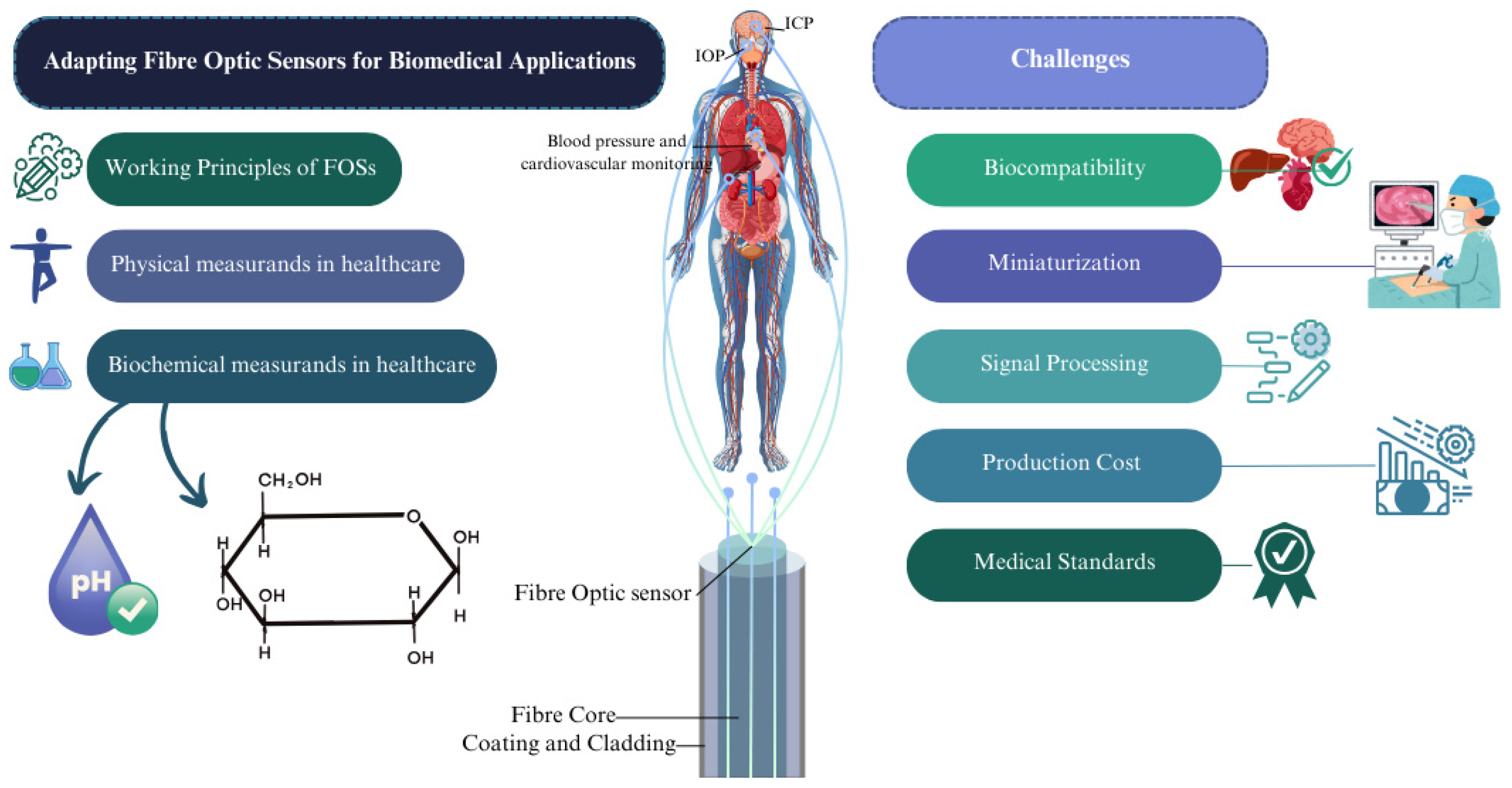
Challenges in Adapting Fibre Optic Sensors for Biomedical Applications

 ,
,  ,
,  ,
,  , , and
Abstract
1. Introduction
1.1. Background
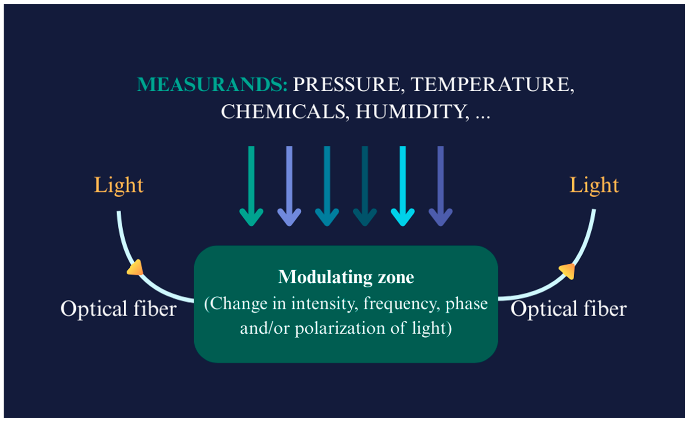
1.2. Working Principles of FOSs

| Fibre Type | Application | Sensitivity | Sensing Mechanism | Ref. |
|---|---|---|---|---|
| SMF | Pressure | 263.15 pm/kPa | FPI | [41] |
| OF | Pressure (IOP) | Low baseline drift (<2.8 mmHg) over >4.5 years | FPI with OCT | [34] |
| MMF | Pressure | 2.49 nm/kPa | Interference-based sensing | [42] |
| OF | Pressure/temperature | 55.468 nm/MPa (pressure), 0.01859 nm/°C (temperature) | FPI with MEMs | [43] |
| U-shaped MMF | Biosensing | 1251.44 nm/RIU | LSPR | [44] |
| PC fibre | Biosensing | 12,000 nm/RIU and 16,000 nm/RIU | SPR | [45] |
| D-shaped OF | Biosensing | 5161 nm/RIU | SPR | [46] |
| D-shaped OF | Biosensing | 4122 nm/RIU | LMR | [47] |
| D-shaped PC fibre | Biosensing | 21,700 nm/RIU | SPR | [48] |
| D-shaped PC fibre | Biosensing | 20,000 nm/RIU | SPR | [49] |
| Plastic OF | Cholesterol detection | 140 mg/dL to 250 nm/dL | - | [50] |
| SMF | Temperature | 210.25 KHz/°C | Vernier effect | [51] |
| Fibre tip integrated ZnO-nanowire-nanograting | Temperature | 0.066 nW/°C | Bragg reflection | [52] |
| MMF with spherical end | Pressure/temperature | 0.139 mV/kPa (pressure), 0.87 mV/°C (temperature) | RI modulation using MEMS-based silicon | [53] |
| SMF with a Hollow Silica Tube (HST) | Pressure | 396 pm/kPa | FPI | [54] |
| SMF with FBG | Pressure | 1.466 pm/kPa | FBG array | [55] |
| Ultra-miniature fibre optic sensor | Pressure (IPP) | (r ≥ 0.7, p < 0.001) | Diaphragm-based FO integrated with a proportional–integral–derivative (PID) | [56] |
| Distributed OF | Pressure | 65.920 μϵ/kPa | Axial strain change detection with a sensitizing structure | [57] |
1.3. Physical Measurands in Healthcare
1.4. Biochemical Measurands in Healthcare
2. Challenges for FOSs in Biomedical Applications
2.1. Biocompatibility
2.2. Miniaturization, Durability, and Longevity
2.3. Signal Processing, Data Integration, and Interoperability
2.4. Production Cost and Manufacturing
2.5. Medical Standards and Regulatory Approval
2.6. Ethical Considerations
3. Future Perspectives
4. Conclusions
Funding
Acknowledgments
Conflicts of Interest
Abbreviations
| FOSs | Fibre Optic Sensors (FOSs) |
| POSs | Polymer-based optical sensors |
| OF | Optical Fibre |
| SPR | Surface Plasmon Resonance |
| POFs | Polymer Optical Fibres |
| FBGs | Fibre Bragg Gratings |
| OCT | Optical Coherence Tomography |
| IOP | Intraocular Pressure |
| PCF | Photonic Crystal Fibre |
| SMF | Single-mode Fibre |
| FPI | Fabry–Pérot Interferometer |
| MMF | Multi-mode Fibre |
| MEMs | Micro-Electromechanical Systems |
| LSPR | Localized Surface Plasmon Resonance |
| LMR | Lossy Mode Resonance |
| HST | Hollow Silica Tube |
| PID | Proportional Integral Derivative |
| PANi | Polyaniline |
| TFBG | Tilted Fibre Bragg Grating |
| PAAm | Polyacrylamide |
| GO | Graphene Oxide |
| GOD | Glucose Oxidase |
| LPFG | Long-period Fibre Grating |
| TOFI | Tapered Optical Fibre Interferometer |
| 3-APBA | 3-Aminophenylboronic Acid |
| LDOF | Lossy Dielectric Optical Fibre |
| HBF | High-birefringence fibre |
| PLA | Polylactic acid |
| FDA | Food and Drug Administration |
| PEG | Polyethylene Glycol |
| POC | Poly (Octamethylene Citrate) |
| POMC | Poly (Octamethylene Maleate Citrate) |
| PVC | Polyvinyl Chloride |
| SU-8 | Negative Photoresist Polymer |
| PLLA | Poly (L-Lactic Acid) |
| PDLLA | Poly (D, L-Lactic Acid) |
| PLGA | Poly (L-Lactic-Co-Glycolic Acid) |
| PDLGA | Poly (D, L-Lactic-Co-Glycolic Acid) |
| PCL | Poly (ε-Caprolactone) |
| PGs | Phosphate Glass |
| PDMS | Polydimethylsiloxane |
| PAA | Polyacrylic Acid |
| AG | Agarose Hydrogel |
| AuNPs | Gold Nanoparticles |
| MRI | Magnetic Resonance Imaging |
| CT | Computed Tomography |
| EHRs | Electronic Health Records |
| NCAs | National Competent Authorities |
| ANSI | American National Standards Institute |
| AAMI | Association for the Advancement of Medical Instrumentation |
| TIR | Technical Information Report |
| MDR | Medical Devices Regulation |
| IVDR | In Vitro Diagnostic Regulation |
References
- Abdhul Rahuman, M.A.; Kahatapitiya, N.S.; Amarakoon, V.N.; Wijenayake, U.; Silva, B.N.; Jeon, M.; Kim, J.; Ravichandran, N.K.; Wijesinghe, R.E. Recent Technological Progress of Fiber-Optical Sensors for Bio-Mechatronics Applications. Technologies 2023, 11, 157. [Google Scholar] [CrossRef]
- Roriz, P.; Frazão, O.; Lobo-Ribeiro, A.B.; Santos, J.L.; Simões, J.A. Review of fiber-optic pressure sensors for biomedical and biomechanical applications. J. Biomed. Opt. 2013, 18, 050903. [Google Scholar] [CrossRef]
- Ngiejungbwen, L.A.; Hamdaoui, H.; Chen, M.Y. Polymer optical fiber and fiber Bragg grating sensors for biomedical engineering Applications: A comprehensive review. Opt. Laser Technol. 2024, 170, 110187. [Google Scholar] [CrossRef]
- Bartnik, K.; Koba, M.; Śmietana, M. Advancements in optical fiber sensors for in vivo applications—A review of sensors tested on living organisms. Measurement 2024, 224, 113818. [Google Scholar] [CrossRef]
- Padha, B.; Yadav, I.; Dutta, S.; Arya, S. Recent Developments in Wearable NEMS/MEMS-Based Smart Infrared Sensors for Healthcare Applications. ACS Appl. Electron. Mater. 2023, 5, 5386–5411. [Google Scholar] [CrossRef]
- Yu, L.; Kim, B.J.; Meng, E. Chronically implanted pressure sensors: Challenges and state of the field. Sensors 2014, 14, 20620–20644. [Google Scholar] [CrossRef]
- Presti, D.L.; Massaroni, C.; Leitao, C.S.J.; Domingues, M.D.F.; Sypabekova, M.; Barrera, D.; Floris, I.; Massari, L.; Oddo, C.M.; Sales, S.; et al. Fiber Bragg Gratings for Medical Applications and Future Challenges: A Review. IEEE Access 2020, 8, 156863–156888. [Google Scholar] [CrossRef]
- Yoo, E.H.; Lee, S.Y. Glucose biosensors: An overview of use in clinical practice. Sensors 2010, 10, 4558–4576. [Google Scholar] [CrossRef]
- Tosi, D.; Sypabekova, M.; Bekmurzayeva, A.; Molardi, C.; Dukenbayev, K. Optical Fiber Biosensors: Device Platforms, Biorecognition, Applications; Academic Press: Cambridge, MA, USA, 2021. [Google Scholar]
- Venketeswaran, A.; Lalam, N.; Wuenschell, J.; Ohodnicki, P.R.; Badar, M.; Chen, K.P.; Lu, P.; Duan, Y.; Chorpening, B.; Buric, M. Recent Advances in Machine Learning for Fiber Optic Sensor Applications. Adv. Intell. Syst. 2021, 4, 2100067. [Google Scholar] [CrossRef]
- Xu, Y.; Wu, X.; Guo, X.; Kong, B.; Zhang, M.; Qian, X.; Mi, S.; Sun, W. The Boom in 3D-Printed Sensor Technology. Sensors 2017, 17, 1166. [Google Scholar] [CrossRef]
- Chadha, U.; Bhardwaj, P.; Agarwal, R.; Rawat, P.; Agarwal, R.; Gupta, I.; Panjwani, M.; Singh, S.; Ahuja, C.; Selvaraj, S.K.; et al. Recent progress and growth in biosensors technology: A critical review. J. Ind. Eng. Chem. 2022, 109, 21–51. [Google Scholar] [CrossRef]
- Ayyanar, N.; Sreekanth, K.V.; Raja, G.T.; Rajan, M.S.M. Photonic Crystal Fiber-Based Reconfigurable Biosensor Using Phase Change Material. IEEE Trans. Nanobioscience 2021, 20, 338–344. [Google Scholar] [CrossRef]
- Wang, J.; Dong, J. Optical waveguides and integrated optical devices for medical diagnosis, health monitoring and light therapies. Sensors 2020, 20, 3981. [Google Scholar] [CrossRef]
- Peng, C.; Yang, C.; Zhao, H.; Liang, L.; Zheng, C.; Chen, C.; Qin, L.; Tang, H. Optical Waveguide Refractive Index Sensor for Biochemical Sensing. Appl. Sci. 2023, 13, 3829. [Google Scholar] [CrossRef]
- Leiner, M.J.P. Luminescence chemical sensors for biomedical applications: Scope and limitations. Anal. Chim. Acta 1991, 255, 209–222. [Google Scholar] [CrossRef]
- Englebienne, P.; Van Hoonacker, A.; Verhas, M. Surface plasmon resonance: Principles, methods and applications in biomedical sciences. Spectroscopy 2003, 17, 255–273. [Google Scholar] [CrossRef]
- Grattan, L.S.; Meggitt, B.T. Optical Fiber Sensor Technology: Fundamentals; Springer: New York, NY, USA, 2010; Available online: https://books.google.com/books?id=hbp1cgAACAAJ (accessed on 1 March 2025).
- Pirzada, M.; Altintas, Z. Recent progress in optical sensors for biomedical diagnostics. Micromachines 2020, 11, 356. [Google Scholar] [CrossRef] [PubMed]
- Xiong, L.; Zhong, H.; Wan, S.; Yu, J. Single-point curved fiber optic pulse sensor for physiological signal prediction based on the genetic algorithm-support vector regression model. Opt. Fiber Technol. 2024, 82, 103583. [Google Scholar] [CrossRef]
- Pendão, C.; Silva, I. Optical Fiber Sensors and Sensing Networks: Overview of the Main Principles and Applications. Sensors 2022, 22, 7554. [Google Scholar] [CrossRef]
- Nagar, M.A.; Janner, D. Polymer-Based Optical Guided-Wave Biomedical Sensing: From Principles to Applications. Photonics 2024, 11, 972. [Google Scholar] [CrossRef]
- Soge, A.O.; Dairo, O.F.; Sanyaolu, M.E.; Kareem, S.O. Recent developments in polymer optical fiber strain sensors: A short review. J. Opt. 2021, 50, 299–313. [Google Scholar] [CrossRef]
- Zhang, Z.; Zhang, C.; Zuo, S. A Novel Bioinspired Whisker Sensor for Gastrointestinal Endoscopy. IEEE/ASME Trans. Mechatronics 2023, 29, 636–646. [Google Scholar] [CrossRef]
- Theodosiou, A. Recent Advances in Fiber Bragg Grating Sensing. Sensors 2024, 24, 532. [Google Scholar] [CrossRef]
- Zhu, C.; Zheng, H.; Ma, L.; Yao, Z.; Liu, B.; Huang, J.; Rao, Y. Advances in Fiber-Optic Extrinsic Fabry-Perot Interferometric Physical and Mechanical Sensors: A Review. IEEE Sens. J. 2023, 23, 6406–6426. [Google Scholar] [CrossRef]
- Elsherif, M.; Salih, A.E.; Muñoz, M.G.; Alam, F.; AlQattan, B.; Antonysamy, D.S.; Zaki, M.F.; Yetisen, A.K.; Park, S.; Wilkinson, T.D.; et al. Optical Fiber Sensors: Working Principle, Applications, and Limitations. Adv. Photonics Res. 2022, 3, 2100371. [Google Scholar] [CrossRef]
- Moeglen Paget, B.; Vinod Ram, K.; Zhang, S.; Perumal, J.; Vedraine, S.; Humbert, G.; Olivo, M.; Dinish, U.S. A review on photonic crystal fiber based fluorescence sensing for chemical and biomedical applications. Sens. Actuators B Chem. 2024, 400, 134828. [Google Scholar] [CrossRef]
- Azmi, A.N.; Wan Ismail, W.Z.; Abu Hassan, H.; Halim, M.M.; Zainal, N.; Muskens, O.L.; Wan Ahmad Kamil, W.M. Review of Open Cavity Random Lasers as Laser-Based Sensors. ACS Sensors 2022, 7, 914–928. [Google Scholar] [CrossRef]
- Fatima Domingues, M.; Direito, I.; Sousa, C.; Radwan, A.; Antunes, P.; Andre, P.; Helguero, L.; Alberto, N. Optical Fibre FPI End-Tip based Sensor for Protein Aggregation Detection. In Proceedings of the 2022 IEEE International Conference on E-health Networking, Application & Services (HealthCom), Genoa, Italy, 17–19 October 2022; pp. 129–134. [Google Scholar] [CrossRef]
- Wang, F.; Li, X.; Wang, S.; Cao, Y.; Zhang, L.; Zhao, Y.; Dong, X.; Zheng, M.; Liu, H.; Lu, W.; et al. 3D fiber-probe surface plasmon resonance microsensor towards small volume sensing. Sens. Actuators B Chem. 2023, 384, 133647. [Google Scholar] [CrossRef]
- Mi, H.; Wang, Y.; Jin, P.; Lei, L. Design of a ultrahigh-sensitivity SPR-based optical fiber pressure sensor. Optik 2013, 124, 5248–5250. [Google Scholar] [CrossRef]
- Samimi, K.; Contreras Guzman, E.; Wu, M.; Carlson, L.; Feltovich, H.; Hall, T.J.; Myers, K.M.; Oyen, M.L.; Skala, M.C. Optical coherence tomography of human fetal membrane sub-layers during loading. Biomed. Opt. Express 2023, 14, 2969. [Google Scholar] [CrossRef]
- Hui, P.C.; Shtyrkova, K.; Zhou, C.; Chen, X.; Chodosh, J.; Dohlman, C.H.; Paschalis, E.I. Implantable self-aligning fiber-optic optomechanical devices for in vivo intraocular pressure-sensing in artificial cornea. J. Biophotonics 2020, 13, e202000031. [Google Scholar] [CrossRef]
- Nithish, A.N.; Patel, S.K.; Ayyanar, N.; Surve, J.; Rajaram, S.; Deepa, S.N.; Nguyen, T.K.; Al-Zahrani, F.A. Terahertz Women Reproductive Hormones Sensor Using Photonic Crystal Fiber with Behavior Prediction Using Machine Learning. IEEE Access 2023, 11, 75424–75433. [Google Scholar] [CrossRef]
- Gupta, B.D.; Pathak, A.; Shrivastav, A.M. Optical Biomedical Diagnostics Using Lab-on-Fiber Technology: A Review. Photonics 2022, 9, 86. [Google Scholar] [CrossRef]
- Uniyal, A.; Srivastava, G.; Pal, A.; Taya, S.; Muduli, A. Recent Advances in Optical Biosensors for Sensing Applications: A Review. Plasmonics 2023, 18, 735–750. [Google Scholar] [CrossRef]
- Katrenova, Z.; Alisherov, S.; Abdol, T.; Molardi, C. Status and future development of distributed optical fiber sensors for biomedical applications. Sens. Bio-Sensing Res. 2024, 43, 100616. [Google Scholar] [CrossRef]
- Butt, M.A.; Kazanskiy, N.L.; Khonina, S.N.; Voronkov, G.S.; Grakhova, E.P.; Kutluyarov, R.V. A Review on Photonic Sensing Technologies: Status and Outlook. Biosensors 2023, 13, 568. [Google Scholar] [CrossRef]
- Schenato, L. Fiber-optic sensors for geo-hydrological applications: Basic concepts and applications. Rend. Online Soc. Geol. Ital. 2014, 30, 51–54. [Google Scholar] [CrossRef]
- Zhang, S.; Lei, Q.; Hu, J.; Zhao, Y.; Gao, H.; Shen, J.; Li, C. An optical fiber pressure sensor with ultra-thin epoxy film and high sensitivity characteristics based on blowing bubble method. IEEE Photonics J. 2021, 13, 6800510. [Google Scholar] [CrossRef]
- Jauregui-Vazquez, D.; Gutierrez-Rivera, M.E.; Garcia-Mina, D.F.; Sierra-Hernandez, J.M.; Gallegos-Arellano, E.; Estudillo-Ayala, J.M.; Hernandez-Garcia, J.C.; Rojas-Laguna, R. Low-pressure and liquid level fiber-optic sensor based on polymeric Fabry–Perot cavity. Opt. Quantum Electron. 2021, 53, 237. [Google Scholar] [CrossRef]
- Wang, S.; Wang, J.; Li, W.; Liu, Y.; Li, J.; Jia, P. A MEMS-Based High-Fineness Fiber-Optic Fabry–Perot Pressure Sensor for High-Temperature Application. Micromachines 2022, 13, 763. [Google Scholar] [CrossRef]
- An, C.L.I.; Hen, Z.L.I.; Huanglu, S.L.I.; Hang, Y.A.Z.; Aoping, B.; Un, S.; Uehao, Y.Y.U.; En, H.A.R.; Iang, S.H.J. LSPR optical fiber biosensor based on a 3D composite structure of gold nanoparticles and multilayer graphene films. Opt. Express 2020, 28, 6071–6083. [Google Scholar]
- Selvendran, S.; Susheel, A.; Tarun, P.V.; Muthu, K.E.; Raja, A.S. A novel surface plasmon based photonic crystal fiber sensor. Opt. Quantum Electron. 2020, 52, 290. [Google Scholar] [CrossRef]
- Melo, A.A.; Santiago, M.F.; Silva, T.B.; Moreira, C.S.; Cruz, R.M. Investigation of a D-shaped optical fiber sensor with graphene overlay. IFAC-PapersOnLine 2018, 51, 309–314. [Google Scholar] [CrossRef]
- Tien, C.; Lin, H.; Su, S. High Sensitivity Refractive Index Sensor by D-Shaped Fibers and Titanium Dioxide Nanofilm. Adv. Condens. Matter Phys. 2018, 2018, 1–6. [Google Scholar] [CrossRef]
- Wu, T.; Shao, Y.; Wang, Y.; Cao, S.; Cao, W.; Zhang, F.; Liao, C.; He, J.; Huang, Y.; Hou, M.; et al. Surface plasmon resonance biosensor based on gold-coated side-polished hexagonal structure photonic crystal fiber. Opt. Express 2017, 25, 227–231. [Google Scholar] [CrossRef] [PubMed]
- Haque, E.; Hossain, A.; Ahmed, F.; Namihira, Y. Surface Plasmon Resonance Sensor Based on Modified D -Shaped Photonic Crystal Fiber for Wider Range of Refractive Index Detection. IEEE Sens. J. 2018, 18, 8287–8293. [Google Scholar] [CrossRef]
- Yunianto, M.; Permata, A.N.; Eka, D.; Ariningrum, D.; Wahyuningsih, S.; Marzuki, A. Design of a Fiber Optic Biosensor for Cholesterol Detection in Human Blood Design of a Fiber Optic Biosensor for Cholesterol Detection in Human Blood. IOP Conf. Ser. Mater. Sci. Eng. 2017, 176, 012014. [Google Scholar] [CrossRef]
- Cheng, Y.; Wang, Y.; Song, Z.; Lei, J. High-sensitivity optical fiber temperature sensor based on a dual-loop optoelectronic oscillator with the Vernier effect. Opt. Express 2020, 28, 35264–35271. [Google Scholar] [CrossRef]
- Cao, H.; Li, D.; Zhou, K.; Chen, Y. Demonstration of a ZnO-Nanowire-Based Nanograting Temperature Sensor. Photonic Sens. 2023, 13, 1–7. [Google Scholar] [CrossRef]
- Zhou, N.; Jia, P.; Liu, J.; Ren, Q.; An, G.; Liang, T.; Xiong, J. MEMS-based reflective intensity-modulated fiber-optic sensor for pressure measurements. Sensors 2020, 20, 2233. [Google Scholar] [CrossRef]
- Chen, Y.; Zheng, Y.; Liang, D.; Zhang, Y.; Guo, J.; Lian, S.; Yu, Y.; Du, C.; Ruan, S. Fiber-Tip Fabry-Perot Cavity Pressure Sensor With UV-Curable Polymer Film Based on Suspension Curing Method. IEEE Sens. J. 2022, 22, 6651–6660. [Google Scholar] [CrossRef]
- Qureshi, K.K. Detection of Plantar Pressure Using an Optical Technique. In Proceedings of the 2021 7th International Conference on Engineering, Applied Sciences and Technology (ICEAST), Pattaya, Thailand, 1–3 April 2021; pp. 77–80. [Google Scholar] [CrossRef]
- Yoshida, T.; Tsuruoka, N.; Haga, Y.; Kinoshita, H.; Lee, S.S.; Matsunaga, T. Automatic irrigation system with a fiber-optic pressure sensor regulating intrapelvic pressure for flexible ureteroscopy. Sci. Rep. 2023, 13, 22853. [Google Scholar] [CrossRef] [PubMed]
- Han, J.; Chen, M.; Wen, J.; Yang, T.; Dong, Y. High sensitivity fiber optic esophageal pressure sensor based on OFDR. J. Phys. Conf. Ser. 2023, 2581, 012007. [Google Scholar] [CrossRef]
- Shang, C.; Fu, B.; Tuo, J.; Guo, X.; Li, Z.; Wang, Z.; Xu, L.; Guo, J. Soft Biomimetic Fiber-Optic Tactile Sensors Capable of Discriminating Temperature and Pressure. ACS Appl. Mater. Interfaces 2023, 15, 53264–53272. [Google Scholar] [CrossRef]
- Zhang, H.; Cong, B.; Zhang, F.; Qi, Y.; Hu, T. Simultaneous measurement of refractive index and temperature by Mach–Zehnder cascaded with FBG sensor based on multi-core microfiber. Opt. Commun. 2021, 493, 126985. [Google Scholar] [CrossRef]
- Narayan, V.; Mohammed, N.; Savardekar, A.R.; Patra, D.P.; Notarianni, C.; Nanda, A. Noninvasive Intracranial Pressure Monitoring for Severe Traumatic Brain Injury in Children: A Concise Update on Current Methods. World Neurosurg. 2018, 114, 293–300. [Google Scholar] [CrossRef]
- He, C.; Teng, C.; Xiong, Z.; Lin, X.; Li, H.; Li, X. Intracranial pressure monitoring in neurosurgery: The present situation and prospects. Chin. Neurosurg. J. 2023, 9, 14. [Google Scholar] [CrossRef]
- Mimura, M.; Akagi, T.; Kohmoto, R.; Fujita, Y.; Sato, Y.; Ikeda, T. Measurement of vitreous humor pressure in vivo using an optic fiber pressure sensor. Sci. Rep. 2023, 13, 18233. [Google Scholar] [CrossRef]
- Raveendran, R.; Prabakaran, L.; Senthil, R.; Yesudhason, B.V.; Dharmalingam, S.; Sathyaraj, W.V.; Atchudan, R. Current Innovations in Intraocular Pressure Monitoring Biosensors for Diagnosis and Treatment of Glaucoma—Novel Strategies and Future Perspectives. Biosensors 2023, 13, 663. [Google Scholar] [CrossRef]
- Ordookhanian, C.; Nagappan, M.; Elias, D.; Kaloostian, P.E. Management of Intracranial Pressure in Traumatic Brain Injury. In Traumatic Brain Injury: Pathobiology, Advanced Diagnostics and Acute Management; BoD–Books on Demand: Norderstedt, Germany, 2018. [Google Scholar] [CrossRef]
- Zhao, C.; Liu, D.; Xu, G.; Zhou, J.; Zhang, X.; Liao, C.; Wang, Y. Recent advances in fiber optic sensors for respiratory monitoring. Opt. Fiber Technol. 2022, 72, 103000. [Google Scholar] [CrossRef]
- Tosi, D.; Poeggel, S.; Iordachita, I.; Schena, E. Fiber Optic Sensors for Biomedical Applications; Elsevier Inc.: Amsterdam, The Netherlands, 2018. [Google Scholar] [CrossRef]
- Poeggel, S.; Duraibabu, D.; Kalli, K.; Leen, G.; Dooly, G.; Lewis, E.; Kelly, J.; Munroe, M. Recent improvement of medical optical fibre pressure and temperature sensors. Biosensors 2015, 5, 432–449. [Google Scholar] [CrossRef] [PubMed]
- González-Cely, A.X.; Diaz, C.A.R.; Callejas-Cuervo, M.; Bastos-Filho, T. Optical fiber sensors for posture monitoring, ulcer detection and control in a wheelchair: A state-of-the-art. Disabil. Rehabil. Assist. Technol. 2023, 19, 1773–1790. [Google Scholar] [CrossRef]
- Zhang, X.; Wang, C.; Zheng, T.; Wu, H.; Wu, Q.; Wang, Y. Wearable Optical Fiber Sensors in Medical Monitoring Applications: A Review. Sensors 2023, 23, 6671. [Google Scholar] [CrossRef]
- Najafzadeh, A.; Gunawardena, D.S.; Liu, Z.; Tran, T.; Tam, H.Y.; Fu, J.; Chen, B.K. Application of fibre bragg grating sensors in strain monitoring and fracture recovery of human femur bone. Bioengineering 2020, 7, 98. [Google Scholar] [CrossRef] [PubMed]
- De Tommasi, F.; Romano, C.; Lo Presti, D.; Massaroni, C.; Carassiti, M.; Schena, E. FBG-Based Soft System for Assisted Epidural Anesthesia: Design Optimization and Clinical Assessment. Biosensors 2022, 12, 645. [Google Scholar] [CrossRef]
- Wang, X.; Zhou, H.; Chen, M.; He, Y.; Zhang, Z.; Gan, J.; Yang, Z. Highly Sensitive Strain Sensor Based on Microfiber Coupler for Wearable Photonics Healthcare. Adv. Intell. Syst. 2023, 5, 2200344. [Google Scholar] [CrossRef]
- Zhao, J.; Zhang, S.; Sun, Y.; Zhou, N.; Yu, H.; Zhang, H.; Jia, D. Wearable Optical Sensing in the Medical Internet of Things (MIoT) for Pervasive Medicine: Opportunities and Challenges. ACS Photonics 2022, 9, 2579–2599. [Google Scholar] [CrossRef]
- Elsherif, M.; Hassan, M.U.; Yetisen, A.K.; Butt, H. Hydrogel optical fibers for continuous glucose monitoring. Biosens. Bioelectron. 2019, 137, 25–32. [Google Scholar] [CrossRef]
- Qu, W.; Chen, Y.; Ma, C.; Peng, D.; Bai, X.; Zhao, J.; Liu, S.; Luo, L. Application of Optical Fiber Sensing Technology and Coating Technology in Blood Component Detection and Monitoring. Coatings 2024, 14, 173. [Google Scholar] [CrossRef]
- Zhang, J.; Mai, X.; Hong, X.; Chen, Y.; Li, X. Optical fiber SPR biosensor with a solid-phase enzymatic reaction device for glucose detection. Sens. Actuators B Chem. 2022, 366, 131984. [Google Scholar] [CrossRef]
- Li, Y.; Luo, S.; Gui, Y.; Wang, X.; Tian, Z.; Yu, H. Difunctional Hydrogel Optical Fiber Fluorescence Sensor for Continuous and Simultaneous Monitoring of Glucose and pH. Biosensors 2023, 13, 287. [Google Scholar] [CrossRef]
- Werner, J.; Belz, M.; Klein, K.; Sun, T.; Grattan, K.T.V. Fiber optic sensor designs and luminescence-based methods for the detection of oxygen and pH measurement. Measurement 2021, 178, 109323. [Google Scholar] [CrossRef]
- Gong, J.; Tanner, M.G.; Venkateswaran, S.; Stone, J.M.; Zhang, Y.; Bradley, M. Analytica Chimica Acta A hydrogel-based optical fibre fluorescent pH sensor for observing lung tumor tissue acidity. Anal. Chim. Acta 2020, 1134, 136–143. [Google Scholar] [CrossRef] [PubMed]
- Khan, R.R.; Watekar, A.V.; Kang, S. Fiber-Optic Biosensor to Detect pH and Glucose. IEEE Sens. J. 2018, 18, 1528–1538. [Google Scholar] [CrossRef]
- Steinegger, A.; Wolfbeis, O.S.; Borisov, S.M. Optical Sensing and Imaging of pH Values: Spectroscopies, Materials, and Applications. Chem. Rev. 2020, 120, 12357–12489. [Google Scholar] [CrossRef] [PubMed]
- Paltusheva, Z.U.; Ashikbayeva, Z.; Tosi, D. Highly Sensitive Zinc Oxide Fiber-Optic Biosensor for the Detection of CD44 Protein. Biosensors 2022, 12, 1015. [Google Scholar] [CrossRef] [PubMed]
- Song, H.; Wang, Q.; Zhao, W. A novel SPR sensor sensitivity-enhancing method for immunoassay by inserting MoS2 nanosheets between metal film and fiber. Opt. Lasers Eng. 2020, 132, 106135. [Google Scholar] [CrossRef]
- Cai, J.; Liu, Y.; Shu, X. Long-Period Fiber Grating Sensors for Chemical and Biomedical Applications. Sensors 2023, 23, 542. [Google Scholar] [CrossRef]
- Chen, X.; Xiao, L.; Li, X.; Yi, D.; Zhang, J.; Yuan, H.; Ning, Z.; Hong, X.; Chen, Y. Tapered Fiber Bioprobe Based on U-Shaped Fiber Transmission for Immunoassay. Biosensors 2023, 13, 940. [Google Scholar] [CrossRef]
- Vijayalakshmi, D.; Ayyanar, N.; Manimegalai, C.T.; Alzahrani, F.A. Photonic crystal fiber—Based biosensor for detection of women reproductive hormones. Opt. Quantum Electron. 2023, 55, 442. [Google Scholar] [CrossRef]
- Villegas-cantoran, D.S.; Gómez, C.L.; Gómez-pavón, L.C.; Zaca-morán, P.; Castillo-lópez, D.N.; Luis-ramos, A.; Muñoz-pacheco, J.M. Quantification of hCG Hormone Using Tapered Optical Fiber Decorated with Gold Nanoparticles. Sensors 2023, 23, 8538. [Google Scholar] [CrossRef] [PubMed]
- Mohammed, N.A.; Khedr, O.E.; El-Rabaie, E.S.M.; Khalaf, A.A.M. Early detection of brain cancers biomedical sensor with low losses and high sensitivity in the terahertz regime based on photonic crystal fiber technology. Opt. Quantum Electron. 2023, 55, 230. [Google Scholar] [CrossRef]
- Perspectives, F. Overview of optical biosensors for early cancer detection: Fundamentals, applications and future perspectives. Biology 2023, 12, 232. [Google Scholar] [CrossRef] [PubMed]
- Sun, D.; Ran, Y. Label-Free Detection of Cancer Biomarkers Using an In-Line Taper Fiber-Optic Interferometer and a Fiber Bragg Grating. Sensors 2017, 17, 2559. [Google Scholar] [CrossRef]
- Arcadio, F.; Seggio, M.; Pitruzzella, R.; Zeni, L.; Bossi, A.M.; Cennamo, N. An Efficient Bio-Receptor Layer Combined with a Plasmonic Plastic Optical Fiber Probe for Cortisol Detection in Saliva. Biosensors 2024, 14, 351. [Google Scholar] [CrossRef]
- Naresh, V.; Lee, N. A review on biosensors and recent development of nanostructured materials-enabled biosensors. Sensors 2021, 21, 1109. [Google Scholar] [CrossRef]
- Leitão, C.; Pereira, S.O.; Marques, C.; Cennamo, N.; Zeni, L.; Shaimerdenova, M.; Ayupova, T.; Tosi, D. Cost-Effective Fiber Optic Solutions for Biosensing. Biosensors 2022, 12, 575. [Google Scholar] [CrossRef]
- Prasanth, A.; Meher, S.R.; Alex, Z.C. Metal oxide thin films coated evanescent wave based fiber optic VOC sensor. Sens. Actuators A Phys. 2022, 338, 113459. [Google Scholar] [CrossRef]
- Pathak, A.K.; Viphavakit, C. A review on all-optical fiber-based VOC sensors: Heading towards the development of promising technology. Sens. Actuators A Phys. 2022, 338, 113455. [Google Scholar] [CrossRef]
- Buszewski, B.; Grzywinski, D.; Ligor, T.; Stacewicz, T.; Bielecki, Z.; Wojtas, J. Detection of Volatile Organic Compounds as Biomarkers in Breath Analysis by Different Analytical Techniques. Bioanalysis 2013, 5, 2287–2306. [Google Scholar] [CrossRef]
- Grantzioti, E.; Pissadakis, S.; Konstantaki, M. Optical Fiber Volatile Organic Compound Vapor Sensor With ppb Detectivity for Breath Biomonitoring. IEEE Sens. J. 2025, 25, 8224–8229. [Google Scholar] [CrossRef]
- Rohan, R.; Venkadeshwaran, K.; Ranjan, P. Recent advancements of fiber Bragg grating sensors in biomedical application: A review. J. Opt. 2023, 53, 282–293. [Google Scholar] [CrossRef]
- Tentor, F.; Grønholt Schrøder, B.; Nielsen, S.; Schertiger, L.; Stærk, K.; Emil Andersen, T.; Bagi, P.; Feldskov Nielsen, L. Development of an ex-vivo porcine lower urinary tract model to evaluate the performance of urinary catheters. Sci. Rep. 2022, 12, 17818. [Google Scholar] [CrossRef] [PubMed]
- Guo, T.; Liu, F.; Liang, X.; Qiu, X.; Huang, Y.; Xie, C.; Xu, P.; Mao, W.; Guan, B.O.; Albert, J. Highly sensitive detection of urinary protein variations using tilted fiber grating sensors with plasmonic nanocoatings. Biosens. Bioelectron. 2016, 78, 221–228. [Google Scholar] [CrossRef]
- Xiong, H.; Zhang, X.; Sun, J.; Xue, Y.; Yu, W.; Mou, S.; Hsia, K.J.; Wan, H.; Wang, P. Recent advances in biosensors detecting biomarkers from exhaled breath and saliva for respiratory disease diagnosis. Biosens. Bioelectron. 2025, 267, 116820. [Google Scholar] [CrossRef]
- Mulyanti, B.; Nugroho, H.S.; Wulandari, C.; Rahmawati, Y.; Hasanah, L.; Hamidah, I.; Pawinanto, R.E.; Majlis, B.Y. SPR-Based Sensor for the Early Detection or Monitoring of Kidney Problems. Int. J. Biomater. 2022, 2022, 9135172. [Google Scholar] [CrossRef]
- Vogt, B. Catheter-Free Urodynamics Testing: Current Insights and Clinical Potential. Res. Rep. Urol. 2024, 16, 1–17. [Google Scholar] [CrossRef]
- Zhang, Y.; Hu, Y.; Liu, Q.; Lou, K.; Wang, S.; Zhang, N.; Jiang, N.; Yetisen, A.K. Multiplexed optical fiber sensors for dynamic brain monitoring. Matter 2022, 5, 3947–3976. [Google Scholar] [CrossRef]
- Zhou, B.; Fan, K.; Li, T.; Luan, G.; Kong, L. A biocompatible hydrogel-coated fiber-optic probe for monitoring pH dynamics in mammalian brains in vivo. Sens. Actuators B Chem. 2023, 380, 133334. [Google Scholar] [CrossRef]
- Aldaba, A.L.; González-Vila, Á.; Debliquy, M.; Lopez-Amo, M.; Caucheteur, C.; Lahem, D. Chemical Polyaniline-coated tilted fiber Bragg gratings for pH sensing. Sens. Actuators B Chem. 2018, 254, 1087–1093. [Google Scholar] [CrossRef]
- Zhao, Y.; Lei, M.; Liu, S.; Zhao, Q. Chemical Smart hydrogel-based optical fiber SPR sensor for pH measurements. Sens. Actuators B Chem. 2018, 261, 226–232. [Google Scholar] [CrossRef]
- Jiang, B.; Zhou, K.; Wang, C.; Sun, Q.; Yin, G.; Tai, Z.; Wilson, K.; Zhao, J.; Zhang, L. Chemical Label-free glucose biosensor based on enzymatic graphene oxide-functionalized tilted fiber grating. Sens. Actuators B Chem. 2018, 254, 1033–1039. [Google Scholar] [CrossRef]
- Li, Y.; Ma, H.; Gan, L.; Liu, Q.; Yan, Z.; Liu, D.; Sun, Q. Chemical Immobilized optical fiber microprobe for selective and high sensitive glucose detection. Sens. Actuators B Chem. 2018, 255, 3004–3010. [Google Scholar] [CrossRef]
- Wu, C.W. S-shaped long period fiber grating glucose concentration biosensor based on immobilized glucose oxidase. Optik 2020, 203, 163960. [Google Scholar] [CrossRef]
- Li, J.; Zhang, W.; Tong, Z.; Liu, J. Fiber optic sensor modified by graphene oxide–glucose oxidase for glucose detection. Opt. Commun. 2021, 492, 126983. [Google Scholar] [CrossRef]
- Li, X.; Gong, P.; Zhang, Y.; Zhou, X. Label-Free Micro Probe Optical Fiber Biosensor for Selective and Highly Sensitive Glucose Detection. IEEE Trans. Instrum. Meas. 2022, 71, 7008608. [Google Scholar] [CrossRef]
- Ujah, E.; Lai, M.; Slaughter, G. Ultrasensitive tapered optical fiber refractive index glucose sensor. Sci. Rep. 2023, 13, 4495. [Google Scholar] [CrossRef]
- Lee, S.-L.; Kim, J.; Choi, S.; Han, J.; Lee, Y.W. Optical glucose detection using birefringent long-period fiber grating functionalized with graphene oxide. Opt. Eng. 2021, 60, 87102. [Google Scholar] [CrossRef]
- Rahaman, M.E.; Jibon, R.H.; Ahsan, M.S.; Ahmed, F.; Sohn, I.B. Glucose Level Measurement Using Photonic Crystal Fiber–based Plasmonic Sensor. Plasmonics 2022, 17, 1–11. [Google Scholar] [CrossRef]
- Bekmurzayeva, A.; Ashikbayeva, Z.; Assylbekova, N.; Myrkhiyeva, Z.; Dauletova, A.; Ayupova, T.; Shaimerdenova, M.; Tosi, D. Ultra-wide, attomolar-level limit detection of CD44 biomarker with a silanized optical fiber biosensor. Biosens. Bioelectron. 2022, 208, 114217. [Google Scholar] [CrossRef]
- Li, H.; Huang, T.; Yuan, H.; Lu, L.; Cao, Z.; Zhang, L.; Yang, Y.; Yu, B.; Wang, H. Combined Ultrasensitive Detection of Renal Cancer Proteins and Cells Using an Optical Microfiber Functionalized with Ti3C2 MXene and Gold Nanorod-Nanosensitized Interfaces. Anal. Chem. 2023, 95, 5142–5150. [Google Scholar] [CrossRef]
- Zhu, T.; Chah, K.; Chiavaioli, F.; Villatoro, J.; Caucheteur, C. Gold-coated optical fiber supermode interferometer for insulin bio-sensing. Opt. Laser Technol. 2024, 168, 109878. [Google Scholar] [CrossRef]
- Liu, L.; Zhang, X.; Zhu, Q.; Li, K.; Lu, Y.; Zhou, X.; Guo, T. Ultrasensitive detection of endocrine disruptors via super fi ne plasmonic spectral combs. Light Sci. Appl. 2021, 10, 181. [Google Scholar] [CrossRef] [PubMed]
- Leitão, C.; Leal-Junior, A.; Almeida, A.R.; Pereira, S.O.; Costa, F.M.; Pinto, J.L.; Marques, C. Cortisol AuPd plasmonic unclad POF biosensor. Biotechnol. Rep. 2021, 29, e00587. [Google Scholar] [CrossRef]
- Oyez, M.É.L.; Acão, M.A.F.; Hristophe, C.; Aucheteur, C.; Egatto, M.A.E.V.S.; Lorinda, F.M.; Osta, C.; Eitão, C.Á.L.; Ereira, S.Ó.O.P.; Antos, N.U.N.O.F.S.; et al. Label-free plasmonic immunosensor for cortisol detection in a D-shaped optical fiber. Biomed. Opt. Express 2022, 13, 3259–3274. [Google Scholar]
- Kim, H.M.; Jeong, D.H.; Lee, H.Y.; Park, J.H.; Lee, S.K. Design and validation of fiber optic localized surface plasmon resonance sensor for thyroglobulin immunoassay with high sensitivity and rapid detection. Sci. Rep. 2021, 11, 15985. [Google Scholar] [CrossRef] [PubMed]
- Huang, Z.; Liu, L.; Zhang, J.; Conde, K.; Phansalkar, J.; Li, Z.; Yao, L.; Xu, Z.; Wang, W.; Zhou, J.; et al. Glucose-sensing glucagon-like peptide-1 receptor neurons in the dorsomedial hypothalamus regulate glucose metabolism. Sci. Adv. 2022, 8, eabn5345. [Google Scholar] [CrossRef] [PubMed]
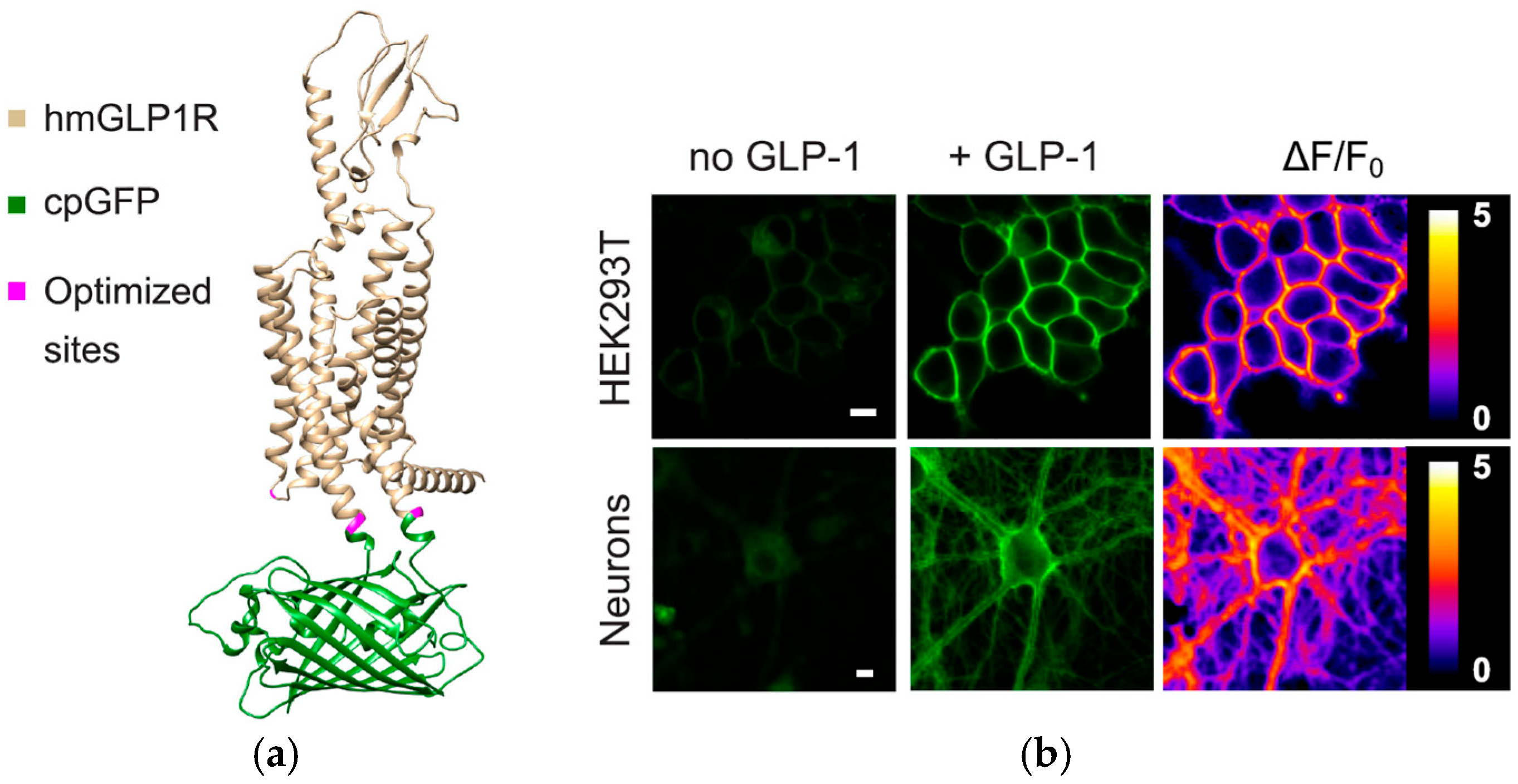
- Duffet, L.; Williams, E.T.; Gresch, A.; Chen, S.; Bhat, M.A.; Benke, D.; Hartrampf, N.; Patriarchi, T. Optical tools for visualizing and controlling human GLP-1 receptor activation with high spatiotemporal resolution. eLife 2023, 12, 1–20. [Google Scholar] [CrossRef] [PubMed]
- Mirdita, M.; Schütze, K.; Moriwaki, Y.; Heo, L.; Ovchinnikov, S.; Steinegger, M. ColabFold: Making protein folding accessible to all. Nat. Methods 2022, 19, 679–682. [Google Scholar] [CrossRef]
- Luo, Y.; Abidian, M.R.; Ahn, J.H.; Akinwande, D.; Andrews, A.M.; Antonietti, M.; Bao, Z.; Berggren, M.; Berkey, C.A.; Bettinger, C.J.; et al. Technology Roadmap for Flexible Sensors. ACS Nano 2023, 17, 5211–5295. [Google Scholar] [CrossRef]
- Gierej, A.; Geernaert, T.; Van Vlierberghe, S.; Dubruel, P.; Thienpont, H.; Berghmans, F. Challenges in the fabrication of biodegradable and implantable optical fibers for biomedical applications. Materials 2021, 14, 1972. [Google Scholar] [CrossRef] [PubMed]
- Zhang, P.; Kim, J.W.; Gehlbach, P.; Iordachita, I.; Kobilarov, M. Autonomous Needle Navigation in Retinal Microsurgery: Evaluation in ex vivo Porcine Eyes. In Proceedings of the 2023 IEEE International Conference on Robotics and Automation (ICRA), London, UK, 29 May–2 June 2023; pp. 4661–4667. [Google Scholar] [CrossRef]
- Liu, Y.; Jing, Z.; Li, R.; Zhang, Y.; Liu, Q.; Li, A.; Zhang, C.; Peng, W. Miniature fiber-optic tip pressure sensor assembled by hydroxide catalysis bonding technology. Opt. Express 2020, 28, 948. [Google Scholar] [CrossRef]
- Liu, Z.; Zhang, Z.F.; Tam, H.; Tao, X. Multifunctional Smart Optical Fibers: Materials, Fabrication, and Sensing Applications. Photonics 2019, 6, 48. [Google Scholar] [CrossRef]
- Schyrr, B.; Boder-pasche, S.; Ischer, R.; Smajda, R.; Voirin, G. Fiber-optic protease sensor based on the degradation of thin gelatin films. Sens. Bio-Sens. Res. 2015, 3, 65–73. [Google Scholar] [CrossRef]
- Mcdonald, S.R.; Tao, S. An optical fiber chlorogenic acid sensor using a Chitosan membrane coated bent optical fiber probe. Anal. Chim. Acta 2024, 1288, 342142. [Google Scholar] [CrossRef]
- Marpu, S.B.; Benton, E.N. Shining Light on Chitosan: A Review on the Usage of Chitosan for Photonics and Nanomaterials Research. Int. J. Mol. Sci. 2018, 19, 1795. [Google Scholar] [CrossRef] [PubMed]
- Suna, F.A.N.; Yi, Z.; Xiangyu, H.; Lihong, G.; Huili, S. Silk materials for medical, electronic and optical applications. Sci. China Technol. Sci. 2019, 62, 903–918. [Google Scholar]
- Fujiwara, E.; Oku, H.; Cordeiro, C.M.B. De Recent developments in agar—Based optical devices. MRS Commun. 2024, 14, 237–247. [Google Scholar] [CrossRef]
- Arefnia, F.; Zibaii, M.I.; Layeghi, A.; Rostami, S. Citrate polymer optical fiber for measuring refractive index based on LSPR sensor. Sci. Rep. 2024, 14, 18637. [Google Scholar] [CrossRef]
- Chen, Z.; Lee, J. Biocompatibility of SU-8 and Its Biomedical Device Applications. Micromachines 2021, 12, 794. [Google Scholar] [CrossRef]
- Manvi, P.K.; Beckers, M.; Mohr, B.; Seide, G.; Gries, T.; Bunge, C. Chapter 3. Polymer Fiber-Based Biocomposites for Medical Sensing Applications; Elsevier Inc.: Amsterdam, The Netherlands, 2019. [Google Scholar] [CrossRef]
- Han, S.; Shin, G. Biodegradable Optical Fiber in a Soft Optoelectronic Device for Wireless Optogenetic Applications. Coatings 2020, 10, 1153. [Google Scholar] [CrossRef]
- Raghunandhan, R.; Chen, L.H.; Long, H.Y.; Leam, L.L.; So, P.L.; Ning, X.; Chan, C.C. Chemical Chitosan/PAA based fiber-optic interferometric sensor for heavy metal ions detection. Sens. Actuators B Chem. 2016, 233, 31–38. [Google Scholar] [CrossRef]
- Liang, D.; Yu, W.; Pang, X.; Huang, P.; Lin, Y.; Xin, W.; Zhang, R.; Tao, H. Silk Fibroin—Based Wearable All—Fiber Multifunctional Sensor for Smart Clothing. Adv. Fiber Mater. 2022, 4, 873–884. [Google Scholar] [CrossRef]
- Rabizah, S.; Hasbullah, M.; Haziq, M.; Akashah, N.; Abdul, R.; Scully, P.J.; Gardner, P. Enhancing fibre optic sensor signals via gold nanoparticle-decorated agarose hydrogels. Opt. Mater. 2023, 143, 114247. [Google Scholar] [CrossRef]
- Tarar, A.A.; Mohammad, U.; Srivastava, S.K. Wearable skin sensors and their challenges: A review of transdermal, optical, and mechanical sensors. Biosensors 2020, 10, 56. [Google Scholar] [CrossRef]
- Jing, J.; An, X.; Luo, Y.; Chen, L.; Chu, Z.; Li, K.H. A Compact Optical Pressure Sensor Based on a III-Nitride Photonic Chip with Nanosphere-Embedded PDMS. ACS Appl. Electron. Mater. 2021, 3, 1982–1987. [Google Scholar] [CrossRef]
- Zhang, H.; Zhou, X.; Li, X.; Gong, P.; Zhang, Y.; Zhao, Y. Recent Advancements of LSPR Fiber-Optic Biosensing: Combination Methods, Structure, and Prospects. Biosensors 2023, 13, 405. [Google Scholar] [CrossRef]
- Li, M.; Singh, R.; Wang, Y.; Marques, C.; Zhang, B.; Kumar, S. Advances in Novel Nanomaterial-Based Optical Fiber Biosensors—A Review. Biosensors 2022, 12, 843. [Google Scholar] [CrossRef]
- Sreejith, S.; Ajayan, J.; Radhika, J.M.; Uma Reddy, N.V.; Manikandan, M. Recent advances in nano biosensors: An overview. Measurement 2024, 236, 115073. [Google Scholar] [CrossRef]
- Rahman, B.M.A.; Viphavakit, C.; Chitaree, R.; Ghosh, S.; Pathak, A.K.; Verma, S.; Sakda, N. Optical Fiber, Nanomaterial, and THz-Metasurface-Mediated Nano-Biosensors: A Review. Biosensors 2022, 12, 42. [Google Scholar] [CrossRef]
- Seena, R.; Paul, S.; Sudheer, V.R. Hybrid Nano-Structured SPR Biosensors: A Novel Approach to Breast and Cervical Cancer Detection. Plasmonics 2025. [Google Scholar] [CrossRef]
- Medrano-Lopez, J.A.; Villalpando, I.; Salazar, M.I.; Torres-Torres, C. Hierarchical Nanobiosensors at the End of the SARS-CoV-2 Pandemic. Biosensors 2024, 14, 108. [Google Scholar] [CrossRef] [PubMed]
- Thanjavur, N.; Bugude, L.; Kim, Y.J. Integration of Functional Materials in Photonic and Optoelectronic Technologies for Advanced Medical Diagnostics. Biosensors 2025, 15, 38. [Google Scholar] [CrossRef] [PubMed]
- Meena, K.V.; Ravi Sankar, A. Biomedical Catheters with Integrated Miniature Piezoresistive Pressure Sensors: A Review. IEEE Sens. J. 2021, 21, 10241–10290. [Google Scholar] [CrossRef]
- Friedemann, M.; Barz, S.; Voigt, S.; Barz, T.; Melloh, M.; Müller, A.; Mehner, J. In-Vivo Animal Trial of a Fiber-Optic Pressure Sensor Probe with Distributed Sensing Points for the Diagnosis of Lumbar Spinal Stenosis. In Proceedings of the 9th World Congress on Electrical Engineering and Computer Systems and Science, London, UK, 3–5 August 2023; pp. 1–9. [Google Scholar] [CrossRef]
- Gan, L.; Wang, J.; Xie, L.; Zhou, Y. A High Precision Triaxial Force Sensor Based on Fiber Bragg Gratings for Catheter Ablation. IEEE Trans. Instrum. Meas. 2023, 73, 7001511. [Google Scholar] [CrossRef]
- Sadek, I.; Biswas, J.; Abdulrazak, B. Ballistocardiogram signal processing: A review. Health Inf. Sci. Syst. 2019, 7, 10. [Google Scholar] [CrossRef]
- Cibula, E.; Pevec, S.; Lenardic, B.; Pinet, E.; Donlagic, D. Miniature all-glass robust pressure sensor. Opt. Express 2009, 17, 5098. [Google Scholar] [CrossRef]
- Zhao, Z.; Chen, J.; Yang, J.; Jiang, Q. Photonic sensor with radio frequency power detection for body pressure monitoring. Optoelectron. Lett. 2023, 19, 752–755. [Google Scholar] [CrossRef]
- Yi, L.; Hou, B.; Liu, X. Optical Integration in Wearable, Implantable and Swallowable Healthcare Devices. ACS Nano 2023, 17, 19491–19501. [Google Scholar] [CrossRef]
- Fisher, C.; Harty, J.; Yee, A.; Li, C.L.; Komolibus, K.; Grygoryev, K.; Lu, H.; Burke, R.; Wilson, B.C.; Andersson-Engels, S. Perspective on the integration of optical sensing into orthopedic surgical devices. J. Biomed. Opt. 2022, 27, 010601. [Google Scholar] [CrossRef]
- Li, L.; Li, Y.; Yang, L.; Fang, F.; Yan, Z.; Sun, Q. Continuous and Accurate Blood Pressure Monitoring Based on Wearable Optical Fiber Wristband. IEEE Sens. J. 2021, 21, 3049–3057. [Google Scholar] [CrossRef]
- Ochoa, M.; Algorri, J.F.; Roldan-Varona, P.; Rodriguez-Cobo, L.; Lopez-Higuera, J.M. Recent advances in biomedical photonic sensors: A focus on optical-fibre-based sensing. Sensors 2021, 21, 6469. [Google Scholar] [CrossRef] [PubMed]
- Xavier, M.S.; Tawk, C.D.; Zolfagharian, A.; Pinskier, J.; Howard, D.; Young, T.; Lai, J.; Harrison, S.M.; Yong, Y.K.; Bodaghi, M.; et al. Soft Pneumatic Actuators: A Review of Design, Fabrication, Modeling, Sensing, Control and Applications. IEEE Access 2022, 10, 59442–59485. [Google Scholar] [CrossRef]
- Shen, F.; Ai, M.; Li, Z.; Lu, X.; Pang, Y.; Liu, Z. Pressure measurement methods in microchannels: Advances and applications. Microfluid. Nanofluidics 2021, 25, 39. [Google Scholar] [CrossRef]
- Bills, E. Risk management for IEC 60601-1 third edition. Biomed. Instrum. Technol. 2006, 40, 390–392. Available online: http://www.ncbi.nlm.nih.gov/pubmed/17078374 (accessed on 1 March 2025). [CrossRef]
- Tettey, F.; Parupelli, S.K.; Desai, S. A Review of Biomedical Devices: Classification, Regulatory Guidelines, Human Factors, Software as a Medical Device, and Cybersecurity. Biomed. Mater. Devices 2024, 2, 316–341. [Google Scholar] [CrossRef]
- Wirges, M.; Funke, A.; Serno, P.; Knop, K.; Kleinebudde, P. Development and in-line validation of a Process Analytical Technology to facilitate the scale up of coating processes. J. Pharm. Biomed. Anal. 2013, 78–79, 57–64. [Google Scholar] [CrossRef]
- The European Parliament and the Council of the European Union Regulation (EU) 2017/746 of the European parliament and of the council on in vitro diagnostic medical devices. Off. J. Eur. Union 2017, 5, 117–176.
- Cruz Rivera, S.; Torlinska, B.; Marston, E.; Denniston, A.K.; Oliver, K.; Hoare, S.; Calvert, M.J. Advancing UK Regulatory Science Strategy in the Context of Global Regulation: A Stakeholder Survey. Ther. Innov. Regul. Sci. 2021, 55, 646–655. [Google Scholar] [CrossRef]
- Phillips, J.A. Polio: Another Cause for Global Concern? Workplace Health Saf. 2015, 63, 92. [Google Scholar] [CrossRef]
- Becker, S.H. Approved American National Standards. SMPTE J. 1991, 100, 852–855. [Google Scholar] [CrossRef]
- Alden, A.E. Approved American National Standards. SMPTE J. 1981, 90, 415–464. [Google Scholar] [CrossRef]
- ISO 13485:2016; Medical Devices—Quality Management Systems—Requirements for Regulatory Purposes. International Organization for Standardization: Geneva, Switzerland, 2016. Available online: https://www.iso.org/standard/59752.html (accessed on 1 March 2025).
- Ulrich, C.M.; Demiris, G.; Kennedy, R.; Rothwell, E. The ethics of sensor technology use in clinical research. Nurs. Outlook 2020, 68, 720–726. [Google Scholar] [CrossRef] [PubMed]
- Bujugundla, R.S.; Pradhan, H.S. Emerging technologies for Fiber-Optic Based Sensors in Biomedical Domain: A Review and Recent Developments. IEEE Trans. Instrum. Meas. 2024, 73, 7010332. [Google Scholar] [CrossRef]
- Leal-Junior, A.; Silva, J.; Macedo, L.; Marchesi, A.; Morau, S.; Valentino, J.; Valentim, F.; Costa, M. The Role of Optical Fiber Sensors in the New Generation of Healthcare Devices: A Review. Sens. Diagn. 2024, 3, 1135–1158. [Google Scholar] [CrossRef]
- Liang, L.; Xie, F.; Jin, L.; Yang, B.; Sun, L.; Guan, B. Optical Microfiber Biomedical Sensors: Classification, Applications, and Future Perspectives. Adv. Sens. Res. 2025, 2400185. [Google Scholar] [CrossRef]
- Liu, H.; Song, X.; Wang, X.; Wang, S.; Yao, N.; Li, X.; Fang, W.; Tong, L.; Zhang, L. Optical Microfibers for Sensing Proximity and Contact in Human-Machine Interfaces. ACS Appl. Mater. Interfaces 2022, 14, 14447–14454. [Google Scholar] [CrossRef]
- Ueno, A.; Hu, J.; An, S. AI for optical metasurface. npj Nanophotonics 2024, 1, 36. [Google Scholar] [CrossRef]
- González-León, K.; Delgado-Macuil, R.J.; Vertti-Cervantes, B.; Muñoz-Aguirre, S.; Castillo-Mixcóatl, J.; García-Juárez, M.; Montes-Narvaez, O.; Ramírez-Sánchez, E.; Beltrán-Pérez, G. Application of support vector machine technique to optical fiber biosensors for neuroprotector (IL-10) detection in serum samples of murine model. Opt. Laser Technol. 2025, 186, 112629. [Google Scholar] [CrossRef]
- Zha, B.; Wang, Z.; Ma, L.; Chen, J.; Wang, H.; Li, X.; Kumar, S.; Min, R. Intelligent Wearable Photonic Sensing System for Remote Healthcare Monitoring Using Stretchable Elastomer Optical Fiber. IEEE Internet Things J. 2024, 11, 17317–17329. [Google Scholar] [CrossRef]
- Zhang, L.; Tong, L. A bioinspired flexible optical sensor for force and orientation sensing. Opto-Electronic Adv. 2023, 6, 230051. [Google Scholar] [CrossRef]
- Lyu, C.; Li, P.; Zhang, J.; Du, Y. Fiber Optic Sensors in Tactile Sensing: A Review. IEEE Trans. Instrum. Meas. 2025, 74, 7001816. [Google Scholar] [CrossRef]
- Jha, R.; Mishra, P.; Kumar, S. Advancements in optical fiber-based wearable sensors for smart health monitoring. Biosens. Bioelectron. 2024, 254, 116232. [Google Scholar] [CrossRef] [PubMed]
- Amjad, A.; Xian, X. Optical sensors for transdermal biomarker detection: A review. Biosens. Bioelectron. 2025, 267, 116844. [Google Scholar] [CrossRef]

| Sensing Application | Responsive Material with Fibre Type | Detection Range | Sensitivity | LOD (Limit of Detection) | Ref. |
|---|---|---|---|---|---|
| pH | PANi with TFBG | 2–12 | minimum of 30 pm/pH maximum of 82 pm/pH | - | [106] |
| PAAm hydrogel with SPR | 8–10 | 13 nm/pH at | - | [107] | |
| gold nanoparticle-functionalized fibre optic probes with FPI | 2–12 | 1.95 nm/pH | - | [80] | |
| Hydrogel + polymer microarrays with miniature optical fibre | 5.5–8 | mean precision of 0.10 pH units | - | [79] | |
| glucose | GO/GOD with LPFG | 0–8 mM | ∼0.24 nm/mM | - | [108] |
| GOD with multimode microfibre | 0.0–166.67 mM | 1.74 nm/(mg/mL) | - | [109] | |
| 3-APBA with LDOF | 0–50 mM | 2.6 μWmM−1 | [74] | ||
| GO with LPFG | 0∼1 wt% | 6.229 dB/wt% | [110] | ||
| GO/GOD with PCF | 10 g/L to 70 g/L | - | - | [111] | |
| SPR with Microsphere optical fibre | 0–200 mg/dL | 0.1688 nm/(mg/dL) | 4 mg/dL | [112] | |
| Gold nanoparticles (AuNPs) and LSPR with TOF | 1.328–1.393 (5–45 wt%) | For bare TOF: 1265%/RIU For AuNP-decorated TOF: 2032%/RIU | - | [113] | |
| GO/GOD with PS-LPFG inscribed on high-birefringence fibre (HBF) | 5–25 mM | ∼20.8 pm/mM | - | [114] | |
| Gold-coated plasmonic layer with PCF | Not specified | 2500 nm/RIU (wavelength), 152 RIU−1 (amplitude) | - | [115] | |
| SPR with enzymatic reaction | 0–400 mg/d | 3.10 pm/(mg/dL) | - | [76] | |
| gold nanoparticle-functionalized fibre optic probes with FPI | 1 μM–1 M | 3.25 nm/mM | - | [80] | |
| cancer biomarkers | Tapered fibre optic interferometer cascaded with FBG for HER2 protein | - | - | 2 ng/mL | [90] |
| FOSs catheter embedded for CD44 protein | - | - | 4.68 aM | [116] | |
| Ti3C2-supported gold nanorod hybrid nanointerfaces with microfibre, integrated with hybrid nanointerfaces | - | - | 13.8 zM (buffer), 0.19 aM (30% serum) | [117] | |
| hormones | Oestrogen receptor on gold-coated polystyrene with Spoon-shaped SPR | - | - | 0.1 pM | [91] |
| Thin gold layer with 7-core fibre + SMF for Insulin | - | - | 10−8 g/mL | [118] | |
| Oestrogen receptor with gold-coated tilted fibre Bragg grating (TFBG) | - | - | 1.5 × 10⁻³ ng/mL | [119] | |
| Anti-cortisol antibody on AuPd-coated with SPR on plastic optical fibre (POF) for cortisol | 0.005–10 ng/mL | - | 1 pg/mL | [120] | |
| Anti-cortisol antibody on gold-coated D-shaped SPR for cortisol | 0.01–100 ng/mL | 0.65 ± 0.02 nm/log(ng/mL) | 1.46 ng/mL | [121] | |
| Gold nanoparticles with FOS microfluidic channel for thyroglobulin (Tg) | - | - | 93.11 fg/mL | [122] |
| Material Type | Material Example | Advantages | Disadvantages | Refs. |
|---|---|---|---|---|
| Natural | Proteins: silk Polysaccharides: alginate, cellulose, agarose, chitosan, gelatine | biocompatibility and biodegradability | limited design flexibility, restricted availability and quantity, batch-to-batch variability, low mechanical strength, and potential immunogenicity | [131,132,133,134,135] |
| Synthetic | Hydrogels: Polyethylene Glycol (PEG), Pluronic (Poloxamer) Citrate-based elastomers: poly (Octamethylene Citrate) (POC), poly (Octamethylene Maleate Citrate) (POMC), Polymer-Based: Polyvinyl Chloride (PVC), SU-8 (Negative Photoresist Polymer), Poly (L-Lactic Acid) (PLLA), Poly (D, L-Lactic Acid) (PDLLA), Poly (L-Lactic-Co-Glycolic Acid) (PLGA), Poly (D,L-Lactic-Co-Glycolic Acid) (PDLGA), Poly-ε-Caprolactone (PCL) Inorganic materials: Calcium–Phosphate Glass (PGs) Silicon-Based Materials: Silicon, Polydimethylsiloxane (PDMS) | adaptable and flexible structure, tunable biodegradability, and customizable physical, mechanical, and chemical characteristics | biocompatibility should be verified and the rigidness and brittleness of glass should be confirmed | [22,127,136,137,138,139] |
| Hybrid Biomaterials (Natural and Synthetic) | Chitosan and Polystyrene Membranes/PAA Silk Fibroin Film, Agarose hydrogel (AG) with gold nanoparticles (AuNPs) | biocompatibility, mechanical strength and tunable properties for PAA, controlled permeability, chemical resistance | limited flexibility, surface modification required for some, degradation issues, Processing complexity, for AuNPs, agglomeration of AuNPs, and limited long-term stability | [133,134,140,141,142] |
| Regulatory Body | Standard/Guideline | Scope and Relevance to FOSs |
|---|---|---|
| FDA (USA) | FDA Medical Device Approval Process | Safety, efficacy, and reliability assessments ensure FOSs meet regulatory requirements before market approval. |
| NCAs (EU) | Medical Device Regulation (MDR) and In Vitro Diagnostic Regulation (IVDR) | Regulation of general medical devices in the EU governs their safety and performance. |
| ISO | ISO 13485/ISO 10993 | Quality management system for medical devices/Biocompatibility evaluation of medical devices. |
| AAMI/ANSI | AAMI TIR42 | Guidance on biocompatibility evaluation, which supports compliance with ISO 10993 for medical FOSs. |
| Challenge | Key Considerations | Refs. |
|---|---|---|
| Biocompatibility | - Ensuring non-toxicity, immune safety, and mechanical stability of materials - Coatings for biocompatibility | [127,128] |
| Miniaturization and Nanomaterials | - Maintaining accuracy in miniaturized sensors - Integration into medical devices - Advancements in nanomaterials for improved sensitivity - Long-term sensor reliability | [145,146] |
| Signal Processing and Data Integration | Fast and accurate data processing - Ensuring seamless system integration with medical devices - Real-time data transmission and processing | [158,159] |
| Production Cost and Manufacturing | - Balancing cost-effectiveness with high quality - Ensuring scalability in production | [93,161] |
| Medical Standards and Regulatory Approval | - Adhering to FDA and other regulatory body requirements - Ensuring sensor stability after sterilization - Ensuring human safety testing and approval | [164,165,167,170] |
| Ethical Considerations | - Ensuring patient privacy, safety, and secure data transmission - Obtaining informed consent and preventing misuse of health data | [173] |
Disclaimer/Publisher’s Note: The statements, opinions and data contained in all publications are solely those of the individual author(s) and contributor(s) and not of MDPI and/or the editor(s). MDPI and/or the editor(s) disclaim responsibility for any injury to people or property resulting from any ideas, methods, instructions or products referred to in the content. |
© 2025 by the authors. Licensee MDPI, Basel, Switzerland. This article is an open access article distributed under the terms and conditions of the Creative Commons Attribution (CC BY) license (https://creativecommons.org/licenses/by/4.0/).
Share and Cite
Karimian, S.; Ali, M.M.; McAfee, M.; Saleem, W.; Duraibabu, D.; Memon, S.F.; Lewis, E. Challenges in Adapting Fibre Optic Sensors for Biomedical Applications. Biosensors 2025, 15, 312. https://doi.org/10.3390/bios15050312
Karimian S, Ali MM, McAfee M, Saleem W, Duraibabu D, Memon SF, Lewis E. Challenges in Adapting Fibre Optic Sensors for Biomedical Applications. Biosensors. 2025; 15(5):312. https://doi.org/10.3390/bios15050312
Chicago/Turabian StyleKarimian, Sahar, Muhammad Mahmood Ali, Marion McAfee, Waqas Saleem, Dineshbabu Duraibabu, Sanober Farheen Memon, and Elfed Lewis. 2025. "Challenges in Adapting Fibre Optic Sensors for Biomedical Applications" Biosensors 15, no. 5: 312. https://doi.org/10.3390/bios15050312
APA StyleKarimian, S., Ali, M. M., McAfee, M., Saleem, W., Duraibabu, D., Memon, S. F., & Lewis, E. (2025). Challenges in Adapting Fibre Optic Sensors for Biomedical Applications. Biosensors, 15(5), 312. https://doi.org/10.3390/bios15050312