Next Issue
Volume 15, November-1
Previous Issue
Volume 15, October-1


Diagnostics, Volume 15, Issue 20 (October-2 2025) – 114 articles

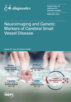
Cover Story (view full-size image): Cerebral small vessel disease (SVD) is a leading cause of stroke and dementia worldwide. By performing a meta-analysis encompassing 18,000 individuals, the NEUROGEN-SVD study reveals that 57% of CSVD patients exhibit global cognitive impairment. It demonstrates that specific MRI markers are powerfully associated with distinct dementias: white matter hyperintensities with vascular dementia, and lacunes with all-cause dementia. The APOE ε4 genotype further amplifies risk. This work underscores a critical vascular–neurodegenerative synergy, charting a path for improved early detection, risk stratification and interventions. View this paper
  • Issues are regarded as officially published after their release is announced to the table of contents alert mailing list.
  • You may sign up for e-mail alerts to receive table of contents of newly released issues.
  • PDF is the official format for papers published in both, html and pdf forms. To view the papers in pdf format, click on the "PDF Full-text" link, and use the free Adobe Reader to open them.
Order results
Result details
Section
Select all
Export citation of selected articles as:
19 pages, 1976 KB  
Article
Large-Scale Saliva-Based Clinical Surveillance Enables Real Time SARS-CoV-2 Outbreak Detection and Genomic Tracking (Arizona, 2020–2023)
by Steven C. Holland, ABCTL Diagnostic Testing and Sequencing Teams, Ian Shoemaker, Theresa Rosov, Carolyn C. Compton, Joshua LaBaer, Efrem S. Lim and Vel Murugan
Diagnostics 2025, 15(20), 2663; https://doi.org/10.3390/diagnostics15202663 - 21 Oct 2025
Viewed by 449
Abstract
Background/Objectives: Monitoring community health and tracking SARS-CoV-2 evolution were critical priorities throughout the COVID-19 pandemic. However, widespread shortages of personal protective equipment, the necessity for social distancing, and the redeployment of healthcare personnel to clinical duties presented significant barriers to traditional sample collection. [...] Read more.
Background/Objectives: Monitoring community health and tracking SARS-CoV-2 evolution were critical priorities throughout the COVID-19 pandemic. However, widespread shortages of personal protective equipment, the necessity for social distancing, and the redeployment of healthcare personnel to clinical duties presented significant barriers to traditional sample collection. Methods: In this study, we evaluated the feasibility of using self-collected saliva specimens for the qualitative detection of SARS-CoV-2 infection. Following confirmation of reliable viral detection in saliva, we established a large-scale surveillance program in Arizona, USA, to enable clinical diagnosis and genomic sequencing from self-collected samples. Between April 2020 and December 2023, we tested approximately 1.4 million saliva samples using RT-PCR, identifying 94,330 SARS-CoV-2 infections. Whole genome sequencing was performed on 69,595 samples, yielding 54,040 high-quality consensus genomes. Results: This surveillance approach enabled real-time monitoring of general infection trends that matched regional case counts. We monitored multiple wave-like introductions of viral lineages over the course of the pandemic. We identified three periods of S gene target failure on a commercial assay and assessed its ability to make fast, genotyping assignment during the pandemic (PPV = 0.98, 95% CI = 0.97–0.99; NPV = 0.94, 95% CI = 0.94–0.96). The co-location of clinical testing and sequencing capabilities within the same facility resulted in low turnaround time from the sample collection to the generation of sequencing data (median = 12 days, IQR: 9.0–19.75). Conclusions: Our findings support the use of self-collected saliva as a scalable, cost-effective, and practical strategy for infectious disease surveillance in future pandemics. Full article
(This article belongs to the Section Diagnostic Microbiology and Infectious Disease)
Show Figures

Figure 1

18 pages, 559 KB  
Article
Plasma Cystine as a Marker of Acute Stroke Severity
by Alexander Vladimirovich Ivanov, Mikhail Aleksandrovich Popov, Polina Alexandrovna Pudova, Ruslan Andreevich Maslennikov, Valery Vasil’evich Aleksandrin, Maria Pavlovna Galdobina, Maria Petrovna Kruglova, Ekaterina Vladimirovna Silina, Victor Alexandrovich Stupin, Marina Yurievna Maksimova and Aslan Amirkhanovich Kubatiev
Diagnostics 2025, 15(20), 2662; https://doi.org/10.3390/diagnostics15202662 - 21 Oct 2025
Viewed by 510
Abstract
Background/Objectives: The amino acid cysteine (Cys) plays an important role in the neuronal injury process in stroke. Cys is present in blood plasma in various forms. The relationship between Cys and its forms and the severity of acute stroke has not been [...] Read more.
Background/Objectives: The amino acid cysteine (Cys) plays an important role in the neuronal injury process in stroke. Cys is present in blood plasma in various forms. The relationship between Cys and its forms and the severity of acute stroke has not been sufficiently studied. We investigated the levels of total Cys and two of its forms (reduced Cys and its disulfide (cystine, CysS)) in blood plasma and their influence on stroke severity in patients at admission. Methods: A total of 210 patients (39–59 years old) with ischemic stroke and intracerebral or subarachnoid hemorrhage were examined. The contents of the different forms of Cys were determined in the first 10–72 h. Stroke severity was estimated using the National Institutes of Health Stroke Scale (NIHSS) and the modified Rankin Scale (mRs). Results: CysS levels < 54 μM were associated with severe (NIHSS > 13) neurological deficit (ischemic stroke: RR = 5.58 and p = 0.0021; hemorrhagic stroke: RR = 3.56 and p = 0.0003). Smoking and high levels of total Cys and other thiols (glutathione and homocysteine) appear to be factors determining this relationship. Conclusions: Low CysS levels may serve as a potential biomarker of acute stroke severity. Full article
(This article belongs to the Special Issue Neurological Disorders: Diagnosis and Management)
Show Figures

Figure 1

21 pages, 48081 KB  
Article
A Public Health Approach to Automated Pain Intensity Recognition in Chest Pain Patients via Facial Expression Analysis for Emergency Care Prioritization
by Rita Wiryasaputra, Yu-Tse Tsan, Qi-Xiang Zhang, Hsing-Hung Liu, Yu-Wei Chan and Chao-Tung Yang
Diagnostics 2025, 15(20), 2661; https://doi.org/10.3390/diagnostics15202661 - 21 Oct 2025
Viewed by 454
Abstract
Background/Objectives: Cardiovascular disease remains a leading cause of death worldwide, with chest pain often serving as an initial reason for emergency visits. However, the severity of chest pain does not necessarily correlate with the severity of myocardial infarction. Facial expressions are an [...] Read more.
Background/Objectives: Cardiovascular disease remains a leading cause of death worldwide, with chest pain often serving as an initial reason for emergency visits. However, the severity of chest pain does not necessarily correlate with the severity of myocardial infarction. Facial expressions are an essential medium to convey the intensity of pain, particularly in patients experiencing speech difficulties. Automating the recognition of facial pain expression may therefore provide an auxiliary tool for monitoring chest pain without replacing clinical diagnosis. Methods: Using streaming technology, the system captures real-time facial expressions and classifies pain levels using a deep learning framework. The PSPI scores were incorporated with the YOLO models to ensure precise classification. Through extensive fine-tuning, we compare the performance of YOLO-series models, evaluating both computational efficiency and diagnostic accuracy rather than focusing solely on accuracy or processing time. Results: The custom YOLOv4 model demonstrated superior performance in pain level recognition, achieving a precision of 97% and the fastest training time. The system integrates a web-based interface with color-coded pain indicators, which can be deployed on smartphones and laptops for flexible use in healthcare settings. Conclusions: This study demonstrates the potential of automating pain assessment based on facial expressions to assist healthcare professionals in observing patient discomfort. Importantly, the approach does not infer the underlying cause of myocardial infarction. Future work will incorporate clinical metadata and a lightweight edge computing model to enable real-time pain monitoring in diverse care environments, which may support patient monitoring and assist in clinical observation. Full article
(This article belongs to the Section Machine Learning and Artificial Intelligence in Diagnostics)
Show Figures

Figure 1

17 pages, 4186 KB  
Article
A Revised Concept for Ocular Surface Imprinting: Easy-to-Use Device for Morphological and Biomolecular-Based Differential Diagnosis
by Bijorn Omar Balzamino, Ilaria Ghezzi, Roberto Sgrulletta, Rossella Anna Maria Colabelli Gisoldi, Augusto Pocobelli, Antonio Di Zazzo, Loredana Zollo and Alessandra Micera
Diagnostics 2025, 15(20), 2660; https://doi.org/10.3390/diagnostics15202660 - 21 Oct 2025
Viewed by 367
Abstract
Background/objectives: The continuous necessity to support biostrumental data with biolomecular data collected using non-invasive tools is influencing the world of ocular surface devices. The ocular imprint still represents a non-invasive and safety technique for collecting corneal and conjunctival epithelia in an easy way, [...] Read more.
Background/objectives: The continuous necessity to support biostrumental data with biolomecular data collected using non-invasive tools is influencing the world of ocular surface devices. The ocular imprint still represents a non-invasive and safety technique for collecting corneal and conjunctival epithelia in an easy way, as performed in human and veterinary clinics. Although used in clinical practice since 1977, operators might benefit from improvements in these techniques, especially in terms of handling and management. Methods: Herein, by reporting the design and characteristics of a patent of ocular surface sampling (the SurfAL pen and periocular-assisted SurfAL pen; PCT WO2016IB51474 20160316), we performed a validation and analysis of its value compared to gold standards. The level-headedness and advantages of this device were verified in 15 sclerocorneal specimens (sampling advantages) and tested in 25 volunteers (handling and operator efficiency, as well as frequency of discomfort in volunteers). Morphological as well as biomolecular analyses were used to compare SurfAL devices with conventional ones. Results: The easy management of SurfAL pens and the good detection of epithelial/goblet cells were confirmed. The SurfAL pen was found to be smart and suitable for routine analysis, as confirmed by quick and reproducible onsite sampling. Periocular-assisted SurfAL pen was comparable in terms of sampling quality but less comparable in terms of subject confidence due to its geometry. Conclusions: This study suggests that the SurfAL pen and periocular-assisted SurfAL pen might represent an additional and hands-on way of sampling ocular surface cells and improve the diagnostic route in ophthalmology. Full article
(This article belongs to the Special Issue New Insights into the Diagnosis and Prognosis of Eye Diseases)
Show Figures

Figure 1

21 pages, 633 KB  
Review
Basophil Activation Test in IgE-Mediated Wheat Allergy: Diagnostic and Clinical Applications—A Narrative Review
by Elena Camelia Berghea, Mădălina Coman-Stanemir and Ioana Raluca Papacocea
Diagnostics 2025, 15(20), 2659; https://doi.org/10.3390/diagnostics15202659 - 21 Oct 2025
Viewed by 536
Abstract
The basophil activation test (BAT) is an emerging tool in the diagnosis and management of IgE-mediated wheat allergies (WAs), particularly in complex or high-risk phenotypes. This narrative review explores the clinical applications of BAT across a spectrum of WA presentations, including wheat-dependent exercise-induced [...] Read more.
The basophil activation test (BAT) is an emerging tool in the diagnosis and management of IgE-mediated wheat allergies (WAs), particularly in complex or high-risk phenotypes. This narrative review explores the clinical applications of BAT across a spectrum of WA presentations, including wheat-dependent exercise-induced anaphylaxis (WDEIA), contact urticaria, and pediatric food allergy. The BAT provides a functional measure of allergen-triggered basophil activation, bridging the gap between mere sensitization and true clinical reactivity. We highlight its utility in detecting sensitization to specific wheat components such as ω-5 gliadin, glutenin subunits, and hydrolyzed wheat proteins, and its value in cases where traditional diagnostics are inconclusive. Furthermore, BAT is discussed as a dynamic biomarker in therapeutic monitoring, especially in patients receiving omalizumab, where reduced basophil reactivity correlates with improved clinical outcomes. While standardization and access remain challenges, the BAT holds significant promise as a precision diagnostic and monitoring tool in wheat allergies. Full article
Show Figures

Figure 1

23 pages, 1451 KB  
Article
CCNA2 and CCNB3 as Early Potential Molecular Candidates of Oocyte Maturation in Cumulus-Oophorous Complex Cells from Follicular Fluid
by Nergis Özlem Kılıç, Çağrı Öner, Duygu Kütük, Belgin Selam, İbrahim Orçun Olcay and Ertuğrul Çolak
Diagnostics 2025, 15(20), 2658; https://doi.org/10.3390/diagnostics15202658 - 21 Oct 2025
Viewed by 468
Abstract
Background/Objectives: Oocyte maturation is a process involving both nuclear and cytoplasmic development regulated by epigenetic changes in gene expression. Cyclin-B3 (CCNB3) and cyclin-A2 (CCNA2) genes are thought to be involved in oocyte maturation; however, the expression profiles and key function in Metaphase-I [...] Read more.
Background/Objectives: Oocyte maturation is a process involving both nuclear and cytoplasmic development regulated by epigenetic changes in gene expression. Cyclin-B3 (CCNB3) and cyclin-A2 (CCNA2) genes are thought to be involved in oocyte maturation; however, the expression profiles and key function in Metaphase-I (MI) and Metaphase-II (MII) phases have yet to be fully elucidated. Small non-coding RNA sequences are involved in epigenetic regulation of specific transcriptional targets, whereas microRNAs (miRNAs) participate in the post-transcriptional and translational repression of target genes. This study examined the expression levels of CCNB3, CCNA2, and their associated miRNAs (miR-17, miR-106b, miR-190a, miR-1275) in cumulus oophorous complex (COC) cells derived from MI and MII oocytes of NOR and DOR IVF cases, with particular emphasis on elucidating their functions during the transition from MI to MII stage. Methods: Follicular fluid containing cumulus–oocyte complex (COC) cells obtained from oocytes of 120 cases in each group NOR MI (n = 30), NOR MII (n = 30), DOR MI (n = 30), and DOR MII (n = 30) who were admitted to the Istanbul Bahçeci Health Group Assisted Reproductive Treatment Center. Following total RNA isolation from COC cells, the gene and protein expression levels of CCNB3 and CCNA2, along with the expression of miR-17, miR-106b, miR-190a, and miR-1275, were assessed using (qPCR-based assay) and immunohistochemistry (IHC). To investigate the functional roles of COC cell populations, morphological analysis was performed using H&E staining. Additionally, metadata of the cases, including age, number of oocytes, fertilization, and embryonic development rates, were evaluated. Results: The expressions of miR-17 and miR-1275 were significantly elevated in both NOR MI and DOR MI groups compared to their respective NOR MII and DOR MII groups (p < 0.05). Additionally, miR-106b levels were higher in the NOR MII group relative to NOR MI (p < 0.05), while an increase was also observed in DOR MI compared to DOR MII (p < 0.05). No difference was observed in miR-190a expression between the NOR and DOR (p > 0.05). Based on the results of H and E staining, the NOR MI, NOR MII, DOR MI, and DOR MII groups exhibited distinct variations in cellular morphology, nuclear characteristics, cytoplasmic volume, and cell density. Conclusions: CCNB3 is predicted to be a potential candidate for determining MI between the NOR and DOR cases. On the other hand, only for the NOR MII cases could CCNA2 provide evidence of oocyte maturation. Moreover, we determined the relationship between related genes and miRNAs which target CCNA2 and CCNB3. Genetic and protein expression analysis across diverse molecular pathways and miRNAs yielded comprehensive preliminary data regarding the developmental stages of oocytes at the MI and MII phases, and their fertilization potential following maturation shows potential and warrants prospective validation with clinical performance evaluation. Full article
(This article belongs to the Special Issue Biomarker-Guided Advances in Diagnostic Medicine)
Show Figures

Figure 1

11 pages, 888 KB  
Review
Application of Nanogenerators in Lumbar Motion Monitoring: Fundamentals, Current Status, and Perspectives
by Yudong Zhao, Hongbin He, Junhao Tong, Tianchang Wang, Shini Wang, Zhuoran Sun, Weishi Li and Siyu Zhou
Diagnostics 2025, 15(20), 2657; https://doi.org/10.3390/diagnostics15202657 - 21 Oct 2025
Viewed by 388
Abstract
Nanogenerators (NGs), especially triboelectric nanogenerators (TENGs), represent an emerging technology with great potential for self-powered lumbar motion monitoring. Conventional wearable systems for assessing spinal kinematics are often limited by their reliance on external power supplies, hindering long-term and real-time clinical applications. NGs can [...] Read more.
Nanogenerators (NGs), especially triboelectric nanogenerators (TENGs), represent an emerging technology with great potential for self-powered lumbar motion monitoring. Conventional wearable systems for assessing spinal kinematics are often limited by their reliance on external power supplies, hindering long-term and real-time clinical applications. NGs can convert biomechanical energy from lumbar motion into electrical energy, providing both sensing and power-generation capabilities in a single platform. This review summarizes the fundamental working mechanisms, device architectures, and current progress of NG-based motion monitoring technologies, with a particular focus on their applications in lumbar spine research and clinical rehabilitation. By enabling high-sensitivity, continuous, and battery-free monitoring, NG-based systems may enhance the diagnosis and management of low back pain (LBP) and postoperative recovery assessment. Furthermore, the integration of NGs with wearable electronics, the Internet of Things (IoT), and artificial intelligence (AI) holds promise for developing intelligent, self-sustaining monitoring platforms that bridge biomedical engineering and spine medicine. Full article
(This article belongs to the Section Point-of-Care Diagnostics and Devices)
Show Figures

Figure 1

11 pages, 614 KB  
Article
Phenotypes of Food Allergies in Patients with Atopic Dermatitis Aged Under 24 Months: A Multicenter Study
by Mujde Tuba Cogurlu, Metin Aydogan, Ozlem Cavkaytar, Pinar Uysal, Hazal Cansu Culpan, Nalan Yakici, Zeynep Hizli Demirkale, Erdem Topal, Hasan Yuksel, Sezin Aydemir, Nursen Cigerci Gunaydin, Cigdem Aydogmus, Sukru Cekic, Emre Akkelle, Tuba Tuncel, Isil Eser Simsek, Mustafa Arga, Zeynep Ülker Altinel, Fatih Kaplan, Ayca Kiykim, Ayse Suleyman, Nermin Guler, Esra Yucel, Haluk Cokugras, Nihat Sapan, Hikmet Tekin Nacaroglu, Adem Yasar, Yakup Yesil, Gonca Hancioglu, Recep Sancak, Mehmet Sarper Erdogan, Oner Ozdemir, Cevdet Ozdemir and Fazil Orhanadd Show full author list remove Hide full author list
Diagnostics 2025, 15(20), 2656; https://doi.org/10.3390/diagnostics15202656 - 21 Oct 2025
Viewed by 324
Abstract
Background: Atopic dermatitis (AD) and food allergy (FA) are common allergic diseases in early childhood. AD may be concomitant with FA, particularly in young children. Although studies report the prevalence of FA in children with AD, there is insufficient data regarding different phenotypes [...] Read more.
Background: Atopic dermatitis (AD) and food allergy (FA) are common allergic diseases in early childhood. AD may be concomitant with FA, particularly in young children. Although studies report the prevalence of FA in children with AD, there is insufficient data regarding different phenotypes of FA. Objective: The aim of our research was to determine the prevalence and clinical predictors of different phenotypes of concomitant FA in children with AD. Methods: This cross-sectional multicenter study included patients younger than 24 months old diagnosed with AD, recruited from 14 pediatric allergy centers. Patients were categorized into two groups using skin testing, allergen-specific IgE, and ultimately food challenge testing (FCT): those with FA and those without. Individuals with FA were classified into three distinct phenotypes: IgE-mediated, non-IgE-mediated, and concurrent IgE- and non-IgE-mediated. Results: The data of 530 children [59% male, median-age 7 months (IQR: 5–11)] were analyzed. IgE-mediated FA was found in 28.1% of participants, whereas 22.4% (n = 119/530) exhibited non-IgE-mediated FA. Concurrent IgE- and non-IgE-mediated FA was reported in 12.1% (n = 64/530) of patients. Cow’s milk (69.6%) and egg-white (68.9%) were identified as the most prevalent allergens. Cow’s milk was primarily responsible for non-IgE-mediated and egg-white for IgE-mediated FA. The most significant predictors of FA were severe AD and the presence of blood in stool with odds ratios of 8.25 (95% Cl: 3.04–22.39) and 10.04 (95% CI: 2.03–49.59), respectively (p < 0.01) (p < 0.005). Conclusions: The study’s findings indicate that children with early-onset and mild-to-moderate AD deserve to be comprehensively assessed for FA symptoms. The most significant indicators of concomitant FA in AD patients were the presence of blood in stool and severe AD. It is important to consider that those who exhibit IgE-mediated FA may also have concurrent non-IgE-mediated FA. We underline that it is important to consider that children with AD who exhibit IgE-mediated FA may also have concurrent non-IgE-mediated FA. Addressing these symptoms may assist healthcare practitioners in clinical practice to improve the quality of care for AD patients having FA. Full article
(This article belongs to the Special Issue Novel Advances in Allergy Diagnosis)
Show Figures

Figure 1

13 pages, 1588 KB  
Article
Advancing Aortic Dissection Imaging: First Clinical Experience of Photon-Counting CT with Ultra-Fast Spectral Imaging
by Daniel Dillinger, Maria Weiss, Hanns L. Kaatsch, Christian Bauer, Achim Hagen, Matthias F. Froelich, Stephan Waldeck and Daniel Overhoff
Diagnostics 2025, 15(20), 2655; https://doi.org/10.3390/diagnostics15202655 - 21 Oct 2025
Viewed by 298
Abstract
Background: Computed tomography (CT) is the standard of reference for diagnosis and follow-up in aortic dissection (AD). Localizing the entry and identifying false and true lumen are as important as differing post-treatment changes from contrast media extravasations. Photon-counting detector CT (PCDCT) allows [...] Read more.
Background: Computed tomography (CT) is the standard of reference for diagnosis and follow-up in aortic dissection (AD). Localizing the entry and identifying false and true lumen are as important as differing post-treatment changes from contrast media extravasations. Photon-counting detector CT (PCDCT) allows for virtual monoenergetic (VME) reconstructions, which can augment contrast media effects on lower energy levels, and for virtual non-contrast (VNC) reconstructions. The aim of this study was to analyze the influence of VME reconstructions on contrast media effects in different dissection compartments as well as compare true and VNC series in AD patients. Methods: We retrospectively analyzed PCDCT datasets from 28 patients with aortic dissections, with different dissection types and different treatment statuses. Attenuation and standard deviation values of the ascending and descending aorta, as well as CT values of the false lumen, were measured. These measurements were obtained from VME images at energy levels ranging from 40 to 190 keV in 10 keV increments, as well as from non-contrast (NC) and VNC reconstructions. The signal-to-noise ratio (SNR) was calculated. Additionally, subjective values for dissection assessability and native aspects of the images were acquired for different reconstructions. Results: CT values decreased with higher energy levels in VME imaging. Ascending aorta showed higher attenuation values than descending aorta, which was higher than false lumen (e.g., at 70 keV ascending 357 [310; 419] HU, descending 346 [305; 401] HU and false lumen 298 [248; 363] HU). These differences increased on lower VME reconstructions with statistical significance for the comparisons of ascending and descending aorta with the false lumen on all energy levels. In line with this, SNR showed highest values for ascending aorta compared to descending aorta and false lumen on all energy levels. For NC comparisons, VNC and VME at 190 keV reconstructions showed higher CT values than NC reconstructions (e.g., overall data NC 48 [42; 55] HU, VNC 66 [57; 73] HU, 190 keV 97 [89; 105] HU). Subjective ratings were worse with VNC than with NC images. Conclusions: VME reconstructions on lower energy levels can be helpful in differentiating between true and false lumen in aortic dissections. Full article
(This article belongs to the Special Issue Advances in Cardiovascular Diseases: Diagnosis and Management)
Show Figures

Figure 1

11 pages, 828 KB  
Case Report
A Rare Combination: Cold Agglutinin Disease Followed by Waldenström Macroglobulinemia—A Case of Early Treatment Response
by Anna Kozub, Aleksandra Nasiek, Natalia Bohun, Martyna Bednarczyk, Łukasz Sędek and Sebastian Grosicki
Diagnostics 2025, 15(20), 2654; https://doi.org/10.3390/diagnostics15202654 - 21 Oct 2025
Viewed by 394
Abstract
Background and Clinical Significance: Waldenström macroglobulinemia (WM) is a rare, indolent B-cell non-Hodgkin lymphoma, characterised by the presence of monoclonal immunoglobulin M (IgM) and lymphoplasmacytic infiltration of the bone marrow. It is often associated with various haematological and systemic disorders, including previous [...] Read more.
Background and Clinical Significance: Waldenström macroglobulinemia (WM) is a rare, indolent B-cell non-Hodgkin lymphoma, characterised by the presence of monoclonal immunoglobulin M (IgM) and lymphoplasmacytic infiltration of the bone marrow. It is often associated with various haematological and systemic disorders, including previous cold agglutinin disease (CAD), a condition where cold-sensitive antibodies lead to haemolysis. Case Presentation: A 55-year-old male patient was admitted to the Internal Diseases Ward with symptoms of weakness, reduced effort tolerance, and weight loss, along with life-threatening normoblastic anaemia (haemoglobin [Hb]: 3.90 g/dL). Initial blood tests raised suspicion of CAD due to the presence of multiple blood clots, as well as a decrease in lymphocyte and neutrophil counts. CAD was then confirmed by a cold agglutinin titre of 1:2000 and direct antiglobulin test ([DAT] 4+). Two weeks later, upon transfer to the Haematological Diseases Ward, further investigation revealed elevated IgM levels (up to 31.55 g/L). Additional diagnostic tests, including serum protein electrophoresis, imaging, multiparametric flow cytometry, and bone marrow biopsy, confirmed the diagnosis of WM. The L265P MYD88 mutation test was positive. Treatment with intravenous rituximab was initiated, followed by bendamustine/rituximab (BR) therapy protocol as first-line treatment. After two cycles, the patient’s clinical condition and laboratory results significantly improved, with a marked reduction in IgM (<0.4 g/L). Hb levels steadily rose to 12.60 g/dL, eliminating the need for further blood transfusions. Conclusions: This case highlights the importance of recognising the coexistence of CAD and WM, which may present with overlapping clinical features, including life-threatening anaemia. Extensive diagnostics and prompt treatment with combination therapy can lead to effective clinical improvement. Full article
(This article belongs to the Special Issue Rare Diseases: Diagnosis and Management)
Show Figures

Figure 1

12 pages, 354 KB  
Article
Management of Monochorionic Twin Pregnancies Complicated by Selective Fetal Growth Restriction: Retrospective Single-Center 12-Year Experience
by Sofia Roero, Silvana Arduino, Agata Ingala, Carlotta Bossotti, Isabella Ferrando, Miriam Folino Gallo, Chiara Peila, Alessandra Coscia and Alberto Revelli
Diagnostics 2025, 15(20), 2653; https://doi.org/10.3390/diagnostics15202653 - 21 Oct 2025
Viewed by 418
Abstract
Objectives: To describe the perinatal outcomes of a series of monochorionic diamniotic (MCDA) twin pregnancies complicated by selective fetal growth restriction (sFGR), classified according to the umbilical artery (UA) Doppler flow pattern of the smaller twin, and managed in a single centre over [...] Read more.
Objectives: To describe the perinatal outcomes of a series of monochorionic diamniotic (MCDA) twin pregnancies complicated by selective fetal growth restriction (sFGR), classified according to the umbilical artery (UA) Doppler flow pattern of the smaller twin, and managed in a single centre over a 12-year period. Methods: Retrospective cohort study involving MCDA twin pregnancies followed up at the Twin Pregnancy Care Unit of Sant’Anna Hospital, Turin, Italy, between January 2010 and May 2023. We compared perinatal outcomes of MCDA pregnancies classified based on the UA Doppler flow pattern of the smaller twin. Results: The percentage of sFGR in our sample was 14.8%. A total of 103 MCDA pregnancies with sFGR were included. In 34.9% cases, the UA flow pattern changed throughout pregnancy. At last examination, 58.3% cases were classified as type I, 25.2% as type II and 16.5% as type III. The perinatal survival rate of both twins in type I and III was 100%, in type II 88.5%. Type II sFGR had the highest perinatal mortality rate (7.7%). Type III twins were more likely to have malformations compared to type II and type I; compared to type I sFGR babies, they were more likely to develop RDS and to be admitted to NICU, where the length of stay was longer. Conclusions: Although the UA flow pattern correlates with perinatal outcome, it can change throughout pregnancy. Type III sFGR may have lower risk of fetal demise than traditionally thought. The main challenge remains finding the optimal balance between adverse outcomes and premature birth. Full article
(This article belongs to the Special Issue Recent Advances in Prenatal Diagnosis and Clinical Management)
Show Figures

Figure 1

15 pages, 11724 KB  
Article
Improved Clinical and Radiological Outcomes with Double-Cage Biportal Endoscopic Transforaminal Lumbar Interbody Fusion: A Comparative CT-Based Study
by Yu-Hao Huang and Jwo-Luen Pao
Diagnostics 2025, 15(20), 2652; https://doi.org/10.3390/diagnostics15202652 - 21 Oct 2025
Viewed by 440
Abstract
Background/Objectives: When transitioning from an older surgical technique to a newer one, we expect improved treatment outcomes and fewer complications. However, direct comparative studies to confirm these advantages are often lacking. Tubular minimally invasive transforaminal lumbar interbody fusion (MISTLIF) has been widely [...] Read more.
Background/Objectives: When transitioning from an older surgical technique to a newer one, we expect improved treatment outcomes and fewer complications. However, direct comparative studies to confirm these advantages are often lacking. Tubular minimally invasive transforaminal lumbar interbody fusion (MISTLIF) has been widely used, but limitations in visualization and endplate preparation may compromise fusion quality. Biportal endoscopic TLIF (BETLIF), a more recent alternative, offers enhanced magnification and superior hemostasis. Still, CT-based comparative data on fusion integrity remain limited. To evaluate the clinical and radiological outcomes following a chronological transition from MISTLIF to BETLIF, using thin-slice CT to assess fusion integrity. Methods: This retrospective study analyzed 179 patients treated by a single surgeon between January 2018 and May 2021. The first 90 cases underwent MISTLIF, followed by 89 BETLIF procedures. Clinical outcomes included Visual Analog Scale (VAS), Oswestry Disability Index (ODI), and Japanese Orthopedic Association (JOA) scores. Radiological assessments at one year postoperatively (X-ray and thin-slice CT) included disc height, segmental lordosis, Bridwell fusion grade, cage subsidence, and subchondral osteolysis. Results: BETLIF was associated with significantly shorter hospital stays (5.7 vs. 7.4 days) and fewer transfusions (0% vs. 14.7%). BETLIF showed significantly better ODI (12.7 vs. 23.5), JOA scores (26.4 vs. 20.6), and comparable VAS improvement. Radiologically, BETLIF had significantly higher fusion rates (93.3% vs. 82.4%), greater disc height restoration, and lower rates of cage subsidence (5.0% vs. 13.7%) and osteolysis (13.3% vs. 52.9%). Conclusions: BETLIF demonstrated superior clinical and radiological outcomes, likely due to enhanced endoscopic visualization and precise endplate preparation. Full article
(This article belongs to the Special Issue Diagnosis and Management of Spinal Diseases)
Show Figures

Figure 1

15 pages, 1523 KB  
Article
Dynamic Whole-Body FDG PET/CT for Predicting Malignancy in Head and Neck Tumors and Cervical Lymphadenopathy
by Gregor Horňák, André H. Dias, Ole L. Munk, Lars C. Gormsen, Jaroslav Ptáček and Pavel Karhan
Diagnostics 2025, 15(20), 2651; https://doi.org/10.3390/diagnostics15202651 - 21 Oct 2025
Viewed by 349
Abstract
Background: Dynamic whole-body (D-WB) FDG PET/CT is a novel technique that enables the direct reconstruction of multiparametric images representing the FDG metabolic uptake rate (MRFDG) and “free” FDG (DVFDG). Applying complementary parameters with distinct characteristics compared to static SUV [...] Read more.
Background: Dynamic whole-body (D-WB) FDG PET/CT is a novel technique that enables the direct reconstruction of multiparametric images representing the FDG metabolic uptake rate (MRFDG) and “free” FDG (DVFDG). Applying complementary parameters with distinct characteristics compared to static SUV images, the aims of this study are as follows: (1) to determine the threshold values of SUV, MRFDG, and DVFDG for malignant and benign lesions; (2) to compare the specificity of MRFDG and DVFDG images with static SUVbw images; and (3) to assess whether any of the dynamic imaging parameters correlate more significantly with malignancy or non-malignancy in the examined lesions based on the measured values obtained from D-WB FDG PET/CT. Methods: The study was a retrospective analysis of D-WB PET/CT data from 43 patients (23 males and 20 females) included both in the context of primary staging as well as imaging performed due to suspicion of post-therapeutic relapse or recurrence. Standard scanning was performed using a multiparametric PET acquisition protocol on a Siemens Biograph Vision 600 PET/CT scanner. Pathological findings were manually delineated, and values for SUVbw, MRFDG, and DVFDG were extracted. The findings were classified and statistically evaluated based on their was histological verification of a malignant or benign lesion. Multinomial and binomial logistic regression analyses were used to find parameters for data classification in different models, employing various combinations of the input data (SUVbw, MRFDG, DVFDG). ROC curves were generated by changing the threshold p-value in the regression models to compare the models and determine the optimal thresholds. Results: Patlak PET parameters (MRFDG and DVFDG) combined with mean SUVbw achieved the highest diagnostic accuracy of 0.82 (95% CI 0.75–0.89) for malignancy detection (F1-score = 0.90). Sensitivity reached 0.85 (95% CI 0.77–0.91) and specificity 0.93 (95% CI 0.87–0.98). Classification accuracy in tumors was 0.86 (95% CI 0.78–0.92) and in lymph nodes 0.81 (95% CI 0.73–0.88). Relative contribution analysis showed that DVFDG accounted for up to 65% of the classification weight. ROC analysis demonstrated AUC values above 0.8 for all models, with optimal thresholds achieving sensitivities of around 0.85 and specificities up to 0.93. Thresholds for malignancy detection were, for mean values, SUVbw > 5.8 g/mL, MRFDG > 0.05 µmol/mL/min, DVFDG > 68%, and, for maximal values, SUVbw > 8.7 g/mL, MRFDG > 0.11 µmol/mL/min, DVFDG > 202%. Conclusions: The D-WB [18F]FDG PET/CT images in this study highlight the potential for improved differentiation between malignant and benign lesions compared to conventional SUVbw imaging in patients with locally advanced head and neck cancers presenting with cervical lymphadenopathy and carcinoma of unknown primary origin (CUP). This observation may be particularly relevant in common diagnostic dilemmas, especially in distinguishing residual or recurrent tumors from post-radiotherapy changes. Further validation in larger cohorts with histopathological confirmation is warranted, as the small sample size in this study may limit the generalizability of the findings. Full article
Show Figures

Figure 1

46 pages, 25323 KB  
Hypothesis
Cervical Oculopathy: The Cervical Spine Etiology of Visual Symptoms and Eye Diseases—A Hypothesis Exploring Mechanisms Linking the Neck and the Eye
by Ross A. Hauser, Morgan Griffiths, Danielle Matias and Benjamin R. Rawlings
Diagnostics 2025, 15(20), 2650; https://doi.org/10.3390/diagnostics15202650 - 21 Oct 2025
Viewed by 838
Abstract
Background: Eye and visual symptoms are becoming increasingly common in young people, along with the emerging conditions text neck and computer vision syndrome, though underlying mechanisms are not fully elucidated. The link between cervical spine structure and the eye remains relatively unexplored. Methods: [...] Read more.
Background: Eye and visual symptoms are becoming increasingly common in young people, along with the emerging conditions text neck and computer vision syndrome, though underlying mechanisms are not fully elucidated. The link between cervical spine structure and the eye remains relatively unexplored. Methods: This paper employs a hypothesis-driven, literature-based evidence approach, aiming to explore the hypothesis that cervical spine structural issues may be an underlying mechanism for visual symptoms and eye diseases. The purpose of exploring this hypothesis is to lay the groundwork for future research, and advance diagnostics and treatment options. No new analysis was performed. Results: This article lays the groundwork for the hypothesis that cervical spine structural dysfunctions, including a forward-displaced atlas (C1), can cause dynamic carotid sheath compression, contributing to neurological and neurovascular mechanisms that affect the eye, primarily by (1) impaired venolymphatic drainage of the eye and brain due to compression of the internal jugular veins, and (2) ocular dysautonomia from a disruption of the parasympathetic/sympathetic system balance, partly due to vagus nerve degeneration. Conclusions: Potential mechanisms, diagnostics, and treatment options for visual disorders initiated by cervical structural dysfunction are discussed, providing a foundation for future research aimed at improving clinical outcomes for some eye conditions which have an otherwise unknown etiology. Full article
(This article belongs to the Section Clinical Diagnosis and Prognosis)
Show Figures

Figure 1

13 pages, 2900 KB  
Article
Multiplexing Proteomic and Ingenuity Pathway Analysis of Attention/Working Memory in Virally Suppressed Women with HIV: A Feasibility Study
by Wei Li, Leah H. Rubin, Yanxun Xu, Yuezhe Wang, Raha Dastgheyb, Travis Ptacek, Ge Wang, Mirjam-Colette Kempf, Jodie A. Dionne, Deborah Konkle-Parker, Daniel Y. Li, Anandi Sheth, Igho Ofotokun and David E. Vance
Diagnostics 2025, 15(20), 2649; https://doi.org/10.3390/diagnostics15202649 - 21 Oct 2025
Viewed by 284
Abstract
Background/Objectives: Individual plasma protein biomarkers have been shown to correlate with cognitive performance in people with HIV (PWH). This study aimed to investigate the association between plasma proteomic signatures and attention/working memory in virologically well-controlled women with HIV (WWH). Methods: Seventy-seven WWH from [...] Read more.
Background/Objectives: Individual plasma protein biomarkers have been shown to correlate with cognitive performance in people with HIV (PWH). This study aimed to investigate the association between plasma proteomic signatures and attention/working memory in virologically well-controlled women with HIV (WWH). Methods: Seventy-seven WWH from three Women’s Interagency HIV Study (WIHS) sites completed neuropsychological (NP) testing and a blood draw. Selected protein biomarkers (200 total) were analyzed using a multiplexing method. Results: Random forest analysis was used to identify the top 10 biomarkers that were each positively or negatively associated with attention/working memory. Ingenuity pathway analysis (IPA) was used to facilitate data interpretation. Tumor necrosis factor receptor 1 (TNF RI), TNF RII, interleukin 1 receptor 1 (IL-1RI), and IL-6R were negatively associated with attention/working memory. Conclusions: Based on the IPA, two gene signaling networks were proposed for associating these plasma protein biomarkers with attention/working memory function. This novel methodology demonstrates how gene networks can be identified using blood draws in conjunction with cognitive assessment, and then used in random forest analysis, to derive value that can be put in IPA. Full article
(This article belongs to the Section Clinical Laboratory Medicine)
Show Figures

Figure 1

10 pages, 2921 KB  
Article
Lung Ultrasound Assessment of Lung Injury Following Acute Spinal Cord Injury in Rats
by Na Ni, Ruiliang Chu, Kai Gu and Yi Zhong
Diagnostics 2025, 15(20), 2648; https://doi.org/10.3390/diagnostics15202648 - 21 Oct 2025
Viewed by 315
Abstract
Background/Objectives: Acute spinal cord injury (ASCI) often leads to pulmonary complications, yet reliable, non-invasive assessment tools are limited. This study aimed to evaluate the utility of lung ultrasound (LUS) in assessing lung injury following ASCI in a rat model. Methods: Fifty-four female Sprague [...] Read more.
Background/Objectives: Acute spinal cord injury (ASCI) often leads to pulmonary complications, yet reliable, non-invasive assessment tools are limited. This study aimed to evaluate the utility of lung ultrasound (LUS) in assessing lung injury following ASCI in a rat model. Methods: Fifty-four female Sprague Dawley rats were randomized into sham (n = 27) or ASCI (n = 27) groups. LUS was performed at 12 h, 48 h, and 1 week post-injury, with lung injury quantified using a modified B-line score (BLS). Pulmonary function was assessed non-invasively, and histopathological evaluation and wet-to-dry (W/D) weight ratios were conducted post-mortem. Correlations between BLS and functional and pathological parameters were analyzed. Results: Histological analysis revealed progressive pulmonary hemorrhage, edema, and inflammatory infiltration peaking at 48 h post-injury, with residual hemorrhage and fibroplasia at 1 week. LUS findings evolved from narrow-based B-lines at 12 h to confluent B-lines with pleural abnormalities by 1 week. ASCI rats showed significant reductions in respiratory frequency, peak inspiratory and expiratory flow, and EF50 at all time points (p < 0.05). Tidal volume and minute volume decreased initially, with partial recovery at 1 week. BLS negatively correlated with all pulmonary function parameters and positively with the histological score and W/D ratio (p < 0.001). Conclusions: LUS reliably detects and tracks lung injury after ASCI, correlating well with physiological and pathological indicators. These findings support its potential as a non-invasive monitoring tool. Future refinement of ultrasound scoring may improve clinical applicability in ASCI-related pulmonary assessment. Full article
(This article belongs to the Special Issue Critical Ultrasound in Newborns/Children)
Show Figures

Figure 1

14 pages, 1480 KB  
Article
Evaluation of MRI-Based Measurements for Patellar Dislocation: Reliability and Reproducibility
by Ivan Brumini, Tamara Pranjkovic and Danijela Veljkovic Vujaklija
Diagnostics 2025, 15(20), 2647; https://doi.org/10.3390/diagnostics15202647 - 20 Oct 2025
Viewed by 422
Abstract
Background/Objectives: The aim of our study was to identify the most reliable MRI measurements associated with patellar dislocation. Methods: MRI scans from 86 knees (48 controls and 38 with a history of patellar dislocation) were retrospectively analyzed. The following parameters were measured: lateral [...] Read more.
Background/Objectives: The aim of our study was to identify the most reliable MRI measurements associated with patellar dislocation. Methods: MRI scans from 86 knees (48 controls and 38 with a history of patellar dislocation) were retrospectively analyzed. The following parameters were measured: lateral trochlear inclination (LTI) and its modified version, sulcus angle (SA), trochlear depth (TD), tibial tubercle–trochlear groove distance (TT–TG), patellar tendon–lateral trochlear ridge distance (PT–LTR), and PT–LTR horizontal, a novel modification. Inter-rater reliability was assessed using intraclass correlation coefficients (ICCs), and diagnostic accuracy was evaluated using ROC analysis. Results: All measurements significantly differed between the groups (p < 0.05). SA and TD were highly discriminative (AUC > 0.8) but demonstrated lower inter-rater agreement. PT-LTR horizontal strongly correlated with PT-LTR and was equally sensitive and specific for patellar dislocation as PT-LTR (81.6% and 87.5%, respectively) when in line or extending more laterally than the lateral trochlear ridge (AUC = 0.896, p < 0.001). LTI demonstrated the highest diagnostic performance with a sensitivity of 89.5% and a specificity of 97.9% for a cut-off ≤12.85° (AUC = 0.981), with excellent inter-rater agreement. LTI modified also performed well (AUC = 0.937), with a sensitivity and specificity of 81.6% and 93.7%, respectively. Conclusions: LTI, PT–LTR, and their modified versions demonstrated the highest reliability and diagnostic performance among the MRI measurements evaluated. Given their reproducibility and ease of application, these parameters may be useful in the imaging assessment of patellar dislocation. Further prospective studies are recommended to confirm their clinical utility in broader populations. Full article
Show Figures

Figure 1

13 pages, 1102 KB  
Article
From Prompts to Practice: Evaluating ChatGPT, Gemini, and Grok Against Plastic Surgeons in Local Flap Decision-Making
by Gianluca Marcaccini, Luca Corradini, Omar Shadid, Ishith Seth, Warren M. Rozen, Luca Grimaldi and Roberto Cuomo
Diagnostics 2025, 15(20), 2646; https://doi.org/10.3390/diagnostics15202646 - 20 Oct 2025
Viewed by 377
Abstract
Background: Local flaps are a cornerstone of reconstructive plastic surgery for oncological skin defects, ensuring functional recovery and aesthetic integration. Their selection, however, varies with surgeon experience. Generative artificial intelligence has emerged as a potential decision-support tool, although its clinical role remains [...] Read more.
Background: Local flaps are a cornerstone of reconstructive plastic surgery for oncological skin defects, ensuring functional recovery and aesthetic integration. Their selection, however, varies with surgeon experience. Generative artificial intelligence has emerged as a potential decision-support tool, although its clinical role remains uncertain. Methods: We evaluated three generative AI platforms (ChatGPT-5 by OpenAI, Grok by xAI, and Gemini by Google DeepMind) in their free-access versions available in September 2025. Ten preoperative photographs of suspected cutaneous neoplastic lesions from diverse facial and limb sites were submitted to each platform in a two-step task: concise description of site, size, and tissue involvement, followed by the single most suitable local flap for reconstruction. Outputs were compared with the unanimous consensus of experienced plastic surgeons. Results: Performance differed across models. ChatGPT-5 consistently described lesion size accurately and achieved complete concordance with surgeons in flap selection. Grok showed intermediate performance, tending to recognise tissue planes better than lesion size and proposing flaps that were often acceptable but not always the preferred choice. Gemini estimated size well, yet was inconsistent for anatomical site, tissue involvement, and flap recommendation. When partially correct answers were considered acceptable, differences narrowed but the overall ranking remained unchanged. Conclusion: Generative AI can support reconstructive reasoning from clinical images with variable reliability. In this series, ChatGPT-5 was the most dependable for local flap planning, suggesting a potential role in education and preliminary decision-making. Larger studies using standardised image acquisition and explicit uncertainty reporting are needed to confirm clinical applicability and safety. Full article
18 pages, 7766 KB  
Article
Epidemiological and Histopathological Characterization of Endometrial Carcinoma: A Retrospective Cohort from Romania
by Andrei Muraru, Alex-Emilian Stepan, Claudiu Margaritescu, Mirela Marinela Florescu, Anne-Marie Badiu, Iulia Oana Cretu, Bianca Catalina Andreiana and Raluca Niculina Ciurea
Diagnostics 2025, 15(20), 2645; https://doi.org/10.3390/diagnostics15202645 - 20 Oct 2025
Viewed by 366
Abstract
Background/Objectives: Endometrial carcinoma is an emerging challenge for public health systems globally, especially in countries with a high development index. Traditionally, histopathological staging and grading have been the main criteria informing treatment modalities. More recently, clinically actionable molecular targets have been developed, [...] Read more.
Background/Objectives: Endometrial carcinoma is an emerging challenge for public health systems globally, especially in countries with a high development index. Traditionally, histopathological staging and grading have been the main criteria informing treatment modalities. More recently, clinically actionable molecular targets have been developed, following observations from the TCGA project and the ProMisE cohort. Although promising, the cost of these methods is an obstacle for some countries that lack well developed theranostics infrastructure in their public systems. This study aimed to contextualize our center’s diagnostic experience from the perspective of histopathological diagnosis. Methods: This is a retrospective study that selected 109 cases of already diagnosed endometrial carcinoma from the interval of 2017–2023. We analyzed traditional parameters related to staging and grading, using the FIGO 2009 system as well as basic histological parameters (lymphovascular invasion, perineural invasion, necrosis). Excel and SPSS 26 were used for database management and correlations. Findings were contextualized using the more recent studies that reported on similar parameters. Results: Higher-grade tumors were associated with lymphovascular invasion (p = 0.04) and lymph node involvement (p = 0.0006), as well as deeper myoinvasion (p = 0.0018). Myoinvasion (p = 0.013) and lymphovascular invasion (p = 0.0001) were associated with advanced disease (FIGO III and IV). Our cohort showed a relative paucity (6.5%) of non-endometrioid endometrial carcinoma and presence of lymphovascular invasion (9.2%). Perineural invasion was found in 3 cases with extrauterine involvement. Conclusions: Histopathological diagnosis represents an integral component in informing clinical management for endometrial carcinoma and should serve as a means of triage for more expensive molecular techniques. It nevertheless presents reproducibility issues. Further efforts should focus on resolving such issues or possibly introducing less-researched parameters like perineural invasion. Full article
(This article belongs to the Special Issue Advances in Diagnosis and Management of Endometrial Cancer)
Show Figures

Figure 1

12 pages, 573 KB  
Article
An Ensemble Model for Fundus Images to Aid in Age-Related Macular Degeneration Grading
by Roberto Romero-Oraá, María Herrero-Tudela, María Isabel López, Roberto Hornero, Pere Romero-Aroca and María García
Diagnostics 2025, 15(20), 2644; https://doi.org/10.3390/diagnostics15202644 - 20 Oct 2025
Viewed by 239
Abstract
Background: Age-related macular degeneration (AMD) is a leading cause of visual impairment in the elderly population. Periodic examinations through fundus image analysis are paramount for early diagnosis and adequate treatment. Automatic artificial intelligence algorithms have proven useful for AMD grading, with the ensemble [...] Read more.
Background: Age-related macular degeneration (AMD) is a leading cause of visual impairment in the elderly population. Periodic examinations through fundus image analysis are paramount for early diagnosis and adequate treatment. Automatic artificial intelligence algorithms have proven useful for AMD grading, with the ensemble strategies recently gaining special attention. Methods: This study presents an ensemble model that combines 2 individual models of a different nature. The first model was based on the ResNetRS architecture and supervised learning. The second model, known as RETFound, was based on a visual transformer architecture and self-supervised learning. Results: Our experiments were conducted using 149,819 fundus images from the Age-Related Eye Disease Study (AREDS) public dataset. An additional private dataset of 1679 images was used to validate our approach. The results on AREDS achieved a quadratic weighted kappa of 0.7364 and an accuracy of 66.03%, which outperforms the previous methods in the literature. Conclusions: The ensemble strategy presented in this study could be useful for the screening of AMD in a clinical setting. Consequently, eye care for AMD patients would be improved while clinical costs and workload would be reduced. Full article
Show Figures

Figure 1

15 pages, 1536 KB  
Article
Evaluation of the Risk of Urinary System Stone Recurrence Using Anthropometric Measurements and Lifestyle Behaviors in a Developed Artificial Intelligence Model
by Hikmet Yasar, Kadir Yildirim, Mucahit Karaduman, Bayram Kolcu, Mehmet Ezer, Ferhat Yakup Suceken, Fatih Bicaklioğlu, Mehmet Erhan Aydin, Coskun Kaya, Muhammed Yildirim and Kemal Sarica
Diagnostics 2025, 15(20), 2643; https://doi.org/10.3390/diagnostics15202643 - 20 Oct 2025
Viewed by 429
Abstract
Background/Objectives: Urinary system stone disease is an important health problem both clinically and economically due to its high recurrence rates. In this study, an innovative hybrid approach based on deep learning is proposed to predict the recurrence risk of stone disease. Methods: Patient [...] Read more.
Background/Objectives: Urinary system stone disease is an important health problem both clinically and economically due to its high recurrence rates. In this study, an innovative hybrid approach based on deep learning is proposed to predict the recurrence risk of stone disease. Methods: Patient data were divided into three subsets: anthropometric measurements (Part A), derived body composition indices (Part B), and other clinical and demographic information (Part C). Each data subset was processed with autoencoder models, and low-dimensional, meaningful features were extracted. The obtained features were combined, and the classification process was performed using four different machine learning algorithms: Extreme Gradient Boosting (XGBoost), Cubic Support Vector Machines (Cubic SVM), k-Nearest Neighbor algorithm (KNN), and Decision Tree (DT). Results: According to the experimental results, the highest classification performance was obtained with the XGBoost algorithm. The suggested approach adds to the literature by offering a novel solution that makes early risk calculation for stone disease recurrence easier. It also shows how well structural feature engineering and deep representation can be integrated in clinical prediction issues. Conclusions: Prediction of the stone recurrence risk in advance is of great importance both in terms of improving the quality of life of patients and reducing the unnecessary diagnostic evaluations along with lowering treatment costs. Full article
(This article belongs to the Special Issue New Technologies and Tools Used for Risk Assessment of Diseases)
Show Figures

Figure 1

15 pages, 662 KB  
Article
Left Atrial Appendage Morphology Predicts Atrial Fibrillation Recurrence: The Hidden Risks of Windsock Anatomy
by Yu-Sheng Lin, Hui-Ting Wang, Yen-Nan Fang, Huang-Chung Chen, Yi-Wei Lee and Yung-Lung Chen
Diagnostics 2025, 15(20), 2642; https://doi.org/10.3390/diagnostics15202642 - 20 Oct 2025
Viewed by 245
Abstract
Background/Objectives: Left atrial appendage (LAA) morphology has been implicated in atrial fibrillation (AF) recurrence following catheter ablation. However, the predictive value of specific anatomical shapes remains unclear. We aimed to evaluate the association between distinct LAA morphologies and AF recurrence post-ablation. Methods [...] Read more.
Background/Objectives: Left atrial appendage (LAA) morphology has been implicated in atrial fibrillation (AF) recurrence following catheter ablation. However, the predictive value of specific anatomical shapes remains unclear. We aimed to evaluate the association between distinct LAA morphologies and AF recurrence post-ablation. Methods: In this retrospective, single-center study, 463 patients with AF undergoing first-time catheter ablation were included. Pre-ablation contrast-enhanced cardiac computed tomography was performed to classify LAA morphology into chicken-wing, windsock, cauliflower, and cactus types. Patients were followed for one year, with AF recurrence defined as documented atrial tachyarrhythmia episodes lasting more than 30 s occurring between 3 and 12 months post-procedure. Clinical, anatomical, and procedural factors were analyzed using multivariable logistic regression to identify independent predictors of recurrence. Results: Among the four morphologies, the windsock-type LAA had the highest recurrence rate at 48.3%, significantly greater than chicken-wing (25.2%), cauliflower (20.8%), and cactus (18.2%) types (p = 0.017). Multivariable analysis confirmed windsock morphology as an independent predictor for AF recurrence (adjusted OR = 2.720, 95% CI: 1.209–6.118; p = 0.016). Additionally, persistent AF (adjusted OR = 1.748, 95% CI: 1.075–2.842; p = 0.024) and antiarrhythmic drug use in the blanking period (adjusted OR = 2.862, 95% CI: 1.689–4.849; p < 0.001) independently increased the risk of recurrence. Conclusions: Windsock-type LAA morphology significantly predicts increased AF recurrence following catheter ablation, underscoring the importance of morphological assessment in ablation planning. Individualized strategies targeting high-risk LAA morphologies may enhance procedural success and reduce AF recurrence. Future prospective studies are warranted to validate these findings. Full article
(This article belongs to the Special Issue Advances in Diagnosis and Treatment of Cardiac Arrhythmias 2025)
Show Figures

Figure 1

16 pages, 2651 KB  
Article
Navigating Biopsy Safety: Complication Rates Under Ultrasound and CT Guidance
by Theresa Sophie Patzer, Franziska Müller, Michael Meir, Henner Huflage, Lukas Müller, Thorsten Alexander Bley, Jan-Peter Grunz and Andreas Steven Kunz
Diagnostics 2025, 15(20), 2641; https://doi.org/10.3390/diagnostics15202641 - 20 Oct 2025
Viewed by 393
Abstract
Background/Objectives: The frequency of image-guided biopsies has increased substantially in recent decades; however, high technical success rates are offset by potential complications. Methods: This retrospective study compared the safety profile of ultrasound- and CT-guided percutaneous biopsies in 250 patients involving the liver, thoracic [...] Read more.
Background/Objectives: The frequency of image-guided biopsies has increased substantially in recent decades; however, high technical success rates are offset by potential complications. Methods: This retrospective study compared the safety profile of ultrasound- and CT-guided percutaneous biopsies in 250 patients involving the liver, thoracic organs, retroperitoneum, peripheral lymph nodes, and bone. The parameters analyzed included procedure duration, technical success, as well as type, frequency, severity, timing, and treatment of complications. Statistical comparisons comprised Mann–Whitney-U and Chi-square tests. Results: The overall technical success rate was 97.6%, with no significant difference between CT and ultrasound (p = 0.491). Ultrasound-guided biopsies were performed more often in women; CT-guided procedures were performed more often in men (p = 0.031). Ultrasound-guided interventions were significantly faster with a median duration of 19:00 min vs. 25:30 min in CT (p < 0.001). Median radiation dose for CT-guided procedures was 445 mGy·cm (interquartile range 307.8–634.0). Including minor events, complications occurred in 19.6% of cases. Complication rates were significantly higher for CT- (30.3%) compared to ultrasound-guided biopsies (7.6%; p < 0.001). Bleeding and pneumothorax were significantly more frequent in CT-guided interventions (p = 0.004). Most complications were mild (85.7%) with no life-threatening events. The majority of complications occurred within four hours post-biopsy (93.9%). The severity of complications did not differ significantly between modalities (p = 0.399). Conclusions: CT-guided biopsies were associated with higher complication rates, likely reflecting procedural complexity and better detection of minor complications. Post-interventional complications such as pneumothorax and bleeding were mostly mild, while severe complications occurrence was extremely rare. Full article
(This article belongs to the Section Medical Imaging and Theranostics)
Show Figures

Figure 1

17 pages, 1926 KB  
Systematic Review
Quantitative Ultrasound for Hepatic Steatosis: A Systematic Review Highlighting the Diagnostic Performance of Ultrasound-Derived Fat Fraction
by Dimitrios Kavvadas, Vasileios Rafailidis, Aris Liakos, Emmanouil Sinakos, Sasan Partovi, Theodora Papamitsou and Panos Prassopoulos
Diagnostics 2025, 15(20), 2640; https://doi.org/10.3390/diagnostics15202640 - 20 Oct 2025
Viewed by 1446
Abstract
Background/Objectives: Metabolic-dysfunction-associated steatotic liver disease (MASLD) is a leading cause of chronic liver disease worldwide, requiring accurate and accessible diagnostic tools. Methods: A systematic review evaluated the diagnostic performance of Ultrasound-Derived Fat Fraction (UDFF), with a primary focus on prospective studies [...] Read more.
Background/Objectives: Metabolic-dysfunction-associated steatotic liver disease (MASLD) is a leading cause of chronic liver disease worldwide, requiring accurate and accessible diagnostic tools. Methods: A systematic review evaluated the diagnostic performance of Ultrasound-Derived Fat Fraction (UDFF), with a primary focus on prospective studies comparing UDFF to MRI Proton Density Fat Fraction (MRI-PDFF) as the reference standard and a secondary appraisal of its performance against other modalities. Additional parameters, such as technical feasibility, inter-observer agreement, and proposed thresholds, were summarized to support clinical applicability. Results: Seven prospective MRI-based studies (n = 862) demonstrated excellent correlation (average r = 0.848) and reproducibility (inter-observer intraclass correlation coefficient ICC = 0.978, intra-observer ICC = 0.980) of UDFF, with high diagnostic accuracy across steatosis grades (AUCs ≥ 0.89). Additional studies comparing UDFF with Controlled Attenuation Parameter (CAP), histology, and other quantitative ultrasound techniques (attenuation- or backscatter-based methods) confirmed high sensitivity and specificity, particularly for advanced steatosis, and emphasized the potential of UDFF as a comprehensive quantitative biomarker. Proposed UDFF cut-offs for mild, moderate, and severe steatosis ranged from 5% to 23%, demonstrating high sensitivity and specificity. Factors like body position, probe pressure, and visceral fat influenced measurements, underscoring the need for standardized protocols. Conclusions: UDFF seems to offer a reliable and cost-effective quantitative ultrasound modality. So far, it correlates strongly with MRI-PDFF and accurately grades steatosis, especially for S2–S3. Given cut-off variability and protocol sensitivity, broad routine adoption may be premature. Therefore, we recommend further studies focusing on standardized acquisition and cut-off calibration to MRI-PDFF. Full article
(This article belongs to the Special Issue Diagnostic Imaging in Gastrointestinal and Liver Diseases)
Show Figures

Figure 1

14 pages, 2063 KB  
Article
Impact of AI Assistance in Pneumothorax Detection on Chest Radiographs Among Readers of Varying Experience
by Chen-Wei Ho, Yu-Lun Wu, Yi-Chun Chen, Yu-Jeng Ju and Ming-Ting Wu
Diagnostics 2025, 15(20), 2639; https://doi.org/10.3390/diagnostics15202639 - 19 Oct 2025
Viewed by 411
Abstract
Objectives: We aimed to investigate whether AI assistance could improve the performance of pneumothorax detection on chest radiographs (CXR) by readers with varying experience from radiologists to the frontline healthcare providers, and whether AI assistance could diminish the potential confounders for readers’ detecting [...] Read more.
Objectives: We aimed to investigate whether AI assistance could improve the performance of pneumothorax detection on chest radiographs (CXR) by readers with varying experience from radiologists to the frontline healthcare providers, and whether AI assistance could diminish the potential confounders for readers’ detecting pneumothorax. Methods: In this retrospective, single-center, blinded, multi-reader diagnostic accuracy study, 125 CXRs were prepared from radiological information system (March 2024 to August 2024) for test. The 18 readers were composed of six groups, each had 3 persons: board-certified radiologists (Group-1), senior radiology residents (Group-2), junior radiology residents (Group-3), postgraduate year residents (Group-4), senior radiographers (Group-5), and junior radiographers (Group-6). They read the CXR independently twice, without and with AI assistance, at an interval of one month. We used receiver operating characteristic curve for performance analysis and generalized estimating equation (GEE) model for confounding factor analysis. Results: AI software alone achieved a high area under curve of 0.965 (95% CI: 0.926, 0.995). With AI assistance, the performance in all groups significantly improved (p < 0.01) especially the junior readers (the frontline healthcare providers, Group-3, 4, 6) and diminished the difference among all groups except some related to Group-1. GEE model showed that AI assistance, reader’s experience, and projection type interfere with the readers’ performance (all p < 0.05). Conclusions: AI assistance could improve the performance of pneumothorax detection by varying experience of readers, especially the frontline healthcare providers. The influence of confounders, such as reader’s experience, also be diminished by AI assistance. Full article
(This article belongs to the Section Machine Learning and Artificial Intelligence in Diagnostics)
Show Figures

Figure 1

10 pages, 541 KB  
Article
Interactive Effect of C-Reactive Protein upon the Relationship Between Chlamydia trachomatis and Depression
by Kay Banerjee, W. Sumner Davis and Sri Banerjee
Diagnostics 2025, 15(20), 2638; https://doi.org/10.3390/diagnostics15202638 - 19 Oct 2025
Viewed by 316
Abstract
Introduction: Chlamydia trachomatis is an important indicator of overall health and plays a vital role in various health conditions. In 2018, C. trachomatis reached the highest level ever recorded, resulting in USD 691 million in expenditures, with 1.8 million reported cases. This [...] Read more.
Introduction: Chlamydia trachomatis is an important indicator of overall health and plays a vital role in various health conditions. In 2018, C. trachomatis reached the highest level ever recorded, resulting in USD 691 million in expenditures, with 1.8 million reported cases. This amount reflects a 19% increase since 2014, according to the Centers for Disease Control and Prevention. Depression has also been on the rise between 2013 (8.2%) to 2023 (13.1%). C. trachomatis and depression may have inflammation as a final common pathway. The purpose of this study was to explore a potential connection between Chlamydia and depression, and whether C-reactive protein (CRP) modifies this effect. Methods: For this study, we utilized the 2005–2016 National Health and Nutrition Examination Survey (NHANES) and US adults aged between 20 and 59 years. Depression was determined from the Patient Health Questionnaire (PHQ-9). Logistic regression models were used to determine if C. trachomatis in the previous year was predictive of depression. Models were adjusted for known confounders, including age, gender, race/ethnicity, poverty, and education. Results: For C. trachomatisra, the unadjusted Odds Ratio (OR) for C. trachomatis to no C. trachomatis was OR = 2.9. The adjusted OR was elevated, at OR = 6.3 among individuals who had both CT and elevated CRP, but close to 1.0 among individuals who had C. trachomatis but reported low CRP after adjusting for demographic and health variables. Conclusions: Using a nationally representative sample, our study was the first to demonstrate, across unadjusted and adjusted models, that there is a strong connection between a history of C. trachomatis and high CRP leading to worse outcomes in individuals with depression than C. trachomatis infection alone. This finding indicates the need to conduct mental health screening among individuals with Sexually Transmitted Infections (STIs) or other infectious diseases. Full article
Show Figures

Figure 1

20 pages, 1080 KB  
Article
The Prognostic Impact of Paroxysmal Atrial Fibrillation on Disability Severity and Activity of Daily Living After Acute Ischemic Stroke
by Marius Militaru, Daniel-Florin Lighezan, Florina Buleu, Stela Iurciuc, Daian Ionel Popa, Adriana Cojocaru, Tiberiu Buleu and Anda Gabriela Militaru
Diagnostics 2025, 15(20), 2637; https://doi.org/10.3390/diagnostics15202637 - 19 Oct 2025
Viewed by 320
Abstract
Background: The ongoing discourse surrounding the connection between atrial fibrillation (AF) and stroke continues to be a topic of considerable discussion. Atrial fibrillation (AF) is a well-established risk factor for ischemic stroke, yet the prognostic significance of paroxysmal AF in functional recovery remains [...] Read more.
Background: The ongoing discourse surrounding the connection between atrial fibrillation (AF) and stroke continues to be a topic of considerable discussion. Atrial fibrillation (AF) is a well-established risk factor for ischemic stroke, yet the prognostic significance of paroxysmal AF in functional recovery remains uncertain. While persistent AF has consistently been associated with more severe strokes and poorer outcomes, evidence regarding paroxysmal AF is limited and conflicting. This research examines how paroxysmal AF influences the severity of post-stroke disability in individuals experiencing acute ischemic stroke. Materials and Methods: A total of 236 patients presenting with acute ischemic stroke and cardiovascular risk factors were evaluated upon admission to the Neurology Department. Of these, 118 patients with paroxysmal AF were assigned to Group A, and 118 patients without AF were assigned to Group B. To determine the severity of disability, clinical, neurological, and imaging assessments were performed utilizing the modified Rankin Scale (mRS), Activities of Daily Living (ADL) score, National Institutes of Health Stroke Scale (NIHSS), and Medical Research Council (MRC) scale. Results: Patients in Group A exhibited significantly poorer outcomes in comparison to those in Group B, evidenced by lower ADL scores, elevated NIHSS and MRC scores, and increased levels of disability (p < 0.05). Within Group A, a stronger correlation was observed between mRS scores and neurological symptoms, motor deficits, and daily functioning. Logistic regression analysis indicated that among all stroke patients (comprising Groups A and B), the probability of experiencing moderate to severe disability (mRS ≥ 3) escalated by 31.6% for each unit increase in NIHSS and diminished by 64.5% for every unit increase in MRC. In Group A, an increase of one unit in ADL correspondingly lowered the risk of mRS ≥ 3 by 22.7%, in contrast to a reduction of 17.8% in the overall stroke population (Groups A and B combined). Additionally, an enhancement in MRC score led to an 83.5% decrease in the risk of disability within Group A, compared to a 75.8% reduction in Group B. Moreover, in Group A, each unit increment in the HAS-BLED score was associated with a 32.5% rise in the risk of severe disability (OR = 1.325; 95% CI: 1.015–1.729; p < 0.05). Conclusions: Paroxysmal atrial fibrillation was significantly associated with a higher risk of moderate to severe disability following acute ischemic stroke compared to patients without AF. The severity of post-stroke disability in Group A is closely linked to reduced functional independence (lower ADL), more pronounced neurological impairment (higher NIHSS), greater motor deficits (lower MRC), and increased bleeding risk (higher HAS-BLED score). These findings highlight the importance of early identification and comprehensive monitoring of functional, neurological, and cardiovascular parameters in stroke patients with paroxysmal AF. Tailored rehabilitation strategies aimed at improving motor function, daily living activities, and controlling hemorrhagic risk can play a crucial role in reducing long-term disability and enhancing the reintegration of these patients into family and social life. Full article
(This article belongs to the Section Clinical Diagnosis and Prognosis)
Show Figures

Figure 1

13 pages, 1050 KB  
Article
The Hidden Signal: P Wave Morphology and In-Hospital Mortality in Acute Pulmonary Embolism
by Corina Cinezan, Alexandra Manuela Buzle, Maria Luiza Hiceag and Camelia Bianca Rus
Diagnostics 2025, 15(20), 2636; https://doi.org/10.3390/diagnostics15202636 - 19 Oct 2025
Viewed by 296
Abstract
Background: Electrocardiographic (ECG) abnormalities are common in acute pulmonary embolism (PE), but the prognostic significance of P wave morphology remains unclear. Early identification of high-risk patients is critical for guiding therapy and monitoring. Methods: We retrospectively analyzed 300 patients with confirmed [...] Read more.
Background: Electrocardiographic (ECG) abnormalities are common in acute pulmonary embolism (PE), but the prognostic significance of P wave morphology remains unclear. Early identification of high-risk patients is critical for guiding therapy and monitoring. Methods: We retrospectively analyzed 300 patients with confirmed PE. P wave morphology (normal, biphasic, notched, peaked) was evaluated for association with in-hospital mortality using chi-square and logistic regression, adjusted for age, sex, PESI score, and oxygen saturation. Results: Mortality differed significantly across P wave groups (χ2 = 35.3, df = 3, p < 0.001). In univariate analysis, biphasic (OR 15.38, 95% CI 5.02–47.10, p < 0.001) and peaked (OR 7.21, 95% CI 2.35–22.10, p = 0.001) morphologies were strongly associated with mortality, whereas notched P waves were not (OR 1.44, 95% CI 0.16–12.87, p = 0.743). After adjustment, biphasic (OR 14.87, 95% CI 4.77–46.37, p < 0.001) and peaked (OR 6.58, 95% CI 2.11–20.53, p = 0.001) shapes remained independent predictors. Age, sex, PESI score, and oxygen saturation were not significant in multivariable analysis. Conclusions: Biphasic and peaked P wave morphologies on ECG are strong predictors of in-hospital mortality in patients with PE. Routine assessment of P wave shape may provide a simple tool for early risk stratification, warranting validation in prospective cohorts. Full article
Show Figures

Figure 1

10 pages, 526 KB  
Article
Sarcopenia as a Potential Risk Factor for Denosumab-Related Osteonecrosis of the Jaw in Asian Prostate Cancer Patients with Bone Metastases
by Shinobu Mizushima, Daisuke Watanabe, Kazuki Yanagida, Norikazu Kawae, Kashia Goto, Tatsuya Takagi, Hajime Kajihara and Akio Mizushima
Diagnostics 2025, 15(20), 2635; https://doi.org/10.3390/diagnostics15202635 - 19 Oct 2025
Viewed by 355
Abstract
Background/Objectives: Denosumab-related osteonecrosis of the jaw (DRONJ) is a serious complication in patients receiving long-term antiresorptive therapy for bone metastases from prostate cancer. While established risk factors include invasive dental procedures and poor oral health, the role of body composition, with a [...] Read more.
Background/Objectives: Denosumab-related osteonecrosis of the jaw (DRONJ) is a serious complication in patients receiving long-term antiresorptive therapy for bone metastases from prostate cancer. While established risk factors include invasive dental procedures and poor oral health, the role of body composition, with a particular focus on sarcopenia and inflammatory biomarkers, remains unclear. This study aims to evaluate the association between skeletal muscle mass, fat distribution, and systemic inflammatory biomarkers with DRONJ risk in Asian prostate cancer patients with bone metastases. Methods: This retrospective study reviewed 64 patients who received denosumab between 2014 and 2023. Baseline CT scans were used to measure total psoas muscle index (TPI), visceral fat area (VFA), subcutaneous fat area (SFA), and body mass index (BMI). Neutrophil-to-lymphocyte ratio (NLR) and platelet-to-lymphocyte ratio (PLR) were calculated from blood counts. Group comparisons used the Wilcoxon rank-sum or chi-squared test, and correlations were assessed using Spearman’s coefficient. Results: Twelve patients (18.8%) developed DRONJ, with a mean onset time of 20.3 months. The prevalence of sarcopenia was significantly higher in the DRONJ group compared to the non-DRONJ group (p = 0.0331). VFA, SFA, BMI, diabetes, and visceral obesity were not significant predictors. NLR, but not PLR, showed a significant negative correlation with TPI (ρ = −0.2487, p = 0.0475), but no direct association with DRONJ, suggesting an indirect effect via sarcopenia. Conclusions: Sarcopenia may be an independent risk factor for DRONJ. Inflammatory biomarkers, particularly NLR, may contribute indirectly through reduced muscle mass. Body composition assessment may improve DRONJ risk stratification. Full article
(This article belongs to the Special Issue An Update on Molecular Diagnostics in Prostate Cancer)
Show Figures

Figure 1

16 pages, 1468 KB  
Article
Prognostic Value of Tryptophanyl-tRNA Synthetase in Sepsis Combined with Kidney Dysfunction or Urinary Tract Infection: A Prospective Observational Study
by Uihwan Kim, Sijin Lee, Kap Su Han, Su Jin Kim, Sungwoo Lee, Dae Won Park and Juhyun Song
Diagnostics 2025, 15(20), 2634; https://doi.org/10.3390/diagnostics15202634 - 19 Oct 2025
Viewed by 294
Abstract
Background: Although tryptophanyl-tRNA synthetase (WRS) is a novel biomarker released during bacterial and viral infections, its prognostic value in sepsis has rarely been reported. This study aimed to evaluate the prognostic performance of WRS in patients with sepsis in the emergency department (ED). [...] Read more.
Background: Although tryptophanyl-tRNA synthetase (WRS) is a novel biomarker released during bacterial and viral infections, its prognostic value in sepsis has rarely been reported. This study aimed to evaluate the prognostic performance of WRS in patients with sepsis in the emergency department (ED). Methods: This prospective, observational study included 243 patients with sepsis. Blood samples were collected to measure full-length WRS levels. The prognostic value of WRS was evaluated using the area under the receiver operating characteristic curve, Kaplan–Meier survival curve analysis, and the Cox proportional hazards model. Results: The WRS levels were higher in patients with septic shock than in those without shock (p = 0.018). WRS could predict 30-day mortality (area under the curve, 0.648; 95% confidence interval [CI], 0.569–0.726; sensitivity, 56.7%; specificity, 73.3%; cut-off value, 84.15 µg/L; p < 0.001). Patients with WRS levels of ≥84.15 µg/L showed higher 30-day mortality than those with WRS levels of <84.15 µg/L. Among patients with WRS levels of ≥84.15 µg/L, those with positive urine culture results had higher 30-day mortality than those with negative urine culture. Patients with renal Sequential Organ Failure Assessment (SOFA) score of ≥1 had higher 30-day mortality than those with renal SOFA score of 0. WRS was an independent risk factor of 30-day mortality (hazard ratio = 1.003; 95% CI, 1.001–1.005; p = 0.014). Conclusions: WRS effectively predicted clinical outcome in patients with sepsis and could be more useful in those with kidney dysfunction or urinary tract infection. Full article
(This article belongs to the Section Clinical Diagnosis and Prognosis)
Show Figures

Figure 1

Previous Issue
Back to TopTop