Journal Description
Biosensors
Biosensors
is an international, peer-reviewed, open access journal on the technology and science of biosensors, published monthly online by MDPI.
- Open Access— free for readers, with article processing charges (APC) paid by authors or their institutions.
- High Visibility: indexed within Scopus, SCIE (Web of Science), PubMed, MEDLINE, PMC, Ei Compendex, Embase, CAPlus / SciFinder, Inspec, and other databases.
- Journal Rank: JCR - Q1 (Instruments and Instrumentation) / CiteScore - Q1 (Instrumentation)
- Rapid Publication: manuscripts are peer-reviewed and a first decision is provided to authors approximately 20.6 days after submission; acceptance to publication is undertaken in 3.5 days (median values for papers published in this journal in the second half of 2025).
- Recognition of Reviewers: reviewers who provide timely, thorough peer-review reports receive vouchers entitling them to a discount on the APC of their next publication in any MDPI journal, in appreciation of the work done.
Impact Factor:
5.6 (2024);
5-Year Impact Factor:
5.7 (2024)
Latest Articles
From the Clinic, to the Clinic: Improving the Fluorescent Imaging Quality of ICG via Amphiphilic NIR-IIa AIE Probe
Biosensors 2026, 16(2), 90; https://doi.org/10.3390/bios16020090 (registering DOI) - 1 Feb 2026
Abstract
Fluorescence imaging is crucial for providing detailed information in clinical practice. However, traditional first near-infrared (NIR-I) dyes such as indocyanine green (ICG) exhibit limitations such as shallow penetration depth, low contrast, and suboptimal clarity due to light scattering and autofluorescence. To overcome these
[...] Read more.
Fluorescence imaging is crucial for providing detailed information in clinical practice. However, traditional first near-infrared (NIR-I) dyes such as indocyanine green (ICG) exhibit limitations such as shallow penetration depth, low contrast, and suboptimal clarity due to light scattering and autofluorescence. To overcome these drawbacks, we utilized a novel amphiphilic second near-infrared (NIR-II) aggregation-induced emission (AIE) probe (TCP) with an emission range beyond 1300 nm (NIR-IIa). Using approximately 200 co-registered NIR-I/NIR-IIa image pairs acquired with TCP, we trained a SwinUnet-based deep learning model to transform low-quality NIR-I ICG images into high-resolution NIR-IIa-like images. Owing to its superior brightness and photostability, TCP enhances in vivo fluorescent angiography, offering clearer vascular details and a higher signal-to-background ratio (SBR) in the NIR-IIa region, 2.6-fold higher than that of ICG in the NIR-I region. The deep learning model successfully converted blurred NIR-I images into high-SBR NIR-IIa-like images, achieving rapid imaging speeds without compromising quality. This work introduces a synergistic “probe-plus-AI” paradigm that substantially improves both the quality and speed of clinical fluorescence imaging, providing a pathway that is immediately translatable to enhanced diagnostics and image-guided surgery.
Full article
(This article belongs to the Special Issue Advanced Luminescent Materials in Biological Detection, Imaging, and Diagnostic Applications)
►
Show Figures
Open AccessArticle
Nanozyme-Based Colorimetric Assay on a Magnetic Microfluidic Platform for Integrated Detection of TTX
by
Chenqi Zhang, Shuo Wu, Fangzhou Zhang, Chang Chen, Jianlong Zhao, Shilun Feng and Bo Liu
Biosensors 2026, 16(2), 89; https://doi.org/10.3390/bios16020089 (registering DOI) - 1 Feb 2026
Abstract
Tetrodotoxin (TTX) is a potent marine neurotoxin, necessitating sensitive and user-friendly on-site assays. To address long workflows of traditional immunoassays and limited signal amplification in colorimetric microfluidics, we developed a nanozyme-catalyzed colorimetric magnetic microfluidic immunosensor (Nano-CMI). This platform combines an aptamer–antibody sandwich capture
[...] Read more.
Tetrodotoxin (TTX) is a potent marine neurotoxin, necessitating sensitive and user-friendly on-site assays. To address long workflows of traditional immunoassays and limited signal amplification in colorimetric microfluidics, we developed a nanozyme-catalyzed colorimetric magnetic microfluidic immunosensor (Nano-CMI). This platform combines an aptamer–antibody sandwich capture format with catalytic amplification via AuNR@Pt@m-SiO2 (APMS) nanozymes on a magnetically actuated microfluidic chip. Magnetic actuation simplifies sample handling and washing, while APMS catalysis enhances sensitivity and visual readout. The Nano-CMI has been used for the detection of TTX samples ranging from 0.2 to 20 ng/mL with a detection limit of 0.2 ng/mL in 10 min, following the linear equation: y = −31.14ln x + 110.15, and the entire “capture-reaction-detection” workflow can be completed within 1 h. With rapid response, minimal hands-on time, and robust performance, this platform offers a practical, high-sensitivity solution for on-site TTX screening in food safety and customs inspection.
Full article
(This article belongs to the Special Issue Design and Application of Microfluidic Biosensors in Biomedicine)
►▼
Show Figures

Figure 1
Open AccessReview
Acoustic Bubble Sensing Techniques and Bioapplications
by
Renjie Ning, Jonathan Faulkner, Mengren Wu and Yuan Gao
Biosensors 2026, 16(2), 88; https://doi.org/10.3390/bios16020088 (registering DOI) - 31 Jan 2026
Abstract
Acoustic bubbles are emerging as powerful microscale sensors that convert local biochemical and biomechanical cues into measurable signals in a remote, label-free, and clinically compatible manner. Originally developed as vascular contrast agents, microbubbles are now engineered so that their resonance frequency, nonlinear oscillations,
[...] Read more.
Acoustic bubbles are emerging as powerful microscale sensors that convert local biochemical and biomechanical cues into measurable signals in a remote, label-free, and clinically compatible manner. Originally developed as vascular contrast agents, microbubbles are now engineered so that their resonance frequency, nonlinear oscillations, cavitation emissions, microstreaming, and radiation-force-induced motion encode information about pressure, rheology, oxygenation, and cell or tissue mechanics. In this review, we first summarize the fundamental physics of bubble dynamics, and then describe how these dynamics are translated into practical sensing observables. We then highlight key bioapplications where acoustic bubbles function as environment-responsive probes, ranging from hemodynamic pressure and fluid rheology to oxygen levels and cellular mechanics. Across these examples, we emphasize advantages such as non-invasive and wireless readout, high sensitivity arising from nonlinear bubble dynamics, and biochemical and molecular tunability. Finally, we outline current challenges and future opportunities for translating acoustic bubble-based sensing into robust, quantitative tools for biomedical applications.
Full article
(This article belongs to the Special Issue Micro/Nanofluidic System-Based Biosensors)
Open AccessArticle
Pigment-Resistant, Portable Corneal Fluorescence Device for Non-Invasive AGEs Monitoring in Diabetes
by
Jianming Zhu, Qirui Yang, Jinghui Lu, Ziming Wang, Rizhen Xie, Haoshan Liang, Lihong Xie, Shengjie Zhang, Zhencheng Chen and Baoli Heng
Biosensors 2026, 16(2), 87; https://doi.org/10.3390/bios16020087 - 30 Jan 2026
Abstract
Advanced glycation end products (AGEs) are important biomarkers associated with diabetes and metabolic disorders; yet existing detection methods are invasive and unsuitable for frequent monitoring. This study aimed to develop a non-invasive and portable AGEs detection device, optimize strategies for mitigating pigmentation-related interference,
[...] Read more.
Advanced glycation end products (AGEs) are important biomarkers associated with diabetes and metabolic disorders; yet existing detection methods are invasive and unsuitable for frequent monitoring. This study aimed to develop a non-invasive and portable AGEs detection device, optimize strategies for mitigating pigmentation-related interference, and evaluate its feasibility for metabolic assessment. The proposed system employs a 365 nm ultraviolet LED excitation source, an optical filter assembly integrated into an ergonomic dark chamber, and an eyelid-signal-based algorithm to suppress ambient light and skin pigmentation interference. Simulation experiments were conducted to evaluate the influence of different pigment colors and skin tones on fluorescence measurements. A clinical study was performed in 200 participants, among whom 42 underwent concurrent serum AGEs measurement as the reference standard. Predictive models combining corneal fluorescence signals and body mass index (BMI) were constructed and evaluated. The results indicated that purple and blue pigments introduced greater interference, whereas green and pink pigments had minimal effects. Device-derived AGEs estimates demonstrated good agreement with serum AGEs, with a mean error below 8%. A hybrid model incorporating BMI achieved improved predictive accuracy compared with single-parameter models. Participants with high-AGE dietary habits exhibited elevated fluorescence signals and BMI. These findings suggest that the proposed device enables stable and accurate non-invasive AGEs assessment, with potential utility for metabolic monitoring. Incorporating lifestyle-related parameters may further enhance predictive performance and expand clinical applicability.
Full article
(This article belongs to the Special Issue Biomedical Applications of Smart Sensors)
Open AccessCommunication
Electrochemically Modulated Optical Imaging Sensors Integrated with Microfluidics
by
Zehao Ye, Jiying Xu, Yi Chen and Pengfei Zhang
Biosensors 2026, 16(2), 86; https://doi.org/10.3390/bios16020086 - 30 Jan 2026
Abstract
Microfluidics has emerged as a powerful platform for the analysis of minute sample volumes, driving its widespread adoption in biosensing applications. Optical imaging and electrochemical sensing are two typical integration strategies, each offering distinct advantages. The optical methods provide detailed spatial mapping of
[...] Read more.
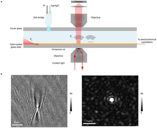
Microfluidics has emerged as a powerful platform for the analysis of minute sample volumes, driving its widespread adoption in biosensing applications. Optical imaging and electrochemical sensing are two typical integration strategies, each offering distinct advantages. The optical methods provide detailed spatial mapping of chemical processes, while electrochemical techniques enable selective detection that is unhindered by optical scattering from impurities. Here, we introduce a novel optical imaging–electrochemical sensor for integrated microfluidic analysis. This approach employs an electrochemical workstation to modulate optical signals, enabling the simultaneous acquisition of decoupled optical images and electrochemical readings. Consequently, it delivers complementary information, revealing both the spatial distribution of analytes and their intrinsic electrochemical properties. We detail the system design and imaging principle, demonstrate its utility through the analysis of noble metal nanoparticles, which are commonly used for signal amplification in biosensors, and finally apply it to monitor biological processes on live cells. We believe this integrated methodology will develop into a powerful tool for operando analysis in microfluidics, significantly expanding its application in the biosensing of complex biological fluids.
Full article
(This article belongs to the Section Biosensor and Bioelectronic Devices)
►▼
Show Figures

Figure 1
Open AccessArticle
Sensitive Visual Detection of Breast Cancer Cells via a Dual-Receptor (Aptamer/Antibody) Lateral Flow Biosensor
by
Yurui Zhou, Jiahui Wang, Ying Han, Meijing Ma, Junhong Li, Haidong Li, Xueji Zhang and Guodong Liu
Biosensors 2026, 16(2), 85; https://doi.org/10.3390/bios16020085 - 30 Jan 2026
Abstract
We report a novel dual-receptor lateral flow biosensor (LFB) for the rapid, sensitive, and visual detection of MCF-7 breast cancer cells as a model for circulating tumor cells (CTCs). The biosensor employs a MUC1-specific aptamer conjugated to colloidal gold nanoparticles as the detection
[...] Read more.
We report a novel dual-receptor lateral flow biosensor (LFB) for the rapid, sensitive, and visual detection of MCF-7 breast cancer cells as a model for circulating tumor cells (CTCs). The biosensor employs a MUC1-specific aptamer conjugated to colloidal gold nanoparticles as the detection probe and an anti-MUC1 antibody immobilized at the test line as the capture probe, forming a unique “aptamer–cell–antibody” sandwich complex upon target recognition. This design enables instrument-free, visual readout within minutes, achieving a detection limit of 675 cells. The assay also demonstrates robust performance in spiked human blood samples, highlighting its potential as a simple, cost-effective dual-mode point-of-care testing (POCT) platform. This platform supports both rapid visual screening and optional strip-reader-based quantification, making it suitable for early detection and monitoring of breast cancer CTCs.
Full article
(This article belongs to the Special Issue The Research and Application of Lateral Flow Biosensors)
Open AccessReview
Electrochemical Sensors as a Tool for Taste Perception in Pharmaceutical Products: Advances and Perspectives
by
Juliana Luz Melo Gongoni, Marilia Medeiros, Hatylas Azevedo and Margarete Moreno de Araújo
Biosensors 2026, 16(2), 84; https://doi.org/10.3390/bios16020084 - 30 Jan 2026
Abstract
Taste masking in pharmaceutical products is a complex and subjective process that requires reliable evaluation methods. This review focuses on the electronic tongue (e-tongue), an emerging sensor-based technology designed to mimic human taste perception without the need for human panels. E-tongue systems provide
[...] Read more.
Taste masking in pharmaceutical products is a complex and subjective process that requires reliable evaluation methods. This review focuses on the electronic tongue (e-tongue), an emerging sensor-based technology designed to mimic human taste perception without the need for human panels. E-tongue systems provide objective data to support the development of palatable formulations. In this review, we discuss the principles, types of e-tongue devices, data processing approaches, and their applications in pharmaceutical research. By comparing e-tongue performance with human taste assessment, we highlight its potential as a complementary tool to traditional in vitro assays, accelerating formulation development and improving patient adherence.
Full article
(This article belongs to the Special Issue Label-Free Electrochemical Biosensing)
Open AccessArticle
A Graphene Field-Effect Transistor-Based Biosensor Platform for the Electrochemical Profiling of Amino Acids
by
Roanne Deanne Aves, Janwa El-Maiss, Divya Balakrishnan, Naveen Kumar, Mafalda Abrantes, Jérôme Borme, Vihar Georgiev, Pedro Alpuim and César Pascual García
Biosensors 2026, 16(2), 83; https://doi.org/10.3390/bios16020083 - 29 Jan 2026
Abstract
In this work, we present the introductory methodology for a graphene field-effect transistor (GFET)-based platform for probing the electrochemical fingerprints of amino acids, designed to enable stable and controlled surface chemistry and electrochemical measurements toward peptide and protein sequencing. We begin with a
[...] Read more.
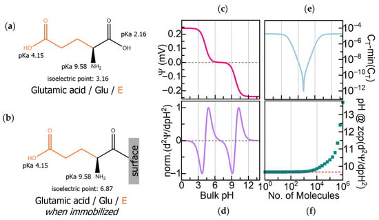
In this work, we present the introductory methodology for a graphene field-effect transistor (GFET)-based platform for probing the electrochemical fingerprints of amino acids, designed to enable stable and controlled surface chemistry and electrochemical measurements toward peptide and protein sequencing. We begin with a focused conceptual review that motivates electrochemical fingerprinting as a strategy for amino acid and peptide identification and contextualizes this approach within recent advances in protein manipulation relevant to sequencing. We then describe a graphene functionalization protocol that facilitates the directional attachment of amino acids onto the graphene surface. This surface chemistry is quantitatively characterized through surface plasmon resonance (SPR), yielding surface densities in the order of 1012 molecules/cm2. The same functionalization protocol enables in situ peptide synthesis directly on graphene, as demonstrated by the successful synthesis of a model tripeptide. To support electrochemical interrogation, we developed three complementary platforms for sensor preconditioning, surface functionalization, and titration-based electrochemical measurements, compatible with both aqueous and organic solutions. Preliminary stability measurements indicate a Dirac point drift below 10 mV over 45 min. Altogether, this work establishes the experimental foundations for electrochemical amino acid and peptide fingerprinting using GFET sensors and provides a framework for the future development of electrochemically enabled protein sequencing technologies.
Full article
(This article belongs to the Special Issue Selected Papers from the 5th International Electronic Conference on Biosensors (IECB 2025))
►▼
Show Figures

Figure 1
Open AccessArticle
Denoising Non-Invasive Electroespinography Signals by Different Cardiac Artifact Removal Algorithms
by
Desirée I. Gracia, Eduardo Iáñez, Mario Ortiz and José M. Azorín
Biosensors 2026, 16(2), 82; https://doi.org/10.3390/bios16020082 - 29 Jan 2026
Abstract
The non-invasive recording of spinal cord neuronal activity, also known as electrospinography (ESG), using high-density surface electromyography (HD-sEMG) is a promising emerging biosensing modality. However, these recordings often contain electrocardiographic (ECG) artifacts that must be removed for accurate analysis. Given the emerging nature
[...] Read more.
The non-invasive recording of spinal cord neuronal activity, also known as electrospinography (ESG), using high-density surface electromyography (HD-sEMG) is a promising emerging biosensing modality. However, these recordings often contain electrocardiographic (ECG) artifacts that must be removed for accurate analysis. Given the emerging nature of ESG and the lack of dedicated signal processing methods, this study assesses the performance of seven established EMG denoising algorithms for their ability to preserve the broad spectral bandwidth needed for future ESG characterization: Template Subtraction (TS), Adaptive Template Subtraction (ATS), High-Pass Filtering at 200 Hz (HP200), ATS combined with HP200, Second-Order Extended Kalman Smoother (EKS2), Stationary Wavelet Transform (SWT), and Empirical Mode Decomposition (EMD). Performance was quantified using six metrics: Relative Error (RE), Signal-to-Noise Ratio (SNR), Cross-Correlation (CC), Spectral Distortion (SD), and Kurtosis Ratio (KR2) and its variation (
(This article belongs to the Special Issue Biophysical Sensors for Biomedical/Health Monitoring Applications (2nd Edition))
►▼
Show Figures

Figure 1
Open AccessArticle
Capturing Emotions Induced by Fragrances in Saliva: Objective Emotional Assessment Based on Molecular Biomarker Profiles
by
Laurence Molina, Francisco Santos Schneider, Malik Kahli, Alimata Ouedraogo, Mellis Alali, Agnés Almosnino, Julie Baptiste, Jeremy Boulestreau, Martin Davy, Juliette Houot-Cernettig, Telma Mountou, Marine Quenot, Elodie Simphor, Victor Petit and Franck Molina
Biosensors 2026, 16(2), 81; https://doi.org/10.3390/bios16020081 - 28 Jan 2026
Abstract
In this study, we describe a non-invasive approach to objectively assess fragrance-induced emotions using multiplex salivary biomarker profiling. Traditional self-reports, physiological monitoring, and neuroimaging remain limited by subjectivity, invasiveness, or poor temporal resolution. Saliva offers an advantageous alternative, reflecting rapid neuroendocrine changes linked
[...] Read more.
In this study, we describe a non-invasive approach to objectively assess fragrance-induced emotions using multiplex salivary biomarker profiling. Traditional self-reports, physiological monitoring, and neuroimaging remain limited by subjectivity, invasiveness, or poor temporal resolution. Saliva offers an advantageous alternative, reflecting rapid neuroendocrine changes linked to emotional states. We combined four key salivary biomarkers, cortisol, alpha-amylase, dehydroepiandrosterone, and oxytocin, to capture multidimensional emotional responses. Two clinical studies (n = 30, n = 63) and one user study (n = 80) exposed volunteers to six fragrances, with saliva collected before and 5 and 20 min after olfactory stimulation. Subjective emotional ratings were also obtained through questionnaires or an implicit approach. Rigorous analytical validation accounted for circadian variation and sample stability. Biomarker patterns revealed fragrance-induced emotional profiles, highlighting subgroups of participants whose biomarker dynamics correlated with particular emotional states. Increased oxytocin and decreased cortisol levels aligned with happiness and relaxation; in comparison, distinct biomarker combinations were associated with confidence or dynamism. Classification and Regression Trees (CART) analysis results demonstrated high sensitivity for detecting these profiles. Validation in an independent cohort using an implicit association test confirmed concordance between molecular profiles and behavioral measures, underscoring the robustness of this method. Our findings establish salivary biomarker profiling as an objective tool for decoding real-time emotional responses. Beyond advancing affective neuroscience, this approach holds translational potential in personalized fragrance design, sensory marketing, and therapeutic applications for stress-related disorders.
Full article
(This article belongs to the Special Issue Biosensing and Diagnosis—2nd Edition)
►▼
Show Figures

Figure 1
Open AccessArticle
Smart Clot: An Automated Point-of-Care Flow Assay for Quantitative Whole-Blood Platelet, Fibrin, and Thrombus Kinetics
by
Alessandro Foladore, Simone Lattanzio, Ekaterina Baryshnikova, Martina Anguissola, Elisabetta Lombardi, Marco Valvasori, Roberto Vettori, Francesco Agostini, Roberto Tassan Toffola, Lidia Rota, Marco Ranucci and Mario Mazzucato
Biosensors 2026, 16(2), 80; https://doi.org/10.3390/bios16020080 - 28 Jan 2026
Abstract
Hemostasis depends on the coordinated interaction between platelets, coagulation factors, endothelium, and shear forces. Current point-of-care (POC) assays evaluate isolated components of haemostasis or operate under nearly static conditions, limiting their ability to reproduce physiological thrombus formation. In this study, we performed the
[...] Read more.
Hemostasis depends on the coordinated interaction between platelets, coagulation factors, endothelium, and shear forces. Current point-of-care (POC) assays evaluate isolated components of haemostasis or operate under nearly static conditions, limiting their ability to reproduce physiological thrombus formation. In this study, we performed the technical validation of Smart Clot, a fully automated, microfluidic POC platform that quantifies platelet aggregation, fibrin formation, and total thrombus growth under controlled arterial shear using unmodified whole blood. Recalcified citrated blood was perfused over collagen at
(This article belongs to the Special Issue Smart, Connected, and Portable Biosensors and Bioelectronics for Advancing Human Healthcare, Disease Diagnosis, and Therapeutics (2nd Edition))
►▼
Show Figures

Figure 1
Open AccessArticle
Inductor-Based Biosensors for Real-Time Monitoring in the Liquid Phase
by
Miriam Hernandez, Patricia Noguera, Nuria Pastor-Navarro, Marcos Cantero-García, Rafael Masot-Peris, Miguel Alcañiz-Fillol and David Gimenez-Romero
Biosensors 2026, 16(2), 79; https://doi.org/10.3390/bios16020079 - 28 Jan 2026
Abstract
Current liquid-phase resonant biosensors, such as Quartz Crystal Microbalance, Surface Acoustic Wave, or Surface Plasmon Resonance, typically rely on specialized piezoelectric substrates or complex optical setups. These requirements often necessitate cleanroom fabrication, thereby limiting cost-effective scalability. This study presents a high-integration sensing platform
[...] Read more.
Current liquid-phase resonant biosensors, such as Quartz Crystal Microbalance, Surface Acoustic Wave, or Surface Plasmon Resonance, typically rely on specialized piezoelectric substrates or complex optical setups. These requirements often necessitate cleanroom fabrication, thereby limiting cost-effective scalability. This study presents a high-integration sensing platform based on standard Printed Circuit Board (PCB) technology, incorporating an embedded inductor within a fluidic system for real-time monitoring. This design leverages industrial manufacturing standards to achieve a compact, low-cost, and scalable architecture. Detection is governed by shifts in the resonance frequency of an LC tank circuit; specifically, increases in bulk ionic strength induce a frequency decrease, whereas biomolecular adsorption at the sensor surface leads to a frequency increase. This phenomenon can be explained by the modulation of the inter-turn capacitance, which is modeled as a combination of capacitive elements accounting for contributions from the bulk electrolyte and the surface-bound dielectric layer. Such divergent responses provide an intrinsic self-discriminating capability, allowing for the analytical differentiation between surface interactions and bulk effects. To the best of our knowledge, this is the first demonstration of an inductor-based resonant sensor fully embedded in a PCB fluidic architecture for continuous liquid-phase analyte monitoring. Validated through a protein-antibody model (Bovine Serum Albumin-anti-Bovine Serum Albumin), the sensor demonstrated a limit of detection of 1.7 ppm (0.026 mM) and a linear dynamic range of 31–211 ppm (0.47–3.2 mM). These performance metrics, combined with a reproducibility of 4 ± 3%, indicate that the platform meets the requirements for robust analytical applications. Its inherent simplicity and potential for miniaturization position this technology as a viable candidate for point-of-care diagnostics in diverse environments.
Full article
(This article belongs to the Section Biosensor and Bioelectronic Devices)
►▼
Show Figures

Graphical abstract
Open AccessArticle
Development of a Non-Contact Flow Sensor Based on a Permanent Magnet Metal Clip for Monitoring Circulation Status
by
Kicheol Yoon, Seung Hee Choi, Tae-Hyeon Lee, Sangyun Lee, Sunghoon Kang, Sun Jin Sym and Kwang Gi Kim
Biosensors 2026, 16(2), 78; https://doi.org/10.3390/bios16020078 - 27 Jan 2026
Abstract
Foreign matter accumulating on catheters during ascites paracentesis in cancer patients can interfere with the procedure. The paracentesis site must be visually inspected by patients or medical staff. We propose a monitoring method using sensors, as they enable real-time, automatic status detection. The
[...] Read more.
Foreign matter accumulating on catheters during ascites paracentesis in cancer patients can interfere with the procedure. The paracentesis site must be visually inspected by patients or medical staff. We propose a monitoring method using sensors, as they enable real-time, automatic status detection. The proposed design integrates a sensor into the drainage tube to detect liquid flow using the Lorentz force. The sensor consists of a permanent magnet, a yoke, and a signal processing circuit. Mu-metal shields the magnet to prevent interference with surrounding circuits. Physiological saline solution is used as a substitute for bodily fluids. Sensor performance was evaluated via finite element analysis. The Lorentz force generated an average voltage of 11.07 μV when liquid was present, enabling detection of the flow status. The proposed sensor is non-invasive and features a clip design, allowing attachment and detachment from the drainage tube for reuse. Non-invasiveness ensures safety from infection, and reusability can reduce medical costs. This study proposes a sensor for monitoring peritoneal puncture status. By detecting liquid flow using the Lorentz force, the system enables real-time monitoring during the procedure.
Full article
(This article belongs to the Section Biosensors and Healthcare)
►▼
Show Figures

Figure 1
Open AccessArticle
In Vitro Investigation of the PneumoWave Biosensor for the Identification of Central Sleep Apnea in Pediatrics
by
Burcu Kolukisa Birgec, Ross Langley, Jennifer Miller, Osian Meredith, Beyza Toprak and Alexander Balfour Mullen
Biosensors 2026, 16(2), 77; https://doi.org/10.3390/bios16020077 - 27 Jan 2026
Abstract
The interpretation and diagnosis of central sleep apnea in pediatrics by nocturnal polysomnography is challenging due to its technical complexity, which involves the simultaneous recording of multiple physiological parameters related to sleep and wakefulness. Furthermore, the unfamiliar environment of a sleep laboratory can
[...] Read more.
The interpretation and diagnosis of central sleep apnea in pediatrics by nocturnal polysomnography is challenging due to its technical complexity, which involves the simultaneous recording of multiple physiological parameters related to sleep and wakefulness. Furthermore, the unfamiliar environment of a sleep laboratory can hinder sleep evaluation, and diagnostic backlogs are common due to restricted capacity at specialist tertiary centers. The ability to undertake home sleep studies in a familiar environment using simple, robust, and low-cost technology is attractive. The potential to repurpose the PneumoWave biosensor, a UKCA Class 1 device, registered as an accelerometer-based monitoring device that is intended to capture and store chest motion data continuously over a period of time for retrospective analysis, was explored in an in vitro model of central sleep apnea. The PneumoWave system contains a biosensor (PW010), which was able to record simulated apnea episodes of 5 to 20 s across physiologically relevant pediatric breathing rates using an in vitro manikin model and manual annotation. The findings confirm that the PneumoWave biosensor could be a useful technology to support home sleep apnea testing and warrant further exploration.
Full article
(This article belongs to the Section Biosensors and Healthcare)
►▼
Show Figures

Figure 1
Open AccessArticle
Portable Point-of-Care Uric Acid Detection System with Cloud-Based Data Analysis and Patient Monitoring
by
Yardnapar Parcharoen, Pratya Phetkate, Kanon Jatuworapruk, Calin Trif and Chiravoot Pechyen
Biosensors 2026, 16(2), 76; https://doi.org/10.3390/bios16020076 - 27 Jan 2026
Abstract
Uric acid is closely related to diseases such as gout, kidney failure, and metabolic disorders. A conventional method for measuring uric acid over 24 h is time intensive and cumbersome for patients who have to take samples to the hospital. At present, hospitals
[...] Read more.
Uric acid is closely related to diseases such as gout, kidney failure, and metabolic disorders. A conventional method for measuring uric acid over 24 h is time intensive and cumbersome for patients who have to take samples to the hospital. At present, hospitals use only laboratory instruments to determine 24-h uric acid concentrations in the urine. This study presents the proof-of-concept of a portable point-of-care tool called Uricia, designed to improve the quality of life of patients monitoring uric acid. Spectrophotometry was performed at a fixed wavelength of 295 nm. The urine sample contained within the cuvette absorbs ultraviolet light, with uric acid specifically responsible for this absorption, thereby allowing the device to measure its concentration. An internal calibration algorithm was used to accommodate the nonlinear optical response of Uricia and was calibrated to a benchtop GENESYS 10S UV–Vis spectrophotometer. The experiments further evaluated potential urinary interferences, revealing that while most constituents had minimal impact, ascorbic acid demonstrated the highest interference, contributing up to 15% of the total signal at high physiological concentrations. This device and the corresponding spectrophotometry method revealed that high concentrations of uric acid precipitated insoluble crystals. A dilution set to an alkali solution vial to be premixed and dissolve the uric acid crystals was added, increasing the detection window to 10 mg/dL, with an LOD of 0.0232 mg/dL and LOQ of 0.0702 mg/dL. Cloud-based data measurement enables spot analysis, which is meant to provide insight into patient status development. These results validated the technical architecture of a controlled matrix for measuring uric acid.
Full article
(This article belongs to the Section Biosensors and Healthcare)
►▼
Show Figures

Figure 1
Open AccessArticle
A Deep-Learning-Enhanced Ultrasonic Biosensing System for Artifact Suppression in Sow Pregnancy Diagnosis
by
Xiaoying Wang, Jundong Wang, Ziming Gao, Xinjie Luo, Zitong Ding, Yiyang Chen, Zhe Zhang, Hao Yin, Yifan Zhang, Xuan Liang and Qiangqiang Ouyang
Biosensors 2026, 16(2), 75; https://doi.org/10.3390/bios16020075 - 27 Jan 2026
Abstract
The integration of artificial intelligence (AI) with ultrasonic biosensing presents a transformative opportunity for enhancing diagnostic accuracy in agricultural and biomedical applications. This study develops a data-driven deep learning model to address the challenge of acoustic artifacts in B-mode ultrasound imaging, specifically for
[...] Read more.
The integration of artificial intelligence (AI) with ultrasonic biosensing presents a transformative opportunity for enhancing diagnostic accuracy in agricultural and biomedical applications. This study develops a data-driven deep learning model to address the challenge of acoustic artifacts in B-mode ultrasound imaging, specifically for sow pregnancy diagnosis. We designed a biosensing system centered on a mechanical sector-scanning ultrasound probe (5.0 MHz) as the core biosensor for data acquisition. To overcome the limitations of traditional filtering methods, we introduced a lightweight Deep Neural Network (DNN) based on the YOLOv8 architecture, which was data-driven and trained on a purpose-built dataset of sow pregnancy ultrasound images featuring typical artifacts like reverberation and acoustic shadowing. The AI model functions as an intelligent detection layer that identifies and masks artifact regions while simultaneously detecting and annotating key anatomical features. This combined detection–masking approach enables artifact-aware visualization enhancement, where artifact regions are suppressed and diagnostic structures are highlighted for improved clinical interpretation. Experimental results demonstrate the superiority of our AI-enhanced approach, achieving a mean Intersection over Union (IOU) of 0.89, a Peak Signal-to-Noise Ratio (PSNR) of 34.2 dB, a Structural Similarity Index (SSIM) of 0.92, and clinically tested early gestation accuracy of 98.1%, significantly outperforming traditional methods (IoU: 0.65, PSNR: 28.5 dB, SSIM: 0.72, accuracy: 76.4). Crucially, the system maintains a single-image processing time of 22 ms, fulfilling the requirement for real-time clinical diagnosis. This research not only validates a robust AI-powered ultrasonic biosensing system for improving reproductive management in livestock but also establishes a reproducible, scalable framework for intelligent signal enhancement in broader biosensor applications.
Full article
(This article belongs to the Special Issue Artificial Intelligence (AI) and Machine Learning (ML) in Biosensors: Innovation, Application, and Challenge)
►▼
Show Figures

Figure 1
Open AccessArticle
Innovative Fatty Acid-Guided Biosensor Design for Neutrophil Gelatinase, a Prognostic and Diagnostic Biomarker for Chronic Kidney Disease
by
Kaustubh Jumle, Priya Paliwal, Mohamed A. M. Ali, Ravi Ranjan Kumar Niraj, Anis Ahmad Chaudhary and Manali Datta
Biosensors 2026, 16(2), 74; https://doi.org/10.3390/bios16020074 - 26 Jan 2026
Abstract
Chronic kidney disease (CKD) afflicts 850 million people worldwide, with an estimate that it is the 5th highest cause of years of life lost (YLLs). Standard confirmatory procedures for disease are blood and urine analysis with ultrasound for confirmation. Neutrophil gelatinase-associated lipocalin (NGAL)
[...] Read more.
Chronic kidney disease (CKD) afflicts 850 million people worldwide, with an estimate that it is the 5th highest cause of years of life lost (YLLs). Standard confirmatory procedures for disease are blood and urine analysis with ultrasound for confirmation. Neutrophil gelatinase-associated lipocalin (NGAL) has been established as a prognostic biomarker, especially for the pre-clinical stages of CKD, thus presenting itself as a dependable predictor of the progression. With the aim of designing diagnostics, fatty acids were explored as potential biorecognition elements for the selective capture of NGAL. Three fatty acids—linoleic acid, arachidonic acid, and retinoic acid—were shortlisted as plausible candidates based on their known affinity toward lipocalin family proteins. Docking followed by molecular dynamics simulations were employed to evaluate the binding affinity and stability of each complex. Among them, linoleic acid exhibited the most favorable interaction, as evidenced by the lowest binding free energy. Subsequently, fluorescence and electrochemical techniques—square-wave voltammetry, differential pulse voltammetry, cyclic voltammetry, and electrochemical impedance spectroscopy (EIS)—were systematically compared for qualitative and quantitative checking of the accuracy of NGAL detection. Amongst the electrochemical techniques, differential pulse voltammetry DPV demonstrated superior analytical performance with an LOD of 0.05 ng/mL with a sensitivity of 23.2 µA/cm2/pg. To the best of our knowledge, this is the first report of a fatty acid-based biosensor platform for NGAL detection, presenting a novel approach for CKD diagnostics. The sensitivity obtained is comparable with available NGAL detection methods yet cost-effective and robust.
Full article
(This article belongs to the Special Issue Electrochemical Biosensing Platforms for Food, Drug and Health Safety—2nd Edition)
►▼
Show Figures

Figure 1
Open AccessArticle
A RPA-CRISPR/Cas12a-Powered Catalytic Hairpin Assembly Fluorescence Biosensor for Duck Plague Virus Virulent Strain Detection
by
Yue Wu, Jiaxin Wan, Xingbo Wang, Yunjie Shen, Xiangjun Li, Weidong Zhou, Yinchu Zhu and Xing Xu
Biosensors 2026, 16(2), 73; https://doi.org/10.3390/bios16020073 - 26 Jan 2026
Abstract
Duck plague virus (DPV), a highly contagious α-herpesvirus in the livestock and poultry environment, poses a significant threat to the healthy growth of ducks, potentially causing substantial economic losses. Effective control of DPV requires the development of specific diagnostic tools. A new fluorescent
[...] Read more.
Duck plague virus (DPV), a highly contagious α-herpesvirus in the livestock and poultry environment, poses a significant threat to the healthy growth of ducks, potentially causing substantial economic losses. Effective control of DPV requires the development of specific diagnostic tools. A new fluorescent biosensor (R-C-CHA) was developed to detect virulent strains of DPV. It combined recombinase polymerase amplification (RPA), a CRISPR/Cas12a system, and catalytic hairpin assembly (CHA) for signal enhancement. The RPA primers were specifically designed to target the conserved DPV-CHv UL2 gene region, allowing for the rapid, efficient amplification of the target nucleic acids in isothermal conditions. The CRISPR/Cas12a system was used for sequence-specific recognition, activating its lateral cleavage activity. Furthermore, the CHA cascade reaction was utilized for enzyme-free fluorescent signal amplification. The results showed that the R-C-CHA biosensor completed the detection process in 40 min with a detection limit of 0.02 fg/μL, which was an approximate five-fold improvement compared to traditional RPA-CRISPR/Cas12a biosensors. The R-C-CHA biosensor also demonstrated perfect consistency with clinical detection and polymerase chain reaction (PCR) diagnosis, highlighting its strong potential for rapid detection in livestock and poultry farming settings.
Full article
(This article belongs to the Special Issue Sensors for Environmental Monitoring and Food Safety—2nd Edition)
►▼
Show Figures

Figure 1
Open AccessArticle
Rapid and Sensitive Detection of Candida albicans Using Microfluidic-Free Droplet Digital Non-Amplification Dependent CRISPR/Cas12a Assay
by
Jie Peng, Chao Guo, Ze-Yun Huang, Wen-Fei Xu and Xu-Hui Li
Biosensors 2026, 16(2), 72; https://doi.org/10.3390/bios16020072 - 26 Jan 2026
Abstract
Candida albicans is a major fungal pathogen associated with vulvovaginal candidiasis, and rapid, sensitive detection remains challenging, particularly in amplification-free formats. Here, we report NaPddCas, a microfluidic-free, droplet-based CRISPR/Cas12a detection strategy for qualitative identification of Candida albicans DNA. Unlike conventional bulk CRISPR assays,
[...] Read more.
Candida albicans is a major fungal pathogen associated with vulvovaginal candidiasis, and rapid, sensitive detection remains challenging, particularly in amplification-free formats. Here, we report NaPddCas, a microfluidic-free, droplet-based CRISPR/Cas12a detection strategy for qualitative identification of Candida albicans DNA. Unlike conventional bulk CRISPR assays, NaPddCas partitions the reaction mixture into vortex-generated polydisperse droplets, enabling spatial confinement of Cas12a activation events and effective suppression of background fluorescence. This compartmentalization substantially enhances detection sensitivity without nucleic acid amplification or microfluidic devices. Using plasmid and genomic DNA templates, NaPddCas achieved reliable detection at concentrations several orders of magnitude lower than bulk CRISPR/Cas12a reactions. The assay further demonstrated high specificity against non-target bacterial and fungal species and was successfully applied to clinical vaginal secretion samples. Importantly, NaPddCas is designed as a qualitative or semi-qualitative droplet-dependent digital detection method rather than a quantitative digital assay. Owing to its simplicity, sensitivity, and amplification-free workflow, NaPddCas represents a practical approach for laboratory-based screening of Candida albicans infections.
Full article
(This article belongs to the Special Issue Biosensing and Diagnosis—2nd Edition)
►▼
Show Figures

Figure 1
Open AccessReview
Probing Glycosaminoglycan–Protein Interactions: Applications of Surface Plasmon Resonance
by
Changkai Bu, Lin Pan, Lianli Chi, Vitor H. Pomin, Jonathan S. Dordick, Chunyu Wang and Fuming Zhang
Biosensors 2026, 16(2), 71; https://doi.org/10.3390/bios16020071 - 25 Jan 2026
Abstract
Glycosaminoglycans (GAGs) are highly negatively charged polysaccharides that play essential roles in numerous physiological and pathological processes through their interactions with proteins. These interactions govern cellular signaling, inflammation, coagulation, and recognition. Surface Plasmon Resonance (SPR) has emerged as a key biophysical technique for
[...] Read more.
Glycosaminoglycans (GAGs) are highly negatively charged polysaccharides that play essential roles in numerous physiological and pathological processes through their interactions with proteins. These interactions govern cellular signaling, inflammation, coagulation, and recognition. Surface Plasmon Resonance (SPR) has emerged as a key biophysical technique for label-free, real-time characterization of biomolecular interactions, offering insights into binding kinetics, affinity, and specificity. SPR-based approaches to glycosaminoglycan–protein interaction studies offer powerful tools for elucidating the roles of GAGs in a wide range of physiological and pathological processes. In this review, we systematically discuss experimental strategies, data analysis methods, and representative applications of SPR-based glycosaminoglycan–protein interactions. Special attention is given to the challenges associated with GAG heterogeneity and immobilization, as well as recent technological advances that enhance sensitivity and throughput. To our knowledge, this review represents one of the first systematic and up-to-date summaries specifically focused on recent advances in applying SPR to the study of glycosaminoglycan–protein interactions.
Full article
(This article belongs to the Special Issue Surface Plasmon Resonance-Based Biosensors and Their Applications)
►▼
Show Figures

Figure 1

Journal Menu
► ▼ Journal Menu-
- Biosensors Home
- Aims & Scope
- Editorial Board
- Reviewer Board
- Topical Advisory Panel
- Instructions for Authors
- Special Issues
- Topics
- Sections & Collections
- Article Processing Charge
- Indexing & Archiving
- Editor’s Choice Articles
- Most Cited & Viewed
- Journal Statistics
- Journal History
- Journal Awards
- Conferences
- Editorial Office
Journal Browser
► ▼ Journal BrowserHighly Accessed Articles
Latest Books
E-Mail Alert
News
Topics
Topic in
Applied Sciences, Biosensors, Designs, Electronics, Materials, Micromachines, Applied Biosciences
Wearable Bioelectronics: The Next Generation of Health Insights
Topic Editors: Shuo Gao, Yu Wu, Wenyu WangDeadline: 31 March 2026
Topic in
Applied Nano, Biosensors, Materials, Nanomaterials, Chemosensors, Applied Biosciences, Laboratories
Applications of Nanomaterials in Biosensing: Current Trends and Future Prospects
Topic Editors: Kundan Sivashanmugan, Xianming KongDeadline: 30 April 2026
Topic in
Analytica, Antibiotics, Bioengineering, Biosensors, Microorganisms
Spectroscopic Phenotypic Typing of Multidrug-Resistant Bacteria: Advances in FTIR and Raman Applications
Topic Editors: László Orosz, David Rodríguez TemporalDeadline: 31 January 2027
Topic in
Biosensors, C, Sensors, Foods, Physchem
Development of Photoelectrochemical Sensors
Topic Editors: Reyhan Selin Uysal, Aslı Barla-Demirkoz, Baykal SariogluDeadline: 28 February 2027

Conferences
Special Issues
Special Issue in
Biosensors
Recent Advances in Glucose Biosensors
Guest Editors: Natalija German, Anton PopovDeadline: 20 February 2026
Special Issue in
Biosensors
New Advances in Bioimaging and Biosensing Based on Nanomaterials
Guest Editor: Steven WuDeadline: 28 February 2026
Special Issue in
Biosensors
Trends and Perspective of Advanced Nanotechnology for Bio-Sensing, Imaging and Cancer Therapy
Guest Editors: Xuemei Wang, Xiaohui LiuDeadline: 28 February 2026
Special Issue in
Biosensors
Micro/Nano-Biosensors for Environmental Applications
Guest Editors: Lijuan Liang, Lei WuDeadline: 28 February 2026
Topical Collections
Topical Collection in
Biosensors
Microsystems for Cell Cultures
Collection Editors: Iordania Constantinou, Thomas E. Winkler


