Article

Ultra-Sensitive Detection of Mercury by Using Field-Effect Transistor Biosensors Based on Single-Walled Carbon Nanotubes

1
Department of Medical Imaging, Jiangsu University Affiliated People’s Hospital (Zhenjiang First People’s Hospital), Zhenjiang 212002, China
2
School of Life Sciences, Jiangsu University, Zhenjiang 212013, China
3
School of Life Sciences, Qinghai Normal University, Xining 810000, China
*
Authors to whom correspondence should be addressed.
Current address: Suzhou Yaoming Biologics Co., Ltd., Suzhou 215028, China.
Biosensors 2025, 15(12), 779; https://doi.org/10.3390/bios15120779 (registering DOI)
Submission received: 15 October 2025 / Revised: 12 November 2025 / Accepted: 21 November 2025 / Published: 26 November 2025

Abstract

In recent years, the amount of mercury discharged by human activities has continued to increase. Most of the mercury in surface water settles into the sediment, where it can be directly or indirectly transformed into mercury ion (Hg2+) compounds (such as dimethylmercury) under the action of microorganisms. Hg2+ display high toxicity and bioaccumulation in food, such as fish and rice, and thus the contamination of mercury ion is a serious concern for human health. Practical Hg2+ detection methods are usually limited by the sensitivity and selectivity of the used methods, such as colorimetric determination and fluorescence biosensor based on the solution phase. Therefore, it is urgent to develop Hg2+ detection methods in the practical environment with high sensitivity and selectivity. DNA is low-cost, relatively stable, and has been used for different fields. In this study, DNA for Hg2+detection was absorbed on the surface of single-walled carbon nanotubes (SWNTs) by using 1,5-diaminonaphthalene (DAN) based on field-effect transistor (FET) biosensors. The interaction between DNA and Hg2+ can be directly converted into electrical signals based on the SWNTs biosensors. The experimental results showed that the limit of detection (LOD) of Hg2+ without the phase-locked amplifier was about 42.6 pM. The function of the phase-locked amplifier is to achieve fast detection of the biosensor with strong anti-noise ability. Intriguingly, the sensitivity of the biosensor combined with a phase-locked amplifier to detect Hg2+ was further improved to be 5.14 pM compared with some current methods of biosensors. Furthermore, this biosensor has an excellent selectivity and practical detection in tap water, which demonstrates its high performance and low cost in practical application in Hg2+ detection. These results show this method for Hg2+ detection using SWNTs biosensors with a phase-locked amplifier is promising.

1. Introduction

In recent years, the amount of mercury discharged by human activities has continued to increase, causing mercury pollution in water bodies, mainly from wastewater emissions from industries such as chlor-alkali, plastics, and electronics. Most of the mercury in surface water settles into the sediment, where it can be directly or indirectly transformed into mercury ion (Hg2+) compounds (such as dimethylmercury) under the action of microorganisms. Mercury displays high toxicity and long-lasting bioaccumulation in ecosystems. Excessive mercury in the human body can cause lung injury, vomiting, diarrhea, nausea, movement disorders, as well as language and hearing impairment. Moreover, mercury damages nerves and other organs and causes severe dysfunctions such as kidney and muscle issues, deformed limbs, paralysis, degeneration and necrosis of brain cells, difficulty in swallowing, and death [1,2,3,4]. This has raised public awareness of the dangers posed by Hg2+. The limits in Hg2+ content for different fields were set. The highest permitted level of mercury in industrial pollutants, according to the regulations of the European Parliament and Council of the European Union, is 0.03 mg⋅mL−1 (150 nM) [5]. Therefore, developing advanced methods for Hg2+ detectionin the practical environment with high sensitivity and selectivity were further required [6].
At present, some techniques have been established to detect Hg2+ in the literature, including but not limited to atomic absorption spectrometry (AAS) [7], mass spectrometry (MS) [8], and atomic emission spectrometry (AES) [9]. However, some methods either have poor detection sensitivity or require expensive equipment, complicated processes, and lengthy pre-treatment. DNA is low-cost, relatively stable, and has been used in different fields. And DNA sequence with ‘T’ bases can specifically bind with Hg2+. To address these issues for Hg2+ detection, DNA sequence was used to build biosensors for Hg2+ detection with convenience offabrication and measurement [10]. However, the actual sensitivity of the detection based on biosensors based on the DNA sequence also needs to be improved. A field effect transistor (FET) biosensor is mainly made of nano-materials or organic semiconductors (OSC) as channel sensing film, such as carbon nanotubes, graphene, and pentacene [11,12,13]. A metal oxide semiconductor (MOS) FET device usually consists of the source, drain, and gate terminals, where the current in the semiconductor channel between the source and drain (ID) terminals is regulated by the electronic field generated by the voltage applied between the gate terminals (VG) and the source drain (VSD) terminals. Gate voltages can be conducted through either the bottom gate, named ‘back gate FET’, or the top gate, named ‘top gate FET’ [14]. FET biosensors have been widely used in the biological field due to their characteristics of high selectivity; high sensitivity; real-time response; and unlabeled detection in the detection of proteins, nucleic acids, and viruses [15,16,17,18]. Single-walled carbon nanotube (SWNT) is a typical carbon nanomaterial with high quality and preparation convenience compared with some other carbon nanomaterials like fullerene and graphene. Nonetheless, SWNT-based FET biosensors are limited by the absence of functional groups on the surface of the SWNTs and the poor electrical connectivity between the molecular detector and SWNTs. These issues can be further solved by using a connecting compound and a lock-in amplifier.
A lock-in amplifier is an electronic device that can measure dynamic signals in real time with strong anti-noise ability; it is mainly composed of oscillators, mixers, low-pass filters, and other parts. For weak signals buried in noise, the principle of orthogonality is used to retain the signal of selected frequency, weaken the impact of noise, and accurately extract the signal through frequency conversion [19,20]. Moreover, the other function of the phase-locked amplifier is to achieve fast detection of the biosensor because the phase-locked amplifier can provide a fast response to the signals. In this study, 1,5-diaminonaphthalene (DAN) as a connecting channel between the surface of the SWNTs and DNA sequence and a lock-in amplifier as the measurement instrument were used to improve the sensitivity of Hg2+ detection. Then, SWNT biosensors were further used for detection in real samples.

2. Materials and Methods

2.1. Chemical Reagents and Experimental Materials

DNA, 5′-NH2-TTT TTT GGG TGG GTG GGT GGG TTT TTT-3′, was from Shanghai Sangon Co., Ltd. (Shanghai, China) and was verified for correctness via high performance liquid chromatography (HPLC) purification and mass spectrometry analysis. The ultrapure water (UP) used for HPLC was prepared using a laboratory ultrapure water system (UPF-10 L). Other reagents such as ferric chloride (FeCl3), sodium chloride (NaCl), calcium chloride (CaCl2), potassium chloride (KCl), zinc chloride (ZnCl2), magnesium chloride (MgCl2), concentrated sulfuric acid (H2SO4), hydrogen peroxide (H2O2), and ethanol (CH3CH2OH) were purchased from Sinopharm Chemical Reagent Co., Ltd. (Shanghai, China). Copper mesh AG200F4 was purchased from Medium Lens Instrument Co., Ltd. Dithiothreitol (DTT) was purchased from Sigma-Aldrich (Shanghai, China). Silicon wafer withthermal oxide layer on itand gold thread were purchased from Hefei Kejing Co., Ltd. (Hefei, China).

2.2. Instruments

The tubular high temperature vacuum muffle furnace (OTF-1200X) equipped with three mass flow controllers (SSL-3Z-LCD) was purchased from Hefei Kejing Technology Co., Ltd. (Hefei, China). The high vacuum resistance thermal evaporator (ZHD300) was made by Beijing Taikenuo Technology Co., Ltd. (Beijing, China). Vacuum dryer (IPC250) was made by Baoding Fage Instrument Manufacturing Co., Ltd. (Baoding, China). The vacuum systems mentioned above were maintained by a two-stage vacuum pump (VRD-8/220V/50/60Hz). The room temperature magnetic stirrer (B11-3) was purchased from Shanghai Sile Instrument Co., Ltd. (Shanghai, China). Fourier transform infrared spectroscopy (FT-IR) was conducted by a Nicolet iN10 (Shanghai, China). The field emission scanning electron microscope (FE-SEM) withultra-high resolution was made by Hitachi (SU8030) (Beijing, China). The semiconductor parameter meter (B1500A) was purchased from Keysight Technologies (Beijing, China), and the lock-in amplifier (MF-DEV5811) was purchased from Beijing Yanjing Electronics Co., Ltd. (Beijing, China). The BioTek Synergy H4 multipurpose microplate reader was used to measure the fluorescence intensity. Origin 8.0 was used to process the data.

2.3. Principle of the Biosensor

FET based on SWNT was used as an ultra-sensitive biosensor for Hg2+ detection, and multi-T DNA sequences were functionally attached to the surface of the SWNTs to selectively determine Hg2+. A thermal layer of SiO2 on the wafer surface allows for thesubstrate insulation and can be used as the dielectric layer in the FET device. Therefore, the SWNT connected to the substrate is the unique conductive channel between the source and the drain, which constitutes the SWNT FET with the back gate type and low leakage current. Because charged biomolecules are adsorbed to SWNTs, the band structure of SWNT FET can be modified by the biomarkers, resulting in changes in source-drain current (ID). Therefore, such changes in electrical properties of the devices can be used to detect biomolecules.
Due to the π-π stacking interaction between the coagulant DAN and the SWNTs sp2 plane, DAN can be connected to the SWNTs. Thus, the SWNT FET biosensor was treated with 10 μM DAN in methanol (40 μL) at room temperature for 3 h and then washed with 0.01 M phosphate buffer solution (PBS, pH 7.4). After that, 40 μL 2% (V/V) glutaraldehyde (GA) was added for 3 h to bind DAN by Schiff reaction. Then, 40 μL ssDNA (20 μM) was fixed to GA on the surface of the SWNTs at room temperature for 6 h after the biosensor was rinsed with PBS buffer to remove excess GA. Its electrical characteristics were measured by the semiconductor parameter meter to screen qualified devices. DNA is conductive. As shown in Figure 1, the DNA sequence was connected to the surface of the SWNTs. Hg2+ can react with the DNA sequence with the stable structure of “T-Hg-T”. Therefore, the conformation of DNA sequence change and the corresponding current change were induced. Finally, different concentrations of Hg2+ onto the surface of the SWNTs were added and reacted with the DNA sequence at room temperature for 30 min. The specific detection of Hg2+ using the SWNT FET biosensor was measured. The combination of Hg2+ and DNA sequence resulted in DNA conformation change, which can further lead to the static change at the interface and electrical signal change.

2.4. Single-Walled Carbon Nanotube Preparation

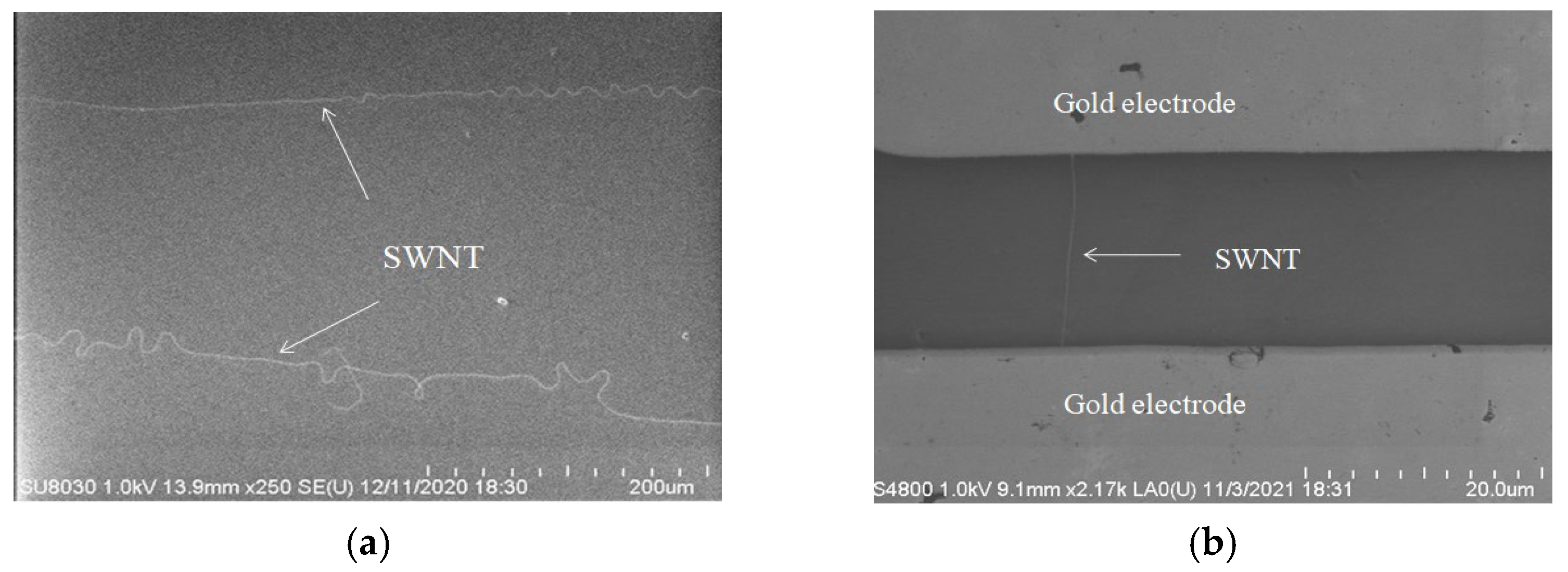
SWNTs were prepared by chemical vapor deposition (CVD). In this method, SWNTs were grown by the chemical reaction of two or more precursors through a template attached with catalyst particles at 800~1200 °C. First, concentrated H2SO4 and H2O2 liquid mixture with a volume ratio of 7:3 was added to the petri dish. Then, a sliced silicon wafer of about 1 cm2 was added, and the above system was heated to 110 °C for 2 h. After that, the H2SO4 and H2O2liquid mixture in the dish was removed, and the waferwas soaked by ultra-pure water with the help of ultrasonic cleaning. After 10 min, the silicon wafer was taken out and cleaned by ultra-pure water again. Finally, it was blow-dried with nitrogen and placed under vacuum for later use. The silicon wafer spin-coated with 0.05 mmol/L Fe(OH)3 was put into the muffle furnace on the quartz boat and placed in the middle of the muffle furnace chamber. Second, the vacuum was kept and maintained in the furnace, and argon gas (Ar, 212 sccm) was injected under the monitoring of mass flow controller. Third, hydrogen (H2, 297 sccm) was then introduced. Ten minutes later, the carbon source—ethanol (argon as carrier gas)—was introduced. After 20 min, ethanol, hydrogen, and argon were gradually shut down, and the muffle furnace began to cool down. Figure 2a shows the scanning electron microscope (SEM, Hitachi SU8030, Beijing, China) image of the SWNTs.

2.5. Thermal Evaporation of Metal Electrode

Then 80 nm Cr and 200 nm Au were thermal evaporated on the silicon wafers. The purpose of Cr evaporation was used to make the silicon wafer have better contact with the gold electrode. As shown in Figure 2b, the SEM image of the SWNT after gold electrode evaporation demonstrated that SWNT spanned the gold electrodes, showing that the SWNT FET sensors had been made.

2.6. 1,5-Diaminonaphthalene Connection

After the SWNT FET biosensor was successfully prepared, it was treated with DAN dissolved in methanol (10 mM, 40 μL) at room temperature for 3 h and then washed with 10 mM PBS (pH 7.4) to remove the excess DAN. Then, 40 μL 2% (V/V) glutaraldehyde (GA) was added and stayed for 3 h. After combination with DAN via the Schiff reaction, the excess GA was washed by PBS buffer. Then the DNA sequence was connected on the surface of SWNT via DAN.

2.7. DTT Reaction with Hg2+

DTT is a sulfhydryl compounded with sulfhydryl groups at both ends, which is often used as a small molecule reducing agent [21]. Sulfur has low electronegativity and high polarizability, while Hg2+ has a large volume and low positive charge. Sulfhydryl has strong adsorption for Hg2+. After adding 100 μM Hg2+on the surface of the SWNT FET electrode and then immersing it in 2.5 mM DTT/ethanol solution, the SWNT biosensors were kept for 24 h at 4 °C. After cleaning with ultra-pure water and drying with nitrogen, the current of the device was measured. Then, 100 µM Hg2+ was added again and the reaction time was for 30 min at room temperature. After adding Hg2+, the current of SWNT FET decreased due to the combination of “T-Hg2+-T” with DNA. After adding DTT, the sulfhydryl group absorbed Hg2+ with positive charges and enhanced the electronic conductivity of SWNT FET on its surface, which restored the current.

3. Results and Discussion

3.1. Current Changes Before and After Connection of DNA

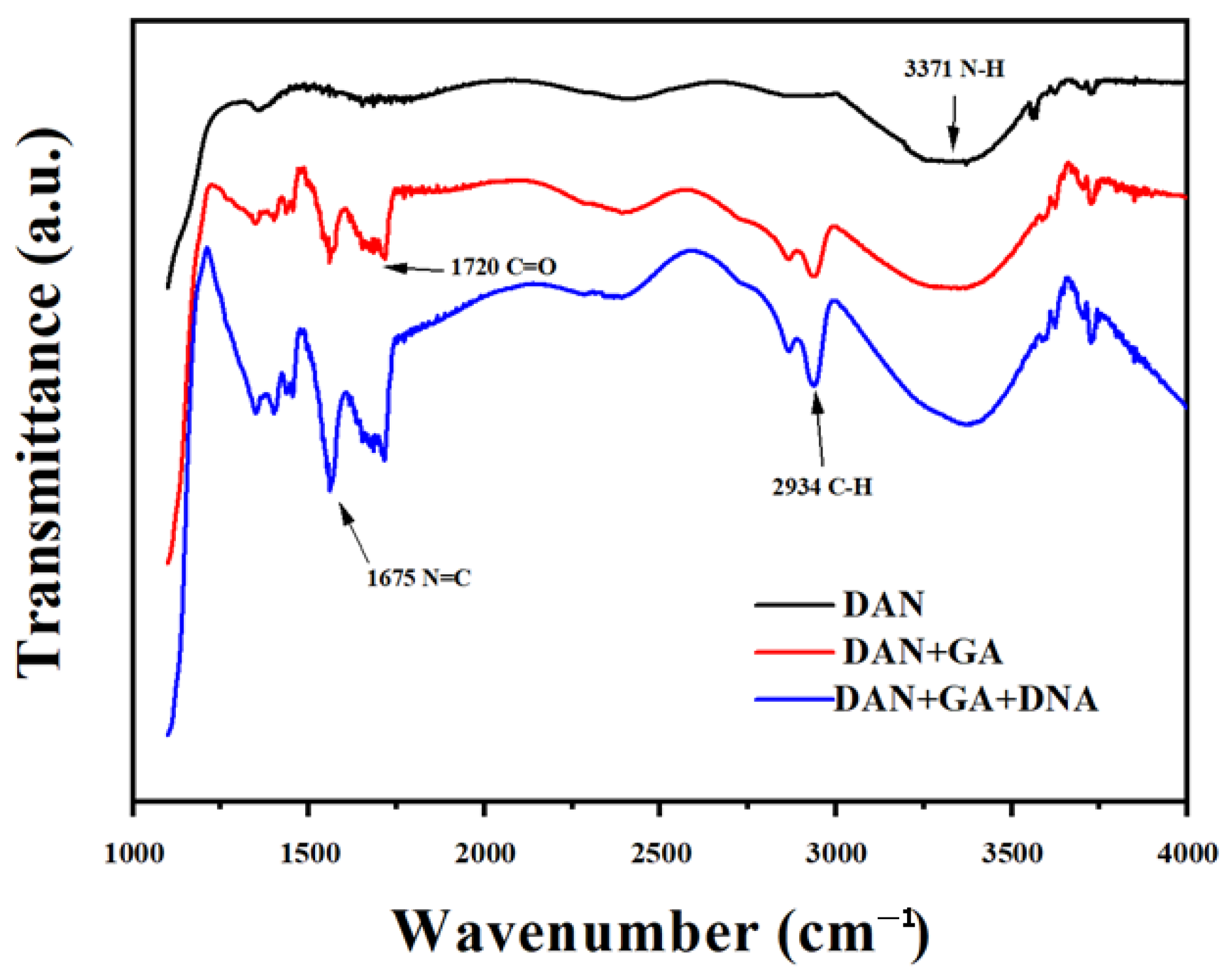
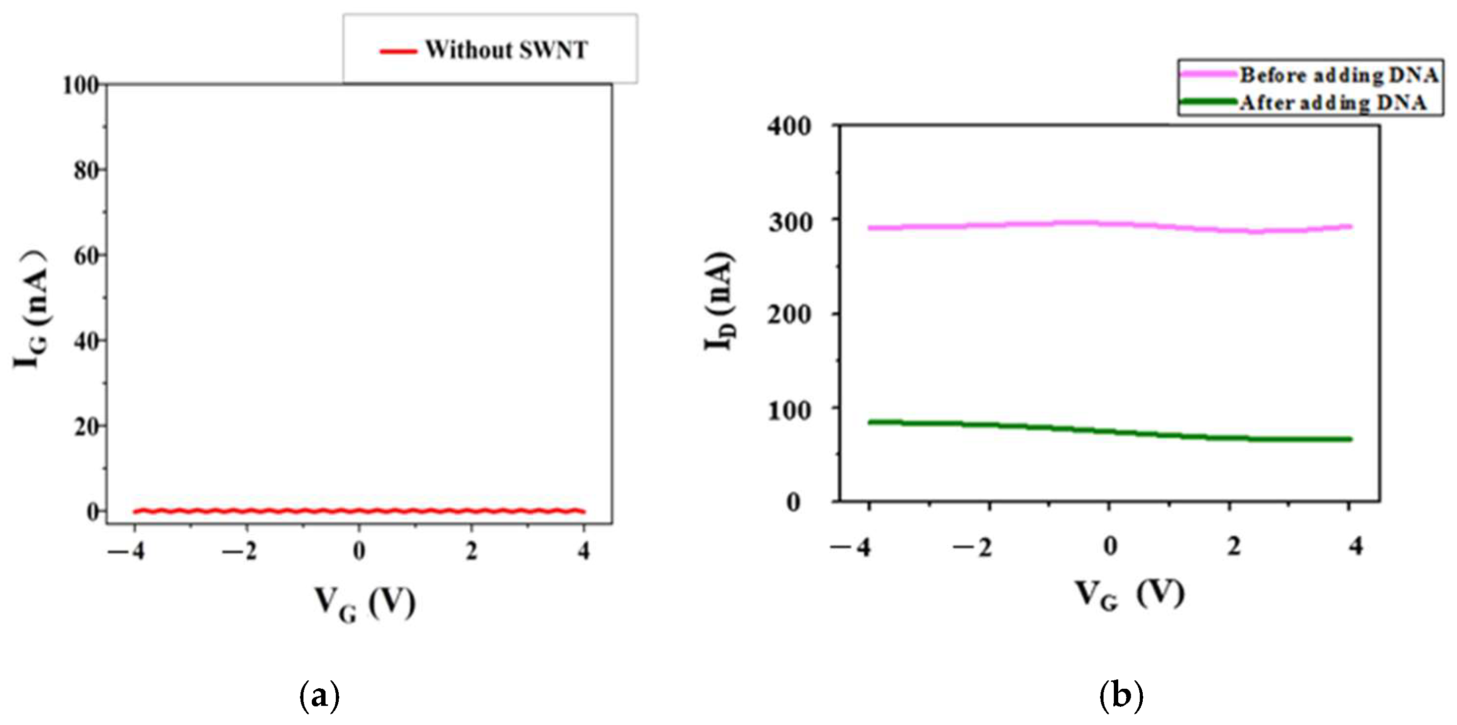
Usually, the surface of SWNT has no functional groups. The Schiff-base reaction proceeded through chemical attachment between the aldehyde group of GA and the amine group of the DAN connected at the SWNT. DNA was immobilized to the GA on the SWNT surface, which was also based on the Schiff-base reaction [22]. As shown in Figure 3, it is known that -NH2 and –C=O are the characteristic functional groups of DAN and GA+DAN, respectively. C=N is the characteristic functional group of DAN+GA+DNA. Figure 3 shows the FT-IR spectrums of DAN, DAN+GA, and DAN+GA+DNA, which demonstrate that DNA was connected to DAN successfully. Then, 40 μL ssDNA (20 μM) was fixed on GA on the surface of the SWNTs at room temperature for 6 h. After that, the excess DNA was rinsed with PBS buffer. As shown in Figure 4a, the control device without the single-walled carbon nanotubes showed no current value because SWNTs in the sensors were the same as the conductive material. As shown in Figure 4b, after the addition of DNA on the surface of the SWNTs, ID was significantly reduced for about 200 nA. This reveals the successful conjugation of DNA on the surface of the SWNTs.

3.2. Hg2+ Sensitivity Detection

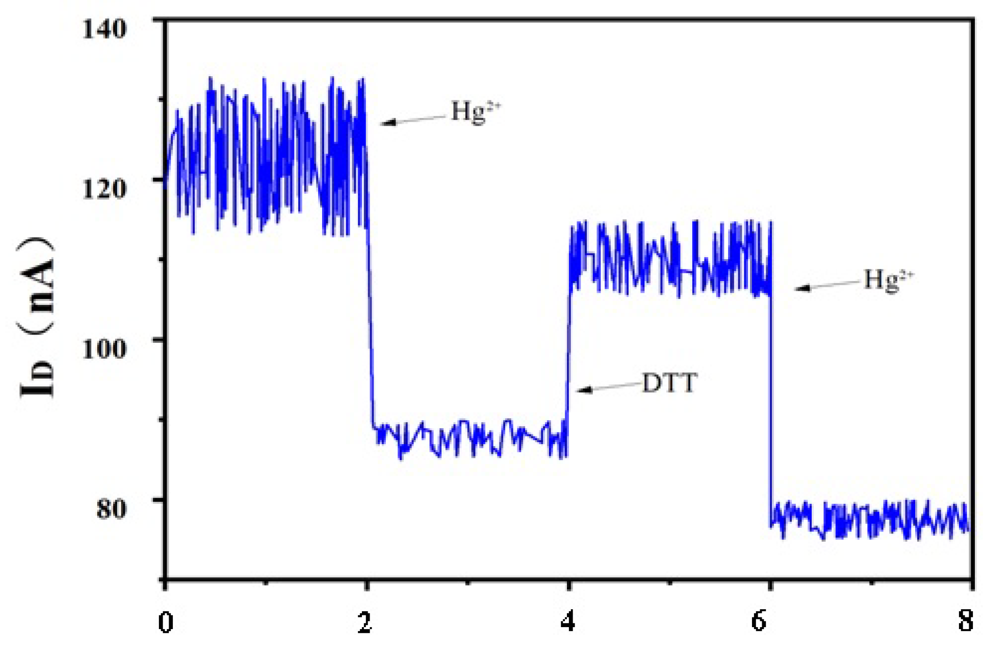
After the successful fabrication of the biosensor, 100 µM Hg2+ was added to the surface of the electrode modified with DTT, and then the current change of the biosensor was measured and calculated. The reaction between Hg2+ and DNA forming “T-Hg2+-T” led to the decrease of the measured current (Figure 5). When Hg2+ was added to the system, it specifically interacted with DNA sequences rich in multiple ‘T’ bases due to its unique chemical properties. This can form a structurally stable and orderly ‘T-Hg2+-T’ hairpin complex. The special structure interfered with the electron transport in the relevant pathway, leading to a significant decrease in the measured current intensity. When DTT was added to the system, the thiol (-SH) reactive groups present in the DTT molecular structure played a key role. These SH groupscan quickly and efficiently adsorb Hg2+ bound to the DNA due to their strong coordination ability. As Hg2+ was largely removed, the electron conduction pathways that were previously blocked by Hg2+ binding to DNA responded. This significantly enhanced the electron transport performance of the SWNT biosensors and resulted in a noticeable recovery of the current intensity. After adding Hg2+ and DTT, the change in current can repeatedly appear. However, the conductivity of the SWNT biosensor was effectively reduced due to multiple repetitions. This can decrease the current value of the SWNT biosensor.
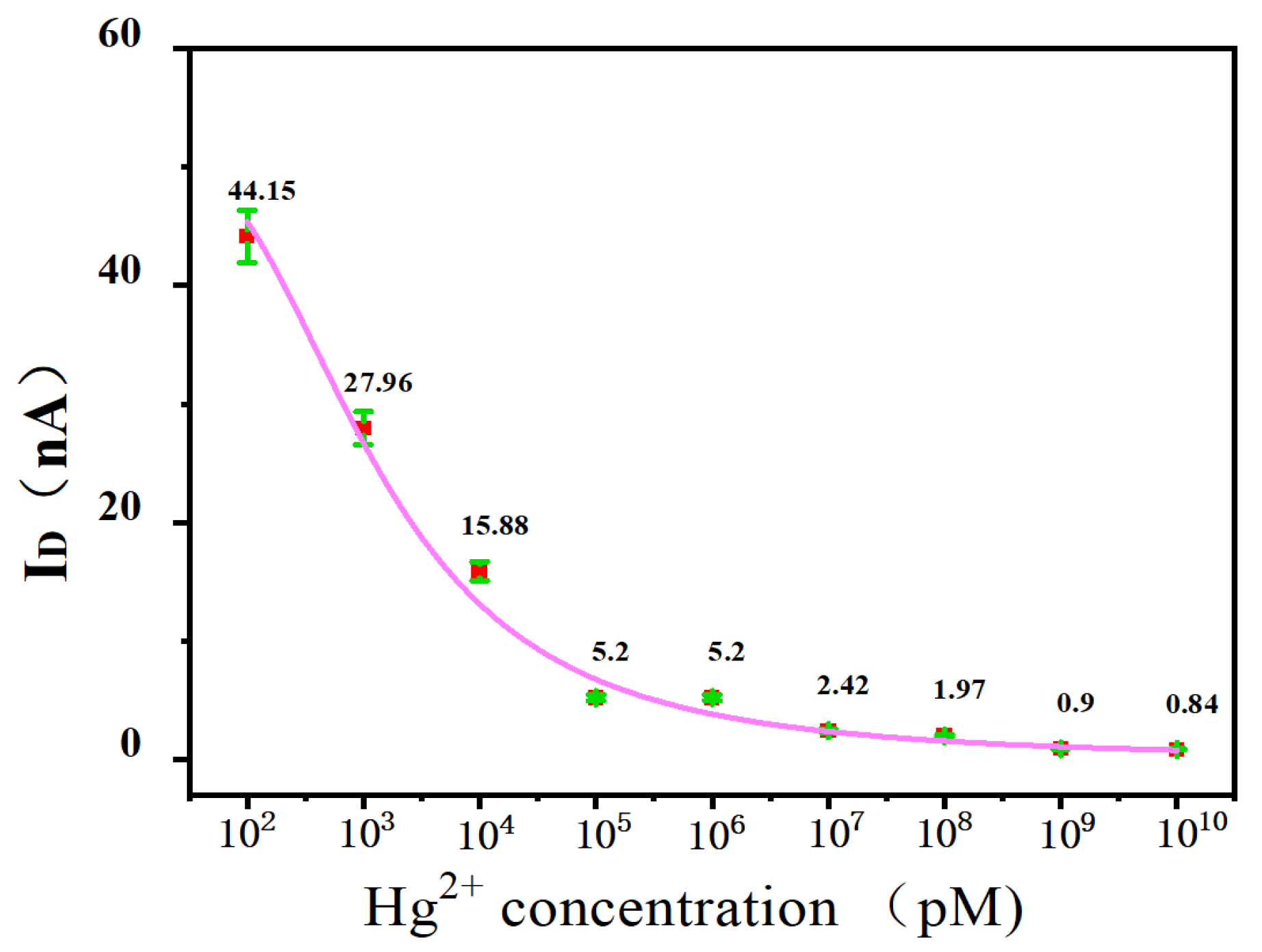
Then, different concentrations of Hg2+ (100 pM, 1 nM, 10 nM, 100 nM, 1 μM, 10 μM, 100 μM, 1 mM, and 10 mM) were added to the SWNT FET biosensor and reacted at room temperature for 30 min. The electrical measurement results were shown in Figure 6. As the binding between T-Hg2+-T was very tight, with the increase of Hg2+ concentration, more Hg2+ bound to the device. Thus, the current response will continue to decrease (i.e., the measured ID will continue to decrease). When the concentration of Hg2+ was in the range of 0.1~100 nM, it showed an obvious linear relationship, and its linear regression equation can be given as follows: y = −11.77 lg (x) + 52.19 with the square of thelinear correlation coefficient R2=0.99. Based on 3S/N, the limit of detection (LOD) was calculated to be 42.6 pM.

3.3. Hg2+ Sensitivity Detection with Phase-Locked Amplifier

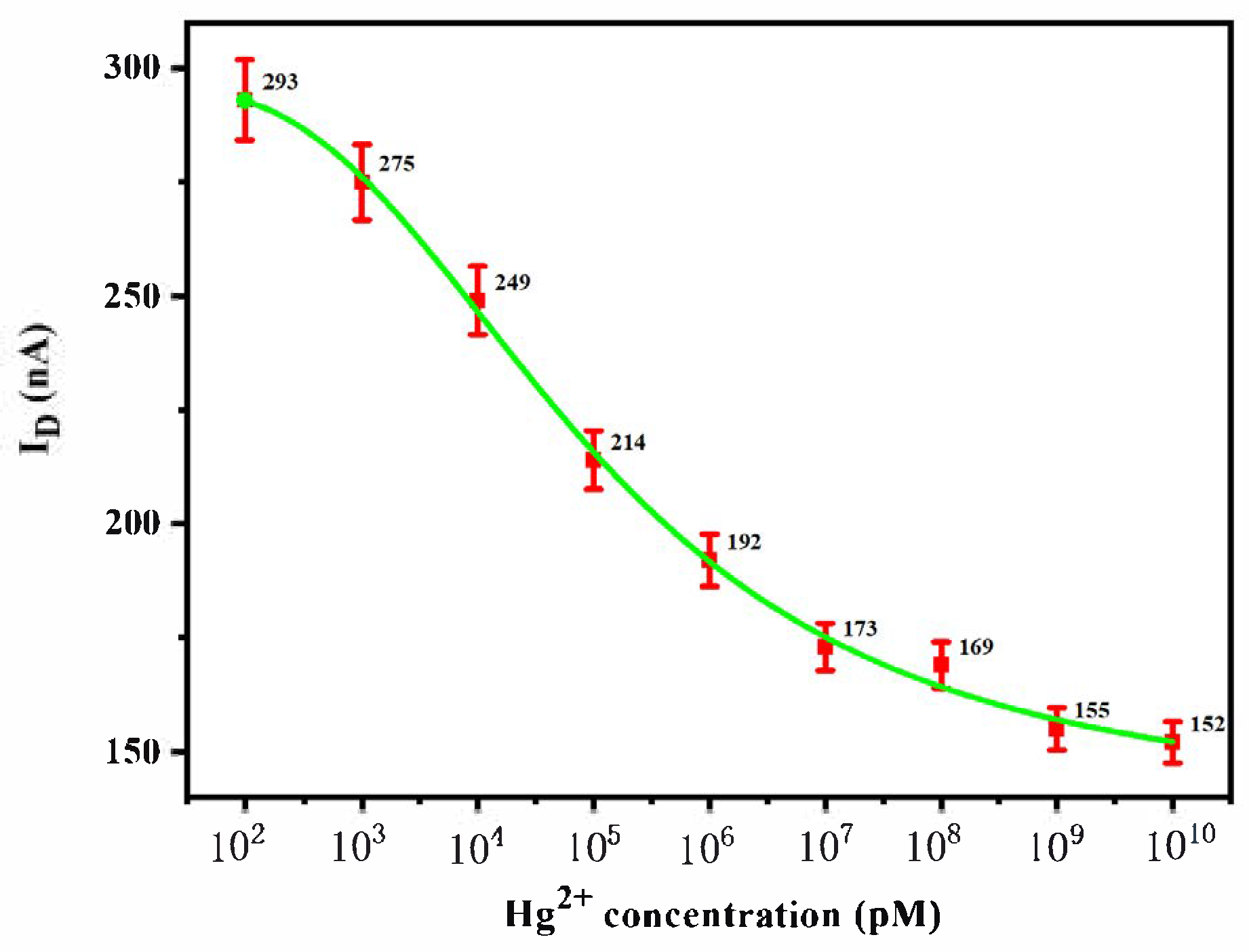
The sensitivity of the SWNT FET biosensor was further analyzed using the phase-locked amplifier to optimize the LOD. After adding different concentrations of Hg2+ (100 pM~0 mM) to the SWNT FET biosensor and reaction at room temperature for 30 min, its current change was detected by the phase-locked amplifier. As shown in Figure 7, when the concentration of Hg2+ (cHg2+) was in the range of 0.1~1 μM, ID had a significant linear relationship with cHg2+. The linear regression equation was as follows: y = −25.69 lg (cHg2+) + 322.89, with R2 = 0.99. Based on 3S/N, the LOD was calculated to be 5.14 pM.
To compare the results of this work and the other reported literature, the linear range and the LOD of this work and some other studies are shown in Table 1. It can be clearly concluded that the LOD of this work has an obvious advantage compared with that of other works.

3.4. Selectivity Analysis

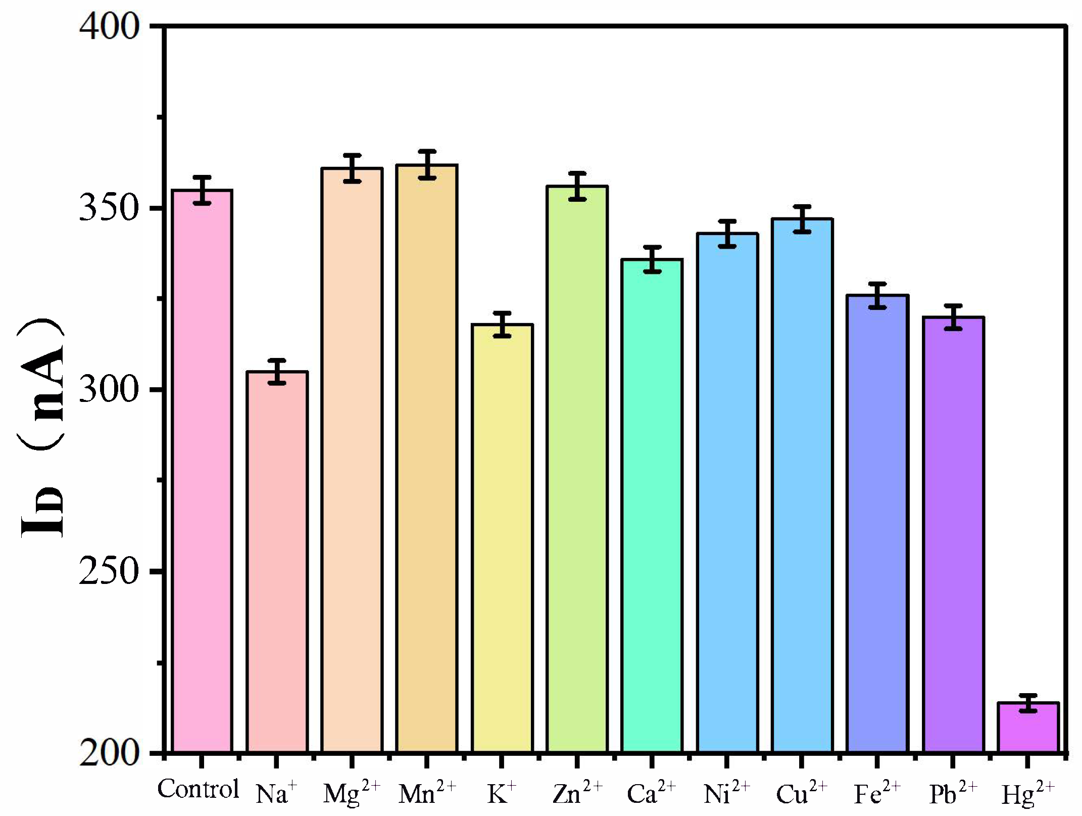
Selectivity is the result of strong binding by species of interest and less binding by species of noninterest, which is an important index to evaluate the specific performance of the SWNT FET sensor in detecting Hg2+. To assess the specificity of the biosensor for Hg2+ detection, Na+, Mg2+, Mn2+, K+, Zn2+, Ca2+, Ni2+, Cu2+, Fe2+, Pb2+, and Hg2+ were added separately at the same concentration of 100 μM to the biosensor for evaluating the selectivity of the device. As shown in Figure 8, the ID measured by adding Hg2+ was significantly lower than that of other ions and control. Other ions had less change for ID. This showed that SWNT FET biosensor had good selectivity to Hg2+.

3.5. Detection in Real Samples

In order to evaluate the sensor’s stability and practical applicability and determine the practical application of the Hg2+ biosensor, three samples of tap water containing 100 nM, 1 nM, and 10 nM of Hg2+ (adding different qualities of Hg2+ solution into tap water) were analyzed using the SWNT FET biosensor system by the standard sampling method, and the results were shown in Table 2 below. The reproducibility of all samples was 95.98~102.05%, and the relative standard deviation (RSD) was 3.08~5.06%, which showed that the biosensor had good stability and fulfilled the requirements of practical application.

4. Conclusions

A SWNT FET-based biosensor was developed with single-stranded DNA for Hg2+ detection. The biosensor can directly convert the interaction between the DNA sequence and Hg2+ into electrical signals, which can maintain high sensitivity and selectivity. When the concentration of Hg2+ was within the range of 0.1~100 nM, an obvious linear relationship between the concentration of Hg2+ and the electrical signal was observed. The LOD was 42.6 pM. The environmental noise generated by its non-specific binding was reduced in order to achieve efficient signal transmission using the characteristics of lock-in amplifier. The sensitivity of the biosensor combined with a lock-in amplifier to detect Hg2+ was therefore further improved. The device showed a detection limit as low as 5.14 pM with excellent selectivity at the same time. DTT has strong adsorption with Hg2+, which can significantly enhance the electron transport performance of the SWNT biosensors and result in a noticeable recovery of the current intensity. In addition, the SWNT FET biosensor with a lock-in amplifier can be expected to be used for the detection of other biomolecules due to its simple manufacturing process and excellent sensing performance, which has great application prospects. These results show that this method for Hg2+ detection using SWNTs biosensors with a phase-locked amplifier is promising.

Author Contributions

Methodology, Q.L.; Validation, Q.L. and Y.L.; Investigation, C.L.; Resources, Y.L.; Writing—original draft, C.L.; Writing—review & editing, L.G.; Supervision, L.G. All authors have read and agreed to the published version of the manuscript.

Funding

This work is supported by National Natural Science Foundation of China (NSAF, No.U2230132), the open fund of Information Materials and Intelligent Sensing Laboratory of Anhui Province (Grant No. IMIS202209), the Guiding Science and Technology Plan Project for Social Development in Zhenjiang City (FZ2022052), the Zhenjiang Science and Technology Plan (Social development, SH2024098), the National Foreign Experts Program Project of China (H20240553), and the Key Laboratory of Medicinal Animal and Plant Resources of Qinghai-Tibetan Plateau in Qinghai Province.

Institutional Review Board Statement

Not applicable.

Informed Consent Statement

Not applicable.

Data Availability Statement

The data supporting this study’s findings are available from the corresponding author upon reasonable request.

Conflicts of Interest

Author Qiuxiang Lv was employed by the company Suzhou Yaoming Biologics Co., Ltd. The remaining authors declare that the research was conducted in the absence of any commercial or financial relationships that could be construed as a potential conflict of interest.

References

  1. Xing, X.; Zhang, Y.; Tan, Y.; Hu, T.; Chen, Z.; Liu, B. A multi-Well SERS chip for the detection of Hg2+. ACS Appl. Appl. Appl. Nano Material 2024, 7, 17287–17294. [Google Scholar] [CrossRef]
  2. Yu, X.; Chang, W.; Cai, Z.; Yu, C.; Lai, L.; Zhou, Z.; Li, P.; Yang, Y.; Zeng, C. Hg2+detection and information encryption of new [1+1] lanthanide cluster. Talanta 2024, 26, 125105. [Google Scholar] [CrossRef]
  3. Hu, P.; Liu, J.; Xia, C.; Liu, B.; Zhu, H.; Niu, X. Matrix redox interference-free nanozyme-amplified detection of Hg2+ using thiol-modified phosphatase-mimetic nanoceria. Sens. Actuators B-Chem. 2024, 401, 135030. [Google Scholar] [CrossRef]
  4. Zhou, J.; Zhang, C.; Hu, C.; Li, S.; Liu, Y.; Chen, Z.; Li, S.; Chen, H.; Sami, R.; Deng, Y. Electrochemical aptasensor based on black phosphorus-porous graphene nanocomposites for high-performance detection of Hg2+. Chin. Chem. Lett. 2024, 35, 109561. [Google Scholar] [CrossRef]
  5. European Parliament and Council of the European Union, Directive 2010/75/EU of the European Parliament and Council of 24 November 2010 on Industrial Emissions (Integrated Pollution Prevention and Control) (Recast). Available online: https://www.legislation.gov.uk/eudr/2010/75 (accessed on 10 October 2025).
  6. Xia, N.; Feng, F.; Liu, C.; Li, R.; Xiang, W.; Shi, H.; Gao, L. The detection of mercury ion using DNA as sensors based on fluorescence resonance energy transfer. Talanta 2019, 192, 500–507. [Google Scholar] [CrossRef] [PubMed]
  7. Zhao, B.; He, M.; Chen, B.; Hu, B. Facile green synthesis of magnetic porous organic polymers for fast preconcentration of trace lead andmercuryfrom environmental water followed by graphite urnace atomic absorption spectrometry detection. Spectrochim. Acta Part B-At. Spectrosc. 2022, 196, 106524. [Google Scholar] [CrossRef]
  8. Yang, H.; Qi, L.; Zhou, J.; Li, Q.; Yuan, X.; Zhang, M.; He, Y.; Huang, K.; Chen, P. Metal ions-regulated chemical vapor generation of Hg2+: Mechanism and application in miniaturized point discharge atomic emission spectrometry assay of oxalate in clinical urolithiasis samples. Anal. Chim. Acta 2023, 1262, 341223. [Google Scholar] [CrossRef] [PubMed]
  9. Ilmiah, K.; Sumranjit, J.; Wutikhun, T.; Siripinyanond, A. Tracking silver nanoparticles during their synthesis by inductively coupled plasmamassspectrometry: Implications for colorimetric sensing of mercury ions. ACS Appl. Nano Mater. 2023, 6, 1250–1260. [Google Scholar] [CrossRef]
  10. Gao, L.; Lv, Q.; Xia, N.; Lin, Y.; Lin, F.; Han, B. Detection of mercury ion with high sensitivity and selectivity using a DNA/graphene oxide hybrid immobilized on glass slides. Biosensors 2021, 11, 300. [Google Scholar] [CrossRef]
  11. Ouyang, J.; Li, Y.; Yang, F.; Wu, X.; Qiu, Z.; Shu, J. Electrochemical and field effect transistor dual-mode biosensor chip for label-free detection of cytokine storm biomarker with high sensitivity within a wide range. Adv. Funct. Mater. 2024, 34, 2405212. [Google Scholar] [CrossRef]
  12. Howe, L.; Wang, Y.; Ellepola, K.; Ho, V.; Dohmen, R.; Pinto, M.; Hoff, W.; Cooney, M.; Vinh, N. Interfacial photogating of graphene field-effect transistor for photosensorybiomolecular detection. Adv. Electron. Mater. 2025, 11, 2400716. [Google Scholar] [CrossRef]
  13. Li, D.; Ren, Y.; Chen, R.; Wu, H.; Zhuang, S.; Zhang, M. Label-free MXene-assisted field effect transistor for the determination of IL-6 in patients with kidney transplantation infection. Microchim. Acta 2023, 190, 284. [Google Scholar] [CrossRef]
  14. Guo, L.; Zhang, Q.; Chan, K. A dual-gate field-effect transistor in graphene heterojunctions. Superlattices Microstruct. 2021, 150, 106778. [Google Scholar] [CrossRef]
  15. Hu, J.; Liu, X.; Li, F.; Qiu, Y.; Hu, Y.; Zhou, Y.; Wang, P.; Wan, H. Ultrasensitive graphene field-effect transistor biosensor for rapidly detecting miRNA-208a. Sens. Actuators B-Chem. 2024, 418, 136262. [Google Scholar] [CrossRef]
  16. Gwyther, R.; Côté, S.; Lee, C.; Miao, H.; Ramakrishnan, K.; Palma, M.; Jones, D. Optimising CNT-FET biosensor design through modelling of biomolecular electrostatic gating and its application to β-lactamase detection. Nat. Commun. 2024, 15, 7482. [Google Scholar] [CrossRef]
  17. Li, J.; Zhang, Y.; Wei, C.; Li, Y.; Peng, Z.; Chuang, H.; Pearce, L.; Boon, A.; Huang, Y.; Kim, D.; et al. Advanced detection of SARS-CoV-2 and omicron variants via MXene-graphene hybrid biosensors utilizing nucleic acid probes. ACS Appl. Nano Mater. 2024, 24, 28255–28272. [Google Scholar] [CrossRef]
  18. Huang, K.; Geng, Y.; Zhang, X.; Chen, D.; Cai, Z.; Wang, M.; Zhu, Z.; Wang, Z. A wide-band digital lock-in amplifier and its application in microfluidic impedance measurement. Sensors 2019, 19, 3519. [Google Scholar] [CrossRef] [PubMed]
  19. Zhang, Y.; Yu, Z.; Li, Y.; Li, S.; Lin, T. A lock-in amplifier modeling recovery method to extract the surface nuclear magnetic resonance signal from residual noise. Rev. Sci. Instrum. 2019, 90, 114710. [Google Scholar] [CrossRef] [PubMed]
  20. Xiao, K.; Zhu, R.; Zhang, X.; Du, C.; Chen, J. Ultrasensitive detection and efficient removal of mercury ions based on covalent organic framework spheres with double active sites. Anal. Chim. Acta 2023, 1278, 341751. [Google Scholar] [CrossRef]
  21. Xing, Y.; Han, J.; Wu, X.; Pierce, D.; Zhao, J. Aggregation-based determination of mercury (II) using DNA-modified single gold nanoparticle, T-Hg (II)-T interaction, and single-particle ICP-MS. Microchim. Acta 2019, 187, 56. [Google Scholar] [CrossRef]
  22. Tu, J.; Gan, Y.; Liang, T.; Hu, Q.; Wang, Q.; Ren, T.; Sun, Q.; Wan, H.; Wang, P. Graphene FET array biosensor based on ssDNA aptamer for ultrasensitive Hg2+ detection in environmental pollutants. Front. Chem. 2018, 6, 333. [Google Scholar] [CrossRef] [PubMed]
  23. Dang, W.; Li, Y.; Zhang, J. Highly sensitive detection of Hg2+ based on imprinting sensor modified DNA. IEEE Sens. J. 2024, 24, 23369–23375. [Google Scholar] [CrossRef]
  24. Wang, M.; Guan, J.; Liu, S.; Chen, K.; Gao, Z.; Liu, Q.; Chen, X. Dual-ligand lanthanide metal-organic framework probe for ratiometric fluorescence detection of mercury ions in wastewater. Microchim. Acta 2023, 190, 359. [Google Scholar] [CrossRef]
  25. Gao, L.; Liu, C.; Li, R.; Xia, N.; Xiong, Y. Highly sensitive detection of Hg2+ using covalent linking single-strand DNA to the surface of graphene oxide with co-anchor strand. Anal. Methods 2019, 11, 4416–4420. [Google Scholar] [CrossRef]
  26. Hu, H.; Yin, Z.; Cui, H.; Xiong, W.; Yu, F.; Zhang, J.; Liao, F.; Wei, G.; Yang, L.; Zhang, J.; et al. A novel dual-detection electrochemiluminescence sensor for the selective detection of Hg2+ and Zn2+: Signal suppression and activation mechanisms. Anal. Chim. Acta 2024, 1330, 343283. [Google Scholar] [CrossRef] [PubMed]
  27. Zhang, J.; Deng, Z.; Feng, H.; Shao, B.; Liu, D. A multifunctional fluorescent sensor for Ag+ and Hg2+ detection in seawater. Environ. Monit. Assess. 2024, 196, 22. [Google Scholar] [CrossRef] [PubMed]
  28. Wang, Y.; Feng, J.; Tan, Z.; Wang, H. Electrochemical impedance spectroscopy aptasensor for ultrasensitive detection of adenosine with dual backfillers. Biosens. Bioelectron. 2014, 60, 218–223. [Google Scholar] [CrossRef]
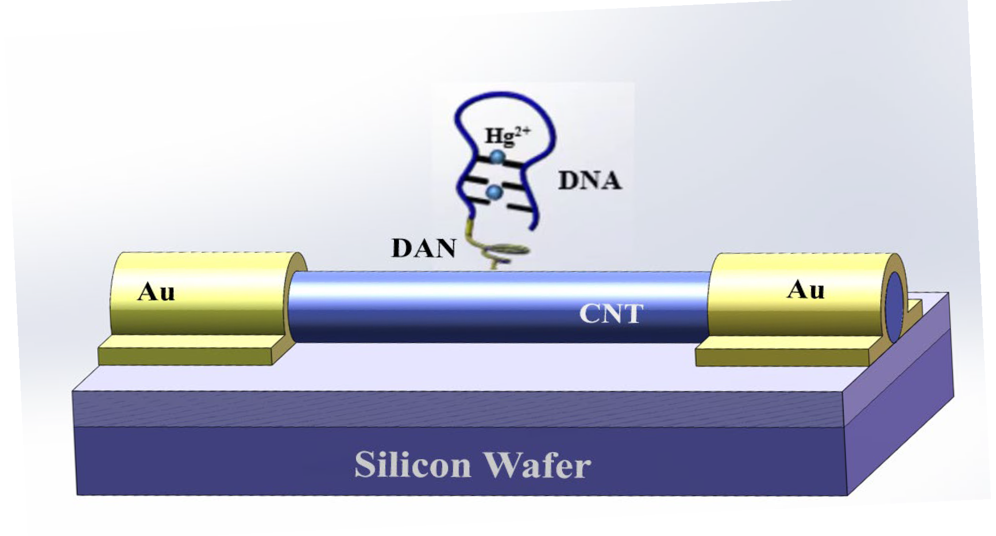
Figure 1. Schematic diagram of field-effect transistor biosensors using single-walled carbon nanotubes. DNA sequences with ‘T’ bases were functionally attached to the surface of single-walled carbon nanotube to selectively determine Hg2+. The conformation of DNA sequence change and the corresponding current change were induced. Au was gold electrode; DAN was 1,5-diaminonaphthalene; CNT was carbon nanotube in this figure.
Biosensors 15 00779 g001
Figure 2. (a) Scanning electron microscope image of single-walled carbon nanotubes. (b) Scanning electron microscope of single-walled carbon nanotubes between gold electrodes. SWNT was single-walled carbon nanotube in this figure.
Biosensors 15 00779 g002
Figure 3. FT-IR spectrums of DAN, DAN+GA, and DAN+GA+DNA. NH2 and –C=O are the characteristic functional groups of DAN and GA+DAN, respectively. C=N is the characteristic functional group of DAN+GA+DNA. Thus, it is shown that DNA was connected to DAN successfully.
Biosensors 15 00779 g003
Figure 4. (a) The control device without the single-walled carbon nanotubes showed no current value because the single-walled carbon nanotubes in the sensors were the same as the conductive material. (b) The currents for the biosensor before and after connecting DNA on the surface of single-walled carbon nanotubes. After the addition of DNA, ID was significantly decreased. This showed that the DNA sequence was connected on the surface of single-walled carbon nanotubes and also decreased the current value of biosensor.
Biosensors 15 00779 g004
Figure 5. Response of SWNT FET biosensor after adding DTT and 100 µM Hg2+. When DTT was added to the system, the thiol (-SH) reactive groups present in the DTT molecular structure played a key role. These SH groups can quickly and efficiently adsorb Hg2+ bound to the DNAdue to their strong coordination ability. After Hg2+ was largely removed, the electron conduction pathways that were previously blocked by Hg2+ binding to DNA wererestored.
Biosensors 15 00779 g005
Figure 6. Sensitivity of field-effect transistor biosensor based on single-walled carbon nanotube to different concentrations (100 pM, 1 nM, 10 nM, 100 nM, 1 μM, 10 μM, 100 μM, 1 mM, and 10 mM) of Hg2+. As the binding between T-Hg2+-T was tight with the increasing of Hg2+ concentration, more Hg2+ bound to the device. Therefore, the current (the values were also showed in the figure) response continued to decrease (i.e., the measured ID will continue to decrease).
Biosensors 15 00779 g006
Figure 7. Sensitivity for different concentration of Hg2+ (100 pM~0 mM) based on SWNT FET biosensor by phase-locked amplifier. When the concentration of Hg2+ (cHg2+) was in the range of 0.1~1 μM, ID (the values were also showed in the figure) had a significant linear relationship with cHg2+. The LOD was calculated to be 5.14 pM.
Biosensors 15 00779 g007
Figure 8. Selectivity for various metal ions (Na+, Mg2+, Mn2+, K+, Zn2+, Ca2+, Ni2+, Cu2+, Fe2+, Pb2+, and Hg2+) using the field-effect transistor biosensor based on single-walled carbon nanotube. ID measured by adding Hg2+ was significantly lower than that of other ions. This showed that the field-effect transistor biosensor based on single-walled carbon nanotubehad good selectivity to Hg2+.
Biosensors 15 00779 g008
Table 1. A comparison of our method with other methods for Hg2+ detection.
MethodLinear RangeDD ODReference
Imprinting sensor0.01~100,000 nM0.006 nM[23]
Fluorescence sensor using metal–organic framework probe10–60 nM1.62 nM[24]
Fluorescence sensor based on graphene oxide sensor2~ 20 μM40 nM[25]
Electrochemiluminescence sensor0.02 μM~0.1μM2.52 nM[26]
Fluorescent sensor using gold nanoparticles 1 uM~1nM4.71 nM[27]
Electrochemical impedance spectroscopy aptasensor100–900 nM5.59 nM[28]
SWNT FET sensor0.1~100 nM5.14pMThis study
Table 2. Detection results of Hg2+ biosensor in tap water.
SamplesAdded
(nM)
Obtained
(nM)
Recovery
(%)
RSD
(%)
10.10.102102.054.22
210.9897.813.08
3109.6095.985.06
Disclaimer/Publisher’s Note: The statements, opinions and data contained in all publications are solely those of the individual author(s) and contributor(s) and not of MDPI and/or the editor(s). MDPI and/or the editor(s) disclaim responsibility for any injury to people or property resulting from any ideas, methods, instructions or products referred to in the content.