Next Issue
Volume 17, August-2
Previous Issue
Volume 17, July-2
 
 
cancers-logo

Journal Browser

Journal Browser

Cancers, Volume 17, Issue 15 (August-1 2025) – 187 articles

Cover Story (view full-size image): 
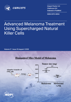
NK cell-based therapies are yielding exciting advancements in cancer treatment. This study highlights the potential of osteoclast-expanded supercharged (sNK) cells as a therapeutic approach to bolstering the immune system, thereby establishing enduring immunity in cancer patients. A comparison with the untreated group showcases substantial enhancements in immune responses upon treatment with sNK cells. Noteworthy outcomes include increased IFN-γ secretion and elevated cytotoxicity of immune cells in different tissues post-sNK cell treatment, surpassing levels observed in the untreated group. These findings underscore the promising trajectory of sNK cell-based interventions in melanoma therapy, emphasizing advancements in immune response amplification and innovative strategies to combat tumors effectively. View this paper
  • Issues are regarded as officially published after their release is announced to the table of contents alert mailing list.
  • You may sign up for e-mail alerts to receive table of contents of newly released issues.
  • PDF is the official format for papers published in both, html and pdf forms. To view the papers in pdf format, click on the "PDF Full-text" link, and use the free Adobe Reader to open them.
Order results
Result details
Section
Select all
Export citation of selected articles as:
27 pages, 31050 KB  
Article
Diagnostic System for Early In Situ Melanoma Detection Using Acoustic Microscopy and Infrared Spectroscopic Mapping Imaging
by Georgios th Karagiannis, Ioannis Grivas, Anastasia Tsingotjidou, Georgios Apostolidis, Eirini Tsardaka, Ioanna Dori, Kyriaki-Nefeli Poulatsidou, Ioannis Tsougos, Stefan Wesarg, Argyrios Doumas and Panagiotis Georgoulias
Cancers 2025, 17(15), 2599; https://doi.org/10.3390/cancers17152599 - 7 Aug 2025
Viewed by 675
Abstract
This study proposes a novel diagnostic system for the early detection of cutaneous melanoma based on morphological and biochemical changes during tumor formation. The methods used in this system are acoustic microscopy and infrared (IR) spectroscopy. The former identifies the anatomical parameters of [...] Read more.
This study proposes a novel diagnostic system for the early detection of cutaneous melanoma based on morphological and biochemical changes during tumor formation. The methods used in this system are acoustic microscopy and infrared (IR) spectroscopy. The former identifies the anatomical parameters of the developing tumor, whilst the latter identifies its biochemical features, both at the micron scale. To implement this diagnostic method, an animal model that mimics human melanoma was developed. The results of this investigation show that using high-frequency (>20 MHz) acoustic microscopy in conjunction with spectroscopic images provides useful information about distinct features of melanoma tumors’ 3D structures. The structures and cytoarchitecture of the tumors were assessed using conventional histology, and their malignant nature was confirmed using histological and immumohistochemical analysis. The proposed approach may provide an invaluable tool in diagnostic dermatology, as it is noninvasive and produces highly detailed and accurate data about the early appearance and development of melanoma tumors. Full article
Show Figures

Figure 1

18 pages, 766 KB  
Systematic Review
Timing Matters: A Systematic Review of Early Versus Delayed Palliative Care in Advanced Cancer
by Ioana Creangă-Murariu, Eliza-Maria Froicu, Dragos Viorel Scripcariu, Gema Bacoanu, Mihaela Poroch, Mihaela Moscalu, Claudia Cristina Tarniceriu, Teodora Alexa-Stratulat and Vladimir Poroch
Cancers 2025, 17(15), 2598; https://doi.org/10.3390/cancers17152598 - 7 Aug 2025
Viewed by 825
Abstract
Cancer has become a public health problem, especially in developing countries [...] Full article
(This article belongs to the Special Issue Integrating Palliative Care in Oncology)
Show Figures

Figure 1

14 pages, 693 KB  
Article
Dermoscopy of Melanoma According to Age Groups: A Retrospective Monocentric Study on 285 Patients
by Francesco Cavallo, Umberto Santaniello, Elisa Bin, Gabriele Roccuzzo, Silvia Giordano, Andrea Agostini, Martina Merli, Paolo Fava, Pietro Quaglino, Simone Ribero and Paolo Broganelli
Cancers 2025, 17(15), 2597; https://doi.org/10.3390/cancers17152597 - 7 Aug 2025
Cited by 1 | Viewed by 704
Abstract
Background/Objectives: Melanoma is an aggressive skin cancer with increasing incidence worldwide. Dermoscopy has revolutionized early melanoma detection, but most studies have focused on the general adult population. This study aims to analyze dermoscopic and histological differences in melanoma across age groups, evaluating whether [...] Read more.
Background/Objectives: Melanoma is an aggressive skin cancer with increasing incidence worldwide. Dermoscopy has revolutionized early melanoma detection, but most studies have focused on the general adult population. This study aims to analyze dermoscopic and histological differences in melanoma across age groups, evaluating whether specific patterns vary between younger and older patients. Methods: This retrospective study included 285 histopathological confirmed melanomas diagnosed at the Dermatology Clinic of the University of Turin between November 2021 and April 2024. Patients were stratified by age (<40 vs. ≥40 years), and statistical analyses (Chi-square, logistic regression) assessed differences in dermoscopic, histopathological, and anatomical parameters. Results: Younger patients showed a higher prevalence of growth-related features (e.g., pseudopods, OR = 5.43; asymmetric globules, OR = 2.33) and a thicker Breslow index (mean = 1.05 mm). Older patients exhibited more regression-associated signs (scar-like depigmentation, OR = 0.15; peppering, OR = 0.39), greater lesion size, and solar elastosis. Dermoscopic regression significantly predicted histological regression, with age-stratified analysis revealing peppering as a predictor in younger patients (p = 0.015) and scar-like depigmentation in older ones (p = 0.012). Conclusions: Melanoma exhibits distinct dermoscopic features depending on patient age, with growth-associated patterns being more common in younger individuals and regressive patterns predominating in older patients. These findings highlight the importance of age-specific diagnostic considerations in melanoma detection, potentially improving early diagnosis and patient outcomes. Full article
(This article belongs to the Special Issue Dermoscopy in Skin Cancer)
Show Figures

Figure 1

15 pages, 614 KB  
Article
Multi-Institutional Comparison of Ablative 5-Fraction Magnetic Resonance-Guided Online Adaptive Versus 15/25-Fraction Computed Tomography-Guided Moderately Hypofractionated Offline Adapted Radiation Therapy for Locally Advanced Pancreatic Cancer
by Michael D. Chuong, Eileen M. O’Reilly, Robert A. Herrera, Melissa Zinovoy, Kathryn E. Mittauer, Muni Rubens, Adeel Kaiser, Paul B. Romesser, Nema Bassiri-Gharb, Abraham J. Wu, John J. Cuaron, Alonso N. Gutierrez, Carla Hajj, Antonio Ucar, Fernando DeZarraga, Santiago Aparo, Christopher H. Crane and Marsha Reyngold
Cancers 2025, 17(15), 2596; https://doi.org/10.3390/cancers17152596 - 7 Aug 2025
Viewed by 1172
Abstract
Background: Radiation dose escalation for locally advanced pancreatic cancer (LAPC) using stereotactic magnetic resonance (MR)-guided online adaptive radiation therapy (SMART) or computed tomography (CT)-guided moderately hypofractionated ablative radiation therapy (HART) can achieve favorable outcomes although have not previously been compared. Methods: We performed [...] Read more.
Background: Radiation dose escalation for locally advanced pancreatic cancer (LAPC) using stereotactic magnetic resonance (MR)-guided online adaptive radiation therapy (SMART) or computed tomography (CT)-guided moderately hypofractionated ablative radiation therapy (HART) can achieve favorable outcomes although have not previously been compared. Methods: We performed a multi-center retrospective analysis of SMART (50 Gy/5 fractions) vs. HART (75 Gy/25 fractions or 67.5 Gy/15 fractions with concurrent capecitabine) for LAPC. Gray’s test and Cox proportional regression analyses were performed to identify factors associated with local failure (LF) and overall survival (OS). Results: A total of 211 patients (SMART, n = 91; HART, n = 120) were evaluated, and none had surgery. Median follow-up after SMART and HART was 27.0 and 40.0 months, respectively (p < 0.0002). SMART achieved higher gross tumor volume (GTV) coverage and greater hotspots. Two-year LF after SMART and HART was 6.5% and 32.9% (p < 0.001), while two-year OS was 31.0% vs. 35.3% (p = 0.056), respectively. LF was associated with SMART vs. HART (HR 5.389, 95% CI: 1.298–21.975; p = 0.021) and induction mFOLFIRINOX vs. non-mFOLFIRINOX (HR 2.067, 95% CI 1.038–4.052; p = 0.047), while OS was associated with CA19-9 decrease > 40% (HR 0.725, 95% CI 0.515–0.996; p = 0.046) and GTV V120% (HR 1.022, 95% CI 1.006–1.037; p = 0.015). Acute grade > 3 toxicity was similar (3.3% vs. 5.8%; p = 0.390), while late grade > 3 toxicity was less common after SMART (2.2% vs. 9.2%; p = 0.037). Conclusions: Ablative SMART and HART both achieve favorable oncologic outcomes for LAPC with minimal toxicity. We did not observe an OS difference, although technical advantages of SMART might improve target coverage and reduce LF. Full article
(This article belongs to the Section Cancer Therapy)
Show Figures

Figure 1

28 pages, 3251 KB  
Article
Predictors of ISUP Grade Group Discrepancies Between Biopsy and Radical Prostatectomy: A Single-Center Analysis of Clinical, Imaging, and Histopathological Parameters
by Victor Pasecinic, Dorin Novacescu, Flavia Zara, Cristina-Stefania Dumitru, Vlad Dema, Silviu Latcu, Razvan Bardan, Alin Adrian Cumpanas, Raluca Dumache, Talida Georgiana Cut, Hossam Ismail and Ademir Horia Stana
Cancers 2025, 17(15), 2595; https://doi.org/10.3390/cancers17152595 - 7 Aug 2025
Viewed by 572
Abstract
Background/Objectives: ISUP grade group discordance between prostate biopsy and radical prostatectomy (RP) impacts treatment decisions in over a third (~25–40%) of prostate cancer (PCa) patients. We aimed to identify ISUP grade migration predictors and assess the impact of preoperative imaging (MRI) in [...] Read more.
Background/Objectives: ISUP grade group discordance between prostate biopsy and radical prostatectomy (RP) impacts treatment decisions in over a third (~25–40%) of prostate cancer (PCa) patients. We aimed to identify ISUP grade migration predictors and assess the impact of preoperative imaging (MRI) in a contemporary Romanian PCa cohort. Methods: We retrospectively analyzed 142 PCa patients undergoing RP following biopsy between January 2021 and December 2024 at Pius Brinzeu County Hospital, Timișoara: 90 without and 52 with preoperative MRI. Clinical parameters, MRI findings (PI-RADS), and biopsy characteristics were evaluated. Machine learning models (gradient boosting, random forest) were developed with SHAP analysis for interpretability. Results: Grade migration occurred in 69/142 patients (48.6%): upstaging in 55 (38.7%) and downstaging in 14 (9.9%). In the non-MRI cohort, 37/90 (41.1%) were upstaged and 9/90 (10.0%) were downstaged, versus 18/52 (34.6%) upstaged and 5/52 (9.6%) downstaged in the MRI cohort. The MRI group showed a 6.5% absolute reduction in upstaging (34.6% vs. 41.1%), a promising non-significant trend (p = 0.469) that requires further investigation. Grade 1 patients showed the highest upstaging (69.4%), while Grades 3–4 showed the highest downstaging (11/43, 25.6%). PI-RADS 4 lesions had the highest upstaging (43.5%). PSA density > 0.20 ng/mL2 emerged as the strongest predictor. Gradient boosting achieved superior performance (AUC = 0.812) versus logistic regression (AUC = 0.721), representing a 13% improvement in discrimination. SHAP analysis revealed PSA density as the most influential (importance: 0.287). Grade migration associated with adverse pathology: extracapsular extension (52.7% vs. 28.7%, p = 0.008) and positive margins (38.2% vs. 21.8%, p = 0.045). Conclusions: ISUP grade migration affects 48.6% of Romanian patients, with 38.7% upstaged and 9.9% downstaged. The 69.4% upstaging in Grade 1 patients emphasizes the need for enhanced risk stratification tools, while 10% downstaging suggests potential overtreatment. Machine learning with SHAP analysis provides superior predictive performance (13% AUC improvement) while offering clinically interpretable risk assessments. PSA density dominates risk assessment, while PI-RADS 4 lesions warrant closer scrutiny than previously recognized. Full article
(This article belongs to the Special Issue Prostate Cancer: Contemporary Standards and Challenges)
Show Figures

Figure 1

13 pages, 1488 KB  
Article
Validation of a Quantitative Ultrasound Texture Analysis Model for Early Prediction of Neoadjuvant Chemotherapy Response in Breast Cancer: A Prospective Serial Imaging Study
by Daniel Moore-Palhares, Lakshmanan Sannachi, Adrian Wai Chan, Archya Dasgupta, Daniel DiCenzo, Sonal Gandhi, Rossanna Pezo, Andrea Eisen, Ellen Warner, Frances Wright, Nicole Look Hong, Ali Sadeghi-Naini, Mia Skarpathiotakis, Belinda Curpen, Carrie Betel, Michael C. Kolios, Maureen Trudeau and Gregory J. Czarnota
Cancers 2025, 17(15), 2594; https://doi.org/10.3390/cancers17152594 - 7 Aug 2025
Viewed by 644
Abstract
Background/Objectives: Patients with breast cancer who do not achieve a complete response to neoadjuvant chemotherapy (NAC) may benefit from intensified adjuvant systemic therapy. However, such treatment escalation is typically delayed until after tumour resection, which occurs several months into the treatment course. Quantitative [...] Read more.
Background/Objectives: Patients with breast cancer who do not achieve a complete response to neoadjuvant chemotherapy (NAC) may benefit from intensified adjuvant systemic therapy. However, such treatment escalation is typically delayed until after tumour resection, which occurs several months into the treatment course. Quantitative ultrasound (QUS) can detect early microstructural changes in tumours and may enable timely identification of non-responders during NAC, allowing for earlier treatment intensification. In our previous prospective observational study, 100 breast cancer patients underwent QUS imaging before and four times during NAC. Machine learning algorithms based on QUS texture features acquired in the first week of treatment were developed and achieved 78% accuracy in predicting treatment response. In the current study, we aimed to validate these algorithms in an independent prospective cohort to assess reproducibility and confirm their clinical utility. Methods: We included breast cancer patients eligible for NAC per standard of care, with tumours larger than 1.5 cm. QUS imaging was acquired at baseline and during the first week of treatment. Tumour response was defined as a ≥30% reduction in target lesion size on the resection specimen compared to baseline imaging. Results: A total of 51 patients treated between 2018 and 2021 were included (median age 49 years; median tumour size 3.6 cm). Most were estrogen receptor–positive (65%) or HER2-positive (33%), and the majority received dose-dense AC-T (n = 34, 67%) or FEC-D (n = 15, 29%) chemotherapy, with or without trastuzumab. The support vector machine algorithm achieved an area under the curve of 0.71, with 86% accuracy, 91% specificity, 50% sensitivity, 93% negative predictive value, and 43% positive predictive value for predicting treatment response. Misclassifications were primarily associated with poorly defined tumours and difficulties in accurately identifying the region of interest. Conclusions: Our findings validate QUS-based machine learning models for early prediction of chemotherapy response and support their potential as non-invasive tools for treatment personalization and clinical trial development focused on early treatment intensification. Full article
(This article belongs to the Special Issue Clinical Applications of Ultrasound in Cancer Imaging and Treatment)
Show Figures

Figure 1

28 pages, 1748 KB  
Review
Neutrophil Dynamics in Response to Cancer Therapies
by Huazhen Xu, Xiaojun Chen, Yuqing Lu, Nihao Sun, Karis E. Weisgerber, Manzhu Xu and Ren-Yuan Bai
Cancers 2025, 17(15), 2593; https://doi.org/10.3390/cancers17152593 - 7 Aug 2025
Cited by 1 | Viewed by 902
Abstract
Neutrophils are increasingly recognized as key players in the tumor microenvironment (TME), displaying functional plasticity that enables them to either promote or inhibit cancer progression. Depending on environmental cues, tumor-associated neutrophils (TANs) may polarize toward antitumor “N1” or protumor “N2” phenotypes, exerting diverse [...] Read more.
Neutrophils are increasingly recognized as key players in the tumor microenvironment (TME), displaying functional plasticity that enables them to either promote or inhibit cancer progression. Depending on environmental cues, tumor-associated neutrophils (TANs) may polarize toward antitumor “N1” or protumor “N2” phenotypes, exerting diverse effects on tumor growth, metastasis, immune modulation, and treatment response. While previous studies have focused on the pathological roles of TANs in cancer, less attention has been given to how cancer therapies themselves influence the behavior of TANs. This review provides a comprehensive synthesis of current knowledge regarding the dynamics of TANs in response to major cancer treatment modalities, including chemotherapy, radiotherapy, cell-based immunotherapies, and oncolytic viral and bacterial therapies. We discuss how these therapies influence TAN recruitment, polarization, and effector functions within the TME, and highlight key molecular regulators involved. By consolidating mechanistic and translational insights, this review emphasizes the potential to therapeutically reprogram TANs to enhance treatment efficacy. A deeper understanding of context-dependent TAN roles will be essential for developing more effective, neutrophil-informed cancer therapies. Full article
(This article belongs to the Special Issue The Role of Neutrophils in Tumor Progression and Metastasis)
Show Figures

Figure 1

17 pages, 1852 KB  
Article
Overall Survival Associated with Real-World Treatment Sequences in Patients with CLL/SLL in the United States
by Joanna M. Rhodes, Naleen Raj Bhandari, Manoj Khanal, Dan He, Sarang Abhyankar, John M. Pagel, Lisa M. Hess and Alan Z. Skarbnik
Cancers 2025, 17(15), 2592; https://doi.org/10.3390/cancers17152592 - 7 Aug 2025
Viewed by 3006
Abstract
Background/Objectives: This study compared overall survival (OS) associated with common real-world treatment sequences in patients with chronic lymphocytic leukemia/small lymphocytic lymphoma (CLL/SLL) in the United States. Methods: Utilizing the nationwide Flatiron Health electronic health record-derived de-identified database, adult CLL/SLL patients who initiated [...] Read more.
Background/Objectives: This study compared overall survival (OS) associated with common real-world treatment sequences in patients with chronic lymphocytic leukemia/small lymphocytic lymphoma (CLL/SLL) in the United States. Methods: Utilizing the nationwide Flatiron Health electronic health record-derived de-identified database, adult CLL/SLL patients who initiated systemic therapy (JAN2016-NOV2023) and received at least two lines of therapy (LoTs) were analyzed. Treatment regimens were categorized based on drug class, and most frequent (n ≥ 50) sequences (first LoT followed by [→] second LoT) were compared. OS from initiation of the first LoT was compared using multivariable Cox proportional hazard models, and adjusted hazard ratios with 95% CIs were reported. Results: Among 2354 eligible patients, n = 1711 (73%) received the 16 most frequent treatment sequences. Sequencing chemoimmunotherapy (CIT) → CIT (HR: 2.29 [1.23–4.28]), anti-CD20 monoclonal antibody (anti-CD20mab) monotherapy → CIT (1.95 [1.03–3.69]), and covalent Bruton tyrosine kinase inhibitor (cBTKi) monotherapy → anti-CD20mab monotherapy (2.00 [1.07–3.74]) were associated with worse OS compared to patients treated with cBTKi monotherapy → B-cell lymphoma 2 inhibitors (BCL2i) + anti-CD20mab (reference). Conclusions: OS associated with other sequences were not significantly different from the reference sequence in adjusted analyses, suggesting a lack of evidence for the optimal standard of care for sequencing the first two LoTs in real-world settings. Future research should reassess sequencing outcomes as novel treatments become adopted into clinical practice. Full article
(This article belongs to the Section Cancer Therapy)
Show Figures

Figure 1

17 pages, 2539 KB  
Article
Auxiliary Value of [18F]F-Fluorocholine PET/CT in Evaluating Post-Stereotactic Radiosurgery Recurrence of Lung Cancer Brain Metastases: A Comparative Analysis with Contrast-Enhanced MRI
by Yafei Zhang, Mimi Xu, Shuye Yang, Lili Lin, Huatao Wang, Kui Zhao, Hong Yang and Xinhui Su
Cancers 2025, 17(15), 2591; https://doi.org/10.3390/cancers17152591 - 7 Aug 2025
Viewed by 646
Abstract
Background/Objectives: This study aims to evaluate the additional value of [18F]F-fluorocholine ([18F]F-FCH) PET/CT over contrast-enhanced magnetic resonance imaging (CE-MRI) in detecting the recurrence of brain metastases (BMs) after stereotactic radiosurgery (SRS) in patients with lung cancer brain metastases (LCBMs). [...] Read more.
Background/Objectives: This study aims to evaluate the additional value of [18F]F-fluorocholine ([18F]F-FCH) PET/CT over contrast-enhanced magnetic resonance imaging (CE-MRI) in detecting the recurrence of brain metastases (BMs) after stereotactic radiosurgery (SRS) in patients with lung cancer brain metastases (LCBMs). Methods: Thirty-one patients with suspected recurrence of BM in LCBM after SRS were enrolled in this retrospective study. They underwent both [18F]F-FCH PET/CT and CE-MRI within 2 weeks. The tumor imaging parameters and clinical features were analyzed. The results of histopathology or radiographic follow-up served as the reference standard for the final diagnosis. Results: In these 31 patients, there were 54 lesions, of which 27 lesions were proven to be BM recurrence, while 27 lesions were non-recurrence. [18F]F-FCH PET/CT showed high radiotracer uptake in recurrent lesions of BM and identified 24 positive lesions (88.89% of sensitivity), while CE-MRI indicated 23 positive lesions (85.19% of sensitivity). [18F]F-FCH PET/CT indicated higher specificity (81.48%) and accuracy (85.19%) in detecting recurrence of BM than CE-MRI (40.74% and 62.96%, both p < 0.05), particularly in frontal lobes and cerebella. For lesion sizes, the accuracy of [18F]F-FCH PET/CT in detecting recurrent lesions was higher than that of CE-MRI for lesions over 1.0 cm but below 2.0 cm (p = 0.016). The detective performance of [18F]F-FCH PET/CT combined with CE-MRI was higher than [18F]F-FCH PET/CT or CE-MRI alone (all p < 0.05). Interestingly, TLC (≥4.11) was significantly correlated with poor intracranial PFS (iPFS), meaning it was a significant prognostic factor for iPFS. Conclusions: This study identified that compared with CE-MRI, [18F]F-FCH PET/CT demonstrated higher specificity and accuracy in diagnosing recurrence of BM in LCBM after SRS. Combining [18F]F-FCH PET/CT with CE-MRI has the potential to improve diagnostic performance for recurrence of BM and management of patient treatment. TLC was an independent risk factor for iPFS. Full article
(This article belongs to the Section Cancer Metastasis)
Show Figures

Figure 1

21 pages, 1727 KB  
Review
Immune Evasion in Head and Neck Squamous Cell Carcinoma: Roles of Cancer-Associated Fibroblasts, Immune Checkpoints, and TP53 Mutations in the Tumor Microenvironment
by Chung-Che Tsai, Yi-Chiung Hsu, Tin-Yi Chu, Po-Chih Hsu and Chan-Yen Kuo
Cancers 2025, 17(15), 2590; https://doi.org/10.3390/cancers17152590 - 7 Aug 2025
Viewed by 1307
Abstract
Head and neck squamous cell carcinoma (HNSCC) is a highly aggressive malignancy characterized by complex interactions within the tumor microenvironment (TME) that facilitate immune evasion and tumor progression. The TME consists of diverse cellular components, including cancer-associated fibroblasts, immune and endothelial cells, and [...] Read more.
Head and neck squamous cell carcinoma (HNSCC) is a highly aggressive malignancy characterized by complex interactions within the tumor microenvironment (TME) that facilitate immune evasion and tumor progression. The TME consists of diverse cellular components, including cancer-associated fibroblasts, immune and endothelial cells, and extracellular matrix elements, that collectively modulate tumor growth, metastasis, and resistance to therapy. Immune evasion in HNSCC is orchestrated through multiple mechanisms, including the suppression of cytotoxic T lymphocytes, recruitment of immunosuppressive cells, such as regulatory T and myeloid-derived suppressor cells, and upregulation of immune checkpoint molecules (e.g., PD-1/PD-L1 and CTLA-4). Natural killer (NK) cells, which play a crucial role in anti-tumor immunity, are often dysfunctional within the HNSCC TME due to inhibitory signaling and metabolic constraints. Additionally, endothelial cells contribute to tumor angiogenesis and immune suppression, further exacerbating disease progression. Recent advancements in immunotherapy, particularly immune checkpoint inhibitors and NK cell-based strategies, have shown promise in restoring anti-tumor immunity. Moreover, TP53 mutations, frequently observed in HNSCC, influence tumor behavior and therapeutic responses, highlighting the need for personalized treatment approaches. This review provides a comprehensive analysis of the molecular and cellular mechanisms governing immune evasion in HNSCC with a focus on novel therapeutic strategies aimed at improving patient outcomes. Full article
(This article belongs to the Special Issue Oral Cancer: Prevention and Early Detection (2nd Edition))
Show Figures

Figure 1

28 pages, 845 KB  
Review
Circulating Tumor DNA in Prostate Cancer: A Dual Perspective on Early Detection and Advanced Disease Management
by Stepan A. Kopytov, Guzel R. Sagitova, Dmitry Y. Guschin, Vera S. Egorova, Andrei V. Zvyagin and Alexey S. Rzhevskiy
Cancers 2025, 17(15), 2589; https://doi.org/10.3390/cancers17152589 - 6 Aug 2025
Viewed by 1632
Abstract
Prostate cancer (PC) remains a leading cause of malignancy in men worldwide, with current diagnostic methods such as prostate-specific antigen (PSA) testing and tissue biopsies facing limitations in specificity, invasiveness, and ability to capture tumor heterogeneity. Liquid biopsy, especially analysis of circulating tumor [...] Read more.
Prostate cancer (PC) remains a leading cause of malignancy in men worldwide, with current diagnostic methods such as prostate-specific antigen (PSA) testing and tissue biopsies facing limitations in specificity, invasiveness, and ability to capture tumor heterogeneity. Liquid biopsy, especially analysis of circulating tumor DNA (ctDNA), has emerged as a transformative tool for non-invasive detection, real-time monitoring, and treatment selection for PC. This review examines the role of ctDNA in both localized and metastatic PCs, focusing on its utility in early detection, risk stratification, therapy selection, and post-treatment monitoring. In localized PC, ctDNA-based biomarkers, including ctDNA fraction, methylation patterns, fragmentation profiles, and mutations, demonstrate promise in improving diagnostic accuracy and predicting disease recurrence. For metastatic PC, ctDNA analysis provides insights into tumor burden, genomic alterations, and resistance mechanisms, enabling immediate assessment of treatment response and guiding therapeutic decisions. Despite challenges such as the low ctDNA abundance in early-stage disease and the need for standardized protocols, advances in sequencing technologies and multimodal approaches enhance the clinical applicability of ctDNA. Integrating ctDNA with imaging and traditional biomarkers offers a pathway to precision oncology, ultimately improving outcomes. This review underscores the potential of ctDNA to redefine PC management while addressing current limitations and future directions for research and clinical implementation. Full article
Show Figures

Graphical abstract

17 pages, 1865 KB  
Article
Biomarkers in Renal Cell Carcinoma: A Systematic Review and Immunohistochemical Validation Study
by Brett Berezowski, Robert Boothe, Jr., Billy Chaplin, Sharon J. Del Vecchio, Zakariya Fares, Tyrone L. R. Humphries, Keng Lim Ng, Taylor Noonan, Hemamali Samaratunga, Aaron Urquhart, David A. Vesey, Simon T. Wood, Glenda C. Gobe and Robert J. Ellis
Cancers 2025, 17(15), 2588; https://doi.org/10.3390/cancers17152588 - 6 Aug 2025
Viewed by 601
Abstract
Background and Objectives: The worldwide incidence of renal cell carcinoma (RCC) rose by 22% between 2012 and 2022. In Australia, RCC accounted for 2.8% of all cancer diagnoses and contributing to 1.8% of cancer-related deaths. Identification of RCC biomarkers may aid in [...] Read more.
Background and Objectives: The worldwide incidence of renal cell carcinoma (RCC) rose by 22% between 2012 and 2022. In Australia, RCC accounted for 2.8% of all cancer diagnoses and contributing to 1.8% of cancer-related deaths. Identification of RCC biomarkers may aid in diagnosis and management. Methods: A systematic review of immunohistochemical markers of RCC studies published between 1990 and 2019 was undertaken to select candidate biomarkers of RCC. Immunohistochemical staining of 73 clear cell RCC tumors and paired normal tissue was undertaken using selected markers. Semi-quantitative and quantitative analysis of staining intensity between paired samples was undertaken to evaluate utility as potential biomarkers, using Chi-square tests and paired t-tests for analysis. As an exploratory analysis, staining intensity was also compared on clinical/demographic variables using linear and logistic regression. Results: There were 123 candidate biomarkers identified in 91 studies. Four candidate markers were selected for further investigation: aminopeptidase A (APA)/cluster of differentiation (CD)249, aminopeptidase N (APN)/CD13, gamma-glutamyl transferase (GGT), and neuron-specific enolase (NSE). APA, GGT, and APN all demonstrated reduced staining intensity in the tumor compared with normal tissue (p < 0.001 for all). NSE demonstrated a statistically significant increase in expression in tumor compared with normal tissue (p < 0.001), and this was more pronounced in patients aged >60 years (p = 0.038). Conclusions: The utility of APA, APN, and GGT as diagnostic biomarkers in clear cell RCC is limited. NSE may have some role as a biomarker for clear cell RCC, particularly among older patients; however, further investigation is required. Full article
(This article belongs to the Special Issue Optimizing Surgical Procedures and Outcomes in Renal Cancer)
Show Figures

Figure 1

14 pages, 340 KB  
Article
FLOT Versus CROSS—What Is the Optimal Therapeutic Approach for Locally Advanced Adenocarcinoma of the Esophagus and the Esophagogastric Junction?
by Martin Leu, Hannes Mahler, Johanna Reinecke, Ute Margarethe König, Leif Hendrik Dröge, Manuel Guhlich, Benjamin Steuber, Marian Grade, Michael Ghadimi, Volker Ellenrieder, Stefan Rieken and Alexander Otto König
Cancers 2025, 17(15), 2587; https://doi.org/10.3390/cancers17152587 - 6 Aug 2025
Viewed by 828
Abstract
Background/Objectives: Neoadjuvant radiochemotherapy and perioperative chemotherapy are both well-established treatment strategies for locally advanced adenocarcinoma of the esophagus (EAC) and the esophagogastric junction (AEGJ). However, recent knowledge controversially discusses whether neoadjuvant radiotherapy or perioperative chemotherapy represents superior therapeutic options to prolong survival or [...] Read more.
Background/Objectives: Neoadjuvant radiochemotherapy and perioperative chemotherapy are both well-established treatment strategies for locally advanced adenocarcinoma of the esophagus (EAC) and the esophagogastric junction (AEGJ). However, recent knowledge controversially discusses whether neoadjuvant radiotherapy or perioperative chemotherapy represents superior therapeutic options to prolong survival or cause less toxicity. Methods: We retrospectively analyzed 76 patients with locally advanced EAC or AEGJ treated at our tertiary cancer center between January 2015 and March 2023. Patients received either perioperative FLOT chemotherapy (n = 36) or neoadjuvant radiochemotherapy following the CROSS protocol (n = 40), followed by surgical resection and standardized follow-up. We compared survival outcomes, toxicity profiles, treatment compliance, and surgical results between the two groups. Results: There were no statistically significant differences between FLOT and CROSS treatments in five-year loco-regional controls (LRC: 61.5% vs. 68.6%; p = 0.81), progression-free survival (PFS: 33.9% vs. 42.8%; p = 0.82), overall survival (OS: 60.2% vs. 63.4%; p = 0.91), or distant controls (DC: 42.1% vs. 56.5%; p = 0.39). High-grade hematologic toxicities did not significantly differ between groups (p > 0.05). Treatment compliance was lower in the FLOT group, with 50% (18/36) not completing all the planned chemotherapy cycles, compared to 17.5% (7/40) in the CROSS group. All the patients in the CROSS group received the full radiotherapy dose. Surgical outcomes and post-surgical tumor status were comparable between the groups. Conclusions: Although perioperative chemotherapy with FLOT has recently become a standard of care for locally advanced EAC and AEGJ, neoadjuvant radiochemotherapy per the CROSS protocol remains a well-tolerated alternative. In appropriately selected patients, both approaches yield comparable oncological outcomes. Full article
(This article belongs to the Special Issue Current Treatments of Esophageal and Esophagogastric Junction Cancers)
Show Figures

Figure 1

22 pages, 885 KB  
Article
MRI-Based Radiomics for Outcome Stratification in Pediatric Osteosarcoma
by Esther Ngan, Dolores Mullikin, Ashok J. Theruvath, Ananth V. Annapragada, Ketan B. Ghaghada, Andras A. Heczey and Zbigniew A. Starosolski
Cancers 2025, 17(15), 2586; https://doi.org/10.3390/cancers17152586 - 6 Aug 2025
Viewed by 617
Abstract
Background/Objectives: Osteosarcoma (OS) is the most common malignant bone tumor in children and adolescents; the survival rate is as low as 24%. Accurate prediction of clinical outcomes remains a challenge due to tumor heterogeneity and the complexity of pediatric cases. This study [...] Read more.
Background/Objectives: Osteosarcoma (OS) is the most common malignant bone tumor in children and adolescents; the survival rate is as low as 24%. Accurate prediction of clinical outcomes remains a challenge due to tumor heterogeneity and the complexity of pediatric cases. This study aims to improve predictions of progressive disease, therapy response, relapse, and survival in pediatric OS using MRI-based radiomics and machine learning methods. Methods: Pre-treatment contrast-enhanced coronal T1-weighted MR scans were collected from 63 pediatric OS patients, with an additional nine external cases used for validation. Three strategies were considered for target region segmentation (whole-tumor, tumor sampling, and bone/soft tissue) and used for MRI-based radiomics. These were then combined with clinical features to predict OS clinical outcomes. Results: The mean age of OS patients was 11.8 ± 3.5 years. Most tumors were located in the femur (65%). Osteoblastic subtype was the most common histological classification (79%). The majority of OS patients (79%) did not have evidence of metastasis at diagnosis. Progressive disease occurred in 27% of patients, 59% of patients showed adequate therapy response, 25% experienced relapse after therapy, and 30% died from OS. Classification models based on bone/soft tissue segmentation generally performed the best, with certain clinical features improving performance, especially for therapy response and mortality. The top performing classifier in each outcome achieved 0.94–1.0 validation ROC AUC and 0.63–1.0 testing ROC AUC, while those without radiomic features (RFs) generally performed suboptimally. Conclusions: This study demonstrates the strong predictive capabilities of MRI-based radiomics and multi-region segmentations for predicting clinical outcomes in pediatric OS. Full article
(This article belongs to the Special Issue The Roles of Deep Learning in Cancer Radiotherapy)
Show Figures

Figure 1

14 pages, 1870 KB  
Article
Analysis of Risk Factors for High-Risk Lymph Node Metastasis in Papillary Thyroid Microcarcinoma
by Yi-Hsiang Chiu, Shu-Ting Wu, Yung-Nien Chen, Wen-Chieh Chen, Lay-San Lim, Yvonne Ee Wern Chiew, Ping-Chen Kuo, Ya-Chen Yang, Shun-Yu Chi and Chen-Kai Chou
Cancers 2025, 17(15), 2585; https://doi.org/10.3390/cancers17152585 - 6 Aug 2025
Viewed by 537
Abstract
Background: Papillary thyroid microcarcinoma (PTMC) is associated with certain features that carry an increased risk of local recurrence, underscoring the importance of preoperative risk assessment. This study investigated the clinicopathological factors associated with high-risk lymph node metastasis (HRLNM) and patient outcomes. HRLNM is [...] Read more.
Background: Papillary thyroid microcarcinoma (PTMC) is associated with certain features that carry an increased risk of local recurrence, underscoring the importance of preoperative risk assessment. This study investigated the clinicopathological factors associated with high-risk lymph node metastasis (HRLNM) and patient outcomes. HRLNM is defined as ≥5 metastatic lymph nodes and/or lateral neck metastasis. Methods: We conducted a retrospective review of 985 patients with PTMC who underwent thyroidectomy at the Kaohsiung Chang Gung Memorial Hospital from 2013 to 2022. Results: Among the 985 patients, 100 (10.2%) had lymph node metastasis (LNM), and 27% of these were classified as having HRLNM. Male sex (OR 3.61, p = 0.04) and extranodal extension (OR 3.76, p = 0.043) were independent predictors of HRLNM. Patients with LNM exhibited lower rates of excellent treatment response (75% vs. 87%, p = 0.001), higher recurrence rates (9.0% vs. 0.6%, p = 0.001), and an increased risk of distant metastasis (2.0% vs. 0%). Recurrence-free survival (RFS) was significantly shorter in patients with LNM (120.9 vs. 198.6 months, p < 0.001). Although HRLNM showed a trend toward reduced RFS (113.5 vs. 124.6 months, p = 0.177), its impact on long-term survival remains uncertain. Conclusions: Male sex and extranodal extension were significant risk factors for HRLNM in patients with PTMC. These findings highlight the need for individualized risk stratification to guide treatment strategies and improve patient outcomes. Full article
Show Figures

Figure 1

21 pages, 1557 KB  
Review
Neoadjuvant Therapy or Upfront Surgery for Pancreatic Cancer—To Whom, When, and How?
by Daria Kwaśniewska, Marta Fudalej, Anna Maria Badowska-Kozakiewicz, Aleksandra Czerw and Andrzej Deptała
Cancers 2025, 17(15), 2584; https://doi.org/10.3390/cancers17152584 - 6 Aug 2025
Viewed by 1980
Abstract
The management of resectable pancreatic ductal adenocarcinoma (R-PDAC) and borderline resectable pancreatic ductal adenocarcinoma (BR-PDAC) remains a topic of active debate. Although neoadjuvant therapy (NAT) has shown clinical benefits in BR-PDAC, especially in increasing resectability and achieving higher rates of margin-negative (R0) resections, [...] Read more.
The management of resectable pancreatic ductal adenocarcinoma (R-PDAC) and borderline resectable pancreatic ductal adenocarcinoma (BR-PDAC) remains a topic of active debate. Although neoadjuvant therapy (NAT) has shown clinical benefits in BR-PDAC, especially in increasing resectability and achieving higher rates of margin-negative (R0) resections, its role in R-PDAC is less clearly defined. Additionally, the role of immunotherapy in PDAC is still being explored, with ongoing trials investigating new combinations to overcome the tumor’s immune-resistant microenvironment. This article provides a comprehensive narrative review of the current evidence comparing NAT with upfront surgery in pancreatic cancer management, focusing on randomized controlled trials and meta-analyses that assess outcomes in R-PDAC and BR-PDAC. The review aims to determine whether NAT offers a significant survival advantage over traditional post-operative strategies and to clarify which clinical scenarios may benefit most from NAT. The literature was identified through a systematic search of PubMed, Scopus, and Google Scholar databases up to March 2025. Article selection adhered to the PRISMA guidelines. Our review of existing evidence supports NAT as the standard of care for BR-PDAC. Meanwhile, management of R-PDAC should be tailored individually, guided by risk stratification that considers both clinical parameters and molecular features. Immunotherapy and targeted therapies are still in early research phases, and their further integration as NAT remains controversial. Full article
Show Figures

Figure 1

18 pages, 2164 KB  
Article
The Fanconi Anemia Pathway Inhibits mTOR Signaling and Prevents Accelerated Translation in Head and Neck Cancer Cells
by Bianca Ruffolo, Sara Vicente-Muñoz, Khyati Y. Mehta, Cosette M. Rivera-Cruz, Xueheng Zhao, Lindsey Romick, Kenneth D. R. Setchell, Adam Lane and Susanne I. Wells
Cancers 2025, 17(15), 2583; https://doi.org/10.3390/cancers17152583 - 6 Aug 2025
Viewed by 754
Abstract
Background/Objectives: The Fanconi anemia (FA) pathway is essential for the repair of DNA interstrand crosslinks and maintenance of genomic stability. Germline loss of FA pathway function in the inherited Fanconi anemia syndrome leads to increased DNA damage and a range of clinical phenotypes, [...] Read more.
Background/Objectives: The Fanconi anemia (FA) pathway is essential for the repair of DNA interstrand crosslinks and maintenance of genomic stability. Germline loss of FA pathway function in the inherited Fanconi anemia syndrome leads to increased DNA damage and a range of clinical phenotypes, including a heightened risk of head and neck squamous cell carcinoma (HNSCC). Non-synonymous FA gene mutations are also observed in up to 20% of sporadic HNSCCs. The mechanistic target of rapamycin (mTOR) is known to stimulate cell growth, anabolic metabolism including protein synthesis, and survival following genotoxic stress. Methods/Results: Here, we demonstrate that FA− deficient (FA−) HNSCC cells exhibit elevated intracellular amino acid levels, increased total protein content, and an increase in protein synthesis indicative of enhanced translation. These changes are accompanied by hyperactivation of the mTOR effectors translation initiation factor 4E Binding Protein 1 (4E-BP1) and ribosomal protein S6. Treatment with the mTOR inhibitor rapamycin reduced the phosphorylation of these targets and blocked translation specifically in FA− cells but not in their isogenic FA− proficient (FA+) counterparts. Rapamycin-mediated mTOR inhibition sensitized FA− but not FA+ cells to rapamycin under nutrient stress, supporting a therapeutic metabolism-based vulnerability in FA− cancer cells. Conclusions: These findings uncover a novel role for the FA pathway in suppressing mTOR signaling and identify mTOR inhibition as a potential strategy for targeting FA− HNSCCs. Full article
(This article belongs to the Special Issue Targeted Therapy in Head and Neck Cancer)
Show Figures

Figure 1

23 pages, 789 KB  
Perspective
Therapeutic Cancer Vaccines in Colorectal Cancer: Platforms, Mechanisms, and Combinations
by Chiara Gallio, Luca Esposito and Alessandro Passardi
Cancers 2025, 17(15), 2582; https://doi.org/10.3390/cancers17152582 - 6 Aug 2025
Viewed by 2098
Abstract
Colorectal cancer (CRC) remains one of the most lethal malignancies worldwide, with high recurrence rates and limited curative options in metastatic settings. Cancer vaccines represent an emerging immunotherapeutic approach that aims to stimulate robust, tumor-specific immune responses. This review summarizes the current state [...] Read more.
Colorectal cancer (CRC) remains one of the most lethal malignancies worldwide, with high recurrence rates and limited curative options in metastatic settings. Cancer vaccines represent an emerging immunotherapeutic approach that aims to stimulate robust, tumor-specific immune responses. This review summarizes the current state of CRC vaccine development, including tumor cell-based, dendritic cell-based, peptide-based, nucleic acid-based (DNA and mRNA), and virus-based platforms. We highlight findings from key clinical trials that demonstrate immunogenicity, safety, and preliminary efficacy, with particular attention to combinations with chemotherapy and immune checkpoint inhibitors. Furthermore, we explore critical challenges such as tumor heterogeneity, immunosuppressive tumor microenvironments, and the logistical complexity; in this context, we particularly focus on the current development of personalized cancer vaccines, exploring the newly identified encouraging epitopes and their safety and efficacy in recent trials. The integration of cancer vaccines with in silico modeling, advanced delivery systems such as nanoparticles or AI-guided designs, and microbiome modulation represents a promising avenue for enhancing their clinical utility. Overall, therapeutic and prophylactic cancer vaccines may soon contribute meaningfully to the comprehensive management of CRC, especially in settings of minimal residual disease or early recurrence. Full article
(This article belongs to the Special Issue Exploring Immunotherapy in Colorectal Cancer)
Show Figures

Figure 1

12 pages, 616 KB  
Article
Surgical Margin Analysis in Osteosarcoma: Impact on Survival and Local Control
by Sebastian Breden, Simone Beischl, Florian Hinterwimmer, Sarah Consalvo, Carolin Knebel, Rüdiger von Eisenhart-Rothe, Rainer Burgkart and Ulrich Lenze
Cancers 2025, 17(15), 2581; https://doi.org/10.3390/cancers17152581 - 6 Aug 2025
Viewed by 687
Abstract
Background/Objectives: The quality of surgical margins has been shown to be a prognostic factor in many sarcoma entities, yet its role in osteosarcoma remains controversial. While previous studies have shown that the outcome was not related to the margin width in bone, the [...] Read more.
Background/Objectives: The quality of surgical margins has been shown to be a prognostic factor in many sarcoma entities, yet its role in osteosarcoma remains controversial. While previous studies have shown that the outcome was not related to the margin width in bone, the impact of the extraosseous margin width (margin at the soft tissue invasion)—which needs to be close sometimes due to neurovascular structures—needs to be assessed. This study aims to evaluate the influence of soft tissue surgical margins on local recurrence and overall survival in patients with high-grade osteosarcoma. Methods: We conducted a retrospective, single-center study including 75 patients treated for high-grade osteosarcoma. All patients underwent standardized neoadjuvant chemotherapy followed by complete surgical resection. Patients were stratified into three groups based on the histological margin width of the extraosseous parts: group 1 (<1 mm), group 2 (1–5 mm), and group 3 (≥5 mm). Primary endpoints were local recurrence and overall survival (OS), analyzed using Kaplan–Meier estimates, log-rank tests, and Cox regression. Results: Local recurrence occurred in seven patients (9%). Although the overall comparison between the three groups was not statistically significant (p = 0.074), a subgroup analysis revealed a significantly higher recurrence rate in patients with margins < 1 mm compared to those with wider margins (p = 0.024). No significant differences in overall survival (OS) were observed between the groups (p = 0.896). Tumor location, metastatic status, and UICC stage were significant predictors for both endpoints in univariate analysis. However, none of these association were confirmed in multivariate analyses. Conclusions: Very close surgical margins (<1 mm) may increase the risk of local recurrence in high-grade osteosarcoma; however, they do not appear to affect overall survival. Full article
(This article belongs to the Special Issue Clinical Treatment of Osteosarcoma)
Show Figures

Figure 1

23 pages, 406 KB  
Systematic Review
Advances in Bidirectional Therapy for Peritoneal Metastases: A Systematic Review of Pressurized Intraperitoneal Aerosol Chemotherapy (PIPAC) Combined with Systemic Chemotherapy
by Manuela Robella, Marco Vitturini, Andrea Di Giorgio, Matteo Aulicino, Martin Hubner, Emanuele Koumantakis, Felice Borghi, Paolo Catania, Armando Cinquegrana and Paola Berchialla
Cancers 2025, 17(15), 2580; https://doi.org/10.3390/cancers17152580 - 6 Aug 2025
Viewed by 1184
Abstract
Background: Peritoneal metastases (PM) represent a common and challenging manifestation of several gastrointestinal and gynecologic malignancies. Bidirectional treatment—combining Pressurized Intraperitoneal Aerosol Chemotherapy (PIPAC) with systemic chemotherapy—has emerged as a strategy to enhance locoregional control while maintaining systemic coverage. Objective: This systematic [...] Read more.
Background: Peritoneal metastases (PM) represent a common and challenging manifestation of several gastrointestinal and gynecologic malignancies. Bidirectional treatment—combining Pressurized Intraperitoneal Aerosol Chemotherapy (PIPAC) with systemic chemotherapy—has emerged as a strategy to enhance locoregional control while maintaining systemic coverage. Objective: This systematic review aimed to analyze the study design, characteristics, and timing of the treatments administered—including the type of systemic chemotherapy, intraperitoneal agents used in PIPAC, and interval between administrations—as well as the clinical outcomes, safety profile, and overall methodological quality of the available literature on bidirectional treatment for peritoneal metastases. Methods: A systematic literature search was conducted across the PubMed, Embase, and Cochrane Library databases up to April 2025. Studies were included if they reported clinical outcomes of patients undergoing bidirectional treatment. Data extraction focused on survival, response assessment (PRGS, PCI), adverse events, systemic and intraperitoneal regimens, treatment interval, and study methodology. Results: A total of 22 studies involving 1015 patients (742 treated with bidirectional therapy) were included. Median overall survival ranged from 2.8 to 19.6 months, with the most favorable outcomes observed in gastric and colorectal cancer cohorts. PRGS improvement after multiple PIPAC cycles was reported in >80% of evaluable cases. High-grade adverse events (CTCAE ≥ 3) occurred in up to 17% of patients in most studies, with only one study reporting treatment-related mortality. However, methodological quality was generally moderate, with considerable heterogeneity in treatment protocols, response criteria, systemic regimens, and toxicity attribution. Conclusions: Bidirectional therapy with PIPAC and systemic chemotherapy appears to be a feasible and potentially effective strategy for selected patients with peritoneal metastases. Despite encouraging outcomes, definitive conclusions are limited by the retrospective nature and heterogeneity of available studies. Prospective standardized trials are needed to confirm efficacy, clarify patient selection, and optimize treatment protocols. Full article
(This article belongs to the Section Cancer Therapy)
Show Figures

Figure 1

22 pages, 688 KB  
Review
The Evolving Treatment Landscape for the Elderly Multiple Myeloma Patient: From Quad Regimens to T-Cell Engagers and CAR-T
by Matthew James Rees and Hang Quach
Cancers 2025, 17(15), 2579; https://doi.org/10.3390/cancers17152579 - 5 Aug 2025
Viewed by 914
Abstract
Multiple myeloma (MM) is predominantly a disease of the elderly. In recent years, a surge of highly effective plasma cell therapies has revolutionized the care of elderly multiple myeloma (MM) patients, for whom frailty and age-related competing causes of mortality determine management. Traditionally, [...] Read more.
Multiple myeloma (MM) is predominantly a disease of the elderly. In recent years, a surge of highly effective plasma cell therapies has revolutionized the care of elderly multiple myeloma (MM) patients, for whom frailty and age-related competing causes of mortality determine management. Traditionally, the treatment of newly diagnosed elderly patients has centered on doublet or triplet combinations composed of immunomodulators (IMIDs), proteasome inhibitors (PIs), anti-CD38 monoclonal antibodies (mAbs), and corticosteroids producing median progression-free survival (PFS) rates between 34 and 62 months. However, recently, a series of large phase III clinical trials examining quadruplet regimens of PIs, IMIDs, corticosteroids, and anti-CD38 mAbs have shown exceptional outcomes, with median PFS exceeding 60 months, albeit with higher rates of peripheral neuropathy (≥Grade 2: 27% vs. 10%) when PIs and IMIDs are combined, and infections (≥Grade 3: 40% vs. 29–41%) with the addition of anti-CD38mAbs. The development of T-cell redirecting therapies including T-cell engagers (TCEs) and CAR-T cells has further expanded the therapeutic arsenal. TCEs have shown exceptional activity in relapsed disease and are being explored in the newly diagnosed setting with promising early results. However, concerns remain regarding the logistical challenges of step-up dosing, which often necessitates inpatient admission, the infectious risks, and the financial burden associated with TCEs in elderly patients. CAR-T, the most potent commercially available therapy for MM, offers the potential of a ‘one and done’ approach. However, its application to elderly patients has been tempered by significant concerns of cytokine release syndrome, early and delayed neurological toxicity, and its overall tolerability in frail patients. Robust data in frail patients are still needed. How CAR-T and TCEs will be sequenced among the growing therapeutic armamentarium for elderly MM patients remains to be determined. This review explores the safety, efficacy, cost, and logistical barriers associated with the above treatments in elderly MM patients. Full article
Show Figures

Figure 1

17 pages, 4105 KB  
Article
Evaluation of the Effect of X-Ray Therapy on Glioma Rat Model Using Chemical Exchange Saturation Transfer and Diffusion-Weighted Imaging
by Kazuki Onishi, Koji Itagaki, Sachie Kusaka, Tensei Nakano, Junpei Ueda and Shigeyoshi Saito
Cancers 2025, 17(15), 2578; https://doi.org/10.3390/cancers17152578 - 5 Aug 2025
Viewed by 463
Abstract
Background/Objectives: This study aimed to examine the changes in brain metabolites and water molecule diffusion using chemical exchange saturation transfer (CEST) imaging and diffusion-weighted imaging (DWI) after 15 Gy of X-ray irradiation in a rat model of glioma. Methods: The glioma-derived [...] Read more.
Background/Objectives: This study aimed to examine the changes in brain metabolites and water molecule diffusion using chemical exchange saturation transfer (CEST) imaging and diffusion-weighted imaging (DWI) after 15 Gy of X-ray irradiation in a rat model of glioma. Methods: The glioma-derived cell line, C6, was implanted into the striatum of the right brain of 7-week-old male Wistar rats. CEST imaging and DWI were performed on days 8, 10, and 17 after implantation using a 7T-magnetic resonance imaging. X-ray irradiation (15 Gy) was performed on day 9. Magnetization transfer ratio (MTR) and apparent diffusion coefficient (ADC) values were calculated for CEST and DWI, respectively. Results: On day 17, the MTR values at 1.2 ppm, 1.5 ppm, 1.8 ppm, 2.1 ppm, and 2.4 ppm in the irradiated group decreased significantly compared with those of the control group. The standard deviation for the ADC values on a pixel-by-pixel basis increased from day 8 to day 17 (0.6 ± 0.06 → 0.8 ± 0.17 (×10−3 mm2/s)) in the control group, whereas it remained nearly unchanged (0.6 ± 0.06 → 0.8 ± 0.11 (×10−3 mm2/s)) in the irradiated group. Conclusions: This study revealed the effects of 15 Gy X-ray irradiation in a rat model of glioma using CEST imaging and DWI. Full article
Show Figures

Figure 1

23 pages, 1642 KB  
Review
The Multifaceted Role of Autophagy in Nasopharyngeal Carcinoma: Translational Perspectives on Pathogenesis, Biomarkers, Treatment Resistance, and Emerging Therapies
by Abdul L. Shakerdi, Emma Finnegan, Yin-Yin Sheng and Graham P. Pidgeon
Cancers 2025, 17(15), 2577; https://doi.org/10.3390/cancers17152577 - 5 Aug 2025
Viewed by 806
Abstract
Background: Nasopharyngeal carcinoma (NPC) is an epithelial malignancy arising from the nasopharyngeal mucosa. Despite treatment advances such as the use of intensity-modulated radiotherapy and immune checkpoint inhibitors, resistance remains a significant clinical challenge. Many tumours are also diagnosed at an advanced stage associated [...] Read more.
Background: Nasopharyngeal carcinoma (NPC) is an epithelial malignancy arising from the nasopharyngeal mucosa. Despite treatment advances such as the use of intensity-modulated radiotherapy and immune checkpoint inhibitors, resistance remains a significant clinical challenge. Many tumours are also diagnosed at an advanced stage associated with poor prognosis. Objective: This review aims to explore the biological roles of autophagy in NPC, primarily highlighting its involvement in disease pathogenesis and treatment resistance. Methods: We performed a review of the recent literature examining the role of autophagy-related pathways in NPC pathogenesis, biomarker discovery, and therapeutic targeting. Results: Autophagy plays a dual role in NPC as it contributes to both tumour suppression and progression. It is involved in tumour initiation, metastasis, immune modulation, and treatment resistance. Autophagy-related genes such as SQSTM1, Beclin-1, and AURKA may serve as prognostic and therapeutic biomarkers. Various strategies are being investigated for their role to modulate autophagy using pharmacologic inhibitors, RNA interventions, and natural compounds. Conclusions: Further research into autophagy’s context-dependent roles in NPC may inform the development of personalised therapies and allow progress in translational and precision oncology. Full article
Show Figures

Figure 1

16 pages, 4092 KB  
Article
Ribosome Biogenesis Underpins Tumor Progression: A Comprehensive Signature for Survival and Immunotherapy Response Prediction
by Amr R. Elhamamsy, Salma M. Aly, Rajeev S. Samant and Lalita A. Shevde
Cancers 2025, 17(15), 2576; https://doi.org/10.3390/cancers17152576 - 5 Aug 2025
Viewed by 514
Abstract
Background: RiBi is integral to cell proliferation, and its dysregulation is increasingly recognized as a hallmark of aggressive cancers. We sought to develop and validate a composite “PanRibo-515 score” reflecting RiBi activity across multiple tumor types, assess its prognostic significance, and explore [...] Read more.
Background: RiBi is integral to cell proliferation, and its dysregulation is increasingly recognized as a hallmark of aggressive cancers. We sought to develop and validate a composite “PanRibo-515 score” reflecting RiBi activity across multiple tumor types, assess its prognostic significance, and explore its relationship with immune checkpoint therapy outcomes. Methods: We curated 515 RiBi–associated genes (PanRibo-515) and used a LASSO regression-based strategy on a training dataset (GSE202203) to select the prognostically most relevant subset of 68 genes (OncoRibo-68). Directionality (positive or negative impact on survival) was assigned based on the sign of the LASSO coefficients. We integrated a forward selection approach to identify a refined subset of genes for computing the OncoRibo-68 score. For validation, patients in The Cancer Genome Atlas (TCGA) were stratified into high or low OncoRibo-68 score groups for survival analyses. Additional validation for immunotherapy response was conducted using bioinformatic platforms used for immunotherapy response analysis. Results: A higher OncoRibo-68 score consistently correlated with poorer overall and progression-free survival across multiple cancers. Elevated OncoRibo-68 score was linked to an immunosuppressive tumor microenvironment, but interestingly to increased response to checkpoint inhibitors. Conclusions: Our findings highlight RiBi as an important determinant of tumor aggressiveness and identify the OncoRibo-68 score as a promising biomarker for risk stratification and therapy selection. Future research may evaluate whether targeting RiBi pathways could enhance treatment efficacy, particularly in combination with immunotherapy. Full article
Show Figures

Figure 1

23 pages, 3521 KB  
Article
Efficacy of NAMPT Inhibitors in Pancreatic Cancer After Stratification by MAP17 (PDZK1IP1) Levels
by Eva M. Verdugo-Sivianes, Julia Martínez-Pérez, Lola E Navas, Carmen Sáez and Amancio Carnero
Cancers 2025, 17(15), 2575; https://doi.org/10.3390/cancers17152575 - 5 Aug 2025
Viewed by 712
Abstract
Background/Objectives: Pancreatic cancer (PC) is the seventh leading cause of cancer-related deaths worldwide, with its incidence rising each year. Despite its relatively low incidence, the aggressiveness of pancreatic cancer results in high mortality, with only 12% of patients surviving five years post-diagnosis. [...] Read more.
Background/Objectives: Pancreatic cancer (PC) is the seventh leading cause of cancer-related deaths worldwide, with its incidence rising each year. Despite its relatively low incidence, the aggressiveness of pancreatic cancer results in high mortality, with only 12% of patients surviving five years post-diagnosis. Surgical resection remains the only potentially curative treatment, but the tumor is often diagnosed at an advanced stage. The goal of this work is to identify vulnerabilities that can affect the efficacy of treatments and improve the efficacy of therapy. Methods: MAP17 overexpression in pancreatic cancer cell lines, RT-qPCR analysis, xenografts, in vitro and in vivo treatments, analysis of data from pancreatic tumors in transcriptomic patient databases. Results: We studied the prognostic and predictive value of MAP17 (PDZK1IP1) expression in pancreatic cancer, and we found that high MAP17 mRNA expression was associated with poor prognosis. In addition, single-cell analysis revealed that high MAP17 expression was present only in tumor cells. We investigated whether the response to various antitumor agents depended on MAP17 expression. In 2D culture, MAP17-expressing pancreatic cancer cells responded better to gemcitabine and 5-fluorouracil. However, in vivo xenograft tumors with MAP17 expression showed resistance to all treatments. Additionally, MAP17-expressing cells had a high NAD pool, which seems to be effectively depleted in vivo by NAMPT inhibitors, the primary enzyme for NAD biosynthesis. Conclusions: Our findings suggest that MAP17 expression could enhance the prognostic stratification of pancreatic cancer patients. Moreover, the coadministration of NAMPT inhibitors with current treatments may sensitize tumors with high MAP17 expression to chemotherapy and improve the efficacy of chemotherapy. Full article
(This article belongs to the Section Molecular Cancer Biology)
Show Figures

Figure 1

15 pages, 1582 KB  
Article
Evaluation of the Effect of Optic Nerve Compression by Craniopharyngioma on Retinal Nerve Fiber Layer Thickness in Pediatric Patients
by Klaudia Rakusiewicz-Krasnodębska, Agnieszka Bogusz-Wójcik, Elżbieta Moszczyńska, Maciej Jaworski, Paweł Kowalczyk and Wojciech Hautz
Cancers 2025, 17(15), 2574; https://doi.org/10.3390/cancers17152574 - 5 Aug 2025
Viewed by 549
Abstract
Purpose: The present study aims to evaluate alterations in the peripapillary retinal nerve fiber layer (RNFL) thickness in pediatric patients following surgical resection of childhood-onset craniopharyngioma (CP) and to identify tumor characteristics and other factors influencing these alterations, including changes in the lesion’s [...] Read more.
Purpose: The present study aims to evaluate alterations in the peripapillary retinal nerve fiber layer (RNFL) thickness in pediatric patients following surgical resection of childhood-onset craniopharyngioma (CP) and to identify tumor characteristics and other factors influencing these alterations, including changes in the lesion’s location. Design: retrospective clinical cohort study. Methods: A retrospective analysis was conducted on 73 eyes from 38 patients with CP and 64 eyes from 32 age- and sex-matched healthy controls. The mean age of the CP patients was 10.3 ± 4.2 years (range 4–17), while the control group had a mean age of 10.5 ± 3.1 years (range 4–17). Optical coherence tomography (OCT) was used to assess the peripapillary RNFL thickness in the study and control groups. RNFL thickness was analyzed in the superior, inferior, and average sectors, as well as across eight optic nerve sectors. Tumor characteristics were evaluated to determine their correlation with changes in RNFL thickness in individual sectors. Results: Postoperative thickness of peripapillary RNFL in all individual sectors was significantly reduced in the CP group compared to healthy controls. Location, tumor volume, maximum tumor diameter, calcification, ventriculoperitoneal shunt, surgery technique, total resection, presence of Rosenthal fibers, and reoperation due to progression or recurrence correlated with damage to RNFL. Conclusions: CP is associated with significant reductions in RNFL thickness, indicating the tumor’s impact on optic nerve fibers. OCT is a valuable tool for monitoring visual pathway impairment and postoperative outcomes. Correlations between RNFL thickness in individual sectors and clinical parameters may offer valuable insights for diagnosis and monitoring, underlining their potential role in predicting visual outcomes. Regular RNFL evaluation should be integrated into the long-term care of CP patients to optimize visual prognosis and detect progressive or residual damage. Full article
(This article belongs to the Section Pediatric Oncology)
Show Figures

Figure 1

15 pages, 357 KB  
Article
Apalutamide Monotherapy in Metastatic Hormone-Sensitive Prostate Cancer: A Viable Alternative to First-Generation Anti-Androgen Agents to Avoid the Flare Phenomenon and an Effective Treatment for Achieving Early PSA Response
by Gaetano Facchini, Andrea D’Arienzo, Antonella Nicastro, Fabiano Flauto, Michela Izzo, Liliana Montella, Filippo Riccardo, Giovanni Maria Fusco, Francesco Trama, Giovanni Di Lauro, Giuseppe Di Costanzo, Anna Giacoma Tucci, Francesca Iasiello, Lorena Di Lorenzo, Salvatore Maddaluno, Carmela Liguori, Rita Assante di Cupillo, Paola Coppola, Angela Minissale, Maria Teresa Di Nardo, Luigi Formisano, Erika Martinelli, Giuliana Ciappina, Salvatore Pisconti, Massimiliano Berretta and Chiara Barracoadd Show full author list remove Hide full author list
Cancers 2025, 17(15), 2573; https://doi.org/10.3390/cancers17152573 - 5 Aug 2025
Viewed by 824
Abstract
Background/Objectives: Androgen deprivation therapy (ADT) is the mainstay of prostate cancer treatment, especially in advanced disease. In particular, the gonadotropin-releasing hormone agonists (aGnRH) reduce the production of gonadotropin and, therefore, of testosterone. In about 10% of patients, the non-pulsatile stimulation of GnRH receptor [...] Read more.
Background/Objectives: Androgen deprivation therapy (ADT) is the mainstay of prostate cancer treatment, especially in advanced disease. In particular, the gonadotropin-releasing hormone agonists (aGnRH) reduce the production of gonadotropin and, therefore, of testosterone. In about 10% of patients, the non-pulsatile stimulation of GnRH receptor initially causes a surge in LH and testosterone, defined as the “flare-up phenomenon”, leading to increased bone pain, spinal cord compression, bladder outlet obstruction and cardiovascular issues. To mitigate this effect, combining a first-generation antiandrogen agent (FGA) with aGnRH is recommended. However, second-generation anti-androgens, such as apalutamide, bind selectively and irreversibly to the androgen receptor (AR), exhibiting a more efficient inhibition of the AR pathway. Methods: This is a descriptive retrospective study of 27 patients (pts) with mHSPC, treated at a single center (“Santa Maria delle Grazie” Hospital in Pozzuoli, ASL Napoli 2 Nord, Italy) between June 2022 and April 2024. Patients received apalutamide monotherapy for 14 days followed by continuous combination with aGnRH plus apalutamide. Serum PSA and testosterone levels were measured at baseline, at day 14 (after 13 days of apalutamide monotherapy), at day 28 (after an additional 15 days of apalutamide plus a aGnRH), and at day 60. Results: PSA levels decreased from a mean of 45.2 (±63.1) ng/mL at baseline to a mean of 12.6 (±23.4) ng/mL at day 14 and to 3.3 ng/mL (±6.0) at day 28 of treatment. After 14 days of apalutamide monotherapy, 21 patients (77.8%) achieved a >50% PSA reduction and 4 (14.8%) a >90% PSA reduction. The number of patients with undetectable PSA was one (3.7%) at day 14, two (7.4%) at day 28, and nine (33.3%) at day 60. The mean serum testosterone levels were 6.56 (±4.46) ng/mL at baseline, 6.58 (±4.42) ng/mL at day 14, and 2.40 (± 3.38) ng/mL at day 28. No significant difference in PSA and testosterone level reduction during treatment emerged between subgroups of patients with low- vs. high-volume disease. Conclusions: Apalutamide alone is a viable option for mitigating the flare-up phenomenon, avoiding first generation anti-androgen therapy, and it can achieve rapid and deep biochemical control. Full article
(This article belongs to the Special Issue Advances in Therapeutic Strategies for Prostate Cancer)
Show Figures

Figure 1

29 pages, 3455 KB  
Review
Recent Advances in Nanoparticle and Nanocomposite-Based Photodynamic Therapy for Cervical Cancer: A Review
by Dorota Bartusik-Aebisher, Mohammad A. Saad, Agnieszka Przygórzewska and David Aebisher
Cancers 2025, 17(15), 2572; https://doi.org/10.3390/cancers17152572 - 4 Aug 2025
Viewed by 686
Abstract
Cervical cancer represents a significant global health challenge. Photodynamic therapy (PDT) appears to be a promising, minimally invasive alternative to standard treatments. However, the clinical efficacy of PDT is sometimes limited by the low solubility and aggregation of photosensitizers, their non-selective distribution in [...] Read more.
Cervical cancer represents a significant global health challenge. Photodynamic therapy (PDT) appears to be a promising, minimally invasive alternative to standard treatments. However, the clinical efficacy of PDT is sometimes limited by the low solubility and aggregation of photosensitizers, their non-selective distribution in the body, hypoxia in the tumor microenvironment, and limited light penetration. Recent advances in nanoparticle and nanocomposite platforms have addressed these challenges by integrating multiple functional components into a single delivery system. By encapsulating or conjugating photosensitizers in biodegradable matrices, such as mesoporous silica, organometallic structures and core–shell construct nanocarriers increase stability in water and extend circulation time, enabling both passive and active targeting through ligand decoration. Up-conversion and dual-wavelength responsive cores facilitate deep light conversion in tissues, while simultaneous delivery of hypoxia-modulating agents alleviates oxygen deprivation to sustain reactive oxygen species generation. Controllable “motor-cargo” constructs and surface modifications improve intratumoral diffusion, while aggregation-induced emission dyes and plasmonic elements support real-time imaging and quantitative monitoring of therapeutic response. Together, these multifunctional nanosystems have demonstrated potent cytotoxicity in vitro and significant tumor suppression in vivo in mouse models of cervical cancer. Combining targeted delivery, controlled release, hypoxia mitigation, and image guidance, engineered nanoparticles provide a versatile and powerful platform to overcome the current limitations of PDT and pave the way toward more effective, patient-specific treatments for cervical malignancies. Our review of the literature summarizes studies on nanoparticles and nanocomposites used in PDT monotherapy for cervical cancer, published between 2023 and July 2025. Full article
(This article belongs to the Section Cancer Therapy)
Show Figures

Figure 1

19 pages, 1242 KB  
Review
Modeling the Bone Marrow Microenvironment to Better Understand the Pathogenesis, Progression, and Treatment of Hematological Cancers
by Kathryn A. Skelding, Daniel L. Barry and Lisa F. Lincz
Cancers 2025, 17(15), 2571; https://doi.org/10.3390/cancers17152571 - 4 Aug 2025
Viewed by 727
Abstract
Despite significant advancements in understanding the pathogenesis and treatment of hematological malignancies, including leukemia and multiple myeloma, the majority of patients continue to experience poor long-term outcomes. This is partly due to the difficulty of accurately recapitulating the malignant microenvironment in vitro, particularly [...] Read more.
Despite significant advancements in understanding the pathogenesis and treatment of hematological malignancies, including leukemia and multiple myeloma, the majority of patients continue to experience poor long-term outcomes. This is partly due to the difficulty of accurately recapitulating the malignant microenvironment in vitro, particularly the bone marrow niche. The complexity of the bone marrow microenvironment poses a challenge for the in vitro examination of hematological malignancies. Traditionally, 2D culture and animal models have been utilized, but these representations are limited and have been criticized for their lack of human physiological relevance. In an attempt to overcome this, 3D models have been developed that more accurately recapitulate the in vivo microenvironment. Herein, we present an overview of recent developments in 2D and 3D models used for studying the bone marrow niche in hematological malignancies, highlighting their advantages and limitations. Full article
(This article belongs to the Section Tumor Microenvironment)
Show Figures

Graphical abstract

13 pages, 491 KB  
Article
Optimizing One-Sample Tests for Proportions in Single- and Two-Stage Oncology Trials
by Alan David Hutson
Cancers 2025, 17(15), 2570; https://doi.org/10.3390/cancers17152570 - 4 Aug 2025
Viewed by 507
Abstract
Background/Objectives: Phase II oncology trials often rely on single-arm designs to test H0:π=π0 versus Ha:π>π0, especially when randomized trials are infeasible due to cost or disease rarity. Traditional approaches, such [...] Read more.
Background/Objectives: Phase II oncology trials often rely on single-arm designs to test H0:π=π0 versus Ha:π>π0, especially when randomized trials are infeasible due to cost or disease rarity. Traditional approaches, such as the exact binomial test and Simon’s two-stage design, tend to be conservative, with actual Type I error rates falling below the nominal α due to the discreteness of the underlying binomial distribution. This study aims to develop a more efficient and flexible method that maintains accurate Type I error control in such settings. Methods: We propose a convolution-based method that combines the binomial distribution with a simulated normal variable to construct an unbiased estimator of π. This method is designed to precisely control the Type I error rate while enabling more efficient trial designs. We derive its theoretical properties and assess its performance against traditional exact tests in both one-stage and two-stage trial designs. Results: The proposed method results in more efficient designs with reduced sample sizes compared to standard approaches, without compromising the control of Type I error rates. We introduce a new two-stage design incorporating interim futility analysis and compare it with Simon’s design. Simulations and real-world examples demonstrate that the proposed approach can significantly lower trial cost and duration. Conclusions: This convolution-based approach offers a flexible and efficient alternative to traditional methods for early-phase oncology trial design. It addresses the conservativeness of existing designs and provides practical benefits in terms of resource use and study timelines. Full article
(This article belongs to the Special Issue Application of Biostatistics in Cancer Research)
Show Figures

Figure 1

Previous Issue
Next Issue
Back to TopTop