




Next Issue
Volume 16, May-1
Previous Issue
Volume 16, April-1


Diagnostics, Volume 16, Issue 8 (April-2 2026) – 135 articles
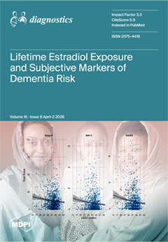
Cover Story (view full-size image): Alzheimer’s disease disproportionately affects females, underscoring the importance of identifying sex-specific risk factors that may contribute to this disparity. Endogenous estradiol has been proposed to confer neuroprotection against Alzheimer’s disease; however, whether lifetime exposure to endogenous estradiol (LEE2) relates to early markers of dementia risk remains unclear. In this study of 1156 dementia-free postmenopausal females, LEE2 was not associated with objective or subjective cognitive outcomes, but longer LEE2 was associated with lower severity of behavioural and functional symptoms. These findings suggest that relationships between hormonal exposure and brain health may extend beyond cognition, supporting a broader framework for early risk detection in females. View this paper
  • Issues are regarded as officially published after their release is announced to the table of contents alert mailing list.
  • You may sign up for e-mail alerts to receive table of contents of newly released issues.
  • PDF is the official format for papers published in both, html and pdf forms. To view the papers in pdf format, click on the "PDF Full-text" link, and use the free Adobe Reader to open them.
Order results
Result details
Section
Select all
Export citation of selected articles as:
23 pages, 362 KB  
Review
Current Melioidosis Diagnostic Landscape and Missed Opportunities in Biomarker Development
by Sri Agung Fitri Kusuma, Santi Rukminita Anggraeni, Qurnia Wulan Sari and Neng Tanty Sofyana
Diagnostics 2026, 16(8), 1247; https://doi.org/10.3390/diagnostics16081247 - 21 Apr 2026
Viewed by 800
Abstract
Background/Objectives: Melioidosis, caused by Burkholderia pseudomallei, is a severe tropical infectious disease associated with high mortality in endemic regions. Early diagnosis remains challenging because conventional diagnostic methods, including culture, serological assays, and molecular techniques, have limitations in sensitivity, specificity, processing time, [...] Read more.
Background/Objectives: Melioidosis, caused by Burkholderia pseudomallei, is a severe tropical infectious disease associated with high mortality in endemic regions. Early diagnosis remains challenging because conventional diagnostic methods, including culture, serological assays, and molecular techniques, have limitations in sensitivity, specificity, processing time, and accessibility in resource-limited settings. This review evaluates current diagnostic approaches and highlights the potential of short peptide biomarkers for improving melioidosis detection. Methods: A narrative literature review was conducted using four electronic databases (PubMed, Scopus, Web of Science, and Google Scholar) covering publications from 2000 to 2024. Relevant studies were identified using predefined keywords related to melioidosis diagnostics, biomarkers, and peptide-based approaches, and were screened based on relevance to diagnostic methods and peptide biomarker development in Burkholderia pseudomallei. Results: Several biomarkers have been investigated for melioidosis diagnostics, including capsular polysaccharide (CPS), type III secretion system 1 (TTS1), and other virulence-associated proteins such as Hcp1 and BPSS1187. Among these, CPS and TTS1 are highly conserved and specific targets widely used in molecular and antigen-based detection methods. Short peptide epitopes derived from these antigens demonstrate promising advantages over whole proteins, including improved stability, high specificity, easier synthesis, and reduced production costs. Advances in epitope prediction technologies and peptide-based biosensors have further expanded the potential applications of short peptides in rapid diagnostic platforms, including ELISA, lateral flow immunoassays, and biosensor-based detection systems. Conclusions: Short peptide–based biomarkers represent a promising strategy for developing rapid, sensitive, and cost-effective diagnostic tools for melioidosis, particularly in endemic and resource-limited settings. Full article
(This article belongs to the Section Diagnostic Microbiology and Infectious Disease)
17 pages, 7287 KB  
Article
From Neurological Severity to Anatomical Burden: An Integrated Clinical–CT Model for Predicting In-Hospital Mortality in Adults with Penetrating Cranial Gunshot Injuries
by Mustafa Emre Sarac, Zeki Boga, Ali Arslan, Ümit Kara, Mehmet Ozer, Ali Harmanoğullarından and Yurdal Gezercan
Diagnostics 2026, 16(8), 1246; https://doi.org/10.3390/diagnostics16081246 - 21 Apr 2026
Cited by 1 | Viewed by 465
Abstract
Background/Objectives: Cranial gunshot injuries represent severe traumatic brain injuries associated with high mortality rates. This study investigated whether integrating clinical findings at admission, including GCS score and pupillary response, with a CT-derived anatomical burden score and midline shift improves the prediction of in-hospital [...] Read more.
Background/Objectives: Cranial gunshot injuries represent severe traumatic brain injuries associated with high mortality rates. This study investigated whether integrating clinical findings at admission, including GCS score and pupillary response, with a CT-derived anatomical burden score and midline shift improves the prediction of in-hospital mortality. Methods: Adult patients aged 18 years and older with penetrating cranial gunshot injuries (n = 143) treated at a tertiary referral centre between 1 January 2005 and 31 December 2025 were retrospectively analysed using a single-centre cohort design. All included patients completed in-hospital follow-up, defined as hospital discharge or in-hospital death. Clinical variables, the anatomical burden score, and midline shift were evaluated using a multivariable logistic regression model where the primary outcome was in-hospital mortality. Model performance was assessed using ROC analysis, calibration measures, and bootstrap internal validation. Results: The in-hospital mortality rate was 56.6%, with early mortality occurring in 33.6% of patients. In the multivariable analysis, a low admission GCS score (≤8), bilateral non-reactive pupils, an increased anatomical burden score, and midline shift were independently associated with a higher risk of mortality. The model demonstrated good discrimination (AUC = 0.87; 95% CI 0.81–0.93), and similar performance was maintained following internal validation (optimism-corrected AUC = 0.86). The addition of radiological parameters to clinical variables improved model discrimination (ΔAUC = 0.07; 95% CI 0.02–0.11). Conclusions: The combined evaluation of admission clinical findings and CT-based anatomical parameters may support a more structured early estimation of in-hospital mortality risk in adult patients with penetrating cranial gunshot injuries. Full article
Show Figures

Figure 1

19 pages, 747 KB  
Review
Monitoring the Depth of Sedation During Gastrointestinal Endoscopy: A Narrative Review of Current Evidence and Clinical Recommendations
by Sonia Elena Popovici, Bogdan Miutescu, Stelian Adrian Ritiu, Tudor Voicu Moga, Ioan Sporea, Dorel Sandesc, Ovidiu Bedreag, Marius Păpurică, Mădălina Butaș and Alina Popescu
Diagnostics 2026, 16(8), 1245; https://doi.org/10.3390/diagnostics16081245 - 21 Apr 2026
Viewed by 821
Abstract
Sedation and anesthesia are integral components of modern gastrointestinal endoscopy, enhancing patient comfort and procedural success while adding risks such as respiratory and cardiovascular complications. Accurate monitoring of sedation depth is essential to balance safety and procedural efficacy. This narrative literature review synthesizes [...] Read more.
Sedation and anesthesia are integral components of modern gastrointestinal endoscopy, enhancing patient comfort and procedural success while adding risks such as respiratory and cardiovascular complications. Accurate monitoring of sedation depth is essential to balance safety and procedural efficacy. This narrative literature review synthesizes current evidence on monitoring depth of anesthesia during endoscopic procedures, including clinical assessment scales, capnography, and processed electroencephalogram (pEEG)-based technologies. The effects of commonly used sedative agents on monitoring parameters and the impact of different monitoring strategies on clinical outcomes are also discussed. Current evidence indicates that clinical assessment remains the cornerstone of monitoring during moderate sedation, while capnography improves early detection of respiratory compromise during deep sedation. pEEG-based monitoring may provide additional value in selected high-risk or prolonged procedures but should complement, not replace, clinical evaluation. A multimodal monitoring approach tailored to sedation depth and patient risk profile is likely to be the most effective strategy for optimizing patient safety. Future research should focus on standardizing monitoring protocols and identifying populations most likely to benefit from advanced monitoring techniques. Full article
(This article belongs to the Special Issue New Insights into Gastrointestinal Endoscopy)
Show Figures

Figure 1

16 pages, 1859 KB  
Article
A Deep Learning Model for IMMP-Based Residual Disease Monitoring in AML with Monocytic Differentiation
by Jing Ding, Huiying Qiu, Chunling Zhang, Weilin Liu, Xinyi Jin, Ting Xu, Zongyue Lu, Jiatao Lou and Huidan Li
Diagnostics 2026, 16(8), 1244; https://doi.org/10.3390/diagnostics16081244 - 21 Apr 2026
Cited by 1 | Viewed by 527
Abstract
Background: Acute myeloid leukemia (AML) with monocytic differentiation poses significant clinical challenges, including high relapse rates and chemotherapy resistance. Current morphological assessment is limited by inter-observer variability, low sensitivity, and inefficiency, especially for detecting low-level residual disease. This creates an urgent need for [...] Read more.
Background: Acute myeloid leukemia (AML) with monocytic differentiation poses significant clinical challenges, including high relapse rates and chemotherapy resistance. Current morphological assessment is limited by inter-observer variability, low sensitivity, and inefficiency, especially for detecting low-level residual disease. This creates an urgent need for automated, objective tools to improve diagnostic consistency and monitoring. Artificial intelligence, particularly deep learning, offers potential for extracting high-dimensional cytomorphological features to address these gaps. Methods: A retrospective cohort of 184 bone marrow smear slides from patients with monocytic leukemia was used. The core biomarker was the immature monocyte percentage (IMMP), defined as monoblasts plus promonocytes among nucleated cells, with a 2.0% clinical cutoff. An EfficientNet-based convolutional neural network was developed via transfer learning and trained to classify four cell types: monoblasts, promonocytes, monocytes, and other cells. Results: The model achieved robust cell-level classification, with F1 scores of 0.82 for monoblasts and 0.34 for promonocytes. At the slide level, using an optimized IMMP threshold of 0.045, it accurately assessed persistent leukemic cell burden with 78.9% Accuracy, 81.1% Recall, and 76.9% Specificity. Model-predicted IMMP values showed strong correlation with expert-derived values (Pearson r = 0.827), demonstrating reliable quantitative agreement. Conclusions: This deep learning model provides an automated, objective tool for quantifying immature monocytes, addressing key limitations in morphological assessment of monocytic AML. The IMMP metric shows promise for monitoring treatment response, predicting relapse, and potentially identifying patients at risk of venetoclax-based therapy resistance. While promising, prospective multicenter validation is needed to translate these findings into routine clinical practice. Full article
Show Figures

Figure 1

11 pages, 426 KB  
Article
A Study on the Establishment of Diagnostic Reference Levels for Cardiovascular Angiography and Interventional Procedures: Korean General Hospital
by Daeho Kim and Jungsu Kim
Diagnostics 2026, 16(8), 1243; https://doi.org/10.3390/diagnostics16081243 - 21 Apr 2026
Viewed by 465
Abstract
Background/Objectives: Cardiovascular interventions require prolonged fluoroscopy, which increases the risk of radiation. Diagnostic Reference Levels (DRLs), set at the 75th percentile of the dose distribution, are vital benchmarks for dose optimization. Following the release of national DRLs by the Korea Disease Control [...] Read more.
Background/Objectives: Cardiovascular interventions require prolonged fluoroscopy, which increases the risk of radiation. Diagnostic Reference Levels (DRLs), set at the 75th percentile of the dose distribution, are vital benchmarks for dose optimization. Following the release of national DRLs by the Korea Disease Control and Prevention Agency in March 2025, this study established institutional DRLs at a tertiary center to evaluate local optimization against national and international standards. Methods: This study analyzed radiation doses from 2022 to 2024 using DICOM Radiation Dose Structured Reports data from a single center’s angiography system. The total kerma-area product values and fluoroscopy times were evaluated across the categorized procedures. Following the International Commission on Radiological Protection guidelines, institutional DRLs were established at the 75th percentile of the dose distribution to benchmark against national and international DRLs. Results: Analysis of 1663 radiation dose structured reports established institutional DRLs, with the total kerma-area product ranging from 23.43 Gy·cm2 for coronary angiography to 329.45 Gy·cm2 for chronic total occlusion interventions. Complexity significantly increased the radiation burden; multivessel percutaneous coronary intervention and acute myocardial infarction nearly doubled the doses and fluoroscopy times in single-vessel interventions. Although the diagnostic procedures were cine image-driven, for moderate-complexity interventions, the contribution of fluoroscopy was greater. Conclusions: These findings support institutional optimization and development of safety guidelines to enhance patient protection during high-complexity cardiovascular procedures. Full article
(This article belongs to the Special Issue Advances in Cardiovascular and Vascular Imaging)
Show Figures

Figure 1

22 pages, 2775 KB  
Article
Phonosurgical Treatment of Laryngeal Leukoplakia and Dysplasia: Results of Multidimensional Voice Diagnostics Including the VEM
by Moonef Alotaibi, Felix Caffier, Ahmad S. A. Alghamdi, Carla Azar, Martin Kampmann, Tadeus Nawka, Dirk Mürbe and Philipp P. Caffier
Diagnostics 2026, 16(8), 1242; https://doi.org/10.3390/diagnostics16081242 - 21 Apr 2026
Cited by 1 | Viewed by 791
Abstract
Background/Objectives: Laryngeal leukoplakia and dysplasia carry a variable risk of malignant transformation. Although microlaryngoscopic excision is standard of care, data on voice function are limited. Multidimensional diagnostics, including the Vocal Extent Measure (VEM), were employed to assess pre- and postoperative status while [...] Read more.
Background/Objectives: Laryngeal leukoplakia and dysplasia carry a variable risk of malignant transformation. Although microlaryngoscopic excision is standard of care, data on voice function are limited. Multidimensional diagnostics, including the Vocal Extent Measure (VEM), were employed to assess pre- and postoperative status while identifying factors associated with vocal outcomes. Methods: This retrospective cohort included 44 patients with histologically confirmed vocal fold leukoplakia or dysplasia. All underwent cold steel or laser-assisted phonomicrosurgery. Voice assessments were conducted pre- and three months postoperatively, comprising videolaryngostroboscopy, auditory-perceptual evaluation of grade, roughness and breathiness (GRB), self-assessment (Voice Handicap Index, VHI-9i), and objective acoustic-aerodynamic measures. Results: Overall, 57% of patients were active smokers; 73% consumed alcohol. Lesions were mostly unilateral (77%), craniomedially localized (65%), and involved up to one-third of the vocal fold (48%), with impaired mucosal wave (76%). Histopathology revealed mainly hyperkeratosis (52%) and dysplasia (35%). Recurrence rate was 14%, with histology unchanged. Postoperatively, subjective measures showed significant improvements (post- vs. preoperative), with decreased VHI-9i scores (10 vs. 14) and GRB ratings (p < 0.05). Objective measures showed positive trends, including enhanced vocal capacity (VEM 85 vs. 82), stability (jitter 0.6 vs. 0.8%), and aerodynamics (maximum phonation time 18 vs. 15 s). Phonosurgical method, histopathology, and age did not significantly affect voice outcomes; however, higher dysplasia grades and younger age showed trends toward greater VEM gains. Conclusions: Phonomicrosurgical excision of laryngeal leukoplakia and dysplasia effectively preserves or enhances vocal function. The VEM provides a reliable, quantitative complement to established voice diagnostics and should be integrated into standardized assessment protocols. Full article
(This article belongs to the Special Issue Diagnosis and Management in Otolaryngology 2026)
Show Figures

Figure 1

16 pages, 3433 KB  
Article
Radiographic and Clinical Outcomes of Dual Mobility Total Hip Arthroplasty: A Retrospective Comparative Study from a Tertiary Centre
by Monica Georgiana Roman, Alexandru Lisias Dimitriu, Elisa Georgiana Popescu, Eduard Catalin Georgescu, Liliana Mirea, Razvan Ene and Dragos Ene
Diagnostics 2026, 16(8), 1241; https://doi.org/10.3390/diagnostics16081241 - 21 Apr 2026
Viewed by 500
Abstract
Background: Dual mobility (DM) total hip arthroplasty (THA) was introduced to reduce postoperative instability, one of the most frequent causes of revision after hip replacement. Its use has progressively expanded beyond revision surgery to selected high-risk primary cases; however, comparative data integrating both [...] Read more.
Background: Dual mobility (DM) total hip arthroplasty (THA) was introduced to reduce postoperative instability, one of the most frequent causes of revision after hip replacement. Its use has progressively expanded beyond revision surgery to selected high-risk primary cases; however, comparative data integrating both clinical and radiographic outcomes from real-world tertiary centers remain limited. Methods: A retrospective comparative study was conducted including 78 patients who underwent THA with a DM acetabular component between January 2019 and December 2024, and 78 matched controls who received conventional fixed-bearing THA during the same period. Matching criteria were age, sex, and procedure type (primary versus revision). Clinical outcomes were assessed using the Harris Hip Score (HHS) and visual analogue scale (VAS) for pain. Radiographic evaluation focused on component positioning, radiolucent lines, and signs of loosening. Complications and revision rates were compared between groups. Results: The mean age was 71 ± 9 years, and 62% of patients were female. Mean follow-up was 38 months. HHS improved from 54 ± 10 preoperatively to 89 ± 8 postoperatively in the DM group (p < 0.001), with similar final functional outcomes in the conventional THA group (90 ± 9, p = 0.48), and comparable improvement between groups (p = 0.62). Radiographic parameters demonstrated stable fixation and appropriate component positioning in both groups, with no significant intergroup differences. The dislocation rate was numerically lower in the DM group (1.3% vs. 5.1%), although this difference did not reach statistical significance (p = 0.37). No cases of intraprosthetic dislocation occurred. Overall implant survival free from revision at five years was 96.5% for DM and 94.7% for conventional THA (p = 0.47). Conclusions: DM THA achieved excellent clinical and radiographic outcomes, with a numerically lower dislocation rate than conventional THA. Mid-term implant survivorship was comparable between groups, supporting DM as a reliable option for improving stability in appropriately selected patients. Full article
(This article belongs to the Section Medical Imaging and Theranostics)
Show Figures

Figure 1

11 pages, 978 KB  
Article
Application of Long-Read Whole-Genome Sequencing to Clarify Genotypic–Phenotypic Discrepancies in Methicillin-Resistant Staphylococcus aureus
by Jin Ho Jhang, Kwangjin Ahn, Dokyun Kim, Seok Hoon Jeong, Hyun Soo Kim, Young Ree Kim, Young Ah Kim, Kyeong Seob Shin, Jeong Hwan Shin, Jeong Su Park, Kyoung Un Park, Yong Jun Kwon, Soo Hyun Kim, Jong Hee Shin, Soon Young Ahn, Sung Young Lee, Song-mee Bae, Jung Sik Yoo and Young Uh
Diagnostics 2026, 16(8), 1240; https://doi.org/10.3390/diagnostics16081240 - 21 Apr 2026
Cited by 2 | Viewed by 522
Abstract
Background/Objectives: The Korean Global Antimicrobial Resistance Surveillance System monitors bloodstream Staphylococcus aureus infections by combining antimicrobial susceptibility testing (AST) with conventional polymerase chain reaction (PCR). Considering the clinical significance of methicillin-resistant S. aureus (MRSA), we performed an in-depth analysis of isolates showing [...] Read more.
Background/Objectives: The Korean Global Antimicrobial Resistance Surveillance System monitors bloodstream Staphylococcus aureus infections by combining antimicrobial susceptibility testing (AST) with conventional polymerase chain reaction (PCR). Considering the clinical significance of methicillin-resistant S. aureus (MRSA), we performed an in-depth analysis of isolates showing genotypic–phenotypic discrepancies. Methods: Isolates were collected from designated collection centers in the Republic of Korea between 2017 and 2024. The 30 μg cefoxitin disk diffusion method was used to define the phenotypes. PCR targeting mecA and the staphylococcal cassette chromosome mec (SCCmec) was used to identify genotypes through gel electrophoresis. Long-read whole-genome sequencing (WGS) was performed using the Revio system (Pacific Biosciences) for isolates exhibiting discrepancies between phenotypes and genotypes. Results: In total, 5808 isolates were screened, and seven cases of genotypic–phenotypic discrepancies were identified, including one infant and six elderly patients with chromosomal SCCmec type IV. Although WGS confirmed intact PCR primer-binding sites, structural alterations were observed: three isolates had normal-length mecA and mecR1, two had partial deletions in mecA, and two featured either mecA or mecR1 split into two proteins. Notably, although the six isolates with intact mecR1 genes matched the nucleotide length of SCCmec type IV, their sequences exhibited high homology with SCCmec type II. Conclusions: Despite the presence of mecA, the non-standard configuration of regulatory genes within the SCCmec elements suppressed actual resistance expression. Because conventional PCR focusing on partial gene segments could overlook such phenotypic traits, the meticulous observation and implementation of WGS are crucial for the accurate characterization of genotypic–phenotypic discrepancies. Full article
(This article belongs to the Section Diagnostic Microbiology and Infectious Disease)
Show Figures

Graphical abstract

34 pages, 1770 KB  
Review
Point-of-Care Diagnostic Technologies for Antimicrobial Resistance: Principles, Platforms, Clinical Impact, and Future Directions
by Nahed N. Mahrous, Mohannad M. Fallatah, Rawan A. Fitaihi, Hala Aldahshan, Areej A. Alhhazmi, Samiyah Al-Khaldi, Hussam Fallatah, Abdulmajeed A. Althobaiti, Abdulaziz Saleh Alkhoshaiban, Jawaher Alguraini, Esraa A. Aldkheil and Yahya F. Jamous
Diagnostics 2026, 16(8), 1239; https://doi.org/10.3390/diagnostics16081239 - 21 Apr 2026
Cited by 1 | Viewed by 1390
Abstract
Antimicrobial resistance (AMR) is an ever-growing threat to global healthcare. It is largely driven by delayed or inadequate pathogen identification and antimicrobial susceptibility testing in routine clinical workflows. By the time the clinician receives results to guide treatment from traditional culture-based diagnostics, several [...] Read more.
Antimicrobial resistance (AMR) is an ever-growing threat to global healthcare. It is largely driven by delayed or inadequate pathogen identification and antimicrobial susceptibility testing in routine clinical workflows. By the time the clinician receives results to guide treatment from traditional culture-based diagnostics, several days may have elapsed, leading to the use and potential over-prescription of broad-spectrum antibiotics and the development of resistant pathogens. A rapid and clinically actionable diagnostic approach at the clinical point of care (POC) may help address this gap. This review examines current and emerging POC diagnostic technologies for AMR and outlines the fundamental principles and mechanistic classifications of POC diagnostic technologies. These include phenotypic, genotypic, immunological, and biosensor-based approaches. A critical overview of key technological platforms, including rapid phenotypic antimicrobial susceptibility testing (AST), microfluidics and isothermal nucleic acid amplification (e.g., LAMP and RPA), CRISPR-based diagnostics, nanomaterial-enhanced biosensors, and mobile-integrated systems is provided. The impact of POC diagnostics on antimicrobial stewardship, time to appropriate therapy, and patient outcomes in primary care settings, hospitals, intensive care units, and resource-limited settings is presented and discussed. In addition to clinical implementation challenges, this review considers the issues of analytical performance, workflow, regulatory pathways, cost, and implementation readiness. In addition, it outlines key trends regarding digital integration, surveillance, workforce training, and policy frameworks. Overall, the review outlines the role of POC diagnostics in enhancing antimicrobial response surveillance and the global fight against AMR. Among emerging platforms, rapid phenotypic AST, microfluidic and isothermal-based assays, CRISPR-based diagnostics, and integrated biosensor systems show the greatest potential for near-term clinical impact; however, widespread implementation remains constrained by challenges related to clinical validation, cost, workflow integration, and alignment with antimicrobial stewardship frameworks. Full article
Show Figures

Figure 1

15 pages, 3661 KB  
Article
Accuracy of Modified Budin Views for the Femoral Neck Anteversion at Different Hip Abduction Angles: An Experimental Study on Dry Bones
by Murat Yuncu, Emre Mucahit Kartal, Sacide Efsun Urger, Levent Sarıkcıoglu, Serkan Gurcan and Ozkan Kose
Diagnostics 2026, 16(8), 1238; https://doi.org/10.3390/diagnostics16081238 - 21 Apr 2026
Viewed by 491
Abstract
Background/Objectives: The modified Budin radiographic technique is a practical alternative to CT for measuring femoral neck anteversion (FNA); however, the impact of hip abduction angle on its accuracy remains unclear. This experimental study examined how varying abduction angles affect agreement between modified Budin [...] Read more.
Background/Objectives: The modified Budin radiographic technique is a practical alternative to CT for measuring femoral neck anteversion (FNA); however, the impact of hip abduction angle on its accuracy remains unclear. This experimental study examined how varying abduction angles affect agreement between modified Budin measurements and CT. Methods: Twenty-seven dry adult femora underwent CT scanning, and FNA was measured using a validated three-slice superimposition method as the reference standard. Modified Budin radiographs were obtained at 20°, 30°, and 40° of femoral abduction. Two orthopedic surgeons independently measured FNA on all images twice, with at least 15 days between measurements. Intra- and interobserver reliability were assessed using the intraclass correlation coefficient (ICC). Mean values per femur were analyzed. Agreement with CT was evaluated using Pearson correlation, Bland–Altman analysis, and absolute error comparisons across abduction angles. Results: Reliability was excellent across all modalities (ICC, 0.982–0.998). Mean CT-derived FNA was 10.0° ± 8.5°, compared with 9.1° ± 8.0° at 20°, 8.3° ± 7.8° at 30°, and 7.8° ± 7.5° at 40° of abduction (p < 0.001). Correlation with CT was strong at all positions, but systematic underestimation increased with abduction angle. Among the tested positions, 20° abduction showed the smallest bias, the narrowest limits of agreement, and the lowest absolute error. Conclusions: Hip abduction angle significantly influences the accuracy of the modified Budin view. Under controlled experimental conditions, 20° abduction provided the closest agreement with CT among the tested positions. These findings suggest that lower abduction angles may improve geometric accuracy, although clinical feasibility and performance must be confirmed in vivo before routine clinical application can be recommended. Full article
(This article belongs to the Section Medical Imaging and Theranostics)
Show Figures

Figure 1

16 pages, 2043 KB  
Article
Clinical Profiles and Prognostic Patterns in Critically Ill Cardiac Patients Requiring Invasive Mechanical Ventilation: A Five-Year Retrospective Cohort Study
by Liviu Macovei, Andreea Chiper, Daniel Dăscălescu, Cristian Stătescu and Grigore Tinică
Diagnostics 2026, 16(8), 1237; https://doi.org/10.3390/diagnostics16081237 - 21 Apr 2026
Viewed by 514
Abstract
Background: Critically ill cardiac patients who require invasive mechanical ventilation represent a high-risk population with persistently elevated in-hospital mortality, despite advances in cardiovascular and critical care management. Real-world data describing clinical profiles and prognostic patterns in this population remain limited. Objectives: The aim [...] Read more.
Background: Critically ill cardiac patients who require invasive mechanical ventilation represent a high-risk population with persistently elevated in-hospital mortality, despite advances in cardiovascular and critical care management. Real-world data describing clinical profiles and prognostic patterns in this population remain limited. Objectives: The aim of this study was to characterize clinical profiles and prognostic patterns among critically ill cardiac patients requiring invasive mechanical ventilation and to identify variables associated with in-hospital mortality. Methods: We conducted a five-year retrospective observational cohort study, including 492 adult patients admitted to a tertiary cardiovascular intensive care unit who required invasive mechanical ventilation. The demographic characteristics, cardiovascular risk factors, primary cardiac diagnoses, major in-hospital complications, duration of mechanical ventilation, length of hospital stay, and in-hospital mortality were analyzed. Results: The overall in-hospital mortality was 53.9%. Acute myocardial infarction was the most frequent primary diagnosis. Advanced age, diabetes mellitus, cardiogenic shock, acute renal dysfunction, hepatic dysfunction and prolonged hospitalization were significantly associated with increased mortality (p < 0.05 for all comparisons). Cardiogenic shock showed the strongest association (p < 0.001). Ventilator-associated respiratory infections occurred in 16.9% of patients, and were associated with a prolonged hospital stay (p < 0.05), without a statistically significant association with mortality. Conclusions: Critically ill cardiac patients requiring invasive mechanical ventilation exhibit distinct high-risk clinical profiles characterized by advanced age, cardiogenic shock, metabolic comorbidities, and the development of multi-organ dysfunction. These findings highlight prognostic patterns that may support risk stratification and generate hypotheses for future prospective studies in cardiac intensive care. Full article
(This article belongs to the Section Clinical Diagnosis and Prognosis)
Show Figures

Figure 1

13 pages, 415 KB  
Article
Clinical Predictors of Dosimetric Precedence for Deep Inspiratory Breath Hold Radiation Therapy for Breast Cancer
by Alzahra’a Al Matairi, Abdulla Alzibdeh, Issa Mohamad, Ramiz Abu-Hijlih, Wafa Asha, Fadwa Abdel Rahman, Haitham Kanaan, Soha Ahmad, Hikmat Abdel-Razeq and Fawzi Abuhijla
Diagnostics 2026, 16(8), 1236; https://doi.org/10.3390/diagnostics16081236 - 21 Apr 2026
Viewed by 553
Abstract
Background/Objectives: Radiation therapy for left-sided breast cancer exposes the heart and lungs to incidental radiation, increasing long-term risk of cardiopulmonary morbidity. This study evaluated the dosimetric impact of DIBH compared with free breathing (FB) and explored patient- and treatment-related predictors of benefit. [...] Read more.
Background/Objectives: Radiation therapy for left-sided breast cancer exposes the heart and lungs to incidental radiation, increasing long-term risk of cardiopulmonary morbidity. This study evaluated the dosimetric impact of DIBH compared with free breathing (FB) and explored patient- and treatment-related predictors of benefit. Methods: We retrospectively analyzed 90 patients with left-sided breast cancer or chest wall irradiation planned under both FB and DIBH. Dosimetric parameters included mean heart dose (MHD), mean lung dose (MLD), heart V5, and lung V20. Univariable analyses assessed associations between dose reductions (Δ%) and clinical factors (age, BMI, fractionation, boost, nodal fields, and smoking). Multivariable regression identified independent predictors. Results: DIBH significantly reduced mean doses to the heart (FB 4.25 ± 1.47 Gy vs. DIBH 2.64 ± 1.28 Gy; Δ −1.60 Gy, p < 0.001, and Cohen’s d = 2.46) and lung (FB 14.00 ± 3.45 Gy vs. DIBH 11.64 ± 3.32 Gy; Δ −2.36 Gy, p < 0.001, and Cohen’s d = 2.73). Similarly, heart V5 (median Δ −10.5%, p < 0.001) and lung V20 (median Δ −5.5%, p < 0.001) were significantly improved. Higher BMI was independently associated with smaller relative reductions in MHD (−1.4% per unit, p < 0.001) and MLD (−0.31% per unit, p = 0.019). Hypofractionation was linked to greater MHD and MLD reductions (+8.9%, p = 0.007), (3.7%, p = 0.035), respectively. Supraclavicular field irradiation increased lung exposure, while internal mammary chain fields elevated both lung and heart doses. Age showed no significant influence. Conclusions: DIBH provides robust and consistent reductions in cardiac and pulmonary radiation doses during left-sided breast irradiation. The degree of sparing is modulated by BMI, fractionation, and nodal field inclusion. Full article
(This article belongs to the Special Issue Innovations in Imaging for Radiation Oncology)
Show Figures

Figure 1

12 pages, 413 KB  
Article
Impact of Total Laboratory Automation on Urine Culture Turnaround Time: A Comparative Study Between Manual Workflow and WASPLab™
by Fizza Khalid, Ahmed J. Alzahrani, Hilal Mohammed, Aymen Khalaf Allah Gamma, Mohamed Elhadi Hassan, Christy Poulose, Azza ElSheikh, Khalid Sumaily, Ahmad Ali Alharbi, Najah Fayyad Aldrous, Mohammed Alsaadan, Mohammed Alnamnakani and Osamah T. Khojah
Diagnostics 2026, 16(8), 1235; https://doi.org/10.3390/diagnostics16081235 - 21 Apr 2026
Cited by 1 | Viewed by 825
Abstract
Background: Turnaround time (TAT) is a key performance indicator in clinical microbiology, particularly for urine cultures, which represent a high-volume workload and directly impact antimicrobial stewardship. Methods: This retrospective observational study compared urine culture TAT before (2023, manual workflow) and after (2025, total [...] Read more.
Background: Turnaround time (TAT) is a key performance indicator in clinical microbiology, particularly for urine cultures, which represent a high-volume workload and directly impact antimicrobial stewardship. Methods: This retrospective observational study compared urine culture TAT before (2023, manual workflow) and after (2025, total laboratory automation using WASPLab™) implementation in a high-volume reference laboratory. A total of 16,210 cultures in 2023 and 60,474 in 2025 were included. TAT was defined as the time from laboratory receipt to final report validation. Results: Implementation of total laboratory automation significantly reduced median TAT for both negative cultures (from 49.68 to 34.38 h) and positive cultures (from 50.42 to 34.62 h) (p < 0.001). In addition, variability in reporting times decreased, indicating improved consistency. Laboratory productivity increased from 2316 to 7559 cultures per full-time equivalent, representing a 3.26-fold improvement. Conclusions: Total laboratory automation significantly improved the speed and consistency of urine culture reporting, supporting enhanced laboratory efficiency and facilitating timely clinical decision-making. Full article
(This article belongs to the Section Clinical Laboratory Medicine)
Show Figures

Figure 1

22 pages, 4789 KB  
Article
DTF-STCANet: A Dual Time–Frequency Swin Transformer and ConvNeXt Attention Network for Heart Sound Classification
by Mehmet Nail Bilen, Fatih Mehmet Çelik, Mehmet Ali Kobat and Fatih Demir
Diagnostics 2026, 16(8), 1234; https://doi.org/10.3390/diagnostics16081234 - 21 Apr 2026
Viewed by 579
Abstract
Background/Objectives: Cardiovascular diseases are the leading cause of death worldwide. Therefore, early diagnosis and treatment of these diseases are of critical importance. Stethoscopes are the easiest and fastest medical devices for the initial diagnosis of cardiovascular diseases. However, interpreting heart sounds requires [...] Read more.
Background/Objectives: Cardiovascular diseases are the leading cause of death worldwide. Therefore, early diagnosis and treatment of these diseases are of critical importance. Stethoscopes are the easiest and fastest medical devices for the initial diagnosis of cardiovascular diseases. However, interpreting heart sounds requires considerable expertise. The use of artificial intelligence in healthcare for decision support has increased and become popular recently. Methods: The popular 2016 PhysioNet/CinC Challenge dataset, consisting of phonocardiogram (PCG) signals, was used to implement the proposed approach. Spectrogram and continuous wavelet transform (CWT) images of the PCG signals were first generated. This increased the distinguishability of the data in terms of both time and frequency components. These two-input images were tested on the developed Dual Time–Frequency Swin Transformer–ConvNeXt Attention Network (DTF-STCANet) model. To further improve classification accuracy, the Weighted KNN algorithm was preferred during the classification phase. Results: With the proposed approach, a 99.29% classification accuracy was achieved. Performance was compared with other state-of-the-art models. Conclusions: The proposed approach, through the integration of PCG signals with artificial intelligence, further strengthens the concept of early diagnosis of heart disease. Full article
(This article belongs to the Special Issue Artificial Intelligence in Biomedical Diagnostics and Analysis 2025)
Show Figures

Figure 1

14 pages, 2129 KB  
Review
Quantitative Imaging Biomarkers of PRP-Induced Tendon Remodelling in Chronic Tendinopathy: Review and Single-Centre Experience with Ultrasound Radiomics and MRI T2 Profiling
by Živa Miriam Geršak, Karlo Pintarić, Jernej Vidmar and Vladka Salapura
Diagnostics 2026, 16(8), 1233; https://doi.org/10.3390/diagnostics16081233 - 20 Apr 2026
Viewed by 546
Abstract
Platelet-rich plasma (PRP) is widely used as a second-line treatment for chronic tendinopathy that persists despite structured conservative care, yet outcomes and imaging correlates remain heterogeneous. This review outlines PRP biology and preparation, summarises quantitative imaging techniques for monitoring tendon response, and presents [...] Read more.
Platelet-rich plasma (PRP) is widely used as a second-line treatment for chronic tendinopathy that persists despite structured conservative care, yet outcomes and imaging correlates remain heterogeneous. This review outlines PRP biology and preparation, summarises quantitative imaging techniques for monitoring tendon response, and presents the experience of a single centre integrating these methods into routine supraspinatus and lateral elbow PRP workflows. PRP is described as an autologous platelet concentrate with variable leukocyte and fibrin content, with leukocyte-rich formulations commonly selected for chronic tendinopathy. Quantitative approaches—including ultrasound shear-wave elastography and radiomics, MRI T2/T2* mapping, CT-based bone metrics, PET/CT, and optical techniques—offer numerical biomarkers of tendon structure, mechanics, and inflammation but are rarely implemented in PRP trials. At the authors’ centre, leukocyte-rich PRP is injected under ultrasound guidance after failed physiotherapy, and follow-up combines validated questionnaires with grey-level run-length matrix texture analysis of ultrasound and 3.0 T MRI T2 distribution profiling. A pilot ultrasound study in supraspinatus and common extensor tendinosis showed uniform short-term clinical improvement and significant changes in most texture features, with selected parameters correlating with symptom relief. A prospective supraspinatus cohort demonstrated significant six-month clinical gains in both tendinosis and small partial-thickness tears, whereas only the tendinosis group exhibited T2 profile convergence toward asymptomatic patterns. These data indicate that quantitative ultrasound radiomics and whole-length T2 profiling are feasible imaging biomarkers that capture PRP-induced tendon remodelling beyond qualitative imaging and may help tailor PRP protocols to specific tendon phenotypes. Full article
(This article belongs to the Special Issue Advances in Musculoskeletal Radiology)
Show Figures

Figure 1

13 pages, 2142 KB  
Article
Shear-Dependent Agreement and Clinical Reclassification of Whole-Blood Viscosity Measurements: A Paired Comparison of Rheovis 2000A and Hemovister
by Jongho Yi, Hong-Geun Jung, Seoung Joon Lee, Tae-Young Kim, Hahn Young Kim, Kyeong Ryong Lee, Hyun Suk Yang and Mina Hur
Diagnostics 2026, 16(8), 1232; https://doi.org/10.3390/diagnostics16081232 - 20 Apr 2026
Viewed by 996
Abstract
Background/Objectives: Whole-blood viscosity (WBV) is increasingly used in cardiovascular risk assessment; however, inter-device comparability may depend on shear-rate definition. We performed a paired comparison of two scanning capillary viscometers to evaluate shear-dependent analytical agreement and its impact on clinical classification. Methods: [...] Read more.
Background/Objectives: Whole-blood viscosity (WBV) is increasingly used in cardiovascular risk assessment; however, inter-device comparability may depend on shear-rate definition. We performed a paired comparison of two scanning capillary viscometers to evaluate shear-dependent analytical agreement and its impact on clinical classification. Methods: In 300 identical blood samples, WBV was measured using Rheovis 2000A and Hemovister. Systolic WBV was defined at 300 s−1 for both devices (shear-matched), whereas clinically defined diastolic WBV corresponded to 1 s−1 for Rheovis 2000A and 5 s−1 for Hemovister. Agreement was assessed using linear regression and Bland–Altman analysis. Hematocrit tertiles were examined as effect modifiers. Clinical agreement was evaluated using quadratic weighted Cohen’s κ. Results: Across matched shear rates (1000 to 1 s−1), Hemovister yielded consistently higher WBV values than Rheovis 2000A, with statistically significant inter-device differences at all shear levels except 1000 s−1. The magnitude of bias increased progressively as shear rate decreased, reaching −8.34 mPa·s at 1 s−1. Under shear-matched systolic conditions (300 s−1), the mean difference was −0.25 mPa·s (limits of agreement −1.72 to 1.22). In contrast, under clinically defined diastolic conditions (1 vs. 5 s−1), the mean difference was 14.54 mPa·s (3.93 to 25.15), increasing across hematocrit tertiles. Clinical agreement was fair for systolic (κ = 0.31; 95% CI 0.24 to 0.39) and moderate for diastolic WBV (κ = 0.44; 95% CI 0.37 to 0.51). Notably, among samples classified as high by Hemovister, 72.8% (systolic) and 54.0% (diastolic) were reclassified as normal by Rheovis 2000A. Conclusions: Inter-device agreement in WBV measurement is strongly shear-dependent. Although numerical divergence increases at low shear, categorical concordance may remain moderate when device-specific reference thresholds are applied. Harmonization of shear definitions and reference frameworks may therefore be essential for consistent cross-platform interpretation. Full article
(This article belongs to the Special Issue Advances in Laboratory Markers of Human Disease—2nd Edition)
Show Figures

Figure 1

16 pages, 1080 KB  
Article
Immediate vs. Delayed Implant Placement Following Alveolar Ridge Procedures with Xeno-Hybrid Bovine Bone Graft: A Retrospective Cohort with Operator-Level Comparison
by Marius Meier, Pascal Grün, Tim Schiepek, Pina Jankowski, Anna Bandura, Sebastian Fitzek, Flora Turhani and Dritan Turhani
Diagnostics 2026, 16(8), 1231; https://doi.org/10.3390/diagnostics16081231 - 20 Apr 2026
Viewed by 705
Abstract
Background: In everyday practice, we observed an increased number of complications with a xeno-hybrid bovine graft in challenging cases, prompting a systematic review of timing strategies and the impact of the operator. Objective: To compare early safety and rehabilitation timelines for immediate versus [...] Read more.
Background: In everyday practice, we observed an increased number of complications with a xeno-hybrid bovine graft in challenging cases, prompting a systematic review of timing strategies and the impact of the operator. Objective: To compare early safety and rehabilitation timelines for immediate versus delayed implant placement after alveolar ridge procedures with a xeno-hybrid bovine graft, and to examine operator-level effects. Materials and Methods: Single center retrospective cohort (Danube Private University, Krems, Austria; January 2021–October 2023). Consecutive patients undergoing alveolar ridge preservation (ARP) or reconstruction (ARR; conservative protocol without meshes or rigid frameworks) with a xeno-hybrid bovine graft and subsequent implant were included. Strata: ARP, ARR, ARP with immediate implant placement (ARP+II), ARR with immediate implant placement (ARR+II). Primary endpoint: early implant loss < 12 months after the index surgery. Secondary endpoints: days to implant exposure and days to definitive prosthesis. Results: We analyzed 158 coded interventions (ARP 33; ARR 16; ARP+II 32; ARR+II 77). Early implant loss was uncommon (7/158; 4.4%) and occurred only with immediate implant placement (ARP+II 6.3%; ARR+II 6.5%); no early failures occurred in delayed strata. Immediate implant placement accelerated rehabilitation (exposure: 358/364 vs. 144/162 days; prosthesis: 406/419 vs. 196/204 days; both p < 0.0001). After adjustment for treatment base, timing, and age, no independent operator level effect on early loss was detected. Conclusions: In this university cohort using a xeno-hybrid bovine graft, early implant loss was rare and confined to immediate implant placement, which nonetheless shortened the pathway to exposure and restoration by ~5–7 months. Differences across providers were explained by case selection and protocol choice after adjustment. Clinical Significance: With appropriate case selection and surgical execution, immediate implant placement after ARP/ARR can reduce treatment time by ~5–7 months without a clear increase in early failure within the limits of this cohort; treatment protocol and case triage are the main levers of early outcome. Full article
(This article belongs to the Section Clinical Diagnosis and Prognosis)
Show Figures

Figure 1

26 pages, 9631 KB  
Article
A Multi-Teacher Knowledge Distillation Framework for Enhancing the Robustness of Automated Sperm Morphology Assessment
by Osman Emre Tutay, Hamza Osman Ilhan, Hakkı Uzun, Merve Huner Yigit and Gorkem Serbes
Diagnostics 2026, 16(8), 1230; https://doi.org/10.3390/diagnostics16081230 - 20 Apr 2026
Viewed by 580
Abstract
Background/Objectives: The manual analysis of sperm morphology, crucial for male infertility diagnosis, is subjective and time-consuming. Automated methods using deep learning, offer a promising alternative; however, standard deep models are prone to overfitting when applied to small, heavily unbalanced clinical datasets, limiting their [...] Read more.
Background/Objectives: The manual analysis of sperm morphology, crucial for male infertility diagnosis, is subjective and time-consuming. Automated methods using deep learning, offer a promising alternative; however, standard deep models are prone to overfitting when applied to small, heavily unbalanced clinical datasets, limiting their generalization capability. This study proposes a knowledge distillation approach that functions as a strong regularizer, improving the robustness of automated sperm morphology analysis. Methods: We utilize soft distillation to transfer knowledge from a set of high-capacity teacher models to a smaller student model (SwinV2-base). The teacher architectures include SwinV2-large, EfficientNetV2-m, and ConvNeXtV2-large. To maximize performance, we investigated two distillation strategies: a single-teacher approach, where the student learns from one specific architecture, and a multi-teacher approach, where the student learns from an averaged response of multiple teachers. The models were trained on the imbalanced Hi-LabSpermMorpho dataset, which comprises 18 different sperm morphology categories derived from three differently stained (BesLab, Histoplus, GBL) sample sets. We adopted a cross-dataset training approach in which the teacher models were fine-tuned using the combination of two stained datasets, and the student model was trained on the third, distinct stained dataset. The global loss function combined cross-entropy loss with Kullback–Leibler divergence, employing the teacher’s soft probabilities to prevent the student from over-confidence. Results: The experimental results demonstrate that the student model trained in a multi-teacher setup with augmentation and soft distillation attains higher accuracies (70.94% on BesLab, 73.61% on Histoplus, 71.63% on GBL) than the baseline models. Conclusions: This approach mitigates challenges associated with data scarcity and heavily unbalanced sperm morphology datasets, providing consistent improvements and offering a highly generalizable solution for clinical diagnostics. Full article
(This article belongs to the Section Machine Learning and Artificial Intelligence in Diagnostics)
Show Figures

Figure 1

16 pages, 693 KB  
Article
Ultrasound-Assessed Brachial Artery Flow-Mediated Dilation and Carotid Plaque Burden as Markers of Vascular Health in Relation to Weekly Physical Activity Duration in Older Adults
by Michał Fułek, Tomasz Harych, Piotr Macek, Katarzyna Fułek, Krzysztof Kraik, Barbara Dziadkowiec-Macek, Jarosław Domaradzki, Małgorzata Poręba, Andrzej Wysocki, Paweł Gać and Rafał Poręba
Diagnostics 2026, 16(8), 1229; https://doi.org/10.3390/diagnostics16081229 - 20 Apr 2026
Viewed by 531
Abstract
Objectives: This study assessed the association between physical activity and vascular endothelial function and carotid atherosclerotic plaques in older adults using ultrasound imaging. Methods and Results: A total of 60 older adults were divided into three groups based on weekly physical [...] Read more.
Objectives: This study assessed the association between physical activity and vascular endothelial function and carotid atherosclerotic plaques in older adults using ultrasound imaging. Methods and Results: A total of 60 older adults were divided into three groups based on weekly physical activity: low (<4 h/week), moderate (4–12 h/week), and high (>12 h/week). All participants underwent flow-mediated dilation (FMD) assessment and carotid ultrasound to determine plaque burden. Participants with moderate physical activity demonstrated greater FMD (12.3 ± 2.5%) than those with low (1.2 ± 1.8%) or high activity (7.1 ± 1.3%), and a lower carotid plaque burden. In adjusted logistic regression models, moderate activity remained independently associated with a lower likelihood of impaired FMD (OR 0.22; 95% CI 0.18–0.27) and lower carotid plaque burden (OR 0.16; 95% CI 0.11–0.19). Conclusions: Moderate physical activity was associated with more favorable vascular endothelial function and reduced carotid atherosclerosis in older adults, indicating an association with a more favorable vascular profile. Full article
Show Figures

Figure 1

19 pages, 2835 KB  
Review
Zinner Syndrome: A Narrative Review of Imaging Findings with an Illustrative Case Report
by Calin Schiau, Roxana Pintican, Simona Manole, Andrei Roman, Ioana Teofana Dulgheriu, Delia Doris Donci, Loredana Elisabeta Popa, Anca Ileana Ciurea and Ioana Bene
Diagnostics 2026, 16(8), 1228; https://doi.org/10.3390/diagnostics16081228 - 20 Apr 2026
Viewed by 818
Abstract
Zinner syndrome is a rare congenital anomaly of the male genitourinary tract, characterized by the triad of unilateral renal agenesis, ipsilateral seminal vesicle cyst, and ejaculatory duct obstruction. Owing to its low prevalence and nonspecific clinical presentation, diagnosis is often delayed or incidental, [...] Read more.
Zinner syndrome is a rare congenital anomaly of the male genitourinary tract, characterized by the triad of unilateral renal agenesis, ipsilateral seminal vesicle cyst, and ejaculatory duct obstruction. Owing to its low prevalence and nonspecific clinical presentation, diagnosis is often delayed or incidental, with imaging playing a central role in detection and characterization. This study presents a narrative review with an illustrative case report, aiming to summarize the imaging features of Zinner syndrome, outline the main radiologic differential diagnoses of seminal vesicle cysts, and highlight common diagnostic pitfalls, with emphasis on cross-sectional imaging techniques such as computed tomography (CT) and magnetic resonance imaging (MRI). The narrative review of the literature highlights that CT and MRI are essential for accurate anatomical localization, characterization of cystic content, and identification of associated genitourinary anomalies. MRI, in particular, provides superior soft-tissue contrast and is considered the reference modality for diagnosis and differential evaluation of male pelvic cystic lesions. Key differential diagnoses include Müllerian duct cysts, prostatic utricle cysts, and ejaculatory duct cysts. As an illustrative example, we report the case of a young adult male presenting with pelvic discomfort, infertility, and mild lower urinary tract symptoms. Imaging findings, including ultrasound and cross-sectional studies, demonstrated a seminal vesicle cyst associated with ipsilateral renal agenesis, consistent with Zinner syndrome. Zinner syndrome should be considered in the evaluation of male pelvic cystic lesions, particularly in the presence of unilateral renal agenesis. Awareness of its characteristic imaging features is essential for accurate diagnosis and appropriate management, with MRI playing a pivotal role in confirming the diagnosis and distinguishing it from other pelvic cystic entities. Full article
Show Figures

Figure 1

16 pages, 8390 KB  
Article
An Adaptive Deep Learning Framework for Multi-Label Chest X-Ray Diagnosis Using a Hybrid CNN–Transformer Architecture and Class-Wise Ensemble Fusion
by Chi-Feng Hsieh, Hsu-Hsia Peng, Yu-Hsiang Tsai, Chia-Ching Chang, Cheng-Hsuan Juan, Hsian-He Hsu and Chun-Jung Juan
Diagnostics 2026, 16(8), 1227; https://doi.org/10.3390/diagnostics16081227 - 20 Apr 2026
Viewed by 964
Abstract
Background/Objectives: To develop and externally evaluate a deep learning framework for multi-label thoracic disease classification on chest radiographs using hybrid convolutional neural network (CNN)–transformer architectures, hierarchical scalar-weighted fusion, and ensemble strategies. Methods: This retrospective, multi-center study utilized publicly available datasets: NIH [...] Read more.
Background/Objectives: To develop and externally evaluate a deep learning framework for multi-label thoracic disease classification on chest radiographs using hybrid convolutional neural network (CNN)–transformer architectures, hierarchical scalar-weighted fusion, and ensemble strategies. Methods: This retrospective, multi-center study utilized publicly available datasets: NIH ChestX-ray14 (112,120 images; 30,805 patients) for model development and internal testing, and CheXpert (223,415 images) plus ChestX-Det10 (3578 images) for external validation. Nine CNN–transformer hybrids were systematically benchmarked, and the proposed model incorporated multi-scale DenseNet121 features, scalar-weighted fusion, positional encodings, and cross-attention. Four post hoc ensemble methods were explored, including a class-wise Top-3 Grid Search. Performance was evaluated using AUROC as the primary metric, along with precision, recall, F1-score, accuracy, specificity, positive predictive value, and negative predictive value. Statistical comparisons were performed using bootstrapped resampling and appropriate parametric or non-parametic tests. Results: On the NIH internal test set, the proposed hybrid model achieved a mean AUROC of 0.8495, which was significantly higher than that of the DenseNet121 baseline (0.8441, p = 0.032). The Top-3 Grid Search ensemble further improved internal performance, achieving a mean AUROC of 0.8577 (p < 0.00001). On external validation, the ensemble consistently outperformed DenseNet121, achieving mean AUROCs of 0.6500 on CheXpert (p < 0.001) and 0.6592 on ChestX-Det10 (p < 0.001). Per-class analysis revealed significant improvements for clinically important conditions such as cardiomegaly, mass, and pneumothorax. Grad-CAM visualizations demonstrated the strong alignment of predicted abnormalities with radiologically relevant regions. Conclusions: This CNN–transformer framework, particularly when combined with class-wise ensemble strategies, provided modest but statistically significant improvements in multi-label chest X-ray classification. External validation suggested partial generalizability across datasets, although performance remained moderate under domain shift. Full article
(This article belongs to the Special Issue Artificial Intelligence in Diagnostic Imaging)
Show Figures

Figure 1

17 pages, 1554 KB  
Review
Dermoscopic Interface Features in Melanoma–Seborrheic Keratosis Collision Tumors: A Scoping Review with an Illustrative Case Report on Wood’s Lamp Dermoscopy
by Alexandre Raphael Meduri, Francesca Ambrogio, Lucia Lospalluti, Domenico Bonamonte, Giulia Ciccarese, Gerardo Cazzato, William Andrew Rosato, Paolo Romita, Mario Della Mura, Hugo Guillermou and Caterina Foti
Diagnostics 2026, 16(8), 1226; https://doi.org/10.3390/diagnostics16081226 - 20 Apr 2026
Viewed by 615
Abstract
Background/Objectives: Collision tumors between seborrheic keratosis and melanoma represent a well-known diagnostic pitfall, particularly when the benign keratinocytic component constitutes the predominant portion of the lesion. In such cases, melanoma-specific dermoscopic clues may be obscured by typical seborrheic keratosis patterns, leading to potential [...] Read more.
Background/Objectives: Collision tumors between seborrheic keratosis and melanoma represent a well-known diagnostic pitfall, particularly when the benign keratinocytic component constitutes the predominant portion of the lesion. In such cases, melanoma-specific dermoscopic clues may be obscured by typical seborrheic keratosis patterns, leading to potential underestimation. The aim of this scoping review was to map and summarize the dermoscopic interface features reported in melanoma–seborrheic keratosis collision tumors. Secondary aims were to discuss diagnostic pitfalls, explore potential tumor microenvironment considerations, and assess the adjunctive role of Wood’s lamp-assisted dermoscopy. Methods: This review was conducted as a scoping review and reported according to the PRISMA-ScR guidelines using PubMed, Scopus, and Web of Science. Studies reporting histologically confirmed melanoma–seborrheic keratosis collision tumors with available dermoscopic documentation were included. Eligible articles consisted of case reports and case series. Dermoscopic features at the interface between seborrheic keratosis and melanoma were qualitatively synthesized. Results: Five studies describing five melanoma-seborrheic keratosis collision tumors met the inclusion criteria. In all cases, the seborrheic keratosis component was dermoscopically recognizable. Asymmetric interface-related hyperpigmentation was consistently observed in collisions involving pigmented melanomas, whereas it was absent in the single reported case of hypopigmented melanoma. Conclusions: Asymmetric interface-related hyperpigmentation within seborrheic keratosis is a recurrent dermoscopic finding in melanoma–seborrheic keratosis collision tumors and could be considered a monitoring clue rather than a melanoma-specific diagnostic criterion. Given the dynamic nature of melanoma growth, longitudinal assessment of the dermoscopic interface may be particularly informative. Adjunctive techniques, including Wood’s lamp-assisted dermoscopy, may support interface-focused evaluation in selected equivocal cases. Full article
Show Figures

Figure 1

12 pages, 1100 KB  
Article
Diagnostic Accuracy of 2 cm Versus 4 cm Insertion Depth for Nasal Swabs for SARS-CoV-2 Rapid Antigen Testing—A Randomized Controlled Trial
by Rasmus Eið Callesen, Tobias Todsen, Rebekka Consuelo Eið, Sabrina Dandanell Stange, Tobias Gredal, Nikolai Kirkby, Michael Papesch, Christian von Buchwald and Kathrine K. Jakobsen
Diagnostics 2026, 16(8), 1225; https://doi.org/10.3390/diagnostics16081225 - 20 Apr 2026
Viewed by 682
Abstract
Background/Objectives: Optimal specimen collection is essential for accurate diagnostic tests for upper airway infections. Rapid antigen diagnostic tests (RDTs) are commonly used for SARS-CoV-2 testing, yet the optimal sampling depth remains unclear. This study aimed to compare the diagnostic sensitivity and patient [...] Read more.
Background/Objectives: Optimal specimen collection is essential for accurate diagnostic tests for upper airway infections. Rapid antigen diagnostic tests (RDTs) are commonly used for SARS-CoV-2 testing, yet the optimal sampling depth remains unclear. This study aimed to compare the diagnostic sensitivity and patient discomfort associated with two nasal swab depths: 2 cm (anterior nasal) and 4 cm (proposed mid-turbinate). Methods: In this randomized, paired clinical trial conducted at a public COVID-19 test center in Copenhagen, Denmark, 309 adults presenting for SARS-CoV-2 RT-PCR testing were enrolled. Each participant underwent bilateral nasal sampling using RDTs: one nostril with a 2 cm swab and the other with a 4 cm swab, randomized by side. RT-PCR from oropharyngeal swabs served as the reference standard. Discomfort was rated using a 10-point visual analog scale (VAS). Results: Among the 309 participants, 57 (18.4%) tested positive for SARS-CoV-2 by RT-PCR. RDT sensitivity was 62.1% (95% CI: 48.4–74.5%) for 2 cm swabs and 70.2% (95% CI: 56.6–81.6%) for 4 cm swabs, a non-significant difference (p = 0.34). Among symptomatic individuals, sensitivity increased to 74.4% (2 cm) and 86.0% (4 cm), though the difference also remained non-significant (p = 0.17). Discomfort scores were significantly higher for the 4 cm swab (mean VAS: 5.2) compared to 2 cm (mean VAS: 3.8; p < 0.001). Conclusions: While not statistically significant, deeper mid-turbinate swabbing (4 cm) showed higher diagnostic sensitivity than anterior nasal swabbing (2 cm), especially in symptomatic individuals. However, this came at the cost of increased discomfort. These findings highlight the importance of balancing diagnostic performance and patient tolerability in pandemic testing strategies. The study contributes valuable evidence to inform future guideline development, particularly regarding swab technique, test accuracy, and feasibility in clinical and public health settings. Full article
(This article belongs to the Section Diagnostic Microbiology and Infectious Disease)
Show Figures

Figure 1

16 pages, 537 KB  
Review
Global and Regional Diagnostic Results of Progress Toward Cervical Cancer Elimination, According to the WHO Strategy: A Systematic Literature Review with Narrative Synthesis
by Dan Cristian Luca, Ciprian Cirimbei, Sinziana Octavia Ionescu, Vlad Rotaru, Dan Nicolae Straja, Mihnea Alecu, Elena Chitoran, Daniela Cristina Stefan and Laurentiu Simion
Diagnostics 2026, 16(8), 1224; https://doi.org/10.3390/diagnostics16081224 - 20 Apr 2026
Cited by 2 | Viewed by 1116 | Correction
Abstract
Background/Objectives: In 2020, the World Health Organization (WHO) launched a strategy to eliminate cervical cancer by ensuring high vaccination coverage, effective screening, and access to treatment. This review assesses how this strategy has been implemented in different world regions, what results have [...] Read more.
Background/Objectives: In 2020, the World Health Organization (WHO) launched a strategy to eliminate cervical cancer by ensuring high vaccination coverage, effective screening, and access to treatment. This review assesses how this strategy has been implemented in different world regions, what results have been achieved, and what disparities and challenges have been faced. Methods: A systematic search was performed in PubMed, Web of Science, GLOBOCAN, IARC, ASCO, and ESMO for publications issued between November 2020 and December 2024. A total of 721 records were reviewed and assessed, resulting in the inclusion of 47 studies in the final analysis. Results: Considerable regional variations were identified between the regions analyzed. Countries with organized vaccination programs, structured screening systems, and stronger treatment infrastructure generally reported more favorable outcomes. In contrast, several settings in Eastern Europe, parts of Asia, and sub-Saharan Africa remained below the WHO targets, with lower vaccination uptake, limited screening participation, and a persistently high cervical cancer burden. Progress was further constrained by inequalities in access to healthcare, differences in program organization, and limited comparability of available national data. Conclusions: Although many countries demonstrate that eliminating cervical cancer is achievable, global alignment with WHO targets remains uneven. Strengthening health infrastructure, improving vaccination uptake, and ensuring equitable access to screening and treatment are essential to turning this strategy into a realistic global outcome. Full article
(This article belongs to the Special Issue Gynecological Cancer: Diagnosis and Screening)
Show Figures

Figure 1

30 pages, 1495 KB  
Article
Echocardiography Report Translation and Inference Based on Parameter-Efficient Fine-Tuning of LLaMA Models
by Hsin-Ta Chiao, Wei-Wen Lin, Shang-Yang Tseng, Yu-Cheng Hsieh and Chao-Tung Yang
Diagnostics 2026, 16(8), 1223; https://doi.org/10.3390/diagnostics16081223 - 20 Apr 2026
Viewed by 755
Abstract
Background/Objectives: Echocardiography reports are essential diagnostic tools, but their complexity and specialized English terminology frequently hinder comprehension for non-specialists and patients. This study addresses these accessibility gaps by developing a resource-efficient large language model (LLM) system designed to translate and summarize English echocardiography [...] Read more.
Background/Objectives: Echocardiography reports are essential diagnostic tools, but their complexity and specialized English terminology frequently hinder comprehension for non-specialists and patients. This study addresses these accessibility gaps by developing a resource-efficient large language model (LLM) system designed to translate and summarize English echocardiography results into Traditional Chinese. Methods: To overcome significant hardware constraints, we utilized Quantized Low-Rank Adapter (QLoRA) techniques and the Unsloth acceleration framework to fine-tune LLaMA-3.2-1B and LLaMA-3.2-3B-Instruct models on a single mid-tier GPU. The system employs a dual-stage inference architecture: the first stage provides technical medical translation for clinicians, while the second stage generates simplified, patient-centric educational summaries to enhance health literacy. Results: Evaluation across multiple metrics, including BLEU, ROUGE, METEOR, and Perplexity, demonstrated that the LLaMA-3.2-3B-Instruct model with the AdamW 8-bit optimizer achieved the most stable validation performance, excelling in semantic coherence and structural consistency. A preliminary qualitative error analysis conducted in the Discussion section further identified clinical nuances, such as terminology simplification and minor hallucinations, underscoring the critical necessity of a Human-in-the-Loop verification procedure. Conclusions: These findings validate the feasibility of deploying cutting-edge medical AI in resource-limited clinical environments. While the results reflect validation-only performance on a specialized dataset, the platform offers a scalable foundation for enhancing clinical decision support and health literacy through accessible, automated medical text processing. Full article
(This article belongs to the Section Machine Learning and Artificial Intelligence in Diagnostics)
Show Figures

Figure 1

19 pages, 1337 KB  
Article
Radiomics in the Evaluation of Cystic and Neoplastic Lytic Lesions of the Jaws
by Paola Di Giacomo, Pasquale Frisina, Alberto Fratocchi, Pierluigi Barra, Cira Rosaria Tiziana Di Gioia, Flavia Adotti, Giovanni Falisi, Fabrizio Spallaccia, Iole Vozza, Antonella Polimeni, Carlo Di Paolo and Daniela Messineo
Diagnostics 2026, 16(8), 1222; https://doi.org/10.3390/diagnostics16081222 - 20 Apr 2026
Viewed by 586
Abstract
Background/Objectives. Radiomics is an emerging imaging-based tool that enhances lesion characterization beyond conventional diagnostic approaches. Its potential in evaluating osteolytic lesions of the jaws lies in improving discrimination between benign and malignant entities. This study aimed at developing a predictive model to identify [...] Read more.
Background/Objectives. Radiomics is an emerging imaging-based tool that enhances lesion characterization beyond conventional diagnostic approaches. Its potential in evaluating osteolytic lesions of the jaws lies in improving discrimination between benign and malignant entities. This study aimed at developing a predictive model to identify radiomic features capable of distinguishing benign from malignant lesions. Methods. Subjects with preoperative CT or CBCT and histopathological confirmation were included. A pilot cohort was used for feature selection via LASSO regression, which ranked features by frequency and absolute coefficient. Malignancy was coded as class 1, benign lesions as class 0. Positive coefficients indicated association with malignancy, while negative coefficients with benign characteristics. The most stable features were initially trained on the pilot cohort and then validated on an independent test set through machine learning classifiers as LASSO, support vector machine, artificial neural network, random forest e XGboost. Results. The sample comprised 69 subjects (pilot cohort = 57, test cohort = 12). The predictors selected from LASSO regression were: DifferenceEntropy_GLCM (−0.768), CenterOfMassShift_MORPHOLOGICAL (−1.390), INTENSITY-HISTOGRAM_MaximumHistogramGradientGrayLevel (1.139), GLRLM_ShortRunLowGrayLevelEmphasis (−0.742), and Maximum3DDiameter_MORPHOLOGICAL (0.932). As for model performance on test, LASSO achieved the best performance (AUC 0.83), with perfect specificity and sensitivity of 0.71. SVM showed good AUC but poor sensitivity, while random forest and XGBoost performed poorly (AUC 0.57 and 0.37, respectively). Conclusions. The LASSO model proved to be a transparent and robust classifier, suitable for both feature selection and external validation. The selected features demonstrated strong discriminative ability, supporting the potential of radiomics in improving lesion assessment and guiding clinical decision-making. Full article
(This article belongs to the Section Medical Imaging and Theranostics)
Show Figures

Figure 1

20 pages, 9582 KB  
Article
CT-Based Radiomic Signatures Associated with Serum CEA Status in Colon Cancer
by Demet Doğan, Coşku Öksüz, Özgür Çakır and Oğuzhan Urhan
Diagnostics 2026, 16(8), 1221; https://doi.org/10.3390/diagnostics16081221 - 19 Apr 2026
Viewed by 598
Abstract
Background/Objectives: Carcinoembryonic antigen (CEA) is widely used in colon cancer management; however, its diagnostic and prognostic accuracy is limited by biological variability, as well as false-positive or false-negative results. Radiomics provides quantitative descriptors of tumor heterogeneity and offers objective assessment of tumor characteristics. [...] Read more.
Background/Objectives: Carcinoembryonic antigen (CEA) is widely used in colon cancer management; however, its diagnostic and prognostic accuracy is limited by biological variability, as well as false-positive or false-negative results. Radiomics provides quantitative descriptors of tumor heterogeneity and offers objective assessment of tumor characteristics. This study aimed to evaluate the potential of computed tomography (CT)-based radiomic features to distinguish between CEA-positive and CEA-negative colon cancer patients. Methods: In this retrospective study, 150 patients with histopathologically confirmed colon cancer were screened, and 109 were eligible after image-quality assessment (53 CEA-positive, 56 CEA-negative). A total of 107 radiomic features were extracted from preoperative contrast-enhanced CT images. After z-score normalization, feature robustness was assessed using intra- and inter-observer agreement. Correlation-based feature selection (|ρ| ≥ 0.7) was applied. Five machine-learning classifiers—Support Vector Machine (SVM), Decision Tree, Ensemble, k-Nearest Neighbor (k-NN), and Neural Network (NN)—were trained using stratified 5-fold cross-validation. Performance was evaluated using accuracy, recall, specificity, F1-score, and ROC-AUC. Results: The best performance was obtained with 41 selected features. The k-NN classifier achieved the highest accuracy (77.4 ± 2%) and ROC-AUC (0.8523 ± 0.013), while SVM and NN achieved the highest recall (83.0 ± 0.3). These models showed balanced and robust performance in distinguishing CEA-positive from CEA-negative patients. Conclusions: CT-based radiomic analysis combined with machine learning—particularly k-NN, SVM, and neural network classifiers—showed promising performance in differentiating colon cancer patients according to serum CEA status. Radiomic features may provide imaging-based information associated with serum biomarkers such as CEA, potentially enhancing tumor characterization and supporting more personalized decision-making. Full article
(This article belongs to the Section Medical Imaging and Theranostics)
Show Figures

Figure 1

14 pages, 459 KB  
Article
Additional Diagnostic Yield of Endocervical Curettage in Type 3 Transformation Zone for High-Grade Cervical Lesions: A Retrospective Analysis by Human Papillomavirus Genotype
by Elena Lavinia Rusu, Victor Bogdan Buciu, Lavinia Balan, Denis Mihai Serban, Jasmina Chiriac and Veronica Daniela Chiriac
Diagnostics 2026, 16(8), 1220; https://doi.org/10.3390/diagnostics16081220 - 19 Apr 2026
Viewed by 570
Abstract
Background/Objectives: When the squamocolumnar junction is not fully visible (type 3 transformation zone), colposcopy-directed biopsy may under-sample endocervical disease. Endocervical curettage (ECC) is recommended in selected settings, but its incremental diagnostic yield in this setting, and whether this yield is concentrated in women [...] Read more.
Background/Objectives: When the squamocolumnar junction is not fully visible (type 3 transformation zone), colposcopy-directed biopsy may under-sample endocervical disease. Endocervical curettage (ECC) is recommended in selected settings, but its incremental diagnostic yield in this setting, and whether this yield is concentrated in women with HPV16/18, remains clinically debated. Methods: We performed a retrospective cohort study of women referred to colposcopy because of HPV16/18 positivity regardless of cytology, persistent non-16/18 high-risk HPV positivity, and non-16/18 high-risk HPV positivity with abnormal cytology. Persistent non-16/18 high-risk HPV positivity was defined as repeated positivity on two tests performed at least 6 months apart. Eligible women had type 3 transformation zone documented and underwent paired ectocervical biopsy plus ECC at the same visit; biopsy was obtained in all women, including targeted sampling of the most abnormal ectocervical area when no discrete lesion was evident. Women were stratified by HPV genotype into HPV16/18 and non-16/18 high-risk HPV groups. The primary outcome was index high-grade cervical lesion, defined histologically as CIN2, CIN3, or carcinoma in situ; invasive cervical cancer was excluded. The added diagnostic yield of ECC was defined as ECC-only CIN2+, that is, CIN2+ detected on ECC when biopsy was <CIN2. Results: The cohort included 690 women (HPV16/18: 310; non-16/18 high-risk HPV: 380). Baseline cytology was negative for intraepithelial lesion or malignancy in 116 individuals (16.8%), ASC-US in 155 (22.5%), LSIL in 205 (29.7%), and ASC-H/HSIL in 214 (31.0%). On index composite histology, 122/690 (17.7%) had CIN2, 198/690 (28.7%) had CIN3, and 11/690 (1.6%) had carcinoma in situ. ECC identified CIN2+ not detected by biopsy in 19/690 (2.8%) cases, representing 19/331 (5.7%) of all CIN2+ diagnoses. ECC-only CIN2+ was more frequent in HPV16/18 than non-16/18 high-risk HPV (4.5% vs. 1.3%, p = 0.017). In multivariable analysis, HPV16/18 was associated with increased odds of ECC-only CIN2+ (aOR 3.22, 95% CI 1.10–9.44). No invasive cervical cancers were included in the analytic cohort. Conclusions: In type 3 transformation zone colposcopy, ECC adds a modest incremental yield for CIN2+ detection, with higher yield in HPV16/18-positive women. These findings support a risk-weighted approach to ECC rather than universal routine sampling. Full article
(This article belongs to the Special Issue Diagnostic Progress in Gynecologic Oncology)
Show Figures

Figure 1

26 pages, 8307 KB  
Case Report
Parry–Romberg Syndrome: Radioclinical Dissociation in a Paucisymptomatic Form and a Proposed Diagnostic Framework
by Cristian Turlea, Andrei I. Cucu, Alexandru Carauleanu, Roxana Covali, Camelia Tamas, Mihnea A. Popa, Victor Constantinescu, Anca P. Morosan, Elena Porumb-Andrese, Iulian Prutianu, Claudia F. Costea, Amelian Bobu, Adriana Hristea and Alexandru Nemtoi
Diagnostics 2026, 16(8), 1219; https://doi.org/10.3390/diagnostics16081219 - 19 Apr 2026
Cited by 1 | Viewed by 2560
Abstract
Background and Clinical Significance: Parry–Romberg syndrome (PRS), also known as progressive hemifacial atrophy, is a rare disorder characterized by progressive unilateral hemifacial atrophy, with potential involvement of the cranial bones and the brain. Although neurological manifestations are frequently described, central nervous system involvement [...] Read more.
Background and Clinical Significance: Parry–Romberg syndrome (PRS), also known as progressive hemifacial atrophy, is a rare disorder characterized by progressive unilateral hemifacial atrophy, with potential involvement of the cranial bones and the brain. Although neurological manifestations are frequently described, central nervous system involvement may be subclinical and detectable only through neuroimaging. Owing to its rarity and the lack of standardized diagnostic criteria, most available data derive from case reports and small case series. Case Presentation: We report the case of a 38-year-old female patient diagnosed with PRS (stage 2 according to the Guerrerosantos classification), with onset in childhood characterized by left parietal alopecia, followed by progressive left-sided hemifacial atrophy and a linear “en coup de sabre” lesion. Neurological examination was normal, with no history of seizures or focal deficits. Brain MRI revealed ipsilateral cutaneous, subcutaneous, muscular, and osseous atrophy, as well as atrophy of the left parotid and submandibular glands. Additionally, subcortical white matter lesions were identified in the left frontal lobe in the absence of hemispheric cerebral atrophy. Conclusions: This case highlights a significant radioclinical dissociation, demonstrating that central nervous system involvement may occur even in clinically stable and paucisymptomatic forms of PRS. This disease may be associated with subclinical intracranial abnormalities, underscoring the need for systematic neuroimaging evaluation even in the absence of neurological manifestations. Based on the available literature and the specific features of the present case, we propose a practical clinical framework and imaging algorithm to facilitate early diagnosis and to contribute to the standardization of the diagnostic approach in this rare disorder. Full article
(This article belongs to the Special Issue Neurological Disorders: Diagnosis and Management)
Show Figures

Figure 1

18 pages, 2182 KB  
Article
Quantitative Evaluation of Pectoral Muscle Visualisation as an Indicator of Positioning Quality in Screening Mammography
by Maja Karić, Doris Šegota Ritoša and Petra Valković Zujić
Diagnostics 2026, 16(8), 1218; https://doi.org/10.3390/diagnostics16081218 - 19 Apr 2026
Viewed by 559
Abstract
Background/Objectives: Image quality of mammograms in breast cancer screening is strongly operator-dependent, particularly in the mediolateral oblique (MLO) projection where adequate visualisation of the pectoralis major muscle serves as a surrogate marker of posterior tissue inclusion. Current positioning assessment is predominantly qualitative and [...] Read more.
Background/Objectives: Image quality of mammograms in breast cancer screening is strongly operator-dependent, particularly in the mediolateral oblique (MLO) projection where adequate visualisation of the pectoralis major muscle serves as a surrogate marker of posterior tissue inclusion. Current positioning assessment is predominantly qualitative and subject to inter-observer variability. This study aimed to quantitatively evaluate pectoral muscle visualisation and compression force variability among radiographers participating in a national screening programme. Methods: A retrospective observational study was conducted at Clinical Hospital Center Rijeka in January and February 2020. A total of 464 digital MLO mammograms were analysed. Images from nine radiographers were randomly retrieved from the institutional Picture Archiving and Communication System (PACS). Pectoral muscle length and width were measured using a standard clinical workstation with an integrated distance measurement tool. Additional variables included radiographer gender, breast side (LMLO vs. RMLO), imaging order, and applied compression force. Statistical analyses included Welch’s ANOVA, one-way ANOVA, t-tests, and appropriate post hoc comparisons. Results: Across all MLO projections, the combined mean pectoral muscle width was 41.0 ± 11.4 mm and the mean length was 134.3 ± 21.7 mm. Significant inter-operator differences were observed in pectoral muscle width (p < 0.001) and length (p = 0.023). Mean muscle width ranged from 35.0 mm to 54.2 mm, and mean length from 126.5 mm to 139.4 mm across radiographers. No significant differences were found with respect to radiographer gender, breast side, or imaging order (all p > 0.05). Compression force differed significantly among radiographers (p < 0.001), ranging from 117.0 ± 18.3 N to 184.8 ± 33.9 N. Conclusions: This study demonstrates significant inter-operator variability in both pectoral muscle visualisation and applied compression force during MLO mammography. These findings indicate that important technical aspects of mammographic examination remain strongly operator-dependent and highlight the need for more consistent positioning practices within screening programmes. Quantitative measurement of pectoral muscle dimensions may serve as a practical and objective approach for monitoring positioning quality and supporting quality assurance in routine clinical practice. Full article
(This article belongs to the Special Issue Recent Advances in Breast Cancer Imaging 2026)
Show Figures

Figure 1

Previous Issue
Next Issue
Back to TopTop
